Human complement factor B: functional properties of a ... · Immunobiology Zeitschrift für...
16
Immunobiology Zeitschrift für Immunitätsforschung Editors D. BlTTER-SUERMANN, Hannover • M. P. DlERlCH, Innsbruck • M. FELDMANN, London • G. E. GRAU, Geneve • S. H . E. KAUFMANN, Ulm * W. KÖHLER, Jena • U. KOSZINOWSKI, Ulm • P. KRAMMER, Heidelberg • A. LANZAVECCHIA, Basel • M. LOOS, Mainz • T. LUGER, Münster • P. MATZINGER, Bethesda • W. R. MAYR, Vienna • L. J. OLD, New York • J. J. OPPENHEIM, Frederick • H. H. PETER, Freiburg • K. PFIZENMAIER, Stuttgart • G. RlETH- MÜLLER, München • M. RÖLLINGHOFF, Erlangen • K. SCHAUENSTEIN, Graz • D. SCHENDEL, München • V. SCHIRRMACHER, Heidelberg • C. SORG, Münster • R. VAN FURTH, Leiden • H. WAGNER, München • H. WEKERLE, Martinsried • R. ZlNKERNAGEL, Zürich • M. ZÖLLER, Heidelberg Editor-in-Chief D. GEMSA, Marburg Editorial Advisory Board B. ARNOLD, Heidelberg • J. F. BACH, Paris • R. BENNER, Rotterdam • W. BESSLER, Freiburg • H. v. BOEHMER, Basel • D. G. BRAUN, Basel • V. BRAUN, Tübingen • A. COUTINHO, Paris • M. DEBATIN, Heidelberg • T. DIAMANTSTEIN, Berlin • W. DRÖGE, Heidelberg • P. DUKOR, Vienna • P. ERB, Basel • H.-D. FLAD, Borstel • O. GÖTZE, Göttingen • E. GÜNTHER, Göttingen • U. HADDING, Düsseldorf • H. HAHN, Berlin • K. HÄLA, Innsbruck • G. J. HÄMMERLING, Heidelberg • K. U. HARTMANN, Marburg • H . ZUR HAUSEN, Heidelberg • M. HESS, Bern • T. HÜNIG, Würzburg • J. KALDEN, Erlangen • T. J. KINDT, Rockville • W. KNAPP, Vienna • E. KOWNATZKI, Freiburg • U. KRAWINKEL, Konstanz • W. LEIBOLD, Hannover • F. LILLY, New York • H. METZGER, Bethesda • V. TER MEULEN, Würzburg • H. J. MÜLLER-EBERHARD, Hamburg • W. MÜLLER-RUCHHOLTZ, Kiel • H. PETERS, Göttingen • E. PlCK, Tel Aviv • H.-G. RAMMENSEE, Tübingen • J. P. REVILLARD, Lyon • E. P. RIEBER, München • TH. RlETSCHEL, Borstel • E. RÜDE, Mainz • W. SOLBACH, Erlangen • G. SUNSHINE, London • G. TILL, Ann Arbor • G. UHLENBRUCH., Köln • M. WAGNER, Jena Vol. 188 43?/3 SEMPER 2 Gustav Fischer Verlag • Stuttgart • New York • 1993
Transcript of Human complement factor B: functional properties of a ... · Immunobiology Zeitschrift für...
Immunobiology Zeitschrift für Immunitätsforschung
Editors D. B lTTER-SUERMANN, Hannover • M. P. D l E R l C H , Innsbruck • M. FELDMANN, London • G. E. GRAU, Geneve • S. H . E . KAUFMANN, Ulm * W. KÖHLER, Jena • U. KOSZINOWSKI, Ulm • P. KRAMMER, Heidelberg • A. LANZAVECCHIA, Basel • M. LOOS, Mainz • T. L U G E R , Münster • P. MATZINGER, Bethesda • W. R. MAYR, Vienna • L. J. OLD, New York • J. J. OPPENHEIM, Frederick • H. H. PETER, Freiburg • K. PFIZENMAIER, Stuttgart • G. R l E T H -MÜLLER, München • M. RÖLLINGHOFF, Erlangen • K. SCHAUENSTEIN, Graz • D. SCHENDEL, München • V. SCHIRRMACHER, Heidelberg • C. SORG, Münster • R. VAN FURTH, Leiden • H. WAGNER, München • H. W E K E R L E , Martinsried • R. Z lNKERNAGEL, Zürich • M. ZÖLLER, Heidelberg
Editor-in-Chief D. GEMSA, Marburg
Editorial Advisory Board B. ARNOLD, Heidelberg • J . F. BACH, Paris • R. BENNER, Rotterdam • W. BESSLER, Freiburg • H. v. BOEHMER, Basel • D. G. BRAUN, Basel • V. BRAUN, Tübingen • A. COUTINHO, Paris • M. DEBATIN, Heidelberg • T. DIAMANTSTEIN, Berlin • W. DRÖGE, Heidelberg • P. DUKOR, Vienna • P. ERB, Basel • H.-D. FLAD, Borstel • O. GÖTZE, Göttingen • E . GÜNTHER, Göttingen • U. HADDING, Düsseldorf • H. HAHN, Berlin • K. HÄLA, Innsbruck • G. J. HÄMMERLING, Heidelberg • K. U. HARTMANN, Marburg • H. ZUR HAUSEN, Heidelberg • M. HESS, Bern • T. HÜNIG, Würzburg • J. KALDEN, Erlangen • T. J. KINDT, Rockville • W. KNAPP, Vienna • E. KOWNATZKI, Freiburg • U. KRAWINKEL, Konstanz • W. LEIBOLD, Hannover • F. LILLY, New York • H. METZGER, Bethesda • V. TER MEULEN, Würzburg • H. J. MÜLLER-EBERHARD, Hamburg • W. MÜLLER-RUCHHOLTZ, Kiel • H. PETERS, Göttingen • E. P lCK, Tel Aviv • H.-G. RAMMENSEE, Tübingen • J. P. REVILLARD, Lyon • E . P. RIEBER, München • T H . R l E T S C H E L , Borstel • E . RÜDE, Mainz • W. SOLBACH, Erlangen • G. SUNSHINE, London • G. T I L L , Ann Arbor • G. UHLENBRUCH., Köln • M. WAGNER, Jena
Vol. 188
43?/3
SEMPER 2
Gustav Fischer Verlag • Stuttgart • New York • 1993
ISSN Immunobiology • Zeitschrift für Immunitätsforschung • 0171-2985 © Gustav Fischer Verlag • Stuttgart • New York • 1993 Alle Rechte vorbehalten Printed by Druckerei Ungeheuer+Ulmer KG GmbH + Co, Ludwigsburg Printed in Germany
Contents Volume 188 • 1993
Original Papers
A D A C H I Y., M. I N ABA, K. I N ABA, S. T H A N , Y. KOBAYASHI , and S. I K E H A R A : Analyses of Thymic Abnormalities in Autoimmune-Prone (NZWxBXSB) Fl Mice 340
A L L E V A , D. G., C. J . BURGER, and K. D. ELGERT: Tumor-Induced Macrophage Tumor Necrosis Factor-oc Production Suppresses Autoreactive T Cell Proliferation 430
A R M E R D I N G , D., F. C. V A N REIJSEN, A. H R E N , and G. C. M Ü D D E : Induction of IgE and IgGl in Human B Cell Cultures with Staphylococcal Superantigens: Role of Helper T Cell Interaction, Resistance to Interferon-Gamma 259
BUTTING, A. M. J . , Z. DE R O V E R , E. CLAASSEN, and N . V A N ROOIJEN: In vivo Distribution of Particulate Antigens and Liposomes in Murine Spleen. A Possible Role in the Humoral Immune Response 13
C H E N , H. , H . L U O , P. D A L O Z E , D. X U , X. S H A N , G. ST-LOUIS , and J . Wu: Long-Term in vivo Effects of Rapamycin on Humoral and Cellular Immune Responses in the Rat 303
C H E N , Y -H . , G. B Ö C K , R. V O R N H A G E N , F. STEINDL, H . K A T I N G E R , and M. P. D I E R I C H : HIV-1 gp41 Binding to Human Peripheral Blood Mononuclear Cells Occurs Preferentially to B Lymphocytes and Monocytes 323
D E MEESTER, I . , S. SCHÄRPE, G. V A N H A M , E. BOSMANS, H . H E Y L I G E N , G. V A N H O O F , and G. CORTE: Antibody Binding Profile of Purified and Cell-Bound CD26. Designation of BT5/9 and TA5.9 to the CD26 Cluster 145
FAUNTLEROY, M. B., R. ASOFSKY, P. J . BAKER, T H R A B A , A. BROOKS, P. STASHAK, and C. E. T A Y LOR: Effects of IL-4 Depletion on the Antibody Response to Pseudomonas aeruginosa Lipopolysaccharide in Mice 379
FERRO, L. M., H . M. W E E D O N , L. R. F L E G O , D. BEROUKAS, and H . Z O L A : An Organ Fragment Culture Model to Study Lymphocyte Activation in Human Lymphoid Tissue 51
G H A D I R I A N , E. and A. S A L I M I : In vitro Effect of Recombinant Interferon Gamma in Combination with LPSon Amoebicidal Activity of Murine Kupffer Cells 203
GOEBELER, M., J. ROTH, M . K U N Z , and C. SORG: Expression of Intercellular Adhesion Molecule-1 by Murine Macrophages is Up-Regulated during Differentiation and Inflammatory Activation 159
H Ä G G L U N D , B. and G. SANDBERG: Effect of L-Alanine and Some Other Amino Acids on Thymocyte Proliferation in vivo 62
HlRT, W., A. SAALMÜLLER, and M. J. REDDEHASE: Expression of y/8 T Cell Receptors in Porcine Thymus 70
J A H N , S., B. BAUER, J . SCHWAB, F. K I R C H M A I R , K. N E U H A U S , S. T. KIESSIG, H . D. V O L K , H . M A U , R. V O N B A E H R , and U. SPECHT: Immune Restoration in Children after Partial Splenectomy 370
JÄNOSSY, T , L. B A R A N Y I , A. C. KNULST , C. ViZLER, R. B E N N E R , and P. V E G H : MHC-Specific Graft-Protective and Delayed-Type Hypersensitivity (DTH) Suppressive Activity of a CD4+ CD8+, a ß T Cell Receptor (TCR) Positive Lymphoma Isolated from a Tolerant Mouse 172
KEISARI , Y, M. SEGER, J . L E N G Y , H . P A U L I , E. N A T H A N , and D. G O L D : IL-1 , TNF-oc and IL-2 Production by Peritoneal and Spleen Cells from Schistosoma mansoni Infected Mice and Its Potentiation by Preimmunization with schistosomal Antigens and Immunostimulants 446
KITAGAWA, H. , J . T O K I , M. I N A B A , K. SUGIURA, R. O G A W A , and S. I K E H A R A : Analyses of Origin of Synovial Cells and Repairing Mechanism of Arthritis by Allogeneic Bone Marrow Transplantation 99
K L U C K , R. M., D. E. C H A P M A N , M . E G A N , C. A. M C D O U G A L L , B. V. H A R M O N , D . J . Moss,J. F. R. KERR, and J . W. H A L L I D A Y : Spontaneous Apoptosis in NS-1 Myeloma Cultures: Effects of Cell Density, Conditioned Medium and Acid pH 124
K N A B E L , M., J . ClFIAK, and U . L Ö S C H : Characterization of New Monoclonal Antibodies Identifying Avian T Lymphocyte Antigens 415
IV • Contents Volume 188
L E U , R. W., C . A. STEWART, M. J . HERRIOTT, D . J . FAST, and J . A. R U M M A G E : Inhibitor of Clq Secretion Suppresses the Macrophage Response to Lipid A for Nitric Oxide but not for TNF Production: Evidence for a Role of Clq in Autocrine Binding of TNF 242
Li, Y. S., M.-T D E A R D E N - B A D E T , and J.-P. R E V I L L A R D : Induction of Mouse IgA B Cell Differentiation by Phorbol Ester in the Absence of Proliferation 23
M A A S , R. A., M . J . BECKER, I . S. W E I M A R , J . C. D E N O O Y , H . E J . DULLENS , and W. D E N OTTER: Transfer of Tumor Immunity by Both CD4+ and CD8+ Tumor Infiltrating T Lymphocytes Activated in vivo by IL-2 Therapy of Tumor Bearing Mice 281
M A T T E R N , T , S. ANSORGE, H . - D . F L A D , and A. J . U L M E R : Anti-CD26 Monoclonal Antibodies Can Reversibly Arrest Human T Lymphocytes in the Late G, Phase of the Cell Cycle 36
MlTOV, L, M . FREUDENBERG, U . BAMBERGER, and C. G A L A N O S : Cross-Binding Activity and Protective Capacity of Monoclonal Antibodies to Lipid A 1
M O N N E R , D . A. and P. F. M Ü H L R A D T : Surface Expression of Forssman Glycosphingolipid Antigen on Murine Bone Marrow-Derived Macrophages is Subject to Both Temporal and Population-Specific Regulation and is Modulated by IL-4 and IL-6 82
N E U S T O C K , P., J . M. B R A N D , A. KRUSE, and H . K I R C H N E R : Cytokine Production of the Human Monocytic Cell Line Mono Mac 6 in Comparison to Mature Monocytes in Peripheral Blood Mononuclear Cells 293
PLUSCHKE G., A. GINTER, H . TAUBE, I . MELCHERS , H . H . PETER, and U . K R A W I N K E L : Analysis of T Cell Receptor Vß Regions Expressed by Rheumatoid Synovial T Lymphocytes 330
R E I N H O L D , D . , U . B A N K , F. B Ü H L I N G , K . NEUBERT, T M A T T E R N , A. J . ULMER, H . D. F L A D , and S. A N S O R G E : Dipeptidyl Peptidase IV (CD26) on Human Lymphocytes. Synthetic Inhibitors of and Antibodies against Dipeptidyl Peptidase IV Suppress the Proliferation of Pokeweed Mito-gen-Stimulated Peripheral Blood Mononuclear Cells, and IL-2 and IL-6 Production 403
SCHWAEBLE, W , B. L U T T I G , T SOKOLOWSKI , C. ESTALLER, E . H . WEISS, K . - H . M E Y E R Z U M BÜSCHENFELDE, K . W H A L E Y , and W. D I P P O L D : Human Complement Factor B : Functional Properties of a Recombinant Zymogen of the Alternative Activation Pathway Convertase 221
T A K A D A , M. , T . K O I Z U M I , D . B A C H I L L E R , U. R Ü T H E R , and T . T O K U H I S A : Deregulated c-fos Modulates IgG2b Production of B Cells Mediated by Lipopolysaccharide 233
T I L Z , G. P., W. DOMEJ , A. D I E Z - R U I Z , G. WEISS, R. BREZINSCHEK, H . P. BREZINSCHEK, E . H Ü T T L , H . PRISTAUTZ, H . W Ä C H T E R , and D. FUCHS: Increased Immune Activation during and after Physical Exercise 194
TöTH, J . and M. KUBES: Masking of HLA Class I Molecules Expressed on K-562 Target Cells Can Restore Their Susceptibility to NK Cell Cytolysis 134
T Y U T Y U L K O V A , S., M. STAMENOVA, V. TSVETKOVA, I . KEHAYOV, and S. K Y U R K C H I E V : An AntiDigoxin Monoclonal Antibody Seems to Express More than One Functional Paratope 113
V I A C , J., C. S O L ER, Y. C H A R D O N N E T , S. E U V R A R D , and D. SCHMITT: Expression of Immune Associated Surface Antigens of Keratinocytes in Human Papillomavirus-Derived Lesions 392
VijTIUK, N . , D. KoSUTA, I . BA§IC', and I . VALPOTIC': In vivo Modulating Effects of Bacterial Pepti-doglycans on PHA-Induced Responses of Porcine PBL and Splenocytes 274
Z H A N G , X . - Y . , K. T A N A K A , Y. K O G A , Y. W A N G , and K . N O M O T O : The Effect of Adult Thymectomy on Immune Response to Infection with Listeria monocytogenes in Mice 355
Short Communications
F A U L K N E R L., D. R. K A T Z , and P. M. BRICKELL: Retinoic Acid Induces Changes in c-fgr Proto-Oncogene mRNA Levels in Burkitt's Lymphoma Cells 460
F O R T I N , A. and H.-M. T H E R I E N : Mechanism of Liposome Adjuvanticity: An in vivo Approach 316
Authors' Index 469
Subject Index 471

Immunobiol., vol. 188, pp. 221-232 (1993)
Original Papers
'First Medical Clinic, University Hospital, Mainz, institute of Immunology, University of Munich, Munich, Germany, and 3Department of Immunology, Leicester Royal Infirmary, Leicester, England
Human Complement Factor B: Functional Properties of a Recombinant Zymogen of the Alternative Activation Pathway Convertase*
WILHELM SCHWAEBLE 1, BIRGIT L Ü T T I G 1 , TOBIAS SOKOLOWSKI 1 + , CORNELIA ESTALLER 2 , ELISABETH H . WEISS 2, KARL-HERMANN MEYER ZUM BÜSCHENFELDE 1 , KEITH W H A L E Y 3 , and WOLFGANG D I P P O L Ü 1 0
Received October 20, 1992 • Accepted February 8, 1993
Abstract
The human complement factor B is a centrally important component of the alternative pathway activation of the complement system. Here we report the isolation, characterization and eukaryotic expression of the first full length cDNA transcript for human factor B. In a factor B dependent haemolysis assay, the recombinant human factor B generated by transient COS cell transfection was shown to reconstitute haemolytic activity of factor B depleted human serum. To study the biological activities assigned to factor B, the availability of recombinant polypeptides representing definite portions of the human factor B molecule is desirable.
Introduction
The human complement factor B is one of the six components that constitute the alternative pathway activation of the complement system which provides an antibody independent route for the destruction of bacteria, neutralization of viruses and lysis of certain mammalian cells (1,2). Factor B is a heat-labile, single-chain glycoprotein of 93 kDa which is contained in human plasma in its zymogen form at a concentration of 100-400 mg/1. In the presence of magnesium ions, factor B binds to fluid phase or surface bound C3b or hydrolysed C3 (C3H 20) forming a C3bB proenzyme complex. This complex is the substrate for the cleavage of factor B by a serum protease, factor D. Immediately upon removal of a 30 kDa
: :' This study was supported by Deutsche Forschungsgemeinschaft (DFG, grant Di 245/4-1). + This work is in partial fulfillment of T. Sokolowski's MD thesis. 0 Recipient of a professorship of the Stifterverband für die Deutsche Wissenschaft.
222 • W. SCHWALBLL et al.
aminoterminal fragment (Ba) of factor B, the remaining C3bBb complex expresses C3 convertase activity. The conversion of C3 to C3a and C3b again forms an important amplification loop as nascent C3b may initiate the generation of other C3bBb complexes. Activation of the alternative pathway ends in the assembly of the C5 convertase which is created by binding of an additional C3b molecule in close proximity to the C3bBb complex. These complexes, C3bBb and C3b2Bb, have a short half-life in solution since they are subject to spontaneous as well as factor-H-mediated decay dissociation (3, 4). Therefore, the activation of the alternative pathway is centrally regulated by properdin, a serum component that stabilizes these enzymes (5, 6). Besides the zymogen function, additional biological activities have been observed for both the Ba and the Bb fragments of factor B. Purified human Ba was shown to inhibit proliferation of preactivated human B lymphocytes by binding to a distinct cell surface receptor (7), whereas purified Bb was shown to enhance proliferation of preactivated human B lymphocytes and to act synergistically with IL-2 and B cell growth factor by binding to the high molecular weight B cell growth factor receptor which induces B lymphocyte differentiation into antibody-secreting cells (8). Furthermore, purified Bb was shown to induce the rapid spreading of human peripheral blood monocytes (9), to stimulate lymphocyte blastogenesis (10), to promote the intracellular killing of Staphylococcus aureus by human peripheral blood monocytes (11), and to enhance the cell mediated lysis of xenogenic erythrocytes by human peripheral blood monocytes (12).
The structure and organization of the human factor B gene within the class I I I region of the major histocompatibility complex (MHC) on the short arm of chromosome 6 (13) has been determined by gene cloning (14, 15). The entire amino acid sequence of human factor B has been established combining both peptide sequencing and translation of the partial nucleotide sequences of cDNA and cosmid clones (14-19). In this contribution, we present the first full-length cDNA transcript for human factor B and the functional analysis of the recombinant zymogen obtained by expressing this cDNA in COS cells.
Materials and Methods
Construction and screening of a human cDNA library in the eukaryotic expression vector CDM8
RNA was prepared from human liver tissue by extraction with guanidinium thiocyanate and centrifugation through a cesium chloride gradient (20). Poly(A)+ RNA was purified by oligo(dT) cellulose chromatography. Approximately 15 u.g (poly(A)+ RNA was employed for cDNA synthesis using a kit supplied by BRL, Karlsruhe, Germany. Size-selected cDNA fragments (> 1 kb) were ligated with Bst X I linker and cloned into purified Bst Xl-linearized vector C D M 8 , following exactly the protocol provided by Dr. B. SLLD, Boston, MA, USA (21 , 22). About 300,000 colonies were screened with cDNA probe FBI (kindly provided by Dr. R. D. CAMPBELL, Oxford, UK) representing ca. 500 bp of the human factor B mRNA (14). Positive colonies were subjected to a rescreening procedure which yielded five different cDNA
Recombinant complement factor B expresses functional activity • 223
clones for human factor B, termed pFBl-5. These clones were further charcterized by restriction enzyme analysis and cDNA sequencing.
DNA sequence analysis
To determine whether one or more of the potential factor B clones contained the complete coding sequence of the factor B mRNA, the cDNA insertions from each of the five clones were partially sequenced from both ends, using oligonucleotides, ocdm8-l (CTGGCTAAC-TAGAGAAC) located 5' and ocdm8-2 (GATCCTCTAGAGTCGC) located 3' of the Bst X I cloning site. DNA sequence analysis was performed with T7 polymerase and the reagent kit supplied by Pharmacia, Freiburg, Germany.
Northern blot analysis
Total RNA was prepared by the method of CHIRGWIN et al. (20), quantified by measuring the absorbance at 260 nm. 10 ug were separated on a formaldehyde-containing 1.2% agarose gel and blotted to Hybond N filters. Agarose gel electrophoresis, RNA transfer and hybridization of blots were performed by standard techniques (23). Poly (A+) RNA was selected from total RNA preparations using the «mRNA Purification Kit» (Pharmacia). Factor B cDNA was labeled with 3 2P-dATP using the random priming method (24). The probe was used at a concentration of 5 x 10⁶ cpm of labelled cDNA/ml hybridization solution. Hybridization and washing of the Northern blots was carried out according to the method described by CHURCH and Gii.BF.RT (25). The last washing step was performed in 0.3xSSC/0.1 % SDS for l h at 65 °C.
COS cell transiection
The factor B cDNA in the vector CDM8 was transfected into COS-7 cells via electropora-tion using a Gene Pulser apparatus (BioRad, Munich, Germany). Prior to transfection, 1 x 10⁷
COS cells in suspension were washed in sucrose buffer (272 inM sucrose, 7 mM Na 2 HP0 4 pH 7.4, 1 mM MgCL) and resuspended in 0.8 ml sucrose buffer. The suspension was transferred to a 0.4 cm electroporation kuvette (BioRad) and 30 u.g DNA were added. The sample was incubated on ice for 10 min and then electroporated at 25 up and 400 V. Then the cells were incubated for an additional 10 min on ice and transferred to a cell culture bottle containing 15 ml DM EM and 10% FCS. After overnight incubation at 37 °C the culture supernatant was collected and centrifuged. The cells were resuspended in 30 ml CG-medium and added to the remaining adherent COS cells. 84 h post transfection, the supernatant was collected and subjected to a Western blot analysis. As a control, supernatant from nontransfected COS cells was loaded on the same gel.
Western blot analysis
Supernatant of COS cell transfectants (5 ul) were analyzed by SDS-PAGE on a 9-15% gradient gel under nonreducing conditions (26) and blotted to nitrocellulose according to the method of TOWBIN et al. (27). Blots were stained with a polyclonal goat anti-human factor B antibody (purchased by Quidel, San Diego, CA, USA) and peroxidase-conjugated anti-goat IgG (purchased by Dakopatts, Hamburg, Germany) using standard protocols.
Factor B ELISA
An enzyme-linked immunosorbent assay (ELISA) was carried out for factor B on microtiter plates (78-381-04, Flow Laboratories, UK). The wells were coated overnight at 4°C with 5 ug/ ml of rabbit anti-human factor B IgG (Quidel). The plates were washed five times in PBS/ tween-buffer and blocked with PBS/tween-buffer containing BS A (10 mg/ml) for 1 h at room temperature. After washing 5x with PBS/tween-buffer, the samples (100 ul) containing factor B were applied in PBS/tween-buffer containing BSA (0.5 mg/ml) and dilutions made in the same buffer. To quantify the relative amount of factor B contained in supernatant of the COS cells transfected with cDNA clone pFB2, a standard curve was drawn with dilutions of normal human serum at 1:2000, 1:4000, 1:8000, 1:16,000, 1:32,000, 1:64,000, 1:128,000, and 1:256,000
224 • W . SCHWAEBLi£ et al.
which correspond to 100, 50, 25, 12.5, 6.25, 3.12, 1.56, and 0.78 ng of serum factor B per ml. The samples were incubated at 4°C in a moist box overnight. After washing 5x in PBS/tween-buffer, the bound factor B was detected by the addition of biotinylated rabbit anti-human factor B IgG (150 ul of 1:2000 dilution of 10 mg/ml biotinylated antibody IgG fraction) followed by streptavidin-alkaline phosphatase (150 ul of 1:10,000 dilution of 1 mg/ml solution). Finally phosphatase substrate (Sigma 104; 50 ul of 1 mg/ml in pH 9.5 development buffer) was added and the absorbancy was measured at 405 nm. The assay was sensitive to a level of 0.1 ng/100 ul of sample.
Haemolytic assay
Factor B activity was determined in a haemolysis assay measuring the formation of C3bBbP on EAC43b cells (28) by the ability of culture supernatant to restore lysis of rabbit erythrocytes by factor B depleted serum. Fifty microliters of supernatant of pFB2 transfected COS cells were mixed with 50 \i\ of factor B-depleted human serum (purchased from Quidel) and rabbit red blood cells (2 x 10⁸ in 25 ul of PBS-Mg++-EGTA), and incubated at 37 °C for 15 min. After addition of ice-cold PBS-Mg++-EGTA (1.0 ml), the samples were spun (10 min at 1500 rpm) and the degree of lysis estimated from the E 1 cm/412 nm of the supernatants.
Results
Isolation and sequence analysis of the full-length cDNA clone pFB2 Five potential factor B cDNA clones obtained the human liver cDNA
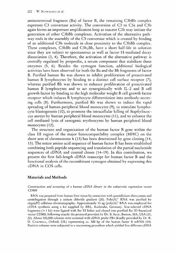
library were subjected to a Northern blot analysis. All of them strongly hybridized to a mRNA species of ca. 2.6 kb in human liver RNA (not shown). The orientation of the cDNA clones was determined by digestion with Hindl l l which cuts in the 5' region of the human factor B cDNA and in the 5' linker sequence of the vector CDM8 (see Fig. 1). Three of the five potential factor B clones were found to contain a cDNA insert in the correct 5' to 3' orientation. Sequencing of the 5' and 3' ends of the inserts confirmed the orientation and revealed that one of these clones, pFB2 potentially comprises the entire coding sequence of the factor B mRNA. Therefore, the complete cDNA sequence of 2447 bp contained in pFB2 was determined. As shown in Figure 2 pFB2 comprises 111 nucleotides of a 5' untranslated region, followed by an open reading frame of 2292 bp,
200bp
Figure 1. Full-length factor B cDNA pFB2 in the vector CDM8. The restriction map of pFB2 is shown for the following restriction enzymes: E, Eco RI; H , Hind I I I ; P, Pst 1. Short horizontal arrows show the direction of DNA sequence determination with oligonucleotides localized in the CDM8 linker region or the internal cDNA sequence. Sites marked with an asterisk are located in the CDM8 polylinker sequence.
Recombinant complement factor B expresses functional activity • 225
encoding a typical leader peptide of 25 amino acids and the entire 739 amino acids of mature factor B. The coding sequence is followed by a stop codon (position 2404) and a 3'untranslated region of 41 bp containing a poly-adenylation signal (ATTAAA) 33 bp downstream of the stop codon. A comparison between the nucleotide sequence of pFB2 and previously published partial cDNA and genomic sequences is given in Figure 1.
Partial exon sequencing data which have been determined from the factor B specific cosmid Cos 10 (15) were aligned over a stretch of 408 bp to the corresponding sequence at the 5' end of pFB2. The comparison revealed that the 5' untranslated region of pFB2 starts 18 bp downstream from the mRNA start site contained in Cos 10. The 5' sequence of pFB2 is in total agreement with the sequence of the two exons analyzed in Cos 10. The pFB2 sequence confirms that no intron interrupts the 5' untranslated and the coding sequence for the signal peptide as previously determined by a Si nuclease mapping analysis of Cos 10.
Moreover, the 3' portion of pFB2 was colinear with the sequence of eleven exons analyzed in another factor B cosmid, termed Cosl (14). Within this 1299 bp stretch, one transition (a «G» in Cosl instead of an «A» in position 1966 of the pFB2 sequence) was observed which results in an amino acid exchange in the derived factor B peptide sequence («GAG» codes for glutamic acid in the Cosl sequence instead of «AAG coding for lysine in pBF2). Furthermore, the pFB2 sequence was aligned to three other previously published partial cDNA sequences for human factor B, the cDNA FBI (14) that was used to isolate pFB2, the cDNA pFB3a (15) containing the coding sequence for the Ba fragment of factor B in a reversed orientation, and the cDNA pBfA28 (18) that comprises the coding region for the entire Bb fragment. The alignment of the 515 bp long FBI sequence shows silent exchanges in two nucleotide positions, one transition at 1326 («T» in the FBI sequence instead of a «C» in the pFB2 sequence) and a transversion in position 1422 («C» in the FBI sequence instead of a «G» in pFB2). A comparison of the pFB2 sequence with the 710 bp long coding sequence for the Ba fragment contained in pFB3a revealed total identity. The alignment of the pFB2 sequence with pBfA28 includes a stretch of 2075 bp. Within this length, pBFA28 differs from the pFB2 sequence in sixteen nucleotide positions. The substitutions in the positions 552, 561, 663, 1194, 1401, 1632, 1755, 1770, 1821, and 2316 do not alter the derived amino acid sequence whereas the transversions in the positions 1009 («CTG» coding for leucine in pBfA28 instead of «GTC» coding for valine in pFB2) and
Figure 2 (see next pages). Nucleotide sequence of the full length cDNA clone pFB2 in • comparison with all other as yet published cDNA and exon sequence data for human factor B. The protein coding region is shown in capital letters. Identity between pFB2 and the compared sequence is indicated by dots. Wherever an aligned sequence differs from the sequence of pFB2, the divergent base is given below. The polyadenylation signal, ATTAAA, in the 3'untranslated region is shown in bold type underlined.
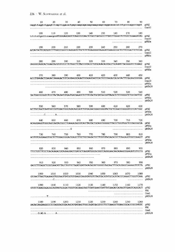
226 • W. SCHWAEBLK et al.
10 20 30 40 caggtctaggt ctggagt t tcagct tggacactgagccaagcagac
60 70 80 90 rccaggacacaccatcctgccocaggoccagct pFB2
, CoslO
100 110 120 130 140 150 160 170 180 tctctoctgccttocaacgacATCm^^ pFB2
CoslO pFB3a
190 200 210 220 230 240 250 260 270 AXACCACTOATOSICITro pFB2
CoslO pFB3a
280 290 300 310 320 330 340 350 360 (^<3J0CCA(XX3HJK^K^ pFB2
CoslO pFB3a
370 380 390 400 410 420 430 440 450 pcceTGW&crcmzcc^^ PFB2
CoslO pFB3a pBfA28
460 470 480 490 500 510 520 530 540 TACTOQOCmSSICI^^ pFB2
pFB3a pB£A28
550 560 570 580 590 600 610 620 630 ?C?lXXX2>GmFX£^ pFB2
pFB3a C A pBfA28
640 650 660 670 680 690 700 710 720 PCKKZMOJKXXm^ pFB2
pFB3a G pBfA28
730 740 750 760 770 780 790 800 810 ACGTCICAGGA03GTOC£^^ pFB2
pFB3a
820 830 840 850 860 870 880 890 900 TirjcrcirjTirariG^^ PFB2
pFB3a pBfA28
910 920 930 940 950 960 970 980 990 GAOCCITCÄGQCK^ pFB2
pBfA28
1000 1010 1020 1030 1040 1050 1060 1070 1080 G i r ^ C H M T I G S G A ^ ^ pFB2
C pBfA28
1090 11O0 1110 1120 1130 1140 1150 1160 1170 GTGICTCAAQCÄ^^ pFB2
FBI Cosl
T pB£A28
1180 1190 1200 1210 1220 1230 1240 1250 1260 MO^CCMGMOXCCTC^ pFB2
FBI Cosl
G.AG.A A pBfA28

Recombinant complement factor B expresses functional activity • 227
1270 1280 1290 1300 1310 1320 1330 1340 1350 ATCATOTTCAflXACT^ pFB2
T FBI Cosl pBfA28
1360 1370 1380 1390 1400 1410 1420 1430 1440 GATCQCAAAAACD AAC<X]WX pFB2
C FBI Cosl
G pBfA28
1450 1460 1470 1480 1490 1500 1510 1520 1530 TOCÄAGÄABSÄC^^ pFB2
FBI Cosl pBfA28
1540 1550 1560 1 570 1580 1590 1600 1610 1620 TUIXITXAGICTCTC pFB2
FBI Cosl pBfA28
1630 1640 1650 1660 1670 1680 1690 1700 1710 CCTIÜVAG3GAC/03?^ pFB2
FBI Cosl
C pBfA28
1720 1730 1740 1750 1760 1770 1780 1790 1800 GMCACnCAATCA^OJICACCJr^ pFB2
Cosl C A pBfA28
1810 1820 1830 1840 1850 1860 1870 1880 1890 AAAAAAGAAGCAGGAATIOC ^ pFB2
Cosl C pBfA28
1900 1910 1920 1930 1940 1950 1960 1970 1980 axÄTnrncTOocro^ pFB2
G Cosl pBfA28
1990 2000 2010 2020 2030 2040 2050 2060 2070 (XTQCACAGGMATCAMGCT^^ pFB2
Cosl pBfA28
2080 2090 2100 2110 2120 2130 2140 2150 2160 (TXIAQCTCTIGACACAGATTX pFB2
\ . Cosl pB£A28
2170 2180 2190 2200 2210 2220 2230 2240 2250 C<ÄCÖ£rPGAGT^ pFB2
Cosl pBfA28
2260 2270 2280 2290 2300 2310 2320 2330 2340 CTTCXnrjITWiraGeramO^ pFB2
Cosl C pBߣ8
2350 2360 2370 2380 2390 2400 2410 2420 2430 MOTIUITICAAGIGTia^ pFB2
Cosl pB£A28
2440 2450 tgggattg^attaaaac pFB2
Cosl pBfA28
228 • W. SCHWAUBLE et al.
1094 («GTC» coding for valine in pBfA28 instead of «GAC» coding for aspartic acid in pFB2) lead to changes in the derived amino acid sequence. In addition, two inversions also lead to changes in the derived amino acid sequence, one in position 1177-1179 («AAG» coding for lysine in pFB2 instead of «GAA» coding for glutamic acid in pBfA28) and another in position 1180-1183 («GAA» coding for glutamic acid in pBfA28 instead of «AAG» coding for lysine in pFB2). The cDNA sequence of pFB2 completes the pBfA28 sequence in sections where the nucleotide sequence was not determined or ambiguity was observed between the cDNA sequence data and the determined protein sequence (see gaps indicated by the omission of dots). Two of these gaps in the pBfA28 sequence were also not filled by any of the other as yet published cDNA or exon sequence data. For these sections, the pFB2 cDNA adds a novel coding sequence which taken together comprises 16 nucleotide residues. The peptides encoded by this added sequence are consistent with the protein sequencing data (15-18).
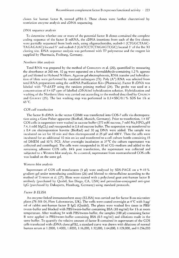
Eukaryotic expression of a recombinant zymogen for human factor B As the liver cDNA library was constructed in the eukaryotic expression
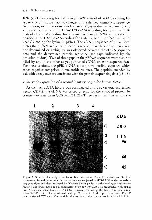
vector CDM8, the cDNA was tested directly for the encoded protein by transient expression in COS cells (21, 22). Three days after transfection, the
1 2 3 4
Figure 3. Western blot analysis for factor B expression in Cos cell transfectants. 50 ul of supernatant from different transfection assays were subjected to SDS-PAGE under nonreduc-ing conditions and then analyzed by Western blotting with a polyclonal goat anti-human factor B antiserum. Lane 1: 5 ul supernatant from 15 x 10f> COS cells transfected with pFB2, lane 2: 5 ul supernatant from 5 x 10⁶ COS cells transfected with pFB2, lane 3: 5 ul supernatant from 9 x l 0 6 COS cells transfected with pFB2, lane 4: 5 ul supernatant from 15x106
nontransfected COS cells. On the right, the position of the sizemarkers is indicated in kDa.
Recombinant complement factor B expresses functional activity • 229
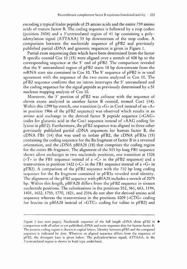
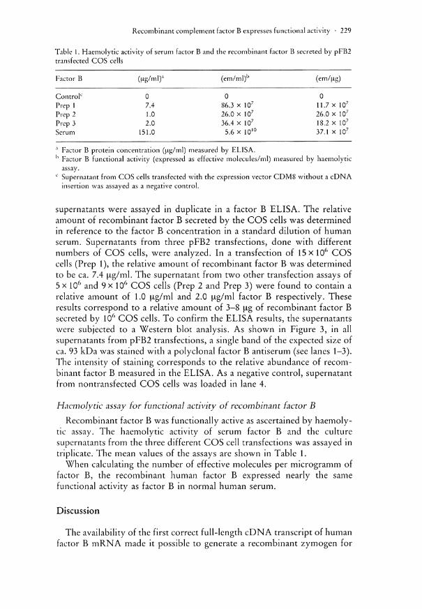
Table 1. Haemolytic activity of serum factor B and the recombinant factor B secreted by pFB2 transfected COS cells
Factor B (Hg/ml)a (em/ml)b (em/jig)
Control0 0 0 0 Prep 1 7.4 86.3 x 10⁷ 11.7 x 10⁷
Prep 2 1.0 26.0 x 10⁷ 26.0 x 10⁷
Prep 3 2.0 36.4 x 10⁷ 18.2 x 10⁷
Serum 151.0 5.6 x 10¹⁰ 37.1 x 10⁷
,x Factor B protein concentration (ug/ml) measured by ELISA. h Factor B functional activity (expressed as effective molecules/ml) measured by haemolytic
assay. c Supernatant from COS cells transfected with the expression vector CDM8 without a cDNA
insertion was assayed as a negative control.
supernatants were assayed in duplicate in a factor B ELISA. The relative amount of recombinant factor B secreted by the COS cells was determined in reference to the factor B concentration in a standard dilution of human serum. Supernatants from three pFB2 transfections, done with different numbers of COS cells, were analyzed. In a transfection of 15xi0 6 COS cells (Prep 1), the relative amount of recombinant factor B was determined to be ca. 7.4 [Ag/ml. The supernatant from two other transfection assays of 5 x 10⁶ and 9x 10⁶ COS cells (Prep 2 and Prep 3) were found to contain a relative amount of 1.0 fxg/ml and 2.0 u,g/ml factor B respectively. These results correspond to a relative amount of 3-8 j ig of recombinant factor B secreted by 106 COS cells. To confirm the ELISA results, the supernatants were subjected to a Western blot analysis. As shown in Figure 3, in all supernatants from pFB2 transfections, a single band of the expected size of ca. 93 kDa was stained with a polyclonal factor B antiserum (see lanes 1-3). The intensity of staining corresponds to the relative abundance of recombinant factor B measured in the ELISA. As a negative control, supernatant from nontransfected COS cells was loaded in lane 4.
Haemolytic assay tor functional activity of recombinant factor B Recombinant factor B was functionally active as ascertained by haemoly
tic assay. The haemolytic activity of serum factor B and the culture supernatants from the three different COS cell transfections was assayed in triplicate. The mean values of the assays are shown in Table 1.
When calculating the number of effective molecules per microgramm of factor B, the recombinant human factor B expressed nearly the same functional activity as factor B in normal human serum.
Discussion
The availability of the first correct full-length cDNA transcript of human factor B mRNA made it possible to generate a recombinant zymogen for
230 • W . SCHWAi:i$LF. et al.
the activation of the alternative pathway of the complement system. The nucleotide sequence of the cDNA contained in clone pFB2 completes the fragmentary cDNA sequence data determined from previously published partial cDNA clones for human factor B. At the 5' end, the pFB2 sequence adds 156 residues to the known cDNA sequence for factor B and confirms the corresponding genomic sequence data obtained from a factor B cosmid. Moreover, the cDNA sequence of pFB2 adds 16 residues of the nucleotide sequence in the coding region of the factor B mRNA which were neither contained in the previous cDNA nor in the genomic sequence data determined for the factor B gene. An alignment between the pFB2 sequence to all the as yet known nucleotide sequence data for factor B revealed a polymorphic structure for factor B mRNA. The pFB2 sequence can be related to the S allele of the factor B gene as in amino acid position seven of the mature protein. pFB2 codes for an arginine, which is specific for the BF S type, instead of a glutamine in the BF F allotype (29, 30). Of the observed nucleotide substitutions, one seen in comparison with the Cosl sequence and three seen in comparison with the pBfA28 cDNA resulted in changes in the encoded peptide sequence. The transfection assays with pFB2 have shown, that a reasonable amount of recombinant human factor B can be expressed in COS cells. In a factor B-dependent haemolysis assay, the translational product of pFB2 was shown to express functional activity. The number of effective molecules per microgramm of recombinant factor B varied between the different transfection assays, but was nearly the same as that of its natural serum counterpart. A reconstitution of the alternative pathway activation by the recombinantly expressed zymogen requires that the generated peptide can efficiently bind to C3b or C3(H 20), that the peptide can be converted subsequently to Bb in a factor D mediated cleavage, and that the Bb portion of this recombinant factor B exhibits serine esterase activity for the conversion of C3 to C3b. As most of the functional sites of factor B have recently been mapped on proteolytic subfragments of the factor B molecule (14, 31), clone pFB2 could be a useful tool to generate definite truncated forms of factor B that express some of the important biological activities of this complement regulatory component.
Acknowledgement
The authors are most grateful to Dr. PETF.R SCHNEIDER, Institute for Legal Medicine, Mainz, for his valuable comments.
References
1. PILLEMER, L., L. BLUM, I . H . LEPOW, O. A . Ross, E . W . TODD, and A . C . WARDLAW. 1954. The properdin system and immunity I . Demonstration and isolation of a new serum protein, properdin, and its role in immune phenomena. Science 120: 279.
Recombinant complement factor B expresses functional activity • 231
2. MÜLLER-EBERHARD, H . J., and R. D. SCHREIBER. 1980. Molecular biology and chemistry of the alternative pathway of complement. Adv. Immunol. 29: 1.
3. PANGBURN, M . K., and H . J. MÜLLER-EBERHARD. 1984. The alternative pathway of complement. Springer Semin. Immunopath. 7: 163.
4. PANGBURN, M. K., R. D. SCHEIBER, and H . J. MÜLLER-EBERHARD. 1981. Formation of the initial C3 convertase of the alternative pathway: acquisition of C3b-like activities by spontaneous hydrolysis of the putative thioester in native C3. j . Exp. Med. 154: 856.
5. SMITH, C. A., M. K. PANGBURN, C. W. VOGEL, and H . J. MÜLLER-EBERHARD. 1984. Molecular Architecture of Human Properdin, a Positive Regulator of the Alternative Pathway of Complement. J. Biol. Chem. 259: 4582.
6. NOLAN, K . F., W. SCHWAEBLE, S. KALUZ, M. P. DIERICH, and K . B. M. RIED. 1991. Molecular cloning of the cDNA coding for properdin, a positive regulator of the alternative pathway of human complement. Eur. J. Immunol. 21: 771.
7. PETERS, M. , J. L. AMBROSUS, A. S. FAUCI, and E.J. BROWN. 1987. Fragments Ba and Bb of complement factor B markedly affect human B cell proliferation. Complement 4: 212.
8. PETERS, M. G., J. L. AMBRUS, A. S. FAUCI, and E. J. BROWN. 1988. The Bb fragment of complement factor B acts as a B cell growth factor. J. Exp. Med. 168: 1225.
9. GÖTZE, O., C. BIANCO, and Z. A. C O H N . The induction of macrophage spreading by factor B of the properdin system, j . Exp. Med. 149: 372.
10. SUNDSMO, J. S. 1983. Leukocyte complement: a possible role for C5 in lymphocyte stimulation.]. Immunol. 131: 886.
11. LEIJH, P. C. J., M. T. VAN DEN BARSELAAR, M. R. DAHA, and R. VAN FURTH. 1982. Stimulation of the intracellular killing of Staphylococcus aureus by monocytes: regulation by immunoglobulin G and complement components C3/C3b and B/Bb. J. Immunol. 129: 332.
12. HALL , R. E., R. M. BLEASE, A. E. DAVIS, J. M. DECKER, B. F. TACK, H . R. COLTEN, and A. V . MUCHMORL. 1982. Cooperative interaction of Factor B and other complement components with mononuclear cells in the antibody-independent lysis of xenogenic erythrocytes. J. Exp. Med. 156: 834.
13. AI.PER, C. A. 1981. Complement and MHC. In: DORP, H . E. (ed.): The Role of the Histocompatibility complex in Immunobiology. Garland Press, New York, 133 pp.
14. CAMPBELL, R. D. , and R. R. PORTER. 1983. Molecular cloning and characterization of the gene coding for human complement protein factor B. Proc. Natl. Acad. Sei. USA 80: 4464.
15. MORLEV, B. J., and R. D. CAMPBELL. 1984. Internal homologies of the Ba fragment from human complement Factor B, a class I I I MHC antigen. EMBO j . 3: 157.
16. Wu, L. C , B . J. MORLEY, and R. D. CAMPBELL. 1987. Cell-specific expression of the human complement protein factor B gene: Evidence for the role of two distinct 5'-flanking elements. Cell 48: 331.
17. CHRISTIE, D. L., J. GAGNON, and R. R. PORTER. 1980. Partial sequence of human complement Factor B: Novel type of serin protease. Proc. Natl. Acad. Sei. USA 77: 4923.
18. MOLE, J. E., J. K. ANDERSON, E. A. DAVIDSON, and D. E. WOODS. 1984. Complete primary structure for the zymogen of human complement factor B. J. Biol. Chem. 259: 3407.
19. WOODS, D. E., A. F. MARKIIAM, A. T. RICKER, G. GOLDBERGER, and H . R. COLLEN. 1982. Isolation of cDNA clones for the human complement factor B, a class I I I major histocompatibility complex gene product. Proc. Natl. Acad. Sei. USA 79: 5661.
20. CHIRGWIN, J. M . , A. E. PRZYBYLA, R. J. MACDONALD, and W . J . RUTTER. 1979. Isolation of biologically active ribonucleic acid from sources enriched in ribonucleases. Biochemistry 18: 5294.
21. SEED, B. 1987. An LFA-3 cDNA encodes a phospholipid-linked membrane protein homologous to its receptor CD2. Nature 329: 840.
22. ARUI-IO, A., and B. SEED. 1987. Molecular cloning of a CD28 cDNA by a high-efficiency COS cell expression system. Proc. Natl. Acad. Sei. USA 84: 8573.
23. SAMHROOK, J., E. F. FRISCH, and T. MANIATIS. 1989. Molecular cloning. A laboratory manual, Cold Spring Harbor Laboratory. Cold Spring Harbor Press, New York.
232 • W . SCHWABBLE et al.
24. FEINBERG, A. P., and B . VOGELSTEIN. 1983. A technique for radiolabeling DNA restriction endonuclease fragments to high specific activity. Anal. Biochem. 132: 2.
25. CHURCH, G. M. , and W . GILBERT. 1984. Genomic sequencing. Proc. Natl. Acad. Sei. USA 81: 1991.
26. LAEMMLI, U. K., and M. FAVRE. 1970. Maturation of the head of bacteriophage T4. Nature 227: 680.
27. TOWBIN, H . , T . STAEHELIN, and J. GORDON. 1979. Electrophoretic transfer of proteins from Polyacrylamid gels to nitrocellulose sheets: procedure and some applications. Proc. Natl. Acad. Sei. USA 76: 4350.
28. WHALEY, K. 1985. In: WHAELY, K. (ed.): Methods in Complement for Clinical Immunologists. Churchill Livingstone, Edinburgh.
29. BENI LEY, D. R., and R. D. CAMPBELL. 1986. C2 and factor B: structure and genetics. Biochem. Soc. Symp. 51: 7.
30. ABBAL, M. , and C. DAVRINCHE. 1990. BF DNA Reference Typing. Complement Inflamm. 7: 190.
31. LAMBRIS, J . D., and H . J . MÜLLER-EBERHARD. 1984. Isolation and Characterization of a 33,000-Dalton Fragment of complement Factor B with catalytic and C3b Binding Activity. J. Biol. Chem. 259: 12685.
Dr. WILHELM SCHWAEBLE, Department of Internal Medicine, Johannes-Gutenberg-University, Verfügungsgebäude, Obere Zahlbacher Straße 63, 55131 Mainz, Germany
\