
IAPA 4th newsletter final
8
IAPA Office bearers President Dr Pradnya Sawant Vice President Dr Elsa Varghese Secretary Dr M Subrahmanyam Treasurer Dr Vibhavari Naik Executive Body Dr Neerja Bhardwaj Dr Sanjay Choubey Dr Nandini Dave Dr Lakshmi Kumar Dr Ekta Rai Dr Pushkar Ranjan Dr R Jayanthi Newsletter Editors Dr Elsa Varghese Dr Gita Nath Dr Vibhavari Naik Dr Jayanti Sripathi Dr MSRC Murthy IAPA Fellowships for students Accredited Institutions are • GKNM Hospital, Coimbatore, • Rainbow Hospitals, Hyderabad • Rainbow Hospitals, Bangalore, • Chacha Nehru Bal Chikitsalaya, New Delhi. Prof Elsa Varghese Vice-President, IAPA Bangalore The FDA warning on anesthetic and sedative agents in children and pregnant patients…. how should we react to it? In December 2016, the Food and Drug Administration (FDA) of USA, issued a warning for the administering of general anesthetic and sedation drugs in children aged less than 3 years and in pregnant women in the third trimester. 1 It states, that ‘exposure to these medicines for lengthy periods of time or over multiple surgeries may negatively affect brain development’. It also recommends that labels of general anesthetic and sedation drugs listed in the alert, should have this warning printed on them. The FDA alert, quotes studies conducted in pregnant and young animals exposed to general anesthetic and sedation drugs for more than 3 hours, which caused widespread nerve cells loss in the developing brain. Other animal studies have suggested that these changes result in long-term abnormal behaviour or learning disability. 2,3,4 The drugs listed in the FDA warning include: desflurane, etomidate, halothane, isoflurane, sevoflurane, etomidate, pentobarbitone, propofol, ketamine, methohexital, midazolam and lorazepam. Subsequently in April 2017, the FDA amended its earlier statement by emphasizing that ‘General anesthetic and sedation drugs are necessary for patients, including young children and pregnant women, who require surgery or other painful and stressful procedures. In the United States of America, surgeries during the third trimester of pregnancy requiring general anesthesia are performed only when medically necessary and rarely last longer than 3 hours. We are advising that in these situations, pregnant women should not delay or avoid surgeries or procedures during pregnancy, as doing so can negatively affect themselves and their infants. Similarly, surgeries or procedures in children younger than 3 years should not be delayed or avoided when medically necessary. Consideration should be given to delaying potentially elective surgery in young children where medically appropriate’. 4 There are several multicentred, international trials in progress attempting to address this critical issue. The ‘Pediatric Anesthesia and Neurodevelopment Assessment ‘(PANDA) interim study results have found no evidence for association between exposure to anaesthesia and outcomes in psychometric assessments. 5 The initial reports of ‘The General Anesthesia vs. Spinal Anesthesia’ (GAS) trial has shown no difference in immediate outcomes between general or awake-regional anesthesia in children. The long- term results of both these studies are awaited. 6 The Mayo Anesthesia Safety in Kids (MASK) study is in progress, to compare children exposed to a single anesthetic with children not exposed to anesthesia. 7 Neurodevelopmental outcomes of young children who are subjected to multiple anesthetic exposures will also be evaluated in the MASK study, expected to be released in 2017. 8 The Recognition Memory Study, which evaluates prolonged anesthesia in young children is expected in a few years. Unfortunately, there is no data available which can assess the association between anesthetic exposure to human foetuses in utero with postnatal neurodevelopmental delay. 9 EDITORIAL IAPA NEWSLETTER by Indian Association of Paediatric Anaesthesiologists August 2017
Transcript of IAPA 4th newsletter final

IAPA Office bearers
PresidentDr Pradnya Sawant
Vice PresidentDr Elsa Varghese
SecretaryDr M Subrahmanyam
TreasurerDr Vibhavari Naik
Executive BodyDr Neerja BhardwajDr Sanjay ChoubeyDr Nandini DaveDr Lakshmi KumarDr Ekta RaiDr Pushkar RanjanDr R Jayanthi
Newsletter Editors Dr Elsa VargheseDr Gita NathDr Vibhavari NaikDr Jayanti SripathiDr MSRC Murthy
IAPA Fellowships for
studentsAccredited Institutions are• GKNM
Hospital, Coimbatore,
• Rainbow Hospitals, Hyderabad
• Rainbow Hospitals, Bangalore,
• Chacha Nehru Bal Chikitsalaya, New Delhi.
Prof Elsa VargheseVice-President, IAPA
Bangalore
The FDA warning on anesthetic and sedative agents in children and pregnantpatients…. how should we react to it?
In December 2016, the Food and Drug Administration (FDA) of USA, issued a warning forthe administering of general anesthetic and sedation drugs in children aged less than 3years and in pregnant women in the third trimester.1 It states, that ‘exposure to thesemedicines for lengthy periods of time or over multiple surgeries may negatively affect braindevelopment’. It also recommends that labels of general anesthetic and sedation drugslisted in the alert, should have this warning printed on them. The FDA alert, quotes studiesconducted in pregnant and young animals exposed to general anesthetic and sedationdrugs for more than 3 hours, which caused widespread nerve cells loss in the developingbrain. Other animal studies have suggested that these changes result in long-termabnormal behaviour or learning disability.2,3,4 The drugs listed in the FDA warning include:desflurane, etomidate, halothane, isoflurane, sevoflurane, etomidate, pentobarbitone,propofol, ketamine, methohexital, midazolam and lorazepam.
Subsequently in April 2017, the FDA amended its earlier statement by emphasizing that‘General anesthetic and sedation drugs are necessary for patients, including young childrenand pregnant women, who require surgery or other painful and stressful procedures. In theUnited States of America, surgeries during the third trimester of pregnancy requiringgeneral anesthesia are performed only when medically necessary and rarely last longerthan 3 hours. We are advising that in these situations, pregnant women should not delay oravoid surgeries or procedures during pregnancy, as doing so can negatively affectthemselves and their infants. Similarly, surgeries or procedures in children younger than 3years should not be delayed or avoided when medically necessary. Consideration shouldbe given to delaying potentially elective surgery in young children where medicallyappropriate’. 4
There are several multicentred, international trials in progress attempting to address thiscritical issue. The ‘Pediatric Anesthesia and Neurodevelopment Assessment ‘(PANDA)interim study results have found no evidence for association between exposure toanaesthesia and outcomes in psychometric assessments.5 The initial reports of ‘TheGeneral Anesthesia vs. Spinal Anesthesia’ (GAS) trial has shown no difference inimmediate outcomes between general or awake-regional anesthesia in children. The long-term results of both these studies are awaited.6 The Mayo Anesthesia Safety in Kids(MASK) study is in progress, to compare children exposed to a single anesthetic withchildren not exposed to anesthesia.7 Neurodevelopmental outcomes of young children whoare subjected to multiple anesthetic exposures will also be evaluated in the MASK study,expected to be released in 2017.8 The Recognition Memory Study, which evaluatesprolonged anesthesia in young children is expected in a few years. Unfortunately, there isno data available which can assess the association between anesthetic exposure to humanfoetuses in utero with postnatal neurodevelopmental delay.9
EDITORIAL
IAPA NEWSLETTERby Indian Association of Paediatric Anaesthesiologists
August 2017

EDITORIAL CONTD....impact of information technology, we have to beprepared for the repercussions of this FDA in India aswell. How should anesthesiologists and surgeonsrespond especially since millions of children areinvolved? When questioned, and when surgery cannotbe postponed, how do we discuss with the families of achild younger than 3 years of age, or if the child is likelyto require multiple or prolonged surgeries andanesthetic exposures?
Physicians, surgeons and hospital administrations needto discuss this matter and prepare a coordinatedresponse, with the aim of putting this warning inperspective. That these studies demonstrate nodevelopmental problems in children exposed to a single,short anesthetic or sedation is documented. There is noalternative to providing anesthesia. Most surgicalprocedures performed in small children are clearlyindicated and cannot be delayed. What is worrisome isthat the fear of neurological complications of anesthesia,may result in postponement or delay in necessaryprocedures, resulting in a marked increase in morbidityand mortality. Our main role as anesthesiologists is tocontinue to help parents, pregnant women and familiesmake their decision and consent for anesthesia bydiscussing the benefits and risks involved if surgery is tobe postponed.
Until results of studies in progress are published, we willhave to try and quell untoward anxiety. Concertedefforts need to be made by our professionals across theglobe to study and follow up effects of anesthetics onfoetuses and small children. Our response to the FDAwarning depends on how we interpret it.
Anesthesiologists have been on the alert since thepublication of the animal studies in the 1990s. However,the December 2016 FDA alert to the public at large, hasbeen received by individual anesthesiologists,obstetricians, paediatricians, physicians andprofessional medical societies, in the US and the rest ofthe world, with a sense of shock. It is true, that thereneeds to be an awareness amongst the public at large,but without enough clinical scientific evidence inhumans, this alert is likely to create a panic reaction.The American College of Obstetrics and Gynecology(ACOG), believes the clinical significance of thesefindings are unknown and could inappropriatelydissuade clinicians from providing women withnecessary care during pregnancy.10 Two editorials havebeen published more recently in the New EnglandJournal of Medicine and in the journal PediatricAnesthesia.11,12
There are several issues that these editorials raise. Thistopic has been a concern since the initial animal studiespublished in the late 1990s. The FDA statement isbased on inadequate clinical evidence, the only humanclinical studies available being weak and notconfirmatory. The 3-year age cut off and the evidence ofmultiple vs single exposure is inadequate in animalstudies and human studies. Parents of children,pregnant women along with the lay public are likely toonly read the title of the FDA alert and not the statementthat there are ‘limitations of our knowledge and theweak nature of the evidence’.
The practical and difficult issue at hand is, how do wedeal with this alert in our daily practice? Given the global
QUIZ ANSWERS from Pg3Q1 Ans. C-The use of nitrous oxide for minimal sedation is defined as the administration of nitrous oxide 50% or less with the balance as oxygen without any other sedative, narcotic or other depressant drug. Q2 Ans. C -There is no difference in the tidal volume, but neonates have increased oxygen consumption, to compensate for this, the alveolar ventilation is increased and there is an increase in respiratory rate to balance it.Q3 Ans. D-Even during CPB, hypoglycemia is common in children, due to low glycogen reserves while hyperglycemia is frequent in adults. Q4 Ans. B -Wilm’stumor is common malignancy in children, presenting with abdominal mass. The treatment includes surgery, chemotherapy and radiotherapy. The chemotherapeutic agents commonly used –cyclophosphamide and Adriamycin. Both can cause thrombocytopenia, cardiac toxicity and cyclophosphamide can cause inhibition of plasma cholinesterase. However, pulmonary fibrosis is not seen.Q5 Ans. D-According to AHA updated guidelines in 2015, the maximum dose for defibrillation should not exceed 10 J/Kg or adult maximum dose.
IAPA NEWSLETTERby Indian Association of Paediatric Anaesthesiologists
August 2017
IAPA NEWSLETTERby Indian Association of Paediatric Anaesthesiologists
August 2017
EDITORIAL CONTD....
QUIZ SECTION
1. According to AAP Guidelines on monitoring and management of pediatric patients during and after sedation for diagnostic and therapeutic procedures introduced in 2016, the use of nitrous oxide for minimal sedation is defined as administration of a) Nitrous oxide > 20% but < 50% with oxygen and
narcotic b) Nitrous oxide > 20% but < 50% without oxygen and
narcoticc) Nitrous Oxide < 50% with oxygen d) Nitrous oxide > 50% with oxygen
2. Which of the following respiratory indices in increased in newborn in comparison to adults? a) TV (mL/kg)b) pHc) Alveolar ventilation (mL/kg/min)d) None of the above
3. During CPB, about the, glucose regulation among adults and pediatrics, the correct statement is a) Hyperglycemia is common in children, hypoglycemia
in adults
Dr Mahima Gupta, Dr Rakesh GargAIIMS, New Delhi
b) Hyperglycemia is common in both c) Hypoglycemia is common in bothd) Hypoglycemia is common in children, hyperglycemia
in adults
4. A 3yr old child of Wilm’s tumor post surgery, post chemotherapy consisting of adriamycin and cyclophosphamide is scheduled for non – oncologic surgery under general anesthesia, anaesthetic concerns include all except -a) Cardiac depressionb) Pulmonary fibrosisc) Inhibition of plasma cholinesterased) Thrombocytopenia
5. According to AHA 2015 updated guidelines on pediatric ACLS, maximum dose of energy that can be used during defibrillation is a) 2 J/Kg b) 6 J/Kg c) 8 J/Kg d) 10 J/Kg (Answers on page 2)
References1. FDA Drug Safety Communication: FDA review
results in new warnings about using generalanesthetics and sedation drugs in young children andpregnant women issued on December 14th 2016
2. Wilder RT, Flick RP, Sprung J, et al. Early exposureto anesthesia and learning disabilities in apopulation-based birthcohort. Anesthesiology 2009;110:796-804
3. Sanders RD, Hassell J, Davidson AJ et al. Impact ofanaesthetics and surgery on neurodevelopment: anupdate. Br J Anaesth 2013;110 Suppl:i53-i72
4. FDA Drug Safety Communication: FDA approveslabel changes for use of general anesthetic andsedation drugs in young children issued on April 27th2017
5. Sun LS, Li G, Miller TL et al. Association between asingle general anesthesia exposure before age 36months and neurocognitive outcomes in laterchildhood. JAMA 2016;315:2312-2320
6. Davidson AJ, Disma N, de Graaff JC et al,Neurodevelopmental outcomes at 2 years of ageafter general anaesthesia and awake -regionalanaesthesia in infancy (GAS): an international
multicentre, randomised controlled trial. Lancet2016;386:239-250
7. Gleich SJ, Flick R, Hu D. et al. Neurodevelopment ofchildren exposed to anesthesia: Design of the MayoAnesthesia Safety in Kids (MASK)study. Contemporary Clinical Trials, 201541, 45-54.DOI: 10.1016/j.cct.2014.12.020
8. Pinyavat T, Warner DO, Flick RP, et al. Summary ofthe update session on clinical neurotoxicity studies. JNeurosurg Anesthesiol 2016; 28:356-360
9. Vutskits L, Xie Z. Lasting impact of generalanaesthesia on the brain: mechanisms andrelevance. Nat Rev Neurosci 2016; 17: 705–717.
10. http://www.acog.org/About-ACOG/News-Room/Statements/2016/ACOG-Statement-on-the-FDA-Warning-Regarding-Use-of-General-Anesthetics-Pregnancy
11. Andropoulos DB, Greene MF. Anesthesia andDeveloping Brains — Implications of the FDAWarning. N Engl J Med. 2017; 376(10): 905-07
12. Davidson AJ, Vutskits L. The new FDA drug safetycommunication on the use of general anesthetics inyoung children: what should we make of it? PediatrAnesth. 2017; 27(4): 336–337

IAPA NEWSLETTERby Indian Association of Paediatric Anaesthesiologists
August 2017
Inadvertent massive overdose of lignocaine in an infant with arthrogryposis multiplex congenita successfully treated with lipid emulsion
Dr Sapna Bathla, Dr Anju Gupta, Dr Aikta Gupta, Dr Geeta Kamal, Dr Nidhi Sehgal, Dr Manoj GuptaChacha Nehru Bal Chikitsalaya (CNBC), Delhi
Introduction: Intravenous lipid emulsion (IVLE) has remarkable success and safety record in local anaestheticsystemic toxicity (LAST).1 However, there is paucity in the literature of its application and safety in infants. Moreover,use of 10% IVLE in LAST has not been documented.
Case report: A 3 month old male weighing 3.9 kg, diagnosed as Arthrogryposis multiplex congenita was posted forfemoral shortening and soft tissue alignment surgery. Child had upper respiratory tract infection which had beentreated 2 weeks ago. Preoperative investigations were within normal limits. After preoxygenation, induction was doneusing sevoflurane (5-8%) in 100% oxygen. The child had anticipated difficult airway due to retrognathia and higharched palate, so his trachea was intubated with a Truview EVO2® video laryngoscope. At the end of uneventfulsurgery, following reversal of neuromuscular blockade with neostigmine and glycopyrrolate, the child developedinspiratory stridor and paradoxical movement of chest and abdomen. Positive pressure ventilation, jaw thrust and inj.propofol 4 mg administered for possible laryngospasm, partially relieved the symptoms. At the same time, 6 mgpreservative free lignocaine (Xylocard TM) was administered intravenously by a trainee. Suddenly the baby becameunresponsive and hemodynamically unstable. Heart rate decreased from 180 bpm to 124 bpm and blood pressuredecreased from 67/44 mmHg to 41/18 mmHg respectively, with a wide complex rhythm and the baby had focalseizures. Bag-mask ventilation was continued; Ringer’s lactate bolus of 50 ml and intravenous midazolam 0.2 mgwere also administered. The child transiently improved but soon started having generalized tonic clonic seizure.Retrospectively, it was revealed that 6 ml of Lignocaine (120 mg) had been injected instead of 6 mg. 10% IVLE(Fresenius Kabi AB, Uppsala, Sweden) was administered in the dose of 3 ml/kg (12 ml) slowly over 2 - 3 minutes.Within seconds of completion of its injection, seizures subsided and patient became hemodynamically stable.Considering the baby was still lethargic and had low muscular tone, a second bolus of 10 ml of 10% IVLE wasrepeated, followed by infusion at 1 ml/min for 2 hours. He was intubated and shifted to ICU for overnight ventilationwith midazolam sedation. There was no recurrence of seizure in ICU, his vitals remained stable and was extubatednext morning. He was discharged 3 days later after confirming normal biochemistry profile (including serum amylaseand triglyceride).
Discussion: The present report describes use of 10% IVLE to treat life threatening lignocaine over dosage in aninfant. IVLE has been reported for resuscitating patients with amide local anaesthetic (LA) toxicities.1 The LAs beinglipid soluble, are taken up into IVLE (lipid sink), reducing their concentration in aqueous phase, decreasing theirunbound fraction and reducing toxicity.2 Most case reports of LA toxicity are reported in regional blocks and 20%IVLE has been used as antidote. Infants have increased vulnerability to toxicity with lignocaine due to prolongedelimination half-life, increased volume of distribution and reduced serum binding proteins. IVLE has been shown toreduce serum lidocaine levels from toxic levels of 7.6 mcg/ml to therapeutic levels of 3 mcg/ml and treat its systemictoxicity.3,4 IVLE may not be effective when hypoxia, acidosis and electrolyte abnormalities set in, after ongoingresuscitative measures have failed. So there has been a recent trend of early use of IVLE during suspected LAST.5 Inthe patient with suspected LA toxicity, the initial step is stabilization of potential threats to life. If the signs andsymptoms develop during administration of the LA, stop the injection immediately and prepare to treat the reaction.Ensure adequate oxygenation, whether by face mask or by intubation. Attention to impending airway compromise,significant hypotension, dysrhythmias, and seizures takes precedence.
Accidental high dose of intravenous LA, lead to recurrent seizure and imminent danger of cardiovascular collapse.Increasing evidence suggests that the IVLE can reverse the cardiac and neurologic effects of LA toxicity. Casereports support the early use of lipid emulsion at the first sign of arrhythmia, prolonged seizure activity, or rapidprogression of toxic manifestations in patients with suspected local anaesthetic toxicity.1 Since 20% IVLE was not
CASE REPORT
IAPA NEWSLETTERby Indian Association of Paediatric Anaesthesiologists
August 2017
CASE REPORT CONTD...
Conference SecretariatMr Veeraswamy, M: 92465 87379
Organising ChairpersonDr M Subrahmanyam
Jointly hosted by
available, we used 3 ml/kg of 10% IVLE (instead of 1.5ml/kg of 20%) within 5-7 minutes of the injection oflignocaine. Infusion was continued to maintain LA levelsbelow critical levels so as to avoid recurrence of toxicity.We presume that IVLE injection brought the plasmalevels of lignocaine below the toxic range and led toimmediate improvement in the clinical picture. We did nothave the facility to objectively demonstrate any fall inlignocaine levels after IVLE but drawing analogy from aprevious case report, the favourable outcome in case ofour patient was presumably due to IVLE.4 In our case wehad used 142 ml of 10% IVLE which is well below themaximum recommended dose (40 ml/kg over 24 hours,160 ml for our patient) as mentioned in its package insert.Laboratory testing and one-week follow-up of the child didnot reveal any adverse effects.
Conclusion: Immediate recognition of the toxicity,continuity of care and early use of IVLE possibly led tofavourable outcome in our patient.
Ethics: NAFunding: NoneDisclosures/conflict of interest: None
References:1. Cave G, Harvey M, Graudins A. Review article:Intravenous lipid emulsion as antidote: A summary ofpublished human experience. Emerg Med Australas2011:23;123–141.
2. Weinberg GL, Di Gregorio G, Ripper R, et al.Resuscitation with lipid versus epinephrine in a rat modelof Bupivacaine overdose. Anesthesiology2008;108(5):907–913.3. Mather LE, Copeland SE and Ladd LA: Acute toxicityof 1ocal anesthetics: Underlying pharmacokinetic andpharmacodynamic concepts. Reg Anesth Pain Med2005;30:553-66.4. Dix SK, Rosner GF, Nayar M, Harris JJ, Guglin ME,Winterfield JR, Xiong Z. Intractable cardiac arrest due tolidocaine toxicity successfully resuscitated with lipidemulsion. Critical Care Medicine 2011;39(4):872–74.5. Hiller DB, Gregorio GD, Ripper R, et al. Epinephrineimpairs lipid resuscitation from bupivacaine overdose: athreshold effect. Anesthesiology 2009;111(3):498–505.
Want to know your IAPA
membership number? Not receiving IAPA mails?
Visit IAPA website www.iapaindia.comOr write to [email protected]
Or call +91 92465 87379Update your member profile and get your
queries resolved

Ultrasound – A Key Gadget for Paediatric Anaesthesiologists’Dr. Ekta Rai, Dr. Vibhavari Naik
IntroductionThe use of ultrasound has recently gained popularityamongst intensivists and anaesthesiologists as itenables rapid, by-the-bed, and relatively inexpensivediagnostic evaluation of unstable patients. It has notonly changed the outlook of intensivists in managementof critically ill patients, but also helpedanaesthesiologists by making different regionaltechniques safer and accurate. The structures ofinterest in children are superficial and need highfrequency probes which can give a better resolutionimages. In infants, the cranial sutures are not fused andhence ultrasound can provide crucial information aboutthe brain. The most common use of ultrasound amongpaediatric anaesthesiologists is for gaining vascularaccess and for performing various regional blocks.Other ‘point of care’ ultrasound applications are inhemodynamic monitoring, lung scanning, airwayassessment, detection of esophageal intubation,identification of cricothyroid membrane prior tointubating a difficult airway child, and optic nerve sheathscanning to reflect on intracranial pressure.
We intend to cover the role of ultrasound as a gadget foranaesthesiologists for our newsletter readers in amodule based manner over the next few newsletters.
Ultrasound Principle and KnobologyUltrasound imaging is based on the principle ofgeneration of sound waves beyond the audible range(20 kHz). Frequencies used for ultrasound are higherthan those in the audible range, and typically vary from2 to 15 MHz for diagnostic procedures. These waveshave power to penetrate different tissues of the body atdifferent speed and are reflected back from the tissueinterface. The amplitude of reflected energy is used togenerate ultrasound images. The knowledge ofknobology is an essential first step of using ultrasoundfor various applications. A hands-on course or workshopin knobology is desirable to acquaint one-self with theavailable ultrasound machine and its function.
MODULE 1 : Ultrasound for AirwayUltrasound does not penetrate air and hence was nevertraditionally used for evaluating the airway. The recentinterest in using this modality at the ‘point of care’ hasopened a whole new arena of its applications in theairway which will be covered in this module.
1.What is the need of using ultrasound in airwaymanagement?Safe airway management has always been theanaesthesiologists’ primary concern. Airwaymanagement in infants and neonates is challenging dueto higher incidence of airway related complications, ascompared to older children. Ultrasound has been shownto add safety and accuracy in airway management. It iseasier to learn, teach and reproduce. Also, mosthospitals are equipped with ultrasound machines, thatcan be availed by the anaesthesiologists. Hence it isessential for all of us to learn ultrasound and itsapplications relevant to us.
2. What is the ideal position for performing airwayultrasound?Position – centered, sniffing positionIdeal USG Probe - 7.5-Mhz high-frequency lineartransducer.Hydrogel – to eliminate the air interfaceIdeal Settings - Depth-3-4 cm and Focus 1 cm posteriorto the target structureApproaches - Longitudinal (long axis) and Transverse(short axis) at various levels
3. What should I look for?A) Learn to identify normal structuresHyoid (Transverse Axis) - is the main bone present inairway anatomy and is hyperechoic.Epiglottis (Transverse Axis) - Hypoechoic U-shapedstructure present at thyroid–hyoid space. The spaceanterior to epiglottis is pre-epiglottic space, which hashyperechoic air–mucosa interface.Thyroid cartilage (Transverse Axis) - Hypoechoicstructure with respect to the vocal cords, followed by anacoustic shadow corresponding to the airway. Thyroid–hyoid space and the acoustic shadow in the hyoid bonecan be evaluated.Cricoid cartilage (Transverse Axis) - Hypoechoic,inverted U structure inferior to the thyroid cartilage is thecricoid cartilage.Cricothyroid membrane (Longitudinal Axis) - Ahypoechoic structure between the thyroid and thecricoid cartilages can be observed through a longitudinalsection along the larynx.Tracheal rings (Longitudinal Axis) - Tracheal cartilagein longitudinal plane is seen as a “string of beads” andinverted U in the transverse section.
IAPA NEWSLETTERby Indian Association of Paediatric Anaesthesiologists
August 2017
SPECIAL ARTICLE

Vocal Cord (Transverse Axis) - Vocal cord is bestseen using transverse plane through the thyroidcartilage as a window. The hyperechoic appearance ofthe vocal ligaments delineates the vocal cord.
B) Learn to identify abnormal structuresEpiglottitis - With the transducer placed in longitudinalplane of thyrohyoid membrane, a hyperechoic thickenedepiglottis seen in relation to the acoustic shadowing ofthe hyoid bone as an “alphabet P sign” signifies acuteepiglottitis.
3. What are the clinical indications of use ofultrasound for airway?
I. Anticipating difficult airway - Ultrasound has beenused by many to identify the difficult airway predictors.Various distances like pre-epiglottic space, anteriorspace to thyroid, distance between the hyoid andmentum, depth of tongue, are being studied to helppredict difficult airway. Since the studies are mainly inadults, their role in children is unclear.
II. Predicting perioperative gastric aspiration risk –Studies have suggested that it is possible to quantify theamount of gastric contents in children with ultrasound,thus predicting the gastric aspiration risk. The gastricantrum is visualized in right parasagittal plane betweenthe left lobe of liver and pancreas; and the cross-sectional area is measured.Fig 1 Subcostal view of gastric antrum to evaluate theaspiration risk
III. Identifying position of endotracheal tube - Theconfirmation of the endotracheal intubation can be donedirectly or indirectly. Direct verification is identifying theETT in trachea or esophagus. The indirect evidencewould be identifying pulmonary ventilation and themovement of diaphragm. The successful endotrachealintubation may be confirmed in real time with the
artifacts generated when the tube is introduced insidethe trachea. And if tracheal intubation fails, a newcircular structure can be seen with artifacts generatedby the presence of the tube inside the esophagus. Thesensitivity of this technique is high and there is no needto ventilate. This technique is superior to clinicalauscultation of breath sounds as well as capnography,both of which need ventilation with a few breaths toidentify correct placement of the ETT. But the time takenby ultrasound may be longer as compared tocapnography.Fig 2 Transverse view of the neck to identify type ofintubation
The indirect tracheal intubation verification includes theuse of the linear transducer for checking ventilationthroughout the pulmonary fields.The ‘sliding lung sign’ between two ribs, is highlysensitive to confirm the tracheal intubation. Unilaterallung sliding is an indication of endobronchial intubationin the absence of other lung pathology. Diaphragmaticmovements seen from the substernal view are alsoindicative of tracheal intubation, but need moreexpertise and training.
IV. ETT sizing and depth of insertion- There has beenextensive research in the ability of ultrasound todetermine airway size and estimate appropriate size ofETT, tracheostomy and double lumen tube. Theultrasound measured subglottic diameter correlates wellwith the outer diameter of the optimal ETT in olderchildren. It is found that this technique underestimatesthe ETT size in children less than 1 year. Theultrasound based ETT size selection is found superior tothe traditional age based formula. The direct ETT tipvisualization is possible in neonates and infants but notin older children. The optimal depth of ETT is when thetip lies about 1 cm above the aortic arch in infants,which corresponds to about 0.5cm above the carina.Ultrasound determination of the depth of insertion ofETT is faster than the gold standard chest X ray. Fillingthe ETT cuff with saline improves its visualization at thesuprasternal notch and is a good technique for verifying
IAPA NEWSLETTERby Indian Association of Paediatric Anaesthesiologists
August 2017
SPECIAL ARTICLE CONTD.......
Left lobe of liver Gastric
antrum with air shadows
Semi circular empty tracheal shadow
Tubular shadow of ETT malposition in esophagus
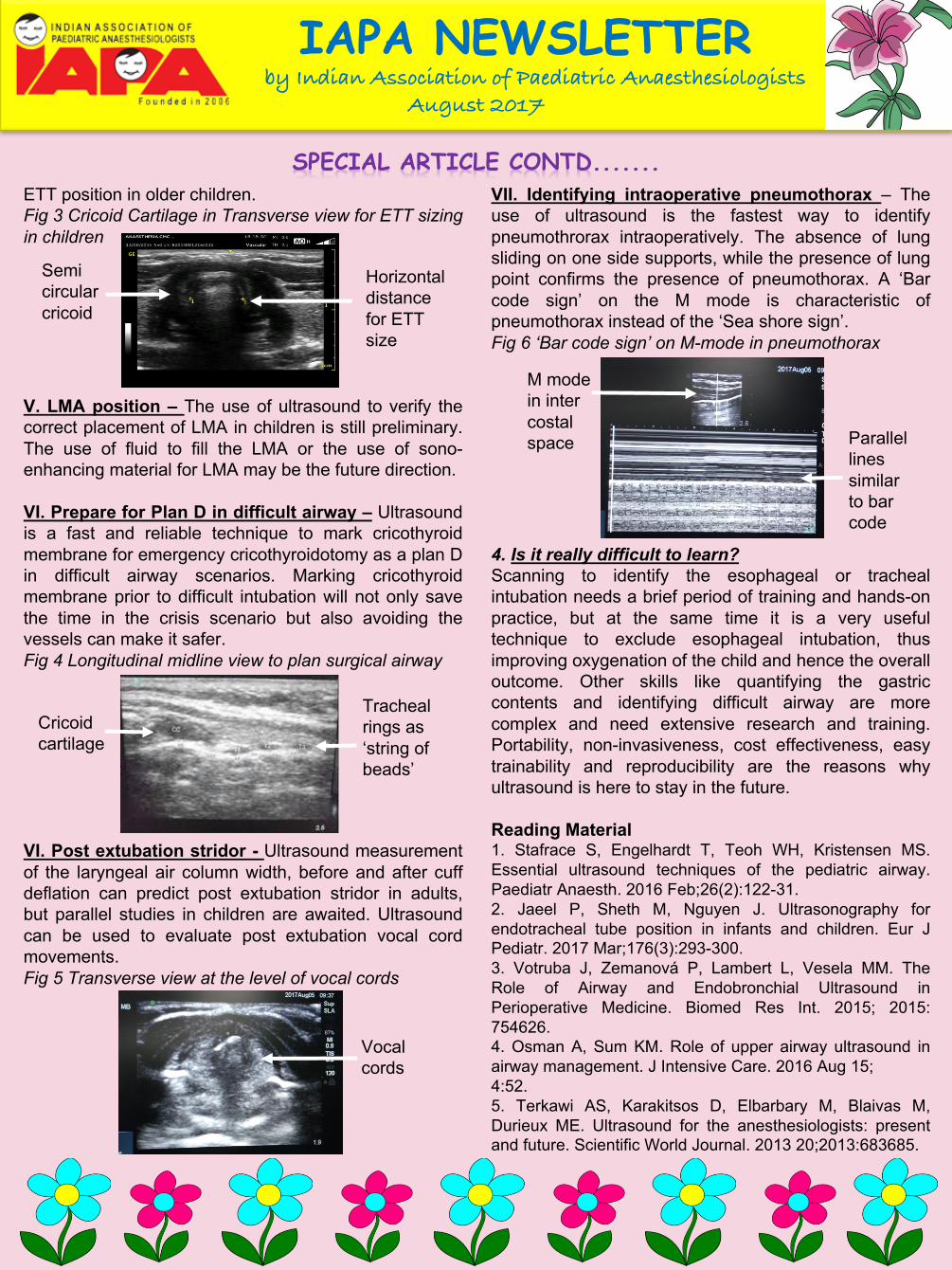
ETT position in older children.Fig 3 Cricoid Cartilage in Transverse view for ETT sizingin children
V. LMA position – The use of ultrasound to verify thecorrect placement of LMA in children is still preliminary.The use of fluid to fill the LMA or the use of sono-enhancing material for LMA may be the future direction.
VI. Prepare for Plan D in difficult airway – Ultrasoundis a fast and reliable technique to mark cricothyroidmembrane for emergency cricothyroidotomy as a plan Din difficult airway scenarios. Marking cricothyroidmembrane prior to difficult intubation will not only savethe time in the crisis scenario but also avoiding thevessels can make it safer.Fig 4 Longitudinal midline view to plan surgical airway
VI. Post extubation stridor - Ultrasound measurementof the laryngeal air column width, before and after cuffdeflation can predict post extubation stridor in adults,but parallel studies in children are awaited. Ultrasoundcan be used to evaluate post extubation vocal cordmovements.Fig 5 Transverse view at the level of vocal cords
VII. Identifying intraoperative pneumothorax – Theuse of ultrasound is the fastest way to identifypneumothrorax intraoperatively. The absence of lungsliding on one side supports, while the presence of lungpoint confirms the presence of pneumothorax. A ‘Barcode sign’ on the M mode is characteristic ofpneumothorax instead of the ‘Sea shore sign’.Fig 6 ‘Bar code sign’ on M-mode in pneumothorax
4. Is it really difficult to learn?Scanning to identify the esophageal or trachealintubation needs a brief period of training and hands-onpractice, but at the same time it is a very usefultechnique to exclude esophageal intubation, thusimproving oxygenation of the child and hence the overalloutcome. Other skills like quantifying the gastriccontents and identifying difficult airway are morecomplex and need extensive research and training.Portability, non-invasiveness, cost effectiveness, easytrainability and reproducibility are the reasons whyultrasound is here to stay in the future.
Reading Material1. Stafrace S, Engelhardt T, Teoh WH, Kristensen MS.Essential ultrasound techniques of the pediatric airway.Paediatr Anaesth. 2016 Feb;26(2):122-31.2. Jaeel P, Sheth M, Nguyen J. Ultrasonography forendotracheal tube position in infants and children. Eur JPediatr. 2017 Mar;176(3):293-300.3. Votruba J, Zemanová P, Lambert L, Vesela MM. TheRole of Airway and Endobronchial Ultrasound inPerioperative Medicine. Biomed Res Int. 2015; 2015:754626.4. Osman A, Sum KM. Role of upper airway ultrasound inairway management. J Intensive Care. 2016 Aug 15;4:52.5. Terkawi AS, Karakitsos D, Elbarbary M, Blaivas M,Durieux ME. Ultrasound for the anesthesiologists: presentand future. Scientific World Journal. 2013 20;2013:683685.
IAPA NEWSLETTERby Indian Association of Paediatric Anaesthesiologists
August 2017
SPECIAL ARTICLE CONTD.......
Semi circular cricoid
Horizontal distance for ETT size
Cricoid cartilage
Tracheal rings as ‘string of beads’
M mode in inter costal space Parallel
lines similar to bar code
Vocal cords