Inhaltsverzeichnis der Evidenztabellen -siehe Lesezeichen ... · PPI and statin use were identified...
66
Inhaltsverzeichnis der Evidenztabellen -siehe Lesezeichen des PDFs Schlüsselfrage: AG 1 Risikofaktoren: Wird das Ösophaguskarzinomrisiko (SCC AC) durch einen der folgenden Faktoren beeinflusst? Citation Evidence Level Study Type Vinogradova, Y. 2013 3b Series of nested case-control studies Levi, Z. 2013 2b Cohort study Pottegard, A. 2013 3b population-based case-control study Cooper, S. 2014 3b Nested-case control study Feng, X. S. 2014 2b Prospective Cohort Study Alexandre, L. 2014 3b Case-control study Hvid-Jensen, F. 2014 3b Nested case-control study Masclee, G. M. 2014 2b dynamic population-based retrospective cohort study Jia, N. 2014 2b- Retrospective Cohort Study Agrawal, S. 2014 3b- Retrospective case-control study Lindkvist, B. 2014 2b Prospective cohort study Moura, M. A. 2014 3b case-control study Cook, M. B. 2015 2b Prospective Cohort Study Hazelton, W. D. 2015 2b Cohort Study Wienecke, A. 2015 1b- Cohort Study Bhat, G. A. 2015 3b Case-control study Buckland, G. 2015 1b prospective cohort study Chen, T. 2015 3b population-based case-control study Rafiq, R. 2016 3b Case-control study Sewram, V. 2016 3b hospital-based Case-Control Study Thota, P. N. 2016 1b- Retrospective Cohort Study Kestens, C. 2016 2b Retrospective population-based cohort study Krishnamoorthi, R. 2016 1b population-based cohort study Zakaria, D. 2017 2b- Cohort Study Nguyen, T. 2017 2b Retrospective cohort study Ji, J. 2017 2b Retrospective cohort study Busby, J. 2017 3b Nested case-control study Cook, M. B. 2017 2b Cohort study
Transcript of Inhaltsverzeichnis der Evidenztabellen -siehe Lesezeichen ... · PPI and statin use were identified...

Inhaltsverzeichnis der Evidenztabellen -siehe Lesezeichen des PDFs
Schlüsselfrage:
AG 1 Risikofaktoren: Wird das Ösophaguskarzinomrisiko (SCC AC) durch einen der folgenden Faktoren beeinflusst?Citation Evidence Level Study TypeVinogradova, Y. 2013 3b Series of nested case-control studiesLevi, Z. 2013 2b Cohort studyPottegard, A. 2013 3b population-based case-control studyCooper, S. 2014 3b Nested-case control studyFeng, X. S. 2014 2b Prospective Cohort StudyAlexandre, L. 2014 3b Case-control studyHvid-Jensen, F. 2014 3b Nested case-control studyMasclee, G. M. 2014 2b dynamic population-based retrospective cohort studyJia, N. 2014 2b- Retrospective Cohort StudyAgrawal, S. 2014 3b- Retrospective case-control studyLindkvist, B. 2014 2b Prospective cohort studyMoura, M. A. 2014 3b case-control studyCook, M. B. 2015 2b Prospective Cohort StudyHazelton, W. D. 2015 2b Cohort StudyWienecke, A. 2015 1b- Cohort StudyBhat, G. A. 2015 3b Case-control studyBuckland, G. 2015 1b prospective cohort studyChen, T. 2015 3b population-based case-control studyRafiq, R. 2016 3b Case-control studySewram, V. 2016 3b hospital-based Case-Control StudyThota, P. N. 2016 1b- Retrospective Cohort StudyKestens, C. 2016 2b Retrospective population-based cohort studyKrishnamoorthi, R. 2016 1b population-based cohort studyZakaria, D. 2017 2b- Cohort StudyNguyen, T. 2017 2b Retrospective cohort studyJi, J. 2017 2b Retrospective cohort studyBusby, J. 2017 3b Nested case-control studyCook, M. B. 2017 2b Cohort study

Evidenztabellenzurück
Schlüsselfrage:AG 1 Risikofaktoren: Wird das Ösophaguskarzinomrisiko (SCC AC) durch einen der folgenden Faktoren beeinflusst?
Bewertungsvorlage:NEWCASTLE - OTTAWA Checklist 4: Cohort
Buckland, G. et al. Healthy lifestyle index and risk of gastric adenocarcinoma in the EPIC cohort study. Int J Cancer. 137. 598-606.2015
Evidence level Methodical Notes Patientcharacteristics Interventions
Evidence level: 1bStudy type: prospectivecohort study
Funding sources: SANCO, German Cancer Aid, German Cancer ResearchCentre, German Federal Ministry of Education and Research, Danish CancerSociety, Dutch Cancer Registry, CIBERESP, The Spanish Ministry of Health,Spanish Regional Governments of Andalusia, Asturias, Basque Country, Murcia;ICO-IDIBELL, Cancer Research UK, Medical Research Council UK, HellenicHealth Foundation, Italian Association for Research on Cancer, Italian NationalResearch Council, Dutch Ministry of Public Health, Welfare and Sports; DutchMinistry of Health, Dutch Prevention Funds, LK Research Funds, Dutch ZON,WCRF, Swedish Cancer Society, Swedish Scientific Council, RegionalGovernment of Skane, Västerbotten, Sweden; Research Council of Norway,Helga, Associazione Italiana per la Ricerca sul Cancro-AIRCConflict of Interests: not reportedRandomization: -Blinding: -Dropout rates: Not relevant, drop out was exclusion criteria
Total no. patients: 461 550 participants662 gastricadenocarcinomasRecruiting Phase: aged 25-70 years,recruited between1992 and 2000mainly from thegeneral population Inclusion criteria: general populationof France, Italy,Spain, UnitedKingdom, TheNetherlands,Greece, Germany,Sweden, Denmark,Norway (Not furtherdescribed)Exclusion criteria: for cases: gastriclymphomas,nonadenocarcinomaGCall: incompletefollow-up, missingdietary and lifestyledata, ratio for energyintake versus energyexpenditure in thetop and bottom 1%,missing informationfor the componentsused to construct thehealthy lifestyleindex
Interventions: healthy lifestyleindex (combiningsmoking status,alcoholconsumption, dietquality evaluatedon the basis ofadherence to theMediterraneandietary patternand body massindex)Comparison: -
Notes: NOS-rating: 6/8 stars
-part of anthropometric data is based on self-reports (risk of bias)-dietary questionnaire regarding Mediterranean diet for central-/nothern european countries-BMI as a factor to assess obesity/overweight without considering body fat percentageAuthor's conclusion: Results indicate that following a combination of modifiable healthy lifestyle behaviors coulddramatically decrease the burden of gastric cancer. These findings are particularly relevant considering the very poorrelative survival rate for GC (25% at 5-years), which is reported to be worse for cardia GC (20% at 5-years) compared tonon-cardia GC (31% at 5-years). Understanding the impact of combined lifestyle habits on GC risk further underscores theimportance of health promotion strategies to eradicate cigarette smoking, reduce overweight/obesity, limit alcoholconsumption if consumed and improve diet quality.
OutcomeMeasures/results
Primary -(Cox proportional hazards regression models and hazard ratios (HR))associations between healthy lifestyle index and GC
Secondary -(Population attributable risk (PAR) fractions) proportion of GC casesthat could have been avoided, assuming a causal relationship, if all the studiedpopulation had been in the healthiest category for all the healthy lifestylebehaviors within the index
Results: -Never smoking/quitting morethan 10 years previously compared withsmokers was associated withdecreased risk of overall GC (HR0.64%, 95% CI 0.54-0.75), noncardiaGC (HR 0.67, 95% CI 0.53-0.86) andcardia GC (HR 0.56, 95% CI 0.41-0.75)-Strong inverse association betweenalcohol intake and overall GC,especially noncardia GC (HR 0.74, 95%CI 0.56-0.97), but no association wasobserved for cardia GC-High compared with low rMED score(Mediterranean diet) was onlysignificant related to cardia GC (HR0.61, 95% CI 0.38-0.97)-For BMI a normal compared with non-normal weight was not associated with

overall or noncardia GC, but there wasa lower, albeit nonsignificant risk ofcardia GC
-Overall healthy lifestyle index wasrelated to a large significant reduction inGC risk, reaching a 51% (95% CI 30%to 65%) lower risk associated withparticipants scoring 3 points (followingall three healthy behaviors) comparedwith none.-There was no evidence of effectmodification by sex
PAR proportion of GCs that could havebeen avoided if the entire cohortfollowed the healthiest behaviors in theindex, was -18.8% (95% CI 0.2-35.0) forall GC cases-62.4% (95% CI 15.4-90.2)for cardia GCand -10.2% (95% CI 16.4-33.0) for non-cardia GC
Krishnamoorthi, R. et al. Rates and predictors of progression to esophageal carcinoma in a large population-based Barrett'sesophagus cohort. Gastrointest Endosc. 84. 40-46.e7. 2016Evidence level Methodical Notes Patient characteristics InterventionsEvidence level: 1bStudy type: population-basedcohort study
Funding sources: TakedaPharmaceuticals, Inc. Prasad Iyer andAmitabh Chak are members of theNational Cancer Institute–supportedBarrett's Esophagus TranslationalResearchNetworkConflict of Interests: Not reportedRandomization: N.r.Blinding: N.r.Dropout rates: N.r.
Total no. patients: 9660Recruiting Phase: Inclusion criteria: Allpatients with a diagnosisof BE in the GPRDdatabase between May1991 and April 2010 Exclusion criteria: -Subjects who developedEC within 12 months ofthe index date-missing data
Interventions: Age, gender, overweight, medication(PPI, NSAIDs, statins, insulin, metformin and otheranti-diabetic medications (OAD))Comparison: -different ages-female v.s male-Overweight categories (overweight (BMI 25- 29.9),obese-I (BMI 30- 34.9), obese-II (>34.9))-BE progression (“Progressors” were defined as BEsubjects who developed EC 12 months after theindex date, “Non-progressors” were defined as BEsubjects who did not have a diagnosis of EC in theentire GPRD follow-up)-different days of medication use
Notes: NOS-rating: 8/8 starsAuthor's conclusion: Increasing age, male sex and increasing BMI were found to be risk factors that predictedprogression to EC. PPI and statin use were identified as independent factors that protect against progression to EC. Theseresults remained valid with a number of sensitivity analyses. NSAIDs and metformin use showed a trend toward protectionagainst malignant progression. Subjects with high BMI may constitute a group of subjects who could be targeted bysuitable chemopreventive agents. Prospective studies are needed to confirm these associations.
OutcomeMeasures/results
Primary Incidence rates of EC in BEcohortHazard Ratios of risk of progression toesophageal cancerSecondary -
Results: -The overall incidence rate of EC in the cohort was 2.23 per 1000person years of follow-up-Significant association between increasing age, male gender, overweight (BMI25-29.9), and progression to EC. -On multivariate analysis (adjusting for age, gender, smoking, BMI, hiatalhernia, DM2, PPI, NSAIDs, Statin, Metformin, Insulin, and OAD), increasingage, male gender, and being overweight continued to be independent riskfactors predictive of progression to EC. -Obese-I (BMI 30-34.9) patients showed a trend toward significance as a riskfactor for predicting progression (p = 0.08).-Increasing hazard ratios for the 3 BMI groups - overweight, Obese-I andObese-II (HR= 1.63, 1.72 and 2.24) demonstrated a statistically significanttrend across the 3 groups (p= 0.034), suggesting increased risk of progressionwith higher BMI.-Using PDC (Proportion days covered) to determine exposure to medicationsduring the follow-up intervals, PPI use (HR = 0.43, p <0.0001) and statin use(HR = 0.61, p = 0.002) were protective against progression to EC. Once a dayversus twice a day PPI use did not appear to influence the protective effect ofPPIs
Thota, P. N. et al. Influence of body mass index on the prevalence and progression of dysplasia in Barrett's esophagus: aretrospective analysis (.). Scand J Gastroenterol. 51. 1288-93. 2016Evidence level Methodical Notes Patient characteristics InterventionsEvidence level: 1b-Study type: RetrospectiveCohort Study
Funding sources: not describedConflict of Interests: authors reportno conflicts of interestRandomization: N.r.Blinding: N.r.Dropout rates: N.r.
Total no. patients: 1239Recruiting Phase: -228 (18.4%) → BMI lower 25-239 (19%) → BMI 25-27.4-262 (21.1%) → BMI 27.5-29.9-303 (24.5%) → BMI 30-34.9-126 (10.2%) → BMI 35-39.9-86 (6.8%) → BMI ≥ 40 kg/m2
Inclusion criteria: -All patients diagnosed withBarrett's esophagus (BE) at the Cleveland ClinicDigestive Disease Institute from January 2000 -December 2012-Patients with at least 1 upper endoscopic evidence ofBE and confirmed by the presence of intestinal
Interventions: -BMI(lower 25, 25-27.4, 27.5-29.9, 30-34.9, 35-39.9 ≥ 40kg/m2)Comparison: -differentBMI levels

metaplasia on histology. Exclusion criteria: -unavailable data regarding BMIwithin one year of initial endoscopy-patients who did not undergo follow up biopsy or forwhom BMI within 1 year of follow up biopsy wasunavailable
Notes: NOS-rating: 6/8 stars
-interpretation of results is not consistent with actual results (authors: "high BMI was associated with higher prevalence ofdysplasia (p= 0.002)")Author's conclusion: High BMI was associated with higher prevalence of dysplasia in BE. But once in a surveillanceprogram, higher BMI is not associated with progression of dysplasia in NDBE
OutcomeMeasures/results
Primary Prevalence of dysplasia inBE (%)Secondary Hazard Ratios (HR) ofBMI and progression to dysplasia innon-dysplastic barrett's esophagus(NDBE)
Results: -Lower BMI groups tended to have lower prevalence of dysplasia whilehigher BMI groups had higher prevalence of dysplasia (p= 0.002)
-BMI or BMI change was not associated with progression to high-grade dysplasiaor esophageal adenocarcinoma in NDBE (p= 0.055)
Wienecke, A. et al. Incident cancers attributable to alcohol consumption in Germany, 2010. Cancer Causes Control. 26. 903-11. 2015Evidence level Methodical Notes Patient characteristics InterventionsEvidence level: 1b-Study type: Cohort Study
Funding sources: Not reportedConflict of Interests: Theauthors declare that they have noconflicts of interest. Randomization: n.r.Blinding: n.r.Dropout rates: n.r.
Total no. patients: 2,919 men, 3,007 women (total:5926)Recruiting Phase: average age: 54 SD 11.9 (men) and55 SD 12.3 (women) Inclusion criteria: men and women aged ≥ 35 years ofage diagnosed with different cancer types includingsquamous cell carcinoma (ICD-O-3 morphology codes8050) of the esophagus (C15) in Germany in the year2010Exclusion criteria: not reported
Interventions: alcoholconsumption: -amount in bottles/glasses,frequency permonth/week/day → averagegrams of alcohol consumedper day-moderate drinking (≤3 drinksper day) heavy drinking (atleast 3 drinks per day → 3drinks = more than 24 ml/30g)
smoking habits:-smoking status (currentsmoker: cigarettes/day; ex-smoker: former nr. ofcigarettes/day) Comparison: Neverexposed to tobacco oralcohol
Notes: NOS-rating: 5/8 stars
-For esophageal cancer, simulations could not be conducted, because confidence intervals for the relative risks were notpublished for the exposure-specific analysisAuthor's conclusion: In Germany, a substantial proportion of cases of common cancers can be attributed to alcoholconsumption, even when consumed at moderate levels. Alcohol consumption with concurrent tobacco smoking isespecially important for cancers of the UADT. These findings strengthen the rationale for prevention measures thataddress exposure at all levels.
OutcomeMeasures/results
Primary Population attributablerisk (PAR%) of incident cases bycancer type attributable to alcoholconsumption in Germany, 2010Secondary -
Results: -PAR was highest for alcohol consumption for esophageal cancer (men: 47.6%, women: 35.8 %; 2.5th -97.5th percentile)-Regarding estimated prevalence and corresponding population attributable risks foresophageal cancer in Germany by sex and alcohol and tobacco exposure category,highest PARs were found for 15-24 cig/day and 1-24ml/d (8.6% men, 7.9% women) →corresponding Prevalences: 15.7% men, 10.0% women
Cook, M. B. et al. Cancer incidence and mortality risks in a large US Barrett's oesophagus cohort. Gut. . . 2017Evidence level Methodical Notes Patient characteristics InterventionsEvidence level: 2bStudy type: Cohort study
Funding sources: This study was supportedentirely by the Intramural Research Program ofthe Division of Cancer Epidemiology andGenetics, Natiional Institutes of Health,Bethesda, MD, USA. No funding or otherfinancial support was received. Conflict of Interests: None declared. Randomization: N.r.Blinding: N.r.Dropout rates: N.r.
Total no. patients: 8929Recruiting Phase: KPNC (KaiserPermanente Northern California)Inclusion criteria: Patients with BEdiagnosed at KPNC at ages 18 years andolder during 1995 through 2012 Exclusion criteria: - any cancerdiagnosis (excluding skin cancer) prior totheir BE diagnosis - no diagnosis date associated with acancer diagnosis- no enrolment information- unknown sex
Interventions: Diagnosis ofBE (ICD-9: 530.85; 530.2 andSNOMED code M73330)Comparison: -
Notes: NOS-rating: 6/8 stars
Author's conclusion: Patients with BE had a persistent excess risk of oesophageal adenocarcinoma over time, althoughtheir absolute excess risks for this cancer, any cancer and overall mortality were modest.
OutcomeMeasures/results
Primary -cancer incidence (Standardisedincidence ratio (SIR))
Secondary -Mortality (Standardised mortality
Results: Oesophageal adenocarcinoma risk was increasd 24 times inthe BE cohort, which translated into an excess absolute risk of 24 casesper 10 000 person years. Although oesophageal adenocarcinoma riskdecreased with time since BE diagnosis, oesophageal cancer mortality

ratio (SMR))-excess absolute risks as the excess number ofcancers per 10 000 BE person-years
did not, indicating that the true risk is stable and persistent with time. -121 oesophagaeal adenocarcinomas diagnosed in the BE cohort (95%CI, SIR 23.86 (19.80-28.51)-crude incidence rates of OA was 2.5 per 1000 person-years (95% CI 2.1to 3.0) which translates to a crude absolute annual risk of 0.25% (95% CI0.21% to 0.30%)Oesophageal cancer overall (including squamous cell carcinoma andother oesophageal malignancies) had a slightly lower relative risk (SIR) of16 compared with the total KPNC population, which decreased furtherwhen assessed as a joint outcome of either all oesophageal cancers pluscardia cancers (SIR=8.94) or all oesophageal cancers plus cardiacancers (SIR=14.34)
-SIR for OA was much higher for female patients with BE (SIR=59.61)compared with male patients with BE (SIR=21.46)
Oesophageal cancer had the highest relative mortality risk with an SMRover 10 for this BE cohort and excess absolute risk of 15 deaths per 10000 person-years. Risk of OC-death did not vary by time since diagnosis of BE
Cook, M. B. et al. Childhood body mass index in relation to future risk of oesophageal adenocarcinoma. Br J Cancer. 112. 601-7.2015Evidence level Methodical Notes Patient characteristics InterventionsEvidence level: 2bStudy type: ProspectiveCohort Study
Funding sources: This study was funded by the IntramuralProgram of the National Cancer Institute, National Institutes ofHealth, Department of Health and Human Services and by theEuropean Research Council-European Union's SeventhFramework Programme Conflict of Interests: The authors declare no conflict ofinterest.Randomization: Not relevantBlinding: Not relevantDropout rates: Not relevant
Total no. patients: 255 053individuals (128 330 males, 126 723females)Recruiting Phase: Inclusion criteria: -boys and girlsborn 1930 to 1971-registered in Copenhagen SchoolHealth Records Register (CSHRR)-BMI and cancer data available at allages -having personal ID Number
Exclusion criteria: -emigrated/deseased/lost to follow-upprior to 40 years-Height or BMI measures outlier atall ages
Interventions: childhood BMI (z-scores)childhood height (z-scores)Comparison: -
Notes: NOS rating: 7/8 starsAuthor's conclusion: Childhood BMI was associated with increased risk of oesoohageal adenocarcinoma in adulthood.Whether childhood BMI is directly related to oesophageal adenocarcinoma, or associated indirectly through increasedlikelihood of adult obesity cannot be determined from our data. Nevertheless, our findings support lifestyle interventionstargeted towards the growing number of overweight and obese children worldwide.
OutcomeMeasures/results
Primary Relationship between childhood anthropometricvariables and risk of oesophageal adenocarcinoma (Coxproportional hazards regression models using age as theunderlying time metric with the baseline hazard)Secondary -birth cohort in 5-year intervals [Hazard ratios (HR)]-sex [Hazard ratios (HR)]
Results: -During more than 5.4 million person-years offollow-up, there were 254 incident oesophagealadenocarcinoma cases (216 males and 38 females).Incidence rates increased with increasing age and withmore recent birth cohorts.
Hazard ratios of the associations between per unitincrease in childhood BMI z-score and oesophagealadenocarcinoma risk:-For females and males: HRs increased from 1.14 (0.99-1.31; 95% CI; N=240 435, 241 cases) at 7 years to 1.31(1.13-1.51; 95% CI; N= 240 913, 241 cases) per BMI z-score at the age of 13 -For females: HRs increased from 1.30 (0.90-1.87; 95%CI;N= 119 398 34 cases) at 7 years to 1.68 (1.15-2.44;95% CI; 120 581, 36 cases) per BMI z-score at the age of13 -For males: HRs increased from 1.11 (0.95-1.30; 95% CI,N= 121 037, 207 cases) at 7 years to 1.25 (1.06-1.46;95% CI; N= 120 332, 205 cases) per BMI z-score at theage of 13 yearsHRs were not significantly different between the sexes.
Feng, X. S. et al. Prevalence and age, gender and geographical area distribution of esophageal squamous cell carcinomas in NorthChina from 1985 to 2006. Asian Pac J Cancer Prev. 15. 1981-7. 2014Evidence level Methodical Notes Patient characteristics InterventionsEvidence level: 2bStudy type: ProspectiveCohort Study
Funding sources: The FirstAffiliated Hospital of HenanUniversity of Science andTechnology Endoscopy CenterConflict of Interests: NotreportedRandomization: N.r.Blinding: N.r.Dropout rates: N.r.
Total no. patients: 4092Recruiting Phase: Patients of The First AffiliatedHospital of Henan University of Science andTechnology (North China)Inclusion criteria: All the cases of ESCC thatwere diagnosed by endoscopy and histologicallyconfirmed in the 22 years period from January1985 to December 2006Exclusion criteria: Patients with onlyadenocarcinoma of the esophagogastric junction
Interventions: Age, Sex,Geographical Area Comparison: 10 year age bands(20-29, 30-39, 40-49, 50-59, 60-69,70-79, 80-89), male vs. female, ruralvs. urban area

Notes: NOS-rating: 5/8 stars
Author's conclusion: In summary, our current study is the first to describe the prevalence and distribution status ofESCC in North China with a novel epidemiological approach. We found the prevalence of ESCC is higher in male and ruralarea patients though the overall rates decline and the median age of onset increases, which suggested that rural areasand male patients are more urgent need for the public health initiatives aimed at reducing risk factors such as unhealthylifestyles.
OutcomeMeasures/results
Primary Prevalence of ESCC Odds Ratio (female:male;rural:urban)Secondary -
Results: -4092 cases among 74,854 patients-Prevalence among males (5.90%) was higher than that among females (4.91%) (OR:1,2; 95% CI 1.2-1.3)-Prevalence in rural areas was higher than in urban areas (OR: 2.6; 95% CI 2.4-2.9)-The rural:urban ORs and the 95% CI increased continuously from 2.6 (2.3-3.0) to 2.7(2.2-3.3) for 4 consecutive periods during the 22 years study period -Onset age of male is later than female, and the onset age for both sexes risecontinuously during study period
Hazelton, W. D. et al. The Role of Gastroesophageal Reflux and Other Factors during Progression to Esophageal Adenocarcinoma.Cancer Epidemiol Biomarkers Prev. 24. 1012-23. 2015Evidence level Methodical Notes Patient characteristics InterventionsEvidence level: 2bStudy type: Cohort Study
Funding sources: This research was supported by the NationalCancer Institute (NCI) and by a Graduate Research Fellowship fromthe National Science Foundation.
Conflict of Interests: J.M. Inadomi reports receiving a commercialresearch grant from Ninepoint [provided equipment for an NIH grant(U01)] and is a consultant/advisory board member for ChemImage(Clinical Advisory Committee). J.H. Rubenstein is aconsultant/advisory board member of ORC, International andAnalogy Growth Partners. No potential conflicts of interest weredisclosed by the other authors.Randomization: N.r.Blinding: N.r.Dropout rates: N.r.
Total no. patients: estimationof 100,000 person yearsRecruiting Phase: Inclusion criteria: -EACincidence and population datafor all-race men and women bysingle years for ages 20 to 84years and calendar years 1975to 2009 from nine SEERincidence databases-EAC incidence definded usingICD-O-3 histology codes(8140–8141, 8143–8145,8190–8231, 8260– 8263, 8310,8401, 8480–8490, 8550–8551,8570–8574,8576)
Exclusion criteria: -
Interventions: -Symptomaticgastroesophagealreflux disease(sGERD)-Other factors (OF):obesity, eradicationof H. pylori, smoking,less frequent or non-symptomatic GERD,proton pumpinhibitors (PPI)Comparison: -
Notes: NOS-rating: 5/8 stars
-Interpretations of results concerning intervention "Other factors" cannot be transferred to single factors (OFs are collectionof multiple factors)-sGERD incidence and prevalence data are extracted from two U.S. cohort studies-results rely partly on calculated estimations, not on real data-no statement regarding exclusion criteria
Author's conclusion: This analysis suggests that premalignant promotion is the most important biologic mechanismdriving EAC incidence trends, accounting for 95.0% (95% CI, 88.4%–100.0%) of the increase among men from 1975 to2009, and 90.1% (95% CI, 84.5%– 97.3%) among women. Individuals with early onset of both BE and sGERD are athighest risk. For extended duration of sGERD (greater than 40 years), the absolute sGERD-associated EAC risk forwomen approaches one third to one half that of men, depending on age and calendar year, whereas the risk is 10- to 20-fold lower for women than men for individuals who never acquire sGERD.
The dominant driver of promotion is OF. Premalignant cell promotion is an important driver of carcinogenesis that causesincidence to increase exponentially with sGERD and OF exposure duration. Thus, prevention and screening should focuson long-duration exposures, including earlyonset sGERD.
OutcomeMeasures/results
Primary Incidence rates for EAC
Secondary
Results: -Men: 77.8% [95% credibility interval (CI),64.9%–85.6%] of the incidence trend is attributable toOF, 13.4% (95% CI, 11.4%–17.3%) to sGERD, and8.8% (95% CI, 4.2%– 13.7%) to sGERD–OFinteractions.
-Women: 32.6% m(95% CI, 27.0%–39.9%) of thetrend is attributable to OF, 13.6% (95% CI, 12.5%–15.9%) to sGERD, and 47.4% (95% CI, 30.7%–64.6%) to interactions. The predicted trends werecompared with historical trends for obesity, smoking,and proton pump inhibitor use.
Ji, J. et al. Associations of alcohol use disorders with esophageal and gastric cancers: a population-based study in Sweden. Eur JCancer Prev. 26. 119-124. 2017Evidence level Methodical Notes Patient characteristics InterventionsEvidence level: 2bStudy type: Retrospectivecohort study
Funding sources: Swedish ResearchCouncil, The Swedish Research Councilfor Health, Working Life and SocialResearch, ALF, Swedish FreemasonsFoundation, Conflict of Interests: There are noconflicts of interest. Randomization: -
Total no. patients: Total no. patients: - 14 518 patients withesophageal cancer (735 with alcohol use disorders (AUD), 13783 without)- 73 504 patients with gastric cancer (641 with AUD, 72 863without)Recruiting Phase: Swedish registers for AUD's during1973-2010: Swedish Hospital Discharge Register andOutpatient Register by ICD-Codes (ICD-9 & ICD-10), the Crime
Interventions: alcohol useComparison: no alcoholuse

Blinding: -Dropout rates: -
Register for 1973-2010, the Prescription Drug Register for2005-2010. Swedish Cancer Registry for identifying cases ofesophageal and gastric cancers during study period
Inclusion criteria: -esophaegeal cancer (ICD-7 code: 150)-gastric cancer (ICD-7 code: 151)-AUDs (ICD-9: 291A-291F, 291 W, 291X, 303, 305A; ICD-10:F10)Exclusion criteria: N.r.
Notes: No report of how No-AUD group was constituted
NOS grade: 6/8 starsAuthor's conclusion: In summary, individuals with AUDs, as a proxy for heavy alcohol drinking, had an increased risk ofesophaegaeal cancer, both squamous cell carcinoma and adenocarcinoma. In addition, they had a lower risk of gastriccancer, especially corpus cancer, which may be related to the elimination of H. pylori. However, the underlyingmechanisms need to be explored in future studies.
OutcomeMeasures/results
Primary Incidence of esophageal orgastric cancer (Observed number ofcases, standardized incidence ratio)
Secondary N.r.
Results: - Incidence of esophageal cancer is significantly increased amongAUDs compared to those without AUD (SIR = 2.24 [95%CI 2.08-2.41])- Risk of gastric cancer is decreased in AUDs compared to those without AUD(SIR = 0.73 [95%CI 0.68-0.79) - decrease more prominant for corpus cancer inthe stomach compared with cardia cancer)- Risk of esophageal cancer is somewhat higher in women (SIR = 3.93 [95%CI 3.17-4.81]compared to men (SIR = 2.11 [95% CI 1.95-2.28]
Kestens, C. et al. Patients With Barrett's Esophagus and Persistent Low-grade Dysplasia Have an Increased Risk for High-gradeDysplasia and Cancer. Clin Gastroenterol Hepatol. 14. 956-962.e1. 2016
Evidence level MethodicalNotes Patient characteristics Interventions
Evidence level: 2bStudy type: Retrospectivepopulation-basedcohort study
Fundingsources: PALGAfoundationConflict ofInterests: Theauthors discloseno conflicts.Randomization: N.r.Blinding: N.r.Dropout rates: N.r.
Total no. patients: 1579 Recruiting Phase: n=50 no-dsyplasia, n= 14 indefinite for dysplasia, n=161 Low grade dysplasia, n= 2 high grade dysplasia, n= 4 unknownInclusion criteria: -all histopathology reports (diagnostic codes of BE andLGD) from January 2005 to December 2010, with followup data until July2014. Exclusion criteria: -HGD/EAC in the same set of biopsies during the indexLGD diagnosis-a history of HGD/EAC before the index LGD diagnosis-index LGD diagnosis before 200,-cases with no follow-up or follow-up of less than 1 year-Cases of prevalent HGD/EAC, defined as detected within 1 year after theinitial LGD diagnosis
Interventions: BarrettEsophagus, Low GradeDysplasiaComparison: noconfirmed BE or LGD
Notes: NOS-rating: 6/8 starsAuthor's conclusion: We demonstrate that confirmed and persistent LGD identifies a subgroup of patients with anincreased risk of malignant progression. In addition, in half of these patients LGD was no longer detected during follow-up,and one-fourth of them exhibited persistent ND BE. Therefore, we believe that endoscopic treatment of LGD BE isindicated in patients with confirmed and persistent LGD. In patients in whom confirmed LGD does not persist, it may wellbe that a wait and see policy isjustified.
OutcomeMeasures/results
Primary Incidence rateof developingHigh gradedysplasia orEAC or EACalone Secondary -
Results: -Incidence rate in patients with ND BE at the first follow-up endoscopy after an initiallyconfirmed LGD diagnosis was significantly lower 2.32 (95% CI, 1.08–4.40; p< .0001) and 1.45 (95% CI,0.53–3.21; p = .007) for HGD/EAC and EAC, respectively than in patients with confirmed and persistentLGD. In addition, patients with 2 consecutive endoscopies showing ND BE after a confirmed LGDdiagnosis (29%, n = 46) developed no HGD/EAC during a follow-up of 117 patient-years.
-In patients with ND BE after an unconfirmed LGD diagnosis (n = 765) (median follow-up, 4.35 years;IQR, 2.99–5.95), the incidence rate was significantly lower 0.99 (95% CI, 0.70–1.37; P < .001) and 0.38(95% CI, 0.21–0.63; P < .0001) per 100 person-years, respectively than in patients with unconfirmedpersistent LGD diagnosis.
-History of no-dysplasia BE did not affect risk of developing HGD/EACLindkvist, B. et al. Metabolic risk factors for esophageal squamous cell carcinoma and adenocarcinoma: a prospective study of580,000 subjects within the Me-Can project. BMC Cancer. 14. 103. 2014
Evidence level Methodical Notes Patientcharacteristics Interventions
Evidence level: 2bStudy type: Prospectivecohort study
Funding sources: World CancerResearch Fund, Wereld KankerOnderzoek FondsConflict of Interests: The authorsdeclare that they have no competing ofinterests. Randomization: N.r.Blinding: N.r.Dropout rates: N.r.
Total no. patients: 578 700Recruiting Phase: -289 866 men-288 834 womenInclusion criteria: not reportedExclusion criteria: -unrealistic or missingbaseline data -prevalent cancerdiagnosis
Interventions: -metabolic factors (BMI, mid bloodpressure, smoking habits, blood plasma, serum levels ofglucose, total cholesterol, triglycerides)-Metabolic Syndrome score (cluster of metabolic riskfactors, including obesity, hypertension, insulinresistance/hyperglycemia and dyslipidemia-BMI Quintiles (Mean, SD: 1= 20.7 (1.5)2= 23.0 (1.1)3= 24.7 (1.0)4= 26.8 (1.0)5= 31.3 (3.3)Comparison: -
Notes: NOS-rating: 6/8 stars
-mid blood pressure is not convincing as variable for blood pressure
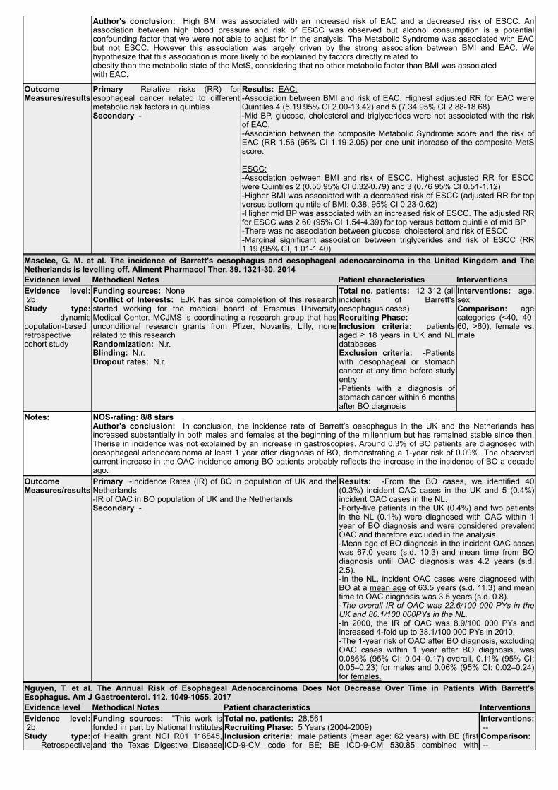
Author's conclusion: High BMI was associated with an increased risk of EAC and a decreased risk of ESCC. Anassociation between high blood pressure and risk of ESCC was observed but alcohol consumption is a potentialconfounding factor that we were not able to adjust for in the analysis. The Metabolic Syndrome was associated with EACbut not ESCC. However this association was largely driven by the strong association between BMI and EAC. Wehypothesize that this association is more likely to be explained by factors directly related toobesity than the metabolic state of the MetS, considering that no other metabolic factor than BMI was associatedwith EAC.
OutcomeMeasures/results
Primary Relative risks (RR) foresophageal cancer related to differentmetabolic risk factors in quintilesSecondary -
Results: EAC:-Association between BMI and risk of EAC. Highest adjusted RR for EAC wereQuintiles 4 (5.19 95% CI 2.00-13.42) and 5 (7.34 95% CI 2.88-18.68)-Mid BP, glucose, cholesterol and triglycerides were not associated with the riskof EAC. -Association between the composite Metabolic Syndrome score and the risk ofEAC (RR 1.56 (95% CI 1.19-2.05) per one unit increase of the composite MetSscore.
ESCC:-Association between BMI and risk of ESCC. Highest adjusted RR for ESCCwere Quintiles 2 (0.50 95% CI 0.32-0.79) and 3 (0.76 95% CI 0.51-1.12)-Higher BMI was associated with a decreased risk of ESCC (adjusted RR for topversus bottom quintile of BMI: 0.38, 95% CI 0.23-0.62)-Higher mid BP was associated with an increased risk of ESCC. The adjusted RRfor ESCC was 2.60 (95% CI 1.54-4.39) for top versus bottom quintile of mid BP-There was no association between glucose, cholesterol and risk of ESCC-Marginal significant association between triglycerides and risk of ESCC (RR1.19 (95% CI, 1.01-1.40)
Masclee, G. M. et al. The incidence of Barrett's oesophagus and oesophageal adenocarcinoma in the United Kingdom and TheNetherlands is levelling off. Aliment Pharmacol Ther. 39. 1321-30. 2014Evidence level Methodical Notes Patient characteristics InterventionsEvidence level: 2bStudy type: dynamicpopulation-basedretrospectivecohort study
Funding sources: NoneConflict of Interests: EJK has since completion of this researchstarted working for the medical board of Erasmus UniversityMedical Center. MCJMS is coordinating a research group that hasunconditional research grants from Pfizer, Novartis, Lilly, nonerelated to this researchRandomization: N.r.Blinding: N.r.Dropout rates: N.r.
Total no. patients: 12 312 (allincidents of Barrett'soesophagus cases)Recruiting Phase: Inclusion criteria: patientsaged ≥ 18 years in UK and NLdatabases Exclusion criteria: -Patientswith oesophageal or stomachcancer at any time before studyentry-Patients with a diagnosis ofstomach cancer within 6 monthsafter BO diagnosis
Interventions: age,sexComparison: agecategories (<40, 40-60, >60), female vs.male
Notes: NOS-rating: 8/8 starsAuthor's conclusion: In conclusion, the incidence rate of Barrett’s oesophagus in the UK and the Netherlands hasincreased substantially in both males and females at the beginning of the millennium but has remained stable since then.Therise in incidence was not explained by an increase in gastroscopies. Around 0.3% of BO patients are diagnosed withoesophageal adenocarcinoma at least 1 year after diagnosis of BO, demonstrating a 1-year risk of 0.09%. The observedcurrent increase in the OAC incidence among BO patients probably reflects the increase in the incidence of BO a decadeago.
OutcomeMeasures/results
Primary -Incidence Rates (IR) of BO in population of UK and theNetherlands-IR of OAC in BO population of UK and the NetherlandsSecondary -
Results: -From the BO cases, we identified 40(0.3%) incident OAC cases in the UK and 5 (0.4%)incident OAC cases in the NL. -Forty-five patients in the UK (0.4%) and two patientsin the NL (0.1%) were diagnosed with OAC within 1year of BO diagnosis and were considered prevalentOAC and therefore excluded in the analysis. -Mean age of BO diagnosis in the incident OAC caseswas 67.0 years (s.d. 10.3) and mean time from BOdiagnosis until OAC diagnosis was 4.2 years (s.d.2.5). -In the NL, incident OAC cases were diagnosed withBO at a mean age of 63.5 years (s.d. 11.3) and meantime to OAC diagnosis was 3.5 years (s.d. 0.8). -The overall IR of OAC was 22.6/100 000 PYs in theUK and 80.1/100 000PYs in the NL. -In 2000, the IR of OAC was 8.9/100 000 PYs andincreased 4-fold up to 38.1/100 000 PYs in 2010. -The 1-year risk of OAC after BO diagnosis, excludingOAC cases within 1 year after BO diagnosis, was0.086% (95% CI: 0.04–0.17) overall, 0.11% (95% CI:0.05–0.23) for males and 0.06% (95% CI: 0.02–0.24)for females.
Nguyen, T. et al. The Annual Risk of Esophageal Adenocarcinoma Does Not Decrease Over Time in Patients With Barrett'sEsophagus. Am J Gastroenterol. 112. 1049-1055. 2017Evidence level Methodical Notes Patient characteristics InterventionsEvidence level: 2bStudy type: Retrospective
Funding sources: "This work isfunded in part by National Institutesof Health grant NCI R01 116845,and the Texas Digestive Disease
Total no. patients: 28,561Recruiting Phase: 5 Years (2004-2009)Inclusion criteria: male patients (mean age: 62 years) with BE (firstICD-9-CM code for BE; BE ICD-9-CM 530.85 combined with
Interventions: --Comparison: --
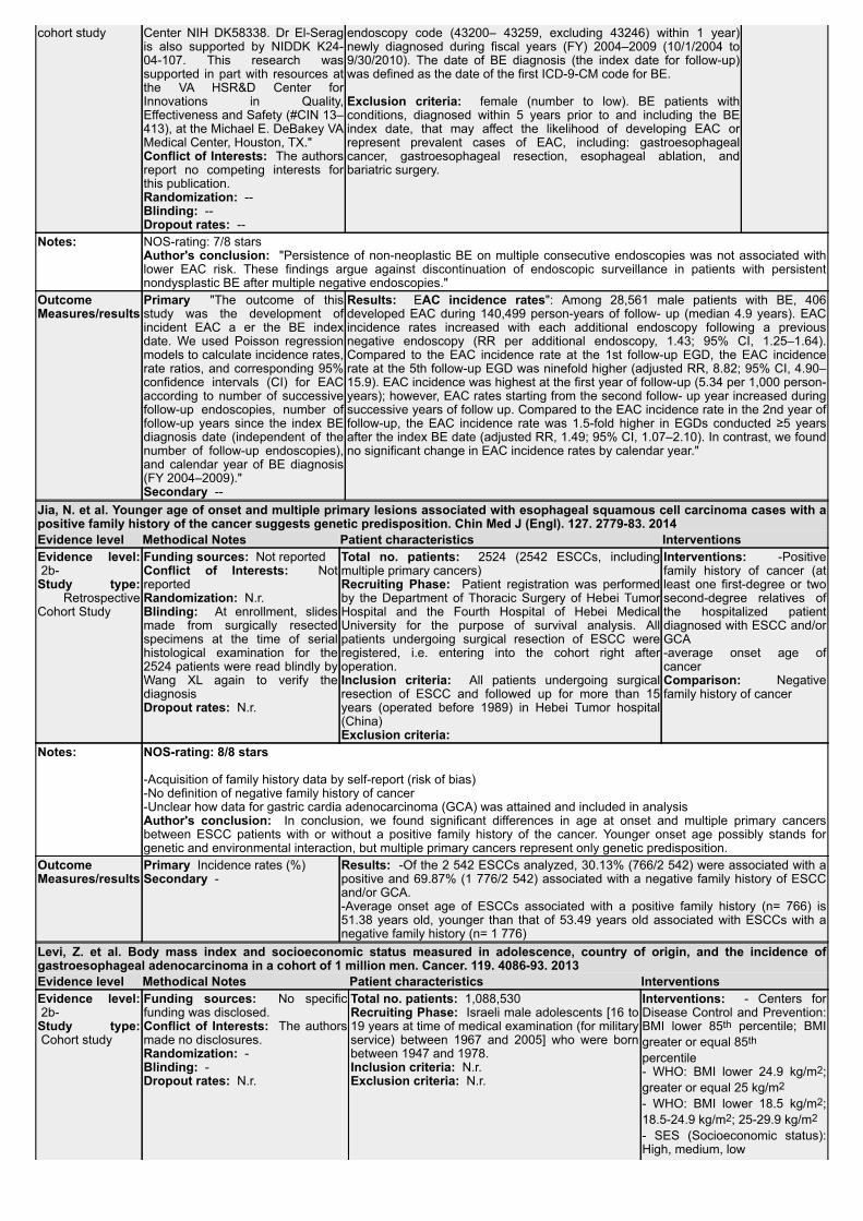
cohort study Center NIH DK58338. Dr El-Seragis also supported by NIDDK K24-04-107. This research wassupported in part with resources atthe VA HSR&D Center forInnovations in Quality,Effectiveness and Safety (#CIN 13–413), at the Michael E. DeBakey VAMedical Center, Houston, TX."Conflict of Interests: The authorsreport no competing interests forthis publication.Randomization: --Blinding: --Dropout rates: --
endoscopy code (43200– 43259, excluding 43246) within 1 year)newly diagnosed during fiscal years (FY) 2004–2009 (10/1/2004 to9/30/2010). The date of BE diagnosis (the index date for follow-up)was defined as the date of the first ICD-9-CM code for BE.
Exclusion criteria: female (number to low). BE patients withconditions, diagnosed within 5 years prior to and including the BEindex date, that may affect the likelihood of developing EAC orrepresent prevalent cases of EAC, including: gastroesophagealcancer, gastroesophageal resection, esophageal ablation, andbariatric surgery.
Notes: NOS-rating: 7/8 starsAuthor's conclusion: "Persistence of non-neoplastic BE on multiple consecutive endoscopies was not associated withlower EAC risk. These findings argue against discontinuation of endoscopic surveillance in patients with persistentnondysplastic BE after multiple negative endoscopies."
OutcomeMeasures/results
Primary "The outcome of thisstudy was the development ofincident EAC a er the BE indexdate. We used Poisson regressionmodels to calculate incidence rates,rate ratios, and corresponding 95%confidence intervals (CI) for EACaccording to number of successivefollow-up endoscopies, number offollow-up years since the index BEdiagnosis date (independent of thenumber of follow-up endoscopies),and calendar year of BE diagnosis(FY 2004–2009)."Secondary --
Results: EAC incidence rates": Among 28,561 male patients with BE, 406developed EAC during 140,499 person-years of follow- up (median 4.9 years). EACincidence rates increased with each additional endoscopy following a previousnegative endoscopy (RR per additional endoscopy, 1.43; 95% CI, 1.25–1.64).Compared to the EAC incidence rate at the 1st follow-up EGD, the EAC incidencerate at the 5th follow-up EGD was ninefold higher (adjusted RR, 8.82; 95% CI, 4.90–15.9). EAC incidence was highest at the first year of follow-up (5.34 per 1,000 person-years); however, EAC rates starting from the second follow- up year increased duringsuccessive years of follow up. Compared to the EAC incidence rate in the 2nd year offollow-up, the EAC incidence rate was 1.5-fold higher in EGDs conducted ≥5 yearsafter the index BE date (adjusted RR, 1.49; 95% CI, 1.07–2.10). In contrast, we foundno significant change in EAC incidence rates by calendar year."
Jia, N. et al. Younger age of onset and multiple primary lesions associated with esophageal squamous cell carcinoma cases with apositive family history of the cancer suggests genetic predisposition. Chin Med J (Engl). 127. 2779-83. 2014Evidence level Methodical Notes Patient characteristics InterventionsEvidence level: 2b-Study type: RetrospectiveCohort Study
Funding sources: Not reportedConflict of Interests: NotreportedRandomization: N.r.Blinding: At enrollment, slidesmade from surgically resectedspecimens at the time of serialhistological examination for the2524 patients were read blindly byWang XL again to verify thediagnosisDropout rates: N.r.
Total no. patients: 2524 (2542 ESCCs, includingmultiple primary cancers)Recruiting Phase: Patient registration was performedby the Department of Thoracic Surgery of Hebei TumorHospital and the Fourth Hospital of Hebei MedicalUniversity for the purpose of survival analysis. Allpatients undergoing surgical resection of ESCC wereregistered, i.e. entering into the cohort right afteroperation.Inclusion criteria: All patients undergoing surgicalresection of ESCC and followed up for more than 15years (operated before 1989) in Hebei Tumor hospital(China)Exclusion criteria:
Interventions: -Positivefamily history of cancer (atleast one first-degree or twosecond-degree relatives ofthe hospitalized patientdiagnosed with ESCC and/orGCA-average onset age ofcancerComparison: Negativefamily history of cancer
Notes: NOS-rating: 8/8 stars
-Acquisition of family history data by self-report (risk of bias)-No definition of negative family history of cancer-Unclear how data for gastric cardia adenocarcinoma (GCA) was attained and included in analysis Author's conclusion: In conclusion, we found significant differences in age at onset and multiple primary cancersbetween ESCC patients with or without a positive family history of the cancer. Younger onset age possibly stands forgenetic and environmental interaction, but multiple primary cancers represent only genetic predisposition.
OutcomeMeasures/results
Primary Incidence rates (%)Secondary -
Results: -Of the 2 542 ESCCs analyzed, 30.13% (766/2 542) were associated with apositive and 69.87% (1 776/2 542) associated with a negative family history of ESCCand/or GCA.-Average onset age of ESCCs associated with a positive family history (n= 766) is51.38 years old, younger than that of 53.49 years old associated with ESCCs with anegative family history (n= 1 776)
Levi, Z. et al. Body mass index and socioeconomic status measured in adolescence, country of origin, and the incidence ofgastroesophageal adenocarcinoma in a cohort of 1 million men. Cancer. 119. 4086-93. 2013Evidence level Methodical Notes Patient characteristics InterventionsEvidence level: 2b-Study type: Cohort study
Funding sources: No specificfunding was disclosed.Conflict of Interests: The authorsmade no disclosures.Randomization: -Blinding: -Dropout rates: N.r.
Total no. patients: 1,088,530 Recruiting Phase: Israeli male adolescents [16 to19 years at time of medical examination (for militaryservice) between 1967 and 2005] who were bornbetween 1947 and 1978. Inclusion criteria: N.r.Exclusion criteria: N.r.
Interventions: - Centers forDisease Control and Prevention:BMI lower 85th percentile; BMIgreater or equal 85th percentile- WHO: BMI lower 24.9 kg/m2;greater or equal 25 kg/m2
- WHO: BMI lower 18.5 kg/m2;18.5-24.9 kg/m2; 25-29.9 kg/m2
- SES (Socioeconomic status):High, medium, low

- Country of birth: Israel, West,Africa, Former Soviet Union,Asia- No. of years of education: 12,11, 10, lower 9Comparison: -
Notes: NOS rating: 5/8 stars
- Confusing separation into EAC and GEJAC group, although previously stated that distinction between both groups isdifficult outside surgical setting (?) - therefore combination of both group by authors. Resulting unclear validity of resultsconcerning separated and combined groups- Unclear validity of SES grouping into low, medium and high- Unclear validity of BMI results due to confounding variable classifications as dichotomous and ordinal - No reporting on why cohort number is once stated as 1,088,530 and once as 1,088,242Author's conclusion: Overweight during adolescence was found to be substantially associated with the subsequentdevelopment of EAC and GEJAC. In addition, although potential confounding by Helicobacter pylori infection status orlifestyle factors was not fully accounted for in the analyses, lower SES as well as immigration from higher-risk countries arecountries are important determinants of NCGC.
OutcomeMeasures/results
Primary Incidence ofgastroesophageal cancer,gastroesophageal junction adenomacarcinoma and noncardia gastriccancer
Secondary - Risk for EAC andGEJAC, NCGC, NCGC intestinaland Mucinous (Multivariable coxproportional Hazard Ratios) - Cumulative Incidence for EAC andGEJAC-group and NCGC
Results: -Association between BMI greater or equal 85th percentile duringadolescence and future adenocarcinoma of the lower esophagus and gastric cardia-Association between adult obesity (especially abdominal obesity) and an increasedrisk of EAC and GEJAC-Lower SES and immigration from higher-risk countries (Asia and former SovietUnion) are important determinants of NCGC
Zakaria, D. et al. Cancers attributable to excess body weight in Canada in 2010. Health Promot Chronic Dis Prev Can. 37. 205-214.2017Evidence level Methodical Notes Patient characteristics InterventionsEvidence level: 2b-Study type: Cohort Study
Funding sources: N.r.Conflict of Interests: The authors declare noconflicts of interest.Randomization: N.r.Blinding: N.r.Dropout rates: N.r.
Total no. patients: N.r.Recruiting Phase: Canadianadults aged 25+ years in2010Inclusion criteria: Canadianadults aged 25+ years in2010Exclusion criteria: N.r.
Interventions: BMI (Overweight:25.00 - 29.99 kg/m2; Obese: 30.00+kg/m2)
Comparison: N.r.
Notes: NOS-rating: 2/8 stars
BMI data is partly based on self-report (bias), partly on adjusted data on a subsample of respondents who agreed to havetheir height and weight measured in addition to providing self-reports. Data was pooled later on.
No report of duration of overweight/obesity - impact on cancer risk
Different sources of cancer case data were merged later on (Canadian Cancer Registry for whole Canada and StatisticsCanada's website especially for Quebec)Cancer case counts for Quebec needed to be adjusted for a few cancers not directly available through Statistics Canada'swebsite.
No report of how BMI and cancer data were linked.
Assumption of no cancer risk for BMI below 25.00 kg/m2 without evidence.Results only applicable on BMI above 25.00kg/m2
Author's conclusion: An estimated 5.7% (1 in 18) of all new cancer cases diagnosed in Canadian adults in 2010 wereattributable to high BMI after correcting for bias in self-reported height and weight.
OutcomeMeasures/results
Primary Not explicitely reported (possibly PAFs ofcancer cases, attributable cases and plausibleranges)Secondary N.r.
Results: 5.7% of all cancer cases, or 9645 cancer cases, diagnosedin Canadian adults in 2010 were attributable to excess body weight.
Esophageal adenocarcinoma: Total in whole Canada N= 435; PAF:41.3 (plausible range: 32.8-51.8)Males in whole Canada N= 380; PAF: 42.2 (34.3-52.6)Females in whole Canada N= 50; PAF: 36.1 (23.6-47.0)
Evidenztabellenzurück
Schlüsselfrage:AG 1 Risikofaktoren: Wird das Ösophaguskarzinomrisiko (SCC AC) durch einen der folgenden Faktoren beeinflusst?
Bewertungsvorlage:NEWCASTLE - OTTAWA Checklist 3: Case Control
Alexandre, L. et al. Statin use is associated with reduced risk of histologic subtypes of esophageal cancer: a nested case-controlanalysis. Gastroenterology. 146. 661-8. 2014Evidence level Methodical Notes Patient characteristics InterventionsEvidence level: 3bStudy type: Case-controlstudy
Funding sources: The MedicalResearch Council provided funding forthis study under a project license. Thefunding source had no input regardingthe design, conduct, or interpretation ofthis study.Conflict of Interests: The authorsdisclose no conflicts.Randomization: N.r.Blinding: Not reportedDropout rates: N.r.
Total no. patients: 1126 cases, 4192 controlsPatient characteristics: EAC: 581 patients withEAC, 2167 controlsEGJA: 213 participants with EGJA, 783 controlsESCC: 332 participants with ESCC, 1242 controlsInclusion criteria: cases: patients with EAC, EGJA,ESCCcontrols: patients without a history of any cancer;according to sex, year of birth, general practice(socioeconomic staus)Exclusion criteria: cases: participants with lessthan 10 months of statin use in the year beforediagnosis
Interventions: -Statinprescription-Statin duration (≥ 1 to < 4years; ≥ 4 to < 6 years; ≥ 6years)Comparison: -No Statinprescription-Statin duration
Notes: NOS-rating: 6/8 stars
-There were too few prescriptions of individual statins to allow meaningful analysisAuthor's conclusion: In a nested case-control analysis of a UK population-based cohort, statin use was inverselyassociated with histologic subtypes of esophageal cancer. Randomized controlled trials are warranted to determinewhether statins have chemo-preventive effects in high-risk groups.
OutcomeMeasures/results
Primary Adjusted Odds Ratios (95%CI) Secondary
Results: EAC:
-Regular statin prescription was inversely associated with EAC (OR = 0.58; 95%CI: 0.390.87; p = .009) and there was evidence of both a dose-response (p fortrend = .036) and duration-response (p for trend = .005) relationship.
EGJA:
-Regular statin prescription was not significantly associated with EGJA (OR =0.60; 95% CI: 0.331.11; p = .102) (Table 2), however, there was evidence of adoseresponse (p for trend = .040) and durationresponse (p for trend = .052) withborderline significance. Only high-dosage regular statin prescriptions weresignificantly inversely associated with EGJA (OR = 0.29; 95% CI: 0.090.92;p =.036).
ESCC:
-Regular statin prescription was non-significantly inversely associated with risk ofESCC (OR = 0.61; 95% CI: 0.351.06; p = .081) with borderline evidence of adoseresponse (p for trend = .057) relationship, and no significantdurationresponse (p for trend = .249). Statin use for between 1 and 4 years wassignificantly inversely associated with ESCC (OR = 0.51 95% CI: 0.270.98; p =.045).
Bhat, G. A. et al. Family history of cancer and the risk of squamous cell carcinoma of oesophagus: a case-control study in Kashmir,India. Br J Cancer. 113. 524-32. 2015Evidence level Methodical Notes Patient characteristics InterventionsEvidence level: 3bStudy type: Case-controlstudy
Funding sources: This study wasfinancially supported by Extramuralgrant of Indian Council of MedicalResearch (ICMR), New DelhiConflict of Interests: The authorsdeclare no conflict of interestRandomization: Not relevantBlinding: Not relevantDropout rates: Not relevant
Total no. patients: 2367 (703 ESCCcases and 1664 controls withoutESCC)Patient characteristics:
SDRs: cousins, uncles, aunts,stepsiblingsInclusion criteria: cases: -histopathologically confirmed ESCC -age above 18 years -no personal history of cancercontrols:-hospital-based-matched for sex, age (± 5 years),place of residenceExclusion criteria: controls: diseasewith relation to tobacco or alcohol useor affection of dietary habits of thepatient (e.g. diabetes)
Interventions: -Family History of Cancer[FHC: FDRs= Parents, siblings andchildren; Second-degree relatives=cousins, uncles, aunts, stepsiblings]
Comparison: No FHC, FDRs, SCRs
Notes: NOS-rating: 5/8 stars

-possible source of bias regarding self-reported information of family history data
Author's conclusion: Our results showed that FHC was strongly associated with ESCC risk in Kashmir. It seems bothgenetic factors and shared environment are involved in this association.
OutcomeMeasures/results
Primary ESCC risk (Adjusted OddsRatio)Secondary gene polymorphisms(Adjusted Odds Ratio)
Results: -A strong increase in ESCC risk was observed in subjects who had FHC(OR=5.8; 95% CI= 4.1-8.3)-The risk was stronger when first-degree relatives (FDRs) had FHC (OR=6.8; 95%CI= 4.6-9.9)-Having a sibling with a cancer showed the strongest association (OR=10.8; 95%CI= 6.0-19.3)-A history of any cancer in the spouse was associated with ESCC risk (OR=4.1;95% CI= 1.6-20.2)-Having a child with a cancer was not associated with ESCC risk
Busby, J. et al. The effect of medications which cause inflammation of the gastro-oesophageal tract on cancer risk: a nested case-control study of routine Scottish data. Int J Cancer. 140. 1828-1835. 2017Evidence level Methodical Notes Patient characteristics InterventionsEvidence level: 3bStudy type: Nested case-control study
Funding sources: Not reportedConflict of Interests: Not reportedRandomization: N.r.Blinding: Not reportedDropout rates: Not reported
Total no. patients: 3,098 cases, 14 870 controlsPatient characteristics: Between 1993 and 2011, the PCCIURcollected computerised medical records from around 15% of theScottish general practice population, and includes details onpatient demographics, clinical diagnoses and prescriptions. Inclusion criteria: cases: patients with a first-time oesophageal(Read code: B10.) or gastric (Read code: B11.) cancer diagnosisafter January 1, 1999 and before April 30, 2011. controls: matched on age, gender, year of diagnosis and generalpracticeExclusion criteria: -cases and controls with an earlier cancerdiagnosis (other than non-melanoma skin cancer) and those withless than three years of exposure prior to index date-prescriptions before January 1, 1996 and those in the year priorto index date
Interventions: medication useComparison: never, ever,lower usage,higher usage ofmedication
Notes: NOS-rating: 6/8 stars
Author's conclusion: Overall, there is little evidence that the use of biphosphonate, tetracycline or spironolactone isassociated with increased risk of gastro-oesophageal cancer. Our findings should reassure GPs and patients that thesewidely-used medications are safe with respect to gastro-oesophageal cancer risk.
OutcomeMeasures/results
Primary Odds Ratio (OR) for theassociation between medication use(Biphosphonate, Tetracycline,Spironolactone) and osesophagealcancer risk Secondary -
Results: -There was evidence of a 34% increased risk (ORadj = 1.34; 95% CI:1.03, 1.74) of oesophageal cancer in bisphosphonate users-The association between bisphosphonate use and oesophageal or gastric cancerdid not appear to follow a dose–response relationship.
-little associations were observed between tetracycline use and oesophageal(ORadj = 1.01; 95% CI: 0.82, 1.25)
-little evidence of higher risk for oesophageal cancer alone in spironolactone users,with adjusted odds ratios of 1.04 (95% CI: 0.68, 1.61)
Chen, T. et al. Family history of esophageal cancer increases the risk of esophageal squamous cell carcinoma. Sci Rep. 5. 16038.2015Evidence level Methodical Notes Patient characteristics InterventionsEvidence level: 3bStudy type: population-basedcase-control study
Funding sources: National Natural ScienceFoundation of China, Key Projects in the NationalScience & Technology Pillar Program, Key Scientificand Technological Projects of Shandong ProvinceConflict of Interests: The authors declare nocompeting financial interests.Randomization: random selection of populationcontrolsBlinding: Not reportetDropout rates: Not relevant
Total no. patients: 619 esophageal cancercases (648 cases of ESCC, 63 cases ofesophageal adenocarcinoma, 7 cases of othertypes of esophageal cancer)772 controlsPatient characteristics: local inhabitantsaged 40-85 who have lived in Taixing for atleast 5 years prior to diagnosis date for casesor interview date for controls-Interviews with study subjects face-to-faceusing a structured questionnaire, which coversinformation on demographic characteristics,lifestyles and family history of cancer. Inclusion criteria: -cases: ESCC cases inTaixing of Jiangsu Province from 10.2010-03.2012.-controls: population controls which werefrequency matched to the cases of ESCC onsex and age (in 5-year groups)Exclusion criteria: -incompletequestionnaire information on family historycancer
Interventions: Family history ofcancer (First-degreerelatives, parents,siblings)Comparison: nofamily history ofcancer (First-degreerelatives, parents,siblings)
Notes: NOS-rating: 5/8 stars
-no mentioning of exclusion criteria of cases, untransparent description of case recruitment-review of section performed only by one study pathologist (risk of bias) Author's conclusion: Our results indicate that familial aggregation of ESCC in endemic area is notable. The sharedgenetic susceptibility and environmental exposures, or possibility their interaction, might contribute to this phenomenonwhich urges future studies to explore the underlying mechanisms.
Outcome Primary Risk of ESCC (adjusted Odds Ratio) Results: -excess risks of ESCC increased monotonically with the
Measures/results Secondary - increasing number of first-degree relatives reportedly afflicted withesophageal cancer-individuals whose both parents were diagnosed with esophagealcancer had an 8-fold excess risk of ESCC, compared with thosewithout any parents affected by esophageal cancer (adjustedOR=7.96, 95% CI: 1.74-36.32)-increasing number of affected siblings did not seem to furtherincrease the relative risks-excess ESCC risks were associated with a positive family history ofany cancer (Adjusted OR=1.43, 95% CI:1.13-1.81) or digestive tractcancer (adjusted OR=1.55, 95% CI: 1.23-1.96)
Cooper, S. et al. Risk factors for the development of oesophageal adenocarcinoma in Barrett's oesophagus: a UK primary careretrospective nested case-control study. United European Gastroenterol J. 2. 91-8. 2014Evidence level Methodical Notes Patient characteristics InterventionsEvidence level: 3bStudy type: Nested-casecontrol study
Funding sources: ‘The Upper GIBlues’, CSDMedical ResearchUKConflict ofInterests: Theauthors declarethat there is noconflict of interest.Randomization: N.r.Blinding: N.r.Dropout rates: N.r.
Total no. patients: 3749Patient characteristics: BO subjects were identified from TheHealth Improvement Network (THIN) database. THIN databasecontains computerized and anonymized longitudinal recordsfrom 326 UK general practice (GP) surgeries, covering 5 millionpatients that are regionally and demographically representativeof the UK populationInclusion criteria: BO subjects (data record period: 1988-2004)with a minimum of 1 year of follow up, and when applicable, aminimum of 1 year between diagnosis of BO and OC cases: Subjects developing OC (oesphageal cancer) controls: Subjects who did not develop OCExclusion criteria: Cases proven to be squamous cellcarcinoma
Interventions: age, gender,smoking, body mass index,medication (aspirin/nonsteroidal anti-inflammatory drugs/proton pumpinhibitors, lower oesophagealsphincterrelaxing and asthma drugs)Comparison: -male:female-ever smoking: never smoking-high BMI: mid BMI: low BMI-medication quintiles
Notes: NOS-rating: 6/8 stars
-It cannot be guaranteed that medication is dispensed or taken by the patient. In some cases (e.g. b-agonist inhalers),multiple devices may be obtained but not used.-overthe-counter medication and drugs prescribed at other institutions will not be recorded.Author's conclusion: Progression to OAC from BO is more common among men and with increasing age. There is someevidence of smoking being associated with progression to OAC but this association was not significant on multivariateanalysis. LOS-relaxing drugs do not appear to be associated with OAC development once drugs for asthma are excluded.The association of inhaled steroids with OAC development strongly suggests that it is the pathophysiolology ofasthma/chronic asthma or the severity of gastro oesophageal reflux necessary to cause asthma, rather the drugsthemselves that are associated with progression to OAC.
OutcomeMeasures/results
Primary -HazardRatios of risk ofdevelopingoesophagealadenocarcinomafrom Barrett'soesphagusSecondary -
Results: -Male gender was associated with progression to OAC (HR 3.06, 95% CI 1.50–6.24, p =0.002), with 84% of those developing OAC compared with 63% of those remaining with BO. -Increasing age (HR (for each year: 1.03, 95% CI 1.01–1.05, p = 0.005) was associated withdeveloping OAC, with a median age of 67 years (Interquartile range IQR 59–73 years) among thosedeveloping OC, compared with a median age of 63 years (IQR 52–72 years) among those who did notprogress.-Having smoked doubled the risk for progression to OAC on univariate analysis (HR 2.36, 95% CI1.13– 4.93, p = 0.023), but there was no significant association when corrected for age and gender(HR 1.99, 95% CI 0.94–4.19, p = 0.07).-There was no association between increasing BMI and progression to OC on univariate andmultivariate analyses.-No association was seen when analysed by categorizing BMI 25 kg/m2, overweight (BMI 25.1–30kg/m2), and obese (BMI >30 kg/m2)
-No association was seen between developing OAC and the following drug classes: aspirin, NSAIDs,COX-2 inhibitors, and statins. There was also no association with iron preparations, anticholinergics,ACE-I, calcium-channel antagonists, tricyclic antidepressants, benzodiazepines, or nicorandil -The use of both inhaled steroids (HR 2.11, 95% CI 1.12–3.97, p = 0.021) and steroid and b-agonistcombination inhalers (HR 2.54, 95% CI 1.17–5.51, p = 0.018) was associated with progression to OACon both univariate and multivariate analysis-Increasing number of drugs used for asthma showed an increasing association with progression toOAC (HR 2.91, 95% CI 1.10–7.68, p = 0.031 for the use of all three examined drugs) followingcorrection for age, gender, and smoking status
Hvid-Jensen, F. et al. Proton pump inhibitor use may not prevent high-grade dysplasia and oesophageal adenocarcinoma inBarrett's oesophagus: a nationwide study of 9883 patients. Aliment Pharmacol Ther. 39. 984-91. 2014Evidence level Methodical Notes Patient characteristics InterventionsEvidence level: 3bStudy type: Nested case-control study
Funding sources: Institute of ClinicalMedicine, Aarhus University Hospital,DenmarkConflict of Interests: NoneRandomization: N.r.Blinding: N.r.Dropout rates: N.r.
Total no. patients: 9,883 Patient characteristics: Inclusion criteria: all: All patients with newdiagnosis of BO from 1995 to 2009 in Denmark
cases: Patients with HGD or OACcontrols: no diagnosis of HGD or OAC before thediagnosis date of the patient, matched according tobirth date and date of BO Exclusion criteria: -Patients with a diagnosis ofHGD or OAC, made before or up to 1 year after thediagnosis of BO
Interventions: everusers of PPI (more than 2prescriptions)Comparison: never/rareusers of PPI (less than 2prescriptions)
Notes: NOS-rating: 7/8 stars
-no data regarding patient's actual compliance to PPI'sAuthor's conclusion: No cancer-protective effects from PPI’s were seen. In fact, high-adherence and long-term use ofPPI were associated with a significantly increased risk of adenocarcinoma or high-grade dysplasia. This could partly bedue to confounding by indication or a true negative effect from PPIs. Until the results from future studies hopefully canelucidate the association further, continuous PPI therapy should be directed at symptom control and additional modalitiesconsidered as aid or replacement.
OutcomeMeasures/results
Primary Odds Ratios (ORs) as a measureof the relative risks (RR) of oesophagealadenocarcinoma and high grade dysplasiaSecondary -
Results: -Relative risk of OAC or HGD among BO patients using PPIcompared to never/rare users, was 1.1 (95% CI: 0.4–3.3) in former PPI users,1.9 (95% CI: 0.7–4.9) in ever users and 2.1 (95% CI: 0.8–5.6) in recent users.-Long-term PPI use yielded a relative risk of OAC or HGD of 2.2 (95% CI: 0.7–6.7) in the low-adherence group and 3.4 (95% CI: 1.1–10.5) in high-adherenceusers.
Moura, M. A. et al. The magnitude of the association between smoking and the risk of developing cancer in Brazil: a multicenterstudy. BMJ Open. 4. e003736. 2014Evidence level Methodical Notes Patient characteristics InterventionsEvidence level: 3bStudy type: case-controlstudy
Funding sources: This researchreceived no specific grant from anyfunding agency in the public,commercial or not-for-profit sectors.Conflict of Interests: NoneRandomization: N.r.Blinding: N.r.Dropout rates: N.r.
Total no. patients: 231 102Patient characteristics: 204 131 cancer cases, 26 971 controlsInclusion criteria: -patients with initial diagnosis of 30 differentcancer types including oesophageal cancer, diagnosed between1998 and 2011 and seen in 168 reference centres for cancertreatment, in 24 Brazilian states-controls: patients with non-melanoma skin cancerExclusion criteria: -patients younger than 18 years and olderthan 100 years-patients with no information on gender and smoking
Interventions: gender,smokingComparison: female vs.male, smokingyes vs. no
Notes: NOS-rating: 5/8 starsAuthor's conclusion: This study confirms a high risk of developing cancer of the hypopharynx, bronchi and lung, larynx,oropharynx and oral cavity, oesophagus and bladder cancer among smokers and establishes the AF attributable tosmoking in the development of different types of cancer in Brazil.
OutcomeMeasures/results
Primary Odds Ratio (OR) ofassociation of risk between tobaccoconsumption and cancer developmentSecondary Attributable Fractions (AF)referring to cancer sites for bothgenders
Results: -Tobacco was classified as a strong risk factor for cancers of theoesophagus (adjusted OR = 4.0 (95% CI 3.7-4.2))-THE AF results referring to cancer sites for both genders was 58.7% foroesophageal cancer
Pottegard, A. et al. Use of benzodiazepines or benzodiazepine related drugs and the risk of cancer: a population-based case-controlstudy. Br J Clin Pharmacol. 75. 1356-64. 2013Evidence level Methodical Notes Patient characteristics InterventionsEvidence level: 3bStudy type: population-basedcase-control study
Funding sources: Not reported.Conflict of Interests: All authors have completed the UnifiedCompeting Interest form athttp://www.icmje.org/coi_disclosure.pdf (availableon request from the corresponding author) and declare MAand JH have participated in research projects funded byNycomed, the manufacturer of nitrazepam, and Pfizer, themanufacturer of Halcion (triazolam) and Tafil (alprazolam), withgrants paid to institutions where they have been employed. JHhas personally received fees for teaching from Nycomed. APand SF declare no conflicts of interest.Randomization: N.r.Blinding: N.r.Dropout rates: N.r.
Total no. patients: 149 360cases, 1 194 729 controlsPatient characteristics: Patients registered in TheDanish Cancer Registry Inclusion criteria: -All Danishresidents alive on January 2002-Lived in Denmark continuouslyfrom 1995 to the index date-No history of any cancer(except non-melanoma skincancer) prior to the index dateExclusion criteria: -Personswho redeemed a prescription forany anxiolytic, hypnotic orsedative (ATC-codes, N05B andN05C) during the first 2 runningyears of the prescriptiondatabase, i.e. 1995 and 1996
Interventions: Ever useand long term use ofBZRD (cumulativeamount of BZRD equalto/greater than 500 DDDwithin a period of 5 to 1year prior to the indexdate)BZRD: Benzodiazepinesor benzodiazepinerelated drugsComparison: No use ofBZRD
Notes: NOS-rating: 7/8 starsAuthor's conclusion: In conclusion, our findings do not support a carcinogenic effect of BZRD. Most ORs were close tounity, except a few that seemingly can be explained by lifestyle confounding. We also found that the recently reportedexcess of cancers among BZRD users can be explained entirely by a flawed design. For other reasons thancarcinogenesis, however, use of BZRD should generally be avoided, or reserved for short term use in select patientgroups.
OutcomeMeasures/results
Primary Odds Ratio (OR) for cancer associated with use ofBZRDSecondary -
Results: Association between long term exposure toBZRD and oesophageal cancer risk: Adjusted OR = 1.43(95% CI: 1.01 - 2.02)
Sewram, V. et al. Tobacco and alcohol as risk factors for oesophageal cancer in a high incidence area in South Africa. CancerEpidemiol. 41. 113-21. 2016
Evidence level Methodical Notes Patientcharacteristics Interventions
Evidence level: 3bStudy type: hospital-basedCase-ControlStudy
Funding sources: South AfricanMedical ResearchCouncil, TheRockefellerFoundation, CancerCouncil NSW andUICC are
Total no. patients: 670 cases; 1188controlsPatientcharacteristics: Inclusion criteria: CASES-All patients with
Interventions: Tobacco use (Smoking status: never vs. ever; Commercialcigarettes: never vs. ever; No. of cigarettes per day: Never vs. 1-4; Hand-rolled cigarettes: Never vs. ever; No. of hand-rolled cigarettes per day: Nevervs. 1-3, 4-6, 7+; Pipe: Never vs. ever; No. of pipes per day: Never vs. 1-3, 4-6, 7+; Total Tobacco (grams per day/All smokers): Never vs. 1-7, 7.1-14, 14.5)
Alcohol consumption (Alcohol consumption: Never vs. ever; Maize beer(consumption per week: Never, ≤ 1 day, 2-4 days, 5-7 days); Quantity of
acknowledged fortheir financialsupport of this study.Conflict ofInterests: Theauthors declare thatthey have no conflictof interest.Randomization: N.r.Blinding: N.r.Dropout rates: N.r.
incidenthistopathologically,radiologically orendoscopicallyconfirmed squamouscell carcinoma of theoesophagus betweenNovember 2001 andFebruary 2003, SouthAfrica-sufficient goodphysical and mentalhealth-Patients lived in theEastern CapeProvince for at least 5years prior todiagnosis
CONTROLS-diseases/conditionsnot related tosmoking, alcoholconsumption or dietExclusion criteria: -
Maize beer per week (Litres): Never, ≤ 1 vs. 1.01-3, 3.01+; Sorghum beer:Never vs. ≤ 1 day, 2-4 days, 5-7 days; Quantity Sorghum beer peer week(Litres): Never vs. ≤ 1, 1.1-3, >3; Commercial beer: Never vs. ≤ 1 day, 1.01-2,>2; Home-made spirits: Never vs. ever; Commercial spirits: Never vs. ≤ 1 day,2-4 days, 5-7 days; Quantity commercial spirits consumed per week (Litres):Never vs. 0.025-0.1, 0.11+; Wine: Never vs. ≤ 1 day, 2+ days; Quantity wineconsumed per week (Litres): Never vs. 0.1-1, >1Comparison: see "interventions"
Notes: NOS-rating: 4/8 stars
-interview was not blinded to case/control status (risk of bias)Author's conclusion: Our study shows that 58% and 48% of oesophageal cancers were attributed to smoking andalcohol consumption respectively, therefore a substantial health benefit could be expected by efforts to reduce theprevalence of smoking and drinking. Recent data suggest that only after at least 10 years of abstaining from drinking doesthe risk of oesophageal cancer return to being within the risk levels for abstainers and that stopping smoking for 5 yearscuts the risk by 50%. After 10 or more years since stopping both habits the relative risk is about one-tenth of that of currentsmokers and drinkers, but local data on this effect are unavailable.
OutcomeMeasures/results
Primary AdjustedOdds Ratio (OR) forrisk of developingoesophageal cancerSecondary Populationattributable fractions(PAFs)
Results: Tobacco use:
-Males: ever smokers (70%) had 4-fold increased odds compared to never smokers (OR = 4.11, 95%CI 2.55–6.65). -Females: ever smokers had approximately 3.5-fold increased odds (OR = 3.45, 95% CI 2.47–4.82)compared with nonsmokers.-Male commercial smokers: 78% indicated smoking commercial cigarettes with ever smokers havingalmost 40% greater odds of developing OC (OR = 1.39, 95% CI 1.01–1.92). -Males smoking hand-rolled cigarettes (70%) and pipe smoking (64%): Those reporting havingsmoked 7 or more hand-rolled cigarettes per day had 4.4-times greater odds of developing OC (OR= 4.40, 95% CI 2.35–8.24), whilst those smoking 7 or more pipes per day had a 7.72 times increasedodds compared to non-smokers (95% CI 3.99–14.92). -Amongst the female smokers, 43% indicated having smoked commercial cigarettes. Femaleshaving smoked 7 or more hand-rolled cigarettes per day had 3-times greater odds of developing OC(OR = 3.14, 95% CI 1.09–9.07), whilst those smoking 7 or more pipes per day had almost 6-foldincreased odds compared to nonsmokers (OR = 5.63, 95% CI 2.05–15.43). -Males and females smoking more than 14 g of tobacco per day had approximately 6-times greaterodds of developing OC compared to non-smokers (Male OR = 6.27, 95% CI 3.74–10.52, female OR= 5.60, 95% CI 3.23–9.73).
Alcohol use:
-Male ever drinkers had a 3.5-fold increased odds of OC (OR = 3.48, 95% CI 1.99–6.06) andfemales had 2-fold increased odds (OR = 2.23, 95% CI 1.60–3.11) compared to nondrinkers.-Males and females consuming maize beer 2–4 days per week had 4-fold increased odds comparedto non-drinkers (malesOR = 4.04, 95% CI 2.19–7.46; females OR = 4.29, 95% CI 2.49–7.37)-Risk increased with the quantity of each beverage type consumed with ORs ranging between 4.00and 5.50 for the highest quantity category, the exception being for females consuming more than 1litre of wine per week who had 7 times greater odds of developing OC (OR = 7.10, 95% CI 3.39–14.87). -Total ethanol consumption (representing the sum of the averages of grams of ethanol from each ofbeer, spirits and wine) was positively associated with OC risk with male drinkers consuming morethan 52.8 g per day having almost 5-times the odds of developing OC (OR = 4.72, 95% CI 2.64–8.41) than non-drinkers. -Female drinkers, 5-fold increased odds was observed for those consuming more than 52.8 g ofethanol per day (OR = 5.24, 95% CI 3.34–8.23). -Lower estimated ORs were observed for lower alcohol consumption.
Joint effects:
-Those using more than 14 g of tobacco/day and consuming more than 371 g ethanol/week had8.45-fold increased odds of developing oesophageal cancer (95% CI 5.51–12.96) compared to thosewho are both non-smokers and non-drinkers.
PAFs:
-The attributed fraction for both exposures (alcohol and tobacco) combined was 64%

Vinogradova, Y. et al. Exposure to bisphosphonates and risk of gastrointestinal cancers: series of nested case-control studies withQResearch and CPRD data. Bmj. 346. f114. 2013Evidence level Methodical Notes Patient characteristics InterventionsEvidence level: 3bStudy type: Series of nestedcase-controlstudies
Funding sources: This work was funded by thedivision of primary care of University of Nottingham.Conflict of Interests: All authors have completed theUnified Competing Interest fromwww.icmje.org/coi_disclosure.pdf (available on requestfrom the corresponding author) and declare no supportfrom any additional organisation for the submitted work. Randomization: Not relevantBlinding: Not relevantDropout rates: Not relevant
Total no. patients: QResearch database:5364 cases, 25 101 controlsCPRD database: 5132 cases, 24 053controls
Total: 59 650Patient characteristics: Patients agedover or equal 50 with a diagnosis of primarygastrointestinal cancer in 1997-2011, eachmatched with up to five controls by age,sex, practice and calendar year Inclusion criteria: -open cohort forpatients aged over or equal 50 years andregistered with the practice at some timeduring the study period (January 1997 toJuly 2011)-gastrointestinal cancers (oesophageal,gastric colorectal)-at least two years of data before their indexdate to ensure the completeness of recordsExclusion criteria: -patients aged lower50 years -cases and controls with prescriptions forbisphosohonates licensed for anymalignancies before the index date. -patients with Paget's disease
Interventions: exposure tobiphosphonates(alendronate,etidronate,ibandronate,risedronate)Comparison: Noexposure tobiphosphonates(alendronate,etidronate,ibandronate,risedronate)
Notes: NOS-rating: 5/8 stars
-selection of cases was based on the first record of a cancer while the exact origin site might have been determined onlylater-no data available on adherence to treatmentAuthor's conclusion: In this series of population based case-control studies in two large primary care databases,exposure to biphosphonates was not associated with an increased risk of common gastrointestinal cancers.
OutcomeMeasures/results
Primary Odds ratios for incident gastrointestinalcancers (colorectal, oesophageal, gastric) and use ofbiphosphonates, adjusted for smoking status, ethnicity,comorbidities, and use of other drugs. Secondary -
Results: -5135 cases of oesophageal cancer cases wereidentified from QResearch and CPRD.-Overall biphosphonate use was not associated with risk ofoesophageal in either database. Adjusted odds ratio (95% CI) forQResearch and CPRD were 0.97 (95% CI; 0.79-1.18) and 1.18(95% CI; 0.97-1.43) for oesophageal cancer-There were no significant associations for individual types ofbisphosphonate
Agrawal, S. et al. Metformin use and the risk of esophageal cancer in Barrett esophagus. South Med J. 107. 774-9. 2014Evidence level Methodical Notes Patient characteristics InterventionsEvidence level: 3b-Study type: Retrospectivecase-control study
Funding sources: Theauthors have no financialrelationships to discloseConflict of Interests: Theauthors have no conflicts ofinterest to reportRandomization: n.r.Blinding: n.r.Dropout rates: n.r.
Total no. patients: 583Patient characteristics: Veterans (Militaryveteran's hospital) 115 EAC, 468 BE, 98% men,96% whiteInclusion criteria: All patients at Militaryveteran's hospital (U.S.) with diagnoses of BEand EAC between 1992 and 2012Exclusion criteria: All patients with histologicaldiagnosis of esophageal squamous cellcarcinoma
Interventions: medication (metformin,statin, aspirin, proton pump inhibitor),age, BMI, alcohol use, Comparison: no use of medication,BMI categories (<25.00, 25-29.99, ≥30),no alcohol use
Notes: NOS-rating: 5/8 stars
-no association analysis was conducted concerning metformin use and risk of EAC -no collection of data regarding duration and dosage of metformin use-risk of recall bias due to data collection via chart review-participants not representative of average population: very specific cases and controls regarding demographic data (98%men, 96% white)Author's conclusion: The three independent variables that predicted progression of Barrett esophagus to esophagealadenocarcinoma in our study were older age, smoking and diabetes mellitus. Statin use showed protective effect againstdevelopment of esophageal adenocarcinoma. Metformin use did not demonstrate any statistically significant protectiveeffect.
OutcomeMeasures/results
Primary Odds Ratios (OR) ofrisk of developing EAC Secondary -
Results: -No significant difference in metformin use in patients with EAC and BE
-Age (OR 1.04; 95% CI 1.02-1.07), smoking (OR 2.27; 95% CI 1.28-4.02), diabetes mellitus(OR 2.15; 95% CI 1.27-3.64) were significant risk factors for the development of EAC-Statin use was protective against the development of cancer (OR 0.46; 95% CI 0.28-0.75)
Rafiq, R. et al. Secondhand Smoking and the Risk of Esophageal Squamous Cell Carcinoma in a High Incidence Region, Kashmir,India: A Case-control-observational Study. Medicine (Baltimore). 95. e2340. 2016Evidence level Methodical Notes Patient characteristics InterventionsEvidence level: 3b-Study type: Case-control
Funding sources: This study wassupported by Extramural Grant ofIndian Council of Medical Research(ICMR), New Delhi. Rumaisa Rafiq
Total no. patients: 703 ESCC patients, 1664 controlsPatient characteristics: Inclusion criteria: -cases: ESCC patients from Regional CancerCentre and Department of Radiation Oncology of Sher-i-Kashmir
Interventions: weekly exposureto secondhandsmoking
study was supported by Department ofScience and Technology (DST), NewDelhi Conflict of Interests: The authorshave no conflicts of interest todiscloseRandomization: N.r.Blinding: Not reportedDropout rates: N.r.
Institute of Medical Sciences (SKIMS) from September 2008 toJanuary 2012-controls: SKIMS, Government Medical College Hospital, and 10district hospitals of Kashmir. Matched for cases regarding sex,age, and district of residence. Exclusion criteria: -cases: without history of previous cancer-controls:Disease for which they had been admitted did not havea strong association with tobacco or alcohol consumption
Comparison: no(never) exposureto secondhandsmoking
Notes: NOS-rating: 5/8 stars
-unclear if interviews were blinded to case/control status-control group consisted of patients when disease for which they had been admitted did not have a strong association withtobacco/alcohol consumption → ESCC patients not explicitly excluded-in group of tobacco consumers, patients who chew tobacco were also included (cultural specificity)Author's conclusion: Our findings indicate increased risk of ESCC due to SHS exposure in dose-dependent manner.Our results may help to increase the awareness about harms of SHS, particularly in developing populations where tobaccouse is on rise and ESCC incidence is high. However, more studies with a larger sample size are required before makingany conclusion on the association between SHS and ESCC risk.
OutcomeMeasures/results
Primary Odds Ratios (95% CI) forrisk of ESCC development Secondary
Results: -Secondhand smoking (SHS) in the unadjusted model increased ESCCrisk (OR = 1.64; 95% CI, 1.14– 2.36); however, the association was attenuated andthe 95% CI included unity (OR = 1.23; 95% CI, 0.72–2.11) in the models adjusted fortobacco smoking and chewing and other potential confounding factors.The OR (95%CI) for the association between weekly exposure to secondhand smoke for >14 hand ESCC risk, compared to no exposure, was (OR = 1.91; 95% CI, 0.75–4.89)-When analysis was limited to never tobacco users (never smokers and neverchewers) the OR (95% CI) for the association between SHS and ESCC risk, inadjusted model, was (OR = 1.32; 95% CI, 0.43–4.02) (Table 2). The OR increasedwith a higher exposure (OR = 2.69; 95% CI, 0.75– 20.65) for SHS >14 h a weekversus no exposure

Inhaltsverzeichnis der Evidenztabellen
Schlüsselfrage:
AG 2 Diagnostik: Stellenwert endoskopischer Spezialuntersuchungen (z.B. Chromoendoskopie, NBI, Zoom etc.)(Primärdiagnostik)
Citation EvidenceLevel Study Type
Qumseya, B. J.2013 2a Syst REview, Meta Analysis, 14 studies (11 RCTs) (n=843)
Sharma, P. 2013 1b RCT - cross over within 3-8 weeksCanto, M. I. 2014 1a RCTGupta, A. 2014 2a Systematic REview, Meta Analysis, 8 studies, n= 345 patients, n=3080 lesions
Fugazza, A. 2016 2a Systematic REview, Meta Analysis, 102 studies (prospective, retrospective clinical studies)n=6943, 16countries.
Chung, C. S. 2016 2a Systematic review and meta-analysis (n= 4918 patients from 16 prospective and randomized trials)Coletta, M. 2016 2a Meta Analysis, 13 prospective studies (n= 1690)

Evidenztabellenzurück
Schlüsselfrage:AG 2 Diagnostik: Stellenwert endoskopischer Spezialuntersuchungen (z.B. Chromoendoskopie, NBI, Zoom etc.) (Primärdiagnostik)
Bewertungsvorlage:OXFORD Appraisal Sheet 1: Systematic Reviews
Chung, C. S. et al. Image-enhanced endoscopy for detection of second primary neoplasm in patients with esophageal and head andneck cancer: A systematic review and meta-analysis. Head Neck. 38 Suppl 1. E2343-9. 2016Evidencelevel/StudyTypes
Population Outcomes/Results LiteratureReferences
MethodicalNotes
Evidence level: 2aStudy type: Systematicreview and meta-analysis (n= 4918patients from 16prospective andrandomized trials)Databases: PubMed andCochrane LibrarySearch period: January 1, 1990,to May 1, 2014.InclusionCriteria: (1)studies onhumans and inthe Englishlanguage; (2) randomizedcontrolled trials,prospectivestudies, or cohortstudies; (3) outcomemeasurement,includingsensitivity,specificity, oraccuracy; (4) availability ofadequate data foranalysis.
ExclusionCriteria: Studiesof Barrett’sesophagus,adenocarcinoma,Studies withoutraw data Studies primarilydesigned as casereports, reviews,or retrospectivestudies
Intervention: Patientswith esophageal (n=2205)and head and neck(n=1781) cancer. Image-enhanced endoscopy fordetection of secondprimary neoplasm. Comparison: White-lightimaging (WLI), narrowband imaging (NBI), andLugol chromoendoscopy
Primary: detection of second primary neoplasmSecondary: ---Results: WLI, NBI, and Lugol chromoendoscopypooled sensitivity 0.53 (95% CI = 0.48–0.59; chi-square = 30.00; p = .0016; I2 =63.3%),0.87 (95% CI = 0.83–0.90; chi-square = 113.02; p < .0001; I2 =90.3%),0.88 (95% CI = 0.85–0.91; chisquare = 15.61; p = .0484; I2 =48.7%), pooled specificity 0.99 (95% CI = 0.98–0.99; chisquare = 108.59; p < .0001; I2 =89.9%), 0.95 (95% CI = 0.94–0.96, chi-square = 138.11; p < .0001; I2=92.0%), 0.63 (95% CI = 0.61–0.66, chi-square = 105.01; p < .0001; I2 =92.4%).
the areas under the receiver-operating characteristic (ROC)curve were 66%, 97%, and 82%.NBI endoscopy has the most highly accurate diagnosticperformance for detection of second primary neoplasms in high-risk patients.Lugol chromoendoscopy was never used in evaluation of a headand neck second primary neoplasm. The sensitivity of NBI inscreening the esophagus (0.97%; 95% CI = 0.95–0.99) wassuperior to head and neck second primary neoplasms (0.61%;95% CI = 0.51–0.70).
Author's Conclusion: In this systematic review and meta-analysis of 16 studies consisting of 4918 patients withesophageal cancers, head and neck cancers, and high-riskendoscopy for the detection of second primary neoplasm. Ourresult highlights the high diagnostic performance of NBIendoscopy and proposes routine surveillance for second primaryneoplasms in high-risk populations.
Morimoto M,Nishiyama K,Nakamura S,et al. Jpn JClin Oncol2010;Chung CS,Liao LJ, LoWC, et al.BMCGastroenterol2013;Yokoyama A,Ichimasa K,Ishiguro T, etal. DigEndosc 2012.Watanabe A,Taniguchi M,Tsujie H, et al.OtolaryngolHead NeckSurg 2008;Wang CH,Lee YC,Wang CP, etal. DigEndosc 2014;Tincani AJ,Brandalise N,Altemani A, etal. Head Neck2000;Takenaka R,Kawahara Y,Okada H, etal. Am JGastroenterol2009; Shiozaki H,Tahara H,Kobayashi K,et al. Cancer1990;Nonaka S,Saito Y, OdaI, Kozu T,Saito D. JGastroenterolHepatol 2010;Muto M,Minashi K,Yano T, et al.J Clin Oncol2010;Moschler O,Spahn TW,Middelberg–Bisping C, etal. Digestion2006;Lee YC,Wang CP,Chen CC, etal.Gastrointest
FundingSources: ---COI: ---Study Quality: Risk of bias inQualityAssessment ofDiagnosticAccuracyStudies-2 waslow. Heterogeneity: See ResultsPublicationBias: ---Notes:
Endosc 2009;Lee CT,Chang CY,Lee YC, et al.Endoscopy2010;Katada C,Tanabe S,Koizumi W, etal. Endoscopy2010;Katada C,Muto M,Nakayama M,et al.Laryngoscope2012;Ishihara R,Takeuchi Y,Chatani R, etal. DisEsophagus2010;Ide E, Maluf–Filho F,Chaves DM,MatugumaSE, Sakai P.World JGastroenterol2011;Hori K, OkadaH, KawaharaY, et al. Am JGastroenterol2011;HashimotoCL, Iriya K,Baba ER, etal. Am JGastroenterol2005;Fukuhara T,Hiyama T,Tanaka S, etal. J ClinGastroenterol2010;Dubuc J,Legoux JL,Winnock M,et al.Endoscopy2006;Boller D,Spieler P,SchoeneggR, et al. SurgEndosc 2009;
Coletta, M. et al. Acetic acid chromoendoscopy for the diagnosis of early neoplasia and specialized intestinal metaplasia in Barrett'sesophagus: a meta-analysis. Gastrointest Endosc. 83. 57-67 e1. 2016
Evidence level/Study Types Population Outcomes/Results LiteratureReferences
MethodicalNotes
Evidence level: 2aStudy type: Meta Analysis, 13 prospective studies(n= 1690)Databases: Ovid MEDLINE, Ovid Embase,Web ofScienceSearch period: up to March 2014Inclusion Criteria: (1) included adult patients 18years of age or older, (2) reported data on the diagnostic accuracy ofAAC with or without magnification (index test) forthe detection of HGD/EC or SIM, (3) used histopathological assessment as thereference standard,(4) described the endoscopic and mucosal patternsof the assessed areas, or (5) performed real-time assessment of lesions orpost hoc characterization of digital images orvideosExclusion Criteria: (1) there was no description
Intervention: Acetic acidchromoendoscopyfor diagnosisComparison: Histopathology
Primary: diagnostic accuracy inHGD/ECSecondary: Diagnosticaccuracy in SIMResults: 1. HGD/EC9 studies, n=1379. Sensitivity0.92 (95% CI,0.83-0.97).Specificity 0.96 (95% CI, 0.85-0.99)LR+ 25.0 (95% CI, 5.9-105.3).LR- 0.08 (95% CI, 0.04-0.18)
2. SIM8 studies, n=516. Sensitivity 0.96(95% CI, 0.83-0.99). Specificity0.69 (95% CI, 0.54-0.81)LR+ 3.0 (95% CI, 2.0-4.7), LR-0.06 (95% CI, 0.01-0.26)Author's Conclusion: AAC has
Bhandari et al,2012, DisEsophagusFerguson et al,2006, Am JGastroenterolFortun et al,2006,Aliment PharmacolTherGuelrud et al,2001,GastrointestEndoscHoffman etal,2006,GastrointestEndosc Hoffman et al,2014,GastrointestEndoscLongcroft-Wheaton
FundingSources: DrSami is fundedby an OlympusCore NationalEndoscopyResearchFellowshipgrant(RB4803),Core charity,UnitedKingdom. COI: All otherauthorsdisclosed nofinancialrelationshipsrelevant to this
of lesion characterization or endoscopic technique; (2) other imaging techniques in combination withAAC were used, namely, methylene blue, indigocarmine,narrow-band imaging, iScan, and FujiIntelligence Chromoendoscopy;or (3) there were insufficient data
an overall high diagnosticaccuracy for detecting HGD/ECin patients with BE. For SIMcharacterization, AAC sensitivityis very high but has poorspecificity, suggesting thathistological confirmationis necessary when AAC ispositive.
et al, 2010, ClinGastroenterolHepatolMayinger et al,2006, Scand JGastroenterolPohl et al, 2007,EndoscopyPohl et al, 2010,Am J GastroenterolReaud et al, 2006,Gastroenterol ClinBiolVazquez-Iglesias etal, 2007, JGastroenterolHepatolYagi et al, 2006, DigEndosc
publication.Study Quality: ProspectiveStudiesHeterogeneity: No significantsources ofheterogeneityPublicationBias: no infoNotes:
Fugazza, A. et al. Confocal Laser Endomicroscopy in Gastrointestinal and Pancreatobiliary Diseases: A Systematic Review andMeta-Analysis. Biomed Res Int. 2016. 4638683. 2016Evidence level/StudyTypes Population Outcomes/Results Literature
ReferencesMethodicalNotes
Evidence level: 2aStudy type: Systematic REview,Meta Analysis, 102studies (prospective,retrospective clinicalstudies)n=6943, 16countries. Databases: MEDLINE,EMBASE, Scopus, andCochrane Oral HealthGroup SpecializedRegisterSearch period: UntilJanuary 2015.Inclusion Criteria: Thesearch was restricted tostudies that wereperformedin humans and thatwere published inEnglish. Prospective andretrospective clinicalstudies were botheligible for inclusion,and there were no limitsbased on trial duration.Exclusion Criteria: Review articles, casereports, commentaries,editorials, letters, andconference abstractswere not considered.Likewise, ex vivostudies were excluded.
Intervention: Evaluatethe applicability anddiagnostic yield of CLE inpatients withgastrointestinal andpancreatobiliarydiseases.
Comparison: histopathologicaldiagnosis
Primary: sensitivity,specificity, accuracy of CLESecondary: noneResults: Esophagussurveillance and evaluation of suspicious lesions, Barrett’sesophagus“per biopsy” meta-analysis of 7 studies. Pooled sensitivity of 58% (CI95%: 52%–63%; I2: 95.2%),specificity of 90% (CI95%: 89%–91%; I2: 96.9%), Pooledpositive likelihood ratio (LR) of 11.57 (CI95%: 5.38–24.89; I2:93.7%), pooled negative LR of 0.23 (CI95%: 0.08–0.64; I2:98%).Theare a under the curve was 0.9758.
“per patient” meta-analysis based on 4 studies. Pooled sensitivity of 79% (CI95%: 65%–90%; I2 : 58.5%),specificity of 90%(CI95%: 85%–94%; I2: 82.9%), Pooledpositive LR of 8.04 (CI95%: 2.28–28.3; I2: 83.5%), negativeLR of 0.24 (CI95%: 0.08–0.69; I2: 55.4%). The area underthe curve was 0.926.
Stomach and Duodenum. Detection of polyps and neoplastic lesions “per patient” meta-analysis for 3 of the included studies.Pooled sensitivity of 85% (CI95%: 78%–91%; I2: 52.3%),specificity of 99% (CI95%: 98%-99%; I2: 92.9%), Pooledpositive LR of 16.49 (CI95%: 1.48–183.19; I2: 96%),negative LR of 0.16 (CI95%: 0.08–0.35; I2: 57.4%). The areaunder the curve was 0.929. The estimated diagnosticaccuracy of CLE ranged from 85% to 98.8%.Gastritis and gastric metaplasia, “per biopsy” metaanalysis 6 studies were included.Pooled sensitivity of 94% (CI95%: 92%–96%; I2: 54.8%),specificity of 95% (CI95%: 92%–97%; I2: 55.6%), Pooledpositive LR of 17.66 (CI95%: 9.04–34.51; I2: 63.8%),negative LR of 0.07 (CI95%: 0.04–0.12; I2: 47.4%). The areaunder the curve was 0.9832.
Helicobacter Pylori-related gastritisA meta-analysis of two studies Pooled sensitivity of 86% (CI95%: 76%–93%; I2: 0%),specificity of 93% (CI95%: 87%–97%; I2: 2.6%), Pooledpositive LR of 11.28 (CI95%: 5.4–23.57; I2: 15.5%), negativeLR of 0.16 (CI95%: 0.09–0.27; I2: 0%).
Assessing celiac disease (intraepithelial lymphocytes andvillous atrophy)A meta-analysis performed on 3 studies.Pooled Sensitivity of 84% (CI95%: 72%–92%; I2: 71.3%),specificity of 94% (CI95%: 85%–99%; I2: 66.4%), Pooledpositive LR of 9.9 (CI95%: 2.12–46.35; I2: 53.9%), negativeLR of 0.15 (CI95%: 0.04–0.52; I2: 45.2%). The area underthe curve was 0.9691.
ColonDysplasia and neoplasia in IBD patientsA meta-analysis of 4 studies. “per lesion” sensitivity of 80% (CI95%: 61%–92%; I2:84.5%), pooled specificity of 93% (CI95%: 9%–96%; I2:86.3%), positive LR of 8.76 (CI95%: 1.78–44.23; I2: 71.7%),
102 studies FundingSources: noinfoCOI: None ofthe authorshave a conflictof interests todisclose inrelation to thepresentsystematicreview.Study Quality: Based on theGRADE system,the overallquality of theevidenceincluded in ouranalysiswas judgedaslow(10studieshadaverylowlevelofquality;59were low; 31weremoderate;and 2 werehigh).Heterogeneity: no infoPublicationBias: 6 RCTShigh risk, 17studies high riskNotes:

negative LR of 0.25 (CI95%: 0.01–7.44; I2: 96.2%).The areaunder the curve was 0.9630.
Colorectal neoplasms and polypsA meta-analysis of 7 studies. “per lesion” sensitivity of 83% (CI95%: 79%–87%; I2:88.8%), pooled specificity of 90% (CI95%: 87%–92%; I2:94.8%), Pooled positive LR of 6.65 (CI95%: 2.8–15.8; I2:90.3%), negative LR of 0.17 (CI95%: 0.07–0.43; I2: 92%).The area under the curve was 0.9430.
Biliary Ductdiagnosis of common biliary duct lesions.Meta-analysis of 8 studiesPooled sensitivity of 90% (CI95%: 86%–94%; I2: 1.6%),specificity of 72% (CI95%: 65%–79%; I2: 0%), Pooledpositive LR of 3.21 (CI95%: 2.55–4.11; I2: 0%), negative LRof 0.15 (CI95%: 0.10–0.23; I2: 0%).The area under the curvewas 0.8578.
PancreasPancreatic lesionsA meta-analysis of two studies.Pooled sensitivity of 68% (CI95%: 55%–80%; I2: 79.8%),specificity of 90% (CI95%: 74%– 98%; I2: 82.4%), Pooledpositive LR of 6.72 (CI95%: 0.94–47.89; I2: 52%), negativeLR of 0.30 (CI95%: 0.10–0.84; I2: 60.6%).
Author's Conclusion: In gastrointestinal andpancreatobiliary diseases, endoscopyassociatednew technologies should offer the possibility tomake clear diagnosis when routine procedures make itdifficultly be cost-effective with clear impact on the choice ofendoscopy versus surgical therapies for macroscopic lesionsand achieve early detection of malignancies in thoseindividualswith very high risk of cancer development. CLE is oneof these new technologies able to address the challenge.Theoverall sensitivity, specificity, accuracy, and predictive valuesof CLE are favorable and were often found to be superiorin comparison with standard endoscopy plus histopathology.However, the widespread use of CLE remains limited by itslow availability, high costs, and need for trained personnel.Moreover, there is a need for further clinical trials, includingmedicoeconomic evaluations, to assess the applicability andimplementation of CLE in routine clinical practice, ascurrentlyvery few such studies exist.
Gupta, A. et al. Utility of confocal laser endomicroscopy in identifying high-grade dysplasia and adenocarcinoma in Barrett'sesophagus: a systematic review and meta-analysis. Eur J Gastroenterol Hepatol. 26. 369-77. 2014
Evidence level/Study Types Population Outcomes/Results LiteratureReferences
MethodicalNotes
Evidence level: 2aStudy type: SystematicREview, Meta Analysis, 8studies, n= 345 patients,n=3080 lesionsDatabases: Medline,Cochrane Central RegisterSearch period: 1946 to May2013Inclusion Criteria: Studiescarried out in humans andpublished in the EnglishliteratureProspective studies thatcompared the accuracy of CLEwith standard four-quadrantbiopsies for the detection ofHGD and EAC in Barrett’sesophagus.Exclusion Criteria: Casereports, reviewpapers,consensus letters,abstracts, studies that included patientswith squamous cell carcinoma
Intervention: Confocallaser endomicroscopy inidentifying high-gradedysplasia andadenocarcinoma inBarrett’s esophagusComparison: standardfour-quadrant biopsies
Primary: diagnostic accuracy of the CLE-basedtargeted biopsies in detectingHGD/adenocarcinomaSecondary: ----Results: Per-lesion’ analysis (7 studies) for thediagnosis of HGD/adenocarcinoma yielded apooled sensitivity and specificity of 68% (95% CIof 64–73%, I2 statistic of 96.1%)and 88% (95%CI of 87–89%, I2 statistic of 95.6%), respectively.The pooled positive and negative likelihood ratioswere 6.56 (95% CI of 3.61–11.90, I2 statistic of89%) and 0.24 (95% CI of 0.09–0.63, I2 statisticof 98%),respectively.
Similar numbers were calculated on the basis of‘per-patient’ basis (4 studies), which showed apooled sensitivity and specificity of 86% (95% CIof 74–94%, I2 statistic of 54%) and 83% (95% CIof 77–88%, I2 statistic of 90.9%), respectively. The pooled positive and negative likelihood ratioswere 5.61 (95% CI of 2.00–15.69, I2 statistic of80.5%) and 0.21 (95% CI of 0.08–0.59, I2statistic of 55.8%), respectively.Author's Conclusion: Our systematic reviewand meta-analyses suggest that CLEwith targeted biopsies has good diagnostic
Kiesslich R,Gossner L,Goetz M, et al.ClinGastroenterolHepatol 2006;Gaddam S,Mathur SC,Singh M, et al.Am JGastroenterol2011; Dunbar KB,Okolo P,Montgomery E,Canto MI.GastrointestEndosc 2009; Pohl H, RoschT, Vieth M, KochM, et al. Gut2008; Sharma P,Meining AR,Coron E, etal.GastrointestEndosc 2011; Wallace MB,
FundingSources: ---COI: There areno conflicts ofinterestStudy Quality: Oxford RoBtable: low riskHeterogeneity: High , seeresultsPublicationBias: ----Notes:
accuracy fordetecting HGD/EAC. However, because of itsrelativelylow sensitivity and positive LR, it may not replacethestandard of care at this time. The overallprevalence ofHGD/EAC in the studies included was muchhigher thanwhat would be seen in clinical practice and theseresultsshould be interpreted with caution. Furtherstudies areneeded before CLE can surpass randombiopsies for thediagnosis of dysplasia in Barrett’s esophagus.
Sharma P,Lightdale C, etal.GastrointestEndosc 2010; Jayasekera C,Taylor A,Desmond P,Macrae F,Williams R.Endoscopy2012;Bajbouj M, ViethM, Rosch T,Miehlke S, et al.Endoscopy2010;
Qumseya, B. J. et al. Advanced imaging technologies increase detection of dysplasia and neoplasia in patients with Barrett'sesophagus: a meta-analysis and systematic review. Clin Gastroenterol Hepatol. 11. 1562-70 e1-2. 2013Evidence level/StudyTypes Population Outcomes/Results Literature
References Methodical Notes
Evidence level: 2aStudy type: SystREview, MetaAnalysis, 14 studies(11 RCTs) (n=843)Databases: Medlineand EmbaseSearch period: Thelast date of search was10/1/2012.Inclusion Criteria: (i)prospective clinicalstudies andrandomized controlledtrials; (ii) studies that werepublished in peer-reviewed journals; (iii) studies that hadthe assessment ofdysplasia and/or non-invasive EAC as oneof their outcomes; (iv)studies thatincluded both WLEwith random biopsyand CE (or VC) withtargeted biopsies;(v) studies withextractable informationregarding thediagnostic yield ofWLE vs. CE (or VC).Exclusion Criteria: (i) no random biopsieswere performed or ifthe diagnostic yieldwas not extractablefrom the study design; (ii) diagnostic yieldassessment was doneon a per-lesion basiswith no results on aper-patient basis. (iii) if the outcomereported was intestinalmetaplasia, and notdysplasia or neoplasia.
Intervention: White lightendoscopy (WLE),random biopsiesComparison: Advanced imagingtechnologies (i.e.Chromoendoscopy(CE), virtualchromoendoscopy(VC))
Primary: Our metameter (estimate) of interest was thepaired-risk difference (RD), defined as thedifference in yield of detection of dysplasia or cancer usingadvanced imaging vs WLE.Secondary: none
Results: Advanced imaging techniques increased thediagnostic yield for detection ofdysplasia or cancer by 34%(95% CI, 20%–56%; P < .0001 a subgroup analysisshowed that virtual chromoendoscopy significantlyincreased diagnostic yield ci p the rd for was there nosignificant difference between and based on student t Author's Conclusion: Based on a meta-analysis,advanced imaging techniques such aschromoendoscopy or virtual chromoendoscopy significantlyincrease diagnostic yield foridentification of dysplasia or cancer in patients with BE.
Camus M, CoriatR, Leblanc S, etal. World JGastroenterol.2012;Curvers WL,Herrero LA,Wallace MB, etal.Gastroenterology.2010; Curvers WL, vanVilsteren FG,Baak LC, et al.EGastrointestEndosc. 2011; Wolfsen HC,Crook JE,Krishna M, et al.Gastroenterology.2008; Horwhat JD,MaydonovitchCL, Ramos F, etal. Am JGastroenterol.2008;Curvers WL,Singh R, SongLM, et al. Gut.2008; Fortun PJ,AnagnostopoulosGK, Kaye P, et al.2006; Ragunath K,Krasner N,Raman VS, et al.Endoscopy.2003; Niepsuj K,Niepsuj G,Cebula W, et al.GastrointestEndosc. 2003; Wo JM, Ray MB,Mayfield-StokesS, et al. CGastrointestEndosc. 2001; Canto MI,Setrakian S,Willis J, et al.GastrointestEndosc. 2000; Kara MA, PetersFP, RosmolenWD, et al.Endoscopy.2005; Gossner L, PechO, May A, et al.
FundingSources: Dr.White's effort wassupported in partby a NIDDKCareerDevelopmentAward (K01DK078154-04) andtheHouston VAHSR&D Center ofExcellence(HFP90-020). Dr.Sharma receivesfunding from Grantsuport fromOlympus, Cookand Takeda.COI: Drs.Qumseya, Wang,Uzomba, Badie,and Parasa haveno conflicts toreport.Study Quality: QUADAS Score(1-14), between11-14 Heterogeneity: I2was found to be58%Publication Bias: A potentially smallstudy orpublication biaswas assessedusing the funnelplot and classicfail-safe test.Notes:

Digestive andLiver Disease.2006; Sharma P,Hawes RH,Bansal A, et al.Gut. 2012

Evidenztabellenzurück
Schlüsselfrage:AG 2 Diagnostik: Stellenwert endoskopischer Spezialuntersuchungen (z.B. Chromoendoskopie, NBI, Zoom etc.) (Primärdiagnostik)
Bewertungsvorlage:OXFORD Appraisal Sheet 2: RCT
Sharma, P. et al. Standard endoscopy with random biopsies versus narrow band imaging targeted biopsies in Barrett's oesophagus:a prospective, international, randomised controlled trial. Gut. 62. 15-21. 2013Population Intervention Outcomes/Results Methodical NotesEvidence level: 1bStudy type: RCT -cross over within 3-8 weeksNumber ofPatient: 123RecruitungPhase: October2005 to April 2009Inclusion Criteria: over the age of 18ExclusionCriteria: erosiveoesophagitis orgrossly visiblenodules orlesions (>5mm)within the BOsegmentsuggestive ofinvasive OAC orcontraindications tooesophagealbiopsies such asanticoagulation orvarices
Intervention:White light
endoscopywith randombiopsiesComparison:
Narrow bandimaging(NBI)targetedbiopsies
Primary: Proportion of patients with biopsy-confirmed IMSecondary: proportion of patients with dysplasia/cancer, proportion of areas with dysplasia and/or cancer and number of biopsies obtained using each procedureResults: Overall detectionBoth HD-WLE and NBI detected 104/113 (92%) patients with IM, butNBI required fewer biopsies per patient (3.6vs 7.6, p < 0.0001). Detection of dysplasiaNBI detected a higher proportion of areas with dysplasia (30% vs21%, p = 0.01). During examination with NBI, all areas of high-gradedysplasia and cancer had an irregular mucosal or vascular pattern.Detection of subtle visible lesionsThere was no statistically significantdifference in the proportion of dysplastic visible lesions identified byNBI compared with HD-WLE (5/11 vs 6/22,p = 0.44).Characteristics of NBI surface patternsOf the 143 ridged/villous mucosal pattern areas, IM was detected in56% and 17% had LGD. Of the 33 circular mucosal pattern areas,IM was detected in 70% and 9% had LGD. HGD and OAC were onlyfound in areas containing an irregular mucosal pattern.Utility of NBI biopsiesTargeted detection of intestinal metaplasiaNBI targeted biopsies detected 99/113 (87.6%) patients with IMcompared with 104/113 (92%) in the HD-WLE group (p = 0.36).HD-WLE (targeted and random biopsies) had a sensitivity,specificity,negative predictive value (NPV) and positive predictivevalue(PPV) for the detection of patients with IM of 92%, 100%, 53% and100%. NBI (targeted biopsies only) had a sensitivity, specificity,NPV and PPV for the detection of patients with IM of 87.6%, 100%,41.7% and 100%. Targeted detection of dysplasiaComparison of these NBI targeted biopsies alone with both targetedand random biopsies for HD-WLE showed that NBI did not detectmore patients with higher grades of neoplasia.HD-WLE (targeted andrandom biopsies) had a sensitivity, specificity, NPV and PPV forthe detection of patients with dysplasia of 63.6%, 100%, 77.3% and100%. NBI (targeted biopsies only) had a sensitivity, specificity,NPVand PPV for the detection of patients with dysplasia of 52.7%, 100%,72.3% and 100%.Author's Conclusion: NBI targeted biopsies can have the sameIM detection rate as an HD-WLE examination with theSeattle protocol while requiring fewer biopsies. Inaddition, NBI targeted biopsies can detect more areaswith dysplasia. Regular appearing NBI surface patternsdid not harbour high-grade dysplasia/cancer, suggestingthat biopsies could be avoided in these areas.
Funding Sources: This studywas funded through an ASGEresearch award and an investigatorinitiated grant from OlympusAmerica. COI: PS has received previousgrants/research support fromOlympus America Inc, BARRXMedical Inc and TakedaPharmaceutical Company Ltd.RHH serves as a consultant forOlympus America. PF serves as aconsultant forBoston Scientific and ToraxMedical. He has receivedgrant/research support fromOlympus Medical Systems androyalties from Elsevier. AR hasreceived previousgrant/research support fromOlympus America. JJB hasreceived previousgrant/research support fromBARRX Medical, Cook Medical,Olympus and AstraZeneca. All other authors have noconflicts of interest to declare.Randomization: Patients wererandomised in a 1:1 ratio using acomputergeneratedlist of random numbers andadministered by studycoordinators in sealed opaqueenvelopes that were opened afterpatient enrolment and immediatelybefore the first studyprocedure.Blinding: The performingendoscopists kept blinded to thepatient’s previous endoscopy andbiopsy resultsDropout Rate/ITT-Analysis: perprotocolno info about drop outsNotes: registered atclinicaltrials.gov (NCT00576498),correct sample size calculation
Canto, M. I. et al. In vivo endomicroscopy improves detection of Barrett's esophagus-related neoplasia: a multicenter internationalrandomized controlled trial (with video). Gastrointest Endosc. 79. 211-21. 2014Population Intervention Outcomes/Results Methodical NotesEvidence level: 1bStudy type: RCTNumber of Patient: 192Recruitung Phase: February2010 to December 2011Inclusion Criteria: adultpatients undergoing outpatientendoscopy for either routinesurveillance of Barrett’sesophagus (BE) (surveillancegroup) or suspected or biopsy-proven unlocalized BE-associated HGD and/or earlyintramucosal ECA (neoplasiagroup) referred for confirmation
Intervention: high definitionwhite light endoscopy alone(HDWLE) with random biopsy(RB)Comparison: HDWLE+endoscope-based confocal laserendomicroscopy(eCLE)+targetedbiopsy(TB)
Primary: Diagnostic yield Secondary: performance characteristics, clinical impactResults: Diagnostic Yieldper biopsy analysisThe addition of eCLE to HDWLE endoscopy decreasedthe number of mucosal biopsies obtained duringendoscopy and led to a 4.8-fold reduction in the totalnumber of biopsies obtained and an overall decrease inthe median number of biopsies obtained per patient (2for HDWLE+eCLE vs 4 for HDWLE alone, p < .0001,Wilcoxon rank sum test) by allowing TB of abnormal BEmucosa.The reduction in median biopsy number was from 6 to 3in the neoplasia group (p = 0.0001)and 3 to1 in the
FundingSources: COI: ---Randomization: 1:1 allocationusing acentralizedcomputer-generatedpermuted blockrandomizationstratified by studysite and byprocedureindication
of diagnosis and/or endoscopictherapyExclusion Criteria: patientswith BE ha 1cm and > 10 cm, known ECA, advanced BE lesions 2 cm ormore in size, Parisclassification of 0-Ip (polypoid),0-Is (protruding sessile), 0-IIa(flat elevated), or 0-IIb (flat), 4)any Paris 0-IIc (superficialshallow depressed) or 0-III(excavated) lesion, esophageal strictures or alteredanatomy preventing passage ofthe endomicroscope, allergy to fluorescein or historyof any severe anaphylacticreaction, active gastrointestinal bleeding,coagulopathy orc chronicanticoagulation, pregnancy, contraindications to endoscopydue to medical instability.
surveillance group (p < 0.0001)comparing HDWLE andHDWLE + eCLE. With comparable sampling ofneoplastic BE (41 in HDWLE+eCLE versus 40 inHDWLE-alone) and fewer biopsies of non-neoplasticBE, there was a higher diagnostic yield for neoplasiaobtained using HDWLE+eCLE+TB approach (yield40/119 or 34%) compared to HDWLE+RB (yield 41/580or 7%, p < 0.0001). The difference in diagnostic yieldwas seen mainly in the neoplasia group (45% witheCLE versus 9% for HDWLE alone, p=0.004). Thediagnostic yield was higher in the surveillance group(12% versus 5%) but this did not reach statisticalsignificance.per patient analyis.the addition of eCLE to HDWLE led to a 2.7-fold higherdiagnostic yield for neoplasia (6/98 or 22% vs. 21/94 or6%, p=.002). This difference betweenHDWLE+eCLE+TB and HDWLE+RB was foundprimarily in patients with neoplasia (12/24 or 75% vs.5/23 or 22%, p=.0004).
Performance Characteristicsper biopsy basisThe sensitivity of the HDWLE+eCLE+TB for the in vivodetection of BE neoplasia was higher than HDWLE+RB(86% versus 10%, respectively, p < 0.0001) withcomparable overall accuracy (92% versus 93%,p=0.45), despite lower specificity.
per patient analysisthe addition of eCLE imaging to HDWLE increased thesensitivity of neoplasia detection from 40% to 95% (p <0.0001) and increased the NPV from 90% to 98%(p=0.005), with comparable accuracy.
Clinical ImpactOf the 94 patients in the HDWLE+eCLE group, 32(34%) had a correct change in dysplasia grade aftereCLE when compared to initial HDWLE endoscopicfindings.Author's Conclusion: Real time eCLE and targetedbiopsy after HDWLE can improve the diagnostic yieldand accuracy for neoplasia and significantly impact invivo decision-making by altering the diagnosis andguiding therapy.
In summary, the addition of in vivo imaging with eCLE toHDWLE is associated with improved targeting ofneoplasia, decrease in unnecessary mucosal biopsies,and significant change in diagnosis and managementplan in BE patients. The approach of real time CLEdiagnosis and imaging-guided therapy represents apotential paradigm shift in BE surveillance. Researchstudies are needed to address training in CLE,comparative effectiveness studies of advancedendoscopic imaging techniques, the role of imaging-guided therapy, and advances in CLE devices andcontrast agents.
(surveillance orsuspectedneoplasia) basedon review of theendoscopic andpathology recordsBlinding: singleblind: biopsyspecimens wereblindly interpretedby 2 expertgastrointestinalpathologists DropoutRate/ITT-Analysis: n=20(9,43%)Notes: This trialwas registered onClinicaltrials.gov(registrationnumberNCT004876, nosample sizecalculation

Inhaltsverzeichnis der Evidenztabellen
Schlüsselfrage:
AG 2 Erweiterte Diagnostik: Stellenwert des endoskopischen Ultraschalls EUS
Citation Evidence Level Study TypeRussell, I. T. 2013 1b RCTFindlay, J. M. 2015 1b Cohort
van Rossum, P. S. 2016 2a- Systematic Review, Meta Analysis, 23 studies, N=1281 patients.
Luo, L. N. 2016 2a Meta Analysis, 44 studies (n=2280)

Evidenztabellenzurück
Schlüsselfrage:AG 2 Erweiterte Diagnostik: Stellenwert des endoskopischen Ultraschalls EUS
Bewertungsvorlage:OXFORD Appraisal Sheet 1: Systematic Reviews
Luo, L. N. et al. Endoscopic Ultrasound for Preoperative Esophageal Squamous Cell Carcinoma: a Meta-Analysis. PLoS One. 11.e0158373. 2016Evidence level/Study Types Population Outcomes/Results Literature References Methodical NotesEvidence level: 2aStudy type: Meta Analysis, 44studies (n=2280)Databases: PubMed, CochraneLibrary, Web of Science,Embaseand Google ScholarSearch period: - October 2015Inclusion Criteria: If the EUSresults of preoperative patientswith ESCC were confirmed byfinal pathological staging ofsurgery, endoscopic mucosalresection (EMR) or endoscopicsubmucosal dissection (ESD).Exclusion Criteria: Reviews,abstracts, editorials or letters,case reports and non-Englishpublications.
Intervention: EndoscopicUltrasoundComparison: Pathologicalstaging
Primary: The overall T-staging diagnosticaccuracy of EUS Secondary: Diagnostic accuracies of EUSand CT in T-stagingResults: The overall T-staging diagnosticaccuracy of EUS was 79% (95%CI: 77 to80), and for the overall N-staging thediagnostic accuracy of EUS was 71%(95%CI: 69 to 73).
The pooled sensitivity and specificity of T1were 77% (95%CI: 73 to 80) and 95%(95%CI: 94 to 96). Among the T1 patients,EUS had a pooled sensitivity in differentiatingT1a and T1b of 84% (95%CI: 80 to 88) and83% (95%CI: 80 to 86), and a specificity of91% (95%CI: 88 to 94) and 89% (95%CI: 86to 92).
For the T2 stage, EUS had a pooledsensitivity of 66% (95%CI: 61 to 70) and aspecificity of 88% (95%CI: 86 to 89).
For T3 staging cancer, EUS had a pooledsensitivity of 87% (95%CI: 85 to 89) and apooled specificity of 87% (95%CI: 84 to 89)
To stage T4, EUS had a pooled sensitivity of84% (95%CI: 79 to 89) and a specificity of96% (95%CI: 95 to 97).
The diagnostic accuracies of EUS and CT inT-staging were 77% (95%CI: 73% to 81%)and 59% (95%CI: 54 to 64).Author's Conclusion: EUS has gooddiagnostic accuracy for staging ESCC, whichhas better performance in T1sub-staging (T1a and T1b) and advanceddisease (T4).
Lee G, Hoseok I, Kim SJ,Jeong YJ, Kim IJ, Pak K,et al. Journal of NuclearMedicine [Internet]. 2014Choi J, Kim SG, Kim JS,Jung HC, Song IS.Surgical endoscopy. 2010;Binmoeller KF, Seifert H,Seitz U, Izbicki JR, KidaM, Soehendra N.Ultrasonic esophagoprobefor TNMstaging of highly stenosingesophageal carcinoma.Gastrointestinalendoscopy. 1995;41(6):547–52.Epub 1995/06/01. PMID:7672546.Catalano MF, Alcocer E,Chak A, Nguyen CC,Raijman I, Geenen JE, etal. Gastrointestinalendoscopy. 1999; Catalano MF, Sivak MVJr., Rice T, Gragg LA, VanDam J. Gastrointestinalendoscopy. 1994; Gheorghe C, Stanescu C,Gheorghe L, Bancila I,Herlea V, Becheanu G, etal. Journal ofgastrointestinaland liver diseases: JGLD.2006; Goda K, Tajiri H, IkegamiM, Yoshida Y, YoshimuraN, Kato M, et al. Diseasesof the esophagus: officialjournal of the InternationalSociety for Diseases of theEsophagus / ISDE. 2009; Grimm H, Binmoeller KF,Hamper K, Koch J,Henne-Bruns D,Soehendra N. Endoscopy.1993; Hasegawa N, Niwa Y,Arisawa T, Hase S, GotoH, Hayakawa T.Gastrointestinalendoscopy. 1996; He LJ, Shan HB, Luo GY,Li Y, Zhang R, Gao XY, etal. World journal ofgastroenterology. 2014; Heintz A, Hohne U,Schweden F, Junginger T.Surgical endoscopy. 1991;Hunerbein M, Dohmoto M,Rau B, Schlag PM.Surgical endoscopy. 1996;Hunerbein M, Ulmer C,Handke T, Schlag PM.Surgical endoscopy. 2003;1Kawano T, Ohshima M,Iwai T. Abdominal imaging.
Funding Sources: This work wassupported by theScienceand TechnologyPlan Projects ofGuangdongProvince, P.R.China (No:2012B061700076and2014A020212146);and Sun Yat-SenUniversityCancer CenterClinical Research308 Program andPlan Project ofGuangdongEsophagealCancerResearch Institute.COI: The authorshave declaredthat no competinginterests exist.Study Quality: prospectivedesigns (43%)andretrospectivedesigns (57%).The includedstudies had amedian qualityscore.
Heterogeneity: I2between 22% and91%Publication Bias: --Notes:

2003; Kienle P, Buhl K, Kuntz C,Dux M, Hartmann C, AxelB, et al. Digestion. 2002; Kutup A, Link BC, SchurrPG, Strate T, Kaifi JT,Bubenheim M, et al.Endoscopy. 2007; Lok KH, Lee CK, Yiu HL,Lai L, Szeto ML, LeungSK. CJournal of digestivediseases. 2008; Massari M, Cioffi U, DeSimone M, Lattuada E,Montorsi M, Segalin A, etal. Surgical laparoscopy &endoscopy. 1997; May A. AGut. 2004; Murata Y. 1996.Murata Y, Suzuki S,Hashimoto H. Surgicalendoscopy. 1988; Natsugoe S, Yoshinaka H,Morinaga T, Shimada M,Baba M, Fukumoto T, etal. Endoscopy. 1996; Nesje LB, Svanes K, VisteA, Laerum OD, OdegaardS. Scandinavian journal ofgastroenterology. 2000; Nishimaki T, Tanaka O,Ando N, Ide H, WatanabeH, Shinoda M, et al. TheAnnals of thoracic surgery.1999; Pham T, Roach E, FalkGL, Chu J, Ngu MC,Jones DB. STheAustralian and NewZealand journal of surgery.1998;Sandha GS, Severin D,Postema E, McEwan A,Stewart K. Gastrointestinalendoscopy. 2008; Shin S, Kim HK, Choi YS,Kim K, Shim YM.European journal ofcardio-thoracic surgery:official journal of theEuropean Association forCardio-thoracic Surgery.2014; Shinkai M, Niwa Y,Arisawa T, Ohmiya N,Goto H, Hayakawa T. Gut.2000; Takemoto T, Ito T, Aibe T,Okita K. EEndoscopy.1986; Takizawa K, Matsuda T,Kozu T, Eguchi T, Kato H,Nakanishi Y, et al. Journalof gastroenterology andhepatology. 2009; Tekola BD, Sauer BG,Wang AY, White GE,Shami VM. Journal ofgastrointestinal cancer.2014;Tio TL, Coene PP, LuikenGJ, Tytgat GN.Gastrointestinalendoscopy. 1990; Tio TL, Coene PP,Schouwink MH, TytgatGN. Radiology. 1989; Tio TL, Cohen P, CoenePP, Udding J, den HartogJager FC, Tytgat GN.EGastroenterology. 1989; Toh Y, Baba K, Ikebe M,
Adachi Y, Kuwano H,Sugimachi K. Hepato-gastroenterology. 1993Vazquez-Sequeiros E,Norton ID, Clain JE, WangKK, Affi A, Allen M, et al.Gastrointestinalendoscopy. 2001; Vickers J. Annals of theRoyal College of Surgeonsof England. 1998; Vickers J, Alderson D. TheBritish journal of surgery.1998; Wakelin SJ, Deans C,Crofts TJ, Allan PL, PlevrisJN, Paterson-Brown S.European journal ofradiology. 2002; Wu LF, Wang BZ, FengJL, Cheng WR, Liu GR,Xu XH, et al. World journalof gastroenterology.2003; Yanai H. 1996.Yen TJ, Chung CS, WuYW, Yen RF, Cheng MF,Lee JM, et al. Diseases ofthe esophagus: officialjournal of the InternationalSociety for Diseases of theEsophagus / ISDE. 2012; Yoshikane H, TsukamotoY, Niwa Y, Goto H, HaseS, Shimodaira M, et al.The American journal ofgastroenterology. 1994;Ziegler K, Sanft C, ZeitzM, Friedrich M, Stein H,Haring R, et al. Gut. 1991;
van Rossum, P. S. et al. Endoscopic biopsy and EUS for the detection of pathologic complete response after neoadjuvantchemoradiotherapy in esophageal cancer: a systematic review and meta-analysis. Gastrointest Endosc. 83. 866-79. 2016
Evidence level/Study Types Population Outcomes/Results LiteratureReferences Methodical Notes
Evidence level: 2a-Study type: Systematic Review, MetaAnalysis, 23 studies, N=1281 patients. Databases: PubMed/Medline,Embase, Cochrane librarySearch period: - July 14, 2015Inclusion Criteria: - Diagnosticstudies that reported on the accuracy ofendoscopic biopsy or EUS after nCRTfor esophageal cancer anddiscriminating between ypTþ and ypT0or between ypNþ and ypN0 - studies that used histopathologicexamination after surgical resection asthe reference standardExclusion Criteria: - Reviews,editorials, letters to the editor, studieswith less than 10 included patients,case reports, and congressabstracts- languages other than Dutch, English,or German
Intervention: Endoscopicbiopsy or EUS Comparison: Histopathologyas thereferencestandard
Primary: Diagnostic accuracy in detectingresidual cancer versus complete response afterneoadjuvant chemoradiotherapySecondary: ---Results: Pooled estimates for sensitivity ofendoscopic biopsy after nCRT for predictingypTþ were 34.5% (95% confidence interval[CI], 26.0%-44.1%) and for specificity 91.0%(95% CI, 85.6%-94.5%). Pooled estimates for sensitivity of EUS afternCRT were 96.4% (95% CI, 91.7%-98.5%) andfor specificity were 10.9% (95% CI,3.5%-29.0%) for detecting ypTþ, and 62.0%(95% CI, 46.0%-75.7%) and 56.7% (95% CI,41.8%-70.5%) for detecting ypNþ, respectively.
Subgroup analysisSensitivity of endoscopic biopsy after nCRTwas significantly higher for studies mainlyincluding patients with squamouscell carcinoma (n Z 5) compared with studiesmainly including patients with adenocarcinoma(n Z 5) (49.3% vs 23.6%, respectively; P <.001)with similar specificities (90.6% vs 88.2%,respectively; P = .633).Author's Conclusion: Endoscopic biopsy afternCRT is a specific but not sensitive method fordetecting residual esophagealcancer. Although EUS after nCRT yields a highsensitivity, only a limited number of patients willhave negativefindings at EUS with still a substantial false-negative rate. Furthermore, EUS provides only
Ajani JA, CorreaAM, HofstetterWL, et al. AnnOncol 2012;Yang Q, ClearyKR, Yao JC, etal. DisEsophagus2004;Miyata H,Yamasaki M,Takiguchi S, etal. Ann Surg2011;Molena D, SunHH, Badr AS, etal. DisEsophagus2014;Sarkaria IS,Rizk NP, BainsMS, et al. AnnSurg 2009;Schneider PM,Metzger R,Schaefer H, etal. Ann Surg2008;Kalha I, Kaw M,Fukami N, et al.Cancer 2004;Griffin JM, ReedCE, Denlinger
Funding Sources: ---COI: All authorsdisclosed nofinancialrelationshipsrelevant to thispublication.Study Quality: Most studiesincluded aconsecutive seriesof patientswith appropriateexclusions only. Ingeneral, theendoscopicprocedures andpathologicassessments weresufficientlydescribed andconsidered valid.Partial verificationbias was ofparticular concernin most studiesbecauseoften not allpatients whounderwent post-nCRT endoscopicexamination

moderate accuracyfor detecting residual lymph node involvement.Based on these findings, these endoscopicmodalities cannot beused to withhold surgical treatment in test-negative patients after nCRT.
CE. Ann ThoracSurg 2012;Yen TJ, ChungCS, Wu YW, etal. DisEsophagus2012;Owaki T,Matsumoto M,Okumura H, etal. Am J Surg2012;Eloubeidi MA,Cerfolio RJ,Bryant AS, et al.Eur JCardiothoracSurg 2011;Chao YK, YehCJ, Lee MH, etal. Medicine(Baltimore)2015;Bowrey DJ,Clark GW,Roberts SA, etal. JGastrointestSurg 1999;Agarwal B,Swisher S, AjaniJ, et al. EAm JGastroenterol2004;Cerfolio RJ,Bryant AS, OhjaB, et al. JThoracCardiovasc Surg2005;Kim JH, ChoiEK, Kim SB, etal. Int J RadiatOncol Biol Phys2001;Shaukat A,Mortazavi A,Demmy T, et al.Dis Esophagus2004;Peng HQ,Halsey K, SunCC, et al.Cancer 2009;Dittler HJ, FinkU, Siewert GR.Endoscopy1994;Giovannini M,Seitz JF,Thomas P, et al.Endoscopy1997;Laterza E, deManzoni G,Guglielmi A, etal. Ann ThoracSurg 1999;Zuccaro G Jr,Rice TW,Goldblum J, etal. Am JGastroenterol1999;9Willis J, CooperGS, Isenberg G,et al.GastrointestEndosc 2002;
underwentsubsequentsurgical resection,which could lead tounderestimation ofboth sensitivityand specificityestimates. Heterogeneity: --Publication Bias: --Notes: Clinicaltrial registrationnumber:CRD42015016527.

Evidenztabellenzurück
Schlüsselfrage:AG 2 Erweiterte Diagnostik: Stellenwert des endoskopischen Ultraschalls EUS
Bewertungsvorlage:OXFORD Appraisal Sheet 2: RCT
Russell, I. T. et al. Cancer of Oesophagus or Gastricus - New Assessment of Technology of Endosonography (COGNATE): report ofpragmatic randomised trial. Health Technol Assess. 17. 1-170. 2013Population Intervention Outcomes/Results Methodical NotesEvidence level: 1bStudy type: RCTNumber of Patient: 223Recruitung Phase: 2005-2009Inclusion Criteria: diagnosis of gastro-oesophageal cancer, hadnot started treatment,were free of metastaticdisease, were fit forsurgery (even if notplanned) and hadAmerican Society ofAnesthesiologists andWorld Health Organizationgrades of less than 3.Exclusion Criteria: evidence of metastasesor plans for palliativetreatment or known to bemedically unfit for surgery
Intervention: 1. Allpatients should receivebiochemistry, haematology,pulmonary function testsand cardiac assessment,not least to exclude patientswhose World HealthOrganization (WHO) statusis 3 or 4, or who aremedically unsuitable foreither surgery orchemotherapy.2. Patients who aremedically fit for surgerywithout evidence ofmetastases should undergoCT following an agreedprotocol using spiralscanner and intravenouscontrast.3. Patients with anysuspicion of peritonealdisease should undergolaparoscopy as the bestmeans of detectingperitoneal tumour deposits.4. Fit patients with localisedtumours and nocontraindications wereeligible for randomisation toEUS
In the resulting interventiongroup (or ‘EUS group’), thefinal choice of treatmentfollowed the EUS scan.Comparison: no EUSIn the resulting controlgroup (or ‘non-EUS group`),the choice of treatmentdepended on the results ofthe completed initial staginginvestigations, revisited ifnecessary.
Primary: quality-adjusted survivalSecondary: (1) survival censored at between 12 months(for those last recruited) and 54 months(2) participant-reported quality of life using threequestionnaires: European Quality of Life – 5 Dimensions(EQ-5D) (generic), Functional Assessment of CancerTherapy – General (FACT-G) scale (cancer related) andFACT Additional Concerns (FACT-AC) scale (gastro-oesophageal cancer specific)(3) process of care:- changes in management plans agreed by MDTs- complete resection rate, and- adverse events related to EUS(4) use of health-care resourcesResults: 1. Endoscopic ultrasound significantly improvedparticipant survival, with a hazard ratio of 0.706 [95%confidence interval (CI) from 0.501 to 0.996] and an increaseof 121 days in estimated median survival – from 1.63 yearsin the control group to 1.96 years in the intervention group.
2. Participants reported consistent, although non-significant,improvements in mean outcomes at 12 months, notably adifference of 0.061 (95% CI from –0.043 to 0.164) in meanEQ-5D scores between 0.449 in the control group and 0.509in the intervention group; and a difference of 0.12 (95% CIfrom –0.27 to 0.51) in mean FACT-G between 2.15 in thecontrol group and 2.27 in the intervention group.
Combining survival and quality of life, EUS improved survivaladjusted for generic quality of life with a hazard ratio of0.705 (95% CI from 0.499 to 0.995) and an increase of 66days in estimated median quality-adjusted survival – from0.94 QALYs in the control group to 1.12 QALYs in theintervention group.
3-4. Trial sites reported consistent, although non-significant,reductions in total resource use in secondary andpharmaceutical care (including EUS scans whenundertaken), generating mean savings of about £2860 (95%‘bootstrapped’ CI from –£2200 to £8000) from an average of£32,000 [with a standard deviation (SD) of £22,000] in thecontrol group to £29,200 (SD £14,900) in the interventiongroup. Combining these estimated benefits and savingsyields probability of 96.6% that EUS is cost-effective in thesense of achieving the NICE criterion of costing less than£20,000 to gain a QALY.
There were no serious adverse reactions attributable toEUS.
Both management plans and final treatment varied betweencentres. EUS increased the proportion oftumours completely resected from 80% (44 out of 55) to91% (48 out of 53).
Author's Conclusion: Endoscopic ultrasound significantlyimproves (quality-adjusted) survival, has the potential toreduce health-care resource use (not statistically significant)and is probably cost-effective (with 96%probability). We recommend research into the best time toevaluate new technologies.
Funding Sources: This project was fundedby the NIHR HealthTechnology Assessmentprogramme and will bepublished in full inHealth TechnologyAssessment; Vol. 17,No. 39. See the HTAprogramme website forfurther projectinformation.COI: H Barr receivedmoney frompharmaceuticalcompanies forconsultancy, travel andaccommodationRandomization: inequal proportionsbetween EUS and notBlinding: thoseresponsible for analysisremained blind until theTrial SteeringCommittee hadreviewed the definitiveanalysisDropout Rate/ITT-Analysis: ---Notes: Trial registrationISRCTN1444215

Evidenztabellenzurück
Schlüsselfrage:AG 2 Erweiterte Diagnostik: Stellenwert des endoskopischen Ultraschalls EUS
Bewertungsvorlage:NEWCASTLE - OTTAWA Checklist 4: Cohort
Findlay, J. M. et al. Pragmatic staging of oesophageal cancer using decision theory involving selective endoscopic ultrasonography,PET and laparoscopy. Br J Surg. 102. 1488-99. 2015
Evidence level Methodical Notes Patientcharacteristics Interventions
Evidence level: 1bStudy type: Cohort
Funding sources: ---Conflict of Interests: The authorsdeclare no conflict of interest.Randomization: ---Blinding: ---Dropout rates: ---
Total no. patients: 953 Patientcharacteristics: Data development(n=829)between May2006 and July 2013Data validation(n=124)from July2013 to July 2014Inclusion criteria: Consecutive patientswithoesophageal/GOJcancer stagedbeyond CTExclusion criteria: ---
Interventions: Patients without unequivocalmetastases on CT were - routinely staged sequentially using[18F]fluorodeoxyglucose (FDG) PET–CT, EUS andlaparoscopy, with oesophagogastroduodenoscopy(OGD) for GOJ tumours and distal oesophagealtumours extending below the diaphragm.- Neoadjuvant chemotherapy was considered fordisease beyond T1 N0. - ER was used from 2008 for possible T1a tumours.Comparison: Data development vs validation
Notes:Author's conclusion: Although EUS provided additional information on T and N category, its risk outweighedpotential benefit in patients with T2–T4a disease on CT. Laparoscopy seemed justified for distaloesophageal tumours of T2 or greater.
OutcomeMeasures/results
Primary Calculate the net benefits andrisks of EUS, PET–CT and laparoscopy,their primary utilities (probability ofalteringmanagement)and probability thresholds (Pt; at which testbenefit equalsrisk), using decision theory in adevelopment data set. Secondary - Determine whether clinical,radiological and histopathological factorscould be identified that were related tothese endpoints, in order to generatepredictive models to identify patientsubgroups for selective staging. - Refine existing staging algorithms on thebasis of optimal pragmatism, maximalefficiency and minimal patient risk,evaluated using a validation data set.
Results: A total of 953 consecutive patients were staged following CT by[18F]FDGPET–CT (918), EUS (798) and laparoscopy (458).Of these, 829 comprised the development data set (800, 698 and 397respectively) and 124 the validation set (118, 100and 61).
PET-CT[18F]FDGPET–CT altered management in 23⋅0 per cent: confirmingmetastases (7⋅1 per cent), identifying unsuspected metastases (13⋅0 per cent)and additional pathology (2⋅1 per cent), and staging synchronous cancers (0⋅8per cent).Predicting unsuspected metastasesAnalysis was restricted to the 700 patients with CT M0 examinations. No factors could be used to identify patients with a probability below the Pt(0⋅083 per cent), that is patients in whom the risk of demonstrating metastaseswas sufficiently low not to justify the risk of PET–CT. Although there was zeroincidence in EUS T1 disease, the 95 % CI was broad (0–6⋅12 per cent),suggesting that, contrary to common clinical practice, PET–CT may have utilityin tumours staged by EUS as T1.
Endoscopic ultrasonographyIn 501 patients (71⋅8 per cent) without possible T1 or T4b disease on CT, EUSaltered management in just two (0⋅4 per cent). In the 81 patients withimpassable tumours, EUS altered management in three (4 per cent),confirming T4b with miniprobe EUS.Excluding the 17 patients who, after EUS, underwent ER without surgicalresection (in whom pN status could not be assessed), EUS was 83 per centsensitive and 84 per cent specific for pT1N0 (PPV 83 per cent; NPV 84 percent).The Pt for EUS T4b disease was 2⋅02 per cent (based on T4 disease overall).
Staging laparoscopySome 397 patients underwent laparoscopy, and metastases weredemonstrated in 28 (7⋅1 per cent). Metastases were demonstrated in two (4per cent) of 54 distal oesophageal tumours not involving the GOJendoscopically. No factor could identify patients below the Pt (0⋅38 per cent).
Refinement of existing algorithmAs a result of the findings that the incidence of T1N0 disease on EUS amongpatients staged as T2–T4a by CT was minimal, and insufficient to justify theEUS test risk, it is proposed that EUS should be reserved only for patients withpossible T1 or T4b disease on CT.

Validation of new endoscopic ultrasonography algorithmSome 91 patients in the validation set underwent PET–CT and EUS. Nopatient was staged by EUS as having T1N0 disease among the 60 with avidnodes. Twelve had possible T4b disease on CT; seven underwent EUS refutingT4b and EUS was omitted in five.
ModellingThe optimal model for identifying T1N0 disease by EUS before PET–CT was adecision tree; this reserved EUS for those with possible T1 disease on CT, andwas identical to the pragmatic CT-guided algorithm.
After PET–CT, the optimal model was a modified decision tree; this reservedEUS for patients with possible T1 disease on CT without FDG-avid nodes, orCT T2–T4a disease with SUVmax below 6⋅38 and length less than 3⋅4 cm onPET–CT. The optimal model for identifying T4b disease by EUS was a decision treeidentical to the proposed algorithm; this reserved EUS for patients withpossible T4b disease on CT (100 per cent sensitivity).
Suggested staging algorithmBased on these findings, the following staging algorithm is proposed whenconsidering patients for resection. Following CT, EUS (with or without FNA orstaging ER) should be reserved for patients with either: Tx/possible T1 diseaseon CT, passable at OGD; or possible T4b disease without metastases on PET–CT. For all other patients EUS can be omitted, thereby reducing risk, delay andexpenditure.

Inhaltsverzeichnis der Evidenztabellen
Schlüsselfrage:
AG 2 Erweiterte Diagnostik: Stellenwert der PET-CT
Citation Evidence Level Study TypeGoense, L. 2015 2a Systematic Review, Meta Analysis,8 studies with n=486 patientsFindlay, J. M. 2015 1b Cohort

Evidenztabellenzurück
Schlüsselfrage:AG 2 Erweiterte Diagnostik: Stellenwert der PET-CT
Bewertungsvorlage:OXFORD Appraisal Sheet 1: Systematic Reviews
Goense, L. et al. Diagnostic Performance of (1)(8)F-FDG PET and PET/CT for the Detection of Recurrent Esophageal Cancer AfterTreatment with Curative Intent: A Systematic Review and Meta-Analysis. J Nucl Med. 56. 995-1002. 2015
Evidence level/Study Types Population Outcomes/Results LiteratureReferences
MethodicalNotes
Evidence level: 2aStudy type: Systematic Review,Meta Analysis,8 studies with n=486patientsDatabases: PubMed, Embase, andCochrane librarySearch period: - December 16,2014Inclusion Criteria: Studies thatincluded patients who werepreviously treated with curativeintent for esophageal cancer andthat reportedon the diagnostic accuracy of 18F-FDG PET or PET/CT for thedetection of disease recurrencewere included.Treatment with curative intentshould have had at least includedsurgery, either or not combined withneoadjuvant chemoradiotherapy.The reference standard wasrecurrent esophageal cancer asconfirmed by histopathologic biopsyor clinical follow-up.Exclusion Criteria: Case reports,studies with fewer than 10 includedpatients, reviews, poster abstracts,and animal studiesLanguage other than Dutch, English,or German.
Intervention: Assess thediagnosticperformance of18F-FDG PETand integrated18F-FDG PET/CTfor diagnosingrecurrentesophagealcancer after initialtreatment withcurative intent.Comparison: ----histopathologicbiopsy or clinicalfollow-up
Primary: Presence of recurrent esophageal canceras determined by histopathologicbiopsy or clinical follow-up.Secondary: ---Results: Pooled estimates of sensitivity andspecificity for 18F-FDG PET and PET/CT indiagnosing recurrent esophageal cancer were 96%(95% confidence interval, 93%–97%) and 78% (95%confidence interval, 66%–86%), respectively. Subgroup analysis revealed no statistically significantdifference in diagnostic accuracy according to type ofPET scanner (standalone PET vs. integratedPET/CT) or indication of scanning (routine follow-upvs. on indication).Author's Conclusion: 18F-FDG PET and PET/CTare reliable imaging modalities with a highsensitivityand moderate specificity for detecting recurrentesophageal cancer after treatment with curativeintent. The use of 18F-FDG PET or PET/CTparticularly allows for a minimal false-negative rate.However, histopathologic confirmation of 18F-FDGPET– or PET/CTsuspectedlesions remains required, because a considerablefalsepositive rate is noticed.
Sharma P,Jain S,Karunanithi S,et al. Eur JNucl Med MolImaging.2014;Sun L, SuXH, Guan YS,et al. World JGastroenterol.2009;Roedl JB,HarisinghaniMG, ColenRR, et al. AnnThorac Surg.2008;Guo H, ZhuH, Xi Y, et al.J Nucl Med.2007;Jadvar H,HendersonRW, ContiPS. MolImaging Biol.2006;Teyton P,Metges JP,Atmani A, etal. JGastrointestSurg. 2009;Kato H,Miyazaki T,Nakajima M,Fukuchi M,Manda R,Kuwano H. BrJ Surg. 2004;Flamen P,Lerut A, VanCutsem E, etal. J ThoracCardiovascSurg. 2000;
FundingSources: Thecosts ofpublication ofthis article weredefrayed in partbythe payment ofpage charges.Therefore, andsolely toindicate thisfact, this articleis herebymarked“advertisement”in accordancewith 18 USCsection 1734. COI: Nopotentialconflict ofinterestrelevant to thisarticle wasreported.Study Quality: The quality ofthe includedstudiesassessed bythe QUADAS-2tool wasconsideredreasonable;there were fewconcerns withregard to therisk of bias andapplicability.The risk of biasconcerningpatientselection waslow in 7 of theincludedstudies. Heterogeneity: ---PublicationBias: ---Notes:
Evidenztabellenzurück
Schlüsselfrage:AG 2 Erweiterte Diagnostik: Stellenwert der PET-CT
Bewertungsvorlage:NEWCASTLE - OTTAWA Checklist 4: Cohort
Findlay, J. M. et al. Pragmatic staging of oesophageal cancer using decision theory involving selective endoscopic ultrasonography,PET and laparoscopy. Br J Surg. 102. 1488-99. 2015
Evidence level Methodical Notes Patientcharacteristics Interventions
Evidence level: 1bStudy type: Cohort
Funding sources: ---Conflict of Interests: The authorsdeclare no conflict of interest.Randomization: ---Blinding: ---Dropout rates: ---
Total no. patients: 953 Patientcharacteristics: Data development(n=829)between May2006 and July 2013Data validation(n=124)from July2013 to July 2014Inclusion criteria: Consecutive patientswithoesophageal/GOJcancer stagedbeyond CTExclusion criteria: ---
Interventions: Patients without unequivocalmetastases on CT were - routinely staged sequentially using[18F]fluorodeoxyglucose (FDG) PET–CT, EUS andlaparoscopy, with oesophagogastroduodenoscopy(OGD) for GOJ tumours and distal oesophagealtumours extending below the diaphragm.- Neoadjuvant chemotherapy was considered fordisease beyond T1 N0. - ER was used from 2008 for possible T1a tumours.Comparison: Data development vs validation
Notes:Author's conclusion: Although EUS provided additional information on T and N category, its risk outweighedpotential benefit in patients with T2–T4a disease on CT. Laparoscopy seemed justified for distaloesophageal tumours of T2 or greater.
OutcomeMeasures/results
Primary Calculate the net benefits andrisks of EUS, PET–CT and laparoscopy,their primary utilities (probability ofalteringmanagement)and probability thresholds (Pt; at which testbenefit equalsrisk), using decision theory in adevelopment data set. Secondary - Determine whether clinical,radiological and histopathological factorscould be identified that were related tothese endpoints, in order to generatepredictive models to identify patientsubgroups for selective staging. - Refine existing staging algorithms on thebasis of optimal pragmatism, maximalefficiency and minimal patient risk,evaluated using a validation data set.
Results: A total of 953 consecutive patients were staged following CT by[18F]FDGPET–CT (918), EUS (798) and laparoscopy (458).Of these, 829 comprised the development data set (800, 698 and 397respectively) and 124 the validation set (118, 100and 61).
PET-CT[18F]FDGPET–CT altered management in 23⋅0 per cent: confirmingmetastases (7⋅1 per cent), identifying unsuspected metastases (13⋅0 per cent)and additional pathology (2⋅1 per cent), and staging synchronous cancers (0⋅8per cent).Predicting unsuspected metastasesAnalysis was restricted to the 700 patients with CT M0 examinations. No factors could be used to identify patients with a probability below the Pt(0⋅083 per cent), that is patients in whom the risk of demonstrating metastaseswas sufficiently low not to justify the risk of PET–CT. Although there was zeroincidence in EUS T1 disease, the 95 % CI was broad (0–6⋅12 per cent),suggesting that, contrary to common clinical practice, PET–CT may have utilityin tumours staged by EUS as T1.
Endoscopic ultrasonographyIn 501 patients (71⋅8 per cent) without possible T1 or T4b disease on CT, EUSaltered management in just two (0⋅4 per cent). In the 81 patients withimpassable tumours, EUS altered management in three (4 per cent),confirming T4b with miniprobe EUS.Excluding the 17 patients who, after EUS, underwent ER without surgicalresection (in whom pN status could not be assessed), EUS was 83 per centsensitive and 84 per cent specific for pT1N0 (PPV 83 per cent; NPV 84 percent).The Pt for EUS T4b disease was 2⋅02 per cent (based on T4 disease overall).
Staging laparoscopySome 397 patients underwent laparoscopy, and metastases weredemonstrated in 28 (7⋅1 per cent). Metastases were demonstrated in two (4per cent) of 54 distal oesophageal tumours not involving the GOJendoscopically. No factor could identify patients below the Pt (0⋅38 per cent).
Refinement of existing algorithmAs a result of the findings that the incidence of T1N0 disease on EUS amongpatients staged as T2–T4a by CT was minimal, and insufficient to justify theEUS test risk, it is proposed that EUS should be reserved only for patients withpossible T1 or T4b disease on CT.
Validation of new endoscopic ultrasonography algorithmSome 91 patients in the validation set underwent PET–CT and EUS. Nopatient was staged by EUS as having T1N0 disease among the 60 with avidnodes. Twelve had possible T4b disease on CT; seven underwent EUS refutingT4b and EUS was omitted in five.
ModellingThe optimal model for identifying T1N0 disease by EUS before PET–CT was adecision tree; this reserved EUS for those with possible T1 disease on CT, andwas identical to the pragmatic CT-guided algorithm.
After PET–CT, the optimal model was a modified decision tree; this reservedEUS for patients with possible T1 disease on CT without FDG-avid nodes, orCT T2–T4a disease with SUVmax below 6⋅38 and length less than 3⋅4 cm onPET–CT. The optimal model for identifying T4b disease by EUS was a decision treeidentical to the proposed algorithm; this reserved EUS for patients withpossible T4b disease on CT (100 per cent sensitivity).
Suggested staging algorithmBased on these findings, the following staging algorithm is proposed whenconsidering patients for resection. Following CT, EUS (with or without FNA orstaging ER) should be reserved for patients with either: Tx/possible T1 diseaseon CT, passable at OGD; or possible T4b disease without metastases on PET–CT. For all other patients EUS can be omitted, thereby reducing risk, delay andexpenditure.

Inhaltsverzeichnis der Evidenztabellen
Schlüsselfrage:
AG 2 Pathologie: Korrelation Tumorregressionsgrad (TRG)
Citation Evidence Level Study TypeDavies, A. R. 2014 2b Prospective cohort studyRobb, Wb 2015 1b RCT post hoc analysisSmyth, E. C. 2016 1b RCT, MAGIC-Trial, Sub-StudyShapiro, J. 2017 1b Post hoc analysis RCT
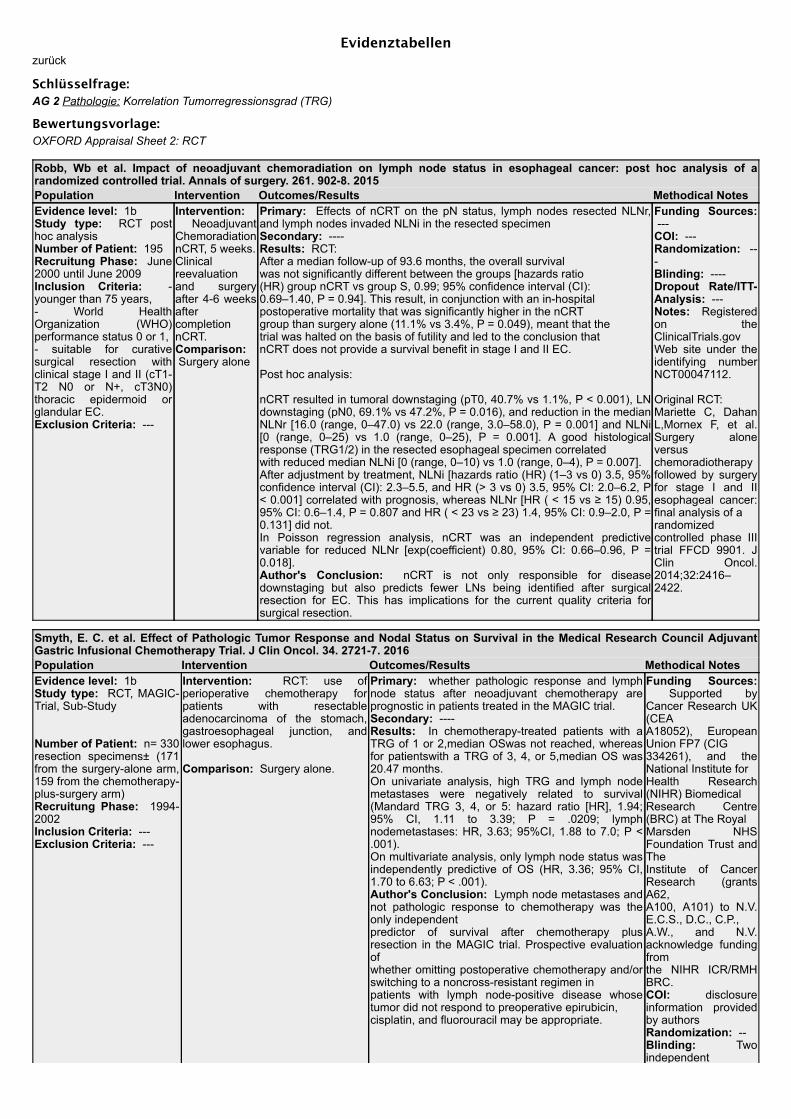
Evidenztabellenzurück
Schlüsselfrage:AG 2 Pathologie: Korrelation Tumorregressionsgrad (TRG)
Bewertungsvorlage:OXFORD Appraisal Sheet 2: RCT
Robb, Wb et al. Impact of neoadjuvant chemoradiation on lymph node status in esophageal cancer: post hoc analysis of arandomized controlled trial. Annals of surgery. 261. 902-8. 2015Population Intervention Outcomes/Results Methodical NotesEvidence level: 1bStudy type: RCT posthoc analysisNumber of Patient: 195Recruitung Phase: June2000 until June 2009Inclusion Criteria: -younger than 75 years,- World HealthOrganization (WHO)performance status 0 or 1, - suitable for curativesurgical resection withclinical stage I and II (cT1-T2 N0 or N+, cT3N0)thoracic epidermoid orglandular EC.Exclusion Criteria: ---
Intervention: NeoadjuvantChemoradiationnCRT, 5 weeks.Clinicalreevaluationand surgeryafter 4-6 weeksaftercompletionnCRT. Comparison: Surgery alone
Primary: Effects of nCRT on the pN status, lymph nodes resected NLNr,and lymph nodes invaded NLNi in the resected specimenSecondary: ----Results: RCT: After a median follow-up of 93.6 months, the overall survivalwas not significantly different between the groups [hazards ratio(HR) group nCRT vs group S, 0.99; 95% confidence interval (CI):0.69–1.40, P = 0.94]. This result, in conjunction with an in-hospitalpostoperative mortality that was significantly higher in the nCRTgroup than surgery alone (11.1% vs 3.4%, P = 0.049), meant that thetrial was halted on the basis of futility and led to the conclusion thatnCRT does not provide a survival benefit in stage I and II EC.
Post hoc analysis:
nCRT resulted in tumoral downstaging (pT0, 40.7% vs 1.1%, P < 0.001), LNdownstaging (pN0, 69.1% vs 47.2%, P = 0.016), and reduction in the medianNLNr [16.0 (range, 0–47.0) vs 22.0 (range, 3.0–58.0), P = 0.001] and NLNi[0 (range, 0–25) vs 1.0 (range, 0–25), P = 0.001]. A good histologicalresponse (TRG1/2) in the resected esophageal specimen correlatedwith reduced median NLNi [0 (range, 0–10) vs 1.0 (range, 0–4), P = 0.007]. After adjustment by treatment, NLNi [hazards ratio (HR) (1–3 vs 0) 3.5, 95%confidence interval (CI): 2.3–5.5, and HR (> 3 vs 0) 3.5, 95% CI: 2.0–6.2, P< 0.001] correlated with prognosis, whereas NLNr [HR ( < 15 vs ≥ 15) 0.95,95% CI: 0.6–1.4, P = 0.807 and HR ( < 23 vs ≥ 23) 1.4, 95% CI: 0.9–2.0, P =0.131] did not. In Poisson regression analysis, nCRT was an independent predictivevariable for reduced NLNr [exp(coefficient) 0.80, 95% CI: 0.66–0.96, P =0.018].Author's Conclusion: nCRT is not only responsible for diseasedownstaging but also predicts fewer LNs being identified after surgicalresection for EC. This has implications for the current quality criteria forsurgical resection.
Funding Sources: ---COI: ---Randomization: ---Blinding: ----Dropout Rate/ITT-Analysis: ---Notes: Registeredon theClinicalTrials.govWeb site under theidentifying numberNCT00047112.
Original RCT: Mariette C, DahanL,Mornex F, et al.Surgery aloneversuschemoradiotherapyfollowed by surgeryfor stage I and IIesophageal cancer:final analysis of arandomizedcontrolled phase IIItrial FFCD 9901. JClin Oncol.2014;32:2416–2422.
Smyth, E. C. et al. Effect of Pathologic Tumor Response and Nodal Status on Survival in the Medical Research Council AdjuvantGastric Infusional Chemotherapy Trial. J Clin Oncol. 34. 2721-7. 2016Population Intervention Outcomes/Results Methodical NotesEvidence level: 1bStudy type: RCT, MAGIC-Trial, Sub-Study
Number of Patient: n= 330resection specimens± (171from the surgery-alone arm,159 from the chemotherapy-plus-surgery arm)Recruitung Phase: 1994-2002Inclusion Criteria: ---Exclusion Criteria: ---
Intervention: RCT: use ofperioperative chemotherapy forpatients with resectableadenocarcinoma of the stomach,gastroesophageal junction, andlower esophagus.
Comparison: Surgery alone.
Primary: whether pathologic response and lymphnode status after neoadjuvant chemotherapy areprognostic in patients treated in the MAGIC trial.Secondary: ----Results: In chemotherapy-treated patients with aTRG of 1 or 2,median OSwas not reached, whereasfor patientswith a TRG of 3, 4, or 5,median OS was20.47 months. On univariate analysis, high TRG and lymph nodemetastases were negatively related to survival(Mandard TRG 3, 4, or 5: hazard ratio [HR], 1.94;95% CI, 1.11 to 3.39; P = .0209; lymphnodemetastases: HR, 3.63; 95%CI, 1.88 to 7.0; P <.001). On multivariate analysis, only lymph node status wasindependently predictive of OS (HR, 3.36; 95% CI,1.70 to 6.63; P < .001).Author's Conclusion: Lymph node metastases andnot pathologic response to chemotherapy was theonly independentpredictor of survival after chemotherapy plusresection in the MAGIC trial. Prospective evaluationofwhether omitting postoperative chemotherapy and/orswitching to a noncross-resistant regimen inpatients with lymph node-positive disease whosetumor did not respond to preoperative epirubicin,cisplatin, and fluorouracil may be appropriate.
Funding Sources: Supported byCancer Research UK(CEAA18052), EuropeanUnion FP7 (CIG334261), and theNational Institute forHealth Research(NIHR) BiomedicalResearch Centre(BRC) at The RoyalMarsden NHSFoundation Trust andTheInstitute of CancerResearch (grantsA62,A100, A101) to N.V.E.C.S., D.C., C.P.,A.W., and N.V.acknowledge fundingfromthe NIHR ICR/RMHBRC.COI: disclosureinformation providedby authorsRandomization: --Blinding: Twoindependent

pathologists using theMandard tumorregression gradingsystem (TRG).Dropout Rate/ITT-Analysis: ---RCT: ITTNotes: the results donot differentiate theindividual tumorentities
No description of theoriginal RCT. Medical ResearchCouncil OesophagealCancerWorking Group:Surgical resectionwith or withoutpreoperativechemotherapy inoesophagealcancer: A randomisedcontrolled trial.Lancet 359:1727-1733, 2002
Cunningham D,Allum WH, StenningSP, et al:Perioperativechemotherapy versussurgery alone forresectablegastroesophagealcancer. N Engl J Med355:11-20, 2006

Evidenztabellenzurück
Schlüsselfrage:AG 2 Pathologie: Korrelation Tumorregressionsgrad (TRG)
Bewertungsvorlage:NEWCASTLE - OTTAWA Checklist 4: Cohort
Davies, A. R. et al. Tumor stage after neoadjuvant chemotherapy determines survival after surgery for adenocarcinoma of theesophagus and esophagogastric junction. J Clin Oncol. 32. 2983-90. 2014
Evidence level Methodical Notes Patientcharacteristics Interventions
Evidence level: 2bStudy type: Prospectivecohort study
Funding sources: Research Funding: David Cunningham,Roche, sanofi-aventis, AstraZeneca, Amgen, Merck, Celgene,Novartis Conflict of Interests: Employment or Leadership Position:None Consultant or Advisory Role: None Stock Ownership:None Honoraria: William H. Allum, Eli Lilly, Nestle, AstellasPharma Expert Testimony: None Patents, Royalties, andLicenses: None Other Remuneration: NoneRadnomization: -Blinding: -Dropout rates: -
Total no.patients: 584patients with ACCof esophagus orEGJ. Patientcharacteristics: Between 2000and 2010 in twohigh volumeinstitutions foresophagealCancer in London.Inclusion criteria: Patients withadenocarcinomaof the esophagusor esophago-gastric junction.Studycharacteristics:584patients, the meanage was 63 years(range, 28 to 83years), 86% male.Overall survival ofthe cohort was80% and 45% at 1and 5 years,respectively.Exclusioncriteria: -
Interventions: Chemotherapy:400 out of 580 underwent pre-operative chemotherapy.Tumor staging: Each patient wasallocated a tumor stage (cTNM)before commencement ofneoadjuvant chemotherapy asdecided by the multidisciplinaryteam. After neoadjuvantchemotherapy, patients wererestaged using CT (thorax,abdomen, and pelvis), but notroutinely using endoscopy, EUS, orfluorode- oxyglucose PET. Resection and staging: All patientsunderwent definitive resection and,therefore, had final tumor histologyavailable for comparison (ypTNM).This pathologic stage wasdetermined using the seventhedition of the American JointCommittee on Cancer TNM stagingsystem. Downstaging was definedas a reduction in T stage or N stageof pathologic staging (ypTNM)compared with clinical staging(cTNM). Pathologic tumorregression used a categorical scalebetween 1 and 5 as described byMandard.Comparison: -
Notes: Criteria for inclusion or exclusion are inadequately described.Author's conclusion: "This study indicates that tumor stage after neo-adjuvant chemotherapy determines survival inpatients with adenocarcinoma of the esophagus and esophagogastric junction. The importance of of tumor downstaging interms of survival, complete surgical resection, and recurrence pattern has significant clinical implications."
OutcomeMeasures/results
Primary Tumor staging: Each patient was allocated a tumor stage(cTNM) before commencement of neoadjuvant chemotherapy asdecided by the multidisciplinary team. After neoadjuvantchemotherapy, patients were restaged using CT but not routinelyusing endoscopy, EUS, or FDG-PET. All patients underwentdefinitive resection; final tumor histology available for comparison(ypTNM), and analyzed by a member of a team of dedicated upperGI histopathologists. This pathologic stage was determined usingthe 7th edition of the American Joint Committee on Cancer TNMstaging system. Downstaging was defined as a reduction in Tstage or N stage of pathologic staging (ypTNM) compared withclinical staging (cTNM). Pathologic tumor regression used acategorical scale between 1 and 5 accordin to Mandard.Secondary -
Results: Downstaging Effect of ChemotherapyPrimary:neoadjuvant chemotherapy group: 175 patients (44%)benefitted from a downstaging effect. This group ofresponders, compared with nonresponders, hadimproved rates of clear surgical resection margins (R0:74% vs 40%, respectively; P < .001) and lower rates ofisolated local recurrence (6% v 13%) p = .03).The responders also experienced lower rates ofsystemic metastatic recurrence compared with non-responders, both alone (19 v 29%, p < .027) and incombination with locoregional recurrence (30% v 48%,p < 0.001).The majority of down-staged patients had evidence ofpathologic response to neoadjuvant chemotherapy(Mandard score 1-4 in 144 of 162 patients, 89%). Thisgroup of downstaged patients had significantlyimproved Mandard scores compared with those whowere not downstaged (p < .001).
Shapiro, J. et al. Prognostic Value of Pretreatment Pathological Tumor Extent in Patients Treated With NeoadjuvantChemoradiotherapy Plus Surgery for Esophageal or Junctional Cancer. Ann Surg. 265. 356-362. 2017Evidence level Methodical Notes Patient characteristics InterventionsEvidence level: 1bStudy type: Posthoc analysis RCT
Funding sources: ----Conflict of Interests: ----Radnomization: ----Blinding: The interobserveragreement was determinedbetween 3 independentlyscoring upper-GI pathologists
Total no. patients: 180Patient characteristics: 2003 - 2011Inclusion criteria: Potentially curableesophageal or junctional cancer, who weretreated with neoadjuvant chemoradiotherapy(nCRT) plus surgery according to theCROSS regimen.
Interventions: from the nCRT plussurgery group: resection specimens(primary tumor and all resected lymphnodes)Comparison:
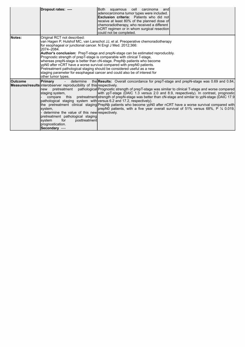
Dropout rates: ---- Both squamous cell carcinoma andadenocarcinoma tumor types were included.Exclusion criteria: Patients who did notreceive at least 80% of the planned dose ofchemoradiotherapy, who received a differentnCRT regimen or in whom surgical resectioncould not be completed.
Notes: Original RCT not described:van Hagen P, Hulshof MC, van Lanschot JJ, et al. Preoperative chemoradiotherapyfor esophageal or junctional cancer. N Engl J Med. 2012;366:2074–2084.Author's conclusion: PrepT-stage and prepN-stage can be estimated reproducibly.Prognostic strength of prepT-stage is comparable with clinical T-stage,whereas prepN-stage is better than cN-stage. PrepNþ patients who becomeypN0 after nCRT have a worse survival compared with prepN0 patients.Pretreatment pathological staging should be considered useful as a newstaging parameter for esophageal cancer and could also be of interest forother tumor types.
OutcomeMeasures/results
Primary - determine theinterobserver reproducibility of thisnew pretreatment pathologicalstaging system, - compare this pretreatmentpathological staging system withthe pretreatment clinical stagingsystem, - determine the value of this newpretreatment pathological stagingsystem for posttreatmentprognostication.Secondary ----
Results: Overall concordance for prepT-stage and prepN-stage was 0.69 and 0.84,respectively. Prognostic strength of prepT-stage was similar to clinical T-stage and worse comparedwith ypT-stage (DAIC 1.3 versus 2.0 and 8.9, respectively). In contrast, prognosticstrength of prepN-stage was better than cN-stage and similar to ypN-stage (DAIC 17.9versus 6.2 and 17.2, respectively).PrepNþ patients who become ypN0 after nCRT have a worse survival compared withprepN0 patients, with a five year overall survival of 51% versus 68%, P ¼ 0.019,respectively.

Inhaltsverzeichnis der Evidenztabellen
Schlüsselfrage:
AG 3 Chirurgie: Art des operativen Zugangs
Citation Evidence Level Study TypeKurokawa, Y. 2015 1b Randomized trialMaas, K. W. 2015 1b Randomized clinical trial multicentric.Straatman, J. 2017 1b Randomized clinical trial.

Evidenztabellenzurück
Schlüsselfrage:AG 3 Chirurgie: Art des operativen Zugangs
Bewertungsvorlage:OXFORD Appraisal Sheet 2: RCT
Kurokawa, Y. et al. Ten-year follow-up results of a randomized clinical trial comparing left thoracoabdominal and abdominaltranshiatal approaches to total gastrectomy for adenocarcinoma of the oesophagogastric junction or gastric cardia. Br J Surg. 102.341-8. 2015Population Intervention Outcomes/Results Methodical NotesEvidence level: 1bStudy type: Randomized trialNumber of Patient: 167 (82, 85 per arm)Recruitung Phase: Between July 1995and December 2003,27 hospitals inJapan.Inclusion Criteria:
histologicallyconfirmed ACCof the gastricbody or cardiawithoesophagealinvasion of 3cm or less, cT2– 4 categoryage 75 years orlessno distantmetastasisno lymph nodeslarger than 1cmin thehepatoduodenalligament orpara-aortic fielda forcedexpiratoryvolume in 1s ofat least 50 percentarterial oxygentension of atleast 9⋅3 kPawhile breathingambient air
Exclusion Criteria: none described
Intervention: Transhiatial surgery (TH):totalgastrectomy with D2 lymphadenectomy includingsplenectomy. Additional dissection of the lymph nodesalong the left inferior phrenic vessels and the para-aortic nodes lateral to the aorta and above the left renalvein was performed in patients with curable disease.This included patients with positive findings onperitoneal lavage cytology, but without overt peritonealmetastasis. All procedures were undertaken vialaparotomy, and the lower mediastinum was accessedtranshiatally. Mediastinal resection included the loweroesophagus and perioesophageal lymph nodes only.Comparison: Left thoraco-abdominal surgery (LTA):An oblique incision over the left thorax and abdomenwas made for the LTA approach, followed by the sameprocedure in the abdominal cavity as for the THoperation. In the thoracic cavity, a thorough mediastinalnode dissection below the left inferior pulmonary veinwas undertaken with appropriate oesophagectomy.
Primary: Overall survival (OS):Over 10 year follow up period.Secondary: Disease-free survival(DFS), Morbidity and Mortality,Postoperative symptoms andpostoperative respiratory functionResults: Median follow-up for allcensored patients was 10.6 (range5⋅1 – 17⋅1) years. There had been52 and 63 deaths in the TH and LTAgroup respectively, with 42 and 50patients respectively dying fromcancer.Primary:OS:The 5- and 10-yearOS rates for all randomizedpatients were 51% (95 % CI 40, 61)and 37% (26 to 47) for the THapproach, and 37% (26, 47) and24% (15, 34) per cent for the LTAapproach. The log rank test showedmarginal differences between thegroups (2-sided P=0.060, 1-sided P= 0.970), and the hazard ratio (HR)for the LTA versus the TH approachwas 1.42 (95% CI 0⋅98, 2⋅05).
Author's Conclusion: "LTAresections should be avoided in thetreatment of adenocarcinoma of theOGJ or gastric cardia."
Funding Sources: "Thestudy was funded in partby Grants-in-Aid for Can-cer Research and for theSecond-TermComprehensive 10-yearStrategy for CancerControl from the Ministryof Health, Labour andWelfare, Japan, and by theNational Cancer CentreResearch andDevelopment Fund (26-A-4)."COI: The authors declareno conflict of interest.Randomization: "Randomly generatedassignment (1 : 1) into oneof the treatment groups. Aminimization method wasused to stratify treatmentgroups according toinstitution, cT category(cT2 versus cT3/4) andBorrmann type (0 – 2versus 3 or 5) for randomnumber generation."Blinding: No blinding wasperformed, measures areobjective (survival).Dropout Rate/ITT-Analysis: All analyseswere based on anintention-to-treat basis.Notes: "Baselinecharacteristics of the twogroups were similar,except for Siewertclassification"
Maas, K. W. et al. Quality of Life and Late Complications After Minimally Invasive Compared to Open Esophagectomy: Results of aRandomized Trial. World J Surg. 39. 1986-93. 2015Population Intervention Outcomes/Results Methodical NotesEvidence level: 1bStudy type: Randomized clinicaltrial multicentric.Number of Patient: 115 (56,59 per arm)Recruitung Phase: Between June 1,
Intervention: Patients in both groups receivedidentical pre and postoperative treatment. Formost patients, neoadjuvant treatment consistedof weekly administrations of 50 mg/m2 paclitaxelplus carboplatin and concurrent radiotherapy(41,4 Gy in 23 fractions for 5 days per week).After 6–8 weeks, neoadjuvant treatment wasfollowed by surgery by open or minimally invasive
Primary: Postoperative pulmonary infection:,defined as clinical manifestation of pneumonia orbronchopneumonia confirmed by thoracicradiographs or CT scan and a positive sputumculture, within the first 2 weeks of surgery andduring the whole stay in hospital.Secondary: Short term endpoints:Postoperative complications: (e.g., anastomotic
Funding Sources: Digestive SurgeryFoundation of theUnit of DigestiveSurgery of the VUUniversity MedicalCentre. (originalpaper)

2009 and March 31,2011 at five centers:3 in theNetherlands, 1 inSpain, 1 in Italy.Inclusion Criteria:
resectableesophagealcancer (cT1–3,N0–1, M0)histologicallyproven AC,SCC, orundifferentiatedcarcinoma oftheintrathoracicesophagus andGEJPatients wereaged 18–75yearsWHOperformancestatus of two orless.
Exclusion Criteria:
patients withcervicalesophagealcancer oranothermalignancy
esophagectomy. Both procedures included a two-field esophageal resection with 3–4 cm widegastric tube formation followed by a cervical orintrathoracic anastomosis. Open esophagectomy (OE): involved a rightposterolateral thoracotomy in the lateraldecubitus position with double tracheal intubationand lung block, midline laparotomy, and cervicalor intrathoracic anastomosis. MIE was performedthrough a right thoracoscopy in the prone positionwith single-lumen tracheal intubation, upperabdominal laparoscopy, and cervical incision. Comparison: Minimally invasiveesophagectomy (MIE): was performed through aright thoracoscopy in the prone position withsingle-lumen tracheal intubation, upperabdominal laparoscopy, and cervical incision. Forpatients undergoing MIE with an intrathoracicanastomosis, a bronchus blocker was placed inthe right bronchus to help with one-lungventilation during anastomosis.
leakage, vocal cord paralysis confirmed bylaryngoscopy), QoL: quality of life assessed bySF 36 Health Survey (version 2) and EORTCQoL questionnaires C30 and OES18 module.Mid-term endpoints: QoL at 1 year:(assessedby SF 36 and EORTC C30 and OES18 moduleincidence of late complications: (e.g.,anastomotic stenosis) overall and disease-freesurvivalResults: Secondary: Qol after 1 year:Significantly better scores after 1-year follow-upfor the MIE group as compared to the OE group.These differences are present in three domains:physical activity [SF36: 50 (6; 48–53) vs .45 (9;42–48) p .003]; global health [C30: 79 (10; 76–83) vs. 67 (21; 60–75) p .004]; and pain [OES18:6 (9; 2–8) versus 16 (16; 10–22) p .001]. Latecomplications: After 1 year, 26 patients (44 %) inthe MIE and 22 patients (39 %) in the OE groupwere diagnosed and treated for symptomaticstenosis of the anastomosis. Recurrence: 32 patients died during the first year,18(32%) in the OE group and 14(23%) in the MIEgroup (p = 0.314). Death was related principallyto distant metastases (19 patients), withoutsignificant differences between the two groups (p= 0.167). Local recurrence was observed in threepatients in the OE group (p = 0.072). overall anddisease-free survival:: No significant differencesbetween the two groups.Author's Conclusion: "In conclusion, this firstrandomized trial shows that MIE for esophagealcancer is associated with a better mid-term 1-year quality of life compared to openesophagectomy."
COI: Nodescription.Randomization: eused a computer-generatedrandomisationsequence torandomly assignpatients, in a 1:1ratio, to undergoeither open orminimally invasiveesophagectomy.Randomisation wasstratified by studycenter.Blinding: Noblinding wasperformed.Dropout Rate/ITT-Analysis: "Datawere analysedaccording to theintention-to-treatprinciple." Similardistribution ofdropouts (less than20% total).Notes: Malesurplus in bothgroups. Nodescription ofpotential COI orfunding sources.Primary outcomenot reported, likelydue to intial studythat was previouslypublished.
Straatman, J. et al. Minimally Invasive Versus Open Esophageal Resection: Three-year Follow-up of the Previously ReportedRandomized Controlled Trial: the TIME Trial. Ann Surg. . . 2017Population Intervention Outcomes/Results Methodical NotesEvidence level: 1bStudy type: Randomized clinicaltrial.Number of Patient: 115 (56, 59 per arm).Recruitung Phase: Between June 2009and March 2011. 5European centers.Inclusion Criteria:
Patients between18 and 75 yearsresectableesophagealcancer (cT1-3,N0-1, M0) ofintrahoacicesophagus orGEJindication forneoadjuvanttherapyECOGperformance
Intervention: Both groups: All patients receivedneo-adjuvant treatment, mostly chemo-radiotherapy according to the CROSS scheme,before resection. Both procedures included a 2-field esophageal resection with a 3 to 4cm widegastric tube formation followed by a cervical orintrathoracic anastomosis. For patientsundergoing MIS with an intrathoracicanastomosis, a bronchus blocker was placed inthe right bronchus to help with 1-lung ventilationduring anastomosis. Open esophagectomy: Open esophagectomyinvolved a right posterolateral thoracotomy in thelateral decubitus position with double trachealintubation and lung block, midline laparotomy,and cervical incision. No cervical incision wasused for patients in this treatment group with anintrathoracic anastomosis.Comparison: MIS: was performed through aright thoracoscopy in the prone position withsingle-lumen tracheal intubation, upperabdominal laparoscopy, and cervical incision. Tomaintain partial collapse of the right lung duringthoracoscopy, the thoracic activity was insufflatedwith carbon dioxide at 8mm Hg.
Primary: Respiratory infectionswere defined as clinicalmanifestation of pneumonia orbronchopneumonia confirmed bythoracic radiographs or CT scan(assessed by independentradiologists) and a positivesputum culture, within the first 2weeks of surgery and during thewhole stay in hospital.Secondary: surgery,perioperative, and postoperative-related events: such as duration ofthe procedure, blood loss, andconversion rate. postoperativemorbidity: including reoperationsand intensive care unit admission.Morbidity was registered duringadmission, and in the first 14 dayspostoperatively.long-term survivalanalysisResults: Mean age 62±8,4 yearsper group. Patients receivednCRT according CROSS scheme(92.2%) or chemotherapy alone(7.8%).Primary:Respiratory infections:At2 weeks postoperatively, 5(9%) inthe MIS had a pulmonaryinfection, versus 16(29%) in theopen group (P= 0.05). Similar
Funding Sources: "TheDigestive Surgery Foundation ofthe Unit of Digestive Surgery ofthe VU University MedicalCentre supported the TIME trial.The sponsor of the study had norole in study design, datacollection, data analysis, datainterpretation, or writing of thereport."COI: All authors declare thatthey have no conflict of interestor financial ties to disclose.Randomization: Randomization was performedcentrally via an online module,stratified for participatingcenters. Patients wererandomized in a 1:1 fashionbetween open and MIS.Blinding: No blinding wasperformed, measures areobjective.Dropout Rate/ITT-Analysis: "Data were analyzed accordingto the intention-to-treatprinciple." Dropouts per group(6,6; 10%,10%).Notes: Male surplus in bothgroups. "The principalinvestigator visited all

status of 0,1 or 2Participatingsurgeonsperformed, andhad experiencewith, both openand minimallyinvasiveprocedures, witha minimum of 10MIE performedbefore start of thetrialOnly institutionsthat performedmore than 30esophagectomiesper year
Exclusion Criteria: none described.
results were seen for total in-hospital pulmonary infection rates,being 7 (12%) in the minimallyinvasive group versus 19 (34%) inthe open group (P=0.005).Secondary:Complications: Nodifferences were seen incomplications due to the operativetechnique P=0.302.Survival: 3years follow-up: No differenceswere observed for overall survivaland disease-free survival inpatients who underwent MIScompared with openesophagectomy.Author's Conclusion: "Inconclusion, the TIME trial showedless pulmonary complications anda better QoL in the short-termfollow-up for MIE. For the long-term follow up, it showed anequally safe outcome regardingsurvival and disease-freesurvival."
participating centers, where heobserved at least 2 MIE bythoracoscopy in prone positionper surgeon, in order to assurequality and standardizedtreatment." could potentiallyhave a large impact on theresults or introduce selectionbias.

Inhaltsverzeichnis der Evidenztabellen
Schlüsselfrage:
AG 3 Chirurgie: Ausmaß der Lymphadenektomie
Citation Evidence Level Study TypeLi, B. 2015 1b Randomized clinical trial
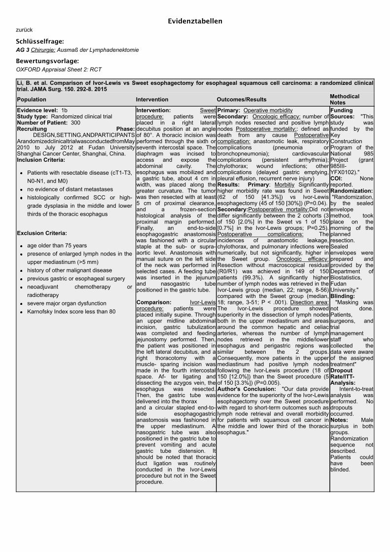
Evidenztabellenzurück
Schlüsselfrage:AG 3 Chirurgie: Ausmaß der Lymphadenektomie
Bewertungsvorlage:OXFORD Appraisal Sheet 2: RCT
Li, B. et al. Comparison of Ivor-Lewis vs Sweet esophagectomy for esophageal squamous cell carcinoma: a randomized clinicaltrial. JAMA Surg. 150. 292-8. 2015
Population Intervention Outcomes/Results MethodicalNotes
Evidence level: 1bStudy type: Randomized clinical trialNumber of Patient: 300Recruitung Phase: DESIGN,SETTING,ANDPARTICIPANTSArandomizedclinicaltrialwasconductedfromMay2010 to July 2012 at Fudan UniversityShanghai Cancer Center, Shanghai, China.Inclusion Criteria:
Patients with resectable disease (cT1-T3,N0-N1, and M0)no evidence of distant metastaseshistologically confirmed SCC or high-grade dysplasia in the middle and lowerthirds of the thoracic esophagus
Exclusion Criteria:
age older than 75 yearspresence of enlarged lymph nodes in theupper mediastinum (>5 mm)history of other malignant diseaseprevious gastric or esophageal surgeryneoadjuvant chemotherapy orradiotherapysevere major organ dysfunctionKarnofsky Index score less than 80
Intervention: Sweetprocedure: patients wereplaced in a right lateraldecubitus position at an angleof 80°. A thoracic incision wasperformed through the sixth orseventh intercostal space. Thediaphragm was incised toaccess and expose theabdominal cavity. Theesophagus was mobilized anda gastric tube, about 4 cm inwidth, was placed along thegreater curvature. The tumorwas then resected with at least5 cm of proximal clearance,and a frozen-sectionhistological analysis of theproximal margin performed.Finally, an end-to-sideesophagogastric anastomosiswas fashioned with a circularstaple at the sub- or supra-aortic level. Anastomosis withmanual suture on the left sideof the neck was performed inselected cases. A feeding tubewas inserted in the jejunumand nasogastric tubepositioned in the gastric tube.
Comparison: Ivor-Lewisprocedure: patients wereplaced initially supine. Throughan upper midline abdominalincision, gastric tubulizationwas completed and feedingjejunostomy performed. Then,the patient was positioned inthe left lateral decubitus, and aright thoracotomy with amuscle- sparing incision wasmade in the fourth intercostalspace. Af- ter ligating anddissecting the azygos vein, theesophagus was resected.Then, the gastric tube wasdelivered into the thoraxand a circular stapled end-to-side esophagogastricanastomosis was fashioned inthe upper mediastinum. Anasogastric tube was alsopositioned in the gastric tube toprevent vomiting and acutegastric tube distension. Itshould be noted that thoracicduct ligation was routinelyconducted in the Ivor-Lewisprocedure but not in the Sweetprocedure.
Primary: Operative morbiditySecondary: Oncologic efficacy: number oflymph nodes resected and positive lymphnodes Postoperative mortality:: defined asdeath from any cause Postoperativecomplication: anastomotic leak, respiratorycomplications (pneumonia orbronchopneumonia); cardiovascularcomplications (persistent arrhythmia);chylothorax; wound infections; othercomplications (delayed gastric emptying,pleural effusion, recurrent nerve injury)Results: Primary: Morbitiy Significantlyhigher morbidity rate was found in Sweet(62 of 150 [41.3%]) vs Ivor-Lewisesophagectomy (45 of 150 [30%]) (P=0.04).Secondary:Postoperative mortality:Did notdiffer significantly between the 2 cohorts (3of 150 [2.0%] in the Sweet vs 1 of 150[0.7%] in the Ivor-Lewis groups; P=0.25).Postoperative complications: Theincidences of anastomotic leakage,chylothorax, and pulmonary infections werenumerically, but not significantly, higher inthe Sweet group. Oncologic efficacy:Resection without macroscopical residual(R0/R1) was achieved in 149 of 150patients (99.3%). A significantly highernumber of lymph nodes was retrieved in theIvor-Lewis group (median, 22; range, 8-56)compared with the Sweet group (median,18; range, 3-51; P < .001). Disection area:The Ivor-Lewis procedure showedsuperiority in the dissection of lymph nodesboth in the upper mediastinum and areasaround the common hepatic and celiacarteries, whereas the number of lymphnodes retrieved in the middle/loweresophagus and perigastric regions wassimilar between the 2 groups.Consequently, more patients in the uppermediastinum had positive lymph nodesfollowing the Ivor-Lewis procedure (18 of150 [12.0%]) than the Sweet procedure (5of 150 [3.3%]) (P=0.005).Author's Conclusion: "Our data provideevidence for the superiority of the Ivor-Lewisesophagectomy over the Sweet procedurewith regard to short-term outcomes such aslymph node retrieval and overall morbidityfor patients with squamous cell cancer inthe middle and lower third of the thoracicesophagus."
FundingSources: "Thisstudy wasfunded by theKeyConstructionProgram of theNational 985Project (grant985III-YFX0102)."COI: Nonereported.Randomization: "Randomization,by the sealedenvelopemethod, tookplace on themorning of theplannedresection.Sealedenvelopes wereprepared andprovided by theDepartment ofBiostatistics,FudanUniversity."Blinding: "Masking wasnot done.Patients,surgeons, andtrialmanagementstaff whocollected thedata were awareof the assignedtreatment"DropoutRate/ITT-Analysis: Intent-to-treatanalysis wasperformed. Nodropoutsoccurred.Notes: Malesurplus in bothgroups.Randomizationsequence notdescribed.Patients couldhave beenblinded.

Inhaltsverzeichnis der Evidenztabellen
Schlüsselfrage:
AG 3 Multimodale Therapie: Verbessert eine adjuvante Radio- oder Radiochemotherapie das Überleben?
Citation Evidence Level Study TypeMa, D. Y. 2014 1b- Randomized controlled trial.

Evidenztabellenzurück
Schlüsselfrage:AG 3 Multimodale Therapie: Verbessert eine adjuvante Radio- oder Radiochemotherapie das Überleben?
Bewertungsvorlage:OXFORD Appraisal Sheet 2: RCT
Ma, D. Y. et al. Concurrent three-dimensional conformal radiotherapy and chemotherapy for postoperative recurrence of mediastinallymph node metastases in patients with esophageal squamous cell carcinoma: a phase 2 single-institution study. Radiat Oncol. 9.28. 2014Population Intervention Outcomes/Results Methodical NotesEvidence level: 1b-Study type: Randomizedcontrolled trial.Number of Patient: 98 (49 pergroup)Recruitung Phase: BetweenJanuary 2002 and June 2003,from the First Hospital affiliatedwith North Sichuan MedicalCollege, P.R. ChinaInclusion Criteria: Patients withhistopathologically confirmedadvanced locoregional ESCC.Post operative normal liver,kidney, and bone marrowfunctions were demonstrated byblood tests.Good tolerance for radiotherapy orchemotherapy according to theWorld Health Organizationperformance status of 0 or 1.Exclusion Criteria: "Patientswho underwent neoadjuvant oradjuvant radiotherapy and/orchemotherapy were excluded fromour study. In addition, those withsupraclavicular lymph nodeinvolvement, only anastomoticstoma recurrence, orhematogenous metastases."
Intervention: -Initial radicalesophagectomy and lymph nodedissection for ESCC with a R0margin-Assessment of locoregionalmediastinal recurrence (confirmed bythe presence of a growing irregularmass by chest CT or MRI.)Intervention: group A: three-dimensional conformal radiotherapy:"The prescribed dosage for 95% PTVwas calculated using 4–6 fields of thecoplanar or noncoplanar 3-DCRTplan, which was determined to be62–70 Gy/31–35 fractions. for 1week, divided into two phases"details see paperComparison: Comparison: GroupB: Concurrent chemotherapy;intravenously administered cisplatinat a dose of 30 mg per m2 of body-surface area weekly.
Primary: Overall survival [%]:calculated as the time interval frominitiation of treatment to death and wasanalyzed using the Kaplan–Meiermethod.Secondary: Severe morbidity [%]: ofgrade 2 or higher.Results: Primary: overall survivalFor survivers, the median follow-upwas 60 months (range, 8–63). The ITTanalyses showed a median overallsurvival of 19 months in group Aversus 35 months in group B (P=0.051log-rank test; HR, 0.76; 95% CI, 28–34). No difference in the overall survivalrate at five years between both groups(P = 0.051), the overall survival ratesat 1 year and 3 years in group B weresignificantly better than those in groupA (P = 0.032, P = 0.038).Mortality: 5 (10.2%); vs 13 (26.5%) ingroup B,A died from distantmetastases of ESCC (χ2 = 4.356, P =0.036).Secondary: Morbidities and adverseeffects:No life-threatening toxic effects wereobserved in either group. The adverseeffects in the hematological andgastrointestinal systems in group Bwere obviously more common than ingroup A. However, there was nosignificant difference between theincidence of late adverse effectsbetween both groups.Author's Conclusion: "In summary,the combined modality of 3-DCRT andchemotherapy was well toleratedcompared to radiation alone andyielded superior overall survival ratesin patients with postoperativerecurrence of mediastinal lymph nodemetastases of ESCC."
Funding Sources: Thiswork was supported byMedjaden.COI: The authors declarethat they have no competinginterests.Randomization: Assignment by using "arandom number table",Blinding: no blinding ismentioned, but at leastpartial blinding could havebeen achieved.Dropout Rate/ITT-Analysis: The intention-to-treat analyses, nomentioning of dropoutsNotes: -No blinding orconcealment of allocationwas performed. This mightnot impact the primaryendpoint (survival), but it isstill a risk of bias and couldhave partially beenachieved-Only 31% femaleparticipants-Potentially unequaltreatment between groups:"In parallel with concurrentradiochemotherapy, thethymic peptide α1 wasinjected i.h. at a dose of 1.6mg per day for 3 weeks inorder to retain systematicimmune function. "

Inhaltsverzeichnis der Evidenztabellen
Schlüsselfrage:
AG 3 Multimodale Therapie: Verbessert eine präoperative (bzw. prä- und) postoperative (fortgesetzte) Chemotherapie dasÜberleben?
Citation Evidence Level Study TypeZhao, Y 2014 1b Randomized controlled trial.

Evidenztabellenzurück
Schlüsselfrage:AG 3 Multimodale Therapie: Verbessert eine präoperative (bzw. prä- und) postoperative (fortgesetzte) Chemotherapie das Überleben?
Bewertungsvorlage:OXFORD Appraisal Sheet 2: RCT
Zhao, Y et al. Perioperative versus preoperative chemotherapy with surgery in patients with resectable squamous-cell carcinoma ofesophagus: A phase III randomized trial. Journal of clinical oncology. 32. . 2014
Population Intervention Outcomes/Results MethodicalNotes
Evidence level: 1bStudy type: Randomized controlled trial.Number of Patient: 346 (175, 171 per arm).Recruitung Phase: Between January 2005 andApril 2007, in two Chinese hospitals (FirstAffiliated Hospital and the Second AffiliatedHospital of Xi’an Jiaotong University).Inclusion Criteria: -no evidences of previouschemotherapy or radiotherapy, Patients aged 18years and older; WHO performance status 0 or 1were eligible if they had histopathologicallyproven squamous cell carcinoma of esophagusthat was considered as suitable for curativeresection.The disease had to be confined to primary andregional nodes, although celiac nodalinvolvement (M1a) was permitted for primarytumor localized in the distal esophagus orgastroesophageal junction. Patients had to beoperative candidates without excessive clinicalrisks and had no evidences of distant disease orinvolvement of tracheobronchial tree or otherstructures that would preclude a completeresection. Laboratory parameters includedadequate bone marrow reserve consisting of awhite blood cell count of more than 3500 cells/ml,platelet count of more than 100,000 cells/ml,normal liver function with total bilirubin of lessthan 1.5mg/100ml, and creatinine clearance ofmore than 60ml/min.Exclusion Criteria: none described.
Intervention: Each armreceived two pre-operativecycle of chemotherapy,followed by surgery.The intervention arm (armA) received two additionalcycles of PCF post surgery.Each 3-week cycleconsisted of PCF: paclitaxel(100 mg per square meterof body surface area) by a3-hour intravenous infusionon day 1, cisplatin (60 mgper square meter of bodysurface area) intravenouslywith hydration on day 1,and 5- uorouracil (700mgper square meter of bodysurface area) daily throughday 1 to 5 by continuousintravenous infusion with adouble-lumen Hickmancatheter.Comparison: Comparisonpatients received two pre-operative cycles of PCFbefore surgery (arm B).
Primary: Relapse-free survival wascalculated from randomization to the firstevent (i.e., local recurrence, distantrecurrence, or death from any cause), Secondary: Overall survival: wascalculated from randomization to death fromany causes.Results: The median follow-up was 60 and61 months in arm A and arm B. Beforedeaths, local recurrence was confirmed in 25patients (14.2%) in arm A and 35 patients(20.5%) in arm B, and distant metastasis wasconfirmed in 41 patients (23.4%) in arm Aand 62 patients (36.3%) in arm B. The median relapse-free survival andoverall survival were 23 and 29 months inarm A versus 15 and 22 months in arm B.Comparing with arm B, arm A had thesignificantly higher possibility of relapse-freesurvival (hazard ratio for relapse, 0.62; 95%confidence interval [CI], 0.49–0.73; p <0.001, Fig. 2A) and of overall survival (hazardratio for death, 0.79; 95% CI, 0.59–0.95; p <0.001, Fig. 2B). Five-year relapse-freesurvival rate was 35.0% (95% CI, 26.1–47.2)in arm A compared with 19.1% (95% CI,15.3–28.7) in arm B. Five-year survival ratewas 38.0% (95% CI, 29.5–43.0) in arm Acompared with 22.0% (95% CI, 16.6–29.4) inarm B.Author's Conclusion: "In conclusion, ourresults showed that perioperativechemotherapy with the regimen of PCFimproved 5-year relapse-free and overallsurvival in patients with resectable squamouscell carcinoma of esophagus compared withpreoperative che-motherapy alone.Therefore, this treatment should beconsidered as an option for patients withresectable squamous cell carcinoma ofesophagus."
FundingSources: Thiswork wassupported byNational NaturalScienceFoundation ofChina (No.81301847) andtheFundamentalResearch Fundsfor the CentralUniversities.COI: Theauthors declareno con ict ofinterest.Randomization: Randomizationnot specified.Blinding: nonblinded study.DropoutRate/ITT-Analysis: Itention-to-treatanalysis. 3 outof 175 and 2 outof 171 patientswere excludedin group A andB.Notes: Randomizationprotocol notdescribed. Noblinding wasperformed.Significantsurplus of menin both groups.

Inhaltsverzeichnis der Evidenztabellen
Schlüsselfrage:
AG 3 Multimodale Therapie: Verbessert eine präoperative Radiochemotherapie das Überleben?
Citation Evidence Level Study TypeAjani, J. A. 2013 1b- Phase II Randomized controlled trialMariette, C. 2014 1b Phase III randomized controlled trial, multicentric study (30 centers in France).Klevebro, F. 2015 1b Randomized controlled trialShapiro, J. 2015 1b Randomized controlled trialRajabi Mashhadi, M. 2015 1b- Randomized controlled trial.Nederlof, N. 2016 1b Randomized controlled trialKlevebro, F. 2016 1b Randomized clinical trialStahl, M. 2017 1b Unblinded, prospective and randomised phase III study.

Evidenztabellenzurück
Schlüsselfrage:AG 3 Multimodale Therapie: Verbessert eine präoperative Radiochemotherapie das Überleben?
Bewertungsvorlage:OXFORD Appraisal Sheet 2: RCT
Klevebro, F. et al. A randomized clinical trial of neoadjuvant chemotherapy versus neoadjuvant chemoradiotherapy for cancer of theoesophagus or gastro-oesophageal junction. Ann Oncol. 27. 660-7. 2016Population Intervention Outcomes/Results Methodical NotesEvidence level: 1bStudy type: Randomizedclinical trialNumber of Patient: 181(90 and 91 per group).Recruitung Phase: Theneoadjuvant chemotherapyversus chemoradiotherapyin resectable cancer of theoesophagus and gastriccardia (NeoRes) trial wasperformed in Norway andSweden during the period2006–2013.Inclusion Criteria: Patients with histologicallyconfirmed SCC or AC ofthe oesophagus or GOJ(including Siewert types Iand II) who were eligible forcurative treatment withsurgical resection wereenrolled. Clinical tumourstage; T1–3, any N (withthe excep- tion of T1N0)were included, cervicalcancers were required tobe resectable withoutlaryngectomy.Exclusion Criteria: nonedescribed.
Intervention: Neoadjuvant chemotherapy(nCT): Treatment had to be started within 2weeks of randomization. Three cycles ofcisplatin, 100 mg/m2 day 1, and fluorouracil 750mg/m2/24 h, days 1–5, were given. Each cyclelasted 21 days. The same chemotherapyregimen was administered in each treatmentarm.Surgery: in both arms, Patients were scheduledto undergo resection 4–6 weeks after havingcompleted neoadjuvant treatment. The protocolrequired two-field lymphadenectomy, and therecommended procedure was oesophagectomywith intrathoracic anastomosis through alaparotomy and a right-sided thoracotomy (IvorLewis procedure). A three-stage resection, with aright-sided thoracotomy, laparotomy, and cervicalincision (McKeown procedure), wasrecommended for tumours in the middle andupper thirds of the oesopha- gus. Otherprocedures were accepted in cases where theindividual surgeon considered it appropriate.
Comparison: Neoadjuvant Radiotherapy(nCRT) In patients randomized to receivechemoradiotherapy, 40 Gy was given (2 Gy oncedaily in 20 fractions, 5 days a week) with aphoton beam linear accelerator concomitant withchemotherapy cycles 2 and 3. A 3D doseplanning system was used.
Primary: Histological completeresponseSecondary:
Overall survivalnumber of lymph-nodemetastasesR0-resection rateprogression-free survivalsite of recurrence
Results: Primary: Histologicalcomplete response was achieved in7 (9%) of the patients in the nCTarm versus 22 (28%) in the nCRTarm (P = 0.002).Secondary: Three-year overallsurvival: was 49% in the nCT arm,and 47% in the nCRT arm (P =0.77).R0 resection was achieved in 58(74%) patients in the nCT armversus 68 (87%) in the nCRT arm(P = 0.04).Number of lymph node metastases:Of the patients with histologicalcomplete response, 26 (90%) didnot have any metastatic lymphnodes, whereas 3 patients (10%),all treated with nCRT, had at leastone metastatic lymph node. Ofpatients resected in the nCT arm,48 (62%) had lymph-nodemetastases versus 27 (35%) in thenCRT arm (P = 0.001). Progression-free survival: was 44% in bothtreatment arms.Author's Conclusion: Inconclusion, this trial confirmsprevious findings that the addition ofradiotherapy to neoadjuvantchemotherapy increases thecomplete histological response andR0 resection rates and decreasesthe proportion of patients withlymph-node metastases, withoutsignificantly affecting survival.Moreover, we conclude that this trialdoes not provide any evidence insupport of using completehistological response as a surrogatemarker for survival.
Funding Sources: Thiswork was financiallysupported by the SwedishSociety of Medicine, theSwedish Cancer Society,the Cancer ResearchFoundations ofRadiumhemmet, and theStockholm CountyCouncil, grant number notapplicable. The sponsorshad no involvement in thestudy design, datacollection, or interpretationof the results.COI: The authors havedeclared no conflicts ofinterest.Randomization: Nodescription of therandomization sequenceor protocol.Blinding: The pathologistreviewing the surgicalspecimen was blinded tothe randomizationoutcome of eachindividual patient.Dropout Rate/ITT-Analysis: "Data wereanalyzed according to theintention-to-treat principlein all randomizedpatients."Notes: Randomizationsequence not described;male surplus in bothgroups; primary outcome:histological completeresponse is not described
Klevebro, F. et al. Morbidity and mortality after surgery for cancer of the oesophagus and gastro-oesophageal junction: Arandomized clinical trial of neoadjuvant chemotherapy vs. neoadjuvant chemoradiation. Eur J Surg Oncol. 41. 920-6. 2015
Population Intervention Outcomes/Results MethodicalNotes
Evidence level: 1bStudy type: Randomizedcontrolled trialNumber of Patient: 181patients (91, 90 per arm)Recruitung Phase: "to thisend, between 2006 and 2013,we have performed a
Intervention: Chemotherapy: The nCT treatment cycle was 21 days(treatment during weeks 1, 4, and 7). Cisplatin in a dose of 100mg/m2 (day 1) was given intravenously, in combination with 5-fluorouracil in the amount of 750 mg/m2/24 h (days 1e5). In patientswith borderline renal function or with severely impaired hearing,cisplatin was replaced by oxaliplatin (130 mg/m2) in adenocarcinomapatients or with carboplatin (AUC 5) in squamous carcinoma patients.Surgery: Patients were scheduled to undergo resection 4e6 weeks
Primary: Incidence ofperioperativecomplications: directlycaused by surgery ornonsurgicalcomplications. Severityof perioperativecomplications:
FundingSources: TheSwedish Societyof Medicine hasfinanciallysupported theconduct of thestudy.
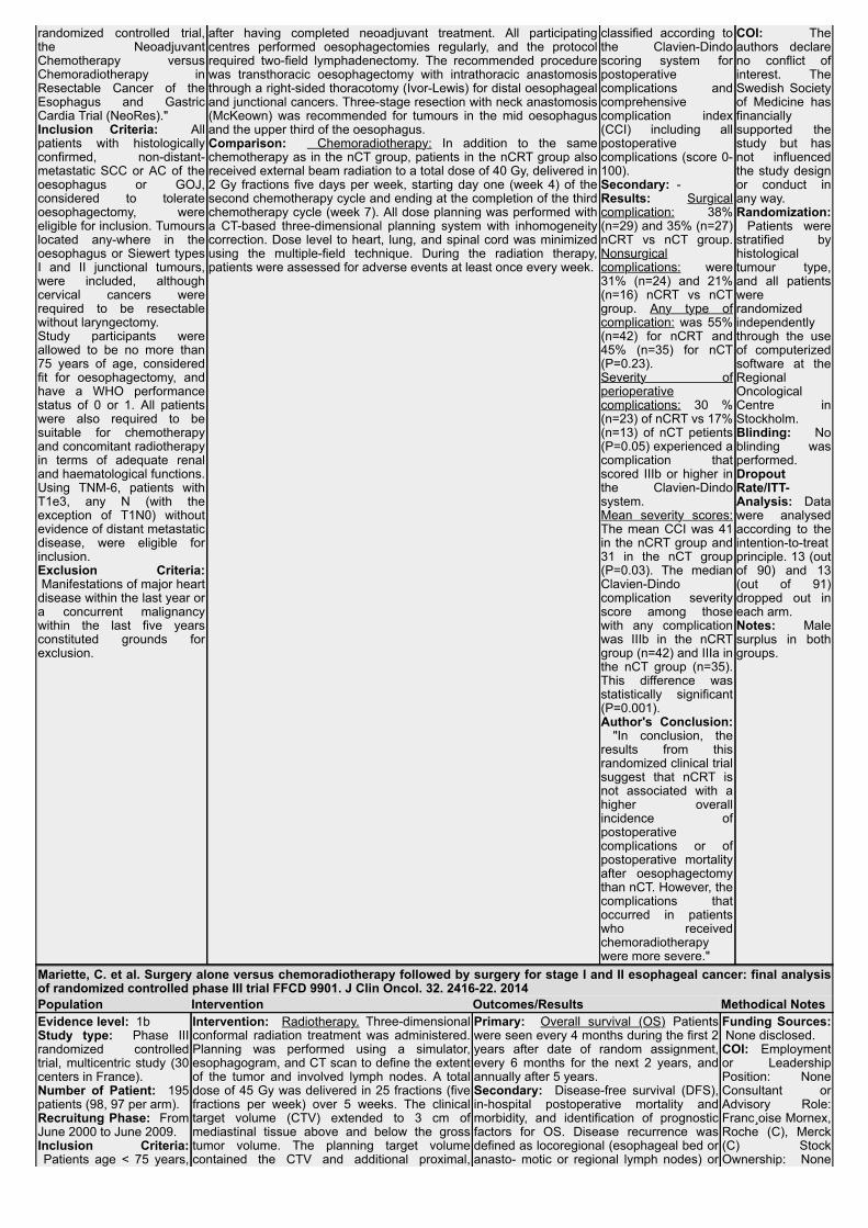
randomized controlled trial,the NeoadjuvantChemotherapy versusChemoradiotherapy inResectable Cancer of theEsophagus and GastricCardia Trial (NeoRes)."Inclusion Criteria: Allpatients with histologicallyconfirmed, non-distant-metastatic SCC or AC of theoesophagus or GOJ,considered to tolerateoesophagectomy, wereeligible for inclusion. Tumourslocated any-where in theoesophagus or Siewert typesI and II junctional tumours,were included, althoughcervical cancers wererequired to be resectablewithout laryngectomy. Study participants wereallowed to be no more than75 years of age, consideredfit for oesophagectomy, andhave a WHO performancestatus of 0 or 1. All patientswere also required to besuitable for chemotherapyand concomitant radiotherapyin terms of adequate renaland haematological functions.Using TNM-6, patients withT1e3, any N (with theexception of T1N0) withoutevidence of distant metastaticdisease, were eligible forinclusion.Exclusion Criteria: Manifestations of major heartdisease within the last year ora concurrent malignancywithin the last five yearsconstituted grounds forexclusion.
after having completed neoadjuvant treatment. All participatingcentres performed oesophagectomies regularly, and the protocolrequired two-field lymphadenectomy. The recommended procedurewas transthoracic oesophagectomy with intrathoracic anastomosisthrough a right-sided thoracotomy (Ivor-Lewis) for distal oesophagealand junctional cancers. Three-stage resection with neck anastomosis(McKeown) was recommended for tumours in the mid oesophagusand the upper third of the oesophagus.Comparison: Chemoradiotherapy: In addition to the samechemotherapy as in the nCT group, patients in the nCRT group alsoreceived external beam radiation to a total dose of 40 Gy, delivered in2 Gy fractions five days per week, starting day one (week 4) of thesecond chemotherapy cycle and ending at the completion of the thirdchemotherapy cycle (week 7). All dose planning was performed witha CT-based three-dimensional planning system with inhomogeneitycorrection. Dose level to heart, lung, and spinal cord was minimizedusing the multiple-field technique. During the radiation therapy,patients were assessed for adverse events at least once every week.
classified according tothe Clavien-Dindoscoring system forpostoperativecomplications andcomprehensivecomplication index(CCI) including allpostoperativecomplications (score 0-100).Secondary: -Results: Surgicalcomplication: 38%(n=29) and 35% (n=27)nCRT vs nCT group.Nonsurgicalcomplications: were31% (n=24) and 21%(n=16) nCRT vs nCTgroup. Any type ofcomplication: was 55%(n=42) for nCRT and45% (n=35) for nCT(P=0.23).Severity ofperioperativecomplications: 30 %(n=23) of nCRT vs 17%(n=13) of nCT petients(P=0.05) experienced acomplication thatscored IIIb or higher inthe Clavien-Dindosystem.Mean severity scores:The mean CCI was 41in the nCRT group and31 in the nCT group(P=0.03). The medianClavien-Dindocomplication severityscore among thosewith any complicationwas IIIb in the nCRTgroup (n=42) and IIIa inthe nCT group (n=35).This difference wasstatistically significant(P=0.001).Author's Conclusion: "In conclusion, theresults from thisrandomized clinical trialsuggest that nCRT isnot associated with ahigher overallincidence ofpostoperativecomplications or ofpostoperative mortalityafter oesophagectomythan nCT. However, thecomplications thatoccurred in patientswho receivedchemoradiotherapywere more severe."
COI: Theauthors declareno conflict ofinterest. TheSwedish Societyof Medicine hasfinanciallysupported thestudy but hasnot influencedthe study designor conduct inany way.Randomization: Patients werestratified byhistologicaltumour type,and all patientswererandomizedindependentlythrough the useof computerizedsoftware at theRegionalOncologicalCentre inStockholm.Blinding: Noblinding wasperformed.DropoutRate/ITT-Analysis: Datawere analysedaccording to theintention-to-treatprinciple. 13 (outof 90) and 13(out of 91)dropped out ineach arm.Notes: Malesurplus in bothgroups.
Mariette, C. et al. Surgery alone versus chemoradiotherapy followed by surgery for stage I and II esophageal cancer: final analysisof randomized controlled phase III trial FFCD 9901. J Clin Oncol. 32. 2416-22. 2014Population Intervention Outcomes/Results Methodical NotesEvidence level: 1bStudy type: Phase IIIrandomized controlledtrial, multicentric study (30centers in France).Number of Patient: 195patients (98, 97 per arm).Recruitung Phase: FromJune 2000 to June 2009.Inclusion Criteria: Patients age < 75 years,
Intervention: Radiotherapy. Three-dimensionalconformal radiation treatment was administered.Planning was performed using a simulator,esophagogram, and CT scan to define the extentof the tumor and involved lymph nodes. A totaldose of 45 Gy was delivered in 25 fractions (fivefractions per week) over 5 weeks. The clinicaltarget volume (CTV) extended to 3 cm ofmediastinal tissue above and below the grosstumor volume. The planning target volumecontained the CTV and additional proximal,
Primary: Overall survival (OS) Patientswere seen every 4 months during the first 2years after date of random assignment,every 6 months for the next 2 years, andannually after 5 years.Secondary: Disease-free survival (DFS),in-hospital postoperative mortality andmorbidity, and identification of prognosticfactors for OS. Disease recurrence wasdefined as locoregional (esophageal bed oranasto- motic or regional lymph nodes) or
Funding Sources: None disclosed.COI: Employmentor LeadershipPosition: NoneConsultant orAdvisory Role:Franc oise Mornex,Roche (C), Merck(C) StockOwnership: None
judged suitable for curativeresection, with untreatedstage I or II (T1 or T2, N0or N1 and T3N0, M0)5thoracic esophagealadenocarcinoma orsquamous cell carcinoma,as assessed by computedtomography (CT) scan andendoscopic ultrasound(EUS), were included. Allpatients were required tobe capable of receivingeither treatment, withWHO performance statusof 0 or 1.Exclusion Criteria: Reasons for patientexclusion included weightloss > 10% at baselineand respiratory, liver, orcardiac insufficiency.Patients with a previouslytreated malignancy,evidence ofsupraclavicular or celiacnodes, a multifocal tumor,a tumor with a proximallimit < 19 cm from theincisor teeth, or evidenceof invasion of thetracheobronchial tree wereexcluded.
distal, and lateral margins of 1 cm to account foruncertainties in repositioning and patientmovement. Photon beams from a linearaccelerator with energy 6 MeV were usedthroughout this study.Chemotherapy. Chemotherapy was deliveredconcomitantly; two cycles of fluorouracil (FU) andcisplatin. FU 800 mg/m2 per 24 hours wasadministered as a continuous infusion from days1 to 4 and 29 to 32. Cisplatin 75 mg/m2 wasdelivered by infusion on day 1 or 2 and again onday 29 or 30. Alternatively, it was delivered as aninfusion at a dose of 15 mg/m2 from days 1 to 5and 29 to 33. Administration of the second cycleof chemotherapy as a half dose was permitted incases of moderate hematologic toxicity(granulocytes between 1,000 and 1,500/mm3and/or platelets between 75,000 and100,000/mm3); it could be omitted in cases ofsevere hematologic toxicity (granulocytes1,000/mm3 and/or platelets 75,000/mm3) orpersistent grade 3 to 4 digestive toxicity.Comparison: Surgery: All patients in group CRTunderwent clinical re-evaluation 2 to 4 weeksafter finishing NCRT, including physicalexamination, weight evaluation, blood laboratoryanalysis, and thoracoabdominal CT scan.Surgery was performed 4 to 8 weeks aftercompletion of NCRT in group CRT and within 4weeks of random assignment in group S. Atransthoracic esophagec- tomy was mandatorywith an extended two-field lymphadenectomy andhigh intrathoracic anastomosis for tumors withinfracarinal proximal margin; cervicalanastomosis was mandatory when the proximalmargin was above the carina.
metastatic (supraclavicular lymph nodes ordistant organs).Results: Primary: OS: Median follow-upwas 93.6 months. Total number of deathswas 125 (64.1%; 61 [62.4%] in group CRTv 64 [66.0%] in group S). Median, 3-year,and 5-year OS were 31.8 months (95% CI,25.2 to 67.8 months), 47.5% (95% CI,37.1% to 57.2%), and 41.1% (95% CI,30.8% to 51.0%) in group CRT versus 41.2months (95% CI, 29.0 to 53.9 months),53.0% (95% CI, 42.3% to 62.5%), and33.8% (95% CI, 23.9% to 43.9%) in groupS.OS was not significantly different betweengroups (HR for group CRT versus group S,0.99; 95% CI, 0.69 to 1.40; P=.94.Secondary: DFS In the overall population,recurrent disease was observed in 71patients (36.4%; 28.6% in group CRT vs44.3% in group S; P=.02). Locoregionalrecurrence was diagnosed in 43 patients(22.1%; 15.3% in group CRT v 28.9% ingroup S; P=.02), whereas distantrecurrence was diagnosed in 50 patients(25.6%; 22.5% in group CRT v 28.9% ingroup S; P=.31). Median DFS was 27.8(95% CI, 15.0 to 42.9) and 26.7 months(95% CI, 22.9 to 41.1), and 5-year DFSwas 35.6% (95% CI, 25.9% to 45.4%) and27.7% (95% CI, 18.6% to 37.6%) in groupsCRT and S. DFS did not differ betweengroups (HR for group CRT vs group S,0.92; 95% CI, 0.66 to 1.30; P=.648).Postop Morbidity and Mortality similarbetween groups (55.6% v 52.8%; P=.720);in-hospital postop mortality wassignificantly higher in the CRT group(11.1% v 3.4%; P=.049).Author's Conclusion: "Compared withsurgery alone, NCRT with cisplatin plusfluorouracil does not improve R0 resectionrate or survival but enhances postoperativemortality in patients with stage I or II EC."
Honoraria: Francoise Mornex,Roche, MerckResearch Funding:None ExpertTestimony: NonePatents, Royalties,and Licenses:None OtherRemuneration:NoneRandomization: "Randomizationwas performedcentrally with aminimizationtechnique thatensured equaldistribu- tion ofpatients regardingstratificationfactors."Blinding: Nonblinded trial.Dropout Rate/ITT-Analysis: Analyses wereperformed using anintent-to-treatapproach,including allpatients asrandomly assignedregardless ofeligibility ortreatment. Notes: Malesurplus in bothgroups.
Nederlof, N. et al. Using the Comprehensive Complication Index to Assess the Impact of Neoadjuvant Chemoradiotherapy onComplication Severity After Esophagectomy for Cancer. Ann Surg Oncol. 23. 3964-3971. 2016
Population Intervention Outcomes/Results MethodicalNotes
Evidence level: 1bStudy type: Randomized controlled trialNumber of Patient: 368 (180, 188 per arm)Recruitung Phase: Inclusion Criteria: Patients with histologicallyproven SCC or AC of the esophagus or GEJ;The tumor must not extend more than 2 cminto the gastric cardia. Longitudinal tumorlength must not exceed 8 cm, radial size mustnot exceed 5 cm. cT1N0 tumors are noteligible. Patients must have adequatehematological, renal, hepatic and pulmonaryfunctions defined as: granulocytes ≥ 1.5 ×10⁹/L, platelets ≥ 100 × 109/L, total bilirubin ≤1.5 × upper normal limit, creatinine ≤ 120µmol/L and FEV1 ≥ 1.5 L. In the absence oflocal irresectability and/or distantdissemination patients with an acceptablegeneral condition (ECOG performance status0, 1, 2; weight loss < 10%) will be invited toparticipate in the randomized trial.Exclusion Criteria: none described.
Intervention: Chemotherapy regimenPaclitaxel 50 mg/m2 and Carboplatin AUC = 2will be given by intravenous infusion on days 1,8, 15, 22 and 29. All patients receivingPaclitaxel will receive half an hour before thestart of the Paclitaxel infusion premedication:Dexamethason 10 mg i.v., Clemastine 2 mg i.v.and Ranitidine 50 mg i.v..At hour 0, the total calculated dose of Paclitaxel,diluted in 500 ml of normal saline will be infusedover one hour. After the completion of thePaclitaxel infusion, 100 ml NaCl 0.9% will beinfused over 0.5 h, followed by an infusion of 8mg Ondansetron or its equivalent diluted in 100ml NaCl 0.9% over 0.5 hour. Hereafter the totalcalculated dose of Carboplatin, diluted in 500 mlglucose 5% will be infused over one hour.Radiotherapy treatment A total dose of 41.4 Gywill be given in 23 fractions of 1.8 Gy, 5fractions per week, starting the first day of thefirst cycle of chemotherapy. All patients will beradiated by external beam radiation, using 3-Dconformal radiation technique. The patient willbe positioned in supine position.Comparison: Surgery:Patients randomized forsurgery alone will be treated asap afterrandomization. In the chemoradiation arm,surgery will be performed preferably within 6weeks after the completion of thechemoradiation. For carcinomas proximal to thetracheal bifurcation a transthoracic esophagealresection with a two field lymph node dissectionis preferred. For carcinomas distal of thetracheal bifurcation but proximal to the gastro-
Primary: 30 days postoperative complications:Definition according to theNational Cancer Institute'sCommon TerminologyCriteria for Adverse effects,4.0. Severity ofcomplications: Grading ofcomplications using CCIindex for complicationindex, based on Clavien-Dindo classificationSecondary: Subgroupanalysis of complications:
Anastomotic leakagePulmonarycomplicationsCardiac complicationsThromboemboliceventsChyle leakageWound infections
Results: Primary:Complications:Grade Icomplications were seen in43 % of patients in NCRTversus 49 % of patientsafter surgery alone (p =
FundingSources: nonedescribed.COI: Theauthors declarethat they haveno competinginterests(in thestudy protocol).Randomization: "Blockrandomizationwas performedcentrally bytelephone or atthe central trialoffice, accordingto computer-generatedrandomizationlists for eachstratum, withrandom blocksizes of 4 or 6."Blinding: Noblinding wasperformed.DropoutRate/ITT-Analysis: Dropouts:19(11%) and27(14%)dropped out of
esophageal junction, a transthoracic approachwith a two field lymph node dissection or atranshiatal approach can be performed,depending on both patient characteristics andlocal expertise.For distal tumors involving thegastro-esophageal junction a transhiatalesophageal resection is preferred.
0.37). There also was nostatistically significantdifference for grade II-grade V complications.Severity of complications:There was no statisticallysignificant difference in theCCI between both groups.Median CCI in thecombined treatment groupwas 26.22 (IQR 17.28–42.43) compared with25.74 (IQR 8.66–43.01) inthe surgery alone group (p= 0.58).Secondary: Subgroupanalysis of complications Insubgroup analyses of thespecific complications, CCIfor patients who developedan anastomotic leak wasnot statistically differentbetween groups: 8.66[8.66–33.73] vs. 8.66[8.66–33.73] (p = 0.78).The same was true for theother subgroups withpatients who developedpulmonary or cardiaccomplications,thromboembolic event,chyle leakage, or woundinfection.Author's Conclusion: "Neoadjuvantchemoradiotherapyaccording to CROSS didnot have a negative impacton postoperativecomplication severityexpressed by CCIcompared with patientswho underwent surgeryalone for potentially curableesophageal or junctionalcancer."
the nCRT andsurgery group."Data will beanalyzedaccording to the'Intention totreat' principle."Notes:
Shapiro, J. et al. Neoadjuvant chemoradiotherapy plus surgery versus surgery alone for oesophageal or junctional cancer (CROSS):long-term results of a randomised controlled trial. Lancet Oncol. 16. 1090-8. 2015Population Intervention Outcomes/Results Methodical NotesEvidence level: 1bStudy type: Randomizedcontrolled trialNumber of Patient: 368,(178 and 188 per studyarm)Recruitung Phase: Between March 30,2004, and Dec 2, 2008,patients from 8centres(five academic centresand three large non-academic teachinghospitals) in theNetherlands wereenrolled .Inclusion Criteria: Aged75 years or younger;adequate haematological,renal, hepatic, andpulmonary function; aWHO performance scoreof 2 or better, without apast or present history ofother malignancy. Only patients with locallyadvanced (clinical stageT1N1M0 or clinical stageT2–3N0–1M0, accordingto UICC TNM cancerstaging, 6th edition10),histologically proven, andpotentially curable
Intervention: Chemoradiotherapy followed byesophagectomy 4-6 weeks after completion ofthe regiment.Details: Carboplatin (AUC 2 mg/mL per min)and paclitaxel (50 mg/m2 of body-surfacearea) intravenously for five cycles, starting ondays 1, 8, 15, 22, and 29. A total concurrentradiation dose of 41·4 Gy was given in 23fractions of 1·8 Gy, on 5 days per week(excluding weekends), starting on the first dayof the first chemotherapy cycle. The totalduration of neoadjuvant treatment was 23 days(5 days per week in weeks 1, 2, 3, 4, then 3days in week 5).
Comparison: Surgery only, as soon aspossible Details (for both groups): For carcinomas at orabove the level of the carina, a transthoracicoesophageal resection with two-field lymphnode dissection was done. For carcinomaslocated well below the level of the carina,either a transthoracic approach with two-fieldlymph node dissection or a transhiatalapproach was used, depending on both patientcharacteristics and local preferences. Forcarcinomas involving the oesophagogastricjunction, a transhiatal oesophageal resectionwas preferred. In both approaches, an upperabdominal lymphadenectomy, includingresection of nodes along the hepatic artery,splenic artery, and left gastric artery, was done.
Primary: Overall survival:Calculated from the randomizationdate to date of all-cause death or lastday of follow-up. Follow-ups wereconducted up to 96 months.Secondary: progression-freesurvival: defined as the intervalbetween randomisation and theearliest occurrence of diseaseprogression resulting in primary (orperoperative) irresectability ofdisease, loco- regional recurrence(after completion of therapy), distantdissemination (during or aftercompletion of treatment), or deathfrom any causeand disease recurrence patterns.Results: Median follow-up forsurviving patients of 84,1 months(range 61,1–116,8, IQR 70,7–96,6), overall survival Median was 48·6months (95% CI 32,1–65,1) in theneoadjuvant chemoradiotherapy plussurgery group and 24·0 months(14,2–33,7) in the surgery alonegroup (HR 0·68 [95% CI 0·53–0·88];log-rank p=0·003).Subgroup analysis: Median overallsurvival for patients with SCCs was81,6 months (95% CI 47·2–116·0) inthe neoadjuvant chemoradiotherapyplus surgery group and 21·1 months(15·4–26·7) in the surgery alone
Funding Sources: DutchCancer Foundation (KWFKankerbestrijding).COI: "JJBvL has receivedgrants from the DutchCancer Foundation (KWFKankerbestrijding) duringthe conduct of the study,and grants from the DutchCancer Foundation (KWFKankerbestrijding), theCoolsingel Stichting, andthe Erasmus MC/MRacefund, outside the submittedwork. The other authorsdeclare no competinginterests."Randomization: Patientswere randomly assigned 1:1to each group, and werestratified according tohistological tumour type (ACvs SCC), treatment centre,clinical nodal status (cN0 vscN1), and WHOperformance score (WHO-0vs WHO-1 vs WHO-2).Randomisation was donecentrally.. by computer-generated randomisationlists for each stratum, withrandom permuted blocksizes of four or six.

squamous cell carcinomaor adenocarcinoma of theoesophagus oroesophagogastric junction(ie, tumours involvingboth the cardia and theoesophagus onendoscopy) were eligible Exclusion Criteria: Pastor current history ofmalignancy other than theoesophageal malignancy,previous chemotherapyand/or radiotherapy, andweight loss of more than10% of the originalbodyweight.
group (HR 0·48 [95% CI 0·28–0·83];log-rank p=0·008); for patients withACs, it was 43·2 months (24·9–61·4)in the neoadjuvantchemoradiotherapy plus surgerygroup and 27·1 months (13·0–41·2) inthe surgery alone group (HR 0·73[95% CI 0·55–0·98]; log-rankp=0·038).Author's Conclusion: "Inconclusion, chemoradiotherapyaccording to the CROSS regimenimproves long-term overall andprogression-free survival in patientswith oesophageal and junctionalcancer. This improvement isstatistically significant and clinicallyrelevant for both squamous cellcarcinoma and adenocarcinomasubtypes. Neoadjuvantchemoradiotherapy according toCROSS followed by surgical resectionshould be viewed as a standard ofcare for patients with resectablelocally advanced oesophageal orjunctional cancer."
Blinding: No blinding wasperformed.Dropout Rate/ITT-Analysis: Data wereanalysed according to anintention-to-treat principle.Two patients dropped out ofthe chemoradiotherapygroup by withdrawingconsent.Notes: No blinding wasperformed; male surplus inboth study arms; timebetween immediate surgeryand surgery afterchemoradio-therapy (29days regiment + 4-6 weeks)might influencecomparability.
Stahl, M. et al. Preoperative chemotherapy versus chemoradiotherapy in locally advanced adenocarcinomas of theoesophagogastric junction (POET): Long-term results of a controlled randomised trial. Eur J Cancer. 81. 183-190. 2017Population Intervention Outcomes/Results Methodical NotesEvidence level: 1bStudy type: Unblinded,prospective and randomisedphase III study.Number of Patient: 126, (59, 60per arm) from 19 Germancentres.Recruitung Phase: BetweenNovember 2000 and December2005.Inclusion Criteria: Patients upto 70 years old, histologicallyproven (type I to III Siewert'sclassification) untreated locallyAC of the oesophagogastricjunction; locally advanceddiseases(T3-T4 NX mo)according to computedtomography scan, endoscopicultrasound (EUS), and diagnosticlaparoscopy, good generalcondition (WHO performancestatus grade 0 to 1) allowingmajor surgery, normal liver, renaland bone marrow function.Exclusion Criteria: Nonedescribed.
Intervention: Chemotherapy: Arm A:Patients received 12applications of peroperativechemotherapy with weekly5-fluorouracil (2000 mg/m2,24 h infusion)/folinic acid(500 mg/m2, 2 h infusion)and biweekly cisplatin (50mg/ m2, 1 h infusion), within14 weeks, followed byanother 3-weeklyapplications.Both groups were followedby surgery, for details seeoriginal article.Comparison: Radiochemotherapy:Patients assigned to arm Breceived the same 14-weeks preoperativechemotherapy for induction,followed by a 3-weekcourse of combined CRTwith cisplatin (50 mg/m2, 1h infusion, days 2 and 8)and etoposide (80 mg/m2, 1h infusion, days 3e5). Atotal dose of 30 Gy wasapplied, using 15 fractionsof 2 Gy within 3 weeks.
Primary: Overall survival: The primary end-pointof the study was overall survival at 3 years whichwas calculated from the date of randomisation tothe date of death or to the last day of follow-up.Secondary: Progression-free survival: wasdefined as the interval from randomisation todisease progression at any site or to death fromany cause. Local progression-free survival: was defined as theinterval from randomisation to disease progressionwithin the (potential) radiation field or to death. Results: Primary: Overall survival: Median overallsurvival was 21.1 months in arm A and 30.8months in arm B. Survival at 3 and 5 years reached26.1% (16.9-40.3%) and 24.4% (15.5-38.4%) in thechemotherapy plus surgery group compared with46.7% (35.6-61.2%) and 39.5% (28.8-54.2%),respectively, in the CRT plus surgery group (HR0.65; 0.42e1.01, p value 0.055 in favour of the CRTgroup). Secondary Progression-free survival wasincreased for patients receiving combinedpreoperative therapy (HR 0.64, 0.39-1.06, p=0.03).Local progression-free survival after resection wassignificantly improved by CRT (HR 0.37; 0.16=0.85,p=value 0.01).Author's Conclusion: "Although the primary end-point overall survival of the study was not met, ourlong-term follow-up data suggest a benefit in localprogression-free survival when radio- therapy wasadded to preoperative chemotherapy in patientswith locally advanced adenocarcinoma of theoesophagogastric junction."
Funding Sources: Thisresearch was supportedby grants from OrthoBiotech (Janssen) andfrom Baxter forconducting andmonitoring the study. Thefunding companies hadno role in the studydesign, data analysis,data interpreta- tion or thewriting of the report.COI: None declared.Randomization: "Randomisation wasdone centrally at theInstitute for MedicalInformatics, Biometry andEpidemiology, Universityof Duisburg-Essen,Germany."Blinding: No blindingwas performed.Dropout Rate/ITT-Analysis: Data analysiswas done according tothe intention-to- treatprinciple.Notes: No tests fordifferences in groupdemographics aredisplayed. Thechemoradiotherapyregiment in group Bconsists of the inductionchemotherapy regimentin group A plus additionalradio and chemotherapy.The differences inobserved effect might notbe solely related to theaddition of radiotherapy.Male surplus in botharms.
Ajani, J. A. et al. A phase II randomized trial of induction chemotherapy versus no induction chemotherapy followed by preoperativechemoradiation in patients with esophageal cancer. Ann Oncol. 24. 2844-9. 2013Population Intervention Outcomes/Results Methodical NotesEvidence level: 1b-Study type: Phase IIRandomized controlled trialNumber of Patient: 126 (63per group)
Intervention: Arm A: consisted ofpreoperative chemoradiation:Patients received 50.4 Gy of protonor photon (intensity modulated)radiation in 28 fractions.
Primary: Primary: Pathological completeresponse rate (pathCR): in three groups: 0%tumor cells (pathCR), 1-50%, 51-100% tumorcells.Secondary: Secondary: Disease free survival
Funding Sources: The trial was partlysupported by SanofiOncology, NJ and partlyfunded by the Sultan,

Recruitung Phase: Thestudy was conducted at theUniversity of Texas M. D.Anderson Cancer Centerbetween 2005 and 2011.Inclusion Criteria: Patientswith local-regional thoracicesophageal orgastroesophageal junctioncarcinoma (histologicdocumentation of AC or SCC)who could physiologicallywithstand surgery; Patientshad to have adequate organfunction, performance statusof 0–1, chronological age <76years, eusT1N+ or eusT2–3with any N baseline clinicalstage.Exclusion Criteria: Patientswith eusT1N0, T4 with any N,and any M1 cancer were notincluded.
Concurrently, patients receivedfluorouracil (250 mg/m2/daily as 24-h infusion from Monday to Friday for5 weeks) and oxaliplatin (40 mg/m2intravenously once a week for fivedoses).Esophagectomy: Upon completionthe chemoradiation regiment(minimally invasive esophagectomy,three-field approach, transhiatal, ortransthoracic), as chosen by theoperating team.Follow-up: Upon completion of allprotocol treatment, patients werefollowed every 3 months for 1 year,then every 6 months for twoadditional years, and finally once ayear for up to 5 years. Comparison: Arm B inductionchemotherapy followed by fullprotocol of Arm A Inductionchemotherapy: up to 8 weeks, witheach 4-week cycle consisting ofoxaliplatin 100 mg/m2 on days 1and 15 and fluorouracil 2200 mg/m2over 48 h as infusion starting ondays 1 and 15. This particularregimen was a modification of acolon regimen and agreed upon bythe Sponsor. A maximum of twocycles (four doses) wereadministered.
(DFS), overall survival (OS).Results: Primary: PathCR: 7 (11% of 63randomized) in Arm A achieved a pathCR,compared with 14 (22% of 63 randomized) inArm B (P = 0.094, Fisher’s exact test).Secondary: Overall survival: The medianactuarial OS for all patients (54 deaths) was45.62 months [95% CI, 27.63–NA], with medianOS 45.62 months (95% CI 25.56–NA) in Arm Aand 43.68 months (95% CI 27.63–NA) in Arm B(P = 0.69). Author's Conclusion: In conclusion, our datademonstrate that the use of inductionchemotherapy before chemoradiation may notmeaningfully increase the rate of pathCR,almost certainly does not increase 30-daysurgical mortality, does not prolong OS, does notincrease the rate of surgical complications, andis associated with no significant increase ingrade 3 or 4 toxic effects. Based on the resultsof this first randomized study addressing thisstrategy, we cannot recommend the use ofinduction chemotherapy in trimodality-eligiblepatients undergoing therapy.
Cantu, Dallas, Park,Oaks, Fairman,Caporella,Vanstekelenberg, Dio,Milrod, and Frazierfamilies and SchecterFoundation, the KevinFund, as well as theRivercreek Foundation.COI: The authors havedeclared no conflict ofinterest.Randomization: Therandomization wasconducted using an in-house web-basedsoftware program thatdynamically balancedthe two groups forhistology, baselinestage, gender, race,and age.Blinding: No blindingwas performedDropout Rate/ITT-Analysis: Totaldropouts were 8(13%)and 9(14%) per group.ITT analysis wasperformed, whichconsideres all patientsthat were initiallyrandomized.Notes: Male surplus inboth groups (94% maleparticipants).Incomplete reporting ofinitially mentionedoutcomes (DFS notavailable). Lackingoutcome definition.Lack of blinding of thepathologists could haveinfluenced the results.
Rajabi Mashhadi, M. et al. The Effect of Neoadjuvant Therapy on Early Complications of Esophageal Cancer Surgery. Iran JOtorhinolaryngol. 27. 279-84. 2015
Population Intervention Outcomes/Results MethodicalNotes
Evidence level: 1b-Study type: Randomized controlledtrial.Number of Patient: 100 (50 per arm).Recruitung Phase: Between 2009 and2011.Inclusion Criteria: Inclusion criteriawere (1) lower esophageal cancer; (2)general condition suitable for surgery, aswell as lack of previous cardiac,pulmonary, or renal problems; (3) nocontraindication to neoadjuvanttreatment; and (4) lack of distantmacroscopic metastases.Exclusion Criteria: Exclusion criteriaincluded (1) cervical, upper, and middle-part esophageal cancer; (2) no desire forsurgery following NACR; (3) intoleranceto surgery after receiving NACR; (4)acute malnutrition (albumin< 2.5g/dl); (5)macrometastases (Stage 4); and (6)serious complication during surgerysuch as airway damage or intensebleeding.
Intervention: Chemoradiotherapy: Group Apatients receivedchemoradiotherapy and cisplatin,followed by 50 Gy radiation andthen undergoing surgery 3–4weeks later (see comparison).The proximal field of radiationtherapy was 5–7 cm to the tumorand the distal field was adjacentto L1. Comparison: Surgery: Group Bincluded 50 patients undergoingsurgery only. Patients underwentundertranshiatal esophagectomy,and the stomach was used as aconduit.
Primary: Post-operative complications:Anastomotic site leakeage; Pulmonarycomplications (atelectasia, pneumonia,empyema, and pulmonary insufficiency);chylotharx; cardiovascular;Secondary: 30 day-Mortality; perio-perativeblood loss, time of surgery number oflymph nodes resectedResults: Primary: Complications:
Anastomosis site leakage was detected innone of the patients in the group receivingNACR plus surgery and one patient in thesurgery-only group, although thedifference was not statistically significant(P>0.05)Pulmonary complications were observedin four patients in each of the groups, withno significant difference between the twogroups (P>0.99).Chylothorax was observed in two cases inGroup A and one case in Group B(P>0.99).Cardiovascular: In Group A, five patientsdeveloped post-operative accidents(myocardial infarction [MI] in threepatients and arrhythmia requiring
FundingSources: notdisclosed.COI: notdisclosed.Randomization: "Patients wererandomlyassigned to oneof two groupsusing computer-generatedrandomnumbers."Blinding: Nonblinded study.DropoutRate/ITT-Analysis: ITTanalysis wasperformed.Notes: Nodisclosure ofpotential conflictsof interest orfunding.Outcomes notexplicitly stated ordescribed, whichgives way to datadredging and riskof bias.

treatment in two patients). In Group B, sixpatients showed complications (threecases of MI and three cases ofarrhythmia). Two patients from Group Aand three patients from Group Bdeveloped deep vein thrombosis (DVT)and underwent appropriate treatment.
Secondary: Mortality In the first 30 days aftersurgery, mortalities occurred in 4 patients in thenCRT group (two MI complications and 2pulmonary embolism) and in 3 patients in thesurgery group (2 MI and 1 pulmonaryembolism).Significant differences (p<0.05) between thetwo groups were identified in terms of time ofsurgery (185±20 and 175±25 in group A and B(,perioperative blood loss(405cc±25 and390cc±15 in group A and B) , and number oflymph nodes resected (5±2 and 7±2 in group Aand B).Author's Conclusion: "There was nosignificant difference between the groupsreceiving or not receiving NACR in terms ofearly side effects of transhiatal esophagectomyfor esophageal SCC. Only the emergence ofchylothorax in the group receiving NACR washigher. Therefore, the use of NACR does notcause an increase in early post-operativecomplications."

Inhaltsverzeichnis der Evidenztabellen
Schlüsselfrage:
AG 3 Multimodale Therapie: Stellenwert und Indikation der definitiven Radiochemotherapie
Citation Evidence Level Study TypeTeoh, A. Y. 2013 1b- prospective multicentered randomized controlled study

Evidenztabellenzurück
Schlüsselfrage:AG 3 Multimodale Therapie: Stellenwert und Indikation der definitiven Radiochemotherapie
Bewertungsvorlage:OXFORD Appraisal Sheet 2: RCT
Teoh, A. Y. et al. Long-term survival outcomes after definitive chemoradiation versus surgery in patients with resectable squamouscarcinoma of the esophagus: results from a randomized controlled trial. Ann Oncol. 24. 165-71. 2013Population Intervention Outcomes/Results Methodical NotesEvidence level: 1b-Study type: prospectivemulticenteredrandomized controlledstudyNumber of Patient: 80 (44, 36 per arm)Recruitung Phase: Between July 2000and December 2004 infive regional hospitalsin Hong Kong.Inclusion Criteria: patients <75 yearswith resectable mid-orlower thoracicesophageal SCC.Staging workupincludedesophagoscopy,bronchoscopy formidthoracic tumor,EUS, CT of thorax andabdomen with contrastand ultrasonographyof the cervical regionwith fine-needleaspiration cytology forany suspicious nodes.Positive emissiontomography was notroutinely performed. Exclusion Criteria: Patients wereexcluded if there wasevidence of distantmetastasis or adjacentorgan invasion. Theywere also excluded ifthe premorbidcondition precluded athoracotomy or if thecreatinine clearancewas less than 50ml/min.
Intervention: Standard esophagectomywith two-field lymphadenectomySurgery was performed by the respectiveupper gastrointestinal specialists in eachhospital. A two or three-stage esophagectomywas performed to achieve a 5-cm proximalmargin clearance. An en bloc two-fieldlymphadenectomy was performed through anopen approach that included removal ofmediastinal and abdominal lymph nodes. Thecontinuity of the gastrointestinal tract wasrestored using a transposed stomach or thecolon when the patient had previous history ofgastrectomy. A curative surgical resection wasdefined as macroscopic clearance of theesophageal tumor.Comparison: ChemoradiotherapyPatients received two 3-weekly cycles ofcisplatin and 5-FU chemotherapy. Cisplatin 60mg/m2 with hydration therapy was given ondays 1 and 22, whereas 5-FU wasadministrated as a continuous infusion at 200mg/m2/day from day 1 to 42. Radiotherapywas delivered in a three- dimensionalconformal mode with a total of 50–60 Gy givenin 25–30 fractions over 5–6 weeks. Phase Istarted with anterior– posterior-opposingportals to 30 Gy, while phase II was given withthree fields to another 20 Gy. Phase III usedreduced portal length to give up to 10 Gy,subject to limiting radiation dose to the heart,lung and spinal cord. The dosage for individualpatients was governed by the dose constraintsof normal organs. Target volume lengthincluded 5 cm on each side of image visibletumor and malignant nodes.
Primary: 2-year overall survivalSecondary: 5-year overall survival, disease-free survival, patterns of recurrence Results: The median follow-up time was 93months (95% CI 83.65–102.36). Primary: 2-year overall survival: No significantdifferences in the 2-year cumulative survival anddisease-free survival were detected in ourprevious report.Secondary: overall 5-year survival: favors CRTand was 29.4% (95% CI 15.9–42.9) in the surgerygroup and 50% (95% CI 32.5–64.7) in the CRTgroup. The difference was, however, insignificant(P = 0.147). 5-year disease-free survival showeda trend to significance favoring CRT, with surgerybeing 25% (95% CI 12.06–37.54) and CRT being47.2% (95% CI 32.5–64.7, P = 0.068).Recurrence: The mean (SD) time to recurrencewas 481.88 (424.39) days in the surgery groupand 525.74 (790.83) days in the CRT group (P =0.219). The patterns of recurrences in bothgroups were similar. About 31.8% of the patientsin the surgery group and 25% of the patients inthe CRT group suffered from mediastinalrecurrences (P = 0.385). While 29.5% of thepatients in the surgery group and 36.1% of thepatients in the CRT group suffered fromrecurrences in other sites (P = 0.209).Author's Conclusion: "In conclusion, definitiveCRT for squamous esophageal carcinomaresulted in comparable long-term survival tosurgery. Further large-scale studies would berequired to confirm the results of the current studyand to further investigate the role of CRT in node-positive patients."
Funding Sources: This work wassupported by theResearch GrantCouncil of HongKong SpecialAdministrativeRegion, China.COI: The authorshave declared noconflicts of interest.Randomization: Sequence botdescribed, nocentralizedrandomization.Blinding: Nonblinded study.Dropout Rate/ITT-Analysis: "Statisticalanalyses werecarried outaccording to theintention-to-treatprinciple."Notes:
Nodescription ofrandomizationsequencerandomizationwas notcentralizedNoinformation onage or genderin thecharacteristicsTests forgroupdifferencesare notmentioned.

Inhaltsverzeichnis der Evidenztabellen
Schlüsselfrage:
AG 4 Palliative Therapie: Indikation, Nutzen und Schaden der palliativen Chemotherapie
Citation Evidence Level Study TypeHall, P. S. 2017 1b- Randomised phase II trial
Evidenztabellenzurück
Schlüsselfrage:AG 4 Palliative Therapie: Indikation, Nutzen und Schaden der palliativen Chemotherapie
Bewertungsvorlage:OXFORD Appraisal Sheet 2: RCT
Hall, P. S. et al. A randomised phase II trial and feasibility study of palliative chemotherapy in frail or elderly patients with advancedgastroesophageal cancer (321GO). Br J Cancer. 116. 472-478. 2017Population Intervention Outcomes/Results Methodical NotesEvidence level: 1b-Study type: Randomised phase II trialNumber of Patient: 55 (17,19,19 per group)Recruitung Phase: The 321GO trial tookplace in six UK centres across two CancerResearch Networks, between June 2009 andJanuary 2011.Inclusion Criteria: "The patient should notbe considered a candidate for standard full-dose three-drug chemotherapy regimens.""judged as fit and suitable for reduced-dosechemotherapy by the clinician". Histologicallyconfirmed carcinoma of the oesophagus,GEJ or stomach of either squamous,adenocarcinoma or undifferentiated type andplanned for treatment with palliative intent.Patients were required to be over the age of18 years but there was no upper age limit.Exclusion Criteria: Patients were excludedif they had previously received chemotherapyfor gastric or oesophageal cancer; hadanother malignancy that in the opinion of thetreating consultant would potentially impedeinterpretation of the outcome of 321GOtherapy; had treatment with anotherinvestigational agent within 30 days ofcommencing treatment; and had previouslybeen treated with anthracyclines to a totalcumulative dose of epirubicin of 900 mg m-2
(or equivalent) including the treatment to beadministered within this trial. Patients werenot excluded for a medical condition unlessthis impaired their ability to consent or wasso severe as to preclude protocol treatment.
Intervention: Trial regimens (at80% of full dose) were EOX:epirubicin 40 mg m 2 i.v. bolusand oxaliplatin 104 mg m 2 i.v.infusion over 2 h andcapecitabine 500 mg m 2 b.d. ondays 1–21, repeated every 21days. OX was identical to EOXother than the omission ofepirubicin. Before each cycle, toxicity wasscored with CommonTerminology Criteria for AdverseEvents version 3.0 (CTCAEv3).At 6 weeks, doses could beescalated to 100% of standarddoses provided that no gradetwo or worse non-haematologicaltoxic effects had occurred andthat the patient consented. Afterweek 12, radiological responsewas assessed with RECIST v1.1criteria; the clinician assessedwhether there had been clinicaldeterioration in the patient andthe CHA was repeated.Thereafter, patients withoutradiological or clinical evidenceof deterioration could continuethe same regimen for up to 12further weeks.Comparison: X wascapecitabine 1000 mg m 2 b.d.on days 1–14 only of a cyclerepeated every 21 days.
Primary: Rate ofrecruitment achieveable:The primary outcomemeasure to determinefeasibility was the rate ofrecruitment achievable over18 months in two UK cancernetworks. For a nationalphase three trial planned asa non-inferiority trial, using anon-inferiority margin of a 1-month reduction in medianPFS between any two of thethree regimens, with 80%power at the one-sided 5%significance level, 720patients would be needed.Secondary:
Incidence of CTCAEv3grade &genon-haematologicaltoxicities at 6 weeksincidence of SAEs anddose delays/reductionsthe ability/willingness todose escalate to 100%at week 6patient acceptabilityscoresquality of life andnutritional andsymptom changesProgression-freesurvival (PFS)overall survival (OS)
Results: Median age was75 years (range 50–87).Secondary: PFS: Overall,median PFS was 4.4months. Median PFS was5.4, 5.6 and 3.0 months forpatients receiving EOX, OXand X, respectively. OS:Median overall survival was7.1 months. Median OS was8.1, 9.5 and 3.6 months forpatients receiving EOX, OXand X. QoL: At 12 weeks,the mean global QoL score,adjusted for baseline values,was 67.8, 70.3 and 64.8 forpatients receiving EOX, OXand X.
Author's Conclusion: It isfeasible to recruit elderlyand/or frail patients withadvanced GO cancer to arandomised clinical trial. TheOX is the preferred regimen
Funding Sources: This trialwas run within the NationalHealth Service and supportedby the National Institute ofHealth Research (NIHR)National Cancer ResearchNetwork. We thank the 55participating patients, clinicians,research nurses and othersupport staff in the participatingcentres. This work wassupported by a Feasibility StudyProject Grant from CancerResearch UK (CRUK/08/033)and an unconditional grant fromRoche.COI: Roche provided anunconditional grant but had noinfluence over the design orpublication of this study. MTShas received travel,accommodation anddepartmental research fundingrom Roche. PSH was formerlyemployed by the University ofLeeds on a research grant fromRoche as a trial administratorfor an unconnected project.SRL has received travel andaccommodation funding fromRoche. MTS, HM, MJ, and HHare employed by the Universityof Leeds, the study sponsor.Randomization: Patients wererandomly assigned in a 1:1:1ratio using a central telephonerandomisation service. Stratifiedpermuted block randomisationwas used with the stratificationfactors age (p75 vs 475 years)and the presence of distantmetastases (yes vs no).Treatment allocation was notmasked.Blinding: No blinding wasperformed.Dropout Rate/ITT-Analysis: No description of dropouts, nomention of analysis principle orITT.Notes: With similar treatmentregiments at least partialblinding could have beenperformed; allocation is also notconcealed. No tests for groupdifferences are described. Nomention of dropouts, analysispriniciple or ITT. Lack ofdescription and reportingreduce the confidence in theprimary outcome (recruitmentrate achievable)

for further study. Overalltreatment utility showspromise as a comparatorbetween treatment regimensfor feasibility andrandomised trials in theelderly and/or frail GOcancer population.


![Evidenztabellen zur Leitlinie Epidemiologie, Diagnostik ... · Larsson J 2016 [139] Risk prediction models for mortality in patients with ventilator -associated pneumonia: A systematic](https://static.fdokument.com/doc/165x107/5d4ba00188c99363618bcfb5/evidenztabellen-zur-leitlinie-epidemiologie-diagnostik-larsson-j-2016-139.jpg)