Memory T cells skew toward terminal differentiation in the ...
BRAINA JOURNAL OF NEUROLOGY
Low proliferation and differentiation capacitiesof adult hippocampal stem cells correlate withmemory dysfunction in humansRoland Coras,1,* Florian A. Siebzehnrubl,1,2,* Elisabeth Pauli,3,* Hagen B. Huttner,3
Marleisje Njunting,1 Katja Kobow,1 Carmen Villmann,4 Eric Hahnen,5 Winfried Neuhuber,6
Daniel Weigel,7 Michael Buchfelder,7 Hermann Stefan,3 Heinz Beck,8 Dennis A. Steindler2 andIngmar Blumcke1
1 Institute of Neuropathology, University Hospital Erlangen, 91054 Erlangen, Germany
2 Department of Neuroscience, McKnight Brain Institute, University of Florida, Gainesville, FL 32610, USA
3 Epilepsy Centre, Department of Neurology, University Hospital Erlangen, 91054 Erlangen, Germany
4 Institute for Biochemistry, Emil-Fischer-Centre, Friedrich-Alexander-University of Erlangen-Nurnberg, 91054 Erlangen, Germany
5 Institute of Human Genetics, Institute of Genetics and Centre for Molecular Medicine Cologne (CMMC), University of Cologne,
50931 Cologne, Germany
6 Department of Anatomy, Friedrich-Alexander-University of Erlangen-Nurnberg, Krankenhausstr. 9, 91054 Erlangen, Germany
7 Department of Neurosurgery, University Hospital Erlangen, 91054 Erlangen, Germany
8 Experimental Epileptology and Cognition Research, Life and Brain Centre, University of Bonn Medical Centre, 53105 Bonn, Germany
*These authors contributed equally to this work.
Correspondence to: Ingmar Blumcke,
University Hospital Erlangen,
Schwabachanlage 6,
D – 91054 Erlangen, Germany
E-mail: [email protected]
The hippocampal dentate gyrus maintains its capacity to generate new neurons throughout life. In animal models, hippocampal
neurogenesis is increased by cognitive tasks, and experimental ablation of neurogenesis disrupts specific modalities of learning
and memory. In humans, the impact of neurogenesis on cognition remains unclear. Here, we assessed the neurogenic potential
in the human hippocampal dentate gyrus by isolating adult human neural stem cells from 23 surgical en bloc hippocampus
resections. After proliferation of the progenitor cell pool in vitro we identified two distinct patterns. Adult human neural stem
cells with a high proliferation capacity were obtained in 11 patients. Most of the cells in the high proliferation capacity cultures
were capable of neuronal differentiation (53� 13% of in vitro cell population). A low proliferation capacity was observed in
12 specimens, and only few cells differentiated into neurons (4� 2%). This was reflected by reduced numbers of proliferating
cells in vivo as well as granule cells immunoreactive for doublecortin, brain-derived neurotrophic factor and cyclin-dependent
kinase 5 in the low proliferation capacity group. High and low proliferation capacity groups differed dramatically in declarative
memory tasks. Patients with high proliferation capacity stem cells had a normal memory performance prior to epilepsy surgery,
while patients with low proliferation capacity stem cells showed severe learning and memory impairment. Histopathological
examination revealed a highly significant correlation between granule cell loss in the dentate gyrus and the same patient’s
regenerative capacity in vitro (r = 0.813; P50.001; linear regression: R2adjusted = 0.635), as well as the same patient’s ability to
store and recall new memories (r = 0.966; P = 0.001; linear regression: R2adjusted = 0.9). Our results suggest that encoding new
memories is related to the regenerative capacity of the hippocampus in the human brain.
doi:10.1093/brain/awq215 Brain 2010: 133; 3359–3372 | 3359
Received March 8, 2010. Revised June 2, 2010. Accepted June 20, 2010. Advance Access publication August 18, 2010
� The Author (2010). Published by Oxford University Press on behalf of the Guarantors of Brain. All rights reserved.
For Permissions, please email: [email protected]
Dow
nloaded from https://academ
ic.oup.com/brain/article-abstract/133/11/3359/313863 by guest on 23 N
ovember 2018
Keywords: stem cells; neurogenesis; hippocampus; memory; learning; epilepsy; doublecortin
Abbreviations: BDNF = brain-derived neurotrophic factor; cdk5 = cyclin-dependent kinase 5; HPC = high proliferation capacity;IAT = intracarotid amobarbital testing; LPC = low proliferation capacity
IntroductionMultipotent progenitor cells reside below the granule cell layer of
the dentate gyrus in the adult mammalian hippocampus (Altman,
1962; Eriksson et al., 1998; Gage, 2000) and give rise to newborn
granule cells that mature and functionally integrate into neuronal
microcircuits (van Praag et al., 2002; Tashiro et al., 2006; Zhao
et al., 2006; Jessberger et al., 2008). The role of the specific re-
generative capacity in the dentate gyrus, herein defined as cap-
acity of adult hippocampal stem cells to proliferate and
differentiate into neurons in vitro, has remained enigmatic until
recent years, in which a number of studies have examined the role
of the dentate gyrus in specific forms of memory and pattern
processing. Particularly strong evidence is available for a role of
both human and rodent hippocampus for pattern separation,
which constitutes the distinct representation of similar inputs
(Squire et al., 2004; Leutgeb et al., 2007; Bakker et al., 2008;
Nakashiba et al., 2008). Pattern separation as a general principle
may be applicable to different processing modalities both in
human and in rodents and is probably a key process, important
for spatial and episodic memory formation (Leutgeb et al., 2007;
Bakker et al., 2008; Nakashiba et al., 2008). More importantly,
recent evidence indicates that pattern recognition is impaired if
neurogenesis is reduced by irradiation (Clelland et al., 2009).
In addition to pattern separation, other forms of hippocampal-
dependent memory tasks are also impaired when neurogenesis is
experimentally reduced. For instance, hippocampus-dependent
trace conditioning is impaired following pharmacological ablation
of neurogenesis in the adult rat (Shors et al., 2001). Transgenic
inhibition of adult-born granule cells revealed deficient long-term
spatial memory and further supports the notion that immature
neurons that undergo maturation make an important contribution
to learning and memory (Deng et al., 2009; Jessberger et al.,
2009). Timing of neuronal differentiation plays a critical role in
the functional integration of newly generated hippocampal neu-
rons, and continuous production of neurons may be required not
only for new memory acquisition, but also to use previously
consolidated memories (Farioli-Vecchioli et al., 2008). Taken to-
gether, these studies point to neurogenesis as a required deter-
minant of dentate gyrus-dependent information processing and
memory. Prospective studies that address the importance of neuro-
genesis in the human hippocampus are impossible to perform,
for obvious reasons. We have taken advantage of the oppor-
tunity to obtain hippocampal specimens from epilepsy surgery
to quantitatively assess the regenerative potential in the dentate
gyrus in individual human subjects (Roy et al., 2000; Walton
et al., 2006). We found two distinct groups of patients,
one with a high potential for generation of neural precursors
[high proliferation capacity (HPC)] and subsequent differen-
tiation, and another with a severe deficiency in these
processes [low proliferation capacity (LPC)]. As many patients
with epilepsy also suffer from variable degrees of de-
clarative learning and memory impairment (Helmstaedter and
Elger, 2009), we have used these patient groups to ask if
these dramatic differences in neurogenesis are reflected in
altered hippocampus-dependent memory processes in human
subjects.
Materials and methods
Human hippocampal tissueHippocampal specimens were obtained from 23 consecutive patients
who underwent epilepsy surgery at the Erlangen Epilepsy Centre
during 2006–09 (Table 1), in which drug-resistant unilateral mesial
temporal lobe epilepsy was diagnosed by preoperative evaluation.
Pre-surgical epilepsy monitoring included interictal and ictal video
EEG monitoring, using 32–64 EEG channels, as well as MRI (1.5
Tesla Sonata Siemens, Munich, Germany) and neuropsychological
evaluation. Intracarotid amytal testing (see below), PET, magnetic en-
cephalography and intraoperative electrocorticography were applied
when necessary to characterize the epileptogenic zone (Engel, 1994;
Stefan et al., 2004, 2009). Anti-epileptic drug treatment (at the time
of surgery as well as previously administered during the course of the
disease) is shown in Table 2. Mean duration of epilepsy was 24.7
years, ranging from 4 to 40 years. The average age at surgery was
36.9 years, ranging from 21 to 55 years. Tailored anterior temporal
resections were performed in all patients including en bloc resection of
the hippocampus. Available clinical data are summarized in Table 1.
Informed and written consent was given by all patients included
in our study for additional scientific investigations approved by
the local ethics committee of the University of Erlangen. All
procedures were conducted in accordance with the Declaration of
Helsinki (1964).
Human hippocampal cell cultureAfter surgical en bloc resection, the hippocampus was coronally
sliced along the anterior–posterior axis. One naıve 5 mm thick
section from the anteromedial body and adjacent to that used for
histological analysis was available from each patient and processed
for cell culture. The dentate gyrus was micro-dissected from adjacent
structures (in particular from the ventricular wall) under a stereomicro-
scope (Olympus SZX9, Tokyo, Japan) and dissociated mechanically,
followed by enzymatic digestion as described (Siebzehnrubl et al.,
2007). Isolated cells were plated in N5 medium (Walton et al.,
2006), supplemented with 20 ng/ml epithelial growth factor
(CellSystems, St Katharinen, Germany), 20 ng/ml fibroblast growth
factor 2 (R&D Systems, Minneapolis, MN, USA) and 10 ng/ml leukae-
mia inhibitory factor (Millipore, Billerica, MA, USA) onto poly-L-orni-
thine/laminin-coated (Sigma-Aldrich, Schnelldorf, Germany) culture
plates. Growth factors were added every third day and medium was
exchanged once a week. For differentiation, 20 000 cells were plated
onto poly-L-ornithine/laminin-coated coverslips in N2 medium
3360 | Brain 2010: 133; 3359–3372 R. Coras et al.
Dow
nloaded from https://academ
ic.oup.com/brain/article-abstract/133/11/3359/313863 by guest on 23 N
ovember 2018
containing 31.25 mg/ml bovine pituitary extract (Invitrogen, Karlsruhe,
Germany), 1% foetal calf serum (Biochrom, Berlin, Germany); 500 ng/
ml sonic hedgehog (R&D Systems), 100 ng/ml fibroblast growth factor
8 (Peprotech, Hamburg, Germany) and 1 mM suberoylanilide hydroxa-
mic acid (Axxora, Lorrach, Germany) for 48 h. Thereafter, medium was
changed to N2 supplemented with bovine pituitary extract, 1% foetal
calf serum, 10 ng/ml nerve growth factor (Sigma-Aldrich) and 5 mM
forskolin (Sigma-Aldrich). Cellular differentiation was immunocyto-
chemically analysed after 14 days (see below).
Fluorescence-immunocytochemicalanalysisImmunocytochemistry of fixed cells and quantitative microscopic
evaluation were performed as described earlier (Siebzehnrubl et al.,
2007). Primary antibodies were used at the following dilutions:
mouse-anti-Map2ab (Sigma-Aldrich) 1 : 250, chicken-anti-MAP2
(Abcam, Cambridge, UK) 1:5.000, rabbit-anti-nestin (Millipore)
1 : 200, mouse-anti-nestin (Millipore) 1 : 200, rabbit-anti-Musashi
(Millipore) 1 : 200, mouse-anti-Pax6 1:50 (Millipore), mouse-anti-bIII
tubulin (Promega, Mannheim, Germany) 1 : 500, rabbit-anti-bIII tubu-
lin (Covance, Berkeley, USA), mouse-anti-NeuN (Millipore) 1 : 1000,
rabbit-anti-glial fibrillary acidic protein (Dako, Glostup, Denmark)
1 : 600, mouse-anti-glial fibrillary acidic protein (Millipore) 1 : 600,
mouse-anti-20,30-Cyclic-nucleotide 30-phosphodiesterase (Millipore)
1 : 200, rabbit-anti-doublecortin (Abcam) 1 : 500, mouse-anti-Ki67
(Dako) 1 : 100, rabbit-anti-brain-derived neurotrophic factor
(BDNF; Abcam) 1 : 100, rabbit-anti-cyclin dependent kinase
5 (cdk5; Abcam) 1 : 100, goat-anti-Sox2 (Santa Cruz Biotechnology,
Santa Cruz, CA, USA) 1 : 100. Secondary antibodies were obtained
from Invitrogen and used in appropriate dilutions. Cellular nuclei
were counterstained with Hoechst 33342 (Sigma-Aldrich). After differ-
entiation with suberoylanilide hydroxamic acid, sonic hedgehog,
fibroblast growth factor 8, forskolin and nerve growth factor, neur-
onal profiles were assessed in vitro by counting MAP2-
immunoreactive cells.
Histopathological examinationEach surgical hippocampus specimen was dissected into 5 mm
thick slices along the anterior–posterior axis. Tissue from the mid-
hippocampal body (see above) was fixed overnight in 10% formalin
and routinely processed into liquid paraffin. All specimens
were cut at 4 mm on a rotation microtome (Microm; Heidelberg,
Germany) and stained with haematoxylin and eosin. Hippocampal
pyramidal neurons and granule cells of the dentate gyrus
were detected using immunohistochemistry for NeuN (Millipore,
1 : 1000) and an automated staining apparatus (Ventana, Strasbourg,
France). Microwave pretreatment was applied for anti-doublecortin,
anti-BDNF, anti-cdk5 and anti-Ki67 labelling of paraffin
embedded tissue.
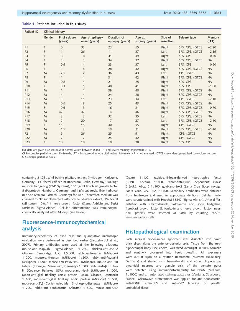
Table 1 Patients included in this study
Patient ID Clinical history
Gender First seizure(years)
Age at epilepsyonset (years)
Duration ofepilepsy (years)
Age atsurgery (years)
Side ofresection
Seizure type Memory(IAT)
P1 F 0 32 23 55 Right SPS, CPS, sGTCS �2.20
P2 F 1 26 11 37 Left SPS, CPS, sGTCS �2.35
P3 F 8 8 31 39 Right SPS, CPS 0.30
P4 F 3 3 34 37 Right SPS, CPS, sGTCS NA
P5 F 0.5 14 23 37 Left SPS, CPS 0.50
P6 F 1 4 28 32 Right SPS, CPS, sGTCS NA
P7 M 2.5 7 36 43 Left CPS, sGTCS NA
P8 F 1 11 31 42 Right SPS, CPS, sGTCS NA
P9 M 0.8 4 21 25 Right SPS, CPS NA
P10 F 0.1 1 40 41 Right SPS, CPS �1.00
P11 M 1 1 39 40 Right SPS, CPS, sGTCS NA
P12 M 4 4 24 28 Right SPS, CPS, sGTCS NA
P13 M 3 11 23 34 Left CPS, sGTCS �2.10
P14 M 0.5 18 25 43 Right SPS, CPS, sGTCS NA
P15 F 0.5 5 16 21 Right SPS, CPS, sGTCS �0.70
P16 M 42 42 4 46 Right SPS, CPS, sGTCS NA
P17 M 2 3 32 35 Left SPS, CPS, sGTCS NA
P18 M 2 20 7 27 Left SPS, CPS, sGTCS �2.10
P19 F 15 15 40 55 Right CPS, sGTCS NA
P20 M 1.5 2 19 21 Right SPS, CPS, sGTCS �1.40
P21 M 5 26 25 51 Right CPS, sGTCS NA
P22 M 7 7 25 32 Right CPS, sGTCS NA
P23 F 18 18 10 28 Right SPS, CPS NA
IAT data are given as z-scores with normal values between 0 and �1, and severe memory impairment5�2.CPS = complex partial seizures; F = female; IAT = Intracarotid amobarbital testing; M = male; NA = not analysed; sGTCS = secondary generalized tonic–clonic seizures;
SPS = simple partial seizures.
Hippocampal neurogenesis and memory dysfunction in humans Brain 2010: 133; 3359–3372 | 3361
Dow
nloaded from https://academ
ic.oup.com/brain/article-abstract/133/11/3359/313863 by guest on 23 N
ovember 2018
Neuronal cell countsSemi-quantitative cell density measurements were obtained from
all patients using 4 mm thin paraffin sections and NeuN immunohisto-
chemistry (Wolf et al., 1996). Hippocampal sectors CA1, CA2, CA3
and CA4 and the dentate gyrus were examined at �40 objective
magnification. Ten randomly placed microscopic fields were examined
for each anatomical subregion. Measurements were performed with a
microcomputer imaging system (ColorView II CCD camera, Soft ima-
ging system SIS, Stuttgart, Germany) attached to a BX51 microscope
(Olympus). Immunohistochemically stained neuronal cell bodies were
tagged on the computer screen, counted within the region of interest
and expressed as the mean number of neurons/mm2 using AnalySIS
imaging software (SIS) and Excel software (Microsoft, Redmond,
Washington, USA). Histopathological data are summarized in Table
2. The same methodology was applied for assessing the proliferation
activity in vivo using Ki67 immunoreactivity. The subgranular and
granule cell layers were analysed in 18 patients from which sufficient
material was available for preparing 10 serial 4 mm sections. We could
not perform these experiments in patients P1, P2, P3, P5 and
P7 (Table 1).
Nestin-, Sox2-, PAX6-, doublecortin-, BDNF- and cdk5-
immunoreactive cells were semi-quantitatively estimated by the same
method using fluorescence labelling. Four adjacent microscopic fields
were placed into the dentate gyrus granule cell layer at �20 objective
magnification and immunoreactive cell bodies were identified using
appropriate filter combinations.
Electrophysiological recordings fromhuman hippocampal progenitor cellsMembrane currents were measured by applying the patch-clamp
technique in a whole-cell recording configuration. Current signals
were amplified with an EPC-9 amplifier (HEKA, Lambrecht,
Germany). Whole-cell recordings were performed after three expan-
sion periods and induced differentiation (see above). After 14 days
in vitro, cells with a neuronal morphology by phase contrast imaging
were chosen (Fig. 2C). All cells were held at �70 mV. Following a
40 ms prepulse to �120 mV, voltage steps incremented by 10 mV
were applied from �80 to +10 mV every 2 s. The external buffer con-
sisted of 145 mM NaCl, 5 mM KCl, 2.4 mM CaCl2, 1 mM MgCl2,
1.8 mM glucose, 10 mM HEPES, pH adjusted to 7.4 with NaOH; the
internal buffer contained 150 mM CsCl, 5 mM EGTA, 10 mM HEPES,
and the pH adjusted to 7.2 using CsOH. All experiments were carried
out at room temperature (�22�C). Recording pipettes were fabricated
from borosilicate capillaries with open resistances of 5–6 MV.
Neuropsychological examinationIntracarotid amobarbital testing (IAT; WADA) was carried out separ-
ately in both hemispheres as part of the presurgical evaluation in nine
patients. The test is employed in patients in whom the risk for post-
operative memory loss has to be clarified preoperatively. The greatest
potential risk in surgical treatment is verbal memory loss in patients
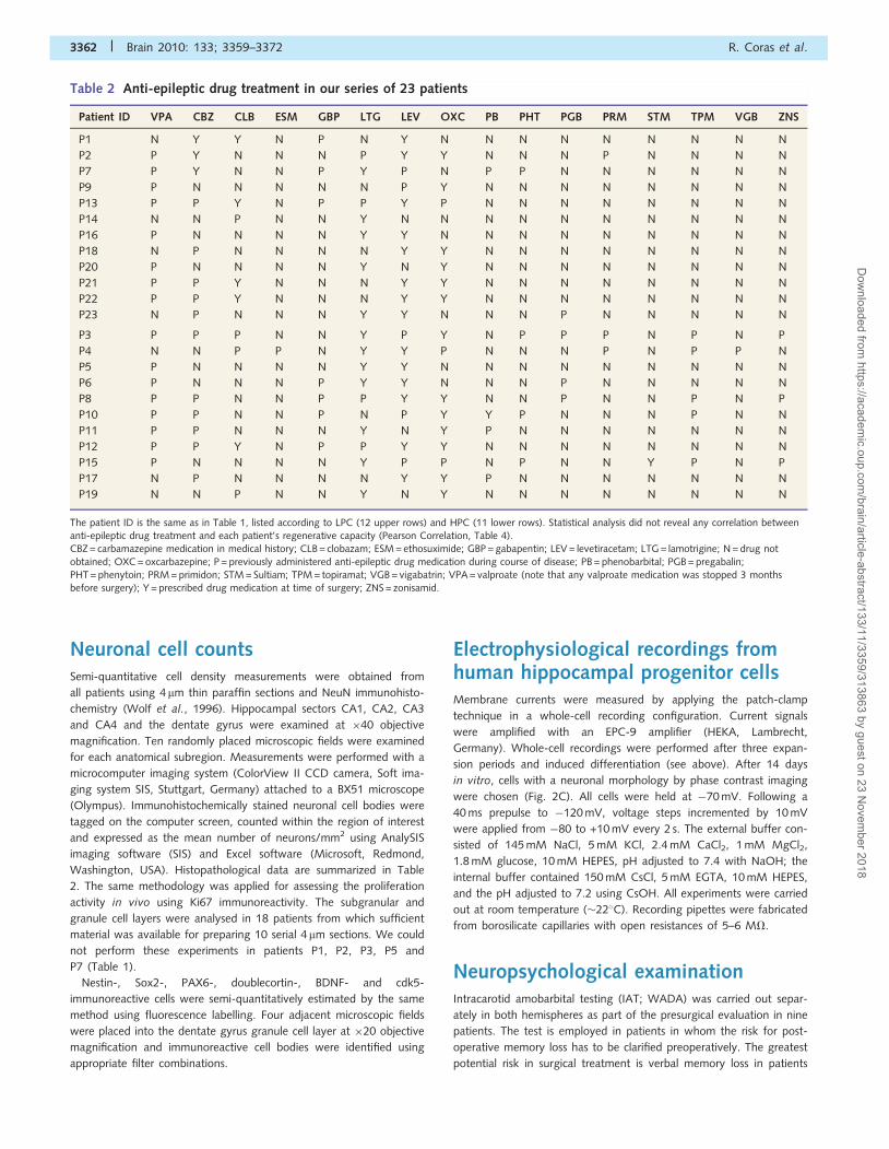
Table 2 Anti-epileptic drug treatment in our series of 23 patients
Patient ID VPA CBZ CLB ESM GBP LTG LEV OXC PB PHT PGB PRM STM TPM VGB ZNS
P1 N Y Y N P N Y N N N N N N N N N
P2 P Y N N N P Y Y N N N P N N N N
P7 P Y N N P Y P N P P N N N N N N
P9 P N N N N N P Y N N N N N N N N
P13 P P Y N P P Y P N N N N N N N N
P14 N N P N N Y N N N N N N N N N N
P16 P N N N N Y Y N N N N N N N N N
P18 N P N N N N Y Y N N N N N N N N
P20 P N N N N Y N Y N N N N N N N N
P21 P P Y N N N Y Y N N N N N N N N
P22 P P Y N N N Y Y N N N N N N N N
P23 N P N N N Y Y N N N P N N N N N
P3 P P P N N Y P Y N P P P N P N P
P4 N N P P N Y Y P N N N P N P P N
P5 P N N N N Y Y N N N N N N N N N
P6 P N N N P Y Y N N N P N N N N N
P8 P P N N P P Y Y N N P N N P N P
P10 P P N N P N P Y Y P N N N P N N
P11 P P N N N Y N Y P N N N N N N N
P12 P P Y N P P Y Y N N N N N N N N
P15 P N N N N Y P P N P N N Y P N P
P17 N P N N N N Y Y P N N N N N N N
P19 N N P N N Y N Y N N N N N N N N
The patient ID is the same as in Table 1, listed according to LPC (12 upper rows) and HPC (11 lower rows). Statistical analysis did not reveal any correlation betweenanti-epileptic drug treatment and each patient’s regenerative capacity (Pearson Correlation, Table 4).CBZ = carbamazepine medication in medical history; CLB = clobazam; ESM = ethosuximide; GBP = gabapentin; LEV = levetiracetam; LTG = lamotrigine; N = drug not
obtained; OXC = oxcarbazepine; P = previously administered anti-epileptic drug medication during course of disease; PB = phenobarbital; PGB = pregabalin;PHT = phenytoin; PRM = primidon; STM = Sultiam; TPM = topiramat; VGB = vigabatrin; VPA = valproate (note that any valproate medication was stopped 3 monthsbefore surgery); Y = prescribed drug medication at time of surgery; ZNS = zonisamid.
3362 | Brain 2010: 133; 3359–3372 R. Coras et al.
Dow
nloaded from https://academ
ic.oup.com/brain/article-abstract/133/11/3359/313863 by guest on 23 N
ovember 2018

suffering from left-sided temporal lobe epilepsy (Chelune, 1995), but
suspicion of atypical memory dominance in right-sided temporal lobe
epilepsy should in some cases also require examination. Thus 5 out of
6 patients with left-sided temporal lobe epilepsy and 4 out of 17 pa-
tients with right-sided temporal lobe epilepsy underwent IAT following
the Erlangen Wada Test protocol. This protocol is described in detail
elsewhere (Pauli et al., 2006). In brief, double encodeable memory
items are tested under recall and recognition conditions and the results
are transformed into z-scores according to normative values specific
for speech dominant and for non-dominant hemispheres, respectively.
Since healthy control data are not available for IAT, standardization
was based on values from the contralateral, non-affected left or right
temporal lobes, including only those patients from our database
(n4200) presenting with: (i) unilateral mesial temporal lobe epilepsy;
(ii) unilateral left-sided speech dominance; (iii) complete postoperative
seizure freedom; and (iv) normal range IQ. IAT memory scores were
transformed into z-values following the calculation rule: (i) if the re-
sected hippocampus was taken from the speech dominant hemisphere:
z(IAT memory) = [(Total memory score�Meanleft)/SDleft]; and (ii) if the
resected hippocampus was taken from the non-dominant hemisphere:
z(IAT memory) = [(Total memory score�Meanright)/SDrigh]). Thus, a z-
score of 0 indicates full functional integrity of the investigated left or
right hippocampal structure; i.e. indicates equality with the average
score of non-affected left or right hippocampi in unilateral temporal
lobe epilepsy.
Statistical analysisStatistical Package for the Social Sciences (SPSS, version 16) was used
for statistical evaluation. The threshold for significance was set to 0.05.
Correlation analysis, linear multiple regression and partial correlations
were calculated to evaluate the relationship between clinical histories,
histopathological data, cell culture and memory performance. Cluster
analysis and discriminance analysis were used to explore neurogenesis
in vitro. Correlation analysis (Pearson) was performed to relate
(i) neurogenesis in vitro with clinical data (age, onset and duration
of epilepsy); (ii) neurogenesis in vitro with cell densities in hippocam-
pal subfields and the dentate gyrus; and (iii) neurogenesis in vitro with
IAT memory capacity. Only variables that significantly correlated with
the dependent variable were included in linear regression and partial
correlation analysis. Since the multiple correlation coefficient R tends
to overestimate the correlation between observed and predicted values
of dependent variables, adjusted R2, which is thought to more closely
reflect the goodness of fit of the model in a population, was calcu-
lated. R2adjusted describes the proportion of variance of the dependent
variable that is explained by the independent variables. To assess the
relative importance of each independent variable, we applied the
t-statistic for the linear regression coefficient and only values 42
were considered significant. To check for multicolinearity, tolerance
statistics were used. In addition, partial correlation analysis was calcu-
lated to assess the correlation of the independent variable with the
dependent variable after removing the linear effect of the other vari-
ables in the model. Neuropsychological parameters as well as cell den-
sities in the hippocampal subfields were transformed into z-scores,
representing, in standard deviation units, the amount score deviates
from the mean of the population from which the score is drawn (null
hypothesis). For histopathological data (cell densities in the hippocam-
pal subfields and the dentate gyrus), standardization was based on
age-matched non-epileptic autopsy controls (Blumcke et al., 2007).
For IAT, standardization was based on normal values from contralat-
eral, non-affected left or right temporal lobes in unilateral temporal
lobe epilepsy.
Results
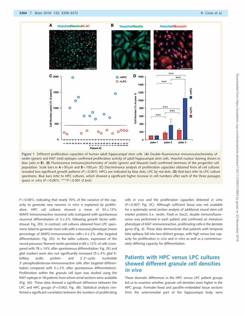
HPC and LPC in adult humanhippocampal stem cells in vitroAdult hippocampal progenitor cells have the capacity for
self-renewal, to generate new cells by asymmetric division and
further differentiate into various cell lineages of the brain (Gage,
2000). We obtained surgical hippocampal tissue from 23 patients
with drug-resistant epilepsy (Table 1). Following microdissection of
the dentate gyrus, cells were dissociated and proliferated as a
monolayer culture (Fig. 1A). Three cell culture passages were
applied to expand the progenitor cell population (‘Materials and
methods’ section), which were characterized by abundant expres-
sion of nestin and Musashi1 (Fig. 1B). Cluster analysis of data
obtained after three cell culture passages in vitro from all subjects
revealed two significant patterns (Fig. 1C; F = 45.1; df = 1/22;
P50.001). An HPC was observed in 11 specimens, showing dou-
bling of primarily seeded cells after 2–3 weeks and continued pro-
liferation through further passages in vitro (Fig. 1D). An LPC was
evident in the remaining 12 specimens. Although cell numbers
increased during each passage in LPC cultures, their amount re-
mained significantly lower after three passages in vitro compared
with HPC specimens.
HPC versus LPC cultures showeddifferent propensities for neuronaldifferentiationAfter completing our proliferation protocols, growth factor with-
drawal induced spontaneous differentiation into a neuronal
phenotype in most cell cultures, ranging from 0 to 6% (mean
2� 2%) of the total cell population. Differentiation into a neur-
onal phenotype was significantly enhanced using suberoylanilide
hydroxamic acid, an inhibitor of histone deacetylases (Hsieh et al.,
2004; Siebzehnrubl et al., 2007), as well as sonic hedgehog and
fibroblast growth factor 8 for 48 h, followed by treatment with the
adenylcyclase activator forskolin and nerve growth factor for
12 days (mean 28� 27%). Cellular phenotype was microscopically
analysed after 14 days in culture and staining with antibodies dir-
ected against antigens specific for neurons, i.e. MAP2 and NeuN
(Fig. 2A and B), astrocytes (glial fibrillary acidic protein,
not shown) and oligodendrocytes (20,30-cyclic nucleotide
30-phosphodiesterase, not shown), suggesting the maintenance
of multipotent adult hippocampal stem cells in our cultures. In
addition, patch-clamp recordings detected sodium inward currents
of up to 300 pA (Fig. 2C) in cells with dendrite-like neuronal ar-
borizations. These findings demonstrated that adult hippocampal
stem cells can be isolated from human surgical tissue and differ-
entiated into neurons.
There was a highly significant correlation between proliferation
and the same patient’s capacity to generate neurons in vitro
(r = 0.834, R2 = 0.696, P50.001). Linear regression analysis
revealed proliferation as the only variable meeting the demands
of the predictors criterion |t|42 (T = 6.94, R2adjusted = 0.696,
Hippocampal neurogenesis and memory dysfunction in humans Brain 2010: 133; 3359–3372 | 3363
Dow
nloaded from https://academ
ic.oup.com/brain/article-abstract/133/11/3359/313863 by guest on 23 N
ovember 2018
P50.001), indicating that nearly 70% of the variance of the cap-
acity to generate new neurons in vitro is explained by prolifer-
ation. HPC cell cultures showed a mean of 53� 13%
MAP2-immunoreactive neuronal cells (compared with spontaneous
neuronal differentiation of 3� 2% following growth factor with-
drawal; Fig. 2D). In contrast, cell cultures obtained from LPC speci-
mens failed to generate more cells with a neuronal phenotype (mean
percentage of MAP2-immunoreactive cells = 4� 2% after targeted
differentiation; Fig. 2D). In the latter cultures, expression of the
neural precursor filament nestin persisted in 69� 12% of cells (com-
pared with 78� 14% after spontaneous differentiation; Fig. 2E) and
glial markers were also not significantly increased (5� 4% glial fi-
brillary acidic protein- and 20,30-cyclic nucleotide
30-phosphodiesterase-immunoreactive cells after targeted differen-
tiation compared with 3� 2% after spontaneous differentiation).
Proliferation within the granule cell layer was studied using the
Ki67-epitope in 18 patients from whom serial sections were available
(Fig. 3A). These data showed a significant difference between the
LPC and HPC groups (P = 0.002; Fig. 3B). Statistical analysis con-
firmed a significant correlation between the numbers of proliferating
cells in vivo and the proliferation capacities detected in vitro
(P = 0.007; Fig. 3C). Although sufficient tissue was not available
for a systematic serial section analysis of additional neural stem cell
marker proteins (i.e. nestin, Pax6 or Sox2), double immunofluore-
sence was performed in each patient and confirmed an immature
phenotype of Ki67-immunoreactive, proliferating cells in the dentate
gyrus (Fig. 4). These data demonstrate that patients with temporal
lobe epilepsy fall into two distinct groups, with high versus low cap-
acity for proliferation in vivo and in vitro as well as a commensur-
ately differing capacity for differentiation.
Patients with HPC versus LPC culturesshowed different granule cell densitiesin vivoThese dramatic differences in the HPC versus LPC patient groups
led us to examine whether granule cell densities were higher in the
HPC group. Formalin-fixed and paraffin-embedded tissue sections
from the anteromedial part of the hippocampal body were
Figure 1 Different proliferation capacities of human adult hippocampal stem cells. (A) Double-fluorescence immunocytochemistry of
nestin (green) and Ki67 (red) epitopes confirmed proliferation activity of adult hippocampal stem cells. Hoechst nuclear staining shown in
blue (also in B). (B) Fluorescence immunocytochemistry of nestin (green) and Musashi (red) confirmed stemness of the progenitor cell
population. Scale bars in A = 50 mm and B = 100mm. (C) Discriminance analysis of proliferation capacities obtained from all cell cultures
revealed two significant growth patterns (P50.001). HPCs are indicated by blue dots, LPC by red dots. (D) Red bars refer to LPC culture
specimens. Blue bars refer to HPC cultures, which showed a significant higher increase in cell numbers after each of the three passages
(pass) in vitro (P50.001). ***P50.001 (t test).
3364 | Brain 2010: 133; 3359–3372 R. Coras et al.
Dow
nloaded from https://academ
ic.oup.com/brain/article-abstract/133/11/3359/313863 by guest on 23 N
ovember 2018
available from all surgical specimens and were adjacent to those
used for cell culture experiments (Fig. 5). Neuronal cell numbers
and densities were quantitatively determined in all hippocampal
subfields, including the pyramidal cell layer as well as the granule
cell layer of the dentate gyrus (Table 3). There was a highly sig-
nificant correlation between granule cell densities and the same
patient’s proliferation capacity in vitro (r = 0.813, R2= 0.661,
P50.001). Neuronal cell numbers obtained from hippocampal
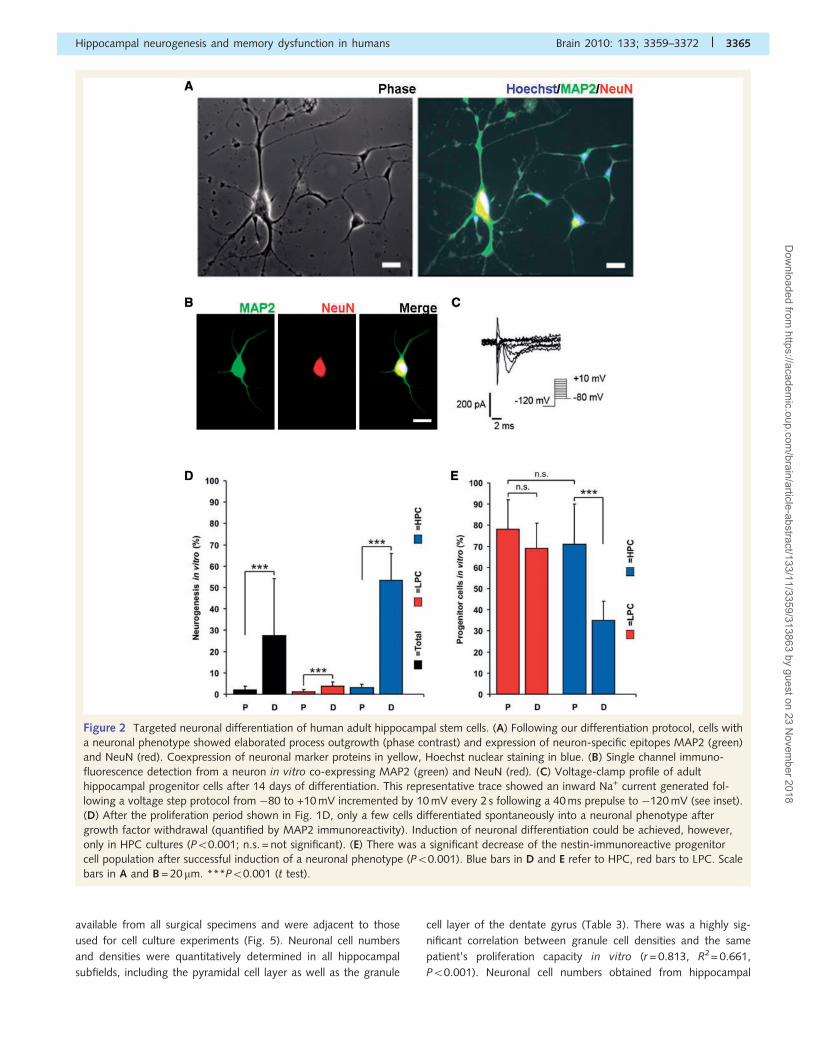
Figure 2 Targeted neuronal differentiation of human adult hippocampal stem cells. (A) Following our differentiation protocol, cells with
a neuronal phenotype showed elaborated process outgrowth (phase contrast) and expression of neuron-specific epitopes MAP2 (green)
and NeuN (red). Coexpression of neuronal marker proteins in yellow, Hoechst nuclear staining in blue. (B) Single channel immuno-
fluorescence detection from a neuron in vitro co-expressing MAP2 (green) and NeuN (red). (C) Voltage-clamp profile of adult
hippocampal progenitor cells after 14 days of differentiation. This representative trace showed an inward Na+ current generated fol-
lowing a voltage step protocol from �80 to +10 mV incremented by 10 mV every 2 s following a 40 ms prepulse to �120 mV (see inset).
(D) After the proliferation period shown in Fig. 1D, only a few cells differentiated spontaneously into a neuronal phenotype after
growth factor withdrawal (quantified by MAP2 immunoreactivity). Induction of neuronal differentiation could be achieved, however,
only in HPC cultures (P50.001; n.s. = not significant). (E) There was a significant decrease of the nestin-immunoreactive progenitor
cell population after successful induction of a neuronal phenotype (P50.001). Blue bars in D and E refer to HPC, red bars to LPC. Scale
bars in A and B = 20 mm. ***P50.001 (t test).
Hippocampal neurogenesis and memory dysfunction in humans Brain 2010: 133; 3359–3372 | 3365
Dow
nloaded from https://academ
ic.oup.com/brain/article-abstract/133/11/3359/313863 by guest on 23 N
ovember 2018
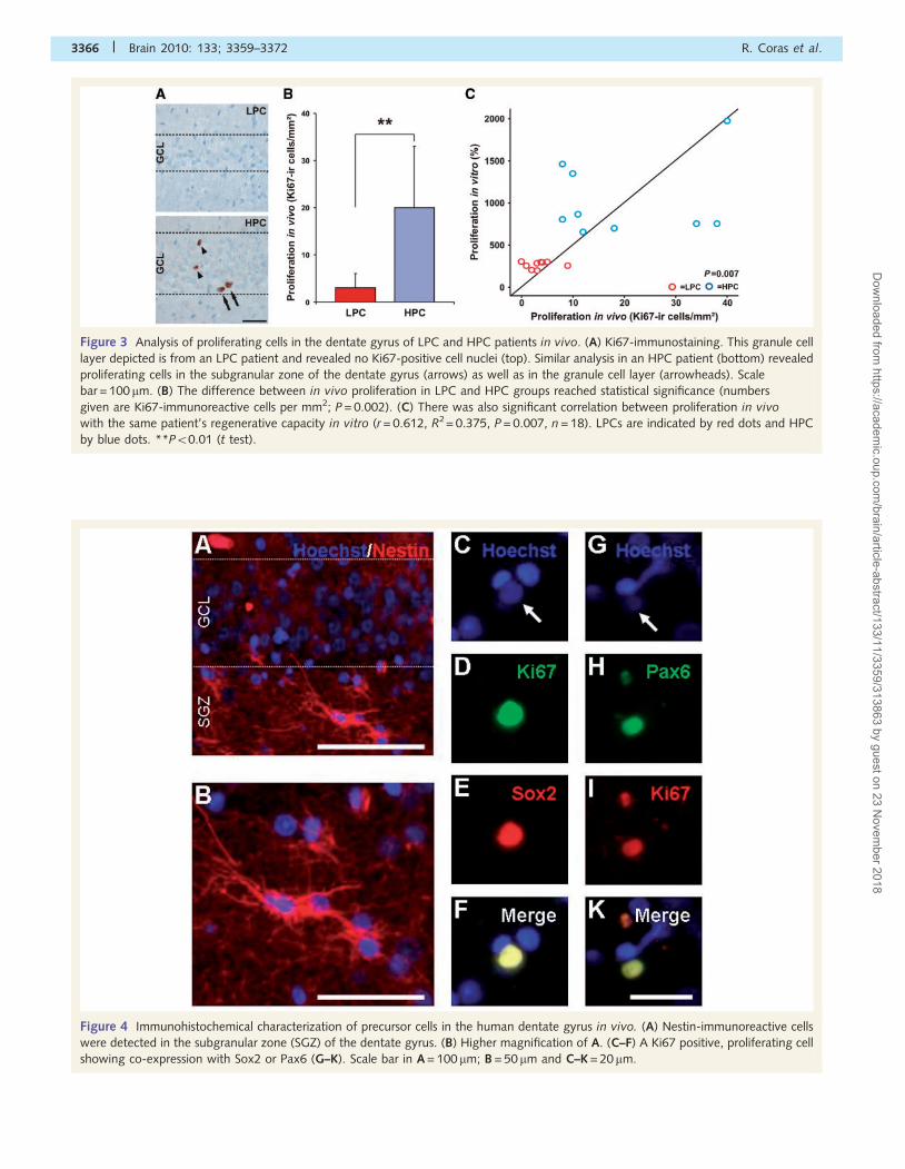
Figure 4 Immunohistochemical characterization of precursor cells in the human dentate gyrus in vivo. (A) Nestin-immunoreactive cells
were detected in the subgranular zone (SGZ) of the dentate gyrus. (B) Higher magnification of A. (C–F) A Ki67 positive, proliferating cell
showing co-expression with Sox2 or Pax6 (G–K). Scale bar in A = 100 mm; B = 50 mm and C–K = 20 mm.
Figure 3 Analysis of proliferating cells in the dentate gyrus of LPC and HPC patients in vivo. (A) Ki67-immunostaining. This granule cell
layer depicted is from an LPC patient and revealed no Ki67-positive cell nuclei (top). Similar analysis in an HPC patient (bottom) revealed
proliferating cells in the subgranular zone of the dentate gyrus (arrows) as well as in the granule cell layer (arrowheads). Scale
bar = 100 mm. (B) The difference between in vivo proliferation in LPC and HPC groups reached statistical significance (numbers
given are Ki67-immunoreactive cells per mm2; P = 0.002). (C) There was also significant correlation between proliferation in vivo
with the same patient’s regenerative capacity in vitro (r = 0.612, R2= 0.375, P = 0.007, n = 18). LPCs are indicated by red dots and HPC
by blue dots. **P50.01 (t test).
3366 | Brain 2010: 133; 3359–3372 R. Coras et al.
Dow
nloaded from https://academ
ic.oup.com/brain/article-abstract/133/11/3359/313863 by guest on 23 N
ovember 2018
sectors CA4 (r = 0.652, P = 0.001) and CA3 (r = 0.487, P = 0.040)
correlated significantly with the patient’s stem cell proliferation
capacity in vitro, although to a lower extent. Even so, there was
a high intercorrelation of cell densities in the different hippocampal
subfields and the dentate gyrus. Therefore, multiple linear regres-
sions were applied to further examine these correlations and
to determine the most important predictor for stem cell prolifer-
ation in vitro. When cell densities in CA3 and CA4 as well
as granule cell density were entered stepwise into the equation,
R2adjusted reached 0.64, but only the granule cell density of
the dentate gyrus fulfilled the predictor’s criteria for relative
importance (ktkabsolut = 5.58642). Removing the effect of cell
densities in CA3 and CA4 by partial correlation analysis, a signifi-
cant partial correlation coefficient resulted [R(partial) = 0.81,
P50.001], not different from the basic correlation coefficient
(r = 0.813).
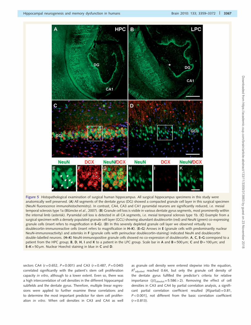
Figure 5 Histopathological examination of surgical human hippocampus. All surgical hippocampus specimens in this study were
anatomically well preserved. (A) All segments of the dentate gyrus (DG) showed a compacted granule cell layer in this surgical specimen
(NeuN fluorescence immunohistochemistry). In contrast, CA4, CA3 and CA1 pyramidal neurons are significantly reduced, i.e. mesial
temporal sclerosis type 1a (Blumcke et al., 2007). (B) Granule cell loss is visible in various dentate gyrus segments, most prominently within
the internal limb (asterisk). Pyramidal cell loss is detected in all CA segments, i.e. mesial temporal sclerosis type 1b. (C) Example from a
surgical specimen with a densely populated granule cell layer (GCL) showing abundant doublecortin (red) and NeuN (green) co-expressing
granule cells (insert refers to magnification in E–G). (D) In this severely depleted granule cell layer we observed virtually no
doublecortin-immunoreactive cells (insert refers to magnification in H–K). (E–G) Arrows in E (granule cells with predominantly nuclear
NeuN-immunoreactivity) and asterisks in F (granule cells with perinuclear doublecortin-staining) indicated NeuN and doublecortin
double-labelled neurons. (H–K) NeuN-immunopositive granule cells showed no co-expression of doublecortin. A, C, E–G correspond to a
patient from the HPC group; B, D, H, I and K to a patient in the LPC group. Scale bar in A and B = 500 mm; C and D = 100 mm; and
E–K = 50mm. Nuclear Hoechst staining in blue in C and D.
Hippocampal neurogenesis and memory dysfunction in humans Brain 2010: 133; 3359–3372 | 3367
Dow
nloaded from https://academ
ic.oup.com/brain/article-abstract/133/11/3359/313863 by guest on 23 N
ovember 2018
Diagnostic evaluation of segmental neuronal cell loss patterns
revealed mesial temporal sclerosis in 19 out of 23 patients, eight in
the group with abundant neurogenesis in vitro and 11 in the
group with a reduced regenerative capacity.
Patients with HPC versus LPC culturesrevealed different immunoreactivitypatterns for doublecortin, brain-derivedneurotrophic factor and cdk5 in vivoThe increased neurogenesis in the HPC patient group should be
reflected in an increased proportion of neurons expressing surface
markers characteristic of newly generated and integrated neurons.
We carried out double-immunofluorescence labelling for double-
cortin and NeuN in an additional set of experiments to address this
issue (Liu et al., 2008). In 12 patients with LPC, doublecortin ex-
pression was observed in 20.3� 8.5% of the total granule cell
population. The amount of doublecortin-positive granule cells
was significantly higher in the group of 11 patients with HPC
in vitro (53.4� 13.6%), and correlated significantly with the pro-
liferation rate observed in vitro (r = 0.790, P50.001; Fig. 7). These
cell numbers were significantly higher compared with recently
published data by Knoth et al. (2010) in autopsy controls and
Gerber et al. (2009) in meningitis specimens, whereas staining
patterns were similar to that reported by Jin et al. (2004) in the
dentate gyrus of patients with Alzheimer’s disease. Different anti-
body origin and antigen retrieval systems, as well as fixation inter-
vals, are likely to account for these differences. Furthermore, we
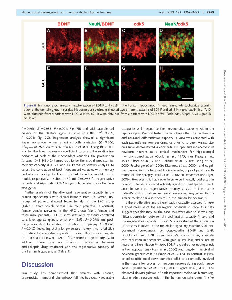
studied the expression of two molecules involved in the molecular
signalling machinery of hippocampal neurogenesis, i.e. BDNF and
cdk5 (Fig. 6). BDNF- and cdk5-expression was significantly
reduced in 12 specimens with LPC in vitro when compared with
the 11 patients with HPC in vitro, and the numbers correlated
significantly with each patient’s proliferation rate observed
in vitro (BDNF: r = 0.596, P = 0.003; cdk5: r = 0.596, P = 0.003).
Significant correlation between theregenerative capacity in vitro and thesame patient’s memoryWe observed a striking and highly significant correlation between
each patient’s regenerative capacity deduced either from our cell
culture assay (in vitro) or from histopathological characterization
(in vivo) and the same patient’s ability to acquire and recall new
memories during preoperative examination. IAT memory corre-
lated significantly with the regenerative capacity in vitro
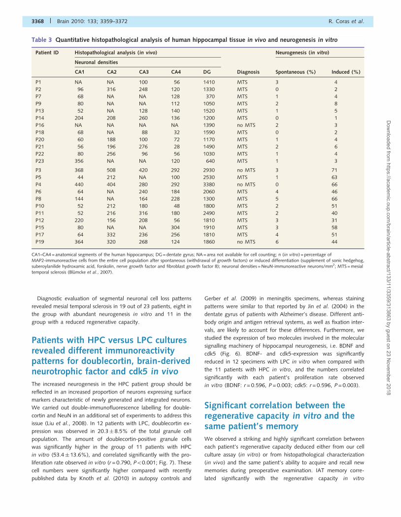
Table 3 Quantitative histopathological analysis of human hippocampal tissue in vivo and neurogenesis in vitro
Patient ID Histopathological analysis (in vivo) Neurogenesis (in vitro)
Neuronal densities
CA1 CA2 CA3 CA4 DG Diagnosis Spontaneous (%) Induced (%)
P1 NA NA 100 56 1410 MTS 3 4
P2 96 316 248 120 1330 MTS 0 2
P7 68 NA NA 128 370 MTS 1 4
P9 80 NA NA 112 1050 MTS 2 8
P13 52 NA 128 140 1520 MTS 1 5
P14 204 208 260 136 1200 MTS 0 1
P16 NA NA NA NA 1390 no MTS 2 3
P18 68 NA 88 32 1590 MTS 0 2
P20 60 188 100 72 1170 MTS 1 4
P21 56 196 276 28 1490 MTS 2 6
P22 80 256 96 56 1030 MTS 1 4
P23 356 NA NA 120 640 MTS 1 3
P3 368 508 420 292 2930 no MTS 3 71
P5 44 212 NA 100 2530 MTS 1 63
P4 440 404 280 292 3380 no MTS 0 66
P6 64 NA 240 184 2060 MTS 4 46
P8 144 NA 164 228 1300 MTS 5 66
P10 52 212 180 48 1800 MTS 2 51
P11 52 216 316 180 2490 MTS 2 40
P12 220 156 208 56 1810 MTS 3 31
P15 80 NA NA 304 1910 MTS 3 58
P17 64 332 236 256 1810 MTS 4 51
P19 364 320 268 124 1860 no MTS 6 44
CA1–CA4 = anatomical segments of the human hippocampus; DG = dentate gyrus; NA = area not available for cell counting; n (in vitro) = percentage ofMAP2-immunoreactive cells from the entire cell population after spontaneous (withdrawal of growth factors) or induced differentiation (supplement of sonic hedgehog,suberoylanilide hydroxamic acid, forskolin, nerve growth factor and fibroblast growth factor 8); neuronal densities = NeuN-immunoreactive neurons/mm2; MTS = mesialtemporal sclerosis (Blumcke et al., 2007).
3368 | Brain 2010: 133; 3359–3372 R. Coras et al.
Dow
nloaded from https://academ
ic.oup.com/brain/article-abstract/133/11/3359/313863 by guest on 23 N
ovember 2018
(r = 0.966, R2 = 0.933, P50.001; Fig. 7B) and with granule cell
density of the dentate gyrus in vivo (r = 0.888, R2 = 0.789,
P = 0.001; Fig. 7C). Regression analysis showed a significant
linear regression when entering both variables (R = 0.966,
R2adjusted = 0.923, F = 96.976, df = 1/7, P50.001). Using the t-stat-
istic for the linear regression coefficient to assess the relative im-
portance of each of the independent variables, the proliferation
in vitro (t = 9.84842) turned out to be the crucial predictor for
memory capacity (Fig. 7A and B). Partial correlation analysis, to
assess the correlation of both independent variables with memory
and when removing the linear effect of the other variable in the
model, respectively, resulted in R(partial) = 0.966 for regenerative
capacity and R(partial) = 0.682 for granule cell density in the den-
tate gyrus.
Further analysis of the divergent regenerative capacity in the
human hippocampus with clinical histories from LPC versus HPC
groups of patients showed fewer females in the LPC group
(Table 1; three female versus nine male patients). In contrast,
female gender prevailed in the HPC group (eight female and
three male patients). LPC in vitro was only by trend correlated
to a later age at epilepsy onset (r =�3.53, P = 0.098) and posi-
tively correlated to a shorter duration of epilepsy (r = 0.426,
P = 0.042), indicating that a longer seizure history is not predictive
for reduced regenerative capacities in vitro. There was no signifi-
cant correlation between age at first seizure or age at surgery. In
addition, there was no significant correlation between
anti-epileptic drug treatment and the regenerative capacity of
the human hippocampus (Table 4).
DiscussionOur study has demonstrated that patients with chronic,
drug-resistant temporal lobe epilepsy fall into two clearly separable
categories with respect to their regenerative capacity within the
hippocampus. We first tested the hypothesis that the proliferation
and neuronal differentiation capacity in vitro was correlated with
each patient’s memory performance prior to surgery. Animal stu-
dies have demonstrated a constitutive supply and replacement of
newborn neurons as a critical mechanism for hippocampal
memory consolidation (Gould et al., 1999; van Praag et al.,
1999; Shors et al., 2001; Clelland et al., 2009; Deng et al.,
2009; Jessberger et al., 2009; Kitamura et al., 2009), and cogni-
tive dysfunction is a frequent finding in subgroups of patients with
temporal lobe epilepsy (Pauli et al., 2006; Helmstaedter and Elger,
2009). However, this has never been experimentally addressed in
humans. Our data showed a highly significant and specific correl-
ation between the regenerative capacity in vitro and the same
patient’s ability to store and recall memories, suggesting that a
similar mechanism also operates in the human hippocampus.
Is the proliferative and differentiation capacity assessed in vitro
a good measure of the neurogenic potential in vivo? Our data
suggest that this may be the case. We were able to show a sig-
nificant correlation between the proliferation capacity in vivo and
the regenerative capacity in vitro. We also studied the expression
of proteins involved in the molecular signalling machinery of hip-
pocampal neurogenesis, i.e. doublecortin, BDNF and cdk5.
Doublecortin and BDNF, as well as cdk5, revealed a highly signifi-
cant reduction in specimens with granule cell loss and failure of
neuronal differentiation in vitro. BDNF is required for neurogenesis
in the hippocampus (Rossi et al., 2006) and long-term survival of
newborn granule cells (Sairanen et al., 2005). In contrast, region-
or cell-specific knockdown identified cdk5 to be critically involved
in the maturation process of newborn neurons during adult neuro-
genesis (Jessberger et al., 2008, 2009; Lagace et al., 2008). The
observed downregulation of both important molecular factors reg-
ulating adult neurogenesis in the human dentate gyrus in vivo
Figure 6 Immunohistochemical characterization of BDNF and cdk5 in the human hippocampus in vivo. Immunohistochemical examin-
ation of the dentate gyrus in surgical hippocampus specimens showed two different patterns of BDNF and cdk5 immunoreactivities. (A–D)
were obtained from a patient with HPC in vitro. (E–H) were obtained from a patient with LPC in vitro. Scale bar = 50 mm. GCL = granule
cell layer.
Hippocampal neurogenesis and memory dysfunction in humans Brain 2010: 133; 3359–3372 | 3369
Dow
nloaded from https://academ
ic.oup.com/brain/article-abstract/133/11/3359/313863 by guest on 23 N
ovember 2018
confirmed a compromised molecular machinery for hippocampal
neurogenesis in the patient group with an LPC in vitro. This sug-
gests that neurogenesis in the human hippocampus, first detected
in five autoptic brain samples obtained from cancer patients trea-
ted with the thymidine analog bromodeoxyuridine (Eriksson et al.,
1998), is closely linked to the neurogenic potential in vivo. Finally,
the close correlation of the neurogenic potential with the density
of granule cells suggests that neurogenesis also regulates the gran-
ule cell numbers in human subjects.
Increased hippocampal neurogenesis has been observed in dif-
ferent epilepsy models (Parent et al., 1997, 2006; Siebzehnrubl
and Blumcke, 2008), and increased numbers of nestin-
immunoreactive neural precursor cells were detected in the den-
tate gyrus of patients with temporal lobe epilepsy younger than
4 years at time of operation (Blumcke et al., 2001). In contrast,
the regenerative capacity of the hippocampus declined in chronic
seizure models (Hattiangady et al., 2004; Hattiangady and Shetty,
2009) and is also likely to decrease with age (Fahrner et al., 2007;
Ahlenius et al., 2009). However, our data suggest no clear effects
of early epilepsy onset, longer duration of seizures or higher age at
surgery on adult neurogenesis. This may be due to the very long
period of chronic seizures in all patients, far exceeding that studied
in any animal model. Rather, compromised neurogenesis was
observed in patients with late onset of first seizures. This observa-
tion will need further clarification. It is in line with the notion,
however, that less vulnerable neuronal and stem cell populations
can be detected in younger compared with older animals (Haas
et al., 2001; Liu et al., 2003). Furthermore, nestin-expressing pre-
cursor cells persisted in cultures obtained from patients with tem-
poral lobe epilepsy with compromised neurogenesis in vitro
(Fig. 2E). Similar data are obtained from an animal model of tem-
poral lobe epilepsy, showing no change in the neural precursor cell
population but a dramatic decline in neuronal fate-choice decision
of newly generated cells (Hattiangady and Shetty, 2009).
Prolonged nestin expression at the expense of newly generated
neurons was also observed in aged rats, particularly in those ani-
mals with severe spatial learning deficits during Morris Water
Maze testing (Nyffeler et al., 2008). Indeed, causal events in the
disruption of the neurogenic fate-choice of progenitor cells may be
involved and will need clarification.
There is much debate in regenerative medicine about the func-
tional and clinical impact of hippocampal progenitor cells and
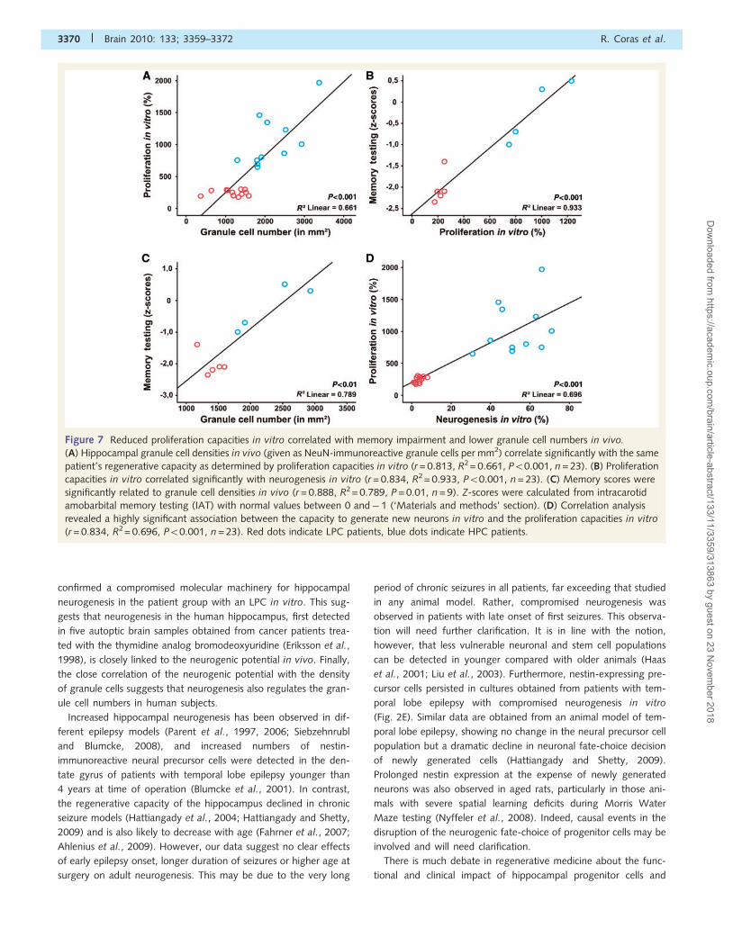
Figure 7 Reduced proliferation capacities in vitro correlated with memory impairment and lower granule cell numbers in vivo.
(A) Hippocampal granule cell densities in vivo (given as NeuN-immunoreactive granule cells per mm2) correlate significantly with the same
patient’s regenerative capacity as determined by proliferation capacities in vitro (r = 0.813, R2 = 0.661, P50.001, n = 23). (B) Proliferation
capacities in vitro correlated significantly with neurogenesis in vitro (r = 0.834, R2 = 0.933, P50.001, n = 23). (C) Memory scores were
significantly related to granule cell densities in vivo (r = 0.888, R2 = 0.789, P = 0.01, n = 9). Z-scores were calculated from intracarotid
amobarbital memory testing (IAT) with normal values between 0 and� 1 (‘Materials and methods’ section). (D) Correlation analysis
revealed a highly significant association between the capacity to generate new neurons in vitro and the proliferation capacities in vitro
(r = 0.834, R2= 0.696, P50.001, n = 23). Red dots indicate LPC patients, blue dots indicate HPC patients.
3370 | Brain 2010: 133; 3359–3372 R. Coras et al.
Dow
nloaded from https://academ
ic.oup.com/brain/article-abstract/133/11/3359/313863 by guest on 23 N
ovember 2018
neurogenesis (Steindler and Pincus, 2002). Adult stem cells and
the endogenous regenerative capacity of the human brain are
promising therapeutic targets for many neurodegenerative dis-
orders, such as Alzheimer’s or Parkinson’s disease (Rodriguez
et al., 2008; Winner et al., 2009), even though the regenerative
capacity may be severely compromised during a given patient’s
long-term disease history. Our data show evidence that neurogen-
esis is not necessarily affected by chronic neurological disease, as
the majority of our patients suffered from a very long period of
chronic seizures originating in the hippocampus. Drugs targeting
the molecular machinery towards a neuronal fate-choice of hippo-
campal precursor cells may thus be promising therapeutic options
to ameliorate learning and memory deficits associated with a var-
iety of neurological disorders.
AcknowledgementsWe thank Dorit Muller and Birte Rings for their expert technical
assistance. F.A.S. is a scholar of the German national academic
foundation (Studienstiftung des deutschen Volkes e.V.).
FundingThe European Community (LSH-CT-2006-037315 EPICURE);
German Research Council (DFG Bl 421/1-2); and Bavarian
Research Council (ForNeuroCell).
ReferencesAhlenius H, Visan V, Kokaia M, Lindvall O, Kokaia Z. Neural stem and
progenitor cells retain their potential for proliferation and differenti-
ation into functional neurons despite lower number in aged brain. J
Neurosci 2009; 29: 4408–19.
Altman J. Are new neurons formed in the brains of adult mammals?
Science 1962; 135: 1127–8.
Bakker A, Kirwan CB, Miller M, Stark CE. Pattern separation in the
human hippocampal CA3 and dentate gyrus. Science 2008; 319:
1640–2.
Blumcke I, Pauli E, Clusmann H, Schramm J, Becker A, Elger C, et al. A
new clinico-pathological classification system for mesial temporal scler-
osis. Acta Neuropathol 2007; 113: 235–44.
Blumcke I, Schewe JC, Normann S, Brustle O, Schramm J, Elger CE, et al.
Increase of nestin-immunoreactive neural precursor cells in the dentate
gyrus of pediatric patients with early-onset temporal lobe epilepsy.
Hippocampus 2001; 11: 311–21.
Chelune GJ. Hippocampal adequacy versus functional reserve: predicting
memory functions following temporal lobectomy. Arch Clin
Neuropsychol 1995; 10: 413–32.
Clelland CD, Choi M, Romberg C, Clemenson GD Jr, Fragniere A,
Tyers P, et al. A functional role for adult hippocampal neurogenesis
in spatial pattern separation. Science 2009; 325: 210–3.
Deng W, Saxe MD, Gallina IS, Gage FH. Adult-born
hippocampal dentate granule cells undergoing maturation
modulate learning and memory in the brain. J Neurosci 2009; 29:
13532–42.
Engel J Jr. Epilepsy surgery. Curr Opin Neurol 1994; 7: 140–7.Eriksson PS, Perfilieva E, Bjork-Eriksson T, Alborn AM, Nordborg C,
Peterson DA, et al. Neurogenesis in the adult human hippocampus.
Nat Med 1998; 4: 1313–7.
Fahrner A, Kann G, Flubacher A, Heinrich C, Freiman TM, Zentner J,
et al. Granule cell dispersion is not accompanied by enhanced neuro-
genesis in temporal lobe epilepsy patients. Exp Neurol 2007; 203:
320–32.
Farioli-Vecchioli S, Saraulli D, Costanzi M, Pacioni S, Cina I, Aceti M,
et al. The timing of differentiation of adult hippocampal neurons is
crucial for spatial memory. PLoS Biol 2008; 6: e246.Gage FH. Mammalian neural stem cells. Science 2000; 287: 1433–8.
Gerber J, Tauber SC, Armbrecht I, Schmidt H, Bruck W, Nau R. Increased
neuronal proliferation in human bacterial meningitis. Neurology 2009;
73: 1026–32.Gould E, Beylin A, Tanapat P, Reeves A, Shors TJ. Learning enhances
adult neurogenesis in the hippocampal formation. Nat Neurosci 1999;
2: 260–5.
Haas KZ, Sperber EF, Opanashuk LA, Stanton PK, Moshe SL. Resistance
of immature hippocampus to morphologic and physiologic alterations
following status epilepticus or kindling. Hippocampus 2001; 11:
615–25.
Hattiangady B, Rao MS, Shetty AK. Chronic temporal lobe epilepsy is
associated with severely declined dentate neurogenesis in the adult
hippocampus. Neurobiol Dis 2004; 17: 473–90.Hattiangady B, Shetty AK. Decreased neuronal differentiation
of newly generated cells underlies reduced hippocampal neurogen-
esis in chronic temporal lobe epilepsy. Hippocampus 2009; 20:
97–112.
Helmstaedter C, Elger CE. Chronic temporal lobe epilepsy: a neurodeve-
lopmental or progressively dementing disease? Brain 2009; 132:
2822–30.
Hsieh J, Nakashima K, Kuwabara T, Mejia E, Gage FH. Histone
deacetylase inhibition-mediated neuronal differentiation of multipotent
adult neural progenitor cells. Proc Natl Acad Sci USA 2004; 101:
16659–64.
Jessberger S, Aigner S, Clemenson GD Jr, Toni N, Lie DC, Karalay O,
et al. Cdk5 regulates accurate maturation of newborn granule cells in
the adult hippocampus. PLoS Biol 2008; 6: e272.
Jessberger S, Clark RE, Broadbent NJ, Clemenson GD Jr, Consiglio A,
Lie DC, et al. Dentate gyrus-specific knockdown of adult neurogenesis
impairs spatial and object recognition memory in adult rats. Learn
Mem 2009; 16: 147–54.Jin K, Peel AL, Mao XO, Xie L, Cottrell BA, Henshall DC, et al. Increased
hippocampal neurogenesis in Alzheimer’s disease. Proc Natl Acad Sci
USA 2004; 101: 343–7.
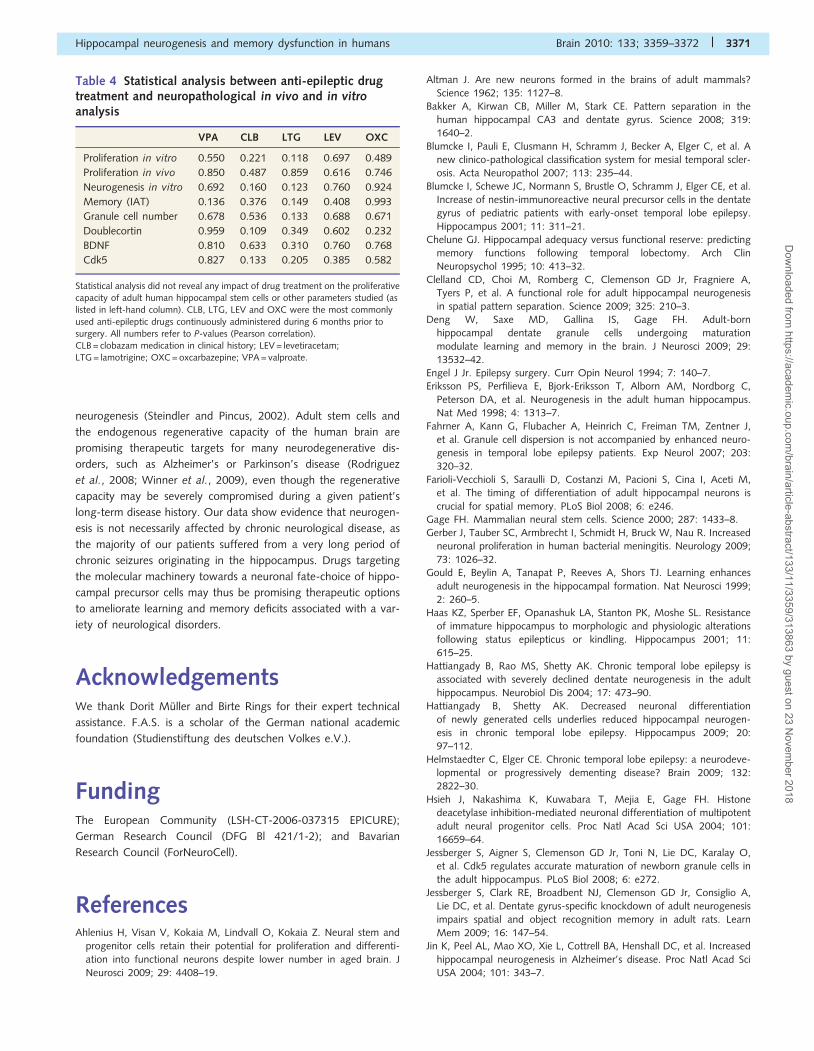
Table 4 Statistical analysis between anti-epileptic drugtreatment and neuropathological in vivo and in vitroanalysis
VPA CLB LTG LEV OXC
Proliferation in vitro 0.550 0.221 0.118 0.697 0.489
Proliferation in vivo 0.850 0.487 0.859 0.616 0.746
Neurogenesis in vitro 0.692 0.160 0.123 0.760 0.924
Memory (IAT) 0.136 0.376 0.149 0.408 0.993
Granule cell number 0.678 0.536 0.133 0.688 0.671
Doublecortin 0.959 0.109 0.349 0.602 0.232
BDNF 0.810 0.633 0.310 0.760 0.768
Cdk5 0.827 0.133 0.205 0.385 0.582
Statistical analysis did not reveal any impact of drug treatment on the proliferativecapacity of adult human hippocampal stem cells or other parameters studied (aslisted in left-hand column). CLB, LTG, LEV and OXC were the most commonly
used anti-epileptic drugs continuously administered during 6 months prior tosurgery. All numbers refer to P-values (Pearson correlation).CLB = clobazam medication in clinical history; LEV = levetiracetam;LTG = lamotrigine; OXC = oxcarbazepine; VPA = valproate.
Hippocampal neurogenesis and memory dysfunction in humans Brain 2010: 133; 3359–3372 | 3371
Dow
nloaded from https://academ
ic.oup.com/brain/article-abstract/133/11/3359/313863 by guest on 23 N
ovember 2018
Kitamura T, Saitoh Y, Takashima N, Murayama A, Niibori Y, Ageta H,et al. Adult neurogenesis modulates the hippocampus-dependent
period of associative fear memory. Cell 2009; 139: 814–27.
Knoth R, Singec I, Ditter M, Pantazis G, Capetian P, Meyer RP, et al.
Murine features of neurogenesis in the human Hippocampus acrossthe lifespan from 0 to 100 years. PLoS One 2010; 5: e8809.
Lagace DC, Benavides DR, Kansy JW, Mapelli M, Greengard P, Bibb JA,
et al. Cdk5 is essential for adult hippocampal neurogenesis. Proc Natl
Acad Sci USA 2008; 105: 18567–71.Leutgeb JK, Leutgeb S, Moser MB, Moser EI. Pattern separation in the
dentate gyrus and CA3 of the hippocampus. Science 2007; 315:
961–6.Liu H, Kaur J, Dashtipour K, Kinyamu R, Ribak CE, Friedman LK.
Suppression of hippocampal neurogenesis is associated with develop-
mental stage, number of perinatal seizure episodes, and glucocorticos-
teroid level. Exp Neurol 2003; 184: 196–213.Liu YW, Curtis MA, Gibbons HM, Mee EW, Bergin PS, Teoh HH, et al.
Doublecortin expression in the normal and epileptic adult human brain.
Eur J Neurosci 2008; 28: 2254–65.
Nakashiba T, Young JZ, McHugh TJ, Buhl DL, Tonegawa S. Transgenicinhibition of synaptic transmission reveals role of CA3 output in hip-
pocampal learning. Science 2008; 319: 1260–4.
Nyffeler M, Yee BK, Feldon J, Knuesel I. Abnormal differentiation of
newborn granule cells in age-related working memory impairments.Neurobiol Aging 2008.
Parent JM, Elliott RC, Pleasure SJ, Barbaro NM, Lowenstein DH. Aberrant
seizure-induced neurogenesis in experimental temporal lobe epilepsy.Ann Neurol 2006; 59: 81–91.
Parent JM, Yu TW, Leibowitz RT, Geschwind DH, Sloviter RS,
Lowenstein DH. Dentate granule cell neurogenesis is increased by seiz-
ures and contributes to aberrant network reorganization in the adultrat hippocampus. J Neurosci 1997; 17: 3727–38.
Pauli E, Hildebrandt M, Romstock J, Stefan H, Blumcke I. Deficient
memory acquisition in temporal lobe epilepsy is predicted by hippo-
campal granule cell loss. Neurology 2006; 67: 1383–9.Rodriguez JJ, Jones VC, Tabuchi M, Allan SM, Knight EM, LaFerla FM,
et al. Impaired adult neurogenesis in the dentate gyrus of a triple
transgenic mouse model of Alzheimer’s disease. PLoS One 2008; 3:e2935.
Rossi C, Angelucci A, Costantin L, Braschi C, Mazzantini M, Babbini F,
et al. Brain-derived neurotrophic factor (BDNF) is required for the en-
hancement of hippocampal neurogenesis following environmental en-richment. Eur J Neurosci 2006; 24: 1850–6.
Roy NS, Wang S, Jiang L, Kang J, Benraiss A, Harrison-Restelli C, et al.
In vitro neurogenesis by progenitor cells isolated from the adult human
hippocampus. Nat Med 2000; 6: 271–7.
Sairanen M, Lucas G, Ernfors P, Castren M, Castren E. Brain-derivedneurotrophic factor and antidepressant drugs have different but coor-
dinated effects on neuronal turnover, proliferation, and survival in the
adult dentate gyrus. J Neurosci 2005; 25: 1089–94.
Shors TJ, Miesegaes G, Beylin A, Zhao M, Rydel T, Gould E.Neurogenesis in the adult is involved in the formation of trace mem-
ories. Nature 2001; 410: 372–6.
Siebzehnrubl FA, Blumcke I. Neurogenesis in the human hippocampus
and its relevance to temporal lobe epilepsies. Epilepsia 2008; 49(Suppl. 5): 55–65.
Siebzehnrubl FA, Buslei R, Eyupoglu IY, Seufert S, Hahnen E, Blumcke I.
Histone deacetylase inhibitors increase neuronal differentiation in adultforebrain precursor cells. Exp Brain Res 2007; 176: 672–8.
Squire LR, Stark CE, Clark RE. The medial temporal lobe. Annu Rev
Neurosci 2004; 27: 279–306.
Stefan H, Hildebrandt M, Kerling F, Kasper BS, Hammen T, Dorfler A,et al. Clinical prediction of postoperative seizure control: structural,
functional findings and disease histories. J Neurol Neurosurg
Psychiatry 2009; 80: 196–200.
Stefan H, Scheler G, Hummel C, Walter J, Romstock J, Buchfelder M,et al. Magnetoencephalography (MEG) predicts focal epileptogenicity
in cavernomas. J Neurol Neurosurg Psychiatry 2004; 75: 1309–13.
Steindler DA, Pincus DW. Stem cells and neuropoiesis in the adult human
brain. Lancet 2002; 359: 1047–54.Tashiro A, Sandler VM, Toni N, Zhao C, Gage FH.
NMDA-receptor-mediated, cell-specific integration of new neurons in
adult dentate gyrus. Nature 2006; 442: 929–33.van Praag H, Christie BR, Sejnowski TJ, Gage FH. Running enhances
neurogenesis, learning, and long-term potentiation in mice. Proc Natl
Acad Sci USA 1999; 96: 13427–31.
van Praag H, Schinder AF, Christie BR, Toni N, Palmer TD, Gage FH.Functional neurogenesis in the adult hippocampus. Nature 2002; 415:
1030–4.
Walton NM, Sutter BM, Chen HX, Chang LJ, Roper SN, Scheffler B,
et al. Derivation and large-scale expansion of multipotent astroglialneural progenitors from adult human brain. Development 2006; 133:
3671–81.
Winner B, Vogt-Weisenhorn DM, Lie CD, Blumcke I, Winkler J. Cellularrepair strategies in Parkinson’s disease. Therapeutic Advances in
Neurological Disorders 2009; 2: 51–60.
Wolf HK, Buslei R, Schmidt-Kastner R, Schmidt-Kastner PK, Pietsch T,
Wiestler OD, et al. NeuN: a useful neuronal marker for diagnostichistopathology. J Histochem Cytochem 1996; 44: 1167–71.
Zhao C, Teng EM, Summers RG Jr, Ming GL, Gage FH. Distinct mor-
phological stages of dentate granule neuron maturation in the adult
mouse hippocampus. J Neurosci 2006; 26: 3–11.
3372 | Brain 2010: 133; 3359–3372 R. Coras et al.
Dow
nloaded from https://academ
ic.oup.com/brain/article-abstract/133/11/3359/313863 by guest on 23 N
ovember 2018