Mode of Action of Synthetic Antimalarial Peroxides
128
Mode of Action of Synthetic Antimalarial Peroxides INAUGURALDISSERTATION zur Erlangung der Würde eines Doktors der Philosophie vorgelegt der Philosophisch-Naturwissenschaftlichen Fakultät der Universität Basel von Joëlle Pascale Jourdan aus Muttenz, Baselland Basel, 2018 Originaldokument gespeichert auf dem Dokumentenserver der Universität Basel edoc.unibas.ch
Transcript of Mode of Action of Synthetic Antimalarial Peroxides

Mode of Action of Synthetic Antimalarial
Peroxides
INAUGURALDISSERTATION
zur
Erlangung der Würde eines Doktors der Philosophie
vorgelegt der
Philosophisch-Naturwissenschaftlichen Fakultät
der Universität Basel
von
Joëlle Pascale Jourdan
aus Muttenz, Baselland
Basel, 2018
Originaldokument gespeichert auf dem Dokumentenserver der Universität
Basel
edoc.unibas.ch

Genehmigt von der Philosophisch-Naturwissenschaftlichen Fakultät
auf Antrag von
Prof. Pascal Mäser (Fakultätsverantwortlicher und Dissertationsleiter) und
Prof. Dominique Soldati-Favre (Korreferentin)
Basel, den 12. Dezember 2017
Prof. Dr. Martin Spiess
Dekan der Philosophisch-
Naturwissenschaftlichen Fakultät

I
Table of contents
Acknowledgements ................................................................................................................................II
Summary ............................................................................................................................................... IV
Abbreviations ....................................................................................................................................... VI
1. Introduction ....................................................................................................................................... 1
1.1. Malaria ............................................................................................................................ 1
1.2. Prevention and treatment of malaria ............................................................................ 3
1.3. Artemisinin ..................................................................................................................... 5
1.4. Mode of Action ............................................................................................................... 7
1.5. Artemisinin Resistance .................................................................................................. 9
1.6. Disadvantages of Artemisinin ...................................................................................... 11
1.7. Synthetic peroxides ...................................................................................................... 12
1.8. OZ277 ........................................................................................................................... 13
1.9. OZ439 ........................................................................................................................... 14
1.10. Mode of Action ......................................................................................................... 15
1.11. Objectives ................................................................................................................. 16
References ................................................................................................................................. 17
2. Monoclonal Antibodies that Recognize the Alkylation Signature of Antimalarial Ozonides
OZ277 (Arterolane) and OZ439 (Artefenomel) ............................................................................. 24
3. In vitro activity of anti-malarial ozonides against an artemisinin-resistant isolate ..................... 54
4. Identification of the Alkylation Signatures of Antimalarial Ozonides and Artemisinin ............. 76
5. Discussion ....................................................................................................................................... 109
6. Conclusion...................................................................................................................................... 115
References ............................................................................................................................... 116

II
Acknowledgements
First and foremost, I like to thank my supervisor Pascal Mäser for giving me the possibility to do
this PhD thesis. I am particularly grateful for his continuous support, for his guidance, motivation
and patience. It was a privilege to work with him and benefit from his immense knowledge and his
outstanding enthusiasm.
A special thank goes to my co-supervisor Sergio Wittlin who had always confidence in me and
supported me along the way. Without his constant help, openness, patience in reading, improving
and correcting my texts and his effort driving the project forward, this thesis would not be possible.
I would also like to thank Remo Schmidt for his support in dealing with proteomics, his numerous
ideas and the fruitful discussions.
I am greatly indebted to all members of the Parasite Chemotherapy Unit, especially Anja, Sibylle,
Christin, Christoph and Christian for their continuous help and support in the lab.
Many thanks also to Hugues Matile for his great ideas, his vast expertise in dealing with antibodies
and the opportunity to learn from his knowledge in his laboratory at Roche with Doris Zulauf,
Bernhard Rutten and Nicole Soder.
I like also to thank Dominique Soldati to join the thesis committee.
My sincere thanks go to Jonathan Vennerstrom who supported us by preparing a lot of different
click chemistry compounds and his enormous support and knowledge in chemistry.
I am grateful to Alexander Schmidt who gave me access to his laboratory and helped us in
proteomics.
Many thanks also to Oliver Biehlmaier, who introduced me to the Imaging Core Facility and helped
me dealing with immunofluorescence microscopy and optimizing pictures.
I thank Ellen Reift and Fabian Baumgärtner for their work on my thesis.

III
Last but not least I like to thank my family for their financial and emotional support during my
studies and my friends, especially Sabina, who supported me with patience and optimism, and for
always being there if needed.

IV
Summary
Malaria is one of the most widespread infectious diseases which caused an estimated 212 million
cases and 429,000 deaths worldwide in 2015. Today, artemisinin-based combination therapy (ACT),
a combination of the fast-acting artemisinin with a longer lasting drug, is recommended to treat
uncomplicated Plasmodium falciparum infections. Artemisinin is of highest antimalarial potency
and selectivity. However, as a natural product, artemisinin and its derivatives also have drawbacks.
In 2004 Jonathan Vennerstrom reported the development of synthetic peroxides that might
overcome these shortcomings. A first-generation synthetic peroxide, OZ277, was registered with
piperaquine for combination therapy in India in 2012 and the next-generation ozonide OZ439 is
being tested in Phase IIb clinical trials in combination with piperaquine or ferroquine. The exact
mode of action of synthetic peroxides and artemisinins, both thought to have similar modes of
action, is not known. Recently, prolonged parasite clearance rates in patients after treatment with
artesunate or ACTs were published, indicating a starting artemisinin resistance. Therefore, the fear
of cross-resistance of artemisinin-resistant clinical isolates against OZ439, the leading candidate in
the drug pipeline, overshadowed its superior properties.
In this PhD thesis the mode of action of synthetic peroxides was further elucidated. First, I used
monoclonal antibodies raised against the adamantane-portion of ozonides to perform subcellular
localization studies and to identify alkylation signatures in P. falciparum. Since it was not possible
to identify the alkylated parasite proteins with antibodies, I set up a novel approach using click
chemistry with newly synthesized alkyne derivatives of antimalarial peroxides. Further, the
potential of cross-resistance by an artemisinin-resistant clinical isolate to OZ439 was tested.
I showed that alkylation of proteins by OZ277 and OZ439 takes place in the cytoplasm and other
structures such as the nucleus and the food vacuole, in agreement with previous findings.
Comparing the alkylation signatures of artemisinin and ozonides, I identified common targets to

V
both drugs, such as the protein PFNF54_01699. Overall, the ozonides had a larger target space than
artemisinin. In addition, we found, that there is no cross-resistance in vitro of an artemisinin-
resistant clinical P. falciparum isolate to OZ439, indicating that OZ439 has the potential to
circumvent artemisinin resistance. Nevertheless, larger clinical studies are needed to investigate if
OZ439 is effective against artemisinin-resistant malarial parasites.
VI
Abbreviations
AA2 artemisinin-alkyne 2
ACT artemisinin-based combination therapy
Cam3.IIC580Y Cambodian isolate carrying a C580Y mutation
Cam3.IR539T Cambodian isolate carrying a R539T mutation
deoxyAA2 deoxy-artemisinin-alkyne 2
EMP erythrocyte membrane protein
IRS indoor residual spraying
ITNs insecticide treated mosquito nets
K13 Kelch 13 propeller domain
kDa kilodalton
MDR1 multidrug resistant protein 1
OZ ozonide
RBC red blood cells
Swiss TPH Swiss Tropical and Public Health Institute
TAMRA 6-carboxytetramethylrhodamine
WHO World Health Organization

1 | P a g e
1. Introduction
1.1. Malaria
Malaria is one of the most important tropical diseases as it caused an estimated 212 million cases and
429,000 deaths worldwide in 2015 (World Health Organization (WHO) Malaria Report, 2016). The
majority of malaria deaths (70% of the global total) affect children below the age of five, which
means that every two minutes a child dies from malaria (WHO Malaria Report, 2016). Malaria cases
are mostly restricted to tropical and subtropical areas, whereby the highest numbers of cases and
deaths occur in Africa (90% and 92%, respectively in 2015), followed by South-East Asia and the
Eastern Mediterranean Region (WHO Malaria Report, 2016). The infectious disease is caused by
protozoan pathogens of Plasmodium spp., first discovered in 1880 by Charles Louis Alphonse
Laveran, belonging to the phylum Apicomplexa (WHO Malaria Report 2016; Hempelmann et al.,
2013; Cox, 2010). Of the five different Plasmodium species, namely P. falciparum, P. vivax, P.
malariae, P. ovale and P. knowlesi, that cause malaria in humans, P. falciparum leads to the most
severe form of malaria, called malaria tropica. It was responsible for 99% of malaria deaths
worldwide in 2015 (WHO Malaria Report 2016).
Plasmodium falciparum has a complex life cycle including an insect vector as the first host and a
human being as an intermediate host. During a blood meal of female mosquitoes belonging to the
genus Anopheles, sporozoites are injected with the saliva into the human host. They penetrate blood
vessels and enter the bloodstream where they quickly reach hepatocytes in the liver. The parasite
divides asexually in the hepatocytes, a process called schizogony, resulting in tens of thousands of
merozoites which are released into the bloodstream and invade red blood cells (RBCs). Inside the
erythrocytes they develop into ring forms, trophozoites and finally schizonts. This process takes 48
hours and is called erythrocytic schizogony. It ends with the rupture of schizonts, each containing
16-32 merozoites, which then will invade new RBCs. A small fraction of merozoites develop into

2 | P a g e
male and female gametocytes, the stage infective for mosquitoes. When these are taken up during a
blood meal, the sexual reproduction takes place in the mosquito, resulting in up to 10,000
sporozoites which migrate to the salivary glands and can infect new human hosts (Mawson, 2013;
Cowman et al., 2016; Phillips et al., 2017). Clinical symptoms of malaria manifest after rupture of
infected RBCs and include fever, chills, headache, muscle aches, anemia and digestive symptoms
(Mawson, 2013; Cowman et al. 2016).

3 | P a g e
1.2. Prevention and treatment of malaria
Since malaria transmission is reported in 91 countries, prevention strategies are indispensable
(WHO Malaria Report 2016). The main prevention strategies are vector control with insecticide
treated mosquito nets (ITNs) and indoor residual spraying (IRS), which both effectively reduce
malaria transmission in the African Region (WHO Malaria Report 2016, Lengeler, 2000). While it
is estimated that ITNs are responsible for 50% of the decline in parasite prevalence among children
2-10 years of age between 2001 and 2015 (WHO Malaria Report 2016), the impact of IRS is difficult
to be valued because of limited randomized trial data. Nevertheless, it is assumed that IRS have a
similar impact than ITNs (WHO Malaria Report 2016). Currently, twelfe insecticides belonging to
four different chemical classes exist, but emerging resistance of Anopheles mosquitoes to
insecticides is a major problem. Therefore, efforts were undertaken to screen 4 million compounds,
resulting in 3 new insecticides entering development primariliy to be used for ITNs (MalEra, 2017).
Another prevention strategy is intermittent preventive treatment of malaria in pregnancy, which
reduces perinatal mortality, and intermittent preventive treatment in infants, providing protection
against malaria (WHO Malaria Report 2016). An up to 80% reduction of the incidence of clinical
cases and severe malaria in children below the age of five can be achieved by seasonal
chemoprevention of young children (3-59 months) in areas where malaria is seasonal (WHO
Malaria Report 2016; MalEra, 2017). A further prevention strategy is the development of a vaccine.
RTS,S/AS01 is the sole candidate having completed phase III testing and shown protection, albeit
only partial (WHO Malaria Report 2016; Vandoolaeghe et al., 2016). After four doses, clinical
incidence was reduced by 39% and severe malaria by 31.5% in young children 5-17months of age
(WHO Malaria Report 2016; MalEra, 2017). Thus, chemotherapy remains a main pilar of the global
fight against malaria. For the effective treatment of malaria, several drugs exist, with artemisinin-

4 | P a g e
combination therapy (ACT) being the recommended first-line treatment of uncomplicated malaria
tropica (WHO Malaria Report 2016, Phillips et al., 2017).

5 | P a g e
1.3. Artemisinin
In the 1950s malaria parasites resistant to chloroquine, which was widely used to treat malaria,
emerged, resulting in an urgent need for new drugs (Tu, 2011;
Cowman et al., 2016).
During the Vietnam war, the US army launched a drug
discovery program that resulted in the development of
mefloquine (Su et al., 2015; Kitchen et al., 2006). In China, the
Project 523 was launched in 1967, in which more than 2,000
Chinese herb preparations used in traditional remedies were
investigated (Tu, 2011; Su et al., 2015). Youyou Tu and
coworkers found, that extracts of Artemisia annua, a plant also called sweet wormwood or qinghao,
showed promising inhibition of parasite grow. But these data were not reproducible and an intense
review of literature was done. This resulted in the idea that the high temperatures used in the
extraction could have destroyed the active ingredient (Tu, 2011). In 1971, a natural extract was
found which was active against mice infected with P. berghei and monkeys infected with P.
cynomolgi, when using lower temperatures for extraction. Clinical efficacy of this extract was also
tested in patients infected with P. falciparum or P. vivax, showing fast disappearance of symptoms.
In 1972 the active ingredient was isolated and called qinghaosu or artemisinin (Tu, 2011; Klayman,
1985; Su et al., 2015). The discovery of artemisinin was appreciated in 2015 with the Nobel prize in
physiology or medicine awarded to Youyou Tu (Su et al., 2015). Since it was found that artemisinin
is poorly soluble in water, efforts to modify the structure were undertaken. Beside the discovery of
dihydroartemisinin by Youyou Tu, which showed improved water-solubility and treatment
efficacy, other artemisinin derivatives such as artesunate, artemether or arteether were synthesized
(Tu, 2011, Su et al., 2015, White, 1994, Meshnick, 2002). Today, fixed dose ACTs are the first-line
Figure 1 Chemical structure of artemisinin

6 | P a g e
treatment of uncomplicated P. falciparum infections. Thereby Coartem (consisting of artemether
and lumefantrine) and Coarsucam (amodiaquine-and artesunate) are the main combination
therapies used. For severe malaria tropica, intramuscular injections of artesunate are recommended
(Anthony et al., 2012; Wells et al., 2015; White, 2008).

7 | P a g e
1.4. Mode of Action
Already in 1975, the stereo-structure of artemisinin was investigated and found to be a sesquiterpene
lactone, which differed from the structures of all perviously used antimalarial drugs, indicating a
different mode of action (Tu, 2011; Klayman, 1985; Meshnick, 2002). When seven other
sesquiterpene lactones isolated from A. annua, all lacking the peroxy group, were tested and found
to be inactive, it was evident that the endoperoxide bridge was essential for antimalarial efficacy
(Klayman, 1985). This finding was supported by the observation that only small amounts of
radiolabeled deoxyarteether were taken up by parasites compared to radiolabeled arteether
(Asawamahasakda et al., 1994), and by the fact that deoxyartemisinin, lacking the peroxide bridge,
is over 1000-fold less active against P. falciparum than artemisinin (Kaiser et al., 2007). The exact
mode of action is still discussed controversially (O`Neill et al., 2010). The iron- dependent alkylation
hypothesis is one of the proposed modes of actions of artemisinins. It is suggested that there is a
reductive cleavage of the peroxide bridge in artemisinins by ferrous heme or free Fe (II) derived
from heme. The reducing agent is suggested to be released by the hemoglobin digestion which takes
place in the food vacuole. Toxic free radicals are generated which then damage specific intracellular
targets by alkylation (Tilley et al., 2016; O`Neill et al., 2010; Meshnick et al., 1993; Creek et al.,
2009). This mode of action model is supported by the fact that artemisinin localizes to the food
vacuole and mitochondria, therefore direct contact of artemisinin and heme possibly takes place in
the food vacuole (Maeno et al., 1993; Crespo et al., 2008). Further, artemisinin activity is only found
in hemoglobin-degrading pathogens, but not in pathogens (or life-cycle stages) that do not degrade
hemoglobin (Kaiser et al., 2007). P. falciparum young ring stages, in which hemoglobin digestion
just began and less heme was produced, were slightly less sensitive than trophozoites or schizonts
(Skinner et al., 1996; Ter Kuile et al., 1993; Maerki et al., 2006; Klayman, 1985; Kaiser et al., 2007).
Also, young gametocytes, having a greater heme content, were more susceptible to artemisinins
8 | P a g e
than more mature gametocytes (Dechy-Cabaret et al., 2012; Kaiser et al., 2007). Sporozoites, which
do not digest hemoglobin, are not susceptible to artemisinins (Kaiser et al., 2007). Another point,
supporting the iron-dependent alkylation hypothesis is the finding, that the formation of carbon-
centered radicals is critical to the activity of artemisinin and that the interaction of artemisinin with
parasite targets is irreversible (Fügi et al., 2010; Abiodun et al., 2013). Further, antimalarial
endoperoxides appeared to react specifically with malarial proteins (Asawamahasakda et al., 1994).
9 | P a g e
1.5. Artemisinin Resistance
Although artemisinin and its derivatives are highly potent and fast acting, prolonged parasite
clearance rates in patients after treatment with artesunate or ACTs were observed along the Thai-
Cambodian border in 2006 (Noedl et al., 2008; Dondorp et al., 2009). WHO defines the “delayed
parasite clearance” as “partial resistance” (WHO Malaria Report, 2016). Today, this type of
artemisinin resistance has emerged in five countries in the Greater Mekong subregion, namely
Cambodia, Myanmar, Vietnam, Lao PDR and China (WHO Malaria Report, 2016; Zaw et al., 2017;
Paloque et al., 2016). It was found that mutations in the Kelch 13 propeller domain protein (K13)
confer to ring-stage parasites the ability to enter a quiescent stage upon exposure to artemisinin.
When drug pressure is removed, normal growth is reached again quickly (Ariey et al., 2014; Paloque
et al., 2016). Several different mutations in the K13 gene were found, all having emerged
independently, since same mutations were found in different geographic locations (Takala-Harrison
et al., 2015). Individual point mutations in K13 were shown to be sufficient for artemisinin
resistance by Zinc-finger-based genetic engineering of P. falciparum isolates (Straimer et al., 2015).
Recently, K13 mutant alleles were mapped worldwide, whereby 108 non-synonymous mutations
were identified out of 1250 P. falciparum isolates, mostly in samples obtained from Asia (Ménard et
al., 2016). Although some K13 mutations were found in Africa, none of these had been associated
with clinical artemisinin resistance, except one recent report of a migrant worker who showed
delayed parasite clearance after ACT treatment (Tilley et al., 2016; Zaw et al., 2017; Ménard et al.,
2016). Since infections in patients showing delayed parasite clearance because of artemisinin
resistance can still be cleared by the partner drug of ACTs, treatment failures of ACTs are limited to
Asia, where resistance to several of the partner drugs of ACTs was reported. Today, partial resistance
to mefloquine and amodiaquine, high resistance against piperaquine and little evidence of
lumefantrine resistance was observed in Cambodia, resulting in high failure rates after ACT
10 | P a g e
treatment of four different ACTs in Cambodia (WHO Report, 2016; Blasco et al., 2017). The
emergence of resistance occurs often in areas with high parasite loads, low transmission and low
host immunity resulting in high numbers of patients having symptoms and seeking treatment
(Talisuna et al., 2012). Therefore, reasons for the lack of artemisinin resistance in Africa could be
the higher degree of acquired immunity because of permanent exposure to P. falciparum in endemic
regions resulting in control of drug-resistant infection by the acquired host immunity (Blasco et al.,
2017, Tilley et al., 2016). Further the occurrence of polyclonal infections in Africa select against
parasites resistant to drugs having reduced growth-rates (Tilley et al., 2016; Blasco et al., 2017;
Paloque et al., 2016). In addition, high numbers of chronic asymptomatic infections result in lower
drug pressure in Africa (Blasco et al., 2017; Tilley et al., 2016).

11 | P a g e
1.6. Disadvantages of Artemisinin
Besides the problem of recently reported artemisinin resistance artemisinins are facing other
disadvantages. Since artemisinins are natural products extracted from the leaves of A. annua, they
are dependent on the availability of the plant (Tu, 2011). The first total synthesis of artemisinin was
reported by Schmid and Hofheinz in 1983 (Schmid et al., 1983), followed by several other groups
describing total or partial syntheses of artemisinin over the past decades (Wang et al., 2014). Also, a
method to scale up artemisinin synthesis was described by Lévesque and Seeberger (Lévesque et al.,
2012), but total or partial synthesis of artemisinin is a complicated process, involves several steps
(Avery et al., 1992), results in low yields, or includes high costs of starting material (White, 2008;
Wang et al., 2014). Therefore, no reasonable alternative for a large-scale production is available at
the moment. Also, efforts were undertaken to scale up plant production since artemisinin
concentration in plants is relatively low. However, Youyou Tu and coworkers had already found
that artemisinin is only present in leaves of the species Artemisia annua but not in other species of
the genus Artemisia (Tu, 2011). Therefore, cultivation and processing of Artemisia annua plants
were even reported in partnerships in India to provide sufficient artemisinin for malaria treatment
(Kumar et al., 2005). Another possibility is breeding new varieties as reported in 2010 (Graham et
al., 2010). Nevertheless, artemisinin and its derivatives are much more expensive than other
antimalarial drugs; they cost, for example, 10 times more than chloroquine (White et al., 2008).
Another disadvantage of artemisinins is their poor bioavailability. Further, their short in vivo half-
life, which results in recrudescence of parasitemia after 5 days of artemisinin monotherapy in spite
of multiple applications, is an additional disadvantage. Since artemisinins are short-lived, reducing
the high numbers of parasites for two asexual parasite cycles only, artemisinins are co-administered
with longer half-life drugs (White, 2008; Tilley et al., 2016; Meshnick, 2002).

12 | P a g e
1.7. Synthetic peroxides
The next generation of antimalarials must overcome the drawbacks of artemisinins such as the
cumbersome production, the limiting pharmacokinetics, the poor bioavailability and costs. In 2004,
Vennerstrom et al. reported the identification of synthetic peroxides, with OZ277 as development
candidate, which overcome the disadvantages of artemisinins (Vennerstrom et al., 2004). A series of
over 700 peroxides was synthesized and tested by a consortium consisting of Jonathan
Vennerstrom`s group at the University of Nebraska (USA), Monash University (Australia), Swiss
Tropical and Public Health Institute (Swiss TPH, Switzerland), Basilea Pharmaceutica AG
(Switzerland) and F. Hoffmann-La Roche (Switzerland; Vennerstrom et al., 2004; Mäser et al., 2012).
The synthesized peroxides showed best activity when a trioxolane heterocycle was stabilized by two
additional ring structures, one adamantane ring and one phenyl ring. This turned out to be an ideal
balance between a sterically hindered and a sterically unhindered peroxy oxygen atom for attack by
iron (II) species (Vennerstrom et al., 2004).
13 | P a g e
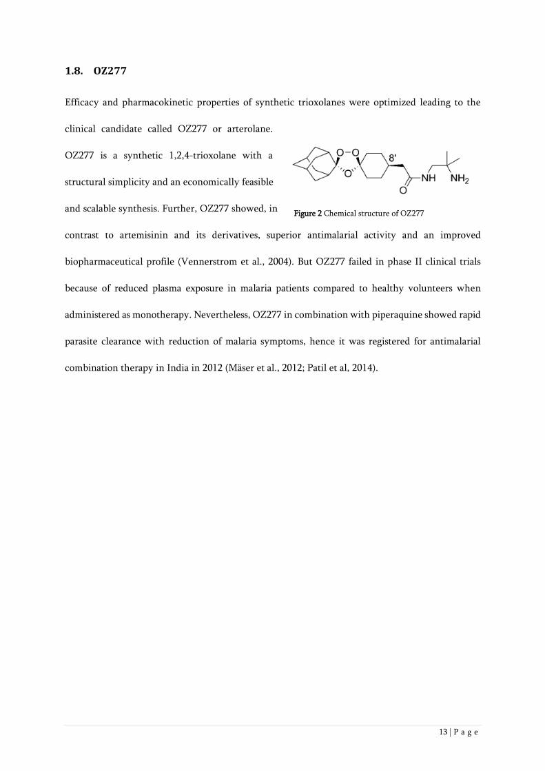
1.8. OZ277
Efficacy and pharmacokinetic properties of synthetic trioxolanes were optimized leading to the
clinical candidate called OZ277 or arterolane.
OZ277 is a synthetic 1,2,4-trioxolane with a
structural simplicity and an economically feasible
and scalable synthesis. Further, OZ277 showed, in
contrast to artemisinin and its derivatives, superior antimalarial activity and an improved
biopharmaceutical profile (Vennerstrom et al., 2004). But OZ277 failed in phase II clinical trials
because of reduced plasma exposure in malaria patients compared to healthy volunteers when
administered as monotherapy. Nevertheless, OZ277 in combination with piperaquine showed rapid
parasite clearance with reduction of malaria symptoms, hence it was registered for antimalarial
combination therapy in India in 2012 (Mäser et al., 2012; Patil et al, 2014).
Figure 2 Chemical structure of OZ277

14 | P a g e
1.9. OZ439
OZ439, also called artefenomel, is a next-generation ozonide containing a cis-8`-phenyl substituent
in contrast to the cis-8`-alkyl group in OZ277. It was designed to provide a single-dose oral cure in
humans and indeed, it was the first compound of all synthetic peroxides and artemisinin derivatives
which completely cured P. berghei infected mice with a single oral dose of 20 mg/kg. Further,
OZ439 was shown to be >50-fold more
stable to Fe(II)-mediated degradation,
suggesting that this is the reason for its
prolonged plasma profiles in malaria
patients (Charman et al., 2011). In addition, excellent prophylactic activity, superior to that of
mefloquine, was demonstrated with OZ439 (Charman et al., 2011). Also, a rapid onset of action and
activity against all asexual P. falciparum blood stages was shown in vitro (Charman et al., 2011; Kim
et al., 2017). Phase I clinical trials of OZ439 showed good safety and pharmacokinetic profiles in
healthy volunteers (Möhrle et al., 2012; McCarthy et al., 2016). Furthermore, good safety profiles
were also observed in malaria patients in phase IIa studies (Phyo et al., 2016). OZ439 showed fast
clearance of P. falciparum and P. vivax parasites in malaria patients and its long half-life suggests to
use OZ439 in combination with another drug as single-dose oral cure (Phyo et al., 2016). OZ439 is
now tested in Phase IIb clinical trials with piperaquine and additionally also in combination with
ferroquine by Sanofi (Wells et al., 2015).
Figure 3 Chemical structure of OZ439

15 | P a g e
1.10. Mode of Action
Although synthetic peroxides have a different structure to artemisinin and its derivatives, except
for the peroxide bridge, it was suggested that both have a similar mode of action (Vennerstrom et
al., 2004; Jefford, 2001; Kaiser et al., 2007). As in artemisinin, it was found that the peroxide bridge
is essential for antimalarial activity in OZ277 (Kaiser et al., 2007). Further, OZ277 was 1,000 fold
more active against hemoglobin-degrading P. falciparum parasites than against parasites that do not
degrade hemoglobin (Kaiser et al., 2007). This supports the iron-dependent alkylation hypothesis.
The synthetic peroxides OZ277 and OZ439 were found to be active against all asexual blood stages
of P. falciparum in vitro (Märki et al., 2006; Hofer et al., 2008; Charman et al., 2011; Kim et al.,
2017). Localization studies showed that in some parasites OZ277 localizes to the parasite cytosol and
in some parasites to the food vacuole (Uhlemann et al., 2007). It was shown that for the activity of
OZ277 and artemisinin the formation of carbon-centered radicals is essential (Fügi et al., 2010; Tang
et al., 2005). Further it was found that the antimalarial properties of both, artemisinin and OZ277,
derived from irreversible interactions with parasite targets (Fügi et al., 2010; Abiodun et al., 2013).
Different studies showed that OZ277 activation was dependent on free iron, heme or ferrous heme
(Creek et al., 2008; Creek et al., 2009). Together, all these facts corroborate the iron-dependent
alkylation hypothesis.
16 | P a g e
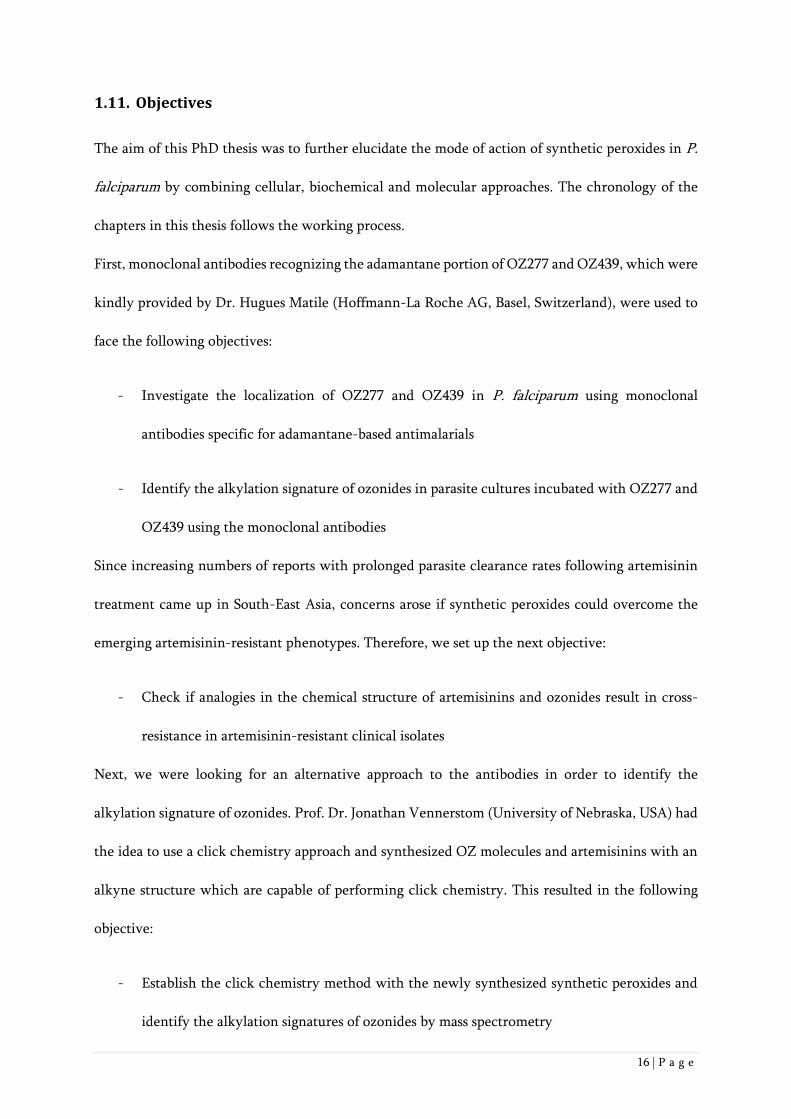
1.11. Objectives
The aim of this PhD thesis was to further elucidate the mode of action of synthetic peroxides in P.
falciparum by combining cellular, biochemical and molecular approaches. The chronology of the
chapters in this thesis follows the working process.
First, monoclonal antibodies recognizing the adamantane portion of OZ277 and OZ439, which were
kindly provided by Dr. Hugues Matile (Hoffmann-La Roche AG, Basel, Switzerland), were used to
face the following objectives:
- Investigate the localization of OZ277 and OZ439 in P. falciparum using monoclonal
antibodies specific for adamantane-based antimalarials
- Identify the alkylation signature of ozonides in parasite cultures incubated with OZ277 and
OZ439 using the monoclonal antibodies
Since increasing numbers of reports with prolonged parasite clearance rates following artemisinin
treatment came up in South-East Asia, concerns arose if synthetic peroxides could overcome the
emerging artemisinin-resistant phenotypes. Therefore, we set up the next objective:
- Check if analogies in the chemical structure of artemisinins and ozonides result in cross-
resistance in artemisinin-resistant clinical isolates
Next, we were looking for an alternative approach to the antibodies in order to identify the
alkylation signature of ozonides. Prof. Dr. Jonathan Vennerstom (University of Nebraska, USA) had
the idea to use a click chemistry approach and synthesized OZ molecules and artemisinins with an
alkyne structure which are capable of performing click chemistry. This resulted in the following
objective:
- Establish the click chemistry method with the newly synthesized synthetic peroxides and
identify the alkylation signatures of ozonides by mass spectrometry

17 | P a g e
References
Abiodun O.O., Brun R., Wittlin S., (2013) In vitro interaction of artemisinin derivatives or the fully
synthetic peroxidic anti-malarial OZ277 with thapsigargin in Plasmodium falciparum strains. Malar.
J. 12, 43.
Anthony M.P., Burrows J.N., Duparc S., Moehrle J.J., Wells T.N., (2012) The global pipeline of new
medicines for the control and elimination of malaria. Malar J. 11, 316.
Ariey F., Witkowski B., Amaratunga C., Beghain J., Langlois A.C., Khim N., Kim S., Duru, V.,
Bouchier C., Ma L., Lim P., Leang R., Duong S., Sreng S., Suon S., Chuor C.M., Bout D.M., Ménard
S., Rogers W.O., Genton B., Fandeur T., Miotto O., Ringwald P., Le Bras J., Berry A., Barale J.C.,
Fairhurst R.M., Benoit-Vical F., Mercereau-Puijalon O., Ménard D., (2014) A molecular marker of
artemisinin-resistant Plasmodium falciparum malaria. Nature. 505, 50-55.
Asawamahasakda W., Ittarat I., Pu Y.M., Ziffer H., and Meshnick S.R., (1994) Reaction of
antimalarial endoperoxides with specific parasite proteins. Antimicrob. Agents Chemother. 38,
1854–1858.
Avery M.A., Chong W.K.M., Jennings-White C., (1992) Stereoselective Total Synthesis of (+)-
Artemisinin, the Antimalarial Constituent of Artemisia annua L. J. Am. Chem. Soc. 114, 974-979.
Blasco B., Leroy D., Fidock D.A., (2017) Antimalarial drug resistance: linking Plasmodium
falciparum parasite biology to the clinic. Nat Med. 23 (8), 917-928.
Charman S. A., Arbe-Barnes S., Bathurst I. C., Brun R., Campbell M., Charman W. N., Chiu F. C.,
Chollet J., Craft J. C., Creek D. J., Dong Y., Matile H., Maurer M., Morizzi J., Nguyen T.,
Papastogiannidis P., Scheurer C., Shackleford D. M., Sriraghavan K., Stingelin L., Tang Y., Urwyler
H., Wang X., White K. L., Wittlin S., Zhou L., and Vennerstrom J. L., (2011) Synthetic ozonide drug
candidate OZ439 offers new hope for a single-dose cure of uncomplicated malaria. Proc. Natl. Acad.
Sci. U. S. A. 108, 4400−4405.
Cowman A.F., Healer J., Marapana D., Marsh K., (2016) Malaria: Biology and Disease. Cell. 167 (3),
610-624.
http://www.ncbi.nlm.nih.gov/pubmed/?term=Witkowski%20B%5BAuthor%5D&cauthor=true&cauthor_uid=24352242
http://www.ncbi.nlm.nih.gov/pubmed/?term=Langlois%20AC%5BAuthor%5D&cauthor=true&cauthor_uid=24352242
18 | P a g e
Cox F.E.G., (2010) History of the discovery of the malaria parasites and their vectors. Parasites &
Vectors. 3 (5).
Creek D. J., Charman W. N., Chiu F. C. K., Prankerd R. J., Dong Y., Vennerstrom J. L., and Charman
S. A., (2008) Relationship between antimalarial activity and haem alkylation for spiro- and dispiro-
1,2,4-trioxolane antimalarials. Antimicrob. Agents Chemother. 52, 1291-1296.
Creek D.J., Ryan E., Charman W.N., Chiu F.C., Prankerd R.J., Vennerstrom J.L., Charman S.A.,
(2009) Stability of peroxide antimalarials in the presence of human hemoglobin. Antimicrob. Agents
Chemother. 53 (8), 3496-3500.
Crespo M. D., Avery T. D., Hanssen E., Fox E., Robinson T. V., Valente P., Taylor D. K., and Tilley
L., (2008) Artemisinin and a series of novel endoperoxide antimalarials exert early effects on
digestive vacuole morphology. Antimicrob. Agents Chemother. 52, 98−109.
Dechy-Cabaret O., Benoit-Vical F., (2012) Effects of antimalarial molecules on the gametocyte stage
of Plasmodium falciparum: the debate. J. Med. Chem. 55 (23), 10328-10344.
Dondorp A.M., Nosten F., Yi P., Das D., Phyo A.P., Tarning J., Lwin K.M., Ariey F., Hanpithakpong
W., Lee S.J., Ringwald P., Silamut K., Imwong M., Chotivanich K., Lim P., Herdman T., An S.S.,
Yeung S., Singhasivanon P., Day N.P.J., Lindegardh N., Socheat D., White N.J., (2009) Artemisinin
Resistance in Plasmodium falciparum Malaria. N. Engl. J. Med. 38, 455-467.
Fügi M.A., Wittlin S., Dong Y., Vennerstrom J.L., (2010) Probing the antimalarial mechanism of
artemisinin and OZ277 (arterolane) with nonperoxidic isosteres and nitroxyl radicals. Antimicrob.
Agents Chemother. 54, 1042–1046.
Graham I.A., Besser K., Blumer S., Branigan C.A., Czechowski T., Elias L., Guterman I., Harvey D.,
Isaac P.G., Khan A.M., Larson T.R., Li Y., Pawson T., Penfield T., Rae A.M., Rathbone D.A., Reid
S., Ross J., Smallwood M.F., Segura V., Townsend T., Vyas D., Winzer T., Bowles D., (2010) The
genetic map of Artemisia annua L. identifies loci affecting yield of the antimalarial drug artemisinin.
Science. 327 (5963), 328-331.
Hempelmann E., Kraft K., (2013) Bad air, amulets and mosquitoes: 2,000 years of changing
perspectives on malaria. Malar. J. 12, 232.

19 | P a g e
Hofer S., Brun R., Maerki S., Matile H., Scheurer C., Wittlin S., (2008), In vitro assessment of the
pharmacodynamic properties of DB75, piperaquine, OZ277 and OZ401 in cultures of Plasmodium
falciparum. J. Antimicrob. Chemother. 62 (5).
Jefford C.W., (2001) Why artemisinin and certain synthetic peroxides are potent antimalarials.
Implications for the mode of action. Curr. Med. Chem. 8 (15), 1803-1826.
Kaiser M., Wittlin S., Nehrbass-Stuedli A., Dong Y., Wang X., Hemphill A., Matile H., Brun R.,
Vennerstrom J.L., (2007) Peroxide bond-dependent antiplasmodial specificity of artemisinin and
OZ277 (RBx11160). Antimicrob. Agents Chemother. 51, 2991-2993.
Kim H.S., Hammill J.T., Guy R.K., (2017) Seeking the Elusive Long-Acting Ozonide: Discovery of
Artefenomel (OZ439). J. Med. Chem. 60 (7), 2651-2653.
Kitchen L.W., Vaughn D.W., Skillman D.R., (2006) Role of US military research programs in the
development of US Food and Drug Administration--approved antimalarial drugs. Clin. Infect. Dis.
43 (1), 67-71.
Klayman D. L., (1985) Qinghaosu (artemisinin): an antimalarial drug from China. Science. 228, 1049
−1055.
Kumar S., Srivastava S., (2005) Establishment of artemisinin combination therapy as first line
treatment for combating malaria: Artemisia annua cultivation in India needed for providing
sustainable supply chain of artemisinin. Current Science. 89, 1097-1102.
Lengeler C., (2000) Insecticide-treated bednets and curtains for preventing malaria. Cochrane
Database Syst. Rev. 2.
Lévesque F., Seeberger P.H., (2012) Continuous-flow synthesis of the anti-malaria drug artemisinin.
Angew. Chem. Int. Ed. Engl. 51 (7), 1706-1709.
Maeno Y., Toyoshima T., Fujioka H., Ito Y., Meshnick S.R., Benakis A., Milhous W.K., Aikawa M.,
(1993) Morphologic effects of artemisinin in Plasmodium falciparum. Am. J. Trop. Med. Hyg. 49 (4),
485-491.

20 | P a g e
Maerki S., Brun R., Charman S.A., Dorn A., Matile H., Wittlin S., (2006) In vitro assessment of the
pharmacodynamic properties and the partitioning of OZ277/RBx-11160 in cultures of Plasmodium
falciparum. J. Antimicrob. Chemother. 58, 52-58.
malERA Refresh Consultative Panel on Tools for Malaria Elimination, (2017) malERA: An updated
research agenda for diagnostics, drugs, vaccines, and vector control in malaria elimination and
eradication. PLoS Med. 14 (11).
Mäser P., Wittlin S., Rottmann M., Wenzler T., Kaiser M., Brun R., (2012) Antiparasitic agents: new
drugs on the horizon. Curr. Opin. Pharmacol. 12, 562-566.
Mawson A.R., (2013) The pathogenesis of malaria: a new perspective. Pathog. Glob. Health. 107 (3),
122-129.
McCarthy J.S., Baker M., O'Rourke P., Marquart L., Griffin P., van Huijsduijnen R.H., Möhrle J.J.,
(2016) Efficacy of OZ439 (artefenomel) against early Plasmodium falciparum blood-stage malaria
infection in healthy volunteers. J. Antimicrob. Chemother. 71 (9), 2620–2627.
Ménard D., Khim N., Beghain J., Adegnika A.A., Shafiul-Alam M., Amodu O., Rahim-Awab G.,
Barnadas C., Berry A., Boum Y., Bustos M.D., Cao J., Chen J.H., Collet L., Cui L., Thakur G.D., Dieye
A., Djallé D., Dorkenoo M.A., Eboumbou-Moukoko C.E., Espino F.E., Fandeur T., Ferreira-da-Cruz
M.F., Fola A.A., Fuehrer H.P., Hassan A.M., Herrera S., Hongvanthong B., Houzé S., Ibrahim M.L.,
Jahirul-Karim M., Jiang L., Kano S., Ali-Khan W., Khanthavong M., Kremsner P.G., Lacerda M.,
Leang R, Leelawong M., Li M., Lin K., Mazarati J.B., Ménard S., Morlais I., Muhindo-Mavoko H.,
Musset L., Na-Bangchang K., Nambozi M., Niaré K., Noedl H., Ouédraogo J.B., Pillai D.R., Pradines
B., Quang-Phuc B., Ramharter M., Randrianarivelojosia M., Sattabongkot J., Sheikh-Omar A., Silué
K.D., Sirima S.B., Sutherland C., Syafruddin D., Tahar R., Tang L.H., Touré O.A., Tshibangu-wa-
Tshibangu P., Vigan-Womas I., Warsame M., Wini L., Zakeri S., Kim S., Eam R., Berne L., Khean
C., Chy S., Ken M., Loch K., Canier L., Duru V., Legrand E., Barale J.C., Stokes B., Straimer J.,
Witkowski B., Fidock D.A., Rogier C., Ringwald P., Ariey F., Mercereau-Puijalon O., KARMA
Consortium, (2016) A Worldwide Map of Plasmodium falciparum K13-Propeller Polymorphisms.
N. Engl. J. Med. 374 (25), 2453-2464.
Meshnick S. R., (2002) Artemisinin: Mechanisms of action, resistance and toxicity. Int. J. Parasitol.
32, 1655–1660.

21 | P a g e
Meshnick S.R.,Yang Y.Z., Lima V., Kuypers F., Kamchonwongpaisan S., Yuthavong Y., (1993) Iron-
dependent free radical generation from the antimalarial agent artemisinin (qinghaosu). Antimicrob.
Agents Chemother. 37 (5), 1108–1114.
Moehrle J.J., Duparc S., Siethoff C., van Giersbergen P.L., Craft J.C., Arbe-Barnes S., Charman S.A.,
Gutierrez M., Wittlin S., Vennerstrom J.L., (2012) First-in-man safety and pharmacokinetics of
synthetic ozonide OZ439 demonstrates an improved exposure profile relative to other peroxide
antimalarials. Br. J. Clin. Pharmacol. 75, 524-537.
Noedl H., Se Y., Schaecher K., Smith B.L., Socheat D., Fukuda M.M., (2008) Artemisinin Resistance
in Cambodia 1 (ARC1) Study Consortium Evidence of artemisinin-resistant malaria in western
Cambodia. N. Engl. J. Med. 359, 2619-2620.
O'Neill P.M., Barton V.E., Ward S.A., (2010) The molecular mechanism of action of artemisinin-the
debate continues. Molecules. 15 (3), 1705-1721.
Paloque L., Ramadani A.P., Mercereau-Puijalon O., Augereau J.M., Benoit-Vical F., (2016)
Plasmodium falciparum: multifaceted resistance to artemisinins. Malaria J. 15, 149.
Patil C.Y., Katare S.S., Baig M.S., Doifode S.M., (2014) Fixed Dose Combination of Arterolane and
Piperaquine: A Newer Prospect in Antimalarial Therapy. Annals of Medical and Health Sciences
Research. 4, 466-71.
Phillips M.A., Burrows J.N., Manyando C., van Huijsduijnen R.H., Van Voorhis W.C., Wells T.N.C.,
(2017) Malaria. Nat. Rev. Dis. Primers. 3, 17050.
Phyo A.P., Jittamala P., Nosten F.H., Pukrittayakamee S., Imwong M., White N.J., Duparc S.,
Macintyre F., Baker M., Möhrle J.J., (2016) Antimalarial activity of artefenomel (OZ439), a novel
synthetic antimalarial endoperoxide, in patients with Plasmodium falciparum and Plasmodium
vivax malaria: an open-label phase 2 trial. Lancet Infect. Dis. 16, 61-69.
Schmid G., Hofheinz W., (1983) Total synthesis of qinghaosu. J. Am. Chem. Soc. 105 (3), 624–625.
Skinner T.S., Manning L.S., Johnston W.A., Davis T.M., (1996) In vitro stage-specific sensitivity of
Plasmodium falciparum to quinine and artemisinin drugs. Int. J. Parasitol. 26 (5), 519-525.

22 | P a g e
Straimer J., Gnädig N.F., Witkowski B., Amaratunga C., Duru V., Ramadani A.P., Dacheux M., Khim
N., Zhang L., Lam S., Gregory P.D., Urnov F.D., Mercereau-Puijalon O., Benoit-Vical F., Fairhurst
R.M., Ménard D., Fidock D.A., (2015) Drug resistance. K13-propeller mutations confer artemisinin
resistance in Plasmodium falciparum clinical isolates. Science. 347 (6220), 428-431.
Su X.Z., Miller L.H., (2015) The discovery of artemisinin and the Nobel Prize in Physiology or
Medicine. Sci China Life Sci. 58, 1175-1179.
Takala-Harrison S., Jacob C.G., Arze C., Cummings M.P., Silva J.C., Dondorp A.M., Fukuda M.M.,
Hien T.T., Mayxay M., Noedl H., Nosten F., Kyaw M.P., Nhien N.T., Imwong M., Bethell D., Se Y.,
Lon C., Tyner S.D., Saunders D.L., Ariey F., Mercereau-Puijalon O., Menard D., Newton P.N.,
Khanthavong M., Hongvanthong B., Starzengruber P., Fuehrer H.P., Swoboda P., Khan W.A., Phyo
A.P., Nyunt M.M., Nyunt M.H., Brown T.S., Adams M., Pepin C.S., Bailey J., Tan J.C., Ferdig M.T.,
Clark T.G., Miotto O., MacInnis B., Kwiatkowski D.P., White N.J., Ringwald P., Plowe C.V., (2015)
Independent emergence of artemisinin resistance mutations among Plasmodium falciparum in
Southeast Asia. J. Infect. Dis. 211 (5), 670-679.
Talisuna A.O., Karema C., Ogutu B., Juma E., Logedi J., Nyandigisi A., Mulenga M., Mbacham W.F.,
Roper C., Guerin P.J., D’Alessandro U., Snow R.W., (2012) Mitigating the threat of artemisinin
resistance in Africa: improvement of drug-resistance surveillance and response systems. Lancet
Infect. Dis. 12 (11), 888–896.
Tang Y., Dong Y., Vennerstrom J.L., (2004) Synthetic peroxides as antimalarials. Med. Res. Rev. 24,
425–448.
Tang Y., Dong Y., Wang X., Sriraghavan K., Wood J.K., Vennerstrom J.L., (2005) Dispiro-1,2,4-
trioxane analogues of a prototype dispiro-1,2,4-trioxolane: mechanistic comparators for artemisinin
in the context of reaction pathways with iron(II). J. Org. Chem. 70 (13), 5103-5110.
Ter Kuile F., White N.J., Holloway P., Pasvol G., Krishna S., (1993) Plasmodium falciparum: in vitro
studies of the pharmacodynamic properties of drugs used for the treatment of severe malaria. Exp.
Parasitol. 76 (1), 85-95.
Tilley L., Straimer J., Gnädig N.F., Ralph S.A., Fidock D.A., (2016) Artemisinin Action and
Resistance in Plasmodium falciparum. Trends Parasitol. 32, 682-696.

23 | P a g e
Tu Y., (2011) The discovery of artemisinin (qinghaosu) and gifts from Chinese medicine. Nat. Med.
17, 1217-2120.
Uhlemann A.C., Wittlin S., Matile H., Bustamante L.Y., Krishna S., (2007) Mechanism of
antimalarial action of the synthetic trioxolane RBX11160 (OZ277). Antimicrob. Agents Chemother.
51, 667–672.
Vandoolaeghe P., Schuerman L., (2016) The RTS,S/AS01 malaria vaccine in children 5 to 17 months
of age at first vaccination. Expert Rev. Vaccines. 15 (12), 1481-1493.
Vennerstrom J.L., Arbe-Barnes S., Brun R., Charman S.A., Chiu F.C., Chollet J., Dong Y., Dorn A.,
Hunziker D., Matile H., McIntosh K., Padmanilayam M., Santo Tomas J., Scheurer C., Scorneaux B.,
Tang Y., Urwyler H., Wittlin S., Charman W.N., (2004) Identification of an antimalarial synthetic
trioxolane drug development candidate. Nature. 430, 900–904.
Wang Z., Yang L., Yang X., Zhang X., (2014) Advances in the Chemical Synthesis of Artemisinin.
Synthetic Communications. 44 (14), 1987-2003.
Wells T.N., van Huijsduijnen R.H., and Van Voorhis W.C., (2015) Malaria medicines: a glass half
full? Nat. Rev. Drug Discov. 14, 424-442.
White N.J., (1994) Artemisinin: Current status. Trans. R. Soc. Trop. Med. Hyg. 88, 3-4.
White N.J., (2008) Qinghaosu (artemisinin): The price of success. Science. 320, 330–334.
WHO, (2016) World Malaria Report 2016. ISBN: 978 92 4 151171 1.
Zaw M.T., Emran N.A., Lin Z., (2017) Updates on k13 mutant alleles for artemisinin resistance in
Plasmodium falciparum. J. Microbiol. Immunol. Infect. S1684-1182 (17), 30122-30126.

24 | P a g e
2. Monoclonal Antibodies that Recognize the
Alkylation Signature of Antimalarial Ozonides
OZ277 (Arterolane) and OZ439 (Artefenomel)
Joëlle Jourdan1,2, Hugues Matile3, Ellen Reift1,2, Oliver Biehlmaier4, Yuxiang Dong5, Xiaofang Wang5,
Pascal Mäser1,2, Jonathan L. Vennerstrom5, and Sergio Wittlin1,2*
1Swiss Tropical and Public Health Institute, Socinstrasse 57, CH-4002 Basel, Switzerland
2University of Basel, CH-4003 Basel, Switzerland
3F. Hoffmann- La Roche Ltd., Basel, Switzerland
4Imaging Core Facility, Biozentrum, University of Basel, CH-4003 Basel, Switzerland
5College of Pharmacy, University of Nebraska Medical Center, 986025 Nebraska Medical Center,
Omaha, NE, USA
*Corresponding author
tel: 41 61 284 81 36
email: [email protected]
Published in: ACS Infectious Diseases (2015)
Personal Contribution to the Paper: I produced and analysed immunofluorescence, Western blot and
immunoprecipitation data and helped writing the paper
25 | P a g e
Abstract
The singular structure of artemisinin, with its embedded 1,2,4-trioxane heterocycle, has inspired
the discovery of numerous semisynthetic artemisinin and structurally diverse synthetic peroxide
antimalarials, including ozonides OZ277 (arterolane) and OZ439 (artefenomel). Despite the critical
importance of artemisinin combination therapies (ACTs), the precise mode of action of peroxidic
antimalarials is not fully understood. However, it has long been proposed that the peroxide bond in
artemisinin and other antimalarial peroxides undergoes reductive activation by ferrous heme
released during hemoglobin digestion to produce carbon-centered radicals that alkylate heme and
parasite proteins. To probe the mode of action of OZ277 and OZ439, we now describe initial studies
with monoclonal antibodies that recognize the alkylation signature (sum of heme and protein
alkylation) of these synthetic peroxides. Immunofluorescence experiments conducted with
ozonide-treated parasite cultures showed that ozonide alkylation is restricted to the parasite, as no
signal was found in the erythrocyte or its membrane. In Western blot experiments with ozonide-
treated P. falciparum malaria parasites, distinct protein bands were observed. Significantly, no
protein bands were detected in parallel Western blot experiments performed with lysates from
ozonide-treated Babesia divergens, parasites that also proliferate inside erythrocytes, but in contrast
to P. falciparum, do not catabolize hemoglobin. However, subsequent immunoprecipitation
experiments with these antibodies failed to identify the P. falciparum proteins alkylated by OZ277
and OZ439. To the best of our knowledge, this shows for the first time that antimalarial ozonides,
like the artemisinins, alkylate proteins in P. falciparum.
Keywords. alkylation, artemisinin, immunofluorescence, monoclonal antibody, ozonide,
Plasmodium falciparum.

26 | P a g e
Introduction
The discovery of artemisinin (ART) from Artemisia annua1 gave rise to the semisynthetic
artemisinins dihydroartemisinin (DHA), artemether (AM), and artesunate (AS), which as ART
combination therapies (ACT), are the preferred treatment for uncomplicated Plasmodium
falciparum malaria2 (Figure 1). The singular structure of ART, with its embedded 1,2,4-trioxane
heterocycle, inspired the discovery of additional semisynthetic artemisinins and structurally diverse
synthetic peroxide antimalarials.3-6 One of these, ozonide (1,2,4-trioxolane) OZ277,7 also known as
arterolane maleate, was introduced in 2012 to the Indian market as a combination product with
piperaquine phosphate (Synriam®).8-10 More recently, the ‘next generation’ ozonide OZ439
(artefenomel)11,12 has progressed to Phase IIb trials (Figure 1).
Figure 1. Artemisinin and ozonide structures.
The peroxide bond in ART and antimalarial synthetic peroxides is essential for antiplasmodial
activity,6,13 suggesting a chemistry-driven mechanism of action. A considerable amount of data4,14-
24 demonstrates that the activity of antimalarial peroxides does not derive from reversible
interactions with parasite targets and that the peroxide bond in ART and other antimalarial
peroxides undergoes reductive activation by ferrous heme released during hemoglobin digestion to
produce carbon-centered radicals that alkylate heme and parasite proteins (Figure 2). This is
accompanied by disruption of the parasite digestive vacuole including lipid peroxidation.25-27 This
mechanism accounts not only for the high antiplasmodial potency and specificity of peroxides, but

27 | P a g e
also for their weak and peroxide-bond independent activities against pathogens that do not degrade
hemoglobin such as other protozoa, bacteria, and fungi.13,28,29
Figure 2. Alkylation reactions of ART and ozonides OZ277 and OZ439.
Electron transfer from heme to the peroxide bond antibonding * orbitals of ART and antimalarial
ozonides produces short-lived alkoxy radicals (Figure 2). For ART, rearrangment via -scission
forms a primary carbon-centered radical; for OZ277 and OZ439, rearrangement via -scission forms
a secondary carbon-centered radical. As these two ozonides have the same spiroadamantane
substructure, they produce the same bicyclic carboxylic acid signature of ozonide alkylation – be
that with heme or with proteins. Since we had good success in capturing the ozonide-derived
secondary carbon-centered radical with the stable nitroxide radical TEMPO and its analogs,7,22,30 we
decided to capitalize on this finding and synthesized OZH04 as a potential hapten for this ozonide-
derived bicyclic carboxylic acid with OZH05 as a control (Scheme 1). We now describe the creation
of monoclonal antibodies to OZH04 and their application in immunofluorescence and Western blot
experiments.

28 | P a g e
Results and Discussion
Our first approach was to synthesize OZH04 by reductive amination of 7-(4-oxo-2,2,6,6-
tetramethyl-1-piperidinyloxy)bicyclo-[3.3.1]nonane-3-carboxylic acid,30 but the workup of this
reaction was difficult, so we elected to access OZH04 by a two-step procedure (Scheme 1). Thus,
reaction of prototypical ozonide 1 with ferrous iron in the presence of 4-((tert-
butoxycarbonyl)amino)-2,2,6,6-tetramethylpiperidinyl-1-oxyl (2) led to formation of intermediate
3 in 26% yield. We synthesized OZH04 as its dihydrochloride salt (92% yield) by deprotection of
3 with HCl. OZH05 was synthesized in 64% yield by exposing 4-acetamido-2,2,6,6-
tetramethylpiperidinyl-1-oxyl (4) to a mixture of cuprous chloride and aq. hydrogen peroxide
according to the method of Dichtl et al.31
Scheme 1. Synthesis of N-alkoxyamines OZH04 and OZH05. (a) Fe(OAc)2, 1:1 CH2Cl2:CH3CN, Fe(OAc)2, 35 °C, 24 h; (b)
6N HCl, THF, rt, 12 h; (c) 50% H2O2, CuCl, acetone, rt, 12 h.
The monoclonal antibodies OZH04-2/2 and OZH04-1/8 were raised in Naval Medical Research
Institute (NMRI) mice injected subcutaneously with OZH04 hapten coupled to KLH (keyhole
limpet hemocyanin). After the third boost, blood was collected and the serum was tested for the
presence of antihapten antibodies by ELISA using BSA-conjugated OZH04 antigen to coat the
ELISA-plates. Animals with strong immune responses were selected for fusion to PAI myeloma
cells.

29 | P a g e
In order to determine if the monoclonal IgG1 antibodies raised against the OZH04 hapten were
binding to P. falciparum parasites that had been exposed to OZ277 or OZ439, NF54 cultures were
treated with either of the two ozonides, DHA or DMSO, and immunofluorescence experiments were
performed. The two monoclonal antibodies OZH04-2/2 and OZH04-1/8 gave positive signals after
incubation with parasites exposed to either OZ277 or OZ439 (Table 1, see 2 top rows). No
immunofluorescence signals were detected with DHA-treated parasites, 0.1% DMSO or an
unrelated IgG1 control antibody. An antibody raised against the cytosolic protein GAPDH served
as a positive control.
Table 1: Immunofluorescence experiments with P. falciparum cultures treated with 10 µg/mL OZ277, 10 µg/mL OZ439,
10 µg/mL DHA or 0.1% DMSO for 2 h. Primary antibodies used were OZH04-2/2, OZH04-1/8, IgG1 negative control or
GAPDH positive control. Goat-anti mouse Alexa 488 was used as secondary antibody. + indicates fluorescence and –
indicates no fluorescence.
Competition experiments with hapten OZH04 and control hapten OZH05 (Scheme 1) showed that
the antibodies OZH04-1/8 and OZH04-2/2 specifically recognize the bicyclic carboxylic acid
alkylation substructure, or alkylation signature, of ozonides OZ277and OZ439 (Table 2).
Table 2: Immunofluorescence experiments with P. falciparum cultures treated with 10 µg/mL OZ277 or 0.1% DMSO for
2 h. For competition, 33 µM of the hapten OZH04 or OZH05 were combined with the primary antibodies OZH04-2/2
30 | P a g e
(0.33 µM) or OZH04-1/8 (0.33 µM) and incubated at RT for 1 h. The secondary antibody was goat-anti mouse Alexa 488.
+ indicates fluorescence; – indicates no fluorescence.
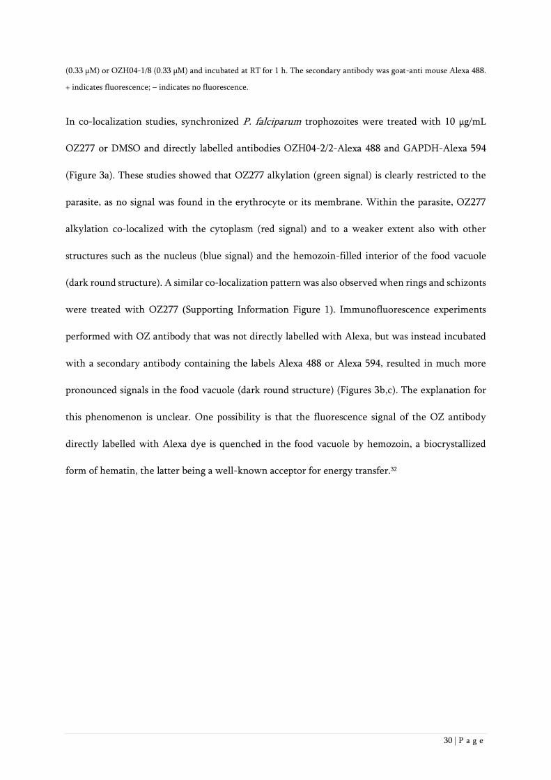
In co-localization studies, synchronized P. falciparum trophozoites were treated with 10 µg/mL
OZ277 or DMSO and directly labelled antibodies OZH04-2/2-Alexa 488 and GAPDH-Alexa 594
(Figure 3a). These studies showed that OZ277 alkylation (green signal) is clearly restricted to the
parasite, as no signal was found in the erythrocyte or its membrane. Within the parasite, OZ277
alkylation co-localized with the cytoplasm (red signal) and to a weaker extent also with other
structures such as the nucleus (blue signal) and the hemozoin-filled interior of the food vacuole
(dark round structure). A similar co-localization pattern was also observed when rings and schizonts
were treated with OZ277 (Supporting Information Figure 1). Immunofluorescence experiments
performed with OZ antibody that was not directly labelled with Alexa, but was instead incubated
with a secondary antibody containing the labels Alexa 488 or Alexa 594, resulted in much more
pronounced signals in the food vacuole (dark round structure) (Figures 3b,c). The explanation for
this phenomenon is unclear. One possibility is that the fluorescence signal of the OZ antibody
directly labelled with Alexa dye is quenched in the food vacuole by hemozoin, a biocrystallized
form of hematin, the latter being a well-known acceptor for energy transfer.32
31 | P a g e

32 | P a g e
Figure 3: Immunofluorescence studies with P. falciparum trophozoites that were treated with 10 µg/mL OZ277 for 2 h. a)
In immunofluorescence co-localisation studies, the blood smears were fixed with 5% formaldehyde and 0.01%
glutaraldehyde, permeabilized with 0.5% Triton-X-100 and blocked with 1% BSA in PBS for 1 h. The primary antibodies
used were directly labelled OZH04-2/2 (Alexa 488, green signal) and GAPDH (Alexa 594, red signal). For both, the
exposure time was 2 s. Nuclei were stained with DAPI (blue signal, exposure time of 0.05 s). The bottom row left shows
the merge of OZH04-2/2, GAPDH and DAPI, and on the bottom row right, the reference image (exposure of 1 s). Scale
bar 2 µm. b) Same as a), except that unlabelled primary antibodies OZH04-2/2 were used. After 6 washes with PBS, 20
µg/mL of the secondary antibody goat anti-mouse Alexa 488 (green signal) was incubated for 1 h at RT. The exposure time
was 0.1 s. Nuclei were stained with DAPI (blue signal, exposure time of 0.05 s). The bottom row left shows the merge of
OZH04-2/2 and DAPI, and the bottom row right shows the merge of OZH04-2/2 and the reference image (exposure of 1
s). Scale bar 2 µm.
c) Same as b), except that the secondary antibody used was goat anti-mouse Alexa 594 with an exposure of 1s (red signal).
Previously, ultrastructural autoradiographic studies of [3H]-dihydroartemisinin-treated parasites
have shown that the drug and its alkylation reaction products are present in the parasitophorous
vacuole membranes, digestive vacuole membranes and mitochondria.15 Other work indicated that a
fluorescent TAMRA OZ277 conjugate was associated with the food vacuole and the parasite
endoplasmic reticulum,33 although in this study, fluorescence signal was derived from the parent
ozonide, not from its alkylation products. To ascertain if ozonide alkylation localizes to specific
parasite membranes, further studies will be required.
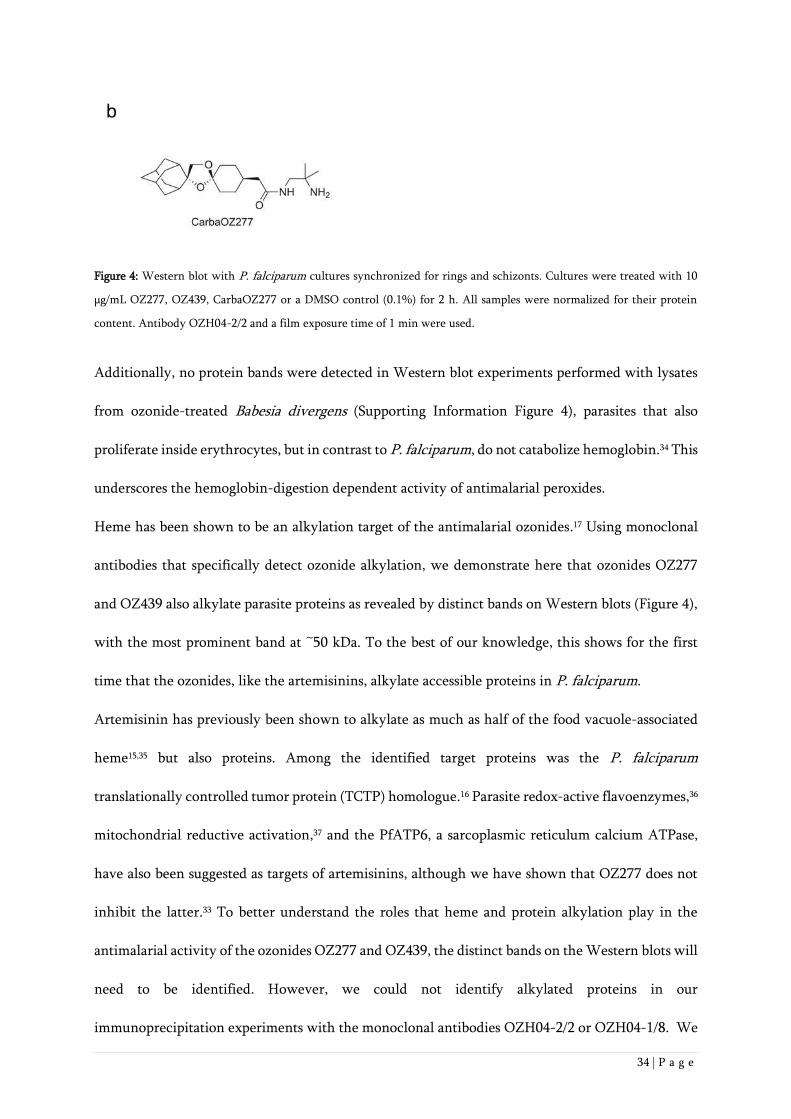
Western Blot experiments demonstrated that monoclonal antibody OZH04-2/2 recognizes distinct
P. falciparum protein bands (Figure 4a) in parasites treated with OZ277 or OZ439. This indicates
that the two ozonides alkylate parasite proteins. Ring stages showed one prominent band at ~50
kDa. In schizont stages, a variety of bands ranging from ~28 to ~98 kDa could be found after OZ277
treatment. After longer film exposure times, the same pattern of bands was also observed in
schizonts treated with OZ439. We conclude that the extent of ozonide alkylation seems to be higher
in schizonts vs. rings, consistent with the greater hemoglobin digestion that has occurred in the
former.

33 | P a g e
The same bands were also found when the concentrations of the ozonides were lowered to 100
ng/mL, which at a parasitemia of 8-10%, corresponds to the IC99 (concentration where 99% of
parasite growth is inhibited compared to untreated control parasites) (Supporting Information
Figure 2). However, Western blot experiments performed at these lower ozonide concentrations
were not practical as they required longer film exposure times, where false-positive signals can
become an issue. Based on this, 10,000 ng/mL was found to be the most practical concentration.
No bands could be observed in parasite cultures treated with carbaOZ277, the nonperoxidic analogue of
OZ277 (Figure 4)13 or when uninfected erythrocytes were used (Supporting Information Figure 3).
This data indicates that the antibody binding was specific for alkylated proteins.
34 | P a g e
Figure 4: Western blot with P. falciparum cultures synchronized for rings and schizonts. Cultures were treated with 10
µg/mL OZ277, OZ439, CarbaOZ277 or a DMSO control (0.1%) for 2 h. All samples were normalized for their protein
content. Antibody OZH04-2/2 and a film exposure time of 1 min were used.
Additionally, no protein bands were detected in Western blot experiments performed with lysates
from ozonide-treated Babesia divergens (Supporting Information Figure 4), parasites that also
proliferate inside erythrocytes, but in contrast to P. falciparum, do not catabolize hemoglobin.34 This
underscores the hemoglobin-digestion dependent activity of antimalarial peroxides.
Heme has been shown to be an alkylation target of the antimalarial ozonides.17 Using monoclonal
antibodies that specifically detect ozonide alkylation, we demonstrate here that ozonides OZ277
and OZ439 also alkylate parasite proteins as revealed by distinct bands on Western blots (Figure 4),
with the most prominent band at ~50 kDa. To the best of our knowledge, this shows for the first
time that the ozonides, like the artemisinins, alkylate accessible proteins in P. falciparum.
Artemisinin has previously been shown to alkylate as much as half of the food vacuole-associated
heme15,35 but also proteins. Among the identified target proteins was the P. falciparum
translationally controlled tumor protein (TCTP) homologue.16 Parasite redox-active flavoenzymes,36
mitochondrial reductive activation,37 and the PfATP6, a sarcoplasmic reticulum calcium ATPase,
have also been suggested as targets of artemisinins, although we have shown that OZ277 does not
inhibit the latter.33 To better understand the roles that heme and protein alkylation play in the
antimalarial activity of the ozonides OZ277 and OZ439, the distinct bands on the Western blots will
need to be identified. However, we could not identify alkylated proteins in our
immunoprecipitation experiments with the monoclonal antibodies OZH04-2/2 or OZH04-1/8. We

35 | P a g e
suggest that the antibodies were not able to bind to the alkylated proteins under the non-denaturing
conditions required for those experiments. Due to lack of binding of the antibodies to the native
proteins, an alternative, antibody-independent approach using click chemistry is currently
underway.

36 | P a g e
Methods
Monoclonal antibody preparation
Hapten OZH04 was coupled to KLH (keyhole limpet hemocyanin, Thermo Scientific 77600, Imject
mcKLH) and BSA (bovine serum albumin, Thermo Scientific 77110 Imject BSA) respectively, by
crosslinking with glutaraldehyde based on the method of Onica et al.38
0.1 mL of OZH04 dissolved at 57.5 mg/mL in DMSO was mixed with 20 mg of the respective carrier
protein in 0.2 M Na2HPO4 pH 8.0, and 1 mL of 0.2% glutaraldehyde in 0.2 M Na2HPO4 was then
slowly added with agitation. After 1 h incubation at RT, the reaction was stopped by adding 0.25
mL 1 M glycine pH 8.0 and dialyzed against PBS overnight.
Naval Medical Research Institute (NMRI) mice were immunized with 100 µg subcutaneous
injections of KLH-conjugated OZH04 emulsified in aluminum hydroxide gel (Alhydrogel-2%,
Brenntag Biosector) containing CPGOGN as previously described.39 After the third boost, blood was
collected and the serum was tested for the presence of antihapten antibodies by ELISA using BSA-
conjugated OZH04 antigen to coat the ELISA-plates. Animals with strong immune responses were
selected for fusion.40
Parasite cultivation
P. falciparum strain NF54 (Origin: Airport, Netherlands; Provider MR4, MRA-1000) asexual blood
stages were cultivated in a variation of the medium previously described,41-43 consisting of RPMI
1640 supplemented with 0.5% ALBUMAX® II, 25 mM Hepes, 25 mM NaHCO3 (pH 7.3), 0.36 mM
hypoxanthine, and 100 µg/mL neomycin. Human erythrocytes served as host cells. Cultures were
maintained at 37 °C in an atmosphere of 3% O2, 4% CO2, and 93% N2 in humidified modular
chambers. Fifty, ninety and ninety nine percent inhibitory concentrations (IC50, IC90 and IC99) of
OZ277 in ng/mL against unsynchronized NF54 parasites were 0.91, 1.7 and 2.7 when determined at
the standard parasitemia of 0.3% parasitemia.7 At about 10-30x higher parasitemia, the inhibitory

37 | P a g e
concentrations were found to be 10-30x higher, which is consistent with the so called "incoculum
effect". The term refers to an increase in the amount of drug necessary to inhibit microbial growth
with greater numbers of microorganisms per milliliter. This effect has been previously observed
with antimalarial compounds such as chloroquine and artesunate, which show enrichment in P.
falciparum-infected red blood cells.44,45 Also, OZ277 has been shown to partition into P. falciparum-
infected red blood cells.46
Immunofluorescence
P. falciparum NF54 cultures (5% (v/v) hematocrit, 8–10% parasitemia) were either synchronized
with 5% D-sorbitol for trophozoites47 prior to treatment or used as mixed cultures. The cultures
were treated with 10 µg/mL OZ277, 10 µg/mL OZ439 and 0.1% DMSO for 2 h. Cultures were
washed once with PBS and pelleted RBCs were smeared on glass slides. The blood smears were fixed
with prechilled methanol (-20°C; 100%) and blocked with 1% BSA in PBS for 1 h.
Primary antibodies OZH04-2/2, OZH04-1/8, IgG1 (Hoffmann-La Roche AG, Basel, Switzerland)
and GAPDH (glyceraldehyde-3-phosphate-dehydrogenase, a gift from Paola Favuzza, Swiss TPH,
Basel, Switzerland) were incubated for 1 h at RT with concentrations of 50 µg/mL. After 6 washes
with PBS, the secondary antibodies goat anti-mouse Alexa 488 (Invitrogen) or goat anti-rabbit Alexa
594 (Invitrogen) were incubated with concentrations of 20 µg/mL for 1 h at RT. Control
experiments, performed with secondary antibody only, resulted in no detectable
immunofluorescence signals (not shown).
For competition experiments, 33 µM of OZH04 or OZH05 were combined with the primary
antibody OZH04-1/8 (0.33 µM) or OZH04-2/2 (0.33 µM) and incubated at RT for 1 h. As secondary
antibody, goat anti-mouse Alexa 488 (20 µg/ml) was incubated for 1 h at RT.
For co-localization studies, blood smears were fixed with 5% formaldehyde and 0.01%
glutaraldehyde and permeabilized with 0.5% Triton-X-100. Both antibodies, OZH04-2/2 and

38 | P a g e
GAPDH, were directly labelled with Alexa Fluor 488 succinidyl ester (Life Technologies) and Alexa
Fluor594 succinidyl ester (Life Technologies), respectively. 50 µg/ml of OZH04-2/2 directly labelled
with Alexa 488 was incubated for 1 h at RT. After washing 6 times with PBS, 50 µg/mL of GAPDH
directly labelled with Alexa 594 was incubated for 1 h at RT. Vectashield Hard Set with DAPI
(Vector Laboratories) was added to all slides after washing 6 times with PBS. The slides were
analysed with a Widefield Delta Vision core microscope based on an Olympus IX71 stand, using a
60x/1.42NA oil objective. Basic image analysis (e.g. contrast and brightness adjustments) were done
with ImageJ Fiji and images were analysed with deconvolution (SoftWorx 4.1.2; enhanced ratio
aggressive; Number of cycles:10).
Western blots
P. falciparum lysates were prepared from NF54 cultures (5% (v/v) hematocrit, 8–10% parasitemia)
synchronized with 5% D-sorbitol for rings and schizonts.47 Cultures were treated with 10 µg/mL
OZ277, 10 µg/mL OZ439, 10 µg/mL CarbaOZ277 or 0.1% DMSO for 2 h. After centrifugation, pellets
of samples were resuspended in 1.5 mL 0.1% saponin in PBS and incubated for 10 min on ice.
Samples were washed twice with PBS and pellets were incubated for 10-15 min with 50 µL
completed Ripa-Lysisbuffer (20 mM Tris-HCl pH 8.0, 137 mM NaCl, 10% glycerol, 1% NP40, 2 mM
EDTA, EDTA-free protease inhibitor mixture tablets (Roche Applied Science)) on ice. After
centrifugation, extracts were used for Western blot. For normalizing protein contents in ring- and
schizont samples, a BCA protein assay was performed using a BCA protein assay kit from Merck
Millipore (Product No. 21285-3) with BSA as standard prior to the Western blot.
NP40 extracts were diluted 1:2 with 2x LDS sample buffer (50% 4x LDS sample buffer, Invitrogen;
20% mercaptoethanol; 30% ddH2O), heated for 10 min at 70 °C, loaded onto polyacrylamide gels
(10 µL of samples; 4-12% Bis-Tris-polyacrylamide Gels, Invitrogen) and run for 35 min (120 mA;
200 V) using 1x MES SDS Running Buffer (20x MES DS Running Buffer, Invitrogen) as running

39 | P a g e
buffer. Gels were transferred onto nitrocellulose membranes (0.2 µm pore size; 100% nitrocellulose,
Invitrogen) and blocked for 1 h with 3% milk powder blocking solution (3% milk powder in TBS-
Tween buffer pH 8.0; TBS-Tween buffer: 20 mM Tris, 150 mM NaCl, 0.05% Tween-20, pH adjusted
to 8.0 with HCl). 1 µg/mL of OZH04-2/2 antibody, diluted in 0.5% milk powder blocking solution,
was added to the membrane and incubated for 1 h at rt. Membranes were washed 3 times for 5 min
with TBS- Tween buffer and incubated with polyclonal rabbit anti- mouse immunoglobulin
horseradish peroxidase 1:5000 (1.3 g/L; DAKO; diluted in 0.5% milk powder in TBS-Tween buffer)
for 1 h at RT. Membranes were washed 4 times for 5 min with TBS-Tween buffer. Super Signal West
Pico Chemiluminescent Substrate (Thermo Scientific) was added to the membrane, incubated for 5
min and the Western blot was developed using a Carestream Kodak Biomax light film (Sigma).
Supporting Information. Synthesis and characterization of OZH04 and OZH05.
Immunofluorescence experiments with P. falciparum rings, trophozoites and schizonts treated with
OZ277 (SI Figure 1). Western Blot experiments with mixed P. falciparum cultures treated with a range
of OZ277 concentrations (SI Figure 2). Western blot and Coomassie gel for uninfected erythrocytes (SI
Figure 3) or Babesia divergens (SI Figure 4) treated with OZ277, OZ439, CarbaOZ277 or a DMSO
control. This material is available free of charge via the Internet at http://pubs.acs.org.
40 | P a g e
Abbreviations
ACT, artemisinin combination therapy; AM, artemether; ART, artemisinin; AS, artesunate; BCA,
bicinchoninic acid assay; BSA, bovine serum albumin; CarbaOZ277, nonperoxidic analogue of
OZ277; DAPI, 4',6-diamidino-2-phenylindole DNA fluorescent stain; DHA, dihydroartemisinin;
DMSO, dimethyl sulfoxide; ELISA, enzyme linked immunosorbent assay; GAPDH, glyceraldehyde
3-phosphate dehyderogenase; IC, inhibitory concentration; KLH, keyhole limpet hemocyanin;
NF54, P. falciparum strain (Netherlands Airport strain); NMRI, naval medical research institute
mice; OZ277, arterolane; OZ439, artefenomel; OZH04, hapten for ozonide-derived bicyclic
carboxylic acid; OZH04-1/8, monoclonal antibody raised against OZH04; OZH04-2/2, monoclonal
antibody raised against OZH04; OZH05, control hapten; PBS, phosphate buffered saline; RBC, red
blood cells; RT, room temperature; TEMPO, 2,2,6,6-tetramethyl-piperidine 1-oxyl. α-CRT,
chloroquine resistance transporter.
Conflicts of Interest. The authors declare no competing interests.
Author Information. Sergio Wittlin, Swiss Tropical and Public Health Institute, Socinstrasse 57,
CH-4002 Basel, Switzerland.
Tel: 41 61 284 8136 E-mail: [email protected]
Author Contribution. X.W. and Y.D. synthesized the haptens, H.M. created the monoclonal
antibodies, J.J., H.M., E.R., O.B., P.M. and S.W. analyzed the data. J.J., H.M., P.M., J.L.V., and
S.W. wrote the paper.

41 | P a g e
Acknowledgements
This work was financially supported by the Swiss National Science Foundation (grant
310030_149896 to SW), the Medicines for Malaria Venture and the Swiss Tropical and Public Health
Institute. We are grateful to Timothy N. C. Wells and Susan A. Charman for critically reading the
manuscript and making valuable suggestions. We wish to thank Hans-Peter Beck for providing the
GAPDH antibody and Till Voss and Tobias Spielmann for their critical assessment and input in the
context of the immunofluorescence images.

42 | P a g e
References
(1) Tu, Y. (2011) The discovery of artemisinin (qinghaosu) and gifts from Chinese medicine. Nat.
Med. 17, 1217-2120.
(2) White, NJ (2008) Qinghaosu (artemisinin): The price of success. Science 320, 330–334.
(3) Tang Y., Dong Y., Vennerstrom J.L. (2004) Synthetic peroxides as antimalarials. Med. Res. Rev.
24, 425–448.
(4) Jefford C.W. (2007) New developments in synthetic peroxidic drugs as artemisinin mimics. Drug
Discov. Today 12, 487–495.
(5) Muraleedharan, K. M., and Avery M. A. (2009) Progress in the development of peroxide-based
anti-parasitic agents. Drug Discov. Today 14, 793-803.
(6) Tilley L., Charman S., Vennerstrom J.L. (2011) Semisynthetic artemisinin and synthetic peroxide
antimalarials. In RSC Drug Discovery Series No. 14. Neglected Diseases and Drug Discovery. Edited
by Mike P, Timothy NCW.: RSC Publishing; 2011:33–64. ISBN 9781849731928.
(7) Vennerstrom J.L., Arbe-Barnes S., Brun R., Charman S.A., Chiu F.C., Chollet J., Dong Y., Dorn
A., Hunziker D., Matile H., McIntosh K., Padmanilayam M., Santo Tomas J., Scheurer C., Scorneaux
B., Tang Y., Urwyler H., Wittlin S., Charman W.N. (2004) Identification of an antimalarial synthetic
trioxolane drug development candidate. Nature 430, 900–904.
(8) Mäser P., Wittlin S., Rottmann M., Wenzler T., Kaiser M., Brun R. (2012) Antiparasitic agents:
new drugs on the horizon. Curr. Opin. Pharmacol. 12, 562-566.
(9) Anthony M.P., Burrows J.N., Duparc S., Moehrle J.J., Wells T.N. (2012) The global pipeline of
new medicines for the control and elimination of malaria. Malar. J. 11, 316.
(10) Wells, T.N., van Huijsduijnen, R.H., and Van Voorhis, W.C. (2015) Malaria medicines: a glass
half full? Nat. Rev. Drug Discov. 14, 424-442.
(11) Charman S.A., Arbe-Barnes S., Bathurst I.C., Brun R., Campbell M., Charman W.N., Chiu F.C.,
Chollet J., Craft J.C., Creek D.J., Dong Y., Matile H., Maurer M., Morizzi J., Nguyen T.,
Papastogiannidis P., Scheurer C., Shackleford D.M., Sriraghavan K., Stingelin L., Tang Y., Urwyler

43 | P a g e
H., Wang X., White K.L., Wittlin S., Zhou L., Vennerstrom J.L. (2011) Synthetic ozonide drug
candidate OZ439 offers new hope for a single-dose cure of uncomplicated malaria. Proc. Natl. Acad.
Sci. U.S.A. 108, 4400-4405.
(12) Moehrle J.J., Duparc S., Siethoff C., van Giersbergen P.L., Craft J.C., Arbe-Barnes S., Charman
S.A., Gutierrez M., Wittlin S., Vennerstrom J.L. (2012) First-in-man safety and pharmacokinetics of
synthetic ozonide OZ439 demonstrates an improved exposure profile relative to other peroxide
antimalarials. Br. J. Clin. Pharmacol. 75, 524-537.
(13) Kaiser M., Wittlin S., Nehrbass-Stuedli A., Dong Y., Wang X., Hemphill A., Matile H., Brun R.,
Vennerstrom J.L. (2007) Peroxide bond-dependent antiplasmodial specificity of artemisinin and
OZ277 (RBx11160). Antimicrob. Agents Chemother. 51, 2991-2993.
(14) Asawamahasakda, W., Ittarat, I., Pu, Y.M., Ziffer, H., and Meshnick, S.R. (1994) Reaction of
antimalarial endoperoxides with specific parasite proteins. Antimicrob. Agents Chemother. 38,
1854–1858.
(15) Meshnick S.R., Taylor T.E., Kamchonwongpaisan S. (1996) Artemisinin and the antimalarial
endoperoxides: from herbal remedy to targeted chemotherapy. Microbiol. Rev. 60, 301-315.
(16) Meshnick, S. R. (2002) Artemisinin: Mechanisms of action, resistance and toxicity. Int. J.
Parasitol. 32, 1655–1660.
(17) Creek, D. J., Charman, W. N., Chiu, F. C. K., Prankerd, R. J., Dong, Y., Vennerstrom, J. L., and
Charman, S. A. (2008) Relationship between antimalarial activity and haem alkylation for spiro-
and dispiro-1,2,4-trioxolane antimalarials. Antimicrob. Agents Chemother. 52, 1291-1296.
(18) Klonis, N., Crespo-Ortiz, M. P., Bottova, I., Abu-Bakar, N., Kenny, S., Rosenthal, P. J., and
Tilley, L. (2011) Artemisinin activity against Plasmodium falciparum requires hemoglobin uptake
and digestion. Proc. Natl. Acad. Sci. U.S.A. 108, 11405−11410.
(19) Klonis, N., Creek, D. J., and Tilley L. (2013) Iron and heme metabolism in Plasmodium
falciparum and the mechanism of action of artemisinins. Curr. Opin. Microbiol. 16, 722–727.
(20) Meunier, B., and Robert, A. (2010). Heme as trigger and target for trioxane-containing
antimalarial drugs. Acc. Chem. Res. 43, 1444–1451.

44 | P a g e
(21) Slack, R. D., Jacobine, A. M., and Posner, G. H. (2012) Antimalarial peroxides: Advances in drug
discovery and design. MedChemComm 3, 281–297.
(22) Fügi, M.A., Wittlin, S., Dong, Y., Vennerstrom J.L. (2010) Probing the antimalarial mechanism
of artemisinin and OZ277 (arterolane) with nonperoxidic isosteres and nitroxyl radicals.
Antimicrob. Agents Chemother. 54, 1042–1046.
(23) Abiodun, O.O., Brun, R., Wittlin, S. (2013) In vitro interaction of artemisinin derivatives or the
fully synthetic peroxidic anti-malarial OZ277 with thapsigargin in Plasmodium falciparum strains.
Malar. J. 12, 43.
(24) Robert, A., Claparols, C., Witkowski, B., Benoit-Vical, F. (2013) Correlation between
Plasmodium yoelii nigeriensis susceptibility to artemisinin and alkylation of heme by the drug.
Antimicrob. Agents Chemother. 57, 3998-4000.
(25) Crespo, M. D., Avery, T. D., Hanssen, E., Fox, E., Robinson, T. V., Valente, P., Taylor, D. K.,
and Tilley, L. (2008) Artemisinin and a series of novel endoperoxide antimalarials exert early effects
on digestive vacuole morphology. Antimicrob. Agents Chemother. 52, 98−109.
(26) Hartwig, C. L., Rosenthal, A. S., D’Angelo, J., Griffin, C. E., Posner, G. H., and Cooper, R. A.
(2009) Accumulation of artemisinin trioxane derivatives within neutral lipids of Plasmodium
falciparum malaria parasites is endoperoxide-dependent. Biochem. Pharmacol. 77, 322−336.
(27) Hartwig, C. L., Lauterwasser, E. M. W., Mahajan, S. S., Hoke, J. M., Cooper, R. A., and Renslo,
A. R. (2011) Investigating the antimalarial action of 1,2,4-trioxolanes with fluorescent chemical
probes. J. Med. Chem. 54, 8207−8213.
(28) Klayman, D. L. (1985) Qinghaosu (artemisinin): an antimalarial drug from China. Science 228,
1049-1055.
(29) Krishna, S., Bustamante, L., Haynes, R. K., and Staines, H. M. (2008) Artemisinins: Their
growing importance in medicine. Trends Pharmacol. Sci. 29, 520-527.
(30) Tang Y., Dong Y., Wang X., Sriraghavan K., Wood J.K., Vennerstrom J.L. (2005) Dispiro-1,2,4-
trioxane analogs of a prototype dispiro-1,2,4-trioxolane: Mechanistic comparators for artemisinin in
the context of reaction pathways with iron (II). J. Org. Chem. 70, 5103-5110.

45 | P a g e
(31) Dichtl A., Seyfried M., Schoening K.U. (2008) A novel method for the synthesis of N-
alkoxyamines starting from nitroxide radicals and ketones. Synlett 12,1877-1881.
(32) Pato, C., Célier, C., Rezaei, H., Grosclaude, J., and Marden M.C. (2004) Heme as an optical
probe of a conformational transition of ovine recPrP. Protein Sci. 13, 1100-1107.
(33) Uhlemann, A.C., Wittlin, S., Matile, H., Bustamante, L.Y., Krishna, S. (2007) Mechanism of
antimalarial action of the synthetic trioxolane RBX11160 (OZ277). Antimicrob. Agents Chemother.
51, 667–672.
(34) Richier, E., Biagini, G. A., Wein, S., Boudou, F., Bray, P. G., Ward, S. A., Precigout, E., Calas,
M., Dubremetz, J.-F., and Vial, H. J. (2006) Potent antihematozoan activity of novel bisthiazolium
drug T16: Evidence for inhibition of phosphatidylcholine metabolism in erythrocytes infected with
Babesia and Plasmodium spp. Antimicrob. Agents Chemother. 50, 3381–3388.
(35) Robert, A., Coppel, Y., and Meunier, B. (2002) Alkylation of heme by the antimalarial drug
artemisinin. Chem. Commun. 414-415.
(36) Haynes, R. K., Cheu, K. W., Chan, H. W., Wong, H. N., Li, K. Y., Tang, M. M., Chen M. J., Guo,
Z. F., Guo, Z. H., Sinniah, K., Witte, A. B., Coghi, P., and Monti, D. (2012) Interactions between
artemisinins and other antimalarial drugs in relation to the cofactor model–A unifying proposal for
drug action. ChemMedChem 7, 2204-2226.
(37) Wang, J., Huang, L., Li, J., Fan, Q., Long, Y., Li, Y., and Zhou, B. (2010) Artemisinin directly
targets malarial mitochondria through its specific mitochondrial activation. PLoS One, 5, e9582
(38) Onica, D., Mota, G., Calugaru, A., Manciulea, M., Dima, S. (1983) Immunogenicity and effector
functions of glutaraldehyde-treated rabbit and mouse immunoglobulins. Molecular Immunology
20, 709-718.
(39) Davis, H.L., Weeratna, R., Waldschmidt, T.J., Tygrett, L., Schorr, J., Krieg, A.M. (1998) CpG
DNA is a potent enhancer of specific immunity in mice immunized with recombinant hepatitis B
surface antigen. J. Immunol. 160, 870-6.
(40) Köhler, G., Milstein, C. (1975) Continuous cultures of fused cells secreting antibody of
predefined specificity. Nature 256, 495-497.

46 | P a g e
(41) Dorn, A., Stoffel, R., Matile, H., Bubendorf, A., Ridley, R.G. (1995) Malarial haemozoin/beta-
haematin supports haem polymerization in the absence of protein. Nature 374, 269–271.
(42) Trager, W., Jensen, J.B. (1976) Human malaria parasites in continuous culture. Science 193,
673–675.
(43) Snyder, C., Chollet, J., Santo-Tomas, J., Scheurer, C., Wittlin, S. (2007) In vitro and in vivo
interaction of synthetic peroxide RBx11160 (OZ277) with piperaquine in Plasmodium models. Exp.
Parasitol. 115,.296–300.
(44) Gluzman, I.Y., Schlesinger, P.H., Krogstad, D.J. (1987) Inoculum effect with chloroquine and
Plasmodium falciparum. Antimicrob. Agents Chemother. 31, 32-36.
(45) Ittarat, W., Pickard, A.L., Rattanasinganchan, P., Wilairatana, P., Looareesuwan, S., Emery, K.,
Low, J., Udomsangpetch, R., Meshnick, S.R. (2003) Recrudescence in artesunate-treated patients
with falciparum malaria is dependent on parasite burden not on parasite factors. Am. J. Trop. Med.
Hyg. 68, 147-152.
(46) Maerki, S., Brun, R., Charman, S.A., Dorn, A., Matile, H., Wittlin, S. (2006) In vitro assessment
of the pharmacodynamic properties and the partitioning of OZ277/RBx-11160 in cultures of
Plasmodium falciparum. J. Antimicrob. Chemother. 58, 52-58.
(47) Lambros, C., Vanderberger, J.P. (1979) Synchronization of Plasmodium falciparum erythrocytic
stages in culture. J. Parasitol. 65, 418–420.

47 | P a g e
SUPPORTING INFORMATION
Synthesis and characterization of OZH04 and OZH05.
Melting points are uncorrected. 1H and 13C NMR spectra were recorded on a 500 MHz spectrometer.
Combustion and HPLC analysis confirmed that the target compounds possessed purities ≥ 95%.
7-(4-Amino-2,2,6,6-tetramethyl-1-piperidinyloxy)bicyclo-[3.3.1]nonane-3-carboxylic acid
(OZH04). Step 1. To a solution of adamantane-2-spiro-3'-1',2',4',9',12'-
pentaoxadispiro[4.2.4.2]tetradecane (1)1 (0.70 g, 2.17 mmol), 4-((tert-butoxycarbonyl)amino)-
2,2,6,6-tetramethylpiperidinyl-1-oxyl (2) (0.70 g, 2.58 mmol) in CH2Cl2 (30 mL) and CH3CN (30 mL)
was added Fe(OAc)2 (0.70 g, 4.03 mmol). The resulting mixture was stirred under Ar and 35 °C for
24 h before being quenched with water (50 mL). After separation of the organic layer, the aqueous
layer was extracted with CH2Cl2 (2 x 30 mL). The combined extracts were washed with brine (2 x
30 mL), dried over MgSO4 and filtered and concentrated. The residue was purified by flash
chromatography (silica gel, 10-50% ether in hexane) to afford 7-(4-((tert-butoxycarbonyl)amino)-
2,2,6,6-tetramethyl-1-piperidinyloxy)bicyclo-[3.3.1]nonane-3-carboxylic acid (3) (mixture of
rotamers) as a colorless solid (0.25 g, 26%). 1H NMR (CDCl3) δ 1.17 (s, 6H), 1.20 (s, 6H), 1.44 (s, 9H),
1.00-1.52 (m, 7H), 1.56 (d, J = 14 Hz, 1H), 1.66-1.82 (m, 2H), 2.00-2.28 (m, 6H), 2.40-2.54 (m, 1H),
3.55-3.68 (m, 0.4H), 3.72-3.86 (m, 0.6H), 3.95-4.08 (m, 1H), 4.30-4.40 (m, 0.6H), 6.48 (m, 0.4H); 13C
NMR (CDCl3) δ 20.77, 20.99, 25.93, 28.39, 29.21, 29.46, 33.70, 34.15, 35.16, 35.58, 39.70, 39.96)
42.16, 43.16, 45.87, 59.67, 60.25, 75.64, 79.24, 80.36) 155.23, 157.51, 180.79, 181.06. Step 2. A

48 | P a g e
mixture of 3 (0.15 g, 0.34 mmol) and 6 N HCl (0.50 mL) in THF (20 mL) was stirred at rt overnight.
After removal of solvent, the residue was washed with CH2Cl2 to afford OZH04 dihydrochloride
monohydrate as a white solid (0.12 g, 92%). 1H NMR (D2O) δ 1.19 (d, J = 13 Hz, 1H), 1.41 (t, J =
12.5 Hz, 2H), 1.49 (s, 6H), 1.57 (s, 6H), 1.50-1.70 (m, 3H), 2.06 (t, J = 13 Hz, 2H), 2.14-2.38 (m, 4H),
2.38-2.44 (m, 4H), 2.55-2.66 (m, 1H), 3.82-3.95 (m, 1H), 4.55-4.68 (m 1H); 13C NMR (D2O) δ 20.22,
26.26, 27.58, 28.78, 28.85, 35.01, 38.27, 40.37, 41.51, 69.63, 83.83, 181.63. Anal. Calcd for
C19H36Cl2N2O3 H2O: C, 53.14; H, 8.92; N, 6.52. Found: C, 52.97; H, 8.63; N, 6.41.
N-(1-Methoxy-2,2,6,6-tetramethylpiperidin-4-yl)acetamide (OZH05). To a solution of 4-acetamido-
2,2,6,6-tetramethylpiperidinyl-1-oxyl (1.0 g, 4.33 mmol) in acetone (20 mL) was added CuCl (20
mg, 0.12 mmol) followed by 50% H2O2 (1.0 mL). The reaction mixture was stirred at rt overnight
and then concentrated. The residue was diluted with CH2Cl2 (20 mL), washed with brine (2 x 10
mL), dried over MgSO4 and concentrated to afford OZH05 as a white solid (0.68 g, 64%). 1H NMR
(CDCl3) δ 1.18 (s, 6H), 1.19 (s, 6H), 1.30 (t, J = 12 Hz, 2H), 1.78 (d, J = 11 Hz, 2H), 1.94 (s, 3H), 3.60
(s, 3H), 4.06-4.18 (m, 1H), 5.08 (brs, 1H); 13C NMR (CDCl3) δ 20.45, 23.50, 32.86, 41.01, 45.63, 59.82,
65.36, 169.42. Anal. Calcd for C12H24N2O2: C, 63.12; H, 10.59; N, 12.27. Found: C, 62.93; H, 10.37; N,
11.93.

49 | P a g e
SI Figure 1:
Immunofluorescence co-localisation studies with P. falciparum rings (top row) and schizonts
(bottom row) that were treated with 10 µg/mL OZ277 for 2 h. The blood smears were fixed with
prechilled methanol (-20 °C; 100%) and blocked with 1% BSA in PBS for 1 h. Scale bar 2 µm. The
primary antibodies used were directly labelled OZH04-2/2 (Alexa 488, green signal) and GAPDH
(Alexa 594, red signal). The left column shows parasites with OZH04-2/2 with an exposure of 2 s.
The next column shows GAPDH with an exposure of 2 s, followed by DAPI staining (blue signal)
with an exposure of 0.025 s and a scale bar of 2 µm, and then a merge of OZH04-2/2, GAPDH and
DAPI. The last image is a merge of OZH04-2/2, GAPDH, DAPI and the reference image (exposure
0.1 s). Scale bar 2 µm.

50 | P a g e
SI Figure 2:
a) Western blot with mixed P. falciparum cultures. Cultures were treated with 10,000, 1,000, 100,
10, 1 and 0.1 ng/mL OZ277 or a DMSO control for 2 h. Under these experimental condition, a
concentration of 100 ng/mL corresponds to ~1x IC99. Antibody OZH04-2/2 and a film exposure
time of 1 s were used.
b) Longer film exposure (1 minute) of same blot.
ba
Ma
rke
r
OZ
27
7 1
00
00n
g/m
l
OZ
27
7 1
00
0ng/m
l
OZ
27
7 1
00
ng/m
l
OZ
27
7 1
0n
g/m
l
OZ
27
7 1
ng/m
l
OZ
27
7 0
.1n
g/m
l
Ma
rke
r
DM
SO
0.1
%
Ma
rke
r
52kDA
Ma
rke
r
OZ
27
7 1
00
00n
g/m
l
OZ
27
7 1
00
0ng/m
l
OZ
27
7 1
00
ng/m
l
OZ
27
7 1
0n
g/m
l
OZ
27
7 1
ng/m
l
OZ
27
7 0
.1n
g/m
l
Ma
rke
r
DM
SO
0.1
%
Ma
rke
r

51 | P a g e
SI Figure 3:
a) Western blot with uninfected erythrocytes (RBCs), which were treated with 10 µg/mL OZ277,
OZ439, CarbaOZ277 or a DMSO control for 2 h. Positive control samples from infected erythrocytes
(SL, saponin lysate) are loaded next to the left and middle protein marker. Antibody OZH04-2/2
and a film exposure time of 1 s were used. b) Coomassie stained gel. All samples were normalized
for their protein content.
~50kDA
Mark
er
OZ
27
7 (
SL
)
DM
SO
(S
L)
OZ
277
OZ
439
Carb
aO
Z277
DM
SO
Mark
er
OZ
277 (
SL)
DM
SO
(S
L)
OZ
277
OZ
43
9
Carb
aO
Z277
DM
SO
Mark
er
Uninfected RBCsUninfected RBCs
supernatant
ba
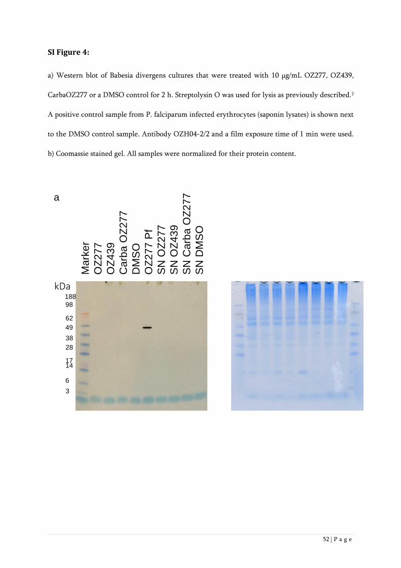
52 | P a g e
SI Figure 4:
a) Western blot of Babesia divergens cultures that were treated with 10 µg/mL OZ277, OZ439,
CarbaOZ277 or a DMSO control for 2 h. Streptolysin O was used for lysis as previously described.2
A positive control sample from P. falciparum infected erythrocytes (saponin lysates) is shown next
to the DMSO control sample. Antibody OZH04-2/2 and a film exposure time of 1 min were used.
b) Coomassie stained gel. All samples were normalized for their protein content.
a
kDa188
98
62
49
38
28
1714
6
3
Mark
er
OZ
277
OZ
439
Carb
aO
Z277
DM
SO
OZ
277 P
f
SN
OZ
277
SN
OZ
439
SN
Carb
aO
Z277
SN
DM
SO
kDa

53 | P a g e
SI References
1. Vennerstrom, J. L.; Dong, Y.; Chollet, J.; Matile, H.; Padmanilayam, M.; Tang, Y.; Charman, W.
N. Spiro and dispiro 1,2,4-trioxolane antimalarials. U. S. Patent 6,906,205 (14 June 2005).
2. Richier, E.; Biagini, G. A.; Wein, S.; Boudou, F.; Bray,P. G.; Ward, S. A.; Precigout, E. ; Calas, M.;
Dubremetz, J.; Vial, H. J. (2006) Potent Antihematozoan Activity of Novel Bisthiazolium Drug T16:
Evidence for Inhibition of Phosphatidylcholine Metabolism in Erythrocytes Infected with Babesia
and Plasmodium spp. Antimicrob. Agents Chemother. 50, 3381–3388.

54 | P a g e
3. In vitro activity of anti-malarial ozonides against an
artemisinin-resistant isolate
Fabian Baumgärtner1,2 , Joëlle Jourdan1,2 , Christian Scheurer1,2 , Benjamin Blasco3 , Brice Campo3 ,
Pascal Mäser1,2, Sergio Wittlin1,2,*
1Swiss Tropical and Public Health Institute, Socinstrasse 57, CH-4002 Basel, Switzerland
2University of Basel, Petersplatz 1, CH-4001 Basel, Switzerland
3Medicines for Malaria Venture, ICC, 20 Route de Pré-Bois, PO Box 1826, 1215 Geneva, Switzerland
*Corresponding author
Email: [email protected]
Tel: +41 61 284 81 36
Published in: Malaria Journal (2017)
Personal Contribution to the Paper: I supported Fabian Baumgärtner in the Lab, analysed data and
helped writing the paper

55 | P a g e
Abstract
Background
Recently published data suggest that artemisinin derivatives and synthetic peroxides, such as the
ozonides OZ277 and OZ439, have a similar mode of action. Here the cross-resistance of OZ277 and
OZ439 and four additional next-generation ozonides was probed against the artemisinin-resistant
clinical isolate Plasmodium falciparum Cam3.I, which carries the K13-propeller mutation R539T
(Cam3.IR539T).
Methods
The previously described in vitro ring-stage survival assay (RSA0-3h) was employed and a simplified
variation of the original protocol was developed.
Results
At the pharmacologically relevant concentration of 700 nM, all six ozonides were highly effective
against the dihydroartemisinin-resistant P. falciparum Cam3.IR539T parasites, showing a per cent
survival ranging from <0.01 to 1.83%. A simplified version of the original RSA0-3h method was
developed and gave similar results, thus providing a practical drug discovery tool for further
optimization of next-generation anti-malarial peroxides.
Conclusion
The absence of in vitro cross-resistance against the artemisinin-resistant clinical isolate Cam3.IR539T
suggests that ozonides could be effective against artemisinin-resistant P. falciparum. How this will
translate to the human situation in clinical settings remains to be investigated.
Keywords Ring-stage survival assay, Artemisinin, Ozonide, Plasmodium falciparum, Cam3.IR539T,
Drug resistance
56 | P a g e
Background
Malaria is one of the most important tropical diseases resulting in 214 million new cases and an
estimated 438,000 malaria deaths worldwide in 2015 [1]. The discovery of artemisinin in the 1970s
was an important step forward in anti-malarial drug therapy and was recognized with the Nobel
Prize in Physiology or Medicine in 2015 [2,3]. Artemisinin and its semi-synthetic derivatives, such
as dihydroartemisinin (DHA) (Fig. 1), artesunate and artemether, contain a unique sesquiterpene
lactone peroxide (1,2,4-trioxane) structure and artemisinin-based combination therapy (ACT)
represents the current first-line treatment of uncomplicated Plasmodium falciparum malaria [4,5,6].
Since the starting material artemisinin is a natural product, its production is limited to the
availability of the plant [7,4], although several total syntheses of artemisinin have been described
[8]. In 2004, Vennerstrom et al. reported the discovery of a completely synthetic peroxide anti-
malarial containing a 1,2,4-trioxolane (ozonide) pharmacophore named OZ277 (arterolane) (Fig. 1)
with anti-malarial activity comparable to the artemisinin derivatives [9,10]. In combination with
piperaquine, arterolane was registered for anti-malarial combination therapy in India in 2011
[11,12,13,14]. The next-generation ozonide, OZ439 (artefenomel) (Fig. 1), exhibits an increased
pharmacokinetic half-life and good safety profile and is now being tested in phase IIb clinical trials
[12,14,15,16,17]. The iron-dependent alkylation hypothesis is one of the proposed modes of action
of artemisinin and synthetic peroxides [18,19,20,21] where the peroxide is thought to be activated
by the reductive cleavage in the presence of ferrous haem (or free Fe(II) derived from haem) released
as a by-product of hemoglobin digestion in the food vacuole [20,22,23,24, 25,26,27]. Thereby
carbon-centred radicals are generated, which then alkylate haem and parasite proteins
[28,29,30,31,32,33]. The interaction of the artemisinin derivatives or ozonides with parasite targets
is irreversible [31,34]. Although the semi-synthetic artemisinins are highly effective, prolonged
parasite clearance times were first reported along the Thai–Cambodian border in 2006, suggesting
57 | P a g e
an emerging artemisinin resistance phenotype [35]. Today, delayed parasite clearance following
treatment with artemisinin derivatives has been observed across Southeast Asia [36,37,38,39,40,41].
It was found that mutations in the Kelch 13 propeller domain are associated with ring-stage parasites
entering a quiescent state with delayed parasite clearance after exposure to artemisinins
[41,42,43,44,45]. When 50% inhibitory concentrations (IC50) were measured using conventional
methods such as the [3H] hypoxanthine incorporation assay [46], no difference was observed
between artemisinin-resistant and -susceptible strains after treatment with artemisinin or its
derivatives [47,48,49,50]. In an effort to correlate the delayed parasite clearance observed in vivo
with in vitro parasite survival, Witkowski et al. [48,49] developed a ring-stage survival assay (RSA0-
3h) that exploited the differences in susceptibility observed between wild-type and K13 mutants at
the early ring stage of the asexual blood cycle following a short pulse of artemisinin treatment. In
the RSA0-3h, synchronized young ring stage parasites (0-3 hrs old) are exposed to drugs for six hours,
and then cultured in drug free culture medium for 66 hours before relative growth is determined
by microscopic analysis [48,49]. Since the structural analogies between artemisinins and ozonides
(Fig. 1) suggest that they share similar modes of action, and thus some level of cross resistance
[9,10,51,52], the per cent survival of an artemisinin-resistant clinical isolate (Cam3.IR539T) treated
with DHA, OZ277, OZ439, and four additional next-generation ozonides (Fig. 1) using the RSA0-3h
as described by Witkowski et al. [48,49] was evaluated. Additionally, a sub-set of these compounds
was tested in the RSA0-3h described by Xie et al. [53] that also uses tightly synchronized ring-stage
cultures, but allows the assay to be performed routinely within a convenient time-frame.
58 | P a g e
Methods
Parasite cultivation
The artemisinin-resistant P. falciparum isolate Cam3.IR539T from Battambang, Cambodia was
obtained from BEI Resources [54] with the accession number MRA-1240. The drug-sensitive P.
falciparum strain NF54 (airport strain from The Netherlands) was provided by F. Hoffmann-La
Roche Ltd. Parasites were cultivated in standard cultivation medium, consisting of hypoxanthine
(50 mg/l), RPMI (10.44 g/l) supplemented with HEPES (5.94 g/l), albumax (5 g/l), sodium
bicarbonate (2.1 g/l) and neomycin (100 mg/l) [55].
Ring-stage survival assays (RSA0-3h)
Ring-stage survival assays (RSA0-3h) were carried out essentially as previously described by
Witkowski et al. [48], but with a few modifications in the drug-washing procedure to ensure that
no residual peroxide was present during the 66-hour post-treatment period [56]. Briefly, zero to
three hours post-invasion ring stages were adjusted to 1% parasitaemia and 2.5% haematocrit by
adding uninfected erythrocytes, transferred in a total volume of 1 mL into 48-well plates and
exposed for six hours to a range of concentrations (700, 350, 175, 88, and 49 nM) of DHA or one of
the six ozonides tested in this study. The synthesis of the four next-generation ozonides, OZ493,
OZ609, OZ655 and OZ657, will be reported in due course by the laboratory of Prof. Jonathan
Vennerstrom (manuscript with ID jm-2016-01586n in the Journal of Medicinal Chemistry is currently
in press). After six hours, cultures were transferred to 15 mL conical tubes, centrifuged at 1,400 rpm
(400 g) for 2 min and carefully washed two times with 12 mL of culture medium. The complete
removal of compound after washing was verified by incubating the supernatant recovered after the
last washing step with fresh cultures of NF54 parasites, ensuring that no growth inhibition was
detected. After washing, blood pellets were resuspended in complete drug-free culture medium,
transferred into new wells and cultured for 66 hours under standard conditions.
59 | P a g e
Thin blood smears were prepared, methanol-fixed and stained with 10% Giemsa. Per cent survival
was assessed using light microscopy, counting the number of parasitized cells in ≥10,000 red blood
cells (RBCs) and comparing survival to that of the drug-free dimethylsulfoxid incubation.
Microscopy analysis was performed independently by two microscopists, one having more than 15 years
of work experience.
Alternative parasite synchronization method
Parasites were synchronized according to Xie et al. [53] with 5% d-sorbitol. After 30 and 43 hours,
parasites were synchronized a second and third time, respectively, resulting in zero to one-hour old
ring-stage parasites. The RSA0-3h was initiated two hours later.
Standard [3H] hypoxanthine incorporation assay
The in vitro anti-malarial activity was measured using the [3H]-hypoxanthine incorporation assay
[55]. Results were expressed as the concentration resulting in 50% inhibition (IC50).
60 | P a g e
Results
The per cent survival of parasites exposed to a concentration range of DHA and six different
ozonides (Fig. 1) was determined using the artemisinin-resistant P. falciparum Cambodian isolate
Cam3.IR539T. As expected, DHA exposure gave a high survival rate ranging from 74 to 33% at
concentrations of 49 and 700 nM, respectively (Fig. 2), which is comparable to the observed survival
value of 40% at 700 nM published previously [44]. In contrast, when tested at 700 nM, the two
ozonides OZ277 and OZ439 showed an approximate 18- to 45-fold increase in potency compared
with DHA (Fig. 2). Full and equal potency was observed when DHA, OZ277 and OZ439 were tested
in parallel in the RSA0-3h using the artemisinin-sensitive strain NF54 (Supplementary Table S1). At
the lowest concentration (49 nM), OZ277 had poor activity, showing a similar per cent survival to
that of DHA, whereas OZ439 was still about five-fold more potent. A possible explanation for
OZ439 being more potent than OZ277 could be related to its improved stability in blood as
previously described [15]. In those studies, OZ277 or OZ439 were incubated at 37°C in P.
falciparum-infected human blood. After 2 hours more than 90% of OZ277 was degraded, whereas
OZ439 was found to be about 10-20x more stable. A similar and more recent study found similar
differences in stability for OZ277 and OZ439 [56]. The same compounds were also tested in a more
convenient variation of the standard RSA0-3h that uses synchronized ring-stage cultures that can be
easily produced during normal working hours [53]. As shown in Table 1, this alternative
synchronization method gave results that were comparable to those obtained using the standard
RSA0-3h.
To investigate further the level of cross-resistance between DHA and the ozonides, four additional
next-generation ozonides (OZ493, OZ609, OZ655, OZ657) (Fig. 1) were tested against the
Cam3.IR539T parasites. While all six ozonides had a similar IC50 value using a conventional 72-hour
[3H] hypoxanthine incorporation assay (Supplementary Table S2), the RSA0-3h showed that OZ493,

61 | P a g e
OZ609 and OZ655 were highly potent and completely inhibited the growth of the artemisinin-
resistant isolate at the two highest concentrations tested (Fig. 2). At the lowest concentration,
potency was comparable to that for OZ439. The overall potency of OZ657 was comparable to that
of OZ277.
The RSA0-3h was recently developed to provide an in vitro correlate of the longer in vivo parasite
clearance times observed after artemisinin treatment in Southeast Asia, which is widely interpreted
as a sign of potential artemisinin resistance [57,58]. Provided that the RSA0-3h does indeed predict
the potency of compounds against artemisinin-resistant parasites in malaria patients, the here
described data suggest that all of the tested ozonides are highly potent against isolates such as P.
falciparum Cam3.IR539T. These data are in line with the recent clinical observation that the parasite
clearance rate following OZ439 treatment is not significantly affected by resistance-associated
mutations in the Kelch 13 propeller region [17] and the recent data published by Siriwardana et al.
[59], which showed no reduced susceptibility of OZ439 in a different delayed clearance phenotype
parasite (Cam3.II) in vitro.
62 | P a g e
Conclusion
In the traditional RSA0-3h, as well as a more convenient variation of the original method, all of the
tested ozonides, were highly potent against the artemisinin-resistant isolate P. falciparum
Cam3.IR539T in contrast to results for DHA. These data indicate that artemisinin-resistant P.
falciparum infections could be successfully treated with ozonide anti-malarial drugs.
63 | P a g e
Abbreviations
OZ277: arterolane; OZ439: artefenomel; DHA: Dihydroartemisinin; ACT: Artemisinin combination
therapy; RBC: Red blood cell.
Declarations
Ethics approval and consent to participate
Not applicable.
Consent for publication
Not applicable.
Availability of data and material
All data generated or analysed during this study are included in this published article and its
supplementary information files.
Competing interests
The authors declare that they have no competing interests.
Funding
This work was financially supported by the Swiss National Science Foundation (Grant
310030_149896 to SW), the Medicines for Malaria Venture, and the Swiss Tropical and Public
Health Institute.
Authors' contributions
FB, JJ, CS, BB, BC, PM and SW designed the research. FB and CS performed the research. All authors
analysed data. FB, JJ and SW wrote the manuscript. All authors had full access to all data in the
study and read and approved the final manuscript.

64 | P a g e
Acknowledgements
We are grateful to Jonathan L Vennerstrom, Hugues Matile and Susan A Charman for critically
reading the manuscript and making valuable suggestions.

65 | P a g e
References
[1] WHO. World Malaria Report 2015. 2015; ISBN: 978 92 4 156515 8.
[2] Tu Y. The discovery of artemisinin (qinghaosu) and gifts from Chinese medicine. Nat Med.
2011;17:1217-20.
[3] Su XZ, Miller LH. The discovery of artemisinin and the Nobel Prize in Physiology or Medicine.
Sci China Life Sci. 2015;58:1175-9.
[4] Klayman DL. Qinghaosu (artemisinin): an antimalarial drug from China. Science. 1985;
228:1049−55.
[5] White NJ. Artemisinin: Current status. Trans R Soc Trop Med Hyg. 1994;88:3-4.
[6] White NJ. Qinghaosu (artemisinin): the price of success. Science. 2008;320:330−4.
[7] Kumar S, Srivastava S. Establishment of artemisinin combination therapy as first line treatment
for combating malaria: Artemisia annua cultivation in India needed for providing sustainable supply
chain of artemisinin. Current Science. 2005;89:1097-102.
[8] Avery MA, Chong WKM, Jennings- White C. Stereoselective Total Synthesis of (+)-
Artemisinin, the Antimalarial Constituent of Artemisia annua L. J Am Chem. Soc. 1992;114:974-9.
[9] Vennerstrom JL, Arbe-Barnes S, Brun R, Charman SA, Chiu FC, Chollet, J, Dong Y, Dorn A,
Hunziker D, Matile H, McIntosh K, Padmanilayam M, Santo Tomas J, Scheurer C, Scorneaux B,
Tang Y, Urwyler H, Wittlin S, Charman WN. Identification of an antimalarial synthetic trioxolane
drug development candidate. Nature. 2004;430:900−4.
[10] Tang Y, Dong Y, Vennerstrom JL. Synthetic peroxides as antimalarials. Med Res Rev.
2004;24:425−48.
[11] Anthony MP, Burrows JN, Duparc S, Moehrle JJ, Wells TN. The global pipeline of new
medicines for the control and elimination of malaria. Malar J. 2012;11:316.
[12] Mäser P, Wittlin S, Rottmann M, Wenzler T, Kaiser M, Brun R. Antiparasitic agents: new drugs
on the horizon. Curr Opin Pharmacol. 2012;12:562−6.
66 | P a g e
[13] Patil CY, Katare SS, Baig MS, Doifode SM. Fixed Dose Combination of Arterolane and
Piperaquine: A Newer Prospect in Antimalarial Therapy. Annals of Medical and Health Sciences
Research. 2014;4:466-71.
[14] Wells TN, van Huijsduijnen RH, Van Voorhis WC. Malaria medicines: a glass half full? Nat Rev
Drug Discovery. 2015;14:424−42.
[15] Charman SA, Arbe-Barnes S, Bathurst IC, Brun R, Campbell M, Charman WN, Chiu FC, Chollet
J, Craft JC, Creek DJ, Dong Y, Matile H, Maurer M, Morizzi J, Nguyen T, Papastogiannidis P,
Scheurer C, Shackleford DM, Sriraghavan K, Stingelin L, Tang Y, Urwyler H, Wang X, White KL,
Wittlin S, Zhou L, Vennerstrom JL. Synthetic ozonide drug candidate OZ439 offers new hope for
a single-dose cure of uncomplicated malaria. Proc Natl Acad Sci U S A. 2011;108:4400−5.
[16] Moehrle JJ, Duparc S, Siethoff C, van Giersbergen PL, Craft JC, Arbe-Barnes S, Charman SA,
Gutierrez M, Wittlin S, Vennerstrom JL. First-in-man safety and pharmacokinetics of synthetic
ozonide OZ439 demonstrates an improved exposure profile relative to other peroxide antimalarials.
Br J Clin Pharmacol. 2013;75:535−48.
[17] Phyo AP, Jittamala P, Nosten FH, Pukrittayakamee S, Imwong M, White NJ, Duparc S,
Macintyre F, Baker M, Möhrle JJ. Antimalarial activity of artefenomel (OZ439), a novel synthetic
antimalarial endoperoxide, in patients with Plasmodium falciparum and Plasmodium vivax malaria:
an open-label phase 2 trial. Lancet Infect Dis. 2016;16:61-9.
[18] Kaiser M, Wittlin S, Nehrbass-Stuedli A, Dong Y, Wang X, Hemphill A, Matile H, Brun R,
Vennerstrom JL. Peroxide bond-dependent antiplasmodial specificity of artemisinin and OZ277
(RBx11160). Antimicrob Agents Chemother. 2007;51:2991−3.
[19] Creek DJ, Charman WN, Chiu FCK, Prankerd RJ, Dong Y, Vennerstrom JL, Charman SA.
Relationship between antimalarial activity and haem alkylation for spiro- and dispiro-1,2,4-
trioxolane antimalarials. Antimicrob Agents Chemother. 2008;52:1291−6.
[20] Meunier B, Robert A. Heme as trigger and target for trioxane-containing antimalarial drugs.
Acc Chem Res. 2010;43:1444−51.
[21] Tilley L, Straimer J, Gnädig NF, Ralph SA, Fidock DA. Artemisinin Action and Resistance in
Plasmodium falciparum. Trends Parasitol. 2016;32:682-96.

67 | P a g e
[22] Klonis N, Crespo-Ortiz MP, Bottova I, Abu-Bakar N, Kenny S, Rosenthal PJ, Tilley L.
Artemisinin activity against Plasmodium falciparum requires hemoglobin uptake and digestion.
Proc Natl Acad Sci U S A. 2011;108:11405−10.
[23] Crespo MD, Avery TD, Hanssen E, Fox E, Robinson TV, Valente P, Taylor DK, Tilley L.
Artemisinin and a series of novel endoperoxide antimalarials exert early effects on digestive vacuole
morphology. Antimicrob Agents Chemother. 2008;52:98-109.
[24] Hartwig CL, Rosenthal AS, D’Angelo J, Griffin CE, Posner GH, Cooper RA. Accumulation of
artemisinin trioxane derivatives within neutral lipids of Plasmodium falciparum malaria parasites is
endoperoxide-dependent. Biochem Pharmacol. 2009;77:322−36.
[25] Robert A, Claparols C, Witkowski B, Benoit-Vical F. Correlation between Plasmodium yoelii
nigeriensis susceptibility to artemisinin and alkylation of heme by the drug. Antimicrob Agents
Chemother. 2013;57:3998−4000.
[26] Klonis N, Creek DJ, Tilley L. Iron and heme metabolism in Plasmodium falciparum and the
mechanism of action of artemisinins. Curr Opin Microbiol. 2013;16:722-7.
[27] Uhlemann AC, Wittlin S, Matile H, Bustamante LY, Krishna S. Mechanism of antimalarial
action of the synthetic trioxolane RBX11160 (OZ277). Antimicrob Agents Chemother.
2007;51:667−72.
[28] Asawamahasakda W, Ittarat I, Pu YM, Ziffer H, Meshnick SR. Reaction of antimalarial
endoperoxides with specific parasite proteins. Antimicrob Agents Chemother. 1994;38:1854-8.
[29] Meshnick SR. Artemisinin: mechanisms of action, resistance and toxicity. Int J Parasitol.
2002;32:1655−60.
[30] Tang Y, Dong Y, Wang X, Sriraghavan K, Wood JK, Vennerstrom JL. Dispiro-1,2,4-trioxane
analogs of a prototype dispiro-1,2,4-trioxolane: mechanistic comparators for artemisinin in the
context of reaction pathways with iron (II). J Org Chem. 2005:70; 5103−10.
[31] Fügi MA, Wittlin S, Dong Y, Vennerstrom JL. Probing the antimalarial mechanism of
artemisinin and OZ277 (arterolane) with nonperoxidic isosteres and nitroxyl radicals. Antimicrob
Agents Chemother. 2010;54:1042-6.

68 | P a g e
[32] Hartwig CL, Lauterwasser EMW, Mahajan SS, Hoke JM, Cooper RA, Renslo AR. Investigating
the antimalarial action of 1,2,4-trioxolanes with fluorescent chemical probes. J Med Chem.
2011;54:8207−13.
[33] Tilley L, Charman S, Vennerstrom JL. Semisynthetic artemisinin and synthetic peroxide
antimalarials. In: Palmer MJ, Wells TNC, editors. RSC Drug Discovery Series No. 14. Neglected
Diseases and Drug Discovery. London; 2011. p. 33−64.
[34] Abiodun OO, Brun R, Wittlin S. In vitro interaction of artemisinin derivatives or the fully
synthetic peroxidic anti-malarial OZ277 with thapsigargin in Plasmodium falciparum strains. Malar
J. 2013;12:43.
[35] Noedl H, Se Y, Schaecher K, Smith BL, Socheat D, Fukuda MM. Artemisinin Resistance in
Cambodia 1 (ARC1) Study Consortium Evidence of artemisinin-resistant malaria in western
Cambodia. N Engl J Med. 2008;359:2619-20.
[36] Noedl H, Socheat D, Satimai W. Artemisinin-resistant malaria in Asia. N Engl J Med. 2009;
361:540-1.
[37] Dondorp AM, Nosten F, Yi P, Das D, Phyo AP, Tarning J, Lwin KM, Ariey F, Hanpithakpong
W, Lee SJ, Ringwald P, Silamut K, Imwong M, Chotivanich K, Lim P, Herdman T, An SS, Yeung S,
Singhasivanon P, Day NPJ, Lindegardh N, Socheat D, White NJ. Artemisinin Resistance in
Plasmodium falciparum Malaria. N Engl J Med. 2009;38: 455-67.
[38] Amaratunga C, Sreng S, Suon S, Phelps ES, Stepniewska K, Lim P, Zhou C, Mao S, Anderson
JM, Lindegardh N, Jiang H, Song J, Su XZ, White NJ, Dondorp AM, Anderson, TJ, Fay MP, Mu J,
Duong S, Fairhurst RM. Artemisinin-resistant Plasmodium falciparum in Pursat province, western
Cambodia: a parasite clearance rate study. Lancet Infect Dis. 2012;12:851-8.
[39] Phyo AP, Nkhoma S, Stepniewska K, Ashley EA, Nair S, McGready R, ler Moo C, Al-Saai S,
Dondorp AM, Lwin KM, Singhasivanon P, Day NP, White NJ, Anderson TJ, Nosten F. Emergence
of artemisinin-resistant malaria on the western border of Thailand: a longitudinal study. Lancet.
2012;379:1960-6.
[40] Ashley EA, Dhorda M, Fairhurst RM, Amaratunga C, Lim P, Suon S, Sreng S, Anderson JM,
Mao S, Sam B, Sopha C, Chuor CM, Nguon C, Sovannaroth S, Pukrittayakamee S, Jittamala P,
http://www.ncbi.nlm.nih.gov/pubmed/?term=Schaecher%20K%5BAuthor%5D&cauthor=true&cauthor_uid=19064625
http://www.ncbi.nlm.nih.gov/pubmed/?term=Anderson%20JM%5BAuthor%5D&cauthor=true&cauthor_uid=22940027
http://www.ncbi.nlm.nih.gov/pubmed/?term=Anderson%20JM%5BAuthor%5D&cauthor=true&cauthor_uid=22940027
http://www.ncbi.nlm.nih.gov/pubmed/?term=Anderson%20TJ%5BAuthor%5D&cauthor=true&cauthor_uid=22940027
http://www.ncbi.nlm.nih.gov/pubmed/?term=ler%20Moo%20C%5BAuthor%5D&cauthor=true&cauthor_uid=22484134

69 | P a g e
Chotivanich K, Chutasmit K, Suchatsoonthorn C, Runcharoen R, Hien TT, Thuy-Nhien NT, Thanh
NV, Phu NH, Htut Y, Han KT, Aye KH, Mokuolu OA, Olaosebikan RR, Folaranmi OO, Mayxay M,
Khanthavong M, Hongvanthong B, Newton PN, Onyamboko, MA, Fanello CI, Tshefu AK, Mishra
N, Valecha N, Phyo AP, Nosten F, Yi P, Tripura R, Borrmann S, Bashraheil M, Peshu J, Faiz MA,
Ghose A, Hossain MA, Samad R, Rahman MR, Hasan MM, Islam A, Miotto O, Amato R, MacInnis
B, Stalker J, Kwiatkowski DP, Bozdech Z, Jeeyapant A, Cheah PY, Sakulthaew T, Chalk J, Intharabut
B, Silamut K, Lee SJ, Vihokhern B, Kunasol C, Imwong M, Tarning J, Taylor WJ, Yeung S, Woodrow
CJ, Flegg JA, Das D, Smith J, Venkatesan M, Plowe CV, Stepniewska K, Guerin PJ, Dondorp AM,
Day NP, White NJ. Spread of Artemisinin Resistance in Plasmodium falciparum Malaria. N Engl J
Med. 2014;371:411–23.
[41] Paloque L, Ramadani AP, Mercereau-Puijalon O, Augereau JM, Benoit-Vical F. Plasmodium
falciparum: multifaceted resistance to artemisinins. Malaria J. 2016;15:149.
[42] Ariey F, Witkowski B, Amaratunga C, Beghain J, Langlois AC, Khim N, Kim S, Duru, V,
Bouchier C, Ma L, Lim P, Leang R, Duong S, Sreng S, Suon S, Chuor CM, Bout DM, Ménard S,
Rogers WO, Genton B, Fandeur T, Miotto O, Ringwald P, Le Bras J, Berry A, Barale JC, Fairhurst
RM, Benoit-Vical F, Mercereau-Puijalon O, Ménard D. A molecular marker of artemisinin-resistant
Plasmodium falciparum malaria. Nature. 2014;505:50-5.
[43] Mok S, Ashley EA, Ferreira PE, Zhu L, Lin Z, Yeo T, Chotivanich K, Imwong M,
Pukrittayakamee S, Dhorda M, Nguon C, Lim P, Amaratunga C, Suon S, Hien TT, Htut Y, Abul
Faiz M, Onyamboko MA, Mayxay M, Newton PN, Tripura R, Woodrow CJ, Miotto O, Kwiatkowski
DP, Nosten F, Day NPJ, Preiser PR, White NJ, Dondorp, AM, Fairhurst RM, Bozdech Z. Population
transcriptomics of human malaria parasites reveals the mechanism of artemisinin resistance.
Science. 2015;347:431-5.
[44] Straimer J, Gnädig NF, Witkowski B, Amaratunga C, Duru V, Ramadani AP, Dacheux M, Khim
N, Zhang L, Lam S, Gregory PD, Urnov FD, Mercereau-Puijalon O, Benoit-Vical F, Fairhurst RM,
Ménard D, Fidock DA. Drug resistance. K13-propeller mutations confer artemisinin resistance in
Plasmodium falciparum clinical isolates. Science. 2015;347:428-31.
[45] Mbengue A, Bhattacharjee S, Pandharkar T, Liu H, Estiu G, Stahelin RV, Rizk SS, Njimoh DL,
Ryan Y, Chotivanich K, Nguon C, Ghorbal M, Lopez-Rubio JJ, Pfrender M, Emrich S, Mohandas N,
http://www.ncbi.nlm.nih.gov/pubmed/?term=Witkowski%20B%5BAuthor%5D&cauthor=true&cauthor_uid=24352242
http://www.ncbi.nlm.nih.gov/pubmed/?term=Langlois%20AC%5BAuthor%5D&cauthor=true&cauthor_uid=24352242
http://www.ncbi.nlm.nih.gov/pubmed/?term=Le%20Bras%20J%5BAuthor%5D&cauthor=true&cauthor_uid=24352242
http://www.ncbi.nlm.nih.gov/pubmed/?term=Witkowski%20B%5BAuthor%5D&cauthor=true&cauthor_uid=25502314
http://www.ncbi.nlm.nih.gov/pubmed/?term=Ramadani%20AP%5BAuthor%5D&cauthor=true&cauthor_uid=25502314

70 | P a g e
Dondorp AM, Wiest O, Haldar K.A molecular mechanism of artemisinin resistance in Plasmodium
falciparum malaria. Nature. 2015;520:683-7.
[46] Desjardins RE, Canfield CJ, Haynes JD, Chulay JD. Quantitative assessment of antimalarial
activity in vitro by a semiautomated microdilution technique. Antimicrob Agents Chemother.
1979;16:710-8.
[47] Witkowski B, Lelièvre J, Barragán MJ, Laurent V, Su XZ, Berry A, Benoit-Vical F. Increased
tolerance to artemisinin in Plasmodium falciparum is mediated by a quiescence mechanism.
Antimicrob Agents Chemother. 2010;54:1872-7.
[48] Witkowski B, Khim N, Chim P, Kim S, Ke S, Kloeung N, Chy S, Duong S, Leang R, Ringwald
P, Dondorp AM, Tripura R, Benoit-Vical F, Berry A, Gorgette O, Ariey F, Barale JC, Mercereau-
Puijalon O, Menard D. Reduced artemisinin susceptibility of Plasmodium falciparum ring stages in
western Cambodia. Antimicrob Agents Chemother. 2013;57:914-23.
[49] Witkowski B, Amaratunga C, Khim N, Sreng S, Chim P, Kim S, Lim P, Mao S, Sopha C, Sam B,
Anderson JM, Duong S, Chuor CM, Taylor WR, Suon S, Mercereau-Puijalon O, Fairhurst RM,
Menard D. Novel phenotypic assays for the detection of artemisinin-resistant Plasmodium
falciparum malaria in Cambodia: in-vitro and ex-vivo drug-response studies. Lancet Infect Dis.
2013;13:1043-9.
[50] Teuscher F, Gatton ML, Chen N, Peters J, Kyle DE, Cheng Q. Artemisinin‐induced dormancy
in plasmodium falciparum: duration, recovery rates, and implications in treatment failure. J Infect
Dis. 2010;202:1362-8.
[51] Jourdan J, Matile H, Reift E, Biehlmaier O, Dong Y, Wang X, Mäser P, Vennerstrom JL, Wittlin
S. Monoclonal Antibodies That Recognize the Alkylation Signature of Antimalarial Ozonides
OZ277 (Arterolane) and OZ439 (Artefenomel). ACS Infect Dis. 2015;2:54–61.
[52] Ismail HM, Barton VE, Panchana M, Charoensutthivarakul S, Biagini GA, Ward SA, O'Neill
PM. A Click Chemistry-Based Proteomic Approach Reveals that 1,2,4-Trioxolane and Artemisinin
Antimalarials Share a Common Protein Alkylation Profile. Angew Chem Int Ed. 2016;55:1-6.
[53] Xie S, Dogovski C, Kenny S, Tilley L, Klonis N. Optimal assay design for determining the in
vitro sensitivity of ring stage Plasmodium falciparum to artemisinins. Int J Parasitol. 2014;44:893-9.
http://www.ncbi.nlm.nih.gov/pubmed/?term=Witkowski%20B%5BAuthor%5D&cauthor=true&cauthor_uid=20160056
http://www.ncbi.nlm.nih.gov/pubmed/?term=Witkowski%20B%5BAuthor%5D&cauthor=true&cauthor_uid=23208708
http://www.ncbi.nlm.nih.gov/pubmed/?term=Witkowski%20B%5BAuthor%5D&cauthor=true&cauthor_uid=24035558
71 | P a g e
[54] B E I Resources. https://www.beiresources.org/Catalog/BEIParasiticProtozoa/MRA-1240.aspx.
Accessed 26 Nov 2014
[55] Snyder C, Chollet J, Santo-Tomas J, Scheurer C, Wittlin S. In vitro and in vivo interaction of
synthetic peroxide RBx11160 (OZ277) with piperaquine in Plasmodium models. Exp Parasitol.
2007;115:296−300.
[56] Yang T, Xie SC, Cao P, Giannangelo C, McCaw J, Creek DJ, Charman SA, Klonis N, Tilley L.
Comparison of the Exposure Time Dependence of the Activities of Synthetic Ozonide Antimalarials
and Dihydroartemisinin against K13 Wild-Type and Mutant Plasmodium falciparum Strains.
Antimicrob Agents Chemother. 2016;60:4501-10.
[57] Phyo AP, Ashley EA, Anderson TJ, Bozdech Z, Carrara VI, Sriprawat K, Nair S, White MM,
Dziekan J, Ling C, Proux S, Konghahong K, Jeeyapant A, Woodrow CJ, Imwong M, McGready R,
Lwin KM, Day NP, White NJ, Nosten F. Declining Efficacy of Artemisinin Combination Therapy
Against P. Falciparum Malaria on the Thai-Myanmar Border (2003-2013): The Role of Parasite
Genetic Factors. Clin Infect Dis. 2016;63:784-91.
[58] Meshnick SR. Artemisinin Resistance in Southeast Asia. Clin Infect Dis. 2016;63:1527.
[59] Siriwardana A, Iyengar K, Roepe PD. Endoperoxide drug cross resistance patterns for
Plasmodium falciparum exhibiting an artemisinin delayed clearance phenotype. Antimicrob Agents
Chemother. 2016;60:6952-6

72 | P a g e
Fig. 1 Chemical structures of dihydroartemisinin (DHA) and the six ozonides investigated

73 | P a g e
Fig. 2 Mean per cent survival ± standard error (SE) of Plasmodium falciparum isolate Cam3.IR539T parasites after a 6-hour
exposure to a range of concentrations of dihydroartemisinin (DHA) or six different ozonides
Three biological replicates were performed per compound.
49 nM 88 nM 175 nM 350 nM 700 nM
DHA 73.50 60.03 47.94 37.68 33.02
OZ277 53.80 16.87 4.65 2.20 1.83
OZ439 13.70 0.93 0.73 0.73 0.73
OZ493 3.19 0.00 0.53 0.00 0.00
OZ609 1.29 0.17 0.12 0.00 0.00
OZ655 0.76 0.00 0.00 0.00 0.00
OZ657 59.10 22.93 2.33 0.00 0.00
0
10
20
30
40
50
60
70
80
90
100
Pe
rce
nt
surv
ival

74 | P a g e
Table 1 Mean per cent survival (individual values in brackets) of Cam3.IR539T isolate after 6 hours exposure to a range of
concentrations of DHA, OZ439 or OZ277 using the synchronization protocol from Xie et al. [53].
Two biological replicates were performed per compound.
Compounds RSA values (per cent survival) at different concentrations
175 nM 350 nM 700 nM
DHA 46 (49,43) 42 (45,39) 37 (39,35)
OZ277 4.0 (4.4,3.6) 2.3 (1.9,2.7) 1.4 (1.7,1.1)
OZ439 <0.01 (<0.01,<0.01) <0.01 (<0.01,<0.01) <0.01 (<0.01,<0.01)

75 | P a g e
Supplementary Table S1 Mean per cent survival (individual values in brackets) of NF54 after 6 hours exposure to 500 nM
of DHA, OZ439 or OZ277 using the synchronization protocol from Straimer et al. [44.]
Two biological replicates were performed per compound.
Compounds Per cent survival
DHA <0.01 (<0.01,<0.01)
OZ277 <0.01 (<0.01,<0.01)
OZ439 <0.01 (<0.01,<0.01)
Supplementary Table S2 Mean in vitro IC50 values (single values in brackets) for Plasmodium falciparum isolate Cam3.IR539T
and NF54 in the 72-hour [3H] hypoxanthine assay
Two biological replicates were performed per compound.
Compounds IC50 (nM)
Cam3.IR539T
IC50 (nM)
NF54
DHA 1.5 (1.7,1.3) 2.6 (2.4,2.8)
OZ277 1.8 (1.8,1.8) 1.7 (1.7,1.8)
OZ439 4.6 (4.6,4.6) 4.2 (4.6,3.9)
OZ493 3.6 (3.6,3.7) 3.8 (4.5,3.1)
OZ609 1.8 (1.8,1.8) 1.3 (1.4,1.2)
OZ655 2.8 (2.7,2.8) 2.5 (2.3,2.7)
OZ657 2.9 (2.9,2.9) 2.3 (2.3,2.3)
76 | P a g e
4. Identification of the Alkylation Signatures of
Antimalarial Ozonides and Artemisinin
Joëlle Jourdan1,2, Hugues Matile3, Alexander Schmidt4, Yuxiang Dong5, Xiaofang Wang5, Jonathan
L. Vennerstrom5, Remo S. Schmidt1,2, Sergio Wittlin1,2 and Pascal Mäser1,2*
1Swiss Tropical and Public Health Institute, Socinstrasse 57, CH-4002 Basel, Switzerland
2University of Basel, CH-4003 Basel, Switzerland
3F. Hoffmann- La Roche Ltd., CH-4070 Basel, Switzerland
4Proteomics Core Facility, Biozentrum, University of Basel, CH-4056 Basel, Switzerland
5College of Pharmacy, University of Nebraska Medical Center, 986025 Nebraska Medical Center,
Omaha, Nebraska 68198, United States
*Corresponding author
E-mail: [email protected]
Tel: 41 61 284 83 38
Personal Contribution to the Paper: I tested drug sensitivity, produced and analysed Western blot
data, optimized conditions for sample preparation and prepared samples for mass spectrometry.
Further, I analysed hit lists and helped writing the paper.

77 | P a g e
Abstract
Antimalarial peroxides such as the phytochemical artemisinin or the synthetic artefenomel are
prodrugs that need to be activated by reductive cleavage of the peroxide bond. This happens inside
the malaria parasite, presumably in the food vacuole with ferrous heme as the electron donor. The
generated radicals will then alkylate heme itself and proteins. Here we determine the alkylation
signatures of artemisinin and synthetic ozonides by chemical proteomics in Plasmodium falciparum
using alkyne probes that allow to identify the target proteins by click chemistry and affinity
purification. Two forms of controls are included: the non-clickable parent peroxides, as well as non-
peroxidic derivatives of the alkyne probes. These permitted to stringently determine the target space
of the peroxides, identifying a total of 18 proteins for artemisinin and 41 for the synthetic ozonides,
with an intersect of 14. We further compare the alkylation signatures of the antimalarial peroxides
to each other and to previously published datasets.
78 | P a g e
Introduction
Malaria remains one of the most prevalent infectious diseases worldwide, with an estimated death
toll of 429,000 in 2015 (WHO Report 2016). The most lethal form of malaria, malaria tropica, is
caused by Plasmodium falciparum. The current first-line treatments for malaria tropica are the
artemisinin-based combination therapies, ACTs (White, 2008), such as artemether-lumefantrine
(Coartem) or dihydroartemisinin-piperaquine (Artekin; Anthony et al., 2012). Artemisinin,
discovered in 1971 by Youyou Tu (Tu, 2011), is a sesquiterpene lactone with an endoperoxide bridge
(Figure 1). It is of highest activity and specificity against malaria parasites. However, as a natural
product artemisinin has two limitations: its production is laborious and constrained by the
availability of the plant Artemisia annua, and its pharmacological properties are difficult to improve
(Avery et al. 1992, Klayman et al. 1985, Kumar et al 2005). Thus, synthetic antimalarial peroxides
are a promising alternative for future antimalarials (Vennerstrom et al., 2004; O`Neill et al., 2017).
Vennerstrom and co-workers concentrated on the synthesis of adamantane-based 1,2,4-trioxolanes
(Figure 1), herein called ozonides, and presented a first drug candidate, OZ277 or arterolane maleate
in 2004 (Vennerstrom et al., 2004; Tang et al., 2004). In spite of its suboptimal stability in P.
falciparum-infected blood, arterolane maleate was subsequently registered for antimalarial therapy
in India in combination with piperaquine phosphate (Synriam®; Mäser et al., 2012; Anthony et al.,
2012; Patil et al., 2014; Wells et al., 2015). A next generation ozonide with improved
pharmacokinetic properties, OZ439 or artefenomel, is presently being tested in phase IIb clinical
trials in combination with ferroquine (Charman et al., 2011; Moehrle et al., 2013; Wells et al., 2015,
Phyo et al., 2016 and Blasco et al., 2017).
Artemisinins and ozonides are thought to possess similar modes of action since for both classes, the
endoperoxide bridge was found to be necessary for antimalarial activity (Klayman 1985; Kaiser et
al. 2007). One of the proposed modes of actions is therefore the iron-dependent artemisinin

79 | P a g e
alkylation hypothesis (Kaiser et al., 2007; Creek et al., 2008; Meunier et al., 2010), which suggests
that ferrous heme, or FeII derived from heme, is released as a product of hemoglobin digestion in
the parasite's food vacuole and leads to a reductive cleavage of the peroxide bond (Klonis et al., 2011;
Robert et al., 2013; Klonis e al., 2013; Hartwig et al., 2009). This generates highly reactive radicals
that alkylate intracellular targets such as proteins and heme itself (Asawamahasaksa et al., 1994;
Meshnick, 2002; Tilley et al., 2011). The alkylation hypothesis is supported by the findings that the
digestive vacuole is an important initial site for antimalarial peroxide activity (Crespo et al.; 2008;
Uhlemann et al., 2007), and that artemisinin and OZ277 are inactive against pathogens that do not
degrade hemoglobin (Kaiser et al., 2007). The formation of carbon-centered radicals was found to
be critical for the activity of antimalarial peroxides (Tang et al., 2005; Hartwig et al., 2011, Fügi et
al., 2010) and the activities of artemisinin as well as ozonides do not derive from reversible
interactions with parasite targets (Fügi et al., 2010; Abiodun et al., 2013).
The elucidation of the mode of action and identification of the intracellular targets of the
antimalarial peroxides has become all the more pressing after the emergence of artemisinin-resistant
P. falciparum isolates in South-East Asia (Noedl et al., 2008; Dondorp et al., 2009), and recently also
in Africa (Lu et al., 2017), whose mechanism of resistance is not fully understood either (Blasco et
al., 2017). Alkyne-tagged artemisinins and 1,2,4-trioxolanes have been used as chemical probes for
activity-based profiling of the peroxide target signatures in P. falciparum (Wang et al., 2015; Ismail
et al., 2016a; Ismail et al., 2016b), and the alkylation signature of a clickable 1,2,4-trioxolane was
found to be similar to that of artemisinin (Ismail et al., 2016b). Here we further sharpen this
approach, assessing the alkylation signatures of artemisinin and ozonides in parallel, and including
control molecules that allow to focus on those drug-target interactions that depend on the presence
of a peroxide bond.
80 | P a g e
Results
Design and synthesis of alkynes as chemical probes
If the peroxides are indeed activated by reductive cleavage and subsequently alkylate proteins, then
the covalent interaction between drug and target will facilitate the identification of the latter. We
designed alkyne derivatives of artemisinin and of an archetypal ozonide, OZ03, as chemical probes
that can be linked by azide-alkyne Huisgen cycloaddition (click-chemistry; Kolb et al., 2001) to
biotin or other markers (Figure 1). In addition to the parent compounds, which allow to discriminate
false positive hits, we included a second type of control molecules that possessed the alkyne group
but lacked the peroxide bridge: the deoxy form of the artemisinin alkyne, deoxyAA2, and the carba
form of OZ727, carbaOZ727 (Figure 1). These allow to discriminate hits that may actually be bound
by the probes, but not by means of peroxide reactivity.
Figure 1. Chemical structures of the parent molecules artemisinin and OZ03 plus the newly synthesized alkyne
derivatives as chemical probes.
Bioactivity of the chemical probes
The 50% inhibitory concentrations (IC50) of the newly synthesized alkynes against intraerythrocytic
P. falciparum in culture were determined using the [3H]-hypoxanthine incorporation assay
(Desjardins et al., 1979). The alkynes AA2, OZ726 and OZ727 were highly active against P.
falciparum NF54 parasites, whereas the nonperoxidic analogues carbaOZ277, carbaOZ727 and
deoxyAA2 were at least 1,000-fold less active (Table 1). Thus, as expected, the antiplasmodial

81 | P a g e
activity was dependent on the presence of the peroxide bridge, while the chosen alkyne
modifications did not compromise the antiplasmodial activity of the molecules. In particular, it was
interesting to note that the ozonides appeared to permit modification of the adamantane moiety.
Peroxide? Alkyne? IC50 [ng/ml]
AA2 Yes Yes 2.44
Artemisinin Yes No 2.28
DeoxyAA2 No Yes >10,000
OZ439 Yes No 2.03
OZ277 Yes No 0.91
CarbaOZ277 No No 4,900
OZ03 Yes No 2.36
OZ726 Yes Yes 0.74
OZ727 Yes Yes 0.98
CarbaOZ727 No Yes >10,000
Table 1. Mean IC50 values of the parent molecules and the newly synthesized alkynes against
P. falciparum NF54. All assays were performed in three biological replicates.
Protein alkylation by the chemical probes
A series of pilot experiments was carried out to optimize the incubation parameters (Figure S1) and
the reaction conditions for click chemistry. In brief (see Material and Methods for details), P.
falciparum cultures were incubated for 4 h with a chemical probe, followed by harvesting and lysis
of the parasites. The protein extracts were then treated with biotin-azide under copper to catalyze
the cycloaddition with the alkyne. After acetone precipitation, the samples were run on
polyacrylamide gels for Western blotting, using streptavidin-coupled horseradish peroxidase to
detect the alkylated proteins.
Parasite cultures treated with 1 µg/ml AA2, OZ726 or OZ727 produced clear signals with
reproducible banding patterns, whereas neither the non-clickable parent compounds nor the

82 | P a g e
nonperoxidic alkynes gave a signal on the Western blot (Figure 2A). The signal of the alkyne OZ726
was outcompeted by increasing ratios of the parent compound OZ03 (Figure 2B), indicating that the
parental ozonide and the alkyne derivative have the same binding sites and that binding is saturable.
These results confirmed that the peroxides covalently bind to proteins, as was known from previous
studies (Meshnick et al., 1993), and that they possess specific alkylation signatures rather than
binding unspecifically to all the P. falciparum proteins (Asawamahasakda et al., 1994; Fügi et al.,
2010; Abiodun et al., 2013). In order to identify these alkylation signatures by mass spectrometry,
we tested whether the click chemistry also works for precipitation of the bound proteins with
neutravidin-coupled agarose beads. This was indeed the case: protein bands were detected by
Western blot in P. falciparum cultures treated with 100 ng/ml AA2, whereas no bands were
observed in parasite cultures treated with 0.1% DMSO. Negative controls such as the neutravidin
beads alone or uninfected erythrocytes without drug gave no signal (Figure 2C).
Figure 2. Western blot experiments with P. falciparum cultures treated with (A) 1 µg/ml OZ726, OZ727, AA2,
OZ03, carbaOZ727, deoxyAA2 or artemisinin for 4 h, protein extracts prepared with 1.2% SDS in PBS, exposure
time 1 min, and (B) competing concentrations of OZ726 and the non-clickable parent OZ03 for 2 h, extracts
prepared with Ripa-Lysis buffer, exposure time 10 s. (C) shows a precipitation experiment with cultures treated

83 | P a g e
with 100 ng/ml AA2 in 0.1% DMSO for 4 h and different negative controls. Protein extracts were prepared
with 1.2% SDS in PBS and precipitated with neutravidin agarose resin; exposure time 1 min.
Mass spectrometry-based proteomics to identify alkylated proteins
The proteomics experiments were performed at an about 30x higher parasitemia (10%) compared
to the hypoxanthine incorporation assays (0.3%). From previous work it is known that a 30x higher
parasitemia results in a 30x higher inhibitory concentration (Jourdan et al., 2015). As a consequence,
in the here described studies we treated P. falciparum NF54 cultures of 7% to 12% parasitemia with
100ng/ml (corresponding to about an IC99) as well as 1000ng/ml artemisinin, AA2, deoxyAA2, OZ03,
OZ726, OZ727, carbaOZ727 or DMSO only, each in biological triplicates. The lysed samples were
subjected to click-chemistry with biotin-azide as above, incubated with neutravidin agarose beads,
and washed extensively (see Figures S2 and S3 for optimization of the washing conditions). The
bound proteins were eluted, digested with trypsin, and processed for mass spectrometry as described
in Material and Methods. The proteins were identified by their fingerprints of trypsin fragments as
compared to the predicted peptides of the proteome of P. falciparum NF54 (Uniprot ID
UP000030673).
Overall only 59 different proteins were significantly (>1.8 fold, p<0.05) enriched in any one of the
probe-treated cultures vs. the DMSO-treated control, indicating again that the peroxides have
specific alkylation signatures. The results are summarized in Tables S1 and S2. All the proteins that
were also obtained with the unmodified parent molecules, 1 (PFNF54_01079, dihydrofolate
reductase-thymidylate synthase) for artemisinin and 10 for OZ03 (Table S1), were removed from
the list of hits. Next, we removed the hits that were also obtained with the nonperoxidic alkyne
probes, 6 for deoxyAA2 but 0 for carbaOZ727 (Table S1). A potentially interesting hit among the
proteins fished with deoxyAA2 was 1-cys peroxiredoxin (PFNF54_02028), since another P.
falciparum peroxiredoxin, PfAOP, had been implicated in artemisinin susceptibility (Djuika et al.,

84 | P a g e
2017). The removal of all the hits that turned up in any of the control samples yielded a final list of
45 P. falciparum proteins that were bound by the chemical probes by means of peroxide activity
(Table 2).
Alkylation signatures of artemisinin and ozonides
We used the 100 ng/ml samples to compare the alkylation signatures of artemisinin and the two
ozonides. A total of 25 different P. falciparum proteins was alkylated by either AA2, OZ726 or
OZ727 (Table 2). Unsurprisingly, the largest degree of overlap was between the identified target
spaces of OZ726 and OZ727 (Figure 3A). There was also a clear overlap between the identified
targets of AA2 with those of OZ726, and to a lesser extent with those of OZ727 (Figure 3A). Only
one protein was identified with all three peroxides: PFNF54_01699, which corresponds to
PlasmoDB entry PF3D7_0706500. However, this protein is not linked to any publication and does
not contain any known domains (according to PlasmoDB). A blastp search with PF3D7_0706500
revealed significant similarity to proteins annotated as erythrocyte membrane proteins in P.
coatneyi and P. yoelii, as well as a PfEMP3-like protein in P. berghei. The latter contains an RNA
recognition motif (RRM) that was also found by the NCBI algorithm during the blastp search.
Figure 3. Venn diagrams of (A) all the hits obtained after treatment with 100 ng/ml artemisinin alkyne, OZ726 or
OZ727; and (B) and of all the hits identified with an alkyne derivative of artemisinin in the present study compared to
three published datasets (Wang et al., 2015; Ismail et al., 2016a and b).

85 | P a g e
The samples incubated with 1000 ng/ml of the chemical probes, though arguably a less physiological
concentration than 100 ng/ml, also yielded a number of interesting hits, and they had a stronger
overlap between the artemisinine-alkyne and ozonide-alkyne samples. Nine of the 11 proteins
identified with AA2 were also present in the target space of OZ726. However, OZ726 alkylated an
additional 16 proteins that were not identified in any of the AA2 1000 ng/ml experiments (Table 2).
Thioredoxin peroxidase 1 and a thioredoxin-related protein were identified as alkylation targets of
both artemisinin and synthetic peroxides (Table 2). Proteins of interest that were identified with
ozonide alkynes but not with artemisinin alkyne were subunits of the V-type ATPase, multidrug
resistant protein 1, the proteasome subunit beta type-6, aminopeptidase P and plasmepsin III (Table
2).
Comparison to published datasets
The obtained results (Table 2) were compared with other studies that had also used alkyne
derivatives of antimalarial peroxides, followed by click chemistry and mass spectrometry-based
proteomics, to identify potential targets (Wang et al., 2015; Ismail et al., 2016a and 2016b). Table 3
lists all the common hits with these studies, Figure 3B focuses on the intersects between the hits
obtained with artemisinin-alkynes. The overlap of our results with those of Wang et al. was larger
than with those of Ismail et al. Using AA2 (Figure 1), we have identified a total of 18 hits (Figure
3B, Table 2), of which 8 were also identified in the 124 hits by Wang et al (p, assuming a protein
space of 3000), while there was only 1 common protein between our hits and the 58 hits obtained
by Ismail et al. (Figure 3B). The negative controls were DMSO in Wang et al, and DMSO and
nonperoxidic-alkynes in Ismail 2016a.
86 | P a g e
Discussion
The present study is in agreement with earlier work (Asawamahaska et al., 1994; Jourdan et al.,
2015; Ismail et al 2016a and b; Wang et al., 2015) to support the notion that antimalarial peroxides
covalently bind target molecules in P. falciparum after reductive bioactivation of the peroxide
bridge. Heme itself is among these targets (Robert et al., 2005; Creek et al., 2008), and we cannot
exclude further non-proteinaceous structures to be alkylated as well. Here we focus on the protein
targets of artemisinin and synthetic ozonides, since these are straightforward to identify by mass
spectrometry based on their fingerprints after digestion with trypsin. The same approach, using
alkyne derivatives as chemical probes, had been taken by two other labs (Wang et al., 2015; Ismail
et al., 2016a and b). While there was a clear overlap between the target spaces identified herein and
those by Wang et al. (2015), there is less consistency with the data from Ismail et al. (2016 a and b;
Table 3). This might be because the two former sets of experiments were performed with
unsynchronized cultures of the P. falciparum erythrocytic cycle, whereas the latter concentrated
on trophozoites. Furthermore, the present study incorporated additional controls in the form of the
non-alkyne parent molecules artemisinin and OZ03, plus the aperoxidic, inactive but clickable
molecules deoxyAA2 and carbaOZ727 (Figure 1). These turned out to be helpful to scrutinize the
obtained hits. Dihydrofolate reductase-thymidylate synthase (DHFR-TS, PF3D7_0417200), for
example, had been identified as a candidate target of artemisinin (Ismail et al., 2016a) and was also
isolated here with all the clickable peroxides, AA2, OZ726 and OZ727 (Table S1). However, as
DHFR-TS was also isolated with the non-clickable controls artemisinin and OZ03, and with the
non-peroxidic control deoxyAA2 (Table S1), it was removed from the list of alkylation targets (Table
1). The remaining intersect of the protein sets identified by the different proteomics studies (Table
2) present a robust set of candidate alkylation targets of antimalarial peroxides.
87 | P a g e
It was also interesting to compare our results to the study by Asawamahaska et al., who had used
radiolabeled dihydroartemisinin as a probe to identify four major P. falciparum protein bands of 25
kDa, 50 kDa, 65 kDa and >200 kDa, plus two minor bands of 32 kDa and 42 kDa as the alkylation
signature (Asawamahasakda et al., 1994). This resembled the pattern observed by Western blot with
artemisinin alkyne (Figure 2C), with protein bands at ~25 kDa, ~50 kDa, ~68 kDa, ~32 kDa and 42
kDa. Several P. falciparum proteins were identified that match these molecular weights and are
alkylated by peroxide probes (Table 2), e.g. a thioredoxin-related protein of 24 kDa, a glideosome
associated protein of 42.6 kDa, or the proteasome subunit beta type-6 of 32.7 kDa. The latter may
be of interest since a recent study (Leann Tilley; MMV Meeting Oktober 2017) found artemisinin
to act against the proteasome.
We paid special attention to hits of about 50 kDa, since this was the size of the main band identified
in P. faclciparum cultures treated with OZ277 and labeled with monoclonal antibodies against the
adamantane portion of the ozonides (Jourdan et al., 2015). Three candidates were identified (Table
2): V-type proton ATPase subunit H (53.8 kDa), ATP-dependent RNA helicase UAP56 (52.2 kDa)
and plasmepsin III (51.7 kDa). Plasmepsin III is a likely target since it localizes to the food vacuole
and was suggested to digest hemoglobin (Moura et al., 2009). Further proteins of the parasite's food
vacuole that were alkylated by the peroxidic probes were aminopeptidase P, also involved in
hemoglobin degradation (Rhageb et al., 2009), and multidrug resistance protein 1, a transporter in
the digestive vacuolar membrane (Ding et al., 2011; Valderramos et al., 2006; Duraisingh et al.,
2005). Only one of the 45 identified alkylation targets (Table 2) was present in all the peroxide-
treated cultures, the EMP-like protein PFNF54_01699, a parasite protein thought to be exported to
the membrane of the erythrocyte and to associate with its cytoskeleton (Pei et al., 2007; Senczuk et
al., 2001).
In conclusion, we show that the designed peroxide probes alkylate P. falciparum proteins in a
peroxide-dependent manner, and that they possess alkylation signatures of distinct Western blot

88 | P a g e
bands and relatively small sets of target proteins. There was an overlap between the alkylation
signatures of artemisinin and ozonides, but to a lesser degree than observed in a previous study
(Ismail et al., 2016b). The presence of food vacuolar proteins among the alkylated targets, plasmepsin
III in particular, supports the iron-dependent alkylation model. While we cannot presently say to
what degree the observed protein alkylation signatures contribute to the antimalarial activity of the
peroxides, we think that the identified sets of target proteins may hold a key to a better
understanding of the mode of action of artemisinin and synthetic peroxides.

89 | P a g e
Material and Methods
Parasite cultivation and drug sensitivity testing
Asexual blood stages of P. falciparum strain NF54 (Origin: Airport, Netherlands; Provider MR4,
MRA-1000) were cultivated in human erythrocytes by standard methods described previously
(Dorn et al., 1995; Trager et al., 1976), maintained in humidified modular chambers in an
atmosphere of 93% N2, 4% CO2 and 3% O2 at 37 °C. The medium consisted of RPMI 1640
supplemented with 0.5% ALBUMAX® II, 25 mM Hepes, 25 mM NaHCO3 (pH 7.3), 0.36 mM
hypoxanthine and 100 µg/mL neomycin (Snyder et al., 2007). The 50% inhibitory concentrations
(IC50) were determined in vitro by quantifying the multiplication of P. falciparum via the
incorporation of [3H]-hypoxanthine as described (Desjardins et al., 1979).
Protein isolation
Parasite cultures at 5% (v/v) hematocrit and 7-12.5% parasitemia were treated for 4 h with 1 µg/mL
or 0.1 µg/mL OZ726, OZ727, AA2, OZ03, carbaOZ727, deoxyAA2 or artemisinin. As control,
parasites were treated with 0.1% DMSO. Cells were harvested by centrifugation and the cell pellets
were resuspended in 0.3% saponin in PBS and incubated for 10 min on ice. The cells were washed
three times with PBS and lysed in either 1.2% SDS plus EDTA-free protease inhibitor mixture
tablets (Roche Applied Science) or in 50 μL of complete Ripa-Lysis buffer (20 mM Tris-HCl, pH 8.0,
137 mM NaCl, 10% glycerol, 1% NP40, 2 mM EDTA, EDTA-free protease inhibitor mixture tablets
(Roche Applied Science)). After brief sonication, the samples were centrifuged and extracts
normalized for protein content using a BCA protein assay kit (Merck Millipore).

90 | P a g e
Click chemistry
Azide-alkyne Huisgen cycloaddition reactions were performed using the Click-iT® Plus Alexa Fluor
488 Picolyl Azide Toolkit (Life Technologies), using 1.25 µM biotin azide (Thermo Fisher Scientific)
instead of the Alexa Fluor 488 azide. 150 µL Click-iT Plus reaction cocktail were added to 35 µL
protein extract and incubated for 30 min. Proteins were precipitated with four volumes of cold
acetone (-20°C), incubated at -20°C for 1h, and centrifuged for 10 min at 13148 g. Pellets were air-
dried for 15 min and resuspended in 1 ml 0.1% SDS in PBS with sonication.
Western blot
Resuspended protein samples were diluted 1:2 with 2x LDS sample buffer (50% 4x LDS sample
buffer, Invitrogen; 20% mercaptoethanol; 30% H2O). Samples were heated at 95 °C for 10 min and
loaded onto polyacrylamide gels (NuPAGE 4-12% bis-tris-polyacrylamide gels, Invitrogen). Gels
were run in 1x MES SDS running buffer (Invitrogen) and transferred onto nitrocellulose membranes
(0.2 µm pore size, Invitrogen). After blocking for 1 h in 3% milk powder in TBS-Tween pH 8.0,
streptavidin-coupled horseradish peroxidase (Pierce, Thermo Fisher Scientific), diluted 1/500 in
blocking solution (0.5% milk powder in TBS-Tween pH 8.0), was added and incubated for 1 h at
RT. Membranes were washed four times with TBS-Tween, and Super Signal West Pico
chemiluminescent substrate (Thermo Fisher Scientific) was added. Signals were detected after 5 min
on Carestream Kodak Biomax light film (Sigma Aldrich).
Sample preparation for mass spectometry
Samples were prepared according to a recently published protocol (Turriziani et al., 2014). In brief,
EDTA-free protease inhibitor mixture tablets (Roche Applied Science) were added to the samples
and samples were incubated with 50 µL Pierce™ NeutrAvidin™ Agarose (Thermo Fisher Scientific;
Product No. 29201) with rotation overnight at 4 °C. Samples were centrifuged and washed 3 times

91 | P a g e
with 1 ml 1% SDS in PBS, once with 0.1% SDS in PBS, once with 6 M Urea in ddH2O, once with
PBS and once with ddH2O. Then, samples were washed 3 times with 700 µL 100 mM
ammoniumbicarbonate. Next 100 µL elution buffer 1 (1.6 M urea, 100 mM ammoniumbicabonate,
5 µg/ml trypsin) was added, samples were vortexed and incubated for 30 min on a thermomixer at
27 °C at 1200 rpm. Samples were centrifuged and the supernatant was collected into a fresh tube.
Then, 40 µL elution buffer 2 (1.6 M urea, 100 mM Ammoniumbicabonate, 1 mM tris(2-
carboxyethyl)phosphine) was added, samples were vortexed, centrifuged and the supernatant was
added to the first eluate. This washing step was repeated once. Next, the tubes containing the
supernatants were vortexed and incubated overnight at RT. Then, 40 µl of an aqueous iodoacetamide
solution (5 mg/ml) was added, samples were vortexed and incubated in the dark for 30 min.
Subsequently, samples were acidified with 170 µL 5% trifluoroacetic acid to a pH below 2. Next,
peptides were cleaned up using C18 microspin columns (Harvard Apparatus, SEM SS18V) following
the manufacturer’s instructions. Finally, peptides were dried under vacuum and stored at -80°C until
further processing.
Mass spectrometry and statistical analyses
1 ug of peptides of each sample were subjected to LC–MS analysis using a dual pressure LTQ-
Orbitrap Elite mass spectrometer connected to an electrospray ion source (both Thermo Fisher
Scientific) as described recently (Ahrné et al., 2016) with a few modifications. In brief, peptide
separation was carried out using an EASY nLC-1000 system (Thermo Fisher Scientific) equipped
with a RP-HPLC column (75 μm × 30 cm) packed in-house with C18 resin (ReproSil-Pur C18–AQ,
1.9 μm resin; Dr. Maisch GmbH, Ammerbuch-Entringen, Germany) using a linear gradient from
95% solvent A (0.15% formic acid, 2% acetonitrile) and 5% solvent B (98% acetonitrile, 0.15%
formic acid) to 28% solvent B over 90 min at a flow rate of 0.2 μl/min. The data acquisition mode
was set to obtain one high resolution MS scan in the FT part of the mass spectrometer at a resolution

92 | P a g e
of 120,000 full width at half-maximum (at m/z 400) followed by MS/MS scans in the linear ion trap
of the 20 most intense ions using rapid scan speed. The charged state screening modus was enabled
to exclude unassigned and singly charged ions and the dynamic exclusion duration was set to 30s.
The ion accumulation time was set to 300 ms (MS) and 25 ms (MS/MS).
For label-free quantification, the generated raw files were imported into the Progenesis QI software
(Nonlinear Dynamics (Waters), Version 2.0) and analyzed using the default parameter settings.
MS/MS-data were exported directly from Progenesis QI in mgf format and searched against a decoy
database the forward and reverse sequences of the predicted proteome from Plasmodium falciparum
(isolate NF54, UniProt, download date: 12/10/2015, total of 12,376 entries) using MASCOT (version
2.4.1). The search criteria were set as follows: full tryptic specificity was required (cleavage after
lysine or arginine residues); 3 missed cleavages were allowed; carbamidomethylation (C) was set as
fixed modification; oxidation (M) as variable modification. The mass tolerance was set to 10 ppm for
precursor ions and 0.6 Da for fragment ions. Results from the database search were imported into
Progenesis QI and the final peptide measurement list containing the peak areas of all identified
peptides, respectively, was exported. This list was further processed and statically analyzed using
our in-house developed SafeQuant R script (Ahrné et al., 2016). The peptide and protein false
discovery rate (FDR) was set to 1% using the number of reverse hits in the dataset.
Data availability
All mass spectrometry raw data files have been deposited to the ProteomeXchange Consortium
(accession code PXDxxx, http://proteomecentral.proteomexchange.org, reviewer login: username:
xxx, password: xxx) via the PRIDE partner repository (Vizcaino et al., 2013).

93 | P a g e
Acknowledgments
J.J. was supported by a Swiss National Science Foundation Ph.D. Fellowship, a grant of the
Freiwillige Akademische Gesellschaft (Switzerland) and a grant of the Gottfried and Julia Bangerter-
Rhyner-Foundation (Switzerland). This investigation received financial support from Medicines
for Malaria Venture and the Swiss Tropical and Public Health Institute. We thank Christian
Scheurer for his great support with providing IC50 data of synthetic peroxides (Table 1).
Conflict of Interest
The use of ozonides as antimalarials has been patented by the University of Nebraska (US
20040039008 A1 and US 20080125411 A1).
Author Contribution
Chemical syntheses were performed by XW, YD and JLV, all other experiments by JJ. Planning of
experiments and data analysis were mainly done by JJ, with helpful suggestions from SW, JLV, HM,
AS, RSS and PM. JJ and PM wrote most of the manuscript, with contributions from all authors.

94 | P a g e
References
Abiodun, O. O., Brun, R., and Wittlin, S. (2013) In vitro interaction of artemisinin derivatives or
the fully synthetic peroxidic anti-malarial OZ277 with thapsigargin in Plasmodium falciparum
strains. Malar. J. 12, 43.
Ahrné, E., Glatter, T., Viganò, C., von Schubert, C., Nigg, E.A., Schmidt, A., (2016) Evaluation and
Improvement of Quantification Accuracy in Isobaric Mass Tag-Based Protein Quantification
Experiments. J. Proteome Res. 15 (8), 2537–2547.
Anthony, M. P., Burrows, J. N., Duparc, S., Moehrle, J. J., and Wells, T. N. (2012) The global pipeline
of new medicines for the control and elimination of malaria. Malar. J. 11, 316.
Asawamahasakda, W., Ittarat, I., Pu, Y. M., Ziffer, H., and Meshnick, S. R. (1994) Reaction of
antimalarial endoperoxides with specific parasite proteins. Antimicrob. Agents Chemother. 38,
1854− 1858.
Avery, M. A., Chong, W. K. M. and Jennings- White, C. (1992) Stereoselective Total Synthesis of
(+)- Artemisinin, the Antimalarial Constituent of Artemisia annua L. J. Am. Chem: Soc. 114, 974-
979.
Blasco, B., Leroy, D. and Fidock, D.A. (2017) Antimalarial drug resistance: linking Plasmodium
falciparum parasite biology to the clinic. Nat Med. 23(8), 917-928.
Charman, S. A., Arbe-Barnes, S., Bathurst, I. C., Brun, R., Campbell, M., Charman, W. N., Chiu, F.
C., Chollet, J., Craft, J. C., Creek, D. J., Dong, Y., Matile, H., Maurer, M., Morizzi, J., Nguyen, T.,
Papastogiannidis, P., Scheurer, C., Shackleford, D. M., Sriraghavan, K., Stingelin, L., Tang, Y.,
Urwyler, H., Wang, X., White, K. L., Wittlin, S., Zhou, L., and Vennerstrom, J. L. (2011) Synthetic
ozonide drug candidate OZ439 offers new hope for a single-dose cure of uncomplicated malaria.
Proc. Natl. Acad. Sci. U. S. A. 108, 4400−4405.
Creek, D. J., Charman, W. N., Chiu, F. C. K., Prankerd, R. J., Dong, Y., Vennerstrom, J. L., and
Charman, S. A. (2008) Relationship between antimalarial activity and haem alkylation for spiro-
and dispiro-1,2,4-trioxolane antimalarials. Antimicrob. Agents Chemother. 52, 1291−1296.

95 | P a g e
Crespo, M. D., Avery, T. D., Hanssen, E., Fox, E., Robinson, T. V., Valente, P., Taylor, D. K., and
Tilley, L. (2008) Artemisinin and a series of novel endoperoxide antimalarials exert early effects on
digestive vacuole morphology. Antimicrob. Agents Chemother. 52, 98− 109.
Desjardins,R.E., Canfield, C.J., Haynes, J.D. and Chulay, J.D. (1979) Quantitative assessment of
antimalarial activity in vitro by a semiautomated microdilution technique. Antimicrob. Agents
Chemother. 16(6), 710–718.
Ding, X.C., Beck, H.P., Raso, G., (2011) Plasmodium sensitivity to artemisinins: magic bullets hit
elusive targets. Trends Parasitol. 27 (2), 73-81.
Djuika, C.F., Staudacher, V., Sanchez, C.P., Lanzer, M., Deponte, M., (2017) Knockout of the
peroxiredoxin 5 homologue PFAOP does not affect the artemisinin susceptibility of Plasmodium
falciparum. Sci Rep. 7, 4410.
Dondorp A.M., Nosten F., Yi P., Das D., Phyo A.P., Tarning J., Lwin K.M., Ariey F., Hanpithakpong
W., Lee S.J., Ringwald P., Silamut K., Imwong M., Chotivanich K., Lim P., Herdman T., An S.S.,
Yeung S., Singhasivanon P., Day N.P.J., Lindegardh N., Socheat D., White N.J., (2009) Artemisinin
Resistance in Plasmodium falciparum Malaria. N. Engl. J. Med. 38, 455-467.
Dorn, A., Stoffel, R., Matile, H., Bubendorf, A., and Ridley, R. G. (1995) Malarial haemozoin/beta-
haematin supports haem polymerization in the absence of protein. Nature 374, 269−271.
Duraisingh, M.T., Cowman, A.F., (2005) Contribution of the pfmdr1 gene to antimalarial drug-
resistance. Acta. Trop. 94 (3), 181-190.
Fügi, M. A., Wittlin, S., Dong, Y., and Vennerstrom, J. L. (2010) Probing the antimalarial mechanism
of artemisinin and OZ277 (arterolane) with nonperoxidic isosteres and nitroxyl radicals.
Antimicrob. Agents Chemother. 54, 1042−1046.
Hartwig, C. L., Lauterwasser, E. M. W., Mahajan, S. S., Hoke, J. M., Cooper, R. A., and Renslo, A. R.
(2011) Investigating theantimalarial action of 1,2,4-trioxolanes with fluorescent chemical probes. J.
Med. Chem. 54, 8207−8213.
Hartwig, C. L., Rosenthal, A. S., D’Angelo, J., Griffin, C. E., Posner, G. H., and Cooper, R. A. (2009)
Accumulation of artemisinin trioxane derivatives within neutral lipids of Plasmodium falciparum
malaria parasites is endoperoxide-dependent. Biochem. Pharmacol. 77, 322−336.

96 | P a g e
Ismail, H. M., Barton, V., Phanchana, M., Charoensutthivarakul, S., Wong, M. H. L., Hemingway,
J., Biagini, G. A., O’Neill, P. M. and Ward, S.A. (2016a) Artemisinin activity-based probes identify
multiple molecular targets within the asexual stage of the malaria parasites Plasmodium falciparum
3D7. PNAS 113(8), 2080–2085.
Ismail, H.M., Barton, V.E., Panchana, M., Charoensutthivarakul, S., Biagini, G.A., Ward, S.A. and
O'Neill, P.M. (2016b) A Click Chemistry-Based Proteomic Approach Reveals that 1,2,4-Trioxolane
and Artemisinin Antimalarials Share a Common Protein Alkylation Profile. Angew. Chem. Int. Ed.
55, 1-6.
Jourdan, J., Matile, H., Reift, E., Biehlmaier, O., Dong, Y., Wang, X., Mäser, P., Vennerstrom, J.L.
and Wittlin, S. (2015) Monoclonal Antibodies That Recognize the Alkylation Signature of
Antimalarial Ozonides OZ277 (Arterolane) and OZ439 (Artefenomel). ACS Infect. Dis. 2(1), 54–61.
Kaiser, M., Wittlin, S., Nehrbass-Stuedli, A., Dong, Y., Wang, X., Hemphill, A., Matile, H., Brun, R.,
and Vennerstrom, J. L. (2007) Peroxide bond-dependent antiplasmodial specificity of artemisinin
and OZ277 (RBx11160). Antimicrob. Agents Chemother. 51, 2991−2993.
Klayman, D. L. (1985) Qinghaosu (artemisinin): an antimalarial drug from China. Science 228,
1049−1055.
Klonis, N., Creek, D. J., and Tilley, L. (2013) Iron and heme metabolism in Plasmodium falciparum
and the mechanism of action of artemisinins. Curr. Opin. Microbiol. 16, 722−727.
Klonis, N., Crespo-Ortiz, M. P., Bottova, I., Abu-Bakar, N.,Kenny, S., Rosenthal, P. J., and Tilley, L.
(2011) Artemisinin activity against Plasmodium falciparum requires hemoglobin uptake and
digestion. Proc. Natl. Acad. Sci. U. S. A. 108, 11405−11410.
Kolb, H.C., Finn, M.G., Sharpless, K.B., (2001) Click Chemistry: Diverse Chemical Function from a
Few Good Reactions. Angew. Chem. Int. Ed. Engl. 40 (11), 2004-2021.
Kumar, S. and Srivastava, S. (2005) Establishment of artemisinin combination therapy as first line
treatment for combating malaria: Artemisia annua cultivation in India needed for providing
sustainable supply chain of artemisinin. Current Science 89 (7).
Lu, F., Culleton, R., Zhang, M., Ramaprasad, A., von Seidlein, L., Zhou, H., Zhu, G., Tang, J., Liu,
Y., Wang, W., Cao, Y., Xu, S., Gu, Y., Li, J., Zhang, C., Gao, Q., Menard, D., Pain, A., Yang, H.,

97 | P a g e
Zhang, Q., Cao, J., (2017) Emergence of Indigenous Artemisinin-Resistant Plasmodium falciparum
in Africa. N. Engl. J. Med. 376 (10), 991-993.
Mäser, P., Wittlin, S., Rottmann, M., Wenzler, T., Kaiser, M., and Brun, R. (2012) Antiparasitic
agents: new drugs on the horizon. Curr. Opin. Pharmacol. 12, 562−566.
Meshnick S.R.,Yang Y.Z., Lima V., Kuypers F., Kamchonwongpaisan S., Yuthavong Y., (1993) Iron-
dependent free radical generation from the antimalarial agent artemisinin (qinghaosu). Antimicrob.
Agents Chemother. 37 (5), 1108–1114.
Meshnick, S. R. (2002) Artemisinin: mechanisms of action, resistance and toxicity. Int. J. Parasitol.
32, 1655−1660.
Meunier, B., and Robert, A. (2010) Heme as trigger and target for trioxane-containing antimalarial
drugs. Acc. Chem. Res. 43, 1444− 1451.
Moehrle, J. J., Duparc, S., Siethoff, C., van Giersbergen, P. L., Craft, J. C., Arbe-Barnes, S., Charman,
S. A., Gutierrez, M., Wittlin, S., and Vennerstrom, J. L. (2013) First-in-man safety and
pharmacokinetics of synthetic ozonide OZ439 demonstrates an improved exposure profile relative
to other peroxide antimalarials. Br. J. Clin. Pharmacol. 75, 535−5548.
Moura, P.A., Dame, J.B., Fidock, D.A., (2009) Role of Plasmodium falciparum digestive vacuole
plasmepsins in the specificity and antimalarial mode of action of cysteine and aspartic protease
inhibitors. Antimicrob. Agents Chemother. 53 (12), 4968-4978.
Noedl, H., Se, Y., Schaecher, K., Smith, B.L., Socheat, D., Fukuda, M.M., (2008) Artemisinin
Resistance in Cambodia 1 (ARC1) Study Consortium Evidence of artemisinin-resistant malaria in
western Cambodia. N. Engl. J. Med. 359, 2619-2620.
O'Neill, P.M., Amewu, R.K., Charman, S.A., Sabbani, S., Gnädig, N.F., Straimer, J., Fidock, D.A.,
Shore, E.R., Roberts, N.L., Wong, M.H., Hong, W.D., Pidathala, C., Riley, C., Murphy, B.,
Aljayyoussi, G., Gamo, F.J., Sanz, L., Rodrigues, J., Cortes, C.G., Herreros, E., Angulo-Barturén, I.,
Jiménez-Díaz, M.B., Bazaga, S.F., Martínez-Martínez, M.S., Campo, B., Sharma, R., Ryan, E.,
Shackleford, D.M., Campbell, S., Smith, D.A., Wirjanata, G., Noviyanti, R., Price, R.N., Marfurt, J.,
Palmer, M.J., Copple, I.M., Mercer, A.E., Ruecker, A., Delves, M.J., Sinden, R.E., Siegl, P., Davies,
J., Rochford, R., Kocken, C.H.M., Zeeman, A.M., Nixon, G.L., Biagini, G.A., Ward, S.A., (2017) A

98 | P a g e
tetraoxane-based antimalarial drug candidate that overcomes PfK13-C580Y dependent artemisinin
resistance. Nat. Commun. 8, 15159.
Patil, C. Y., Katare, S. S., Baig, M. S. and Doifode, S. M. (2014) Fixed Dose Combination of Arterolane
and Piperaquine: A Newer Prospect in Antimalarial Therapy. Annals of Medical and Health Sciences
Research 4(4); 466-471.
Pei, X., Guo, X., Coppel, R., Mohandas, N., An, X., (2007) Plasmodium falciparum erythrocyte
membrane protein 3 (PfEMP3) destabilizes erythrocyte membrane skeleton. J. Biol. Chem. 282
(37),26754-26758.
Phyo, A.P., Jittamala, P., Nosten, F.H., Pukrittayakamee, S., Imwong, M., White, N.J., Duparc, S.,
Macintyre, F., Baker and M., Möhrle, J.J. (2016) Antimalarial activity of artefenomel (OZ439), a
novel synthetic antimalarial endoperoxide, in patients with Plasmodium falciparum and
Plasmodium vivax malaria: an open-label phase 2 trial. Lancet Infect Dis. 16(1); 61-69.
Ragheb, D., Bompiani, K., Dalal, S., Klemba, M., (2009) Evidence for catalytic roles for Plasmodium
falciparum aminopeptidase P in the food vacuole and cytosol. J. Biol. Chem. 284 (37), 24806-24815.
Robert, A., Benoit-Vical, F., Claparols, C., Meunier, B., (2005) The antimalarial drug artemisinin
alkylates heme in infected mice. Proc. Natl. Acad. Sci. U S A. 102 (38), 13676-13680.
Robert, A., Claparols, C., Witkowski, B., and Benoit-Vical, F. (2013) Correlation between
Plasmodium yoelii nigeriensis susceptibility to artemisinin and alkylation of heme by the drug.
Antimicrob. Agents Chemother. 57, 3998−4000.
Senczuk, A.M., Reeder, J.C., Kosmala, M.M., Ho, M., (2001) Plasmodium falciparum erythrocyte
membrane protein 1 functions as a ligand for P-selectin. Blood. 98 (10), 3132-3135.
Snyder, C., Chollet, J., Santo-Tomas, J., Scheurer, C., and Wittlin, S. (2007) In vitro and in vivo
interaction of synthetic peroxide RBx11160 (OZ277) with piperaquine in Plasmodium models. Exp.
Parasitol. 115, 296−300.
Tang, Y., Dong, Y., and Vennerstrom, J. L. (2004) Synthetic peroxides as antimalarials. Med. Res.
Rev. 24, 425−448.

99 | P a g e
Tang, Y., Dong, Y., Wang, X., Sriraghavan, K., Wood, J. K., and Vennerstrom, J. L. (2005) Dispiro-
1,2,4-trioxane analogs of a prototype dispiro-1,2,4-trioxolane: mechanistic comparators for
artemisinin in the context of reaction pathways with iron (II). J. Org. Chem. 70, 5103−5110.
Tilley, L., Charman, S., and Vennerstrom, J. L. (2011) Semisynthetic artemisinin and synthetic
peroxide antimalarials. In RSC Drug Discovery Series No. 14. Neglected Diseases and Drug
Discovery (Palmer, M. J., and Wells, T. N. C., Eds.), ISBN 9781849731928, pp 33−64, RSC Publishing,
London, UK.10.1039/9781849733496-00033
Trager, W., and Jensen, J. B. (1976) Human malaria parasites in continuous culture. Science 193,
673−675.
Tu, Y. (2011) The discovery of artemisinin (qinghaosu) and gifts from Chinese medicine. Nat. Med.
17, 1217−2120.
Turriziani, B., Garcia-Munoz, A., Pilkington, R., Raso, C., Kolch, W., von Kriegsheim, A., (2014)
On-Beads Digestion in Conjunction with Data-Dependent Mass Spectrometry: A Shortcut to
Quantitative and Dynamic Interaction Proteomics. Biology. 3 (2), 320–332.
Uhlemann, A. C., Wittlin, S., Matile, H., Bustamante, L. Y., and Krishna, S. (2007) Mechanism of
antimalarial action of the synthetic trioxolane RBX11160 (OZ277). Antimicrob. Agents Chemother.
51, 667−672.
Valderramos, S.G., Fidock, D.A., (2006) Transporters involved in resistance to antimalarial drugs.
Trends Pharmacol. Sci. 27 (11), 594-601.
Vennerstrom, J. L., Arbe-Barnes, S., Brun, R., Charman, S. A., Chiu, F. C., Chollet, J., Dong, Y.,
Dorn, A., Hunziker, D., Matile, H., McIntosh, K., Padmanilayam, M., Santo Tomas, J., Scheurer, C.,
Scorneaux, B., Tang, Y., Urwyler, H., Wittlin, S., and Charman, W. N. (2004) Identification of an
antimalarial synthetic trioxolane drug development candidate. Nature 430, 900−904.
Vizcaíno, J.A., Côté, R.G., Csordas, A., Dianes, J.A., Fabregat, A., Foster, J.M., Griss, J., Alpi, E.,
Birim, M., Contell, J., O’Kelly, G., Schoenegger, A., Ovelleiro, D., Pérez-Riverol, Y., Reisinger, F.,
Ríos, D., Wang, R., Hermjakob, H., (2013) The Proteomics Identifications (PRIDE) database and
associated tools: status in 2013. Nucleic Acids Res. 41.

100 | P a g e
Wang, J., Zhang, C.J., Chia, W.N., Loh, C.C., Li, Z., Lee, Y.M., He, Y., Yuan, L.X., Lim, T.K., Liu,
M., Liew, C.X., Lee, Y.Q., Zhang, J., Lu, N., Lim, C.T., Hua, Z.C., Liu, B., Shen, H.M., Tan, K.S. and
Lin, Q. (2015) Haem-activated promiscuous targeting of artemisinin in Plasmodium falciparum. Nat.
Commun. 6.
Wells, T. N., van Huijsduijnen, R. H., and Van Voorhis, W. C. (2015) Malaria medicines: a glass half
full? Nat. Rev. Drug Discovery 14, 424−442.
White, N. J. (2008) Qinghaosu (artemisinin): the price of success. Science 320, 330−334.
WHO, (2016) World Malaria Report 2016. ISBN: 978 92 4 151171 1.
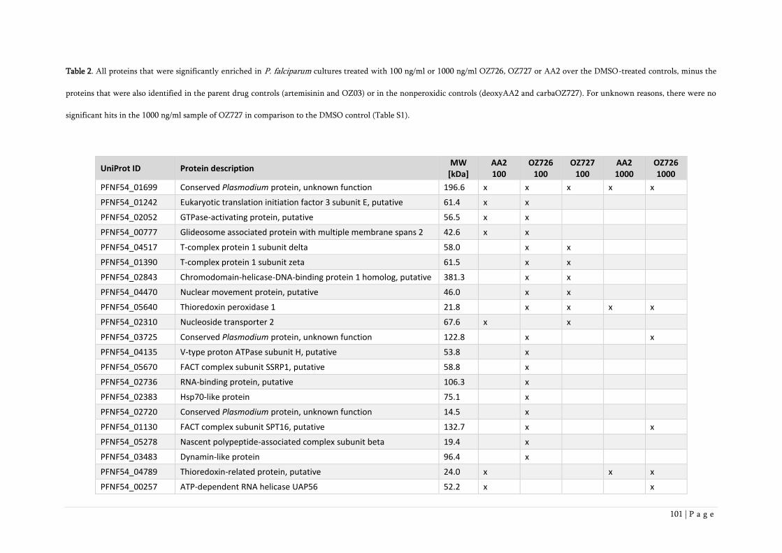
101 | P a g e
Table 2. All proteins that were significantly enriched in P. falciparum cultures treated with 100 ng/ml or 1000 ng/ml OZ726, OZ727 or AA2 over the DMSO-treated controls, minus the
proteins that were also identified in the parent drug controls (artemisinin and OZ03) or in the nonperoxidic controls (deoxyAA2 and carbaOZ727). For unknown reasons, there were no
significant hits in the 1000 ng/ml sample of OZ727 in comparison to the DMSO control (Table S1).
UniProt ID Protein description MW
[kDa] AA2 100
OZ726 100
OZ727 100
AA2 1000
OZ726 1000
PFNF54_01699 Conserved Plasmodium protein, unknown function 196.6 x x x x x
PFNF54_01242 Eukaryotic translation initiation factor 3 subunit E, putative 61.4 x x PFNF54_02052 GTPase-activating protein, putative 56.5 x x PFNF54_00777 Glideosome associated protein with multiple membrane spans 2 42.6 x x PFNF54_04517 T-complex protein 1 subunit delta 58.0 x x PFNF54_01390 T-complex protein 1 subunit zeta 61.5 x x PFNF54_02843 Chromodomain-helicase-DNA-binding protein 1 homolog, putative 381.3 x x PFNF54_04470 Nuclear movement protein, putative 46.0 x x PFNF54_05640 Thioredoxin peroxidase 1 21.8 x x x x
PFNF54_02310 Nucleoside transporter 2 67.6 x x PFNF54_03725 Conserved Plasmodium protein, unknown function 122.8 x x
PFNF54_04135 V-type proton ATPase subunit H, putative 53.8 x PFNF54_05670 FACT complex subunit SSRP1, putative 58.8 x PFNF54_02736 RNA-binding protein, putative 106.3 x PFNF54_02383 Hsp70-like protein 75.1 x PFNF54_02720 Conserved Plasmodium protein, unknown function 14.5 x PFNF54_01130 FACT complex subunit SPT16, putative 132.7 x x
PFNF54_05278 Nascent polypeptide-associated complex subunit beta 19.4 x PFNF54_03483 Dynamin-like protein 96.4 x PFNF54_04789 Thioredoxin-related protein, putative 24.0 x x x
PFNF54_00257 ATP-dependent RNA helicase UAP56 52.2 x x

102 | P a g e
PFNF54_02120 Histone S-adenosyl methyltransferase, putative 129.2 x x PFNF54_05599 Conserved protein, unknown function 32.6 x PFNF54_03298 T-complex protein 1 subunit alpha 60.3 x PFNF54_01342 Conserved Plasmodium protein, unknown function 334.4 x PFNF54_01696 Importin-7, putative 145.5 x x
PFNF54_03261 Spermidine synthase 36.6 x x
PFNF54_03012 Casein kinase II subunit beta 28.4 x x
PFNF54_03252 Prefoldin subunit alpha, putative 29.1 x x
PFNF54_03153 Insulinase, putative 173.6 x x
PFNF54_00668 V-type proton ATPase subunit B 55.8 x x
PFNF54_00014 Nucleoside transporter 4 50.0 x
PFNF54_04551 Conserved Plasmodium protein, unknown function 291.0 x
PFNF54_03586 Eukaryotic translation initiation factor 5A 17.6 x
PFNF54_02629 Proteasome subunit beta type-6, putative 32.6 x
PFNF54_05054 Aminopeptidase P 90.1 x
PFNF54_01342 Conserved Plasmodium protein, unknown function 334.4 x
PFNF54_01192 Multidrug resistance protein 162.3 x
PFNF54_04908 Plasmepsin III 51.7 x
PFNF54_05266 60S Ribosomal protein L7-3, putative 32.7 x
PFNF54_03239 Protein phosphatase, putative 34.5 x
PFNF54_04191 V-type proton ATPase catalytic subunit A 68.6 x
PFNF54_00552 T-complex protein 1 subunit epsilon 59.2 x
PFNF54_02838 60S Ribosomal protein L30e, putative 11.8 x
PFNF54_02025 Glutamate dehydrogenase, putative 160.4 x

103 | P a g e
Table 3. Hits identified herein that are also present in the published datasets by Wang et al. (2015) and Ismail et al. (2016a, 2016b).
UniProt ID Protein description AA2 100
OZ726 100
OZ727 100
AA2 1000
OZ726 1000
Wang 2015
Ismail 2016a,b
PFNF54_04789 Thioredoxin-related protein, putative x x x x
PFNF54_00257 ATP-dependent RNA helicase UAP56 x x x
PFNF54_02310 Nucleoside transporter 2 x x x
PFNF54_05640 Thioredoxin peroxidase 1 x x x x x
PFNF54_01696 Importin-7, putative x x x
PFNF54_03261 Spermidine synthase x x x
PFNF54_03153 Insulinase, putative x x x
PFNF54_02025 Glutamate dehydrogenase, putative x x
PFNF54_04517 T-complex protein 1 subunit delta x x x
PFNF54_01390 T-complex protein 1 subunit zeta x x x
PFNF54_04908 Plasmepsin III x x
PFNF54_02838 60S ribosomal protein L30e, putative x x
PFNF54_01192 Multidrug resistance protein x x x
PFNF54_04191 V-type proton ATPase catalytic subunit A x x x
PFNF54_00668 V-type proton ATPase subunit B x x x

104 | P a g e
Supplementary Material
Table S1. All significant hits obtained with chemical probes as compared to DMSO-treated P. falciparum control cultures.
UniProt ID Protein description MW AA2 AA2 ART ART deoxyAA2 deoxyAA2 OZ726 OZ726 OZ727 OZ727 OZ03 OZ03 carbaOZ727 carbaOZ727
[kDa] 100 1000 100 1000 100 1000 100 1000 100 1000 100 1000 100 1000
PFNF54_01699 conserved Plasmodium protein, unknown function 196.6 X X X X X
PFNF54_01242 eukaryotic translation initiation factor 3 subunit E, putative 61.4 X X
PFNF54_02052 GTPase-activating protein, putative 56.5 X X
PFNF54_00777 glideosome associated protein with multiple membrane spans 2 42.6 X X
PFNF54_04517 T-complex protein 1 subunit delta 58.0 X X
PFNF54_01390 T-complex protein 1 subunit zeta 61.5 X X
PFNF54_02843 chromodomain-helicase-DNA-binding protein 1 homolog, putative 381.3 X X
PFNF54_04470 nuclear movement protein, putative 46.0 X X
PFNF54_05640 thioredoxin peroxidase 1 21.8 X X X X
PFNF54_02310 nucleoside transporter 2 67.6 X X
PFNF54_03725 conserved Plasmodium protein, unknown function 122.8 X X
PFNF54_04135 V-type proton ATPase subunit H, putative 53.8 X
PFNF54_05670 FACT complex subunit SSRP1, putative 58.8 X
PFNF54_02736 RNA-binding protein, putative 106.3 X
PFNF54_02383 heat shock protein 70 75.1 X
PFNF54_02720 conserved Plasmodium protein, unknown function 14.5 X
PFNF54_01130 FACT complex subunit SPT16, putative 132.7 X X
PFNF54_05278 basic transcription factor 3b, putative 19.4 X
PFNF54_03483 dynamin-like protein 96.4 X
PFNF54_04789 thioredoxin-related protein, putative 24.0 X X X
PFNF54_00257 ATP-dependent RNA helicase UAP56 52.2 X X
PFNF54_02120 histone S-adenosyl methyltransferase, putative 129.2 X X
PFNF54_05599 conserved protein, unknown function 32.6 X
PFNF54_03298 T-complex protein 1 subunit alpha 60.3 X
PFNF54_01342 conserved Plasmodium protein, unknown function 334.4 X
PFNF54_01696 importin-7, putative 145.5 X X
PFNF54_03261 spermidine synthase 36.6 X X
PFNF54_03012 casein kinase II beta chain 28.4 X X
PFNF54_03252 prefoldin subunit 5, putative 29.1 X X
PFNF54_03153 insulinase, putative 173.6 X X
PFNF54_00668 V-type proton ATPase subunit B 55.8 X X
PFNF54_00014 nucleoside transporter 4 50.0 X
PFNF54_04551 conserved Plasmodium protein, unknown function 291.0 X
PFNF54_03586 eukaryotic translation initiation factor 5A 17.6 X
PFNF54_02629 proteasome subunit beta type-6, putative 32.6 X
PFNF54_05054 aminopeptidase P 90.1 X
PFNF54_01342 conserved Plasmodium protein, unknown function 334.4 X
PFNF54_01192 multidrug resistance protein 1 162.3 X
PFNF54_04908 plasmepsin III 51.7 X
PFNF54_05266 60S ribosomal protein L7-3, putative 32.7 X
PFNF54_03239 protein phosphatase, putative 34.5 X
PFNF54_04191 V-type proton ATPase catalytic subunit A 68.6 X
PFNF54_00552 T-complex protein 1 subunit epsilon 59.2 X
PFNF54_02838 60S ribosomal protein L30e, putative 11.8 X
PFNF54_02025 glutamate dehydrogenase, putative 160.4 X
PFNF54_02028 1-cys peroxiredoxin 25.2 X
PFNF54_03743 cell cycle associated protein, putative 324.1 X X X X
PFNF54_01079 bifunctional dihydrofolate reductase-thymidylate synthase 371.7 X X X X X X X X X
PFNF54_05172 60S ribosomal protein L44 12.2 X X X X X X X
PFNF54_03313 antigen UB05 13.7 X X
PFNF54_03023 histone H4 11.5 X
PFNF54_01156 conserved Plasmodium protein, unknown function 22.7 X X X
PFNF54_01149 40S ribosomal protein S24 15.4 X
PFNF54_00991 deoxyribodipyrimidine photo-lyase, putative 129.2 X X X
PFNF54_03121 conserved Plasmodium protein, unknown function 33.9 X X X X X X
PFNF54_01036 40S ribosomal protein S9, putative 22.1 X
PFNF54_03241 tRNA (guanine-N(7)-)-methyltransferase, putative 35.6 X X
PFNF54_04551 conserved Plasmodium protein, unknown function 291.0 X X
PFNF54_00516 40S ribosomal protein S15A, putative 14.9 X X
59 15 13 1 0 6 0 24 29 16 0 10 2 0 0

105 | P a g e
UniProt ID Protein description MW AA2 AA2 ART ART deoxyAA2 deoxyAA2 OZ726 OZ726 OZ727 OZ727 OZ03 OZ03 carbaOZ727 carbaOZ727
[kDa] 100 1000 100 1000 100 1000 100 1000 100 1000 100 1000 100 1000
PFNF54_01699 conserved Plasmodium protein, unknown function 196.6 X X X X X
PFNF54_01242 eukaryotic translation initiation factor 3 subunit E, putative 61.4 X X
PFNF54_02052 GTPase-activating protein, putative 56.5 X X
PFNF54_00777 glideosome associated protein with multiple membrane spans 2 42.6 X X
PFNF54_04517 T-complex protein 1 subunit delta 58.0 X X
PFNF54_01390 T-complex protein 1 subunit zeta 61.5 X X
PFNF54_02843 chromodomain-helicase-DNA-binding protein 1 homolog, putative 381.3 X X
PFNF54_04470 nuclear movement protein, putative 46.0 X X
PFNF54_05640 thioredoxin peroxidase 1 21.8 X X X X
PFNF54_02310 nucleoside transporter 2 67.6 X X
PFNF54_03725 conserved Plasmodium protein, unknown function 122.8 X X
PFNF54_04135 V-type proton ATPase subunit H, putative 53.8 X
PFNF54_05670 FACT complex subunit SSRP1, putative 58.8 X
PFNF54_02736 RNA-binding protein, putative 106.3 X
PFNF54_02383 heat shock protein 70 75.1 X
PFNF54_02720 conserved Plasmodium protein, unknown function 14.5 X
PFNF54_01130 FACT complex subunit SPT16, putative 132.7 X X
PFNF54_05278 basic transcription factor 3b, putative 19.4 X
PFNF54_03483 dynamin-like protein 96.4 X
PFNF54_04789 thioredoxin-related protein, putative 24.0 X X X
PFNF54_00257 ATP-dependent RNA helicase UAP56 52.2 X X
PFNF54_02120 histone S-adenosyl methyltransferase, putative 129.2 X X
PFNF54_05599 conserved protein, unknown function 32.6 X
PFNF54_03298 T-complex protein 1 subunit alpha 60.3 X
PFNF54_01342 conserved Plasmodium protein, unknown function 334.4 X
PFNF54_01696 importin-7, putative 145.5 X X
PFNF54_03261 spermidine synthase 36.6 X X
PFNF54_03012 casein kinase II beta chain 28.4 X X
PFNF54_03252 prefoldin subunit 5, putative 29.1 X X
PFNF54_03153 insulinase, putative 173.6 X X
PFNF54_00668 V-type proton ATPase subunit B 55.8 X X
PFNF54_00014 nucleoside transporter 4 50.0 X
PFNF54_04551 conserved Plasmodium protein, unknown function 291.0 X
PFNF54_03586 eukaryotic translation initiation factor 5A 17.6 X
PFNF54_02629 proteasome subunit beta type-6, putative 32.6 X
PFNF54_05054 aminopeptidase P 90.1 X
PFNF54_01342 conserved Plasmodium protein, unknown function 334.4 X
PFNF54_01192 multidrug resistance protein 1 162.3 X
PFNF54_04908 plasmepsin III 51.7 X
PFNF54_05266 60S ribosomal protein L7-3, putative 32.7 X
PFNF54_03239 protein phosphatase, putative 34.5 X
PFNF54_04191 V-type proton ATPase catalytic subunit A 68.6 X
PFNF54_00552 T-complex protein 1 subunit epsilon 59.2 X
PFNF54_02838 60S ribosomal protein L30e, putative 11.8 X
PFNF54_02025 glutamate dehydrogenase, putative 160.4 X
PFNF54_02028 1-cys peroxiredoxin 25.2 X
PFNF54_03743 cell cycle associated protein, putative 324.1 X X X X
PFNF54_01079 bifunctional dihydrofolate reductase-thymidylate synthase 371.7 X X X X X X X X X
PFNF54_05172 60S ribosomal protein L44 12.2 X X X X X X X
PFNF54_03313 antigen UB05 13.7 X X
PFNF54_03023 histone H4 11.5 X
PFNF54_01156 conserved Plasmodium protein, unknown function 22.7 X X X
PFNF54_01149 40S ribosomal protein S24 15.4 X
PFNF54_00991 deoxyribodipyrimidine photo-lyase, putative 129.2 X X X
PFNF54_03121 conserved Plasmodium protein, unknown function 33.9 X X X X X X
PFNF54_01036 40S ribosomal protein S9, putative 22.1 X
PFNF54_03241 tRNA (guanine-N(7)-)-methyltransferase, putative 35.6 X X
PFNF54_04551 conserved Plasmodium protein, unknown function 291.0 X X
PFNF54_00516 40S ribosomal protein S15A, putative 14.9 X X
59 15 13 1 0 6 0 24 29 16 0 10 2 0 0
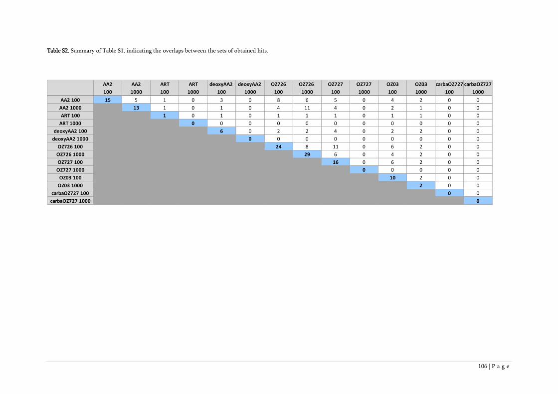
106 | P a g e
Table S2. Summary of Table S1, indicating the overlaps between the sets of obtained hits.
AA2 AA2 ART ART deoxyAA2 deoxyAA2 OZ726 OZ726 OZ727 OZ727 OZ03 OZ03 carbaOZ727 carbaOZ727
100 1000 100 1000 100 1000 100 1000 100 1000 100 1000 100 1000
AA2 100 15 5 1 0 3 0 8 6 5 0 4 2 0 0
AA2 1000 13 1 0 1 0 4 11 4 0 2 1 0 0
ART 100 1 0 1 0 1 1 1 0 1 1 0 0
ART 1000 0 0 0 0 0 0 0 0 0 0 0
deoxyAA2 100 6 0 2 2 4 0 2 2 0 0
deoxyAA2 1000 0 0 0 0 0 0 0 0 0
OZ726 100 24 8 11 0 6 2 0 0
OZ726 1000 29 6 0 4 2 0 0
OZ727 100 16 0 6 2 0 0
OZ727 1000 0 0 0 0 0
OZ03 100 10 2 0 0
OZ03 1000 2 0 0
carbaOZ727 100 0 0
carbaOZ727 1000 0

107 | P a g e
Figure S1. Optimization of assay parameters as determined by Western blot. A) Different incubation times of
P. falciparum cultures with 1000 ng/ml ozonide alkyne, exposure 10 s; B) Different concentrations of ozonide
alkyne at 4 h drug incubation, exposure 30 s. Parasite cultures were lysed with Ripa-Lysis buffer (see Material
and Methods for details).
Figure S2. Optimization of washing conditions of the neutravidin-coupled agarose beads after reaction with
biotinylated peptides as determined by silver stain. P. falciparum cultures were incubated with 100 ng/ml AA2
or 0.1% DMSO for 4 h. Protein extracts were prepared with 1.2% SDS in PBS.

108 | P a g e
Figure S3. Optimization of washing conditions of the neutravidin-coupled agarose beads after reaction with
biotinylated peptides as determined by Western blot. P. falciparum cultures were treated with 100 ng/ml AA2
or 0.1% DMSO and extracts were prepared with 1.2% SDS in PBS. Exposure 1 min.

109 | P a g e
5. Discussion
Artemisinin is an antimalarial of highest potency and tolerability. However, it also has drawbacks:
As natural products, artemisinin and it derivatives suffer from poor availability and high costs.
Furthermore, they have short in vivo half-lifes and are difficult to chemically modify and improve
(White, 2008; Tilley et al., 2016). Vennerstrom reported in 2004 the development of synthetic
peroxides, which should overcome the drawbacks of artemisinins (Vennerstrom et al., 2004). The
next-generation ozonide OZ439 showed promising properties in clinical trials with a rapid parasite
clearance, good safety profiles and a prolonged elimination half-life of 46-62 hours, in contrast to
the first-generation ozonide OZ277 (Phyo et al., 2016; Möhrle et al., 2012). This raises hope for a
single-dose treatment of OZ439 in combination with another drug to become an important new
treatment for P. falciparum infections (Phyo et al., 2016; Möhrle et al., 2012; Mäser et al., 2010;
Phillips et al., 2017). However, a prerequisite for any new antimalarial is activity against
artemisinin-resistant P. falciparum isolates, which have been reported from South-East Asia since
2006 and are the cause of the phenomenon of delayed parasite clearance after artemisinin treatment
(Noedl et al., 2008; Dondorp et al., 2009). Several different mutations in the K13 propeller domain
were found to be major determinants of artemisinin resistance (Chapter 1). Thereby individual point
mutations in K13 were sufficient to induce enhanced parasite clearance half-lives of >5h after
artemisinin treatment, indicating artemisinin resistance in P. falciparum isolates (Blasco et al., 2017;
Straimer et al., 2015). It is suggested that ring stages become refractory to drug action and that these
ring stages have an enhanced capacity to regulate cellular stress response to counteract the protein
damage caused by artemisinin. Thereby the unfolded protein response or the ubiquitin/proteasome
system, which serves as a regulated protein degradation system, are involved. But the exact function
and mechanism of K13 mutations protecting ring-stages in not known (Blasco et al., 2017; Tilley et
al., 2016, Wang et al., 2017).

110 | P a g e
The fear of cross-resistance of artemisinin-resistant parasites against synthetic peroxides
overshadowed the superior pharmacological properties of OZ439, since both, artemisinins and
synthetic peroxides, share the peroxide bridge essential for drug action (Kaiser et al., 2007). Hence,
we undertook in vitro studies to test for potential cross-resistance of an artemisinin-resistant P.
falciparum clinical isolate from Battambang (Cambodia) carrying a K13 mutation (Cam3.IR539T)
against synthetic peroxides (Chapter 3). The mutation R539T was found to confer increased in vitro
survival rates and delayed parasite clearance in patients after artemisinin treatment (Ariey et al.,
2014; Ménard et al., 2016; Straimer et al., 2015). In Chapter 3 we showed that the artemisinin-
resistant clinical isolate had a 45-fold lower survival rate in vitro when exposed to OZ439 than to
dihydroartemisinin, indicating no cross-resistance between the two antimalarial peroxides (Chapter
3). Our finding was supported by other in vitro studies showing no cross-resistance of the clinical
isolate Cam3.IIR539T and another clinical isolate carrying a C580Y mutation (Cam3.IIC580Y) against
OZ439 (Straimer et al., 2017; Siriwardana et al., 2016). In addition, Yang et al. found that synthetic
peroxides are more effective than dihydroartemisinin against early-ring stages when exposed for
physiologically relevant periods (Yang et al., 2016). The ability of OZ439 to overcome artemisinin
resistance was further supported by the finding that the median parasite clearance half-life in
patients infected with P. falciparum parasites carrying mutations in K13 was only one hour longer
than in patients with P. falciparum parasites lacking K13 mutations (Phyo et al., 2016). Together,
all these facts raise hope that OZ439 is a promising drug candidate to overcome artemisinin
resistance in combination with a partner drug.
It was shown that the mode of action of synthetic peroxides involves the formation of carbon-
centered radicals, predominantly on the adamantane-portion of the ozonides (Vennerstrom et al.,
2004; Maerki et al., 2006). Therefore, the production of monoclonal antibodies raised against this
adamantane-portion offered us the unique opportunity to investigate the alkylation signature of the
synthetic peroxides OZ277 and OZ439. In Chapter 2 I showed that both monoclonal antibodies used

111 | P a g e
in this study bind specifically to the alkylation targets of OZ277 and OZ439 (Chapter 2). Since no
protein bands were observed in a Western blot experiment using Babesia divergens treated with
synthetic peroxides, an intraerythrocytic protozoan parasite that does not digest hemoglobin and
therefore does not produce hemozoin, we could demonstrate that digestion of hemoglobin is
essential for the activity of ozonides (Chapter 2; Zintl et al., 2003; Richier et al., 2006). This finding
was in agreement with Kaiser et al., who showed that OZ277 was not active in vitro against B.
divergens (Kaiser et al., 2007).
In Chapter 4, I analysed the alkylation signatures of the newly synthesized alkyne OZ726 by mass
spectrometry and identified aminopeptidase P and plasmepsin III, both suggested to be involved in
hemoglobin catabolism, further supporting the iron-dependent alkylation hypothesis (Chapter 4). I
could also show that the peroxide bridge in synthetic peroxides is essential for drug action, since
carbaOZ727 and deoxyAA2 showed no activity in vitro against P. falciparum NF54 and no protein
alkylation was found in Western blot experiments with the nonperoxidic analogues OZ727 and AA2
(Chapter 4). Further, mass spectrometry with carbaOZ727 and deoxyAA2 identified only four hits
with deoxyAA2, while no hit was found in parasite cultures treated with carbaOZ727 (Chapter 4).
In addition to the alkylation of heme which was described by Creek (Creek et al., 2008), I showed
here that several other parasite proteins were alkylated by OZ277 and OZ439 (Chapter 2). This
alkylation signature was further investigated by the establishment and application of the click
chemistry approach using novel synthesized alkynes of artemisinin and synthetic peroxides. I
identified 47 alkylated proteins in P. falciparum after treatement with OZ726, OZ727 or AA2 for 4
h. One of these proteins, PFNF54_01699, was found to be most promising, since it was found in all
samples (except 1000 ng/ml OZ727, which did not return a single hit). This protein is suggested to
be an erythrocyte membrane protein (EMP)-like protein, a parasite protein thought to be exported
to the RBC membrane (Pei et al., 2007; Senczuk et al., 2001). It appears there at the later stages of
the parasite life cycle (Pei et al., 2007; Senczuk et al., 2001). P. falciparum EMP 1 mediates the

112 | P a g e
adhesion of the infected RBCs to the human vascular endothelium and is therefore important in the
host-pathogen interaction (Pei et al., 2007; Senczuk et al., 2001; Sampaio et al., 2017). But further
studies will be needed to characterize this alkylated protein.
Another protein of the alkylation signature of OZ726 was the multidrug resistance protein 1
(MDR1), an ABC transporter located in the digestive vacuole membrane (Blasco et al., 2017;
Rohrbach et al., 2006). PfMDR1 was found to be linked to the sensitivity of P. falciparum to several
antimalarial drugs (Ding et al., 2010). Thereby, partial but insufficient associations between
mutations in PfMDR1 and chloroquine resistance were found (Duraising et al., 2005, Valderramos
et al., 2006). Further, it was shown that increased copy numbers of PfMDR1 resulted in decreased
in vitro susceptibility to mefloquine, quinine, halofantrine and artemisinin (Valderramos et al.,
2006; Sidhu et al., 2006).
In 1994 Asawamahasakda identified four major protein bands (25kDa, 50kDa, 65kDa and >200kDa)
and two minor ones (32kDa and 42kDa) with radiolabelled dihydroartemisinin. I have observed
similar protein bands, except for the >200kDa band, when using the newly synthesized alkyne AA2
in Western blots. The same pattern was also observed with either OZ726 or OZ727 (Chapter 4). The
antibody studies also revealed a prominent protein band at 50 kDa in rings and schizonts treated
with either OZ277 or OZ439 (Chapter 2). All these findings support the data of Asawamahasakda
(Asawamahasakda et al., 1994). In addition, I found different proteins with the same identified
masses than Asawamahasakda (Asawamahasakda et al., 1994) in the mass spectometry experiments
when identifying the alkylation signatures of the synthetic peroxides and artemisinin-alkynes (as
discussed in Chapter 4).
Next, we could show that protein alkylation by OZ277 takes place in the cytoplasm and other
structures such as the nucleus and the food vacuole (Chapter 2). This alkylation signature was similar
to the ultrastructural autoradiographic studies performed by Maeno, where radiolabeled
dihydroartemisinin and artemisinin located to the food vacuole, mitochondria and nuclear

113 | P a g e
membranes (Maeno et al., 1993). Other studies showed that TAMRA-labeled OZ277 was found in
the cytosol or in the food vacuole (Uhlemann et al., 2007). In addition, it was found that artemisinin
causes the disruption of the digestive vacuole membrane and that the food vacuole is an important
site of synthetic peroxide action (Crespo et al., 2008). By analysing alkylation signatures of synthetic
peroxides and artemisinin with mass spectrometry, we identified various proteins located to the
cytoplasm, nucleus or food vacuole (Chapter 4).
When we compared our identified alkylation signatures of synthetic peroxides and artemisinin to
the recently observed data of Wang, who had also used a click chemistry approach with newly
synthesized, but structurally slightly different artemisinin alkyne, we obtained 14 identical proteins,
6 of which previously proposed as antimalarial drug targets (Wang et al., 2015). In contrast, click
chemistry studies with synthetic peroxides and artemisinin performed by Ismail et al. did not show
such a high degree of overlap (Ismail et al., 2016a and 2016b). This could be due to the use of
trophozoites only compared to mixed parasite cultures used in our experiments, or because of
prolonged drug incubation time (6 h in Ismail et al., 2016a and 2016b compared to 4 h in Chapter
4). To further investigate the variation of identified alkylation signatures in different studies (Wang
et al., 2015, Ismail et al., 2016a and 2016b; Chapter 4), one should use more stringent criteria, such
as the use of highly synchronized parasite stages (rings, trophozoites or schizonts), identical drug
incubation times and identical parasitemia.
We demonstrated an overlap between the alkylation signatures of synthetic peroxides and
artemisinin, but we did not observe such a high identity, i.e. 90%, as Ismail (Ismail et al., 2016b;
Chapter 4). Our results strengthen the hypothesis that artemisinins and synthetic peroxides have a
similar, but not identical mode of action (Vennerstrom et al., 2004; Jefford, 2001; Kaiser et al., 2007).
Since our approach using alkynes with click chemistry worked well, a next step could be the
investigation of alkylation signatures of synthetic peroxides and artemisinin in artemisinin-resistant
P. falciparum strains compared to artemisinin-susceptible strains. Thereby one could identify the

114 | P a g e
differences in alkylation patterns in relation to artemisinin resistance and possibly shed light on the
mechanism of artemisinin resistance.

115 | P a g e
6. Conclusion
In conclusion, I could show that OZ277 and OZ439 localize to the cytoplasm of P. falciparum and
other structures such as the nucleus and the food vacuole. I have identified the alkylation signatures
of both, artemisinin and synthetic peroxides by using a click chemistry approach with newly
synthesized alkynes. Further we could demonstrate that there is no cross-resistance between
artemisinin and OZ439 in a Kelch-13 mutant P. falciparum field isolate. Further studies will be
needed to investigate the importance of the most prominent identified alkylation target
(PFNF54_01699) for drug action and identify the mechanism of artemisinin resistance to ensure
that OZ439 remains a promising drug candidate that will overcome artemisinin resistance in
combination with a partner drug.
116 | P a g e
References
Ariey F., Witkowski B., Amaratunga C., Beghain J., Langlois A.C., Khim N., Kim S., Duru, V.,
Bouchier C., Ma L., Lim P., Leang R., Duong S., Sreng S., Suon S., Chuor C.M., Bout D.M., Ménard
S., Rogers W.O., Genton B., Fandeur T., Miotto O., Ringwald P., Le Bras J., Berry A., Barale J.C.,
Fairhurst R.M., Benoit-Vical F., Mercereau-Puijalon O., Ménard D., (2014) A molecular marker of
artemisinin-resistant Plasmodium falciparum malaria. Nature. 505, 50-55.
Asawamahasakda W., Ittarat I., Pu Y.M., Ziffer H., and Meshnick S.R., (1994) Reaction of
antimalarial endoperoxides with specific parasite proteins. Antimicrob. Agents Chemother. 38,
1854–1858.
Blasco B., Leroy D. and Fidock D.A., (2017) Antimalarial drug resistance: linking Plasmodium
falciparum parasite biology to the clinic. Nat. Med. 23 (8), 917-928.
Charman S. A., Arbe-Barnes S., Bathurst I. C., Brun R., Campbell M., Charman W. N., Chiu F. C.,
Chollet J., Craft J. C., Creek D. J., Dong Y., Matile H., Maurer M., Morizzi J., Nguyen T.,
Papastogiannidis P., Scheurer C., Shackleford D. M., Sriraghavan K., Stingelin L., Tang Y., Urwyler
H., Wang X., White K. L., Wittlin S., Zhou L., and Vennerstrom J. L., (2011) Synthetic ozonide drug
candidate OZ439 offers new hope for a single-dose cure of uncomplicated malaria. Proc. Natl. Acad.
Sci. U. S. A. 108, 4400−4405.
Crespo M. D., Avery T. D., Hanssen E., Fox E., Robinson T. V., Valente P., Taylor D. K., and Tilley
L., (2008) Artemisinin and a series of novel endoperoxide antimalarials exert early effects on
digestive vacuole morphology. Antimicrob. Agents Chemother. 52, 98−109.
Ding X.C., Beck H.P., Raso G., (2011) Plasmodium sensitivity to artemisinins: magic bullets hit
elusive targets. Trends Parasitol. 27 (2), 73-81.
Dondorp A.M., Nosten F., Yi P., Das D., Phyo A.P., Tarning J., Lwin K.M., Ariey F., Hanpithakpong
W., Lee S.J., Ringwald P., Silamut K., Imwong M., Chotivanich K., Lim P., Herdman T., An S.S.,
Yeung S., Singhasivanon P., Day N.P.J., Lindegardh N., Socheat D., White N.J., (2009) Artemisinin
Resistance in Plasmodium falciparum Malaria. N. Engl. J. Med. 38, 455-467.
Duraisingh M.T., Cowman A.F., (2005) Contribution of the pfmdr1 gene to antimalarial drug-
resistance. Acta. Trop. 94 (3), 181-190.

117 | P a g e
Ismail H. M., Barton V., Phanchana M., Charoensutthivarakul S., Wong M. H. L., Hemingway J.,
Biagini G. A., O’Neill P. M. and Ward S.A., (2016a) Artemisinin activity-based probes identify
multiple molecular targets within the asexual stage of the malaria parasites Plasmodium falciparum
3D7. PNAS. 113 (8), 2080–2085.
Ismail H.M., Barton V.E., Panchana M., Charoensutthivarakul S., Biagini G.A., Ward S.A. and
O'Neill P.M., (2016b) A Click Chemistry-Based Proteomic Approach Reveals that 1,2,4-Trioxolane
and Artemisinin Antimalarials Share a Common Protein Alkylation Profile. Angew. Chem. Int. Ed.
55, 1-6.
Jefford C.W., (2001) Why artemisinin and certain synthetic peroxides are potent antimalarials.
Implications for the mode of action. Curr. Med. Chem. 8 (15), 1803-1826.
Kaiser M., Wittlin S., Nehrbass-Stuedli A., Dong Y., Wang X., Hemphill A., Matile H., Brun R.,
Vennerstrom J.L., (2007) Peroxide bond-dependent antiplasmodial specificity of artemisinin and
OZ277 (RBx11160). Antimicrob. Agents Chemother. 51, 2991-2993.
Maeno Y., Toyoshima T., Fujioka H., Ito Y., Meshnick S.R., Benakis A., Milhous W.K., Aikawa M.,
(1993) Morphologic effects of artemisinin in Plasmodium falciparum. Am. J. Trop. Med. Hyg. 49 (4),
485-491.
Maerki S., Brun R., Charman S.A., Dorn A., Matile H., Wittlin S., (2006) In vitro assessment of the
pharmacodynamic properties and the partitioning of OZ277/RBx-11160 in cultures of Plasmodium
falciparum. J. Antimicrob. Chemother. 58, 52-58.
Mäser P., Wittlin S., Rottmann M., Wenzler T., Kaiser M., Brun R., (2012) Antiparasitic agents: new
drugs on the horizon. Curr. Opin. Pharmacol. 12, 562-566.
Ménard D., Khim N., Beghain J., Adegnika A.A., Shafiul-Alam M., Amodu O., Rahim-Awab G.,
Barnadas C., Berry A., Boum Y., Bustos M.D., Cao J., Chen J.H., Collet L., Cui L., Thakur G.D., Dieye
A., Djallé D., Dorkenoo M.A., Eboumbou-Moukoko C.E., Espino F.E., Fandeur T., Ferreira-da-Cruz
M.F., Fola A.A., Fuehrer H.P., Hassan A.M., Herrera S., Hongvanthong B., Houzé S., Ibrahim M.L.,
Jahirul-Karim M., Jiang L., Kano S., Ali-Khan W., Khanthavong M., Kremsner P.G., Lacerda M.,
Leang R, Leelawong M., Li M., Lin K., Mazarati J.B., Ménard S., Morlais I., Muhindo-Mavoko H.,
Musset L., Na-Bangchang K., Nambozi M., Niaré K., Noedl H., Ouédraogo J.B., Pillai D.R., Pradines
B., Quang-Phuc B., Ramharter M., Randrianarivelojosia M., Sattabongkot J., Sheikh-Omar A., Silué

118 | P a g e
K.D., Sirima S.B., Sutherland C., Syafruddin D., Tahar R., Tang L.H., Touré O.A., Tshibangu-wa-
Tshibangu P., Vigan-Womas I., Warsame M., Wini L., Zakeri S., Kim S., Eam R., Berne L., Khean
C., Chy S., Ken M., Loch K., Canier L., Duru V., Legrand E., Barale J.C., Stokes B., Straimer J.,
Witkowski B., Fidock D.A., Rogier C., Ringwald P., Ariey F., Mercereau-Puijalon O., KARMA
Consortium, (2016) A Worldwide Map of Plasmodium falciparum K13-Propeller Polymorphisms.
N. Engl. J. Med. 374 (25), 2453-2464.
Moehrle J.J., Duparc S., Siethoff C., van Giersbergen P.L., Craft J.C., Arbe-Barnes S., Charman S.A.,
Gutierrez M., Wittlin S., Vennerstrom J.L., (2012) First-in-man safety and pharmacokinetics of
synthetic ozonide OZ439 demonstrates an improved exposure profile relative to other peroxide
antimalarials. Br. J. Clin. Pharmacol. 75, 524-537.
Noedl H., Se Y., Schaecher K., Smith B.L., Socheat D., Fukuda M.M., (2008) Artemisinin Resistance
in Cambodia 1 (ARC1) Study Consortium Evidence of artemisinin-resistant malaria in western
Cambodia. N Engl J Med. 359, 2619-2620.
Pei X., Guo X., Coppel R., Mohandas N., An X., (2007) Plasmodium falciparum erythrocyte
membrane protein 3 (PfEMP3) destabilizes erythrocyte membrane skeleton. J. Biol. Chem. 282 (37),
26754-26758.
Phillips M.A., Burrows J.N., Manyando C., van Huijsduijnen R.H., Van Voorhis W.C., Wells T.N.C.,
(2017) Malaria. Nat. Rev. Dis. Primers. 3, 17050.
Phyo A.P., Jittamala P., Nosten F.H., Pukrittayakamee S., Imwong M., White N.J., Duparc S.,
Macintyre F., Baker M., Möhrle J.J., (2016) Antimalarial activity of artefenomel (OZ439), a novel
synthetic antimalarial endoperoxide, in patients with Plasmodium falciparum and Plasmodium
vivax malaria: an open-label phase 2 trial. Lancet Infect. Dis. 16, 61-69.
Richier E., Biagini G.A., Wein S., Boudou F., Bray P.G., Ward S.A., Precigout E., Calas M.,
Dubremetz J.F., Vial H.J., (2006) Potent antihematozoan activity of novel bisthiazolium drug T16:
evidence for inhibition of phosphatidylcholine metabolism in erythrocytes infected with Babesia
and Plasmodium spp. Antimicrob. Agents Chemother. 50 (10), 3381-3388.
Rohrbach P., Sanchez C.P., Hayton K., Friedrich O., Patel J., Sidhu A.B.S., Ferdig M.T., Fidock D.A.,
Lanzer M., (2006) Genetic linkage of pfmdr1 with food vacuolar solute import in Plasmodium
falciparum. EMBO J. 25 (13), 3000–3011.
119 | P a g e
Sampaio N.G., Eriksson E.M., Schofield L., (2017) Plasmodium falciparum PfEMP1 modulates
monocyte/macrophage transcription factor activation, and cytokine and chemokine responses.
Infect. Immun. doi: 10.1128/IAI.00447-17.
Senczuk A.M., Reeder J.C., Kosmala M.M., Ho M., (2001) Plasmodium falciparum erythrocyte
membrane protein 1 functions as a ligand for P-selectin. Blood. 98 (10), 3132-3135.
Sidhu A.B.S., Uhlemann A., Valderramos S.G., Valderramos J., Krishna S., Fidock D.A., (2006)
Decreasing pfmdr1 Copy Number in Plasmodium falciparum Malaria Heightens Susceptibility to
Mefloquine, Lumefantrine, Halofantrine, Quinine, and Artemisinin. J. Infect. Dis. 194 (4), 528–535.
Siriwardana A., Iyengar K., Roepe P.D., (2016) Endoperoxide Drug Cross-Resistance Patterns for
Plasmodium falciparum Exhibiting an Artemisinin Delayed-Clearance Phenotype. Antimicrob.
Agents Chemother. 60 (11), 6952-6956.
Straimer J., Gnädig N.F., Stokes B.H., Ehrenberger M., Crane A.A., Fidock D.A., (2017) Plasmodium
falciparum K13 Mutations Differentially Impact Ozonide Susceptibility and Parasite Fitness In
Vitro. M. Bio. 8 (2).
Straimer J., Gnädig N.F., Witkowski B., Amaratunga C., Duru V., Ramadani A.P., Dacheux M., Khim
N., Zhang L., Lam S., Gregory P.D., Urnov F.D., Mercereau-Puijalon,O., Benoit-Vical F., Fairhurst
R.M., Ménard D., Fidock D.A., (2015) Drug resistance. K13-propeller mutations confer artemisinin
resistance in Plasmodium falciparum clinical isolates. Science. 347 (6220), 428-431.
Tilley L., Straimer J., Gnädig N.F., Ralph S.A., Fidock D.A., (2016) Artemisinin Action and
Resistance in Plasmodium falciparum. Trends Parasitol. 32, 682-696.
Uhlemann A.C., Wittlin S., Matile H., Bustamante L.Y., Krishna S., (2007) Mechanism of
antimalarial action of the synthetic trioxolane RBX11160 (OZ277). Antimicrob. Agents Chemother.
51, 667–672.
Valderramos S.G., Fidock D.A., (2006) Transporters involved in resistance to antimalarial drugs.
Trends Pharmacol. Sci. 27 (11), 594-601.
Vennerstrom J.L., Arbe-Barnes S., Brun R., Charman S.A., Chiu F.C., Chollet J., Dong Y., Dorn A.,
Hunziker D., Matile H., McIntosh K., Padmanilayam M., Santo Tomas J., Scheurer C., Scorneaux B.,

120 | P a g e
Tang Y., Urwyler H., Wittlin S., Charman W.N., (2004) Identification of an antimalarial synthetic
trioxolane drug development candidate. Nature. 430, 900–904.
Wang J., Xu C., Lun Z.R., Meshnick S.R., (2017) Unpacking 'Artemisinin Resistance'. Trends
Pharmacol. Sci. 38 (6), 506-511.
Wang J., Zhang C.J., Chia W.N., Loh C.C., Li Z., Lee Y.M., He Y., Yuan L.X., Lim T.K., Liu M., Liew
C.X., Lee Y.Q., Zhang J., Lu N., Lim C.T., Hua Z.C., Liu B., Shen H.M., Tan K.S. and Lin Q., (2015)
Haem-activated promiscuous targeting of artemisinin in Plasmodium falciparum. Nat. Commun. 6.
White, N.J., (2008) Qinghaosu (artemisinin): The price of success. Science. 320, 330–334.
Yang T., Xie S.C., Cao P., Giannangelo C., McCaw J., Creek D.J., Charman S.A., Klonis N., Tilley L.,
(2016) Comparison of the Exposure Time Dependence of the Activities of Synthetic Ozonide
Antimalarials and Dihydroartemisinin against K13 Wild-Type and Mutant Plasmodium falciparum
Strains. Antimicrob. Agents Chemother. 60 (8), 4501-4510.
Zintl A., Mulcahy G., Skerrett H.E., Taylor S.M., Gray J.S., (2003) Babesia divergens, a Bovine Blood
Parasite of Veterinary and Zoonotic Importance. Clin. Microbiol. Rev. 16 (4), 622–636.


![Review Potential Mechanisms of Action of Curcumin for ... · angiogenesis and metastasis [26]. The molecular basis of the anticancer activities of curcumin is mainly attributed to](https://static.fdokument.com/doc/165x107/5ed770259ab714631510fda8/review-potential-mechanisms-of-action-of-curcumin-for-angiogenesis-and-metastasis.jpg)