Original Article DOI: 10.19027/jai.16.1.15-21 Growth ...
7
Jurnal Akuakultur Indonesia 16 (1), 15–21 (2017) Growth, biomass, and chlorophyll-a and carotenoid content of Nannochloropsis sp. strain BJ17 under different light intensities Pertumbuhan, biomassa, dan konsentrasi pigmen klorofil-a dan karotenoid Nannochloropsis sp. strain BJ17 pada intensitas cahaya berbeda Muhammad Fakhri*, Nasrullah Bai Arifin, Anik Martina Hariati, Ating Yuniarti Program Studi Budidaya Perairan, Fakultas Perikanan dan Ilmu Kelautan, Universitas Brawijaya Jalan Veteran, Malang, 65145 *E-mail: [email protected] ABSTRACT Nannochloropsis sp. has been identified as sources of live feed and pigment in aquaculture. To increase the production, the optimal environmental conditions for microalgae are required. Light intensity is one of the important factors that significantly affects the biomass and pigment of microalgae. The study aimed to determine the effect of light intensity (1,500; 3,000; and 4,500 lux) on growth, biomass production, chlorophyll-a, and carotenoid content of Nannochloropsis sp. strain BJ17. The results showed that different light intensities significantly affected the growth, biomass, chlorophyll-a and carotenoid contents of Nannochloropsis sp. strain BJ17. Increasing light intensity resulted in the increase of the growth rate, biomass, chlorophyll-a, and carotenoid contents of Nannochloropsis sp. strain BJ17. The cell achieved the highest specific growth rate of 1.729 /day and the cell concentration of 43.333×10 6 cell/mL at a light intensity of 4,500 lux. The highest chlorophyll-a and carotenoid concentrations of algae were obtained at 4,500 lux (8.304 µg/mL and 3.892 µg/mL, respectively). This study suggested that increasing light intensity led to the increase in the growth, biomass, chlorophyll-a, and carotenoid content of Nannochloropsis sp. strain BJ17. Keywords: carotenoid, chlorophyll, biomass, growth rate, light intensity ABSTRAK Nannochloropsis sp. diketahui sebagai sumber pakan alami dan pigmen pada budidaya perikanan. Budidaya pada kondisi lingkungan yang optimal diperlukan untuk meningkatkan produksi mikroalga. Intensitas cahaya merupakan salah satu faktor esensial yang secara signifikan mempengaruhi biomassa dan pigmen mikroalga. Tujuan penelitian ini adalah untuk menentukan pengaruh intensitas cahaya yang berbeda (1.500, 3.000, and 4.500 lux) terhadap pertumbuhan, produksi biomassa, klorofil-a, dan karotenoid Nannochloropsis sp. strain BJ17. Hasil menunjukkan bahwa intensitas cahaya yang berbeda berpengaruh secara signifikan terhadap pertumbuhan, biomassa dan klorofil-a dan karotenoid Nannochloropsis sp. strain BJ17. Semakin tinggi intensitas cahaya maka laju pertumbuhan, biomassa, kandungan klorofil-a dan total karotenoid Nannochloropsis sp. strain BJ17 semakin tinggi. Laju pertumbuhan spesifik tertinggi 1,729 /hari dan konsentrasi sel maksimum tertinggi 43,333×10 6 sel/ mL dihasilkan pada intensitas cahaya 4.500 lux. Konsentrasi klorofil-a (8,304 µg/mL) dan karotenoid (3,892 µg/mL) tertinggi juga diperoleh pada intensitas cahaya 4.500 lux. Studi ini menunjukkan bahwa peningkatan intensitas cahaya berperan dalam meningkatkan pertumbuhan, produksi biomassa, klorofil-a, dan karotenoid Nannochloropsis sp. strain BJ17. Kata kunci: karotenoid, klorofil, biomassa, pertumbuhan, intensitas cahaya Original Article DOI: 10.19027/jai.16.1.15-21 INTRODUCTION Microalgae is a photosynthetic organism with a rapid growth rate and capable of converting light energy, carbon dioxide, and nutrients into biomass and generating essential pigment (Mata et al., 2010). Chlorophyll and carotenoids are essential, dominant pigment in microalgae (Koller et al., 2014) that have antioxidant properties (Cardozo et al., 2007) and pharmaceutical application (Koller et al., 2014). Furthermore, the carotenoids are also used as micro-ingredients in aquaculture feed (Yaakob et al., 2014). Nannochloropsis sp. is microalgae that has been widely used as a live feed in aquaculture (Camacho-Rodríguez et al., 2013; Freire et al.,
Transcript of Original Article DOI: 10.19027/jai.16.1.15-21 Growth ...
Jurnal Akuakultur Indonesia 16 (1), 15–21 (2017)
Growth, biomass, and chlorophyll-a and carotenoid content of Nannochloropsis sp. strain BJ17 under different light intensities
Pertumbuhan, biomassa, dan konsentrasi pigmen klorofil-a dan karotenoid Nannochloropsis sp. strain BJ17 pada intensitas cahaya berbeda
Muhammad Fakhri*, Nasrullah Bai Arifin, Anik Martina Hariati, Ating Yuniarti
Program Studi Budidaya Perairan, Fakultas Perikanan dan Ilmu Kelautan, Universitas BrawijayaJalan Veteran, Malang, 65145*E-mail: [email protected]
ABSTRACT
Nannochloropsis sp. has been identified as sources of live feed and pigment in aquaculture. To increase the production, the optimal environmental conditions for microalgae are required. Light intensity is one of the important factors that significantly affects the biomass and pigment of microalgae. The study aimed to determine the effect of light intensity (1,500; 3,000; and 4,500 lux) on growth, biomass production, chlorophyll-a, and carotenoid content of Nannochloropsis sp. strain BJ17. The results showed that different light intensities significantly affected the growth, biomass, chlorophyll-a and carotenoid contents of Nannochloropsis sp. strain BJ17. Increasing light intensity resulted in the increase of the growth rate, biomass, chlorophyll-a, and carotenoid contents of Nannochloropsis sp. strain BJ17. The cell achieved the highest specific growth rate of 1.729 /day and the cell concentration of 43.333×10⁶ cell/mL at a light intensity of 4,500 lux. The highest chlorophyll-a and carotenoid concentrations of algae were obtained at 4,500 lux (8.304 µg/mL and 3.892 µg/mL, respectively). This study suggested that increasing light intensity led to the increase in the growth, biomass, chlorophyll-a, and carotenoid content of Nannochloropsis sp. strain BJ17.
Keywords: carotenoid, chlorophyll, biomass, growth rate, light intensity
ABSTRAK
Nannochloropsis sp. diketahui sebagai sumber pakan alami dan pigmen pada budidaya perikanan. Budidaya pada kondisi lingkungan yang optimal diperlukan untuk meningkatkan produksi mikroalga. Intensitas cahaya merupakan salah satu faktor esensial yang secara signifikan mempengaruhi biomassa dan pigmen mikroalga. Tujuan penelitian ini adalah untuk menentukan pengaruh intensitas cahaya yang berbeda (1.500, 3.000, and 4.500 lux) terhadap pertumbuhan, produksi biomassa, klorofil-a, dan karotenoid Nannochloropsis sp. strain BJ17. Hasil menunjukkan bahwa intensitas cahaya yang berbeda berpengaruh secara signifikan terhadap pertumbuhan, biomassa dan klorofil-a dan karotenoid Nannochloropsis sp. strain BJ17. Semakin tinggi intensitas cahaya maka laju pertumbuhan, biomassa, kandungan klorofil-a dan total karotenoid Nannochloropsis sp. strain BJ17 semakin tinggi. Laju pertumbuhan spesifik tertinggi 1,729 /hari dan konsentrasi sel maksimum tertinggi 43,333×106 sel/mL dihasilkan pada intensitas cahaya 4.500 lux. Konsentrasi klorofil-a (8,304 µg/mL) dan karotenoid (3,892 µg/mL) tertinggi juga diperoleh pada intensitas cahaya 4.500 lux. Studi ini menunjukkan bahwa peningkatan intensitas cahaya berperan dalam meningkatkan pertumbuhan, produksi biomassa, klorofil-a, dan karotenoid Nannochloropsis sp. strain BJ17. Kata kunci: karotenoid, klorofil, biomassa, pertumbuhan, intensitas cahaya
Original Article DOI: 10.19027/jai.16.1.15-21
INTRODUCTION
Microalgae is a photosynthetic organism with a rapid growth rate and capable of converting light energy, carbon dioxide, and nutrients into biomass and generating essential pigment (Mata et al., 2010). Chlorophyll and carotenoids are essential, dominant pigment in microalgae (Koller et al.,
2014) that have antioxidant properties (Cardozo et al., 2007) and pharmaceutical application (Koller et al., 2014). Furthermore, the carotenoids are also used as micro-ingredients in aquaculture feed (Yaakob et al., 2014).
Nannochloropsis sp. is microalgae that has been widely used as a live feed in aquaculture (Camacho-Rodríguez et al., 2013; Freire et al.,

16 Muhammad Fakhri et al. / Jurnal Akuakultur Indonesia 16 (1), 15–21 (2017)
2016). It is also known as potential producers of pigments such as chlorophyll and carotenoids (Lubian et al., 2000; Hosikian et al., 2010).
Microalgae biomass and pigment production vary depending on culture conditions (Rao et al., 2007), such as photoperiod, temperature and light intensity (Kitaya et al., 2008; Fakhri et al., 2015). Environmental adjustment can effectively optimize the growth rate of macroalgae (Mata et al., 2010). Moreover, light intensity affects the physiology of microalgae (Khoeyi et al., 2012). Photoautotrophic culture and the intensity of light are essential factors, which determine growth rate and biomass production of microalgae. Light intensity also affects the metabolism and pigment composition of microalgae (Sánchez-Saavedra & Voltolina, 1996). In response to the increase or reduction of light intensity, the pigments contents of microalgae may be altered (Sánchez-Saavedra & Voltolina, 2002).
The effect of light intensity on Nannochloropsis sp. growth has been previously studied (Wahidin et al., 2013; Probir et al., 2011). However, information related to the effect of light intensity on pigment contents (chlorophyll and carotenoid) of Nannochloropsis sp. is still limited. Furthermore, algae growth varies depending on the species (species-specific) (Banerjee et al., 2011). This study aimed to determine the optimum light intensity for the growth, biomass and pigment production of Nannochloropsis sp. BJ17 strain. This study can also be applied to evaluate the potential Nannochloropsis sp. BJ17 strain to generate chlorophyll and carotenoids.
MATERIALS AND METHODS
Strain and growth medium of Nannochloropsis sp. strain BJ17
Nannochloropsis sp. BJ17 strains were obtained from Brackish Water Aquaculture Institute of Jepara, Central Java. These strains can rapidly growth in a wide range of salinity. Walne medium, which contained 100 g/L NH4NO3; 20 g NaH2PO4; 33.6 g H3BO3; 0.36 g MnCl2; 1.3 g FeCl2; 45.0 g EDTA/L and 12.01 g/L vitamin B12, was used as medium with seawater. Walne and vitamin dose of fertilizer were 1 mL/L and 0.5 mL/ L, respectively.
Nannochloropsis sp. BJ17 stock culture conditions
Nannochloropsis sp. BJ17 culture in exponential phase was used as an inoculant.
The inoculant was then cultivated in 500 mL erlenmeyer. A total volume of media and inoculant was 350 mL. The cultures were incubated under continuous light 24:0 (light:dark cycle). The incubation conditions used were 29±2 °C, the salinity of 15 g/L, the light intensity of 3,000 lux, and continuous aeration for four days (Fakhri et al., 2015).
Culture treatmentsThe light intensity treatments (in lux) were
1,500; 3,000; and 4,500 with three replicates per light intensity treatment. Microalgae biomass was harvested after six days of culture, and used for biomass and pigment analyses.
Nannochloropsis sp. BJ17 was used as inoculants at the exponential phase or four-day-old cultures. The inoculant was cultured into a 2.5 L container with a total volume of 1.5 L seawater with 15 g/L salinity. Initial cell concentration for all treatments was 5.5×10⁵ cells/mL. Microalgae cells were cultured at 28 °C, with photoperiod 24: 0 and continuously aerated.
Analysis of growthMicroalgae growth was observed using cells
concentration calculation method with 0.1 mm deep Neubauer haemocytometer (BOECO, Hamburg, Germany). Specific growth rate (μ) was calculated using the following formula (Fogg & Thake, 1987) :
µ (/day) = ln (x2) – ln (x1) t2–t1
Note:μ = growth rate of per unit cell concentrationx1 = cell concentration at time 1x2 = cell concentration at time 2tl = time 1t2 = time 2
Cell doubling time (td) is the average generation time required for cell to double in concentration. Doubling time can be calculated using the formula (Ak et al., 2008):
td (day) = ln 2 = 0,693 µ µ
Biomass analysisThe cell biomass of Nannochloropsis sp. BJ17
strains were analyzed following the method of Janssen (2002). Microalgae samples were taken
Muhammad Fakhri et al. / Jurnal Akuakultur Indonesia 16 (1), 15–21 (2017) 17
at the day-6 of microalgae growth. The filter paper GF/C (diameter 90 mm) were dried at 105 °C for 2 h until the weight is constant [A]. 25 mL of microalgae suspension was filtered through a filter paper GF/C, and washed with 25 mL of fresh water to avoid contamination with the insoluble salt in the media. The filter paper was placed in an oven at 105 °C for 2 h until the weight is constant. The filter paper was placed in a desiccator, and re-weighed [B]. The calculation used equation below:
Dry weight/biomass (mg/L) = [B] – [A] × 1,000 sample volume
Note:A = filter paper weight B = filter paper weight + algae
Chlorophyll-a and carotenoids analysisIn this study, chlorophyll-a and carotenoids
were analyzed using methanol extraction method (Ritchie, 2006). 10 mL of microalgae samples were centrifuged at 6,000 rpm for 10 min. 10 mL of absolute methanol was added to the centrifuged and vortexed pellet. Tube wrapped in aluminum foil and the mixture (pellet and solvent) was put in a water bath at a temperature of 70 °C for 10 min. The sample was vortexed and centrifuged at 6,000 rpm for 10 min. The clear supernatant was measured at the wavelengths of 480, 652, and 665 nm using a spectrophotometer (Spectroquant Pharo 300, Merck Millipore).
The content of chlorophyll-a was calculated using the method of Ritchie (2006), and total carotenoids were calculated following the method of Kim et al. (2014).
Chlorophyll-a (µg/mL) = 16,5169 × A665 – 8,0962 × A652
Total carotenoid (µg/mL) = 4 × A480
Statistical analysisStatistical analysis was performed using
SPSS 16.0. Growth, biomass, chlorophyll-a and carotenoids data were analyzed by analysis of variance (ANOVA) test with 95% confidence. To determine the relationship between the light intensity and growth rate and biomass, regression analysis was carried out.
RESULTS AND DISCUSSION
Growth and biomass of Nannochloropsis sp. strain BJ17
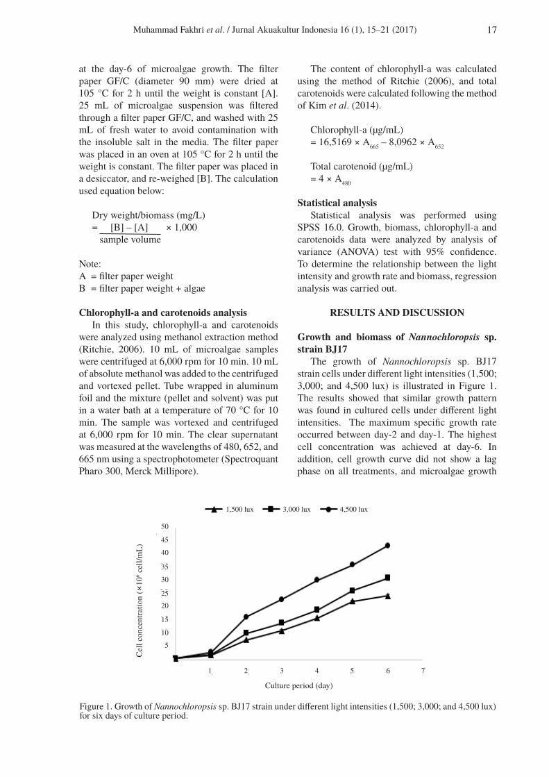
The growth of Nannochloropsis sp. BJ17 strain cells under different light intensities (1,500; 3,000; and 4,500 lux) is illustrated in Figure 1. The results showed that similar growth pattern was found in cultured cells under different light intensities. The maximum specific growth rate occurred between day-2 and day-1. The highest cell concentration was achieved at day-6. In addition, cell growth curve did not show a lag phase on all treatments, and microalgae growth
Figure 1. Growth of Nannochloropsis sp. BJ17 strain under different light intensities (1,500; 3,000; and 4,500 lux) for six days of culture period.
Cel
l con
cent
ratio
n (×
106 c
ell/m
L)
5
10
15
2025
30
35
40
45
50
1 2 3 4 5 6 7
Culture period (day)
1,500 lux 3,000 lux 4,500 lux

18 Muhammad Fakhri et al. / Jurnal Akuakultur Indonesia 16 (1), 15–21 (2017)
achieved rapid logarithmic phase. Similarly, Sforza et al. (2014) reported that the microalgae, Scenedesmus obliquus do not experience lag phase when they were treated with different light intensities. This indicates that some species of microalgae can grow in different conditions of light intensity without experiencing stress.
Table 1 shows that different light intensities have a significant (P<0.05) effect on the specific growth rate, doubling time, maximum cell concentration and biomass of Nannochloropsis sp. BJ17 strain. The highest specific growth rate was 1.73/day (P<0.05) with a doubling time about 0.401 days or 9.62 h. The highest cell concentration of 43.3×10⁶ cells/mL (P<0.05) was obtained at 4,500 lux of light intensity, while the highest biomass of 0.824 g/L (P<0.05) was resulted from 4,500 lux of light intensity.
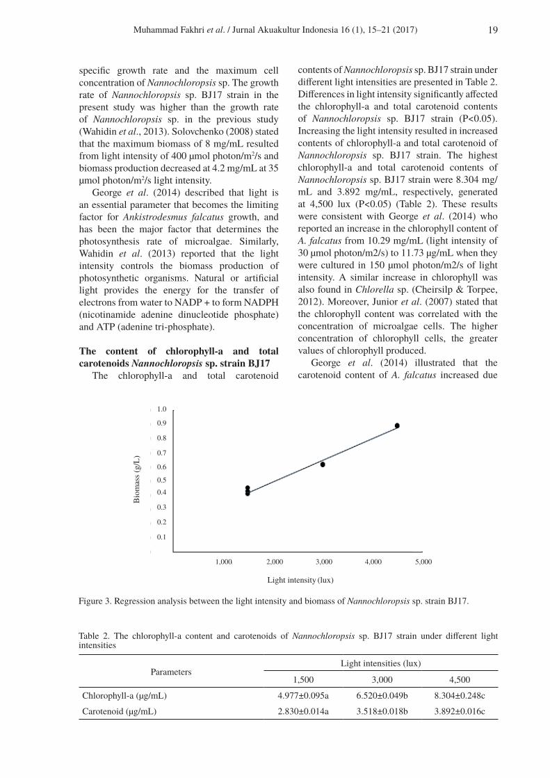
Figure 2 shows the relationship between the light intensity and the growth rate generates a linear pattern with the equation of y = 0.0001x + 1.2761 and R2 = 0.901. Figure 3 shows the
relationship between the light intensity and biomass, which generate a linear pattern with the equation of y = 0,0002x + 0.1893 and R2 = 0.985. The results indicate that the increase in light intensity can accelerate growth rate and biomass of Nannochloropsis sp. BJ17 strain.
Imaizumi et al. (2014) reported that the culture of Chlorella zofingiensis with light intensity between 75, 150, and 250 µmol photon/m2/sec produced specific growth rate between 0.399/day, 0.490/day, and 0,700/day, respectively. Furthermore, the increase in the light intensity from 75 µmol photons/m2/s to 250 µmol photon/m2/sec amplified the maximum cell concentration of C. Zofingiensis (Imaizumi et al., 2014). The result showed that the growth rate and cell concentration increases with increasing light intensity. Nonetheless, the growth significantly decreased in lower light intensities. Additionally, Wahidin et al. (2013) reported that the increase in the light intensity from 50 µmol photon/m2/s to 100 µmol photons/m2/s boosted the
1,000
Light intensity (lux)
2,000 3,000 4,000 5,0000
Spec
ific
grow
th ra
te (/
day)
0.2
0.4
0.6
0.8
1,0
1,2
1,4
1,6
1,8
2,0
Figure 2. Regression analysis between the light intensity and the specific growth rate of Nannochloropsis sp. strain BJ17.
ParametersLight intensities (lux)
1,500 3,000 4,500
Specific growth rate (/day) 1.431±0.038a 1.561±0.055b 1.729±0.048c
Doubling time (day) 0.484±0.013a 0.444±0.015b 0.400±0.011c
Maximum cell concentration (× 106 cell/mL) 24.333±0.577a 31.133±0.981b 43.333±1.433c
Biomass (g/L) 0.434±0.022a 0.620±0.004b 0.824±0.008c
Table 1. Specific growth rate, doubling time, maximum cell concentration and biomass of Nannochloropsis sp. BJ17 strain under different light intensities
Muhammad Fakhri et al. / Jurnal Akuakultur Indonesia 16 (1), 15–21 (2017) 19
specific growth rate and the maximum cell concentration of Nannochloropsis sp. The growth rate of Nannochloropsis sp. BJ17 strain in the present study was higher than the growth rate of Nannochloropsis sp. in the previous study (Wahidin et al., 2013). Solovchenko (2008) stated that the maximum biomass of 8 mg/mL resulted from light intensity of 400 µmol photon/m2/s and biomass production decreased at 4.2 mg/mL at 35 µmol photon/m2/s light intensity.
George et al. (2014) described that light is an essential parameter that becomes the limiting factor for Ankistrodesmus falcatus growth, and has been the major factor that determines the photosynthesis rate of microalgae. Similarly, Wahidin et al. (2013) reported that the light intensity controls the biomass production of photosynthetic organisms. Natural or artificial light provides the energy for the transfer of electrons from water to NADP + to form NADPH (nicotinamide adenine dinucleotide phosphate) and ATP (adenine tri-phosphate).
The content of chlorophyll-a and total carotenoids Nannochloropsis sp. strain BJ17
The chlorophyll-a and total carotenoid
contents of Nannochloropsis sp. BJ17 strain under different light intensities are presented in Table 2. Differences in light intensity significantly affected the chlorophyll-a and total carotenoid contents of Nannochloropsis sp. BJ17 strain (P<0.05). Increasing the light intensity resulted in increased contents of chlorophyll-a and total carotenoid of Nannochloropsis sp. BJ17 strain. The highest chlorophyll-a and total carotenoid contents of Nannochloropsis sp. BJ17 strain were 8.304 mg/mL and 3.892 mg/mL, respectively, generated at 4,500 lux (P<0.05) (Table 2). These results were consistent with George et al. (2014) who reported an increase in the chlorophyll content of A. falcatus from 10.29 mg/mL (light intensity of 30 µmol photon/m2/s) to 11.73 µg/mL when they were cultured in 150 µmol photon/m2/s of light intensity. A similar increase in chlorophyll was also found in Chlorella sp. (Cheirsilp & Torpee, 2012). Moreover, Junior et al. (2007) stated that the chlorophyll content was correlated with the concentration of microalgae cells. The higher concentration of chlorophyll cells, the greater values of chlorophyll produced.
George et al. (2014) illustrated that the carotenoid content of A. falcatus increased due
Figure 3. Regression analysis between the light intensity and biomass of Nannochloropsis sp. strain BJ17.
Light intensity (lux)
Bio
mas
s (g/
L)
0.1
0.2
0.3
0.40.5
0.6
0.7
0.8
0.9
1.0
1,000 2,000 3,000 4,000 5,000
ParametersLight intensities (lux)
1,500 3,000 4,500
Chlorophyll-a (µg/mL) 4.977±0.095a 6.520±0.049b 8.304±0.248c
Carotenoid (µg/mL) 2.830±0.014a 3.518±0.018b 3.892±0.016c
Table 2. The chlorophyll-a content and carotenoids of Nannochloropsis sp. BJ17 strain under different light intensities

20 Muhammad Fakhri et al. / Jurnal Akuakultur Indonesia 16 (1), 15–21 (2017)
to increased exposure to light. This is most likely as a mechanism of adaptation to high light intensity. Also, Richardson et al. (1983) reported pigment content and photosynthetic activity of microalgae cultured significantly changed due to differences in light intensities. In addition, Kumar et al. (2011) reported that Spirulina platensis carotenoid content increased with increasing light. This is a form of adaptation mechanisms of microalgae for photo-protection.
Chlorophyll-a and carotenoid contents of Nannochloropsis sp. BJ17 strain were respectively 36% and 11% higher than chlorophyll-a and carotenoid contents of Chlorella sp. (Fathi & Asem, 2013). In addition, the carotenoid content of Nannochloropsis sp. BJ17 strain was higher than the carotenoid content of A. falcatus cultured in bold basal medium, CHU-10 and Zarrouk Medium (George et al., 2014). The results suggested that Nannochloropsis sp. BJ17 strain is a potential source of chlorophyll-a pigment and carotenoids.
CONCLUSION
The results showed that the different light intensities significantly affected the growth, biomass and chlorophyll-a and total carotenoids of Nannochloropsis sp. BJ17 strain. Furthermore, the higher of light intensity exposed to Nannochloropsis sp. BJ17 strain, the growth rate, biomass, chlorophyll-a, and carotenoids of Nannochloropsis sp. BJ17 strain were also increased.
ACKNOWLEDGEMENTS
We would like to thank the Faculty of Fisheries and Marine Sciences, Brawijaya University, for providing the fund, which allowed us to undertake the research.
REFERENCES
Ak I, Cirik S, Goksan T. 2008. Effect of light intensity, salinity, and temperature on growth in Camalt1 strain of Dunaliella viridis Teodoresco from Turkey. Journal of Biological Sciences 8: 1356–1359.
Banerjee S, Hew WE, Khatoon H, Shariff M, Yusoff FM. 2011. Growth and proximate composition of tropical marine Chaetoceros calcitrans and Nannochloropsis oculata cultured outdoors and under laboratory
conditions. African Journal of Biotechnology 10: 1375–1383.
Camacho-Rodríguez J, Cerón-García MC, González-López CV, Fernández-Sevilla JM, Contreras-Gómez A, Molina-Grima E. 2013. A low-cost culture medium for the production of Nannochloropsis gaditana biomass optimized for aquaculture. Bioresource Technology 144: 57–66.
Cardozo KH, Guaratini T, Barros MP, Falcão VR, Tonon AP, Lopes NP, Campos S, Torres MA, Souza AO, Colepicolo P, Pinto E. 2007. Metabolites from algae with economical impact. Comparative Biochemistry and Physiology 146: 60–78.
Cheirsilp B, Torpee S. 2012. Enhanced growth and lipid production of microalgae under mixotrophic culture condition: Effect of light intensity, glucose concentration and fed-batch cultivation. Bioresource Technology 110: 510–516.
Fakhri M, Arifin NB, Budianto B, Yuniarti A, Hariati AM. 2015. Effect of salinity and photoperiod on growth of microalgae Nannochloropsis sp. and Tetraselmis sp. Nature Environment and Pollution Technology 14: 563–566.
Fathi M, Asem A. 2013. Investigating the impact of NaCl salinity on growth, β-carotene, and chlorophyll a in the content life of halophytes of algae Chlorella sp. AACL Bioflux 6: 241–245.
Fogg GE, Thake B. 1987. Algae Cultures and Phytoplankton Ecology, 3rd ed. Wisconsins, United States: The University of Wisconsins Press.
Freire I, Cortina-Burgueño A, Grillea P, Arizcun MA, Abellán, E, Segura M, Sousa FW, Otero, A. 2016. Nannochloropsis limnetica: A freshwater microalga for marine aquaculture. Aquaculture 459: 124–130.
George B, Pancha I, Desai C, Chokshi K, Paliwal C, Ghosh T, Mishra S. 2014. Effects of different media composition, light intensity and photoperiod on morphology and physiology of freshwater microalgae Ankistrodesmus falcatus – A potential strain for bio-fuel production. Bioresource Technology 171: 367–374.
Hosikian A, Lim S, Halim R, Danquah MK. 2010. Chlorophyll extraction from microalgae: A review on the process engineering aspects. International Journal of Chemical Engineering 1: 1–11.

Muhammad Fakhri et al. / Jurnal Akuakultur Indonesia 16 (1), 15–21 (2017) 21
Imaizumi Y, Nagao N, Yusoff FMD., Taguchi S, Toda T. 2014. Estimation of optimum specific light intensity per cell on a high-cell-density continuous culture of Chlorella zofingiensis not limited by nutrients or CO2. Bioresource Technology 162: 53–59.
Janssen MGJ. 2002. Cultivation of microalgae: Effect of light/dark cycles on biomass yield [Thesis]. Netherland: Wageningen University.
Junior AMM, Neto EB, Koening ML, Leça EE. 2007. Chemical compositon of three microalgae species for possible use in mariculture. Brazilian Archives of Biology and Technology 50: 461–467.
Khoeyi ZA, Seyfabadi J, Ramezanpour Z. 2012. Effect of light intensity and photoperiod on biomass and fatty acid composition of the microalgae Chlorella vulgaris. Aquaculture International 20: 41–49.
Kim CW, Sung MG, Nam K, Moon M, Kwon JH, and Yang JW. 2014. Effect of monochromatic illumination on lipid accumulation of Nannochloropsis gaditana under continuous cultivation. Bioresource Technology 159: 30–35.
Kitaya Y, Xiao L, Masuda A, Ozawa T, Tsuda M, Omasa K. 2008. Effects of temperature, photosynthesis photon flux density, photoperiod and O2 and CO2 concentrations on growth rates of the symbiotic Dinoflagellate, Amphidinium sp. Journal of Applied Phycology 20: 287–292.
Koller M, Muhr A, Braunegg G. 2014. Microalgae as versatile cellular factories for valued products. Algal Research 6: 52–63.
Kumar M, Kulshreshtha J, Singh GP. 2011. Growth and biopigment accumulation of cyanobacterium Spirulina platensis at different light intensities and temperature. Brazilian Journal of Microbiology 42: 1128–1135.
Lubian LM, Montero O, Moreno-Garrido I, Huertas IE, Sobrino C, Gonzalez-del Valle M, Pares, G. 2000. Nannochloropsis (Eustigmatophyceae) as source of commercially valuable pigments. Journal of Applied Phycology 12: 249–255.
Mata MT, Martins AA, Caetano, N.S. 2010. Microalgae for biodiesel production and other applications: a review. Renewable Sustainable Energy Reviews 14: 217–232.
Parmar A, Singh NK, Pandey A, Gnansounou E, Madamwar D. 2011. Cyanobacteria and microalgae: a positive prospect for biofuels. Bioresource Technology 102: 10163–10172.
Probir D, Lei W, Siti SA, Jeffrey PO. 2011. Enhanced algae growth in both phototrophic and mixotrophic culture under blue light. Bioresource Technology 102: 3883–3887.
Rao AR, Dayananda C, Sarada R, Shamala TR, Ravishankar, GA. 2007. Effect of salinity on growth of green alga Botryococcus braunii and its constituents. Bioresource Technology 98: 560–564.
Richardson K, Beardall J, Raven, JA. 1983. Adaptation of unicellular algae to irradiance: an analysis of strategies. New Phytologist 93: 157–191.
Ritchie R. 2006. Consistent sets of spectrophotometric chlorophyll equations for acetone, methanol and ethanol solvents. Photosynthesis Research 89: 27–41.
Sánchez-Saavedra MP, Voltolina D. 1996. Effect of blue-green light on growth rate and chemical composition of three diatoms. Journal of Applied Phycology 8: 131–137.
Sánchez-Saavedra MP, Voltolina D. 2002. Effect of photon fluence rates of white and blue-green light on growth efficiency and pigment content of three diatoms species in batch cultures. Ciencias Marinas 28: 273–279.
Sforza E, Gris B, De-Farias Silva CE, Morosinotto T, Bertucco A. 2014. Effects of light on cultivation of Scenedesmus obliquus in batch and continuous flat plate photobioreactor. Chemical Engineering Transactions 38: 211–216.
Solovchenko AE, Khozin-Goldberg I, Didi-Cohen S, Cohen Z, Merzlyak MN. 2008. Effects of light intensity and nitrogen starvation on growth, total fatty acids and arachidonic acid in the green microalga Parietochloris incisa. Journal of Applied Phycology 20: 245–251
Wahidin S, Idris A, Shaleh SRM. 2013. The influence of light intensity and photoperiod on the growth and lipid content of microalgae Nannochloropsis sp. Bioresource Technology 129: 7–11.
Yaakob Z, Ali E, Zainal A, Mohamad M, Takriff MS. 2014. An overview: biomolecules from microalgae for animal feed and aquaculture.