Part I Overview of Amyloidosis and Amyloid Proteins · SAP was shown to be identical to the AP...
28
Part I Overview of Amyloidosis and Amyloid Proteins Amyloid Proteins. The Beta Sheet Conformation and Disease. J. D. Sipe Copyright © 2005 WILEY-VCH Verlag GmbH & Co. KGaA, Weinheim ISBN: 3-527-31072-X
Transcript of Part I Overview of Amyloidosis and Amyloid Proteins · SAP was shown to be identical to the AP...
Part IOverview of Amyloidosis and Amyloid Proteins
Amyloid Proteins. The Beta Sheet Conformation and Disease. J. D. SipeCopyright © 2005 WILEY-VCH Verlag GmbH & Co. KGaA, WeinheimISBN: 3-527-31072-X


Es gibt fast kein Problem in der allgemeinen und speziellenPathologie, das sich über Jahrhunderte in einer so sphinxhaftenWeise verhalten hat wie die Amyloidose. [There is almostno problem in general and systemic pathology that overthe centuries has behaved in such a sphinx-like wayas amyloidosis.] Letterer (1966) [1]
1.1Early History
History is not an absolute science. It is rather a somewhat subjective interpreta-tion of available data and, when it comes to more recent history, of ones ownmemories, all set on a background of the spirit of the age. This should be re-membered when reading this short history.
1.1.1Initial Studies
The early history of amyloid and amyloidosis is fascinating, and the definitionand nature of the amyloid-related alteration was the subject of intense debateduring the 19th century. The interested reader is referred to several comprehen-sive and well-written reviews (e.g. [2–5]). This chapter addresses, primarily, therecent history of the amyloid proteins. However, it is hardly possible, or particu-larly fruitful, to distinguish between the history of amyloid proteins and that ofamyloid itself. Anyone who wants an insight into the modern history of amyloidand amyloidosis will find an invaluable source in the volumes of the proceed-ings of the 10 international symposia on amyloid and amyloidosis, starting withthat covering the First International Symposium on Amyloidosis, Groningen,The Netherlands (1967). The most recent volume covers the 10th InternationalSymposium on Amyloidosis, Tours, France (2004). In addition, the excellent vol-
3
Amyloid Proteins. The Beta Sheet Conformation and Disease. J. D. SipeCopyright © 2005 WILEY-VCH Verlag GmbH & Co. KGaA, WeinheimISBN: 3-527-31072-X
1Amyloidosis and Amyloid Proteins:Brief History and DefinitionsPer Westermark
ume resulting from the International Course on Amyloidosis, Groningen, TheNetherlands (1986) contains a wealth of historical information [6].
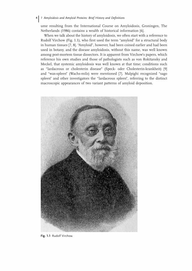
When we talk about the history of amyloidosis, we often start with a reference toRudolf Virchow (Fig. 1.1), who first used the term “amyloid” for a structural bodyin human tissues [7, 8]. “Amyloid”, however, had been coined earlier and had beenused in botany, and the disease amyloidosis, without this name, was well knownamong post-mortem tissue dissectors. It is apparent from Virchow’s papers, whichreference his own studies and those of pathologists such as von Rokitansky andMeckel, that systemic amyloidosis was well known at that time; conditions suchas “lardaceous or cholesterin disease” (Speck- oder Cholesterin-krankheit) [9]and “wax-spleen” (Wachs-milz) were mentioned [7]. Malpighi recognized “sagospleen” and other investigators the “lardaceous spleen”, referring to the distinctmacroscopic appearances of two variant patterns of amyloid deposition.
1 Amyloidosis and Amyloid Proteins: Brief History and Definitions4
Fig. 1.1 Rudolf Virchow.

Virchow used a water solution of iodine in combination with hydrated sulfu-ric acid as a stain for cellulose in the human body [10, 11]. A cellulose-like sub-stance had earlier been described in lower animals. Virchow found that corporaamylacea in ependyme and choroid plexus showed a typical cellulose reactionwith iodine, and stated in his first report that “no doubt regarding the cellulosenature is possible” [10]. Afterwards, Virchow and also Meckel [7] tested tissuescorresponding to what we today call systemic amyloidosis and found a similarreaction to iodine as had been observed with corpora amylacea. Thus, Virchowfound that the wax-like deposits and degeneration of spleen, liver and kidneys,in cases of what must have been amyloid A (AA) amyloidosis due to chronic in-fectious diseases, showed a starch-like reaction with iodine. This initiated a de-bate as to whether the iodine reaction depended on cellulose or on cholesterin.
The hypothesis of the cellulose nature of amyloid did not stand for long. Fried-reich and Kekulé dissected out amyloid-rich segments from the spleen of a pa-tient with amyloidosis, probably AA in nature [12]. In contrast to Virchow, theyperformed quite elegant direct chemical analyses of material extracted in differ-ent ways and came to the definitive conclusion that the main substance wasprotein in nature. This was confirmed by Hanssen [13], who showed that amy-loid is digestible with pepsin. Ironically, 100 years later it was found that cor-pora amylacea in reality contain little protein and are essentially polysaccharidein nature [14].
The nature of the amyloid protein or proteins was a puzzle for a long time.Furthermore, it was debated whether the amyloid substance developed locallyfrom underlying cells. It was also suggested that the protein originated fromblood and that the specific protein was precipitated in organs by abnormalamounts of sulfuric acid present locally [15]. This is not too far from today’s the-ory that circulating proteins interact, not with sulfuric acid, but with glycosami-noglycans [16, 17].
1.1.2Different Chemical Forms of Amyloid: Early Studies
Systemic amyloidosis was initially regarded as a complication of chronic infec-tious diseases such as tuberculosis, syphilis and osteomyelitis. Later, it becameclear that amyloidosis could also occur after the onset of non-infectious chronicinflammatory disorders, e.g. rheumatoid arthritis. However, quite early, singlecases of amyloidosis without any obvious predisposing disease were described[18, 19]. In one case, Wild noted that, in addition to the absence of any addi-tional disease in his patient, the distribution of amyloid was quite remarkable[19], best comparable with what is today known as AL amyloidosis. Interestingly,Soyka described cardiac amyloidosis without predisposing diseases, particularlyat a high age [18]. Certainly, these cases must have been examples of transthyre-tin-derived senile systemic amyloidosis. Remarkable variation in the clinicalmanifestation of primary amyloidosis was noted quite early [20, 21]. The first in-vestigator to identify “primary amyloidosis” as a distinctive group was Lubarsch
1.1 Early History 5

[20]. We now know that this group originally included not only AL amyloidosis,but also different familial forms of amyloidosis, derived from several proteins,and the transthyretin-derived senile systemic amyloidosis.
Although, after Virchow’s initial studies, amyloidosis was typically found to begeneralized, there are early descriptions of characteristic localized AL amyloido-sis, particularly from the conjunctiva [22]. For example, Vossius described twocases of localized, tumor-like amyloidosis of the conjunctiva [23]. Very large in-ter-individual variation is also evident in the localized forms of AL amyloidosisand this is the obvious reason for the large number of case reports of AL amy-loidosis appearing in the medical literature over the years.
One popular theory of the origin of amyloid was that amyloid represents anantigen–antibody precipitate [24], perhaps depending on an autoantigen [25]. Itwas possible to identify immunoglobulin and complement proteins in amyloiddeposits by immunohistochemistry [26]. However, in extracts of amyloid, it wasnot possible to demonstrate �-globulin (immunoglobulin) in reasonableamounts to explain the nature of amyloid [27, 28]. For a long time it was be-lieved that the composition of all amyloids is one and the same, and, in the old-er literature, other possibilities are not discussed. However, Gellerstedt, in 1938,noted that amyloid in the islets of Langerhans differed in tinctorial propertiesas compared with vascular secondary amyloid in cases where both alterationswere observed [29]. He obviously understood that the two types of deposits weredifferent, although he did not explicitly state that they must contain differentproteins. It was not until the first direct protein sequence analyses were per-formed (Section 1.2.1) that the complex chemical nature of the amyloid depositsstarted to become apparent.
1.1.3Amyloid Staining Methodology
The initial method used to identify amyloid was that of iodine staining, intro-duced by Virchow. This method was soon replaced by metachromatic stains likecrystal violet. The use of the most important histological staining marker foramyloid, Congo red, was introduced by Bennhold [30]. This direct dye for cottonhad been used in the textile industry since 1884. Congo red was observed tohave a strong affinity for amyloid deposits and there was the observation thatits clearance from plasma could be used as a diagnostic method for amyloidosis.However, the value of Congo red in histology turned out to be much higher. Animportant discovery was made by Divry and Florkin in 1927 who noticed theenhanced birefringence of amyloid deposits after staining with Congo red [31].It was suggested that this property of amyloid depends on an ordered arrange-ment of the elongated Congo red molecules in the amyloid, indicating that, infact, the substance is not amorphous, as earlier described, but has an organizedsubstructure [31–34]. A standardized Congo red staining method was introducedby Puchtler et al. [35] and is still used. Additional staining methods have beenand are still used. The most important of these is probably Thioflavin T or S.
1 Amyloidosis and Amyloid Proteins: Brief History and Definitions6

There were once divided opinions as to which of the two stainings Congo redor Thioflavin S is more specific for amyloid, but each method now has its ownrole today in the study of amyloidosis [36].
1.2Amyloid Proteins – Modern History
The modern history of amyloidosis can be said to have started with the discov-ery by Cohen and Calkins, using electron microscopy, that amyloid, which ishyaline and structureless under the light microscopy, has a characteristic fine fi-brillar ultrastructure [37]. This finding was confirmed in several other studies[38–42] and pointed to a specific structural organization of the constituent mole-cules – a concept that at that time was completely unknown.
The amyloid was found to contain “rigid”, unbranched fibrils, around 10 nmin diameter and of undetermined length. The fibrils were usually without orien-tation, but when close to cells appeared in parallel bundles, sometimes perpen-dicular and close to cell membranes [43, 44]. This fibrillar organization wastaken by some researchers as an indication that the fibrils were made by thesecells [45], but others were hesitant about this. This situation gave rise to the fa-mous argument raised by Bywaters that on pictures of San Sebastian transfixedwith arrows “he looked a bit sick too, but nobody had suggested he was secret-ing them” [46]. Careful electron microscopic studies by Shirahama and Cohenshowed that the amyloid fibrils, irrespective of origin, were composed of eventhinner subelements, designated protofibrils [47–49].
1.2.1The Amyloid Proteins
The first amyloid component to be identified, although not based on amino acidsequence data, was a soluble protein, which could be extracted from amyloid-la-den tissues. When rabbits were immunized with extracted amyloid fibrils, anti-bodies to this protein were detected [50] that also recognized an immunoreactivecomponent in plasma. The amyloid protein in tissues was consequently desig-nated amyloid plasma component or P component (later AP). The circulatingplasma counterpart, later shown to be identical, was called “serum amyloid Pcomponent” (SAP). SAP was shown to be identical to the AP particle that hadbeen identified in tissue amyloid deposits by electron microscopy and describedas a pentagonal structure [51]. SAP has been shown to be a ubiquitous part ofall of the chemical types of amyloid deposits. The SAP protein binds non-cova-lently and calcium dependently to ligands [52] such as �-pleated sheet fibrils.SAP is a normal constituent of the glomerular basement membrane [53] andelastic microfibrils [54].
The nature of the amyloid fibril was long an enigma. Methods to concentrateamyloid fibrils and to make good, representative fibrillar extracts of systemic
1.2 Amyloid Proteins – Modern History 7

amyloid deposits were developed by Cohen et al. [48] and Pras et al. [55].Further advancement and analysis of constitutive fibril subunits was difficult,however. Benditt et al. extracted secondary amyloid materials directly from tis-sues, without prior purification of fibrils [27]. They identified a protein compo-nent that was soluble in 6 M urea and exhibited an unusual amino acid compo-sition, in that both cysteine and threonine were lacking. At the time, it was notpossible to characterize this protein further and the investigators discussedmany different possibilities, including a virus protein.
Amyloid was long believed to be a singular substance, perhaps of some un-specific degenerative origin. The relationship to other structureless deposits,particularly “hyaline”, was often discussed in the literature and a process oftransformation from hyaline to amyloid was suggested. Even when it had beenshown by electron microscopy that the light microscopically amorphous amyloidconsists of fine fibrils of a characteristic appearance, amyloid deposits wereusually regarded as one unique kind of substance. The demonstration of a con-sistent cross-�-pleated sheet structure in amyloid fibrils was an important stepin our understanding how amyloid is formed [56, 57]. The �-pleated sheet struc-tured fibril seems to be the basis of the unusual resistance of all kinds of amy-loid to degradation and, therefore, the progressive deposition of the material. Ithad been shown already that insulin could be converted into a fibrous form un-der conditions that are denaturing to the secondary structure of proteins [58]. Itwas shown that insulin fibrils [59] as well as synthetic fibrils made from othersmall proteins [60] all had the properties of amyloid fibrils, including affinityfor Congo red and green birefringence when viewed under polarizing light mi-croscopy. Glenner incorporated the characteristic X-ray diffraction pattern ofamyloid fibrils into the definition of amyloid and proposed that the term “amy-loid” be used primarily as a generic adjectival term to indicate the presence ofnon-branching, 80- to 100-Å fibrillar (linear or concentric) proteinaceous depos-its demonstrated to have either Congo red birefringence or a �-pleated sheet X-ray diffraction pattern, together with the chemical nature of the fibril (if known)and the site or tissue of origin or deposition noted [60].
The first definitive proof of a chemically specific protein constituent of amyloidfibrils came from the studies of Glenner et al. They showed that, in cases of pri-mary and myeloma-associated systemic amyloidosis, the fibril protein originatedfrom homogeneous immunoglobulin light chains [61]. Glenner et al.’s workhad an enormous immediate impact and, since, at the time, amyloid was widelybelieved to be a single substance, all of the clinical forms of amyloid were initiallyregarded to be of immunoglobulin origin. When reading the literature from theearly 1970s, it is evident that there was a very high degree of international compe-tition between four or five prominent amyloid research groups and the citationsdo not always seem to be fully correct. Later, Benditt wrote [62] that he had diffi-culties with the publication of an important paper in which he presented evidencefor the existence of multiple chemical classes of amyloid substance [63]. Benditt’sgroup showed that a unique protein, which had a characteristic amino acid com-position and a uniform electrophoretic mobility, was present in all cases of typical
1 Amyloidosis and Amyloid Proteins: Brief History and Definitions8

“secondary” amyloidosis, but absent in cases with other kinds of amyloid. Theycalled this component protein A and the protein(s) extracted from other kindsof amyloidosis, such as what is now known as AL, protein B [63]. Protein A (laterdesignated as amyloid A or AA) was characterized by amino acid sequence in 1971(see below). Based on varying amino acid compositions and electrophoretic mobi-lities, Benditt et al. also suggested that, in fact, there would likely be several differ-ent protein B forms [63]. Subsequent developments in amyloid research haveshown that they were absolutely right.
The pioneering work of the laboratories of Benditt and Glenner showed thatthe most fruitful method for elucidation of the nature of the amyloidosesshould be recognition of putative clinically specific amyloid diseases, and isola-tion from tissues and purification of the corresponding amyloid fibril proteins,followed by their chemical identification by amino acid sequence analysis. Thisapproach was immediately initiated. A third amyloid fibril protein was soonidentified by amino acid sequence analysis and was shown to be derived fromcalcitonin (or possibly procalcitonin), occurring in the amyloid of medullary thy-roid carcinoma [64]. This observation was clear evidence of the diversity in thenature of the amyloid fibril.
1.2.2Specific Amyloid Fibril Proteins
1.2.2.1 Protein AA and its Precursor, Serum AABenditt et al.’s pioneering papers from the pre-amyloid protein sequencing eraclearly showed that the major protein associated with amyloidosis secondary toinflammatory diseases has specific properties, including a constant electrophoret-ic mobility and unusual amino acid composition [27, 63, 65, 66]. Most remark-able was the lack of threonine. The definitive proof of a unique nature camewith the purification of the major protein and the demonstration of a unique N-terminal amino acid sequence of the protein designated protein A [67]. Thiswas soon verified by several other groups [68–70]. The full amino acid sequenceof amyloid protein A from several patients was soon published, indicating a 76-amino-acid protein [70, 71]. However, it has subsequently become clear that pro-tein A of different lengths, but with the same N-terminus, exists [68, 72–74].
The nomenclature for this unique protein varied in publications from differ-ent groups. Since a Staphylococcus protein already was called “protein A”, alter-native names were proposed, such as protein AS [69], ASF [70] or AUO [68].This confusion was ultimately resolved at the Second International Symposiumon Amyloidosis, Helsinki (1975), where the foundation of a modern amyloid no-menclature based on biochemistry was created [75]. At this time, protein A be-came protein AA.
Protein AA was soon found to have a circulating counterpart in plasma [76,77] now called serum AA (SAA). SAA, today known to be a protein family withseveral members of which two are circulating acute-phase reactants, was thusdiscovered through the ability of structurally related protein(s) to aggregate into
1.2 Amyloid Proteins – Modern History 9

amyloid fibrils. In plasma fractionated by gel filtration under physiological con-ditions, SAA appeared as a large protein, around 180–200 kDa [78], but a low-molecular-weight component, around 12 kDa, could be isolated by gel filtrationunder denaturing conditions [79, 80]. The explanation for this difference in pro-tein mass came when it was found that SAA is an apolipoprotein, mainly asso-ciated with high-density lipoprotein, in humans [81], mice [82] and rabbits [83].The blood plasma protein SAA was shown to be about 40% larger than the 76-amino-acid residue AA protein that was first described. This larger size appar-ently depended on a C-terminal extension in the SAA molecule [84]. Subse-quently, it was established that human SAA is a 104-amino-acid molecule ofwhich amyloid protein A corresponds to the major N-terminal part [85]. Theacute-phase SAA is produced by the liver, but extrahepatic expression of SAAwas demonstrated. Thus, it was not immediately accepted that the circulatingSAA is the precursor of the amyloid protein. Eventually, however, direct animalexperimental work showed unequivocally that circulating SAA is converted intoamyloid fibrils [86, 87].
A systemic form of amyloidosis, resembling human secondary amyloidosis,had long been known to occur in many different mammals (for reviews, see[88, 89]), the most well known being that seen in mice. Secondary amyloidosiswas also a common outcome in horses which had been immunized for antiser-um production. At an early date, it was found to be possible to induce second-ary amyloidosis in laboratory animals, including rabbits, hens [90] and mice [91,92]. Protein AA was shown to be the major amyloid fibril component also inthese species ([93]; for review, see [89]). Most important for future experimentalstudies was the finding that the experimentally inducible amyloid in mice is as-sociated with protein AA [94]. As with human, a plasma component, antigeni-cally identical to the tissue-derived protein AA, could be extracted in several ani-mal species after induction of inflammation [95]. Also, as in humans, the plas-ma component appeared in a high-molecular-weight form in its native state, butcould be extracted as a 12-kDa protein after denaturation [96]. Sipe et al. [96]seem to be the first to name SAA as an acute-phase protein. Today we knowSAA as one of the most sensitive acute-phase reactants.
1.2.2.2 Immunoglobulin-derived Amyloid (AL and AH)Cases with simultaneous occurrence of multiple myeloma and amyloidosis weredescribed at an early date. The chemical nature of this amyloid was the subjectof many studies and immunoglobulin was a natural candidate. Many attemptsto extract significant amounts of immunoglobulin from the corresponding amy-loidotic tissues were unsuccessful. Added to the difficulties was the still wide-spread belief that, chemically, amyloid was either one specific substance or anon-specific degeneration product. To make the situation even more confusing,the amyloid associated with myelomatosis was sometimes called “secondary”.
One of the most important advances in amyloid history was the purificationof the fibrillar protein from tissues of a patient with primary amyloidosis and
1 Amyloidosis and Amyloid Proteins: Brief History and Definitions10

the subsequent demonstration by Edman degradation that the N-terminal ami-no acid sequence corresponded to a monoclonal immunoglobulin light chain[97]. Several N-terminal sequences obtained from other individuals with thesame technique confirmed the initial report [61, 98]. The findings fitted nicelywith the demonstration that immunoglobulin light chains contain two sets of �-sheets, of which one comprises most of the variable region [99]. Some princi-ples rapidly became clear. The amyloid in primary and myeloma-associated amy-loidosis is biochemically identical, and consists of an N-terminal fragment of amonoclonal immunoglobulin chain. The fragment varies in length and, in rareoccasions, whole light chains constitute the major fibril protein. A novel immu-noglobulin light chain subtype was discovered by its unusual preponderance toform amyloid fibrils [100–102]. Later, it was shown that, in rare instances,monoclonal immunoglobulin heavy chains may make up amyloid [103, 104]and that also the constant region of light chains is amyloidogenic [105–107].
1.2.2.3 TransthyretinFamilial amyloidosis with varying clinical manifestations was described frommany parts of the world long before biochemical characterization of amyloidwas possible [108]. The first description of a familial amyloidosis was probablythat of Ostertag [109]. Many familial amyloid forms from Portugal, Japan, Swe-den, the USA and other countries had a progressive polyneuropathy as a majorindication. In 1978, Costa et al. [110] showed that the fibrils in the Portuguesetype were associated with prealbumin, which was the earlier name for transthyr-etin. The name prealbumin refers to the electrophoretic mobility of the protein.The primary structure of human transthyretin had been determined in 1974[111] and its crystal structure was published in 1978 [112]. Transthyretin wasfound to have a high degree of �-structure. Soon, many reports verified thetransthyretin nature of the amyloid fibril in many, but not all, of the hereditaryamyloid forms [113–115]. Further analyses showed that in all of the cases ofATTR there was a mutation creating an amino acid substitution. The most com-mon was found to be V30M [116–120]. A continuous stream of publications onnew transthyretin variants, amyloidogenic or even protective against amyloido-sis, has appeared until the present [121]. It has become increasingly clear thatfamilial transthyretin amyloidosis is spread all over the world.
In addition to the different familial forms, including the common V122I mu-tation [120, 122], transthyretin was found to be the fibril protein identified in se-nile systemic amyloidosis [123]. Senile systemic amyloidosis is probably themost common of all the systemic amyloidoses and is of great theoretical inter-est since it is so obviously connected to aging. In contrast to the familial forms,transthyretin in this senile systemic amyloid form is of the wild-type [124].
1.2 Amyloid Proteins – Modern History 11
1.2.2.4 Other Biochemical Forms of Familial AmyloidosisThe identification throughout the world of more families with amyloid syn-dromes and the development of more efficient, sensitive methods in proteinanalyses, combined with molecular biologic methods, led to the identificationnot only of new transthyretin variants associated with amyloidosis, but also newand unexpected amyloid proteins. The strategy was generally the same: identifi-cation of the family, purification of the major amyloid fibril protein and aminoacid sequence analysis followed by sequencing of the specific gene. In this wayit has become relatively easy to rapidly determine the specific genetic cause ofmany familial amyloidoses. These include amyloidosis derived from cystatin C[125, 126], apolipoprotein A-I [127], apolipoprotein A-II [128], fibrinogen [129],gelsolin [130, 131], lysozyme [132], ABri [133] and ADan [134].
1.2.2.5 �2-Microglobulin (�2M)�2M shares strong structural similarities with immunoglobulin light and heavychain constant regions [135], and is part of the HLA class I complex. The pro-tein has a high degree of �-sheet conformation and is a small molecule, thus fit-ting well as an amyloid fibril protein precursor. Indeed, in 1985, Gejyo et al.[136] and directly afterwards Gorevic et al. [137] showed that �2M is the fibrilprotein in amyloidosis occurring as a complication to long-term hemodialysis, adisease that was described almost simultaneously [138–140]. Both full-length�2M and fragments thereof were found in the amyloid deposits [141]. The amy-loid disease had a peculiar systemic distribution, with destructive arthropathy asa major manifestation. An important cause of the disease is increased plasmaconcentration of �2M in individuals on dialysis. Fortunately, this is an amyloidthat is disappearing due to better treatment of kidney failure.
1.2.2.6 Specific Amyloid Forms in the Central Nervous System
Alzheimer and Amyloid The plaques in the cerebral cortex, described by Alzhei-mer [142], and the cerebral amyloid angiopathy, associated with Alzheimer’s dis-ease and aging [143], were entities known for long time, but not initially the sub-ject of any intense interest. Amyloid was rarely, if ever, mentioned as important inthe pathogenesis of the disease. The turning point came with the biochemicalcharacterization of the amyloid fibril protein, initially from the vascular amyloid[144, 145]. Purification of cerebral plaque amyloid was more difficult, but, by ap-plication of wool technology with solubilization in formic acid, Masters et al. [146]succeeded in characterizing the fibril protein, which turned out to be the same asGlenner had found in angiopathy. Glenner called the protein A�, while the nameused by Masters was A4. After some confusion, the name of the protein has be-come A�. A� was found to be an internal fragment of a much larger protein,the A� protein precursor (A�PP). The further development of the field has putA� protein at the center of the pathogenesis of Alzheimer’s disease [147, 148].
1 Amyloidosis and Amyloid Proteins: Brief History and Definitions12

Spongiform Encephalopathies Amyloid itself is probably more of an epipheno-menon in the different spongiform encephalopathies (kuru, Creutzfeldt-Jakobdisease, Gerstmann-Sträussler-Scheinker disease), but is characteristic of sometypes [149]. The history of the causative agent in these and in related animaldiseases is very fascinating, and contains some of the most beautiful achieve-ments in medicine. It started with Gajdusek’s field studies in New Guineawhere the disease kuru was identified in a small isolated Papuan population[150]. Gajdusek found evidence that the disease was transmitted by ritualisticcannibalism and he was also able to transmit the disease from human to chim-panzees [151], thereby proving its contagious properties. Later, Prusiner foundthat the transmissible agent, prion, is not a virus but a protein [152]. The prionprotein aggregates and forms amyloid-like fibrils in vitro, and is the major com-ponent of spongiform plaques amyloid [153].
1.2.2.7 Polypeptide Hormone-derived (“Endocrine”) AmyloidIt has been known for a long time that amyloid may be deposited in some hor-mone-producing tissues. The first described example was amyloid in the isletsof Langerhans, although this was initially called hyaline [154, 155] and the amy-loid nature was accepted much later [29, 156]. Later, amyloid was described inother endocrine tissues and in polypeptide hormone-producing tumors, such asmedullary carcinoma of the thyroid. This “endocrine” amyloid was suggested byPearse et al. [157] to be derived from non-functional parts of pro-hormones. Athird class of amyloid fibril proteins was therefore suggested [157, 158]. Pearseet al. based this assumption on histochemical and microspectrofluorometricstudies of amyloid in an insulinoma and in three medullary carcinomas, whichindicated lack of both tyrosine and tryptophan. At that time the structure ofpro-insulin had been determined [159] and the C-peptide shown to lack aro-matic amino acid residues [160], but the more complicated precursor of calcito-nin was unknown. However, further studies on amyloid from a medullary carci-noma clearly showed the presence of tyrosine [161] and amino acid sequenceanalysis of the amyloid protein from the same case showed identity with calcito-nin [64]. A larger size of the fibril protein and an amino acid composition diverg-ing from that of calcitonin was interpreted as a sign of the presence of pro-calci-tonin in the amyloid, but no further amino acid sequence was obtained.
1.2.2.8 Islet Amyloid PolypeptideThe initial studies with medullary carcinoma showed that polypeptide hor-mones may give rise to amyloid fibrils in vivo. A close contact occurred betweenbundles of amyloid fibrils and � cells, resembling that seen between suspectedfibril-forming cells and amyloid in experimental AA amyloidosis [162], andtherefore islet amyloid was believed to be derived from proinsulin. The strongassociation between localized amyloid in the islets of Langerhans and Type 2diabetes [156, 163, 164] made analysis of this kind of amyloid highly warranted.
1.2 Amyloid Proteins – Modern History 13
It took a long time and hard work to purify the amyloid protein, and this wasfirst done from an insulin-producing tumor [165]. Surprisingly, the major amy-loid fibril protein was a previously unknown polypeptide with partial identitywith calcitonin gene-related peptide (CGRP), initially called islet amyloid pep-tide and later islet amyloid polypeptide (IAPP). Further analyses showed thatIAPP consists of 37 amino acid residues, and that it is the major protein also inamyloid of human and feline islets of Langerhans [166, 167]. The findings wereverified by another group, which called the protein “diabetes-associated peptide”[168]. A later name has been “amylin”. IAPP was found to be a normal productof islet � cells, and is stored and released together with insulin [169, 170]. Sev-eral structural features of IAPP indicated a hormonal nature including C-termi-nal amidation [171, 172] and it is now accepted as a �-cell hormone, the firstdiscovered since insulin [173]. The identification of IAPP started a new branchin diabetes mellitus research. The interested can go to several reviews [174–176]and to Chapter 28 in this book. IAPP, together with A�, became popular modelmolecules for amyloid fibril formation.
1.3Classification of Amyloid Diseases
Until affinity for Congo red and green birefringence after this staining, com-bined with a characteristic fine fibrillar ultrastructure, were generally acceptedto be diagnostic for amyloidosis, there was a discussion about what should beincluded in the group of amyloidoses. The designation “amyloid” was usuallyreserved for systemic amyloidosis and for tumoral localized amyloid (todayknown to be of immunoglobulin origin). For many years there was a discussionof the nature of hyaline and its relationship to amyloid. It was even suggestedthat hyaline (which we know today is usually composed of collagen) could be-come transformed into amyloid. Scattered, small hyaline alterations were shownto occur in certain organs and some, but not all, of them are today included inthe amyloid group. A good example is the amyloid of the islets of Langerhans,initially described as hyalinization [154]. The similarity of the histological ap-pearance of the islet alteration and deposits in systemic amyloidosis was real-ized early, and the designation “para-amyloidosis” was coined for these altera-tions [29]. The following are the previously most commonly used categorizationsof amyloid deposits.
1.3.1Reimann’s Classification
An early classification, that is partly used even today, is that of Reimann et al.[177] who divided the amyloid types into four categories:(1) Primary amyloidosis(2) Secondary amyloidosis
1 Amyloidosis and Amyloid Proteins: Brief History and Definitions14

(3) Tumor-forming amyloidosis(4) Amyloidosis associated with multiple myeloma.
1.3.2King’s Classification
Another classification, which is less commonly referred to, is that of King [178],who divided the amyloidoses into two groups, one with “typical amyloidosis”,i.e. with an organ distribution seen in what we today know as AA amyloidosis,and “atypical amyloidosis”, which is a group including all other cases. The des-ignation “atypical amyloidosis”, discriminating an amyloid from the depositionpattern of systemic amyloidosis in conjunction with a chronic inflammatory dis-ease, was already used earlier (e.g. [25]) and is still seen occasionally. With mod-ern knowledge, these designations should be avoided.
1.3.3Classification of Missmahl et al.
A third classification is that of Missmahl et al. This was also suggested beforethe biochemical era of amyloidosis and was based on polarization findings withCongo red. These researchers noted that amyloid infiltration occurs either in as-sociation with collagen fibrils or with reticulin fibrils and based a differentiationof types on this difference [179, 180]. The classification into the pericollagenform, now known to include AL amyloidosis, and the perireticulin form, partic-ularly including AA amyloidosis, did not survive for long.
1.3.4Modern Classification
The nomenclature based on the chemical nature of amyloid proteins should inprinciple replace all earlier systems. However, the original classification with pri-mary, secondary and familial systemic amyloidoses and localized amyloidosis issurprisingly difficult to eradicate. Unfortunately, this classification, although sim-ple, often leads to confusion and misunderstanding. An example is the use of “sec-ondary” amyloidosis for amyloid associated with multiple myeloma, which is che-mically identical with that in primary amyloidosis, i.e. AL amyloidosis. It can alsobe noted that none of the older classifications include familial amyloidosis as oneseparate group. We know today that the group of familial amyloidosis is a highlyheterogeneous one, containing many biochemically different amyloids.
1.3.4.1 The Present Classification of Amyloid Fibril ProteinsThe modern classification had its beginnings at the Second International Sym-posium on Amyloidosis, Helsinki (1974) [75]. Here, it was decided that the des-ignation of all amyloid forms should be based on their chemical composition.
1.3 Classification of Amyloid Diseases 15

1 Amyloidosis and Amyloid Proteins: Brief History and Definitions16
Table 1.1 Amyloid fibril proteins and their precursors in human (from [182], slightly modified)
Amyloid Precursor protein Systemic (S)or localized (L)
Syndrome orinvolved tissues
Reference
AL immunoglobulinlight chain
S, L primarymyeloma associated
61
AH immunoglobulinheavy chain
S, L primarymyeloma associated
103
ATTR transthyretin S familialsenile systemic
110
L? tenosynoviumA�2M �2-microglobulin S hemodialysis 136
L? jointsAA (apo)serum AA S secondary, reactive 67AApoAI apolipoprotein AI S familial 127
L aorticAApoAII apolipoprotein AII S familial 128AGel gelsolin S familial 130ALys lysozyme S familial 132AFib fibrinogen � chain S familial 129ACys cystatin C S familial 126ABria ABriPP S familial dementia,
British133
L?AApoAIVc) apolipoprotein AIV S senile 184A� A� protein precursor
(A�PP)L Alzheimer’s disease,
aging144
APrP prion protein L spongioform enceph-alopathies
152
ACal (pro)calcitonin L C-cell thyroid tu-mors
64
AIAPP islet amyloid poly-peptide
L islets of Langerhansinsulinomas
165
AANF atrial natriuretic factor L cardiac atria 185APro prolactin L aging pituitary
prolactinomas186
AIns insulin L iatrogenic 187AMed lactadherin L senile aortic, media 188AKer kerato-epithelin L cornea; familial 189ALac lactoferrin L cornea; familial 190A(tbn) b, c) tbn L Pindborg tumors 191
a) ADan comes from the same gene as ABri and has an identical N-terminal sequence. ADan istherefore not included in the nomenclature as a separate protein.
b) To be named.c) Proteins that are preliminary.

The principle was created that all amyloid fibril proteins should be named “pro-tein A” with a suffix identifying the specific protein molecule. The amyloid typeand disease should then be named from the protein. Thus the term AA amyloi-dosis should replace secondary amyloidosis, and AL amyloidosis should replacethe previously used names primary and myeloma-associated amyloid. At thetime of the foundation of this classification, only the two chemical types of amy-loidosis, AA and AL, were known with certainty, although it was suspected thatthe composition of amyloid would not be as uniform as earlier often believed.However, probably no one had then imagined the enormous heterogeneity ofthe human amyloid substances that later has been found to be the case.
The first real amyloid Nomenclature Committee was founded at the Third In-ternational Symposium on Amyloidosis, Povoa de Varzim, Portugal (1979).When this meeting was held, two more amyloid fibril proteins had been de-scribed, transthyretin and (pro)calcitonin, and it was now more definitely under-stood that there were more to be discovered. In addition to protein AA and AL,preliminary terms were decided for familial amyloid proteins (AF), amyloid inendocrine tissues (AE) and amyloid associated with aging (AS; S for senile)[181]. These designations have been dropped since many of the amyloid fibrilproteins are now known. An amyloid Nomenclature Committee has been work-ing since the meeting in Povoa de Varzim and is now formally elected by theBoard of the newly formed International Society of Amyloidosis. To be acceptedas an amyloid fibril protein, the protein must be definitely shown to be the ma-jor component of a distinctive amyloid deposit and the nature of the proteinidentified by amino acid sequence. The data should also have been published ina major scientific journal. Table 1.1 lists the amyloid fibril proteins so far identi-fied [182].
1.4What is Amyloid?
We now return to the starting point: how to define amyloid? Amyloid was origi-nally described as an in vivo phenomenon and amyloidosis as a disease charac-terized by deposition of this material. With increasing knowledge of the natureof amyloid, new problems have arisen. It is possible to make Congophilic �-pleated sheet fibrils from synthetic peptides corresponding to known amyloidproteins or segments thereof. Are these fibrils amyloid? It is even possible tomake similar fibrils from normally occurring peptides never found in amyloidor from completely laboratory-designed peptides. With increasing frequency, allof these kinds of fibrils are called amyloid in the scientific literature. It shouldbe remembered that the amyloid, deposited in tissues, does not only containthe fibrils made from a single, pure protein species, but also additional proteinssuch as SAP, and glycosaminoglycans and proteoglycans. How these compo-nents are associated with the fibrils and what importance they may have inamyloidogenesis and in the persistence of the amyloid are questions that are in-
1.4 What is Amyloid? 17
sufficiently answered. The Nomenclature Committee of the International So-ciety of Amyloidosis has discussed this problem and suggests that the designa-tion “amyloid” should only be used for the abnormal, in vivo deposited material.Also, by definition, amyloid is mainly extracellular, which means that cellularinclusions, e.g. in Parkinson’s disease, are not amyloid. In vitro produced fibrilsshould be called “amyloid-like” [182] (or amylog as suggested by Buxbaum[183]).
Acknowledgments
Supported by the Swedish Research Council.
1 Amyloidosis and Amyloid Proteins: Brief History and Definitions18
References
1 Letterer, E. Zur Ätiologie und formalenPathogenese der Amyloidose. Nova ActaLeopoldina 1966, 31, 11–22.
2 Kyle, R.A. and E.D. Bayrd. Amyloidosis:review of 236 cases. Medicine 1975, 54,271–299.
3 Glenner, G. G. Amyloid deposits andamyloidosis. The �-fibrilloses. N Engl JMed 1980, 302, 1283–1292 and 1333–1343.
4 Cohen, A. S. General introduction and abrief history of amyloidosis. In Amyloi-dosis, J. Marrink and M.H. van Rijswijk(eds). Martinus Nijhoff, Dordrecht,1986, pp. 3–19.
5 Sipe, J. D. and A.S. Cohen. Review: his-tory of the amyloid fibril. J Struct Biol2000, 130, 88–98.
6 Marrink, J. and M.H. van Rijswijk(eds). Amyloidosis. Martinus Nijhoff,Dordrecht, 1986.
7 Virchow, R. Zur Cellulose-Frage. Vir-chows Arch Pathol Anat 1854, 6, 416–426.
8 Virchow, R. Ueber den Gang der amy-loiden Degeneration. Virchows ArchPathol Anat 1855, 8, 364–368.
9 Meckel, H. Die Speck- oder Cholesterin-krankheit. Ann Charité Krankenhaus1853, 4, 264–320.
10 Virchow, R. Ueber eine im Gehirn undRueckenmark des Menschen aufgefun-dene Substanz mit der chemischen Re-action der Cellulose. Virchows ArchPathol Anat 1854, 6, 135–138.
11 Virchow, R. Weitere Mittheilungen überdas Vorkommen der pflanzlichen Cellu-lose beim Menschen. Virchows ArchPathol Anat 1854, 6, 268–271.
12 Friedreich, N. and A. Kekulé. Zur Amy-loidfrage. Virchows Arch Pathol Anat1859, 16, 50–65.
13 Hanssen, O. Ein Beitrag zur Chemieder amyloiden Entartung. Biochem Z1908, 13, 185–198.
14 Sakay, M., J. Austin, F. Witmer and L.Trueb. Studies of corpora amylacea. I.Isolation and preliminary characteriza-tion by chemical and histochemicaltechniques. Arch Neurol 1969, 21, 526–544.
15 Fahr, T. Pathologische Anatomie desMorbus Brightii. In Handbuch der spe-ziellen pathologischen Anatomie und His-tologie, F. Henke and O. Lubarsch (eds).Julius Springer, Berlin, 1925, pp. 156–472.
16 Leupold, E. Cited by Fahr [15].17 Kisilevsky, R. Review: amyloidogenesis –
unquestioned answers and unansweredquestions. J Struct Biol 2000, 130, 99–108.
18 Soyka, J. Ueber die amyloide Degenera-tion. Prag Med Wschr 1876, 1, 165–171.
19 Wild, C. Beitrag zur Kenntnis der amy-loiden und der hyalinen Degenerationdes Bindegewebes. Beitr Pathol AnatPhysiol 1886, 1, 175–200.

References 19
20 Lubarsch, O. Zur Kenntnis ungewöhnli-cher Amyloidablagerungen. VirchowsArch Pathol Anat 1929, 271, 867–889.
21 Symmers, W. S.C. Amyloidosis. Fivecases of primary generalized amyloido-sis and some other unusual cases. JClin Pathol 1956, 9, 212–228.
22 Raehlmann, E. Ueber hyaline und amy-loide Degeneration der Conjunctiva desAuges. Virchows Arch Pathol Anat, 1882,87, 325–335.
23 Vossius, A. Ueber amyloide Degenera-tion der Conjunctiva. Beitr Pathol Anat1889, 4, 335–360.
24 Löschke, H. Vorstellung über das We-sen von Hyalin und Amyloid auf Grundvon serologischen Versuchen. BeitrPathol Anat 1927, 77, 231–239.
25 Letterer, E. Neue Untersuchungen überdie Entstehung des Amyloids. VirchowsArch Pathol Anat 1934, 293, 34–72.
26 Vogt, A. Immunhistochemische Be-funde am Amyloid. Nova Acta Leopoldi-na 1966, 31, 37–41.
27 Benditt, E.P., D. Lagunoff, N. Eriksenand O.A. Iseri. Amyloid. Extraction andpreliminary characterization of someproteins. Arch Pathol 1962, 74, 323–330.
28 Calkins, E., A. S. Cohen and D. Gitlin.Immunochemical determinations ofgamma globulin content of amyloid.Fedn Proc 1958, 183, 1202–1203.
29 Gellerstedt, N. Die elektive, insuläre(Para-)Amyloidose der Bauchspeichel-drüse. Zugleich ein Beitrag zur Kennt-nis der „senilen Amyloidose“. BeitrPathol Anat 1938, 101, 1–13.
30 Bennhold, H. Eine specifische Amyloid-färbung mit Kongorot. Münch Med Wo-chenschr 1922, 69, 1537–1538.
31 Divry, P. and M. Florkin. Sur les pro-priétées optiques de l’amyloide. CR SocBiol (Paris) 1927, 97, 1808–1810.
32 Romhányi, G. Über die submikrosko-pische Struktur des Amyloids. ZentralblAllg Pathol 1943, 80, 411.
33 Romhányi, G. Über die submikrosko-pische Struktur des Amyloid. Schweiz ZPathol (Pathol Microbiol) 1949, 12, 253–262.
34 Missmahl, H.P. and M. Hartwig. Polari-sationsoptische Untersuchungen an der
Amyloid-substanz. Virchows Arch PatholAnat 1953, 324, 489–508.
35 Puchtler, H., F. Sweat and M. Levine.On the binding of Congo red by amy-loid. J Histochem Cytochem 1962, 10,355–364.
36 Mandema, E., L. Ruinen, J. H. Scholtenand A.S. Cohen (eds). Amyloidosis, Ex-cerpta Medica, Amsterdam, 1968.
37 Cohen, A. S. and E. Calkins. Electronmicroscopic observations on a fibrouscomponent in amyloid of diverse ori-gins. Nature 1959, 183, 1202–1203.
38 Frühling, L., J. Kempf and A. Porte.Structure et formation de la substanceamyloide dans l’amylose expérimentalede la Souris. Étude au microscope élec-tronique. C R Acad Sci (Paris) 1960,250, 1385–1386.
39 Caesar, R. Die Feinstruktur von Milzund Leber bei experimenteller Amyloi-dose. Z Zellforsch 1960, 52, 653–673.
40 Battaglia, S. Electronenoptische Unter-suchungen am Leberamyloid der Maus.Beitr Pathol Anat 1962, 126, 301–320.
41 Cohen, A. S., A. Frensdorff, S. Lam-precht and E. Calkins. A study of thefine structure of the amyloid associatedwith familial Mediterranean fever. Am JPathol 1962, 41, 567–578.
42 Heefner, W. A. and G. D. Sorenson. Ex-perimental amyloidosis. I. Light andelectron microscopic observations ofspleen and lymph nodes. Lab Invest1962, 11, 585–593.
43 Cohen, A. S. and E.S. Cathcart. Casein-duced experimental amyloidosis. InMethods and Achievements in Experimen-tal Pathology, E. Bajusz and G. Jasmin(eds). Karger, Basel, 1972, pp. 207–242.
44 Shirahama, T. and A.S. Cohen. Intraly-sosomal formation of amyloid fibrils.Am J Pathol 1975, 81, 101–116.
45 Gueft, B. and J. J. Ghidoni. The site offormation and ultrastructure of amyloid.Am J Pathol 1963, 43, 837–854.
46 Bywaters, E.G. L. Discussion. In Amyloi-dosis, E. Mandema, L. Ruinen, H. Schol-ten and A.S. Cohen (eds). ExcerptaMedica, Amsterdam, 1968, p. 80.
47 Shirahama, T. and A.S. Cohen. High-resolution electron microscopic analysis

1 Amyloidosis and Amyloid Proteins: Brief History and Definitions20
of the amyloid fibril. J Cell Biol 1967,33, 679–708.
48 Shirahama, T. and A.S. Cohen. Recon-stitution of amyloid fibrils from alkalineextracts. J Cell Biol 1967, 35, 459–464.
49 Shirahama, T. and A.S. Cohen. A briefreview of the ultrastructure of amyloid.In Amyloidosis, J. Marrink and M.H.van Rijswijk (eds). Martinus Nijhoff,Dordrecht, 1976, pp. 51–57.
50 Cathcart, E. S., F.R. Comerford andA. S. Cohen. Immunologic studies on aprotein extracted from human second-ary amyloid. N Engl J Med 1965, 273,143–146.
51 Bladen, H.A., M.U. Nylen and G. G.Glenner. The ultrastructure of humanamyloid as revealed by the negativestaining technique. J Ultrastruct Res1966, 14, 449–459.
52 Pepys, M.B., A.C. Dash, E. A. Munn, A.Feinstein, M. Skinner, A.S. Cohen, H.Gerwurz, A. P. Osmand and R. H. Pain-ter. Isolation of amyloid P component(protein AP) from normal serum as acalcium-dependent binding protein.Lancet 1977, i, 1029–1031.
53 Dyck, R. F., C.M. Lockwood, M. Ker-shaw, N. McHugh, M.L. Baltz and M.B.Pepys. Amyloid P-component is a con-stituent of normal human glomerularbasement membrane. J Exp Med 1980,152, 1162–1174.
54 Breathnach, S. M., S.M. Melrose, B.Bhogal, F. C. de Beer, R. F. Dyck, G.Tennent, M.M. Black and M.B. Pepys.Amyloid P component is located onelastic microfibrils in normal humantissue. Nature 1981, 293, 652–654.
55 Pras, M., M. Schubert, D. Zucker-Frank-lin, A. Rimon and E.C. Franklin. Thecharacterization of soluble amyloid pre-pared in water. J Clin Invest 1968, 47,924–933.
56 Eanes, E.D. and G. G. Glenner. X-raydiffraction studies on amyloid fila-ments. J Histochem Cytochem 1968, 16,673–677.
57 Bonar, L., A. S. Cohen and M. Skinner.Characterization of the amyloid fibril asa cross-� protein. Proc Soc Exp Biol Med1969, 131, 1373–1375.
58 Waugh, D.F. A fibrous modification ofinsulin. I. The heat precipitate of insu-lin. J Am Chem Soc 1946, 68, 247–250.
59 Westermark, P. On the nature of theamyloid in human islets of Langerhans.Histochemistry 1974, 38, 27–33.
60 Glenner, G. G., E.D. Eanes, H.A. Bla-den, R. P. Linke and J. D. Termine. �-pleated sheet fibrils. A comparison ofnative amyloid with synthetic protein fi-brils. J Histochem Cytochem 1974, 22,1141–1158.
61 Glenner, G. G., W. Terry, M. Harada, C.Isersky and D. Page. Amyloid fibril pro-teins: proof of homology with immuno-globulin light chains by sequence analy-sis. Science 1971, 172, 1150–1151.
62 Benditt, E.P. Amyloid protein AA andits precursor, the acute phase protein(s)apoSAA: a perspective. In Amyloidosis, J.Marrink and M.H. van Rijswijk (eds).Martinus Nijhoff, Dordrecht, 1986,pp. 101–106.
63 Benditt, E.P. and N. Eriksen. Chemicalclasses of amyloid substance. Am JPathol 1971, 65, 231–252.
64 Sletten, K., P. Westermark and J. B. Nat-vig. Characterization of amyloid fibrilproteins from medullary carcinoma of thethyroid. J Exp Med 1976, 143, 993–998.
65 Benditt, E.P. and N. Eriksen. Chemicalsimilarity among amyloid substancesassociated with long standing inflam-mation. Lab Invest 1962, 26, 615–625.
66 Benditt, E.P. and N. Eriksen. Amyloid,III. A protein related to the subunitstructure of human amyloid fibrils. ProcNatl Acad Sci USA 1966, 55, 308–316.
67 Benditt, E.P., N. Eriksen, M.A. Her-modson and L.H. Ericsson. The majorproteins of human and monkey amyloidsubstance: common properties includ-ing unusual N-terminal amino acid se-quences. FEBS Lett 1971, 19, 169–173.
68 Ein, D., S. Kimura, W. D. Terry, J. Mag-notta and G. G. Glenner. Amino acid se-quence of an amyloid fibril protein ofunknown origin. J Biol Chem 1972, 247,5653–5655.
69 Husby, G., K. Sletten, T.E. Michaelsenand J. B. Natvig. Alternative non-immu-noglobulin origin of amyloid fibrils. Na-ture (New Biol) 1972, 238, 187.

References 21
70 Levin, M., E.C. Franklin, B. Frangioneand M. Pras. The amino acid sequenceof a major nonimmunoglobulin compo-nent of some amyloid fibrils. J Clin In-vest 1972, 51, 2773–2776.
71 Sletten, K. and G. Husby. The completeamino-acid sequence of non-immuno-globulin amyloid fibril protein AS inrheumatoid arthritis. Eur J Biochem1974, 41, 117–125.
72 Møyner, K., K. Sletten, G. Husby andJ. B. Natvig. An unusually large (83 ami-no acid residues) amyloid fibril proteinAA from a patient with Waldenström’smacroglobulinaemia and amyloidosis.Scand J Immunol 1980, 11, 549–554.
73 Sletten, K., G. Husby and J. B. Natvig.The complete amino acid sequence ofan amyloid fibril protein AA of unusualsize (64 residues). Biochem Biophys ResCommun 1976, 69, 19–25.
74 Westermark, G. T., P. Westermark andK. Sletten. Amyloid fibril protein AA.Characterization of uncommon subspe-cies from a patient with rheumatoid ar-thritis. Lab Invest 1987, 57, 57–64.
75 Cohen, A. S., E. C. Franklin, G. G. Glen-ner, J. B. Natvig, E.F. Osserman and O.Wegelius. Nomenclature. In Amyloidosis,O. Wegelius and A. Pasternack (eds).Academic Press, London, 1976, p. ix.
76 Levin, M., M. Pras and E.C. Franklin.Immunologic studies of the major non-immunoglobulin protein of amyloid. I.Identification and partial characteriza-tion of a related serum component. JExp Med 1973, 138, 373–380.
77 Husby, G. and J. B. Natvig. A serumcomponent related to nonimmunoglo-bulin amyloid protein AS, a possibleprecursor of the fibrils. J Clin Invest1974, 53, 1054–1061.
78 Sipe, J. D., T. F. Ignaczak, P.S. Pollockand G. G. Glenner. Amyloid fibril proteinAA: purification and properties of theantigenically related serum componentas determined by solid phase radioim-munoassay. J Immunol 1976, 116, 1151–1156.
79 Linke, R. P., J.D. Sipe, P. S. Pollock,T.F. Ignaczak and G. G. Glenner. Isola-tion of a low-molecular-weight serumcomponent antigenically related to an
amyloid fibril protein of unknown ori-gin. Proc Natl Acad Sci USA 1975, 72,1473–1476.
80 Rosenthal, C. J., E. C. Franklin, B. Fran-gione and J. Greenspan. Isolation andpartial characterization of SAA – anamyloid-related protein from human se-rum. J Immunol 1976, 116, 1415–1418.
81 Benditt, E.P. and N. Eriksen. Amyloidprotein SAA is associated with highdensity lipoprotein from human serum.Proc Natl Acad Sci USA 1977, 74, 4025–4028.
82 Benditt, E.P. and N. Eriksen. Amyloidprotein SAA is an apoprotein of mouseplasma high density lipoprotein. ProcNatl Acad Sci USA 1979, 76, 4092–4096.
83 Skogen, B., A. L. Borresen, J. B. Natvig,K. Berg and T.E. Michaelsen. High-den-sity lipoprotein as carrier for amyloid-re-lated protein SAA in rabbit serum.Scand J Immunol 1979, 10, 39–45.
84 Bausserman, L.L., P.N. Herbert andK. P.W. J. McAdam. Heterogeneity ofhuman amyloid A proteins. J Exp Med1980, 152, 641–656.
85 Sletten, K., G. Marhaug and G. Husby.The covalent structure of amyloid-re-lated serum protein SAA from two pa-tients with inflammatory disease. HoppeSeylers Zeitschr Physiol Chem 1983, 364,1039–1046.
86 Husebekk, A., B. Skogen, G. Husby andG. Marhaug. Transformation of amyloidprecursor SAA to protein AA and incor-poration in amyloid fibrils in vivo. ScandJ Immunol 1985, 21, 283–287.
87 Tape, C., R. Tan, M. Nesheim and R. Ki-silevsky. Direct evidence for circulatingapoSAA as the precursor of tissue AAamyloid deposits. Scand J Immunol1988, 28, 317–324.
88 Zschiesche, W. and W. Jakob. Pathologyof animal amyloidoses. Pharmacol Ther1989, 41, 49–83.
89 Johnson, K. H., P. Westermark, K. Slet-ten and T. D. O’Brien. Amyloid proteinsand amyloidosis in domestic animals.Amyloid: Int J Exp Clin Invest 1996, 3,270–289.
90 Maximow, A. Ueber die experimentellhervorgerufene Amyloid-Entartung der

1 Amyloidosis and Amyloid Proteins: Brief History and Definitions22
Leber. Virchows Arch Pathol Anat 1898,153, 353–401.
91 Domagk, G. Untersuchungen über dieBedeutung des retikuloendothelialenSystems für die Vernichtung von Infek-tionserregern und für die Entstehungdes Amyloids. Virchows Arch PatholAnat 1924, 253, 594–638.
92 Morgenstern, Z. Zur Frage über Amy-loidose und Resorption. Virchows ArchPathol Anat 1926, 259, 698–725.
93 Benditt, E.P. and N. Eriksen. Thechemical classes of amyloid substancein men, monkeys and ducks. ProtidesBiol Fluids 1973, 20, 81–85.
94 Eriksen, N., L.H. Ericsson, N. Pearsall,D. Lagunoff and E. P. Benditt. Mouseamyloid protein AA: homology withnonimmunoglobulin protein of humanand monkey amyloid substance. ProcNatl Acad Sci USA 1976, 73, 964–967.
95 Anders, R.F., J. B. Natvig, K. Sletten, G.Husby and K. Nordstoga. Amyloid-re-lated serum protein SAA from three an-imal species: comparison with humanSAA. J Immunol 1977, 118, 229–234.
96 Sipe, J. D., K.P. W. J. McAdam, B. F.Torain and P. S. Pollock. Isolation andstructural properties of murine SAA –the acute phase serum precursor ofamyloid AA. Immunol Commun 1977, 6,1–12.
97 Glenner, G. G., J. Harbaugh, J. I. Ohms,M. Harada and P. Cuatrecasas. An amy-loid protein: the amino-terminal vari-able fragment of an immunoglobulinlight chain. Biochem Biophys Res Com-mun 1970, 41, 1287–1289.
98 Lian, J. B., M. Skinner, M.D. Bensonand A.S. Cohen. Fractionation of pri-mary amyloid fibrils. Characterizationand chemical interaction of the sub-units. Biochim Biophys Acta 1977, 491,167–176.
99 Schiffer, M., R.L. Girling, K. R. Ely andA. B. Edmundson. Structure of a lamb-da-type Bence-Jones protein at 3.5-Åresolution. Biochemistry 1973, 12, 4620–4631.
100 Husby, G., J. B. Natvig and K. Sletten.New, third class of amyloid fibril pro-tein. J Exp Med 1974, 139, 773–778.
101 Sletten, K., J.B. Natvig, G. Husby andJ. Juul. The complete amino acid se-
quence of a prototype immunoglobu-lin-� light-chain-type amyloid-fibril pro-tein AR. Biochem J 1981, 195, 561–572.
102 Solomon, A., B. Frangione and E. C.Franklin. Bence Jones proteins andlight chains of immunoglobulins. Pre-ferential association of the V�VI sub-group of human light chains with amy-loidosis AL(�). J Clin Invest 1982, 70,453–460.
103 Eulitz, M., D.T. Weiss and A. Solomon.Immunoglobulin heavy-chain-asso-ciated amyloidosis. Proc Natl Acad SciUSA 1990, 87, 6542–6546.
104 Tan, S.Y., I.E. Murdoch, T. J. Sullivan,J. E. Wright, O. Truong, J. J. Hsuan,P.N. Hawkins and M.B. Pepys. Pri-mary localized orbital amyloidosis com-posed of the immunoglobulin gammaheavy chain CH3 domain. Clin Sci1994, 87, 487–491.
105 Engvig, J. P., K.E. Olsen, R. E. Gisle-foss, K. Sletten, O. Wahlström and P.Westermark. Constant region of a � IIIimmunoglobulin light chain as a majorAL-amyloid protein. Scand J Immunol1998, 48, 92–98.
106 Solomon, A., D.T. Weiss, C.L. Murphy,R. Hrncic, J. S. Wall and M. Schell.Light chain-associated amyloid depositscomprised of a novel kappa constantdomain. Proc Natl Acad Sci USA 1998,95, 9547–9551.
107 Wally, J., G. Kica, Y. Zhang, T. Erics-son, L.H. Connors, M.D. Benson, J. J.Liepnieks, J. Murray, M. Skinner andR. L. Comenzo. Identification of a novelsubstitution in the constant region of agene coding for an amyloidogenic kap-pa1 light chain. Biochim Biophys Acta1999, 1454, 49–56.
108 Andrade, C., S. Araki, W.D. Block,A. S. Cohen, C.E. Jackson, Y. Kuroiwa,J. Nissim, E. Sohar, V. A. McKusick andM.W. Van Allen. Hereditary amyloido-sis. Arthritis Rheum 1970, 13, 902–915.
109 Ostertag, B. Demonstration einer ei-genartigen familiären „Paramyloidose“.Zentralbl Pathol 1933, 56, 253–254.
110 Costa, P.P., A. S. Figueira and F.R.Bravo. Amyloid fibril protein related toprealbumin in familial amyloidoticpolyneuropathy. Proc Natl Acad SciUSA 1978, 75, 4499–4503.

References 23
111 Kanda, Y., D. S. Goodman, R. E. Can-field and F. J. Morgan. The amino acidsequence of human plasma prealbu-min. J Biol Chem 1974, 249, 6796–6805.
112 Blake, C.C.F., M.J. Geisow, S. J. Oatley,B. Rérat and C. Rérat. Structure of pre-albumin: secondary, tertiary and qua-ternary interactions determined byFourier refinement at 1.8 Å. J Mol Biol1978, 121, 339–356.
113 Benson, M.D. Partial amino acid se-quence homology between a heredofa-milial amyloid protein and humanplasma prealbumin. J Clin Invest 1981,67, 1035–1041.
114 Skinner, M. and A.S. Cohen. The pre-albumin nature of the amyloid proteinin familial amyloidotic polyneuropathy(FAP) – Swedish variety. Biochem Bio-phys Res Commun 1981, 99, 1326–1332.
115 Tawara, S., S. Araki, K. Toshimori, H.K. Nakagawa and S. Ohtaki. Amyloidfibril protein in type I familialamyloidotic polyneuropathy in Japa-nese. J Lab Clin Med 1981, 98, 811–822.
116 Tawara, S., M. Nakazato, K. Kangawa,H. Matsuo and S. Araki. Identificationof amyloid prealbumin variant in fa-milial amyloidotic polyneuropathy (Ja-panese type). Biochem Biophys Res Com-mun 1983, 116, 880–888.
117 Saraiva, M. J.M., S. Birken, P.P. Costaand D.S. Goodman. Amyloid fibril pro-tein in familial amyloidotic polyneuro-pathy, Portuguese type. Definition ofmolecular abnormality in transthyretin(prealbumin). J Clin Invest 1984, 74,104–119.
118 Dwulet, F.E. and M.D. Benson. Char-acterization of a transthyretin (prealbu-min) variant associated with familialamyloidotic polyneuropathy type II (In-diana/Swiss). J Clin Invest 1986, 78,880–886.
119 Westermark, P., K. Sletten and B.-O.Olofsson. Prealbumin variants in theamyloid fibrils of Swedish familialamyloidotic polyneuropathy. Clin ExpImmunol 1987, 69, 695–701.
120 Gorevic, P.D., F.C. Prelli, J. Wright,M. Pras and B. Frangione. Systemic se-
nile amyloidosis. Identification of anew prealbumin (transthyretin) variantin cardiac tissue: immunologic andbiochemical similarity to one form offamilial amyloidotic polyneuropathy. JClin Invest 1989, 83, 836–843.
121 Connors, L.H., A. Lim, T. Prokaeva,V. A. Roskens and C.E. Costello. Tabu-lation of human transthyretin (TTR)variants, 2003. Amyloid: J Protein Fold-ing Disorder 2003, 10, 160–184.
122 Jacobson, D.R., P. Gorevic and J. N.Buxbaum. A homozygous transthyretinvariant associated with senile systemicamyloidosis: evidence for a late-onsetdisease of genetic etiology. Am J HumGenet 1990, 47, 127–136.
123 Westermark, P., J.B. Natvig and B. Jo-hansson. Characterization of an amy-loid fibril protein from senile cardiacamyloid. J Exp Med 1977, 146, 631–636.
124 Westermark, P., K. Sletten, B. Johans-son and G.G. Cornwell III. Fibril in se-nile systemic amyloidosis is derivedfrom normal transthyretin. Proc NatlAcad Sci USA 1990, 87, 2843–2845.
125 Grubb, A. and H. Löfberg. Human �-trace, a basic microprotein: amino acidsequence and presence in the adenohy-pophysis. Proc Natl Acad Sci USA1982, 79, 3024–3027.
126 Cohen, D. H., H. Feiner, O. Jenssonand B. Frangione. Amyloid fibril in he-reditary cerebral hemorrhage withamyloidosis (HCHWA) is related to thegastroentero-pancreatic neuroendocrineprotein, gamma trace. J Exp Med 1983,158, 623–628.
127 Nichols, W.C., F. E. Dwulet, J. Liep-nieks and M.D. Benson. Variant apoli-poprotein AI as a major constituent ofa human hereditary amyloid. BiochemBiophys Res Commun 1988, 156, 762–768.
128 Benson, M.D., J. Liepnieks, M. Yazaki,T. Yamashita, K. Hamidi Asl, B. Guen-ther and B. Kluve-Beckerman. A newhuman hereditary amyloidosis: the re-sult of a stop-codon mutation in theapolipoprotein AII gene. Genomics2001, 72, 272–277.

1 Amyloidosis and Amyloid Proteins: Brief History and Definitions24
129 Benson, M.D., J. Liepnieks, T. Uemi-chi, G. Wheeler and R. Correa. Heredi-tary renal amyloidosis associated witha mutant fibrinogen alpha-chain. Na-ture Genetics 1993, 3, 252–256.
130 Maury, C.P. and M. Baumann. Isola-tion and characterization of cardiacamyloid in familial amyloid polyneuro-pathy type IV (Finnish): relation of theamyloid protein to variant gelsolin. Bio-chim Biophys Acta 1990, 1096, 84–86.
131 Levy, E., M. Haltia, I. Fernandez-Ma-drid, O. Koivunen, J. Ghiso, F. Prelliand B. Frangione. Mutation in the gel-solin gene in Finnish hereditary amy-loidosis. J Exp Med 1990, 172, 1865–1867.
132 Pepys, M.B., P.N. Hawkins, D. R.Booth, D. M. Vigushin, G. A. Tennet,A. K. Soutar, N. Totty, O. Nguyen, C.C.F. Blake, C. J. Terry, T. G. Feest, A. M.Zalin and J. J. Hsuan. Human lyso-zyme gene mutations cause hereditarysystemic amyloidosis. Nature 1993,362, 553–557.
133 Vidal, R., B. Frangione, A. Rostagno, S.Mead, T. Revesz, G. Plant and J. Ghiso.A stop-codon mutation in the BRI geneassociated with familial British demen-tia. Nature 1999, 399, 776–781.
134 Vidal, R., T. Revesz, A. Rostagno, E.Kim, J. L. Holton, T. Bek, M. Bojsen-Moller, H. Braendgaard, G. Plant, J.Ghiso and B. Frangione. A decamerduplication in the 3� region of the BRIgene originates an amyloid peptidethat is associated with dementia in aDanish kindred. Proc Natl Acad SciUSA 2000, 97, 4920–4925.
135 Becker, J. W. and G.N. J. Reeke. Three-dimensional structure of �2-microglo-bulin. Proc Natl Acad Sci USA 1985,82, 4225–4229.
136 Gejyo, F., T. Yamada, S. Odani, Y. Na-kagawa, M. Arakawa, T. Kunitomo, H.Kataoka, M. Suzuki, Y. Hirasawa, T.Shirahama, A.S. Cohen and K.Schmid. A new form of amyloid pro-tein associated with chronic hemodialy-sis was identified as �2-microglobulin.Biochem Biophys Res Commun 1985,129, 701–706.
137 Gorevic, P.D., T. T. Casey, W.J. Stone,C.R. DiRaimondo, F. Prelli and B.Frangione. Beta-2 microglobulin is anamyloidogenic protein in man. J ClinInvest 1985, 76, 2425–2429.
138 Bardin, T., D. Kuntz, J. Zingraff, M.-C.Voisin, A. Zelmar and J. Lansaman.Synovial amyloidosis in patients under-going long-term hemodialysis. ArthritisRheum 1985, 28, 1052–1058.
139 Morita, T., M. Suzuki, A. Kamimuraand Y. Hirasawa. Amyloidosis of a pos-sible new type in patients receivinglong-term hemodialysis. Arch PatholLab Med 1985, 109, 1029–1032.
140 Munoz-Gómez, J., E. Bergadá-Barado,R. Gómez-Pérez, E. Llopart-Buisán, E.Subías-Sobrevía, J. Rotés-Querol andM. Solé-Arqués. Amyloid arthropathyin patients undergoing periodicalhaemodialysis for chronic renal failure:a new complication. Ann Rheum Dis1985, 44, 729–733.
141 Linke, R. P., H. Hampl, H. Lobeck, E.Ritz, J. Bommer, R. Waldherr and M.Eulitz. Lysine-specific cleavage of �2-microglobulin in amyloid deposits as-sociated with hemodialysis. Kidney Int1989, 36, 675–681.
142 Alzheimer, A. Ueber eine eigenartigeErkrankung der Hirnrinde. CentrablNervenheilk Psychiatr 1907, 30, 177–179.
143 Pantelakis, S. Un type particulier d’an-giopathie sénile du système nerveuxcentral: L’angiopathi congophile. Topo-graphie et fréquence. Monatsschr Psy-chiatr Neurol 1954, 128, 219–256.
144 Glenner, G. G. and C.W. Wong. Alzhei-mer’s disease: initial report of thepurification and characterization of anovel cerebrovascular amyloid protein.Biochem Biophys Res Commun 1984,120, 885–890.
145 Glenner, G. G. and C.W. Wong. Alzhei-mer’s disease and Down’s syndrome:sharing of a unique cerebrovascularamyloid fibril protein. Biochem BiophysRes Commun 1984, 122, 1131–1135.
146 Masters, C., G. Simms, N. A. Wein-man, G. Multhaup, B.L. McDonaldand K. Beyreuther. Amyloid plaquecore protein in Alzheimer disease and
References 25
Down syndrome. Proc Natl Acad SciUSA 1985, 82, 4245–4249.
147 Selkoe, D. J. Alzheimer’s disease: a cen-tral role for amyloid. J Neuropath ExpNeurol 1994, 53, 438–447.
148 Selkoe, D. J. The origins of Alzheimerdisease: A is for amyloid. J Am MedAss 2000, 283, 1615–1617.
149 Masters, C.L., D. C. Gajdusek and C. J.Gibbs Jr. Creutzfeldt-Jakob diseasevirus isolations from the Gerstmann-Sträussler syndrome. With an analysisof the various forms of amyloid plaquedeposition in the virus-induced spongi-form encephalopathies. Brain 1981,104, 559–588.
150 Gajdusek, D.C. and V. Zigas. Degen-erative disease of the central nervoussystem in New Guinea; the endemicoccurrence of kuru in the native popu-lation. N Engl J Med 1957, 257, 974–978.
151 Gajdusek, D.C., C. J. Gibbs Jr and M.Alpers. Experimental transmission of aKuru-like syndrome to chimpanzees.Nature 1966, 209, 794–796.
152 Bolton, D.C., M.P. McKinley and S. B.Prusiner. Identification of a proteinthat purifies with the scrapie prion.Science 1982, 218, 1309–1311.
153 Prusiner, S.B., M.P. McKinley, K. A.Bowman, D.C. Bolton, P.E. Bendheim,D.F. Groth and G. G. Glenner. Scrapieprions aggregate to form amyloid-likebirefringent rods. Cell 1983, 35, 349–358.
154 Opie, E.L. On relation of chronic inter-stitial pancreatitis to the islands of Lan-gerhans and to diabetes mellitus. J ExpMed 1901, 5, 397–428.
155 Weichselbaum, A. and E. Stangl. ZurKenntnis der feineren Veränderungendes Pankreas bei Diabetes mellitus.Wien Klin Wochenschr 1901, 14, 968–972.
156 Ehrlich, J.C. and I.M. Ratner. Amyloi-dosis of the islets of Langerhans. A re-study of islet hyalin in diabetic andnondiabetic individuals. Am J Pathol1961, 38, 49–59.
157 Pearse, A. G. E., S.W.B. Ewen and J. M.Polak. The genesis of apudamyloid inendocrine polypeptide tumours: histo-
chemical distinction from immunamy-loid. Virchows Arch B Zellpathol 1972,10, 93–107.
158 Westermark, P. Amyloid of human is-lets of Langerhans. I. Isolation andsome characteristics. Acta Pathol Micro-biol Scand C 1975, 83, 439–446.
159 Steiner, D. F. and P.E. Oyer. The bio-synthesis of insulin and a probableprecursor of insulin by a human isletadenoma. Proc Natl Acad Sci USA1967, 57, 473–480.
160 Ko, A.S. C., D. G. Smyth, J. Markussenand F. Sundby. The amino acid se-quence of the C-peptide of humanproinsulin. Eur J Biochem 1971, 20,190–199.
161 Westermark, P. Amyloid of medullarycarcinoma of the thyroid: partial char-acterization. Upsala J Med Sci 1975, 80,88–92.
162 Westermark, P. Fine structure of isletsof Langerhans in insular amyloidosis.Virchows Arch A 1973, 359, 1–18.
163 Bell, E.T. Hyalinization of the islets ofLangerhans in diabetes mellitus. Dia-betes 1952, 1, 341–344.
164 Westermark, P. Quantitative studies ofamyloid in the islets of Langerhans.Upsala J Med Sci 1972, 77, 91–94.
165 Westermark, P., C. Wernstedt, E. Wi-lander and K. Sletten. A novel peptidein the calcitonin gene related peptidefamily as an amyloid fibril protein inthe endocrine pancreas. Biochem Bio-phys Res Commun 1986, 140, 827–831.
166 Westermark, P., C. Wernstedt, E. Wi-lander, D.W. Hayden, T.D. O’Brienand K.H. Johnson. Amyloid fibrils inhuman insulinoma and islets of Lan-gerhans of the diabetic cat are derivedfrom a neuropeptide-like protein alsopresent in normal islet cells. Proc NatlAcad Sci USA 1987, 84, 3881–3885.
167 Westermark, P., C. Wernstedt, T.D.O’Brien, D. W. Hayden and K. H. John-son. Islet amyloid in type 2 human dia-betes mellitus and adult diabetic catscontains a novel putative polypeptidehormone. Am J Pathol 1987, 127, 414–417.
168 Cooper, G. J., A. C. Willis, A. Clark,R. C. Turner, R.B. Sim and K.B. M.

1 Amyloidosis and Amyloid Proteins: Brief History and Definitions26
Reid. Purification and characterizationof a peptide from amyloid-rich pan-creases of type 2 diabetic patients. ProcNatl Acad Sci USA 1987, 84, 8628–8632.
169 Lukinius, A., E. Wilander, G.T. Wester-mark, U. Engström and P. Wester-mark. Co-localization of islet amyloidpolypeptide and insulin in the B cellsecretory granules of the human pan-creatic islets. Diabetologia 1989, 32,240–244.
170 Johnson, K. H., T.D. O’Brien, D. W.Hayden, K. Jordan, H.K. G. Ghobrial,W. C. Mahoney and P. Westermark. Im-munolocalization of islet amyloid poly-peptide (IAPP) in pancreatic Beta cellsby means of peroxidase-antiperoxidase(PAP) and protein A-gold techniques.Am J Pathol 1988, 130, 1–8.
171 Betsholtz, C., V. Svensson, F. Rorsman,U. Engström, G. T. Westermark, E. Wi-lander, K. H. Johnson and P. Wester-mark. Islet amyloid polypeptide(IAPP): cDNA cloning and identifica-tion of an amyloidogenic region asso-ciated with species-specific occurrenceof age-related diabetes mellitus. ExpCell Res 1989, 183, 484–493.
172 Nishi, M., T. Sanke, S. Seino, R. L.Eddy, Y.S. Fan, M.G. Byers, T.B.Shows, G. I. Bell and D.F. Steiner. Hu-man islet amyloid polypeptide gene:complete nucleotide sequence, chromo-somal localization, and evolutionaryhistory. Mol Endocrinol 1989, 3, 1775–1781.
173 Banting, F.G., C.H. Best, J. B. Collipand J. J.R. Macleod. The effect of pan-creatic extract (insulin) on normal rab-bits. Am J Physiol 1922, 62, 162–176.
174 Westermark, P., K.H. Johnson, T. D.O’Brien and C. Betsholtz. Islet amyloidpolypeptide – a novel controversy indiabetes research. Diabetologia 1992,35, 297–303.
175 Kahn, S.E., S. Andrikopoulos and C.B.Verchere. Islet amyloid. A long-recog-nized but underappreciated pathologi-cal feature of type 2 diabetes. Diabetes1999, 48, 241–253.
176 Höppener, J. W., B. Ahrén and C. J.Lips. Islet amyloid and type 2 diabetes
mellitus. N Engl J Med 2000, 343, 411–419.
177 Reimann, H.A., R.F. Koucky and C.M.Eklund. Primary amyloidosis limited tothe tissue of mesodermal origin. Am JPathol 1935, 11, 977–988.
178 King, L.S. Atypical amyloid disease;with observations on a new silver stainfor amyloid. Am J Pathol 1948, 24,1095–1115.
179 Heller, H., H.-P. Missmahl, E. Soharand J. Gafni. Amyloidosis: its differen-tiation into peri-reticulin and peri-col-lagen types. J Pathol Bacteriol 1964, 88,15–34.
180 Missmahl, H.-P. Reticulin and collagenas important factors for the localizationof amyloid and the use of polarizationmicroscopy as a tool in the detection ofthe composition of amyloid. In Amyloi-dosis, E. Mandema, L. Ruinen, J. H.Scholten and A.S. Cohen (eds). Excerp-ta Medica, Amsterdam, 1968, pp. 22–29.
181 Benditt, E.P., A.S. Cohen, P.P. Costa,E.C. Franklin, G. G. Glenner, G. Hus-by, E. Mandema, J. B. Natvig, E. F. Os-serman, E. Sohar, O. Wegelius and P.Westermark. Guidelines for nomencla-ture. In Amyloid and Amyloidosis, G.G.Glenner, P. P. Costa and A.F. Freitas(eds). Excerpta Medica, Amsterdam,1980, pp. xi–xii.
182 Westermark, P., M.D. Benson, J.N.Buxbaum, A.S. Cohen, B. Frangione,S. Ikeda, C.L. Masters, G. Merlini,M.J. Saraiva and J. D. Sipe. Amyloid fi-bril protein nomenclature – 2002. Amy-loid: J Protein Fold Dis 2002, 9, 197–200.
183 Buxbaum, J.N. Diseases of protein con-formation: what do in vitro experi-ments tell us about in vivo diseases?Trends Biochem Sci 2003, 28, 585–592.
184 Bergström, J., C.L. Murphy, D. T.Weiss, A. Solomon, K. Sletten, U. Hell-man and P. Westermark. Two differenttypes of amyloid deposits – apolipopro-tein A-IV and transthyretin – in a pa-tient with systemic amyloidosis. LabInvest 2004, 84, 981–988.
185 Johansson, B., C. Wernstedt and P.Westermark. Atrial natriuretic peptide

References 27
deposited as atrial amyloid fibrils. Bio-chem Biophys Res Commun 1987, 148,1087–1092.
186 Westermark, P., L. Eriksson, U. Eng-ström, S. Eneström and K. Sletten.Prolactin-derived amyloid in the agingpituitary gland. Am J Pathol 1997, 150,67–73.
187 Dische, F.E., C. Wernstedt, G. T. Wes-termark, P. Westermark, M.B. Pepys,J. A. Rennie, S.G. Gilbey and P. J. Wat-kins. Insulin as an amyloid-fibril pro-tein at sites of repeated insulin injec-tions in a diabetic patient. Diabetologia1988, 31, 158–161.
188 Häggqvist, B., J. Näslund, K. Sletten,G. T. Westermark, G. Mucchiano, L.O.Tjernberg, C. Nordstedt, U. Engströmand P. Westermark. Medin: an integralfragment of aortic smooth muscle cell-produced lactadherin forms the mostcommon human amyloid. Proc NatlAcad Sci USA 1999, 96, 8669–8674.
189 Korvatska, E., H. Henry, Y. Mashima,M. Yamada, C. Bachmann, F.L. Mu-
nier and D. F. Schorderet. Amyloid andnon-amyloid forms of 5q31-linked cor-neal dystrophy resulting from kerato-epithelin mutations at Arg-124 are as-sociated with abnormal turnover of theprotein. J Biol Chem 2000, 275, 11465–11469.
190 Ando, Y., M. Nakamura, H. Kai, S. Kat-suragi, H. Terazaki, T. Nozawa, T. Oku-da, S. Misumi, N. Matsunaga, K. Hata,T. Tajiri, S. Shoji, T. Yamashita, K. Ha-raoka, K. Obayashi, K. Matsumoto, M.Ando and M. Uchino. A novel localizedamyloidosis associated with lactoferrinin the cornea. Lab Invest 2002, 82, 757–766.
191 Solomon, A., C.L. Murphy, K. Weaver,D.T. Weiss, R. Hrncic, M. Eulitz, R. L.Donnell, K. Sletten, G. Westermarkand P. Westermark. Calcifying epithe-lial odontogenic (Pindborg) tumor-asso-ciated amyloid consists of a novel hu-man protein. J Lab Clin Med 2003, 142,348–355.


![Susanne Vera Tholen - mediaTUM · Radioisotop [11C]-Kohlenstoff gelang die Herstellung eines Radiopharmakons zur Darstellung von fibrillärem Amyloid mit Hilfe der Positronen-Emissions-Tomographie](https://static.fdokument.com/doc/165x107/5cd1322c88c99347028cfe7e/susanne-vera-tholen-mediatum-radioisotop-11c-kohlenstoff-gelang-die-herstellung.jpg)