Relaxin ELISA - immundiagnostik.com · Arbeitsanleitung Relaxin 2 1. VERWENDUNGSZWECK Der hier...
30
Arbeitsanleitung / Manual Relaxin ELISA Zur in-vitro-Bestimmung von Relaxin in Serum, Plasma, Urin, Seminalplasma und Gewebe For the in vitro determination of relaxin in serum, plasma, urine, seminal plasma and tissue Gültig ab / Valid from 2017-01-24 Immundiagnostik AG, Stubenwald-Allee 8a, 64625 Bensheim, Germany Tel.: +49 6251 70190-0 Fax: + 49 6251 849430 e.mail: [email protected] www.immundiagnostik.com K 9210 +2 °C +8 °C 96
Transcript of Relaxin ELISA - immundiagnostik.com · Arbeitsanleitung Relaxin 2 1. VERWENDUNGSZWECK Der hier...
Arbeitsanleitung / Manual
Relaxin ELISA Zur in-vitro-Bestimmung von Relaxin in Serum, Plasma,
Urin, Seminalplasma und Gewebe
For the in vitro determination of relaxin in serum, plasma, urine, seminal plasma and tissue
Gültig ab / Valid from 2017-01-24
Immundiagnostik AG, Stubenwald-Allee 8a, 64625 Bensheim, Germany
Tel.: +49 6251 70190-0 Fax: + 49 6251 849430
e.mail: [email protected] www.immundiagnostik.com
K 9210+2 °C
+8 °C
96

1
Arbeitsanleitung Relaxin
Inhalt
1. VERWENDUNGSZWECK ________________________________________________ 2
2. EINLEITUNG __________________________________________________________ 2
3. INHALT DER TESTPACKUNG ____________________________________________ 3
4. ERFORDERLICHE LABORGERÄTE UND HILFSMITTEL _______________________ 4
5. LAGERUNG UND VORBEREITUNG DER REAGENZIEN _______________________ 4
6. PROBENLAGERUNG UND -VORBEREITUNG _______________________________ 5
Probenlagerung_ ________________________________________________________ 5Probenvorbereitung_ _____________________________________________________ 5
7. TESTDURCHFÜHRUNG _________________________________________________ 6
Testprinzip_ ____________________________________________________________ 6Pipettierschema_ ________________________________________________________ 7
8. ERGEBNISSE __________________________________________________________ 8
9. EINSCHRÄNKUNGEN __________________________________________________ 9
10. QUALITÄTSKONTROLLE _______________________________________________ 10
Referenzwerte__________________________________________________________ 10
11. TESTCHARAKTERISTIKA ______________________________________________ 10
Präzision_und_Reproduzierbarkeit___________________________________________ 10Analytische_Sensitivität___________________________________________________ 10Spezifität______________________________________________________________ 10Wiederfindung_in_der_Verdünnung__________________________________________ 11
12. VORSICHTSMASSNAHMEN ____________________________________________ 11
13. TECHNISCHE MERKMALE _____________________________________________ 12
14. ALLGEMEINE HINWEISE ZUM TEST _____________________________________ 12
15. LITERATUR __________________________________________________________ 13
Arbeitsanleitung Relaxin
2
1. VERWENDUNGSZWECKDer hier beschriebene Assay ist für die Bestimmung von Relaxin in Serum, Plasma, Urin, Seminalplasmen und Gewebe geeignet. Nur zur in-vitro-Diagnostik.
2. EINLEITUNGRelaxin ist ein Peptidhormon der Insulinfamilie mit einem Molekulargewicht von 6500 Da. Seine Hauptfunktion ist die relaxierende Wirkung auf die glatte Muskula-tur. Wegen der stark erhöhten Konzentration des Relaxins während der Ovulation und Schwangerschaft liegen die meisten Erkenntnisse über die Eigenschaften von Relaxin im Bereich der Reproduktionsmedizin und Gynäkologie vor. In letzter Zeit wurden jedoch neue Wirkungen von Relaxin entdeckt. Es wurde insbesondere ge-zeigt, dass Relaxin: (i) die Dilatation von Blutgefässen in Organen und Geweben, wie z.B. Uterus, Brustdrüse, Lunge und Herz, fördert; (ii) chronotropisch auf das Herz wirkt; (iii) die Stimulation vom potentesten Vasokonstriktor, Endothelin-1, bei Herzin-farkt inhibiert; (iv) die Histaminfreisetzung aus den Mastzellen hemmt und auf diese Weise die allergischen Symptome bei Asthma lindert; (v) die Plättchenaggregation und -freisetzung aus den Megakaryozyten vermindert; (vi) die Hormonsekretion der Hypophyse beeinflusst; und (vii) zur Regulation des Flüssigkeitsgleichgewichts im Körper beiträgt.Spezifische G Protein-gekoppelte Rezeptoren für Relaxin, LGR7 und LGR8, wurden im Gehirn (Wechselwirkung mit ADH-Sekretion), Uterus und Herzen (Einfluss auf die Herzfrequenz) identifiziert. Dschietzig et al. (2004) zeigten, dass Relaxin als Glukokor-tikoid-Rezeptor-Agonist agiert. In weiteren Arbeiten wird der Zusammenhang zwi-schen Relaxin und oxidativem Stress beschrieben. Bani et al. (1997) und Nistri (2003) berichten, dass Relaxinzusatz in die Reperfusionslösung von ischämischen Ratten-herzen Schutz gegen oxidative Veränderungen des Myokardgewebes bietet. Dabei wird die Produktion von Malondialdehyd (Abbauprodukt bei der Lipidoxidation) und Myeloperoxidase (Marker für die Granulozytenaktivität) deutlich vermindert. Als Fol-ge wird eine geringere Schädigung durch Ischämie / Reperfusion am Myokardge-webe, und dadurch bedingt, auch eine geringere Mortalitätsrate beobachtet. Dass das Peptidhormon ein unabhängiger Risikofaktor zur Voraussage der Mortalität bei männlichen Patienten mit terminaler Niereninsuffizienz (TNI) ist, zeigen schließlich Hocher et al. (2004) an 245 Langzeitdialysepatienten.
Indikationen
• Untersuchungen zum Schutz bei Reperfusion/Ischämie• Untersuchungen zur Regulation der Zirkulation und Mikrozirkulation von Kör-
perflüssigkeiten• Untersuchungen zur Angiogenese

3
Arbeitsanleitung Relaxin
• Untersuchungen zur Immunmodulation• Untersuchungen in der Reproduktionsmedizin• Prognosefaktor zur Überlebenswahrscheinlichkeit bei Patienten mit Nieren-
insuffizienz
3. INHALT DER TESTPACKUNG
Art.-Nr. Bezeichnung Kit-Komponenten Menge
K 9210 PLATE Mikrotitermodul, vorbeschichtet 12 x 8 Vertiefungen
K 9210 WASHBUF ELISA-Waschpufferkonzentrat, 10 x 1 x 100 ml
K 9210 CONJ Konjugatkonzentrat, peroxidasemarkiert 1 x 200 µl
K 9210 AB Detektionsantikörperkonzentrat, biotinyliertes Kaninchen anti-Relaxin 1 x 200 µl
K 9210 STD Standards, lyophilisiert (0; 3.1; 9.3; 28; 83; 250 pg/ml) 2 x 6 vials
K 9210 CTRL1Kontrolle, lyophilisiert
(Bereich der Spezifikation entneh-men)
2 x 1 vial
K 9210 CTRL2Kontrolle, lyophilisiert
(Bereich der Spezifikation entneh-men)
2 x 1 vial
K 9210 SAMPLEBUF Probenverdünnungspuffer, gebrauchsfertig 1 x 50 ml
K 9210 SUB TMB-Substrat (Tetramethylbenzidin), gebrauchsfertig 1 x 15 ml
K 9210 STOP ELISA-Stopplösung, gebrauchsfertig 1 x 15 ml
Für Nachbestellungen von Einzelkomponenten verwenden Sie als Bestellnummer die Artikel-nummer gefolgt von der Bezeichnung.

Arbeitsanleitung Relaxin
4
4. ERFORDERLICHE LABORGERÄTE UND HILFSMITTEL• Reinstwasser*• Präzisionspipetten und Pipettenspitzen für den Einmalgebrauch mit varia-
blen Volumina von 10–1000 µl• Folie zum Abkleben der Mikrotiterplatte• Mikrotiterplattenschüttler• Multikanal- bzw. Multipipette• Vortex-Mixer• Laborübliche Glas- oder Plastikröhrchen (Einmalartikel)• Mikrotiterplattenphotometer (benötigte Filter siehe Kapitel 7)
* Immundiagnostik AG empfiehlt die Verwendung von Reinstwasser nach ISO 3696. Es han-delt sich dabei um Wasser des Typs 1, welches frei von ungelösten und kolloidalen Ionen und organischen Molekülen ist (frei von Partikeln > 0,2 µm) mit einer elektrischen Leitfähigkeit von 0,055 µS/cm bei 25 °C (≥ 18,2 MΩ cm).
Nur für Gewebeextrakte
• Micro-Dismembrator• Ultra-Zentrifuge, 100 000 g
5. LAGERUNG UND VORBEREITUNG DER REAGENZIEN
• Bitte achten Sie bei mehrfachem Einsatz des Kits darauf, dass die Reagenzien wie in der Vorschrift beschrieben gelagert und nur die für den jeweiligen Ansatz benötigten Reagenzienmengen frisch angesetzt werden. Der Kit kann so bis zu 4 x je nach Probenaufkommen bis zum angegebenen Haltbar-keitsdatum verwendet werden.
• Reagenzien mit einem Volumen kleiner 100 µl sollten vor Gebrauch kurz an-zentrifugiert werden, um Volumenverluste zu vermeiden.
• Vorbereitung des Waschpuffers: Das Waschpufferkonzentrat (WASHBUF) muss vor Gebrauch 1:10 in Reinstwasser verdünnt werden (100 ml WASH-BUF + 900 ml Reinstwasser), gut mischen. Aufgrund des hohen Salzgehalts im Konzentrat kann es zu Kristallbildungen kommen. Die Kristalle lösen sich bei Raumtemperatur bzw. im Wasserbad bei 37 °C auf. Das WASHBUF kann bei 2–8 °C bis zum angegebenen Haltbarkeitsdatum aufbewahrt werden. Der Waschpuffer (1:10 verdünntes WASHBUF) ist 1 Monat bei 2–8 °C in einem geschlossenen Gefäß haltbar.
• Die lyophilisierten Standards (STD) und Kontrollen (CTRL) sind bei 2–8 °C bis zum angegebenen Haltbarkeitsdatum verwendbar. STDs und CTRLs wer-den mit 500 µl Reinstwasser rekonstituiert, zum Lösen 10 Minuten stehen gelassen und anschließend gründlich gemischt. Standards und Kontrollen
5
Arbeitsanleitung Relaxin
(rekonstituierte STDs und CTRLs) können 4 Wochen bei 2–8 °C gelagert werden.
• Vorbereitung des biotinylierten Antikörpers: Das Antikörperkonzentrat (AB) wird vor Gebrauch 1:1001 in Waschpuffer verdünnt (10 µl AB + 10 ml Waschpuffer). Das AB ist bei 2–8 °C bis zum angegebenen Haltbarkeitsdatum stabil. Biotinylierter Antikörper (1:1001 verdünntes AB) ist nicht stabil und kann nicht aufbewahrt werden.
• Vorbereitung des Konjugats: Das Konjugatkonzentrat (CONJ) wird vor Ge-brauch 1:1001 in Waschpuffer verdünnt (10 µl CONJ + 10 ml Waschpuffer). Das CONJ ist bei 2–8 °C bis zum angegebenen Haltbarkeitsdatum stabil. Kon-jugat (1:1001 verdünntes CONJ) ist nicht stabil und kann nicht aufbewahrt werden.
• Alle anderen Testreagenzien sind bei 2–8 °C zu lagern und bei entsprechender Lagerung bis zum angegebenen Verfallsdatum (siehe Etikett) verwendbar.
6. PROBENLAGERUNG UND -VORBEREITUNG
ProbenlagerungSerum, Plasma, Urin, Seminalplasma und Gewebe sind bei -20 °C zu lagern.
Probenvorbereitung
Serum und Plasma
Serum und Plasma Proben werden vor dem Einsatz im Test mindestens 1:3 verdünnt,z. B. 100 µl Probe + 200 µl Verdünnungspuffer (SAMPLEBUF), gut mischen.Für eine Bestimmung in Doppelwerten werden 2 x je 100 µl jeder vorbereiteten Pro-be im Test eingesetzt.Serum und Plasma Proben können Rheumafaktor und heterophile Antikörper ent-halten. Diese stellen eine potenzielle Interferenz dar, welche bei Sandwich Immuno-assays zu falsch positiven Ergebnissen führen können. Daher empfehlen wir eine Probenvorbehandlung der Serum- und Plasmaproben 2 x mit 5 % (v/v) Anti Interferenzreagenz (Immundiagnostik Artikelnummer K 9212) wie folgt:
• 10 µl Anti Interferenzreagenz + 200 µl Probe• 1 Stunde bei 4 °C schütteln• zentrifugieren und den Überstand abnehmen (vorbehandelte Probe).
Arbeitsanleitung Relaxin
6
Urin
Urinproben werden vor dem Einsatz im Test mindestens 1:4 verdünnt,z. B. 100 µl Probe + 300 µl Verdünnungspuffer (SAMPLEBUF), gut mischen.Für eine Bestimmung in Doppelwerten werden 2 x je 100 µl jeder vorbereiteten Pro-be im Test eingesetzt.
Seminalplasma
EDTA-Plasma- oder Serumproben werden vor dem Einsatz im Test mindestens 1:10 verdünnt, z. B. 30 µl Probe + 270 µl Verdünnungspuffer (SAMPLEBUF), gut mischen.Für eine Bestimmung in Doppelwerten werden 2 x je 100 µl jeder vorbereiteten Pro-be im Test eingesetzt.
Gewebeextrakt
• Gewebeproben (ab 200 mg) aus dem flüssigen Stickstoff entnehmen und in dem vorgefrorenen Schüttelbehälter im Micro-Dismembrator (30 s/1500 Upm) pulverisieren.
• Mit 1 ml Phosphatpuffer (0,14 M NaCl; 2,6 mM KCl; 8 mM Na2HPO4; 1,4 mM KH2PO4), 1 % Triton-X 100; pH 7,4 homogenisieren. Nach der Ultra-Zentrifuga-tion (1 h/100 000 g) im Überstand die Proteinkonzentration (Pierce-BCA oder wahlweise Peterson-Lowry Protein Assay) bestimmen.
• 2 x je 100 µl des Zytosol-Überstands im Assay einsetzen.
7. TESTDURCHFÜHRUNG
TestprinzipDieser ELISA dient zur quantitativen Bestimmung von Relaxin. Der Test basiert auf der “Sandwich”-ELISA Technik. Es werden zwei ausgewählte polyklonale Antikörper, die humanes Relaxin erkennen, verwendet.Teststandards, Kontrollen und verdünnte Patientenproben, die auf Relaxin zu un-tersuchen sind, werden in die Vertiefungen einer Mikrotiterplatte pipettiert, welche mit einem hochaffinen polyklonalen anti-human Realaxin Antikörper beschichtet wurden. In diesem ersten Inkubationsschritt wird das Relaxin von dem gekoppelten Fängerantikörper gebunden. Anschließend wird mit dem Detektionsantikörper (bi-otinylierter, polyklonaler anti-Relaxin Antikörper) inkubiert. Dann wird das Konjugat (Streptavidin, peroxidase-markiert) zugegeben und es bildet sich folgender Komplex an der Wand der Mikrotiterplatte: Fängerantikörper - humanes Relaxin – Detektions-antikörper - Peroxidase-Konjugat. Als Peroxidasesubstrat wird Tetramethylbenzidin (TMB) eingesetzt. Die Enzymreaktion wird durch Zugabe von Säure abgestoppt. Da-
7
Arbeitsanleitung Relaxin
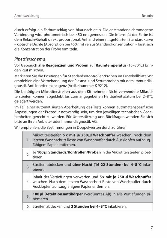
durch erfolgt ein Farbumschlag von blau nach gelb. Die entstandene chromogene Verbindung wird photometrisch bei 450 nm gemessen. Die Intensität der Farbe ist dem Relaxin-Gehalt direkt proportional. Anhand einer mitgeführten Standardkurve – optische Dichte (Absorption bei 450 nm) versus Standardkonzentration – lässt sich die Konzentration der Probe ermitteln.
PipettierschemaVor Gebrauch alle Reagenzien und Proben auf Raumtemperatur (15–30 °C) brin-gen, gut mischen.Markieren Sie die Positionen für Standards/Kontrollen/Proben im Protokollblatt. Wir empfehlen eine Vorbehandlung der Plasma- und Serumproben mit dem Immundia-gnostik Anti Interferenzreagenz (Artikelnummer K 9212).Die benötigten Mikrotiterstreifen aus dem Kit nehmen. Nicht verwendete Mikroti-terstreifen können abgeklebt bis zum angegebenen Haltbarkeitsdatum bei 2–8 °C gelagert werden.Im Fall einer automatisierten Abarbeitung des Tests können automatenspezifische Anpassungen der Prozedur notwendig sein, um den jeweiligen technischen Gege-benheiten gerecht zu werden. Für Unterstützung und Rückfragen wenden Sie sich bitte an Ihren Anbieter oder Immundiagnostik AG.Wir empfehlen, die Bestimmungen in Doppelwerten durchzuführen.
1.Mikrotiterstreifen 5 x mit je 250 µl Waschpuffer waschen. Nach dem letzten Waschschritt Reste von Waschpuffer durch Ausklopfen auf saug-fähigem Papier entfernen.
2. Je 100 µl Standards/Kontrollen/Proben in die Mikrotiterstreifen pipet-tieren.
3. Streifen abdecken und über Nacht (16-22 Stunden) bei 4–8 °C inku-bieren.
4.Inhalt der Vertiefungen verwerfen und 5 x mit je 250 µl Waschpuffer waschen. Nach dem letzten Waschschritt Reste von Waschpuffer durch Ausklopfen auf saugfähigem Papier entfernen.
5. 100 µl Detektionsantikörper (verdünntes AB) in alle Vertiefungen pi-pettieren.
6. Streifen abdecken und 2 Stunden bei 4–8 °C inkubieren.

Arbeitsanleitung Relaxin
8
7.Inhalt der Vertiefungen verwerfen und 5 x mit je 250 µl Waschpuffer waschen. Nach dem letzten Waschschritt Reste von Waschpuffer durch Ausklopfen auf saugfähigem Papier entfernen.
8. 100 µl Konjugat (verdünntes CONJ) in alle Vertiefungen pipettieren.
9. Streifen abdecken und 1 Stunde bei 4–8 °C inkubieren.
10.Inhalt der Vertiefungen verwerfen und 5 x mit je 250 µl Waschpuffer waschen. Nach dem letzten Waschschritt Reste von Waschpuffer durch Ausklopfen auf saugfähigem Papier entfernen.
11. 100 µl Substrat (SUB) in alle Vertiefungen pipettieren.
12. 20–30 min* bei Raumtemperatur (15–30 °C) im Dunkeln inkubieren.
13. 50 µl Stopplösung (STOP) in alle Vertiefungen pipettieren, gut mischen
14.
Extinktion sofort im Mikrotiterplattenphotometer bei 450 nm gegen die Referenzwellenlänge 620 nm (oder 690 nm) messen. Ist keine Refe-renzwellenlänge vorhanden, wird nur bei 450 nm gemessen. Falls die Extinktion des höchsten Standards den Messbereich des Photometers übersteigt, sollte sofort bei 405 nm gegen 620 nm (690 nm) gemessen werden.
* Die Intensität der Farbentwicklung ist temperaturabhängig. Es wird empfohlen, den Farbum-schlag während der Inkubationszeit zu beobachten und entsprechend der Farbentwicklung die Reaktion zu stoppen.
8. ERGEBNISSEDie unten beschriebenen mathematischen Modelle können alternativ zur Auswer-tung benutzt werden. Wir empfehlen die 4-Parameter-Funktion:
1. 4-Parameter-Funktion
Für die optische Dichte empfehlen wir eine lineare Ordinate und für die Konzen-tration eine logarithmische Abszisse (bei einer logarithmischen Abszisse muss für den Standard mit der Konzentration 0 ein Wert kleiner 1 eingegeben werden z. B. 0,001).
2. Punkt-zu-Punkt-Auswertung
Für die optische Dichte und für die Konzentration empfehlen wir eine lineare Ordinate bzw. Abszisse.
9
Arbeitsanleitung Relaxin
3. Gewichtete Spline-Funktion
Für die optische Dichte und für die Konzentration empfehlen wir eine lineare Ordinate bzw. Abszisse.
Vor jeder automatischen Auswertung sollte stets eine Kontrolle der Doppelwerte auf Plausibilität („Ausreißerkontrolle“) durchgeführt werden; falls dies nicht durch das verwendete Programm erfolgt, sollte die Kontrolle manuell durchgeführt werden.
Serum und Plasma
Die ermittelten Ergebnisse werden mit dem Verdünnungsfaktor 3 multipliziert, um die tatsächlichen Konzentrationen zu erhalten.Sollte ein anderer Verdünnungsfaktor verwendet worden sein, so ist die ermittelte Konzentration mit dem verwendeten Verdünnungsfaktor zu multiplizieren.
Urin
Die ermittelten Ergebnisse werden mit dem Verdünnungsfaktor 4 multipliziert, um die tatsächlichen Konzentrationen zu erhalten.Sollte ein anderer Verdünnungsfaktor verwendet worden sein, so ist die ermittelte Konzentration mit dem verwendeten Verdünnungsfaktor zu multiplizieren.
Seminalplasma
Die ermittelten Ergebnisse werden mit dem Verdünnungsfaktor 10 multipliziert, um die tatsächlichen Konzentrationen zu erhalten.Sollte ein anderer Verdünnungsfaktor verwendet worden sein, so ist die ermittelte Konzentration mit dem verwendeten Verdünnungsfaktor zu multiplizieren.
Gewebeextrakt
Die ermittelten Konzentrationen werden mit der gewählten Verdünnung multipli-ziert, um die tatsächlichen Konzentrationen zu ermitteln.
9. EINSCHRÄNKUNGENProben mit Konzentrationen oberhalb des Messbereichs (Definition siehe unten) müssen stärker verdünnt und erneut gemessen werden. Bitte beachten Sie diese stärkere Verdünnung bei der Ergebnisberechnung.Proben mit Konzentrationen unterhalb des Messbereichs (Definition siehe unten) können nicht klar quantifiziert werden.Die Obergrenze des Messbereichs ergibt sich aus:
höchste_Konzentration_der_Standardkurve × anzuwendender_Probenverdünnungs-faktor
Arbeitsanleitung Relaxin
10
Die Untergrenze des Messbereichs ergibt sich aus:Analytische_Sensitivität × anzuwendender_Probenverdünnungsfaktor
10. QUALITÄTSKONTROLLEImmundiagnostik empfiehlt den Einsatz von externen Kontrollen für die interne Qualitätskontrolle, wenn möglich.Wir empfehlen, bei jedem Testansatz Kontrollen mitzumessen. Die Ergebnisse der Kontrollen müssen auf Richtigkeit überprüft werden. Liegen eine oder mehrere Kon-trollen außerhalb des angegebenen Bereiches, kann Immundiagnostik die Richtig-keit der Messergebnisse nicht gewährleisten.
ReferenzwerteWir empfehlen jedem Labor, einen eigenen Referenzbereich zu etablieren.
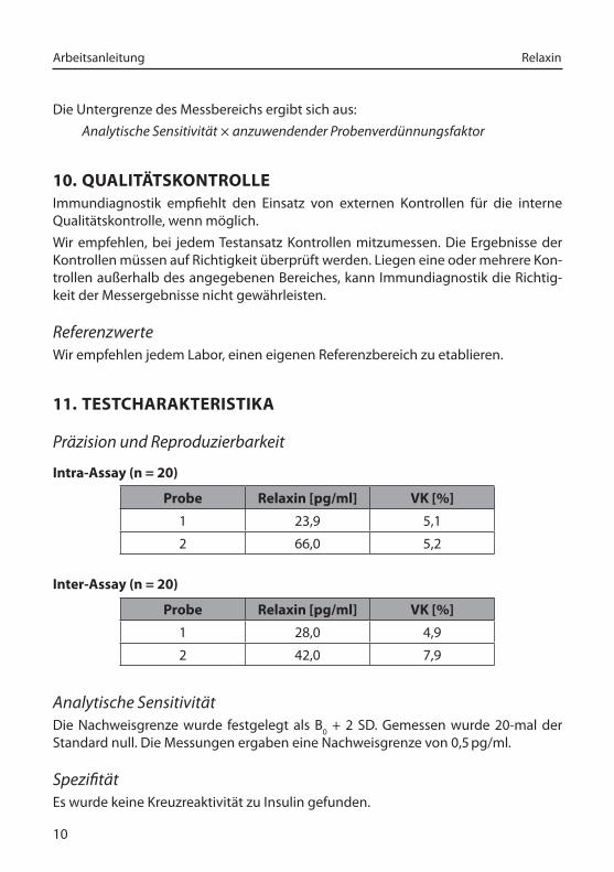
11. TESTCHARAKTERISTIKA
Präzision_und_Reproduzierbarkeit
Intra-Assay (n = 20)
Probe Relaxin [pg/ml] VK [%]
1 23,9 5,1
2 66,0 5,2
Inter-Assay (n = 20)
Probe Relaxin [pg/ml] VK [%]
1 28,0 4,9
2 42,0 7,9
Analytische_SensitivitätDie Nachweisgrenze wurde festgelegt als B0 + 2 SD. Gemessen wurde 20-mal der Standard null. Die Messungen ergaben eine Nachweisgrenze von 0,5 pg/ml.
SpezifitätEs wurde keine Kreuzreaktivität zu Insulin gefunden.
11
Arbeitsanleitung Relaxin
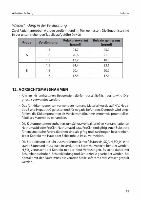
Wiederfindung_in_der_VerdünnungZwei Patientenproben wurden verdünnt und im Test gemessen. Die Ergebnisse sind in der unten stehenden Tabelle aufgeführt (n = 2)
Probe VerdünnungRelaxin erwartet
[pg/ml]Relaxin gemessen
[pg/ml]
A
1:5 24,7 25,2
1:6 20,6 21,0
1:7 17,7 18,5
B
1:5 24,4 23,1
1:6 20,4 20,0
1:7 17,5 17,4
12. VORSICHTSMASSNAHMEN
• Alle im Kit enthaltenen Reagenzien dürfen ausschließlich zur in-vitro-Dia-gnostik verwendet werden.
• Das für Kitkomponenten verwendete humane Material wurde auf HIV, Hepa-titis B und Hepatitis C getestet und für negativ befunden. Dennoch wird emp-fohlen, die Kitkomponenten als Vorsichtsmaßnahme immer wie potentiell in-fektiöses Material zu behandeln.
• Die Kitkomponenten enthalten zum Schutz vor bakteriellen Kontaminationen Natriumazid oder ProClin. Natriumazid bzw. ProClin sind giftig. Auch Substrate für enzymatische Farbreaktionen sind als giftig und karzinogen beschrieben. Jeder Kontakt mit Haut oder Schleimhaut ist zu vermeiden.
• Die Stopplösung besteht aus verdünnter Schwefelsäure (H2SO4). H2SO4 ist eine starke Säure und muss auch in verdünnter Form mit Vorsicht benutzt werden. H2SO4 verursacht bei Kontakt mit der Haut Verätzungen. Es sollte daher mit Schutzhandschuhen, Schutzkleidung und Schutzbrille gearbeitet werden. Bei Kontakt mit der Säure muss die verätzte Stelle sofort mit viel Wasser gespült werden.
Arbeitsanleitung Relaxin
12
13. TECHNISCHE MERKMALE
• Reagenzien der Testpackung dürfen nicht mit anderen Chargen gemischt werden. Ferner dürfen Kavitäten unterschiedlicher Mikrotiterplatten, selbst der gleichen Charge, nicht zusammengefügt und zur Analyse verwendet wer-den.
• Qualitätskontrollen sollten immer mitgemessen werden.
• Die Reagenzien dürfen nach Ablauf des Mindesthaltbarkeitsdatums nicht mehr verwendet werden.
• Substratlösung muss vor Gebrauch farblos sein.
• Mikrotiterstreifen müssen während den Inkubationen mit Folie abgedeckt sein.
• Vermeiden Sie Schaumbildung beim Mischen der Reagenzien.
• Stopfen und Verschlüsse verschiedener Reagenzien dürfen nicht vertauscht werden.
• Der Assay ist immer nach der im Kit beigefügten Arbeitsanleitung durchzu-führen.
14. ALLGEMEINE HINWEISE ZUM TEST
• Dieser Kit wurde nach der IVD-Richtlinie 98/79/EG hergestellt und in den Ver-kehr gebracht.
• Für die Qualitätskontrolle sind die für medizinische Laboratorien erstellten Richtlinien zu beachten.
• Die Testcharakteristika wie Inkubationszeiten, Inkubationstemperaturen und Pipettiervolumina der verschiedenen Komponenten wurden vom Hersteller festgelegt. Nicht mit dem Hersteller abgesprochene Veränderungen in der Testdurchführung können die Resultate beeinflussen. Die Firma Immundia-gnostik AG übernimmt für die hierdurch entstandenen Schäden und Folge-schäden keine Haftung.
• Bei Gewährleistungsansprüchen ist das beanstandete Material mit schrift-licher Erklärung innerhalb von 14 Tagen zum Hersteller, der Immundiagnostik AG, zurückzusenden.

13
Arbeitsanleitung Relaxin
15. LITERATUR1. Armbruster et al. (2001) Eur_J_Med_Res 6:1-9
2. Armbruster et al. (2001) Proceed_third_Intern_Conference_on_Relaxin_&_Relates_Pep-tides, 2-27 October 2000, Broome, Australia , 273-274. Netherlands, Kluwer Aca-demic Publishers. 2-10-200
3. Bani D (1997) Gen._Pharmac. 28:13-22
4. Bani D et al. (1998)_A._J._Patholoy 152:1367-1375
5. Nistri S et al. (2003) FASEB_J_17 (14) 2109-2111
6. Dschietzig R, Stangl K (2002) CMLS 59: 1-13 (Review)
7. Dschietzig et al. (2004) Abstract_of_Fourth_Intern_Conference_on_Relaxin_&_Related_Peptides, September 5-10, Jackson Hole, USA
8. Hocher B et al. (2004) Circulation 109: 2266-2268

Manual
Relaxin ELISA
For the in vitro determination of relaxin in serum, plasma, urine, seminal plasma and tissue
Valid from 2017-01-24
Immundiagnostik AG, Stubenwald-Allee 8a, 64625 Bensheim, Germany
Tel.: +49 6251 70190-0 Fax: + 49 6251 849430
e.mail: [email protected] www.immundiagnostik.com
K 9210+2 °C
+8 °C
96
Manual Relaxin
16
Table of Contents
1. INTENDED USE ______________________________________________________ 17
2. INTRODUCTION ______________________________________________________ 17
3. MATERIAL SUPPLIED _________________________________________________ 18
4. MATERIAL REQUIRED BUT NOT SUPPLIED _______________________________ 18
5. STORAGE AND PREPARATION OF REAGENTS ____________________________ 19
6. STORAGE AND PREPARATION OF SAMPLES _____________________________ 20
Sample_storage_________________________________________________________ 20Sample_preparation_ ____________________________________________________ 20
7. ASSAY PROCEDURE __________________________________________________ 21
Principle_of_the_test______________________________________________________ 21Test_procedure__________________________________________________________ 21
8. RESULTS ____________________________________________________________ 22
9. LIMITATIONS ________________________________________________________ 23
10. QUALITY CONTROL ___________________________________________________ 24
Reference_range_ _______________________________________________________ 24
11. PERFORMANCE CHARACTERISTICS ____________________________________ 24
Precision_and_reproducibility_ _____________________________________________ 24Analytical_Sensitivity_____________________________________________________ 24Dilution_recovery________________________________________________________ 25Specificity_ ____________________________________________________________ 25
12. PRECAUTIONS _______________________________________________________ 25
13. TECHNICAL HINTS ___________________________________________________ 25
14. GENERAL NOTES ON THE TEST AND TEST PROCEDURE ___________________ 26
15. REFERENCES ________________________________________________________ 26
17
Manual Relaxin
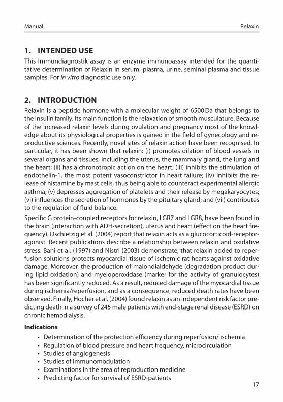
1. INTENDED USEThis Immundiagnostik assay is an enzyme immunoassay intended for the quanti-tative determination of Relaxin in serum, plasma, urine, seminal plasma and tissue samples. For in_vitro_diagnostic use only.
2. INTRODUCTIONRelaxin is a peptide hormone with a molecular weight of 6500 Da that belongs to the insulin family. Its main function is the relaxation of smooth musculature. Because of the increased relaxin levels during ovulation and pregnancy most of the knowl-edge about its physiological properties is gained in the field of gynecology and re-productive sciences. Recently, novel sites of relaxin action have been recognised. In particular, it has been shown that relaxin: (i) promotes dilation of blood vessels in several organs and tissues, including the uterus, the mammary gland, the lung and the heart; (ii) has a chronotropic action on the heart; (iii) inhibits the stimulation of endothelin-1, the most potent vasoconstrictor in heart failure; (iv) inhibits the re-lease of histamine by mast cells, thus being able to counteract experimental allergic asthma; (v) depresses aggregation of platelets and their release by megakaryocytes; (vi) influences the secretion of hormones by the pituitary gland; and (vii) contributes to the regulation of fluid balance.Specific G protein-coupled receptors for relaxin, LGR7 and LGR8, have been found in the brain (interaction with ADH-secretion), uterus and heart (effect on the heart fre-quency). Dschietzig et al. (2004) report that relaxin acts as a glucocorticoid-receptor-agonist. Recent publications describe a relationship between relaxin and oxidative stress. Bani et al. (1997) and Nistri (2003) demonstrate, that relaxin added to reper-fusion solutions protects myocardial tissue of ischemic rat hearts against oxidative damage. Moreover, the production of malondialdehyde (degradation product dur-ing lipid oxidation) and myeloperoxidase (marker for the activity of granulocytes) has been significantly reduced. As a result, reduced damage of the myocardial tissue during ischemia/reperfusion, and as a consequence, reduced death rates have been observed. Finally, Hocher et al. (2004) found relaxin as an independent risk factor pre-dicting death in a survey of 245 male patients with end-stage renal disease (ESRD) on chronic hemodialysis.
Indications
• Determination of the protection efficiency during reperfusion/ ischemia• Regulation of blood pressure and heart frequency, microcirculation• Studies of angiogenesis• Studies of immunomodulation• Examinations in the area of reproduction medicine• Predicting factor for survival of ESRD-patients
Manual Relaxin
18
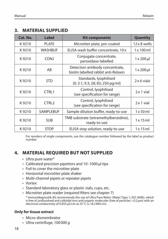
3. MATERIAL SUPPLIED
Cat. No. Label Kit components Quantity
K 9210 PLATE Microtiter plate, pre-coated 12 x 8 wells
K 9210 WASHBUF ELISA wash buffer concentrate, 10 x 1 x 100 ml
K 9210 CONJ Conjugate concentrate, peroxidase-labelled 1 x 200 µl
K 9210 AB Detection antibody concentrate, biotin labelled rabbit anti-Relaxin 1 x 200 µl
K 9210 STD Standards, lyophilised (0; 3.1; 9.3; 28; 83; 250 pg/ml) 2 x 6 vials
K 9210 CTRL1 Control, lyophilised (see specification for range) 2 x 1 vial
K 9210 CTRL2 Control, lyophilised (see specification for range) 2 x 1 vial
K 9210 SAMPLEBUF Sample dilution buffer, ready-to-use 1 x 50 ml
K 9210 SUB TMB substrate (tetramethylbenzidine), ready-to-use 1 x 15 ml
K 9210 STOP ELISA stop solution, ready-to-use 1 x 15 ml
For reorders of single components, use the catalogue number followed by the label as product number.
4. MATERIAL REQUIRED BUT NOT SUPPLIED• Ultra pure water*• Calibrated precision pipettors and 10–1000 µl tips• Foil to cover the microtiter plate• Horizontal microtiter plate shaker• Multi-channel pipets or repeater pipets• Vortex• Standard laboratory glass or plastic vials, cups, etc. • Microtiter plate reader (required filters see chapter 7)
* Immundiagnostik AG recommends the use of Ultra Pure Water (Water Type 1; ISO 3696), which is free of undissolved and colloidal ions and organic molecules (free of particles > 0.2 µm) with an electrical conductivity of 0.055 µS/cm at 25 °C (≥ 18.2 MΩ cm).
Only for tissue extract
• Micro-dismembrator • Ultra-centrifuge, 100 000 g
19
Manual Relaxin
5. STORAGE AND PREPARATION OF REAGENTS
• To run the assay more than once, ensure that reagents are stored at the condi-tions stated on the label. Prepare only the appropriate amount necessary for each run. The kit can be used up to 4 times within the expiry date stated on the label.
• Reagents with a volume less than 100 µl should be centrifuged before use to avoid loss of volume.
• Preparation of the wash buffer: The wash buffer concentrate (WASHBUF) has to be diluted with ultra pure water 1:10 before use (100 ml WASHBUF + 900 ml ultra pure water), mix well. Crystals could occur due to high salt con-centration in the concentrate. Before dilution, the crystals have to be redis-solved at room temperature or in a water bath at 37 °C. The WASHBUF is stable at 2–8 °C until the expiry date stated on the label. Wash buffer (1:10 diluted WASHBUF) can be stored in a closed flask at 2–8 °C for 1 month.
• The lyophilised standards (STD) and controls (CTRL) are stable at 2–8 °C un-til the expiry date stated on the label. Before use, the STDs and CTRLs have to be reconstituted with 500 µl of ultra pure water. Allow the vial content to dissolve for 10 minutes and mix thoroughly to ensure complete reconstituti-on. Standards and controls (reconstituted STDs and CTRLs) can be stored at 2–8 °C for 4 weeks.
• Preparation of the detection antibody: Before use, the detection antibody concentrate (AB) has to be diluted 1:1001 in wash buffer (10 µl AB + 10 ml wash buffer). The AB is stable at 2–8 °C until the expiry date stated on the label. Detection antibody (1:1001 diluted AB) is not stable and cannot be stored.
• Preparation of the conjugate: Before use, the conjugate concentrate (CONJ) has to be diluted 1:1001 in wash buffer (10 µl CONJ + 10 ml wash buffer). The CONJ is stable at 2–8 °C until the expiry date stated on the label. Conjugate (1:1001 diluted CONJ) is not stable and cannot be stored.
• All other test reagents are ready-to-use. Test reagents are stable until the ex-piry date (see label of test package) when stored at 2–8 °C.
Manual Relaxin
20
6. STORAGE AND PREPARATION OF SAMPLES
Sample_storageSerum, plasma, urine, seminal plasma and tissue can be stored at -20 °C.
Sample_preparationSerum and plasma
EDTA plasma or serum samples must be diluted at least 1:3 before performing the assay, e.g. 100 µl sample + 200 µl dilution buffer (SAMPLEBUF), mix well. For testing in duplicates, pipette 2 x 100 µl of each prepared sample per well.Serum and plasma samples could contain rheumatoid factor and heterophilic anti-bodies, which can cause false positive results in sandwich immunoassays. To reduce the potential interference from rheumatoid factor and heterophylic antibodies, the samples can be cleared by treating twice with 5 % (v/v) Anti Interference Reagent (Immundiagnostik Catalog number K 9212) as follows:
• 10 µl anti interference reagent + 200 µl sample• shake for 1 hour at 4 °C • centrifuge and collect the supernatant (pre-cleared/pre-treated sample).
Urine
Urine samples must be diluted at least 1:4 before performing the assay, e.g. 100 µl sample + 300 µl dilution buffer (SAMPLEBUF), mix well. For testing in duplicates, pipette 2 x 100 µl of each prepared sample per well.
Seminal plasma
Seminal plasma must be diluted at least 1:10 before performing the assay, e.g. 10 µl sample + 1990 µl dilution buffer (SAMPLEBUF), mix well. For testing in duplicates, pipette 2 x 100 µl of each prepared sample per well.
Tissue extract
• Pulverise about 200 mg of deep frozen tissue sample in a pre-frozen shaking holder of a micro-dismembrator (30 s/1500 rpm).
• Homogenise the powder in 1 ml of phosphate buffer(0,14 M NaCl, 2.6 mM KCl, 8 mM Na2HPO4, 1.4 mM KH2PO4, 1 % Triton-X 100, pH 7.4 ). After ultra-centrif-ugation (1 h/100 000 g), the protein concentration should be determined in the supernatant by the commercially available Pierce-BCA or Peterson-Lowry Protein Assay.
• For testing in duplicates, pipette 2 x 100 µl of each supernatant per well.
21
Manual Relaxin
7. ASSAY PROCEDURE
Principle_of_the_testThis ELISA is designed for the quantitative determination of relaxin. The assay utilises the “sandwich” technique with two selected polyclonal antibodies that bind to hu-man Realxin.Assay standards, controls and pre-diluted patient samples which are assayed for human Relaxin are added into the wells of a microplate coated with a high affine polyclonal anti-human Relaxin antibody. During the first incubation step, Relaxin is bound by the immobilised antibody. Then a detection antibody, biotin-labelled anti Relaxin, is added. Afterwards a peroxidase-conjugate is added into each microtiter well and a “sandwich” of capture antibody - human Relaxin - detection antibody– peroxidase-conjugate is formed. Tetramethylbenzidine (TMB) is used as peroxidase substrate. Finally, an acidic stop solution is added to terminate the reaction. The col-our changes from blue to yellow. The intensity of the yellow colour is directly pro-portional to the concentration of Relaxin. A dose response curve of the absorbance unit (optical density, OD at 450 nm) vs. concentration is generated using the values obtained from the standard. Relaxin present in the patient samples is determined directly from this curve.
Test_procedureBring all reagents and samples to room temperature (15–30 °C) and mix well.Mark the positions of standards/controls/samples on a protocol sheet. We recom- We recom-mend a pretreatment of plasma and serum samples with the Immundiagnostik Anti Interference Reagent (Catalog number K 9212) prior to analysis. Take as many microtiter strips as needed from the kit. Store unused strips covered at 2–8 ° C. Strips are stable until expiry date stated on the label.For automated ELISA processors, the given protocol may need to be adjusted accord-ing to the specific features of the respective automated platform. For further details please contact your supplier or Immundiagnostik AG.We recommend to carry out the tests in duplicate.
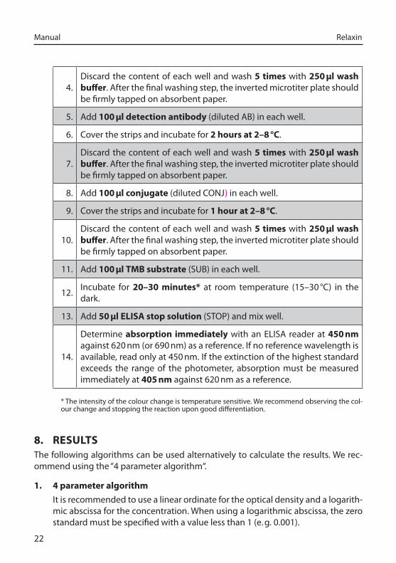
1.Wash each well 5 times with 250 µl wash buffer. After the final wash-ing step, the inverted microtiter plate should be firmly tapped on ab-sorbent paper.
2. Add each 100 µl standards/controls/samples into the respective wells.
3. Cover the strips and incubate for over night (16-22 hours) at 2–8 °C.
Manual Relaxin
22
4.Discard the content of each well and wash 5 times with 250 µl wash buffer. After the final washing step, the inverted microtiter plate should be firmly tapped on absorbent paper.
5. Add 100 µl detection antibody (diluted AB) in each well.
6. Cover the strips and incubate for 2 hours at 2–8 °C.
7.Discard the content of each well and wash 5 times with 250 µl wash buffer. After the final washing step, the inverted microtiter plate should be firmly tapped on absorbent paper.
8. Add 100 µl conjugate (diluted CONJ) in each well.
9. Cover the strips and incubate for 1 hour at 2–8 °C.
10.Discard the content of each well and wash 5 times with 250 µl wash buffer. After the final washing step, the inverted microtiter plate should be firmly tapped on absorbent paper.
11. Add 100 µl TMB substrate (SUB) in each well.
12. Incubate for 20–30 minutes* at room temperature (15–30 °C) in the dark.
13. Add 50 µl ELISA stop solution (STOP) and mix well.
14.
Determine absorption immediately with an ELISA reader at 450 nm against 620 nm (or 690 nm) as a reference. If no reference wavelength is available, read only at 450 nm. If the extinction of the highest standard exceeds the range of the photometer, absorption must be measured immediately at 405 nm against 620 nm as a reference.
* The intensity of the colour change is temperature sensitive. We recommend observing the col-our change and stopping the reaction upon good differentiation.
8. RESULTSThe following algorithms can be used alternatively to calculate the results. We rec-ommend using the “4 parameter algorithm“.
1. 4 parameter algorithm
It is recommended to use a linear ordinate for the optical density and a logarith-mic abscissa for the concentration. When using a logarithmic abscissa, the zero standard must be specified with a value less than 1 (e. g. 0.001).
23
Manual Relaxin
2. Point-to-point calculation
We recommend a linear ordinate for the optical density and a linear abscissa for the concentration.
3. Spline algorithm
We recommend a linear ordinate for the optical density and a linear abscissa for the concentration.
The plausibility of the duplicate values should be examined before the automatic evaluation of the results. If this option is not available with the programme used, the duplicate values should be evaluated manually.Serum and plasma samples
The obtained results have to be multiplied with the dilution factor of 3 to get the actual concentrations.In case another dilution factor has been used, multiply the obtained result with the dilution factor used.Urine samples
The obtained results have to be multiplied with the dilution factor of 4 to get the actual concentrations.In case another dilution factor has been used, multiply the obtained result with the dilution factor used. Seminal plasma samples
The obtained results have to be multiplied with the dilution factor of 10 to get the actual concentrations.In case another dilution factor has been used, multiply the obtained result with the dilution factor used. Tissue extract
The obtained results have to be multiplied with the used dilution factor to get the actual concentrations.
9. LIMITATIONS
Samples with concentrations above the measurement range (see definition below) must be further diluted and re-assayed. Please consider this greater dilution when calculating the results.
Samples with concentrations lower than the measurement range (see definition be-low) cannot be clearly quantified.
Manual Relaxin
24
The upper limit of the measurement range can be calculated as:highest_concentration_of_the_standard_curve × sample_dilution_factor_to_be_used
The lower limit of the measurement range can be calculated as:Analytical_sensitivity × sample_dilution_factor_to_be_used
10. QUALITY CONTROLImmundiagnostik recommends the use of external controls for internal quality con-trol, if possible.Control samples should be analysed with each run. Results, generated from the analysis of control samples, should be evaluated for acceptability using appropriate statistical methods. The results for the patient samples may not be valid if within the same assay one or more values of the quality control sample are outside the accept-able limits.
Reference_rangeWe recommend each laboratory to establish its own reference range.
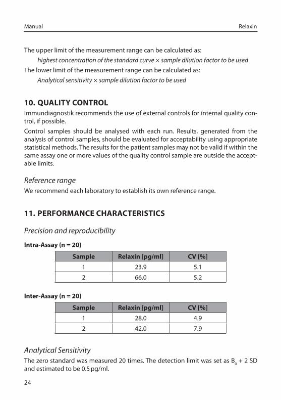
11. PERFORMANCE CHARACTERISTICS
Precision_and_reproducibility
Intra-Assay (n = 20)
Sample Relaxin [pg/ml] CV [%]
1 23.9 5.1
2 66.0 5.2
Inter-Assay (n = 20)
Sample Relaxin [pg/ml] CV [%]
1 28.0 4.9
2 42.0 7.9
Analytical_SensitivityThe zero standard was measured 20 times. The detection limit was set as B0 + 2 SD and estimated to be 0.5 pg/ml.
25
Manual Relaxin
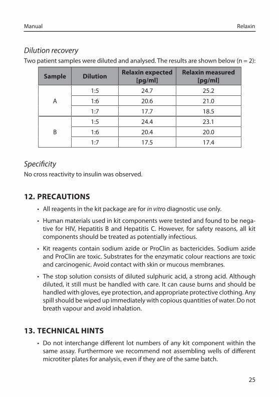
Dilution_recoveryTwo patient samples were diluted and analysed. The results are shown below (n = 2):
Sample DilutionRelaxin expected
[pg/ml]Relaxin measured
[pg/ml]
A
1:5 24.7 25.2
1:6 20.6 21.0
1:7 17.7 18.5
B
1:5 24.4 23.1
1:6 20.4 20.0
1:7 17.5 17.4
SpecificityNo cross reactivity to insulin was observed.
12. PRECAUTIONS
• All reagents in the kit package are for in_vitro diagnostic use only.
• Human materials used in kit components were tested and found to be nega-tive for HIV, Hepatitis B and Hepatitis C. However, for safety reasons, all kit components should be treated as potentially infectious.
• Kit reagents contain sodium azide or ProClin as bactericides. Sodium azide and ProClin are toxic. Substrates for the enzymatic colour reactions are toxic and carcinogenic. Avoid contact with skin or mucous membranes.
• The stop solution consists of diluted sulphuric acid, a strong acid. Although diluted, it still must be handled with care. It can cause burns and should be handled with gloves, eye protection, and appropriate protective clothing. Any spill should be wiped up immediately with copious quantities of water. Do not breath vapour and avoid inhalation.
13. TECHNICAL HINTS
• Do not interchange different lot numbers of any kit component within the same assay. Furthermore we recommend not assembling wells of different microtiter plates for analysis, even if they are of the same batch.
Manual Relaxin
26
• Control samples should be analysed with each run.
• Reagents should not be used beyond the expiration date stated on the kit label.
• Substrate solution should remain colourless until use.
• To ensure accurate results, proper adhesion of plate sealers during incubation steps is necessary.
• Avoid foaming when mixing reagents.
• Do not mix plugs and caps from different reagents.
• The assay should always be performed according to the enclosed manual.
14. GENERAL NOTES ON THE TEST AND TEST PROCEDURE
• This assay was produced and distributed according to the IVD guidelines of 98/79/EC.
• The guidelines for medical laboratories should be followed.
• Incubation time, incubation temperature and pipetting volumes of the com-ponents are defined by the producer. Any variation of the test procedure, which is not coordinated with the producer, may influence the results of the test. Immundiagnostik AG can therefore not be held responsible for any dam-age resulting from incorrect use.
• Warranty claims and complaints regarding deficiencies must be logged with-in 14 days after receipt of the product. The product should be send to Immun-diagnostik AG along with a written complaint.
15. REFERENCES1. Armbruster et al. (2001) Eur_J_Med_Res 6:1-9
2. Armbruster et al. (2001) Proceed_third_Intern_Conference_on_Relaxin_&_Relates_Pep-tides, 2-27 October 2000, Broome, Australia , 273-274. Netherlands, Kluwer Aca-demic Publishers. 2-10-200
3. Bani D (1997) Gen._Pharmac. 28:13-22
4. Bani D et al. (1998)_A._J._Patholoy 152:1367-1375
5. Nistri S et al. (2003) FASEB_J_17 (14) 2109-2111
6. Dschietzig R, Stangl K (2002) CMLS 59: 1-13 (Review)
27
Manual Relaxin
7. Dschietzig et al. (2004) Abstract_of_Fourth_Intern_Conference_on_Relaxin_&_Related_Peptides, September 5-10, Jackson Hole, USA
8. Hocher B et al. (2004) Circulation 109: 2266-2268

Immundiagnostik AG
Stubenwald-Allee 8a D-64625 Bensheim
Tel.: +49 (0) 62 51/70 19 00 Fax: +49 (0) 62 51/84 94 30
[email protected] www.immundiagnostik.com