Research Center Borstel - zhb.uni-luebeck.de · Danksagung Die vorliegende Arbeit wurde am...
107
-
Upload
nguyenthuan -
Category
Documents
-
view
220 -
download
0
Transcript of Research Center Borstel - zhb.uni-luebeck.de · Danksagung Die vorliegende Arbeit wurde am...

Research Center BorstelDivision of Pulmonary Pharmacology
Mechanisms of bronchoconstriction in the early allergic response
Dissertation
zur Erlangung des akademischen Grades desDoktors der Naturwissenschaft en
an der Universität zu Lübeck- Aus der Technisch- Naturwissenschaft lichen Fakultät -
vorgelegt von
Anna-Rebekka Reßmeyeraus Kiel
im April 2006

Meinen Eltern
Danksagung
Die vorliegende Arbeit wurde am Forschungszentrum Borstel, Abteilung Immunchemie und Biochemische Mikrobiologie, in der Laborgruppe Lungenpharmakologie angefertigt.Für das hervorragende Forschungsumfeld in Borstel und die gute Unterstützung möchte ich mich ganz herzlich bei Prof. Dr. Ernst Th . Rietschel bedanken. Ein besonderer Dank gilt Prof. Dr. Stefan Uhlig für die Aufnahme in seine Laborgruppe, seine ständige Diskussionsbereitschaft , die Anregungen und Ideen, sowie die Schaff ung großartiger Arbeitsbedingungen.Dr. Christian Martin gebührt ein sehr großer Dank für die Einarbeitung in die Methode der Lungenschnitte, für sein unermüdliches Engagement, die ständige Bereitschaft Fragen zu diskutieren, seine Ideen und die stete Unterstützung. Das alles hat entscheidend zum Gelingen der Arbeit beigetragen.Prof. Dr. Stefan Uhlig und Prof. Jelkmann sowie dem Graduiertenkolleg 288 sei ganz herzlich gedankt für das Stipendium und die Möglichkeit an internationalen Fachkongressen teilzunehmen. Für eine sehr gute Zusammenarbeit mit der Pathologie in Borstel möchte ich mich bei Prof. Dr. Dr. E. Vollmer bedanken, ohne ihn und sein Team wäre die Präparation der humanen Lungenschnitte nicht möglich gewesen.Für eine erfolgreiche Kooperation und Bereitstellung der Primatenlungen möchte ich mich bei Dr. A. Braun und Dr. K. Sewald aus Hannover bedanken.Ebenso bedanken möchte ich mich für eine sehr gute Zusammenarbeit bei A.K. Larsson und S.-E. Dahlén aus Stockholm. Jürgen Sarau hat mich nicht nur in technischer Hinsicht die gesamte Zeit meiner Promotion hervorragend unterstützt, sondern auch die Höhen und Tiefen eines Wissenschaft salltags mit mir durchlebt. Vielen, vielen Dank dafür. Durch seine fröhliche Art hat das Arbeiten immer sehr viel Spaß gemacht. Ich denke gerne an die Zeit im Keller!Dörte, Christina, Stephie, Solveig, Heike, und Constanze, vielen Dank für Eure Freundschaft . Die Zeit mit Euch in Borstel wird unvergessen bleiben. Weiterhin möchte ich meinen Freunden danken, die in dieser Zeit oft auf mich verzichten mußten, und mich doch nicht vergessen haben (Doris), aber ganz besonders Merle, die zu jeder Zeit ein off enes Ohr für mich hat. Alex, Jörg, René, schade, daß ich Euch erst so spät kennengelernt habe, die Abende mit Euch hier in Borstel waren wirklich sehr lustig.Bei meinen Eltern und meiner Familie möchte ich mich bedanken für ihre stete Unterstützung. Ich weiß, ich kann immer auf Euch zählen.Lisa und allen anderen “Kühls” möchte ich danken für die immer off ene Tür, und die aufmunternden Worte.Mein größter Dank gilt jedoch Hauke, der immer hinter mir steht, mir den Rücken stärkt, und mit dem ich hier eine wirklich schöne Zeit hatte.
Publications contributing to this study
Martin C.,Göggel R., Ressmeyer A.-R., and Uhlig S. Pressor responses to platelet-activating fac-tor and thromboxane are mediated by Rho-kinase. Am J Physiol Lung Cell Mol Physiol 287: L250-L257, 2004
Ressmeyer A.-R., Larsson A.K., Vollmer E., Dahlén S.-E., Uhlig S., and Martin C. Characteri-zation of guinea pig precision-cut lung slices (PCLS): Comparison with human tissue. Eur Respir J. 2006 under revision.
Abstracts
Ressmeyer AR., Larsson AK., Dahlén SE., Uhlig S., Martin C. (2005): Th e early allergic airway response in precision-cut lung slices. Proceedings Am Th or Soc 2:A82
Martin C., Ressmeyer AR., Uhlig S. (2005): Th e role of Rho-kinase and calcium in early allergic response in precision-cut lung slices (PCLS). Proceedings Am Th or Soc 2: A83
Ressmeyer AR., Uhlig S., Martin C. (2005) Characterization of guinea pig precision-cut lung sli-ces (PCLS). Proceedings Am Th or Soc 2: A76
Martin C., Ressmeyer AR., Uhlig S. (2004): Airway hypo-reactivity of pseudomonas aeruginosa infected precision-cut lung slices (PCLS) Am J Respir Crit Care Med 169 (7): A588
Martin C., Goeggel R., Ressmeyer AR., Uhlig S. (2004): Contribution of IP3 and Rho-kinase to platelet-activating factor-induced pressor responses. Am J Respir Crit Care Med 169 (7): A413
Table of contents
- I -
Table of contents
1 Introduction 11.1 Asthma bronchiale 1
1.1.1 Epidemiology of asthma 1
1.1.2 Defi nition of asthma 1
1.1.3 Types of asthmatic responses aft er allergen exposure 1
1.1.3.1 Th e early allergic response (EAR) 2
1.1.3.2 Th e late phase response (LPR) 2
1.1.3.3 Airway hyperresponsiveness (AHR) 2
1.1.3.4 Airway remodeling 3
1.2 Infl ammatory cells and mediators in asthma 3
1.2.1 Dendritic cells 3
1.2.2 Macrophages 3
1.2.3 T lymphocytes 4
1.2.4 B lymphocytes 5
1.2.5 Mast cells and its mediators 5
1.2.5.1 Preformed mediators 6
1.2.5.2 Newly synthesized mediators 6
1.2.5.3 Cytokines 7
1.2.6 Eosinophils 9
1.2.7 Neutrophils 9
1.2.8 Basophils 9
1.3 Signal transduction pathways of airway smooth muscle contraction 10
1.3.1 Ca2+-dependent signal transduction pathway 11
1.3.1.1 Ca2+ channels in the plasma membrane 11
1.3.2 Ca2+ sensitization mechanism 12
1.4 Mechanism of airway relaxation 12
1.5 Models of asthma 13
Table of contents
- II -
1.5.1 Limitations to study asthma in humans 13
1.5.2 Animal models 14
1.5.2.1 Animals with naturally occurring recurrent airway obstruction 14
1.5.2.2 Animals showing AHR without provocation 14
1.5.2.3 Models of airway obstruction and AHR aft er allergen challenge 15
1.5.2.4 In vitro models in small mammalian species 18
1.5.2.5 Th e model of precision-cut lung slices (PCLS) 18
2 Aim of the study 203 Material and Methods 213.1 Animals 21
3.1.1 Rats 21
3.1.2 Mice 21
3.1.3 Guinea pigs 22
3.1.4 Monkeys 22
3.1.5 Pentobarbital solution 22
3.2 Human lung donors 22
3.3 Chemicals 24
3.4 Equipment 25
3.5 Methods 26
3.5.1 Precision -cut lung slices (PCLS) 26
3.5.1.1 Preparation of rat lung slices 26
3.5.1.2 Preparation of mouse lung slices 26
3.5.1.3 Preparation of guinea pig lung slices 26
3.5.1.4 Preparation of human lung slices 27
3.5.2 Culture medium 28
3.5.2.1 Incubation medium 28
3.5.2.2 Agarose solution 28
3.5.2.3 Slicing medium 28
3.5.3 Viability of the PCLS 29
Table of contents
- III -
3.5.3.1 Lactate dehydrogenase (LDH) release 29
3.5.3.2 Two-photon microscopy 29
3.5.4 Measurement and Imaging of broncho - or vasoconstriction 30
3.5.5 Passive-sensitization as a tool to study the EAR 30
3.5.5.1 Rat serum 30
3.5.5.2 Mouse serum 30
3.5.5.3 Guinea pig serum 31
3.5.5.4 Human serum 31
3.5.6 Pharmacological intervention studies 31
3.5.6.1 Mediators/ agonists and antagonists 31
3.5.6.2 Inhibitors of the signal transduction pathways 32
3.5.7 Airway relaxation in the guinea pig 32
3.5.8 Calcium imaging with two-photon microscopy 32
3.6 Statistics 33
4 Results 344.1 Part Ia: Establishment of guinea pig (GP) PCLS 34
4.1.1 Viability of GP PCLS 34
4.1.1.1 Lactate dehydrogenase-release 34
4.1.1.2 Two-photon microscopy 35
4.1.2 Mediator-induced bronchoconstriction in GP PCLS 35
4.1.3 Relaxation of precontracted airways 36
4.1.4 Airway and vascular responses within one slice 37
4.1.5 Induction of the EAR in GP PCLS 38
4.2 Part Ib: Data from monkey PCLS 38
4.2.1 Viability of monkey PCLS 38
4.2.2 Airway responses of monkey PCLS 39
4.3 Part II: Th e EAR in diff erent species 40
4.3.1 C48/80 40
Table of contents
- IV -
4.3.1.1 C48/80-induced bronchoconstriction 40
4.3.1.2 Viability of C48/80-treated PCLS 41
4.3.1.3 Mediators of the EAR induced by C48/80 42
4.3.2.1 Mediators of the allergen-induced EAR in rat PCLS 46
4.3.2.2 Mediators of the allergen-induced EAR in human PCLS 47
4.3.2.3 Mediators in the allergen-induced EAR in GP PCLS 48
4.3.3 Active sensitization 50
4.3.3.1 Airway hyperresponsiveness 51
4.3.3.1.1 Mice: Airway hyperresponsiveness 51
4.3.3.1.2 Rats: Airway hyperresponsiveness 52
4.4 Part III: Mediator signal cascades during the EAR 53
4.4.1 Signaling in human PCLS 53
4.4.2 Signaling in GP PCLS 55
4.4.3 Mediator-induced bronchoconstriction in murine PCLS 56
4.4.3.1 Airway responses in Maxi-K-channel knockout mice 57
4.4.4 Signaling in rat PCLS 58
5 Discussion 635.1 Th e model of guinea pig PCLS 63
5.2 Th e early allergic response in PCLS 65
5.3 PCLS from non-human primates 67
5.4 Mast cell degranulation 68
5.5 Airway hyperresponsiveness in PCLS 69
5.6 Mechanisms of bronchoconstriction 70
5.7 Role of the Maxi-K channel in bronchoconstriction 73
6 Summary 757 Deutsche Zusammenfassung 778 Reference List 79
- V -
Abbreviations
AHR airway hyperresponsiveness
BAL bronchoalveolar lavage
C48/80 Compound 48/80
COX cyclooxygenase
CTMC connective tissue-type mast cell
DAG diacylglycerol
ELISA enzyme-linked immunosorbant assay
EAR early allergic response
EC50 concentration leading to half maximal eff ect
EP-receptor prostaglandin E-receptor
ET-1 endothelin-1
GM-CSF granulocyte monocyte colony stimulating factor
GP guinea pig
5-HT2-receptor 5-hydroxytryptamine2 (serotonin)-receptor
IP3 inositol 1,4,5 trisphosphate
LDH lactate dehydrogenase
LPR late phase response
LTD4 leukotriene D4
MLC20 myosin light chain 20 kDA
MLCK myosin light chain kinase
MLCP Myosin light chain phosphatase
MCT mast cell containing only tryptase
MCTC mast cell containing both: tryptase and chymase
MMC mucosal-type mast cell
Ova ovalbumin
PAF platlet-activating factor
PCLS precision-cut lung slices
PDE phosphodiesterase
PE pollen extract
PGD2 prostaglandin D2
PI-PLC phosphatidylinositol specifi c phospholipase C
ROCC receptor-operated Ca2+-channel
ROCK Rho-kinase
SOCC store-operated Ca2+-channel
TP-receptor thromboxane-prostanoid receptor
VOCC voltage-operated Ca2+-channel
1. Introduction
- 1 -
1 Introduction
1.1 Asthma bronchiale
1.1.1 Epidemiology of asthma
Asthma bronchiale is one of the most important chronic diseases which occurs in 10% of the children and 5% of the adult population in Germany [1]. It is the most common chronic disease in infancy. 64% of german asthmatic individuals suff er from mild, 34% from moderate asthma, and 2% are aff ected by severe asthma. As most of the individuals receive medication, the costs increase exponentially with the degree of asthma. About 2 billion € are spent every year for the treatment and medication of the approximately 4 million asthmatics in Germany [2]. In central Europe one person out of 100 000 dies because of asthma [2]. Genetic predispositions are an important factor for the development of atopy, and several genes have now been identifi ed [3]. In addition, environmental factors appear to be important in determining whether asthma develops in an atopic individual [2].
1.1.2 Defi nition of asthma
Asthma is defi ned on three levels: physiologically, pathologically, and clinically. Physiologically, asthma is an airway disease, characterized by airway obstructions with airfl ow limitations that are variable and reversible either spontaneously or with treatment, accompanied with an unspecifi c airway hyperresponsiveness [4]. Central to its development is a chronic infl ammation of the airways. Pathologically, asthma is defi ned by multiple abnormalities in airway epithelium, lamina propria and submucosa [5]. Many diff erent cell types play a role in this infl ammatory disorder of the airways, in particular mast cells, eosinophils and T lymphocytes. Clinically, this infl ammation causes recurrent episodes of wheezing, breathlessness, chest thightness, and cough, particularly at night or in the early morning in susceptible individuals. Various forms of asthma exist, e.g. extrinsic (allergic) asthma, intrinsic asthma, exercise-induced asthma, aspirin-sensitive asthma and occupational asthma, demonstrating that asthma is a very heterogeneous lung disease.
1.1.3 Types of asthmatic responses after allergen exposure
Apart from various forms of asthma, there are cardinal symptoms that are: the early allergic response, the late phase response, airway hyperresponsiveness, and airway remodeling.
1. Introduction
- 2 -
1.1.3.1 The early allergic response (EAR)
In the lungs, the immediate response to allergens occurs within minutes, is triggered by an exposure to inhaled antigens or irritants and results in bronchoconstriction [6]. It is caused by specifi c allergens that cross-link IgE-molecules bound to the surface of mast cells. Aft er this activation the mast cells degranulate and release a number of infl ammatory mediators. Some mediators are preformed and stored in mast cells`cytoplasmatic granules, i.e. histamine, proteoglycans and serine proteases. Other mediators, e.g. certain lipid-derived substances including prostaglandins, cysteinyl leukotrienes, thromboxane and platelet-activating factor (PAF) are synthesized de novo aft er mast cell activation [7]. Histamine, prostaglandins, thromboxane, cysteinyl leukotrienes and PAF then cause an airway smooth muscle contraction, thereby narrowing the airways.
1.1.3.2 The late phase response (LPR)
Th e early allergic response is followed 4 - 6 hours later by a late phase response, which is defi ned by a second contraction of the airways and by infi ltration of eosinophils, activated neutrophils, mast cells, T lymphocytes, and leukocytes into the tissue [6,8]. Th ere is evidence from both clinical and animal studies that mast cell-derived mediators, in particular TNFα but also other cytokines, are responsible for the leukocyte recruitment [9-11]. Th e interaction of complementary adhesion molecules, expressed on the surface of leukocytes (e.g. VLA-4, CD11/18) and on vascular endothelial cells (e.g. VCAM-1, ICAM-1 and E-selectin (ELAM)), as well as the action of chemokines, contribute to leukocyte emigration into the lung during the late phase response [12-14]. Th ese molecules are part of a complex mechanistic interplay in vivo and products from activated mast cells and many other cell types [13,14].
1.1.3.3 Airway hyperresponsiveness (AHR)
Airway hyperresponsiveness is an exaggerated airway narrowing in response to a variety of unspecifi c stimuli and an important characteristic of bronchial asthma. AHR can be measured, for example, by a bronchial provocation with histamine or methacholine. Compared to a non-asthmatic individual, the airways of an asthmatic patient respond stronger, indicated by a left shift of the dose-response curve [15-17].
1. Introduction
- 3 -
1.1.3.4 Airway remodeling
Th e term “airway remodeling” refers to structural changes in the airways and is thought to occur as a consequence of chronic airway infl ammation. Th ese structural changes are characterized by a thickening of the airway wall which has been shown in pathologic as well as in radiographic studies [18,19]. Th is airway wall thickening may explain the incomplete reversibility of airway narrowing in asthmatic patients [20]. Hyperplasia of goblet cells coupled with hyperthrophy of submucosal glands and increased vascularity of the airway wall amplify the mucus secretion and plasma protein leakage, which are responsible for the formation of the characteristic mucus plugs that obstruct the airways [21,22]. Furthermore, smooth muscle hypertrophy and hyperplasia are characteristic features in airway remodeling as well as subepithelial fi brosis [23-25].
1.2 Infl ammatory cells and mediators in asthma
In asthma, pulmonary immunity plays an important role with a variety of infl ammatory cell types involved.
1.2.1 Dendritic cells
In the respiratory tract, dendritic cells are localized in the epithelium and act as antigen-presenting cells [26]. Th ey have the capacity to bind allergens, process them into peptides and present them via the major histocompatibility complex II (MHC II) molecules on the cell surface to undiff erentiated T lymphocytes [27]. Th e number of dendritic cells is increased in asthmatic patients [28], and they may initiate and sustain airway infl ammation through enhanced expression of costimulatory molecules that facilitate T-cell activation and diff erentiation [29,30].
1.2.2 Macrophages
Macrophages are derived from blood monocytes. Th ey perform several functions in the immune response. In asthma, they function in uptake, processing and presenting of antigens, and in the secretion of a variety of cytokines, arachidonic acid metabolites and proteases [31-33]. Macrophages may both enhance and diminish infl ammation. Alveolar macrophages normally suppress infl ammatory lymphocyte function, but this may be impaired in asthma aft er allergen exposure [34]. It has been shown that IL-10, which is one of the anti-infl ammatory cytokines secreted by macrophages, is reduced in asthmatics [33]. Th e alveolar macrophages from these individuals are functionally and phenotypically activated [35,36], but up to now it is only poorly understood
1. Introduction
- 4 -
what this activation means for the airway infl ammation in asthma. Th e role of proinfl ammatory cytokines is described in 1.2.5.3.
1.2.3 T lymphocytes
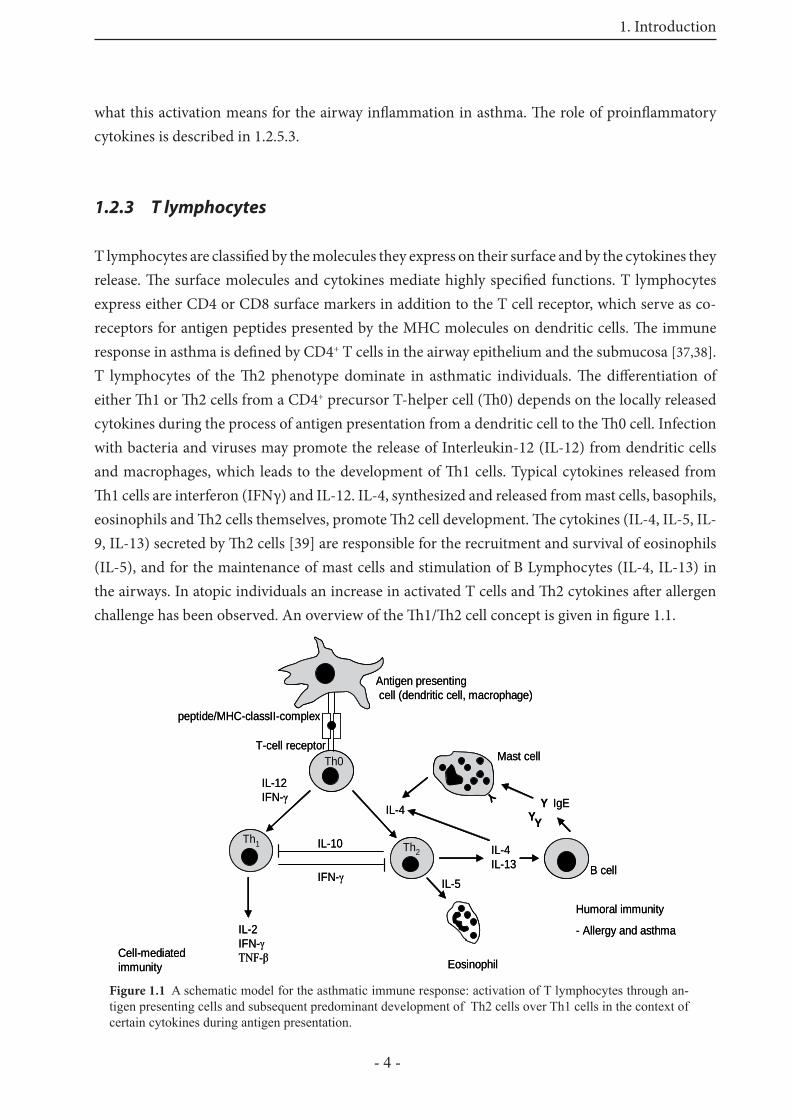
T lymphocytes are classifi ed by the molecules they express on their surface and by the cytokines they release. Th e surface molecules and cytokines mediate highly specifi ed functions. T lymphocytes express either CD4 or CD8 surface markers in addition to the T cell receptor, which serve as co-receptors for antigen peptides presented by the MHC molecules on dendritic cells. Th e immune response in asthma is defi ned by CD4+ T cells in the airway epithelium and the submucosa [37,38]. T lymphocytes of the Th 2 phenotype dominate in asthmatic individuals. Th e diff erentiation of either Th 1 or Th 2 cells from a CD4+ precursor T-helper cell (Th 0) depends on the locally released cytokines during the process of antigen presentation from a dendritic cell to the Th 0 cell. Infection with bacteria and viruses may promote the release of Interleukin-12 (IL-12) from dendritic cells and macrophages, which leads to the development of Th 1 cells. Typical cytokines released from Th 1 cells are interferon (IFNγ) and IL-12. IL-4, synthesized and released from mast cells, basophils, eosinophils and Th 2 cells themselves, promote Th 2 cell development. Th e cytokines (IL-4, IL-5, IL-9, IL-13) secreted by Th 2 cells [39] are responsible for the recruitment and survival of eosinophils (IL-5), and for the maintenance of mast cells and stimulation of B Lymphocytes (IL-4, IL-13) in the airways. In atopic individuals an increase in activated T cells and Th 2 cytokines aft er allergen challenge has been observed. An overview of the Th 1/Th 2 cell concept is given in fi gure 1.1.
Antigen presentingcell (dendritic cell, macrophage)
IL-12IFN-γ
IL-4
IL-5
Mast cell
IFN-γ
IL-10
Eosinophil
IL-4IL-13 B cell
YY
IgEY
Th1 Th2
IL-2IFN-γTNF-β
peptide/MHC-classII-complex
T-cell receptorTh0
Y
Cell-mediatedimmunity
Humoral immunity
- Allergy and asthma
Antigen presentingcell (dendritic cell, macrophage)
IL-12IFN-γ
IL-4
IL-5
Mast cell
IFN-γ
IL-10
Eosinophil
IL-4IL-13 B cell
YY
IgEY
Th1 Th2
IL-2IFN-γTNF-β
peptide/MHC-classII-complex
T-cell receptorTh0
Y
Cell-mediatedimmunity
Humoral immunity
- Allergy and asthma
Figure 1.1 A schematic model for the asthmatic immune response: activation of T lymphocytes through an-tigen presenting cells and subsequent predominant development of Th2 cells over Th1 cells in the context of certain cytokines during antigen presentation.
1. Introduction
- 5 -
1.2.4 B lymphocytes
In allergic diseases, B lymphocytes are responsible for the secretion of IgE molecules [40]. An important player in stimulating B lymphocytes to produce and secrete IgE is IL-4. Immature B-cells express surface IgM molecules, which serves as an antigen receptor. B lymphocytes travel to lung draining lymphnodes, and act as effi cient antigen presenting cells. Peptides of a captured antigen are presented via the MHC II complex to T lymphocytes of the same antigen specifi city. T helper cells then start upregulation of the accessory molecule CD40, which interacts with the CD40 ligand on B cells. Th en B-cells progress from a quiescent state to cell cycle and proliferation [41]. Furthermore T helper cells support the switch from IgM isotype molecules on the surface to IgE or IgG1 isotype.
1.2.5 Mast cells and its mediators
In the human body mast cells are located in proximity to blood and lymphatic vessels in tissues that interface with the environment, like the gastrointestinal tract, the respiratory system or the skin [42]. Increased numbers of mast cells in the lung and enhanced levels of specifi c mast cell-derived mediators in bronchoalveolar lavage (BAL) fl uid have been found in asthmatics [43]. Human mast cells are derived from a CD34+ bone marrow progenitor [44], circulate in the blood and lymphatics and migrate into diff erent tissues. Th ere they start diff erentiation and maturation with diff erential expression of the secretory granule proteases, like chymase, tryptase, carboxypeptidase, and cathepsin-G. Th e heterogeneity of mature mast cells in humans is refl ected by their expression of these diff erent proteases. Mast cells expressing only tryptase (MCT) are found in the lung tissue and in the intestinal mucosa. Th ose containing both tryptase and chymase (MCTC) are found in the skin, lymphnodes and intestinal submucosa [45,46]. Next to these, mast cells lacking tryptase are found [47]. In rodents (rats and mice) two classes of mast cells can be distinguished: connective tissue-type mast cells (CTMC) and mucosal-type mast cells (MMC) [48,49]. Th e quantity of rat or mouse MMC expand clearly during T cell dependent immune responses to certain intestinal parasites. In contrast CTMC are T cell independent [48-51]. Activation of mast cells occurs via high affi nity receptors for IgE (FcεRI), a tetrameric complex, which consists of one α, one β and two identical disulfi de-linked γ-chains expressed on the surface of mature mast cells [7]. When the α-chains of adjacent receptors are cross-linked by a multivalent antigen, the β and the γ chain are phosphorylated, activating membrane associated serine proteases. Th is leads to the activation of several signal transduction pathways [43], which in turn leads to solubilization of granule contents, swelling of the granules, ruffl ing of the membrane, and fusion of the perigranular and plasma membranes for exocytosis of the granule contents [52] (Fig. 1.2).

1. Introduction
- 6 -
1.2.5.1 Preformed mediators
Preformed in granulae are the biogenic amines histamine and 5-HT, as well as the serine endopeptidases tryptase, chymase, cathepsin G, and the carboxypeptidase A. Histamine causes bronchoconstriction, enhances airway mucus production and increases vascular permeability [53]. Tryptase levels are increased in the BAL fl uid from allergen-challenged patients with asthma and are used as a marker for mast cell activation. Tryptase has several functions in vitro, including inactivation of fi brinogen, inhibition of fi brinogenesis, activation of tissue matrix metalloproteinases, inactivation of bronchodilatory neuropeptides, stimulation of fi broblast proliferation, collagen synthesis, eosinophil chemotaxis, and upregulation of the adhesion molecule ICAM-1 expression by bronchial endothelial cells. Th erefore mast cell tryptase may be important in the pathogenesis of asthmatic infl ammation [54-57]. Chymase is also an activator of matrix metalloproteinases. It converts angiotensin I to angiotensin II and inactivates bronchodilatory neuropeptides [58,59]. Cathepsin G leads to endothelial cell injury and stimulates mucus gland secretion [60].
1.2.5.2 Newly synthesized mediators
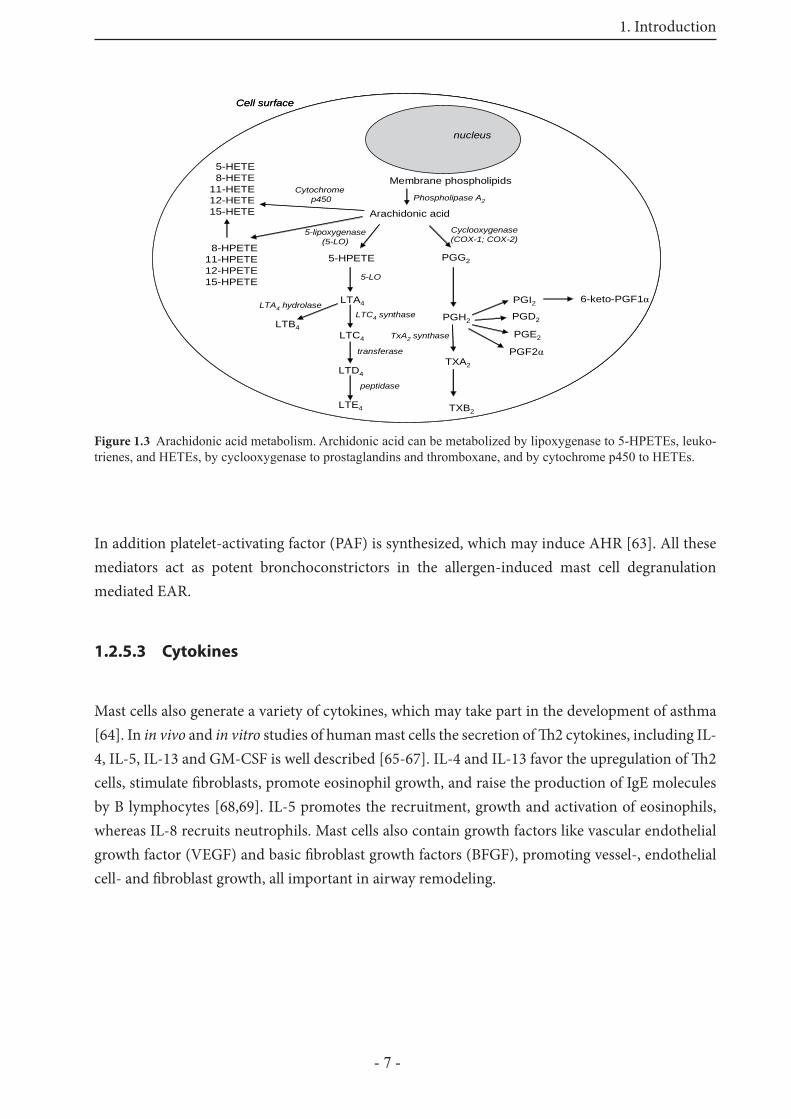
Activation of the FcεRI leads through the trimeric G-protein Ras to the activation of the MAPK pathway [61], which results in translocation of the cytosolic phospholipase A2 (cPLA2) to the membrane compartment. Within the compartment the stimulated cPLA2 mobilizes arachidonic acid from membrane phospholipids to form lipid mediators, such as leukotrienes, prostaglandins, and thromboxane within 2 minutes [62]. Th e synthesis of these lipid mediators is shown in fi gure 1.3.
Figure 1.2 Electronic micrography of a peritoneal rat mast cell before (left side) and after (right side) degranula-tion. From reference 10.
1. Introduction
- 7 -
In addition platelet-activating factor (PAF) is synthesized, which may induce AHR [63]. All these mediators act as potent bronchoconstrictors in the allergen-induced mast cell degranulation mediated EAR.
1.2.5.3 Cytokines
Mast cells also generate a variety of cytokines, which may take part in the development of asthma [64]. In in vivo and in vitro studies of human mast cells the secretion of Th 2 cytokines, including IL-4, IL-5, IL-13 and GM-CSF is well described [65-67]. IL-4 and IL-13 favor the upregulation of Th 2 cells, stimulate fi broblasts, promote eosinophil growth, and raise the production of IgE molecules by B lymphocytes [68,69]. IL-5 promotes the recruitment, growth and activation of eosinophils, whereas IL-8 recruits neutrophils. Mast cells also contain growth factors like vascular endothelial growth factor (VEGF) and basic fi broblast growth factors (BFGF), promoting vessel-, endothelial cell- and fi broblast growth, all important in airway remodeling.
Arachidonic acid
PGG2
PGH2 PGD2
PGF2α
PGI2LTA4
TXA2
6-keto-PGF1α
PGE2LTC4
5-HPETE
5-HETE8-HETE
11-HETE12-HETE15-HETE
8-HPETE11-HPETE12-HPETE15-HPETE
nucleus
TXB2
LTD4
LTE4
Cytochromep450
LTB4
5-lipoxygenase(5-LO)
Cyclooxygenase(COX-1; COX-2)
5-LO
LTC4 synthaseLTA4 hydrolase
Phospholipase A2
Membrane phospholipids
transferase
peptidase
TxA2 synthase
Cell surface
Arachidonic acid
PGG2
PGH2 PGD2
PGF2α
PGI2LTA4
TXA2
6-keto-PGF1α
PGE2LTC4
5-HPETE
5-HETE8-HETE
11-HETE12-HETE15-HETE
8-HPETE11-HPETE12-HPETE15-HPETE
nucleus
TXB2
LTD4
LTE4
Cytochromep450
LTB4
5-lipoxygenase(5-LO)
Cyclooxygenase(COX-1; COX-2)
5-LO
LTC4 synthaseLTA4 hydrolase
Phospholipase A2
Membrane phospholipids
transferase
peptidase
TxA2 synthase
Cell surface
Figure 1.3 Arachidonic acid metabolism. Archidonic acid can be metabolized by lipoxygenase to 5-HPETEs, leuko-trienes, and HETEs, by cyclooxygenase to prostaglandins and thromboxane, and by cytochrome p450 to HETEs.
1. Introduction
- 8 -
Table 1.1 Overview on mast cell mediators from reference [70]BIOGENIC AMINESHistamine increases vascular and endothelial permeability; bronchoconstriction (BC) [71]Serotonin little or none in human mast cells; main mediator of immediate hypersensitivity
reactions in rat and mouse lung [72,73]
GLYCOSAMINOGLYCANS AND PROTEOGLYCANSHeparin acts as local anticoagulant; stabilizes mast cell tryptase; allows packing of cationic
mediators [74,75]Chondroitin sulfate structural role similar to heparin ? [76]
NEUTRAL PROTEASESTryptase bronchoconstriction (induces AHR) [60,77]; cell growth (promotes fi broblast
growth via PAR-2 [78] and smooth muscle growth) [60]; anticoagulation (hydrolyses fi brinogen and activates stromelysin (MMP-3) [60]; connective tissue degradation (hydrolyzes fi brinogen and high molecular weight kiniogen] [60]); Activates pro-urokinase [79]; control of neurogenic infl ammation (inactivates calcitonin gene related peptide [60]).
Chymase stimulates mucus glands secretion [60]; connective tissue degradation (cleaves type IV collagen, laminin, fi bronectin, proteoglycan, glycocalyx [60]); converting enzyme activity (generates extravascular angiotensin II [60]); neurogenic infl ammation (inactivates Substance P [60]; augments histamine-induced vessel leak [60]; activates progelatinase B; liberates latent TGF-ß from extracellular matrix
Cathepsin G degrades extracellular matrix [60]; stimulates mucus glands secretion [60]; endothelial cell injury [60]; antimicrobial defence (kills bacteria noncatalytically) [60]
Carboxypeptidase A degrades peptides in tandem with chymase and cathepsin GTPA activates plasminogen, leading to fi brinolysis [80]Gelatinase A, B degrade extracellular matrix [81]
ACID HYDROLASES
Aryl sulfatase
Glucuronidase
Hexosaminidase
lysosomal enzymes, extracellular roles? [60,82-84]
Cathepsin C (Dipeptidylpeptidase I) actives protryptase and prochymase intracellularly [85,86]
PURINESAdenosine causes weak bronchocondtriction; vasodilation; augments mast cell mediator
release [87]Cytokines, Chemokines and Growth FactorsTNF-α enhances eosinophil cytotoxicity; induces expression of endothelial adhesion
molecules [88]; recruits neutrophils, monocytes and basophils; enhances microvascular permeability; increases bronchial responsiveness
IL-1 enhances IL-9 production in eosinophils [89]IL-4, IL-13 promote IgE production; stimulate fi broblasts; promote eosinophil growth
[68,69,90]IL-5 promotes eosinophil recruitment, growth and activation [69]IL-8 recruits neutrophils [91]MIP-1α recruits leukocytes [91]; induces mediator release from infl ammatory cells;
regulates T-cell binding to adhesion moleculesBFGF promotes growth of fi broblasts, vessels and other cells [92]VEGF promotes endothelial cell and vessel growth [90,93]Tryptase promotes fi broblast and airway smooth muscle growth [60]
LIPID MEDIATORSPGD2 bronchoconstriction; dilates vascular smooth muscle [94,95]LTB4 attracts neutrophils [94,95]LTC4, LTD4, LTE4 bronchoconstriction; increases vascular permeability [94,95] PAF Bronchoconstriction; increases vascular permeability; induces airway
hyperreactivity [94,95]
1. Introduction
- 9 -
1.2.6 Eosinophils
Eosinophil infi ltration is a characteristic feature in asthmatic infl ammation. Before 1916, asthma was termed “chronic eosinophilic bronchitis”. Asthmatics underlying a LPR show an increased number of eosinophils in the BAL, compared to those individuals who experienced only an EAR. Th is demonstrates clearly the accumulation of eosinophils in the airways as part of the LPR in asthma. Eosinophil recruitment to the airways initially involves adhesion of eosinophils to vascular endothelial cells, followed by migration into the submucosa and their subsequent activation. Activated eosinophils release some toxic granule products. Eosinophil cationic protein (ECP), major basic protein (MBP), eosinophil peroxidase (EPO) and eosinophil-derived neurotoxin (EDN) are the four major granule proteins. In parallel, Th 2-like cytokines, oxygen-free radicals, eicosanoids and growth factors are released. Th ese products are able to contract airway smooth muscle [96], induce airway hyperreactivity [97], or increase vascular permeability [98]. Activated eosinophils have been associated with tissue destruction [99].
1.2.7 Neutrophils
Th e general role of neutrophils in asthma is still poorly defi ned. Neutrophils have been found in airways of patients with acute severe asthma [21,100], but are not prominent in patients with mild to moderate asthma [101,102]. In patients who died suddenly because of asthma, large amounts of neutrophils have been found in the airways [103]. Neutrophil elastase, cathepsin G, and proteinase 3 are secreted by neutrophils and are important mediators of goblet cell and submucosal gland cell degranulation [104,105]. Th erefore, neutrophils may play an important role in acute exacerbations by inducing mucin hypersecretion.
1.2.8 Basophils
Basophils can be considered as the circulating form of mast cells, and they can play a role in infl ammatory reactions through recruitment to diff erent tissues sites [106]. Like mast cells, basophils express the high affi nity receptors for IgE (FcεRI). Th ey are also activated by cross-linking of these through an antigen. Secreted mediators of a basophil are histamine (preformd) and leukotriene C4 (newly synthesized), as well as a variety of cytokines, including IL-4, IL-5, IL-6 and granulocyte-monocyte-colony-stimulating-factor (GM-CSF) [106]. Th e number of basophils is also increased in sputum of asthmatics aft er allergen inhalation challenge [107].
1. Introduction
- 10 -
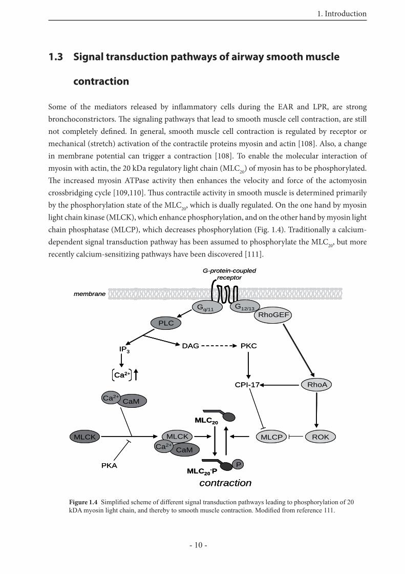
1.3 Signal transduction pathways of airway smooth muscle
contraction
Some of the mediators released by infl ammatory cells during the EAR and LPR, are strong bronchoconstrictors. Th e signaling pathways that lead to smooth muscle cell contraction, are still not completely defi ned. In general, smooth muscle cell contraction is regulated by receptor or mechanical (stretch) activation of the contractile proteins myosin and actin [108]. Also, a change in membrane potential can trigger a contraction [108]. To enable the molecular interaction of myosin with actin, the 20 kDa regulatory light chain (MLC20) of myosin has to be phosphorylated. Th e increased myosin ATPase activity then enhances the velocity and force of the actomyosin crossbridging cycle [109,110]. Th us contractile activity in smooth muscle is determined primarily by the phosphorylation state of the MLC20, which is dually regulated. On the one hand by myosin light chain kinase (MLCK), which enhance phosphorylation, and on the other hand by myosin light chain phosphatase (MLCP), which decreases phosphorylation (Fig. 1.4). Traditionally a calcium-dependent signal transduction pathway has been assumed to phosphorylate the MLC20, but more recently calcium-sensitizing pathways have been discovered [111].
Gq/11
DAG
RhoA
PLC
MLCK
G12/13RhoGEF
ROK
CPI-17
MLC20
MLCP
PKCIP3
PPKAMLC20
-P
CaMCa2+
MLCK
CaMCa2+
Ca2+
contraction
membrane
G-protein-coupledreceptor
Gq/11
DAG
RhoA
PLC
MLCK
G12/13RhoGEF
ROK
CPI-17
MLC20
MLCP
PKCIP3
PPKAMLC20
-P
CaMCa2+
MLCK
CaMCa2+
Ca2+Ca2+
contraction
membrane
G-protein-coupledreceptor
Figure 1.4 Simplifi ed scheme of different signal transduction pathways leading to phosphorylation of 20 kDA myosin light chain, and thereby to smooth muscle contraction. Modifi ed from reference 111.
1. Introduction
- 11 -
1.3.1 Ca2+-dependent signal transduction pathway
Th e initial step in the contractile activation of smooth muscle is an increase in the cytosolic Ca2+ provided by a Ca2+ fl ux into the cytoplasm [109]. Ca2+ can be released from intracellular stores, in particular the sarcoplasmatic reticulum or enter from the extracellular space through Ca2+ channels into the cytoplasm. Agonists binding to receptors, coupled to a heterotrimeric G protein (Gq/11), stimulate phospholipase C activity. Th is enzyme is specifi ed to catalyze the formation of two potent second messengers, inositol trisphosphate (IP3) and diacylglycerol (DAG) from the membrane lipid phosphatidylinositol 4,5 bisphosphate. Th e binding of IP3 to IP3 receptors on the sarcoplasmatic reticulum results in Ca2+ release into the cytoplasm [112].Ryanodine also promotes the release of Ca2+ from intracellular stores via ryanodine receptors. Binding of ryanodine to its receptor results either in opening (at low concentrations 1-10 μM) or in irreversibly inhibition (at high concentrations ~ 100μM) of their Ca2+ channels [113]. Increased cytosolic free Ca2+ binds to calmodulin, and this complex activates the MLCK, resulting in phosphorylation of MLC20. MLCK is activated at cytosolic calcium levels higher than 10-6 M. It has been recognized, that the increase in intracellular calcium does not always correlate with the degree in MLCK activity and that the degree of MLC20 phosphorylation is higher than expected [114]. Th is has been referred to as the Ca2+ sensitization mechanism [109].
1.3.1.1 Ca2+ channels in the plasma membrane
Th e calcium fl ow from extracellular space into the cytoplasm can be regulated through voltage-dependent (VOCC), or voltage-independent Ca2+ channels. Voltage gated Ca2+ channels can be distinguished into four subtypes, the L-type (long lasting), T-type (transient), N-type (neuronal), and P-type (purkinje), depending on their characteristics in voltage activation or inactivation [115]. In smooth muscle cells only L-type and T-type Ca2+ channels have been identifi ed [116]. T-type Ca2+ channels are activated at more negative potentials, their inactivation is much more rapid and they are insensitive to dihydropyridines, whereas the opposite is true for L-type Ca2+ channels [116]. Futhermore, both receptor operated (ROCC) and store operated Ca2+ channels (SOCC) are known to regulate voltage-independent Ca2+ entry. ROCCs are activated by a ligand, which is binding to a receptor-Ca2+ channel complex. Examples for such complexes are the P2x purinoreceptor [117] and the nicotinic acetylcholine receptor [118]. Depletion of intracellular Ca2+ stores trigger the activation of Ca2+ entry from the extracellular space via so called store operated Ca2+ channels, to refi ll again the stores (i.e. sarcoplasmatic reticulum) [119]. Re-uptake of Ca2+ ions in the intracellular stores is mediated by Ca2+-ATPases.
1. Introduction
- 12 -
1.3.2 Ca2+ sensitization mechanism
Th e Ca2+ mediated pathway has long been regarded as the main mechanism by which phosphorylation of MLC20 is regulated. Recently, by analyzing the phenomenon of “Ca2+ sensitization” in smooth muscle cells, a second pathway regulating the phosphorylation state of MLC20 has been found. Studies revealed that this regulation occurs through the inhibition of myosin phosphatase and involves the monomeric GTP-binding protein RhoA [109,120]. In unstimulated cells, Rho A is maintained in the cytoplasm through binding the guanine nucleotide dissociation inhibitor (GDI) [110]. Activation of heterotrimeric G proteins (G12/13) leads to stimulation of guanine nucleotide exchange factors (GEFs), exchange of bound GDP to GTP, followed by dissociation of RhoAGTP from the complex and translocation to the plasma membrane [121,122]. One target protein of the now activated RhoA is the Rho-kinase, a serine/threonine-kinase that contains a Rho-binding domain. It is activated upon interaction with RhoA via a specifi c region in the C-terminal coil-coil domain [123]. Th is interaction occurs upon recruitment of both proteins to the plasma membrane [122,124]. Activated Rho-kinase phosphorylates the regulatory subunit MYPT1 of MLCP thereby inhibiting the myosin light chain phosphatase activity [125]. Besides inhibition of MLCP, activated Rho-kinase can also directly phosphorylate MLC20 [126]. Recently the presence of two other kinases, the Zip-like kinase and CPI-17, that can also regulate the MLCP have been reported, but their functional signifi cance is still unclear. CPI-17 is activated via phosphorylation through proteinkinase C (PKC), an enzyme activated by DAG [127], but recent evidence has shown that CPI-17 is also phosphorylated by Rho-kinase [128].
1.4 Mechanism of airway relaxation
A rise in intracellular Ca2+ results in contraction of airway smooth muscle. To relax airway smooth muscle there must be effi cient intracellular mechanisms for lowering again the Ca2+ level. Bronchodilator action might be due to this calcium effl ux mechanism. Adenosine 3`5-cyclic monophosphate (cAMP)-elevating agents such as β-adrenergic agonists and phosphodiesterase (PDE) inhibitors are most widely used clinically to relax airway smooth muscle [129]. An increased cAMP level in the cytoplasm may stimulate calcium effl ux to the extracellular space via Na+/Ca2+ exchange mechanism or via Ca2+-ATPases into the sarcoplasmatic reticulum. Also proteinkinase A (PKA) is a target of cAMP, its activation leads to phosphorylation of the MLCK, thereby reducing its activity, leading to decreased basal MLC20 phosphorylation, as shown by Garcia and coworkers for endothelial cells [130]. PKA activity also attenuates RhoA activation via RhoA phosphorylation at Ser 188 [131] , which decreases Rho association with its downstream target Rho kinase [132].
1. Introduction
- 13 -
1.5 Models of asthma
Diff erent animal models are used to study several aspects of asthma. In general, the primary focus is to understand the pathogenesis of asthma in humans to develop new strategies in the treatment of asthma.
1.5.1 Limitations to study asthma in humans
In clinical settings, it is very diffi cult to determine certain parts of asthma. For example, the function of IgE in the LPR is not easily characterized, because immunization of humans to produce specifi c IgE molecules or transfusion of humans with antigen specifi c antibodies is impossible. Also the transfer of infl ammatory cells from the circulation into the lung tissue cannot be performed within a clinical setting. In addition, it is diffi cult to completely characterize airway responses to allergens, such as bronchoconstriction, which is the main eff ect during EAR. Such a provocation is unpleasant and risky. Although the use of BAL and bronchial biopsy in humans has given new insights into cells and mediators playing a role in asthma [133,134] also these procedures are not without risk for the patients. Additionally, there is limitation in the availability of human lung tissue from atopic patients, for use in in vitro studies. Another limitation of clinical investigations is that some potentially useful compounds are not available to use in humans. To gain more insights into asthma, animal models that mimic asthma have been developed.
GTP GDP
membrane
cAMPATP
AMP
phosphodiesterase
PKA
MLCKPhosphorylation
therebyinhibition
Na+/Ca2+ exchangeCa2+
Na+
stimulationCa2+ ATPase
SR
AC
Ca2+
Ca2+Ca2+
activation
RhoA
GTP GDP
membrane
cAMPATP
AMP
phosphodiesterase
PKA
MLCKPhosphorylation
therebyinhibition
Na+/Ca2+ exchangeCa2+
Na+
stimulationCa2+ ATPase
SR
AC
Ca2+
Ca2+Ca2+
activation
RhoA
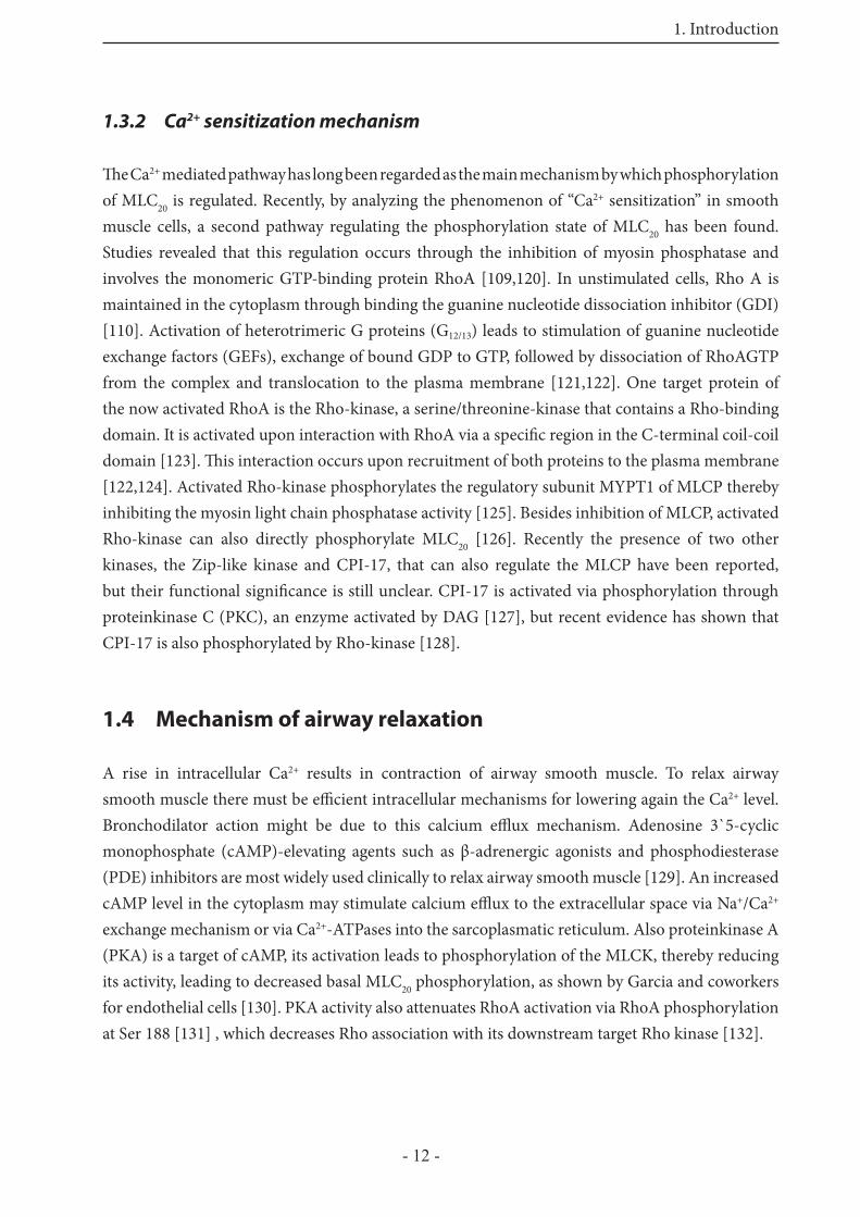
Figure 1.5 Mechanisms of airway relaxation via increase of cytosolic adenosine monophosphate (cAMP). Stimula-tion of a β2-receptor leading to activation of adenylate cyclase (AC), which in turn lead to formation of cAMP from adenosine triphosphate (ATP). An increase of cAMP results in activation of proteinkinase A (PKA), re-uptake of free cytosolic Ca2+ into the sarcoplasmatic reticulum (SR), and stimulation of Na+/Ca2+ exchanger, leading to relaxation of the cell. Degradation of cAMP to AMP was perfomed by phosphodiesterases.
1. Introduction
- 14 -
1.5.2 Animal models
Th ree main groups of animal models for asthma can be distinguished: (i) animals having naturally occurring recurrent airway obstruction, (ii) animals having an AHR before any provocation in the laboratory occurs, and (iii) animals developing reversible airway obstruction and /or increased airway responsiveness only aft er challenge.
1.5.2.1 Animals with naturally occurring recurrent airway obstruction
Th is group of animals is represented by cats, which express so called feline asthma. Th e feline asthma is characterized by recurrent attacks of dyspnea, wheezing, and cough [135]. Enhanced levels of eosinophils are found in the blood and sputum during an attack, and BAL fl uids from cats with bronchial disease revealed that eosinophils may be the predominant cell type within the fl uid [136]. Pathological aspects of the feline asthma include hyperplasia of submucosal glands, proliferation of goblet cells, and smooth muscle hypertrophy [135]. Norris Reinero et al. showed allergen-specifi c IgE production, airway hyperreactivity, airway eosinophilia and an acute Th 2 cell cytokine profi le in BAL fl uid cells in cats, sensitized with house dust mite or Bermuda grass allergen [137]. However, the cat has not become a widely distributed asthma model, because feline asthma is not commonly recognized, and the generation and identifi cation of asthmatic cats in a laboratory would be very cost-intensive. Horses and Ponies represent the second group of animals, in which naturally recurrent airway obstruction occurs. Th e exposure of sensitive animals to hay containing mold spores leads to a response of their airways. In pathologic studies of such animals Th urlbeck and Lowell have found a bronchiolitis, including leukocytes, in particular eosinophils in the bronchioles, bronchoconstriction, smooth muscle hyperthrophy, and mucus hypersecretion [138]. Th e number of neutrophils in the BAL is enhanced during exposure to hay in the barn compared to animals removed from that environment [139]. Airway hyperresponsiveness has been shown during the exposure to hay, and removal from that environment leads to reduction of responsiveness to a normal level [140-142]. Airway obstruction in horses and ponies resembles some forms of industrial asthma in humans in which symptoms are only present during exposure and decrease with removal from that environment. But there are some diff erences nevertheless. Increased neutrophils have been found in ponies instead of eosinophils in the BAL fl uid, and ponies do not show an AHR during remission, whereas most human individuals with allergic asthma persistently exhibit an AHR.
1. Introduction
- 15 -
1.5.2.2 Animals showing AHR without provocation
Th ere is a strain of dogs, based on a basenji-greyhound cross, showing a non-specifi c AHR to various stimuli, without a previous allergen challenge before [143,144]. Th is persistent AHR is comparable with moderate to severe human asthma, but in contrast to asthma, the dogs do not show a naturally occurring recurrent airway obstruction, which might be induced in laboratories [143].
1.5.2.3 Models of airway obstruction and AHR after allergen challenge
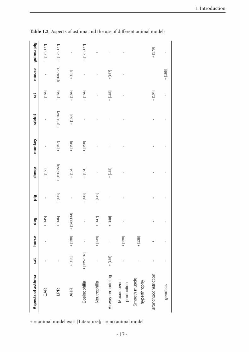
Several animal models have been developed to investigate diff erent aspects of asthma. Th erefore, diff erent species have been used in the laboratory, and various models in vivo and in vitro are created. However, such studies must be performed with awareness of the strength and weakness of the currently available animal models. Table 1.2 summarizes the characteristic features of asthma that can be investigated in the diff erent animal models. Canine models of allergic asthma have been primarily employed to achieve a better characterization of the EAR [145]. In other studies neutrophilia during LPR in dogs, accompanied by increasing vascular permeability and submucosal thickness have been shown [146,147]. Airway remodeling has been studied in a canine model of hyperpnea, with cold, dry air, to get more information about an increased incidence of asthma in winter athletes [148]. Pigs have been used to investigate the role of eosinophils and neutrophils in allergen-induced LPR [149]. Th e sheep model of allergic airway diseases represents many pathophysiological properties of human allergic airway diseases. Abraham et al. demonstrated 1983 that sheep with a natural sensitivity to ascaris suum challenged with this antigen, showed an immediate EAR, and 6 to 8 hours later a LPR [150-153]. Furthermore AHR has been observed in sheep, only if an EAR and LPR was present before [154]. A study from Bischof et al. [155] describes, for the fi rst time, the ability of house dust mites to induce allergic responses in sheep lungs. Using human relevant allergens in the sheep model of allergic lung infl ammation will be a useful tool to study the immunological and physiological mechanisms of allergic asthma. A model to study airway remodeling in sheep has also been developed [156]. Primate models have been used to study LPR [157], AHR and eosinophilia [158]. Also the importance of adhesion molecules in terms of changes in airway function has been investigated in monkeys [159,160]. An asthma model in rabbits has been developed to investigate the immunopathogenesis of the LPR. As in humans, the LPR leads to stronger airway obstruction than the EAR [161], and the importance of antigen-specifi c IgE and IgG to this pattern of airway obstruction has been investigated within this model [162]. Furthermore the LPR in rabbits is accompanied by subsequent airway hyperresponsiveness [163]. Th e rat has been extensively used to study allergen-induced bronchoconstriction, eosinophilic infl ammation, as well as LPR and AHR. Furthermore airway remodeling has been observed aft er repeated allergen exposure [164,165]. Mice, another species of small mammals have
1. Introduction
- 16 -
also become important to study antigen-induced airway responsiveness. Th ey are interesting and attractive for these studies, because their immune system is very well known, specifi c knockout or transgenic mice are available, and studying the global transcriptional changes aft er allergen challenge is possible [166]. Although mouse models rarely completely reproduce all features of human asthma, murine strains have also been used to investigate AHR. Furthermore mice develop aft er sensitization clinical syndromes that resemble allergic asthma, including eosinophilia, AHR, increased IgE levels, mucus hypersecretion and sometimes airway remodeling [167], but there are no published descriptions of antigen-induced LPRs [168-171]. Th ere are some known diff erences between mouse and human physiology of asthma. First, methacholine-induced AHR in mice is only transient aft er allergen exposure in contrast to humans, which show an increased AHR even when they are symptom free [167]. Second, repeated exposure of allergen to sensitized mice results in suppression of the disease, whereas in human patients it leads to chronic allergic asthma [167]. Th ird, mast cells and IgE molecules seem to play no role in the development of allergic asthma in mice [167]. Th ese diff erences limit the value of mouse models for allergic asthma. Taken together, these observations demonstrate that no animal model allows investigation of all questions. Guinea pigs, which are no rodents [172], are widely used in pulmonary pharmacology, because their airways responsiveness to mediators and drugs is thought to resemble human airway more closely than do those of mice or rats [173,174]. Most of the previous studies have been focussed on in vivo experiments, where EAR, LPR, AHR, and eosinophilia have been investigated [175-177]. To study airway pharmacology there is a great need for experimental in vitro models, which are relevant with regard to human main components in allergen-induced airway obstruction. Until now, used models dealing with tissue from small mammals (rats and mice) show that the mediators of bronchoconstriction and airway infl ammation are very diff erent from those in humans. Th e bronchoconstriction during EAR in humans is mainly mediated through leukotrienes, thromboxane, and partly by histamine [178,179], whereas mouse and rat airways do not respond with signifi cant bronchoconstriction to challenge with histamine or leukotrienes [180-184]. Th e guinea pig shows responsiveness to those mediators [185], making it a valuable model to study airway pharmacology relevant with regard to humans.
1. Introduction
- 17 -
Table 1.2 Aspects of asthma and the use of diff erent animal modelsg
uin
ea
pig
+ [1
75,1
77]
+ [1
75,1
77]
-
+ [1
75,1
77]
+ - -
+ [1
78]
-
mo
use
-
+[16
8-17
1]
+[16
7]
- -
+[16
7]
-
+ [1
66]
rat
+ [1
64]
+ [1
64]
+ [1
64]
+ [1
64]
-
+ [1
65]
-
+ [1
64]
-
rab
bit
-
+ [1
61,1
62]
+ [1
63]
- - - - - -
mo
nk
ey
-
+ [1
57]
+ [1
58]
+ [1
58]
- - - - -
she
ep
+ [1
50]
+ [1
50-1
53]
+ [1
54]
+ [1
51]
-
+ [1
56]
- - -
pig -
+ [1
49]
-
+ [1
49]
+ [1
49]
- - - -
do
g
+ [1
45]
+ [1
46]
+ [1
43,1
44]
-
+ [1
47]
+ [1
48]
- - -
ho
rse
-
+ [1
38]
-
+ [1
39]
-
+ [1
38]
+ [1
38]
+ -
ca
t - -
+ [1
35]
+ [1
35-1
37]
-
+ [1
35]
- - - -
Asp
ec
ts o
f a
sth
ma
EA
R
LPR
AH
R
Eos
inop
hilia
Neu
troph
ilia
Airw
ay re
mod
elin
g
Muc
us o
ver
prod
uctio
n
Sm
ooth
mus
cle
hype
rthro
phy
Bro
ncho
cons
trict
ion
gene
tics
+ = animal model exist [Literature]; - = no animal model

1. Introduction
- 18 -
1.5.2.4 In vitro models in small mammalian species
Apart from the in vivo asthma models in several species, there are also a variety of in vitro models, to investigate single physiological parameters of asthma. Th ese in vitro models are best approached within small mammalian species, like rats, mice and guinea pigs, for which the costs for “living” in the animal facility are less prohibitive. To study broncho- or vasoconstriction tissue organ bath preparations have been widely applied [180,186,187]. Th ese include parenchymal strips, as well as isolated tracheae, bronchi or vessels. Isolated bronchi or tracheae are well established standard methods for studying airway smooth muscle. However, they are best suited to monitor large (> 2 mm diameter) rather than peripheral airways that recently have received much attention [188]. Furthermore, in that kind of preparation interaction with the surrounding parenchymal tissue is missing, so a comparison with the in vivo situation remains diffi cult. Parenchymal strips include peripheral airways and are easy to prepare, but they do not allow to distinguish between vascular and airway responses. Th e isolated perfused lung (IPL) represents a very good ex vivo model to investigate lung parameters, crucial in asthma, including bronchoconstriction, vasoconstriction, edema, and gas exchange. Its disadvantages are: (i) one animal is needed for one experiment, and (ii) the amount of drugs or agonists needed may be relatively high.
1.5.2.5 The model of precision-cut lung slices (PCLS)
A recently developed alternative to the classical pharmacological models is the precision-cut lung slice model. In 1980 Krumdieck et al. published for the fi rst time the establishment of precision-cut liver slices using a mechanical tissue slicer [189]. Organ slices have been used for a long time to investigate biochemical pathways, but the production of slices with reproducible properties was diffi cult, because only manual techniques have been applied. Only the development of the mechanical slicing procedure allowed the production of slices of almost identical thickness, which is important for the comparability of spatial relationship of gas and nutrient exchange by diff usion. Liver slices, which long have been used for toxicological studies, were followed by production of kidney, heart and lung slices. In 1992 Stefaniak et al. published the fi rst paper describing the use of agar-fi lled precision-cut lung slices [190]. Siminski et al. [191] studied the long term maintenance of lung slices cultured in defi ned media, followed later by toxicological studies [192]. Th e use of precision-cut lung slices in pharmacological studies was introduced by Martin et al. [193]. Th ey produced rat lung slices of 250 μm ± 20 μm thickness with a Krumdieck tissue slicer. Contraction of single airways were induced with methacholine (a stable analogue of acetylcholine), and via videomicroscopy and digital imaging the proportion of contraction were visualized and quantifi ed. Further, pharmacological investigations followed this initial study, characterizing the response of mouse airways and pulmonary vessels to several endogenous mediators, and comparing these responses to those in the isolated perfused and ventilated mouse lung [181]. Utilization of PCLS were applied for further studies of peripheral airway pharmacology [194,195]. Beside the use of tissue from laboratory animals (rats and mice), human lung tissue from patients undergoing
1. Introduction
- 19 -
surgery for cancer can also be used to produce PCLS, providing insight into human responses to various endogenous and exogenous stimuli [178,196]. In PCLS smooth muscle contraction in airways and vessels is auxotonic, i.e. stress and length change simultaneously, which may resemble in vivo airway contraction more appropriately than other in vitro methods. Another advantage of this model is the amount of slices, prepared from one lung (up to 50 slices), which does save animals, and can also help to reduce experimental error by internal controls and statistical pairing. PCLS provide the opportunity to examine physiological responses in diff erent species by the same experimental model. Th is seems of particular importance at a time when diff erences between various asthma models compared to human asthma are increasingly being recognized as a major impediment for drug development [197,198]. With regard to the guinea pig, as a small mammalian animal that shows many similarities to human airway responses, PCLS from the guinea pig would be a valuable tool in airway pharmacology.
2. Aim of the study
- 20 -
2 Aim of the study
One characteristic feature of human asthma is the early allergic response (EAR), occurring within minutes aft er allergen contact and resulting in a strong contraction of the airways. For induction of an EAR in PCLS, two methods can be applied, on the one hand passive sensitization of the slices, on the other had the use of PCLS from actively sensitized animals. Another characteristic feature is airway hyperresponsiveness (AHR), an increased reactivity of infl amed and remodeled airways in response to unspecifi c stimuli. Various animal models are used to investigate characteristic features of asthma, but important species diff erences limit the portability of these models to human asthma.
Th e model of precision-cut lung slices (PCLS) off ers the opportunity to investigate airway responses in diff erent animal species, as well as in human lung tissue. Th e access to human tissue provides an almost unique possibility to explore lung functions in human airways with an intact microanatomy. However, because of the limited availability of human tissue, and the fact that the tissue comes from patients undergoing surgery for lung cancer, that are treated with diverse pharmaceuticals, there is a strong need of animal models resembling the human responses. Previous studies had indicated that guinea pig lungs resemble human airway responses more closely than do rats or mice.
Th erefore, a major aim of the present study was to establish the model of PCLS from guinea pigs. A further aim was, to identify the mediators inducing bronchoconstriction during EAR and the activated signal transduction pathways in PCLS of the diff erent species, i.e. guinea pigs, monkeys, rats, humans and mice. Finally, the results should be compared among these species.Additionally, we wanted to investigate EAR and AHR in PCLS from actively sensitized mice and rats.
3. Material and Methods
- 21 -
3 Material and Methods
3.1 Animals
Th e animal experiments were approved by the local ethic committee.
3.1.1 Rats
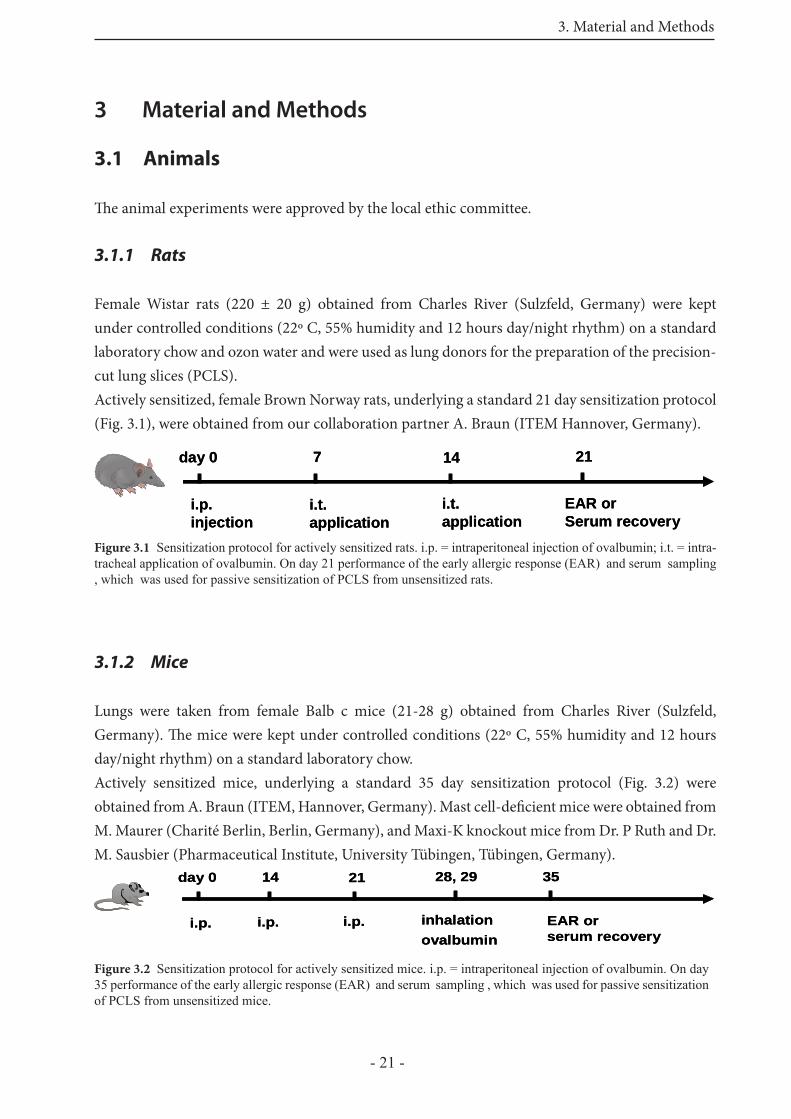
Female Wistar rats (220 ± 20 g) obtained from Charles River (Sulzfeld, Germany) were kept under controlled conditions (22º C, 55% humidity and 12 hours day/night rhythm) on a standard laboratory chow and ozon water and were used as lung donors for the preparation of the precision-cut lung slices (PCLS).Actively sensitized, female Brown Norway rats, underlying a standard 21 day sensitization protocol (Fig. 3.1), were obtained from our collaboration partner A. Braun (ITEM Hannover, Germany).
3.1.2 Mice
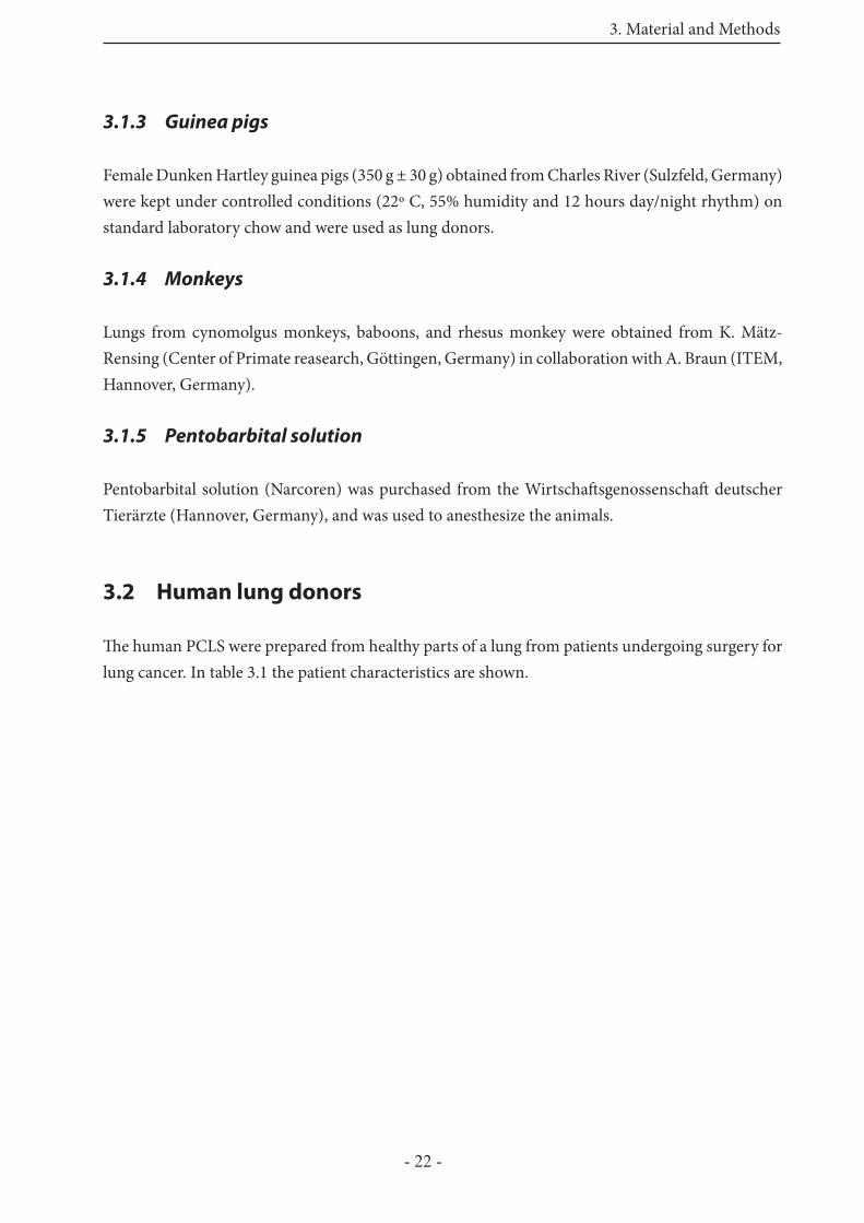
Lungs were taken from female Balb c mice (21-28 g) obtained from Charles River (Sulzfeld, Germany). Th e mice were kept under controlled conditions (22º C, 55% humidity and 12 hours day/night rhythm) on a standard laboratory chow. Actively sensitized mice, underlying a standard 35 day sensitization protocol (Fig. 3.2) were obtained from A. Braun (ITEM, Hannover, Germany). Mast cell-defi cient mice were obtained from M. Maurer (Charité Berlin, Berlin, Germany), and Maxi-K knockout mice from Dr. P Ruth and Dr. M. Sausbier (Pharmaceutical Institute, University Tübingen, Tübingen, Germany).
Figure 3.1 Sensitization protocol for actively sensitized rats. i.p. = intraperitoneal injection of ovalbumin; i.t. = intra-tracheal application of ovalbumin. On day 21 performance of the early allergic response (EAR) and serum sampling , which was used for passive sensitization of PCLS from unsensitized rats.
Figure 3.2 Sensitization protocol for actively sensitized mice. i.p. = intraperitoneal injection of ovalbumin. On day 35 performance of the early allergic response (EAR) and serum sampling , which was used for passive sensitization of PCLS from unsensitized mice.
day 0
i.p.injection
7 14 21
i.t.application
i.t.application
EAR orSerum recovery
day 0
i.p.injection
7 14 21
i.t.application
i.t.application
EAR orSerum recovery
day 0
i.p.
14 21 28, 29
i.p. i.p. inhalationovalbumin
EAR orserum recovery
35day 0
i.p.
14 21 28, 29
i.p. i.p. inhalationovalbumin
EAR orserum recovery
35
3. Material and Methods
- 22 -
3.1.3 Guinea pigs
Female Dunken Hartley guinea pigs (350 g ± 30 g) obtained from Charles River (Sulzfeld, Germany) were kept under controlled conditions (22º C, 55% humidity and 12 hours day/night rhythm) on standard laboratory chow and were used as lung donors.
3.1.4 Monkeys
Lungs from cynomolgus monkeys, baboons, and rhesus monkey were obtained from K. Mätz-Rensing (Center of Primate reasearch, Göttingen, Germany) in collaboration with A. Braun (ITEM, Hannover, Germany).
3.1.5 Pentobarbital solution
Pentobarbital solution (Narcoren) was purchased from the Wirtschaft sgenossenschaft deutscher Tierärzte (Hannover, Germany), and was used to anesthesize the animals.
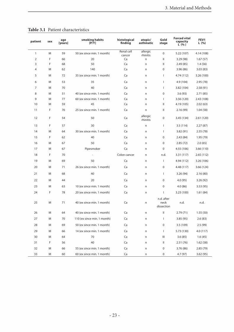
3.2 Human lung donors
Th e human PCLS were prepared from healthy parts of a lung from patients undergoing surgery for lung cancer. In table 3.1 the patient characteristics are shown.
3. Material and Methods
- 23 -
Table 3.1 Patient characteristics
patient sexage
[years]smoking habits
[P/Y]histological
fi ndingatopic/
asthmaticGoldstage
Forced vital capacity
L (% )
FEV1 L (%)
1 M 59 50 (ex since min. 1 month) Renal cell cancer
allergic rhinitis 0 5.22 (107) 4.14 (108)
2 F 66 20 Ca n II 3.29 (98) 1.67 (57)
3 F 68 50 Ca n II 2.49 (85) 1.4 (56)
4 M 62 140 Ca n 0 3.96 (86) 3.03 (83)
5 M 72 35 (ex since min. 1 month) Ca n I 4.74 (112) 3.26 (100)
6 M 53 35 Ca n I 4.9 (104) 2.95 (78)
7 M 70 40 Ca n I 3.82 (104) 2.58 (91)
8 M 51 40 (ex since min. 1 month) Ca n 0 3.6 (93) 2.71 (85)
9 M 77 60 (ex since min. 1 month) Ca n I 3.56 (120) 2.43 (108)
10 M 59 45 Ca n II 4.19 (105) 2.02 (63)
11 F 76 25 (ex since min. 1 month) Ca n II 2.16 (99) 1.04 (58)
12 F 54 50 Ca allergic rhinitis 0 3.45 (134) 2.61 (120)
13 F 57 30 Ca n I 3.5 (114) 2.27 (87)
14 M 64 30 (ex since min. 1 month) Ca n I 3.82 (91) 2.55 (78)
15 F 62 40 Ca n 0 2.43 (84) 1.95 (79)
16 M 67 50 Ca n 0 2.85 (72) 2.0 (65)
17 M 67 Pipesmoker Ca n 0 4.55 (106) 3.66 (110)
18 F 70 - Colon cancer n n.d. 3.31 (117) 2.65 (112)
19 M 69 50 Ca n I 4.94 (112) 3.26 (106)
20 M 71 26 (ex since min. 1 month) Ca n 0 4.48 (117) 3.66 (124)
21 M 68 40 Ca n I 3.26 (94) 2.16 (80)
22 M 44 20 Ca n 0 4.0 (95) 3.26 (92)
23 M 63 10 (ex since min. 1 month) Ca n 0 4.0 (86) 3.53 (95)
24 F 78 20 (ex since min. 1 month) Ca n I 3.23 (100) 1.61 (84)
25 M 71 40 (ex since min. 1 month) Ca nn.d. after
neck dissection
n.d. n.d.
26 M 64 40 (ex since min. 1 month) Ca n II 2.79 (71) 1.55 (50)
27 M 70 110 (ex since min. 1 month) Ca n I 3.85 (95) 2.6 (83)
28 M 69 50 (ex since min. 1 month) Ca n 0 3.5 (109) 2.5 (99)
29 M 66 14 (ex since min. 1 month) Ca n I 5.73 (130) 4.0 (117)
30 M 64 70 Ca n III 3.6 (85) 1.6 (45)
31 F 56 40 Ca n II 2.51 (76) 1.62 (58)
32 M 66 55 (ex since min. 1 month) Ca n 0 3.76 (86) 2.85 (79)
33 M 60 60 (ex since min. 1 month) Ca n 0 4.7 (97) 3.62 (95)
3. Material and Methods
- 24 -
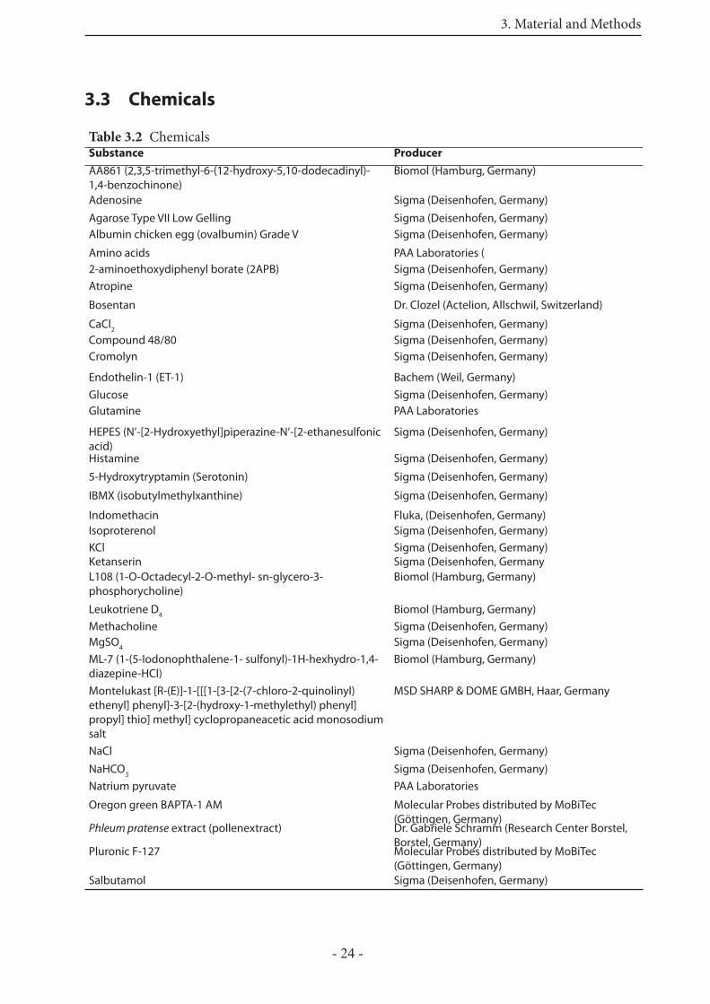
3.3 Chemicals
Table 3.2 Chemicals Substance Producer
AA861 (2,3,5-trimethyl-6-(12-hydroxy-5,10-dodeca dinyl)-1,4-benzo chinone)
Biomol (Hamburg, Germany)
Adenosine Sigma (Deisenhofen, Germany)
Agarose Type VII Low Gelling Sigma (Deisenhofen, Germany)Albumin chicken egg (ovalbumin) Grade V Sigma (Deisenhofen, Germany)
Amino acids PAA Laboratories (2-aminoethoxydiphenyl borate (2APB) Sigma (Deisenhofen, Germany)Atropine Sigma (Deisenhofen, Germany)
Bosentan Dr. Clozel (Actelion, Allschwil, Switzerland)
CaCl2 Sigma (Deisenhofen, Germany)Compound 48/80 Sigma (Deisenhofen, Germany)Cromolyn Sigma (Deisenhofen, Germany)
Endothelin-1 (ET-1) Bachem (Weil, Germany)Glucose Sigma (Deisenhofen, Germany)Glutamine PAA Laboratories
HEPES (N’-[2-Hydroxyethyl]piperazine-N’-[2-ethanesulfonic acid)
Sigma (Deisenhofen, Germany)
Histamine Sigma (Deisenhofen, Germany)
5-Hydroxytryptamin (Serotonin) Sigma (Deisenhofen, Germany)
IBMX (isobutylmethylxanthine) Sigma (Deisenhofen, Germany)
Indomethacin Fluka, (Deisenhofen, Germany)Isoproterenol Sigma (Deisenhofen, Germany)KCl Sigma (Deisenhofen, Germany)Ketanserin Sigma (Deisenhofen, GermanyL108 (1-O-Octadecyl-2-O-methyl- sn-glycero-3-phosphorycholine)
Biomol (Hamburg, Germany)
Leukotriene D4 Biomol (Hamburg, Germany)Methacholine Sigma (Deisenhofen, Germany)MgSO4 Sigma (Deisenhofen, Germany)ML-7 (1-(5-Iodonophthalene-1- sulfonyl)-1H-hexhydro-1,4- diazepine-HCl)
Biomol (Hamburg, Germany)
Montelukast [R-(E)]-1-[[[1-[3-[2-(7-chloro-2-quinolinyl) ethenyl] phenyl]-3-[2-(hydroxy-1-methylethyl) phenyl] propyl] thio] methyl] cyclopropaneacetic acid monosodium salt
MSD SHARP & DOME GMBH, Haar, Germany
NaCl Sigma (Deisenhofen, Germany)
NaHCO3 Sigma (Deisenhofen, Germany)Natrium pyruvate PAA Laboratories
Oregon green BAPTA-1 AM Molecular Probes distributed by MoBiTec (Göttingen, Germany)
Phleum pratense extract (pollenextract) Dr. Gabriele Schramm (Research Center Borstel, Borstel, Germany)
Pluronic F-127 Molecular Probes distributed by MoBiTec (Göttingen, Germany)
Salbutamol Sigma (Deisenhofen, Germany)
3. Material and Methods
- 25 -
SQ29548 (5-hepte noic acid, 7-[3-[[2-[(phenylamino)carbonyl]hy drazino]me thyl]-7-oxa bicyc lo-[2.2.1]hept-2-yl]-, [1S-[1α,2α(Z),3α,4α]]-)
RBI (Deisenhofen, Germany)
Substance P Sigma (Deisenhofen, Germany)
Sulfobromophthalein Sigma (Deisenhofen, Germany)
Triprolidine Sigma (Deisenhofen, Germany)
Triton X-100 Detergenz Roche (Mannheim)
U46619 (9,11-dideoxy-9α,11α-methanoepoxy-prosta-5Z,13E-dien-1-oic acid)
Cayman (Ann Arbor, Michigan, USA)
vitamins PAA Laboratories
WEB 2086 (3-[4-(2-chlorophenyl)-9-methyl-6H-thienol[3,2-f ][1,2,4]triazolo-[4,3-a][1,4]-diazepin-2-yl]-1-(4-morpholinyl)-1-propanon)
Dr. Heuer (Boehringer Mannheim, Mannheim, Germany)
Xestospongin C Calbiochem (Bad Soden, Germany)
Y27623 (trans-4-[(1R)-1-aminoethyl]-N-4-pyridinylcyclohexanecarboxamide)
Tocris distributed by Biotrend (Cologne, Germany)
3.4 Equipment
Table 3.3 Instruments and equipmentInstrument Producer
Beam splitter Trim Scope, La Vision, BioTec, Bielefeld, Germany
Coring tool (Black & Decker battery operated screwdriver + self-made coring device 0.9 mm Digital camera (2-Photon microscope) Imager QE, La Vision BioTec, Bielefeld, GermanyELISA (Histamine) IBL, Hamburg, GermanyELISA (Serotonin) IBL, Hamburg, GermanyGraphPadPrism 4.02 software GraphPad, San Diego, CA, USA Imspector software La Vision BioTec; Bielefeld, GermanyIncubator Heraeus, Hannover, GermanyInverted microscope Leica DMIRB and DMIL Zeiss, Oberkochen, GermanyFixed stage upright microscope BX51WI Olympus, Hamburg, GermanyKrumdieck tissue slicer Alabama research and development, Munford, USAOptimas 6.1 Optimas Corporation, Bothell, USAOptimas 6.5 Optimas Corporation, Bothell, USAPolytron PT1200 (Ultrathorax) Kinematica AG, Littau, SwitzerlandTi:Sa femtosecond laser Coherent, Dieburg, GermanyViability/cytotoxicity assay kit Molecular Probes (Eugene, Oregon, USA)Video camera JAI PROTEC JAI20040, JAI Pulnix, Alzenau, GermanyVideo camera Visicam 1300 and Visicam 640 Visitron Systems, Munich, Germany
3. Material and Methods
- 26 -
3.5 Methods
3.5.1 Precision -cut lung slices (PCLS)
3.5.1.1 Preparation of rat lung slices
Th e rats were anesthesized through intraperitoneal injection of 60 mg/kg pentobarbital (Narcoren, Pharmazeutische Handelsgesellschaft mbH, Garbsen, Germany). Aft er narcotization the trachea was cannulated and the animals were exsanguinated by cutting the vena cava inferior. Th e diaphragm was cut, to collapse the lungs, which were then subsequently fi lled with a low-melting point agarose solution (0.75%, fi nal concentration) through the cannula. In order to solidify the agarose and harden them for cutting, the whole chest was covered with ice for 10 to 15 minutes. Aft er that time, the lungs were removed from the thoracic cavity and placed on ice for another 10 minutes. Th e lobes were separated and tissue cores prepared with a rotating sharpened metal tube (diameter 8 mm). Th ese cores were cut into 220 μm thick slices with a Krumdieck tissue slicer (Alabama Research and Development, Al, USA).
3.5.1.2 Preparation of mouse lung slices
Mice were anesthesized with 45 mg/ml pentobarbital. Preparation follows as described for the rats. Aft er the lungs solidifi ed on ice and were removed from the chest, they were separated into the single lobes and embedded in 3% agarose in a cryotube. Th e cryotubes were then put on ice again to solidify the agarose. From the embedded lung lobes mouse PCLS (220 μM) were prepared with the Krumdieck tissue slicer (Alabama Research and Development, Al, USA).
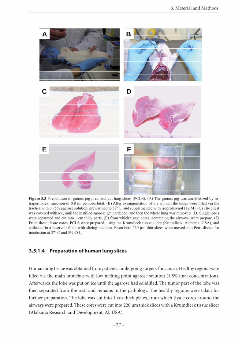
3.5.1.3 Preparation of guinea pig lung slices
Guinea pig PCLS were prepared as described for the other species (rat, mouse) with the following modifi cations. Aft er injection of pentobarbital (95 mg/kg) the trachea was cannulated and the animals exsanguinated by cutting the vena cava inferior. Th rough the cannula the lung was fi lled with a low-melting point agarose solution (0.75%, fi nal concentration) containing isoproterenol (1 μM). To allow the agarose to solidify, the whole chest was covered with ice. Subsequently the lungs were removed, lobes were separated and tissue cores prepared with a rotating sharpened metal tube (diameter 8 mm). Th ese cores were cut into 220 μm thick slices with a Krumdieck tissue slicer (Alabama Research and Development, Al, USA). In fi gure 3.3 the preparation of GP PCLS was shown.
3. Material and Methods
- 27 -
3.5.1.4 Preparation of human lung slices
Human lung tissue was obtained from patients, undergoing surgery for cancer. Healthy regions were fi lled via the main bronchus with low melting point agarose solution (1.5% fi nal concentration). Aft erwards the lobe was put on ice until the agarose had solidifi ed. Th e tumor part of the lobe was then separated from the rest, and remains in the pathology. Th e healthy regions were taken for further preparation. Th e lobe was cut into 1 cm thick plates, from which tissue cores around the airways were prepared. Th ese cores were cut into 220 μm thick slices with a Krumdieck tissue slicer (Alabama Research and Development, Al, USA).
A
D
E F
C
BAA
DD
EE FF
CC
BB
Figure 3.3 Preparation of guinea pig precision-cut lung slices (PCLS). (A) The guinea pig was anesthetized by in-traperitoneal injection of 0.8 ml pentobarbital. (B) After exsanguination of the animal, the lungs were fi lled via the trachea with 0.75% agarose solution, prewarmed to 37º C, and supplemented with isoproterenol (1 μM). (C) The chest was covered with ice, until the instilled agarose get hardened, and then the whole lung was removed. (D) Single lobes were separated and cut into 1 cm thick parts, (E) from which tissue cores, containing the airways, were prepare. (F) From these tissue cores, PCLS were prepared, using the Krumdieck tissue slicer (Krumdieck, Alabama, USA), and collected in a reservoir fi lled with slicing medium. From here 250 μm thin slices were moved into Petri-dishes for incubation at 37º C and 5% CO2.
3. Material and Methods
- 28 -
3.5.1.5 Preparation of monkey PCLS
Monkey PCLS were prepared in accordance to human PCLS. Complete lungs were fi lled via the trachea with agarose solution (1.5% fi nal concentration), which was prewarmed to 37˚ C before. Th e trachea was closed with a threat and the lungs were put on ice for approximately 30 minutes until solidity. Aft er that, the lungs were separated into the single lobes, which were then cut into 1 cm thick slabs. With a coring tool tissue cores were prepared containing airways, vessels or both. As in the other species, the tissue cores were cut into 250 ± 20 μm thin slices with a Krumdieck tissue slicer (Krumdieck, Alabama research, USA). Aft er the slicing procedure the PCLS were incubated at 37° C and 5% CO2.
3.5.2 Culture medium
3.5.2.1 Incubation medium
Aft er the slicing procedure the lung slices were incubated at 37˚ C in a humid atmosphere in minimal essential medium (MEM), (pH 7.2) composed of CaCl2 (1.8 mM), MgSO4 (0.8 mM), KCl (5.4 mM), NaCl (116.4 mM), glucose (16.7 mM), NaHCO3 (26.1 mM), Hepes (25.17 mM), natrium pyruvate (10 ml/l), amino acids (20 ml/l), vitamins (10 ml/l), glutamine (10 ml/l). Th e pH-value was adjusted to 7.2. Th e incubation medium was changed every 30 minutes during the fi rst two hours aft er slicing, followed by a change every hour for the next two hours, in order to remove the agarose and cell debris from the tissue. Subsequently, medium was further supplemented with penicillin and streptomycin (100 U and 100 μg/ml) and changed every 24 hours. In case of the guinea pig isoproterenol (1 μM) was added to the incubation medium during the fi rst 3 hours aft er slicing.
3.5.2.2 Agarose solution
Double concentrated incubation medium was mixed with the same volume of low melting point agarose at 37º C, to obtain the fi nal concentration of agarose solution for instillation into the lung.
3.5.2.3 Slicing medium
Th e slicing procedure was performed in approximately 500 ml incubation medium without natrium pyruvate, amino acids, vitamins, and glutamine.
3. Material and Methods
- 29 -
3.5.3 Viability of the PCLS
3.5.3.1 Lactate dehydrogenase (LDH) release
Th e viability of the PCLS was assessed by measuring the relative amount of lactate dehydrogenase (LDH) released from the slices into the incubation medium. LDH was taken as a marker of cell death, because it was only released in great amounts, when cells start lyses. Th ree slices per well were placed into a 24-well-plate and covered with 1 ml incubation medium. At the indicated time points (24 hours, 48 hours, 72 hours), slices were removed from the incubation medium and lysed in 1 ml 0.2% Triton X-100 solution. Th e incubation supernatant was kept and stored on ice (4º C), until the end of the preparation. Th e slices in the 0.2% Triton X-100 solution were homogenized (Polytron; Kinematica AG, Littau, Switzerland) and centrifuged, 5 minutes at 13,000 x g (Eppendorf; Hamburg, Germany). Th e supernatant was taken and on ice. Th en both solutions, the incubation supernatant and the supernatant from the homogenisation were analyzed by a commercially available LDH-assay (Dimension pan; Dade Behring, Schwalbach, Germany). Viability of the slices was expressed as the ratio of LDH in the incubation supernatant to the total LDH (sum of LDH in slices and the supernatatant).
3.5.3.2 Two-photon microscopy
To visualize also the viability of the guinea pig PCLS we used 2-photon microscopy in combination with the LIVE/DEAD® viability/cytotoxicity assay kit (Molecular Probes, Eugene, Oregon, USA). PCLS were incubated with 5 μM Calcein AM (acetomethylester of calcein, live staining) and 10 μM ethidium homodimer (EthD, dead staining) for 40 minutes and then washed to remove external dye. Th e fl uorescent dyes were excited at 800 nm with a Ti:Sa femtosecond laser (Coherent, Dieburg, Germany). Th e laser beam was split up into 64 individual beams (beam splitter from Trim Scope, LaVision BioTec, Bielefeld, Germany) that simultaneously excited and scanned the object on the microscope (Olympus, Hamburg, Germany). Th e images were acquired using a digital camera (Imager QE, LaVision, Bielefeld, Germany). Th e emission of calcein AM (emission fi lter 500/50 nm) for the cytoplasm (live staining, green) and the EthD (emission fi lter 625/50) for the staining of nuclei (dead staining, blue) were recorded separately. Figure 4.2 in the result part shows overlay images of both dyes. Slices were analyzed 24 hours, 48 hours, and 72 hours aft er preparation. To visualize the total amount of dead cells, some PCLS were treated with 1% Triton X-100 for 20 minutes prior incubation with dyes.
3. Material and Methods
- 30 -
3.5.4 Measurement and Imaging of broncho - or vasoconstriction
Th e airways and vessels were imaged and digitized with a digital and an analogue video camera (IRB640, Visitron Systems, Munich, Germany; JAI 2040, JAI PULNiX, Alzenau, Germany). Th e airway or vessel area before addition of the mediators was defi ned as 100%. Bronchoconstriction or vasoconstriction was determined as the percentage decrease in the airway/vessel area compared to the initial area. For the measurements, slices with comparable airway size were selected. Th ese slices were put on 24-well-plates and fi xed with a nylon thread attached to a platinum wire. Th e slices were continuously covered with 1 ml incubation medium. Th e 24-well plate was positioned on the stage of an inverted microscope. Images were recorded by analogue (JAI 2040, JAI PULNiX, Alzenau, Germany) or digital camera (IRB640, Visitron Systems, Munich, Germany). A control image was taken before addition of the mediator, frames were recorded every 5 or 30 s for 5, 10 or 20 minutes depending on the study.
3.5.5 Passive-sensitization as a tool to study the EAR
Animal PCLS were treated and incubated overnight (16 hours) with 1% serum from animals that have been actively sensitized with ovalbumin by standard protocols. Human PCLS were incubated overnight with 1% serum from an atopic individual with sensitivity to grass pollen (phleum pratense). Th e next day, passively sensitized slices were transferred into a 24-well plate with fresh medium free of serum. Changes of airway /vessel area was measured as described in 3.5.4.
3.5.5.1 Rat serum
Rat serum was obtained from Dr. A. Braun from the ITEM (Hannover, Germany). Female Brown Norway rats were sensitized to ovalbumin underlying a 21 day sensitization protocol. On day 0 the rats were sensitized by an intraperitoneal injection (i.p.) of 10 μg ovalbumin plus 20 mg Al(OH)3 gel. On day 7 and 14 the rats were sensitized intratracheal. On day 21 the animals were killed, and blood samples were taken from their hearts (Fig. 3.1). Th e total IgE amount was determined by ELISA.
3.5.5.2 Mouse serum
Mouse serum was also obtained from Dr. A. Braun from the ITEM (Hannover, Germany). Mice underlying a 35 day sensitization protocol (Fig. 3.2).
3. Material and Methods
- 31 -
3.5.5.3 Guinea pig serum
Th e guinea pig serum was a gift from Prof. S.-E. Dahlén (Karolinska Institute, Stockholm, Sweden). Guinea pigs were sensitized within four weeks by one i.p and one s.c. injection of 10 mg/ml ovalbumin dissolved in 0.4 ml of saline-aluminium hydrogel (1:1). Animals were killed, and blood was collected from the hearts.
3.5.5.4 Human serum
Human serum was from an atopic person, who has allergic rhinitis and specifi c IgE levels to grass pollen (Phleum pratense) and velvet grass (Holcus lanatus). Th e serum contains a total IgE amount of 450 IU/ml.
3.5.6 Pharmacological intervention studies
3.5.6.1 Mediators/ agonists and antagonists
To determine the mediators that are involved in the early allergic response (EAR), PCLS were preincubated for at least 10 min with diff erent receptor antagonists. Th e leukotriene (CysLT1)-receptor antagonist montelukast (10 μM), the thromboxane prostanoid-receptor antagonist SQ29548 (10 μM), or the histamine (H1)-receptor antagonist triprolidine (5 μM) were added alone or in combination.
Table 3.4 Overview of the receptor agonists and antagonists
Agonist Receptor Antagonist
Leukotriene D4 CysLT1-receptor Montelukast (10 μM)
Th romboxane TP-receptor SQ29548 (10 μM)
Histamine H1-receptor Triprolidine (5 μM)
3. Material and Methods
- 32 -
3.5.6.2 Inhibitors of the signal transduction pathways
To investigate on which signal transduction pathway the bronchoconstriction during EAR was mediated, diff erent signal transduction pathways were inhibited. PCLS were preincubated 20 minutes with the following diff erent inhibitors. During measurement the inhibitor was also present next to the agonist in the incubation medium.
Table 3.5 Inhibitors of the signal transduction pathways
Inhibitor fi nal concentration solved in
L108 (ihibitor of PI-PLC) 30 μM EtOH
Mibefradil (inhibitor of T-type Ca2+-channel) 25 μM Milli Q H2O
ML-7 (inhibitor of MLCK) 35 μM EtOH
Nifedipine (inhibitor of L-type Ca2+-channel) 5 μM EtOH
Xestospongin C (inhibitor of IP3-receptor) 10 μM EtOH
Y27632 (inhibitor of Rho-kinase) 10 μM Milli Q H2O
Th e fi nal concentration of EtOH was always < 0,1% and did not aff ect the responses to the mediators.
3.5.7 Airway relaxation in the guinea pig
For the relaxation experiments PCLS were contracted with Mch (10-6.5 M) to 10 - 20% of their initial airway area. Subsequently, increasing concentrations of the β-agonist salbutamol (10 nM to 10 μM), the unspecifi c PDE-inhibitor IBMX (10 nM to 10 μM) and the combination of both were added in the presence of Mch. Measurements were performed as described in 3.5.4.
3.5.8 Calcium imaging with two-photon microscopy
Rat PCLS were prepared as already described above with one modifi cation: the fi nal concentration of the agarose solution to fi ll the lung is 3,5%. For the experiments with serotonin, the slices were selected immediately aft er the slicing procedure. One slice per well was placed on a 24-well plate. Every well was covered with 500 μl incubation medium. To visualize later intracellular Ca2+, the slices were loaded for 2 hours at 37º C with Oregon green (5 μM), 0,2% pluronic (Pluronic-F127; 5 μM) and sulfobromophthalein (100 μM). For the measurements the slices were placed on a petri dish fi xed with a nylon thread attached to a platinum wire, and covered by a 1ml drop of incubation medium without dye. Th en the perti dish was put under the microscope, and the layer of airway smooth muscle cells was adjusted via microscopy. Th e fl uorescent dye (Oregon green) was excited
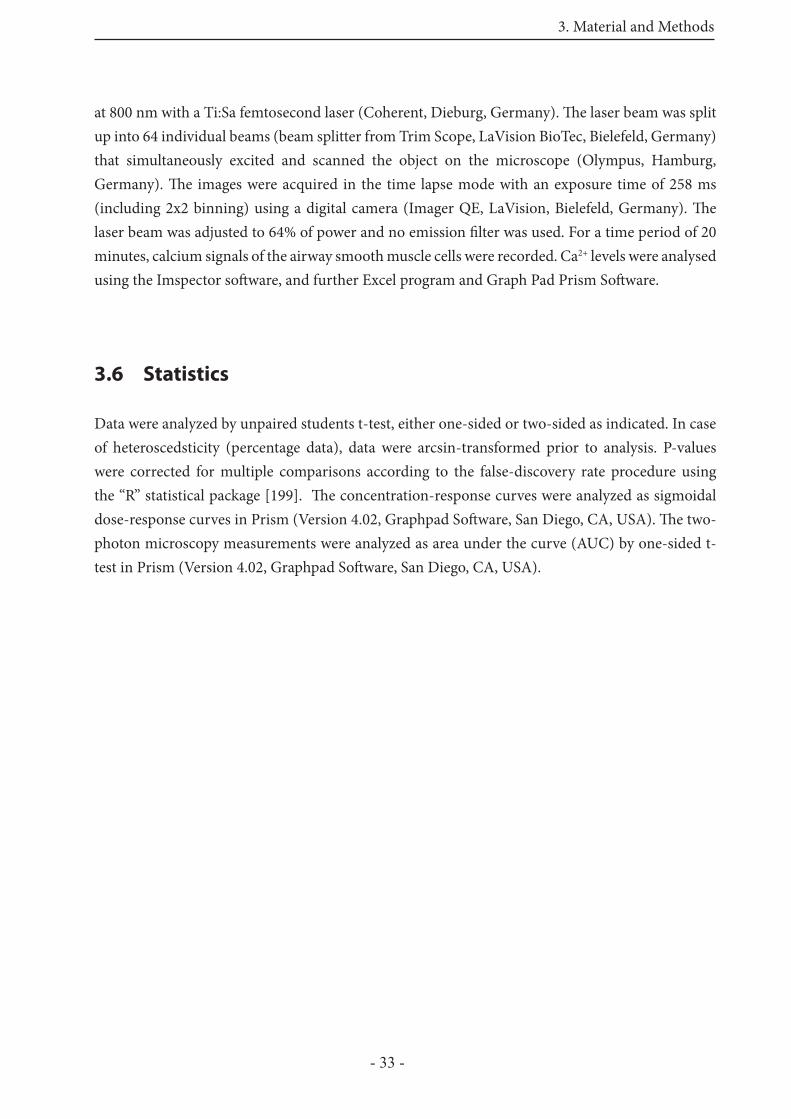
3. Material and Methods
- 33 -
at 800 nm with a Ti:Sa femtosecond laser (Coherent, Dieburg, Germany). Th e laser beam was split up into 64 individual beams (beam splitter from Trim Scope, LaVision BioTec, Bielefeld, Germany) that simultaneously excited and scanned the object on the microscope (Olympus, Hamburg, Germany). Th e images were acquired in the time lapse mode with an exposure time of 258 ms (including 2x2 binning) using a digital camera (Imager QE, LaVision, Bielefeld, Germany). Th e laser beam was adjusted to 64% of power and no emission fi lter was used. For a time period of 20 minutes, calcium signals of the airway smooth muscle cells were recorded. Ca2+ levels were analysed using the Imspector soft ware, and further Excel program and Graph Pad Prism Soft ware.
3.6 Statistics
Data were analyzed by unpaired students t-test, either one-sided or two-sided as indicated. In case of heteroscedsticity (percentage data), data were arcsin-transformed prior to analysis. P-values were corrected for multiple comparisons according to the false-discovery rate procedure using the “R” statistical package [199]. Th e concentration-response curves were analyzed as sigmoidal dose-response curves in Prism (Version 4.02, Graphpad Soft ware, San Diego, CA, USA). Th e two-photon microscopy measurements were analyzed as area under the curve (AUC) by one-sided t-test in Prism (Version 4.02, Graphpad Soft ware, San Diego, CA, USA).
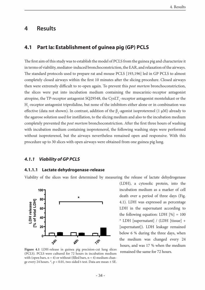
4. Results
- 34 -
4 Results
4.1 Part Ia: Establishment of guinea pig (GP) PCLS
Th e fi rst aim of this study was to establish the model of PCLS from the guinea pig and characterize it in terms of viability, mediator-induced bronchoconstriction, the EAR, and relaxation of the airways. Th e standard protocols used to prepare rat and mouse PCLS [193,196] led in GP PCLS to almost completely closed airways within the fi rst 10 minutes aft er the slicing procedure. Closed airways then were extremely diffi cult to re-open again. To prevent this post mortem bronchoconstriction, the slices were put into incubation medium containing the muscarinic-receptor antagonist atropine, the TP-receptor antagonist SQ29548, the CysLT1-receptor antagonist montelukast or the H1-receptor antagonist triprolidine, but none of the inhibitors either alone or in combination was eff ective (data not shown). In contrast, addition of the β2-agonist isoproterenol (1 μM) already to the agarose solution used for instillation, to the slicing medium and also to the incubation medium completely prevented the post mortem bronchoconstriction. Aft er the fi rst three hours of washing with incubation medium containing isoprotenerol, the following washing steps were performed without isoproterenol, but the airways nevertheless remained open and responsive. With this procedure up to 30 slices with open airways were obtained from one guinea pig lung.
4.1.1 Viability of GP PCLS
4.1.1.1 Lactate dehydrogenase-release
Viability of the slices was fi rst determined by measuring the release of lactate dehydrogenase (LDH), a cytosolic protein, into the incubation medium as a marker of cell death over a period of three days (Fig. 4.1). LDH was expressed as percentage LDH in the supernatant according to the following equation: LDH [%] = 100 * LDH [supernatant] / (LDH [tissue] + [supernatant]). LDH leakage remained below 6 % during the three days, when the medium was changed every 24 hours, and was 17 % when the medium remained the same for 72 hours.
24h
48h
72h
0
20
40
60
80
100
*
LDH
rel
ease
[% o
f tot
al L
DH
]
Figure 4.1 LDH-release in guinea pig precision-cut lung slices (PCLS). PCLS were cultured for 72 hours in incubation medium with (open bars, n = 4) or without (fi lled bars, n = 4) medium chan-ge every 24 hours. *, p < 0.01, two-sided t-test. Data are mean ± SE.
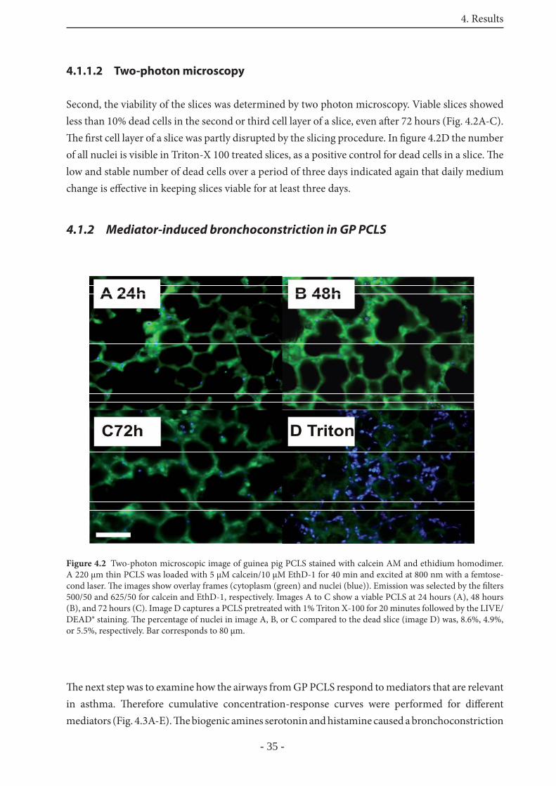
4. Results
- 35 -
4.1.1.2 Two-photon microscopy
Second, the viability of the slices was determined by two photon microscopy. Viable slices showed less than 10% dead cells in the second or third cell layer of a slice, even aft er 72 hours (Fig. 4.2A-C). Th e fi rst cell layer of a slice was partly disrupted by the slicing procedure. In fi gure 4.2D the number of all nuclei is visible in Triton-X 100 treated slices, as a positive control for dead cells in a slice. Th e low and stable number of dead cells over a period of three days indicated again that daily medium change is eff ective in keeping slices viable for at least three days.
4.1.2 Mediator-induced bronchoconstriction in GP PCLS
Th e next step was to examine how the airways from GP PCLS respond to mediators that are relevant in asthma. Th erefore cumulative concentration-response curves were performed for diff erent mediators (Fig. 4.3A-E). Th e biogenic amines serotonin and histamine caused a bronchoconstriction
Figure 4.2 Two-photon microscopic image of guinea pig PCLS stained with calcein AM and ethidium homodimer. A 220 μm thin PCLS was loaded with 5 μM calcein/10 μM EthD-1 for 40 min and excited at 800 nm with a femtose-cond laser. Th e images show overlay frames (cytoplasm (green) and nuclei (blue)). Emission was selected by the fi lters 500/50 and 625/50 for calcein and EthD-1, respectively. Images A to C show a viable PCLS at 24 hours (A), 48 hours (B), and 72 hours (C). Image D captures a PCLS pretreated with 1% Triton X-100 for 20 minutes followed by the LIVE/DEAD® staining. Th e percentage of nuclei in image A, B, or C compared to the dead slice (image D) was, 8.6%, 4.9%, or 5.5%, respectively. Bar corresponds to 80 μm.
4. Results
- 36 -
with EC50 values of 69 nM and 217 nM, respectively. Methacholine, the stable acetylcholine analogue, contracted the airways with an EC50 value of 231 nM. Th e stable TP-receptor antagonist U46619 and leukotriene D4 as representatives for eicosanoids were the most potent bronchoconstrictors with EC50 values of 16 nM and 1.8 nM, respectively.
4.1.3 Relaxation of precontracted airways
β2-agonists are widely used as spasmolytic agents in asthma therapy. Two commonly used bronchodilators, the β2-agonist salbutamol and the unspecifi c phosphodiesterase (PDE)-inhibitor IBMX were used to study relaxation of precontracted airways in guinea pig PCLS. Aft er pre-contracting airways with methacholine to 10% - 20% of their initial area, salbutamol, IBMX or a combination of both were added cumulatively. Th ese interventions produced a concentration-dependent airway relaxation with signifi cant eff ects of salbutamol above 100 nM and IBMX at 10 μM (Fig. 4.4A,B). Th e administration of both bronchodilators showed an additive eff ect, visible above concentrations of 100 nM each (Fig. 4.4C).
-10 -9 -8 -7 -6 -5
0
20
40
60
80
100
A
[log M]
airw
ay a
rea
[% o
f ini
tial a
rea]
-9 -8 -7 -6 -5
0
20
40
60
80
100
B
[log M]
airw
ay a
rea
[% o
f int
ial a
rea]
-11 -10 -9 -8 -7 -6
0
20
40
60
80
100
C
[log M]
airw
ay a
rea
[% o
f in
itia
l are
a]
Figure 4.3 Bronchoconstriction induced by several medi-ators in PCLS from guinea pigs. Concentration-response-curves for (A) histamine (■, n = 13, EC50 = 217 nM) and serotonin ( , n = 4, EC50 = 69 nM), (B) methacholine ( n = 6, EC50 = 231 nM), and (C) U46619 ( , n = 4, EC50 = 16 nM) and leukotriene D4 ( , n = 9, EC50 = 1.8 nM). Data are mean ± SE.
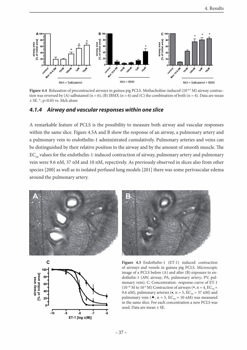
4. Results
- 37 -
4.1.4 Airway and vascular responses within one slice
A remarkable feature of PCLS is the possibility to measure both airway and vascular responses within the same slice. Figure 4.5A and B show the response of an airway, a pulmonary artery and a pulmonary vein to endothelin-1 administrated cumulatively. Pulmonary arteries and veins can be distinguished by their relative position to the airway and by the amount of smooth muscle. Th e EC50 values for the endothelin-1-induced contraction of airway, pulmonary artery and pulmonary vein were 9.6 nM, 37 nM and 10 nM, repectively. As previously observed in slices also from other species [200] as well as in isolated perfused lung models [201] there was some perivascular edema around the pulmonary artery.
contro
l
Mch 10
-6.5M
10nM
100n
M1μ
M10μM
0
20
40
60
80
100
Mch + Salbutamol
Aai
rway
are
a[%
of i
nitia
l are
a] **
*
cont
rol
Mch 10
-6.5M
10nM
100n
M1μ
M10μM
0
20
40
60
80
100
Mch + IBMX
airw
ay a
rea
[% o
f ini
tial a
rea]
B
*
contr
ol
Mch 10
-6.5M
10nM
100n
M1μ
M10μM
0
20
40
60
80
100
Mch + Salbutamol + IBMX
C ** *
airw
ay a
rea
[% o
f ini
tial a
rea]
Figure 4.4 Relaxation of precontracted airways in guinea pig PCLS. Methacholine-induced (10-6.5 M) airway contrac-tion was reversed by (A) salbutamol (n = 6), (B) IBMX (n = 6) and (C) the combination of both (n = 4). Data are mean ± SE. *, p<0.05 vs. Mch alone
A BA BB
-10 -9 -8 -7 -6
0
20
40
60
80
100
C
ET-1 [log c(M)]
airw
ay a
rea
[% o
f ini
tial a
rea]
Figure 4.5 Endothelin-1 (ET-1) induced contraction of airways and vessels in guinea pig PCLS. Microscopic image of a PCLS before (A) and aft er (B) exposure to en-dothelin-1 (AW, airway; PA, pulmonary artery; PV, pul-monary vein). C: Concentration- response-curve of ET-1 (10-10 M to 10-6 M) Contraction of airways ( , n = 4, EC50 = 9.6 nM), pulmonary arteries (■, n = 5, EC50 = 37 nM) and pulmonary vein ( , n = 5, EC50 = 10 nM) was measured in the same slice. For each concentration a new PCLS was used. Data are mean ± SE.
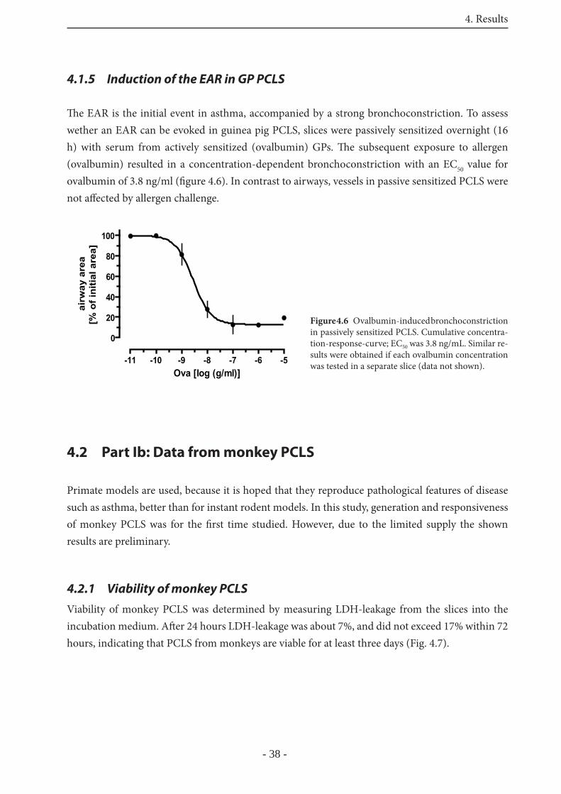
4. Results
- 38 -
4.1.5 Induction of the EAR in GP PCLS
Th e EAR is the initial event in asthma, accompanied by a strong bronchoconstriction. To assess wether an EAR can be evoked in guinea pig PCLS, slices were passively sensitized overnight (16 h) with serum from actively sensitized (ovalbumin) GPs. Th e subsequent exposure to allergen (ovalbumin) resulted in a concentration-dependent bronchoconstriction with an EC50 value for ovalbumin of 3.8 ng/ml (fi gure 4.6). In contrast to airways, vessels in passive sensitized PCLS were not aff ected by allergen challenge.
4.2 Part Ib: Data from monkey PCLS
Primate models are used, because it is hoped that they reproduce pathological features of disease such as asthma, better than for instant rodent models. In this study, generation and responsiveness of monkey PCLS was for the fi rst time studied. However, due to the limited supply the shown results are preliminary.
4.2.1 Viability of monkey PCLS
Viability of monkey PCLS was determined by measuring LDH-leakage from the slices into the incubation medium. Aft er 24 hours LDH-leakage was about 7%, and did not exceed 17% within 72 hours, indicating that PCLS from monkeys are viable for at least three days (Fig. 4.7).
-11 -10 -9 -8 -7 -6 -5
0
20
40
60
80
100
Ova [log (g/ml)]
airw
ay a
rea
[% o
f ini
tial a
rea]
Figure 4.6 Ovalbumin-induced bronchoconstriction in passively sensitized PCLS. Cumulative concentra-tion-response-curve; EC50 was 3.8 ng/mL. Similar re-sults were obtained if each ovalbumin concentration was tested in a separate slice (data not shown).
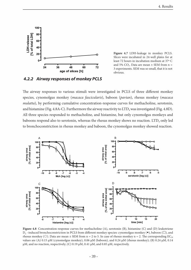
4. Results
- 39 -
4.2.2 Airway responses of monkey PCLS
Th e airway responses to various stimuli were investigated in PCLS of three diff erent monkey species, cynomolgus monkey (macaca fascicularis), baboon (pavian), rhesus monkey (macaca mulatta), by performing cumulative concentration-response curves for methacholine, serotonin, and histamine (Fig. 4.8A-C). Furthermore the airway reactivity to LTD4 was investigated (Fig. 4.8D). All three species responded to methacholine, and histamine, but only cynomolgus monkeys and baboons respond also to serotonin, whereas the rhesus monkey shows no reaction. LTD4 only led to bronchoconstriction in rhesus monkey and baboon, the cynomolgus monkey showed reaction.
24 36 48 60 72
0
20
40
60
80
100
age of slices [h]
LDH-
rele
ase
[% o
f tot
al L
DH]
Figure 4.7 LDH-leakage in monkey PCLS. Slices were incubated in 24-well plates for at least 72 hours in incubation medium at 37º C and 5% CO2. Data are mean ± SEM from n = 3 experiments. SEM was so small, that it is not obvious.
-10 -9 -8 -7 -6 -5 -4
0
20
40
60
80
100
A
Mch [log (c)]
airw
ay a
rea
[% o
f int
ial a
rea]
-10 -9 -8 -7 -6 -5
0
20
40
60
80
100B
serotonin [log (c)]
airw
ay a
rea
[% o
f int
ial a
rea]
-8 -7 -6 -5 -4
0
20
40
60
80
100C
histamine [log (c)]
airw
ay a
rea
[% o
f int
ial a
rea]
0 5 10 15 20
0
20
40
60
80
100
time [min]
airw
ay a
rea
[% o
f the
initi
al a
rea]
D
Figure 4.8 Concentration-response curves for methacholine (A), serotonin (B), histamine (C) and (D) leukotriene D4 –induced bronchoconstriction in PCLS from diff erent monkey species: cynomolgus monkey ( ), baboon ( ), and rhesus monkey ( ). Data are mean ± SEM from n = 2 to 5. In case of rhesus monkey n = 2. Th e corresponding EC50 values are (A) 0.15 μM (cynomolgus monkey), 0.06 μM (baboon), and 0.24 μM (rhesus monkey); (B) 0.24 μM, 0.14 μM, and no reaction, respectively; (C) 0.19 μM, 0.41 μM, and 0.85 μM, respectively.
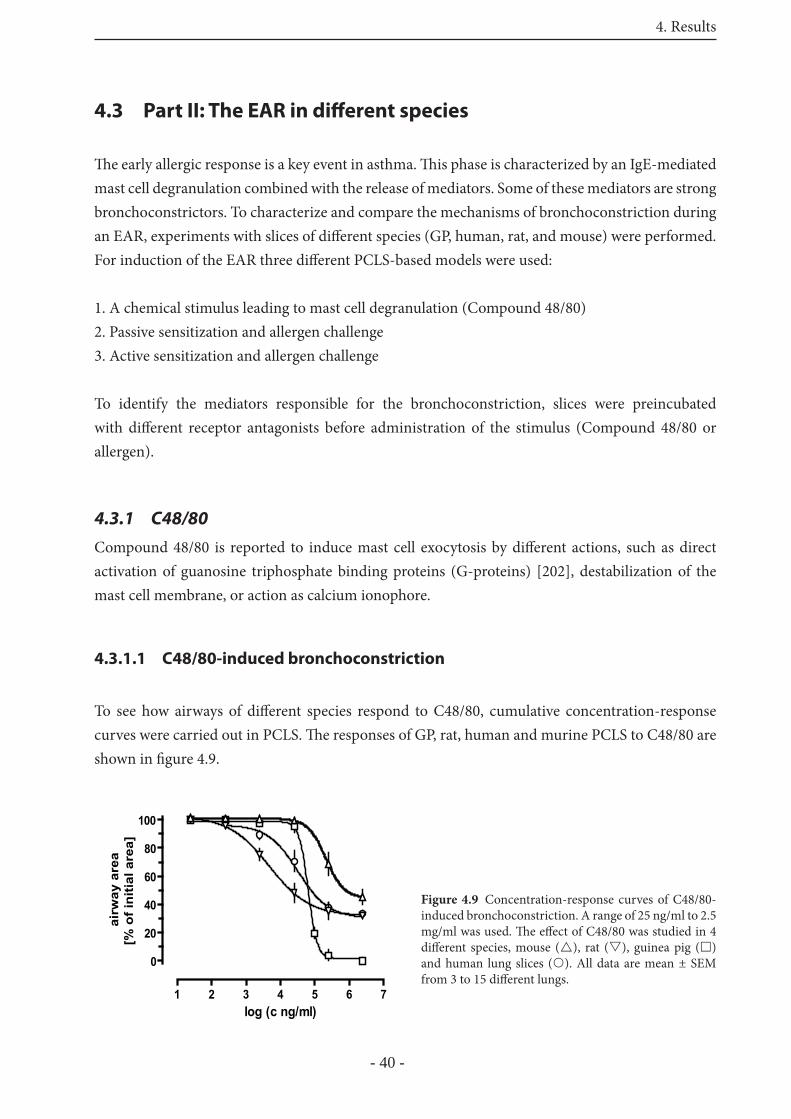
4. Results
- 40 -
4.3 Part II: The EAR in diff erent species
Th e early allergic response is a key event in asthma. Th is phase is characterized by an IgE-mediated mast cell degranulation combined with the release of mediators. Some of these mediators are strong bronchoconstrictors. To characterize and compare the mechanisms of bronchoconstriction during an EAR, experiments with slices of diff erent species (GP, human, rat, and mouse) were performed. For induction of the EAR three diff erent PCLS-based models were used:
1. A chemical stimulus leading to mast cell degranulation (Compound 48/80)2. Passive sensitization and allergen challenge3. Active sensitization and allergen challenge
To identify the mediators responsible for the bronchoconstriction, slices were preincubated with diff erent receptor antagonists before administration of the stimulus (Compound 48/80 or allergen).
4.3.1 C48/80
Compound 48/80 is reported to induce mast cell exocytosis by diff erent actions, such as direct activation of guanosine triphosphate binding proteins (G-proteins) [202], destabilization of the mast cell membrane, or action as calcium ionophore.
4.3.1.1 C48/80-induced bronchoconstriction
To see how airways of diff erent species respond to C48/80, cumulative concentration-response curves were carried out in PCLS. Th e responses of GP, rat, human and murine PCLS to C48/80 are shown in fi gure 4.9.
1 2 3 4 5 6 7
0
20
40
60
80
100
log (c ng/ml)
airw
ay a
rea
[% o
f ini
tial a
rea]
Figure 4.9 Concentration-response curves of C48/80-induced bronchoconstriction. A range of 25 ng/ml to 2.5 mg/ml was used. Th e eff ect of C48/80 was studied in 4 diff erent species, mouse ( ), rat ( ), guinea pig ( ) and human lung slices ( ). All data are mean ± SEM from 3 to 15 diff erent lungs.
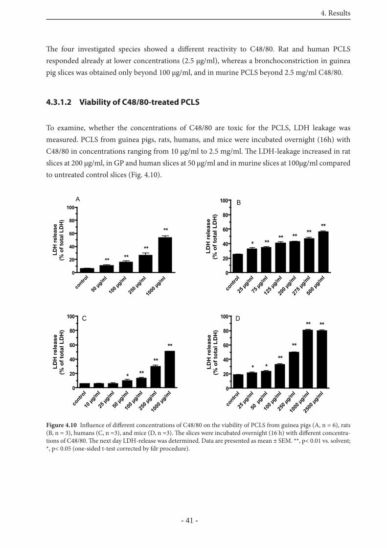
4. Results
- 41 -
Th e four investigated species showed a diff erent reactivity to C48/80. Rat and human PCLS responded already at lower concentrations (2.5 μg/ml), whereas a bronchoconstriction in guinea pig slices was obtained only beyond 100 μg/ml, and in murine PCLS beyond 2.5 mg/ml C48/80.
4.3.1.2 Viability of C48/80-treated PCLS
To examine, whether the concentrations of C48/80 are toxic for the PCLS, LDH leakage was measured. PCLS from guinea pigs, rats, humans, and mice were incubated overnight (16h) with C48/80 in concentrations ranging from 10 μg/ml to 2.5 mg/ml. Th e LDH-leakage increased in rat slices at 200 μg/ml, in GP and human slices at 50 μg/ml and in murine slices at 100μg/ml compared to untreated control slices (Fig. 4.10).
contro
l
50 μg
/ml
100 μ
g/ml
250 μ
g/ml
1000
μg/m
l 0
20
40
60
80
100A
**
**
****
LDH
rele
ase
(% o
f tot
al L
DH
)
cont
rol
25 μg
/ml
75 μg
/ml
125 μ
g/ml
200 μ
g/ml
275 μ
g/ml
500 μ
g/ml
0
20
40
60
80
100 BLD
H r
elea
se(%
of t
otal
LD
H)
** ******
**
*
cont
rol
10 μg
/ml
25 μg
/ml
50 μg
/ml
100 μ
g/ml
250 μ
g/ml
1000
μg/m
l0
20
40
60
80
100 C
LDH
rel
ease
(% o
f tot
al L
DH
)
* **
**
**
cont
rol
25 μg
/ml
50 μ
g/ml
100 μ
g/ml
250 μ
g/ml
1000
μg/m
l
2500
μg/m
l0
20
40
60
80
100 D
LDH
rel
ease
(% o
f tot
al L
DH
)
**
**
**
**
* *
Figure 4.10 Infl uence of diff erent concentrations of C48/80 on the viability of PCLS from guinea pigs (A, n = 6), rats (B, n = 3), humans (C, n =3), and mice (D, n =3). Th e slices were incubated overnight (16 h) with diff erent concentra-tions of C48/80. Th e next day LDH-release was determined. Data are presented as mean ± SEM. **, p< 0.01 vs. solvent; *, p< 0.05 (one-sided t-test corrected by fdr procedure).
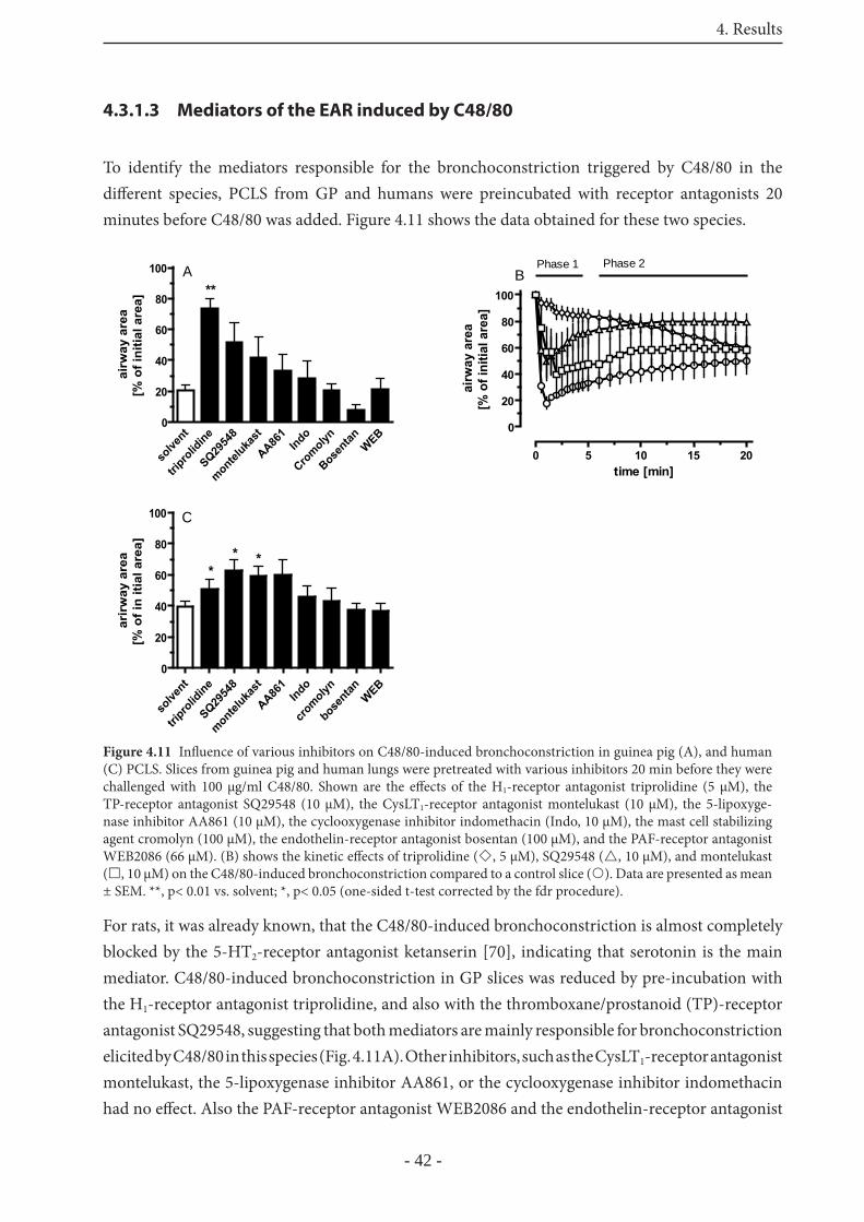
4. Results
- 42 -
4.3.1.3 Mediators of the EAR induced by C48/80
To identify the mediators responsible for the bronchoconstriction triggered by C48/80 in the diff erent species, PCLS from GP and humans were preincubated with receptor antagonists 20 minutes before C48/80 was added. Figure 4.11 shows the data obtained for these two species.
For rats, it was already known, that the C48/80-induced bronchoconstriction is almost completely blocked by the 5-HT2-receptor antagonist ketanserin [70], indicating that serotonin is the main mediator. C48/80-induced bronchoconstriction in GP slices was reduced by pre-incubation with the H1-receptor antagonist triprolidine, and also with the thromboxane/prostanoid (TP)-receptor antagonist SQ29548, suggesting that both mediators are mainly responsible for bronchoconstriction elicited by C48/80 in this species (Fig. 4.11A). Other inhibitors, such as the CysLT1-receptor antagonist montelukast, the 5-lipoxygenase inhibitor AA861, or the cyclooxygenase inhibitor indomethacin had no eff ect. Also the PAF-receptor antagonist WEB2086 and the endothelin-receptor antagonist
solve
nt
tripro
lidine
SQ2954
8
monteluka
st
AA861
Indo
Cromolyn
Bosentan WEB
0
20
40
60
80
100 A**
airw
ay a
rea
[% o
f ini
tial a
rea]
0 5 10 15 20
0
20
40
60
80
100
Phase 1 Phase 2
time [min]ai
rway
are
a[%
of i
nitia
l are
a]
B
solve
nt
tripro
lidine
SQ2954
8
monteluka
st
AA861
Indo
cromolyn
bosentan WEB
0
20
40
60
80
100 C
***
arir
way
are
a[%
of i
n iti
al a
rea]
Figure 4.11 Infl uence of various inhibitors on C48/80-induced bronchoconstriction in guinea pig (A), and human (C) PCLS. Slices from guinea pig and human lungs were pretreated with various inhibitors 20 min before they were challenged with 100 μg/ml C48/80. Shown are the eff ects of the H1-receptor antagonist triprolidine (5 μM), the TP-receptor antagonist SQ29548 (10 μM), the CysLT1-receptor antagonist montelukast (10 μM), the 5-lipoxyge-nase inhibitor AA861 (10 μM), the cyclooxygenase inhibitor indomethacin (Indo, 10 μM), the mast cell stabilizing agent cromolyn (100 μM), the endothelin-receptor antagonist bosentan (100 μM), and the PAF-receptor antagonist WEB2086 (66 μM). (B) shows the kinetic eff ects of triprolidine ( , 5 μM), SQ29548 ( , 10 μM), and montelukast ( , 10 μM) on the C48/80-induced bronchoconstriction compared to a control slice ( ). Data are presented as mean ± SEM. **, p< 0.01 vs. solvent; *, p< 0.05 (one-sided t-test corrected by the fdr procedure).
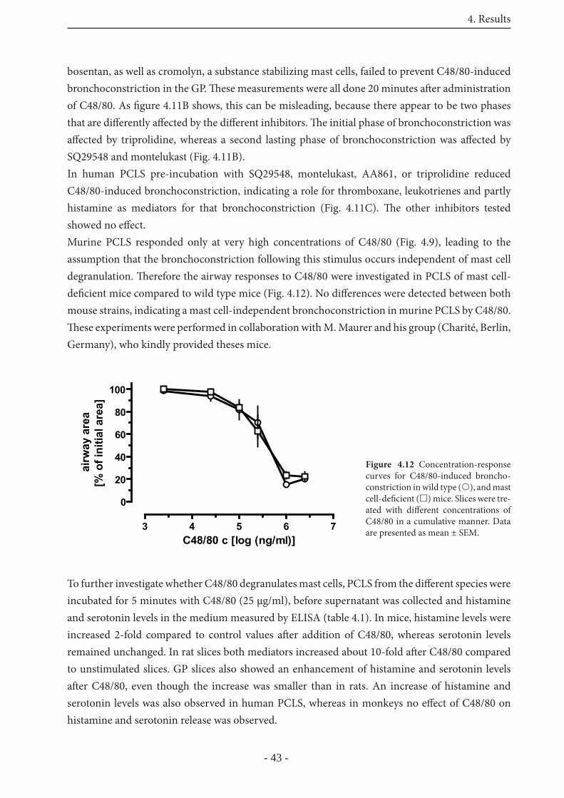
4. Results
- 43 -
bosentan, as well as cromolyn, a substance stabilizing mast cells, failed to prevent C48/80-induced bronchoconstriction in the GP. Th ese measurements were all done 20 minutes aft er administration of C48/80. As fi gure 4.11B shows, this can be misleading, because there appear to be two phases that are diff erently aff ected by the diff erent inhibitors. Th e initial phase of bronchoconstriction was aff ected by triprolidine, whereas a second lasting phase of bronchoconstriction was aff ected by SQ29548 and montelukast (Fig. 4.11B).In human PCLS pre-incubation with SQ29548, montelukast, AA861, or triprolidine reduced C48/80-induced bronchoconstriction, indicating a role for thromboxane, leukotrienes and partly histamine as mediators for that bronchoconstriction (Fig. 4.11C). Th e other inhibitors tested showed no eff ect. Murine PCLS responded only at very high concentrations of C48/80 (Fig. 4.9), leading to the assumption that the bronchoconstriction following this stimulus occurs independent of mast cell degranulation. Th erefore the airway responses to C48/80 were investigated in PCLS of mast cell- defi cient mice compared to wild type mice (Fig. 4.12). No diff erences were detected between both mouse strains, indicating a mast cell-independent bronchoconstriction in murine PCLS by C48/80. Th ese experiments were performed in collaboration with M. Maurer and his group (Charité, Berlin, Germany), who kindly provided theses mice.
To further investigate whether C48/80 degranulates mast cells, PCLS from the diff erent species were incubated for 5 minutes with C48/80 (25 μg/ml), before supernatant was collected and histamine and serotonin levels in the medium measured by ELISA (table 4.1). In mice, histamine levels were increased 2-fold compared to control values aft er addition of C48/80, whereas serotonin levels remained unchanged. In rat slices both mediators increased about 10-fold aft er C48/80 compared to unstimulated slices. GP slices also showed an enhancement of histamine and serotonin levels aft er C48/80, even though the increase was smaller than in rats. An increase of histamine and serotonin levels was also observed in human PCLS, whereas in monkeys no eff ect of C48/80 on histamine and serotonin release was observed.
3 4 5 6 7
0
20
40
60
80
100
C48/80 c [log (ng/ml)]
airw
ay a
rea
[% o
f ini
tial a
rea]
Figure 4.12 Concentration-response curves for C48/80-induced broncho-constriction in wild type ( ), and mast cell-defi cient ( ) mice. Slices were tre-ated with diff erent concentrations of C48/80 in a cumulative manner. Data are presented as mean ± SEM.
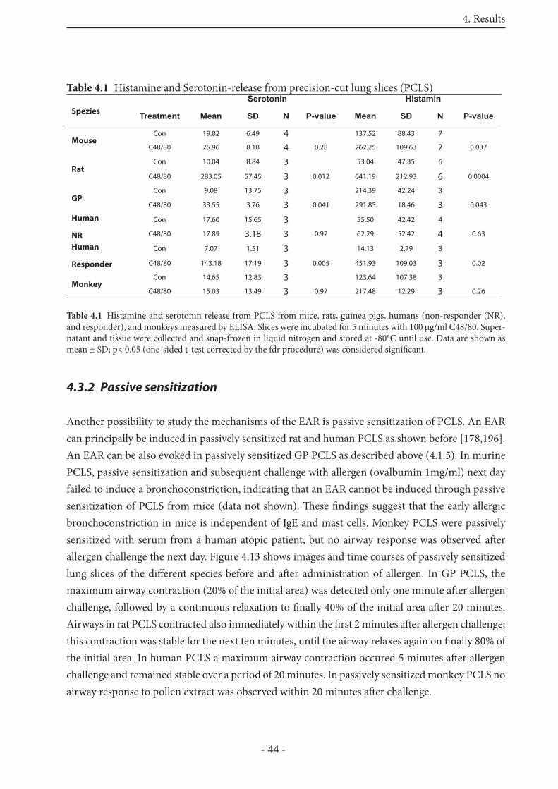
4. Results
- 44 -
4.3.2 Passive sensitization
Another possibility to study the mechanisms of the EAR is passive sensitization of PCLS. An EAR can principally be induced in passively sensitized rat and human PCLS as shown before [178,196]. An EAR can be also evoked in passively sensitized GP PCLS as described above (4.1.5). In murine PCLS, passive sensitization and subsequent challenge with allergen (ovalbumin 1mg/ml) next day failed to induce a bronchoconstriction, indicating that an EAR cannot be induced through passive sensitization of PCLS from mice (data not shown). Th ese fi ndings suggest that the early allergic bronchoconstriction in mice is independent of IgE and mast cells. Monkey PCLS were passively sensitized with serum from a human atopic patient, but no airway response was observed aft er allergen challenge the next day. Figure 4.13 shows images and time courses of passively sensitized lung slices of the diff erent species before and aft er administration of allergen. In GP PCLS, the maximum airway contraction (20% of the initial area) was detected only one minute aft er allergen challenge, followed by a continuous relaxation to fi nally 40% of the initial area aft er 20 minutes. Airways in rat PCLS contracted also immediately within the fi rst 2 minutes aft er allergen challenge; this contraction was stable for the next ten minutes, until the airway relaxes again on fi nally 80% of the initial area. In human PCLS a maximum airway contraction occured 5 minutes aft er allergen challenge and remained stable over a period of 20 minutes. In passively sensitized monkey PCLS no airway response to pollen extract was observed within 20 minutes aft er challenge.
Table 4.1 Histamine and Serotonin-release from precision-cut lung slices (PCLS)
Spezies
Serotonin Histamin
Treatment Mean SD N P-value Mean SD N P-value
MouseCon 19.82 6.49 4 137.52 88.43 7
C48/80 25.96 8.18 4 0.28 262.25 109.63 7 0.037
RatCon 10.04 8.84 3 53.04 47.35 6
C48/80 283.05 57.45 3 0.012 641.19 212.93 6 0.0004
GPCon 9.08 13.75 3 214.39 42.24 3
C48/80 33.55 3.76 3 0.041 291.85 18.46 3 0.043
Human
NR
Con 17.60 15.65 3 55.50 42.42 4 C48/80 17.89 3.18 3 0.97 62.29 52.42 4 0.63
Human
Responder
Con 7.07 1.51 3 14.13 2.79 3 C48/80 143.18 17.19 3 0.005 451.93 109.03 3 0.02
MonkeyCon 14.65 12.83 3 123.64 107.38 3
C48/80 15.03 13.49 3 0.97 217.48 12.29 3 0.26
Table 4.1 Histamine and serotonin release from PCLS from mice, rats, guinea pigs, humans (non-responder (NR), and responder), and monkeys measured by ELISA. Slices were incubated for 5 minutes with 100 μg/ml C48/80. Super-natant and tissue were collected and snap-frozen in liquid nitrogen and stored at -80°C until use. Data are shown as mean ± SD; p< 0.05 (one-sided t-test corrected by the fdr procedure) was considered signifi cant.
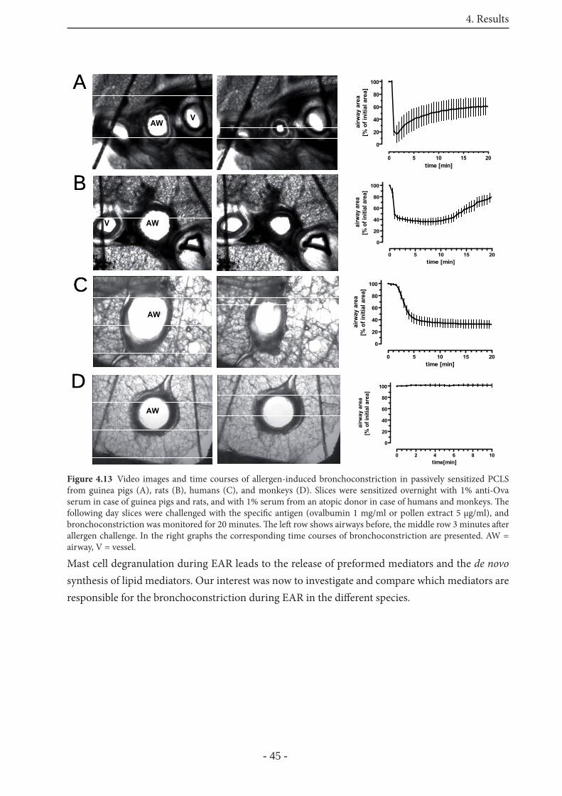
4. Results
- 45 -
Mast cell degranulation during EAR leads to the release of preformed mediators and the de novo synthesis of lipid mediators. Our interest was now to investigate and compare which mediators are responsible for the bronchoconstriction during EAR in the diff erent species.
0 5 10 15 20
0
20
40
60
80
100
time [min]
airw
ay a
rea
[% o
f ini
tial a
rea]
0 5 10 15 20
0
20
40
60
80
100
time [min]
airw
ay a
rea
[% o
f ini
tial a
rea]
0 5 10 15 20
0
20
40
60
80
100
time [min]ai
rway
are
a[%
of i
nitia
l are
a]
Figure 4.13 Video images and time courses of allergen-induced bronchoconstriction in passively sensitized PCLS from guinea pigs (A), rats (B), humans (C), and monkeys (D). Slices were sensitized overnight with 1% anti-Ova serum in case of guinea pigs and rats, and with 1% serum from an atopic donor in case of humans and monkeys. Th e following day slices were challenged with the specifi c antigen (ovalbumin 1 mg/ml or pollen extract 5 μg/ml), and bronchoconstriction was monitored for 20 minutes. Th e left row shows airways before, the middle row 3 minutes aft er allergen challenge. In the right graphs the corresponding time courses of bronchoconstriction are presented. AW = airway, V = vessel.
A
AWV
A
AWV
B
AWV
B
AWV
CAW
CAW
DAW
DAW
0 2 4 6 8 10
0
20
40
60
80
100
time[min]
airw
ay a
rea
[% o
f ini
tial a
rea]
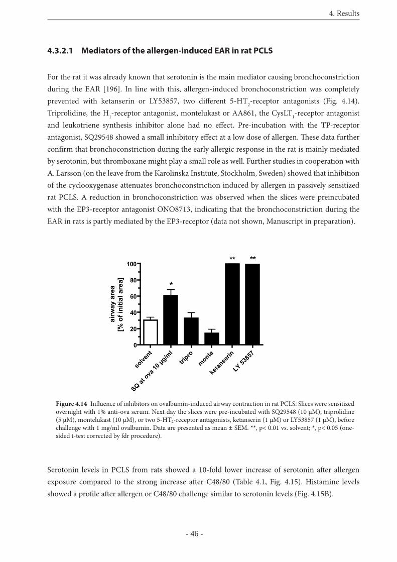
4. Results
- 46 -
4.3.2.1 Mediators of the allergen-induced EAR in rat PCLS
For the rat it was already known that serotonin is the main mediator causing bronchoconstriction during the EAR [196]. In line with this, allergen-induced bronchoconstriction was completely prevented with ketanserin or LY53857, two diff erent 5-HT2-receptor antagonists (Fig. 4.14). Triprolidine, the H1-receptor antagonist, montelukast or AA861, the CysLT1-receptor antagonist and leukotriene synthesis inhibitor alone had no eff ect. Pre-incubation with the TP-receptor antagonist, SQ29548 showed a small inhibitory eff ect at a low dose of allergen. Th ese data further confi rm that bronchoconstriction during the early allergic response in the rat is mainly mediated by serotonin, but thromboxane might play a small role as well. Further studies in cooperation with A. Larsson (on the leave from the Karolinska Institute, Stockholm, Sweden) showed that inhibition of the cyclooxygenase attenuates bronchoconstriction induced by allergen in passively sensitized rat PCLS. A reduction in bronchoconstriction was observed when the slices were preincubated with the EP3-receptor antagonist ONO8713, indicating that the bronchoconstriction during the EAR in rats is partly mediated by the EP3-receptor (data not shown, Manuscript in preparation).
Serotonin levels in PCLS from rats showed a 10-fold lower increase of serotonin aft er allergen exposure compared to the strong increase aft er C48/80 (Table 4.1, Fig. 4.15). Histamine levels showed a profi le aft er allergen or C48/80 challenge similar to serotonin levels (Fig. 4.15B).
solve
nt
SQ at ova
10 μg
/ml
tripro
monte
ketan
serin
LY 5385
70
20
40
60
80
100
*
** **
airw
ay a
rea
[% o
f ini
tial a
rea]
Figure 4.14 Infl uence of inhibitors on ovalbumin-induced airway contraction in rat PCLS. Slices were sensitized overnight with 1% anti-ova serum. Next day the slices were pre-incubated with SQ29548 (10 μM), triprolidine (5 μM), montelukast (10 μM), or two 5-HT2-receptor antagonists, ketanserin (1 μM) or LY53857 (1 μM), before challenge with 1 mg/ml ovalbumin. Data are presented as mean ± SEM. **, p< 0.01 vs. solvent; *, p< 0.05 (one-sided t-test corrected by fdr procedure).
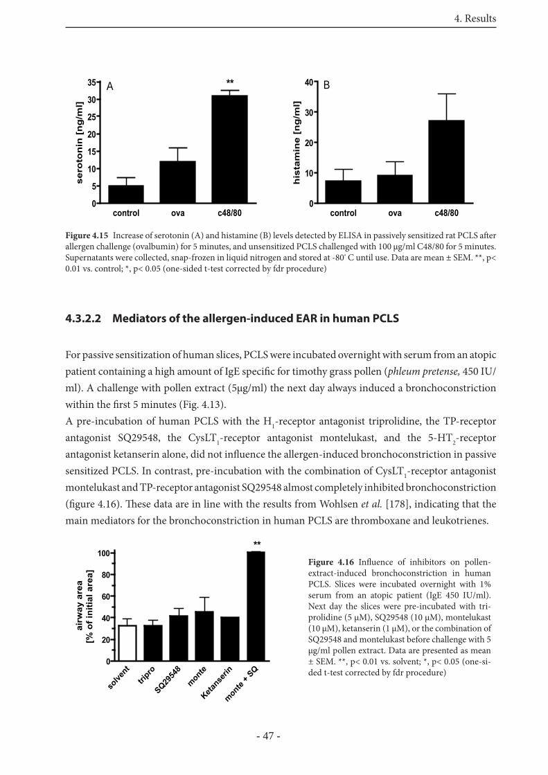
4. Results
- 47 -
4.3.2.2 Mediators of the allergen-induced EAR in human PCLS
For passive sensitization of human slices, PCLS were incubated overnight with serum from an atopic patient containing a high amount of IgE specifi c for timothy grass pollen (phleum pretense, 450 IU/ml). A challenge with pollen extract (5μg/ml) the next day always induced a bronchoconstriction within the fi rst 5 minutes (Fig. 4.13).A pre-incubation of human PCLS with the H1-receptor antagonist triprolidine, the TP-receptor antagonist SQ29548, the CysLT1-receptor antagonist montelukast, and the 5-HT2-receptor antagonist ketanserin alone, did not infl uence the allergen-induced bronchoconstriction in passive sensitized PCLS. In contrast, pre-incubation with the combination of CysLT1-receptor antagonist montelukast and TP-receptor antagonist SQ29548 almost completely inhibited bronchoconstriction (fi gure 4.16). Th ese data are in line with the results from Wohlsen et al. [178], indicating that the main mediators for the bronchoconstriction in human PCLS are thromboxane and leukotrienes.
control ova c48/800
5
10
15
20
25
30
35 Ase
roto
nin
[n
g/m
l]**
control ova c48/800
10
20
30
40 B
his
tam
ine
[ng
/ml]
Figure 4.15 Increase of serotonin (A) and histamine (B) levels detected by ELISA in passively sensitized rat PCLS aft er allergen challenge (ovalbumin) for 5 minutes, and unsensitized PCLS challenged with 100 μg/ml C48/80 for 5 minutes.Supernatants were collected, snap-frozen in liquid nitrogen and stored at -80º C until use. Data are mean ± SEM. **, p< 0.01 vs. control; *, p< 0.05 (one-sided t-test corrected by fdr procedure)
Figure 4.16 Infl uence of inhibitors on pollen-extract-induced bronchoconstriction in human PCLS. Slices were incubated overnight with 1% serum from an atopic patient (IgE 450 IU/ml). Next day the slices were pre-incubated with tri-prolidine (5 μM), SQ29548 (10 μM), montelukast (10 μM), ketanserin (1 μM), or the combination of SQ29548 and montelukast before challenge with 5 μg/ml pollen extract. Data are presented as mean ± SEM. **, p< 0.01 vs. solvent; *, p< 0.05 (one-si-ded t-test corrected by fdr procedure)
solve
nttri
pro
SQ2954
8
monte
Ketans
erin
monte
+ SQ
0
20
40
60
80
100**
airw
ay a
rea
[% o
f ini
tial a
rea]
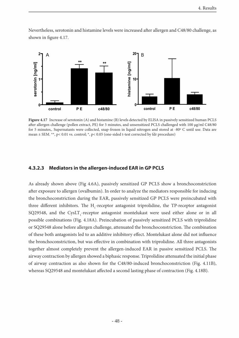
4. Results
- 48 -
Nevertheless, serotonin and histamine levels were increased aft er allergen and C48/80 challenge, as shown in fi gure 4.17.
4.3.2.3 Mediators in the allergen-induced EAR in GP PCLS
As already shown above (Fig 4.6A), passively sensitized GP PCLS show a bronchoconstriction aft er exposure to allergen (ovalbumin). In order to analyze the mediators responsible for inducing the bronchoconstriction during the EAR, passively sensitized GP PCLS were preincubated with three diff erent inhibitors. Th e H1-receptor antagonist triprolidine, the TP-receptor antagonist SQ29548, and the CysLT1-receptor antagonist montelukast were used either alone or in all possible combinations (Fig. 4.18A). Preincubation of passively sensitized PCLS with triprolidine or SQ29548 alone before allergen challenge, attenuated the bronchoconstriction. Th e combination of these both antagonists led to an additive inhibitory eff ect. Montelukast alone did not infl uence the bronchoconstriction, but was eff ective in combination with triprolidine. All three antagonists together almost completely prevent the allergen-induced EAR in passive sensitized PCLS. Th e airway contraction by allergen showed a biphasic response. Triprolidine attenuated the initial phase of airway contraction as also shown for the C48/80-induced bronchoconstriction (Fig. 4.11B), whereas SQ29548 and montelukast aff ected a second lasting phase of contraction (Fig. 4.18B).
Figure 4.17 Increase of serotonin (A) and histamine (B) levels detected by ELISA in passively sensitized human PCLS aft er allergen challenge (pollen extract, PE) for 5 minutes, and unsensitized PCLS challenged with 100 μg/ml C48/80 for 5 minutes,. Supernatants were collected, snap-frozen in liquid nitrogen and stored at -80º C until use. Data are mean ± SEM. **, p< 0.01 vs. control; *, p< 0.05 (one-sided t-test corrected by fdr procedure)
control P E c48/800
1
2 A** **
sero
toni
n [n
g/m
l]
control P E c48/800
10
20 B
hist
amin
e [n
g/m
l]
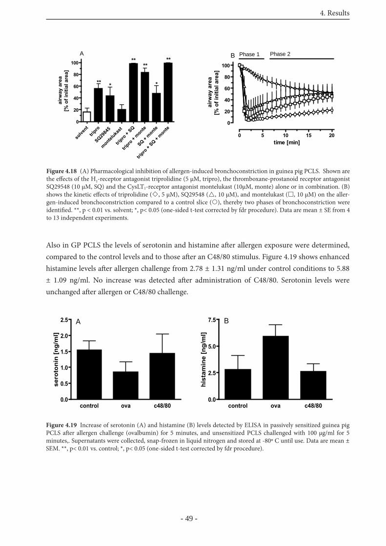
4. Results
- 49 -
Also in GP PCLS the levels of serotonin and histamine aft er allergen exposure were determined, compared to the control levels and to those aft er an C48/80 stimulus. Figure 4.19 shows enhanced histamine levels aft er allergen challenge from 2.78 ± 1.31 ng/ml under control conditions to 5.88 ± 1.09 ng/ml. No increase was detected aft er administration of C48/80. Serotonin levels were unchanged aft er allergen or C48/80 challenge.
0 5 10 15 20
0
20
40
60
80
100
time [min]
airw
ay a
rea
[% o
f ini
tial a
rea]
Phase 1 Phase 2B
Figure 4.18 (A) Pharmacological inhibition of allergen-induced bronchoconstriction in guinea pig PCLS. Shown are the eff ects of the H1-receptor antagonist triprolidine (5 μM, tripro), the thromboxane-prostanoid receptor antagonist SQ29548 (10 μM, SQ) and the CysLT1-receptor antagonist montelukast (10μM, monte) alone or in combination. (B) shows the kinetic eff ects of triprolidine ( , 5 μM), SQ29548 ( , 10 μM), and montelukast ( , 10 μM) on the aller-gen-induced bronchoconstriction compared to a control slice ( ), thereby two phases of bronchoconstriction were identifi ed. **, p < 0.01 vs. solvent; *, p< 0.05 (one-sided t-test corrected by fdr procedure). Data are mean ± SE from 4 to 13 independent experiments.
solve
nttri
pro
SQ2984
5
monteluka
st
tripro
+ SQ
tripro
+ monte
SQ + monte
tripro
+ SQ +
monte0
20
40
60
80
100
*** *
****
**ai
rway
are
a[%
of i
nitia
l are
a]
A
control ova c48/800.0
0.5
1.0
1.5
2.0
2.5 A
sero
toni
n [n
g/m
l]
control ova c48/800.0
2.5
5.0
7.5 B
hist
amin
e [n
g/m
l]
Figure 4.19 Increase of serotonin (A) and histamine (B) levels detected by ELISA in passively sensitized guinea pig PCLS aft er allergen challenge (ovalbumin) for 5 minutes, and unsensitized PCLS challenged with 100 μg/ml for 5 minutes,. Supernatants were collected, snap-frozen in liquid nitrogen and stored at -80º C until use. Data are mean ± SEM. **, p< 0.01 vs. control; *, p< 0.05 (one-sided t-test corrected by fdr procedure).
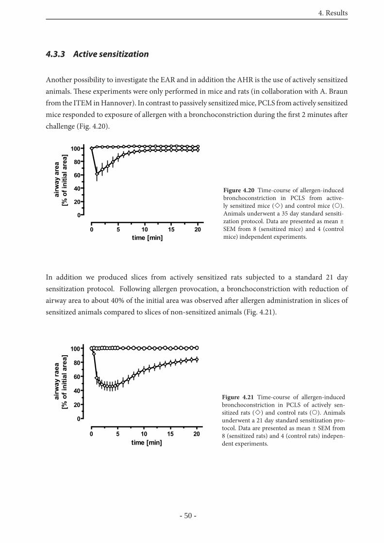
4. Results
- 50 -
4.3.3 Active sensitization
Another possibility to investigate the EAR and in addition the AHR is the use of actively sensitized animals. Th ese experiments were only performed in mice and rats (in collaboration with A. Braun from the ITEM in Hannover). In contrast to passively sensitized mice, PCLS from actively sensitized mice responded to exposure of allergen with a bronchoconstriction during the fi rst 2 minutes aft er challenge (Fig. 4.20).
In addition we produced slices from actively sensitized rats subjected to a standard 21 day sensitization protocol. Following allergen provocation, a bronchoconstriction with reduction of airway area to about 40% of the initial area was observed aft er allergen administration in slices of sensitized animals compared to slices of non-sensitized animals (Fig. 4.21).
Figure 4.20 Time-course of allergen-induced bronchoconstriction in PCLS from active-ly sensitized mice ( ) and control mice ( ). Animals underwent a 35 day standard sensiti-zation protocol. Data are presented as mean ± SEM from 8 (sensitized mice) and 4 (control mice) independent experiments.
0 5 10 15 20
0
20
40
60
80
100
time [min]
airw
ay a
rea
[% o
f ini
tial a
rea]
0 5 10 15 20
0
20
40
60
80
100
time [min]
airw
ay ra
ea[%
of i
nitia
l are
a]
Figure 4.21 Time-course of allergen-induced bronchoconstriction in PCLS of actively sen-sitized rats ( ) and control rats ( ). Animals underwent a 21 day standard sensitization pro-tocol. Data are presented as mean ± SEM from 8 (sensitized rats) and 4 (control rats) indepen-dent experiments.
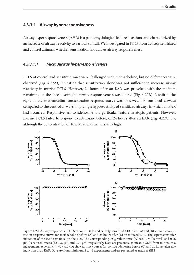
4. Results
- 51 -
4.3.3.1 Airway hyperresponsiveness
Airway hyperresponsiveness (AHR) is a pathophysiological feature of asthma and characterized by an increase of airway reactivity to various stimuli. We investigated in PCLS from actively sensitized and control animals, whether sensitization modulates airway responsiveness.
4.3.3.1.1 Mice: Airway hyperresponsiveness
PCLS of control and sensitized mice were challenged with methacholine, but no diff erences were observed (Fig. 4.22A), indicating that sensitization alone was not suffi cient to increase airway reactivity in murine PCLS. However, 24 hours aft er an EAR was provoked with the medium remaining on the slices overnight, airway responsiveness was altered (Fig. 4.22B). A shift to the right of the methacholine concentration-response curve was observed for sensitized airways compared to the control airways, implying a hyporeactivity of sensitized airways in which an EAR had occurred. Responsiveness to adenosine is a particular feature in atopic patients. However, murine PCLS failed to respond to adenosine before, or 24 hours aft er an EAR (Fig. 4.22C, D), although the concentration of 10 mM adenosine was very high.
-8 -7 -6 -5 -4
0
20
40
60
80
100
A
Mch [log (C)]
airw
ay a
rea
[% o
f ini
tial a
rea]
-8 -7 -6 -5 -4
0
20
40
60
80
100
B
Mch [log (C)]
airw
ay a
rea
[% o
f ini
tial a
rea]
0 5 10 15 20
0
20
40
60
80
100
C
time [min]
airw
ay a
rea
[% o
f ini
tial a
rea]
0 5 10 15 20
0
20
40
60
80
100
D
time [min]
airw
ay a
rea
[% o
f ini
tial a
rea]
Figure 4.22 Airway responses in PCLS of control ( ) and actively sensitized ( ) mice. (A) and (B) showed concen-tration-response curves for methacholine before (A) and 24 hours aft er (B) an induced EAR. Th e supernatant aft er induction of the EAR remained on the slice. Th e corresponding EC50 values were (A) 0.23 μM (control) and 0.26 μM (sensitized mice); (B) 0.29 μM and 0.71 μM, respectively. Data are presented as mean ± SEM from minimum 8 independent experiments. (C) and (D) showed time courses for 10 mM adenosine before (C) and 24 hours aft er (D) induction of an EAR. Data are from minimum 2 to 16 experiments and are presented as mean ± SEM.
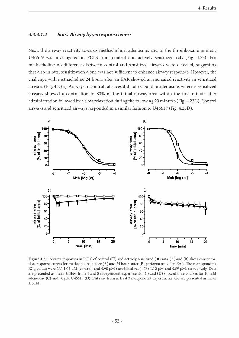
4. Results
- 52 -
4.3.3.1.2 Rats: Airway hyperresponsiveness
Next, the airway reactivity towards methacholine, adenosine, and to the thromboxane mimetic U46619 was investigated in PCLS from control and actively sensitized rats (Fig. 4.23). For methacholine no diff erences between control and sensitized airways were detected, suggesting that also in rats, sensitization alone was not suffi cient to enhance airway responses. However, the challenge with methacholine 24 hours aft er an EAR showed an increased reactivity in sensitized airways (Fig. 4.23B). Airways in control rat slices did not respond to adenosine, whereas sensitized airways showed a contraction to 80% of the initial airway area within the fi rst minute aft er adminiatration followed by a slow relaxation during the following 20 minutes (Fig. 4.23C). Control airways and sensitized airways responded in a similar fashion to U46619 (Fig. 4.23D).
-8 -7 -6 -5 -4
0
20
40
60
80
100
A
Mch [log (c)]
airw
ay ra
ea[%
of i
nitia
l are
a]
-8 -7 -6 -5 -4
0
20
40
60
80
100
B
Mch [log (c)]
airw
ay ra
ea[%
of i
nitia
l are
a]
0 5 10 15 20
0
20
40
60
80
100C
time [min]
airw
ay a
rea
[% o
f ini
tial a
rea]
0 5 10 15 20
0
20
40
60
80
100
D
time [min]
airw
ay a
rea
[% o
f ini
tial a
rea]
Figure 4.23 Airway responses in PCLS of control ( ) and actively sensitized ( ) rats. (A) and (B) show concentra-tion-response curves for methacholine before (A) and 24 hours aft er (B) performance of an EAR. Th e corresponding EC50 values were (A) 1.08 μM (control) and 0.98 μM (sensitized rats); (B) 1.12 μM and 0.59 μM, respectively. Data are presented as mean ± SEM from 4 and 8 independent experiments. (C) and (D) showed time courses for 10 mM adenosine (C) and 50 μM U46619 (D). Data are from at least 3 independent experiments and are presented as mean ± SEM.

4. Results
- 53 -
4.4 Part III: Mediator signal cascades during the EAR
To investigate the signaling pathways that become activated by mediators of the EAR in the diff erent species, inhibitors of diff erent pathways were examined. Th ese experiments were performed either in passively sensitized PCLS treated with allergen or in unsensitized PCLS stimulated with mediators alone.
4.4.1 Signaling in human PCLS
Pre-incubation of passively sensitized human PCLS with the inhibitor of the phosphatidyl-specifi c phospholipase C (PI-PLC) L108, the IP3-receptor inhibitor xestospongin C or the inhibitor of the myosin light chain kinase (MLCK), ML-7 did not signifi cantly reduce pollen extract-induced bronchoconstriction. On the other hand, the Rho-kinase inhibitor Y27632, as well as the two Ca2+-channel antagonists nifedipine (L-type Ca2+-channel antagonist) and mibefradil (T-type Ca2+-channel antagonist) signifi cantly attenuated the allergen-induced bronchoconstriction (Fig. 4.24). Th e combination of ML-7 and Y27632 failed to show any additive eff ects.
Th romboxane and leukotrienes have been identifi ed as the major mediators of the EAR in human PCLS [178]. Th erefore, the signal transduction pathways activated by these mediators were investigated, using the same inhibitors as before. U46619 (100 nM), the stable TP-receptor agonist, contracted the airways to 30% of the initial airway area. At higher concentrations of U46619 the inhibitors had no eff ect. At lower concentrations, the bronchoconstriction was prevented to almost 100% in the case of nearly all inhibitors. For all the mediator shown below, concentration-response curves were performed and a concentration was selected that, reduced the airway area to about 20% - 40% of the initial area to examine the eff ects of the diff erent inhibitors. At this
Figure 4.24 Allergen-induced bronchoconstric-tion in passively sensitized human PCLS, and the eff ect of various inhibitors. Slices were pre-incubated 20 minutes before administration of the allergen with the respectively inhibitor. Th e phosphatidyl-specifi c phospholipase C inhibitor L108 (30 μM), the IP3-receptor inhibitor xesto-spongin C (1 μM), the myosin light chain kinase (MLCK) inhibitor ML-7 (35 μM), the Rho-kina-se inhibitor Y27632 (10 μM), the L-type Ca2+-channel blocker nifedipine (10 μM) and the T-type Ca2+-channel blocker mibefradil (25 μM) were used to inhibit diff erent signal transduc-tion pathways. Ca2+ was removed 2 hours before allergen-challenge. Data are presented as mean ± SEM. **, p< 0.01 vs. solvent; *, p< 0.05 (one-sided t-test corrected by fdr procedure). w/o = without.
solve
ntL10
8xe
sto ML-7 Y27
ML-7 / Y
2763
2
nifedipine
mibefrad
il 2+
w/o Ca
0
20
40
60
80
100
**
* **
airw
ay a
rea
[% o
f ini
tial a
rea]
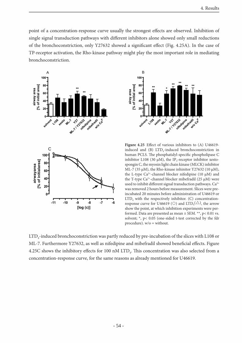
4. Results
- 54 -
point of a concentration-response curve usually the strongest eff ects are observed. Inhibition of single signal transduction pathways with diff erent inhibitors alone showed only small reductions of the bronchoconstriction, only Y27632 showed a signifi cant eff ect (Fig. 4.25A). In the case of TP-receptor activation, the Rho-kinase pathway might play the most important role in mediating bronchoconstriction.
LTD4-induced bronchoconstriction was partly reduced by pre-incubation of the slices with L108 or ML-7. Furthermore Y27632, as well as nifedipine and mibefradil showed benefi cial eff ects. Figure 4.25C shows the inhibitory eff ects for 100 nM LTD4. Th is concentration was also selected from a concentration-response curve, for the same reasons as already mentioned for U46619.
solve
ntL10
8xe
sto ML-7 Y27
ML-7 / Y
2763
2
nifedipine
mibefrad
il 2+
w/o Ca
0
20
40
60
80
100
B
** *** ****
**
aira
y ar
ea[%
of i
nitia
l are
a]
solve
ntL10
8xe
sto ML-7 Y27
ML-7 / Y
2763
2
nifedipine
mibefrad
il 2+
w/o Ca
0
20
40
60
80
100A
****
airw
ay a
rea
[% o
f ini
tial a
rea]
Figure 4.25 Eff ect of various inhibitors to (A) U46619-induced and (B) LTD4-induced bronchoconstriction in human PCLS. Th e phosphatidyl-specifi c phospholipase C inhibitor L108 (30 μM), the IP3-receptor inhibitor xesto-spongin C, the myosin light chain kinase (MLCK) inhibitor ML-7 (35 μM), the Rho-kinase inhinitor Y27632 (10 μM), the L-type Ca2+-channel blocker nifedipine (10 μM) and the T-type Ca2+-channel blocker mibefradil (25 μM) were used to inhibit diff erent signal transduction pathways. Ca2+ was removed 2 hours before measurement. Slices were pre-incubated 20 minutes before administration of U46619 or LTD4 with the respectively inhibitor. (C) concentration-response curve for U46619 ( ) and LTD4( ), the arrow show the point, at which inhibition experiments were per-formed. Data are presented as mean ± SEM. **, p< 0.01 vs. solvent; *, p< 0.05 (one-sided t-test corrected by the fdr procedure). w/o = without.
-11 -10 -9 -8 -7 -6
0
20
40
60
80
100
C
[log (c)]
airw
ayar
ea[%
of i
nitia
lare
a]
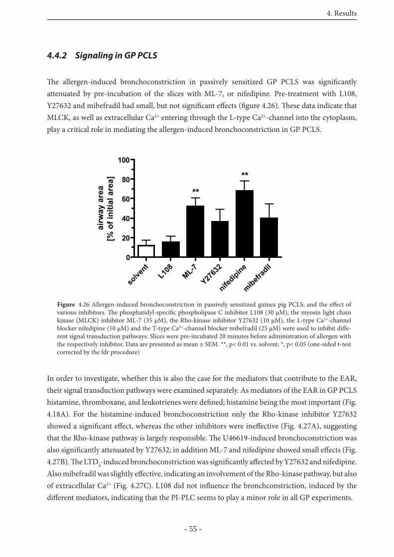
4. Results
- 55 -
4.4.2 Signaling in GP PCLS
Th e allergen-induced bronchoconstriction in passively sensitized GP PCLS was signifi cantly attenuated by pre-incubation of the slices with ML-7, or nifedipine. Pre-treatment with L108, Y27632 and mibefradil had small, but not signifi cant eff ects (fi gure 4.26). Th ese data indicate that MLCK, as well as extracellular Ca2+ entering through the L-type Ca2+-channel into the cytoplasm, play a critical role in mediating the allergen-induced bronchoconstriction in GP PCLS.
In order to investigate, whether this is also the case for the mediators that contribute to the EAR, their signal transduction pathways were examined separately. As mediators of the EAR in GP PCLS histamine, thromboxane, and leukotrienes were defi ned; histamine being the most important (Fig. 4.18A). For the histamine-induced bronchoconstriction only the Rho-kinase inhibitor Y27632 showed a signifi cant eff ect, whereas the other inhibitors were ineff ective (Fig. 4.27A), suggesting that the Rho-kinase pathway is largely responsible. Th e U46619-induced bronchoconstriction was also signifi cantly attenuated by Y27632; in addition ML-7 and nifedipine showed small eff ects (Fig. 4.27B). Th e LTD4-induced bronchoconstriction was signifi cantly aff ected by Y27632 and nifedipine. Also mibefradil was slightly eff ective, indicating an involvement of the Rho-kinase pathway, but also of extracellular Ca2+ (Fig. 4.27C). L108 did not infl uence the bronchconstriction, induced by the diff erent mediators, indicating that the PI-PLC seems to play a minor role in all GP experiments.
solve
ntL10
8ML-7
Y2763
2
nifedipine
mibefrad
il0
20
40
60
80
100
airw
ay a
rea
[% o
f ini
tial a
rea]
**
**
Figure 4.26 Allergen-induced bronchoconstriction in passively sensitized guinea pig PCLS, and the eff ect of various inhibitors. Th e phosphatidyl-specifi c phospholipase C inhibitor L108 (30 μM), the myosin light chain kinase (MLCK) inhibitor ML-7 (35 μM), the Rho-kinase inhibitor Y27632 (10 μM), the L-type Ca2+-channel blocker nifedipine (10 μM) and the T-type Ca2+-channel blocker mibefradil (25 μM) were used to inhibit diff e-rent signal transduction pathways. Slices were pre-incubated 20 minutes before administration of allergen with the respectively inhibitor. Data are presented as mean ± SEM. **, p< 0.01 vs. solvent; *, p< 0.05 (one-sided t-test corrected by the fdr procedure)
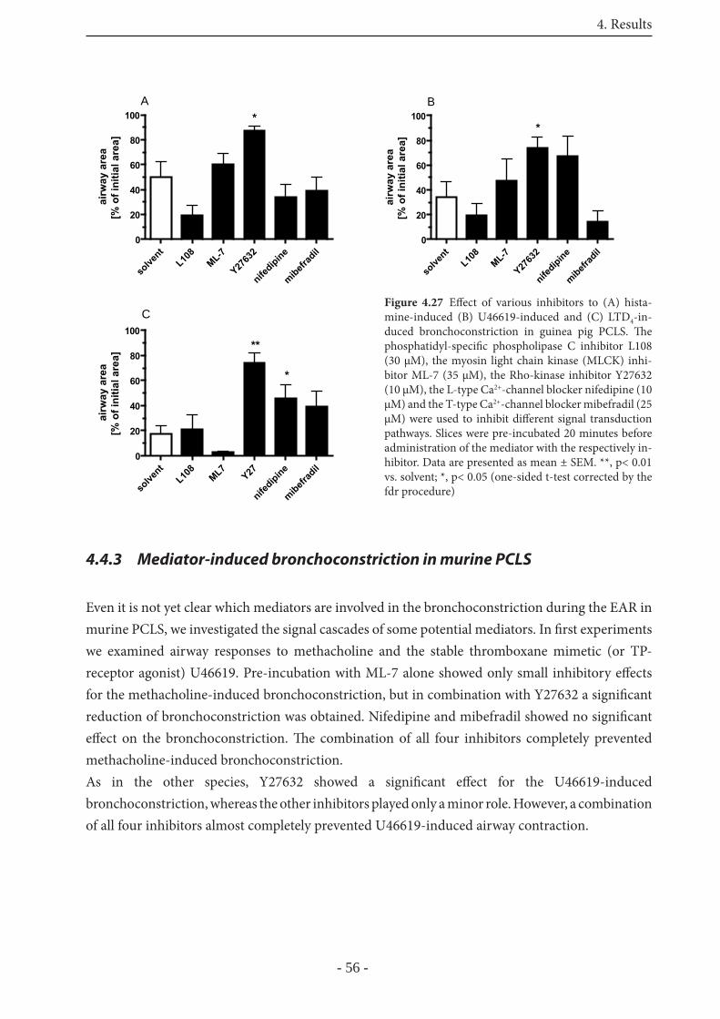
4. Results
- 56 -
4.4.3 Mediator-induced bronchoconstriction in murine PCLS
Even it is not yet clear which mediators are involved in the bronchoconstriction during the EAR in murine PCLS, we investigated the signal cascades of some potential mediators. In fi rst experiments we examined airway responses to methacholine and the stable thromboxane mimetic (or TP-receptor agonist) U46619. Pre-incubation with ML-7 alone showed only small inhibitory eff ects for the methacholine-induced bronchoconstriction, but in combination with Y27632 a signifi cant reduction of bronchoconstriction was obtained. Nifedipine and mibefradil showed no signifi cant eff ect on the bronchoconstriction. Th e combination of all four inhibitors completely prevented methacholine-induced bronchoconstriction.As in the other species, Y27632 showed a signifi cant eff ect for the U46619-induced bronchoconstriction, whereas the other inhibitors played only a minor role. However, a combination of all four inhibitors almost completely prevented U46619-induced airway contraction.
solve
ntL10
8ML-7
Y2763
2
nifedipine
mibefrad
il0
20
40
60
80
100ai
rway
are
a[%
of i
nitia
l are
a]*
A
solve
ntL10
8ML7
Y27
nifedipine
mibefrad
il0
20
40
60
80
100C
airw
ay a
rea
[% o
f ini
tial a
rea]
**
*
solve
ntL10
8ML-7
Y2763
2
nifedipine
mibefrad
il0
20
40
60
80
100
airw
ay a
rea
[% o
f ini
tial a
rea]
*
B
Figure 4.27 Eff ect of various inhibitors to (A) hista-mine-induced (B) U46619-induced and (C) LTD4-in-duced bronchoconstriction in guinea pig PCLS. Th e phosphatidyl-specifi c phospholipase C inhibitor L108 (30 μM), the myosin light chain kinase (MLCK) inhi-bitor ML-7 (35 μM), the Rho-kinase inhibitor Y27632 (10 μM), the L-type Ca2+-channel blocker nifedipine (10 μM) and the T-type Ca2+-channel blocker mibefradil (25 μM) were used to inhibit diff erent signal transduction pathways. Slices were pre-incubated 20 minutes before administration of the mediator with the respectively in-hibitor. Data are presented as mean ± SEM. **, p< 0.01 vs. solvent; *, p< 0.05 (one-sided t-test corrected by the fdr procedure)
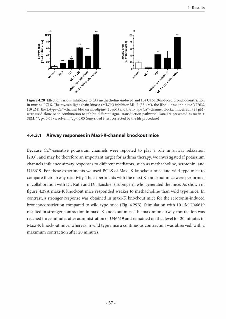
4. Results
- 57 -
4.4.3.1 Airway responses in Maxi-K-channel knockout mice
Because Ca2+-sensitive potassium channels were reported to play a role in airway relaxation [203], and may be therefore an important target for asthma therapy, we investigated if potassium channels infl uence airway responses to diff erent mediators, such as methacholine, serotonin, and U46619. For these experiments we used PCLS of Maxi-K knockout mice and wild type mice to compare their airway reactivity. Th e experiments with the maxi K knockout mice were performed in collaboration with Dr. Ruth and Dr. Sausbier (Tübingen), who generated the mice. As shown in fi gure 4.29A maxi-K knockout mice responded weaker to methacholine than wild type mice. In contrast, a stronger response was obtained in maxi-K knockout mice for the serotonin-induced bronchoconstriction compared to wild type mice (Fig. 4.29B). Stimulation with 10 μM U46619 resulted in stronger contraction in maxi-K knockout mice. Th e maximum airway contraction was reached three minutes aft er administration of U46619 and remained on that level for 20 minutes in Maxi-K knockout mice, whereas in wild type mice a continuous contraction was observed, with a maximum contraction aft er 20 minutes.
solve
ntML-7 Y27
ML-7 + Y
27
nifedipine +
mibefr
adil
ML-7 + Y
27 +
nife +
mibe0
20
40
60
80
100
*
**
**A
airw
ay a
rea
[% o
f ini
tial a
rea]
solve
ntML-7 Y27
nifedipine +
mibefr
adil
ML-7 + Y
27 +
nife +
mibe0
20
40
60
80
100 **
*
airw
ay a
rea
[% o
f ini
tial a
rea]
B
Figure 4.28 Eff ect of various inhibitors to (A) methacholine-induced and (B) U46619-induced bronchoconstriction in murine PCLS. Th e myosin light chain kinase (MLCK) inhibitor ML-7 (35 μM), the Rho-kinase inhinitor Y27632 (10 μM), the L-type Ca2+-channel blocker nifedipine (10 μM) and the T-type Ca2+-channel blocker mibefradil (25 μM) were used alone or in combination to inhibit diff erent signal transduction pathways. Data are presented as mean ± SEM. **, p< 0.01 vs. solvent; *, p< 0.05 (one-sided t-test corrected by the fdr procedure)
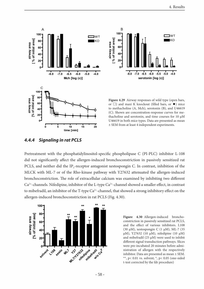
4. Results
- 58 -
4.4.4 Signaling in rat PCLS
Pretreatment with the phosphatidylinositol-specifi c phospholipase C (PI-PLC) inhibitor L-108 did not signifi cantly aff ect the allergen-induced bronchoconstriction in passively sensitized rat PCLS, and neither did the IP3-receptor antagonist xestospongin C. In contrast, inhibition of the MLCK with ML-7 or of the Rho-kinase pathway with Y27632 attenuated the allergen-induced bronchoconstriction. Th e role of extracellular calcium was examined by inhibiting two diff erent Ca2+-channels. Nifedipine, inhibitor of the L-type Ca2+-channel showed a smaller eff ect, in contrast to mibefradil, an inhibitor of the T-type Ca2+-channel, that showed a strong inhibitory eff ect on the allergen-induced bronchoconstriction in rat PCLS (Fig. 4.30).
-8.0 -7.0 -6.5 -6.0 -5.0 -4.0
0
20
40
60
80
100WTKO
A
Mch [log (c)]
airw
ay a
rea
[ % o
f ini
tial a
rea]
-8.0 -7.0 -6.5 -6.0 -5.5 -5.0 -4.0
0
20
40
60
80
100WTKO
serotonin [log (c)]
airw
ay a
rea
[ % o
f ini
tial a
rea]
B
0 5 10 15 20
0
20
40
60
80
100
time [min]
airw
ay a
rea
[% o
f ini
tial a
rea]
C
Figure 4.29 Airway responses of wild type (open bars, or ) and maxi K knockout (fi lled bars, or ) mice to methacholine (A, Mch), serotonin (B), and U46619 (C). Shown are concentration-response curves for me-thacholine and serotonin, and time courses for 10 μM U46619 in both mice types. Data are presented as mean ± SEM from at least 4 independent experiments.
solve
ntL10
8xe
sto ML7Y27
ML7/Y27
632
nifedipine
mibefrad
il 2+
w/o Ca
0
20
40
60
80
100
*****
** ****
airw
ay a
rea
[% o
f ini
tial a
ea]
Figure 4.30 Allergen-induced broncho-constriction in passively sensitized rat PCLS, and the eff ect of various inhibitors. L108 (30 μM), xestospongin C (1 μM), ML-7 (35 μM), Y27632 (10 μM), nifedipine (10 μM) and mibefradil (25 μM) were used to inhibit diff erent signal transduction pathways. Slices were pre-incubated 20 minutes before admi-nistration of allergen with the respectively inhibitor. Data are presented as mean ± SEM. **, p< 0.01 vs. solvent; *, p< 0.05 (one-sided t-test corrected by the fdr procedure)
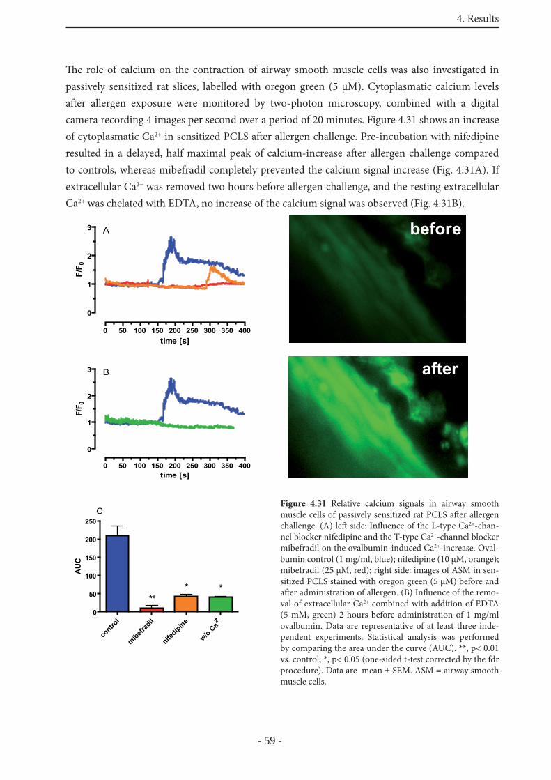
4. Results
- 59 -
Th e role of calcium on the contraction of airway smooth muscle cells was also investigated in passively sensitized rat slices, labelled with oregon green (5 μM). Cytoplasmatic calcium levels aft er allergen exposure were monitored by two-photon microscopy, combined with a digital camera recording 4 images per second over a period of 20 minutes. Figure 4.31 shows an increase of cytoplasmatic Ca2+ in sensitized PCLS aft er allergen challenge. Pre-incubation with nifedipine resulted in a delayed, half maximal peak of calcium-increase aft er allergen challenge compared to controls, whereas mibefradil completely prevented the calcium signal increase (Fig. 4.31A). If extracellular Ca2+ was removed two hours before allergen challenge, and the resting extracellular Ca2+ was chelated with EDTA, no increase of the calcium signal was observed (Fig. 4.31B).
before
after
before
after
0 50 100 150 200 250 300 350 400
0
1
2
3 B
time [s]
F/F 0
contro
l
mibefrad
il
nifedipine 2+
w/o Ca
0
50
100
150
200
250
****
AU
C
C
0 50 100 150 200 250 300 350 400
0
1
2
3 A
time [s]
F/F 0
Figure 4.31 Relative calcium signals in airway smooth muscle cells of passively sensitized rat PCLS aft er allergen challenge. (A) left side: Infl uence of the L-type Ca2+-chan-nel blocker nifedipine and the T-type Ca2+-channel blocker mibefradil on the ovalbumin-induced Ca2+-increase. Oval-bumin control (1 mg/ml, blue); nifedipine (10 μM, orange); mibefradil (25 μM, red); right side: images of ASM in sen-sitized PCLS stained with oregon green (5 μM) before and aft er administration of allergen. (B) Infl uence of the remo-val of extracellular Ca2+ combined with addition of EDTA (5 mM, green) 2 hours before administration of 1 mg/ml ovalbumin. Data are representative of at least three inde-pendent experiments. Statistical analysis was performed by comparing the area under the curve (AUC). **, p< 0.01 vs. control; *, p< 0.05 (one-sided t-test corrected by the fdr procedure). Data are mean ± SEM. ASM = airway smooth muscle cells.
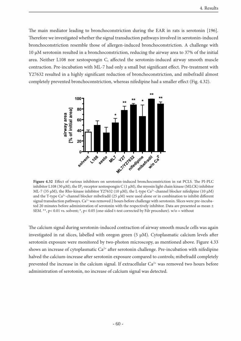
4. Results
- 60 -
Th e main mediator leading to bronchoconstriction during the EAR in rats is serotonin [196]. Th erefore we investigated whether the signal transduction pathways involved in serotonin-induced bronchoconstriction resemble those of allergen-induced bronchoconstriction. A challenge with 10 μM serotonin resulted in a bronchoconstriction, reducing the airway area to 37% of the initial area. Neither L108 nor xestospongin C, aff ected the serotonin-induced airway smooth muscle contraction. Pre-incubation with ML-7 had only a small but signifi cant eff ect. Pre-treatment with Y27632 resulted in a highly signifi cant reduction of bronchoconstriction, and mibefradil almost completely prevented bronchoconstriction, whereas nifedipine had a smaller eff ect (Fig. 4.32).
Th e calcium signal during serotonin-induced contraction of airway smooth muscle cells was again investigated in rat slices, labelled with oregon green (5 μM). Cytoplasmatic calcium levels aft er serotonin exposure were monitored by two-photon microscopy, as mentioned above. Figure 4.33 shows an increase of cytoplasmatic Ca2+ aft er serotonin challenge. Pre-incubation with nifedipine halved the calcium-increase aft er serotonin exposure compared to controls; mibefradil completely prevented the increase in the calcium signal. If extracellular Ca2+ was removed two hours before administration of serotonin, no increase of calcium signal was detected.
solve
ntL10
8xe
sto ML7Y27
ML7/Y27
632
nifedipine
mibefrad
il 2+
w/o Ca
0
20
40
60
80
100
*
*****
airw
ay a
rea
[% o
f ini
tial a
rea]
** **
Figure 4.32 Eff ect of various inhibitors on serotonin-induced bronchoconstriction in rat PCLS. Th e PI-PLC inhibitor L108 (30 μM), the IP3-receptor xestospongin C (1 μM), the myosin light chain kinase (MLCK) inhibitor ML-7 (35 μM), the Rho-kinase inhibitor Y27632 (10 μM), the L-type Ca2+-channel blocker nifedipine (10 μM) and the T-type Ca2+-channel blocker mibefradil (25 μM) were used alone or in combination to inhibit diff erent signal transduction pathways. Ca2+ was removed 2 hours before challenge with serotonin. Slices were pre-incuba-ted 20 minutes before administration of serotonin with the respectively inhibitor. Data are presented as mean ± SEM. **, p< 0.01 vs. solvent; *, p< 0.05 (one-sided t-test corrected by Fdr procedure). w/o = without
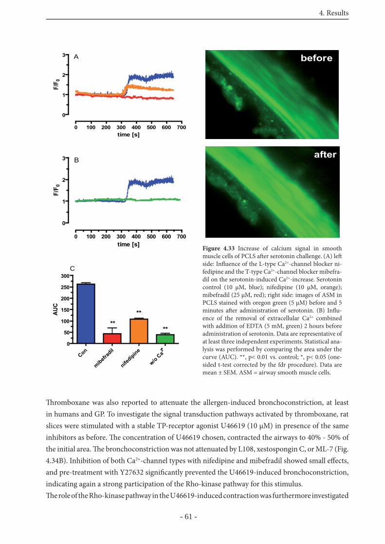
4. Results
- 61 -
Th romboxane was also reported to attenuate the allergen-induced bronchoconstriction, at least in humans and GP. To investigate the signal transduction pathways activated by thromboxane, rat slices were stimulated with a stable TP-receptor agonist U46619 (10 μM) in presence of the same inhibitors as before. Th e concentration of U46619 chosen, contracted the airways to 40% - 50% of the initial area. Th e bronchoconstriction was not attenuated by L108, xestospongin C, or ML-7 (Fig. 4.34B). Inhibition of both Ca2+-channel types with nifedipine and mibefradil showed small eff ects, and pre-treatment with Y27632 signifi cantly prevented the U46619-induced bronchoconstriction, indicating again a strong participation of the Rho-kinase pathway for this stimulus. Th e role of the Rho-kinase pathway in the U46619-induced contraction was furthermore investigated
0 100 200 300 400 500 600 700
0
1
2
3 A
time [s]
F/F 0
before
after
before
after
Con
mibefrad
il
nifedipine 2+
w/o Ca
0
50
100
150
200
250
300
****
**AU
C
C
0 100 200 300 400 500 600 700
0
1
2
3 B
time [s]
F/F 0
Figure 4.33 Increase of calcium signal in smooth muscle cells of PCLS aft er serotonin challenge. (A) left side: Infl uence of the L-type Ca2+-channel blocker ni-fedipine and the T-type Ca2+-channel blocker mibefra-dil on the serotonin-induced Ca2+-increase. Serotonin control (10 μM, blue); nifedipine (10 μM, orange); mibefradil (25 μM, red); right side: images of ASM in PCLS stained with oregon green (5 μM) before and 5 minutes aft er administration of serotonin. (B) Infl u-ence of the removal of extracellular Ca2+ combined with addition of EDTA (5 mM, green) 2 hours before administration of serotonin. Data are representative of at least three independent experiments. Statistical ana-lysis was performed by comparing the area under the curve (AUC). **, p< 0.01 vs. control; *, p< 0.05 (one-sided t-test corrected by the fdr procedure). Data are mean ± SEM. ASM = airway smooth muscle cells.
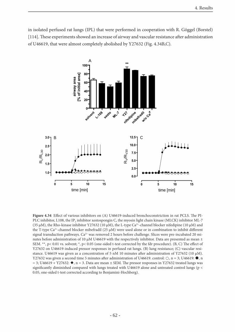
4. Results
- 62 -
in isolated perfused rat lungs (IPL) that were performed in cooperation with R. Göggel (Borstel) [114]. Th ese experiments showed an increase of airway and vascular resistance aft er administration of U46619, that were almost completely abolished by Y27632 (Fig. 4.34B,C).
Figure 4.34 Eff ect of various inhibitors on (A) U46619-induced bronchoconstriction in rat PCLS. Th e PI-PLC inhibitor, L108, the IP3 inhibitor xestospongin C, the myosin light chain kinase (MLCK) inhibitor ML-7 (35 μM), the Rho-kinase inhibitor Y27632 (10 μM), the L-type Ca2+-channel blocker nifedipine (10 μM) and the T-type Ca2+-channel blocker mibefradil (25 μM) were used alone or in combination to inhibit diff erent signal transduction pathways. Ca2+ was removed 2 hours before challenge. Slices were pre-incubated 20 mi-nutes before administration of 10 μM U46619 with the respectively inhibitor. Data are presented as mean ± SEM. **, p< 0.01 vs. solvent; *, p< 0.05 (one-sided t-test corrected by the fdr procedure). (B, C) Th e eff ect of Y27632 on U46619-induced pressor responses in perfused rat lungs. (B) lung resistance; (C) vascular resi-stance. U46619 was given as a concentration of 5 nM 10 minutes aft er administration of Y27632 (10 μM). Y27632 was given a second time 5 minutes aft er administration of U46619. control: , n = 3; U46619: , n = 3; U46619 + Y27632: , n = 3. Data are mean ± SEM. Th e pressor responses in Y27632 treated lungs was signifi cantly diminished compared with lungs treated with U46619 alone and untreated control lungs (p < 0.05, one-sided t-test corrected according to Benjamini-Hochberg).
solve
ntL-10
8xe
sto ML-7 Y27
nifedipine
mibefrad
il 2+
w/o Ca
0
20
40
60
80
100 **A
airw
ay a
rea
[% o
f ini
tial a
rea]
0 5 10 15
0.0
2.5
5.0
7.5
10.0
12.5 C
time [min]
RV/R
V30
0 5 10 15
1.0
1.5
2.0
2.5
3.0 B
time [min]
RL/
RL3
0
5. Discussion
- 63 -
5 Discussion
Asthma is a disease with an increasing incidence worldwide [1], and thus it is more important than ever to have animal models that allow to defi ne the pathogenesis of human asthma. For pharmacological and mechanistic studies relevant to human disease, diff erences in airway pharmacology between rodent models and human lungs remain an important problem. Because the preparation of precision-cut lung slices (PCLS) is essentially the same in all species, this model provides an excellent way to compare the airway pharmacology of diff erent species. PCLS allow investigation of single airways and vessels under cell culture conditions, and have already been used to elucidate important mechanisms of airway contraction in rat, mouse and human peripheral lungs [181,193,195,196].In this study we characterized and established PCLS from guinea pigs as a model to study single airway responses and show that GP PCLS resemble to human airways better than do PCLS from mice or rats. Furthermore, the preparation of monkey PCLS was introduced.Two characteristic features of human asthma are the early allergic response (EAR) and airway hyperresponsiveness (AHR). Th e present study focussed on airway responses in PCLS of diff erent species aft er induction of an EAR, the mediators leading to bronchoconstriction during this phase, and the signal transduction pathways involved.
5.1 The model of guinea pig PCLS
In the present study, we describe for the fi rst time the preparation and properties of guinea pig PCLS. Th e major obstacle in producing good quality slices from guinea pigs was the severe post mortem bronchoconstriction that has been documented radiographically [204] and in isolated lungs [205]. Th is post mortem bronchoconstriction has been attributed to the release of substance P from sensory nerves (that relaxes vessels at the same time), but other mediators or direct eff ects of changing microenvironment (pH, CO2) on airway muscle may also be involved [206]. Chronic treatment with capsaicin to empty substance P stores from sensory nerves or in vivo pre-treatment with morphine avoided the post mortem bronchoconstriction [207]. However, such treatments are impractical for routine use, carry some ethical concerns as capsaicin treatment is painful, and may interfere with the purpose of the experiments. Here we report the successful elimination of the post-mortem bronchoconstriction by a short initial (3 hours) inclusion of isoproterenol in all the media used for preparation of the PCLS. For the same purpose, β-agonists are also routinely used during the preparation of isolated perfused guinea pig lungs [208]. Once the preparation of the PCLS is fi nished, isoproterenol treatment can be discontinued, whereupon airways and vessels respond normally to various stimuli showing that the eff ect of isoproterenol is not sustained. Th is is also
5. Discussion
- 64 -
supported by the bronchorelaxant eff ects observed upon addition of salbutamol to precontracted, but not to untreated preparations.Compared to classical models of studying airway functions in vitro, such as tracheal rings or parenchymal strips, PCLS off er many advantages such as auxotonic contraction, economic use of expensive agents, longevity and the possibility to simultaneously study airway and vascular responses; PCLS even permit to diff erentiate between pulmonary arteries and pulmonary veins in the same slice. Th is was demonstrated in the present study for endothelin-1, and in related studies using lung explants for histamine and serotonin [209]. In all these studies pulmonary veins responded stronger than pulmonary arteries, corroborating many other fi ndings in rat, sheep and human lungs [210]. In contrast, Cardell et al. using isolated vessels reported that veins are less responsive than arteries [211]. Th e reason for this diff erence is unknown, but may relate to diff erences in the two preparations, the size of vessels studied or the particular guinea pig strain that was used. In any case it should be noted that it is diffi cult to extrapolate from contractions of individual vessels to total pulmonary resistance in vivo.Guinea pigs are frequently employed for studies in pulmonary pharmacology, because they are thought to possess a pharmacological profi le [173,212,213] similar to that observed in humans [179,214]. However, this assumption has never been thoroughly tested in the same laboratory using the same methology. Now having at hand GP and human PCLS we were able to check this assumption and to defi ne similarities and diff erences between both species with relevance to human asthma. Comparison between GP and human airways was performed by examining their airway responsiveness towards a variety of endogenous mediators (table 5.1). Th e EC50 values for human and GP bronchoconstriction are almost identical for LTD4 and methacholine. Airways of both species responded to thromboxane and histamine, with human airways being more sensitive to thromboxane, and GP airways being more sensitive to histamine. Th e most signifi cant diff erence was obtained for serotonin which was quite eff ective in GP and completely ineff ective in humans. Overall, the comparison between GP and human airways showed that GP are not a perfect match but do nevertheless provide a reasonable agreement to humans. Th is is certainly true in comparison to mouse and rat airways that do or only weakly respond to leukotrienes and histamine [181,196], both of which mediators are thought to play a role in human asthma.
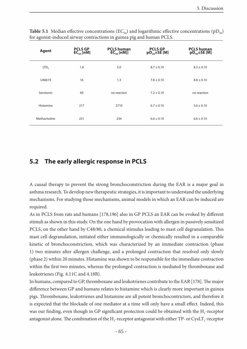
5. Discussion
- 65 -
Table 5.1 Median eff ective concentrations (EC50) and logarithmic eff ective concentrations (pD50) for agonist-induced airway contractions in guinea pig and human PCLS.
Agent PCLS GP EC50 [nM]
PCLS human EC50 [nM]]
PCLS GP pD50±SE [M]
PCLS human pD50±SE [M]
LTD4 1.8 5.0 8.7 ± 0.10 8.3 ± 0.10
U46619 16 1.3 7.8 ± 0.10 8.8 ± 0.10
Serotonin 69 no reaction 7.2 ± 0.10 no reaction
Histamine 217 2710 6.7 ± 0.10 5.6 ± 0.10
Methacholine 231 234 6.6 ± 0.10 6.6 ± 0.10
5.2 The early allergic response in PCLS
A causal therapy to prevent the strong bronchoconstriction during the EAR is a major goal in asthma research. To develop new therapeutic strategies, it is important to understand the underlying mechanisms. For studying those mechanisms, animal models in which an EAR can be induced are required. As in PCLS from rats and humans [178,196] also in GP PCLS an EAR can be evoked by diff erent stimuli as shown in this study. On the one hand by provocation with allergen in passively sensitized PCLS, on the other hand by C48/80, a chemical stimulus leading to mast cell degranulation. Th is mast cell degranulation, initiated either immunologically or chemically resulted in a comparable kinetic of bronchoconstriction, which was characterized by an immediate contraction (phase 1) two minutes aft er allergen challenge, and a prolonged contraction that resolved only slowly (phase 2) within 20 minutes. Histamine was shown to be responsible for the immediate contraction within the fi rst two minutes, whereas the prolonged contraction is mediated by thromboxane and leukotrienes (Fig. 4.11C and 4.18B). In humans, compared to GP, thromboxane and leukotrienes contribute to the EAR [178]. Th e major diff erence between GP and humans relates to histamine which is clearly more important in guinea pigs. Th romboxane, leukotrienes and histamine are all potent bronchocontrictors, and therefore it is expected that the blockade of one mediator at a time will only have a small eff ect. Indeed, this was our fi nding, even though in GP signifi cant protection could be obtained with the H1-receptor antagonist alone. Th e combination of the H1-receptor antagonist with either TP- or CysLT1-receptor
5. Discussion
- 66 -
antagonist was particularly eff ective. In general, these results are in line with previous fi ndings in other guinea pig preparations such as the perfused lung [213] and isolated tracheas [215] or parenchymal strips [212], that also show that histamine, leukotrienes and cyclooxygenase (COX) products contribute additively to the EAR. Th ose studies, however, employed COX inhibitors rather than TP-receptor antagonists; in comparison, it appears that when given alone, COX inhibitors are more eff ective than TP-receptor antagonist. Th is may indicate that other COX products such as PGE2 (mediating bronchoconstriction via EP1- receptors) and PGF2α also involved in the EAR. An important diff erence between our study in PCLS and the discussed organ baths studies is that the latter experiments were all performed in lung tissue from actively sensitized animals. It is therefore possible that in those studies some COX products came from infi ltrated eosinophils and other infl ammatory cells. Nonetheless, all these studies show that the EAR can only be prevented if at least two of three bronchoactive mast cell mediators are blocked.
None of the mediators that play a role in GP and humans, are involved in passively sensitized PCLS from rats, where the EAR is almost exclusively mediated by serotonin (Fig. 4.14) as has been described before by Wohlsen et al. [196]. In actively sensitized rats, though, in addition to serotonin, a role of LTD4 was described for the EAR [216].
In the mouse the mechanism of the EAR appears to be even more diff erent as in the other species examined. In contrast to PCLS from guinea pigs, rats, and humans, murine PCLS could not be passively sensitized and they did not respond to non-toxic concentrations of the mast cell degranulator C48/80. Th e bronchoconstriction at very high concentration of C48/80 in murine PCLS is therefore most likely a mast cell-independent mechanism. Investigations with mast cell-defi cient mice that responded in the same way as wild type mice to the chemical stimulus strengthen this assumption. Similar observations were made by Crosby et al., demonstrating that in sensitized mast cell-defi cient mice an airway response aft er ovalbumin challenge occurs, whereas in sensitized mice defi cient for B and T cells an airway response to ovalbumin challenge was absent. Th e transfer of ova-specifi c IgG, but not IgE before allergen challenge was capable to induce the EAR also in lymphocyte-defi cient mice [217]. Another study showed that sensitization and challenge with allergen results in histological changes of the airway epithelium and lung dysfunction also in mice defi cient of IgE [218,219]. Taken together, these data indicate that the EAR in mice is immunologically dependent on allergen-specifi c IgG, but independent of mast cell degranulation and IgE.In contrast passively sensitized murine PCLS, we have shown that a contractile airway response to allergen occurs in PCLS from actively sensitized mice. Since the EAR in murine PCLS seems to be mast cell-independent, a role of eosinophils seems possible, also because during the procedure of sensitization the number of eosinophils in the lung is increased [220]. In humans a degranulation of eosinophils was triggered by binding of IgG to its receptor on the surface [221], and also by the chemical stimulus C48/80 [222]. Once degranulated, eosinophils release mediators like PAF, LTC4,
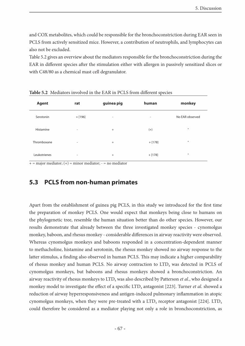
5. Discussion
- 67 -
and COX metabolites, which could be responsible for the bronchoconstriction during EAR seen in PCLS from actively sensitized mice. However, a contribution of neutrophils, and lymphocytes can also not be excluded.Table 5.2 gives an overview about the mediators responsible for the bronchoconstriction during the EAR in diff erent species aft er the stimulation either with allergen in passively sensitized slices or with C48/80 as a chemical mast cell degranulator.
Table 5.2 Mediators involved in the EAR in PCLS from diff erent species
Agent rat guinea pig human monkey
Serotonin + [196] - - No EAR observed
Histamine - + (+) “
Thromboxane - + + [178] “
Leukotrienes - + + [178] “
+ = major mediator; (+) = minor mediator; - = no mediator
5.3 PCLS from non-human primates
Apart from the establishment of guinea pig PCLS, in this study we introduced for the fi rst time the preparation of monkey PCLS. One would expect that monkeys being close to humans on the phylogenetic tree, resemble the human situation better than do other species. However, our results demonstrate that already between the three investigated monkey species - cynomolgus monkey, baboon, and rhesus monkey - considerable diff erences in airway reactivity were observed. Whereas cynomolgus monkeys and baboons responded in a concentration-dependent manner to methacholine, histamine and serotonin, the rhesus monkey showed no airway response to the latter stimulus, a fi nding also observed in human PCLS. Th is may indicate a higher comparability of rhesus monkey and human PCLS. No airway contraction to LTD4 was detected in PCLS of cynomolgus monkeys, but baboons and rhesus monkeys showed a bronchoconstriction. An airway reactivity of rhesus monkeys to LTD4 was also described by Patterson et al., who designed a monkey model to investigate the eff ect of a specifi c LTD4 antagonist [223]. Turner et al. showed a reduction of airway hyperresponsiveness and antigen-induced pulmonary infl ammation in atopic cynomolgus monkeys, when they were pre-treated with a LTD4 receptor antagonist [224]. LTD4 could therefore be considered as a mediator playing not only a role in bronchoconstriction, as
5. Discussion
- 68 -
described for humans [179,225-227], but also in AHR and infl ammation of the airways as shown in this monkey model of asthma [224]. In order to investigate whether an EAR in monkey PCLS can be evoked with pollen extract as antigen, PCLS from monkeys were passively sensitized overnight with serum from a human atopic donor, containing the specifi c IgE molecules. Th e subsequent challenge with the extract of timothy grass pollen did not elicit a strong bronchoconstriction in PCLS from baboons or rhesus monkeys. In contrast, the challenge of sensitized human PCLS with allergen always results in a contraction of the airways within the fi rst 5 minutes. Th at no EAR was seen in monkey PCLS, is possibly explained by the fact that IgE receptors in monkeys do not recognize human IgE. All three monkey species, investigated in this study belong to the non-human primates. In other primate models of airway hyperresponsiveness or airway infl ammation, diff erent allergens such as Ascaris suum or house dust mite antigens were used to sensitize the animals [224,228-230]. Monkey models have been performed in rhesus monkeys, sensitized to Ascaris suum antigen [231,232]. Some monkeys also show a natural allergy to this antigen, used by Michoud et al. to investigate wether sensitive animals respond stronger to histamine and methacholine than unsensitive animals [233]. No diff erences in reactivity were observed between allergic and nonallergic monkeys in response to both stimuli. However, the breathing pattern changed into a rapid shallow breathing when the ascaris antigen or histamine was administred to ascaris-sensitive monkeys [233]. Allergy to the ascaris antigen seems to be a naturally occurring allergy in monkeys, induced by infection with Ascaris suum [234], whereas allergy to grass pollen naturally occurs in humans. As described for C48/80-induced bronchoconstriction in humans [70], also in monkeys responder and non responder to allergen exist [235]. Th at could be another reason, why no airway response in our passively sensitized PCLS occurs, but this has to be further investigated in the future. Taken together, as indicated by the fi rst few data, PCLS from monkeys, even from non-human monkeys, off er a possibility to study airway responses with resemblance to humans, even if no EAR was obtained under these conditions.
5.4 Mast cell degranulation
Mast cells play a central role in the EAR, by releasing a wide range of potent mediators that are responsible for bronchoconstriction, airway hyperresponsiveness and attraction of infl ammatory cells. We have used two diff erent stimuli, to induce mast cell degranulation, and thereby the release of mediators. On the one hand passive sensitization of PCLS and stimulation with allergen as an immunological stimulus, and on the other hand the use of C48/80, a chemical mast cell degranulator. Both stimuli led to bronchoconstriction in GP, rat and human PCLS, but the amount of preformed mediators released upon the diff erent stimuli diff ers. In rat PCLS the levels of serotonin and histamine in the supernatant were 2-fold higher aft er C48/80, than aft er allergen
5. Discussion
- 69 -
challenge. Serotonin levels in human PCLS increased to the same eff ect aft er both stimuli, whereas histamine levels showed no signifi cant diff erences between control conditions and stimulation, neither immunologically nor chemically. In GP PCLS, serotonin levels were not increased, neither with allergen or with C48/80, and histamine levels showed only a small increase aft er ovalbumin challenge. Th ese fi ndings lead to the assumption that mast cells possess certain compartments that are activated and degranulate diff erent upon an immunological or a chemical stimulus. It has been described before that chemical stimulation induces a pattern of mediator release diff erent from those associated with IgE-dependent mast cell activation [94]. Th is might be one explanation for the release of diff erent amounts of preformed mediators. Another possible explanation is mast cell heterogeneity. Diff erent types of mast cells are known, such as MCT and MCTC in humans, and CTMC and MMC in rats and mice [94]. Maybe the response of the diff erent mast cell types varies depending on the stimulus. For example, Church et al. described that only mast cells of the MCTC type release histamine aft er C48/80, whereas MCT mast cells are unresponsive to this stimulus [236]. Liu et al showed a response of CTMC and MMC mast cells to C48/80 inhalation in rats [237]. A third possibility for the diff erent release of preformed mediators is that the extent of the exocytosis is dependent on the stimulus. In particular, the granules may release only a part of their content, or empty completely. Hide et al. using light and electron microscopy showed diff ering morphological changes accompanying degranulation on the one hand elicited by C48/80 and on the other hand by the eff ector combination Ca2+ plus GTP-γ-S. While the eff ector combination has the characteristics of an all-or-none process, C48/80 induces a partial degree of exocytosis [238].
5.5 Airway hyperresponsiveness in PCLS
An important feature of asthma is airway hyperresponsiveness (AHR), an increased airway reactivity to a variety of provocative agents, including chemical mediators (methacholine, adenosine, histamine) and physical stimuli, such as exercise or ventilation of cold air. Many animal models exist to study this disorder of airway reactivity. In this study, we investigated, whether in PCLS of sensitized animals, i.e. mice and rats, an AHR is possible.
We used murine PCLS from sensitized Balb/c mice and non sensitized control mice, to investigate whether hyperresponsiveness develops in the context of sensitization. PCLS from sensitized and non-sensitized mice were treated with methacholine to induce airway contraction, but no diff erences were observed between both mice. In contrast, a weaker response was observed for methacholine in sensitized PCLS, aft er an EAR has been performed in the same PCLS 24 hours before. Since adenosine was reported to induce an AHR in asthmatic patients only, we investigated its eff ect on murine PCLS, but no airway response was detected, neither in PCLS from sensitized
5. Discussion
- 70 -
nor in PCLS from control mice. Also no bronchoconstriction was observed in PCLS treated with adenosine 24 hours aft er an EAR had been performed in the same slice. Furthermore, in vivo no AHR was observed aft er an appropriate analysis (data not shown). Th e mechanisms of AHR in mice are still unclear, but in one study from Leigh et al. it was shown that sustained AHR and airway remodelling occurr in mice aft er chronic allergen exposure [239]. Th ey further demonstrated that IL-4 and IL-3 knockout mice were protected from developing a sustained AHR compared to wild type or IL-5 knockout mice, which showed a strong AHR. Th us, IL-4 and IL-3 may play a critical role in the development of AHR in mice [239]. Whereas in murine PCLS no AHR was detected, in PCLS of sensitized rats an increased reactivity towards methacholine aft er an EAR was observed. One explanation for the increased reactivity of airways, 24 hours aft er an EAR in the same slice has been occurred, is the release of mediators, i.e. cytokines from mast cells aft er activation that might induce a stronger response to following stimuli. TNFα and IL-1, two proinfl ammatory cytokines released by mast cells are described to increase airway resistance in the isolated perfused rat lung [241]. In that study thromboxane was determined to mediate the cytokine-induced bronchoconstriction [241]. An enhanced production of TNFα has been demonstrated aft er crosslinking of the FcεRI in human skin mast cells [242]. It is furthermore described, that preformed TNFα is rapidly released from activated mouse mast cells, and newly synthesized TNFα is released over a period of hours aft er cell activation [243,244]. Mast cells can also release IL-4 in response to FcεRI-dependent activation [245], which is reported to be found at sites of allergic infl ammation and may play a role in the shift to a Th 2 cell diff erentiation [246,247]. Finally, in the PCLS of sensitized animals an AHR was only observed, aft er an EAR has been performed before, indicating a role for cytokines, such as TNFα, IL-1, and IL-4 to induce AHR in this model. Adenosine contracted the airways in PCLS from sensitized rats, whereas absolutely no airway response was detected in PCLS of control animals. L. Wollin et al. has demonstrated that sensitized rats in vivo show an AHR towards adenosine aft er challenge with ovalbumin, this eff ect was now also reproduced with the PCLS [240]. Th erefore PCLS from actively sensitized rats might be a model to study an increased airway reactivity compared to PCLS from untreated control animals.
5.6 Mechanisms of bronchoconstriction
Th e contraction of airway smooth muscle cells, leading to bronchoconstriction during an asthmatic attack, is mediated by diff erent signal transduction pathways [248]. A better understanding of the mechanisms for regulation of smooth muscle contraction might lead to improved treatments for an occurring bronchoconstriction in asthma patients. Some experimental models exist to study single signal transduction pathways, but the pathways that are important for the signaling in human bronchoconstriction are only poorly defi ned.
5. Discussion
- 71 -
Clearly established is the role of calcium as an intracellular messenger that triggers muscle contraction [249]. An increase of the intracellular Ca2+-level is thought to activate myosin light chain kinase (MLCK), followed by activation of the 20 kDa light chain of myosin (MLC20) and contraction of the smooth muscle cell. In recent years, many studies have shown that not only an enhancement of the intracellular Ca2+-level and phosphorylation of MLCK results in smooth muscle contraction, but also the inhibition of the myosin light chain phosphatase (MLCP) via the Rho-kinase pathway [250]. In the present study diff erent signal transduction pathways and their participation for bronchoconstriction during EAR, were investigated. An important observation was that the bronchconstriction during EAR in diff erent species is mediated by diff erent mediators. Th erefore it was expected that diff erent signal transduction pathways are activated by the diff erent mediators, and that this is also species-dependent. In contrast to the other investigated species, the allergen-induced bronchoconstriction in mice appears to be mast cell independent. Nonetheless, in this species we investigated the signaling pathways for single potent bronchoconstrictors, the stable acetylcholine derivate methacholine and the thromboxane mimetic U46619, using inhibitors of the “classic” pathway via MLCK, of the Rho-kinase pathway, or Ca2+-channel inhibitors. With both stimuli a reduction of bronchoconstriction was observed, if the Rho-kinase pathway was inhibited. An almost completely prevention of bronchoconstriction was obtained in combination with Ca2+-channel inhibitors. Th ese results indicate a strong role for the Rho-kinase pathway and for extracellular Ca2+, mediating bronchoconstriciton in mice, at least for thromboxane and methacholine.In rats, allergen as well as serotonin-induced bronchoconstriction was completely abolished by mibefradil, indicating a prominent role for extracellular Ca2+ entering via the T-type Ca2+-channel; but also the MLCK, and the Rho-kinase pathway played a role. Comparing the inhibition experiments of the allergen and the serotonin-induced bronchoconstriction, similar inhibition profi les were observed for both stimuli (Fig. 4.30 and 4.32). Th is is another indication that serotonin is the main mediator during EAR in rats, because very similar eff ects with several inhibitors were obtained for both allergen and serotonin-induced airway contraction. Most relevant to human asthma are of course human PCLS, to which from all these animal models, the best resemblance was observed with GP PCLS; here the same mediators as in human PCLS appear to be responsible for bronchoconstriction. Th e analysis of the signal transduction pathways of human and GP PCLS showed that the allergen-induced bronchoconstriction in passively sensitized human PCLS was attenuated by inhibition of the Rho-kinase pathway and extracellular Ca2+. However, the inhibitors only partly reduced the bronchoconstriction, and no complete prevention was observed (Fig. 4.24). Th ese results suggest that the EAR in human PCLS is dependent on extracellular Ca2+ and the Rho-kinase pathway, but further signal transduction pathways may be activated as well.In GP PCLS the inhibition of the MLCK and the L-type Ca2+-channel, but not of the Rho-kinase pathway signifi cantly diminished bronchoconstriction by allergen challenge (Fig. 4.26), indicating
5. Discussion
- 72 -
that the “classic” pathway via MLCK phosphorylation and extracellular Ca2+ mediate the ovalbumin-induced bronchoconstriction in GP.Th e LTD4-induced bronchoconstriction in human PCLS was, with exception of the IP3-receptor antagonist, signifi cantly attenuated by all inhibitors (Fig. 4.25B), but again no complete prevention of bronchoconstriction was observed with any single inhibitor alone. Th ese fi ndings suggest that LTD4-induced bronchoconstriction depends on PI-PLC, MLCK, the Rho-kinase pathway and extracellular Ca2+, but not on intracellular Ca2+ released via IP3-receptors. It was discussed, that LTD4 perhaps mediates the bronchoconstriction not only via activation of CysLT1-receptors, but also via formation of thromboxane. However as described by Wohlsen et al. inhibition of the TP-receptor does not reduce the LTD4-induced bronchoconstriction in human PCLS [178]. In line with our fi ndings, in experiments with dissected human intralobuar bronchioles a partly reduction of the response to LTD4 was obtained by inhibition of the L-type Ca2+-channel with nifedipine [251]. Th e inhibition of IP3-receptors with 2-APB showed no eff ect on the LTD4-induced bronchoconstriction [251]. Th ese data are in line with our results, i.e. that internal Ca2+ release via IP3-receptors plays no role for the LTD4-induced bronchoconstriction in human PCLS (Fig. 4.25B). Snetkov et al. showed that also a release of Ca2+ from intracellular stores provoked with caff eine, does not cause directly any contractions in human small bronchioles [251]. T-type Ca2+-channels have been reported to be activated by diacylglycerol (DAG) [258]. Th e fact, that L108 reduced LTD4-induced bronchoconstriction in human PCLS could imply that LTD4 stimulates PI-PLC mediated formation of DAG, and thus activation of T-type Ca2+-channels, and infl ux of extracellular Ca2+. In GP PCLS only the Rho-kinase inhibitor and the L-type Ca2+-channel inhibitor were eff ective, indicating a dependence on the Rho-kinase pathway and extracellular Ca2+. In contrast to human PCLS, it has been reported that in GP airways the bronchoconstriction induced by LTD4 is partly mediated by thromboxane [252,253]. Aizawa et al. showed on tracheal strips compared to parenchymal strips from the GP, that in central airways (tracheal strips) LTD4 acts directly on smooth muscle, but in peripheral airways (parenchymal strips) LTD4 induces bronchoconstriction both directly and indirectly via thromboxane formation [252]. Th e eff ect of Y27632 on the LTD4-induced bronchoconstriction in PCLS from GP might be therefore due to inhibition of the thromboxane-mediated part of the contraction. On the other hand, perhaps the direct induction of bronchoconstriction by LTD4 in GP PCLS was attenuated by inhibition of the L-type Ca2+-channel. Th e histamine-induced bronchoconstriction in GP PCLS was signifi cantly attenuated only with the Rho-kinase inhibitor (Fig. 4.27A). In line with that, Tokuyama et al. showed in isolated perfused GP lungs that increases in lung resistance (RL) caused by LTD4 or histamine were attenuated by treatment with Y27632 [254].
Besides the activation of diff erent signal transduction pathways by the main mediators of the EAR in the diff erent species, there was one similarity that was observed in all species. In all cases, the U46619-induced bronchoconstriction was signifi cantly diminished by preincubation with the Rho-kinase inhibitor Y27632. Th is indicates a central role of the Rho-kinase pathway in mediating the
5. Discussion
- 73 -
TP-receptor triggered bronchoconstriction in PCLS, that it appears to be independent of PI-PLC, IP3 and extracellular Ca2+ (Fig. 4.25A, 4.27B, and 4.34). Th ese data are corroborated by fi ndings in the isolated perfused rat lungs, where Y27632 completely abolished U46619-induced pressor responses, both in airways and vessels [114] (Fig. 4.35). Furthermore, the eff ects of U46619 in GP tracheae and main bronchi preparations were inhibited by Y27632 [255]. Taken together, diff erent signal transduction pathways are activated by diff erent mediators, - to further complicate matters - this is also species dependent. Only the U46619-induced bronchoconstriction is mediated via the Rho-kinase pathway in mice rats, humans and GP. Th e release of Ca2+ from intracellular stores via IP3 appears to play a minor role in mediating bronchoconstriction induced by the diff erent mediators.
Th e combination of PCLS and two-photon microscopy allows further to study Ca2+-signaling in airway smooth muscle cells. With this technique it is possible to investigate airway contraction on the one hand and intracellular free Ca2+-level increases aft er stimulation on the other hand within the same model. In contrast to confocal microscopy, two-photon microscopy off ers the possibility to measure Ca2+ signals not only in one cell layer, but also up to 150 μm deep in the tissue of the PCLS. Investigations with rat PCLS either passively sensitized or unsensitized and treated with serotonin showed an increase of the intracellular Ca2+ levels aft er stimulation (Fig. 4.31 and 4.33). As an increase of intracellular Ca2+ is responsible for contraction of smooth muscle cells, these data are in line with the results obtained in the contraction experiments, where the airways in PCLS contract in response to allergen or serotonin. Pre-incubation of the slices with mibefradil completely prevented the response in both cases. No airway contraction and no increase of the Ca2+ level in airway smooth muscle cells were observed anymore. Th e L-type Ca2+-channel inhibitor nifedipine only partly reduced bronchoconstriction on the one hand, and increase of the Ca2+-signal on the other hand. If extracellular Ca2+ was removed before stimulation, no response was observed, neither bronchoconstrition nor Ca2+-level increase in airway smooth muscle cells was determined (Fig. 4.31 and 4.33). Because we were able to inhibit the allergen or serotonin-induced bronchoconstriction and Ca2+-signaling with mibefradil or the remove of extracellular Ca2+, the allergen and serotonin-induced Ca2+-signaling appears to directly mediate bronchoconstriction in rat PCLS.
5.7 Role of the Maxi-K channel in bronchoconstriction
Calcium-sensitive potassium channels (Maxi-K channels) are reported to play an important role in regulating the tone of airway smooth muscle cells [203,256,257]. It has been suggested that their activation might be useful in asthma therapy to inhibit contraction of the airways.
5. Discussion
- 74 -
K+-channels are important for maintenance of resting membrane potential for repolarization of action potentials and other depolarizing stimuli. Th e concentration of K+ ions inside cells is much higher (150 mM) than outside (3-5 mM). Th e opening of K+-channels lead to an outfl ow of K+ ions along the electriochemical gradient, resulting in hyperpolarization of the cell membrane to -80 mV from the resting value of about -60 mV. As a consequence the excitability of the cell to contractile stimuli is reduced [257]. Th erefore one would expect that animals, lacking potassium channels respond stronger to contractile stimuli compared to control animals.In the present study we used maxi-K knockout and control mice, to compare airway contraction towards various stimuli. Concentration-response curves for methacholine showed a weaker contractile response in the maxi-K knockout mice compared to wild type mice (Fig. 4.29A). Bronchoconstriction towards serotonin and U46619, however, were stronger in the knockout mice compared to the wild type (Fig. 4.29B,C). Data from isolated perfused lungs confi rm the results obtained for methacholine. Th e respiratory capacity was signifi cantly decreased in wild type mice compared to maxi-K knockout mice (unpublished data). M. Sausbier and colleagues detected an increased synthesis of cGMP in maxi-K knockout mice, which is maybe responsible for the weaker airway contraction towards methacholine (unpublished data).Taken together, the maxi-K channel seem to play a role for airway relaxation in murine PCLS, but this is maybe dependent on the stimulus.
5.8 Conclusions
PCLS off er the opportunity to study several aspects concerning airway reactivity in response to various stimuli. Th e establishment of PCLS from guinea pigs and monkeys in addition to those of rat, mice and humans, provides the possibility to compare airway and vessel responses from diff erent species to a variety of stimuli by using the same method. Animal models are important and off er new insights into various areas of a disease, but all animal models have their limitations. GP are shown to resemble the human situation more than do mice or rats, therefore PCLS from guinea pigs are a valuable model to study the EAR and maybe further airway pharmacology with regard to humans.
6. Summary
- 75 -
6 Summary
Species diff erences are a critical point for the extrapolation of data from animal models to the human situation. Th is appears to be particularly true for asthma. Th e model of precision-cut lung slices (PCLS) off ers the possibility to analyze airway and vessel responses by digital video microscopy in diff erent species such as rat, mouse and even human. In the present study the model of PCLS was established for guinea pigs and monkeys, animals that may resemble human airway responses better than do rodents. Th e early allergic response (EAR) is one characteristic feature of human asthma, characterized by mast cell degranulation and subsequent bronchoconstriction. Th romboxane, leukotrienes and histamine were identifi ed as the mediators responsible for airway contraction during the EAR in guinea pig and human PCLS, whereas in PCLS from rats the major mediator is serotonin. In contrast to the other species, murine PCLS showed no airway contraction aft er passive sensitization. In actively sensitized mice, however, allergen provocation induced a bronchoconstriction, but this appeared to be mast cell-independent, calling into question the suitability of the mouse model to study the acute asthmatic response. In monkey PCLS no EAR was observed, when the slices are passively sensitized with human serum. Th us, concerning the mediators responsible for the bronchoconstriction during an EAR, the best resemblance to humans was observed with guinea pig PCLS. Th e PCLS model also allows investigation of airway hyperresponsiveness (AHR), another hallmark of asthma. However, an AHR was only observed in PCLS from actively sensitized rats in which an EAR has been performed 24h before. Th e mechanisms of this response are unknown, but this interesting fi nding suggests a new model for studying the mechanisms of AHR in asthma.A major part of this thesis was focussed on the signalling pathways involved in airway smooth muscle contraction in the various species under various conditions. Th e present fi ndings suggest that in guinea pig airways the early allergic bronchoconstriction depends on the myosin light chain kinase (MLCK) and extracellular Ca2+, entering via L-type Ca2+-channels. In humans, PCLS extracellular Ca2+ and the Rho-kinase pathway are involved. However, in both species inhibition of a single pathway attenuated the bronchoconstriction only to a small degree, indicating that further (and maybe unknown) signal transduction pathways may become activated. In rats, inhibition of the T-type Ca2+-channels or removal of extracellular Ca2+ almost completely prevented the bronchoconstriction during an EAR, again suggesting a major role for extracellular Ca2+. Th is conclusion was further supported by direct Ca2+-measurement in single airway smooth cells by the means of two-photon microscopy. Th e investigation of signalling pathways of bronchoconstriction triggered by thromboxane showed a participation of the Rho-kinase pathway in all species. However, in most cases other signalling pathways contribute as well indicating that airway smooth muscle contraction is a complex, but robust process that is controlled at diff erent levels.
6. Summary
- 76 -
Taken together, guinea pig PCLS have been established as a model to study airway responses with relevance to humans. In addition, the novel combination of PCLS with two-photon microscopy off ers the opportunity to investigate signaling responses of single cells in their intact microenvironment. With respect to asthma therapy our studies suggest that the signal transduction pathways of bronchoconstriction may be too complex for developing useful asthma drugs targeted at this level. However, studies also indicate that reliable and complete protection against allergen-induced bronchoconstriction can be obtained if several receptors (i.e. TP, CysLT, H1) are blocked simultaneously, a concept that is currently examined in a clinical trial.
7. Deutsche Zusammenfassung
- 77 -
7 Deutsche Zusammenfassung
Speziesunterschiede sind ein kritischer Punkt bei der Datenübertragung von Tiermodellen auf die humane Situation. Besonders beim Asthma scheint das deutlich zu sein. Das Modell der präzisions-geschnittenen Lungenschnitte bietet die Möglichkeit mittels Videomikroskopie Atemwegs- und Gefäßreaktivität in unterschiedlichen Spezies, wie Ratte, Maus, und auch Mensch, zu untersuchen. In der vorliegenden Arbeit wird die Etablierung des Modells der präzisions-geschnittenen Lungenschnitte von Meerschweinen und Primaten gezeigt. Diese Tiere spiegeln humane Atemwegsreaktivität eventuell eher wieder, als es Nagetiere tun.Die allergische Frühphase ist ein charakteristisches Merkmal des Asthma bronchiale, gekennzeichnet durch eine IgE-vermittelte Mastzelldegranulation und anschliessende Kontraktion der Atemwege. Es wurde gezeigt, dass Th romboxan, Leukotriene und Histamin in Lungenschnitten von Meerschweinen und Menschen verantwortlich für die Atemwegskontraktion sind. In Lungenschnitten von Ratten ist es hingegen Serotonin. Im Gegensatz zu den anderen Spezies, zeigen Lungenschnitte von der Maus keine Atemwegskontraktion nach passiver Sensibilisierung. In aktiv sensibilisierten Mäusen löst eine Allergenprovokation jedoch eine Atemwegskontraktion aus. Diese scheint aber Mastzell-unabhängig zu sein, und stellt damit die Eignung des Mausmodels zur Untersuchung der allergischen Frühphase in Frage. In Lungenschnitten von Primaten konnte keine allergische Frühphase beobachtet werden, wenn die Lungenschnitte mit humanem Serum passiv sensibilisiert wurden. In Bezug auf die Mediatoren, die verantwortlich für die Atemwegskontraktion während der allergischen Frühphase sind, kann man sagen, dass das Meerschwein die humane Situation am ehesten widerspiegelt.Das Modell der Lungenschnitte erlaubt auch die Untersuchung von Atemwegshyperreagibilität, ein weiteres Kennzeichen des Asthma bronchiale. Sie konnte jedoch nur in Lungenschnitten von aktiv sensibilisierten Ratten beobachtet werden, an denen 24 Stunden zuvor eine allergische Frühphase ausgelöst wurde. Die Mechanismen für diese Hyperreaktivität sind noch nicht bekannt, aber die interessanten Daten deuten auf ein neues Modell hin, um die Mechanismen der Atemwegshyperreagibilität im Asthma näher zu untersuchen.Ein großer Teil der Arbeit war auf die Signaltransduktionswege gerichtet, die bei der Atemwegskontraktion in den verschiedenen Spezies aktiviert werden. Die aktuellen Daten zeigen, dass beim Meerschwein die Atemwegskontraktion während der allergischen Frühphase abhängig ist von der Myosin-Leichtkettenkinase und extrazellulärem Ca2+, welches über den L-typ-Ca2+-Kanal ins Zytoplasma strömt. Bei Lungenschnitten vom Menschen ist ebenfalls extrazelluläres Ca2+ und auch der Rho-kinase Signaltransduktionsweg involviert. In diesen beiden Spezies führt die Hemmung jeweils nur eines Signalweges nur zu einem leichten Schutzeff ekt, was darauf schliessen lässt, dass eventuell noch andere, bisher vielleicht noch nicht bekannte Signaltransduktionswege aktiviert werden. In Lungenschnitten von Ratten konnte gezeigt werden, dass die Hemmung des T-typ Ca2+-Kanals oder die Wegnahme von extrazellulärem Ca2+ eine allergen-induzierte Atemwegskontraktion fast komplett verhindert, was wiederum zeigt, dass extrazelluläres Ca2+ eine
7. Deutsche Zusammenfassung
- 78 -
große Rolle zu spielen scheint. Diese Feststellung wird weiter untermauert durch direkte Ca2+- Messungen in Glattmuskelzellen der Atemwege mittels Zwei-Photonen Mikroskopie.Für eine Th romboxan-vermittelte Atemwegskontraktion konnte in allen Spezies die Beteiligung des Rho-kinase Weges gezeigt werden.
Das Modell der Präzisionslungenschnitte vom Meerschwein wurde etabliert als ein Modell, mit dem es möglich ist, Atemwegsreaktivität im Hinblick auf den Menschen zu untersuchen. Desweiteren bietet die neue Kombination von Lungenschnitten und Zwei-Photonen Mikroskopie die Möglichkeit, Signalprozesse einzelner Zellen in einer intakten Mikroanatomie zu untersuchen.Mit Bezug zur Asthmatherapie, deuten unsere Studien daraufh in, dass die bei einer Atemwegskontraktion aktivierten Signaltransduktionswege zu komplex sind, um Asthmamedikamente auf diesem Level einzusetzen.Studien haben aber auch gezeigt, dass ein vertrauenswürdiger und kompletter Schutzeff ekt gegen eine Allergen-induzierte Atemwegskontraktion besteht, wenn mehrere Rezeptoren gleichzeitig geblockt werden, z.B. der Th romboxan-Rezeptor, CysLT1-Rezeptor und H1-Rezeptor. Dieses Konzept wird gerade in einer klinischen Studie getestet.
8. Literature
- 79 -
8 Reference List
1. Biedermann A., Beeh K.-M., Vogelmeier C.F., Kienast K., and Buhl R. 2006. Asthma bronchiale. In Allergologie Handbuch Grundlagen und klinische Praxis. Saloga J., Klimek L., Buhl R., Mann W., and Knop J., editors. Schattauer. Stuttgart, New York. 301-318.
2. Fabel,H., and Konietzko,N. 2005. Weißbuch Lunge 2005. Georg Th ieme Verlag KG. Stuttgart, New York.
3. Cookson,W.O., and Moff att,M.F. 2000. Genetics of asthma and allergic disease. Hum. Mol. Genet. 9:2359-2364.
4. Sears,M.R. 1998. Epidemiology. In Asthma: Basic mechanisms and clinical management. P.J.Barnes, Rodger,I.W., and Th ompson,N.C., editors. Academic Press. San Diego, CA; London, UK. 1-33.
5. Boushey,H.A., Corry D.B., and Fahy J.V. 2005. Asthma.
6. Barnes,P.J. 2002. Th e role of infl ammation and anti-infl ammatory medication in asthma. Respir. Med. 96 Suppl A:S9-15.
7. Galli Stephen J., and Costa John J. 1997. Mast Cells. In Th e Lung: Scientifi c Foundations Second edition. Crystal R.G., and West J.B.et al., editors. Raven Publishers. Philadelphia. 929-946.
8. Lemanske,R.F., Jr., and Kaliner,M.A. 1983. Late phase allergic reactions. Int. J. Dermatol. 22:401-409.
9. Costa J.J., and Galli S.J. 1996. Mast cell and basophils. In Clinical immunology: principles and practice. Rich RR, editor. Mosby. St.Louis. 408-430.
10. Galli,S.J. 1993. New concepts about the mast cell. N. Engl. J. Med. 328:257-265.
11. Galli,S.J., and Costa,J.J. 1995. Mast-cell-leukocyte cytokine cascades in allergic infl ammation. Allergy 50:851-862.
12. Bochner,B.S., Peachell,P.T., Brown,K.E., and Schleimer,R.P. 1988. Adherence of human basophils to cultured umbilical vein endothelial cells. J. Clin. Invest 81:1355-1364.
13. Springer,T.A. 1994. Traffi c signals for lymphocyte recirculation and leukocyte emigration: the multistep paradigm. Cell 76:301-314.
14. Bevilacqua,M.P. 1993. Endothelial-leukocyte adhesion molecules. Annu. Rev. Immunol. 11:767-804.
15. Reynolds,A.M., Holmes,M.D., and Scicchitano,R. 2000. Cytokines enhance airway smooth muscle contractility in response to acetylcholine and neurokinin A. Respirology. 5:153-160.
16. Bel,E.H., Timmers,M.C., Zwinderman,A.H., Dijkman,J.H., and Sterk,P.J. 1991. Th e eff ect of inhaled corticosteroids on the maximal degree of airway narrowing to methacholine in asthmatic subjects. Am. Rev. Respir. Dis. 143:109-113.
17. Rosati,G., Hargreave,F.E., and Ramsdale,E.H. 1989. Inhalation of adenosine 5’-monophosphate increases methacholine airway responsiveness. J. Appl. Physiol 67:792-796.
18. Lynch,D.A., Newell,J.D., Tschomper,B.A., Cink,T.M., Newman,L.S., and Bethel,R. 1993. Uncomplicated asthma in adults: comparison of CT appearance of the lungs in asthmatic and healthy subjects. Radiology 188:829-833.
8. Literature
- 80 -
19. Paganin,F., Seneterre,E., Chanez,P., Daures,J.P., Bruel,J.M., Michel,F.B., and Bousquet,J. 1996. Computed tomography of the lungs in asthma: infl uence of disease severity and etiology. Am. J. Respir. Crit Care Med. 153:110-114.
20. Brown,P.J., Greville,H.W., and Finucane,K.E. 1984. Asthma and irreversible airfl ow obstruction. Th orax 39:131-136.
21. Fahy,J.V., Kim,K.W., Liu,J., and Boushey,H.A. 1995. Prominent neutrophilic infl ammation in sputum from subjects with asthma exacerbation. J. Allergy Clin. Immunol. 95:843-852.
22. Nadel,J.A., and Takeyama,K. 1999. Mechanisms of hypersecretion in acute asthma, proposed cause of death, and novel therapy. Pediatr. Pulmonol. Suppl 18:54-55.
23. Ebina,M., Yaegashi,H., Chiba,R., Takahashi,T., Motomiya,M., and Tanemura,M. 1990. Hyperreactive site in the airway tree of asthmatic patients revealed by thickening of bronchial muscles. A morphometric study. Am. Rev. Respir. Dis. 141:1327-1332.
24. Dunnill,M.S. 1960. Th e pathology of asthma, with special reference to changes in the bronchial mucosa. J. Clin. Pathol. 13:27-33.
25. Djukanovic,R., Roche,W.R., Wilson,J.W., Beasley,C.R., Twentyman,O.P., Howarth,R.H., and Holgate,S.T. 1990. Mucosal infl ammation in asthma. Am. Rev. Respir. Dis. 142:434-457.
26. Holt,P.G., and Stumbles,P.A. 2000. Regulation of immunologic homeostasis in peripheral tissues by dendritic cells: the respiratory tract as a paradigm. J. Allergy Clin. Immunol. 105:421-429.
27. Holt,P.G. 1993. Regulation of antigen-presenting cell function(s) in lung and airway tissues. Eur. Respir. J. 6:120-129.
28. Moller,G.M., Overbeek,S.E., Helden-Meeuwsen,C.G., Van Haarst,J.M., Prens,E.P., Mulder,P.G., Postma,D.S., and Hoogsteden,H.C. 1996. Increased numbers of dendritic cells in the bronchial mucosa of atopic asthmatic patients: downregulation by inhaled corticosteroids. Clin. Exp. Allergy 26:517-524.
29. Lambrecht,B.N., Salomon,B., Klatzmann,D., and Pauwels,R.A. 1998. Dendritic cells are required for the development of chronic eosinophilic airway infl ammation in response to inhaled antigen in sensitized mice. J. Immunol. 160:4090-4097.
30. Dupuis,M., and McDonald,D.M. 1997. Dendritic-cell regulation of lung immunity. Am. J. Respir. Cell Mol. Biol. 17:284-286.
31. Unanue,E.R., and Allen,P.M. 1987. Th e basis for the immunoregulatory role of macrophages and other accessory cells. Science 236:551-557.
32. Nathan,C.F. 1987. Secretory products of macrophages. J. Clin. Invest 79:319-326.
33. John,M., Lim,S., Seybold,J., Jose,P., Robichaud,A., O’Connor,B., Barnes,P.J., and Chung,K.F. 1998. Inhaled corticosteroids increase interleukin-10 but reduce macrophage infl ammatory protein-1alpha, granulocyte-macrophage colony-stimulating factor, and interferon-gamma release from alveolar macrophages in asthma. Am. J. Respir. Crit Care Med. 157:256-262.
34. Spiteri,M.A., Knight,R.A., Jeremy,J.Y., Barnes,P.J., and Chung,K.F. 1994. Alveolar macrophage-induced suppression of peripheral blood mononuclear cell responsiveness is reversed by in vitro allergen exposure in bronchial asthma. Eur. Respir. J. 7:1431-1438.
35. Viksman,M.Y., Liu,M.C., Bickel,C.A., Schleimer,R.P., and Bochner,B.S. 1997. Phenotypic analysis of alveolar macrophages and monocytes in allergic airway infl ammation. I. Evidence for activation of alveolar macrophages, but not peripheral blood monocytes, in subjects with allergic rhinitis and asthma. Am. J. Respir. Crit Care Med. 155:858-863.
8. Literature
- 81 -
36. Agea,E., Forenza,N., Piattoni,S., Russano,A., Monaco,A., Flenghi,L., Bistoni,O., Gillies,D.A., Azuma,M., Bertotto,A. et al 1998. Expression of B7 co-stimulatory molecules and CD1a antigen by alveolar macrophages in allergic bronchial asthma. Clin. Exp. Allergy 28:1359-1367.
37. Humbert,M., Durham,S.R., Kimmitt,P., Powell,N., Assoufi ,B., Pfi ster,R., Menz,G., Kay,A.B., and Corrigan,C.J. 1997. Elevated expression of messenger ribonucleic acid encoding IL-13 in the bronchial mucosa of atopic and nonatopic subjects with asthma. J. Allergy Clin. Immunol. 99:657-665.
38. Robinson,D.S., Hamid,Q., Ying,S., Tsicopoulos,A., Barkans,J., Bentley,A.M., Corrigan,C., Durham,S.R., and Kay,A.B. 1992. Predominant TH2-like bronchoalveolar T-lymphocyte population in atopic asthma. N. Engl. J. Med. 326:298-304.
39. Mosmann,T.R., and Sad,S. 1996. Th e expanding universe of T-cell subsets: Th 1, Th 2 and more. Immunol. Today 17:138-146.
40. Gould,H.J., Beavil,R.L., and Vercelli,D. 2000. IgE isotype determination: epsilon-germline gene transcription, DNA recombination and B-cell diff erentiation. Br. Med. Bull. 56:908-924.
41. Lane,P., Brocker,T., Hubele,S., Padovan,E., Lanzavecchia,A., and McConnell,F. 1993. Soluble CD40 ligand can replace the normal T cell-derived CD40 ligand signal to B cells in T cell-dependent activation. J. Exp. Med. 177:1209-1213.
42. Galli,S.J., Dvorak,A.M., and Dvorak,H.F. 1984. Basophils and mast cells: morphologic insights into their biology, secretory patterns, and function. Prog. Allergy 34:1-141.
43. Bingham,C.O., III, and Austen,K.F. 2000. Mast-cell responses in the development of asthma. J. Allergy Clin. Immunol. 105:S527-S534.
44. Kirshenbaum,A.S., Kessler,S.W., Goff ,J.P., and Metcalfe,D.D. 1991. Demonstration of the origin of human mast cells from CD34+ bone marrow progenitor cells. J. Immunol. 146:1410-1415.
45. Weidner,N., and Austen,K.F. 1990. Evidence for morphologic diversity of human mast cells. An ultrastructural study of mast cells from multiple body sites. Lab Invest 63:63-72.
46. Irani,A.A., Schechter,N.M., Craig,S.S., DeBlois,G., and Schwartz,L.B. 1986. Two types of human mast cells that have distinct neutral protease compositions. Proc. Natl. Acad. Sci. U. S. A 83:4464-4468.
47. Weidner,N., and Austen,K.F. 1991. Ultrastructural and immunohistochemical characterization of normal mast cells at multiple body sites. J. Invest Dermatol. 96:26S-30S.
48. Enerbäck L 1986. Mast cell heterogeneity: the evolution of the concept of a specifi c mucosal mast cell. In Mast cell diff erentiation and heterogeneity. Befus AD, Bienenstock J, and Denburg JA, editors. Raven Press. New York. 1-26.
49. Miller HRP, Huntley JF, Newlands GFJ, and et al 1989. Mast cell granule proteases in mouse and rat: a guide to masr cell heterogeneity and activation inthe gastrointestinal tract. In Mast cell and basophil diff erentiation and function in health and disease. Galli SJ, and Austen KF, editors. Raven Press. New York. 81-91.
50. Kitamura,Y. 1989. Heterogeneity of mast cells and phenotypic change between subpopulations. Annu. Rev. Immunol. 7:59-76.
51. Galli,S.J. 1990. New insights into “the riddle of the mast cells”: microenvironmental regulation of mast cell development and phenotypic heterogeneity. Lab Invest 62:5-33.
8. Literature
- 82 -
52. Ludowyke,R.I., Scurr,L.L., and McNally,C.M. 1996. Calcium ionophore-induced secretion from mast cells correlates with myosin light chain phosphorylation by protein kinase C. J. Immunol. 157:5130-5138.
53. Hart,P.H. 2001. Regulation of the infl ammatory response in asthma by mast cell products. Immunol. Cell Biol. 79:149-153.
54. Vliagoft is,H., Lacy,P., Luy,B., Adamko,D., Hollenberg,M., Befus,D., and Moqbel,R. 2004. Mast cell tryptase activates peripheral blood eosinophils to release granule-associated enzymes. Int. Arch. Allergy Immunol. 135:196-204.
55. Fiorucci,L., and Ascoli,F. 2004. Mast cell tryptase, a still enigmatic enzyme. Cell Mol. Life Sci. 61:1278-1295.
56. Berger,P., N’guyen,C., Buckley,M., Scotto-Gomez,E., Marthan,R., and Tunon-de-Lara,J.M. 2002. Passive sensitization of human airways induces mast cell degranulation and release of tryptase. Allergy 57:592-599.
57. Nadel,J.A., and Caughey,G.H. 1989. Roles of mast cell proteases in airways. Chest 95:1328-1330.
58. Lazaar,A.L., Plotnick,M.I., Kucich,U., Crichton,I., Lotfi ,S., Das,S.K., Kane,S., Rosenbloom,J., Panettieri,R.A., Jr., Schechter,N.M. et al 2002. Mast cell chymase modifi es cell-matrix interactions and inhibits mitogen-induced proliferation of human airway smooth muscle cells. J. Immunol. 169:1014-1020.
59. Miller,H.R., and Pemberton,A.D. 2002. Tissue-specifi c expression of mast cell granule serine proteinases and their role in infl ammation in the lung and gut. Immunology 105:375-390.
60. Caughey,G.H. 1994. Serine proteinases of mast cell and leukocyte granules. A league of their own. Am. J. Respir. Crit Care Med. 150:S138-S142.
61. Jabril-Cuenod,B., Zhang,C., Scharenberg,A.M., Paolini,R., Numerof,R., Beaven,M.A., and Kinet,J.P. 1996. Syk-dependent phosphorylation of Shc. A potential link between FcepsilonRI and the Ras/mitogen-activated protein kinase signaling pathway through SOS and Grb2. J. Biol. Chem. 271:16268-16272.
62. Murakami,M., Austen,K.F., and Arm,J.P. 1995. Th e immediate phase of c-kit ligand stimulation of mouse bone marrow-derived mast cells elicits rapid leukotriene C4 generation through posttranslational activation of cytosolic phospholipase A2 and 5-lipoxygenase. J. Exp. Med. 182:197-206.
63. Longphre,M., Zhang,L.Y., Paquette,N., and Kleeberger,S.R. 1996. PAF-induced airways hyperreactivity is modulated by mast cells in mice. Am. J. Respir. Cell Mol. Biol. 14:461-469.
64. Gordon,J.R., Burd,P.R., and Galli,S.J. 1990. Mast cells as a source of multifunctional cytokines. Immunol. Today 11:458-464.
65. Bradding,P., Feather,I.H., Wilson,S., Bardin,P.G., Heusser,C.H., Holgate,S.T., and Howarth,P.H. 1993. Immunolocalization of cytokines in the nasal mucosa of normal and perennial rhinitic subjects. Th e mast cell as a source of IL-4, IL-5, and IL-6 in human allergic mucosal infl ammation. J. Immunol. 151:3853-3865.
66. Burd,P.R., Th ompson,W.C., Max,E.E., and Mills,F.C. 1995. Activated mast cells produce interleukin 13. J. Exp. Med. 181:1373-1380.
8. Literature
- 83 -
67. Bressler,R.B., Lesko,J., Jones,M.L., Wasserman,M., Dickason,R.R., Huston,M.M., Cook,S.W., and Huston,D.P. 1997. Production of IL-5 and granulocyte-macrophage colony-stimulating factor by naive human mast cells activated by high-affi nity IgE receptor ligation. J. Allergy Clin. Immunol. 99:508-514.
68. Pawankar,R., Okuda,M., Yssel,H., Okumura,K., and Ra,C. 1997. Nasal mast cells in perennial allergic rhinitics exhibit increased expression of the Fc epsilonRI, CD40L, IL-4, and IL-13, and can induce IgE synthesis in B cells. J. Clin. Invest 99:1492-1499.
69. Bradding,P., Roberts,J.A., Britten,K.M., Montefort,S., Djukanovic,R., Mueller,R., Heusser,C.H., Howarth,P.H., and Holgate,S.T. 1994. Interleukin-4, -5, and -6 and tumor necrosis factor-alpha in normal and asthmatic airways: evidence for the human mast cell as a source of these cytokines. Am. J. Respir. Cell Mol. Biol. 10:471-480.
70. Wohlsen,A. 2002. Responses of small airways to allergens and cytokines; a study with precision-cut lung slices.
71. Peters,S.P. 1990. Mast cells and histamine in asthma. J. Allergy Clin. Immunol. 86:642-646.
72. Purcell,W.M., Cohen,D.L., and Hanahoe,T.H. 1989. Comparison of histamine and 5-hydroxytryptamine content and secretion in rat mast cells isolated from diff erent anatomical locations. Int. Arch. Allergy Appl. Immunol. 90:382-386.
73. Church,M.K. 1975. Response of rat lung to humoral mediators of anaphylaxis and its modifi cation by drugs and sensitization. Br. J. Pharmacol. 55:423-430.
74. Schwartz,L.B., and Bradford,T.R. 1986. Regulation of tryptase from human lung mast cells by heparin. Stabilization of the active tetramer. J. Biol. Chem. 261:7372-7379.
75. Kjellen,L., and Lindahl,U. 1991. Proteoglycans: structures and interactions. Annu. Rev. Biochem. 60:443-475.
76. Stevens,R.L., Fox,C.C., Lichtenstein,L.M., and Austen,K.F. 1988. Identifi cation of chondroitin sulfate E proteoglycans and heparin proteoglycans in the secretory granules of human lung mast cells. Proc. Natl. Acad. Sci. U. S. A 85:2284-2287.
77. Johnson,P.R., Ammit,A.J., Carlin,S.M., Armour,C.L., Caughey,G.H., and Black,J.L. 1997. Mast cell tryptase potentiates histamine-induced contraction in human sensitized bronchus. Eur. Respir. J. 10:38-43.
78. Akers,I.A., Parsons,M., Hill,M.R., Hollenberg,M.D., Sanjar,S., Laurent,G.J., and McAnulty,R.J. 2000. Mast cell tryptase stimulates human lung fi broblast proliferation via protease-activated receptor-2. Am. J. Physiol Lung Cell Mol. Physiol 278:L193-L201.
79. Stack,M.S., and Johnson,D.A. 1994. Human mast cell tryptase activates single-chain urinary-type plasminogen activator (pro-urokinase). J. Biol. Chem. 269:9416-9419.
80. Sillaber,C., Baghestanian,M., Bevec,D., Willheim,M., Agis,H., Kapiotis,S., Fureder,W., Bankl,H.C., Kiener,H.P., Speiser,W. et al 1999. Th e mast cell as site of tissue-type plasminogen activator expression and fi brinolysis. J. Immunol. 162:1032-1041.
81. Fang,K.C., Wolters,P.J., Steinhoff ,M., Bidgol,A., Blount,J.L., and Caughey,G.H. 1999. Mast cell expression of gelatinases A and B is regulated by kit ligand and TGF-beta. J. Immunol. 162:5528-5535.
82. Macaldowie,C.N., Huntley,J.F., Mackellar,A., McInnes,C., and Haig,D.M. 1997. Th e eff ects of recombinant ovine interleukin-3 and recombinant ovine stem cell factor on the growth and mediator expression of caprine and ovine bone marrow-derived mast cells. Vet. Immunol. Immunopathol. 60:97-110.
8. Literature
- 84 -
83. Schwartz,L.B., Austen,K.F., and Wasserman,S.I. 1979. Immunologic release of beta-hexosaminidase and beta-glucuronidase from purifi ed rat serosal mast cells. J. Immunol. 123:1445-1450.
84. Schwartz,L.B., Lewis,R.A., Seldin,D., and Austen,K.F. 1981. Acid hydrolases and tryptase from secretory granules of dispersed human lung mast cells. J. Immunol. 126:1290-1294.
85. Sakai,K., Ren,S., and Schwartz,L.B. 1996. A novel heparin-dependent processing pathway for human tryptase. Autocatalysis followed by activation with dipeptidyl peptidase I. J. Clin. Invest 97:988-995.
86. McEuen,A.R., Ashworth,D.M., and Walls,A.F. 1998. Th e conversion of recombinant human mast cell prochymase to enzymatically active chymase by dipeptidyl peptidase I is inhibited by heparin and histamine. Eur. J. Biochem. 253:300-308.
87. Wasserman,S.I. 1987. Th e regulation of infl ammatory mediator production by mast cell products. Am. Rev. Respir. Dis. 135:S46-S48.
88. Walsh,L.J., Trinchieri,G., Waldorf,H.A., Whitaker,D., and Murphy,G.F. 1991. Human dermal mast cells contain and release tumor necrosis factor alpha, which induces endothelial leukocyte adhesion molecule 1. Proc. Natl. Acad. Sci. U. S. A 88:4220-4224.
89. Gounni,A.S., Nutku,E., Koussih,L., Aris,F., Louahed,J., Levitt,R.C., Nicolaides,N.C., and Hamid,Q. 2000. IL-9 expression by human eosinophils: regulation by IL-1beta and TNF-alpha. J. Allergy Clin. Immunol. 106:460-466.
90. Grutzkau,A., Kruger-Krasagakes,S., Baumeister,H., Schwarz,C., Kogel,H., Welker,P., Lippert,U., Henz,B.M., and Moller,A. 1998. Synthesis, storage, and release of vascular endothelial growth factor/vascular permeability factor (VEGF/VPF) by human mast cells: implications for the biological signifi cance of VEGF206. Mol. Biol. Cell 9:875-884.
91. Yano,K., Yamaguchi,M., de Mora,F., Lantz,C.S., Butterfi eld,J.H., Costa,J.J., and Galli,S.J. 1997. Production of macrophage infl ammatory protein-1alpha by human mast cells: increased anti-IgE-dependent secretion aft er IgE-dependent enhancement of mast cell IgE-binding ability. Lab Invest 77:185-193.
92. Qu,Z., Liebler,J.M., Powers,M.R., Galey,T., Ahmadi,P., Huang,X.N., Ansel,J.C., Butterfi eld,J.H., Planck,S.R., and Rosenbaum,J.T. 1995. Mast cells are a major source of basic fi broblast growth factor in chronic infl ammation and cutaneous hemangioma. Am. J. Pathol. 147:564-573.
93. Boesiger,J., Tsai,M., Maurer,M., Yamaguchi,M., Brown,L.F., Claff ey,K.P., Dvorak,H.F., and Galli,S.J. 1998. Mast cells can secrete vascular permeability factor/ vascular endothelial cell growth factor and exhibit enhanced release aft er immunoglobulin E-dependent upregulation of fc epsilon receptor I expression. J. Exp. Med. 188:1135-1145.
94. Galli SJ., and Costa JJ. 1996. Mast Cells. In Th e Lung: Scietifi c Foundations Second edition. Crystal R.G., and West J.B.et al., editors. Raven Publishers. Philadelphia. 929-946.
95. Kay,A.B. 1988. Leucocytes in asthma. Immunol. Invest 17:679-705.
96. Rabe,K.F., Munoz,N.M., Vita,A.J., Morton,B.E., Magnussen,H., and Leff ,A.R. 1994. Contraction of human bronchial smooth muscle caused by activated human eosinophils. Am. J. Physiol 267:L326-L334.
97. Leff ,A.R. 1994. Infl ammatory mediation of airway hyperresponsiveness by peripheral blood granulocytes. Th e case for the eosinophil. Chest 106:1202-1208.
8. Literature
- 85 -
98. Collins,D.S., Dupuis,R., Gleich,G.J., Bartemes,K.R., Koh,Y.Y., Pollice,M., Albertine,K.H., Fish,J.E., and Peters,S.P. 1993. Immunoglobulin E-mediated increase in vascular permeability correlates with eosinophilic infl ammation. Am. Rev. Respir. Dis. 147:677-683.
99. Hamzaoui,A., Hamzaoui,K., Salah,H., and Chabbou,A. 1999. Lymphocytes apoptosis in patients with acute exacerbation of asthma. Mediators. Infl amm. 8:237-243.
100. Lamblin,C., Gosset,P., Tillie-Leblond,I., Saulnier,F., Marquette,C.H., Wallaert,B., and Tonnel,A.B. 1998. Bronchial neutrophilia in patients with noninfectious status asthmaticus. Am. J. Respir. Crit Care Med. 157:394-402.
101. Jatakanon,A., Uasuf,C., Maziak,W., Lim,S., Chung,K.F., and Barnes,P.J. 1999. Neutrophilic infl ammation in severe persistent asthma. Am. J. Respir. Crit Care Med. 160:1532-1539.
102. Gibson,P.G., Simpson,J.L., and Saltos,N. 2001. Heterogeneity of airway infl ammation in persistent asthma : evidence of neutrophilic infl ammation and increased sputum interleukin-8. Chest 119:1329-1336.
103. Sur,S., Crotty,T.B., Kephart,G.M., Hyma,B.A., Colby,T.V., Reed,C.E., Hunt,L.W., and Gleich,G.J. 1993. Sudden-onset fatal asthma. A distinct entity with few eosinophils and relatively more neutrophils in the airway submucosa? Am. Rev. Respir. Dis. 148:713-719.
104. Sommerhoff ,C.P., Nadel,J.A., Basbaum,C.B., and Caughey,G.H. 1990. Neutrophil elastase and cathepsin G stimulate secretion from cultured bovine airway gland serous cells. J. Clin. Invest 85:682-689.
105. Nadel,J.A., Takeyama,K., and Agusti,C. 1999. Role of neutrophil elastase in hypersecretion in asthma. Eur. Respir. J. 13:190-196.
106. Schroeder JT, Mac Glashan DW, Kagey-Sobotka A, and Lichtenstein LM 1997. Th e basophil. In Th e Lung: Scientifi c Foundations Second Edition. Crystal R.G., West J.B., Weibel,E.R., and Barnes,P.J., editors. Lippincott - Raven. Philadelphia. 917-928.
107. Gauvreau,G.M., Lee,J.M., Watson,R.M., Irani,A.M., Schwartz,L.B., and O’Byrne,P.M. 2000. Increased numbers of both airway basophils and mast cells in sputum aft er allergen inhalation challenge of atopic asthmatics. Am. J. Respir. Crit Care Med. 161:1473-1478.
108. Webb,R.C. 2003. Smooth muscle contraction and relaxation. Adv. Physiol Educ. 27:201-206.
109. Somlyo,A.P., and Somlyo,A.V. 1994. Signal transduction and regulation in smooth muscle. Nature 372:231-236.
110. Somlyo,A.P., and Somlyo,A.V. 2000. Signal transduction by G-proteins, rho-kinase and protein phosphatase to smooth muscle and non-muscle myosin II. J. Physiol 522 Pt 2:177-185.
111. Wettschureck,N., and Off ermanns,S. 2002. Rho/Rho-kinase mediated signaling in physiology and pathophysiology. J. Mol. Med. 80:629-638.
112. Iino,M. 1999. Dynamic regulation of intracellular calcium signals through calcium release channels. Mol. Cell Biochem. 190:185-190.
113. Bootman,M.D., Collins,T.J., Peppiatt,C.M., Prothero,L.S., Mackenzie,L., de Smet,P., Travers,M., Tovey,S.C., Seo,J.T., Berridge,M.J. et al 2001. Calcium signalling--an overview. Semin. Cell Dev. Biol. 12:3-10.
114. Martin,C., Goggel,R., Ressmeyer,A.R., and Uhlig,S. 2004. Pressor responses to platelet-activating factor and thromboxane are mediated by Rho-kinase. Am. J. Physiol Lung Cell Mol. Physiol 287:L250-L257.
8. Literature
- 86 -
115. Varadi,G., Mori,Y., Mikala,G., and Schwartz,A. 1995. Molecular determinants of Ca2+ channel function and drug action. Trends Pharmacol. Sci. 16:43-49.
116. Bean,B.P. 1989. Classes of calcium channels in vertebrate cells. Annu. Rev. Physiol 51:367-384.
117. Benham,C.D., and Tsien,R.W. 1987. A novel receptor-operated Ca2+-permeable channel activated by ATP in smooth muscle. Nature 328:275-278.
118. Caulfi eld,M.P. 1993. Muscarinic receptors--characterization, coupling and function. Pharmacol. Th er. 58:319-379.
119. Putney,J.W., Jr. 1986. A model for receptor-regulated calcium entry. Cell Calcium 7:1-12.
120. Gong,M.C., Iizuka,K., Nixon,G., Browne,J.P., Hall,A., Eccleston,J.F., Sugai,M., Kobayashi,S., Somlyo,A.V., and Somlyo,A.P. 1996. Role of guanine nucleotide-binding proteins--ras-family or trimeric proteins or both--in Ca2+ sensitization of smooth muscle. Proc. Natl. Acad. Sci. U. S. A 93:1340-1345.
121. Gong,M.C., Fujihara,H., Somlyo,A.V., and Somlyo,A.P. 1997. Translocation of rhoA associated with Ca2+ sensitization of smooth muscle. J. Biol. Chem. 272:10704-10709.
122. Gong,M.C., Gorenne,I., Read,P., Jia,T., Nakamoto,R.K., Somlyo,A.V., and Somlyo,A.P. 2001. Regulation by GDI of RhoA/Rho-kinase-induced Ca2+ sensitization of smooth muscle myosin II. Am. J. Physiol Cell Physiol 281:C257-C269.
123. Matsui,T., Amano,M., Yamamoto,T., Chihara,K., Nakafuku,M., Ito,M., Nakano,T., Okawa,K., Iwamatsu,A., and Kaibuchi,K. 1996. Rho-associated kinase, a novel serine/threonine kinase, as a putative target for small GTP binding protein Rho. EMBO J. 15:2208-2216.
124. Taggart,M.J., Lee,Y.H., and Morgan,K.G. 1999. Cellular redistribution of PKCalpha, rhoA, and ROKalpha following smooth muscle agonist stimulation. Exp. Cell Res. 251:92-101.
125. Kimura,K., Ito,M., Amano,M., Chihara,K., Fukata,Y., Nakafuku,M., Yamamori,B., Feng,J., Nakano,T., Okawa,K. et al 1996. Regulation of myosin phosphatase by Rho and Rho-associated kinase (Rho-kinase). Science 273:245-248.
126. Amano,M., Ito,M., Kimura,K., Fukata,Y., Chihara,K., Nakano,T., Matsuura,Y., and Kaibuchi,K. 1996. Phosphorylation and activation of myosin by Rho-associated kinase (Rho-kinase). J. Biol. Chem. 271:20246-20249.
127. Kitazawa,T., Eto,M., Woodsome,T.P., and Brautigan,D.L. 2000. Agonists trigger G protein-mediated activation of the CPI-17 inhibitor phosphoprotein of myosin light chain phosphatase to enhance vascular smooth muscle contractility. J. Biol. Chem. 275:9897-9900.
128. Koyama,M., Ito,M., Feng,J., Seko,T., Shiraki,K., Takase,K., Hartshorne,D.J., and Nakano,T. 2000. Phosphorylation of CPI-17, an inhibitory phosphoprotein of smooth muscle myosin phosphatase, by Rho-kinase. FEBS Lett. 475:197-200.
129. Endou,K., Iizuka,K., Yoshii,A., Tsukagoshi,H., Ishizuka,T., Dobashi,K., Nakazawa,T., and Mori,M. 2004. 8-Bromo-cAMP decreases the Ca2+ sensitivity of airway smooth muscle contraction through a mechanism distinct from inhibition of Rho-kinase. Am. J. Physiol Lung Cell Mol. Physiol 287:L641-L648.
130. Garcia,J.G., Davis,H.W., and Patterson,C.E. 1995. Regulation of endothelial cell gap formation and barrier dysfunction: role of myosin light chain phosphorylation. J. Cell Physiol 163:510-522.
8. Literature
- 87 -
131. Lang,P., Gesbert,F., Delespine-Carmagnat,M., Stancou,R., Pouchelet,M., and Bertoglio,J. 1996. Protein kinase A phosphorylation of RhoA mediates the morphological and functional eff ects of cyclic AMP in cytotoxic lymphocytes. EMBO J. 15:510-519.
132. Dong,J.M., Leung,T., Manser,E., and Lim,L. 1998. cAMP-induced morphological changes are counteracted by the activated RhoA small GTPase and the Rho kinase ROKalpha. J. Biol. Chem. 273:22554-22562.
133. Bradley,B.L., Azzawi,M., Jacobson,M., Assoufi ,B., Collins,J.V., Irani,A.M., Schwartz,L.B., Durham,S.R., Jeff ery,P.K., and Kay,A.B. 1991. Eosinophils, T-lymphocytes, mast cells, neutrophils, and macrophages in bronchial biopsy specimens from atopic subjects with asthma: comparison with biopsy specimens from atopic subjects without asthma and normal control subjects and relationship to bronchial hyperresponsiveness. J. Allergy Clin. Immunol. 88:661-674.
134. Wenzel,S.E., Westcott,J.Y., and Larsen,G.L. 1991. Bronchoalveolar lavage fl uid mediator levels 5 minutes aft er allergen challenge in atopic subjects with asthma: relationship to the development of late asthmatic responses. J. Allergy Clin. Immunol. 87:540-548.
135. Moses,B.L., and Spaulding,G.L. 1985. Chronic bronchial disease of the cat. Vet. Clin. North Am. Small Anim Pract. 15:929-948.
136. Moise,N.S., Wiedenkeller,D., Yeager,A.E., Blue,J.T., and Scarlett,J. 1989. Clinical, radiographic, and bronchial cytologic features of cats with bronchial disease: 65 cases (1980-1986). J. Am. Vet. Med. Assoc. 194:1467-1473.
137. Norris Reinero,C.R., Decile,K.C., Berghaus,R.D., Williams,K.J., Leutenegger,C.M., Walby,W.F., Schelegle,E.S., Hyde,D.M., and Gershwin,L.J. 2004. An experimental model of allergic asthma in cats sensitized to house dust mite or bermuda grass allergen. Int. Arch. Allergy Immunol. 135:117-131.
138. THURLBECK,W.M., and LOWELL,F.C. 1964. HEAVES IN HORSES. Am. Rev. Respir. Dis. 89:82-88.
139. Derksen,F.J., Scott,J.S., Miller,D.C., Slocombe,R.F., and Robinson,N.E. 1985. Bronchoalveolar lavage in ponies with recurrent airway obstruction (heaves). Am. Rev. Respir. Dis. 132:1066-1070.
140. Derksen,F.J., Robinson,N.E., Armstrong,P.J., Stick,J.A., and Slocombe,R.F. 1985. Airway reactivity in ponies with recurrent airway obstruction (heaves). J. Appl. Physiol 58:598-604.
141. Armstrong,P.J., Derksen,F.J., Slocombe,R.F., and Robinson,N.E. 1986. Airway responses to aerosolized methacholine and citric acid in ponies with recurrent airway obstruction (heaves). Am. Rev. Respir. Dis. 133:357-361.
142. Gray,P.R., Derksen,F.J., Broadstone,R.V., Robinson,N.E., Johnson,H.G., and Olson,N.C. 1992. Increased pulmonary production of immunoreactive 15-hydroxyeicosatetraenoic acid in an animal model of asthma. Am. Rev. Respir. Dis. 145:1092-1097.
143. Hirshman,C.A., Malley,A., and Downes,H. 1980. Basenji-Greyhound dog model of asthma: reactivity to Ascaris suum, citric acid, and methacholine. J. Appl. Physiol 49:953-957.
144. Osborne,M.L., Evans,T.W., Sommerhoff ,C.P., Chung,K.F., Hirshman,C.A., Boushey,H.A., and Nadel,J.A. 1987. Hypotonic and isotonic aerosols increase bronchial reactivity in basenji-greyhound dogs. Am. Rev. Respir. Dis. 135:345-349.
145. Chung,K.F., Becker,A.B., Lazarus,S.C., Frick,O.L., Nadel,J.A., and Gold,W.M. 1985. Antigen-induced airway hyperresponsiveness and pulmonary infl ammation in allergic dogs. J. Appl. Physiol 58:1347-1353.
8. Literature
- 88 -
146. Ohrui,T., Sekizawa,K., Aikawa,T., Yamauchi,K., Sasaki,H., and Takishima,T. 1992. Vascular permeability and airway narrowing during late asthmatic response in dogs treated with Metopirone. J. Allergy Clin. Immunol. 89:933-943.
147. Turner,C.R., and Spannhake,E.W. 1990. Acute topical steroid administration blocks mast cell increase and the late asthmatic response of the canine peripheral airways. Am. Rev. Respir. Dis. 141:421-427.
148. Davis,M.S., Schofi eld,B., and Freed,A.N. 2003. Repeated peripheral airway hyperpnea causes infl ammation and remodeling in dogs. Med. Sci. Sports Exerc. 35:608-616.
149. Fornhem,C., Peterson,C.G., Dahlback,M., Scheynius,A., and Alving,K. 1996. Granulocyte function in the airways of allergen-challenged pigs: eff ects of inhaled and systemic budesonide. Clin. Exp. Allergy 26:1436-1448.
150. Abraham,W.M., Delehunt,J.C., Yerger,L., and Marchette,B. 1983. Characterization of a late phase pulmonary response aft er antigen challenge in allergic sheep. Am. Rev. Respir. Dis. 128:839-844.
151. Abraham,W.M., Sielczak,M.W., Wanner,A., Perruchoud,A.P., Blinder,L., Stevenson,J.S., Ahmed,A., and Yerger,L.D. 1988. Cellular markers of infl ammation in the airways of allergic sheep with and without allergen-induced late responses. Am. Rev. Respir. Dis. 138:1565-1571.
152. Okayama,H., Aikawa,T., Ohtsu,H., Sasaki,H., and Takishima,T. 1989. Leukotriene C4 and B4 in bronchoalveolar lavage fl uid during biphasic allergic bronchoconstriction in sheep. Am. Rev. Respir. Dis. 139:725-731.
153. Abraham,W.M., and Perruchoud,A.P. 1989. Role of arachidonic acid metabolites in allergen-induced late responses. Respiration 56:48-56.
154. Lanes,S., Stevenson,J.S., Codias,E., Hernandez,A., Sielczak,M.W., Wanner,A., and Abraham,W.M. 1986. Indomethacin and FPL-57231 inhibit antigen-induced airway hyperresponsiveness in sheep. J. Appl. Physiol 61:864-872.
155. Bischof,R.J., Snibson,K., Shaw,R., and Meeusen,E.N. 2003. Induction of allergic infl ammation in the lungs of sensitized sheep aft er local challenge with house dust mite. Clin. Exp. Allergy 33:367-375.
156. Snibson,K.J., Bischof,R.J., Slocombe,R.F., and Meeusen,E.N. 2005. Airway remodelling and infl ammation in sheep lungs aft er chronic airway challenge with house dust mite. Clin. Exp. Allergy 35:146-152.
157. Hamel,R., McFarlane,C.S., and Ford-Hutchinson,A.W. 1986. Late pulmonary responses induced by Ascaris allergen in conscious squirrel monkeys. J. Appl. Physiol 61:2081-2087.
158. Gundel,R.H., Gerritsen,M.E., Gleich,G.J., and Wegner,C.D. 1990. Repeated antigen inhalation results in a prolonged airway eosinophilia and airway hyperresponsiveness in primates. J. Appl. Physiol 68:779-786.
159. Wegner,C.D., Gundel,R.H., Reilly,P., Haynes,N., Letts,L.G., and Rothlein,R. 1990. Intercellular adhesion molecule-1 (ICAM-1) in the pathogenesis of asthma. Science 247:456-459.
160. Gundel,R.H., Wegner,C.D., Torcellini,C.A., Clarke,C.C., Haynes,N., Rothlein,R., Smith,C.W., and Letts,L.G. 1991. Endothelial leukocyte adhesion molecule-1 mediates antigen-induced acute airway infl ammation and late-phase airway obstruction in monkeys. J. Clin. Invest 88:1407-1411.
161. Shampain,M.P., Behrens,B.L., Larsen,G.L., and Henson,P.M. 1982. An animal model of late pulmonary responses to Alternaria challenge. Am. Rev. Respir. Dis. 126:493-498.
8. Literature
- 89 -
162. Behrens,B.L., Clark,R.A., Marsh,W., and Larsen,G.L. 1984. Modulation of the late asthmatic response by antigen-specifi c immunoglobulin G in an animal model. Am. Rev. Respir. Dis. 130:1134-1139.
163. Marsh,W.R., Irvin,C.G., Murphy,K.R., Behrens,B.L., and Larsen,G.L. 1985. Increases in airway reactivity to histamine and infl ammatory cells in bronchoalveolar lavage aft er the late asthmatic response in an animal model. Am. Rev. Respir. Dis. 131:875-879.
164. Martin,J.G., and Tamaoka,M. 2005. Rat models of asthma and chronic obstructive lung disease. Pulm. Pharmacol. Th er.
165. Moir,L.M., Leung,S.Y., Eynott,P.R., McVicker,C.G., Ward,J.P., Chung,K.F., and Hirst,S.J. 2003. Repeated allergen inhalation induces phenotypic modulation of smooth muscle in bronchioles of sensitized rats. Am. J. Physiol Lung Cell Mol. Physiol 284:L148-L159.
166. Follettie,M.T., Ellis,D.K., Donaldson,D.D., Hill,A.A., Diesl,V., Declercq,C., Sypek,J.P., Dorner,A.J., and Wills-Karp,M. 2006. Gene expression analysis in a murine model of allergic asthma reveals overlapping disease and therapy dependent pathways in the lung. Pharmacogenomics. J.
167. Epstein,M.M. 2004. Do mouse models of allergic asthma mimic clinical disease? Int. Arch. Allergy Immunol. 133:84-100.
168. Xisto,D.G., Farias,L.L., Ferreira,H.C., Picanco,M.R., Amitrano,D., Lapa E Silva JR, Negri,E.M., Mauad,T., Carnielli,D., Silva,L.F. et al 2005. Lung parenchyma remodeling in a murine model of chronic allergic infl ammation. Am. J. Respir. Crit Care Med. 171:829-837.
169. Tanaka,H., Masuda,T., Tokuoka,S., Komai,M., Nagao,K., Takahashi,Y., and Nagai,H. 2001. Th e eff ect of allergen-induced airway infl ammation on airway remodeling in a murine model of allergic asthma. Infl amm. Res. 50:616-624.
170. Tanaka,H., Masuda,T., Tokuoka,S., Takahashi,Y., Komai,M., Nagao,K., and Nagai,H. 2002. Time course study on the development of allergen-induced airway remodeling in mice: the eff ect of allergen avoidance on established airway remodeling. Infl amm. Res. 51:307-316.
171. de Bie,J.J., Kneepkens,M., Kraneveld,A.D., Jonker,E.H., Henricks,P.A., Nijkamp,F.P., and van Oosterhout,A.J. 2000. Absence of late airway response despite increased airway responsiveness and eosinophilia in a murine model of asthma. Exp. Lung Res. 26:491-507.
172. D’Erchia,A.M., Gissi,C., Pesole,G., Saccone,C., and Arnason,U. 1996. Th e guinea-pig is not a rodent. Nature 381:597-600.
173. Muccitelli,R.M., Tucker,S.S., Hay,D.W., Torphy,T.J., and Wasserman,M.A. 1987. Is the guinea pig trachea a good in vitro model of human large and central airways? Comparison on leukotriene-, methacholine-, histamine- and antigen-induced contractions. J. Pharmacol. Exp. Th er. 243:467-473.
174. Sundström,E. 2005. Interactions among Th ree Classes of Mediators Explain Antigen- Induced Bronchoconstriction in the Isolated Perfused and Ventilated Guinea Pig Lung.
175. Brattsand R, Andersson P, Wieslander E, Linden M, Axelsson B, and Paulsson I 1985. Pathophysiological characteristics of a guinea-pig model for dual bronchial obstruction. In Glucocorticosteroids, infl ammation bronchial hyperreactivity. Hogg JC, Ellul-Micallef R, and Brattsand R, editors. Excerpta Medica. Amsterdam. 51-66.
176. Iijima,H., Ishii,M., Yamauchi,K., Chao,C.L., Kimura,K., Shimura,S., Shindoh,Y., Inoue,H., Mue,S., and Takishima,T. 1987. Bronchoalveolar lavage and histologic characterization of late asthmatic response in guinea pigs. Am. Rev. Respir. Dis. 136:922-929.
8. Literature
- 90 -
177. Hutson,P.A., Church,M.K., Clay,T.P., Miller,P., and Holgate,S.T. 1988. Early and late-phase bronchoconstriction aft er allergen challenge of nonanesthetized guinea pigs. I. Th e association of disordered airway physiology to leukocyte infi ltration. Am. Rev. Respir. Dis. 137:548-557.
178. Wohlsen,A., Martin,C., Vollmer,E., Branscheid,D., Magnussen,H., Becker,W.M., Lepp,U., and Uhlig,S. 2003. Th e early allergic response in small airways of human precision-cut lung slices. Eur. Respir. J. 21:1024-1032.
179. Roquet,A., Dahlen,B., Kumlin,M., Ihre,E., Anstren,G., Binks,S., and Dahlen,S.E. 1997. Combined antagonism of leukotrienes and histamine produces predominant inhibition of allergen-induced early and late phase airway obstruction in asthmatics. Am. J. Respir. Crit Care Med. 155:1856-1863.
180. Piper,P.J., and Samhoun,M.N. 1981. Th e mechanism of action of leukotrienes C4 and D4 in guinea-pig isolated perfused lung and parenchymal strips of guinea pig, rabbit and rat. Prostaglandins 21:793-803.
181. Held,H.D., Martin,C., and Uhlig,S. 1999. Characterization of airway and vascular responses in murine lungs. Br. J. Pharmacol. 126:1191-1199.
182. Uhlig,S., and Wendel,A. 1995. Lipid mediators in perfused rat lung. In Interdisziplinäre Aspekte der Pneumologie 23. Symposium der Gesellschaft für Fortgeschrittene in der Inneren Medizin, Köln. von Wichert P, and Siegenthaler W, editors. Georg Th ieme Verlag. Stuttgart, New York. 66-74.
183. Hedqvist,P., Dahlen,S.E., Gustafsson,L., Hammarstrom,S., and Samuelsson,B. 1980. Biological profi le of leukotrienes C4 and D4. Acta Physiol Scand. 110:331-333.
184. Martin,T.R., Gerard,N.P., Galli,S.J., and Drazen,J.M. 1988. Pulmonary responses to bronchoconstrictor agonists in the mouse. J. Appl. Physiol 64:2318-2323.
185. Jonsson,E.W., and Dahlen,S.E. 1994. Interactions between leukotrienes and histamine in the anaphylactic contraction of guinea pig lung parenchyma. J. Pharmacol. Exp. Th er. 271:615-623.
186. Malo,P.E., Wasserman,M.A., and Pfeiff er,D.F. 1987. Enhancement of leukotriene D4-induced contraction of guinea-pig isolated trachea by platelet activating factor. Prostaglandins 33:209-225.
187. Planquois,J.M., Ruffi n-Morin,Y., Lagente,V., Payne,A.N., and Dahl,S.G. 1996. Diff erential inhibition by selective phosphodiesterase inhibitors of antigen, LTC4 and histamine-induced contraction of guinea-pig isolated trachea. Pulm. Pharmacol. 9:251-258.
188. Kraft ,M. 1999. Th e distal airways: are they important in asthma? Eur. Respir. J. 14:1403-1417.
189. Krumdieck,C.L., dos Santos,J.E., and Ho,K.J. 1980. A new instrument for the rapid preparation of tissue slices. Anal. Biochem. 104:118-123.
190. Stefaniak,M.S., and Brendel,K. 1992. Biochemical and histological characterization of agar-fi lled precision-cut rat lung slices in dynamic organ culture as an in vitro tool.
191. Siminski,J.T., Kavanagh,T.J., Chi,E., and Raghu,G. 1992. Long-term maintenance of mature pulmonary parenchyma cultured in serum-free conditions. Am. J. Physiol 262:L105-L110.
192. Price,R.J., Renwick,A.B., Wield,P.T., Beamand,J.A., and Lake,B.G. 1995. Toxicity of 3-methylindole, 1-nitronaphthalene and paraquat in precision-cut rat lung slices. Arch. Toxicol. 69:405-409.
193. Martin,C., Uhlig,S., and Ullrich,V. 1996. Videomicroscopy of methacholine-induced contraction of individual airways in precision-cut lung slices. Eur. Respir. J. 9:2479-2487.
8. Literature
- 91 -
194. Martin,C., Held,H.D., and Uhlig,S. 2000. Diff erential eff ects of the mixed ET(A)/ET(B)-receptor antagonist bosentan on endothelin-induced bronchoconstriction, vasoconstriction and prostacyclin release. Naunyn Schmiedebergs Arch. Pharmacol. 362:128-136.
195. Martin,C., Ullrich,V., and Uhlig,S. 2000. Eff ects of the thromboxane receptor agonist U46619 and endothelin-1 on large and small airways. Eur. Respir. J. 16:316-323.
196. Wohlsen,A., Uhlig,S., and Martin,C. 2001. Immediate allergic response in small airways. Am. J. Respir. Crit Care Med. 163:1462-1469.
197. Coff man,R.L., and Hessel,E.M. 2005. Nonhuman primate models of asthma. J. Exp. Med. 201:1875-1879.
198. Boyce,J.A., and Austen,K.F. 2005. No audible wheezing: nuggets and conundrums from mouse asthma models. J. Exp. Med. 201:1869-1873.
199. R Development Core Team 2005. R: A language and environment for statistical computing. R Foundation for statistical computing, Vienna, Austria. http://www.R-project.org.
200. Pabst,R., and Tschernig,T. 2002. Perivascular capillaries in the lung: an important but neglected vascular bed in immune reactions? J. Allergy Clin. Immunol. 110:209-214.
201. Uhlig,S., Brasch,F., Wollin,L., Fehrenbach,H., Richter,J., and Wendel,A. 1995. Functional and fi ne structural changes in isolated rat lungs challenged with endotoxin ex vivo and in vitro. Am. J. Pathol. 146:1235-1247.
202. Aridor,M., Rajmilevich,G., Beaven,M.A., and Sagi-Eisenberg,R. 1993. Activation of exocytosis by the heterotrimeric G protein Gi3. Science 262:1569-1572.
203. Ichinose,M., Miura,M., Takahashi,T., Yamauchi,H., Kageyama,N., Tomaki,M., Endoh,N., Sakurai,E., Watanabe,T., and Shirato,K. 1995. Allergic airway response and potassium channels: histamine release and airway infl ammation. Methods Find. Exp. Clin. Pharmacol. 17 Suppl C:36-39.
204. Parish,W.E., AKESTER,A.R., and GREGG,D.M. 1964. THE DEMONSTRATION OF BRONCHOSPASM IN ANAPHYLAXIS BY RADIOGRAPHY. Int. Arch. Allergy Appl. Immunol. 25:89-104.
205. Lai,Y.L., Lamm,W.J., Luchtel,D.L., and Hildebrandt,J. 1984. Massive postmortem bronchoconstriction in guinea pig lungs. J. Appl. Physiol 56:308-314.
206. Lai,Y.L., Lamm,W.J., and Hildebrandt,J. 1984. Factors aff ecting massive postmortem bronchoconstriction in guinea pig lungs. J. Appl. Physiol 57:692-697.
207. Lai,Y.L., and Cornett,A.F. 1987. Substance P-inducing massive postmortem bronchoconstriction in guinea pig lungs. J. Appl. Physiol 62:746-751.
208. Atzori,L., Bannenberg,G., Corriga,A.M., Moldeus,P., and Ryrfeldt,A. 1992. Sulfur dioxide-induced bronchoconstriction in the isolated perfused and ventilated guinea-pig lung. Respiration 59:16-21.
209. Shi,W., Wang,C.G., Dandurand,R.J., Eidelman,D.H., and Michel,R.P. 1998. Diff erential responses of pulmonary arteries and veins to histamine and 5-HT in lung explants of guinea-pigs. Br. J. Pharmacol. 123:1525-1532.
210. Gao,Y., and Raj,J.U. 2005. Role of veins in regulation of pulmonary circulation. Am. J. Physiol Lung Cell Mol. Physiol 288:L213-L226.
8. Literature
- 92 -
211. Cardell,L.O., Uddman,R., and Edvinsson,L. 1990. Analysis of endothelin-1-induced contractions of guinea-pig trachea, pulmonary veins and diff erent types of pulmonary arteries. Acta Physiol Scand. 139:103-111.
212. Jonsson,E.W., and Dahlen,S.E. 1994. Interactions between leukotrienes and histamine in the anaphylactic contraction of guinea pig lung parenchyma. J. Pharmacol. Exp. Th er. 271:615-623.
213. Sundstrom,E., Lastbom,L., Ryrfeldt,A., and Dahlen,S.E. 2003. Interactions among three classes of mediators explain antigen-induced bronchoconstriction in the isolated perfused and ventilated guinea pig lung. J. Pharmacol. Exp. Th er. 307:408-418.
214. Bjorck,T., and Dahlen,S.E. 1993. Leukotrienes and histamine mediate IgE-dependent contractions of human bronchi: pharmacological evidence obtained with tissues from asthmatic and non-asthmatic subjects. Pulm. Pharmacol. 6:87-96.
215. Martin,T.J., and Broadley,K.J. 2002. Mediators of adenosine- and ovalbumen-induced bronchoconstriction of sensitized guinea-pig isolated airways. Eur. J. Pharmacol. 451:89-99.
216. Martin,J.G., Xu,L.J., Toh,M.Y., Olivenstein,R., and Powell,W.S. 1993. Leukotrienes in bile during the early and the late airway responses aft er allergen challenge of sensitized rats. Am. Rev. Respir. Dis. 147:104-110.
217. Crosby,J.R., Cieslewicz,G., Borchers,M., Hines,E., Carrigan,P., Lee,J.J., and Lee,N.A. 2002. Early phase bronchoconstriction in the mouse requires allergen-specifi c IgG. J. Immunol. 168:4050-4054.
218. Korsgren,M., Erjefalt,J.S., Korsgren,O., Sundler,F., and Persson,C.G. 1997. Allergic eosinophil-rich infl ammation develops in lungs and airways of B cell-defi cient mice. J. Exp. Med. 185:885-892.
219. Hamelmann,E., Takeda,K., Schwarze,J., Vella,A.T., Irvin,C.G., and Gelfand,E.W. 1999. Development of eosinophilic airway infl ammation and airway hyperresponsiveness requires interleukin-5 but not immunoglobulin E or B lymphocytes. Am. J. Respir. Cell Mol. Biol. 21:480-489.
220. Tomaki,M., Zhao,L.L., Lundahl,J., Sjostrand,M., Jordana,M., Linden,A., O’Byrne,P., and Lotvall,J. 2000. Eosinophilopoiesis in a murine model of allergic airway eosinophilia: involvement of bone marrow IL-5 and IL-5 receptor alpha. J. Immunol. 165:4040-4050.
221. Weller,P.F. 1991. Th e immunobiology of eosinophils. N. Engl. J. Med. 324:1110-1118.
222. Gleich,G.J. 1977. Th e eosinophil: new aspects of structure and function. J. Allergy Clin. Immunol. 60:73-82.
223. Patterson,R., Harris,K.E., and Krell,R.D. 1988. Eff ect of a leukotriene D4 (LTD4) antagonist on LTD4 and ascaris antigen-induced airway responses in rhesus monkeys. Int. Arch. Allergy Appl. Immunol. 86:440-445.
224. Turner,C.R., Smith,W.B., Andresen,C.J., Swindell,A.C., and Watson,J.W. 1994. Leukotriene D4 receptor antagonism reduces airway hyperresponsiveness in monkeys. Pulm. Pharmacol. 7:49-58.
225. Devillier,P., Millart,H., and Advenier,C. 1997. [Th e anti-leukotrienes: their use in asthma]. Rev. Med. Brux. 18:279-285.
226. Devillier,P., Baccard,N., and Advenier,C. 1999. Leukotrienes, leukotriene receptor antagonists and leukotriene synthesis inhibitors in asthma: an update. Part I: synthesis, receptors and role of leukotrienes in asthma. Pharmacol. Res. 40:3-13.
8. Literature
- 93 -
227. Mechiche,H., Naline,E., Candenas,L., Pinto,F.M., Birembault,P., Advenier,C., and Devillier,P. 2003. Eff ects of cysteinyl leukotrienes in small human bronchus and antagonist activity of montelukast and its metabolites. Clin. Exp. Allergy 33:887-894.
228. Van Scott,M.R., Hooker,J.L., Ehrmann,D., Shibata,Y., Kukoly,C., Salleng,K., Westergaard,G., Sandrasagra,A., and Nyce,J. 2004. Dust mite-induced asthma in cynomolgus monkeys. J. Appl. Physiol 96:1433-1444.
229. Wegner,C.D., Gundel,R.H., Abraham,W.M., Schulman,E.S., Kontny,M.J., Lazer,E.S., Homon,C.A., Graham,A.G., Torcellini,C.A., Clarke,C.C. et al 1993. Th e role of 5-lipoxygenase products in preclinical models of asthma. J. Allergy Clin. Immunol. 91:917-929.
230. Miller,L.A., Hurst,S.D., Coff man,R.L., Tyler,N.K., Stovall,M.Y., Chou,D.L., Putney,L.F., Gershwin,L.J., Schelegle,E.S., Plopper,C.G. et al 2005. Airway generation-specifi c diff erences in the spatial distribution of immune cells and cytokines in allergen-challenged rhesus monkeys. Clin. Exp. Allergy 35:894-906.
231. Pare,P.D., Michound,M.C., and Hogg,J.C. 1976. Lung mechanics following antigen challenge of Ascaris suum-sensitive rhesus monkeys. J. Appl. Physiol 41:668-676.
232. Johnson,H.G., and Stout,B.K. 1993. Late phase bronchoconstriction and eosinophilia as well as methacholine hyperresponsiveness in Ascaris-sensitive rhesus monkeys were reversed by oral administration of U-83836E. Int. Arch. Allergy Immunol. 100:362-366.
233. Michoud,M.C., Pare,P.D., Boucher,R., and Hogg,J.C. 1978. Airway responses to histamine and methocholine in Ascaris suum-allergic rhesus monkeys. J. Appl. Physiol 45:846-851.
234. Patterson,R., Harris,K.E., and Pruzansky,J.J. 1983. Induction of IgE-mediated cutaneous, cellular, and airway reactivity in rhesus monkeys by Ascaris suum infection. J. Lab Clin. Med. 101:864-872.
235. Katayama,S., Sakuma,Y., Abe,S., Tsunoda,H., Yamatsu,I., and Katayama,K. 1993. Inhibition of IgE-mediated leukotriene generation and bronchoconstriction in primates with a new 5-lipoxygenase inhibitor, E6080. Int. Arch. Allergy Immunol. 100:178-184.
236. Church,M.K., Bradding,P., Walls,A.F., and Okayama,Y. 1997. Human mast cells and basophils. In Allergy and Allergic Diseases. A.B.Kay, editor. Blackwell. Oxford.
237. Liu,S., Hiedayati,N., Shudou,M., and Maeyama,K. 2006. Activation of connective tissue-type and mucosal-type mast cells in compound 48/80-induced airway response. Eur. J. Pharmacol. 530:128-135.
238. Hide,I., Bennett,J.P., Pizzey,A., Boonen,G., Bar-Sagi,D., Gomperts,B.D., and Tatham,P.E. 1993. Degranulation of individual mast cells in response to Ca2+ and guanine nucleotides: an all-or-none event. J. Cell Biol. 123:585-593.
239. Leigh,R., Ellis,R., Wattie,J.N., Hirota,J.A., Matthaei,K.I., Foster,P.S., O’Byrne,P.M., and Inman,M.D. 2004. Type 2 cytokines in the pathogenesis of sustained airway dysfunction and airway remodeling in mice. Am. J. Respir. Crit Care Med. 169:860-867.
240. Wollin,L., Bundschuh DS, Wohlsen A, Marx D, and Beume R 2005. Inhibition of airway hyperresponsiveness and pulmonary infl ammation by rofl umilast and other PDE4 inhibitors. Pulm. Pharmacol. Th er. 27.
241. Martin,C., Wohlsen,A., and Uhlig,S. 2001. Changes in airway resistance by simultaneous exposure to TNF-alpha and IL-1beta in perfused rat lungs. Am. J. Physiol Lung Cell Mol. Physiol 280:L595-L601.
8. Literature
- 94 -
242. Benyon,R.C., Bissonnette,E.Y., and Befus,A.D. 1991. Tumor necrosis factor-alpha dependent cytotoxicity of human skin mast cells is enhanced by anti-IgE antibodies. J. Immunol. 147:2253-2258.
243. Gordon,J.R., and Galli,S.J. 1990. Mast cells as a source of both preformed and immunologically inducible TNF-alpha/cachectin. Nature 346:274-276.
244. Gordon,J.R., and Galli,S.J. 1991. Release of both preformed and newly synthesized tumor necrosis factor alpha (TNF-alpha)/cachectin by mouse mast cells stimulated via the Fc epsilon RI. A mechanism for the sustained action of mast cell-derived TNF-alpha during IgE-dependent biological responses. J. Exp. Med. 174:103-107.
245. Brunner,T., Heusser,C.H., and Dahinden,C.A. 1993. Human peripheral blood basophils primed by interleukin 3 (IL-3) produce IL-4 in response to immunoglobulin E receptor stimulation. J. Exp. Med. 177:605-611.
246. Paul,W.E., and Seder,R.A. 1994. Lymphocyte responses and cytokines. Cell 76:241-251.
247. Romagnani,S. 1992. Induction of TH1 and TH2 responses: a key role for the ‘natural’ immune response? Immunol. Today 13:379-381.
248. Ganitkevich,V., Hasse,V., and Pfi tzer,G. 2002. Ca2+-dependent and Ca2+-independent regulation of smooth muscle contraction. J. Muscle Res. Cell Motil. 23:47-52.
249. Heilbrunn,L.V., and Wiercinski,F.J. 1947. Th e action of various cations on muscle protoplasm. J. Cell Comp. Physiol. 29:15-32.
250. Nahorski,S.R. 2006. Pharmacology of intracellular signalling pathways. Br. J. Pharmacol. 147 Suppl 1:S38-S45.
251. Snetkov,V.A., Hapgood,K.J., McVicker,C.G., Lee,T.H., and Ward,J.P. 2001. Mechanisms of leukotriene D4-induced constriction in human small bronchioles. Br. J. Pharmacol. 133:243-252.
252. Aizawa,H., Inoue,H., Matsumoto,K., Koto,H., Nakano,H., and Hara,N. 1996. Th romboxane A2 antagonist inhibits leukotriene D4-induced smooth muscle contraction in guinea-pig lung parenchyma, but not in trachea. Prostaglandins Leukot. Essent. Fatty Acids 55:437-440.
253. Piper,P.J., and Samhoun,M.N. 1982. Stimulation of arachidonic acid metabolism and generation of thromboxane A2 by leukotrienes B4, C4 and D4 in guinea-pig lung in vitro. Br. J. Pharmacol. 77:267-275.
254. Tokuyama,K., Nishimura,H., Iizuka,K., Kato,M., Arakawa,H., Saga,R., Mochizuki,H., and Morikawa,A. 2002. Eff ects of Y-27632, a Rho/Rho kinase inhibitor, on leukotriene D(4)- and histamine-induced airfl ow obstruction and airway microvascular leakage in guinea pigs in vivo. Pharmacology 64:189-195.
255. Mercier,F., Morin,C., Cloutier,M., Proteau,S., Rokach,J., Powell,W.S., and Rousseau,E. 2004. 5-Oxo-ETE regulates tone of guinea pig airway smooth muscle via activation of Ca2+ pools and Rho-kinase pathway. Am. J. Physiol Lung Cell Mol. Physiol 287:L631-L640.
256. McManus,O.B., Harris,G.H., Giangiacomo,K.M., Feigenbaum,P., Reuben,J.P., Addy,M.E., Burka,J.F., Kaczorowski,G.J., and Garcia,M.L. 1993. An activator of calcium-dependent potassium channels isolated from a medicinal herb. Biochemistry 32:6128-6133.
257. Nielsen-Kudsk,J.E. 1996. Potassium channel modulation: a new drug principle for regulation of smooth muscle contractility. Studies on isolated airways and arteries. Dan. Med. Bull. 43:429-447.
258. Kiselyov K., Muallem S. 1999. Fatty acids, diacylglycerol, Ins (1, 4, 5) P3 receptors and Ca2+ infl ux.. Trends Neurosci. Aug: 22 (8) : 334-7.


Anna-Rebekka Reßmeyer
Parkallee 2623845 Borstel
Persönliche Angaben
Geburtstag: 14.06.1977
Geburtsort: Kiel
Staatsangehörigkeit: Deutsch
Ausbildung
1983 – 1987 Grundschule Hildesheim1987 – 1996 Gymnasium Michelsenschule Hildesheim Abitur 1996 – 1997 Veterinär Praktikum 1997 – 2002 Georg-August Universität Göttingen Studium der Biologie Vordiplom05.2002 - 12.2002 Diplomarbeit am Institut für Zoophysiologie Th ema: Untersuchungen über den Melatonin-Metaboliten N1-Acetyl5-methoxykynuramin (AMK) (Note: 1.0)02.2003 Abschluss Diplom Biologe (Note: sehr gut)
seit 04.2003 Promotion bei Prof. Uhlig Abteilung Lungenpharmakologie Forschungszentrum Borstel, Leibniz Institut für Medizin und Biowissenschaft en