S2k-Guideline Helicobacter pylori and gastroduodenal ulcer ... · S2k-Guideline Helicobacter pylori...
40
S2k-Guideline Helicobacter pylori and gastroduodenal ulcer disease 1 S2k-Leitlinie Helicobacter pylori und gastroduodenale Ulkuskrankheit Authors Wolfgang Fischbach 1, *, P. Malfertheiner 2, *, P. Lynen Jansen 3 , W. Bolten 4 , J. Bornschein 5 , S. Buderus 6 , E. Glocker 7 , J. C. Hoffmann 8 , S. Koletzko 9 , J. Labenz 10 , J. Mayerle 11 , S. Miehlke 12 , J. Mössner 13 , U. Peitz 14 , C. Prinz 15 , M. Selgrad 16 , S. Suerbaum 17 , M. Venerito 2 , M. Vieth 18 Responsible in representation of the DGVS: W. Fischbach 1 , P. Malfertheiner 2 Affiliations 1 Medizinische Klinik II und Klinik für Palliativmedizin, Klinikum Aschaffenburg, Aschaffenburg 2 Klinik für Gastroenterologie, Hepatologie und Infektiologie, Universitätsklinikum Magdeburg, Magdeburg 3 DGVS Geschäftsstelle, Berlin 4 Innere Medizin, Rheumatologie, spez. Schmerztherapie, Privatpraxis Dr. Peter von Seck, Wiesbaden 5 MRC Cancer Unit, University of Cambridge, Hutchison/MRC Research Centre, Cambridge,UK 6 GFO-Kliniken Bonn, St. Marien-Hospital, Abt. Pädiatrie 7 Institut für Medizinische Mikrobiologie und Hygiene, Universität Freiburg, Freiburg 8 Medizinische Klinik I, St. Marien- und St. Annastiftskrankenhaus, Ludwigshafen am Rhein 9 Abteilung Pädiatrische Gastroenterolgoie und Hepatologie, Dr. von Hauner’sches Kinderspital, Ludwig-Maximilians-Universität München, München 10 Abteilung Innere Medizin, Diakonie Klinikum GmbH, Jung-Stilling- Krankenhaus, Siegen 11 Klinik und Poliklinik für Innere Medizin A, Zentrum für Innere Medizin, Universitätsmedizin Greifswald, Greifswald 12 Magen-Darm-Zentrum, Facharztzentrum Eppendorf, Hamburg 13 Klinik und Poliklinik für Gastroenterologie und Rheumatologie, Department für Innere Medizin, Neurologie und Dermatologie, Universitätsklinikum Leipzig, Leipzig 14 Medizinische Klinik II – Gastroenterologie, Raphaelsklinik Münster GmbH, Münster 15 Medizinische Klinik 2 (Gastroenterologie, Diabetologie, Endokrinologie), HELIOS Klinikum Wuppertal, Wuppertal 16 Klinik und Poliklinik für Innere Medizin I, Universitätklinikum Regensburg, Regensburg 17 Institut für Medizinische Mikrobiologie und Krankenhaushygiene, Zentrum Laboratoriumsmedizin, MHH, Hannover 18 Institut für Pathologie, Klinikum Bayreuth Key words Helicobacter pylori, gastroduodenal ulcer disease, guideline received 11.10.2016 accepted 14.10.2016 Coordination of the update PD Dr. med. Petra Lynen Jansen DGVS Geschäftsstelle, Olivaer Platz 7, 10707 Berlin Tel.: ++49-30-3198315000 Email: [email protected] Bibliography DOI http://dx.doi.org/10.1055/s-0042-119653 Published online: December 5, 2016 | Z Gastroenterol 2017; 54: 167–206 © Georg Thieme Verlag KG Stuttgart · New York ISSN 0044-2771 Correspondence Prof. Dr. med. Wolfgang Fischbach Medizinische Klinik II und Klinik für Palliativmedizin, Klinikum Aschaffenburg, Akad. Lehrkrankenhaus der Universität Würzburg Am Hasenkopf 63739 Aschaffenburg Germany Tel.: ++ 49/60 21/32 30 10 Fax: ++ 49/60 21/32 30 31 [email protected] 1 Guideline of the German Society of Gastroenterology, Digestive and Metabolic Diseases (Deutsche Gesellschaft für Gastroenterologie, Verdauungs- und Stoffwechselkrankheiten; DGVS) in cooperation with the German Society of Pathology (Deutsche Gesellschaft für Pathologie e. V.; DGP) and the Federal Association of German Pathologists (Bundesverband Deutscher Pathologen e. V.), the Society of Pediatric Gastroenterology and Nutrition (Gesellschaft für Pädiatrische Gastroenterologie und Ernährung e. V.; GPGE), the German Society of Rheumatology (Deutsche Gesellschaft für Rheumatologie e. V.; DGRh), the German Society of Hygiene and Micorbiology (Deutsche Gesellschaft für Hygiene und Mikrobiologie e. V.; DGHM), the German Society of Cardiology and Research on Heart and Circulation (Deutsche Gesellschaft für Kardiologie – Herz- und Kreislaufforschung e. V.; DKG) and the GastroLiga. AWMF Registry-No. 021 – 001 – Update. * Guideline coordinators with equal responsibilities, appointed by the DGVS. Leitlinie 167 Fischbach Wolfgang et al. S2k-Guideline Helicobacter pylori… Z Gastroenterol 2017; 54: 167–206 This document was downloaded for personal use only. Unauthorized distribution is strictly prohibited.
Transcript of S2k-Guideline Helicobacter pylori and gastroduodenal ulcer ... · S2k-Guideline Helicobacter pylori...
S2k-Guideline Helicobacter pylori and gastroduodenalulcer disease1
S2k-Leitlinie Helicobacter pylori und gastroduodenaleUlkuskrankheit
Authors
Wolfgang Fischbach1, *, P. Malfertheiner2, *, P. Lynen Jansen3, W. Bolten4,
J. Bornschein5, S. Buderus6, E. Glocker7, J. C. Hoffmann8, S. Koletzko9,
J. Labenz10, J. Mayerle11, S. Miehlke12, J. Mössner13, U. Peitz14, C. Prinz15,
M. Selgrad16, S. Suerbaum17, M. Venerito2, M. Vieth18
Responsible in representation of the DGVS:
W. Fischbach1, P. Malfertheiner2
Affiliations
1 Medizinische Klinik II und Klinik für Palliativmedizin, Klinikum
Aschaffenburg, Aschaffenburg
2 Klinik für Gastroenterologie, Hepatologie und Infektiologie,
Universitätsklinikum Magdeburg, Magdeburg
3 DGVS Geschäftsstelle, Berlin
4 Innere Medizin, Rheumatologie, spez. Schmerztherapie, Privatpraxis Dr.
Peter von Seck, Wiesbaden
5 MRC Cancer Unit, University of Cambridge, Hutchison/MRC Research
Centre, Cambridge,UK
6 GFO-Kliniken Bonn, St. Marien-Hospital, Abt. Pädiatrie
7 Institut für Medizinische Mikrobiologie und Hygiene, Universität Freiburg,
Freiburg
8 Medizinische Klinik I, St. Marien- und St. Annastiftskrankenhaus,
Ludwigshafen am Rhein
9 Abteilung Pädiatrische Gastroenterolgoie und Hepatologie, Dr. von
Hauner’sches Kinderspital, Ludwig-Maximilians-Universität München,
München
10 Abteilung Innere Medizin, Diakonie Klinikum GmbH, Jung-Stilling-
Krankenhaus, Siegen
11 Klinik und Poliklinik für Innere Medizin A, Zentrum für Innere Medizin,
Universitätsmedizin Greifswald, Greifswald
12 Magen-Darm-Zentrum, Facharztzentrum Eppendorf, Hamburg
13 Klinik und Poliklinik für Gastroenterologie und Rheumatologie,
Department für Innere Medizin, Neurologie und Dermatologie,
Universitätsklinikum Leipzig, Leipzig
14 Medizinische Klinik II – Gastroenterologie, Raphaelsklinik Münster GmbH,
Münster
15 Medizinische Klinik 2 (Gastroenterologie, Diabetologie, Endokrinologie),
HELIOS Klinikum Wuppertal, Wuppertal
16 Klinik und Poliklinik für Innere Medizin I, Universitätklinikum Regensburg,
Regensburg
17 Institut für Medizinische Mikrobiologie und Krankenhaushygiene, Zentrum
Laboratoriumsmedizin, MHH, Hannover
18 Institut für Pathologie, Klinikum Bayreuth
Key words
Helicobacter pylori, gastroduodenal ulcer disease, guideline
received 11.10.2016
accepted 14.10.2016
Coordination of the update
PD Dr. med. Petra Lynen Jansen
DGVS Geschäftsstelle,
Olivaer Platz 7, 10707 Berlin
Tel.: ++49-30-3198315000
Email: [email protected]
Bibliography
DOI http://dx.doi.org/10.1055/s-0042-119653
Published online: December 5, 2016 | Z Gastroenterol 2017; 54: 167–206
© Georg Thieme Verlag KG Stuttgart · New York
ISSN 0044-2771
Correspondence
Prof. Dr. med. Wolfgang Fischbach
Medizinische Klinik II und Klinik für Palliativmedizin, Klinikum Aschaffenburg,
Akad. Lehrkrankenhaus der Universität Würzburg
Am Hasenkopf
63739 Aschaffenburg
Germany
Tel.: ++ 49/60 21/32 30 10
Fax: ++ 49/60 21/32 30 31
1 Guideline of the German Society of Gastroenterology, Digestive and Metabolic Diseases (Deutsche Gesellschaft für Gastroenterologie, Verdauungs- undStoffwechselkrankheiten; DGVS) in cooperation with the German Society of Pathology (Deutsche Gesellschaft für Pathologie e. V.; DGP) and the FederalAssociation of German Pathologists (Bundesverband Deutscher Pathologen e. V.), the Society of Pediatric Gastroenterology and Nutrition (Gesellschaft fürPädiatrische Gastroenterologie und Ernährung e. V.; GPGE), the German Society of Rheumatology (Deutsche Gesellschaft für Rheumatologie e. V.; DGRh),the German Society of Hygiene and Micorbiology (Deutsche Gesellschaft für Hygiene und Mikrobiologie e. V.; DGHM), the German Society of Cardiologyand Research on Heart and Circulation (Deutsche Gesellschaft für Kardiologie – Herz- und Kreislaufforschung e. V.; DKG) and the GastroLiga. AWMFRegistry-No. 021 – 001 – Update.
* Guideline coordinators with equal responsibilities, appointed by the DGVS.
Leitlinie
167Fischbach Wolfgang et al. S2k-Guideline Helicobacter pylori… Z Gastroenterol 2017; 54: 167–206
Thi
s do
cum
ent w
as d
ownl
oade
d fo
r pe
rson
al u
se o
nly.
Una
utho
rized
dis
trib
utio
n is
str
ictly
pro
hibi
ted.
Chapter 1: Guideline report1. Scope of application and rationale for theselected guideline topic
Despite a decreasing prevalence of infection with Helicobacterpylori (H. pylori) during the last decades, according to internation-al population-based studies, about 50% of the adult world popu-lation above the age of 40 years remains affected by this infection.There are no acknowledged prevention strategies. An effectivevaccine is not available. Infection with H. pylori induces a chronicactive gastritis. Possible complications or related diseases aredyspeptic symptoms, gastroduodenal ulcer disease, distal gastriccancer, primary gastric MALT (mucosa-associated lymphoidtissue) lymphoma, and extra-digestive diseases [1]. H. pyloriinfection therefore has ongoing relevance, and due to new know-ledge, we present an update and enhancement of the previousguideline from 2009 [2].
Aim of the guideline
Update of the guideline from 2009. New evidence concerningthe definition, epidemiology, and resistance rates of H. pylori aswell as progress in diagnosis and therapy will be assessed andintegrated.
Patient target group
The guideline gives recommendations for adults who are sufferingfrom H. pylori infection, related diseases, or from non-H. pylori-associated gastroduodenal ulcer disease. Specific aspects of theinfection in children will be discussed in a distinct chapter.
Area of care
The guideline is applicable for medical care in both the out- andthe inpatient sector, addressing prevention, diagnostic approa-ches, and therapy for primary and specialist care.
User target group
All doctors involved in the consultation, diagnosis, and therapy ofthe disease are addressed.
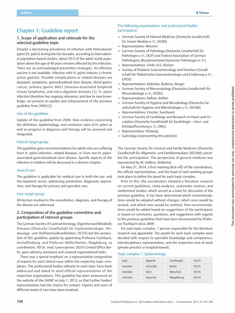
2. Composition of the guideline committee andparticipation of interest groups
The German Society of Gastroenterology, Digestive and MetabolicDiseases (Deutsche Gesellschaft für Gastroenterologie, Ver-dauungs- und Stoffwechselkrankheiten; DGVS) led the produc-tion of this guideline update by appointing Professor Fischbach,Aschaffenburg, and Professor Malfertheiner, Magdeburg, ascoordinators. PD Dr. med. Lynen-Jansen, DGVS Central Office Ber-lin, gave advisory assistance and covered organizational tasks.There was a special emphasis on a representative composition
of experts for each clinical issue within the respective topic com-plexes. The professional bodies relevant to each topic have beenaddressed and asked to send official representatives of therespective organizations. This guideline has been announced onthe website of the AWMF on July 1, 2013, so that further bodies/representatives had the chance for contact. Experts and users ofdifferent levels of care have been involved.
The following organizations and professional bodiesparticipated:
▪ German Society of Internal Medicine (Deutsche Gesellschaftfür Innere Medizin e. V.; DGIM)
▪ Representative: Mössner▪ German Society of Pathology (Deutsche Gesellschaft für
Pathologie e. V.; DGP) and Federal Association of GermanPathologists (Bundesverband Deutscher Pathologen e. V.)
▪ Representatives: Vieth, Eck, Röcken▪ Society of Pediatric Gastroenterology and Nutrition (Gesell-
schaft für Pädiatrische Gastroenterologie und Ernährung e. V.;GPGE)
▪ Representatives: Koletzko, Buderus, Berger▪ German Society of Rheumatology (Deutsche Gesellschaft für
Rheumatologie e. V.; DGRh)▪ Representatives: Kellner, Bolten▪ German Society of Hygiene and Microbiology (Deutsche Ge-
sellschaft für Hygiene und Mikrobiologie e. V.; DGHM)▪ Representatives: Glocker, Suerbaum▪ German Society of Cardiology and Research on Heart and Cir-
culation (Deutsche Gesellschaft für Kardiologie – Herz- undKreislaufforschung e. V.; DKG)
▪ Representative: Nickenig▪ Gastroliga (representing the patients)
The German Society for General and Family Medicine (DeutscheGesellschaft für Allgemein- und Familienmedizin; DEGAM) cancel-led the participation. The perspective of general medicine wasrepresented by M. Hollenz, Rödental.On May 21, 2014, a first meeting (kick-off) of the coordinators,
the official representatives, and the head of each working groupstook place to define the panel for each topic complex.Prior to this, the coordinators initiated a literature research
on current guidelines, meta-analyses, systematic reviews, andrandomized studies, which served as a base for discussion of theprevious guideline. It has been determined which recommenda-tions would be adopted without changes, which ones would berevised, and which ones would be omitted. New recommenda-tions would be added based on suggestions of the participantsor based on comments, questions, and suggestions with regardsto the previous guidelines that have been documented by Profes-sor Fischbach since 2009.For each topic complex, 1 person responsible for the literature
research was appointed. The panels for each topic complex weredecided with respect to specialist knowledge and competence,interdisciplinary representation, and the respective area of work(private practice or hospital-based).
Topic complex 1: Epidemiology
lead Mayerle Greifswald DGVS
member Scherübl Berlin DGVS
member Storr München DGVS
member Venerito Magdeburg DGVS
168 Fischbach Wolfgang et al. S2k-Guideline Helicobacter pylori… Z Gastroenterol 2017; 54: 167–206
Leitlinie
Thi
s do
cum
ent w
as d
ownl
oade
d fo
r pe
rson
al u
se o
nly.
Una
utho
rized
dis
trib
utio
n is
str
ictly
pro
hibi
ted.
member Rad Greifswald DGVS
literatureresearch
Venerito
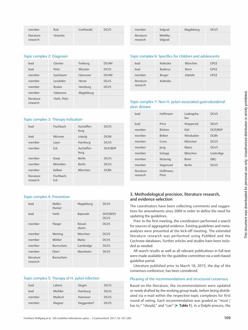
Topic complex 2: Diagnosis
lead Glocker Freiburg DGHM
lead Peitz Münster DGVS
member Suerbaum Hannover DGHM
member Leodolter Herne DGVS
member Rosien Hamburg DGVS
member Vabanova Magdeburg
literatureresearch
Vieth, Peitz
Topic complex 3: Therapy indication
lead Fischbach Aschaffen-burg
DGVS
lead Mössner Leipzig DGIM
member Layer Hamburg DGVS
member Eck Aschaffen-burg
DGP/BDP
member Koop Berlin DGVS
member Mönnikes Berlin DGVS
member Kellner München DGRh
literatureresearch
Fischbach,Eck
Topic complex 4: Prevention
lead Malfer-theiner
Magdeburg DGVS
lead Vieth Bayreuth DGP/BDP/DGVS
member Flieger Rüssel-sheim
DGVS
member Meining München DGVS
member Möhler Mainz DGVS
member Bornschein Cambridge DGVS
member Ebert Mannheim DGVS
literatureresearch
Bornschein
Topic complex 5: Therapy of H. pylori infection
lead Labenz Siegen DGVS
lead Miehlke Hamburg DGVS
member Madisch Hannover DGVS
member Wagner Deggendorf DGVS
member Selgrad Magdeburg DGVS
literatureresearch
Miehlke,Selgrad
Topic complex 6: Specifics for children and adolescents
lead Koletzko München GPGE
lead Buderus Bonn GPGE
member Berger Datteln GPGE
literatureresearch
Koletzko
Topic complex 7: Non-H. pylori-associated gastroduodenalulcer disease
lead Hoffmann Ludwigsha-fen
DGVS
lead Prinz Wuppertal DGVS
member Röcken Kiel DGP/BDP
member Bolten Wiesbaden DGRh
member Gross München DGVS
member Jung Mainz DGVS
member Schepp München Gastroliga
member Nickenig Bonn DKG
member Siegmund Berlin DGVS
literatureresearch
Hoffmann,Prinz
3. Methodological precision, literature research,and evidence selection
The coordinators have been collecting comments and sugges-tions for amendments since 2009 in order to define the need forupdating the guidelines.Prior to the first meeting, the coordinators performed a search
for sources of aggregated evidence. Existing guidelines and meta-analyses were presented at the kick-off meeting. The extendedliterature research was performed using PubMed and theCochrane databases. Further articles and studies have been inclu-ded as needed.All search results as well as all relevant publications in full text
were made available for the guideline committee via a web-basedguideline portal.Literature published prior to March 18, 2015, the day of the
consensus conference, has been considered.
Phrasing of the recommendations and structured consensus
Based on the literature, the recommendations were updatedor newly drafted by the working group leads, before being distrib-uted via e-mail within the respective topic complexes for firstround of voting. Each recommendation was graded as “must /has to,” “should,” and “can” (▶ Table 1). In a Delphi process, the
169Fischbach Wolfgang et al. S2k-Guideline Helicobacter pylori… Z Gastroenterol 2017; 54: 167–206
Thi
s do
cum
ent w
as d
ownl
oade
d fo
r pe
rson
al u
se o
nly.
Una
utho
rized
dis
trib
utio
n is
str
ictly
pro
hibi
ted.
recommendations were voted on by all guideline participantsaccording to a 3-levelled decision scale (agree, undecided,disagree). For each recommendation, for which there was noagreement, a justifying comment must have been entered. Re-commendations with an agreement above 95 % were alreadypassed on at this stage (▶ Table 2).Comments and suggestions for alterations made during the
Delphi process were viewed and assessed by the coordinators.All recommendations that did not achieve an agreement of 95%during the first round of voting were revised within the respectivetopic complex and were discussed again at the concluding con-sensus conference. The conference was chaired by ProfessorFischbach and PD Lynen independently. During a nominal groupprocess, suggestions for alterations were collected and documen-ted before voting on a final version via a TED system. The resultof the voting was documented and the strength of consensusdetermined (▶ Table 2). Subsequently to the consensus confer-ence, there was a final revision of the commenting texts by theleads of each topic complex and the editorial processing of theguideline by the coordinators.▶ Table 3 summarizes the time schedule of establishing the
guideline.
4. External review and approval
The guideline has been presented to all professional bodies, whichgave final comments, and has then been approved. A formalexternal review was undertaken by the AWMF.
5. Editorial independence and handling of potentialconflicts of interest
The guideline has been funded by the DGVS. Representatives ofpharmaceutical companies have not been involved in the processof guideline development in order to maintain neutrality and in-
dependence. All participants disclosed their potential conflicts ofinterest prior to the consensus conference. Any conflict of interestwas documented in the form of the Working Group for ScientificMedicine of the Professional Bodies (Arbeitsgemeinschaft derWissenschaftlichen Medizinischen Fachgesellschaften e. V.;AWMF), including material and immaterial interests, which wasthen made available in tabular form to the guideline committee.The assessment of the declared conflicts of interest was underta-ken by the entire guideline committee. Potential conflicts of inte-rest were discussed openly. It was decided unanimously that peo-ple with potential conflicts of interest abstain from voting onrecommendations that could be affected by this conflict of inter-est. An overview on the potential conflicts of interest can be foundin the supplements.
6. Distribution and Implementation
The guideline, as well as the methods report, is freely availableon the homepage of the DGVS (www.dgvs.de) and the AWMF(www.awmf.de) for download. The full version of the guideline ispublished in the Zeitschrift für Gastroenterologie in both Ger-man and English. A supporting guideline app is available. Inaddition, the guideline recommendations have been presentedat conferences and topically related educational seminars ofthe DGVS.
7. Duration of validity and further updates
The guideline will be valid for 5 years (July 2020). An update ofthe guideline due to newly available data may occur at an earlierpoint in time. The update will be coordinated by the central officeof the DGVS.
Chapter 2: Topic complexes1. Epidemiology
RECOMMENDATION/STATEMENT 1.1
The prevalence of the infection with H. pylori varies with geo-
graphy (industrial and developing countries), ethnic origin,
and socio-economic status. There is an age-dependent in-
crease. Globally, H. pylori infection rates have decreased in
the last decades.
Strength of consensus: strong consensus
Comment
▶ Table 3 Time schedule.
March 2013 appointment of the coordinators by the DGVS
July 2013 announcement at the AWMF
May 2014 kick-off-meeting Berlin
February 2015 Delphi-process
March 2015 consensus conference Berlin
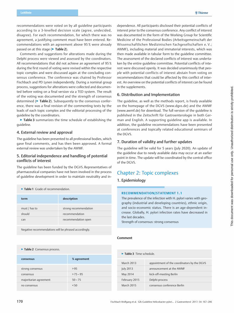
▶ Table 1 Grade of recommendation.
term description
must / has to strong recommendation
should recommendation
can recommendation open
Negative recommendations will be phrased accordingly.
▶ Table 2 Consensus process.
consensus % agreement
strong consensus > 95
consensus > 75 – 95
majoritarian agreement 50 – 75
no consensus < 50
170 Fischbach Wolfgang et al. S2k-Guideline Helicobacter pylori… Z Gastroenterol 2017; 54: 167–206
Leitlinie
Thi
s do
cum
ent w
as d
ownl
oade
d fo
r pe
rson
al u
se o
nly.
Una
utho
rized
dis
trib
utio
n is
str
ictly
pro
hibi
ted.
The prevalence of H. pylori infection varies strongly betweenindustrial and developing countries, different regions (e. g., UK13.4 %, Korea 80.8 %), as well as within a single population [3].Currently, 50% of the world population is supposed to be infectedwith H. pylori [4].Differences in the prevalence between different ethnic groups
are a consequence of a variable intensity of the exposure toH. pylori (socio-economic factors, alimentary, and environmentalfactors) [5 – 7]. The individual genetic disposition also has to beconsidered. Recently, polymorphisms in the toll-like receptor1 (TLR1) gene have been identified as a susceptibility gene in 2 in-dependent cohorts [8]. After immigration into an industrial coun-try, the country of birth represents a risk factor for the infectionwith H. pylori with the risk correlating negatively with the durationof stay in the country into which they immigrated [9]. The preval-ence of the infection depends on socio-economic status (profes-sion, income, living situation), especially during childhood, whentransmission is most likely to take place [10]. Within a population,there is an age-dependent increase (ca. 1 % per year of life in in-dustrial nations). This is interpreted as a result of the birth cohorteffect [11, 12]. The prevalence of infection in developing coun-tries is already high at an age below 20 years, culminating in thethird decade [13].
RECOMMENDATION/STATEMENT 1.2
The prevalence of H. pylori infection in Germany ranges from
3% (children) to 48% (adults). It is significantly higher for im-
migrants (36 – 86%).
Strength of consensus: strong consensus
CommentThe prevalence of H. pylori infection is 3 % for children at the
age of 4 years [14] and 5 –7% for children between 5 and 7 years[15]. An essential risk factor for the infection during childhood isthe mother’s infection status (OR 13.0; 95 % CI 3.0 – 55.2) [14].The infection rate in children has recently stabilized, and a furtherdecrease has not been documented [16]. The prevalence amongwomen and men below 30 years is 19 % and 25 %, respectively,above 30 years 35% and 55%, respectively, and at an age above65 years at 69% and 90%, respectively [17, 18]. Interestingly, therisk of H. pylori infection in Germany increases with the number ofsiblings (OR 1.65). If, however, this is adjusted for age, gender,education, incidence of gastric cancer within a family, and nico-tine and alcohol consumption, the higher prevalence does not re-main [19]. In addition, in Germany there is a high variation ofH. pylori prevalence depending on origin and country of birth. Im-migrants from Turkey show a prevalence of 30 % compared to44.5 % for Turkish people living in Turkey and 13% for Germans inan age-matched cohort [20].
RECOMMENDATION/STATEMENT 1.3
The transmission of H. pylori happens from human to human.
The exact route of transmission (oral-oral, gastral-oral, fecal-
oral, or any combination) is not clear.
Strength of consensus: strong consensus
CommentH. pylori can be cultured from vomit, stool, and saliva [21].
Vomited stomach contents show an especially high bacterialdensity [22]. H. pylori transmission from person to person contacthas been seen following episodes of acute gastrointestinal infec-tions [23]. The close contact with H. pylori-contaminated bodilyfluids within families explains the increased occurrence of theinfection within families. Interestingly, the contagion with H. py-lori does not happen to the same degree outside of the family, asit has been shown by a meta-analysis of 16 studies of children inkindergarten or nurseries [24]. It could be that the higher trans-mission rate within families is mediated by susceptibility geneslike TLR1 [7]. There is no clear evidence for zoonotic transmissionof H. pylori, although the bacteria have been detected in primatesand, more rarely, in other animals [25 –27].
RECOMMENDATION/STATEMENT 1.4
Close contact between children and family members infected
with H. pylori represents the most important route of trans-
mission.
Strength of consensus: strong consensus
CommentThe transmission of H. pylori within a family is well documented
[28 – 31]. There is consistent molecular biology of single-trans-mitted H. pylori strains within mothers and their respective chil-dren [32, 33]. The number of family members and the size of theliving area are additional risk factors [34]. Breast-feeding of new-borns has no influence on the transmission of H. pylori [35, 36].The infection of older siblings represents a particular predictorfor H. pylori infection [37]. The incidence rates of the infectionwith H. pylori are highest in children below 3 years and clearlydecrease after the age of 5 [38]. Transient infections during child-hood have been described [39].
RECOMMENDATION/STATEMENT 1.5
Contamination of drinking water and food with H. pylori has
been described. Transmission of the bacteria via water or sew-
age is discussed and is controversial.
Strength of consensus: strong consensus
CommentThe relevance of water or sewage as a potential source for infec-
tion is controversial [40 – 44]. Despite evidence of H. pylori DNA inwater and sewage, there are only few descriptions of positive cul-tures [45]. Due to the restricted metabolic and regulatory abilitiesof H. pylori in an environment outside the stomach, a long extra-gastric survival of the bacteria is unlikely to be possible [46, 47].
171Fischbach Wolfgang et al. S2k-Guideline Helicobacter pylori… Z Gastroenterol 2017; 54: 167–206
Thi
s do
cum
ent w
as d
ownl
oade
d fo
r pe
rson
al u
se o
nly.
Una
utho
rized
dis
trib
utio
n is
str
ictly
pro
hibi
ted.
RECOMMENDATION/STATEMENT 1.6
The rate of recurrent infection in adults after successful eradi-
cation therapy in industrial countries is low.
Strength of consensus: strong consensus
CommentThe rate of recurrent infection in adults after successful H. py-
lori eradication is about 2 % per year in industrial countries and6 – 12% in developing countries [48]. The re-infection rate in chil-dren older than 5 years is about 2% per year [49]. In case of an in-fection within the first year after eradication therapy, in 60% thesame strain can be identified, whereas in cases of detection aftermore than 12 months, a new strain is usually isolated. Therefore,recurrence of H. pylori within 12 months is supposed to be a“true” recurrence or recrudescence and not a new infection [50].
RECOMMENDATION/STATEMENT 1.7
There are no established strategies for prevention of H. pylori
infection. An effective vaccine is currently not available.
Strength of consensus: strong consensus
CommentCurrently, there is no effective H. pylori vaccine available. It has
been estimated that an effective vaccine would result in a signifi-cant reduction of H. pylori prevalence and associated diseasesafter a 10-year vaccination regimen [51]. This would be cost-ef-fective given an efficacy of the vaccination of 55 %. In a studythat has been published following this consensus conference, effi-cacy of an oral recombinant vaccine against H. pylori could bedemonstrated in 4464 participants [52]. Vaccination was success-ful in 71.8 % (95% CI 48.2 – 85.6), and side-effects occurred in lessthan 1%. The evaluation of long-term success is still missing anda planned follow-up of 3 years is awaited. Calculations for cost-ef-fectiveness should consider prevalence of the infection as wellas associated diseases. The variable and declining prevalence ofH. pylori does not allow a cost-effectiveness analysis at the mo-ment [53, 54].Spontaneous elimination of infection with H. pylori is unlikely.
In a German study with more than 2235 children of preschoolage, in 30 out of 104 H. pylori-positive children, the bacteria couldnot be detected anymore after 2 years [55]. A survey among par-ents was possible in 25 of the 30 children. Most of the childrenreceived triple therapy for eradication (18/25) or antibiotics foranother reason (4/25). Thus, spontaneous elimination of an infec-tion with H. pylori in children (in the cited study 3/25 children,12%) is considered as a rare event.After partial gastrectomy, spontaneous elimination of H. pylori
has been seen in 43 % [56]. Loss of the antrum with secondaryachlorhidria is considered to be the mechanism of spontaneousH. pylori elimination [57]. Furthermore, enterogastric bile refluxis associated with a reduced H. pylori colonization [58]. Anotherreason for spontaneous elimination of an H. pylori infectionin adults is achlorhidria in case of severe atrophy of the gastric
body mucosa, progression of the course of the infection, andin case of auto-immune gastritis [59].
RECOMMENDATION/STATEMENT 1.8
Gastroduodenal ulcer disease, gastric cancer and the gastric
marginal zone B-cell lymphoma of MALT are diseases associat-
ed with H. pylori infection.
Strength of consensus: consensus
CommentInfection with H. pylori induces chronic-active gastritis. Possible
complications and related diseases are gastroduodenal ulcerdisease, gastric adenocarcinoma, and the marginal zone B-celllymphoma of MALT [60 – 62].Infection with H. pylori increases the risk of distal gastric cancer
by a factor of 2 – 3 (OR 1.92 – 2.56) compared to non-infectedindividuals. The association of H. pylori infection with differenttypes of gastric cancer is comparable: intestinal type OR 2.49 –4.45; diffuse type OR 2.58 – 3.39 [63 – 67]. The relative risk ishigher if serum samples used for H. pylori diagnosis were takenlonger before the cancer diagnosis (OR 5.9); thus, the associationbetween H. pylori and gastric cancer could be underestimateddue to elimination of the bacteria during progression of thedisease [68, 69]. If a previous infection is confirmed by persistentCagA antibodies in the serum, then the predicted risk for gastriccancer rises 18 – 20 fold [70, 71].The incidence of MALT lymphoma correlates with the preval-
ence of H. pylori infection. The relative risk of developing a pri-mary gastric lymphoma is increased by a factor of 6 in cases wherethere was serological evidence for H. pylori in large case-controlstudies [72]. Helicobacter heilmannii can be detected mainly inanimals with prevalence in humans of 0.5 % and is also associatedwith an increased risk for a gastric MALT lymphoma [73, 74].The NHANES-III study from the USA demonstrated that an
H. pylori infection is not associated with an increased mortalityrate and has even protective effects on the developments of a cer-ebrovascular accident [75]. Although there is an increased risk ofgastric cancer with H. pylori, this has no impact on the mortalityof the cohort due to the low gastric cancer prevalence. Adenocar-cinoma of the esophagus is inversely associated with H. pyloriinfection, although a plausible cause for this has not yet beendescribed [76]. Furthermore, infection with H. pylori is associatedwith an 18 % risk reduction of atopic disease in epidemiologicalstudies. It is unclear if there is a causal relation [77].
RECOMMENDATION/STATEMENT 1.9
Direct contact between doctors or nursing staff and patients
is not a relevant risk factor for H. pylori infection.
Strength of consensus: strong consensus
CommentThe direct contact of doctors or nurses with H. pylori-positive
patients is not a significant risk factor for infection [78]. A meta-
172 Fischbach Wolfgang et al. S2k-Guideline Helicobacter pylori… Z Gastroenterol 2017; 54: 167–206
Leitlinie
Thi
s do
cum
ent w
as d
ownl
oade
d fo
r pe
rson
al u
se o
nly.
Una
utho
rized
dis
trib
utio
n is
str
ictly
pro
hibi
ted.
analysis of 15 studies shows only a mildly increased risk for H. py-lori infection among gastroenterologists (RR 1.6; 95 % CI: 1.3 –2.0) and endoscopy staff (RR 1.4; 95% CI: 1.1 – 1.8) [79].
RECOMMENDATION/STATEMENT 1.10
The direct transmission of H. pylori infection between part-
ners is possible but rare. The route of transmission is unclear.
Strength of consensus: strong consensus
CommentThe direct transmission of an H. pylori infection between part-
ners is possible. Transmission however, is only confirmed if thesame strain is detected in both partners (e. g., by fingerprint). Ina serum study on 389 married couples from the UK, there was anincreased risk for the spouse [80]. In a study from Germany on670 married couples, there was only an increased risk for subjectswho were married to a partner of non-German origin (OR 6.05;95% CI: 1.31 – 17.96) [81]. The route of transmission is not clear,but orol-oral transmission seems unlikely [82]. After successfulH. pylori eradication, there is only rarely re-infection even in caseof an H. pylori-positive partner [83].
2. Diagnosis
RECOMMENDATION/STATEMENT 2.1
The following methods for the detection of H. pylori are ade-
quately validated and can be applied for the diagnosis of the
infection under consideration of the respective clinical set-
ting. Invasive methods include culture, histology, rapid urease
test, and polymerase chain reaction (PCR). Non-invasive
methods include urea breath test (UBT), stool antigen test
with monoclonal antibodies, and immunglobulin G (IgG) anti-
bodies in the serum.
Strength of consensus: strong consensus
CommentThe methods mentioned above are sufficiently validated but
vary in their accuracy [84 – 90]. Furthermore, the different testshave specific areas of use.
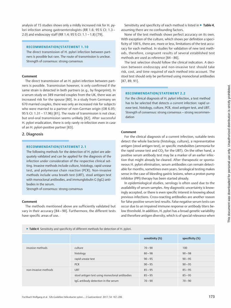
Sensitivity and specificity of each method is listed in ▶ Table 4,assuming there are no confounding factors.None of the test methods shows perfect accuracy on its own.
With exception of the culture, which shows per definition a speci-ficity of 100%, there are, more or less, limitations of the test accu-racy for each method. In studies for validation of new test meth-ods, therefore, congruent results of several established testmethods are used as reference [84 – 86].The test selection should follow the clinical indication. A deci-
sion between endoscopy and non-invasive test should takerisk, cost, and time required of each method into account. Thestool test should only be performed using monoclonal antibodies[87, 89, 91].
RECOMMENDATION/STATEMENT 2.2
For the clinical diagnosis of H. pylori infection, a test method
has to be selected that detects a current infection: rapid ur-
ease test, histology, culture, PCR, stool antigen test, and UBT.
Strength of consensus: strong consensus – strong recommen-
dation
CommentFor the clinical diagnosis of a current infection, suitable tests
detect the whole bacteria (histology, culture), a representativeantigen (stool antigen test), or specific metabolites (ammonia forthe rapid urease test and CO2 for the UBT). On the other hand, apositive serum antibody test may be a marker of an earlier infec-tion that might already be cleared. After therapeutic or sponta-neous H. pylori elimination, serum antibodies can remain detect-able for months, sometimes even years. Serological testing makessense in the case of bleeding gastric lesions, when a proton pumpinhibitor (PPI) therapy has been started already.In epidemiological studies, serology is often used due to the
availability of serum samples. Any diagnostic uncertainty is know-ingly accepted, or there is even specific interest in knowing aboutprevious infections. Cross-reacting antibodies are another reasonfor false positive serum test results. False negative serum tests canoccur due to an impaired immune response or antibody titers be-low threshold. In addition, H. pylori has a broad genetic variabilityand therefore antigen diversity, which is of special relevance when
▶ Table 4 Sensitivity and specificity of different methods for detection of H. pylori.
sensitivity (%) specificity (%)
invasive methods culture 70 – 90 100
histology 80– 98 90 – 98
rapid urease test 90 – 95 90 – 95
PCR 90 – 95 90 – 95
non-invasive methods UBT 85 – 95 85 – 95
stool antigen test using monoclonal antibodies 85 – 95 85 – 95
IgG antibody detection in the serum 70 – 90 70 – 90
173Fischbach Wolfgang et al. S2k-Guideline Helicobacter pylori… Z Gastroenterol 2017; 54: 167–206
Thi
s do
cum
ent w
as d
ownl
oade
d fo
r pe
rson
al u
se o
nly.
Una
utho
rized
dis
trib
utio
n is
str
ictly
pro
hibi
ted.
comparing patients from different continents. Thus, test kits forthe detection of H. pylori IgG antibodies should be validated foruse in Europe.
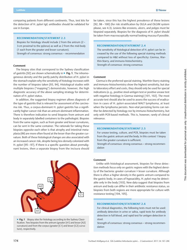
RECOMMENDATION/STATEMENT 2.3
Biopsies for histology should include 2 from the antrum (2 –
3 cm proximal to the pylorus) as well as 2 from the mid-body
(1 each from the greater and lesser curvature).
Strength of consensus: strong consensus – recommendation
CommentThe biopsy sites that correspond to the Sydney classification
of gastritis [92] are shown schematically in ▶ Fig. 1. The inhomo-geneous density and the partly patchy distribution of H. pylori inthe stomach explain why the sensitivity of histology increases withthe number of biopsies taken [93, 94]. Histological studies withmultiple biopsies (“mapping”) demonstrate, however, the highdiagnostic accuracy of the above sampling strategy for determi-nation of H. pylori status.In addition, the suggested biopsy regimen allows diagnosis of
the type of gastritis that is relevant for assessment of the carcino-ma risk. Thus, a corpus-dominant H. pylori gastritis has a signifi-cantly higher cancer risk than an antrum-dominant inflammation.There is therefore indication to send biopsies from antrum andbody in separately labelled containers to the pathologist. Biopsiesfrom the same region, such as from greater and lesser curvatures,can be sent in the same container. The rationale for taking thesebiopsies opposite each other is that atrophy and intestinal meta-plasia (IM) are more often found at the lesser than the greater cur-vature. Both of these histological changes are also associated withan increased cancer risk, despite being less densely colonized withH. pylori [95 – 97]. If there is a specific question about premalig-nant lesions, then a separate biopsy from the incisura should
be taken, since this has the highest prevalence of these lesions[92, 98 – 100] (for risk stratification by OLGA and OLGIM systemplease, see 4.3). Lesions like erosions, ulcers, and polyps must bebiopsied separately. Biopsies for the diagnosis of H. pylori shouldbe taken frommacroscopically normal looking mucosa if possible.
RECOMMENDATION/STATEMENT 2.4
The sensitivity of histological detection of H. pylori can be in-
creased by the use of the following special staining methods
compared to H&E without loss of specificity: Giemsa, War-
thin-Starry, and immuno-histochemistry.
Strength of consensus: strong consensus
CommentGiemsa is the preferred special staining. Warthin-Starry staining
and immuno-histochemistry show the highest sensitivity, but dueto laboratory effort and costs, they should only be used for specialindications (e. g., positive stool-antigen test or positive urease testwith negative histology in Giemsa staining) [101, 102]. The mostaccurate test should also be used to assess the success of eradica-tion in cases of H. pylori-associated MALT lymphoma, at leastwhen the lymphoma persists. Non-vital persisting forms can nei-ther be detected by histology nor by immuno-histochemistry, butonly with PCR-based methods. This is, however, rarely of clinicalrelevance.
RECOMMENDATION/STATEMENT 2.5
For urease testing, culture, and PCR, biopsies must be taken
from the gastric antrum and the body. In this context 1 biopsy
from the greater curvature is sufficient.
Strength of consensus: strong consensus – strong recommen-
dation
CommentUnlike with histological assessment, biopsies for these detec-
tion methods focus only on gastric regions with the highest densi-ty of the bacteria: greater curvature > lesser curvature. Althoughthere is often a higher density in the gastric antrum compared tothe gastric body, in cases of hypoacidity, H. pylori may be detect-able only in the body [103]. New data suggest that biopsies fromantrum and body can differ in their antibiotic resistance status, sobiopsies from both regions are more appropriate for culture withresistance testing [104, 105].
RECOMMENDATION/STATEMENT 2.6
For clinical diagnostics, the following tests must not be used:
antibody detection in urine or saliva, rapid tests for antibody
detection in full blood, and rapid test for antigen detection in
stool.
Strength of consensus: strong consensus – strong recommen-
dation
A1
A2
C1
C2
▶ Fig. 1 Biopsy sites for histology according to the Sydney Classi-fication. Two biopsies from the antrum (greater [A1] and lesser [A2]curvature) and from the corpus (greater [C1] and lesser [C2] curva-ture), respectively.
174 Fischbach Wolfgang et al. S2k-Guideline Helicobacter pylori… Z Gastroenterol 2017; 54: 167–206
Leitlinie
Thi
s do
cum
ent w
as d
ownl
oade
d fo
r pe
rson
al u
se o
nly.
Una
utho
rized
dis
trib
utio
n is
str
ictly
pro
hibi
ted.
CommentEven with such tests being partly used in practice outside of the
laboratory (in-office tests), they should currently not be usedin clinical diagnostics since they are not sufficiently validatedand/or not of adequate accuracy [106 – 108].
RECOMMENDATION/STATEMENT 2.7
Confounding factors have to be considered for the selection
of test methods and their interpretation.
Bacterial overgrowth of the stomach can lead to false positive
results on urease-dependent tests.
False negative results from tests for the detection of a current
infection may be due to the following:
▪ treatment with a PPI
▪ upper gastrointestinal bleeding
▪ previous partial gastrectomy
▪ mucosal atrophy and IM
▪ gastric cancer and MALT lymphoma
Strength of consensus: strong consensus – strong recommen-
dation
CommentThe UBT and the rapid urease test are urease-dependent.
Urease cleaves urea into carbon dioxide and ammonia. Carbon di-oxide is the indicator reagent for the UBT, ammonia for the rapidurease test. H. pylori is characterized by a very high urease activ-ity, but other bacteria within the gastrointestinal tract are alsocapable of cleaving urea. Bacterial overgrowth of the stomachwith urease-producing bacteria other than H. pylori can occur incases of delayed gastrointestinal motility or hypochlorhydria,leading occasionally to false positive results of urease-dependenttest [109, 110]. Urease-producing bacteria other than H. pyloriare the reason for a late color change in the rapid urease test.Therefore, it is important to respect the latest time point forread-out given by the manufacturer.Sensitivity of the all tests for proof of a current infection (i. e.,
serology excluded) is reduced by conditions that lead to a reducedcolonization density [103, 104]. A reduced bacterial density isespecially seen with PPI treatment and with antibiotics that affectH. pylori. In contrast, H2 blockers reduce the sensitivity only a lit-tle. A reduced H. pylori density can furthermore be found in casesof hypochlorhydria and mucosal atrophy, gastric cancer, or MALTlymphoma of the stomach [111, 112].The sensitivity of the biopsy-based test is reduced to 70 % in
case of an acute upper gastrointestinal bleeding, while specificityis maintained. The reason for this observation is not yet fully ex-plained. For the breath test, despite being less well validated, thisreduction of sensitivity has not been shown in meta-analysis[113]. A PCR seems to be the most sensitive method in this situa-tion but is less commonly used [114, 115]. For clinical practice, itcan therefore be recommended to obtain histology in case of anupper gastrointestinal bleeding or to perform serological testing[116]. Histology is preferred in this condition.
RECOMMENDATION/STATEMENT 2.8
For reliable H. pylori diagnosis, the following minimal intervals
without H. pylori suppressive therapy should be respected:
2 weeks after completing a PPI therapy
4 weeks after preceding H. pylori eradication or other antibio-
tic therapy
Strength of consensus: strong consensus – recommendation
CommentAfter completion of an acid-suppressing or antibiotic therapy,
establishing of the original H. pylori density takes several daysor weeks, depending on the intensity and duration of the previoustreatment. During this period the sensitivity of all direct tests isreduced. This is a relevant problem in clinical practice thus far,since dyspepsia is often primarily treated with a PPI, before H. py-lori is tested for or endoscopy undertaken.If the above-mentioned intervals are respected, all test modal-
ities are suitable for detection of a current infection (2.2) as well asfor control of eradication success [88, 117].
RECOMMENDATION/STATEMENT 2.10
For a reliable diagnosis of H. pylori, 2 positive test results
should be available. Exemptions are:
▪ In case of a duodenal ulcer, 1 positive test result is suffi-
cient to establish the diagnosis of H. pylori infection.
▪ The histological proof of H. pylori in combination with a
chronic-active gastritis is nearly 100% specific and there-
fore sufficient.
▪ A positive culture is per se 100% specific and sufficient.
Strength of consensus: majority agreement – recommen-
dation
CommentAs with the previous consensus conference for the S3 guideline
of 2009 [1], the first sentence of this statement was highly deba-ted and received a majority agreement. Only a minority pleadedthat 1 positive test result is sufficient for the diagnosis of H. pyloriinfection, as stated in the Maastricht IV/Florence consensus report[118].The requirement of positive results in at least 2 tests for a reli-
able positive diagnosis is due to the low and further decreasingprevalence of H. pylori infection in industrial countries. At a lowprevalence, a constant proportion of false positive results has ahigher impact than in case of high prevalence leading to a low po-sitive predictive value.Cases with duodenal ulceration are, however, associated with
a high H. pylori prevalence on the other hand, so in this situationa positive result in only 1 test is sufficient for the diagnosis of anH. pylori infection. Further conditions for a high prevalence areorigin from a region with high H. pylori prevalence or a gastric ul-cer without other cause (e. g., non-steroidal anti-inflammatorydrugs).
175Fischbach Wolfgang et al. S2k-Guideline Helicobacter pylori… Z Gastroenterol 2017; 54: 167–206
Thi
s do
cum
ent w
as d
ownl
oade
d fo
r pe
rson
al u
se o
nly.
Una
utho
rized
dis
trib
utio
n is
str
ictly
pro
hibi
ted.
A positive histology for H. pylori is nearly 100% specific. For atrained pathologist, the attribution of the bacterial morphologyis highly reliable. Furthermore, presence of a typical chronic-ac-tive gastritis with clear infiltration by neutrophil granulocytesis an additional criterion. For the use the activity of inflammationas a diagnostic criterion, it is important that the biopsies have notbeen taken from areas with erosions or ulcers. This is another rea-son for the recommendation above to biopsy lesions separately.If the diagnosis of H. pylori is assessed by an invasive endoscopy-based test, a combination of rapid urease test and histology isadvisable (apart from cases with present duodenal ulcer) becausethe histological result will not be available at the time of the inves-tigation.By definition, there cannot be false positive results in an
adequate culture, resulting in a specificity of 100 % (see 2.1 and▶ Table 1). For clinical use, diagnosis by culture is, however, toolaborious and should be used primarily for resistance testing.
RECOMMENDATION/STATEMENT 2.11
The investigation of bacterial virulence factors should not be
performed outside of scientific research.
Strength of consensus: strong consensus – strong recommen-
dation
CommentPathogenic factors of H. pylori have an influence on the devel-
opment of complications associated with H. pylori-induced gastri-tis like the gastroduodenal ulcer disease or gastric carcinoma.Knowledge about the existence of these virulence factors, how-ever, is not relevant for a clinical approach [119].
RECOMMENDATION/STATEMENT 2.12
After 2 treatment failures, a resistance test has to be per-
formed.
Strength of consensus: strong consensus – strong recommen-
dation
CommentAlready after 1 treatment failure, resistance rates against clari-
thromycin rise to 60 %; after 2 unsuccessful therapy attempts,they rise to 80 % [120]. After 2 treatment failures, more than60% of H. pylori isolates show a combined resistance against clar-ithromycin and metronidazole. In addition, there are increasingrates of resistance against quinolones [120, 121]. The possibilityof successfully applying further empirical treatment regimensis therefore drastically limited. On the other hand, the cultureand incubation of H. pylori with resistance testing enable targetedtherapy.The antimicrobial sensitivity of H. pylori can be determined by
agar diffusion testing. A well-standardized agar diffusion test fordetermination of resistance is the application of the Etest® [122].This consists of a plastic or paper strip that is coated with concen-tration gradient of a specific antibiotic. After placement of the
strip on a H. pylori culture on a fixed culture medium, the antibio-tic diffuses into the culture medium according to the gradient,enabling a precise read-out of the minimal inhibitory concentra-tion. This makes stratification into sensitive and resistant possible,according to the European Committee for antimicrobial sensitivitytesting (www.eucast.org).Etest strips are commercially available for the antibiotics that
are usually applied for eradication therapy like clarithromycin,metronidazole, levofloxacin, tetracycline, and amoxicillin. Fortesting the sensitivity on rifabutin, a rifampicin strip can be usedas an alternative.The sensitivity testing for H. pylori gives results on the in-vitro
resistance. According to experience, the actual clinical relevanceof such resistance requires confirmation within clinical studiesdue to the particular pharmacokinetic conditions within the stom-ach. Therefore, antibiotics for eradication therapy should not justbe combined based on the sensitivity testing, but also based onthe experience from clinical studies.If a high clarithromycin resistance is expected (e. g., in patients
with an unsuccessful previous eradication, in patient with migra-tion background, and in young patients) sensitivity testing can beperformed before first-or second-line therapy. Such sensitivitytesting can be done in a microbiological laboratory using pheno-typic and genotypic methods [123]. For the latter, gastric biopsiescan also be used that have been obtained for pathology or for therapid urease test [124, 125].Microbiological laboratories that have established methods for
genotypic resistance testing can use these as reliable tests for thedetermination of resistance. Except for metronidazole, the mole-cular mechanisms of resistance against antibiotics used in eradi-cation therapies are known. These are due to mutations of therespective microbial receptor molecules and allow genotypicresistance testing in individual cases [126].Since resistance against tetracyclin and rifabutin is rare and re-
sistance against amoxicillin is practically non-existent in Germany[120], test methods can be applied that can detect resistance-in-ducing mutations against clarithromycin and/or levofloxacin.Such tests are commercially available and adequately validated.There is good conformity of the results of phenotypic and geno-typic resistance testing [127 – 129]. Alternatively, validatedin-house methods can be applied. Such methods of molecular-ge-netic resistance testing can be sufficient to guide appropriatefirst- or second-line therapy [130].
3. Indication for treatment
Peptic ulcer
RECOMMENDATION/STATEMENT 3.1
In case of a gastric or duodenal ulceration, H. pylori infection
must undergo eradication treatment.
Strength of consensus: strong consensus – strong recommen-
dation
176 Fischbach Wolfgang et al. S2k-Guideline Helicobacter pylori… Z Gastroenterol 2017; 54: 167–206
Leitlinie
Thi
s do
cum
ent w
as d
ownl
oade
d fo
r pe
rson
al u
se o
nly.
Una
utho
rized
dis
trib
utio
n is
str
ictly
pro
hibi
ted.
CommentThere are several meta-analyses that clearly demonstrate
the benefit of eradication therapy in the case of ulcers of thestomach and the duodenum with or without complications[131 – 137]. Similarly, the decreasing association of H. pylori andgastric/duodenal ulcers, due to a decreasing prevalence of theinfection in the Western countries and a parallel increase of acet-ylsalicylic acid (aspirin)/NSAID-associated ulcers, make it compul-sory to prove the presence of H. pylori (see also topic complex1 and 2).
Gastric marginal-zone-B-cell lymphoma (MZBCL) of MALT –MALT lymphoma
RECOMMENDATION/STATEMENT 3.2
In H. pylori-positive MALT lymphomas, eradication must be
undertaken.
Strength of consensus: strong consensus – strong recommen-
dation
CommentAll gastric MALT lymphomas should initially undergo eradica-
tion therapy, irrespective of the stage of disease. This is the ther-apy of first choice with curative intent [118, 138]. According toa meta-analysis, a successful H. pylori eradication leads to com-plete lymphoma remission in 77.5 % in stage I and II (78% in stageI and 56 % in stage II) [139]. The remission is also stable in thelong-term, so that the majority of patients with a gastric MALTlymphoma are healed with eradication therapy only [140, 141].Recurrence is only observed in 3 – 7%, and high malignant trans-formation in these cases is rare at only 0.05% [139 – 141].Following successful H. pylori eradiation, with minimal histolo-
gical residues of MALT lymphoma and normalization of theendoscopic findings, there is a favorable disease course withoutfurther oncological therapy, so that in this situation a watch-and-wait strategy with regular endoscopic-biopsy controls can berecommended [142]. According to a meta-analysis, even in H. py-lori-negative patients there can be in about 15 % lymphomaremissions after a regular eradication therapy [143].
Diffuse large-cell B-cell lymphoma (DLCBCL) of the stomach
RECOMMENDATION/STATEMENT 3.3
Diffuse large cell B-cell lymphomas (DLBCL) of the stomach
with or without MALT component in stage I or II can be sub-
jected to H. pylori eradication. Standard therapy of these lym-
phomas is an immune-chemotherapy with Rituximab plus
CHOP, which should be induced quickly when there is no
lymphoma regression in response to H. pylori eradication
(1 – 2 months).
Strength of consensus: strong consensus – recommendation
open
CommentPatients with H. pylori-positive DLBCL in stage I can undergo
sole eradication treatment initially, in strict association with fre-quent clinical assessment with endoscopy and biopsies [138].There are reports of varying rates of lymphoma remission in theliterature [144 – 146]. If there are no definite signs of lymphomaregression after H. pylori eradication, patients should receive earlyimmune-chemotherapy with the anti-CD20 antibody Rituximaband chemotherapy according to the CHOP protocol.
Functional dyspepsia
RECOMMENDATION/STATEMENT 3.4
In patients with functional dyspepsia (irritable stomach) and
H. pylori infection, an eradication therapy can be undertaken.
Strength of consensus: strong consensus – recommendation
open
CommentThe elimination of the H. pylori infection in patients with
dyspeptic symptoms that are persisting for at least 4 weeks andwithout endoscopic findings leads, in up to 10 %, to a sustainedsymptom improvement. The number-needed-to-treat (NNT) isapproximately 12 [147]. A recent meta-analysis on 14 randomizedcontrolled studies demonstrated a significant improvement of thedyspeptic symptoms following eradication, compared with con-trols: OR 1.38; 95 % CI 1.18 – 1.62; p < 0.001 [148]. This benefithas been shown for populations in America, Asia, and Europe.Further, more recent studies, which were not fully included inthis meta-analysis, show H. pylori eradication resulted in the gen-eral improvement of symptoms or improvement of the singlesymptom of functional dyspepsia to various degrees [149– 154].According to the Kyoto consensus report on H. pylori gastritisof 2015, H. pylori eradication is the treatment option of firstchoice [155].For an individualized decision on H. pylori eradication, the fol-
lowing arguments can be considered, besides the patient’s wishand the subjective degree of suffering: the lack of therapeuticalternatives; cancer prevention (see topic complex 4); reductionin medical consultations [156]; and endoscopies [157]. On theother hand, the likelihood of side effects from the eradicationtherapy is 10 –25% with most being transient in nature.
RECOMMENDATION/STATEMENT 3.5
A non-invasive test for H. pylori with subsequent eradication
treatment cannot be generally recommended for Germany.
Strength of consensus: majority consensus – recommenda-
tion open
CommentThe recommendation can already be found in the previous
S3 guideline of 2009. This has been controversially discussed.The recommendation is based on the specific conditions in Ger-many, including the low and further decreasing H. pylori preval-
177Fischbach Wolfgang et al. S2k-Guideline Helicobacter pylori… Z Gastroenterol 2017; 54: 167–206
Thi
s do
cum
ent w
as d
ownl
oade
d fo
r pe
rson
al u
se o
nly.
Una
utho
rized
dis
trib
utio
n is
str
ictly
pro
hibi
ted.
ence as well as the wide availability and low cost of endoscopy,which have not changed. The discussion about a test-and-treatstrategy has focused on patients with dyspeptic symptoms andhas not focused on preventive aspects of H. pylori diagnosticsand therapy in asymptomatic individuals, which will be discussedin topic complex 4.
Reflux
RECOMMENDATION/STATEMENT 3.6
Reflux symptoms or a reflux esophagitis is not an indication
for H. pylori eradication. The decision on H. pylori eradication
due to other indications can be made independently from any
reflux symptoms or reflux disease.
Strength of consensus: strong consensus – no recommen-
dations
CommentEpidemiological studies suggest a negative association be-
tween H. pylori and the reflux disease [158 – 161]. In addition,Barrett’s esophagus and esophageal adenocarcinomas are seenmore rarely with H. pylori infection, although a recent meta-anal-ysis could not prove a clear negative association between H. pyloriand Barrett’s esophagus [162, 163]. This leads to the conclusionthat H. pylori has a protective effect and that an eradication canlead to reflux disease or its exacerbation. The majority of studiescould not detect a negative effect of an H. pylori eradication onreflux symptoms or a reflux esophagitis [164 – 168]. Therefore,the decision whether to undertake H. pylori eradication canbe made independently from the presence of reflux symptoms ora reflux disease.Long-term treatment with PPI, however, requires H. pylori era-
dication, since this medication can lead to the development ofatrophic changes of the gastric body mucosa as well as a H. py-lori-positive corpus-predominant gastritis. The latter is consideredas risk gastritis for gastric cancer. The long-term intake of PPIis not associated with an increased rate of gastric cancers orNETs, however [169].
Further indications (ITP, Menetrier’s disease, lymphocyticgastritis, iron deficiency anaemia)
RECOMMENDATION/STATEMENT 3.7
Patients with idiopathic thrombocytopenic purpura (ITP)
must be investigated for H. pylori infection and treated with
eradication therapy, if the bacteria are detected.
Strength of consensus: strong consensus – strong recommen-
dation
CommentTwo systematic literature analyses demonstrated that H. pylori
eradication leads to a significantly increased number of thrombo-cytes in 50 % of patients [170, 171]. Children also show signifi-cantly higher thrombocyte counts after eradication [172].
RECOMMENDATION/STATEMENT 3.8
Patients with Menetrier’s disease and positive evidence for
H. pylori infection should receive eradication therapy.
Strength of consensus: strong consensus – recommendation
CommentThere are only uncontrolled case reports concerning this [173 –
178].
RECOMMENDATION/STATEMENT 3.9
Patients with lymphocytic gastritis in whom H. pylori infection
is detected should be treated with eradication therapy.
Strength of consensus: strong consensus – recommendation
open
CommentBesides a recent case report on a child, there is a single litera-
ture review and 1 randomized placebo-controlled study [179 –181]. These show a positive effect of the eradication on lympho-cytic gastritis.
RECOMMENDATION/STATEMENT 3.10
Patients with unexplained iron deficiency anemia (after ade-
quate investigation) can be tested for H. pylori infection, and
if positive, be treated with eradication therapy.
Strength of consensus: strong consensus – recommendation
open
CommentThere are 2 meta-analyses on this topic [182, 183]. According
to these, H. pylori-infected patients have a higher risk of iron defi-ciency (OR 1.38; 1.16 – 1.65) and iron deficiency anemia (OR 2.8;95% CI 1.9 – 4.2) [182]. The association of H. pylori infection withiron deficiency anemia was confirmed, with heterogeneous re-sults, in a meta-analysis on 15 observational studies (OR 2.2;1.52 – 3.24; p < 0.0001). In 5 randomized controlled intervention-al studies, H. pylori eradication did not significantly improvehemoglobin and serum ferritin [183].New data also suggest an association of iron deficiency (ane-
mia) with H. pylori infection. In 311 children, H. pylori correlatedwith ferritin and hemoglobin [184]. Also in children, H. pylorieradication with oral iron supplementation increased the func-tional iron pool [185]. In a small case series on 20 adults withiron deficiency anemia of unclear etiology, eradication led to abetter response than oral iron substitution [186].
178 Fischbach Wolfgang et al. S2k-Guideline Helicobacter pylori… Z Gastroenterol 2017; 54: 167–206
Leitlinie
Thi
s do
cum
ent w
as d
ownl
oade
d fo
r pe
rson
al u
se o
nly.
Una
utho
rized
dis
trib
utio
n is
str
ictly
pro
hibi
ted.
Aspirin and non-steroidal anti-inflammatory drugs (NSAIDs)
RECOMMENDATION/STATEMENT 3.11
Prior to a planned long-term treatment with low dose aspirin,
patients with a previous ulcer history must be investigated for
H. pylori infection and receive eradication therapy, if positive
for the infection.
Strength of consensus: strong consensus – strong recommen-
dation
CommentBy restriction to patients with a history of ulceration, this state-
ment amends the previous S3 guideline, in which there was nogeneral recommendation to test for H. pylori prior to long-termtreatment with low dose aspirin. For these patients, eradicationis assumed to offer a protective effect, although the long-termbenefit of this strategy is not yet clear.
RECOMMENDATION/STATEMENT 3.12
Patients who develop a gastroduodenal bleed while taking
aspirin must be investigated for H. pylori infection and subjec-
ted to eradication therapy if positive.
Strength of consensus: strong consensus – strong recommen-
dation
CommentIt has been shown in a randomized study that the likelihood of
recurrent ulcer bleeding while taking aspirin following H. pylorieradication is comparable to long-term treatment with omepra-zole (1.9 % and 0.9 %, respectively, within a 6-month period)[187]. A further study form Hong Kong also demonstrated a riskreduction for recurrent ulcer bleeding in patients with low doseaspirin (< 160mg/d) following H. pylori eradication [188]. Patientswith a H. pylori-negative ulcer bleed, however, had a sustainedhigh risk for a recurrent ulcer bleed while taking aspirin. Thus,the conclusion can be drawn that only patients with risk factorsfor recurrent ulceration in addition to aspirin intake should beprescribed long-term PPI after successful H. pylori eradication.H. pylori-negative patients after an ulcer bleed, on the otherhand, require permanent PPI cover, if the intake of aspirin is con-tinued (see also topic complex 7).
RECOMMENDATION/STATEMENT 3.13
Prior to a planned long-term treatment with non-steroidal
anti-inflammatory drugs (NSAIDs) patients with a history of
peptic ulcer must be investigated for H. pylori infection and
receive eradication therapy, if positive for the infection.
Strength of consensus: strong consensus – strong recommen-
dation
CommentIn NSAID-naïve patients, the risk of developing gastroduodenal
ulcers is significantly decreased by an H. pylori eradication [189,190]. In a meta-analysis, however, eradication has been reportedto be less protective than PPI co-treatment [191]. Patients whoare already on long-term treatment with NSAIDs do not benefitform H. pylori eradication [192 – 194].
RECOMMENDATION/STATEMENT 3.14
Patients who develop a gastroduodenal bleed while on
NSAIDs must be investigated for H. pylori infection and sub-
jected to eradication therapy, if positive.
Strength of consensus: strong consensus – strong recommen-
dation
CommentConsidering the fact that H. pylori and NSAIDs are independent
risk factors for gastroduodenal ulcers and their complications, aprotective effect from eradication can be assumed. The benefitis, however, less than from long-term PPI therapy. In a randomizedstudy from Hong Kong, the risk of a recurrent ulcer bleed with thecontinued intake of naproxen following ulcer healing was 18.8 %after eradication only and 4.4 % with continued concomitant ome-prazole medication [187]. Therefore, PPI co-medication is indica-ted when the (per se contraindicated) NSAID is continued afterNSAID-associated ulcer bleeding. The question of whether thecombination of PPI plus H. pylori eradication further lowers theulcer recurrence risk in this situation has yet to be investigated.
4. Prevention
RECOMMENDATION/STATEMENT 4.1
H. pylori is the main risk factor for gastric cancer. This includes
a subgroup of carcinomas at the esophagogastric junction.
Strength of consensus: strong consensus
CommentH. pylori was classified as a class I carcinogen by the WHO
already in 1994. The risk is comparable for the intestinal and thediffuse cancer types [195]. There is evidence for an early role ofthe infection in carcinogenesis, on the genetic level [196 – 198].The risk of developing cancer depends furthermore on host[199 – 201] and environmental [202] and bacterial virulence fac-tors [203 – 206]. Alimentary habits also contribute to the cancerrisk [207 – 209]. H. pylori eradication can prevent the progressionor incidence of pre-/paracancerous changes such as atrophy andIM [210].The carcinogenic potential of H. pylori also applies to a sub-
group of tumors at the esophagogastric junction. For Siewertclassification type III junctional cancers [211], the role of H. pylorias a carcinogen has been confirmed [212]. Type II tumors, “classiccardia cancers,” seem to comprise 2 different entities: H. pylori-and reflux-associated carcinomas [213 – 216]. A differentiationof these subtypes is currently only possible using surrogate
179Fischbach Wolfgang et al. S2k-Guideline Helicobacter pylori… Z Gastroenterol 2017; 54: 167–206
Thi
s do
cum
ent w
as d
ownl
oade
d fo
r pe
rson
al u
se o
nly.
Una
utho
rized
dis
trib
utio
n is
str
ictly
pro
hibi
ted.

parameters [217, 218]. Tumors that are located more proximallyare of different etiology [219 –224].
RECOMMENDATION/STATEMENT 4.2
H. pylori eradication, with the aim of gastric cancer preven-
tion, should be undertaken in individuals at risk.
Strength of consensus: strong consensus – recommendation
CommentThe frequency of pangastritis and/or body-dominant H. pylori
gastritis within a population correlates with the gastric cancerrisk [221] and the status as high-risk population [226]. In Germa-ny, there is no general high-risk situation putting more emphasison the individual risk. Pangastritis and body-dominant H. pylorigastritis cause a 34-fold increased risk of gastric cancer. Mucosalatrophy and IM lead to a 5-fold increased risk [227]. The body-dominant H. pylori gastritis is found significantly more often in pa-tients with gastric cancer [228], first degree relatives of patientswith gastric cancer [229], as well as in patients with adenomas[230] and hyperplastic polyps [231].Eradication of H. pylori has the potential to prevent the devel-
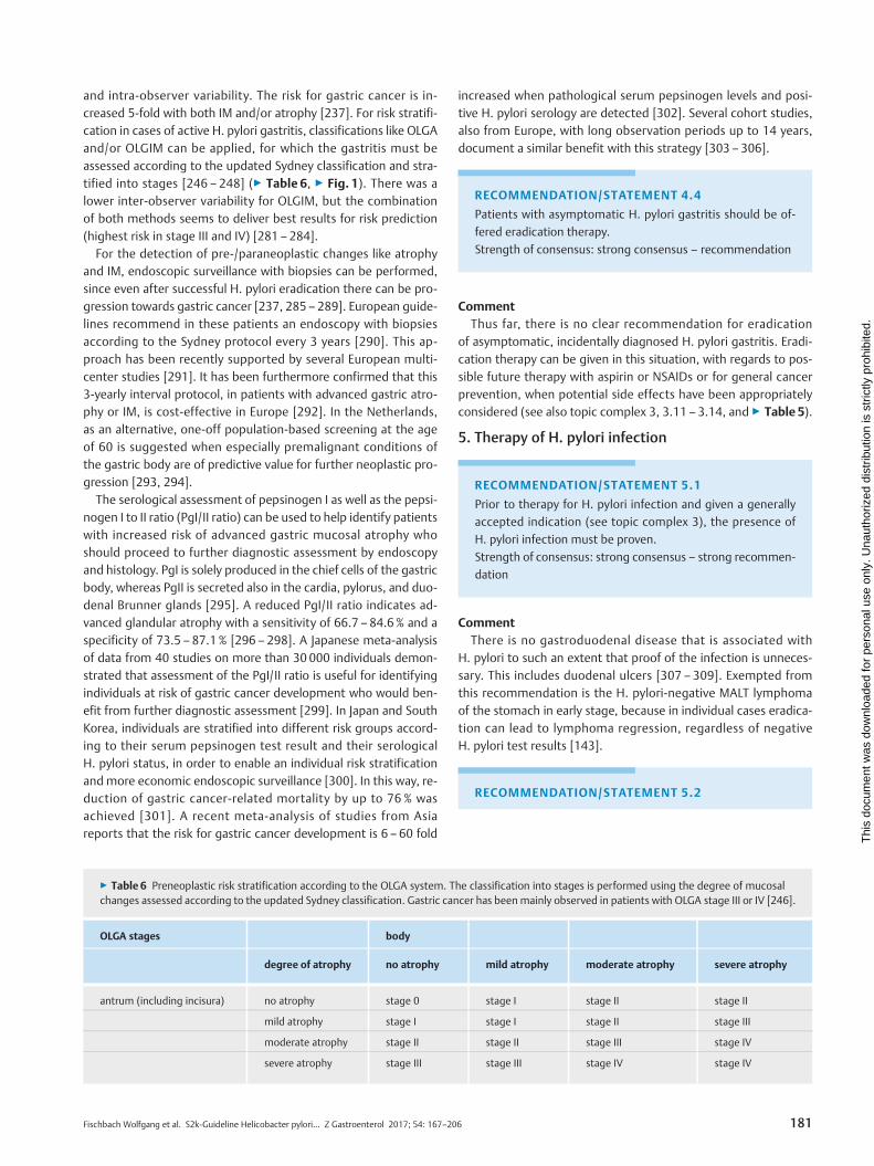
opment of gastric cancer [232]. Apart from studies from Asiancountries, this has been confirmed in a large Finnish cohort aswell as in a meta-analysis [233 – 236]. The time point of treatmentis crucial for the efficacy of H. pylori eradication on prevention ofgastric cancer [237]. Eradication is mostly effective if there are nopre-/paraneoplastic changes such as atrophy or IM [237 – 239],but can even show an effect in cases with advanced changes in-cluding after endoscopic resection of an early gastric cancer[240 – 245]. The individual risk can be stratified according to theOLGA or OLGIM classifications [246 – 248] (please see also 4.3).Since these scores can give false positives in individuals withoutactive H. pylori gastritis, they should only be applied in peoplewith active H. pylori gastritis (personal communication P. Malfert-heiner). It has to be noted that the so-called point-of-no-returnwith regards to these risk parameters has not been clearly de-fined. Due to the low prevalence of H. pylori infection and thelow incidence of gastric cancer, mass-screening in Germanywon’t be cost-effective [249]. The cost-efficiency of prophylacticH. pylori eradication increases, however, if the simultaneous pre-
vention of other H. pylori-associated diseases (gastric/duodenalulcer, MALT lymphoma, dyspepsia) are considered as well [250].H. pylori eradication with the aim of cancer prevention should
be undertaken in at-risk individuals, as defined in the MaastrichtIV/Florence consensus [232] (▶ Table 5). This includes patientswith gastric cancer and prior partial gastrectomy [251], ulcer pa-tients [252], patients with long-term PPI intake [253], and firstdegree relatives of patients with gastric cancer [254, 256]. Follow-ing successful eradication and after exclusion of recrudescence,the re-infection rate in industrial countries is about 1.5 % [50,256]. Although familial transmission of positive H. pylori statushas been reported [257, 258], an influence on the re-infectionrate has not been confirmed [259, 260]. Testing or treating part-ners for H. pylori is not indicated in Germany if there are no symp-toms or risk constellations justifying this strategy.Polymorphisms of immune-regulatory genes play an important
role in carcinogenesis. Best investigated is the risk association ofpolymorphisms in the gene of the pro-inflammatory IL1β; despitea positive risk association for gastric cancer development in Cau-casians in meta-analyses, the available data is heterogeneous[262 – 266]. This is also the case for polymorphisms of specificloci of the TNFα gene [267 – 270]. For polymorphisms of the IL10Gene, some analyses demonstrated a protective effect [271, 272];the data concerning IL8 is not clear and seems to depend on tu-mor-specific factors [273 – 275]. Furthermore, a risk conferred bytoll-like receptor genes has been described [276 – 279]. Genetictesting for any of these parameters is in Germany neither cost-ef-fective nor of diagnostic or therapeutic relevance due to the con-flicting data and the low gastric cancer incidence [280].
RECOMMENDATION/STATEMENT 4.3
Atrophy and IM are associated with an increased risk of gastric
cancer. Therefore, patients with advanced atrophy/IM can
undergo endoscopic surveillance with biopsies even after suc-
cessful H. pylori eradication.
Strength of consensus: consensus – recommendation open
CommentFocal atrophy and IM are histological diagnoses. For the assess-
ment of gastric mucosal atrophy there is particularly high inter-
▶ Table 5 Individuals at risk and risk constellations, for which a H. pylori eradication is reasonable by cancer-preventive aspects.
individuals at risk / risk constellations (according to [1, 2, 231]) comments
risk gastritis pangastritis or body-dominant gastritis
first degree relatives of gastric cancer patients
previous gastric neoplasia endoscopic resection or partial gastrectomy for gastric adenoma or earlygastric cancer; MALT lymphoma
long-term PPI medication > 1 year
potential further indications
atrophy and/or IM extensive, multifocal atrophy
180 Fischbach Wolfgang et al. S2k-Guideline Helicobacter pylori… Z Gastroenterol 2017; 54: 167–206
Leitlinie
Thi
s do
cum
ent w
as d
ownl
oade
d fo
r pe
rson
al u
se o
nly.
Una
utho
rized
dis
trib
utio
n is
str
ictly
pro
hibi
ted.
and intra-observer variability. The risk for gastric cancer is in-creased 5-fold with both IM and/or atrophy [237]. For risk stratifi-cation in cases of active H. pylori gastritis, classifications like OLGAand/or OLGIM can be applied, for which the gastritis must beassessed according to the updated Sydney classification and stra-tified into stages [246 – 248] (▶ Table 6, ▶ Fig. 1). There was alower inter-observer variability for OLGIM, but the combinationof both methods seems to deliver best results for risk prediction(highest risk in stage III and IV) [281 – 284].For the detection of pre-/paraneoplastic changes like atrophy
and IM, endoscopic surveillance with biopsies can be performed,since even after successful H. pylori eradication there can be pro-gression towards gastric cancer [237, 285– 289]. European guide-lines recommend in these patients an endoscopy with biopsiesaccording to the Sydney protocol every 3 years [290]. This ap-proach has been recently supported by several European multi-center studies [291]. It has been furthermore confirmed that this3-yearly interval protocol, in patients with advanced gastric atro-phy or IM, is cost-effective in Europe [292]. In the Netherlands,as an alternative, one-off population-based screening at the ageof 60 is suggested when especially premalignant conditions ofthe gastric body are of predictive value for further neoplastic pro-gression [293, 294].The serological assessment of pepsinogen I as well as the pepsi-
nogen I to II ratio (PgI/II ratio) can be used to help identify patientswith increased risk of advanced gastric mucosal atrophy whoshould proceed to further diagnostic assessment by endoscopyand histology. PgI is solely produced in the chief cells of the gastricbody, whereas PgII is secreted also in the cardia, pylorus, and duo-denal Brunner glands [295]. A reduced PgI/II ratio indicates ad-vanced glandular atrophy with a sensitivity of 66.7 –84.6 % and aspecificity of 73.5 – 87.1 % [296 – 298]. A Japanese meta-analysisof data from 40 studies on more than 30 000 individuals demon-strated that assessment of the PgI/II ratio is useful for identifyingindividuals at risk of gastric cancer development who would ben-efit from further diagnostic assessment [299]. In Japan and SouthKorea, individuals are stratified into different risk groups accord-ing to their serum pepsinogen test result and their serologicalH. pylori status, in order to enable an individual risk stratificationand more economic endoscopic surveillance [300]. In this way, re-duction of gastric cancer-related mortality by up to 76 % wasachieved [301]. A recent meta-analysis of studies from Asiareports that the risk for gastric cancer development is 6 – 60 fold
increased when pathological serum pepsinogen levels and posi-tive H. pylori serology are detected [302]. Several cohort studies,also from Europe, with long observation periods up to 14 years,document a similar benefit with this strategy [303 – 306].
RECOMMENDATION/STATEMENT 4.4
Patients with asymptomatic H. pylori gastritis should be of-
fered eradication therapy.
Strength of consensus: strong consensus – recommendation
CommentThus far, there is no clear recommendation for eradication
of asymptomatic, incidentally diagnosed H. pylori gastritis. Eradi-cation therapy can be given in this situation, with regards to pos-sible future therapy with aspirin or NSAIDs or for general cancerprevention, when potential side effects have been appropriatelyconsidered (see also topic complex 3, 3.11 – 3.14, and ▶ Table 5).
5. Therapy of H. pylori infection
RECOMMENDATION/STATEMENT 5.1
Prior to therapy for H. pylori infection and given a generally
accepted indication (see topic complex 3), the presence of
H. pylori infection must be proven.
Strength of consensus: strong consensus – strong recommen-
dation
CommentThere is no gastroduodenal disease that is associated with
H. pylori to such an extent that proof of the infection is unneces-sary. This includes duodenal ulcers [307 – 309]. Exempted fromthis recommendation is the H. pylori-negative MALT lymphomaof the stomach in early stage, because in individual cases eradica-tion can lead to lymphoma regression, regardless of negativeH. pylori test results [143].
RECOMMENDATION/STATEMENT 5.2
▶ Table 6 Preneoplastic risk stratification according to the OLGA system. The classification into stages is performed using the degree of mucosalchanges assessed according to the updated Sydney classification. Gastric cancer has been mainly observed in patients with OLGA stage III or IV [246].
OLGA stages body
degree of atrophy no atrophy mild atrophy moderate atrophy severe atrophy
antrum (including incisura) no atrophy stage 0 stage I stage II stage II
mild atrophy stage I stage I stage II stage III
moderate atrophy stage II stage II stage III stage IV
severe atrophy stage III stage III stage IV stage IV
181Fischbach Wolfgang et al. S2k-Guideline Helicobacter pylori… Z Gastroenterol 2017; 54: 167–206
Thi
s do
cum
ent w
as d
ownl
oade
d fo
r pe
rson
al u
se o
nly.
Una
utho
rized
dis
trib
utio
n is
str
ictly
pro
hibi
ted.

In case of endoscopically proven duodenal ulcer, a definitely
positive rapid urease test is sufficient for induction of an era-
dication therapy (see also 2.10).
Strength of consensus: strong consensus
CommentSince patients with duodenal ulceration frequently have H. py-
lori infection, there is a high positive predictive value of the ureasetest and a low likelihood of false positive test results. In cases offunctional dyspepsia, however, the infection should be confirmedby a validated complementary method, because a high false posi-tive rate of the test is anticipated, due to the low H. pylori preval-ence, especially in young patients.
RECOMMENDATION/STATEMENT 5.3
The exclusive serological detection of antibodies against
H. pylori or its virulence factors is not sufficient to make a
decision about therapy.
Strength of consensus: consensus
CommentSerology does not allow a conclusion as to whether there is
active infection or not (see also 2.2).
RECOMMENDATION/STATEMENT 5.4
The pre-therapeutic resistance status of H. pylori is of great
therapeutic relevance.
Strength of consensus: consensus
CommentPrevious therapy with antibiotics—even for other indications—
should be considered for the selection of the treatment regimen.Resistance to clarithromycin, the key antibiotic of the standard tri-ple therapy, is the main reason for therapy failure. In Germany, theresistance situation is currently stable. Over the past few years,however, a clear increase in resistance in other European countrieshas been seen [311 – 313]. A pre-therapeutic resistance againstamoxicillin is extremely rare. In case of resistance against so-calledreserve antibiotics (levofloxacin, moxifloxacin, tetracyclin, rifabu-tin) a loss of efficacy has to be assumed [118, 313, 314].
RECOMMENDATION/STATEMENT 5.5
Factors influencing the efficacy of H. pylori therapy are com-
pliance, smoking, and the degree of acid inhibition.
Strength of consensus: strong consensus
CommentThe statement is based on explorative analysis of clinical stud-
ies. Correct prescription, a treatment regimen that can be appliedas simply as possible, motivation for compliance, as well as smok-
ing cessation are means that can improve treatment success.Acid suppression needs to be adequately high. The degree ofacid suppression is decisive for the efficacy of clarithromycinand amoxicillin. Examples of further non-modifiable factors in-clude the indication for H. pylori therapy and the patient’s age[315 – 320].Compliance can be improved by detailed consenting about
indication and the course of therapy as well as potential sideeffects. The extent of the acid suppression is defined by the selec-tion, dosage, and frequency of intake of the PPI as well as bygenetic polymorphisms in the cytochrome-P450 2C19 (affectsmainly racemic omeprazole and lansoprazole; with impact alsoon other PPIs under certain conditions). With increasing age,there are changes in kidney and liver function that can result inmuch higher drug levels for similar dosing.
RECOMMENDATION/STATEMENT 5.6
H. pylori testing should only be performed if a positive test
result would lead to therapeutic consequences.
Strength of consensus: consensus – recommendation
CommentA positive test result without subsequent H. pylori therapy is
difficult to communicate between doctor and patient, and diag-nostic tests without therapeutic implication are not reasonablefrom an economic point of view. Prophylactic determining of theH. pylori status in case an indication later arises (e. g., prior to aninduction of a therapy with aspirin or NSAIDs) should be refused,since the test for the infection should be performed promptlybefore commencement of an H. pylori directed therapy.
RECOMMENDATION/STATEMENT 5.7
An absolute contra-indication for H. pylori therapy is not
known.
Strength of consensus: strong consensus
CommentThere are always relative contra-indications to therapy, particu-
larly when the benefit-risk ratio is poor. Relative contra-indicationsinclude proven or assumed drug intolerance or allergy, whichincrease the risk of therapy. Previous pseudomembraneous colitisis not a contra-indication.It is, however, contra-indicated to merely repeat the therapy re-
gimen that has previously been applied correctly, but which hasbeen unsuccessful.
RECOMMENDATION/STATEMENT 5.8
Therapy regimens should be applied that achieve eradication
rates of at least 80% in the intention-to-treat (ITT) analysis in
randomized controlled trials.
Strength of consensus: strong consensus – recommendation
182 Fischbach Wolfgang et al. S2k-Guideline Helicobacter pylori… Z Gastroenterol 2017; 54: 167–206
Leitlinie
Thi
s do
cum
ent w
as d
ownl
oade
d fo
r pe
rson
al u
se o
nly.
Una
utho
rized
dis
trib
utio
n is
str
ictly
pro
hibi
ted.
CommentIn specific clinical situations (e. g., multiple allergies, certain
resistance status) clinical management can deviate from thisrecommendation. Economic aspects, like per-day costs of treat-ment, are only relevant for regimens of comparable efficacy.Efficacy (eradication rates) is the most important factor for treat-ment choice, since subsequent costs of failed therapy (diagnos-tics, repeat therapy) are normally much higher.This recommendation was first introduced in the Maastricht
recommendations, with the 80 % threshold being arbitrary. Ap-proving bodies (e. g., the FDA) apply slightly different criteria.From a scientific point of view, it was recently suggested thatonly regimens with > 90% eradication rate (ITT) should be prescri-bed. This is desirable but not realistic in view of the availability ofdrugs, the necessity for official approval, and the often poor com-pliance in daily routine [321, 322].
RECOMMENDATION/STATEMENT 5.9
The rate of severe side effects of a therapy regimen should be
below 5%.
Strength of consensus: strong consensus – recommendation
CommentInfection with H. pylori is, for most, a benign disease, and there
are well-tolerated regimens with few complications availablefor its treatment. In individual cases, where indicated, there arealso therapeutic alternatives to eradication, such as the long-term treatment of ulcer disease with PPI. Therefore, the risk oftherapy for H. pylori must not be disproportionally higher thanthe benefit.
RECOMMENDATION/STATEMENT 5.10
The selection of a first-line therapy regimen must take into
account the likelihood of possible antibiotic resistance.
Strength of consensus: strong consensus – strong recommen-
dation
CommentResistance of H. pylori to antibiotics is an important factor for
failure of eradication therapy [323]. Primary clarithromycin resist-ance reduces eradication rates of first-line therapy with a standardtriple regimen of clarithromycin and amoxicillin by 66 % and ofclarithromycin and metronidazole by 35 % [314]. The latter canbe further negatively influenced by primary metronidazole resist-ance [314]. In a German multicenter study (ResiNet), the primaryclarithromycin resistance rate rose from 4.8 % in 2001/2002 to10.9 % in 2011/2012 [311]. There is broad variation of the primaryresistance rates against clarithromycin across Europe varyingfrom 5.6 – 36.6 %, with resistance rates > 20 % being observedmostly in southern and eastern European countries [313]. Theprimary resistance rate for metronidazole was in Germany at36% in 2011/2012 [311].
RECOMMENDATION/STATEMENT 5.11
In cases with a high probability of primary clarithromycin re-
sistance, a bismuth-containing quadruple therapy or a con-
comitant quadruple therapy should be used as first-line treat-
ment.
Strength of consensus: strong consensus – recommendation
RECOMMENDATION/STATEMENT 5.12
In cases with a low probability of primary clarithromycin re-
sistance, a standard triple therapy or a bismuth-containing
quadruple therapy can be used as first-line treatment.
Strength of consensus: strong consensus – recommendation
open
CommentA European multicenter study demonstrated that first-line ther-
apy with a 10-day bismuth-containing quadruple therapy is signif-icantly and clinically superior to a 7-day standard triple therapywith PPI, clarithromycin, and amoxicillin (ITT eradication rates80 vs. 55%) [324] (▶ Table 7). Primary clarithromycin resistancehad a significant effect on the standard triple therapy (eradication8%). On the other hand, primary metronidazole resistance had noeffect on the efficacy of a bismuth-containing quadruple therapy.It is unclear, however, how significant the influence of differenttherapy length in both therapy arms has been on the overall resultof the study. A recent meta-analysis confirmed the superiority ofbismuth-containing quadruple therapy over a standard triple ther-apy [325]. The bismuth-containing quadruple therapy is approvedand has been available in Germany since January 2013. Acombined – concomitant – bismuth-free quadruple therapy(▶ Table 7) is significantly superior to standard triple therapy[326 – 330].Results of sequential therapy (PPI plus amoxicillin on day 1 –
5 followed by PPI plus clarithromycin and an imidazol derivativeon day 6 – 10) are controversial. In a previous meta-analysis, a10-day sequential therapy was significantly more effective than a7-day standard triple therapy [331]. In recent randomized multi-center trials from Asia, superiority of a 10-day sequential therapyover standard triple therapy could not be confirmed [330, 332 –334]. Additionally, it has been demonstrated in these studiesthat the efficacy of sequential therapy is reduced by a metronida-zole or clarithromycin resistance. Another meta-analysis, that is sofar only available as conference abstract (UEGW 2014), demon-strated superiority of a concomitant quadruple therapy over se-quential therapy. If all data are considered, sequential therapycannot be recommended. Up to now, several quadruple regimensin first-line treatment have been directly compared in random-ized, multicentric trials resulting in eradication rates of about90% and more [335 – 337].Several prospective randomized trials assessed a levofloxacin-
based triple therapy as first-line treatment and compared with astandard triple therapy [338 – 340]. In addition, there are 2 recent
183Fischbach Wolfgang et al. S2k-Guideline Helicobacter pylori… Z Gastroenterol 2017; 54: 167–206
Thi
s do
cum
ent w
as d
ownl
oade
d fo
r pe
rson
al u
se o
nly.
Una
utho
rized
dis
trib
utio
n is
str
ictly
pro
hibi
ted.
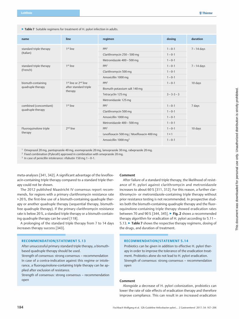
meta-analyses [341, 342]. A significant advantage of the levoflox-acin-containing triple therapy compared to a standard triple ther-apy could not be shown.The 2012 published Maastricht IV consensus report recom-
mends, for regions with a primary clarithromycin resistance rate> 20%, the first-line use of a bismuth-containing quadruple ther-apy or another quadruple therapy (sequential therapy, bismuth-free quadruple therapy). If the primary clarithromycin resistancerate is below 20%, a standard triple therapy or a bismuth-contain-ing quadruple therapy can be used [118].A prolonging of the standard triple therapy from 7 to 14 days
increases therapy success [343].
RECOMMENDATION/STATEMENT 5.13
After unsuccessful primary standard triple therapy, a bismuth-
based quadruple therapy should be used.
Strength of consensus: strong consensus – recommendation
In case of a contra-indication against this regime or intole-
rance, a fluoroquinolone-containing triple therapy can be ap-
plied after exclusion of resistance.
Strength of consensus: strong consensus – recommendation
open
CommentAfter failure of a standard triple therapy, the likelihood of resist-
ance of H. pylori against clarithromycin and metronidazoleincreases to about 60% [311, 312]. For this reason, a further clar-ithromycin- or metronidazole-containing triple therapy withoutprior resistance testing is not recommended. In prospective stud-ies both the bismuth-containing quadruple therapy and the fluor-oquinolone-containing triple therapy showed eradication ratesbetween 70 and 90% [344, 345]. ▶ Fig. 2 shows a recommendedtherapy algorithm for eradication of H. pylori according to 5.11 –5.13. ▶ Table 7 shows the respective therapy regimens, dosing ofthe drugs, and duration of treatment.
RECOMMENDATION/STATEMENT 5.14
Probiotics can be given in addition to effective H. pylori ther-
apy in order to improve the tolerance of the eradication treat-
ment. Probiotics alone do not lead to H. pylori eradication.
Strength of consensus: strong consensus – recommendation
open
CommentAlongside a decrease of H. pylori colonization, probiotics can
lower the rate of side effects of eradication therapy and thereforeimprove compliance. This can result in an increased eradication
▶ Table 7 Suitable regimens for treatment of H. pylori infection in adults.
name line regimen dosing duration
standard triple therapy(Italian)
1st line PPI1 1 – 0-1 7 – 14 days
Clarithromycin 250 – 500mg 1 – 0-1
Metronidazole 400 – 500mg 1 – 0-1
standard triple therapy(French)
1st line PPI1 1 – 0-1 7 – 14 days
Clarithromycin 500mg 1 – 0-1
Amoxicillin 1000mg 1 – 0-1
bismuth-containingquadruple therapy
1st line or 2nd lineafter standard tripletherapy
PPI2 1 – 0-1 10 days
Bismuth-potassium salt 140mg
Tetracyclin 125mg 3 – 3-3 – 3
Metronidazole 125mg
combined (concomitant)quadruple therapy
1st line PPI1 1 – 0-1 7 days
Clarithromycin 500mg 1 – 0-1
Amoxicillin 1000mg 1 – 0-1
Metronidazole 400 – 500mg 1 – 0-1
Fluoroquinolone tripletherapy
2nd line PPI1 1 – 0-1 10 days
Levofloxacin 500mg / Moxifloxacin 400mg 1 ×1
Amoxicillin 1000mg3 1 – 0-1
1 Omeprazol 20mg, pantoprazole 40mg, esomeprazole 20mg, lansoprazole 30mg, rabeprazole 20mg.2 Fixed combination (Pylera®) approved in combination with omeprazole 20mg.3 In case of penicillin intolerance: rifabutin 150mg 1 – 0-1.
184 Fischbach Wolfgang et al. S2k-Guideline Helicobacter pylori… Z Gastroenterol 2017; 54: 167–206
Leitlinie
Thi
s do
cum
ent w
as d
ownl
oade
d fo
r pe
rson
al u
se o
nly.
Una
utho
rized
dis
trib
utio
n is
str
ictly
pro
hibi
ted.
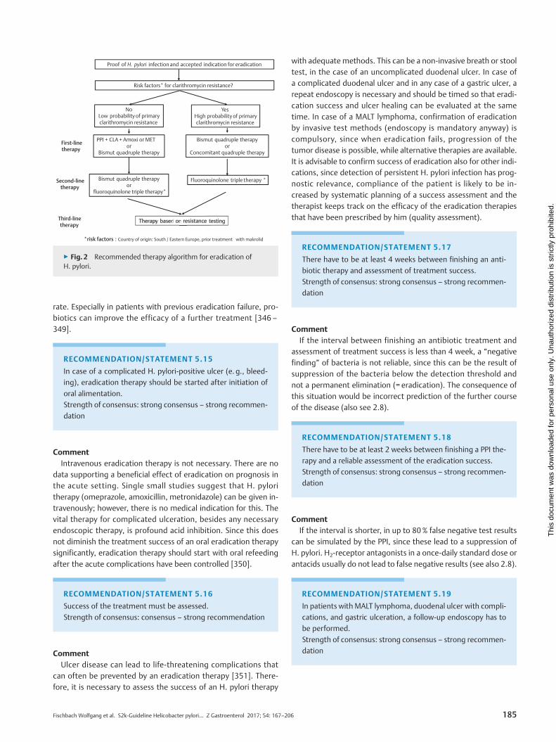
rate. Especially in patients with previous eradication failure, pro-biotics can improve the efficacy of a further treatment [346 –349].
RECOMMENDATION/STATEMENT 5.15
In case of a complicated H. pylori-positive ulcer (e. g., bleed-
ing), eradication therapy should be started after initiation of
oral alimentation.
Strength of consensus: strong consensus – strong recommen-
dation
CommentIntravenous eradication therapy is not necessary. There are no
data supporting a beneficial effect of eradication on prognosis inthe acute setting. Single small studies suggest that H. pyloritherapy (omeprazole, amoxicillin, metronidazole) can be given in-travenously; however, there is no medical indication for this. Thevital therapy for complicated ulceration, besides any necessaryendoscopic therapy, is profound acid inhibition. Since this doesnot diminish the treatment success of an oral eradication therapysignificantly, eradication therapy should start with oral refeedingafter the acute complications have been controlled [350].
RECOMMENDATION/STATEMENT 5.16
Success of the treatment must be assessed.
Strength of consensus: consensus – strong recommendation
CommentUlcer disease can lead to life-threatening complications that
can often be prevented by an eradication therapy [351]. There-fore, it is necessary to assess the success of an H. pylori therapy
with adequate methods. This can be a non-invasive breath or stooltest, in the case of an uncomplicated duodenal ulcer. In case ofa complicated duodenal ulcer and in any case of a gastric ulcer, arepeat endoscopy is necessary and should be timed so that eradi-cation success and ulcer healing can be evaluated at the sametime. In case of a MALT lymphoma, confirmation of eradicationby invasive test methods (endoscopy is mandatory anyway) iscompulsory, since when eradication fails, progression of thetumor disease is possible, while alternative therapies are available.It is advisable to confirm success of eradication also for other indi-cations, since detection of persistent H. pylori infection has prog-nostic relevance, compliance of the patient is likely to be in-creased by systematic planning of a success assessment and thetherapist keeps track on the efficacy of the eradication therapiesthat have been prescribed by him (quality assessment).
RECOMMENDATION/STATEMENT 5.17
There have to be at least 4 weeks between finishing an anti-
biotic therapy and assessment of treatment success.
Strength of consensus: strong consensus – strong recommen-
dation
CommentIf the interval between finishing an antibiotic treatment and
assessment of treatment success is less than 4 week, a “negativefinding” of bacteria is not reliable, since this can be the result ofsuppression of the bacteria below the detection threshold andnot a permanent elimination (= eradication). The consequence ofthis situation would be incorrect prediction of the further courseof the disease (also see 2.8).
RECOMMENDATION/STATEMENT 5.18
There have to be at least 2 weeks between finishing a PPI the-
rapy and a reliable assessment of the eradication success.
Strength of consensus: strong consensus – strong recommen-
dation
CommentIf the interval is shorter, in up to 80% false negative test results
can be simulated by the PPI, since these lead to a suppression ofH. pylori. H2-receptor antagonists in a once-daily standard dose orantacids usually do not lead to false negative results (see also 2.8).
RECOMMENDATION/STATEMENT 5.19
In patients with MALT lymphoma, duodenal ulcer with compli-
cations, and gastric ulceration, a follow-up endoscopy has to
be performed.
Strength of consensus: strong consensus – strong recommen-
dation
NoLow probability of primaryclarithromycin resistance
First-linetherapy
PPI + CLA + Amoxi or METor
Bismut quadruple therapy
Bismut quadruple therapyor
Concomitant quadruple therapy
Therapyy basedd on n resistancee testingThird-linetherapy
Second-linetherapy
Bismut quadruple therapyor
fluoroquinolone triple therapy*
*risk factors : Country of origin: South / Eastern Europe, prior treatment with makrolid
Proof of H. pylori infection and accepted indication for eradication
Risk factors* for clarithromycin resistance?
YesHigh probability of primaryclarithromycin resistance
Fluoroquinolone triple therapy *
▶ Fig. 2 Recommended therapy algorithm for eradication ofH. pylori.
185Fischbach Wolfgang et al. S2k-Guideline Helicobacter pylori… Z Gastroenterol 2017; 54: 167–206
Thi
s do
cum
ent w
as d
ownl
oade
d fo
r pe
rson
al u
se o
nly.
Una
utho
rized
dis
trib
utio
n is
str
ictly
pro
hibi
ted.
CommentThe arguments for this approach can be found in the comment
on 5.16.
RECOMMENDATION/STATEMENT 5.20
If a follow-up endoscopy is not necessary, eradication must be
tested by 13C-urea breath test (13C-UBT) or a monoclonal stool
antigen test.
Strength of consensus: strong consensus – strong recommen-
dation
CommentIf there is no indication for a repeat endoscopy, then the 13C-
UBT and the monoclonal stool antigen test are considered asequivalent options for ensuring eradication. A serological resultwould be only usable if a relevant decrease (more than 50%) ofthe titer compared to the pre-therapeutic test could be shownwith the identical test kit. It can take, however, up to 1 year untilsuch a decrease can be seen. In some patients there is no sucheffect at all, despite successful eradication. Therefore, serologyis generally not recommended as a clinical control in the courseof the disease (see also 2.2).
RECOMMENDATION/STATEMENT 5.21
A routine search for H. pylori re-infection should not be per-
formed if the primary eradication control has been performed
correctly.
Strength of consensus: strong consensus – recommendation
CommentData from developed countries suggest a low likelihood for
re-infection (< 1 % per year), as long as the “eradication” hasbeen performed with a recommended therapy (see above), theeradication success has been assessed with a combination of reli-able methods at least 4 weeks after completion of the antibiotictreatment, and confounding factors such as bacteria suppressionby PPI have been excluded at the time of the diagnostic test. Ifsuch an approach has been undertaken, routine follow is unneces-sary. In case of a “vital” indication (e. g., status post-ulcer bleed,MALT lymphoma), a repeat check for “permanent eradication”(e. g., after 1 year) may be advisable.
6. Special features for children and adolescents
RECOMMENDATION/STATEMENT 6.1
An invasive or non-invasive diagnostic test for H. pylori infec-
tion in children or adolescents should only be performed if
a treatment is intended in case of a positive test result.
Strength of consensus: strong consensus – recommendation
CommentIn countries with low prevalence like Germany, chronic infection
with H. pylori is mostly acquired at young age during childhood.The observed immunological reaction against the infectionis usually milder in children compared to adults. This is due to adown regulation of the immune response and an increase of reg-ulatory T-cells and anti-inflammatory cytokines (e. g. IL-10) [352].In a mouse model, early infection with H. pylori, and also adminis-tration of an H. pylori extract, reduces the risk for asthma [353 –355] and dextran sodium sulfate (DSS)-induced colitis [356]. Epi-demiological studies show an inverse relationship between theinfection and asthma [357] and atopy [358]. These potentiallypositive long-term effects of early infection on individual healthhave to be weighed against the possible risks of an ulcer diseaseor gastric cancer at a later point in time. Chronic infection is rarelysymptomatic in children. The risk for an ulcer is 6 – 7% in sympto-matically infected children and adolescents. H. pylori-inducedmalignancies do not occur at this age [359]. In contrast, the avail-able treatment options are more restricted for children comparedto adults. The same treatment regimens seem also to be lesseffective. The healing rates after first-line therapy are only about70% (intention to treat).In conclusion, there is a different benefit-risk consideration in
children and adolescents compared to adults. Testing for theinfection should, in children and adolescents, therefore be restric-ted to such individuals who have a high likelihood to directlybenefit from eradication therapy. Therapy for prevention of com-plications at a later age should be postponed until adulthood.
RECOMMENDATION/STATEMENT 6.2
Children and adolescents with chronic abdominal pain/dys-
pepsia should in the course of their assessment not be inves-
tigated for H. pylori infection with a non-invasive test.
Strength of consensus: strong consensus – recommendation
CommentNon-invasive tests are easily available in Germany; the costs are
covered by the health care insurance providers. The threshold istherefore very low for ordering such a test in cases of non-specificsymptoms (e. g., abdominal pain) or in asymptomatic childrenand siblings of infected individuals. A positive test result impliesthat the result has to be communicated to the parents and thepatient. The potential risks and costs of further subsequent diag-nostics (including upper endoscopy) and therapy are controversialdue to the lack of direct benefit for children, since most symp-toms, even in H. pylori-infected children, are of a functionalnature.Abdominal pain is a frequent complaint in children and adoles-
cents. The analysis of the KIGGS study on the health of childrenand adolescents in Germany showed that 69.3 % of 3 – 10 yearolds and 59.6 % of adolescents of 11 – 17 years suffered at leastonce within the last 3 months from abdominal pain, 14.5 and18.0 % in both age categories, respectively, more often thanonce a week [360]. A systematic literature review and meta-anal-ysis of 38 studies that have been published between 1966 and
186 Fischbach Wolfgang et al. S2k-Guideline Helicobacter pylori… Z Gastroenterol 2017; 54: 167–206
Leitlinie
Thi
s do
cum
ent w
as d
ownl
oade
d fo
r pe
rson
al u
se o
nly.
Una
utho
rized
dis
trib
utio
n is
str
ictly
pro
hibi
ted.
2009 came to the conclusion that there is no significant associa-tion between abdominal pain or other gastrointestinal complaintslike vomiting or diarrhea and H. pylori infection in children andadolescents [361]. For epigastric symptoms, the results arecontroversial. In in-patients, there was a positive association withnon-specific abdominal pain; a selection bias could not, however,be excluded.
RECOMMENDATION/STATEMENT 6.3
In the following diseases and situations, children and adoles-
cents should not be tested for H. pylori infection: chronic ITP,
Otitis media, chronic urticaria, dwarfism, or H. pylori infection
of a person living in the same household.
Strength of consensus: consensus – recommendation
CommentEpidemiological cross-sectional or case-control studies on the
association between the above mentioned extra-gastrointestinaldiseases and an H. pylori infection in children and adolescentshave to take into account that H. pylori occurs more frequently inmigrants and those with a lower socio-economic status. Factorslike growth retardation, iron deficiency, and infections of theairways have also a higher prevalence in those of a low social sta-tus, so only controlled interventional studies can prove a causalrelationship [362]. In epidemiological studies, results have to beadjusted for social status, as well as for confounding factors thatare associated with low social status. Among these are, for exam-ple, pre- and postpartum exposure towards passive smoking,birth weight, the postnatal method of feeding, and the parents’height.
Dwarfism/Growth retardation
While data from studies from South and Central America pointtowards a reduced number of H. pylori-positive children compar-ed to H. pylori-negative ones, or successfully treated patients[363 – 365], a respective confirmation for children and adoles-cents is lacking for Europe. A Czech cross-sectional study couldnot detect a statistical association between H. pylori infectionand body height, after adjustment for the educational level ofthe parents [366].Dwarfism and growth retardation are not indications to test for
H. pylori infection.
Chronic ITP and chronic urticaria
As for other auto-immune diseases, infections are suspected as atrigger for cITP. These could vary between children and adults. Incountries with low prevalence, most children with ITP have notbeen infected with H. pylori (e. g., only 3 of 33 children in Holland[367] and none in a Finnish study) [368]. The results of 2 Italiancohort studies suggest that single patients with cITP can benefitfrom an H. pylori eradication therapy; both studies show substan-tial methodological limitations, however [369, 370]. There are noprospective randomized interventional studies for the cITP andchronic urticaria in children and adolescents.
Family members
The benefit of therapy for infected members of a household ofthe index patient, aiming to reduce risk for re-infection, is dispu-ted. Overall, the re-infection rate in children and adolescents inGermany is low at 2.3 % per year [371]. An Irish study confirmeda low re-infection rate of only 2 % per year, although 81% of thechildren had at least 1 infected parent and two-thirds had aninfected sibling [372].
RECOMMENDATION/STATEMENT 6.4
Children and adolescents with treatment refractory iron defi-
ciency anemia in whom other causes (e. g., occult blood loss,
coeliac disease, or parasite infestation) have been excluded,
must be investigated for H. pylori, and if identified, eradica-
tion treatment should be undertaken.
Strength of consensus: strong consensus – strong recommen-
dation
CommentOf all extra-gastrointestinal manifestations of H. pylori infec-
tion, for iron deficiency anemia there is the best evidence for acausal relationship. Pacifico et al. have compiled the studies onthe possible biological mechanisms in children [362]. Most inter-ventional studies have been performed in developing countriesand in populations with low socio-economic status, in which theproportion of children with other risk factors for an iron deficiencyanemia (worm infections, low vitamin C and iron intake, malnutri-tion) is high [373 – 376]. In Germany an iron deficiency anemiain children and adolescents is mostly due to alimentary causesor due to background organic disease (e. g., coeliac disease,chronic blood loss in chronic intestinal disease, reflux esophagitis,etc.). Therefore, iron deficiency or iron deficiency anemia are notprimary indications for H. pylori diagnostics. An endoscopic inves-tigation for H. pylori should only be undertaken if there is noresponse to iron therapy or a further Hb drop after cessation ofiron supplementation and organic diseases have been widely ex-cluded.
RECOMMENDATION/STATEMENT 6.5
In children, if there is suspicion of H. pylori infection during
esophagogastroduodenoscopy (nodularity in the antrum,
gastric or duodenal ulcer, or erosions), biopsies for histology
and antibiotic resistance testing (culture or PCR) should be
obtained.
Strength of consensus: strong consensus – recommendation
187Fischbach Wolfgang et al. S2k-Guideline Helicobacter pylori… Z Gastroenterol 2017; 54: 167–206
Thi
s do
cum
ent w
as d
ownl
oade
d fo
r pe
rson
al u
se o
nly.
Una
utho
rized
dis
trib
utio
n is
str
ictly
pro
hibi
ted.
RECOMMENDATION/STATEMENT 6.6
In children and adolescents with H. pylori infection and gas-
troduodenal ulcer or erosions, eradication of the bacteria
must be undertaken.
Strength of consensus: strong consensus – strong recommen-
dation
RECOMMENDATION/STATEMENT 6.7
In children and adolescents with proven H. pylori gastritis, era-
dication of the bacteria can be undertaken.
Strength of consensus: strong consensus – recommendation
open
RECOMMENDATION/STATEMENT 6.8
In patients with H. pylori gastritis without previously docu-
mented ulceration who have no symptoms anymore after
failure of an eradication therapy, repeat eradication therapy
during childhood or adolescence can be withheld.
Strength of consensus: strong consensus – recommendation
open
CommentThe indication for esophagogastroduodenoscopy is stricter in
children compared to adults. Functional symptoms are not anindication. If an upper endoscopy is performed, usually multi-levelbiopsies are obtained, so that current H. pylori infection is iden-tified histologically, raising the question about treatment. In caseof ulceration or erosions, this can be answered with a clear yes,since also in children there is a high recurrence risk for ulcers incase of a persisting infection.If there is only H. pylori gastritis, which is the case in > 90% of
the children [359], then there is no compulsory indication fortherapy. This is especially true if it is an incidental finding (e. g., inthe course of diagnostics for coeliac disease). Nodularity in theantrum that can be found in 70 – 80% of H. pylori-infected chil-dren shows no association with symptoms and represents noindication for therapy. Children with failed therapy without ulcerdetection at the initial therapy should follow the same rational.If only gastritis is present (primary or after failed therapy), the
benefit and the risk of therapy as well as possible therapy failurehas to be discussed with the parents. The age of the child, possi-ble symptoms, the family history of complications of H. pyloriinfection, and the histology (active or corpus-predominant gastri-tis) play a role.If the doctor, or the parents/the patient, respectively, decides
in favor of a therapy, the choice of antibiotics depends on antibio-tic resistance testing [377]. Since the decision of the parents foror against therapy has not been made at the time of endoscopy,it is recommended in cases of endoscopic suspicion of infectionto obtain tissue specimens for culture or PCR for testing for anti-biotic susceptibility in addition to biopsies for histology.
RECOMMENDATION/STATEMENT 6.9
The 13C-UBT is suitable for the non-invasive detection of H. py-
lori infection and for surveillance of therapy success in
children and adolescents.
Strength of consensus: strong consensus
RECOMMENDATION/STATEMENT 6.10
Of the currently available stool tests only the ELISA using
monoclonal antibodies is suitable for the non-invasive detec-
tion of an H. pylori infection and for surveillance of therapy
success in children and adolescents.
Strength of consensus: strong consensus
CommentThe 13C-UBT [378] and the monoclonal stool antigen test by
ELISA [379, 380] are suitable for detection of an active H. pyloriinfection amongst the non-invasive tests. Monitoring following in-fection is the main indication for these tests, since there are only afew indications for a non-invasive test in the course of primarydiagnostics. An example of one exception to this is the testing ofchildren in whom 1 parent had a gastric cancer. If the child’s test isnegative, an endoscopy is unnecessary. Non-specific symptoms(abdominal pain, dyspepsia) do not represent an indication (see6.2). Both test methods are suitable for epidemiological studies.Practical advice:
▪ Antibiotics have to be stopped at least 4 weeks and PPI at least2 weeks before the test.
▪ Bacterial overgrowth can lead to false positive results of the13C-UBT.
▪ In children under 6 years of age, false positive results of the13C-UBT are more frequent [378]. In such young children,however, there is rarely an indication for therapy.
▪ Due to radiation exposure, the 14C-UBT should not be used inchildren with general availability of the 13C-UBT (stable, non-radiogenic isotopes).
▪ The monoclonal ELISA for antigen detection in the stool is notage-dependent, but should be validated in the local population[380, 381].
▪ So-called one-step or rapid tests (bed-side tests) and polyclo-nal ELISAs are not suitable for clinical application [379, 382]
RECOMMENDATION/STATEMENT 6.11
Methods for the detection of specific antibodies against H. py-
lori in the serum, full blood, urine, or saliva should not be used
for the diagnosis of an infection in children and adolescents.
Strength of consensus: strong consensus – recommendation
CommentThe antibody test cannot distinguish between an acute or
already successfully treated infection and is therefore not suitablefor monitoring of therapy success. For epidemiological studies, it
188 Fischbach Wolfgang et al. S2k-Guideline Helicobacter pylori… Z Gastroenterol 2017; 54: 167–206
Leitlinie
Thi
s do
cum
ent w
as d
ownl
oade
d fo
r pe
rson
al u
se o
nly.
Una
utho
rized
dis
trib
utio
n is
str
ictly
pro
hibi
ted.
should be taken into account that the antibody response is lesspronounced in children [383], so the sensitivity of the tests islower compared to adults [384]. Most of the recent tests evaluat-ed in children, which are based on, for example, multiplex tech-nology, are not validated in infected children under 6 years sothat their reliability cannot be assessed [385].
RECOMMENDATION/STATEMENT 6.12
Antibiotic resistance testing should be done in H. pylori-infec-
ted children and adolescents prior to the first therapy. The
choice of antibiotics should be based on the result.
Strength of consensus: strong consensus – recommendation
CommentIn children and adolescents there is restricted access to many of
the reserve drugs for the eradication therapy such as bismuthsalts, tetracyclin, gyrase inhibitors, and rifabutin due to lack ofapproval or approved contraindications; for children, even morethan for adults, the cure rate with first therapy should be as highas possible. One or more failed therapies represent a special bur-den for the children and their parents: induction of anxiety; possi-bly a repeat endoscopy to obtain biopsies in case of unclear resist-ance status; and further therapies with potential side effects.The success of the therapy depends on the sensitivity of the
organisms to the antibiotics that are used, the dose and durationof the medication, and the compliance of the drug intake. Inves-tigations on antibiotic resistance of H. pylori have shown big dif-ferences within different populations [359, 386]. In Germany therates of primary resistance against clarithromycin and metronida-zole are at about 20%, and 5% of children carry a double-resistantstrain prior to the first therapy [387]. For the triple therapy (PPIand 2 antibiotics), resistance against clarithromycin is highly pre-dictive of therapy failure if clarithromycin is part of the treatmentregimen [388]. Additionally, metronidazole resistance impairsthe cure success, although the in vitro resistance can partly beovercome with higher doses of metronidazole and longer dura-tion of treatment [388].For the detection of antibiotic resistance, there are different
techniques available. These are not different between childrenand adults.There should always be at least 1 biopsy from the antrum and
from the body by obtained, since mixed infection with distinct re-sistance patterns can be found in 10 – 15% of the children [383].Practical advice:
▪ The culture of bacteria form a gastric biopsy with subsequentresistance testing for different antibiotics by an Etest repre-sents the current method of choice.
▪ For clarithromycin: direct detection of mutations can be usedon gastric biopsies, either fresh or paraffin embedded, by PCRor fluorescence-in-situ hybridization (FISH) [387].
▪ For clarithromycin, the real-time PCR on stool is an attractivenon-invasive method, although the results of a culture withEtest on gastric biopsies are superior to the stool test [389–392].
RECOMMENDATION/STATEMENT 6.13
The test-and-treat strategy (i. e., screening with a non-inva-
sive test for H. pylori and eradication therapy in case of a
positive test result) should not be performed in children and
adolescents.
Strength of consensus: strong consensus – recommendation
CommentThe aim of diagnostic assessment in symptomatic children is to
identify the cause of the complaints and not to confirm or excludean H. pylori infection. The reason for dismissal of a test-and-treatstrategy is not evidence-based, but results from the recommen-dations 6.2 and 6.12.A test-and-treat strategy in populations with high prevalence of
the infection (immigrants) carries the risk of overtreatment ofchildren with functional symptoms and at the same time includesthe problems of low eradication rates in cases of “blind therapy.”In some cases, organic diseases responsible for the symptomswould be identified only with a delay. In populations with lowH. pylori prevalence (< 5%) and low ulcer rates, the test-and-treatstrategy is not cost-efficient. To find 1 child with an H. pylori-in-duced ulcer, > 200 children would have to be investigated with ahighly sensitive diagnostic test. The unnecessary use of antibioticsin cases with an absent indication (functional symptoms) increas-es the risk of multi-resistant germs in the child that has beentreated, as well as in the population.
RECOMMENDATION/STATEMENT 6.14
The therapy of first choice should be a triple therapy over
14 days that is chosen by the resistance status of the bacteria.
Strength of consensus: strong consensus – recommendation
CommentThe aim of the first-line therapy is an eradication rate of > 90%,
if the drugs are taken as prescribed. For children and adolescentsthere is no regimen so far that achieves this aim. With the pre-viously often used 1-week triple therapy (PPI-amoxicillin-clarithro-mycin) only eradication rates of about 70 % are achieved [393].Thus, for children and adolescents the best option so far is a tripletherapy that is directed by the result of the resistance test [394,395]. In cases of completely sensitive bacteria or in cases of me-tronidazole resistance, PPI, amoxicillin, and clarithromycin aregiven. In case of clarithromycin resistance, this is replaced by me-tronidazole.Since the cure rates depend on therapy duration, for a triple
therapy at least 2 weeks of treatment are recommended [396].In children, the dose has to be adjusted to the body weight,
being higher per kilogram body weight compared to adults. Sincenot all antibiotics are available in liquid form or are accepted bythe patient, dosing usually follows weight classes. The relevanceof reliable drug intake for therapy success and for the avoidanceof the development of resistance has to be pointed out. Written
189Fischbach Wolfgang et al. S2k-Guideline Helicobacter pylori… Z Gastroenterol 2017; 54: 167–206
Thi
s do
cum
ent w
as d
ownl
oade
d fo
r pe
rson
al u
se o
nly.
Una
utho
rized
dis
trib
utio
n is
str
ictly
pro
hibi
ted.
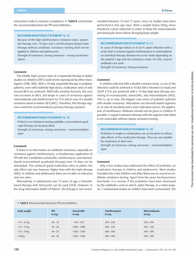
instructions help to improve compliance. ▶ Table 8 summarizesthe recommended doses for PPI and antibiotics.
RECOMMENDATION/STATEMENT 6.15
Because of the high clarithromycin resistance rates, sequen-
tial therapy over 10 days and a clarithromycin-based triple
therapy without antibiotic resistance testing shall not be
applied in children and adolescents.
Strength of consensus: strong consensus – strong recommen-
dation
CommentThe initially high success rates of a sequential therapy in Italian
studies on children [397] could not be reproduced by other inves-tigators [398, 400]. With a 10-day sequential therapy in pediatricpatients, even with relatively high doses, eradication rates of onlyaround 80% are achieved. With fully sensitive bacteria, the curerate increases to 86 %, but drops in cases of resistance againstmetronidazole and clarithromycin to 73% and in case of a doubleresistance down to below 30% [401]. Therefore, this therapy regi-men cannot be recommended as primary therapy anymore.
RECOMMENDATION/STATEMENT 6.16
If there is no resistance testing available, a concomitant quad-
ruple therapy can be prescribed.
Strength of consensus: strong consensus – recommendation
open
CommentIf there is no information on antibiotic resistance, especially on
resistance against clarithromycin, a simultaneous application ofPPI with the 3 antibiotics amoxicillin, clarithromycin, and metroni-dazole (concomitant quadruple therapy) over 14 days can beattempted. This achieved good eradication rates in adults; theside effect rate was, however, higher than with the triple therapy[402]. In children and adolescents there are no data on toleranceand cure rates.Alternatively, in adolescents over 12 years of age, a bismuth-
based therapy with tetracyclin can be used [324]. However, inthe drug information leaflet of Pylera®, the therapy is not recom-
mended between 12 and 17 years, since no studies have beenperformed in this age class. With a weight below 50 kg, thereshould be a dose reduction in order to keep the metronidazoleand tetracyclin doses below 30mg/kg body weight.
RECOMMENDATION/STATEMENT 6.17
In cases of therapy failure or of an H. pylori infection with a
strain that is resistant against clarithromycin or metronidazol,
an individual therapy decision has to be made depending on
the patient’s age and the resistance result. For this, reserve
antibiotics are used.
Strength of consensus: strong consensus
CommentIn children infected with a double-resistant strain, a cure of the
infection could be achieved in 41/62 (66% intention to treat) and33/45 (73%, per protocol) with a 14-day high dose therapy con-sisting of esomeprazole, amoxicillin, and metronidazole [403].This is, up to now, the biggest case series of pediatric patientswith double resistance. Alternatives are bismuth-based regimensor, in case of sensitivity and a strict indication (ulcer), the applica-tion of levofloxacin. Rifabutin should not be given in children ifpossible. A repeat treatment attempt with the regimen that failedis not reasonable without repeat resistance testing.
RECOMMENDATION/STATEMENT 6.18
Probiotics in single or combination use can be given to reduce
side effects of the eradication therapy. They are not suitable
for treatment on their own.
Strength of consensus: strong consensus – recommendation
open
CommentOnly a few studies have addressed the effect of probiotics on
eradication therapy in children and adolescents. Most studiesincluded only a few children and often there was no record on an-tibiotic resistance testing. Apart from the yeast Saccharomycesbourlardii, it is unclear if the probiotics have been destroyedby the antibiotics used as anti-H. pylori therapy. In a meta-analy-sis, 7 randomized studies on children have been summarized. The
▶ Table 8 Recommended dosing for PPI and antibiotics.
body weight PPIin mg
Amoxicillinin mg
Clarithromycinin mg
Metronidazolein mg
> 15 – 25 kg 20 – 10 750 – 750 250 – 250 250 – 250
> 25 – 35 kg 20 – 20 1000 – 1000 500 – 250 500 – 250
> 35 – 50 kg 40 – 20 1500 – 1500 500 – 500 500 – 500
> 50 kg 40 – 40 1500 – 1500 500 – 500 500 – 500
190 Fischbach Wolfgang et al. S2k-Guideline Helicobacter pylori… Z Gastroenterol 2017; 54: 167–206
Leitlinie
Thi
s do
cum
ent w
as d
ownl
oade
d fo
r pe
rson
al u
se o
nly.
Una
utho
rized
dis
trib
utio
n is
str
ictly
pro
hibi
ted.
authors came to the conclusion that the cure rate is not improvedby addition of probiotics, although there have been less sideeffects compared to placebo [404]. It has to be mentioned,though, that both the used probiotics and the therapy regimenwere different in the studies. With regards to the poor availabledata, a clear recommendation for the use of probiotics for thereduction of antibiotics associated side effects cannot be given.It has been postulated that the reliability of the drugs has beenimproved, but this has not been shown in studies.
RECOMMENDATION/STATEMENT 6.19
Control of therapy success should be undertaken using a reli-
able method at earliest 4 weeks after completion of the the-
rapy. Usually a non-invasive test (13C-UBT, monoclonal stool
test) is sufficient for this.
Strength of consensus: strong consensus
CommentImprovement of symptoms is not an indicator of cure of the
infection. There is a big placebo effect in children. Thus, in allpatients the therapy success has to be assessed and parents andpatient should be informed about the result. This is compulsoryin cases of ulcer disease. If the infection persists, further therapyhas to be undertaken until cure of the infection is confirmed. A re-peat endoscopy is usually not necessary, since malignant changesdo not play a role in children and adolescents, even in case of agastric ulcer.
7. Gastroduodenal ulcer disease not associatedwith H. pylori
PREAMBLE
Risk factors for gastroduodenal ulcer disease while taking
NSAIDs are higher age (> 65 years), a history of ulcers, H. py-
lori infection, severe general illness, co-medication with
glucocorticoids, coagulation-modifying drugs, or with selec-
tive serotonin reuptake inhibitors (SSRI) [405 – 410].
For further on the topic of aspirin/NSAIDs and H. pylori infec-
tion, please see 3.11 – 3.14.
CommentCoagulation-altering substances include vitamin K antagonists
(VKA), novel oral anticoagulants (NOACs: factor Xa inhibitors[apixaban, rivaroxaban, edoxaban] and thrombin inhibitors [dabi-gatran]), selective factor X inhibitors (fondaparinux), heparins,platelet aggregation inhibitors, low dose aspirin (75 – 100 mg –called aspirin in the following), and traditional non-steroidal anti-inflammatory drugs (tNSAIDs) including high-dose aspirin. Gluco-corticoids are not primarily ulcerogenic. They lead, however, to asignificantly worse healing of existing ulcers and increase the riskfor an ulcer bleeding, even in low doses, when given together withother ulcerogenic drugs [407]. In hospitalized patients the riskfor ulcer bleeding is increased by corticosteroids [411]. Meta-ana-
lyses show that the intake of SSRIs is associated with a significant-ly increased risk when an NSAID is taken simultaneously [410].
RECOMMENDATION/STATEMENT 7.1
If a therapy with traditional tNSAIDs is induced, a simulta-
neous therapy with a PPI should be given if there is at least
1 risk factor (see preamble) for a gastroduodenal ulcer bleed
present.
Strength of consensus: strong consensus – recommendation
CommentNumerous studies document that NSAIDs lead to gastroduode-
nal ulcers in a dose-dependent manner and increase the occur-rence of upper gastrointestinal bleeding [405, 406, 412]. Accord-ing to meta-analyses, the long-term application of tNSAIDs isassociated in 10 –25% with gastroduodenal ulcers [413].Besides age (> 65 years), additional risk factors for an upper
gastrointestinal bleeding related to a chronic treatment withNSAIDs include male gender, H. pylori infection, a previous gas-trointestinal bleed, or a history of gastroduodenal ulcers as wellas the intake of coagulation-active substances or corticosteroids[414 – 416]. A new, clinically relevant risk factor is the intake ofSSRI [410].Prospective randomized, double-blind studies have shown that
the risk for such bleeding can be significantly reduced by intake ofa PPI [415, 417 – 420]. The simultaneous application of a PPIdecreases the frequency of bleeding and perforations significantly(1.6 – 4.0 %). Co-medication with a PPI for those on long-termNSAID therapy should not be withheld, because a benefit-riskassessment – especially in older patients – favors the use of PPI[421 – 423]. The general co-medication of a PPI in case of NSAIDintake in patients less than 65 years without further risk factors is,however, not recommended. If there are further risk factors a PPIshould be added to the tNSAID. On the other hand, all patientsabove 65 years should be prophylactically treated with a PPI.
RECOMMENDATION/STATEMENT 7.2
To prevent gastroduodenal complications from tNSAID ther-
apy, the use of a selective COX-2 inhibitor is an alternative to
the combination of a tNSAID plus PPI.
Strength of consensus: strong consensus – recommendation
open
CommentCOX-2 inhibitors carry a considerably lower risk of ulcer bleed-
ing and other tNSAID-associated complications but are associatedwith a higher risk of dyspepsia compared to tNSAID plus PPI [239,426].Two prospective randomized, double-blind studies document
that selective COX-2 inhibitors have a lower complication ratecompared to tNSAIDs [424, 425]. With regards to ulcers andupper gastrointestinal bleeding, a further prospective randomizedand double-blind study did not show a significant difference
191Fischbach Wolfgang et al. S2k-Guideline Helicobacter pylori… Z Gastroenterol 2017; 54: 167–206
Thi
s do
cum
ent w
as d
ownl
oade
d fo
r pe
rson
al u
se o
nly.
Una
utho
rized
dis
trib
utio
n is
str
ictly
pro
hibi
ted.
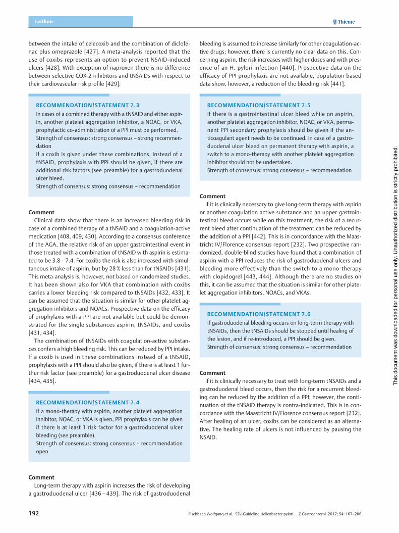
between the intake of celecoxib and the combination of diclofe-nac plus omeprazole [427]. A meta-analysis reported that theuse of coxibs represents an option to prevent NSAID-inducedulcers [428]. With exception of naproxen there is no differencebetween selective COX-2 inhibitors and tNSAIDs with respect totheir cardiovascular risk profile [429].
RECOMMENDATION/STATEMENT 7.3
In cases of a combined therapy with a tNSAID and either aspir-
in, another platelet aggregation inhibitor, a NOAC, or VKA,
prophylactic co-administration of a PPI must be performed.
Strength of consensus: strong consensus – strong recommen-
dation
If a coxib is given under these combinations, instead of a
tNSAID, prophylaxis with PPI should be given, if there are
additional risk factors (see preamble) for a gastroduodenal
ulcer bleed.
Strength of consensus: strong consensus – recommendation
CommentClinical data show that there is an increased bleeding risk in
case of a combined therapy of a tNSAID and a coagulation-activemedication [408, 409, 430]. According to a consensus conferenceof the AGA, the relative risk of an upper gastrointestinal event inthose treated with a combination of tNSAID with aspirin is estima-ted to be 3.8 – 7.4. For coxibs the risk is also increased with simul-taneous intake of aspirin, but by 28% less than for tNSAIDs [431].This meta-analysis is, however, not based on randomized studies.It has been shown also for VKA that combination with coxibscarries a lower bleeding risk compared to tNSAIDs [432, 433]. Itcan be assumed that the situation is similar for other platelet ag-gregation inhibitors and NOACs. Prospective data on the efficacyof prophylaxis with a PPI are not available but could be demon-strated for the single substances aspirin, tNSAIDs, and coxibs[431, 434].The combination of tNSAIDs with coagulation-active substan-
ces confers a high bleeding risk. This can be reduced by PPI intake.If a coxib is used in these combinations instead of a tNSAID,prophylaxis with a PPI should also be given, if there is at least 1 fur-ther risk factor (see preamble) for a gastroduodenal ulcer disease[434, 435].
RECOMMENDATION/STATEMENT 7.4
If a mono-therapy with aspirin, another platelet aggregation
inhibitor, NOAC, or VKA is given, PPI prophylaxis can be given
if there is at least 1 risk factor for a gastroduodenal ulcer
bleeding (see preamble).
Strength of consensus: strong consensus – recommendation
open
CommentLong-term therapy with aspirin increases the risk of developing
a gastroduodenal ulcer [436 – 439]. The risk of gastroduodenal
bleeding is assumed to increase similarly for other coagulation-ac-tive drugs; however, there is currently no clear data on this. Con-cerning aspirin, the risk increases with higher doses and with pres-ence of an H. pylori infection [440]. Prospective data on theefficacy of PPI prophylaxis are not available, population baseddata show, however, a reduction of the bleeding risk [441].
RECOMMENDATION/STATEMENT 7.5
If there is a gastrointestinal ulcer bleed while on aspirin,
another platelet aggregation inhibitor, NOAC, or VKA, perma-
nent PPI secondary prophylaxis should be given if the an-
ticoagulant agent needs to be continued. In case of a gastro-
duodenal ulcer bleed on permanent therapy with aspirin, a
switch to a mono-therapy with another platelet aggregation
inhibitor should not be undertaken.
Strength of consensus: strong consensus – recommendation
CommentIf it is clinically necessary to give long-term therapy with aspirin
or another coagulation active substance and an upper gastroin-testinal bleed occurs while on this treatment, the risk of a recur-rent bleed after continuation of the treatment can be reduced bythe addition of a PPI [442]. This is in concordance with the Maas-tricht IV/Florence consensus report [232]. Two prospective ran-domized, double-blind studies have found that a combination ofaspirin with a PPI reduces the risk of gastroduodenal ulcers andbleeding more effectively than the switch to a mono-therapywith clopidogrel [443, 444]. Although there are no studies onthis, it can be assumed that the situation is similar for other plate-let aggregation inhibitors, NOACs, and VKAs.
RECOMMENDATION/STATEMENT 7.6
If gastroduodenal bleeding occurs on long-term therapy with
tNSAIDs, then the tNSAIDs should be stopped until healing of
the lesion, and if re-introduced, a PPI should be given.
Strength of consensus: strong consensus – recommendation
CommentIf it is clinically necessary to treat with long-term tNSAIDs and a
gastroduodenal bleed occurs, then the risk for a recurrent bleed-ing can be reduced by the addition of a PPI; however, the conti-nuation of the tNSAID therapy is contra-indicated. This is in con-cordance with the Maastricht IV/Florence consensus report [232].After healing of an ulcer, coxibs can be considered as an alterna-tive. The healing rate of ulcers is not influenced by pausing theNSAID.
192 Fischbach Wolfgang et al. S2k-Guideline Helicobacter pylori… Z Gastroenterol 2017; 54: 167–206
Leitlinie
Thi
s do
cum
ent w
as d
ownl
oade
d fo
r pe
rson
al u
se o
nly.
Una
utho
rized
dis
trib
utio
n is
str
ictly
pro
hibi
ted.
RECOMMENDATION/STATEMENT 7.7
If a gastroduodenal ulcer bleed occurs on long-term treat-
ment with aspirin, another platelet aggregation inhibitor,
NOAC, or VKA, then permanent treatment with PPI has to be
given.
Strength of consensus: strong consensus – strong recommen-
dation
CommentProspective randomized, double-blind studies have shown that
secondary prophylaxis with a PPI can considerably lower the risk ofa recurrent gastroduodenal bleed in patients who require apermanent aspirin therapy [442]. In this situation, timely conti-nuation of the aspirin therapy in cardiovascular risk patients is ofgreat importance [445, 446].
RECOMMENDATION/STATEMENT 7.8
In case of a simultaneous therapy with 2 coagulation active
substances, prophylaxis with PPI has to be given.
Strength of consensus: strong consensus – strong recommen-
dation
CommentIn this point the current guideline differs from the recommen-
dations of the European Society of Cardiology (ESC), which com-ments on the use of PPI with platelet inhibitory therapy in patientswith coronary disease [447]. The guideline restricts the routineprophylactic administration of a PPI in those on double plateletinhibition to only patients with a high risk of gastroduodenalbleeding. These are patients with known ulcer disease, previousGI-bleeds, or other risk factors (like H. pylori infection, additionaladministration of an anticoagulant, age > 65 years, intake ofNSAIDs or steroids).The simultaneous administration of aspirin and clopidogrel in-
creases the risk for a gastroduodenal bleed from 1.8 and 1.1,respectively, to 7.1 [448]. On the background of the discussionabout a possible interaction between PPIs and clopidogrel, withweakening of the platelet inhibitory effect, the German Societyfor Digestive and Metabolic Diseases (Deutsche Gesellschaftfür Verdauungs- und Stoffwechselerkrankungen, DGVS) and theGerman Society of Cardiology (Deutsche Gesellschaft für Kardio-logie, DGK) published together a position paper in 2010 [449].According to this, in case of a dual therapy with aspirin and clopi-dogrel and high cardiovascular risk, a PPI co-medication should beconsidered, depending on the gastrointestinal risk as possible(low risk), reasonable (high risk), or mandatory (very high risk).Only in case of a very high cardiovascular risk – acute coronarysyndrome, main branch- or multiple vessel intervention, interven-tion with reduced left ventricular function, history of stent-throm-bosis – and in case of a lack of a gastrointestinal risk it is recom-mended to omit the PPI. The position paper also addresses thechoice of PPI and the deferred intake of clopidogrel and PPI.
Similarly, the simultaneous administration of aspirin and VKAincreases the bleeding rate significantly [450, 451]. A Spanishcohort study demonstrated that lower gastrointestinal bleeds aremore frequent than upper in a population, with frequent PPI in-take under dual platelet inhibition [452]. Based on this we recom-mend a PPI co-medication in case of intake of 2 coagulation-activesubstances, although there is no directly applicable study on this.
RECOMMENDATION/STATEMENT 7.9
Crohn’s disease-associated gastroduodenal ulcers or their
complications should primarily be treated with glucocorti-
coids in combination with a PPI.
Strength of consensus: strong consensus – recommendation
CommentThere are no studies that have investigated the therapy of
Crohn’s-associated gastroduodenal ulcers systematically. Gener-ally, the efficacy of steroid treatment on inflammatory ulcers isdocumented in European and American studies [453, 454]. Reser-vations against the use of steroids in this situation are most likelynot justified. Case series have shown that PPI can have a positiveinfluence on the healing of Crohn’s-associated gastroduodenal ul-cers [455 – 457].When Crohn’s affects the upper GI tract, it is usually associated
with a severe course [458]. Thus, the early use of anti-TNFα anti-bodies in case of side effects of the steroids is a possible treat-ment approach. Concerning the respective evidence, there areonly case series available [459, 460].
RECOMMENDATION/STATEMENT 7.10
In the situation of gastroduodenal ulcer disease with no H. py-
lori infection and/or NSAID medication, then other causes
should be searched for.
Strength of consensus: strong consensus – recommendation
CommentBesides H. pylori infection and the intake of NSAIDs, there
are numerous, although rare, reasons for gastroduodenal ulcers:Crohn’s disease, eosinophilic gastroduodenitis, ischaemia, sys-temic mastocytosis, metastases, radiation ulcers, tumours (e. g.,gastrinoma), vasculitis, viral infections, or a severe consuminggeneral disease. In immunosuppressed patients (transplantpatients, HIV infection), there are often CMV infections [460 –462]. In a small proportion of patients, no cause is found (idio-pathic ulcers).
RECOMMENDATION/STATEMENT 7.11
If no reason for the gastroduodenal ulcer disease is found
(idiopathic ulcers), a PPI therapy should be given.
Strength of consensus: strong consensus – recommendation
193Fischbach Wolfgang et al. S2k-Guideline Helicobacter pylori… Z Gastroenterol 2017; 54: 167–206
Thi
s do
cum
ent w
as d
ownl
oade
d fo
r pe
rson
al u
se o
nly.
Una
utho
rized
dis
trib
utio
n is
str
ictly
pro
hibi
ted.
CommentThere are no direct studies on this topic, but it can be assumed
that acid inhibition leads to accelerated healing of ulcers. Further-more, 120 patients with bleeding from idiopathic ulcers duringan observation period of 7 years demonstrated significantlymore frequent recurrent bleeding than patients with H. pylori-associated ulcers (42.5 vs 11.2 %) [463]. Mortality was also consid-erably higher in patients with idiopathic ulcer bleeding. Thus, therecommendation for permanent PPI therapy after idiopathic ulcerbleed is well justified.
RECOMMENDATION/STATEMENT 7.12
The occurrence of so-called stress ulcers and the associated
bleeding, in severe diseases like ARDS, shock with hypoten-
sion, sepsis, polytrauma, burns, craniocerebral injury with
neurosurgical intervention, liver or kidney failure, as well as
ongoing mechanic ventilation, can be reduced by prophylac-
tic administration of PPI. The administration of H2-receptor
antagonists (e. g., Ranitidine) or Sucralfate are less effective
stress ulcer prophylaxis.
Strength of consensus: strong consensus – recommendation
open
CommentSo-called stress ulcers that occur in the course of severe
diseases occur more frequently in certain risk groups like patientswith burns, coagulopathy, cardiac surgery patients, or patientswith mechanic ventilation [464 – 466]. The strongest evidencefor stress ulcers exists for patients with burns and craniocerebralinjury [467]. Further risk factors are ARDS, sepsis, polytrauma,craniocerebral injury, as well as liver and kidney failure. A meta-a-nalysis shows that Sucralfate and H2-recpetor blockers alsodecrease the likelihood of gastroduodenal stress ulcers [468,469]. There have been no such analyses using PPIs. Since PPIshave been shown to be superior at acid suppression, it can be con-cluded indirectly that these should be used prophylactically inthese risk groups. Thus, H2-receptor blockers and Sucralfate arenow only rarely used for this indication. They are recommendedby the participants of the consensus meeting only with majorityacceptance.While initial studies pointed towards an increased risk of hospi-
tal acquired pneumonia while on PPI, this was not reproducible inlater studies. Early enteral feeding shows the same effect as H2-blockers with regards to stress ulcer prophylaxis, but carries ahigher risk for hospital acquired pneumonia [470].
RECOMMENDATION/STATEMENT 7.13
SSRI are associated with an increased risk of gastroduodenal
bleeding.
Strength of consensus: strong consensus
CommentSSRIs like paroxetine, fluoxetine, citalopram, and sertraline are
used in the treatment of depression and anxiety disorders. In thelast decade, bleeding of the upper gastrointestinal tract has beendescribed a possible side effect.The release of serotonin by platelets plays an important role in
the regulation of haemostatic reactions to a vessel injury. The big-gest serotonin stores within our body are within the platelets.Serotonin is taken up from the circulation via serotonin transpor-ters not only by neuronal structures but also by platelets. In ther-apeutic doses, fluoxetine and other SSRIs block the uptake ofserotonin into platelets. This leads, after a few weeks of therapy,to depletion of serotonin. Presumably it is the influence of theSSRIs via this route that can, under certain conditions, alter hemo-stasis and therefore the bleeding risk.The suspicion of more frequent upper gastrointestinal bleeds
with simultaneous intake of SSRI and NSAIDs was initially con-firmed in a meta-analysis in 2008 [471]. A recent meta-analysisshows a higher rate of gastroduodenal ulcer bleeds with SSRI in-take, especially when NSAIDs are taken simultaneously [472].This meta-analysis of 4 observational studies with a total of153 000 patients demonstrated a doubling of the relative riskfor gastrointestinal bleeds under SSRI (odds ratio 2.36), whichwas tripled under NSAIDs (odds ratio 3.16) and increased by thefactor 6 under combination of SSRI and NSAIDs (odds ratio 6.33).The number needed to harm (NNH) was, for patients under50 years of age and on an SSRI, 318 per year and, on SSRI plusNSAID, 82 per year. In patients with a history of ulcer disease therisk was considerably higher: they showed a NNH of 70 per yearon SSRI and 19 per year on SSRI plus NSAID. A subgroup analysisof 101 cases showed that bleeding occurred after an average of25 weeks of SSRI intake.Due to the increased bleeding risk under SSRI, co-medication
with PPI can be considered, especially if NSAIDs are taken simulta-neously.▶ Table 9 gives an overview on the recommended co-medica-
tion if NSAIDs and/or coagulation active substances are used inspecific clinical situations.
Chapter 3: Abbreviations
DGHM Deutsche Gesellschaft für Hygiene und Mikrobiolo-gie (German Society of Hygiene and Microbiology)
DGP Deutsche Gesellschaft für Pathologie (GermanSociety of Pathology)
DGRh Deutsche Gesellschaft für Rheumatologie (Ger-man Society of Rheumatology)
DGVS Deutsche Gesellschaft für Gastroenterologie, Ver-dauungs- und Stoffwechselkrankheiten (GermanSociety of Gastroenterology, Digestive and Meta-bolic Diseases)
DKG Deutsche Gesellschaft für Kardiologie – Herz- undKreislauf-forschung (German Society of Cardiolo-gy and cardiovascular research)
DLBCL diffuse large B-cell lymphomaDOAK Direct oral anticoagulants
194 Fischbach Wolfgang et al. S2k-Guideline Helicobacter pylori… Z Gastroenterol 2017; 54: 167–206
Leitlinie
Thi
s do
cum
ent w
as d
ownl
oade
d fo
r pe
rson
al u
se o
nly.
Una
utho
rized
dis
trib
utio
n is
str
ictly
pro
hibi
ted.
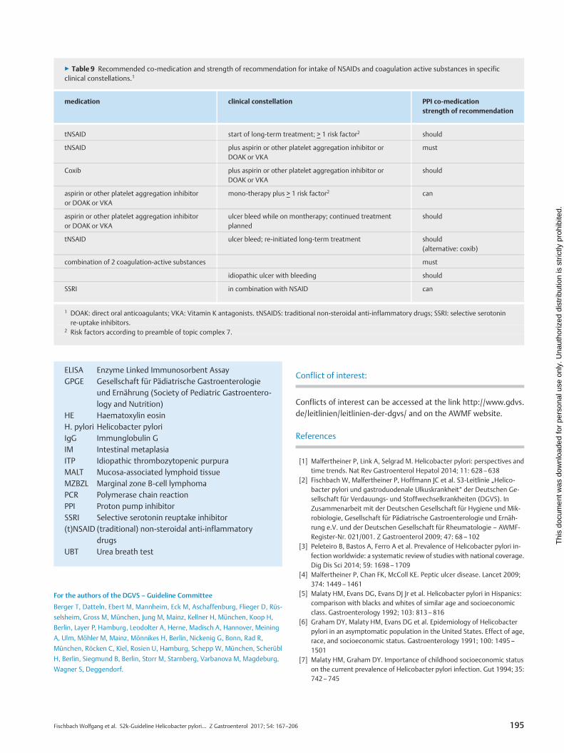
ELISA Enzyme Linked Immunosorbent AssayGPGE Gesellschaft für Pädiatrische Gastroenterologie
und Ernährung (Society of Pediatric Gastroentero-logy and Nutrition)
HE Haematoxylin eosinH. pylori Helicobacter pyloriIgG Immunglobulin GIM Intestinal metaplasiaITP Idiopathic thrombozytopenic purpuraMALT Mucosa-associated lymphoid tissueMZBZL Marginal zone B-cell lymphomaPCR Polymerase chain reactionPPI Proton pump inhibitorSSRI Selective serotonin reuptake inhibitor(t)NSAID (traditional) non-steroidal anti-inflammatory
drugsUBT Urea breath test
For the authors of the DGVS – Guideline Committee
Berger T, Datteln, Ebert M, Mannheim, Eck M, Aschaffenburg, Flieger D, Rüs-
selsheim, Gross M, München, Jung M, Mainz, Kellner H, München, Koop H,
Berlin, Layer P, Hamburg, Leodolter A, Herne, Madisch A, Hannover, Meining
A, Ulm, Möhler M, Mainz, Mönnikes H, Berlin, Nickenig G, Bonn, Rad R,
München, Röcken C, Kiel, Rosien U, Hamburg, Schepp W, München, Scherübl
H, Berlin, Siegmund B, Berlin, Storr M, Starnberg, Varbanova M, Magdeburg,
Wagner S, Deggendorf.
Conflict of interest:
Conflicts of interest can be accessed at the link http://www.gdvs.de/leitlinien/leitlinien-der-dgvs/ and on the AWMF website.
References
[1] Malfertheiner P, Link A, Selgrad M. Helicobacter pylori: perspectives andtime trends. Nat Rev Gastroenterol Hepatol 2014; 11: 628–638
[2] Fischbach W, Malfertheiner P, Hoffmann JC et al. S3-Leitlinie „Helico-bacter pylori und gastroduodenale Ulkuskrankheit“ der Deutschen Ge-sellschaft für Verdauungs- und Stoffwechselkrankheiten (DGVS). InZusammenarbeit mit der Deutschen Gesellschaft für Hygiene und Mik-robiologie, Gesellschaft für Pädiatrische Gastroenterologie und Ernäh-rung e.V. und der Deutschen Gesellschaft für Rheumatologie – AWMF-Register-Nr. 021/001. Z Gastroenterol 2009; 47: 68–102
[3] Peleteiro B, Bastos A, Ferro A et al. Prevalence of Helicobacter pylori in-fection worldwide: a systematic review of studies with national coverage.Dig Dis Sci 2014; 59: 1698–1709
[4] Malfertheiner P, Chan FK, McColl KE. Peptic ulcer disease. Lancet 2009;374: 1449–1461
[5] Malaty HM, Evans DG, Evans DJ Jr et al. Helicobacter pylori in Hispanics:comparison with blacks and whites of similar age and socioeconomicclass. Gastroenterology 1992; 103: 813–816
[6] Graham DY, Malaty HM, Evans DG et al. Epidemiology of Helicobacterpylori in an asymptomatic population in the United States. Effect of age,race, and socioeconomic status. Gastroenterology 1991; 100: 1495–1501
[7] Malaty HM, Graham DY. Importance of childhood socioeconomic statuson the current prevalence of Helicobacter pylori infection. Gut 1994; 35:742–745
▶ Table 9 Recommended co-medication and strength of recommendation for intake of NSAIDs and coagulation active substances in specificclinical constellations.1
medication clinical constellation PPI co-medicationstrength of recommendation
tNSAID start of long-term treatment; > 1 risk factor2 should
tNSAID plus aspirin or other platelet aggregation inhibitor orDOAK or VKA
must
Coxib plus aspirin or other platelet aggregation inhibitor orDOAK or VKA
should
aspirin or other platelet aggregation inhibitoror DOAK or VKA
mono-therapy plus > 1 risk factor2 can
aspirin or other platelet aggregation inhibitoror DOAK or VKA
ulcer bleed while on montherapy; continued treatmentplanned
should
tNSAID ulcer bleed; re-initiated long-term treatment should(alternative: coxib)
combination of 2 coagulation-active substances must
idiopathic ulcer with bleeding should
SSRI in combination with NSAID can
1 DOAK: direct oral anticoagulants; VKA: Vitamin K antagonists. tNSAIDS: traditional non-steroidal anti-inflammatory drugs; SSRI: selective serotoninre-uptake inhibitors.
2 Risk factors according to preamble of topic complex 7.
195Fischbach Wolfgang et al. S2k-Guideline Helicobacter pylori… Z Gastroenterol 2017; 54: 167–206
Thi
s do
cum
ent w
as d
ownl
oade
d fo
r pe
rson
al u
se o
nly.
Una
utho
rized
dis
trib
utio
n is
str
ictly
pro
hibi
ted.
[8] Mayerle J, den Hoed CM, Schurmann C et al. Identification of genetic lociassociated with Helicobacter pylori serologic status. JAMA 2013; 309:1912–1920
[9] Perez-Perez GI, Olivares AZ, Foo FY et al. Seroprevalence of Helicobacterpylori in New York City populations originating in East Asia. J UrbanHealth 2005; 82: 510–516
[10] Malaty HM, Kim JG, Kim SD et al. Prevalence of Helicobacter pylori in-fection in Korean children: inverse relation to socioeconomic status de-spite a uniformly high prevalence in adults. Am J Epidemiol 1996; 143:257–262
[11] Banatvala N, Mayo K, Megraud F et al. The cohort effect and Helicobac-ter pylori. J Infect Dis 1993; 168: 219–221
[12] Roosendaal R, Kuipers EJ, Buitenwerf J et al. Helicobacter pylori and thebirth cohort effect: evidence of a continuous decrease of infection ratesin childhood. Am J Gastroenterol 1997; 92: 1480–1482
[13] al-Moagel MA, Evans DG, Abdulghani ME et al. Prevalence of Helicobac-ter (formerly Campylobacter) pylori infection in Saudi Arabia, and com-parison of those with and without upper gastrointestinal symptoms. AmJ Gastroenterol 1990; 85: 944–948
[14] Weyermann M, Rothenbacher D, Brenner H. Acquisition of Helicobacterpylori infection in early childhood: independent contributions of infectedmothers, fathers, and siblings. Am J Gastroenterol 2009; 104: 182–189
[15] Grimm W, Fischbach W. Helicobacter pylori infection in children and ju-veniles: an epidemiological study on prevalence, socio-economic factorsand symptoms. Dtsch Med Wochenschr 2003; 128: 1878–1883
[16] den Hoed CM, Vila AJ, Holster IL et al. Helicobacter pylori and the birthcohort effect: evidence for stabilized colonization rates in childhood.Helicobacter 2011; 16: 405–409
[17] Wex T, Venerito M, Kreutzer J et al. Serological prevalence of Helicobac-ter pylori infection in Saxony-Anhalt, Germany, in 2010. Clin VaccineImmunol 2011; 18: 2109–2112
[18] Michel A, Pawlita M, Boeing H et al. Helicobacter pylori antibody patternsin Germany: a cross-sectional population study. Gut Pathog 2014; 6: 10
[19] Gao L, Weck MN, Raum E et al. Sibship size, Helicobacter pylori infectionand chronic atrophic gastritis: a population-based study among 9444older adults from Germany. Int J Epidemiol 2010; 39: 129–134
[20] Porsch-Ozcürümez M, Doppl W, Hardt PD et al. Impact of migration onHelicobacter pylori seroprevalence in the offspring of Turkish immi-grants in Germany. Turk J Pediatr 2003; 45: 203–208
[21] Parsonnet J, Shmuely H, Haggerty T. Fecal and oral shedding of Helico-bacter pylori from healthy infected adults. JAMA 1999; 282: 2240–2245
[22] Leung WK, Siu KL, Kwok CK et al. Isolation of Helicobacter pylori fromvomitus in children and its implication in gastro-oral transmission. AmJ Gastroenterol 1999; 94: 2881–2884
[23] Laporte R, Pernes P, Pronnier P et al. Acquisition of Helicobacter pyloriinfection after outbreaks of gastroenteritis: prospective cohort survey ininstitutionalised young people. BMJ 2004; 329: 204–205
[24] Bastos J, Carreira H, La Vecchia C et al. Childcare attendance and Helico-bacter pylori infection: systematic review andmeta-analysis. Eur J CancerPrev 2013; 22: 311–319
[25] Handt LK, Fox JG, Dewhirst FE et al. Helicobacter pylori isolated from thedomestic cat: public health implications. Infect Immun 1994; 62: 2367–2374
[26] Dore MP, Sepulveda AR, Osato MS et al. Helicobacter pylori in sheepmilk. Lancet 1999; 354: 132
[27] Dore MP, Sepulveda AR, El-Zimaity H et al. Isolation of Helicobacter py-lori from sheep-implications for transmission to humans. Am J Gastro-enterol 2001; 96: 1396–1401
[28] Rocha GA, Rocha AM, Silva LD et al. Transmission of Helicobacter pyloriinfection in families of preschool-aged children from Minas Gerais, Bra-zil. Trop Med Int Health 2003; 8: 987–991
[29] Rothenbacher D, Winkler M, Gonser T et al. Role of infected parents intransmission of helicobacter pylori to their children. Pediatr Infect Dis J2002; 21: 674–679
[30] Kivi M, Johansson AL, Reilly M et al. Helicobacter pylori status in familymembers as risk factors for infection in children. Epidemiol Infect 2005;133: 645–652
[31] Tindberg Y, Bengtsson C, Granath F et al. Helicobacter pylori infection inSwedish school children: lack of evidence of child-to-child transmissionoutside the family. Gastroenterology 2001; 121: 310–316
[32] Han SR, Zschausch HC, Meyer HG et al. Helicobacter pylori: clonal pop-ulation structure and restricted transmission within families revealed bymolecular typing. J Clin Microbiol 2000; 38: 3646–3651
[33] Kivi M, Tindberg Y, Sorberg M et al. Concordance of Helicobacter pyloristrains within families. J Clin Microbiol 2003; 41: 5604–5608
[34] Mendall MA, Goggin PM, Molineaux N et al. Childhood living conditionsand Helicobacter pylori seropositivity in adult life. Lancet 1992; 339:896–897
[35] Jafar S, Jalil A, Soheila N et al. Prevalence of helicobacter pylori infectionin children, a population-based cross-sectional study in west Iran. IranJ Pediatr 2013; 23: 13–18
[36] Carter F, Seaton T, Yuan Y et al. Prevalence of Helicobacter pylori infec-tion in children in the Bahamas. West Indian Med J 2012; 61: 698–702
[37] Goodman KJ, Correa P. Transmission of Helicobacter pylori among sib-lings. Lancet 2000; 355: 358–362
[38] Rowland M, Daly L, Vaughan M et al. Age-specific incidence of Helico-bacter pylori. Gastroenterology 2006; 130: 65–72 quiz 211
[39] Duque X, Vilchis J, Mera R et al. Natural history of Helicobacter pylori in-fection in Mexican schoolchildren: incidence and spontaneous clearance.J Pediatr Gastroenterol Nutr 2012; 55: 209–216
[40] Goodman KJ, Correa P, Tengana Aux HJ et al. Helicobacter pylori infec-tion in the Colombian Andes: a population-based study of transmissionpathways. Am J Epidemiol 1996; 144: 290–299
[41] Glynn MK, Friedman CR, Gold BD et al. Seroincidence of Helicobacterpylori infection in a cohort of rural Bolivian children: acquisition and a-nalysis of possible risk factors. Clin Infect Dis 2002; 35: 1059–1065
[42] Aguemon BD, Struelens MJ, Massougbodji A et al. Prevalence and riskfactors for Helicobacter pylori infection in urban and rural Beninese po-pulations. Clin Microbiol Infect 2005; 11: 611–617
[43] Klein PD, Graham DY, Gaillour A et al. Water source as risk factor forHelicobacter pylori infection in Peruvian children. Gastrointestinal Phys-iology Working Group. Lancet 1991; 337: 1503–1506
[44] Hulten K, Han SW, Enroth H et al. Helicobacter pylori in the drinkingwater in Peru. Gastroenterology 1996; 110: 1031–1035
[45] Lu Y, Redlinger TE, Avitia R et al. Isolation and genotyping of Helicobac-ter pylori from untreated municipal wastewater. Appl Environ Microbiol2002; 68: 1436–1439
[46] Tomb JF, White O, Kerlavage AR et al. The complete genome sequenceof the gastric pathogen Helicobacter pylori. Nature 1997; 388: 539–547
[47] Calvet X, Ramírez Lázaro MJ, Lehours P et al. Diagnosis and epidemiologyof Helicobacter pylori infection. Helicobacter 2013; 18 (Suppl. 1): 5–11
[48] Yan TL, Hu QD, Zhang Q et al. National rates of Helicobacter pylori re-currence are significantly and inversely correlated with human develop-ment index. Aliment Pharmacol Ther 2013; 37: 963–968
[49] Feydt-Schmidt A, Kindermann A, Konstantopoulos N et al. Reinfectionrate in children after successful Helicobacter pylori eradication. Eur JGastroenterol Hepatol 2002; 14: 1119–1123
[50] Take S, Mizuno M, Ishiki K et al. Reinfection rate of Helicobacter pyloriafter eradication treatment: a long-term prospective study in Japan.J Gastroenterol 2012; 47: 641–646
[51] Rupnow MF, Shachter RD, Owens DK et al. Quantifying the populationimpact of a prophylactic Helicobacter pylori vaccine. Vaccine 2001; 20:879–885
[52] Zeng M, Mao XH, Li JX et al. Efficacy, safety, and immunogenicity of anoral recombinant Helicobacter pylori vaccine in children in China: a ran-domised, double-blind, placebo-controlled, phase 3 trial. Lancet 2015;386: 1457–1464
[53] de Vries R, Klok RM, Brouwers JR et al. Cost-effectiveness of a potentialfuture Helicobacter pylori vaccine in the Netherlands: the impact ofvarying the discount rate for health. Vaccine 2009; 27: 846–852
[54] Rupnow MF, Chang AH, Shachter RD et al. Cost-effectiveness of a po-tential prophylactic Helicobacter pylori vaccine in the United States.J Infect Dis 2009; 200: 1311–1317
[55] Rehmann A, Müller D, Krumbiegel P et al. Spontaneous elimination ofhelicobacter pylori infection in children. Klin Padiatr 2005; 217: 15–17
196 Fischbach Wolfgang et al. S2k-Guideline Helicobacter pylori… Z Gastroenterol 2017; 54: 167–206
Leitlinie
Thi
s do
cum
ent w
as d
ownl
oade
d fo
r pe
rson
al u
se o
nly.
Una
utho
rized
dis
trib
utio
n is
str
ictly
pro
hibi
ted.
[56] Bair MJ, Wu MS, Chang WH et al. Spontaneous clearance of Helicobacterpylori colonization in patients with partial gastrectomy correlates withoperative procedures and duration after operation. J Formos Med Assoc2009; 108: 13–19
[57] Lin YS, Chen MJ, Shih SC et al. Management of Helicobacter pylori infec-tion after gastric surgery. World J Gastroenterol 2014; 20: 5274–5282
[58] O'Connor HJ, Dixon MF, Wyatt JI et al. Effect of duodenal ulcer surgeryand enterogastric reflux on Campylobacter pyloridis. Lancet 1986; 2:1178–1181
[59] Venerito M, Radünz M, Reschke K et al. Autoimmune gastritis in autoim-mune thyroid disease. Aliment Pharmacol Ther 2015; 41: 686–693
[60] Malfertheiner P, Chan FK, McColl KE. Peptic ulcer disease. Lancet 2009;374: 1449–1461
[61] IARC Monogr Eval Carcinog Risks Hum. A review of human carcinogens.IARC Working Group on the Evaluation of Carcinogenic Risks to Humans;2012; 100(Pt B: 1–441 Biological agents. Volume 100 B
[62] Fischbach W. Gastric Mucosal-Associated Lymphoid Tissue Lymphoma.Gastroenterol Clin N Am 2013; 42: 371–380
[63] Huang JQ, Sridhar S, Chen Y et al. Meta-analysis of the relationship be-tween Helicobacter pylori seropositivity and gastric cancer. Gastroente-rology 1998; 114: 1169–1179
[64] Danesh J. Helicobacter pylori infection and gastric cancer: systematicreview of the epidemiological studies. Aliment Pharmacol Ther 1999; 13:851–856
[65] Eslick GD, Lim LL, Byles JE et al. Association of Helicobacter pylori infec-tion with gastric carcinoma: a meta-analysis. Am J Gastroenterol 1999;94: 2373–2379
[66] Xue FB, Xu YY, Wan Y et al. Association of H. pylori infection with gastriccarcinoma: a meta analysis. World J Gastroenterol 2001; 7: 801–804
[67] Huang JQ, Zheng GF, Sumanac K et al. Meta-analysis of the relationshipbetween cagA seropositivity and gastric cancer. Gastroenterology 2003;125: 1636–1644
[68] Helicobacter and Cancer Collaborative Group. Gastric cancer and Heli-cobacter pylori: a combined analysis of 12 studies nested within pro-spective cohorts. Gut 2001; 49: 347–353
[69] Brenner H, Arndt V, Stegmaier C et al. Is Helicobacter pylori infection anecessary condition for noncardia gastric cancer? Am J Epidemiol 2004;159: 252–258
[70] Siman JH, Engstrand L, Berglund G et al. Helicobacter pylori and CagAseropositivity and its association with gastric and oesophageal carcino-ma. Scand J Gastroenterol 2007; 42: 933–940
[71] Helicobacter and Cancer Collaborative Group. Gastric cancer and Heli-cobacter pylori: a combined analysis of 12 case control studies nestedwithin prospective cohorts. Gut 2001; 49: 347–353
[72] Parsonnet J, Hansen S, Rodriguez L et al. Helicobacter pylori infectionand gastric lymphoma. N Engl J Med 1994; 330: 1267–1271
[73] Solnick JV, Schauer DB. Emergence of diverse Helicobacter species in thepathogenesis of gastric and enterohepatic diseases. Clin Microbiol Rev2001; 14: 59–97
[74] Morgner A, Lehn N, Andersen LP et al. Helicobacter heilmannii-associat-ed primary gastric low-grade MALT lymphoma: complete remission aftercuring the infection. Gastroenterology 2000; 118: 821–828
[75] Chen Y, Segers S, Blaser MJ. Association between Helicobacter pylori andmortality in the NHANES III study. Gut 2013; 62: 1262–1269
[76] Xie FJ, Zhang YP, Zheng QQ et al. Helicobacter pylori infection andesophageal cancer risk: an updated meta-analysis. World J Gastroenterol2013; 19: 6098–6107
[77] Taye B, Enquselassie F, Tsegaye A et al. Is Helicobacter Pylori infectioninversely associated with atopy? A systematic review and meta-analysis.Clin Exp Allergy 2015; 45: 882–890
[78] Kim MS, Kim N, Kim SE et al. Long-term follow-up Helicobacter pylorireinfection rate and its associated factors in Korea. Helicobacter 2013;18: 135–142
[79] Peters C, Schablon A, Harling M et al. The occupational risk of Helico-bacter pylori infection among gastroenterologists and their assistants.BMC Infect Dis 2011; 11: 154
[80] Stone MA, Taub N, Barnett DB et al. Increased risk of infection with Heli-cobacter pylori in spouses of infected subjects: observations in a general
population sample from the UK. Hepatogastroenterology 2000; 47:433–436
[81] Brenner H, Weyermann M, Rothenbacher D. Clustering of Helicobacterpylori infection in couples: differences between high- and low-preval-ence population groups. Ann Epidemiol 2006; 16: 516–520
[82] Luman W, Zhao Y, Ng HS et al. Helicobacter pylori infection is unlikely tobe transmitted between partners: evidence from genotypic study inpartners of infected patients. Eur J Gastroenterol Hepatol 2002; 14:521–528
[83] Gisbert JP, Arata IG, Boixeda D et al. Role of partner's infection in rein-fection after Helicobacter pylori eradication. Eur J Gastroenterol Hepatol2002; 14: 865–871
[84] Cutler AF, Havstad S, Ma CK et al. Accuracy of invasive and noninvasivetests to diagnose Helicobacter pylori infection. Gastroenterology 1995;109: 136–141
[85] Thijs JC, van Zwet AA, Thijs WJ et al. Diagnostic tests for Helicobacterpylori: a prospective evaluation of their accuracy, without selecting asingle test as the gold standard. Am J Gastroenterol 1996; 91: 2125–2129
[86] Laheij RJ, de Boer WA, Jansen JB et al. Diagnostic performance of biopsy-based methods for determination of Helicobacter pylori infection with-out a reference standard. J Clin Epidemiol 2000; 53: 742–746
[87] Gisbert JP, de la Moreana MF, Abraira V. Accuracy of monoclonal stoolantigen test for the diagnosis of H. pylori infection: a systematic reviewand meta-analysis. Am J Gastroenterol 2006; 101: 1921–1930
[88] Calvet X, Lario S, Ramirez-Lazaro MJ et al. Comparative accuracy of 3monoclonal stool tests for diagnosis of Helicobacter pylori infectionamong patients with dyspepsia. Clin Infect Dis 2010; 50: 323–328
[89] Deguchi R, Matsushima M, Suzuki T et al. Comparison of a monoclonalwith a polyclonal antibody-based enzyme immunoassay stool test in di-agnosing Helicobacter pylori infection after eradication therapy. J Gas-troenterol 2009; 44: 713–716
[90] Gisbert JP, Pajares JM. Review article: C-urea breath test in the diagnosisof Helicobacter pylori infection – a critical review. Aliment PharmacolTher 2004; 20: 1001–1017
[91] Korkmaz H, Kesli R, Karabagli P et al. Comparison of the diagnostic ac-curacy of five different stool antigen tests for the diagnosis of Helico-bacter pylori infection. Helicobacter 2013; 18: 384–391
[92] Dixon MF, Genta RM, Yardley JH et al. Classification and grading of gas-tritis. The updated Sydney System. International Workshop on the His-topathology of Gastritis, Houston 1994. Am J Surg Pathol 1996; 20:1161–1181
[93] Genta RM, Graham DY. Comparison of biopsy sites for the histopatholo-gic diagnosis of Helicobacter pylori: a topographic study of H. pyloridensity and distribution. Gastrointest Endosc 1994; 40: 342–345
[94] El-Zimaity HM, Graham DY. Evaluation of gastric mucosal biopsy site andnumber for identification of Helicobacter pylori or intestinal metaplasia:role of the Sydney System. Hum Pathol 1999; 30: 72–77
[95] Craanen ME, Blok P, Dekker W et al. Subtypes of intestinal metaplasiaand Helicobacter pylori. Gut 1992; 33: 597–600
[96] Sudraba A, Daugule I, Rudzite D et al. Performance of routine Helico-bacter pylori tests in patients with atrophic gastritis. J Gastrointestin Liv-er Dis 2011; 20: 349–354
[97] Lan HC, Chen TS, Li AF et al. Additional corpus biopsy enhances the de-tection of Helicobacter pylori infection in a background of gastritis withatrophy. BMC Gastroenterol 2012; 12: 182
[98] Stolte M, Muller H, Talley NJ et al. In patients with Helicobacter pylorigastritis and functional dyspepsia, a biopsy from the incisura angularisprovides useful diagnostic information. Pathol Res Pract 2006; 202:405–413
[99] Dinis-Ribeiro M, Areia M, de Vries AC et al. Management of precancerousconditions and lesions in the stomach (MAPS): guideline from the Euro-pean Society of Gastrointestinal Endoscopy (ESGE), European Helico-bacter Study Group (EHSG), European Society of Pathology (ESP), andthe Sociedade Portuguesa de Endoscopia Digestiva (SPED). Endoscopy2012; 44: 74–94
[100] Rugge M, Genta RM. Staging and grading of chronic gastritis. HumPathol 2005; 36: 228–233
197Fischbach Wolfgang et al. S2k-Guideline Helicobacter pylori… Z Gastroenterol 2017; 54: 167–206
Thi
s do
cum
ent w
as d
ownl
oade
d fo
r pe
rson
al u
se o
nly.
Una
utho
rized
dis
trib
utio
n is
str
ictly
pro
hibi
ted.
[101] Laine L, Lewin DW, Naritoku W et al. Prospective comparison of H&E,Giemsa, and genta stains for the diagnosis of Helicobacter pylori. Gas-trointest Endosc 1997; 45: 463–467
[102] Fallone CA, Loo VG, Lough J et al. Hematoxylin and eosin staining ofgastric tissue for the detection of Helicobacter pylori. Helicobacter1997; 2: 32–35
[103] Guidelines for clinical trials in Helicobacter pylori infection. WorkingParty of the European Helicobacter Pylori Study Group. Gut 1997; 41(Suppl. 2): S1–S9
[104] Megraud F, Lehours P. Helicobacter pylori detection and antimicrobialsusceptibility testing. Clin Microbiol Rev 2007; 20: 280–322
[105] Selgrad M, Tammer I, Bornschein J et al. Different antibiotic suscept-ibility between antrum and corpus of the stomach, a possible reasonfor treatment failure of Helicobacter pylori infection. World J Gastro-enterol 2014; 20: 16245–16251
[106] Anderson JC, Cheng E, Roeske M et al. Detection of serum antibodies toHelicobacter pylori by an immunochromatographic method. Am J Gas-troenterol 1997; 92: 1135–1139
[107] Calvet X, Lario S, Ramírez-Lázaro MJ et al. Accuracy of monoclonal stooltests for determining cure of Helicobacter pylori infection after treat-ment. Helicobacter 2010; 15: 201–205
[108] Calvet X, Lario S, Ramírez-Lázaro MJ et al. Comparative accuracy of3 monoclonal stool tests for diagnosis of Helicobacter pylori infectionamong patients with dyspepsia. Clin Infect Dis 2010; 50: 323–328
[109] Brandi G, Biavati B, Calabrese C et al. Urease-positive bacteria otherthan Helicobacter pylori in human gastric juice and mucosa. Am J Gas-troenterol 2006; 101: 1756–1761
[110] Urita Y, Hike K, Torii N et al. Influence of urease activity in the intestinaltract on the results of 13C-urea breath test. J Gastroenterol Hepatol2006; 21: 744–747
[111] Capurso G, Carnuccio A, Lahner E et al. Corpus-predominant gastritis asa risk factor for false-negative 13C-urea breath test results. AlimentPharmacol Ther 2006; 24: 1453–1460
[112] Lehours P, Ruskone-Fourmestraux A, Lavergne A et al. Which test to useto detect Helicobacter pylori infection in patients with low-grade gas-tric mucosa-associated lymphoid tissue lymphoma? Am J Gastroenterol2003; 98: 291–295
[113] Gisbert JP, Abraira V. Accuracy of Helicobacter pylori diagnostic tests inpatients with bleeding peptic ulcer: a systematic review and meta-a-nalysis. Am J Gastroenterol 2006; 101: 848–863
[114] Rimbara E, Sasatsu M, Graham DY. PCR detection of Helicobacter pyloriin clinical samples. Methods Mol Biol 2013; 943: 279–287
[115] Saez J, Belda S, Santibanez M et al. Real-time PCR for diagnosing Heli-cobacter pylori infection in patients with upper gastrointestinal bleed-ing: comparison with other classical diagnostic methods. J Clin Micro-biol 2012; 50: 3233–3237
[116] Tian XY, Zhu H, Zhao J et al. Diagnostic performance of urea breathtest, rapid urea test, and histology for Helicobacter pylori infection inpatients with partial gastrectomy: a meta-analysis. J Clin Gastroenterol2012; 46: 285–292
[117] Shimoyama T, Kato C, Kodama M et al. Applicability of a monoclonalantibody based stool antigen test to evaluate the results of Helicobac-ter pylori eradication therapy. Jpn J Infect Dis 2009; 62: 225–227
[118] Malfertheiner P, Megraud F, O’Morain CA et al. Management of Heli-cobacter pylori infection – the Maastricht IV/Florence ConsensusReport. Gut 2012; 61: 646–664
[119] Haley KP, Gaddy JA. Helicobacter pylori: genomic insight into the host-pathogen interaction. Int J Genomics 2015; 2015: 386905
[120] Wüppenhorst N, Draeger S, Stüger HP et al. Prospective multicentrestudy on antimicrobial resistance of Helicobacter pylori in Germany.J Antimicrob Chemother 2014; 69: 3127–3133
[121] Wueppenhorst N, Stueger HP, Kist M et al. High secondary resistance toquinolones in German Helicobacter pylori clinical isolates. J AntimicrobChemother 2013; 68: 1562–1566
[122] Best LM, Haldane DJ, Keelan M et al. Multilaboratory comparison ofproficiencies in susceptibility testing of Helicobacter pylori and corre-lation between agar dilution and E test methods. Antimicrob AgentsChemother 2003; 47: 3138–3144
[123] Grignon B, Tankovic J, Megraud F et al. Validation of diffusion methodsfor macrolide susceptibility testing of Helicobacter pylori. Microb DrugResist 2002; 8: 61–66
[124] Perna F, Gatta L, Figura N et al. Susceptibility of Helicobacter pylori tometronidazole. Am J Gastroenterol 2003; 98: 2157–2161
[125] Schmitt BH, Regner M, Mangold KA et al. PCR detection of clarithro-mycin-susceptible and -resistant Helicobacter pylori from formalin-fixed, paraffin-embedded gastric biopsies. Mod Pathol 2013; 26:1222–1227
[126] Megraud F, Lehours P. Helicobacter pylori detection and antimicrobialsusceptibility testing. Clin Microbiol Rev 2007; 20: 280–322
[127] Cambau E, Allerheiligen V, Coulon C et al. Evaluation of a new test,genotype HelicoDR, for molecular detection of antibiotic resistance inHelicobacter pylori. J Clin Microbiol 2009; 47: 3600–3607
[128] Oleastro M, Ménard A, Santos A et al. Real-time PCR assay for rapid andaccurate detection of point mutations conferring resistance to clari-thromycin in Helicobacter pylori. J Clin Microbiol 2003; 41: 397–402
[129] Miendje Deyi VY, Burette A, Bentatou Z et al. Practical use of Geno-Type® HelicoDR, a molecular test for Helicobacter pylori detection andsusceptibility testing. Diagn Microbiol Infect Dis 2011; 70: 557–560
[130] Liou JM, Chen CC, Chang CY et al. Efficacy of genotypic resistance-guided sequential therapy in the third-line treatment of refractoryHelicobacter pylori infection: a multicentre clinical trial. J AntimicrobChemother 2013; 68: 450–456
[131] Ford AC, Delaney BC, Forman D et al. Eradication therapy for peptic ul-cer disease in Helicobacter pylori positive patients. Cochrane DatabaseSyst Rev 2006; 2: CD003840
[132] Laine L, Hopkins RJ, Girardi LS. Has the impact of Helicobacter pyloritherapy on ulcer recurrence in the United States been overstated? Ameta-analysis of rigorously designed trials. Am J Gastroenterol 1998;93: 1409–1415
[133] Leodolter A, Kulig M, Brasch H et al. A meta-analysis comparing eradi-cation, healing and relapse rates in patients with Helicobacter pylori-associated gastric or duodenal ulcer. Aliment Pharmacol Ther 2001; 15:1949–1958
[134] Liu CC, Lee CL, Chan CC et al. Maintenance treatment is not necessaryafter Helicobacter pylori eradication and healing of bleeding peptic ul-cer: a 5-year prospective, randomized, controlled study. Arch InternMed 2003; 163: 2020–2024
[135] Malfertheiner P, Megraud F, O'Morain C et al. Current concepts in themanagement of Helicobacter pylori infection: the Maastricht III Con-sensus Report. Gut 2007; 56: 772–781
[136] Sharma VK, Sahai AV, Corder FA et al. Helicobacter pylori eradication issuperior to ulcer healing with or without maintenance therapy to pre-vent further ulcer haemorrhage. Aliment Pharmacol Ther 2001; 15:1939–1947
[137] Sonnenberg A, Olson CA, Zhang J. The effect of antibiotic therapy onbleeding from duodenal ulcer. Am J Gastroenterol 1999; 94: 950–954
[138] Ruskoné-Fourmestraux A, Fischbach W, Aleman BMP et al. EGILS con-sensus report. Gastric extranodal marginal zone B cell lymphoma ofMALT. Gut 2011; 60: 747–758
[139] Zullo A, Hassan C, Cristofari F et al. Effects of Helicobacter pylori eradi-cation on early stage gastric mucosa-associated lymphoid tissue lym-phoma. Clin Gastroenterol Hepatol 2010; 8: 105–110
[140] Fischbach W, Goebeler-Kolve ME, Dragosics B et al. Long term outcomeof patients with gastric marginal zone B cell lymphoma of mucosaassociated lymphoid tissue (MALT) following exclusive Helicobacterpylori eradication: experience from a large prospective series. Gut2004; 53: 34–37
[141] Wündisch T, Dieckhoff P, Greene B et al. Second cancers and residualdisease in patients treated for gastric mucosa-associated lymphoid tis-sue lymphoma by Helicobacter pylori eradication and followed for10 years. Gastroenterology 2012; 143: 936–942
[142] Fischbach W, Goebeler ME, Ruskone-Fourmestraux A et al. Most pa-tients with minimal histological residuals of gastric MALT lymphomaafter successful eradication of Helicobacter pylori can be safely mana-ged by a watch-and-wait strategy. Experience from a large internation-al series. Gut 2007; 56: 1685–1687
198 Fischbach Wolfgang et al. S2k-Guideline Helicobacter pylori… Z Gastroenterol 2017; 54: 167–206
Leitlinie
Thi
s do
cum
ent w
as d
ownl
oade
d fo
r pe
rson
al u
se o
nly.
Una
utho
rized
dis
trib
utio
n is
str
ictly
pro
hibi
ted.
[143] Zullo A, Hassan C, Ridola L et al. Eradication therapy in Helicobacterpylori-negative, gastric low-grade mucosa-associated lymphoid tissuelymphoma patients. A systematic review. J Clin Gastroenterol 2013; 47:824–827
[144] Morgner A, Miehlke S, Fischbach W et al. Complete remission of pri-mary high-grade B-cell gastric lymphoma after cure of Helicobacterpylori infection. J Clin Oncol 2001; 19: 2041–2048
[145] Chen LT, Lin JT, Tai JJ et al. Long-term results of anti-Helicobacter pyloritherapy in early-stage gastric high-grade transformed MALT lympho-ma. J Natl Cancer Inst 2005; 97: 1345–1353
[146] Kuo SH, Yeh KH, Wu MS. Helicobacter pylori eradication therapy is ef-fective in the treatment of early-stage H. pylori-positive diffuse large B-cell lymphomas. Blood 2012; 119: 4838–4844
[147] Moayyedi P, Deeks J, Talley NJ et al. An update of the Cochrane sys-tematic review of Helicobacter pylori eradication therapy in nonulcerdyspepsia: resolving the discrepancy between systematic reviews. AmJ Gastroenterol 2003; 98: 2621–2626
[148] Zhao B, Zhao J, Cheng WF et al. Efficacy of Helicobacter pylori eradica-tion therapy on functional dyspepsia. A meta-analysis of randomizedcontrolled studies with 12-month follow-up. J Clin Gastroenterol 2014;48: 241–247
[149] Xu S, Wan X, Zheng X et al. Symptom improvement after helicobacterpylori eradication in patients with functional dyspepsia – a multicenter,randomized, prospective cohort study. Int J Clin Exp Med 2013; 6:747–756
[150] Zhao W, Zhong X, Zhuang X et al. Evaluation of Helicobacter pylorieradication and drug therapy in patients with functional dyspepsia. ExpTher Med 2013; 6: 37–44
[151] Kim SEK, Park YS, Kim N et al. Effect of Helicobacter pylori eradicationon functional dyspepsia. J Neurogastroenterol Motil 2013; 19: 233–243
[152] Sodhi JS, Javid G, Zargar SA et al. Prevalence of Helicobacter pylori in-fection and the effect of its eradication on symptoms of functionaldyspepsia in Kashmir, India. J Gastroenterol Hepatol 2013; 28: 808–813
[153] Lan L, Yu J, Chen YL et al. Symptom-based tendencies of Helicobacterpylori eradication in patients with functional dyspepsia. World J Gas-troenterol 2011; 17: 3242–3247
[154] Mazzoleni LE, Sander GB, Francesconi CF et al. Helicobacter pylori era-dication in functional dyspepsia. HEROES trial. Arch Intern Med 2011;171: 1929–1936
[155] Sugano K, Tack J, Kuipers EJ et al. Kyoto global consensus report onHelicobacter pylori gastritis. Gut 2015; 64: 1353–1367
[156] Harvey RF, Lane JA, Nair P et al. Clinical trial: prolonged beneficial effectof Helicobacter pylori eradication on dyspepsia consultations – theBristol helicobacter project. Aliment Pharmacol Ther 2010; 32: 394–400
[157] Suzuki H, Moayyedi P. Helicobacter pylori infection in functional dys-pepsia. Nat Rev Gastroenterol Hepatol 2013; 10: 168–174
[158] Cullen D, Hawkey G, Greenwood D et al. H. pylori and gastroesophagealreflux disease: a community-based study. Helicobacter 2008; 13: 352–360
[159] Nam SY, Choi IJ, Ryu KH et al. Effect of Helicobacter pylori Infection andits eradication on reflux esophagitis and reflux symptoms. Am J Gas-troenterol 2010; 105: 2153–2162
[160] Ashktorab H, Entezari O, Nouraie M et al. Helicobacter pylori protectionagainst reflux esophagitis. Dig Dis Sci 2012; 57: 2924–2928
[161] Xie T, Cui X, Zheng H et al. Meta-analysis: eradication of Helicobacterpylori infection is associated with the development of endoscopic gas-troesophageal reflux disease. Eur J Gastroenterol Hepatol 2013; 25:1195–1205
[162] Rokkas T, Pistiolas D, Sechopoulos P et al. Relationship between Heli-cobacter pylori infection and esophageal neoplasia: a meta-analysis.Clin Gastroenterol Hepatol 2007; 5: 1413–1417
[163] Wang C, Yuan Y, Hunt RH. Helicobacter pylori infection and Barrett’sesophagus: a systematic review and meta-analysis. Am J Gastroenterol2009; 104: 492–500
[164] Yaghoobi M, Farrokhyar F, Yuan Y et al. Is there an increased risk ofGERD after Helicobacter pylori eradication?: a meta-analysis. AmJ Gastroenterol 2010; 105: 1007–1013
[165] Qian B, Shijie M, Shang L et al. Effect of H. pylori eradication on gastro-esophageal reflux disease. Helicobacter 2011; 121: 1120–1126
[166] Rodriguez L, Faria CM, Geocze S et al. Helicobacter pylori eradicationdoes not influence gastroeosphageal reflux disease: a prospective, par-allel, randomized, open-label, controlled trial. Arq Gastroenterol 2012;49: 56–63
[167] Saad AM, Choudhary A, Bechtold ML. Effect of Helicobacter pyloritreatment on gastroesopha-geal reflux disease (GERD): meta-analysisof randomized controlled trials. Scand J Gastroenterol 2012; 47: 129–135
[168] Hirata K, Suzuki H, Matsuzaki J et al. Improvement of reflux symptomrelated quality of life after Helicobacter pylori eradication therapy.J Clin Biochem Nutr 2013; 52: 172–178
[169] Lundell L, Vieth M, Gibson F et al. Systematic review: the effects oflong-term proton pump inhibitor use on serum gastrin levels and gas-tric histology. Aliment Pharmacol Ther 2015; 42: 649–663
[170] Arnold DM, Bernotas A, Nazi I et al. Platelet count response to H pyloritreatment in patients with immune thrombocytopenic purpura withand without H. pylori infection: a systematic review. Haematologica2009; 94: 850–856
[171] Stasi R, Sarpatwari A, Segal JB et al. Effects of eradication of Helicobac-ter pylori infection in patients with immune thrombocytopenic pur-pura: A systematic review. Blood 2009; 113: 1231–1240
[172] Russo G, Miraglia V, Branciforte F et al. Effect of eradication of Helico-bacter pylori in children with chronic immune thrombocytopenia: aprospective, controlled, multicenter study. Pediatr Blood Cancer 2011;56: 273–278
[173] Yoshimura M, Hirai M, Tanaka N et al. Remission of severe anemia per-sisting for over 20 years after eradication of Helicobacter pylori in casesof Menetrier’s disease and atrophic gastritis: Helicobacter pylori as apathogenic factor in iron-deficiency anemia. Intern Med 2003; 42:971–977
[174] Kawasaki M, Hizawa K, Aoyagi K et al. Menetrier’s disease associatedwith Helicobacter pylori infection: resolution of enlarged gastric foldsand hypoproteinemia after antibacterial treatment. Am J Gastroenterol1997; 92: 1909–1912
[175] Yamada M, Sumazaki R, Adachi H et al. Resolution of protein-losinghypertrophic gastropathy by eradication of Helicobacter pylori. EurJ Pediatr 1997; 156: 182–185
[176] Bayerdörffer E, Ritter MM, Hatz R et al. Healing of protein losing hy-pertrophic gastropathy by eradication of Helicobacter pylori–is Helico-bacter pylori a pathogenic factor in Menetrier's disease? Gut 1994; 35:701–704
[177] Madsen LG, Taskiran M, Madsen JL et al. Menetrier`s disease and Heli-cobacter pylori. Normalization of gastrointestinal protein loss aftereradication therapy. Dig Dis Sci 1999; 44: 2307–2312
[178] Badov D, Lambert JR, Finlay M et al. Helicobacter pylori as a pathoge-netic factor in Menetrier’s disease. Am J Gastroenterol 1998; 93: 1976–1979
[179] Kim MJ, Eom DW, Park K. Helicobacter pylori associated lymphocyticgastritis in a child. Pediatr Gastroenterol Hepatol Nutr 2014; 17: 186–190
[180] Hayat M, Arora D, Clark B et al. Effects of Helicobacter pylori eradica-tion on the natural history of lymphocytic gastritis. Gut 1999; 45: 495–498
[181] Madisch A, Miehlke S, Neuber F et al. Healing of lymphocytic gastritisafter Helicobacter pylori eradication therapy–a randomized, double-blind, placebo-controlled multicentre trial. Aliment Pharmacol Ther2006; 23: 473–479
[182] Muhsen K, Cohen D. Helicobacter pylori infection and iron stores: asystematic review and meta-analysis. Helicobacter 2008; 13: 323–340
[183] Qu XH, Huang XL, Xiong P et al. Does Helicobacter pylori infection playa role in iron deficiency anemia? World J Gastroenterol 2010; 16: 886–896
199Fischbach Wolfgang et al. S2k-Guideline Helicobacter pylori… Z Gastroenterol 2017; 54: 167–206
Thi
s do
cum
ent w
as d
ownl
oade
d fo
r pe
rson
al u
se o
nly.
Una
utho
rized
dis
trib
utio
n is
str
ictly
pro
hibi
ted.
[184] Queiroz DMM, Harris PR, Sanderson IR et al. Iron status and Helicobac-ter pylori infection in symptomatic children: an international multicen-tered study. PLOS One 2013; 8: e68833
[185] Duque X, Moran S, Mera R et al. Effect of eradication of Helicobacterpylori and iron supplementation on the iron status of children with irondeficiency. Arch Med Res 2010; 41: 38–45
[186] Kotb NA, Bedewy MM, Soliman HH et al. The impact of H. pylori eradi-cation on response to oral iron therapy in patients with iron deficiencyanemia. Egypt J Immunol 2012; 19: 11–18
[187] Chan FKL, Chung SC, Suen BY et al. Preventing recurrent upper gastro-intestinal bleeding in patients with helicobacter pylori infection whoare taking low-dose aspirin or naproxen. N Engl J Med 2001; 344: 967–973
[188] Chan FKL, Ching JYL, Suen BY et al. Effects of Helicobacter pylori infec-tion on long-term risk of peptic ulcer bleeding in low-dose aspirin users.Gastroenterology 2013; 144: 528–535
[189] Chan FK, To KF, Wu JC et al. Eradication of Helicobacter pylori and risk ofpeptic ulcers in patients starting long-term treatment with non-steroi-dal anti-inflammatory drugs: a randomised trial. Lancet 2002; 359: 9–13
[190] Lai KC, Lau CS, Ip WY et al. Effect of treatment of Helicobacter pylori onthe prevention of gastroduodenal ulcers in patients receiving long-term NSAIDs: a double-blind, placebo-controlled trial. Aliment Phar-macol Ther 2003; 17: 799–805
[191] Vergara M, Catalan M, Gisbert JP et al. Meta-analysis: role of Helico-bacter pylori eradication in the prevention of peptic ulcer in NSAIDusers. Aliment Pharmacol Ther 2005; 21: 1411–1418
[192] Lai KC, Lam SK, Chu KM et al. Lansoprazole reduces ulcer relapse aftereradication of Helicobacter pylori in nonsteroidal anti-inflammatorydrug users–a randomized trial. Aliment Pharmacol Ther 2003; 18:829–836
[193] Hawkey CJ, Tulassay Z, Szczepanski L et al. Randomised controlled trialof Helicobacter pylori eradication in patients on non-steroidal anti-in-flammatory drugs: HELP NSAIDs study. Helicobacter Eradication forLesion Prevention. Lancet 1998; 352: 1016–1021
[194] de Leest HT, Steen KS, Lems WF et al. Eradication of Helicobacter pyloridoes not reduce the incidence of gastroduodenal ulcers in patients onlong-term NSAID treatment: double-blind, randomized, placebo-con-trolled trial. Helicobacter 2007; 12: 477–485
[195] Ekstrom AM, Hansson LE, Signorello LB et al. Decreasing incidence ofboth major histologic subtypes of gastric adenocarcinoma–a popula-tion-based study in Sweden. Br J Cancer 2000; 83: 391–396
[196] Wang C, Yuan Y, Hunt RH. The association between Helicobacter pyloriinfection and early gastric cancer: a meta-analysis. Am J Gastroenterol2007; 102: 1789–1798
[197] Derks S, Bass AJ. Mutational signatures in Helicobacter pylori-inducedgastric cancer: lessons from new sequencing technologies. Gastroen-terology 2014; 147: 267–269
[198] Shimizu T, Marusawa H, Matsumoto Y et al. Accumulation of somaticmutations in TP53 in gastric epithelium with Helicobacter pylori infec-tion. Gastroenterology 2014; 147: 407–417
[199] Stoicov C, Saffari R, Cai X et al. Molecular biology of gastric cancer: He-licobacter infection and gastric adenocarcinoma: bacterial and hostfactors responsible for altered growth signaling. Gene 2004; 341: 1–17
[200] Kodaman N, Pazos A, Schneider BG et al. Human and Helicobacter py-lori coevolution shapes the risk of gastric disease. Proc Natl Acad SciUSA 2014; 111: 1455–1460
[201] Bornschein J, Leja M, Kupcinskas J et al. Molecular diagnostics in gastriccancer. Front Biosci (Landmark Ed) 2014; 19: 312–338
[202] Ekstrom AM, Eriksson M, Hansson LE et al. Occupational exposures andrisk of gastric cancer in a population-based case-control study. CancerRes 1999; 59: 5932–5937
[203] Miehlke S, Kirsch C, Agha-Amiri K et al. The Helicobacter pylori vacA s1,m1 genotype and cagA is associated with gastric carcinoma in Germa-ny. Int J Cancer 2000; 87: 322–327
[204] Basso D, Zambon CF, Letley DP et al. Clinical relevance of Helicobacterpylori cagA and vacA gene polymorphisms. Gastroenterology 2008;135: 91–99
[205] Jang S, Jones KR, Olsen CH et al. Epidemiological link between gastricdisease and polymorphisms in VacA and CagA. J Clin Microbiol 2010;48: 559–567
[206] Rizzato C, Torres J, Plummer M et al. Variations in Helicobacter pyloricytotoxin-associated genes and their influence in progression to gastriccancer: implications for prevention. PLoS One 2012; 7: e29605
[207] Ekstrom AM, Serafini M, Nyren O et al. Dietary antioxidant intake andthe risk of cardia cancer and noncardia cancer of the intestinal and dif-fuse types: a population-based case-control study in Sweden. IntJ Cancer 2000; 87: 133–140
[208] Gonzalez CA, Jakszyn P, Pera G et al. Meat intake and risk of stomachand esophageal adenocarcinoma within the European Prospective In-vestigation Into Cancer and Nutrition (EPIC). J Natl Cancer Inst 2006;98: 345–354
[209] Gonzalez CA, Lopez-Carrillo L. Helicobacter pylori, nutrition and smok-ing interactions: their impact in gastric carcinogenesis. Scand J Gastro-enterol 2010; 45: 6–14
[210] Sung JJ, Lin SR, Ching JY et al. Atrophy and intestinal metaplasia oneyear after cure of H. pylori infection: a prospective, randomized study.Gastroenterology 2000; 119: 7–14
[211] Siewert JR, Stein HJ. Classification of adenocarcinoma of the oesopha-gogastric junction. Br J Surg 1998; 85: 1457–1459
[212] Bornschein J, Selgrad M, Warnecke M et al. H. pylori infection is a keyrisk factor for proximal gastric cancer. Dig Dis Sci 2010; 55: 3124–3131
[213] Cavaleiro-Pinto M, Peleteiro B, Lunet N et al. Helicobacter pylori infec-tion and gastric cardia cancer: systematic review and meta-analysis.Cancer Causes Control 2011; 22: 375–387
[214] Derakhshan MH, Malekzadeh R, Watabe H et al. Combination of gastricatrophy, reflux symptoms and histological subtype indicates two dis-tinct aetiologies of gastric cardia cancer. Gut 2008; 57: 298–305
[215] Hansen S, Vollset SE, Derakhshan MH et al. Two distinct aetiologies ofcardia cancer; evidence from premorbid serological markers of gastricatrophy and Helicobacter pylori status. Gut 2007; 56: 918–925
[216] Peleteiro B, Cavaleiro-Pinto M, Barros R et al. Is cardia cancer aetiologi-cally different from distal stomach cancer? Eur J Cancer Prev 2011; 20:96–101
[217] McColl KE, Watabe H, Derakhshan MH. Role of gastric atrophy in med-iating negative association between Helicobacter pylori infection andreflux oesophagitis, Barrett's oesophagus and oesophageal adenocar-cinoma. Gut 2008; 57: 721–723
[218] Bornschein J, Dingwerth A, Selgrad M et al. Adenocarcinomas at differ-ent positions at the gastro-oesophageal junction show distinct asso-ciation with gastritis and gastric preneoplastic conditions. Eur J Gastro-enterol Hepatol 2015; 27: 492–500
[219] PJ DEJ, Wolters LM, Steyerberg EW et al. Environmental risk factors inthe development of adenocarcinoma of the oesophagus or gastric car-dia: a cross-sectional study in a Dutch cohort. Aliment Pharmacol Ther2007; 26: 31–39
[220] Tajima Y, Yamazaki K, Makino R et al. Differences in the histologicalfindings, phenotypic marker expressions and genetic alterations be-tween adenocarcinoma of the gastric cardia and distal stomach. BrJ Cancer 2007; 96: 631–638
[221] Crew KD, Neugut AI. Epidemiology of gastric cancer. World J Gastroen-terol 2006; 12: 354–362
[222] Islami F, Kamangar F. Helicobacter pylori and esophageal cancer risk: ameta-analysis. Cancer Prev Res (Phila) 2008; 1: 329–338
[223] Xie FJ, Zhang YP, Zheng QQ et al. Helicobacter pylori infection andesophageal cancer risk: an updated meta-analysis. World J Gastroen-terol 2013; 19: 6098–6107
[224] Anderson LA, Murphy SJ, Johnston BT et al. Relationship between Heli-cobacter pylori infection and gastric atrophy and the stages of the oe-sophageal inflammation, metaplasia, adenocarcinoma sequence: re-sults from the FINBAR case-control study. Gut 2008; 57: 734–739
200 Fischbach Wolfgang et al. S2k-Guideline Helicobacter pylori… Z Gastroenterol 2017; 54: 167–206
Leitlinie
Thi
s do
cum
ent w
as d
ownl
oade
d fo
r pe
rson
al u
se o
nly.
Una
utho
rized
dis
trib
utio
n is
str
ictly
pro
hibi
ted.
[225] Meining A, Stolte M. Close correlation of intestinal metaplasia and cor-pus gastritis in patients infected with Helicobacter pylori. Z Gastroen-terol 2002; 40: 557–560
[226] Matsuhisa T, Matsukura N, Yamada N. Topography of chronic activegastritis in Helicobacter pylori-positive Asian populations: age-, gen-der-, and endoscopic diagnosis-matched study. J Gastroenterol 2004;39: 324–328
[227] Imagawa S, Yoshihara M, Ito M et al. Evaluation of gastric cancer riskusing topography of histological gastritis: a large-scaled cross-sectionalstudy. Dig Dis Sci 2008; 53: 1818–1823
[228] Miehlke S, Hackelsberger A, Meining A et al. Severe expression of cor-pus gastritis is characteristic in gastric cancer patients infected withHelicobacter pylori. Br J Cancer 1998; 78: 263–266
[229] Meining AG, Bayerdorffer E, Stolte M. Helicobacter pylori gastritis ofthe gastric cancer phenotype in relatives of gastric carcinoma patients.Eur J Gastroenterol Hepatol 1999; 11: 717–720
[230] Meining A, Riedl B, Stolte M. Features of gastritis predisposing to gastricadenoma and early gastric cancer. J Clin Pathol 2002; 55: 770–773
[231] Oberhuber G, Stolte M. Gastric polyps: an update of their pathologyand biological significance. Virchows Arch 2000; 437: 581–590
[232] Malfertheiner P, Megraud F, O’Morain CA et al. Management of Heli-cobacter pylori infection–the Maastricht IV/ Florence Consensus Re-port. Gut 2012; 61: 646–664
[233] Fuccio L, Zagari RM, Eusebi LH et al. Meta-analysis: can Helicobacterpylori eradication treatment reduce the risk for gastric cancer? Ann In-tern Med 2009; 151: 121–128
[234] Kosunen TU, Pukkala E, Sarna S et al. Gastric cancers in Finnish patientsafter cure of Helicobacter pylori infection: A cohort study. Int J Cancer2011; 128: 433–439
[235] Ogura K, Hirata Y, Yanai A et al. The effect of Helicobacter pylori eradi-cation on reducing the incidence of gastric cancer. J Clin Gastroenterol2008; 42: 279–283
[236] Ford AC, Forman D, Hunt RH et al. Helicobacter pylori eradicationtherapy to prevent gastric cancer in healthy asymptomatic infected in-dividuals: systematic review and meta-analysis of randomised con-trolled trials. BMJ 2014; 348: g3174
[237] Wong BC, Lam SK, Wong WM et al. Helicobacter pylori eradication toprevent gastric cancer in a high-risk region of China: a randomizedcontrolled trial. JAMA 2004; 291: 187–194
[238] Shiotani A, Uedo N, Iishi H et al. Predictive factors for metachronousgastric cancer in high-risk patients after successful Helicobacter pylorieradication. Digestion 2008; 78: 113–119
[239] Maehata Y, Nakamura S, Fujisawa K et al. Long-term effect of Helico-bacter pylori eradication on the development of metachronous gastriccancer after endoscopic resection of early gastric cancer. GastrointestEndosc 2012; 75: 39–46
[240] Fukase K, Kato M, Kikuchi S et al. Effect of eradication of Helicobacterpylori on incidence of metachronous gastric carcinoma after endo-scopic resection of early gastric cancer: an open-label, randomisedcontrolled trial. Lancet 2008; 372: 392–397
[241] Kwon YH, Heo J, Lee HS et al. Failure of Helicobacter pylori eradicationand age are independent risk factors for recurrent neoplasia after en-doscopic resection of early gastric cancer in 283 patients. AlimentPharmacol Ther 2014; 39: 609–618
[242] Seo JY, Lee DH, Cho Y et al. Eradication of Helicobacter pylori reducesmetachronous gastric cancer after endoscopic resection of early gastriccancer. Hepatogastroenterology 2013; 60: 776–780
[243] Toyokawa T, Suwaki K, Miyake Y et al. Eradication of Helicobacter pyloriinfection improved gastric mucosal atrophy and prevented progressionof intestinal metaplasia, especially in the elderly population: a long-term prospective cohort study. J Gastroenterol Hepatol 2010; 25:544–547
[244] Li WQ, Ma JL, Zhang L et al. Effects of Helicobacter pylori treatment ongastric cancer incidence and mortality in subgroups. J Natl Cancer Inst2014: 106
[245] Kodama M, Murakami K, Okimoto T et al. Helicobacter pylori eradica-tion improves gastric atrophy and intestinal metaplasia in long-termobservation. Digestion 2012; 85: 126–130
[246] Rugge M, Genta RM. Staging gastritis: an international proposal. Gas-troenterology 2005; 129: 1807–1808
[247] Rugge M, Meggio A, Pennelli G et al. Gastritis staging in clinical prac-tice: the OLGA staging system. Gut 2007; 56: 631–636
[248] Capelle LG, de Vries AC, Haringsma J et al. The staging of gastritis withthe OLGA system by using intestinal metaplasia as an accurate alterna-tive for atrophic gastritis. Gastrointest Endosc 2010; 71: 1150–1158
[249] Forman D, Graham DY. Review article: impact of Helicobacter pylori onsociety-role for a strategy of ‘search and eradicate’. Aliment PharmacolTher 2004; 19 (Suppl. 1): 17–21
[250] Lansdorp-Vogelaar I, Sharp L. Cost-effectiveness of screening andtreating Helicobacter pylori for gastric cancer prevention. Best PractRes Clin Gastroenterol 2013; 27: 933–947
[251] Cho SJ, Choi IJ, Kook MC et al. Randomised clinical trial: the effects ofHelicobacter pylori eradication on glandular atrophy and intestinalmetaplasia after subtotal gastrectomy for gastric cancer. AlimentPharmacol Ther 2013; 38: 477–489
[252] Wu CY, Kuo KN, Wu MS et al. Early Helicobacter pylori eradication de-creases risk of gastric cancer in patients with peptic ulcer disease. Gas-troenterology 2009; 137: 1641–1648
[253] Yang HB, Sheu BS, Wang ST et al. H. pylori eradication prevents theprogression of gastric intestinal metaplasia in reflux esophagitis pa-tients using long-term esomeprazole. Am J Gastroenterol 2009; 104:1642–1649
[254] Rokkas T, Sechopoulos P, Pistiolas D et al. Helicobacter pylori infectionand gastric histology in first-degree relatives of gastric cancer patients:a meta-analysis. Eur J Gastroenterol Hepatol 2010; 22: 1128–1133
[255] Shin CM, Kim N, Yang HJ et al. Stomach cancer risk in gastric cancer re-latives: interaction between Helicobacter pylori infection and familyhistory of gastric cancer for the risk of stomach cancer. J Clin Gastro-enterol 2010; 44: e34–e39
[256] Niv Y, Hazazi R. Helicobacter pylori recurrence in developed and devel-oping countries: meta-analysis of 13C-urea breath test follow-up aftereradication. Helicobacter 2008; 13: 56–61
[257] Brenner H, Rothenbacher D, Bode G et al. Active infection with Helico-bacter pylori in healthy couples. Epidemiol Infect 1999; 122: 91–95
[258] Fischbach W, Jung T, Goebeler-Kolve M et al. Comparative analysis ofthe Helicobacter pylori status in patients with gastric MALT-type lym-phoma and their respective spouses. Z Gastroenterol 2000; 38: 627–630
[259] Gisbert JP, Arata IG, Boixeda D et al. Role of partner’s infection in rein-fection after Helicobacter pylori eradication. Eur J Gastroenterol Hepa-tol 2002; 14: 865–871
[260] Knippig C, Arand F, Leodolter A et al. Prevalence of H. pylori-infection infamily members of H. pylori positive and its influence on the reinfectionrate after successful eradication therapy: a two-year follow-up. Z Gas-troenterol 2002; 40: 383–387
[261] El-Omar EM, Carrington M, Chow WH et al. Interleukin-1 polymorph-isms associated with increased risk of gastric cancer. Nature 2000; 404:398–402
[262] Vincenzi B, Patti G, Galluzzo S et al. Interleukin 1beta-511T gene (IL1-beta) polymorphism is correlated with gastric cancer in the Caucasianpopulation: results from a meta-analysis. Oncol Rep 2008; 20: 1213–1220
[263] Camargo MC, Mera R, Correa P et al. IL1B polymorphisms and gastriccancer risk. Cancer Epidemiol Biomarkers Prev 2007; 16: 635
[264] Xue H, Lin B, Ni P et al. Interleukin-1B and interleukin-1 RN poly-morphisms and gastric carcinoma risk: a meta-analysis. J GastroenterolHepatol 2010; 25: 1604–1617
[265] He B, Zhang Y, Pan Y et al. Interleukin 1 beta (IL1B) promoter poly-morphism and cancer risk: evidence from 47 published studies. Muta-genesis 2011; 26: 637–642
[266] Xu J, Yin Z, Cao S et al. Systematic review and meta-analysis on the as-sociation between IL-1B polymorphisms and cancer risk. PLoS One2013; 8: e63654
[267] Gorouhi F, Islami F, Bahrami H et al. Tumour-necrosis factor-A poly-morphisms and gastric cancer risk: a meta-analysis. Br J Cancer 2008;98: 1443–1451
201Fischbach Wolfgang et al. S2k-Guideline Helicobacter pylori… Z Gastroenterol 2017; 54: 167–206
Thi
s do
cum
ent w
as d
ownl
oade
d fo
r pe
rson
al u
se o
nly.
Una
utho
rized
dis
trib
utio
n is
str
ictly
pro
hibi
ted.
[268] Li M, Wang Y, Gu Y. Quantitative assessment of the influence of tumornecrosis factor alpha polymorphism with gastritis and gastric cancerrisk. Tumour Biol 2014; 35: 1495–1502
[269] Yu JY, Li L, Ma H et al. Tumor necrosis factor-alpha 238 G/A poly-morphism and gastric cancer risk: a meta-analysis. Tumour Biol 2013;34: 3859–3863
[270] Zhu F, Zhao H, Tian X et al. Association between tumor necrosis factor-alpha rs1800629 polymorphism and risk of gastric cancer: a meta-a-nalysis. Tumour Biol 2014; 35: 1799–1803
[271] Pan F, Tian J, Pan YY et al. Association of IL-10-1082 promoter poly-morphism with susceptibility to gastric cancer: evidence from 22 case-control studies. Mol Biol Rep 2012; 39: 7143–7154
[272] Xue H, Wang YC, Lin B et al. A meta-analysis of interleukin-10 -592promoter polymorphism associated with gastric cancer risk. PLoS One2012; 7: e39868
[273] Liu L, Zhuang W, Wang C et al. Interleukin-8 -251 A/T gene poly-morphism and gastric cancer susceptibility: a meta-analysis of epide-miological studies. Cytokine 2010; 50: 328–334
[274] Wang J, Pan HF, Hu YT et al. Polymorphism of IL-8 in 251 allele andgastric cancer susceptibility: a meta-analysis. Dig Dis Sci 2010; 55:1818–1823
[275] Xue H, Liu J, Lin B et al. A meta-analysis of interleukin-8 -251 promoterpolymorphism associated with gastric cancer risk. PLoS One 2012; 7:e28083
[276] Mayerle J, den Hoed CM, Schurmann C et al. Identification of geneticloci associated with Helicobacter pylori serologic status. JAMA 2013;309: 1912–1920
[277] Castano-Rodriguez N, Kaakoush NO, Goh KL et al. The role of TLR2,TLR4 and CD14 genetic polymorphisms in gastric carcinogenesis: acase-control study and meta-analysis. PLoS One 2013; 8: e60327
[278] Zhang K, Zhou B, Wang Y et al. The TLR4 gene polymorphisms andsusceptibility to cancer: a systematic review and meta-analysis. Eur JCancer 2013; 49: 946–954
[279] Zou TH, Wang ZH, Fang JY. Positive association between Toll-like re-ceptor 4 gene +896A/G polymorphism and susceptibility to gastriccarcinogenesis: a meta-analysis. Tumour Biol 2013; 34: 2441–2450
[280] Persson C, Canedo P, Machado JC et al. Polymorphisms in inflammatoryresponse genes and their association with gastric cancer: A HuGE sys-tematic review and meta-analyses. Am J Epidemiol 2011; 173: 259–270
[281] Isajevs S, Liepniece-Karele I, Janciauskas D et al. Gastritis staging: inter-observer agreement by applying OLGA and OLGIM systems. VirchowsArch 2014; 464: 403–407
[282] Leja M, Funka K, Janciauskas D et al. Interobserver variation in assess-ment of gastric premalignant lesions: higher agreement for intestinalmetaplasia than for atrophy. Eur J Gastroenterol Hepatol 2013; 25:694–699
[283] Marcos-Pinto R, Carneiro F, Dinis-Ribeiro M et al. First-degree relativesof patients with early-onset gastric carcinoma show even at young agesa high prevalence of advanced OLGA/OLGIM stages and dysplasia. Ali-ment Pharmacol Ther 2012; 35: 1451–1459
[284] Rugge M, de Boni M, Pennelli G et al. Gastritis OLGA-staging and gastriccancer risk: a twelve-year clinico-pathological follow-up study. AlimentPharmacol Ther 2010; 31: 1104–1111
[285] Take S, Mizuno M, Ishiki K et al. The long-term risk of gastric cancerafter the successful eradication of Helicobacter pylori. J Gastroenterol2011; 46: 318–324
[286] Yanaoka K, Oka M, Ohata H et al. Eradication of Helicobacter pyloriprevents cancer development in subjects with mild gastric atrophyidentified by serum pepsinogen levels. Int J Cancer 2009; 125: 2697–2703
[287] Kodama M, Murakami K, Okimoto T et al. Histological characteristics ofgastric mucosa prior to Helicobacter pylori eradication may predictgastric cancer. Scand J Gastroenterol 2013; 48: 1249–1256
[288] Rokkas T, Pistiolas D, Sechopoulos P et al. The long-term impact of He-licobacter pylori eradication on gastric histology: a systematic reviewand meta-analysis. Helicobacter 2007; 12 (Suppl. 2): 32–38
[289] Yoshida T, Kato J, Inoue I et al. Cancer development based on chronicactive gastritis and resulting gastric atrophy as assessed by serum levelsof pepsinogen and Helicobacter pylori antibody titer. Int J Cancer 2014;134: 1445–1457
[290] Dinis-Ribeiro M, Areia M, de Vries AC et al. Management of precancer-ous conditions and lesions in the stomach (MAPS): guideline from theEuropean Society of Gastrointestinal Endoscopy (ESGE), European He-licobacter Study Group (EHSG), European Society of Pathology (ESP),and the Sociedade Portuguesa de Endoscopia Digestiva (SPED). Endos-copy 2012; 44: 74–94
[291] O’Connor A, McNamara D, O’Morain CA. Surveillance of gastric intes-tinal metaplasia for the prevention of gastric cancer. Cochrane Data-base Syst Rev 2013; 9: CD009322
[292] Areia M, Dinis-Ribeiro M, Rocha Goncalves F. Cost-utility analysis ofendoscopic surveillance of patients with gastric premalignant condi-tions. Helicobacter 2014; 19: 425–436
[293] den Hoed CM, van Eijck BC, Capelle LG et al. The prevalence of prema-lignant gastric lesions in asymptomatic patients: predicting the futureincidence of gastric cancer. Eur J Cancer 2011; 47: 1211–1218
[294] den Hoed CM, Holster IL, Capelle LG et al. Follow-up of premalignantlesions in patients at risk for progression to gastric cancer. Endoscopy2013; 45: 249–256
[295] di Mario F, Cavallaro LG. Non-invasive tests in gastric diseases. Dig LiverDis 2008; 40: 523–530
[296] Kikuchi S, Kato M, Katsuyama T et al. Design and planned analyses of anongoing randomized trial assessing the preventive effect of Helicobac-ter pylori eradication on occurrence of new gastric carcinomas afterendoscopic resection. Helicobacter 2006; 11: 147–151
[297] Leja M, Kupcinskas L, Funka K et al. The validity of a biomarker methodfor indirect detection of gastric mucosal atrophy versus standard his-topathology. Dig Dis Sci 2009; 54: 2377–2384
[298] Kitahara F, Kobayashi K, Sato T et al. Accuracy of screening for gastriccancer using serum pepsinogen concentrations. Gut 1999; 44: 693–697
[299] Miki K. Gastric cancer screening using the serum pepsinogen testmethod. Gastric Cancer 2006; 9: 245–253
[300] Watabe H, Mitsushima T, Yamaji Y et al. Predicting the development ofgastric cancer from combining Helicobacter pylori antibodies and se-rum pepsinogen status: a prospective endoscopic cohort study. Gut2005; 54: 764–768
[301] Yoshihara M, Hiyama T, Yoshida S et al. Reduction in gastric cancermortality by screening based on serum pepsinogen concentration: acase-control study. Scand J Gastroenterol 2007; 42: 760–764
[302] Terasawa T, Nishida H, Kato K et al. Prediction of gastric cancer devel-opment by serum pepsinogen test and Helicobacter pylori seropositiv-ity in Eastern Asians: a systematic review and meta-analysis. PLoS One2014; 9: e109783
[303] Oishi Y, Kiyohara Y, Kubo M et al. The serum pepsinogen test as a pre-dictor of gastric cancer: the Hisayama study. Am J Epidemiol 2006; 163:629–637
[304] Yanaoka K, Oka M, Mukoubayashi C et al. Cancer high-risk subjectsidentified by serum pepsinogen tests: outcomes after 10-year follow-up in asymptomatic middle-aged males. Cancer Epidemiol BiomarkersPrev 2008; 17: 838–845
[305] Zhang X, Xue L, Xing L et al. Low serum pepsinogen I and pepsinogen I/II ratio and Helicobacter pylori infection are associated with increasedrisk of gastric cancer: 14-year follow up result in a rural Chinese com-munity. Int J Cancer 2012; 130: 1614–1619
[306] Lomba-Viana R, Dinis-Ribeiro M, Fonseca F et al. Serum pepsinogentest for early detection of gastric cancer in a European country. EurJ Gastroenterol Hepatol 2012; 24: 37–41
[307] Malfertheiner P, Link A, Selgrad M. Helicobacter pylori: perspectivesand time trends. Nat Rev Gastroenterol Hepatol 2014; 11: 628–638
[308] Uemura N, Okamoto S, Yamamoto S et al. Helicobacter pylori infectionand the development of gastric cancer. N Engl J Med 2001; 345: 784–789
202 Fischbach Wolfgang et al. S2k-Guideline Helicobacter pylori… Z Gastroenterol 2017; 54: 167–206
Leitlinie
Thi
s do
cum
ent w
as d
ownl
oade
d fo
r pe
rson
al u
se o
nly.
Una
utho
rized
dis
trib
utio
n is
str
ictly
pro
hibi
ted.
[309] NIH Consensus Conference. Helicobacter pylori in peptic ulcer disease.NIH Consensus Development Panel on Helicobacter pylori in Peptic Ul-cer Disease. JAMA 1994; 272: 65–69
[310] Selgrad M, Kandulski A, Malfertheiner P. Helicobacter pylori: diagnosisand treatment. Curr Opin Gastroenterol 2009; 25: 549–556
[311] Wüppenhorst N, Draeger S, Stüger HP et al. Prospective multicentrestudy on antimicrobial resistance of Helicobacter pylori in Germany.J Antimicrob Chemother 2014; 69: 3127–3133
[312] Selgrad M, Meile J, Borschein J et al. Antibiotic susceptibility of Helico-bacter pylori in central Germany and its relationship with the number oferadication therapies. Eur J Gastroenterol Hepatol 2013; 25: 1257–1260
[313] Megraud F, Coenen S, Versporten A et al. Helicobacter pylori resistanceto antibiotics in Europe and its relationship to antibiotic consumption.Gut 2013; 62: 34–42
[314] Fischbach L, Evans EL. Meta-analysis: the effect of antibiotic resistancestatus on the efficacy of triple and quadruple first-line therapies forHelicobacter pylori. Aliment Pharmacol Ther 2007; 26: 343–357
[315] Malfertheiner P, Peitz U, Treiber G. What constitutes failure for Helico-bacter pylori eradication therapy? Can J Gastroenterol 2003; 17: 53B–57B
[316] Furuta T, Shirai N, Sugimoto M et al. Influence of CYP2C19 pharmaco-genetic polymorphism on proton pump inhibitor-based therapies.Drug Metab Pharmacokinet 2005; 20: 153–167
[317] Nagaraja V, Eslick GD. Evidence-based assessment of proton-pump in-hibitors in Helicobacter pylori eradication: A systematic review. WorldJ Gastroenterol 2014; 20: 14527–14536
[318] Selgrad M, Malfertheiner P. Treatment of Helicobacter pylori. Curr OpinGastroenterol 2011; 27: 565–570
[319] Villoria A, Garcia P, Calvet X et al. Meta-analysis: high-dose protonpump inhibitors vs. standard dose in triple therapy for Helicobacterpylori eradication. Aliment Pharmacol Ther 2008; 28: 868–877
[320] Kuo CH, Lu CY, Shih HY et al. CYP2C19 polymorphism influences Heli-cobacter pylori eradication. World J Gastroenterol 2014; 20: 16029–16036
[321] Treiber G, Malfertheiner P, Klotz U. Treatment and dosing of Helico-bacter pylori infection: when pharmacology meets clinic. Expert OpinPharmacother 2007; 8: 329–350
[322] Hopkins RJ. Current FDA-approved treatments for Helicobacter pyloriand the FDA approval process. Gastroenterology 1997; 113: S126–S130
[323] Graham DY, Fischbach L. Helicobacter pylori treatment in the era ofincreasing antibiotic resistance. Gut 2010; 59: 1143–1153
[324] Malfertheiner P, Bazzoli F, Delchier JC et al. Helicobacter pylori eradica-tion with a capsule containing bismuth subcitrate potassium, metroni-dazole, and tetracycline given with omeprazole versus clarithromycin-based triple therapy: a randomised, open-label, non-inferiority, phase3 trial. Lancet 2011; 377: 905–913
[325] Venerito M, Krieger T, Ecker T et al. Meta-analysis of bismuth quadrupletherapy versus clarithromycin triple therapy for empiric primary treat-ment of Helicobacter pylori infection. Digestion 2013; 88: 33–45
[326] Essa AS, Kramer JR, Graham DY et al. Meta-analysis: four-drug, three-antibiotic, non-bismuth-containing “concomitant therapy” versus tri-ple therapy for Helicobacter pylori eradication. Helicobacter 2009; 14:109–118
[327] Molina-Infante J, Pazos-Pacheco C, Vinagre-Rodriguez G et al. Nonbis-muth quadruple (concomitant) therapy: empirical and tailored efficacyversus standard triple therapy for clarithromycin-susceptible Helico-bacter pylori and versus sequential therapy for clarithromycin-resistantstrains. Helicobacter 2012; 17: 269–276
[328] Georgopoulos S, Papastergiou V, Xirouchakis E et al. Nonbismuthquadruple “concomitant” therapy versus standard triple therapy, bothof the duration of 10 days, for first-line H. pylori eradication: a ran-domized trial. J Clin Gastroenterol 2013; 47: 228–232
[329] Federico A, Nardone G, Gravina AG et al. Efficacy of 5-day levofloxacin-containing concomitant therapy in eradication of Helicobacter pyloriinfection. Gastroenterology 2012; 143: 55–61
[330] Hsu PI, Wu DC, Chen WC et al. Randomized controlled trial comparing7-day triple, 10-day sequential, and 7-day concomitant therapies forHelicobacter pylori infection. Antimicrob Agents Chemother 2014; 58:5936–5942
[331] Gatta L, Vakil N, Leandro G et al. Sequential therapy or triple therapy forHelicobacter pylori infection: systematic review and meta-analysis ofrandomized controlled trials in adults and children. Am J Gastroenterol2009; 104: 3069–3079
[332] Romano M, Cuomo A, Gravina AG et al. Empirical levofloxacin-contain-ing versus clarithromycin-containing sequential therapy for Helicobac-ter pylori eradication: a randomised trial. Gut 2010; 59: 1465–1470
[333] Liou JM, Chen CC, Chen MJ et al. Sequential versus triple therapy for thefirst-line treatment of Helicobacter pylori: a multicentre, open-label,randomised trial. Lancet 2013; 381: 205–213
[334] Zhou L, Zhang J, Chen M et al. A comparative study of sequential ther-apy and standard triple therapy for Helicobacter pylori infection: a ran-domized multicenter trial. Am J Gastroenterol 2014; 109: 535–541
[335] Liu KSH, Hung IFN, Seto WKW et al. Ten day sequential versus 10 daymodified bismuth quadruple therapy as empirical firstline and second-line treatment for Helicobacter pylori in Chinese patients: an open la-bel, randomised, crossover trial. Gut 2014; 63: 1410–1415
[336] McNicholl AG, Marin AC, Molina-Infante J et al. Randomised clinical trialcomparing sequential and concomitant therapies for Helicobacter py-lori eradication in routine clinical practice. Gut 2014; 63: 244–249
[337] Molina-Infante J, Romano M, Fernandez-Bermejo M et al. Optimizednonbismuth quadruple therapies cure most patients with Helicobacterpylori infection in populations with high rates of antibiotic resistance.Gastroenterology 2013; 145: 121–128
[338] Molina-Infante J, Perez-Gallardo B, Fernandez-Bermejo M et al. Clinicaltrial: clarithromycin vs. levofloxacin in first-line triple and sequentialregimens for Helicobacter pylori eradication. Aliment Pharmacol Ther2010; 31: 1077–1084
[339] Liou JM, Lin JT, Chang CY et al. Levofloxacin-based and clarithromycin-based triple therapies as first-line and second-line treatments for Heli-cobacter pylori infection: a randomised comparative trial with cross-over design. Gut 2010; 59: 572–578
[340] Sacco F, Spezzaferro M, Amitrano M et al. Efficacy of four differentmoxifloxacin-based triple therapies for first-line H. pylori treatment.Dig Liver Dis 2010; 42: 110–114
[341] Xiao SP, Gu M, Zhang GX. Is levofloxacin-based triple therapy an alter-native for first-line eradication of Helicobacter pylori? A systematic re-view and meta-analysis. Scand J Gastroenterol 2014; 49: 528–538
[342] Peedikayil MC, Alsohaibani FI, Alkhenizan AH. Levofloxacin-based first-line therapy versus standard first-line therapy for Helicobacter pylorieradication: meta-analysis of randomized controlled trials. PLoS One2014; 9: e85620
[343] Yuan Y, Ford AC, Khan KJ et al. Cochrane Database Systematic Reviews.Optimum duration of regimens for Helicobacter pylori eradication. Co-chrane Database Syst Rev 2013; 12: CD008337
[344] Kuo CH, Hu HM, Kuo FC et al. Efficacy of levofloxacin-based rescuetherapy for Helicobacter pylori infection after standard triple therapy: arandomized controlled trial. J Antimicrob Chemother 2009; 63: 1017–1024
[345] Delchier JC, Malfertheiner P, Thieroff-Ekerdt R. Use of a combinationformulation of bismuth, metronidazole and tetracycline with omepra-zole as a rescue therapy for eradication of Helicobacter pylori. AlimentPharmacol Ther 2014; 40: 171–177
[346] Zhu R, Chen K, Zheng YY et al. Meta-analysis of the efficacy of probio-tics in Helicobacter pylori eradication therapy. World J Gastroenterol2014; 20: 18013–18021
[347] Zhang MM, Qian W, Qin YY et al. Probiotics in Helicobacter pylori era-dication therapy: A systematic review and meta-analysis. World J Gas-troenterol 2015; 21: 4345–4357
[348] De Bortoli N, Leonardi G, Ciancia G et al. Helicobacter pylori eradica-tion: a randomized prospective study of triple therapy versus tripletherapy plus lactoferrin and probiotics. Am J Gastroenterol 2007; 102:951–956
203Fischbach Wolfgang et al. S2k-Guideline Helicobacter pylori… Z Gastroenterol 2017; 54: 167–206
Thi
s do
cum
ent w
as d
ownl
oade
d fo
r pe
rson
al u
se o
nly.
Una
utho
rized
dis
trib
utio
n is
str
ictly
pro
hibi
ted.
[349] Gotteland M, Brunser O, Cruchet S. Systematic review: are probioticsuseful in controlling gastric colonization by Helicobacter pylori? Ali-ment Pharmacol Ther 2006; 23: 1077–1086
[350] Adamek RJ, Opferkuch W, Pfaffenbach B et al. Cure of Helicobacter py-lori infection: role of duration of treatment with omeprazole andamoxicillin. Am J Gastroenterol 1996; 91: 98–100
[351] Gisbert JP. Helicobacter pylori-related diseases. Gastroenterol Hepatol2012; 35 (Suppl. 1): 12–25
[352] Freire de Melo MF, Rocha AM, Rocha GA et al. A regulatory instead of anIL-17 T response predominates in Helicobacter pylori-associated gas-tritis in children. Microbes Infect 2012; 14: 341–347
[353] Arnold IC, Dehzad N, Reuter S et al. Helicobacter pylori infection pre-vents allergic asthma in mouse models through the induction of regu-latory T cells. J Clin Invest 2011; 121: 3088–3093
[354] Oertli M, Sundquist M, Hitzler I et al. DC-derived IL-18 drives Treg dif-ferentiation, murine Helicobacter pylori-specific immune tolerance,and asthma protection. J Clin Invest 2012; 122: 1082–1096
[355] Engler DB, Reuter S, van WY et al. Effective treatment of allergic airwayinflammation with Helicobacter pylori immunomodulators requiresBATF3-dependent dendritic cells and IL-10. Proc Natl Acad Sci USA2014; 111: 11810–11815
[356] Engler DB, Leonardi I, Hartung ML et al. Helicobacter pylori-specificProtection Against Inflammatory Bowel Disease Requires the NLRP3Inflammasome and IL-18. Inflamm Bowel Dis 2015; 21: 854–861
[357] Wang Q, Yu C, Sun Y. The association between asthma and Helicobac-ter pylori: a meta-analysis. Helicobacter 2013; 18: 41–53
[358] Taye B, Enquselassie F, Tsegaye A et al. Is Helicobacter Pylori infectioninversely associated with atopy? A systematic review and meta-analy-sis. Clin Exp Allergy 2015; 45: 882–890
[359] Koletzko S, Richy F, Bontems P et al. Prospective multicenter study onantibiotic resistance of Helicbacter pylori strains obtained from chil-dren living in Europe. Gut 2006; 55: 1711–1716
[360] Schwille IJ, Giel KE, Ellert U et al. A community-based survey of ab-dominal pain prevalence, characteristics, and health care use amongchildren. Clin Gastroenterol Hepatol 2009; 7: 1062–1068
[361] Spee LA, Madderom MB, Pijpers M et al. Association between helico-bacter pylori and gastrointestinal symptoms in children. Pediatrics2010; 125: e651–e669
[362] Pacifico L, Osborn JF, Tromba V et al. Helicobacter pylori infection andextragastric disorders in children: a critical update. World J Gastroen-terol 2014; 20: 1379–1401
[363] Vilchis J, Duque X, Mera R et al. Association of Helicobacter pylori in-fection and height of Mexican children of low socioeconomic level at-tending boarding schools. Am J Trop Med Hyg 2009; 81: 1091–1096
[364] Goodman KJ, Correa P, Mera R et al. Effect of Helicobacter pylori infec-tion on growth velocity of school-age Andean children. Epidemiology2011; 22: 118–126
[365] Mera RM, Bravo LE, Goodman KJ et al. Long-term effects of clearingHelicobacter pylori on growth in school-age children. Pediatr Infect DisJ 2012; 31: 263–266
[366] Kopacova M, Koupil I, Seifert B et al. Blood pressure and stature in He-licobacter pylori positive and negative persons. World J Gastroenterol2014; 20: 5625–5631
[367] Neefjes VM, Heijboer H, Tamminga RY. H. pylori infection in childhoodchronic immune thrombocytopenic purpura. Haematologica 2007; 92:576
[368] Rajantie J, Klemola T. Helicobacter pylori and idiopathic thrombocyto-penic purpura in children. Blood 2003; 101: 1660
[369] Ferrara M, Capozzi L, Russo R. Effect of Helicobacter pylori eradicationon platelet count in children with chronic idiopathic thrombocytopenicpurpura. Hematology 2009; 14: 282–285
[370] Russo G, Miraglia V, Branciforte F et al. Effect of eradication of Helico-bacter pylori in children with chronic immune thrombocytopenia: aprospective, controlled, multicenter study. Pediatr Blood Cancer 2011;56: 273–278
[371] Feydt-Schmidt A, Kindermann A, Konstantopoulos N et al. Reinfectionrate in children after successful Helicobacter pylori eradication. Eur JGastroenterol Hepatol 2002; 14: 1119–1123
[372] Rowland M, Kumar D, Daly L et al. Low rates of Helicobacter pylori re-infection in children. Gastroenterology 1999; 117: 336–341
[373] Cardenas VM, Prieto-Jimenez CA, Mulla ZD et al. Helicobacter pylorieradication and change in markers of iron stores among non-iron-defi-cient children in El Paso, Texas: an etiologic intervention study. J PediatrGastroenterol Nutr 2011; 52: 326–332
[374] Xia W, Zhang X, Wang J et al. Survey of anaemia and Helicobacter pyloriinfection in adolescent girls in Suihua, China and enhancement of ironintervention effects by H. pylori eradication. Br J Nutr 2012; 108: 357–362
[375] Sarker SA, Mahmud H, Davidsson L et al. Causal relationship of Helico-bacter pylori with iron-deficiency anemia or failure of iron supplemen-tation in children. Gastroenterology 2008; 135: 1534–1542
[376] Fagan RP, Dunaway CE, Bruden DL et al. Controlled, household-ran-domized, open-label trial of the effect of treatment of Helicobacterpylori infection on iron deficiency among children in rural Alaska: re-sults at 40 months. J Infect Dis 2009; 199: 652–660
[377] Koletzko S, Jones NL, Goodman KJ et al. Evidence-based guidelines fromESPGHAN and NASPGHAN for Helicobacter pylori infection in children.J Pediatr Gastroenterol Nutr 2011; 53: 230–243
[378] Leal YA, Flores LL, Fuentes-Panana EM et al. 13C-urea breath test for thediagnosis of Helicobacter pylori infection in children: a systematic re-view and meta-analysis. Helicobacter 2011; 16: 327–337
[379] Leal YA, Cedillo-Rivera R, Simon JA et al. Utility of stool sample-basedtests for the diagnosis of Helicobacter pylori infection in children. J Pe-diatr Gastroenterol Nutr 2011; 52: 718–728
[380] Raguza D, Machado RS, Ogata SK et al. Validation of a monoclonal stoolantigen test for diagnosing Helicobacter pylori infection in young chil-dren. J Pediatr Gastroenterol Nutr 2010; 50: 400–403
[381] Koletzko S, Konstantopoulos N, Bosman D et al. Evaluation of a novelmonoclonal enzyme immunoassay for detection of Helicobacter pyloriantigen in stool from children. Gut 2003; 52: 804–806
[382] Konstantopoulos N, Russmann H, Tasch C et al. Evaluation of the Heli-cobacter pylori stool antigen test (HpSA) for detection of Helicobacterpylori infection in children. Am J Gastroenterol 2001; 96: 677–683
[383] Michel A, Pawlita M, Boeing H et al. Helicobacter pylori antibody pat-terns in Germany: a cross-sectional population study. Gut Pathog 2014;6: 10
[384] Guarner J, Kalach N, Elitsur Y et al. Helicobacter pylori diagnostic testsin children: review of the literature from 1999 to 2009. Eur J Pediatr2010; 169: 15–25
[385] Michel A, Waterboer T, Kist M et al. Helicobacter pylori multiplex serol-ogy. Helicobacter 2009; 14: 525–535
[386] Megraud F, Coenen S, Versporten A et al. Helicobacter pylori resistanceto antibiotics in Europe and its relationship to antibiotic consumption.Gut 2013; 62: 34–42
[387] Feydt-Schmidt A, Russmann H, Lehn N et al. Fluorescence in situ hybri-dization vs. epsilometer test for detection of clarithromycin-susceptibleand clarithromycin-resistant Helicobacter pylori strains in gastric biop-sies from children. Aliment Pharmacol Ther 2002; 16: 2073–2079
[388] Montes M, Villalon FN, Eizaguirre FJ et al. Helicobacter pylori infectionin children. Antimicrobial resistance and treatment response. Helico-bacter 2015; 20: 169–175
[389] Vecsei A, Innerhofer A, Graf U et al. Helicobacter pylori eradicationrates in children upon susceptibility testing based on noninvasive stoolpolymerase chain reaction versus gastric tissue culture. J Pediatr Gas-troenterol Nutr 2011; 53: 65–70
[390] Scaletsky IC, Aranda KR, Garcia GT et al. Application of real-time PCRstool assay for Helicobacter pylori detection and clarithromycin sus-ceptibility testing in Brazilian children. Helicobacter 2011; 16: 311–315
[391] Xiong LJ, Tong Y, Wang Z et al. Detection of clarithromycin-resistantHelicobacter pylori by stool PCR in children: a comprehensive review ofliterature. Helicobacter 2013; 18: 89–101
[392] Lottspeich C, Schwarzer A, Panthel K et al. Evaluation of the novel Heli-cobacter pylori ClariRes real-time PCR assay for detection and clari-thromycin susceptibility testing of H. pylori in stool specimens fromsymptomatic children. J Clin Microbiol 2007; 45: 1718–1722
204 Fischbach Wolfgang et al. S2k-Guideline Helicobacter pylori… Z Gastroenterol 2017; 54: 167–206
Leitlinie
Thi
s do
cum
ent w
as d
ownl
oade
d fo
r pe
rson
al u
se o
nly.
Una
utho
rized
dis
trib
utio
n is
str
ictly
pro
hibi
ted.
[393] Khurana R, Fischbach L, Chiba N et al. Meta-analysis: Helicobacter py-lori eradication treatment efficacy in children. Aliment Pharmacol Ther2007; 25: 523–536
[394] Arenz T, Antos D, Russmann H et al. Esomeprazole-based 1-week tripletherapy directed by susceptibility testing for eradication of Helicobac-ter pylori infection in children. J Pediatr Gastroenterol Nutr 2006; 43:180–184
[395] Bontems P, Kalach N, Oderda G et al. Sequential therapy versus tailoredtriple therapies for Helicobacter pylori infection in children. J PediatrGastroenterol Nutr 2011; 53: 646–650
[396] Yuan Y, Ford AC, Khan KJ et al. Optimum duration of regimens for He-licobacter pylori eradication. Cochrane Database Syst Rev 2013; 12:CD008337
[397] Francavilla R, Lionetti E, Castellaneta SP et al. Improved efficacy of 10-Day sequential treatment for Helicobacter pylori eradication in chil-dren: a randomized trial. Gastroenterology 2005; 129: 1414–1419
[398] Gatta L, Vakil N, Vaira D et al. Global eradication rates for Helicobacterpylori infection: systematic review and meta-analysis of sequentialtherapy. BMJ 2013; 347: f4587
[399] Gatta L, Vakil N, Leandro G et al. Sequential therapy or triple therapy forHelicobacter pylori infection: systematic review and meta-analysis ofrandomized controlled trials in adults and children. Am J Gastroenterol2009; 104: 3069–3079
[400] Horvath A, Dziechciarz P, Szajewska H. Meta-analysis: sequential ther-apy for Helicobacter pylori eradication in children. Aliment PharmacolTher 2012; 36: 534–541
[401] Schwarzer A, Bontems P, Urruzuno P et al. Sequential therapy for Heli-cobacter pylori infection in treatment naïve children. Helicobacter2016; 21: 106–113
[402] Molina-Infante J, Lucendo AJ, Angueira T et al. Optimised empiric tripleand concomitant therapy for Helicobacter pylori eradication in clinicalpractice: the OPTRICON study. Aliment Pharmacol Ther 2015; 41:581–589
[403] Schwarzer A, Urruzuno P, Iwanczak B et al. New effective treatment re-gimen for children infected with a double-resistant Helicobacter pyloristrain. J Pediatr Gastroenterol Nutr 2011; 52: 424–428
[404] Pacifico L, Osborn JF, Bonci E et al. Probiotics for the treatment of Heli-cobacter pylori infection in children. World J Gastroenterol 2014; 20:673–683
[405] Hawkey CJ, Weinstein WM, Smalley W et al. Effect of risk factors oncomplicated and uncomplicated ulcers in the TARGET lumiracoxib out-comes study. Gastroenterology 2007; 133: 57–64
[406] Hernandez-Diaz S, Rodriguez LA. Association between nonsteroidalanti-inflammatory drugs and upper gastrointestinal tract bleeding/perforation: an overview of epidemiologic studies published in the1990s. Arch Intern Med 2000; 160: 2093–2099
[407] Okabe S, Saziki R, Takagi K. Cortisone acetate and stress on the healingprocess of chronic gastric ulcer in rats. J Appl Physiol 1971; 30: 793–796
[408] Shorr RI, Ray WA, Daugherty JR et al. Concurrent use of nonsteroidalanti-inflammatory drugs and oral anticoagulants places elderly personsat high risk for hemorrhagic peptic ulcer disease. Arch Intern Med1993; 153: 1665–1670
[409] Weil J, LangmanMJ, Wainwright P et al. Peptic ulcer bleeding: accessoryrisk factors and interactions with non-steroidal anti-inflammatorydrugs. Gut 2000; 46: 27–31
[410] Anglin R, Yuan Y, Moayyedi P et al. Risk of upper gastrointestinalbleeding with selective serotonin reuptake inhibitors with or withoutconcurrent nonsteroidal anti-inflammatory use: a systematic reviewand meta-analysis. Am J Gastroenterol 2014; 109: 811–819
[411] Narum S, Westergren T, Klemp M. Corticosteroids and risk of gastroin-testinal bleeding: a systematic review and meta-analysis. BMJ Open2014; 4: e004587
[412] Hansen JM, Hallas J, Lauritsen JM et al. Non-steroidal anti-inflammatorydrugs and ulcer complications: a risk factor analysis for clinical deci-sion-making. Scand J Gastroenterol 1996; 31: 126–130
[413] Salvo F, Fourrier-Reglat A, Bazin F et al. Cardiovascular and gastroin-testinal safety of NSAIDs: a systematic review of meta-analyses of ran-domized clinical trials. Clin Pharmacol Ther 2011; 89: 855–866
[414] Henry D, Lim LL, Garcia Rodriguez LA et al. Variability in risk of gastro-intestinal complications with individual non-steroidal anti-inflamma-tory drugs: results of a collaborative meta-analysis. BMJ 1996; 312:1563–1566
[415] Scheiman JM, Yeomans ND, Talley NJ et al. Prevention of ulcers byesomeprazole in at-risk patients using non-selective NSAIDs and COX-2inhibitors. Am J Gastroenterol 2006; 101: 701–710
[416] Weil J, LangmanMJ, Wainwright P et al. Peptic ulcer bleeding: accessoryrisk factors and interactions with non-steroidal anti-inflammatorydrugs. Gut 2000; 46: 27–31
[417] Graham DY, Agrawal NM, Campbell DR et al. Ulcer prevention in long-term users of nonsteroidal anti-inflammatory drugs: results of a dou-ble-blind, randomized, multicenter, active- and placebo-controlledstudy of misoprostol vs lansoprazole. Arch Intern Med 2002; 162: 169–175
[418] Labenz J, Blum AL, Bolten WW et al. Primary prevention of diclofenacassociated ulcers and dyspepsia by omeprazole or triple therapy in He-licobacter pylori positive patients: a randomised, double blind, placebocontrolled, clinical trial. Gut 2002; 51: 329–335
[419] Miyake K, Ueki N, Suzuki K et al. Preventive therapy for non-steroidalanti-inflammatory drug-induced ulcers in Japanese patients with rheu-matoid arthritis: the current situation and a prospective controlled-study of the preventive effects of lansoprazole or famotidine. AlimentPharmacol Ther 2005; 21 (Suppl. 2): 67–72
[420] Stupnicki T, Dietrich K, Gonzalez-Carro P et al. Efficacy and tolerabilityof pantoprazole compared with misoprostol for the prevention ofNSAID-related gastrointestinal lesions and symptoms in rheumatic pa-tients. Digestion 2003; 68: 198–208
[421] Bolten WW. Empfehlungen für die Therapie mit nicht-steroidalen Anti-rheumatika. MMW Fortschr Med 2005; 147: 24–27
[422] Cullen D, Bardhan KD, Eisner M et al. Primary gastroduodenal prophy-laxis with omeprazole for non-steroidal anti-inflammatory drug users.Aliment Pharmacol Ther 1998; 12: 135–140
[423] Wang WH, Huang JQ, Zheng GF et al. Non-steroidal anti-inflammatorydrug use and the risk of gastric cancer: a systematic review and metaa-nalysis. J Natl Cancer Inst 2003; 95: 1784–1791
[424] Bombardier C, Laine L, Reicin A et al. Comparison of upper gastrointes-tinal toxicity of rofecoxib and naproxen in patients with rheumatoid ar-thritis. VIGOR Study Group. N Engl J Med 2000; 343: 1520–1528
[425] Silverstein FE, Faich G, Goldstein JL et al. Gastrointestinal toxicity withcelecoxib vs nonsteroidal anti-inflammatory drugs for osteoarthritisand rheumatoid arthritis: the CLASS study: A randomized controlledtrial. Celecoxib Long-term Arthritis Safety Study. JAMA 2000; 284:1247–1255
[426] Jarupongprapa S, Ussavasodhi P, Katchamart W. Comparison of gas-trointestinal adverse effects between cyclooxygenase-2 inhibitors andnon-selective, non-steroidal anti-inflammatory drugs plus protonpump inhibitors: a systematic review and meta-analysis. J Gastroenter-ol 2013; 48: 830–838
[427] Chan FK, Hung LC, Suen BY et al. Celecoxib versus diclofenac plusomeprazole in high-risk arthritis patients: results of a randomized dou-ble-blind trial. Gastroenterology 2004; 127: 1038–1043
[428] Hooper L, Brown TJ, Elliott R et al. The effectiveness of five strategiesfor the prevention of gastrointestinal toxicity induced by non-steroidalanti-inflammatory drugs: systematic review. BMJ 2004; 329: 948
[429] Kearney PM, Baigent C, Godwin J et al. Do selective cyclo-oxygenase-2inhibitors and traditional non-steroidal anti-inflammatory drugs in-crease the risk of atherothrombosis? Meta-analysis of randomisedtrials. BMJ 2006; 332: 1302–1308
[430] Lanas A, Benito P, Alonso J et al. Safe prescription recommendations fornon steroidal anti-inflammatory drugs: consensus document ellabora-ted by nominated experts of three scientific associations (SER-SEC-AEG). Reumatol Clin 2014; 10: 68–84
205Fischbach Wolfgang et al. S2k-Guideline Helicobacter pylori… Z Gastroenterol 2017; 54: 167–206
Thi
s do
cum
ent w
as d
ownl
oade
d fo
r pe
rson
al u
se o
nly.
Una
utho
rized
dis
trib
utio
n is
str
ictly
pro
hibi
ted.
[431] Rostom A, Muir K, Dube C et al. Prevention of NSAID-related uppergastrointestinal toxicity: a meta-analysis of traditional NSAIDs withgastroprotection and COX-2 inhibitors. Drug Healthc Patient Saf 2009;1: 47–71
[432] Cheetham TC, Levy G, Niu F et al. Gastrointestinal safety of nonsteroi-dal antiinflammatory drugs and selective cyclooxygenase-2 inhibitorsin patients on warfarin. Ann Pharmacother 2009; 43: 1765–1773
[433] Dentali F, Douketis JD, Woods K et al. Does celecoxib potentiate theanticoagulant effect of warfarin? A randomized, double-blind, con-trolled trial. Ann Pharmacother 2006; 40: 1241–1247
[434] Chan FK, Wong VW, Suen BY et al. Combination of a cyclo-oxygenase-2inhibitor and a proton-pump inhibitor for prevention of recurrent ulcerbleeding in patients at very high risk: a double-blind, randomised trial.Lancet 2007; 369: 1621–1626
[435] Goldstein JL, Cryer B, Amer F et al. Celecoxib plus aspirin versus na-proxen and lansoprazole plus aspirin: a randomized, double-blind, en-doscopic trial. Clin Gastroenterol Hepatol 2007; 5: 1167–1174
[436] Kelly JP, Kaufman DW, Jurgelon JM et al. Risk of aspirin-associated majorupper-gastrointestinal bleeding with enteric-coated or buffered prod-uct. Lancet 1996; 348: 1413–1416
[437] Slattery J, Warlow CP, Shorrock CJ et al. Risks of gastrointestinal bleed-ing during secondary prevention of vascular events with aspirin–analy-sis of gastrointestinal bleeding during the UK-TIA trial. Gut 1995; 37:509–511
[438] Weil J, Colin-Jones D, Langman M et al. Prophylactic aspirin and risk ofpeptic ulcer bleeding. BMJ 1995; 310: 827–830
[439] Yeomans ND, Lanas AI, Talley NJ et al. Prevalence and incidence of gas-troduodenal ulcers during treatment with vascular protective doses ofaspirin. Aliment Pharmacol Ther 2005; 22: 795–801
[440] Lanas A, Fuentes J, Benito R et al. Helicobacter pylori increases the riskof upper gastrointestinal bleeding in patients taking low-dose aspirin.Aliment Pharmacol Ther 2002; 16: 779–786
[441] Lin KJ, Hernandez-Diaz S, Garcia Rodriguez LA. Acid suppressants re-duce risk of gastrointestinal bleeding in patients on antithrombotic oranti-inflammatory therapy. Gastroenterology 2011; 141: 71–79
[442] Lai KC, Lam SK, Chu KM et al. Lansoprazole for the prevention of recur-rences of ulcer complications from long-term low-dose aspirin use.N Engl J Med 2002; 346: 2033–2038
[443] Chan FK, Ching JY, Hung LC et al. Clopidogrel versus aspirin andesomeprazole to prevent recurrent ulcer bleeding. N Engl J Med 2005;352: 238–244
[444] Lai KC, Chu KM, Hui WM et al. Esomeprazole with aspirin versus clopi-dogrel for prevention of recurrent gastrointestinal ulcer complications.Clin Gastroenterol Hepatol 2006; 4: 860–865
[445] Garcia Rodriguez LA, Johansson S, Nagy P et al. Use of proton pump in-hibitors and the risk of coronary events in new users of low-dose acet-ylsalicylic acid in UK primary care. Thromb Haematol 2013; 111: 131–139
[446] Sung JJ, Lau JY, Ching JY et al. Continuation of low-dose aspirin therapyin peptic ulcer bleeding: a randomized trial. Ann Intern Med 2010; 152:1–9
[447] Windecker S, Kolh P, Alfonso F et al. 2014 ESC/EATCS Guidelines onmyocardial revascularization: web addenda. Eur Heart J 2014; 35:2541–2619
[448] Hallas J, Dall M, Andries A et al. Use of single and combined antithrom-botic therapy and risk of serious upper gastrointestinal bleeding: pop-ulation based case-control study. BMJ 2006; 333: 726
[449] Fischbach W, Darius H, Gross M et al. Gleichzeitige Anwendung vonThrombozytenaggregationshemmern und Protonenpumpeninhibito-ren (PPIs). Positionspapier der deutschen Gesellschaft für Verdauungs-und Stoffwechselkrankheiten (DGVS) und der Deutschen Gesellschaftfür Kardiologie (DKG). Z Gastro 2010; 48: 1156–1163
[450] Schalekamp T, Klungel OH, Souverein PC et al. Effect of oral antiplateletagents on major bleeding in users of coumarins. Thromb Haemost2008; 100: 1076–1083
[451] Sorensen R, Hansen ML, Abildstrom SZ et al. Risk of bleeding in patientswith acute myocardial infarction treated with different combinations of
aspirin, clopidogrel, and vitamin K antagonists in Denmark: a retro-spective analysis of nationwide registry data. Lancet 2009; 374: 1967–1974
[452] Casado Arroyo R, Polo-Tomas M, Roncales MP et al. Lower GI bleeding ismore common than upper among patients on dual antiplatelet ther-apy: long-term follow-up of a cohort of patients commonly using PPIco-therapy. Heart 2012; 98: 718–723
[453] Malchow H, Ewe K, Brandes JW et al. European Cooperative Crohn'sDisease Study (ECCDS): results of drug treatment. Gastroenterology1984; 86: 249–266
[454] Summers RW, Switz DM, Sessions JT et al. National Cooperative Crohn'sDisease Study: results of drug treatment. Gastroenterology 1979; 77:847–869
[455] Dickinson JB. Is omeprazole helpful in inflammatory bowel disease?J Clin Gastroenterol 1994; 18: 317–319
[456] Miehsler W, Puspok A, Oberhuber T et al. Impact of different thera-peutic regimens on the outcome of patients with Crohn's disease of theupper gastrointestinal tract. Inflamm Bowel Dis 2001; 7: 99–105
[457] Jess T, Winther KV, Munkholm P et al. Mortality and causes of death inCrohn's disease: follow-up of a population-based cohort in CopenhagenCounty, Denmark. Gastroenterology 2002; 122: 1808–1814
[458] Grubel P, Choi Y, Schneider D et al. Severe isolated Crohn's-like diseaseof the gastroduodenal tract. Dig Dis Sci 2003; 48: 1360–1365
[459] Tremaine WJ. Gastroduodenal Crohn's disease: medical management.Inflamm Bowel Dis 2003; 9: 127–128
[460] Cheung AN, Ng IO. Cytomegalovirus infection of the gastrointestinaltract in non-AIDS patients. Am J Gastroenterol 1993; 88: 1882–1886
[461] Dorigo-Zetsma JW, van der Meer JT, Tersmette M et al. Value of la-boratory investigations in clinical suspicion of cytomegalovirus-inducedupper gastrointestinal tract ulcerations in HIV-infected patients. J MedVirol 1996; 49: 29–33
[462] Peter A, Telkes G, Varga M et al. Endoscopic diagnosis of cytomegalo-virus infection of upper gastrointestinal tract in solid organ transplantrecipients: Hungarian single-center experience. Clin Transplant 2004;18: 580–584
[463] Wong GL, Wong VW, Chan Y et al. High incidence of mortality and re-current bleeding in patients with Helicobacter pylori-negative idio-pathic ulcers. Gastroenterology 2009; 137: 525–531
[464] Cook DJ, Fuller HD, Guyatt GH et al. Risk factors for gastrointestinalbleeding in critically ill patients. Canadian Critical Care Trials Group.N Engl J Med 1994; 330: 377–381
[465] Cook DJ, Reeve BK, Guyatt GH et al. Stress ulcer prophylaxis in criticallyill patients. Resolving discordant meta-analyses. JAMA 1996; 275:308–314
[466] Tryba M, Cook D. Current guidelines on stress ulcer prophylaxis. Drugs1997; 54: 581–596
[467] Marik PE, Vasu T, Hirani A et al. Stress ulcer prophylaxis in the new mil-lennium: a systematic review and meta-analysis. Crit Care Med 2010;38: 2222–2228
[468] Rosen HR, Vlahakes GJ, Rattner DW. Fulminant peptic ulcer disease incardiac surgical patients: pathogenesis, prevention, and management.Crit Care Med 1992; 20: 354–359
[469] Janicki T, Stewart S. Stress-ulcer prophylaxis for general medical pa-tients: a review of the evidence. J Hosp Med 2007; 2: 86–92
[470] Herzig SJ, Howell MD, Ngo LH et al. Acid-suppressive medication useand the risk for hospital-acquired pneumonia. JAMA 2009; 301: 2120–2128
[471] Loke YK, Trivedi AN et al. Meta-analysis: gastrointestinal bleeding dueto interaction between selective serotonin uptake inhibitors and non-steroidal anti-inflammatory drugs. Aliment Pharmacol and Ther 2008;27: 31–40
[472] Anglin R, Yuan Y, Moayyedi P et al. Risk of upper gastrointestinalbleeding with selective serotonin reuptake inhibitors with or withoutconcurrent nonsteroidal anti-inflammatory use: a systematic reviewand meta-analysis. Am J Gastroenterol 2014; 109: 811–819
206 Fischbach Wolfgang et al. S2k-Guideline Helicobacter pylori… Z Gastroenterol 2017; 54: 167–206
Leitlinie
Thi
s do
cum
ent w
as d
ownl
oade
d fo
r pe
rson
al u
se o
nly.
Una
utho
rized
dis
trib
utio
n is
str
ictly
pro
hibi
ted.