Segment-specific impact of TNF-α-induced inflammation on ...
52
Segment-specific impact of TNF-α-induced inflammation on HCO 3 - homeostasis and epithelial barrier function in the murine intestine von der Naturwissenschaftlichen Fakultät der Gottfried Wilhelm Leibniz Universität Hannover zur Erlangung des Grades Doktorin der Naturwissenschaften Dr. rer. nat. genehmigte Dissertation von Dipl.-Biol. Marina Juric geboren am 16. Januar 1981 in München 2012
Transcript of Segment-specific impact of TNF-α-induced inflammation on ...
Segment-specific impact of TNF-α-induced
inflammation on HCO3- homeostasis and
epithelial barrier function in the murine intestine
von der Naturwissenschaftlichen Fakultät
der Gottfried Wilhelm Leibniz Universität Hannover
zur Erlangung des Grades
Doktorin der Naturwissenschaften
Dr. rer. nat.
genehmigte Dissertation
von
Dipl.-Biol. Marina Juric
geboren am 16. Januar 1981 in München
2012

Referent: PD Dr. med. Oliver Bachmann
Korreferent: Prof. Dr. Stephan Steinlechner
Tag der Promotion: 06.Dezember 2012

Table of Contents
Abstract ............................................................................................................................................ I
Zusammenfassung ........................................................................................................................... II
List of Abbreviations ....................................................................................................................... III
1. General Introduction ................................................................................................................... 1
1.1 pH /HCO3- homeostasis and epithelial ion transport mechanisms ................................................ 1
1.2 Inflammation-associated epithelial barrier function ..................................................................... 3
1.3 The TNFΔARE
mouse model .............................................................................................................. 5
1.4 Objective......................................................................................................................................... 6
2. Results ......................................................................................................................................... 7
2.1 Publication „Loss of Downregulated in Adenoma (DRA) Impairs Mucosal HCO3- Secretion
in Murine Ileocolonic Inflammation” ................................................................................................... 7
2.2 Manuscript „Increased epithelial permeability is the primary cause for bicarbonate loss in
inflamed murine colon”........................................................................................................................ 9
3. General Discussion .................................................................................................................... 34
4. References ................................................................................................................................. 39
Curriculum Vitae ............................................................................................................................ 43
Acknowledgements ....................................................................................................................... 45
Declaration .................................................................................................................................... 46
ABSTRACT
I
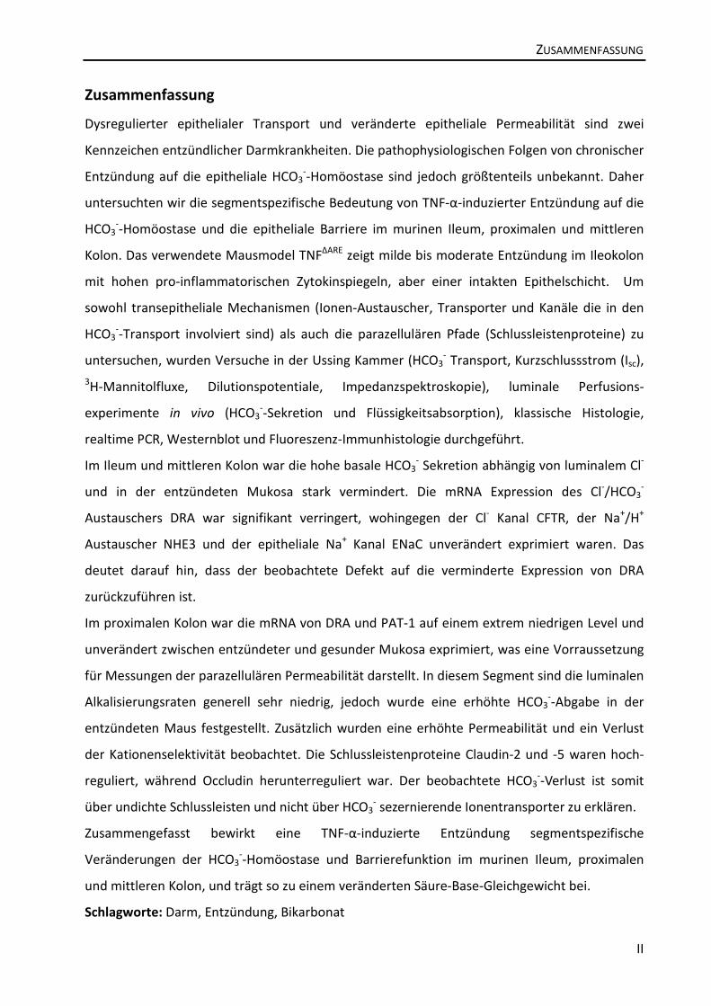
Abstract
Dysregulated epithelial ion transport and impaired epithelial permeability are central to
inflammatory bowel disease pathophysiology, but data on the importance of chronic
inflammation for Ileocolonic epithelial HCO3- output is sparse. We therefore studied the
segment-specific impact of TNF-α-induced inflammation on HCO3-
homeostasis and epithelial
barrier function in the murine ileum, proximal and mid colon. The TNFΔARE
mouse model
displays mild to moderate inflammation in the ileocolon with high a pro-inflammatory cytokine
profile, but an intact epithelial lining. Ussing chamber experiments (HCO3- output, Isc,
3H-
mannitol fluxes, dilution potentials, impedance spectroscopy), in vivo luminal perfusion
experiments (HCO3- secretion and fluid absorption), classical histology, realtime PCR, western
blot, and fluorescence immunohistochemistry were used to examine transepithelial
mechanisms (ion exchangers, transporters, and channels involved in HCO3- transport) as well as
paracellular pathways (tight junction associated proteins).
In the Ileum and mid colon, the high basal HCO3- secretion was dependent on luminal Cl
-, and
strongly decreased in the inflamed mucosa. Cl-/HCO3
- exchanger DRA mRNA and protein were
both down-regulated, whereas Cl- channel CFTR, Na
+/H
+ exchanger NHE3 and epithelial Na
+
channel ENaC mRNA were not significantly changed. This indicates that the observed defect
occurs due to downregulation of DRA.
In the proximal colon, DRA and PAT-1 mRNA abundance was similar in inflamed and healthy
mice with extremely low levels, which is a precondition to study paracellular permeability. In
this segment, where luminal alkalization rates are very low in general, HCO3- output was found
to be significantly increased in inflamed mice. Additionally, increased permeability together
with a loss of cation selectivity was observed. Tight junctional protein expression of claudin-2
and -5 was found to be upregulated while occludin was down-regulated. The observed HCO3-
loss is likely to occur via leaky tight junctions rather than via HCO3- secreting ion transporters.
Taken together, TNF-α-induced inflammation has a segment-specific impact on HCO3-
homeostasis and epithelial barrier function in the murine ileum, proximal and mid colon, which
leads to an altered acid-base balance.
Keywords: intestine, inflammation, bicarbonate
ZUSAMMENFASSUNG
II
Zusammenfassung
Dysregulierter epithelialer Transport und veränderte epitheliale Permeabilität sind zwei
Kennzeichen entzündlicher Darmkrankheiten. Die pathophysiologischen Folgen von chronischer
Entzündung auf die epitheliale HCO3--Homöostase sind jedoch größtenteils unbekannt. Daher
untersuchten wir die segmentspezifische Bedeutung von TNF-α-induzierter Entzündung auf die
HCO3--Homöostase und die epitheliale Barriere im murinen Ileum, proximalen und mittleren
Kolon. Das verwendete Mausmodel TNFΔARE
zeigt milde bis moderate Entzündung im Ileokolon
mit hohen pro-inflammatorischen Zytokinspiegeln, aber einer intakten Epithelschicht. Um
sowohl transepitheliale Mechanismen (Ionen-Austauscher, Transporter und Kanäle die in den
HCO3--Transport involviert sind) als auch die parazellulären Pfade (Schlussleistenproteine) zu
untersuchen, wurden Versuche in der Ussing Kammer (HCO3- Transport, Kurzschlussstrom (Isc),
3H-Mannitolfluxe, Dilutionspotentiale, Impedanzspektroskopie), luminale Perfusions-
experimente in vivo (HCO3--Sekretion und Flüssigkeitsabsorption), klassische Histologie,
realtime PCR, Westernblot und Fluoreszenz-Immunhistologie durchgeführt.
Im Ileum und mittleren Kolon war die hohe basale HCO3- Sekretion abhängig von luminalem Cl
-
und in der entzündeten Mukosa stark vermindert. Die mRNA Expression des Cl-/HCO3
-
Austauschers DRA war signifikant verringert, wohingegen der Cl- Kanal CFTR, der Na
+/H
+
Austauscher NHE3 und der epitheliale Na+ Kanal ENaC unverändert exprimiert waren. Das
deutet darauf hin, dass der beobachtete Defekt auf die verminderte Expression von DRA
zurückzuführen ist.
Im proximalen Kolon war die mRNA von DRA und PAT-1 auf einem extrem niedrigen Level und
unverändert zwischen entzündeter und gesunder Mukosa exprimiert, was eine Vorraussetzung
für Messungen der parazellulären Permeabilität darstellt. In diesem Segment sind die luminalen
Alkalisierungsraten generell sehr niedrig, jedoch wurde eine erhöhte HCO3--Abgabe in der
entzündeten Maus festgestellt. Zusätzlich wurden eine erhöhte Permeabilität und ein Verlust
der Kationenselektivität beobachtet. Die Schlussleistenproteine Claudin-2 und -5 waren hoch-
reguliert, während Occludin herunterreguliert war. Der beobachtete HCO3--Verlust ist somit
über undichte Schlussleisten und nicht über HCO3- sezernierende Ionentransporter zu erklären.
Zusammengefasst bewirkt eine TNF-α-induzierte Entzündung segmentspezifische
Veränderungen der HCO3--Homöostase und Barrierefunktion im murinen Ileum, proximalen
und mittleren Kolon, und trägt so zu einem veränderten Säure-Base-Gleichgewicht bei.
Schlagworte: Darm, Entzündung, Bikarbonat

LIST OF ABBREVIATIONS
III
List of Abbreviations
ARE AU-rich regulatory element
Caco-2 cells Human colorectal intestinal epithelial cell line
C2BBe1 Human colorectal epithelial cell line, subclone of Caco-2
CA IV Carbonic anhydrase isoform IV
CD8+ T-lymphocyte CD8 positive cytotoxic T-lymphocyte, expressing the glycoprotein CD8 on
its cells surface
CD Crohn´s disease
CFTR Cystic fibrosis transmembrane regulator; Cl- channel
DRA Downregulated in adenoma (SLC26a3); Cl-/HCO3
- exchanger
ENaC Epithelial Na+ channel
H&E Haematoxylin and Eosin
HCO3- Hydrogen carbonate, bicarbonate
IBD Inflammatory bowel disease
Isc Short circuit current
Na+/K
+ pump Sodium-potassium adenosine triphosphatase
NBC Sodium bicarbonate cotransporter
NHE1 Sodium-hydrogen exchanger 1 (SLC9a1)
NHE3 Sodium-hydrogen exchanger 3 (SLC9a3)
NKCC1 Na+K
+ 2Cl
- cotransporter (SLC12a2)
PAT-1 Putative anion transporter 1 (SLC36a1)
SCFAs Short chain fatty acids
TER Transepithelial resistance
TJL-scoring system The Jackson Laboratory scoring system
TNF-α Tumor necrosis factor-α
UC Ulcerative colitis

1. GENERAL INTRODUCTION
1
1. General Introduction
Inflammatory bowel disease (IBD), encompassing both Crohn´s disease (CD) and ulcerative
colitis (UC), is characterized by chronic and relapsing inflammation of the gastrointestinal tract.
Although it is known that genetic predisposition and dysregulated immune response (Kaser et
al. 2010), environmental factors such as the gut mictobiota (Hill and Artis, 2010), impaired
epithelial barrier properties (Bruewer et al. 2006) and altered epithelial ion transport (Farkas et
al. 2011) play major roles in these complex diseases, the pathogenesis is still not fully
uncovered. Since luminal pH control is critical for many important epithelial functions, this
thesis illuminates the segment-specific impact of TNF-α (Tumor necrosis factor-α)-induced
inflammation on HCO3- homeostasis and epithelial barrier function in a murine model of IBD.
The gastrointestinal barrier, consisting of epithelial cells and the connecting tight junctions that
provide contact between neighbouring cells, separates the body from the luminal environment
which is potentially loaded with noxious pathogens and harmful molecules. At the same time,
water, nutrients, ions and solutes have to cross the epithelium to maintain homeostasis of the
internal milieu. The permeability is mediated via two major routes: the transepithelial/
transcellular pathway which is predominantly regulated by selective transporters, exchangers
and channels for specific ions, electrolytes, sugars and short chain fatty acids (SCFAs), and the
paracellular pathway, which leads through the space between the cells and is regulated by the
tight junctional complex (see figure 1).
1.1 pH /HCO3- homeostasis and epithelial ion transport mechanisms
pH homeostasis is one of the major tasks of an organism, since protein and enzyme function,
the structure of the cell and the permeability of membranes are challenged by a changing pH.
Intestinal surface and luminal pH control, with HCO3- being one of the major protagonists
(Binder 2005), is critical for many aspects of the epithelial defence and transport function,
including the rheological properties of the mucus layer and thus the opportunity for bacterial
penetration (Swidsinski et al. 2007) as well as the luminal bacterial composition, colonization
and proliferation (Duncan et al. 2009). A large number of transporters, channels and
exchangers maintain the required pH in the intestine.

1. GENERAL INTRODUCTION
2
Cl-
HCO3-
Na+
Na+
transcellular
paracellular
tightjunction
NHE3 DRA/PAT-1
H+
ATPK+
mucus layer
H2O
+CO2
Na+/K+
pump NHE1 NBC
Na+
H+
HCO3- Na+
HCO3-
+H+
JAM
ZO-1
Actin
Claudin
Occludin
Cadherin
CA
Cl-/ HCO3-
CFTRCl-
HCO3-
Na+
Na+
transcellular
paracellular
tightjunction
NHE3 DRA/PAT-1
H+
ATPK+
mucus layer
H2O
+CO2
Na+/K+
pumpNa+/K+
pump NHE1NHE1 NBCNBC
Na+
H+
HCO3- Na+
HCO3-
+H+
JAM
ZO-1
Actin
Claudin
Occludin
Cadherin
CACA
Cl-/ HCO3-
CFTR
Figure 1: Structure of the ileocolonic epithelial lining with the mucus layer and possible transcellular and
paracellular pathways for HCO3-. The electrolyte transporters CFTR, NHE3 and DRA are located in the apical
membrane, whereas the Na+/K
+ pump, NHE1 and NBC are found on the basolateral side. They are expressed in
different amounts and patterns along the ileocolon. Additionally, HCO3- can be generated by carbonic anhydrases
(CA, left). The complex structure of tight junctions with some of the composing proteins is schematically shown on
the right.
All segments from duodenum to distal colon have varying and specialized mechanisms for both
absorbing and secreting acid and base equivalents. Key pathways including the Cl- channel
CFTR, the cation exchanger NHE3 (Na+/H
+) as well as DRA and PAT-1 (Cl
-/HCO3
-), the Na
+/K
+
pump which provides the driving force, the Na+/H
+ exchanger NHE1 and the Na
+/HCO3
-
cotransporter NBC are in charge of the intestinal acid-base balance. These transporters are
expressed in a highly segment-specific manner (see figure 1).
HCO3- is actively secreted in most parts of the intestine, with the highest secretion found in the
duodenum. Here, it is the most important defense mechanism against acidic discharge from the
stomach (Flemström et al. 2002). Along the whole intestine, the epithelium is covered with a
mucus layer composed of mucins, phospholipids, electrolytes, water and a large amount of
HCO3- that keeps the immediate vicinity of the surface epithelium at neutral pH (Flemström and
Kivilaakso 1983). The mucus layer serves as a protection against potentially infectious agents
from the lumen and ensures a smooth passage of digested food. Additionally, Garcia et al.
(2009) reported that mucin requires CFTR-dependent HCO3--secretion in order to be released
and expanded properly. The loss of the intestinal mucus layer is sufficient to lead to injury and
barrier dysfunction of the otherwise intact intestine (Sharpe et al. 2010).

1. GENERAL INTRODUCTION
3
In the proximal large intestine, SCFAs are generated mainly from nonabsorbed carbohydrates
by colonic bacterial metabolism and form the main luminal anions in colonic fluid (Binder et al.
2005). They provide the main nutrition for enterocytes and are absorbed via nonionic diffusion
driven by pH or proton gradient or actively e.g. via a SCFA/HCO3- exchanger (Ruppin et al. 1980,
Mascolo et al. 1991), which to this date is not molecularly identified (Binder 2010). In this
particular segment, the functional expression of DRA is almost negligible, as Talbot and Lytle
(2010) showed in the rodent colon. They also correlated mucosal surface pH with the apical
expression of DRA and NHE3 along the ileocolon and found a matching low pH in the proximal
colon that rose towards the mid colon.
Although evidence for dysregulated electrolyte transport was found some time ago (Harris et a.
1972, Hawker et al. 1980), only more recently the molecular identification of ion transporters
helped to understand their complex involvement in inflammation (Nemeth et al. 2002; Seidler
et al. 2006; Laubitz et al. 2008). It is now well established that cytokines have a direct impact on
the expression and function of several ion transport proteins. In the rat colon, Markossian and
Kreydiyyeh (2005) showed that TNF-α inhibited net water and chloride absorption, down-
regulated the basolateral Na+/K
+/2Cl
- cotransporter NKCC1 and reduced the protein expression
and activity of the Na+/K
+ pump. While the epithelial barrier function was not altered, the
electrogenic sodium absorption via ENaC was found to be strongly impaired in human
macroscopically noninflamed CD colon because of reduced γ-ENaC transcription (Zeissig et al.
2008). Additionally, this group reported that TNF-α exposure to rat distal colon led to the same
effect. Farkas et al. 2011 found a moderate dysfunction of NHE3, but a major decrease of the Cl-
/HCO3- exchange in biopsies from ulcerative colitis patients, associated with a downregulation
of DRA.
1.2 Inflammation-associated epithelial barrier function
Concerning the paracellular pathway, meanwhile more than 40 proteins that are part of or are
associated with tight junctions have been identified (Schneeberger and Lynch, 2004). Among
them, the family of claudins, named from the Latin word claudere for “to close”, have emerged
to be the crucial proteins defining the tight junction properties and their selectivity.
Although the group of barrier builders is proportionally bigger including claudin-1, -3, -4, -5, -8, -
11, -14 and -19, there are claudins mediating permeability like claudin-2 and -10. Claudin-2

1. GENERAL INTRODUCTION
4
forms paracellular pores which are selective for small cations and water but almost
impermeable for anions or uncharged solutes of any size (Amasheh et al. 2002, Rosenthal et al.
2010). While claudin-5 seems to be a sealing claudin without selectivity (Amasheh et al. 2005),
claudin-8 has been reported to form a barrier to cations, including protons, ammonium and
bicarbonate ions (Yu et al. 2003, Angelow et al. 2006).
Occludin was firstly thought of as not required for the formation of tight junction strands since
Saitou et al. (1998) showed that occludin-deficient embryonic stem cells still can differentiate
into polarized epithelial cells bearing tight junctions. In occludin knockout mice, the tight
junctions themselves did not appear to be affected morphologically, and the barrier function of
the intestinal epithelium was normal as far as examined electrophysiologically (Saitou et al.
2000). However, ongoing research revealed an important, yet not fully understood regulatory
contribution of occludin concerning barrier permeability. Al-Sadi et al. (2011) showed that
occludin depletion in intestinal epithelial cells leads to a selective or preferential increase in
macromolecule flux, suggesting that occludin plays a crucial role in the maintenance of tight
junction barrier.
This highly selective and thoroughly regulated barrier is known to be weakened or disrupted in
pathophysiological situations like inflammation, indicated by an increase in paracellular
permeability and a decrease in transcellular electrical resistance. Pro-inflammatory cytokines
like TNF-α and interferon-γ are known to contribute to a disturbed barrier function. In cell
culture experiments using Caco-2 cells, a human intestinal epithelial cell line, Marano et al.
(1998) showed that as little as 5ng/ml TNF-α applied for only 5 minutes was sufficient to reduce
the transepithelial resistance (TER) for 40-50% and to decrease the short circuit current (Isc) for
30% after 24 hours.
In animal models of IBD, intestinal tissue expression of occludin was found to be markedly
decreased (Fries et al. 1999, Gassler et al. 2001). In patients with mild to moderate Crohn’s
disease, Zeissig et al. (2007) showed an upregulation of pore-forming claudin-2 and
downregulation and redistribution of the sealing claudin-5 and -8 as well as a downregulated
expression of occludin. Furthermore, reduced and discontinuous tight junction strands were
demonstrated. According to the authors, this leads to a pronounced barrier dysfunction in the
inflamed intestine.

1. GENERAL INTRODUCTION
5
1.3 The TNFΔARE
mouse model
The mouse model used in this study was originally established in the laboratory of George
Kollias (Kontoyiannis et al. 1999). A genetic deletion of the Tumor necrosis factor alpha (TNF-α)
AU-rich regulatory element (ARE) leads to an inaccurate posttranscriptional degradation and
therefore to an overproduction of TNF-α. This pro-inflammatory cytokine is predominantly
produced by macrophages, but also by other cell types like lymphocytes, natural killer cells,
myeloid cells, adipocytes and intestinal epithelial cells. Homozygous TNFΔARE/ΔARE
and
heterozygous TNF+/ΔARE
mice develop a CD8+ T-lymphocyte-dependent chronic inflammatory
arthritis and chronic inflammation along the intestine. A dys-/ upregulation of TNF-α was
shown to be present in a large number of diseases including Alzheimer’s disease, cancer and
IBD. Several studies proved a multiple fold higher amount of TNF-α in patients suffering from
Ulcerative colitis as well as Crohn´s disease (MacDonald et al. 1990; Segain et al. 2000). The
importance of this cytokine is underlined by the therapeutic treatment based on anti-TNF-α
antibodies like infliximab or adalimumab as IBD treatments.
To quantify the severity of inflammation in the different intestinal segments of the TNF+/ΔARE
mouse, a modified TJL-scoring system was used (Bleich et al. 2004). While the small intestine
starting from the duodenum is only mildly affected, the Ileum is the most inflamed part
followed by the colon from proximal to distal. This mimics Crohn’s-like pathology in terms of
locality. Additionally, the crypt and villus architecture as well as the epithelial lining seem to be
intact judged from histological Haematoxylin and Eosin (H&E) staining. These facts make the
TNFΔARE
mice an ideal animal model to study ion transport and epithelial barrier properties in
the context of IBD.

1. GENERAL INTRODUCTION
6
1.4 Objective
Electrolyte transport dysregulation and barrier dysfunction have both been implicated in the
complex pathophysiology of IBD. However, their contribution to epithelial pH homeostasis
during intestinal inflammation has not been unraveled.
The aims of this thesis therefore were:
- to test the suitability of the TNF-α overexpressing mouse model TNFΔARE
in matters of
intestinal ion-transport and epithelial barrier experiments for the first time
- to investigate if there are inflammation-associated disturbances in the HCO3-
homeostasis in different segments of the intestine (Ileum, proximal colon and mid
colon)
- to explore the underlying mechanisms for each examined part of the gut.
Since HCO3- output is modulated differently along the intestine, transepithelial (ion exchangers,
transporters, and channels involved in HCO3- transport) as well as paracellular pathways (tight
junction associated proteins) were examined. For this purpose, a multitude of methods was
used including Ussing chamber experiments (HCO3- output, Isc,
3H-mannitol fluxes, dilution
potentials, impedance spectroscopy), in vivo luminal perfusion experiments (HCO3- secretion
and fluid absorption), classical histology, realtime PCR, western blot, and fluorescence
immunohistochemistry.

2. RESULTS - 2.1 ILEOCOLONIC HCO3- TRANSPORT IN INFLAMMATION
7
2. Results
2.1 „Loss of Downregulated in Adenoma (DRA) Impairs Mucosal HCO3- Secretion in Murine
Ileocolonic Inflammation”
Running head: Ileocolonic HCO3- Transport in Inflammation
Manuscript originally published in Inflammatory Bowel Diseases (2012;18:101–111)
Fang Xiao*, Marina Juric*, Junhua Li, Brigitte Riederer, Sunil Yeruva, Anurag Kumar Singh,
Lifei Zheng, Silke Glage, George Kollias, Pradeep Dudeja, De-An Tian, Gang Xu, Jinxia Zhu,
Oliver Bachmann, and Ursula Seidler
*shared first authorship
Received for publication March 17, 2011; Accepted March 28, 2011.
Copyright © 2011 Crohn’s & Colitis Foundation of America, Inc.
DOI 10.1002/ibd.21744
Published online 6 May 2011 in Wiley Online Library.
Link: http://onlinelibrary.wiley.com/doi/10.1002/ibd.21744/pdf
Author’s contributions to the manuscript
provided the mouse model: GK
provided the DRA antibody: PD
designed experiments: FX, MJ, JL, BR, SY, AKS, SG, OB, US
performed experiments: FX, MJ, JL, BR, SY, AKS, LZ, SG
analyzed data: FX, MJ, US, OB
wrote the article: FX, MJ, US, OB
corrected and approved the manuscript: DT, GX, JZ

2. RESULTS - 2.1 ILEOCOLONIC HCO3- TRANSPORT IN INFLAMMATION
8
Abstract
Background: Ileocolonic luminal pH has been reported to be abnormally low in inflammatory
bowel disease (IBD) patients, and one of the causative factors may be reduced epithelial HCO
secretory rate (JHCO3-). Disturbances in JHCO3
- may occur due to inflammation-induced changes in
the crypt and villous architecture, or due to the effect of proinflammatory cytokines on
epithelial ion transporters.
Methods: To discriminate between these possibilities, the tumor necrosis factor alpha (TNF-α)
overexpressing (TNF+/ΔARE
) mouse model was chosen, which displays high proinflammatory
cytokine levels in both ileum and colon, but develops only mild colonic histopathology and
diarrhea. HCO secretion, mRNA expression, immunohistochemistry, and fluid absorptive
capacity were measured in ileal and mid-colonic mucosa of TNF+/ΔARE
and wildtype (WT) (TNF+/+
)
mice in Ussing chambers, and in anesthetized mice in vivo.
Results: The high basal JHCO3-
observed in WT ileal and mid-colonic mucosa were luminal Cl−-
dependent and strongly decreased in TNF+/ΔARE
mice. Downregulated in adenoma (DRA) mRNA
and protein expression was strongly decreased in TNF+/ΔARE
ileocolon, whereas cystic fibrosis
transmembrane conductance regulator (CFTR), Na+/H
+ exchanger 3 (NHE3), Na
+/HCO
cotransporter (NBC), and epithelial sodium channel (ENaC) expression was not significantly
altered. This indicates that the severe defect in ileocolonic JHCO3-
was due to DRA
downregulation. Fluid absorption was severely depressed in the ileum but only mildly affected
in the mid-distal colon, preventing the development of overt diarrhea.
Conclusions: Even mild ileocolonic inflammation may result in a decrease of epithelial HCO
secretion, which may contribute to alterations in surface pH, intestinal flora, and mucus barrier
properties.
Keywords: inflammatory bowel disease; bicarbonate; anion exchanger; colon

2. RESULTS - 2.2 HCO3- LEAKAGE IN INFLAMED MURINE COLON
9
2.2 „Increased epithelial permeability is the primary cause for bicarbonate loss in inflamed
murine colon”
Running head: HCO3- leakage in inflamed murine colon
Manuscript accepted for publication in Inflammatory Bowel Diseases
Marina Juric*, Fang Xiao*, Salah Amasheh, Oliver May, Kristin Wahl, Heike Bantel,
Michael P. Manns, Ursula Seidler, Oliver Bachmann
*shared first authorship
Received for publication August 31, 2012; Accepted September 06, 2012.
Copyright © 2012 Crohn’s & Colitis Foundation of America, Inc.
Author’s contributions to the manuscript
designed experiments: MJ, FX, SA, US, OB
performed experiments: MJ, FX, SA, OM, KW
analyzed data: MJ, FX, US, OB
wrote the article: MJ, US, OB
corrected and approved the manuscript: HB, MPM

2. RESULTS - 2.2 HCO3- LEAKAGE IN INFLAMED MURINE COLON
10
Increased epithelial permeability is the primary cause for bicarbonate loss in inflamed murine
colon
Marina Juric*, Fang Xiao, MD*,#
, Salah Amasheh, PhD§, Oliver May, Kristin Wahl, Heike Bantel,
MD, Michael P. Manns, MD, Ursula Seidler, MD‡, Oliver Bachmann, MD‡
Dept. of Gastroenterology, Hepatology and Endocrinology, Hannover Medical School, Germany,
#present address: Dept. of Gastroenterology and Hepatology, Tongji Hospital, Wuhan, China,
§Institute of Clinical Physiology, Charité, Campus Benjamin Franklin, Berlin, Germany;
*these two authors share the first, and ‡these two the corresponding authorship
Address for correspondence:
Oliver Bachmann
Dept. of Gastroenterology, Hepatology and Endocrinology
Hannover Medical School
Carl-Neuberg-Str. 1
30625 Hannover, Germany
Phone +49 1761 532 3359, Fax +49 511 532 8892
This work was supported by grants from the Deutsche Forschungsgemeinschaft to O.
Bachmann (SFB621-C10) and to U. Seidler (SFB621-C9 and DFG SE460/13-4).
2. RESULTS - 2.2 HCO3- LEAKAGE IN INFLAMED MURINE COLON
11
Abstract
Background: Bicarbonate loss into the lumen occurs during intestinal inflammation in different
species. However, candidate pathways like CFTR or DRA are inhibited in the inflamed gut. This
study addressed the question whether and how inflammation-associated increased intestinal
permeability may result in epithelial HCO3- loss.
Methods: Murine proximal colon was studied because it does not express functional DRA, but
is inflamed in the TNF-α overexpressing mouse model (TNFΔARE
). Luminal alkalization, 3H-
mannitol fluxes, impedance spectroscopy and dilution potentials were measured in Ussing
chambers, while expression and localization of tight junction associated proteins were analyzed
by Western blots and immunohistochemistry.
Results: Luminal alkalization rates and 3H-mannitol fluxes were increased in TNF
+/ΔARE proximal
colon, while forskolin-stimulated Isc was not altered. Epithelial resistance was reduced, but
subepithelial resistance increased. The epithelial lining was intact, and enterocyte apoptosis
rate was not increased despite massively increased Th1 cytokine levels and
lymphoplasmacellular infiltration. Measurement of dilution potentials suggested a loss of
cation selectivity with increased anion permeability. Western analysis revealed a
downregulation of occludin expression, and an upregulation of both claudin-2 and claudin-5,
with no change in ZO-1, E-cadherin, claudin-4 and claudin-8. Immunohistochemistry suggested
correct occludin localization, but reduced tight junction density in TNF+/ΔARE
surface epithelium.
Conclusions: Inflammation during TNF-α overexpression leads to increased epithelial
permeability in murine proximal colon, decreased tight junctional cation selectivity, and
increased HCO3- loss into the lumen. Inflammation-associated colonic HCO3
- loss may occur via
leaky tight junctions rather than via HCO3- secreting ion transporters.
Keywords: Tight junction; claudin; occludin; large intestine.

2. RESULTS - 2.2 HCO3- LEAKAGE IN INFLAMED MURINE COLON
12
Introduction
The ability of the colonic epithelium to control surface and luminal pH is critical for many
aspects of the epithelial interface barrier and transport function, including the absorption of
short chain fatty acids (SCFAs), mucus production, as well as bacterial colonization and
proliferation (1-3). Acid-base homeostasis is altered during intestinal inflammation, which is
often the cause of acute and chronic diarrheal diseases, and has been associated with increased
luminal HCO3- content in humans (4) and animal models (5). Particularly severe and chronic
intestinal inflammation occurs in inflammatory bowel disease (IBD), and early studies have
attempted to define the impact of chronic colonic inflammation on luminal pH. Due to the
technical complexity, gastrointestinal pH profiles have only been measured in small numbers of
patients suffering from IBD using a radiotelemetry capsule, and most of the more recent
studies have demonstrated higher values in the colon of IBD patients than in the healthy
controls [(6, 7); for review see (8)].
Intestinal inflammation and pro-inflammatory cytokines weaken the epithelial barrier by
various mechanisms, including altered tight junctional anatomy (9). Tight junctions are formed
by multiprotein complexes including e. g. occludin, tricellulin, claudins, and junctional adhesion
molecules (JAMs), which restrict the free movement of ions across the epithelium. The
continued exploration of barrier components has revealed that not all of them are “tightening”
(10): While claudin-2 is selective for cations (11), several other claudins promote or regulate
anion permeability (12-15). Intestinal inflammation influences the tight junction composition,
reducing the expression of “tightening” claudins and occludin, leading to strand breaks (16), or
up-regulating the pore-forming claudin-2 (17, 18).
HCO3- movement into the intestinal lumen occurs in a highly segment-specific and tightly
regulated fashion (19-21), and represents a major mechanism for luminal pH control (22).
During chronic colitis, HCO3- could potentially reach the lumen via the paracellular pathway,
which plays a minor role in the healthy epithelium (23). However, the significance of tight
junction modification for acid-base balance in the inflamed intestine has not been studied, and
it is not known whether specific changes to components of the epithelial barrier by the
inflammatory process can lead to HCO3- losses. One major obstacle to such studies is the
influence of inflammation on active HCO3- transporters, predominantly luminal Cl
-/HCO3
-
transporters from the Slc26 family. The proximal colon of rats was shown to lack DRA (down-
regulated in adenoma, Slc26a6), which is the primary pathway for basal HCO3- output into the

2. RESULTS - 2.2 HCO3- LEAKAGE IN INFLAMED MURINE COLON
13
colonic lumen in the absence of nutrients (24, 25), and murine proximal colon was found to be
similar [(26), and this study]. The expected rates of HCO3- transport across the apical membrane
of the proximal colonic epithelium under resting conditions should be negligible, which makes
this segment ideal to test the hypothesis that changes to the epithelial barrier during intestinal
inflammation cause HCO3- leakage.
Materials and Methods
Animals: TNFΔARE
mice (TNFtm2Gkl
) (27) were bred on a C57BL/6J background at the animal care
facility at Hannover Medical School under standardized conditions. Sex- and age-matched
heterozygous mice (TNF+/ΔARE
) and their healthy littermates (TNF+/+
) at the age of 6-8 months
were used for the experiments. Mice were sacrificed by CO2 narcosis and subsequent cervical
dislocation. Animal care and experimentation were approved by and carried out in accordance
with the Medical School of Hannover and the local authorities for the regulation of animal
welfare (Niedersächsisches Landesamt für Verbraucherschutz und Lebensmittelsicherheit).
pH-Stat titration of HCO3- secretory rates and Isc: Experiments were carried out exactly as
described previously (26). Excised proximal colon was stripped of external muscle layers,
mounted into Ussing chambers and bathed with equiosmolar solutions, pH 7.4 (serosally (in
mM): 108 NaCl, 25 NaHCO3, 3 KCl, 1.3 MgSO4, 2 CaCl2, 2.25 KH2PO4-, 8.9 glucose, 10 sodium
pyruvate, gassed with 95% O2/5% CO2, and luminally with 154 mM NaCl, gassed with 100% O2).
Neural activity and prostaglandin generation were blocked with 1 µM tetrodotoxin and 3 µM
indomethacin (serosal). HCO3- secretory rates were determined by pH-stat titration and
electrical parameters recorded under open circuit conditions. Isc was calculated from recording
tissue resistance (Rt) and potential difference (PD) in 2-min intervals.
Fluxes of [3H] mannitol: To assess tissue permeability, bilateral [
3H] mannitol fluxes were
measured as previously described (28) in isolated proximal colonic TNF+/ΔARE
and TNF+/+
mucosa,
with bilaterally identical, CO2/O2 gassed solutions (as described for the serosal solution above,
except for 9 mM lactose in the mucosal bath and 1 mM mannitol in both), under short circuit
conditions. After 30 min equilibration, 74 KBq/ml 3H-mannitol (Perkin Elmer, Waltham, MA,
USA) were added either to the serosal or the mucosal bath, and samples were taken as
quadruple in 15 min intervals for 90 min.
2. RESULTS - 2.2 HCO3- LEAKAGE IN INFLAMED MURINE COLON
14
Dilution potentials: The permeability of sodium and chloride ions was measured with a
modified dilution potential technique (29). In brief, pieces of stripped proximal colon were
mounted into Ussing chambers and equilibrated in our basal solution with mannitol instead of
glucose on the luminal side (see pH-Stat titration). The mucosal solution was then substituted
with a 54 mM NaCl containing solution while osmolarity was maintained with 108 mM of
mannitol. Electrical parameters were recorded in 30 sec intervals for 30 min before and after
the substitution. Ion permeability ratio (PNa/PCl) was calculated from the dilution potential by
using the Goldman-Hodgkin-Katz equation. Using Ohm´s law, the total conductance G was
calculated. The absolute permeabilities of sodium (PNa) and chloride (PCl) were obtained by
using a simplified Kimizuka-Koketsu equation.
Impedance spectroscopy: Tissues were mounted in Ussing chambers and one-path impedance
spectroscopy was performed in order to discriminate between epithelial (Repi) and subepithelial
(Rsub) resistance exactly as described previously (30). To adjust mucosal surface area from
TNF+/ΔARE
and TNF+/+
, images of hematoxylin/ eosin stained paraffin sections were analysed with
the processing software Image J (NIH, Bethesda, MD; http://rsb.info.nih.gov/ij/). Villus height
and crypt depth of the epithelium as well as the thickness of the subepithelial tissue layers
were measured. The number of villi and crypts were counted. On average, 10 adjacent sections
were analyzed. The ratio of mucosal to serosal surface area was determined, representing
apical epithelial area and subepithelial area corresponding to the opening area of the Ussing
chamber.
See Supplementary Materials for quantitative real-time polymerase chain reaction (PCR),
measurements of apoptosis, western blot analysis, immunofluorescence, and statistical
analysis.

2. RESULTS - 2.2 HCO3- LEAKAGE IN INFLAMED MURINE COLON
15
Results
Characterization of proximal colonic inflammation in the TNF∆ARE
colitis model
As a model of chronic intestinal inflammation, TNF+/ΔARE
mice and their wild-type littermates
were utilized. These mice develop a Crohn’s disease-like phenotype with ileocolitis and
pronounced arthritis (26, 27) (Figure 1A). A significant separation of the weight curves after 16
weeks for both male and female mice was observed (Figure 1B). In analogy to human
inflammatory bowel disease (31-33), it was previously established that heterozygeous animals
suffer from a pronounced intestinal absorptive defect, but also display a reduced net secretory
response (26).
Histologically, mild to moderate colonic inflammation with a proximal-to-distal gradient was
observed (Figure 2A,B). The inflammatory infiltrate (consisting mostly of B- and T-lymphocytes,
supplementary fig. 1B,C) partially extended into the submucosa, but no gross ulceration was
present. There was no indication of disrupted continuity of the epithelium, which could have
represented a bias for transport- and permeability studies. TUNEL (Terminal deoxynucleotidyl
transferase dUTP nick end labeling) staining did not reveal obvious differences in the frequency
of apoptotic cells (Figure 2C), which was further substantiated by caspase 3/7 activity assay
(Figure 2D). In line with the histological signs of inflammation, the content of the inflammatory
cytokines TNF (tumor necrosis factor)-α and IL (interleukin)-1β was markedly elevated on an
mRNA level (Figure 2E,F). The results of Figure 1 and 2 therefore substantiate the presence of a
mild, non-erosive colitis with chronically strongly elevated Th1 cytokines in the proximal colon.
Basal luminal alkalization rates in the proximal colon of TNF∆ARE
mice and analysis of active
transport involvement
Luminal HCO3- output in the isolated proximal colonic mucosa was low, consistent with
previous results (26), and due to virtual absence of DRA (Figure 3A,C and 4A). However, basal
alkalization rates were significantly higher in TNF+/ΔARE
than in wild-type proximal colon (Figure
3A,C) without a difference between male and female animals. The Isc was not significantly
different (Figure 3B,D), suggesting that electrogenic Cl- secretion was not disturbed. To exclude
that chronic inflammation leads to an up-regulation of Cl-/HCO3
- exchangers, DRA (Slc26a3),
and PAT1 (Slc26a6), mRNA abundance was measured by semiquantitative RT-PCR. The
expression of both was very low in murine proximal colon (Figure 4A,B). No differences in anion
exchanger expression between wild-type and heterozygous mice were detected with all

2. RESULTS - 2.2 HCO3- LEAKAGE IN INFLAMED MURINE COLON
16
reference genes used (Figure 4C,D). This suggested that neither alterations in electrogenic
HCO3- secretion nor luminal Cl
-/HCO3
- exchangers were involved.
Epithelial permeability and barrier selectivity in the inflamed proximal colon
To assess permeability changes in the proximal colon of TNF+/ΔARE
mice and their normal
littermates as a potential cause of HCO3- leakage,
3H-labelled mannitol flux rates were
measured in isolated proximal colonic epithelium. Indeed, higher fluxes were observed in
TNF+/ΔARE
tissue (Figure 5A), which indicates increased paracellular permeability for small
uncharged solutes. Impedance spectroscopy, which allows the differentiation of epithelial (Repi)
and subepithelial (Rsub) resistances, demonstrated a markedly lower Repi (Figure 5B). In contrast,
Rsub was elevated, probably due to subepithelial thickening (Figure 2B, 5B, and supplementary
fig. 1A).
Next, we measured dilution potentials in heterozygous compared to healthy colonic epithelia
during isoosmolar dilution of the apical NaCl concentration by 50% (see Materials and
Methods). Interestingly, the dilution potential was significantly altered in heterozygous animals,
reflecting a calculated shift in the permeability ratio towards Cl- (Table 1). Using the measured
conductance and the modified Kimitsuka-Koketsu equation, the Na+ permeability, which was
overall higher, was found to be unchanged (Figure 5C). This suggested a decrease in cation
selectivity of the inflamed proximal colonic mucosa.
Expression and localization of tight junction associated proteins
The influence of proinflammatory cytokines on the differential expression of tight junction
components has been documented in several studies (34, 35) and could explain the observed
permeability changes. To investigate whether an altered abundance of the different proteins
can be observed in inflamed vs. healthy mice, the expression of occludin as well as
representative members of pore-forming (claudin-2) and tightening (claudin-5, -4, and -8)
claudins as well as adherens junction components was assessed. Occludin, which was suggested
as a general indicator for tight junction integrity, and which was shown to be strongly regulated
by TNF-α, was downregulated in TNF+/ΔARE
proximal colonic tissue. Localization of occludin,
however, was unchanged in heterozygous vs. wild type animals, but disturbed strand
morphology was present, with an apparent reduction in tight junction density than in WT
mucosa. (Figure 6C,D). While claudin-2 was upregulated on a protein level, analysis of the

2. RESULTS - 2.2 HCO3- LEAKAGE IN INFLAMED MURINE COLON
17
tightening claudins gave a mixed image, with an upregulation of claudin-5 and no change of
claudin-4 or claudin-8. The adherens junction associated proteins E-Cadherin (E-Cad) and Zona
Occludens Protein 1 (ZO-1) were not changed (Figure 6A,B).
Discussion
Intestinal inflammation results in disturbed barrier function and a dysregulation of electrolyte
transport (31, 36). However, changes in epithelial pH homeostasis due to altered permeability
and paracellular leakage have not been studied. The experiments presented here demonstrate
the existence of a higher paracellular permeability and loss of cation selectivity in otherwise
mildly and chronically inflamed proximal colonic mucosa, leading to enhanced HCO3- leakage
into the proximal colonic lumen.
We and others have previously investigated the impact of intestinal inflammation on epithelial
HCO3- transport (26, 37). Zhang et al. injected mice with an anti-CD3 monoclonal antibody and
measured ileal Isc and HCO3- secretion in the Ussing chamber (37). In this acute inflammation
model, the authors found a shift from electroneutral Cl-/HCO3
- exchange to a predominantly
electrogenic anion secretion with an increased intracellular cAMP content, but no expressional
changes for CFTR. Similarly, the chronically inflamed ileum and mid-distal colon of TNFΔARE
heterozygotes displays reduced Cl- dependent HCO3
- secretion, which is due to the loss of DRA
(26). A down-regulation of this exchange process was also found in the colon of these mice (26),
the HLA-B27/β2m transgenic rat, the interleukin-10 (IL-10) knockout mouse with spontaneous
colitis, and in patients with ulcerative colitis (38). While these studies had established a
dysregulation of pH regulating ion transporters during intestinal inflammation, the importance
of barrier changes for intestinal pH homeostasis remained unresolved, since the experimental
design did not permit to separate active transport from paracellular leakage.
Recent studies have reported a pronounced segmental heterogeneity for ion transporter
expression (24), leading to significant variability within even the same organ such as the colon,
and different species (20). In particular, the proximal murine colonic mucosa displays very low
rates of luminal alkalization (39), and DRA expression in this segment is almost negligible (24).
We therefore chose the murine proximal colon of TNF-α overexpressing mice (which develop
an ileocolitis, and in which the proximal colon is more affected than the distal colon) for the
present study, assuming that it would permit us to measure the passive leakage of HCO3-
without the influence of an inflammation-associated decrease in DRA-mediated Cl-/HCO3
-

2. RESULTS - 2.2 HCO3- LEAKAGE IN INFLAMED MURINE COLON
18
exchange. Indeed, luminal alkalization occurred at almost negligible rates in WT mucosa,
compared to the tenfold higher rates in the ileal and mid-distal murine colonic mucosa (26),
paralleled by a very low DRA expression, which was unchanged with respect to mRNA
abundance in TNF+/ΔARE
mice (Figure 4). Luminal alkalization rate was significantly increased in
proximal colonic mucosa of TNFΔARE
heterozygotes, which was in contrast to the decreased
HCO3- output rates in the mid-distal colon (26). Isc, on the other hand, was not significantly
altered by the inflammatory state in the proximal colon, neither the basal nor the forskolin-
stimulated value. The crypt-expressed CFTR anion channel (or any other channel) is therefore
not responsible for the higher luminal alkalization rates. In addition, CFTR expression levels
were unchanged. We speculated that another potential reason may be less luminal acidification
by the apical Na+/H
+ exchanger NHE3, which has been shown to be dysfunctional despite
normal expression and localization in moderately severe murine and human intestinal
inflammation (31, 33, 40). However, NHE3 mRNA expression was also equal in TNF+/ΔARE
and
TNF+/+
mice, and luminal application of fully NHE3 inhibitory concentrations of the specific
NHE3 inhibitor S1611 did not affect luminal alkalization rates in the chambered proximal colon
(supplementary fig. 2A,B), indicating that this transporter is quiescent under the conditions of
our experiments. The likely reason for this is the complete absence of CO2 in the lumen (which
would diffuse into the enterocytes, acidify them, and stimulate NHE3), the absence of any other
acidifying substances in the luminal bath (such as SCFAs), and therefore an inactivation of NHE3
exchange by the high pHi in colonic surface cells under the given experimental conditions. Thus,
transporter-mediated events do not explain the observed higher alkalization rates in TNFΔARE
heterozygote mucosa.
In addition to disturbed electrolyte transport, changes in intestinal epithelial barrier function
have been described in many experimental models of bowel inflammation, sometimes
preceding inflammation (32, 35, 41). Cytokines can directly influence barrier components (42).
As a major inflammatory messenger, TNF-α has been shown to down-regulate claudin-1, up-
regulate claudin-2 (43), and to degrade occludin mRNA in a micro-RNA-dependent manner (44).
Occludin was shown to regulate macromolecule flux across the intestinal epithelium (45), which
may explain our observation of increased 3H-mannitol permeability during TNF-α
overexpression, but also that of increased HCO3- leakage.
Due to the cation selectivity of small and large intestinal mucosa, paracellular permeation of
anions occurs at a much lower rate than cations, i.e. Na+, in healthy mucosa. A paracellular

2. RESULTS - 2.2 HCO3- LEAKAGE IN INFLAMED MURINE COLON
19
passage of HCO3- ions may occur when the charge-selectivity for cations is decreased, which is
what we found. We therefore searched for the reason for the decrease in cation selectivity.
Expression of WT and mutated claudins in Madin-Darby Canine Kidney (MDCK) cells under an
inducible promoter first clearly demonstrated that claudins can mediate charge selectivity of
the paracellular pathway for ions. When the charge of selected extracellular amino acids was
reversed, ion selectivity increased Na+- (claudin-4) or switched the preference from Na
+ to Cl
-
permeability (claudin-15) (10). When claudin-2 is overexpressed in MDCK monolayers,
paracellular permeability increased 5.6-fold compared to the vector control, with relative
cation selectivity (11). Several studies have linked claudins to increased Cl- permeability, but
whether this occurs via an increase in paracellular permeability or via upregulation of
transcellular transport was not clearly determined in most of them (12-15). Recently, claudin-
10a and -17 have been characterized as being anion selective (46), but claudin-10a is expressed
at extremely low levels in the proximal colon (data not shown), and claudin-17 mRNA is absent
in the colon (47) and is also not up-regulated in the inflamed intestine (J.-D. Schulzke, personal
communication). In our study, the observed changes in claudin expression pattern included
increased expression of “tightening” as well as “pore-forming” components, and no obvious
explanation for the decrease in cation selectivity. However, it is known that claudins are
expressed in rodent colon which have not been studied for their ion permeation properties in
detail, but do pose additional pathways for cations and/ or anion (48, 49). We did find a strong
down-regulation in occludin expression (as mentioned above), and the immunohistochemistry
revealed a normal localization of the tight junction components in the zones of cell-cell
contacts, but a coarser architecture in the TNFΔARE
heterozygote mucosa. Given the complexity
of the tight junction, it may be that in the present case, not a single protein directly confers
increased HCO3- permeability, but that expressional changes of other, even “tightening”
components alter TJ regulation and increase permeability for HCO3-, as it has been suggested
for renal cells (50).
What may be the clinical implications of our findings? On a local level, the increased HCO3-
leakage will change mucosal surface pH and likely affect the mucosa-associated
hydrogenotrophic gut flora (51) as well as luminal flora (3), alter the microbial fermentation,
and influence colonic health. Alterations of gut flora composition have indeed been observed
during intestinal inflammation (52), and are believed to be involved in IBD pathogenesis (53).
The functional down-regulation of both Na+/H
+ as well as Cl
-/HCO3
- exchangers of the surface

2. RESULTS - 2.2 HCO3- LEAKAGE IN INFLAMED MURINE COLON
20
epithelium, in combination with increased HCO3- leakage, observed even in this mild non-
erosive inflammatory state as observed in the TNF+/ΔARE
proximal colon, will completely alter
the segmental surface cell pH profiles, as well as luminal bulk pH in the different colonic
segments and thus be one of the reasons for alteration of gut microbiome during intestinal
inflammation, with possible detrimental consequences. The second consequence of
paracellular HCO3- leakage in the inflamed intestine may be a very considerable loss of base
equivalents from the systemic circulation. It is likely that this leakage occurs wherever intestinal
epithelium is inflamed, although, as discussed above, this will be difficult or eve impossible to
prove in intestinal segments with significant active HCO3- transport masking additional HCO3
-
leakage. Since inflammation also down-regulates DRA-mediated Cl-/HCO3
- exchange, this loss of
HCO3- is in part compensated, but for the price of losing Cl
-. A third consequence of paracellular
HCO3- leakage will be the loss of acidic microclimate in those segments of the gut where it is
present and necessary to drive proton-coupled nutrient uptake, such as in the jejunum and
ileum, or nonionic diffusion of weak acids, such as SCFAs in the proximal colon.
In summary, we describe an enhanced luminal alkalization associated with a tight junction
defect in chronic intestinal inflammation, which is likely to have a significant effect on epithelial
pH homeostasis, making it an important pathophysiological factor in inflammatory bowel
disease.
Acknowledgements
We thank George Kollias for providing the TNFΔARE
mouse model, Ulrike Bode and Manuela
Büttner for the B220 antibody, and Mathias Hornef and Natalie Torow for the CD3 antibody.

2. RESULTS - 2.2 HCO3- LEAKAGE IN INFLAMED MURINE COLON
21
Reference list
1. Garcia MA, Yang N, Quinton PM. Normal mouse intestinal mucus release requires cystic
fibrosis transmembrane regulator-dependent bicarbonate secretion. The Journal of clinical
investigation. 2009;119:2613-2622
2. Vidyasagar S, Barmeyer C, Geibel J, et al. Role of short-chain fatty acids in colonic HCO(3)
secretion. AmJPhysiol GastrointestLiver Physiol. 2005;288:G1217-G1226
3. Duncan SH, Louis P, Thomson JM, et al. The role of pH in determining the species
composition of the human colonic microbiota. Environmental microbiology. 2009;11:2112-2122
4. Field M. Intestinal ion transport and the pathophysiology of diarrhea. The Journal of
clinical investigation. 2003;111:931-943
5. Sundaram U, West AB. Effect of chronic inflammation on electrolyte transport in rabbit
ileal villus and crypt cells. AmJPhysiol. 1997;272:G732-G741
6. Press AG, Hauptmann IA, Hauptmann L, et al. Gastrointestinal pH profiles in patients
with inflammatory bowel disease. Alimentary pharmacology & therapeutics. 1998;12:673-678
7. Ewe K, Schwartz S, Petersen S, et al. Inflammation does not decrease intraluminal pH in
chronic inflammatory bowel disease. DigDisSci. 1999;44:1434-1439
8. Nugent SG, Kumar D, Rampton DS, et al. Intestinal luminal pH in inflammatory bowel
disease: possible determinants and implications for therapy with aminosalicylates and other
drugs. Gut. 2001;48:571-577
9. Schmitz H, Barmeyer C, Fromm M, et al. Altered tight junction structure contributes to
the impaired epithelial barrier function in ulcerative colitis. Gastroenterology. 1999;116:301-
309
10. Colegio OR, Van Itallie CM, McCrea HJ, et al. Claudins create charge-selective channels in
the paracellular pathway between epithelial cells. Am J Physiol-Cell Ph. 2002;283:C142-C147
11. Amasheh S, Meiri N, Gitter AH, et al. Claudin-2 expression induces cation-selective
channels in tight junctions of epithelial cells. Journal of cell science. 2002;115:4969-4976
12. Gunzel D, Amasheh S, Pfaffenbach S, et al. Claudin-16 affects transcellular Cl- secretion
in MDCK cells. The Journal of physiology. 2009;587:3777-3793
13. Ohta A, Yang SS, Rai T, et al. Overexpression of human WNK1 increases paracellular
chloride permeability and phosphorylation of claudin-4 in MDCKII cells. Biochemical and
biophysical research communications. 2006;349:804-808
14. Tatum R, Zhang Y, Lu Q, et al. WNK4 phosphorylates ser(206) of claudin-7 and promotes
paracellular Cl(-) permeability. FEBS letters. 2007;581:3887-3891
15. Angelow S, Kim KJ, Yu AS. Claudin-8 modulates paracellular permeability to acidic and
basic ions in MDCK II cells. The Journal of physiology. 2006;571:15-26
16. Van Itallie CM, Anderson JM. Measuring size-dependent permeability of the tight
junction using PEG profiling. Methods MolBiol. 2011;762:1-11
17. Prasad S, Mingrino R, Kaukinen K, et al. Inflammatory processes have differential effects
on claudins 2, 3 and 4 in colonic epithelial cells. Laboratory Investigation. 2005;85:1139-1162

2. RESULTS - 2.2 HCO3- LEAKAGE IN INFLAMED MURINE COLON
22
18. Zeissig S, Burgel N, Gunzel D, et al. Changes in expression and distribution of claudin 2, 5
and 8 lead to discontinuous tight junctions and barrier dysfunction in active Crohn's disease.
Gut. 2007;56:61-72
19. Seidler U. Acta Physiologica symposium: acid-base transporters and epithelial
electrolyte transport. Acta Physiol (Oxf). 2011;201:1-2
20. Bachmann O, Seidler U. News from the end of the gut--how the highly segmental
pattern of colonic HCO transport relates to absorptive function and mucosal integrity.
BiolPharmBull. 2011;34:794-802
21. Bachmann O, Juric M, Seidler U, et al. Basolateral ion transporters involved in colonic
epithelial electrolyte absorption, anion secretion and cellular homeostasis. Acta Physiol (Oxf).
2011;201:33-46
22. Binder HJ, Rajendran V, Sadasivan V, et al. Bicarbonate secretion: a neglected aspect of
colonic ion transport. Journal of clinical gastroenterology. 2005;39:S53-58
23. Endeward V, Gros G. Low carbon dioxide permeability of the apical epithelial membrane
of guinea-pig colon. The Journal of physiology. 2005;567:253-265
24. Talbot C, Lytle C. Segregation of Na/H exchanger-3 and Cl/HCO3 exchanger SLC26A3
(DRA) in rodent cecum and colon. American journal of physiology Gastrointestinal and liver
physiology. 2010;299:G358-367
25. Schweinfest CW, Spyropoulos DD, Henderson KW, et al. slc26a3 (dra)-deficient mice
display chloride-losing diarrhea, enhanced colonic proliferation, and distinct up-regulation of
ion transporters in the colon. The Journal of biological chemistry. 2006;281:37962-37971
26. Xiao F, Juric M, Li J, et al. Loss of downregulated in adenoma (DRA) impairs mucosal
HCO(3) (-) secretion in murine ileocolonic inflammation. InflammBowelDis. 2012;18:101-111
27. Kontoyiannis D, Pasparakis M, Pizarro TT, et al. Impaired on/off regulation of TNF
biosynthesis in mice lacking TNF AU-rich elements: implications for joint and gut-associated
immunopathologies. Immunity. 1999;10:387-398
28. Guba M, Kuhn M, Forssmann WG, et al. Guanylin strongly stimulates rat duodenal
HCO3- secretion: proposed mechanism and comparison with other secretagogues.
Gastroenterology. 1996;111:1558-1568
29. Kahle KT, MacGregor GG, Wilson FH, et al. Paracellular Cl- permeability is regulated by
WNK4 kinase: Insight into normal physiology and hypertension. Proceedings of the National
Academy of Sciences of the United States of America. 2004;101:14877-14882
30. Markov AG, Veshnyakova A, Fromm M, et al. Segmental expression of claudin proteins
correlates with tight junction barrier properties in rat intestine. Journal of comparative
physiology B, Biochemical, systemic, and environmental physiology. 2010;180:591-598
31. Seidler U, Lenzen H, Cinar A, et al. Molecular mechanisms of disturbed electrolyte
transport in intestinal inflammation. Annals of the New York Academy of Sciences.
2006;1072:262-275
32. Clayburgh DR, Musch MW, Leitges M, et al. Coordinated epithelial NHE3 inhibition and
barrier dysfunction are required for TNF-mediated diarrhea in vivo. The Journal of clinical
investigation. 2006;116:2682-2694

2. RESULTS - 2.2 HCO3- LEAKAGE IN INFLAMED MURINE COLON
23
33. Yeruva S, Farkas K, Hubricht J, et al. Preserved Na(+)/H(+) exchanger isoform 3
expression and localization, but decreased NHE3 function indicate regulatory sodium transport
defect in ulcerative colitis. InflammBowelDis. 2010;16:1149-1161
34. Heller F, Florian P, Bojarski C, et al. Interleukin-13 is the key effector Th2 cytokine in
ulcerative colitis that affects epithelial tight junctions, apoptosis, and cell restitution.
Gastroenterology. 2005;129:550-564
35. Tang Y, Clayburgh DR, Mittal N, et al. Epithelial NF-kappaB enhances transmucosal fluid
movement by altering tight junction protein composition after T cell activation. The American
journal of pathology. 2010;176:158-167
36. Martinez-Augustin O, Romero-Calvo I, Suarez MD, et al. Molecular bases of impaired
water and ion movements in inflammatory bowel diseases. InflammBowelDis. 2008
37. Zhang H, Ameen N, Melvin JE, et al. Acute inflammation alters bicarbonate transport in
mouse ileum. The Journal of physiology. 2007;581:1221-1233
38. Yang H, Jiang W, Furth EE, et al. Intestinal inflammation reduces expression of DRA, a
transporter responsible for congenital chloride diarrhea. The American journal of physiology.
1998;275:G1445-1453
39. Yu H, Riederer B, Stieger N, et al. Secretagogue stimulation enhances NBCe1
(electrogenic Na(+)/HCO(3)(-) cotransporter) surface expression in murine colonic crypts.
AmJPhysiol GastrointestLiver Physiol. 2009;297:G1223-G1231
40. Farkas K, Yeruva S, Rakonczay Z, Jr., et al. New therapeutic targets in ulcerative colitis:
the importance of ion transporters in the human colon. Inflammatory bowel diseases.
2011;17:884-898
41. Suenaert P, Maerten P, Van AG, et al. Effects of T cell-induced colonic inflammation on
epithelial barrier function. InflammBowelDis. 2010;16:1322-1331
42. Hering NA, Schulzke JD. Therapeutic options to modulate barrier defects in
inflammatory bowel disease. Digestive diseases. 2009;27:450-454
43. Amasheh M, Fromm A, Krug SM, et al. TNFalpha-induced and berberine-antagonized
tight junction barrier impairment via tyrosine kinase, Akt and NFkappaB signaling. Journal of
cell science. 2010;123:4145-4155
44. Ye D, Guo S, Al-Sadi R, et al. MicroRNA regulation of intestinal epithelial tight junction
permeability. Gastroenterology. 2011;141:1323-1333
45. Al-Sadi R, Khatib K, Guo S, et al. Occludin regulates macromolecule flux across the
intestinal epithelial tight junction barrier. American journal of physiology Gastrointestinal and
liver physiology. 2011;300:G1054-1064
46. Krug SM, Gunzel D, Conrad MP, et al. Charge-selective claudin channels. Annals of the
New York Academy of Sciences. 2012;1257:20-28
47. Krug SM, Gunzel D, Conrad MP, et al. Claudin-17 forms tight junction channels with
distinct anion selectivity. Cellular and molecular life sciences : CMLS. 2012;69:2765-2778
48. Inai T, Sengoku A, Guan X, et al. Heterogeneity in expression and subcellular localization
of tight junction proteins, claudin-10 and -15, examined by RT-PCR and immunofluorescence
microscopy. Archives of histology and cytology. 2005;68:349-360
2. RESULTS - 2.2 HCO3- LEAKAGE IN INFLAMED MURINE COLON
24
49. Fujita H, Chiba H, Yokozaki H, et al. Differential expression and subcellular localization of
claudin-7, -8, -12, -13, and -15 along the mouse intestine. The journal of histochemistry and
cytochemistry : official journal of the Histochemistry Society. 2006;54:933-944
50. Sas D, Hu M, Moe OW, et al. Effect of claudins 6 and 9 on paracellular permeability in
MDCK II cells. American journal of physiology Regulatory, integrative and comparative
physiology. 2008;295:R1713-1719
51. Nava GM, Carbonero F, Croix JA, et al. Abundance and diversity of mucosa-associated
hydrogenotrophic microbes in the healthy human colon. The ISME journal. 2012;6:57-70
52. van Nuenen MH, Venema K, van der Woude JC, et al. The metabolic activity of fecal
microbiota from healthy individuals and patients with inflammatory bowel disease. DigDisSci.
2004;49:485-491
53. Swidsinski A, Weber J, Loening-Baucke V, et al. Spatial organization and composition of
the mucosal flora in patients with inflammatory bowel disease. JClinMicrobiol. 2005;43:3380-
3389
2. RESULTS - 2.2 HCO3- LEAKAGE IN INFLAMED MURINE COLON
25
Figures, Tables and Legends
Figure 1:
Figure 2:
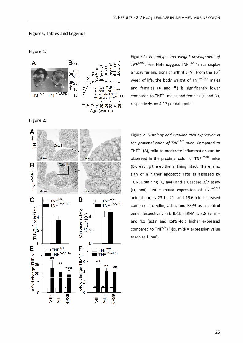
Figure 1: Phenotype and weight development of
TNFΔARE
mice. Heterozygous TNF+/ΔARE
mice display
a fuzzy fur and signs of arthritis (A). From the 16th
week of life, the body weight of TNF+/ΔARE
males
and females (● and ) is significantly lower
compared to TNF+/+
males and females (○ and ),
respectively. n= 4-17 per data point.
Figure 2: Histology and cytokine RNA expression in
the proximal colon of TNFΔARE
mice. Compared to
TNF+/+
(A), mild to moderate inflammation can be
observed in the proximal colon of TNF+/ΔARE
mice
(B), leaving the epithelial lining intact. There is no
sign of a higher apoptotic rate as assessed by
TUNEL staining (C, n=4) and a Caspase 3/7 assay
(D, n=4). TNF-α mRNA expression of TNF+/ΔARE
animals (■) is 23.1-, 21- and 19.6-fold increased
compared to villin, actin, and RSP9 as a control
gene, respectively (E). IL-1β mRNA is 4.8 (villin)-
and 4.1 (actin and RSP9)-fold higher expressed
compared to TNF+/+
(F)(□, mRNA expression value
taken as 1, n=6).

2. RESULTS - 2.2 HCO3- LEAKAGE IN INFLAMED MURINE COLON
26
Figure 3:
Figure 4:
Figure 3: Time course of basal HCO3--output (A)
and Isc (B) in the proximal colon of TNFΔARE
mice.
Basal HCO3--output of TNF
+/ΔARE mice (■) is
significantly higher compared to TNF+/+
animals (□,
A,C). No significant differences are found in basal
and forskolin-stimulated Isc (ΔIsc) among the two
groups (B,D, n=6/5).
Figure 4: RNA expression of the apical Cl-/ HCO3
--
exchanger DRA and PAT1 in the proximal and distal
colon of TNFΔARE
mice. To illustrate the
heterogeneity of DRA, ΔCT values of proximal and
distal colonic mRNA are shown. DRA mRNA is
significantly higher expressed in the distal colon,
where it additionally differs significantly between
TNF+/+
(□) and TNF+/ΔARE
(■) mice (A). PAT1 at a very
low expression level does neither differ between
the two colonic segments nor the two groups (B).
Independently of the control gene, the expression
level of both exchangers does not differ between
TNF+/ΔARE
and TNF+/+
mice in the proximal colon
(C,D) (mRNA expression value of the control group
taken as 1, n=6).

2. RESULTS - 2.2 HCO3- LEAKAGE IN INFLAMED MURINE COLON
27
Figure 5:
Figure 6:
Figure 5: Permeability studies in the proximal colon
of TNFΔARE
mice. As an indication for paracellular
permeability, TNF+/ΔARE
(■) proximal colon displays
a significantly higher 3H-mannitol flux rate
compared to TNF+/+
(□, A, n=4) colon. Impedance
spectroscopy measurements (B, n=8) reveal
furthermore a significantly lower epithelial
resistance (Repi), and coevally significantly elevated
subepithelial resistance (Rsub) in TNF+/ΔARE
tissue.
Additionally, dilution potentials (C, n=7/8) show an
overall higher permeability of Na+ vs. Cl
-, but also a
significantly higher permeability for Cl- ions in
TNF+/ΔARE
proximal colon compared to the control
group.
Figure 6: Expression of tight junction associated
proteins and localization of zona occludens protein
1 (ZO-1, magenta), occludin (Ocln, green) and
claudin-2 (Cldn-2, green) in the proximal colon of
TNFΔARE
mice. Densitometric analysis of the original
western blots (A, n=4/6) shows a significant
upregulation of claudin-2 (Cldn-2) and claudin-5
(Cldn-5) in TNF+/ΔARE
tissue. While claudin-4 (Cldn-
4), claudin-8 (Cldn-8), e-cadherin (E-Cad) and zona
occludens protein 1 (ZO-1) remain unchanged,
occludin (Ocln) expression is found to be reduced
in the inflamed colon (B).
In TNF+/+
(C) and to TNF+/ΔARE
animals (D), ZO-1 and
Ocln are expressed in the same apical region of
every enterocyte (merge). Occludin seems not to
be differentially localized but tight junction density
appeared to be lower. Cldn-2 localization is
restricted to the lower part of the crypt in both
groups. Bar=25µm.

2. RESULTS - 2.2 HCO3- LEAKAGE IN INFLAMED MURINE COLON
28
Table 1:
DP P(Na+)/P(Cl
-) J(Na
+) J(Cl
-)
[mV] [·10-6
cm/s] [µEq·cm-2
·h-1
] [µEq·cm-2
·h-1
]
TNF +/+
-8.00 ± 0.24 2.62 ± 0.08 6.43 ± 0.37 2.46 ± 0.21
TNF +/ΔARE
-5.06 ± 0.01 1.80 ± 0.07 6.41 ± 0.13 3.59 ± 0.16
p-value < .001 < .001 n.s. < .001
Table 1: Results of dilution potentials in the proximal colon of TNFΔARE
mice. Data represent mean ± standard
error of the mean (SEM). Abbreviations: DP= dilution potential, P= permeability, J= flux, n.s.= not significant.

2. RESULTS - 2.2 HCO3- LEAKAGE IN INFLAMED MURINE COLON
29
Supplementary Materials
Increased epithelial permeability is the primary cause for bicarbonate loss in inflamed murine
colon
Marina Juric*, Fang Xiao, MD*, Salah Amasheh, PhD, Oliver May, Kristin Wahl, Heike Bantel,
MD, Michael P. Manns, MD, Ursula Seidler, MD‡, Oliver Bachmann, MD‡
Supplementary Materials and Methods
Materials: All the reagents were purchased from Sigma-Aldrich (Deisenhofen, Germany) unless
mentioned otherwise. S1611 was kindly provided by Sanofi-Aventis (Frankfurt, Germany). The
following antibodies were used for Western blotting and/ or Immunhistofluorescence:
monoclonal mouse anti-occludin, monoclonal mouse anti-claudin 2 (Invitrogen, Camarillo, CA,
USA), polyclonal rabbit anti-claudin 4, monoclonal mouse anti-claudin 5, polyclonal rabbit anti-
claudin 8, polyclonal rabbit anti-E-cadherin (Abcam, Cambridge, UK), polyclonal rabbit anti-ZO-1
(Proteintech, Chicago, IL, USA), rabbit anti-actin (santa cruz, Santa Cruz, CA, USA), polyclonal
rabbit anti-CD3, polyclonal chicken anti-SLC26A3 (DRA) (Sigma-Aldrich, Deisenhofen, Germany),
monoclonal rat anti-CD45R (B220) (AbD serotec, Oxford, UK).
Quantitative real-time PCR: Total RNA isolation and real time quantitative PCR was performed
exactly as described previously (Xiao et al. 2012). Data were standardized to three different
housekeeper genes [villin, actin and ribosomal protein S9 (RPS9)] for each sample and then
normalized to control tissue samples (TNF+/+
). The sequences of the primers used are:
Villin: 5`-TCATACTCAAGACTCCGTCC-3´ and 5´-TACCACTTGTTTCTCCGTCC-3´
Actin: 5´-AGAGGGAAATCGTGCGTGAC-3´ and 5´-CAATAGTGATGACCTGGCCGT-3´
RPS9: 5´-AAGCACATCGACTTCTCCC-3´ and 5´-ACAATCCTCCAGTTCAGCC-3´
TNF-α: 5´-CATCTTCTCAAAATTCGAGTGACAA-3´ and 5´-TGGGAGTAGACAAGGTACAACCC-3´
IL-1β: 5´-CAACCAACAAGTGATATTCTCCATG-3´ and 5´-GATCCACACTCTCCAGCTGCA-3´
DRA: 5´-TTCCCCTCAACATCACCATCC-3´ and 5´-GTAAAATCGTTCTGAGGCCCC-3´
PAT1: 5´-GGCTCCTGGGTGATCTGTTA-3´ and 5´-CCAAACATAGGAGGCAATCC-3´

2. RESULTS - 2.2 HCO3- LEAKAGE IN INFLAMED MURINE COLON
30
TUNEL staining: Terminal deoxynucleotidyl transferase-mediated dUTP nick end labeling (In Situ
Cell Death Detection Kit, Fluorescein, from Roche, Mannheim, Germany) was used to quantify
apoptotic cells following the producer’s directions. For permeabilization, 20µg/ml proteinase K
solution was used for 15 min at room temperature. Six random, non-overlapping pictures of
20x magnified optical fields of two different colon layers were taken (n=4), and the number of
TUNEL-positive cells per field was counted by a person blinded to the genotype of the sample.
Caspase 3/7 assay: To access caspase activity, 2 pieces of proximal colon of 4 healthy and 4
inflamed mice were homogenized in lysis buffer (in mM) 10 Tris-HCl pH 7.4, 10 MgCl, 150 NaCl,
10 dithiothreitol, 0.5% octylphenoxypolyethoxyethanol (Nonidet P-40), 1% Protease-Inhibitor
(Sigma Aldrich), and the total protein concentration was assessed by a Bradford assay (BioRad,
Munich, Germany). Activity of caspase 3 and caspase 7 was measured by a luminescent
substrate assay (Caspase-Glo; Promega, Mannheim, Germany) according to the manufactures’
instruction. Luminescence of each sample was measured twice and the results are given in
relative light units (RLU).
Western blot: To determine tight junction protein expression, western blot analysis was
performed with protein lysates of proximal colonic tissue of TNF+/+
and TNF+/ΔARE
mice.
Scratched mucosa was put into Lysate buffer containing 1mM EGTA, 1% Triton X100, 0.1% SDS
and 10% Protease Inhibitor Cocktail (ProteoBlock, Fermentas, St. Leon-Rot, Germany) in PBS
(pH7.4), homogenized and sucked through a 24Gx1” needle. The samples were centrifuged at
12.000g and 4°C, and protein concentration of the collected supernatant was determined by a
Bradford assay (Quick Start BSA Standard Set and Bradford Dye Reagent, Biorad, München,
Germany). 20µg protein were separated on 12-15% SDS-polyacrylgels and blotted on a PVDF
Transfer Membrane (Hybond-P, GE Healthcare, Buckinghamshire, UK). The Blots were blocked
in RotiBlock-Solution (Carl Roth, Karlsruhe, Germany) for 2 hours before being incubated with
the corresponding primary antibody overnight at 4°C. Between two washing steps with TBS-
Tween, the Membranes were incubated with the corresponding horseradish peroxidase
conjugated secondary antibodies (Cell Signaling, Danvers, MA, USA), and developed for
visualisation of protein with ECL Western Blotting Detection Reagents (GE Healthcare,
Buckinghamshire, UK). Densitometric analysis was performed using Image J Software (NIH,
Bethesda, MD; http://rsb.info.nih.gov/ij/).
2. RESULTS - 2.2 HCO3- LEAKAGE IN INFLAMED MURINE COLON
31
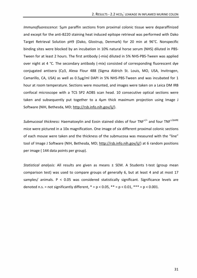
Immunofluorescence: 5µm paraffin sections from proximal colonic tissue were deparaffinized
and except for the anti-B220 staining heat induced epitope retrieval was performed with Dako
Target Retrieval Solution pH9 (Dako, Glostrup, Denmark) for 20 min at 96°C. Nonspecific
binding sites were blocked by an incubation in 10% natural horse serum (NHS) diluted in PBS-
Tween for at least 2 hours. The first antibody (-mix) diluted in 5% NHS-PBS-Tween was applied
over night at 4 °C. The secondary antibody (-mix) consisted of corresponding fluorescent dye
conjugated antisera (Cy3, Alexa Flour 488 (Sigma Aldrich St. Louis, MO, USA, Invitrogen,
Camarillo, CA, USA) as well as 0.5µg/ml DAPI in 5% NHS-PBS-Tween and was incubated for 1
hour at room temperature. Sections were mounted, and images were taken on a Leica DM IRB
confocal microscope with a TCS SP2 AOBS scan head. 10 consecutive optical sections were
taken and subsequently put together to a 4µm thick maximum projection using Image J
Software (NIH, Bethesda, MD; http://rsb.info.nih.gov/ij/).
Submucosal thickness: Haematoxylin and Eosin stained slides of four TNF+/+
and four TNF+/ΔARE
mice were pictured in a 10x magnification. One image of six different proximal colonic sections
of each mouse were taken and the thickness of the submucosa was measured with the “line”
tool of Image J Software (NIH, Bethesda, MD; http://rsb.info.nih.gov/ij/) at 6 random positions
per image ( 144 data points per group).
Statistical analysis: All results are given as means ± SEM. A Students t-test (group mean
comparison test) was used to compare groups of generally 6, but at least 4 and at most 17
samples/ animals. P < 0.05 was considered statistically significant. Significance levels are
denoted n.s. = not significantly different, * = p < 0.05, ** = p < 0.01, *** = p < 0.001.
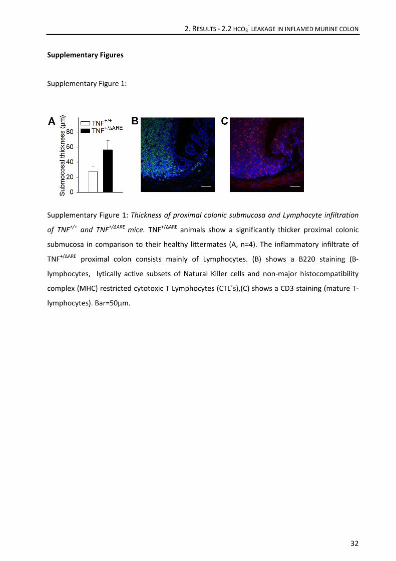
2. RESULTS - 2.2 HCO3- LEAKAGE IN INFLAMED MURINE COLON
32
Supplementary Figures
Supplementary Figure 1:
Supplementary Figure 1: Thickness of proximal colonic submucosa and Lymphocyte infiltration
of TNF+/+
and TNF+/ΔARE
mice. TNF+/ΔARE
animals show a significantly thicker proximal colonic
submucosa in comparison to their healthy littermates (A, n=4). The inflammatory infiltrate of
TNF+/ΔARE
proximal colon consists mainly of Lymphocytes. (B) shows a B220 staining (B-
lymphocytes, lytically active subsets of Natural Killer cells and non-major histocompatibility
complex (MHC) restricted cytotoxic T Lymphocytes (CTL´s),(C) shows a CD3 staining (mature T-
lymphocytes). Bar=50µm.

2. RESULTS - 2.2 HCO3- LEAKAGE IN INFLAMED MURINE COLON
33
Supplementary Figure 2:
Supplementary Figure 2: Luminal alkalization of murine wildtype proximal colon during NHE3
inhibition, RNA expression and localization of NHE3 in TNF+/+
and TNF+/ΔARE
mice. Addition of a
fully NHE3 inhibitory concentration of S1611 to the luminal side did not change the basal
alkalization rate. A: time course of luminal alkalization, where the pH deviation occurring in
response to addition of the acidic S1611 solution is omitted. B: Mean basal luminal alkalization
rates, t-test: not significant. n= 6. Independently of the control gene, the RNA expression level
of NHE3 does not differ between TNF+/ΔARE
and TNF+/+
mice in the proximal colon (C) (mRNA
expression value of the control group taken as 1, n=5). There is no indication of a changed DRA
protein localization (D, E), although it has to be taken into consideration that reduced specificity
has been reported for the available DRA antibodies, which may lead to staining of other related
transport proteins. Bar=25µm.
3. GENERAL DISCUSSION
34
3. General Discussion
Luminal HCO3- has been found essential for the release and proper function of intestinal mucus
(Garcia et al. 2009), intestinal wound healing (Feil et al. 1989) and intestinal electrolyte
reabsorption (Binder et al. 2005). Intraluminal pH has been also found to be changed in the
colon of UC patients, but the importance of this has so far been discussed predominantly in the
context of anti-inflammatory drug release (Nugent et al. 2001). Additionally, changes in
epithelial pH homeostasis due to altered permeability and paracellular leakage have rarely
been studied in the context of intestinal inflammation.
Therefore, the presented studies were designed to get further insight into the mechanisms that
are responsible for an alteration in HCO3- output, if any, in intestinal inflammation, with special
emphasis on the segmental heterogeneity. Recent studies have reported a highly segment
specific expression of ion transporters which leads to a large variability even within the same
organ such as the colon (Talbot and Lytle, 2010). The considered parts of the intestine were the
ileum, the proximal colon and the mid colon, which represent the most affected intestinal parts
in IBD patients. The used TNF-α overexpressing mouse model TNFΔARE
mimics this disease
pattern in matters of locality and develops mild to moderate inflammation in the respective
segments without gross destruction of the crypt-villlus architecture.
The key findings of this project were:
- basal HCO3-
secretion was found to be Cl- dependent and significantly reduced in the Ileum
and the mid colon of inflamed TNF+/ΔARE
mice. DRA protein and mRNA were strongly decreased
and proved to cause this circumstance.
- in the inflamed proximal colon of TNF+/ΔARE
animals, increased HCO3- output and epithelial
permeability together with a loss of cation selectivity was observed. This was likely due to
disturbances in tight junctional protein expression or formation.
HCO3- ions can pass the luminal membrane in various ways: either in exchange against another
anion, through anion conductance or anion-selective pores in the tight junction complexes, or
they can be generated from CO2 that perfuses to the lumen. In most segments of the intestine,

3. GENERAL DISCUSSION
35
the discrimination of paracellular in contrast to transcellular pathways is difficult or even
impossible, since passive leakage would be masked by active HCO3- transport.
In Ussing chamber experiments, it is possible to discriminate between HCO3-/anion exchange
and conductance by removing the dependent anion from the luminal solution. In the ileum and
mid colon, we found a significantly lower HCO3- secretion in inflamed versus non-inflamed
tissue. Further experiments revealed that this secretion was Cl- dependent in the basal state,
which pointed to an alteration of HCO3-/Cl
- exchange. Both probable apical proteins, DRA and
PAT-1 (which is only weakly expressed in the colon, Wang et al. 2002) were found to be
significantly downregulated on an mRNA level. However, PAT-1 knockout mice had no defect in
ileal HCO3- secretion (Xiao F. and Singh A., unpublished observations), and since Yang et al.
(1998) as well as Farkas et al. (2011) alike found a drastic decrease in the expression of DRA in
UC patients and rodent colitis models, this exchanger can be considered to be the responsible
one.
Concerning the reduced ΔIsc in the ileum (which is an index of active Cl- secretion, Clarke and
Harline (1996)), CFTR mRNA expression was examined, but no changes were found. Other
potential defects in HCO3- uptake or supply pathways in the Ileum were further explored, but
did not give a distinct explanation. While Carbonic anhydrase IV (CA IV) mRNA was found to be
downregulated in the inflamed mucosa, CA IV-deficient mice did not show any disturbances in
ileal HCO3- -secretion. Thus, we assumed that disturbances in the signaling pathway for
regulating the CFTR HCO3- conductivity during inflammation, or the overall villus shortening
with reduction of epithelial surface area, may be explanations for the observed defect.
Cytokines like TNF-α and INF-γ are known to downregulate Na+ absorptive mechanisms in cells
(Amin et al. 2006 and 2011), and reduced Na+ or fluid absorptive rates were also found in
biopsies from IBD patients (Yeruva et al. 2011). When we measured the ileal and mid-colonic
fluid absorptive capacity in anesthetized mice, a reduced capacity was found in the ileal mucosa
of TNF+/ΔARE
. In contrast to that, the mid-colonic fluid absorptive capacity was unexpectedly well
maintained even though the mRNA expression of TNF-α, INF-γ and IL-1β were higher in our
inflamed animal model than those in the previously mentioned biopsies. Therefore we studied
the epithelial ion channel ENaC, which is expressed in the mid to distal colon and reabsorbs Na+
actively. In the distal colon, no change of ENaC activity could be detected between inflamed
and healthy mice, and its transport rate could be further induced by a three week given low
sodium diet. All three subunits of ENaC mRNA remained unchanged between the groups,

3. GENERAL DISCUSSION
36
indicating a still intact response to aldosterone despite the inflammation. The studies of
Bergann et al. (2009) which showed that that TNF-α may enhance glucocorticoid-induced ENaC
expression via stabilization of glucocorticoid receptors, may explain this finding.
Neither ENaC nor NHE3 or CFTR were affected in mRNA expression which demonstrates a
discriminating sensitivity of DRA to TNF-α induced inflammation. This sensitivity may possibly
be related to the observed downregulation of DRA promoter activity by INF-γ (Saksena et al.
2010). An additional defect in ileal barrier function cannot be excluded, but is impossible to
distinguish from the high amount of actively transported HCO3-.
However, in the rodent proximal colon the expression of the Cl-/HCO3
- exchanger DRA and PAT-
1 was found to be almost negligible and very low, respectively (Talbot and Lytle 2010; Wang et
al. 2002), and the rates of luminal alkalization are very small (Yu et al. 2009). The second part of
the study was therefore addressed to the murine proximal colon which would permit
measurements of possible passive leakage of HCO3- without the influence of an inflammation-
associated alteration in active Cl-/HCO3
- exchange.
In this study we indeed found very low luminal alkalization rates in wildtype mice, but these
were significantly higher in the inflamed mucosa of TNF+/ΔARE
animals. Also, the expression of
DRA mRNA was found to be unchanged between the groups and at a very low level. There was
no change in the basal or forskolin stimulated Isc and CFTR mRNA levels, which would have
pointed to active transport. To rule out other possible transporters involved, we studied the
role of NHE3, the apical Na+/H
+ exchanger, which potentially could lead to a lesser luminal
acidification if downregulated or impaired in function. Such dysfunction was shown in murine
and human inflamed mucosa, despite normal mRNA and protein expression as well as
localization (Seidler et al. 2006; Yeruva et al. 2010; Farkas et al. 2011). Here, NHE3 mRNA
expression was unchanged. Additionally, the application of the NHE3 specific inhibitor S1611
did not lead to altered luminal alkalization of murine proximal colonic mucosa in Ussing
chamber experiments, excluding a participation of NHE3.
TNF-α as well as other cytokines are known to cause disturbances in epithelial barrier function
by various mechanisms, e.g. direct influence the expression or the intracellular localization of
one or several claudins (Amasheh et al. 2010) or other tight junctional proteins. Occludin
expression was shown to be downregulated in a micro-RNA-dependent manner when exposed
to TNF-α (Ye et al. 2011). In Caco2 monolayers and the intact murine intestine, Al-Sadi et al.

3. GENERAL DISCUSSION
37
2011 revealed that occludin regulates epithelial macromolecule flux, suggesting that it plays a
crucial role in the maintenance of tight junction barrier. Since we found a significant occludin
protein downregulation in the proximal colon of TNF+/ΔARE
mice, this could explain not only our
observed increased 3H flux but also the increased HCO3
- leakage.
We further found increased anion permeability with a decrease in cation selectivity in the
inflamed mucosa. Adjusted ion selectivity was also reported for claudin-4 and -15, which, when
expressed in Madin-Darby Canine Kidney (MDCK) cells and challenged with extracellular amino
acids with reversed charge, increased their Na+ selectivity or switched the preference from Na
+
to Cl- permeability (Colegio et al. 2002). Several claudins have been linked to increased Cl
-
permeability, like e.g. claudin-7, which was shown to promote paracellular Cl- permeability
when phosphorylated at the Ser206
phosphorylation site by WNK4 (Tatum et al. 2007). Several
claudins are known to have splice variants, some with fundamental differences between these
isoforms. Only recently, the splice variant claudin-10a, which is barely detectable in the
proximal colon but evident amongst others in the nephron, was described as anion specific,
while one of the other variants, claudin-10b was identified as cation selective (Krug et al. 2012).
In the TNF+/ΔARE
mouse, we saw an upregulated expression of tightening (claudin-5) as well as
pore-forming (claudin-2) claudins, but no apparent explanation for the decrease in cation
selectivity. However, we did see a downregulation of occludin. Given the fact that the
properties of the epithelial barrier function are massively influenced by the combination of the
expressed claudins (Krug et al. 2012), it is possible that not a single protein causes higher HCO3-
permeability in intestinal inflammation, but the combination of altered tight junction
components.
In our two studies we showed that TNFΔARE
mice are a suitable model to study ion transport and
epithelial barrier properties under mild inflammatory conditions. With its high expression of
Th1 cytokines and its segmental inflammation pattern it resembles Crohn´s disease in humans,
although there are limitations like in all established inflammatory mouse models (e.g. IL-10
knockout, transfer colitis or DSS colitis). However, similar to many CD patients, these mice do
not have overt diarrhea. Therefore, our studies underline the fact that the absence of diarrhea
does not necessarily indicate the absence of ion transport defects in inflamed colon, and that
other pathophysiological aspects of disturbed transport function need to be considered.

3. GENERAL DISCUSSION
38
The segment-specific expression pattern of transporters involved in HCO3- secretion (especially
DRA) allows us to gain insights in defective transcellular pathways in the ileum and mid colon,
as well as paracellular pathways in the proximal colon, where active transport occurs at
extremely low levels. Although a defective barrier function is not unlikely to occur in other
segments, it would be masked by active transport.
The relevance of defective HCO3- secretion or leakage in the lower gut may be manifold. In the
upper digestive tract, the importance of HCO3- for mucosal protection against low luminal pH
has been broadly studied (Shorrock and Rees 1988; Flemström and Sjöblom 2002). In addition,
epithelial wound healing requires an alkaline surface pH both in the upper GI tract and in the
colon (Feil et al. 1989, Semble and Wu 1989). Furthermore, intact HCO3- secretion has been
shown to be essential for mucus secretion in intestine and airways (Garcia et al. 2009, Chen et
al. 2010). In this context, an intact mucus layer has recently been shown to be crucial for the
prevention of bacteria to attach to the colonic mucosa and elicit an inflammatory response
(Johansson et al. 2010). A disturbed HCO3- secretion or leakage is most likely to change mucosal
surface and luminal pH and thereby influence several circumstances, for example the acidic
microclimate which is needed to drive the nonionic diffusion of week acids like SCFAs.
Furthermore, a changed pH presumably affects the mucosa-associated hydrogenotrophic gut
flora (Nava et al. 2012) as well as luminal flora (Duncan et al. 2009). A shift of this flora
composition has indeed been observed during intestinal inflammation (van Nuenen et al. 2004),
and are believed to be involved in IBD pathogenesis (Swidsinski et al.2005).
Taken together, our studies show that TNF-α-induced inflammation has differential effects on
HCO3- output in the mildely inflamed ileum, proximal colon and mid colon of TNF
ΔARE mice. The
exquisite sesitivity of DRA expression and the resultant reduced ileal HCO3- secretion in the
ileum and mid colon as well as the enhanced colonic luminal alkalization associated with a
changed tight junctional protein expression cause a changed surface and luminal pH and thus
influence epithelial pH homeostasis in chronic intestinal inflammation.
4. REFERENCES
39
4. References
Al-Sadi R, Khatib K, Guo S, et al. Occludin regulates macromolecule flux across the intestinal
epithelial tight junction barrier. Am J Physiol Gastrointest Liver Physiol. 2011;300:G1054-
1064
Amasheh S, Meiri N, Gitter AH, et al. Claudin-2 expression induces cation-selective channels in
tight junctions of epithelial cells. Journal of Cell Science. 2002;115:4969-4976
Amasheh S, Schmidt T, Mahn M, et al. Contribution of claudin-5 to barrier properties in tight
junctions of epithelial cells. Cell Tissue Res. 2005;321:89-96
Amasheh M, Fromm A, Krug SM, et al. TNFalpha-induced and berberine-antagonized tight
junction barrier impairment via tyrosine kinase, Akt and NFkappaB signaling. Journal of cell
science. 2010;123:4145-4155
Amin MR, Malakooti J, Sandoval R, et al. IFN-gamma and TNFalpha regulate human NHE3 gene
expression by modulating the Sp family transcription factors in human intestinal epithelial
cell line C2BBe1. Am J Physiol Cell Physiol. 2006;291:C887–896.
Amin MR, Orenuga T, Tyagi S, et al. Tumor necrosis factor-alpha represses the expression of
NHE2 through NF-kappaB activation in intestinal epithelial cell model, C2BBe1. Inflamm
Bowel Dis. 2011;17:720-731
Angelow S, Kim KJ, Yu AS. Claudin-8 modulates paracellular permeability to acidic and basic ions
in MDCK II cells. J Physiol. 2006;571:15-26
Bergann T, Zeissig S, Fromm A, et al. Glucocorticoids and tumor necrosis factor-alpha synergize
to induce absorption by the epithelial sodium channel in the colon. Gastroenterology.
2009;136:933–942.
Binder HJ, Rajendran V, Sadasivan V, et al. Bicarbonate Secretion - A neglected Aspect of colonic
ion transport. J Clin Gastroenterol. 2005;39:S53-S58
Binder HJ. Role of colonic short-chain fatty acid transport in diarrhea. Annu Rev Physiol.
2010;72:297-313
Bleich A, Mähler M, Most C, et al. Refined histopathologic scoring system improves power
todetect colitis QTL in mice. Mammalian Genome. 2004;15:865-871
Bruewer M, Samarin S, Nusrat A. Inflammatory bowel disease and the apical junctional
complex. Ann N Y Acad Sci. 2006;1072:242-252
Chen EY, Yang N, Quinton PM, et al. A new role for bicarbonate in mucus formation. Am J
Physiol Lung Cell Mol Physiol. 2010;299: L542–549.
Clarke LL, Harline MC. CFTR is required for cAMP inhibition of intestinal Na+ absorption in a
cystic fibrosis mouse model. Am J Physiol. 1996;270:G259-267
Colegio OR, Van Itallie CM, McCrea HJ, et al. Claudins create charge-selective channels in the
paracellular pathway between epithelial cells. American journal of physiology Cell
physiology. 2002;283:C142-147
Duncan SH, Louis P, Thomson JM, et al. The role of pH in determining the species composition
of the human colonic microbiota. Environ Microbiol. 2009;11:2112-2122

4. REFERENCES
40
Farkas K, Yeruva S, Rakonczay Z, Jr., et al. New therapeutic targets in ulcerative colitis: the
importance of ion transporters in the human colon. Inflamm Bowel Dis. 2011;17:884-898
Feil W, Klimesch S, Karner P, et al. Importance of an alkaline microenvironment for rapid
restitution of the rabbit duodenal mucosa in vitro. Gastroenterology. 1989;97:112-122
Flemström G, Kivilaakso E. Demonstration of a pH gradient at the luminal surface of rat
duodenum in vivo and its dependence on mucosal alkaline secretion. Gastroenterology.
1983;84:787-794
Flemström G, Sjöblom M. Duodenal defence mechanisms: role of mucosal bicarbonate
secretion. Inflammopharmacology. 2002;10:327-332
Fries W, Mazzon E, Squarzoni S, et al. Experimental colitis increases small intestine permeability
in the rat. Lab Invest. 1999;79:49-57
Garcia MA, Yang N, Quinton PM. Normal mouse intestinal mucus release requires cystic fibrosis
transmembrane regulator-dependent bicarbonate secretion. J Clin Invest. 2009;119:2613-
2622
Gassler N, Rohr C, Schneider A, et al. Inflammatory bowel disease is associated with changes of
enterocytic junctions. Am J Physiol Gastrointest Liver Physiol. 2001;281:G216-228
Harris J, Archampong EQ, Clark CG. The effect of salazopyrin on water and electrolyte transport
in the human colon measured in vivo and in vitro. Gut. 1972;13:855
Hawker PC, McKay JS, Turnberg LA. Electrolyte transport across colonic mucosa from patients
with inflammatory bowel disease. Gastroenterology. 1980;79:508-511
Hill DA, Artis D. Intestinal bacteria and the regulation of immune cell homeostasis. Annu Rev
Immunol. 2010;28:623-667
Johansson ME, Gustafsson JK, Sjoberg KE, et al. Bacteria penetrate the inner mucus layer before
inflammation in the dextran sulfate colitis model. PLoS One. 2010;5:e12238.
Kaser A, Zeissig S, Blumberg RS. Inflammatory bowel disease. Annu Rev Immunol. 2010;28:573-
621
Kontoyiannis D, Pasparakis M, Pizarro TT, et al. Impaired On/Off Regulation of TNF Biosynthesis
in Mice Lacking TNF AU-Rich Elements Implications for Joint and Gut-Associated
Immunopathologies. Immunity. 1999;10:387-398
Krug SM, Gunzel D, Conrad MP, et al. Charge-selective claudin channels. Ann N Y Acad Sci.
2012;1257:20-28
Laubitz D, Larmonier CB, Bai A, et al. Colonic gene expression profile in NHE3-deficient mice:
evidence for spontaneous distal colitis. Am J Physiol Gastrointest Liver Physiol.
2008;295:G63-G77
MacDonald TT, Hutchings P, Choy M-Y, et al. Tumour necrosis factor-alpha and interferon-
gamma production measured at the single cell level in normal and inflamed human
intestine. Clin exp Immunol. 1990:301-305
Markossian S, Kreydiyyeh SI. TNF-α down-regulates the Na+K
+ ATPase and the Na
+K
+2Cl
cotransporter in the rat colon via PGE2. Cytokine. 2005;30:319-27
4. REFERENCES
41
Marano CW, Lewis SA, Garulacan LA, et al. TNF-a Increases Sodium and Chloride Conductance
Across the TJ of CACO-2 BBE, a Human Intestinal Epithelial Cell Line. J Membrane Biol.
1998;161:263-274
Mascolo N, Rajendran VM, Binder HJ. Mechanism of short-chain fatty acid uptake by apical
membrane vesicles of rat distal colon. Gastroenterology. 1991;101:331-338
Nava GM, Carbonero F, Croix JA, et al. Abundance and diversity of mucosa-associated
hydrogenotrophic microbes in the healthy human colon. The ISME journal. 2012;6:57-70
Nemeth ZH, Deitch EA, Szabo C, et al. Na+/H+ exchanger blockade inhibits enterocyte
inflammatory response and protects against colitis. Am J Physiol Gastrointest Liver Physiol.
2002;283:G122-132
Nugent SG, Kumar D, Rampton DS, et al. Intestinal luminal pH in inflammatory bowel disease:
possible determinants and implications for therapy with aminosalicylates and other drugs.
Gut. 2001;48:571-577
Rosenthal R, Milatz S, Krug SM, et al. Claudin-2, a component of the tight junction, forms a
paracellular water channel. Journal of Cell Science. 2010;123:1913-1921
Ruppin H, Bar-Meir S, Soergel KH, et al. Absorption of short-chain fatty acids by the colon.
Gastroenterology. 1980;78:1500-1507
Saksena S, Singla A, Goyal S, et al. Mechanisms of transcriptional modulation of the human
anion exchanger SLC26A3 gene expression by IFN-gamma. Am J Physiol Gastrointest Liver
Physiol. 2010;298:G159–166.
Saitou M, Fujimoto K, Doi Y, et al. Occludin-deficient embryonic stem cells can differentiate into
polarized epithelial cells bearing tight junctions. The Journal of Cell Biology. 1998;141:397-
408
Saitou M, Furuse M, Sasaki H, et al. Complex phenotype of mice lacking occludin, a component
of tight junction strands. Molecular biology of the cell. 2000;11:4131-4142
Schneeberger EE, Lynch RD. The tight junction: a multifunctional complex. American journal of
physiology: Cell physiology. 2004;286:C1213-1228
Segain JP, Raingeard de la Bletiere D, Bourreille A, et al. Butyrate inhibits inflammatory
responses through NFkappaB inhibition: implications for Crohn's disease. Gut. 2000;47:397-
403
Seidler U, Lenzen H, Cinar A, et al. Molecular mechanisms of disturbed electrolyte transport in
intestinal inflammation. Ann N Y Acad Sci. 2006;1072:262-275
Semble EL, Wu WC. Prostaglandins in the gut and their relationship to non-steroidal anti-
inflammatory drugs. Baillieres Clin Rheumatol. 1989;3:247–269.
Sharpe SM, Qin X, Lu Q, et al. Loss of the intestinal mucus layer in the normal rat causes gut
injury but not toxic mesenteric lymph nor lung injury. Shock. 2010;34:475-481
Shorrock CJ, Rees WD. Overview of gastroduodenal mucosal protection. Am J Med.
1988;84:25–34.
Swidsinski A, Sydora BC, Doerffel Y, et al. Viscosity gradient within the mucus layer determines
the mucosal barrier function and the spatial organization of the intestinal microbiota.
Inflamm Bowel Dis. 2007;13:963-970

4. REFERENCES
42
Swidsinski A, Weber J, Loening-Baucke V, et al. Spatial organization and composition of the
mucosal flora in patients with inflammatory bowel disease. JClinMicrobiol. 2005;43:3380-
3389
Talbot C, Lytle C. Segregation of Na/H exchanger-3 and Cl/HCO3 exchanger SLC26A3 (DRA) in
rodent cecum and colon. Am J Physiol Gastrointest Liver Physiol. 2010;299:G358–G367
Tatum R, Zhang Y, Lu Q, et al. WNK4 phosphorylates ser(206) of claudin-7 and promotes
paracellular Cl(-) permeability. FEBS Lett. 2007;581:3887-3891
van Nuenen MH, Venema K, van der Woude JC, et al. The metabolic activity of fecal microbiota
from healthy individuals and patients with inflammatory bowel disease. DigDisSci.
2004;49:485-491
Wang Z, Petrovic S, Mann E, et al. Identification of an apical Cl(-)/HCO3(-) exchanger in the
small intestine. Am J Physiol Gastrointest Liver Physiol. 2002;282:G573–579.
Yang H, Jiang W, Furth EE, et al. Intestinal inflammation reduces expression of DRA, a
transporter responsible for congenital chloride diarrhea. Am J Physiol. 1998;275:G1445-
1453
Ye D, Guo S, Al-Sadi R, et al. MicroRNA regulation of intestinal epithelial tight junction
permeability. Gastroenterology. 2011;141:1323-1333
Yeruva S, Farkas K, Hubricht J, et al. Preserved Na(+)/H(+) exchanger isoform 3 expression and
localization, but decreased NHE3 function indicate regulatory sodium transport defect in
ulcerative colitis. Inflamm Bowel Dis. 2010;16:1149-1161
Yu AS, Enck AH, Lencer WI, et al. Claudin-8 expression in Madin-Darby canine kidney cells
augments the paracellular barrier to cation permeation. J Biol Chem. 2003;278:17350-
17359
Yu H, Riederer B, Stieger N, et al. Secretagogue stimulation enhances NBCe1 (electrogenic
Na(+)/HCO(3)(-) cotransporter) surface expression in murine colonic crypts. AmJPhysiol
GastrointestLiver Physiol. 2009;297:G1223-G1231
Zeissig S, Burgel N, Gunzel D, et al. Changes in expression and distribution of claudin 2, 5 and 8
lead to discontinuous tight junctions and barrier dysfunction in active Crohn's disease. Gut.
2007;56:61-72
Zeissig S, Bergann T, Fromm A, et al. Altered ENaC expression leads to impaired sodium
absorption in the noninflamed intestine in Crohn’s disease. Gastroenterology. 2008;
134:1436–1447

CURRICULUM VITAE
43
Curriculum Vitae
Personal Data
Name: Marina Juric
Date and Place of Birth: 16. January 1981 in Munich
Scientific Education
Doctoral thesis at the laboratory of PD Dr. med. Oliver Bachmann, Dept. of Gastroenterology,
Hepatology and Endocrinology, Hannover Medical School (MHH), Hannover.
Title:
Segment-specific impact of TNF-α-induced inflammation on HCO3- homeostasis and epithelial
barrier function in the murine intestine.
Diploma thesis at the laboratory of PD Dr. Ute Radespiel, Institute of Zoology, University of
Veterinary Medicine (TiHo), Hannover.
Title:
Soziogenetische Strukturen in einer Population von goldbraunen Mausmakis (Microcebus
ravelobensis).
Graduate studies of Biology (Diplom) at the Gottfried Wilhelm Leibniz University, Hannover
Examination subjects: Zoology, Genetics, Behaviour and Ecology
Undergraduate studies of Biology (Vordiplom) at the University of Technology (TUM), Munich

CURRICULUM VITAE
44
Publicized manuscripts and abstracts in chronological order
Submitted Manuscript:
Singh AK, Xia W, Riederer B, Juric M, Li J, Zheng W, Cinar A, Xiao F, Bachmann O, Song P,
Praetorius J, Aalkjaer C, Seidler U (2013). Essential role of the electroneutral Na+/HCO3
-
cotransporter NBCn1 in murine duodenal acid/base balance and colonic mucus layer build-
up in vivo. J Physiol
Accepted Manuscript:
Juric M*, Xiao F*, Amasheh S, May O, Wahl K, Bantel H, Manns MP, Seidler U, Bachmann O
(2012/13). Increased epithelial permeability is the primary cause for bicarbonate loss in
inflamed murine colon. Inflamm Bowel Dis
* shared first authorship
Xiao F*, Juric M*, Li J, Riederer B, Yeruva S, Singh AK, Zheng L, Glage S, Kollias G, Dudeja P, Tian
D, Xu G, Zhu J, Bachmann O, Seidler U (2012). Loss of Downregulated in Adenoma (DRA)
Impairs Mucosal HCO3- Secretion in Murine Ileocolonic Inflammation.
Inflamm Bowel Dis, Vol 18, Issue 1, Pages 101-111
* shared first authorship
Bachmann O, Juric M, Seidler U, Manns MP, Yu H (2011). Basolateral ion transporters involved
in colonic epithelial electrolyte absorption, anion secretion and cellular homeostasis. Acta
Physiol Vol. 201, Issue 1, Pages 33–46
Juric M, Yu H, May O, Seidler U, Bachmann O (2011). Na+/HCO3
- Cotransport in the Colonic
Epithelium. Extended abstract Z Gastroenterol Vol 49, A12 (DOI: 10.1055/s-0031-1304772)
Juric M, Xiao F, Glage S, Amasheh S, Manns MP, Seidler U, Bachmann O (2011). TNF-α
Overexpression Leads to Increased Epithelial Permeability and Luminal Alkalinisation in
Murine Proximal Colon. Gastroenterology Vol. 140, Issue 5, Supplement 1, Page S-506
Juric M, Xiao F, Singh AK, Glage S, Riederer B, Manns MP, Seidler U, Bachmann O (2010).
Differential Modulation of HCO3- Transport in Different Intestinal Segments During Chronic
Intestinal Inflammation. Gastroenterology Vol. 138, Issue 5, Supplement 1, Page S-592
Juric M, Riederer B, Aalkjaer C, Manns MP, Seidler U, Bachmann O (2010). Molecular and
Functional Evidence for a Predominant Role of Electrogenic Na+/HCO3
− Cotransport (NBCe1)
Compared to Electroneutral Na+/HCO3
− Cotransport (NBCn1) in Murine Colon.
Gastroenterology Vol. 138, Issue 5, Supplement 1, Page S-597
Xiao F, Singh AK, Chen M, Li J, Riederer B, Bonhagen J, Juric M, Bachmann O, Soleimani M,
Seidler U (2010). Differential Involvement of SLC26A3 (DRA), CFTR, and Other HCO3−
Transporters in Murine Ileal, Proximal and Distal Colonic HCO3− Secretion
Gastroenterology Vol. 138, Issue 5, Supplement 1, Page S-595
Radespiel U, Juric M, Zimmermann E (2009). Sociogenetic structures, dispersal and the risk of
inbreeding in a small nocturnal lemur, the golden-brown mouse lemur (Microcebus
ravelobensis). Behaviour, Vol 146, Numbers 4-5, Pages 607-628

ACKNOWLEDGEMENTS
45
Acknowledgements
First, I would like to thank my supervisor PD Dr. med. Oliver Bachmann for the opportunity to
work on this interesting project in his lab. Thanks for the encouragement and the patience.
I thank Prof. Dr. med. Ursula Seidler for the close cooperation, being part of her lab team, and
her willingness to act as examiner at my defence.
I would like to thank Prof. Dr. Stephan Steinlechner for his kind acceptance as second referee.
Many thanks to Dr. med. Fang Xiao, who returned to China. We not only shared the project in
Hannover, but also good times in Berlin and Chicago.
Many, many thank yous to all the former and present members of the AG Bachmann and AG
Seidler, who taught me a lot of methods and provided excellent strawberry cake, chinese and
indian food.
Special thanks to my neighbours in K5 and the members of the “Mittagstisch”, with whom I
shared my experimental achievements and flops, doctoral joys and fears, gossip and a lot of fun
(especially with Katja and Nina).
Many thanks to Esther, who had to listen to my worries most of the time once a day at our
break, and always managed to make me feel better.
I thank all of my friends, the old bavarian ones and the new ones I found in Hannover, they
provided the necessary refugium for recharging in my time of work.
I owe a lot to my parents, my brother and sister-in-law together with my little nephew. Their
fundamental back up and guidance throughout my personal and academic journey formed me
as a human being.
Last but not least I want to thank my soon-to-be husband who always raises me up when I am
about to falter and still claims I am not a complicated person. Life is a lot more fun with him
and I am looking forward to our shared future, no matter where in the world.

46
Declaration
Erklärung zur Dissertation
gemäß §6(1) der Promotionsordnung der Naturwissenschaftlichen Fakultät der Gottfried
Wilhelm Leibniz Universität Hannover
für die Promotion zum Dr. rer. nat.
Hierdurch erkläre ich, dass ich meine Dissertation mit dem Titel
Segment-specific impact of TNF-α-induced inflammation on HCO3- homeostasis and epithelial
barrier function in the murine intestine
selbständig verfasst und die benutzten Hilfsmittel und Quellen sowie gegebenenfalls die
zu Hilfeleistungen herangezogenen Institutionen vollständig angegeben habe.
Die Dissertation wurde nicht schon als Masterarbeit, Diplomarbeit oder andere
Prüfungsarbeit verwendet.
--------------------------------
Marina Juric