SHORT REPORT Open Access Assessment of peri- and ...
7
SHORT REPORT Open Access Assessment of peri- and postoperative complications and Karnofsky-performance status in head and neck cancer patients after radiation or chemoradiation that underwent surgery with regional or free-flap reconstruction for salvage, palliation, or to improve function Christian Simon 1* , Cem Bulut 1 , Philippe A Federspil 1 , Marc W Münter 2 , Katja Lindel 2 , Zazie Bergmann 1 , Serkan Sertel 1 , Sarah Leitzbach 1 and Peter K Plinkert 1 Abstract Background: Surgery after (chemo)radiation (RCTX/RTX) is felt to be plagued with a high incidence of wound healing complications reported to be as high as 70%. The additional use of vascularized flaps may help to decrease this high rate of complications. Therefore, we examined within a retrospective single-institutional study the peri– and postoperative complications in patients who underwent surgery for salvage, palliation or functional rehabilitation after (chemo)radiation with regional and free flaps. As a second study end point the Karnofsky performance status (KPS) was determined preoperatively and 3 months postoperatively to assess the impact of such extensive procedures on the overall performance status of this heavily pretreated patient population. Findings: 21 patients were treated between 2005 and 2010 in a single institution (17 male, 4 female) for salvage (10/21), palliation (4/21), or functional rehabilitation (7/21). Overall 23 flaps were performed of which 8 were free flaps. Major recipient site complications were observed in only 4 pts. (19%) (1 postoperative haemorrhage, 1 partial flap loss, 2 fistulas) and major donor site complications in 1 pt (wound dehiscence). Also 2 minor donor site complications were observed. The overall complication rate was 33%. There was no free flap loss. Assessment of pre- and postoperative KPS revealed improvement in 13 out of 21 patients (62%). A decline of KPS was noted in only one patient. Conclusions: We conclude that within this (chemo)radiated patient population surgical interventions for salvage, palliation or improve function can be safely performed once vascularised grafts are used. Keywords: head and neck cancer, radiation, free flap, regional flap, Karnofsky performance status * Correspondence: [email protected] 1 University of Heidelberg, Department of Otolaryngology - Head and Neck Surgery, Im Neuenheimer Feld 400, 69120 Heidelberg, Germany Full list of author information is available at the end of the article Simon et al. Radiation Oncology 2011, 6:109 http://www.ro-journal.com/content/6/1/109 © 2011 Simon et al; licensee BioMed Central Ltd. This is an Open Access article distributed under the terms of the Creative Commons Attribution License (http://creativecommons.org/licenses/by/2.0), which permits unrestricted use, distribution, and reproduction in any medium, provided the original work is properly cited.
Transcript of SHORT REPORT Open Access Assessment of peri- and ...

SHORT REPORT Open Access
Assessment of peri- and postoperativecomplications and Karnofsky-performance statusin head and neck cancer patients after radiationor chemoradiation that underwent surgery withregional or free-flap reconstruction for salvage,palliation, or to improve functionChristian Simon1*, Cem Bulut1, Philippe A Federspil1, Marc W Münter2, Katja Lindel2, Zazie Bergmann1,Serkan Sertel1, Sarah Leitzbach1 and Peter K Plinkert1
Abstract
Background: Surgery after (chemo)radiation (RCTX/RTX) is felt to be plagued with a high incidence of woundhealing complications reported to be as high as 70%. The additional use of vascularized flaps may help to decreasethis high rate of complications. Therefore, we examined within a retrospective single-institutional study the peri–and postoperative complications in patients who underwent surgery for salvage, palliation or functionalrehabilitation after (chemo)radiation with regional and free flaps. As a second study end point the Karnofskyperformance status (KPS) was determined preoperatively and 3 months postoperatively to assess the impact ofsuch extensive procedures on the overall performance status of this heavily pretreated patient population.
Findings: 21 patients were treated between 2005 and 2010 in a single institution (17 male, 4 female) for salvage(10/21), palliation (4/21), or functional rehabilitation (7/21). Overall 23 flaps were performed of which 8 were freeflaps. Major recipient site complications were observed in only 4 pts. (19%) (1 postoperative haemorrhage, 1 partialflap loss, 2 fistulas) and major donor site complications in 1 pt (wound dehiscence). Also 2 minor donor sitecomplications were observed. The overall complication rate was 33%. There was no free flap loss. Assessment ofpre- and postoperative KPS revealed improvement in 13 out of 21 patients (62%). A decline of KPS was noted inonly one patient.
Conclusions: We conclude that within this (chemo)radiated patient population surgical interventions for salvage,palliation or improve function can be safely performed once vascularised grafts are used.
Keywords: head and neck cancer, radiation, free flap, regional flap, Karnofsky performance status
* Correspondence: [email protected] of Heidelberg, Department of Otolaryngology - Head and NeckSurgery, Im Neuenheimer Feld 400, 69120 Heidelberg, GermanyFull list of author information is available at the end of the article
Simon et al. Radiation Oncology 2011, 6:109http://www.ro-journal.com/content/6/1/109
© 2011 Simon et al; licensee BioMed Central Ltd. This is an Open Access article distributed under the terms of the Creative CommonsAttribution License (http://creativecommons.org/licenses/by/2.0), which permits unrestricted use, distribution, and reproduction inany medium, provided the original work is properly cited.

FindingsSurgery after (chemo)radiation (RCTX/RTX) therapy isfelt to be plagued with a high incidence of wound compli-cations as the consequence of radiation induced woundbed changes [1]. Major peri- and postoperative complica-tions upon surgery after RCTX or RTX are reported to beup to 73% for i.e. salvage laryngectomies [2]. The use ofregional and free tissue transfer appears to decrease thesecomplications. However studies on the incidence of majorperi-and postoperative complications after procedures thatinclude using vascularized tissue transfer still displayhighly variable rates that range between 10% [3] and 66%(for doubly irradiated patients) [4]. For salvage laryngec-tomies with reinforcement of the pharyngeal closure usingvascularized tissue transfer the incidence of fistula forma-tion is reported to be between 18% [5] and 29% [6] andthere is still debate whether or not flaps help to decreasethe incidence of such fistulas [7-10]. Thus, there remains aquestion on the safety of performing surgical procedureson (chemo)radiated patients and the role of vascularizedtissue transfer within this patient population.We therefore undertook a retrospective chart review
on our own patient population in order to assess theincidence of peri- and postoperative complications after
procedures including free flap and/or regional flapreconstruction in (chemo)radiated patients undergoingsurgical salvage for recurrent disease, surgical functionalrehabilitation or palliation. Given that such extensiveprocedures as free and regional flaps may compromisethe performance status of the patients, we added anassessment of the pre- and postoperative Karnofsky per-formance status (KPS) as a second endpoint to thisstudy.All patients were treated at the University of Heidel-
berg Medical Center between 2005 and 2010. All(chemo)radiated patients that received a vascularizedtransplant within this interval and were operated on bythe authors C.S. and P. A. F. The institutional reviewboard at UHMC approved this retrospective analysisand the study has therefore been performed in accor-dance with the ethical standards laid down in the 1964declaration of Helsinki. All patients gave their informedconsent prior to their inclusion in the study. Surgicalintervention was chosen as per the treating physiciandiscretion. Free flap reconstruction was used on thebasis of the surgeon’s preference (Table 1).If radiation treatment took place in Heidelberg, radio-
therapy was performed in all cases as intensity modulated
Table 1 Patients characteristics and treatment categories: Treatment categories are divided into salvage and palliativeprocedures, procedures to improve function, closure of a fistula, and management of a radiation induced woundhealing complication.
Number Age Diagnosis Gender Treatment category
1 59 OC (T2N0M0) 98, Hypopharynx-/Larynx(T4N2cM0) 10/06, Hypoharynx-/Larynx recurrence 11/07 male salvage
2 59 OC 97, Hypopharynx (T2N1M0) 04, hypopharynx recurrence 9/07 male fistula
3 56 oropharynx (T3N2bM0) 03/07, oropharynx recurrence 12/07 male salvage
4 47 OC (T2N1M0) 02/06, oropharynx 03/07 male salvage
5 79 Ear SSC 04/04 (T1N3M0), SCC recurrence with involvement of temporal bone, parotid gland12/05
male palliative
6 72 SCC temple region (T4N0M0) 08/06, recurrence 11/07 male palliative
7 62 Ear basosquamous CC (T4N0M0) 01/01, recurrence with cerebral infiltration 03/06 male palliative
8 66 Larynx-SCC (T4N0M0) 97, regional recurrence 06 male palliative
9 60 Hypopharynx-SCC (T3N1M0) 02/94 male functional
10 52 CUP-Syndrom (T0N2bM0) 85, Oropharynx SCC (T4N0M0) 11/04 male salvage
11 56 CUP-Syndrom (T0N2bM0) 85, Oropharynx SCC (T4N0M0) 11/04, Larynx SCC (T4N0M0) 12/07 male salvage
12 58 Oropharynx SCC (T3N0M0) 11/04, Hypopharynx SCC (T4N0M0) 08/06, Rektumkarzinom(T3N2M1) 09/07
female salvage
13 66 Hypopharynx SCC (T1N2M0) 07/06 female functional
14 48 Larynx SCC (T4N2M0) 06/07 male postradiation woundhealing complication
15 64 Nose SCC 05/08 (T2N0M0), recurrence (T4N0M0) 01/09, recurrence (T4N0M0) 07/09 male salvage
16 55 Oropharynx SCC (T2N2BM0) 05/09 male functional
17 52 Larynx SCC (T3N1M0) 07/03 male functional
18 68 CUP-Syndrom (T0N2bM0) 07/04, OC-SCC (T3N0M0) male salvage
19 51 ACC parotid (T3N0M0) 01/07, recurrence 01/08, recurrence 03/08, recurrence 10/08,recurrence 02/09, recurrence 06/09, recurrence 09/09, recurrence 12/09
female salvage
20 37 OC-SCC (T1N0M0) 09/08, regional recurrence 04/09, regional recurrence 09/09 female salvage
21 61 Oropharynx-SCC (T2N0M0) 04/09 male functional
Simon et al. Radiation Oncology 2011, 6:109http://www.ro-journal.com/content/6/1/109
Page 2 of 7

radiotherapy (IMRT) or at least as a 2D/3-D plannedradiotherapy. The applied total doses ranged between 60and 70.4Gy in a single dose of 1.8 to 2.0Gy. Radioche-motherapy was realized as a combination of 5-FU andcisplatin in the first and last treatment week or of cispla-tin once a week during radiotherapy (Table 2).Free and regional flap insetting was in all cases per-
formed with 3.0 Vicryl. In cases of pharyngeal closurethe flap was inset into the defect and sutured to the sur-rounding mucosa. The flaps were NOT just used to
reinforce closure as described elsewhere [5]. The medi-cal records of the patients were reviewed and analyzedwith respect to tumor stage, treatment history, radiationdose, peri- and postoperative complications, KPS, flapperformed, and comorbidities. KPS was determined 3months after surgery as per literature [11]. Statisticalanalysis was performed using Kaplan Meier and Fisher’sexact test.Overall 10 out of the 21 patients underwent salvage
procedures, 4 were treated for palliation, and 7 patients
Table 2 Type of reconstruction, peri-operative complications, Karnofsky-performance status (KI or KPS), no dataavailable (n
Number Indication forsurgery
Type of recon-struction
RTX versusRCTX/dose
2/3DversusIMRT
Postoperativecomplications
Pharyngo-tomy
Karnofskyindexpreop
Karnofskyindexpostop
1 hypopharynxrecurrence
pec major RCTX/90,9Gy 2D + 3DBoost
0 1 50 60
2 Fistula after salvagelaryngectomy
pec major RTX/60Gy 2D 0 1 50 60
3 oropharynx recurrence pec major RCTX/70Gy n.d. 1(hemorrhage) 1 40 40
4 oropharynx recurrence pec major anddeltopectoral andtrapezius
RCTX/70Gy 2D + 3DBoost
0 1 70 80
5 SCC recurrencetemporal bone
lat dorsi RCTX/70Gy n.d. 0 0 60 70
6 SCC recurrencetemporal bone
lat dorsi RCTX/70Gy 3D 1(partial flaploss)
0 50 50
7 BSCC recurrencetemporal bone
lat dorsi RTX/ND (>2*60Gy) n.d. 0 0 60 40
8 Regional recurrencedebulking
pec major RCTX/60Gy n.d. 0 0 60 70
9 Esophageal stenosis forearm RTX/60Gy n.d. 0 1 60 70
10 Oropharynx SCC forearm RTX/62Gy n.d. 0 1 60 70
11 Larynx SCC pec major RTX, RCTX/62Gy+ND (>60Gy)
n.d. 1(fistula) 1 60 60
12 hypopoharynxrecurrence
pec major RTX/60Gy IMRT 0 1 50 60
13 dysphagia forearm RTX/ND (>60Gy) n.d. 0 1 50 60
14 wound healingcomplicationtracheostoma
deltopectoral RCTX/70,4Gy 2D 0 0 50 60
15 Nasal dorsum SCCrecurrence
forearm RCTX/60Gy IMRT 1(fistula) 1 50 60
16 dysphagia due to softpalate defect
forearm RCTX/65,8Gy IMRT 0 1 50 60
17 Radiochondronecrosisof larynx
pec major RCTX/ND (>60Gy) n.d. 1(wounddehiscencedonor site)
1 40 60
18 OC-SCC after CUP forearm RTX/ND (>60Gy) n.d. 1(wounddehiscencedonor site
1 60 60
19 parotid recurrence scapula RTX/60Gy IMRT 1(wounddehiscencedonor site)
0 60 60
20 regional recurrence lat dorsi RCTX/70,4Gy IMRT 0 0 60 60
21 exposed mandibularbone after RCTX
forearm RTX/66Gy IMRT 0 1 80 80
Simon et al. Radiation Oncology 2011, 6:109http://www.ro-journal.com/content/6/1/109
Page 3 of 7

were operated on to improve function or close fistulasand treat wound healing complications after radiation(Table 1).Radiation induced wound healing problems are antici-
pated to occur at the recipient site. Interestingly weobserved only 4 recipient site complications (1 hemor-rhage from the tracheostomy site, 1 partial flap loss of aregional flap, 1 pharyngeal fistula, 1 fistula in the melo-labial region after nasal reconstruction) (19%). Weobserved three donor site complications (1 wounddehiscence requiring a rotational flap for closure, 1small wound dehiscences in the scapula flap harvestregion and one in the forearm harvest region, requiringno further surgical intervention). In total 7 out of 21patients had a complication (33%) (Table 2). Of these 7complications 5 were major complications (24%) and 2were minor complications (small wound dehiscencesthat healed via secondary intention and did not requirea surgical intervention). Overall 13 pharyngotomies/pharyngeal closures were performed and only 1 fistulawas observed (8%). It occurred in a patient who receivedadjuvant radiation therapy twice up to a cumulativedose of >122Gy. Overall 8 free flaps (7 radial forearmflaps (Figure 1, 2), 1 scapula flap) and 15 regional flaps(8 pectoralis major, 4 latissimus dorsi (Figure 3), 2 del-topectoral (Figure 4a, b), 1 trapezius (Figure 4c, d)) wereperformed. No free flap failure occurred but one partialflap loss was observed in the group of patients that hadreceived a regional flap with a latissimus dorsi flap.There was no significant association between the inci-dence of complications and the use of any particularflap. 12 patients received RCTX versus 11 patients that
received RTX. All 4 recipient site complicationsoccurred in the RCTX patient population (Table 2).However, this was not found to be statistically signifi-cant (Fisher exact, two sided, p = 0.09). Neither age norpreoperative status of comorbidities correlated with theincidence of recipient site or donor site complicationswithin this series of patients.Median KPS prior to the operation was 60%. Improve-
ment of KPS was observed in 13 out of 21 patients anddeclined in one patient (Table 2). Looking at the treat-ment categories 50% of the patients treated for salvageimproved with respect to their KPS (5/10). Out of 4patients treated with palliative intention also 50%improved, one patient had a similar index after treat-ment but in one patient the index declined. In contrast80% of the patients treated to improve their functionalstatus improved with respect to their KPS and only onepatient had a similar index after the procedure. Bothpatients treated for a fistula and a post-radiation woundhealing complication improved with respect to theirKPS.Within this study 19% (4 patients) of the patients
developed recipient site complications. Out of the 4patients there was only 1 fistula, one tracheostomybleeding, one partial flap failure of a regional flap and awound dehiscence requiring additional management.While the fistula an wound dehiscence may be a conse-quence of radiation-induced recipient site tissuechanges, this is less likely the case for the tracheostomybleeding and complication of a regional flap. We there-fore believe that the incidence of wound healing compli-cations that may be a consequence of radiation is only 2
Figure 1 Reconstruction of the entire anterior tongue (Pt. 18). A: For this reconstruction a transoral approach without temporarymandibulotomy was used. B, C, D: Residual mobility preserved through preservation of the base-of-tongue.
Simon et al. Radiation Oncology 2011, 6:109http://www.ro-journal.com/content/6/1/109
Page 4 of 7
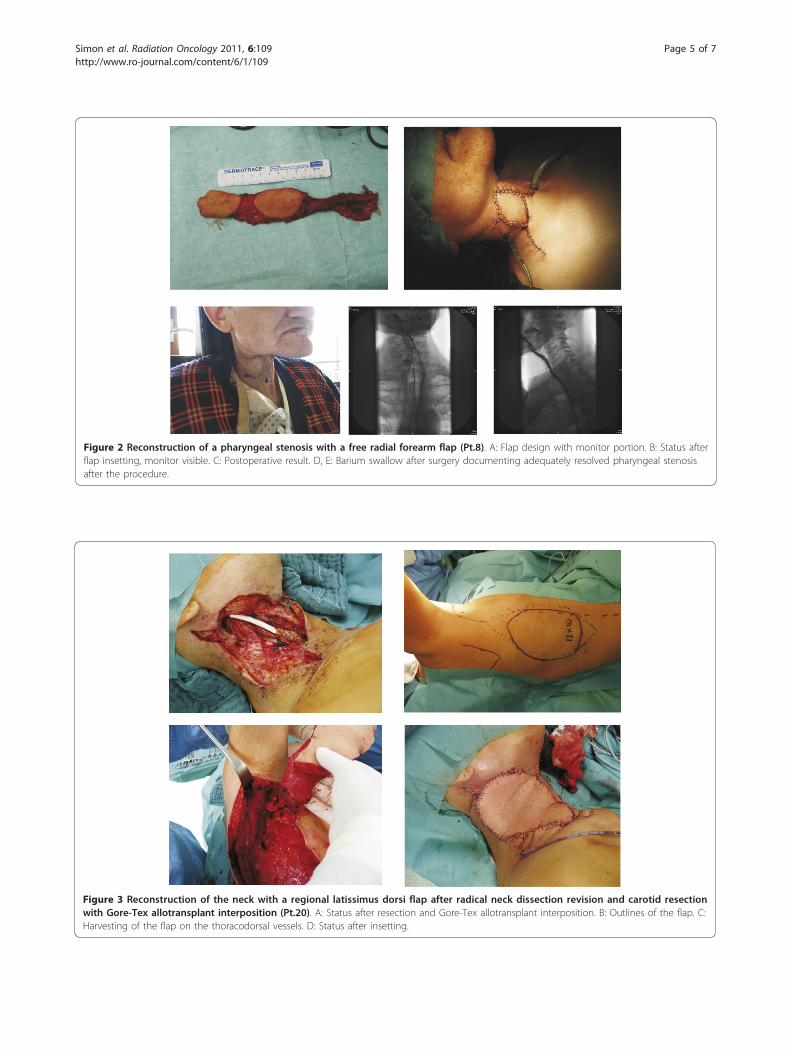
Figure 2 Reconstruction of a pharyngeal stenosis with a free radial forearm flap (Pt.8). A: Flap design with monitor portion. B: Status afterflap insetting, monitor visible. C: Postoperative result. D, E: Barium swallow after surgery documenting adequately resolved pharyngeal stenosisafter the procedure.
Figure 3 Reconstruction of the neck with a regional latissimus dorsi flap after radical neck dissection revision and carotid resectionwith Gore-Tex allotransplant interposition (Pt.20). A: Status after resection and Gore-Tex allotransplant interposition. B: Outlines of the flap. C:Harvesting of the flap on the thoracodorsal vessels. D: Status after insetting.
Simon et al. Radiation Oncology 2011, 6:109http://www.ro-journal.com/content/6/1/109
Page 5 of 7

out of 21 patients (9.5%). This suggests that if propersurgical technique is used, operating in the radiated fieldin the head and neck is likely to be safe. Only 1 patientdeveloped fistula after pharyngeal closure with a regio-nal or free flap. This also compares favorably withpublished data on fistula formation after salvage laryn-gectomy, that are reported to be as high as 18% to 29%[5,6]. This data supports in our opinion the conclusionthat surgery can be safely performed after radiationonce vascularized tissue transfer is used as an adjuncttechnique.It is also noteworthy that we did not encounter any
flap failures with our free flaps despite having to per-form vascular anastomosis in preirradiated and pre-viously operated fields suggesting that free flaps can alsobe safely performed.All recipient site complications occurred in patients
that were treated with RCTX. This finding however wasnot statistically significant. This trend however is consis-tent with published data, indicating a high rate of peri-operative complications after RCTX [2].In order to assess the potential hazard of an extension of
the procedure by a complicated flap surgery on thepatient’s postoperative performance, we measured the pre-and postoperative KPS based on a chart review. It certainlywould have been better to undertake quality-of-life studiesin this patient population. However this was not possibledue to the retrospective nature of the study. Our data
indicate that 62% of our patients had an improved KPS.The preoperative KPS ranged between 40% and 80%, themajority of patients had a KPS between 50% and 70%. AKPS improvement from 50% to 60% indicating to regainthe ability to function mostly independent of help in allkey areas of live, was observed in 8 out of 21 patients(38%). An improvement from a KPS of 60% to 70% wasobserved in 4 out of 21 patients (19%) indicating animprovement towards being entirely independent of helpwithout being able to work. The KPS declined in only onepatient. The data suggest that 3 months after surgerywhen the KPS was assessed the patients had recoveredvery well from surgery despite the extent of the procedure.In summary our data indicate that surgical treatment
of patients after RTX or RCTX is feasible with an accep-table rate of complications, if free or regional flaps areincluded in the operative strategy.
Author details1University of Heidelberg, Department of Otolaryngology - Head and NeckSurgery, Im Neuenheimer Feld 400, 69120 Heidelberg, Germany. 2Universityof Heidelberg, Department of Radiation Oncology, Im Neuenheimer Feld400, 69120 Heidelberg, Germany.
Authors’ contributionsCS designed and coordinated the study, participated in the data acquisitionand analysis, and helped to draft the manuscript, CB participated in the dataacquisition and analysis, MWM and KL both participated in the dataacquisition and analysis, and helped to draft the manuscript, ZB and SS andSL participated in the data acquisition and analysis, PKP and PAF helped to
Figure 4 Reconstruction of the neck after radical extended neck revision with partial pharyngectomy (Pt.4). A, B: Reconstruction of theanterior neck with a deltopectoral flap. C, D: Reconstruction of the residual circumference of the neck with a trapezius flap.
Simon et al. Radiation Oncology 2011, 6:109http://www.ro-journal.com/content/6/1/109
Page 6 of 7
coordinate the study and draft the manuscript. All authors read andapproved the final manuscript.
Competing interestsThe authors declare that they have no competing interests.
Received: 9 March 2011 Accepted: 6 September 2011Published: 6 September 2011
References1. Schultze-Mosgau S, Grabenbauer GG, Radespiel-Troger M, Wiltfang J, Ries J,
Neukam FW, Rodel F: Vascularization in the transition area between freegrafted soft tissues and pre-irradiated graft bed tissues followingpreoperative radiotherapy in the head and neck region. Head Neck 2002,24:42-51.
2. Relic A, Scheich M, Stapf J, Voelter C, Hoppe F, Hagen R, Pfreundner L:Salvage surgery after induction chemotherapy with paclitaxel/cisplatinand primary radiotherapy for advanced laryngeal and hypopharyngealcarcinomas. Eur Arch Otorhinolaryngol 2009, 266:1799-1805.
3. Lin S, Dutra J, Keni J, Dumanian GA, Fine N, Pelzer H: Preoperativeradiation therapy and its effects on outcomes in microsurgical head andneck reconstruction. Otolaryngol Head Neck Surg 2005, 132:845-848.
4. Cohn AB, Lang PO, Agarwal JP, Peng SL, Alizadeh K, Stenson KM, Haraf DJ,Cohen EE, Vokes EE, Gottlieb LJ: Free-flap reconstruction in the doublyirradiated patient population. Plast Reconstr Surg 2008, 122:125-132.
5. Withrow KP, Rosenthal EL, Gourin CG, Peters GE, Magnuson JS, Terris DJ,Carroll WW: Free tissue transfer to manage salvage laryngectomy defectsafter organ preservation failure. Laryngoscope 2007, 117:781-784.
6. Fung K, Teknos TN, Vandenberg CD, Lyden TH, Bradford CR, Hogikyan ND,Kim J, Prince ME, Wolf GT, Chepeha DB: Prevention of woundcomplications following salvage laryngectomy using free vascularizedtissue. Head Neck 2007, 29:425-430.
7. Gil Z, Gupta A, Kummer B, Cordeiro PG, Kraus DH, Shah JP, Patel SG: Therole of pectoralis major muscle flap in salvage total laryngectomy. ArchOtolaryngol Head Neck Surg 2009, 135:1019-1023.
8. Roosli C, Studer G, Stoeckli SJ: Salvage treatment for recurrentoropharyngeal squamous cell carcinoma. Head Neck 32:989-996.
9. Goodwin WJ Jr: Salvage surgery for patients with recurrent squamouscell carcinoma of the upper aerodigestive tract: when do the endsjustify the means? Laryngoscope 2000, 110:1-18.
10. Temam S, Pape E, Janot F, Wibault P, Julieron M, Lusinchi A, Mamelle G,Marandas P, Luboinski B, Bourhis J: Salvage surgery after failure of veryaccelerated radiotherapy in advanced head-and-neck squamous cellcarcinoma. Int J Radiat Oncol Biol Phys 2005, 62:1078-1083.
11. Karnofsky DA, Burchenal JH, Escher GC: Chemotherapy of neoplasticdiseases. Med Clin North Am 1950, 34:439-458, illust.
doi:10.1186/1748-717X-6-109Cite this article as: Simon et al.: Assessment of peri- and postoperativecomplications and Karnofsky-performance status in head and neckcancer patients after radiation or chemoradiation that underwentsurgery with regional or free-flap reconstruction for salvage, palliation,or to improve function. Radiation Oncology 2011 6:109.
Submit your next manuscript to BioMed Centraland take full advantage of:
• Convenient online submission
• Thorough peer review
• No space constraints or color figure charges
• Immediate publication on acceptance
• Inclusion in PubMed, CAS, Scopus and Google Scholar
• Research which is freely available for redistribution
Submit your manuscript at www.biomedcentral.com/submit
Simon et al. Radiation Oncology 2011, 6:109http://www.ro-journal.com/content/6/1/109
Page 7 of 7