Spermidine, but not spermine, is essential for pigment pattern formation in zebrafish · RESEARCH...
9
RESEARCH ARTICLE Spermidine, but not spermine, is essential for pigment pattern formation in zebrafish Hans Georg Frohnho ̈ fer 1 , Silke Geiger-Rudolph 1 , Martin Pattky 2 , Martin Meixner 2 , Carolin Huhn 2 , Hans-Martin Maischein 1, *, Robert Geisler 1, ‡ , Ines Gehring 1,§ , Florian Maderspacher 1,¶ , Christiane Nu ̈ sslein-Volhard 1 and Uwe Irion 1, ** ABSTRACT Polyamines are small poly-cations essential for all cellular life. The main polyamines present in metazoans are putrescine, spermidine and spermine. Their exact functions are still largely unclear; however, they are involved in a wide variety of processes affecting cell growth, proliferation, apoptosis and aging. Here we identify idefix, a mutation in the zebrafish gene encoding the enzyme spermidine synthase, leading to a severe reduction in spermidine levels as shown by capillary electrophoresis-mass spectrometry. We show that spermidine, but not spermine, is essential for early development, organogenesis and colour pattern formation. Whereas in other vertebrates spermidine deficiency leads to very early embryonic lethality, maternally provided spermidine synthase in zebrafish is sufficient to rescue the early developmental defects. This allows us to uncouple them from events occurring later during colour patterning. Factors involved in the cellular interactions essential for colour patterning, likely targets for spermidine, are the gap junction components Cx41.8, Cx39.4, and Kir7.1, an inwardly rectifying potassium channel, all known to be regulated by polyamines. Thus, zebrafish provide a vertebrate model to study the in vivo effects of polyamines. KEY WORDS: Zebrafish, Pigmentation, Pattern formation, Polyamine, Spermidine synthase, Spermine synthase INTRODUCTION One of the most prominent features of adult zebrafish is their pigmentation; the fish display a stereotypical pattern of horizontal dark and light stripes on their flanks and in the anal and caudal fins. Three different types of pigment cells (chromatophores) are responsible for this pattern: melanophores, containing the dark pigment melanin; xanthophores, containing orange pteridine pigments; and iridophores, which contain reflecting guanine platelets (Hirata et al., 2003; Singh and Nüsslein-Volhard, 2015; Irion et al., 2016). All three types of chromatophores are required to establish the pattern. Mutants missing one chromatophore type, nacre (nac) (Lister et al., 1999), pfeffer ( pfe) (Parichy et al., 2000) or shady (shd) (Lopes et al., 2008), where melanophores, xanthophores or iridophores are absent, respectively, only produce a rudimentary pattern (Frohnhöfer et al., 2013). In several other mutants the pattern of the adult fish is affected, but all three types of chromatophores are present in nearly normal numbers. In most of these mutants, e.g. leopard (leo, Cx41.8) (Watanabe et al., 2006), luchs (luc, Cx39.4) (Irion et al., 2014a), schachbrett (sbr, Tjp1a) (Fadeev et al., 2015) and seurat (igsf11) (Eom et al., 2012), the dark stripes are broken up into spots; in a few others, obelix (obe, also known as jaguar, Kir7.1) (Iwashita et al., 2006; Maderspacher and Nüsslein-Volhard, 2003) and asterix (ase) (Haffter et al., 1996), the width of the stripes is affected. In leo and luc the affected genes code for two different connexins, the subunits of gap junctions. Gap junctions allow the molecular and electrical coupling between neighbouring cells via the transfer of small molecules and ions (Kumar and Gilula, 1996). Both leo and luc are required in melanophores and xanthophores (Irion et al., 2014a; Maderspacher and Nüsslein-Volhard, 2003) and it has been suggested that they form heteromeric gap junctions allowing cell-cell communications necessary for normal pattern formation. In obe, which is only required in melanophores (Maderspacher and Nüsslein-Volhard, 2003), the affected gene codes for a K + inwardly rectifying (Kir) channel, kcnj13/Kir7.1. Kir channels play key roles in the maintenance of the resting membrane potential of many cell types and they contribute to the formation of action potentials in excitable cells such as neurons and cardiomyocytes. The properties of both gap junctions and Kir-channels can be regulated by the binding of polyamines to the channel proteins (Musa et al., 2004; Baronas and Kurata, 2014). Recently it was shown that the N-terminus of connexin41.8, which is affected in leo mutants, contains a putative polyamine-binding motif, ExxxE, necessary for the correct pigment pattern formation in vivo (Watanabe et al., 2012); the same motif is also present in Cx39.4, which is encoded by luc (Irion et al., 2014a). Polyamines, mainly putrescine, spermidine and spermine, are important for viability, proliferation and differentiation in all cells tested (Miller-Fleming et al., 2015; Wallace, 2009). They are derived from ornithine via decarboxylation (putrescine) and the addition of one (spermidine) or two (spermine) aminopropyl groups (Pegg, 2009). However, the exact roles polyamines fulfil in the cellular physiologies are still largely unclear. They carry several positive charges already at neutral pH and are thought to stabilize negatively charged molecules in the cell, such as RNA, DNA or membrane phospholipids. Polyamines are known to affect gene expression by regulating transcription or translation of mRNAs (Igarashi and Kashiwagi, 2015; Miller-Fleming et al., 2015). Received 29 March 2016; Accepted 22 April 2016 1 Max-Planck-Institut fu ̈ r Entwicklungsbiologie, Abteilung 3, Spemannstrasse 35, Tu ̈ bingen 72076, Germany. 2 Institut fu ̈ r Physikalische und Theoretische Chemie, Eberhard Karls Universita ̈ t Tu ̈ bingen, Auf der Morgenstelle 18, Tu ̈ bingen 72076, Germany. *Present address: Max-Planck-Institut fu ̈ r Herz- und Lungenforschung, Ludwigstrasse 43, Bad Nauheim 61231, Germany. ‡ Present address: Karlsruhe Institute of Technology (KIT), Hermann-von-Helmholtz-Platz 1, Eggenstein- Leopoldshafen 76344, Germany. § Present address: University of California, Irvine, School of Biological Sciences, Irvine, CA 92697-2300, USA. ¶ Present address: Current Biology, Cell Press, Cambridge, MA 02139, USA. **Author for correspondence ([email protected]) U.I., 0000-0003-2823-5840 This is an Open Access article distributed under the terms of the Creative Commons Attribution License (http://creativecommons.org/licenses/by/3.0), which permits unrestricted use, distribution and reproduction in any medium provided that the original work is properly attributed. 736 © 2016. Published by The Company of Biologists Ltd | Biology Open (2016) 5, 736-744 doi:10.1242/bio.018721 Biology Open by guest on March 25, 2020 http://bio.biologists.org/ Downloaded from
Transcript of Spermidine, but not spermine, is essential for pigment pattern formation in zebrafish · RESEARCH...
RESEARCH ARTICLE
Spermidine, but not spermine, is essential for pigment patternformation in zebrafishHans Georg Frohnhofer1, Silke Geiger-Rudolph1, Martin Pattky2, Martin Meixner2, Carolin Huhn2,Hans-Martin Maischein1,*, Robert Geisler1,‡, Ines Gehring1,§, Florian Maderspacher1,¶,Christiane Nusslein-Volhard1 and Uwe Irion1,**
ABSTRACTPolyamines are small poly-cations essential for all cellular life. Themain polyaminespresent inmetazoansare putrescine, spermidineandspermine. Their exact functions are still largely unclear; however, theyare involved in a wide variety of processes affecting cell growth,proliferation, apoptosis and aging. Herewe identify idefix, a mutation inthe zebrafish geneencoding the enzyme spermidine synthase, leadingto a severe reduction in spermidine levels as shown by capillaryelectrophoresis-mass spectrometry. We show that spermidine, but notspermine, is essential for early development, organogenesis andcolour pattern formation. Whereas in other vertebrates spermidinedeficiency leads to very early embryonic lethality, maternally providedspermidine synthase in zebrafish is sufficient to rescue the earlydevelopmental defects. This allows us to uncouple them from eventsoccurring later during colour patterning. Factors involved in the cellularinteractions essential for colour patterning, likely targets for spermidine,are the gap junction components Cx41.8, Cx39.4, and Kir7.1, aninwardly rectifying potassium channel, all known to be regulated bypolyamines. Thus, zebrafish provide a vertebrate model to study the invivo effects of polyamines.
KEY WORDS: Zebrafish, Pigmentation, Pattern formation,Polyamine, Spermidine synthase, Spermine synthase
INTRODUCTIONOne of the most prominent features of adult zebrafish is theirpigmentation; the fish display a stereotypical pattern of horizontaldark and light stripes on their flanks and in the anal and caudalfins. Three different types of pigment cells (chromatophores) areresponsible for this pattern: melanophores, containing the darkpigment melanin; xanthophores, containing orange pteridinepigments; and iridophores, which contain reflecting guanine
platelets (Hirata et al., 2003; Singh and Nüsslein-Volhard, 2015;Irion et al., 2016). All three types of chromatophores are required toestablish the pattern. Mutants missing one chromatophore type,nacre (nac) (Lister et al., 1999), pfeffer ( pfe) (Parichy et al., 2000) orshady (shd) (Lopes et al., 2008), where melanophores,xanthophores or iridophores are absent, respectively, only producea rudimentary pattern (Frohnhöfer et al., 2013). In several othermutants the pattern of the adult fish is affected, but all three types ofchromatophores are present in nearly normal numbers. In most ofthese mutants, e.g. leopard (leo, Cx41.8) (Watanabe et al., 2006),luchs (luc, Cx39.4) (Irion et al., 2014a), schachbrett (sbr, Tjp1a)(Fadeev et al., 2015) and seurat (igsf11) (Eom et al., 2012), the darkstripes are broken up into spots; in a few others, obelix (obe, alsoknown as jaguar, Kir7.1) (Iwashita et al., 2006; Maderspacher andNüsslein-Volhard, 2003) and asterix (ase) (Haffter et al., 1996), thewidth of the stripes is affected. In leo and luc the affected genes codefor two different connexins, the subunits of gap junctions. Gapjunctions allow the molecular and electrical coupling betweenneighbouring cells via the transfer of small molecules and ions(Kumar and Gilula, 1996). Both leo and luc are required inmelanophores and xanthophores (Irion et al., 2014a; Maderspacherand Nüsslein-Volhard, 2003) and it has been suggested that theyform heteromeric gap junctions allowing cell-cell communicationsnecessary for normal pattern formation. In obe, which is onlyrequired in melanophores (Maderspacher and Nüsslein-Volhard,2003), the affected gene codes for a K+ inwardly rectifying (Kir)channel, kcnj13/Kir7.1. Kir channels play key roles in themaintenance of the resting membrane potential of many cell typesand they contribute to the formation of action potentials in excitablecells such as neurons and cardiomyocytes. The properties of bothgap junctions and Kir-channels can be regulated by the binding ofpolyamines to the channel proteins (Musa et al., 2004; Baronas andKurata, 2014). Recently it was shown that the N-terminus ofconnexin41.8, which is affected in leo mutants, contains a putativepolyamine-binding motif, ExxxE, necessary for the correct pigmentpattern formation in vivo (Watanabe et al., 2012); the same motif isalso present in Cx39.4, which is encoded by luc (Irion et al., 2014a).
Polyamines, mainly putrescine, spermidine and spermine, areimportant for viability, proliferation and differentiation in all cellstested (Miller-Fleming et al., 2015; Wallace, 2009). They arederived from ornithine via decarboxylation (putrescine) and theaddition of one (spermidine) or two (spermine) aminopropyl groups(Pegg, 2009). However, the exact roles polyamines fulfil in thecellular physiologies are still largely unclear. They carry severalpositive charges already at neutral pH and are thought to stabilizenegatively charged molecules in the cell, such as RNA, DNA ormembrane phospholipids. Polyamines are known to affect geneexpression by regulating transcription or translation of mRNAs(Igarashi and Kashiwagi, 2015; Miller-Fleming et al., 2015).Received 29 March 2016; Accepted 22 April 2016
1Max-Planck-Institut fur Entwicklungsbiologie, Abteilung 3, Spemannstrasse 35,Tubingen 72076, Germany. 2Institut fur Physikalische und Theoretische Chemie,Eberhard Karls Universitat Tubingen, Auf der Morgenstelle 18, Tubingen 72076,Germany.*Present address: Max-Planck-Institut fur Herz- und Lungenforschung,Ludwigstrasse 43, Bad Nauheim 61231, Germany. ‡Present address: KarlsruheInstitute of Technology (KIT), Hermann-von-Helmholtz-Platz 1, Eggenstein-Leopoldshafen 76344, Germany. §Present address: University of California,Irvine, School of Biological Sciences, Irvine, CA 92697-2300, USA. ¶Presentaddress: Current Biology, Cell Press, Cambridge, MA 02139, USA.
**Author for correspondence ([email protected])
U.I., 0000-0003-2823-5840
This is an Open Access article distributed under the terms of the Creative Commons AttributionLicense (http://creativecommons.org/licenses/by/3.0), which permits unrestricted use,distribution and reproduction in any medium provided that the original work is properly attributed.
736
© 2016. Published by The Company of Biologists Ltd | Biology Open (2016) 5, 736-744 doi:10.1242/bio.018721
BiologyOpen
by guest on March 25, 2020http://bio.biologists.org/Downloaded from
Reduced polyamine levels have been associated with cellularsenescence and the application of spermidine increases the life spanin yeast, flies and human cells (Eisenberg et al., 2009). In manycancers the levels of polyamines are elevated (Thomas and Thomas,2003). One essential and well-understood function of spermidine inall eukaryotes is its requirement as a substrate for the post-translational modification of a specific lysine residue in eIF5A,leading to the unusual amino acid hypusine [N(ε)-(4-amino-2-hydroxybutyl)-lysine]. Both, eIF5A and the enzymes required forhypusination, are essential genes in mouse, underscoring theimportance also of spermidine (Nishimura et al., 2012).Polyamines are also involved in the regulation of several classesof ion channels, e.g. K+ inward-rectifier channels, ionotropicglutamate receptors and gap junctions. Binding of polyamines isnecessary for the rectification properties of these channels, allowingions to pass through the pore only in one direction (Donevan andRogawski, 1995; Lu, 2004; Musa and Veenstra, 2003; Williams,1997).Here we describe a novel zebrafish mutant, idefix (ide). We show
that idemutants carry a premature stop codon in the gene coding forspermidine synthase, the enzyme responsible for the synthesis ofspermidine from putrescine and S-adenosyl-methioninamine. Thisis the first instance where the loss of spermidine synthase isdescribed in a vertebrate. Surprisingly, the homozygous mutants areviable, but show a maternal-effect lethal phenotype and a verystriking aberration from the wild-type stripe pattern. In agreementwith a recent report (Mastracci et al., 2015) we show thatthe maternal depletion of the enzyme leads to defects in thedevelopment of the pancreas. In the homozygous mutants thepigmentation pattern displays wider light areas and narrower andfewer dark stripes, which show frequent interruptions. The sharp
boundaries between the stripes are not affected. We demonstrate thatthe phenotype is caused by a lack of spermidine and not spermine,as mutations in the gene coding for spermine synthase cause novisible phenotype. To determine the polyamine levels in earlyembryos we established a protocol for separation and detectionbased on capillary electrophoresis coupled to mass spectrometry(CE-MS), which allowed the sensitive and reliable relativequantification of putrescine, spermidine and spermine ratios. Wefound that, as predicted, putrescine accumulates in ide mutants andthe levels of spermidine are greatly reduced. A loss-of-functionmutation in the gene coding for spermine synthase leads to very lowspermine levels (below the detection limit of the method), however,the fish are viable and fertile and show no visible phenotype. Thisdemonstrates that in vivo spermidine has an essential function,whereas spermine does not.
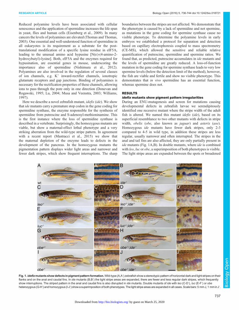
RESULTSidefix mutants show pigment pattern irregularitiesDuring an ENU-mutagenesis and screen for mutations causingdevelopmental defects in zebrafish larvae we serendipitouslyidentified one recessive mutant where the stripe width of the adultfish is altered. We named this mutant idefix (ide), based on itssuperficial resemblance to two other mutants with defects in stripewidth, obelix (obe, also known as jaguar) and asterix (ase).Homozygous ide mutants have fewer dark stripes, only 2-3compared to 4-5 in wild type, in addition these stripes are lessregular, usually narrower and often interrupted. The stripes in theanal and tail fins are also affected, they are only partially present inide mutants (Fig. 1A,B). In double mutants, where ide is combinedwith leo, luc or obe, a superimposition of both phenotypes is visible.The light stripe areas are expanded between the spots or broadened
Fig. 1. idefixmutantsshowdefects inpigmentpattern formation.Wild-type (A,A′) zebrafishshowastereotypic pattern of horizontal darkand light stripes on theirflanks and on the anal and caudal fins. In ide mutants (B,B′) the light stripe areas are expanded, there are fewer and less regular dark stripes, which frequentlyshow interruptions. The striped pattern in the anal and caudal fins is also disrupted in idemutants. Double mutants of ide with leo (C-D′), luc (E-F′) or obeheterozygous (G-H′) andhomozygous (I-J′) showasuperimpositionof bothphenotypes. The light stripeareasareexpanded inall cases.Scalebars: 5 mmJ,1 mminJ′.
737
RESEARCH ARTICLE Biology Open (2016) 5, 736-744 doi:10.1242/bio.018721
BiologyOpen
by guest on March 25, 2020http://bio.biologists.org/Downloaded from
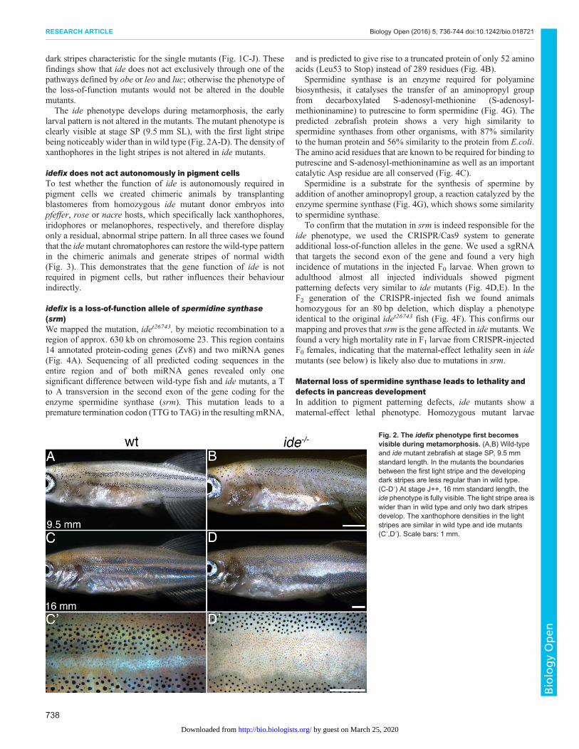
dark stripes characteristic for the single mutants (Fig. 1C-J). Thesefindings show that ide does not act exclusively through one of thepathways defined by obe or leo and luc; otherwise the phenotype ofthe loss-of-function mutants would not be altered in the doublemutants.The ide phenotype develops during metamorphosis, the early
larval pattern is not altered in the mutants. The mutant phenotype isclearly visible at stage SP (9.5 mm SL), with the first light stripebeing noticeably wider than in wild type (Fig. 2A-D). The density ofxanthophores in the light stripes is not altered in ide mutants.
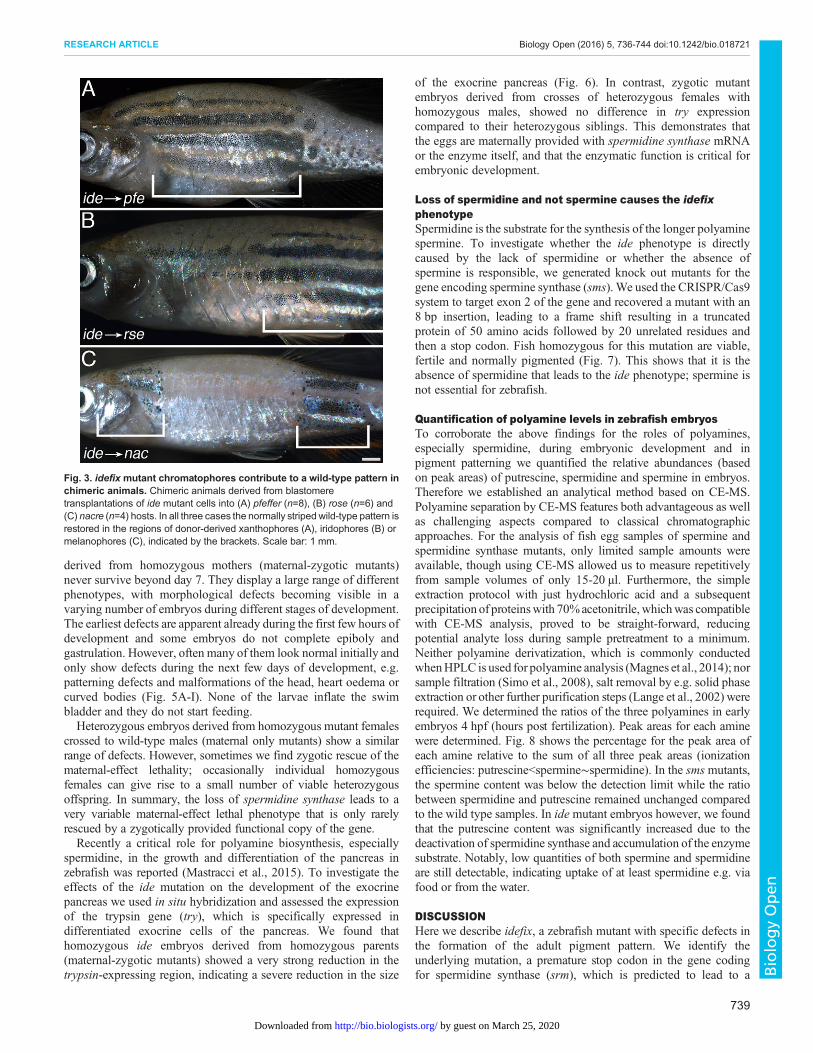
idefix does not act autonomously in pigment cellsTo test whether the function of ide is autonomously required inpigment cells we created chimeric animals by transplantingblastomeres from homozygous ide mutant donor embryos intopfeffer, rose or nacre hosts, which specifically lack xanthophores,iridophores or melanophores, respectively, and therefore displayonly a residual, abnormal stripe pattern. In all three cases we foundthat the idemutant chromatophores can restore the wild-type patternin the chimeric animals and generate stripes of normal width(Fig. 3). This demonstrates that the gene function of ide is notrequired in pigment cells, but rather influences their behaviourindirectly.
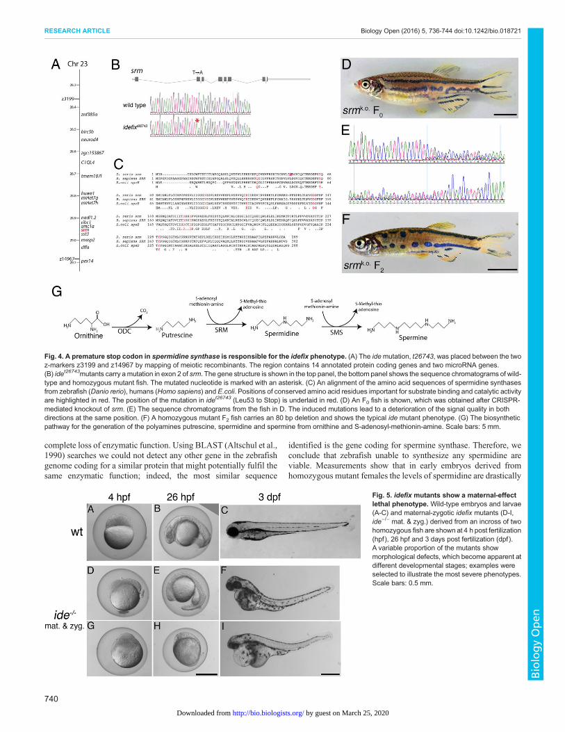
idefix is a loss-of-function allele of spermidine synthase(srm)We mapped the mutation, idet26743, by meiotic recombination to aregion of approx. 630 kb on chromosome 23. This region contains14 annotated protein-coding genes (Zv8) and two miRNA genes(Fig. 4A). Sequencing of all predicted coding sequences in theentire region and of both miRNA genes revealed only onesignificant difference between wild-type fish and ide mutants, a Tto A transversion in the second exon of the gene coding for theenzyme spermidine synthase (srm). This mutation leads to apremature termination codon (TTG to TAG) in the resulting mRNA,
and is predicted to give rise to a truncated protein of only 52 aminoacids (Leu53 to Stop) instead of 289 residues (Fig. 4B).
Spermidine synthase is an enzyme required for polyaminebiosynthesis, it catalyses the transfer of an aminopropyl groupfrom decarboxylated S-adenosyl-methionine (S-adenosyl-methioninamine) to putrescine to form spermidine (Fig. 4G). Thepredicted zebrafish protein shows a very high similarity tospermidine synthases from other organisms, with 87% similarityto the human protein and 56% similarity to the protein from E.coli.The amino acid residues that are known to be required for binding toputrescine and S-adenosyl-methioninamine as well as an importantcatalytic Asp residue are all conserved (Fig. 4C).
Spermidine is a substrate for the synthesis of spermine byaddition of another aminopropyl group, a reaction catalyzed by theenzyme spermine synthase (Fig. 4G), which shows some similarityto spermidine synthase.
To confirm that the mutation in srm is indeed responsible for theide phenotype, we used the CRISPR/Cas9 system to generateadditional loss-of-function alleles in the gene. We used a sgRNAthat targets the second exon of the gene and found a very highincidence of mutations in the injected F0 larvae. When grown toadulthood almost all injected individuals showed pigmentpatterning defects very similar to ide mutants (Fig. 4D,E). In theF2 generation of the CRISPR-injected fish we found animalshomozygous for an 80 bp deletion, which display a phenotypeidentical to the original idet26743 fish (Fig. 4F). This confirms ourmapping and proves that srm is the gene affected in idemutants. Wefound a very high mortality rate in F1 larvae from CRISPR-injectedF0 females, indicating that the maternal-effect lethality seen in idemutants (see below) is likely also due to mutations in srm.
Maternal loss of spermidine synthase leads to lethality anddefects in pancreas developmentIn addition to pigment patterning defects, ide mutants show amaternal-effect lethal phenotype. Homozygous mutant larvae
Fig. 2. The idefix phenotype first becomesvisible during metamorphosis. (A,B) Wild-typeand ide mutant zebrafish at stage SP, 9.5 mmstandard length. In the mutants the boundariesbetween the first light stripe and the developingdark stripes are less regular than in wild type.(C-D′) At stage J++, 16 mm standard length, theide phenotype is fully visible. The light stripe area iswider than in wild type and only two dark stripesdevelop. The xanthophore densities in the lightstripes are similar in wild type and ide mutants(C′,D′). Scale bars: 1 mm.
738
RESEARCH ARTICLE Biology Open (2016) 5, 736-744 doi:10.1242/bio.018721
BiologyOpen
by guest on March 25, 2020http://bio.biologists.org/Downloaded from
derived from homozygous mothers (maternal-zygotic mutants)never survive beyond day 7. They display a large range of differentphenotypes, with morphological defects becoming visible in avarying number of embryos during different stages of development.The earliest defects are apparent already during the first few hours ofdevelopment and some embryos do not complete epiboly andgastrulation. However, often many of them look normal initially andonly show defects during the next few days of development, e.g.patterning defects and malformations of the head, heart oedema orcurved bodies (Fig. 5A-I). None of the larvae inflate the swimbladder and they do not start feeding.Heterozygous embryos derived from homozygous mutant females
crossed to wild-type males (maternal only mutants) show a similarrange of defects. However, sometimes we find zygotic rescue of thematernal-effect lethality; occasionally individual homozygousfemales can give rise to a small number of viable heterozygousoffspring. In summary, the loss of spermidine synthase leads to avery variable maternal-effect lethal phenotype that is only rarelyrescued by a zygotically provided functional copy of the gene.Recently a critical role for polyamine biosynthesis, especially
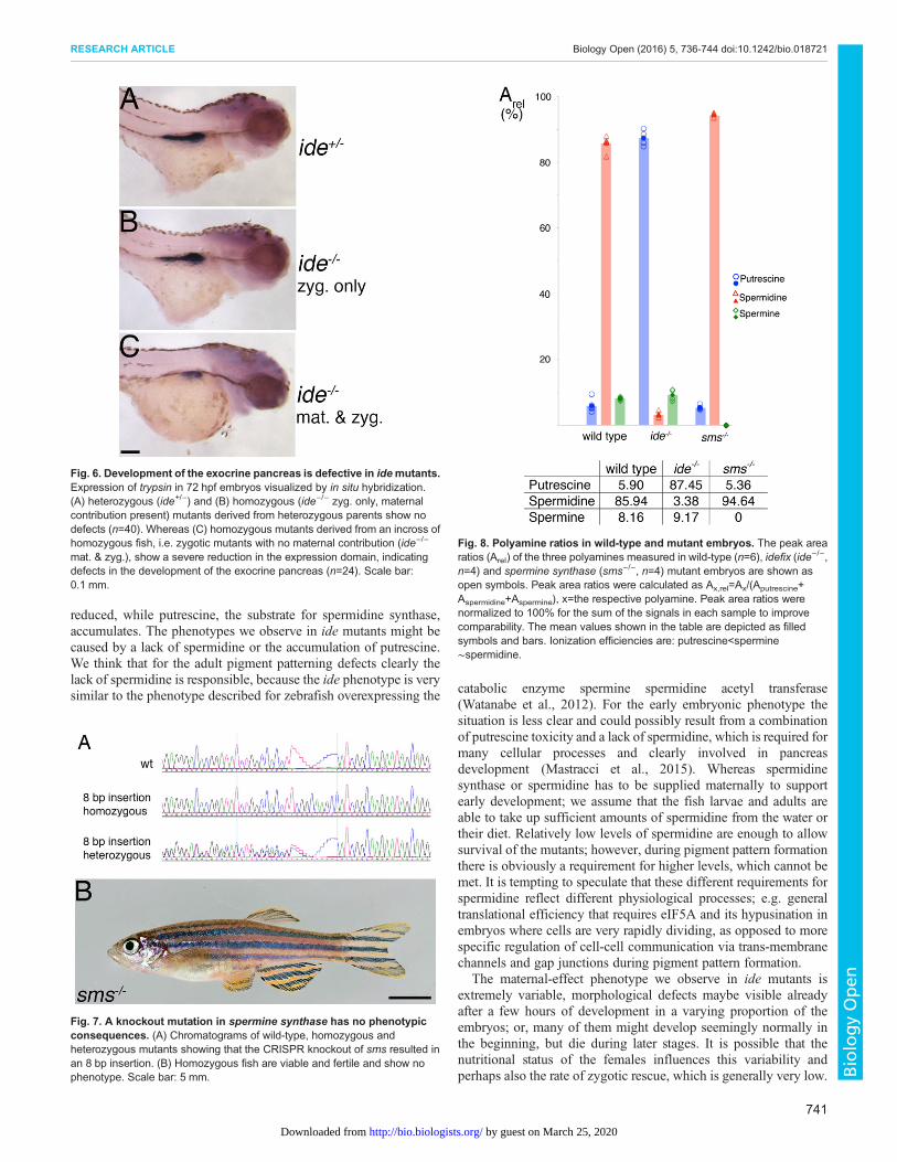
spermidine, in the growth and differentiation of the pancreas inzebrafish was reported (Mastracci et al., 2015). To investigate theeffects of the ide mutation on the development of the exocrinepancreas we used in situ hybridization and assessed the expressionof the trypsin gene (try), which is specifically expressed indifferentiated exocrine cells of the pancreas. We found thathomozygous ide embryos derived from homozygous parents(maternal-zygotic mutants) showed a very strong reduction in thetrypsin-expressing region, indicating a severe reduction in the size
of the exocrine pancreas (Fig. 6). In contrast, zygotic mutantembryos derived from crosses of heterozygous females withhomozygous males, showed no difference in try expressioncompared to their heterozygous siblings. This demonstrates thatthe eggs are maternally provided with spermidine synthase mRNAor the enzyme itself, and that the enzymatic function is critical forembryonic development.
Loss of spermidine and not spermine causes the idefixphenotypeSpermidine is the substrate for the synthesis of the longer polyaminespermine. To investigate whether the ide phenotype is directlycaused by the lack of spermidine or whether the absence ofspermine is responsible, we generated knock out mutants for thegene encoding spermine synthase (sms). We used the CRISPR/Cas9system to target exon 2 of the gene and recovered a mutant with an8 bp insertion, leading to a frame shift resulting in a truncatedprotein of 50 amino acids followed by 20 unrelated residues andthen a stop codon. Fish homozygous for this mutation are viable,fertile and normally pigmented (Fig. 7). This shows that it is theabsence of spermidine that leads to the ide phenotype; spermine isnot essential for zebrafish.
Quantification of polyamine levels in zebrafish embryosTo corroborate the above findings for the roles of polyamines,especially spermidine, during embryonic development and inpigment patterning we quantified the relative abundances (basedon peak areas) of putrescine, spermidine and spermine in embryos.Therefore we established an analytical method based on CE-MS.Polyamine separation by CE-MS features both advantageous as wellas challenging aspects compared to classical chromatographicapproaches. For the analysis of fish egg samples of spermine andspermidine synthase mutants, only limited sample amounts wereavailable, though using CE-MS allowed us to measure repetitivelyfrom sample volumes of only 15-20 µl. Furthermore, the simpleextraction protocol with just hydrochloric acid and a subsequentprecipitation of proteinswith 70%acetonitrile, whichwas compatiblewith CE-MS analysis, proved to be straight-forward, reducingpotential analyte loss during sample pretreatment to a minimum.Neither polyamine derivatization, which is commonly conductedwhenHPLC is used for polyamine analysis (Magnes et al., 2014); norsample filtration (Simo et al., 2008), salt removal by e.g. solid phaseextraction or other further purification steps (Lange et al., 2002) wererequired. We determined the ratios of the three polyamines in earlyembryos 4 hpf (hours post fertilization). Peak areas for each aminewere determined. Fig. 8 shows the percentage for the peak area ofeach amine relative to the sum of all three peak areas (ionizationefficiencies: putrescine<spermine∼spermidine). In the smsmutants,the spermine content was below the detection limit while the ratiobetween spermidine and putrescine remained unchanged comparedto the wild type samples. In ide mutant embryos however, we foundthat the putrescine content was significantly increased due to thedeactivation of spermidine synthase and accumulation of the enzymesubstrate. Notably, low quantities of both spermine and spermidineare still detectable, indicating uptake of at least spermidine e.g. viafood or from the water.
DISCUSSIONHere we describe idefix, a zebrafish mutant with specific defects inthe formation of the adult pigment pattern. We identify theunderlying mutation, a premature stop codon in the gene codingfor spermidine synthase (srm), which is predicted to lead to a
Fig. 3. idefix mutant chromatophores contribute to a wild-type pattern inchimeric animals. Chimeric animals derived from blastomeretransplantations of ide mutant cells into (A) pfeffer (n=8), (B) rose (n=6) and(C) nacre (n=4) hosts. In all three cases the normally striped wild-type pattern isrestored in the regions of donor-derived xanthophores (A), iridophores (B) ormelanophores (C), indicated by the brackets. Scale bar: 1 mm.
739
RESEARCH ARTICLE Biology Open (2016) 5, 736-744 doi:10.1242/bio.018721
BiologyOpen
by guest on March 25, 2020http://bio.biologists.org/Downloaded from
complete loss of enzymatic function. Using BLAST (Altschul et al.,1990) searches we could not detect any other gene in the zebrafishgenome coding for a similar protein that might potentially fulfil thesame enzymatic function; indeed, the most similar sequence
identified is the gene coding for spermine synthase. Therefore, weconclude that zebrafish unable to synthesize any spermidine areviable. Measurements show that in early embryos derived fromhomozygous mutant females the levels of spermidine are drastically
Fig. 4. A premature stop codon in spermidine synthase is responsible for the idefix phenotype. (A) The idemutation, t26743, was placed between the twoz-markers z3199 and z14967 by mapping of meiotic recombinants. The region contains 14 annotated protein coding genes and two microRNA genes.(B) idet26743mutants carry amutation in exon 2 of srm. The gene structure is shown in the top panel, the bottom panel shows the sequence chromatograms of wild-type and homozygous mutant fish. The mutated nucleotide is marked with an asterisk. (C) An alignment of the amino acid sequences of spermidine synthasesfrom zebrafish (Danio rerio), humans (Homo sapiens) and E.coli. Positions of conserved amino acid residues important for substrate binding and catalytic activityare highlighted in red. The position of the mutation in idet26743 (Leu53 to Stop) is underlaid in red. (D) An F0 fish is shown, which was obtained after CRISPR-mediated knockout of srm. (E) The sequence chromatograms from the fish in D. The induced mutations lead to a deterioration of the signal quality in bothdirections at the same position. (F) A homozygous mutant F2 fish carries an 80 bp deletion and shows the typical ide mutant phenotype. (G) The biosyntheticpathway for the generation of the polyamines putrescine, spermidine and spermine from ornithine and S-adenosyl-methionin-amine. Scale bars: 5 mm.
Fig. 5. idefix mutants show a maternal-effectlethal phenotype. Wild-type embryos and larvae(A-C) and maternal-zygotic idefix mutants (D-I,ide−/− mat. & zyg.) derived from an incross of twohomozygous fish are shown at 4 h post fertilization(hpf), 26 hpf and 3 days post fertilization (dpf).A variable proportion of the mutants showmorphological defects, which become apparent atdifferent developmental stages; examples wereselected to illustrate the most severe phenotypes.Scale bars: 0.5 mm.
740
RESEARCH ARTICLE Biology Open (2016) 5, 736-744 doi:10.1242/bio.018721
BiologyOpen
by guest on March 25, 2020http://bio.biologists.org/Downloaded from
reduced, while putrescine, the substrate for spermidine synthase,accumulates. The phenotypes we observe in ide mutants might becaused by a lack of spermidine or the accumulation of putrescine.We think that for the adult pigment patterning defects clearly thelack of spermidine is responsible, because the ide phenotype is verysimilar to the phenotype described for zebrafish overexpressing the catabolic enzyme spermine spermidine acetyl transferase
(Watanabe et al., 2012). For the early embryonic phenotype thesituation is less clear and could possibly result from a combinationof putrescine toxicity and a lack of spermidine, which is required formany cellular processes and clearly involved in pancreasdevelopment (Mastracci et al., 2015). Whereas spermidinesynthase or spermidine has to be supplied maternally to supportearly development; we assume that the fish larvae and adults areable to take up sufficient amounts of spermidine from the water ortheir diet. Relatively low levels of spermidine are enough to allowsurvival of the mutants; however, during pigment pattern formationthere is obviously a requirement for higher levels, which cannot bemet. It is tempting to speculate that these different requirements forspermidine reflect different physiological processes; e.g. generaltranslational efficiency that requires eIF5A and its hypusination inembryos where cells are very rapidly dividing, as opposed to morespecific regulation of cell-cell communication via trans-membranechannels and gap junctions during pigment pattern formation.
The maternal-effect phenotype we observe in ide mutants isextremely variable, morphological defects maybe visible alreadyafter a few hours of development in a varying proportion of theembryos; or, many of them might develop seemingly normally inthe beginning, but die during later stages. It is possible that thenutritional status of the females influences this variability andperhaps also the rate of zygotic rescue, which is generally very low.
Fig. 6. Development of the exocrine pancreas is defective in idemutants.Expression of trypsin in 72 hpf embryos visualized by in situ hybridization.(A) heterozygous (ide+/−) and (B) homozygous (ide−/− zyg. only, maternalcontribution present) mutants derived from heterozygous parents show nodefects (n=40). Whereas (C) homozygous mutants derived from an incross ofhomozygous fish, i.e. zygotic mutants with no maternal contribution (ide−/−
mat. & zyg.), show a severe reduction in the expression domain, indicatingdefects in the development of the exocrine pancreas (n=24). Scale bar:0.1 mm.
Fig. 7. A knockout mutation in spermine synthase has no phenotypicconsequences. (A) Chromatograms of wild-type, homozygous andheterozygous mutants showing that the CRISPR knockout of sms resulted inan 8 bp insertion. (B) Homozygous fish are viable and fertile and show nophenotype. Scale bar: 5 mm.
Fig. 8. Polyamine ratios in wild-type and mutant embryos. The peak arearatios (Arel) of the three polyamines measured in wild-type (n=6), idefix (ide−/−,n=4) and spermine synthase (sms−/−, n=4) mutant embryos are shown asopen symbols. Peak area ratios were calculated as Ax,rel=Ax/(Aputrescine+Aspermidine+Aspermine), x=the respective polyamine. Peak area ratios werenormalized to 100% for the sum of the signals in each sample to improvecomparability. The mean values shown in the table are depicted as filledsymbols and bars. Ionization efficiencies are: putrescine<spermine∼spermidine.
741
RESEARCH ARTICLE Biology Open (2016) 5, 736-744 doi:10.1242/bio.018721
BiologyOpen
by guest on March 25, 2020http://bio.biologists.org/Downloaded from
To determine the relative ratios of the three different polyaminesin early embryos we used CE-MS. The most prominent challenge inpolyamine analysis is related to the analytes’ extreme charge-to-sizeratio and thus their very high effective electrophoretic mobility evenat relatively high pH. In fact, the polyamines migrated in betweenmatrix potassium and sodium ions (visible via adduct formation onthe internal reference masses). On the one hand, this extrememobility gives rise to a high selectivity of the method (hardly anyother organic ions can be expected to co-migrate), but on the otherhand, a good separation from potassium and sodium ions had to beachieved to ensure reliable quantitative data. Sufficient resolutionwas reached using long capillaries of 100 cm. Even thoughmigration time was significantly increased compared to 65 cmcapillaries (data not shown), analytes were still detected within lessthan 10 min in neutrally coated capillaries. The analysis ofpolyamines using bare fused silica capillaries was reported (Simoet al., 2008). However, one has to keep in mind that polyamineshave also been used as dynamic coating agents in the backgroundelectrolyte in CE analysis to prevent adsorption of analytes, whichcontradicts the use of bare fused silica capillaries (Eckhardt et al.,2004). In this study, we thus decided to use neutrally AAEE(N-acryloylamido-ethoxyethanol)-coated capillaries to ensurestable separation conditions with reduced analyte but also matrixcomponent adsorption. We found a strong accumulation ofputrescine combined with very low levels of spermidine in idemutants, suggesting that there is some uptake of spermidine via thefood or water. In sms mutants spermine levels are below thedetection limit, whereas the ratio of putrescine and spermidine areunaltered compared with wild type. These data are in goodagreement with the notion that polyamine biosynthesis is mainlyregulated through ornithine decarboxylase (ODC) and S-adenosyl-methionine decarboxylase (AdoMetDC), two enzymes that actupstream of spermidine synthase.The formation of the striped pattern in zebrafish depends on
direct cellular interactions between the different types ofchromatophores that lead to changes in cell shape and behaviourin iridophores (Singh et al., 2014) and xanthophores (Mahalwaret al., 2014). The channels known to mediate some of thesecellular interactions are regulated by polyamines. Through a seriesof transgenes and rescue experiments it was concluded that thepolyamine sensitivity of gap junctions formed by Cx41.8 isrequired for the formation of the striped pattern in zebrafish(Watanabe et al., 2012). The second connexin, Cx39.4, also hasthe characteristic ExxxE motif, a predicted polyamine-bindingsite, in its cytoplasmic N-terminus (Irion et al., 2014a; Watanabeet al., 2015). In addition, rectification of Kir7.1, which is encodedby the obe gene, is induced by intracellular binding of Mg2+ orpolyamines to the channel (Hibino et al., 2010). However, whetherthe ide phenotype is produced via mis-regulation of theheteromeric gap junctions formed by Cx41.8 and Cx39.4, or ofthe Kir7.1 channel is difficult to say because it is expected that thelack of spermidine leads to a phenotype different from the onecaused by loss-of-function of the channels. The phenotypicanalysis of ide double mutants with leo, luc or obe suggests that idedoesn’t exclusively affect one of these channels, but possibly boththe gap junctions and potassium channels, or even some other asyet unknown channel. Whereas leo, luc and obe only affectmelanophores and xanthophores, an important function ofiridophores in pigment patterning has been described recently(Frohnhöfer et al., 2013; Patterson and Parichy, 2013). During theformation of the pattern iridophores proliferate and migrate in theskin and change from a dense form present in the light stripes to a
lose form in the dark stripe regions (Singh et al., 2014). The idephenotype, with the substantial broadening of the light stripe areain the mutants, could be due to a change in the behaviour of theiridophores. It was also speculated that additional, so farunidentified, connexins might act in iridophores together withTjp1A (Fadeev et al., 2015). While the two connexins known tofunction in melanophores and xanthophores contain spermidine-binding motifs, connexins in iridophores might belong to adifferent class and lead to the formation of rectifying heterotypicgap junctions with the connexins in xanthophores ormelanophores. If Cx41.8 is expressed only in melanophores, butnot in xanthophores (Watanabe et al., 2012; Watanabe and Kondo,2012), a phenotype very similar to ide is produced. Thus, the lossof Cx41.8 function in xanthophores has similar consequences asthe loss of spermidine, arguing for a function of heterotypicpolarized gap junctions in the patterning process.
Blastomere transplantations showed that the product of the idegene, the enzyme spermidine synthase, is not required in thechromatophores themselves. We assume that spermidine isproduced in other tissues and then imported into the pigment cellsto function there in the cytoplasm. This makes ide different fromother genes, like bonaparte or karneol, which also affect theformation of the striped pattern indirectly. In the case of bonaparte,which codes for basonuclin 2, it was speculated that it is required forthe local production of a survival factor chromatophores in the skin(Lang et al., 2009). karneol, which codes for endothelin-convertingenzyme 2, is necessary for the local processing of endothelinligands, which in turn influence iridophore development andsurvival (Krauss et al., 2014). ide, on the other hand, is less likely toinfluence the behaviour of the pigment cells by changes in the localtissue environment, but rather more systemically by providingenough spermidine for the regulation of cell-cell communications.
In contrast to ide, there is no obvious phenotype in sperminesynthase mutants. Fish homozygous for a loss-of-function allelegenerated with the CRISPR/Cas9 system are viable and fertile andshow no defects in their pigment pattern. We could not detectspermine in embryos from these mutants; however, putrescine andspermidine are present in a normal ratio. These findings underscorethe notion that in vivo spermidine is more important than spermine.Similar results have been found in yeast and higher animals(Hamasaki-Katagiri et al., 1998; Pegg and Michael, 2010). Cellularpolyamine levels are regulated at the level of synthesis, degradationor transport. Regulation of synthesis occurs mainly throughornithine decarboxylase (ODC) and S-adenosyl-methioninedecarboxylase (AdoMetDC), the two rate-limiting enzymes. Sofar, mutations in spermidine synthase have not been described inmetazoans, presumably due to early lethality. In mouse, the knockout of Ornithine Decarboxylase leads to early embryonic lethality(Pendeville et al., 2001); however mutations in spermine synthasehave been identified in mouse and humans. Mutations in sperminesynthase lead to the Gyro phenotype in mouse (Meyer et al., 1998)and cause Snyder–Robinson syndrome in humans, an X-linkedmental retardation condition characterized by intellectual disability,hypotonia, facial asymmetry and unsteady gait (Cason et al., 2003).
The mutation in spermidine synthase we describe here providesevidence that spermidine, in addition to a more general role intranslational regulation, mediated via eIF5A, is also likely to bedirectly required in vivo for the regulation of cell-cellcommunications via gap junctions and Kir channels. This processseems to be particularly sensitive to diminished spermidine levelsand might provide an opportunity to further investigate polyaminemetabolism and transport.
742
RESEARCH ARTICLE Biology Open (2016) 5, 736-744 doi:10.1242/bio.018721
BiologyOpen
by guest on March 25, 2020http://bio.biologists.org/Downloaded from
MATERIALS AND METHODSFish husbandryZebrafish were maintained as described earlier (Brand et al., 2002); we usedTU as wild type and the following mutants: idefixt26743, obelixtxg6 (Irionet al., 2014a), asterixts212, leot1 (Watanabe et al., 2006). Staging of juvenileswas done according to Parichy et al., (2009). For photography fish wereanaesthetized in 0.004% MS-222 (Sigma) and imaged with a CanonD5MarkII/Macro 100. For Figs 2 and 3 fish were fixed with 4%formaldehyde/0.08% glutaraldehyd (Sigma) and photographed under aLeica MZ1 stereomicroscope. Images were processed in Adobe Photoshop.All animal experiments were carried out in accordance with the guidelinesof the Max-Planck-Society and approved by the RegierungspräsidiumTübingen, Baden-Württemberg, Germany (Aktenzeichen: 35/9185.81-5;35/9185.46-5 and 35/9185.82-7).
TransplantationsChimeric animals were generated by blastomere transplantations as describedpreviously (Kane and Kishimoto, 2002). Donor embryos were homozygousfor idet26743, hosts were nacrew2 (Lister et al., 1999), pfeffertm236b (Haffteret al., 1996) and rosetlf802 (Frohnhöfer et al., 2013).
Genetic mappingGenetic linkage was determined as described previously (Geisler et al.,2007). For the sequencing of the coding regions total RNA was extractedfrom fin clips of homozygous mutant fish using TRIzol Reagent(Invitrogen) according to the manufacturer’s protocol. To generate cDNAfrom this RNA SuperscriptII (Invitrogen) was used. For PCR andsequencing the oligonucleotide primers listed in Table S1 were used.
The microRNA genes (miR-let-7g, miR-let-7h) were amplified by PCRfrom genomic DNA and sequenced with the following oligonucleotideprimers: 5′-CTGGGATTGGAACAGTCAATGG-3′, 5′-TGAGAGTCAG-GATTGGAGTCGG-3′.
RNA in situ hybridizationA 532 bp fragment of the try coding sequence was amplified by PCR fromcDNAwith the following primers: 5′-TGAACAGCGGCTACCACTTC-3′,5′-TAACCCCAGGACACGATACC-3′, and cloned into pGEM-Teasy(Promega). The resulting plasmid was linearized with SacI and used astemplate to generate a DIG-labelled probe by in vitro transcription with T7RNA polymerase using the DIG-RNA labelling Mix (Roche).Morphologically normal looking embryos were collected and fixed at72 hpf, the in situ hybridization was carried out according to standardprocedures (Thisse and Thisse, 2008). The stained embryos were mountedin glycerol and photographed using a Zeiss Discovery.V20stereomicroscope. Afterwards they were genotyped using the followingoligonucleotide primers: 5′-GTCATCTCGCATGATGCATTCTTC-3′, 5′-TGACCACCTCTCTTAGGACACCAC-3′.
CRISPR/Cas9 mediated gene knock-outsLoss-of-function mutations in the genes coding for spermidine synthase(srm) and spermine synthase (sms) were induced with the CRISPR/Cas9system as described in (Hwang et al., 2013; Irion et al., 2014b). Thefollowing oligonucleotides were used to generate DNA templates inpDR274 for in vitro transcription of sgRNAs: srm, 5′-TAGGGCAAGG-GTGGCAGCAGAG-3′, 5′-AAACCTCTGCTGCCACCCTTGC-3′; sms,5′-TAGGATATCTGGCCACCTTCAT-3′, 5′-AAACATGAAGGTGGCC-AGATAT-3′.
For genotyping, fin biopsies were taken and the genomic regions wereamplified by PCR and sequenced with the following primer pairs: srm,5′-GTCATCTCGCATGATGCATTCTTC-3′, 5′-TGACCACCTCTCTTA-GGACACCAC-3′; sms, 5′-TTTGGAAAGGGCTTTTAGGG-3′, 5′-GCC-ACACACAAAACAGGAAG-3′.
Determination of polyamine levels by CE-MSOne hundred zebrafish embryos (4 hpf) of each genotype were washedtwice with ddH2O to remove buffer components, especially sodium andpotassium ions, and homogenized with a glass homogenizer and pestle. Forpolyamine extraction, 50 µl aliquots were incubated with 10 µl of HCl
(c=0.6 mol/l) to obtain a final solution with c(HCl)=100 mmol/l forextraction. After 30 min incubation time at room temperature, 140 µl ofacetonitrile were added in order to remove matrix components such asproteins via precipitation. After 10 min incubation time and 5 mincentrifugation (16,000 g), the supernatant was used for CE-MS analysiswithout further sample pretreatment.
Capillary electrophoresisAn Agilent 7100 capillary electrophoresis system (Agilent Technologies,Waldbronn, Germany) was used for CE analysis. Due to the highmobility ofpolyamines, a 100 cm capillary coated with polymerized N-acryloylamido-ethoxyethanol (inner diameter 50 µm, outer diameter 360 µm) was used forseparation. The capillary coating was generated in-house, coating synthesiswas adapted from (Chiari et al., 1995). A 3:1 (v/v) mixture of acetic acid:formic acid, each c=1 mol/l, was used as running buffer. Separations werecarried out at 30 kV. The extracted fish egg samples were injectedhydrodynamically (5 s at 100 mbar). Before each run, the capillary wasflushed with running buffer for 500 s to remove potential protein or cellmatrix residues.
Mass spectrometryAn Agilent 6550 iFunnel Q-ToF-MS (Agilent Technologies, Santa Clara,CA, USA) was used for MS detection. The CE was coupled to the massspectrometer via a coaxial sheath liquid interface from Agilent. The sheathliquid, a 50:50 (v/v) mixture of water:isopropanol with 0.1% formic acid,was delivered by an 1260 Infinity isocratic pump (Agilent Technologies,Waldbronn, Germany) at a flow rate of 5 µl/min. A Dual Jetstream ionsource was used. The nebulizer pressure was set to 0.34 bar, the drying gasflow rate was 11 litres/min at a temperature of 150°C. The sheath gas flowwas set to 3.5 litres/min at 195°C. A capillary voltage of 4000 V, a nozzlevoltage of 2000 V and a fragmentor voltage of 380 V were used to ensureefficient ion transmission into the MS. The mass range was set to m/z=50–1700 and the data acquisition rate was 2 spectra/s. The instrument wascalibrated using the G1969-85000 Q-TOF standard calibration tune mixfrom Agilent. To ensure maximummass accuracy, online mass recalibrationwas performed.
AcknowledgementsWe thank Sarah Kohn and Hanna Glasebach for their contributions to the CE-MSprotocol, Jana Krauss, Simon Perathoner and Ajeet Singh for the many helpfuldiscussions and their comments on the manuscript.
Competing interestsThe authors declare no competing or financial interests.
Author contributionsH.G.F., M.P., C.H., R.G., C.N.-V. and U.I. conceived the experiments; H.G.F.,S.G.-R., M.P., M.M., H.-M.M., I.G., F.M. and U.I. performed the experiments; H.G.F.,M.P., C.H., R.G., C.N.-V. and U.I. analyzed the data, H.G.F., M.P., C.N.-V. and U.I.wrote the manuscript.
FundingThis research was supported by the European Commission’s Sixth FrameworkProgramme (ZF-MODELS project) and Seventh Framework Programme (ZF-HEALTH project) funding to R.G.
Supplementary informationSupplementary information available online athttp://bio.biologists.org/lookup/suppl/doi:10.1242/bio.018721/-/DC1
ReferencesAltschul, S. F., Gish, W., Miller, W., Myers, E. W. and Lipman, D. J. (1990). Basic
local alignment search tool. J. Mol. Biol. 215, 403-410.Baronas, V. A. and Kurata, H. T. (2014). Inward rectifiers and their regulation by
endogenous polyamines. Front. Physiol. 5, 325.Brand, M., Granato, M. and Nusslein-Volhard, C. (2002). Keeping and raising
zebrafish. In Zebrafish: A Practical Approach (ed. C. Nusslein-Volhard and R.Dahm), pp.7-37. Oxford, NY: Oxford University Press.
Cason, A. L., Ikeguchi, Y., Skinner, C., Wood, T. C., Holden, K. R., Lubs, H. A.,Martinez, F., Simensen, R. J., Stevenson, R. E., Pegg, A. E. et al. (2003). X-linked spermine synthase gene (SMS) defect: the first polyamine deficiencysyndrome. Eur. J. Hum. Genet. 11, 937-944.
743
RESEARCH ARTICLE Biology Open (2016) 5, 736-744 doi:10.1242/bio.018721
BiologyOpen
by guest on March 25, 2020http://bio.biologists.org/Downloaded from
Chiari, M., Nesi, M., Sandoval, J. E. and Pesek, J. J. (1995). Capillaryelectrophoretic separation of proteins using stable, hydrophilic poly(acryloylaminoethoxyethanol)-coated columns. J. Chromatogr. A 717, 1-13.
Donevan, S. D. and Rogawski, M. A. (1995). Intracellular polyamines mediateinward rectification of Ca(2+)-permeable alpha-amino-3-hydroxy-5-methyl-4-isoxazolepropionic acid receptors. Proc. Natl. Acad. Sci. USA 92, 9298-9302.
Eckhardt, A., Miksik, I., Deyl, Z. and Charvatova, J. (2004). Separation of low-molecular mass peptides by capillary electrophoresis with the use of alkylaminesas dynamic coating agents at low pH. J. Chromatogr. A 1051, 111-117.
Eisenberg, T., Knauer, H., Schauer, A., Buttner, S., Ruckenstuhl, C., Carmona-Gutierrez, D., Ring, J., Schroeder, S., Magnes, C., Antonacci, L. et al. (2009).Induction of autophagy by spermidine promotes longevity. Nat. Cell Biol. 11,1305-1314.
Eom, D. S., Inoue, S., Patterson, L. B., Gordon, T. N., Slingwine, R., Kondo, S.,Watanabe, M. and Parichy, D. M. (2012). Melanophore migration and survivalduring zebrafish adult pigment stripe development require the immunoglobulinsuperfamily adhesion molecule Igsf11. PLoS Genet. 8, e1002899.
Fadeev, A., Krauss, J., Frohnhofer, H. G., Irion, U. and Nusslein-Volhard, C.(2015). Tight junction protein 1a regulates pigment cell organisation duringzebrafish colour patterning. Elife 4, e06545.
Frohnhofer, H. G., Krauss, J., Maischein, H.-M. and Nusslein-Volhard, C.(2013). Iridophores and their interactions with other chromatophores are requiredfor stripe formation in zebrafish. Development 140, 2997-3007.
Geisler, R., Rauch, G.-J., Geiger-Rudolph, S., Albrecht, A., Van Bebber, F.,Berger, A., Busch-Nentwich, E., Dahm, R., Dekens, M. P. S., Dooley, C. et al.(2007). Large-scale mapping of mutations affecting zebrafish development. BMCGenomics 8, 11.
Haffter, P., Odenthal, J., Mullins, M. C., Lin, S., Farrell, M. J., Vogelsang, E.,Haas, F., Brand, M., Van Eeden, F. J. M., Furutani-Seiki, M. et al. (1996).Mutations affecting pigmentation and shape of the adult zebrafish. Dev. GenesEvol. 206, 260-276.
Hamasaki-Katagiri, N., Katagiri, Y., Tabor, C. W. and Tabor, H. (1998). Spermineis not essential for growth of Saccharomyces cerevisiae: identification of the SPE4gene (spermine synthase) and characterization of a spe4 deletion mutant. Gene210, 195-201.
Hibino, H., Inanobe, A., Furutani, K., Murakami, S., Findlay, I. and Kurachi, Y.(2010). Inwardly rectifying potassium channels: their structure, function, andphysiological roles. Physiol. Rev. 90, 291-366.
Hirata, M., Nakamura, K.-I., Kanemaru, T., Shibata, Y. and Kondo, S.(2003). Pigment cell organization in the hypodermis of zebrafish. Dev. Dyn.227, 497-503.
Hwang, W. Y., Fu, Y., Reyon, D., Maeder, M. L., Tsai, S. Q., Sander, J. D.,Peterson, R. T., Yeh, J.-R. J. and Joung, J. K. (2013). Efficient genome editing inzebrafish using a CRISPR-Cas system. Nat. Biotechnol. 31, 227-229.
Igarashi, K. and Kashiwagi, K. (2015). Modulation of protein synthesis bypolyamines. IUBMB Life 67, 160-169.
Irion, U., Frohnhofer, H. G., Krauss, J., Çolak Champollion, T., Maischein,H.-M., Geiger-Rudolph, S., Weiler, C. and Nusslein-Volhard, C. (2014a). Gapjunctions composed of connexins 41.8 and 39.4 are essential for colour patternformation in zebrafish. Elife 3, e05125.
Irion, U., Krauss, J. and Nusslein-Volhard, C. (2014b). Precise and efficientgenome editing in zebrafish using the CRISPR/Cas9 system. Development 141,4827-4830.
Irion, U., Singh, A. P. and Nusslein-Volhard, C. (2016). The developmentalgenetics of vertebrate color pattern formation: lessons from zebrafish. Curr. Top.Dev. Biol. 117, 141-169.
Iwashita, M., Watanabe, M., Ishii, M., Chen, T., Johnson, S. L., Kurachi, Y.,Okada, N. and Kondo, S. (2006). Pigment pattern in jaguar/obelix zebrafish iscaused by a Kir7.1 mutation: implications for the regulation of melanosomemovement. PLoS Genet. 2, e197.
Kane, D. A. and Kishimoto, T. (2002). Cell labeling and transplantation techniques.In Zebrafish: A Practical Approach (ed. C. Nusslein-Volhard and R. Dahm), pp.95-119. New York: Oxford University Press.
Krauss, J., Frohnhofer, H. G., Walderich, B., Maischein, H.-M., Weiler, C.,Irion, U. and Nusslein-Volhard, C. (2014). Endothelin signalling iniridophore development and stripe pattern formation of zebrafish. Biol. Open 3,503-509.
Kumar, N. M. and Gilula, N. B. (1996). The gap junction communication channel.Cell 84, 381-388.
Lang, M. R., Patterson, L. B., Gordon, T. N., Johnson, S. L. and Parichy, D. M.(2009). Basonuclin-2 requirements for zebrafish adult pigment patterndevelopment and female fertility. PLoS Genet. 5, e1000744.
Lange, J., Thomas, K. and Wittmann, C. (2002). Comparison of a capillaryelectrophoresis method with high-performance liquid chromatography for thedetermination of biogenic amines in various food samples. J. Chromatogr. BAnalyt. Technol. Biomed. Life Sci. 779, 229-239.
Lister, J. A., Robertson, C. P., Lepage, T., Johnson, S. L. and Raible, D. W.(1999). nacre encodes a zebrafish microphthalmia-related protein that regulatesneural-crest-derived pigment cell fate. Development 126, 3757-3767.
Lopes, S. S., Yang, X., Muller, J., Carney, T. J., McAdow, A. R., Rauch, G.-J.,Jacoby, A. S., Hurst, L. D., Delfino-Machın, M., Haffter, P. et al. (2008).Leukocyte tyrosine kinase functions in pigment cell development. PLoS Genet. 4,e1000026.
Lu, Z. (2004). Mechanism of rectification in inward-rectifier K+ channels. Annu. Rev.Physiol. 66, 103-129.
Maderspacher, F. and Nusslein-Volhard, C. (2003). Formation of the adultpigment pattern in zebrafish requires leopard and obelix dependent cellinteractions. Development 130, 3447-3457.
Magnes, C., Fauland, A., Gander, E., Narath, S., Ratzer, M., Eisenberg, T.,Madeo, F., Pieber, T. and Sinner, F. (2014). Polyamines in biologicalsamples: rapid and robust quantification by solid-phase extraction online-coupled to liquid chromatography-tandem mass spectrometry. J. Chromatogr. A1331, 44-51.
Mahalwar, P., Walderich, B., Singh, A. P. and Nusslein-Volhard, C. (2014). Localreorganization of xanthophores fine-tunes and colors the striped pattern ofzebrafish. Science 345, 1362-1364.
Mastracci, T. L., Robertson, M. A., Mirmira, R. G. and Anderson, R. M. (2015).Polyamine biosynthesis is critical for growth and differentiation of the pancreas.Sci. Rep. 5, 13269.
Meyer, R. A., Jr, Henley, C. M., Meyer, M. H., Morgan, P. L., McDonald, A. G.,Mills, C. and Price, D. K. (1998). Partial deletion of both the spermine synthasegene and the Pex gene in the X-linked hypophosphatemic, gyro (Gy) mouse.Genomics 48, 289-295.
Miller-Fleming, L., Olin-Sandoval, V., Campbell, K. and Ralser, M. (2015).Remaining mysteries of molecular biology: the role of polyamines in the cell.J. Mol. Biol. 427, 3389-3406.
Musa, H. and Veenstra, R. D. (2003). Voltage-dependent blockade of connexin40gap junctions by spermine. Biophys. J. 84, 205-219.
Musa, H., Fenn, E., Crye, M., Gemel, J., Beyer, E. C. and Veenstra, R. D. (2004).Amino terminal glutamate residues confer spermine sensitivity and affect voltagegating and channel conductance of rat connexin40 gap junctions. J. Physiol. 557,863-878.
Nishimura, K., Lee, S. B., Park, J. H. and Park, M. H. (2012). Essential role ofeIF5A-1 and deoxyhypusine synthase in mouse embryonic development. AminoAcids 42, 703-710.
Parichy, D. M., Ransom, D. G., Paw, B., Zon, L. I. and Johnson, S. L. (2000). Anorthologue of the kit-related gene fms is required for development of neural crest-derived xanthophores and a subpopulation of adult melanocytes in the zebrafish,Danio rerio. Development 127, 3031-3044.
Parichy, D. M., Elizondo, M. R., Mills, M. G., Gordon, T. N. and Engeszer, R. E.(2009). Normal table of postembryonic zebrafish development: staging byexternally visible anatomy of the living fish. Dev. Dyn. 238, 2975-3015.
Patterson, L. B. and Parichy, D. M. (2013). Interactions with iridophores and thetissue environment required for patterning melanophores and xanthophoresduring zebrafish adult pigment stripe formation. PLoS Genet. 9, e1003561.
Pegg, A. E. (2009). Mammalian polyaminemetabolism and function. IUBMB Life 61,880-894.
Pegg, A. E. and Michael, A. J. (2010). Spermine synthase. Cell. Mol. Life Sci. 67,113-121.
Pendeville, H., Carpino, N., Marine, J.-C., Takahashi, Y., Muller, M., Martial, J. A.and Cleveland, J. L. (2001). The ornithine decarboxylase gene is essential forcell survival during early murine development. Mol. Cell. Biol. 21, 6549-6558.
Simo, C., Moreno-Arribas, M. V. and Cifuentes, A. (2008). Ion-trap versus time-of-flight mass spectrometry coupled to capillary electrophoresis to analyze biogenicamines in wine. J. Chromatogr. A 1195, 150-156.
Singh, A. P. and Nusslein-Volhard, C. (2015). Zebrafish stripes as a model forvertebrate colour pattern formation. Curr. Biol. 25, R81-R92.
Singh, A. P., Schach, U. and Nusslein-Volhard, C. (2014). Proliferation, dispersaland patterned aggregation of iridophores in the skin prefigure striped colourationof zebrafish. Nat. Cell Biol. 16, 607-614.
Thisse, C. and Thisse, B. (2008). High-resolution in situ hybridization to whole-mount zebrafish embryos. Nat. Protoc. 3, 59-69.
Thomas, T. and Thomas, T. J. (2003). Polyamine metabolism and cancer. J. CellMol. Med. 7, 113-126.
Wallace, H. M. (2009). The polyamines: past, present and future. Essays Biochem.46, 1-10.
Watanabe, M. and Kondo, S. (2012). Changing clothes easily: connexin41.8regulates skin pattern variation. Pigment Cell Melanoma Res. 25, 326-330.
Watanabe, M., Iwashita, M., Ishii, M., Kurachi, Y., Kawakami, A., Kondo, S. andOkada, N. (2006). Spot pattern of leopard Danio is caused by mutation in thezebrafish connexin41.8 gene. EMBO Rep. 7, 893-897.
Watanabe, M., Watanabe, D. and Kondo, S. (2012). Polyamine sensitivity of gapjunctions is required for skin pattern formation in zebrafish. Sci. Rep. 2, 473.
Watanabe, M., Sawada, R., Aramaki, T., Skerrett, I. M. and Kondo, S. (2015).The physiological characterization of Connexin41.8 and Connexin39.4, which areinvolved in the stripe pattern formation of zebrafish. J. Biol. Chem. 3, 1053-63.
Williams, K. (1997). Interactions of polyamines with ion channels. Biochem. J. 325,289-297.
744
RESEARCH ARTICLE Biology Open (2016) 5, 736-744 doi:10.1242/bio.018721
BiologyOpen
by guest on March 25, 2020http://bio.biologists.org/Downloaded from