Targeting endocardial fibroelastosis in patients with...
91
Targeting Endocardial Fibroelastosis in Patients with Hypoplastic Left Heart Syndrome: A Cell Culture Model Inauguraldissertation zur Erlangung des Grades eines Doktors der Medizin des Fachbereichs Medizin der Justus-Liebig-Universität Gießen In Kooperation mit dem Boston Children‘s Hospital, Harvard Medical School vorgelegt von Vorisek, Carina, 07.02.1990 aus Kronach Gießen (2017)
-
Upload
truongthuan -
Category
Documents
-
view
216 -
download
0
Transcript of Targeting endocardial fibroelastosis in patients with...
Targeting Endocardial Fibroelastosis in Patients with Hypoplastic Left Heart Syndrome:
A Cell Culture Model
Inauguraldissertation
zur Erlangung des Grades eines Doktors der Medizin
des Fachbereichs Medizin
der Justus-Liebig-Universität Gießen
In Kooperation mit dem Boston Children‘s Hospital, Harvard Medical School
vorgelegt von Vorisek, Carina, 07.02.1990
aus Kronach
Gießen (2017)
Aus dem Zentrum für Pränataldiagnostik,
Abteilung für Frauenheilkunde und Geburtshilfe der Justus-Liebig-Universität Gießen,
Leiter: Prof. Dr. Dr. Tinneberg
Stellvertretender Leiter: Prof. Dr. Axt-Fliedner
Department of Cardiac Surgery,
Boston Children‘s Hospital, Harvard Medical School
Ingeborg Friehs, MD
Gutachter: Prof. Dr. Dr. Tinneberg
Gutachter: Prof. Dr. Bellusci
Tag der Disputation: 19.12.2017
Table of Contents
1. Introduction...................................................................................................................11.1 Hypoplastic Left Heart Syndrome (HLHS)........................................................................................1
1.1.1 Definition...........................................................................................................................................................................11.1.2 Therapy..............................................................................................................................................................................21.1.3 Outcome............................................................................................................................................................................4
1.2 Endocardial Fibroelastosis (EFE).......................................................................................................51.3 Endothelial-to-Mesenchymal Transition............................................................................................7
1.3.1 EndMT in Pathologies Other Than EFE...........................................................................................................................91.3.2 TGF-β Superfamily.........................................................................................................................................................10
2. Aims of the Study........................................................................................................12
3. Materials and Methods................................................................................................133.1 Cell Culture......................................................................................................................................13
3.1.1 Human Coronary Artery Endothelial Cells (HCAEC)....................................................................................................133.1.2 EGM-2 MV Growth Medium..........................................................................................................................................133.1.3 Initiation of Cell Culture/ Thawing of Cells...................................................................................................................133.1.4 Cell Culture Maintenance................................................................................................................................................143.1.5 Subculturing of Cells.......................................................................................................................................................143.1.6 Freezing and Thawing of Cells.......................................................................................................................................16
3.2 Induction of Endothelial-to-Mesenchymal Transformation (EndMT).............................................173.2.1 TGF-β1............................................................................................................................................................................173.2.2 TGF-β1 Reconstitution....................................................................................................................................................173.2.3 Treatment of HCAEC with TGF-β1................................................................................................................................173.2.4 Uniaxial Static Stretch.....................................................................................................................................................17
3.3 Determination of Endothelial-to-Mesenchymal Transition (EndMT)..............................................193.4 Immunofluorescent Staining............................................................................................................19
3.4.1 Blocking Buffer...............................................................................................................................................................193.4.2 Antibodies........................................................................................................................................................................193.4.3 Staining............................................................................................................................................................................203.4.4 Visualization....................................................................................................................................................................21
3.5 Inhibition of EndMT........................................................................................................................213.5.1 Treatment with BMP-7....................................................................................................................................................213.5.2 Uniaxial Static Stretch and Treatment with BMP-7........................................................................................................22
3.6 Testing of Localized Drug Delivery Systems...................................................................................223.7 Statistical Analysis...........................................................................................................................22
4. Results.........................................................................................................................234.1 EndMT Can Be Induced by TGF-β1 (HCAEC)...............................................................................234.2 EndMT Can Be Induced by Uniaxial Static Stretch of Eight Hours................................................264.3 TGF-β1 Mediated EndMT Is Inhibited by BMP-7...........................................................................304.4 Uniaxial Static Stretch Induces TGF-β1 Pathway Activation..........................................................384.5 Stretch-Induced EndMT Can Be Inhibited by BMP-7.....................................................................384.6 BMP-7 Supplied on a Drug Carrier..................................................................................................44
5. Discussion....................................................................................................................485.1 Summary..........................................................................................................................................485.2 Limitations........................................................................................................................................545.3 Perspectives and Conclusion............................................................................................................54
6. Summary......................................................................................................................56
7. Zusammenfassung.......................................................................................................57
8. Abbreviations...............................................................................................................58
9. List of Tables...............................................................................................................59
10. List of Figures............................................................................................................60
11. References.................................................................................................................61
12. Publikationsverzeichnis.............................................................................................8312.1 Kongressbänder............................................................................................................................8312.2 Präsentationen..............................................................................................................................8312.3 Auszeichnungen...........................................................................................................................84
13. Declaration/ Ehrenwörtliche Erklärung.....................................................................85
14. Acknowledgments.....................................................................................................86
15. Curriculum Vitae.......................................................................................................87
1. Introduction
1.1 Hypoplastic Left Heart Syndrome (HLHS)
1.1.1 Definition
Congenital heart disease is the most common birth defect and has the highest mortality
among children with birth defects (Centers for Disease Control (CDC), 1989; Lynberg
and Khoury, 1990). Among these, hypoplastic left heart syndrome (HLHS) is the most
common severe congenital heart defect with a prevalence of 2-3:1000 live births and the
highest mortality in the first year of life (Gillum, 1994; Gordon et al., 2008). Further-
more, 20-25% of patients who die from heart failure due to an underlying congenital
heart defect later in life are patients with HLHS who underwent a palliative procedure
(Grossfeld, 2007).
HLHS is characterized by the underdevelopment of the left sided structures of the heart.
Although anatomical presentation varies from patient to patient, HLHS is defined by
stenosis or atresia of the mitral and aortic valve, hypoplasia of the left ventricle as well
as a hypoplastic ascending aorta and aortic arch (Sherif et al., 2013). Immediately after
birth, systemic circulation depends on an intra-atrial communication (ASD) as well as
on a patent ductus arteriosus (Yun, 2011). Therefore, it is possible that newborns with
HLHS who have a non-restrictive ASD may appear without any symptoms during the
first days of life (Connor and Thiagarajan, 2007; Sherif et al., 2013). With the closure of
the ductus arteriosus however, the amount of blood reaching the systemic circulation
significantly decreases leading to hypoxemia, acidosis and finally death (Connor and
Thiagarajan, 2007). If untreated, mortality of HLHS is 100% with 90 – 95 % of these
patients dying in the first ten days of life (Sherif et al., 2013).
1
1.1.2 Therapy
Today, HLHS is most often already diagnosed in the late 2nd trimester of pregnancy
when routine echocardiographic examinations are offered (Wolter et al., 2016). With
early diagnosis, a lot of progress in the therapy of HLHS has been accomplished within
the past decade. With modern treatment, it is anticipated that 70% of patients diagnosed
with HLHS in the USA will reach adulthood (Arnold, Loukanov and Gorenflo,
2014). The number of parents denying comfort care for their children diagnosed with
HLHS but opt for surgical intervention has led to a rise in the number of surgeries
(Chang, Chen and Klitzner, 2002; Gordon et al., 2008). In order to survive, HLHS-
patients have to undergo three operations resulting in a single ventricle palliation.
Another treatment option is heart transplantation (HT) which is limited due to the
scarcity of donor hearts (Moon-Grady, Moore and Azakie, 2011).
Treatment must be started immediately after birth through administration of
prostaglandin E1 to maintain the ductus arteriosus patent which stabilizes the newborn
(Sherif et al., 2013). This is then followed by surgical repair to achieve a single
ventricle palliation involving a three-stage process: The first stage, also called the
Norwood procedure, takes place within the first few days of live in order to establish
blood supply to the systemic circulation. This surgical step involves reconstruction of
the aorta which is connected to the main pulmonary artery which had been separated
from the branch pulmonary arteries. A shunt placed between the branch pulmonary
arteries and the aorta or the right ventricle supplies blood to the lungs (Kulkarni and
2
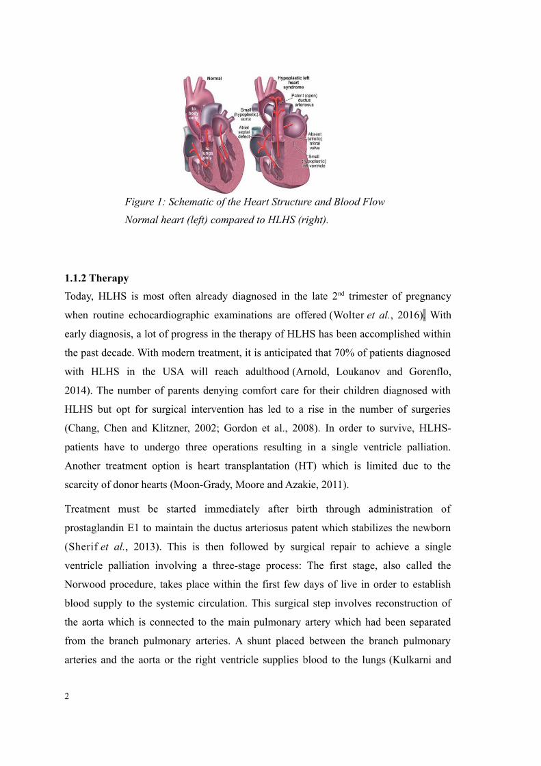
Figure 1: Schematic of the Heart Structure and Blood Flow
Normal heart (left) compared to HLHS (right).
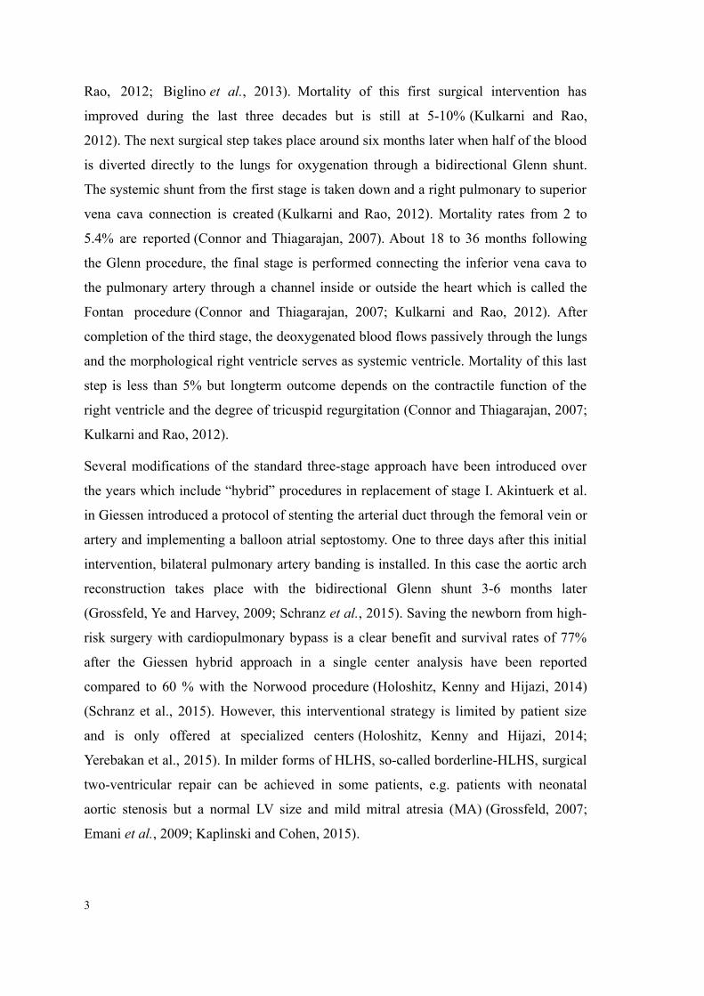
Rao, 2012; Biglino et al., 2013). Mortality of this first surgical intervention has
improved during the last three decades but is still at 5-10% (Kulkarni and Rao,
2012). The next surgical step takes place around six months later when half of the blood
is diverted directly to the lungs for oxygenation through a bidirectional Glenn shunt.
The systemic shunt from the first stage is taken down and a right pulmonary to superior
vena cava connection is created (Kulkarni and Rao, 2012). Mortality rates from 2 to
5.4% are reported (Connor and Thiagarajan, 2007). About 18 to 36 months following
the Glenn procedure, the final stage is performed connecting the inferior vena cava to
the pulmonary artery through a channel inside or outside the heart which is called the
Fontan procedure (Connor and Thiagarajan, 2007; Kulkarni and Rao, 2012). After
completion of the third stage, the deoxygenated blood flows passively through the lungs
and the morphological right ventricle serves as systemic ventricle. Mortality of this last
step is less than 5% but longterm outcome depends on the contractile function of the
right ventricle and the degree of tricuspid regurgitation (Connor and Thiagarajan, 2007;
Kulkarni and Rao, 2012).
Several modifications of the standard three-stage approach have been introduced over
the years which include “hybrid” procedures in replacement of stage I. Akintuerk et al.
in Giessen introduced a protocol of stenting the arterial duct through the femoral vein or
artery and implementing a balloon atrial septostomy. One to three days after this initial
intervention, bilateral pulmonary artery banding is installed. In this case the aortic arch
reconstruction takes place with the bidirectional Glenn shunt 3-6 months later
(Grossfeld, Ye and Harvey, 2009; Schranz et al., 2015). Saving the newborn from high-
risk surgery with cardiopulmonary bypass is a clear benefit and survival rates of 77%
after the Giessen hybrid approach in a single center analysis have been reported
compared to 60 % with the Norwood procedure (Holoshitz, Kenny and Hijazi, 2014)
(Schranz et al., 2015). However, this interventional strategy is limited by patient size
and is only offered at specialized centers (Holoshitz, Kenny and Hijazi, 2014;
Yerebakan et al., 2015). In milder forms of HLHS, so-called borderline-HLHS, surgical
two-ventricular repair can be achieved in some patients, e.g. patients with neonatal
aortic stenosis but a normal LV size and mild mitral atresia (MA) (Grossfeld, 2007;
Emani et al., 2009; Kaplinski and Cohen, 2015).
3
In the recent past HLHS has become amenable to pre-natal interventions since the
diagnosis is already made routinely around 18 to 22 weeks of gestation by fetal
echocardiography (Connor and Thiagarajan, 2007). In select centers around the world,
fetal interventions have been introduced which have significantly altered the post-natal
outcome of a subgroup of patients. In HLHS patients with an intact or highly restrictive
atrial septum, balloon atrial septostomy can have a positive effect on their outcome
(Enzensberger et al., 2012; Mackie, Aiyagari and Zampi, 2014). Furthermore, in utero
aortic valvuloplasty can improve left ventricle outflow and results in continued growth
of the left ventricle with better chances of biventricular repair after birth (McElhinney et
al., 2009). It has been reported that up to 29% of patients with evolving HLHS qualified
for biventricular circulation after successful prenatal aortic valvuloplasty. Outcomes are
variable and are continued to be improved by more stringent selection criteria (Connor
and Thiagarajan, 2007).
1.1.3 Outcome
The most critical time of HLHS patients is the first year of life. It was even shown that
90% of patients who survive infancy also survive up to 18 years of life (Siffel et al.,
2015). Survival rates of patients born with HLHS has improved over the years,
however, newborns with HLHS are at higher risk for neurodevelopmental deficits which
seemingly can not be influenced by prenatal aortic valvuloplasty (McElhinney, Benson,
et al., 2010). Furthermore, it was reported that the median full-scale IQ was
significantly lower in patients with HLHS compared to healthy newborns which was
influenced by the diameter of the neonatal ascending aorta as well as operation-related
factors (Sarajuuri et al., 2012).
It needs to be emphasized that current therapy strategies are only palliative, leaving the
child with a single ventricle physiology for life in which the anatomically right ventricle
(RV) functions as the systemic ventricle (Grossfeld, 2007). It was shown that the
mortality in dominant RV hearts such as HLHS is higher compared to those with
dominant LV (Wolter et al., 2016).Therefore, in borderline HLHS-patients the goal is to
recruit the LV in order to achieve biventricular repair. With this approach, a subset of
patients was identified in whom LV growth in utero is impeded by the presence of a
4
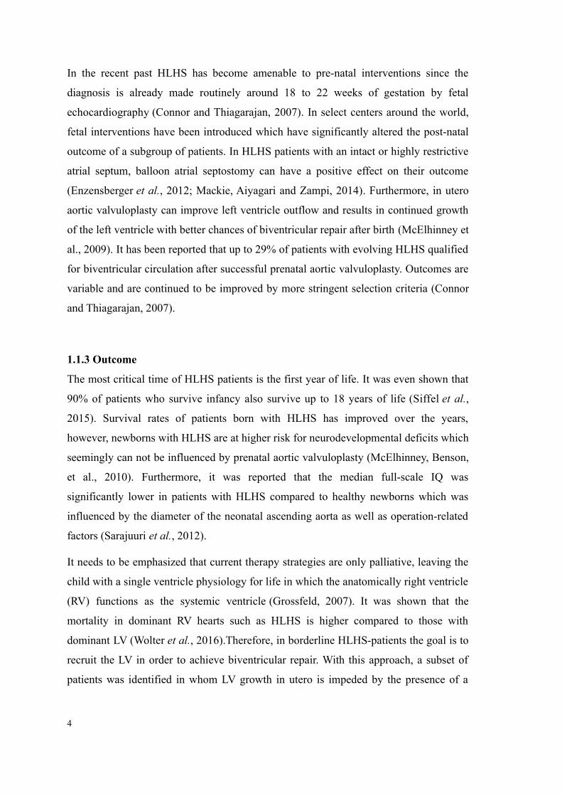
thick layer of white-grayish fibroelastic tissue, so called endocardial fibroelastosis
(EFE) which covers the endocardial surface of the LV. EFE is a diffuse fibrosis but in
comparison to cardiac fibrosis the subendocardial layers are not involved (Lurie, 2010;
Kaplinski and Cohen, 2015). EFE plays an important role on the outcome of patients
with HLHS as they tend to have worse diastolic dysfunction of the left ventricle and
suffer from cessation of left ventricle growth in utero (Cohen and Rychik, 1999; Lurie,
2010). In addition, the degree of EFE is directly associated with poor outcome despite
fetal interventions. EFE contributes to diastolic dysfunction and poor filling even after
relieving aortic stenosis by valvuloplasty (Lofland et al., 2001).
With regard to restriction imposed on the LV by EFE, it has been shown that removal of
EFE tissue during postnatal cardiac surgery for HLHS palliation, resulted in immediate
improvement of restriction and catch-up growth of the LV (Emani et al., 2009). Even
following non-response to fetal aortic valvotomy, LV growth was achieved through EFE
removal whereas observations showed that when EFE could not be resected there was
limited growth of the left ventricle (Emani et al., 2009, 2012). It has also been shown
that borderline left hearts with single ventricular repair could lead to a biventricular
solution by staged recruitment of the left ventricle. This recruitment strategy contained
relief of inflow and outflow obstruction, promotion of blood flow through the LV and
furthermore the resection of EFE (Emani et al., 2012). Currently, treatment to address
the presence of EFE is limited to surgical removal after birth but ideally should be
tackled in utero during the timeframe of rapid cardiac growth and as adjunct therapy to
post-natal treatment. As EFE can be diagnosed routinely in utero by echocardiography
presenting as a thickening of the endocardium it could therefore serve as a target for
new prenatal therapeutic strategies (Xu et al., 2015).
1.2 Endocardial Fibroelastosis (EFE)
EFE presents as a thickening of the inner ventricular endocardium consisting of colla-
gen fibers, elastic tissue and myofibroblasts with scarce vasculature (Friehs et al., 2013).
EFE was first described in 1943 by Weinberg and Himelfarb when this unexplained
5
heart failure caused early deaths in children (ROSAHN, 1955). Back then, secondary
EFE was distinguished from primary EFE which was not associated with any structural
cardiac anomalies (Seki et al., 2013). However, nowadays it is clear that EFE develops
secondary to some factors. Hence if there is no known cause, the term “idiopathic”
should be used rather than “primary” (Lurie, 2010). EFE is mostly seen in structural car-
diac malformations but there are also reports about EFE in the context of genetic, hy-
poxic or infectious diseases. EFE was reported in cases with left ventricular non-com-
paction (Weiford, Subbarao and Mulhern, 2004), primary x-linked isolated EFE
(Hanukoglu, Fried and Somekh, 1986), autosomal recessive isolated EFE (Rios, Cas-
taneda and Simpson, 1984) and postnatal infections (Factor, 1978). Cases of maternal
infections with Lupus, Parvovirus B19 and Mumps leading to EFE are reported as well
(Ni et al., 1997; Buyon and Clancy, 2003; Silingardi et al., 2009). In general EFE was
described as a reaction of the heart to stress such as pressure or volume overload which
mainly takes place during fetal development (Lurie, 2010). It is a disease of the growing
heart which has not been described in adults. In this context Boston Children's Hospital
observed that EFE can regrow after being resected, but at that time presenting as a dif-
ferent type of tissue covering the endocardium. All these observations suggest that the
etiology of EFE could be genetic, infectious or mechanical but the real pathomechanism
is still unknown.
When Boston Children's Hospital started to surgically remove EFE tissue, it was ob-
served that EFE could be easily resected as whole layer without any myocardial con-
tamination. This observation which indicated lack of infiltrative growth into the suben-
docardial layers led to the assumption that EFE tissue derives from the endocardium
(Friehs et al., 2013; Xu et al., 2015). Further investigations showed that indeed EFE de-
rives from the endocardium and specifically from the endocardial endothelial cells (EC)
which underwent a phenotypical transformation to mesenchymal cells.
6
1.3 Endothelial-to-Mesenchymal Transition
Histologically EFE tissue has been shown to consist of collagen fibers, elastic tissue and
mainly myofibroblasts (Shimada et al., 2014). In a study examining surgically excised
EFE tissue, it was determined that endocardial ECs are the origin of EFE. In a
transgenic mouse model where ECs were irreversibly fluorescently labelled, ECs
double-stained with mesenchymal and endothelial markers at the same time following
exposure to hemodynamic conditions mimicking HLHS in utero (Emani et al.,
2012). This phenotypical change is known as endothelial to mesenchymal transition
(EndMt) (Xu et al., 2015). In order to change the phenotype, molecular and architectural
rearrangements are necessary. ECs in general are hexagonal cells lined in a monolayer
which separates and regulates the blood stream from the surrounding tissue. The
diameter of ECs varies from 10 to 50 μm and the longitudinal dimension is in direction
to blood flow (Sumpio, Riley and Dardik, 2002). The EC has three surfaces: The
smooth luminal surface, the subluminal surface adherent to the connective tissue and the
cohesive surface where cells are adjacent to one another. Cell-cell and cell-tissue
connection is provided by two major types of junctions, the adherent junctions and the
tight junctions. In addition there are gap junctions for communication. Adherent
junctions are formed by transmembrane adhesion proteins of the cadherin family which
are key structures for the maintenance of cell-specific properties. Vascular endothelial
cadherin (VE-cadherin) is a specific cadherin that is only found in ECs. Another
7
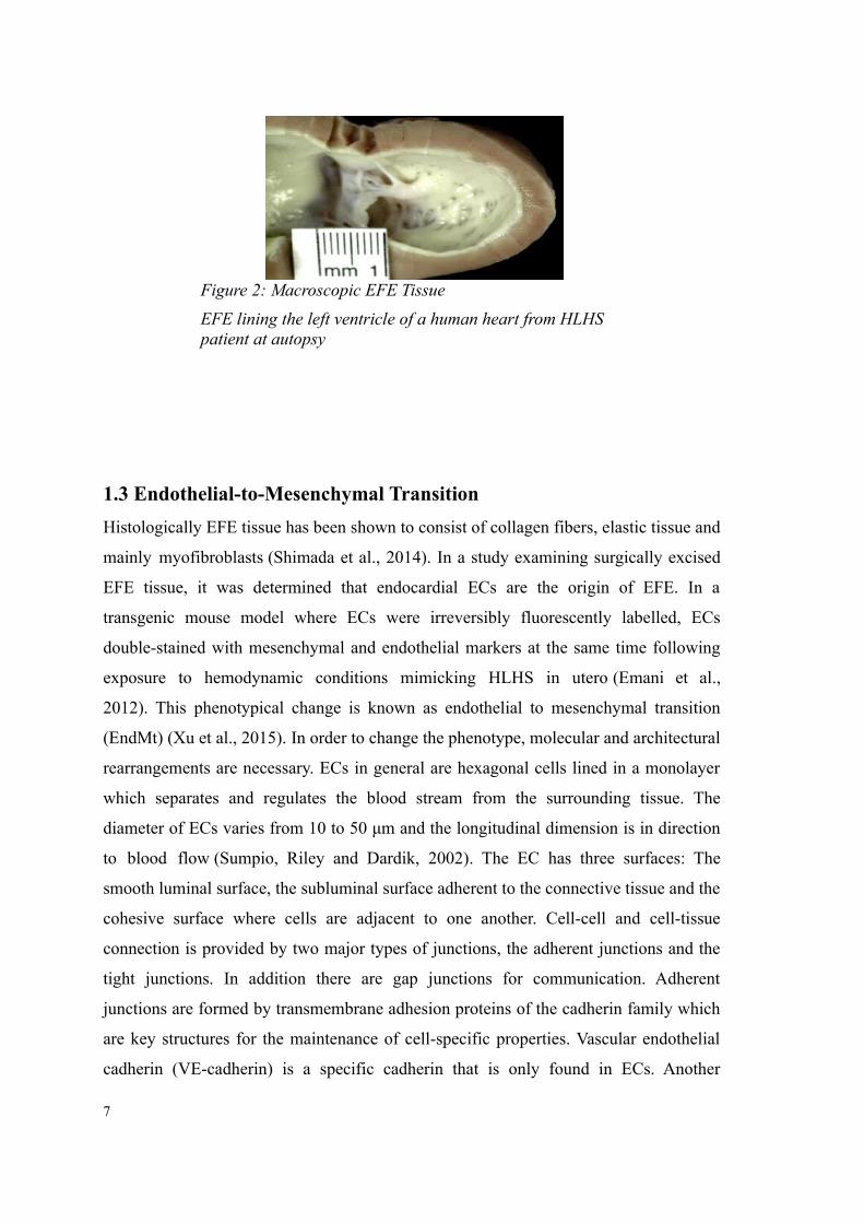
Figure 2: Macroscopic EFE Tissue
EFE lining the left ventricle of a human heart from HLHS patient at autopsy
constituent of the endothelial intercellular junction is CD31. CD31 also called platelet
endothelial cell adhesion molecule 1 (PECAM1) is a member of the immunoglobulin
superfamily. This transmembrane glycoprotein plays a major role in the adhesion
cascade between EC and the inflammatory cells during inflammation and angiogenesis
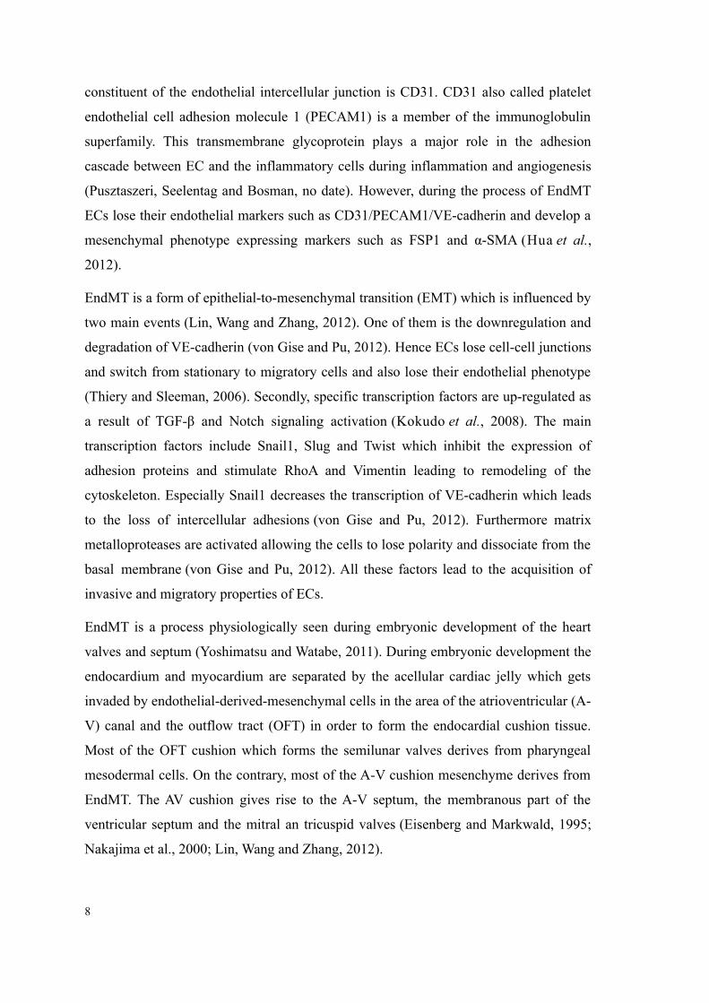
(Pusztaszeri, Seelentag and Bosman, no date). However, during the process of EndMT
ECs lose their endothelial markers such as CD31/PECAM1/VE-cadherin and develop a
mesenchymal phenotype expressing markers such as FSP1 and α-SMA (Hua et al.,
2012).
EndMT is a form of epithelial-to-mesenchymal transition (EMT) which is influenced by
two main events (Lin, Wang and Zhang, 2012). One of them is the downregulation and
degradation of VE-cadherin (von Gise and Pu, 2012). Hence ECs lose cell-cell junctions
and switch from stationary to migratory cells and also lose their endothelial phenotype
(Thiery and Sleeman, 2006). Secondly, specific transcription factors are up-regulated as
a result of TGF-β and Notch signaling activation (Kokudo et al., 2008). The main
transcription factors include Snail1, Slug and Twist which inhibit the expression of
adhesion proteins and stimulate RhoA and Vimentin leading to remodeling of the
cytoskeleton. Especially Snail1 decreases the transcription of VE-cadherin which leads
to the loss of intercellular adhesions (von Gise and Pu, 2012). Furthermore matrix
metalloproteases are activated allowing the cells to lose polarity and dissociate from the
basal membrane (von Gise and Pu, 2012). All these factors lead to the acquisition of
invasive and migratory properties of ECs.
EndMT is a process physiologically seen during embryonic development of the heart
valves and septum (Yoshimatsu and Watabe, 2011). During embryonic development the
endocardium and myocardium are separated by the acellular cardiac jelly which gets
invaded by endothelial-derived-mesenchymal cells in the area of the atrioventricular (A-
V) canal and the outflow tract (OFT) in order to form the endocardial cushion tissue.
Most of the OFT cushion which forms the semilunar valves derives from pharyngeal
mesodermal cells. On the contrary, most of the A-V cushion mesenchyme derives from
EndMT. The AV cushion gives rise to the A-V septum, the membranous part of the
ventricular septum and the mitral an tricuspid valves (Eisenberg and Markwald, 1995;
Nakajima et al., 2000; Lin, Wang and Zhang, 2012).
8
1.3.1 EndMT in Pathologies Other than EFE
EndMT is a physiological process during development but also plays a central role in
several pathologies of many different organs such as the heart, kidney, lung and bones
(Arciniegas et al.; Zeisberg et al., 2007, 2008; Medici et al., 2010). EndMT is involved
during myocardial infarction when ischemic damage to the myocardium results in repair
mechanisms elicited by ECs which serve as repairing cells filling up the gap with
granulation tissue (Aisagbonhi et al., 2011). In fibrodysplasia ossificans progressive
(FOP) bone tissue is reimbursed by muscle and connective tissue. In a mouse model of
FOP, osteoblasts and chondroblasts showed endothelial markers suggesting the process
of EndMT in this disease as well (Medici et al., 2010). Furthermore, ECs contribute to
fibroblast formation during fibrosis in kidneys, lungs, intestines and the heart
9
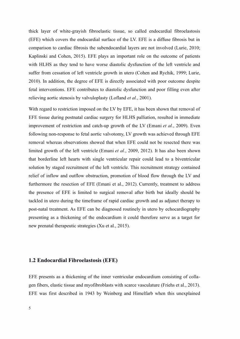
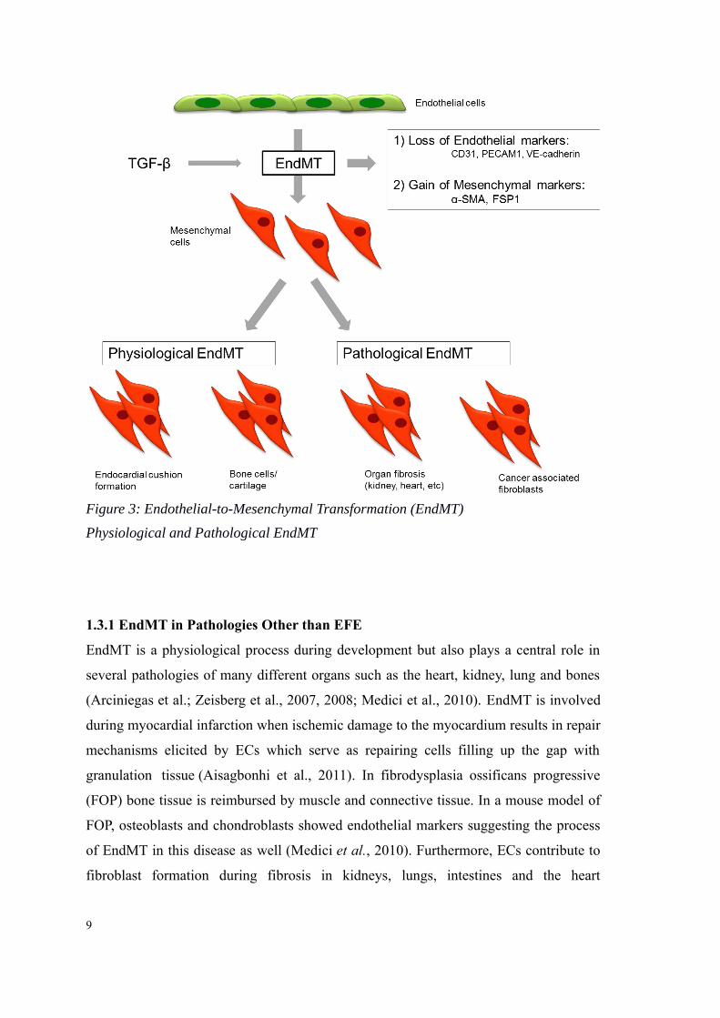
Figure 3: Endothelial-to-Mesenchymal Transformation (EndMT)
Physiological and Pathological EndMT
(Arciniegas et al.; Zeisberg et al., 2007, 2008; Rieder et al., 2011). Interestingly, EndMT
was even seen in tumor pathology giving cancer cells the ability to migrate and
transform into malignant cells (Kalluri and Zeisberg, 2006). It is also believed that
EndMT is relevant in the setting of systemic sclerosis, mitral valve pathology and
transplant vasculopathy (Cooley et al., 2014). This implies that EndMT is an important
factor not only in EFE but also in other pathological processes of the heart.
Having a closer look at the pathomechanism of EndMT in EFE, it was shown that
EndMT is regulated by an interplay of Bone Morphogenetic Protein (BMP)-7 and
Transforming Growth Factor (TGF)-β pathways (Xu et al., 2015). When EFE-tissue
was excised from the LV during routine open heart surgery and examined, it was
revealed that the upregulation of TGF-β1 and downregulation of BMP-7 was caused by
promoter hypermethylation of BMP-7, an epigenetic phenomenon altering the
physiological balance of these pathways (Xu et al., 2015). The interplay between TGF-β
and BMP pathways plays a key role in normal heart development. This information
regarding the underlying mechanism of EFE formation in children with HLHS, has the
potential to become the basis for therapeutic interventions directly targeting formation
and progression of EFE.
1.3.2 TGF-β Superfamily
As indicated by studies examining excised EFE tissue, EndMT in this case is regulated
by an imbalance of the TGF-β/BMP pathways (Xu et al., 2015). BMPs regulate cell
proliferation, apoptosis, embryonic development, and bone or cartilage induction (Yang
et al., 2015). TGF-β is a multifunctional growth factor regulating proliferation,
differentiation, apoptosis and migration of cells (Attisano and Wrana, 2000; Massagué,
2000).
TGF-β and BMP-7 are homodimeric proteins and part of the TGF-β superfamily
containing more than 35 molecules in mammals which are activated through binding to
specific transmembranous receptors eliciting an intracellular signaling cascade (Pegorier
et al., 2010). Following binding to these serine-threonine kinase receptors and
phosphorylation, intracellular Small mothers against decapentaplegic (SMADs) are
activated (Feng and Derynck, 2005). SMADs form intracellular complexes and
10
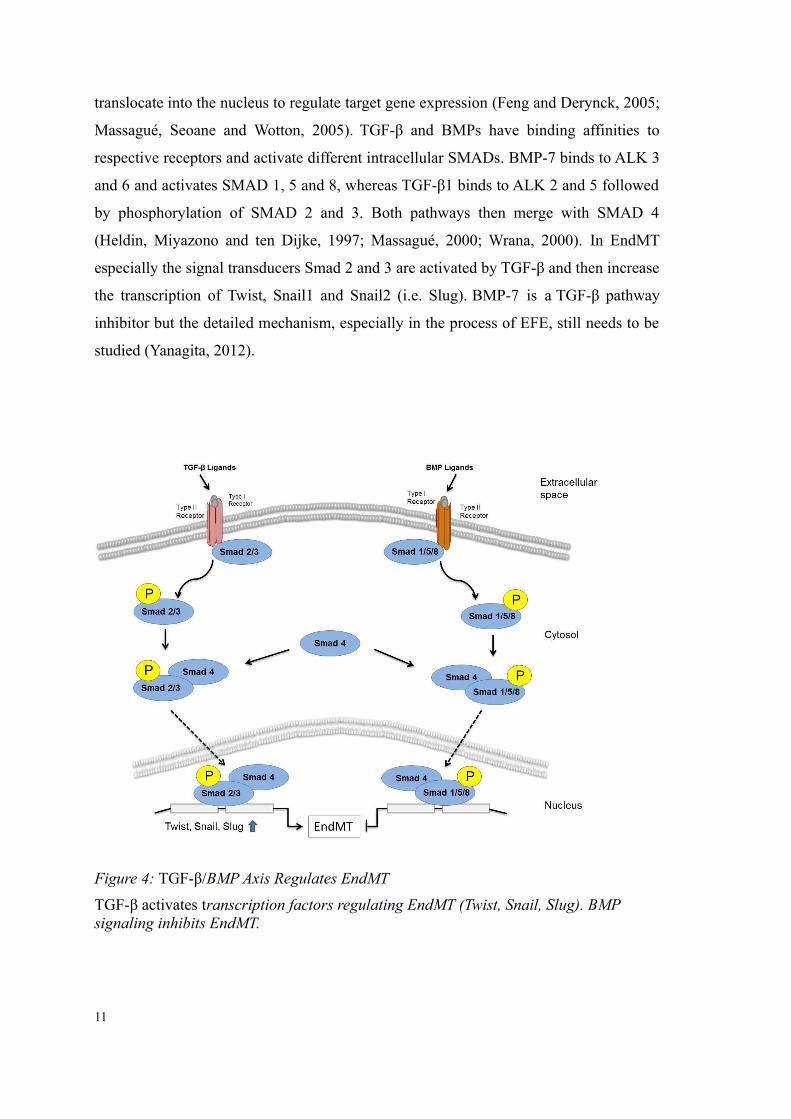
translocate into the nucleus to regulate target gene expression (Feng and Derynck, 2005;
Massagué, Seoane and Wotton, 2005). TGF-β and BMPs have binding affinities to
respective receptors and activate different intracellular SMADs. BMP-7 binds to ALK 3
and 6 and activates SMAD 1, 5 and 8, whereas TGF-β1 binds to ALK 2 and 5 followed
by phosphorylation of SMAD 2 and 3. Both pathways then merge with SMAD 4
(Heldin, Miyazono and ten Dijke, 1997; Massagué, 2000; Wrana, 2000). In EndMT
especially the signal transducers Smad 2 and 3 are activated by TGF-β and then increase
the transcription of Twist, Snail1 and Snail2 (i.e. Slug). BMP-7 is a TGF-β pathway
inhibitor but the detailed mechanism, especially in the process of EFE, still needs to be
studied (Yanagita, 2012).
11
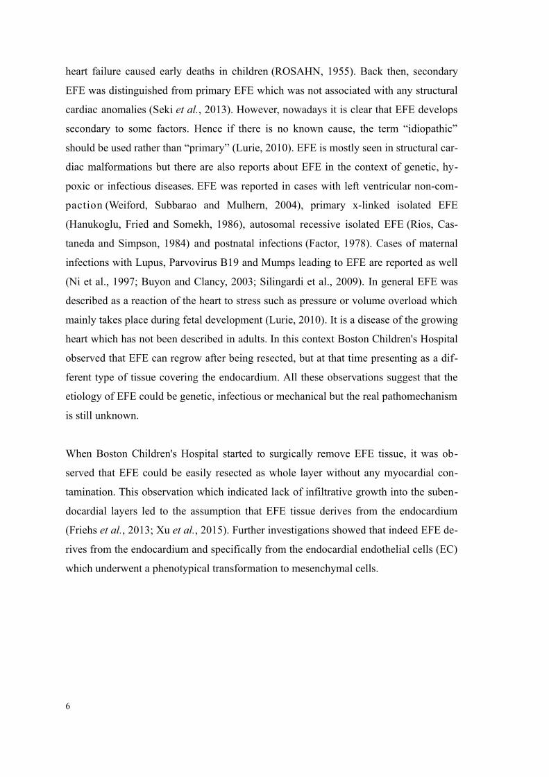
Figure 4: TGF-β/BMP Axis Regulates EndMT
TGF-β activates transcription factors regulating EndMT (Twist, Snail, Slug). BMP signaling inhibits EndMT.
2. Aims of the Study
The presence of EFE is a major putative effector in the mal-development of the fetal
heart. There are several hypotheses on the trigger of EFE formation which include lack
of blood flow with hypoxic/ischemia injury to the heart leading to atrophy of the left
ventricle. Furthermore, an inflammatory cause has also been hypothesized (Nield et al.,
2002) and genetic defects are a focus of several research groups but only a minority of
all patients with HLHS have been identified with genetic mutations (Iascone et al.,
2012). However, siblings’ recurrence of HLHS has been reported in 8% (Hinton et al.,
2007). Clinical observation indicates that mechanical forces on the LV play a potential
role in the development of EFE since closure of the aortic valve and lack of antegrade
blood flow through the aortic valve leads to dilatation of the LV and decline of
contractile function. With dilation of the LV, EFE formation rapidly progresses leading
to growth arrest of the LV (Sharland et al., 1991).
In summary, EFE is a response to stress from a variety of causes imposed on the left
ventricle. In this study an in vitro EndMT model was established in order to determine
the pathogenesis of EFE formation. The main focus was on mechanical stimuli as a
result of clinical observation which indicated this specific stress as central component.
With this model, I sought to determine whether mechanical forces imposed on the LV
induce EFE formation via a TGF-β mediated pathway. As a second aim therapeutic
strategies were investigated to inhibit EFE development as a first step toward a potential
localized application in the human fetus and infants.
12
3. Materials and Methods
3.1 Cell Culture
3.1.1 Human Coronary Artery Endothelial Cells (HCAEC)
ECs from coronary arteries share the same embryonic origin as endocardial cells as it
has been shown in lineage tracing studies (Wu et al., 2012). Therefore, Cloneticsᵀᴹ
human coronary artery endothelial cells (HCAEC) were used for these experiments and
purchased from Lonza (Cat. No.: CC-2585). According to the manufacturer’s
specifications, cells were isolated from the main stems of the right and left coronary
artery, the anterior descending artery and the circumflex branches. The cells for
culturing are delivered in a cryopreserved vial with over 500,000 cells after the third
passage. The cells are stored in liquid nitrogen at -180°C until use.
3.1.2 EGM-2 MV Growth Medium
Item Company/ Catalog Number
EBM-2 Basal Medium Lonza/ CC-3156
Supplemented with: EGM-2 MV SingleQuot Kit/ CC-4147
rhEGF
GA-1000 (30 mg/ ml of Gentamicin,
15g/ml of Amphotericin-B)
FBS
VEGF
rhFGF-B
R³-IGF-1
Ascorbic Acid
Table 1: EGM-2 MV Growth Medium
3.1.3 Initiation of Cell Culture/ Thawing of Cells
Culture dishes were coated with 1% gelatin. For the 1% gelatin solution, 5g gelatin was
dissolved in 500 ml phosphate buffered saline (PBS) and then autoclaved for 30
minutes. The solution was filter sterilized and then stored at room temperature. Growth
medium was added to the dishes using 1 ml for each 5 cm² of the culture dish.
13
Afterwards the dishes were placed in a 37℃, 5% CO₂ incubator for at least 30 minutes
in order to equilibrate. After removing the cryopreserved vial of HCAEC from the
freezer and wiping it with ethanol, the cap of the vial was turned a quarter turn to
release any liquid nitrogen that may be captured in the threads and then the cap was
closed again. The lower part of the vial was placed in a 37℃ water bath for a maximum
of two minutes until the last silver of ice melted. Cells were pipetted into a flask
containing 9 ml of medium and centrifuged at 1,200 x g for 5 min. The supernatant was
aspirated, except for 100-200 μl, and a volume of 2 ml of medium was added to the
flask. After pipetting the cells with a micropipette five times up and down, the cells
were dispensed into the culture dish which had been prepared earlier. The culture dishes
were agitated carefully on a rocking platform to equally distribute the cells and then
returned to the incubator. After 24 hours, the growth medium was changed to eliminate
all traces of dimethyl sulfoxide (DMSO), a cell culture reagent which is used for
cryopreservation. The growth medium was changed every other day and the volume
was doubled as soon as the cells reached a 60% confluence.
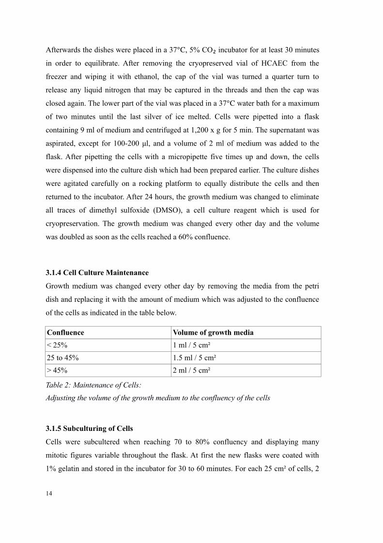
3.1.4 Cell Culture Maintenance
Growth medium was changed every other day by removing the media from the petri
dish and replacing it with the amount of medium which was adjusted to the confluence
of the cells as indicated in the table below.
Confluence Volume of growth media
< 25% 1 ml / 5 cm²
25 to 45% 1.5 ml / 5 cm²
> 45% 2 ml / 5 cm²
Table 2: Maintenance of Cells:
Adjusting the volume of the growth medium to the confluency of the cells
3.1.5 Subculturing of Cells
Cells were subcultered when reaching 70 to 80% confluency and displaying many
mitotic figures variable throughout the flask. At first the new flasks were coated with
1% gelatin and stored in the incubator for 30 to 60 minutes. For each 25 cm² of cells, 2
14
ml of 0,25% trypsin/EDTA, 7-10 ml of PBS and 4 ml of trypsin neutralizing solution
were thawed to room temperature. The medium was aspirated from the culture dish and
the cells were rinsed with 5 ml of room temperature PBS. Then PBS was aspirated and
the cells were covered with 2 ml of Trypsin/EDTA solution. Trypsinization took place
until approximately 90% of the cells were rounded up which took about two minutes.
Confirmation was obtained by light microscopy. Tapping carefully against the culture
dish supported the dissociation of cells from the surface. Making sure that the majority
of cells were detached, the trypsin in the flask was neutralized with 4 ml of room
temperature trypsin-neutralizing-solution. Afterwards the detached cells were
transferred quickly to a sterile 15 ml centrifuge tube. To collect residual cells, a final 2
ml of PBS were used to rinse the flask and then added to the centrifuge tube. By
examining the flask under the microscope, it was ensured that less than 5% of the cells
were left behind. The harvested cells were centrifuged at 1200 rpm for 5 minutes to
pellet the cells. Then the supernatant was aspirated, except for 100 – 200 µl. The pellet
was loosened by flicking the tube and the cells were diluted to a final volume of 2 to 3
ml of growth medium. Cell count was determined with a hemacytometer and viability
through trypan blue staining. Trypan blue is a dye that cannot leak across an intact cell
membrane and thus, does not stain viable cells. In contrast, dead cells with
compromised cell membranes become stained. Each flask was labeled with the passage
number, strain number, cell type and date. The growth medium was carefully transferred
to the new culture dish by adding 1 ml growth medium for every 5 cm² surface area of
the flask. To get uniform suspension, the diluted cells were mixed with a 5 ml pipet.
Finally, the previously calculated volume was filled in the prepared subculture flasks
and were incubated in the 37℃ humidified incubator with 5% CO₂.
15
Item Company/ Catalog Number
Phosphate Buffered Saline (PBS) Sigma-Aldrich/ P5368
Gelatin, powder Sigma-Aldrich/ G9391
Trypsin-EDTA (0.25%), phenol red Life technologies/ 25200-056
DMEM, high glucose, pyruvate Gibco/ 11995-065
Human Plasma Fibronectin Purified
Protein
Millipore/ FC010
HCAEC Lonza/ CC-2585
Tissue Culture Dish (60,8 cm²) Olympus plastics/ 25-202
Pipettes VWR
Centrifuge 5810 R Eppendorf
CO2 Incubator Sanyo
Electric Pipette Drummond Scientific/ 108818
Serological Glass Pipettes Olympus Plastic/ 12-101/ 12-102/ 12-104
Light Microscope Nikon TMS/ 02747
Hemocytometer Set Hausser Scientific/ 1483
Vortexer VWR
Fluorescence Microscope Zeiss Observer.Z1
6-Well Tissue Culture Plates Olympus plastics/ 25-105
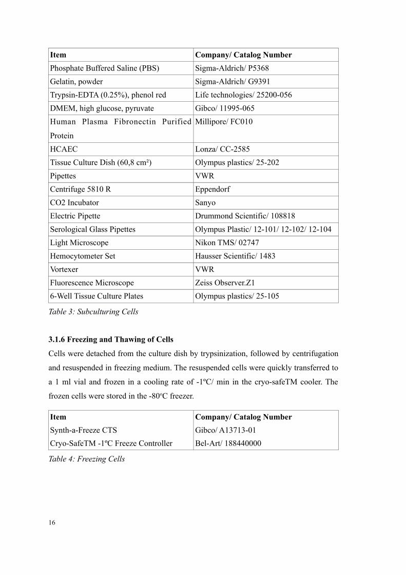
Table 3: Subculturing Cells
3.1.6 Freezing and Thawing of Cells
Cells were detached from the culture dish by trypsinization, followed by centrifugation
and resuspended in freezing medium. The resuspended cells were quickly transferred to
a 1 ml vial and frozen in a cooling rate of -1ºC/ min in the cryo-safeTM cooler. The
frozen cells were stored in the -80oC freezer.
Item Company/ Catalog Number
Synth-a-Freeze CTS Gibco/ A13713-01
Cryo-SafeTM -1ºC Freeze Controller Bel-Art/ 188440000
Table 4: Freezing Cells
16
3.2 Induction of Endothelial-to-Mesenchymal Transformation
(EndMT)
3.2.1 TGF-β1
EndMT was induced by treating ECs in culture with transforming growth factor β 1
(TGF-β1) as our group has previously reported in more detail (Xu et al., 2015).
3.2.2 TGF-β1 Reconstitution
Purified recombinant human TGF-β1 was obtained from R&D Systems. Since it is an
extremely hydrophobic protein adhering strongly to surfaces, it was reconstituted at 20
µg/ml in 100 µl sterile 4mM HCl containing 1 mg/ml bovine serum albumin.
3.2.3 Treatment of HCAEC with TGF-β1
HCAEC were used between the third and sixth passage. As soon as the cells reached
80% confluence, the medium was changed to EBM-2 Basal Medium (Lonza/ CC-3156)
supplemented with 10 ng/ml of TGF-β1. Cells were incubated with this solution for 24
hours followed by staining as described below in detail to determine EndMT.
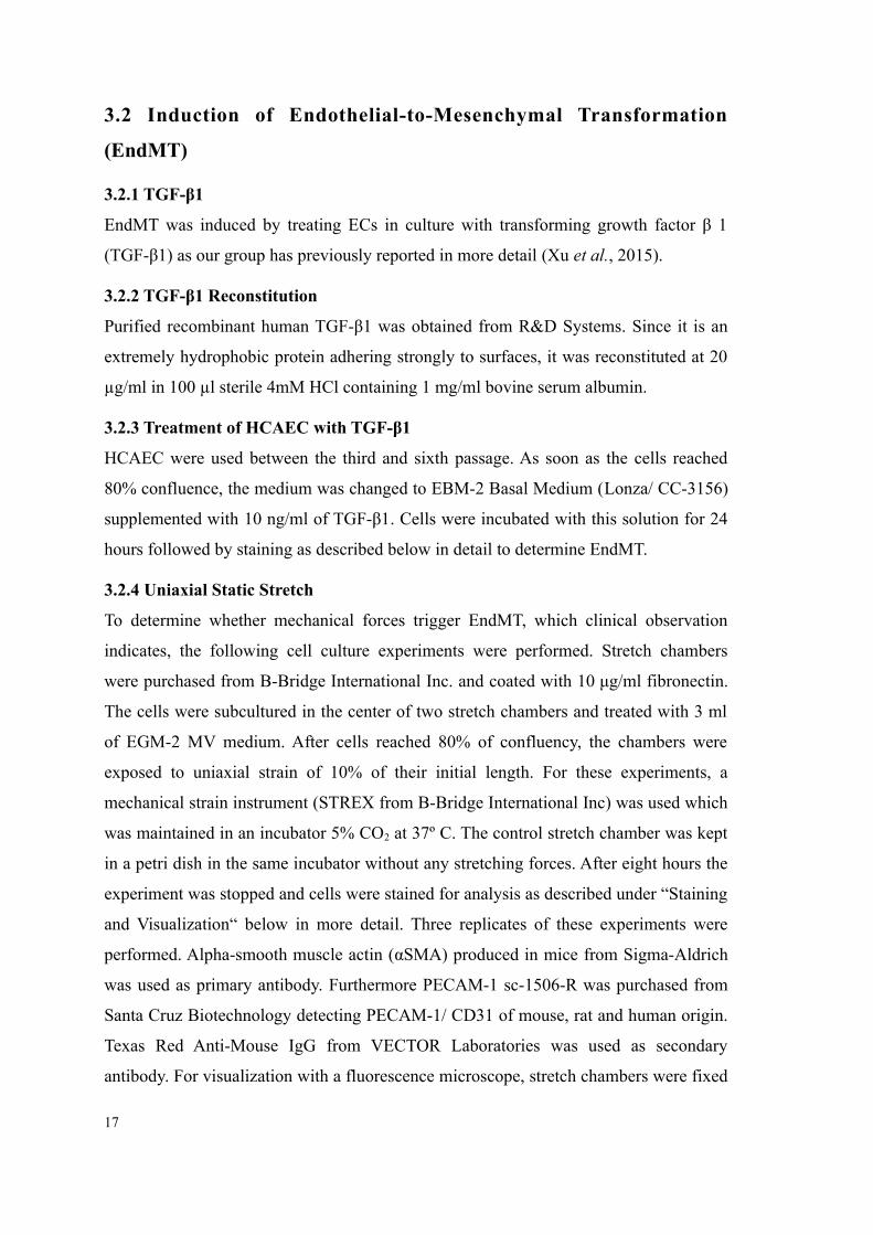
3.2.4 Uniaxial Static Stretch
To determine whether mechanical forces trigger EndMT, which clinical observation
indicates, the following cell culture experiments were performed. Stretch chambers
were purchased from B-Bridge International Inc. and coated with 10 μg/ml fibronectin.
The cells were subcultured in the center of two stretch chambers and treated with 3 ml
of EGM-2 MV medium. After cells reached 80% of confluency, the chambers were
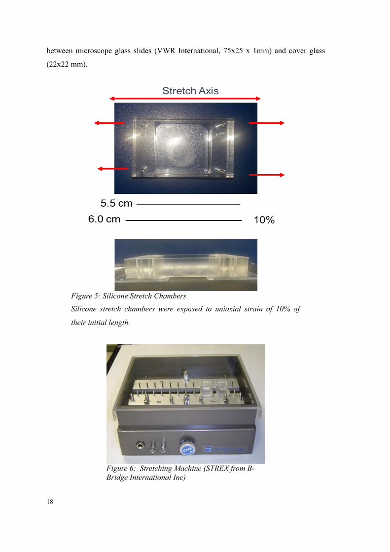
exposed to uniaxial strain of 10% of their initial length. For these experiments, a
mechanical strain instrument (STREX from B-Bridge International Inc) was used which
was maintained in an incubator 5% CO2 at 37º C. The control stretch chamber was kept
in a petri dish in the same incubator without any stretching forces. After eight hours the
experiment was stopped and cells were stained for analysis as described under “Staining
and Visualization“ below in more detail. Three replicates of these experiments were
performed. Alpha-smooth muscle actin (αSMA) produced in mice from Sigma-Aldrich
was used as primary antibody. Furthermore PECAM-1 sc-1506-R was purchased from
Santa Cruz Biotechnology detecting PECAM-1/ CD31 of mouse, rat and human origin.
Texas Red Anti-Mouse IgG from VECTOR Laboratories was used as secondary
antibody. For visualization with a fluorescence microscope, stretch chambers were fixed
17
between microscope glass slides (VWR International, 75x25 x 1mm) and cover glass
(22x22 mm).
18
Figure 5: Silicone Stretch Chambers
Silicone stretch chambers were exposed to uniaxial strain of 10% of
their initial length.
Figure 6: Stretching Machine (STREX from B-Bridge International Inc)
3.3 Determination of Endothelial-to-Mesenchymal Transition (EndMT)
Data of human EFE-tissue analysis indicate that EFE is formed by EndMT where ECs
lose their endothelial markers such as CD31/PECAM1/VE-cadherin and develop a mes-
enchymal phenotype expressing markers such as FSP1 and α S MA (H ua et al.,
2012). Standard histological determination of EndMT is performed through staining
with PECAM1 (an endothelial marker) and αSMA (mesenchymal marker). Double-
staining of one cell with both markers is indicative of active EndMT. Determination of
EndMT was performed through immunohistochemical staining of tissue sections with
antibodies directed against CD-31, an EC marker and α-smooth muscle actin (αSMA), a
fibroblast marker. Secondary immunoreagents conjugated to red-fluorescent Texas Red
(Vector Laboratories) or green-fluorescent Alexa-488TM fluorophores (Life Technolo-
gies, Grand Island, NY) were used and nuclei were stained with blue fluorescent DAPI
nucleic acid stain (Life Technologies, Grand Island, NY).
3.4 Immunofluorescent Staining
Immunofluorescent staining was used to detect special antigens with two types of
antibodies. Whereas the primary antibody binds to the target molecule and the
secondary antibody which contains the flurophore binds to the primary antibody making
the target visible under a fluorescent microscope. Blocking buffer is used to block non-
specific binding sites of the antibodies.
3.4.1 Blocking Buffer
In order to produce 10 ml of blocking buffer (BB), 0.15 g of Bovine Serum Albumine
was added to 10 ml of PBS.
3.4.2 Antibodies
Monoclonal Anti-Actin αSMA primary antibody, produced in mouse, was used as mes-
enchymal cell marker.
Rabbit PECAM1 was used as a primary antibody to detect ECs.
Corresponding secondary antibodies goat anti-rabbit IgG (Alexa Fluor 488) and Texas
Red Anti-Mouse IgG were obtained. Details are provided in the table below. Nuclei
were counterstained with 4',6-diamidino-2-phenylindole (DAPI). Thus, ECs were
19
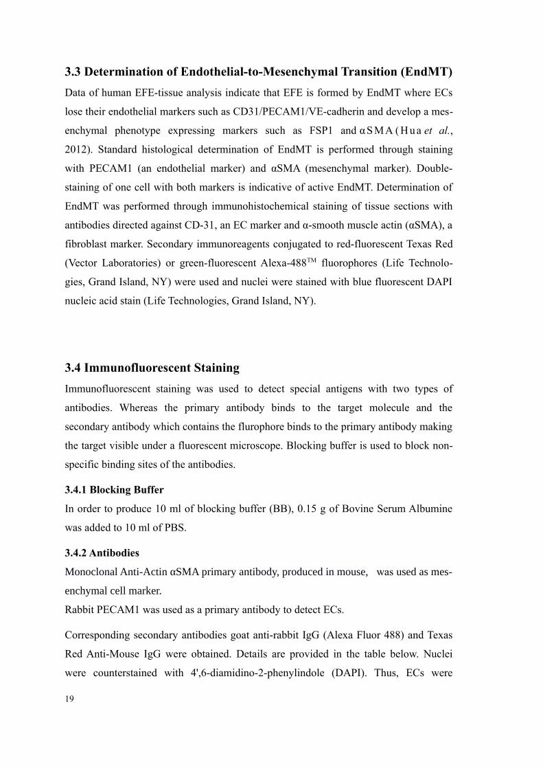
detected in green, mesenchymal cells in red and nuclei in blue.
Primary Antibody (Ab) Company/ Catalog Number Concentration Ab:BB
PECAM1 Santa Cruz Biotechnology/
sc-1506-R
1:50
Monoclonal Anti-Actin,
αSMA produced in mouse
Sigma-Aldrich/ A2547 1:400
Secondary Antibody
Alexa Fluor 488 Life Technologies/ A11034 1:200
Texas Red Anti-Mouse IgG Vector Laboratories/ TI-2000 1:100
DAPI Molecular Probes/ D1306 1:1,000
Table 5: Immunofluorescent Staining: Antibodies
Item Company/ Catalog Number
Bovine Serum Albumin Sigma-Aldrich/ A7906
32% Paraformaldehyde Electron Microscopy Sciences/ 15714-S
Triton® X-100 SIGMA/ 103K0062
Pipettes Eppendorf
Mounting Medium Dako/ S3025
Microscope Slides VWR/ 16004-422
Table 6: Immunofluorescent Staining
3.4.3 Staining
Cells contained in the stretch chambers were washed twice with PBS. Washing was re-
peated after the cells were covered to a depth of 2-3 mm with 4% paraformaldehyde for
15 minutes. 0.2% Triton X-100 was applied for 10 minutes and then slides were washed
again twice with PBS. Cells were incubated in blocking buffer containing the primary
antibody for one hour and washed twice with PBS thereafter. Then the cells were incu-
bated with blocking buffer solution containing the secondary antibody in the dark pre-
venting light exposure. After one hour the cells were washed twice with PBS again and
secondly washed with ddH2O. As a last step the slide was mounted.
20
3.4.4 Visualization
Cells were assessed with a Zeiss Observer.Z1 fluorescent microscope. EndMT was de-
fined as cells staining positive for both endothelial and mesenchymal marker. Cells were
visualized using a Zeiss Axiovert inverted microscope with a Nikon 10x objective, NA
= 10x/0.25 and 20x objective. The microscope is equipped with various fluorescent fil-
ters and Leica digital color camera. Ten fields randomly picked from each sample were
analyzed by a blinded microscopist and number of single stained and double stained
cells counted. The microscope was configured to maximal range and automatic
Min/Max. The software Axiovision was used and for comparison, the same exposure
time was chosen for each experiment.
3.5 Inhibition of EndMT
3.5.1 Treatment with BMP-7
HCAEC were cultured in an eight chamber glass slide. When cells reached 80%
confluence, the growth medium was changed to EBM-2 containing Ascorbic Acid,
Gentamicin and 20 % FBS only. This preparatory step of excluding growth factors that
support EC morphology, is called cell starvation. Following 12 hours of starvation,
treatment was induced by adding EBM-2 and growth medium in a dilution of 10:1 to
prevent cells from undergoing apoptosis. The following experimental groups were
analyzed: group 1 was treated with TGF-β1 to undergo EndMT; group 2 contained
BMP-7 only; group 3 was treated with TGF-β1 and BMP-7 to inhibit the process of ECs
undergoing EndMT; and group 4 served as negative control containing growth medium
only. TGF-β1 was used in a concentration of 10 ng/ml and BMP-7 was added in a
concentration of 100 ng/ml. The medium of each chamber was changed every 24 hours
and after 72 hours the experiment was terminated and cells were stained for analysis.
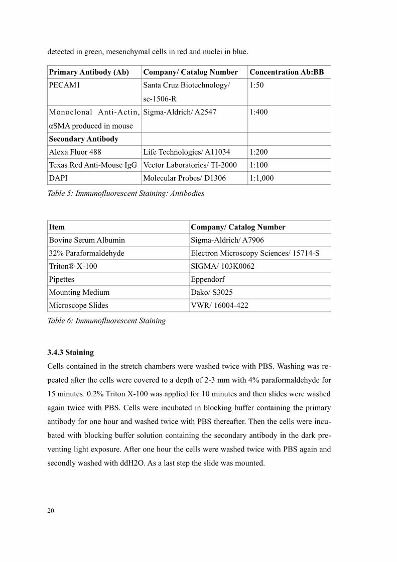
Item Company/ Catalog Number
Recombinant Human TGF-β1 R&D Systems/ 240-B
Recombinant Human BMP-7, Active Gibco/ PHC7204
Table 7: Inducing and Inhibiting EndMT
21
3.5.2 Uniaxial Static Stretch and Treatment with BMP-7
The experiment labelled “10% of Static Stretch for 8 hours” was repeated using an
additional stretch chamber containing 100 ng/ ml of BMP-7. One stretch chamber which
was exposed to stretch as well served as a positive control and one stretch chamber just
placed in the incubator served as a negative control.
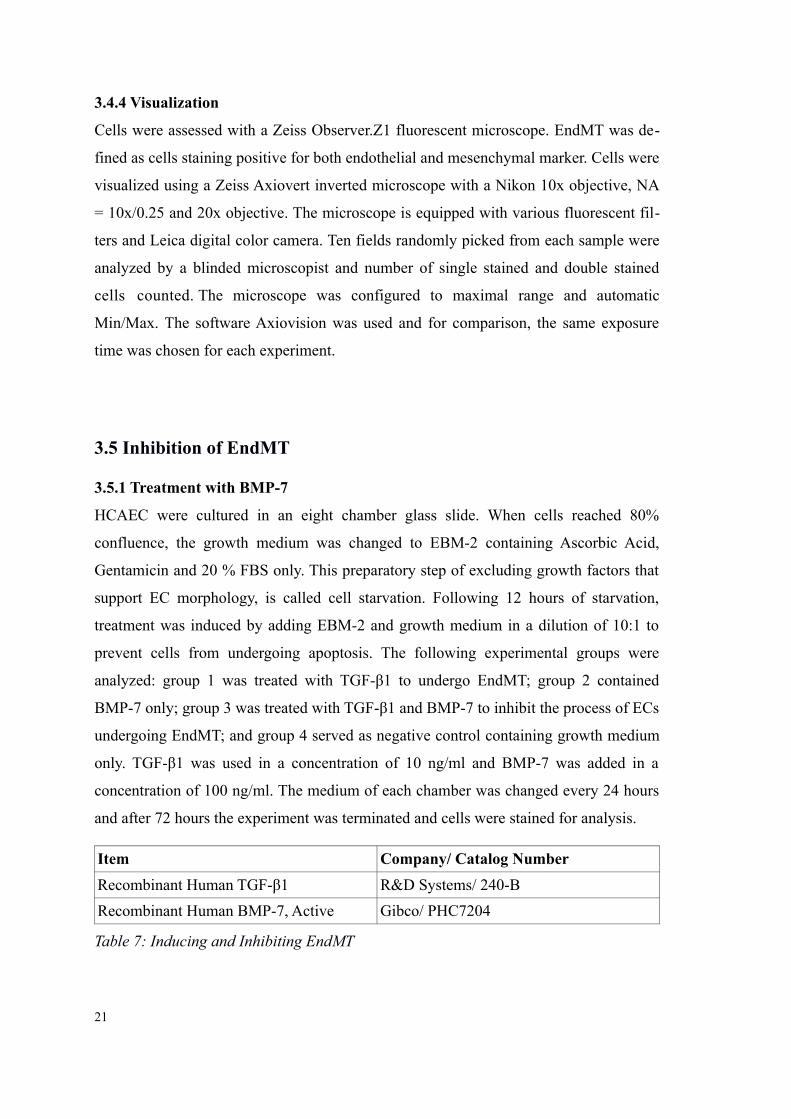
Item Company/ Catalog Number
Stretching device
Stretch chamber 10 cm2 B-Bridge International/ ST-CH-10
Table 8: Uniaxial Static Stretch
3.6 Testing of Localized Drug Delivery Systems
Drug carriers were tested for future application to inhibit EndMT locally as a first step
toward therapy of EFE in humans (Shimada et al., 2014; Xu et al., 2015). The goal of
these experiments was to establish an ex vivo set-up to determine the compatibility of
drug to drug carrier and the drug release profile. As proof-of-concept, gelatin sponges
were used as carrier model. A piece of 1mm² of the gelatin sponge was loaded with 100
ng/ml BMP-7 and incubated within the cell culture media. Unloaded gelatin sponges
served as control. The following parameters were tested: cell viability with trypan blue
and EndMT through fluorescent staining as described below.
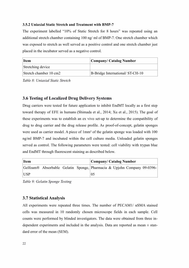
Item Company/ Catalog Number
Gelfoam® Absorbable Gelatin Sponge,
USP
Pharmacia & Upjohn Company 09-0396-
05
Table 9: Gelatin Sponge Testing
3.7 Statistical Analysis
All experiments were repeated three times. The number of PECAM1/ αSMA stained
cells was measured in 10 randomly chosen microscope fields in each sample. Cell
counts were performed by blinded investigators. The data were obtained from three in-
dependent experiments and included in the analysis. Data are reported as mean ± stan-
dard error of the mean (SEM).
22
Following confirmation of normal distribution of data, student t-test (GraphPad Prism
5.1) or ANOVA for multiple group comparisons and Bonferroni's post-hoc analysis
were used to obtain calculations of statistical significance. Probability values of ≤ 0.05
were regarded statistically significant.
4. Results
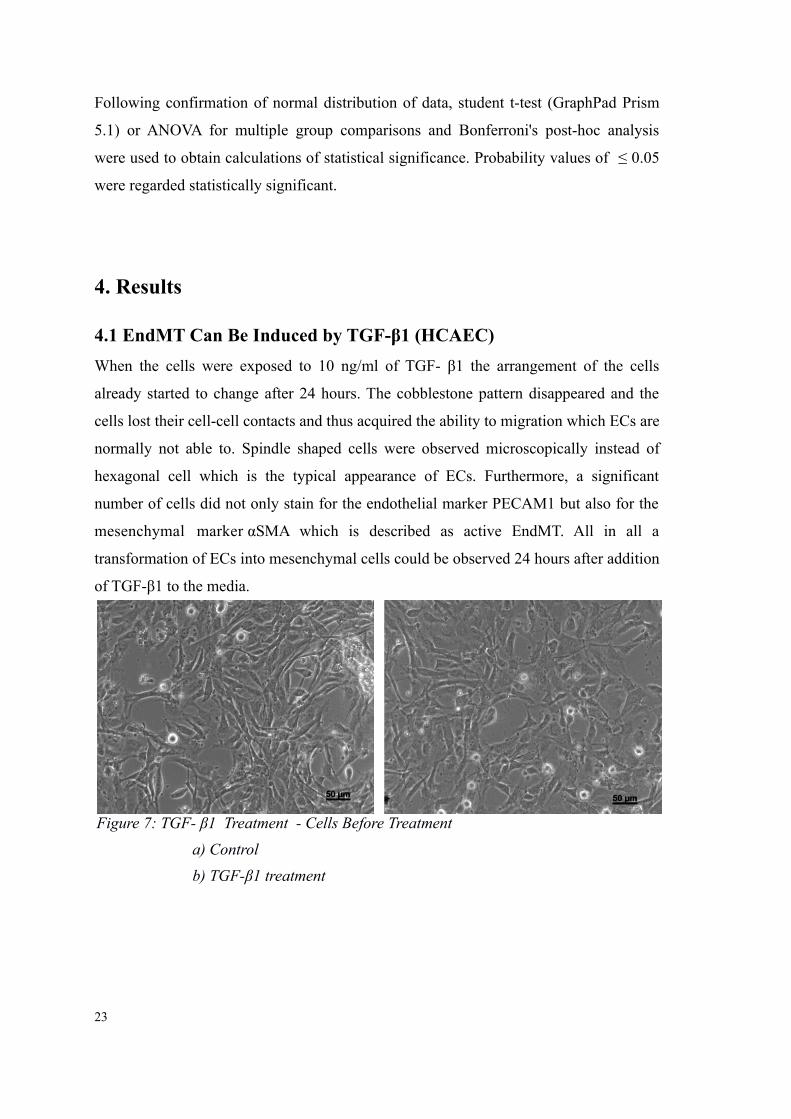
4.1 EndMT Can Be Induced by TGF-β1 (HCAEC)
When the cells were exposed to 10 ng/ml of TGF- β1 the arrangement of the cells
already started to change after 24 hours. The cobblestone pattern disappeared and the
cells lost their cell-cell contacts and thus acquired the ability to migration which ECs are
normally not able to. Spindle shaped cells were observed microscopically instead of
hexagonal cell which is the typical appearance of ECs. Furthermore, a significant
number of cells did not only stain for the endothelial marker PECAM1 but also for the
mesenchymal marker αSMA which is described as active EndMT. All in all a
transformation of ECs into mesenchymal cells could be observed 24 hours after addition
of TGF-β1 to the media.
23
Figure 7: TGF- β1 Treatment - Cells Before Treatment
a) Control
b) TGF-β1 treatment
24
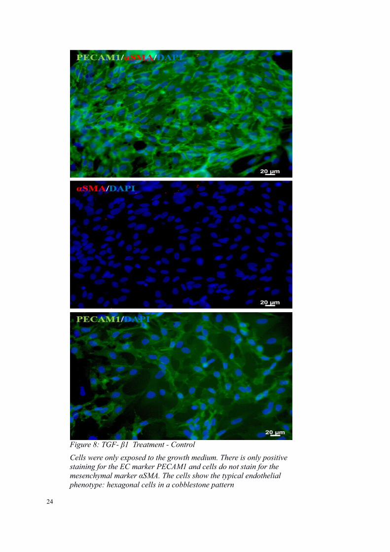
Figure 8: TGF- β1 Treatment - Control
Cells were only exposed to the growth medium. There is only positive staining for the EC marker PECAM1 and cells do not stain for the mesenchymal marker αSMA. The cells show the typical endothelial phenotype: hexagonal cells in a cobblestone pattern
25
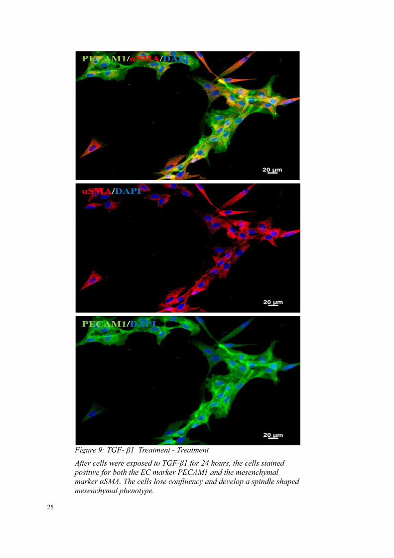
Figure 9: TGF- β1 Treatment - Treatment
After cells were exposed to TGF-β1 for 24 hours, the cells stained positive for both the EC marker PECAM1 and the mesenchymal marker αSMA. The cells lose confluency and develop a spindle shapedmesenchymal phenotype.
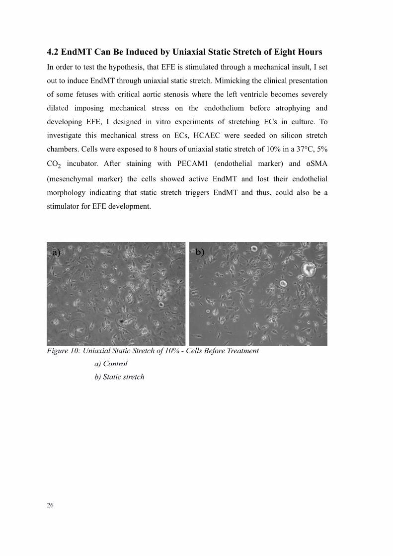
4.2 EndMT Can Be Induced by Uniaxial Static Stretch of Eight Hours
In order to test the hypothesis, that EFE is stimulated through a mechanical insult, I set
out to induce EndMT through uniaxial static stretch. Mimicking the clinical presentation
of some fetuses with critical aortic stenosis where the left ventricle becomes severely
dilated imposing mechanical stress on the endothelium before atrophying and
developing EFE, I designed in vitro experiments of stretching ECs in culture. To
investigate this mechanical stress on ECs, HCAEC were seeded on silicon stretch
chambers. Cells were exposed to 8 hours of uniaxial static stretch of 10% in a 37°C, 5%
CO2 incubator. After staining with PECAM1 (endothelial marker) and αSMA
(mesenchymal marker) the cells showed active EndMT and lost their endothelial
morphology indicating that static stretch triggers EndMT and thus, could also be a
stimulator for EFE development.
26
Figure 10: Uniaxial Static Stretch of 10% - Cells Before Treatment
a) Control
b) Static stretch
27
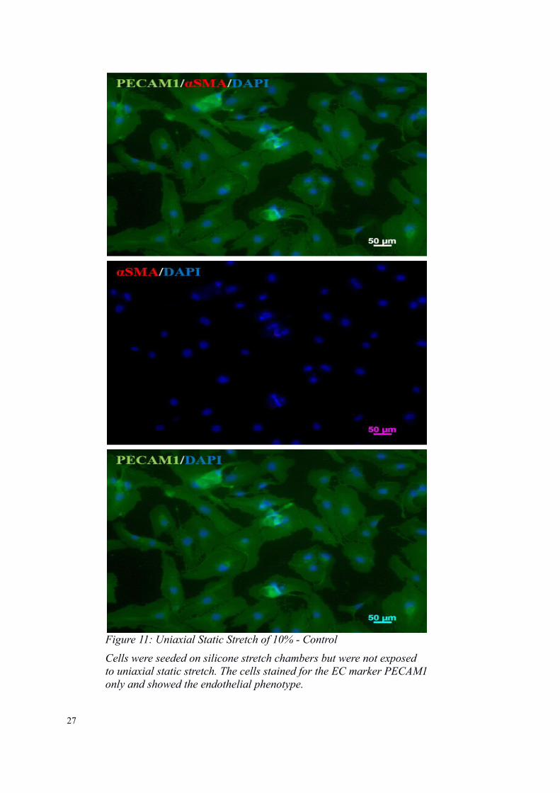
Figure 11: Uniaxial Static Stretch of 10% - Control
Cells were seeded on silicone stretch chambers but were not exposed to uniaxial static stretch. The cells stained for the EC marker PECAM1only and showed the endothelial phenotype.
28
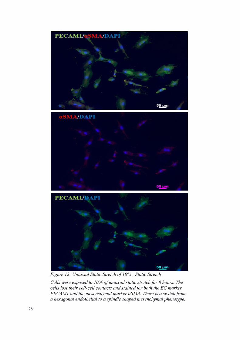
Figure 12: Uniaxial Static Stretch of 10% - Static Stretch
Cells were exposed to 10% of uniaxial static stretch for 8 hours. The cells lost their cell-cell contacts and stained for both the EC marker PECAM1 and the mesenchymal marker αSMA. There is a switch from a hexagonal endothelial to a spindle shaped mesenchymal phenotype.
29
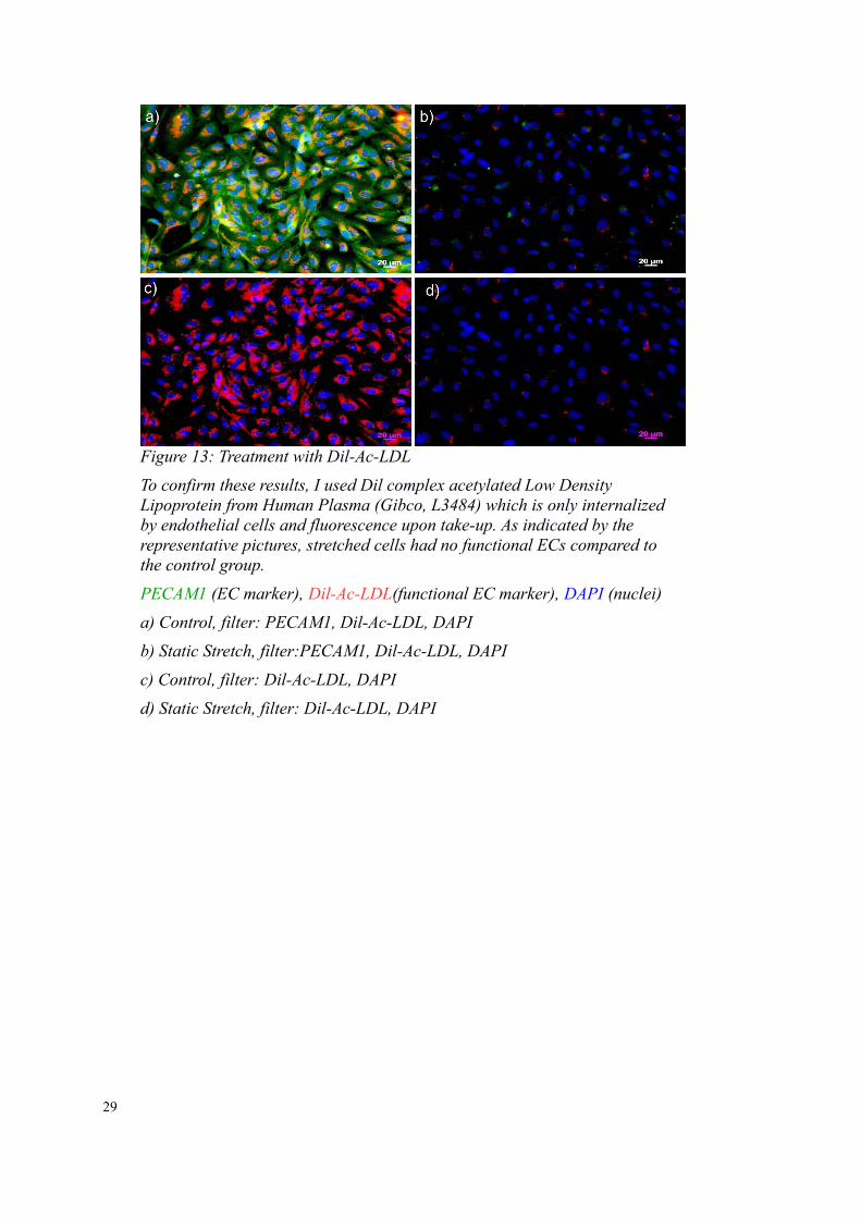
Figure 13: Treatment with Dil-Ac-LDL
To confirm these results, I used Dil complex acetylated Low Density Lipoprotein from Human Plasma (Gibco, L3484) which is only internalized by endothelial cells and fluorescence upon take-up. As indicated by the representative pictures, stretched cells had no functional ECs compared to the control group.
PECAM1 (EC marker), Dil-Ac-LDL(functional EC marker), DAPI (nuclei)
a) Control, filter: PECAM1, Dil-Ac-LDL, DAPI
b) Static Stretch, filter:PECAM1, Dil-Ac-LDL, DAPI
c) Control, filter: Dil-Ac-LDL, DAPI
d) Static Stretch, filter: Dil-Ac-LDL, DAPI
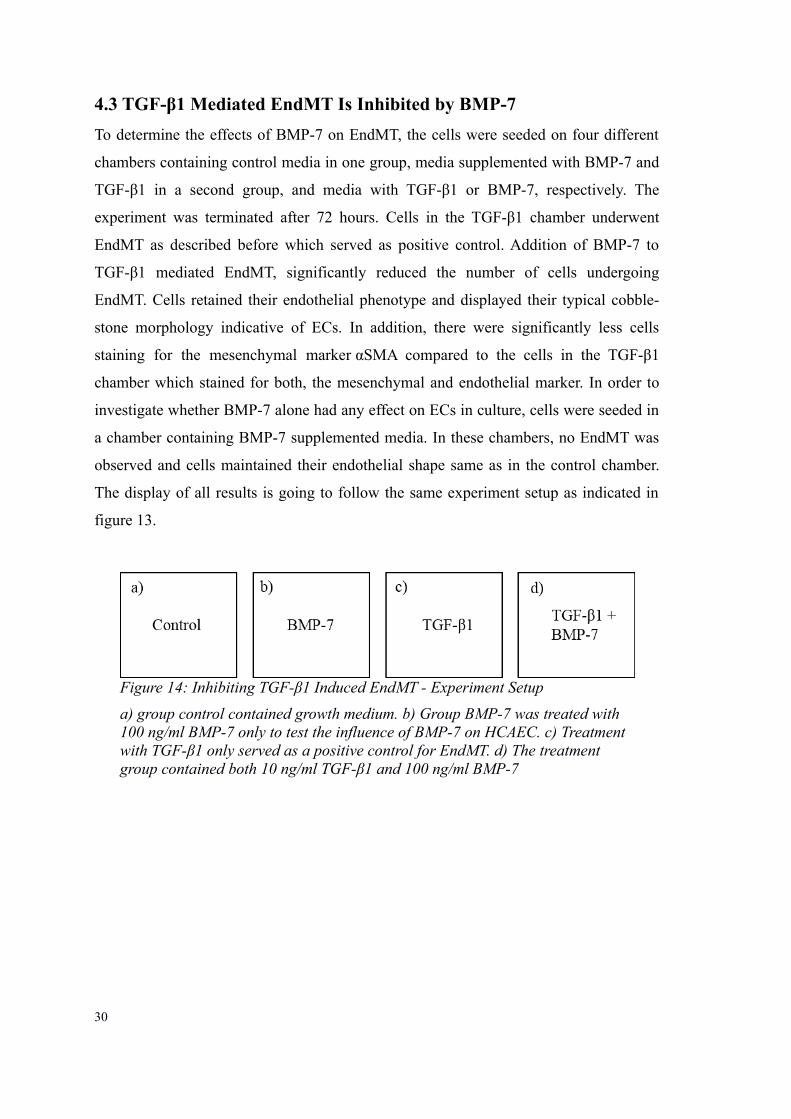
4.3 TGF-β1 Mediated EndMT Is Inhibited by BMP-7
To determine the effects of BMP-7 on EndMT, the cells were seeded on four different
chambers containing control media in one group, media supplemented with BMP-7 and
TGF-β1 in a second group, and media with TGF-β1 or BMP-7, respectively. The
experiment was terminated after 72 hours. Cells in the TGF-β1 chamber underwent
EndMT as described before which served as positive control. Addition of BMP-7 to
TGF-β1 mediated EndMT, significantly reduced the number of cells undergoing
EndMT. Cells retained their endothelial phenotype and displayed their typical cobble-
stone morphology indicative of ECs. In addition, there were significantly less cells
staining for the mesenchymal marker αSMA compared to the cells in the TGF-β1
chamber which stained for both, the mesenchymal and endothelial marker. In order to
investigate whether BMP-7 alone had any effect on ECs in culture, cells were seeded in
a chamber containing BMP-7 supplemented media. In these chambers, no EndMT was
observed and cells maintained their endothelial shape same as in the control chamber.
The display of all results is going to follow the same experiment setup as indicated in
figure 13.
30
Figure 14: Inhibiting TGF-β1 Induced EndMT - Experiment Setup
a) group control contained growth medium. b) Group BMP-7 was treated with 100 ng/ml BMP-7 only to test the influence of BMP-7 on HCAEC. c) Treatment with TGF-β1 only served as a positive control for EndMT. d) The treatment group contained both 10 ng/ml TGF-β1 and 100 ng/ml BMP-7
31
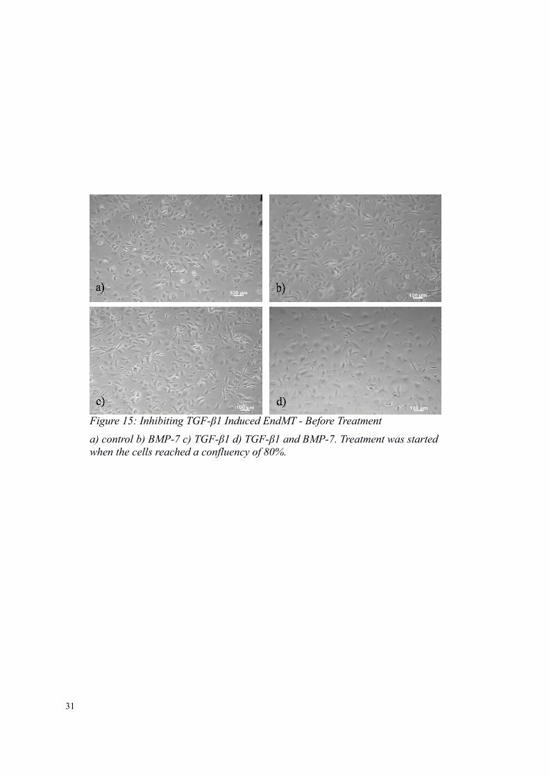
Figure 15: Inhibiting TGF-β1 Induced EndMT - Before Treatment
a) control b) BMP-7 c) TGF-β1 d) TGF-β1 and BMP-7. Treatment was startedwhen the cells reached a confluency of 80%.
32
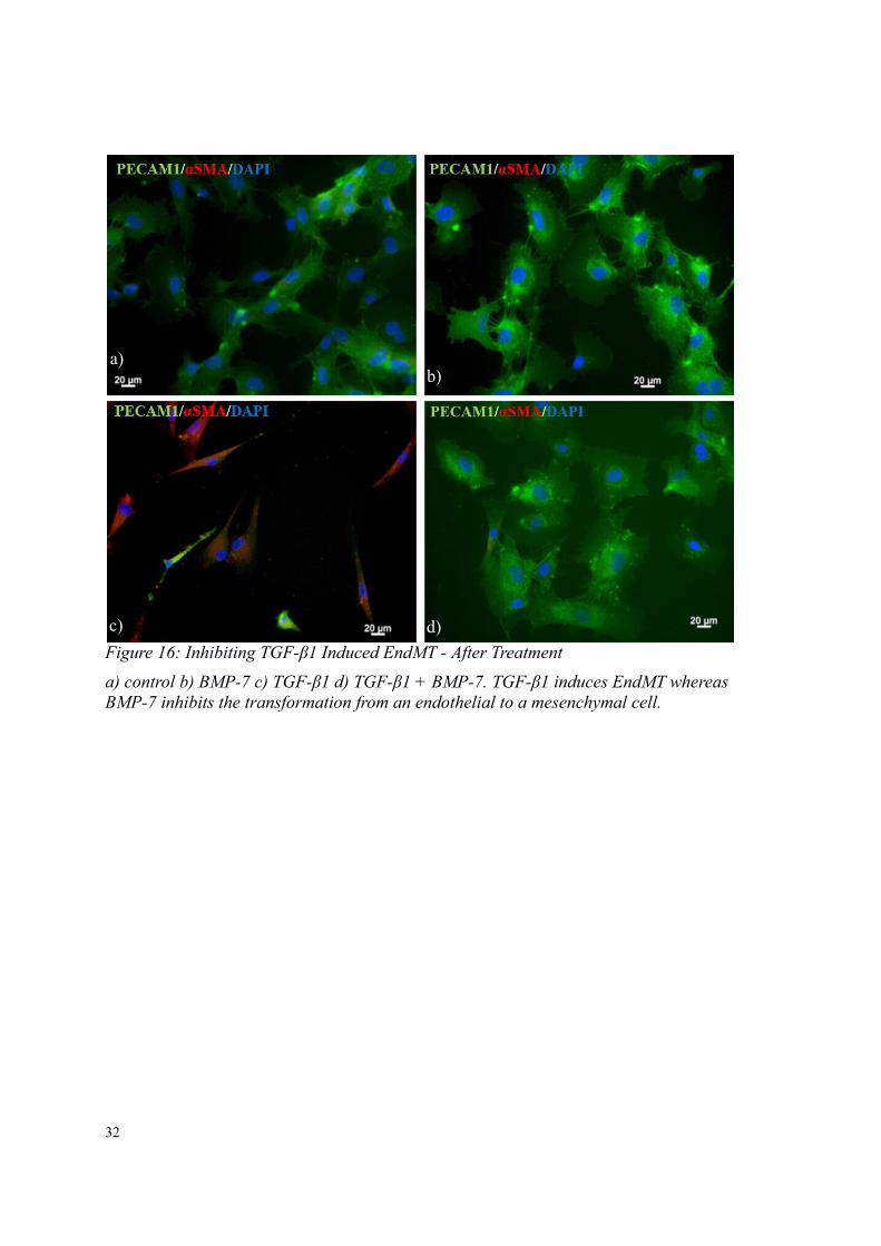
Figure 16: Inhibiting TGF-β1 Induced EndMT - After Treatment
a) control b) BMP-7 c) TGF-β1 d) TGF-β1 + BMP-7. TGF-β1 induces EndMT whereas BMP-7 inhibits the transformation from an endothelial to a mesenchymal cell.
a)
c)
b)
d)
33
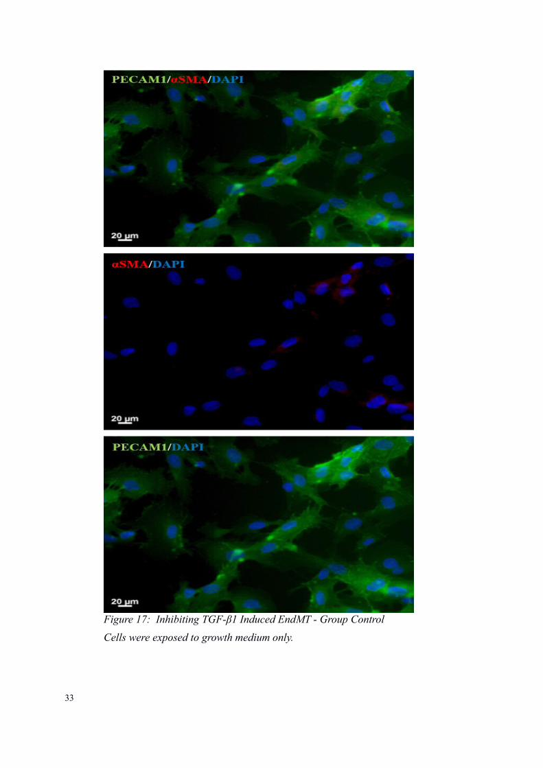
Figure 17: Inhibiting TGF-β1 Induced EndMT - Group Control
Cells were exposed to growth medium only.
34
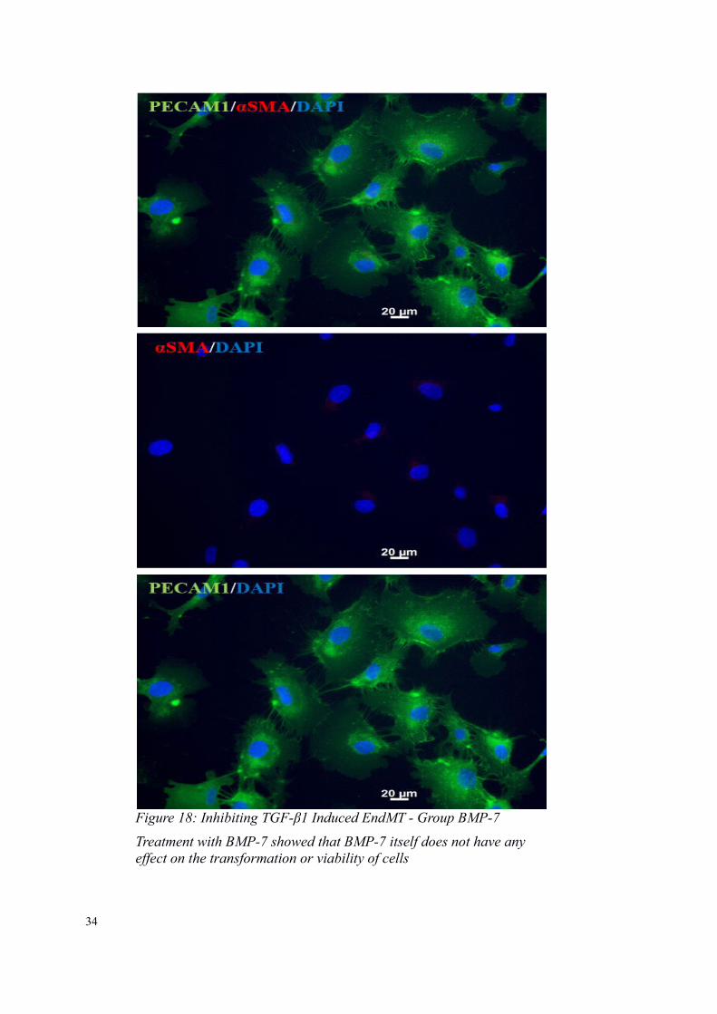
Figure 18: Inhibiting TGF-β1 Induced EndMT - Group BMP-7
Treatment with BMP-7 showed that BMP-7 itself does not have any effect on the transformation or viability of cells
35
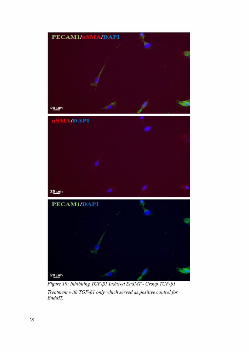
Figure 19: Inhibiting TGF-β1 Induced EndMT - Group TGF-β1
Treatment with TGF-β1 only which served as positive control for EndMT.
36
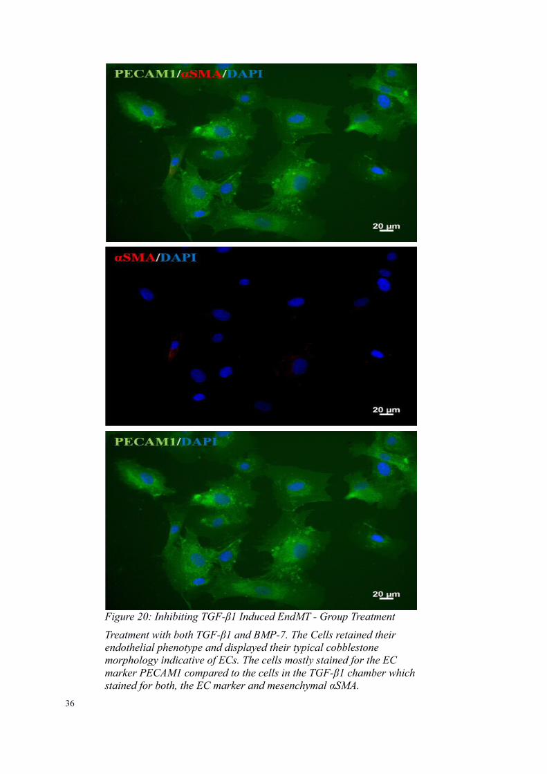
Figure 20: Inhibiting TGF-β1 Induced EndMT - Group Treatment
Treatment with both TGF-β1 and BMP-7. The Cells retained their endothelial phenotype and displayed their typical cobblestone morphology indicative of ECs. The cells mostly stained for the EC marker PECAM1 compared to the cells in the TGF-β1 chamber whichstained for both, the EC marker and mesenchymal αSMA.
37
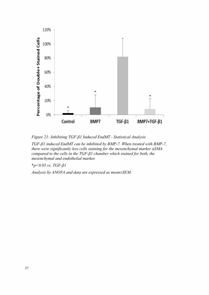
Figure 21: Inhibiting TGF-β1 Induced EndMT - Statistical Analysis
TGF-β1 induced EndMT can be inhibited by BMP-7. When treated with BMP-7,there were significantly less cells staining for the mesenchymal marker αSMA compared to the cells in the TGF-β1 chamber which stained for both, the mesenchymal and endothelial marker.
*p<0.05 vs. TGF-β1
Analysis by ANOVA and data are expressed as mean±SEM.
*
**
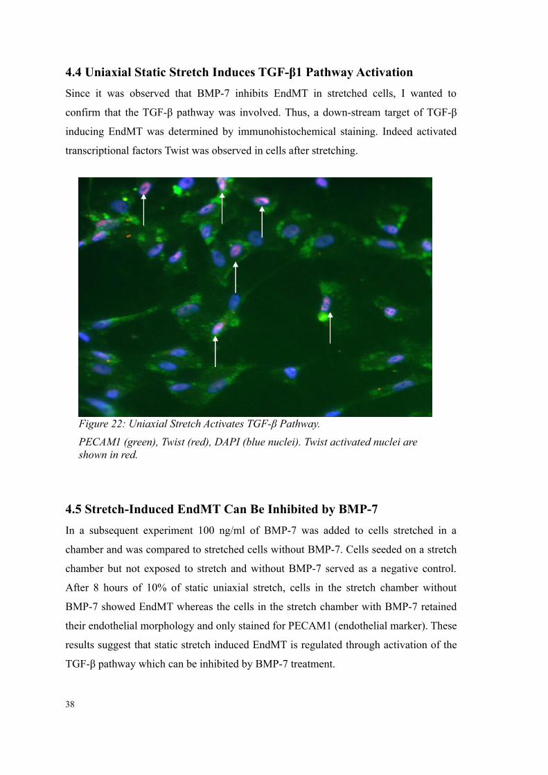
4.4 Uniaxial Static Stretch Induces TGF-β1 Pathway Activation
Since it was observed that BMP-7 inhibits EndMT in stretched cells, I wanted to
confirm that the TGF-β pathway was involved. Thus, a down-stream target of TGF-β
inducing EndMT was determined by immunohistochemical staining. Indeed activated
transcriptional factors Twist was observed in cells after stretching.
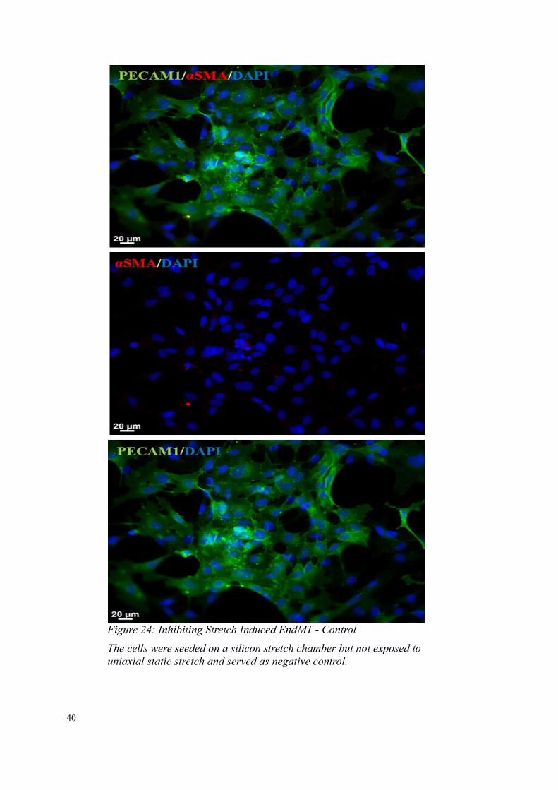
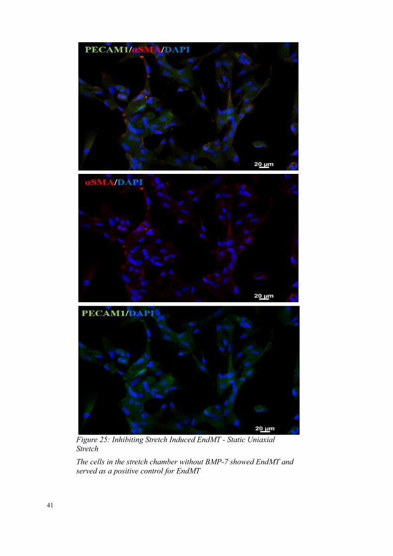
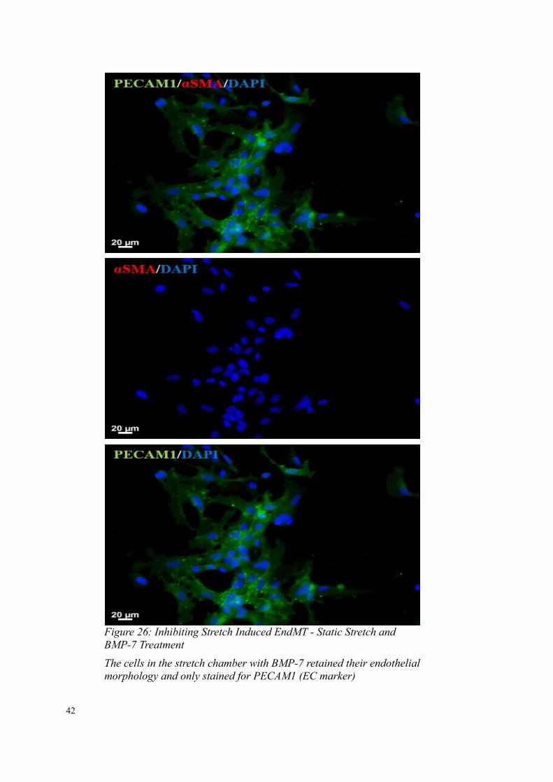
4.5 Stretch-Induced EndMT Can Be Inhibited by BMP-7
In a subsequent experiment 100 ng/ml of BMP-7 was added to cells stretched in a
chamber and was compared to stretched cells without BMP-7. Cells seeded on a stretch
chamber but not exposed to stretch and without BMP-7 served as a negative control.
After 8 hours of 10% of static uniaxial stretch, cells in the stretch chamber without
BMP-7 showed EndMT whereas the cells in the stretch chamber with BMP-7 retained
their endothelial morphology and only stained for PECAM1 (endothelial marker). These
results suggest that static stretch induced EndMT is regulated through activation of the
TGF-β pathway which can be inhibited by BMP-7 treatment.
38
Figure 22: Uniaxial Stretch Activates TGF-β Pathway.
PECAM1 (green), Twist (red), DAPI (blue nuclei). Twist activated nuclei are shown in red.
39
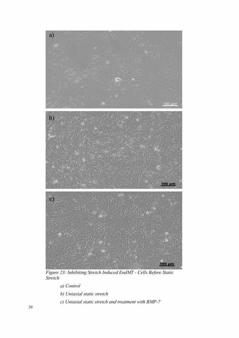
Figure 23: Inhibiting Stretch Induced EndMT - Cells Before Static Stretch
a) Control
b) Uniaxial static stretch
c) Uniaxial static stretch and treatment with BMP-7
40
Figure 24: Inhibiting Stretch Induced EndMT - Control
The cells were seeded on a silicon stretch chamber but not exposed touniaxial static stretch and served as negative control.
41
Figure 25: Inhibiting Stretch Induced EndMT - Static Uniaxial Stretch
The cells in the stretch chamber without BMP-7 showed EndMT and served as a positive control for EndMT
42
Figure 26: Inhibiting Stretch Induced EndMT - Static Stretch and BMP-7 Treatment
The cells in the stretch chamber with BMP-7 retained their endothelialmorphology and only stained for PECAM1 (EC marker)

43
Figure 27: Inhibiting Stretch Induced EndMT
Uniaxial static stretch induces EndMT which can be inhibited by BMP-7; a) Control, b) Uniaxial static stretch, c) Uniaxial static stretch and BMP-7 treatment
a)
b)
c)
4.6 BMP-7 Supplied on a Drug Carrier
In regards to local treatment, gelatin sponges were used as carrier for BMP-7 in the cell
stretch experiments. Instead of adding BMP-7 to the media, a gelatin sponge was
soaked with BMP-7 and then added to the media of the stretch chamber. The set up was
the same as the experiment “Uniaxial Stretch and Treatment with BMP-7” in order to
compare the results and cell viability. It was shown that the stretched cells with the
gelatin sponge preserved the endothelial phenotype and did not induce EndMT
compared to the stretched cells only containing the media. Cell viability was maintained
throughout the entire experiment. These results are promising for further investigations
on collagen-based BMP-7 carriers in order to inhibit EndMT locally preventing side
effects occurring with systemic application.
44
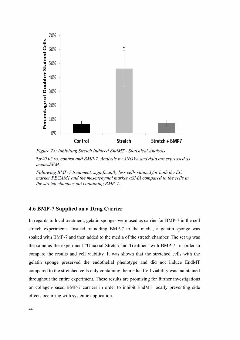
Figure 28: Inhibiting Stretch Induced EndMT - Statistical Analysis
*p<0.05 vs. control and BMP-7. Analysis by ANOVA and data are expressed as mean±SEM.
Following BMP-7 treatment, significantly less cells stained for both the EC marker PECAM1 and the mesenchymal marker αSMA compared to the cells in the stretch chamber not containing BMP-7.
*
45
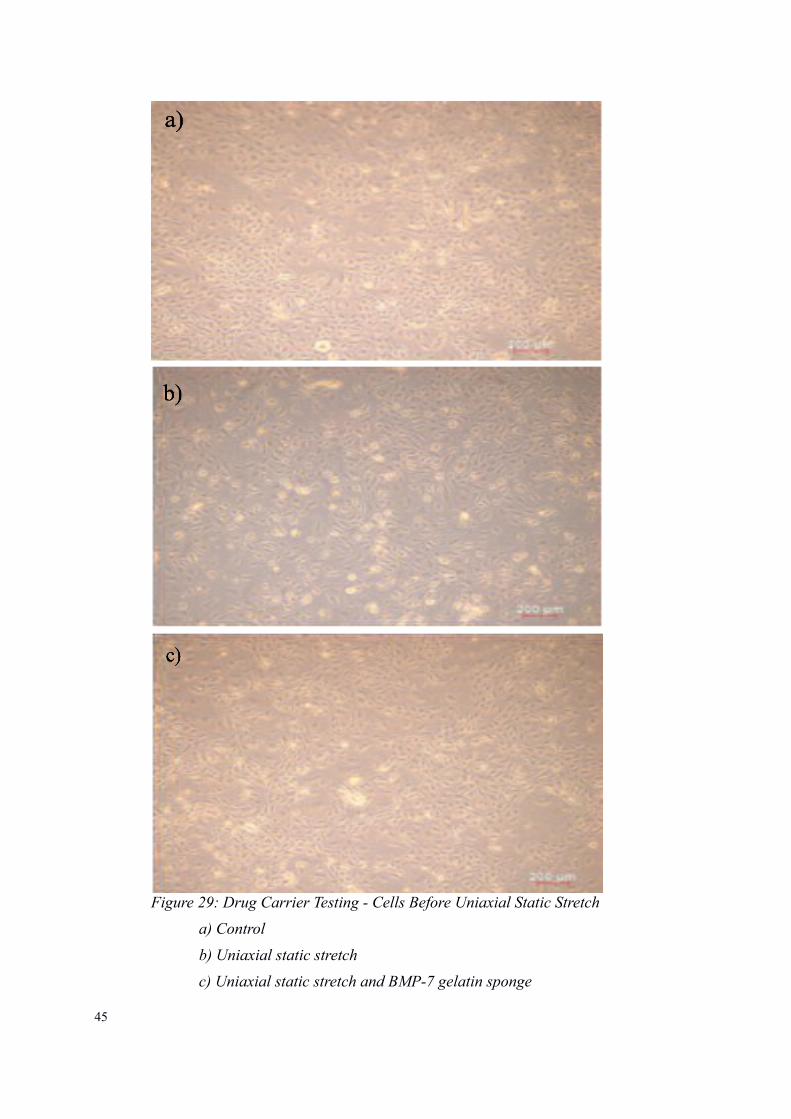
Figure 29: Drug Carrier Testing - Cells Before Uniaxial Static Stretch
a) Control
b) Uniaxial static stretch
c) Uniaxial static stretch and BMP-7 gelatin sponge
46
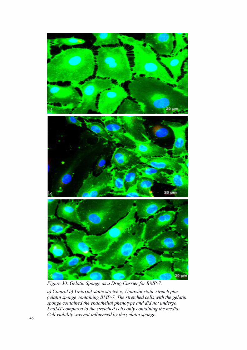
Figure 30: Gelatin Sponge as a Drug Carrier for BMP-7.
a) Control b) Uniaxial static stretch c) Uniaxial static stretch plus gelatin sponge containing BMP-7. The stretched cells with the gelatin sponge contained the endothelial phenotype and did not undergo EndMT compared to the stretched cells only containing the media. Cell viability was not influenced by the gelatin sponge.
a)
b)
c)
47
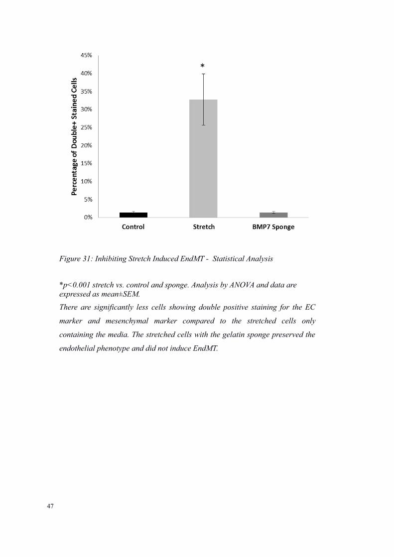
Figure 31: Inhibiting Stretch Induced EndMT - Statistical Analysis
*p<0.001 stretch vs. control and sponge. Analysis by ANOVA and data are expressed as mean±SEM.
There are significantly less cells showing double positive staining for the EC
marker and mesenchymal marker compared to the stretched cells only
containing the media. The stretched cells with the gelatin sponge preserved the
endothelial phenotype and did not induce EndMT.
5. Discussion
5.1 Summary
Even in this day of age, treatment of congenital heart disease still faces major obstacles.
Despite advances in early diagnosis and treatment of severe congenital heart defects
such as HLHS, mortality and morbidity due to the palliative nature of the reconstruction
remain a challenge. Following a three-step surgery performed within the first weeks to
months of life, the majority of patients reach adulthood nowadays but are left with a
single ventricle physiology where the anatomically right ventricle supports the systemic
circulation (Arnold, Loukanov and Gorenflo, 2014). As these patients reach 20 and 30
years of age, the prevalence of complications rises affecting not only the cardiovascular
system but multiple other organ systems as well (Egbe et al., 2016). With advances in
fetal intervention, even complex lesions such as HLHS have become amenable to
treatment modifications which significantly improved the long-term outcome for a
subset of patients. However, there is more to be accomplished by focusing on
pathologies aggravating disease progression pre- and postnatally.
In order to achieve the best possible outcome and prevent heart transplantation and
complications, therapeutic strategies have to be adjusted to anatomical variabilities in
each HLHS patient (Moon-Grady, Moore and Azakie, 2011). This should especially be
considered in the milder borderline HLHS patients where a biventricular repair might be
feasible. One limiting factor in borderline HLHS, is the presence of EFE tissue which
impedes diastolic compliance and growth of the ventricle (McElhinney, Vogel, et al.,
2010; Friedman et al., 2014). Therapeutically targeting EFE through surgical resection
postnatally has shown to benefit LV growth when this novel procedure was introduced
over a decade ago at Boston Children's Hospital (Emani et al., 2009). Several publica-
tions by Boston Children's Hospital have shown that resection of EFE significantly im-
proves outcome of patients with borderline LV by allowing catch-up growth of the LV
(Tworetzky et al., 2005; Emani et al., 2009). As diagnosis of HLHS is made during rou-
tine prenatal echocardiography around 18-20 weeks of gestation and fetal cardiac inter-
ventions such as aortic valvotomy are already in place at specialized institutions around
the world, EFE inhibition could be attempted during fetal life and subsequently postna-
tally as well in order to recruit the LV with the goal of biventricular physiology (Connor
48
and Thiagarajan, 2007). However, there are still a lot of unanswered questions regarding
EFE formation and progression which need to be addressed in order to develop local-
ized treatment options.
The goal of this project was to shed light on the pathomechanism of EFE development
and progression in HLHS. There are three major hypotheses regarding the cause of EFE
formation, including ischemia, infection and mechanical injury. EFE has been reported
in association with mumps, coxsackie, adenovirus or lactobacillus induced myocarditis,
arguing for an infectious origin (Fruhling L, Korn R, LaVillaureix J, Surjus A, 1962;
Amrikachi et al., 1997; Ni et al., 1997). With regard to ischemia, EFE has been detected
in vascular conditions such as myocardial infarction, twin-twin transfusion and
lymphatic obstruction (Lazda, no date; KLINE et al., 1964; Hutchins and Bannayan,
1971). The majority of EFE cases is seen in patients with dilated, restrictive or
hypertrophic cardiomyopathies and congenital malformations such as aortic stenosis,
coarctation of the aorta, anomalous coronary artery, intracranial arteriovenous fistula
and especially HLHS (Bland, White and Garland, 1933; OPPENHEIMER, 1953;
ANDERSEN and KELLY, 1956; Ursell et al., 1984; Newbould, Armstrong and Barson,
1991; Lurie, 2010). The latter indicates that the majority of EFE cases were associated
with mechanical insults on the ventricle due to pressure or volume overload (Lurie,
2010). Cardiac fibrosis is also a consequence of mechanical strain on the ventricle but
unlike cardiac fibrosis, EFE presents as a thickening of the endocardial layer only
without myocardial involvement (Lurie, 2010). The thickening of the endocardium
without myocardial involvement, points toward an endocardial endothelial origin which
has been confirmed in a recent study from Boston Children’s Hospital: EFE resected
during routine LV rehabilitation surgery was examined and revealed that EFE derives
from endocardial ECs undergoing transformation into mesenchymal cells through an
imbalance of the BMP/TGF-β1 pathway (Xu et al., 2015).
TGF-β1 is the main
stimulator of EndMT and BMP-7 has been shown to inhibit this process. In my cell
culture model, I established that isolated ECs follow the same stimulator and inhibitor
signaling for the regulation of EndMT. When the balance of the growth factors in the
media was changed to TGF-β, a known stimulator of EndMT, HCAEC changed
morphology. The cells underwent a transformation from a hexagonal shape which is the
typical appearance of endothelial and endocardial ECs to a spindle-shaped phenotype
49
which is indicative for fibroblasts. To confirm the light microscopic appearance, cells
were stained with markers for ECs and fibroblasts, respectively. The presence of active
EndMT was confirmed by visualizing and counting all cells which stained for both an
EC marker and a fibroblast marker at the same time. The mesenchymal marker αSMA
was used instead of FSP1, since it has been reported that FSP1 is not specific for all
fibroblasts in cardiac remodeling and fibrosis (Kong et al., 2013). In addition, double-
stained cells were identified which are indicative of ECs actively undergoing EndMT.
EndMT is a physiological process seen in heart development: the endocardial ECs in the
atrioventricular canal give rise to the mesenchymal endocardial cushion cells which
form the heart valves and septum (Yoshimatsu and Watabe, 2011). Furthermore, there is
indication that EndMT may play a role in embryonic vascular development and
formation of intimal thickening (Arciniegas et al.). However, EndMT is an important
mechanism in pathologies of several organ systems as well. EndMT recently presented
as new therapeutic target for fibrotic disorders as it was identified as key player in the
pathogenesis of pulmonary, intestinal, cardiac and kidney fibrosis (Zeisberg et al., 2007,
2008; Rieder et al., 2011). It was demonstrated that ECs do not only contribute to the
etiology of kidney fibrosis but furthermore to the progression of diabetic nephropathy as
latest studies indicate (Zeisberg et al., 2008; Li, Qu and Bertram, 2009). In myocardial
infarction EndMT may be a source of cardiac tissue repair after infarction and acute
ischemic injury (Aisagbonhi et al., 2011). Furthermore, in whole heart models of
mechanical stretch such as cardiac hypertrophy and dyssynchronous pacing induced
heart failure, EndMT was upregulated leading to cardiac fibrosis (Zeisberg et al., 2007;
Mai et al., 2014; Illigens et al., 2017). Even in the pathology of cancer it was reported
that cells achieve the ability to migrate and transform into malignant cells via EndMT
(Kalluri and Zeisberg, 2006). The main regulator is TGF-β but the stimulating events for
TGF-β release are not well described. One aim of this study was to establish a potential
trigger for TGF-β release and subsequent EndMT stimulation and thus, making this
pathomechanism amenable for treatment potentially as early as the fetal stage.
To further elucidate on the triggers for TGF-β release, we evaluated the clinical
progression of the disease. Clinical observations indicate that mechanical strain such as
distention of the LV in utero negatively impact progression of EFE formation. This was
50
the basis to further pursue the notion that distention of the LV myocardium might play a
significant role in the development and progression of EndMT. It is already shown that
mechanical forces can lead to changes in EC behavior. In general ECs provide a
selective permeability wall between the vessel wall and the blood and obtain metabolic
and synthetic functions reacting to physical and chemical stimuli (Sumpio, Riley and
Dardik, 2002). In the pathogenesis of atherosclerosis it was shown that laminar blood
flow and high fluid shear stress are protective for ECs by inducing an atheroprotective
gene program (Traub and Berk, 1998). On the contrary, atherosclerosis is seen in areas
with turbulent blood flow and low fluid shear stress (Yoshizumi et al., 2003). ECs need
to be stabilized by their environment through laminar blood flow and shear forces which
could be inhibited by the thickened endocardium due to EFE formation and lack of flow
due to aortic stenosis as well. In cell culture models using human umbilical vein
endothelial cells it has also been reported that cyclic stretch can induce EndMT through
an integrin β1 pathway which further supports the notion that endothelial cells respond
to mechanical forces by undergoing EndMT (Suzuki et al., 1997). Based on this
evidence, an in vitro model was developed mimicking the clinical situation. Human
coronary artery endothelial cells were used since they share the same embryogenic
origin as endocardial cells (Wu et al., 2012). To mimic LV distention, a cell culture
model was created in which cells were exposed to uniaxial static stretch while seeded on
stretching chambers. Following testing of several time frames and stretch amounts, I
determined that exposure of endothelial cells to 10% stretch over an 8-hour period
induced equivalent amount of cells undergoing EndMT as treatment with TGF-β. These
results suggested a mechanical trigger in the pathomechanism of EFE development.
EndMT through static stretch was mediated by the TGF-β1 pathway which was
confirmed by detecting a down-stream transcription factor (Twist) which regulates
EndMT through downregulation of VE-cadherin. Therefore, the stretched cells were
stained for Twist and co-localization with nuclei was determined. Twist is a member of a
large protein family called basic helix-loop-helix (bHLH) transcription factors. Each of
these proteins include a region called the bHLH domain which enables them to target a
special sequence of DNA. It is a negative regulator of endothelial gene expression but
also supports mesenchymal gene expression (Peinado, Olmeda and Cano, 2007;
Khanbabaei, Teimoori and Mohammadi, 2016). The positive staining for the
51
intranuclear marker Twist in the stretched cells indicates that uniaxial static stretch
activates the TGF-β1 pathway and induces EndMT. It was found that mechanical stress
activated the TGF-β pathway and induced EndMT equivalent to exposing resident
HCAEC in culture to TGF-β1. These results provide insight into a possible cause for
EndMT resulting in thickening of the endocardium (i.e. endocardial fibroelastosis).
These data also confirm EndMT by showing loss of endothelial function through lack of
uptake of acydilated LDL.
As a next step, we were interested to determine whether TGF-β could be inhibited to
block EndMT formation. It has previously been reported that EFE formation in HLHS is
caused by an imbalance of the TGF-β and BMP signaling with an upregulation of TGF-
β and downregulation of BMP proteins promoting EndMT of endocardial endothelial
cells lining the LV (Xu et al., 2015). As indicated by a paper by Xu et al., epigenetic
modification of the BMP-7 promoter plays a key role in the misbalance of the TGF-
β/BMP pathway in EFE tissue obtained from HLHS patients (Xu et al., 2015). My cell
culture model of TGF-β induced EndMT was used to establish that BMP-7 was a
suitable inhibitor of the process and provided data for BMP-7 dosage. Uniaxial stretch
experiments were designed to supply BMP-7 during the entire time ECs were exposed
to the mechanical stimulus. Stretched cells without BMP-7 treatment underwent EndMT
while those treated with BMP-7 retained their endothelial phenotype and only showed a
few double-stained cells indicative of EndMT. It could be shown that BMP-7
significantly reduces the number of ECs undergoing EndMT when either exposed to
TGF-β1 alone, or when exposed to uniaxial static stretch which stimulated the TGF- β1
pathway as indicated by Twist activation. TGF-β and BMP pathways are in balance
during development and tipping the balance toward TGF-β results in EndMT (Xu et al.,
2015). Looking at previous studies it has been shown that BMP-7 treatment could
successfully inhibit fibrosis of the heart and kidneys (Zeisberg et al., 2003,
2007). Furthermore, clinical trials are currently on the way to test BMP-7 derivatives
which were introduced by Kalluri et. al in order to treat fibrosis (Sugimoto et al., 2012).
These BMP-7 based small peptide agonists of BMP signaling are already in their first
clinical trials for treatment of renal fibrosis and other fibrotic diseases. In this trial THR-
184, a peptid agonist of the BMP-7 pathway, is administered intravenously to patients
who are scheduled for cardiac surgery and are at increased risk to develop acute kidney
52
injury (Clinicaltrials.gov, Andreas Orfanos, 2013). As my results indicate, BMP-7 was
able to prevent EndMT both induced by TGF-β1 and by uniaxial static stretch. These
results support the conclusion that BMP-7, a drug already FDA improved for orthopedic
lesions, could be a therapeutic agent to treat EFE and consequently preserve the growth
potential of the LV and successfully alter the course for patients diagnosed with
imminent HLHS. Treatment to inhibit EFE formation could be used as adjunct therapy
to post-natal surgical resection of EFE to prevent recurrence of EFE which is currently
requiring subsequent resection in several cases.
From a clinical point of view, BMP-7 treatment is a reasonable approach, however, not
as systemic application. Considering the fact that BMP-7 is involved in the
developmental process of several organs such as skeletal, kidney, and brown adipose
tissue development, the next part of the study aimed for a local treatment option in order
to avoid adverse effects of systemic treatment (Manson et al., 2015; Seale, 2015;
Salazar, Gamer and Rosen, 2016). BMP-7 is already used for special bone fractures
where BMP-7 is applied locally through an absorbable gelatin sponge. BMPs have been
in clinical use for local treatment of specific orthopedic lesions for a decade and are
FDA approved for use in humans for non-union, tibial bone fractures and spinal fusion
(White et al., 2007). BMP-7 (osteogenic protein 1 (OP-1); Stryker Biotech, Hopkinton,
MA) and BMP2 (rhBMP-2, (InFUSE); Medtronic Sofamor Danek, Memphis, TN) are
the two BMPs which are currently available for clinical applications and are
manufactured by mammalian cell expression (Bishop and Einhorn, 2007). Collagen-
based carriers containing BMP-7 are the basis for some of these applications in order to
maintain the concentration of BMP-7. Stryker Biotech is using a purified type I bovine
bone collagen powder as a carrier (Stryker, 2017). Whereas BMP-2 is placed on
absorbable gelatin sponges (Medtronic, 2017). There are also reports about injectable
hydrogel containing BMP-7 (Dyondi, Webster and Banerjee, 2013). Aiming for a
possible local treatment option in HLHS patients, experiments were started with BMP-7
loaded gelatin sponges to assess cell viability and interference with protein activity. One
mm² gelatin sponges were added to cells in the stretch chambers and control cells, and
were compared to cells in stretch chambers where BMP-7 was contained in the media.
No adverse events on cell viability as indicated by trypan blue staining was observed
through gelatin sponges. BMP-7 supplied on a gelatin-based drug carrier was equally as
53
effective to inhibit EndMT as when added to the media. In terms of clinical relevance,
this is important information since the goal is to supply an EndMT inhibitor directly at
the source of the disease which would allow for locally applied BMP-7 during surgical
repair of HLHS. When EFE is resected in the setting of surgery, a BMP-7 sponge could
inhibit EFE in areas of incomplete resection and also prevent re-development of EFE.
The young patient of HLHS would also benefit of a localized drug treatment instead of
a surgical one. In the long run, there could be the possibility of placing BMP-7
containing drug delivery devices into the cavity of the LV as adjunct therapy to fetal
aortic valvuloplasty to inhibit EFE in the very beginning with better chances of recovery
of the LV. BMP-7 treatment for EFE could also be a breakthrough in other patient
groups as EndMT is a process found in several pathologies in the heart, kidney and
lungs as well as in cancer pathology (Zeisberg et al., 2007, 2008).
5.2 Limitations
Two blinded investigators analyzed all immunohistochemical data but it is
acknowledged that the methodology has inherent problems. ECs from the coronary
arteries and not endocardial ECs were used but preliminary data indicate that rat
endocardial cell culture which we have already established show the same results.
Experiments in vitro alone are not necessarily equivalent to the in vivo situation but
animal data indicate that there is a close correlation and more in vivo experiments are
currently performed (Shimada et al., 2014). Furthermore, direct access to EFE tissue
will also lead to isolation of cells further analysis of the same mechanical stimuli and
inhibitor studies as described here.
5.3 Perspectives and Conclusion
The resection of EFE tissue in HLHS patients can improve the outcome of function of
the left ventricle (Tworetzky et al., 2005). Therefore, targeting EFE opens the field for
new therapeutic possibilities in the treatment of HLHS. Regarding EFE, it is well
established that the tissue is derived from ECs of the endocardium through a process
called EndMT which transforms endothelial cells into collagen producing mesenchymal
54
cells (Xu et al., 2015). This study focused on the cause of EndMT development and
elucidate on possible treatment options. For this purpose, an EndMT cell model was
successfully created by treating cells with TGF-β1 to investigate possible treatment
options. With regard to the finding of mechanical distention triggering EFE, cells were
exposed to uniaxial static stretch which was used as second experimental set-up. It was
found that uniaxial static stretch can induce EndMT in ECs by activation of the TGF-β
pathway. In both EndMT models, HCAEC underwent significantly less EndMT when
treated with BMP-7, a TGF-β pathway inhibitor. Based on FDA approved BMP-7
options (White et al., 2007), collagen carriers for orthopedic lesions, the influence on
cell viability of gelatin sponges was tested. These cell models gave more insight on the
pathomechanism of EndMT and could serve to develop possible local treatment options
with BMP-7 as therapeutic agent. In the future, it should be investigated whether
inhibition of EndMT induced by uniaxial static stretch is possible. This could be
achieved by reducing the stretch membranes to their original length or by treating the
cells with BMP-7 after stretching. Furthermore, it would be interesting to know whether
other mechanical stimuli such as cyclic stretch and/or in combination with changes in
flow would also induce EndMT. Also, there will be an emphasis on the development of
a drug eluting device for local treatment with BMP-7 which can be ideally applied in
utero during the aortic valvuloplasty procedure. As in most cases EFE already exists at
the time when aortic valvuloplasty is installed, it is even more important to find out the
impact of BMP-7 when cells already have undergone EndMT. In the settings of this
study an isolation method for endocardial cells from the left ventricle of rat hearts was
established. These cells could serve for a new cell culture EndMT model in the future.
All in all, this model provides insight on the pathomechanism of EFE development with
regard to BMP-7 being a possible agent for local treatment of EFE in HLHS patients.
The cell culture results so far are promising but need to be repeated with rat endocardial
cells which I have already established a new protocol for isolation and maintenance in
culture. This could be a life changing therapy for patients with univentricular Fontan
circulation who eventually end up with heart failure in their 30s. Preventing EFE in
HLHS patients and establishing a better hemodynamics in these patients, could improve
the outcome of the LV and maybe even lead to biventricular repair with less mortality
and co-morbidity.
55
6. Summary
Introduction: Successful postnatal surgical removal of Endocardial Fibroelastosis
(EFE) can improve the outcome of Hypoplastic Left Heart Syndrome, one of the
severest congenital heart failures by preserving ventricular function and growth
potential (Xu et al., 2015). It has been shown that the transformation of endothelial to
mesenchymal cells (EndMT) leads to the development of EFE tissue (Xu et al.,
2015). The pathomechanism of EFE is still unknown and was investigated in this study
to develop local prenatal treatment options.
Methods: Cells from human coronary arteries were used for the experiments as they
have the same embryonic origin as endocardial cells (Wu et al., 2012). The cells were
exposed to 10ng/ml TGF-β1, 100 ng/ml rhBMP-7 (=TGF-β1 inhibitor), 10 ng/ml TGF-
β1 plus 100 ng/ml rhBMP-7 for 72 hours or exposed to 10% of uniaxial static stretch for
8 hours. Cells treated with media only served as control.
Results: The treatment with TGF-β1 as well as uniaxial static stretch of already 10%
changes cell morphology. Uniaxial static stretch is also regulated by TGF-β shown by
the activation of transcriptional factors of the TGF-β1 pathway (Twist) in the nucleus.
The cells did not only express the endothelial marker PECAM1 as in the control but in
addition stained for the mesenchymal marker αSMA showing active EndMT. After
treating the cells with BMP-7 to inhibit the TGF-β pathway there were significantly less
cells undergoing transformation from an endothelial to a mesenchymal cell.
Conclusion: This in vitro model showed that uniaxial static stretch activates the growth
factor TGF-β and induces EndMT which can be inhibited by BMP-7. That information
makes it possible to develop therapeutic strategies for local treatment in utero.
56
7. Zusammenfassung
Einleitung: Die erfolgreiche postnatale Entfernung der Endokardfibroelastose (EFE)
kann das Outcome der Ventrikelfunktion beim Hypoplastischen Linksherz, einem der
schwersten angeborenen Herzfehler, deutlich verbessern (Xu et al., 2015). Als Ursache
für die EFE Entstehung wurde von uns die Transformation von Endothelzellen zu mes-
enchymalen Zellen (EndMt) nachgewiesen (Xu et al., 2015). Der Pathomechanismus
von EFE-Entwicklung durch EndMt ist aber unbekannt und wurde in dieser Studie un-
tersucht um potenziell pränatale Lokaltherapien entwickeln zu können.
Material und Methoden: Zellkulturen von menschlichen Koronararterien (HCAEC)
wurden verwendet, da sie den gleichen embryologischen Ursprung wie Endokardzellen
haben (Wu et al., 2012). Die Zellen wurden für 72h mit 10ng/ml TGF-β1, 100 ng/ml
rhBMP-7 (=TGF-β1 inhibitor), 10 ng/ml TGF-β1 plus 100 ng/ml rhBMP-7 behandelt,
oder 8 Stunden statischem Stretch von 10% in Stretchkammern aus Silikon ausgesetzt.
Als Kontrolle wurden Zellen nur mit Nährmedium behandelt.
Ergebnisse: Die Gabe von TGF-β1 und auch statischer Zug von bereits 10% veränderte
die Zellmorphologie. Statischer Zellzug unterliegt ebenfalls der Kontrolle von TGF-β,
was durch die Aktivierung von Transkriptionsfaktoren (Twist) des TGF-β Signalweges
im Nuclus gezeigt werden konnte. Die Zellen exprimieren nicht nur den Endothelzell-
marker PECAM1 wie in der Kontrollgruppe, sondern zusätzlich auch den mesenchy-
malen Marker αSMA als Indiz für aktive EndMT. Mit der gleichzeitigen Gabe von
BMP-7 zur Blockierung des TGF-β Pathway, zeigen sich signifikant weniger Zellen, die
einer Transformation von endothelialer zur mesenchymalen Zelle unterlaufen.
Schlussfolgerung: Durch das in vitro Modell konnte auf mehreren Ebenen gezeigt wer-
den, dass statisch-mechanischer Zug den Wachstumsfaktor TGF-β aktiviert und EndMt
induziert, was durch BMP-7 inhibiert werden kann. Diese Informationen lassen es nun
zu, dass pharmakologische Interventionen entwickelt werden, die idealerweise zur Ther-
apie in utero verwendet werden können.
57
8. Abbreviations
AB Antibody
ALK Activine receptor-like kinase receptors
AS Aortic Stenosis
A-V Atrioventricular
BB Blocking Buffer
bHLH Basic helix-loop-helix
BMP Bone morphogenetic proteins
CAFs Cancer associated fibroblasts
DAPI 4',6-diamidino-2-phenylindole
DMSO Dimethyl sulfoxide
EBM-2 Endothelial cell basal medium-2
EC Endothelial cell
EDTA Ethylene-Diamine-Tetra-Acetic acid
EFE Endocardial fibroelastosis
EGM-2 MV Endothelial cell growth medium
EMT Epithelial to mesenchymal transition
EndMT Endothelial to mesenchymal transition
FBS Fetal bovine serum
FOP Fibrodysplasia ossificans progressive
GA-1000 30 mg/ml of Gentamicin, 15 μg/ml Amphotericin
hEGF Human epidermal growth factor
hFGF-β Human acidic fibroblast growth factor 1
HLHS Hypoplastic left heart syndrome
HT Heart transplantation
58
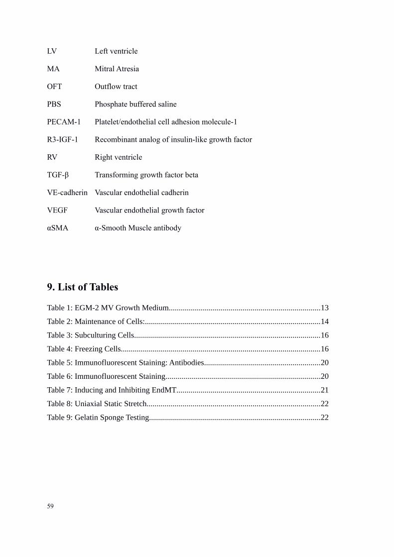
LV Left ventricle
MA Mitral Atresia
OFT Outflow tract
PBS Phosphate buffered saline
PECAM-1 Platelet/endothelial cell adhesion molecule-1
R3-IGF-1 Recombinant analog of insulin-like growth factor
RV Right ventricle
TGF-β Transforming growth factor beta
VE-cadherin Vascular endothelial cadherin
VEGF Vascular endothelial growth factor
αSMA α-Smooth Muscle antibody
9. List of Tables
Table 1: EGM-2 MV Growth Medium............................................................................13
Table 2: Maintenance of Cells:........................................................................................14
Table 3: Subculturing Cells.............................................................................................16
Table 4: Freezing Cells....................................................................................................16
Table 5: Immunofluorescent Staining: Antibodies..........................................................20
Table 6: Immunofluorescent Staining..............................................................................20
Table 7: Inducing and Inhibiting EndMT........................................................................21
Table 8: Uniaxial Static Stretch.......................................................................................22
Table 9: Gelatin Sponge Testing......................................................................................22
59
10. List of Figures
Figure 1: Schematic of the Heart Structure and Blood Flow............................................2
Figure 2: Macroscopic EFE Tissue....................................................................................7
Figure 3: Endothelial-to-Mesenchymal Transformation (EndMT)...................................9
Figure 4: TGF-β/BMP Axis Regulates EndMT...............................................................11
Figure 5: Silicone Stretch Chambers...............................................................................18
Figure 6: Stretching Machine (STREX from B-Bridge International Inc)......................18
Figure 7: TGF- β1 Treatment - Cells Before Treatment..................................................23
Figure 8: TGF- β1 Treatment - Control...........................................................................24
Figure 9: TGF- β1 Treatment - Treatment.......................................................................25
Figure 10: Uniaxial Static Stretch of 10% - Cells Before Treatment..............................26
Figure 11: Uniaxial Static Stretch of 10% - Control.......................................................27
Figure 12: Uniaxial Static Stretch of 10% - Static Stretch..............................................28
Figure 13: Treatment with Dil-Ac-LDL..........................................................................29
Figure 14: Inhibiting TGF-β1 Induced EndMT - Experiment Setup...............................30
Figure 15: Inhibiting TGF-β1 Induced EndMT - Before Treatment...............................31
Figure 16: Inhibiting TGF-β1 Induced EndMT - After Treatment..................................32
Figure 17: Inhibiting TGF-β1 Induced EndMT - Group Control....................................33
Figure 18: Inhibiting TGF-β1 Induced EndMT - Group BMP-7....................................34
Figure 19: Inhibiting TGF-β1 Induced EndMT - Group TGF-β1...................................35
Figure 20: Inhibiting TGF-β1 Induced EndMT - Group Treatment................................36
Figure 21: Inhibiting TGF-β1 Induced EndMT - Statistical Analysis.............................37
Figure 22: Uniaxial Stretch Activates TGF-β Pathway...................................................38
Figure 23: Inhibiting Stretch Induced EndMT - Cells Before Static Stretch..................39
Figure 24: Inhibiting Stretch Induced EndMT - Control.................................................40
Figure 25: Inhibiting Stretch Induced EndMT - Static Uniaxial Stretch.........................41
Figure 26: Inhibiting Stretch Induced EndMT - Static Stretch and BMP-7 Treatment...42
Figure 27: Inhibiting Stretch Induced EndMT................................................................43
Figure 28: Inhibiting Stretch Induced EndMT - Statistical Analysis..............................44
Figure 29: Drug Carrier Testing - Cells Before Uniaxial Static Stretch..........................45
Figure 30: Gelatin Sponge as a Drug Carrier for BMP-7................................................46
Figure 31: Inhibiting Stretch Induced EndMT - Statistical Analysis..............................47
60
11. References
Aisagbonhi, O., Rai, M., Ryzhov, S., Atria, N., Feoktistov, I. and Hatzopoulos, A. K. (2011) “Experimental myocardial infarction triggers canonical Wnt signaling and en-dothelial-to-mesenchymal transition.,” Disease models & mechanisms, 4(4), pp. 469–83. doi: 10.1242/dmm.006510.
Amrikachi, M., Lopez, L., Cutler, D. and Boor, P. J. (1997) “Lactobacillus Endocarditis:A Chronic Active Infection,” Cardiovascular Pathology, 6(6), pp. 341–344. doi: 10.1016/S1054-8807(97)00032-X.
ANDERSEN, D. H. and KELLY, J. (1956) “Endocardial fibro-elastosis. I. Endocardial fibro-elastosis associated with congenital malformations of the heart.,” Pediatrics, 18(4), pp. 513–38. Available at: http://www.ncbi.nlm.nih.gov/pubmed/13370221 (Ac-cessed: August 8, 2016).
Arciniegas, E., Neves, C. Y., Carrillo, L. M., Zambrano, E. A. and Ramírez, R. “En-dothelial-mesenchymal transition occurs during embryonic pulmonary artery develop-ment.,” Endothelium : journal of endothelial cell research, 12(4), pp. 193–200. doi: 10.1080/10623320500227283.
Arnold, R. R., Loukanov, T. and Gorenflo, M. (2014) “Hypoplastic left heart syndrome -unresolved issues.,” Frontiers in pediatrics, 2, p. 125. doi: 10.3389/fped.2014.00125.
Attisano, L. and Wrana, J. L. (2000) “Smads as transcriptional co-modulators.,” Currentopinion in cell biology, 12(2), pp. 235–43. Available at: http://www.ncbi.nlm.nih.gov/pubmed/10712925 (Accessed: June 3, 2016).
Biglino, G., Giardini, A., Hsia, T.-Y., Figliola, R., Taylor, A. M. and Schievano, S. (2013) “Modeling single ventricle physiology: review of engineering tools to study first stage palliation of hypoplastic left heart syndrome.,” Frontiers in pediatrics. Frontiers Media SA, 1, p. 31. doi: 10.3389/fped.2013.00031.
Bishop, G. B. and Einhorn, T. A. (2007) “Current and future clinical applications of bone morphogenetic proteins in orthopaedic trauma surgery.,” International or-thopaedics. Springer, 31(6), pp. 721–7. doi: 10.1007/s00264-007-0424-8.
Bland, E. F., White, P. D. and Garland, J. (1933) “Congenital anomalies of the coronary arteries: Report of an unusual case associated with cardiac hypertrophy,” American Heart Journal. Mosby, 8(6), pp. 787–801. doi: 10.1016/S0002-8703(33)90140-4.
Buyon, J. P. and Clancy, R. M. (2003) “Neonatal lupus syndromes.,” Current opinion inrheumatology, 15(5), pp. 535–41. Available at: http://www.ncbi.nlm.nih.gov/pubmed/12960477 (Accessed: May 10, 2016).
Centers for Disease Control (CDC) (1989) “Contribution of birth defects to infant mor-
61
tality--United States, 1986.,” MMWR. Morbidity and mortality weekly report, 38(37), pp. 633–5. Available at: http://www.ncbi.nlm.nih.gov/pubmed/2505044 (Accessed: April 4, 2018).
Chang, R.-K. R., Chen, A. Y. and Klitzner, T. S. (2002) “Clinical management of infantswith hypoplastic left heart syndrome in the United States, 1988-1997.,” Pediatrics, 110(2 Pt 1), pp. 292–8. Available at: http://www.ncbi.nlm.nih.gov/pubmed/12165581 (Accessed: May 7, 2016).
Clinicaltrials.gov, Andreas Orfanos, Mbbc. (2013) A Multi-Center, Parallel-Group, Randomized, Double Blind, Adaptive Study Investigating the Safety and Efficacy of THR-184 in Patients at Increased Risk of Developing Cardiac Surgery Associated-Acute Kidney Injury (CSA-AKI), dentifier: NCT01830920. Available at: https://clinical-trials.gov/ct2/show/NCT01830920.
Cohen, M. S. and Rychik, J. (1999) “The small left ventricle: How small is too small forbiventricular repair?,” Seminars in thoracic and cardiovascular surgery. Pediatric car-diac surgery annual, 2, pp. 189–202. Available at: http://www.ncbi.nlm.nih.gov/pubmed/11486236 (Accessed: May 28, 2016).
Connor, J. A. and Thiagarajan, R. (2007) “Hypoplastic left heart syndrome.,” Orphanet journal of rare diseases. BioMed Central, 2, p. 23. doi: 10.1186/1750-1172-2-23.
Cooley, B. C., Nevado, J., Mellad, J., Yang, D., St. Hilaire, C., Negro, a., Fang, F., Chen, G., San, H., Walts, a. D., Schwartzbeck, R. L., Taylor, B., Lanzer, J. D., Wragg, a., Elagha, a., Beltran, L. E., Berry, C., Feil, R., Virmani, R., Ladich, E., Kovacic, J. C. and Boehm, M. (2014) “TGF- Signaling Mediates Endothelial-to-Mesenchymal Transi-tion (EndMT) During Vein Graft Remodeling,” Science Translational Medicine, 6, p. 227ra34-227ra34. doi: 10.1126/scitranslmed.3006927.
Dyondi, D., Webster, T. J. and Banerjee, R. (2013) “A nanoparticulate injectable hydro-gel as a tissue engineering scaffold for multiple growth factor delivery for bone regener-ation.,” International journal of nanomedicine, 8, pp. 47–59. doi: 10.2147/IJN.S37953.
Egbe, A. C., Connolly, H. M., Niaz, T. and McLeod, C. J. (2016) “Outcome of direct current cardioversion for atrial arrhythmia in adult Fontan patients,” International Jour-nal of Cardiology, 208, pp. 115–119. doi: 10.1016/j.ijcard.2016.01.209.
Eisenberg, L. M. and Markwald, R. R. (1995) “Molecular regulation of atrioventricular valvuloseptal morphogenesis.,” Circulation research, 77(1), pp. 1–6. Available at: http://www.ncbi.nlm.nih.gov/pubmed/7788867 (Accessed: February 16, 2015).
Emani, S. M., Bacha, E. A., McElhinney, D. B., Marx, G. R., Tworetzky, W., Pigula, F. A. and del Nido, P. J. (2009) “Primary left ventricular rehabilitation is effective in main-taining two-ventricle physiology in the borderline left heart.,” The Journal of thoracic and cardiovascular surgery, 138(6), pp. 1276–82. doi: 10.1016/j.jtcvs.2009.08.009.
62
Emani, S. M., McElhinney, D. B., Tworetzky, W., Myers, P. O., Schroeder, B., Zu-rakowski, D., Pigula, F. A., Marx, G. R., Lock, J. E. and del Nido, P. J. (2012) “Staged left ventricular recruitment after single-ventricle palliation in patients with borderline left heart hypoplasia.,” Journal of the American College of Cardiology, 60(19), pp. 1966–74. doi: 10.1016/j.jacc.2012.07.041.
Enzensberger, C., Vogel, M., Degenhardt, J., Fass, N., Kawecki, A., Kohl, T., Widriani, E. and Axt-Fliedner, R. (2012) “Fetal pulmonary venous flow and restrictive foramen ovale in hypoplastic left heart.,” Ultraschall in der Medizin (Stuttgart, Germany : 1980),33(7), pp. E38-45. doi: 10.1055/s-0032-1325376.
Factor, S. M. (1978) “Endocardial fibroelastosis: myocardial and vascular alterations as-sociated with viral-like nuclear particles.,” American heart journal, 96(6), pp. 791–801. Available at: http://www.ncbi.nlm.nih.gov/pubmed/717242 (Accessed: May 10, 2016).
Feng, X.-H. and Derynck, R. (2005) “Specificity and versatility in tgf-beta signaling through Smads.,” Annual review of cell and developmental biology, 21, pp. 659–93. doi:10.1146/annurev.cellbio.21.022404.142018.
Friedman, K. G., Schidlow, D., Freud, L., Escobar-Diaz, M. and Tworetzky, W. (2014) “Left Ventricular Diastolic Function and Characteristics in Fetal Aortic Stenosis,” The American Journal of Cardiology, 114(1), pp. 122–127. doi: 10.1016/j.amjcard.2014.04.013.
Friehs, I., Illigens, B., Melnychenko, I., Zhong-Hu, T., Zeisberg, E. and Del Nido, P. J. (2013) “An animal model of endocardial fibroelastosis.,” The Journal of surgical re-search, 182(1), pp. 94–100. doi: 10.1016/j.jss.2012.07.069.
Fruhling L, Korn R, LaVillaureix J, Surjus A, F. S. (1962) “La myoendocardite chronique fibroelastique du nouveau-ne et du nourisson.,” Ann D’Anat Pathol, p. 7:227-303.
Gillum, R. F. (1994) “Epidemiology of congenital heart disease in the United States.,” American heart journal, 127(4 Pt 1), pp. 919–27. Available at: http://www.ncbi.nlm.ni-h.gov/pubmed/8154432 (Accessed: May 7, 2016).
von Gise, A. and Pu, W. T. (2012) “Endocardial and epicardial epithelial to mesenchy-mal transitions in heart development and disease.,” Circulation research. NIH Public Access, 110(12), pp. 1628–45. doi: 10.1161/CIRCRESAHA.111.259960.
Gordon, B. M., Rodriguez, S., Lee, M. and Chang, R.-K. (2008) “Decreasing number ofdeaths of infants with hypoplastic left heart syndrome.,” The Journal of pediatrics, 153(3), pp. 354–8. doi: 10.1016/j.jpeds.2008.03.009.
Grossfeld, P. (2007) “Hypoplastic left heart syndrome: new insights.,” Circulation re-search, 100(9), pp. 1246–8. doi: 10.1161/01.RES.0000268192.20525.c2.
63

Grossfeld, P., Ye, M. and Harvey, R. (2009) “Hypoplastic left heart syndrome: new ge-netic insights.,” Journal of the American College of Cardiology, 53(12), pp. 1072–4. doi: 10.1016/j.jacc.2008.12.024.
Hanukoglu, A., Fried, D. and Somekh, E. (1986) “Inheritance of familial primary endo-cardial fibroelastosis.,” Clinical pediatrics, 25(5), pp. 272–5. Available at: http://www.ncbi.nlm.nih.gov/pubmed/3698448 (Accessed: May 10, 2016).
Heldin, C. H., Miyazono, K. and ten Dijke, P. (1997) “TGF-beta signalling from cell membrane to nucleus through SMAD proteins.,” Nature, 390(6659), pp. 465–71. doi: 10.1038/37284.
Hinton, R. B., Martin, L. J., Tabangin, M. E., Mazwi, M. L., Cripe, L. H. and Benson, D. W. (2007) “Hypoplastic left heart syndrome is heritable.,” Journal of the American College of Cardiology, 50(16), pp. 1590–5. doi: 10.1016/j.jacc.2007.07.021.
Holoshitz, N., Kenny, D. and Hijazi, Z. M. (2014) “Hybrid Interventional Procedures in Congenital Heart Disease.,” Methodist DeBakey cardiovascular journal. Methodist De-Bakey Heart & Vascular Center, 10(2), pp. 93–98. Available at: /pmc/articles/PMC4117326/?report=abstract (Accessed: October 1, 2014).
Hua, J., Zhang, Z., Jiang, X., He, Y. and Chen, P. (2012) “[Relationship between en-dothelial-to-mesenchymal transition and cardiac fibrosis in acute viral myocarditis].,” Zhejiang da xue xue bao. Yi xue ban = Journal of Zhejiang University. Medical sci-ences, 41(3), pp. 298–304. Available at: http://www.ncbi.nlm.nih.gov/pubmed/22723165 (Accessed: January 10, 2016).
Hutchins, G. M. and Bannayan, G. A. (1971) “Development of endocardial fibroelasto-sis following myocardial infarction.,” Archives of pathology, 91(2), pp. 113–8. Availableat: http://www.ncbi.nlm.nih.gov/pubmed/5099882 (Accessed: August 8, 2016).
Iascone, M., Ciccone, R., Galletti, L., Marchetti, D., Seddio, F., Lincesso, A., Pezzoli, L., Vetro, A., Barachetti, D., Boni, L., Federici, D., Soto, A., Comas, J., Ferrazzi, P. and Zuffardi, O. (2012) “Identification of de novo mutations and rare variants in hypoplasticleft heart syndrome,” Clinical Genetics, 81(6), pp. 542–554. doi: 10.1111/j.1399-0004.2011.01674.x.
Illigens, B. M.-W., Casar Berazaluce, A., Poutias, D., Gasser, R., del Nido, P. J. and Friehs, I. (2017) “Vascular Endothelial Growth Factor Prevents Endothelial-to-Mes-enchymal Transition in Hypertrophy,” The Annals of Thoracic Surgery, 104(3), pp. 932–939. doi: 10.1016/j.athoracsur.2017.01.112.
Kalluri, R. and Zeisberg, M. (2006) “Fibroblasts in cancer.,” Nature reviews. Cancer, 6(5), pp. 392–401. doi: 10.1038/nrc1877.
Kaplinski, M. and Cohen, M. S. (2015) “Characterising adequacy or inadequacy of the
64
borderline left ventricle: what tools can we use?,” Cardiology in the young, 25(8), pp. 1482–8. doi: 10.1017/S1047951115002267.
Khanbabaei, H., Teimoori, A. and Mohammadi, M. (2016) “The interplay between mi-croRNAs and Twist1 transcription factor: a systematic review.,” Tumour biology : the journal of the International Society for Oncodevelopmental Biology and Medicine, 37(6), pp. 7007–19. doi: 10.1007/s13277-016-4960-y.
KLINE, I. K., MILLER, A. J., PICK, R. and KATZ, L. N. (1964) “THE RELATION-SHIP BETWEEN HUMAN ENDOCARDIAL FIBROELASTOSIS AND OBSTRUC-TION OF THE CARDIAC LYMPHATICS.,” Circulation, 30, pp. 728–35. Available at: http://www.ncbi.nlm.nih.gov/pubmed/14226171 (Accessed: August 8, 2016).
Kokudo, T., Suzuki, Y., Yoshimatsu, Y., Yamazaki, T., Watabe, T. and Miyazono, K. (2008) “Snail is required for TGFbeta-induced endothelial-mesenchymal transition of embryonic stem cell-derived endothelial cells.,” Journal of cell science, 121(Pt 20), pp. 3317–24. doi: 10.1242/jcs.028282.
Kong, P., Christia, P., Saxena, A., Su, Y. and Frangogiannis, N. G. (2013) “Lack of specificity of fibroblast-specific protein 1 in cardiac remodeling and fibrosis.,” Ameri-can journal of physiology. Heart and circulatory physiology, 305(9), pp. H1363-72. doi:10.1152/ajpheart.00395.2013.
Kulkarni, S. and Rao, S. (2012) “Definitive therapy for hypoplastic left heart syndrome - Indian scenario.,” Indian heart journal. Elsevier, 64(4), pp. 338–40. doi: 10.1016/j.ihj.2012.06.022.
Lazda, E. J. (no date) “Endocardial fibroelastosis in growth-discordant monozygotic twins.,” Pediatric and developmental pathology : the official journal of the Society for Pediatric Pathology and the Paediatric Pathology Society, 1(6), pp. 522–7. Available at: http://www.ncbi.nlm.nih.gov/pubmed/9724339 (Accessed: August 8, 2016).
Li, J., Qu, X. and Bertram, J. F. (2009) “Endothelial-Myofibroblast Transition Contrib-utes to the Early Development of Diabetic Renal Interstitial Fibrosis in Streptozotocin-Induced Diabetic Mice,” The American Journal of Pathology, 175(4), pp. 1380–1388. doi: 10.2353/ajpath.2009.090096.
Lin, F., Wang, N. and Zhang, T.-C. (2012) “The role of endothelial-mesenchymal transi-tion in development and pathological process.,” IUBMB life, 64(9), pp. 717–23. doi: 10.1002/iub.1059.
Lofland, G. K., McCrindle, B. W., Williams, W. G., Blackstone, E. H., Tchervenkov, C. I., Sittiwangkul, R. and Jonas, R. A. (2001) “Critical aortic stenosis in the neonate: a multi-institutional study of management, outcomes, and risk factors. Congenital Heart Surgeons Society.,” The Journal of thoracic and cardiovascular surgery, 121(1), pp. 10–27. Available at: http://www.ncbi.nlm.nih.gov/pubmed/11135156 (Accessed: May
65
10, 2016).
Lurie, P. R. (2010) “Changing concepts of endocardial fibroelastosis.,” Cardiology in the young, 20(2), pp. 115–23. doi: 10.1017/S1047951110000181.
Lynberg, M. C. and Khoury, M. J. (1990) “Contribution of birth defects to infant mortal-ity among racial/ethnic minority groups, United States, 1983.,” MMWR. CDC surveil-lance summaries : Morbidity and mortality weekly report. CDC surveillance summaries/ Centers for Disease Control, 39(3), pp. 1–12. Available at: http://www.ncbi.nlm.nih.-gov/pubmed/2115106 (Accessed: May 7, 2016).
Mackie, S. A., Aiyagari, R. and Zampi, J. D. (2014) “Balloon atrial septostomy by a right internal jugular venous approach in a newborn with hypoplastic left heart syn-drome with a restrictive atrial septum.,” Congenital heart disease, 9(5), pp. E140-2. doi:10.1111/chd.12108.
Mai, J., Hu, Q., Xie, Y., Su, S., Qiu, Q., Yuan, W., Yang, Y., Song, E., Chen, Y. and Wang, J. (2014) “Dyssynchronous Pacing Triggers Endothelial-Mesenchymal TransitionThrough Heterogeneity of Mechanical Stretch in a Canine Model,” Circulation Journal,79(107), pp. 201–209. doi: 10.1253/circj.CJ-14-0721.
Manson, S. R., Austin, P. F., Guo, Q. and Moore, K. H. (2015) “BMP-7 Signaling and its Critical Roles in Kidney Development, the Responses to Renal Injury, and Chronic Kidney Disease,” in Vitamins and hormones, pp. 91–144. doi: 10.1016/bs.vh.2015.05.003.
Massagué, J. (2000) “How cells read TGF-beta signals.,” Nature reviews. Molecular cell biology, 1(3), pp. 169–78. doi: 10.1038/35043051.
Massagué, J., Seoane, J. and Wotton, D. (2005) “Smad transcription factors.,” Genes & development, 19(23), pp. 2783–810. doi: 10.1101/gad.1350705.
McElhinney, D. B., Benson, C. B., Brown, D. W., Wilkins-Haug, L. E., Marshall, A. C., Zaccagnini, L. and Tworetzky, W. (2010) “Cerebral blood flow characteristics and biometry in fetuses undergoing prenatal intervention for aortic stenosis with evolving hypoplastic left heart syndrome.,” Ultrasound in medicine & biology, 36(1), pp. 29–37. doi: 10.1016/j.ultrasmedbio.2009.09.004.
McElhinney, D. B., Marshall, A. C., Wilkins-Haug, L. E., Brown, D. W., Benson, C. B., Silva, V., Marx, G. R., Mizrahi-Arnaud, A., Lock, J. E. and Tworetzky, W. (2009) “Pre-dictors of technical success and postnatal biventricular outcome after in utero aortic valvuloplasty for aortic stenosis with evolving hypoplastic left heart syndrome.,” Circu-lation, 120(15), pp. 1482–90. doi: 10.1161/CIRCULATIONAHA.109.848994.
McElhinney, D. B., Vogel, M., Benson, C. B., Marshall, A. C., Wilkins-Haug, L. E., Silva, V. and Tworetzky, W. (2010) “Assessment of left ventricular endocardial fibroe-
66

lastosis in fetuses with aortic stenosis and evolving hypoplastic left heart syndrome.,” The American journal of cardiology, 106(12), pp. 1792–7. doi: 10.1016/j.amjcard.2010.08.022.
Medici, D., Shore, E. M., Lounev, V. Y., Kaplan, F. S., Kalluri, R. and Olsen, B. R. (2010) “Conversion of vascular endothelial cells into multipotent stem-like cells.,” Na-ture medicine, 16(12), pp. 1400–6. doi: 10.1038/nm.2252.
Medtronic (2017) http://www.infusebonegraft.com/hospital-administrators/how-it-works/index.htm.
Moon-Grady, A. J., Moore, P. and Azakie, A. (2011) “Ross-Konno and endocardial fi-broelastosis resection after hybrid stage I palliation in infancy: successful staged left-ventricular rehabilitation and conversion to biventricular circulation after fetal diagnosisof aortic stenosis.,” Pediatric cardiology. Springer, 32(2), pp. 211–4. doi: 10.1007/s00246-010-9841-3.
Nakajima, Y., Yamagishi, T., Hokari, S. and Nakamura, H. (2000) “Mechanisms in-volved in valvuloseptal endocardial cushion formation in early cardiogenesis: roles of transforming growth factor (TGF)-beta and bone morphogenetic protein (BMP).,” The Anatomical record, 258(2), pp. 119–27. Available at: http://www.ncbi.nlm.nih.gov/pubmed/10645959 (Accessed: June 3, 2016).
Newbould, M. J., Armstrong, G. R. and Barson, A. J. (1991) “Endocardial fibroelastosisin infants with hydrops fetalis.,” Journal of clinical pathology, 44(7), pp. 576–9. Avail-able at: http://www.ncbi.nlm.nih.gov/pubmed/1856290 (Accessed: August 8, 2016).
Ni, J., Bowles, N. E., Kim, Y. H., Demmler, G., Kearney, D., Bricker, J. T. and Towbin, J. A. (1997) “Viral infection of the myocardium in endocardial fibroelastosis. Molecularevidence for the role of mumps virus as an etiologic agent.,” Circulation, 95(1), pp. 133–9. Available at: http://www.ncbi.nlm.nih.gov/pubmed/8994428 (Accessed: May 10,2016).
Nield, L. E., Silverman, E. D., Taylor, G. P., Smallhorn, J. F., Mullen, J. B. M., Silver-man, N. H., Finley, J. P., Law, Y. M., Human, D. G., Seaward, P. G., Hamilton, R. M. and Hornberger, L. K. (2002) “Maternal anti-Ro and anti-La antibody-associated endo-cardial fibroelastosis.,” Circulation, 105(7), pp. 843–8. Available at: http://www.ncbi.nlm.nih.gov/pubmed/11854125 (Accessed: June 28, 2016).
OPPENHEIMER, E. H. (1953) “The association of adult-type coarctation of the aorta with endocardial fibroelastosis in infancy.,” Bulletin of the Johns Hopkins Hospital, 93(5), pp. 309–19. Available at: http://www.ncbi.nlm.nih.gov/pubmed/13106584 (Ac-cessed: August 8, 2016).
Pegorier, S., Campbell, G. A., Kay, A. B. and Lloyd, C. M. (2010) “Bone morpho-genetic protein (BMP)-4 and BMP-7 regulate differentially transforming growth factor
67
(TGF)-beta1 in normal human lung fibroblasts (NHLF).,” Respiratory research, 11, p. 85. doi: 10.1186/1465-9921-11-85.
Peinado, H., Olmeda, D. and Cano, A. (2007) “Snail, Zeb and bHLH factors in tumour progression: an alliance against the epithelial phenotype?,” Nature reviews. Cancer, 7(6), pp. 415–28. doi: 10.1038/nrc2131.
Pusztaszeri, M. P., Seelentag, W. and Bosman, F. T. (no date) “Immunohistochemical Expression of Endothelial Markers CD31, CD34, von Willebrand Factor, and Fli-1 in Normal Human Tissues.” doi: 10.1369/jhc.4A6514.2005.
Rieder, F., Kessler, S. P., West, G. A., Bhilocha, S., de la Motte, C., Sadler, T. M., Gopalan, B., Stylianou, E. and Fiocchi, C. (2011) “Inflammation-induced endothelial-to-mesenchymal transition: a novel mechanism of intestinal fibrosis.,” The American journal of pathology, 179(5), pp. 2660–73. doi: 10.1016/j.ajpath.2011.07.042.
Rios, B., Castaneda, P. and Simpson, J. W. (1984) “Endocardial fibroelastosis occurring in a mother and daughter.,” Texas Heart Institute journal / from the Texas Heart Insti-tute of St. Luke’s Episcopal Hospital, Texas Children’s Hospital, 11(3), pp. 296–300. Available at: http://www.pubmedcentral.nih.gov/articlerender.fcgi?artid=341729&tool=pmcentrez&rendertype=abstract (Accessed: May 10, 2016).
ROSAHN, P. D. (1955) “Endocardial fibroelastosis: old and new concepts.,” Bulletin ofthe New York Academy of Medicine, 31(6), pp. 453–72. Available at: http://www.pub-medcentral.nih.gov/articlerender.fcgi?artid=1804556&tool=pmcentrez&rendertype=ab-stract (Accessed: May 10, 2016).
Salazar, V. S., Gamer, L. W. and Rosen, V. (2016) “BMP signalling in skeletal develop-ment, disease and repair.,” Nature reviews. Endocrinology, 12(4), pp. 203–21. doi: 10.1038/nrendo.2016.12.
Sarajuuri, A., Jokinen, E., Mildh, L., Tujulin, A.-M., Mattila, I., Valanne, L. and Lön-nqvist, T. (2012) “Neurodevelopmental burden at age 5 years in patients with univen-tricular heart.,” Pediatrics, 130(6), pp. e1636-46. doi: 10.1542/peds.2012-0486.
Schranz, D., Bauer, A., Reich, B., Steinbrenner, B., Recla, S., Schmidt, D., Apitz, C., Thul, J., Valeske, K., Bauer, J., Müller, M., Jux, C., Michel-Behnke, I. and Akintürk, H. (2015) “Fifteen-year single center experience with the ‘Giessen Hybrid’ approach for hypoplastic left heart and variants: current strategies and outcomes.,” Pediatric cardiol-ogy, 36(2), pp. 365–73. doi: 10.1007/s00246-014-1015-2.
Seale, P. (2015) “Transcriptional Regulatory Circuits Controlling Brown Fat Develop-ment and Activation.,” Diabetes, 64(7), pp. 2369–75. doi: 10.2337/db15-0203.
Seki, A., Patel, S., Ashraf, S., Perens, G. and Fishbein, M. C. (2013) “Primary endocar-dial fibroelastosis: an underappreciated cause of cardiomyopathy in children,” Cardio-
68
vascular Pathology, 22(5), pp. 345–350. doi: 10.1016/j.carpath.2013.02.003.
Sharland, G. K., Chita, S. K., Fagg, N. L., Anderson, R. H., Tynan, M., Cook, A. C. and Allan, L. D. (1991) “Left ventricular dysfunction in the fetus: relation to aortic valve anomalies and endocardial fibroelastosis.,” British heart journal. BMJ Group, 66(6), pp.419–24. Available at: /pmc/articles/PMC1024814/?report=abstract (Accessed: Septem-ber 24, 2014).
Sherif, L., Jain, P., Shetty, K. and Badiger, S. (2013) “Emergency laparotomy for necro-tising enterocolitis in a newborn with hypoplastic left heart syndrome.,” Indian journal of anaesthesia, 57(4), pp. 387–9. doi: 10.4103/0019-5049.118562.
Shimada, S., Robles, C., Illigens, B. M. W., Berazaluce, A. M. C., Nido, P. J. and Friehs,I. (2014) “Distention of the Immature Left Ventricle Triggers Development of Endocar-dial Fibroelastosis : An Animal Model of Endocardial Fibroelastosis Introducing Mor-phopathological Features of Evolving Fetal Hypoplastic Left Heart Syndrome.”
Siffel, C., Riehle-Colarusso, T., Oster, M. E. and Correa, A. (2015) “Survival of Chil-dren With Hypoplastic Left Heart Syndrome.,” Pediatrics, 136(4), pp. e864-70. doi: 10.1542/peds.2014-1427.
Silingardi, E., Santunione, A. L., Rivasi, F., Gasser, B., Zago, S. and Garagnani, L. (2009) “Unexpected intrauterine fetal death in parvovirus B19 fetal infection.,” The American journal of forensic medicine and pathology, 30(4), pp. 394–7. doi: 10.1097/PAF.0b013e3181c17b2e.
Stryker (2017) http://www.stryker.com/cn/products/Orthobiologicals/Osteoinductive/OP-1/index.htm.
Sugimoto, H., LeBleu, V. S., Bosukonda, D., Keck, P., Taduri, G., Bechtel, W., Okada, H., Carlson, W., Bey, P., Rusckowski, M., Tampe, B., Tampe, D., Kanasaki, K., Zeis-berg, M. and Kalluri, R. (2012) “Activin-like kinase 3 is important for kidney regenera-tion and reversal of fibrosis.,” Nature medicine, 18(3), pp. 396–404. doi: 10.1038/nm.2629.
Sumpio, B. E., Riley, J. T. and Dardik, A. (2002) “Cells in focus: endothelial cell.,” The international journal of biochemistry & cell biology, 34(12), pp. 1508–12. Available at: http://www.ncbi.nlm.nih.gov/pubmed/12379270 (Accessed: March 28, 2018).
Suzuki, M., Naruse, K., Asano, Y., Okamoto, T., Nishikimi, N., Sakurai, T., Nimura, Y. and Sokabe, M. (1997) “Up-regulation of Integrin β3Expression by Cyclic Stretch in Human Umbilical Endothelial Cells,” Biochemical and Biophysical Research Commu-nications, 239(2), pp. 372–376. doi: 10.1006/bbrc.1997.7364.
Thiery, J. P. and Sleeman, J. P. (2006) “Complex networks orchestrate epithelial-mes-enchymal transitions.,” Nature reviews. Molecular cell biology, 7(2), pp. 131–42. doi:
69
10.1038/nrm1835.
Traub, O. and Berk, B. C. (1998) “Laminar shear stress: mechanisms by which endothe-lial cells transduce an atheroprotective force.,” Arteriosclerosis, thrombosis, and vascu-lar biology, 18(5), pp. 677–85. Available at: http://www.ncbi.nlm.nih.gov/pubmed/9598824 (Accessed: August 10, 2016).
Tworetzky, W., del Nido, P. J., Powell, A. J., Marshall, A. C., Lock, J. E. and Geva, T. (2005) Usefulness of Magnetic Resonance Imaging of Left Ventricular Endocardial Fi-broelastosis in Infants After Fetal Intervention for Aortic Valve Stenosis, The American Journal of Cardiology. doi: 10.1016/j.amjcard.2005.07.066.
Ursell, P. C., Neill, C. A., Anderson, R. H., Ho, S. Y., Becker, A. E. and Gerlis, L. M. (1984) “Endocardial fibroelastosis and hypoplasia of the left ventricle in neonates with-out significant aortic stenosis.,” British heart journal, 51(5), pp. 492–7. Available at: http://www.ncbi.nlm.nih.gov/pubmed/6721945 (Accessed: August 8, 2016).
Weiford, B. C., Subbarao, V. D. and Mulhern, K. M. (2004) “Noncompaction of the ventricular myocardium.,” Circulation, 109(24), pp. 2965–71. doi: 10.1161/01.CIR.0000132478.60674.D0.
White, A. P., Vaccaro, A. R., Hall, J. A., Whang, P. G., Friel, B. C. and McKee, M. D. (2007) “Clinical applications of BMP-7/OP-1 in fractures, nonunions and spinal fusion.,” International orthopaedics. Springer, 31(6), pp. 735–41. doi: 10.1007/s00264-007-0422-x.
Wolter, A., Nosbüsch, S., Kawecki, A., Degenhardt, J., Enzensberger, C., Graupner, O., Vorisek, C., Akintürk, H., Yerebakan, C., Khalil, M., Schranz, D., Ritgen, J., Stressig, R.and Axt-Fliedner, R. (2016) “Prenatal diagnosis of functionally univentricular heart, as-sociations and perinatal outcomes.,” Prenatal diagnosis, 36(6), pp. 545–54. doi: 10.1002/pd.4821.
Wrana, J. L. (2000) “Regulation of Smad activity.,” Cell, 100(2), pp. 189–92. Available at: http://www.ncbi.nlm.nih.gov/pubmed/10660041 (Accessed: June 3, 2016).
Wu, B., Zhang, Z., Lui, W., Chen, X., Wang, Y., Chamberlain, A. A., Moreno-Ro-driguez, R. A., Markwald, R. R., O’Rourke, B. P., Sharp, D. J., Zheng, D., Lenz, J., Baldwin, H. S., Chang, C.-P. and Zhou, B. (2012) “Endocardial cells form the coronary arteries by angiogenesis through myocardial-endocardial VEGF signaling.,” Cell. NIH Public Access, 151(5), pp. 1083–96. doi: 10.1016/j.cell.2012.10.023.
Xu, X., Friehs, I., Zhong Hu, T., Melnychenko, I., Tampe, B., Alnour, F., Iascone, M., Kalluri, R., Zeisberg, M., Del Nido, P. J. and Zeisberg, E. M. (2015) “Endocardial Fi-broelastosis is Caused by Aberrant Endothelial to Mesenchymal Transition.,” Circula-tion research. doi: 10.1161/CIRCRESAHA.116.305629.
70
Yanagita, M. (2012) “Inhibitors/antagonists of TGF-β system in kidney fibrosis.,” Nephrology, dialysis, transplantation : official publication of the European Dialysis andTransplant Association - European Renal Association, 27(10), pp. 3686–91. doi: 10.1093/ndt/gfs381.
Yang, Y.-R., Li, Y.-X., Gao, X.-Y., Zhao, S.-S., Zang, S.-Z. and Zhang, Z.-Q. (2015) “MicroRNA-137 inhibits cell migration and invasion by targeting bone morphogenetic protein-7 (BMP7) in non-small cell lung cancer cells.,” International journal of clinical and experimental pathology. e-Century Publishing Corporation, 8(9), pp. 10847–53. Available at: /pmc/articles/PMC4637613/?report=abstract (Accessed: December 3, 2015).
Yerebakan, C., Valeske, K., Elmontaser, H., Yörüker, U., Mueller, M., Thul, J., Mann, V., Latus, H., Villanueva, A., Hofmann, K., Schranz, D. and Akintuerk, H. (2015) “Hy-brid therapy for hypoplastic left heart syndrome: Myth, alternative, or standard?,” The Journal of thoracic and cardiovascular surgery. doi: 10.1016/j.jtcvs.2015.10.066.
Yoshimatsu, Y. and Watabe, T. (2011) “Roles of TGF-β signals in endothelial-mes-enchymal transition during cardiac fibrosis.,” International journal of inflammation. Hindawi Publishing Corporation, 2011, p. 724080. doi: 10.4061/2011/724080.
Yoshizumi, M., Abe, J.-I., Tsuchiya, K., Berk, B. C. and Tamaki, T. (2003) “Stress and vascular responses: atheroprotective effect of laminar fluid shear stress in endothelial cells: possible role of mitogen-activated protein kinases.,” Journal of pharmacological sciences, 91(3), pp. 172–6. Available at: http://www.ncbi.nlm.nih.gov/pubmed/12686737 (Accessed: August 10, 2016).
Yun, S. W. (2011) “Congenital heart disease in the newborn requiring early intervention,” Korean J Pediatr, 54554(5), pp. 183–191. doi: 10.3345/kjp.2011.54.5.183.
Zeisberg, E. M., Potenta, S. E., Sugimoto, H., Zeisberg, M. and Kalluri, R. (2008) “Fi-broblasts in kidney fibrosis emerge via endothelial-to-mesenchymal transition.,” Jour-nal of the American Society of Nephrology : JASN, 19(12), pp. 2282–7. doi: 10.1681/ASN.2008050513.
Zeisberg, E. M., Tarnavski, O., Zeisberg, M., Dorfman, A. L., McMullen, J. R., Gustafs-son, E., Chandraker, A., Yuan, X., Pu, W. T., Roberts, A. B., Neilson, E. G., Sayegh, M. H., Izumo, S. and Kalluri, R. (2007) “Endothelial-to-mesenchymal transition contributesto cardiac fibrosis.,” Nature medicine, 13(8), pp. 952–61. doi: 10.1038/nm1613.
Zeisberg, M., Bottiglio, C., Kumar, N., Maeshima, Y., Strutz, F., Müller, G. A. and Kalluri, R. (2003) “Bone morphogenic protein-7 inhibits progression of chronic renal fi-brosis associated with two genetic mouse models.,” American journal of physiology. Renal physiology, 285(6), pp. F1060-7. doi: 10.1152/ajprenal.00191.2002.
71
Aisagbonhi, O., Rai, M., Ryzhov, S., Atria, N., Feoktistov, I. and Hatzopoulos, A. K. (2011) “Experimental myocardial infarction triggers canonical Wnt signaling and en-dothelial-to-mesenchymal transition.,” Disease models & mechanisms, 4(4), pp. 469–83. doi: 10.1242/dmm.006510.
Amrikachi, M., Lopez, L., Cutler, D. and Boor, P. J. (1997) “Lactobacillus Endocarditis:A Chronic Active Infection,” Cardiovascular Pathology, 6(6), pp. 341–344. doi: 10.1016/S1054-8807(97)00032-X.
ANDERSEN, D. H. and KELLY, J. (1956) “Endocardial fibro-elastosis. I. Endocardial fibro-elastosis associated with congenital malformations of the heart.,” Pediatrics, 18(4), pp. 513–38. Available at: http://www.ncbi.nlm.nih.gov/pubmed/13370221 (Ac-cessed: August 8, 2016).
Arciniegas, E., Neves, C. Y., Carrillo, L. M., Zambrano, E. A. and Ramírez, R. “En-dothelial-mesenchymal transition occurs during embryonic pulmonary artery develop-ment.,” Endothelium : journal of endothelial cell research, 12(4), pp. 193–200. doi: 10.1080/10623320500227283.
Arnold, R. R., Loukanov, T. and Gorenflo, M. (2014) “Hypoplastic left heart syndrome -unresolved issues.,” Frontiers in pediatrics, 2, p. 125. doi: 10.3389/fped.2014.00125.
Attisano, L. and Wrana, J. L. (2000) “Smads as transcriptional co-modulators.,” Currentopinion in cell biology, 12(2), pp. 235–43. Available at: http://www.ncbi.nlm.nih.gov/pubmed/10712925 (Accessed: June 3, 2016).
Biglino, G., Giardini, A., Hsia, T.-Y., Figliola, R., Taylor, A. M. and Schievano, S. (2013) “Modeling single ventricle physiology: review of engineering tools to study first stage palliation of hypoplastic left heart syndrome.,” Frontiers in pediatrics. Frontiers Media SA, 1, p. 31. doi: 10.3389/fped.2013.00031.
Bishop, G. B. and Einhorn, T. A. (2007) “Current and future clinical applications of bone morphogenetic proteins in orthopaedic trauma surgery.,” International or-thopaedics. Springer, 31(6), pp. 721–7. doi: 10.1007/s00264-007-0424-8.
Bland, E. F., White, P. D. and Garland, J. (1933) “Congenital anomalies of the coronary arteries: Report of an unusual case associated with cardiac hypertrophy,” American Heart Journal. Mosby, 8(6), pp. 787–801. doi: 10.1016/S0002-8703(33)90140-4.
Buyon, J. P. and Clancy, R. M. (2003) “Neonatal lupus syndromes.,” Current opinion inrheumatology, 15(5), pp. 535–41. Available at: http://www.ncbi.nlm.nih.gov/pubmed/12960477 (Accessed: May 10, 2016).
Centers for Disease Control (CDC) (1989) “Contribution of birth defects to infant mor-tality--United States, 1986.,” MMWR. Morbidity and mortality weekly report, 38(37),
72
pp. 633–5. Available at: http://www.ncbi.nlm.nih.gov/pubmed/2505044 (Accessed: April 4, 2018).
Chang, R.-K. R., Chen, A. Y. and Klitzner, T. S. (2002) “Clinical management of infantswith hypoplastic left heart syndrome in the United States, 1988-1997.,” Pediatrics, 110(2 Pt 1), pp. 292–8. Available at: http://www.ncbi.nlm.nih.gov/pubmed/12165581 (Accessed: May 7, 2016).
Clinicaltrials.gov, Andreas Orfanos, Mbbc. (2013) A Multi-Center, Parallel-Group, Randomized, Double Blind, Adaptive Study Investigating the Safety and Efficacy of THR-184 in Patients at Increased Risk of Developing Cardiac Surgery Associated-Acute Kidney Injury (CSA-AKI), dentifier: NCT01830920. Available at: https://clinical-trials.gov/ct2/show/NCT01830920.
Cohen, M. S. and Rychik, J. (1999) “The small left ventricle: How small is too small forbiventricular repair?,” Seminars in thoracic and cardiovascular surgery. Pediatric car-diac surgery annual, 2, pp. 189–202. Available at: http://www.ncbi.nlm.nih.gov/pubmed/11486236 (Accessed: May 28, 2016).
Connor, J. A. and Thiagarajan, R. (2007) “Hypoplastic left heart syndrome.,” Orphanet journal of rare diseases. BioMed Central, 2, p. 23. doi: 10.1186/1750-1172-2-23.
Cooley, B. C., Nevado, J., Mellad, J., Yang, D., St. Hilaire, C., Negro, a., Fang, F., Chen, G., San, H., Walts, a. D., Schwartzbeck, R. L., Taylor, B., Lanzer, J. D., Wragg, a., Elagha, a., Beltran, L. E., Berry, C., Feil, R., Virmani, R., Ladich, E., Kovacic, J. C. and Boehm, M. (2014) “TGF- Signaling Mediates Endothelial-to-Mesenchymal Transi-tion (EndMT) During Vein Graft Remodeling,” Science Translational Medicine, 6, p. 227ra34-227ra34. doi: 10.1126/scitranslmed.3006927.
Dyondi, D., Webster, T. J. and Banerjee, R. (2013) “A nanoparticulate injectable hydro-gel as a tissue engineering scaffold for multiple growth factor delivery for bone regener-ation.,” International journal of nanomedicine, 8, pp. 47–59. doi: 10.2147/IJN.S37953.
Egbe, A. C., Connolly, H. M., Niaz, T. and McLeod, C. J. (2016) “Outcome of direct current cardioversion for atrial arrhythmia in adult Fontan patients,” International Jour-nal of Cardiology, 208, pp. 115–119. doi: 10.1016/j.ijcard.2016.01.209.
Eisenberg, L. M. and Markwald, R. R. (1995) “Molecular regulation of atrioventricular valvuloseptal morphogenesis.,” Circulation research, 77(1), pp. 1–6. Available at: http://www.ncbi.nlm.nih.gov/pubmed/7788867 (Accessed: February 16, 2015).
Emani, S. M., Bacha, E. A., McElhinney, D. B., Marx, G. R., Tworetzky, W., Pigula, F. A. and del Nido, P. J. (2009) “Primary left ventricular rehabilitation is effective in main-taining two-ventricle physiology in the borderline left heart.,” The Journal of thoracic and cardiovascular surgery, 138(6), pp. 1276–82. doi: 10.1016/j.jtcvs.2009.08.009.
73

Emani, S. M., McElhinney, D. B., Tworetzky, W., Myers, P. O., Schroeder, B., Zu-rakowski, D., Pigula, F. A., Marx, G. R., Lock, J. E. and del Nido, P. J. (2012) “Staged left ventricular recruitment after single-ventricle palliation in patients with borderline left heart hypoplasia.,” Journal of the American College of Cardiology, 60(19), pp. 1966–74. doi: 10.1016/j.jacc.2012.07.041.
Enzensberger, C., Vogel, M., Degenhardt, J., Fass, N., Kawecki, A., Kohl, T., Widriani, E. and Axt-Fliedner, R. (2012) “Fetal pulmonary venous flow and restrictive foramen ovale in hypoplastic left heart.,” Ultraschall in der Medizin (Stuttgart, Germany : 1980),33(7), pp. E38-45. doi: 10.1055/s-0032-1325376.
Factor, S. M. (1978) “Endocardial fibroelastosis: myocardial and vascular alterations as-sociated with viral-like nuclear particles.,” American heart journal, 96(6), pp. 791–801. Available at: http://www.ncbi.nlm.nih.gov/pubmed/717242 (Accessed: May 10, 2016).
Feng, X.-H. and Derynck, R. (2005) “Specificity and versatility in tgf-beta signaling through Smads.,” Annual review of cell and developmental biology, 21, pp. 659–93. doi:10.1146/annurev.cellbio.21.022404.142018.
Friedman, K. G., Schidlow, D., Freud, L., Escobar-Diaz, M. and Tworetzky, W. (2014) “Left Ventricular Diastolic Function and Characteristics in Fetal Aortic Stenosis,” The American Journal of Cardiology, 114(1), pp. 122–127. doi: 10.1016/j.amjcard.2014.04.013.
Friehs, I., Illigens, B., Melnychenko, I., Zhong-Hu, T., Zeisberg, E. and Del Nido, P. J. (2013) “An animal model of endocardial fibroelastosis.,” The Journal of surgical re-search, 182(1), pp. 94–100. doi: 10.1016/j.jss.2012.07.069.
Fruhling L, Korn R, LaVillaureix J, Surjus A, F. S. (1962) “La myoendocardite chronique fibroelastique du nouveau-ne et du nourisson.,” Ann D’Anat Pathol, p. 7:227-303.
Gillum, R. F. (1994) “Epidemiology of congenital heart disease in the United States.,” American heart journal, 127(4 Pt 1), pp. 919–27. Available at: http://www.ncbi.nlm.ni-h.gov/pubmed/8154432 (Accessed: May 7, 2016).
von Gise, A. and Pu, W. T. (2012) “Endocardial and epicardial epithelial to mesenchy-mal transitions in heart development and disease.,” Circulation research. NIH Public Access, 110(12), pp. 1628–45. doi: 10.1161/CIRCRESAHA.111.259960.
Gordon, B. M., Rodriguez, S., Lee, M. and Chang, R.-K. (2008) “Decreasing number ofdeaths of infants with hypoplastic left heart syndrome.,” The Journal of pediatrics, 153(3), pp. 354–8. doi: 10.1016/j.jpeds.2008.03.009.
Grossfeld, P. (2007) “Hypoplastic left heart syndrome: new insights.,” Circulation re-search, 100(9), pp. 1246–8. doi: 10.1161/01.RES.0000268192.20525.c2.
74
Grossfeld, P., Ye, M. and Harvey, R. (2009) “Hypoplastic left heart syndrome: new ge-netic insights.,” Journal of the American College of Cardiology, 53(12), pp. 1072–4. doi: 10.1016/j.jacc.2008.12.024.
Hanukoglu, A., Fried, D. and Somekh, E. (1986) “Inheritance of familial primary endo-cardial fibroelastosis.,” Clinical pediatrics, 25(5), pp. 272–5. Available at: http://www.ncbi.nlm.nih.gov/pubmed/3698448 (Accessed: May 10, 2016).
Heldin, C. H., Miyazono, K. and ten Dijke, P. (1997) “TGF-beta signalling from cell membrane to nucleus through SMAD proteins.,” Nature, 390(6659), pp. 465–71. doi: 10.1038/37284.
Hinton, R. B., Martin, L. J., Tabangin, M. E., Mazwi, M. L., Cripe, L. H. and Benson, D. W. (2007) “Hypoplastic left heart syndrome is heritable.,” Journal of the American College of Cardiology, 50(16), pp. 1590–5. doi: 10.1016/j.jacc.2007.07.021.
Holoshitz, N., Kenny, D. and Hijazi, Z. M. (2014) “Hybrid Interventional Procedures in Congenital Heart Disease.,” Methodist DeBakey cardiovascular journal. Methodist De-Bakey Heart & Vascular Center, 10(2), pp. 93–98. Available at: /pmc/articles/PMC4117326/?report=abstract (Accessed: October 1, 2014).
Hua, J., Zhang, Z., Jiang, X., He, Y. and Chen, P. (2012) “[Relationship between en-dothelial-to-mesenchymal transition and cardiac fibrosis in acute viral myocarditis].,” Zhejiang da xue xue bao. Yi xue ban = Journal of Zhejiang University. Medical sci-ences, 41(3), pp. 298–304. Available at: http://www.ncbi.nlm.nih.gov/pubmed/22723165 (Accessed: January 10, 2016).
Hutchins, G. M. and Bannayan, G. A. (1971) “Development of endocardial fibroelasto-sis following myocardial infarction.,” Archives of pathology, 91(2), pp. 113–8. Availableat: http://www.ncbi.nlm.nih.gov/pubmed/5099882 (Accessed: August 8, 2016).
Iascone, M., Ciccone, R., Galletti, L., Marchetti, D., Seddio, F., Lincesso, A., Pezzoli, L., Vetro, A., Barachetti, D., Boni, L., Federici, D., Soto, A., Comas, J., Ferrazzi, P. and Zuffardi, O. (2012) “Identification of de novo mutations and rare variants in hypoplasticleft heart syndrome,” Clinical Genetics, 81(6), pp. 542–554. doi: 10.1111/j.1399-0004.2011.01674.x.
Illigens, B. M.-W., Casar Berazaluce, A., Poutias, D., Gasser, R., del Nido, P. J. and Friehs, I. (2017) “Vascular Endothelial Growth Factor Prevents Endothelial-to-Mes-enchymal Transition in Hypertrophy,” The Annals of Thoracic Surgery, 104(3), pp. 932–939. doi: 10.1016/j.athoracsur.2017.01.112.
Kalluri, R. and Zeisberg, M. (2006) “Fibroblasts in cancer.,” Nature reviews. Cancer, 6(5), pp. 392–401. doi: 10.1038/nrc1877.
Kaplinski, M. and Cohen, M. S. (2015) “Characterising adequacy or inadequacy of the
75

borderline left ventricle: what tools can we use?,” Cardiology in the young, 25(8), pp. 1482–8. doi: 10.1017/S1047951115002267.
Khanbabaei, H., Teimoori, A. and Mohammadi, M. (2016) “The interplay between mi-croRNAs and Twist1 transcription factor: a systematic review.,” Tumour biology : the journal of the International Society for Oncodevelopmental Biology and Medicine, 37(6), pp. 7007–19. doi: 10.1007/s13277-016-4960-y.
KLINE, I. K., MILLER, A. J., PICK, R. and KATZ, L. N. (1964) “THE RELATION-SHIP BETWEEN HUMAN ENDOCARDIAL FIBROELASTOSIS AND OBSTRUC-TION OF THE CARDIAC LYMPHATICS.,” Circulation, 30, pp. 728–35. Available at: http://www.ncbi.nlm.nih.gov/pubmed/14226171 (Accessed: August 8, 2016).
Kokudo, T., Suzuki, Y., Yoshimatsu, Y., Yamazaki, T., Watabe, T. and Miyazono, K. (2008) “Snail is required for TGFbeta-induced endothelial-mesenchymal transition of embryonic stem cell-derived endothelial cells.,” Journal of cell science, 121(Pt 20), pp. 3317–24. doi: 10.1242/jcs.028282.
Kong, P., Christia, P., Saxena, A., Su, Y. and Frangogiannis, N. G. (2013) “Lack of specificity of fibroblast-specific protein 1 in cardiac remodeling and fibrosis.,” Ameri-can journal of physiology. Heart and circulatory physiology, 305(9), pp. H1363-72. doi:10.1152/ajpheart.00395.2013.
Kulkarni, S. and Rao, S. (2012) “Definitive therapy for hypoplastic left heart syndrome - Indian scenario.,” Indian heart journal. Elsevier, 64(4), pp. 338–40. doi: 10.1016/j.ihj.2012.06.022.
Lazda, E. J. (no date) “Endocardial fibroelastosis in growth-discordant monozygotic twins.,” Pediatric and developmental pathology : the official journal of the Society for Pediatric Pathology and the Paediatric Pathology Society, 1(6), pp. 522–7. Available at: http://www.ncbi.nlm.nih.gov/pubmed/9724339 (Accessed: August 8, 2016).
Li, J., Qu, X. and Bertram, J. F. (2009) “Endothelial-Myofibroblast Transition Contrib-utes to the Early Development of Diabetic Renal Interstitial Fibrosis in Streptozotocin-Induced Diabetic Mice,” The American Journal of Pathology, 175(4), pp. 1380–1388. doi: 10.2353/ajpath.2009.090096.
Lin, F., Wang, N. and Zhang, T.-C. (2012) “The role of endothelial-mesenchymal transi-tion in development and pathological process.,” IUBMB life, 64(9), pp. 717–23. doi: 10.1002/iub.1059.
Lofland, G. K., McCrindle, B. W., Williams, W. G., Blackstone, E. H., Tchervenkov, C. I., Sittiwangkul, R. and Jonas, R. A. (2001) “Critical aortic stenosis in the neonate: a multi-institutional study of management, outcomes, and risk factors. Congenital Heart Surgeons Society.,” The Journal of thoracic and cardiovascular surgery, 121(1), pp. 10–27. Available at: http://www.ncbi.nlm.nih.gov/pubmed/11135156 (Accessed: May
76

10, 2016).
Lurie, P. R. (2010) “Changing concepts of endocardial fibroelastosis.,” Cardiology in the young, 20(2), pp. 115–23. doi: 10.1017/S1047951110000181.
Lynberg, M. C. and Khoury, M. J. (1990) “Contribution of birth defects to infant mortal-ity among racial/ethnic minority groups, United States, 1983.,” MMWR. CDC surveil-lance summaries : Morbidity and mortality weekly report. CDC surveillance summaries/ Centers for Disease Control, 39(3), pp. 1–12. Available at: http://www.ncbi.nlm.nih.-gov/pubmed/2115106 (Accessed: May 7, 2016).
Mackie, S. A., Aiyagari, R. and Zampi, J. D. (2014) “Balloon atrial septostomy by a right internal jugular venous approach in a newborn with hypoplastic left heart syn-drome with a restrictive atrial septum.,” Congenital heart disease, 9(5), pp. E140-2. doi:10.1111/chd.12108.
Mai, J., Hu, Q., Xie, Y., Su, S., Qiu, Q., Yuan, W., Yang, Y., Song, E., Chen, Y. and Wang, J. (2014) “Dyssynchronous Pacing Triggers Endothelial-Mesenchymal TransitionThrough Heterogeneity of Mechanical Stretch in a Canine Model,” Circulation Journal,79(107), pp. 201–209. doi: 10.1253/circj.CJ-14-0721.
Manson, S. R., Austin, P. F., Guo, Q. and Moore, K. H. (2015) “BMP-7 Signaling and its Critical Roles in Kidney Development, the Responses to Renal Injury, and Chronic Kidney Disease,” in Vitamins and hormones, pp. 91–144. doi: 10.1016/bs.vh.2015.05.003.
Massagué, J. (2000) “How cells read TGF-beta signals.,” Nature reviews. Molecular cell biology, 1(3), pp. 169–78. doi: 10.1038/35043051.
Massagué, J., Seoane, J. and Wotton, D. (2005) “Smad transcription factors.,” Genes & development, 19(23), pp. 2783–810. doi: 10.1101/gad.1350705.
McElhinney, D. B., Benson, C. B., Brown, D. W., Wilkins-Haug, L. E., Marshall, A. C., Zaccagnini, L. and Tworetzky, W. (2010) “Cerebral blood flow characteristics and biometry in fetuses undergoing prenatal intervention for aortic stenosis with evolving hypoplastic left heart syndrome.,” Ultrasound in medicine & biology, 36(1), pp. 29–37. doi: 10.1016/j.ultrasmedbio.2009.09.004.
McElhinney, D. B., Marshall, A. C., Wilkins-Haug, L. E., Brown, D. W., Benson, C. B., Silva, V., Marx, G. R., Mizrahi-Arnaud, A., Lock, J. E. and Tworetzky, W. (2009) “Pre-dictors of technical success and postnatal biventricular outcome after in utero aortic valvuloplasty for aortic stenosis with evolving hypoplastic left heart syndrome.,” Circu-lation, 120(15), pp. 1482–90. doi: 10.1161/CIRCULATIONAHA.109.848994.
McElhinney, D. B., Vogel, M., Benson, C. B., Marshall, A. C., Wilkins-Haug, L. E., Silva, V. and Tworetzky, W. (2010) “Assessment of left ventricular endocardial fibroe-
77
lastosis in fetuses with aortic stenosis and evolving hypoplastic left heart syndrome.,” The American journal of cardiology, 106(12), pp. 1792–7. doi: 10.1016/j.amjcard.2010.08.022.
Medici, D., Shore, E. M., Lounev, V. Y., Kaplan, F. S., Kalluri, R. and Olsen, B. R. (2010) “Conversion of vascular endothelial cells into multipotent stem-like cells.,” Na-ture medicine, 16(12), pp. 1400–6. doi: 10.1038/nm.2252.
Medtronic (2017) http://www.infusebonegraft.com/hospital-administrators/how-it-works/index.htm.
Moon-Grady, A. J., Moore, P. and Azakie, A. (2011) “Ross-Konno and endocardial fi-broelastosis resection after hybrid stage I palliation in infancy: successful staged left-ventricular rehabilitation and conversion to biventricular circulation after fetal diagnosisof aortic stenosis.,” Pediatric cardiology. Springer, 32(2), pp. 211–4. doi: 10.1007/s00246-010-9841-3.
Nakajima, Y., Yamagishi, T., Hokari, S. and Nakamura, H. (2000) “Mechanisms in-volved in valvuloseptal endocardial cushion formation in early cardiogenesis: roles of transforming growth factor (TGF)-beta and bone morphogenetic protein (BMP).,” The Anatomical record, 258(2), pp. 119–27. Available at: http://www.ncbi.nlm.nih.gov/pubmed/10645959 (Accessed: June 3, 2016).
Newbould, M. J., Armstrong, G. R. and Barson, A. J. (1991) “Endocardial fibroelastosisin infants with hydrops fetalis.,” Journal of clinical pathology, 44(7), pp. 576–9. Avail-able at: http://www.ncbi.nlm.nih.gov/pubmed/1856290 (Accessed: August 8, 2016).
Ni, J., Bowles, N. E., Kim, Y. H., Demmler, G., Kearney, D., Bricker, J. T. and Towbin, J. A. (1997) “Viral infection of the myocardium in endocardial fibroelastosis. Molecularevidence for the role of mumps virus as an etiologic agent.,” Circulation, 95(1), pp. 133–9. Available at: http://www.ncbi.nlm.nih.gov/pubmed/8994428 (Accessed: May 10,2016).
Nield, L. E., Silverman, E. D., Taylor, G. P., Smallhorn, J. F., Mullen, J. B. M., Silver-man, N. H., Finley, J. P., Law, Y. M., Human, D. G., Seaward, P. G., Hamilton, R. M. and Hornberger, L. K. (2002) “Maternal anti-Ro and anti-La antibody-associated endo-cardial fibroelastosis.,” Circulation, 105(7), pp. 843–8. Available at: http://www.ncbi.nlm.nih.gov/pubmed/11854125 (Accessed: June 28, 2016).
OPPENHEIMER, E. H. (1953) “The association of adult-type coarctation of the aorta with endocardial fibroelastosis in infancy.,” Bulletin of the Johns Hopkins Hospital, 93(5), pp. 309–19. Available at: http://www.ncbi.nlm.nih.gov/pubmed/13106584 (Ac-cessed: August 8, 2016).
Pegorier, S., Campbell, G. A., Kay, A. B. and Lloyd, C. M. (2010) “Bone morpho-genetic protein (BMP)-4 and BMP-7 regulate differentially transforming growth factor
78

(TGF)-beta1 in normal human lung fibroblasts (NHLF).,” Respiratory research, 11, p. 85. doi: 10.1186/1465-9921-11-85.
Peinado, H., Olmeda, D. and Cano, A. (2007) “Snail, Zeb and bHLH factors in tumour progression: an alliance against the epithelial phenotype?,” Nature reviews. Cancer, 7(6), pp. 415–28. doi: 10.1038/nrc2131.
Pusztaszeri, M. P., Seelentag, W. and Bosman, F. T. (no date) “Immunohistochemical Expression of Endothelial Markers CD31, CD34, von Willebrand Factor, and Fli-1 in Normal Human Tissues.” doi: 10.1369/jhc.4A6514.2005.
Rieder, F., Kessler, S. P., West, G. A., Bhilocha, S., de la Motte, C., Sadler, T. M., Gopalan, B., Stylianou, E. and Fiocchi, C. (2011) “Inflammation-induced endothelial-to-mesenchymal transition: a novel mechanism of intestinal fibrosis.,” The American journal of pathology, 179(5), pp. 2660–73. doi: 10.1016/j.ajpath.2011.07.042.
Rios, B., Castaneda, P. and Simpson, J. W. (1984) “Endocardial fibroelastosis occurring in a mother and daughter.,” Texas Heart Institute journal / from the Texas Heart Insti-tute of St. Luke’s Episcopal Hospital, Texas Children’s Hospital, 11(3), pp. 296–300. Available at: http://www.pubmedcentral.nih.gov/articlerender.fcgi?artid=341729&tool=pmcentrez&rendertype=abstract (Accessed: May 10, 2016).
ROSAHN, P. D. (1955) “Endocardial fibroelastosis: old and new concepts.,” Bulletin ofthe New York Academy of Medicine, 31(6), pp. 453–72. Available at: http://www.pub-medcentral.nih.gov/articlerender.fcgi?artid=1804556&tool=pmcentrez&rendertype=ab-stract (Accessed: May 10, 2016).
Salazar, V. S., Gamer, L. W. and Rosen, V. (2016) “BMP signalling in skeletal develop-ment, disease and repair.,” Nature reviews. Endocrinology, 12(4), pp. 203–21. doi: 10.1038/nrendo.2016.12.
Sarajuuri, A., Jokinen, E., Mildh, L., Tujulin, A.-M., Mattila, I., Valanne, L. and Lön-nqvist, T. (2012) “Neurodevelopmental burden at age 5 years in patients with univen-tricular heart.,” Pediatrics, 130(6), pp. e1636-46. doi: 10.1542/peds.2012-0486.
Schranz, D., Bauer, A., Reich, B., Steinbrenner, B., Recla, S., Schmidt, D., Apitz, C., Thul, J., Valeske, K., Bauer, J., Müller, M., Jux, C., Michel-Behnke, I. and Akintürk, H. (2015) “Fifteen-year single center experience with the ‘Giessen Hybrid’ approach for hypoplastic left heart and variants: current strategies and outcomes.,” Pediatric cardiol-ogy, 36(2), pp. 365–73. doi: 10.1007/s00246-014-1015-2.
Seale, P. (2015) “Transcriptional Regulatory Circuits Controlling Brown Fat Develop-ment and Activation.,” Diabetes, 64(7), pp. 2369–75. doi: 10.2337/db15-0203.
Seki, A., Patel, S., Ashraf, S., Perens, G. and Fishbein, M. C. (2013) “Primary endocar-dial fibroelastosis: an underappreciated cause of cardiomyopathy in children,” Cardio-
79

vascular Pathology, 22(5), pp. 345–350. doi: 10.1016/j.carpath.2013.02.003.
Sharland, G. K., Chita, S. K., Fagg, N. L., Anderson, R. H., Tynan, M., Cook, A. C. and Allan, L. D. (1991) “Left ventricular dysfunction in the fetus: relation to aortic valve anomalies and endocardial fibroelastosis.,” British heart journal. BMJ Group, 66(6), pp.419–24. Available at: /pmc/articles/PMC1024814/?report=abstract (Accessed: Septem-ber 24, 2014).
Sherif, L., Jain, P., Shetty, K. and Badiger, S. (2013) “Emergency laparotomy for necro-tising enterocolitis in a newborn with hypoplastic left heart syndrome.,” Indian journal of anaesthesia, 57(4), pp. 387–9. doi: 10.4103/0019-5049.118562.
Shimada, S., Robles, C., Illigens, B. M. W., Berazaluce, A. M. C., Nido, P. J. and Friehs,I. (2014) “Distention of the Immature Left Ventricle Triggers Development of Endocar-dial Fibroelastosis : An Animal Model of Endocardial Fibroelastosis Introducing Mor-phopathological Features of Evolving Fetal Hypoplastic Left Heart Syndrome.”
Siffel, C., Riehle-Colarusso, T., Oster, M. E. and Correa, A. (2015) “Survival of Chil-dren With Hypoplastic Left Heart Syndrome.,” Pediatrics, 136(4), pp. e864-70. doi: 10.1542/peds.2014-1427.
Silingardi, E., Santunione, A. L., Rivasi, F., Gasser, B., Zago, S. and Garagnani, L. (2009) “Unexpected intrauterine fetal death in parvovirus B19 fetal infection.,” The American journal of forensic medicine and pathology, 30(4), pp. 394–7. doi: 10.1097/PAF.0b013e3181c17b2e.
Stryker (2017) http://www.stryker.com/cn/products/Orthobiologicals/Osteoinductive/OP-1/index.htm.
Sugimoto, H., LeBleu, V. S., Bosukonda, D., Keck, P., Taduri, G., Bechtel, W., Okada, H., Carlson, W., Bey, P., Rusckowski, M., Tampe, B., Tampe, D., Kanasaki, K., Zeis-berg, M. and Kalluri, R. (2012) “Activin-like kinase 3 is important for kidney regenera-tion and reversal of fibrosis.,” Nature medicine, 18(3), pp. 396–404. doi: 10.1038/nm.2629.
Sumpio, B. E., Riley, J. T. and Dardik, A. (2002) “Cells in focus: endothelial cell.,” The international journal of biochemistry & cell biology, 34(12), pp. 1508–12. Available at: http://www.ncbi.nlm.nih.gov/pubmed/12379270 (Accessed: March 28, 2018).
Suzuki, M., Naruse, K., Asano, Y., Okamoto, T., Nishikimi, N., Sakurai, T., Nimura, Y. and Sokabe, M. (1997) “Up-regulation of Integrin β3Expression by Cyclic Stretch in Human Umbilical Endothelial Cells,” Biochemical and Biophysical Research Commu-nications, 239(2), pp. 372–376. doi: 10.1006/bbrc.1997.7364.
Thiery, J. P. and Sleeman, J. P. (2006) “Complex networks orchestrate epithelial-mes-enchymal transitions.,” Nature reviews. Molecular cell biology, 7(2), pp. 131–42. doi:
80

10.1038/nrm1835.
Traub, O. and Berk, B. C. (1998) “Laminar shear stress: mechanisms by which endothe-lial cells transduce an atheroprotective force.,” Arteriosclerosis, thrombosis, and vascu-lar biology, 18(5), pp. 677–85. Available at: http://www.ncbi.nlm.nih.gov/pubmed/9598824 (Accessed: August 10, 2016).
Tworetzky, W., del Nido, P. J., Powell, A. J., Marshall, A. C., Lock, J. E. and Geva, T. (2005) Usefulness of Magnetic Resonance Imaging of Left Ventricular Endocardial Fi-broelastosis in Infants After Fetal Intervention for Aortic Valve Stenosis, The American Journal of Cardiology. doi: 10.1016/j.amjcard.2005.07.066.
Ursell, P. C., Neill, C. A., Anderson, R. H., Ho, S. Y., Becker, A. E. and Gerlis, L. M. (1984) “Endocardial fibroelastosis and hypoplasia of the left ventricle in neonates with-out significant aortic stenosis.,” British heart journal, 51(5), pp. 492–7. Available at: http://www.ncbi.nlm.nih.gov/pubmed/6721945 (Accessed: August 8, 2016).
Weiford, B. C., Subbarao, V. D. and Mulhern, K. M. (2004) “Noncompaction of the ventricular myocardium.,” Circulation, 109(24), pp. 2965–71. doi: 10.1161/01.CIR.0000132478.60674.D0.
White, A. P., Vaccaro, A. R., Hall, J. A., Whang, P. G., Friel, B. C. and McKee, M. D. (2007) “Clinical applications of BMP-7/OP-1 in fractures, nonunions and spinal fusion.,” International orthopaedics. Springer, 31(6), pp. 735–41. doi: 10.1007/s00264-007-0422-x.
Wolter, A., Nosbüsch, S., Kawecki, A., Degenhardt, J., Enzensberger, C., Graupner, O., Vorisek, C., Akintürk, H., Yerebakan, C., Khalil, M., Schranz, D., Ritgen, J., Stressig, R.and Axt-Fliedner, R. (2016) “Prenatal diagnosis of functionally univentricular heart, as-sociations and perinatal outcomes.,” Prenatal diagnosis, 36(6), pp. 545–54. doi: 10.1002/pd.4821.
Wrana, J. L. (2000) “Regulation of Smad activity.,” Cell, 100(2), pp. 189–92. Available at: http://www.ncbi.nlm.nih.gov/pubmed/10660041 (Accessed: June 3, 2016).
Wu, B., Zhang, Z., Lui, W., Chen, X., Wang, Y., Chamberlain, A. A., Moreno-Ro-driguez, R. A., Markwald, R. R., O’Rourke, B. P., Sharp, D. J., Zheng, D., Lenz, J., Baldwin, H. S., Chang, C.-P. and Zhou, B. (2012) “Endocardial cells form the coronary arteries by angiogenesis through myocardial-endocardial VEGF signaling.,” Cell. NIH Public Access, 151(5), pp. 1083–96. doi: 10.1016/j.cell.2012.10.023.
Xu, X., Friehs, I., Zhong Hu, T., Melnychenko, I., Tampe, B., Alnour, F., Iascone, M., Kalluri, R., Zeisberg, M., Del Nido, P. J. and Zeisberg, E. M. (2015) “Endocardial Fi-broelastosis is Caused by Aberrant Endothelial to Mesenchymal Transition.,” Circula-tion research. doi: 10.1161/CIRCRESAHA.116.305629.
81

Yanagita, M. (2012) “Inhibitors/antagonists of TGF-β system in kidney fibrosis.,” Nephrology, dialysis, transplantation : official publication of the European Dialysis andTransplant Association - European Renal Association, 27(10), pp. 3686–91. doi: 10.1093/ndt/gfs381.
Yang, Y.-R., Li, Y.-X., Gao, X.-Y., Zhao, S.-S., Zang, S.-Z. and Zhang, Z.-Q. (2015) “MicroRNA-137 inhibits cell migration and invasion by targeting bone morphogenetic protein-7 (BMP7) in non-small cell lung cancer cells.,” International journal of clinical and experimental pathology. e-Century Publishing Corporation, 8(9), pp. 10847–53. Available at: /pmc/articles/PMC4637613/?report=abstract (Accessed: December 3, 2015).
Yerebakan, C., Valeske, K., Elmontaser, H., Yörüker, U., Mueller, M., Thul, J., Mann, V., Latus, H., Villanueva, A., Hofmann, K., Schranz, D. and Akintuerk, H. (2015) “Hy-brid therapy for hypoplastic left heart syndrome: Myth, alternative, or standard?,” The Journal of thoracic and cardiovascular surgery. doi: 10.1016/j.jtcvs.2015.10.066.
Yoshimatsu, Y. and Watabe, T. (2011) “Roles of TGF-β signals in endothelial-mes-enchymal transition during cardiac fibrosis.,” International journal of inflammation. Hindawi Publishing Corporation, 2011, p. 724080. doi: 10.4061/2011/724080.
Yoshizumi, M., Abe, J.-I., Tsuchiya, K., Berk, B. C. and Tamaki, T. (2003) “Stress and vascular responses: atheroprotective effect of laminar fluid shear stress in endothelial cells: possible role of mitogen-activated protein kinases.,” Journal of pharmacological sciences, 91(3), pp. 172–6. Available at: http://www.ncbi.nlm.nih.gov/pubmed/12686737 (Accessed: August 10, 2016).
Yun, S. W. (2011) “Congenital heart disease in the newborn requiring early intervention,” Korean J Pediatr, 54554(5), pp. 183–191. doi: 10.3345/kjp.2011.54.5.183.
Zeisberg, E. M., Potenta, S. E., Sugimoto, H., Zeisberg, M. and Kalluri, R. (2008) “Fi-broblasts in kidney fibrosis emerge via endothelial-to-mesenchymal transition.,” Jour-nal of the American Society of Nephrology : JASN, 19(12), pp. 2282–7. doi: 10.1681/ASN.2008050513.
Zeisberg, E. M., Tarnavski, O., Zeisberg, M., Dorfman, A. L., McMullen, J. R., Gustafs-son, E., Chandraker, A., Yuan, X., Pu, W. T., Roberts, A. B., Neilson, E. G., Sayegh, M. H., Izumo, S. and Kalluri, R. (2007) “Endothelial-to-mesenchymal transition contributesto cardiac fibrosis.,” Nature medicine, 13(8), pp. 952–61. doi: 10.1038/nm1613.
Zeisberg, M., Bottiglio, C., Kumar, N., Maeshima, Y., Strutz, F., Müller, G. A. and Kalluri, R. (2003) “Bone morphogenic protein-7 inhibits progression of chronic renal fi-brosis associated with two genetic mouse models.,” American journal of physiology. Renal physiology, 285(6), pp. F1060-7. doi: 10.1152/ajprenal.00191.2002.
82
12. Publikationsverzeichnis
12.1 Kongressbänder
Vorisek C, Shimada S, Axt-Fliedner R, Friehs I. BMP-7 als mögliche pränatale
Therapiestrategie der Endokardfibroelastose bei Patienten mit Hypoplastischem
Linksherz. Kongressband der Deutschen Gesellschaft für Gynäkologie und Geburtshilfe
2016;
Vorisek C, Shimada S, Axt-Fliedner R, Friehs I. Mechanische Belastung als Ursache
für die Entstehung der Endokardfibroelastose bei Patienten mit Hypoplastischem
Linksherz. Kongressband der Deutschen Gesellschaft für Pränatal- und Geburtsmedizin
2016;
Vorisek C, Shimada S, Axt-Fliedner R, Friehs I. Hemmung der Endokardfibroelastose
am Hypoplastischen Linksherz im Zellkulturmodell. Kongressband des Kongresses für
Perinatale Medizin 2015;
12.2 Präsentationen
07/2017 Poster: „What Stimulates Endocardial Fibroealstosis Formation?“, 7th World Congress of Pediatric Cardiology and Cardiac Surgery,
Barcelona, Spain
10/2016 Poster: „BMP-7 als mögliche pränatale Therapiestrategie der
Endokardfibroelastose bei Patienten mit Hypoplastischem Linksherz“,
Kongress der Deutschen Gesellschaft für Gynäkologie und Geburtshilfe,
Stuttgart
06/2016 Vortrag: „Mechanische Belastung als Ursache für die Entstehung der
Endokardfibroelastose bei Patienten mit Hypoplastischem Linksherz“,
Sitzung „best of the best“, Kongress der Deutschen Gesellschaft für
Pränatal- und Geburtsmedizin, Bonn
83
12/2015 Poster: „Hemmung der Endokardfibroelastose am Hypoplastischen
Linksherz im Zellkulturmodell“, Kongress für Perinatale Medizin,
Berlin
12.3 Auszeichnungen
06/2016 Bester Vortragspreis, „Mechanische Belastung als Ursache
für die Entstehung der Endokardfibroelastose bei Patienten mit
Hypoplastischem Linksherz“,„ Kongress der Deutschen
Gesellschaft für Pränatal- und Geburtsmedizin, Bonn
10/2016 Bester Posterpreis: „BMP-7 als mögliche pränatale
Therapiestrategie der Endokardfibroelastose bei Patienten mit
Hypoplastischem Linksherz“, Kongress der Deutschen Gesellschaft
für Gynäkologie und Geburtshilfe, Stuttgart
84
13. Declaration/ Ehrenwörtliche Erklärung
Erklärung zur Dissertation
“Hiermit erkläre ich, dass ich die vorliegende Arbeit selbstständig und ohne unzulässige
Hilfe oder Benutzung anderer als der angegebenen Hilfsmittel angefertigt habe. Alle
Textstellen, die wörtlich oder sinngemäß aus veröffentlichten oder nichtveröffentlichten
Schriften entnommen sind, und alle Angaben, die auf mündlichen Auskunften beruhen,
sind als solche kenntlich gemacht. Bei den von mir durchgeführten und in der
Dissertation erwähnten Untersuchungen habe ich die Grundsätze guter
wissenschaftlicher Praxis, wie sie in der “Satzung der Justus-Liebig-Universität Gießen
zur Sicherung guter wissenschaftlicher Praxis” niedergelegt sind, eingehalten sowie
ethische, datenschutzrechtliche und tierschutzrechtliche Grundsätze befolgt. Ich
versichere, dass Dritte von mir weder unmittelbar noch mittelbar geldwerte Leistungen
für Arbeiten erhalten haben, die im Zusammenhang mit dem Inhalt der vorgelegten
Dissertation stehen, oder habe diese nachstehend spezifiziert. Die vorgelegte Arbeit
wurde weder im Inland noch im Ausland in gleicher oder ähnlicher Form einer anderen
Prüfungsbehörde zum Zweck einer Promotion oder einer anderen Prüfungsverfahrens
vorgelegt. Alles aus anderen Quellen und von anderen Personen übernommene Material,
das in der Arbeit verwendet wurde oder auf das direkt Bezug genommen wird, wurde
als solches kenntlich gemacht. Insbesondere wurden alle Personen genannt, die direkt
und indirekt an der Entstehung der vorliegenden Arbeit beteiligt waren. Mit der
Überprüfung meiner Arbeit durch eine Plagiatserkennungssoftware bzw. ein
internetbasiertes Softwareprogramm erkläre ich mich einverstanden.”
Carina Vorisek
85
14. Acknowledgments
First and foremost I would like to express my sincere gratitude to Prof. Dr. Dr. Tin-
neberg, chief of the Gynecology Department at the University Hospital Giessen, and
Dr. del Nido, chief of the Cardiac Surgery Department at Boston Children's Hospital,
for the opportunity of working in their departments and their support in completing my
thesis.
I am indebted to Prof. Dr. Axt-Fliedner who supported me and my dream of doing re-
search in the United States from the very beginning. He mentored and encouraged me
all along my thesis and I'm very grateful for his continuous support.
Especially I would like to thank Dr. Ingeborg Friehs who gave me the opportunity to
work with her and her ambitious team in the laboratory of Boston Children‘s Hospital.
Thank you for your patient and motivating guidance during the research project and be-
yond.
Simply said, I could not wish for better and friendlier supervisors.
I would also like to thank Dr. Wayne Tworetzky for introducing me to Dr. Ingeborg
Friehs. I am grateful that I had the opportunity to observe him in clinic and to get to
know the insights on the clinical aspects of the project.
My gratitude goes to Dr. Shogo Shimada, Dr. Ruei-Zeng Lin and Dr. Juan Melero
Martin for their generosity in providing constructive feedback and encouragement.
They helped me to establish myself in the laboratory and were there for me scientifi-
cally and technically whenever there was need.
I am grateful for the financial support from the “DAAD PROMOS” Scholarship, the
“von-Behring-Röntgen-Foundation“ and the „Deutschen Gesellschaft für Pränatal- und
Geburtsmedizin“ scholarship.
Most importantly, I wish to thank my family, my mother Ingrid Vorisek, my father Dr.
Ondrej Vorisek and my brother Kent Vorisek for their support. Thank you for your
endless love, for always believing in me and for encouraging me to pursue my goals.
86

15. Curriculum Vitae
87


![Einfluss der DNA-Glykosylase OGG1 auf die TGF vermittelte ... · Benzo[a]pyren), Naturstoffen (z.B. Aflatoxin B1) und Arzneistoffen (z.B. Temozolomid) auch verschiedenen Formen der](https://static.fdokument.com/doc/165x107/5e140af2cd6c5f157520ef8e/einfluss-der-dna-glykosylase-ogg1-auf-die-tgf-vermittelte-benzoapyren-naturstoffen.jpg)