The effect of intraperitoneal administration of serine protease inhibitor
64
Frauenklinik und Poliklinik der Technischen Universität München, Klinikum rechts der Isar High-affinity urokinase-derived cyclic peptides inhibiting urokinase/urokinase receptor-interaction: effects on tumor growth and spread Sumito Sato Vollständiger Abdruck der von der Fakultät für Medizin der Technischen Universität München zur Erlangung des akademischen Grades eines Doktors der Medizin (Dr. med.) genehmigten Dissertation. Vorsitzender: Univ.-Prof. Dr. D. Neumeier Prüfer der Dissertation: 1. Priv.-Doz. Dr. V. Magdolen 2. Univ.-Prof. Dr. M. Schmitt Die Dissertation wurde am 16.06.2008 bei der Technischen Universität München eingereicht und durch die Fakultät für Medizin am18.03.2009 angenommen.
Transcript of The effect of intraperitoneal administration of serine protease inhibitor
Frauenklinik und Poliklinik der Technischen Universität München, Klinikum rechts der Isar
High-affinity urokinase-derived cyclic peptides inhibiting urokinase/urokinase receptor-interaction: effects on tumor
growth and spread
Sumito Sato Vollständiger Abdruck der von der Fakultät für Medizin der Technischen Universität München zur Erlangung des akademischen Grades eines
Doktors der Medizin (Dr. med.)
genehmigten Dissertation. Vorsitzender: Univ.-Prof. Dr. D. Neumeier Prüfer der Dissertation: 1. Priv.-Doz. Dr. V. Magdolen 2. Univ.-Prof. Dr. M. Schmitt Die Dissertation wurde am 16.06.2008 bei der Technischen Universität München
eingereicht und durch die Fakultät für Medizin am18.03.2009 angenommen.
!. Summary 2. Introduction
2.1 The urokinase-type plasminogen activator system in tumor invasion and metastasis 2.2 Competitive antagonists of uPA/uPAR-interaction derived from the uPAR binding site of uPA
3. Materials and Methods 3.1 Reagents 3.2 Cell lines 3.3 uPA, uPAR and PAI-1 ELISA 3.4 Proliferation assay 3.5 Invasion assay 3.6 Adhesion assay 3.7 Animal model 3.8 Statistical analyses 4. Results
4.1 Determination of uPA, PAI-1 and uPAR by ELISA 4.2 Characterization of proliferation of OvMz-6 cell
lines 4.3 Invasive capacity of OvMz-6 cells 4.4 Biological activity of OvMz-6 in cell adhesion assay
4.5 Effect of synthetic cyclic competitive uPA-derived peptide WX-360 and WX-360-Nle in vivo
5. Discussion 6. Acknowledgements 7. References "#Curriculum vitae
2
!#Summary
Tumor cell invasion and metastasis depend on the coordinated and temporal expression
of proteolytic enzymes to degrade the surrounding extracellular matrix. The tumor
cell-associated urokinase-type plasminogen activator system, consisting of the serine
protease plasmin, urokinase-type plasminogen activator (uPA), its specific receptor
uPAR (CD87), and the two inhibitors PAI-1 and PAI-2, plays an important role in these
pericellular processes. Especially, association of the proteolytic activity of uPA with
the cell surface via interaction with uPAR significantly increases the invasive capacity
of tumor cells, uPA/uPAR system becomes an attractive novel target for anti-metastatic
therapy. uPA binds with high affinity to its specific cell surface receptor, uPAR via a
binding site within the N-terminal region of the molecule.
Previously, the minimal binding region spanning amino acids 19-31 of uPA was
determined. A synthetic cyclic uPA-derived peptide, cyclo19,31uPA19-31 was designed,
serving as a lead structure for the development of two small uPA-derived competitive
peptide antagonists to interfere with uPA/uPAR-interaction based on the uPAR binding
site in uPA: WX-360 (cyclo21,29 [D-Cys21]-uPA21-30 [S21C;H29C]) and its norleucine
(Nle) derivative WX-360-Nle (cyclo21,29[D-Cys21] -uPA21-30 [S21C;K23Nle;H29C]).
These peptides display an only five to ten-fold lower affinity to uPAR as compared to
the naturally occurring uPAR-ligand uPA.
In this study, we investigated the characteristics of OvMz-6 human ovary cancer cells,
which typically induce a large primary and intraperitoneal tumor metastases, and
WX-360 and WX-360-Nle were tested in nude mice for their potency to inhibit tumor
growth and intraperitoneal spread of lacZ tagged OvMz-6. Intraperitoneal
administration of cyclic peptide (20 mg peptide/kg; 1U daily for 37 days) into
tumor-bearing nude mice resulted in a significant reduction of tumor weight and spread
within the peritoneum as compared to the untreated control group. This is the first
report demonstrating effective reduction of tumor growth and spread of human ovarian
3

cancer cells in vivo by small synthetic uPA-derived cyclic peptides competitively
interfering with uPA/uPAR-interaction. Thus, both WX-360 and WX-360-Nle are
promising novel compounds to reduce dissemination of human ovarian carcinoma.
4
2. Introduction 2.1 The urokinase-type plasminogen activator system in tumor
invasion and metastasis Invasion and metastasis of solid tumors are complex multi-step processes. The invasive
behavior of malignant tumor cells and their ability to form distant metastases are
facilitated by different cell-associated proteolytic systems. In the host, the extracellular
matrix provides a structural barrier for the tumor cells, and malignant cells are able to
degrade proteins of the extracellular matrix and basement membrane, leading to local
invasion of the tissue and metastasis. Proteolysis is involved in all the steps of the
metastatic cascade, namely detachment of tumor cells from the primary tumor site,
intravasation, dissemination through the blood circulation or the lymphatic system,
extravasation, and formation of metastases at distant sites (Schmitt et al., 1997).
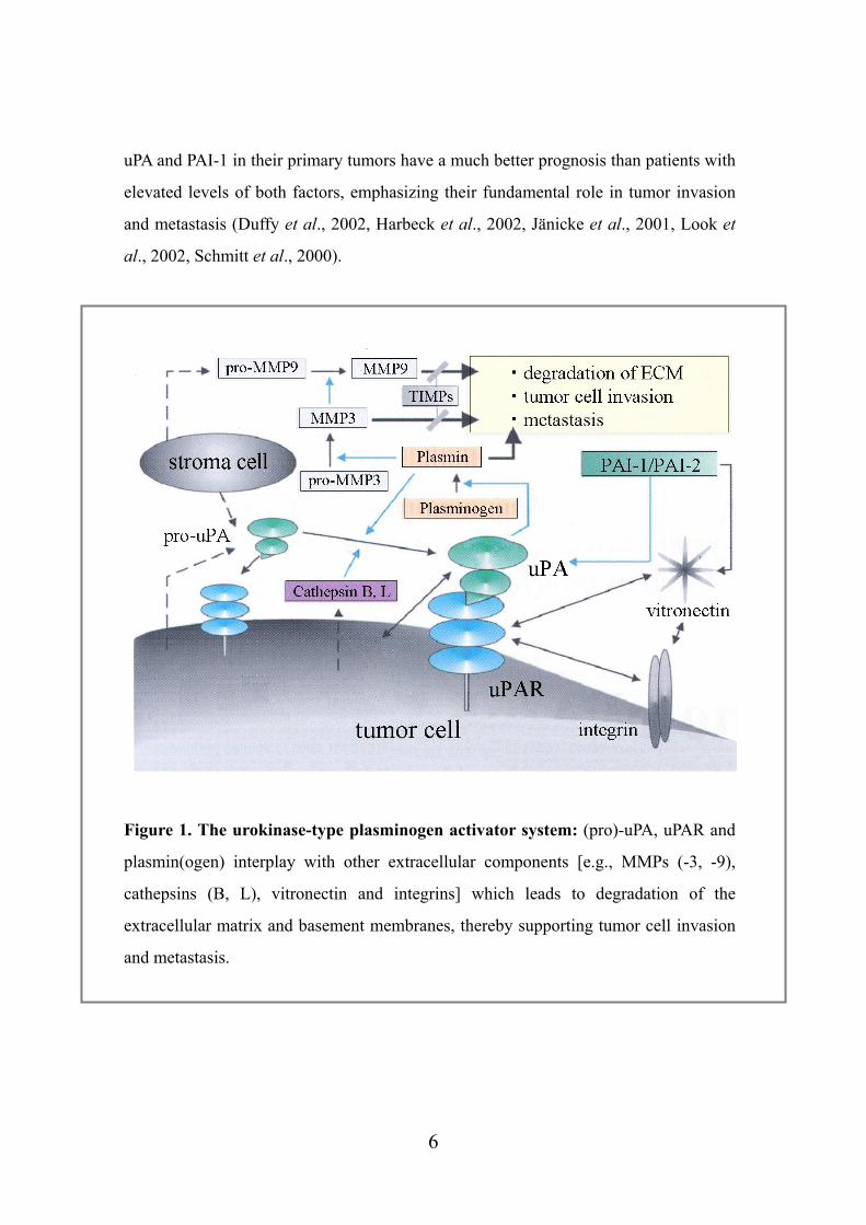
Various proteolytic systems, including the urokinase-type plasminogen activator (uPA)
system, matrix metalloproteinases (MMPs), and cysteine proteases (cathepsin B, L),
which partly interact and cooperate, contribute to the net proteolytic activity at the
tumor-host interface (Fig. 1) (Andreasen et al., 2000, Chapman et al., 1997, Noel et al.,
1997, Schmitt et al., 2000, Yan et al., 1998). Not only the tumor cells but also stromal
cells present within the surrounding tissue or extracellular matrix synthesize
components of the different proteolytic systems, thus contributing to proteolysis on the
surface of tumor cells (Dublin et al., 2000).
The urokinase-type plasminogen activator (uPA) system plays a pivotal role in this
degradation process, with components like the serine protease plasmin, its activator
urokinase-type plasminogen activator (uPA), the cell surface-associated uPA receptor
uPAR (CD87), and the two inhibitors, plasminogen activator inhibitor type 1 (PAI-1)
and type 2 (PAI-2) (Fig. 2). For numerous types of solid malignant tumors, a strong
clinical value of the plasminogen activation system in predicting disease recurrence
and survival in cancer patients has been demonstrated. Patients with low levels of both
5
uPA and PAI-1 in their primary tumors have a much better prognosis than patients with
elevated levels of both factors, emphasizing their fundamental role in tumor invasion
and metastasis (Duffy et al., 2002, Harbeck et al., 2002, Jänicke et al., 2001, Look et
al., 2002, Schmitt et al., 2000).
Figure 1. The urokinase-type plasminogen activator system: (pro)-uPA, uPAR and
plasmin(ogen) interplay with other extracellular components [e.g., MMPs (-3, -9),
cathepsins (B, L), vitronectin and integrins] which leads to degradation of the
extracellular matrix and basement membranes, thereby supporting tumor cell invasion
and metastasis.
6
The serine protease uPA (Mr: approximately 55,000), which plays a central role in the
conversion of plasminogen to plasmin, is secreted by various normal and cancer cells
as a single-chain polypeptide (pro-uPA). pro-uPA consists of 411 amino acids (aa), and
several proteases, e.g. plasmin, plasma or glandular tissue kallikrein, and cathepsin B
or L (Dano et al., 1985), are able to convert pro-uPA by limited proteolysis into the
enzymatically active serine protease, HMW-uPA ("high-molecular-weight-uPA"), by
cleavage of the peptide bond between Lys158 and Ile159. The A-chain of uPA (aa 1-158)
and the B-chain (aa 159-411) are covalently connected via a disulfide bridge between
Cys148 and Cys279. The B-chain harbors the active site of the serine protease with its
catalytic triad His204, Asp255, and Ser356 (Fig. 3). By further proteolytic action on
HMW-uPA, an amino-terminal fragment (ATF; aa 1-135) is released yielding the
"low-molecular-weight" form of uPA (LMW-uPA). Both HMW- and LMW-uPA
display very similar enzymatic activities towards the substrate plasminogen. ATF
consists of two different domains (Fig. 3), the so-called growth factor-like domain
(GFD; aa 1-49) and the kringle domain (aa 50-135) which displays structural
homology to the kringle domains of e.g. prothrombin, tPA, and plasmin.
The cell surface-associated uPA receptor uPAR (CD87) is a cysteine-rich, heavily
N-glycosylated protein of Mr: 45 - 60,000. It is translated into a 313 amino acid
polypeptide with a 21 amino acid signal peptide. It consists of three homologous
repeats: domains I, II, and III, as numbered from the N terminus (Fig. 4) (Fowler et al.,
1998, Mondino et al., 1999, Ploug et al., 2002). uPAR lacks a transmembrane
sequences, and is associated with cell membranes by a glycosyl phosphatidyl inositol
(GPI) anchor (Ploug et al., 1991). Soluble uPAR variants without a GPI anchor have
also been identified in the conditioned medium from various cell lines and in body
fluids from cancer patients, and may arise by differential splicing, by proteolysis, or by
phospholipase cleavage of the GPI anchor (Brunner et al., 1999, Pedersen et al., 1993,).
Required structural determinants for binding of uPA are located within the N-terminal
domain I of uPAR. Within this domain residues Arg53, Leu55, Tyr57, and Leu66,
respectively, were identified to be essential for the uPA/uPAR interaction, as shown by
7
a systematic Ala scan (Gårdsvoll et al., 1999). However, as shown by several different
approaches, the intact three-domain uPAR molecule is required for high-affinity
interaction with uPA (Gardsvoll et al., 2006, Llinas et al., 2005, Ploug et al., 1998,
Ploug et al., 2002)
Binding of uPA to its specific high-affinity receptor uPAR (CD87; Kd ~ 1 nM) is
mediated by the N-terminally located GFD of uPA, whereas pro-uPA has an activity
about 250-fold less than that two-chain uPA (Petersen et al., 1988). pro-uPA is
activated into uPA either in solution or when bound to uPAR at the cell surface, the
latter activation occurs much faster. Furthermore, active uPA bound to uPAR is more
efficient than free uPA in converting plasminogen to plasmin. By binding to the
receptor, the activity of uPA is focused to the cell surface, and gives the cell the ability
to efficiently degrade its surrounding matrix, which enables tumor cells to detach from
the primary tumor and leads to tumor invasion and metastasis (Andreasen et al., 2000).
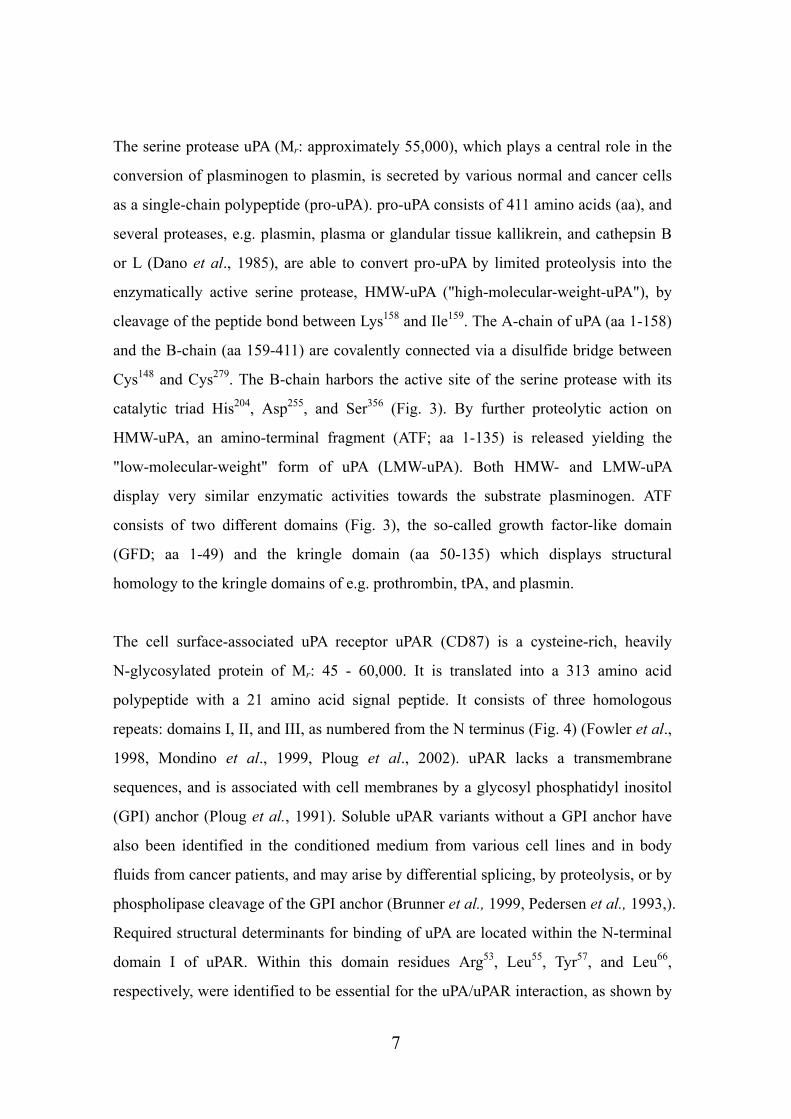
The two natural uPA inhibitors, plasminogen activator inhibitor type 1 (PAI-1) and
type 2 (PAI-2), belong to the serpin (serine protease inhibitor) family having an
arginine in their reactive inhibitory center. They function by acting as pseudosubstrates
and form an irreversible complex with their target protease. PAI-1 (Mr: approximately
50,000) is one of the main inhibitors of uPA and is a single-chain glycoprotein,
inactivates both uPA and tPA (tissue-type plasminogen activator) by rapid formation of
1:1 complexes (Andreasen et al., 2000). PAI-1 also binds to the extracellular matrix
protein vitronectin with high affinity. Secreted PAI-1 as an unstable active inhibitor, is
rapidly converted into its latent form unless it is stabilized by binding to vitronectin.
Bound to vitronectin, PAI-1 stays active towards serine protease and can inhibit
plasminogen activation by uPA at the cell surface (Conese and Blasi, 1995). PAI-2 (Mr:
approximately 50,000) is also able to inhibit both uPA and tPA, although it reacts more
slowly than PAI-1 (Rijken 1995).
8
Figure 2. Schematic presentation of uPA, uPAR, PAI-1 and plasmin: uPA consists
of a serine protease domain (SPD), a kringle domain (K), and a growth factor-like
domain (G) harboring the uPAR binding site. uPAR has three domains and is attached
to the cell membrane by a GPI anchor. The reactive center loop (RCL) of the inhibitor
PAI-1 can bind to the active site of uPA, but PAI-1 can also interact with the
extracellular matrix protein vitronectin to stay active towards serine proteases and to
inhibit plasminogen activation by uPA at the cell surface. Plasmin also contains a
serine protease domain as well and plus five kringle domains.
9
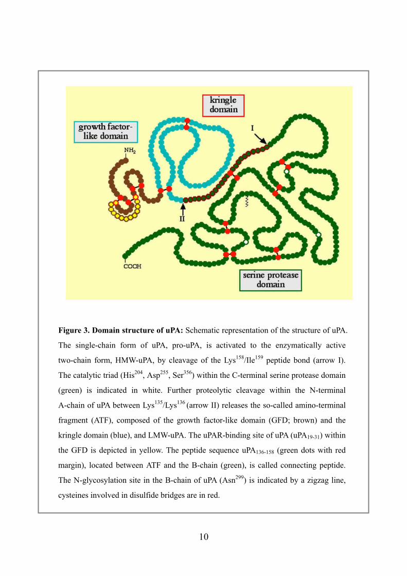
Figure 3. Domain structure of uPA: Schematic representation of the structure of uPA.
The single-chain form of uPA, pro-uPA, is activated to the enzymatically active
two-chain form, HMW-uPA, by cleavage of the Lys158/Ile159 peptide bond (arrow I).
The catalytic triad (His204, Asp255, Ser356) within the C-terminal serine protease domain
(green) is indicated in white. Further proteolytic cleavage within the N-terminal
A-chain of uPA between Lys135/Lys136 (arrow II) releases the so-called amino-terminal
fragment (ATF), composed of the growth factor-like domain (GFD; brown) and the
kringle domain (blue), and LMW-uPA. The uPAR-binding site of uPA (uPA19-31) within
the GFD is depicted in yellow. The peptide sequence uPA136-158 (green dots with red
margin), located between ATF and the B-chain (green), is called connecting peptide.
The N-glycosylation site in the B-chain of uPA (Asn299) is indicated by a zigzag line,
cysteines involved in disulfide bridges are in red.
10
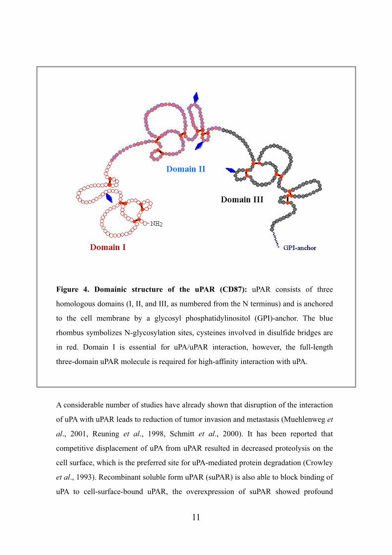
Figure 4. Domainic structure of the uPAR (CD87): uPAR consists of three
homologous domains (I, II, and III, as numbered from the N terminus) and is anchored
to the cell membrane by a glycosyl phosphatidylinositol (GPI)-anchor. The blue
rhombus symbolizes N-glycosylation sites, cysteines involved in disulfide bridges are
in red. Domain I is essential for uPA/uPAR interaction, however, the full-length
three-domain uPAR molecule is required for high-affinity interaction with uPA.
A considerable number of studies have already shown that disruption of the interaction
of uPA with uPAR leads to reduction of tumor invasion and metastasis (Muehlenweg et
al., 2001, Reuning et al., 1998, Schmitt et al., 2000). It has been reported that
competitive displacement of uPA from uPAR resulted in decreased proteolysis on the
cell surface, which is the preferred site for uPA-mediated protein degradation (Crowley
et al., 1993). Recombinant soluble form uPAR (suPAR) is also able to block binding of
uPA to cell-surface-bound uPAR, the overexpression of suPAR showed profound
11
inhibitory effects on primary tumor growth, tumor spread, or experimental metastasis
(Krüger et al., 2000, Lutz et al., 2001, Wilhelm et al., 1994). Various different
experiments to interfere with the expression or reactivity of uPA or uPAR at the gene or
protein level were performed successfully, including the use of antisense oligodeoxy-
nucleotides, antibodies, inhibitors and recombinant or synthetic uPA and uPAR
analogues (Kobayashi et al., 1995, Magdolen et al., 2001, Rabbani et al., 1995,
Schmitt et al., 1995, Wilhelm et al., 1995). It was also shown that broad-spectrum
serine protease inhibitors suppressed tumor growth and metastasis (Novak et al., 2005,
Ohkoshi et al., 2002, Witschi et al., 1989).
Recently, several studies have been performed to identify and define new prognostic
markers for the prediction of patients afflicted with solid malignant tumors. In a variety
of malignancies including cancer of the breast, ovary, cervix, uteri, bladder, upper
urogenital tract, kidney, head and neck, brain, lung, soft-tissue, stomach, colon,
pancreas, esophagus, and liver, a strong prognostic impact has been attributed to
components of the uPA-system: uPA, uPAR, and the two inhibitors PAI-1 and PAI-2
are statistically independent as prognostic factors (Duffy et al., 2001, Reuning et al.,
1998, Schmitt et al., 2000).
In general, upregulation of the tumor antigen levels of uPA and/or PAI-1 is associated
with poor disease outcome and tumor cell spread and metastasis (Grondahl-Hansen et
al., 1993, Reuning et al., 1998, Schmitt et al., 2000). Especially, in breast cancer,
determination of tumor antigen levels of uPA and PAI-1 is close to clinical routine to
decide an individualized patients therapy (Duffy et al., 2002, Harbeck et al., 2002,
Look et al., 2002). Additionally, increased levels of a soluble form of uPAR which are
present in blood and ascites of patients with cancer of the ovary, lung, breast or colon,
have also been associated with poor prognosis (Pedersen et al., 1993, Sier et al., 1998).
In contrast to PAI-1, elevated PAI-2 antigen levels predict a good prognosis of cancer
patients (Foekens et al., 1995). Thus the strong correlation between elevated tumor
antigen levels and poor prognosis raises the possibility that plasminogen activation
system has a key role in extracellular matrix degradation.
12
2.2 Competitive antagonist of uPA/uPAR-interaction derived from the uPAR binding site of uPA
Binding of (pro)-uPA to its cell surface receptor not only generates a pericellular
proteolytic system, furthermore, surface-associated feedback-activation of pro-uPA by
plasmin results in potentiation of proteolytic activity compared to activation in solution
(Ellis et al., 1989). In addition to this, pro-forms of other proteolytic enzymes such as
matrix metalloproteinases are activated by plasmin as well, allowing tumor cells to
degrade the surrounding extracellular matrix. This matrix degradation facilitates cell
migration and enables tumor cells to detach from the primary tumor and to spread to
distant loci in the body (Andreasen et al., 2000, Del Rosso et al., 2002, Reuning et al.,
1998).
In contrast to uPAR in which different domains contribute to a composite uPA-binding
site, the uPAR binding site within the uPA molecule encompasses a single continuous
sequence within the GFD. Appella and coworkers demonstrated that a peptide
comprising aa 12-32 of uPA (in which for technical reasons Cys19 was substituted by
Ala) efficiently competed with ATF for binding to cell surface-associated uPAR. The
rational for testing synthetic peptides derived from the N-terminal region of uPA
originated from the finding that GFD binds with similar affinity to uPAR as do ATF
and HMW-uPA, respectively. Moreover, ATF cleaved between Lys23/Tyr24 or
Phe25/Ser26 does not interact with uPAR. A peptide encompassing aa 18-32 still
competed with ATF for binding to uPAR, whereas a peptide spanning aa 9-20
including a Cys19 to Ala19 substitution had almost no effect, indicating that aa residues
20-30 of uPA, corresponding to the so-called loop B of GFD, confer receptor binding
specificity (Appella et al., 1987).
NMR structural analysis of ATF (Hansen et al., 1994[1], Hansen et al., 1994[2])
revealed that the region Thr18-Asn32 of uPA is folded into a flexible, seven-residue
W-loop, i.e. a ring-like structure from Asn22 to Ile28, that results from the
double-stranded, antiparallel ß sheet between Thr18-Ser21 and His29-Asn32. Cys19and
13
Cys31, although in close proximity, form disulfide bonds with other cysteines
(Cys11/Cys19 and Cys13/Cys31, respectively) (Fig. 5). Exchange of aa residues Lys23,
Tyr24, Phe25, Ile28, or Trp30 by Ala in loop B of GFD led to a strongly reduced or even a
complete loss of binding affinity towards uPAR. An Ala scan was also performed with
the uPA derived synthetic peptide uPA14-32 (Magdolen et al., 1996). The individual
replacement of aa 14-18, 20-23, 26-27, 29, or 32 by Ala had only minor effects on
uPAR binding, whereas the individual substitution of Cys19, Tyr24, Phe25, Ile28, Trp30, or
Cys31 by Ala resulted in a considerable loss of uPAR binding activity. Interestingly, in
the spatial structure of uPA, all of the side chains of Tyr24, Phe25, Ile28, and Trp30 are
displayed on one side of the ring-like structure, suggesting their involvement in
specific hydrophobic interactions of uPA within the binding pocket of uPAR.
In order to determine the minimal uPAR binding region of uPA, a series of peptides
with different lengths derived from wild-type uPA were tested for uPAR binding
activity. By this, peptide uPA19-31 was found to compete with ATF for binding to uPAR,
whereas peptides uPA18-30, uPA20-32 or uPA20-30 were ineffective. Interestingly, uPA19-31
binds to uPAR with higher affinity than the longer peptides uPA14-32 or uPA16-32 (Bürgle
et al., 1997). The results obtained with peptides derived from wild-type uPA are in
agreement with the Ala-scanning experiments of uPA14-32, defining Cys19 and Cys31 as
the essential aa residues representing the N- and C-terminal borders of the uPAR
binding epitope (Bürgle et al., 1997, Magdolen et al., 1996).
In uPA, Cys19 and Cys31 are not connected by a disulfide bond but are in close
proximity (Fig. 5). The short distance between Cys19 and Cys31 in the native uPA
molecule may explain, why the cyclic synthetic peptide cyclo19,31uPA19-31 in which
Cys19 and Cys31 are linked by a disulfide bond, retain suPAR binding activity. The
disulfide-bridged form of uPA19–31 displays an IC50-value similar to that of its
corresponding linear form (IC50: ~ 700 nM) (Bürgle et al., 1997). The distance
between the Ca-atoms at aa positions 19 and 31 was further narrowed down from 6.1 Å
in native uPA and 5.2 Å in cyclo19,31uPA19-31 to about 4.8 Å by generation of another
peptide variant, cyclo19,31[Ala19-S-Ala31]-uPA19-31, in which the cystine [Cys19-Cys31,
14
which formally corresponds to Ala19-S-S-Ala31] was substituted by lanthionine
[Ala19-SAla31]. This peptide (cyclo19,31[Ala19-S-Ala31]-uPA19-31) still interfered with
uPA/uPAR interaction but exhibited an about 10-fold lower inhibitory activity than the
parent peptide cyclo19,31-uPA19-31 (Magdolen et al., 2001).
A systematic D-aa scan was performed, in which each of the 13 L-aa of
cyclo19,31uPA19-31 was individually substituted by the corresponding D-aa (Magdolen et
al., 2001). This led to the identification of cyclo19,31[D-Cys19]-uPA19-31 (WX-306) as a
potent inhibitor of uPA/uPAR interaction (Fig. 5), displaying an only 20 to 40-fold
lower binding capacity (IC50: ~ 200 nM) than the naturally occurring uPAR ligands
uPA and ATF (IC50: ~ 5-10 nM). As one can see in figure 5, the switch of the cysteine
bridges to produce cyclo19,31uPA19-31 requires a change of the Cys side chain
orientation, which is achieved by change in chirality. In addition to the uPA derived
peptides with single D-aa substitutions, a cyclic peptide was synthesized in which all
L-aa were substituted by their corresponding D-enantiomers and in which all of the
peptide bonds were inverted. This so-called retro-inverso, protease-insensitive
cyclo19,31-uPA19-31-peptide did not show any uPAR binding activity. Several reasons for
the lack of binding activity of the retro-inverso peptide are conceivable:
i) Retro-D-peptides harbor exchanged N- and C-termini; the terminal groups in
cyclo19,31uPA19-31 may be important for the receptor-ligand interaction with uPAR. One
has to consider that in a cyclic retro-inverso peptide, the side chain orientation is
different from that in the parent peptide (Wermuth et al., 1997).
ii) The amide bonds in the backbone of the peptide (which are reversed in the
retro-inverso peptide) may be necessary for uPAR binding.
iii) The hydrogen bond pattern in retroinverso peptides is different from that of the
parent peptides.
A series of cyclic peptides was synthesized in which certain aa of cyclo19-31uPA19-31
were deleted and/or replaced by other aa (Bürgle et al., 1997, Guthaus et al., 2002,
Schmiedeberg et al., 2002). Since - in addition to Cys19 and Cys31- Lys23, Tyr24, Phe25,
Ile28, and Trp30, respectively, have been proven to be important determinants of uPAR
15
binding (Magdolen et al., 1996), mainly disulfide-bridged cyclic peptides of different
ring sizes were synthesized encompassing those aa. Among them, only the decameric
peptide cyclo21,29uPA21-30[S21C;H29C] inhibited uPA/uPAR interaction displaying an
IC50 of ~ 900 nM (IC50 of cyclo19-31uPA19-31: ~ 700 nM) (Guthaus et al., 2002,
Schmiedeberg et al., 2002). The systematic substitution of each aa residue in
cyclo21,29uPA21-30[S21C;H29C] by Ala indicated that the side chains of Cys21 and Cys29
(leading to linear peptides), Tyr24, Phe25, Ile28, and Trp30, respectively, are essential for
uPAR binding (Schmiedeberg et al., 2002). In addition, the impact of modification of
the stereochemical orientation of individual aa side chains in
cyclo21,29uPA21-30[S21C;H29C] on uPAR binding activity was analyzed. Derivatives of
cyclo21,29uPA21-30[S21C;H29C] harboring either D-Tyr24, D-Phe25, or D-Ile28 did not
elicit uPAR binding activity. The peptide variant containing D-Trp30 displayed an about
three times weaker uPAR binding activity than the lead peptide with L-Trp30 in the
C-terminal position. This may be explained by the rotational freedom of the exocyclic
aa enabling the aromatic side chain to adopt the conformation required for uPAR
binding. Substitution of L-aa in cyclo21,29uPA21-30[S21C;H29C] by D-aa at positions
Asn22, Lys23, Ser26, and Asn27, respectively, yielded peptides with strongly reduced
uPAR binding affinity. This finding indicates that stereochemical inversion of most
side-chain residues in cyclo21,29uPA21-30[S21C;H29C] reduces uPAR binding affinity,
regardless of whether the corresponding aa side chains are required for receptor
binding or not. Stereochemical modification at Cys21, yielding cyclo21,29
[D-Cys21]uPA21-30[S21C;H29C] (WX-360) (Fig. 5), displayed a much higher uPAR
binding activity (IC50: ~ 40 nM) than its L-isomer (~ 900 nM). This finding is in
agreement with that for cyclo19,31[D-Cys19]-uPA19-31 in which the introduction of
D-Cys at position 19 resulted in a more active peptide (IC50: ~ 200 nM versus ~ 700
nM).
The inversion of the stereochemical orientation of Cys21 in the smaller cyclic peptide
cyclo21,29uPA21-30[S21C;H29C] led to an even more pronounced effect regarding uPAR
binding activity as compared to the exchange of L-Cys19 by D-Cys in cyclo19,31
uPA19-31. The resulting high-affinity uPA mimic peptide displays a binding reactivity to
16
cellular uPAR (IC50: ~ 40 nM) that is only 4-8 times weaker than that of the natural
ligand uPA (IC50: ~ 5 - 10 nM). Thus, cyclo21,29[D-Cys21]uPA21-30[S21C;H29C]
(WX-360) represents one of the most active synthetic uPAR antagonists known to date
(Schmiedeberg et al., 2000, Schmiedeberg et al., 2002).
Efforts have been undertaken to reduce the in vivo proteolytic degradation of
cyclo21,29[D-Cys21]uPA21-30[S21C;H29C] (WX-360). For this purpose, the Lys residue
of cyclo21,29uPA21-30[S21C;H29C] was individually replaced by several nonnatural aa
such as norleucine (Nle), ornithine, diaminobutyric acid, or diaminopropionic acid.
The Nle-variant, cyclo21,29[D-Cys21]-uPA21-30[S21C;K23Nle;H29C] (WX-360-Nle),
exerts similar binding affinities (IC50: ~ 70 nM) as cyclo21,29[D-Cys21]uPA21-30
[S21C;H29C] (WX-360). Both cyclo21,29 [D-Cys21]uPA21-30[S21C;H29C] (WX-360)
and cyclo21,29[D-Cys21] -uPA21-30[S21C;K23Nle;H29C] (WX-360-Nle) were highly
resistant to proteolytic degradation in human and rodent plasma or serum when
compared to other uPA-derived peptides lacking a non-natural D-aa (e.g.
cyclo19,31-uPA16-32). The Nle-variant WX-360-Nle was completely resistant to
plasmin-mediated proteolysis, whereas cyclo21,29[D-Cys21] uPA21-30[S21C;H29C]
(WX-360) was readily degraded (Schmiedeberg et al., 2002).
The two peptides, WX-360 and WX-360-Nle were tested in in vivo nude mice model
for their potency to inhibit tumor growth and intraperitoneal spread.
17
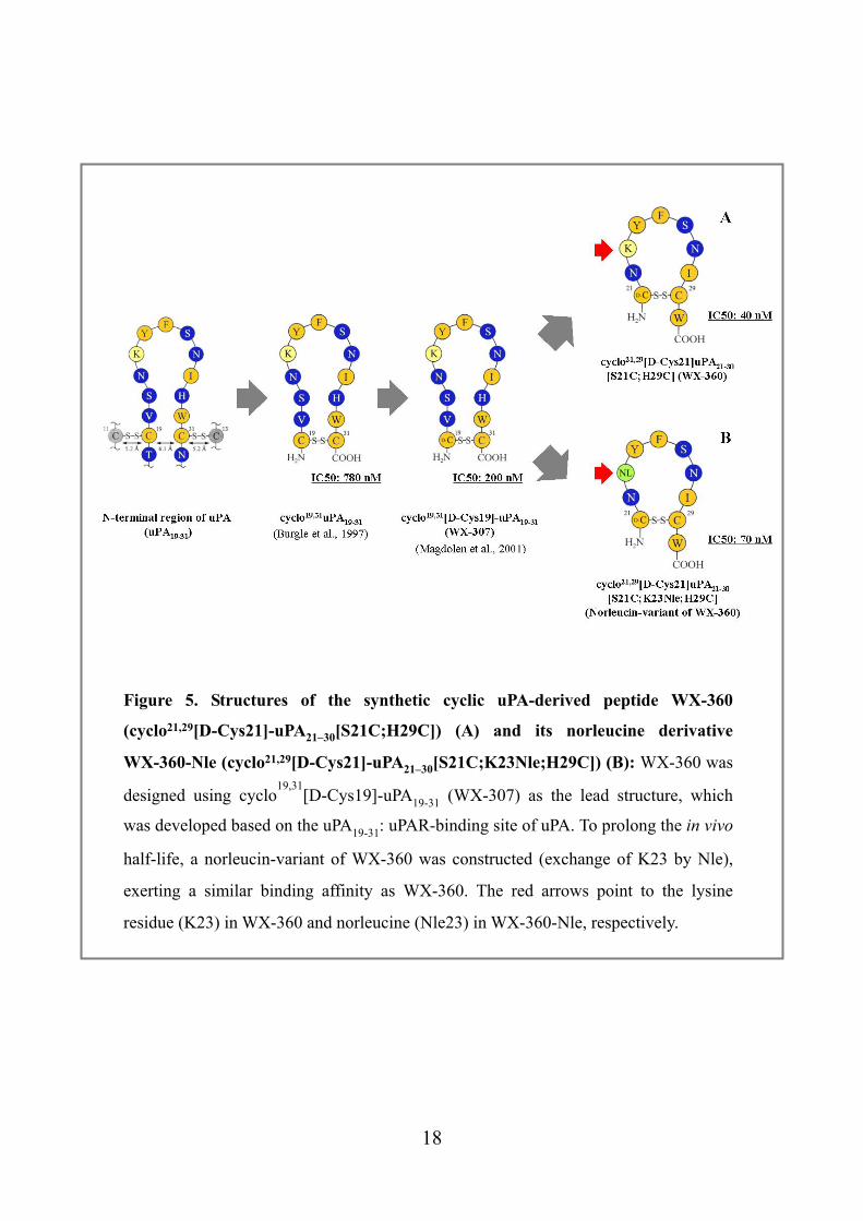
Figure 5. Structures of the synthetic cyclic uPA-derived peptide WX-360
(cyclo21,29[D-Cys21]-uPA21–30[S21C;H29C]) (A) and its norleucine derivative
WX-360-Nle (cyclo21,29[D-Cys21]-uPA21–30[S21C;K23Nle;H29C]) (B): WX-360 was
designed using cyclo19,31
[D-Cys19]-uPA19-31 (WX-307) as the lead structure, which
was developed based on the uPA19-31: uPAR-binding site of uPA. To prolong the in vivo
half-life, a norleucin-variant of WX-360 was constructed (exchange of K23 by Nle),
exerting a similar binding affinity as WX-360. The red arrows point to the lysine
residue (K23) in WX-360 and norleucine (Nle23) in WX-360-Nle, respectively.
18
3. Materials and Methods
3.1 Reagents Two synthetic cyclic peptides have been developed based on the lead structure
WX-307 (cyclo19,31[D-Cys19]-uPA19-31) (Magdolen et al., 2001): WX-360
(cyclo21,29[D-Cys21] uPA21-30[S21C;H29C]) and its derivative WX-360-Nle
(cyclo21,29[D-Cys21]-uPA21-30 [S21C;K23Nle;H29C]), in which lysine 23 is substituted
by norleucine (Guthaus et al., 2002, Schmiedeberg et al., 2002) and was obtained from
Wilex AG (Munich Germany).
3.2 Cell lines OvMz-6 was originally established from a patient with advanced serous
cystadenocarcinoma of the ovary (Möbus et al., 1994), typically induce a large primary
tumor and abundant intraperitoneal metastases. MDA231 and MCF-7 are mammary
adenocarcinoma cell line, established from a pleural effusion of a patient. ZR75 is
invasive ductal adenocarcinoma of breast cancer, derived from an ascites of a patient,
MCF10a human immortalized breast epithelial cells in a fibrocystic disease (ATCC,
Rockland, USA).
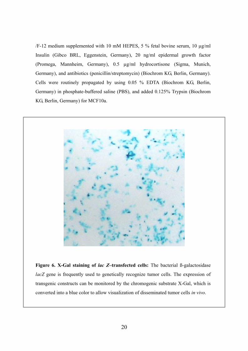
OvMz-6 cells were transfected with the bacterial lacZ gene which encodes for
ß-galactosidase (OvMz-6-BAK) and, thus, the resulting cells can be stained with the
ß-galactosidase substrate 5-bromo-4-chloro-3-indolyl ß-D-galactoside (X-Gal) (Roche
Diagnostics, Mannheim, Germany) (Fig. 6) in order to follow spreading of tumors in in
vivo models (Krüger et al., 1998).
Cells were cultured to 80 % confluence in T-25 cell culture flasks (Becton Dickinson,
Heidelberg, Germany) in DMEM medium supplemented with 10 mM HEPES, 10 %
fetal bovine serum, 200 mM L-glutamine (Gibco BRL, Eggenstein, Germany), and
antibiotics (penicillin/streptomycin) (Biochrom KG, Berlin, Germany) at 37 oC in
humidified atmosphere containing 5 % CO2/95 % air. In case of MCF10a, DMEM
19
/F-12 medium supplemented with 10 mM HEPES, 5 % fetal bovine serum, 10 µg/ml
Insulin (Gibco BRL, Eggenstein, Germany), 20 ng/ml epidermal growth factor
(Promega, Mannheim, Germany), 0.5 µg/ml hydrocortisone (Sigma, Munich,
Germany), and antibiotics (penicillin/streptomycin) (Biochrom KG, Berlin, Germany).
Cells were routinely propagated by using 0.05 % EDTA (Biochrom KG, Berlin,
Germany) in phosphate-buffered saline (PBS), and added 0.125% Trypsin (Biochrom
KG, Berlin, Germany) for MCF10a.
Figure 6. X-Gal staining of lac Z–transfected cells: The bacterial ß-galactosidase
lacZ gene is frequently used to genetically recognize tumor cells. The expression of
transgenic constructs can be monitored by the chromogenic substrate X-Gal, which is
converted into a blue color to allow visualization of disseminated tumor cells in vivo.
20
3.3 uPA, uPAR and PAI-1 ELISA Expression of uPA, uPAR and PAI-1 in OvMz-6-BAK cells and its parent cell lines
was investigated using ELISAs.
ELISAs for uPA and PAI-1 were obtained from American Diagnostica GmbH
(Pfungstadt, Germany), IMUBIND uPA ELISA kit #894, IMUBIND PAI-1 ELISA kit
#821 respectively. The uPAR antigen was kindly estimated by Dr. Mathias Kotzsch
(Institute of Pathology in TU-Dresden, Germany) (Kotzsch et al., 2000). The protein
content of the cell extracts was determined by the BCA protein assay reagent kit.
7.0 x 10⁵ cells were suspended in 1 mL of the supplemented medium, seeded on
24-well plates, and incubated for 24 h. After this incubation, the medium was
exchanged and incubated for more 24 h. The cell medium was removed and
centrifuged (10 min, 12,000 x g, 4 °C). The supernatant was collected for
determination of uPA and PAI-1 antigen. For preparation of cell extracts, the cells were
incubated in 400 µl TBS, 1 % Triton X-100, 3 h with gentle shaking, the solution
collected and centrifuged (30 min, 12,000 x g, 4 °C). The amount of uPA and PAI-1
was also estimated in the cell culture supernatant. Concerning the uPAR ELISA,
approximately 2.0 x 10⁶ cells (80 % confluence), were collected from cell culture
flasks.
Mouse tumor tissue extracts were prepared by suspending pulverized tissues,
previously frozen in liquid nitrogen, in Trisbuffered saline (TBS), followed by
centrifugation (30 min, 12000 x g, 4 °C). Supernatants of this non-detergent extraction
were subjected to uPA, PAI-1 and uPAR antigen measurement by ELISA.
21
3.4 Proliferation assay
Growth curves of OvMz-6 were taken to compare with human carcinoma cell lines,
MCF7, MDA231, ZR75, and human immortalized breast epithelial cells MCF10a.
Initially, 15,000 cells in 1ml 10 % FCS DMEM medium were seeded in the wells of
24-well plate and incubated for 48 to 96 h. After incubation cells were washed with
PBS, detached from the plate using 0.05 % EDTA solution and counted in
hemocytometer upon Trypan blue exclusion. Experiments were repeated 4 times in
triplicates.
3.5 Invasion assay
Invasion assays were performed using inserts (Becton Dickinson, Heidelberg,
Germany) incorporating polyethylene terephthalate (PET) track-etched membranes,
8mm pore size. Aliquots of Matrigel (11.3 mg/ml, Becton Dickinson Labware, Bedford,
MA) were stored frozen at –20 oC. After thawing on ice overnight the Matrigel was
diluted 1:24 with cold PBS and the upper side of the filters was coated with Matrigel
per insert. By testing different concentrations of Matrigel, it was found that 70 µl per
filter gave the best resolution of the invasive capacity of the cells. The plates were
incubated for 3 h at 37 oC in a cell culture incubator. After gelling the Matrigel was
dried overnight in uncovered plates in a laminar hood. The next day, the gel was
rehydrated for 2 h by addition of 200 µl serum-free DMEM/0.1 % BSA. Cells were
grown until 60 to 80 % confluency and adjusted to 105 cells/ml DMEM/0.1 % BSA. 5
x 104 cells/500 µl medium were seeded into each insert. The lower chambers of the
inserts were filled with 750 µl DMEM containing 10 % FCS as a chemoattractant.
After 96 hours of incubation the Matrigel with the noninvaded cells was removed with
Kimwipes and invaded cells on the lower side of the filter were fixed and stained using
Diff-Quick (Dade Behring AG, Switzerland). The stained cells were counted under a
light microscope with the help of a grid. Based on the growth curve of proliferation
assay, the time when 100 cells invade the Matrigel layer was calculated. The
Experiments were repeated 4 times in triplicates.
22
3.6 Adhesion assay
Fibronectin (Becton Dickinson, Heidelberg, Germany), vitronectin (Promega,
Germany), collagen type IV and Laminin (Sigma, Munich, Germany) were diluted
with PBS and added to each well of a 96-well plate (the final concentration of 10
µg/ml). After overnight incubation at 4 oC, wells were washed two times with PBS,
blocked with PBS/2 % BSA for 3 h at RT and again washed with PBS. 40,000 cells in
100 µl DMEM/0.5 % BSA were seeded to each well, the plate was incubated for 2 h at
37 oC. Non-adherent cells were carefully removed by washing with PBS, the adherent
cells were quantified by determining the activity of the ubiquitous lysosomal enzyme
N-acetyl-ß-D-hexosaminidase by incubating them with substrate solution (50 µl/well +
50µl/well PBS) for 1 h at 37 oC. The resulting color reaction was stopped with the stop
solution and the absorbance measured at 405 nm. The serial dilutions of cell
suspensions in the range of 2,500 to 40,000 cells in 50 µl PBS served as the standard
values of the measurements. Assays were performed 4 times in triplicate.
3.7 Animal model Pathogen-free female athymic (nu/nu) mice (4-6 weeks old) were obtained from
Charles River Laboratories (Sulzfeld, Germany). 3.0 x 10⁷ OvMz-6-BAK cells were
suspended in 500 µl PBS and inoculated into the peritoneal cavity of nude mice. These
mice were divided into three groups, which either received WX-360, WX-360-Nle (20
mg/kg/day, respectively), or vehicle only (5 % mannitol, 0.6 % DMSO) in a blinded
manner. The peptides or the vehicle only were injected intraperitoneally once per day
for 37 days (treatment started one day post-inoculation). At the end of the study, the
mice were sacrificed, all intraperitoneal organs removed and stained with X-Gal in
order to facilitate identification of the spreading tumor cells. Tumor tissue was partly
snap frozen in liquid nitrogen for the evaluation total protein levels of selected markers
(uPA, uPAR, PAI-1) by ELISA. The internal organs were washed in ice-cold PBS,
fixed (1 hour in 2 % formaldehyde, 0.2 % glutaraldehyde solution), washed three times
in ice-cold PBS, and then incubated in X-Gal solution containing 5 mmol/L
K3[Fe(CN)]6, 5 mmol/L K4Fe(CN)6·3H2O, 2 mmol/L MgCl2, 0.01 % sodium
23
desoxycholate, 0.02 % NP40, and 1 mg/mL X-Gal (3 to 4 h, 37 oC) and then overnight
at 4 oC (Krüger et al., 1998). To account for weight differences between individual
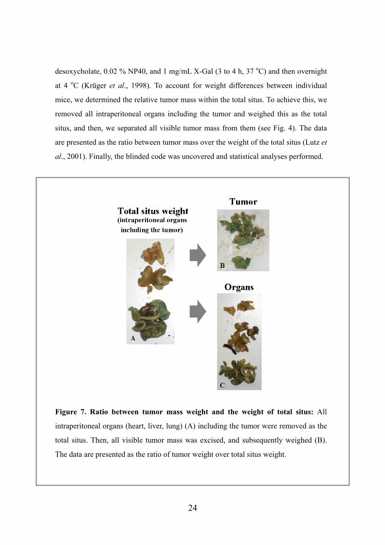
mice, we determined the relative tumor mass within the total situs. To achieve this, we
removed all intraperitoneal organs including the tumor and weighed this as the total
situs, and then, we separated all visible tumor mass from them (see Fig. 4). The data
are presented as the ratio between tumor mass over the weight of the total situs (Lutz et
al., 2001). Finally, the blinded code was uncovered and statistical analyses performed.
Figure 7. Ratio between tumor mass weight and the weight of total situs: All
intraperitoneal organs (heart, liver, lung) (A) including the tumor were removed as the
total situs. Then, all visible tumor mass was excised, and subsequently weighed (B).
The data are presented as the ratio of tumor weight over total situs weight.
24
3.8 Statistical analyses Significant differences in the tumor weight between mice were calculated using
Student’s t-test owing to normal distribution of the data. Concerning data on tumor
weight over total situs weight, the normal distribution Student’s t-test was not
applicable; therefore, a Kruskal-Wallis one-way analysis and Dunn’s multiple
comparison test were performed to investigate for differences between the three groups.
A significance level of p < 0.05 was considered statistically significant.
25

4. Results
4.1 Determination of uPA, PAI-1 and uPAR by ELISA OvMz-6 cells are known to grow locally invasive and to form liver metastasis. The
amount of uPA, PAI-1, and uPAR antigens in the cell extracts and conditioned medium
was determined by specific ELISAs. The measurements demonstrated that the
lacZ-tagged cell line OvMz-6-BAK shows high expression levels of uPA (7.2 ng/mg,
0.97 ng/mL in the cell extract and culture medium, respectively) and PAI-1 (11.9
ng/mg, 23 ng/mL in the cell extract and culture medium, respectively) (Table 1). For
uPAR ELISA, two different antibody, HU/IIIF10 and HU/HD13 were applied which
measure different forms of uPAR (Kotzsch et al., 2000). In case of OvMz-6-BAK, we
also found high level of uPAR, 3.34 ng/mg (HU/IIIF10) and 1.5 ng/mg (HU/HD13) in
the cell extract (Table 1). These expression levels in OvMz-6-BAK were not
significantly different from those of the parent cell line. Results may indicate that the
tumor growth and metastasis of OvMz-6 strongly depend on urokinase plasminogen
activation system.
4.2 Characterization of proliferation of OvMz-6 cell lines
Proliferation of OvMz-6 cells was tested under the normal conditions, in DMEM
medium supplemented with 10 % fetal bovine serum, at 37 oC in humidified
atmosphere containing 5 % CO2/95 % air. Cells were subcultured in 24-well plate, for
48 and 96 h, to compare the growth rate. In addition to OvMz-6 cells, proliferation of
MCF7, MDA231, ZR75, human mammary carcinoma cells and MCF10a human
immortalized breast epithelial cells were also determined. As shown in Figure 8,
OvMz-6 cells showed fastest proliferation, grew 1.5 times faster than ZR75 cells, and 3
times than MDA231 cells. Especially those cells showed a fast growth rate after 48 h.
26
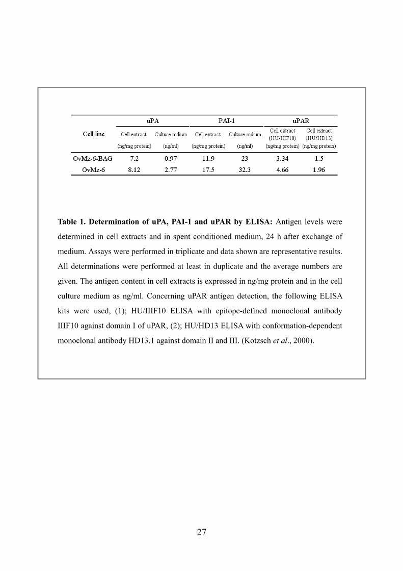
Table 1. Determination of uPA, PAI-1 and uPAR by ELISA: Antigen levels were
determined in cell extracts and in spent conditioned medium, 24 h after exchange of
medium. Assays were performed in triplicate and data shown are representative results.
All determinations were performed at least in duplicate and the average numbers are
given. The antigen content in cell extracts is expressed in ng/mg protein and in the cell
culture medium as ng/ml. Concerning uPAR antigen detection, the following ELISA
kits were used, (1); HU/IIIF10 ELISA with epitope-defined monoclonal antibody
IIIF10 against domain I of uPAR, (2); HU/HD13 ELISA with conformation-dependent
monoclonal antibody HD13.1 against domain II and III. (Kotzsch et al., 2000).
27
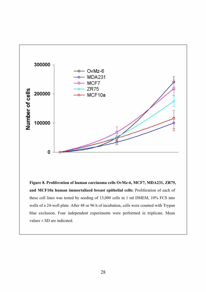
Figure 8. Proliferation of human carcinoma cells OvMz-6, MCF7, MDA231, ZR75,
and MCF10a human immortalized breast epithelial cells: Proliferation of each of
these cell lines was tested by seeding of 15,000 cells in 1 ml DMEM, 10% FCS into
wells of a 24-well plate. After 48 or 96 h of incubation, cells were counted with Trypan
blue exclusion. Four independent experiments were performed in triplicate. Mean
values ± SD are indicated.
28
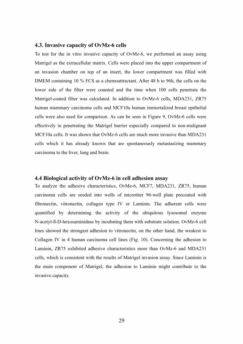
4.3. Invasive capacity of OvMz-6 cells To test for the in vitro invasive capacity of OvMz-6, we performed an assay using
Matrigel as the extracellular matrix. Cells were placed into the upper compartment of
an invasion chamber on top of an insert, the lower compartment was filled with
DMEM containing 10 % FCS as a chemoattractant. After 48 h to 96h, the cells on the
lower side of the filter were counted and the time when 100 cells penetrate the
Matrigel-coated filter was calculated. In addition to OvMz-6 cells, MDA231, ZR75
human mammary carcinoma cells and MCF10a human immortalized breast epithelial
cells were also used for comparison. As can be seen in Figure 9, OvMz-6 cells were
effectively in penetrating the Matrigel barrier especially compared to non-malignant
MCF10a cells. It was shown that OvMz-6 cells are much more invasive than MDA231
cells which it has already known that are spontaneously metastasizing mammary
carcinoma to the liver, lung and brain.
4.4 Biological activity of OvMz-6 in cell adhesion assay To analyze the adhesive characteristics, OvMz-6, MCF7, MDA231, ZR75, human
carcinoma cells are seeded into wells of microtiter 96-well plate precoated with
fibronectin, vitronectin, collagen type IV or Laminin. The adherent cells were
quantified by determining the activity of the ubiquitous lysosomal enzyme
N-acetyl-ß-D-hexosaminidase by incubating them with substrate solution. OvMz-6 cell
lines showed the strongest adhesion to vitronectin, on the other hand, the weakest to
Collagen IV in 4 human carcinoma cell lines (Fig. 10). Concerning the adhesion to
Laminin, ZR75 exhibited adhesive characteristics more than OvMz-6 and MDA231
cells, which is consistent with the results of Matrigel invasion assay. Since Laminin is
the main component of Matrigel, the adhesion to Laminin might contribute to the
invasive capacity.
29
Figure 9. Matrigel invasion by human carcinoma cells OvMz-6, MDA231, ZR75
and MCF10a human immortalized breast epithelial cells: 80 % confluent cells were
placed into the upper compartments of the invasion chambers (5 x 10⁴ cells in 500 µl
of 0.1 % BSA/DMEM). The lower chambers of the inserts were filled with 750 µl
DMEM containing 10 % FCS as the chemoattractant. After 48 to 72 h of incubation,
the Matrigel layer of the upper compartment was removed, the invaded cells on the
lower side of the filter were fixed, stained, and counted. Based on the growth curve
established by the proliferation assay, the time when 100 cells invade the Matrigel
layer was calculated. The experiments were performed 4 times in triplicate, mean
values ± SD are indicated.
30
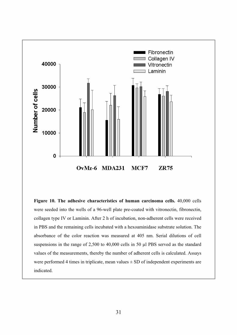
Figure 10. The adhesive characteristics of human carcinoma cells. 40,000 cells
were seeded into the wells of a 96-well plate pre-coated with vitronectin, fibronectin,
collagen type IV or Laminin. After 2 h of incubation, non-adherent cells were received
in PBS and the remaining cells incubated with a hexoaminidase substrate solution. The
absorbance of the color reaction was measured at 405 nm. Serial dilutions of cell
suspensions in the range of 2,500 to 40,000 cells in 50 µl PBS served as the standard
values of the measurements, thereby the number of adherent cells is calculated. Assays
were performed 4 times in triplicate, mean values ± SD of independent experiments are
indicated.
31
4.5 Effect of synthetic cyclic competitive uPA-derived peptide WX-360 and WX-360-Nle in vivo
uPA/uPAR-interaction via the specific sequence within the N-terminal region of uPA
(uPA19-31) directs the proteolytic step in tumor cell proliferation, invasion and
metastasis. Synthetic cyclic peptides, derived from the uPAR-binding sequences within
the N-terminal region of uPA (uPA19-31), were applied and tested whether such
competitive antagonists of uPA/uPAR-interaction reduce tumor burden of a human
ovarian carcinoma in nude mice (Fig. 11).
WX-360 (cyclo21,29 [D-Cys21]-uPA21-30 [S21C;H29C]) is a competitive peptide
antagonist of the uPA/uPAR interaction derived from receptor binding region of uPA.
This peptides display an only five to ten-fold lower affinity to uPAR (IC50: ~ 40 nM)
as compared to the naturally occurring uPAR-ligand uPA. Furthermore, as WX-360
harbors a lysine residue (K23) and, thus, a target site for serine proteases such as
plasmin, K23 was replaced by the non-protein amino acid norleucine. This derivative
WX-360-Nle (cyclo21,29[D-Cys21] -uPA21-30 [S21C;K23Nle;H29C]), still displays high
binding affinity (IC50: ~70 nM).
In this study, WX-360 and WX-360-Nle were tested in nude mice for their potency to
inhibit tumor growth and intraperitoneal spread of lacZ tagged human ovarian cancer
by inhibiting the binding of uPA to uPAR.
32
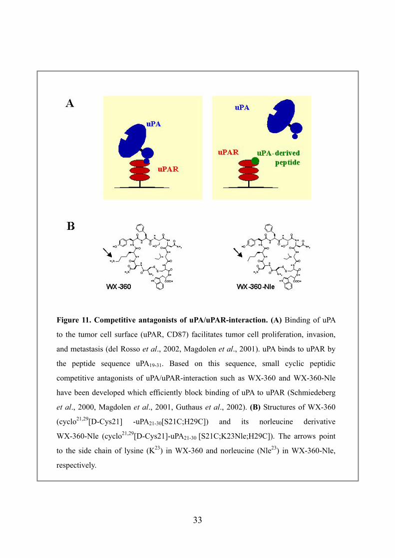
Figure 11. Competitive antagonists of uPA/uPAR-interaction. (A) Binding of uPA
to the tumor cell surface (uPAR, CD87) facilitates tumor cell proliferation, invasion,
and metastasis (del Rosso et al., 2002, Magdolen et al., 2001). uPA binds to uPAR by
the peptide sequence uPA19-31. Based on this sequence, small cyclic peptidic
competitive antagonists of uPA/uPAR-interaction such as WX-360 and WX-360-Nle
have been developed which efficiently block binding of uPA to uPAR (Schmiedeberg
et al., 2000, Magdolen et al., 2001, Guthaus et al., 2002). (B) Structures of WX-360
(cyclo21,29[D-Cys21] -uPA21-30[S21C;H29C]) and its norleucine derivative
WX-360-Nle (cyclo21,29[D-Cys21]-uPA21-30 [S21C;K23Nle;H29C]). The arrows point
to the side chain of lysine (K23) in WX-360 and norleucine (Nle23) in WX-360-Nle,
respectively.
33
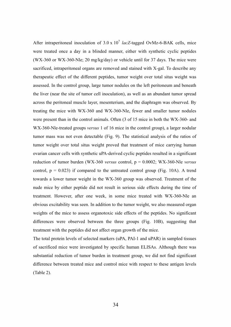
After intraperitoneal inoculation of 3.0 x 10⁷ lacZ-tagged OvMz-6-BAK cells, mice
were treated once a day in a blinded manner, either with synthetic cyclic peptides
(WX-360 or WX-360-Nle; 20 mg/kg/day) or vehicle until for 37 days. The mice were
sacrificed, intraperitoneal organs are removed and stained with X-gal. To describe any
therapeutic effect of the different peptides, tumor weight over total situs weight was
assessed. In the control group, large tumor nodules on the left peritoneum and beneath
the liver (near the site of tumor cell inoculation), as well as an abundant tumor spread
across the peritoneal muscle layer, mesenterium, and the diaphragm was observed. By
treating the mice with WX-360 and WX-360-Nle, fewer and smaller tumor nodules
were present than in the control animals. Often (3 of 15 mice in both the WX-360- and
WX-360-Nle-treated groups versus 1 of 16 mice in the control group), a larger nodular
tumor mass was not even detectable (Fig. 9). The statistical analysis of the ratios of
tumor weight over total situs weight proved that treatment of mice carrying human
ovarian cancer cells with synthetic uPA-derived cyclic peptides resulted in a significant
reduction of tumor burden (WX-360 versus control, p = 0.0002; WX-360-Nle versus
control, p = 0.023) if compared to the untreated control group (Fig. 10A). A trend
towards a lower tumor weight in the WX-360 group was observed. Treatment of the
nude mice by either peptide did not result in serious side effects during the time of
treatment. However, after one week, in some mice treated with WX-360-Nle an
obvious excitability was seen. In addition to the tumor weight, we also measured organ
weights of the mice to assess organotoxic side effects of the peptides. No significant
differences were observed between the three groups (Fig. 10B), suggesting that
treatment with the peptides did not affect organ growth of the mice.
The total protein levels of selected markers (uPA, PAI-1 and uPAR) in sampled tissues
of sacrificed mice were investigated by specific human ELISAs. Although there was
substantial reduction of tumor burden in treatment group, we did not find significant
difference between treated mice and control mice with respect to these antigen levels
(Table 2).
34
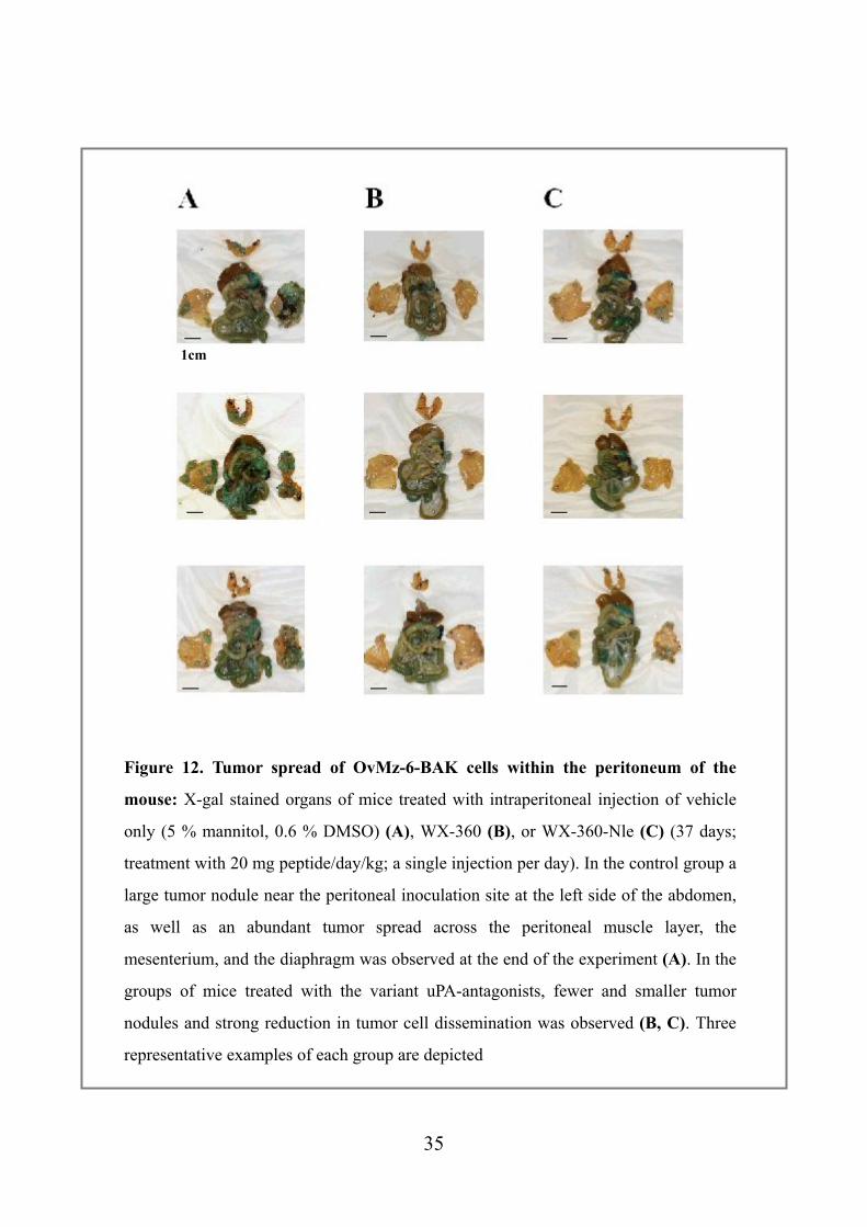
Figure 12. Tumor spread of OvMz-6-BAK cells within the peritoneum of the
mouse: X-gal stained organs of mice treated with intraperitoneal injection of vehicle
only (5 % mannitol, 0.6 % DMSO) (A), WX-360 (B), or WX-360-Nle (C) (37 days;
treatment with 20 mg peptide/day/kg; a single injection per day). In the control group a
large tumor nodule near the peritoneal inoculation site at the left side of the abdomen,
as well as an abundant tumor spread across the peritoneal muscle layer, the
mesenterium, and the diaphragm was observed at the end of the experiment (A). In the
groups of mice treated with the variant uPA-antagonists, fewer and smaller tumor
nodules and strong reduction in tumor cell dissemination was observed (B, C). Three
representative examples of each group are depicted
1cm
35
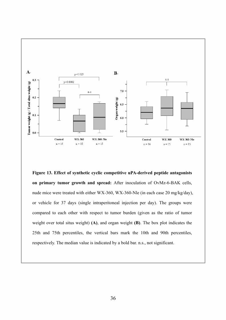
Figure 13. Effect of synthetic cyclic competitive uPA-derived peptide antagonists
on primary tumor growth and spread: After inoculation of OvMz-6-BAK cells,
nude mice were treated with either WX-360, WX-360-Nle (in each case 20 mg/kg/day),
or vehicle for 37 days (single intraperitoneal injection per day). The groups were
compared to each other with respect to tumor burden (given as the ratio of tumor
weight over total situs weight) (A), and organ weight (B). The box plot indicates the
25th and 75th percentiles, the vertical bars mark the 10th and 90th percentiles,
respectively. The median value is indicated by a bold bar. n.s., not significant.
36
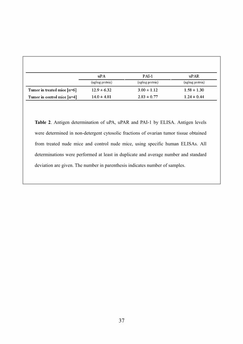
Table 2. Antigen determination of uPA, uPAR and PAI-1 by ELISA. Antigen levels
were determined in non-detergent cytosolic fractions of ovarian tumor tissue obtained
from treated nude mice and control nude mice, using specific human ELISAs. All
determinations were performed at least in duplicate and average number and standard
deviation are given. The number in parenthesis indicates number of samples.
37
5. Discussion
The malignant features of a variety of solid tumors is associated with the pericellular
uPA/uPAR system. In particular, the uPAR-mediated localization of active uPA to
tumor cell surface receptor, uPAR, represents a crucial step for tumor cell proliferation,
invasion, and metastasis, which is proved by the inhibitory action of a series of
different therapeutic molecules targeted to uPA or uPAR at the protein or mRNA/gene
level.
A considerable number of studies have already shown that interference with binding of
uPA to its receptor uPAR leads to reduction of tumor invasion and metastasis.
Therefore, this system with its increased activity in tumor cells represents an attractive
target to attack tumor invasion and metastasis (Sperl et al., 2002). Several strategies for
interfering with the uPA/uPAR interaction have been applied to date, including
administration of antibodies to uPAR, synthetic peptides, and recombinant
uPA-derived proteins as well as gene transfer of therapeutic molecules into tumor cells
or host tissue by transfection with expression vectors or systemic adenoviral gene
transfer.
One strategy to antagonize the uPA/uPAR system and to evaluate its role as a
diagnostic and therapeutic target in metastatic cancer is the interruption of the
uPA/uPAR interaction by specific blocking antibodies. Rabbani and Gladu generated a
polyclonal antibody which is directed to the N-terminal uPA binding domain of rat
uPAR (Rabbini and Glade 2002). The antimetastatic efficacy of this antibody was
evaluated in a syngeneic model of rat breast cancer. For this, Mat B-III uPAR breast
38
cancer cells, which overexpress rat uPAR by stable transfection, were inoculated into
the mammary fat pad of syngeneic female Fischer rats and the antibodies topically and
daily applied for one week. Animals were dosed with anti r-uPAR antibodies from day
1-7 after tumor cell inoculation. These animals showed a marked decrease in tumor
growth and a significant inhibition of metastasis to retroperitoneal and mesenteric
lymph nodes as well as an obvious delay of metastasis to lung, liver, and spleen,
respectively, when compared to control tumor-bearing animals receiving the same dose
of preimmune rabbit IgG. Further analysis showed significantly increased tumor
necrosis and cellular apoptosis (Rabbini and Glade 2002).
In order to specifically target uPAR-expressing cells, several approaches using fusion
proteins have been undertaken in past years. Kobayashi and coworkers constructed a
bifunctional hybrid molecule comprising aa 1-134 of uPA fused to the C-terminal
domain II of the urinary trypsin inhibitor (UTI; aa 78-136), which efficiently inhibits
plasmin. In an in vivo nude-mouse metastasis model, uPA1-134-UTI78-136
significantly reduced human ovarian carcinoma and choriocarcinoma cell spread to
lungs and/or lymphatic tissues. Treatment with UTI domain II or ATF alone resulted in
a relatively small therapeutic effect indicating a synergistic effect of both domains
within the fusion protein (Kobayashi et al., 1998).
bi - trifunctional inhibitors encompassing functionally independent domains which are
directed against the uPA/plasmin system, have been also successfully applied in in vivo
models (Krol et al., 2003). A bifunctional inhibitor was generated by substitution of a
loop within the cysteine protease inhibitor chicken cystatin with the uPA receptor
(uPAR)-binding site of uPA (chCys-uPA19-31; Muehlenweg et al., 2000). This
39
recombinant fusion protein inhibits both, the enzymatic activity of cysteine proteases
and binding of uPA to its cell surface receptor uPAR, the latter representing a crucial
step in activating the uPA/plasmin system. This concept was extended and trifunctional
inhibitors that are also directed against matrix metalloproteinases (MMPs) were
designed. It was demonstrated that these bi - trifunctional inhibitors inhibit cysteine
proteases and MMPs while at the same time interfering with uPA/uPAR interaction by
surface plasmon resonance technology. To test the impact of multifunctional inhibitors
on tumor growth in vivo, cell lines expressing these inhibitors were inoculated into the
peritoneum of CD1 nude mice, resulting in a significant reduction of tumor burden and
spread compared to a vector-transfected control cell line.
Another antagonistic fusion protein encompasses the uPAR binding GFD domain of
either murine or human uPA (residues 1-48) and the constant region of human IgG. The
engineered molecules exhibited potent, species specific binding affinity to uPAR
accompanied by a marked improvement in pharmacokinetics compared to the GFD
alone (Min et al., 1996, Tressler et al., 1999). Treatment of immunodeficient mice with
these antagonists (s.c. application) significantly reduced primary tumor growth of
human breast cancer cells (which had been subcutaneously injected into the mice).
Both human and murine uPAR antagonists showed significant inhibition of primary
tumor growth, demonstrating that in vivo tumor as well as stromal cells can contribute
to uPAR dependent plasminogen activation or other uPAR activities, and by this,
support tumor growth (Tressler et al., 1999). Moreover, the murine uPA based fusion
protein substantially suppressed basic fibroblast growth factor-induced angiogenesis
and B16 melanoma growth in syngeneic mice (Min et al., 1996).
40
Various transfection strategies targeting tumor cells or host tissue with cDNAs
encoding components of the uPA system and variations thereof have been conducted in
order to evaluate the potential of the uPA system as a target for cancer gene therapy. In
an early study by Crowley and coworkers, a cDNA encoding a mutant uPA molecule,
which lacks enzymatic activity due to replacement of the active site Ser356 by Ala but
retaining uPAR binding affinity ([Ala356]uPA) was constructed. [Ala356]uPA was
transfected into prostate cancer cells which were then inoculated into a spontaneous
metastasis model (Crowly et al., 1993). When compared to parental cells,
[Ala356]uPA-transfected cancer cells exhibited a significantly reduced metastatic
potential to regional lymph nodes, brain, and lungs, respectively, whereas primary
tumor growth was not affected by overexpression of [Ala356]uPA.
Exogenous addition of ATF reduced the in vitro invasiveness of uPAR-expressing cells
derived from a primary carcinoma of the breast (Luparello et al., 1996). The same
effect was noticed by overexpression of ATF in human lung giant-cell carcinoma cells
(Zhu et al., 2001), and a recombinant adenovirus encoding murine ATF has been
studied in a series of cancer models (Li et al., 1998). Liver metastasis was inhibited
after intrasplenal inoculation of human LS174T colon carcinoma cells into nude mice
after intranasal adenovirus delivery. Inhibition of primary tumor growth in nude mice
was demonstrated by a single intratumoral injection of the ATF-encoding adenovirus
into a pre-established MDA231 breast cancer subcutaneous tumor or into a
pre-established syngeneic Lewis lung carcinoma. The reduction of tumor volume in
both cases is thought to be due primarily to reduced angiogenesis since suppressed
neovascularization within the tumor and in adjacent tissue close to the injection site of
the tumor cells was observed, and the murine ATF expressed does not bind to human
uPAR (Li et al., 1998).
41
Systemic adenoviral gene transfer of a chimeric protein composed of a mutant human
ATF (mhATF) with high affinity for mouse and rat uPAR, linked to bovine pancreatic
trypsin inhibitor (mhATF-BPTI) significantly inhibited subcutaneous tumor growth
and decreased experimental lung metastasis in two rat bronchial carcinoma animal
models (Zhu et al., 2001) and neointima formation and restenosis (Quax et al., 2001).
In these tumor models, mhATF alone or human endostatin (even at ~ 10-fold higher
molecular plasma concentrations than mhATF-BPTI or mhATF) did not lead to a
significant inhibition of either tumor growth or metastasis (Legesvre et al., 2002).
In another experimental strategy, an expression plasmid encoding a soluble form of
uPAR (suPAR) was transfected into human breast cancer cells (Krüger et al., 2000).
Inoculation of those high level suPAR-expressing tumor cells resulted in significantly
decreased experimental lung metastases when compared to parental tumor cells. While
high expression levels of suPAR in breast cancer cells did not influence cell
proliferation in vitro, tumor growth in the mammary fat pad of nude mice was
markedly reduced (Krüger et al., 2000). However, in human ovarian cancer cells
expressing high amounts of suPAR, a reduction of both cell proliferation in vitro and
tumor burden (by up to 86 %) in vivo after intraperitoneal inoculation of these cells
into nude mice was observed (Lutz et al., 2001), suggesting different roles for the
uPAR system in tumor progression in different tissues.
All of these animal experiments provide "proof of principle" for the use of antagonists
of uPA/uPAR-interaction in tumor inhibition. However, the application of large
recombinant proteins for treatment of patients appears rather difficult and depends on
sophisticated, e.g. viral, delivery systems (Sperl S. et al., 2002). Therefore, others and
42
we have concentrated on the development of small, synthetic competitive or
non-competitive peptide antagonists (Bürgle et al., 1997, Goodson et al., 1994, Guo et
al., 2000, Guthaus et al., 2002, Magdolen et al., 2001, Ploug et al., 2001,
Schmiedeberg et al., 2002, Tressler et al., 1999).
A non-competitive peptide antagonist of the uPA/uPAR interaction derived from a
non-receptor binding region of uPA (aa 136-143), Å6, reduced spontaneous metastasis
of orthotopically growing human breast cancer cells in a mouse xenograft as well as
tumor growth and spontaneous metastasis in a rat breast cancer model in syngeneic rats
after intraperitoneal application (Guo et al., 2000, Guo et al., 2002). In the syngeneic
rat breast cancer model the reduction of primary tumor growth by Å6 was further
increased by combined treatment of Å6 with tamoxifen, a nonsteroidal antiestrogen
(Guo et al., 2002). In a mouse xenograft model with human glioma cells, subcutaneous
and intracranial tumor growth was inhibited upon daily application of Å6 (Mishima et
al., 2000). Å6 inhibited breast cancer cell invasion but did not alter cell proliferation of
these three tumor cell lines in vitro. Moreover, it led to decreased migration but not
proliferation of human microvascular endothelial cells (Guo et al., 2000, Guo et al.,
2002, Mishima et al., 2000). The anti-tumorigenic effect of Å6 is thus speculated to be
at least in part a consequence of impaired tumor angiogenesis. Indeed, reduced tumor
mass correlated with decreased microvessel density in all models. Å6 also increased
apoptosis of human and rat cancer cells (Guo et al., 2002, Mishima et al., 2000).
Despite these intriguing results the molecular basis of Å6 action is still poorly
understood.
43
Several efforts have been undertaken over the years to develop competitive antagonists
of uPA/uPAR-interaction that bind with high affinity to uPAR on tumor cell surfaces.
One of the first approaches within this scenario was the use of synthetic peptides
derived from the growth factor-like domain (GFD) of uPA (Kobayashi et al., 1994).
Peptides of murine and human uPA were examined in order to determine whether they
inhibit experimental and spontaneous lung metastasis by murine Lewis lung carcinoma
cells. In an in vivo experimental metastasis assay, which determines mainly the later
steps of the metastatic process, none of the peptides inhibited pulmonary metastases
when co-injected intravenously into syngeneic mice. However, in an alternative in vivo
test system that measures metastasis from a primary tumor (spontaneous metastasis
model), multiple intraperitoneal injections of the murine uPA-derived peptide
muPA17-34 for one week after subcutaneous tumor cell inoculation, significantly
blocked metastasis to the lung in a dose-dependent manner, whereas the human peptide
uPA17-34 had no effect (Kobayashi et al., 1994).
Later, several linear peptides spanning uPA14-32 were synthesized in which the naturally
occurring amino acids were individually replaced by alanine (Ala scan) in order to
identify the amino acids critical for binding to uPAR (Magdolen et al., 1996). The
exchange of Cys19, Lys23, Tyr24, Phe25, Ile28, Trp30, and Cys31, respectively, by
Ala resulted in peptides with strongly impaired uPAR-binding capacities, whereas
replacement of the other amino acids had no or little effect on uPAR binding. Finally,
the minimal uPAR-binding region of uPA was located to uPA19-31 using synthetic
peptides which were successively shortened from the amino- and/or carboxy-terminus
starting with uPA10-32 (Bürgle et al., 1997). The region between amino acids Thr18 and
Asn32 of uPA consists of a flexible, seven-residue !-loop (Asn22 to Ile28) which by
44
means of a double stranded, antiparallel ß-sheet (between Thr18 to Ser21 and His29 to
Asn32) is forced into a ring-like structure (Hansen et al., 1994a, b; Magdolen et al.,
1996; Schmitt et al., 2000). In uPA, Cys19 and Cys31, although in close proximity,
form disulfide bonds with distinct cysteines (Cys11/Cys19 and Cys13/Cys31,
respectively; Hansen et al., 1994a, b). The short distance between Cys19 and Cys31 in
the native molecule could lead cyclo19,31uPA19-31, which displays an even increased
uPAR-binding activity (Bürgle et al., 1997). Furthermore, systematic D-amino acid
scan of uPA19-31 are performed, in which each of the 13 L-amino acids was individually
substituted by the corresponding D-amino acid. This led to the identification of
cyclo19,31[D-Cys19]-uPA19-31 WX-307 as a potent inhibitor of uPA/uPAR-interaction,
displaying only a 20 to 40-fold lower binding capacity as compared to the naturally
occurring uPAR-ligands uPA and its amino-terminal fragment. These peptides not only
block binding of uPA to uPAR but are also capable to displace uPAR-bound uPA from
the cell surface and to inhibit uPA-mediated, tumor cell-associated plasminogen
activation and fibrin degradation (Bürgle et al., 1997, Magdolen et al., 2001).
cyclo19,31-uPA19-31 was evaluated in a pilot study in a xenogeneic mice tumor model
with respect to its efficacy to suppress tumor growth and metastasis. Upon treatment of
nude mice (n = 6) with a daily dose of 10 mg/kg, cyclo19,31-uPA19-31 reduced the growth
of human MDA231 breast cancer cells after five weeks of treatment when compared to
the vehicle-treated control group (Sperl et al., 2002).
Reduction of the ring-size and D-amino acid scanning led to WX-360
(cyclo21,29[D-Cys21] uPA21-30 [S21C;H29C]), displaying a 5-fold higher affinity
towards uPAR than WX-307 (IC50: ~ 40 nM versus ~ 200 nM) (Guthaus E. et al.,
2002, Schmiedeberg N. et al., 2000,). Furthermore, as WX-360 harbors a lysine
45
residue (K23) and, thus, a target site for serine proteases such as plasmin, K23 was
replaced by the non-protein amino acid norleucin (WX-360-Nle). This derivative still
displays high binding affinity (IC50: ~ 70 nM). Stability-testing showed that both
WX-360 and WX-360-Nle, respectively, were highly resistant to proteolytic
degradation in human and rodent plasma or serum whereas other uPA-derived peptides
lacking a non-natural D-amino acid (such as cyclo19,31-uPA16-32) were significantly less
stable: after 1 hour incubation at 37 °C, peptide cyclo19,31-uPA16-32 was completely
degraded, whereas WX-360 and WX-360-Nle, respectively, were stable over a period
of 24 hours. After incubation with high amounts of plasmin (5 µg of peptide were
incubated with 0.01 U [~ 4 µg] of plasmin at 37 oC), however, WX-360 was degraded,
whereas WX-360-Nle was completely resistant to proteolysis (Schmiedeberg et al.,
2002).
In the present study, we demonstrate that treatment of with small, synthetic, cyclic,
competitive uPAR-binding site-derived uPA antagonists results in a highly significant
reduction of tumor burden and also dissemination in the peritoneal cavity in vivo. In
the nude mouse model, we used OvMz-6 human ovarian cancer cells, which typically
induce a large primary tumor and abundant intraperitoneal metastases (Lutz et al.,
2001). This cells showed fast proliferation, invasive characteristics and strong adhesion
to ECM proteins in our in vitro analyses, which might explain the aggressive
characteristics in the nude mice model.
The results of in vivo analysis are in line with those of a previous study, in which
administration of Å6 synthetic non-competitive peptidic antagonist of uPA/uPAR
interaction (75 mg per kg per day; two intraperitoneal injections per day), inhibited
46

tumor growth significantly, and suppressed the development of lymph node metastases
in several breast cancer models (Guo et al., 2000).
Substitution of K23 in WX-360 with the non-protein amino acid norleucine did not
significantly alter the biological efficacy of the peptide, although WX-360-Nle displays
a further increased proteolytic stability as compared to its parent peptide WX-360.
Compared to WX-360, WX-360-Nle is distinctly less soluble in aequeous solutions,
because the exchange of the positively charged lysine side chain by the aliphatic
norleucine side chain results in a zero net charge of the peptide. Interestingly, some
mice developed an excitable behavior during the treatment with WX-360-Nle, which
was characterized by heavy struggle against injections and an aggressive behavior
beginning after the first week of administration. It is tempting to speculate that the
lower solubility of WX-360-Nle may give rise to intraperitoneal peptidic precipitates.
The behavioral changes in the mice receiving WX-360-Nle may be related to a poor
tolerability of such intraperitoneal precipitates.
In conclusion, we have demonstrated that small synthetic cyclic competitive uPA
peptide-antagonists can effectively reduce tumor growth and spread of human ovarian
cancer cells in a mouse tumor model. These results strongly suggest that the peptides
WX-360 and WX-360-Nle are sufficiently stable within the peritoneal cavity to
efficiently interfere with uPA/uPAR-interaction on the tumor cells. Since ovarian
cancer is a disease, which spreads throughout the abdominal cavity, putative new
uPAR-directed drugs could be administered intraperitoneally. Thus, WX-360 and
WX-360-Nle represent promising new compounds to inhibit tumor burden and
dissemination of human ovarian carcinomas.
47
Malignant tumors are life-threatening because of its potential invade and abrogate the
function of vital organs at distant sites, emphasizing the importance of targeting tumor
metastasis to fight cancer. Since the interplay between several tumor proteolytic
systems including plasminogen activation system facilitates extracellular matrix
degradation, tumor invasion and metastasis, several synthetic inhibitors designed to
attenuate the plasminogen activation system may eventually serve as novel therapeutic
agents for cancer therapy in the near future.
48
6. Acknowledgements
I thank my supervisors, Dr. Viktor Magdolen, Prof. Dr. Manfred Schmitt (Klinische
Forschergruppe der Frauenklinik), and Dr. Bernd Muehlenweg (Wilex AG) for helpful
discussions and the opportunity to study a lot. I am also thankful to Prof. Dr. Hiroshi
Kobayashi (Department of Obstetrics and Gynecology, Nara Medical University), Prof.
Dr. Achim Krüger, Dr. Matthias Arlt, Dr. Charlotte Kopitz (Institut fur Experimentelle
Onkologie und Therapieforschung), Sabine Creutzburg, Kawsar Bhuiyan, and Dr.
Wolfgang Schmalix (Wilex AG) for their continuous and generous support of my
work.
49
7. References
Andreasen, P. A., Egelund, R., and Petersen, H. H. The plasminogen activation system
in tumor growth, invasion, and metastasis. Cell Mol.Life Sci., 57: 25-40, 2000.
Appella, E., Robinson, E. A., Ullrich, S. J., Stoppelli, M. P., Corti, A., Cassani, G., and
Blasi, F. The receptor-binding sequence of urokinase. A biological function for the
growth-factor module of proteases. J.Biol.Chem., 262: 4437-4440, 1987.
Brunner, N., Nielsen, H. J., Hamers, M., Christensen, I. J., Thorlacius-Ussing, O., and
Stephens, R. W. The urokinase plasminogen activator receptor in blood from healthy
individuals and patients with cancer. APMIS, 107: 160-167, 1999.
Burgle, M., Koppitz, M., Riemer, C., Kessler, H., Konig, B., Weidle, U. H.,
Kellermann, J., Lottspeich, F., Graeff, H., Schmitt, M., Goretzki, L., Reuning, U.,
Wilhelm, O., and Magdolen, V. Inhibition of the interaction of urokinase-type
plasminogen activator (uPA) with its receptor (uPAR) by synthetic peptides.
Biol.Chem., 378: 231-237, 1997.
Chapman, H. A., Riese, R. J., and Shi, G. P. Emerging roles for cysteine proteases in
human biology. Annu.Rev.Physiol, 59: 63-88, 1997.
Conese, M. and Blasi, F. Urokinase/urokinase receptor system:
internalization/degradation of urokinase-serpin complexes: mechanism and regulation.
Biol.Chem.Hoppe Seyler, 376: 143-155, 1995.
50
Crowley, C. W., Cohen, R. L., Lucas, B. K., Liu, G., Shuman, M. A., and Levinson, A.
D. Prevention of metastasis by inhibition of the urokinase receptor.
Proc.Natl.Acad.Sci.U.S.A, 90: 5021-5025, 1993.
Dano, K., Andreasen, P. A., Grondahl-Hansen, J., Kristensen, P., Nielsen, L. S., and
Skriver, L. Plasminogen activators, tissue degradation, and cancer. Adv.Cancer Res.,
44: 139-266, 1985.
Del Rosso, M., Fibbi, G., Pucci, M., D'Alessio, S., Del Rosso, A., Magnelli, L., and
Chiarugi, V. Multiple pathways of cell invasion are regulated by multiple families of
serine proteases. Clin.Exp.Metastasis, 19: 193-207, 2002.
Dublin, E., Hanby, A., Patel, N. K., Liebman, R., and Barnes, D. Immunohistochemical
expression of uPA, uPAR, and PAI-1 in breast carcinoma. Fibroblastic expression has
strong associations with tumor pathology. Am.J.Pathol., 157: 1219-1227, 2000.
Duffy, M. J. Urokinase plasminogen activator and its inhibitor, PAI-1, as prognostic
markers in breast cancer: from pilot to level 1 evidence studies. Clin.Chem., 48:
1194-1197, 2002.
Duffy, M. J., Reilly, D., O'Grady, P., Collen, D., and Plow, E. F. Tissue-type and
urokenase-type plasminogen activator as prognostic markers in breast cancer. In
Fibrinolysis in disease Glass-Green-walt, CDC Press, New York, 14-18, 2001.
Ellis, V., Scully, M. F., and Kakkar, V. V. Plasminogen activation initiated by
51
single-chain urokinase-type plasminogen activator. Potentiation by U937 monocytes.
J.Biol.Chem., 264: 2185-2188, 1989.
Foekens, J. A., Buessecker, F., Peters, H. A., Krainick, U., van Putten, W. L., Look, M.
P., Klijn, J. G., and Kramer, M. D. Plasminogen activator inhibitor-2: prognostic
relevance in 1012 patients with primary breast cancer. Cancer Res., 55: 1423-1427,
1995.
Fowler, B., Mackman, N., Parmer, R. J., and Miles, L. A. Binding of human single
chain urokinase to Chinese Hamster Ovary cells and cloning of hamster u-PAR.
Thromb.Haemost., 80: 148-154, 1998.
Gardsvoll, H., Gilquin, B., Le Du, M. H., Menez, A., Jorgensen, T. J., and Ploug, M.
Characterization of the functional epitope on the urokinase receptor. Complete alanine
scanning mutagenesis supplemented by chemical cross-linking. J.Biol.Chem., 281:
19260-19272, 2006.
Gardsvoll, H., Dano, K., and Ploug, M. Mapping part of the functional epitope for
ligand binding on the receptor for urokinase-type plasminogen activator by
site-directed mutagenesis. J.Biol.Chem., 274: 37995-38003, 1999.
Goodson, R. J., Doyle, M. V., Kaufman, S. E., and Rosenberg, S. High-affinity
urokinase receptor antagonists identified with bacteriophage peptide display.
Proc.Natl.Acad.Sci.U.S.A, 91: 7129-7133, 1994.
52
Grondahl-Hansen, J., Christensen, I. J., Rosenquist, C., Brunner, N., Mouridsen, H. T.,
Dano, K., and Blichert-Toft, M. High levels of urokinase-type plasminogen activator
and its inhibitor PAI-1 in cytosolic extracts of breast carcinomas are associated with
poor prognosis. Cancer Res., 53: 2513-2521, 1993.
Guo, Y., Higazi, A. A., Arakelian, A., Sachais, B. S., Cines, D., Goldfarb, R. H., Jones,
T. R., Kwaan, H., Mazar, A. P., and Rabbani, S. A. A peptide derived from the
nonreceptor binding region of urokinase plasminogen activator (uPA) inhibits tumor
progression and angiogenesis and induces tumor cell death in vivo. FASEB J., 14:
1400-1410, 2000.
Guo, Y., Mazar, A. P., Lebrun, J. J., and Rabbani, S. A. An antiangiogenic
urokinase-derived peptide combined with tamoxifen decreases tumor growth and
metastasis in a syngeneic model of breast cancer. Cancer Res., 62: 4678-4684, 2002.
Guthaus, E., Burgle, M., Schmiedeberg, N., Hocke, S., Eickler, A., Kramer, M. D.,
Sweep, C. G., Magdolen, V., Kessler, H., and Schmitt, M. uPA-silica-Particles
(SP-uPA): a novel analytical system to investigate uPA-uPAR interaction and to test
synthetic uPAR antagonists as potential cancer therapeutics. Biol.Chem., 383: 207-216,
2002.
Hansen, A. P., Petros, A. M., Meadows, R. P., Nettesheim, D. G., Mazar, A. P.,
Olejniczak, E. T., Xu, R. X., Pederson, T. M., Henkin, J., and Fesik, S. W. Solution
structure of the amino-terminal fragment of urokinase-type plasminogen activator.
Biochemistry, 33: 4847-4864, 1994.
53
Hansen, A. P., Petros, A. M., Meadows, R. P., and Fesik, S. W. Backbone dynamics of
a two-domain protein: 15N relaxation studies of the amino-terminal fragment of
urokinase-type plasminogen activator. Biochemistry, 33: 15418-15424, 1994.
Harbeck, N., Kates, R. E., and Schmitt, M. Clinical relevance of invasion factors
urokinase-type plasminogen activator and plasminogen activator inhibitor type 1 for
individualized therapy decisions in primary breast cancer is greatest when used in
combination. J.Clin.Oncol., 20: 1000-1007, 2002.
Janicke, F., Prechtl, A., Thomssen, C., Harbeck, N., Meisner, C., Untch, M., Sweep, C.
G., Selbmann, H. K., Graeff, H., and Schmitt, M. Randomized adjuvant chemotherapy
trial in high-risk, lymph node-negative breast cancer patients identified by
urokinase-type plasminogen activator and plasminogen activator inhibitor type 1.
J.Natl.Cancer Inst., 93: 913-920, 2001.
Kobayashi, H., Gotoh, J., Fujie, M., Shinohara, H., Moniwa, N., and Terao, T.
Inhibition of metastasis of Lewis lung carcinoma by a synthetic peptide within growth
factor-like domain of urokinase in the experimental and spontaneous metastasis model.
Int.J.Cancer, 57: 727-733, 1994.
Kobayashi, H., Sugino, D., She, M. Y., Ohi, H., Hirashima, Y., Shinohara, H., Fujie, M.,
Shibata, K., and Terao, T. A bifunctional hybrid molecule of the amino-terminal
fragment of urokinase and domain II of bikunin efficiently inhibits tumor cell invasion
and metastasis. Eur.J.Biochem., 253: 817-826, 1998.
54
Kobayashi, H., Shinohara, H., Fujie, M., Gotoh, J., Itoh, M., Takeuchi, K., and Terao, T.
Inhibition of metastasis of Lewis lung carcinoma by urinary trypsin inhibitor in
experimental and spontaneous metastasis models. Int.J.Cancer, 63: 455-462, 1995.
Kotzsch, M., Luther, T., Harbeck, N., Ockert, D., Lutz, V., Noack, F., Grossmann, D.,
Albrecht, S., Kramer, M. D., Lossnitzer, A., Grosser, M., Schmitt, M., and Magdolen,
V. New ELISA for quantitation of human urokinase receptor (CD87) in cancer.
Int.J.Oncol., 17: 827-834, 2000.
Krol, J., Sato, S., Rettenberger, P., Assfalg-Machleidt, I., Schmitt, M., Magdolen, V.,
and Magdolen, U. Novel bi- and trifunctional inhibitors of tumor-associated proteolytic
systems. Biol.Chem., 384: 1085-1096, 2003.
Kruger, A., Soeltl, R., Lutz, V., Wilhelm, O. G., Magdolen, V., Rojo, E. E.,
Hantzopoulos, P. A., Graeff, H., Gansbacher, B., and Schmitt, M. Reduction of breast
carcinoma tumor growth and lung colonization by overexpression of the soluble
urokinase-type plasminogen activator receptor (CD87). Cancer Gene Ther., 7: 292-299,
2000.
Kruger, A., Schirrmacher, V., and Khokha, R. The bacterial lacZ gene: an important
tool for metastasis research and evaluation of new cancer therapies. Cancer Metastasis
Rev., 17: 285-294, 1998.
Lefesvre, P., Attema, J., and van Bekkum, D. Adenoviral gene transfer of angiostatic
ATF-BPTI inhibits tumour growth. BMC.Cancer, 2: 17, 2002.
55
Li, H., Lu, H., Griscelli, F., Opolon, P., Sun, L. Q., Ragot, T., Legrand, Y., Belin, D.,
Soria, J., Soria, C., Perricaudet, M., and Yeh, P. Adenovirus-mediated delivery of a
uPA/uPAR antagonist suppresses angiogenesis-dependent tumor growth and
dissemination in mice. Gene Ther., 5: 1105-1113, 1998.
Llinas, P., Le Du, M. H., Gardsvoll, H., Dano, K., Ploug, M., Gilquin, B., Stura, E. A.,
and Menez, A. Crystal structure of the human urokinase plasminogen activator
receptor bound to an antagonist peptide. EMBO J., 24: 1655-1663, 2005.
Look, M. P., van Putten, W. L., Duffy, M. J., Harbeck, N., Christensen, I. J., Thomssen,
C., Kates, R., Spyratos, F., Ferno, M., Eppenberger-Castori, S., Sweep, C. G., Ulm, K.,
Peyrat, J. P., Martin, P. M., Magdelenat, H., Brunner, N., Duggan, C., Lisboa, B. W.,
Bendahl, P. O., Quillien, V., Daver, A., Ricolleau, G., Meijer-van Gelder, M. E.,
Manders, P., Fiets, W. E., Blankenstein, M. A., Broet, P., Romain, S., Daxenbichler, G.,
Windbichler, G., Cufer, T., Borstnar, S., Kueng, W., Beex, L. V., Klijn, J. G., O'Higgins,
N., Eppenberger, U., Janicke, F., Schmitt, M., and Foekens, J. A. Pooled analysis of
prognostic impact of urokinase-type plasminogen activator and its inhibitor PAI-1 in
8377 breast cancer patients. J.Natl.Cancer Inst., 94: 116-128, 2002.
Luparello, C. and Del Rosso, M. In vitro anti-proliferative and anti-invasive role of
aminoterminal fragment of urokinase-type plasminogen activator on 8701-BC breast
cancer cells. Eur.J.Cancer, 32A: 702-707, 1996.
Lutz, V., Reuning, U., Kruger, A., Luther, T., von Steinburg, S. P., Graeff, H., Schmitt,
M., Wilhelm, O. G., and Magdolen, V. High level synthesis of recombinant soluble
56
urokinase receptor (CD87) by ovarian cancer cells reduces intraperitoneal tumor
growth and spread in nude mice. Biol.Chem., 382: 789-798, 2001.
Magdolen, V., Burgle, M., de Prada, N. A., Schmiedeberg, N., Riemer, C., Schroeck, F.,
Kellermann, J., Degitz, K., Wilhelm, O. G., Schmitt, M., and Kessler, H.
Cyclo19,31[D-Cys19]-uPA19-31 is a potent competitive antagonist of the interaction
of urokinase-type plasminogen activator with its receptor (CD87). Biol.Chem., 382:
1197-1205, 2001.
Magdolen, V., Rettenberger, P., Koppitz, M., Goretzki, L., Kessler, H., Weidle, U. H.,
Konig, B., Graeff, H., Schmitt, M., and Wilhelm, O. Systematic mutational analysis of
the receptor-binding region of the human urokinase-type plasminogen activator.
Eur.J.Biochem., 237: 743-751, 1996.
Min, H. Y., Doyle, L. V., Vitt, C. R., Zandonella, C. L., Stratton-Thomas, J. R., Shuman,
M. A., and Rosenberg, S. Urokinase receptor antagonists inhibit angiogenesis and
primary tumor growth in syngeneic mice. Cancer Res., 56: 2428-2433, 1996.
Mishima, K., Mazar, A. P., Gown, A., Skelly, M., Ji, X. D., Wang, X. D., Jones, T. R.,
Cavenee, W. K., and Huang, H. J. A peptide derived from the non-receptor-binding
region of urokinase plasminogen activator inhibits glioblastoma growth and
angiogenesis in vivo in combination with cisplatin. Proc.Natl.Acad.Sci.U.S.A, 97:
8484-8489, 2000.
Mobus, V. J., Moll, R., Gerharz, C. D., Kieback, D. G., Weikel, W., Hoffmann, G., and
57
Kreienberg, R. Establishment of new ovarian and colon carcinoma cell lines:
differentiation is only possible by cytokeratin analysis. Br.J.Cancer, 69: 422-428, 1994.
Mondino, A., Resnati, M., and Blasi, F. Structure and function of the urokinase
receptor. Thromb.Haemost., 82 Suppl 1: 19-22, 1999.
Muehlenweg, B., Assfalg-Machleidt, I., Parrado, S. G., Burgle, M., Creutzburg, S.,
Schmitt, M., Auerswald, E. A., Machleidt, W., and Magdolen, V. A novel type of
bifunctional inhibitor directed against proteolytic activity and receptor/ligand
interaction. Cystatin with a urokinase receptor binding site. J.Biol.Chem., 275:
33562-33566, 2000.
Muehlenweg, B., Sperl, S., Magdolen, V., Schmitt, M., and Harbeck, N. Interference
with the urokinase plasminogen activator system: a promising therapy concept for
solid tumours. Expert.Opin.Biol.Ther., 1: 683-691, 2001.
Noel, A., Gilles, C., Bajou, K., Devy, L., Kebers, F., Lewalle, J. M., Maquoi, E.,
Munaut, C., Remacle, A., and Foidart, J. M. Emerging roles for proteinases in cancer.
Invasion Metastasis, 17: 221-239, 1997.
Novak, J. F. and Trnka, F. Proenzyme therapy of cancer. Anticancer Res., 25:
1157-1177, 2005.
Ohkoshi, M. and Okuda, S. Growth inhibition of mouse autochthonous skin cancer by
oral administration of new serine protease inhibitor ONO-3403. Anticancer Res., 22:
58
821-823, 2002.
Pedersen, N., Schmitt, M., Ronne, E., Nicoletti, M. I., Hoyer-Hansen, G., Conese, M.,
Giavazzi, R., Dano, K., Kuhn, W., Janicke, F., and . A ligand-free, soluble urokinase
receptor is present in the ascitic fluid from patients with ovarian cancer. J.Clin.Invest,
92: 2160-2167, 1993.
Petersen, L. C., Lund, L. R., Nielsen, L. S., Dano, K., and Skriver, L. One-chain
urokinase-type plasminogen activator from human sarcoma cells is a proenzyme with
little or no intrinsic activity. J.Biol.Chem., 263: 11189-11195, 1988.
Ploug, M., Gardsvoll, H., Jorgensen, T. J., Lonborg, H. L., and Dano, K. Structural
analysis of the interaction between urokinase-type plasminogen activator and its
receptor: a potential target for anti-invasive cancer therapy. Biochem.Soc.Trans., 30:
177-183, 2002.
Ploug, M., Ostergaard, S., Gardsvoll, H., Kovalski, K., Holst-Hansen, C., Holm, A.,
Ossowski, L., and Dano, K. Peptide-derived antagonists of the urokinase receptor.
affinity maturation by combinatorial chemistry, identification of functional epitopes,
and inhibitory effect on cancer cell intravasation. Biochemistry, 40: 12157-12168,
2001.
Ploug, M. Identification of specific sites involved in ligand binding by photoaffinity
labeling of the receptor for the urokinase-type plasminogen activator. Residues located
at equivalent positions in uPAR domains I and III participate in the assembly of a
59
composite ligand-binding site. Biochemistry, 37: 16494-16505, 1998.
Ploug, M., Ronne, E., Behrendt, N., Jensen, A. L., Blasi, F., and Dano, K. Cellular
receptor for urokinase plasminogen activator. Carboxyl-terminal processing and
membrane anchoring by glycosyl-phosphatidylinositol. J.Biol.Chem., 266: 1926-1933,
1991.
Quax, P. H., Lamfers, M. L., Lardenoye, J. H., Grimbergen, J. M., de Vries, M. R.,
Slomp, J., de Ruiter, M. C., Kockx, M. M., Verheijen, J. H., and van Hinsbergh, V. W.
Adenoviral expression of a urokinase receptor-targeted protease inhibitor inhibits
neointima formation in murine and human blood vessels. Circulation, 103: 562-569,
2001.
Rabbani, S. A. and Gladu, J. Urokinase receptor antibody can reduce tumor volume
and detect the presence of occult tumor metastases in vivo. Cancer Res., 62: 2390-2397,
2002.
Rabbani, S. A., Harakidas, P., Davidson, D. J., Henkin, J., and Mazar, A. P. Prevention
of prostate-cancer metastasis in vivo by a novel synthetic inhibitor of urokinase-type
plasminogen activator (uPA). Int.J.Cancer, 63: 840-845, 1995.
Reuning, U., Magdolen, V., Wilhelm, O., Fischer, K., Lutz, V., Graeff, H., and Schmitt,
M. Multifunctional potential of the plasminogen activation system in tumor invasion
and metastasis (review). Int.J.Oncol., 13: 893-906, 1998.
60
Rijken, D. C. Plasminogen activators and plasminogen activator inhibitors:
biochemical aspects. Baillieres Clin.Haematol., 8: 291-312, 1995.
Schmiedeberg, N., Burgle, M., Wilhelm, O., Lottspeich, F., Graeff, H., Schmitt, M.,
Magdolen, V., and Kessler, H. in: Peptides for the New Millennium, Proceedings of the
Sixteenth American Peptide Symposium (Fields, G.B., Tam, J.P. and Barany, G., Eds.).
Kluwer Academic Publishers, Dordrecht., 543-545, 2000.
Schmiedeberg, N., Schmitt, M., Rolz, C., Truffault, V., Sukopp, M., Burgle, M.,
Wilhelm, O. G., Schmalix, W., Magdolen, V., and Kessler, H. Synthesis, solution
structure, and biological evaluation of urokinase type plasminogen activator
(uPA)-derived receptor binding domain mimetics. J.Med.Chem., 45: 4984-4994, 2002.
Schmitt, M., Wilhelm, O., Reuning, U., Kruger A., Harbeck, N., Lengyel, E., Graeff,
H., Gansbacher, B., Kssler, H., Burgle, M., Sturzebecher, J., Sperl, S., and Magdolen, V.
The urokinase plasminogen activator system as a novel target for tumour therapy.
Fibrinolysis and Proteolysis, 14: 114-132, 2000.
Schmitt, M., Wilhelm, O., Janicke, F., Magdolen, V., Reuning, U., Ohi, H., Moniwa, N.,
Kobayashi, H., Weidle, U., and Graeff, H. Urokinase-type plasminogen activator (uPA)
and its receptor (CD87): a new target in tumor invasion and metastasis.
J.Obstet.Gynaecol., 21: 151-165, 1995.
Schmitt, M., Harbeck, N., Thomssen, C., Wilhelm, O., Magdolen, V., Reuning, U.,
Ulm, K., Hofler, H., Janicke, F., and Graeff, H. Clinical impact of the plasminogen
61
activation system in tumor invasion and metastasis: prognostic relevance and target for
therapy. Thromb.Haemost., 78: 285-296, 1997.
Sier, C. F., Stephens, R., Bizik, J., Mariani, A., Bassan, M., Pedersen, N., Frigerio, L.,
Ferrari, A., Dano, K., Brunner, N., and Blasi, F. The level of urokinase-type
plasminogen activator receptor is increased in serum of ovarian cancer patients. Cancer
Res., 58: 1843-1849, 1998.
Sperl, S., Muller, M., Wilhelm, O. G., Schitt, M., Magdolen, V., and Moroder, L. The
uPA/uPAR System as a target for tumor therapy. Drug News Perspect, 14: 401-411,
2002.
Tressler, R. J., Pitot, P. A., Stratton, J. R., Forrest, L. D., Zhuo, S., Drummond, R. J.,
Fong, S., Doyle, M. V., Doyle, L. V., Min, H. Y., and Rosenberg, S. Urokinase receptor
antagonists: discovery and application to in vivo models of tumor growth. APMIS,
107: 168-173, 1999.
Wermuth, J., Goodman, S. L., Jonczyk, A., and Kessler, H. Stereoisomerism and
biological activity of the selective and superactive !v"3 integrin inhibitor
cyclo(-RGDfV-) and its retro-inverso peptide. J.Am.Chem.Soc., 119: 1328-1335, 1997.
Wilhelm, O., Weidle, U., Hohl, S., Rettenberger, P., Schmitt, M., and Graeff, H.
Recombinant soluble urokinase receptor as a scavenger for urokinase-type
plasminogen activator (uPA). Inhibition of proliferation and invasion of human ovarian
cancer cells. FEBS Lett., 337: 131-134, 1994.
62
Wilhelm, O., Schmitt, M., Hohl, S., Senekowitsch, R., and Graeff, H. Antisense
inhibition of urokinase reduces spread of human ovarian cancer in mice.
Clin.Exp.Metastasis, 13: 296-302, 1995.
Witschi, H. and Kennedy, A. R. Modulation of lung tumor development in mice with
the soybean-derived Bowman-Birk protease inhibitor. Carcinogenesis, 10: 2275-2277,
1989.
Yan, S., Sameni, M., and Sloane, B. F. Cathepsin B and human tumor progression.
Biol.Chem., 379: 113-123, 1998.
Zhu, F., Jia, S., Xing, G., Gao, L., Zhang, L., and He, F. cDNA transfection of
amino-terminal fragment of urokinase efficiently inhibits cancer cell invasion and
metastasis. DNA Cell Biol., 20: 297-305, 2001.
63
Sumito Sato Curriculum Vitae
Date of Birth November 29, 1976 Place of Birth Sapporo, Japan Gender Male Nationality Japanese Marital status Married, one child Language Japanese, English Recent address 4-1-1 C-603 Nara Aoba-ku Yokohama
227-0038 Kanagawa Japan Phone 090-5875-5569 (++81) E-mail [email protected] Education: 1992-1995 Atsugi Public High School, Kanagawa, Japan 1995-2003 Hamamatsu University School of Medicine, Shizuoka, Japan
August 2000 - November 2001 Exchange student at the Clinical Research Unit at the Department of Obstetrics and Gynecology, Klinikum rechts der Isar, Technische Universität München, Germany
December 2001 - June 2002 Exchange student at the School of Biological and Molecular Sciences, Oxford Brookes University, UK
July 2002 Elective clerkship at the Shriners Hospitals for Children, Honolulu, USA
March 2003 Graduation at the Hamamatsu University School of Medicine, passed the national examinations to be awarded the Japanese degree of Doctor of Medicine (M.D.).
2003 - 2005
General Surgery Internship at Graduate School of Medicine, Chiba University Hospital, Chiba, Japan
2005 - 2006 Visiting scientist at the Clinical Research Unit at the Department of Obstetrics and Gynecology, Klinikum rechts der Isar, Technische Universität München, Munich, Germany
2007 - Present
General Surgery resident at Yokohama Asahi Hospital, Kanagawa, Japan
64