The Novel Bromodomain and Extraterminal Domain Inhibitor INCB054329 Induces ... · Cancer Therapy:...
13
Cancer Therapy: Preclinical The Novel Bromodomain and Extraterminal Domain Inhibitor INCB054329 Induces Vulnerabilities in Myeloma Cells That Inform Rational Combination Strategies Matthew C. Stubbs, Timothy C. Burn, Richard Sparks, Thomas Maduskuie, Sharon Diamond, Mark Rupar, Xiaoming Wen, Alla Volgina, Nina Zolotarjova, Paul Waeltz, Margaret Favata, Ravi Jalluri, Huiqing Liu, Xuesong Mike Liu, Jun Li, Robert Collins, Nikoo Falahatpisheh, Padmaja Polam, Darlise DiMatteo, Patricia Feldman,Valerie Dostalik, Pramod Thekkat, Christine Gardiner, Xin He,Yanlong Li, Maryanne Covington, Richard Wynn, Bruce Ruggeri, Swamy Yeleswaram, Chu-Biao Xue,Wenqing Yao, Andrew P. Combs, Reid Huber, Gregory Hollis, Peggy Scherle, and Phillip C.C. Liu Abstract Introduction: Bromodomain and extraterminal domain (BET) proteins regulate the expression of many cancer-asso- ciated genes and pathways; BET inhibitors have demonstrated activity in diverse models of hematologic and solid tumors. We report the preclinical characterization of INCB054329, a structurally distinct BET inhibitor that has been investigated in phase I clinical trials. Methods: We used multiple myeloma models to investigate vulnerabilities created by INCB054329 treatment that could inform rational combinations. Results: In addition to c-MYC, INCB054329 decreased expression of oncogenes FGFR3 and NSD2/MMSET/WHSC1, which are deregulated in t(4;14)-rearranged cell lines. The profound suppression of FGFR3 sensitized the t(4;14)- positive cell line OPM-2 to combined treatment with a fibro- blast growth factor receptor inhibitor in vivo. In addition, we show that BET inhibition across multiple myeloma cell lines resulted in suppressed interleukin (IL)-6 Janus kinase–signal transducers and activators of transcription (JAK–STAT) signal- ing. INCB054329 displaced binding of BRD4 to the promoter of IL6 receptor (IL6R) leading to reduced levels of IL6R and diminished signaling through STAT3. Combination with JAK inhibitors (ruxolitinib or itacitinib) further reduced JAK–STAT signaling and synergized to inhibit myeloma cell growth in vitro and in vivo. This combination potentiated tumor growth inhibition in vivo, even in the MM1.S model of myeloma that is not intrinsically sensitive to JAK inhibition alone. Conclusions: Preclinical data reveal insights into vulner- abilities created in myeloma cells by BET protein inhibition and potential strategies that can be leveraged in clinical studies to enhance the activity of INCB054329. Clin Cancer Res; 1–12. Ó2018 AACR. Introduction Bromodomain and extraterminal domain (BET) proteins com- prise a family of four related proteins (BRD2, BRD3, BRD4, and BRDT), each harboring two tandem amino-terminal bromodo- mains (BD1 and BD2) that bind selectively to acetylated lysine residues. BET proteins regulate the expression of an array of genes by binding to highly acetylated histone tails at the promoters and enhancers of target genes and by recruiting transcriptional com- plexes including the super-elongation complex and the protein transcriptional elongation complex B (1, 2). Association of BRD4 with chromatin is strongly enriched at large enhancer elements, called super-enhancers, which are frequently co-opted in tumor cells to drive expression of genes involved in cell proliferation, cell fate, and survival (3, 4). Correspondingly, reduction of BRD4 by siRNA in transformed cells inhibits cell-cycle progression, due to G 1 arrest (5, 6). The small-molecule inhibitors, JQ-1 and iBET, that block BET binding to chromatin showed therapeutic potential against both solid and hematologic tumor models, in part, through suppres- sion of oncogenes such as c-MYC (7–9). In addition, BRD2, BRD3, and BRD4 have been shown to control the inflammatory response in models of acute inflammation by regulating the expression of proinflammatory modulators such as interleukin (IL)-6 and tumor necrosis factor (TNF)a (8, 10). Tumor-associated inflam- mation is a hallmark of cancer, and elevated levels of proinflam- matory proteins, including IL6, have been shown to promote multiple aspects of tumorigenesis. Thus, antitumor activity of BET inhibitors may result from modulation of inflammation in addi- tion to direct effects on the tumor cell. Incyte Corporation, Wilmington, Delaware. Note: Supplementary data for this article are available at Clinical Cancer Research Online (http://clincancerres.aacrjournals.org/). Corresponding Authors: Matthew C. Stubbs, Incyte Corporation, 1801 Augustine Cut-off, Wilmington, DE 19803. Phone: 302-498-5716; E-mail: [email protected]; and Phillip C.C. Liu, [email protected] doi: 10.1158/1078-0432.CCR-18-0098 Ó2018 American Association for Cancer Research. Clinical Cancer Research www.aacrjournals.org OF1 Research. on May 28, 2020. © 2018 American Association for Cancer clincancerres.aacrjournals.org Downloaded from Published OnlineFirst September 11, 2018; DOI: 10.1158/1078-0432.CCR-18-0098
Transcript of The Novel Bromodomain and Extraterminal Domain Inhibitor INCB054329 Induces ... · Cancer Therapy:...

Cancer Therapy: Preclinical
The Novel Bromodomain and ExtraterminalDomain Inhibitor INCB054329 InducesVulnerabilities in Myeloma Cells That InformRational Combination StrategiesMatthew C. Stubbs, Timothy C. Burn, Richard Sparks, Thomas Maduskuie,Sharon Diamond, Mark Rupar, XiaomingWen, Alla Volgina, Nina Zolotarjova, PaulWaeltz,Margaret Favata, Ravi Jalluri, Huiqing Liu, Xuesong Mike Liu, Jun Li, Robert Collins,NikooFalahatpisheh, Padmaja Polam,DarliseDiMatteo, Patricia Feldman,ValerieDostalik,Pramod Thekkat, Christine Gardiner, Xin He, Yanlong Li, Maryanne Covington,Richard Wynn, Bruce Ruggeri, Swamy Yeleswaram, Chu-Biao Xue,Wenqing Yao,Andrew P. Combs, Reid Huber, Gregory Hollis, Peggy Scherle, and Phillip C.C. Liu
Abstract
Introduction: Bromodomain and extraterminal domain(BET) proteins regulate the expression of many cancer-asso-ciated genes and pathways; BET inhibitors have demonstratedactivity in diverse models of hematologic and solid tumors.We report the preclinical characterization of INCB054329, astructurally distinct BET inhibitor that has been investigated inphase I clinical trials.
Methods:Weusedmultiplemyelomamodels to investigatevulnerabilities created by INCB054329 treatment that couldinform rational combinations.
Results: In addition to c-MYC, INCB054329 decreasedexpression of oncogenes FGFR3 and NSD2/MMSET/WHSC1,which are deregulated in t(4;14)-rearranged cell lines.The profound suppression of FGFR3 sensitized the t(4;14)-positive cell line OPM-2 to combined treatment with a fibro-blast growth factor receptor inhibitor in vivo. In addition, we
show that BET inhibition across multiple myeloma cell linesresulted in suppressed interleukin (IL)-6 Janus kinase–signaltransducers and activators of transcription (JAK–STAT) signal-ing. INCB054329 displaced binding of BRD4 to the promoterof IL6 receptor (IL6R) leading to reduced levels of IL6R anddiminished signaling through STAT3. Combination with JAKinhibitors (ruxolitinib or itacitinib) further reduced JAK–STATsignaling and synergized to inhibit myeloma cell growth invitro and in vivo. This combination potentiated tumor growthinhibition in vivo, even in theMM1.Smodel ofmyeloma that isnot intrinsically sensitive to JAK inhibition alone.
Conclusions: Preclinical data reveal insights into vulner-abilities created in myeloma cells by BET protein inhibitionand potential strategies that can be leveraged in clinical studiesto enhance the activity of INCB054329. Clin Cancer Res; 1–12.�2018 AACR.
IntroductionBromodomain and extraterminal domain (BET) proteins com-
prise a family of four related proteins (BRD2, BRD3, BRD4, andBRDT), each harboring two tandem amino-terminal bromodo-mains (BD1 and BD2) that bind selectively to acetylated lysineresidues. BET proteins regulate the expression of an array of genesby binding to highly acetylated histone tails at the promoters andenhancers of target genes and by recruiting transcriptional com-plexes including the super-elongation complex and the protein
transcriptional elongation complex B (1, 2). Association of BRD4with chromatin is strongly enriched at large enhancer elements,called super-enhancers, which are frequently co-opted in tumorcells to drive expression of genes involved in cell proliferation, cellfate, and survival (3, 4). Correspondingly, reduction of BRD4 bysiRNA in transformed cells inhibits cell-cycle progression, due toG1 arrest (5, 6).
The small-molecule inhibitors, JQ-1 and iBET, that block BETbinding to chromatin showed therapeutic potential against bothsolid and hematologic tumor models, in part, through suppres-sionof oncogenes such as c-MYC (7–9). In addition, BRD2, BRD3,andBRD4have been shown to control the inflammatory responsein models of acute inflammation by regulating the expression ofproinflammatory modulators such as interleukin (IL)-6 andtumor necrosis factor (TNF)a (8, 10). Tumor-associated inflam-mation is a hallmark of cancer, and elevated levels of proinflam-matory proteins, including IL6, have been shown to promotemultiple aspects of tumorigenesis. Thus, antitumor activity of BETinhibitors may result from modulation of inflammation in addi-tion to direct effects on the tumor cell.
Incyte Corporation, Wilmington, Delaware.
Note: Supplementary data for this article are available at Clinical CancerResearch Online (http://clincancerres.aacrjournals.org/).
CorrespondingAuthors:MatthewC. Stubbs, IncyteCorporation, 1801 AugustineCut-off, Wilmington, DE 19803. Phone: 302-498-5716; E-mail:[email protected]; and Phillip C.C. Liu, [email protected]
doi: 10.1158/1078-0432.CCR-18-0098
�2018 American Association for Cancer Research.
ClinicalCancerResearch
www.aacrjournals.org OF1
Research. on May 28, 2020. © 2018 American Association for Cancerclincancerres.aacrjournals.org Downloaded from
Published OnlineFirst September 11, 2018; DOI: 10.1158/1078-0432.CCR-18-0098

Multiple myeloma is a progressive cancer of malignantplasma cells that initiates from genetic lesions including copy-number alterations and chromosomal translocations resultingfrom immunoglobulin switch recombination (11, 12). Juxta-position of the immunoglobulin heavy-chain (IgH) enhancersto oncogenes, such as FGFR3/MMSET or CCND1 in t(4;14) andt(11;14), respectively, occurs as early initiating events inapproximately 25% of cases (13). Progression corresponds tothe accumulation of secondary genetic and epigenetic altera-tions including the deregulation of c-MYC by various chromo-somal rearrangements (14, 15). The survival and proliferationof both normal and malignant plasma cells are criticallydependent upon the interaction with a variety of cells in thebone marrow microenvironment, including stromal, vascular,bone, and immune cells (16). Interactions among these cellsare mediated by cytokine receptors, which include IL6, insulin-like growth factor-1, basic fibroblast growth factor, and variousother chemokines and cytokines.
We present the preclinical characterization of a novel, phase IBET inhibitor, INCB054329. Our data demonstrate that, in addi-tion to c-MYC, other putative oncogenes such as FGFR3 andpathways including JAK–STAT are strongly affected by BET inhi-bition inmodels ofmyeloma, thereby creating vulnerabilities thatcan be exploited with targeted agents. These results provide astrategy for potential selection of rational combinations with BETinhibitors.
Materials and MethodsCell lines and reagents
All cell lines except INA-6 were purchased from ATCC, DSMZ,or the Japanese Collection of Research Bioresources Cell Bank andwere acquired between 2003 and 2013. INA-6 cells were a giftfrom Dr. Renate Burger (University Hospital Schleswig-Holstein,Kiel, Germany). All cell lines were confirmed to be negative formycoplasma (Bionique Testing Laboratories, Inc.), and cell linesused for in vivo testing were authenticated by short tandem repeatanalysis between 2013 and 2017. INCB054329, INCB054828,
ruxolitinib, itacitinib, and JQ-1 were synthesized at IncyteCorporation.
Bromodomain-binding assaysThe binding of INCB054329 to BET bromodomains
BRD4-BD1 and BRD4-BD2, BRD3-BD1 and BRD3-BD2,BRD2-BD1 and BRD2-BD2, and BRDT-BD1 was assessed usingthe AlphaScreen assay (PerkinElmer). A fluorescence polarizationassay was used to measure binding to the BRDT-BD2. Binding tonon-BET bromodomains was assessed using the BROMOscanassay at a fixed concentration of 3 mmol/L of INCB054329(DiscoverX).
Gene-expression analysisGene-expression analysis was performed using Affymetrix
Human Genome U133 PLUS 2.0 microarrays with databeing analyzed usingOmicSoft's Array Studio software (OmicSoftCorporation) and gene set enrichment analysis (GSEA; refs. 17,18). Chromatin immunoprecipitation (ChIP) sequencing wasperformed as described (19), withmapped reads being visualizedin Array Studio.
Chromatin immunoprecipitationChromatin was isolated from cell lines using the SimpleChIP
Enzymatic ChIP kit (Cell Signaling Technology). The antibodiesused for ChIP were anti-BRD4 (Bethyl Laboratories; A301-985)andanti-HistoneH3-acetyl K27 (Abcam; ab4729). Analyses of theIgH enhancers and the c-MYC transcriptional start site (TSS) wereperformed using published primer-probe sets (19). The forwardand reverse primers for the negative control region of FGFR3 are50-GCGCTAACACCACCGACAAand50-GAGTGATGAGAAAACC-CAATAGAATTG with the probe sequence 50-AGCTAGAGGT-TCTCTCCTTGCACAACGTCA. The primer and probe sequencefor IL6 receptor alpha (IL6Ra) are 50-GCGCGAGTTCCTCAA-ATGTT and 50-TCCCACTCGCACATGACTCA with the probesequence CCTGCGTTGCCAGGACCGTCC. These data areexpressed as fold enrichment (2E[-DDCt[ChIP/nonimmune con-trol]) and normalized to the input (2% of the dimethyl sulfoxide[DMSO] sample).
Western blotting and ELISAAll antibodies were purchased from commercial sources
as follows: antibodies against phosphorylated signal transdu-cers and activators of transcription 3 (pSTAT3; pY705), STAT3,c-MYC, PIM2, phosphorylated fibroblast growth factor sub-strate 2 (FRS2; pY436), GAPDH, and Actin were from CellSignaling Technology; antibodies against WHSC1/MMSET werefrom Novus Biologicals; antibodies against p21 were from BDBiosciences, and antibodies against fibroblast growth factorreceptor 3 (FGFR3), pFGFR3 (pY724), and FRS2 were fromAbcam. The c-MYC ELISA kit was purchased from Cell SignalingTechnology, and ELISA was performed per the manufacturer'sprotocol.
Cell viability assayCell viability assays were performed after 72 hours of incuba-
tion with a serial dilution of INCB054329 or 0.1% DMSO ascontrol using the CellTiter-Glo ATP assay (Promega). All cell lineswere tested with a minimum of three independent experiments,and data are reported as the mean � standard deviation. To testdependence of the OPM-2 cells on FGFR3, gene editing by
Translational Relevance
Here, we describe INCB054329, a structurally distinct bro-modomain and extraterminal domain (BET) inhibitor that ispotent in several preclinical models of B-cell malignanciesand is currently in phase I trials. The key to successful utili-zation of BET inhibitors may lie in their ability to enhance theefficacy of other targeted therapies. Here, we demonstrate thatINCB054329 can be rationally combined with inhibitors ofsignaling pathways in models of multiple myeloma, in whichpathway components are transcriptionally regulated by BETproteins.We report that INCB054329 sensitizesmyeloma cellsboth in vitro and in vivo to clinical Janus kinase inhibitors, suchas ruxolitinib, by reducing interleukin-6 receptor expression,and to the phase II fibroblast growth factor receptor (FGFR)inhibitor INCB054828 in subsets of t(4;14) translocatedmye-lomaswith high FGFR3 expression. These findingsmay lead toimproved stratification strategies for patients receiving BETinhibitors, such as those with activation of BET-regulatedkinase pathways.
Stubbs et al.
Clin Cancer Res; 2018 Clinical Cancer ResearchOF2
Research. on May 28, 2020. © 2018 American Association for Cancerclincancerres.aacrjournals.org Downloaded from
Published OnlineFirst September 11, 2018; DOI: 10.1158/1078-0432.CCR-18-0098
CRISPR-Cas9 was performed using 3 gRNAs targeted to FGFR3and a control gRNA targeting Green Fluorescent Protein (CellectaInc.). OPM-2-Cas9 cells were generated by infection of lentiviralCas9, followed by selection with 5 mg/mL blasticidin for 1 week.OPM-2-Cas9 cells were infected with gRNAs for 2 days and thenselected using 1 mg/mL puromycin for 3 days to eliminate unin-fected cells. Edited cells were counted and seeded into 96 wells(3,000/well), and growth of the cells was measured over time asdescribed. FGFR3knockdownwas confirmedbyWesternblotting.The gRNA sequences used for targeting GFP and FGFR3 are asfollows: gGFP: AAGATCGAGTGCCGCATCAC; gFGFR3-1: GGG-GACGGAGCAGCGCGTCG; gFGFR3-1: GGGAGATGACGAAGA-CGGGG; gFGFR3-1: GCTGCCGGCCAACCAGACGG.
Human T-cell assaysT cells were separated from human peripheral blood mono-
nuclear cells of healthy adults by elutriation and treated withphytohemagglutinin (PHA) for 3 days to induce cell-surfaceexpression of IL2 receptors. The PHA-activated and phosphate-buffered saline-washed T cells were grown in the presence of100U/mL of IL2 and different concentrations of INCB054329 for3 days. Proliferation analysis was performed using CellTiter-GloReagent as described above.
Cell-cycle analysisCell-cycle distribution was determined using propidium
iodide staining and analyzed by flow cytometry according toestablished protocols. The kinetic parameters of the cell cycle(G1, S, G2þM) and the coefficient of variance of G1 peak from50,000 events were computed using the Cflow Plus software(BD Biosciences).
Analysis of apoptosisApoptosis was determined using Annexin V–propidium iodide
staining of cells treated with INCB054329 or DMSO vehicleaccording to standard procedures, and data from 10,000 eventswere analyzed by CflowPlus Software using an Accuri C6 cyt-ometer (BD Biosciences).
In vivo experimentsAll experiments involving mice were performed under the
guidelines and regulations of the Institutional Animal Careand Use Committee of Incyte Corporation. CB17/Icr-Prkdcscid/IcrIco Crl (severe combined immunodeficiency [SCID])and Nu/Nu (nude) mice were purchased from Charles RiverLaboratories.
For the administration by oral gavage, all Incyte compoundswere first reconstituted in N,N-dimethylacetamide (DMAC;Sigma-Aldrich) and then diluted in a 0.5% methylcellulose(Sigma-Aldrich) solution for a final concentration of 5%DMAC.
For xenograft studies, 107 cells were resuspended in Matrigel(BD Biosciences; KMS-12-BM andMM1.S) or resuspended as brei(INA-6) and injected subcutaneously into the dorsal flanks ofmice. Subcutaneous tumors were allowed to grow until theyreached approximately 200 mm3, at which point the mice wererandomized into dosing groups. Tumors were measured at leasttwice weekly with electronic calipers. Tumor volumes were cal-culated using the following formula: volume ¼ (length �width2)/2, where width was the smaller dimension. Body weightswere also monitored.
ResultsDiscovery of a novel BET bromodomain inhibitor
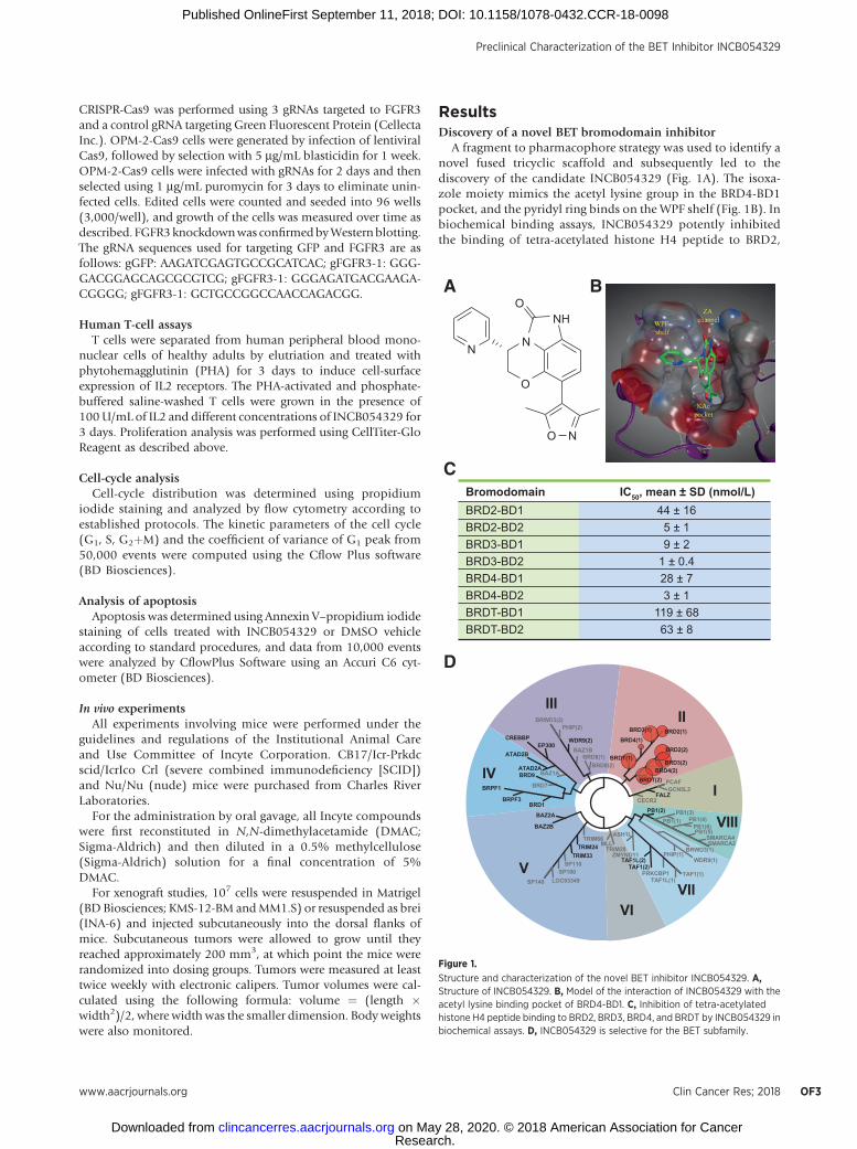
A fragment to pharmacophore strategy was used to identify anovel fused tricyclic scaffold and subsequently led to thediscovery of the candidate INCB054329 (Fig. 1A). The isoxa-zole moiety mimics the acetyl lysine group in the BRD4-BD1pocket, and the pyridyl ring binds on the WPF shelf (Fig. 1B). Inbiochemical binding assays, INCB054329 potently inhibitedthe binding of tetra-acetylated histone H4 peptide to BRD2,
II
I
VIII
III
IV
V
VIVII
BRDT(2)BRD4(2)
BRD3(2)
BRD2(2)
BRD2(1)BRD3(1)BRWD3(2)
PHIP(2)
CREBBPEP300
ATAD2B
ATAD2ABAZ1ABRD9
BRD7
BRD1
BAZ2A
BAZ2B
TRIM66
TRIM28
ASH1L
ZMYND11TAF1L(2)
TAF1(2)PRKCBP1
TAF1L(1)TAF1(1)
PHIP(1)WDR9(1)
BRWD3(1)SMARCA2
SMARCA4PB1(5)PB1(6)
PB1(4)PB1(3)
PB1(1)
PB1(2)CECR2
FALZGCN5L2
PCAF
MLLTRIM24TRIM33
SP110SP100
SP140 LOC93349
BRPF3
BRPF1
WDR9(2)BAZ1B
BRD8(1)BRD8(2)
BRD4(1)
BRDT(1)
BA
C
D
Bromodomain IC50, mean ± SD (nmol/L)61±441DB-2DRB
1±52DB-2DRB2±91DB-3DRB4.0±12DB-3DRB7±821DB-4DRB1±32DB-4DRB
86±9111DB-TDRB8±362DB-TDRB
O
NN
O N
NHO
Figure 1.
Structure and characterization of the novel BET inhibitor INCB054329. A,Structure of INCB054329. B, Model of the interaction of INCB054329 with theacetyl lysine binding pocket of BRD4-BD1. C, Inhibition of tetra-acetylatedhistone H4 peptide binding to BRD2, BRD3, BRD4, and BRDT by INCB054329 inbiochemical assays. D, INCB054329 is selective for the BET subfamily.
Preclinical Characterization of the BET Inhibitor INCB054329
www.aacrjournals.org Clin Cancer Res; 2018 OF3
Research. on May 28, 2020. © 2018 American Association for Cancerclincancerres.aacrjournals.org Downloaded from
Published OnlineFirst September 11, 2018; DOI: 10.1158/1078-0432.CCR-18-0098

BRD3, and BRD4, with modest selectivity for BRDT-BD1 andBRDT-BD2 (Fig. 1C). A screen against 16 non-BET bromodo-mains showed no significant inhibitory activity at 3 mmol/L(Fig. 1D).
INCB054329 exhibits broad antiproliferative activity againsthematologic cancer cell lines and antagonizes c-MYCexpression in vitro and in vivo
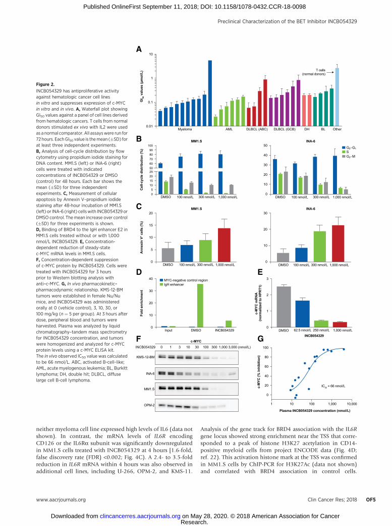
The antiproliferative activity of INCB054329 was profiledagainst a panel of 32 hematologic cancer cell lines derived fromacute myeloid leukemia, non-Hodgkin lymphoma, and mul-tiple myeloma (Fig. 2A). The median 50% growth inhibition(GI50) value was 152 nmol/L (range, 26–5,000 nmol/L), andinhibitory activity was observed across all histologies (Supple-mentary Table S1). In contrast to tumor cell lines, the GI50value against T cells isolated from non-diseased donors stim-ulated ex vivo with IL2 was 2.435 mmol/L. Growth inhibitioncorrelated with a concentration-dependent accumulation ofcells in the G1 phase of the cell cycle (Fig. 2B). An increase inapoptosis was also observed in four of five myeloma cell lines at1,000 nmol/L, and significant increases were observed from100 nmol/L in INA-6 cells (Fig. 2C). Consistent with publishedexperiments (19), ChIP of BRD4 showed enriched binding atthe IgH enhancers in MM1.S cells (Fig. 2D). Treatment withINCB054329 displaced BRD4 binding to the E2 30 enhancer,similar to findings with the BET inhibitor JQ-1 (SupplementaryFig. S1; ref. 19). Concomitant with the abrogation of BRD4binding, levels of c-MYC mRNA were rapidly suppressed,leading to a concentration-dependent decrease in c-MYC pro-tein in multiple cell lines (Fig. 2E and F). Using c-MYC as apharmacodynamic marker in tumor cells, we evaluated thepharmacokinetic–pharmacodynamic relationship after a singleoral administration of INCB054329 in mice bearing subcuta-neous xenografts of KMS-12-BM tumors. The KMS-12-BM cellline has been shown to have very high c-MYC protein levels(20) and is therefore amenable for pharmacokinetic/pharma-codynamic study. A concentration-dependent decrease wasobserved 3 hours after dose with an in vivo half-maximalinhibitory concentration (IC50) of 66 nmol/L and a 90%inhibitory concentration (IC90) of approximately 400 nmol/L(Fig. 2G). INCB054329 exhibited high clearance in mice result-ing in a short half-life (Supplementary Fig. S2A); therefore,twice-daily (b.i.d.) dosing was used in efficacy studies.At exposures that effectively suppressed c-MYC, INCB054329was found to be efficacious and well tolerated in both theKMS-12-BM and MM1.S xenograft models (Supplementary Fig.S2B and S2C).
INCB054329 displaces binding of BRD4 from enhancersdriving oncogenes in models of t(4;14) myeloma
The t(4;14) translocation involves a balanced rearrangementof the IgH locus on chromosome 14, with chromosome 4leading to deregulation of two potential oncogenes FGFR3(encoding a receptor tyrosine kinase) and NSD2/MMSET/WHSC1 (encoding a DNA methyl transferase; Fig. 3A; ref. 13).To assess whether both translocation partners are regulated byBRD4, ChIP-PCR was performed on both the Em and E2enhancers associated with the IgH locus. BRD4 binds to bothelements in two t(4;14)-positive cell lines OPM-2 and KMS-11,with higher normalized levels at the E2 enhancer. Treatmentwith 500 nmol/L INCB054329 reduced BRD4 association with
the E2 and Em enhancers to 8% and 29% of the DMSO controlsamples, respectively, in OPM-2 cells, while only BRD4 bind-ing to E2 was reduced (18% of control) in the KMS-11 cells(Fig. 3B; Supplementary Fig. S1). Steady-state mRNA levels ofFGFR3 were reduced to nearly background in the OPM-2 cellssimilar to c-MYC, while mRNA levels of NSD2 were decreasedapproximately 50% (Fig. 3C). FGFR3 protein levels were alsosuppressed to near background levels (1% of DMSO) at aconcentration of 250 nmol/L (approximately twice the IC50),whereas NSD2/MMSET protein was maximally reduced by50% at 24 hours (Fig. 3D).
In a subcutaneous xenograft model of OPM-2 tumors, oraladministration of INCB054329 resulted in a dose-dependentsuppression of tumor growth and was well tolerated as assessedby body-weight measurements (Fig. 3E). The strong suppres-sion of FGFR3 suggested that greater inhibition of tumorgrowth could be achieved in combination with FGFR targeting.INCB054828 is a selective kinase inhibitor of the FGFR 1, 2,and 3 and is currently being tested in phase I/II trials of solidand hematologic cancers (NCT02393248 and NCT03011372).INCB054828 inhibited OPM-2 cell proliferation (GI50, 97nmol/L) similar to deletion of FGFR3, indicating at least partialdependence of these cells on FGFR signaling (SupplementaryFig. S3). In vivo, partial antitumor efficacy by INCB054828 wasobserved against OPM-2 xenografted tumors at its optimal doseof 0.3 mg/kg once daily (Fig. 3E). When combined withINCB054329, however, a significantly greater decrease in ter-minal tumor volume was achieved than by either compoundalone (P < 0.001 by two-way ANOVA for combination versuseach single agent). Strikingly, FGFR inhibition alone resulted ina feedback upregulation of FGFR3 that was completely abro-gated in the combination group (Fig. 3F). Despite the increasedFGFR3 levels in tumors from INCB054828-treated mice, sig-naling through FGFR3 is blunted by INCB054828 (as deter-mined by pFGFR3 and pFRS2 levels), and slightly more soby the combination of INCB054828 and INCB054329. Thecombination also decreased levels of c-MYC and pSTAT3 andincreased levels of p21 when compared with levels from single-agent–treated tumor samples.
Gene-expression profiling identifies key growth regulatory andinflammatory pathways as targets of INCB054329
To characterize the broader effects of BET inhibition on geneexpression, two myeloma cell lines MM1.S and INA-6 weretreated for 4 and 24 hours with INCB054329 and analyzed bygene-expression profiling (Supplementary Fig. S4A). Pathwayanalysis of differentially expressed genes was evaluated usingtwo methods: MetaCore and GSEA. By MetaCore, a number ofpathways related to growth, development, and inflammationwere enriched (Fig. 4A). To explore molecular signatures ingreater detail, the data were also analyzed by GSEA. c-MYC–dependent gene sets emerged among the top downregulatedgene sets in both MM1.S and INA-6 cell lines (Fig. 4B; Sup-plementary Fig. S4B), and c-MYC was identified as the mostsignificantly downregulated transcription factor network inMM1.S cells, recapitulating results with JQ-1 and other BETinhibitors (19, 21). In addition, the IL6 signature in myelomawas among the top pathways in both cell lines. In monocytes,BET inhibitors repressed production of IL6 (10), andINCB054329 also suppressed secretion of IL6 by immortalized,human stromal cell lines (Supplementary Fig. S5); however,
Stubbs et al.
Clin Cancer Res; 2018 Clinical Cancer ResearchOF4
Research. on May 28, 2020. © 2018 American Association for Cancerclincancerres.aacrjournals.org Downloaded from
Published OnlineFirst September 11, 2018; DOI: 10.1158/1078-0432.CCR-18-0098
neither myeloma cell line expressed high levels of IL6 (data notshown). In contrast, the mRNA levels of IL6R encodingCD126 or the IL6Ra subunit was significantly downregulatedin MM1.S cells treated with INCB054329 at 4 hours [1.6-fold,false discovery rate (FDR) <0.002; Fig. 4C). A 2.4- to 3.5-foldreduction in IL6R mRNA within 4 hours was also observed inadditional cell lines, including U-266, OPM-2, and KMS-11.
Analysis of the gene track for BRD4 association with the IL6Rgene locus showed strong enrichment near the TSS that corre-sponded to a peak of histone H3K27 acetylation in CD14-positive myeloid cells from project ENCODE data (Fig. 4D;ref. 22). This activation histone mark at the TSS was confirmedin MM1.S cells by ChIP-PCR for H3K27Ac (data not shown)and correlated with BRD4 association in control cells.
0.01
0.1
1
10
DH BL OtherMyeloma AML DLBCL (ABC) DLBCL (GCB)
T cells(normal donors)
GI 50
val
ues
(μm
ol/L
)
0
2520
51015
8090
706050
100
0
10
20
40
30
50 G0−G1SG2−M
Cel
l-cyc
le d
istr
ibut
ion
(%)
0
5
15
10
20
0
20
10
30
MYC-negative control regionIgH enhancer
1,000 3,000 (nmol/L)3001003010310INCB054329
KMS-12-BM
INA-6
MM1.S
OPM-2
Ann
exin
V+ c
ells
(%)
A
MM1.SB INA-6
MM1.SC
0
30
20
10
40
Input DMSO INCB054329
Fold
enr
ichm
ent
c-M
YC m
RN
A
(nor
mal
ized
to H
PRT1
)
D
CYM-cF
INA-6
0
2
1
3
IC50 = 66 nmol/L
250 nmol/L62.5 nmol/LDMSO 1,000 nmol/L
1,000 nmol/L300 nmol/L100 nmol/LDMSO 1,000 nmol/L300 nmol/L100 nmol/LDMSO
1,000 nmol/L300 nmol/L100 nmol/LDMSO1,000 nmol/L300 nmol/L100 nmol/LDMSO
E
c-M
YC (%
inhi
bitio
n)
0
80
60
40
20
100
1 10 100
Plasma INCB054329 concentration (nmol/L)
10,0001,000
G INCB054329
Figure 2.
INCB054329 has antiproliferative activityagainst hematologic cancer cell linesin vitro and suppresses expression of c-MYCin vitro and in vivo. A, Waterfall plot showingGI50 values against a panel of cell lines derivedfrom hematologic cancers. T cells from normaldonors stimulated ex vivo with IL2 were usedas a normal comparator. All assayswere run for72 hours. EachGI50 value is themean (�SD) forat least three independent experiments.B, Analysis of cell-cycle distribution by flowcytometry using propidium iodide staining forDNA content. MM1.S (left) or INA-6 (right)cells were treated with indicatedconcentrations of INCB054329 or DMSO(control) for 48 hours. Each bar shows themean (�SD) for three independentexperiments. C, Measurement of cellularapoptosis by Annexin V–propidium iodidestaining after 48-hour incubation of MM1.S(left) or INA-6 (right) cellswith INCB054329 orDMSO control. The mean increase over control(�SD) for three experiments is shown.D, Binding of BRD4 to the IgH enhancer E2 inMM1.S cells treated without or with 1,000nmol/L INCB054329. E, Concentration-dependent reduction of steady-statec-MYC mRNA levels in MM1.S cells.F, Concentration-dependent suppressionof c-MYC protein by INCB054329. Cells weretreated with INCB054329 for 3 hoursprior to Western blotting analysis withanti–c-MYC. G, In vivo pharmacokinetic–pharmacodynamic relationship. KMS-12-BMtumors were established in female Nu/Numice, and INCB054329 was administeredorally at 0 (vehicle control), 3, 10, 30, or100 mg/kg (n ¼ 5 per group). At 3 hours afterdose, peripheral blood and tumors wereharvested. Plasma was analyzed by liquidchromatography–tandem mass spectrometryfor INCB054329 concentration, and tumorswere homogenized and analyzed for c-MYCprotein levels using a c-MYC ELISA kit.The in vivo observed IC50 value was calculatedto be 66 nmol/L. ABC, activated B-cell–like;AML, acute myelogenous leukemia; BL, Burkittlymphoma; DH, double hit; DLBCL, diffuselarge cell B-cell lymphoma.
Preclinical Characterization of the BET Inhibitor INCB054329
www.aacrjournals.org Clin Cancer Res; 2018 OF5
Research. on May 28, 2020. © 2018 American Association for Cancerclincancerres.aacrjournals.org Downloaded from
Published OnlineFirst September 11, 2018; DOI: 10.1158/1078-0432.CCR-18-0098

Treatment with JQ-1 abrogated the BRD4 peaks at this site.Similarly, treatment with INCB054329 resulted in depletion ofBRD4 from the IL6Ra TSS in OPM-2 cells (Fig. 4E).
Suppression of IL6R by INCB054329 sensitizes myeloma cellsto clinical Janus kinase (JAK) inhibitors
To evaluate whether the reduction of IL6Ra would translateinto a vulnerability that could be therapeutically exploited,cellular analyses were performed to assess the effect onIL6Ra protein levels and signaling in myeloma cell lines.INCB054329 treatment induced a dose-dependent decrease incell-surface IL6Ra staining as measured by flow cytometry inMM1.S and U-266 cells and in IL6Ra protein levels by Westernblotting in MM1.S and U-266 cells that corresponded todecreased levels of IL6Ra protein (Fig. 5A). The consequenceof IL6Ra reduction on IL6 signaling was measured in U-266cells because they express high levels of IL6Ra but are notdependent on IL6 for survival, in contrast to INA-6 cells thatrapidly lose viability upon loss of IL6 signaling (23). Basallevels of pSTAT3 were reduced by nearly eight fold withINCB054329 (Fig. 5B, compare lanes 1 to 4). Upon stimulationwith IL6, a concentration-dependent increase in the pSTAT3level was observed in both DMSO and INCB054329-treatedgroups; however, in cells pretreated with INCB054329, pSTAT3was induced to an extent that only slightly exceeded theunstimulated DMSO control (Fig. 5B, compare lanes 1 and6). Therefore, reducing cell-surface IL6Ra levels diminished theresponse to physiologically relevant levels of IL6. Consistentwith this effect, transcriptional profiling showed decreasedlevels in expression of several genes also known to be at leastin part transcriptionally regulated by STAT3, including BCL-xL,and cyclins D1 and D2 (Fig. 4C), indicating BET inhibition ofIL6Ra levels results in decreased levels of several downstreampathway targets. As a possible compensatory response to sup-pressed IL6 signaling, genes in the IL6–JAK2–STAT3 pathwaywere upregulated, including time-dependent increases in JAK1,JAK2, STAT6, and STAT3 transcripts.
We reasoned that targeting of BET in combination with aselective JAK inhibitor could have combinatorial effects onmyeloma cell viability, especially in view of potential compen-satory increases in JAK–STAT pathway genes. Studies were
A
2-MPOB
0
20
15
5
10
25 FGFR3 (IGH_E2) NSD2 (IGH_Eu)
Inpu
t
DM
SO
INC
B054
329
Inpu
t
DM
SO
INC
B054
329
Fold
enr
ichm
ent
Nor
mal
ized
mR
NA
leve
ls
Nor
mal
ized
mR
NA
leve
lsKMS-11
0
8
6
2
4
10 FGFR3 (IGH_E2) NSD2 (IGH_Eu)
Inpu
t
DM
SO
INC
B054
329
Inpu
t
DM
SO
INC
B054
329
Fold
enr
ichm
ent
3RFGFC
0.00
20.00
10.00
30.00
0 2 206
)h(emiT)h(emiT
NSD2
0.00
0.50
1.00
0 2 206
Tum
or v
olum
e (m
m3 )
± SE
M
E
F
0
1,000
500
1,500
2241 201816 26 2824
Days after inoculation
INCB054329 (250 nmol/L) DMSO
0 4 8 24 48 48D
NSD2/MMSET
FGFR3
c-MYC
VehicleINCB054329 25 mg/kg b.i.d.INCB054329 50 mg/kg b.i.d.INCB054329 75 mg/kg b.i.d.
TGI (%)
*
*
****
* P *1000.0< P = 0.0007; ** P < 0.0001
395149
p21
GAPDH
Time (h)
D
Tum
or v
olum
e (m
m3 )
± SE
M
0
1,000
500
1,500
2241 201816 26 2824
Days after inoculation
VehicleINCB054329FGFRiINCB054329 + FGFRi
TGI (%)
512380
Cμ CαEμV D J
V D J
E1 E2 E3 E4
E1 E2 E3 E4
Chr 14
Chr 4
t(4;14)
t(4;14)
der(4)
der(14)
Eμ
NSD2
NSD2
FGFR3
FGFR3
Vehicle
c-MYC
pSTAT3
STAT3
pFGFR3
FGFR3
p21
pFRS2
FRS2
Actin
25 mg/kg 50 mg/kg 75 mg/kg 0.3 mg/kg 1.0 mg/kg 0.3 mg/kg 1.0 mg/kg
INCB054329 INCB054828 INCB054828
50 mg/kg INCB054329
Figure 3.INCB054329 reduces BRD4 binding to the IgH enhancer and reducesexpression of target genes. A, Cartoon depicting the t(4;14)-balancedtranslocation resulting in fusion of the Em enhancer 30 to FGFR3 and the E2enhancer 50 to NSD2 (13). B, ChIP-PCR analysis of BRD4 binding to the 30
enhancer element E2 or the Em intronic enhancer in OPM-2 (left) or KMS-11(right) cells treated with DMSO or INCB054329. Data are expressed as thefold enrichment normalized to the input (2% of DMSO sample). C,INCB054329 reduces expression of genes regulated by the IgH enhancer.Decrease in the steady-state levels of FGFR3 (left) and NSD2 (right) mRNA inOPM-2 cells treated with INCB054329 for 4 hours. D,Western blot analysis ofbromodomain target proteins. OPM-2 cells were treated with 250 nmol/L ofINCB054329, harvested over 48 hours, and analyzed for levels of theindicated proteins. The "0" indicates untreated cells, and "48D" are controlcells treated for 48 hours with DMSO. E, Efficacy of INCB054329 in the OPM-2subcutaneous xenograft model as monotherapy or in combination with theselective FGFR inhibitor INCB054828. F, Xenografted OPM-2 tumors wereharvested from mice after 5 days of treatment with INCB054329 (50 mg/kg),INCB054828 (0.3 mg/kg), both agents, or vehicle, and Western blots of theindicated proteins were performed on the tumor lysates.
Stubbs et al.
Clin Cancer Res; 2018 Clinical Cancer ResearchOF6
Research. on May 28, 2020. © 2018 American Association for Cancerclincancerres.aacrjournals.org Downloaded from
Published OnlineFirst September 11, 2018; DOI: 10.1158/1078-0432.CCR-18-0098

undertaken using the JAK1/2 inhibitor ruxolitinib (24) or theJAK1-selective inhibitor itacitinib (Supplementary Table S2).INA-6 cells were sensitive to both JAK and BET inhibitors, and
combining either JAK inhibitor with INCB054329 potentiatedthe effect on viability. Using Bliss independence calculation(25), the average fractional product (Fua) across a 5 � 5
A
B C
E
Vehicle 24 h4 h
IL6R
JAK1
JAK2
STAT1
STAT2
STAT3
STAT6
CCND1
CCND2
c-MYC
HEXIM1
6-ANIS.1MM
Immune response IFN-alpha/beta signaling via MAPKs
Ligand-independent activation of androgen receptorin prostate cancer
Stem cells cooperation between Hedgehog, IGF-2, and HGFsignaling pathways in medulloblastoma stem cells
Development role of HDAC and calcium/calmodulin-dependentkinase (CaMK) in control of skeletal myogenesis
Stem cell aberrant WNT signaling in medulloblastoma stem cells
9.037e−6
0
RDFspaM
2 4–log (P value)
6 8
1.002e−7
3.201e−73.736e−5
6.075e−42.515e−7
4.659e−45.546e−7
5.347e−68.030e−7
3.696e−64.437e−4
8.309e−64.041e−4
2.334e−41.049e−5
1.763e−41.049e−5
1.238e−54.208e−2
IL6 signaling in multiple myeloma
IGF family signaling in colorectal cancer
Normal and pathological TGF-beta-mediated regulation ofcell proliferation
Role of activation of WNT signaling in the progression oflung cancer
Cell-cycle ESR1 regulation of G1/S transition
0
10
40
30
Fold
enr
ichm
ent
20
50
OPM-2 DMSO-BRD4
OPM-2 DMSO-neg Ab
OPM-2 DMSO-INPUT
OPM-2 INCB054329-BRD4
OPM-2 INCB054329-neg Ab
OPM-2 INCB054329-INPUT
MM1.S INCB054329 4 h vs. controlINA-6 INCB054329 4 h vs. control
-2.5 2.5Relativeexpression (log2)
Cytoband
Scale
Gene
DMSO_2% Input
JQ1_2% Input
DMSO_IP
JQ1_IP
H3K27ac ENCODE
D
IL6R [+] (3)
35
035
0
35
0
35
0
371
0
Figure 4.
Gene-expression profiling ofmyeloma cell lines treated withINCB054329 reveals regulation ofpathways involved in growth,development, and inflammation.A, Top pathways identified usingMetaCore in INA-6 cells and MM1.Scells treated with INCB054329for 4 hours (genes used for pathwayenrichment were selected by |FoldChange|>1.5 and FDR <0.05 in eachcell line). B, Gene set enrichmentanalysis showing representativeplots of gene sets stronglydownregulated by treatment withINCB054329. C, Changes inexpression of JAK–STAT pathwaygenes upon treatment withINCB054329 in MM1.S cells. D, BRD4binding to the IL6Ra TSS is blockedby BET inhibitors. ChIP-sequencingreads of BRD4 in MM1.S cells treatedwith DMSO or 1 mmol/L of JQ-1. Thegene track for H3K27 acetylation inCD14-positive cells from ENCODEconsortium is shown for comparison(22). E, ChIP-PCR for BRD4association with the TSS of IL6Rin OPM-2 cells treated with500 nmol/L of INCB054329 orDMSOusing anti-BRD4or a negativecontrol antibody. Data areexpressed as the fold enrichmentnormalized to the input (2% ofDMSO sample).
Preclinical Characterization of the BET Inhibitor INCB054329
www.aacrjournals.org Clin Cancer Res; 2018 OF7
Research. on May 28, 2020. © 2018 American Association for Cancerclincancerres.aacrjournals.org Downloaded from
Published OnlineFirst September 11, 2018; DOI: 10.1158/1078-0432.CCR-18-0098
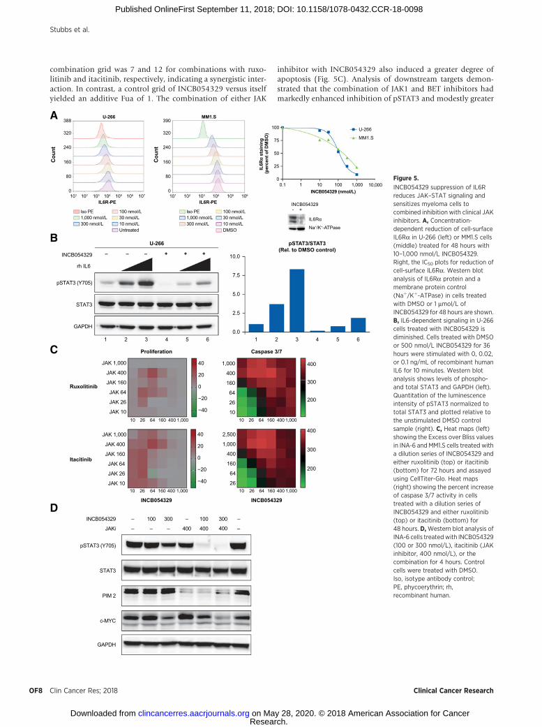
combination grid was 7 and 12 for combinations with ruxo-litinib and itacitinib, respectively, indicating a synergistic inter-action. In contrast, a control grid of INCB054329 versus itselfyielded an additive Fua of 1. The combination of either JAK
inhibitor with INCB054329 also induced a greater degree ofapoptosis (Fig. 5C). Analysis of downstream targets demon-strated that the combination of JAK1 and BET inhibitors hadmarkedly enhanced inhibition of pSTAT3 and modestly greater
U-266A
U-266
–– +– ++
rh IL6
INCB054329
pSTAT3 (Y705)
STAT3
GAPDH
B
7/3 esapsaCnoitarefilorPC
INCB054329
Itacitinib
Ruxolitinib
INCB054329
pSTAT3/STAT3(Rel. to DMSO control)
0.0
7.5
5.0
2.5
10.0
61 2 3 4 561 2 3 4 5
10 26 64 160 400 1,000
JAK 1,000
JAK 400
JAK 160
JAK 64
JAK 26
JAK 10 −40
−20
0
20
40
10 26 64 160 400 1,000
JAK 1,000
JAK 400
JAK 160
JAK 64
JAK 26
JAK 10 −40
−20
0
20
40
10 26 64 160 400 1,000
1,000
400
160
64
26
10
200
300
400
10 26 64 160 400 1,000
2,500
1,000
400
160
64
26
200
300
400
INCB054329 – 100 300 – –100 300
JAKi – – – 400 400 –400
pSTAT3 (Y705)
STAT3
PIM 2
c-MYC
D
GAPDH
388
320
240
160Cou
nt
80
0
390
Iso PE1,000 nmol/L300 nmol/L
30 nmol/L100 nmol/L
320
240
160Cou
nt
80
0
IL6R-PE101 102 103 104 105 106 107
IL6R-PE101 102 103 104 105 106
10 nmol/LUntreated
Iso PE1,000 nmol/L300 nmol/L
30 nmol/L100 nmol/L
10 nmol/LDMSO
MM1.S
IL6Rα
Na+/K+-ATPase
- +INCB054329
0.1 1 10 100 1,000 10,000INCB054329 (nmol/L)
IL6R
α st
aini
ng(p
erce
nt o
f DM
SO)
0
25
50
75
100
MM1.S
U-266
Figure 5.
INCB054329 suppression of IL6Rreduces JAK–STAT signaling andsensitizes myeloma cells tocombined inhibition with clinical JAKinhibitors. A, Concentration-dependent reduction of cell-surfaceIL6Ra in U-266 (left) or MM1.S cells(middle) treated for 48 hours with10–1,000 nmol/L INCB054329.Right, the IC50 plots for reduction ofcell-surface IL6Ra. Western blotanalysis of IL6Ra protein and amembrane protein control(Naþ/Kþ-ATPase) in cells treatedwith DMSO or 1 mmol/L ofINCB054329 for 48 hours are shown.B, IL6-dependent signaling in U-266cells treated with INCB054329 isdiminished. Cells treated with DMSOor 500 nmol/L INCB054329 for 36hours were stimulated with 0, 0.02,or 0.1 ng/mL of recombinant humanIL6 for 10 minutes. Western blotanalysis shows levels of phospho-and total STAT3 and GAPDH (left).Quantitation of the luminescenceintensity of pSTAT3 normalized tototal STAT3 and plotted relative tothe unstimulated DMSO controlsample (right). C, Heat maps (left)showing the Excess over Bliss valuesin INA-6 and MM1.S cells treated witha dilution series of INCB054329 andeither ruxolitinib (top) or itacitinib(bottom) for 72 hours and assayedusing CellTiter-Glo. Heat maps(right) showing the percent increaseof caspase 3/7 activity in cellstreated with a dilution series ofINCB054329 and either ruxolitinib(top) or itacitinib (bottom) for48 hours. D,Western blot analysis ofINA-6 cells treated with INCB054329(100 or 300 nmol/L), itacitinib (JAKinhibitor, 400 nmol/L), or thecombination for 4 hours. Controlcells were treated with DMSO.Iso, isotype antibody control;PE, phycoerythrin; rh,recombinant human.
Stubbs et al.
Clin Cancer Res; 2018 Clinical Cancer ResearchOF8
Research. on May 28, 2020. © 2018 American Association for Cancerclincancerres.aacrjournals.org Downloaded from
Published OnlineFirst September 11, 2018; DOI: 10.1158/1078-0432.CCR-18-0098

0
50
100
150 *
* P < 0.05
BA
C
c-M
YC p
rote
in le
vels
(%
of v
ehic
le)
0
50
25
75
100
125
* P = 0.022** P = 0.0001
E
c-M
YC p
rote
in le
vels
(%
of v
ehic
le)
Vehicle
pSTAT3
STAT3
INCB054329 RuxolitinibINCB054329+ ruxolitinib Itacitinib
INCB054329+ itacitinib
FVehicle
pSTAT3
STAT3
INCB054329 RuxolitinibINCB054329+ ruxolitinib
Tum
or v
olum
e (m
m3 )
± SE
M
0
1,000
750
500
250
1,250
16 220241 18Days after inoculation
*
VehicleINCB054329Ruxolitinib
*Day 21 TGI from all combination groups differed from correspondingsingle-agent TGIP < 0.0001 in each case
TGI (%)
5147
Itacitinib 3389Ruxolitinib + INCB054329
Itacitinib + INCB054329 87
S.1MMD
INA-6
Tum
or v
olum
e (m
m3 )
± SE
M
0
500
250
750
151311 1917
Days after inoculation
* P = 0.027
VehicleINCB054329Ruxolitinib
TGI (%)
342
62Ruxolitinib + INCB054329
Vehicl
e
INCB05
4329
Ruxoli
tinib
INCB05
4329
+ rux
olitin
ib
Itacit
inib
INCB05
4329
+ ita
citini
b
*
Vehicl
e
INCB05
4329
Ruxoli
tinib
INCB05
4329
+ rux
olitin
ib
*
**
*
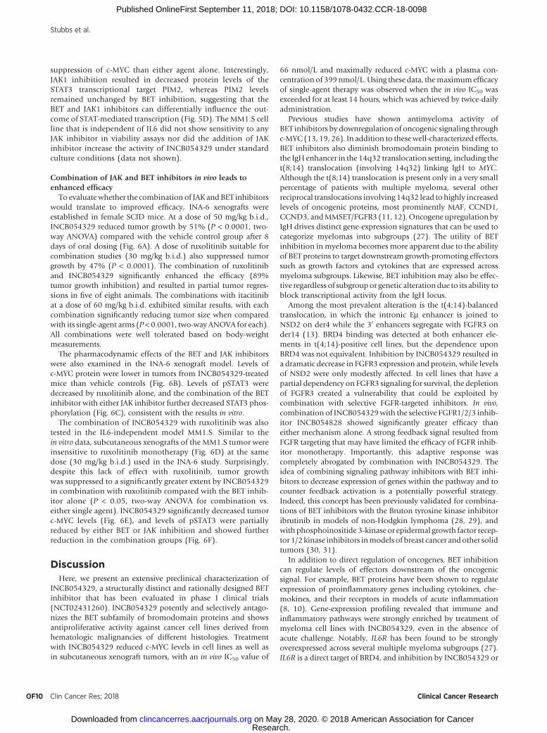
Figure 6.
Combination of BET and JAK inhibitors in vivo shows synergistic effects on tumor growth. A, Efficacy of INCB054329 and JAK inhibitor monotherapy or incombination in the subcutaneous INA-6 humanmyelomaxenograftmodel.B,Pharmacodynamic effects of INCB054329, JAK inhibitors, or the combination on c-MYClevels in INA-6 xenografted tumors following a single oral dose. C, Pharmacodynamic effects of INCB054329, JAK inhibitors, or the combination on pSTAT3 levels inINA-6 xenografted tumors following a single oral dose.D, Efficacy of INCB054329 and ruxolitinib monotherapy or in combination in the subcutaneous MM1.S humanmyelomaxenograftmodel.E,Pharmacodynamic effects of INCB054329, ruxolitinib, or the combination on c-MYC inMM1.S xenografted tumors following a single oraldose. F, Pharmacodynamic effects of INCB054329, ruxolitinib, or the combination on pSTAT3 in MM1.S xenografted tumors following a single oral dose.
Preclinical Characterization of the BET Inhibitor INCB054329
www.aacrjournals.org Clin Cancer Res; 2018 OF9
Research. on May 28, 2020. © 2018 American Association for Cancerclincancerres.aacrjournals.org Downloaded from
Published OnlineFirst September 11, 2018; DOI: 10.1158/1078-0432.CCR-18-0098
suppression of c-MYC than either agent alone. Interestingly,JAK1 inhibition resulted in decreased protein levels of theSTAT3 transcriptional target PIM2, whereas PIM2 levelsremained unchanged by BET inhibition, suggesting that theBET and JAK1 inhibitors can differentially influence the out-come of STAT-mediated transcription (Fig. 5D). The MM1.S cellline that is independent of IL6 did not show sensitivity to anyJAK inhibitor in viability assays nor did the addition of JAKinhibitor increase the activity of INCB054329 under standardculture conditions (data not shown).
Combination of JAK and BET inhibitors in vivo leads toenhanced efficacy
To evaluatewhether the combination of JAK andBET inhibitorswould translate to improved efficacy, INA-6 xenografts wereestablished in female SCID mice. At a dose of 50 mg/kg b.i.d.,INCB054329 reduced tumor growth by 51% (P < 0.0001, two-way ANOVA) compared with the vehicle control group after 8days of oral dosing (Fig. 6A). A dose of ruxolitinib suitable forcombination studies (30 mg/kg b.i.d.) also suppressed tumorgrowth by 47% (P < 0.0001). The combination of ruxolitiniband INCB054329 significantly enhanced the efficacy (89%tumor growth inhibition) and resulted in partial tumor regres-sions in five of eight animals. The combinations with itacitinibat a dose of 60 mg/kg b.i.d. exhibited similar results, with eachcombination significantly reducing tumor size when comparedwith its single-agent arms (P < 0.0001, two-way ANOVA for each).All combinations were well tolerated based on body-weightmeasurements.
The pharmacodynamic effects of the BET and JAK inhibitorswere also examined in the INA-6 xenograft model. Levels ofc-MYC protein were lower in tumors from INCB054329-treatedmice than vehicle controls (Fig. 6B). Levels of pSTAT3 weredecreased by ruxolitinib alone, and the combination of the BETinhibitor with either JAK inhibitor further decreased STAT3 phos-phorylation (Fig. 6C), consistent with the results in vitro.
The combination of INCB054329 with ruxolitinib was alsotested in the IL6-independent model MM1.S. Similar to thein vitro data, subcutaneous xenografts of the MM1.S tumor wereinsensitive to ruxolitinib monotherapy (Fig. 6D) at the samedose (30 mg/kg b.i.d.) used in the INA-6 study. Surprisingly,despite this lack of effect with ruxolitinib, tumor growthwas suppressed to a significantly greater extent by INCB054329in combination with ruxolitinib compared with the BET inhib-itor alone (P < 0.05, two-way ANOVA for combination vs.either single agent). INCB054329 significantly decreased tumorc-MYC levels (Fig. 6E), and levels of pSTAT3 were partiallyreduced by either BET or JAK inhibition and showed furtherreduction in the combination groups (Fig. 6F).
DiscussionHere, we present an extensive preclinical characterization of
INCB054329, a structurally distinct and rationally designed BETinhibitor that has been evaluated in phase I clinical trials(NCT02431260). INCB054329 potently and selectively antago-nizes the BET subfamily of bromodomain proteins and showsantiproliferative activity against cancer cell lines derived fromhematologic malignancies of different histologies. Treatmentwith INCB054329 reduced c-MYC levels in cell lines as well asin subcutaneous xenograft tumors, with an in vivo IC50 value of
66 nmol/L and maximally reduced c-MYC with a plasma con-centrationof 399nmol/L.Using these data, themaximumefficacyof single-agent therapy was observed when the in vivo IC50 wasexceeded for at least 14 hours, which was achieved by twice-dailyadministration.
Previous studies have shown antimyeloma activity ofBET inhibitors by downregulation of oncogenic signaling throughc-MYC (13, 19, 26). In addition to thesewell-characterized effects,BET inhibitors also diminish bromodomain protein binding tothe IgH enhancer in the 14q32 translocation setting, including thet(8;14) translocation (involving 14q32) linking IgH to MYC.Although the t(8;14) translocation is present only in a very smallpercentage of patients with multiple myeloma, several otherreciprocal translocations involving 14q32 lead tohighly increasedlevels of oncogenic proteins, most prominently MAF, CCND1,CCND3, andMMSET/FGFR3 (11, 12).Oncogene upregulation byIgH drives distinct gene-expression signatures that can be used tocategorize myelomas into subgroups (27). The utility of BETinhibition in myeloma becomes more apparent due to the abilityof BET proteins to target downstream growth-promoting effectorssuch as growth factors and cytokines that are expressed acrossmyeloma subgroups. Likewise, BET inhibition may also be effec-tive regardless of subgroupor genetic alterationdue to its ability toblock transcriptional activity from the IgH locus.
Among the most prevalent alteration is the t(4;14)-balancedtranslocation, in which the intronic Em enhancer is joined toNSD2 on der4 while the 30 enhancers segregate with FGFR3 onder14 (13). BRD4 binding was detected at both enhancer ele-ments in t(4;14)-positive cell lines, but the dependence uponBRD4 was not equivalent. Inhibition by INCB054329 resulted ina dramatic decrease in FGFR3 expression and protein, while levelsof NSD2 were only modestly affected. In cell lines that have apartial dependency on FGFR3 signaling for survival, the depletionof FGFR3 created a vulnerability that could be exploited bycombination with selective FGFR-targeted inhibitors. In vivo,combination of INCB054329with the selective FGFR1/2/3 inhib-itor INCB054828 showed significantly greater efficacy thaneither mechanism alone. A strong feedback signal resulted fromFGFR targeting that may have limited the efficacy of FGFR inhib-itor monotherapy. Importantly, this adaptive response wascompletely abrogated by combination with INCB054329. Theidea of combining signaling pathway inhibitors with BET inhi-bitors to decrease expression of genes within the pathway and tocounter feedback activation is a potentially powerful strategy.Indeed, this concept has been previously validated for combina-tions of BET inhibitors with the Bruton tyrosine kinase inhibitoribrutinib in models of non-Hodgkin lymphoma (28, 29), andwith phosphoinositide 3-kinase or epidermal growth factor recep-tor 1/2 kinase inhibitors inmodels of breast cancer andother solidtumors (30, 31).
In addition to direct regulation of oncogenes, BET inhibitioncan regulate levels of effectors downstream of the oncogenicsignal. For example, BET proteins have been shown to regulateexpression of proinflammatory genes including cytokines, che-mokines, and their receptors in models of acute inflammation(8, 10). Gene-expression profiling revealed that immune andinflammatory pathways were strongly enriched by treatment ofmyeloma cell lines with INCB054329, even in the absence ofacute challenge. Notably, IL6R has been found to be stronglyoverexpressed across several multiple myeloma subgroups (27).IL6R is a direct target of BRD4, and inhibition by INCB054329 or
Stubbs et al.
Clin Cancer Res; 2018 Clinical Cancer ResearchOF10
Research. on May 28, 2020. © 2018 American Association for Cancerclincancerres.aacrjournals.org Downloaded from
Published OnlineFirst September 11, 2018; DOI: 10.1158/1078-0432.CCR-18-0098

JQ-1 displaced it from the IL6R TSS suppressing the expression ofIL6Ra and resulting indiminished signaling through STAT3 evenupon IL6 stimulation. An apparent compensatory response todecreased IL6–JAK–STAT signaling was evidenced by upregula-tion of components of the JAK–STAT signaling pathway. Con-sistent with this finding, combinations with the clinical JAKinhibitors ruxolitinib or itacitinib further reduced JAK–STATsignaling in the IL6-dependent INA-6 model, resulting insynergistic inhibition of proliferation and enhanced apoptosisin cells. Combining BET and JAK inhibitors resulted in superiorefficacy at well-tolerated doses and together suppressed levelsof pSTAT3 more effectively than either agent alone. The impor-tance of IL6 as a growth and survival factor for myeloma cellsand as a modulator of cellular interactions in the myelomatumor microenvironment is well documented (11, 32). Gene-expression analysis of human myeloma has revealed a signif-icant subset of tumors with an activated STAT signature (33).In murine models, activated IL6–JAK–STAT signaling andc-MYC have been shown to collaborate in the developmentof myeloma. An intercross of mice harboring a human IL6transgene with Em-c-MYC mice resulted in the acceleration ofplasma cell tumor formation with full penetrance (34). Sep-arately, a murine transduction–transplantation model of mye-loma driven by a constitutively active allele of Gp130 encodingthe common coreceptor subunit for cytokine receptors, includ-ing IL6Ra, also showed enhanced tumorigenesis in coopera-tion with c-MYC. In plasmacytomas arising from Gp130 acti-vation alone, activating genetic alterations in the c-MYC locuswere recovered in three of ten independently arising tumors(33). Combining JAK and BET inhibitors is a potentiallypromising strategy for other hematologic malignancies, includ-ing JAK2V617-dependent myeloproliferative neoplasms andacute lymphocytic leukemia (ALL) characterized by IL7R acti-vation (35–38). In the ALL models, JQ-1 suppressed STAT5-dependent signaling and improved survival; significantly atrelapse, these tumors showed reactivation of STAT5 signalingindicating a feedback upregulation of JAK–STAT signaling.
The mechanism for synergy merits additional study andshould consider both cell-intrinsic and cell-extrinsic mechan-isms given that BET inhibitors may affect expression of solublemediators within the tumor microenvironment that may beimportant for supporting tumor cell viability. As an example,the MM1.S xenograft model, which does not respond to JAKinhibition alone, was unexpectedly sensitized to JAK inhibitionby coadministration of INCB054329 in vivo. This sensitivity tocombined JAK and BET inhibition was not observed in vitro.One potential explanation for these findings is that the levels ofcytokines or growth factors in the tumor microenvironmentthat provide protection from JAK inhibition may not be as highas those in cell culture. It is also possible that BET inhibitioncould make some myelomas more reliant on cytokine signalingfor survival, consistent with the upregulation of JAK–STATpathway genes. Finally, it is possible that BET inhibitors havebiological effects on other cell types within the tumor micro-environment that may support the malignant cells. Preliminarydata show that BET inhibition can markedly reduce the secre-tion of multiple cytokines, including IL6 and IL8 (Supplemen-tary Fig. S5), in cultured stromal cell lines at concentrationsbelow the IC50 for suppression of cell growth. Thus, in vivo BETinhibition may have pleiotropic biological impacts beyonddirect effects on the malignant clone.
In summary, we present the profile of a BET inhibitor,INCB054329, that has advanced into phase I trials. Using mye-loma as a model, we identified oncogenes and inflammatorypathways regulated by BET proteins that are significantly alteredby treatment with the INCB054329. Preliminary clinical datafrom trials with BET inhibitors have shown only modest single-agent efficacy to date in hematologic cancers, includingmyeloma,suggesting that rational therapeutic combinations designedto target different aspects of cancer biology may be required toenhance responses in patients with advanced malignancies (39).By evaluating BET inhibitor–induced changes in growth-promot-ing and survival pathways, vulnerabilities are revealed thatcan guide rational combinations such as with FGFR inhibitorsin the case of selected t(4;14)-rearranged myelomas and withJAK inhibitors in models characterized by an enriched IL6 signa-ture. Importantly, the effects of INCB054329on these targets wereachieved using modest concentrations (approximately 2–3 �in vivo IC50), suggesting that these findings can be translatableto the clinic. By targeting different aspects of tumor biology,combination therapy may overcome the tumor heterogeneityand complexity that may be contributing to limited single-agentefficacy in early trials.
Disclosure of Potential Conflicts of InterestM.C. Stubbs, T.C. Burn, A. Volgina, N. Zolotarjova, J. Li, R. Collins,
V. Dostalik, R. Wynn, B. Ruggeri, S. Yeleswaram, R. Huber, and P.C.C. Liu haveownership interests (including patents) in Incyte Corporation. No potentialconflicts of interest were disclosed by the other authors.
Authors' ContributionsConception and design: M.C. Stubbs, T.C. Burn, T. Maduskuie, R. Wynn,W. Yao, A.P. Combs, R. Huber, G. Hollis, P. Scherle, P.C.C. LiuDevelopment of methodology: M.C. Stubbs, T.C. Burn, A. Volgina,N. Zolotarjova,M. Favata, X.M. Liu, J. Li, N. Falahatpisheh, P. Polam, P. Thekkat,X. He, M. CovingtonAcquisition of data (provided animals, acquired and managed patients,provided facilities, etc.): M. Rupar, X. Wen, N. Zolotarjova, M. Favata,X.M. Liu, J. Li, D. DiMatteo, P. Feldman, P. Thekkat, C. Gardiner, Y. Li,M. Covington, R. Wynn, P.C.C. LiuAnalysis and interpretation of data (e.g., statistical analysis, biostatistics,computational analysis): M.C. Stubbs, T.C. Burn, S. Diamond, M. Rupar,A. Volgina, N. Zolotarjova, M. Favata, H. Liu, X.M. Liu, J. Li, P. Feldman,P. Thekkat, C. Gardiner, X. He, M. Covington, R. Wynn, S. Yeleswaram,P. Scherle, P.C.C. LiuWriting, review, and/or revision of the manuscript: M.C. Stubbs, T.C. Burn,M. Rupar, R. Collins, V. Dostalik,M. Covington, B. Ruggeri, G. Hollis, P. Scherle,P.C.C. LiuAdministrative, technical, or material support (i.e., reporting or organizingdata, constructing databases): B. RuggeriStudy supervision: M.C. Stubbs, S. Diamond, B. Ruggeri, S. Yeleswaram,C.-B. Xue, R. Huber, P.C.C. LiuOther (medicinal chemistry research): R. SparksOther (contributing author): P. WaeltzOther (research: Molecular modeling): R. Jalluri
AcknowledgmentsMedical writing assistance was provided by Abigail Marmont of Evidence
Scientific Solutions Inc., Wilmslow, UK, and funded by Incyte Corporation.
The costs of publication of this articlewere defrayed inpart by the payment ofpage charges. This article must therefore be hereby marked advertisement inaccordance with 18 U.S.C. Section 1734 solely to indicate this fact.
Received February 8, 2018; revised June 20, 2018; accepted September 7,2018; published first September 11, 2018.
Preclinical Characterization of the BET Inhibitor INCB054329
www.aacrjournals.org Clin Cancer Res; 2018 OF11
Research. on May 28, 2020. © 2018 American Association for Cancerclincancerres.aacrjournals.org Downloaded from
Published OnlineFirst September 11, 2018; DOI: 10.1158/1078-0432.CCR-18-0098

References1. Filippakopoulos P, Knapp S. Targeting bromodomains: epigenetic
readers of lysine acetylation. Nat Rev Drug Discov 2014;13:337–56.
2. Shi J, Vakoc CR. The mechanisms behind the therapeutic activity of BETbromodomain inhibition. Mol Cell 2014;54:728–36.
3. Hnisz D, Abraham BJ, Lee TI, Lau A, Saint-Andr�e V, Sigova AA, et al.Super-enhancers in the control of cell identity and disease. Cell 2013;155:934–47.
4. Lov�en J, Hoke HA, Lin CY, Lau A, Orlando DA, Vakoc CR, et al. Selectiveinhibition of tumor oncogenes by disruption of super-enhancers. Cell2013;153:320–34.
5. Mochizuki K, Nishiyama A, Jang MK, Dey A, Ghosh A, Tamura T,et al. The bromodomain protein Brd4 stimulates G1 gene transcrip-tion and promotes progression to S phase. J Biol Chem 2008;283:9040–8.
6. Yang Z,HeN, ZhouQ. Brd4 recruits P-TEFb to chromosomes at latemitosisto promote G1 gene expression and cell cycle progression. Mol Cell Biol2008;28:967–76.
7. Filippakopoulos P, Qi J, Picaud S, Shen Y, Smith WB, Fedorov O,et al. Selective inhibition of BET bromodomains. Nature 2010;468:1067–73.
8. Nicodeme E, Jeffrey KL, Schaefer U, Beinke S, Dewell S, Chung CW, et al.Suppression of inflammation by a synthetic histone mimic. Nature2010;468:1119–23.
9. Zuber J, Shi J, Wang E, Rappaport AR, Herrmann H, Sison EA, et al. RNAiscreen identifies Brd4 as a therapeutic target in acute myeloid leukaemia.Nature 2008;478:524–8.
10. Belkina AC, Nikolajczyk BS, Denis GV. BET protein function is required forinflammation: Brd2 genetic disruption and BET inhibitor JQ1 impairmouse macrophage inflammatory responses. J Immunol 2013;190:3670–8.
11. Anderson KC, Carrasco RD. Pathogenesis of myeloma. Annu Rev Pathol2011;6:249–74.
12. Kuehl WM, Bergsagel PL. Molecular pathogenesis of multiple mye-loma and its premalignant precursor. J Clin Invest 2012;122:3456–63.
13. Chesi M, Nardini E, Lim RS, Smith KD, Kuehl WM, Bergsagel PL. Thet(4;14) translocation in myeloma dysregulates both FGFR3 and a novelgene, MMSET, resulting in IgH/MMSET hybrid transcripts. Blood 1998;92:3025–34.
14. Affer M, Chesi M, Chen WG, Keats JJ, Demchenko YN, Roschke AV, et al.Promiscuous MYC locus rearrangements hijack enhancers but mostlysuper-enhancers to dysregulate MYC expression in multiple myeloma.Leukemia 2014;28:1725–35.
15. Walker BA, Wardell CP, Brioli A, Boyle E, Kaiser MF, Begum DB, et al.Translocations at 8q24 juxtapose MYC with genes that harbor super-enhancers resulting in overexpression and poor prognosis in myelomapatients. Blood Cancer J 2014;4:e191.
16. KawanoY,MoschettaM,Manier S, Glavey S, GorgunGT, Roccaro AM, et al.Targeting the bone marrow microenvironment in multiple myeloma.Immunol Rev 2015;263:160–72.
17. Mootha VK, Lindgren CM, Eriksson KF, Subramanian A, Sihag S, Lehar J,et al. PGC-1alpha-responsive genes involved in oxidative phosphorylationare coordinately downregulated in human diabetes. Nat Genet 2003;34:267–73.
18. SubramanianA, TamayoP,Mootha VK,Mukherjee S, Ebert BL,GilletteMA,et al. Gene set enrichment analysis: a knowledge-based approach forinterpreting genome-wide expression profiles. Proc Natl Acad Sci U S A2005;102:15545–50.
19. Delmore JE, Issa GC, Lemieux ME, Rahl PB, Shi J, Jacobs HM, et al. BETbromodomain inhibition as a therapeutic strategy to target c-Myc. Cell2011;146:904–17.
20. Holien T, Vatsveen TK,Hella H,Waage A, Sundan A. Addiction to c-MYC inmultiple myeloma. Blood 2012;120:2450–3.
21. Mertz JA, Conery AR, Bryant BM, Sandy P, Balasubramanian S, Mele DA,et al. Targeting MYC dependence in cancer by inhibiting BET bromodo-mains. Proc Natl Acad Sci U S A 2011;108:16669–74.
22. Dunham I, ENCODE Project Consortium. An integrated encyclopedia ofDNA elements in the human genome. Nature 2012;489:57–74.
23. Burger R, Guenther A, Bakker F, Schmalzing M, Bernand S, Baum W,et al. Gp130 and ras mediated signaling in human plasma cell line INA-6: a cytokine-regulated tumor model for plasmacytoma. Hematol J2001;2:42–53.
24. Quint�as-Cardama A, Vaddi K, Liu P, Manshouri T, Li J, Scherle PA, et al.Preclinical characterization of the selective JAK1/2 inhibitor INCB018424:therapeutic implications for the treatment of myeloproliferative neo-plasms. Blood 2010;115:3109–17.
25. Foucquier J, Guedj M. Analysis of drug combinations: current methodo-logical landscape. Pharmacol Res Perspect 2015;3:e00149.
26. Chaidos A, Caputo V, Gouvedenou K, Liu B, Marigo I, Chaudhry MS, et al.Potent antimyeloma activity of the novel bromodomain inhibitorsI-BET151 and I-BET762. Blood 2014;123:697–705.
27. Zhan F, Huang Y, Colla S, Stewart JP, Hanamura I, Gupta S, et al.The molecular classification of multiple myeloma. Blood 2006;108:2020–8.
28. Ceribelli M, Kelly PN, Shaffer AL, Wright GW, Xiao W, Yang Y, et al.Blockade of oncogenic IkB kinase activity in diffuse large B-cell lymphomaby bromodomain and extraterminal domain protein inhibitors. Proc NatlAcad Sci U S A 2014;111:11365–70.
29. Sun B, Shah B, Fiskus W, Qi J, Rajapakshe K, Coarfa C, et al. Synergisticactivity of BET protein antagonist-based combinations in mantle celllymphoma cells sensitive or resistant to ibrutinib. Blood 2015;126:1565–74.
30. Stratikopoulos EE, Dendy M, Szabolcs M, Khaykin AJ, Lefebvre C, ZhouMM, et al. Kinase and BET inhibitors together clamp inhibition of PI3Ksignaling and overcome resistance to therapy. Cancer Cell 2015;27:837–51.
31. Stuhlmiller TJ,Miller SM, Zawistowski JS,NakamuraK, BeltranAS,DuncanJS, et al. Inhibition of lapatinib-induced kinome reprogramming in ERBB2-positive breast cancer by targeting BET family bromodomains. Cell Rep2015;11:390–404.
32. Rosean TR, Tompkins VS, Tricot G, Holman CJ, Olivier AK, Zhan F, et al.Preclinical validation of interleukin 6 as a therapeutic target in multiplemyeloma. Immunol Res 2014;59:188–202.
33. Dechow T, Steidle S, Gotze KS, Rudelius M, Behnke K, Pechloff K, et al.GP130 activation induces myeloma and collaborates with MYC. J ClinInvest 2014;124:5263–74.
34. Rutsch S,Neppalli VT, ShinDM,DuBoisW,MorseHC3rd,GoldschmidtH,et al. IL-6 and MYC collaborate in plasma cell tumor formation in mice.Blood 2010;115:1746–54.
35. SaenzDT, FiskusW,Manshouri T, Rajapakshe K, Krieger S, Sun B, et al. BETprotein bromodomain inhibitor-based combinations are highly activeagainst post-myeloproliferative neoplasm secondary AML cells. Leukemia2017;31:678–87.
36. Wyspianska BS, Bannister AJ, Barbieri I, Nangalia J, Godfrey A, Calero-Nieto FJ, et al. BET protein inhibition shows efficacy against JAK2V617F-driven neoplasms. Leukemia 2014;28:88–97.
37. Ott CJ, Kopp N, Bird L, Paranal RM, Qi J, Bowman T, et al. BET bromo-domain inhibition targets both c-Myc and IL7R in high-risk acute lym-phoblastic leukemia. Blood 2012;120:2843–52.
38. Kleppe M, Koche R, Zou L, van Galen P, Hill CE, Dong L, et al. Dualtargeting of oncogenic activation and inflammatory signaling increasestherapeutic efficacy in myeloproliferative neoplasms. Cancer Cell 2018;33:29–43 e7.
39. Amorim S, Stathis A, Gleeson M, Iyengar S, Magarotto V, Leleu X, et al.Bromodomain inhibitor OTX015 in patients with lymphoma or multiplemyeloma: a dose-escalation, open-label, pharmacokinetic, phase 1 study.Lancet Haematol 2016;3:e196–204.
Clin Cancer Res; 2018 Clinical Cancer ResearchOF12
Stubbs et al.
Research. on May 28, 2020. © 2018 American Association for Cancerclincancerres.aacrjournals.org Downloaded from
Published OnlineFirst September 11, 2018; DOI: 10.1158/1078-0432.CCR-18-0098

Published OnlineFirst September 11, 2018.Clin Cancer Res Matthew C. Stubbs, Timothy C. Burn, Richard Sparks, et al. Inform Rational Combination StrategiesINCB054329 Induces Vulnerabilities in Myeloma Cells That The Novel Bromodomain and Extraterminal Domain Inhibitor
Updated version
10.1158/1078-0432.CCR-18-0098doi:
Access the most recent version of this article at:
Material
Supplementary
http://clincancerres.aacrjournals.org/content/suppl/2018/09/11/1078-0432.CCR-18-0098.DC1Access the most recent supplemental material at:
E-mail alerts related to this article or journal.Sign up to receive free email-alerts
Subscriptions
Reprints and
To order reprints of this article or to subscribe to the journal, contact the AACR Publications
Permissions
Rightslink site. (CCC)Click on "Request Permissions" which will take you to the Copyright Clearance Center's
.http://clincancerres.aacrjournals.org/content/early/2018/10/29/1078-0432.CCR-18-0098To request permission to re-use all or part of this article, use this link
Research. on May 28, 2020. © 2018 American Association for Cancerclincancerres.aacrjournals.org Downloaded from
Published OnlineFirst September 11, 2018; DOI: 10.1158/1078-0432.CCR-18-0098