18F-Labeling of Small Molecules and Peptides
194
18 F-Labeling of Small Molecules and Peptides Von der Fakultät für Mathematik, Informatik und Naturwissenschaften der RWTH Aachen University zur Erlangung des akademischen Grades eines Doktors der Naturwissenschaften genehmigte Dissertation vorgelegt von Master of Science H. Jens Rickmeier aus Ahlen Berichter: Professor Dr. Tobias Ritter Universitätsprofessor Dr. Carsten Bolm Tag der mündlichen Prüfung: 13. September 2019 Diese Dissertation ist auf den Internetseiten der Universitätsbibliothek verfügbar.
Transcript of 18F-Labeling of Small Molecules and Peptides

18F-Labeling of Small Molecules and
Peptides
Von der Fakultät für Mathematik, Informatik und Naturwissenschaften der
RWTH Aachen University zur Erlangung des akademischen Grades eines Doktors der
Naturwissenschaften genehmigte Dissertation
vorgelegt von
Master of Science
H. Jens Rickmeier
aus Ahlen
Berichter: Professor Dr. Tobias Ritter
Universitätsprofessor Dr. Carsten Bolm
Tag der mündlichen Prüfung: 13. September 2019
Diese Dissertation ist auf den Internetseiten der Universitätsbibliothek verfügbar.


I
Zusammenfassung
Bis 2018 hat die Food and Drug Administration (FDA) nur zehn Positronen-
Emissionstomographie (PET)-Tracer zugelassen, von denen sechs in den letzten sieben Jahren
entwickelt wurden. Die geringe Menge an zugelassenen PET-Tracern ist zum Teil auf das Fehlen
allgemeiner Methoden zur Herstellung von potenziellen Tracern zurückzuführen. Die
Halbwertszeit, der am häufigst verwendeten Radionuklide, liegt unter zwei Stunden. Daher sollte
das Radionuklid möglichst spät in der Synthese eingeführt werden. Die hohe Dichte an
funktionellen Gruppen kann die Reaktivität von Fluorid verringern und Reagenzien oder
Katalysatoren deaktivieren. Es werden neue Methoden benötigt, die eine hohe strukturelle
Komplexität tolerieren und damit den Zugang zu neuen Tracern ermöglichen.
Die Gruppe von Professor Tobias Ritter hat die robuste Ruthenium-vermittelte
Radiodeoxyfluorierung von Phenolen entwickelt. Das erste Kapitel dieser Arbeit beschreibt ein
verbessertes Verfahren für die Ruthenium-vermittelte Radiodeoxyfluorierung, das es uns
ermöglicht hat, dass sonst unzugängliche [18F]Atorvastatin herzustellen. Unter basischen
Bedingungen wurde eine selektive Komplexierung des 4-Hydroxyphenylsubstituenten gegenüber
anderen Arylsubstituenten in Hydroxy-Atorvastatin erreicht. Die Verwendung protischer polarer
Lösungsmittel ermöglichte eine nahezu quantitative Elution von [18F]Fluorid von der
Anionenaustauschkartusche. Diese Verbesserungen der Methode erlaubten es uns,
[18F]Atorvastatin in 20% radiochemischer Ausbeute zu isolieren und wir konnten zeigen, dass es
im Menschlichen- und Rattenserum stabil ist.
Peptide sind eine vielversprechende Plattform für die Entwicklung von PET-Tracern, da sie sehr
selektive Bindungseigenschaften und eine schnelle Entfernung aus dem Blutkreislauf aufweisen
können. Darüber hinaus ermöglicht die schnelle Synthese von Derivaten durch Festphasen-
Peptidsynthese (SPPS) das schnelle Screening einer Strukturdatenbank. In den letzten zehn Jahren
wurden viele Methoden zur Polypeptidmarkierung mit [18F]Fluorid entwickelt, die jedoch alle die
Einführung einer prosthetischen Gruppe erfordern, welche die Eigenschaften des Peptids
verändert. Der zweite Teil dieser Arbeit berichtet über ein Verfahren, in dem durch
II
Radiodeoxyfluorierung eines Tyrosinrestes in einem Peptid die Einbringung von 4-
[18F]Fluorphenylalanin-Seitenketten ernöglicht wird. Durch den Austausch eines Wasserstoff-
oder Hydroxysubstituenten in der nativen Peptidstruktur mit Fluor-18 werden die sterischen
Eigenschaften des Peptids kaum verändert und damit seine biologischen Funktionen sehr
wahrscheinlich nicht verändert. Die vorgestellte Methode toleriert alle 20 kanonischen
Aminosäuren, ermöglicht die Markierung am C-Terminus, N-Terminus oder innerhalb des
Peptids und der Markierungsvorläufer kann druch einen neuartigen, rutheniumhaltigen
Aminosäurebausteins per SPPS synthetisiert werden.
III
Abstract
As of 2018, the Food and Drug Administration (FDA) has approved just ten positron-emission-
tomography (PET)-tracers, of which six have been developed in the last seven years. The overall
low quantity of approved PET-tracers can be partly attributed to the lack of general methods to
access potential tracers. The half-life of commonly used radionuclides is below two hours;
therefore, the radionuclide should be introduced in the last step of the synthesis. The high density
of functional groups on advanced molecular structures can lower the reactivity of fluoride, and
can deactivate reagents and catalysts. New methods are required that tolerate high structural
complexity and thereby permit access to new tracers.
The group of Ritter has developed the highly functional group tolerant ruthenium-mediated
radio-deoxyfluorination of phenols. The first chapter of this thesis describes an improved
procedure for the ruthenium-mediated radio-deoxyfluorination, which has allowed us to obtain the
otherwise inaccessible [18F]atorvastatin. Under basic conditions, selective complexation to the 4-
hydroxyphenyl substituent over other aryl substituents in hydroxy-atorvastatin was achieved. The
use of protic polar solvents enabled almost quantitative elution of [18F]fluoride from the anion
exchange cartridge without the need for inversion. These improvements of the method allowed us
to isolate [18F]atorvastatin in 20% radiochemical yield and we could show that it is stable in
human and rat serum.
Peptides are a favorable platform for the development of PET-tracers, because they can show
very selective binding and rapid clearance from the bloodstream. Additionally, rapid synthesis of
derivatives by solid phase peptide synthesis (SPPS) enables for the fast screening of a structural
library. Over the last decade, many methods for polypeptide labeling with [18F]fluoride have been
developed, however, all require the introduction of a prosthetic group that changes the properties
of the peptide. The second part of this thesis reports a method that provides access to peptides
containing 4-[18F]fluoro-phenylalanine side chains by radio-deoxyfluorination of a tyrosine
residue bearing a traceless transition metal activating group. By merely exchanging one hydrogen
or hydroxyl substituent of the native peptide structure with fluorine-18, the steric properties of the

IV
peptide are barely altered and thus its biological functions are likely preserved. The presented
method tolerates all 20 canonical amino acids, allows the labeling on the C- terminus, N- terminus
or within the peptide, and enables the labeling precursor to be easily accessed by SPPS using a
novel ruthenium-containing amino acid building block.
V
In memory of my father Heinrich Rickmeier

VI
VII
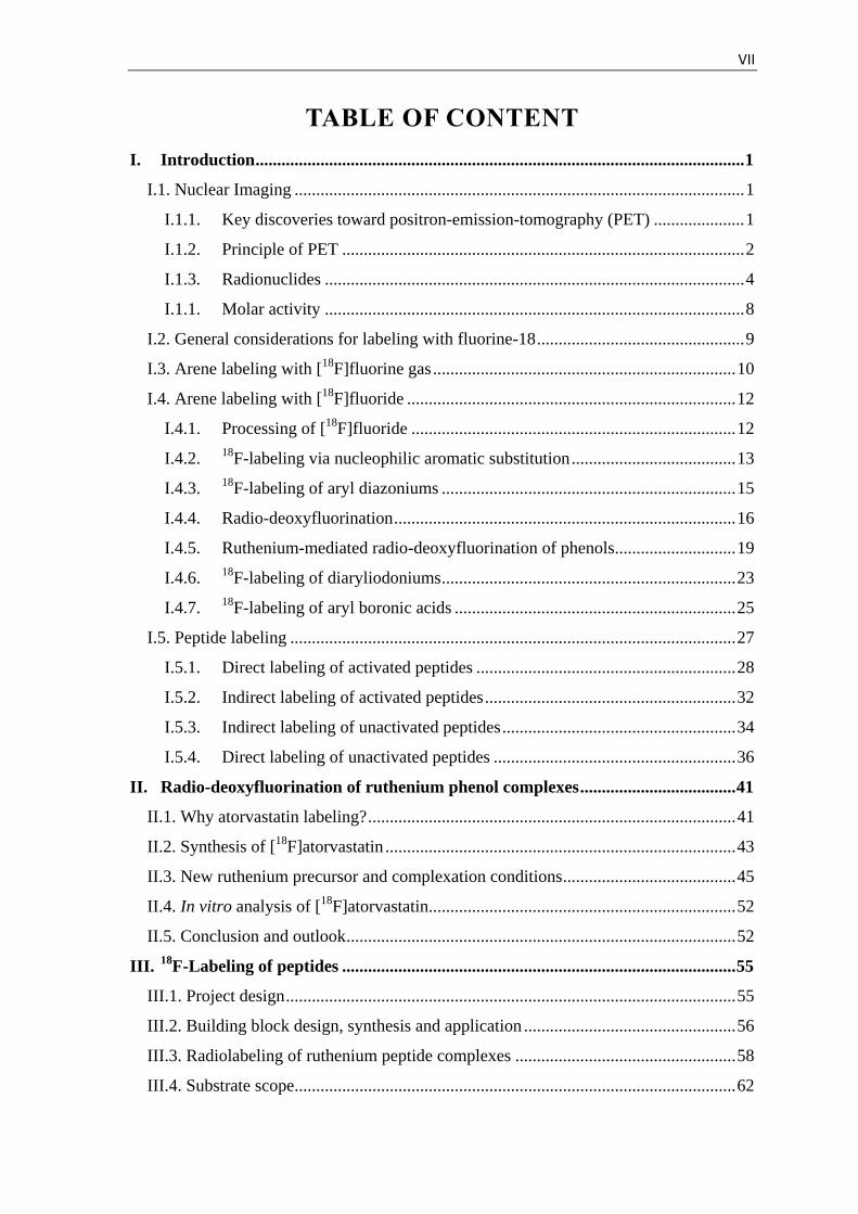
TABLE OF CONTENT
Introduction ................................................................................................................. 1 I.
I.1. Nuclear Imaging ........................................................................................................ 1
Key discoveries toward positron-emission-tomography (PET) ..................... 1 I.1.1.
Principle of PET ............................................................................................. 2 I.1.2.
Radionuclides ................................................................................................. 4 I.1.3.
Molar activity ................................................................................................. 8 I.1.1.
I.2. General considerations for labeling with fluorine-18 ................................................ 9
I.3. Arene labeling with [18
F]fluorine gas ...................................................................... 10
I.4. Arene labeling with [18
F]fluoride ............................................................................ 12
Processing of [18
F]fluoride ........................................................................... 12 I.4.1.
18
F-labeling via nucleophilic aromatic substitution ...................................... 13 I.4.2.
18
F-labeling of aryl diazoniums .................................................................... 15 I.4.3.
Radio-deoxyfluorination ............................................................................... 16 I.4.4.
Ruthenium-mediated radio-deoxyfluorination of phenols............................ 19 I.4.5.
18
F-labeling of diaryliodoniums .................................................................... 23 I.4.6.
18
F-labeling of aryl boronic acids ................................................................. 25 I.4.7.
I.5. Peptide labeling ....................................................................................................... 27
Direct labeling of activated peptides ............................................................ 28 I.5.1.
Indirect labeling of activated peptides .......................................................... 32 I.5.2.
Indirect labeling of unactivated peptides ...................................................... 34 I.5.3.
Direct labeling of unactivated peptides ........................................................ 36 I.5.4.
Radio-deoxyfluorination of ruthenium phenol complexes .................................... 41 II.
II.1. Why atorvastatin labeling? ..................................................................................... 41
II.2. Synthesis of [18
F]atorvastatin ................................................................................. 43
II.3. New ruthenium precursor and complexation conditions ........................................ 45
II.4. In vitro analysis of [18
F]atorvastatin....................................................................... 52
II.5. Conclusion and outlook .......................................................................................... 52
18
F-Labeling of peptides ........................................................................................... 55 III.
III.1. Project design ........................................................................................................ 55
III.2. Building block design, synthesis and application ................................................. 56
III.3. Radiolabeling of ruthenium peptide complexes ................................................... 58
III.4. Substrate scope ...................................................................................................... 62
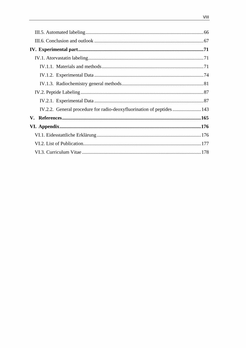
VIII
III.5. Automated labeling ............................................................................................... 66
III.6. Conclusion and outlook ........................................................................................ 67
Experimental part ..................................................................................................... 71 IV.
IV.1. Atorvastatin labeling ............................................................................................. 71
Materials and methods .................................................................................. 71 IV.1.1.
Experimental Data ........................................................................................ 74 IV.1.2.
Radiochemistry general methods .................................................................. 81 IV.1.3.
IV.2. Peptide Labeling ................................................................................................... 87
Experimental Data ........................................................................................ 87 IV.2.1.
General procedure for radio-deoxyfluorination of peptides ....................... 143 IV.2.2.
References ................................................................................................................ 165 V.
Appendix .................................................................................................................. 176 VI.
VI.1. Eidesstattliche Erklärung .................................................................................... 176
VI.2. List of Publication............................................................................................... 177
VI.3. Curriculum Vitae ................................................................................................ 178
IX
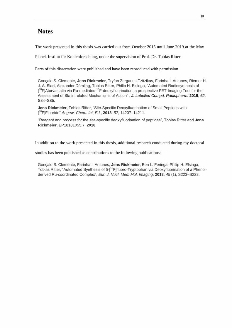
Notes
The work presented in this thesis was carried out from October 2015 until June 2019 at the Max
Planck Institut für Kohlenforschung, under the supervision of Prof. Dr. Tobias Ritter.
Parts of this dissertation were published and have been reproduced with permission.
Gonçalo S. Clemente, Jens Rickmeier, Tryfon Zarganes-Tzitzikas, Farinha I. Antunes, Riemer H.
J. A. Slart, Alexander Dömling, Tobias Ritter, Philip H. Elsinga, “Automated Radiosynthesis of
[18
F]Atorvastatin via Ru-mediated 18
F-deoxyfluorination: a prospective PET-Imaging Tool for the
Assessment of Statin related Mechanisms of Action” , J. Labelled Compd. Radiopharm. 2019, 62,
S84–S85.
Jens Rickmeier, Tobias Ritter, “Site-Specific Deoxyfluorination of Small Peptides with
[18
F]Fluoride” Angew. Chem. Int. Ed., 2018, 57, 14207–14211.
“Reagent and process for the site-specific deoxyfluorination of peptides”, Tobias Ritter and Jens
Rickmeier, EP18181055.7, 2018.
In addition to the work presented in this thesis, additional research conducted during my doctoral
studies has been published as contributions to the following publications:
Gonçalo S. Clemente, Farinha I. Antunes, Jens Rickmeier, Ben L. Feringa, Philip H. Elsinga,
Tobias Ritter, “Automated Synthesis of 5-[18
F]fluoro-Tryptophan via Deoxyfluorination of a Phenol-
derived Ru-coordinated Complex”, Eur. J. Nucl. Med. Mol. Imaging, 2018, 45 (1), S223–S223.
X
List of Abbreviation
3D three-dimensional
α alpha particle
A amount of activity
Å ångström
Ac acetyl
acetyl-CoA acetyl-coenzyme A
Ala alanine
Aq aqeous
Ar aryl
Arg arginine
Asn asparagine
Asp aspartic acid
AY activity yield
β+ positron
Bn benzyl
Boc tert-butyloxycarbonyl
Bq becquerel
br broad tBu tert-butyl
c.a. carrier added
calcd calculated
COD 1,5-cyclooctadiene
Cp cyclopentadienyl
CT computer tomography
CuAAC copper(I)-catalyzed azide alkyne cycloaddition
Cys cysteine
d doublet
DCM dichloromethane
DCC N,N′-dicyclohexylcarbodiimide
DCE dichloroethane
DFI 2,2-difluoro-1,3-dimethylimidazolidine
diCy-18-cr-6 dicyclohexano-18-crown-6
DIPEA diisopropylethylamine
DMF N,N-dimethylformamide
DMSO dimethylsulfoxide
DOTA 1,4,7,10-tetraazacyclododecane-1,4,7,10-tetraacetic acid
DTT dithiothreitol
EC electron capture
EI electron ionization
equiv equivalent
EOB end of bombardment
ESI electrospray ionization
EtOH ethanol
eV electronvolt
FBAM N-[6-(4-fluorobenzylidene)aminooxyhexyl]maleimide
FDA Food and Drug Administration
[18F]FDG 2-deoxy-2-[18F]fluoroglucose
Fmoc fluorenylmethoxycarbonyl
g gram
GC gas chromatography
Gln glutamine

XI
Glu glutamic acid
Gly glycine
HBTU 2-(1H-benzotriazol-1-yl)-1,1,3,3-tetramethyluronium-hexafluorophosphate
HCl hydrochloric acid
HFIP 1,1,1,3,3,3-hexafluoro-2-propanol
His histidine
HOBt 1-hydroxybenzotriazol
HPLC high-performance liquid chromatography
HMG-CoA β-hydroxy β-methylglutaryl-coenzyme A
HRMS high-resolution mass spectrometry
ICP-MS inductively coupled plasma mass spectrometry
Ile isoleucine iPrImCl 2-chloro-1,3-bis(2,6-diisopropylphenyl)imidazolium chloride
J coupling constant
K222 Kryptofix [2.2.2] = 4,7,13,16,21,24-hexaoxa-1,10-diazabicyclo[8.8.8]hexacosane
L liter
Leu leucine
Lys lysine
m multiplet
M moles per liter
MA molar activity
mAbs monoclonal antibodies
Me methyl
MeCN acetonitrile
MeOH methanol
Met methionine
MG11 Minigastrin 11
mol% mole percent
m.p. melting point
MRI magnetic resonance imaging
MS molecular sieves
n amount of substance
N neutron
NaDT sodium decatungstate
n.c.a. no carrier added
n.d. not determined
NFSI N-fluorobenzenesulfonimide
NHS N-hydroxysuccinimide
ν neutrino
NMR nuclear magnetic resonance
NpTMAF neopentyltrimethylammonium fluoride
NpTMAOx bis(neopentyltrimethylammonium) oxalate
Nu nucleophile
OSu N-oxysuccinimide
OTf triflate
P proton
Pbf 2,2,4,6,7-pentamethyldihydrobenzofuran-5-sulfonyl
PBS phosphate-buffered saline
PET positron-emission-tomography
PivCN pivalonitrile
PG protecting group
Ph phenyl
Phe phenylalanine
PMB 4-methoxybenzyl
ppb parts per billion
XII
ppm parts per million iPr isopropyl
Pro proline
q quartet
QMA quaternary methyl-ammonium
R arbitrary organic substituent
RCY radiochemical yield
RT room temperature
s singlet
sat. saturated
SEAr electrophilic aromatic substitution
Ser serine
[18F]SFB N-succinimidyl-4-[18F]fluorobenzoic acid
SiFA silicon fluoride acceptor
SNAr nucleophilic aromatic substitution
t triplet
TEA tetraethylammonium
TBA tetrabutylammonium
TFA trifluoroacetic acid
THF tetrahydrofuran
TIPS triisopropylsilane
TLC thin-layer chromatography
Tmob 2,4,5-trimethoxybenzyl
Thr threonine
Trp tryptophan
Trt trityl
Tyr tyrosine
UV ultraviolet
Val valine
vis visible
X halogen or pseudo halogen

XIII
Acknowledgment
First and foremost I would like to express my gratitude to Prof. Dr. Tobias Ritter for giving me
the opportunity to do my doctoral studies in his group and for giving me the confidence and the
freedom to design and perform my own projects. I would also like to thank him for teaching me
how to select experiments that allow key hypotheses of a project to be tested and how to adjust
the direction of a project accordingly. I would like to thank him for interesting scientific
discussions and for invaluable feedback on my communication and leadership skills. Finally, I
want to thank him for giving me the opportunity to work on interdisciplinary projects with
collaborators/experts from a wide range of areas.
I am grateful to Prof. Dr. Carsten Bolm for the examination of this doctoral thesis. I am thankful
to Matthew Tredwell for teaching me how to work with fluorine-18 and for being a great training
partner for running competitions. I have to thank all members of the Ritter research group for
helpful discussions and the creation of an enjoyable research environment. Special thanks go to
Jonas Börgel, Heejun Lee, and Matthew Plutschack, with whom I shared my office with and with
whom I had invaluable scientific discussions with.
I want also to thank all the talented technicians, visiting students and doctoral students with
whom I had the chance to work with, for their helpful support. I want to highlight the work of
Nele Kronau, who helped me to discover additives to improve the elution efficiency of fluorine-
18, and Sonja Irene Klein, who helped me to expand the substrate scope of the peptide labeling
project. For the successful collaboration in the development of a theranostic peptide pair I want to
thank Nicola Breen. Working with them was a great learning process for me and I hope they
learned as much as I did.
Special thanks go to all the collaborators with whom I could work with throughout my time in
the Ritter laboratory. I want to take the chance to thank in particular Gonçalo Santos Clemente,
for performing the biological tests with [18F]atorvastatin.

XIV
I would like to thank Jonas Börgel, Matthew Plutschack, Florian Berger, and Nicola Breen for
carefully proofreading this manuscript and thereby improving its quality considerably.
Also I want to thank Sylwia Falk for all her support with bureaucratic problems and for helping
me with the organization of events. I would also like to thank Joseph Cornella for enabling me to
invite inspiring speakers to the Institute.
The greatest thanks are due to my family, who always listened to me and unconditionally
support me. Beside my parents I would like to thank Lisa for her endless patience and
understanding. Without your support, I would not have achieved what I have.
I. Introduction 1
Introduction I.
I.1. Nuclear Imaging
Key discoveries toward positron-emission-tomography (PET) I.1.1.
George de Hevesy, also known as “the father of nuclear medicine”, was the first to use
radioactive tracers to study biochemical processes.[1] He proposed that the great sensitivity to
measure radioactivity and the similar chemical behavior of isotopes of the same element would
make radioactive isotopologues promising tracers to study in vivo behavior.[2] In his initial
experiments, he studied the absorption and translocation of lead in plants by immersing the roots
of Vicia Faba in a solution of radioactive lead-212 nitrate and subsequently measuring the relative
activity of the roots, stem, leaves, and fruits of the plants.[1] He also showed that this technique
could be translated to animals while studying the phosphorus metabolism in rats.[3] George de
Hevesy received the Nobel Prize in chemistry in 1943 for discovering that radioactive
isotopologues can be used to study in vivo processes and for the application of this technique.[2]
The next important milestone toward the development of PET was Irène Curie’s discovery that
the irradiation of a boron-10 sample with alpha particles affords a new radioactive element with a
half-life of 14 minutes.[4] This was the first observation that artificial radioactive isotopes can be
synthesized by bombardment of elements with light particles. E. Lawrence et al. built the first
particle accelerator, also known as a cyclotron, and demonstrated that artificial radioactive
substances can be synthesized by bombarding samples with accelerated deuteron particles. This
technology was a key invention to PET because, for the first time, it allowed the reliable and on
demand synthesis of artificial isotopes without the need of another radioactive sample.[5]

I. Introduction 2
Principle of PET I.1.2.
Positron-emission-tomography is a non-invasive imaging technique that enables the
visualization of radiolabeled molecules in vivo and can be used to diagnose diseases, study
biochemical processes and develop drugs.[6] Unlike other imaging techniques that give anatomical
images of the body, such as magnetic resonance imaging (MRI), X-ray and computer tomography
(CT), PET is a functional imaging modality.[7] As such, it visualizes the spatial distribution of
tracer molecules and thereby gives information about biological processes such as metabolism,
receptor concentration or transport across cell membranes. An abnormal change of these
processes is common in diseases and can occur before anatomical changes can be observed,
enabling an earlier diagnosis of diseases.[8] The high sensitivity of PET (10–11 M – 10–12 M) allows
the use of small amounts of tracer molecules (ng quantities) that typically have no relevant
influence on biological systems at low concentration.[6] Since PET-images provide only the spatial
concentration of the tracer, it is often combined with complimentary anatomical imaging
techniques, such as CT, to enable precise localization of the tracer within the body. In addition,
spatially-resolved information about γ-ray scattering obtained by the CT-scan enables a higher
spatial resolution in PET (1 – 6 mm).[9]
PET-tracers are typically administered by inhalation or by intravenous injection to avoid time
consuming uptake into the bloodstream via the gastrointestinal tract.[8] The blood stream
transports the tracer to the target, to which the tracer tightly binds. In order to get a high target-to-
background ratio, non-bound tracer molecules need to be cleared from the bloodstream before
imaging. Upon beta-plus-decay of the neutron-deficient radionuclide in the tracer, one proton (P)
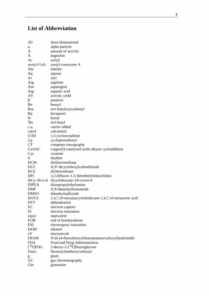
is converted into a neutron (N), and a neutrino (ν) and a positron (β+) are emitted (Figure 1).[10]
The positron migrates through the tissue and loses kinetic energy by collision with other atoms.
Once the kinetic energy of the positron is below 10 eV, it recombines with an electron to form a
positronium that, upon annihilation, emits two γ-rays with an energy of 511 keV at an angle
almost 180° to each other.[11] The small deviation of 180° is a result of the remaining kinetic
energy of the positronium. The emitted γ-rays are detected by a scintillation detectors placed
I. Introduction 3
around the patient. If two opposite detectors measure an impact with 511 keV within a short time
frame the signal is valid. The signals are then converted by a computer into a 3D-image of the
absolute tracer concentration.[7]
Figure 1. General principle of PET.[12]
The most commonly used PET-tracer is [18F]fluorodeoxyglucose ([18F]FDG, 7), with major
applications in oncology.[13] Structurally, [18F]FDG is a glucose analog in which the hydroxyl-
substituent in the two-position is exchanged with its bioisostere fluorine-18. Therefore, [18F]FDG
is metabolically similar to glucose and can be used to image glucose metabolism. Glucose
transporters internalize [18F]FDG into the cells, where it is phosphorylated by hexokinase at the
oxygen in six-position.[12] The next step of the glycolysis cascade, the isomerization of glucose to
fructose, cannot occur with [18F]FDG due to the absence of the hydroxyl substituent in the two-
position, so it is captured in the cell. Due to its high polarity the not trapped [18F]FDG is rapidly
cleared from the bloodstream through the bladder. Many types of cancer have upregulated glucose
transporter and hexokinase expression and, therefore, [18F]FDG accumulates faster in cancerous
tissue than in healthy tissue.[14] Therefore, unusually high glucose uptake may indicate cancer.
Despite the high cost of this imaging technique, it has found widespread applications and has
resulted in a dense cyclotron infrastructure.[13]
I. Introduction 4
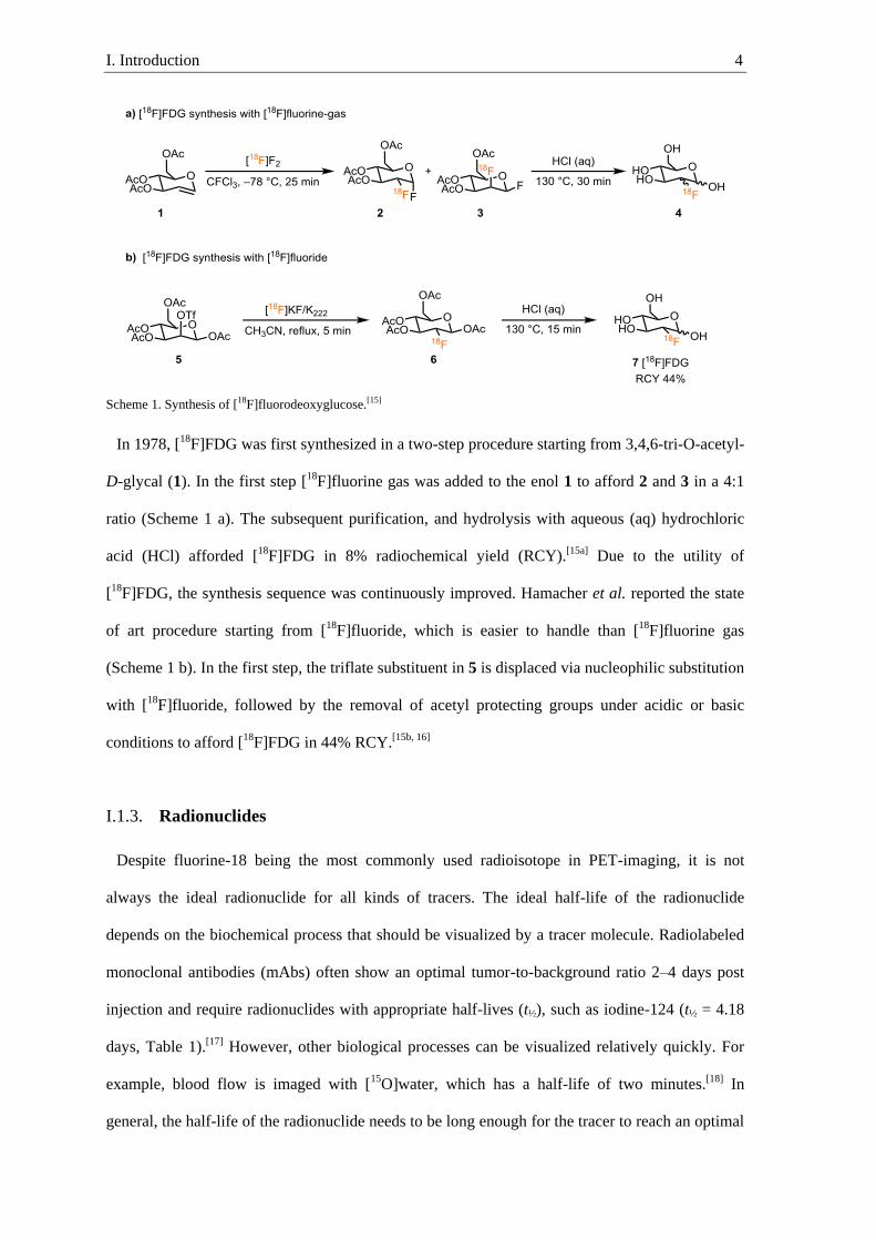
Scheme 1. Synthesis of [18F]fluorodeoxyglucose.[15]
In 1978, [18F]FDG was first synthesized in a two-step procedure starting from 3,4,6-tri-O-acetyl-
D-glycal (1). In the first step [18F]fluorine gas was added to the enol 1 to afford 2 and 3 in a 4:1
ratio (Scheme 1 a). The subsequent purification, and hydrolysis with aqueous (aq) hydrochloric
acid (HCl) afforded [18F]FDG in 8% radiochemical yield (RCY).[15a] Due to the utility of
[18F]FDG, the synthesis sequence was continuously improved. Hamacher et al. reported the state
of art procedure starting from [18F]fluoride, which is easier to handle than [18F]fluorine gas
(Scheme 1 b). In the first step, the triflate substituent in 5 is displaced via nucleophilic substitution
with [18F]fluoride, followed by the removal of acetyl protecting groups under acidic or basic
conditions to afford [18F]FDG in 44% RCY.[15b, 16]
Radionuclides I.1.3.
Despite fluorine-18 being the most commonly used radioisotope in PET-imaging, it is not
always the ideal radionuclide for all kinds of tracers. The ideal half-life of the radionuclide
depends on the biochemical process that should be visualized by a tracer molecule. Radiolabeled
monoclonal antibodies (mAbs) often show an optimal tumor-to-background ratio 2–4 days post
injection and require radionuclides with appropriate half-lives (t½), such as iodine-124 (t½ = 4.18
days, Table 1).[17] However, other biological processes can be visualized relatively quickly. For
example, blood flow is imaged with [15O]water, which has a half-life of two minutes.[18] In
general, the half-life of the radionuclide needs to be long enough for the tracer to reach an optimal
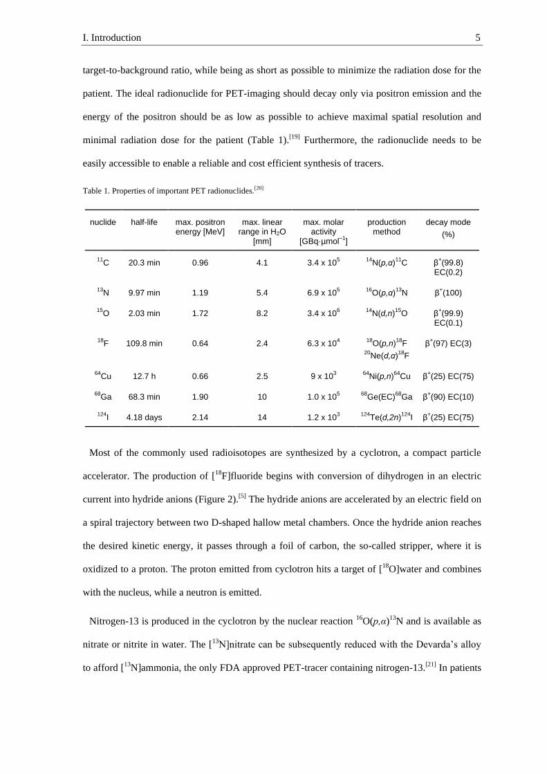
I. Introduction 5
target-to-background ratio, while being as short as possible to minimize the radiation dose for the
patient. The ideal radionuclide for PET-imaging should decay only via positron emission and the
energy of the positron should be as low as possible to achieve maximal spatial resolution and
minimal radiation dose for the patient (Table 1).[19] Furthermore, the radionuclide needs to be
easily accessible to enable a reliable and cost efficient synthesis of tracers.
Table 1. Properties of important PET radionuclides.[20]
nuclide
half-life
max. positron energy [MeV]
max. linear range in H2O
[mm]
max. molar activity
[GBq·µmol–1
]
production method
decay mode
(%)
11C 20.3 min 0.96 4.1 3.4 x 10
5
14N(p,α)
11C β
+(99.8)
EC(0.2)
13N 9.97 min 1.19 5.4 6.9 x 10
5
16O(p,α)
13N β
+(100)
15O 2.03 min 1.72 8.2 3.4 x 10
6
14N(d,n)
15O β
+(99.9)
EC(0.1)
18F 109.8 min 0.64 2.4 6.3 x 10
4
18O(p,n)
18F
20Ne(d,α)
18F
β+(97) EC(3)
64Cu 12.7 h 0.66 2.5 9 x 10
3
64Ni(p,n)
64Cu β
+(25) EC(75)
68Ga 68.3 min 1.90 10 1.0 x 10
5
68Ge(EC)
68Ga β
+(90) EC(10)
124I 4.18 days 2.14 14 1.2 x 10
3
124Te(d,2n)
124I β
+(25) EC(75)
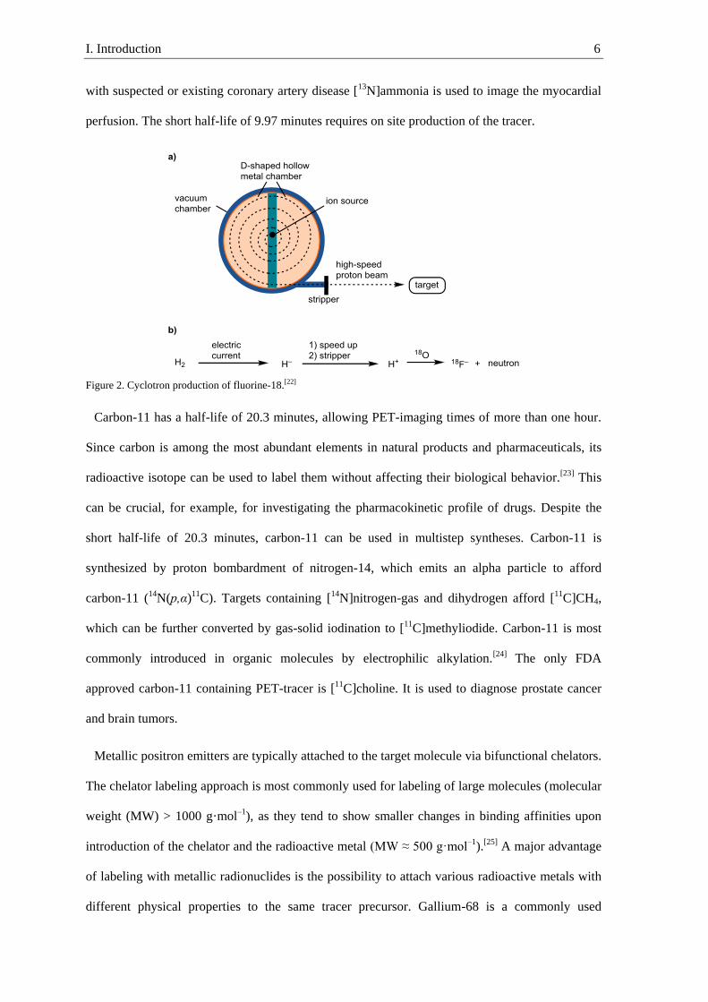
Most of the commonly used radioisotopes are synthesized by a cyclotron, a compact particle
accelerator. The production of [18F]fluoride begins with conversion of dihydrogen in an electric
current into hydride anions (Figure 2).[5] The hydride anions are accelerated by an electric field on
a spiral trajectory between two D-shaped hallow metal chambers. Once the hydride anion reaches
the desired kinetic energy, it passes through a foil of carbon, the so-called stripper, where it is
oxidized to a proton. The proton emitted from cyclotron hits a target of [18O]water and combines
with the nucleus, while a neutron is emitted.
Nitrogen-13 is produced in the cyclotron by the nuclear reaction 16O(p,α)13N and is available as
nitrate or nitrite in water. The [13
N]nitrate can be subsequently reduced with the Devarda’s alloy
to afford [13N]ammonia, the only FDA approved PET-tracer containing nitrogen-13.[21] In patients
I. Introduction 6
with suspected or existing coronary artery disease [13N]ammonia is used to image the myocardial
perfusion. The short half-life of 9.97 minutes requires on site production of the tracer.
Figure 2. Cyclotron production of fluorine-18.[22]
Carbon-11 has a half-life of 20.3 minutes, allowing PET-imaging times of more than one hour.
Since carbon is among the most abundant elements in natural products and pharmaceuticals, its
radioactive isotope can be used to label them without affecting their biological behavior.[23] This
can be crucial, for example, for investigating the pharmacokinetic profile of drugs. Despite the
short half-life of 20.3 minutes, carbon-11 can be used in multistep syntheses. Carbon-11 is
synthesized by proton bombardment of nitrogen-14, which emits an alpha particle to afford
carbon-11 (14N(p,α)11C). Targets containing [14N]nitrogen-gas and dihydrogen afford [11C]CH4,
which can be further converted by gas-solid iodination to [11C]methyliodide. Carbon-11 is most
commonly introduced in organic molecules by electrophilic alkylation.[24] The only FDA
approved carbon-11 containing PET-tracer is [11C]choline. It is used to diagnose prostate cancer
and brain tumors.
Metallic positron emitters are typically attached to the target molecule via bifunctional chelators.
The chelator labeling approach is most commonly used for labeling of large molecules (molecular
weight (MW) > 1000 g·mol–1), as they tend to show smaller changes in binding affinities upon
introduction of the chelator and the radioactive metal (MW ≈ 500 g·mol–1).[25] A major advantage
of labeling with metallic radionuclides is the possibility to attach various radioactive metals with
different physical properties to the same tracer precursor. Gallium-68 is a commonly used

I. Introduction 7
metallic-radionuclide with a half-life of 68.3 minutes. Different to previously mentioned
radionuclides, gallium-68 is obtained from a 68Ga/68Ge generator. In the generator germanium-68
(half-life 271 days) decays by electron capture to gallium-68 that can be extracted with a ligand
from the generator.[26] The only FDA-approved PET-tracer containing a metallic radionuclide is
NETSpot, a receptor-binding peptide used to image neuroendocrine tumors that cannot be imaged
with [18F]FDG.[27]
Fluorine-18 has a longer half-life (109.8 min) than other organic radionuclides such as 11C, 13N
and 15O, which permits the imaging of slower biochemical processes and the shipment of tracers
from a nuclear pharmacy to the imaging site. The maximum positron energy of fluorine-18 (Eβ+ =
0.64 MeV) is among the smallest of commonly used positron emitters. Thus, fluorine-18 also has
a low positron range in water (2.4 mm). The low positron range of fluorine-18 merely impacts the
spatial resolution since current ultra-high spatial resolution cameras have a maximal spatial
resolution of 3–4 mm for human PET scanner and 1–2 mm for small animal PET scanner.
However, the positron range increases drastically as one moves from dense tissue to lung tissue,
affording the positron energy as a major limiting factor in spatial resolution.[28] Despite fluorine
being the thirteenth most abundant element in the earth crust, it is extremely rare in natural
products.[29] However, fluorine has shown privileged properties in drug development, and as of
2018, 45% of FDA-approved drugs contain at least one carbon fluorine bond.[30] Drugs containing
fluorine can be labeled by a formal isotope exchange between fluorine-19 and fluorine-18 without
affecting their pharmacodynamics. Fluorine is frequently used in drug design as a bioisostere of
hydrogen and hydroxyl substituents.[31] The van der Waals radius of fluorine is intermediate (1.47
Å) compared to oxygen (1.52 Å) and hydrogen (1.20 Å). Fluorine is the most electronegative
element (Pauling electronegativity of 4.0) that induces a strong dipole to the C–F bond. This is
more similar to the C–O bond (Pauling electronegativity of oxygen, 3.4) than to the less polarized
C–H bond. Due to the high electronegativity, the lone pairs of fluorine in the C–F are not
available as hydrogen bond acceptors. The inability to act as a hydrogen bond acceptor and donor
are the major differences between fluorine and hydroxyl substituents in organic molecules.[32] As
I. Introduction 8
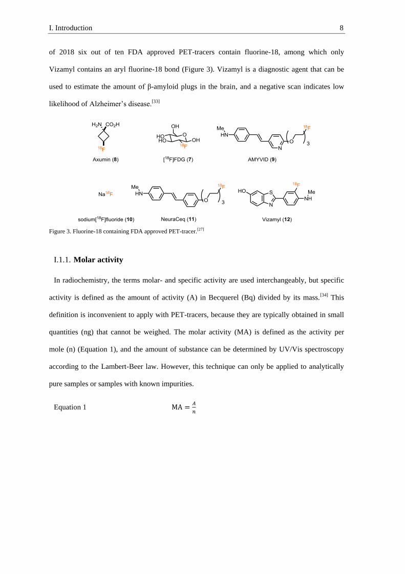
of 2018 six out of ten FDA approved PET-tracers contain fluorine-18, among which only
Vizamyl contains an aryl fluorine-18 bond (Figure 3). Vizamyl is a diagnostic agent that can be
used to estimate the amount of β-amyloid plugs in the brain, and a negative scan indicates low
likelihood of Alzheimer’s disease.[33]
Figure 3. Fluorine-18 containing FDA approved PET-tracer.[27]
Molar activity I.1.1.
In radiochemistry, the terms molar- and specific activity are used interchangeably, but specific
activity is defined as the amount of activity (A) in Becquerel (Bq) divided by its mass.[34] This
definition is inconvenient to apply with PET-tracers, because they are typically obtained in small
quantities (ng) that cannot be weighed. The molar activity (MA) is defined as the activity per
mole (n) (Equation 1), and the amount of substance can be determined by UV/Vis spectroscopy
according to the Lambert-Beer law. However, this technique can only be applied to analytically
pure samples or samples with known impurities.
Equation 1 MA =𝐴
𝑛

I. Introduction 9
I.2. General considerations for labeling with fluorine-18
The major challenge in radiochemistry is the race against time. As a general rule-of-thumb, the
time between the end of bombardment (EOB) and injection of the tracer should be less than three
isotope-half-lives.[8] Therefore, labeling with fluorine-18 should be performed in less than 5.5
hours. This limited synthesis time requires the formation of difficult to synthesize carbon fluorine
bonds at a late stage. In organic chemistry, reagents and substrates are often used in almost
equimolar amounts; in radiochemistry, on the other hand, non-radioactive reagents and substrates
are typically used in large excess to increase the reaction rate. Human imaging with [18F]FDG
requires about 370 MBq (≈ 6 pmol) of labeled tracer, which is synthesized from 10-100 µmol of
substrate and non-radioactive reagents.[35] Due to the large excess of reagents, the reaction is
pseudo-first-order in fluorine-18.[35] However, the use of a large excess of reagents is not entirely
favorable since even small amounts of impurity in the reagents can react with fluorine-18 in an
unfavorable way, preventing the reaction. An additional challenge is that the amount of activity
required for human imaging is not suitable for manual handling. Therefore, it is crucial that the
labeling procedure can be performed in a shielded hot cell by an automated radiochemical
synthesizer.
Two general types of precursor are available for fluorine-18 labeling; no carrier added (n.c.a.)
[18F]fluoride and carrier added (c.a.) [18F]fluorine gas. The major difference between those two
precursors in classical organic synthesis is that fluorine gas is a strong electrophile and fluoride is
typically a poor nucleophile. [18F]fluorine gas is produced via bombardment of neon-20 with
deuterium cations to afford fluorine-18 cations.[36] Today, a small amount of fluorine gas is added
to the target to trap the fluorine-18 cations by isotope exchange with the fluorine-19 gas. Because
just one of the two fluorine atoms in [18F]F2 can react as electrophilic fluorine, and there is
chemically almost no difference between the isotopes, the maximum achievable RCY is 50%.[37]
Another potential problem is that [18
F]F2 is diluted with fluorine-19 to give roughly an overall
ratio of 1:100,000 (0.5 GBq·µmol–1), which is considered low molar activity. Injecting 370 MBq
of a tracer with low molar activity (0.5 GBq·µmol–1) means that about 6 µmol of a fluorinated

I. Introduction 10
substrate are injected to the patient. This large amount of substrate can lead to toxic side effects
and a poor signal-to-noise ratio due to the saturation of receptors present in low concentration.[19]
In a few facilities, electrophilic fluorine-18 sources with high molar activity (50 GBq·µmol–1)
were produced, but the procedure is not widespread since it requires specialized equipment.[38]
The labeling of potential tracers in high molar activity can be achieved with [18F]fluoride.
Bombardment of [18O]H2O with protons affords nucleophilic [18F]fluoride via 18O(p,n)18F
reaction. Using this technique, [18F]fluoride is routinely produced in high molar activity (1,900
GBq·µmol–1), which is still more than an order of magnitude below the theoretical maximum
molar activity (63,000 GBq·µmol–1).[36] The amount of fluorine-19 responsible for the dilution is
very low and can result from minor impurities in reagents. Furthermore, part of the fluorine-19
can be washed in by radiolysis of fluorine containing polymers, such as Teflon.[39]
I.3. Arene labeling with [18
F]fluorine gas
Fluorine gas is a very strong electrophilic fluorinating reagent, and its fluorine-18 isotopolog
can be accessed with a cyclotron. Despite the low molar activity of [18F]fluorine-gas, it has in the
past found vast applications to synthesize otherwise inaccessible 18F-labeled aromatic compounds
via electrophilic aromatic substitution (SEAr). One of the few PET-tracers routinely produced via
electrophilic 18F-fluorination is [18F]F-L-DOPA. Treatment of L-DOPA with [18F]fluorine gas
affords a mixture of all three potential constitutional isomers in overall low RCY (Scheme 2).[40]
The low yield and selectivity can be attributed to the high reactivity of fluorine gas, which can
lead to the formation of side products, such as quinones. The yield and the selectivity can be
increased using less reactive [18F]AcOF.[41] However, decomposition of starting material and the
formation of constitutional isomers results in a lengthy and time consuming purification, which
reduces the RCY by decay of the product. The minute polarity difference between aromatic C–H
and C–F bonds, which results in a difficult separation, creates an additional problem for direct C–
H fluorination reactions.[40, 42]
I. Introduction 11
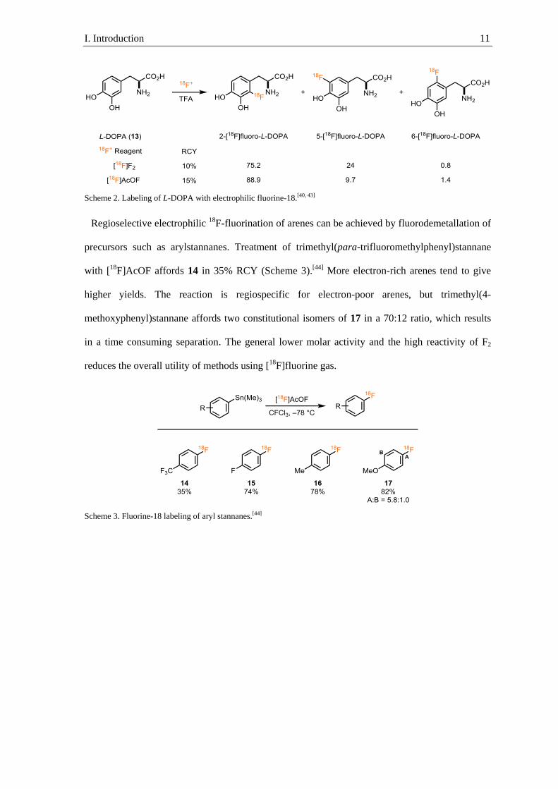
Scheme 2. Labeling of L-DOPA with electrophilic fluorine-18.[40, 43]
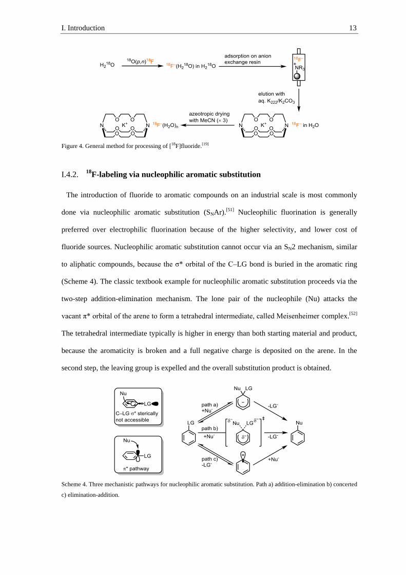
Regioselective electrophilic 18F-fluorination of arenes can be achieved by fluorodemetallation of
precursors such as arylstannanes. Treatment of trimethyl(para-trifluoromethylphenyl)stannane
with [18F]AcOF affords 14 in 35% RCY (Scheme 3).[44] More electron-rich arenes tend to give
higher yields. The reaction is regiospecific for electron-poor arenes, but trimethyl(4-
methoxyphenyl)stannane affords two constitutional isomers of 17 in a 70:12 ratio, which results
in a time consuming separation. The general lower molar activity and the high reactivity of F2
reduces the overall utility of methods using [18F]fluorine gas.
Scheme 3. Fluorine-18 labeling of aryl stannanes.[44]

I. Introduction 12
I.4. Arene labeling with [18
F]fluoride
Processing of [18
F]fluoride I.4.1.
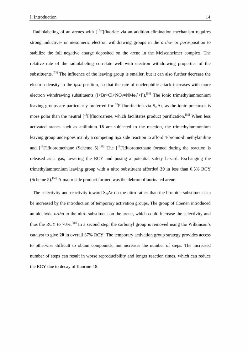
Most labeling methods use [18F]fluoride obtained from a cyclotron in an aqueous solution of
[18O]water. The aqueous solution contains various impurities such as free radicals that can result
in side reactions, metal ions that can deactivate [18F]fluoride, and anions that can compete in
nucleophilic displacements.[45] The purification of [18F]fluoride typically begins by trapping of
[18F]fluoride on a quaternary methyl-ammonium (QMA) anion exchange cartridge, and
subsequent washing of the cartridge to remove impurities (Figure 4). The [18F]fluoride is
afterwards eluted with an aqueous solution containing large cations such as cesium, Kryptofix®
222 (K222) potassium complex or quaternary alkyl-ammoniums.[46] The salts of fluoride and large
soft cations have higher solubility in organic solvents and render fluoride more nucleophilic than
salts of fluoride with small hard cations due to weaker interactions between fluoride and the
cation.[47] Fluoride can form exceptionally strong hydrogen bonds to protic solvents, especially
with water (the hydration energy of fluoride in water is 105 kcal·mol–1).[48] The strong hydrogen
bonding interactions between fluoride and protic solvents, which are lost upon nucleophilic
substitution, lower the nucleophilicity of fluoride.[49] Water is typically removed after elution
under an argon stream, and the water content is further reduced by azeotropic drying with
acetonitrile. Basic conditions during the drying process are critical, because it avoids formation of
[18F]HF, which can react with the glassware or be released as gaseous [18F]HF.[35, 50] The whole
process takes roughly twenty minutes, but even after azeotropic drying the remaining solid
contains small amounts of water. Thus, radiolabeling methods must tolerate small quantities of
water.
I. Introduction 13
Figure 4. General method for processing of [18F]fluoride.[19]
18F-labeling via nucleophilic aromatic substitution I.4.2.
The introduction of fluoride to aromatic compounds on an industrial scale is most commonly
done via nucleophilic aromatic substitution (SNAr).[51] Nucleophilic fluorination is generally
preferred over electrophilic fluorination because of the higher selectivity, and lower cost of
fluoride sources. Nucleophilic aromatic substitution cannot occur via an SN2 mechanism, similar
to aliphatic compounds, because the σ* orbital of the C–LG bond is buried in the aromatic ring
(Scheme 4). The classic textbook example for nucleophilic aromatic substitution proceeds via the
two-step addition-elimination mechanism. The lone pair of the nucleophile (Nu) attacks the
vacant π* orbital of the arene to form a tetrahedral intermediate, called Meisenheimer complex.[52]
The tetrahedral intermediate typically is higher in energy than both starting material and product,
because the aromaticity is broken and a full negative charge is deposited on the arene. In the
second step, the leaving group is expelled and the overall substitution product is obtained.
Scheme 4. Three mechanistic pathways for nucleophilic aromatic substitution. Path a) addition-elimination b) concerted
c) elimination-addition.
I. Introduction 14
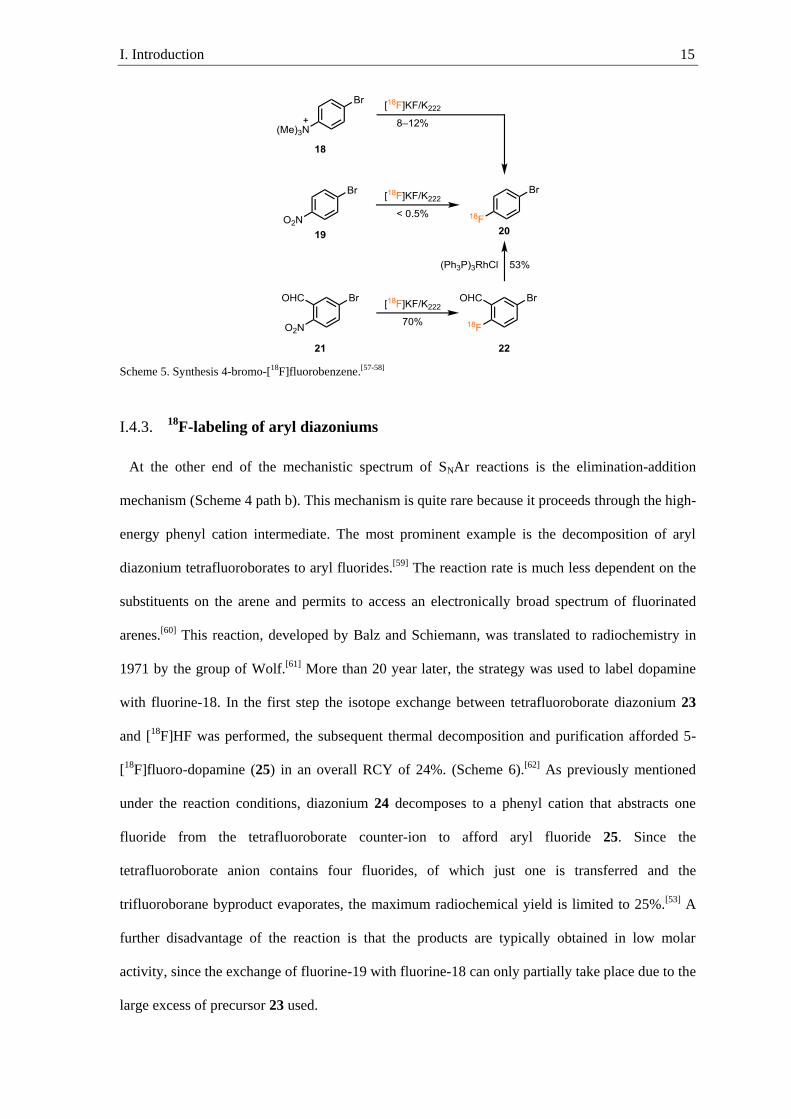
Radiolabeling of an arenes with [18F]fluoride via an addition-elimination mechanism requires
strong inductive- or mesomeric electron withdrawing groups in the ortho- or para-position to
stabilize the full negative charge deposited on the arene in the Meisenheimer complex. The
relative rate of the radiolabeling correlate well with electron withdrawing properties of the
substituents.[53] The influence of the leaving group is smaller, but it can also further decrease the
electron density in the ipso position, so that the rate of nucleophilic attack increases with more
electron withdrawing substituents (I<Br<Cl<NO2=NMe3+<F).[54] The ionic trimethylammonium
leaving groups are particularly preferred for 18F-fluorination via SNAr, as the ionic precursor is
more polar than the neutral [18F]fluoroarene, which facilitates product purification.[55] When less
activated arenes such as anilinium 18 are subjected to the reaction, the trimethylammonium
leaving group undergoes mainly a competing SN2 side reaction to afford 4-bromo-dimethylaniline
and [18F]fluoromethane (Scheme 5).[56] The [18F]fluoromethane formed during the reaction is
released as a gas, lowering the RCY and posing a potential safety hazard. Exchanging the
trimethylammonium leaving group with a nitro substituent afforded 20 in less than 0.5% RCY
(Scheme 5).[57] A major side product formed was the debromofluorinated arene.
The selectivity and reactivity toward SNAr on the nitro rather than the bromine substituent can
be increased by the introduction of temporary activation groups. The group of Coenen introduced
an aldehyde ortho to the nitro substituent on the arene, which could increase the selectivity and
thus the RCY to 70%.[58] In a second step, the carbonyl group is removed using the Wilkinson’s
catalyst to give 20 in overall 37% RCY. The temporary activation group strategy provides access
to otherwise difficult to obtain compounds, but increases the number of steps. The increased
number of steps can result in worse reproducibility and longer reaction times, which can reduce
the RCY due to decay of fluorine-18.
I. Introduction 15
Scheme 5. Synthesis 4-bromo-[18F]fluorobenzene.[57-58]
18F-labeling of aryl diazoniums I.4.3.
At the other end of the mechanistic spectrum of SNAr reactions is the elimination-addition
mechanism (Scheme 4 path b). This mechanism is quite rare because it proceeds through the high-
energy phenyl cation intermediate. The most prominent example is the decomposition of aryl
diazonium tetrafluoroborates to aryl fluorides.[59] The reaction rate is much less dependent on the
substituents on the arene and permits to access an electronically broad spectrum of fluorinated
arenes.[60] This reaction, developed by Balz and Schiemann, was translated to radiochemistry in
1971 by the group of Wolf.[61] More than 20 year later, the strategy was used to label dopamine
with fluorine-18. In the first step the isotope exchange between tetrafluoroborate diazonium 23
and [18F]HF was performed, the subsequent thermal decomposition and purification afforded 5-
[18F]fluoro-dopamine (25) in an overall RCY of 24%. (Scheme 6).[62] As previously mentioned
under the reaction conditions, diazonium 24 decomposes to a phenyl cation that abstracts one
fluoride from the tetrafluoroborate counter-ion to afford aryl fluoride 25. Since the
tetrafluoroborate anion contains four fluorides, of which just one is transferred and the
trifluoroborane byproduct evaporates, the maximum radiochemical yield is limited to 25%.[53] A
further disadvantage of the reaction is that the products are typically obtained in low molar
activity, since the exchange of fluorine-19 with fluorine-18 can only partially take place due to the
large excess of precursor 23 used.
I. Introduction 16
Scheme 6. Fluorine-18 labeling of aryl diazonium.[62]
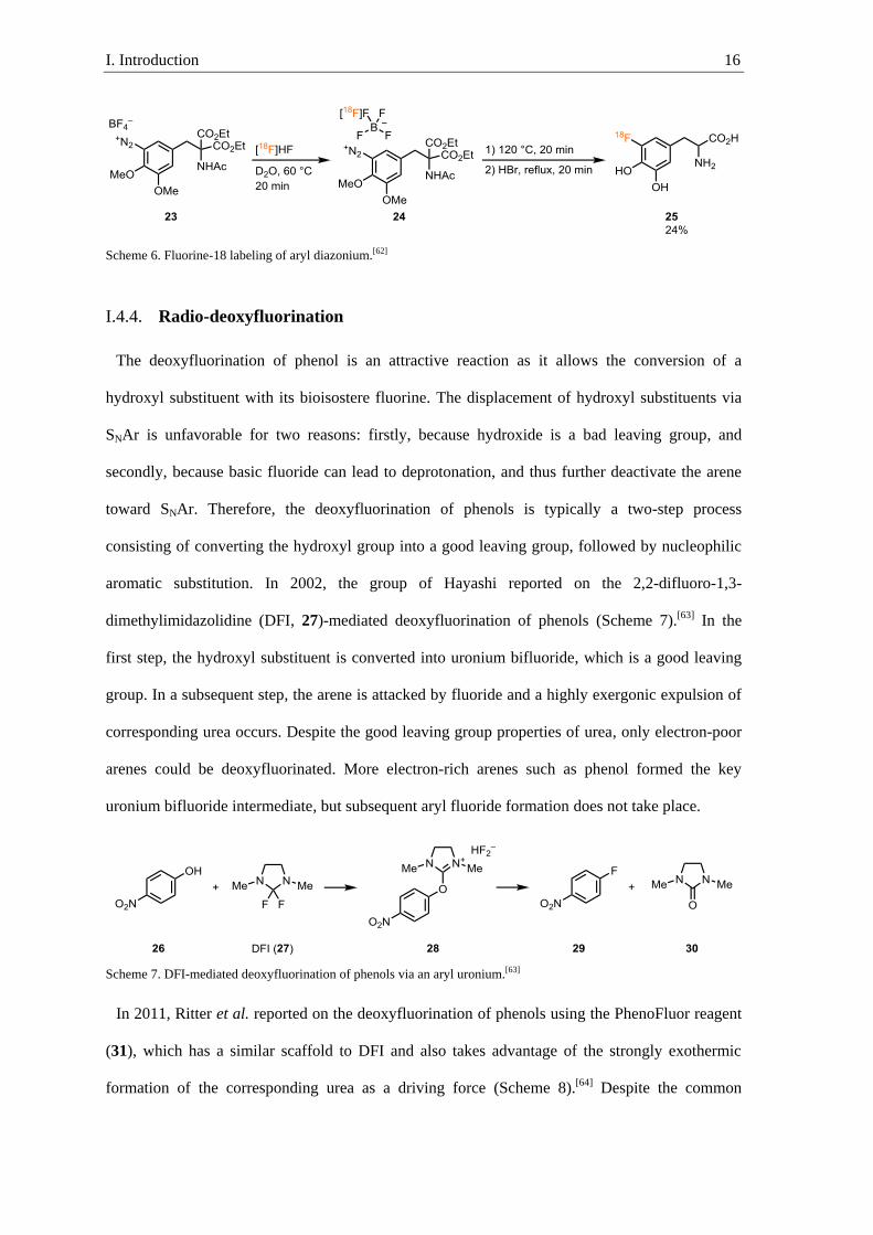
Radio-deoxyfluorination I.4.4.
The deoxyfluorination of phenol is an attractive reaction as it allows the conversion of a
hydroxyl substituent with its bioisostere fluorine. The displacement of hydroxyl substituents via
SNAr is unfavorable for two reasons: firstly, because hydroxide is a bad leaving group, and
secondly, because basic fluoride can lead to deprotonation, and thus further deactivate the arene
toward SNAr. Therefore, the deoxyfluorination of phenols is typically a two-step process
consisting of converting the hydroxyl group into a good leaving group, followed by nucleophilic
aromatic substitution. In 2002, the group of Hayashi reported on the 2,2-difluoro-1,3-
dimethylimidazolidine (DFI, 27)-mediated deoxyfluorination of phenols (Scheme 7).[63] In the
first step, the hydroxyl substituent is converted into uronium bifluoride, which is a good leaving
group. In a subsequent step, the arene is attacked by fluoride and a highly exergonic expulsion of
corresponding urea occurs. Despite the good leaving group properties of urea, only electron-poor
arenes could be deoxyfluorinated. More electron-rich arenes such as phenol formed the key
uronium bifluoride intermediate, but subsequent aryl fluoride formation does not take place.
Scheme 7. DFI-mediated deoxyfluorination of phenols via an aryl uronium.[63]
In 2011, Ritter et al. reported on the deoxyfluorination of phenols using the PhenoFluor reagent
(31), which has a similar scaffold to DFI and also takes advantage of the strongly exothermic
formation of the corresponding urea as a driving force (Scheme 8).[64] Despite the common

I. Introduction 17
uronium intermediate, the PhenoFluor-mediated deoxyfluorination proceeds efficiently for both
electron-poor and electron-rich arenes such as 29 and 17. The reaction is highly functional group
tolerant and substrates containing ketones 33, esters 34, and unprotected primary amines 32 could
be obtained in high yields. Furthermore, heterocycles and complex structures are tolerated under
the reaction condition and the estrone analog 33 and cinchona alkaloid analog 34 could be
accessed in high yields.
Scheme 8. PhenoFluor-mediated deoxyfluorination of phenols.[64]
The electronically diverse substrate scope is atypical for SNAr reactions that proceed through an
addition-elimination mechanism (typical Hammett-value ρ = 3–8) (Scheme 9 a).[65] Constanze N.
Neumann and Tobias Ritter showed that the electronically diverse substrate scope is the result of
a concerted mechanism. The uronium fluoride 35 is in equilibrium with imidazoline 36 (Scheme 9
c). From the tetrahedral intermediate 36, deoxyfluorination proceeds with an activation barrier
between 20–25 kcal·mol–1 via a four-center transition state in a concerted fashion, where the C–O
bond is broken simultaneously with the formation of the C–F bond (Scheme 9 b). As a result of
the concerted mechanism, only a partial negative charge is localized on the arene, and the reaction
has little dependence on the electronics of the arene (Hammett-value ρ = 1.8) compared to an
addition-elimination mechanism. Traditionally, nucleophilic aromatic substitution with fluoride
requires polar solvents for the solvation of metal fluorides. In the SNAr transition state,
desolvation of fluoride contributes significantly to the activation energy required.[65] Therefore,
deoxyfluorination with PhenoFluor is highest yielding in aprotic unpolar solvents since
desolvation contributes to a smaller extent to the reaction barrier and the fluorine is part of

I. Introduction 18
imidazoline 36. Furthermore, fluorine in 36 is non-basic and this prevents side reactions which
can degrade the substrate.
Scheme 9. a) Energy profile of nucleophilic aromatic substitution via addition-elimination mechanism. b) Energy
profile of concerted nucleophilic aromatic substitution. c) Intermediates in the concerted nucleophilic aromatic
substitution.[65]
Neumann and Ritter also translated the PhenoFluor-mediated deoxyfluorination of phenols to a
method using [18F]fluoride.[65] A central challenge in the translation was that the PhenoFluor
reagent contains two fluorine-19 atoms that would lower the molar activity during radiolabeling.
The use of chloroimidazolium chloride 40 (iPrImCl) omits the two fluorine-19 atoms and provides
access to the uronium chloride intermediate (Scheme 10). In the second step, the chloride counter-
ion is replaced via an anion exchange cartridge with [18F]fluoride to form key intermediate
[18F]35. Heating the uronium [18F]fluoride affords the fluorine-18 labeled arene in high RCY. The
reaction has a similar functional group tolerance as the cold reaction, and tolerates primary
amides, esters, secondary amines, and heterocycles. The high functional group tolerance can be
potentially attributed to the non-basic organic-fluorine in the tetrahedral intermediate. Different to
the PhenoFluor-mediated deoxyfluorination, the reaction highly depends on the electronics and
only works with electron-poor arenes and heteroarenes. Electron-rich arenes shift the equilibrium

I. Introduction 19
from fluoroimidazoline 36 to uronium fluoride 35. In the ionic form, the fluoride is more reactive
and can undergo unproductive side reactions with the reaction vial. Complete sequestration of
fluoride is not possible in the cold reaction because an excess of cesium fluoride is used, but in
radiochemistry [18F]fluoride is the limiting reagent. Therefore, the [18F]fluoride can be
sequestered before it can undergo productive aryl fluoride bond formation.
Scheme 10. Radio-deoxyfluorination with iPrImCl.[65]
Ruthenium-mediated radio-deoxyfluorination of phenols I.4.5.
η6-coordination of a transition metal to an arene can drastically alter the properties of the
arene.[66] The electron density on the complexed arene is reduced, resulting in an increased acidity
of aromatic and benzylic protons, and increased electrophilicity, making the complexed arene
more susceptible to nucleophilic attack.[67] Furthermore, the rate of oxidative addition and the rate
of solvolysis for substituents in benzylic position is increased (Figure 5 a).[68] For the sake of the
thesis we will focus on the enhanced electrophilicity.
Figure 5. Arene activation by η6-coordination.[66]
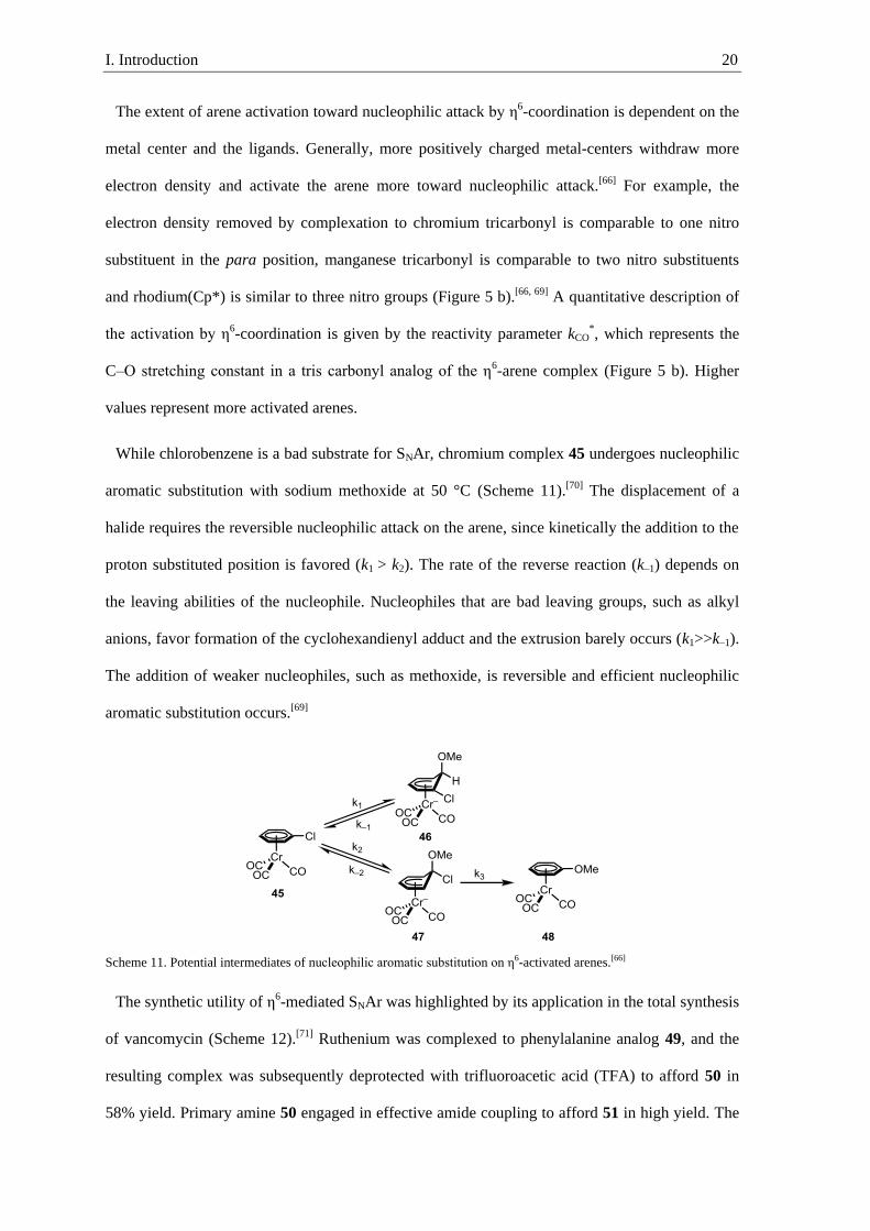
I. Introduction 20
The extent of arene activation toward nucleophilic attack by η6-coordination is dependent on the
metal center and the ligands. Generally, more positively charged metal-centers withdraw more
electron density and activate the arene more toward nucleophilic attack.[66] For example, the
electron density removed by complexation to chromium tricarbonyl is comparable to one nitro
substituent in the para position, manganese tricarbonyl is comparable to two nitro substituents
and rhodium(Cp*) is similar to three nitro groups (Figure 5 b).[66, 69] A quantitative description of
the activation by η6-coordination is given by the reactivity parameter kCO*, which represents the
C–O stretching constant in a tris carbonyl analog of the η6-arene complex (Figure 5 b). Higher
values represent more activated arenes.
While chlorobenzene is a bad substrate for SNAr, chromium complex 45 undergoes nucleophilic
aromatic substitution with sodium methoxide at 50 °C (Scheme 11).[70] The displacement of a
halide requires the reversible nucleophilic attack on the arene, since kinetically the addition to the
proton substituted position is favored (k1 > k2). The rate of the reverse reaction (k–1) depends on
the leaving abilities of the nucleophile. Nucleophiles that are bad leaving groups, such as alkyl
anions, favor formation of the cyclohexandienyl adduct and the extrusion barely occurs (k1>>k–1).
The addition of weaker nucleophiles, such as methoxide, is reversible and efficient nucleophilic
aromatic substitution occurs.[69]
Scheme 11. Potential intermediates of nucleophilic aromatic substitution on η6-activated arenes.[66]
The synthetic utility of η6-mediated SNAr was highlighted by its application in the total synthesis
of vancomycin (Scheme 12).[71] Ruthenium was complexed to phenylalanine analog 49, and the
resulting complex was subsequently deprotected with trifluoroacetic acid (TFA) to afford 50 in
58% yield. Primary amine 50 engaged in effective amide coupling to afford 51 in high yield. The

I. Introduction 21
ruthenium-mediated macrocyclization was performed using 2,6-bis(tert-butyl)phenoxide as non-
nucleophilic base to afford macrocycle 52 in 88% yield. The ruthenium complex was removed
afterwards by irradiation with UV-light for 24 hours in acetonitrile.
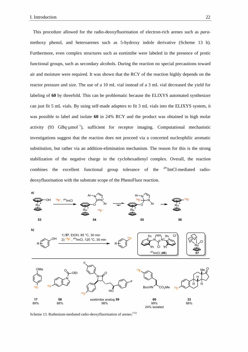
The Ritter group was the first to use the activation of arenes through η6-coordination to
ruthenium to enable labeling with fluorine-18.[72] They showed that electron-rich phenols could be
radio-deoxyfluorinated with iPrImCl if the phenol is coordinated to a ruthenium complex (Scheme
13 a). The complexation of ruthenium cyclopentadienyl to an arene is roughly worth the
activation of two nitro substituents.[69] The lower electron density on the arene and the columbic
repulsion of the two positive charges shifts the equilibrium even for electron-rich phenols to the
side of the tetrahedral intermediate, from which the productive radio-deoxyfluorination can occur
(Scheme 13 a). The ruthenium-mediated radio-deoxyfluorination of phenols begins with the
formation of the ruthenium η6-phenol complex by heating a mixture of ruthenium precursor 57
and the desired phenol in ethanol at 85 °C for 30 minutes. Fluorine-18 was eluted into a vial with
a mixture of ruthenium η6-phenol complex and iPrImCl, and the vial was heated to 125 °C for 30
min to afford the decomplexed fluorine-18 labeled arene.
Scheme 12. Application of η6-mediated SNAr in the synthesis of vancomycin.[71]
I. Introduction 22
This procedure allowed for the radio-deoxyfluorination of electron-rich arenes such as para-
methoxy phenol, and heteroarenes such as 5-hydroxy indole derivative (Scheme 13 b).
Furthermore, even complex structures such as ezetimibe were labeled in the presence of protic
functional groups, such as secondary alcohols. During the reaction no special precautions toward
air and moisture were required. It was shown that the RCY of the reaction highly depends on the
reactor pressure and size. The use of a 10 mL vial instead of a 3 mL vial decreased the yield for
labeling of 60 by threefold. This can be problematic because the ELIXYS automated synthesizer
can just fit 5 mL vials. By using self-made adapters to fit 3 mL vials into the ELIXYS system, it
was possible to label and isolate 60 in 24% RCY and the product was obtained in high molar
activity (93 GBq·µmol–1), sufficient for receptor imaging. Computational mechanistic
investigations suggest that the reaction does not proceed via a concerted nucleophilic aromatic
substitution, but rather via an addition-elimination mechanism. The reason for this is the strong
stabilization of the negative charge in the cyclohexadienyl complex. Overall, the reaction
combines the excellent functional group tolerance of the iPrImCl-mediated radio-
deoxyfluorination with the substrate scope of the PhenoFluor reaction.
Scheme 13. Ruthenium-mediated radio-deoxyfluorination of arenes.[72]

I. Introduction 23
18F-labeling of diaryliodoniums I.4.6.
The first air and moisture stable diaryliodonium salt was isolated in 1894.[73] Since then, many
reports on nucleophilic aromatic substitution on diaryliodonium salts with various nucleophiles
such as alkoxides, amides and fluoride have been reported.[74] Pike et al. were the first to translate
the cold fluorination of diaryliodonium salts to labeling with [18F]fluoride.[75] In stark contrast to
the examples discussed in chapter I.4.2, nucleophilic aromatic substitution of diaryliodoniums has
a small positive Hammett slope (≈ ρ = 1.2) and the substrate scope is little dependent on the
electronics of the arene.[76] Despite this, the electronic structure of the arenes in unsymmetrical
diaryliodoniums is the main factor determining which arene is labeled.[75] To a smaller extent, the
selectivity for C–F bond formation between the two arenes depends on the steric properties of the
arene and favors labeling of the more sterically hindered arene. Pike et al. proposed that the more
bulky arene preferentially occupies the equatorial position on the trigonal bipyramidal substituted
diaryliodonium, and is thus preferentially labeled by the apical fluoride (Scheme 14 a).[77] Most
labeling precursors are valuable material. In order to prevent the use of two equivalents in the
labeling reaction, various dummy arenes were designed. Among the most prominent examples are
2-thienyl substituents. Treatment of aryl(2-thienyl)iodoniums with K222 potassium [18F]fluoride at
130 °C affords the labeled arene and 2-iodothiophene as a byproduct (Scheme 14 b). Electron-rich
arenes such as 17 could be obtained in 29% RCY and the more electron-poor arenes such as 62 in
62% RCY. The electronically diverse substrate scope is a big advantage compared to other
methods; however, aryl(2-thienyl)iodonium salts are difficult to synthesize and purify, and often
show limited stability under ambient conditions.[78]

I. Introduction 24
Scheme 14. a) Mechanism of 18F-labeling of diaryliodonium. b) Substrate scope of 18F-labeling of diaryliodonium.[76]
Sanford et al. developed the copper-catalyzed coupling of diaryliodoniums with fluoride. In
contrast to the uncatalyzed fluorination of diaryliodoniums, the chemoselectivity between the two
aryl substituents mainly depends on the steric bulk and favors labeling of the sterically less
hindered arene.[79] In the proposed mechanism, copper(I) undergoes a chemoselective oxidative
addition to the sterically less hindered aryl iodonium bond, followed by reductive elimination
from copper(III) fluoride.[80] Most notably, the fluorination of mesityl(2-thienyl)iodonium without
a catalyst affords mesityl fluoride in 98:2 selectivity, whereas in the catalyzed reaction 2-
fluorothiophene is formed with 99:1 selectivity.[79] The method was translated to the use of
[18F]fluoride and the reaction proved to have a substrate scope covering a broad range of
electronics on the arene (Scheme 15).[78] Furthermore, the reaction tolerates many functional
groups such as esters, amides and ketones. Using this methodology, even complex molecules such
as [18F]F-DOPA analog 67 could be accessed in 30% RCY. The use of the iodonium
tetrafluoroborate precursor afforded [18F]F-DOPA analog 67 in low molar activity (11 GBq·µmol–
1), likely due to the well-known fluorine exchange between fluorine-18 in solution and the
fluorine-19 in the tetrafluoroborate.[81] The use of iodonium tosylate precursor, which cannot
release fluorine-19, increased the molar activity of the product to 148 GBq·µmol–1
albeit lowering
the RCY to 17%.

I. Introduction 25
Scheme 15. Copper-mediated radio-fluorination of mesityl(aryl)iodonium salts.[78]
18F-labeling of aryl boronic acids I.4.7.
The use of arylboronic acids as starting material is favorable because they are non-toxic and
readily accessible at a late stage via transition metal catalyzed C–H activation or via Suzuki
Miyaura borylation.[82] The copper-catalyzed cross-coupling of arylboronic acids with
heteroatoms was developed simultaneously by Chan, Evans, and Lam.[83] The Chan-Evans-Lam
coupling evolved into one of the most versatile cross couplings, capable of forming otherwise
difficult to introduce bonds such as C–N, C–O and C–Cl under mild conditions using inexpensive
copper reagents.[84] Hartwig et al. were able to show that a copper-mediated C–F bond formation
is possible using F+ sources such as 1-fluoro-2,4,6-trimethylpyridinium.[85] Hartwig and many
other showed the feasibility of the introduction of fluorine via cross coupling, but required the use
of electrophilic fluorine. The use of expensive F+ reagents is not feasible in large-scale application
and is furthermore unfavorable for PET-imaging due to its low molar activity. The Gouverneur
group was the first to report on the fluorine-18 labeling of arylboronic esters using [18F]fluoride in
a copper-mediated Chan-Evans-Lam type reaction.[86] It was proposed that copper (II) captures
[18F]fluoride and undergoes transmetallation with the arylboronic ester. The resulting aryl
copper(II) fluoride disproportionates to copper(I) and aryl copper(III) fluoride, which has been
shown to undergo facile C–F reductive elimination.[80] The copper-mediated 18F-labeling works
well on electron-rich and electron-poor arenes, tolerates many functional groups such as ethers,
nitro, and amides, and tolerates substituents in the ortho-position (Scheme 16). Substrates
containing protic functional groups such as phenols 69 or secondary amides resulted in a low
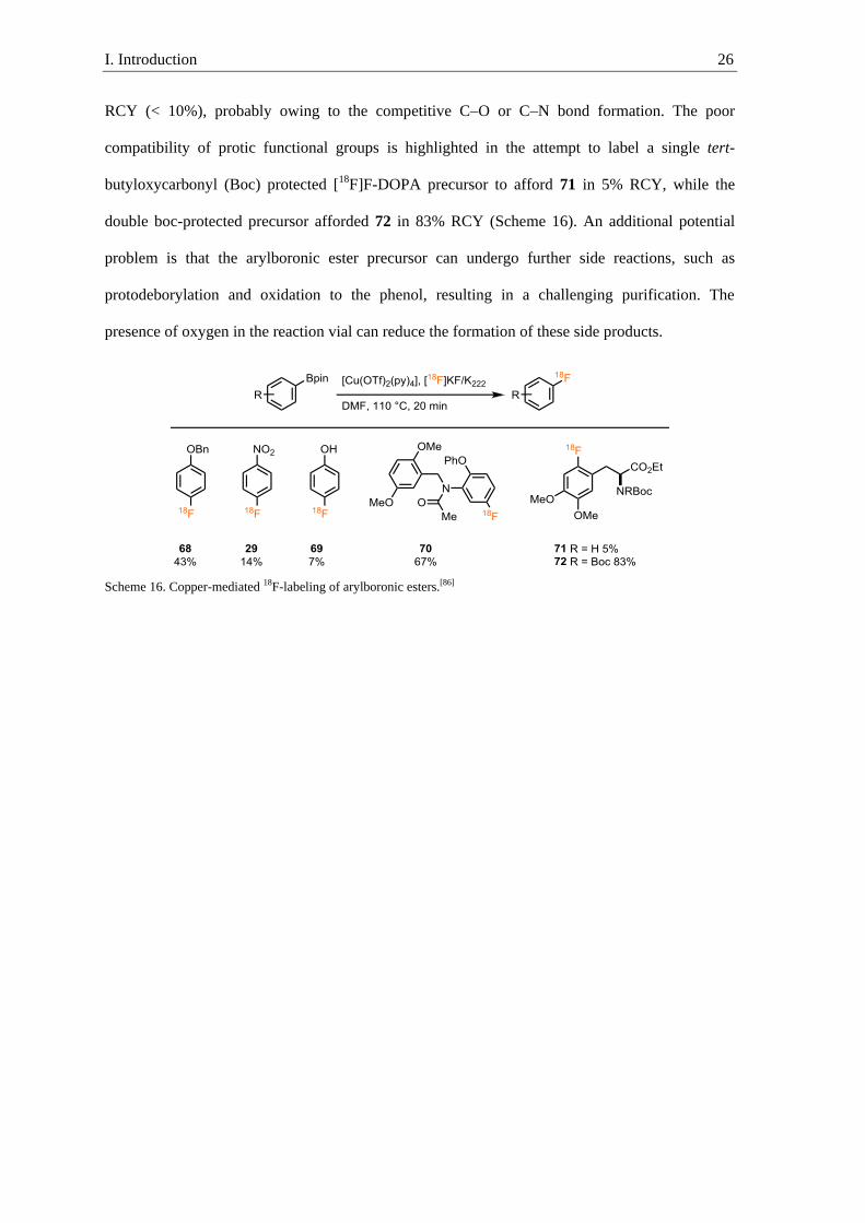
I. Introduction 26
RCY (< 10%), probably owing to the competitive C–O or C–N bond formation. The poor
compatibility of protic functional groups is highlighted in the attempt to label a single tert-
butyloxycarbonyl (Boc) protected [18F]F-DOPA precursor to afford 71 in 5% RCY, while the
double boc-protected precursor afforded 72 in 83% RCY (Scheme 16). An additional potential
problem is that the arylboronic ester precursor can undergo further side reactions, such as
protodeborylation and oxidation to the phenol, resulting in a challenging purification. The
presence of oxygen in the reaction vial can reduce the formation of these side products.
Scheme 16. Copper-mediated 18F-labeling of arylboronic esters.[86]

I. Introduction 27
I.5. Peptide labeling
For decades, labeled antibodies were considered the “magic bullet” in tracer development, due
to their highly specific binding.[87]
Initial enthusiasm about antibody labeling decreased due to
several disadvantages associated with their high molecular weight/large size. Large molecule such
as antibodies can be sequestered by reticuloendothelial cells, and often optimal target-to-
background ratios are achieved after 2–4 days.[87] This long imaging times require the use of long
lived radio nuclides and result in higher radiation exposure of the patients.
IUPAC refers to peptides as amino carboxylic acids that are covalently linked by amide bonds
without restriction in size. In the following thesis, “peptide” will refer to polymeric structures
containing 2–50 amino acids. Similar to antibodies, peptides can show as well high and very
specific binding.[88] In contrast to antibodies, peptides have a lower molecular weight, which
offers several additional advantages. The lower molecular weight permits fast diffusion to the
target cells and rapid blood clearance. Many receptor-binding peptide tracers are internalized into
the cells upon binding to G-coupled proteins. A combination of these properties can result in high
target-to-background ratios in a short amount of time. Another advantage is that peptides can be
conveniently accessed via automated solid phase peptide synthesis (SPPS). Naturally occurring
peptides often show low stability in serum (somatostatin biological half-life = 2 min) and are
therefore not suitable tracers. However, there are several known ways to increase their stability
such as cyclization, introduction of unnatural amino acids and modification of the C– and N–
termini.[89] In addition, peptides are typically non-immunogenic.[90] The only FDA approved
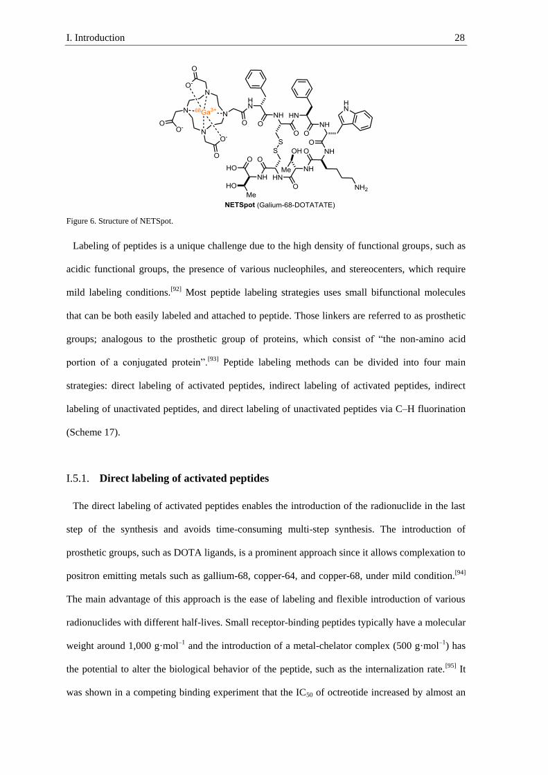
peptide PET-tracer, NETSpot (Galium-68-DOTATATE), is used to image the somatostatin
receptor, which is overexpressed in neuroendocrine tumors (Figure 6).[91] These neuroendocrine
tumors cannot be imaged with [18F]FDG, highlighting the necessity of this NETSpot tracer.
I. Introduction 28
Figure 6. Structure of NETSpot.
Labeling of peptides is a unique challenge due to the high density of functional groups, such as
acidic functional groups, the presence of various nucleophiles, and stereocenters, which require
mild labeling conditions.[92] Most peptide labeling strategies uses small bifunctional molecules
that can be both easily labeled and attached to peptide. Those linkers are referred to as prosthetic
groups; analogous to the prosthetic group of proteins, which consist of “the non-amino acid
portion of a conjugated protein”.[93] Peptide labeling methods can be divided into four main
strategies: direct labeling of activated peptides, indirect labeling of activated peptides, indirect
labeling of unactivated peptides, and direct labeling of unactivated peptides via C–H fluorination
(Scheme 17).
Direct labeling of activated peptides I.5.1.
The direct labeling of activated peptides enables the introduction of the radionuclide in the last
step of the synthesis and avoids time-consuming multi-step synthesis. The introduction of
prosthetic groups, such as DOTA ligands, is a prominent approach since it allows complexation to
positron emitting metals such as gallium-68, copper-64, and copper-68, under mild condition.[94]
The main advantage of this approach is the ease of labeling and flexible introduction of various
radionuclides with different half-lives. Small receptor-binding peptides typically have a molecular
weight around 1,000 g·mol–1
and the introduction of a metal-chelator complex (500 g·mol–1
) has
the potential to alter the biological behavior of the peptide, such as the internalization rate.[95] It
was shown in a competing binding experiment that the IC50 of octreotide increased by almost an

I. Introduction 29
order of magnitude upon addition of an ytterbium-DOTA complex (see Figure 6 for a structurally
similar example).[96]
Scheme 17. Peptide labeling strategies.
Fluorine-18 labeling of peptides has gained growing interest due to the favorable half-life of
109.8 min, which enables multistep synthesis, permits distribution of tracers, and is long enough
to allow imaging over several hours. Low concentration of peptide-receptors demands tracers in
high molar activity (37–370 MBq·µmol–1) and restricts labeling to the use of [18F]fluoride.[97]
Labeling with nucleophilic [18F]fluoride is challenging because of the harsh reaction conditions
typically required and the high functional group density in peptides.[98] To circumvent harsh
labeling conditions, unnatural functional groups capable of capturing [18F]fluoride under mild
conditions can be introduced to the peptide. It is to be expected that the introduction of smaller
organic prosthetic groups will change the properties of the peptides to a smaller extent than large
metal-chelator complexes.[99]
Silicon has a high fluorine affinity and forms strong silicon fluorine bonds (129 kcal·mol–1),
which is advantageous for mild labeling.[100] Rosenthal et al. were the first to synthesize
I. Introduction 30
[18F]fluorotrimethylsilane and evaluated its use for animal experiments. However, during in vivo
evaluation they observed rapid hydrolysis to give [18F]fluoride.[101] The released ionic
[18F]fluoride is taken up by the bones and lead to unnecessary radiation exposure of patients and
for PET-tracers not targeting skeletal PET, the target-to-background ratio is decreased. Fast
hydrolysis can prevent imaging if the tracer is hydrolyzed before it can reach the target. The
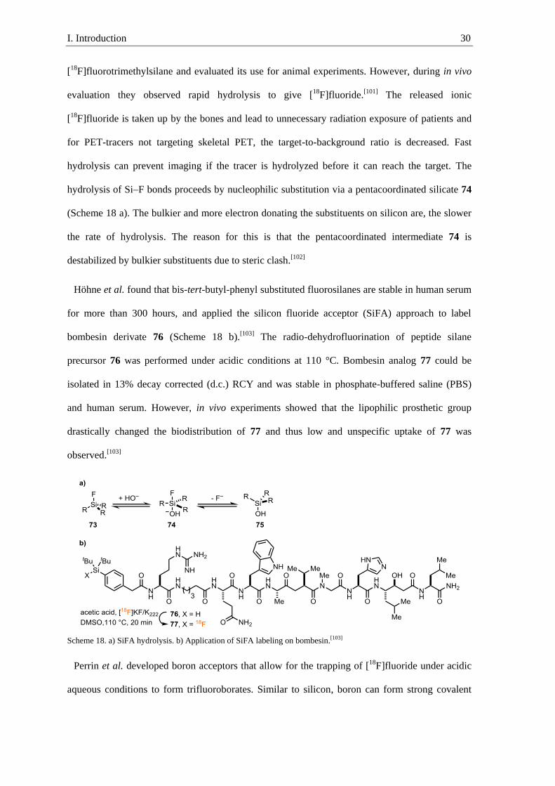
hydrolysis of Si–F bonds proceeds by nucleophilic substitution via a pentacoordinated silicate 74
(Scheme 18 a). The bulkier and more electron donating the substituents on silicon are, the slower
the rate of hydrolysis. The reason for this is that the pentacoordinated intermediate 74 is
destabilized by bulkier substituents due to steric clash.[102]
Höhne et al. found that bis-tert-butyl-phenyl substituted fluorosilanes are stable in human serum
for more than 300 hours, and applied the silicon fluoride acceptor (SiFA) approach to label
bombesin derivate 76 (Scheme 18 b).[103] The radio-dehydrofluorination of peptide silane
precursor 76 was performed under acidic conditions at 110 °C. Bombesin analog 77 could be
isolated in 13% decay corrected (d.c.) RCY and was stable in phosphate-buffered saline (PBS)
and human serum. However, in vivo experiments showed that the lipophilic prosthetic group
drastically changed the biodistribution of 77 and thus low and unspecific uptake of 77 was
observed.[103]
Scheme 18. a) SiFA hydrolysis. b) Application of SiFA labeling on bombesin.[103]
Perrin et al. developed boron acceptors that allow for the trapping of [18F]fluoride under acidic
aqueous conditions to form trifluoroborates. Similar to silicon, boron can form strong covalent

I. Introduction 31
bonds with fluorine (bond dissociation energy = 125 kcal·mol–1), but the anionic trifluoroborate
prosthetic group is rather hydrophilic.[100a] Despite the thermodynamically favorable B–F bond
formation, aryl trifluoroborates hydrolyze at high dilution in water due to the large excess of
water.[104] Hydrolysis occurs via a dissociative pathway, in which the sp3-hybridized borate is
transformed to the sp2-hybridized borane, which can be destabilized by bulky ortho substituents.
Hammett analysis showed a slightly negative value (ρ ≈ –1) for the rate of hydrolysis, which
resulted in the design of 79 containing electron withdrawing substituents in the ortho and para
position and a linker in meta position to the trifluoroborate (Scheme 19).[105] The electron
withdrawing substituent led to an increased solvolytic half-life of ~1000 min. Arylboronic ester
78 can be converted to the aryl trifluoroborate 79 under mildly acidic conditions in the presence
of [18F]KHF2 (Scheme 19).[106] An additional advantage of trifluoroborate substituents is the bio-
orthogonality. However, 79 can also lead to a change in the properties of the peptides by
introducing a negative charge on the peptide.
Scheme 19. Fluorine-18 labeling via formation of aryltrifluoroborate.[106]
The group of Perrin found an inverse correlation between the pKa of alkyl carboxylic acids and
the hydrolytic stability of their alkyltrifluoroborate analogs. The correlation arises from the
negative charge that needs to be stabilized in both equilibriums.[107] Acetic acid has a pKa of 4.81
and the solvolytic half-life of its trifluoroborate analog is 2–3 minutes.[107] In order to overcome
previous limitations of charged prosthetic group, the Perrin group designed ammoniummethylene-
BF3 (AMBF3, 82), which as a betaine analog (pKa of carboxylic acid analog 1.84) should be stable
and overall non-charged.[108] AMBF3 has a solvolytic half-life of more than 11 days and can be
easily introduced to azide containing peptides via copper(I)-catalyzed azide alkyne cycloaddition
(CuAAC). Labeling of 83 is achieved via an ion exchange reaction under acidic conditions at 80
°C. TATE analog 83 could be labeled with this protocol in 20% RCY. Despite the presence of

I. Introduction 32
fluorine-19 in the precursor 83, the labeled peptide 84 was obtained in high molar activity (111
GBq·µmol–1). The high molar activity could be achieved due to the use of small amounts of
precursor 83 (50 nmol). The utility of this methodology was highlighted by the 5-fold higher
binding affinities of TATE analog 84 to sstr2 receptor compared to clinically used 68Ga-
DOTATATE.[109] Automated radiolabeling procedures are crucial for human imaging since large
amounts of the tracer are required. Due to otherwise slow reaction rate the fluoride exchange
labeling of AMBF3 analogs is typically performed in small reaction volumes (1.5 µL), which can
be difficult to handle with automated synthesizer.
Scheme 20. AMBF3 labeling of TATE analog.[109]
Indirect labeling of activated peptides I.5.2.
In the indirect labeling of activated peptides a small bifunctional molecule is first labeled and
afterwards attached to a preactivated peptide containing an artificial substituent allowing
orthogonal attachment. Indirect labeling takes advantage of robust prosthetic groups that can be
labeled under harsh reaction conditions and can be afterwards, attached to a pre-functionalized
site of the peptide under mild conditions. The pre-functionalized site often contains a functionality
that enables orthogonal conjugation of the prosthetic group in the presence of common functional
groups in peptides. Azides, as well as alkynes, are stable toward water, oxygen and most organic
synthesis conditions, making them ideally suited for late stage coupling.[110] Due to the
orthogonality to biological functional groups, CuAAC has found wide application in protein and
peptide modification, and was first used in peptide labeling with fluorine-18 by Sutcliffe and

I. Introduction 33
Marik.[111] The fluorine-18 containing prosthetic group 5-[18F]fluoro-1-pentyne (86) was
synthesized by nucleophilic substitution of 5-tosyl-1-pentyne (85) at 100 °C with [18F]fluoride
(Scheme 21). Alkyne 86 was purified by distillation, and in a second step conjugated to the
peptide at room temperature (RT) to afford 87 in 97% RCY. One concern with using
[18F]alkylfluorides is always the potential for release of [18F]fluoride through nucleophilic
substitution or metabolism.
Scheme 21. CuAAC labeling of peptides.[111]
In contrast to standard CuAAC conditions, in the previous example the CuI was used in large
excess compared to the amine base to obtain high yields in the cycloaddition. For cell studies and
human imaging, an almost quantitative removal of copper is required to avoid cytotoxic side
effects by copper.[112] Copper-free alkyne azide cycloaddition can be achieved with highly
strained alkynes. The energy gained by strain release lowers the transition state for cycloaddition
and permits triazole formation with azides at room temperature.[113] The first copper-free click
labeling of peptides was demonstrated by Feringa et al. and was applied to label bombesin.[114] In
the first step, [18F]fluoroazide 89 was synthesized via SNAr from 88. After purification, 89 was
conjugated in 31% RCY to peptide 90 containing a strained alkyne. One advantage of this
labeling strategy is the possibility to scavenge non-labeled precursor by an azide containing
resin.[115] Different to the copper catalyzed reaction, the strain promoted reaction afforded both
triazole constitutional isomers, which were not separated. The formation of a product mixture is
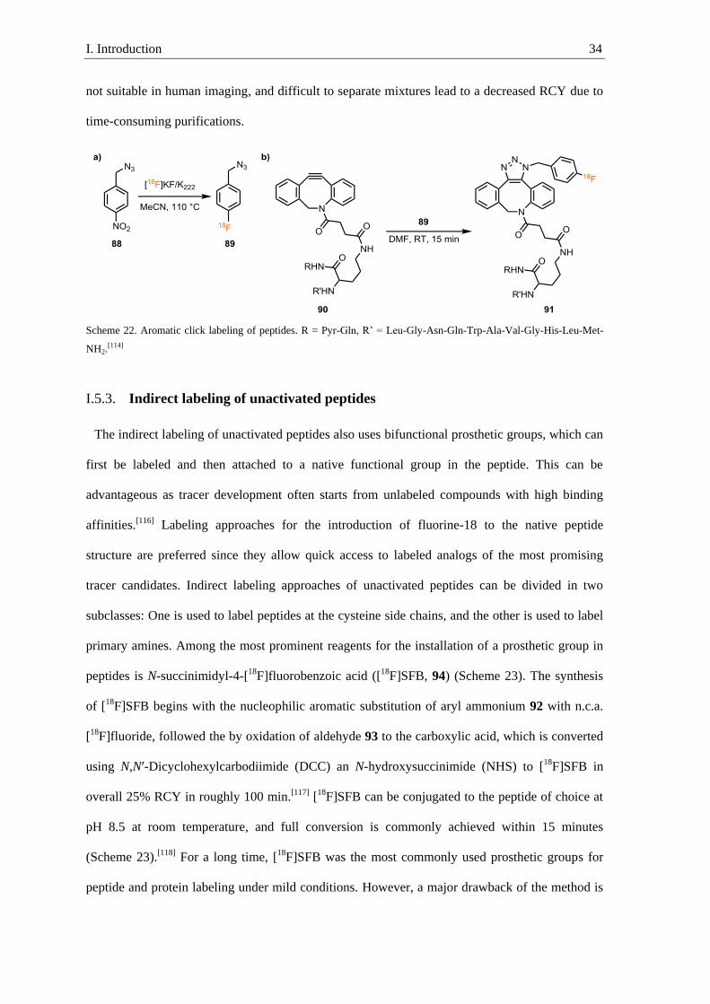
I. Introduction 34
not suitable in human imaging, and difficult to separate mixtures lead to a decreased RCY due to
time-consuming purifications.
Scheme 22. Aromatic click labeling of peptides. R = Pyr-Gln, R’ = Leu-Gly-Asn-Gln-Trp-Ala-Val-Gly-His-Leu-Met-
NH2.[114]
Indirect labeling of unactivated peptides I.5.3.
The indirect labeling of unactivated peptides also uses bifunctional prosthetic groups, which can
first be labeled and then attached to a native functional group in the peptide. This can be
advantageous as tracer development often starts from unlabeled compounds with high binding
affinities.[116] Labeling approaches for the introduction of fluorine-18 to the native peptide
structure are preferred since they allow quick access to labeled analogs of the most promising
tracer candidates. Indirect labeling approaches of unactivated peptides can be divided in two
subclasses: One is used to label peptides at the cysteine side chains, and the other is used to label
primary amines. Among the most prominent reagents for the installation of a prosthetic group in
peptides is N-succinimidyl-4-[18F]fluorobenzoic acid ([18F]SFB, 94) (Scheme 23). The synthesis
of [18F]SFB begins with the nucleophilic aromatic substitution of aryl ammonium 92 with n.c.a.
[18F]fluoride, followed the by oxidation of aldehyde 93 to the carboxylic acid, which is converted
using N,N′-Dicyclohexylcarbodiimide (DCC) an N-hydroxysuccinimide (NHS) to [18F]SFB in
overall 25% RCY in roughly 100 min.[117] [18F]SFB can be conjugated to the peptide of choice at
pH 8.5 at room temperature, and full conversion is commonly achieved within 15 minutes
(Scheme 23).[118] For a long time, [18F]SFB was the most commonly used prosthetic groups for
peptide and protein labeling under mild conditions. However, a major drawback of the method is

I. Introduction 35
labeling of peptides containing more than one primary amine, since differentiation using [18F]SFB
labeling can be very challenging. Thiols are scarcer among peptides and, if present, often just one
cysteine can be found in receptor-binding peptides. Thus, labeling strategies targeting cysteine
often show selective labeling. [18F]FBAM (96) is among the most commonly used reagents for the
selective installation of a fluorine-18 labeled prosthetic group on cysteine. The cysteine adds to
the maleimide substituent in [18F]FBAM via Michael-addition (Scheme 23).[119] Since most
radiotracers are developed from non-radioactive lead compounds a minimal perturbation of its
structure upon labeling is desirable.[120] However, also this method introduces quite large
lipophilic prosthetic groups such as SFB and FBAM that can alter the properties of the
peptide.[120]
Scheme 23. Synthesis and application of [18F]FBAM and [18F]SBF.[117, 119]
In 2018, the Gouverneur group reported the synthesis of the [18F]Umemoto reagent in a two-step
protocol from nucleophilic [18F]fluoride.[121] Substitution of bromide in 97 with [18F]fluoride,
followed by oxidative cyclization with oxone and triflic anhydride affords 5-
[18F](trifluoromethyl)dibenzothiophenium trifluoromethanesulfonate (99) in 5% RCY (Scheme 24
a). Despite the use of [18F]fluoride as a precursor, the molar activity of 99 was 0.08 GBq·µmol–1.
The low molar activity is potentially a result of fluoride leaching of starting material 97 under
labeling conditions in step 1. The reagent 99 enables the 18F-trifluoromethylation of cysteine and

I. Introduction 36
homocysteine. For example using this method, even complex structure such as amyloid-β-
fragment 100 could be obtained in 30% RCY (Scheme 24 b). This method permits
chemoselective labeling in the presence of nucleophilic amino acids such as tyrosine, lysine, and
arginine, and tolerates the presence of all 20 canonical amino acids. However, peptides containing
histidine or tryptophan resulted in the formation of a second radiolabeled side product, which
potentially results from N or C trifluoromethylation of those amino acids. In vivo biodistribution
of cRGDfC([18F]CF3) (101) showed no bone uptake and demonstrated stability of the prosthetic
group.
Scheme 24. Peptide labeling with the [18F]Umemoto reagent.[121]
Direct labeling of unactivated peptides I.5.4.
The direct C–H 18F-fluorination of peptide is advantageous, due to minimal perturbation in the
structure of the peptide. Even though electrophilic 18F-labeling of aromatic amino acids has found
numerous applications, just one reported method for 18
F-fluorination of peptides via electrophilic
aromatic substitution is known. The group of Hatano used [18F]AcOF to label the aryl substituent
on phenylalanine in cyclic peptide 102 (Scheme 25).[122] The labeled peptide 103 was obtained in
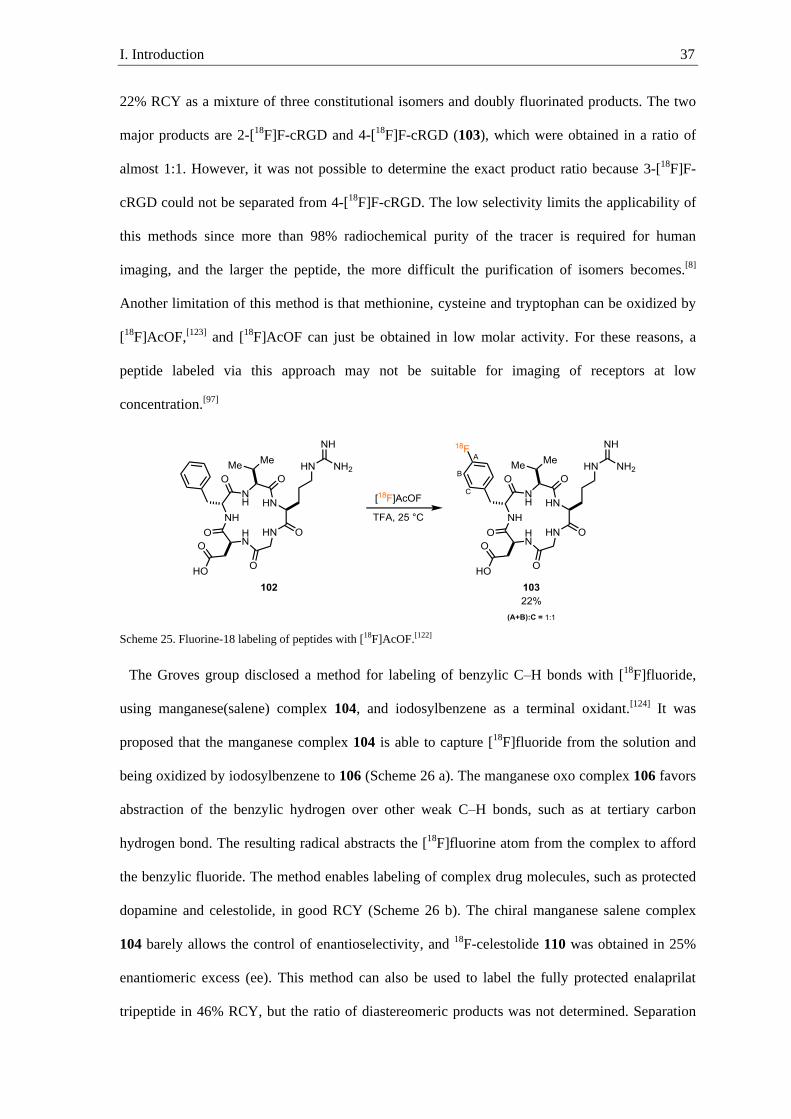
I. Introduction 37
22% RCY as a mixture of three constitutional isomers and doubly fluorinated products. The two
major products are 2-[18F]F-cRGD and 4-[18F]F-cRGD (103), which were obtained in a ratio of
almost 1:1. However, it was not possible to determine the exact product ratio because 3-[18F]F-
cRGD could not be separated from 4-[18F]F-cRGD. The low selectivity limits the applicability of
this methods since more than 98% radiochemical purity of the tracer is required for human
imaging, and the larger the peptide, the more difficult the purification of isomers becomes.[8]
Another limitation of this method is that methionine, cysteine and tryptophan can be oxidized by
[18F]AcOF,[123] and [18F]AcOF can just be obtained in low molar activity. For these reasons, a
peptide labeled via this approach may not be suitable for imaging of receptors at low
concentration.[97]
Scheme 25. Fluorine-18 labeling of peptides with [18F]AcOF.[122]
The Groves group disclosed a method for labeling of benzylic C–H bonds with [18F]fluoride,
using manganese(salene) complex 104, and iodosylbenzene as a terminal oxidant.[124] It was
proposed that the manganese complex 104 is able to capture [18F]fluoride from the solution and
being oxidized by iodosylbenzene to 106 (Scheme 26 a). The manganese oxo complex 106 favors
abstraction of the benzylic hydrogen over other weak C–H bonds, such as at tertiary carbon
hydrogen bond. The resulting radical abstracts the [18F]fluorine atom from the complex to afford
the benzylic fluoride. The method enables labeling of complex drug molecules, such as protected
dopamine and celestolide, in good RCY (Scheme 26 b). The chiral manganese salene complex
104 barely allows the control of enantioselectivity, and 18F-celestolide 110 was obtained in 25%
enantiomeric excess (ee). This method can also be used to label the fully protected enalaprilat
tripeptide in 46% RCY, but the ratio of diastereomeric products was not determined. Separation

I. Introduction 38
of diastereomers of large peptides can be difficult and time consuming, and thus lowering the
RCY by decay or preventing isolation of pure tracer. Furthermore, the method was so far not
shown to be amendable to labeling of larger peptides.
Scheme 26. a) Mechanism of the manganese-mediated benzylic radio-fluorination. b) Substrate scope of the benzylic
C–H radio-fluorination.[125]
Britton and co-workers have developed the UV-light promoted labeling of leucine side-chains in
unprotected peptides (Scheme 27).[126] The UV-light activated sodium decatungstate (NaDT)
selectively abstracts a hydrogen atom in a branched position, followed by quenching of the carbon
centered radical with [18F]N-fluorobenzenesulfonimide ([18F]NFSI).[127] Leucine labeling is
favored over other canonical amino acids containing tertiary carbons hydrogen bond, but double
fluorination has also been observed in substrates containing leucine and additional valine or
isoleucine substituents.[128] Phenylalanine side chains containing weak benzylic C–H bonds are
tolerated and do not lead to competitive benzylic 18F-fluorination. Oxidizable amino acids such as
cysteine and methionine are not tolerated and lead to formation of the corresponding sulfoxide
and many other side products. Tyrosine, containing an electron-rich arene in the side chain, slows
down the rate of reaction, while a tryptophan side chain prevents the reaction. Overall, it could be
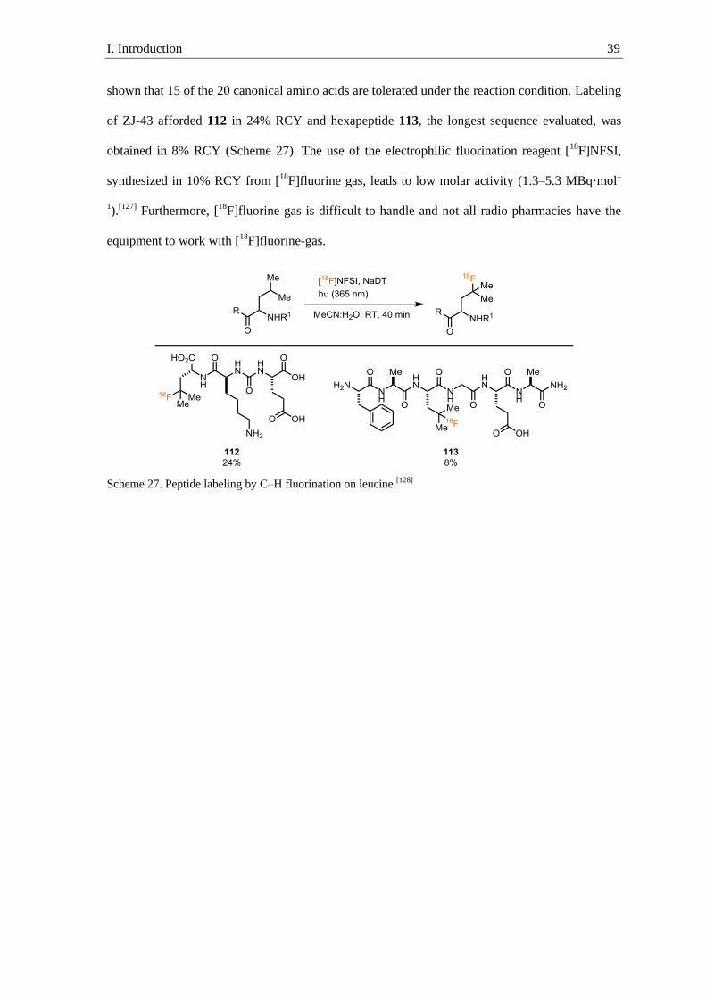
I. Introduction 39
shown that 15 of the 20 canonical amino acids are tolerated under the reaction condition. Labeling
of ZJ-43 afforded 112 in 24% RCY and hexapeptide 113, the longest sequence evaluated, was
obtained in 8% RCY (Scheme 27). The use of the electrophilic fluorination reagent [18F]NFSI,
synthesized in 10% RCY from [18F]fluorine gas, leads to low molar activity (1.3–5.3 MBq·mol–
1).[127] Furthermore, [18F]fluorine gas is difficult to handle and not all radio pharmacies have the
equipment to work with [18F]fluorine-gas.
Scheme 27. Peptide labeling by C–H fluorination on leucine.[128]

I. Introduction 40
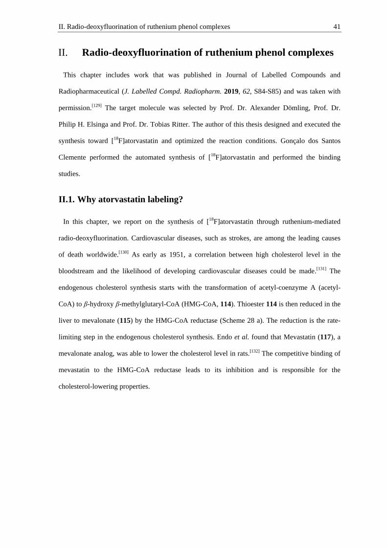
II. Radio-deoxyfluorination of ruthenium phenol complexes 41
Radio-deoxyfluorination of ruthenium phenol complexes II.
This chapter includes work that was published in Journal of Labelled Compounds and
Radiopharmaceutical (J. Labelled Compd. Radiopharm. 2019, 62, S84-S85) and was taken with
permission.[129] The target molecule was selected by Prof. Dr. Alexander Dömling, Prof. Dr.
Philip H. Elsinga and Prof. Dr. Tobias Ritter. The author of this thesis designed and executed the
synthesis toward [18F]atorvastatin and optimized the reaction conditions. Gonçalo dos Santos
Clemente performed the automated synthesis of [18F]atorvastatin and performed the binding
studies.
II.1. Why atorvastatin labeling?
In this chapter, we report on the synthesis of [18F]atorvastatin through ruthenium-mediated
radio-deoxyfluorination. Cardiovascular diseases, such as strokes, are among the leading causes
of death worldwide.[130] As early as 1951, a correlation between high cholesterol level in the
bloodstream and the likelihood of developing cardiovascular diseases could be made.[131]
The
endogenous cholesterol synthesis starts with the transformation of acetyl-coenzyme A (acetyl-
CoA) to β-hydroxy β-methylglutaryl-CoA (HMG-CoA, 114). Thioester 114 is then reduced in the
liver to mevalonate (115) by the HMG-CoA reductase (Scheme 28 a). The reduction is the rate-
limiting step in the endogenous cholesterol synthesis. Endo et al. found that Mevastatin (117), a
mevalonate analog, was able to lower the cholesterol level in rats.[132] The competitive binding of
mevastatin to the HMG-CoA reductase leads to its inhibition and is responsible for the
cholesterol-lowering properties.
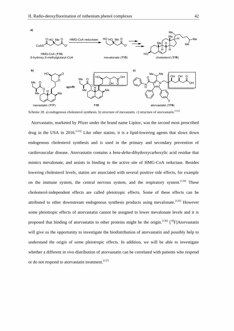
II. Radio-deoxyfluorination of ruthenium phenol complexes 42
Scheme 28. a) endogenous cholesterol synthesis. b) structure of mevastatin. c) structure of atorvastatin.[132]
Atorvastatin, marketed by Pfizer under the brand name Lipitor, was the second most prescribed
drug in the USA in 2016.[133] Like other statins, it is a lipid-lowering agents that slows down
endogenous cholesterol synthesis and is used in the primary and secondary prevention of
cardiovascular disease. Atorvastatin contains a beta-delta-dihydroxycarboxylic acid residue that
mimics mevalonate, and assists in binding to the active site of HMG-CoA reductase. Besides
lowering cholesterol levels, statins are associated with several positive side effects, for example
on the immune system, the central nervous system, and the respiratory system.[134] These
cholesterol-independent effects are called pleiotropic effects. Some of these effects can be
attributed to other downstream endogenous synthesis products using mevalonate.[135] However
some pleiotropic effects of atorvastatin cannot be assigned to lower mevalonate levels and it is
proposed that binding of atorvastatin to other proteins might be the origin.[136] [18F]Atorvastatin
will give us the opportunity to investigate the biodistribution of atorvastatin and possibly help to
understand the origin of some pleiotropic effects. In addition, we will be able to investigate
whether a different in vivo distribution of atorvastatin can be correlated with patients who respond
or do not respond to atorvastatin treatment.[137]

II. Radio-deoxyfluorination of ruthenium phenol complexes 43
II.2. Synthesis of [18
F]atorvastatin
Atorvastatin has a plasma elimination half-life of 7 hours, and to study its biodistribution, it
needs to be labeled with a radionuclide that allows imaging over several hours.[138]
A suitable
isotope is fluorine-18, having a half-life of 109.8 min. Labeling of atorvastatin without changing
its steric and electronic properties is essential to obtain meaningful data. The formal exchange of
fluorine-19 with fluorine-18 will have negligible effects on the in vivo behavior of the drug.[37] We
envisioned accessing the fluorine-18 isotopologue of atorvastatin by ruthenium-mediated radio-
deoxyfluorination (Figure 7). Ruthenium is crucial for the radio-deoxyfluorination, because
arenes with electron-releasing substituents cannot be labeled with iPrImCl alone. The unknown
phenol derivative of atorvastatin was thought to be accessible via Paal-Knorr pyrrole synthesis in
a similar way to the reported synthesis of atorvastatin.[139] In our retrosynthetic analysis we
thought that the key 1,4-diketone intermediate 121 could be synthesized from α,β-unsaturated
ketone 123 and aldehyde 124 via a Stetter reaction.
Figure 7. Retrosynthetic analysis of [18F]atorvastatin.
At first, 1,4-diketone 121 was obtained via the Stetter reaction between α,β-unsaturated ketone
123 and aldehyde 124 catalyzed by thiazolium 125 (Scheme 29).[140] The poor yield of 16% is
mainly attributed to low conversion, and the remaining starting materials could be recovered. The
low yield of the Stetter reaction could not be increased by an increased catalyst loading. Next,
diketone 121 was condensed with the commercially available primary amine 122 in a pivalic acid
catalyzed Paal-Knorr condensation to afford pyrrole 126 in 63% yield.[141] Finally, removal of the
benzyl protecting group by palladium on carbon catalyzed hydrogenolysis afforded the phenol
127.
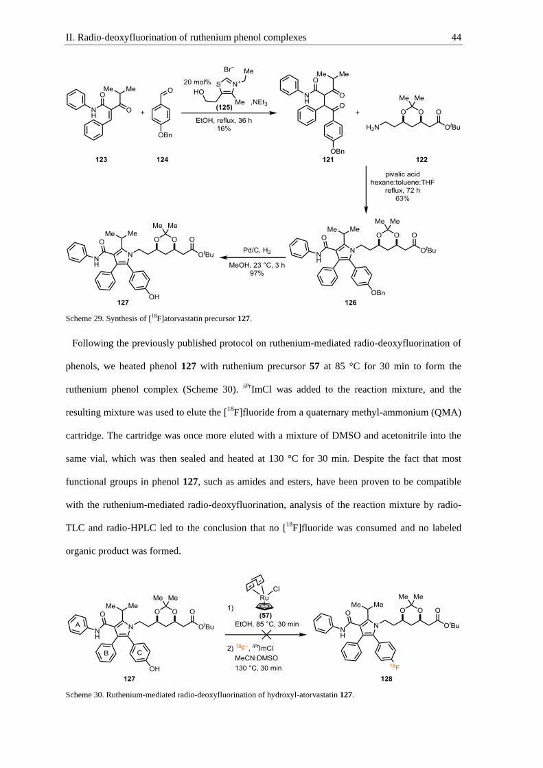
II. Radio-deoxyfluorination of ruthenium phenol complexes 44
Scheme 29. Synthesis of [18F]atorvastatin precursor 127.
Following the previously published protocol on ruthenium-mediated radio-deoxyfluorination of
phenols, we heated phenol 127 with ruthenium precursor 57 at 85 °C for 30 min to form the
ruthenium phenol complex (Scheme 30). iPrImCl was added to the reaction mixture, and the
resulting mixture was used to elute the [18F]fluoride from a quaternary methyl-ammonium (QMA)
cartridge. The cartridge was once more eluted with a mixture of DMSO and acetonitrile into the
same vial, which was then sealed and heated at 130 °C for 30 min. Despite the fact that most
functional groups in phenol 127, such as amides and esters, have been proven to be compatible
with the ruthenium-mediated radio-deoxyfluorination, analysis of the reaction mixture by radio-
TLC and radio-HPLC led to the conclusion that no [18F]fluoride was consumed and no labeled
organic product was formed.
Scheme 30. Ruthenium-mediated radio-deoxyfluorination of hydroxyl-atorvastatin 127.

II. Radio-deoxyfluorination of ruthenium phenol complexes 45
Initial LC-MS examination of the reaction mixture showed a low conversion of 127 and mainly
complexation to the wrong aryl substituent, leading to the conclusion that the phenol 127 was too
electron-rich to undergo radio-deoxyfluorination with iPrImCl without η6-activation. Atorvastatin
analog 127 contains three aromatic rings that could potentially form an η6-complex to ruthenium.
The complexation of ruthenium with the most electron-rich arene is thermodynamically favored.
However, we believe that complexation of ring A is kinetically favored because it is the least
sterically hindered arene and the amide substituent can act as a directing group. Due to the
difficult complexation and because ruthenium precursor 57 is not commercially available, we
thought improving the protocol and re-evaluating other ruthenium precursors.
II.3. New ruthenium precursor and complexation conditions
The ruthenium complex 57 was previously synthesized from commercially available ruthenium
trichloride in a four-step procedure in 16% overall yield (Scheme 31).[72] Complex 57 is not
commercially available and the synthesis procedure is lengthy and uses highly toxic
cyclopentadienylthallium. The use of difficult to access ruthenium precursor 57 diminishes the
utility of the ruthenium-mediated radio-deoxyfluorination methodology developed in our
laboratory.
Scheme 31. Synthesis of ruthenium precursor 57.[72]
When reviewing the literature, we found that ruthenium complex 134 could be a suitable
ruthenium precursor.[142] Complex 134 is air and moisture stable, and can be stored on the bench
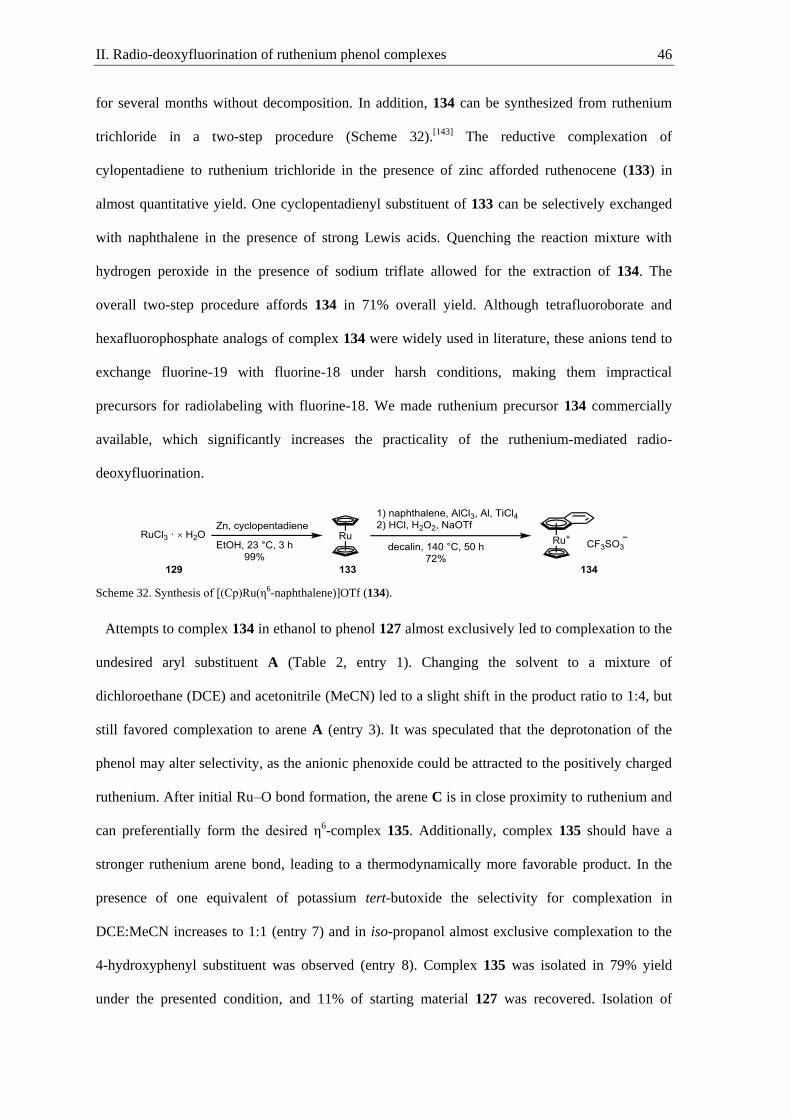
II. Radio-deoxyfluorination of ruthenium phenol complexes 46
for several months without decomposition. In addition, 134 can be synthesized from ruthenium
trichloride in a two-step procedure (Scheme 32).[143] The reductive complexation of
cylopentadiene to ruthenium trichloride in the presence of zinc afforded ruthenocene (133) in
almost quantitative yield. One cyclopentadienyl substituent of 133 can be selectively exchanged
with naphthalene in the presence of strong Lewis acids. Quenching the reaction mixture with
hydrogen peroxide in the presence of sodium triflate allowed for the extraction of 134. The
overall two-step procedure affords 134 in 71% overall yield. Although tetrafluoroborate and
hexafluorophosphate analogs of complex 134 were widely used in literature, these anions tend to
exchange fluorine-19 with fluorine-18 under harsh conditions, making them impractical
precursors for radiolabeling with fluorine-18. We made ruthenium precursor 134 commercially
available, which significantly increases the practicality of the ruthenium-mediated radio-
deoxyfluorination.
Scheme 32. Synthesis of [(Cp)Ru(η6-naphthalene)]OTf (134).
Attempts to complex 134 in ethanol to phenol 127 almost exclusively led to complexation to the
undesired aryl substituent A (Table 2, entry 1). Changing the solvent to a mixture of
dichloroethane (DCE) and acetonitrile (MeCN) led to a slight shift in the product ratio to 1:4, but
still favored complexation to arene A (entry 3). It was speculated that the deprotonation of the
phenol may alter selectivity, as the anionic phenoxide could be attracted to the positively charged
ruthenium. After initial Ru–O bond formation, the arene C is in close proximity to ruthenium and
can preferentially form the desired η6-complex 135. Additionally, complex 135 should have a
stronger ruthenium arene bond, leading to a thermodynamically more favorable product. In the
presence of one equivalent of potassium tert-butoxide the selectivity for complexation in
DCE:MeCN increases to 1:1 (entry 7) and in iso-propanol almost exclusive complexation to the
4-hydroxyphenyl substituent was observed (entry 8). Complex 135 was isolated in 79% yield
under the presented condition, and 11% of starting material 127 was recovered. Isolation of
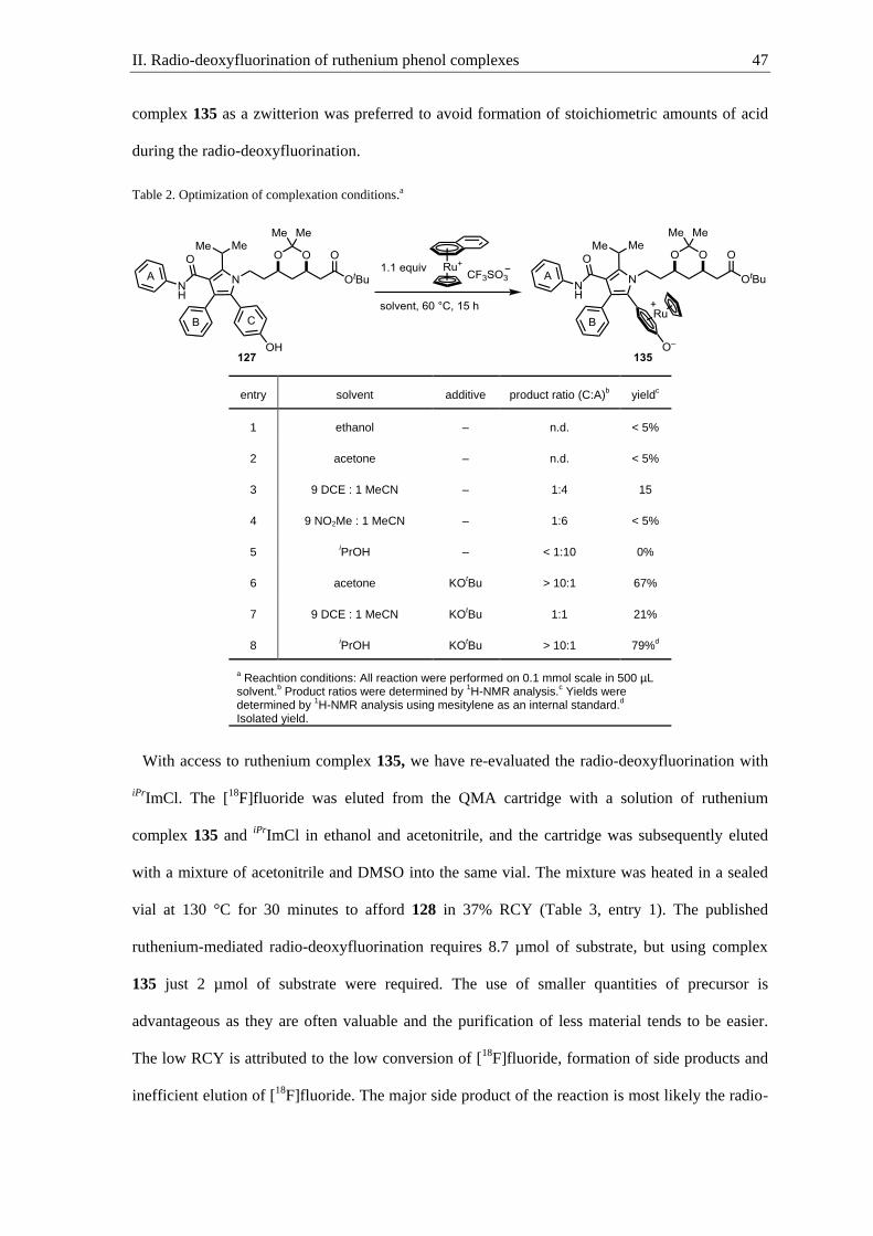
II. Radio-deoxyfluorination of ruthenium phenol complexes 47
complex 135 as a zwitterion was preferred to avoid formation of stoichiometric amounts of acid
during the radio-deoxyfluorination.
Table 2. Optimization of complexation conditions.a
entry solvent additive product ratio (C:A)b yield
c
1 ethanol – n.d. < 5%
2 acetone – n.d. < 5%
3 9 DCE : 1 MeCN – 1:4 15
4 9 NO2Me : 1 MeCN – 1:6 < 5%
5 iPrOH – < 1:10 0%
6 acetone KOtBu > 10:1 67%
7 9 DCE : 1 MeCN KOtBu 1:1 21%
8 iPrOH KO
tBu > 10:1 79%
d
a Reachtion conditions: All reaction were performed on 0.1 mmol scale in 500 µL
solvent.b Product ratios were determined by
1H-NMR analysis.
c Yields were
determined by 1H-NMR analysis using mesitylene as an internal standard.
d
Isolated yield.
With access to ruthenium complex 135, we have re-evaluated the radio-deoxyfluorination with
iPrImCl. The [18F]fluoride was eluted from the QMA cartridge with a solution of ruthenium
complex 135 and iPrImCl in ethanol and acetonitrile, and the cartridge was subsequently eluted
with a mixture of acetonitrile and DMSO into the same vial. The mixture was heated in a sealed
vial at 130 °C for 30 minutes to afford 128 in 37% RCY (Table 3, entry 1). The published
ruthenium-mediated radio-deoxyfluorination requires 8.7 µmol of substrate, but using complex
135 just 2 µmol of substrate were required. The use of smaller quantities of precursor is
advantageous as they are often valuable and the purification of less material tends to be easier.
The low RCY is attributed to the low conversion of [18F]fluoride, formation of side products and
inefficient elution of [18F]fluoride. The major side product of the reaction is most likely the radio-
II. Radio-deoxyfluorination of ruthenium phenol complexes 48
deoxyfluorinated, but still complexed, atorvastatin analog. Acetonitrile was a critical co-solvent
for decomplexation of the arene, but has a low boiling point (83 °C) compared to the reaction
temperature 130 °C. Higher boiling nitrile solvents such as benzonitrile (PhCN) and pivalonitrile
(PivCN) increased the conversion and purity of the reaction to give an overall RCY of 45% and
47%, respectively (entry 2, 3). The exchange of DMSO as a co-solvent with 1,2-
dimethoxybenzene increased the RCY to 58% (entry 5). We think that 1,2-dimethoxybenzene
(veratrole) can act as a bidentate flat ligand that facilitates the decomplexation. Furthermore,
veratrole can trap any released ruthenium to form stable η6-complexes. The exchange of veratrole
as a co-solvent with 1,4-dimethoxybenzene, which cannot act as a bidentate ligand, led to a
significant drop in RCY to 34% (entry 6). The use of only veratrole or pivalonitrile as solvents
lowered the RCY, highlighting the complementary properties (entry 7, 8). Lowering the reaction
temperature to 110 °C resulted in a drastic drop of the RCY, as did shortening the reaction time to
10 minutes (entry 9, 10, 11). Increasing the reaction volume to 2 mL decreased the RCY to 8%,
while decreasing the overall volume to 100 µL had little effect on the reaction (entry 12, 13).
With the new solvent conditions, an almost quantitative conversion of the [18F]fluoride to 128
was observed, but the elution efficiency dropped from 74% to 59%. This trend might be a result
of the lower polarity of the solvents.
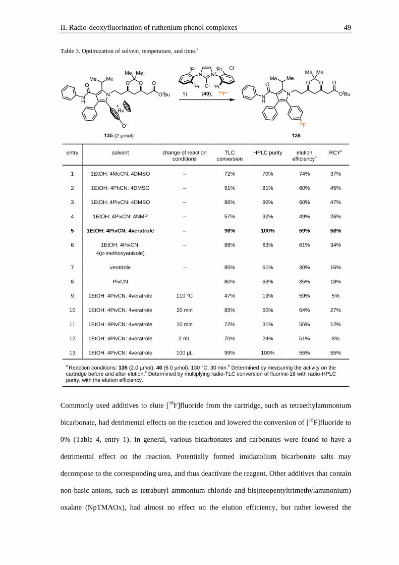
II. Radio-deoxyfluorination of ruthenium phenol complexes 49
Table 3. Optimization of solvent, temperature, and time.a
entry solvent change of reaction conditions
TLC conversion
HPLC purity elution efficiency
b
RCYc
1 1EtOH: 4MeCN: 4DMSO – 72% 70% 74% 37%
2 1EtOH: 4PhCN: 4DMSO – 91% 81% 60% 45%
3 1EtOH: 4PivCN: 4DMSO – 86% 90% 60% 47%
4 1EtOH: 4PivCN: 4NMP – 57% 92% 49% 26%
5 1EtOH: 4PivCN: 4veratrole – 98% 100% 59% 58%
6 1EtOH: 4PivCN:
4(p-methoxyanisole)
– 88% 63% 61% 34%
7 veratrole – 85% 61% 30% 16%
8 PivCN – 80% 63% 35% 18%
9 1EtOH: 4PivCN: 4veratrole 110 °C 47% 19% 59% 5%
10 1EtOH: 4PivCN: 4veratrole 20 min 85% 50% 64% 27%
11 1EtOH: 4PivCN: 4veratrole 10 min 72% 31% 56% 12%
12 1EtOH: 4PivCN: 4veratrole 2 mL 70% 24% 51% 8%
13 1EtOH: 4PivCN: 4veratrole 100 µL 99% 100% 55% 55%
a Reaction conditions: 135 (2.0 µmol), 40 (6.0 µmol), 130 °C, 30 min.
b Determined by measuring the activity on the
cartridge before and after elution.c Determined by multiplying radio-TLC conversion of fluorine-18 with radio-HPLC
purity, with the elution efficiency.
Commonly used additives to elute [18F]fluoride from the cartridge, such as tetraethylammonium
bicarbonate, had detrimental effects on the reaction and lowered the conversion of [18F]fluoride to
0% (Table 4, entry 1). In general, various bicarbonates and carbonates were found to have a
detrimental effect on the reaction. Potentially formed imidazolium bicarbonate salts may
decompose to the corresponding urea, and thus deactivate the reagent. Other additives that contain
non-basic anions, such as tetrabutyl ammonium chloride and bis(neopentyltrimethylammonium)
oxalate (NpTMAOx), had almost no effect on the elution efficiency, but rather lowered the
II. Radio-deoxyfluorination of ruthenium phenol complexes 50
conversion and the purity of the product (entry 2, 3). To further explore the influence of anions on
the reaction and elution, the reaction mixture was pre stirred with silver triflate and silver acetate
before elution, which can exchange the counter-ion of iPrImCl. Both additives increased the
elution efficiency, but the RCY dropped, due to either low conversion or potential slow
decomplexation (entry 4, 5). Protic solvents can form strong hydrogen bonds with fluoride, which
leads to an increased solubility of fluoride salts. In most radiochemistry reactions, [18F]fluoride is
eluted from the cartridge with an aqueous solution of K222 and potassium bicarbonate. Due to the
low reactivity of fluoride in water, water needs to be azeotropically removed in a time consuming
process. In order to avoid azeotropic drying and at the same time benefit from the favorable
elution properties of protic solvents, we evaluated various alcohols that can be easily removed
under an argon stream.[144] Elution of fluorine-18 with a mixture of ruthenium complex 135 and
iPrImCl in methanol increased the elution efficiency to 95% (entry 6). The resulting solution was
evaporated for 5 minutes at 80 °C under a stream of argon. Afterwards a mixture of pivalonitrile
and veratrole was added and the solution was heated to 130 °C for 30 minutes. Despite a slightly
lower conversion of fluorine-18, the overall RCY increased to 75%. The use of longer and
branched alcohols as elution solvents decreased the elution efficiency (entry 7, 8). The use of
more acidic alcohols such as hexafluoroisopropanol (HFIP), which can form even stronger
hydrogen bonds to fluoride, results in low elution efficiency (entry 9). Scott and Sanford reported
that preconditioning of QMA cartridges with different anions had a strong influence on the elution
efficiency and the RCY of the copper-mediated radio-fluorination of arylboronic acids.[145]
Cartridges pretreated with potassium oxalate resulted consistently in the highest yields (entry 6).
The use of potassium chloride for preconditioning cut the elution efficiency almost in half (entry
10). Preconditioning of the cartridge with potassium bicarbonate and potassium triflate also
resulted in an overall lower RCY (entry 11, 12). The use of Waters QMA light cartridges
containing 130 mg of anion exchange resin, compared to Chromafix cartridges containing 45 mg
resin, resulted in almost no change in RCY.

II. Radio-deoxyfluorination of ruthenium phenol complexes 51
Table 4. Optimizing elution efficiency.a
entry eluent solvent additive cartridge TLC conversion
HPLC purity
elution efficiency
b
RCYc
1 EtOH:PivCN TEA bicarbonate Chromafix oxalate 0% 0% 62% 0%
2 EtOH:PivCN TBA chloride Chromafix oxalate 91% 82% 62% 46%
3 EtOH:PivCN NP oxalate Chromafix oxalate 52% 71% 54% 20%
4 EtOH:PivCN silver triflate Chromafix oxalate 69% 36% 65% 16%
5 EtOH:PivCN silver acetate Chromafix oxalate 29% 82% 67% 16%
6 methanol – Chromafix oxalate 88% 90% 95% 75%
7 ethanol – Chromafix oxalate 94% 96% 79% 71%
8 tert-butanol – Chromafix oxalate 89% 83% 23% 17%
9 HFIP – Chromafix oxalate 86% 86% 60% 44%
10 methanol – Chromafix chloride 95% 88% 53% 44%
11 methanol – Chromafix bicarbonate 68% 85% 78% 45%
12 methanol – Chromafix triflate 78% 72% 74% 42%
13 methanol – Waters QMA oxalate 90% 93% 93% 78%
a Reaction conditions: 135 (2.0 µmol), 40 (6.0 µmol), 130 °C, 30 min.
b Determined by measuring the activity on the
cartridge before and after elution.c Determined by multiplying radio-TLC conversion of fluorine-18 with radio-HPLC purity
(n = 2) with the elution efficiency.
To obtain [18F]atorvastatin, the ketal and ester protecting groups on 128 need to be removed in
subsequent steps (Scheme 33). The two-step deprotection protocol consists of acidic removal of
the ketal protecting group using 6 M HCl at 60 °C for 5 minutes, followed by saponification of
the ester using sodium hydroxide at 60 °C for 5 minutes. Using this overall three-step, one-pot
protocol, it was possible to obtain [18F]atorvastatin (120) in 20% decay-corrected RCY after
preparative HPLC purification. After purification, the product was obtained in ≥ 95%
radiochemical purity and in high molar activity 112 ± 77 GBq·µmol–1.[129]

II. Radio-deoxyfluorination of ruthenium phenol complexes 52
Scheme 33. Synthesis of [18F]atorvastatin.[129]
Human imaging typically requires around 300 MBq of radioactive material, and the manual
handling of these large amounts of activity is not possible. In order to access sufficient activity of
[18F]atorvastatin, the protocol was translated to the automated synthesis model Synthra RNplus.
Initial automation experiments were successful and 120 could be isolated in 5% RCY.[129]
II.4. In vitro analysis of [18
F]atorvastatin
Before starting in vivo experiments, we investigated the behavior of [18F]atorvastatin in vitro.
The three-hour incubation of 120 in human serum and rat serum at 37 °C lead to no signs of
decomposition. An ideal PET-tracer would exclusively bind the target. However, this exclusive
binding is typically not possible. A high plasma protein binding can lead to a low target-to-
background ratio, due to the high background radiation. The plasma protein binding in the serum
of rats with a normal diet was 16.7%, and in rats with a high fat diet it was 13.8%. Furthermore, it
could be shown that more [18F]atorvastatin was bound to liver homogenates of rats with high
calorie diet than of rats with low calorie diet.
II.5. Conclusion and outlook
In this chapter, we present an optimized protocol for the ruthenium-mediated radio-
deoxyfluorination of phenols and overcome several of the previous known and unknown
shortcomings. The selective complexation of ruthenium complex to the 4-hydroxyphenyl
substituent against other arenes present in 127 was achieved under basic conditions. Furthermore,
we were able show that the use of complexed precursor enabled us to reduce the amount of
material used in fluorine-18 labeling from 8.7 µmol to 2 µmol. Major losses of fluorine-18 in the
II. Radio-deoxyfluorination of ruthenium phenol complexes 53
previous protocol were attributed to low elution efficiency. Almost quantitative elution of
[18F]fluoride was achieved with iPrImCl in methanol, followed by drying under a stream of argon.
The elution efficiency of this protocol is comparable to elution with the well-established mixture
of K222 and potassium bicarbonate, but circumvents time-consuming azeotropic drying.
Furthermore, the inversion of the cartridge is no longer necessary and the RCY is no longer
dependent on vial size or pressure, allowing easier translation to various automated synthesis
models. Initial results show the feasibility of in vivo imaging with [18F]atorvastatin. A major
downside of the protocol is the high reaction temperature required for the decomplexation of
ruthenium. The development of ruthenium complexes with an easy to trigger decomplexation
functionality would be of great benefit to the protocol. Furthermore, the development of a good
manufacturing practice (GMP) protocol would be a major step forward toward the first in human
imaging studies using this methodology. The Elsinga laboratory at the University of Groningen is
currently working on human imaging with [18F]atorvastatin.

II. Radio-deoxyfluorination of ruthenium phenol complexes 54
III. 18F-Labeling of Peptides 55
18F-Labeling of peptides III.
This chapter includes work that was published in Angewandte Chemie (Angew. Chem. Int. Ed.
2018, 57, 14207–14211) and was taken with permission.[146]
The idea of the project was set out by
Prof. Dr. Tobias Ritter and the author of this thesis. The author of this thesis designed and
conducted experiments that led to the development of the site‐specific radio-deoxyfluorination of
small peptides with [18F]fluoride. The manuscript was prepared in cooperative work between
Prof. Dr. Tobias Ritter and the author of this thesis.
III.1. Project design
Receptor-binding peptides are a rapidly growing class of PET-tracers used in non-invasive
diagnosis, in oncology, and the study of biochemical processes.[147] The use of peptides bears the
inherent advantage of facile access by automated solid phase peptide synthesis, potential high
binding affinity and rapid blood clearance.[97] A critical aspect to peptide labeling is that the
labeling method should result in a minimal perturbation of the structure of the peptide. Labeling
without changing the properties of the peptides can be achieved with carbon-11, nitrogen-13, and
oxygen-15 labeled structures, but the half-lives of 20 minutes or less of these isotopes mitigates
utility of those radionuclides toward PET-imaging with peptide-tracers.[148] Labeling with
radioactive metals with sufficiently long half-lives such as gallium-68 or copper-64 requires the
attachment of prosthetic groups that can drastically change the properties of the peptide. Although
successful examples of peptide labeling via prosthetic groups are known, this requires in-depth
knowledge of the binding site.[147] In contrast to radiometals, fluorine-18 can be introduced
without the need of prosthetic groups and can result in small structural modifications, as it is
considered a bioisostere of a hydrogen or a hydroxyl substituent. In addition, favorable nuclear
properties such as low positron energy, preferential beta-plus-decay and, especially the half-life of
109.8 minutes makes it an ideal radionuclide for peptide labeling.[149] Despite the demand for
methods to label polypeptides by simply exchanging one hydrogen or hydroxyl functionality with
[18F]fluoride, no method was able to achieve this goal. The reason for the lack of methods can be

III. 18F-Labeling of Peptides 56
attributed to the high density of functional groups and the lability of the structure. In our previous
publications on ruthenium-mediated radio-deoxyfluorination of arenes,[72] we have shown that
most functional groups common in peptides are well tolerated. Therefore, we thought that it might
be possible to use the high functional group tolerance of the ruthenium-mediated radio-
deoxyfluorination to achieve peptide labeling by formal exchange of a hydrogen/hydroxyl
substituent with fluorine-18.
III.2. Building block design, synthesis and application
Introduction of fluorine-18 on arenes is often preferred over alkanes due to the higher metabolic
and chemical stability.[126] Among the 20 canonical amino acids are four aromatic amino acids:
phenylalanine, tyrosine, tryptophan, and histidine (Figure 8). It was found that the introduction of
fluorine on aromatic side chains has little effect the on protein function, and up to 80% of
tryptophan in Escherichia coli could be exchanged for 5-F-tryptophan while little effect on the
growth of the bacterium being observed.[150] Similar results were found for the replacement of
phenylalanine with 4-fluoro-phenylalanine.[151] In our previous publication on the ruthenium-
mediated radio-deoxyfluorination of phenols, our group disclosed the radio-deoxyfluorination of
protected tyrosine, which is not possible without ruthenium coordination. Thus, and because the
resulting 4-fluoro-phenylalanine is an analog of phenylalanine and tyrosine, we decided to
investigate the introduction of 4-[18F]fluoro-phenylalanine into peptides.[72]
Figure 8. Aromatic amino acids.
Since unprotected carboxylic acids tend to lower the RCY, most likely by protonation of
[18F]fluoride to afford unreactive [18F]hydrogen fluoride, they need to be protected during
labeling. To avoid this and other side reactions with labile functional groups, we focused on the
labeling of fully protected peptides, which are as easily accessible by solid phase peptide
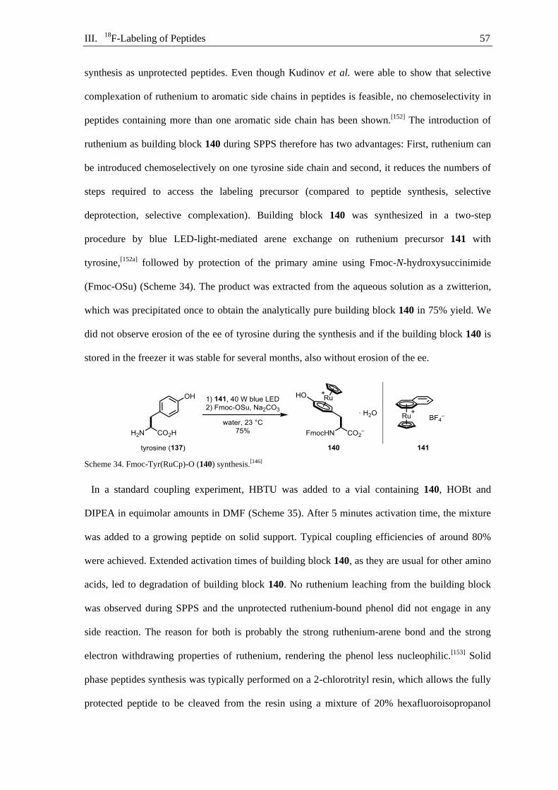
III. 18F-Labeling of Peptides 57
synthesis as unprotected peptides. Even though Kudinov et al. were able to show that selective
complexation of ruthenium to aromatic side chains in peptides is feasible, no chemoselectivity in
peptides containing more than one aromatic side chain has been shown.[152] The introduction of
ruthenium as building block 140 during SPPS therefore has two advantages: First, ruthenium can
be introduced chemoselectively on one tyrosine side chain and second, it reduces the numbers of
steps required to access the labeling precursor (compared to peptide synthesis, selective
deprotection, selective complexation). Building block 140 was synthesized in a two-step
procedure by blue LED-light-mediated arene exchange on ruthenium precursor 141 with
tyrosine,[152a] followed by protection of the primary amine using Fmoc-N-hydroxysuccinimide
(Fmoc-OSu) (Scheme 34). The product was extracted from the aqueous solution as a zwitterion,
which was precipitated once to obtain the analytically pure building block 140 in 75% yield. We
did not observe erosion of the ee of tyrosine during the synthesis and if the building block 140 is
stored in the freezer it was stable for several months, also without erosion of the ee.
Scheme 34. Fmoc-Tyr(RuCp)-O (140) synthesis.[146]
In a standard coupling experiment, HBTU was added to a vial containing 140, HOBt and
DIPEA in equimolar amounts in DMF (Scheme 35). After 5 minutes activation time, the mixture
was added to a growing peptide on solid support. Typical coupling efficiencies of around 80%
were achieved. Extended activation times of building block 140, as they are usual for other amino
acids, led to degradation of building block 140. No ruthenium leaching from the building block
was observed during SPPS and the unprotected ruthenium-bound phenol did not engage in any
side reaction. The reason for both is probably the strong ruthenium-arene bond and the strong
electron withdrawing properties of ruthenium, rendering the phenol less nucleophilic.[153]
Solid
phase peptides synthesis was typically performed on a 2-chlorotrityl resin, which allows the fully
protected peptide to be cleaved from the resin using a mixture of 20% hexafluoroisopropanol
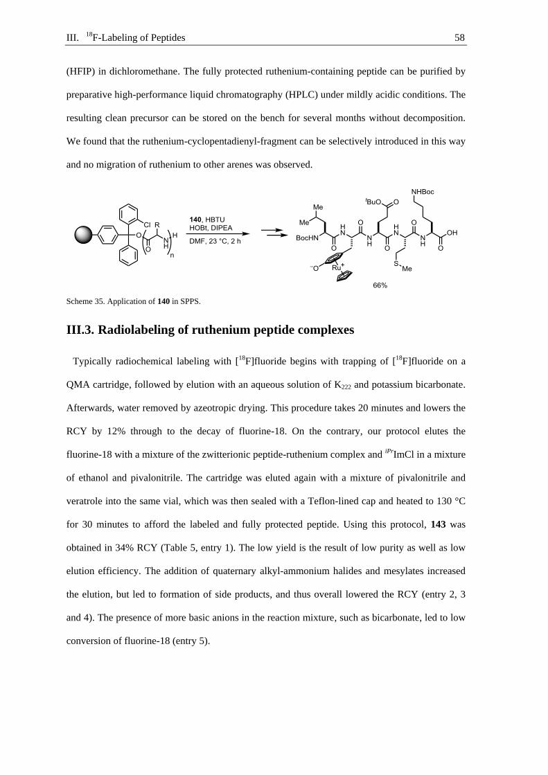
III. 18F-Labeling of Peptides 58
(HFIP) in dichloromethane. The fully protected ruthenium-containing peptide can be purified by
preparative high-performance liquid chromatography (HPLC) under mildly acidic conditions. The
resulting clean precursor can be stored on the bench for several months without decomposition.
We found that the ruthenium-cyclopentadienyl-fragment can be selectively introduced in this way
and no migration of ruthenium to other arenes was observed.
Scheme 35. Application of 140 in SPPS.
III.3. Radiolabeling of ruthenium peptide complexes
Typically radiochemical labeling with [18F]fluoride begins with trapping of [18F]fluoride on a
QMA cartridge, followed by elution with an aqueous solution of K222 and potassium bicarbonate.
Afterwards, water removed by azeotropic drying. This procedure takes 20 minutes and lowers the
RCY by 12% through to the decay of fluorine-18. On the contrary, our protocol elutes the
fluorine-18 with a mixture of the zwitterionic peptide-ruthenium complex and iPrImCl in a mixture
of ethanol and pivalonitrile. The cartridge was eluted again with a mixture of pivalonitrile and
veratrole into the same vial, which was then sealed with a Teflon-lined cap and heated to 130 °C
for 30 minutes to afford the labeled and fully protected peptide. Using this protocol, 143 was
obtained in 34% RCY (Table 5, entry 1). The low yield is the result of low purity as well as low
elution efficiency. The addition of quaternary alkyl-ammonium halides and mesylates increased
the elution, but led to formation of side products, and thus overall lowered the RCY (entry 2, 3
and 4). The presence of more basic anions in the reaction mixture, such as bicarbonate, led to low
conversion of fluorine-18 (entry 5).
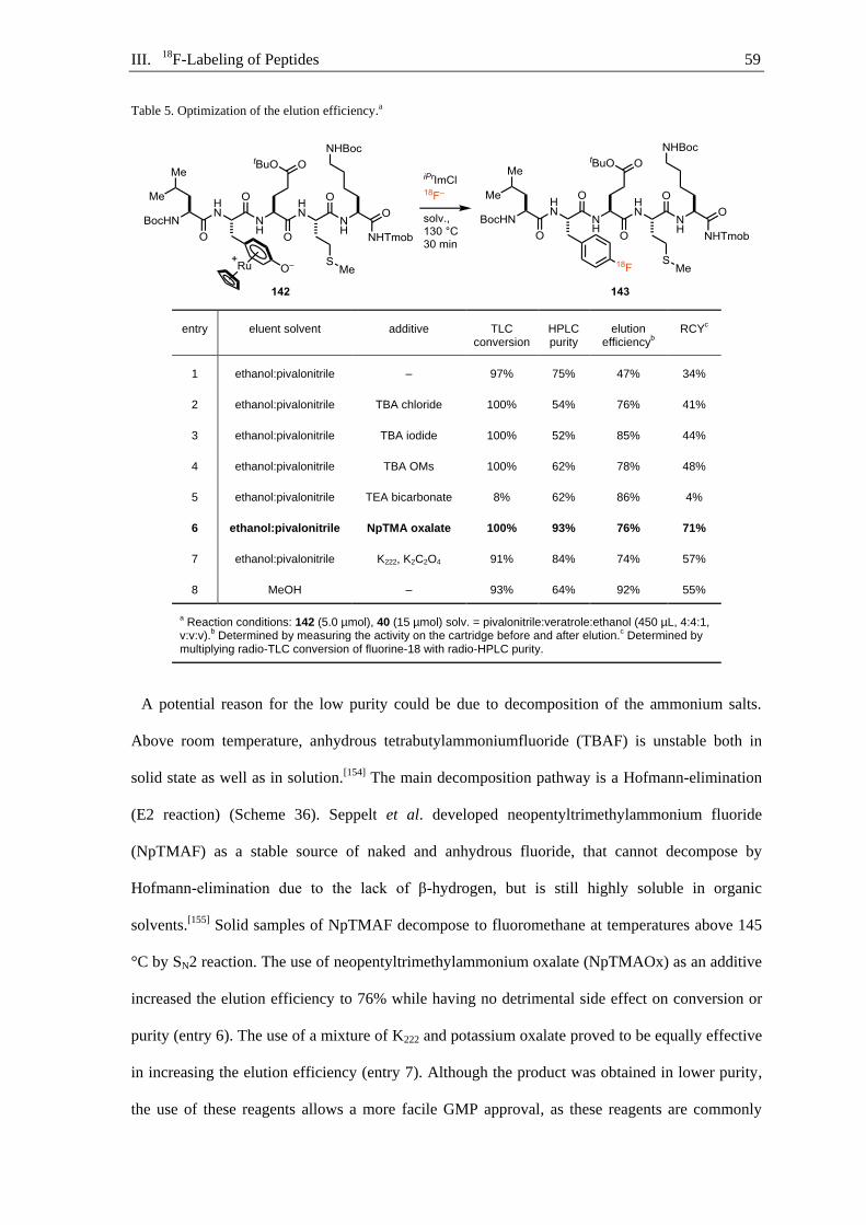
III. 18F-Labeling of Peptides 59
Table 5. Optimization of the elution efficiency.a
entry eluent solvent additive TLC conversion
HPLC purity
elution efficiency
b
RCYc
1 ethanol:pivalonitrile – 97% 75% 47% 34%
2 ethanol:pivalonitrile TBA chloride 100% 54% 76% 41%
3 ethanol:pivalonitrile TBA iodide 100% 52% 85% 44%
4 ethanol:pivalonitrile TBA OMs 100% 62% 78% 48%
5 ethanol:pivalonitrile TEA bicarbonate 8% 62% 86% 4%
6 ethanol:pivalonitrile NpTMA oxalate 100% 93% 76% 71%
7 ethanol:pivalonitrile K222, K2C2O4 91% 84% 74% 57%
8 MeOH – 93% 64% 92% 55%
a Reaction conditions: 142 (5.0 µmol), 40 (15 µmol) solv. = pivalonitrile:veratrole:ethanol (450 µL, 4:4:1,
v:v:v).b Determined by measuring the activity on the cartridge before and after elution.
c Determined by
multiplying radio-TLC conversion of fluorine-18 with radio-HPLC purity.
A potential reason for the low purity could be due to decomposition of the ammonium salts.
Above room temperature, anhydrous tetrabutylammoniumfluoride (TBAF) is unstable both in
solid state as well as in solution.[154] The main decomposition pathway is a Hofmann-elimination
(E2 reaction) (Scheme 36). Seppelt et al. developed neopentyltrimethylammonium fluoride
(NpTMAF) as a stable source of naked and anhydrous fluoride, that cannot decompose by
Hofmann-elimination due to the lack of β-hydrogen, but is still highly soluble in organic
solvents.[155] Solid samples of NpTMAF decompose to fluoromethane at temperatures above 145
°C by SN2 reaction. The use of neopentyltrimethylammonium oxalate (NpTMAOx) as an additive
increased the elution efficiency to 76% while having no detrimental side effect on conversion or
purity (entry 6). The use of a mixture of K222 and potassium oxalate proved to be equally effective
in increasing the elution efficiency (entry 7). Although the product was obtained in lower purity,
the use of these reagents allows a more facile GMP approval, as these reagents are commonly

III. 18F-Labeling of Peptides 60
used in radiochemistry. Almost quantitative elution of [18F]fluoride in protic solvents was
achieved, but the purity of the product was lowered (entry 8).
Scheme 36. Decomposition pathways of ammonium fluorides.[156]
Despite the high reaction temperature and the long reaction time, no extensive decomposition
was observed (Figure 9). The only fluorine-18 containing side product that is formed has a very
similar retention time to that of peptide ruthenium complex 142. If the reaction time is shortened
to 10 minutes, the amount of fluorine-18 labeled side product at 9.7 min increases to 74%, and if
the reaction temperature is lowered to 100 °C, the amount increases to 98 % (Table 6, entry 2 and
3). The fact that the side product converts with extended reaction time and higher reaction
temperature to the labeled peptide 143, and the similar retention time to peptide ruthenium
complex 142 leads to the assumption that it might be the radio-deoxyfluorinated, but still
complexed peptide. This side product is easy to separate and does not pose a problem in
purification.
Figure 9. HPLC trace of crude reaction mixture.
In a clinical setting, the use of rigorously anhydrous solvents cannot be guaranteed. In order to
underline the robustness of the presented method against water, we added 10 µL of exogenous
water to the 450 µL reaction mixture, which resulted in a minor drop of RCY from 87% to 78%
0
200
400
600
800
1000
1200
1400
1600
0200400600800
100012001400160018002000
0 2 4 6 8 10 12 14
mV
mA
U
time · min–1
UV-Vis@280 nm
radio-trace
veratrole
peptide 142
[18F]peptide 143

III. 18F-Labeling of Peptides 61
(entry 4). Furthermore, no special precautions toward air were required. Lowering the amount of
precursor used from 5 µmol (6.5 mg) to 2.5 µmol (3.2 mg) results in an acceptable decrease in
RCY to 35%, and even as little as 1.5 µmol (1.9 mg) is sufficient to obtain 143 in acceptable
yields (entry 5, 6). The lower amounts of precursor resulted in a stronger deviation of the results.
Thus, all subsequent experiments were conducted with 5 µmol ruthenium peptide complexe. The
size of the reaction vial did not have an influence on the RCY, leading to a more facile
automation of the process (entry 7, 8).
Table 6. Modification of standard conditions.a
entry change of reaction conditions
TLC conversion
HPLC purity
elution efficiency
b
RCYc
1 none 100% 93% 76% 71%
2 100 °C 68% 3% 61% 1%
3 10 min 94% 26% 65% 16%
4 10 µL H2O added 100% 78% 57% 45%
5 3 µmol of 142 95% 53% 70% 35%
6 1.5 µmol of 142 92% 47% 65% 28%
7 20 mL vial 100% 91% 80% 73%
8 2 mL vial 100% 96% 70% 67%
a Reaction conditions: 142 (5.0 µmol), NpTMAOx (14 µmol), 40 (15 µmol) solv. =
pivalonitrile:veratrole:ethanol (450 µL, 4:4:1, v:v:v).b Determined by measuring the activity on
the cartridge before and after elution.c Determined by multiplying radio-TLC conversion of
fluorine-18 with radio-HPLC purity (n = 2).

III. 18F-Labeling of Peptides 62
III.4. Substrate scope
Most peptide PET-tracers do not contain protecting groups, and thus an additional deprotection
step is required after labeling of protected peptides. The labeling of unprotected peptides would
be inherently advantageous as it would reduce the overall number of steps of the labeling
protocol. Furthermore, the complexation of ruthenium to tyrosine side chains in unprotected
peptides is known, which would allow the labeling of native peptides.[157] Subjecting the
ruthenium peptide complex 146 to our standard labeling conditions led to the formation of 147 in
12% RCY (Scheme 37). However, a messy radio HPLC-trace was observed, probably due to
decomposition of the labeled product and precursor. The formation of a complex product mixture
can lead to time consuming purification and would reduce the benefit of this approach. Therefore,
we decided to focus on the labeling of protected peptides and add a deprotection step to the
protocol.
Scheme 37. Deoxyfluorination of unprotected peptide 146.
A common deprotection mixture used in SPPS is a combination of TFA, 1,4-dithiothreitol
(DTT), water, and triisopropylsilane (TIPS). All protecting groups common to SPPS with Fmoc
strategy can be removed under acidic conditions as well as the trimethoxybenzyl (Tmob)
protecting group on the C-terminal amide. The highly reactive intermediates formed during
deprotection, such tert-butyl cations, must be quenched with nucleophilic scavengers, such as
water and DTT. The second effect of DTT is that it is able to reduce sulfoxides and disulfides.
The general procedure for the ruthenium-mediated peptide 18F-labeling starts with trapping of
[18F]fluoride on a QMA cartridge that was preconditioned with potassium oxalate. The
[18F]fluoride was eluted with a mixture of the peptide ruthenium complex, iPrImCl, and
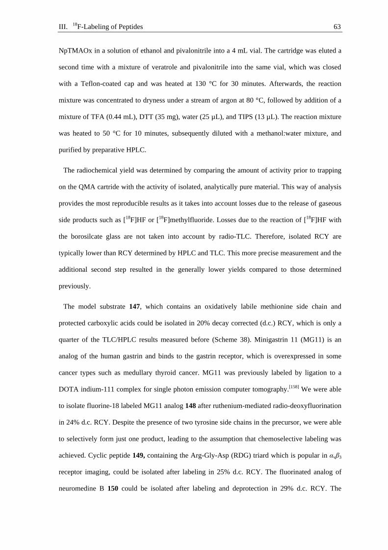
III. 18F-Labeling of Peptides 63
NpTMAOx in a solution of ethanol and pivalonitrile into a 4 mL vial. The cartridge was eluted a
second time with a mixture of veratrole and pivalonitrile into the same vial, which was closed
with a Teflon-coated cap and was heated at 130 °C for 30 minutes. Afterwards, the reaction
mixture was concentrated to dryness under a stream of argon at 80 °C, followed by addition of a
mixture of TFA (0.44 mL), DTT (35 mg), water (25 µL), and TIPS (13 µL). The reaction mixture
was heated to 50 °C for 10 minutes, subsequently diluted with a methanol:water mixture, and
purified by preparative HPLC.
The radiochemical yield was determined by comparing the amount of activity prior to trapping
on the QMA cartride with the activity of isolated, analytically pure material. This way of analysis
provides the most reproducible results as it takes into account losses due to the release of gaseous
side products such as [18F]HF or [18F]methylfluoride. Losses due to the reaction of [18F]HF with
the borosilcate glass are not taken into account by radio-TLC. Therefore, isolated RCY are
typically lower than RCY determined by HPLC and TLC. This more precise measurement and the
additional second step resulted in the generally lower yields compared to those determined
previously.
The model substrate 147, which contains an oxidatively labile methionine side chain and
protected carboxylic acids could be isolated in 20% decay corrected (d.c.) RCY, which is only a
quarter of the TLC/HPLC results measured before (Scheme 38). Minigastrin 11 (MG11) is an
analog of the human gastrin and binds to the gastrin receptor, which is overexpressed in some
cancer types such as medullary thyroid cancer. MG11 was previously labeled by ligation to a
DOTA indium-111 complex for single photon emission computer tomography.[158] We were able
to isolate fluorine-18 labeled MG11 analog 148 after ruthenium-mediated radio-deoxyfluorination
in 24% d.c. RCY. Despite the presence of two tyrosine side chains in the precursor, we were able
to selectively form just one product, leading to the assumption that chemoselective labeling was
achieved. Cyclic peptide 149, containing the Arg-Gly-Asp (RDG) triard which is popular in αvβ3
receptor imaging, could be isolated after labeling in 25% d.c. RCY. The fluorinated analog of
neuromedine B 150 could be isolated after labeling and deprotection in 29% d.c. RCY. The
III. 18F-Labeling of Peptides 64
decapeptide 150 was the longest sequence evaluated, and more extensive decomposition was
observed compared to other peptides. Thus, we think that our method is most applicable for
labeling of small peptide sequences, which benefits most from labeling methods with minimal
perturbation of structure. Most receptor-binding peptide PET-tracers contain C-terminal primary
amides because this motif mimics the native peptide structure better and because peptides tend to
have higher metabolic stability toward carboxypeptidases.[159] Octapeptide 151 containing a C-
terminal carboxylic acid could be isolated in 11% d.c. RCY, starting from a precursor containing
a non-protected C-terminal carboxylic acid. The protection of the carboxylic acid as an acid labile
p-methoxybenzyl (PMB) ester increased the isolated d.c. RCY to 39%. Due to the high reaction
temperature, we were afraid of epimerization of the peptides. Even though no excessive
decomposition was observed, we used pentapeptide 152 as a model substrate, containing a
histidine residue that can facilitate racemization by serving as an internal base.[160] If the product
is compared with authentic reference samples of both potential diastereomers, no epimerization
within the limits of detection could be observed. Overall, we could show that all 20 canonical
amino acids are tolerated and that 4-[18F]fluoro-phenylalanine could be introduced on the C-, N-
terminus or within the peptide structure. The robustness of the method is highlighted by the use of
one set of reaction conditions that allows for the rapid evaluation of tracer candidates.
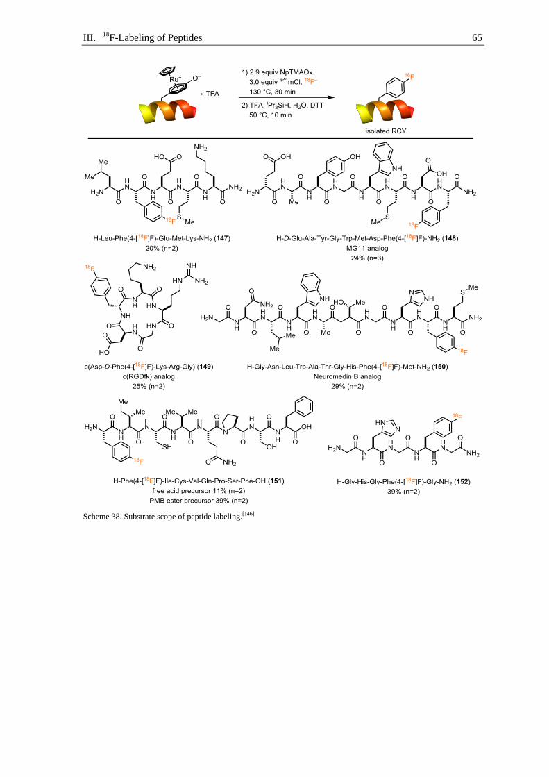
III. 18F-Labeling of Peptides 65
Scheme 38. Substrate scope of peptide labeling.[146]

III. 18F-Labeling of Peptides 66
III.5. Automated labeling
First attempts to translate the ruthenium-mediated radio-deoxyfluorination of peptides to the
ELIXYS FLEX/CHEM platform, connected to a PURE/FORM purification and reformulation
unit (Sofie Bioscience) afforded 147 in low RCY. The RCY could be increased to 21% by
changing the reaction temperature from 130 °C to 150 °C (Scheme 39). The difference in reaction
temperature could be due to the adapter used to fit a 3 mL Vial into the synthesizer. The adapter
might transfers heat slowly, resulting in a temperature difference between target and reaction
temperature.[161] Within 99 minutes, 11.4 GBq of [18F]fluoride could be converted to 1.28 GBq of
147, formulated in a mixture of ethanol and saline, ready to use for biological experiments.
Scheme 39. Automated synthesis of 147.[146]
High molar activity is crucial in peptide labeling, as the receptors are often present in low
concentrations. The use of excessive amounts of hexafluorophosphate counter-ions during SPPS
has the potential to contaminate the precursor with hexafluorophosphate anions, which can leach
out fluorine-19 and decrease the molar activity. Nevertheless, 147 could be obtained high molar
activity 99 GBq·µmol–1. The molar activity of peptide 147 cannot only be lowered by dilution
with fluorine-19, but also by the presence of side products or precursors that cannot be separated.
The separation of precursor and labeled product gets more difficult with increasing size of the
peptide. As previously mentioned, the retention time of complexed peptide is quite different to the
decomplexed peptide, because the ionic ruthenium complex drastically changes the polarity of the
peptide. Under our reaction conditions, we found that the fluorinated product was almost
completely decomplexed while the precursor stays complexed to ruthenium, due to the stronger
bond. This allowed for the facile separation of the precursor from the labeled peptide (Figure 10).

III. 18F-Labeling of Peptides 67
Figure 10. HPLC-trace of preparative HPLC purification of 147.
The separation of ruthenium from the radiolabeled peptide is crucial, as the specific limit
considered safe for human injection is 10 µg per day. The reformulated sample obtained after
automated synthesis contained 1.8 µg of ruthenium, which is well below the limit. The ruthenium
content in the reformulated sample could be further reduced to 0.09 µg by additional C-18
purification before preparative HPLC or by the use of metal scavenger resins such as Quadrasil
AP to 0.11 µg, which is threefold as high as the blank sample.[162]
III.6. Conclusion and outlook
Overall, we have reported the first general protocol to formally exchange a single hydrogen or
hydroxyl substituent in the native polypeptide structure with a single fluorine-18 (Scheme 40).
This methodology avoids the use of large prosthetic groups. Instead, simply exchanging one
hydrogen/hydroxyl substituent of the native peptide structure with fluorine-18, allows for minimal
structural perturbation of the peptide and therefore, a minimal change of the behavior in vivo. We
could show that the method tolerates all 20 canonical amino acids, is operationally simple, and
tolerates water and air. The transformation is enabled by a traceless activation of a tyrosine
residue by coordination in η6-binding mode to ruthenium. The labeling precursors can be easily
accessed by applying novel amino-acid building block 140 in SPPS. For facile adoption of the
method to other research groups and clinical application we made amino acid building block 140
commercially available. This protocol can provide reliable access to novel tracer candidates,
which may facilitate the development of novel peptide-PET-tracers.
0
20
40
60
80
100
120
0
0.5
1
1.5
2
2.5
0 2 4 6 8 10 12 14 16 18 20
mV
AU
time·min−1
UV-trace 210nmradio-trace
ruthenium
peptide
complex
147
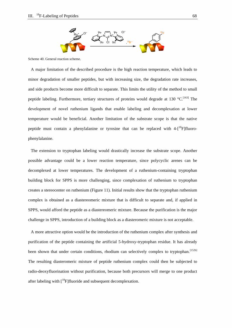
III. 18F-Labeling of Peptides 68
Scheme 40. General reaction scheme.
A major limitation of the described procedure is the high reaction temperature, which leads to
minor degradation of smaller peptides, but with increasing size, the degradation rate increases,
and side products become more difficult to separate. This limits the utility of the method to small
peptide labeling. Furthermore, tertiary structures of proteins would degrade at 130 °C.[163] The
development of novel ruthenium ligands that enable labeling and decomplexation at lower
temperature would be beneficial. Another limitation of the substrate scope is that the native
peptide must contain a phenylalanine or tyrosine that can be replaced with 4-[18F]fluoro-
phenylalanine.
The extension to tryptophan labeling would drastically increase the substrate scope. Another
possible advantage could be a lower reaction temperature, since polycyclic arenes can be
decomplexed at lower temperatures. The development of a ruthenium-containing tryptophan
building block for SPPS is more challenging, since complexation of ruthenium to tryptophan
creates a stereocenter on ruthenium (Figure 11). Initial results show that the tryptophan ruthenium
complex is obtained as a diastereomeric mixture that is difficult to separate and, if applied in
SPPS, would afford the peptide as a diastereomeric mixture. Because the purification is the major
challenge in SPPS, introduction of a building block as a diasteromeric mixture is not acceptable.
A more attractive option would be the introduction of the ruthenium complex after synthesis and
purification of the peptide containing the artificial 5-hydroxy-tryptophan residue. It has already
been shown that under certain conditions, rhodium can selectively complex to tryptophan.[152b]
The resulting diasteromeric mixture of peptide ruthenium complex could then be subjected to
radio-deoxyfluorination without purification, because both precursors will merge to one product
after labeling with [18F]fluoride and subsequent decomplexation.

III. 18F-Labeling of Peptides 69
Figure 11. Potential building blocks for tryptophan labeling.

III. 18F-Labeling of Peptides 70

IV. Experimental Part 71
Experimental part IV.
IV.1. Atorvastatin labeling
Materials and methods IV.1.1.
All air- and moisture-insensitive reactions were carried out under an ambient atmosphere and
monitored by thin-layer chromatography (TLC) or liquid chromatography-mass spectrometry
(LC-MS). High-resolution mass spectra were obtained using Q Exactive Plus from Thermo.
Concentration under reduced pressure was performed by rotary evaporation at 23–40 °C at an
appropriate pressure. Purified compounds were further dried under vacuum (10−6–10−3 bar).
Yields refer to purified and spectroscopically pure compounds. All air- and moisture-sensitive
manipulations were performed using oven-dried glassware (130 °C for a minimum of 12 hours),
including standard Schlenk and glove-box techniques under an atmosphere of argon, although
they can operate equally under an atmosphere of nitrogen.[164] Irradiation of photochemical
reactions were carried out using a 40 W blue LED (Kessil A 160WE Tuna Blue).
Solvents
Acetonitrile, dichloromethane, 2,3-dimethoxybenzene (veratrole), and methanol were purchased
from Sigma-Aldrich and used as received. Peptide grade dimethylformamide (DMF) and
piperidine were purchased from Iris-Biotech and used as received. Ethanol (≥99.8%) was
purchased from Honeywell and used as received. Trimethylacetonitrile (98+%) was purchased
from Alfa Aesar and used as received. Anhydrous solvents were obtained from Phoenix Solvent
Drying Systems. All deuterated solvents were purchased from Euriso-Top.
Chromatography
Thin layer chromatography (TLC) was performed using EMD TLC plates pre-coated with 250
µm thickness silica gel 60 F254 plates and visualized by fluorescence quenching under UV light.
Flash chromatography was performed using silica gel (40–63 µm particle size) purchased from

IV. Experimental Part 72
Geduran. Preparative high-performance liquid chromatographic separation was executed on
Shimadzu Prominence Preparative HPLC system.
Spectroscopy and Instruments
NMR spectra were recorded on a Bruker Ascend™ 500 spectrometer operating at 500 MHz, 471
MHz, and 126 MHz, for 1H, 19F, and 13C acquisitions, respectively. Chemical shifts are reported in
ppm with the solvent residual peak as the internal standard. For 1H NMR: CDCl3, 7.26; CD3OD,
3.31; (CD3)2SO, 2.50; CD3CN, 1.94. For 13C NMR: CDCl3, 77.16; CD3OD, 49.00;
(CD3)2SO, 39.52; CD3CN, 1.32.[165] 19F NMR spectra were referenced using a unified
chemical shift scale based on the 1H resonance of tetramethylsilane (1% v:v solution in the
respective solvent).[166] Data is reported as follows: s = singlet, d = doublet, t = triplet, q = quartet,
m = multiplet, br = broad; coupling constants in Hz; integration. Liquid chromatography-mass
spectroscopic data were obtained on Agilent 1260 Infinity Automated LC/MS Purification
System.
Starting materials
Ruthenium(III) chloride (metal content: 38%–43% ruthenium) was purchased from Johnson
Matthey and used without purification. 2-Chlorotrityl chloride resin (2CTC resin) (100–200 mesh,
1% DVB, 1.6 mmol·g−1), Fmoc-Rink-Amide-2CT resin (200–400 mesh, 1% DVB,
0.68 mmol·g−1), diisopropylethylamine (DIPEA), 2-(1H-benzotriazol-1-yl)-1,1,3,3-
tetramethyluronium-hexafluorophosphat (HBTU), 1-hydroxybenzotriazol (HOBt), Fmoc-L-Ala-
OH·H2O, Fmoc-L-Arg(Pbf)-OH, Fmoc-L-Asn(Trt)-OH, Fmoc-L-Asp(tBu)-OH, Fmoc-L-Cys(Trt)-
OH, Fmoc-L-Glu(tBu)-OH·H2O, Fmoc-L-Gln(Trt)-OH, Fmoc-Gly-OH, Fmoc-L-His(Trt)-OH,
Fmoc-L-Ile-OH, Fmoc-L-Leu-OH, Fmoc-L-Lys(Boc)-OH, Fmoc-L-Met-OH, Fmoc-L-Phe-OH,
Fmoc-L-Pro-OH·H2O, Fmoc-L-Ser(tBu)-OH, Fmoc-L-Thr(tBu)-OH, Fmoc-L-Trp(Boc)-OH,
Fmoc-L-Tyr(tBu)-OH, and Fmoc-L-Val-OH were purchased from Iris-Biotech. Aluminium
powder (~325 mesh, 99.7%) was purchased from Strem chemicals. Fmoc-4-fluoro-L-
phenylalanine (Fmoc-Phe(4-F)-OH), Fmoc-4-fluoro-D-phenylalanine (Fmoc-D-Phe(4-F)-OH),
Fmoc N-hydroxysuccinimide ester (Fmoc-OSu), D-tyrosine (98%), dithiothreitol (DTT), and
IV. Experimental Part 73
trifluoroacetic acid (99%) were purchased from ABCR. Potassium oxalate monohydrate,
triisopropylsilane, and L-tyrosine (98%) were purchased from Sigma Aldrich. 2,4,6-
Trimethoxybenzylamine was purchased from Enamine. Kryptofix® 222 was purchased from
Merck and used as received.
All substrates and reagents were used as received from commercial suppliers unless otherwise
stated
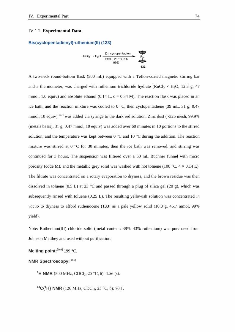
IV. Experimental Part 74
Experimental Data IV.1.2.
Bis(cyclopentadienyl)ruthenium(II) (133)
A two-neck round-bottom flask (500 mL) equipped with a Teflon-coated magnetic stirring bar
and a thermometer, was charged with ruthenium trichloride hydrate (RuCl3 × H2O, 12.3 g, 47
mmol, 1.0 equiv) and absolute ethanol (0.14 L, c = 0.34 M). The reaction flask was placed in an
ice bath, and the reaction mixture was cooled to 0 °C, then cyclopentadiene (39 mL, 31 g, 0.47
mmol, 10 equiv)[167] was added via syringe to the dark red solution. Zinc dust (~325 mesh, 99.9%
(metals basis), 31 g, 0.47 mmol, 10 equiv) was added over 60 minutes in 10 portions to the stirred
solution, and the temperature was kept between 0 °C and 10 °C during the addition. The reaction
mixture was stirred at 0 °C for 30 minutes, then the ice bath was removed, and stirring was
continued for 3 hours. The suspension was filtered over a 60 mL Büchner funnel with micro
porosity (code M), and the metallic grey solid was washed with hot toluene (100 °C, 4 × 0.14 L).
The filtrate was concentrated on a rotary evaporation to dryness, and the brown residue was then
dissolved in toluene (0.5 L) at 23 °C and passed through a plug of silica gel (20 g), which was
subsequently rinsed with toluene (0.25 L). The resulting yellowish solution was concentrated in
vacuo to dryness to afford ruthenocene (133) as a pale yellow solid (10.8 g, 46.7 mmol, 99%
yield).
Note: Ruthenium(III) chloride solid (metal content: 38%–43% ruthenium) was purchased from
Johnson Matthey and used without purification.
Melting point:[168] 199 °C.
NMR Spectroscopy:[169]
1H NMR (500 MHz, CDCl3, 25 °C, δ): 4.56 (s).
13C{1H} NMR (126 MHz, CDCl3, 25 °C, δ): 70.1.
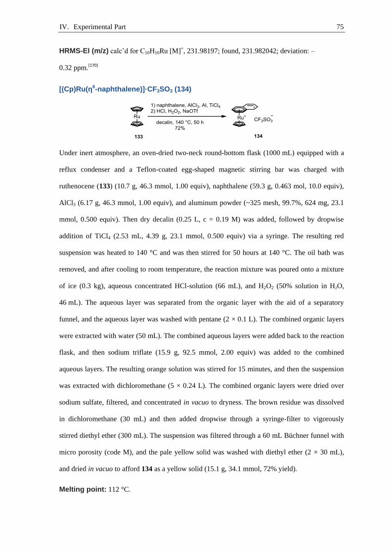
IV. Experimental Part 75
HRMS-EI (m/z) calc’d for C10H10Ru [M]+, 231.98197; found, 231.982042; deviation: –
0.32 ppm.[170]
[(Cp)Ru(η6-naphthalene)]·CF3SO3 (134)
Under inert atmosphere, an oven-dried two-neck round-bottom flask (1000 mL) equipped with a
reflux condenser and a Teflon-coated egg-shaped magnetic stirring bar was charged with
ruthenocene (133) (10.7 g, 46.3 mmol, 1.00 equiv), naphthalene (59.3 g, 0.463 mol, 10.0 equiv),
AlCl3 (6.17 g, 46.3 mmol, 1.00 equiv), and aluminum powder (~325 mesh, 99.7%, 624 mg, 23.1
mmol, 0.500 equiv). Then dry decalin (0.25 L, c = 0.19 M) was added, followed by dropwise
addition of TiCl4 (2.53 mL, 4.39 g, 23.1 mmol, 0.500 equiv) via a syringe. The resulting red
suspension was heated to 140 °C and was then stirred for 50 hours at 140 °C. The oil bath was
removed, and after cooling to room temperature, the reaction mixture was poured onto a mixture
of ice (0.3 kg), aqueous concentrated HCl-solution (66 mL), and H2O2 (50% solution in H2O,
46 mL). The aqueous layer was separated from the organic layer with the aid of a separatory
funnel, and the aqueous layer was washed with pentane (2 × 0.1 L). The combined organic layers
were extracted with water (50 mL). The combined aqueous layers were added back to the reaction
flask, and then sodium triflate (15.9 g, 92.5 mmol, 2.00 equiv) was added to the combined
aqueous layers. The resulting orange solution was stirred for 15 minutes, and then the suspension
was extracted with dichloromethane (5 × 0.24 L). The combined organic layers were dried over
sodium sulfate, filtered, and concentrated in vacuo to dryness. The brown residue was dissolved
in dichloromethane (30 mL) and then added dropwise through a syringe-filter to vigorously
stirred diethyl ether (300 mL). The suspension was filtered through a 60 mL Büchner funnel with
micro porosity (code M), and the pale yellow solid was washed with diethyl ether (2 × 30 mL),
and dried in vacuo to afford 134 as a yellow solid (15.1 g, 34.1 mmol, 72% yield).
Melting point: 112 °C.

IV. Experimental Part 76
NMR Spectroscopy:[171]
1H NMR (500 MHz, CDCl3, 25 ºC, δ): 7.76 (dd, J = 6.7, 3.2 Hz, 2H), 7.59 (dd, J = 6.8, 3.1
Hz, 2H), 7.19–7.06 (m, 2H), 6.63–6.28 (m, 2H), 5.06 (s, 5H).
13C{1H} NMR (126 MHz, CDCl3, 25 ºC, δ): 131.6, 129.5, 97.2, 86.5, 84.1, 80.2.
19F NMR (471 MHz, CDCl3, 25 ºC, δ): −78.1.
HRMS-EI (m/z) calc’d for C15H13Ru [M−OTf]+, 295.00553; found, 295.00563; deviation:
0.35 ppm.
Elemental Analysis calc’d for C16H13F3O3RuS: C, 43.34; H, 2.96; found: C, 43.31; H, 2.94.
UV/vis Spectroscopy (H2O, 23°C): 360 nm (ε = 673 M−1·cm−1).
[18F]Atorvastatin precursor 1,4-diketone 121
A 50 mL one-neck round-bottom flask equipped with a Teflon-coated magnetic stirring bar and a
reflux condenser was charged with 3-ethyl-5-(2-hydroxyethyl)-4-methylthiazolium bromide(125)
(0.860 g, 3.41 mmol, 0.200 equiv), 2-benzyliden-N-phenyl-isobutyloyl-acetamid (123) (5.00 g,
17.0 mmol, 1.00 equiv), 4-benzyloxybenzaldehyde (124) (3.98 g, 18.8 mmol, 1.10 equiv),
triethylamine (2.61 mL, 1.90 g, 18.8 mmol, 1.10 equiv), and absolute ethanol (10 mL, 1.7 mol·L–
1). The reaction mixture was heated at reflux. After 15 hours a second portion of 3-ethyl-5-(2-
hydroxyethyl)-4-methylthiazolium bromide (125) (0.860 g, 3.41 mmol, 0.200 equiv) was added,
and the reaction mixture was stirred and heated at reflux for additional 7 hours. Isopropanol (35
mL) was added to the reaction mixture at 78°C, and the oil bath was removed. After 12 hours the

IV. Experimental Part 77
solid was collected by filtration and dried in vacuo to afford 121 as colorless solid (1.38 g, 2.72
mmol, 16%).
Rf = 0.44 (EtOAc:Hexane, 3:7, v:v).
NMR Spectroscopy:
1H NMR of major stereoisomer (500 MHz, DMSO-d6, 25 °C, δ): 10.15 (s, 1H), 8.05–7.97
(m, 2H), 7.45–7.39 (m, 2H), 7.41–7.37 (m, 2H), 7.38–7.32 (m, 3H), 7.32–7.27 (m, 2H),
7.26–7.17 (m, 4H), 7.17–7.08 (m, 1H), 7.08–7.04 (m, 2H), 7.05–6.96 (m, 1H), 5.40 (d, J =
11.0 Hz, 1H), 5.17 (s, 2H), 4.85 (d, J = 10.9 Hz, 1H), 2.90 (hept, J = 6.9 Hz, 1H), 1.16 (d, J =
7.0 Hz, 3H), 0.94 (d, J = 6.6 Hz, 3H).
13C{1H}NMR of major stereoisomer (126 MHz, DMSO-d6, 25 °C, δ): 207.9, 196.1, 165.1,
162.2, 138.1, 136.4, 135.8, 131.1, 128.8, 128.6, 128.5, 128.0, 127.8, 127.4, 123.9, 119.6,
114.7, 69.5, 62.9, 51.6, 18.8, 18.0.
HRMS-EI (m/z) calc’d for C33H31N1O4Na [M+Na]+, 528.21453; found, 528.21503; deviation: –
0.96 ppm.
[18F]Atorvastatin precursor pyrrole 126
A 50 mL one-neck round-bottom flask equipped with a Teflon-coated magnetic stirring bar and a
reflux condenser was charged with diketone 121 (1.27 g, 2.51 mmol, 1.00 equiv), amino ester 122
(0.891 g, 3.26 mmol, 1.30 equiv), pivalic acid (0.576 mL, 5.01 mmol, 2.00 equiv), and a mixture
of THF:toluene:heptane (1:1:4, 42 mL, 0.06 mol·L–1). The reaction mixture stirred at 130 °C oil
bath temperature for 72 hours. The solvent was removed and the residue was purified by silica
flash chromatography using EtOAc and hexane [1:3]. Pyrrole 126 was isolated as white solid
(1.17 g, 2.51 mmol, 63%).
IV. Experimental Part 78
Rf = 0.58 (EtOAc:Hexane, 3:7, v:v).
NMR Spectroscopy:
1H NMR (500 MHz, CDCl3, 25 °C, δ): 7.46–7.42 (m, 2H), 7.42–7.37 (m, 2H), 7.36–7.32 (m,
1H), 7.23–7.15 (m, 7H), 7.13 (d, J = 8.5 Hz, 2H), 7.09 (d, J = 8.5 Hz, 2H), 6.98 (t, J = 7.3
Hz, 1H), 6.92 (d, J = 8.2 Hz, 2H), 6.89 (s, 1H), 5.04 (s, 2H), 4.17 (dtd, J = 9.0, 6.5, 2.2 Hz,
1H), 4.09 (ddd, J = 15.2, 10.5, 5.1 Hz, 1H), 3.90–3.81 (m, 1H), 3.69 (td, J = 8.4, 7.8, 1H),
3.59 (p, J = 7.1, 1H), 2.39 (dd, J = 15.9, 7.1 Hz, 1H), 2.25 (dd, J = 15.2, 6.0 Hz, 1H), 1.79–
1.62 (m, 2H), 1.60–1.56 (m, 1H) 1.55 (dd, J = 7.1, 2.2 Hz, 6H), 1.44 (s, 9H), 1.37 (s, 3H),
1.35–1.33 (m, 1H) 1.32 (s, 3H), 1.06 (q, J = 11.9 Hz, 1H).
13C{1H} NMR (126 MHz, CDCl3, 25 °C, δ): 170.3, 165.1, 158.4, 141.3, 138.6, 136.8, 135.1,
132.8, 130.6, 129.8, 128.71, 128.65, 128.3, 128.1, 127.6, 126.4, 124.8, 123.5, 121.5, 119.6,
115.2, 114.6, 98.7, 80.7, 70.0, 66.6, 66.0, 42.6, 40.9, 38.2, 36.1, 30.0, 28.2, 26.2, 21.9, 21.7,
19.8.
HRMS-ESI (m/z) calc’d for C47H54N2O6Na [M+Na]+, 765.38741; found, 765.38799; deviation:
−0.76 ppm.
Hydroxy-defluoro-Atorvastatin derivative 127
A round-bottom flask (100 mL) equipped with a septum and a magnetic stirring bar was charged
with pyrrole 126 (765 mg, 1.03 mmol, 1.00 equiv), palladium on carbon (10 w%, 110 mg, 103
µmol, 0.100 equiv), and methanol (50 mL, 0.02 mol·L–1). Argon was bubbled through the
reaction mixture for 5 minutes while stirring. Afterwards, dihydrogen was bubbled through the
reaction mixture for 10 minutes while stirring. The reaction mixture was stirred under dihydrogen
atmosphere at 23 °C for 24 hours. Argon was bubbled through the reaction mixture for 5 min

IV. Experimental Part 79
while stirring. The reaction mixture was filtered through a Celite plug, and the Celite plug was
washed with methanol (2 x 5 mL). The filtrate was concentrated under vacuum. A round-bottom
flask (10 mL) equipped with a septum and a magnetic stirring bar was charged with pale yellow
filtrate, p-toluenesulfonic acid (1.96 mg, 10.3 µmol, 1.00 mol%), 2,2-dimethoxypropan (190 µL,
161 mg, 1.54 mmol, 1.50 equiv), and acetone (2 mL). The reaction mixture was stirred at 23 °C
for 2 hours. The resulting suspension was diluted with DCM (20 mL), and the solution was
washed with saturated aqueous sodium bicarbonate solution (10 mL) and saturated aqueous
sodium chloride solution. The organic layer was dried over sodium sulfate, filtered, and
concentrated in vacuo to dryness to afford 127 as a pale yellow powder (653 mg, 1.00 mmol, 97%
yield).
Rf = 0.33 (EtOAc:Hexane, 3:7, v:v).
NMR Spectroscopy:
1H NMR (500 MHz, CDCl3, 25 ºC, δ): 7.21–7.10 (m, 7H), 7.06 (d, J = 7.9, 2H), 7.03 (d, J =
8.4, 2H), 6.98 (t, J = 7.3 Hz, 1H), 6.91 (s, 1H), 6.77 (d, J = 8.3, 2H), 6.43 (br, 1H), 4.17–4.10
(m, 1H), 4.05 (ddd, J = 15.1, 10.3, 5.3 Hz, 1H), 3.82 (ddd, J = 14.9, 10.0, 5.6 Hz, 1H), 3.66
(tt, J = 11.1, 5.3, 1H), 3.57 (p, J = 7.1, 1H), 2.37 (dd, J = 15.1, 7.2 Hz, 1H), 2.23 (dd, J =
15.1, 5.8 Hz, 1H), 1.72–1.62 (m, 2H), 1.51 (dd, J = 7.1, 4.9 Hz, 6H), 1.44 (s, 9H), 1.35 (s,
3H), 1.31 (s, 3H), 1.29–1.22 (m, 1H) 1.00 (q, J = 11.9 Hz, 1H).
13C{1H} NMR (126 MHz, CDCl3, 25 ºC, δ): 170.7, 165.5, 156.0, 141.2, 138.4, 135.0, 132.9,
130.6, 130.0 ,128.8, 128.4, 126.4, 124.1, 123.8, 121.4, 119.9, 115.6, 115.1, 98.8, 81.1, 66.1,
66.3, 42.7, 40.8, 38.1, 36.0, 30.0, 28.2, 26.2, 21.9, 21.8, 19.8.
HRMS-ESI (m/z) calc’d for C40H48N2O6Na [M+Na]+,675.34046; found, 675.34108; deviation:
−0.92 ppm.
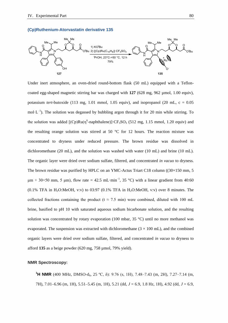
IV. Experimental Part 80
(Cp)Ruthenium-Atorvastatin derivative 135
Under inert atmosphere, an oven-dried round-bottom flask (50 mL) equipped with a Teflon-
coated egg-shaped magnetic stirring bar was charged with 127 (628 mg, 962 µmol, 1.00 equiv),
potassium tert-butoxide (113 mg, 1.01 mmol, 1.05 equiv), and isopropanol (20 mL, c = 0.05
mol·L–1). The solution was degassed by bubbling argon through it for 20 min while stirring. To
the solution was added [(Cp)Ru(η6-naphthalene)]·CF3SO3 (512 mg, 1.15 mmol, 1.20 equiv) and
the resulting orange solution was stirred at 50 °C for 12 hours. The reaction mixture was
concentrated to dryness under reduced pressure. The brown residue was dissolved in
dichloromethane (20 mL), and the solution was washed with water (10 mL) and brine (10 mL).
The organic layer were dried over sodium sulfate, filtered, and concentrated in vacuo to dryness.
The brown residue was purified by HPLC on an YMC-Actus Triart C18 column ((30×150 mm, 5
μm + 30×50 mm, 5 μm), flow rate = 42.5 mL·min–1, 35 °C) with a linear gradient from 40:60
(0.1% TFA in H2O:MeOH, v:v) to 03:97 (0.1% TFA in H2O:MeOH, v:v) over 8 minutes. The
collected fractions containing the product (t ≈ 7.5 min) were combined, diluted with 100 mL
brine, basified to pH 10 with saturated aqueous sodium bicarbonate solution, and the resulting
solution was concentrated by rotary evaporation (100 mbar, 35 °C) until no more methanol was
evaporated. The suspension was extracted with dichloromethane (3 × 100 mL), and the combined
organic layers were dried over sodium sulfate, filtered, and concentrated in vacuo to dryness to
afford 135 as a beige powder (620 mg, 758 µmol, 79% yield).
NMR Spectroscopy:
1H NMR (400 MHz, DMSO-d6, 25 ºC, δ): 9.76 (s, 1H), 7.48–7.43 (m, 2H), 7.27–7.14 (m,
7H), 7.01–6.96 (m, 1H), 5.51–5.45 (m, 1H), 5.21 (dd, J = 6.9, 1.8 Hz, 1H), 4.92 (dd, J = 6.9,

IV. Experimental Part 81
2.9 Hz, 1H), 4.90 (s, 5H), 4.79 (dd, J = 6.8, 2.0 Hz, 1H), 4.49–4.39 (m, 1H), 4.32–4.23 (m,
1H), 4.16–4.02 (m, 2H), 3.18 (p, J = 7.0 Hz, 1H), 2.43 (dd, J = 15.2, 4.8 Hz, 1H), 2.27 (dd, J
= 15.2, 8.1 Hz, 1H), 2.0–1.9 (m, 1H), 1.82–1.71 (m, 1H), 1.64 (dt, J = 12.7, 2.5 Hz, 1H), 1.49
(s, 3H), 1.42 (s, 9H), 1.40–1.32 (m, 9H), 1.27–1.18 (m, 1H).
13C{1H} NMR (101 MHz, DMSO-d6, 25 ºC, δ): 169.6, 165.6, 155.9, 139.3, 137.6, 134.8,
130.1, 128.4, 127.9, 126.2, 123.9, 123.0, 121.2, 119.4, 118.6, 98.2, 87.2, 86.1, 85.3, 79.8,
76.7, 71.8, 71.7, 66.4, 65.8, 42.1, 38.7, 35.4, 30.0, 27.8, 25.5, 22.8, 21.7, 20.0.
HRMS-EI (m/z) calc’d for C45H53N2O6Ru [M−OTf]+, 819.29416; found, 819.29557; deviation:
−1.72 ppm.
Radiochemistry general methods IV.1.3.
No-carrier-added [18F]fluoride was purchased from Advanced Accelerator Applications SA.
Liquid chromatographic (LC) analysis was performed with Thermo Scientific Dionex UltiMate
3000 dual channel HPLC system connected to LabLogic NaI/PMT-radiodetectors with Flow-Ram
output. A Thermo Scientific™ Accucore™ XL C18, 4 μm, 3×150 mm HPLC column was used
for analysis and a Thermo Scientific Hypersil Gold column™, 5 μm, 10×250 mm HPLC column
was used for preparative HPLC. Analytical and preparative HPLC used the following mobile
phases: 0.1% CF3CO2H in water (A), 0.1% CF3CO2H in acetonitrile (B).
Gradient 1
time [min] A [%] B [%]
0 95 5
2 95 5
22 50 50
22.5 5 95
27 5 95
28 95 5
32 95 5
Gradient 2
time [min] A [%] B [%]
0 95 5
2 95 5
10 5 95
14 5 95
15 95 5
18 95 5

IV. Experimental Part 82
All 18F-labeled molecules were characterized by comparing the HPLC radio-trace of the isolated
compound to the HPLC UV-trace of an authentic reference sample. Radioactivity was measured
in a Veenstra Instruments, VIK-203 ionization-chamber.
Note: radioactivity chromatographs are offset by 0.1 or 0.3 minutes on account of the delay
introduced by the spatial separation between the diode array detectors and the radioactivity
detectors.
General procedure for pre-conditioning of Sep-Pak cartridges
Waters Sep-Pak light C18 cartridge (part # WAT023501) was pre-conditioned by sequentially
pushing MeOH (2 mL) and water (10 mL) through the cartridge unless otherwise indicated.
General procedure for pre-conditioning of QMA cartridges
Chromafix PS-HCO3 18F separation cartridge (45 mg) (Product No. 731876 from ABX) was pre-
conditioned by sequentially pushing potassium oxalate solution (3 mL, 10 mg·mL−1 H2O) and
H2O (2 mL) through the cartridge at a flow rate of 5 mL·min-1 unless otherwise indicated.
[18F]Atorvastatin derivative 128
Aqueous 18F-fluoride solution (700 μL) was loaded with a syringe onto a QMA anion-exchange
cartridge that was pre-conditioned according to the general procedure, and then the cartridge was
washed with MeCN (1 mL). The cartridge was dried by pushing air (2 mL) through the cartridge.
The [18F]fluoride was eluted from the cartridge with a solution of ruthenium complex 135 (1.6
mg, 2.0 μmol, 1.0 equiv) and imidazolium chloride 40 (2.8 mg, 6.0 µmol, 3.0 equiv) in methanol
(300 µL), into a 4 mL borosilicate vial (95% elution efficiency). The methanol was removed by
heating at 80 °C under a stream of nitrogen (~5 min). To the vial was added a mixture of veratrole
and pivalonitrile (400 µL, 1:1, v:v). After sealing of the vial with a Teflon-lined cap it was stirred

IV. Experimental Part 83
at 130 °C for 30 minutes. The vial was removed from the hot plate, diluted with methanol (1 mL)
and the resulting solution was analyzed by radio-HPLC and radio-TLC. The product was obtained
in 75% RCY.
Figure S1. Crude radio-HPLC trace (gradient 1) of [18F]atorvastatin derivative 128.
Figure S2. UV-HPLC trace (gradient 1) of acetal ester protected atorvastatin [19F]128.
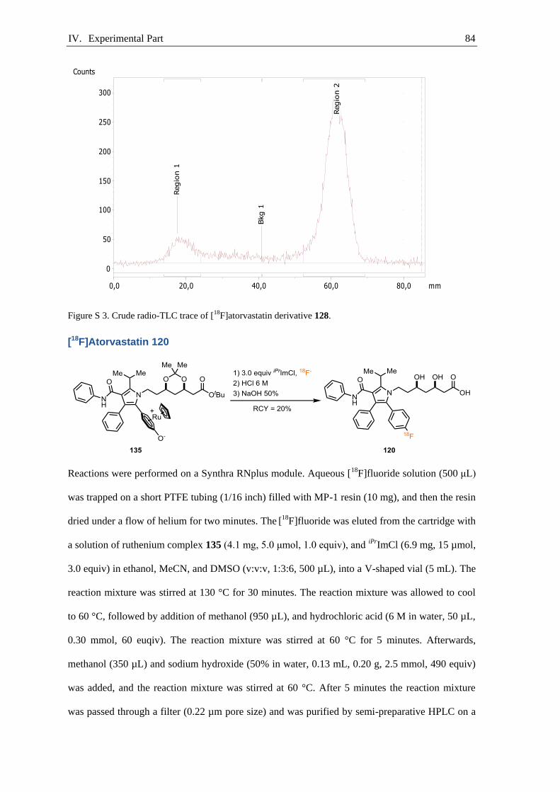
IV. Experimental Part 84
Figure S 3. Crude radio-TLC trace of [18F]atorvastatin derivative 128.
[18F]Atorvastatin 120
Reactions were performed on a Synthra RNplus module. Aqueous [18F]fluoride solution (500 μL)
was trapped on a short PTFE tubing (1/16 inch) filled with MP-1 resin (10 mg), and then the resin
dried under a flow of helium for two minutes. The [18F]fluoride was eluted from the cartridge with
a solution of ruthenium complex 135 (4.1 mg, 5.0 μmol, 1.0 equiv), and iPrImCl (6.9 mg, 15 µmol,
3.0 equiv) in ethanol, MeCN, and DMSO (v:v:v, 1:3:6, 500 µL), into a V-shaped vial (5 mL). The
reaction mixture was stirred at 130 °C for 30 minutes. The reaction mixture was allowed to cool
to 60 °C, followed by addition of methanol (950 µL), and hydrochloric acid (6 M in water, 50 µL,
0.30 mmol, 60 euqiv). The reaction mixture was stirred at 60 °C for 5 minutes. Afterwards,
methanol (350 µL) and sodium hydroxide (50% in water, 0.13 mL, 0.20 g, 2.5 mmol, 490 equiv)
was added, and the reaction mixture was stirred at 60 °C. After 5 minutes the reaction mixture
was passed through a filter (0.22 µm pore size) and was purified by semi-preparative HPLC on a
Regio
n 1
Bkg 1
Regio
n 2
0,0 20,0 40,0 60,0 80,0 mm
0
50
100
150
200
250
300
Counts
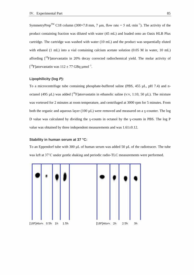
IV. Experimental Part 85
SymmetryPrepTM C18 column (300×7.8 mm, 7 μm, flow rate = 5 mL·min−1). The activity of the
product containing fraction was diluted with water (45 mL) and loaded onto an Oasis HLB Plus
cartridge. The cartridge was washed with water (10 mL) and the product was sequentially eluted
with ethanol (1 mL) into a vial containing calcium acetate solution (0.05 M in water, 10 mL)
affording [18F]atorvastatin in 20% decay corrected radiochemical yield. The molar activity of
[18F]atorvastatin was 112 ± 77 GBq µmol–1.
Lipophilicity (log P):
To a microcentrifuge tube containing phosphate-buffered saline (PBS, 455 µL, pH 7.4) and n-
octanol (495 µL) was added [18F]atorvastatin in ethanolic saline (v:v, 1:10, 50 µL). The mixture
was vortexed for 2 minutes at room temperature, and centrifuged at 3000 rpm for 5 minutes. From
both the organic and aqueous layer (100 µL) were removed and measured on a ɣ-counter. The log
D value was calculated by dividing the ɣ-counts in octanol by the ɣ-counts in PBS. The log P
value was obtained by three independent measurements and was 1.61±0.12.
Stability in human serum at 37 °C:
To an Eppendorf tube with 300 µL of human serum was added 50 µL of the radiotracer. The tube
was left at 37˚C under gentle shaking and periodic radio-TLC measurements were performed.

IV. Experimental Part 86
Rat serum plasma protein binding:
To a microcentrifuge tube containing rat plasma (300 µL, normal or fat diet) was added
[18F]atorvastatin in ethanolic saline (25 µL). The mixture was incubated for 1 hour and afterwards
acetonitrile (900 µL) was added to precipitate the plasma protein. The suspension was centrifuged
at 3000 rpm for 5 minutes. The precipitate (proteins) was separated from the supernatant and was
measured on a ɣ-counter. Plasma proteins binding for normal rats was 16.7% and for fat rats
13.8%.
Liver homogenate binding assay
Tris-HCl (0.05 M, pH 7.4) was added to the extracted rat liver (normal and fat rats, fed with high-
calorie food) to obtain a final concentration of 20 mg liver tissue per mL. The mixture was
homogenized, using a Heidolph DIAX 600 homogenizer, for 30 seconds while being cooled in an
ice bath. The rat liver homogenate was stored at –80°C.
Rat liver homogenate (300 µL or Tris-HCl solution for non-specific binding) was added to each
assay tube. Tris-HCl solution (0.05M, pH 7.4) containing 0.3% human serum albumin was added
to reach a final volume of 475 µL (175 µL) and left for pre-incubation for 15 minutes at room
temperature. Then, 25 µL of [18F]atorvastatin in ethanolic saline was added to each assay tube (8
x control, 12 x fat liver, 6 x non-specific binding). Assay tubes were briefly vortexed and then
incubated for 60 minutes under gentle shaking at 37 ºC.
Incubation was terminated by centrifuging the assay tubes at 4°C for 15 min (3500 RPM). The
supernatant of each tube was taken leaving the pellet in the original tube. Supernatants and pellets
were measured on a ɣ-counter.
% of tracer in cell debris (pellet) of control rats: 5.2%
% of tracer in S9 fraction (supernatant) of control rats: 94.8%
% in cell debris (pellet) of fat diet rats: 9.2%
% in S9 fraction (supernatant) of fat diet rats: 90.8%

IV. Experimental Part 87
IV.2. Peptide Labeling
Experimental Data IV.2.1.
[(Cp)Ru(η6-naphthalene)]·BF4 (141)
Under inert atmosphere, an oven-dried two-neck round-bottom flask (500 mL) equipped with a
reflux condenser and a Teflon-coated egg-shaped magnetic stirring bar was charged with
ruthenocene (133) (4.52 g, 19.5 mmol, 1.00 equiv), naphthalene (25.0 g, 195 mmol, 10.0 equiv),
AlCl3 (2.61 g, 19.5 mmol, 1.00 equiv), and aluminum powder (~325 mesh, 99.7%, 264 mg, 9.77
mmol, 0.500 equiv). Then dry decalin (0.13 L, c = 0.15 M) was added, followed by dropwise
addition of TiCl4 (1.07 mL, 1.85 g, 9.77 mmol, 0.500 equiv) via a syringe. The resulting red
suspension was heated to 140 °C and was then stirred for 50 hours at 140 °C. The oil bath was
removed, and after cooling to room temperature, the reaction mixture was poured onto a mixture
of ice (0.15 kg), aqueous concentrated HCl-solution (28 mL), and H2O2 (35% solution in H2O, 28
mL). The aqueous layer was separated from the organic layer with the aid of a separatory funnel,
and the aqueous layer was washed with pentane (2 × 0.1 L). The combined organic layers were
extracted with water (50 mL). The combined aqueous layers were added back to the reaction
flask, and then fluoroboric acid (48% solution in water, 5.1 mL, 7.2 g, 39 mmol, 2.0 equiv) was
added to the combined aqueous layers. The resulting orange solution was stirred for 15 minutes,
and then the suspension was extracted with dichloromethane (4 × 100 mL). The combined organic
layers were dried over sodium sulfate, filtered, and concentrated in vacuo to dryness. The brown
residue was dissolved in dichloromethane (15 mL) and was then added dropwise through a
syringe-filter to vigorously stirred diethyl ether (150 mL). The suspension was filtered through a
60 mL Büchner funnel with micro porosity (code M), and the pale yellow solid was washed with
diethyl ether (2 × 30 mL), and dried in vacuo to afford 141 as a yellow solid (4.50 g, 11.8 mmol,
60% yield).

IV. Experimental Part 88
NMR Spectroscopy:[142]
1H NMR (500 MHz, CDCl3, 25 ºC, δ): 7.75 (dd, J = 6.7, 3.3 Hz, 2H), 7.59 (dd, J = 6.8, 3.1
Hz, 2H), 7.08–7.02 (m, 2H), 6.44–6.37 (m, 2H), 5.05 (s, 5H).
13C{1H} NMR (126 MHz, CDCl3, 25 ºC, δ): 131.5, 129.5, 97.2, 86.4, 84.0, 80.2.
19F NMR (471 MHz, CDCl3, 25 ºC, δ): −152.3.
HRMS-ESI (m/z) calc’d for C15H13Ru [M−BF4]+, 295.00553; found, 295.00578; deviation:
−0.86 ppm.[157]
[Fmoc-tyrosine(RuCp)-O]·H2O (140)
A round-bottom flask (250 mL) equipped with a Teflon-coated magnetic stirring bar was charged
with [(Cp)Ru(η6-naphthalene)]·BF4 (131) (4.61 g, 12.1 mmol, 1.10 equiv), L-tyrosine (1.99 g,
11.0 mmol, 1.00 equiv), water (0.11 L, c = 0.10 M), and fluoroboric acid (48% in water, 2.9 mL,
4.0 g, 22 mmol, 2.0 equiv). The yellow suspension was irradiated for 36 h with blue LED light
(Kessil A 160WE Tuna Blue, 40 W). It is crucial that the tyrosine is fully consumed at this point;
failure will result in tyrosine contamination, which challenges the purification procedure, and
column chromatography must be used instead. If required, the reaction time needs to be adjusted
and more ruthenium precursor needs to be added. The resulting beige suspension was basified
with sodium carbonate (3.85 g, 36.3 mmol, 3.30 equiv). The suspension was cooled to 0 °C with
an ice bath, and then a solution of Fmoc-OSu (4.45 g, 13.2 mmol, 1.20 equiv) in dioxane (55 mL)
was added. The reaction mixture was stirred at 0 °C for 1 hour, then at 23 °C for 14 hours. The
solution was concentrated by rotary evaporation to two-thirds of the original volume, and then the
aqueous layer was washed with dichloromethane (2 × 30 mL). The aqueous layer was acidified
with HCl (4 M solution in H2O) to pH 4, and was then extracted with DCM (3 × 0.1 L). The
combined organic layers were dried over sodium sulfate, filtered, and concentrated in vacuo to

IV. Experimental Part 89
dryness. The brown residue was dissolved in dichloromethane (15 mL) and filtered through a 15
mL Büchner funnel with fine porosity (code f) into vigorously stirred acetonitrile (150 mL). The
suspension was cooled to 0 °C and filtered over a 60 mL Büchner funnel with micro porosity
(code M), and the beige solid was washed with acetonitrile (2 × 10 mL), and dried in vacuo to
dryness to afford 140 as a beige solid (4.83 g, 8.24 mmol, 75% yield).
Rf = 0.36 (DCM:MeOH:TFA, 92:8:1, v:v:v).
NMR Spectroscopy:
1H NMR (500 MHz, CD3OD, 60 ºC, δ): 7.78 (d, J = 7.5, 2H), 7.62 (t, J = 6.5, 2H), 7.38 (t, J
= 7.4, 2H), 7.30 (t, J = 7.4 Hz, 2H), 5.83 (br, 1H), 5.74–5.64 (m, 3H), 5.14 (s, 5H), 4.47 (br,
1H), 4.36 (br, 1H), 4.27–4.14 (m, 2H) 2.88 (br, 1H), 2.68 (br, 1H).
13C{1H} NMR (126 MHz, CD3OD, 25 ºC, δ): 175.5, 158.0, 145.3, 145.1, 142.7, 142.6, 141.2,
128.82, 128.81, 128.19, 128.17, 126.2, 126.1, 120.97, 120.95, 97.7, 86.9, 86.8, 80.2, 75.9, 75.8,
67.5, 57.8, 37.9. 2 Rotamers of the Fmoc-protecting group are observed at 25 °C and one
carbon resonance is not observed, presumably due to overlap with a solvent carbon
resonance.
HRMS-ESI (m/z) calc’d for C29H26NO5Ru [M−OH]+,570.08490; found, 570.08502; deviation:
0.21 ppm.
Elemental Analysis calc’d for C29H27NO6Ru: C, 59.38; H, 4.64; found: C, 59.06; H, 4.31.
[Fmoc-tyrosine(RuCp)-OH]·CF3CO2 (156)
A round-bottom flask (2 L) equipped with a Teflon-coated magnetic stirring bar was charged with
[(Cp)Ru(η6-naphthalene)]·CF3SO3 (134) (6.65 g, 15.0 mmol, 1.00 equiv), L-tyrosine (3.40 g, 18.8
mmol, 1.25 equiv), water (0.75 L, c = 0.02 M), and trifluoroacetic acid (2.87 mL, 4.27 g, 37.5
mmol, 2.50 equiv). The yellow suspension was irradiated for 24 h with blue LED light (Kessil A
IV. Experimental Part 90
160WE Tuna Blue, 40 W), then the resulting beige suspension was extracted with hexane (2 ×
200 mL), and the combined organic layers were extracted with water (50 mL). The combined
aqueous layers were basified with Na2CO3 (7.95 g, 75.0 mmol, 5.00 equiv) and dioxane (0.25 L)
was added. The suspension was cooled to 0 °C with an ice bath, and then a solution of Fmoc-OSu
(7.59 g, 22.5 mmol, 1.50 equiv) in dioxane (0.25 L) was added. The reaction mixture was stirred
at 0 °C for 1 hour, then at 23 °C for 14 hours. The solution was concentrated by rotary
evaporation to half the original volume, and then the aqueous layer was washed with
dichloromethane (3 × 0.2 L). The aqueous layer was acidified with trifluoroacetic acid to pH 3
and then extracted with dichloromethane (4 × 0.3 L). The combined organic layers were dried
over sodium sulfate, filtered, and concentrated in vacuo to dryness. The residual brown solid was
purified by column chromatography (EtOAc:MeCN:TFA, 90:9:0.1, v:v:v) to afford 156 as a
yellow powder (9.15 g, 13.4 mmol, 89% yield).
Rf = 0.26 (EtOAc:MeCN:TFA, 9:1:0.1, v:v:v).
Rf = 0.56 (EtOAc:MeCN:TFA, 8:2:0.1, v:v:v).
NMR Spectroscopy:
1H NMR (500 MHz, CD3CN, 25 ºC, δ): 7.85 (dt, J = 7.5, 0.9 Hz, 2H), 7.70–7.60 (m, 2H),
7.47–7.40 (m, 2H), 7.35 (td, J = 7.5, 1.1 Hz, 2H), 6.08 (d, J = 8.7 Hz, 1H), 6.07–6.05 (m,
1H), 6.02 (dd, J = 6.2, 1.8 Hz, 1H), 5.83 (d, J = 16.8, 6.2 Hz, 1H), 5.21 (s, 5H), 4.38 (m, 3H),
4.21 (t, J = 6.5 Hz, 1H), 2.91 (dd, J = 13.8, 4.6 Hz, 1H), 2.70 (dd, J = 14.2, 8.9 Hz, 1H).
13C{1H} NMR (126 MHz, CD3CN, 25 ºC, δ): 172.3, 156.9, 145.0, 144.9, 142.2, 134.1, 128.8,
128.1, 126.1, 121.1, 98.3, 86.5, 86.4, 75.9, 75.7, 67.2, 55.7, 48.0, 36.3.
19F NMR (471 MHz, CD3CN, 25 ºC, δ): −77.3.
HRMS-ESI (m/z) calc’d for C29H26NO5Ru [M−CF3CO2]+,570.08490; found, 570.08505;
deviation: 0.26 ppm.
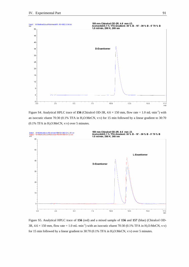
IV. Experimental Part 91
Figure S4. Analytical HPLC trace of 156 (Chiralcel OD-3R, 4.6 × 150 mm, flow rate = 1.0 mL·min-1) with
an isocratic eluent 70:30 (0.1% TFA in H2O:MeCN, v:v) for 15 min followed by a linear gradient to 30:70
(0.1% TFA in H2O:MeCN, v:v) over 5 minutes.
Figure S5. Analytical HPLC trace of 156 (red) and a mixed sample of 156 and 157 (blue) (Chiralcel OD-
3R, 4.6 × 150 mm, flow rate = 1.0 mL·min-1) with an isocratic eluent 70:30 (0.1% TFA in H2O:MeCN, v:v)
for 15 min followed by a linear gradient to 30:70 (0.1% TFA in H2O:MeCN, v:v) over 5 minutes.

IV. Experimental Part 92
[Fmoc-D-tyrosine(RuCp)-OH]·CF3CO2 (157)
A round-bottom flask (1 L) equipped with a Teflon-coated magnetic stirring bar was charged with
[(Cp)Ru(η6-naphthalene)]·CF3SO3 (134) (3.00 g, 6.77 mmol, 1.00 equiv), D-tyrosine (1.35 g, 7.44
mmol, 1.10 equiv), water (0.27 L, c = 0.025 M), and hydrochloric acid (37% in water, 1.1 mL, 1.3
g, 7.4 mmol, 2.0 equiv). The yellow suspension was irradiated for 15 h with blue LED light
(Kessil A 160WE Tuna Blue, 40 W), then the resulting beige suspension was extracted with
hexane (2 × 120 mL), and the combined organic layers were extracted with water (50 mL). The
combined aqueous layers were basified with Na2CO3 (2.15 g, 20.3 mmol, 3.00 equiv) and dioxane
(0.12 L) was added. The suspension was cooled to 0 °C with an ice bath and then a solution of
Fmoc-OSu (2.74 g, 8.12 mmol, 1.20 equiv) in dioxane (0.12 L) was added. The reaction mixture
was stirred at 0 °C for 1 hour, then at 23 °C for 14 hours. The solution was concentrated by rotary
evaporation to half the original volume, and then the aqueous layer was washed with
dichloromethane (3 × 0.12 L). The aqueous layer was acidified with trifluoroacetic acid to pH 3
and then extracted with dichloromethane (4 × 0.12 L). The combined organic layers were dried
over sodium sulfate, filtered, and concentrated in vacuo to dryness. The residual beige solid was
purified by column chromatography (EtOAc:MeCN:TFA, 90:9:0.1 v:v:v) to afford 157 as a
yellow powder (3.89 g, 5.70 mmol, 84% yield).
Rf = 0.26 (EtOAc:MeCN:TFA, 9:1:0.1, v:v:v).
Rf = 0.56 (EtOAc:MeCN:TFA, 8:2:0.1, v:v:v).
NMR Spectroscopy:
1H NMR (500 MHz, CD3CN, 25 ºC, δ): 7.83 (d, J = 7.4 Hz, 2H), 7.67–7.59 (m, 2H), 7.42 (t,
J = 7.3 Hz, 2H), 7.33 (t, J = 7.2 Hz, 2H), 6.14 (d, J = 7.6 Hz, 1H), 6.02 (d, J = 17.0 Hz, 2H),
5.83 (br, 2H), 5.18 (s, 5H), 4.36 (dd, J = 14.7, 7.2 Hz, 3H), 4.21 (t, J = 6.50Hz, 1H), 2.92 (d,

IV. Experimental Part 93
J = 13.8, 1H), 2.71 (br, 1H).
13C{1H} NMR (126 MHz, CD3CN, 25 ºC, δ): 172.3, 156.8, 145.0, 144.8, 142.1, 134.4, 128.7,
128.1, 126.1, 121.0, 98.1, 86.5, 86.4, 81.1, 75.9, 75.7, 67.2, 55.7, 48.0, 36.3.
19F NMR (471 MHz, CD3CN, 25 ºC, δ): −77.2.
HRMS-ESI (m/z) calc’d for C29H26NO5Ru [M−CF3CO2]+,570.08490; found, 570.08507;
deviation: 0.30 ppm.
Figure S6. Analytical HPLC trace of 157 (Chiralcel OD-3R, 4.6 × 150 mm, flow rate = 1.0 mL·min-1) with
an isocratic eluent 70:30 (0.1% TFA in H2O:MeCN, v:v) for 15 min followed by a linear gradient to 30:70
(0.1% TFA in H2O:MeCN, v:v) over 5 minutes.

IV. Experimental Part 94
Neopentyltrimethylammonium iodide (159)
A round-bottom flask (250 mL) equipped with a Teflon-coated magnetic stirring bar was charged
with neopentylamine (7.04 mL, 5.25 g, 60.3 mmol, 1.00 equiv) and ethanol (95 mL, c = 0.63 M).
Potassium carbonate (11.0 g, 79.6 mmol, 1.32 equiv) and methyl iodide (12.0 mL, 27.3 g, 193
mmol, 3.20 equiv) were added sequentially to the reaction mixture, and then the reaction mixture
was stirred at 23 °C for 20 hours. The suspension was filtered through a 60 mL Büchner funnel
with micro porosity (code M), and the filtrate was concentrated in vacuo to dryness to give a
yellow solid. Recrystallization from isopropanol (200 mL) afforded 159 as a colorless crystalline
solid (10.1 g, 39.2 mmol, 65% yield).
NMR Spectroscopy:[172]
1H NMR (500 MHz, (CD3)2SO, 25 ºC, δ): 3.32 (s, 2H), 3.17 (s, 9h), 1.12 (s, 9H).
13C{1H} NMR (126 MHz, (CD3)2SO, 25 ºC, δ): 75.3, 54.5, 32.9, 29.3.
HRMS-ESI (m/z) calc’d for C8H20N [M−I]+, 130.15902; found,130.15901; deviation: 0.11 ppm.
Bis(neopentyltrimethylammonium) oxalate (160)
A round-bottom flask (100 mL) equipped with a Teflon-coated magnetic stirring bar and a
thermometer was charged with neopentyltrimethylammonium iodide (159) (3.00 g, 11.7 mmol,
1.00 equiv) and water (21 mL, c = 0.55 M). The reaction flask was placed in an ice bath, and the
reaction mixture was cooled to 0 °C then silver(I) oxide (1.49 g, 6.42 mmol, 0.550 equiv) was
added. The suspension was allowed to warm to 23 °C. After 1.5 h, oxalic acid (578 mg, 6.42
mmol, 0.550 equiv) was added, and the reaction mixture was stirred for 30 minutes. The
suspension was filtered over a 60 mL Büchner funnel with micro porosity (code M), and the
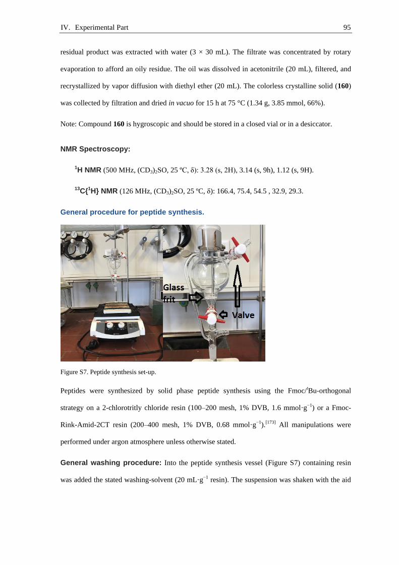
IV. Experimental Part 95
residual product was extracted with water (3 × 30 mL). The filtrate was concentrated by rotary
evaporation to afford an oily residue. The oil was dissolved in acetonitrile (20 mL), filtered, and
recrystallized by vapor diffusion with diethyl ether (20 mL). The colorless crystalline solid (160)
was collected by filtration and dried in vacuo for 15 h at 75 °C (1.34 g, 3.85 mmol, 66%).
Note: Compound 160 is hygroscopic and should be stored in a closed vial or in a desiccator.
NMR Spectroscopy:
1H NMR (500 MHz, (CD3)2SO, 25 ºC, δ): 3.28 (s, 2H), 3.14 (s, 9h), 1.12 (s, 9H).
13C{1H} NMR (126 MHz, (CD3)2SO, 25 ºC, δ): 166.4, 75.4, 54.5 , 32.9, 29.3.
General procedure for peptide synthesis.
Figure S7. Peptide synthesis set-up.
Peptides were synthesized by solid phase peptide synthesis using the Fmoc/tBu-orthogonal
strategy on a 2-chlorotritly chloride resin (100–200 mesh, 1% DVB, 1.6 mmol·g−1) or a Fmoc-
Rink-Amid-2CT resin (200–400 mesh, 1% DVB, 0.68 mmol·g−1).[173] All manipulations were
performed under argon atmosphere unless otherwise stated.
General washing procedure: Into the peptide synthesis vessel (Figure S7) containing resin
was added the stated washing-solvent (20 mL·g−1 resin). The suspension was shaken with the aid

IV. Experimental Part 96
of a Heidolph Vibramax 100 (Figure S7) for 2 minutes at 23 °C, and then the liquid was removed
via vacuum filtration.
General loading procedure (2-CTC resin):
A peptide synthesis vessel (Figure S7) was charged with 2-chlorotrityl chloride resin and DCM
(30 mL·g−1 resin). The suspension was shaken with the aid of a Heidolph Vibramax 100 (Figure
S7) for 30 min at 23 °C. The liquid was removed via vacuum filtration, and a solution of Fmoc-
protected amino acid (4.00 equiv) and DIPEA (10.0 equiv) in DCM (30 mL·g−1 resin) was added
into the peptide synthesis vessel. The resulting suspension was shaken for 15 hours at 23 °C, and
then the liquid was removed via vacuum filtration. The resin was washed with DCM (3 × 20
mL·g−1 resin × 2 min), and a solution of DIPEA, MeOH, and DCM (1:2:17, v:v:v, 30 mL·g−1
resin) was added into the peptide synthesis vessel. The suspension was shaken for 1 hour at 23 °C,
and then the resin was washed sequentially with DMF (2 × 20 mL·g−1 resin), DCM (2 × 20
mL·g−1 resin), MeOH (2 × 20 mL·g−1 resin), and Et2O (2 × 20 mL·g−1 resin). The resin was dried
in vacuo, and the loading efficiency was determined by UV/vis spectroscopy at 289.8 nm.[174]
General deprotection procedure:
Into the peptide synthesis vessel containing resin-bound Fmoc-protected peptide was added 20%
piperidine in DMF (v:v, 20 mL·g−1 resin), and the suspension was shaken for 5 minutes at 23 °C.
Then the liquid was removed via vacuum filtration. This deprotection sequence was repeated
once, and then the resin was washed with DMF (3 × 20 mL·g−1 resin × 2 min).

IV. Experimental Part 97
General HBTU/HOBt coupling procedure:
A round-bottom flask equipped with a Teflon-coated magnetic stirring bar was charged with
Fmoc-protected amino acid (Fmoc-(AA)-OH, 4.00 equiv), HBTU (3.90 equiv), HOBt hydrate
(3.90 equiv), DIPEA (8.00 equiv), and DMF (10 mL·g−1 resin). The solution was stirred for 15
minutes at 23 °C and was then added into the peptide synthesis vessel. The vessel was shaken for
90 minutes at 23 °C, and then the liquid was removed via vacuum filtration. The resin was
washed with DMF (3 × 10 mL·g−1 resin × 2 min).
General [Fmoc-Tyr(RuCp)-OH]·CF3CO2 coupling procedure:
A round-bottom flask equipped with a Teflon-coated magnetic stirring bar was charged with
[Fmoc-Tyr(RuCp)-OH]·CF3CO2 (157) (2.00 equiv), HBTU (1.90 equiv), HOBt hydrate (1.90
equiv), DIPEA (16.0 equiv), and DMF (10 mL·g−1 resin). The solution was stirred for 1 minute at
23 °C and was then added into the peptide synthesis vessel. The vessel was shaken for 2 h at 23
°C, and then the liquid was removed via vacuum filtration. The resin was washed with DMF (3 ×
10 mL·g−1 resin × 2 min).
General [Fmoc-Tyr(RuCp)-O]·H2O coupling procedure:
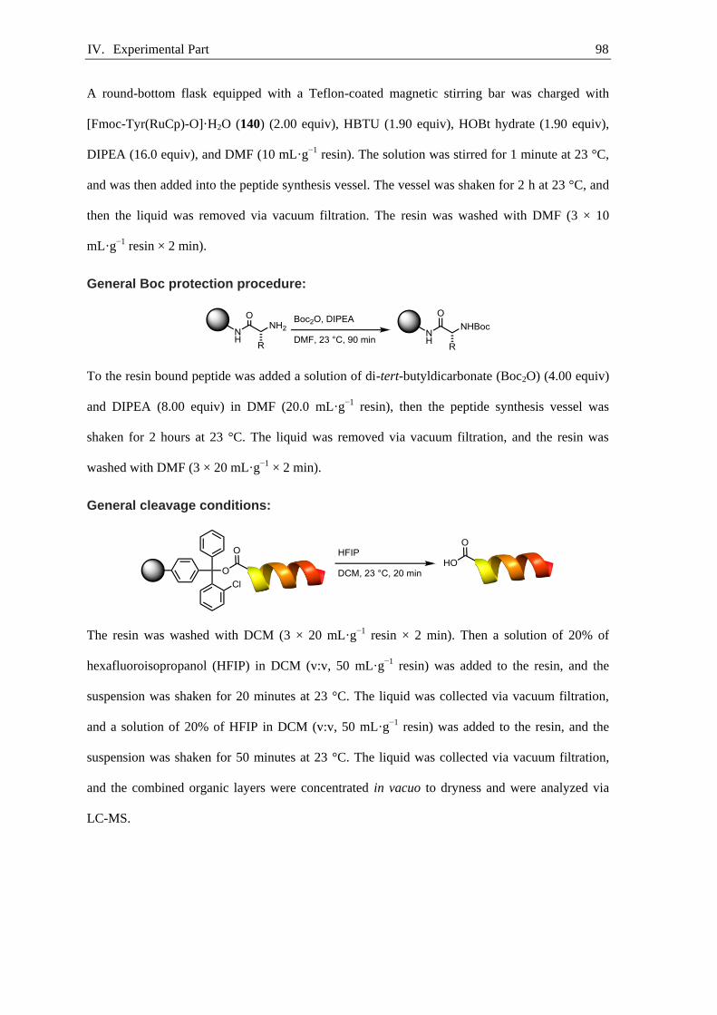
IV. Experimental Part 98
A round-bottom flask equipped with a Teflon-coated magnetic stirring bar was charged with
[Fmoc-Tyr(RuCp)-O]·H2O (140) (2.00 equiv), HBTU (1.90 equiv), HOBt hydrate (1.90 equiv),
DIPEA (16.0 equiv), and DMF (10 mL·g−1 resin). The solution was stirred for 1 minute at 23 °C,
and was then added into the peptide synthesis vessel. The vessel was shaken for 2 h at 23 °C, and
then the liquid was removed via vacuum filtration. The resin was washed with DMF (3 × 10
mL·g−1 resin × 2 min).
General Boc protection procedure:
To the resin bound peptide was added a solution of di-tert-butyldicarbonate (Boc2O) (4.00 equiv)
and DIPEA (8.00 equiv) in DMF (20.0 mL·g−1 resin), then the peptide synthesis vessel was
shaken for 2 hours at 23 °C. The liquid was removed via vacuum filtration, and the resin was
washed with DMF (3 × 20 mL·g−1 × 2 min).
General cleavage conditions:
The resin was washed with DCM (3 × 20 mL·g−1 resin × 2 min). Then a solution of 20% of
hexafluoroisopropanol (HFIP) in DCM (v:v, 50 mL·g−1 resin) was added to the resin, and the
suspension was shaken for 20 minutes at 23 °C. The liquid was collected via vacuum filtration,
and a solution of 20% of HFIP in DCM (v:v, 50 mL·g−1 resin) was added to the resin, and the
suspension was shaken for 50 minutes at 23 °C. The liquid was collected via vacuum filtration,
and the combined organic layers were concentrated in vacuo to dryness and were analyzed via
LC-MS.

IV. Experimental Part 99
[H-Asp(tBu)-D-Phe(4-F)-Lys(Boc)-Arg(Pbf)-Gly-OH]·CF3CO2H (161)
Representative Example: A peptide synthesis vessel (100 mL) was charged with 2-chlorotritly-
chloride resin (100–200 mesh, 1% DVB, 1.6 mmol·g−1, 1.0 g, 1.6 mmol, 1.0 equiv) and DCM (45
mL, 22 g·L−1). The suspension was shaken with the aid of a Heidolph Vibramax 100 (Figure S7)
for 30 minutes at 23 °C. The liquid was removed via vacuum filtration, and a solution of Fmoc-
Gly-OH (1.9 g, 6.4 mmol, 4.0 equiv) and DIPEA (2.8 mL, 2.1 g, 16 mmol, 10 equiv) in DCM (30
mL) was added into the peptide synthesis vessel. The resulting suspension was shaken for 15
hours at 23 °C, and then the liquid was removed via vacuum filtration. The resin was washed with
DCM (3 × 20 mL × 2 min), and a solution of DIPEA, MeOH, and DCM (1:2:17, v:v:v, 30 mL)
was added into the peptide synthesis vessel. The suspension was shaken for 1 hour at 23 °C, and
then the resin was washed sequentially with DMF (2 × 20 mL), DCM (2 × 20 mL), MeOH (2 ×
20 mL), and Et2O (2 × 20 mL). The resin was dried in vacuo to afford Fmoc-Gly-O-2CT resin.
The resin loading was determined to be 0.86 mmol·g−1 by UV/vis spectroscopy.
A peptide synthesis vessel (100 mL) was charged with Fmoc-Gly-O-2CT resin (0.86 mmol·g−1,
1.2 g, 1.0 mmol, 1.0 equiv) and DCM (45 mL, 26 g·L−1). The resulting suspension was shaken for
30 minutes at 23 °C, and then the liquid was removed via vacuum filtration. The resin was
washed with DMF (3 × 10 mL × 2 min). Into the peptide synthesis vessel was added 20%
piperidine in DMF (v:v, 20 mL), and the suspension was shaken for 5 minutes at 23 °C. Then the
liquid was removed via vacuum filtration. This deprotection sequence was repeated once, and
then the resin was washed with DMF (3 × 20 mL × 2 min). A round-bottom flask (20 mL)
equipped with a Teflon-coated magnetic stirring bar was charged with Fmoc-Asp(tBu)-OH (1.6 g,
4.0 mmol, 4.0 equiv), HBTU (1.5 g, 3.9 mmol, 3.9 equiv), HOBt hydrate (0.53 g, 3.9 mmol, 3.9

IV. Experimental Part 100
equiv), DIPEA (1.4 mL, 1.0 g, 8.0 mmol, 8.0 equiv), and DMF (10 mL). The yellow solution was
stirred for 15 minutes at 23 °C and was then added into the peptide synthesis vessel. The vessel
was shaken for 90 minutes at 23 °C, and then the liquid was removed via vacuum filtration. The
resin was washed with DMF (3 × 10 mL × 2 min). Into the peptide synthesis vessel was added
20% piperidine in DMF (v:v, 20 mL), and the suspension was shaken for 5 minutes at 23 °C.
Then the liquid was removed via vacuum filtration. This deprotection sequence was repeated
once, and then the resin was washed with DMF (3 × 20 mL × 2 min). A round-bottom flask (20
mL) equipped with a Teflon-coated magnetic stirring bar was charged with Fmoc-D-Phe(4-F)-OH
(1.6 g, 4.0 mmol, 4.0 equiv), HBTU (1.5 g, 3.9 mmol, 3.9 equiv), HOBt hydrate (0.53 g, 3.9
mmol, 3.9 equiv), DIPEA (1.4 mL, 1.0 g, 8.0 mmol, 8.0 equiv), and DMF (10 mL). The yellow
solution was stirred for 15 minutes at 23 °C and was then added into the peptide synthesis vessel.
The vessel was shaken for 90 minutes at 23 °C, and then the liquid was removed via vacuum
filtration. The resin was washed with DMF (3 × 10 mL × 2 min). Into the peptide synthesis vessel
was added 20% piperidine in DMF (v:v, 20 mL), and the suspension was shaken for 5 minutes at
23 °C. Then the liquid was removed via vacuum filtration. This deprotection sequence was
repeated once, and then the resin was washed with DMF (3 × 20 mL × 2 min). A round-bottom
flask (20 mL) equipped with a Teflon-coated magnetic stirring bar was charged with Fmoc-
Lys(Boc)-OH (1.9 g, 4.0 mmol, 4.0 equiv), HBTU (1.5 g, 3.9 mmol, 3.9 equiv), HOBt hydrate
(0.53 g, 3.9 mmol, 3.9 equiv), DIPEA (1.4 mL, 1.0 g, 8.0 mmol, 8.0 equiv), and DMF (10 mL).
The yellow solution was stirred for 15 minutes at 23 °C and was then added into the peptide
synthesis vessel. The vessel was shaken for 90 minutes at 23 °C, and then the liquid was removed
via vacuum filtration. The resin was washed with DMF (3 × 10 mL × 2 min). Into the peptide
synthesis vessel was added 20% piperidine in DMF (v:v, 20 mL), and the suspension was shaken
for 5 minutes at 23 °C. Then the liquid was removed via vacuum filtration. This deprotection
sequence was repeated once, and then the resin was washed with DMF (3 × 20 mL × 2 min). A
round-bottom flask (20 mL) equipped with a Teflon-coated magnetic stirring bar was charged
with Fmoc-Arg(Pbf)-OH (2.6 g, 4.0 mmol, 4.0 equiv), HBTU (1.5 g, 3.9 mmol, 3.9 equiv), HOBt
hydrate (0.53 g, 3.9 mmol, 3.9 equiv), DIPEA (1.4 mL, 1.0 g, 8.0 mmol, 8.0 equiv), and DMF (10
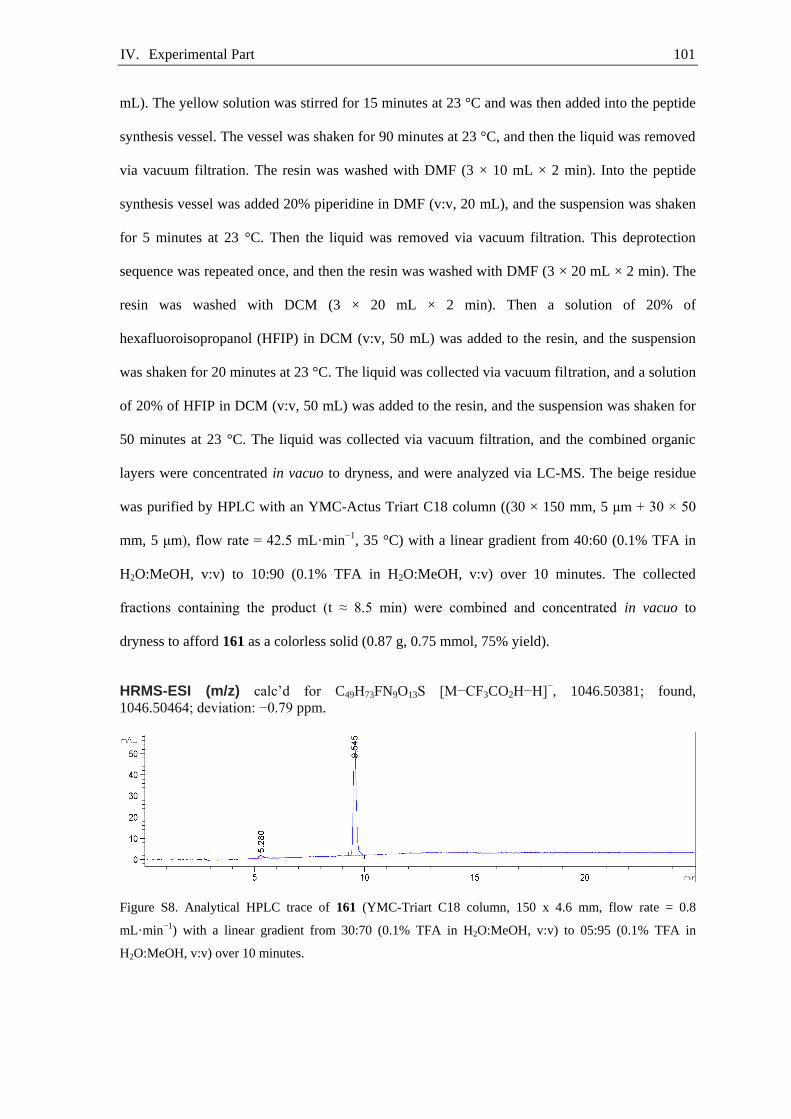
IV. Experimental Part 101
mL). The yellow solution was stirred for 15 minutes at 23 °C and was then added into the peptide
synthesis vessel. The vessel was shaken for 90 minutes at 23 °C, and then the liquid was removed
via vacuum filtration. The resin was washed with DMF (3 × 10 mL × 2 min). Into the peptide
synthesis vessel was added 20% piperidine in DMF (v:v, 20 mL), and the suspension was shaken
for 5 minutes at 23 °C. Then the liquid was removed via vacuum filtration. This deprotection
sequence was repeated once, and then the resin was washed with DMF (3 × 20 mL × 2 min). The
resin was washed with DCM (3 × 20 mL × 2 min). Then a solution of 20% of
hexafluoroisopropanol (HFIP) in DCM (v:v, 50 mL) was added to the resin, and the suspension
was shaken for 20 minutes at 23 °C. The liquid was collected via vacuum filtration, and a solution
of 20% of HFIP in DCM (v:v, 50 mL) was added to the resin, and the suspension was shaken for
50 minutes at 23 °C. The liquid was collected via vacuum filtration, and the combined organic
layers were concentrated in vacuo to dryness, and were analyzed via LC-MS. The beige residue
was purified by HPLC with an YMC-Actus Triart C18 column ((30 × 150 mm, 5 μm + 30 × 50
mm, 5 μm), flow rate = 42.5 mL·min−1, 35 °C) with a linear gradient from 40:60 (0.1% TFA in
H2O:MeOH, v:v) to 10:90 (0.1% TFA in H2O:MeOH, v:v) over 10 minutes. The collected
fractions containing the product (t ≈ 8.5 min) were combined and concentrated in vacuo to
dryness to afford 161 as a colorless solid (0.87 g, 0.75 mmol, 75% yield).
HRMS-ESI (m/z) calc’d for C49H73FN9O13S [M−CF3CO2H−H]−, 1046.50381; found,
1046.50464; deviation: −0.79 ppm.
Figure S8. Analytical HPLC trace of 161 (YMC-Triart C18 column, 150 x 4.6 mm, flow rate = 0.8
mL·min−1) with a linear gradient from 30:70 (0.1% TFA in H2O:MeOH, v:v) to 05:95 (0.1% TFA in
H2O:MeOH, v:v) over 10 minutes.

IV. Experimental Part 102
[c(Asp(tBu)-D-Phe(4-F)-Lys(Boc)-Arg(Pbf)-Gly)]·CF3CO2H (162)
A round-bottom flask (1 L) equipped with a Teflon-coated magnetic stirring bar was charged with
HOBt (0.12 g, 0.88 mmol, 1.2 equiv), HBTU (0.33 g, 0.88 mmol, 1.2 equiv), DIPEA (0.38 mL,
0.28 g, 2.2 mmol, 3.0 equiv), DCM (0.4 L), and DMF (0.1 L). The reaction mixture was cooled to
0°C and a solution of linear peptide 161 (0.85 g, 0.73 mmol, 1.0 equiv) in DMF (20 mL) was
added dropwise via a syringe over 20 minutes. The reaction mixture was allowed to warm to 23
°C and was afterwards stirred for 12 hours at 23 °C. The solution was concentrated by rotary
evaporation to 5 mL and was then diluted with ethyl acetate (50 mL). The solution was washed
with water (2 × 30 mL), and the combined aqueous layers were extracted with ethyl acetate (10
mL). The combined organic layers were dried over sodium sulfate, filtered, and concentrated in
vacuo to dryness. The beige residue was purified by HPLC on an YMC-Actus Triart C18 column
((30 × 150 mm, 5 μm + 30 × 50 mm, 5 μm), flow rate = 42.5 mL·min−1, 35 °C) with a linear
gradient from 30:70 (0.1% TFA in H2O:MeOH, v:v) to 05:95 (0.1% TFA in H2O:MeOH, v:v)
over 10 minutes. The collected fractions containing the product 162 (t ≈ 9.5 min) were combined,
neutralized to pH 7 with saturated aqueous sodium bicarbonate solution, diluted with 100 mL
brine, and the resulting suspension was concentrated by rotary evaporation (100 mbar, 35 °C)
until no more methanol was evaporated. The suspension was extracted with dichloromethane (3 ×
100 mL), and the combined organic layers were dried over sodium sulfate, filtered, and
concentrated in vacuo to dryness to afford 162 a colorless powder (0.62 g, 0.54 mmol, 74%
yield).
HRMS-ESI (m/z) calc’d for C49H72FN9O12SNa [M+Na−CF3CO2H]+, 1052.48974; found,
1052.49063; deviation: −0.85 ppm.
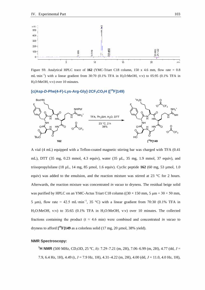
IV. Experimental Part 103
Figure S9. Analytical HPLC trace of 162 (YMC-Triart C18 column, 150 x 4.6 mm, flow rate = 0.8
mL·min−1) with a linear gradient from 30:70 (0.1% TFA in H2O:MeOH, v:v) to 05:95 (0.1% TFA in
H2O:MeOH, v:v) over 10 minutes.
[c(Asp-D-Phe(4-F)-Lys-Arg-Gly]·2CF3CO2H ([19F]149)
A vial (4 mL) equipped with a Teflon-coated magnetic stirring bar was charged with TFA (0.41
mL), DTT (35 mg, 0.23 mmol, 4.3 equiv), water (35 µL, 35 mg, 1.9 mmol, 37 equiv), and
triisopropylsilane (18 µL, 14 mg, 85 µmol, 1.6 equiv). Cyclic peptide 162 (60 mg, 53 µmol, 1.0
equiv) was added to the emulsion, and the reaction mixture was stirred at 23 °C for 2 hours.
Afterwards, the reaction mixture was concentrated in vacuo to dryness. The residual beige solid
was purified by HPLC on an YMC-Actus Triart C18 column ((30 × 150 mm, 5 μm + 30 × 50 mm,
5 μm), flow rate = 42.5 mL·min−1, 35 °C) with a linear gradient from 70:30 (0.1% TFA in
H2O:MeOH, v:v) to 35:65 (0.1% TFA in H2O:MeOH, v:v) over 10 minutes. The collected
fractions containing the product (t ≈ 4.6 min) were combined and concentrated in vacuo to
dryness to afford [19F]149 as a colorless solid (17 mg, 20 µmol, 38% yield).
NMR Spectroscopy:
1H NMR (500 MHz, CD3OD, 25 ºC, δ): 7.29–7.21 (m, 2H), 7.06–6.99 (m, 2H), 4.77 (dd, J =
7.9, 6.4 Hz, 1H), 4.49 (t, J = 7.9 Hz, 1H), 4.31–4.22 (m, 2H), 4.00 (dd, J = 11.0, 4.0 Hz, 1H),

IV. Experimental Part 104
3.19 (qt, J = 13.7, 7.0 Hz, 2H), 2.99 (d, J = 7.9 Hz, 2H), 2.88–2.77 (m, 3H), 2.59 (dd, J =
16.5, 6.4 Hz, 1H), 1.88 (ddt, J = 13.1, 9.2, 6.5 Hz, 1H), 1.76 (dddd, J = 13.6, 9.4, 7.2, 4.0 Hz,
1H) 1.66 (dddd, J = 13.1, 10.0, 7.7, 5.2 Hz, 1H), 1.60–1.41 (m, 5H), 1.07–0.98 (m, 2H). One
proton resonance is not observed, presumably due to overlap with a solvent proton
resonance.
13C{1H} NMR (126 MHz, CD3OD, 25 ºC, δ): 174.6, 173.8, 173.7, 173.4, 172.8, 172.2, 163.2
(1JC–F = 243 Hz), 158.6, 134.0 (4JC–F = 3 Hz), 132.3 (3JC–F = 8 Hz), 116.2 (2JC–F = 21 Hz),
56.9, 55.8, 53.9, 51.0, 44.9, 42.0, 40.3, 37.3, 36.2, 31.7, 29.4, 27.9, 26.3, 24.1.
19F NMR (471 MHz, CD3OD, 25 ºC, δ): −77.7, −118.9.
HRMS-ESI (m/z) calc’d for C27H42FN9O7 [M−2CF3CO2]2+, 311.65901; found, 311.65885;
deviation: 0.52 ppm.
Figure S10. Analytical HPLC trace of [19F]149 (YMC-Triart C18 column, 150 x 4.6 mm, flow rate = 0.8
mL·min−1) with a linear gradient from 70:30 (0.1% TFA in H2O:MeOH, v:v) to 40:60 (0.1% TFA in
H2O:MeOH, v:v) over 10 minutes.

IV. Experimental Part 105
[H-Asp(tBu)-D-Tyr(RuCp)-Lys(Boc)-Arg(Pbf)-Gly-OH]·CF3CO2H·CF3CO2 (163)
The peptide was synthesized according to the general procedure for peptide synthesis starting
with 1 mmol Fmoc-Gly-O-2CT resin. The beige residue was purified by HPLC with an YMC-
Actus Triart C18 column ((30×150 mm, 5 μm + 30×50 mm, 5 μm), flow rate = 42.5 mL·min-1, 35
°C) with a linear gradient from 40:60 (0.1% TFA in H2O:MeOH, v:v) to 10:90 (0.1% TFA in
H2O:MeOH, v:v) over 10 minutes. The collected fractions containing the product (t ≈ 6.5 min)
were combined and concentrated in vacuo to dryness to afford 163 as a colorless solid (1.1 g, 0.73
mmol, 73% yield).
HRMS-ESI (m/z) calc’d for C54H81O14N9RuS [M−2CF3CO2]2+, 606.73281; found, 606.73366;
deviation: 1.40 ppm.
Figure S11. Analytical HPLC trace of 163 (YMC-Triart C18 column, 150 x 4.6 mm, flow rate = 0.8
mL·min−1) with a linear gradient from 30:70 (0.1% TFA in H2O:MeOH, v:v) to 05:95 (0.1% TFA in
H2O:MeOH, v:v) over 10 minutes.

IV. Experimental Part 106
[c(Asp(tBu)-D-Tyr(RuCp)-Lys(Boc)-Arg(Pbf)-Gly)] (164)
A round-bottom flask (1 L) equipped with a Teflon-coated magnetic stirring bar was charged with
HOBt (0.10 g, 0.77 mmol, 1.2 equiv), HBTU (0.29 g, 0.77 mmol, 1.2 equiv), DIPEA (0.33 mL,
0.25 g, 1.9 mmol, 3.0 equiv), DCM (0.4 L), and DMF (0.1 L). The reaction mixture was cooled to
0 °C and a solution of linear peptide 163 (0.92 g, 0.64 mmol, 1.0 equiv) in DMF (20 mL) was
added dropwise via a syringe over 20 min. The reaction mixture was allowed to warm to 23 °C
and was afterwards stirred for 12 hours at 23 °C. The solution was concentrated by rotary
evaporation to 5 mL and afterwards diluted with ethyl acetate (50 mL). The solution was
extracted with water (2 × 30 mL), and the combined aqueous layers were extracted with ethyl
acetate (10 mL). The combined organic layers were dried over sodium sulfate, filtered, and
concentrated in vacuo to dryness. The beige residue was purified by HPLC on an YMC-Actus
Triart C18 column ((30×150 mm, 5 μm + 30×50 mm, 5 μm), flow rate = 42.5 mL·min-1, 35 °C)
with a linear gradient from 20:80 (0.1% TFA in H2O:MeOH, v:v) to 10:90 (0.1% TFA in
H2O:MeOH, v:v) over 10 minutes. The collected fractions containing the product (t ≈ 8.0 min)
were combined, basified to pH 8 with saturated aqueous sodium bicarbonate solution, diluted with
100 mL brine and the resulting solution was concentrated by rotary evaporation (100 mbar, 35 °C)
until no more methanol was evaporated. The suspension was extracted with dichloromethane (3 ×
100 mL), and the combined organic layers were dried over sodium sulfate, filtered, and
concentrated in vacuo to dryness to afford 164 a colorless powder (0.55 g, 0.46 mmol, 72%
yield).
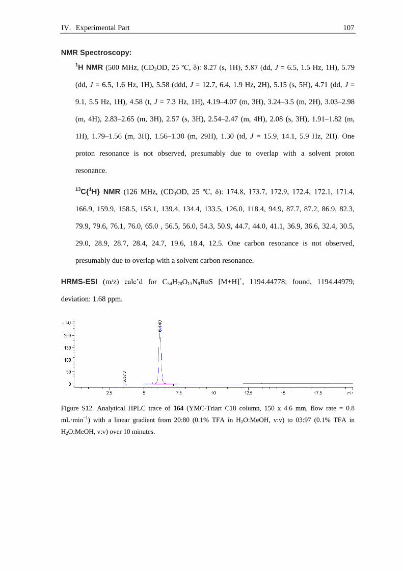
IV. Experimental Part 107
NMR Spectroscopy:
1H NMR (500 MHz, (CD3OD, 25 ºC, δ): 8.27 (s, 1H), 5.87 (dd, J = 6.5, 1.5 Hz, 1H), 5.79
(dd, J = 6.5, 1.6 Hz, 1H), 5.58 (ddd, J = 12.7, 6.4, 1.9 Hz, 2H), 5.15 (s, 5H), 4.71 (dd, J =
9.1, 5.5 Hz, 1H), 4.58 (t, J = 7.3 Hz, 1H), 4.19–4.07 (m, 3H), 3.24–3.5 (m, 2H), 3.03–2.98
(m, 4H), 2.83–2.65 (m, 3H), 2.57 (s, 3H), 2.54–2.47 (m, 4H), 2.08 (s, 3H), 1.91–1.82 (m,
1H), 1.79–1.56 (m, 3H), 1.56–1.38 (m, 29H), 1.30 (td, J = 15.9, 14.1, 5.9 Hz, 2H). One
proton resonance is not observed, presumably due to overlap with a solvent proton
resonance.
13C{1H} NMR (126 MHz, (CD3OD, 25 ºC, δ): 174.8, 173.7, 172.9, 172.4, 172.1, 171.4,
166.9, 159.9, 158.5, 158.1, 139.4, 134.4, 133.5, 126.0, 118.4, 94.9, 87.7, 87.2, 86.9, 82.3,
79.9, 79.6, 76.1, 76.0, 65.0 , 56.5, 56.0, 54.3, 50.9, 44.7, 44.0, 41.1, 36.9, 36.6, 32.4, 30.5,
29.0, 28.9, 28.7, 28.4, 24.7, 19.6, 18.4, 12.5. One carbon resonance is not observed,
presumably due to overlap with a solvent carbon resonance.
HRMS-ESI (m/z) calc’d for C54H78O13N9RuS [M+H]+, 1194.44778; found, 1194.44979;
deviation: 1.68 ppm.
Figure S12. Analytical HPLC trace of 164 (YMC-Triart C18 column, 150 x 4.6 mm, flow rate = 0.8
mL·min−1) with a linear gradient from 20:80 (0.1% TFA in H2O:MeOH, v:v) to 03:97 (0.1% TFA in
H2O:MeOH, v:v) over 10 minutes.
IV. Experimental Part 108
[Boc-D-Glu(tBu)-Ala-Tyr(tBu)-Gly-Trp(Boc)-Met-Asp(tBu)-Phe(4-F)-OH] (165)
The peptide was synthesized according to the general procedure for peptide synthesis starting
with 0.5 mmol Fmoc-Phe(4-F)-O-2CT resin. The beige residue was purified by HPLC with an
YMC-Actus Triart C18 column ((30×150 mm, 5 μm + 30×50 mm, 5 μm), flow rate = 42.5
mL·min-1, 35 °C) with a linear gradient from 15:85 (0.1% TFA in H2O:MeOH, v:v) to 05:95
(0.1% TFA in H2O:MeOH, v:v) over 10 minutes. The collected fractions containing the product (t
≈ 9.0 min) were combined, basified to pH 7 with saturated aqueous sodium bicarbonate solution,
diluted with 100 mL brine, and the resulting solution was concentrated by rotary evaporation (100
mbar, 35 °C) until no more methanol was evaporated. The suspension was extracted with
dichloromethane (3 × 100 mL), and the combined organic layers were dried over sodium sulfate,
filtered, and concentrated in vacuo to dryness to afford the title compound 165 as a colorless solid
(0.32 g, 0.23 mmol, 46% yield).
HRMS-ESI (m/z) calc’d for C70H97FN9O18S [M−H]-, 1402.66618; found, 1402.66603; deviation:
0.11 ppm.
Figure S13. Analytical HPLC trace of 165 (YMC-Triart C18 column, 150 x 4.6 mm, flow rate = 0.8
mL·min−1) with a linear gradient from 30:70 (0.1% TFA in H2O:MeOH, v:v) to 05:95 (0.1% TFA in
H2O:MeOH, v:v) over 10 minutes.

IV. Experimental Part 109
[Boc-D-Glu(tBu)-Ala-Tyr(tBu)-Gly-Trp(Boc)-Met-Asp(tBu)-Phe(4-F)-NHTmob] (166)
A vial (20 mL) equipped with a Teflon-coated magnetic stirring bar was charged with 165 (0.26
g, 0.19 mmol, 1.0 equiv), HOBt (26 mg, 0.19 mmol, 1. equiv), TmobNH2 (38 mg, 0.19 mmol, 1.0
equiv), DIPEA (0.10 mL, 74 mg, 0.57 mmol, 3.1 equiv), and DMF (17 mL, 0.010 mol·L−1). The
reaction mixture was cooled to 0 °C and then HBTU (73 mg, 0.19 mmol, 1.0 equiv) was added.
The reaction mixture was allowed to warm to room temperature and was stirred at 23 °C for 12
hours. The solution was concentrated by rotary evaporation to 5 mL, and was afterwards diluted
with ethyl acetate (50 mL). The solution was extracted with water (2 × 30 mL), and the combined
aqueous layers were extracted with ethyl acetate (10 mL). The combined organic layers were
concentrated in vacuo to dryness. The beige residue was purified by HPLC on an YMC-Actus
Triart C18 column ((30×150 mm, 5 μm + 30×50 mm, 5 μm), flow rate = 42.5 mL·min-1
, 35 °C)
with a linear gradient from 25:75 (0.1% TFA in H2O:MeOH, v:v) to 03:97 (0.1% TFA in
H2O:MeOH, v:v) over 10 minutes. The collected fractions containing the product (t ≈ 9.8 min)
were combined, basified to pH 7 with saturated aqueous sodium bicarbonate solution, diluted with
100 mL brine and the resulting solution was concentrated by rotary evaporation (100 mbar, 35 °C)
until no more methanol was evaporated. The suspension was extracted with dichloromethane (3 ×
100 mL), and the combined organic layers were dried over sodium sulfate, filtered, and
concentrated in vacuo to dryness to afford 166 a colorless powder (0.12 g, 76 µmol, 40% yield).
HRMS-ESI (m/z) calc’d for C80H110FN10O20S [M−H−CF3CO2H]−, 1581.76081; found,
1581.76160.
IV. Experimental Part 110
Figure S14. Analytical HPLC trace of 166 (YMC-Triart C18 column, 150 x 4.6 mm, flow rate = 0.8
mL·min−1) with a linear gradient from 50:50 (0.1% TFA in H2O:MeOH, v:v) to 10:90 (0.1% TFA in
H2O:MeOH, v:v) over 10 minutes.
[H-D-Glu-Ala-Tyr-Gly-Trp-Met-Asp-Phe(4-F)-NH2]·2CF3CO2H ([18F]148)
A vial (4 mL) equipped with a Teflon-coated magnetic stirring bar was charged with TFA (0.41
mL, 0.61 g), DTT (35 mg, 0.23 mmol, 6.0 equiv), water (35 µL, 35 mg, 1.9 mmol, 51 equiv), and
triisopropylsilane (18 µL, 14 mg, 85 µmol, 2.3 equiv). Linear peptide 166 (60 mg, 38 µmol, 1.0
equiv) was added to the emulsion, and the reaction mixture was stirred at 23 °C for 2 hours. The
reaction mixture was concentrated in vacuo to dryness, and the resulting beige solid was purified
by HPLC on an YMC-Actus Triart C18 column ((30×150 mm, 5 μm + 30×50 mm, 5 μm), flow
rate = 42.5 mL·min-1, 35 °C) with a linear gradient from 50:50 (0.1% TFA in H2O:MeOH, v:v) to
15:85 (0.1% TFA in H2O:MeOH, v:v) over 10 minutes. The collected fractions containing
[19
F]148 (t ≈ 6.3 min) were combined and concentrated in vacuo to dryness to afford the [19F]148
as a colorless solid (25 mg, 20 µmol, 52% yield).
NMR Spectroscopy:
1H NMR (500 MHz, CD3OD, 25 ºC, δ): 7.57 (d, J = 7.9 Hz, 1H), 7.32 (d, J = 8.1 Hz, 1H),
7.28–7.23 (m, 2H), 7.19 (s, 1H), 7.08 (ddd, J = 8.2, 7.0, 1.2 Hz, 1H), 7.04–6.92 (m, 5H),
6.75–6.67 (m, 2H), 4.62 (dd, J = 7.5, 6.1 Hz, 1H), 4.58–4.52 (m, 2H), 4.32 (dd, J = 9.1, 6.0
Hz, 1H), 4.28–4.20 (m, 2H), 4.02–3.95 (m, 2H), 3.65 (d, J = 16.5 Hz, 1H), 3.19 (dd, J = 14.1,
IV. Experimental Part 111
5.3 Hz, 1H), 3.07 (dd, J = 14.0, 5.9 Hz, 1H), 2.97 (dd, J = 14.0, 8.9 Hz, 1H), 2.90–2.76 (m,
2H), 2.68 (dd, J = 16.9, 7.5 Hz, 1H), 2.47 (dt, J = 7.5, 1.8 Hz, 2H), 2.25 (ddd, J = 13.6, 8.5,
5.5 Hz, 1H), 2.18–2.08 (m, 3H), 1.99 (s, 3H), 1.89 (ddt, J = 14.3, 10.8, 6.6 Hz, 2H), 1.29 (d,
J = 7.2 Hz, 3H). The resonance for two protons was not observed, presumably due to overlap
with the solvent proton resonance.
13C{1H} NMR (126 MHz, CD3OD, 25 ºC, δ):175.7, 175.6, 175.0, 174.8, 174.1, 174.0, 173.8,
172.6, 172.3, 170.1, 163.2 (1JC-F = 244 Hz), 157.3, 138.0, 134.4 (4JC-F = 3 Hz), 132.1 (3JC-F =
243 Hz), 131.3, 129.2, 128.7, 124.9, 122.6, 119.9, 119.3, 116.2, 116.1 (2JC-F = 22 Hz), 112.5,
110.5, 57.1, 56.4, 56.0, 54.6, 53.8, 51.7, 51.3, 44.0, 37.8, 36.8, 36.4, 31.3, 30.9, 30.1, 28.2,
27.6, 17.8, 15.2.
19F NMR (471 MHz, CD3OD, 25 ºC, δ): −77.1, −118.3.
HRMS-ESI (m/z) calc’d for C48H58F N10O13S [M−H−2CF3CO2H]−, 1033.38950; found,
1033.39025; deviation: −0.72 ppm.
Figure S15. Analytical HPLC trace of [19F]148 (YMC-Triart C18 column, 150 x 4.6 mm, flow rate = 0.8
mL·min−1) with a linear gradient from 50:50 (0.1% TFA in H2O:MeOH, v:v) to 10:90 (0.1% TFA in
H2O:MeOH, v:v) over 10 minutes.

IV. Experimental Part 112
[Boc-D-Glu(tBu)-Ala-Tyr(tBu)-Gly-Trp(Boc)-Met-Asp(tBu)-Tyr(RuCp)-OH] (167)
A peptide synthesis vessel (100 mL) was charged with 2-chlorotritly-chloride resin (100–200
mesh, 1% DVB, 1.6 mmol·g−1, 1.0 g, 1.6 mmol, 1.0 equiv) and DCM (45 mL, 22 g·L−1). The
suspension was shaken with the aid of a Heidolph Vibramax 100 (Figure S7) for 30 minutes at 23
°C. The liquid was removed via vacuum filtration, and a solution of [Fmoc-Tyr(RuCp)-O]·H2O
(1.9 g, 3.2 mmol, 2.0 equiv) and DIPEA (1.4 mL, 1.0 g, 8.0 mmol, 5.0 equiv) in DCM (30 mL)
was added into the peptide synthesis vessel. The resulting suspension was shaken for 15 hours at
23 °C, and then the liquid was removed via vacuum filtration. The resin was washed with DCM
(3 × 20 mL × 2 min), and a solution of DIPEA, MeOH, and DCM (1:2:17, v:v:v, 30 mL) was
added into the peptide synthesis vessel. The suspension was shaken for 1 hour at 23 °C, and then
the resin was washed sequentially with DMF (2 × 20 mL), DCM (2 × 20 mL), MeOH (2 ×
20 mL), and Et2O (2 × 20 mL). The resin was dried in vacuo to afford Fmoc-Tyr(RuCp)-O-2CT
resin. The resin loading was determined to be 0.30 mmol·g−1 by UV/vis spectroscopy.
The peptide was synthesized according to the general procedure for peptide synthesis starting
with 1.0 mmol Fmoc-Tyr(RuCp)-O-2CT resin. The beige residue was purified by HPLC with an
YMC-Actus Triart C18 column ((30×150 mm, 5 μm + 30×50 mm, 5 μm), flow rate = 42.5
mL·min-1, 35 °C) with a linear gradient from 25:75 (0.1% TFA in H2O:MeOH, v:v) to 15:85
(0.1% TFA in H2O:MeOH, v:v) over 10 minutes. The collected fractions containing the product (t
≈ 9.5 min) were combined, neutralized to pH 7 with saturated aqueous sodium bicarbonate
solution, diluted with 100 mL brine, and the resulting solution was concentrated by rotary
evaporation (100 mbar, 35 °C) until no more methanol was evaporated. The suspension was
extracted with dichloromethane (3 × 100 mL), and the combined organic layers were dried over

IV. Experimental Part 113
sodium sulfate, filtered, and concentrated in vacuo to dryness to afford 167 as a colorless solid
(0.65 g, 0.41 mmol, 41% yield).
HRMS-ESI (m/z) calc’d for C75H102O19N9RuS [M−H]−, 1566.60617; found, 1566.60617;
deviation: −0 ppm.
Figure S16. Analytical HPLC trace of 167 (YMC-Triart C18 column, 150 x 4.6 mm, flow rate = 0.8
mL·min−1) with a linear gradient from 30:70 (0.1% TFA in H2O:MeOH, v:v) to 05:95 (0.1% TFA in
H2O:MeOH, v:v) over 10 minutes.
[Boc-D-Glu(tBu)-Ala-Tyr(tBu)-Gly-Trp(Boc)-Met-Asp(tBu)-Tyr(RuCp)-
NHTmob]·CF3CO2 (168)
A 50 mL round-bottom flask equipped with a magnetic stirring bar was charged with 167 (0.60 g,
0.38 mmol, 1.0 equiv), HOBt (53 mg, 0.39 mmol, 1.0 equiv), TmobNH2 (77 mg, 0.39 mmol, 1.0
equiv), DIPEA (0.20 mL, 0.15 g, 1.2 mmol, 3.1 equiv) and DMF (17 mL, 20 mmol·L−1). The
reaction mixture was cooled to 0 °C and then HBTU (0.15 g, 0.39 mmol, 1.0 equiv) was added.
The reaction mixture was allowed to warm to room temperature and was stirred at 23 °C for 12
hours. The solution was concentrated by rotary evaporation to 5 mL, and was afterwards diluted
with ethyl acetate (50 mL). The solution was extracted with water (2 × 30 mL), and the combined
aqueous layers were extracted with ethyl acetate (10 mL). The combined organic layers were
dried over sodium sulfate, filtered, and concentrated in vacuo to dryness. The beige residue was
purified by HPLC on an YMC-Actus Triart C18 column ((30×150 mm, 5 μm + 30×50 mm, 5
IV. Experimental Part 114
μm), flow rate = 42.5 mL·min-1, 35 °C) with a linear gradient from 20:80 (0.1% TFA in
H2O:MeOH, v:v) to 10:90 (0.1% TFA in H2O:MeOH, v:v) over 10 minutes. The collected
fractions containing the product (t ≈ 8 min) were combined, basified to pH 8 with saturated
aqueous sodium bicarbonate solution, diluted with 100 mL brine and the resulting solution was
concentrated by rotary evaporation (100 mbar, 35 °C) until no more methanol was evaporated.
The suspension was extracted with dichloromethane (3 × 100 mL), and the combined organic
layers were dried over sodium sulfate, filtered, and concentrated in vacuo to dryness to afford 168
a colorless powder (0.32 g, 0.17 mmol, 45% yield).
NMR Spectroscopy:
1H NMR (500 MHz, (CD3)2SO, 25 ºC, δ): 8.30 (d, J = 7.7 Hz, 1H), 8.23 (d, J = 7.6 Hz, 1H),
8.18 (d, J = 7.7 Hz, 1H), 8.06 (br, 1H), 8.00 (t, J = 8.6 Hz, 2H), 7.88 (d, J = 7.2 Hz, 1H),
7.75–7.68 (m, 2H), 7.56 (s, 1H), 7.37–7.28 (m, 1H), 7.24 (t, J = 7.5 Hz, 1H), 7.09 (d, J = 8.1
Hz, 2H), 6.92 (d, J = 7.6 Hz, 1H), 6.81 (d, J = 8.0 Hz, 2H), 6.24 (s, 2H), 6.02 (t, J = 4.9 Hz,
2H), 5.95 (d, J = 5.7 Hz, 1H), 5.89 (d, J = 6.3 Hz, 1H), 5.25 (s, 5H), 4.63 (q, J = 8.2 Hz, 1H),
4.55 (q, J = 7.3 Hz, 1H), 4,51–4.39 (m, 2H), 4.34 (dt, J = 13.3, 6.2 Hz, 1H), 4.28–4.09 (m,
3H), 3.97–3.88 (m, 1H), 3.78 (s, 3H), 3.75 (s, 6H), 3.74–3.69 (m, 1H), 3.63 (t, J = 16.9, 5.4
Hz, 1H), 3.12 (d, J = 14.4 Hz, 1H), 3.01–2.89 (m, 2H), 2.75 (dd, J = 14.0, 9.5 Hz, 1H), 2.66–
2.57 (m, 2H), 2.44–2.31 (m, 2H), 2.18 (t, J = 7.8 Hz, 2H), 2.01 (s, 3H), 1.92 (t, J = 7.0 Hz,
1H), 1.82 (dd, J = 8.6, 3.9 Hz, 2H), 1.71–1.64 (m, 1H) ,1.61 (s, 9H), 1.37 (s, 9H), 1.36 (s,
9H), 1.34 (s, 9H), 1.23 (s, 9H),1.09 (d, J = 7.1 Hz, 3H). One proton resonance is not
observed, presumably due to overlap with a solvent proton resonance.
13C{1H} NMR (126 MHz, (CD3)2SO, 25 ºC, δ):171.9, 171.6, 171.3, 171.2, 170.9, 169.9,
169.2, 168.9, 168.7, 160.7, 159.1, 155.3, 153.4, 149.0, 134.6, 132.4, 130.3, 129.6, 124.2,
124.1, 123.4, 122.4, 119.4, 116.4, 114.6, 105.2, 96.1, 85.5, 85.3, 83.5, 80.3, 79.6, 79.1, 78.3,
77.5, 74.1, 74.0, 55.8, 55.3, 54.1, 53.50, 53.45, 52.6, 52.1, 49.6, 48.1, 42.0, 37.0, 36.62, 36.56,
31.7, 31.2, 29.4, 28.5, 28.1, 27.70, 27.73, 27.6, 27.3, 27.2, 18.1, 14.6. Three carbon
resonances are not observed, presumably due to overlap with other carbon resonances.
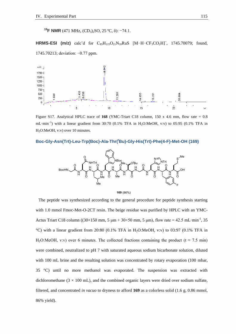
IV. Experimental Part 115
19F NMR (471 MHz, (CD3)2SO, 25 ºC, δ): −74.1.
HRMS-ESI (m/z) calc’d for C85H115O21N10RuS [M−H−CF3CO2H]−, 1745.70079; found,
1745.70213; deviation: −0.77 ppm.
Figure S17. Analytical HPLC trace of 168 (YMC-Triart C18 column, 150 x 4.6 mm, flow rate = 0.8
mL·min−1) with a linear gradient from 30:70 (0.1% TFA in H2O:MeOH, v:v) to 05:95 (0.1% TFA in
H2O:MeOH, v:v) over 10 minutes.
Boc-Gly-Asn(Trt)-Leu-Trp(Boc)-Ala-Thr(tBu)-Gly-His(Trt)-Phe(4-F)-Met-OH (169)
The peptide was synthesized according to the general procedure for peptide synthesis starting
with 1.0 mmol Fmoc-Met-O-2CT resin. The beige residue was purified by HPLC with an YMC-
Actus Triart C18 column ((30×150 mm, 5 μm + 30×50 mm, 5 μm), flow rate = 42.5 mL·min-1, 35
°C) with a linear gradient from 20:80 (0.1% TFA in H2O:MeOH, v:v) to 03:97 (0.1% TFA in
H2O:MeOH, v:v) over 6 minutes. The collected fractions containing the product (t ≈ 7.5 min)
were combined, neutralized to pH 7 with saturated aqueous sodium bicarbonate solution, diluted
with 100 mL brine and the resulting solution was concentrated by rotary evaporation (100 mbar,
35 °C) until no more methanol was evaporated. The suspension was extracted with
dichloromethane (3 × 100 mL), and the combined organic layers were dried over sodium sulfate,
filtered, and concentrated in vacuo to dryness to afford 169 as a colorless solid (1.6 g, 0.86 mmol,
86% yield).

IV. Experimental Part 116
HRMS-ESI (m/z) calc’d for C104H122FN14O17S [M−H]−, 1889.88226; found, 1889.88141;
deviation: 0.45 ppm.
Figure S18. Analytical HPLC trace of 169 (YMC-Triart C18 column, 150 x 4.6 mm, flow rate = 0.8
mL·min−1) with a linear gradient from 30:70 (0.1% TFA in H2O:MeOH, v:v) to 05:95 (0.1% TFA in
H2O:MeOH, v:v) over 10 minutes.
Boc-Gly-Asn(Trt)-Leu-Trp(Boc)-Ala-Thr(tBu)-Gly-His(Trt)-Phe(4-F)-Met-NHTmob
(170)
A 50 mL round-bottom flask equipped with a magnetic stirring bar was charged with 169 (1.6 g,
0.86 mmol, 1.0 equiv), HOBt (0.12 g, 0.89 mmol, 1.0 equiv), TmobNH2 (0.18 mg, 0.89 mmol, 1.0
equiv), DIPEA (0.50 mL, 0.37 g, 2.9 mmol, 3.4 equiv), and DMF (43 mL, 18 mmol·L−1). The
reaction mixture was cooled to 0 °C and then HBTU (0.34 g, 0.89 mmol, 1.0 equiv) was added.
The reaction mixture was allowed to warm to 23 °C and was stirred at 23 °C for 12 hours. The
solution was concentrated by rotary evaporation to 5 mL and was afterwards diluted with ethyl
acetate (50 mL). The solution was extracted with water (2 × 30 mL), and the combined aqueous
layers were extracted with ethyl acetate (10 mL). The combined organic layers were dried over
sodium sulfate, filtered, and concentrated in vacuo to dryness. The beige residue was purified by
HPLC on an YMC-Actus Triart C18 column ((30×150 mm, 5 μm + 30×50 mm, 5 μm), flow rate
= 42.5 mL·min-1, 35 °C) with a linear gradient from 20:80 (0.1% TFA in H2O:MeOH, v:v) to
03:97 (0.1% TFA in H2O:MeOH, v:v) over 10 minutes. The collected fractions containing the
product (t ≈ 7.5 min) were combined, basified to pH 8 with saturated aqueous sodium bicarbonate

IV. Experimental Part 117
solution, diluted with 100 mL brine and the resulting solution was concentrated by rotary
evaporation (100 mbar, 35 °C) until no more methanol was evaporated. The suspension was
extracted with dichloromethane (3 × 100 mL), and the combined organic layers were dried over
sodium sulfate, filtered, and concentrated in vacuo to dryness to afford 170 as a colorless powder
(1.3 g, 0.63 mmol, 73% yield).
HRMS-ESI (m/z) calc’d for C114H135FN15O19S [M−H]−, 2068.97689; found, 2068.97487;
deviation: 0.98 ppm.
Figure S19. Analytical HPLC trace of 170 (YMC-Triart C18 column, 150 x 4.6 mm, flow rate = 0.8
mL·min−1) with a linear gradient from 30:70 (0.1% TFA in H2O:MeOH, v:v) to 05:95 (0.1% TFA in
H2O:MeOH, v:v) over 10 minutes.
[H-Gly-Asn-Leu-Trp-Ala-Thr-Gly-His-Phe(4-F)-Met-NH2]·4CF3CO2H ([19F]150)
A vial (4 mL) equipped with a Teflon-coated magnetic stirring bar was charged with TFA (6.8
mL, 10 g), DTT (0.58 g, 3.7 mmol, 6.4 equiv), water (0.58 mL, 0.58 g, 32 mmol, 55 equiv), and
triisopropylsilane (0.28 mL, 0.22 g, 1.4 mmol, 2.4 equiv). Linear peptide 170 (1.2 g, 0.58 µmol,
1.0 equiv) was added to the emulsion, and the reaction mixture was stirred at 23 °C for 2 hours.
The product was precipitated by addition to rapidly stirring Et2O (100 mL), and the off-white
solid was collected by filtration. The solid was purified by HPLC on an YMC-Actus Triart C18
column ((30×150 mm, 5 μm + 30×50 mm, 5 μm), flow rate = 42.5 mL·min-1, 35 °C) with a linear
gradient from 50:50 (0.1% TFA in H2O:MeOH, v:v) to 25:75 (0.1% TFA in H2O:MeOH, v:v)
IV. Experimental Part 118
over 10 minutes. The collected fractions containing the product (t ≈ 5.0 min) were combined and
concentrated in vacuo to dryness to afford [19F]150 as a colorless solid (0.25 mg, 0.16 mmol, 27%
yield).
NMR Spectroscopy:
1H NMR (500 MHz, CD3OD, 25 ºC, δ): 8.33 (d, J = 2.8 Hz, 1H), 7.56 (d, J = 7.8 Hz, 1H),
7.28 (d, J = 8.1 Hz, 1H), 7.27–7.20 (m, 3H), 7.19 (s, 1H), 7.07 (ddd, J = 8.1, 6.9, 1.2 Hz,
1H), 7.05–6.92 (m, 4H), 4.78 (dd, J = 8.6, 5.0 Hz, 1H), 4.58–4.39 (m, 3H), 4.30–4.18 (m,
2H), 4.18–4.03 (m, 2H), 3.81 (q, J = 16.8 Hz, 2H), 3.72 (s, 2H), 3.48–3.32 (m, 2H), 3.18 (m,
2H), 3.04–2.96 (m, 2H), 2.88 (dd, J = 15.5, 8.7 Hz, 1H), 2.76 (dd, J = 15.5, 5.0 Hz, 1H),
2.58–2.36 (m, 2H), 2.16–1.89 (m, 5H), 1.62 (dt, J = 13.0, 6.5 Hz, 1H), 1.50–1.36 (m, 5H),
1.24 (d, J = 6.5 Hz, 3H), 0.82 (dd, J = 23.8, 6.5 Hz, 3H).
13C{1H} NMR (126 MHz, CD3OD, 25 ºC, δ): 176.4, 176.1, 176.0, 175.8, 174.4 174.0,
174.02, 174.00, 173.2, 171.9, 167.2, 163.3 (1JC-F = 244 Hz), 138.1, 134.6, 134.2 (4JC-F = 3
Hz), 132.1 (3JC-F = 243 Hz), 130.9, 128.7, 124.8, 122.6, 120.0, 119.2, 118.6, 116.2 (2JC-F = 22
Hz), 112.5, 111.1, 68.0, 62.5, 57.7, 56.9, 55.6, 54.2, 53.9, 52.3, 51.6, 44.4, 41.7, 41.1, 37.9,
37.4, 32.6, 31.2, 27.8, 27.5, 25.8, 23.1, 22.0, 20.3, 17.2, 15.3. One carbon resonance is not
observed, presumably due to overlap with a solvent carbon resonance.
19F NMR (471 MHz, CD3OD, 25 ºC, δ): −78.0, −118.8.
HRMS-ESI (m/z) calc’d for C52H73F1N15O12S1 [M−3CF3CO2H−CF3CO2]+, 1150.52623; found,
1150.52721; deviation: −0.85 ppm.

IV. Experimental Part 119
Figure S20. Analytical HPLC trace of [19F]150 (YMC-Triart C18 column, 150 x 4.6 mm, flow rate = 0.8
mL·min−1) with a linear gradient from 50:50 (0.1% TFA in H2O:MeOH, v:v) to 10:90 (0.1% TFA in
H2O:MeOH, v:v) over 10 minutes.
[Boc-Gly-Asn(Trt)-Leu-Trp(Boc)-Ala-Thr(tBu)-Gly-His(Trt)-Tyr(RuCp)-Met-
OH]·CF3CO2 (171)
The peptide was synthesized according to the general procedure for peptide synthesis starting
with 1.0 mmol Fmoc-Met-O-2CT resin. The beige residue was purified by HPLC with an YMC-
Actus Triart C18 column ((30×150 mm, 5 μm + 30×50 mm, 5 μm), flow rate = 42.5 mL·min-1, 35
°C) with a linear gradient from 27:63 (0.1% TFA in H2O:MeOH, v:v) to 03:97 (0.1% TFA in
H2O:MeOH, v:v) over 10 minutes. The collected fractions containing the product (t ≈ 7.5 min)
were combined, neutralized to pH 7 with saturated aqueous sodium bicarbonate solution, diluted
with 100 mL brine, and the resulting solution was concentrated by rotary evaporation (100 mbar,
35 °C) until no more methanol was evaporated. The suspension was extracted with
dichloromethane (3 × 100 mL), and the combined organic layers were dried over sodium sulfate,
filtered, and concentrated in vacuo to dryness to afford 171 as a colorless solid (1.4 g, 0.62 mmol,
62% yield).
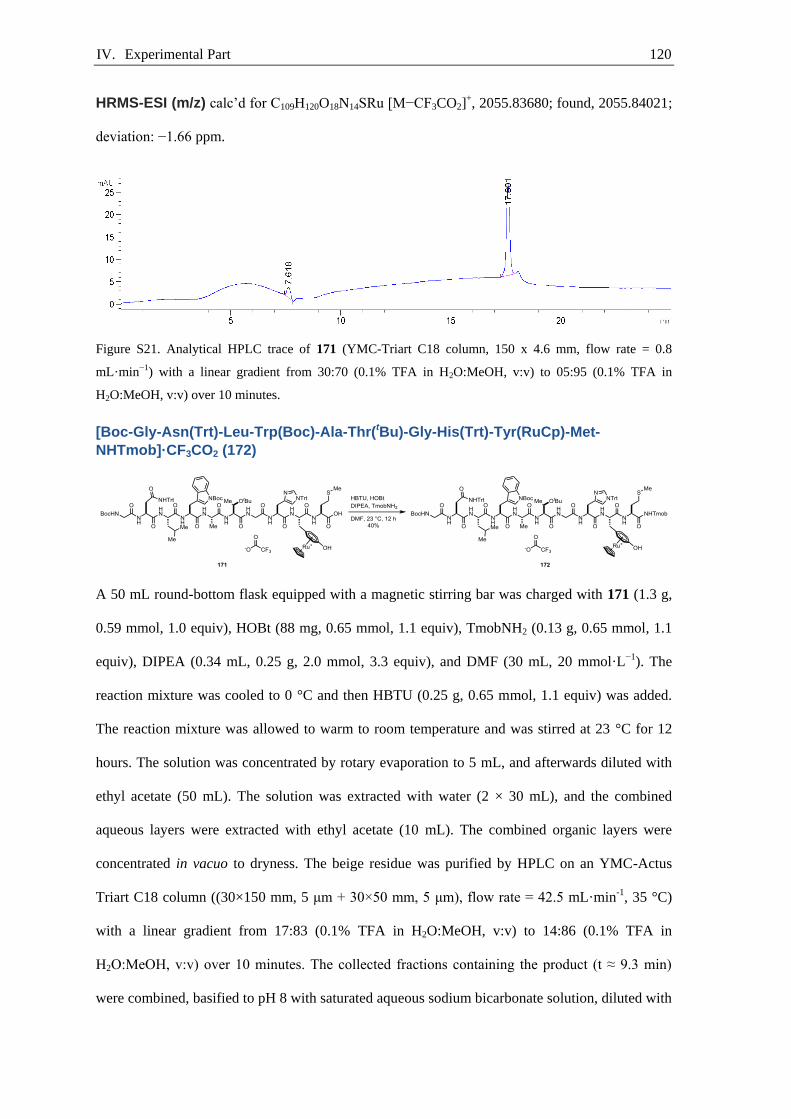
IV. Experimental Part 120
HRMS-ESI (m/z) calc’d for C109H120O18N14SRu [M−CF3CO2]+, 2055.83680; found, 2055.84021;
deviation: −1.66 ppm.
Figure S21. Analytical HPLC trace of 171 (YMC-Triart C18 column, 150 x 4.6 mm, flow rate = 0.8
mL·min−1) with a linear gradient from 30:70 (0.1% TFA in H2O:MeOH, v:v) to 05:95 (0.1% TFA in
H2O:MeOH, v:v) over 10 minutes.
[Boc-Gly-Asn(Trt)-Leu-Trp(Boc)-Ala-Thr(tBu)-Gly-His(Trt)-Tyr(RuCp)-Met-
NHTmob]·CF3CO2 (172)
A 50 mL round-bottom flask equipped with a magnetic stirring bar was charged with 171 (1.3 g,
0.59 mmol, 1.0 equiv), HOBt (88 mg, 0.65 mmol, 1.1 equiv), TmobNH2 (0.13 g, 0.65 mmol, 1.1
equiv), DIPEA (0.34 mL, 0.25 g, 2.0 mmol, 3.3 equiv), and DMF (30 mL, 20 mmol·L−1). The
reaction mixture was cooled to 0 °C and then HBTU (0.25 g, 0.65 mmol, 1.1 equiv) was added.
The reaction mixture was allowed to warm to room temperature and was stirred at 23 °C for 12
hours. The solution was concentrated by rotary evaporation to 5 mL, and afterwards diluted with
ethyl acetate (50 mL). The solution was extracted with water (2 × 30 mL), and the combined
aqueous layers were extracted with ethyl acetate (10 mL). The combined organic layers were
concentrated in vacuo to dryness. The beige residue was purified by HPLC on an YMC-Actus
Triart C18 column ((30×150 mm, 5 μm + 30×50 mm, 5 μm), flow rate = 42.5 mL·min-1, 35 °C)
with a linear gradient from 17:83 (0.1% TFA in H2O:MeOH, v:v) to 14:86 (0.1% TFA in
H2O:MeOH, v:v) over 10 minutes. The collected fractions containing the product (t ≈ 9.3 min)
were combined, basified to pH 8 with saturated aqueous sodium bicarbonate solution, diluted with
IV. Experimental Part 121
100 mL brine and the resulting solution was concentrated by rotary evaporation (100 mbar, 35 °C)
until no more methanol was evaporated. The suspension was extracted with dichloromethane (3 ×
100 mL), and the combined organic layers were dried over sodium sulfate, filtered, and
concentrated in vacuo to dryness to afford 172 as a colorless powder (0.56 g, 0.24 mmol, 40%
yield).
NMR Spectroscopy:
1H NMR (500 MHz, CD3OD, 25 ºC, δ): 8.88, (s, 1H), 8.50 (br, 1H), 8.38 (br, 1H), 8.11 (d, J
= 7.6 Hz, 1H), 8.08–8.01 (m, 2H), 7.93 (d, J = 7.7 Hz, 1H), 7.77 (d, J = 6.1 Hz, 1H), 7.63 (d,
J = 6.8 Hz, 1H), 7.59 (t, J = 4.8 Hz, 1H), 7.47–7.34 (m, 6H), 7.32–7.24 (m, 2H), 7.22–7.13
(m, 25H), 6.14 (s, 2H), 6.10–5.99 (m, 4H), 5.28 (s, 5H), 4.61 (dd, J = 9.2, 4.3 Hz, 1H), 4.57
(dd, J = 9.2, 4.5 Hz, 1H), 4.46 (t, J = 6.4 Hz, 1H), 4.43–4.28 (m, 5H), 4.26–4.16 (m, 2H),
4.14 (dd, J = 6.3, 3.5 Hz, 1H), 4.06 (dt, J = 9.8, 4.9 Hz, 1H), 3.81–3.76 (m, 2H), 3.75 (s, 6H),
3.72 (s, 3H), 3.67 (s, 2H), 3.14 (dd, J = 15.8, 4.5 Hz, 1H), 3.10–3.03 (m, 1H), 2.98–2.91 (m,
3H), 2.87–2.83 (m, 1H), 2.80–2.74 (m, 1H), 2.48 (t, J = 13.5 Hz, 1H), 2.36 (t, J = 8.1 Hz,
2H), 2.02 (s, 3H), 1.83 (dq, J = 14.3, 8.0 Hz, 1H), 1.68 (s, 9H), 1.53 (td, J = 9.5, 9.1, 3.5 Hz,
1H), 1.46 (d, J = 7.1 Hz, 3H), 1.39 (s, 9H), 1.20–1.14 (m, 12H), 0.88 (d, J = 6.6 Hz, 3H),
0.81 (d, J = 6.5 Hz, 3H).
13C{1H} NMR (126 MHz, CD3OD, 25 ºC, δ): 176.0, 175.4, 174.5, 174.3, 173.8, 172.6, 172.3,
171.9, 171.7, 171.6, 171.5, 162.8, 160.9, 158.6, 151.0, 145.7, 141.6, 134.5, 131.5, 130.8,
130.1 129.9, 129.8, 129.3, 128.8, 128.7, 128.6, 128.0, 127.9, 125.6, 123.7, 120.2, 118.3,
116.2, 106.6, 98.8, 91.6, 87.2, 87.0, 85.1, 81.3, 80.9, 79.8, 76.1, 76.0, 75.7, 71.7, 67.8, 61.6,
56.3, 55.9, 55.6, 55.4, 54.0, 53.7, 51.9, 51.8, 44.8, 44.3, 40.8, 38.9, 36.5, 33.5, 32.8, 31.0,
30.7, 28.9, 28.8, 28.7, 28.6, 26.8, 25.8, 23.4, 22.0, 20.4, 17.5, 15.2. One carbon resonance is
not observed, presumably due to overlap with a solvent carbon resonance.
19F NMR (471 MHz, CD3OD, 25 ºC, δ): −77.4.
HRMS-ESI (m/z) calc’d for C119H142N15O20RuS [M-CF3CO2]+, 2234.93143; found, 2234.93487;
deviation: −1.54 ppm.

IV. Experimental Part 122
Figure S22. Analytical HPLC trace of 172 (Agilent Poroshell 120, 50 x 3.0 mm, flow rate = 1 mL·min-1)
with a linear gradient from 100:0 (0.1% TFA in H2O:MeOH, v:v) to 05:99 (0.1% TFA in H2O:MeOH, v:v)
over 5 minutes.
[H-Phe(4-F)-Ile-Cys-Val-Gln-Pro-Ser-Phe-OH]·CF3CO2H ([19F]151)
The peptide was synthesized according to the general procedure for peptide synthesis starting
with 1.0 mmol Fmoc-Phe-O-2CT resin. To the beige residue was added a mixture of TFA (6.8
mL, 10 g), DTT (0.58 g, 3.7 mmol, 3.7 equiv), water (0.58 mL, 0.58 g, 32 mmol, 32 equiv), and
triisopropylsilane (0.29 mL, 0.22 g, 1.4 µmol, 1.4 equiv), and the reaction mixture was stirred at
23 °C for 2 hours. The reaction mixture was added dropwise into a round-bottom flask (100 mL)
containing vigorously stirred diethyl ether (80 mL). The resulting suspension was filtered and the
filter cake was purified by HPLC on an YMC-Actus Triart C18 column ((30×150 mm, 5 μm +
30×50 mm, 5 μm), flow rate = 42.5 mL·min-1, 35 °C) with a linear gradient from 27:73 (0.1%
TFA in H2O:MeOH, v:v) to 03:97 (0.1% TFA in H2O:MeOH, v:v) over 10 minutes. The collected
fractions containing the product (t ≈ 7.5 min) were combined and concentrated in vacuo to
dryness to afford [19F]151 as a colorless solid (0.13 g, 0.12 mmol, 12% yield).
NMR Spectroscopy:
1H NMR (500 MHz, (CD3)2SO, 25 ºC, δ): 8.57 (d, J = 8.7 Hz, 1H), 8.35 (d, J = 7.9 Hz, 1H),
8.17 (d, J = 5.6 Hz, 1H), 8.15–8.05 (m, 2H), 7.92 (dd, J = 13.0, 7.8 Hz, 1H), 7.85 (d, J = 8.7
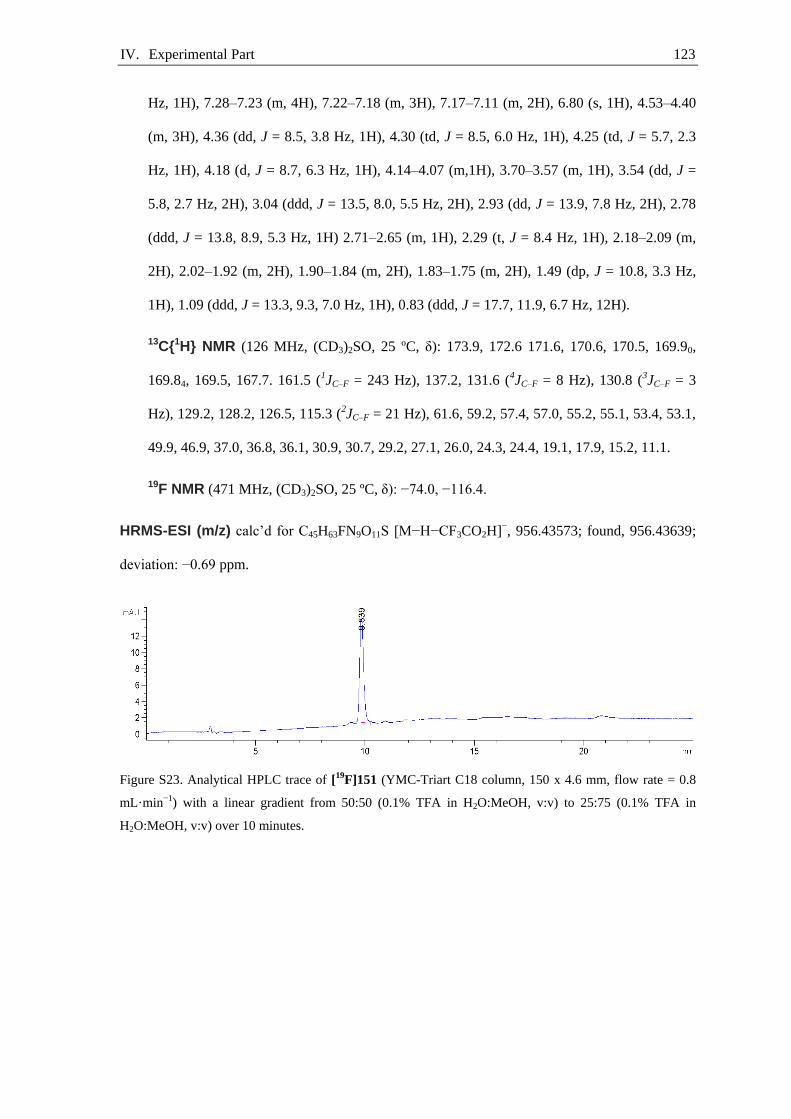
IV. Experimental Part 123
Hz, 1H), 7.28–7.23 (m, 4H), 7.22–7.18 (m, 3H), 7.17–7.11 (m, 2H), 6.80 (s, 1H), 4.53–4.40
(m, 3H), 4.36 (dd, J = 8.5, 3.8 Hz, 1H), 4.30 (td, J = 8.5, 6.0 Hz, 1H), 4.25 (td, J = 5.7, 2.3
Hz, 1H), 4.18 (d, J = 8.7, 6.3 Hz, 1H), 4.14–4.07 (m,1H), 3.70–3.57 (m, 1H), 3.54 (dd, J =
5.8, 2.7 Hz, 2H), 3.04 (ddd, J = 13.5, 8.0, 5.5 Hz, 2H), 2.93 (dd, J = 13.9, 7.8 Hz, 2H), 2.78
(ddd, J = 13.8, 8.9, 5.3 Hz, 1H) 2.71–2.65 (m, 1H), 2.29 (t, J = 8.4 Hz, 1H), 2.18–2.09 (m,
2H), 2.02–1.92 (m, 2H), 1.90–1.84 (m, 2H), 1.83–1.75 (m, 2H), 1.49 (dp, J = 10.8, 3.3 Hz,
1H), 1.09 (ddd, J = 13.3, 9.3, 7.0 Hz, 1H), 0.83 (ddd, J = 17.7, 11.9, 6.7 Hz, 12H).
13C{1H} NMR (126 MHz, (CD3)2SO, 25 ºC, δ): 173.9, 172.6 171.6, 170.6, 170.5, 169.90,
169.84, 169.5, 167.7. 161.5 (1JC–F = 243 Hz), 137.2, 131.6 (4JC–F = 8 Hz), 130.8 (3JC–F = 3
Hz), 129.2, 128.2, 126.5, 115.3 (2JC–F = 21 Hz), 61.6, 59.2, 57.4, 57.0, 55.2, 55.1, 53.4, 53.1,
49.9, 46.9, 37.0, 36.8, 36.1, 30.9, 30.7, 29.2, 27.1, 26.0, 24.3, 24.4, 19.1, 17.9, 15.2, 11.1.
19F NMR (471 MHz, (CD3)2SO, 25 ºC, δ): −74.0, −116.4.
HRMS-ESI (m/z) calc’d for C45H63FN9O11S [M−H−CF3CO2H]−, 956.43573; found, 956.43639;
deviation: −0.69 ppm.
Figure S23. Analytical HPLC trace of [19F]151 (YMC-Triart C18 column, 150 x 4.6 mm, flow rate = 0.8
mL·min−1) with a linear gradient from 50:50 (0.1% TFA in H2O:MeOH, v:v) to 25:75 (0.1% TFA in
H2O:MeOH, v:v) over 10 minutes.
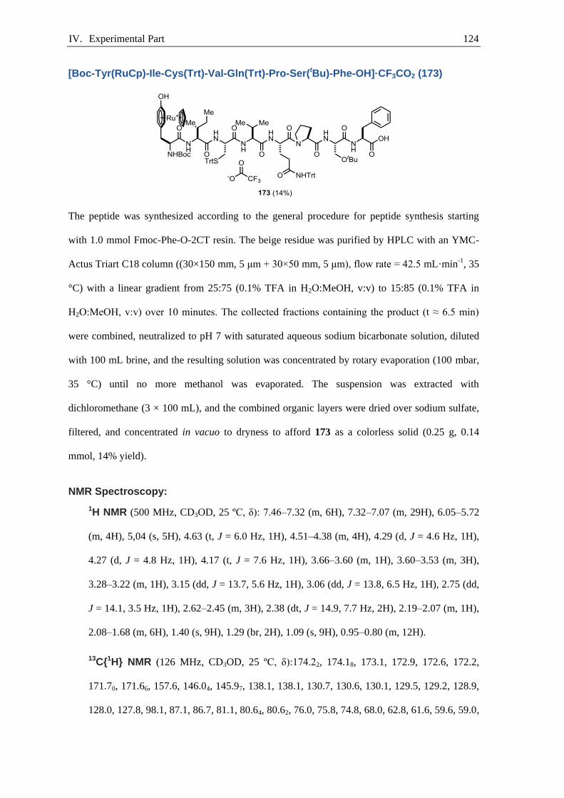
IV. Experimental Part 124
[Boc-Tyr(RuCp)-Ile-Cys(Trt)-Val-Gln(Trt)-Pro-Ser(tBu)-Phe-OH]·CF3CO2 (173)
The peptide was synthesized according to the general procedure for peptide synthesis starting
with 1.0 mmol Fmoc-Phe-O-2CT resin. The beige residue was purified by HPLC with an YMC-
Actus Triart C18 column ((30×150 mm, 5 μm + 30×50 mm, 5 μm), flow rate = 42.5 mL·min-1, 35
°C) with a linear gradient from 25:75 (0.1% TFA in H2O:MeOH, v:v) to 15:85 (0.1% TFA in
H2O:MeOH, v:v) over 10 minutes. The collected fractions containing the product (t ≈ 6.5 min)
were combined, neutralized to pH 7 with saturated aqueous sodium bicarbonate solution, diluted
with 100 mL brine, and the resulting solution was concentrated by rotary evaporation (100 mbar,
35 °C) until no more methanol was evaporated. The suspension was extracted with
dichloromethane (3 × 100 mL), and the combined organic layers were dried over sodium sulfate,
filtered, and concentrated in vacuo to dryness to afford 173 as a colorless solid (0.25 g, 0.14
mmol, 14% yield).
NMR Spectroscopy:
1H NMR (500 MHz, CD3OD, 25 ºC, δ): 7.46–7.32 (m, 6H), 7.32–7.07 (m, 29H), 6.05–5.72
(m, 4H), 5,04 (s, 5H), 4.63 (t, J = 6.0 Hz, 1H), 4.51–4.38 (m, 4H), 4.29 (d, J = 4.6 Hz, 1H),
4.27 (d, J = 4.8 Hz, 1H), 4.17 (t, J = 7.6 Hz, 1H), 3.66–3.60 (m, 1H), 3.60–3.53 (m, 3H),
3.28–3.22 (m, 1H), 3.15 (dd, J = 13.7, 5.6 Hz, 1H), 3.06 (dd, J = 13.8, 6.5 Hz, 1H), 2.75 (dd,
J = 14.1, 3.5 Hz, 1H), 2.62–2.45 (m, 3H), 2.38 (dt, J = 14.9, 7.7 Hz, 2H), 2.19–2.07 (m, 1H),
2.08–1.68 (m, 6H), 1.40 (s, 9H), 1.29 (br, 2H), 1.09 (s, 9H), 0.95–0.80 (m, 12H).
13C{1H} NMR (126 MHz, CD3OD, 25 ºC, δ):174.22, 174.18, 173.1, 172.9, 172.6, 172.2,
171.70, 171.66, 157.6, 146.04, 145.97, 138.1, 138.1, 130.7, 130.6, 130.1, 129.5, 129.2, 128.9,
128.0, 127.8, 98.1, 87.1, 86.7, 81.1, 80.64, 80.62, 76.0, 75.8, 74.8, 68.0, 62.8, 61.6, 59.6, 59.0,

IV. Experimental Part 125
57.1, 55.0, 53.7, 51.7, 38.6, 38.5, 37.3, 33.5, 32.4, 30.9, 30.8, 29.0, 28.7, 27.8, 25.8, 25.7,
19.7, 19.0, 16.0, 11.6. Two carbon resonances are not observed, presumably due to overlap
with a solvent carbon resonance.
19F NMR (471 MHz, CD3OD, 25 ºC, δ): −77.6.
HRMS-ESI (m/z) calc’d for C97H112O14N9RuS [M−H−CF3CO2H]−, 1760.70984; found,
1760.71274; deviation: −1.65 ppm.
Figure S24. Analytical HPLC trace of 173 (YMC-Triart C18 column, 150 x 4.6 mm, flow rate = 0.8
mL·min−1) with a linear gradient from 30:70 (0.1% TFA in H2O:MeOH, v:v) to 05:95 (0.1% TFA in
H2O:MeOH, v:v) over 10 minutes.
[Boc-Tyr(RuCp)-Ile-Cys(Trt)-Val-Gln(Trt)-Pro-Ser(tBu)-Phe-OPMB]·CF3CO2 (174)
A round-bottom flask (25 mL) equipped with a Teflon-coated magnetic stirring bar was charged
with 173 (0.15 g, 80 µmol, 1.0 equiv), HOBt (13 mg, 96 µmol, 1.2 equiv), PMBOH (0.10 mL,
0.11 g, 0.80 mmol, 10 equiv), DIPEA (50 µL, 37 mg, 0.29 mmol, 3.6 equiv) and DMF (4.3 mL,
20 mmol·L−1). The reaction mixture was cooled to 0 °C and then HBTU (33 mg, 88 µmol, 1.1
equiv) was added. The reaction mixture was allowed to warm to room temperature and was stirred
at 23 °C for 12 hours. The reaction mixture was cooled to 0 °C and then HBTU (33 mg, 88 µmol,
1.1 equiv) was added. The reaction mixture was allowed to warm to room temperature and was
stirred at 23 °C for 3 hours. The solution was diluted with ethyl acetate (50 mL). The solution was
extracted with water (2 × 30 mL), and the combined aqueous layers were extracted with ethyl
IV. Experimental Part 126
acetate (10 mL). The combined organic layers were dried over sodium sulfate, filtered, and
concentrated in vacuo to dryness. The beige residue was purified by HPLC on an YMC-Actus
Triart C18 column ((30×150 mm, 5 μm + 30×50 mm, 5 μm), flow rate = 42.5 mL·min-1, 35 °C)
with a linear gradient from 25:75 (0.1% TFA in H2O:MeOH, v:v) to 03:97 (0.1% TFA in
H2O:MeOH, v:v) over 10 minutes. The collected fractions containing the product (t ≈ 8.5 min)
were combined, basified to pH 10 with saturated aqueous sodium bicarbonate solution, diluted
with 100 mL brine and the resulting solution was concentrated by rotary evaporation (100 mbar,
35 °C) until no more methanol was evaporated. The suspension was extracted with
dichloromethane (3 × 50 mL), and the combined organic layers were dried over sodium sulfate,
filtered, and concentrated in vacuo to dryness to afford 174 a colorless powder (96 mg, 48 µmol,
60% yield).
NMR Spectroscopy:
1H NMR (500 MHz, CD3OD, 25 ºC, δ): 7.40–7.34 (m, 6H), 7.28–7.14 (m, 29H), 7.13–7.09
(m, 2H), 6.89–6.84 (m, 2H), 5.68–5.62 (m, 2H), 5.40–5.30 (m, 2H), 5.04–5.00 (m, 2H),
4.92–4.88 (m, 5H), 4.69–4.61 (m, 1H), 4.45–4.38 (m, 3H), 4.36–4.30 (m, 1H), 4.29–4.20 (m,
2H), 4.20–4.14 (m, 1H), 3.76 (m, 3H), 3.58–3.51 (m, 1H), 3.50–3.41 (m, 2H), 3.11 (dd, J =
13.9, 5.9 Hz, 1H), 3.06–2.96 (m, 1H), 2.69 (dd, J = 14.6, 4.3 Hz, 1H), 2.59–2.45 (m, 3H),
2.36 (dt, J = 14.8, 7.4 Hz, 1H), 2.15–1.71 (m, 8H), 1.57–1.50 (m, 1H) 1.40 (s, 9H),1.39–1.34
(m, 2H) 1.29 (br, 2H), 1.06 (s, 2H), 1.09 (s, 9H), 0.93–0.80 (m, 12H).
13C{1H} NMR (126 MHz, CD3OD, 25 ºC, δ):174.08, 174.06, 173.3, 173.0, 172.66, 172.54,
172.50, 171.9, 171.8, 161.3, 157.7, 150.5, 146.1, 146.0, 137.8, 131.5, 131.4, 130.7, 130.4,
130.1, 129.6, 129.2, 128.9, 128.8, 128.0, 127.9, 127.8, 114.9, 94.6, 87.3, 86.8, 81.0, 78.8,
76.1, 75.9, 74.8, 74.7, 71.5, 68.02, 67.96, 62.7, 62.0, 59.7, 59.0, 57.3, 55.8, 55.5, 55.0, 53.7,
51.8, 38.7, 38.5, 37.5, 34.8, 33.6, 32.3, 30.7, 28.8, 27.8, 25.8, 19.7, 18.9, 16.0, 11.6.
19F NMR (471 MHz, CD3OD, 25 ºC, δ): −80.8.
HRMS-ESI (m/z) calc’d for C105H123O15N9RuS [M−CF3CO2]+, 1883.78974; found, 1883.79122;
deviation: −0.79 ppm.

IV. Experimental Part 127
Figure S25. Analytical HPLC trace of 174 (YMC-Triart C18 column, 150 x 4.6 mm, flow rate = 0.8
mL·min−1) with a linear gradient from 30:70 (0.1% TFA in H2O:MeOH, v:v) to 05:95 (0.1% TFA in
H2O:MeOH, v:v) over 10 minutes.
Boc-Leu-(4-F-Phe)-Glu(tBu)-Met-Lys(Boc)-OH (175)
The peptide was synthesized according to the general procedure for peptide synthesis starting
with 0.88 mmol Fmoc-Lys-O-2CT resin. The beige residue was purified by HPLC with an YMC-
Actus Triart C18 column ((30×150 mm, 5 μm + 30×50 mm, 5 μm), flow rate = 42.5 mL·min-1, 35
°C) with a linear gradient from 20:80 (0.1% TFA in H2O:MeOH, v:v) to 03:97 (0.1% TFA in
H2O:MeOH, v:v) over 6 minutes. The collected fractions containing the product (t ≈ 7.5 min)
were combined, neutralized to pH 7 with saturated aqueous sodium bicarbonate solution, diluted
with 100 mL brine and the resulting solution was concentrated by rotary evaporation (100 mbar,
35 °C) until no more methanol was evaporated. The suspension was extracted with
dichloromethane (3 × 100 mL), and the combined organic layers were dried over sodium sulfate,
filtered, and concentrated in vacuo to dryness to afford 175 as a colorless solid (0.45 mg, 0.48
mmol, 55% yield).
HRMS-ESI (m/z) calc’d for C45H73O12N6FSNa [M+Na]+, 963.4883; found 963.4884; deviation:
−0.08 ppm.

IV. Experimental Part 128
Figure S26. Analytical HPLC trace of 175 (Agilent Poroshell 120, 50 x 3.0 mm, flow rate = 1 mL·min-1)
with a linear gradient from 100:0 (0.1% TFA in H2O:MeOH, v:v) to 05:95 (0.1% TFA in H2O:MeOH, v:v)
over 5 minutes.
Boc-Leu-(4-F-Phe)-Glu(tBu)-Met-Lys(Boc)-NHTmob ([19F]143)
A 10 mL round-bottom flask equipped with a magnetic stirring bar was charged with 175 (50 mg,
53 µmol, 1.0 equiv), HOBt (8.6 mg, 64 µmol, 1.2 equiv), TmobNH2 (13 mg, 64 µmol, 1.2 equiv),
DIPEA (33 µL, 25 mg, 0.19 mmol, 3.30 equiv), and DMF (2.7 mL, 20 mmol·L−1). The reaction
mixture was cooled to 0 °C and then HBTU (24 mg, 64 µmol, 1.2 equiv) was added. The reaction
mixture was allowed to warm to room temperature and was stirred at 23 °C for 12 hours. The
solution was diluted with ethyl acetate (50 mL). The solution was extracted with water (2 × 30
mL), and the combined aqueous layers were extracted with ethyl acetate (10 mL). The combined
organic layers were concentrated in vacuo to dryness. The beige residue was purified by HPLC on
an YMC-Actus Triart C18 column ((30×150 mm, 5 μm + 30×50 mm, 5 μm), flow rate = 42.5
mL·min-1, 35 °C) with a linear gradient from 17:83 (0.1% TFA in H2O:MeOH, v:v) to 14:86
(0.1% TFA in H2O:MeOH, v:v) over 10 minutes. The collected fractions containing the product (t
≈ 9.3 min) were combined, basified to pH 8 with saturated aqueous sodium bicarbonate solution,
diluted with 100 mL brine and the resulting solution was concentrated by rotary evaporation (100
mbar, 35 °C) until no more methanol was evaporated. The suspension was extracted with
dichloromethane (3 × 100 mL), and the combined organic layers were dried over sodium sulfate,
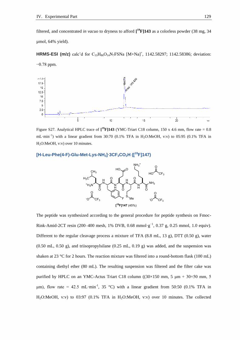
IV. Experimental Part 129
filtered, and concentrated in vacuo to dryness to afford [19F]143 as a colorless powder (38 mg, 34
µmol, 64% yield).
HRMS-ESI (m/z) calc’d for C55H86O14N7FSNa [M+Na]+, 1142.58297; 1142.58386; deviation:
−0.78 ppm.
Figure S27. Analytical HPLC trace of [19F]143 (YMC-Triart C18 column, 150 x 4.6 mm, flow rate = 0.8
mL·min−1) with a linear gradient from 30:70 (0.1% TFA in H2O:MeOH, v:v) to 05:95 (0.1% TFA in
H2O:MeOH, v:v) over 10 minutes.
[H-Leu-Phe(4-F)-Glu-Met-Lys-NH2]·3CF3CO2H ([19F]147)
The peptide was synthesized according to the general procedure for peptide synthesis on Fmoc-
Rink-Amid-2CT resin (200–400 mesh, 1% DVB, 0.68 mmol·g−1, 0.37 g, 0.25 mmol, 1.0 equiv).
Different to the regular cleavage process a mixture of TFA (8.8 mL, 13 g), DTT (0.50 g), water
(0.50 mL, 0.50 g), and triisopropylsilane (0.25 mL, 0.19 g) was added, and the suspension was
shaken at 23 °C for 2 hours. The reaction mixture was filtered into a round-bottom flask (100 mL)
containing diethyl ether (80 mL). The resulting suspension was filtered and the filter cake was
purified by HPLC on an YMC-Actus Triart C18 column ((30×150 mm, 5 μm + 30×50 mm, 5
μm), flow rate = 42.5 mL·min-1, 35 °C) with a linear gradient from 50:50 (0.1% TFA in
H2O:MeOH, v:v) to 03:97 (0.1% TFA in H2O:MeOH, v:v) over 10 minutes. The collected
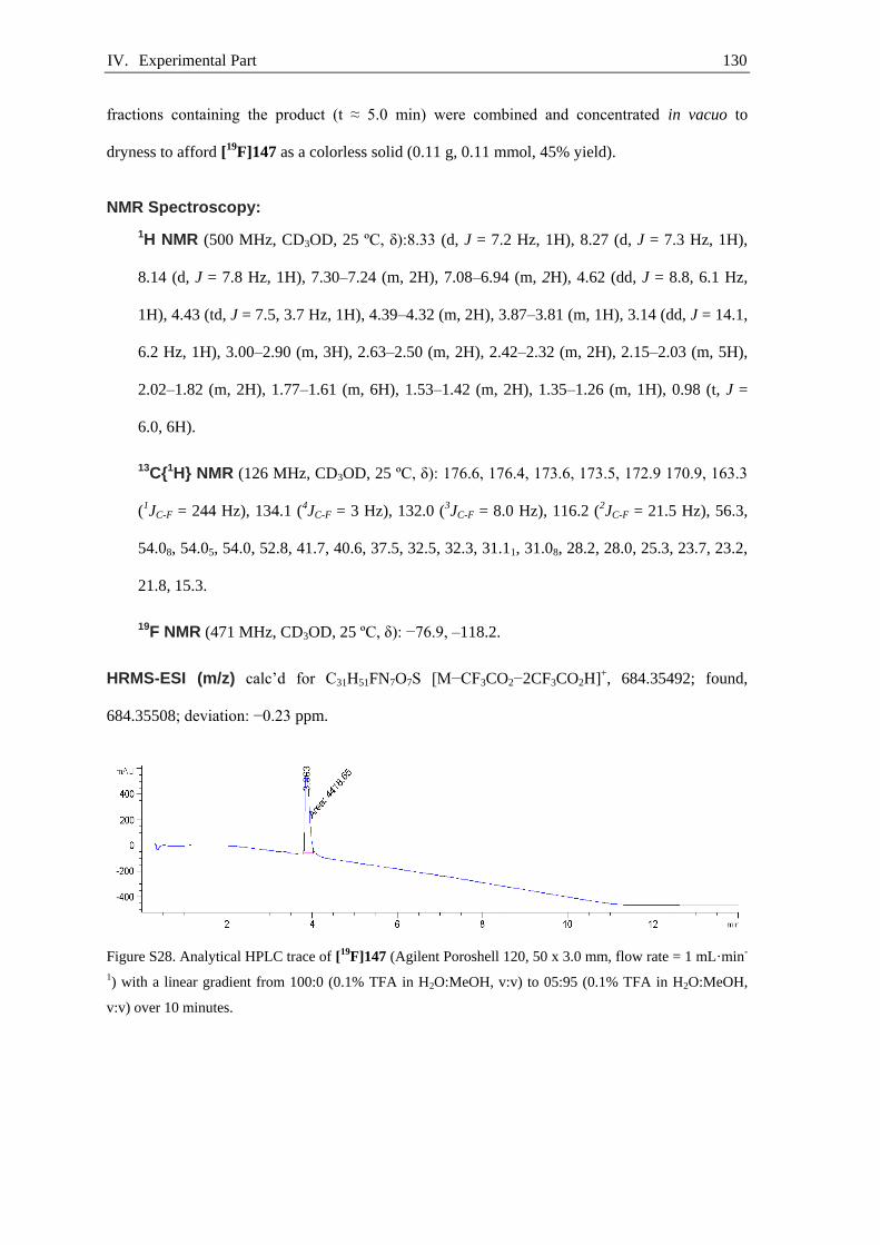
IV. Experimental Part 130
fractions containing the product (t ≈ 5.0 min) were combined and concentrated in vacuo to
dryness to afford [19F]147 as a colorless solid (0.11 g, 0.11 mmol, 45% yield).
NMR Spectroscopy:
1H NMR (500 MHz, CD3OD, 25 ºC, δ):8.33 (d, J = 7.2 Hz, 1H), 8.27 (d, J = 7.3 Hz, 1H),
8.14 (d, J = 7.8 Hz, 1H), 7.30–7.24 (m, 2H), 7.08–6.94 (m, 2H), 4.62 (dd, J = 8.8, 6.1 Hz,
1H), 4.43 (td, J = 7.5, 3.7 Hz, 1H), 4.39–4.32 (m, 2H), 3.87–3.81 (m, 1H), 3.14 (dd, J = 14.1,
6.2 Hz, 1H), 3.00–2.90 (m, 3H), 2.63–2.50 (m, 2H), 2.42–2.32 (m, 2H), 2.15–2.03 (m, 5H),
2.02–1.82 (m, 2H), 1.77–1.61 (m, 6H), 1.53–1.42 (m, 2H), 1.35–1.26 (m, 1H), 0.98 (t, J =
6.0, 6H).
13C{1H} NMR (126 MHz, CD3OD, 25 ºC, δ): 176.6, 176.4, 173.6, 173.5, 172.9 170.9, 163.3
(1JC-F = 244 Hz), 134.1 (4JC-F = 3 Hz), 132.0 (3JC-F = 8.0 Hz), 116.2 (2JC-F = 21.5 Hz), 56.3,
54.08, 54.05, 54.0, 52.8, 41.7, 40.6, 37.5, 32.5, 32.3, 31.11, 31.08, 28.2, 28.0, 25.3, 23.7, 23.2,
21.8, 15.3.
19F NMR (471 MHz, CD3OD, 25 ºC, δ): −76.9, –118.2.
HRMS-ESI (m/z) calc’d for C31H51FN7O7S [M−CF3CO2−2CF3CO2H]+, 684.35492; found,
684.35508; deviation: −0.23 ppm.
Figure S28. Analytical HPLC trace of [19F]147 (Agilent Poroshell 120, 50 x 3.0 mm, flow rate = 1 mL·min-
1) with a linear gradient from 100:0 (0.1% TFA in H2O:MeOH, v:v) to 05:95 (0.1% TFA in H2O:MeOH,
v:v) over 10 minutes.
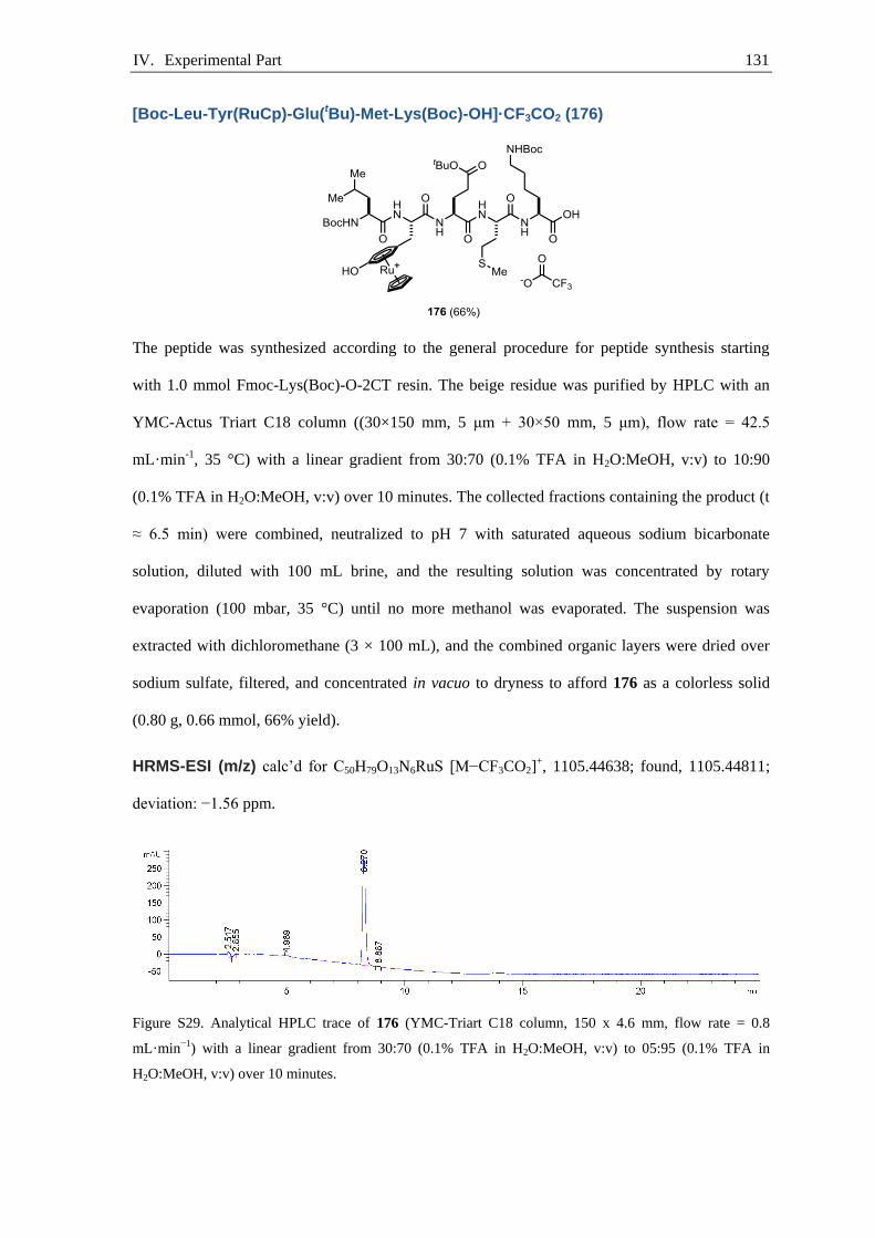
IV. Experimental Part 131
[Boc-Leu-Tyr(RuCp)-Glu(tBu)-Met-Lys(Boc)-OH]·CF3CO2 (176)
The peptide was synthesized according to the general procedure for peptide synthesis starting
with 1.0 mmol Fmoc-Lys(Boc)-O-2CT resin. The beige residue was purified by HPLC with an
YMC-Actus Triart C18 column ((30×150 mm, 5 μm + 30×50 mm, 5 μm), flow rate = 42.5
mL·min-1, 35 °C) with a linear gradient from 30:70 (0.1% TFA in H2O:MeOH, v:v) to 10:90
(0.1% TFA in H2O:MeOH, v:v) over 10 minutes. The collected fractions containing the product (t
≈ 6.5 min) were combined, neutralized to pH 7 with saturated aqueous sodium bicarbonate
solution, diluted with 100 mL brine, and the resulting solution was concentrated by rotary
evaporation (100 mbar, 35 °C) until no more methanol was evaporated. The suspension was
extracted with dichloromethane (3 × 100 mL), and the combined organic layers were dried over
sodium sulfate, filtered, and concentrated in vacuo to dryness to afford 176 as a colorless solid
(0.80 g, 0.66 mmol, 66% yield).
HRMS-ESI (m/z) calc’d for C50H79O13N6RuS [M−CF3CO2]+, 1105.44638; found, 1105.44811;
deviation: −1.56 ppm.
Figure S29. Analytical HPLC trace of 176 (YMC-Triart C18 column, 150 x 4.6 mm, flow rate = 0.8
mL·min−1) with a linear gradient from 30:70 (0.1% TFA in H2O:MeOH, v:v) to 05:95 (0.1% TFA in
H2O:MeOH, v:v) over 10 minutes.
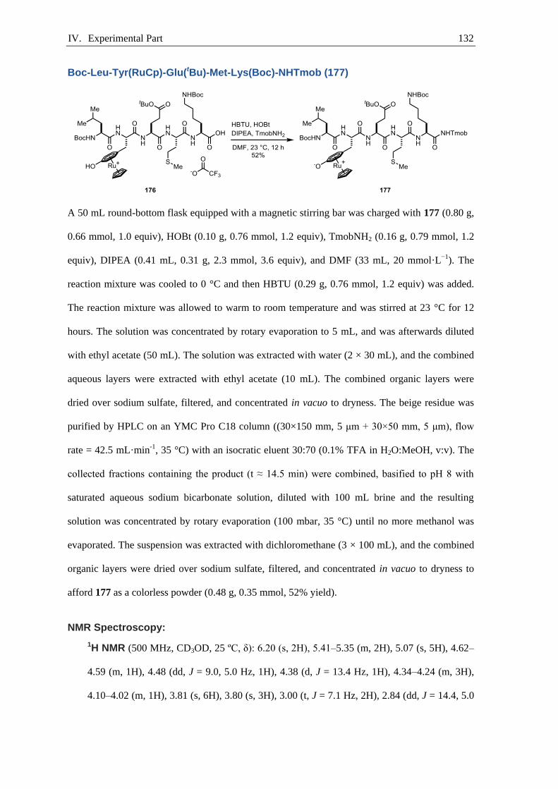
IV. Experimental Part 132
Boc-Leu-Tyr(RuCp)-Glu(tBu)-Met-Lys(Boc)-NHTmob (177)
A 50 mL round-bottom flask equipped with a magnetic stirring bar was charged with 177 (0.80 g,
0.66 mmol, 1.0 equiv), HOBt (0.10 g, 0.76 mmol, 1.2 equiv), TmobNH2 (0.16 g, 0.79 mmol, 1.2
equiv), DIPEA (0.41 mL, 0.31 g, 2.3 mmol, 3.6 equiv), and DMF (33 mL, 20 mmol·L−1). The
reaction mixture was cooled to 0 °C and then HBTU (0.29 g, 0.76 mmol, 1.2 equiv) was added.
The reaction mixture was allowed to warm to room temperature and was stirred at 23 °C for 12
hours. The solution was concentrated by rotary evaporation to 5 mL, and was afterwards diluted
with ethyl acetate (50 mL). The solution was extracted with water (2 × 30 mL), and the combined
aqueous layers were extracted with ethyl acetate (10 mL). The combined organic layers were
dried over sodium sulfate, filtered, and concentrated in vacuo to dryness. The beige residue was
purified by HPLC on an YMC Pro C18 column ((30×150 mm, 5 μm + 30×50 mm, 5 μm), flow
rate = 42.5 mL·min-1, 35 °C) with an isocratic eluent 30:70 (0.1% TFA in H2O:MeOH, v:v). The
collected fractions containing the product (t ≈ 14.5 min) were combined, basified to pH 8 with
saturated aqueous sodium bicarbonate solution, diluted with 100 mL brine and the resulting
solution was concentrated by rotary evaporation (100 mbar, 35 °C) until no more methanol was
evaporated. The suspension was extracted with dichloromethane (3 × 100 mL), and the combined
organic layers were dried over sodium sulfate, filtered, and concentrated in vacuo to dryness to
afford 177 as a colorless powder (0.48 g, 0.35 mmol, 52% yield).
NMR Spectroscopy:
1H NMR (500 MHz, CD3OD, 25 ºC, δ): 6.20 (s, 2H), 5.41–5.35 (m, 2H), 5.07 (s, 5H), 4.62–
4.59 (m, 1H), 4.48 (dd, J = 9.0, 5.0 Hz, 1H), 4.38 (d, J = 13.4 Hz, 1H), 4.34–4.24 (m, 3H),
4.10–4.02 (m, 1H), 3.81 (s, 6H), 3.80 (s, 3H), 3.00 (t, J = 7.1 Hz, 2H), 2.84 (dd, J = 14.4, 5.0
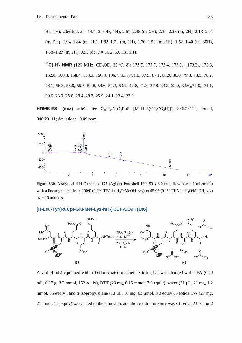
IV. Experimental Part 133
Hz, 1H), 2.66 (dd, J = 14.4, 8.0 Hz, 1H), 2.61–2.45 (m, 2H), 2.39–2.25 (m, 2H), 2.13–2.01
(m, 5H), 1.94–1.84 (m, 2H), 1.82–1.71 (m, 1H), 1.70–1.59 (m, 2H), 1.52–1.40 (m, 30H),
1.38–1.27 (m, 2H), 0.93 (dd, J = 16.2, 6.6 Hz, 6H).
13C{1H} NMR (126 MHz, CD3OD, 25 ºC, δ): 175.7, 173.7, 173.4, 173.32, .173.25, 172.3,
162.8, 160.8, 158.4, 158.0, 150.8, 106.7, 93.7, 91.6, 87.5, 87.1, 81.9, 80.8, 79.8, 78.9, 76.2,
76.1, 56.3, 55.8, 55.5, 54.8, 54.6, 54.2, 53.9, 42.0, 41.3, 37.8, 33.2, 32.9, 32.68,32.65, 31.1,
30.6, 28.9, 28.8, 28.4, 28.3, 25.9, 24.1, 23.4, 22.0.
HRMS-ESI (m/z) calc’d for C36H54N7O8RuS [M–H–3(CF3CO2H)]–, 846.28111; found,
846.28111; deviation: −0.89 ppm.
Figure S30. Analytical HPLC trace of 177 (Agilent Poroshell 120, 50 x 3.0 mm, flow rate = 1 mL·min-1)
with a linear gradient from 100:0 (0.1% TFA in H2O:MeOH, v:v) to 05:95 (0.1% TFA in H2O:MeOH, v:v)
over 10 minutes.
[H-Leu-Tyr(RuCp)-Glu-Met-Lys-NH2]·3CF3CO2H (146)
A vial (4 mL) equipped with a Teflon-coated magnetic stirring bar was charged with TFA (0.24
mL, 0.37 g, 3.2 mmol, 152 equiv), DTT (23 mg, 0.15 mmol, 7.0 equiv), water (21 µL, 21 mg, 1.2
mmol, 55 euqiv), and triisopropylsilane (13 µL, 10 mg, 63 µmol, 3.0 equiv). Peptide 177 (27 mg,
21 µmol, 1.0 equiv) was added to the emulsion, and the reaction mixture was stirred at 23 °C for 2
IV. Experimental Part 134
hours. To the emulsion was added diethyl ether (1 mL) and the precipitate was collected by
filtration. The white precipitate was purified by HPLC on an YMC Triart C18 column ((30×150
mm, 5 μm + 30×50 mm, 5 μm), flow rate = 42.5 mL·min-1, 35 °C) with a linear gradient from
80:20 (0.1% TFA in H2O:MeOH, v:v) to 60:40 (0.1% TFA in H2O:MeOH, v:v) over 10 minutes.
The collected fractions containing the product (t ≈ 8.1 min) were combined, and concentrated by
rotary evaporation to afford 146 as a white powder (14 mg, 11 µmol, 54% yield).
NMR Spectroscopy:
1H NMR (500 MHz, CD3OD, 25 ºC, δ):8.48 (d, J = 7.0, Hz, 1H), 8.39 (d, J = 7.5 Hz, 1H),
6.13–6.09 (m, 4H), 5.34 (s, 5H), 4.65 (t, J = 6.8 Hz, 1H), 4.54–4.49 (m, 1H), 4.38–4.31 (m,
2H), 3.97–3.92 (m, 1H), 2.97–2.90 (m, 3H), 2.75 (dd, J = 14.1, 7.5 Hz, 1H), 2.63–2.51 (m,
2H), 2.47–2.34 (m, 2H), 2.18–2.03 (m, 5H), 2.02–1.82 (m, 2H), 1.77–1.63 (m, 6H), 1.54–
1.40 (m, 2H), 1.35–1.25 (m, 1H), 1.01–0.96 (m, 6H).
13C{1H} NMR (126 MHz, CD3OD, 25 ºC, δ): 176.4, 176.3, 173.6, 173.5, 171.4, 170.9, 134.8,
98.1, 87.1, 86.8, 81.3, 76.0, 75.9, 55.6, 54.3, 54.0, 53.8, 52.8, 41.7, 40.5, 37.3, 32.6, 32.4,
31.1, 31.0, 28.2, 28.1, 25.4, 23.7, 23.1, 22.0, 15.2.
19F NMR (471 MHz, CD3OD, 25 ºC, δ): −76.9.
HRMS-ESI (m/z) calc’d for C36H54N7O8SRu [M−3CF3CO2H]–, 846.28111; found, 846.28111;
deviation: −0.89 ppm.
Figure S31. Analytical HPLC trace of 146 (Agilent Poroshell 120, 50 x 3.0 mm, flow rate = 1 mL·min-1)
with a linear gradient from 100:0 (0.1% TFA in H2O:MeOH, v:v) to 05:95 (0.1% TFA in H2O:MeOH, v:v)
over 10 minutes.

IV. Experimental Part 135
Boc-Gly-His(Trt)-Gly-D-Phe(4-F)-Gly-NHTmob (178)
The peptide was synthesized according to the general procedure for peptide synthesis starting
with 0.25 mmol Fmoc-Gly-O-2CT resin. A 50 mL round-bottom flask equipped with a magnetic
stirring bar was charged with the solid residue from SPPS, HOBt (41 mg, 0.30 mmol, 1.2 equiv),
TmobNH2 (54 mg, 0.28 mmol, 1.1 equiv), DIPEA (0.13 mL, 97 mg, 0.75 mmol, 3.0 equiv), and
DMF (13 mL, 20 mmol·L−1). The reaction mixture was cooled to 0 °C and then HBTU (0.11 g,
0.30 mmol, 1.2 equiv) was added. The reaction mixture was allowed to warm to room
temperature and was stirred at 23 °C for 12 hours. The solution was concentrated by rotary
evaporation to 5 mL, and afterwards diluted with ethyl acetate (50 mL). The solution was
extracted with water (2 × 30 mL), and the combined aqueous layers were extracted with ethyl
acetate (10 mL). The combined organic layers were dried over sodium sulfate, filtered, and
concentrated in vacuo to dryness. The beige residue was purified by HPLC with an YMC Pro C18
column ((30×150 mm, 5 μm + 30×50 mm, 5 μm), flow rate = 42.5 mL·min-1, 35 °C) with a linear
gradient from 40:60 (0.1% TFA in H2O:MeOH, v:v) to 20:80 (0.1% TFA in H2O:MeOH, v:v)
over 10 minutes. The collected fractions containing the product (t ≈ 8.0 min) were combined,
neutralized to pH 7 with saturated aqueous sodium bicarbonate solution, diluted with 100 mL
brine and the resulting solution was concentrated by rotary evaporation (100 mbar, 35 °C) until no
more methanol was evaporated. The suspension was extracted with dichloromethane (3 × 100
mL), and the combined organic layers were dried over sodium sulfate, filtered, and concentrated
in vacuo to dryness to afford 178 as a colorless solid (0.12 g, 0.12 mmol, 49% yield).
HRMS-ESI (m/z) calc’d for C55H62FN8O10 [M+H]+, 1013.45674; found, 1013.45712; deviation:
−0.37 ppm.
IV. Experimental Part 136
Figure S32. Analytical HPLC trace of 178 (YMC-Triart C18 column, 150 x 4.6 mm, flow rate = 0.8
mL·min−1) with a linear gradient from 30:70 (0.1% TFA in H2O:MeOH, v:v) to 05:95 (0.1% TFA in
H2O:MeOH, v:v) over 10 minutes.
[H-Gly-His-Gly-D-Phe(4-F)-Gly-NH2]·2CF3CO2H (179)
A vial (4 mL) equipped with a Teflon-coated magnetic stirring bar was charged with TFA (4.4
mL, 6.55 g), DTT (0.25 g, 1.6 mmol, 16 equiv), water (0.25 mL, 0.25 g), and triisopropylsilane
(0.13 mL, 97 mg, 0.61 mmol, 6.2 equiv). Peptide 178 (0.10 g, 99 µmol, 1.0 equiv) was added to
the emulsion, and the reaction mixture was stirred at 23 °C for 2 hours. The reaction mixture was
concentrated in vacuo and the solid residue was purified by HPLC on an Chiralpak® ZWIX (+)
((10×250 mm, 5 μm), flow rate = 6.0 mL·min-1, 35 °C) with an isocratic eluent 49:49:2
(MeOH:MeCN:H2O+50 mM formic acid, 25 mM diethylamine). The collected fractions
containing the product (t ≈ 15.0 min) were combined and concentrated in vacuo to dryness to
afford 179 as a colorless solid (38 mg, 53 µmol, 54% yield).
NMR Spectroscopy:
1H NMR (500 MHz, CD3OD, 25 ºC, δ): 8.78 (d, J = 1.4 Hz, 1H), 7.38 (d, J =1.4 Hz, 1H),
7.34–7.18 (m, 2H), 7.07–6.88 (m, 2H), 4.68 (dd, J = 7.2, 5.6 Hz, 1H), 4.58 (dd, J = 9.0, 5.6
Hz, 1H), 3.97–3.95 (m, 1H), 3.93–3.90 (m, 1H), 3.88–3.80 (m, 1H), 3.79–3.73 (m, 3H),
3.37–3.30 (m, 1H), 3.19 (ddd, J =13.8, 10.0, 6.4 Hz, dH), 2.97 (dd, J =13.9, 9.0 Hz, 1H).

IV. Experimental Part 137
13C{1H} NMR (126 MHz, CD3OD, 25 ºC, δ): 174.0, 173.8, 172.6, 171.7, 168.1 163.3 (1JC-F =
243.5 Hz), 135.3, 134.4 (4JC-F = 3.2 Hz), 132.1 (3JC-F = 8 Hz), 130.3, 118.9, 116.1 (2JC-F =
21.5 Hz), 56.4, 54.3, 43.3, 43.0, 41.6, 37.6, 27.9.
19F NMR (471 MHz, CD3OD, 25 ºC, δ): −77.6, −119.2.
HRMS-ESI (m/z) calc’d for C21H28FN8O5 [M−CF3CO2−CF3CO2H]+, 491.21612; found,
491.21618; deviation: −0.13 ppm.
Figure S33. Analytical HPLC trace of 179 (Chiralpak® ZWIX(+) 3 μm, flow rate = 1 mL·min-1) with an
isocratic eluent 49:49:2 (MeOH:MeCN:H2O+50 mM formic acid, 25 mM diethylamine).
IV. Experimental Part 138
Boc-Gly-His(Trt)-Gly-Phe(4-F)-Gly-NHTmob (180)
The peptide was synthesized according to the general procedure for peptide synthesis starting
with 0.25 mmol Fmoc-Gly-O-2CT resin. A 50 mL round-bottom flask equipped with a magnetic
stirring bar was charged solid residue from SPPS, HOBt (41 mg, 0.30 mmol, 1.2 equiv),
TmobNH2 (54 mg, 0.28 mmol, 1.1 equiv), DIPEA (0.41 mL, 0.31 g, 2.4 mmol, 3.6 equiv), and
DMF (13 mL, 20 mmol·L−1). The reaction mixture was cooled to 0 °C and then HBTU (0.11 g,
0.30 mmol, 1.2 equiv) was added. The reaction mixture was allowed to warm to room
temperature and was stirred at 23 °C for 12 hours. The solution was concentrated by rotary
evaporation to 5 mL, and afterwards diluted with ethyl acetate (50 mL). The solution was
extracted with water (2 × 30 mL), and the combined aqueous layers were extracted with ethyl
acetate (10 mL). The combined organic layers were dried over sodium sulfate, filtered, and
concentrated in vacuo to dryness. The beige residue was purified by HPLC with an YMC Pro C18
column ((30×150 mm, 5 μm + 30×50 mm, 5 μm), flow rate = 42.5 mL·min-1, 35 °C) with a linear
gradient from 40:60 (0.1% TFA in H2O:MeOH, v:v) to 20:80 (0.1% TFA in H2O:MeOH, v:v)
over 10 minutes. The collected fractions containing the product (t ≈ 8.0 min) were combined,
neutralized to pH 7 with saturated aqueous sodium bicarbonate solution, diluted with 100 mL
brine and the resulting solution was concentrated by rotary evaporation (100 mbar, 35 °C) until no
more methanol was evaporated. The suspension was extracted with dichloromethane (3 ×
100 mL), and the combined organic layers were dried over sodium sulfate, filtered, and
concentrated in vacuo to dryness to afford 180 as a colorless solid (0.11 g, 0.10 mmol, 43%
yield).
HRMS-ESI (m/z) calc’d for C55H62FN8O10 [M+H]+, 1013.45674; found, 1013.45774; deviation:
−0.98 ppm.

IV. Experimental Part 139
Figure S34. Analytical HPLC trace of 180 (Agilent Poroshell 120, 50 x 3.0 mm, flow rate = 1 mL·min-1)
with a linear gradient from 100:0 (0.1% TFA in H2O:MeOH, v:v) to 05:95 (0.1% TFA in H2O:MeOH, v:v)
over 5 minutes.
[H-Gly-His-Gly-Phe(4-F)-Gly-NH2]·2CF3CO2H ([19F]152)
A vial (4 mL) equipped with a Teflon-coated magnetic stirring bar was charged with TFA (4.4
mL, 6.6 g), DTT (0.25 g, 1.6 mmol, 16 equiv), water (0.25 mL, 0.25 g), and triisopropylsilane
(0.13 mL, 97 mg, 0.61 mmol, 6.2 equiv). Peptide 180 (0.10 g, 99 µmol, 1.0 equiv) was added to
the emulsion, and the reaction mixture was stirred at 23 °C for 2 hours. The reaction mixture was
concentrated in vacuo and the solid residue was purified by HPLC on an Chiralpak® ZWIX (+)
((10×250 mm, 5 μm), flow rate = 6.0 mL·min-1, 35 °C) with an isocratic eluent 49:49:2
(MeOH:MeCN:H2O+50 mM formic acid, 25 mM diethylamine). The collected fractions
containing the product (t ≈ 17.5 min) were combined and concentrated in vacuo to dryness to
afford [19F]152 as a colorless solid (32 mg, 45 µmol, 45% yield).
NMR Spectroscopy:
1H NMR (500 MHz, CD3OD, 25 ºC, δ): 8.80 (d, J = 1.5 Hz, 1H), 7.39 (d, J =1.4 Hz, 1H),
7.27 (ddd, J = 8.5, 5.4, 2.7 Hz, 2H), 7.03–6.95 (m, 2H), 4.70 (t, J = 6.6 Hz, 1H), 4.60 (dd, J =
9.4, 5.4 Hz, 1H), 3.99–3.88 (m, 2H), 3.83–3.70 (m, 4H), 3.30–3.24 (m, 1H), 3.24–3.15 (m,
2H), 2.98 (dd, J =14.0, 9.5 Hz, 1H).

IV. Experimental Part 140
13C{1H} NMR (126 MHz, CD3OD, 25 ºC, δ): 174.0, 173.8, 172.1, 171.8, 167.9 163.3 (1JC-F =
243.6 Hz), 135.3, 134.3 (4JC-F = 3.0 Hz), 132.1 (3JC-F = 8.1 Hz), 130.6, 118.8, 116.1 (2JC-F =
21.4 Hz), 56.6, 53.9, 43.3, 43.0, 41.6, 37.6, 27.9.
19F NMR (471 MHz, CD3OD, 25 ºC, δ): −77.9, −119.1.
HRMS-ESI (m/z) calc’d for C21H28FN8O5 [M−CF3CO2H−CF3CO2]+, 491.21611; found,
491.21605; deviation: 0.14 ppm.
Figure S35. Analytical HPLC trace of [19F]152 (Chiralpak® ZWIX(+) 3 μm, flow rate = 1 mL·min-1) with
an isocratic eluent 49:49:2 (MeOH:MeCN:H2O+50 mM formic acid, 25 mM diethylamine).

IV. Experimental Part 141
[Boc-Gly-His(Trt)-Gly-Tyr(RuCp)-Gly-OH]·CF3CO2 (181)
The peptide was synthesized according to the general procedure for peptide synthesis starting
with 0.5 mmol Fmoc-Gly-O-2CT resin. The beige residue was purified by HPLC with an YMC
Pro C18 column ((30×150 mm, 5 μm + 30×50 mm, 5 μm), flow rate = 42.5 mL·min-1, 35 °C) with
a linear gradient from 30:70 (0.1% TFA in H2O:MeOH, v:v) to 05:95 (0.1% TFA in H2O:MeOH,
v:v) over 10 minutes. The collected fractions containing the product (t ≈ 4.5 min) were combined,
neutralized to PH 7 with saturated aqueous sodium bicarbonate solution, diluted with 100 mL
brine and the resulting solution was concentrated by rotary evaporation (100 mbar, 35 °C) until no
more methanol was evaporated. The suspension was extracted with dichloromethane (3 × 100
mL), and the combined organic layers were dried over sodium sulfate, filtered, and concentrated
in vacuo to dryness to afford 181 as a colorless solid (0.16 g, 0.12 mmol, 25% yield).
NMR Spectroscopy:
1H NMR (500 MHz, CD3OD, 25 ºC, δ): 7.42 (s, 1H), 7.37 (dd, J = 5.2, 2.0 Hz, 9H), 7.18–
7.09 (m, 6H), 6.77 (s, 1H), 6.19 (s, 2H), 5.71 (dd, J = 5.8, 3.7 Hz, 2H), 5.46–5.28 (m, 2H),
5.05 (s, 4H), 4.55 (dd, J = 9.1, 4.6 Hz, 1H), 4.49 (t, J = 6.6 Hz, 1H), 4.35 (d, J = 2.2 Hz, 1H),
3.94–3.80 (m, 2H), 3.78 (d, J = 1.1 Hz, 9H), 3.74–3.61 (m, 3H), 3.05 (dd, J = 14.7, 5.7 Hz,
1H), 2.96 (dd, J = 14.7, 7.2 Hz, 1H), 2.86 (dd, J = 14.3, 4.6 Hz, 1H), 2.66 (dd, J = 14.3, 9.2
Hz, 1H), 1.39 (s, 9H).
13C{1H} NMR (126 MHz, CD3OD, 25 ºC, δ): 174.2, 172.8, 172.7, 171.7, 170.4, 162.8, 160.8,
158.5, 150.6, 143.6, 139.8, 137.3, 130.9, 129.4, 129.3, 121.5, 106.7, 94.5, 91.6, 87.6, 86.8,
80.9, 78.9, 76.8, 76.1, 56.3, 55.8, 55.4, 44.9, 44.0, 43.4, 37.2, 33.3, 30.6, 28.8. Two carbon
resonances are not observed, presumably due to overlap with a solvent carbon resonance.
19F NMR (471 MHz, CD3OD, 25 ºC, δ): −77.5.
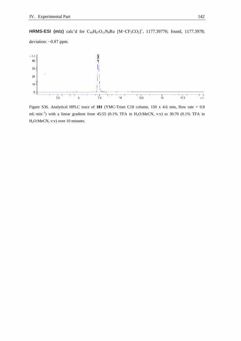
IV. Experimental Part 142
HRMS-ESI (m/z) calc’d for C60H67O11N8Ru [M−CF3CO2]+, 1177.39776; found, 1177.3978;
deviation: −0.87 ppm.
Figure S36. Analytical HPLC trace of 181 (YMC-Triart C18 column, 150 x 4.6 mm, flow rate = 0.8
mL·min−1) with a linear gradient from 45:55 (0.1% TFA in H2O:MeCN, v:v) to 30:70 (0.1% TFA in
H2O:MeCN, v:v) over 10 minutes.

IV. Experimental Part 143
General procedure for radio-deoxyfluorination of peptides IV.2.2.
Aqueous [18F]fluoride solution (700 μL) was loaded with a syringe onto a QMA anion-exchange
cartridge that was pre-conditioned according to the general procedure, and then the cartridge was
washed with MeCN (1 mL). The cartridge was dried by pushing air (2 mL) through the cartridge.
The [18F]fluoride was eluted from the cartridge with a solution of tracer precursor (5.0 μmol, 1.0
equiv), imidazolium chloride 40 (6.9 mg, 15 µmol, 3.0 equiv), and
bis(neopentyltrimethylammonium) oxalate 160 (5.0 mg, 14 µmol, 2.9 equiv) in a mixture of
ethanol and pivalonitrile (200 µL, 1:3, v:v), into a 4 mL borosilicate vial. The remaining substrate
was eluted from the cartridge with a mixture of veratrole and pivalonitrile (250 µL, 4:1, v:v) into
the same vial. The reaction vial containing 450 µL reaction mixture was sealed with a Teflon-
lined cap and was stirred at 130 °C for 30 minutes. The vial was removed from the hot plate, and
after 2 minutes the reaction mixture was concentrated by heating at 80 °C under a stream of
nitrogen (~5 min). To the remaining solution was added a mixture of TFA (0.42 mL),
triisopropylsilane (40 µL), DL-1,4-dithiothreitol (35 mg), and water (20 µL), then the mixture was
stirred at 50 °C for 10 minutes. The reaction mixture was diluted with water and was purified by
HPLC on a Hypersil Gold column (250×10 mm, 5 μm). The activity of the product containing
fraction was measured and the product identity and purity was determined by comparison of the
HPLC radio-trace with the HPLC UV-trace of the authentic reference sample.
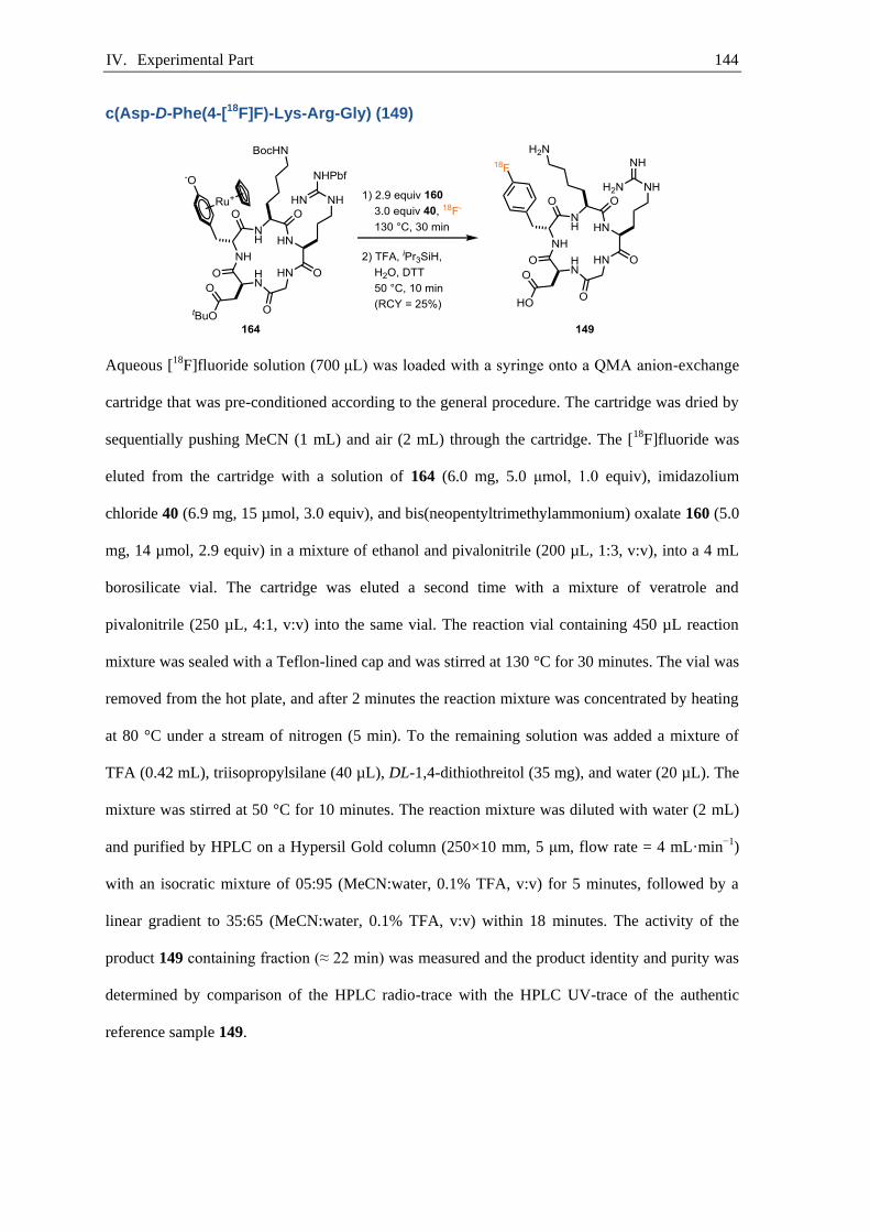
IV. Experimental Part 144
c(Asp-D-Phe(4-[18F]F)-Lys-Arg-Gly) (149)
Aqueous [18F]fluoride solution (700 μL) was loaded with a syringe onto a QMA anion-exchange
cartridge that was pre-conditioned according to the general procedure. The cartridge was dried by
sequentially pushing MeCN (1 mL) and air (2 mL) through the cartridge. The [18F]fluoride was
eluted from the cartridge with a solution of 164 (6.0 mg, 5.0 μmol, 1.0 equiv), imidazolium
chloride 40 (6.9 mg, 15 µmol, 3.0 equiv), and bis(neopentyltrimethylammonium) oxalate 160 (5.0
mg, 14 µmol, 2.9 equiv) in a mixture of ethanol and pivalonitrile (200 µL, 1:3, v:v), into a 4 mL
borosilicate vial. The cartridge was eluted a second time with a mixture of veratrole and
pivalonitrile (250 µL, 4:1, v:v) into the same vial. The reaction vial containing 450 µL reaction
mixture was sealed with a Teflon-lined cap and was stirred at 130 °C for 30 minutes. The vial was
removed from the hot plate, and after 2 minutes the reaction mixture was concentrated by heating
at 80 °C under a stream of nitrogen (5 min). To the remaining solution was added a mixture of
TFA (0.42 mL), triisopropylsilane (40 µL), DL-1,4-dithiothreitol (35 mg), and water (20 µL). The
mixture was stirred at 50 °C for 10 minutes. The reaction mixture was diluted with water (2 mL)
and purified by HPLC on a Hypersil Gold column (250×10 mm, 5 μm, flow rate = 4 mL·min−1)
with an isocratic mixture of 05:95 (MeCN:water, 0.1% TFA, v:v) for 5 minutes, followed by a
linear gradient to 35:65 (MeCN:water, 0.1% TFA, v:v) within 18 minutes. The activity of the
product 149 containing fraction (≈ 22 min) was measured and the product identity and purity was
determined by comparison of the HPLC radio-trace with the HPLC UV-trace of the authentic
reference sample 149.

IV. Experimental Part 145
Table S1. Radiochemical yield of 149.
reaction starting activity /
(MBq)
isolated acitivty after /
HPLC (MBq)
synthesis time / (min) RCY
1 60.1 7.5 146 31%
2 64.8 6.66 100 19%
average RCY 25%
Figure S37. HPLC γ-trace (gradient 2) of the isolated compound c(Asp-D-Phe(4-[18F]F)-Lys-Arg-Gly) 149.
Figure S38. HPLC UV-trace (gradient 2) of c(Asp-D-Phe(4-F)-Lys-Arg-Gly) [19F]149 as the reference.
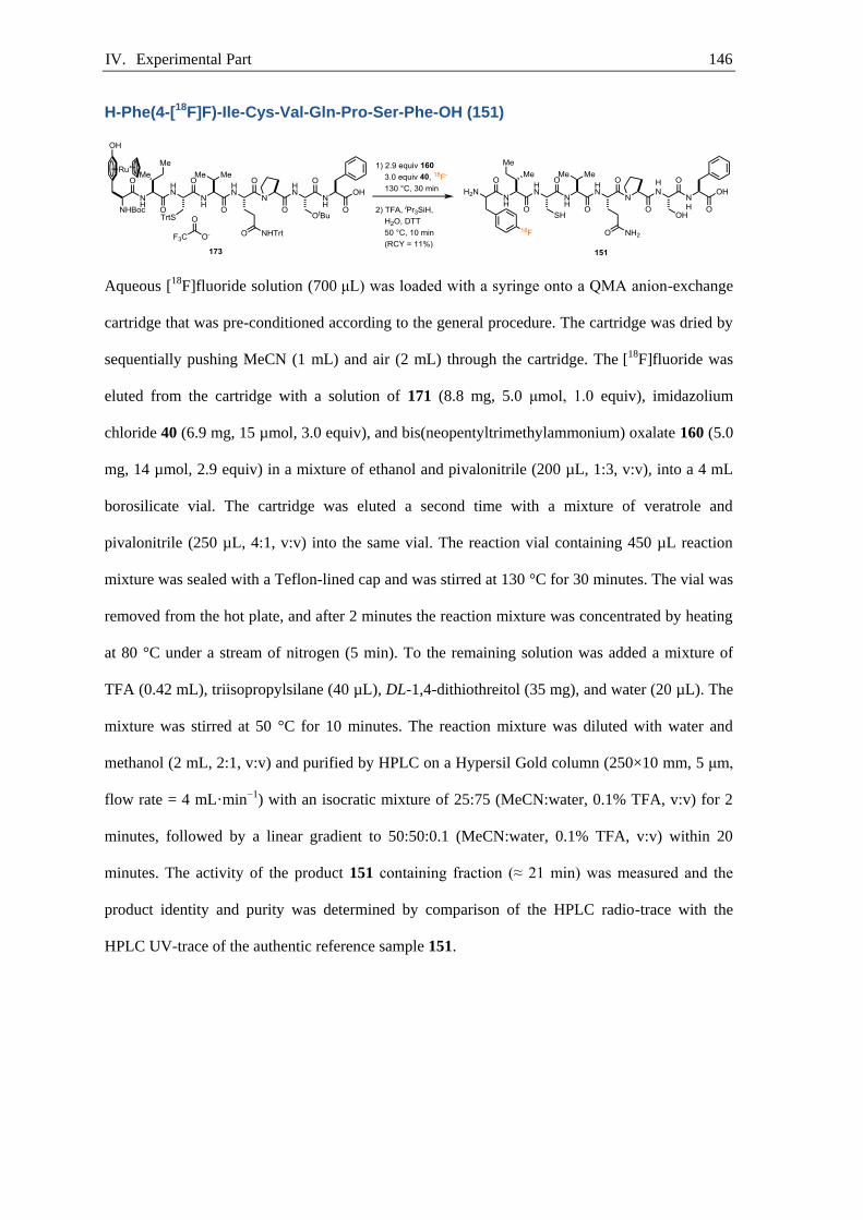
IV. Experimental Part 146
H-Phe(4-[18F]F)-Ile-Cys-Val-Gln-Pro-Ser-Phe-OH (151)
Aqueous [18F]fluoride solution (700 μL) was loaded with a syringe onto a QMA anion-exchange
cartridge that was pre-conditioned according to the general procedure. The cartridge was dried by
sequentially pushing MeCN (1 mL) and air (2 mL) through the cartridge. The [18F]fluoride was
eluted from the cartridge with a solution of 171 (8.8 mg, 5.0 μmol, 1.0 equiv), imidazolium
chloride 40 (6.9 mg, 15 µmol, 3.0 equiv), and bis(neopentyltrimethylammonium) oxalate 160 (5.0
mg, 14 µmol, 2.9 equiv) in a mixture of ethanol and pivalonitrile (200 µL, 1:3, v:v), into a 4 mL
borosilicate vial. The cartridge was eluted a second time with a mixture of veratrole and
pivalonitrile (250 µL, 4:1, v:v) into the same vial. The reaction vial containing 450 µL reaction
mixture was sealed with a Teflon-lined cap and was stirred at 130 °C for 30 minutes. The vial was
removed from the hot plate, and after 2 minutes the reaction mixture was concentrated by heating
at 80 °C under a stream of nitrogen (5 min). To the remaining solution was added a mixture of
TFA (0.42 mL), triisopropylsilane (40 µL), DL-1,4-dithiothreitol (35 mg), and water (20 µL). The
mixture was stirred at 50 °C for 10 minutes. The reaction mixture was diluted with water and
methanol (2 mL, 2:1, v:v) and purified by HPLC on a Hypersil Gold column (250×10 mm, 5 μm,
flow rate = 4 mL·min−1) with an isocratic mixture of 25:75 (MeCN:water, 0.1% TFA, v:v) for 2
minutes, followed by a linear gradient to 50:50:0.1 (MeCN:water, 0.1% TFA, v:v) within 20
minutes. The activity of the product 151 containing fraction (≈ 21 min) was measured and the
product identity and purity was determined by comparison of the HPLC radio-trace with the
HPLC UV-trace of the authentic reference sample 151.
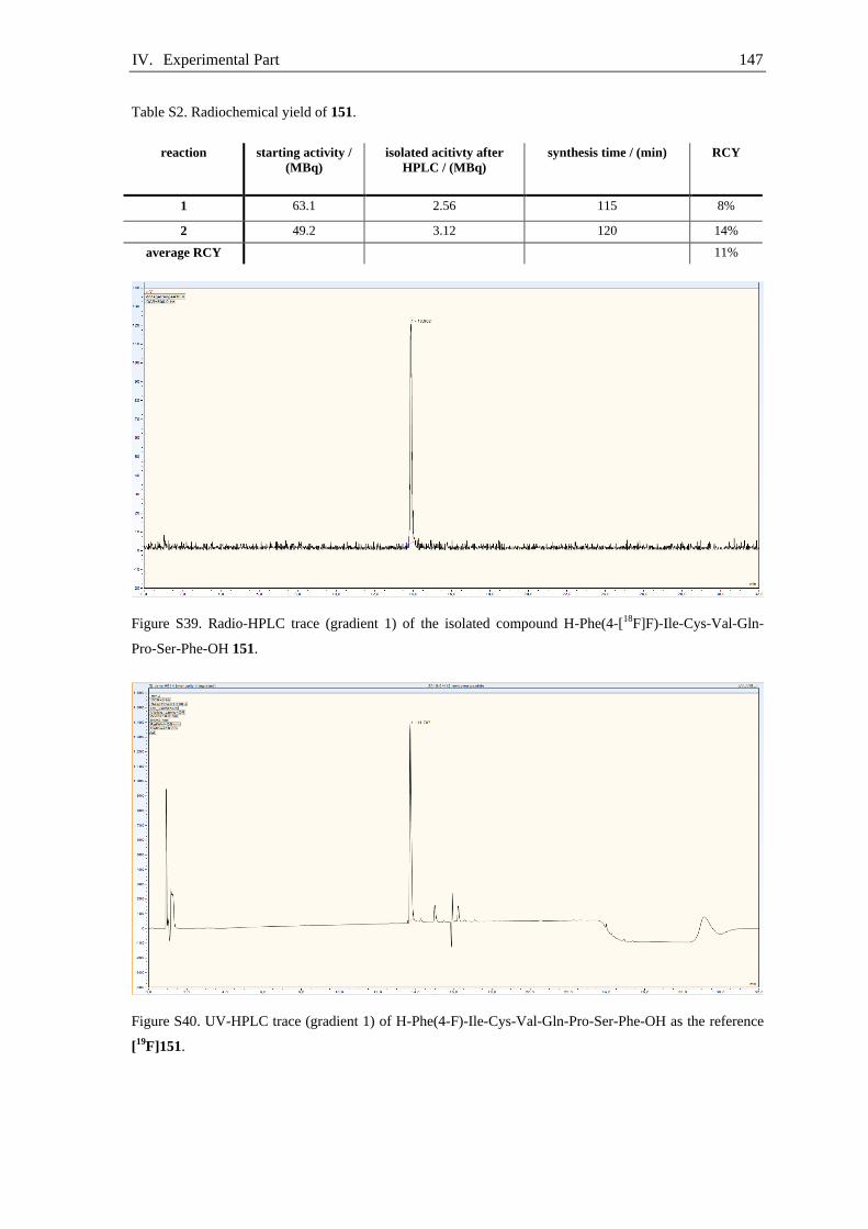
IV. Experimental Part 147
Table S2. Radiochemical yield of 151.
reaction starting activity /
(MBq)
isolated acitivty after
HPLC / (MBq)
synthesis time / (min) RCY
1 63.1 2.56 115 8%
2 49.2 3.12 120 14%
average RCY 11%
Figure S39. Radio-HPLC trace (gradient 1) of the isolated compound H-Phe(4-[18F]F)-Ile-Cys-Val-Gln-
Pro-Ser-Phe-OH 151.
Figure S40. UV-HPLC trace (gradient 1) of H-Phe(4-F)-Ile-Cys-Val-Gln-Pro-Ser-Phe-OH as the reference
[19
F]151.

IV. Experimental Part 148
H-Phe(4-[18F]F)-Ile-Cys-Val-Gln-Pro-Ser-Phe-OH (151)
Aqueous [18F]fluoride solution (700 μL) was loaded with a syringe onto a QMA anion-exchange
cartridge that was pre-conditioned according to the general procedure. The cartridge was dried by
sequentially pushing MeCN (1 mL) and air (2 mL) through the cartridge. The [18F]fluoride was
eluted from the cartridge with a solution of 174 (10 mg, 5.0 μmol, 1.0 equiv), imidazolium
chloride 40 (6.9 mg, 15 µmol, 3.0 equiv), and bis(neopentyltrimethylammonium) oxalate 160 (5.0
mg, 14 µmol, 2.9 equiv) in a mixture of ethanol and pivalonitrile (200 µL, 1:3, v:v), into a 4 mL
borosilicate vial. The cartridge was eluted a second time with a mixture of veratrole and
pivalonitrile (250 µL, 4:1, v:v) into the same vial. The reaction vial containing 450 µL reaction
mixture was sealed with a Teflon-lined cap and was stirred at 130 °C for 30 minutes. The vial was
removed from the hot plate, and after 2 minutes the reaction mixture was concentrated by heating
at 80 °C under a stream of nitrogen (5 min). To the remaining solution was added a mixture of
TFA (0.42 mL), triisopropylsilane (40 µL), DL-1,4-dithiothreitol (35 mg), and water (20 µL). The
mixture was stirred at 50 °C for 10 minutes. The reaction mixture was diluted with water and
methanol (2 mL, 2:1, v:v) and purified by HPLC on a Hypersil Gold column (250×10 mm, 5 μm,
flow rate = 4 mL·min−1) with an isocratic mixture of 25:75 (MeCN:water, 0.1% TFA, v:v) for 2
minutes, followed by a linear gradient to 50:50:0.1 (MeCN:water, 0.1% TFA, v:v) within 20
minutes. The activity of the product 151 containing fraction (≈ 21 min) was measured and the
product identity and purity was determined by comparison of the HPLC radio-trace with the
HPLC UV-trace of the authentic reference sample 151.

IV. Experimental Part 149
Table S3. Radiochemical yield of 151.
reaction starting activity /
(MBq)
isolated acitivty after
HPLC / (MBq)
aynthesis time / (min) RCY
1 54.1 13.1 81 40%
2 51.5 10.9 91 38%
average RCY 39%
Figure S41. Radio-HPLC trace (gradient 1) of the isolated compound H-Phe(4-[18F]F)-Ile-Cys-Val-Gln-
Pro-Ser-Phe-OH 151.
Figure S42. UV-HPLC trace (gradient 1) of H-Phe(4-F)-Ile-Cys-Val-Gln-Pro-Ser-Phe-OH as the reference
[19
F]151.
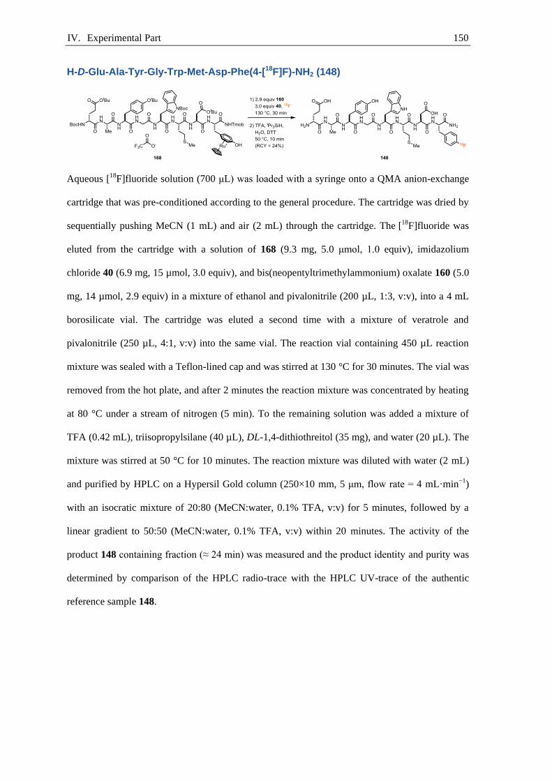
IV. Experimental Part 150
H-D-Glu-Ala-Tyr-Gly-Trp-Met-Asp-Phe(4-[18F]F)-NH2 (148)
Aqueous [18F]fluoride solution (700 μL) was loaded with a syringe onto a QMA anion-exchange
cartridge that was pre-conditioned according to the general procedure. The cartridge was dried by
sequentially pushing MeCN (1 mL) and air (2 mL) through the cartridge. The [18F]fluoride was
eluted from the cartridge with a solution of 168 (9.3 mg, 5.0 μmol, 1.0 equiv), imidazolium
chloride 40 (6.9 mg, 15 µmol, 3.0 equiv), and bis(neopentyltrimethylammonium) oxalate 160 (5.0
mg, 14 µmol, 2.9 equiv) in a mixture of ethanol and pivalonitrile (200 µL, 1:3, v:v), into a 4 mL
borosilicate vial. The cartridge was eluted a second time with a mixture of veratrole and
pivalonitrile (250 µL, 4:1, v:v) into the same vial. The reaction vial containing 450 µL reaction
mixture was sealed with a Teflon-lined cap and was stirred at 130 °C for 30 minutes. The vial was
removed from the hot plate, and after 2 minutes the reaction mixture was concentrated by heating
at 80 °C under a stream of nitrogen (5 min). To the remaining solution was added a mixture of
TFA (0.42 mL), triisopropylsilane (40 µL), DL-1,4-dithiothreitol (35 mg), and water (20 µL). The
mixture was stirred at 50 °C for 10 minutes. The reaction mixture was diluted with water (2 mL)
and purified by HPLC on a Hypersil Gold column (250×10 mm, 5 μm, flow rate = 4 mL·min−1)
with an isocratic mixture of 20:80 (MeCN:water, 0.1% TFA, v:v) for 5 minutes, followed by a
linear gradient to 50:50 (MeCN:water, 0.1% TFA, v:v) within 20 minutes. The activity of the
product 148 containing fraction (≈ 24 min) was measured and the product identity and purity was
determined by comparison of the HPLC radio-trace with the HPLC UV-trace of the authentic
reference sample 148.

IV. Experimental Part 151
Table S4. Radiochemical yield of 148.
reaction starting activity /
(MBq)
isolated acitivty after
HPLC / (MBq)
synthesis time / (min) RCY
1 58.8 5.7 168 28%
2 57.2 7.01 114 24%
3 59.4 4.95 148 21%
average RCY 24%
Figure S43. Radio-HPLC trace (gradient 1) of the isolated compound H-D-Glu-Ala-Tyr-Gly-Trp-Met-Asp-
Phe(4-[18F]F)-NH2 148.
Figure S44. UV-HPLC trace (gradient 1) of H-D-Glu-Ala-Tyr-Gly-Trp-Met-Asp-Phe(4-F)-NH2 [19
F]148 as
the reference.

IV. Experimental Part 152
H-Gly-Asn-Leu-Trp-Ala-Thr-Gly-His-Phe(4-[18F]F)-Met-NH2 (150)
Aqueous [18F]fluoride solution (700 μL) was loaded with a syringe onto a QMA anion-exchange
cartridge that was pre-conditioned according to the general procedure. The cartridge was dried by
sequentially pushing MeCN (1 mL) and air (2 mL) through the cartridge. The [18F]fluoride was
eluted from the cartridge with a solution of 172 (12 mg, 5.0 μmol, 1.0 equiv), imidazolium
chloride 40 (6.9 mg, 15 µmol, 3.0 equiv), and bis(neopentyltrimethylammonium) oxalate 160 (5.0
mg, 14 µmol, 2.9 equiv) in a mixture of ethanol and pivalonitrile (200 µL, 1:3, v:v), into a 4 mL
borosilicate vial. The cartridge was eluted a second time with a mixture of veratrole and
pivalonitrile (250 µL, 4:1, v:v) into the same vial. The reaction vial containing 450 µL reaction
mixture was sealed with a Teflon-lined cap and was stirred at 130 °C for 30 minutes. The vial was
removed from the hot plate, and after 2 minutes the reaction mixture was concentrated by heating
at 80 °C under a stream of nitrogen (5 min). To the remaining solution was added a mixture of
TFA (0.42 mL), triisopropylsilane (40 µL), DL-1,4-dithiothreitol (35 mg), and water (20 µL). The
mixture was stirred at 50 °C for 10 minutes. The reaction mixture was diluted with water (2 mL)
and purified by HPLC on a Hypersil Gold column (250×10 mm, 5 μm, flow rate = 4 mL·min−1)
with an isocratic mixture of 10:90 (MeCN:water, 0.1% TFA v:v:v) for 2 minutes, followed by a
linear gradient to 50:50 (MeCN:water, 0.1% TFA, v:v) within 18 minutes. The activity of the
product 150 containing fraction (≈ 23 min) was measured and the product identity and purity was
determined by comparison of the HPLC radio-trace with the HPLC UV-trace of the authentic
reference sample 150.

IV. Experimental Part 153
Table S5. Radiochemical yield of 150.
reaction starting Activity /
(MBq)
isolated acitivty after
HPLC / (MBq)
synthesis time / (min) RCY
1 56.8 5.16 163 25%
2 68.9 11.4 119 33%
average RCY 29%
Figure S45. Radio-HPLC trace (gradient 1) of the isolated compound H-Gly-Asn-Leu-Trp-Ala-Thr-Gly-
His-Phe(4-[18F]F)-Met-NH2 150.
Figure S46. UV-HPLC trace (gradient 1) of H-Gly-Asn-Leu-Trp-Ala-Thr-Gly-His-Phe(4-F)-Met-NH2
[19
F]150 as the reference.

IV. Experimental Part 154
H-Gly-His-Gly-Phe(4-[18F]F)-Gly-NH2 (152)
Aqueous [18F]fluoride solution (700 μL) was loaded with a syringe onto a QMA anion-exchange
cartridge that was pre-conditioned according to the general procedure. The cartridge was dried by
sequentially pushing MeCN (1 mL) and air (2 mL) through the cartridge. The [18F]fluoride was
eluted from the cartridge with a solution of 181 (6.0 mg, 5.0 μmol, 1.0 equiv), imidazolium
chloride 40 (6.9 mg, 15 µmol, 3.0 equiv), and bis(neopentyltrimethylammonium) oxalate 160 (5.0
mg, 14 µmol, 2.9 equiv) in a mixture of ethanol and pivalonitrile (200 µL, 1:3, v:v), into a 4 mL
borosilicate vial. The cartridge was eluted a second time with a mixture of veratrole and
pivalonitrile (250 µL, 4:1, v:v) into the same vial. The reaction vial containing 450 µL reaction
mixture was sealed with a Teflon-lined cap and was stirred at 130 °C for 30 minutes. The vial was
removed from the hot plate, and after 2 minutes the reaction mixture was concentrated by heating
at 80 °C under a stream of nitrogen (5 min). To the remaining solution was added a mixture of
TFA (0.42 mL), triisopropylsilane (40 µL), DL-1,4-dithiothreitol (35 mg), and water (20 µL). The
mixture was stirred at 50 °C for 10 minutes. The reaction mixture was diluted with water (2 mL)
and purified by HPLC on a Hypersil Gold column (250×10 mm, 5 μm, flow rate = 4 mL·min−1)
with an isocratic mixture of 05:95 (MeCN:water, 0.1% TFA, v:v) for 2 minutes, followed by a
linear gradient to 50:50 (MeCN:water, 0.1% TFA, v:v) within 20 minutes. The activity of the
product 152 containing fraction (≈ 19 min) was measured and the product identity and purity was
determined by comparison of the HPLC radio-trace with the HPLC UV-trace of the authentic
reference sample 152.

IV. Experimental Part 155
Table S6. Radiochemical yield of 152.
reaction starting activity /
(MBq)
isolated acitivty after
HPLC / (MBq)
synthesis time / (min) RCY
1 13.3 3.17 84 41%
2 46.6 8.84 110 37%
average RCY 39%
Figure S47. Radio-HPLC trace (Chiralpak® ZWIX (+) ((10×250 mm, 5 μm), flow rate = 1.0 mL·min−1, 35
°C) with an isocratic eluent 49:49:2 (MeOH:MeCN:H2O+50 mM formic acid, 25 mM diethylamine) of the
isolated compound H-Gly-His-Gly-Phe(4-[18F]F)-Gly-NH2 152.
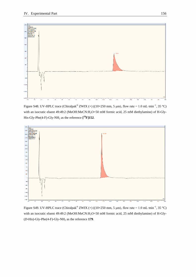
IV. Experimental Part 156
Figure S48. UV-HPLC trace (Chiralpak® ZWIX (+) ((10×250 mm, 5 μm), flow rate = 1.0 mL·min−1, 35 °C)
with an isocratic eluent 49:49:2 (MeOH:MeCN:H2O+50 mM formic acid, 25 mM diethylamine) of H-Gly-
His-Gly-Phe(4-F)-Gly-NH2 as the reference [19F]152.
Figure S49. UV-HPLC trace (Chiralpak® ZWIX (+) ((10×250 mm, 5 μm), flow rate = 1.0 mL·min−1, 35 °C)
with an isocratic eluent 49:49:2 (MeOH:MeCN:H2O+50 mM formic acid, 25 mM diethylamine) of H-Gly-
(D-His)-Gly-Phe(4-F)-Gly-NH2 as the reference 179.
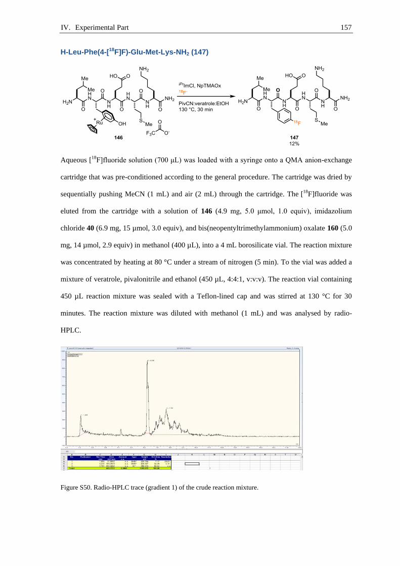
IV. Experimental Part 157
H-Leu-Phe(4-[18F]F)-Glu-Met-Lys-NH2 (147)
Aqueous [18F]fluoride solution (700 μL) was loaded with a syringe onto a QMA anion-exchange
cartridge that was pre-conditioned according to the general procedure. The cartridge was dried by
sequentially pushing MeCN (1 mL) and air (2 mL) through the cartridge. The [18F]fluoride was
eluted from the cartridge with a solution of 146 (4.9 mg, 5.0 μmol, 1.0 equiv), imidazolium
chloride 40 (6.9 mg, 15 µmol, 3.0 equiv), and bis(neopentyltrimethylammonium) oxalate 160 (5.0
mg, 14 µmol, 2.9 equiv) in methanol (400 µL), into a 4 mL borosilicate vial. The reaction mixture
was concentrated by heating at 80 °C under a stream of nitrogen (5 min). To the vial was added a
mixture of veratrole, pivalonitrile and ethanol (450 µL, 4:4:1, v:v:v). The reaction vial containing
450 µL reaction mixture was sealed with a Teflon-lined cap and was stirred at 130 °C for 30
minutes. The reaction mixture was diluted with methanol (1 mL) and was analysed by radio-
HPLC.
Figure S50. Radio-HPLC trace (gradient 1) of the crude reaction mixture.

IV. Experimental Part 158
H-Leu-Phe(4-[18F]F)-Glu-Met-Lys-NH2 (147)
Aqueous [18F]fluoride solution (700 μL) was loaded with a syringe onto a QMA anion-exchange
cartridge that was pre-conditioned according to the general procedure. The cartridge was dried by
sequentially pushing MeCN (1 mL) and air (2 mL) through the cartridge. The [18F]fluoride was
eluted from the cartridge with a solution of 177 (7.0 mg, 5.0 μmol, 1.0 equiv), imidazolium
chloride 40 (6.9 mg, 15 µmol, 3.0 equiv), and bis(neopentyltrimethylammonium) oxalate 160 (5.0
mg, 14 µmol, 2.9 equiv) in a mixture of ethanol and pivalonitrile (200 µL, 1:3, v:v), into a 4 mL
borosilicate vial. The cartridge was eluted a second time with a mixture of veratrole and
pivalonitrile (250 µL, 4:1, v:v) into the same vial. The reaction vial containing 450 µL reaction
mixture was sealed with a Teflon-lined cap and was stirred at 130 °C for 30 minutes. The vial was
removed from the hot plate, and after 2 minutes the reaction mixture was concentrated by heating
at 80 °C under a stream of nitrogen (5 min). To the remaining solution was added a mixture of
TFA (0.42 mL), triisopropylsilane (40 µL), DL-1,4-dithiothreitol (35 mg), and water (20 µL).
Then the mixture was stirred at 50 °C for 10 minutes. The reaction mixture was diluted with water
(2 mL) and purified by HPLC on a Hypersil Gold column (250×10 mm, 5 μm, flow rate = 4
mL·min−1) with an isocratic mixture of 15:85 (MeCN:water, 0.1% TFA, v:v) for 2 minutes,
followed by a linear gradient to 35:65:0.1 (MeCN:water, 0.1% TFA, v:v) within 16 minutes. The
activity of the product 147 containing fraction (≈ 18 min) was measured and the product identity
and purity was determined by comparison of the HPLC radio-trace with the HPLC UV-trace of
the authentic reference sample 147.

IV. Experimental Part 159
Table S7. Radiochemical yield of 147.
reaction starting activity /
(MBq)
isolated acitivty after
HPLC / (MBq)
synthesis time / (min) RCY
1 56.1 4.54 110 16%
2 63.1 8.16 98 24%
average RCY 20%
Figure S51. Radio-HPLC trace (gradient 1) of the isolated compound H-Leu-Phe(4-[18F]F)-Glu-Met-Lys-
NH2 147.
Figure S52. UV-HPLC trace (gradient 1) of H-Leu-Phe(4-F)-Glu-Met-Lys-NH2 as the reference [19F]147.

IV. Experimental Part 160
Automated H-Leu-Phe(4-[18F]F)-Glu-Met-Lys-NH2 (147) synthesis
Reactions were performed on an automated cassette-based radiochemical synthesizer ELIXYS
FLEX/CHEM connected to a PURE/FORM purification and formulation unit (Sofie Biosciences).
Aqueous [18F]fluoride (11.4 GBq, (308 mCi), t = 0) was trapped onto a QMA anion-exchange
cartridge that was pre-conditioned according to the general procedure, the cartridge was then
washed with MeCN (1.0 mL). The [18F]fluoride was eluted from the cartridge with a solution of
177 (6.0 mg, 5.0 μmol, 1.0 equiv), imidazolium chloride 40 (6.9 mg, 15 µmol, 3.0 equiv), and
bis(neopentyltrimethylammonium) oxalate 160 (5.0 mg, 14 µmol, 2.9 equiv) in a mixture of
ethanol and but-2-one (200 µL, 1:3, v:v), into a 3 mL V-vial (an aluminum cylinder 15.0 mm
height and 15.0 mm diameter was placed underneath the V-vial in the reactor to fit the 3 mL V-
vial in the 5 mL V-vial sample holder). The cartridge was eluted a second time with a mixture of
veratrole and but-2-one (250 µL, 4:1, v:v) into the same vial. The reaction vial containing 450 µL
reaction mixture was sealed against a Teflon-liner and was stirred at elevated temperature (set-
point at 150 °C) for 30 minutes and subsequently cooled to 50 °C. The solvent was evaporated at
100 °C under reduced pressure for 5 minutes. To the remaining solution was added a mixture of
TFA (0.42 mL), triisopropylsilane (40 µL), DL-1,4-dithiothreitol (35 mg), and water (20 µL).
Then the mixture was stirred at 50 °C for 10 minutes. The reaction mixture was cooled to room
temperature and then diluted with a solution of water (1.5 mL) and methanol (0.75 mL). The
reaction mixture was transferred from the ELIXYS FLEX/CHEM system to the PURE/FORM,
and was purified by HPLC with a Hypersil Gold (250×10 mm, 5 μm, flow rate = 4 mL·min−1)
column with an isocratic mixture of 15:85 (MeCN:water, 0.1% TFA, v:v) for 2 minutes, followed
by a linear gradient to 45:55 (MeCN:water, 0.1% TFA, v:v) within 18 minutes. The product was
collected from 18 min to 19 min. The activity of the product containing fraction was diluted with
water (40 mL) and loaded onto a C-18 light SepPak cartridge. The C-18 cartridge was washed

IV. Experimental Part 161
with water (3 mL) and the product was sequentially eluted with ethanol (1 mL) and saline (1.7
mL). The ethanolic saline solution contained 1.28 GBq (34.6 mCi, t = 99 min). The total decay
corrected radio chemical yield is 21 %.The radiochemical identity was confirmed by analytical
HPLC.
To determine the molar activity of 147, 22.1 MBq of purified compound 147 (Figure S54, 87%
purity was observed and therefore calculations were proceeded with 19.3 MBq. However, the
observed low purity was presumably due to saturation of the detector. If a smaller amount of
sample was injected 100% purity was observed. (Figure S56)) was injected into an analytical
HPLC and the UV absorption corresponding to the radio-peak was measured. To determine the
amount of 147 the molar response of [19
F]147 was determined (Figure S58) with the authentic
standard H-Leu-Phe(4-F)-Glu-Met-Lys-NH2 [19
F]147. It was calculated that the absorption of
2.5629 mAU·min (Figure S58) corresponds to 0.20 nmol of 147 and therefore the molar activity
of 147 was determined to be 99 GBq·µmol−1 (2.7 Ci·µmol−1).
The ruthenium content in the reformulated sample was 1.8 µg, which is more than fivefold below
the specified limit of 10 µg per day considered safe for injection into humans. If an additional
reformulation with a C-18 cartridge was performed before injection into the preparative HPLC,
the ruthenium content dropped to 0.09 µg. If desired, the residual ruthenium can be reduced by
the use of commercial metal scavengers. To achieve this 200 mg Quadrasil™ AP were added to
the reaction mixture before concentration under a stream of nitrogen. After addition of
Quadrasil™ AP the reaction mixture was stirred at 50 °C for 10 min. The reaction mixture was
filtered and the filter cake was eluted with methanol (0.5 mL). The filtrate was concentrated by
heating at 80 °C under a stream of nitrogen (5 min). To the remaining solution was added a
mixture of TFA (0.42 mL), triisopropylsilane (40 µL), DL-1,4-dithiothreitol (35 mg), and water
(20 µL). Then the mixture was stirred at 50 °C for 10 minutes. The reaction mixture was diluted
with water (2 mL) and purified by HPLC. The use of Quadrasil™ AP lowered the ruthenium
content to 0.11 µg. A blank sample of the HPLC eluent contained 0.03 µg of ruthenium.

IV. Experimental Part 162
Figure S53. Overlay of radio-trace (orange) and UV-trace (blue) of the crude reaction mixture.
Figure S54. Determination of molar activity (gradient 1); radio-trace (22.1 MBq).
0
20
40
60
80
100
120
0
0.5
1
1.5
2
2.5
0 2 4 6 8 10 12 14 16 18 20
mV
AU
time·min−1
UV-trace 210 nm
radio-trace

IV. Experimental Part 163
Figuer S55. Determination of molar activity (gradient 1); UV-trace (22.1 MBq).
Figure S56. Determination of molar activity (gradient 2); radio-trace @ 210 nm.
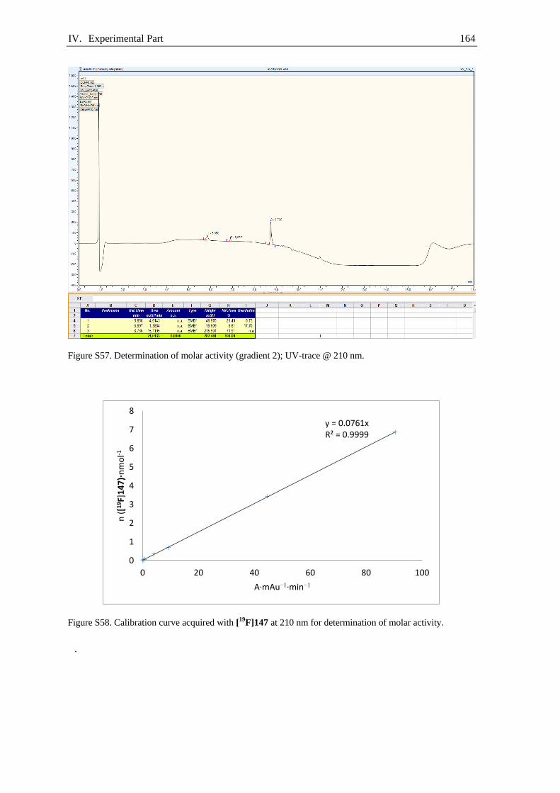
IV. Experimental Part 164
Figure S57. Determination of molar activity (gradient 2); UV-trace @ 210 nm.
Figure S58. Calibration curve acquired with [19F]147 at 210 nm for determination of molar activity.
.
y = 0.0761x R² = 0.9999
0
1
2
3
4
5
6
7
8
0 20 40 60 80 100
n (
[19F]
14
7)·
nm
ol-1
A·mAu−1·min−1
V. References 165
References V.
[1] G. Hevesy, Biochem. J. 1923, 17, 439-445.
[2] W. G. Myers, J. Nucl. Med. 1979, 20, 590-698.
[3] O. Chiewitz, G. Hevesy, Nature 1935, 136, 754-755.
[4] F. Joliot, I. Curie, Nature 1934, 133, 201-202.
[5] E. O. Lawrence, D. Cooksey, Phys. Rev. 1936, 50, 1131-
1140.
[6] P. M. Matthews, E. A. Rabiner, J. Passchier, R. N. Gunn, Br.
J. Clin. Pharmacol. 2012, 73, 175-186.
[7] H. Herzog, Radiochim. Acta 2001, 89, 203-214.
[8] P. W. Miller, N. J. Long, R. Vilar, A. D. Gee, Angew. Chem.,
Int. Ed. 2008, 47, 8998-9033.
[9] (a) D. W. Townsend, S. R. Cherry, Eur. Radiol. 2001, 11,
1968-1974; (b) Y. Berker, Y. Li, Med. Phys. 2016, 43, 807-
832; (c) P. E. Kinahan, B. H. Hasegawa, T. Beyer, Semin.
Nucl. Med. 2003, 33, 166-179.
[10] A. L. Feliu, J. Chem. Educ. 1988, 65, 655-660.
[11] H. J. Ache, Angew. Chem., Int. Ed. 1972, 11, 179-199.
[12] S. Patching, J. Diagn. Imaging Ther. 2015, 2, 30-102.
[13] H. H. Coenen, P. H. Elsinga, R. Iwata, M. R. Kilbourn, M. R.
Pillai, M. G. Rajan, H. N. Wagner, Jr., J. J. Zaknun, Nucl.
Med. Biol. 2010, 37, 727-740.
[14] S. N. Reske, J. Kotzerke, Eur. J. Nucl. Med. 2001, 28, 1707-
1723.
[15] (a) T. Ido, C. N. Wan, V. Casella, J. S. Fowler, A. P. Wolf,
M. Reivich, D. E. Kuhl, J. Labelled Compd. Radiopharm.
1978, 14, 175-183; (b) H. H. C. K. Hamacher, and G.
Stocklin, J. Nucl. Med. 1986, 27, 235-238.
[16] H. H. Coenen, V. W. Pike, G. Stöcklin, R. Wagner, Int. J.
Radiat. Appl. Instrum. Part A 1987, 38, 605-610.
[17] G. A. van Dongen, G. W. Visser, M. N. Lub-de Hooge, E. G.
de Vries, L. R. Perk, Oncologist 2007, 12, 1379-1389.
[18] S. C. Jones, J. H. Greenberg, R. Dann, G. D. Robinson, Jr.,
M. Kushner, A. Alavi, M. Reivich, J. Cereb. Blood Flow
Metab. 1985, 5, 566-575.
V. References 166
[19] M. M. Khalil, Basic Sciences of Nuclear Medicine, Springer,
Berlin, Heidelberg, 2011.
[20] L. Cunha, I. Horvath, S. Ferreira, J. Lemos, P. Costa, D.
Vieira, D. S. Veres, K. Szigeti, T. Summavielle, D. Máthé, L.
F. J. M. D. Metello, Therapy, Mol. Diagn. Ther. 2014, 18,
153-173.
[21] W. F. Walsh, H. R. Fill, P. V. Harper, Semin. Nucl. Med.
1977, 7, 59-66.
[22] E. Britannica,
https://www.britannica.com/technology/cyclotron/images-
videos/media/1/148211/18064.
[23] A. Pekosak, U. Filp, A. J. Poot, A. D. Windhorst, Mol.
Imaging Biol. 2018, 20, 510-532.
[24] F. Wuest, M. Berndt, T. Kniess, Carbon-11 Labeling
Chemistry Based upon [11C]Methyl Iodide, Springer Berlin
Heidelberg, Berlin, Heidelberg, 2007, 183-213.
[25] S. Faulkner, N. J. Long, Dalton Trans. 2011, 40, 6067-6067.
[26] S. R. Banerjee, M. G. Pomper, Appl. Radiat. Isot. 2013, 76,
2-13.
[27] B. N. Clarke, J. Nucl. Med. Technol. 2018, 46, 12-16.
[28] A. Sanchez-Crespo, P. Andreo, S. A. Larsson, Eur. J. Nucl.
Med. Mol. Imaging 2004, 31, 44-51.
[29] D. O'Hagan, D. B. Harper, J. Fluorine Chem. 1999, 100, 127-
133.
[30] L. M. Jarvis, Chem. Eng. News 2019, 97, 33-37.
[31] N. A. Meanwell, J. Med. Chem. 2011, 54, 2529-2591.
[32] D. O'Hagan, Chem. Soc. Rev. 2008, 37, 308-319.
[33] S. C. P. Williams, Nat. Med. 2013, 19, 1551.
[34] R. van Grieken, M. de Bruin, Pure Appl. Chem. 1994, 66,
2513-2526.
[35] L. Cai, S. Lu, V. W. Pike, Eur. J. Org. Chem. 2008, 2008,
2853-2873.
[36] V. Casella, T. Ido, A. P. Wolf, J. S. Fowler, R. R.
MacGregor, T. J. Ruth, J. Nucl. Med. 1980, 21, 750-757.
[37] O. Matsson, J. Persson, B. S. Axelsson, B. Långström, J. Am.
Chem. Soc. 1993, 115, 5288-5289.
[38] J. Bergman, O. Solin, Nucl. Med. Biol. 1997, 24, 677-683.

V. References 167
[39] M. S. Berridge, S. M. Apana, J. M. Hersh, J. Labelled
Compd. Radiopharm. 2009, 52, 543-548.
[40] G. Firnau, R. Chirakal, E. S. Garnett, J. Nucl. Med. 1984, 25,
1228-1233.
[41] M. J. Adam, J. R. Grierson, T. J. Ruth, S. Jivan, Int. J.
Radiat. Appl. Instrum. Part A 1986, 37, 877-882.
[42] H. H. Coenen, K. Franken, P. Kling, G. Stöcklin, Int. J.
Radiat. Appl. Instrum. Part A 1988, 39, 1243-1250.
[43] M. J. Adam, B. F. Abeysekera, T. J. Ruth, S. Jivan, B. D.
Pate, J. Labelled Compd. Radiopharm. 1984, 21, 1227-1228.
[44] H. H. Coenen, S. M. Moerlein, J. Fluorine Chem. 1987, 36,
63-75.
[45] O. Solin, J. Bergman, M. Haaparanta, A. Reissell, Int. J.
Radiat. Appl. Instrum. Part A 1988, 39, 1065-1071.
[46] D. M. Jewett, S. A. Toorongian, G. K. Mulholland, G. L.
Watkins, M. R. Kilbourn, Int. J. Radiat. Appl. Instrum. Part
A 1988, 39, 1109-1111.
[47] C. L. Liotta, H. P. Harris, J. Am. Chem. Soc. 1974, 96, 2250-
2252.
[48] J. Emsley, Chem. Soc. Rev. 1980, 9, 91-124.
[49] X. Chen, J. I. Brauman, J. Am. Chem. Soc. 2008, 130, 15038-
15046.
[50] M. R. Kilbourn, J. R. Huizenga, Fluorine-18 Labeling of
Radiopharmaceuticals, National Academies, 1990.
[51] B. Langlois, L. Gilbert, G. Forat, in The Roots of Organic
Development, Vol. 8, Elsevier, 1996, 244-292.
[52] E. E. Kwan, Y. Zeng, H. A. Besser, E. N. Jacobsen, Nat.
Chem. 2018, 10, 917-923.
[53] S. Preshlock, M. Tredwell, V. Gouverneur, Chem. Rev. 2016,
116, 719-766.
[54] H. H. Coenen, PET Chemistry The Driving Force in
Molecular Imaging, Fluorine-18 Labeling Methods: Features
and Possibilities of Basic Reactions, Springer Berlin,
Heidelberg, 2007, 15-50.
[55] M. S. Haka, M. R. Kilbourn, L. G. Watkins, S. A.
Toorongian, J. Labelled Compd. Radiopharm. 1989, 27, 823-
833.

V. References 168
[56] H. Sun, S. G. DiMagno, J. Fluorine Chem. 2007, 128, 806-
812.
[57] J. Ermert, C. Hocke, T. Ludwig, R. Gail, H. H. Coenen, J.
Labelled Compd. Radiopharm. 2004, 47, 429-441.
[58] L. Allain-Barbier, M.-C. Lasne, C. Perrio-Huard, B. Moreau,
L. Barré, Acta Chem. Scand. 1998, 52, 480-489.
[59] (a) O. Wallach, Liebigs Ann. 1886, 235, 255-271; (b) G. Balz,
G. Schiemann, Ber. Dtsch. Chem. Ges. 1927, 60, 1186-1190.
[60] B. Langlois, in Organo-Fluorine Compounds, Vol. E 10 a,
4th Edition ed., Georg Thieme Verlag, 1998, 686-709.
[61] R. M. Hoyte, S. S. Lin, D. R. Christman, H. L. Atkins, W.
Hauser, A. P. Wolf, J. Nucl. Med. 1971, 12, 280-286.
[62] M. Argentini, C. Wiese, R. Weinreich, J. Fluorine Chem.
1994, 68, 141-144.
[63] H. Hayashi, H. Sonoda, K. Fukumura, T. Nagata, Chem.
Commun. 2002, 1618-1619.
[64] P. Tang, W. Wang, T. Ritter, J. Am. Chem. Soc. 2011, 133,
11482-11484.
[65] C. N. Neumann, J. M. Hooker, T. Ritter, Nature 2016, 534,
369-373.
[66] A. Böttcher, A. Chlenov, B. Gotov, W. D. Harman, M. A.
Kondratenkov, E. P. Kündig, K. Muñz, A. Pape, J. H. Rigby,
H.-G. Schmalz, M. F. Semmelhack, M. Uemura, Transition
Metal Arene π-Complexes in Organic Synthesis and
Catalysis, Vol. 7, Springer-Verlag Berlin Heidelberg, 2004.
[67] E. M. D’Amato, C. N. Neumann, T. Ritter, Organometallics
2015, 34, 4626-4631.
[68] J. D. Holmes, D. A. K. Jones, R. Pettit, J. Organomet. Chem.
1965, 4, 324-331.
[69] M. F. Semmelhack, in Comprehensive Organometallic
Chemistry II, Elsevier, 1995, 979-1015.
[70] B. Nicholls, M. C. Whiting, J. Chem. Soc. 1959, 31, 551-556.
[71] A. J. Pearson, S. Cui, Tetrahedron Lett. 2005, 46, 2639-2642.
[72] M. H. Beyzavi, D. Mandal, M. G. Strebl, C. N. Neumann, E.
M. D’Amato, J. Chen, J. M. Hooker, T. Ritter, ACS Cent. Sci.
2017, 3, 944-948.

V. References 169
[73] C. Hartmann, V. Meyer, Ber. Dtsch. Chem. Ges. 1894, 27,
426-432.
[74] F. M. Beringer, A. Brierley, M. Drexler, E. M. Gindler, C. C.
Lumpkin, J. Am. Chem. Soc. 1953, 75, 2708-2712.
[75] A. Shah, V. W. Pike, D. A. Widdowson, J. Chem. Soc.,
Perkin Trans. 1 1998, 1, 2043-2046.
[76] T. L. Ross, J. Ermert, C. Hocke, H. H. Coenen, J. Am. Chem.
Soc. 2007, 129, 8018-8025.
[77] J.-H. Chun, S. Lu, Y.-S. Lee, V. W. Pike, J. Org. Chem.
2010, 75, 3332-3338.
[78] N. Ichiishi, A. F. Brooks, J. J. Topczewski, M. E. Rodnick,
M. S. Sanford, P. J. H. Scott, Org. Lett. 2014, 16, 3224-3227.
[79] N. Ichiishi, A. J. Canty, B. F. Yates, M. S. Sanford, Org. Lett.
2013, 15, 5134-5137.
[80] A. Casitas, M. Canta, M. Solà, M. Costas, X. Ribas, J. Am.
Chem. Soc. 2011, 133, 19386-19392.
[81] (a) M. F. Ghorab, J. M. Winfield, J. Fluorine Chem. 1990,
49, 367-383; (b) M. Jauregui-Osoro, K. Sunassee, A. J.
Weeks, D. J. Berry, R. L. Paul, M. Cleij, J. P. Banga, M. J.
O'Doherty, P. K. Marsden, S. E. Clarke, J. R. Ballinger, I.
Szanda, S. Y. Cheng, P. J. Blower, Eur. J. Nucl. Med. Mol.
Imaging 2010, 37, 2108-2116.
[82] I. A. I. Mkhalid, J. H. Barnard, T. B. Marder, J. M. Murphy,
J. F. Hartwig, Chem. Rev. 2010, 110, 890-931.
[83] (a) D. M. T. Chan, K. L. Monaco, R.-P. Wang, M. P.
Winters, Tetrahedron Lett. 1998, 39, 2933-2936; (b) D. A.
Evans, J. L. Katz, T. R. West, Tetrahedron Lett. 1998, 39,
2937-2940; (c) P. Y. S. Lam, C. G. Clark, S. Saubern, J.
Adams, M. P. Winters, D. M. T. Chan, A. Combs,
Tetrahedron Lett. 1998, 39, 2941-2944.
[84] J. X. Qiao, P. Y. S. Lam, Synthesis 2011, 6, 829-856.
[85] P. S. Fier, J. Luo, J. F. Hartwig, J. Am. Chem. Soc. 2013, 135,
2552-2559.
[86] M. Tredwell, S. M. Preshlock, N. J. Taylor, S. Gruber, M.
Huiban, J. Passchier, J. Mercier, C. Génicot, V. Gouverneur,
Angew. Chem., Int. Ed. 2014, 53, 7751-7755.
[87] B. D. Wright, S. E. Lapi, J. Nucl. Med. 2013, 54, 1171-1174.

V. References 170
[88] S. Lee, J. Xie, X. Chen, Chem. Rev. 2010, 110, 3087-3111.
[89] L. Gentilucci, R. De Marco, L. Cerisoli, Curr. Pharm. Des.
2010, 16, 3185-3203.
[90] A. N. Eberle, G. Mild, S. Froidevaux, J. Recept. Signal
Transduction 2004, 24, 319-455.
[91] (a) J. C. Reubi, A. M. Landolt, J. Clin. Endocrinol. Metab.
1984, 59, 1148-1151; (b) D. Storch, M. Béhé, M. A. Walter,
J. Chen, P. Powell, R. Mikolajczak, H. R. Mäcke, J. Nucl.
Med. 2005, 46, 1561-1569.
[92] W. Zhao, H. G. Lee, S. L. Buchwald, J. M. Hooker, J. Am.
Chem. Soc. 2017, 139, 7152-7155.
[93] M. Nič, J. Jirát, B. Košata, A. Jenkins, A. McNaught, IUPAC
Compendium of Chemical Terminology, 2009.
[94] D. Brasse, A. Nonat, Dalton Trans. 2015, 44, 4845-4858.
[95] H. R. Maecke, J. C. Reubi, J. Nucl. Med. 2011, 52, 841-844.
[96] S. Froidevaux, A. Heppeler, A. N. Eberle, A. M. Meier, M.
Hausler, C. Beglinger, M. Behe, P. Powell, H. R. Macke,
Endocrinology 2000, 141, 3304-3312.
[97] S. Richter, F. Wuest, Molecules 2014, 19, 20536-20556.
[98] S. Vallabhajosula, Molecular Imaging, Springer-Verlag
Berlin Heidelberg, 2009.
[99] C. L. Charron, J. L. Hickey, T. K. Nsiama, D. R.
Cruickshank, W. L. Turnbull, L. G. Luyt, Nat. Prod. Rep.
2016, 33, 761-800.
[100] (a) J. A. Dean, N. A. Lange, Lange's Handbook of Chemistry,
McGraw-Hill, 1999; (b) R. Schirrmacher, G. Bradtmöller, E.
Schirrmacher, O. Thews, J. Tillmanns, T. Siessmeier, H. G.
Buchholz, P. Bartenstein, B. Wängler, C. M. Niemeyer, K.
Jurkschat, Angew. Chem., Int. Ed. 2006, 45, 6047-6050.
[101] M. S. Rosenthal, A. L. Bosch, R. J. Nickles, S. J. Gatley, Int.
J. Appl. Radiat. Isot. 1985, 36, 318-319.
[102] A. Höhne, L. Yu, L. Mu, M. Reiher, U. Voigtmann, U. Klar,
K. Graham, P. A. Schubiger, S. M. Ametamey, Chem. Eur. J.
2009, 15, 3736-3743.
[103] A. Höhne, L. Mu, M. Honer, P. A. Schubiger, S. M.
Ametamey, K. Graham, T. Stellfeld, S. Borkowski, D.
Berndorff, U. Klar, U. Voigtmann, J. E. Cyr, M. Friebe, L.
V. References 171
Dinkelborg, A. Srinivasan, Bioconjugate Chem. 2008, 19,
1871-1879.
[104] Z. Liu, Y. Li, R. Ting, D. M. Perrin, J. Phys. Org. Chem.
2015, 28, 210-214.
[105] (a) R. Ting, C. W. Harwig, J. Lo, Y. Li, M. J. Adam, T. J.
Ruth, D. M. Perrin, J. Org. Chem. 2008, 73, 4662-4670; (b)
R. Ting, C. Harwig, U. auf dem Keller, S. McCormick, P.
Austin, C. M. Overall, M. J. Adam, T. J. Ruth, D. M. Perrin,
J. Am. Chem. Soc. 2008, 130, 12045-12055.
[106] D. M. Perrin, Acc Chem Res 2016, 49, 1333-1343.
[107] Z. Liu, D. Chao, Y. Li, R. Ting, J. Oh, D. M. Perrin, Chem.
Eur. J. 2015, 21, 3924-3928.
[108] Z. Liu, M. Pourghiasian, M. A. Radtke, J. Lau, J. Pan, G. M.
Dias, D. Yapp, K.-S. Lin, F. Bénard, D. M. Perrin, Angew.
Chem., Int. Ed. 2014, 53, 11876-11880.
[109] Z. Liu, M. Pourghiasian, F. Benard, J. Pan, K. S. Lin, D. M.
Perrin, J. Nucl. Med. 2014, 55, 1499-1505.
[110] (a) V. V. Rostovtsev, L. G. Green, V. V. Fokin, K. B.
Sharpless, Angew. Chem., Int. Ed. 2002, 41, 2596-2599; (b)
C. W. Tornøe, C. Christensen, M. Meldal, J. Org. Chem.
2002, 67, 3057-3064.
[111] J. Marik, J. L. Sutcliffe, Tetrahedron Lett. 2006, 47, 6681-
6684.
[112] N. J. Agard, J. A. Prescher, C. R. Bertozzi, J. Am. Chem. Soc.
2004, 126, 15046-15047.
[113] (a) Z. Li, T. S. Seo, J. Ju, Tetrahedron Lett. 2004, 45, 3143-
3146; (b) G. Wittig, A. Krebs, Chem. Ber. 1961, 94, 3260-
3275.
[114] L. S. Campbell-Verduyn, L. Mirfeizi, A. K. Schoonen, R. A.
Dierckx, P. H. Elsinga, B. L. Feringa, Angew. Chem., Int. Ed.
2011, 50, 11117-11120.
[115] K. Sachin, V. H. Jadhav, E.-M. Kim, H. L. Kim, S. B. Lee,
H.-J. Jeong, S. T. Lim, M.-H. Sohn, D. W. Kim,
Bioconjugate Chem. 2012, 23, 1680-1686.
[116] O. C. Boerman, W. J. G. Oyen, F. H. M. Corstens, Semin.
Nucl. Med. 2000, 30, 195-208.
V. References 172
[117] G. Vaidyanathan, M. R. Zalutsky, Int. J. Radiat. Appl.
Instrum. Part B 1992, 19, 275-281.
[118] H.-J. Wester, K. Hamacher, G. Stöcklin, Nucl. Med. Biol.
1996, 23, 365-372.
[119] M. Berndt, J. Pietzsch, F. Wuest, Nucl. Med. Biol. 2007, 34,
5-15.
[120] R. Schirrmacher, B. Wangler, J. Bailey, V. Bernard-Gauthier,
E. Schirrmacher, C. Wangler, Semin Nucl Med 2017, 47, 474-
492.
[121] S. Verhoog, C. W. Kee, Y. Wang, T. Khotavivattana, T. C.
Wilson, V. Kersemans, S. Smart, M. Tredwell, B. G. Davis,
V. Gouverneur, J. Am. Chem. Soc. 2018, 140, 1572-1575.
[122] M. Ogawa, K. Hatano, S. Oishi, Y. Kawasumi, N. Fujii, M.
Kawaguchi, R. Doi, M. Imamura, M. Yamamoto, K. Ajito, T.
Mukai, H. Saji, K. Ito, Nucl. Med. Biol. 2003, 30, 1-9.
[123] V. M. Labroo, D. Hebel, K. L. Kirk, L. A. Cohen, C.
Lemieux, P. W. Schiller, Int. J. Pept. Protein Res. 1991, 37,
430-439.
[124] X. Huang, W. Liu, H. Ren, R. Neelamegam, J. M. Hooker, J.
T. Groves, J. Am. Chem. Soc. 2014, 136, 6842-6845.
[125] W. Liu, X. Huang, M. S. Placzek, S. W. Krska, P. McQuade,
J. M. Hooker, J. T. Groves, Chem. Sci. 2018, 9, 1168-1172.
[126] M. B. Nodwell, H. Yang, M. Čolović, Z. Yuan, H. Merkens,
R. E. Martin, F. Bénard, P. Schaffer, R. Britton, J. Am. Chem.
Soc. 2017, 139, 3595-3598.
[127] H. Teare, E. G. Robins, E. Årstad, S. K. Luthra, V.
Gouverneur, Chem. Commun. 2007, 2330-2332.
[128] Z. Yuan, M. B. Nodwell, H. Yang, N. Malik, H. Merkens, F.
Bénard, R. E. Martin, P. Schaffer, R. Britton, Angew. Chem.,
Int. Ed. 2018, 57, 12733-12736.
[129] G. Clemente, J. Rickmeier, T. Zarganes‐Tzitzikas, I. Antunes,
R. Slart, A. Dömling, T. Ritter, P. Elsinga, J. Labelled
Compd. Radiopharm. 2019, 62, S84-S85.
[130] G. A. Roth, C. Johnson, A. Abajobir, F. Abd-Allah, S. F.
Abera, G. Abyu, M. Ahmed, B. Aksut, T. Alam, K. Alam, F.
Alla, N. Alvis-Guzman, S. Amrock, H. Ansari, J. Ärnlöv, H.

V. References 173
Asayesh, T. M. Atey, L. Avila-Burgos, A. Awasthi, A.
Banerjee, et al., J. Am. Coll. Cardiol. 2017, 70, 1-25.
[131] D. P. Barr, E. M. Russ, H. A. Eder, Am. J. Med. 1951, 11,
480-493.
[132] A. Endo, M. Kuroda, K. Tanzawa, FEBS Lett. 1976, 72, 323-
326.
[133] N. A. McGrath, M. Brichacek, J. T. Njardarson, J. Chem.
Educ. 2010, 87, 1348-1349.
[134] J. K. Liao, U. Laufs, Annu. Rev. Pharmacol. Toxicol. 2005,
45, 89-118.
[135] A. Oesterle, U. Laufs, J. K. Liao, Circ. Res. 2017, 120, 229-
243.
[136] G. Weitz-Schmidt, K. Welzenbach, V. Brinkmann, T.
Kamata, J. Kallen, C. Bruns, S. Cottens, Y. Takada, U.
Hommel, Nat. Med. 2001, 7, 687-692.
[137] Ž. Reiner, Nutr., Metab. Cardiovasc. Dis. 2014, 24, 1057-
1066.
[138] H. Lennernäs, Clin. Pharmacokinet. 2003, 42, 1141-1160.
[139] B.-C. Chen, J. E. Sundeen, P. Guo, M. S. Bednarz, J. J.
Hangeland, S. Z. Ahmed, M. Jemal, J. Labelled Compd.
Radiopharm. 2000, 43, 261-270.
[140] H. Stetter, M. Schreckenberg, Angew. Chem., Int. Ed. 1973,
12, 81-81.
[141] L. Knorr, Ber. Dtsch. Chem. Ges. 1885, 18, 299-311.
[142] N. A. Vol'kenau, I. N. Bolesova, L. S. Shul'pina, A. N.
Kitaigorodskii, D. M. Kravtsov, J. Organomet. Chem. 1985,
288, 341-348.
[143] E. E. Karslyan, D. S. Perekalin, P. V. Petrovskii, K. A.
Lyssenko, A. R. Kudinov, Russ. Chem. Bull. 2009, 57, 2201.
[144] R. Richarz, P. Krapf, F. Zarrad, E. A. Urusova, B. Neumaier,
B. D. Zlatopolskiy, Org. Biomol. Chem. 2014, 12, 8094-
8099.
[145] A. V. Mossine, A. F. Brooks, N. Ichiishi, K. J. Makaravage,
M. S. Sanford, P. J. H. Scott, Sci. Rep. 2017, 7, 233.
[146] J. Rickmeier, T. Ritter, Angew. Chem., Int. Ed. 2018, 57,
14207-14211.
V. References 174
[147] M. Fani, H. R. Maecke, S. M. Okarvi, Theranostics 2012, 2,
481-501.
[148] M. R. Kilbourn, M. J. Welch, Int. J. Radiat. Appl. Instrum.
Part A 1986, 37, 677-683.
[149] S. M. Okarvi, Eur. J. Nucl. Med. 2001, 28, 929-938.
[150] H.-W. Kim, J. A. Perez, S. J. Ferguson, I. D. Campbell, FEBS
Lett. 1990, 272, 34-36.
[151] M. A. Danielson, J. J. Falke, Annu. Rev. Biophys. Biomol.
Struct. 1996, 25, 163-195.
[152] (a) D. S. Perekalin, E. E. Karslyan, P. V. Petrovskii, Y. V.
Nelyubina, K. A. Lyssenko, A. S. Kononikhin, E. N.
Nikolaev, A. R. Kudinov, Chem. Eur. J. 2010, 16, 8466-
8470; (b) B. Albada, N. Metzler-Nolte, Chem. Rev. 2016,
116, 11797-11839.
[153] N. A. Vol'kenau, I. N. Bolesova, L. S. Shul'pina, A. N.
Kitaigorodskii, J. Organomet. Chem. 1984, 267, 313-321.
[154] (a) D. P. Cox, J. Terpinski, W. Lawrynowicz, J. Org. Chem.
1984, 49, 3216-3219; (b) H. Sun, S. G. DiMagno, J. Am.
Chem. Soc. 2005, 127, 2050-2051.
[155] A. R. Mahjoub, X. Zhang, K. Seppelt, Chem. Eur. J. 1995, 1,
261-265.
[156] M. G. Marino, K. D. Kreuer, ChemSusChem 2015, 8, 513-
523.
[157] D. S. Perekalin, V. V. Novikov, A. A. Pavlov, I. A. Ivanov,
N. Y. Anisimova, A. N. Kopylov, D. S. Volkov, I. F.
Seregina, M. A. Bolshov, A. R. Kudinov, Chem. Eur. J.
2015, 21, 4923-4925.
[158] S. Good, M. A. Walter, B. Waser, X. Wang, J. Müller-Brand,
M. P. Béhé, J. C. Reubi, H. R. Maecke, Eur. J. Nucl. Med.
Mol. Imaging 2008, 35, 1868-1877.
[159] L. Di, AAPS J. 2015, 17, 134-143.
[160] J. H. Jones, W. I. Ramage, M. J. Witty, Int. J. Pept. Protein
Res. 2009, 15, 301-303.
[161] M. Lazari, J. Irribarren, S. Zhang, R. M. van Dam, Appl.
Radiat. Isot. 2016, 108, 82-91.
[162] (a) https://www.gmp-compliance.org/gmp-news/ich-q3d-
implemented-in-the-european-pharmacopoeia-revision-of-
V. References 175
two-general-monographs-with-regard-to-elemental-
impurities; (b) w. i. o. International Conference on
Harmonization. 2009. Final Concept Paper Q3D: Impurities:
Guideline for elemental impurities. Accessed August 08.
[163] Y. Mine, T. Noutomi, N. Haga, J. Agric. Food Chem. 1990,
38, 2122-2125.
[164] D. F. Shriver, M. A. Drezdzon, The Manipulation of Air-
Sensitive Compounds, Wiley, 1986.
[165] G. R. Fulmer, A. J. M. Miller, N. H. Sherden, H. E. Gottlieb,
A. Nudelman, B. M. Stoltz, J. E. Bercaw, K. I. Goldberg,
Organometallics 2010, 29, 2176-2179.
[166] R. K. Harris, E. D. Becker, S. M. Cabral de Menezes, R.
Goodfellow, P. Granger, Solid State Nucl. Magn. Reson.
2002, 22, 458-483.
[167] R. B. Moffett, Org. Synth. 1952, 32, 41-44.
[168] A. Mercier, W. C. Yeo, J. Chou, P. D. Chaudhuri, G.
Bernardinelli, E. P. Kündig, Chem. Commun. 2009, 5227-
5229.
[169] E. P. Kündig, F. R. Monnier, Adv. Synth. Catal. 2004, 346,
901-904.
[170] A. Dorcier, P. J. Dyson, J. S. McIndoe, Eur. J. Inorg. Chem.
2003, 2003, 4294-4297.
[171] D. DiMondo, M. E. Thibault, J. Britten, M. Schlaf,
Organometallics 2013, 32, 6541-6554.
[172] J. B. Edson, C. S. Macomber, B. S. Pivovar, J. M. Boncella,
J. Membr. Sci. 2012, 399-400, 49-59.
[173] W. C. Chan, P. D. White, Fmoc Solid Phase Peptide
Synthesis: A Practical Approach, Oxford University Press,
2000.
[174] S. Eissler, M. Kley, D. Bächle, G. Loidl, T. Meier, D.
Samson, J. Pept. Sci. 2017, 23, 757-762.

VI. Appendix 176
Appendix VI.
VI.1. Eidesstattliche Erklärung
Henrick Jens Rickmeier
erklärt hiermit, dass diese Dissertation und die darin dargelegten Inhalte die eigenen sind und
selbstständig, als Ergebnis der eigenen originären Forschung, generiert wurden.
Hiermit erkläre ich an Eides statt
1.Diese Arbeit wurde vollständig oder größtenteils in der Phase als Doktorand dieser Fakultät und
Universität angefertigt;
2.Sofern irgendein Bestandteil dieser Dissertation zuvor für einen akademischen Abschluss oder
eine andere Qualifikation an dieser oder einer anderen Institution verwendet wurde, wurde dies
klar angezeigt;
3.Wenn immer andere eigene-oder Veröffentlichungen Dritter herangezogen wurden, wurden
diese klar benannt;
4.Wenn aus anderen eigenen- oder Veröffentlichungen Dritter zitiert wurde, wurde stets die
Quelle hierfür angegeben. Diese Dissertation ist vollständig meine eigene Arbeit, mit der
Ausnahme solcher Zitate;
5.Alle wesentlichen Quellen von Unterstützung wurden benannt;
6.Wenn immer ein Teil dieser Dissertation auf der Zusammenarbeit mit anderen basiert, wurde
von mir klar gekennzeichnet, was von anderen und was von mir selbst erarbeitet wurde;
7. Ein Teil oder Teile dieser Arbeit wurden zuvor veröffentlicht und zwar in:
Site‐Specific Deoxyfluorination of Small Peptides with [18F]Fluoride. J. Rickmeier, T. Ritter,
Angew. Chem. Int. Ed. 2018, 57, 14207–14211.
Automated Radiosynthesis of [18F]Atorvastatin via Ru-mediated 18F-deoxyfluorination: a
prospective PET-Imaging Tool for the Assessment of Statin related Mechanisms of Action. G. S.
Clemente, J. Rickmeier, T. Zarganes-Tzitzikas, F. I. Antunes, R. H. J. A. Slart, A. Dömling, T. Ritter, P.
H. Elsinga J. Labelled Compd. Radiopharm. 2019, 62, S84-S85.
“Reagent and process for the site-specific deoxyfluorination of peptides”, Tobias Ritter and
Henrick Jens Rickmeier, EP18181055.7, 2018
19.06.2019
Jens Rickmeier
VI. Appendix 177
VI.2. List of Publication
(5) Gonçalo S. Clemente, Jens Rickmeier, Tryfon Zarganes-Tzitzikas, Farinha I. Antunes, Riemer
H. J. A. Slart, Alexander Dömling, Tobias Ritter, Philip H. Elsinga, “Automated Radiosynthesis
of [18
F]Atorvastatin via Ru-mediated 18
F-deoxyfluorination: a prospective PET-Imaging Tool for
the Assessment of Statin related Mechanisms of Action”, J. Labelled Compd. Radiopharm.
2019, 62, S84-S85.
(4) Goncalo S. Clemente, Farinha I. Antunes, Jens Rickmeier, Ben L. Feringa, Philip H. Elsinga,
Tobias Ritter, “Automated Synthesis Of 5-[18
F]fluoro-tryptophan Via Deoxyfluorination Of A
Phenol-derived Ru-coordinated Complex”, Eur. J. Nucl. Med. Mol. Imaging, 2018, 45 (1), S223–
S223.
(3) Jens Rickmeier and Tobias Ritter, “Site-Specific Deoxyfluorination of Small Peptides with
[18
F]Fluoride” Angew. Chem. Int. Ed., 2018, 57, 14207–14211.
(2) Philip C. Hogan, Chi-Li Chen, Kristen M. Mulvihill, Jonathan F. Lawrence, Eric Moorhead, Jens
Rickmeier and Andrew G. Myers “Large-scale preparation of key building blocks for the
manufacture of fully synthetic macrolide antibiotics” Journal of Antibiotics (Tokyo) 2018, 71,
318–325.
(1) Tatjana Huber, Daniel Kaiser, Jens Rickmeier and Thomas Magauer “Experimental Studies on
the Selective β-C–H Halogenation of Enones” J. Org. Chem., 2015, 80 (4), 2281–2294.
Patents
“Reagent and process for the site-specific deoxyfluorination of peptides”, Tobias Ritter and
Henrick Jens Rickmeier, EP18181055.7, 2018
VI. Appendix 178
VI.3. Curriculum Vitae
Henrick Jens Rickmeier Max-Planck-Institut für Kohlenforschung Kaiser-Wilhelm-Platz 1 45470 Mülheim an der Ruhr
Tel: +49 152 09576492 [email protected]
Date of birth: 4. April 1991 Place of birth: Ahlen
EDUCATION
10/2015–present Ph.D. candidate in chemistry | Max-Planck-Institut für Kohlenforschung,
Mülheim
Advisor: Prof. Dr. Tobias Ritter
Doctoral thesis subject: 18
F labeling of small molecules and peptides
10/2013–08/2015 M.Sc. in chemistry (Final grade 1.1) | Ludwig-Maximilians-Universität,
Munich
10/2010–08/2013 B.Sc. in chemical engineering (Final grade: 1.4) | University of Applied
Science, Münster
VOCATIONAL TRAINING
08/2007–07/2010 Hans-Böckler-Berufskolleg, Münster
Trainee as a chemical-technical assistant (Final mark: 1.7)
University of applied science entrance qualification (Final mark: 1.7)
RESEARCH EXPERIENCE
02/2015–08/2015 Master’s thesis | Harvard University, Boston
Advisor: Prof. Dr. Andrew G. Myers
Development of a scalable synthesis of pseudoephenamine
08/2014–09/2014 Visiting researcher | Ludwig-Maximilians-Universität, Munich
Advisor: Prof. Dr. Thomas Magauer
The one-pot β-chlorination of enones
04/2014–06/2014 Visiting researcher | Ludwig-Maximilians-Universität, Munich
Advisor: Prof. Dr. Sonja Herres-Pawlis
Development of new hybrid guanidine-quinoline ligands for ATRP
03/2013–08/2013 Bachelor’s thesis | James Cook University, Townsville
Advisor: Prof. Dr. Peter Junk
Synthesis of organolanthanoid complexes and crystal structure analysis
SCHOLARSHIPS AND AWARDS
12/10/2017 Lanxess Lecture Award at the Young Chemists‘ Symposium Ruhr 2017 02/2015–08/2015 PROSA
LMU Auslandsstipendium
06/2013–09/2013 DAAD-Schlorschip (PROMOS)