Age-dependent and reactive changes in dopaminergic and ... · Brummelte S. and Teuchert-Noodt G.:...
106
Transcript of Age-dependent and reactive changes in dopaminergic and ... · Brummelte S. and Teuchert-Noodt G.:...


Age-dependent and reactive changes in dopaminergic and GABAergic
structures in the prefrontal-limbic system of the gerbil
(Meriones unguiculatus)
Dissertation
zur Erlangung des
Doktorgrades der Naturwissenschaften
der Fakultät für Biologie
der Universität Bielefeld
vorgelegt von
Susanne Brummelte
Bielefeld, Januar 2007

Table of contents
1. Summary ....................................................................................................................... 3
1.1 Zusammenfassung (deutsch) ..................................................................................... 4
2. Introduction .................................................................................................................. 7
2.1 Neurotransmitter systems and plasticity ................................................................... 8
2.2 The animal model and previous results ..................................................................... 9
2.3 The dopaminergic and GABAergic transmitter systems ......................................... 12
3. Long-term effects of a single (adult) methamphetamine challenge....................... 16
4. Postnatal development of dopaminergic and GABAergic structures in the limbic
system .......................................................................................................................... 17
4.1 Ageing-related changes ........................................................................................... 21
5. Alterations in the GABAergic system....................................................................... 22
6. Consequences of early developmental disturbances (implications for
schizophrenia)............................................................................................................. 25
7. Conclusion and future perspectives.......................................................................... 28
8. References ................................................................................................................... 31
9. Publications................................................................................................................. 39
Brummelte S., Grund T., Czok A., Teuchert-Noodt G. and Neddens J. (2006): Long-term effects of a single adult methamphetamine challenge: Minor impact on dopamine fibre density in limbic brain areas of gerbils. Behav Brain Funct. 2: 12 (‘highly accessed’).
Brummelte S. and Teuchert-Noodt G. (2006): Postnatal development of dopamine innervation in the amygdala and the entorhinal cortex of the gerbil (Meriones unguiculatus). Brain Res.1125: 9-16.
Brummelte S., Witte A.V. and Teuchert-Noodt G.: Postnatal development of GABA and Calbindin cells and fibers in the prefrontal cortex and basolateral amygdala of gerbils (Meriones unguiculatus). Int J Dev Neurosci (accepted).
Brummelte S. and Teuchert-Noodt G.: Density of dopaminergic fibres in the prefrontal cortex of gerbils (Meriones unguiculatus) is sensitive to aging. (Short Communication, submitted).
Brummelte S., Neddens J., and Teuchert-Noodt G. (2007): Alteration in the GABAergic network of the prefrontal cortex in an animal model of psychosis. J Neural Trans (Epub ahead of print).
9.1 Further publications and posters ................................................................................
10. Acknowledgements.........................................................................................................
Eidesstattliche Erklärung ......................................................................................................
2
1. Summary The postnatal development is probably the most important phase during the maturation
process of a living creature. External circumstances and influences will stamp the initial
wiring of the nervous system and therefore contribute to the establishment of cognitive
functions and behavioral repertoires. Disturbances during this crucial time can have
deleterious effects on the whole system and can lead to alterations in the neural networks and
even to the formation of neurological diseases.
Our group has established an animal model of an early systemic challenge during
development using a 2-step approach of impoverished rearing (IR) conditions and a
pharmacological intoxication with methamphetamine (MA) on postnatal day (PD) 14.
Previous work already revealed that this model induces severe and complex alterations in
various transmitter systems and areas and even reflects some findings from schizophrenic
patients.
The current work was conducted to clarify some further points concerning this potential
animal model of psychosis.
● First, are these changes totally due to the immature networks during development or can an
adult challenge with MA cause similar alterations, particularly in the dopaminergic system?
● The second question concerns the variations between the areas after an early challenge and
if their developmental patterns might play a role in mediating this effect.
● Finally, I was interested in the contribution of the GABAergic system to the reactive or
compensative mechanisms within the disturbed neural networks.
To address these questions we applied a comparable dose of MA to adult gerbils as a start and
investigated the long-term effects on the dopaminergic system, which appeared to be quite
3

different from the early challenge, with only a slight oversprouting of fibers in the nucleus
accumbens shell (Brummelte et al., 2006a).
Further, we investigated the postnatal development of dopaminergic and GABAergic fibers in
a long-term study in the prefrontal cortex (PFC), amygdala and entorhinal cortex (EC) from
PD 14 until high age (PD720) to account for potential varying maturation patterns or ageing-
sensibility of these areas or transmitter systems. We found that the different patterns might
indeed contribute to the observed imbalance within the neural networks and that only the
prefrontal dopaminergic fiber density is revealing ageing-related alterations (Brummelte and
Teuchert-Noodt, 2006; Brummelte et al., accepted; Brummelte and Teuchert-Noodt,
submitted).
To eventually estimate the participation of the GABAergic system in the rearrangements after
the early disturbances, we quantified GABAergic fibers as well as boutons around pyramidal
somata in the PFC and revealed that GABA is apparently undergoing a shift from strong
somatic inhibition to more moderate dendritic inhibition of pyramidal neurons and therewith
derogating the synchronization of whole pyramidal populations (Brummelte et al., 2007).
Thus, our results further strengthen our hypothesis that all transmitter systems show a high
neuronal plasticity, partially even in adulthood and that our approach of an early systemic
stress leads to several severe and complex alterations in the neuroanatomical networks, which
underlines the high interdependency of the various transmitter systems and might resemble
some of the changes and deficits seen in schizophrenic individuals.
1.1 Zusammenfassung (deutsch) Die postnatale Entwicklung ist wahrscheinlich die wichtigste Phase während des
Reifungsprozesses eines jeden Lebewesens. Äußere Verhältnisse und Einflüsse wirken auf die
anfängliche Verschaltung des Nervensystems ein und tragen so zur Bildung von kognitiven
4
Fähigkeiten und Verhaltensweisen bei. Störungen während dieser entscheidenden Zeit können
schädliche Effekte auf das ganze System haben, da sie zu Modifizierungen in den
Nervennetzen oder sogar zur Bildung von neurologischen Krankheiten führen können. Unsere
Arbeitsgruppe hat ein Tiermodell einer frühkindlichen Schädigung entwickelt, das aus einem
2-Stufen Modell besteht mit reizarmen Aufzuchtsbedingungen einerseits und einer einzelnen
frühen Methamphetamin-Intoxikation (MA) am postnatalen Tag (PD) 14 andererseits.
Vorherige Arbeiten konnten bereits zeigen, dass dieses Modell schwerwiegende und
komplizierte Veränderungen in verschiedenen Transmittersystemen und Gebieten verursacht
und sogar einige Befunde von schizophrenen Patienten widerspiegelt. Die gegenwärtige
Arbeit wurde durchgeführt, um weitere Aspekte bezüglich dieses potenziellen Tiermodells zur
Psychose zu klären.
● Erstens: Beruhen diese Veränderungen ausschließlich auf den unausgereiften Nervennetzen
während der Entwicklung, oder kann eine MA-Intoxikation im Erwachsenenalter ähnliche
Modifizierungen, besonders im dopaminergen System, verursachen?
● Die zweite Frage betrifft die unterschiedliche Betroffenheit verschiedener limbischer
Gebiete nach der frühen Störung, und ob die möglicherweise unterschiedlichen
Entwicklungsmuster der Areale dabei eine Rolle spielen könnten.
● Schließlich interessierte ich mich für den Beitrag des GABAergen Systems zu den reaktiven
oder kompensatorischen Mechanismen innerhalb der gestörten Nervennetze.
Um diese Fragen zu klären, haben wir zunächst eine vergleichbare Dosis von MA
erwachsenen Rennmäusen verabreicht, um die langfristigen Effekte auf das dopaminerge
System zu untersuchen. Im Gegensatz zu der frühen Intoxikation zeigte sich jedoch nur ein
leichter Faserüberschuss im Nucleus accumbens shell (Brummelte et al., 2006a).
Daraufhin untersuchten wir die postnatale Entwicklung von dopaminergen und GABAergen
Fasern in einer Langzeitstudie im präfrontalen Kortex (PFC), in der Amygdala und im
entorhinalen Kortex (EC) vom PD 14 bis zum hohen Alter (PD720), um potenziell
5
unterschiedliche Reifungsmuster oder alterungsbedingte Veränderungen der entsprechenden
Gebiete und ihrer Transmittersysteme aufzuzeigen. Die Ergebnisse zeigen, dass diese
verschiedenen Muster tatsächlich zur beobachteten Unausgewogenheit innerhalb der
Nervennetze beitragen könnten, und dass nur die dopaminerge Faserdichte im PFC von
Alterungsprozessen betroffen ist. (Brummelte and Teuchert-Noodt 2006; Brummelte et al.
akzeptiert, Brummelte and Teuchert-Noodt, eingereicht).
Um schließlich den Einfluss des GABAergen Systems bei den Umorganisationen nach den
frühen Störungen zu beurteilen, untersuchten wir einerseits GABAerge Fasern und
andererseits GABAerge ‚Boutons’ an nicht angefärbten pyramidale Zellkörpern im PFC und
konnten zeigen, dass die GABAerge Inhibition anscheinend eine Verschiebung von einer
starken somatischen Hemmung zu einer eher mäßigen dendritischen Hemmung der
pyramidalen Neuronen erlebt, wodurch die Synchronisation ganzer pyramidaler Populationen
verringert sein könnte (Brummelte et al., 2007).
Daher bestätigen diese neuen Ergebnisse weiter unsere Hypothese, dass viele
Transmittersysteme eine hohe neuronale Plastizität aufweisen, und dies teilweise sogar im
Erwachsenenalter. Weiterhin unterstreicht unser Ansatz einer frühkindlichen systemischen
Störung die hohe Interdependenz der verschiedenen Transmittersysteme, da er zu vielen
komplizierten Veränderungen in den neuroanatomischen Netzwerken führt, die wiederum
zum Teil einigen beobachteten Veränderungen und Defiziten von schizophrenen Personen
ähneln.
6
2. Introduction
The mammalian brain is capable of tremendous accomplishments, which are in part due to the
fact that the main structural and functional patterns mature postnatally. Right after birth, the
nervous system is like a pool of infinite possibilities in form of an endless number of potential
connections, which need to be directed to eventually form a well functioning system. The
environment plays a fundamental role in the subsequent development of neuronal structures
and functions. This so called experience-dependent plasticity was already shown in the
striking experiments by Wiesel and Hubel in the 1960ies, when they revealed that the
monocular deprivation of kittens during a critical phase of development leads to differences in
the cortical wiring and subsequently to a functional loss of the deprived eye (Hubel and
Wiesel, 1964; Wiesel and Hubel, 1965). The anatomical changes comprised of variances in
the volume of the representing domains of the particular eye and the pattern of the ocular
dominance columns (Hubel et al., 1977; Shatz and Stryker, 1978). This demonstrates the
interconnectivity of structural arrangements and the corresponding functional or behavioral
outcome. Thus, external influences are essential for a natural maturation of the cortical
connectivity, including the connectivity of the various transmitter systems.
Usually, a child learns quite unconsciously and mechanically how to use its motor and
cognitive capacities as the proceeding maturation of the necessary neuronal networks is a
genetically programmed process (Jacobson, 1991). The guidance of particular fibers and
connections depends on morphogenetic factors and guidance cues, which lead the way to the
target innervation side and thus determine the initial wiring of the nervous system (Sperry,
1963). Thereby, the progression generally follows an inferior to superior and posterior to
anterior pattern, with sensory motor structures maturing earlier than associative ones, so that
the prefrontal cortices are the last regions to reach their adult appearance. Considering that the
prefrontal areas are high associative centers, which are responsible for complex cognitive
functions as decision making or the evaluation of new situations and circumstances, it appears
quite reasonable that these structures gain their fine tuning only late in adolescence,
especially, as these functions mainly depend on extrinsic influences. This crucial
neuroplasticity assures a high amount of adaptation to the extrinsic environment, which might
be the reason why the important part of the maturation takes place postnatally. However, one
should keep in mind that some structures such as the dentate gyrus of the hippocampus or the
7
olfactory bulb continue to ‘mature’ throughout the whole life-span, due to the neurogenesis
taking place in these areas, i.e. the ingrowth of new neurons into the existing cell assemblies.
2.1 Neurotransmitter systems and plasticity
The first neurotransmitter, acetylcholine, was already discovered in 1914 by Henry Hallett
Dale and its function as a transmitter in the nervous system was proved by Otto Loewi in
1921. However, it took another quarter of a century with passionate and controversial
arguments until the existence of the chemical messengers was generally acknowledged (rev.
in Valenstein, 2002). Today there is no doubt that acetylcholine, serotonin (5-HT), dopamine
(DA), gamma-aminobutyric acid (GABA) and glutamate are some of the main
neurotransmitters in the mammalian nervous system.
Neurotransmitters are essential for the normal functioning of neural networks and their
interdependency of excitatory and inhibitory influences on neuronal cells eventually
determines our behavior (Birkmayer et al., 1989). The effectiveness of neurotransmitter action
thereby depends on several factors. On the one hand there are the postsynaptic components as
receptor types, densities or sensibilities or the responsiveness of the postsynaptic cell to the
neurotransmitter message. On the other hand presynaptic factors also contribute significantly
to the transmission process. For instance, the position of the synapse on the postsynaptic cell,
e.g. on a dendrite or the soma, plays an important role for the magnitude of the ‘message’.
Also the amount of transmitter released by a particular stimulation can be variable.
Besides these direct factors, there are also several indirect measures to modulate the
neurotransmitter function. Thus, the different neurotransmitter systems can influence each
other e.g. by terminating on the other ones’ synapses or by competing for a particular
innervation site. In summary, the interconnectivity of the different transmitter systems is
highly complex and a disturbance within one neural system might therefore eventually affect
the whole network.
Recently it has been shown that neurotransmitters also exhibit morphogenetic properties and
can therefore regulate the proliferation, growth, migration, differentiation and survival of
neural precursor cells during development (for review see: Nguyen et al., 2001). However,
transmitter systems are themselves affected by drastic changes during the maturation process.
For instance, it has been assumed that GABA-A receptor ligands can induce imbalances in
monoaminergic versus GABAergic transmission in the developing brain (Lauder et al., 1998).
8
So, when considering the maturation of fibers and connections of particular transmitter
systems, one should keep in mind that their properties and functions might on the one hand be
subject to change, too and on the other hand depend on the postsynaptic properties as e.g.
receptor densities.
In addition, external influences can have an important impact on the structural arrangement
and the interconnectivity of neuronal structures and therefore also on the development and
plasticity of neurotransmitter systems.
2.2 The animal model and previous results
Our laboratory has investigated the neuroanatomical distribution and reactive neuroplasticity
of several transmitters using immunohistochemistry to stain cells, fibers or spines containing
these chemical messengers. The animal of choice for these investigations was the Mongolian
gerbil (Meriones unguiculatus), as the genetic variability of these animals is very small
(Thiessen and Yahr, 1977). In addition, their behavioral repertoire and, thus, neuronal
background is considered to resemble the wild form more than that of mice or rats, since they
have not been so intensively domesticated (Rosenzweig and Bennett, 1969).
Animals were either bred in standard makrolon cages (type IV) under impoverished rearing
(IR) conditions or in semi-naturally structured compounds (1m x 1m) containing branches
and hiding opportunities (enriched rearing = ER) and kept in these conditions until weaning
(postnatal day (PD) 30). Afterwards animals from IR conditions were transferred to makrolon
(type III) cages, where they were kept individually until further usage, while animals from ER
were transferred to another semi-natural compound and kept together with their siblings. All
animals received food and water ad libitum and were kept on natural day/night cycles (Fig.1).
Enriched environment has since long been known to cause morphological changes in the brain
(Diamond et al., 1964; Diamond et al., 1976). In addition, animals from enriched environment
reveal better learning and memory skills (Paylor et al., 1992; Nilsson et al., 1999). In contrast,
animals from impoverished environments often reveal pathologic stereotypic behaviors and
cognitive impairments and can be used as animal models for diverse neurological diseases
(Winterfeld et al., 1998).
9

Fig. 1: Different rearing conditions. Left: Enriched environment: Animals live in huge semi-naturally structured compounds with opportunities to hide and play. Right: Impoverished environment: Animals are kept in standard makrolon cages with nothing but sawdust.
However, it is important to distinguish between rearing and keeping conditions. The
impoverished environment during development has a strong influence during the maturation
of the brain, while its effect is less devastating after the main neuronal networks have been
established. Thus, the restricted rearing conditions used by our laboratory are particularly
essential to introduce disturbances during the establishments of important initial connections.
The second part of our animal model consisted of an early methamphetamine (MA)
intoxication on PD 14. Thus animals from IR or ER conditions either received an i.p.
application of 50mg/kg MA or an application of saline. MA is a dopamine agonist which
causes a massive release of DA into the synaptic cleft as well as a blockage of monoamine
oxidase (Ricaurte et al., 1982), thus leading to the formation of neurotoxins as oxygen species
and reactive nitrogen species (Itzhak et al., 1998; Cadet and Brannock, 1998; Lau et al., 2000;
Gluck et al., 2001; Kita et al., 2003), which in turn cause the degeneration of synaptic
terminals.
This 2-step approach of an early challenge during development via IR and the MA
intoxication leads to several alterations in various transmitter systems in particular areas. For
instance, the 5-HT innervation is affected by IR in the central and basolateral amygdala and in
parts of the hippocampus and the entorhinal cortex (EC), while frontal and prefrontal cortices
show no significant alterations (Busche et al., 2002; Lehmann et al., 2003; Neddens et al.,
2003; Neddens et al., 2004). A MA intoxication however, causes an increase of 5-HT fibers
in the nucleus accumbens and the septal dentate gyrus in IR animals (Busche et al., 2002;
Lehmann et al., 2003; Lesting et al., 2005a) and even more widely spread effects comparing
ER MA to ER gerbils (Neddens et al., 2003; Neddens et al., 2004).
10

Transmitter
5-HT ACh Glu
Area Sub-region
IR IR
MA
ER
MA
IR IR
MA
ER
MA
IR IR MA ER MA
Medial n.s. n.s. + 49% n.s. n.s. n.s. - - - PFC
Orbital n.s. n.s. + 23% n.s. n.s. n.s. - - -
Frontal n.s. n.s. n.s. - - - I: 3: n.s. / 5:↓
V: 3: n.s. / 5:↓
VI: 3: ↓ / 5:n.s.
I: 3: n.s. / 5: n.s.
V: 3: n.s. / 5: ↑
VI: 3: :n.s. / 5: ↑
I: 3: n.s. / 5: ↓
V: 3: n.s. / 5: ↓
VI: 3: n.s. / 5: ↓
Insular n.s. n.s. n.s. n.s. n.s. n.s. I: 3: ↓ / 5: n.s.
V: 3: ↓ / 5: n.s.
VI: 3: ↓ / 5: n.s.
I: 3: n.s. / 5: n.s.
V: 3: n.s. / 5: n.s.
VI: 3: n.s. / 5: ↑
I: 3: ↓ / 5: n.s.
V: 3: n.s. / 5:n.s.
VI: 3: n.s. / 5:n.s.
Cortex
Parietal n.s. n.s. n.s. - - - I: 3: ↓ / 5: ↓
V: 3: ↓ / 5: ↓
VI: 3: n.s. / 5:↓
I: 3: n.s. / 5: ↑
V: 3: n.s. / 5: ↑
VI: 3: n.s. / 5: ↑
I: 3: ↓ / 5: ↓
V: 3: ↓ / 5: ↓
VI: 3: n.s. / 5: ↓
Core n.s. + 14% + 13% - - - - - - Ncl. Acc
Shell n.s. + 23% + 23% - - - - - -
BLA + 10% n.s. n.s. - - - - - - Amyg-dala
CE + 30% n.s. n.s. - - - - - -
Ent. Cortex
Ventral + 49% n.s. + 58% + 10% n.s. n.s. - - -
Dent. Gyrus
Temp. + 44% n.s. + 54% + 41% - 13% + 44% - - -
Table 1: Summary of previous results for the serotonin (5-HT), acetylcholine (ACh) and glutamate (Glu) transmitter systems coming from our 2-step animal model. The percentage values or arrows indicate an increase (black) or decrease (red) in fiber densities of the according transmitter between the groups; animals from impoverished rearing with placebo injection (IR) or enriched rearing with methamphetamine challenge (ER MA) are compared to animals from enriched environment with saline treatment (ER = control), while animals from IR conditions with a MA challenge (IR MA) are compared to animals from IR condition without the intoxication (IR). Noteworthy, all treatments appear to have a rather increasing effect on the 5-HT and ACh fiber densities, while the 2-step approach draws a more complicated picture for the glutamatergic innervations. Here, an imbalance between projections from lamina III (3) and lamina V (5) pyramidal neurons to the different cortices is clearly visible in MA intoxicated IR animals (indicated by the numbers 3 and 5 in red color) throughout most of the investigated layers (I, V and VI are only representative examples). Presented values are based on results for the right hemispheres from the following studies: Busche et al., 2002; Neddens et al., 2003; Lehmann et al., 2004, Neddens et al., 2004; Lesting et al., 2005a; Bagorda et al., 2006; Busche et al., 2006. PFC: prefrontal cortex; Ncl. Acc.: Nucleus accumbens; Ent. Cortex: Entorhinal cortex; Dent. Gyrus temp: temporal dentate gyrus; n.s.: not significant.
Acetylcholine fibers exhibited an increase in prefrontal areas of the left hemisphere and the
EC after IR (Lehmann et al., 2004), but showed no effect after MA treatment. A different
picture was revealed for the temporal dentate gyrus, where the MA challenge led to a lower
amount of fibers in IR animals but at the same time to a higher amount in animals from ER
conditions (Busche et al., 2006). A similar reverse effect for MA considering IR and ER
animals was also found for the glutamatergic projections from the PFC. Fibers from lamina V
revealed a denser innervation in their projection fields in IR MA animals, while projection
from lamina III and V revealed a lesser innervation in ER MA animals (Bagorda et al., 2006).
11

In summary, the reactive changes after an early pharmacological treatment are highly diverse
and complex and also depend on the animals’ external environment, whereby IR animals
generally showed stronger reactions than ER animals. Table 1 gives an overview over the
most important findings from previous works concerning 5-HT, acetylcholine and glutamate.
2.3 The dopaminergic and GABAergic transmitter systems
The above mentioned variances in the effects of rearing conditions and especially MA
treatment are in part due to the direct effect on the dopaminergic system, which shows severe
alterations after both, IR and MA intoxication. As MA is a dopamine agonist it appears quite
likely that the most deleterious effects are seen within the DA system, especially since DA is
particularly vulnerable to oxidative stress and can even be a source of reactive oxygen species
itself (Ueda et al., 2002; Cantuti-Castelvetri et al., 2003). However, after impoverished
rearing similar effects can be observed, underlining the suggestion that the dopaminergic
transmitter system is indeed exceptionally vulnerable to both, extrinsic and intrinsic
challenges. In addition, it is frequently associated with ageing-related changes and
neurodegenerative diseases such as Parkinson (Chinta and Andersen, 2005). Interestingly,
caudal and rostral areas of the prefrontal-limbic system seem to be affected in opposite
directions, with the amygdala or the EC showing an overshoot of dopaminergic fiber densities
after the MA challenge or IR (Busche et al., 2004), while the densities are dramatically
diminished in the PFC (Dawirs et al., 1994; Winterfeld et al., 1998; Neddens et al., 2001),
which points to the complexity of the MA neurotoxicity.
Therefore, the focus of this work will generally be on two neurotransmitter systems: on the
one hand on the dopaminergic system, which is mainly directly affected in our animal model
and is further believed to regulate multiple brain functions and to be involved in several
developmental and neurodegenerative diseases (Nieoullon, 2002) and on the other hand on the
GABAergic system, which provides the most important inhibitory control within the nervous
system (Bowery and Smart, 2006) and is believed to be able to influence the development of
monoaminergic structures (Lauder et al., 1998). In addition, GABA appears basically in local
inhibitory interneurons throughout the brain and is therefore assumed to play an important
role concerning compensatory or aggravating reactions after disturbances in the local
networks (Teuchert-Noodt, 2000; Magnusson et al., 2002; Nishimura et al., 2005). Further,
GABA is assumed to play a considerable role in the establishments and consolidation of
neuronal networks, in particular as it is known to undergo a shift during development: it
12

exhibits depolarizing effects until early postnatal stages (Cherubini et al., 1991; Ganguly et
al., 2001; Ben-Ari, 2002), while it then changes to an inhibitory transmitter due to the delayed
expression of the chloride exporter and the according inverted electrochemical gradient for Cl-
in neonatal neurons (Ben-Ari, 2002). Thus, DA and GABA are both essentially important for
a normal maturation of neuroanatomical circuits and their following functional integration.
DA is almost exclusively found in projection neurons, which are nearly all located in a few
nuclei in the brain stem. From here, three major pathways of dopaminergic projections evolve
which are associated with various functions of the brain. The mesocortical pathway connects
the ventral tegmental area (VTA) with the frontal cortices and is therefore involved in
cognitive functions as motivation, attention or memory processes. The mesolimbic pathway
also originates in the VTA and leads to limbic structures in the midbrain, among others to the
nucleus accumbens and amygdala and therefore this pathway is associated with the emotional
and reward system of the brain (Fig. 2A). These two pathways are often named together as the
mesocorticolimbic projection, as both ascend mainly from the VTA and innervate parts of the
big limbic circuit (Fallon et al., 1978; Swanson, 1982; Björklund and Lindvall, 1984). The
third major pathway from the brainstem nuclei is the nigrostriatal, which connects the
substantia nigra with the basal ganglia loop, especially with the striatum, and thus plays a
role in motor function. Another dopaminergic pathway ascends from the arcuate nucleus of
the mediobasal hypothalamus and projects to the median eminence, where it inhibits the
secretion of prolactin from the adenohypophysis. Thus, DA even plays a role in hormone
regulations, which underlines the wide variety of functions of this neurotransmitter in the
brain.
It has been shown before that the mesocortical DA pathway exhibits a prolonged maturation
until adulthood (Kalsbeek et al., 1988; Dawirs et al., 1993a), while more caudal positioned
areas are assumed to reach their adult pattern earlier during development (Busche et al.,
2004). Therefore, it seems likely that the various pathways and the according areas may be
affected differently, which in turn might explain the apparent imbalance in the dopaminergic
system after the early pharmacological challenge.
The GABAergic cell population consists of several subpopulations which can be
distinguished on the basis of their cell properties, distribution, shape, synaptic contacts or the
content of particular substances within the cell, as for instance calcium-binding proteins.
13

Fig. 2: Dopaminergic pathways in the rodent (A) and the imbalance after an early MA challenge (B). PFC: prefrontal cortex; NAC: Nucleus accumbens; AMY: amygdala; HC: hippocampus; MEC: medial entorhinal cortex; LEC: lateral entorhinal cortex; SN: substantia nigra; VTA: ventral tegmental area (taken from Busche, 2004).
Some of these cells have a rather strong influence on the postsynaptic cells due to their
somatic contact while others are more likely to innervate the dendrites of other cells and thus
have a more modulatory effect. Noteworthy, GABA is believed to provide the
synchronization of whole pyramidal populations via the strong somatic input, which is in turn
believed to be the basis for a normal functioning of the brain (Traub et al., 1996; Freund,
2003), as it enables a target-orientated firing of the cortical output neurons. Interestingly, the
fast-spiking subpopulation, which exhibits these contacts, shows a slower developmental
pattern than the other GABAergic cells, as their establishment of axo-somatic synapses
continues well into adolescence (Lewis et al., 2005). In contrast to DA, GABA acts always
inhibitory once the chloride exporters have been expressed. Its appearance is almost limited to
interneurons with only a few exemptions of GABAergic projection neurons as e.g. in the
basal ganglia or in the cerebellum. Due to this local but overall occurrence of GABAergic
cells their innervation fields usually only extend to the close proximity. Within the
GABAergic population, several subpopulations can be distinguished with the aid of calcium-
binding protein markers such as calbindin (CB) or parvalbumin (PV). These proteins are only
expressed in particular subgroups of cells and can therefore be used to further specify the
potential effects on the GABAergic systems (Celio, 1990).
DA and GABA have been shown to exert a high interaction and interdependency. GABAergic
interneurons receive direct dopaminergic input (Goldman-Rakic et al., 1989; Verney et al.,
1990; Benes et al., 1993), whereas it can provide both, inhibitory (Retaux et al., 1991) and
excitatory (Gorelova et al., 2002) effects and different innervation patterns and receptor
distributions, respectively, concerning the various GABAergic subpopulations (Sesack et al.,
1995; Le Moine and Gaspar, 1998). In addition, DA terminals also directly innervate the
pyramidal neurons (Jay et al., 1995; Davidoff and Benes, 1998) and can thus directly and
14

indirectly, via the GABAergic interneurons, modulate the firing pattern of the cortical output
neurons.
GABA in turn has an influence on the dopaminergic neurons in the brainstem via striatonigral
neurons or local circuit neurons in the midbrain (Gale and Guidotti, 1976; Racagni et al.,
1977; Grace and Bunney, 1985) and maybe it can even modulate the dopaminergic impact on
neuronal networks by innervating dopaminergic terminals. Thus, the interconnection of the
GABAergic and dopaminergic system is highly complex and is still influenced by the
contribution of the remaining transmitter systems, such as serotonin or acetylcholine. Figure 3
shows the schematic connectivity of the GABAergic subpopulations with the dopaminergic
projections and the pyramidal output neurons exemplarily in the prefrontal cortex.
I
II
III
V
VI
?
DA
CB
CR
PV
P
P
P
P
G DB
DB
B
B
CH
CA
N
M
G
DA
Fig. 3: Schematic illustration of the potential interconnectivity of the different GABAergic subpopulations with dopaminergic projections and the pyramidal neurons. The GABAergic subpopulations can be classified with the aid of different calcium-binding proteins. Calbindin (CB) is found in double bouquet cells (DB), in Martinotti (M) and neuroglia (N) cells. Parvalbumin (PV) in basket (B) and chandelier neurons (CH) and calretinin (CR) can be found in Cajal-Retzius cells (CA) and double bouquet cells, although it is sometimes co-expressed with one of the others. Dopamine (DA) innervates pyramidal cells (P) as well as different GABAergic cells such as basket cells or chandelier neurons and GABAergic cells in lamina II (G), while it can exert excitatory or inhibitory properties.
15

3. Long-term effects of a single (adult) methamphetamine challenge
Considering the huge amount of data on alterations in the neurotransmitter networks after the
early MA challenge, there were still a few questions which remained unanswered. First, are
these changes totally due to the immature networks during development or can an adult
challenge with MA cause similar alterations, particularly in the dopaminergic system?
Second, why are different areas affected in opposite ways? Is there any relationship with their
developmental pattern? And last but not least, how is the GABAergic system involved in the
reactive or compensative mechanisms within the disturbed neural networks?
It has been shown before that a single adult MA challenge can induce reactive changes in the
prefrontal cortex of gerbils like an increase of spine density on pyramidal neurons (Dawirs et
al., 1991). However, this increase turned out to be only transient and there was actually a
slight decrease in density compared to control levels 30 days after the application (Dawirs et
al., 1993b). Further, the GABAergic innervation in the prefrontal cortex was elevated 30 days
after an adult MA treatment (Dawirs et al., 1997), which points to the plastic capacity of the
GABAergic neuron population. Thus, the MA induced degeneration of dopaminergic
terminals also impairs other transmitter systems during adulthood. Although the deleterious
effect of adult MA is believed to be at least in part reversible (Meredith et al., 2005), the
dopaminergic fiber densities after an adult MA challenge have not been investigated in our
animal model so far.
Therefore, we wanted to know, if a challenge with MA during adulthood would have similar
effects on the dopaminergic system as the early intoxication or if a mature system will be
affected differently. Hence, we applied an adjusted dose of MA to adult gerbils (PD 180) and
checked the long-term effects 180 days later (Brummelte et al., 2006a). The dose, which was
chosen, was actually smaller than the one used for the juvenile animals, but as previous
studies have shown that adults appear to be more sensible to MA than youngsters, these two
different doses were more likely to reveal comparable mortality rates and similar
concentrations in the brain than the same dose would have been (Teuchert-Noodt and Dawirs,
1991; Kokoshka et al., 2000). This already underlines the hypothesis that the neurotoxicity of
MA varies between young and adult rodents.
The dopaminergic fiber densities were investigated in various brain areas. Most of them have
previously revealed impacts after the early intoxication, namely the prefrontal cortex, the
amygdala, the olfactory tubercle and the nucleus accumbens. Interestingly, despite a slight
16

increase in the shell region of the nucleus accumbens, no alterations could be detected.
Table 2 gives an overview over the divergent effects on the dopaminergic system of early
compared to adult intoxication. Interestingly, the drug challenge led to an increase in fiber
density, not to a decrease, so that it is assumed that the degeneration after the pharmacological
challenge was followed by a regeneration of fibers, which resulted in an oversprouting in the
shell. In fact, it has been shown before that the destruction of substantia nigra neurons can
induce a sprouting of dopaminergic fibers in particular areas (Finkelstein et al., 2000), which
proves that there is a high plasticity within the transmitter networks not only during
development but also later in adulthood.
To account for potential reactive or compensative effects of the local GABAergic system in
the affected area, the cell densities of calbindin (CB) and parvalbumin (PV) neurons in the
nucleus accumbens were additionally investigated, but revealed no differences. We used the
markers for different subpopulations as in fact, the two subregions of the nucleus accumbens
are characterized by different cell populations: CB neurons are mainly located in the core
region, while PV neurons are predominantly found in the shell.
Adult (current study) MA 1x 25mg/kg i.p., PD180
Juvenile MA 1x 50mg/kg i.p., PD14
Medial ↔ n.s. ↓ - 38% ** Prefrontal cortex Orbital ↔ n.s. ↓ - 50% ** Core ↔ n.s. ↓ - 28% * Nucleus accumbens Shell ↑ +11% * ↔ n.s. Basolateral ↔ n.s. ↑ +18% ** Amygdala Central ↔ n.s. ↔ n.s.
Olfactory tubercle ↔ n.s No data Table 2: Comparison of age-related long-term effects of a single methamphetamine intoxication (MA) on the dopamine innervation in limbic-cortical areas of the gerbil brain. Based on the studies: Dawirs et al., 1994; Busche et al., 2004; Lesting et al., 2005a; Brummelte et al., 2006a. Significance values: * p<0.05, ** p<0.01.
4. Postnatal development of dopaminergic and GABAergic structures in the limbic system
Although the complex neurotoxicity of MA is still not completely clear, the present results
strongly indicate that developmental alterations must play a role in mediating the effect of this
pharmacological drug. Thus, it is suggested that the maturation patterns of the different areas
and according developmental alterations contribute essentially to the varying impact. For
instance, the two subregions of the nucleus accumbens, shell and core, exhibit a quite
divergent development of their dopaminergic innervation, with the core showing a decrease in
17

fiber density between PD 14 and 30 and then a slow but steady increase until well into
adulthood, while the shell region shows a very steep increase between PD 70-90 (Lesting et
al., 2005b). It is conceivable that the significant regression of fibers in the core region is a
vulnerable process, which takes place during a sensitive period, so that the MA treatment on
PD 14 causes a reduction in adult DA innervation of approximately 20%, while the shell
region appears to be spared from these deleterious effects.
The question then arose, if the developmental patterns can also account for the imbalance
observed within the dopaminergic system after the early pharmacologic challenge with an
oversprouting of fibers in caudal limbic areas and an alleviation in fiber density in frontal
areas (Fig. 2B).
For the prefrontal region it had already been shown that it reveals a prolonged development
concerning the dopaminergic fiber densities until adulthood, both, for rats (Kalsbeek et al.,
1988) and for gerbils (Dawirs et al., 1993a). However, for caudal limbic areas it has only been
assumed that they mature relatively early (Busche et al., 2004), but the exact development of
the DA innervation in the Mongolian gerbil has so far been neglected. Hence, we designed a
long-term study in which we investigated dopaminergic and GABAergic structures in animals
from different age stages starting on PD 14 until high age to account for potential alterations
during development as well as during ageing (Brummelte and Teuchert-Noodt, 2006;
Brummelte et al., accepted). We restricted the study to animals from impoverished rearing
since these showed stronger effects after the pharmacological challenge and since the animal
husbandry did not allow sufficient space to keep animals from enriched environment for up to
two years.
The results revealed that neither the dopaminergic nor the GABAergic fiber densities have
reached their complete mature pattern on PD 14 in all the caudal limbic areas. However, there
were remarkable differences between the areas. Thus, DA fibers in the EC showed no
differences at all between PD 14 and 720, while fibers still increased after PD 14 in the
amygdala and even revealed a tendency for an oversprouting during PD 20 (Fig. 4). GABA
fiber densities were measured in the PFC and the Amygdala, while the EC was not really
suitable for measuring fiber densities due to high background staining. In addition to the
GABAergic fibers, CB fibers were also measured in these areas which generally showed a
similar developmental course with only minor deviations. In the PFC, GABA and CB fibers
increased until PD 30, afterwards the CB fiber density decreased again slightly, while GABA
revealed a further increase between PD 70 and PD 540, indicating a potential enhancement of
a different GABAergic subpopulation. In the amygdala GABA and CB fibers reached their
18

maximum already around PD 20, and GABA showed a later decrease between PD 70 and
PD540 (Fig. 5 and 6). Taken together, the results underline the feature of the frontal areas to
mature later than caudal limbic ones, with the GABAergic fibers reaching their adult pattern
in the PFC before the dopaminergic fibers, while the development within the amygdala
appears quite similar.
0
2
4
6
8
10
12
14
16
18
20 30 40 50 60 70 100 200 300 400 500 600 700
***
14
DA
fib
er
den
sit
y[%
]+
S.E
.M.
Age [d]
CE lat CE med EC BLA
**
Fig. 4: Postnatal development of dopaminergic fiber densities in the amygdala and the entorhinal cortex. Only the lateral part of the central amygdala (CE lat) and the basolateral amygdala (BLA) show a significant increase between PD 14 and PD 20, and also a tendency for a subsequent decline until PD 30 (p<0.07). The medial part of the central amygdala (CE med) and the entorhinal cortex (EC) revealed no alterations. * p<0.05, ** p<0.01, *** p<0.001.
0
2
4
6
8
10
14 20 30 40 50 60 70
PFC
100 200 300 400 500 600 700
****
***
Age [d]
Fib
er
den
sit
y[%
]+
S.E
.M.
***
*
BLA Fig. 5: Postnatal development of GABAergic fiber densities in the prefrontal cortex (PFC) and the basolateral amygdala (BLA). Both areas reveal an early increase, while the fiber densities of the PFC diminish after postnatal day (PD) 70 in the PFC, but show a further augmentation in the BLA between PD70-PD 540. * p<0.05, ** p<0.01, *** p<0.001.
Fib
er
den
sit
y[%
]+
S.E
.M.
0
2
4
6
8
10
14 20 30 40 50 60 70 100 200 300 400 500 600 700
2
PFC
Age [d]
BLA
**** **
Fig. 6: Postnatal development of calbindin fiber densities in the prefrontal cortex (PFC) and the basolateral amygdala (BLA). CB fibers increase in the PFC until PD 20 and decrease slightly after PD 30, while there are no significant differences in the BLA.. * p<0.05, ** p<0.01, *** p<0.001.
19

Thus, one could imagine that during development the initial wiring of subcortical limbic areas
takes place quite simultaneously concerning GABAergic and dopaminergic structures, while
prefrontal areas experience a basic innervation, which is then continuously adapted to the
ingrowth of dopaminergic and other fiber systems and to extrinsic influences. This would be
in line with results showing that the glutamatergic projections from the medial PFC only
reach their adult pattern late during adolescence, too (Witte, Brummelte and Teuchert-Noodt,
submitted). In addition, these projections are assumed to provide a control over subcortical
structures such as the amygdala. Therefore it seems likely that the generally high emotionality
and impulsivity of juveniles is due to the early maturation of caudal limbic areas, which are
then slowly put under the control of the prefrontal cortex, so that eventually cognitive and
reasonable thoughts and behaviors gain the lead.
During this crucial process when different instances within the brain are striving for power,
both, on the microcircuit and on the macrocircuit level, every external disturbance can
essentially influence their success in finding a functioning balance. Transmitter systems and
especially the slowly maturing ones such as DA are again highly involved in this critical
process due to their morphogenetic influence and because of their consistently increasing
number of connections during this phase. In fact, it has been proposed that experience during
a sensitive period modifies the architecture of a circuit in fundamental ways, causing certain
patterns of connectivity to become highly stable and, therefore, energetically preferred
(Knudsen, 2004). It is further assumed that after this sensitive period, plasticity can only alter
the connectivity pattern within this initial architectural constraints (Knudsen, 2004. However,
the concrete distribution pattern of synapses of the various transmitter systems is far from
being a stable arrangement. Even in adulthood there is a continuing reorganization of
connections, which is believed to play a fundamental role in adaptation processes to extrinsic
influences and is also assumed to participate in learning and memory. During development
however, the neuroplasticity is still higher and there is an unlimited multitude of external
influences which contribute essentially to the shaping and arranging of neuronal networks.
This is in concert with our observations and conclusions from the long-term study of an adult
MA challenge, that the adult treatment is likely to cause a regeneration of fibers, while the
early application probably causes a rearrangement of fibers (Brummelte et al., 2006a).
Plasticity during development is therefore very essential to adapt to external circumstances
but also bears the risk of irreversible mismatches.
It has been assumed before that the two main dopaminergic limbic pathways, the meso-
cortical and the meso-limbic one can influence each other during development (Le Moal and
20

Simon, 1991). Thus, one could imagine that the overshoot of fibers in the amygdala and EC
on the one hand and the decrease of fibers in the PFC on the other hand are coherent and
depend on each other. It is conceivable that usually the increasing control from the PFC
somehow regulates the innervation density of the caudal structures, but if this control is
retarded, the amygdala or the EC might end up with higher innervation densities while less
fibers remain to reach the PFC. In fact, it had already been suggested that a deficiency in
mesocortical DA function might cause a disinhibition of mesolimbic DA activity
(Weinberger, 1987). In summary, our results suggest that the different maturation patterns
might indeed contribute to the observed imbalance within the neural networks and that the
incision in the dopaminergic development on PD 14 might therefore even cause a vicious
circle, which is also affecting the plastic potentials of the other transmitter systems.
4.1 Ageing-related changes Interestingly, none of the investigated areas showed ageing-related changes in the
dopaminergic, GABAergic or calbindin fiber density. This is in contrast to other studies,
which found for instance a prominent reduction of calbindin cells in the basal forebrain
(Geula et al., 2003; Wu et al., 2003) with ageing, but also metabolic alterations concerning
GABA and also DA (Del Arco et al., 2001; Gluck et al., 2001; Vicente-Torres et al., 2001;
Segovia et al., 2001). As the PFC is assumed to be particularly vulnerable to ageing effects,
we additionally analyzed the prefrontal fiber density of DA in adult to old gerbils from
PD 180 to 720, as this has not been investigated before. Here we found a significant decrease
in fiber density after 12 months with a 26% decrease compared to 18 month or 24 month old
animals (Fig.7; Brummelte and Teuchert-Noodt, submitted).
The lack of age-related alterations in the remaining areas or transmitters might be due to the
fact that 720 days is the average age of gerbils, while individuals might even get older (Troup
et al., 1969). However, this fact underlines the vulnerability and sensibility of the
dopaminergic system concerning neurodegenerative processes. DA has frequently been
associated with age-related alterations, although the focus has been on striatal or brainstem
regions (Roth and Joseph, 1994). More recently, the attention has shifted to other areas and it
has been revealed that frontal cortices are also strongly affected concerning metabolic or
morphological changes (Kaasinen et al., 2000; Inoue et al., 2001). In fact, it has been
proposed that the mesolimbic pathways are more vulnerable to ageing than the nigrostriatal
one (Cruz-Muros et al., 2006). It has also been assumed that the depletion of DA in the PFC
21

might contribute to age-related cognitive deficits (Arnsten et al., 1995). Our study provides
additional data for neuroanatomical alterations within the prefrontal dopaminergic system
with a quite early decline of fibers. Interestingly, the GABA fiber density shows a slight
increase until PD 540 in the PFC, although the CB fiber density diminishes at the same time.
So, despite a potential decrease of the calcium-binding protein in the fibers, which has been
postulated as the probable reason for the observed age-related changes in CB structures
(Kishimoto et al., 1998), one is tempted to hypothesize on a highly speculative level that
GABAergic fibers might try to compensate the vanishing input from dopaminergic fibers.
0
1
2
3
4
Do
pa
min
erg
icfib
re
de
nsity
[%]+
S.E
.M.
6 12 18 24Age [months]
***
Fig. 7: Ageing-related decrease in the dopaminergic fiber density in the prefrontal cortex. * p<0.05, ** p<0.01.
5. Alterations in the GABAergic system To scrutinize this issue, we wanted to investigate the effect of the early disturbance of the
dopaminergic system on GABAergic structures in the most sensitive PFC. As GABA is
located mainly in interneurons in the PFC, we thought that these local cells might somehow
react to the missing input from DA. As mentioned above, GABA appears in several
subpopulations, which serve different functions within the local networks. The calcium-
binding protein CB, for instance, is found in neuroglia, Martinotti or double bouquet cells. All
these cells mainly innervate distal dendrites of pyramidal cells and thus have a rather
modulating influence on the pyramidal activity (Conde et al., 1994; Gabbott and Bacon,
1996). Then again, there are cells which predominantly innervate the somata of pyramidal
cells and even build their synapses so densely that they look like a basket around the
pyramidal soma; this is the reason why they are named basket cells (DeFelipe and Fairen,
22

1982; Hendry et al., 1983). These axo-somatic connections have a particularly powerful
influence on the firing activity of the pyramidal neurons. Basket cells are also classified as
‘fast-spiking’ neurons and often contain the calcium-binding protein PV (Kawaguchi and
Kubota, 1997). Considering that one basket cell can innervate about a thousand pyramidal
cells, it becomes clear that these GABAergic neurons can regulate whole populations of cells.
Together with the so-called chandelier neurons, which build axo-axonic contacts at the initial
axon segments of the pyramidal neurons, they are further believed to provide the
indispensable synchronization of the cortical output neurons (Somogyi et al., 1982; Tamas et
al., 1997). This synchronization again, is believed to provide the essential frame for cognitive
functions such as working memory and for target-orientated behaviors (Constantinidis et al.,
2002; Lewis et al., 2005). Thus, it becomes clear that these somatic contacts are essentially
important for regulating the activity of local microcircuits and even macrocircuits and
subsequently for assuring a normal working of functional networks.
Therefore, we were particularly interested, if these structures might be influenced by the early
MA challenge or the IR conditions, and hence measured on the one hand the overall
GABAergic fiber densities in particular laminae (Fig. 8 B.1) and on the other hand the density
of GABAergic boutons (Fig. 8 A.1) around unstained pyramidal neurons (Brummelte et al.,
2007).
Results revealed that IR led to a 19% decrease of GABAergic boutons round lamina III
pyramidal neurons, but only to a tendency for a decrease around lamina V neurons. A MA
intoxication however, led to a further decrease in both laminae of more than 20% compared to
IR animals, so that the bouton densities of IR MA animals reached only 62 and 67%,
respectively, of the control (ER) values. Interestingly, the fiber densities exhibit an
augmentation in laminae I/II and V only in IR MA animals, but not in any other group (cf.
Fig. 8).
These reactive changes in the GABAergic transmitter system are rather in contrast with our
initial expectations, as they reveal alterations within the system which are not very likely to
provide a compensating effect. Quite the contrary is the case, since a reduced bouton density
can indicate a reduced somatic inhibition, which in turn might cause a loss in synchronization.
This lessened synchronization again might explain the observed deficits in cognitive functions
such as working memory seen in our animal model (Dawirs et al., 1996). In addition, the
increase in fiber density can be a sign of an increase in dendritic expansion, or can be
interpreted as an enlargement of axonal fibers, which then in turn would entail an increase in
dendritic innervation of the distal parts of the pyramidal neurons. Considering previous results
23

from Nossoll and colleagues (1997) from our laboratory, who found an increase in non-
somatic GABAergic profiles in the PFC after a MA intoxication using electron microscopy
and a study showing that pyramidal cells increase their dendritic range and spine density
(Blaesing et al., 2001), we find it tempting to suggest that the observed increase in fiber
density in the current study may indeed be a sign for a partially ascent of the dendritic
innervation. Thus, our results point to a potential shift within the GABAergic inhibition
pattern from a strong and powerful inhibition at the somatic site to a more moderate influence
at the dendritic sites after the MA intoxication of animals reared under impoverished
conditions.
0
2
4
6
8
10
12
Lamina I/II Lamina V
ER ER MA IR IR MA
*****
0
1
2
3
4
5
6
7
Lamina III Lamina V
GA
BA
erg
icbouto
ndensity
[%]+
S.E
.M. ***
****
*
A
B GABAergic fibre densities
GABAergic bouton densities
GA
BA
erg
icfibre
density
[%]+
S.E
.M.
*****
*
****** *** ***
A.1
B.1
Fig. 8: GABAergic bouton (A) and fiber densities (B) and representative photomicrographs (A.1, B.1) in the analyzed layers of the PFC of gerbils from enriched (ER) and impoverished rearing (IR) conditions treated with either methamphetamine (MA) or saline given by means + standard error (S.E.M.). Bouton density (arrows A.1) is significantly reduced by IR in lamina III and in both laminae after additional MA intoxication. Fiber densities show an augmentation in IR MA animals only. * p<0.05, ** p<0.01, *** p<0.001, scale bar: 20µm.
24

Taking into account that the maturation of GABAergic synapses in general proceeds until
early adulthood (Huang et al., 1999; Chattopadhyaya et al., 2004) and that dopaminergic
afferents especially continue to form synapses on prefrontal GABAergic interneurons during
the prolonged maturation (Benes et al., 1996b), it is conceivable that the early systemic
disturbance has a detrimental influence on the GABAergic system, too. The calcium-binding
protein PV, which is used as a marker for fast-spiking neurons such as basket or chandelier
cells, and which is believed to function as a buffer protein against high and toxic calcium
concentrations within the cell, is not expressed in the gerbil PFC before PD 14 (unpublished
data). Despite the fact that a lack of this protein might result in a higher vulnerability of these
neurons against high excitation (Heizmann, 1992) it is also considered to be a marker for
functional maturity of the cell (Seto-Ohshima et al., 1990; Solbach and Celio, 1991). Thus,
the potentially immature fast-spiking GABAergic neurons might be negatively affected by the
early impact on the dopaminergic system and thus contribute to the variances in the
transmitter connectivity since especially fast-spiking neurons are believed to essentially
contribute to the shaping of receptive and spatial memory fields (Jones, 1993; Rao et al.,
1999; 2000). In addition, the ability to synchronize pyramidal cell activity is assumed to be in
substantial flux until adulthood (Lewis et al., 2005) and although the proliferation and
formation of the typical somatic basket terminals seems to be a stereotyped process, it also
depends on neuronal activity within cortical circuits (Marty et al., 2000; Chattopadhyaya et
al., 2004). Thus, extrinsic and intrinsic influences during this critical period can have
vehement consequences on the establishment of functional systems, including the ability of
basket cells to properly synchronize pyramidal activity.
Taken together, the early systemic impact causes also severe alterations within the
GABAergic system, with a potential shift from somatic to dendritic inhibition, which might
contribute to a functional miswirirng of neuronal networks, which in turn might account for
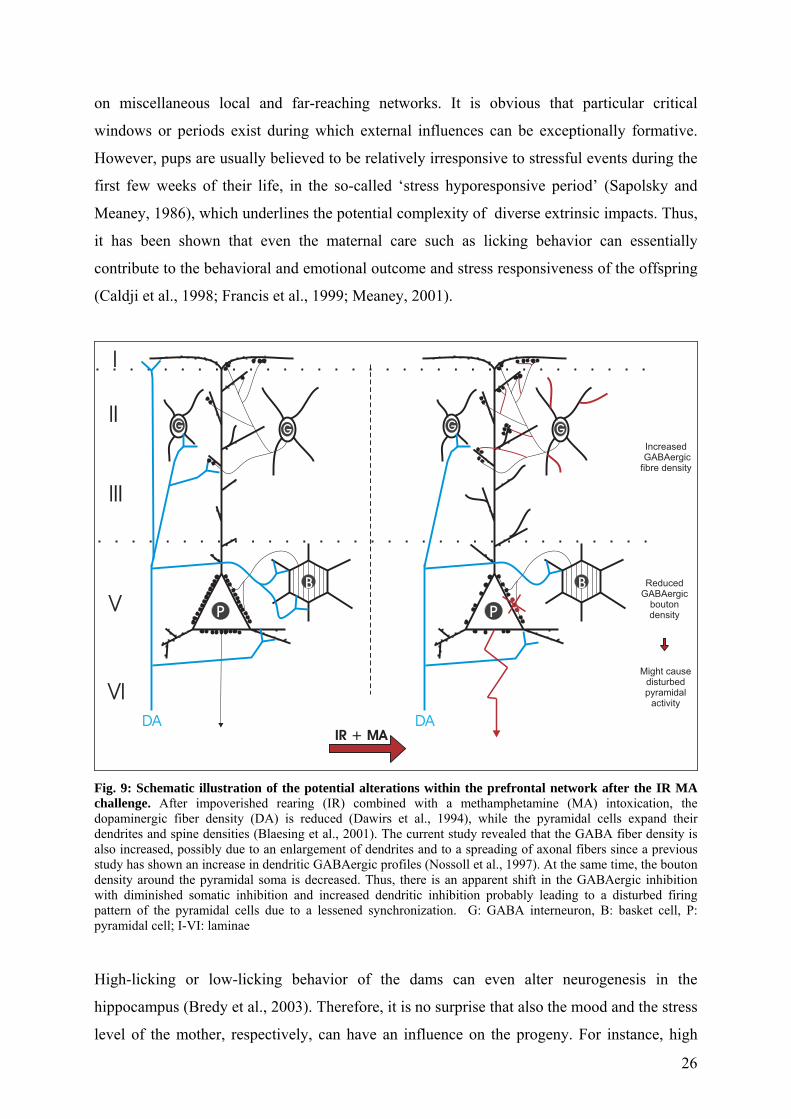
the observed cognitive impairments. Figure 9 gives a schematic overview of altered
morphologies and potential connections in the PFC of IR animals, which received additionally
the MA intoxication.
6. Consequences of early developmental disturbances (implications for schizophrenia)
The remarkable revelation of these studies is that a single disturbance during development
which actual primarily affects the dopaminergic system, can have such a wide-spread impact
25
on miscellaneous local and far-reaching networks. It is obvious that particular critical
windows or periods exist during which external influences can be exceptionally formative.
However, pups are usually believed to be relatively irresponsive to stressful events during the
first few weeks of their life, in the so-called ‘stress hyporesponsive period’ (Sapolsky and
Meaney, 1986), which underlines the potential complexity of diverse extrinsic impacts. Thus,
it has been shown that even the maternal care such as licking behavior can essentially
contribute to the behavioral and emotional outcome and stress responsiveness of the offspring
(Caldji et al., 1998; Francis et al., 1999; Meaney, 2001).
I
II
III
V
VI
DA
P
G
B
IR + MA
IncreasedGABAergic
fibre density
ReducedGABAergic
boutondensity
Might causedisturbedpyramidal
activity
DA
P
G
B
GG
Fig. 9: Schematic illustration of the potential alterations within the prefrontal network after the IR MA challenge. After impoverished rearing (IR) combined with a methamphetamine (MA) intoxication, the dopaminergic fiber density (DA) is reduced (Dawirs et al., 1994), while the pyramidal cells expand their dendrites and spine densities (Blaesing et al., 2001). The current study revealed that the GABA fiber density is also increased, possibly due to an enlargement of dendrites and to a spreading of axonal fibers since a previous study has shown an increase in dendritic GABAergic profiles (Nossoll et al., 1997). At the same time, the bouton density around the pyramidal soma is decreased. Thus, there is an apparent shift in the GABAergic inhibition with diminished somatic inhibition and increased dendritic inhibition probably leading to a disturbed firing pattern of the pyramidal cells due to a lessened synchronization. G: GABA interneuron, B: basket cell, P: pyramidal cell; I-VI: laminae High-licking or low-licking behavior of the dams can even alter neurogenesis in the
hippocampus (Bredy et al., 2003). Therefore, it is no surprise that also the mood and the stress
level of the mother, respectively, can have an influence on the progeny. For instance, high
26
levels of corticosterone, a stress hormone, during the lactation period can cause differences in
hippocampal cell proliferation and can evoke signs of hyperactive behavior in the offspring
(Brummelte et al., 2006b). However, it is clear that the type and degree of the external stress
is important for determining the morphological, behavioral and cognitive consequences.
Our 2-step animal model of using combined early MA intoxication as an acute stressor and IR
as a chronic stress factor has so far revealed several morphological changes in
neuroanatomical brain networks and some cognitive impairments, which resemble some of
the changes and deficits seen in schizophrenic individuals. Thus, Akil and colleagues (1999)
found a decrease in dopaminergic fibers in the prefrontal cortex of schizophrenic individuals,
comparable with the reduction in our animal model (Dawirs et al., 1994). In addition, the
imbalance of the DA system between cortical and subcortical areas, has not only been
observed after our IR and MA challenge (Busche et al., 2004) but was reported for the
schizophrenic human brain (Laruelle et al., 2003; Abi-Dargham, 2004). Besides, low
prefrontal DA levels are associated with negative or cognitive symptoms of schizophrenia,
while a hyperactivity of the mesolimbic pathway is assumed to be responsible for the positive
symptoms (Crow, 1980; Davis et al., 1991). Furthermore, our animal model revealed a
miswiring of prefrontal pyramidal projections (Bagorda et al., 2006), which corroborates the
dysconnection hypothesis of schizophrenia from Weinberger and Lipska (1995). In addition,
this miswiring, resulting from the different impact on lamina III compared to lamina V
pyramidal neurons in IR MA animals might help to explain the discrepancy of human studies,
paradoxically reporting either a hypofunction (Volz et al., 1999) or a hyperfunction (Manoach
et al., 1999) of the glutamatergic system in schizophrenic patients.
Intriguingly, the results of the current study reveal some resemblances with changes in
schizophrenia, too. Thus, a reduction of pyramidal GABAergic synapses has also been
observed in schizophrenic patients (Blum and Mann, 2002), with a reduction in PV-
immunoreactive structures being one of the most prevalent observations in post-mortem
studies (Woo et al., 1998; Pierri et al., 1999; Lewis et al., 1999). In addition, the GABAA
receptor density was upregulated at the cell bodies of pyramidal neurons (Benes et al., 1996a),
possibly compensating for a reduced number of inhibitory terminals (Lewis et al., 2005).
These indices for a reduced GABAergic somatic inhibition are in line with recent
neurophysiological studies, which revealed that some cognitive dysfunctions in schizophrenic
patients, as e.g. working memory deficits are associated with an abnormal neural
synchronization (Spencer et al., 2003; Lee et al., 2003; Spencer et al., 2004; Uhlhaas and
27

Singer, 2006). This again is in concert with the impairment of working memory in our animal
model (Dawirs et al., 1996).
In summary, our results indicate that a single early pharmacological stress is effectual to
induce severe morphological changes in the neuronal networks of the whole limbic system of
animals from IR conditions, which resemble at least some of the changes seen in
schizophrenic individuals. Taking the observed cognitive impairments into account, one is
tempted to suggest that our 2-step approach provides a useful animal model of psychoses and
schizophrenia.
Noteworthy, schizophrenia usually does not appear before early adulthood, even though it is
assumed to have at least partially developmental etiologic reasons. Thus, one could speculate
that the high plasticity during maturation of neuronal networks might somehow prevent the
outbreak of the disease but with the omission of this high plastic capacities, the miswiring
becomes more stable and starts to unfold its deleterious effects.
Interestingly, a treatment with clinical doses of methylphenidate (e.g. Ritalin®) for 30
consecutive days about two weeks after the noxious application of MA leads to a partially
‘recovery’ of the diminished dopaminergic fiber densities in adulthood (Grund et al., 2006;
Grund et al., revision submitted). Thus the deleterious impact of MA can be influenced by
another pharmacological interference, but apparently not by enriched environment
(Brummelte et al., in prep.). Methylphenidate is a stimulant drug which selectively blocks the
reuptake of DA and noradrenaline by binding to the according transporters (Gatley et al.,
1996) and is momentarily the drug of choice for the treatment of attention-
deficit/hyperactivity disorder (ADHD). The enhanced concentration of DA in the synaptic
cleft must somehow trigger an elevated sprouting of dopaminergic fibers, however, this
sprouting is only evident when the animals received the early MA challenge and not when
they received the control injection of saline. This is again a sign for the high plastic potentials
of the neuronal networks during development.
7. Conclusion and future perspectives
Taken together, this work provides additional evidence for a high plasticity of GABAergic
and dopaminergic structures during the maturation process, but in part also during adulthood
and ageing. The different extrinsic and intrinsic influences during postnatal development and
their interactions essentially contribute to the establishment of functional networks, whereby
28

the various transmitter systems play an indispensable role. Disturbances during critical
periods in the development lead to neuroanatomical alterations of the local networks and thus
also of the macrocircuits of the limbic system. The results from our 2-step animal model have
shown that especially DA appears to be particularly vulnerable to interfering effects and can
then subsequently affect all the connected other transmitter systems. The attempt of
microcircuits to compensate the altered innervation patterns probably results in a compromise,
which might provide an equilibrium of local connections, but which in turn might cause a
decompensation and subsequent imbalance of greater circuits and networks. The tendency of
every cell to counterbalance its excitability and its excitatory and inhibitory inputs, e.g. via
regulating the feedback loops, might contribute to the alterations seen at the local level
(Lehmann et al., 2005). However, the effect on the overall networks might be devastating.
Thus, the reactive changes in the morphology cause a different pattern of connectivity and
thus imply functional changes and differences in the behavioral and cognitive outcome. This
again might help to better understand the complex and individually divergent symptomatic
pathology of schizophrenia.
Another important conclusion of these works is the fact that there is not only a high
neuroplasticity of the various transmitter systems, and this during development as well as to a
lesser extent during adulthood, but also a very high interconnectivity and interdependency of
the transmitters. For instance, it has been revealed by others that 5-HT can directly regulate
the cortical DA release, probably via the expression of 5-HT2A receptors at the presynaptical
site (Miner et al., 2000; 2003; Alex and Pehek, 2006; Pehek et al., 2006). Similar intensive
interactions can be assumed for the GABA-DA relationship considering the prominent
alterations within the GABAergic system after the early challenge of the DA system
(Brummelte et al., 2007). In fact, DA is not only innervating pyramidal and GABAergic cell
bodies and dendrites (Sesack et al., 1995; Davidoff and Benes, 1998), but can also act
inhibitory or excitatory at GABAergic axon terminals (Geldwert et al., 2006). Moreover, Liu
and colleagues recently published their intriguing discovery of a direct protein-protein
coupling of the functionally and structurally different GABAA and DA D5 receptors, which
suggests a functional interaction of these two transmitter types (Liu et al., 2000). Hence, one
is tempted to suggest that the dopaminergic system as the main specific modulator and the
GABAergic system as the main inhibitor and thus coordinator of neuronal network activities,
are especially interwoven and interdependent. However, this relationship needs to be further
investigated, since e.g. a direct innervation of GABAergic synapses on cortical dopaminergic
nerve terminals has to our knowledge not been revealed to date. In addition, it would be
29

interesting to further examine the contribution and specific roles of the divergent GABAergic
subpopulations in these networks.
In summary, the interconnectivity of the various transmitter systems, in particular of DA and
GABA, appears to be highly complex and might therefore trigger or contribute to the reactive
processes after external or internal interferences. During development a disturbance of one
neurotransmitter system might additionally cause an imbalance in the temporal coordination
of the various connected maturation processes. Thus, one should keep in mind that
pharmacologic interventions will never only affect one transmitter system, even though they
are, e.g. selective 5-HT reuptake inhibitors (SSRIs) or only affecting the GABAA receptors
(benzodiazepines). This high interdependency and plasticity even during adulthood might also
help to explain, why the effect of neurological drugs is so unpredictable in the individual case.
Therefore, our results lead to the assumption that treatment with pharmaceuticals, especially
during the high phase of neuroplasticity during development, but also during the critical and
vulnerable period of ageing, should always be considered with care, as despite the acute
improvement, there might be hidden long-term side-effects, which might alter the neuronal
networks in perpetuity.
30

8. References Abi-Dargham A (2004) Do we still believe in the dopamine hypothesis? New data bring new evidence. Int J
Neuropsychopharmacol 7 Suppl 1:S1-S5.
Akil M, Pierri JN, Whitehead RE, Edgar CL, Mohila C, Sampson AR, Lewis DA (1999) Lamina-specific alterations in the dopamine innervation of the prefrontal cortex in schizophrenic subjects. Am J Psychiatry 156:1580-1589.
Alex KD, Pehek EA (2006) Pharmacologic mechanisms of serotonergic regulation of dopamine neurotransmission. Pharmacol Ther. (Epub ahead of print)
Arnsten AF, Cai JX, Steere JC, Goldman-Rakic PS (1995) Dopamine D2 receptor mechanisms contribute to age-related cognitive decline: the effects of quinpirole on memory and motor performance in monkeys. J Neurosci 15:3429-3439.
Bagorda F, Teuchert-Noodt G, Lehmann K (2006) Isolation rearing or methamphetamine traumatisation induce a "dysconnection" of prefrontal efferents in gerbils: implications for schizophrenia. J Neural Transm 113:365-379.
Ben-Ari Y (2002) Excitatory actions of gaba during development: the nature of the nurture. Nat Rev Neurosci 3:728-739.
Benes FM, Vincent SL, Molloy R (1993) Dopamine-immunoreactive axon varicosities form nonrandom contacts with GABA-immunoreactive neurons of rat medial prefrontal cortex. Synapse 15:285-295.
Benes FM, Vincent SL, Marie A, Khan Y (1996a) Up-regulation of GABAA receptor binding on neurons of the prefrontal cortex in schizophrenic subjects. Neuroscience 75:1021-1031.
Benes FM, Vincent SL, Molloy R, Khan Y (1996b) Increased interaction of dopamine-immunoreactive varicosities with GABA neurons of rat medial prefrontal cortex occurs during the postweanling period. Synapse 23:237-245.
Birkmayer W, Riederer P, Blau K (1989) Understanding the Neurotransmitters. Key to the Working of the Brain. Springer-Verlag Kg.
Björklund A, Lindvall O (1984) Dopamine-containing systems in the CNS. In: Handbook of chemical neuroanatomy - Classical transmitters in the CNS, Part 1 (Björklund A, Hökfelt T, eds), pp 55-122. Amsterdam: Elsevier.
Blaesing B, Nossoll M, Teuchert-Noodt G, Dawirs RR (2001) Postnatal maturation of prefrontal pyramidal neurones is sensitive to a single early dose of methamphetamine in gerbils (Meriones unguiculatus). J Neural Transm 108:101-113.
Blum BP, Mann JJ (2002) The GABAergic system in schizophrenia. Int J Neuropsychopharmacol 5:159-179.
Bowery NG, Smart TG (2006) GABA and glycine as neurotransmitters: a brief history. Br J Pharmacol 147 Suppl 1:S109-S119.
Bredy TW, Grant RJ, Champagne DL, Meaney MJ (2003) Maternal care influences neuronal survival in the hippocampus of the rat. Eur J Neurosci 18:2903-2909.
Brummelte S, Grund T, Czok A, Teuchert-Noodt G, Neddens J (2006a) Long-term effects of a single adult methamphetamine challenge: minor impact on dopamine fibre density in limbic brain areas of gerbils. Behav Brain Funct 2:12.
31

Brummelte S, Pawluski JL, Galea LA (2006b) High post-partum levels of corticosterone given to dams influence postnatal hippocampal cell proliferation and behavior of offspring: A model of post-partum stress and possible depression. Horm Behav 50:370-382.
Brummelte S, Teuchert-Noodt G (2006) Postnatal development of dopamine innervation in the amygdala and the entorhinal cortex of the gerbil (Meriones unguiculatus). Brain Res 1125: 9-16.
Brummelte S, Neddens J, Teuchert-Noodt G (2007) Alteration in the GABAergic network of the prefrontal cortex in a potential animal model of psychosis. J Neural Transm (Epub ahead of print).
Brummelte S., Witte A.V. and Teuchert-Noodt G.: Postnatal development of GABA and Calbindin cells and fibers in the prefrontal cortex and basolateral amygdala of gerbils (Meriones unguiculatus). Int J Dev Neurosci (accepted).
Busche A, Neddens J, Dinter C, Dawirs RR, Teuchert-Noodt G (2002) Differential influence of rearing conditions and methamphetamine on serotonin fibre maturation in the dentate gyrus of gerbils (Meriones unguiculatus). Dev Neurosci 24:512-521.
Busche A (2004) Zur Entstehung einer Imbalance im limbo-präfrontalen System bei Meriones unguiculatus: Der Einfluss restriktiver Isolationsaufzucht und einer postnatalen Methamphetamin-Intoxikation auf die monoaminergen Transmitter Dopamin und Serotonin in limbischen Regionen. Eine Bewertung quantitativer Datenerhebungen. Dissertation.
Busche A, Polascheck D, Lesting J, Neddens J, Teuchert-Noodt G (2004) Developmentally induced imbalance of dopaminergic fibre densities in limbic brain regions of gerbils ( Meriones unguiculatus). J Neural Transm 111:451-463.
Busche A, Bagorda A, Lehmann K, Neddens J, Teuchert-Noodt G (2006) The maturation of the acetylcholine system in the dentate gyrus of gerbils (Meriones unguiculatus) is affected by epigenetic factors. J Neural Transm 113:113-124.
Cadet JL, Brannock C (1998) Free radicals and the pathobiology of brain dopamine systems. Neurochem Int 32:117-131.
Caldji C, Tannenbaum B, Sharma S, Francis D, Plotsky PM, Meaney MJ (1998) Maternal care during infancy regulates the development of neural systems mediating the expression of fearfulness in the rat. Proc Natl Acad Sci U S A 95:5335-5340.
Cantuti-Castelvetri I, Shukitt-Hale B, Joseph JA (2003) Dopamine neurotoxicity: age-dependent behavioral and histological effects. Neurobiol Aging 24:697-706.
Celio MR (1990) Calbindin D-28k and parvalbumin in the rat nervous system. Neuroscience 35:375-475.
Chattopadhyaya B, Di CG, Higashiyama H, Knott GW, Kuhlman SJ, Welker E, Huang ZJ (2004) Experience and activity-dependent maturation of perisomatic GABAergic innervation in primary visual cortex during a postnatal critical period. J Neurosci 24:9598-9611.
Cherubini E, Gaiarsa JL, Ben-Ari Y (1991) GABA: an excitatory transmitter in early postnatal life. Trends Neurosci 14:515-519.
Chinta SJ, Andersen JK (2005) Dopaminergic neurons. Int J Biochem Cell Biol 37:942-946.
Conde F, Lund JS, Jacobowitz DM, Baimbridge KG, Lewis DA (1994) Local circuit neurons immunoreactive for calretinin, calbindin D-28k or parvalbumin in monkey prefrontal cortex: distribution and morphology. J Comp Neurol 341:95-116.
Constantinidis C, Williams GV, Goldman-Rakic PS (2002) A role for inhibition in shaping the temporal flow of information in prefrontal cortex. Nat Neurosci 5:175-180.
Crow TJ (1980) Positive and negative schizophrenic symptoms and the role of dopamine. Br J Psychiatry 137:383-386.
32

Cruz-Muros I, fonso-Oramas D, Abreu P, Barroso-Chinea P, Rodriguez M, Gonzalez MC, Gonzalez HT (2006) Aging of the rat mesostriatal system: Differences between the nigrostriatal and the mesolimbic compartments. Exp Neurol. (Epub ahead of print)
Davidoff SA, Benes FM (1998) High-resolution scatchard analysis shows D1 receptor binding on pyramidal and nonpyramidal neurons. Synapse 28:83-90.
Davis KL, Kahn RS, Ko G, Davidson M (1991) Dopamine in schizophrenia: a review and reconceptualization. Am J Psychiatry 148:1474-1486.
Dawirs RR, Teuchert-Noodt G, Busse M (1991) Single doses of methamphetamine cause changes in the density of dendritic spines in the prefrontal cortex of gerbils (Meriones unguiculatus). Neuropharmacology 30:275-282.
Dawirs RR, Teuchert-Noodt G, Czaniera R (1993a) Maturation of the dopamine innervation during postnatal development of the prefrontal cortex in gerbils (Meriones unguiculatus). A quantitative immunocytochemical study. J Hirnforsch 34:281-290.
Dawirs RR, Teuchert-Noodt G, Molthagen M (1993b) Indication of methamphetamine-induced reactive synaptogenesis in the prefrontal cortex of gerbils (Meriones unguiculatus). Eur J Pharmacol 241:89-97.
Dawirs RR, Teuchert-Noodt G, Czaniera R (1994) The postnatal maturation of dopamine innervation in the prefrontal cortex of gerbils (Meriones unguiculatus) is sensitive to an early single dose of methamphetamine. A quantitative immunocytochemical study. J Hirnforsch 35:195-204.
Dawirs RR, Teuchert-Noodt G, Czaniera R (1996) Ontogeny of PFC-related behaviours is sensitive to a single non-invasive dose of methamphetamine in neonatal gerbils (Meriones unguiculatus). J Neural Transm 103:1235-1245.
Dawirs RR, Teuchert-Noodt G, Nossoll M (1997) Pharmacologically induced neural plasticity in the prefrontal cortex of adult gerbils (Meriones unguiculatus). Eur J Pharmacol 327:117-123.
De Felipe J, Fairen A (1982) A type of basket cell in superficial layers of the cat visual cortex. A Golgi-electron microscope study. Brain Res 244:9-16.
Del Arco A, Segovia G, Mora F (2001) Dopamine release during stress in the prefrontal cortex of the rat decreases with age. Neuroreport 12:4019-4022.
Diamond MC, Ingham CA, Johnson RE, Bennett EL, Rosenzweig MR (1976) Effects of environment on morphology of rat cerebral cortex and hippocampus. J Neurobiol 7:75-85.
Diamond MC, Krech D, Rosenzweig MR (1964) The effects of an enriched environment on the histology of the rat cerebral cortex. J Comp Neurol 123:111-120.
Fallon JH, Koziell DA, Moore RY (1978) Catecholamine innervation of the basal forebrain. II. Amygdala, suprarhinal cortex and entorhinal cortex. J Comp Neurol 180:509-532.
Finkelstein DI, Stanic D, Parish CL, Tomas D, Dickson K, Horne MK (2000) Axonal sprouting following lesions of the rat substantia nigra. Neuroscience 97:99-112.
Francis DD, Champagne FA, Liu D, Meaney MJ (1999) Maternal care, gene expression, and the development of individual differences in stress reactivity. Ann N Y Acad Sci 896:66-84.
Freund TF (2003) Interneuron Diversity series: Rhythm and mood in perisomatic inhibition. Trends Neurosci 26:489-495.
Gabbott PL, Bacon SJ (1996) Local circuit neurons in the medial prefrontal cortex (areas 24a,b,c, 25 and 32) in the monkey: I. Cell morphology and morphometrics. J Comp Neurol 364:567-608.
Gale KN, Guidotti A (1976) GABA-mediated control of rat neostriatal tyrosine hydroxylase revealed by intranigral muscimol. Nature 263:691-693.
33
Ganguly K, Schinder AF, Wong ST, Poo M (2001) GABA itself promotes the developmental switch of neuronal GABAergic responses from excitation to inhibition. Cell 105:521-532.
Gatley SJ, Pan D, Chen R, Chaturvedi G, Ding YS (1996) Affinities of methylphenidate derivatives for dopamine, norepinephrine and serotonin transporters. Life Sci 58:231-239.
Geldwert D, Norris JM, Feldman IG, Schulman JJ, Joyce MP, Rayport S (2006) Dopamine presynaptically and heterogeneously modulates nucleus accumbens medium-spiny neuron GABA synapses in vitro. BMC Neurosci 7:53.
Geula C, Bu J, Nagykery N, Scinto LF, Chan J, Joseph J, Parker R, Wu CK (2003) Loss of calbindin-D28k from aging human cholinergic basal forebrain: relation to neuronal loss. J Comp Neurol 455:249-259.
Gluck MR, Moy LY, Jayatilleke E, Hogan KA, Manzino L, Sonsalla PK (2001) Parallel increases in lipid and protein oxidative markers in several mouse brain regions after methamphetamine treatment. J Neurochem 79:152-160.
Goldman-Rakic PS, Leranth C, Williams SM, Mons N, Geffard M (1989) Dopamine synaptic complex with pyramidal neurons in primate cerebral cortex. Proc Natl Acad Sci U S A 86:9015-9019.
Gorelova N, Seamans JK, Yang CR (2002) Mechanisms of dopamine activation of fast-spiking interneurons that exert inhibition in rat prefrontal cortex. J Neurophysiol 88:3150-3166.
Grace AA, Bunney BS (1985) Opposing effects of striatonigral feedback pathways on midbrain dopamine cell activity. Brain Res 333:271-284.
Grund T, Lehmann K, Bock N, Rothenberger A, Teuchert-Noodt G (2006) Influence of methylphenidate on brain development - an update of recent animal experiments. Behav Brain Funct 2:2.
Heizmann CW (1992) Calcium-binding proteins: basic concepts and clinical implications. Gen Physiol Biophys 11:411-425.
Hendry SH, Houser CR, Jones EG, Vaughn JE (1983) Synaptic organization of immunocytochemically identified GABA neurons in the monkey sensory-motor cortex. J Neurocytol 12:639-660.
Huang ZJ, Kirkwood A, Pizzorusso T, Porciatti V, Morales B, Bear MF, Maffei L, Tonegawa S (1999) BDNF regulates the maturation of inhibition and the critical period of plasticity in mouse visual cortex. Cell 98:739-755.
Hubel DH, Wiesel TN (1964) Effects of Monocular deprivation in kittens. Naunyn Schmiedebergs Arch Exp Pathol Pharmakol 248:492-497.
Hubel DH, Wiesel TN, LeVay S (1977) Plasticity of ocular dominance columns in monkey striate cortex. Philos Trans R Soc Lond B Biol Sci 278:377-409.
Inoue M, Suhara T, Sudo Y, Okubo Y, Yasuno F, Kishimoto T, Yoshikawa K, Tanada S (2001) Age-related reduction of extrastriatal dopamine D2 receptor measured by PET. Life Sci 69:1079-1084.
Itzhak Y, Gandia C, Huang PL, Ali SF (1998) Resistance of neuronal nitric oxide synthase-deficient mice to methamphetamine-induced dopaminergic neurotoxicity. J Pharmacol Exp Ther 284:1040-1047.
Jacobson M (1991) Developmental Neurobiology. New York: Plenum Press.
Jay TM, Glowinski J, Thierry AM (1995) Inhibition of hippocampoprefrontal cortex excitatory responses by the mesocortical DA system. Neuroreport 6:1845-1848.
Jones EG (1993) GABAergic neurons and their role in cortical plasticity in primates. Cereb Cortex 3:361-372.
Kaasinen V, Vilkman H, Hietala J, Nagren K, Helenius H, Olsson H, Farde L, Rinne J (2000) Age-related dopamine D2/D3 receptor loss in extrastriatal regions of the human brain. Neurobiol Aging 21:683-688.
34
Kalsbeek A, Voorn P, Buijs RM, Pool CW, Uylings HB (1988) Development of the dopaminergic innervation in the prefrontal cortex of the rat. J Comp Neurol 269:58-72.
Kawaguchi Y, Kubota Y (1997) GABAergic cell subtypes and their synaptic connections in rat frontal cortex. Cereb Cortex 7:476-486.
Kishimoto J, Tsuchiya T, Cox H, Emson PC, Nakayama Y (1998) Age-related changes of calbindin-D28k, calretinin, and parvalbumin mRNAs in the hamster brain. Neurobiol Aging 19:77-82.
Kita T, Wagner GC, Nakashima T (2003) Current research on methamphetamine-induced neurotoxicity: animal models of monoamine disruption. J Pharmacol Sci 92:178-195.
Knudsen EI (2004) Sensitive periods in the development of the brain and behavior. J Cogn Neurosci 16:1412-1425.
Kokoshka JM, Fleckenstein AE, Wilkins DG, Hanson GR (2000) Age-dependent differential responses of monoaminergic systems to high doses of methamphetamine. J Neurochem 75:2095-2102.
Laruelle M, Kegeles LS, bi-Dargham A (2003) Glutamate, dopamine, and schizophrenia: from pathophysiology to treatment. Ann N Y Acad Sci 1003:138-158.
Lau JW, Senok S, Stadlin A (2000) Methamphetamine-induced oxidative stress in cultured mouse astrocytes. Ann N Y Acad Sci 914:146-156.
Lauder JM, Liu J, Devaud L, Morrow AL (1998) GABA as a trophic factor for developing monoamine neurons. Perspect Dev Neurobiol 5:247-259.
Le Moal MM, Simon H (1991) Mesocorticolimbic dopaminergic network: functional and regulatory roles. Physiol Rev 71:155-234.
Le Moine C, Gaspar P (1998) Subpopulations of cortical GABAergic interneurons differ by their expression of D1 and D2 dopamine receptor subtypes. Brain Res Mol Brain Res 58:231-236.
Lee KH, Williams LM, Breakspear M, Gordon E (2003) Synchronous gamma activity: a review and contribution to an integrative neuroscience model of schizophrenia. Brain Res Brain Res Rev 41:57-78.
Lehmann K, Lesting J, Polascheck D, Teuchert-Noodt G (2003) Serotonin fibre densities in subcortical areas: differential effects of isolated rearing and methamphetamine. Brain Res Dev Brain Res 147:143-152.
Lehmann K, Hundsdorfer B, Hartmann T, Teuchert-Noodt G (2004) The acetylcholine fiber density of the neocortex is altered by isolated rearing and early methamphetamine intoxication in rodents. Exp Neurol 189:131-140.
Lehmann K, Butz M, Teuchert-Noodt G (2005) Offer and demand: proliferation and survival of neurons in the dentate gyrus. Eur J Neurosci 21:3205-3216.
Lesting J, Neddens J, Busche A, Teuchert-Noodt G (2005a) Hemisphere-specific effects on serotonin but not dopamine innervation in the nucleus accumbens of gerbils caused by isolated rearing and a single early methamphetamine challenge. Brain Res 1035:168-176.
Lesting J, Neddens J, Teuchert-Noodt G (2005b) Ontogeny of the dopamine innervation in the nucleus accumbens of gerbils. Brain Res 1066:16-23.
Lewis DA, Pierri JN, Volk DW, Melchitzky DS, Woo TU (1999) Altered GABA neurotransmission and prefrontal cortical dysfunction in schizophrenia. Biol Psychiatry 46:616-626.
Lewis DA, Hashimoto T, Volk DW (2005) Cortical inhibitory neurons and schizophrenia. Nat Rev Neurosci 6:312-324.
Liu F, Wan Q, Pristupa ZB, Yu XM, Wang YT, Niznik HB (2000) Direct protein-protein coupling enables cross-talk between dopamine D5 and gamma-aminobutyric acid A receptors. Nature 403:274-280.
35
Magnusson AK, Ulfendahl M, Tham R (2002) Early compensation of vestibulo-oculomotor symptoms after unilateral vestibular loss in rats is related to GABA(B) receptor function. Neuroscience 111:625-634.
Manoach DS, Press DZ, Thangaraj V, Searl MM, Goff DC, Halpern E, Saper CB, Warach S (1999) Schizophrenic subjects activate dorsolateral prefrontal cortex during a working memory task, as measured by fMRI. Biol Psychiatry 45:1128-1137.
Marty S, Wehrle R, Sotelo C (2000) Neuronal activity and brain-derived neurotrophic factor regulate the density of inhibitory synapses in organotypic slice cultures of postnatal hippocampus. J Neurosci 20:8087-8095.
Meaney MJ (2001) Maternal care, gene expression, and the transmission of individual differences in stress reactivity across generations. Annu Rev Neurosci 24:1161-1192.
Meredith CW, Jaffe C, ng-Lee K, Saxon AJ (2005) Implications of chronic methamphetamine use: a literature review. Harv Rev Psychiatry 13:141-154.
Miner LA, Backstrom JR, Sanders-Bush E, Sesack SR (2003) Ultrastructural localization of serotonin2A receptors in the middle layers of the rat prelimbic prefrontal cortex. Neuroscience 116:107-117.
Miner LH, Schroeter S, Blakely RD, Sesack SR (2000) Ultrastructural localization of the serotonin transporter in superficial and deep layers of the rat prelimbic prefrontal cortex and its spatial relationship to dopamine terminals. J Comp Neurol 427:220-234.
Neddens J, Brandenburg K, Teuchert-Noodt G, Dawirs RR (2001) Differential environment alters ontogeny of dopamine innervation of the orbital prefrontal cortex in gerbils. J Neurosci Res 63:209-213.
Neddens J, Bagorda F, Busche A, Horstmann S, Moll GH, Dawirs RR, Teuchert-Noodt G (2003) Epigenetic factors differentially influence postnatal maturation of serotonin (5-HT) innervation in cerebral cortex of gerbils: interaction of rearing conditions and early methamphetamine challenge. Brain Res Dev Brain Res 146:119-130.
Neddens J, Dawirs RR, Bagorda F, Busche A, Horstmann S, Teuchert-Noodt G (2004) Postnatal maturation of cortical serotonin lateral asymmetry in gerbils is vulnerable to both environmental and pharmacological epigenetic challenges. Brain Res 1021:200-208.
Nguyen L, Rigo JM, Rocher V, Belachew S, Malgrange B, Rogister B, Leprince P, Moonen G (2001) Neurotransmitters as early signals for central nervous system development. Cell Tissue Res 305:187-202.
Nieoullon A (2002) Dopamine and the regulation of cognition and attention. Prog Neurobiol 67:53-83.
Nilsson M, Perfilieva E, Johansson U, Orwar O, Eriksson PS (1999) Enriched environment increases neurogenesis in the adult rat dentate gyrus and improves spatial memory. J Neurobiol 39:569-578.
Nishimura T, Schwarzer C, Gasser E, Kato N, Vezzani A, Sperk G (2005) Altered expression of GABA(A) and GABA(B) receptor subunit mRNAs in the hippocampus after kindling and electrically induced status epilepticus. Neuroscience 134:691-704.
Nossoll M, Teuchert-Noodt G, Dawirs RR (1997) A single dose of methamphetamine in neonatal gerbils affects adult prefrontal gamma-aminobutyric acid innervation. Eur J Pharmacol 340:R3-R5.
Paylor R, Morrison SK, Rudy JW, Waltrip LT, Wehner JM (1992) Brief exposure to an enriched environment improves performance on the Morris water task and increases hippocampal cytosolic protein kinase C activity in young rats. Behav Brain Res 52:49-59.
Pehek EA, Nocjar C, Roth BL, Byrd TA, Mabrouk OS (2006) Evidence for the preferential involvement of 5-HT2A serotonin receptors in stress- and drug-induced dopamine release in the rat medial prefrontal cortex. Neuropsychopharmacology 31:265-277.
36

Pierri JN, Chaudry AS, Woo TU, Lewis DA (1999) Alterations in chandelier neuron axon terminals in the prefrontal cortex of schizophrenic subjects. Am J Psychiatry 156:1709-1719.
Racagni G, Bruno F, Cattabeni F, Maggi A, Di Giulio AM, Parenti M, Groppetti A (1977) Functional interaction between rat substantia nigra and striatum: GABA and dopamine interrelation. Brain Res 134:353-358.
Rao SG, Williams GV, Goldman-Rakic PS (1999) Isodirectional tuning of adjacent interneurons and pyramidal cells during working memory: evidence for microcolumnar organization in PFC. J Neurophysiol 81:1903-1916.
Rao SG, Williams GV, Goldman-Rakic PS (2000) Destruction and creation of spatial tuning by disinhibition: GABA(A) blockade of prefrontal cortical neurons engaged by working memory. J Neurosci 20:485-494.
Retaux S, Besson MJ, Penit-Soria J (1991) Opposing effects of dopamine D2 receptor stimulation on the spontaneous and the electrically evoked release of [3H]GABA on rat prefrontal cortex slices. Neuroscience 42:61-71.
Ricaurte GA, Guillery RW, Seiden LS, Schuster CR, Moore RY (1982) Dopamine nerve terminal degeneration produced by high doses of methylamphetamine in the rat brain. Brain Res 235:93-103.
Rosenzweig MR, Bennett EL (1969) Effects of differential environments on brain weights and enzyme activities in gerbils, rats, and mice. Dev Psychobiol 2:87-95.
Roth GS, Joseph JA (1994) Cellular and molecular mechanisms of impaired dopaminergic function during aging. Ann N Y Acad Sci 719:129-135.
Sapolsky RM, Meaney MJ (1986) Maturation of the adrenocortical stress response: neuroendocrine control mechanisms and the stress hyporesponsive period. Brain Res 396:64-76.
Segovia G, Porras A, Del AA, Mora F (2001) Glutamatergic neurotransmission in aging: a critical perspective. Mech Ageing Dev 122:1-29.
Sesack SR, Snyder CL, Lewis DA (1995) Axon terminals immunolabeled for dopamine or tyrosine hydroxylase synapse on GABA-immunoreactive dendrites in rat and monkey cortex. J Comp Neurol 363:264-280.
Seto-Ohshima A, Aoki E, Semba R, Emson PC, Heizmann CW (1990) Appearance of parvalbumin-specific immunoreactivity in the cerebral cortex and hippocampus of the developing rat and gerbil brain. Histochemistry 94:579-589.
Shatz CJ, Stryker MP (1978) Ocular dominance in layer IV of the cat's visual cortex and the effects of monocular deprivation. J Physiol 281:267-283.
Solbach S, Celio MR (1991) Ontogeny of the calcium binding protein parvalbumin in the rat nervous system. Anat Embryol (Berl) 184:103-124.
Somogyi P, Freund TF, Cowey A (1982) The axo-axonic interneuron in the cerebral cortex of the rat, cat and monkey. Neuroscience 7:2577-2607.
Spencer KM, Nestor PG, Niznikiewicz MA, Salisbury DF, Shenton ME, McCarley RW (2003) Abnormal neural synchrony in schizophrenia. J Neurosci 23:7407-7411.
Spencer KM, Nestor PG, Perlmutter R, Niznikiewicz MA, Klump MC, Frumin M, Shenton ME, McCarley RW (2004) Neural synchrony indexes disordered perception and cognition in schizophrenia. Proc Natl Acad Sci U S A 101:17288-17293.
Sperry RW (1963) Chemoaffinity in the orderly growth of the nerve fiber patterns and connections. Proc Natl Acad Sci U S A 50:703-710.
Swanson LW (1982) The projections of the ventral tegmental area and adjacent regions: a combined fluorescent retrograde tracer and immunofluorescence study in the rat. Brain Res Bull 9:321-353.
37

Tamas G, Buhl EH, Somogyi P (1997) Fast IPSPs elicited via multiple synaptic release sites by different types of GABAergic neurone in the cat visual cortex. J Physiol 500 ( Pt 3):715-738.
Teuchert-Noodt G (2000) Neuronal degeneration and reorganization: a mutual principle in pathological and in healthy interactions of limbic and prefrontal circuits. J Neural Transm Suppl315-333.
Teuchert-Noodt G, Dawirs RR (1991) Age-related toxicity in prefrontal cortex and caudate-putamen complex of gerbils (Meriones unguiculatus) after a single dose of methamphetamine. Neuropharmacology 30:733-743.
Thiessen D, Yahr P (1977) The gerbil in behavioral investigations. Mechanisms of territoriality and olfactory communication. Austin: University of Texas Press.
Traub RD, Whittington MA, Stanford IM, Jefferys JG (1996) A mechanism for generation of long-range synchronous fast oscillations in the cortex. Nature 383:621-624.
Troup GM, Smith GS, Walford RL (1969) Life span,chronologic disease patterns and age-related changes in relative spleen weights for the mongolian gerbil (Meriones unguiculatus). Exp Gerontol 4:139-143.
Ueda S, Sakakibara S, Watanabe E, Yoshimoto K, Koibuchi N (2002) Vulnerability of monoaminergic neurons in the brainstem of the zitter rat in oxidative stress. Prog Brain Res 136:293-302.
Uhlhaas PJ, Singer W (2006) Neural synchrony in brain disorders: relevance for cognitive dysfunctions and pathophysiology. Neuron 52:155-168.
Valenstein ES (2002) The discovery of chemical neurotransmitters. Brain Cogn 49:73-95.
Verney C, Alvarez C, Geffard M, Berger B (1990) Ultrastructural Double-Labelling Study of Dopamine Terminals and GABA-Containing Neurons in Rat Anteromedial Cerebral Cortex. Eur J Neurosci 2:960-972.
Vicente-Torres MA, Munoz E, Davila D, Gil-Loyzaga P (2001) Changes in the cochlear dopaminergic system of the aged rat. Brain Res 917:112-117.
Volz H, Gaser C, Hager F, Rzanny R, Ponisch J, Mentzel H, Kaiser WA, Sauer H (1999) Decreased frontal activation in schizophrenics during stimulation with the continuous performance test--a functional magnetic resonance imaging study. Eur Psychiatry 14:17-24.
Weinberger DR (1987) Implications of normal brain development for the pathogenesis of schizophrenia. Arch Gen Psychiatry 44:660-669.
Weinberger DR, Lipska BK (1995) Cortical maldevelopment, anti-psychotic drugs, and schizophrenia: a search for common ground. Schizophr Res 16:87-110.
Wiesel TN, Hubel DH (1965) Comparison of the effects of unilateral and bilateral eye closure on cortical unit responses in kittens. J Neurophysiol 28:1029-1040.
Winterfeld KT, Teuchert-Noodt G, Dawirs RR (1998) Social environment alters both ontogeny of dopamine innervation of the medial prefrontal cortex and maturation of working memory in gerbils (Meriones unguiculatus). J Neurosci Res 52:201-209.
Woo TU, Whitehead RE, Melchitzky DS, Lewis DA (1998) A subclass of prefrontal gamma-aminobutyric acid axon terminals are selectively altered in schizophrenia. Proc Natl Acad Sci U S A 95:5341-5346.
Wu CK, Nagykery N, Hersh LB, Scinto LF, Geula C (2003) Selective age-related loss of calbindin-D28k from basal forebrain cholinergic neurons in the common marmoset (Callithrix jacchus). Neuroscience 120:249-25
38

9. Publications Brummelte S., Grund T., Czok A., Teuchert-Noodt G. and Neddens J. (2006a): Long-term
effects of a single adult methamphetamine challenge: Minor impact on dopamine fibre
density in limbic brain areas of gerbils. Behav Brain Funct. 2: 12 (‘highly accessed’)
Brummelte S. and Teuchert-Noodt G. (2006): Postnatal development of dopamine innervation
in the amygdala and the entorhinal cortex of the gerbil (Meriones unguiculatus). Brain
Res.1125: 9-16
Brummelte S., Witte A.V. and Teuchert-Noodt G.: Postnatal development of GABA and
Calbindin cells and fibers in the prefrontal cortex and basolateral amygdala of gerbils
(Meriones unguiculatus). (accepted)
Brummelte S. and Teuchert-Noodt G.: Density of dopaminergic fibres in the prefrontal cortex
of gerbils (Meriones unguiculatus) is sensitive to aging. (Short Communication,
submitted)
Brummelte S., Neddens J., and Teuchert-Noodt G. (2007): Alteration in the GABAergic
network of the prefrontal cortex in an animal model of psychosis. J Neural Trans
(Epub ahead of print)
39

BioMed CentralBehavioral and Brain Functions
ss
Open AcceResearchLong-term effects of a single adult methamphetamine challenge: Minor impact on dopamine fibre density in limbic brain areas of gerbilsSusanne Brummelte1, Thorsten Grund1, Andrea Czok1, Gertraud Teuchert-Noodt1 and Jörg Neddens*1,2Address: 1Department of Neuroanatomy, Faculty of Biology, University of Bielefeld, Universitätsstr. 25, D-33615 Bielefeld, Germany and 2National Institutes of Health, NICHD, Section on Molecular Neurobiology, Bldg. 35, Rm. 2C-1004, Bethesda, MD 20892-3714, USA
Email: Susanne Brummelte - [email protected]; Thorsten Grund - [email protected]; Andrea Czok - [email protected]; Gertraud Teuchert-Noodt - [email protected]; Jörg Neddens* - [email protected]
* Corresponding author
AbstractBackground: The aim of the study was to test long-term effects of (+)-methamphetamine (MA)on the dopamine (DA) innervation in limbo-cortical regions of adult gerbils, in order to understandbetter the repair and neuroplasticity in disturbed limbic networks.
Methods: Male gerbils received a single high dose of either MA (25 mg/kg i.p.) or saline onpostnatal day 180. On postnatal day 340 the density of immunoreactive DA fibres and calbindin andparvalbumin cells was quantified in the right hemisphere.
Results: No effects were found in the prefrontal cortex, olfactory tubercle and amygdala, whereasthe pharmacological impact induced a slight but significant DA hyperinnervation in the nucleusaccumbens. The cell densities of calbindin (CB) and parvalbumin (PV) positive neurons wereadditionally tested in the nucleus accumbens, but no significant effects were found. The presentresults contrast with the previously published long-term effects of early postnatal MA treatmentthat lead to a restraint of the maturation of DA fibres in the nucleus accumbens and prefrontalcortex and a concomitant overshoot innervation in the amygdala.
Conclusion: We conclude that the morphogenetic properties of MA change during maturationand aging of gerbils, which may be due to physiological alterations of maturing vs. mature DAneurons innervating subcortical and cortical limbic areas. Our findings, together with results fromother long-term studies, suggest that immature limbic structures are more vulnerable to persistenteffects of a single MA intoxication; this might be relevant for the assessment of drug experience inadults vs. adolescents, and drug prevention programs.
BackgroundMethamphetamine (MA) is a common illicit drug, whichabuse is currently reaching epidemic proportions. Accord-
ing to the 2002 SAMHSA National Household Survey onDrug Abuse, 12.4 million Americans age 12 and older hadtried methamphetamine at least once in their lifetimes
Published: 28 March 2006
Behavioral and Brain Functions2006, 2:12 doi:10.1186/1744-9081-2-12
Received: 10 November 2005Accepted: 28 March 2006
This article is available from: http://www.behavioralandbrainfunctions.com/content/2/1/12
© 2006Brummelte et al; licensee BioMed Central Ltd.This is an Open Access article distributed under the terms of the Creative Commons Attribution License (http://creativecommons.org/licenses/by/2.0), which permits unrestricted use, distribution, and reproduction in any medium, provided the original work is properly cited.
Page 1 of 11(page number not for citation purposes)

Behavioral and Brain Functions 2006, 2:12 http://www.behavioralandbrainfunctions.com/content/2/1/12
(5.3 percent of the population). This increasing number isespecially alarming since it has been extensively shownthat MA exerts acute neurotoxic effects on the monoamin-ergic transmitter systems, and thus leads to characteristiccognitive impairments like deficits in memory and learn-ing, psychomotor speed and information processing [1].It is especially affecting the dopamine (DA) neurons, lead-ing to dramatic loss of fibres and other DAergic structuresin certain brain areas within a few days [2,3], even after asingle exposure [4].
Some evidence exists that monoaminergic fibres are ableto recover to some extend from this damage during longtime course [5-9]. Moreover, even reactive overshoot wasfound for serotonergic fibres in several limbic areas of thebrain, including left entorhinal cortex [10] and the septalpole of the hippocampal dentate gyrus [11]. For DA fibres,an early MA treatment produces hyperinnervation in amy-gdaloid nuclei and ventral entorhinal cortex [12] and arestraint of the maturation in prefrontal cortex [13,14].This lab has further shown that the single early MA intox-ication produces a loss of DA fibres and concomitanthyperinnervation of serotonin fibres in the nucleusaccumbens (NAC) [15].
Taken together, our recent studies indicate severe changesin the maturation of the limbo-cortical network followingan early single MA challenge. However, a primary studyhas already shown that the neuroplasticity that followsMA treatment might relate on the age of the animals [4].Since the functional maturation and aging of the brain isbased on various structural and physiological changes, thepresent study was carried out to question whether theremodelling of neural networks that is induced by theneurotoxic effects of MA may alter during the lifespan ofgerbils. For that purpose, 6 months old adult gerbilsreceived a single high dose of MA. At the age of 12 monthsthe DA innervation was examined in prefrontal cortex,olfactory tubercle, NAC, and amygdala to check for long-time effects on the fibre density.
MethodsAll experimental procedures were approved by the appro-priate committee for animal care in accordance with theguidelines of the European Communities Council Direc-tive. Breeding gerbils (Meriones unguiculatus) wereobtained from Harlan Winkelmann (Borchen, Germany).From offspring, a total of 18 males (weight 66–91 g; age331–348 days) were used in this study. Young animalswere weaned at postnatal day 30 and subsequently sepa-rated in standard cages (Macrolon® type 4) without anycontent except of sawdust. All animals had free access tofood and water and were kept on natural day/night cycles.On postnatal day 180, a total of 9 gerbils received a singlesystemic injection of (+)-methamphetamine hydrochlo-
ride (Sigma, M 8750; 25 mg/kg, i.p.). The other 9 animalswere sham-treated by an i.p. injection of saline. This dosewas chosen due to our former experiences, which haveshown that juvenile gerbils can tolerate higher doses (50mg/kg) than older ones. Notably, the rate of mortality issimilar at both ages receiving the different doses (unpub-lished data), indicating physiological changes during thepostnatal maturation of the brain.
The methods used for sectioning and DA immunohisto-chemistry have been published recently [15]. For theimmunohistochemistry of calbindin and parvalbumincells, 50 µm thick vibratome sections were taken from thesame animals (perfused with 100 ml 0.1 M sodiumcacodylate pH 6.2, followed by 750 ml 5% glutaraldehydein 0.1 M sodium cacodylate pH 7.6) and treated as fol-lows: Every third section was collected in 0.05 M Tris-HCLbuffered saline [TBS (pH 7.5)] at 4°C; rinsed 3 × 10 minin TBS; incubated 10 min with 1% H2O2 in TBS; rinsedagain 3 × 10 min in TBS; blocked in 10% normal goatserum and 0.4% Triton X-100 (Sigma) for 30 min; incu-bated with the primary antibody (1:3,000 mouse anti-cal-bindin, Sigma; 1:2,000 mouse anti-parvalbumin, Sigma)in 1% normal goat serum and 0.4% Triton X-100 for 18 h;rinsed 3 × 10 min in TBS; incubated for 30 min in bioti-nylated goat-anti-mouse antibody (Sigma) diluted 1:20with 1% normal goat serum; rinsed 3 × 10 min in TBS;incubated with ExtraAvidin-Peroxidase (Sigma) diluted1:20 for 30 min; rinsed 3 × 10 min in TBS; stained in0.05% 3.3-diaminobenzidine (Sigma) with 0.01% H2O2for 4 min. Finally, the sections were rinsed 5 × 10 min inTBS, mounted on glass slides, dried overnight, dehydratedwith ethanol, cleared with xylene and cover slipped withDePeX (Serva, Heidelberg, Germany). To avoid deviationsdue to probably lateralised innervation densities of DA orcalcium-binding proteins only right hemispheres wereused for quantification.
For quantification of fibre and cell densities, brain sec-tions were chosen in areas of interest by means of anatom-ical characteristics according to brain atlases of the rat [16]and the mouse [17]. The identification of the brain regionfollows the nomenclature of the atlas of the rat. The aver-age number of analysed sections was 18 per animal forDA, with a range of 4 up to 6 sections in single regions. Inthe defined region of each section (cf. Fig. 1) all detectablefibre fragments and cells were visualised in standard testfields (2,080 × 1,544 pixel; 0.22 mm2) using a bright fieldmicroscope (BX61, Olympus, Hamburg, Germany) and adigital camera for microscopy (ColorView II, SIS, Mün-ster, Germany) at 200-fold magnification. Cells and fibreswere quantified by software for image analysis (KS300,Jenoptik, Jena, Germany). Immunoreactive DA fibres ofdifferent diameter were standardised to identical thick-ness and visualised using a combination of Gauss filter
Page 2 of 11(page number not for citation purposes)

Behavioral and Brain Functions 2006, 2:12 http://www.behavioralandbrainfunctions.com/content/2/1/12
Page 3 of 11(page number not for citation purposes)
Dopamine immunoreactive fibres in each of the quantified regionsFigure 1Dopamine immunoreactive fibres in each of the quantified regions. Representative photomicrographs, taken from a saline control, of dopamine (DA) immunoreactive fibres of each of the quantified regions. A.1: Prefrontal cortex; A.2: Layer VI of the prelimbic area; A.3: Layer IV of the lateral orbital and agranular insular areas. B.1: Nucleus accumbens (NAC); B.2: Medial shell of NAC; B.3: Lateral core of NAC; B.4: Olfactory tubercle. C.1: Amygdala (AMY); C.2: Central nucleus of AMY; C.3: Basolateral nucleus of AMY. Note the differential innervation pattern and density of DA fibres in the respective regions. Scale bars: 1000 µm (A.1, B.1, C.1); 50 µm (A.2-3, B.2-4, C.2-3).

Behavioral and Brain Functions 2006, 2:12 http://www.behavioralandbrainfunctions.com/content/2/1/12
Page 4 of 11(page number not for citation purposes)
Photomicrographs of Calbindin and Parvalbumin immunoreactive neurons in the nucleus accumbensFigure 2Photomicrographs of Calbindin and Parvalbumin immunoreactive neurons in the nucleus accumbens. Overview (A) and higher magnifications (A1, A2) of the Calbindin innervation of the NAC. The majority of CB+ cells is located in the core, which border to the shell is detectable (black arrows). PV+ cells are almost exclusively located in the shell (B1, B2), how-ever, the overall density is much lower compared to CB+ cells. Scale bars: 2000 µm (A, B); 200 µm (A1, B1); 100 µm (A2, B2).

Behavioral and Brain Functions 2006, 2:12 http://www.behavioralandbrainfunctions.com/content/2/1/12
and Gerig operator that depicts differences of grey valuesof adjacent pixels and transforms the result into binaryimages. The DA fibre density was computed as a percent-age of the evaluated test area. Calbindin and parvalbuminpositive cells were detected by use of a threshold to thegrey value, followed by automatic sorting of adequateshape and minimal size (250 pixels) of the structures.Remaining structures were classified as cells, the size ofthe structures (cell area) being measured cumulatively
and the according cell density calculated by proportion ofcell number per test field area. Calbindin-positive cells arelocated almost exclusively in the core region of the NACand were measured only in this part of the NAC, whereasmedium-sized PV-positive cells are specific to the shelland were counted only in this area. All analyses were doneby a person blind to the pharmacological treatment ofindividual animals.
Dopamine innervation density in four regions of the gerbil brainFigure 3Dopamine innervation density in four regions of the gerbil brain. Dopamine (DA) innervation density ± S.E.M. is pre-sented in four regions of the gerbil brain, namely agranular insular and lateral orbital as well as prelimbic areas of the prefrontal cortex, the olfactory tubercle, core and shell areas of the nucleus accumbens (NAC), and the central and basolateral nuclei of the amygdala complex. Methamphetamine treatment generally tends to increase the DA innervation. However, a significant region-specific change in response to a single adult methamphetamine treatment exclusively occurs in the shell of the NAC (+11%; p = 0.0332). The difference in the core appears somewhat more pronounced but is not significant due to higher vari-ance (+21%; p = 0.1011). Student's t-Test, significance value: * p < 0.05. Following methamphetamine treatment, ANOVA detected a significant overall increase of DA innervation in core and shell of the NAC (F(1,16) = 4.7316; p = 0.0472).
Page 5 of 11(page number not for citation purposes)

Behavioral and Brain Functions 2006, 2:12 http://www.behavioralandbrainfunctions.com/content/2/1/12
The measurements were computed as arithmetic meansby-case and by-group ± S.E.M. of the respective regions(Fig. 3). Statistical analysis revealed regional effects of MAtreatment by the use of Student's t-test. General alterationsin the NAC were additionally investigated by use of 2-wayanalysis of variance (ANOVA), which checked for area-specific and group-specific effects [18]. Data analysis wascomputed with Statistica 6 (StatSoft, Tulsa, USA). The lev-els of significance were set at * p < 0.05, ** p < 0.01, and*** p < 0.001.
ResultsThe innervation pattern of DA immunoreactive fibres ingerbils is generally in line with the results of rats. Theinnervation pattern and density of DA immunoreactivefibres in the gerbil forebrain are region-specific (Figs. 1and 3). Representative photographs of the differential DAinnervation densities and patterns of the four regions thatwere subsequently studied in more detail are provided inFig. 1, taken from a male gerbil of the saline group.
Quantitative DA data were obtained from a total of 327sections that derived from 18 gerbils of two experimentalgroups (Saline n = 9, MA n = 9). The adult single systemicMA challenge induces no general alteration of DA inner-
vation pattern in the investigated regions of the gerbilbrain (Fig. 3). The overall DA fibre density in the NAC isselectively increased by MA [(ANOVA, F(1,16) = 4.7316,p = 0.0472) please note that ANOVA included compari-son of 8 vs. 8 animals only, because some NAC sectionswere damaged in one animal of each experimental group].However, the significant increase (+11%) is limited to theshell (Student's t-test, p = 0.0332), whereas alteration inthe core misses statistical significance (Student's t-test, p =0.1011). No change in DA fibre density was found in theprefrontal cortex, olfactory tubercle and amygdala.
To determine other potential alterations within the NAC,CB- and PV- positive structures were additionally investi-gated in this area. The distribution of CB- and PV-positivesubpopulations in the gerbil generally resembles the dis-tribution in the NAC of rats and primates (Fig. 2) [19-21].A single adult MA intoxication caused no significant alter-ation in the cell density of either CB- positive neurons orPV-positive neurons in the NAC. Neither was there a dif-ference in the cell areas (Fig. 4).
DiscussionAccording to our results, a single adult MA challengeinduces minor long-term changes of the DA innervation
Calbindin and parvalbumin cell densities and cell areas in the nucleus accumbensFigure 4Calbindin and parvalbumin cell densities and cell areas in the nucleus accumbens. Calbindin (CB) and parvalbumin (PV) cell densities and cell areas ± S.E.M. are presented for the nucleus accumbens (NAC). PV-positive cells and CB-positive cells are predominantly located in the shell and in the core of the NAC, respectively, where they were quantified. No statisti-cally significant effect of a single adult methamphetamine challenge could be detected for either number (cell density) or cumu-lative size (cell area) of both PV and CB cells. Generally, the number of CB cells is considerably higher and their average size is doubled compared to PV cells.
Page 6 of 11(page number not for citation purposes)
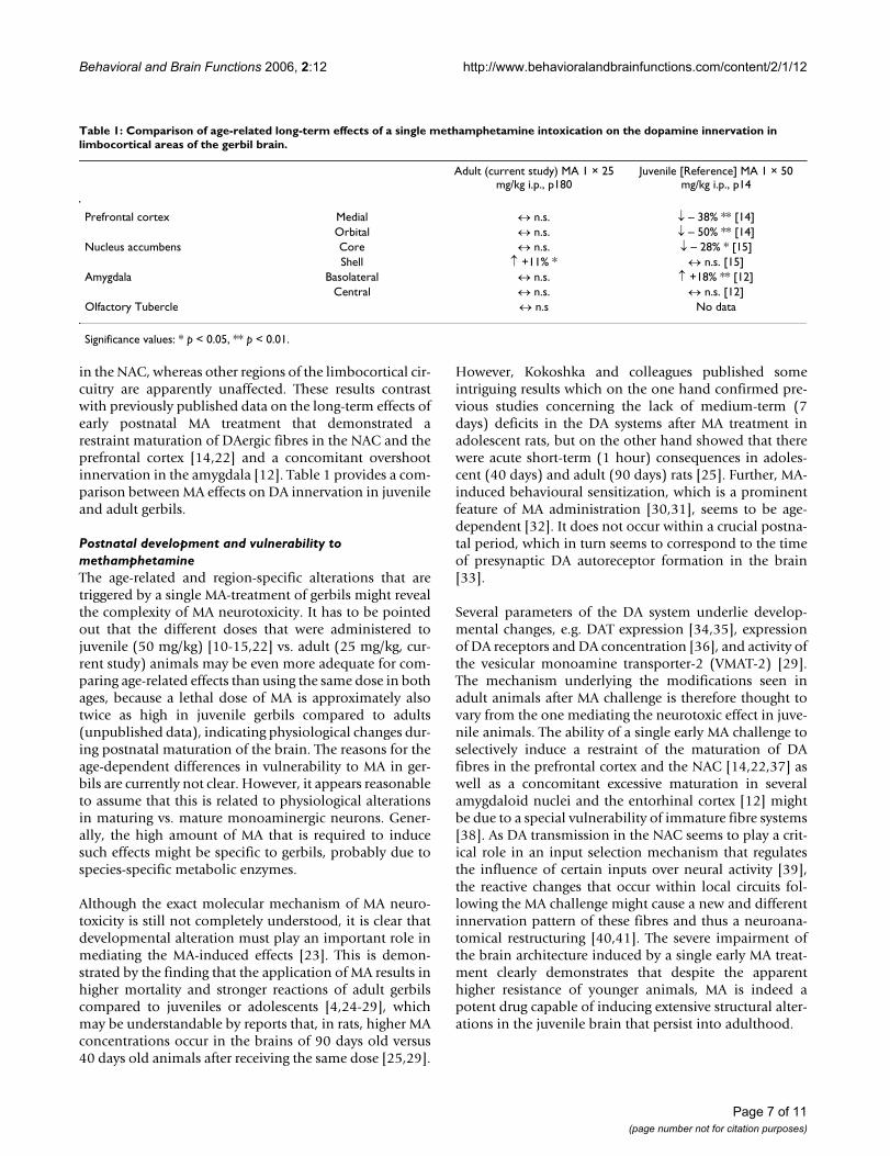
Behavioral and Brain Functions 2006, 2:12 http://www.behavioralandbrainfunctions.com/content/2/1/12
in the NAC, whereas other regions of the limbocortical cir-cuitry are apparently unaffected. These results contrastwith previously published data on the long-term effects ofearly postnatal MA treatment that demonstrated arestraint maturation of DAergic fibres in the NAC and theprefrontal cortex [14,22] and a concomitant overshootinnervation in the amygdala [12]. Table 1 provides a com-parison between MA effects on DA innervation in juvenileand adult gerbils.
Postnatal development and vulnerability to methamphetamineThe age-related and region-specific alterations that aretriggered by a single MA-treatment of gerbils might revealthe complexity of MA neurotoxicity. It has to be pointedout that the different doses that were administered tojuvenile (50 mg/kg) [10-15,22] vs. adult (25 mg/kg, cur-rent study) animals may be even more adequate for com-paring age-related effects than using the same dose in bothages, because a lethal dose of MA is approximately alsotwice as high in juvenile gerbils compared to adults(unpublished data), indicating physiological changes dur-ing postnatal maturation of the brain. The reasons for theage-dependent differences in vulnerability to MA in ger-bils are currently not clear. However, it appears reasonableto assume that this is related to physiological alterationsin maturing vs. mature monoaminergic neurons. Gener-ally, the high amount of MA that is required to inducesuch effects might be specific to gerbils, probably due tospecies-specific metabolic enzymes.
Although the exact molecular mechanism of MA neuro-toxicity is still not completely understood, it is clear thatdevelopmental alteration must play an important role inmediating the MA-induced effects [23]. This is demon-strated by the finding that the application of MA results inhigher mortality and stronger reactions of adult gerbilscompared to juveniles or adolescents [4,24-29], whichmay be understandable by reports that, in rats, higher MAconcentrations occur in the brains of 90 days old versus40 days old animals after receiving the same dose [25,29].
However, Kokoshka and colleagues published someintriguing results which on the one hand confirmed pre-vious studies concerning the lack of medium-term (7days) deficits in the DA systems after MA treatment inadolescent rats, but on the other hand showed that therewere acute short-term (1 hour) consequences in adoles-cent (40 days) and adult (90 days) rats [25]. Further, MA-induced behavioural sensitization, which is a prominentfeature of MA administration [30,31], seems to be age-dependent [32]. It does not occur within a crucial postna-tal period, which in turn seems to correspond to the timeof presynaptic DA autoreceptor formation in the brain[33].
Several parameters of the DA system underlie develop-mental changes, e.g. DAT expression [34,35], expressionof DA receptors and DA concentration [36], and activity ofthe vesicular monoamine transporter-2 (VMAT-2) [29].The mechanism underlying the modifications seen inadult animals after MA challenge is therefore thought tovary from the one mediating the neurotoxic effect in juve-nile animals. The ability of a single early MA challenge toselectively induce a restraint of the maturation of DAfibres in the prefrontal cortex and the NAC [14,22,37] aswell as a concomitant excessive maturation in severalamygdaloid nuclei and the entorhinal cortex [12] mightbe due to a special vulnerability of immature fibre systems[38]. As DA transmission in the NAC seems to play a crit-ical role in an input selection mechanism that regulatesthe influence of certain inputs over neural activity [39],the reactive changes that occur within local circuits fol-lowing the MA challenge might cause a new and differentinnervation pattern of these fibres and thus a neuroana-tomical restructuring [40,41]. The severe impairment ofthe brain architecture induced by a single early MA treat-ment clearly demonstrates that despite the apparenthigher resistance of younger animals, MA is indeed apotent drug capable of inducing extensive structural alter-ations in the juvenile brain that persist into adulthood.
Table 1: Comparison of age-related long-term effects of a single methamphetamine intoxication on the dopamine innervation in limbocortical areas of the gerbil brain.
Adult (current study) MA 1 × 25 mg/kg i.p., p180
Juvenile [Reference] MA 1 × 50 mg/kg i.p., p14
Prefrontal cortex Medial ↔ n.s. ↓ – 38% ** [14]Orbital ↔ n.s. ↓ – 50% ** [14]
Nucleus accumbens Core ↔ n.s. ↓ – 28% * [15]Shell ↑ +11% * ↔ n.s. [15]
Amygdala Basolateral ↔ n.s. ↑ +18% ** [12]Central ↔ n.s. ↔ n.s. [12]
Olfactory Tubercle ↔ n.s No data
Significance values: * p < 0.05, ** p < 0.01.
Page 7 of 11(page number not for citation purposes)

Behavioral and Brain Functions 2006, 2:12 http://www.behavioralandbrainfunctions.com/content/2/1/12
Effects of methamphetamine on different neurotransmitters and brain regionsIt was reported that the mechanism of MA neurotoxicityincludes the formation of reactive oxygen [42-44] andnitrogen [42,45,46] species, which damage monoaminer-gic neurons. However, several other factors may also con-tribute in mediating the neurotoxic effect of MA, leadingto region-specific and neuron-specific differences in vul-nerability. Fumagalli and colleagues have shown that ratslacking the dopamine transporter (DAT) are protectedagainst the MA-related neurotoxicity in the striatum [47].Interestingly, impairment in the function of VMAT-2,which accumulates cytoplasmic DA into synaptic vesiclesas seen in mice heterozygous for this transporter, increasesthe MA neurotoxicity [48]. It has also been demonstratedthat the blockage of either DA D1 or D2 receptors preventsthe damage of repeated doses of MA to striate DA termi-nals [49] and that there are regional differences in sensi-tivity of these terminals to the MA [4,50]. It seems likelythat DAT and DA receptors may be factors limiting theseverity of neurotoxic effects of MA, presumably by influ-encing the concentration and distribution of DA.
MA-induced alterations have also been found in otherneuronal elements like 5-HT fibres [10,15,51,52],GABAergic neurons [53], and the morphology of corticalpyramidal cells [54]. Our animal model has also revealedthat glutamatergic projection fibres from the mediodorsalprefrontal cortex to several other cortical areas are signifi-cantly reduced after an early single MA intoxication [55].Interestingly, in the present study the shell of the NAC isthe only area that reacts to a single adult MA challenge,and it is also almost the only area we have studied lackingany effect of the DA fibre density in response to a singlepostnatal drug treatment [12,14,15] (cf. Table 1). In con-trast, the core region of the NAC exhibits a strong declinein DA fibre density after an early single MA administration[15]. This is in line with results from Broening and col-leagues, who, after repeated MA treatment of rats, foundan almost completely loss of tyrosine hydroxylase immu-noreactivity in the core while the shell was almost spared[56]. In addition, most drugs increase extracellular DA lev-els preferentially in the shell region of the NAC [57],which coincides with differences in DA baseline levels[57,58], and different time-course of the maturation ofthe DA innervation in the core and shell areas [59]. Wemay conclude that the DA fibre systems of the brain are farfrom being homogeneous; thus, statements on the generaleffects of MA intoxication on DA fibres are misleading.
Regeneration and reorganisation of neural networks: implications for psychiatric diseasesIt has been shown before that DA fibres can be rebuiltwithin 24 weeks after a lesion of the NAC with 6-OHDA[60]. Furthermore, Finkelstein and colleagues could show
that a lesion to the substantia nigra causes sprouting ofDA fibres in the striatum of rats [61]. Thus, the increasedfibre density we have found in the NAC might probablybe caused by a specific regeneration rather than a reorgan-isation of fibres.
Several studies in humans and rodents have shown thatthe effects of MA are to a large extend reversible, althoughthis process might last many years and may stronglydepend on the severity and duration of the drug abuse [1].We have shown that a single administration of MA onpostnatal day 90 leads to a transient increase of the den-dritic spine density of prefrontal pyramidal neurons,which regain an almost normal level within 30 days post-treatment [62]. In the present study, we apparently pro-vided sufficient time for the impaired system to recoverfrom MA intoxication and to eventually regain normal DAfibre densities. Our model using only a single administra-tion of MA may therefore be more useful to mimic theeffects on first time users rather than on chronically abus-ers of the drug. In addition, it has been shown that inter-mittent treatment with MA can lead to the development oftolerance to its neurotoxic effects [63-65]. Thus the para-digm of repeated administration of MA as used in themajority of studies might either conceal or modify the del-eterious effects of the psychostimulant. In fact, some stud-ies have shown opposed or stronger effects of single versuschronic administration of MA [66,67].
Chronic MA use is known to cause psychotic symptomsthat mimic that of schizophrenia [68-70]. Further, wecould recently show that an early single intoxication leadsto a 'dysconnection' of prefrontal efferents [55], thus pro-viding an anatomical correlate of schizophrenia [71,72].This is consistent with results from Chen and colleagueswho revealed an association between earlier and larger useof MA with higher risk of psychosis in humans [68].
ConclusionThe results of this study show that even a single applica-tion of MA to adult gerbils may induce long-term physicalalterations in limbic brain areas, although the effects arenot as severe as seen after an early drug challenge. Theincreased DAergic fibre density in the NAC indicates reac-tive over-sprouting, which possibly is a response to alterednetwork requirements after MA treatment. It remains tobe investigated whether other brain areas would revealshort-term modifications, which might be concealed byrecovery in this approach. While it cannot be excludedthat the different effects in adult vs. juvenile gerbils are, atleast in part, due to the different doses of MA, severalother studies also indicate changes in MA effects thatdepend on the age of rats and mice. We thus may con-clude that, in rodents, MA not only acts age-dependent,but also highly region-specific. We have sufficient evi-
Page 8 of 11(page number not for citation purposes)

Behavioral and Brain Functions 2006, 2:12 http://www.behavioralandbrainfunctions.com/content/2/1/12
dence to suggest that early contact with this psychotropicsubstance during childhood might increase the risk of per-sistent severe structural changes of the brain architectureand may result in long-term cognitive and psychiatric dis-turbances.
Competing interestsThe author(s) declare that they have no competing inter-ests.
Authors' contributionsSB participated in the interpretation of the data anddrafted the manuscript.
TG contributed to the benchwork, analysis and interpreta-tion of the data.
AC contributed to the acquisition and interpretation ofdata.
GT contributed to the design of the study and the criticalreviewing of the manuscript.
JN handled the animals, contributed to the benchwork,analysis and interpretation of the data, participated in thedesign of the study, and the drafting and revision of themanuscript.
AcknowledgementsThe authors would like to thank Ulrike Schroeder and Francesco Bagorda for excellent technical assistance. The study was supported by grants of the deutsche Parkinson Vereinigung (dPV).
References1. Meredith CW, Jaffe C, Ang-Lee K, Saxon AJ: Implications of
chronic methamphetamine use: a literature review. Harv RevPsychiatry 2005, 13:141-154.
2. Ricaurte GA, Guillery RW, Seiden LS, Schuster CR, Moore RY:Dopamine nerve terminal degeneration produced by highdoses of methylamphetamine in the rat brain. Brain Res 1982,235:93-103.
3. Ricaurte GA, Schuster CR, Seiden LS: Long-term effects ofrepeated methylamphetamine administration on dopamineand serotonin neurons in the rat brain: a regional study. BrainRes 1980, 193:153-163.
4. Teuchert-Noodt G, Dawirs RR: Age-related toxicity in prefron-tal cortex and caudate-putamen complex of gerbils (Meri-ones unguiculatus) after a single dose of methamphetamine.Neuropharmacology 1991, 30:733-743.
5. Cass WA, Manning MW: Recovery of presynaptic dopaminergicfunctioning in rats treated with neurotoxic doses of meth-amphetamine. J Neurosci 1999, 19:7653-7660.
6. Cass WA: Attenuation and recovery of evoked overflow ofstriatal serotonin in rats treated with neurotoxic doses ofmethamphetamine. J Neurochem 2000, 74:1079-1085.
7. Friedman SD, Castaneda E, Hodge GK: Long-term monoaminedepletion, differential recovery, and subtle behavioralimpairment following methamphetamine-induced neuro-toxicity. Pharmacol Biochem Behav 1998, 61:35-44.
8. Woolverton WL, Ricaurte GA, Forno LS, Seiden LS: Long-termeffects of chronic methamphetamine administration in rhe-sus monkeys. Brain Res 1989, 486:73-78.
9. Sabol KE, Richards JB, Yung K: The effects of high-dose metham-phetamine in the aging rat: differential reinforcement of
low-rate 72-s schedule behavior and neurochemistry. J Phar-macol Exp Ther 2000, 294:850-863.
10. Neddens J, Dawirs RR, Bagorda F, Busche A, Horstmann S, Teuchert-Noodt G: Postnatal maturation of cortical serotonin lateralasymmetry in gerbils is vulnerable to both environmentaland pharmacological epigenetic challenges. Brain Res 2004,1021:200-208.
11. Busche A, Neddens J, Dinter C, Dawirs RR, Teuchert-Noodt G: Dif-ferential influence of rearing conditions and methampheta-mine on serotonin fibre maturation in the dentate gyrus ofgerbils (Meriones unguiculatus). Dev Neurosci 2002, 24:512-521.
12. Busche A, Polascheck D, Lesting J, Neddens J, Teuchert-Noodt G:Developmentally induced imbalance of dopaminergic fibredensities in limbic brain regions of gerbils (Meriones unguic-ulatus). J Neural Transm 2004, 111:451-463.
13. Dawirs RR, Teuchert-Noodt G, Nossoll M: Pharmacologicallyinduced neural plasticity in the prefrontal cortex of adultgerbils (Meriones unguiculatus). Eur J Pharmacol 1997,327:117-123.
14. Dawirs RR, Teuchert-Noodt G, Czaniera R: The postnatal matu-ration of dopamine innervation in the prefrontal cortex ofgerbils (Meriones unguiculatus) is sensitive to an early singledose of methamphetamine. A quantitative immunocyto-chemical study. J Hirnforsch 1994, 35:195-204.
15. Lesting J, Neddens J, Busche A, Teuchert-Noodt G: Hemisphere-specific effects on serotonin but not dopamine innervation inthe nucleus accumbens of gerbils caused by isolated rearingand a single early methamphetamine challenge. Brain Res2005, 1035:168-176.
16. Paxinos G, Watson C: The rat brain in stereotaxic coordinates NewYork: Academic Press; 1986.
17. Valverde F: Golgi atlas of the postnatal mouse brain Wien, New York:Springer-Verlag; 1998.
18. Stahel WA: Statistische Datenanalyse: eine Einführung für Naturwissen-schaftler Braunschweig: Vieweg; 1999.
19. Cote PY, Sadikot AF, Parent A: Complementary Distribution ofCalbindin D-28k and Parvalbumin in the Basal Forebrain andMidbrain of the Squirrel Monkey. Eur J Neurosci 1991,3:1316-1329.
20. Meredith GE, Pattiselanno A, Groenewegen HJ, Haber SN: Shell andcore in monkey and human nucleus accumbens identifiedwith antibodies to calbindin-D28k. J Comp Neurol 1996,365:628-639.
21. Zahm DS, Brog JS: On the significance of subterritories in the"accumbens" part of the rat ventral striatum. Neuroscience1992, 50:751-767.
22. Neddens J, Lesting J, Dawirs RR, Teuchert-Noodt G: An earlymethamphetamine challenge suppresses the maturation ofdopamine fibres in the nucleus accumbens of gerbils: on thesignificance of rearing conditions. J Neural Transm 2002,109:141-155.
23. Frost DO, Cadet JL: Effects of methamphetamine-induced neu-rotoxicity on the development of neural circuitry: a hypoth-esis. Brain Res Brain Res Rev 2000, 34:103-118.
24. Gomes-da-Silva J, de MR, Fernandez-Ruiz J, Summavielle T, TavaresMA: Effects of neonatal exposure to methamphetamine: cat-echolamine levels in brain areas of the developing rat. Ann NY Acad Sci 2004, 1025:602-611.
25. Kokoshka JM, Fleckenstein AE, Wilkins DG, Hanson GR: Age-dependent differential responses of monoaminergic systemsto high doses of methamphetamine. J Neurochem 2000,75:2095-2102.
26. Miller DB, O'Callaghan JP, Ali SF: Age as a susceptibility factor inthe striatal dopaminergic neurotoxicity observed in themouse following substituted amphetamine exposure. Ann NY Acad Sci 2000, 914:194-207.
27. Cappon GD, Morford LL, Vorhees CV: Ontogeny of metham-phetamine-induced neurotoxicity and associated hyperther-mic response. Brain Res Dev Brain Res 1997, 103:155-162.
28. Pu C, Vorhees CV: Developmental dissociation of metham-phetamine-induced depletion of dopaminergic terminalsand astrocyte reaction in rat striatum. Brain Res Dev Brain Res1993, 72:325-328.
29. Truong JG, Wilkins DG, Baudys J, Crouch DJ, Johnson-Davis KL, GibbJW, Hanson GR, Fleckenstein AE: Age-dependent methamphet-amine-induced alterations in vesicular monoamine trans-
Page 9 of 11(page number not for citation purposes)
http://www.ncbi.nlm.nih.gov/entrez/query.fcgi?cmd=Retrieve&db=PubMed&dopt=Abstract&list_uids=6145488
http://www.ncbi.nlm.nih.gov/entrez/query.fcgi?cmd=Retrieve&db=PubMed&dopt=Abstract&list_uids=6145488
http://www.ncbi.nlm.nih.gov/entrez/query.fcgi?cmd=Retrieve&db=PubMed&dopt=Abstract&list_uids=6145488
http://www.ncbi.nlm.nih.gov/entrez/query.fcgi?cmd=Retrieve&db=PubMed&dopt=Abstract&list_uids=7378814
http://www.ncbi.nlm.nih.gov/entrez/query.fcgi?cmd=Retrieve&db=PubMed&dopt=Abstract&list_uids=7378814
http://www.ncbi.nlm.nih.gov/entrez/query.fcgi?cmd=Retrieve&db=PubMed&dopt=Abstract&list_uids=7378814
http://www.ncbi.nlm.nih.gov/entrez/query.fcgi?cmd=Retrieve&db=PubMed&dopt=Abstract&list_uids=1922686
http://www.ncbi.nlm.nih.gov/entrez/query.fcgi?cmd=Retrieve&db=PubMed&dopt=Abstract&list_uids=1922686
http://www.ncbi.nlm.nih.gov/entrez/query.fcgi?cmd=Retrieve&db=PubMed&dopt=Abstract&list_uids=9715805
http://www.ncbi.nlm.nih.gov/entrez/query.fcgi?cmd=Retrieve&db=PubMed&dopt=Abstract&list_uids=9715805
http://www.ncbi.nlm.nih.gov/entrez/query.fcgi?cmd=Retrieve&db=PubMed&dopt=Abstract&list_uids=9715805
http://www.ncbi.nlm.nih.gov/entrez/query.fcgi?cmd=Retrieve&db=PubMed&dopt=Abstract&list_uids=2720435
http://www.ncbi.nlm.nih.gov/entrez/query.fcgi?cmd=Retrieve&db=PubMed&dopt=Abstract&list_uids=2720435
http://www.ncbi.nlm.nih.gov/entrez/query.fcgi?cmd=Retrieve&db=PubMed&dopt=Abstract&list_uids=2720435
http://www.ncbi.nlm.nih.gov/entrez/query.fcgi?cmd=Retrieve&db=PubMed&dopt=Abstract&list_uids=9200549
http://www.ncbi.nlm.nih.gov/entrez/query.fcgi?cmd=Retrieve&db=PubMed&dopt=Abstract&list_uids=9200549
http://www.ncbi.nlm.nih.gov/entrez/query.fcgi?cmd=Retrieve&db=PubMed&dopt=Abstract&list_uids=9200549
http://www.ncbi.nlm.nih.gov/entrez/query.fcgi?cmd=Retrieve&db=PubMed&dopt=Abstract&list_uids=8064138
http://www.ncbi.nlm.nih.gov/entrez/query.fcgi?cmd=Retrieve&db=PubMed&dopt=Abstract&list_uids=8064138
http://www.ncbi.nlm.nih.gov/entrez/query.fcgi?cmd=Retrieve&db=PubMed&dopt=Abstract&list_uids=8064138
http://www.ncbi.nlm.nih.gov/entrez/query.fcgi?cmd=Retrieve&db=PubMed&dopt=Abstract&list_uids=8742307
http://www.ncbi.nlm.nih.gov/entrez/query.fcgi?cmd=Retrieve&db=PubMed&dopt=Abstract&list_uids=8742307
http://www.ncbi.nlm.nih.gov/entrez/query.fcgi?cmd=Retrieve&db=PubMed&dopt=Abstract&list_uids=8742307
http://www.ncbi.nlm.nih.gov/entrez/query.fcgi?cmd=Retrieve&db=PubMed&dopt=Abstract&list_uids=1448200
http://www.ncbi.nlm.nih.gov/entrez/query.fcgi?cmd=Retrieve&db=PubMed&dopt=Abstract&list_uids=1448200
http://www.ncbi.nlm.nih.gov/entrez/query.fcgi?cmd=Retrieve&db=PubMed&dopt=Abstract&list_uids=9427479
http://www.ncbi.nlm.nih.gov/entrez/query.fcgi?cmd=Retrieve&db=PubMed&dopt=Abstract&list_uids=9427479
http://www.ncbi.nlm.nih.gov/entrez/query.fcgi?cmd=Retrieve&db=PubMed&dopt=Abstract&list_uids=9427479
http://www.ncbi.nlm.nih.gov/entrez/query.fcgi?cmd=Retrieve&db=PubMed&dopt=Abstract&list_uids=8097974
http://www.ncbi.nlm.nih.gov/entrez/query.fcgi?cmd=Retrieve&db=PubMed&dopt=Abstract&list_uids=8097974

Behavioral and Brain Functions 2006, 2:12 http://www.behavioralandbrainfunctions.com/content/2/1/12
porter-2 function: implications for neurotoxicity. J PharmacolExp Ther 2005, 314:1087-1092.
30. Akiyama K, Kanzaki A, Tsuchida K, Ujike H: Methamphetamine-induced behavioral sensitization and its implications forrelapse of schizophrenia. Schizophr Res 1994, 12:251-257.
31. Chiu CT, Ma T, Ho IK: Attenuation of methamphetamine-induced behavioral sensitization in mice by systemic admin-istration of naltrexone. Brain Res Bull 2005, 67:100-109.
32. Kuribara H, Yasuda H, Uchihashi Y: Age-dependent inductionand maintenance of sensitization to methamphetamine-induced hyperactivity in mice. Psychiatry Clin Neurosci 1996,50:299-303.
33. Fujiwara Y, Kazahaya Y, Nakashima M, Sato M, Otsuki S: Behavioralsensitization to methamphetamine in the rat: an ontogenicstudy. Psychopharmacology (Berl) 1987, 91:316-319.
34. Coulter CL, Happe HK, Murrin LC: Dopamine transporter devel-opment in postnatal rat striatum: an autoradiographic studywith [3H]WIN 35,428. Brain Res Dev Brain Res 1997, 104:55-62.
35. Moll GH, Mehnert C, Wicker M, Bock N, Rothenberger A, Ruther E,Huether G: Age-associated changes in the densities of presyn-aptic monoamine transporters in different regions of the ratbrain from early juvenile life to late adulthood. Brain Res DevBrain Res 2000, 119:251-257.
36. Noisin EL, Thomas WE: Ontogeny of dopaminergic function inthe rat midbrain tegmentum, corpus striatum and frontalcortex. Brain Res 1988, 469:241-252.
37. Neddens J, Brandenburg K, Teuchert-Noodt G, Dawirs RR: Differ-ential environment alters ontogeny of dopamine innervationof the orbital prefrontal cortex in gerbils. J Neurosci Res 2001,63:209-213.
38. Benes FM: The role of stress and dopamine-GABA interac-tions in the vulnerability for schizophrenia. J Psychiatr Res 1997,31:257-275.
39. Floresco SB, Blaha CD, Yang CR, Phillips AG: Modulation of hip-pocampal and amygdalar-evoked activity of nucleusaccumbens neurons by dopamine: cellular mechanisms ofinput selection. J Neurosci 2001, 21:2851-2860.
40. Teuchert-Noodt G: Neuronal degeneration and reorganiza-tion: a mutual principle in pathological and in healthy inter-actions of limbic and prefrontal circuits. J Neural Transm Suppl2000:315-333.
41. Bennay M, Gernert M, Schwabe K, Enkel T, Koch M: Neonatalmedial prefrontal cortex lesion enhances the sensitivity ofthe mesoaccumbal dopamine system. Eur J Neurosci 2004,19:3277-3290.
42. Cadet JL, Brannock C: Free radicals and the pathobiology ofbrain dopamine systems. Neurochem Int 1998, 32:117-131.
43. Gluck MR, Moy LY, Jayatilleke E, Hogan KA, Manzino L, Sonsalla PK:Parallel increases in lipid and protein oxidative markers inseveral mouse brain regions after methamphetamine treat-ment. J Neurochem 2001, 79:152-160.
44. Kita T, Wagner GC, Nakashima T: Current research on metham-phetamine-induced neurotoxicity: animal models ofmonoamine disruption. J Pharmacol Sci 2003, 92:178-195.
45. Imam SZ, Newport GD, Itzhak Y, Cadet JL, Islam F, Slikker W Jr, AliSF: Peroxynitrite plays a role in methamphetamine-induceddopaminergic neurotoxicity: evidence from mice lackingneuronal nitric oxide synthase gene or overexpressing cop-per-zinc superoxide dismutase. J Neurochem 2001, 76:745-749.
46. Itzhak Y, Gandia C, Huang PL, Ali SF: Resistance of neuronal nitricoxide synthase-deficient mice to methamphetamine-induced dopaminergic neurotoxicity. J Pharmacol Exp Ther 1998,284:1040-1047.
47. Fumagalli F, Gainetdinov RR, Valenzano KJ, Caron MG: Role ofdopamine transporter in methamphetamine-induced neuro-toxicity: evidence from mice lacking the transporter. J Neu-rosci 1998, 18:4861-4869.
48. Fumagalli F, Gainetdinov RR, Wang YM, Valenzano KJ, Miller GW,Caron MG: Increased methamphetamine neurotoxicity inheterozygous vesicular monoamine transporter 2 knock-outmice. J Neurosci 1999, 19:2424-2431.
49. O'Dell SJ, Weihmuller FB, Marshall JF: Methamphetamine-induced dopamine overflow and injury to striatal dopamineterminals: attenuation by dopamine D1 or D2 antagonists. JNeurochem 1993, 60:1792-1799.
50. Eisch AJ, Gaffney M, Weihmuller FB, O'Dell SJ, Marshall JF: Striatalsubregions are differentially vulnerable to the neurotoxiceffects of methamphetamine. Brain Res 1992, 598:321-326.
51. Lehmann K, Lesting J, Polascheck D, Teuchert-Noodt G: Serotoninfibre densities in subcortical areas: differential effects of iso-lated rearing and methamphetamine. Brain Res Dev Brain Res2003, 147:143-152.
52. Neddens J, Bagorda F, Busche A, Horstmann S, Moll GH, Dawirs RR,Teuchert-Noodt G: Epigenetic factors differentially influencepostnatal maturation of serotonin (5-HT) innervation in cer-ebral cortex of gerbils: interaction of rearing conditions andearly methamphetamine challenge. Brain Res Dev Brain Res2003, 146:119-130.
53. Nossoll M, Teuchert-Noodt G, Dawirs RR: A single dose of meth-amphetamine in neonatal gerbils affects adult prefrontalgamma-aminobutyric acid innervation. Eur J Pharmacol 1997,340:R3-R5.
54. Blaesing B, Nossoll M, Teuchert-Noodt G, Dawirs RR: Postnatalmaturation of prefrontal pyramidal neurones is sensitive toa single early dose of methamphetamine in gerbils (Merionesunguiculatus). J Neural Transm 2001, 108:101-113.
55. Bagorda F, Teuchert-Noodt G, Lehmann K: Isolation rearing ormethamphetamine traumatisation induce a "dysconnec-tion" of prefrontal efferents in gerbils: implications for schiz-ophrenia. J Neural Transm 2006, 113:365-379.
56. Broening HW, Pu C, Vorhees CV: Methamphetamine selectivelydamages dopaminergic innervation to the nucleusaccumbens core while sparing the shell. Synapse 1997,27:153-160.
57. Zocchi A, Girlanda E, Varnier G, Sartori I, Zanetti L, Wildish GA, Len-non M, Mugnaini M, Heidbreder CA: Dopamine responsivenessto drugs of abuse: A shell-core investigation in the nucleusaccumbens of the mouse. Synapse 2003, 50:293-302.
58. King D, Zigmond MJ, Finlay JM: Effects of dopamine depletion inthe medial prefrontal cortex on the stress-induced increasein extracellular dopamine in the nucleus accumbens coreand shell. Neuroscience 1997, 77:141-153.
59. Lesting J, Neddens J, Teuchert-Noodt G: Ontogeny of thedopamine innervation in the nucleus accumbens of gerbils.Brain Res 2005, 1066:16-23.
60. Vos PE, Steinbusch HW, van Ree JM: Reinnervation after destruc-tion of the dopaminergic system in the rat nucleusaccumbens: a quantitative immunohistochemical analysis.Neurosci Lett 1996, 207:21-24.
61. Finkelstein DI, Stanic D, Parish CL, Tomas D, Dickson K, Horne MK:Axonal sprouting following lesions of the rat substantianigra. Neuroscience 2000, 97:99-112.
62. Dawirs RR, Teuchert-Noodt G, Molthagen M: Indication of meth-amphetamine-induced reactive synaptogenesis in the pre-frontal cortex of gerbils (Meriones unguiculatus). Eur JPharmacol 1993, 241:89-97.
63. Segal DS, Kuczenski R, O'Neil ML, Melega WP, Cho AK: Escalatingdose methamphetamine pretreatment alters the behavioraland neurochemical profiles associated with exposure to ahigh-dose methamphetamine binge. Neuropsychopharmacology2003, 28:1730-1740.
64. Segal DS, Kuczenski R: Human Methamphetamine Pharmacok-inetics Simulated in the Rat: Single Daily IntravenousAdministration Reveals Elements of Sensitization and Toler-ance. Neuropsychopharmacology 2005 in press.
65. Thomas DM, Kuhn DM: Attenuated microglial activation medi-ates tolerance to the neurotoxic effects of methampheta-mine. J Neurochem 2005, 92:790-797.
66. Ishikawa K, Nitta A, Mizoguchi H, Mohri A, Murai R, Miyamoto Y,Noda Y, Kitaichi K, Yamada K, Nabeshima T: Effects of single andrepeated administration of methamphetamine or morphineon neuroglycan C gene expression in the rat brain. Int J Neu-ropsychopharmacol 2005 in press.
67. Zhu JP, Xu W, Angulo N, Angulo JA: Methamphetamine-inducedstriatal apoptosis in the mouse brain: Comparison of a bingeto an acute bolus drug administration. Neurotoxicology 2005.
68. Chen CK, Lin SK, Sham PC, Ball D, Loh EW, Hsiao CC, Chiang YL,Ree SC, Lee CH, Murray RM: Pre-morbid characteristics and co-morbidity of methamphetamine users with and without psy-chosis. Psychol Med 2003, 33:1407-1414.
Page 10 of 11(page number not for citation purposes)
http://www.ncbi.nlm.nih.gov/entrez/query.fcgi?cmd=Retrieve&db=PubMed&dopt=Abstract&list_uids=8054317
http://www.ncbi.nlm.nih.gov/entrez/query.fcgi?cmd=Retrieve&db=PubMed&dopt=Abstract&list_uids=8054317
http://www.ncbi.nlm.nih.gov/entrez/query.fcgi?cmd=Retrieve&db=PubMed&dopt=Abstract&list_uids=8054317
http://www.ncbi.nlm.nih.gov/entrez/query.fcgi?cmd=Retrieve&db=PubMed&dopt=Abstract&list_uids=9201795
http://www.ncbi.nlm.nih.gov/entrez/query.fcgi?cmd=Retrieve&db=PubMed&dopt=Abstract&list_uids=9201795
http://www.ncbi.nlm.nih.gov/entrez/query.fcgi?cmd=Retrieve&db=PubMed&dopt=Abstract&list_uids=9201795
http://www.ncbi.nlm.nih.gov/entrez/query.fcgi?cmd=Retrieve&db=PubMed&dopt=Abstract&list_uids=3104953
http://www.ncbi.nlm.nih.gov/entrez/query.fcgi?cmd=Retrieve&db=PubMed&dopt=Abstract&list_uids=3104953
http://www.ncbi.nlm.nih.gov/entrez/query.fcgi?cmd=Retrieve&db=PubMed&dopt=Abstract&list_uids=3104953
http://www.ncbi.nlm.nih.gov/entrez/query.fcgi?cmd=Retrieve&db=PubMed&dopt=Abstract&list_uids=9466707
http://www.ncbi.nlm.nih.gov/entrez/query.fcgi?cmd=Retrieve&db=PubMed&dopt=Abstract&list_uids=9466707
http://www.ncbi.nlm.nih.gov/entrez/query.fcgi?cmd=Retrieve&db=PubMed&dopt=Abstract&list_uids=9466707
http://www.ncbi.nlm.nih.gov/entrez/query.fcgi?cmd=Retrieve&db=PubMed&dopt=Abstract&list_uids=3401800
http://www.ncbi.nlm.nih.gov/entrez/query.fcgi?cmd=Retrieve&db=PubMed&dopt=Abstract&list_uids=3401800
http://www.ncbi.nlm.nih.gov/entrez/query.fcgi?cmd=Retrieve&db=PubMed&dopt=Abstract&list_uids=3401800
http://www.ncbi.nlm.nih.gov/entrez/query.fcgi?cmd=Retrieve&db=PubMed&dopt=Abstract&list_uids=9278189
http://www.ncbi.nlm.nih.gov/entrez/query.fcgi?cmd=Retrieve&db=PubMed&dopt=Abstract&list_uids=9278189
http://www.ncbi.nlm.nih.gov/entrez/query.fcgi?cmd=Retrieve&db=PubMed&dopt=Abstract&list_uids=9542724
http://www.ncbi.nlm.nih.gov/entrez/query.fcgi?cmd=Retrieve&db=PubMed&dopt=Abstract&list_uids=9542724
http://www.ncbi.nlm.nih.gov/entrez/query.fcgi?cmd=Retrieve&db=PubMed&dopt=Abstract&list_uids=9495865
http://www.ncbi.nlm.nih.gov/entrez/query.fcgi?cmd=Retrieve&db=PubMed&dopt=Abstract&list_uids=9495865
http://www.ncbi.nlm.nih.gov/entrez/query.fcgi?cmd=Retrieve&db=PubMed&dopt=Abstract&list_uids=9495865
http://www.ncbi.nlm.nih.gov/entrez/query.fcgi?cmd=Retrieve&db=PubMed&dopt=Abstract&list_uids=9634552
http://www.ncbi.nlm.nih.gov/entrez/query.fcgi?cmd=Retrieve&db=PubMed&dopt=Abstract&list_uids=9634552
http://www.ncbi.nlm.nih.gov/entrez/query.fcgi?cmd=Retrieve&db=PubMed&dopt=Abstract&list_uids=9634552
http://www.ncbi.nlm.nih.gov/entrez/query.fcgi?cmd=Retrieve&db=PubMed&dopt=Abstract&list_uids=8473897
http://www.ncbi.nlm.nih.gov/entrez/query.fcgi?cmd=Retrieve&db=PubMed&dopt=Abstract&list_uids=8473897
http://www.ncbi.nlm.nih.gov/entrez/query.fcgi?cmd=Retrieve&db=PubMed&dopt=Abstract&list_uids=8473897
http://www.ncbi.nlm.nih.gov/entrez/query.fcgi?cmd=Retrieve&db=PubMed&dopt=Abstract&list_uids=1486494
http://www.ncbi.nlm.nih.gov/entrez/query.fcgi?cmd=Retrieve&db=PubMed&dopt=Abstract&list_uids=1486494
http://www.ncbi.nlm.nih.gov/entrez/query.fcgi?cmd=Retrieve&db=PubMed&dopt=Abstract&list_uids=1486494
http://www.ncbi.nlm.nih.gov/entrez/query.fcgi?cmd=Retrieve&db=PubMed&dopt=Abstract&list_uids=9537803
http://www.ncbi.nlm.nih.gov/entrez/query.fcgi?cmd=Retrieve&db=PubMed&dopt=Abstract&list_uids=9537803
http://www.ncbi.nlm.nih.gov/entrez/query.fcgi?cmd=Retrieve&db=PubMed&dopt=Abstract&list_uids=9537803
http://www.ncbi.nlm.nih.gov/entrez/query.fcgi?cmd=Retrieve&db=PubMed&dopt=Abstract&list_uids=9266776
http://www.ncbi.nlm.nih.gov/entrez/query.fcgi?cmd=Retrieve&db=PubMed&dopt=Abstract&list_uids=9266776
http://www.ncbi.nlm.nih.gov/entrez/query.fcgi?cmd=Retrieve&db=PubMed&dopt=Abstract&list_uids=9266776
http://www.ncbi.nlm.nih.gov/entrez/query.fcgi?cmd=Retrieve&db=PubMed&dopt=Abstract&list_uids=9044382
http://www.ncbi.nlm.nih.gov/entrez/query.fcgi?cmd=Retrieve&db=PubMed&dopt=Abstract&list_uids=9044382
http://www.ncbi.nlm.nih.gov/entrez/query.fcgi?cmd=Retrieve&db=PubMed&dopt=Abstract&list_uids=9044382
http://www.ncbi.nlm.nih.gov/entrez/query.fcgi?cmd=Retrieve&db=PubMed&dopt=Abstract&list_uids=8710200
http://www.ncbi.nlm.nih.gov/entrez/query.fcgi?cmd=Retrieve&db=PubMed&dopt=Abstract&list_uids=8710200
http://www.ncbi.nlm.nih.gov/entrez/query.fcgi?cmd=Retrieve&db=PubMed&dopt=Abstract&list_uids=8223930
http://www.ncbi.nlm.nih.gov/entrez/query.fcgi?cmd=Retrieve&db=PubMed&dopt=Abstract&list_uids=8223930
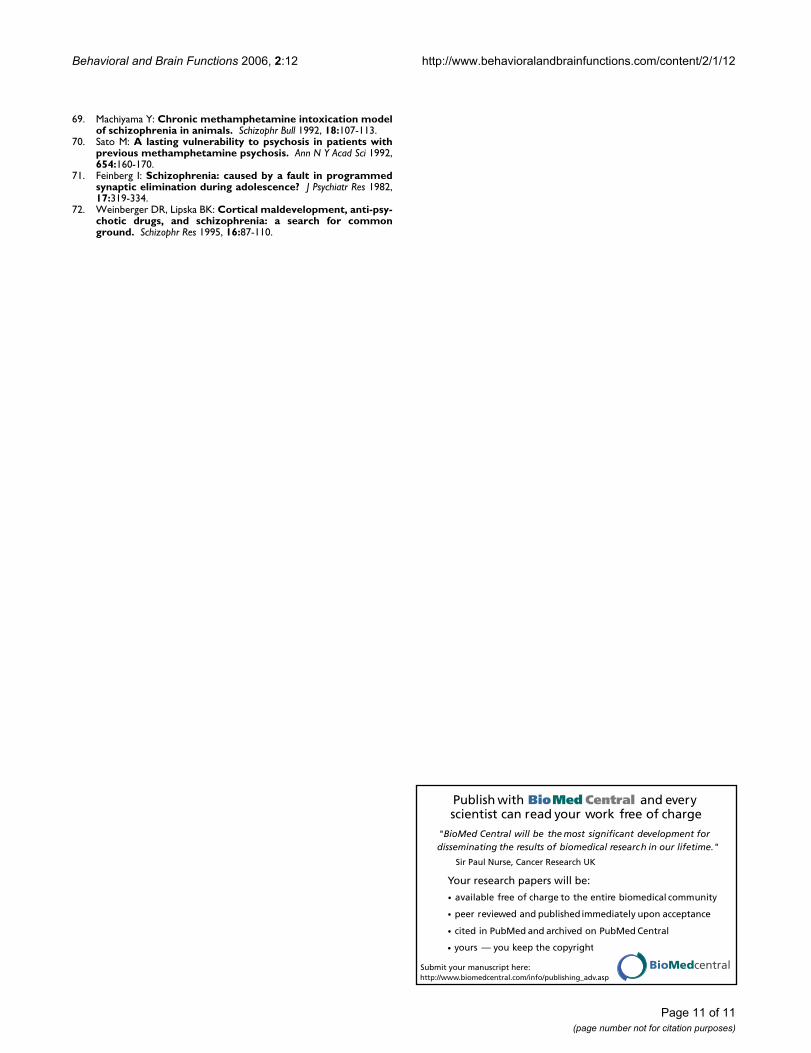
Behavioral and Brain Functions 2006, 2:12 http://www.behavioralandbrainfunctions.com/content/2/1/12
Publish with BioMed Central and every scientist can read your work free of charge
"BioMed Central will be the most significant development for disseminating the results of biomedical research in our lifetime."
Sir Paul Nurse, Cancer Research UK
Your research papers will be:
available free of charge to the entire biomedical community
peer reviewed and published immediately upon acceptance
cited in PubMed and archived on PubMed Central
yours — you keep the copyright
Submit your manuscript here:http://www.biomedcentral.com/info/publishing_adv.asp
BioMedcentral
69. Machiyama Y: Chronic methamphetamine intoxication modelof schizophrenia in animals. Schizophr Bull 1992, 18:107-113.
70. Sato M: A lasting vulnerability to psychosis in patients withprevious methamphetamine psychosis. Ann N Y Acad Sci 1992,654:160-170.
71. Feinberg I: Schizophrenia: caused by a fault in programmedsynaptic elimination during adolescence? J Psychiatr Res 1982,17:319-334.
72. Weinberger DR, Lipska BK: Cortical maldevelopment, anti-psy-chotic drugs, and schizophrenia: a search for commonground. Schizophr Res 1995, 16:87-110.
Page 11 of 11(page number not for citation purposes)
http://www.ncbi.nlm.nih.gov/entrez/query.fcgi?cmd=Retrieve&db=PubMed&dopt=Abstract&list_uids=1553490
http://www.ncbi.nlm.nih.gov/entrez/query.fcgi?cmd=Retrieve&db=PubMed&dopt=Abstract&list_uids=1553490
http://www.ncbi.nlm.nih.gov/entrez/query.fcgi?cmd=Retrieve&db=PubMed&dopt=Abstract&list_uids=1632581
http://www.ncbi.nlm.nih.gov/entrez/query.fcgi?cmd=Retrieve&db=PubMed&dopt=Abstract&list_uids=1632581
http://www.ncbi.nlm.nih.gov/entrez/query.fcgi?cmd=Retrieve&db=PubMed&dopt=Abstract&list_uids=7187776
http://www.ncbi.nlm.nih.gov/entrez/query.fcgi?cmd=Retrieve&db=PubMed&dopt=Abstract&list_uids=7187776
http://www.ncbi.nlm.nih.gov/entrez/query.fcgi?cmd=Retrieve&db=PubMed&dopt=Abstract&list_uids=7577773
http://www.ncbi.nlm.nih.gov/entrez/query.fcgi?cmd=Retrieve&db=PubMed&dopt=Abstract&list_uids=7577773

Autho
r's
pers
onal
co
py
Research Report
Postnatal development of dopamine innervation in theamygdala and the entorhinal cortex of the gerbil(Meriones unguiculatus)
Susanne Brummelte⁎, Gertraud Teuchert-NoodtDepartment of Neuroanatomy, Faculty of Biology, University of Bielefeld, Universitätsstr. 25, D-33615 Bielefeld, Germany
A R T I C L E I N F O A B S T R A C T
Article history:Accepted 3 October 2006Available online 16 November 2006
Dopamine (DA) projections from themesencephalonare believed to play a critical role duringdevelopment and are essential for cognitive and behavioral functions. Since the postnatalmaturation patterns of these projections differ substantially between various brain regions,cortical, limbic or subcortical areas might exhibit varying vulnerabilities concerningdevelopmental disorders. The dopaminergic afferents of the rodent prefrontal cortex showan extremely prolonged maturation which is very sensitive to epigenetic challenges.However, less is known about the development of the DA innervation of caudal limbic areas.Therefore, immunohistochemically stained DA fibers were quantitatively examined in thebasolateral (BLA) and central amygdaloid nucleus (CE) and the ventrolateral entorhinalcortex (EC) of the Mongolian gerbil (Meriones unguiculatus). Animals of different ages, rangingfrom juvenile [postnatal day (PD) 14, 20, 30)] to adolescent (PD70), adult (6, 18 months) andaged (24 months), were analyzed. Results show a significant increase of fibers between PD14and PD20 in the BLA and lateral part of the CE, with a trend for a subsequent decline in fiberdensities until PD30. The EC and medial part of the CE showed no developmental changes.Interestingly, none of the investigated areas showed significant reductions of DA fibersduring aging.
© 2006 Elsevier B.V. All rights reserved.
Keywords:ImmunohistochemistryDevelopmentAgingLimbic system
1. Introduction
The neurotransmitter dopamine (DA) is known to play afundamental role in regulating cortical activity during devel-opment and in adulthood. The dopaminergic innervation ofdistinct areas in the mammalian brain descends fromdifferent pathways which have their origin in the mesence-phalon of the brainstem. The mesostriatal projection has itssource mainly in the pars compacta of the substantia nigra(SN; A9) and the nucleus retrorubralis (A8) and innervates thedorsal striatum (caudate putamen) (Gerfen et al., 1987; Hu
et al., 2004). Thus, it plays a considerable role in themaintenance of motoric functions. Dopaminergic fibers ofthe mesolimbic and mesocortical projections, which are oftennamed together as the mesocorticolimbic projection, largelyoriginate from the ventral tegmental area (VTA; A10) (Fallon etal., 1978; Swanson, 1982) and innervate several subregions ofthe dispersedly organized corticolimbic circuitry, includingthe amygdala and entorhinal cortex (Bjorklund and Lindvall,1984; Descarries et al., 1987; Yoshida et al., 1988).
Remarkably, each DA projection field is characterized by itsown pattern of time sequence of development in postnatal
B R A I N R E S E A R C H 1 1 2 5 ( 2 0 0 6 ) 9 – 1 6
⁎ Corresponding author. Fax: +49 521 106 6470.E-mail address: [email protected] (S. Brummelte).
0006-8993/$ – see front matter © 2006 Elsevier B.V. All rights reserved.doi:10.1016/j.brainres.2006.10.006
ava i l ab l e a t www.sc i enced i rec t . com
www.e l sev i e r. com/ l oca te /b ra in res

Autho
r's
pers
onal
co
py
life. Generally, caudal areas mature before rostral areas, andthus the dopaminergic innervation follows the caudorostralgradient, which also accounts for many other structures andfunctions.
The amygdala is one of the main targets of the mesolimbicDA pathway. It receives its input from the lateral VTA andmedial half of the SN (Fallon et al., 1978; Hasue and Shammah-Lagnado, 2002). Within the amygdaloid complex, severalnuclei can be distinguished due to their different connectivity,function and derivation. The central amygdaloid nucleus (CE)and the basolateral amygdala (BLA) are both consideredoutput entities of the amygdaloid complex (Petrovich et al.,1996; Petrovich and Swanson, 1997), with each being con-nected to a unique set of brain areas (Pitkanen, 2000). Thus,differential developmental patternsmay also affect or even beaffected by other cortical and subcortical regions. In fact, theprefrontal cortex (PFC), which exhibits a prolongedmaturationof its dopaminergic innervation (Kalsbeek et al., 1988; Dawirset al., 1993), has been assumed to stabilize DA subsystemsdepending on its postnatal development (Busche et al., 2004;Bennay et al., 2004).
It has been demonstrated that the DA fiber densities ofcaudal limbic areas in the rodent brain reach their adult levelsrelatively early (Verney et al., 1985; Erickson et al., 1998)compared to prefrontal cortices (Kalsbeek et al., 1988; Dawirset al., 1993) or the nucleus accumbens (Lesting et al., 2005),which continue to increase until adulthood. This prolongedmaturation of the rostral parts of the brain has been assumedto play an essential role in the maintenance of neuralplasticity during development, but according to an animalmodel of psychosis, it might also be involved in developmen-tally induced diseases such as schizophrenia (Teuchert-Noodt, 2000; Dawirs and Teuchert-Noodt, 2001).
However, less is known about the long-term postnataldevelopment and aging of the DA fiber densities in the caudallimbic system. The divergent developmental patterns ofcortical and subcortical areas are of particular interest asvarious neurodevelopmental and degenerative diseases suchas Parkinson's disease or schizophrenia are assumed to exhibitimbalances on several levels concerning the monoaminergicneurotransmitter systems between these areas (Laruelle et al.,2003; Abi-Dargham, 2004). In fact, a recent study from ourlaboratory, using an early postnatal traumatization bymethamphetamine as an animalmodel of psychoses, revealeda disequilibrium in dopaminergic fiber densities between theprefrontal cortex and caudal limbic areas (Busche et al., 2004).Therefore, this studywas conducted to look closely at the long-term maturation of DA fiber densities in different areas of thelimbic circuit, namely the central amygdala, the basolateralamygdala and the ventrolateral entorhinal cortex (EC).
2. Results
The DA innervation pattern in gerbils' amygdala and entorh-inal cortex resembles that described for the rat, which in turnhas been reported to be similar to that of monkeys (Sadikotand Parent, 1990).
The lateral part of the CE receives the densest dopaminer-gic innervation followed by its medial neighbor (Fig. 1C). Thebasolateral amygdala shows only a light DA innervation(Fig. 1C). DA fibers of the ventrolateral EC appear to bearranged in clusters (Figs. 1D, G1–G4).
An analysis of variance (ANOVA) with age as the inde-pendent variable and the DA fiber densities of the differentareas as dependent variables revealed a significant effect ofage (F(24,144)=2.34, p=0.0011). The following Fisher's LSD posthoc test showed a highly significant increase between PD14and PD20 in the lateral part of the CE (28%; p=0.0003) and atendency for a subsequent decrease between PD20 and PD30(p=0.070). There was no such peak in the development of themedial CE or EC (all p's>0.05), although there was a tendencyfor an increase after PD14 in themedial part of theCE (p=0.079).For the BLA, the post hoc test revealed a similar increasebetween PD14 and PD20 (90%; p=0.009) and also a trend for asubsequent decline in fiber density (p=0.059). During aging, nostatistically significant alterations were seen in any of theinvestigated areas. However, the lateral part of the CE showeda tendency (p=0.088) for a decline between PD540 and PD720.
3. Discussion
The present data reveal that the development of the dopami-nergic innervation varies between different structures of thecaudal limbic system of the gerbil. Thus, the ventrolateralentorhinal cortex (EC) and the medial part of the centralnucleus of the amygdala (CE) show no significant alterationsat all between PD14 and PD720, while there is an increase indensity between PD14 and PD20 and a subsequent trend for adecrease until PD30 in the lateral part of the CE and thebasolateral nucleus of the amygdala (BLA). Interestingly, noneof the structures demonstrates any statistically significantdecline in DA fiber densities in high age (Fig. 2).
Beside the dopaminergic fiber densities, there are otherelements of the DA system that show differential developQmental patterns in different brain regions. Thus, Coulter et al.(1996) demonstrated that the maturation of the dopaminetransporter (DAT) follows an anterior-to-posterior and lateral-to-medial gradient, with the prefrontal cortex (PFC) andnucleus accumbens being two of the last areas to reach adult
Fig. 1 – Representative photomicrographs of the dopaminergic innervation of the amygdala and entorhinal cortex at differentage stages. A, B: photomicrographs of representative coronal sections at the level of the amygdala and entorhinal cortex (EC),respectively. Picture (B1) shows a Nissl staining of the ventrolateral EC. The areas of the rectangles are magnified in panels CandD. The dotted line in panel Dmarks the approximate innervation field of the dopamine (DA) projection,which is arranged inclusters (cf. G1–4). Within the amygdala, most fibers are found in the lateral part of the central amygdala (CEl), which issurrounded by themoderate innervation of themedial part (CEm). The BLA shows the slightest innervation density of DA fibers(cf. F1–4). Pictures (E1)–(G4) show examples of DA innervation at juvenile age stages (PD14–PD30) with a comparative sectionfrom an adult animal (PD180). Scale bars: 500 μm (B1), 100 μm (E1–G4).
10 B R A I N R E S E A R C H 1 1 2 5 ( 2 0 0 6 ) 9 – 1 6

Autho
r's
pers
onal
co
py
11B R A I N R E S E A R C H 1 1 2 5 ( 2 0 0 6 ) 9 – 1 6

Autho
r's
pers
onal
co
py
levels. However, in the striatum, DAT density rather declinesafter an early peak during development (Moll et al., 2000).Similar reorganization processes have been observed for DAvaricosities, synapses and DA receptor types in the nucleusaccumbens and caudate putamen (Tarazi et al., 1998, 1999;Antonopoulos et al., 2002), while there is no such peak andsubsequent elimination in receptor densities in the frontalcortex or limbic areas (Tarazi and Baldessarini, 2000). Further-more, DA axons appear to pass through a peak in density inparticular areas of the striatum (Hu et al., 2004; Lesting et al.,2005) which has been assumed to be due to the reorganizationand elimination of non-specific targeting from the dopami-nergic pathways (Hu et al., 2004). However, none of theinvestigated caudal limbic areas from the current study revealsthis ‘pruning effect’, at least not at a statistically significantlevel.
Remarkably, there was also no detectable alteration in DAfiber densities in old animals compared to adolescent or adultones in the current study. This is in line with previousobservations in the nucleus accumbens shell and core in thegerbil, which also showed no age-related changes in DA fiberdensities (Lesting et al., 2005). But it is in contrast with severalstudies, which have reported about metabolic changes withinthe monoaminergic system in older animals, as e.g. differ-ences in the turnover rates, biosynthesis or concentration ofDA or its metabolites (Miguez et al., 1999; Miura et al., 2002).However, none of these alterations suggests a correlatedchange in fiber densities as a reduced DA concentration ormetabolism does not necessarily imply a reduced number ordensity of axons in the particular area. It is rather likely thatthese metabolic differences are mediated by an alteredreceptor physiology or density (Cross et al., 1984, 1988; Sweetet al., 2001), althoughwe have to admit that we cannot excludealterations in animals older than 24 months as this is only themean survival of the Mongolian gerbil, whereas individualsmight get older (Troup et al., 1969).
Due to the caudorostralmaturation gradient, it seems likelythat the entorhinal cortex and amygdala develop relativelyearly during the postnatal period. Erickson and co-workers
(1998) have shown that the densities of DA axons increaseduntil 7 months of age in monkeys in layer III of the rostralentorhinal cortex. However, we could not detect such an age-dependent alteration in the dopaminergic fiber densities in thegerbil EC after PD14. Thismight be due to the fact that there areapparently some discrepancies concerning the exact classifi-cation of the entorhinal area. A recent anatomical studysuggests that the amygdalopiriform transition area, whichwas so far often considered part of the EC, should be viewed asa separate anatomical entity (Santiago and Shammah-Lagnado, 2005). In order to show the boundaries of theinvestigated area in the current study, a picture of a Nisslstaining of the gerbil EC is provided, to be compared with thedopaminergic innervation and measurement field (Fig. 1B1).
Dopaminergic fibers are arranged in clusters or “islands” inthe adult rodent EC (Fallon et al., 1978; Busche et al., 2004),which can already be seen during early development(cf. Figs. 1G1–G3). DA terminals directly innervate excitatoryand inhibitory entorhinal neurons, and thus the innervationpattern resembles that of neocortical or amygdaloid projec-tions (Asan, 1998; Erickson et al., 2000). Forming themain inputto the hippocampus and being intensively connected with theBLAand theCE (McDonaldandMascagni, 1996; Pitkanen, 2000),the EC is in close association to the limbic system and is evenconsidered a part of it by some authors (Amaral and Witter,1995). Disturbances during the maturation process of the ECmight therefore havedevastating consequences for the orderlyinformation flow. In concert with this, neonatally inducedstructural abnormalities in the entorhinal cortex have beenshown to affect DA transmission in the limbic regions at theadolescent stage (Uehara et al., 2000). Furthermore, a reduceddensity of tyrosine hydroxylase-immunoreactive axons hasbeen observed in the entorhinal cortex of schizophrenicpatients (Akil et al., 2000).
Compared to the ventrolateral EC, the amygdala seems tolag behind in the maturation of the innervation density of DA.The increase in the lateral part of the CE after PD14 might bedue to a sprouting of axon collaterals or further elaboration oflocal arbors within the nucleus, which might also account for
Fig. 2 – Development of DA fiber densities in the amygdala and entorhinal cortex of the gerbil. Shown are the DA fiberdensity means as percentage [%] of the reference area+SEM at postnatal day (PD) 14, 20, 30, 70, 180, 540 and 720. The lateralpart of the central amygdala (CE lat) and the basolateral amygdala (BLA) show significant increases between PD14 and PD20,while the entorhinal cortex (EC) and medial part of the CE (CE med) display no alterations during maturation or aging. Thedouble bar in the middle marks a break in the scaling of the x-axis. **p<0.01, ***p<0.001.
12 B R A I N R E S E A R C H 1 1 2 5 ( 2 0 0 6 ) 9 – 1 6

Autho
r's
pers
onal
co
py
the non-significant rise in density in the medial part, just to alesser extent. The CE, with its several distinct subdivisions,receives intensive input from cortical areas as well as fromlimbic structures as the hippocampus or EC. Interestingly, theCE has no reciprocal connections with the aforementionedregions (Pitkanen, 2000) but sends a strong input to thedopaminergic system in themesencephalon, thus influencingthe DA innervation of several other areas and modulatingbehavioral responses (Fudge and Haber, 2000). It is further thenucleus with the highest density of DA synapses and axonswith the lateral part being in turnmore dense than themedialpart (Asan, 1997, 1998). This strong interaction with thedopaminergic cells in the mesencephalon might be a reasonwhy especially the lateral part seems to be relatively resistantto early postnatal environmental and pharmacological chal-lenges (Busche et al., 2004). In addition, the catecholaminergicinnervation of the medial part is assumed to serve moremodulatory functions due to the fact that it is directedpreferentially at peripheral neuronal structures (Asan, 1998).
The BLA is characterized by its strong reciprocal connectionwith the prefrontal cortex (Pitkanen, 2000) and is thought tomediate affective behavior via its DA innervation (Kroner et al.,2005). As this connectivity with the PFC matures relatively lateand is considered to influence the development and integrationof normal or abnormal emotional behavior during adolescence(Cunningham et al., 2002), it is not surprising that the BLA isparticularly sensitive to early environmental or pharmacologi-cal challenges (Busche et al., 2004; Grund et al., 2006).
We have recently shown that an early single methamphe-tamine (MA) intoxication on PD14 can cause a surplus of DAfibers in the adult ventrolateral EC. An even stronger increasewas found in the BLA, but not in the lateral part of the CE andonly to a lesser extent in the left hemisphere of themedial partof the CE (Busche et al., 2004). Considering the markeddecrease of dopaminergic fibers in rostral areas such as theprefrontal cortex using the same pharmacological approach(Dawirs et al., 1994), it seems likely that the differentdopaminergic pathways influence each other, thus causingan imbalance between cortical and caudal limbic innervationareas (Busche et al., 2004). Interestingly, such long-termalterations in fiber densities are not seen in the PFC oramygdala, when MA is applied at adult age, underlining thesensitivity of these areas during development compared toadulthood (Brummelte et al., 2006).
BLA and the lateral part of the CE are affected differently bythe MA challenge, although both structures show a furtherincrease in DA fibers after PD14. It is assumed that the PFC isable to stabilize DA subsystems depending on its postnataldevelopment (Busche et al., 2004; Bennay et al., 2004).However, the PFC is also supposed to be exceedingly damage-able and accident-sensitive during development with theperiod of this vulnerability lasting long until early adulthood(for reviews see: Diamond, 1996; Lewis, 1997; Sullivan andBrake, 2003; Adriani and Laviola, 2004). As the BLA exhibitsparticularly strong reciprocal connections with the PFC, theobserved peak in fiber density during development may beparticularly vulnerable to changes resulting in dysfunctionalconnections with the PFC. In fact, extracellular recordingstudies have observed that a prestimulation of the medial PFCreduces the responsiveness of CE neurons to inputs from the
BLA, thus contributing to the idea that the PFC even gatestransmission within the amygdala (Quirk et al., 2003). Thisinhibitory control by the cortex, probably mediated viaGABAergic neurons, can be diminished by the release of DA(Marowsky et al., 2005). Thus, the DA innervation of theamygdala contributes essentially to a balanced output of thisstructure and consequently regulates the modulation ofaffective behavior (Asan, 1997; Marowsky et al., 2005; Kroneret al., 2005).
Naturally, not only DA plays a significant role during itsown pathway and general development. Thus, the interactionwith other transmitter systems, e.g. serotonin or glutamateand their receptors, might considerably contribute to thedivergent developmental patterns of cortical and subcorticalareas and the according differences in vulnerability duringmaturation. In fact, it has often been assumed that theputative DA imbalance in schizophrenia might be secondaryto alterations of other resources, as e.g. an NMDA hypofunc-tion or alteration in the GABAergic or glutamate system(Laruelle et al., 2003; Abi-Dargham, 2004). Consistent with thishypothesis, we have recently demonstrated a dysconnectionwithin macrocircuits of the glutamatergic system (Bagordaet al., 2006; Witte et al., 2006) and a shift within the prefrontalGABAergic innervation pattern (unpublished data) in ouranimal model of psychosis. Therefore, the authors aretempted to suggest that the mutual impact and interdepen-dency of the areas and their corresponding transmittersystems during development all contribute essentially to anormal and healthy maturation and that disturbances in oneof the integrated features might cause various adaptationsand alterations in several systems.
4. Experimental procedures
4.1. Animals
All experimental procedures were approved by the appro-priate committee for animal care in accordance with theEuropean Communities Council Directive. Male Mongoliangerbils (M. unguiculatus) were kept under natural day/nightcycles with food and water being provided ad libitum. Untilweaning (PD30), animals were kept in standard Macrolon®cages (type 4). Afterwards, they were reared individually inMacrolon® type 3 cages. A total of 51 male Mongolian gerbilswere used for this study. Seven experimental animal groups ofdifferent ageswere investigated to cover convincing periods ofthe life span of gerbils: PD14 (n=6), PD20 (n=6), PD30, (n=9),PD70 (n=9), PD180 (n=6), PD540 (n=7) and PD720 (n=8). Gerbilswere chosen due to their wild-type like behavioral andneuronal repertoire as they have not been so intensivelydomesticated compared to rats or mice (Rosenzweig andBennett, 1969). In addition, the present data can be consideredagainst a huge amount of previously published data of thegerbil brain from our laboratory.
4.2. Dopamine immunohistochemistry
Animals under deep chloralhydrate anesthesia (1.7 g/kg, i.p.)were transcardially perfused with 0.1 M sodium cacodylate pH
13B R A I N R E S E A R C H 1 1 2 5 ( 2 0 0 6 ) 9 – 1 6

Autho
r's
pers
onal
co
py
6.2 followed by 5% glutaraldehyde in 0.1 M sodium cacodylatepH 7.5. Immediately after perfusion, the brains were dissectedand 50 μm thick frontal sections of the right hemisphere werecut with a vibratome (Leica VT 1000S). For immunostaining,the slices were rinsed in wash buffer followed by a pre-incubation in 10% normal goat serum and 0.4% Triton X-100(Sigma). Then the slices were incubated with the primaryantibody (rabbit anti-dopamine, DiaSorin, Stillwater, MN)diluted 1:600 with 1% normal goat serum and 0.4% Triton X-100 for 40 h. The following rinses were done with 0.05 M Tris–HCl buffered saline (pH 7.5). The slices were rinsed andincubated in the biotinylated goat–anti-rabbit antibody(Sigma) diluted 1:20 with 1% normal goat serum, rinsedagain and incubated with ExtraAvidin–Peroxidase (Sigma)diluted 1:20. After another rinse, the slices were stained in0.05% 3.3-diaminobenzidine (DAB, Sigma) with 0.01% H2O2.Then the slices were washed, mounted on glass slides, driedovernight, dehydrated with ethanol, cleared with xylene andcoverslipped with DePeX (Serva, Heidelberg, Germany). Formore details on animal preparation and dopamine immuno-histochemistry, see Lesting et al., 2005.
4.3. Quantification of DA innervation
DA fiber densities were measured in three to four consecutivecoronal slices of the corresponding brain sections, which wereassigned by means of anatomical characteristics according tobrain atlases of the rat (Paxinos and Watson, 1986) and themouse (Valverde, 1998). Fibers were visualized using a bright-field microscope (BX61, Olympus, Hamburg, Germany) and400-fold magnification and a digital camera for microscopy(ColorView II, SIS, Münster, Germany). All pictures wereadjusted in contrast and brightness for better conspicuity ofDA fibers. The lateral and medial part of the CE, the BLA andthe entorhinal cortex were encircled by an experimenter blindto the animals' age, and all detectable fragments werequantified using software for image analysis (KS300, Jenoptik,Jena, Germany; for details of the quantification process, seeLesting et al., 2005). Fiber densities were calculated as apercentage of the reference area. We abstained from addi-tionally measuring the growth of the according areas in eachindividual as the potential increase of the area duringdevelopment is already accounted for by this method.
4.4. Data analysis
Measurements were computed as arithmetic means by-caseand by-group±SEM. An analysis of variance (ANOVA) with age(7 levels) as an independent variable and area (4 levels) asdependent variable was used to check for statistical signifi-cance between groups followed by LSD post hoc test formultiple comparisons. Statistical analysis was computed withStatistica 6 (StatSoft, Tulsa, USA). The levels of significancewere set at *p<0.05, **p<0.01 and ***p<0.001.
Acknowledgments
We would like to thank Dr. Francesco Bagorda and UlrikeSchroeder for excellent technical assistance. Furthermore, we
wish to thank Dr. Jorg Neddens for his help with this work andBeatrix Ahlswede for correcting the English. This study wassupported by grants of the ‘Deutsche Parkinson Vereinigung’.
R E F E R E N C E S
Abi-Dargham, A., 2004. Do we still believe in the dopaminehypothesis? New data bring new evidence. Int.J. Neuropsychopharmacol. 7 (Suppl 1), S1–S5.
Adriani, W., Laviola, G., 2004. Windows of vulnerability topsychopathology and therapeutic strategy in the adolescentrodent model. Behav. Pharmacol. 15, 341–352.
Akil, M., Edgar, C.L., Pierri, J.N., Casali, S., Lewis, D.A., 2000.Decreased density of tyrosine hydroxylase-immunoreactiveaxons in the entorhinal cortex of schizophrenic subjects. Biol.Psychiatry 47, 361–370.
Amaral, D.G., Witter, M.P., 1995. Hippocampal formation. In:Paxinos, G. (Ed.), The Rat Nervous System. Academic Press, SanDiego, pp. 443–493.
Antonopoulos, J., Dori, I., Dinopoulos, A., Chiotelli, M., Parnavelas,J.G., 2002. Postnatal development of the dopaminergic systemof the striatum in the rat. Neuroscience 110, 245–256.
Asan, E., 1997. Ultrastructural features oftyrosine-hydroxylase-immunoreactive afferents and theirtargets in the rat amygdala. Cell Tissue Res. 288, 449–469.
Asan, E., 1998. The catecholaminergic innervation of the ratamygdala. Adv. Anat. Embryol. Cell Biol. 142, 1–118.
Bagorda, F., Teuchert-Noodt, G., Lehmann, K., 2006. Isolationrearing or methamphetamine traumatisation induce a“dysconnection” of prefrontal efferents in gerbils: implicationsfor schizophrenia. J. Neural Transm. 113, 365–379.
Bennay, M., Gernert, M., Schwabe, K., Enkel, T., Koch, M., 2004.Neonatal medial prefrontal cortex lesion enhances thesensitivity of the mesoaccumbal dopamine system. Eur. J.Neurosci. 19, 3277–3290.
Bjorklund, A., Lindvall, O., 1984. Dopamine-containing systems inthe CNS. In: Bjorklund, A., Hokfelt, T. (Eds.), ClassicalTransmitters in the CNS. Elsevier, Amsterdam, pp. 55–122.
Brummelte, S., Grund, T., Czok, A., Teuchert-Noodt, G., Neddens, J.,2006. Long-term effects of a single adult methamphetaminechallenge: minor impact on dopamine fibre density in limbicbrain areas of gerbils. Behav. Brain Funct. 2, 12.
Busche, A., Polascheck, D., Lesting, J., Neddens, J., Teuchert-Noodt,G., 2004. Developmentally induced imbalance of dopaminergicfibre densities in limbic brain regions of gerbils(Meriones unguiculatus). J. Neural Transm. 111, 451–463.
Cortes, R., Probst, A., Palacios, J.M., 1988. Decreased densities ofdopamine D1 receptors in the putamen and hippocampus insenile dementia of the Alzheimer type. Brain Res. 475, 164–167.
Coulter, C.L., Happe, H.K., Murrin, L.C., 1996. Postnataldevelopment of the dopamine transporter: a quantitativeautoradiographic study. Brain Res. Dev. Brain Res. 92, 172–181.
Cross, A.J., Crow, T.J., Ferrier, I.N., Johnson, J.A., Markakis, D., 1984.Striatal dopamine receptors in Alzheimer-type dementia.Neurosci. Lett. 52, 1–6.
Cunningham, M.G., Bhattacharyya, S., Benes, F.M., 2002.Amygdalo-cortical sprouting continues into early adulthood:implications for the development of normal and abnormalfunction during adolescence. J. Comp. Neurol. 453, 116–130.
Dawirs, R.R., Teuchert-Noodt, G., 2001. A novel pharmacologicalconcept in an animal model of psychosis. Acta Psychiatr.Scand. 10–17 (Suppl.).
Dawirs, R.R., Teuchert-Noodt, G., Czaniera, R., 1993. Maturation ofthe dopamine innervation during postnatal development of theprefrontal cortex in gerbils (Meriones unguiculatus). A quantitativeimmunocytochemical study. J. Hirnforsch. 34, 281–290.
14 B R A I N R E S E A R C H 1 1 2 5 ( 2 0 0 6 ) 9 – 1 6

Autho
r's
pers
onal
co
py
Dawirs, R.R., Teuchert-Noodt, G., Czaniera, R., 1994. The postnatalmaturation of dopamine innervation in the prefrontal cortex ofgerbils (Meriones unguiculatus) is sensitive to an early singledose of methamphetamine. A quantitativeimmunocytochemical study. J. Hirnforsch. 35, 195–204.
Descarries, L., Lemay, B., Doucet, G., Berger, B., 1987. Regional andlaminar density of the dopamine innervation in adult ratcerebral cortex. Neuroscience 21, 807–824.
Diamond, A., 1996. Evidence for the importance of dopamine forprefrontal cortex functions early in life. Philos. Trans. R. Soc.Lond., B Biol. Sci. 351, 1483–1493.
Erickson, S.L., Akil, M., Levey, A.I., Lewis, D.A., 1998. Postnataldevelopment of tyrosine hydroxylase- and dopaminetransporter-immunoreactive axons in monkey rostralentorhinal cortex. Cereb. Cortex 8, 415–427.
Erickson, S.L., Sesack, S.R., Lewis, D.A., 2000. Dopamineinnervation of monkey entorhinal cortex: postsynaptic targetsof tyrosine hydroxylase-immunoreactive terminals. Synapse36, 47–56.
Fallon, J.H., Koziell, D.A., Moore, R.Y., 1978. Catecholamineinnervation of the basal forebrain. II. Amygdala, suprarhinalcortex and entorhinal cortex. J. Comp. Neurol. 180, 509–532.
Fudge, J.L., Haber, S.N., 2000. The central nucleus of the amygdalaprojection to dopamine subpopulations in primates.Neuroscience 97, 479–494.
Gerfen, C.R., Herkenham, M., Thibault, J., 1987. The neostriatalmosaic: II. Patch- and matrix-directed mesostriataldopaminergic and non-dopaminergic systems. J. Neurosci. 7,3915–3934.
Grund, T., Lehmann, K., Bock, N., Rothenberger, A.,Teuchert-Noodt, G., 2006. Influence of methylphenidate onbrain development—An update of recent animal experiments.Behav. Brain Funct. 2, 2.
Hasue, R.H., Shammah-Lagnado, S.J., 2002. Origin of thedopaminergic innervation of the central extended amygdalaand accumbens shell: a combined retrograde tracing andimmunohistochemical study in the rat. J. Comp. Neurol. 454,15–33.
Hu, Z., Cooper, M., Crockett, D.P., Zhou, R., 2004. Differentiation ofthe midbrain dopaminergic pathways during mousedevelopment. J. Comp. Neurol. 476, 301–311.
Kalsbeek, A., Voorn, P., Buijs, R.M., Pool, C.W., Uylings, H.B., 1988.Development of the dopaminergic innervation in theprefrontal cortex of the rat. J. Comp. Neurol. 269, 58–72.
Kroner, S., Rosenkranz, J.A., Grace, A.A., Barrionuevo, G., 2005.Dopamine modulates excitability of basolateral amygdalaneurons in vitro. J. Neurophysiol. 93, 1598–1610.
Laruelle, M., Kegeles, L.S., Abi-Dargham, A., 2003. Glutamate,dopamine, and schizophrenia: from pathophysiology totreatment. Ann. N. Y. Acad. Sci. 1003, 138–158.
Lesting, J., Neddens, J., Teuchert-Noodt, G., 2005. Ontogeny of thedopamine innervation in the nucleus accumbens of gerbils.Brain Res. 1066, 16–23.
Lewis, D.A., 1997. Development of the prefrontal cortex duringadolescence: insights into vulnerable neural circuits inschizophrenia. Neuropsychopharmacology 16, 385–398.
Marowsky, A., Yanagawa, Y., Obata, K., Vogt, K.E., 2005. Aspecialized subclass of interneurons mediates dopaminergicfacilitation of amygdala function. Neuron 48, 1025–1037.
McDonald, A.J., Mascagni, F., 1996. Cortico-cortical andcortico-amygdaloid projections of the rat occipital cortex: aPhaseolus vulgaris leucoagglutinin study. Neuroscience 71,37–54.
Miguez, J.M., Aldegunde, M., Paz-Valinas, L., Recio, J.,Sanchez-Barcelo, E., 1999. Selective changes in the contents ofnoradrenaline, dopamine and serotonin in rat brain areasduring aging. J. Neural Transm. 106, 1089–1098.
Miura, H., Qiao, H., Ohta, T., 2002. Influence of aging and socialisolation on changes in brain monoamine turnover and
biosynthesis of rats elicited by novelty stress. Synapse 46,116–124.
Moll, G.H., Mehnert, C., Wicker, M., Bock, N., Rothenberger, A.,Ruther, E., Huether, G., 2000. Age-associated changes in thedensities of presynaptic monoamine transporters in differentregions of the rat brain from early juvenile life to lateadulthood. Brain Res. Dev. Brain Res. 119, 251–257.
Paxinos, G., Watson, C., 1986. The Rat Brain in StereotaxicCoordinates. Academic Press, New York.
Petrovich, G.D., Swanson, L.W., 1997. Projections from the lateralpart of the central amygdalar nucleus to the postulated fearconditioning circuit. Brain Res. 763, 247–254.
Petrovich, G.D., Risold, P.Y., Swanson, L.W., 1996. Organization ofprojections from the basomedial nucleus of the amygdala: aPHAL study in the rat. J. Comp. Neurol. 374, 387–420.
Pitkanen, A., 2000. Connectivity of the rat amygdaloid complex. In:Aggleton, J.P. (Ed.), The Amygdala. Oxford Univ. Press, NewYork, pp. 31–116.
Quirk, G.J., Likhtik, E., Pelletier, J.G., Pare, D., 2003. Stimulation ofmedial prefrontal cortex decreases the responsiveness ofcentral amygdala output neurons. J. Neurosci. 23, 8800–8807.
Rosenzweig, M.R., Bennett, E.L., 1969. Effects of differentialenvironments on brain weights and enzyme activities ingerbils, rats, and mice. Dev. Psychobiol. 2, 87–95.
Sadikot, A.F., Parent, A., 1990. The monoaminergic innervation ofthe amygdala in the squirrel monkey: animmunohistochemical study. Neuroscience 36, 431–447.
Santiago, A.C., Shammah-Lagnado, S.J., 2005. Afferentconnections of the amygdalopiriform transition area in the rat.J. Comp. Neurol. 489, 349–371.
Sullivan, R.M., Brake, W.G., 2003. What the rodent prefrontalcortex can teach us about attention-deficit/hyperactivitydisorder: the critical role of early developmental events onprefrontal function. Behav. Brain Res. 146, 43–55.
Swanson, L.W., 1982. The projections of the ventral tegmental areaand adjacent regions: a combined fluorescent retrograde tracerand immunofluorescence study in the rat. Brain Res. Bull. 9,321–353.
Sweet, R.A., Hamilton, R.L., Healy, M.T., Wisniewski, S.R.,Henteleff, R., Pollock, B.G., Lewis, D.A., DeKosky, S.T., 2001.Alterations of striatal dopamine receptor binding inAlzheimer disease are associated with Lewy bodypathology and antemortem psychosis. Arch. Neurol. 58,466–472.
Tarazi, F.I., Baldessarini, R.J., 2000. Comparative postnataldevelopment of dopamine D(1), D(2) and D(4) receptors in ratforebrain. Int. J. Dev. Neurosci. 18, 29–37.
Tarazi, F.I., Tomasini, E.C., Baldessarini, R.J., 1998. Postnataldevelopment of dopamine D4-like receptors in rat forebrainregions: comparison with D2-like receptors. Brain Res. Dev.Brain Res. 110, 227–233.
Tarazi, F.I., Tomasini, E.C., Baldessarini, R.J., 1999. Postnataldevelopment of dopamine D1-like receptors in rat cortical andstriatolimbic brain regions: an autoradiographic study. Dev.Neurosci. 21, 43–49.
Teuchert-Noodt, G., 2000. Neuronal degeneration andreorganization: a mutual principle in pathological and inhealthy interactions of limbic and prefrontal circuits. J. Neural315–333.
Troup, G.M., Smith, G.S., Walford, R.L., 1969. Life span, chronologicdisease patterns and age-related changes in relative spleenweights for the Mongolian gerbil (Meriones unguiculatus).Exp. Gerontol. 4, 139–143.
Uehara, T., Tanii, Y., Sumiyoshi, T., Kurachi, M., 2000. Neonatallesions of the left entorhinal cortex affect dopaminemetabolism in the rat brain. Brain Res. 860, 77–86.
Valverde, F., 1998. Golgi Atlas of the Postnatal Mouse Brain.Springer-Verlag, Wien, New York.
Verney, C., Baulac, M., Berger, B., Alvarez, C., Vigny, A., Helle, K.B.,
15B R A I N R E S E A R C H 1 1 2 5 ( 2 0 0 6 ) 9 – 1 6

Autho
r's
pers
onal
co
py
1985. Morphological evidence for a dopaminergic terminal fieldin the hippocampal formation of young and adult rat.Neuroscience 14, 1039–1052.
Witte, A.V., Bagorda, F., Teuchert-Noodt, G., Lehmann, K., 2006.Contralateral prefrontal projections in gerbils matureabnormally after earlymethamphetamine trauma and isolated
rearing. J. Neural Transm. (electronic publication ahead ofprint).
Yoshida, M., Sakai, M., Kani, K., Nagatsu, I., Tanaka, M., 1988. Thedopaminergic innervation as observed byimmunohistochemistry using anti-dopamine serum in the ratcerebral cortex. Experientia 44, 700–702.
16 B R A I N R E S E A R C H 1 1 2 5 ( 2 0 0 6 ) 9 – 1 6

Accepted by ‘International Journal of Developmental Neuroscience’ Manuscript Number: DN-D-06-00062R1 (Date of acceptance: January 4, 2007) Postnatal development of GABA and Calbindin cells and fibers in the prefrontal cortex and basolateral amygdala of gerbils (Meriones unguiculatus) Susanne Brummelte1, Veronica Witte1,2 and Gertraud Teuchert-Noodt1
1Department of Neuroanatomy, Faculty of Biology, University of Bielefeld, Universitätsstr. 25, D-33615 Bielefeld, Germany 2 Current address: Department of Neurology, University of Münster, Albert-Schweitzer-Straße 33, 48129 Münster, Germany
Corresponding author: Susanne Brummelte, Department of Neuroanatomy, Faculty of Biology, University of Bielefeld, Universitaetsstr. 25, D-33615 Bielefeld, Germany, Phone: 0049 521 106 5704 Fax: 0049 521 106 6470 e-mail: [email protected] Running title: Late development of GABA cells and fibers in the gerbil Keywords: gamma-amino-butyric acid; calbindin; calcium-binding proteins;
immunohistochemistry; development; limbic system Abbrevations used: GABA: gamma-amino-butyric acid; CB: calbindin; PFC: prefrontal cortex; mPFC: medial prefrontal cortex; BLA: basolateral amygdala; PD: postnatal day; PV: parvalbumin
1
Abstract
The postnatal maturation of immunohistochemically stained gamma-amino-butyric acid
(GABA) and calbindin (CB) cells and fibers were quantitatively examined in the prefrontal
cortex (PFC) and the basolateral amygdala (BLA) of the Mongolian gerbil (Meriones
unguiculatus). Animals of different ages, ranging from juvenile (postnatal day (PD)14, PD20,
PD30), to adolescent (PD70), adult (PD180, PD540) and aged (PD720) were analyzed.
Results reveal an increase in GABAergic fiber densities between PD14-20 in the PFC and the
BLA with a concomitant decrease in cell density. After PD70 GABA fiber density slightly
decreases again in the BLA, while there is a further slow but significant increase in the PFC
between PD70-PD540. Fibers immunoreactive for the calcium binding-protein CB, which is
predominantly localized in particular GABAergic subpopulations, also accumulate between
PD14-PD20 in the PFC and BLA, while a concomitant decrease in cell density is only seen in
the BLA. Both areas reveal a decrease of CB cells between PD30-PD70, which parallels with
a decrease of CB fibers in the PFC. However, there is no particular ‘aging-effect’ in the fiber
or cell densities of GABA or CB in any of the investigated areas in old animals.
In conclusion, we here demonstrate long-term dynamics in cell and fiber densities of the
GABAergic system until late in development which might correspond to the prolonged
maturation of other neuroanatomical and functional systems.
2

1. Introductory statement Gamma-amino-butyric acid (GABA) is probably the most important inhibitory
neurotransmitter in the mammalian nervous system. It is usually expressed in local
interneurons, which can modulate and even control the neuronal activity of cortical and
subcortical output neurons. Further, GABA has been shown to exert important morphogenetic
influences during development (Chronwall and Wolff, 1980; Nguyen et al., 2001) and to play
an essential role in reactive plasticity and reorganization processes during development and
adulthood (Dawirs et al., 1997; Hensch, 2005; Merzenich et al., 1983; Zito and Svoboda,
2002). Thus, GABA has a central part in shaping and maintaining of neuronal networks.
Within the GABAergic population several classes of subpopulation can be distinguished
according to their content of calcium-binding proteins (Baimbridge et al., 1992) and
corresponding different maturation patterns. One of these proteins is calbindin (CB) which is
e.g. found in Marinotti, Neuroglia and Double Bouquet cells within the cortex, i.e. in cells,
that primarily innervate distal parts and spines of pyramidal dendrites (Conde et al., 1994;
DeFelipe et al., 1989; Gabbott and Bacon, 1996; Lund and Lewis, 1993) and appear and
mature relatively early (Alcantara et al., 1993). In the amygdala, CB cells are distributed
differently in the various nuclei (Kemppainen and Pitkanen, 2000), but in contrast to the
prefrontal cortex (PFC), CB varicosities are found in the basolateral amygdala (BLA) to form
basket-like structures around unlabelled projection neuron somata (Berdel and Morys, 2000;
Kemppainen and Pitkanen, 2000; Legaz et al., 2005; Muller et al., 2003). This points to the
particularly interesting role of CB in this subcortical area, as it is widely known, that axo-
somatic synapses have an exceptionally powerful control over target neurons compared to
distal dendritic or spine contacts. In the cortex, these baskets are usually built by GABAergic
cells containing parvalbumin, another calcium-binding protein, or other substances such as
cholecystokinin (Conde et al., 1994; Kawaguchi and Kubota, 1998).
3

Despite this difference, the GABAergic innervation patterns of the PFC and the BLA bear
marked resemblances (Carlsen, 1988; Muller et al., 2006), although the origin and function of
the PFC and the BLA are quite diverse, which is a reason for choosing these particular two
structures for investigation in the current study. Further reasons are the high interconnection
and thus potential interrelation of the PFC and BLA during development and their distant
positions in the brain, which imply divergent developmental patterns. In addition, both areas
belong to one main circuit, characterized by the mesolimbic prefrontal dopamine projections,
which originate in the ventral tegmental area and the substantia nigra (Björklund and
Lindvall, 1984; Fallon and Ciofi, 1992). This dopamine fiber innervation is of particular
interest as it shows a prolonged maturation until adulthood in the rodent medial PFC (mPFC)
(Dawirs et al., 1993; Kalsbeek et al., 1988), while it stays relatively stable after PD 20 in the
gerbil amygdala or entorhinal cortex (Brummelte and Teuchert-Noodt, 2006). This is in line
with the general developing pattern, with the PFC being one of the last areas to reach adult
stages (Mrzljak et al., 1990; Van Eden et al., 1990), while the amygdala maturates relatively
early after birth (Joseph, 1999; Morys et al., 1999).
The prenatal and early postnatal maturation of the GABAergic population in the cortex, with
particular emphasis on the visual cortex, has been intensively investigated in the last two
decades (Chronwall and Wolff, 1981; Del Rio et al., 1992; Parnavelas, 1992; Van Eden et al.,
1989; Wolff et al., 1984). However, less research has been done concerning the late postnatal
development and aging effects of GABAergic and CB fibers and concerning different cortical
or subcortical areas. It is assumed that GABA exhibits a high synaptic plasticity and can help
to reorganize, shape and modulate neuronal circuits not only during development (Chen et al.,
2002; Teuchert-Noodt, 2000). This compensatory effect in plastic processes might be
reflected in changes of the GABAergic or CB fiber densities even during adulthood and
aging. As it is further supposed, that the cortex might continuously adapt to new situations
and experiences by (re)arranging neuronal networks (Bagorda et al., 2006; Holtmaat et al.,
4

2006; Trachtenberg et al., 2002), the current study was conducted to examine the life long
progression of GABAergic and CB structures in two areas of the mesolimbocortical circuit,
the mPFC and BLA.
2. Experimental procedures
A total of 60 male Mongolian gerbils (Meriones unguiculatus) were used for this study.
Breeding gerbils were obtained from Harlan Winkelmann (Borchen, Germany). The animals
were bred in standard cages (Macrolon type 4) and, after weaning on postnatal day (PD) 30,
were reared individually in standard cages (Macrolon type 3). All gerbils were kept under
natural day/night cycles with food and water being provided ad libitum. Seven experimental
animal groups of different ages were investigated to cover convincing periods of the life span
of gerbils: PD14 (n=11), PD 20 (n=6) (juvenile), PD30 (n=12) (weaning), PD70 (n=11)
(young adult), PD180 (n=8), PD540 (n=8) (adult) and PD720 (n=4) (aging). Gerbils were
chosen due to their very small genetic variability (Thiessen and Yahr, 1977), and their rich wild-
type like behavioral repertoire (Rosenzweig and Bennett, 1969). All experimental procedures
were approved by the appropriate committee for animal care in accordance with the European
Communities Council Directive.
Immunohistochemistry
Animals were transcardially perfused under deep chloralhydrate anesthesia (1.7g/kg, i.p.). The
perfusion was performed with 200ml 0.05M phosphate buffer (pH 6.2), containing 1% sodium
metabisulfite, followed by 750ml 5% glutaraldehyde with 1% sodium metabisulfite in 0.1M
phosphate buffer (pH 7.5), with appropriate amounts of solutions for younger animals.
Immediately after perfusion, the brains were removed and postfixed for 30 min. Coronar sections
of 50µm were cut with a vibratome (Vibratome Series 1000, Technical Products International
5
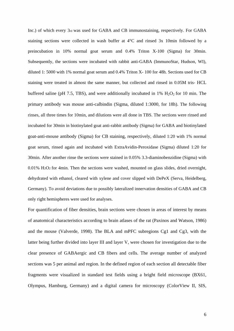
Inc.) of which every 3rd was used for GABA and CB immunostaining, respectively. For GABA
staining sections were collected in wash buffer at 4°C and rinsed 3x 10min followed by a
preincubation in 10% normal goat serum and 0.4% Triton X-100 (Sigma) for 30min.
Subsequently, the sections were incubated with rabbit anti-GABA (ImmunoStar, Hudson, WI),
diluted 1: 5000 with 1% normal goat serum and 0.4% Triton X- 100 for 48h. Sections used for CB
staining were treated in almost the same manner, but collected and rinsed in 0.05M tris- HCL
buffered saline (pH 7.5, TBS), and were additionally incubated in 1% H2O2 for 10 min. The
primary antibody was mouse anti-calbindin (Sigma, diluted 1:3000, for 18h). The following
rinses, all three times for 10min, and dilutions were all done in TBS. The sections were rinsed and
incubated for 30min in biotinylated goat anti-rabbit antibody (Sigma) for GABA and biotinylated
goat-anti-mouse antibody (Sigma) for CB staining, respectively, diluted 1:20 with 1% normal
goat serum, rinsed again and incubated with ExtraAvidin-Peroxidase (Sigma) diluted 1:20 for
30min. After another rinse the sections were stained in 0.05% 3.3-diaminobenzidine (Sigma) with
0.01% H2O2 for 4min. Then the sections were washed, mounted on glass slides, dried overnight,
dehydrated with ethanol, cleared with xylene and cover slipped with DePeX (Serva, Heidelberg,
Germany). To avoid deviations due to possibly lateralized innervation densities of GABA and CB
only right hemispheres were used for analyses.
For quantification of fiber densities, brain sections were chosen in areas of interest by means
of anatomical characteristics according to brain atlases of the rat (Paxinos and Watson, 1986)
and the mouse (Valverde, 1998). The BLA and mPFC subregions Cg1 and Cg3, with the
latter being further divided into layer III and layer V, were chosen for investigation due to the
clear presence of GABAergic and CB fibers and cells. The average number of analyzed
sections was 5 per animal and region. In the defined region of each section all detectable fiber
fragments were visualized in standard test fields using a bright field microscope (BX61,
Olympus, Hamburg, Germany) and a digital camera for microscopy (ColorView II, SIS,
6

Münster, Germany). Calbindin sections were investigated using 200-fold magnification,
GABA sections at 600-fold magnification.
To account for a possible interaction of fiber density and cell density or size of the
investigated area, these parameters were measured additionally for the PFC and BLA at 200-
fold and 20-fold magnification, respectively. Digital images were adjusted in contrast and
intensity before fibers, cells or the size of the area were quantified by software for image
analysis (KS300, Jenoptik, Jena, Germany). For further details of the quantification process
see (Brummelte et al., 2006a; Brummelte and Teuchert-Noodt, 2006). The fiber density was
calculated as a percentage of the evaluated test area, the cell density as number of cells per
test area. Lightly stained cells (cf. qualitative results) were excluded in the counting by a
minimum threshold of gray values for cell recognizing. All analyses were done by a person
blind to the age of individual animals.
Data analysis
Measurements were computed as arithmetic means by-case and by-group ± S.E.M. The
overall size of the particular area in which fiber densities were measured as well as the
number of GABA or CB cells were integrated as covariates in the statistical analysis to
account for a possible interaction of an augmentation of volume or cells and fiber sprouting.
For the PFC, a two-way analysis of covariance (ANCOVA) with age (7 levels) and area (3
levels) as independent variables, GABA or CB as dependent variable and area size and
GABA or CB cell number as covariates were used to check for statistical significance
between groups, followed by Fisher LSD post-hoc test for multiple comparisons if
appropriate. For the BLA, the ANCOVA comprised only one area level. As the covariates
revealed some significant effects on the fiber development, these parameters were also
statistically analyzed using an ANCOVA (cell number, size as covariate) or ANOVA (size)
and subsequent LSD post-hoc tests. Statistical analysis was computed with Statistica 6
7

(StatSoft, Tulsa, USA). The levels of significance were set at * p<0.05, ** p<0.01 and ***
p<0.001.
3. Results
Qualitative observations
The GABAergic fiber innervation is equally dense in all investigated areas, while the CB
fibers are more present in the BLA compared to the mPFC. The overall distribution pattern of
GABAergic and CB fibers, however, is similar in animals from all age stages (cf. Fig. 1). PFC
and BLA contain a population of lightly stained CB pyramidal neurons, which has been
previously observed in rats, too (Celio, 1990; Kemppainen and Pitkanen, 2000). In the PFC
these cells are arranged in a bundle throughout lamina II (Fig.1 B.1). In concert with data
from other species (Hof et al., 1999) hardly any CB or GABA cells were seen in lamina I. In
the BLA, GABAergic and CB cells were distributed quite equally through the nucleus. We
could not detect clear CB basket-like structures in the BLA at light microscope level, although
these have been described for rats (Berdel and Morys, 2000).
8

Fig. 1: Representative photomicrographs of the GABAergic and Calbindin (CB) distribution of the medial prefrontal cortex (mPFC) and the basolateral amygdala (BLA) at different age stages. A.1 and A.2 are photomicrographs of representative coronal sections at the level of the PFC and amygdala, respectively. The areas of the rectangles are magnified in panels B.1 – C.2 for CB and GABA. Pictures (D1)–(G5) show examples of GABA and CB structures at juvenile age stages (PD14–
9

PD30), adolescence (PD 70) and a comparative section from an adult animal (PD540) in lamina III and V of the Cg3 region and in the BLA. Scale bars: 200 µm (B.1-C.2), 50 µm (D1–G5).
Quantitative analysis
PFC
For the GABAergic fiber density in the PFC the two-way analysis for variance reveals a
highly significant effect of age (F (6,147) = 16.67; p<.001), but not for area or interaction of
age and area (F (12,147) =.168; p=.99). Both covariates show a significant effect on the fiber
development (GABA cells: p=.007; size: p=.016). A subsequent Fisher LSD test exhibits a
significant increase in fiber density between PD14-PD20 (+11%; p<.001) and between PD20-
PD30 (+5%; p=.037) and a further trend for an increase between PD70-PD180 (p=.078),
which becomes significant compared to PD 540 (PD70-PD540: +7%; p<.001) (Fig. 2A).
For the CB fibers in the PFC the ANCOVA reveals a significant effect of age (F (6,148) =
23.94; p<.001) and area (F (2, 148) = 6.26; p=.002) but not for the interaction of age and area.
Size as a covariate shows a significant effect (p=.005), while the CB cell number narrowly
fails to reach a significant level (p=.054). The subsequent LSD test for the area effect reveals
a significant difference in innervation density between the Cg1 area and Cg3 lamina III and
between lamina III and lamina V within the Cg3 region (both p’s<.001). Further, a post-hoc
test shows a highly significant increase of fibers between PD14-PD20 (+43%; p<.001),
followed by slight decrease between PD30-PD70 (-8%; p=.017) and PD180-PD540 (-13%;
p=.003) (Fig. 2B).
Due to the significant effect of the covariates, the cell numbers were additionally analyzed
and a two-way analysis of covariance reveals a significant effect of area (F (4, 294) = 85.38;
p<.001), with all areas being significantly different from each other concerning GABA and
CB cells (all p’s <.05). Age also shows a significant effect (F (12, 294) =17.0; p<.001) with
GABA cells exhibiting a decrease between PD14-PD20 (-41%; p<.001), followed by an
10
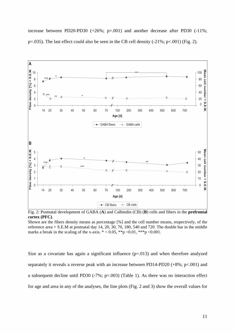
increase between PD20-PD30 (+26%; p=.001) and another decrease after PD30 (-11%;
p=.035). The last effect could also be seen in the CB cell density (-21%; p<.001) (Fig. 2).
0
2
4
6
8
10
14 20 30 40 50 60 70
GABA fibers
100 200 300 400 500 600 700
****
***
20
40
60
80
100
GABA cells
*** ** *
20 30 40 50 60 70
CB fibers
100 200 300 400 500 600 70014
0
1
2
3
4
5
0
10
20
30
40
50
CB cells
****
**
***
Age [d]
Age [d]
Fib
er
den
sit
y[%
]+
S.E
.M.
Fib
er
den
sit
y[%
]+
S.E
.M.
0
Mean
cell
nu
mb
er
+S
.E.M
.M
ean
cell
nu
mb
er
+S
.E.M
.
A
B
Fig. 2: Postnatal development of GABA (A) and Calbindin (CB) (B) cells and fibers in the prefrontal cortex (PFC). Shown are the fibers density means as percentage [%] and the cell number means, respectively, of the reference area + S.E.M at postnatal day 14, 20, 30, 70, 180, 540 and 720. The double bar in the middle marks a break in the scaling of the x-axis. * < 0.05, **p <0.01, ***p <0.001. Size as a covariate has again a significant influence (p=.013) and when therefore analyzed
separately it reveals a reverse peak with an increase between PD14-PD20 (+8%; p<.001) and
a subsequent decline until PD30 (-7%; p=.003) (Table 1). As there was no interaction effect
for age and area in any of the analyses, the line plots (Fig. 2 and 3) show the overall values for
11

the PFC. The separate data for the different investigated areas of the PFC are presented in
table 2 and 3.
BLA
GABAergic fibers in the BLA show a significant effect of age (F (6, 49) = 8.84; p<.001) with
none of the covariates showing a significant contribution. The LSD post-hoc test reveals a
highly significant increase between PD14-PD20 (+15%; p<.001) and a tendency for a decline
after PD70 (PD70-PD180: p=.073), showing a significance compared to PD540 (-7%;
p=.012). CB fibers in the BLA reveal no significant age-effect (p=.21), however, the CB cell
number as a covariate exhibits a significance (p=.031).
An analysis of covariance of the cell numbers in the BLA shows a significant effect of age (F
(12, 98) = 8.85; p<.001) with no effect of size. A following LSD test further reveals a
decrease of GABAergic cells (-14%; p=.025) and CB cells (-18%; p=.001) between PD14-
PD20 with a further slow decrease until PD70 in the GABAergic population (PD20-PD70 -
21%; p=.007) and a more steep decline in the CB cell density (PD30-PD70: -23%; p<.001). In
addition, the CB cell number decreased between PD180-PD540 (-14%; p=.036) (Fig. 3).
Table 1: Circumferences of the two analyzed areas: the prefrontal cortex (PFC) and the basolateral amygdala (BLA) + S.E.M.
AGE PFC BLA 14 [d] 8241.93 ± 349.47 µm 3949.28 ± 36.90 µm 20 [d] 8367.58 ± 561.35 µm *** 4045.24 ± 98.24 µm 30 [d] 8248.79 ± 365.93 µm ** 4019.70 ± 30.63 µm 70 [d] 8226.65 ± 389.70 µm 4106.80 ± 78.18 µm180 [d] 8307.00 ± 340.07 µm 4193.11 ± 112.2 µm540 [d] 8262.83 ± 382.16 µm 4325.24 ± 60.86 µm720 [d] 8225.94 ± 491.41 µm 4218.94 ± 85.76 µm
The PFC reveals a peak in volume on postnatal day 20, while there is no age-dependent effect in the BLA. Levels of significance, compared to the age stage before: **p <0.01, ***p <0.001.
12

2
4
6
8
10
14 20 30 40 50 60 70 100 200 300 400 500 600 700
20
40
60
80
100
0
GABA fibers GABA cells
0
***
*
*
**
0
2
4
6
8
10
14 20 30 40 50 60 70 100 200 300 400 500 600 700
20
40
60
80
100
0
*****
*
CB fibers CB cells
Age [d]
Age [d]
Fib
er
de
ns
ity
[%]
+S
.E.M
.F
ibe
rd
en
sit
y[%
]+
S.E
.M.
Me
an
ce
lln
um
be
r+
S.E
.M.
Me
an
ce
lln
um
be
r+
S.E
.M.
A
B
Fig. 3: Postnatal development of GABA (A) and Calbindin (CB) (B) cells and fibers in the basolateral amygdala (BLA). Shown are the fibers density means as percentage [%] and the cell number means, respectively, of the reference area + S.E.M at postnatal day 14, 20, 30, 70, 180, 540 and 720. The double bar in the middle marks a break in the scaling of the x-axis. * < 0.05, **p <0.01, ***p <0.001.
AGE GABA CB Cg1 Cg3 L III Cg3 L V Cg1 Cg3 L III Cg3 L V
14 [d] 7.31 ± 0.19 7.04 ± 0.27 7.52 ± 0.21 2.76 ± 0.26 2.02 ± 0.16 3.07 ± 0.3320 [d] 8.16 ± 0.25 7.84 ± 0.26 8.26 ± 0.14 3.75 ± 0.24 3.29 ± 0.21 4.22 ± 0.3830 [d] 8.52 ± 0.25 8.35 ± 0.23 8.57 ± 0.17 3.95 ± 0.15 3.80 ± 0.10 4.29 ± 0.1970 [d] 8.22 ± 0.24 8.15 ± 0.28 8.34 ± 0.19 3.84 ± 0.19 3.50 ± 0.16 3.76 ± 0.17
180 [d] 8.59 ± 0.15 8.29 ± 0.15 8.71 ± 0.11 3.62 ± 0.11 3.57 ± 0.13 3.30 ± 0.12540 [d] 8.88 ± 0.16 8.75 ± 0.09 8.83 ± 0.19 3.35 ± 0.09 3.02 ± 0.09 2.79 ± 0.05720 [d] 8.82 ± 0.07 8.58 ± 0.04 8.63 ± 0.12 3.59 ± 0.09 3.29 ± 0.11 3.34 ± 0.07Mean 8.31 ± 0.11 8.09 ± 0.11 8.38 ± 0.09 3.55 ± 0.09 3.21 ± 0.10* 3.56 ± 0.11
Table 2: GABA and CB fiber densities in the various areas within the prefrontal cortex, namely the Cg1 region and lamina (L) III and V of the Cg3 region + S.E.M. There was a difference in the mean innervation density concerning the CB fibers in lamina III of the Cg3 region compared to the other two areas (* p < 0.001).
13
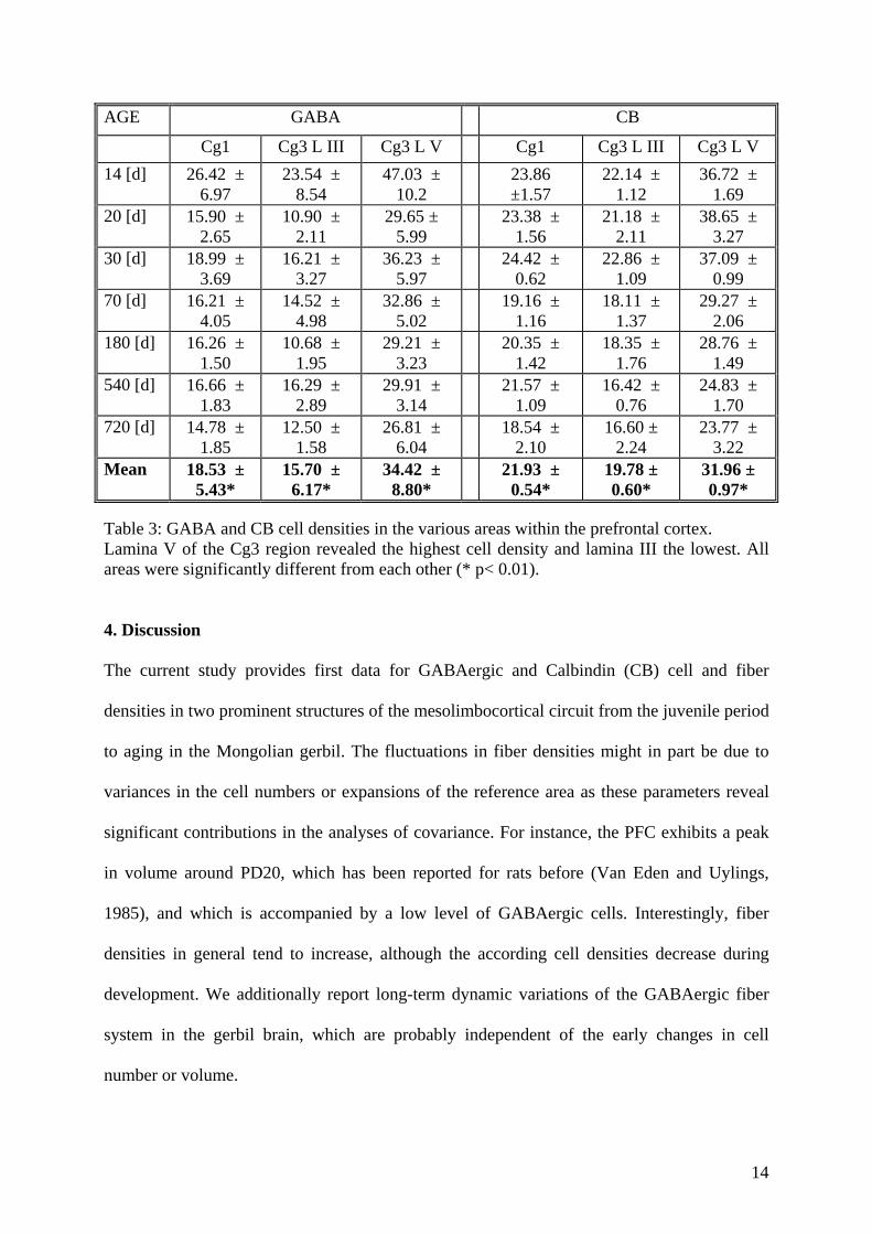
AGE GABA CB
Cg1 Cg3 L III Cg3 L V Cg1 Cg3 L III Cg3 L V 14 [d] 26.42 ±
6.97 23.54 ±
8.54 47.03 ±
10.2 23.86 ±1.57
22.14 ± 1.12
36.72 ± 1.69
20 [d] 15.90 ± 2.65
10.90 ± 2.11
29.65 ± 5.99
23.38 ± 1.56
21.18 ± 2.11
38.65 ± 3.27
30 [d] 18.99 ± 3.69
16.21 ± 3.27
36.23 ± 5.97
24.42 ± 0.62
22.86 ± 1.09
37.09 ± 0.99
70 [d] 16.21 ± 4.05
14.52 ± 4.98
32.86 ± 5.02
19.16 ± 1.16
18.11 ± 1.37
29.27 ± 2.06
180 [d] 16.26 ± 1.50
10.68 ± 1.95
29.21 ± 3.23
20.35 ± 1.42
18.35 ± 1.76
28.76 ± 1.49
540 [d] 16.66 ± 1.83
16.29 ± 2.89
29.91 ± 3.14
21.57 ± 1.09
16.42 ± 0.76
24.83 ± 1.70
720 [d] 14.78 ± 1.85
12.50 ± 1.58
26.81 ± 6.04
18.54 ± 2.10
16.60 ± 2.24
23.77 ± 3.22
Mean 18.53 ± 5.43*
15.70 ± 6.17*
34.42 ± 8.80*
21.93 ± 0.54*
19.78 ± 0.60*
31.96 ± 0.97*
Table 3: GABA and CB cell densities in the various areas within the prefrontal cortex. Lamina V of the Cg3 region revealed the highest cell density and lamina III the lowest. All areas were significantly different from each other (* p< 0.01). 4. Discussion The current study provides first data for GABAergic and Calbindin (CB) cell and fiber
densities in two prominent structures of the mesolimbocortical circuit from the juvenile period
to aging in the Mongolian gerbil. The fluctuations in fiber densities might in part be due to
variances in the cell numbers or expansions of the reference area as these parameters reveal
significant contributions in the analyses of covariance. For instance, the PFC exhibits a peak
in volume around PD20, which has been reported for rats before (Van Eden and Uylings,
1985), and which is accompanied by a low level of GABAergic cells. Interestingly, fiber
densities in general tend to increase, although the according cell densities decrease during
development. We additionally report long-term dynamic variations of the GABAergic fiber
system in the gerbil brain, which are probably independent of the early changes in cell
number or volume.
14

Species- and area-specific maturation of GABAergic cell and fiber densities
Previous prenatal, and early postnatal investigations have shown, that GABAergic cells
appear in the rodent visual or somato-sensory cortex as early as embryonic day (ED) 14-16
(Chronwall and Wolff, 1980; Del Rio et al., 1992) and that there is no apparent change in cell
density after the third postnatal week (Chronwall and Wolff, 1980). CB cells have also been
observed to appear prenatally in the rat cortex and increase until PD 8-11, but their number
seems to decrease notably between PD11-15 while reaching adult levels around the end of the
third postnatal week (Alcantara et al., 1993). In the amygdala first CB cells appeared around
ED13 in the mouse (Legaz et al., 2005), and were observed on ED 20 in the rat, where they
reached adult levels around PD20 (Berdel and Morys, 2000).
These previous observations are in part at variance with our present results from the
Mongolian gerbil. One first explanation for this might be the different developmental pattern
of gerbils compared to rats or mice. The gerbil is known to develop its auditory and visual
capacity later than the rat (Seto-Ohshima et al., 1990) and to lag behind about 2 weeks in
reaching its sexual maturity. In addition, the dopaminergic innervation of the mPFC shows a
prolonged maturation until PD 60 in the rat (Kalsbeek et al., 1988), while dopaminergic
afferents continue to grow until PD 90 in the gerbil (Dawirs et al., 1993). Thus, the partially
highly significant increase in GABAergic and CB fiber densities until PD30 and the
fluctuations in the cell densities we observed in the mPFC and the BLA in the present study
might indicate the postponed maturation of the gerbil nervous system compared to other
rodents and the later onset of functional systems.
The apparent discrepancy of former and our present results might further be due to the
developmental differences of particular areas. Thus, calcium-binding proteins occur several
days later in the associative cortices compared to the primary visual cortex (Alcantara et al.,
1993). In addition, Wolff and colleagues (1984) found no notable difference in GABA cell
proportion after P3 in the layers II-VI in the visual cortex, while Vincent and colleagues
15

(1995) found a decrease of GABA cell density until PD 15 in the mPFC of rats. They further
suggest that this decrease is associated with an expansion of the cortex. The severe decrease
of GABA cells between PD14-PD20 in the gerbil might therefore be due to the peak in mPFC
volume around PD20. Intriguingly, we did not observe such a decline in CB cell density
during this particular time. This might depend on a variation of the amount of the protein
within the cells, so that despite the decreasing number of GABAergic cells more of the
remaining cells expressed enough CB to reach the minimum gray value for cell counting. The
inverse effect might also account for the later decline in the CB number (PD30-PD70), which
would be in line with the hypotheses that one population of CB cells only expresses the
protein transitorily, while the other neurons are permanently immunoreactive for CB
(Alcantara et al., 1993).
The maturation of GABAergic fibers in the BLA seems to differ slightly from the cortex. One
reason for this could be the different targets and functions of the subpopulations in the cortex
and the amygdala, as for instance, CB cells have been shown to build basket like structures in
the BLA (Berdel and Morys, 2000) but not in the cortex. Further, CB interneurons in the
cortex rather present a minor subpopulation (Celio, 1990), while they constitute almost 60%
of the GABA-containing population in the BLA (McDonald and Mascagni, 2001) which
would be in line with our observation of a higher CB fiber density in the BLA compared to
the cortex. In addition, another 60% of these CB neurons have been shown to also coexpress
another calcium-binding protein, parvalbumin (PV), permanently (McDonald and Betette,
2001), while such a coexpression is only transiently observed in the cortex (Alcantara et al.,
1996). This might explain the existence of CB baskets around unlabelled pyramidal neurons
in the BLA and further hints to the particular role of the various calcium-binding proteins in
different subpopulations of the brain.
16

GABA plasticity from adolescence to aging
Regardless of fluctuations in cell densities, there are prolonged variations in fiber densities
and thus in the inhibitory networks of the particular areas during adolescence and even
adulthood. Generally it is thought that local circuit neuron connections mature relatively late
compared to projections from efferent neurons (Miller, 1988). In addition, GABA appears to
exert direct and indirect trophic action and thus initiate the establishment of synaptic contacts
such as excitatory synapses, which usually appear 1-4 days after the GABA cells (Wolff et al.,
1978; 1993). The synaptogenesis of inhibitory GABAergic boutons seems to be even further
delayed and continues well into adulthood (Bahr and Wolff, 1985; Lewis et al., 2005), which
would be in line with a continuing augmentation of fibers.
GABA is known to undergo a shift from an excitatory transmitter before birth into an
inhibitory transmitter after birth (Ben-Ari, 2002; Cherubini et al., 1991; Ganguly et al., 2001).
Further, there is a potential postnatal shift between the different GABAergic subpopulations
with a decrease in CB immunoreactivity, which is usually accompanied by the appearance of
PV-positive structures in various areas of the brain (Cruz et al., 2003; Davila et al., 2005;
Erickson and Lewis, 2002; Legaz et al., 2005). PV cells mature considerably late during
development (Alcantara et al., 1993), e.g. in the gerbil, first PV neurons appear around PD 14
in the mPFC, though their number is very small (unpublished data), and are known to build
axo-somatic contacts and basket like boutons around pyramidal somata, which likewise
appear considerably late (Bahr and Wolff, 1985). These types of connections have a
particularly powerful influence on the firing activity and synchronization of target neurons
(Freund, 2003; Gibson et al., 1999; Klausberger et al., 2003; Miles et al., 1996; Tamas et al.,
1997; Tamas et al., 2000). Such oscillatory (rhythmic) synchronization is for instance
generated by a BLA PV network during emotional arousal (Muller et al., 2005) and is further
believed to create the necessary temporal and spatial frame for functions such as working
memory in the PFC (Constantinidis et al., 2002; Lewis et al., 2005) or consolidation of
17
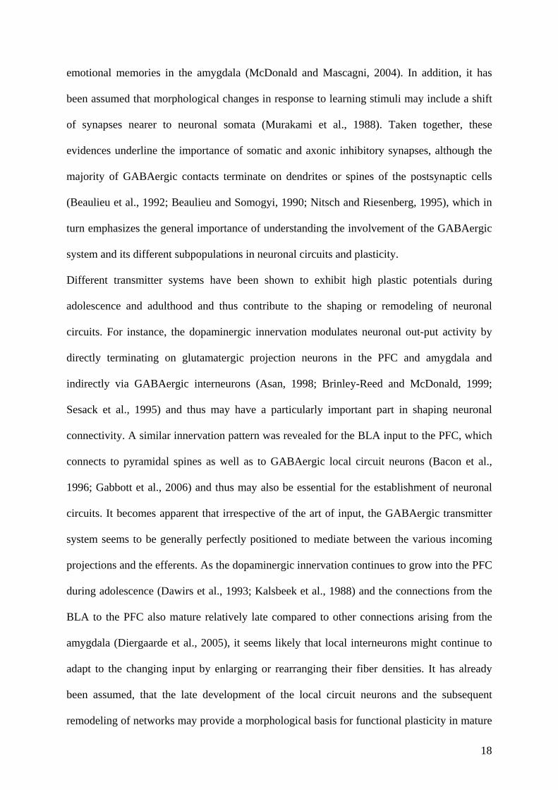
emotional memories in the amygdala (McDonald and Mascagni, 2004). In addition, it has
been assumed that morphological changes in response to learning stimuli may include a shift
of synapses nearer to neuronal somata (Murakami et al., 1988). Taken together, these
evidences underline the importance of somatic and axonic inhibitory synapses, although the
majority of GABAergic contacts terminate on dendrites or spines of the postsynaptic cells
(Beaulieu et al., 1992; Beaulieu and Somogyi, 1990; Nitsch and Riesenberg, 1995), which in
turn emphasizes the general importance of understanding the involvement of the GABAergic
system and its different subpopulations in neuronal circuits and plasticity.
Different transmitter systems have been shown to exhibit high plastic potentials during
adolescence and adulthood and thus contribute to the shaping or remodeling of neuronal
circuits. For instance, the dopaminergic innervation modulates neuronal out-put activity by
directly terminating on glutamatergic projection neurons in the PFC and amygdala and
indirectly via GABAergic interneurons (Asan, 1998; Brinley-Reed and McDonald, 1999;
Sesack et al., 1995) and thus may have a particularly important part in shaping neuronal
connectivity. A similar innervation pattern was revealed for the BLA input to the PFC, which
connects to pyramidal spines as well as to GABAergic local circuit neurons (Bacon et al.,
1996; Gabbott et al., 2006) and thus may also be essential for the establishment of neuronal
circuits. It becomes apparent that irrespective of the art of input, the GABAergic transmitter
system seems to be generally perfectly positioned to mediate between the various incoming
projections and the efferents. As the dopaminergic innervation continues to grow into the PFC
during adolescence (Dawirs et al., 1993; Kalsbeek et al., 1988) and the connections from the
BLA to the PFC also mature relatively late compared to other connections arising from the
amygdala (Diergaarde et al., 2005), it seems likely that local interneurons might continue to
adapt to the changing input by enlarging or rearranging their fiber densities. It has already
been assumed, that the late development of the local circuit neurons and the subsequent
remodeling of networks may provide a morphological basis for functional plasticity in mature
18

cortical neurons (Miller, 1988) and thus it might even contribute to complex processes as
long-term learning and memory.
As we could recently show in our laboratory using an animal model of early traumatized
gerbils, epigenetic disturbances during development can cause a shift within the GABAergic
system, with a loss of GABAergic boutons around pyramidal somata and an increase in
lamina I/II GABAergic fibers in the mPFC of adult animals (Brummelte et al., 2006b). The
lessening of somatic inhibition and the potential subsequent interference of the
synchronization of whole pyramidal populations might contribute to the observed deficits in
PFC-related behaviors and functions such as working memory after this early developmental
disturbance (Dawirs et al., 1996). In addition, GABA has also been shown to exhibit a high
plasticity when challenged in adult animals (Dawirs et al., 1997). Therefore the question
arose, if GABA keeps its natural neuroplastic potential even up to adulthood, especially as it
is believed that disturbances in the GABAergic inhibitory regulation of cortical networks
contribute considerably to cognitive impairments as seen in schizophrenia (Benes and
Berretta, 2001), which’s onset is usually in young adulthood. We here demonstrated that there
are indeed long-term variations in the GABAergic system during adolescence.
Intriguingly, there was no aging-related change in the fiber densities of GABA or CB in
neither the PFC nor the BLA. Several studies have reported about an age-related decrease in
CB immunoreactivity in basal forebrain cholinergic cells (Geula et al., 2003; Wu et al., 1997;
2003) and also about age-related changes of CB structures in some cortical and subcortical
areas (Bu et al., 2003; Hwang et al., 2002; Kishimoto et al., 1998). Further, several studies
have shown alteration in the CB immunoreactivity in Alzheimer patients compared to controls
(Ichimiya et al., 1988; Lally et al., 1997; McLachlan et al., 1987). However, further
investigations suggest, that it might rather be a decrease in the expression of the protein than a
decline of whole cells or branches (Kishimoto et al., 1998). Nevertheless, a decline in CB
within the cell might cause a diminished capacity to buffer high levels of calcium, thus
19

leading to a higher vulnerability towards pathological processes that might cause the
degeneration of the cell in the end (Bu et al., 2003). However, to our knowledge, there is no
study revealing a significant age-related effect in CB immunoreactivity for the PFC or BLA.
But there are other hints for alterations within the GABAergic system as e.g. differences in
GABA activity in specific hypothalamic areas (Jarry et al., 1999) or age-related changes in
GABA receptor compositions (Caspary et al., 1999). Such changes can not be excluded in the
old gerbil referring to our data, but there is apparently no alteration in fiber densities.
However, we have to admit, that 110 weeks is the mean survival of a male gerbil, meaning
that in the individual case the maximum age lies higher (Troup et al., 1969), thus our results
of stable GABAergic and CB fiber densities up to PD720 do not exclude variations in still
older animals.
Nevertheless, GABA seems to appear relatively consistent against the deleterious effects of
age compared to other transmitters as e.g. dopamine, which is believed to play a role in
various age-related diseases such as Alzheimer, Parkinson’s or Huntington disease (reviewed
in: Backman and Farde, 2001; Morgan et al., 1987; Ossowska, 1993) and shows a decline of
fibers in 720 days old gerbils in the PFC (unpublished data) but not in the amygdala
(Brummelte and Teuchert-Noodt, 2006). On a highly speculative level it might be assumed
that neurodegenerative diseases are likely to appear when the GABAergic plasticity finally
vanishes during aging.
Acknowledgments The authors wish to thank Dr. F. Bagorda and U. Schroeder for excellent technical assistance.
20

Reference List
Alcantara,S., de,Lecea.L., Del Rio,J.A., Ferrer,I., and Soriano,E. (1996). Transient colocalization of parvalbumin and calbindin D28k in the postnatal cerebral cortex: evidence for a phenotypic shift in developing nonpyramidal neurons. Eur. J. Neurosci. 8, 1329-1339.
Alcantara,S., Ferrer,I., and Soriano,E. (1993). Postnatal development of parvalbumin and calbindin D28K immunoreactivities in the cerebral cortex of the rat. Anat. Embryol. (Berl) 188, 63-73.
Asan,E. (1998). The catecholaminergic innervation of the rat amygdala. Adv. Anat. Embryol. Cell Biol. 142, 1-118.
Backman,L., and Farde,L. (2001). Dopamine and cognitive functioning: brain imaging findings in Huntington's disease and normal aging. Scand. J. Psychol. 42, 287-296.
Bacon,S.J., Headlam,A.J., Gabbott,P.L., and Smith,A.D. (1996). Amygdala input to medial prefrontal cortex (mPFC) in the rat: a light and electron microscope study. Brain Res. 720, 211-219.
Bagorda,F., Teuchert-Noodt,G., and Lehmann,K. (2006). Isolation rearing or methamphetamine traumatisation induce a "dysconnection" of prefrontal efferents in gerbils: implications for schizophrenia. J Neural Transm. 113, 365-379.
Bahr,S., and Wolff,J.R. (1985). Postnatal development of axosomatic synapses in the rat visual cortex: morphogenesis and quantitative evaluation. J. Comp Neurol. 233, 405-420.
Baimbridge,K.G., Celio,M.R., and Rogers,J.H. (1992). Calcium-binding proteins in the nervous system. Trends Neurosci 15, 303-308.
Beaulieu,C., Kisvarday,Z., Somogyi,P., Cynader,M., and Cowey,A. (1992). Quantitative distribution of GABA-immunopositive and -immunonegative neurons and synapses in the monkey striate cortex (area 17). Cereb. Cortex 2, 295-309.
Beaulieu,C., and Somogyi,P. (1990). Targets and Quantitative Distribution of GABAergic Synapses in the Visual Cortex of the Cat. Eur. J Neurosci 2, 296-303.
Ben-Ari,Y. (2002). Excitatory actions of gaba during development: the nature of the nurture. Nat. Rev Neurosci 3, 728-739.
Benes,F.M., and Berretta,S. (2001). GABAergic interneurons: implications for understanding schizophrenia and bipolar disorder. Neuropsychopharmacology 25, 1-27.
Berdel,B., and Morys,J. (2000). Expression of calbindin-D28k and parvalbumin during development of rat's basolateral amygdaloid complex. Int J Dev. Neurosci 18, 501-513.
Björklund,A., and Lindvall,O. (1984). Dopamine-containing systems in the CNS. In Handbook of chemical neuroanatomy - Classical transmitters in the CNS, Part 1, A. Björklund, and T. Hökfelt, eds. (Amsterdam: Elsevier), pp. 55-122.
Brinley-Reed,M., and McDonald,A.J. (1999). Evidence that dopaminergic axons provide a dense innervation of specific neuronal subpopulations in the rat basolateral amygdala. Brain Res. 850, 127-135.
Brummelte,S., and Teuchert-Noodt,G. (2006). Postnatal development of dopamine innervation in the amygdala and the entorhinal cortex of the gerbil (Meriones unguiculatus). Brain Res. (Epub ahead of print)
Brummelte,S., Grund,T., Czok,A., Teuchert-Noodt,G., and Neddens,J. (2006a). Long-term effects of a single adult methamphetamine challenge: Minor impact on dopamine fibre density in limbic brain areas of gerbils. Behav. Brain Funct. 2, 12.
Brummelte,S., Neddens,J., and Teuchert-Noodt,G. (2006b). Alteration in the GABAergic network of the prefrontal cortex in a potential animal model of psychosis. J. Neural Transm. (in press)
21

Bu,J., Sathyendra,V., Nagykery,N., and Geula,C. (2003). Age-related changes in calbindin-D28k, calretinin, and parvalbumin-immunoreactive neurons in the human cerebral cortex. Exp. Neurol. 182, 220-231.
Carlsen,J. (1988). Immunocytochemical localization of glutamate decarboxylase in the rat basolateral amygdaloid nucleus, with special reference to GABAergic innervation of amygdalostriatal projection neurons. J Comp Neurol. 273, 513-526.
Caspary,D.M., Holder,T.M., Hughes,L.F., Milbrandt,J.C., McKernan,R.M., and Naritoku,D.K. (1999). Age-related changes in GABA(A) receptor subunit composition and function in rat auditory system. Neuroscience 93, 307-312.
Celio,M.R. (1990). Calbindin D-28k and parvalbumin in the rat nervous system. Neuroscience 35, 375-475.
Chen,R., Cohen,L.G., and Hallett,M. (2002). Nervous system reorganization following injury. Neuroscience 111, 761-773.
Cherubini,E., Gaiarsa,J.L., and Ben-Ari,Y. (1991). GABA: an excitatory transmitter in early postnatal life. Trends Neurosci 14, 515-519.
Chronwall,B., and Wolff,J.R. (1980). Prenatal and postnatal development of GABA-accumulating cells in the occipital neocortex of rat. J Comp Neurol. 190, 187-208.
Chronwall,B.M., and Wolff,J.R. (1981). Development of GABA-accumulating neurons and glial cells in the rat visual cortex. Adv. Biochem. Psychopharmacol. 29, 453-458.
Conde,F., Lund,J.S., Jacobowitz,D.M., Baimbridge,K.G., and Lewis,D.A. (1994). Local circuit neurons immunoreactive for calretinin, calbindin D-28k or parvalbumin in monkey prefrontal cortex: distribution and morphology. J Comp Neurol. 341, 95-116.
Constantinidis,C., Williams,G.V., and Goldman-Rakic,P.S. (2002). A role for inhibition in shaping the temporal flow of information in prefrontal cortex. Nat. Neurosci 5, 175-180.
Cruz,D.A., Eggan,S.M., and Lewis,D.A. (2003). Postnatal development of pre- and postsynaptic GABA markers at chandelier cell connections with pyramidal neurons in monkey prefrontal cortex. J Comp Neurol. 465, 385-400.
Davila,J.C., Real,M.A., Olmos,L., Legaz,I., Medina,L., and Guirado,S. (2005). Embryonic and postnatal development of GABA, calbindin, calretinin, and parvalbumin in the mouse claustral complex. J Comp Neurol. 481, 42-57.
Dawirs,R.R., Teuchert-Noodt,G., and Czaniera,R. (1993). Maturation of the dopamine innervation during postnatal development of the prefrontal cortex in gerbils (Meriones unguiculatus). A quantitative immunocytochemical study. J Hirnforsch. 34, 281-290.
Dawirs,R.R., Teuchert-Noodt,G., and Czaniera,R. (1996). Ontogeny of PFC-related behaviours is sensitive to a single non-invasive dose of methamphetamine in neonatal gerbils (Meriones unguiculatus). J Neural Transm. 103, 1235-1245.
Dawirs,R.R., Teuchert-Noodt,G., and Nossoll,M. (1997). Pharmacologically induced neural plasticity in the prefrontal cortex of adult gerbils (Meriones unguiculatus). Eur. J. Pharmacol. 327, 117-123.
DeFelipe,J., Hendry,S.H., and Jones,E.G. (1989). Synapses of double bouquet cells in monkey cerebral cortex visualized by calbindin immunoreactivity. Brain Res. 503, 49-54.
Del Rio,J.A., Soriano,E., and Ferrer,I. (1992). Development of GABA-immunoreactivity in the neocortex of the mouse. J Comp Neurol. 326, 501-526.
Diergaarde,L., Gerrits,M.A., Brouwers,J.P., and van Ree,J.M. (2005). Early amygdala damage disrupts performance on medial prefrontal cortex-related tasks but spares spatial learning and memory in the rat. Neuroscience 130, 581-590.
22

Erickson,S.L., and Lewis,D.A. (2002). Postnatal development of parvalbumin- and GABA transporter-immunoreactive axon terminals in monkey prefrontal cortex. J Comp Neurol. 448, 186-202.
Fallon,J.H., and Ciofi,P. (1992). Distribution of monoamines within the amygdala. In The amygdala: Neurobiological aspects of emotion, memory, and mental dysfunction, Aggleton JP, ed. (New York: Wiley-Liss, Inc.), pp. 97-114.
Freund,T.F. (2003). Interneuron Diversity series: Rhythm and mood in perisomatic inhibition. Trends Neurosci 26, 489-495.
Gabbott,P.L., and Bacon,S.J. (1996). Local circuit neurons in the medial prefrontal cortex (areas 24a,b,c, 25 and 32) in the monkey: II. Quantitative areal and laminar distributions. J Comp Neurol. 364, 609-636.
Gabbott,P.L., Warner,T.A., and Busby,S.J. (2006). Amygdala input monosynaptically innervates parvalbumin immunoreactive local circuit neurons in rat medial prefrontal cortex. Neuroscience.
Ganguly,K., Schinder,A.F., Wong,S.T., and Poo,M. (2001). GABA itself promotes the developmental switch of neuronal GABAergic responses from excitation to inhibition. Cell 105, 521-532.
Geula,C., Bu,J., Nagykery,N., Scinto,L.F., Chan,J., Joseph,J., Parker,R., and Wu,C.K. (2003). Loss of calbindin-D28k from aging human cholinergic basal forebrain: relation to neuronal loss. J. Comp Neurol. 455, 249-259.
Gibson,J.R., Beierlein,M., and Connors,B.W. (1999). Two networks of electrically coupled inhibitory neurons in neocortex. Nature 402, 75-79.
Hensch,T.K. (2005). Critical period mechanisms in developing visual cortex. Curr. Top. Dev. Biol. 69, 215-237.
Hof,P.R., Glezer,I.I., Conde,F., Flagg,R.A., Rubin,M.B., Nimchinsky,E.A., and Vogt Weisenhorn,D.M. (1999). Cellular distribution of the calcium-binding proteins parvalbumin, calbindin, and calretinin in the neocortex of mammals: phylogenetic and developmental patterns. J Chem. Neuroanat. 16, 77-116.
Holtmaat,A., Wilbrecht,L., Knott,G.W., Welker,E., and Svoboda,K. (2006). Experience-dependent and cell-type-specific spine growth in the neocortex. Nature 441, 979-983.
Hwang,I.K., Kang,T.C., Lee,J.C., Lee,I.S., Park,S.K., An,S.J., Jeong,Y.G., Seo,J.G., Oh,Y.S., and Won,M.H. (2002). Age-related change of calbindin D-28k immunoreactive neurons in the rat main olfactory bulb. Neurosci Lett. 326, 159-162.
Ichimiya,Y., Emson,P.C., Mountjoy,C.Q., Lawson,D.E., and Heizmann,C.W. (1988). Loss of calbindin-28K immunoreactive neurones from the cortex in Alzheimer-type dementia. Brain Res. 475, 156-159.
Jarry,H., Wise,P.M., Leonhardt,S., and Wuttke,W. (1999). Effects of age on GABA turnover rates in specific hypothalamic areas in female rats. Exp. Clin. Endocrinol. Diabetes 107, 59-62.
Joseph,R. (1999). Environmental influences on neural plasticity, the limbic system, emotional development and attachment: a review. Child Psychiatry Hum. Dev. 29, 189-208.
Kalsbeek,A., Voorn,P., Buijs,R.M., Pool,C.W., and Uylings,H.B. (1988). Development of the dopaminergic innervation in the prefrontal cortex of the rat. J Comp Neurol. 269, 58-72.
Kawaguchi,Y., and Kubota,Y. (1998). Neurochemical features and synaptic connections of large physiologically-identified GABAergic cells in the rat frontal cortex. Neuroscience 85, 677-701.
Kemppainen,S., and Pitkanen,A. (2000). Distribution of parvalbumin, calretinin, and calbindin-D(28k) immunoreactivity in the rat amygdaloid complex and colocalization with gamma-aminobutyric acid. J Comp Neurol. 426, 441-467.
Kishimoto,J., Tsuchiya,T., Cox,H., Emson,P.C., and Nakayama,Y. (1998). Age-related changes of calbindin-D28k, calretinin, and parvalbumin mRNAs in the hamster brain. Neurobiol. Aging 19, 77-82.
23

Klausberger,T., Magill,P.J., Marton,L.F., Roberts,J.D., Cobden,P.M., Buzsaki,G., and Somogyi,P. (2003). Brain-state- and cell-type-specific firing of hippocampal interneurons in vivo. Nature 421, 844-848.
Lally,G., Faull,R.L., Waldvogel,H.J., Ferrari,S., and Emson,P.C. (1997). Calcium homeostasis in ageing: studies on the calcium binding protein calbindin D28K. J. Neural Transm. 104, 1107-1112.
Legaz,I., Olmos,L., Real,M.A., Guirado,S., Davila,J.C., and Medina,L. (2005). Development of neurons and fibers containing calcium binding proteins in the pallial amygdala of mouse, with special emphasis on those of the basolateral amygdalar complex. J Comp Neurol. 488, 492-513.
Lewis,D.A., Hashimoto,T., and Volk,D.W. (2005). Cortical inhibitory neurons and schizophrenia. Nat. Rev. Neurosci. 6, 312-324.
Lund,J.S., and Lewis,D.A. (1993). Local circuit neurons of developing and mature macaque prefrontal cortex: Golgi and immunocytochemical characteristics. J Comp Neurol. 328, 282-312.
McDonald,A.J., and Betette,R.L. (2001). Parvalbumin-containing neurons in the rat basolateral amygdala: morphology and co-localization of Calbindin-D(28k). Neuroscience 102, 413-425.
McDonald,A.J., and Mascagni,F. (2001). Colocalization of calcium-binding proteins and GABA in neurons of the rat basolateral amygdala. Neuroscience 105, 681-693.
McDonald,A.J., and Mascagni,F. (2004). Parvalbumin-containing interneurons in the basolateral amygdala express high levels of the alpha1 subunit of the GABAA receptor. J Comp Neurol. 473, 137-146.
McLachlan,D.R., Wong,L., Bergeron,C., and Baimbridge,K.G. (1987). Calmodulin and calbindin D28K in Alzheimer disease. Alzheimer Dis. Assoc. Disord. 1, 171-179.
Merzenich,M.M., Kaas,J.H., Wall,J., Nelson,R.J., Sur,M., and Felleman,D. (1983). Topographic reorganization of somatosensory cortical areas 3b and 1 in adult monkeys following restricted deafferentation. Neuroscience 8, 33-55.
Miles,R., Toth,K., Gulyas,A.I., Hajos,N., and Freund,T.F. (1996). Differences between somatic and dendritic inhibition in the hippocampus. Neuron 16, 815-823.
Miller,M.W. Development of Projection and Local Circuit Neurons in Neocortex. Peters, A. and Jones, E. G. Cerebral Cortex: Development and Maturation of Cerebral Cortex. [7], 133-166. 1988. New York and London, Plenum Press.
Morgan,D.G., May,P.C., and Finch,C.E. (1987). Dopamine and serotonin systems in human and rodent brain: effects of age and neurodegenerative disease. J. Am. Geriatr. Soc. 35, 334-345.
Morys,J., Berdel,B., Jagalska-Majewska,H., and Luczynska,A. (1999). The basolateral amygdaloid complex--its development, morphology and functions. Folia Morphol. (Warsz. ) 58, 29-46.
Mrzljak,L., Uylings,H.B., Van Eden,C.G., and Judas,M. (1990). Neuronal development in human prefrontal cortex in prenatal and postnatal stages. Prog. Brain Res. 85, 185-222.
Muller,J.F., Mascagni,F., and McDonald,A.J. (2003). Synaptic connections of distinct interneuronal subpopulations in the rat basolateral amygdalar nucleus. J Comp Neurol. 456, 217-236.
Muller,J.F., Mascagni,F., and McDonald,A.J. (2005). Coupled networks of parvalbumin-immunoreactive interneurons in the rat basolateral amygdala. J Neurosci 25, 7366-7376.
Muller,J.F., Mascagni,F., and McDonald,A.J. (2006). Pyramidal cells of the rat basolateral amygdala: synaptology and innervation by parvalbumin-immunoreactive interneurons. J Comp Neurol. 494, 635-650.
Murakami,F., Oda,Y., and Tsukahara,N. (1988). Synaptic plasticity in the red nucleus and learning. Behav. Brain Res. 28, 175-179.
24

Nguyen,L., Rigo,J.M., Rocher,V., Belachew,S., Malgrange,B., Rogister,B., Leprince,P., and Moonen,G. (2001). Neurotransmitters as early signals for central nervous system development. Cell Tissue Res. 305, 187-202.
Nitsch,C., and Riesenberg,R. (1995). Synaptic reorganisation in the rat striatum after dopaminergic deafferentation: an ultrastructural study using glutamate decarboxylase immunocytochemistry. Synapse 19, 247-263.
Ossowska,K. (1993). Disturbances in neurotransmission processes in aging and age-related diseases. Pol. J. Pharmacol. 45, 109-131.
Parnavelas,J.G. (1992). Development of GABA-containing neurons in the visual cortex. Prog. Brain Res. 90, 523-537.
Paxinos,G., and Watson,C. (1986). The rat brain in stereotaxic coordinates (New York: Academic Press).
Rosenzweig,M.R., and Bennett,E.L. (1969). Effects of differential environments on brain weights and enzyme activities in gerbils, rats, and mice. Dev. Psychobiol. 2, 87-95.
Sesack,S.R., Snyder,C.L., and Lewis,D.A. (1995). Axon terminals immunolabeled for dopamine or tyrosine hydroxylase synapse on GABA-immunoreactive dendrites in rat and monkey cortex. J Comp Neurol. 363, 264-280.
Seto-Ohshima,A., Aoki,E., Semba,R., Emson,P.C., and Heizmann,C.W. (1990). Appearance of parvalbumin-specific immunoreactivity in the cerebral cortex and hippocampus of the developing rat and gerbil brain. Histochemistry 94, 579-589.
Tamas,G., Buhl,E.H., Lorincz,A., and Somogyi,P. (2000). Proximally targeted GABAergic synapses and gap junctions synchronize cortical interneurons. Nat. Neurosci 3, 366-371.
Tamas,G., Buhl,E.H., and Somogyi,P. (1997). Fast IPSPs elicited via multiple synaptic release sites by different types of GABAergic neurone in the cat visual cortex. J Physiol 500 (Pt 3), 715-738.
Teuchert-Noodt,G. (2000). Neuronal degeneration and reorganization: a mutual principle in pathological and in healthy interactions of limbic and prefrontal circuits. J. Neural Transm. Suppl 315-333.
Thiessen,D., and Yahr,P. (1977). The gerbil in behavioral investigations. Mechanisms of territoriality and olfactory communication. (Austin: University of Texas Press).
Trachtenberg,J.T., Chen,B.E., Knott,G.W., Feng,G., Sanes,J.R., Welker,E., and Svoboda,K. (2002). Long-term in vivo imaging of experience-dependent synaptic plasticity in adult cortex. Nature 420, 788-794.
Troup,G.M., Smith,G.S., and Walford,R.L. (1969). Life span,chronologic disease patterns and age-related changes in relative spleen weights for the mongolian gerbil (Meriones unguiculatus). Exp. Gerontol. 4, 139-143.
Valverde,F. (1998). Golgi atlas of the postnatal mouse brain (Wien, New York: Springer-Verlag).
Van Eden,C.G., Kros,J.M., and Uylings,H.B. (1990). The development of the rat prefrontal cortex. Its size and development of connections with thalamus, spinal cord and other cortical areas. Prog. Brain Res. 85, 169-183.
Van Eden,C.G., Mrzljak,L., Voorn,P., and Uylings,H.B. (1989). Prenatal development of GABA-ergic neurons in the neocortex of the rat. J Comp Neurol. 289, 213-227.
Van Eden,C.G., and Uylings,H.B. (1985). Postnatal volumetric development of the prefrontal cortex in the rat. J. Comp Neurol. 241, 268-274.
Vincent,S.L., Pabreza,L., and Benes,F.M. (1995). Postnatal maturation of GABA-immunoreactive neurons of rat medial prefrontal cortex. J Comp Neurol. 355, 81-92.
Wolff,J.R., Balcar,V.J., Zetzsche,T., Bottcher,H., Schmechel,D.E., and Chronwall,B.M. (1984). Development of GABA-ergic system in rat visual cortex. Adv. Exp. Med. Biol. 181, 215-239.
25

Wolff,J.R., Chronwall,B.M., and Rickmann,M. (1978). Morphogenetic relations between cell migration and synaptogenesis in the neocortex of rat. Proc. Eur. Soc. Neurochem. 1, 158-173.
Wolff,J.R., Joo,F., and Kasa,P. (1993). Modulation by GABA of neuroplasticity in the central and peripheral nervous system. Neurochem. Res. 18, 453-461.
Wu,C.K., Mesulam,M.M., and Geula,C. (1997). Age-related loss of calbindin from human basal forebrain cholinergic neurons. Neuroreport 8, 2209-2213.
Wu,C.K., Nagykery,N., Hersh,L.B., Scinto,L.F., and Geula,C. (2003). Selective age-related loss of calbindin-D28k from basal forebrain cholinergic neurons in the common marmoset (Callithrix jacchus). Neuroscience 120, 249-259.
Zito,K., and Svoboda,K. (2002). Activity-dependent synaptogenesis in the adult Mammalian cortex. Neuron 35, 1015-1017.
26
Submitted to ‘Behavioural and Brain Functions’ Manuscript ID 1553343361124716 Density of dopaminergic fibres in the prefrontal cortex of gerbils is sensitive to aging. Susanne Brummelte* and Gertraud Teuchert-Noodt
Department of Neuroanatomy, Faculty of Biology, University of Bielefeld, Universitätsstr.
25, D-33615 Bielefeld, Germany, Phone: 0049 521 106 5704; Fax: 0049 521 106 6470,
[email protected], [email protected]
*corresponding author
Abstract Mesencephalic dopamine (DA) projections are essential for cognitive and behavioral
functions and believed to play a critical role during development and aging. The
dopaminergic afferents of the rodent prefrontal cortex (PFC) show an extremely prolonged
maturation which is very sensitive to epigenetic challenges. However, less is known about the
long-term maturation and aging of these DA axons. Therefore, immunohistochemically
stained DA fibres were quantitatively examined in the PFC of the Mongolian gerbil (Meriones
unguiculatus) ranging from 6 to 24 months of age. Results show a decrease in DA fibre
densities in the superficial layers of the PFC in 24 month old animals compared to 6 and 12
months.

Findings Dopamine (DA) has frequently been associated with age-related changes and
neurodegenerative diseases such as parkinson. In particular, striatal alterations have been in
the focus of many investigations, as these are assumed to contribute to observed cognitive and
motor dysfunction in elderly people or parkinson patients [1].
However, recent studies also suggest age-related DA changes in extrastriatal brain regions.
Mirura and colleagues [2] observed that the level and turnover of monoamines and their
metabolites were reduced in several brain regions as e.g. the prefrontal cortex (PFC), the
amygdala, nucleus accumbens and hippocampus of 18 months old rats compared to young
animals. For humans, it has been shown that the DA synthesis is lower with age in several
extrastriatal regions, including the dorsolateral prefrontal and anterior cingulate cortex [3]. In
addition, an age-related decline in D2 receptors was also found in various extrastriatal areas of
healthy volunteers suggesting an association with normal aging processes [4]. In fact, it
appears that the declines in D1 and D2 receptor binding might even be faster or more
pronounced in the frontal cortices compared to striatal or thalamic regions [4-6]. This is in
line with other studies reporting a greater loss of DA from the PFC compared to motor areas
in aged monkeys [7,8], which underlines the importance of dopaminergic function during
aging in this area.
So far, most studies have focused on the metabolic function of the dopaminergic system
during aging, but less research has been done concerning neuroanatomical alterations. Our
laboratory could recently show, that the dopaminergic fibre densities of the nucleus
accumbens, the amygdala and the entorhinal cortex show no age-related changes in 24 month
old gerbils (Meriones unguiculatus) compared to young animals [9,10]. However, as the PFC
has been frequently associated with an age-related decline in cognitive function, this study
was conducted to check for alterations in the dopaminergic fibre density in this particularly
vulnerable area.

All experimental procedures were approved by the appropriate committee for animal care in
accordance with the European Communities Council Directive. Gerbils were chosen due to
their wild-type like behavioural and neuronal repertoire, as they have not been so intensively
domesticated compared to rats or mice [11]. A total of 33 male Mongolian gerbils were used
in this study (6 Mon n=8; 12 Mon n=5; 18 Mon n=11; 24 Mon n=9). Animal rearing and
keeping conditions as well as the DA staining procedure have been described elsewhere [9].
Prefrontal DA fibre densities were measured in four consecutive coronal slices of the PFC.
Fibre fragments in the upper layers were visualised in standard test fields in the prelimbic
cortex (PrL) and in the infralimbic cortex (IL), using a bright-field microscope (BX61,
Olympus, Hamburg, Germany) and a digital camera for microscopy (ColorView II, SIS,
Münster, Germany) at 400-fold magnification. Fibres were quantified by software for image
analysis (KS300, Jenoptik, Jena, Germany). For details of the quantification see [9]. The fibre
area was calculated as a percentage of the reference area. All measurements were done by an
experimenter blind to the coding of the samples.
Measurements were computed as arithmetic means by-case and by-group ± S.E.M. and a two-
way analysis of variance (ANOVA) with age (4 levels) and area (2 levels) as independent
variables and the dopaminergic fibre density as the dependent variable was used to check for
statistical significance between groups followed by LSD post-hoc test for multiple
comparisons. Statistical analysis was computed with Statistica 6 (StatSoft, Tulsa, USA). The
levels of significance were set at * p<0.05, ** p<0.01 and *** p<0.001.
Statistical analysis revealed a significant effect of age (F(3,56)=3.47; p=.022) and area
(F(1,56)=5.53; p=.022), but no interaction effect (F(3,56)=.184; p=.907). The PrL cortex
showed a dense innervation of DA fibres, which was according to a Fisher LSD post-hoc test
significantly lower in the IL (p=.008). The post-hoc test further revealed a significant age-
related decrease in DA fibre density in the superficial layers of the PFC between 12 month

and 24 month old animals (-26%; p=.025), with the significance being even more prominent
compared to 6 month old gerbils (-26%; p=.0098) (Fig.1).
0
1
2
3
4
Dopam
inerg
icfibre
density
[%]+
S.E
.M.
6 12 18 24Age [months]
***
Fig.1: Development of dopaminergic fibre densities in the prefrontal cortex. There is a significant decline in the density in 24 months old animals compared to 12 months and 6 months old gerbils.
Thus, we here present evidence for age-related anatomical alterations in the dopaminergic
innervation pattern of the gerbil PFC. The decrease in DA fibre densities we found in the
superficial layers of the PFC is in line with other observations of age-related alterations in the
dopaminergic system. For instance, it has been shown, that the stress-related increase of
dopamine diminishes with age as well as the dopamine transporter densities [12,13]. Thus, it
has been assumed that the dopamine depletion of the PFC might contribute essentially to age-
related cognitive declines [14].
As the autoxidation of dopamine leads to the formation of free radicals, which are known to
play a major role in neurodegenerative disease and normal aging processes, it seems likely
that there might exist a strong relation between the dopaminergic system and
neurodegeneration during aging and diseases [15,16]. Alterations within the dopaminergic
system have also been frequently associated with the occurrence of lewy bodies in the brain
[17,18] and have been observed in alzheimer patients [19].

Remarkably, previous studies in the gerbil could not detect a decline in DA fibre densities in
other brain areas than the PFC in old animals compared to adult ones [9,10]. The different
vulnerability of DA fibres in distinct areas might be related to varying maturation patterns of
the DA fibres. The dopaminergic fibre densities of the PFC reveal a prolonged maturation
until early adulthood [20,21] while the innervation patterns of other areas mature relatively
early. This ongoing increase in fibre density has been assumed to be associated with a
continuing high plasticity within the PFC, but also with a high vulnerability concerning
external influences [22]. The observed decline in DA fibres in the gerbil PFC of 24 month-old
animals reflects an age-related disturbance in the DA system, which might also be related to
the high plasticity in this area, thus possible only reflecting reactive or adaptive processes
following other physiological changes. Interestingly, an adult pharmacological challenge only
induced significant long-term effects of the dopaminergic fiber densities in the shell region of
the Nucleus accumbens, but not in the PFC [23]. However, the present results are in line with
observations from Ishida and co-workers [24] who found an early reduction of noradrenergic
innervations in the frontal cortex of aging rats. In addition, it has been shown, that aging can
change the interaction of different transmitters in the brain [25]. As the PFC is known to have
several controlling connection over other brain systems and hence can essentially influence
behavioral and cognitive functions, it seems likely that a disturbance within this superior
cortex division might have extensive and far-reaching consequences for other areas and their
function.
Competing interests
The authors declare that they have no competing interests.
Authors’ contribution:

SB contributed to the benchwork, analysis and interpretation of the data and the drafting and
revision of the manuscript
GT contributed to the design of the study and the critical reviewing of the manuscript.
Acknowledgements
The authors would like to thank Dr. F. Bagorda and Dr. J. Neddens for technical assistance.
The study was supported by grants of the ‘Deutsche Parkinson Vereinigung’ and a doctoral
fellowship from the ‘German National Academic Foundation’ to SB.

References 1. Roth GS, Joseph JA: Cellular and molecular mechanisms of impaired dopaminergic function during
aging. Ann N Y Acad Sci 1994, 719: 129-135.
2. Miura H, Qiao H, Ohta T: Influence of aging and social isolation on changes in brain monoamine turnover and biosynthesis of rats elicited by novelty stress. Synapse 2002, 46: 116-124.
3. Ota M, Yasuno F, Ito H, Seki C, Nozaki S, Asada T et al.: Age-related decline of dopamine synthesis in the living human brain measured by positron emission tomography with L-[beta-11C]DOPA. Life Sci 2006, 79: 730-736.
4. Inoue M, Suhara T, Sudo Y, Okubo Y, Yasuno F, Kishimoto T et al.: Age-related reduction of extrastriatal dopamine D2 receptor measured by PET. Life Sci 2001, 69: 1079-1084.
5. Suhara T, Fukuda H, Inoue O, Itoh T, Suzuki K, Yamasaki T et al.: Age-related changes in human D1 dopamine receptors measured by positron emission tomography. Psychopharmacology (Berl) 1991, 103: 41-45.
6. Kaasinen V, Vilkman H, Hietala J, Nagren K, Helenius H, Olsson H et al.: Age-related dopamine D2/D3 receptor loss in extrastriatal regions of the human brain. Neurobiol Aging 2000, 21: 683-688.
7. Goldman-Rakic PS, Brown RM: Regional changes of monoamines in cerebral cortex and subcortical structures of aging rhesus monkeys. Neuroscience 1981, 6: 177-187.
8. Wenk GL, Pierce DJ, Struble RG, Price DL, Cork LC: Age-related changes in multiple neurotransmitter systems in the monkey brain. Neurobiol Aging 1989, 10: 11-19.
9. Lesting J, Neddens J, Teuchert-Noodt G: Ontogeny of the dopamine innervation in the nucleus accumbens of gerbils. Brain Res 2005, 1066: 16-23.
10. Brummelte S, Teuchert-Noodt G: Postnatal development of dopamine innervation in the amygdala and the entorhinal cortex of the gerbil (Meriones unguiculatus). Brain Res 2006.
11. Rosenzweig MR, Bennett EL: Effects of differential environments on brain weights and enzyme activities in gerbils, rats, and mice. Dev Psychobiol 1969, 2: 87-95.
12. Del Arco A, Segovia G, Mora F: Dopamine release during stress in the prefrontal cortex of the rat decreases with age. Neuroreport 2001, 12: 4019-4022.
13. Lavalaye J, Booij J, Reneman L, Habraken JB, van Royen EA: Effect of age and gender on dopamine transporter imaging with [123I]FP-CIT SPET in healthy volunteers. Eur J Nucl Med 2000, 27: 867-869.
14. Arnsten AF, Cai JX, Steere JC, Goldman-Rakic PS: Dopamine D2 receptor mechanisms contribute to age-related cognitive decline: the effects of quinpirole on memory and motor performance in monkeys. J Neurosci 1995, 15: 3429-3439.
15. Cantuti-Castelvetri I, Shukitt-Hale B, Joseph JA: Dopamine neurotoxicity: age-dependent behavioral and histological effects. Neurobiol Aging 2003, 24: 697-706.
16. Mattila PM, Roytta M, Lonnberg P, Marjamaki P, Helenius H, Rinne JO: Choline acetytransferase activity and striatal dopamine receptors in Parkinson's disease in relation to cognitive impairment. Acta Neuropathol (Berl) 2001, 102: 160-166.
17. O'Brien JT, Colloby S, Fenwick J, Williams ED, Firbank M, Burn D et al.: Dopamine transporter loss visualized with FP-CIT SPECT in the differential diagnosis of dementia with Lewy bodies. Arch Neurol 2004, 61: 919-925.

18. Sweet RA, Hamilton RL, Healy MT, Wisniewski SR, Henteleff R, Pollock BG et al.: Alterations of striatal dopamine receptor binding in Alzheimer disease are associated with Lewy body pathology and antemortem psychosis. Arch Neurol 2001, 58: 466-472.
19. Storga D, Vrecko K, Birkmayer JG, Reibnegger G: Monoaminergic neurotransmitters, their precursors and metabolites in brains of Alzheimer patients. Neurosci Lett 1996, 203: 29-32.
20. Kalsbeek A, Voorn P, Buijs RM, Pool CW, Uylings HB: Development of the dopaminergic innervation in the prefrontal cortex of the rat. J Comp Neurol 1988, 269: 58-72.
21. Dawirs RR, Teuchert-Noodt G, Czaniera R: Maturation of the dopamine innervation during postnatal development of the prefrontal cortex in gerbils (Meriones unguiculatus). A quantitative immunocytochemical study. J Hirnforsch 1993, 34: 281-290.
22. Teuchert-Noodt G: Neuronal degeneration and reorganization: a mutual principle in pathological and in healthy interactions of limbic and prefrontal circuits. J Neural Transm Suppl 2000, 315-333.
23. Brummelte S, Grund T, Czok A, Teuchert-Noodt G, Neddens J: Long-term effects of a single adult methamphetamine challenge: minor impact on dopamine fibre density in limbic brain areas of gerbils. Behav Brain Funct 2006, 2: 12.
24. Ishida Y, Shirokawa T, Miyaishi O, Komatsu Y, Isobe K: Age-dependent changes in noradrenergic innervations of the frontal cortex in F344 rats. Neurobiol Aging 2001, 22: 283-286.
25. Segovia G, Mora F: Dopamine and GABA increases produced by activation of glutamate receptors in the nucleus accumbens are decreased during aging. Neurobiol Aging 2005, 26: 91-101.
J Neural Transm (2007) 000: 1–9
DOI 10.1007/s00702-006-0613-4
Printed in The Netherlands
Alteration in the GABAergic network of the prefrontal cortexin a potential animal model of psychosis
S. Brummelte, J. Neddens, G. Teuchert-Noodt
Department of Neuroanatomy, Faculty of Biology, University of Bielefeld, Bielefeld, Germany
Received: April 4, 2006 = Accepted: November 26, 2006 = Published online: � �, 2007
# Springer-Verlag 2007
13 Summary The GABAergic input on cortical pyramidal cells has an impor-
14 tant influence on the firing activity of the cortex and thus in regulating the
15 behavioural outcome. The aim of the current study was to investigate the
16 long-term neuroplastic adaptation of the GABAergic innervation pattern after
17 an early severe systemic impact. Therefore 40 Mongolian gerbils (Meriones
18 unguiculatus) were either reared under impoverished (IR) or enriched rear-
19 ing conditions (ER) and received a single early (þ)-methamphetamine (MA)
20 challenge (50 mg=kg i.p.) or saline on postnatal day 14. The density of
21 perisomatic immunoreactive GABAergic terminals surrounding layers III
22 and V pyramidal neurons was quantified as well as the overall GABAergic
23 fibre density in layers I=II and V of the medial prefrontal cortex (mPFC) of
24 young adult animals (90 days). We found that IR in combination with an
25 early MA administration led to a significant decrease in GABAergic bouton
26 densities while the overall GABAergic fibre density increased in all inves-
27 tigated layers. The results indicate a shift in inhibition from somatic to
28 dendritic innervation of pyramidal neurons in this potential animal model
29 of psychosis. We conclude that IR combined with early MA trigger changes
30 in the postnatal maturation of the prefrontal cortical GABAergic innerva-
31 tion, which may interfere with proper signal processing within the prefrontal
32 neural network.
33 Keywords: GABA, &, &, &
34 Introduction
35 The interaction of the different transmitter systems plays a
36 decisive role for the functioning of neural circuits through-
37 out the brain. Several transmitters, such as gamma-amino-
38 butyric acid (GABA), serotonin, and dopamine, contribute
39 to the modulation of activity of the cortical pyramidal neu-
40 rons, and thus have an important influence on the beha-
41 vioural outcome. Every segment of the pyramidal neuron,
42from the initial axonal segment and the cell body up to
43dendritic spines, receives dense GABAergic innervation
44(Hendry et al., 1983; Houser et al., 1983; Beaulieu et al.,
451992). Intriguingly, each of these segments receives its in-
46nervation from a distinct subpopulation of GABAergic neu-
47rons (Kisvarday et al., 1990).
48Somatic GABAergic boutons are mainly build by the
49basket cell subpopulation, which owe their name to the bas-
50ket-like arrangement of synapses surrounding pyramidal
51cell bodies (DeFelipe and Fairen, 1982; Hendry et al.,
521983). The majority of cortical basket cells express the cal-
53cium-binding protein parvalbumin [PV (Hendry et al., 1989;
54Kawaguchi and Kubota, 1996)] and they mostly have a
55fast-spiking firing pattern (Kawaguchi and Kondo, 2002).
56A second type of cortical GABA cells, the chandelier neu-
57rons, are also associated with PV and are known to produce
58mainly axo-axonic contacts, which form axonal ‘cartridges’
59along the initial axonal segment of the pyramidal neurons
60(Somogyi et al., 1982; Conde et al., 1994; Gabbott and
61Bacon, 1996). Beside these two populations of powerful
62interneurons, there are additional groups of GABAergic
63cells, which usually contain the calcium-binding proteins
64Calbindin (CB) or Calretinin (CR) and which are known to
65innervate primarily the dendritic spines and shafts of the
66pyramidal neuron (Conde et al., 1994; Gabbott and Bacon,
671996; Radnikow et al., 2002) and are thus less powerful in
68regulating the firing pattern of pyramidal cells. In summary,
69the subpopulations of interneurons each participate differ-
70ently in establishing and maintaining the activity of cortical
71networks. Disturbances in this inhibitory regulation may
72result in extensive impairments in cognitive and behav-
Correspondence: Susanne Brummelte, Department of Neuroanatomy,
Faculty of Biology, University of Bielefeld, Universit€aatsstraße 25, 33615
Bielefeld, Germany
e-mail: [email protected]
JNT-0/613For Author’s Correction Only
Susie
Notiz
Accepted festgelegt von Susie
Susie
Notiz
None festgelegt von Susie
Susie
Notiz
Unmarked festgelegt von Susie

1 ioural function, like those seen in schizophrenia (Benes and
2 Berretta, 2001).
3 Our lab has developed a potential animal model of schizo-
4 phrenia using a combination of a single early metham-
5 phetamine (MA) intoxication on postnatal day 14, which
6 damages monoaminergic fibers (Ricaurte et al., 1980, 1982),
7 and chronically impoverished rearing conditions (IR) of ger-
8 bils. Among effects in several areas of the limbic-cortical
9 system, the model impairs the maturation of the prefrontal
10 cortex (PFC) by inducing diminished dopamine innerva-
11 tion (Dawirs et al., 1994; Neddens et al., 2001), increased
12 GABA innervation (Nossoll et al., 1997), altered shape of
13 pyramidal cells (Blaesing et al., 2001), and ‘miswiring’ of
14 prefrontal efferents (Witte et al., 2006; Bagorda et al.,
15 2006). In effect, the model successfully mimics several
16 characteristics of the schizophrenic human brain (Feinberg,
17 1982; Weinberger and Lipska, 1995; Akil et al., 1999).
18 Since numerous alterations concerning the prefrontal
19 GABAergic network have been reported for schizophrenia,
20 as e.g. a particular defect in the parvalbumin-class of in-
21 terneurons [reviewed in Blum and Mann (2002)], the cur-
22 rent study was designed to investigate whether or how the
23 GABAergic system may be affected in our animal model.
24 Therefore, we analyzed different GABAergic structures,
25 namely fibers and somatic terminals in the medial PFC of
26 the developmentally disturbed gerbils.
27 Material and methods
28 Animals and rearing conditions
29 All experimental procedures were approved by the appropriate committee
30 for animal care in accordance with the guidelines of the European Com-
31 munities Council Directive. Breeding gerbils (Meriones unguiculatus) were
32 obtained from Harlan Winkelmann (Borchen, Germany). Gerbils were cho-
33 sen due to their very small genetic variability (Thiessen and Yahr, 1977),
34 their rich wild-type like behavioural repertoire, and complex social interac-
35 tion (Rosenzweig and Bennett, 1969).
36 A total of 40 males (weight 66–90 g) were used in this study. Half of them
37 were bred in standard makrolon cages (type IV) whereas the other half were
38 bred in semi-naturally structured compounds containing branches and hiding
39 opportunities (1�1 m; enriched condition). At weaning (30 days), the gerbils
40 that were born in cages were assigned to impoverished conditions (IR, ani-
41 mals kept alone in standard makrolon cages type III without any content
42 except of sawdust), while the other group grew up as a group of siblings under
43 enriched rearing conditions (ER, kept in compounds similar to those they
44 were born in), both for further 60 days. On postnatal day 14 a total of
45 20 animals received a single injection of (þ)-methamphetamine hydrochlo-
46 ride [Sigma (50 mg=kg; i.p.)], whereas the remaining 20 gerbils were sham-
47 treated with saline, resulting in four experimental groups: ER-Sal, ER-MA,
48 IR-Sal, IR-MA; n¼ 10 for each group. All animals had free access to food and
49 water and were kept on natural day=night cycles during summer season.
50 Immunohistochemistry
51 On PD 90, animals were transcardically perfused under deep chloralhydrate
52 anesthesia (1.7 g=kg, i.p.). The perfusion was performed with 200 ml 0.05 M
53phosphate buffer (pH 6.2), containing 1% sodium metabisulfite, followed by
54750 ml 5% glutaraldehyde with 1% sodium metabisulfite in 0.1 M phosphate
55buffer (pH 7.5). Immediately after perfusion, the brains were removed and
56postfixed for 30 min. Coronar sections of 50mm were cut with a vibratome
57(Vibratome Series 1000, Technical Products International Inc.) of which
58every 3rd was collected in wash buffer at 4�C. For immunostaining the
59sections were rinsed 3�10 min in cold wash buffer, followed by a prein-
60cubation in 10% normal goat serum and 0.4% Triton X-100 (Sigma) for
6130 min. Subsequent the sections were incubated with rabbit anti-GABA
62(ImmunoStar, Hudson, WI), diluted 1:5000 with 1% normal goat serum
63and 0.4% Triton X-100 for 48 h.
64The following rinses, all three times for 10 min, and dilutions were done
65in 0.05 M tris–HCL buffered saline pH 7.5 (TBS). The sections were rinsed
66and incubated for 30 min in biotinylated goat anti-rabbit IgG (Sigma)
67diluted 1:20 with 1% normal goat serum, rinsed again and incubated with
68ExtraAvidin-Peroxidase (Sigma) diluted 1:20 for 30 min. After another rinse
69the sections were stained in 0.05% 3.3-diaminobenzidine (Sigma) with
700.01% H2O2 for 4 min. Then the sections were washed, mounted on glass
71slides, dried overnight, dehydrated with ethanol, cleared with xylene and
72cover slipped with DePeX (Serva, Heidelberg, Germany). To avoid devia-
73tions due to possibly lateralised innervation densities of GABA only right
74hemispheres were used for quantification.
75Quantification of GABAergic profiles
76For quantification of bouton and fibre densities, brain sections were chosen
77in areas of interest (Fig. 1A–E) by means of anatomical characteristics ac-
78cording to brain atlases of the rat (Paxinos and Watson, 1986) and the mouse
79(Valverde, 1998); identification of the brain regions follows the nomencla-
80ture of the atlas of the rat. For the quantification of GABAergic boutons a
81total number of 3200 cells was analysed, with an average number of 4
82analysed sections per animal and an average of 10 clearly identified pyr-
83amidal cell somata in standard test fields (0.22 mm2) per section and layer
84(layers III and V). A cell was chosen if the unstained soma was clearly lying
85within the range of layer III or V of the cingular cortex area 3 (Cg3) of the
86mPFC and had a round to slightly oval shape which was clearly surrounded
87by darkly stained GABAergic boutons (see Fig. 1G). An experimenter blind
88to the experimental conditions marked the pyramidal cell soma manually.
89All boutons in a range of 1.66mm from this soma were automatically as-
90signed and the density was computed as a percentage of the evaluated test
91area. The fibre densities were quantified in standard test fields (900mm2) in
92layers V and I=II with an average of 10 test fields per section and layer (see
93Fig. 1F and H). Layer I=II was chosen due to their high innervation with
94GABAergic fibres inhibiting distal apical dendrites of pyramidal neurons.
95All detectable GABAergic boutons and fibres were visualised using a
96bright field microscope (Olympus BX61, Hamburg, Germany) and a
97digital camera for microscopy (SIS ColorViewII, Munster, Germany)
98at 600-fold magnification. Boutons and fibres were quantified by software
99for image analysis (KS300, Jenoptik, Jena, Germany), which uses a combi-
100nation of Gauss filter and Gerig operator that depicts differences of grey
101values of adjacent pixels and transforms the result into binary images. In
102effect, fibres were depicted as lines of one pixel width, such that different
103diameters of fibres would not influence the measurement.
104Data analysis
105The data were computed as arithmetic means by-case and by-group� S.E.M.
106of the respective layers and were analysed for the effects of both rearing
107conditions and pharmacological treatment. To account for possible interac-
108tions between the somatic size of the investigated cells and the area being
109covered by perisomatic GABAergic boutons, the size of the pyramidal cell
110bodies was used as a covariate in a 2-way analysis of covariance (ANCOVA)
111of perisomatic terminals.
2 S. Brummelte et al.
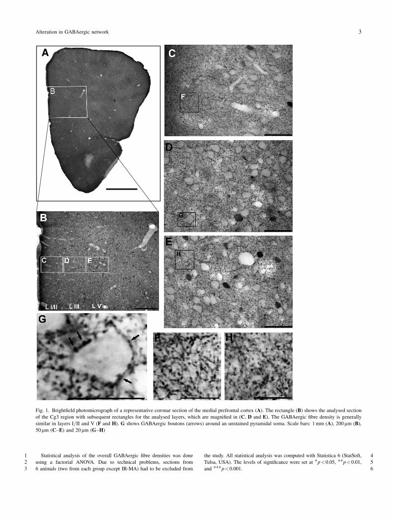
1 Statistical analysis of the overall GABAergic fibre densities was done
2 using a factorial ANOVA. Due to technical problems, sections from
3 6 animals (two from each group except IR-MA) had to be excluded from
4the study. All statistical analysis was computed with Statistica 6 (StatSoft,
5Tulsa, USA). The levels of significance were set at �p<0.05, ��p<0.01,
6and ���p<0.001.
Fig. 1. Brightfield photomicrograph of a representative coronar section of the medial prefrontal cortex (A). The rectangle (B) shows the analysed section
of the Cg3 region with subsequent rectangles for the analysed layers, which are magnified in (C, D and E). The GABAergic fibre density is generally
similar in layers I=II and V (F and H). G shows GABAergic boutons (arrows) around an unstained pyramidal soma. Scale bars: 1 mm (A), 200mm (B),
50 mm (C–E) and 20 mm (G–H)
Alteration in GABAergic network 3

1 Results
2 Qualitative results
3 The GABAergic innervation pattern is relatively homoge-
4 nous throughout the cortex of gerbils and is similar to rats
5 (Seto-Ohshima et al., 1990). It is characterised by a dense
6 fibre innervation in all cortical layers with the highest den-
7 sity in the molecular layer. We identified immunonegative
8 pyramidal neurons in layers III and V by their round or oval
9 shape, their size and orientation, and the presence of bas-
10 ket-like GABAergic innervation (Fig. 1G).
11 Quantitative results
12 GABAergic bouton densities
13 The 2-way ANCOVA revealed a highly significant effect of
14 rearing conditions on boutons in layer III [F(1,29)¼ 28.59,
15p<0.001] and layer V [F(1,29)¼ 25.58, p<0.001], and
16also a significant interaction between rearing and treat-
17ment in both layers [L III: F(1,29)¼ 6.35, p¼ 0.0175; L V:
18F(1,29)¼ 5.0806, p¼ 0.0319]. Post-hoc analysis with
19Newman-Keuls test showed the following results: Isolation
20rearing (IR) led to a significant decrease in GABAergic
21bouton density in layer III (�19%, p¼ 0.032), but not in
22layer V (p¼ 0.093). An early MA intoxication led to a
23further decrease in the bouton densities of both layers in
24IR-MA compared to IR-Sal animals (L III: �24%, p¼250.031; L V: �22%, p¼ 0.032). However, such an effect
26was not seen in animals from enriched rearing conditions
27(ER-MA vs. ER-Sal). Thus, bouton densities were reduced
28in the IR-MA group (L III: �38%, p<0.001; L V: �33%,
29p¼ 0.001) compared to ER-Sal animals (cf. Fig. 2A).
30GABAergic fibre densities
31A factorial ANOVA identified a significant interaction of
32treatment and rearing conditions in both layer V [F(1,30)¼3313.07, p¼ 0.001] and layers I=II [F(1,30)¼ 9.8844, p¼340.004]. Post-hoc Newman-Keuls tests revealed a significant
35increase in layer I=II fibre density of IR-MA animals (þ15,
36þ16, þ18%; all p<0.001) compared to IR-Sal, ER-Sal,
37and ER-MA animals, respectively. A similar increase in
38GABAergic fibre density was found in layer V (þ17 to
39þ19%) of the IR-MA group, compared to IR-Sal, ER-
40Sal, and ER-MA animals [all p<0.001, except ER-Sal:
41p¼ 0.0012 (cf. Fig. 2B)].
42Discussion
43We have demonstrated that a single early MA intoxi-
44cation combined with impoverished rearing (IR) signifi-
45cantly reduces the densitiy of GABAergic boutons that
46surround layers III and V pyramidal neurons in the pre-
47frontal cortex of the Mongolian gerbil, whereas the overall
48GABAergic fibre density in layers I=II and V is increased
49compared to control animals.
50Early MA intoxication and impoverished rearing
51as a model for schizophrenia
52The single early high dose of MA on PD 14 in combination
53with IR used in the current study is effective to disturb
54normal postnatal development of the dopaminergic system,
55by triggering a restraint of the maturation of dopamine
56fibres in the prefrontal cortex and the nucleus accumbens
57(Dawirs et al., 1994; Neddens et al., 2001, 2002) as well as
58a concomitant excessive maturation in several amygdaloid
Fig. 2. GABAergic bouton (A) and fibre densities (B) in the analysed layers
of the PFC of gerbils from enriched (ER) and impoverished rearing (IR)
conditions treated with either methamphetamine (MA) or saline given by
meansþ standard error (S.E.M.). �p<0.05, ��p<0.01, ���p<0.001
4 S. Brummelte et al.
1 nuclei and the entorhinal cortex (Busche et al., 2004). A
2 similar pattern of cortical-subcortical dopaminergic im-
3 balance has also been observed in the schizophrenic brain
4 (Laruelle et al., 2003; Abi-Dargham, 2004). Early MA
5 treatment additionally impairs PFC-related abilities and
6 behaviours, such as working memory and spatial learning
7 (Dawirs et al., 1996; Williams et al., 2002). Again, defi-
8 cits in working memory are well known characteristics of
9 schizophrenic patients (Goldman-Rakic, 1995; Lewis and
10 Anderson, 1995). Furthermore, the early drug challenge in
11 combination with IR leads to a miswiring of prefrontal ef-
12 ferents (Bagorda et al., 2006), in accordance with the dys-
13 connection hypothesis of schizophrenia (Weinberger and
14 Lipska, 1995).
15 Taken together, our approach using combined early MA
16 intoxication and IR leads to several morphological changes
17 in neuroanatomical brain networks and impairs cognitive
18 functions, resembling some of the changes and deficits seen
19 in schizophrenic individuals, and thus provides a potential
20 animal model of the disease. The present study reveals that
21 an early MA intoxication additionally decreases GABAer-
22 gic boutons that surround pyramidal cell somata, indicating
23 a loss of somatic synapses (Karube et al., 2004) and a
24 concomitant increase in overall GABAergic fibre density
25 in the medial prefrontal cortex. These findings raise the
26 possibility that the local prefrontal cortical inhibitory net-
27 work may be functionally disorganised.
28 The role of somatic inhibition
29 The distinct classes of GABAergic synapses play differential
30 roles in regulating the activity of pyramidal neurons. The
31 majority of GABAergic synapses terminate on dendrites or
32 spines of the postsynaptic cells (Beaulieu and Somogyi,
33 1990; Beaulieu et al., 1992; Nitsch and Riesenberg, 1995),
34 thus they are likely to control the efficacy and plasticity of
35 excitatory inputs onto the postsynaptic target (Miles et al.,
36 1996; Tamas et al., 1997, 2003). However, somatic inhibi-
37 tion is thought to be particularly effective in controlling the
38 output of pyramidal neurons and, importantly, has been
39 implicated to synchronize activity patterns of whole pyra-
40 midal populations (Miles et al., 1996; Tamas et al., 1997,
41 2000; Gibson et al., 1999; Freund, 2003; Klausberger et al.,
42 2003). Such oscillatory synchronization is further believed
43 to create the necessary temporal and spatial frame for pre-
44 frontal functions such as working memory (Constantinidis
45 et al., 2002; Lewis et al., 2005). In addition, cortical inter-
46 neurons, in particular ‘fast-spiking’ neurons, have been
47 shown to play an important role in shaping receptive fields
48 as well as spatial memory fields (Jones, 1993; Rao et al.,
491999, 2000). GABAergic somatic inhibition is thus excep-
50tionally essential for the maintenance of cortical and cog-
51nitive functions and one is tempted to suggest that a
52decrease in this type of GABAergic inhibition and the
53potential subsequent deficit in synchronization might con-
54tribute to reported working memory dysfunction in schizo-
55phrenia (Lewis et al., 2005) and our animal model (Dawirs
56et al., 1996). In fact, post-mortem studies of schizophrenic
57patients reveal fewer GABAergic synapses on cortical pyr-
58amidal cells (Blum and Mann, 2002) and in addition, recent
59neurophysiological studies have shown, that some cogni-
60tive dysfunctions in schizophrenia are associated with an
61abnormal neural synchronization (Spencer et al., 2003,
622004; Lee et al., 2003; Uhlhaas et al., 2006).
63The maturation and shift of GABAergic inhibition
64It is well documented that GABA exhibits depolarizing
65effects at early postnatal stages (Cherubini et al., 1991;
66Ganguly et al., 2001; Ben-Ari, 2002), due to an inverted
67electrochemical gradient for Cl� in neonatal neurons
68(Ben-Ari, 2002). The shift from an excitatory to an inhibi-
69tory transmitter is assumed to coincide with the first expres-
70sion of PV in cortical interneurons (Berger et al., 1999) and
71the calcium-binding protein is therefore considered a mar-
72ker of functional maturity of the neuron (Seto-Ohshima
73et al., 1990; Solbach and Celio, 1991). In the gerbil mPFC,
74the first PV-immunoreactive cells appear around PD 14
75(unpublished data), that is, at the time of the MA challenge.
76Interestingly, the maturation of GABAergic synapses in
77general proceeds until early adulthood (Huang et al., 1999;
78Morales et al., 2002; Chattopadhyaya et al., 2004; Lewis
79et al., 2005), and in that, every subpopulation of presyn-
80aptic terminals exhibits a particular developmental pattern
81(Lewis et al., 2005). Therefore, the ability to synchronize
82pyramidal cell activity is assumed to be in substantial flux
83until adulthood (Lewis et al., 2005). Although the prolifera-
84tion and formation of the typical perisomatic basket terminal
85seems to be a largely stereotypical process, it is additionally
86also dependent on neuronal activity within cortical circuits
87(Marty et al., 2000; Chattopadhyaya et al., 2004).
88GABAergic interneurons receive direct dopaminergic
89input (Goldman-Rakic et al., 1989; Verney et al., 1990;
90Benes et al., 1993), with D1 and D2 receptor types being
91most abundantly expressed by PV-neurons (Le Moine and
92Gaspar, 1998). Dopamine modulates cortical GABA cells;
93both inhibitory (Retaux et al., 1991) and excitatory (Gorelova
94et al., 2002) effects on fast-spiking interneurons have been
95reported. The omission of prefrontal dopaminergic affer-
96ent fibres by an early MA challenge (Dawirs et al., 1994;
Alteration in GABAergic network 5

1 Neddens et al., 2001) might therefore induce significant
2 alterations in the local GABAergic networks.
3 The dopaminergic afferents to the prefrontal cortex show
4 a prolonged maturation (Kalsbeek et al., 1988; Dawirs
5 et al., 1993; Rosenberg and Lewis, 1995) and continue to
6 form synapses on GABAergic interneurons until early adult-
7 hood (Benes et al., 1996b). Pyramidal neurons are also
8 directly innervated by dopaminergic terminals (Jay et al.,
9 1995; Davidoff and Benes, 1998) which demonstrates the
10 rather complex capacity of dopamine to directly and indi-
11 rectly regulate the firing pattern of pyramidal neurons. By
12 early MA intoxication we induce a restraint of the matura-
13 tion of prefrontal dopaminergic afferents, which triggers
14 reactive neuroplastic adaptation of the local network. Ana-
15 tomical data suggest that pyramidal cells may adapt by
16 increasing their dendritic range and their spine density
17 (Blaesing et al., 2001). Our current findings indicate an in-
18 crease of GABAergic fibre density, which is in line with
19 an earlier study using electron-microscopy that already
20 revealed an increase in non-somatic GABAergic terminals
21 (Nossoll et al., 1997). Therefore, we find it tempting to
22 suggest that an early MA challenge, by acutely reducing
23 the density of monoaminergic innervation of the PFC, might
24 trigger a reactive shift within the GABAergic networks
25 from somatic to dendritic pyramidal inhibition.
26 GABAergic dysfunction in schizophrenia
27 GABAergic dysfunction in schizophrenia has first been
28 proposed by Roberts (1972). Since then, several studies
29 have revealed disturbances of GABAergic networks in
30 schizophrenic patients (for review see Benes and Berretta,
31 2001) or in animal models of schizophrenia (Cochran et al.,
32 2002, 2003; Keilhoff et al., 2004; Reynolds et al., 2004;
33 Penschuck et al., 2006). A decline in PV-immunoreactive
34 structures, particularly in axon cartridges from chandelier
35 neurons, seems to be one of the most prevalent observa-
36 tions in post-mortem brains from schizophrenic individuals
37 (Woo et al., 1998; Pierri et al., 1999; Lewis et al., 1999).
38 Furthermore, the GABAA receptor density was found to be
39 upregulated at the axon initial segment (Volk et al., 2002)
40 as well as at the cell body of pyramidal neurons (Benes
41 et al., 1996a), possibly compensating for a reduction of in-
42 hibitory terminals from chandelier and basket cells (Lewis
43 et al., 2005). In contrast to the alterations in PV-containing
44 neurons, only few studies reported on changes in the sub-
45 population of CB- or CR-immunopositive cells. Iritani and
46 colleagues (1999) found a fibre disarray from CB-contain-
47 ing cells in the PFC, while Daviss and Lewis (1995) de-
48 scribed an increase in the density of CB cells but no change
49in the CR population in a post-mortem study on schizo-
50phrenic brains. This would also be in line with our findings,
51since an increased number of CB cells and an altered fibre
52pattern are likely to present an elevated GABAergic inhibi-
53tion of afferent pyramidal parts.
54Conclusion
55Here we present evidence for a probable dysfunctional
56reorganization of GABAergic networks in our potential
57animal model of schizophrenia. GABAergic interneurons
58critically contribute to the establishment of complex beha-
59viours by controlling and synchronizing the firing patterns
60of pyramidal neurons. A weakened or altered inhibition
61may give rise to a broad array of disturbances in cogni-
62tive function, like those seen in schizophrenia (Benes and
63Berretta, 2001). The current study indicates a potential shift
64from a strong and powerful somatic inhibition to dendritic
65inhibition, which might attenuate the GABAergic influence
66on pyramidal activity and thus lead to an uncontrolled
67firing or abnormal synchronization. Our data coincide with
68findings of a reduced GABAergic somatic innervation in
69individuals with schizophrenia. We suggest that, in our
70animal model, this change in the GABAergic network is
71secondary, being triggered by the primary impairment of
72monoaminergic and namely dopaminergic afferents. Further
73investigations of the separate subpopulations of GABAer-
74gic interneurons in the PFC of gerbils are in process to
75identify the responsible cell classes for the observed altera-
76tion in the GABAergic network.
77Acknowledgements
78The authors wish to thank F. Bagorda, U. Schroeder and S. Graum€uuller for
79technical assistance.
80References
81Abi-Dargham A (2004) Do we still believe in the dopamine hypothesis?
82New data bring new evidence. Int J Neuropsychopharmacol 7 Suppl 1:
83S1–S5
84Akil M, Pierri JN, Whitehead RE, Edgar CL, Mohila C, Sampson AR,
85Lewis DA (1999) Lamina-specific alterations in the dopamine in-
86nervation of the prefrontal cortex in schizophrenic subjects. Am
87J Psychiatry 156: 1580–1589
88Bagorda F, Teuchert-Noodt G, Lehmann K (2006) Isolation rearing or
89methamphetamine traumatisation induce a ‘‘dysconnection’’ of pre-
90frontal efferents in gerbils: implications for schizophrenia. J Neural
91Transm 113: 365–379
92Beaulieu C, Somogyi P (1990) Targets and quantitative distribution of
93GABAergic synapses in the visual cortex of the cat. Eur J Neurosci 2:
94296–303
6 S. Brummelte et al.

1 Beaulieu C, Kisvarday Z, Somogyi P, Cynader M, Cowey A (1992)
2 Quantitative distribution of GABA-immunopositive and -immunone-
3 gative neurons and synapses in the monkey striate cortex (area 17).
4 Cereb Cortex 2: 295–309
5 Ben-Ari Y (2002) Excitatory actions of gaba during development: the
6 nature of the nurture. Nat Rev Neurosci 3: 728–739
7 Benes FM, Berretta S (2001) GABAergic interneurons: implications for
8 understanding schizophrenia and bipolar disorder. Neuropsychophar-
9 macology 25: 1–27
10 Benes FM, Vincent SL, Molloy R (1993) Dopamine-immunoreactive
11 axon varicosities form nonrandom contacts with GABA-immu-
12 noreactive neurons of rat medial prefrontal cortex. Synapse 15:
13 285–295
14 Benes FM, Vincent SL, Marie A, Khan Y (1996a) Up-regulation of GABAA
15 receptor binding on neurons of the prefrontal cortex in schizophrenic
16 subjects. Neuroscience 75: 1021–1031
17 Benes FM, Vincent SL, Molloy R, Khan Y (1996b) Increased interaction
18 of dopamine-immunoreactive varicosities with GABA neurons of
19 rat medial prefrontal cortex occurs during the postweanling period.
20 Synapse 23: 237–245
21 Berger B, De GN, Alvarez C (1999) Precocious development of parvalbu-
22 min-like immunoreactive interneurons in the hippocampal formation
23 and entorhinal cortex of the fetal cynomolgus monkey. J Comp Neurol
24 403: 309–331
25 Blaesing B, Nossoll M, Teuchert-Noodt G, Dawirs RR (2001) Postnatal
26 maturation of prefrontal pyramidal neurones is sensitive to a single
27 early dose of methamphetamine in gerbils (Meriones unguiculatus).
28 J Neural Transm 108: 101–113
29 Blum BP, Mann JJ (2002) The GABAergic system in schizophrenia. Int
30 J Neuropsychopharmacol 5: 159–179
31 Busche A, Polascheck D, Lesting J, Neddens J, Teuchert-Noodt G (2004)
32 Developmentally induced imbalance of dopaminergic fibre densities in
33 limbic brain regions of gerbils (Meriones unguiculatus). J Neural
34 Transm 111: 451–463
35 Chattopadhyaya B, Di CG, Higashiyama H, Knott GW, Kuhlman SJ,
36 Welker E, Huang ZJ (2004) Experience and activity-depen-
37 dent maturation of perisomatic GABAergic innervation in primary
38 visual cortex during a postnatal critical period. J Neurosci 24:
39 9598–9611
40 Cherubini E, Gaiarsa JL, Ben-Ari Y (1991) GABA: an excitatory trans-
41 mitter in early postnatal life. Trends Neurosci 14: 515–519
42 Cochran SM, Fujimura M, Morris BJ, Pratt JA (2002) Acute and delayed
43 effects of phencyclidine upon mRNA levels of markers of glutamatergic
44 and GABAergic neurotransmitter function in the rat brain. Synapse 46:
45 206–214
46 Cochran SM, Kennedy M, McKerchar CE, Steward LJ, Pratt JA, Morris BJ
47 (2003) Induction of metabolic hypofunction and neurochemical def-
48 icits after chronic intermittent exposure to phencyclidine: differential
49 modulation by antipsychotic drugs. Neuropsychopharmacology 28:
50 265–275
51 Conde F, Lund JS, Jacobowitz DM, Baimbridge KG, Lewis DA (1994)
52 Local circuit neurons immunoreactive for calretinin, calbindin D-28k
53 or parvalbumin in monkey prefrontal cortex: distribution and mor-
54 phology. J Comp Neurol 341: 95–116
55 Constantinidis C, Williams GV, Goldman-Rakic PS (2002) A role for
56 inhibition in shaping the temporal flow of information in prefrontal
57 cortex. Nat Neurosci 5: 175–180
58 Davidoff SA, Benes FM (1998) High-resolution scatchard analysis shows
59 D1 receptor binding on pyramidal and nonpyramidal neurons. Synapse
60 28: 83–90
61 Daviss SR, Lewis DA (1995) Local circuit neurons of the prefrontal cortex
62 in schizophrenia: selective increase in the density of calbindin-immu-
63 noreactive neurons. Psychiatry Res 59: 81–96
64 Dawirs RR, Teuchert-Noodt G, Czaniera R (1993) Maturation of the
65 dopamine innervation during postnatal development of the prefrontal
66cortex in gerbils (Meriones unguiculatus). A quantitative immunocy-
67tochemical study. J Hirnforsch 34: 281–290
68Dawirs RR, Teuchert-Noodt G, Czaniera R (1994) The postnatal matura-
69tion of dopamine innervation in the prefrontal cortex of gerbils
70(Meriones unguiculatus) is sensitive to an early single dose of meth-
71amphetamine. A quantitative immunocytochemical study. J Hirn-
72forsch 35: 195–204
73Dawirs RR, Teuchert-Noodt G, Czaniera R (1996) Ontogeny of PFC-related
74behaviours is sensitive to a single non-invasive dose of methamphe-
75tamine in neonatal gerbils (Meriones unguiculatus). J Neural Transm
76103: 1235–1245
77DeFelipe J, Fairen A (1982) A type of basket cell in superficial layers of the
78cat visual cortex. A Golgi-electron microscope study. Brain Res 244:
799–16
80Feinberg I (1982) Schizophrenia: caused by a fault in programmed synaptic
81elimination during adolescence? J Psychiatr Res 17: 319–334
82Freund TF (2003) Interneuron diversity series: rhythm and mood in
83perisomatic inhibition. Trends Neurosci 26: 489–495
84Gabbott PL, Bacon SJ (1996) Local circuit neurons in the medial prefrontal
85cortex (areas 24a, b, c, 25 and 32) in the monkey: I. Cell morphology
86and morphometrics. J Comp Neurol 364: 567–608
87Ganguly K, Schinder AF, Wong ST, Poo M (2001) GABA itself promotes
88the developmental switch of neuronal GABAergic responses from
89excitation to inhibition. Cell 105: 521–532
90Gibson JR, Beierlein M, Connors BW (1999) Two networks of electrically
91coupled inhibitory neurons in neocortex. Nature 402: 75–79
92Goldman-Rakic PS (1995) Cellular basis of working memory. Neuron 14:
93477–485
94Goldman-Rakic PS, Leranth C, Williams SM, Mons N, Geffard M (1989)
95Dopamine synaptic complex with pyramidal neurons in primate
96cerebral cortex. Proc Natl Acad Sci USA 86: 9015–9019
97Gorelova N, Seamans JK, Yang CR (2002) Mechanisms of dopamine
98activation of fast-spiking interneurons that exert inhibition in rat
99prefrontal cortex. J Neurophysiol 88: 3150–3166
100Hendry SH, Houser CR, Jones EG, Vaughn JE (1983) Synaptic organization
101of immunocytochemically identified GABA neurons in the monkey
102sensory-motor cortex. J Neurocytol 12: 639–660
103Hendry SH, Jones EG, Emson PC, Lawson DE, Heizmann CW, Streit P
104(1989) Two classes of cortical GABA neurons defined by differen-
105tial calcium binding protein immunoreactivities. Exp Brain Res 76:
106467–472
107Houser CR, Hendry SH, Jones EG, Vaughn JE (1983) Morphological
108diversity of immunocytochemically identified GABA neurons in the
109monkey sensory-motor cortex. J Neurocytol 12: 617–638
110Huang ZJ, Kirkwood A, Pizzorusso T, Porciatti V, Morales B, Bear MF,
111Maffei L, Tonegawa S (1999) BDNF regulates the maturation of
112inhibition and the critical period of plasticity in mouse visual cortex.
113Cell 98: 739–755
114Iritani S, Kuroki N, Ikeda K, Kazamatsuri H (1999) Calbindin immunor-
115eactivity in the hippocampal formation and neocortex of schizophre-
116nics. Prog Neuropsychopharmacol Biol Psychiatry 23: 409–421
117Jay TM, Glowinski J, Thierry AM (1995) Inhibition of hippocampopre-
118frontal cortex excitatory responses by the mesocortical DA system.
119Neuroreport 6: 1845–1848
120Jones EG (1993) GABAergic neurons and their role in cortical plasticity in
121primates. Cereb Cortex 3: 361–372
122Kalsbeek A, Voorn P, Buijs RM, Pool CW, Uylings HB (1988) Develop-
123ment of the dopaminergic innervation in the prefrontal cortex of the rat.
124J Comp Neurol 269: 58–72
125Karube F, Kubota Y, Kawaguchi Y (2004) Axon branching and synaptic
126bouton phenotypes in GABAergic nonpyramidal cell subtypes.
127J Neurosci 24: 2853–2865
128Kawaguchi Y, Kondo S (2002) Parvalbumin, somatostatin and cholecys-
129tokinin as chemical markers for specific GABAergic interneuron types
130in the rat frontal cortex. J Neurocytol 31: 277–287
Alteration in GABAergic network 7

1 Kawaguchi Y, Kubota Y (1996) Physiological and morphological identi-
2 fication of somatostatin- or vasoactive intestinal polypeptide-con-
3 taining cells among GABAergic cell subtypes in rat frontal cortex.
4 J Neurosci 16: 2701–2715
5 Keilhoff G, Becker A, Grecksch G, Wolf G, Bernstein HG (2004) Repeated
6 application of ketamine to rats induces changes in the hippocampal
7 expression of parvalbumin, neuronal nitric oxide synthase and cFOS
8 similar to those found in human schizophrenia. Neuroscience 126:
9 591–598
10 Kisvarday ZF, Gulyas A, Beroukas D, North JB, Chubb IW, Somogyi P
11 (1990) Synapses, axonal and dendritic patterns of GABA-immunor-
12 eactive neurons in human cerebral cortex. Brain 113 (Pt 3): 793–812
13 Klausberger T, Magill PJ, Marton LF, Roberts JD, Cobden PM, Buzsaki G,
14 Somogyi P (2003) Brain-state- and cell-type-specific firing of hippo-
15 campal interneurons in vivo. Nature 421: 844–848
16 Laruelle M, Kegeles LS, bi-Dargham A (2003) Glutamate, dopamine, and
17 schizophrenia: from pathophysiology to treatment. Ann Acad Sci NY
18 1003: 138–158
19 Le Moine C, Gaspar P (1998) Subpopulations of cortical GABAergic
20 interneurons differ by their expression of D1 and D2 dopamine
21 receptor subtypes. Brain Res Mol Brain Res 58: 231–236
22 Lee KH, Williams LM, Breakspear M, Gordon E (2003) Synchronous
23 gamma activity: a review and contribution to an integrative neu-
24 roscience model of schizophrenia. Brain Res Brain Res Rev 41:
25 57–78
26 Lewis DA, Anderson SA (1995) The functional architecture of the pre-
27 frontal cortex and schizophrenia. Psychol Med 25: 887–894
28 Lewis DA, Pierri JN, Volk DW, Melchitzky DS, Woo TU (1999) Altered
29 GABA neurotransmission and prefrontal cortical dysfunction in
30 schizophrenia. Biol Psychiatry 46: 616–626
31 Lewis DA, Hashimoto T, Volk DW (2005) Cortical inhibitory neurons and
32 schizophrenia. Nat Rev Neurosci 6: 312–324
33 Marty S, Wehrle R, Sotelo C (2000) Neuronal activity and brain-derived
34 neurotrophic factor regulate the density of inhibitory synapses in
35 organotypic slice cultures of postnatal hippocampus. J Neurosci 20:
36 8087–8095
37 Miles R, Toth K, Gulyas AI, Hajos N, Freund TF (1996) Differences
38 between somatic and dendritic inhibition in the hippocampus. Neuron
39 16: 815–823
40 Morales B, Choi SY, Kirkwood A (2002) Dark rearing alters the develop-
41 ment of GABAergic transmission in visual cortex. J Neurosci 22:
42 8084–8090
43 Neddens J, Brandenburg K, Teuchert-Noodt G, Dawirs RR (2001) Differential
44 environment alters ontogeny of dopamine innervation of the orbital
45 prefrontal cortex in gerbils. J Neurosci Res 63: 209–213
46 Neddens J, Lesting J, Dawirs RR, Teuchert-Noodt G (2002) An early
47 methamphetamine challenge suppresses the maturation of dopamine
48 fibres in the nucleus accumbens of gerbils: on the significance of
49 rearing conditions. J Neural Transm 109: 141–155
50 Nitsch C, Riesenberg R (1995) Synaptic reorganisation in the rat stria-
51 tum after dopaminergic deafferentation: an ultrastructural study using
52 glutamate decarboxylase immunocytochemistry. Synapse 19: 247–263
53 Nossoll M, Teuchert-Noodt G, Dawirs RR (1997) A single dose of
54 methamphetamine in neonatal gerbils affects adult prefrontal gamma-
55 aminobutyric acid innervation. Eur J Pharmacol 340: R3–R5
56 Paxinos G, Watson C (1986) The rat brain in stereotaxic coordinates.
57 Academic Press, New York
58 Penschuck S, Flagstad P, Didriksen M, Leist M, Michael-Titus AT (2006)
59 Decrease in parvalbumin-expressing neurons in the hippocampus and
60 increased phencyclidine-induced locomotor activity in the rat methy-
61 lazoxymethanol (MAM) model of schizophrenia. Eur J Neurosci 23:
62 279–284
63 Pierri JN, Chaudry AS, Woo TU, Lewis DA (1999) Alterations in chandelier
64 neuron axon terminals in the prefrontal cortex of schizophrenic sub-
65 jects. Am J Psychiatry 156: 1709–1719
66Radnikow G, Feldmeyer D, Lubke J (2002) Axonal projection, input and
67output synapses, and synaptic physiology of Cajal-Retzius cells in the
68developing rat neocortex. J Neurosci 22: 6908–6919
69Rao SG, Williams GV, Goldman-Rakic PS (1999) Isodirectional tuning of
70adjacent interneurons and pyramidal cells during working memory:
71evidence for microcolumnar organization in PFC. J Neurophysiol 81:
721903–1916
73Rao SG, Williams GV, Goldman-Rakic PS (2000) Destruction and
74creation of spatial tuning by disinhibition: GABA(A) blockade of
75prefrontal cortical neurons engaged by working memory. J Neurosci
7620: 485–494
77Retaux S, Besson MJ, Penit-Soria J (1991) Synergism between D1
78and D2 dopamine receptors in the inhibition of the evoked re-
79lease of [3H]GABA in the rat prefrontal cortex. Neuroscience 43:
80323–329
81Reynolds GP, Abdul-Monim Z, Neill JC, Zhang ZJ (2004) Calcium binding
82protein markers of GABA deficits in schizophreniapostmortem studies
83and animal models. Neurotox Res 6: 57–61
84Ricaurte GA, Schuster CR, Seiden LS (1980) Long-term effects of
85repeated methylamphetamine administration on dopamine and ser-
86otonin neurons in the rat brain: a regional study. Brain Res 193:
87153–163
88Ricaurte GA, Guillery RW, Seiden LS, Schuster CR, Moore RY
89(1982) Dopamine nerve terminal degeneration produced by high
90doses of methylamphetamine in the rat brain. Brain Res 235:
9193–103
92Roberts E (1972) Prospects for research on schizophrenia. An hypotheses
93suggesting that there is a defect in the GABA system in schizophrenia.
94Neurosci Res Program Bull 10: 468–482
95Rosenberg DR, Lewis DA (1995) Postnatal maturation of the dopami-
96nergic innervation of monkey prefrontal and motor cortices: a ty-
97rosine hydroxylase immunohistochemical analysis. J Comp Neurol
98358: 383–400
99Rosenzweig MR, Bennett EL (1969) Effects of differential environments on
100brain weights and enzyme activities in gerbils, rats, and mice. Dev
101Psychobiol 2: 87–95
102Seto-Ohshima A, Aoki E, Semba R, Emson PC, Heizmann CW (1990)
103Appearance of parvalbumin-specific immunoreactivity in the cerebral
104cortex and hippocampus of the developing rat and gerbil brain. Histo-
105chemistry 94: 579–589
106Solbach S, Celio MR (1991) Ontogeny of the calcium binding protein
107parvalbumin in the rat nervous system. Anat Embryol (Berl) 184:
108103–124
109Somogyi P, Freund TF, Cowey A (1982) The axo-axonic interneuron in
110the cerebral cortex of the rat, cat and monkey. Neuroscience 7:
1112577–2607
112Spencer KM, Nestor PG, Niznikiewicz MA, Salisbury DF, Shenton ME,
113McCarley RW (2003) Abnormal neural synchrony in schizophrenia.
114J Neurosci 23: 7407–7411
115Spencer KM, Nestor PG, Perlmutter R, Niznikiewicz MA, Klump MC,
116Frumin M, Shenton ME, McCarley RW (2004) Neural synchrony
117indexes disordered perception and cognition in schizophrenia. Proc
118Natl Acad Sci USA 101: 17288–17293
119Tamas G, Buhl EH, Somogyi P (1997) Fast IPSPs elicited via multiple
120synaptic release sites by different types of GABAergic neurone in the
121cat visual cortex. J Physiol 500 (Pt 3): 715–738
122Tamas G, Buhl EH, Lorincz A, Somogyi P (2000) Proximally targeted
123GABAergic synapses and gap junctions synchronize cortical inter-
124neurons. Nat Neurosci 3: 366–371
125Tamas G, Lorincz A, Simon A, Szabadics J (2003) Identified sources
126and targets of slow inhibition in the neocortex. Science 299:
1271902–1905
128Thiessen DD, Yahr P (1977) The gerbil in behavioral investigations.
129Mechanisms of territoriality and olfactory communication. In: Uni-
130versity of Texas Press, Austin
8 S. Brummelte et al.
1 Uhlhaas PJ, Linden DE, Singer W, Haenschel C, Lindner M, Maurer K,
2 Rodriguez E (2006) Dysfunctional long-range coordination of neural
3 activity during Gestalt perception in schizophrenia. J Neurosci 26:
4 8168–8175
5 Valverde F (1998) Golgi atlas of the postnatal mouse brain. Springer, Wien,
6 New York
7 Verney C, Alvarez C, Geffard M, Berger B (1990) Ultrastructural
8 double-labelling study of dopamine terminals and GABA-contain-
9 ing neurons in rat anteromedial cerebral cortex. Eur J Neurosci
10 2: 960–972
11 Volk DW, Pierri JN, Fritschy JM, Auh S, Sampson AR, Lewis DA (2002)
12 Reciprocal alterations in pre- and postsynaptic inhibitory markers at
13 chandelier cell inputs to pyramidal neurons in schizophrenia. Cereb
14 Cortex 12: 1063–1070
15Weinberger DR, Lipska BK (1995) Cortical maldevelopment, anti-psycho-
16tic drugs, and schizophrenia: a search for common ground. Schizophr
17Res 16: 87–110
18Williams MT, Vorhees CV, Boon F, Saber AJ, Cain DP (2002) Metham-
19phetamine exposure from postnatal day 11 to 20 causes impairments in
20both behavioral strategies and spatial learning in adult rats. Brain Res
21958: 312–321
22Witte AV, Bagorda F, Teuchert-Noodt G, Lehmann K (2006) Contralateral
23prefrontal projections in gerbils mature abnormally after early
24methamphetamine trauma and isolated rearing. J Neural Transm. Epub
25ahead of print, DOI 10.1007=s00702-006-0506-6
26Woo TU, Whitehead RE, Melchitzky DS, Lewis DA (1998) A subclass of
27prefrontal gamma-aminobutyric acid axon terminals are selectively
28altered in schizophrenia. Proc Natl Acad Sci USA 95: 5341–5346
29
Alteration in GABAergic network 9

9.1 Further publications and posters Refereed Journals
Brummelte S., Teuchert-Noodt G., Grund T., Moll G.H. and Dawirs R.R: Environmental
enrichment has no effect on the development of dopaminergic and GABAergic fibres
during methylphenidate treatment of early traumatized gerbils. (in prep)
Witte A.V., Brummelte S. and Teuchert-Noodt G.: Pattern changes of prefrontal efferents in
the juvenile gerbil (Meriones unguiculatus).(submitted)
Grund T., Teuchert-Noodt G., Busche A., Neddens J., Brummelte S., Moll G.H. and Dawirs
R.R.: Oral Methylphenidate during prepuberty prevents pharmacologically-induced
(preweaning) suppressive development of dopamine projections into the prefrontal
cortex and amygdala. J Child Adolesc Psychopharm (revision submitted)
Brummelte S., Pawluski J.L. and. Galea L.A.M. (2006b): High postpartum levels of
corticosterone given to dams influence postnatal hippocampal cell proliferation and
behaviour of offspring: A model of post-partum stress and possible depression. Horm
Behav. 50(3):370-82
Poster and Presentations
Brummelte S., Pawluski J.L. and Galea L.A.M.: High post-partum levels of corticosterone
given to dams influence postnatal hippocampal cell proliferation and behavior of
offspring: A model of post-partum stress and possible depression. Focused Meeting
of the Physiological Society: New Developments in Stress physiology: From Gene to
Man, Bristol, UK, 2006, PC19

Brummelte S., Witte A.V., Graumueller S., and Teuchert-Noodt G.: Adaptive changes in
GABAergic innervation pattern of the prefrontal cortex in an animal model of
psychosis. FENS Abstr. Vol 3, A080.1, 2006
Witte A.V., Brummelte S., Bagorda F. and Teuchert-Noodt G.: Pattern changes of prefrontal
efferents in the neocortex of juvenile gerbils. FENS Abstr. Vol 3, A228.16, 2006
Brummelte S., Pawluski J.L. and. Galea L.A.M: A possible model of post-partum depression
based on high post-partum levels of corticosterone. SBN (Society for Behavioral
Neuroendocrinology) Annual meeting 2005, Abstr. 50.

10. Acknowledgements
Mein besonderer Dank gilt Frau Prof. Dr. Dr. Teuchert-Noodt, die mir diese Promotion in der
Abteilung für Neuroanatomie ermöglichte und sich immer für mich eingesetzt. Ihre durchgehende
intensive Betreuung und konstruktive Kritik auch noch nach ihrer Pensionierung waren eine
bedeutende Hilfe bei der Erstellung dieser Arbeit.
Auch Herrn Prof. York Winter und seinen Mitarbeitern gilt mein Dank für die freundliche Aufnahme
in die Arbeitsgruppe und die hilfreiche Unterstützung.
Außerdem möchte ich mich herzlich bei Herrn Prof. Egelhaaf, für seinen persönlichen Einsatz
bezüglich meiner erneuten Aufnahme in die Studienstiftung und für die Übernahme der formellen
Betreuung der Doktorarbeit bedanken.
Des Weiteren möchte ich mich bei der gesamten Arbeitsgruppe Neuroanatomie für die gute
Zusammenarbeit, die Unterstützung und ein tolles Arbeitsklima bedanken. Besonders erwähnt seien
hier Frau Ulrike Schroeder für ihre tatkräftige Hilfe im Labor und ihr offenes Ohr, Herr Dr. Francesco
Bagorda für seine Unterstützung bei den Versuchen und dafür, dass er immer da war, wenn man ihn
brauchte, und last but not least Frau Dr. Andrea Busche und Herr Dr. Jörg Neddens, die mir trotz z.T.
großer geographischer Distanzen immer mit Rat und Tat zur Seite standen. Darüber hinaus gilt auch
Herrn Prof. Dawirs mein persönlicher Dank für die partielle finanzielle Unterstützung und die
Kooperation bei den Ritalin-Studien.
Ebenso möchte ich mich bei Frau Dipl. Biol. Veronica Witte bedanken für ihre allgegenwärtige
Motivation und den gegenseitigen Austausch.
I’d further like to thank Dr. Liisa Galea and the Laboratory of Behavioural Neuroscience at the UBC
in Vancouver, Canada for a great project and collaboration and a really nice and successful 7-months
stay as a visiting researcher.
Außerdem möchte ich mich bei der Studienstiftung des deutschen Volkes und der
Graduiertenförderung der Universität Bielefeld für die ideelle und finanzielle Förderung bedanken,
ohne die diese Promotion nicht möglich gewesen wäre.
Mein tiefster Dank gilt meiner Familie und besonders meinen Eltern, die mich auf meinem bisherigen
Weg immer begleitet und nach besten Kräften unterstützt haben.

Eidesstattliche Erklärung Hiermit erkläre ich, dass ich diese Arbeit selbstständig erstellt und nur die angegebenen
Hilfsmittel und Quellen verwendet habe. Weiterhin erkläre ich, dass es sich um meinen ersten
Promotionsversuch handelt.
Bielefeld, Januar 2007
____________________________ S u s a n n e B r u m m e l t e