Anatomical Evidence of Acupuncture Meridians in the Human ...
8
Research Article Anatomical Evidence of Acupuncture Meridians in the Human Extracellular Matrix: Results from a Macroscopic and Microscopic Interdisciplinary Multicentre Study on Human Corpses Norbert Maurer , 1 Helmut Nissel, 2 Monika Egerbacher, 3 Erich Gornik , 4 Patrick Schuller, 4 and Hannes Traxler 5 1 Johannes Bischko Institut f¨ ur Akupunktur/Neurologisches Zentrum am Krankenhaus Rosenh¨ ugel Wien, 1130 Wien, Riedelgasse 5, Private Practice for General Medicine, Kupelwiesergasse 16, 1130 Wien, Austria 2 Johannes Bischko Institut f¨ ur Akupunktur/Neurologisches Zentrum am Krankenhaus Rosenh¨ ugel Wien, 1130 Wien, Riedelgasse 5, Private Practice for Internal Medicine, Schleifgasse 7, 1210 Wien, Austria 3 Institute of Histology and Embryology, Department of Pathobiology, University of Veterinary Medicine, Veterin¨ arplatz 1, 1210 Vienna, Austria 4 Institute for Solid State Electronics, Vienna University of Technology, Gußhausstraße 25-25a (Geb¨ aude CH), 1040 Wien, Austria 5 Center for Anatomy and Cell Biology, Division of Anatomy, Medical University of Vienna, W¨ ahringer Straße 13, 1090 Wien, Austria Correspondence should be addressed to Hannes Traxler; [email protected] Received 12 October 2018; Revised 22 January 2019; Accepted 26 February 2019; Published 21 March 2019 Academic Editor: Gerhard Litscher Copyright © 2019 Norbert Maurer et al. is is an open access article distributed under the Creative Commons Attribution License, which permits unrestricted use, distribution, and reproduction in any medium, provided the original work is properly cited. For more than 2500 years, acupuncture has been applied to support the healing of different diseases and physiologic malfunctions. Although various theories of the meridian system and mechanisms were formulated to explain the functional basis of acupuncture, the anatomical basis for the concept of meridians has not been resolved. e aim of the present study was to search for replicable anatomical structures that could relate to meridians. To this end, four human specimens and additionally two lower legs were dissected anatomically. Our study found evidence that acupuncture meridians were part of the human extracellular matrix and that fascia was an important part of the anatomic substrate of acupuncture meridians. At the same time, we found vessel-nerve- bundles, which were hypothesized to account for 80% of acupuncture points, only in a few acupuncture points. erefore, our findings contradict the theory that acupuncture points are only located along the nervous channels. 1. Introduction As part of traditional Chinese medicine (TCM), acupuncture has been an energetic and vibrant treatment with a successful application for more than 2500 years [1, 2]. Acupuncture is an ancient aspect of TCM with demonstrated therapeutic effects [2]. By acting (by needles, laser, moxa, pressure, etc.) on certain areas on the surface of the skin, functional disorders can be corrected, and pain can be reduced. Such areas are defined as acupuncture points. In TCM, meridians are strings connecting acupuncture points, which are considered as passageways through which energy flows throughout the body [1, 2]. e meridian system is composed of 12 principal meridians, each of which connects to an organ system and extends to an extremity and eight collaterals [1–4]. Acupuncture treatments should improve the flow of energy through the meridian network [5– 10]. e morphological basis for the concept of meridians in TCM has not been resolved. Recent articles support a relationship between acupuncture points/meridians and fascia [1]. Specifically, anatomical observations of body scan data demonstrated that the fascia network resembles the the- oretical meridian system in salient ways, and physiological, Hindawi Evidence-Based Complementary and Alternative Medicine Volume 2019, Article ID 6976892, 8 pages https://doi.org/10.1155/2019/6976892
Transcript of Anatomical Evidence of Acupuncture Meridians in the Human ...
Research ArticleAnatomical Evidence of Acupuncture Meridians inthe Human Extracellular Matrix: Results from a Macroscopicand Microscopic Interdisciplinary Multicentre Study onHuman Corpses
Norbert Maurer ,1 Helmut Nissel,2 Monika Egerbacher,3 Erich Gornik ,4
Patrick Schuller,4 and Hannes Traxler 5
1 Johannes Bischko Institut fur Akupunktur/Neurologisches Zentrum am Krankenhaus Rosenhugel Wien, 1130 Wien,Riedelgasse 5, Private Practice for General Medicine, Kupelwiesergasse 16, 1130 Wien, Austria
2Johannes Bischko Institut fur Akupunktur/Neurologisches Zentrum am Krankenhaus Rosenhugel Wien, 1130 Wien,Riedelgasse 5, Private Practice for Internal Medicine, Schleifgasse 7, 1210 Wien, Austria
3Institute of Histology and Embryology, Department of Pathobiology, University of Veterinary Medicine,Veterinarplatz 1, 1210 Vienna, Austria
4Institute for Solid State Electronics, Vienna University of Technology, Gußhausstraße 25-25a (Gebaude CH), 1040 Wien, Austria5Center for Anatomy and Cell Biology, Division of Anatomy, Medical University of Vienna, Wahringer Straße 13, 1090Wien, Austria
Correspondence should be addressed to Hannes Traxler; [email protected]
Received 12 October 2018; Revised 22 January 2019; Accepted 26 February 2019; Published 21 March 2019
Academic Editor: Gerhard Litscher
Copyright © 2019 NorbertMaurer et al.This is an open access article distributed under the Creative Commons Attribution License,which permits unrestricted use, distribution, and reproduction in any medium, provided the original work is properly cited.
For more than 2500 years, acupuncture has been applied to support the healing of different diseases and physiologic malfunctions.Although various theories of the meridian system andmechanisms were formulated to explain the functional basis of acupuncture,the anatomical basis for the concept of meridians has not been resolved. The aim of the present study was to search for replicableanatomical structures that could relate to meridians. To this end, four human specimens and additionally two lower legs weredissected anatomically. Our study found evidence that acupuncture meridians were part of the human extracellular matrix andthat fascia was an important part of the anatomic substrate of acupuncture meridians. At the same time, we found vessel-nerve-bundles, which were hypothesized to account for 80% of acupuncture points, only in a few acupuncture points. Therefore, ourfindings contradict the theory that acupuncture points are only located along the nervous channels.
1. Introduction
As part of traditional Chinese medicine (TCM), acupuncturehas been an energetic and vibrant treatment with a successfulapplication formore than 2500 years [1, 2]. Acupuncture is anancient aspect of TCMwith demonstrated therapeutic effects[2]. By acting (by needles, laser, moxa, pressure, etc.) oncertain areas on the surface of the skin, functional disorderscan be corrected, and pain can be reduced. Such areas aredefined as acupuncture points.
In TCM, meridians are strings connecting acupuncturepoints, which are considered as passageways through which
energy flows throughout the body [1, 2]. The meridiansystem is composed of 12 principal meridians, each of whichconnects to an organ system and extends to an extremityand eight collaterals [1–4]. Acupuncture treatments shouldimprove the flow of energy through themeridian network [5–10].
The morphological basis for the concept of meridiansin TCM has not been resolved. Recent articles supporta relationship between acupuncture points/meridians andfascia [1]. Specifically, anatomical observations of body scandata demonstrated that the fascia network resembles the the-oretical meridian system in salient ways, and physiological,
HindawiEvidence-Based Complementary and Alternative MedicineVolume 2019, Article ID 6976892, 8 pageshttps://doi.org/10.1155/2019/6976892

2 Evidence-Based Complementary and Alternative Medicine
Figure 1: VNB distal to the medial side of the left lower leg.
histological, and clinical observations support this hypothesis[11, 12].
The aim of the present study was to clarify whether therewas macro- and microanatomical substrate of acupuncturemeridians.
2. Materials and Methods
Four specimens and additionally two lower legs were dis-sected at the Medical University of Vienna/Division ofAnatomy and Cell Biology.
Our aim was to depict vascular nerve bundles of indi-vidual acupuncture points in the course of the associatedmeridian. A vascular nerve bundle (VNB) was defined as acombination of nerves, arteries, veins, and lymphatics in thebody that travelled together.
After the initial incision, the skin and subcutis wereremoved with the help of a scalpel until the superficial fasciawas found. Perforating vascular nerve bundles were carefullyseparated from the subcutaneous adipose tissue. The bursa,such as Bursa patellaris and olecrani, was extirpated. Due tothe lack of a superficial fascia in the facial area, the parotidfascia and the facial muscles were defined as the superficiallayer.
Since, in most cases, no bundles of vascular nervebundles were found at the supposed acupuncture points,we determined the acupuncture point lege artis on thesection preparation and dissected until the panniculus fascia(fascia superficialis corporis). The sites were documentedphotographically.
The collected tissue samples were fixed in formalin andembedded in paraffin for histological section production.Sections were stained with haematoxylin & eosin (HE) andstained for neuronal expression by immunohistochemicaldetection of S-100 (DAKO, Glostrup, DK, dilution 1:1200,detection substrate DAB).
3. Results
The main results from our anatomical and morphologicalinvestigations can be found in the photographs from Figures
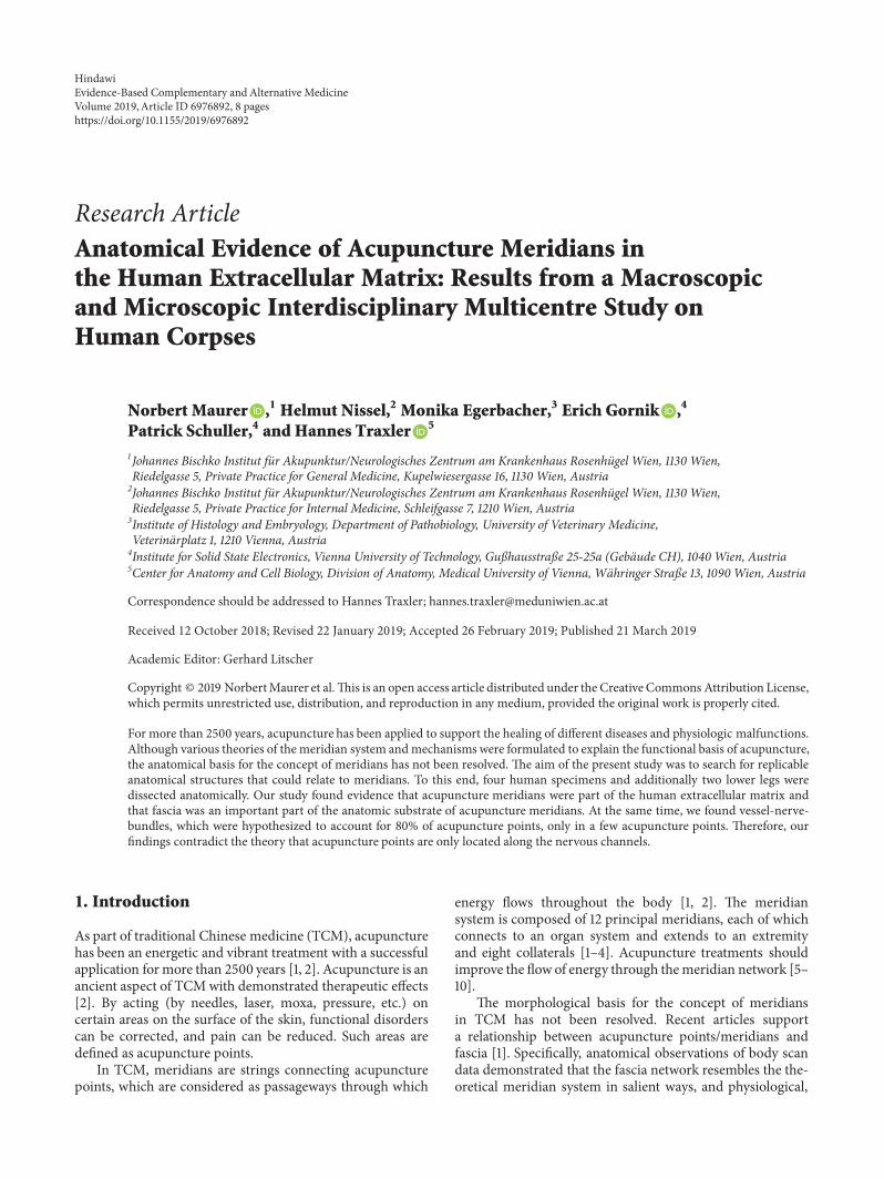
1–13. In Figure 1, diverse vascular nerve bundles (VNBs)distal to the medial side of the left lower leg are depicted.In Figure 2, we can recognize a VNB at the acupuncturepoint and, proximal of it, we can see another VNB on theleft dorsum, but without an acupuncture point nearby. InFigure 3, we see a VNB without a reference to a knownacupuncture point; the next known acupuncture point liesdistal to the VNB and dorsal to the lateral malleolus sinistra.
References of the detected VNBs to the anatomy of theacupuncture meridian can be established with the help of theinvestigations of Dr. Heine [13, 14]. Vascular nerve bundlesare defined according to Heine as a structural principle ofthe acupuncture point. It is a perforating structure consistingof a vegetative nerve, artery, and vein, surrounded by looseconnective tissue.
In Figure 4, the fascia course of the stomach meridiancan be seen. In Figures 5(a) and 5(b), we can perceive thetypical course of the stomach meridian in “Z-form” ST 36-ST 40 on the left (a) and right (b) lower leg. Figure 6represents a preparatory presentation of the fascia of thestomach meridian (superficial fascia cristis sinistra).
In Figure 7, we can discern the fascia course of thegallbladder meridian between GB 35 and GB 36
In Figure 8 we can see the fascia course of the smallintestine meridian SI 10 to SI 11 at the fascia above the rightscapula. In Figure 9, we can discern the fascia course ofthe large intestine meridian LI 10 to LI 11 on the right armlaterally.
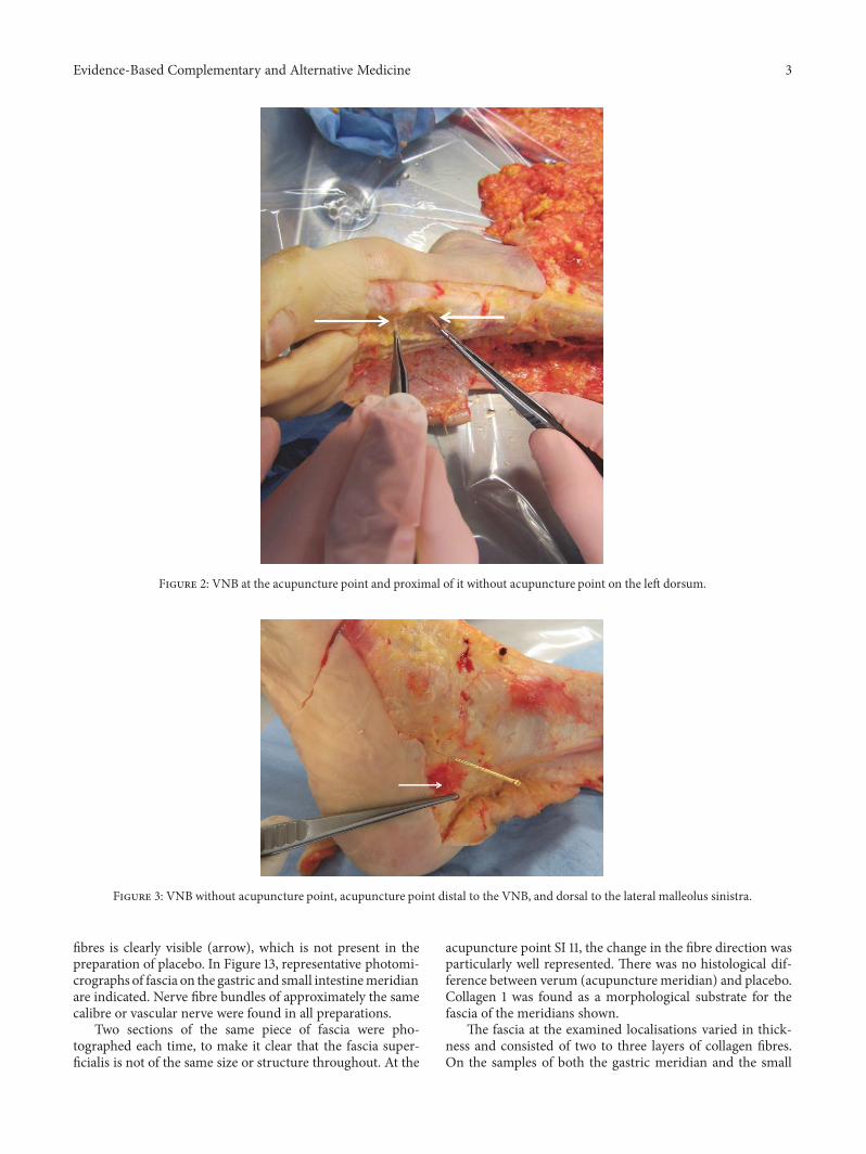
Figure 10 shows that the structures of the external fasciacorporis cross the course of the gastric meridian at rightangles at the thigh. The fascia lata would correspond to thecourse of the gallbladder meridian.
Figure 11 shows that at the forefoot no courses of the fasciacorporis externa corresponding to the gastric meridian canbe found. The aponeuroses tendinum extensorum digitorumpedis follows in one of its parts the course of the gastricmeridian.
In Figure 12, representative microphotographs of fasciaon the gastric and small intestine meridian are summarised.In the verum, the change in the direction of the collagen
Evidence-Based Complementary and Alternative Medicine 3
Figure 2: VNB at the acupuncture point and proximal of it without acupuncture point on the left dorsum.
Figure 3: VNB without acupuncture point, acupuncture point distal to the VNB, and dorsal to the lateral malleolus sinistra.
fibres is clearly visible (arrow), which is not present in thepreparation of placebo. In Figure 13, representative photomi-crographs of fascia on the gastric and small intestinemeridianare indicated. Nerve fibre bundles of approximately the samecalibre or vascular nerve were found in all preparations.
Two sections of the same piece of fascia were pho-tographed each time, to make it clear that the fascia super-ficialis is not of the same size or structure throughout. At the
acupuncture point SI 11, the change in the fibre direction wasparticularly well represented. There was no histological dif-ference between verum (acupuncture meridian) and placebo.Collagen 1 was found as a morphological substrate for thefascia of the meridians shown.
The fascia at the examined localisations varied in thick-ness and consisted of two to three layers of collagen fibres.On the samples of both the gastric meridian and the small

4 Evidence-Based Complementary and Alternative Medicine
Figure 4: Fascia course of the stomach meridian.
(a) Typical course of the stomach meridian in “Z-form” ST 36-ST 40 on theleft lower leg
(b) Typical course of the stomach meridian in “Z-form” ST 36-ST 40 on theright lower leg
Figure 5
Figure 6: Preparatory presentation of the fascia of the stomach meridian (superficial fascia cristis sinistra).

Evidence-Based Complementary and Alternative Medicine 5
Figure 7: Fascia course of the gallbladder meridian between GB 35 and GB 36.
Figure 8: Fascia course of the small intestine meridian within the region of SI 10 to SI 11 at the fascia above the right scapula.
Figure 9: Fascia course of the large intestine meridian LI 10 to LI 11 on the right arm laterally.
6 Evidence-Based Complementary and Alternative Medicine
Figure 10: Fibre course of the fascia corporis externa on the right thigh. The fibres run at right angles to the course of the stomachmeridian.The fascia lata corresponds to the course of the gall bladder meridian.
Figure 11: In the forefoot area, there are no structures of the fascia corporis externa that would correspond to a meridian course. Striking isthe tendons of the aponeuroses tendinum extensorum digitorum pedis, which in parts follow the gastric meridian.
intestine meridian, it was possible to show histologically thechange of the fibre course in a connective tissue layer.Thiswasnot the case with the placebo preparation. In addition, therewas no histological difference between verum (acupuncturemeridian) and placebo. In all preparations, smaller nervefibre bundles or the perivascular plexus could be detected bystaining with S-100. There were no noticeable differences inthe density or location of nerve fibres between the verum andthe placebo.
We dissected four specimens and additionally two lowerlegs, in total 10 lower legs. On each shank, ten acupuncturepoints (5 of the stomach and 5 of the gall bladder meridian)were evaluated. Only in two of the 100 evaluated acupuncturepoints, we found VNBs, corresponding to 2% of cases.
4. Discussion
Thereby, we established a close connection of acupuncturepoints with structures of connective tissue. Heine has found
that, in 80% of acupuncture points, a bundle of vascularnerves of soft connective tissue passes through fascia holesto the skin. The same anatomical structures were also foundby Egerbacher in cattle and dogs, without specifying apercentage of the found VNBs per dissected acupuncturepoints [15].
By finding fascia fractions of the human extracellularmatrix with a fibre run as the course of the acupuncturemeridian, we suggest that the anatomical substrate of theacupuncture meridian is the fascia superficialis corporisof the human extracellular matrix as it was suggested inscientific works [16–18]. At the same time, we found thatparts of muscles, tendons, and ligaments follow the meridiancourse (bladder meridian; large intestine meridian).
The histology showed that, between verum (acupuncturemeridian) and placebo, there is no detectable difference. Thefibre folding in the meridian progression could be detectedmacroscopically and microscopically. However, after fixationwith formaldehyde 80%, the proteins of the tissue were

Evidence-Based Complementary and Alternative Medicine 7
Gastric meridian ST 36 – ST 38
Small intestine meridian SI 11
Gastric meridian Placebo
Small intestine meridianPlacebo
HE HE
Figure 12: Representative microphotographs of fascia on the gastric and small intestine meridian. In the verum (left column), the change inthe direction of the collagen fibres is clearly visible (arrow); this is not present in the preparation of placebo (right column).
Gastric meridian ST 36 – ST 38 Gastric meridian Placebo
Small intestine meridian SI 11Small intestine meridian
Placebo
Figure 13: Representative photomicrograph of fascia on the gastric and small intestine meridian. Nerve fibre bundles of approximately thesame calibre or vascular nerve were found in all preparations. Immunohistochemical staining with an antibody against S-100.
8 Evidence-Based Complementary and Alternative Medicine
denaturated. Therefore, further investigations on unfixedtissue samples will be performed in future projects.
In addition, we could not represent fascia of an entireacupuncture meridian. One of the reasons for this could bethat, in our preparations, the dissection of tissue adhesionwas not easy to perform. At the same time we found nocorresponding fibre courses of the fascia superficialis corporison the thigh and the forefoot, which would correspond to ameridian course. We therefore hypothesize that other partsof the fascia, but also tendon courses, anatomically depict themeridian course.
Our study clearly supports the view that the humanbody’sfascia network may be the physical substrate represented bythe meridians of TCM [1]. Specifically, this hypothesis is sup-ported by our anatomical, morphological, and histologicalobservations made in human corpses.
5. Conclusions
We suggest that not only fascia, especially the fascia corporisexterna, but also deeper parts form the anatomical substrateof acupuncture meridians. In addition, parts of muscles,tendons, and ligaments follow the meridian course. Ourobservations build an anatomical basis for examining TCMprinciples and therapies, and it supports a holistic approachto diagnosis and treatment of diverse diseases.
Since we found VNBs in just a few of the acupuncturepoints and as we found VNBs even without an acupuncturepoint, we are no longer convinced that the sole concept ofthe function of the acupuncture system over neural reflexesis valid.
Data Availability
The data used to support the findings of this study areavailable from the corresponding author upon request.
Disclosure
Norbert Maurer and Helmut Nissel shared first authorship.
Conflicts of Interest
The authors declare that they have no conflicts of interest.
Acknowledgments
The authors wish to express their gratitude referring toIstvan Paraszti who died 2018 for his help with their study.They also want to express their thankfulness for the privatefinancial support to Dr. Burkhard Gantenbein and to Dr.Florian Koschat. Their particular thanks go to Mag. BarbaraKretschmerMAwhohelped themwith her computer literacy.She worked for them even if she was sick and twenty-four-seven if necessary. In addition, the authors want to thankthe photographers Edi Cvillink and Thomas Posch for theirexcellent work.
References
[1] Y. Bai, J. Wang, J.-p. Wu et al., “Review of evidence suggestingthat the fascia network could be the anatomical basis foracupoints and meridians in the human body,” Evidence-BasedComplementary and Alternative Medicine, vol. 2011, Article ID260510, 6 pages, 2011.
[2] S. Xutian, J. Zhang, andW. Louise, “New exploration andunder-standing of traditional Chinese medicine,” American Journal ofChinese Medicine, vol. 37, no. 3, pp. 411–426, 2009.
[3] S. Chang, “The meridian system and mechanism ofacupuncture—a comparative review. Part 1: the meridiansystem,” Taiwanese Journal of Obstetrics & Gynecology, vol. 51,no. 4, pp. 506–514, 2012.
[4] S. Chang, “The meridian system and mechanism ofacupuncture—a comparative review. Part 2. Mechanismof acupuncture analgesia,” Taiwanese Journal of Obstetrics andGynecology, vol. 52, no. 1, pp. 14–24, 2013.
[5] A. Bensoussan, “Part 1: Acupuncture meridians - myth orreality?” Complementary Therapies in Medicine, vol. 2, no. 1, pp.21–26, 1994.
[6] W.-W. Chan, K.-Y. Chen, H. Liu, L.-S. Wu, and J.-H. Lin,“Acupuncture for General Veterinary Practice,” Journal of Vet-erinary Medical Science, vol. 63, no. 10, pp. 1057–1062, 2001.
[7] H. Haltrecht, “Veterinary acupuncture,” Canadian VeterinaryJournal, vol. 40, no. 6, pp. 401–403, 1999.
[8] S. Scott, “Developments in veterinary acupuncture,” Acupunc-ture in Medicine, vol. 19, no. 1, pp. 27–31, 2001.
[9] W.G. Vockeroth, “Veterinaryhomeopathy: an overview,”Cana-dian Veterinary Journal, vol. 40, no. 8, pp. 592–594, 1999.
[10] C. Yu, K. Zhang, G. Lu et al., “Characteristics of acupuncturemeridians and acupoints in animals,” Revue Scientifique etTechnique de l’OIE, vol. 13, no. 3, pp. 927–933, 1994.
[11] J. Kim and D.-I. Kang, “Positioning standardized acupuncturepoints on the whole body based on X-ray computed tomogra-phy images,”Medical Acupuncture, vol. 26, no. 1, pp. 40–49, 2014.
[12] D.-L. Li, W.-B. Zhang, S.-T. Fu et al., “Wave character and quan-tum character of acupuncture systems,” International Journal ofModelling, Identification and Control, vol. 5, no. 3, pp. 229–235,2008.
[13] H. Heine, “Anatomische Struktur der Akupunkturpunkte,”Deutsche Zeitschrift fur Akupunktur, vol. 31, pp. 26–30, 1988.
[14] H. Heine, “Akupunkturtherapie. Perforationen der oberflach-lichen Korperfaszie durch kutane Gefaß-Nervenbundel,”Ther-apeutikon, vol. 4, pp. 238–244, 1988.
[15] M. Egerbacher, “Veterinarakupunktur, Anatomische und his-tologische Struktur ausgewahlter Akupunkturpunkte bei Rindund Schwein,” Deutsche Zeitschrift fur Akupunktur, vol. 36, no.4, 1993.
[16] D. Zhang, W. Yao, G. Ding et al., “A fluid mechanics modelof tissue fluid flow in limb connective tissue—a mechanismof acupuncture signal transmission,” Journal of Hydrodynamics,vol. 21, no. 5, pp. 675–684, 2009.
[17] H. M. Langevin, D. L. Churchill, andM. J. Cipolla, “Mechanicalsignaling through connective tissue: a mechanism for thetherapeutic effect of acupuncture,”The FASEB Journal, vol. 15,no. 12, pp. 2275–2282, 2001.
[18] X. Yu, G. Ding, H. Huang, J. Lin, W. Yao, and R. Zhan, “Roleof collagen fibers in acupuncture analgesia therapy on rats,”Connective Tissue Research, vol. 50, no. 2, pp. 110–120, 2009.


![Evaluation and Comparison of Anatomical Landmark Detection …hamarneh/ecopy/tmi2015b.pdf · 2015-05-04 · ization/identification of cephalometric landmarks [18]–[20]. In 2006,](https://static.fdokument.com/doc/165x107/5ea4823bfa548e7f9520b73b/evaluation-and-comparison-of-anatomical-landmark-detection-hamarnehecopy-2015-05-04.jpg)