Atomic resolution snapshot of Leishmania ribosome ...
22
ARTICLE Atomic resolution snapshot of Leishmania ribosome inhibition by the aminoglycoside paromomycin Moran Shalev-Benami 1,2 , Yan Zhang 2 , Haim Rozenberg 1 , Yuko Nobe 3 , Masato Taoka 3 , Donna Matzov 1 , Ella Zimmerman 1 , Anat Bashan 1 , Toshiaki Isobe 3 , Charles L. Jaffe 4 , Ada Yonath 1 & Georgios Skiniotis 2 Leishmania is a single-celled eukaryotic parasite afflicting millions of humans worldwide, with current therapies limited to a poor selection of drugs that mostly target elements in the parasite’s cell envelope. Here we determined the atomic resolution electron cryo-microscopy (cryo-EM) structure of the Leishmania ribosome in complex with paromomycin (PAR), a highly potent compound recently approved for treatment of the fatal visceral leishmaniasis (VL). The structure reveals the mechanism by which the drug induces its deleterious effects on the parasite. We further show that PAR interferes with several aspects of cytosolic translation, thus highlighting the cytosolic rather than the mitochondrial ribosome as the primary drug target. The results also highlight unique as well as conserved elements in the PAR-binding pocket that can serve as hotspots for the development of novel therapeutics. DOI: 10.1038/s41467-017-01664-4 OPEN 1 Faculty of Chemistry, Department of Structural Biology, Weizmann Institute of Science, Rehovot 761001, Israel. 2 Departments of Molecular and Cellular Physiology, and Structural Biology, Stanford University School of Medicine, Stanford, CA 94305, USA. 3 Graduate School of Science and Engineering, Tokyo Metropolitan University, Hachioji-shi, Tokyo 192-0397, Japan. 4 Department of Microbiology and Molecular Genetics, IMRIC, Hebrew University-Hadassah Medical School, Jerusalem 91120, Israel. Moran Shalev-Benami and Yan Zhang contributed equally to this work. Correspondence and requests for materials should be addressed to M.S.-B. (email: [email protected]) or to A.Y. (email: [email protected]) or to G.S. (email: [email protected]) NATURE COMMUNICATIONS | 8: 1589 | DOI: 10.1038/s41467-017-01664-4 | www.nature.com/naturecommunications 1 1234567890
Transcript of Atomic resolution snapshot of Leishmania ribosome ...
ARTICLE
Atomic resolution snapshot of Leishmania ribosomeinhibition by the aminoglycoside paromomycinMoran Shalev-Benami1,2, Yan Zhang2, Haim Rozenberg1, Yuko Nobe3, Masato Taoka3, Donna Matzov1,
Ella Zimmerman1, Anat Bashan1, Toshiaki Isobe3, Charles L. Jaffe4, Ada Yonath1 & Georgios Skiniotis 2
Leishmania is a single-celled eukaryotic parasite afflicting millions of humans worldwide, with
current therapies limited to a poor selection of drugs that mostly target elements in the
parasite’s cell envelope. Here we determined the atomic resolution electron cryo-microscopy
(cryo-EM) structure of the Leishmania ribosome in complex with paromomycin (PAR),
a highly potent compound recently approved for treatment of the fatal visceral leishmaniasis
(VL). The structure reveals the mechanism by which the drug induces its deleterious effects
on the parasite. We further show that PAR interferes with several aspects of cytosolic
translation, thus highlighting the cytosolic rather than the mitochondrial ribosome as the
primary drug target. The results also highlight unique as well as conserved elements in the
PAR-binding pocket that can serve as hotspots for the development of novel therapeutics.
DOI: 10.1038/s41467-017-01664-4 OPEN
1 Faculty of Chemistry, Department of Structural Biology, Weizmann Institute of Science, Rehovot 761001, Israel. 2 Departments of Molecular and CellularPhysiology, and Structural Biology, Stanford University School of Medicine, Stanford, CA 94305, USA. 3 Graduate School of Science and Engineering, TokyoMetropolitan University, Hachioji-shi, Tokyo 192-0397, Japan. 4Department of Microbiology and Molecular Genetics, IMRIC, Hebrew University-HadassahMedical School, Jerusalem 91120, Israel. Moran Shalev-Benami and Yan Zhang contributed equally to this work. Correspondence and requests for materialsshould be addressed to M.S.-B. (email: [email protected]) or to A.Y. (email: [email protected]) or to G.S. (email: [email protected])
NATURE COMMUNICATIONS |8: 1589 |DOI: 10.1038/s41467-017-01664-4 |www.nature.com/naturecommunications 1
1234
5678
90
Leishmaniasis, a parasitic disease transmitted by the bite ofinfected sand flies, afflicts over 12 million people in nearly100 endemic countries1. The disease is caused by more than
20 pathogenic species of eukaryotic microbes that parasitizemacrophage populations residing in the skin, mucus or visceralorgans. These infections result in a relatively large array of clinicalmanifestations defined as cutaneous-, mucosal-, or visceralleishmaniasis, respectively1. Currently, there is no vaccine againstleishmaniasis and the therapeutic arsenal to treat the disease isrestricted by both the limited drugs available and the emergenceof parasite resistance mechanisms1.
Paromomycin (PAR), a natural aminoglycoside (AG) producedby Streptomyces riomosus, was recently approved for the treat-ment of visceral leishmaniasis (VL), a fatal form of leishmanialinfection2. PAR is a highly potent antibacterial agent known toconfer a broad spectrum of activities against both prokaryotic andeukaryotic microbes3. PAR mechanisms of action in bacteria arewell documented and are mainly attributed to interfering with thebacterial translation apparatus4. By contrast, very little is knownabout PAR actions on eukaryotic microbes, where it has beenproposed to interfere with mitochondrial translation5.
Here we employed a combination of structural and biochem-ical approaches to study PAR activities against the parasiteLeishmania donovani (L. donovani), the main cause of VL6. Ourbiochemical results, showing a strong correlation between inter-ference with cytosolic ribosome translation and inhibition ofparasite growth, suggest the cytosolic rather than mitochondrialribosome as the main drug target. Cryo-EM analysis of the fullyactive cytosolic Leishmania ribosome in complex with PARenabled us to obtain the structure at atomic resolution, therebyproviding a detailed snapshot of the drug’s binding pocket andthe ribosome decoding center. The results reveal unique elementswithin the pocket that contribute to drug binding in eukaryoticmicrobes, and coupled to biochemical and in silico experimentshighlight additional members of the AG family as potentialinhibitors of leishmanial cytosolic translation.
ResultsAGs interfere with the leishmanial cytosolic translation.Although AGs are extensively used as anti-parasitic agents, theidentity of their target in eukaryotic microorganisms has beenunclear. Based on the similarity of key elements shared betweenthe AG-binding pocket in bacteria and the corresponding site inthe mitochondrial ribosome, namely the presence of an adenineresidue at position 1408 (Supplementary Fig. 1), the mitochon-drial translation machinery has been considered to be the maintarget for these compounds in eukaryotic parasites5. However,recent structural studies demonstrating that AGs can bind tosynthetic RNA constructs mimicking their putative binding site
in cytosolic ribosomes have raised the possibility that the leish-manial cytosolic ribosome might also be the target of AGs inLeishmania7–9. Given the lack of direct correlation between theinhibition of cytosolic or mitochondrial translation and parasitegrowth, the main target of AGs in the parasitic cell is yet to bedetermined.
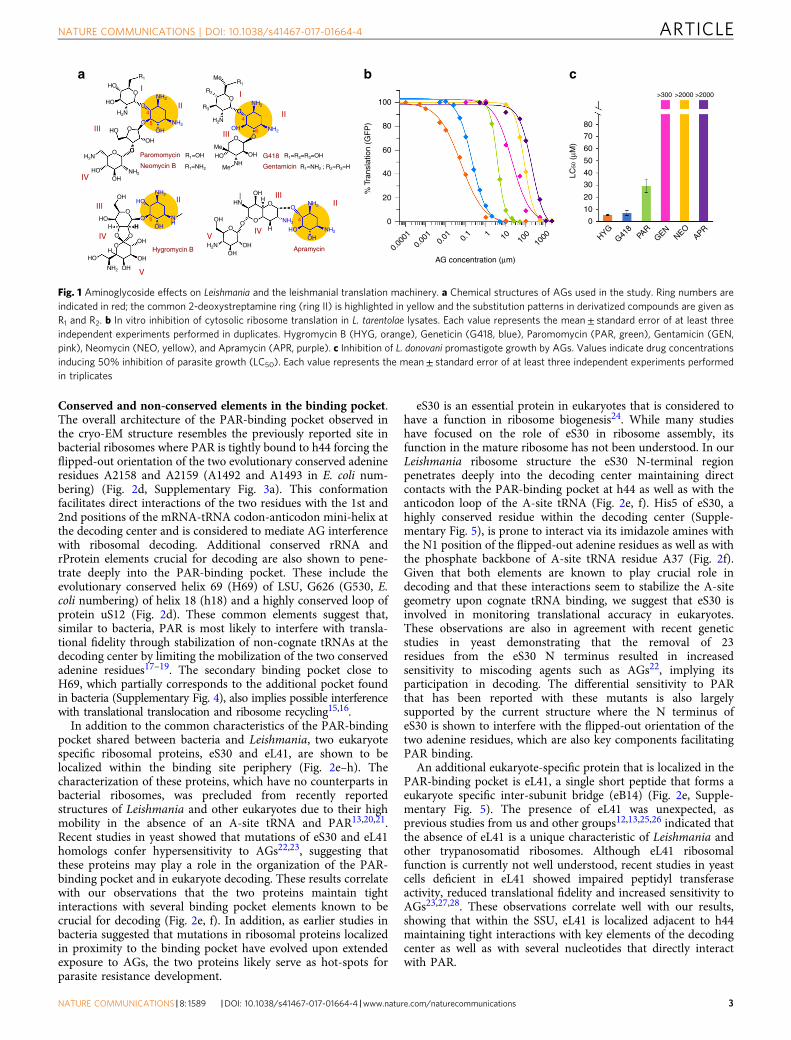
In order to assess whether AGs target the cytosolic ribosome inLeishmania, we measured the interference of six structurallydiverged AGs (Fig. 1a) with leishmanial cytosolic ribosometranslation and their effect on parasite growth. The selectedderivatives were predicted to have differential selectivity foreukaryote cytosolic translation based on previous observations inhigher eukaryotes10. The in vitro inhibition pattern obtained forthe leishmanial cytosolic ribosome shows a diverse range ofinduced activities (Fig. 1b) with 4 orders of magnitudedifferentiating between the most potent compound, hygromycinB (HYG), and the least active derivative, apramycin (APR)(calculated IC50 values are 0.065, 0.15, 3.62, 18.4, 82.4, and 200.6μM for HYG, G418, PAR, gentamicin (GEN), neomycin B (NEO)and APR, respectively; Table 1). The inhibition of parasite growthwas strikingly correlated with the in vitro assay results (Fig. 1c),indicating the strong involvement of cytosolic ribosome inhibi-tion by AGs with the induced anti-parasitic effects.
PAR binds the decoding center of the leishmanial ribosome. Todelineate the PAR mechanism of action in Leishmania weemployed cryo-EM to obtain the structure of intact cytosolicribosomes derived from L. donovani in complex with the com-pound (Fig. 2). The nominal map resolutions for SSU and LSUwere 2.7 and 2.5 Å, respectively (Supplementary Fig. 2). Impor-tantly, the resolution of the map extended to 2.2 Å in the coreregions, enabling us to build and refine an atomic model for theentire assembly including programed mRNA that was anchoredto the ribosome via the leishmanial kozak sequence11 as well asthree tRNA molecules bound at their designated A-, P-, and E-positions (Fig. 2, Supplementary Tables 1 and 2). A well-defineddensity that could accommodate the PAR four-ringed structurewas found at the tip of SSU h44 at the ribosome decoding center(Fig. 2) in a binding pocket that corresponds to the AG primarybinding site in bacterial ribosomes (Fig. 2, Supplementary Fig. 3).The high resolution of the map enabled the complete character-ization of all four PAR sugar rings including the directionality ofring substituents, thus facilitating a full description of PARinteraction pattern within the Leishmania binding pocket (Fig. 2c,Supplementary Fig. 3). The structure reveals shared as well asdistinct structural elements of PAR binding to leishmanial ribo-somes compared to bacteria (Fig. 2d–h). The delineation of theseelements was precluded from earlier X-ray studies performed inminimal RNA-binding pocket constructs9 because they reflectonly a minimum environment required for drug binding and notthe entire chemical milieu of the binding pocket. Differencemapping calculated between the current and the earlier reportedstructures of the L. donovani vacant ribosome12,13 also revealedthe presence of additional PAR molecules that were foundattached to several locations of both the large and small ribosomalsubunits (Supplementary Fig. 4). Multiple secondary binding siteswere also previously reported in X-ray structures of AGs bound tobacterial ribosomes14–16; as these were primarily localized toribosomal loci with no presumable ribosomal activity, they aremostly regarded as non-specific interactions between the posi-tively charged amino-moieties and the rRNA phosphate back-bone. Notably, one PAR molecule was found to bind betweenH45 of the SSU and H69 of LSU, partially overlapping with theAG secondary binding pocket previously reported in bacterialribosomes (Supplementary Fig. 4)15,16.
Table 1 In vitro inhibition of translation measured inL. tarentolaea, E. colib, and rabbitc reticulocyte lysates
Inhibition of translation—IC50 (μM)
Leishmaniaa Bacteriab Reticulocytesc
G418 0.153± 0.0356 0.009± 0.00159 2± 0.39
GEN 18.41± 3.139 0.028± 0.004 62± 9PAR 3.62± 0.176 0.051± 0.0059 57± 49
NEO 82.36± 2.139 0.011± 0.001 28± 5APR 200.6± 16.12 0.049± 0.003 29± 6HYG 0.0647± 0.0053 0.011± 0.002 0.8± 0.1
Each value represents the mean± SE of at least three independent repeatsG418, geneticin; GEN, gentamicin; PAR, paromomycin; NEO, neomycin; APR, apramycin; HYG,hygromycin B
ARTICLE NATURE COMMUNICATIONS | DOI: 10.1038/s41467-017-01664-4
2 NATURE COMMUNICATIONS |8: 1589 |DOI: 10.1038/s41467-017-01664-4 |www.nature.com/naturecommunications
Conserved and non-conserved elements in the binding pocket.The overall architecture of the PAR-binding pocket observed inthe cryo-EM structure resembles the previously reported site inbacterial ribosomes where PAR is tightly bound to h44 forcing theflipped-out orientation of the two evolutionary conserved adenineresidues A2158 and A2159 (A1492 and A1493 in E. coli num-bering) (Fig. 2d, Supplementary Fig. 3a). This conformationfacilitates direct interactions of the two residues with the 1st and2nd positions of the mRNA-tRNA codon-anticodon mini-helix atthe decoding center and is considered to mediate AG interferencewith ribosomal decoding. Additional conserved rRNA andrProtein elements crucial for decoding are also shown to pene-trate deeply into the PAR-binding pocket. These include theevolutionary conserved helix 69 (H69) of LSU, G626 (G530, E.coli numbering) of helix 18 (h18) and a highly conserved loop ofprotein uS12 (Fig. 2d). These common elements suggest that,similar to bacteria, PAR is most likely to interfere with transla-tional fidelity through stabilization of non-cognate tRNAs at thedecoding center by limiting the mobilization of the two conservedadenine residues17–19. The secondary binding pocket close toH69, which partially corresponds to the additional pocket foundin bacteria (Supplementary Fig. 4), also implies possible interferencewith translational translocation and ribosome recycling15,16.
In addition to the common characteristics of the PAR-bindingpocket shared between bacteria and Leishmania, two eukaryotespecific ribosomal proteins, eS30 and eL41, are shown to belocalized within the binding site periphery (Fig. 2e–h). Thecharacterization of these proteins, which have no counterparts inbacterial ribosomes, was precluded from recently reportedstructures of Leishmania and other eukaryotes due to their highmobility in the absence of an A-site tRNA and PAR13,20,21.Recent studies in yeast showed that mutations of eS30 and eL41homologs confer hypersensitivity to AGs22,23, suggesting thatthese proteins may play a role in the organization of the PAR-binding pocket and in eukaryote decoding. These results correlatewith our observations that the two proteins maintain tightinteractions with several binding pocket elements known to becrucial for decoding (Fig. 2e, f). In addition, as earlier studies inbacteria suggested that mutations in ribosomal proteins localizedin proximity to the binding pocket have evolved upon extendedexposure to AGs, the two proteins likely serve as hot-spots forparasite resistance development.
eS30 is an essential protein in eukaryotes that is considered tohave a function in ribosome biogenesis24. While many studieshave focused on the role of eS30 in ribosome assembly, itsfunction in the mature ribosome has not been understood. In ourLeishmania ribosome structure the eS30 N-terminal regionpenetrates deeply into the decoding center maintaining directcontacts with the PAR-binding pocket at h44 as well as with theanticodon loop of the A-site tRNA (Fig. 2e, f). His5 of eS30, ahighly conserved residue within the decoding center (Supple-mentary Fig. 5), is prone to interact via its imidazole amines withthe N1 position of the flipped-out adenine residues as well as withthe phosphate backbone of A-site tRNA residue A37 (Fig. 2f).Given that both elements are known to play crucial role indecoding and that these interactions seem to stabilize the A-sitegeometry upon cognate tRNA binding, we suggest that eS30 isinvolved in monitoring translational accuracy in eukaryotes.These observations are also in agreement with recent geneticstudies in yeast demonstrating that the removal of 23residues from the eS30 N terminus resulted in increasedsensitivity to miscoding agents such as AGs22, implying itsparticipation in decoding. The differential sensitivity to PARthat has been reported with these mutants is also largelysupported by the current structure where the N terminus ofeS30 is shown to interfere with the flipped-out orientation of thetwo adenine residues, which are also key components facilitatingPAR binding.
An additional eukaryote-specific protein that is localized in thePAR-binding pocket is eL41, a single short peptide that forms aeukaryote specific inter-subunit bridge (eB14) (Fig. 2e, Supple-mentary Fig. 5). The presence of eL41 was unexpected, asprevious studies from us and other groups12,13,25,26 indicated thatthe absence of eL41 is a unique characteristic of Leishmania andother trypanosomatid ribosomes. Although eL41 ribosomalfunction is currently not well understood, recent studies in yeastcells deficient in eL41 showed impaired peptidyl transferaseactivity, reduced translational fidelity and increased sensitivity toAGs23,27,28. These observations correlate well with our results,showing that within the SSU, eL41 is localized adjacent to h44maintaining tight interactions with key elements of the decodingcenter as well as with several nucleotides that directly interactwith PAR.
HO
HO
HO
HO
HO
OH
OH
OH
OHHHN
O
O
HO
O
OHOH
HOH H
OH
OH
OHNH
H2NH2N
H2N
H2N
NH2
NH2
NH2
NH2
NH2NH2
NH2
O
NH2
NH2
NH2
O
HOOH
O
OH
HONH
OH
Me
Me
O
O
O
OO
OO
OO
OH
OH
OH
OH
HO
O
44
4
65
5
R1a b cR2
R1=OH R1=R2=R3=OH
R1=NH2 ; R2=R3=HR1=NH2
R3
R1Me
O
O
I
II
IV
III
IV
V
II
VIV
IIIII
II
100
80 80
>300 >2000 >2000
70
60
50
40
30
20
10
0
60
LC50
(μM
)
40
20
0
HYGG41
8PA
RGEN
NEOAPR
0.00
010.
001
0.01
AG concentration (μm)
% T
rans
latio
n (G
FP
)
0.1 1 10 10
010
00
III
I
Hygromycin B
Paromomycin
Neomycin B
G418
Gentamicin
Apramycin
III
Fig. 1 Aminoglycoside effects on Leishmania and the leishmanial translation machinery. a Chemical structures of AGs used in the study. Ring numbers areindicated in red; the common 2-deoxystreptamine ring (ring II) is highlighted in yellow and the substitution patterns in derivatized compounds are given asR1 and R2. b In vitro inhibition of cytosolic ribosome translation in L. tarentolae lysates. Each value represents the mean± standard error of at least threeindependent experiments performed in duplicates. Hygromycin B (HYG, orange), Geneticin (G418, blue), Paromomycin (PAR, green), Gentamicin (GEN,pink), Neomycin (NEO, yellow), and Apramycin (APR, purple). c Inhibition of L. donovani promastigote growth by AGs. Values indicate drug concentrationsinducing 50% inhibition of parasite growth (LC50). Each value represents the mean± standard error of at least three independent experiments performedin triplicates
NATURE COMMUNICATIONS | DOI: 10.1038/s41467-017-01664-4 ARTICLE
NATURE COMMUNICATIONS |8: 1589 |DOI: 10.1038/s41467-017-01664-4 |www.nature.com/naturecommunications 3
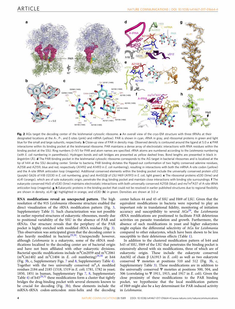
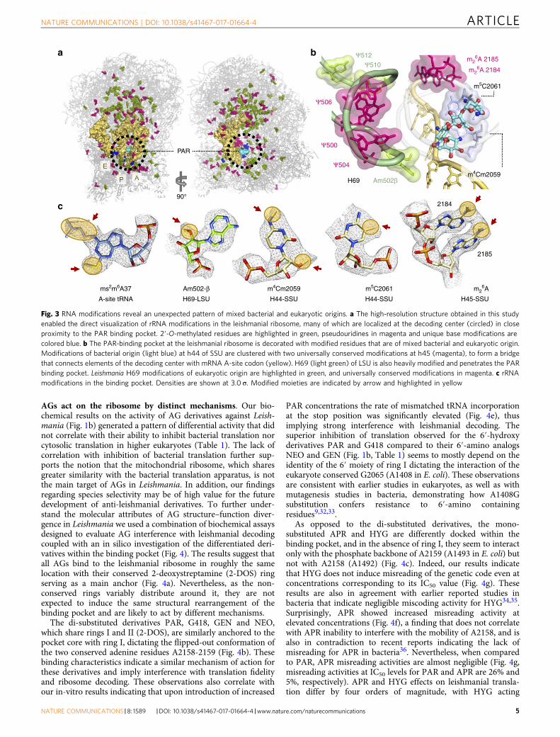
RNA modifications reveal an unexpected pattern. The highresolution of the 91S Leishmania ribosome structure enabled thedirect visualization of the rRNA modification pattern (Fig. 3,Supplementary Table 3). Such characterization was not possiblein earlier reported structures of eukaryotic ribosomes, mostly dueto positional variability of the SSU in the absence of PAR andtRNAs. Our structure reveals that the periphery of the PARpocket is highly enriched with modified rRNA residues (Fig. 3).This observation was anticipated given that the decoding center isalso heavily modified in bacteria29,30. Unexpectedly however,although Leishmania is a eukaryote, some of the rRNA mod-ifications localized to the decoding center are of bacterial originand have not been affiliated with other eukaryotic divisions.Bacterial specific modifications include m4Cm2059 and m5C2061(m4Cm1402 and m5C1404 in E. coli numbering)29,30 at h44(Fig. 3b, c, Supplementary Figs. 7 and 8, Supplementary Table 4).Together with the two universally conserved m6
2A modifiedresidues 2184 and 2185 (1518, 1519 in E. coli; 1781, 1782 in yeast;1850, 1851 in human, Supplementary Figs 7, 8, SupplementaryTable 4) of h4529,31 these modifications form a cluster that tightlybridges the drug-binding pocket with several elements known tobe crucial for decoding (Fig. 3b); these elements include themRNA-tRNA codon-anticodon mini-helix and the decoding
center helices 44 and 45 of SSU and H69 of LSU. Given that theequivalent modifications in bacteria were reported to play animportant role in translational fidelity, monitoring of initiationaccuracy and susceptibility to several AGs29, the LeishmaniarRNA modifications are positioned to facilitate PAR deleteriousactivities on parasite translation and growth. Furthermore, theabsence of such modifications in yeast and higher eukaryotesmight explain the differential selectivity of AGs for Leishmaniacompared to other eukaryotes, which have been shown to be lesssusceptible to their deleterious effects (Table 1).
In addition to the clustered modification pattern of h44 andh45 of SSU, H69 of the LSU that penetrates the binding pocket isextensively altered with six modifications, three of which are ofeukaryotic origin. These include the eukaryote conservedAm502 of chain β (A1913 in E. coli) as well as two eukaryoteconserved Ψ moieties at positions 510 and 512 (Fig. 3b, c,Supplementary Table 3). These modifications are in addition tothe universally conserved Ψ moieties at positions 500, 504, and506 (correlating to Ψ 1911, 1915, and 1917 in E. coli). Given theclose proximity of these modifications to the PAR bindingpocket, we hypothesize that the local modification patternof H69 might also be a key determinant for PAR-induced activityin Leishmania.
ms2m6A37
His5
eS30
eL41A2158
A2158
A2158
A2159
A2159
A2159
Am502
G626
H69
PAR
PAR
eS30
uS12
A-tRNA
A-tRNA
a
b c
d
e
f
g
h
PAR
His5
eS30
eL41
PAR
mRNA
mRNA
h18
G2063 (1405)
90°
� = 5.0
C2064 (1407)G2062 (1405)OP2
O4 U2161 (1495)
32.5
N4
OP2
OP1
2′′′
6′′′
2′′
5′′3′
6′
4′
3′′′
6′
5′′
4′′′ 6′′′
3.2
2.9
3.2
A2156 (1490) A2156 (1490)
A2157 (1491) A2158 (1492)
A2159 (1493)
A2159 (1493)
G2065(1408)
N2N1
A2159 (1493)
G2160 (1494)
4.0 OP2
OP1OP2
OP13.2N7
3.1
3.9
4.0 3.5
1
3.1
3.5
N7
N7
3.2
3.4
IV
III
III
II
I
Fig. 2 AGs target the decoding center of the leishmanial cytosolic ribosome. a An overall view of the cryo-EM structure with three tRNAs at theirdesignated locations at the A-, P-, and E-sites (pink) and mRNA (yellow). PAR is shown in cyan, rRNA in gray, and ribosomal proteins in green and lightblue for the small and large subunits, respectively. b Close-up view of PAR in density map. Observed density is contoured around the ligand at 5.0 σ. c PARinteractome within its binding pocket at the leishmanial ribosome. PAR maintains a dense array of electrostatic interactions with RNA residues within thebinding pocket at the SSU. Ring numbers (I–IV) for PAR and atom names are specified. rRNA atoms are numbered according to the Leishmania numbering(with E. coli numbering in parenthesis). Hydrogen bonds and salt bridges are presented as yellow dashed lines. Bond lengths are presented in black inångström (Å). d The PAR-binding pocket in the leishmanial cytosolic ribosome corresponds to the AG target in bacterial ribosomes and is localized at thetip of h44 at the SSU decoding center. Similar to bacteria, PAR binding dictates the flipped-out conformation of two highly conserved adenine residues,A2158 and A2159, blue and red, respectively (A1492 and A1493 in E. coli numbering), resulting in interactions with both the mRNA A-site codon (yellow)and the A-site tRNA anticodon loop (magenta). Additional conserved elements within the binding pocket include the universally conserved protein uS12(purple) G626 of h18 (G530 in E. coli numbering, gray) and Am502β of LSU H69 (A1913 in E. coli, light green). e The ribosomal proteins eS30 (lime) andeL41 (orange), which are of sole eukaryotic origin, penetrate the drug binding pocket and maintain close interactions with binding site surroundings. f Theeukaryote conserved His5 of eS30 (lime) maintains electrostatic interactions with both universally conserved A2158 (blue) and ms2m6A37 of A-site tRNAanticodon loop (magenta). g, h Eukaryotic proteins in the binding pocket that could not be resolved in earlier published structures due to regional flexibilityare shown in density. eL41 (g) highlighted in orange, and eS30 (h) in green. Densities are shown at 3.0 σ
ARTICLE NATURE COMMUNICATIONS | DOI: 10.1038/s41467-017-01664-4
4 NATURE COMMUNICATIONS |8: 1589 |DOI: 10.1038/s41467-017-01664-4 |www.nature.com/naturecommunications
AGs act on the ribosome by distinct mechanisms. Our bio-chemical results on the activity of AG derivatives against Leish-mania (Fig. 1b) generated a pattern of differential activity that didnot correlate with their ability to inhibit bacterial translation norcytosolic translation in higher eukaryotes (Table 1). The lack ofcorrelation with inhibition of bacterial translation further sup-ports the notion that the mitochondrial ribosome, which sharesgreater similarity with the bacterial translation apparatus, is notthe main target of AGs in Leishmania. In addition, our findingsregarding species selectivity may be of high value for the futuredevelopment of anti-leishmanial derivatives. To further under-stand the molecular attributes of AG structure–function diver-gence in Leishmania we used a combination of biochemical assaysdesigned to evaluate AG interference with leishmanial decodingcoupled with an in silico investigation of the differentiated deri-vatives within the binding pocket (Fig. 4). The results suggest thatall AGs bind to the leishmanial ribosome in roughly the samelocation with their conserved 2-deoxystreptamine (2-DOS) ringserving as a main anchor (Fig. 4a). Nevertheless, as the non-conserved rings variably distribute around it, they are notexpected to induce the same structural rearrangement of thebinding pocket and are likely to act by different mechanisms.
The di-substituted derivatives PAR, G418, GEN and NEO,which share rings I and II (2-DOS), are similarly anchored to thepocket core with ring I, dictating the flipped-out conformation ofthe two conserved adenine residues A2158-2159 (Fig. 4b). Thesebinding characteristics indicate a similar mechanism of action forthese derivatives and imply interference with translation fidelityand ribosome decoding. These observations also correlate withour in-vitro results indicating that upon introduction of increased
PAR concentrations the rate of mismatched tRNA incorporationat the stop position was significantly elevated (Fig. 4e), thusimplying strong interference with leishmanial decoding. Thesuperior inhibition of translation observed for the 6′-hydroxyderivatives PAR and G418 compared to their 6′-amino analogsNEO and GEN (Fig. 1b, Table 1) seems to mostly depend on theidentity of the 6′ moiety of ring I dictating the interaction of theeukaryote conserved G2065 (A1408 in E. coli). These observationsare consistent with earlier studies in eukaryotes, as well as withmutagenesis studies in bacteria, demonstrating how A1408Gsubstitution confers resistance to 6′-amino containingresidues9,32,33.
As opposed to the di-substituted derivatives, the mono-substituted APR and HYG are differently docked within thebinding pocket, and in the absence of ring I, they seem to interactonly with the phosphate backbone of A2159 (A1493 in E. coli) butnot with A2158 (A1492) (Fig. 4c). Indeed, our results indicatethat HYG does not induce misreading of the genetic code even atconcentrations corresponding to its IC50 value (Fig. 4g). Theseresults are also in agreement with earlier reported studies inbacteria that indicate negligible miscoding activity for HYG34,35.Surprisingly, APR showed increased misreading activity atelevated concentrations (Fig. 4f), a finding that does not correlatewith APR inability to interfere with the mobility of A2158, and isalso in contradiction to recent reports indicating the lack ofmisreading for APR in bacteria36. Nevertheless, when comparedto PAR, APR misreading activities are almost negligible (Fig. 4g,misreading activities at IC50 levels for PAR and APR are 26% and5%, respectively). APR and HYG effects on leishmanial transla-tion differ by four orders of magnitude, with HYG acting
Am502β
Ψ512Ψ510
m4Cm2059
2184
2185
Ψ506
Ψ500
Ψ504
PAR
H69
m5C2061
a b
c90°
m26A 2185
m26A 2184
m26A
H45-SSU
m5C2061
H44-SSU
m4Cm2059
H44-SSU
Am502-βH69-LSU
ms2m6A37
A-site tRNA
Fig. 3 RNA modifications reveal an unexpected pattern of mixed bacterial and eukaryotic origins. a The high-resolution structure obtained in this studyenabled the direct visualization of rRNA modifications in the leishmanial ribosome, many of which are localized at the decoding center (circled) in closeproximity to the PAR binding pocket. 2′-O-methylated residues are highlighted in green, pseudouridines in magenta and unique base modifications arecolored blue. b The PAR-binding pocket at the leishmanial ribosome is decorated with modified residues that are of mixed bacterial and eukaryotic origin.Modifications of bacterial origin (light blue) at h44 of SSU are clustered with two universally conserved modifications at h45 (magenta), to form a bridgethat connects elements of the decoding center with mRNA A-site codon (yellow). H69 (light green) of LSU is also heavily modified and penetrates the PARbinding pocket. Leishmania H69 modifications of eukaryotic origin are highlighted in green, and universally conserved modifications in magenta. c rRNAmodifications in the binding pocket. Densities are shown at 3.0 σ. Modified moieties are indicated by arrow and highlighted in yellow
NATURE COMMUNICATIONS | DOI: 10.1038/s41467-017-01664-4 ARTICLE
NATURE COMMUNICATIONS |8: 1589 |DOI: 10.1038/s41467-017-01664-4 |www.nature.com/naturecommunications 5

vigorously at low nanomolar concentrations and APR causingonly mild inhibitory effects at high micromolar concentrations(Fig. 1b, Table 1). These discrepancies in potency may beexplained by their orientation within the binding pocket, whichshows that apart from the co-localization of their 2-DOS rings,their substituent rings are directed 180° away from each other(Fig. 4c). As a result, while APR substituent rings are onlyattached to h44 with no inducible effect on the helicalconformation, HYG is penetrating the mRNA binding channel
and is therefore prone to interfere with the P-site mRNA codon.The suggested divergence in interactions may explain why APRhas negligible effect on leishmanial translation whereas HYG is apotent inhibitor of translation. Notably, the distinct inhibitionpattern of HYG and APR in Leishmania does not correlate withtheir activities in bacteria where they both inhibit translation atsimilar concentrations (Table 1). Such species related discrepan-cies in ribosome inhibition patterns could be explained by theobservation that HYG interacts with binding pocket nucleotides
25
20
15
10
5
0
5 25
20
15
10
5
0HYG APR PAR
4
3
2
1
00.1 0.5 50 125 200
PAR concentration (μm) APR concentration (μm)
2 4
% M
isre
adin
g
% M
isre
adin
g
% M
isre
adin
g at
IC50
fe g
a b
c d
2-DOS2-DOS
2-DOS 2-DOS
HYG
HYG
G418G418
APR
APR
LEISH
PARPAR
A2159
A2159A2159
Ring I
A2158(Flipped out)
A2158(Flipped out)
A2158(Flipped in)
A2158(Flipped in)
A2158(Flipped in)
BACT
Fig. 4 Superposition of AGs bound to the leishmanial ribosome explains their variable interference with decoding. a Structures of PAR (green, obtained inthis study) and superposed structures of G418 (yellow), HYG (orange) and APR (purple) attached to the leishmanial binding pocket highlight that allderivatives target the binding pocket with the conserved 2-DOS ring serving as an anchor. AGs were docked to the leishmanial binding site by superposingthe structures derived from the minimal leishmanial rRNA models for G418 and APR PDB IDs 4k32 and 4k31, respectively, and from the bacterial ribosomestructure for HYG, PDB ID 4v64. b Di-substituted AGs (PAR, NEO, GEN and G418) bind the leishmanial ribosome in a similar fashion, with rings I and II (2-DOS) serving as anchors forcing the flipped out orientation of A2158-A2159 (NEO and GEN were removed from the figure for simplicity as they only differfrom PAR and GEN by the identity of their 6′-position). c Mono-substituted AGs (APR and HYG), lacking ring I, anchor the ribosome through their 2-DOSring, with APR rings III–V directed toward the internal helical core and HYG substituent at position 5 rotated by 180°, thus penetrating the mRNAaccommodation channel and clashing with the mRNA at the P-site (clash is indicated by a black arrow). Both APR and HYG lack ring I and thus do notdictate the flipped-out conformation of A2158 (flipped-in conformation is highlighted in light blue indicating no clash with the drugs). d Superposition ofAPR bound to Leishmania ribosome (LEISH, purple, PDB 4k31) and bacterial ribosomes (BACT, light pink, PDB 4aqy) indicating a 180° rotation in thebacterial binding. Such discrepancy in binding dogma results from sequence alternations in the binding pocket and indicates that while in bacteria APRclashes with the flipped-in conformation of A2158, thus forcing it’s flipped-out orientation, no such interference occurs in Leishmania, potentially explainingthe poor potency when compared to bacteria. e, f In vitro incorporation of tRNA at UGA stop codon locations by exposure to increasing PAR (e) and APR(f) concentrations. g Misreading events as measured by UGA stop codon read-through at 50% inhibition (IC50 values). Each value represents the mean±standard error of at least three independent experiments. Calculated IC50 values are highlighted in Table 1
ARTICLE NATURE COMMUNICATIONS | DOI: 10.1038/s41467-017-01664-4
6 NATURE COMMUNICATIONS |8: 1589 |DOI: 10.1038/s41467-017-01664-4 |www.nature.com/naturecommunications
that are shared between Leishmania and bacteria (SupplementaryFig. 6). Thus, HYG is predicted to bind similarly to the twobinding pockets while APR, which also interacts with non-conserved residues, is differently bound to the leishmanialbinding pocket when compared to bacteria (Fig. 4d, Supplemen-tary Fig. 6)8. As a result, while HYG inhibits both ribosomespecies at similar concentrations, APR shows a 4 orders ofmagnitude reduced activity in Leishmania (Table 1). Suchdifferential selectivity for bacteria is further supported bydifferences in re-organization of the binding pocket upon APRattachment to the distinct ribosome species8 (Fig. 4d). APRbound to the bacterial ribosome induces a steric clash with theintra-helical conformation of A1493 (2059 in Leishmania), thusforcing its flipped out conformation in a way that is similar toother AGs. The bound conformation in Leishmania does notinduce such a conformational change. The lack of HYG bindingselectivity for the different microorganisms (Table 1, Supplemen-tary Fig. 6) also indicates why HYG can’t be used clinically totreat leishmaniasis or bacterial infections, where it inhibits theeukaryotic ribosome at similar therapeutic concentrations,thereby exhibiting increased toxicity35.
Taken together, the biochemical and structural data presentedhere indicate that AGs mediate their activity against Leishmaniaby interfering primarily with cytosolic translation, and that theirinduced activities in these eukaryotic parasites is highlydifferentiated when compared to bacteria and higher eukaryotes.These findings highlight a selectivity window that can be used forthe development of species-specific derivatives for the treatmentof parasitic infections.
MethodsIn vitro inhibition of translation assays. In order to assess cytosolic ribosomesusceptibility and selectivity to putative anti-leishmanial compounds, we per-formed three different cell-free transcription-translation assays in S30 extracts of E.coli (Promega), rabbit reticulocytes (Promega) and L. tarentolae (Jena Bioscience).L. tarentolae is a non-pathogenic Leishmania strain that does not cause disease inhuman. The ribosomes derived from this species are highly similar to L. donovaniribosomes. Three plasmids, compatible with each extract system, were used in thisstudy: pBESTlucTM (Promega) for the prokaryotic translation assay, Luciferase T7DNA (Promega) for the reticulocytes and pLEXSY-invitro2-EGFP (JenaBioscience) for Leishmania. Firefly luciferase was used as a reporter gene for thebacterial and reticulocyte systems and EGFP for Leishmania. Reaction mixtureswere prepared as suggested by the manufacturer except that the final reactionvolume was adjusted to 15 μl to which 1.5 μl 10× of the relevant compound con-centration were supplemented. Assays were performed in white polystyrene 96-wellflat bottom plates (Nunc) for the luciferase and black polystyrene 384-well flat-bottom plates (Greiner) for the EGFP. Incubation times were 60 min at 37 °C,90 min at 30 °C and 120 min at 26 °C for the bacterial, reticulocyte and Leishmaniasystems, respectively. Reactions were stopped by quick snap cooling followed by a5-min incubation on ice. Luciferase activity was measured for each well followingthe addition of 50 μl Luciferase Assay reagent (Promega) by recording the chemi-luminescence signal on supplemented with automatic reagent injector (Tecan).EGFP fluorescence (λex= 488 nm; λem= 507 nm) was recorded on Tecan InfiniteR®F200 microplate reader (Tecan). Extracts lacking the circular DNA templatewere used as negative control to calculate the fluorescence/chemi-luminescencebackground. Reaction mixtures without compound presence were used as positivecontrol and were regarded as 100% translation. At least six different concentrationswere used to plot each translation inhibition curve, experiments were performed inthree independent repeats in duplicates. Half-maximal inhibition concentration(IC50) values were calculated from the concentration–response fitting curves ofusing GraFit7 software37.
In vitro assessment of misreading. To examine AG modes of action in Leish-mania, we designed a dual-reporter assay whereby an in-frame linker containing aUGA stop mutation was introduced in between two genes encoding Renilla andFirefly luciferases cloned into an in vitro Leishmania expression system. A similarsystem was previously used to assess stop codon read-through events in bacteria aswell as other eukaryotes, and as these levels seemed to correspond to tRNA mis-reading levels it has also been previously used to evaluate infidelity oftranslation38,39.
The content of a p2luc dual-luciferase derived from an SV-40 vector with eitherwild type or an in-frame TGA stop codon poly-linker was cloned into pLEXSY-invitro2 that is compatible with Leishmania in vitro translation (Jena Bioscience).
SV-40 constructs were a generous gift by Professor Timor Baasov. Cloning hasbeen performed by restriction-free (RF) PCR methodology as previously describedby Unger et al.40.
5′-TGTAAAGACATTAAACACGTAAGTGAAACCATGACTTCGAAAGTTTATGATCCAG-3′ and 5′-CCGGGGATCCTCTAGAGGTCGACAAGCTTGTTACAATTTGGACTTTCCGCCCTTC-3′ were used as forward and reverse primers,respectively. Primers were produced by custom design (Sigma). The in vitromisreading assays were performed in L. tarentolae lysates and were designedsimilarly to the inhibition of translation assay reported herein. The mainmodifications were that the final reaction volume was adjusted to 20 μl and that theassay has been performed in white polystyrene 96-well flatbottom plates (Nunc).Luciferase activity was determined after 120 min incubation at 26 °C, using theDual luciferase reporter assay system (Promega) and was performed according tothe manufacturer’s instructions.
Leishmania promastigote viability assays. Leishmania susceptibility assays wereperformed using promastigotes of L. donovani MHOM/ET/2009/GR356 clone IV.Parasites were grown in complete M199 medium (Sigma) containing 20% FCS at26 °C. LC50 was determined by serial dilution of tested compounds in completepromastigote medium. Compounds were aliquoted in triplicates (125 μl per well) to96-well flat-bottom plates (Nunc). Promastigotes (2.0 × 10⁶ cells per ml; 125 μl perwell) were added to each well and incubated for 72 h at 26 °C. The alamarBlue(AbD, Serotec) viability indicator was added (25 μl per well) and the plates wereincubated for 5 h. Fluorescence (λex= 544 nm; λem= 590 nm) was measured in amicroplate reader (Fluoroskan Ascent FL). Complete medium was used as anegative control (0% inhibition of promastigote growth). Amphotericin B (1 μM), adrug used to treat VL, was included as a positive control in each plate.
Ribosome purification and complex formation. L. donovani (MHOM/ET/2009/GR356 clone IV) promastigote growth and 91S ribosome purification has beenperformed as previously described12. Ribosome complexes with mRNA, threetRNA molecules and paromomycin were assembled by sequential addition of anmRNA fragment (CACCAUGUUCAAA, GE Dharmacon) containing the leish-manial Kozak sequence41 (highlighted in bold) a P-site start codon (AUG,underlined) and an A-site Phe codon (UUC), P-site tRNAfmet (E.coli, Sigma),A-site tRNAphe (E.coli, Sigma) and Paromomycin sulfate (Sigma) at1:100:5:5:100 stoichiometric ratio. Complex assembly has been performed at 26 °Cin ribosome conservation buffer (20 mM HEPES–KOH pH 7.6, 100 mM KOAc;10 mM Mg(OAc)2, 10 mM NH4OAc, 2 mM β-mercaptoethanol and 1:40 dilutionof RNAsin U (Promega)) with relaxation times of 30 min after the addition of eachcomplex component. Ribosome final concentration was 125 nM.
Cryo-EM data acquisition. A volume of 3.5 μl of ribosome complex samples wasapplied on glow-discharged holey carbon grids (Quantifoil R2/2, 200 mesh) coatedwith a continuous thin carbon film. The grids were blotted and plunge-frozen usinga Vitrobot Mark IV (FEI Company). Cryo-EM micrographs were recorded at liquidnitrogen temperature on a Titan Krios electron microscope (FEI) operating at300 kV. Micrographs were recorded at a nominal magnification of ×29,000 using aK2 Summit direct electron detector (Gatan, Inc.) using a pixel size of 1.02 Å/pixel,with a dose rate of ~5.0 electrons/Å2/s and defocus values ranging from −0.9 to−1.8 μm. The total exposure time was 6.0 s and intermediate frames were recordedin 0.1 s intervals resulting in an accumulated dose of ~30 electrons per Å2 and atotal of 60 frames per micrograph.
Cryo-EM image processing and 3D reconstructions. Cryo-EM images weresubjected to motion correction using MotionCorr242. A sum of all frames, filteredaccording to exposure dose, in each image stack was used for further processing.CTF parameters for each micrograph were determined by CTFFIND443. Particleselection, two-dimensional and three-dimensional classifications were performedin RELION 1.444 as previously described12. The resulting particle projections weresubjected to further refinement with alignment focusing on the SSU and LSU,respectively. Reported resolutions are based on the gold-standard Fourier shellcorrelation (FSC) using the 0.143 criterion (Supplementary Fig. 2). Local resolutionwas determined using ResMap45 with half-reconstructions as input maps (Sup-plementary Fig. 2).
Model building and refinement. Model building of the ribosomal LSU has beenperformed by fitting the previously reported model for L. donovani LSU (PDB ID3JCS) to the calculated density map using Chimera46. The docked model wasmanually manipulated using COOT47 real-space and geometry restraint com-mands to fit into the density maps. De-novo sequence guided model building wasapplied to additional features that were better resolved in the current maps. SSUmodel building has been performed by template guided use of docked human andyeast ribosome structures (PDB IDs 4UG0 and 4V88, respectively) as previouslydescribed for LSU model building in L. donovani LSU12. The rRNA and proteinsequences used for modeling were extracted from the L. donovani GR356 whole-genome sequence that was annotated based on the L. infantum (JPCM5) and L.donovani (BPK282A1) genomes deposited at TriTrypDB48. Protein content wasalso examined by MS analysis (Supplementary Table 2). PAR molecules were
NATURE COMMUNICATIONS | DOI: 10.1038/s41467-017-01664-4 ARTICLE
NATURE COMMUNICATIONS |8: 1589 |DOI: 10.1038/s41467-017-01664-4 |www.nature.com/naturecommunications 7
manually docked to unassigned densities that clearly demonstrated the shape of anintact four ringed structure. The docked structures were later real space refined inCOOT to better fit the electron density and refined by Phenix49. Model refinementwas performed by combination of PHENIX and COOT as previously described12.Structure validation was done by using Molprobity50. Model overfitting wasevaluated through its refinement against cryo-EM half maps (SupplementaryFig. 2). Figures were created using PyMol51, and the UCSF Chimera package46.Local resolution plots were generated in ResMap45.
Liquid chromatography-coupled mass spectrometry analysis. LC-MS analysisto determine samples purity and protein content has been performed as previouslydescribed12. LC-MS analysis of L. donovani 18S rRNA was performed as follows:The 18S rRNA was extracted from 91S L. donovani-purified ribosome sample withphenol/chloroform and was further purified by reversed phase chromatography ona PLRP-S 300 Å column (2.0 mmID × 100 mmL, 3 μm particle size, AgilentTechnologies). rRNA elution was performed with a 120-min linear gradient of11.2–16.8% (v/v) acetonitrile in 100 mM triethylammonium (TEAA) acetate buffer,pH 7.0, containing 0.1 mM diammonium phosphate at a flow rate of 50 µl/min at60 °C52. The purified 18 S rRNA (~100 fmol) was digested with RNase T1 (3 ng/µl,Worthington) in 100 mM TEAA buffer (pH 7.0) at 37 °C for 60 min and the digestswere analyzed by direct nanoflow LC-MS system equipped with a spray-tip column(150 μmID × 120 mmL) packed with a reversed phase material (Develosil C30-UG-3, 3 μm particle size; Nomura Chemical) connected to a high-resolution massspectrometer (Q Exactive Plus, Thermo Fisher Scientific) through an electrosprayinterface as previously described53–55. Briefly, the eluate from the spray-tip columnwas introduced at –1.4 kV with the aid of a spray-assisting device54 into a massspectrometer operating under the negative ion mode and the MS and tandem MS(MS2) spectra were acquired in the data-dependent mode to automatically switchbetween MS and MS2 with a mass resolution of 35,000 and17,500 at m/z 200,respectively. The resulting spectra were analyzed to assign to the L. donovani 18SrRNA sequences and to identify the post-transcriptional modifications with thegenome-oriented database searching software Ariadne (http://ariadne.riken.jp/)56
under the following search parameters: the maximum number of missed cleavageswas set to 1; the variable modification parameters included two methylations perRNA fragment for any residue; RNA mass tolerance of ±5 ppm and MS/MStolerance of ±20 ppm were allowed. When a methylated oligonucleotide wasidentified, the MS/MS spectrum was inspected to distinguish the methyl groupattached to the base or sugar by the presence or absence of methylated base lossfrom the parent ion at m/z 225.02 used as a signature of 2′-O-methylatednucleotide.
Data availability. The cryo-EM data have been deposited in Electron MicroscopyData Bank (EMDB) under accession codes EMD-7024, EMD-7025 for the SSU andLSU. The atomic model has been deposited in the Protein Data Bank (PDB), IDnumbers 6AZ1 and 6AZ3 for SSU and LSU, respectively. The data that support thefindings of this study are available from the corresponding author upon request.
Received: 19 May 2017 Accepted: 5 October 2017
References1. WHO. Second WHO Report on Neglected Tropical Diseases, pp. 67–71 (WHO,
2013).2. Sundar, S. & Chakravarty, J. Leishmaniasis: an update of current
pharmacotherapy. Expert Opin. Pharmacother. 14, 53–63 (2013).3. Moore, T. A. in Harrison's Principles of Internal Medicine, 16th edn, Vol. 1 (ed.
Kasper, D. L.) 1202–1214 (Mc Graw Hill, 2005).4. Lando, D., Cousin, M. A., Ojasoo, T. & Raymond, J. P. Paromomycin and
dihydrostreptomycin binding to Escherichia coli ribosomes. Eur. J. Biochem. 66,597–606 (1976).
5. Maarouf, M., de Kouchkovsky, Y., Brown, S., Petit, P. X. & Robert-Gero, M. Invivo interference of paromomycin with mitochondrial activity of Leishmania.Exp. Cell Res. 232, 339–348 (1997).
6. Ready, P. D. Epidemiology of visceral leishmaniasis. Clin. Epidemiol. 6, 147–154(2014).
7. Hobbie, S. N. et al. Genetic reconstruction of protozoan rRNA decoding sitesprovides a rationale for paromomycin activity against Leishmania andTrypanosoma. PLoS Negl. Trop. Dis. 5, e1161 (2011).
8. Shalev, M. et al. Identification of the molecular attributes required foraminoglycoside activity against Leishmania. Proc. Natl Acad. Sci. USA 110,13333–13338 (2013).
9. Shalev, M. et al. Structural basis for selective targeting of leishmanial ribosomes:aminoglycoside derivatives as promising therapeutics. Nucleic Acids Res. 43,8601–8613 (2015).
10. Kondo, J. et al. Differential selectivity of natural and synthetic aminoglycosidestowards the eukaryotic and prokaryotic decoding A sites. Chembiochem 8,1700–1709 (2007).
11. Lukes, J. et al. Translational initiation in Leishmania tarentolae andPhytomonas serpens (Kinetoplastida) is strongly influenced by pre-ATG tripletand its 5′ sequence context. Mol. Biochem. Parasitol. 148, 125–132 (2006).
12. Shalev-Benami, M. et al. 2.8-A cryo-EM structure of the large ribosomalsubunit from the eukaryotic parasite Leishmania. Cell Rep. 16, 288–294 (2016).
13. Zhang, X. et al. Structures and stabilization of kinetoplastid-specific split rRNAsrevealed by comparing leishmanial and human ribosomes. Nat. Commun. 7,13223 (2016).
14. Matt, T. et al. Dissociation of antibacterial activity and aminoglycosideototoxicity in the 4-monosubstituted 2-deoxystreptamine apramycin. Proc. NatlAcad. Sci. USA 109, 10984–10989 (2012).
15. Wasserman, M. R. et al. Chemically related 4,5-linked aminoglycosideantibiotics drive subunit rotation in opposite directions. Nat. Commun. 6, 7896(2015).
16. Borovinskaya, M. A. et al. Structural basis for aminoglycoside inhibition ofbacterial ribosome recycling. Nat. Struct. Mol. Biol. 14, 727–732 (2007).
17. Ogle, J. M. et al. Recognition of cognate transfer RNA by the 30S ribosomalsubunit. Science 292, 897–902 (2001).
18. Ogle, J. M., Murphy, F. V., Tarry, M. J. & Ramakrishnan, V. Selection of tRNAby the ribosome requires a transition from an open to a closed form. Cell 111,721–732 (2002).
19. Carter, A. P. et al. Functional insights from the structure of the 30S ribosomalsubunit and its interactions with antibiotics. Nature 407, 340–348 (2000).
20. Khatter, H., Myasnikov, A. G., Natchiar, S. K. & Klaholz, B. P. Structure of thehuman 80S ribosome. Nature 520, 640–645 (2015).
21. Wong, W. et al. Cryo-EM structure of the Plasmodium falciparum 80Sribosome bound to the anti-protozoan drug emetine. Elife 3, https://doi.org/10.7554/eLife.03080 (2014).
22. Fernandez-Pevida, A. et al. The eukaryote-specific N-terminal extension ofribosomal protein S31 contributes to the assembly and function of 40Sribosomal subunits. Nucleic Acids Res. 44, 7777–7791 (2016).
23. Dresios, J., Panopoulos, P. & Synetos, D. Eukaryotic ribosomal proteins lackinga eubacterial counterpart: important players in ribosomal function. Mol.Microbiol. 59, 1651–1663 (2006).
24. Lacombe, T. et al. Linear ubiquitin fusion to Rps31 and its subsequent cleavageare required for the efficient production and functional integrity of 40Sribosomal subunits. Mol. Microbiol. 72, 69–84 (2009).
25. Hashem, Y. et al. High-resolution cryo-electron microscopy structure of theTrypanosoma brucei ribosome. Nature 494, 385–389 (2013).
26. Liu, Z. et al. Structure and assembly model for the Trypanosoma cruzi 60Sribosomal subunit. Proc. Natl Acad. Sci. USA 113, 12174–12179 (2016).
27. Dresios, J., Panopoulos, P., Suzuki, K. & Synetos, D. A dispensable yeastribosomal protein optimizes peptidyltransferase activity and affectstranslocation. J. Biol. Chem. 278, 3314–3322 (2003).
28. Meskauskas, A., Harger, J. W., Jacobs, K. L. & Dinman, J. D. Decreasedpeptidyltransferase activity correlates with increased programmed -1 ribosomalframeshifting and viral maintenance defects in the yeast Saccharomycescerevisiae. RNA 9, 982–992 (2003).
29. Fischer, N. et al. Structure of the E. coli ribosome-EF-Tu complex at <3 Aresolution by Cs-corrected cryo-EM. Nature 520, 567–570 (2015).
30. Polikanov, Y. S., Melnikov, S. V., Soll, D. & Steitz, T. A. Structural insights intothe role of rRNA modifications in protein synthesis and ribosome assembly.Nat. Struct. Mol. Biol. 22, 342–344 (2015).
31. Sharma, S. & Lafontaine, D. L. ‘View from a bridge’: a new perspective oneukaryotic rRNA base modification. Trends Biochem. Sci. 40, 560–575 (2015).
32. Salian, S. et al. Structure-activity relationships among the kanamycinaminoglycosides: role of ring I hydroxyl and amino groups. Antimicrob. AgentsChemother. 56, 6104–6108 (2012).
33. Pfister, P., Hobbie, S., Vicens, Q., Bottger, E. C. & Westhof, E. The molecularbasis for A-site mutations conferring aminoglycoside resistance: relationshipbetween ribosomal susceptibility and X-ray crystal structures. Chembiochem 4,1078–1088 (2003).
34. Borovinskaya, M. A., Shoji, S., Fredrick, K. & Cate, J. H. D. Structuralbasis for hygromycin B inhibition of protein biosynthesis. RNA 14, 1590–1599(2008).
35. González, A., Jiménez, A., Vázquez, D., Davies, J. E. & Schindler, D. Studies onthe mode of action of hygromycin B, an inhibitor of translocation ineukaryotes. Biochim. Biophys. Acta 521, 459–469 (1978).
36. Tsai, A. et al. The impact of aminoglycosides on the dynamics of translationelongation. Cell Rep. 3, 497–508 (2013).
37. Leatherbarrow, R. J. GraFit Version 7 (Erithacus Software Ltd., 2010).38. Grentzmann, G., Ingram, J. A., Kelly, P. J., Gesteland, R. F. & Atkins, J. F. A
dual-luciferase reporter system for studying recoding signals. RNA 4, 479–486(1998).
ARTICLE NATURE COMMUNICATIONS | DOI: 10.1038/s41467-017-01664-4
8 NATURE COMMUNICATIONS |8: 1589 |DOI: 10.1038/s41467-017-01664-4 |www.nature.com/naturecommunications
39. Harger, J. W. & Dinman, J. D. An in vivo dual-luciferase assay system forstudying translational recoding in the yeast Saccharomyces cerevisiae. RNA 9,1019–1024 (2003).
40. Unger, T., Jacobovitch, Y., Dantes, A., Bernheim, R. & Peleg, Y. Applications ofthe Restriction Free (RF) cloning procedure for molecular manipulations andprotein expression. J. Struct. Biol. 172, 34–44 (2010).
41. Lukes, J. et al. Translational initiation in Leishmania tarentolae andPhytomonas serpens (Kinetoplastida) is strongly influenced by pre-ATG tripletand its 5′ sequence context. Mol. Biochem. Parasitol. 148, 125–132 (2006).
42. Li, X. et al. Electron counting and beam-induced motion correction enablenear-atomic-resolution single-particle cryo-EM. Nat. Methods 10, 584–590(2013).
43. Mindell, J. A. & Grigorieff, N. Accurate determination of local defocus andspecimen tilt in electron microscopy. J. Struct. Biol. 142, 334–347 (2003).
44. Scheres, S. H. RELION: implementation of a Bayesian approach to cryo-EMstructure determination. J. Struct. Biol. 180, 519–530 (2012).
45. Kucukelbir, A., Sigworth, F. J. & Tagare, H. D. Quantifying the local resolutionof cryo-EM density maps. Nat. Methods 11, 63–65 (2014).
46. Pettersen, E. F. et al. UCSF Chimera--a visualization system for exploratoryresearch and analysis. J. Comput. Chem. 25, 1605–1612 (2004).
47. Emsley, P., Lohkamp, B., Scott, W. G. & Cowtan, K. Features and developmentof Coot. Acta Crystallogr. D Biol. Crystallogr. 66, 486–501 (2010).
48. Aslett, M. et al. TriTrypDB: a functional genomic resource for theTrypanosomatidae. Nucleic Acids Res. 38, D457–D462 (2010).
49. Afonine, P. V., Headd, J. J., Terwilliger, T. C. & Adams, P. D. New tool: phenix.real_space_refine. Comput. Crystallogr. Newsl. 4, 43–44 (2013).
50. Davis, I. W. et al. MolProbity: all-atom contacts and structure validation forproteins and nucleic acids. Nucleic Acids Res. 35, W375–W383 (2007).
51. Py-MOL. The PyMOL Molecular Graphics System, V1.8. (Schrödinger LLC,2010).
52. Yamauchi, Y. et al. Denaturing reversed phase liquid chromatographicseparation of non-coding ribonucleic acids on macro-porous polystyrene-divinylbenzene resins. J. Chromatogr. A 1312, 87–92 (2013).
53. Taoka, M. et al. An analytical platform for mass spectrometry-basedidentification and chemical analysis of RNA in ribonucleoprotein complexes.Nucleic Acids Res. 37, e140 (2009).
54. Nakayama, H., Yamauchi, Y., Taoka, M. & Isobe, T. Direct identification ofhuman cellular microRNAs by nanoflow liquid chromatography-high-resolution tandem mass spectrometry and database searching. Anal. Chem. 87,2884–2891 (2015).
55. Taoka, M. et al The complete chemical structure of Saccharomyces cerevisiaerRNA: partial pseudouridylation of U2345 in 25S rRNA by snoRNA snR9.Nucleic Acids Res. 44, 8951–8961 (2016).
56. Nakayama, H. et al. Ariadne: a database search engine for identification andchemical analysis of RNA using tandem mass spectrometry data. Nucleic AcidsRes. 37, e47 (2009).
AcknowledgementsWe thank Dr. Mary Dan-Goor for the AG assays on L. donovani; Professor Timor Baasovfor the scientific discussions and for the generous gift of the p2luc SV40 plasmids; Dr.Yoav Peleg for his help in RF cloning design, Dr. Yishai Levin and Dr. Meital Kupervaserfor mass-spectrometry; Shoshana Tel-Or, and Dr. Maggie Kessler for experimental sup-port. This work was supported by UM-Israel Partnership for Research, the WeizmannAbroad Postdoctoral Program for Advancing Women in Science (to M.S.-B.), grantOPPGH5336 from the Bill and Melinda Gates Foundation (to C.L.J.), JST CREST grantJPMJCR13M2, Japan (to T.I.), and NIH R01 DK090165 (to G.S.).
Author contributionsM.S.-B. executed all biochemical aspects of the research. M.S.-B. and Y.Z. recorded EMdata. Y.Z. calculated cryo-EM density maps. M.S.-B. and H.R. built the atomic model andperformed the coordinate refinement with the help of D.M. and A.B. Y.N. and M.T.performed the RNA MS analysis. G.S., A.Y., C.L.J. and T.I. supervised the overall projectdesign and execution. M.S.-B., A.B., C.L.J., G.S. and A.Y. wrote the manuscript and allauthors have commented on the final version of manuscript.
Additional informationSupplementary Information accompanies this paper at doi:10.1038/s41467-017-01664-4.
Competing interests: The authors declare no competing financial interests.
Reprints and permission information is available online at http://npg.nature.com/reprintsandpermissions/
Publisher's note: Springer Nature remains neutral with regard to jurisdictional claims inpublished maps and institutional affiliations.
Open Access This article is licensed under a Creative CommonsAttribution 4.0 International License, which permits use, sharing,
adaptation, distribution and reproduction in any medium or format, as long as you giveappropriate credit to the original author(s) and the source, provide a link to the CreativeCommons license, and indicate if changes were made. The images or other third partymaterial in this article are included in the article’s Creative Commons license, unlessindicated otherwise in a credit line to the material. If material is not included in thearticle’s Creative Commons license and your intended use is not permitted by statutoryregulation or exceeds the permitted use, you will need to obtain permission directly fromthe copyright holder. To view a copy of this license, visit http://creativecommons.org/licenses/by/4.0/.
© The Author(s) 2017
NATURE COMMUNICATIONS | DOI: 10.1038/s41467-017-01664-4 ARTICLE
NATURE COMMUNICATIONS |8: 1589 |DOI: 10.1038/s41467-017-01664-4 |www.nature.com/naturecommunications 9
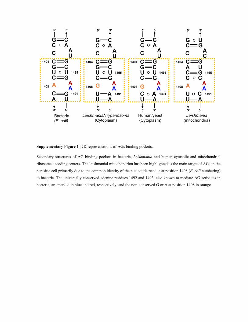
Supplementary Figure 1 | 2D representations of AGs binding pockets.
Secondary structures of AG binding pockets in bacteria, Leishmania and human cytosolic and mitochondrial
ribosome decoding centers. The leishmanial mitochondrion has been highlighted as the main target of AGs in the
parasitic cell primarily due to the common identity of the nucleotide residue at position 1408 (E. coli numbering)
to bacteria. The universally conserved adenine residues 1492 and 1493, also known to mediate AG activities in
bacteria, are marked in blue and red, respectively, and the non-conserved G or A at position 1408 in orange.
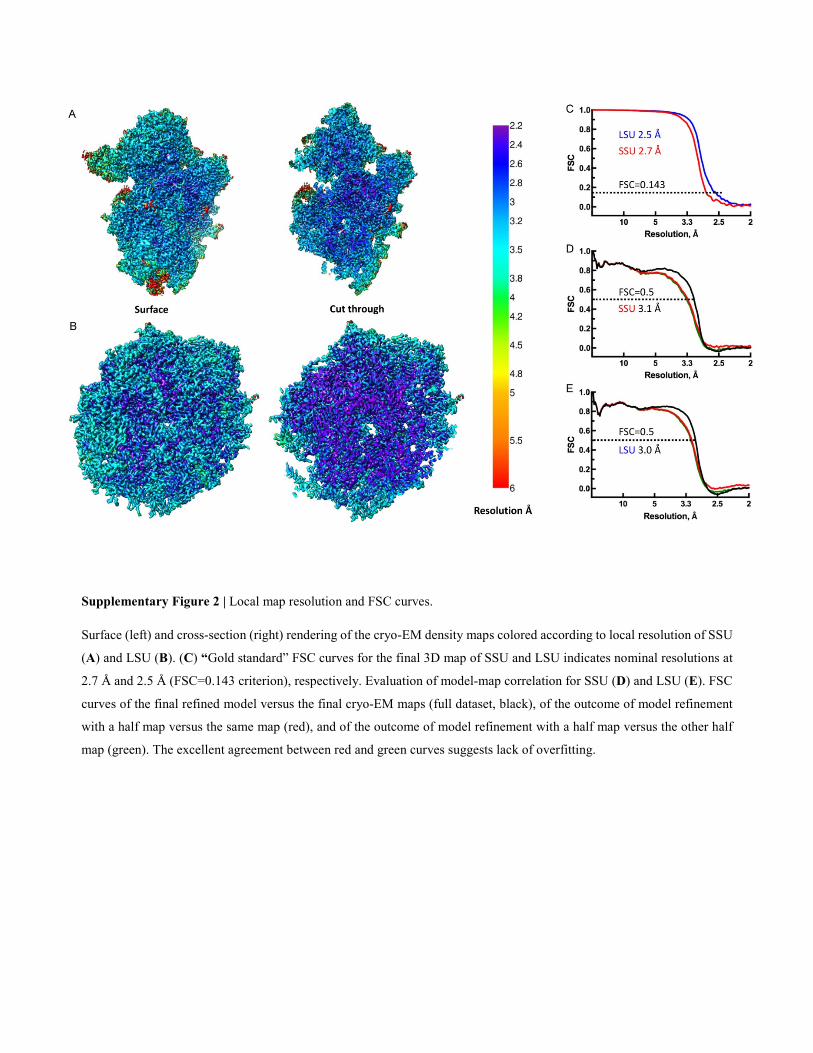
Supplementary Figure 2 | Local map resolution and FSC curves.
Surface (left) and cross-section (right) rendering of the cryo-EM density maps colored according to local resolution of SSU
(A) and LSU (B). (C) “Gold standard” FSC curves for the final 3D map of SSU and LSU indicates nominal resolutions at
2.7 Å and 2.5 Å (FSC=0.143 criterion), respectively. Evaluation of model-map correlation for SSU (D) and LSU (E). FSC
curves of the final refined model versus the final cryo-EM maps (full dataset, black), of the outcome of model refinement
with a half map versus the same map (red), and of the outcome of model refinement with a half map versus the other half
map (green). The excellent agreement between red and green curves suggests lack of overfitting.
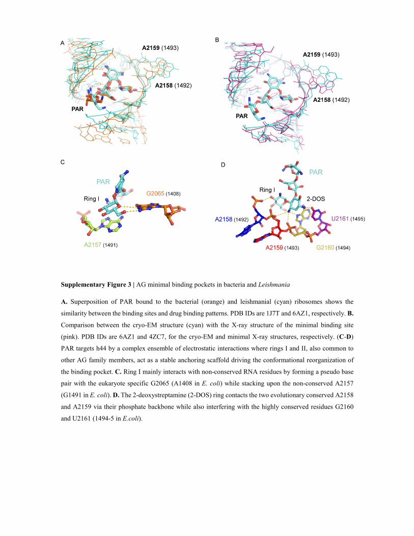
Supplementary Figure 3 | AG minimal binding pockets in bacteria and Leishmania
A. Superposition of PAR bound to the bacterial (orange) and leishmanial (cyan) ribosomes shows the
similarity between the binding sites and drug binding patterns. PDB IDs are 1J7T and 6AZ1, respectively. B.
Comparison between the cryo-EM structure (cyan) with the X-ray structure of the minimal binding site
(pink). PDB IDs are 6AZ1 and 4ZC7, for the cryo-EM and minimal X-ray structures, respectively. (C-D)
PAR targets h44 by a complex ensemble of electrostatic interactions where rings I and II, also common to
other AG family members, act as a stable anchoring scaffold driving the conformational reorganization of
the binding pocket. C. Ring I mainly interacts with non-conserved RNA residues by forming a pseudo base
pair with the eukaryote specific G2065 (A1408 in E. coli) while stacking upon the non-conserved A2157
(G1491 in E. coli). D. The 2-deoxystreptamine (2-DOS) ring contacts the two evolutionary conserved A2158
and A2159 via their phosphate backbone while also interfering with the highly conserved residues G2160
and U2161 (1494-5 in E.coli).
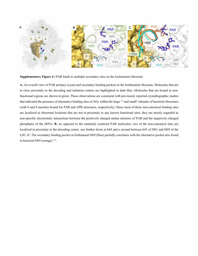
Supplementary Figure 4 | PAR binds to multiple secondary sites on the leishmanial ribosome
A. An overall view of PAR primary (cyan) and secondary binding pockets in the leishmanial ribosome. Molecules that are
in close proximity to the decoding and initiation centers are highlighted in dark blue. Molecules that are bound in non-
functional regions are shown in green. These observations are consistent with previously reported crystallographic studies
that indicated the presence of alternative binding sites of AGs within the large 1,2 and small3 subunits of bacterial ribosomes
(with 6 and 8 moieties bound for PAR and APR structures, respectively). Since most of these non-canonical binding sites
are localized at ribosomal locations that are not in proximity to any known functional sites, they are mostly regarded as
non-specific electrostatic interactions between the positively charged amino moieties of PAR and the negatively charged
phosphates of the rRNA. B. As opposed to the randomly scattered PAR molecules, two of the non-canonical sites are
localized in proximity to the decoding center, one further down at h44 and a second between h45 of SSU and H69 of the
LSU. C. The secondary binding pocket at leishmanial H69 (blue) partially correlates with the alternative pocket also found
in bacterial H69 (orange) 1,2.
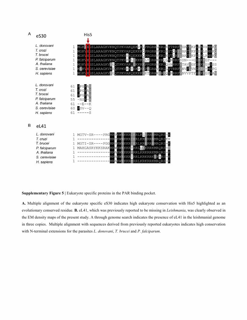
Supplementary Figure 5 | Eukaryote specific proteins in the PAR binding pocket.
A. Multiple alignment of the eukaryote specific eS30 indicates high eukaryote conservation with His5 highlighted as an
evolutionary conserved residue. B. eL41, which was previously reported to be missing in Leishmania, was clearly observed in
the EM density maps of the present study. A through genome search indicates the presence of eL41 in the leishmanial genome
in three copies. Multiple alignment with sequences derived from previously reported eukaryotes indicates high conservation
with N-terminal extensions for the parasites L. donovani, T. brucei and P. falciparum.
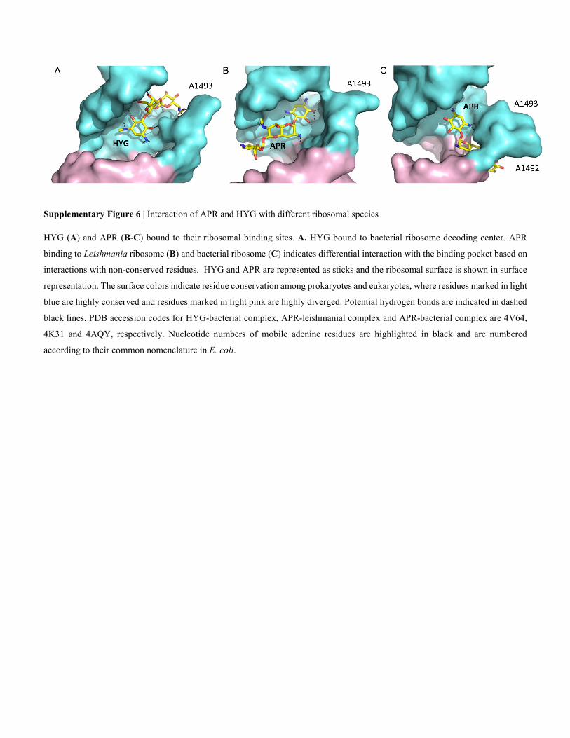
Supplementary Figure 6 | Interaction of APR and HYG with different ribosomal species
HYG (A) and APR (B-C) bound to their ribosomal binding sites. A. HYG bound to bacterial ribosome decoding center. APR
binding to Leishmania ribosome (B) and bacterial ribosome (C) indicates differential interaction with the binding pocket based on
interactions with non-conserved residues. HYG and APR are represented as sticks and the ribosomal surface is shown in surface
representation. The surface colors indicate residue conservation among prokaryotes and eukaryotes, where residues marked in light
blue are highly conserved and residues marked in light pink are highly diverged. Potential hydrogen bonds are indicated in dashed
black lines. PDB accession codes for HYG-bacterial complex, APR-leishmanial complex and APR-bacterial complex are 4V64,
4K31 and 4AQY, respectively. Nucleotide numbers of mobile adenine residues are highlighted in black and are numbered
according to their common nomenclature in E. coli.
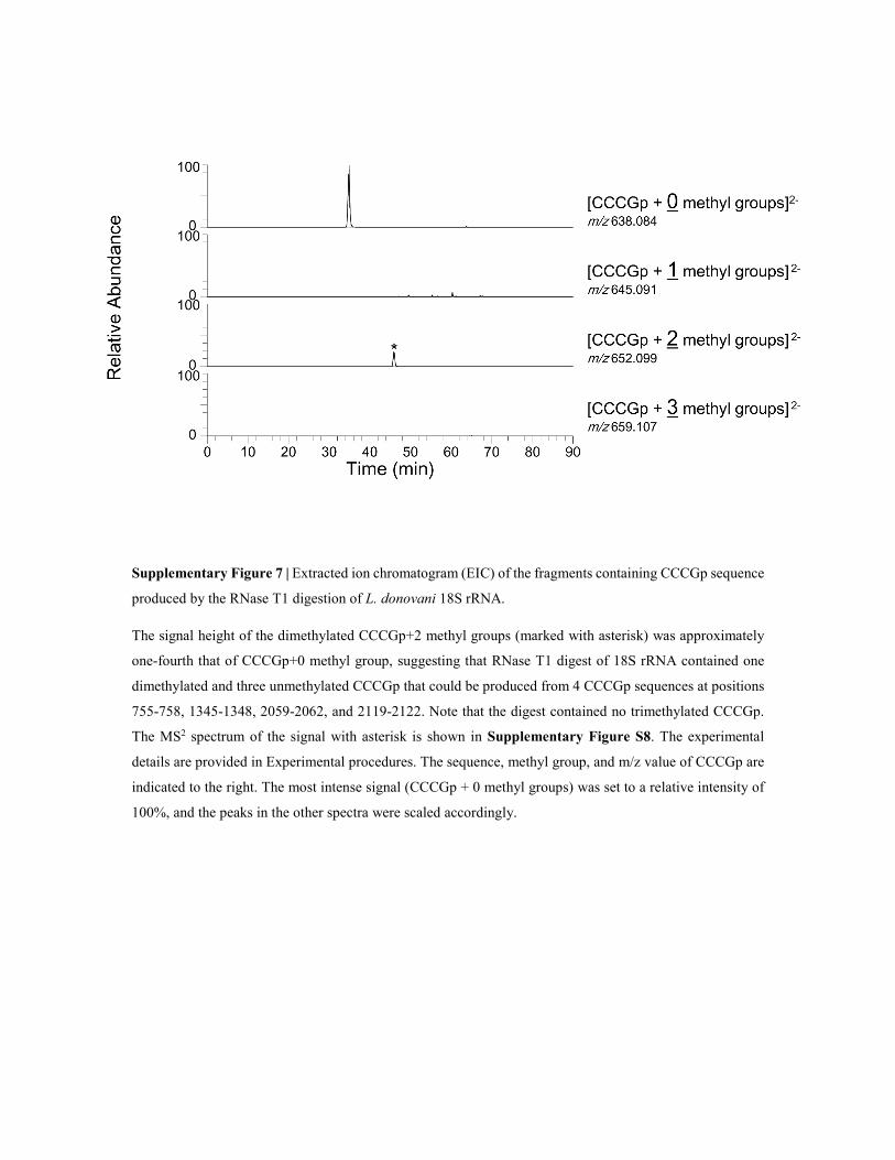
Supplementary Figure 7 | Extracted ion chromatogram (EIC) of the fragments containing CCCGp sequence
produced by the RNase T1 digestion of L. donovani 18S rRNA.
The signal height of the dimethylated CCCGp+2 methyl groups (marked with asterisk) was approximately
one-fourth that of CCCGp+0 methyl group, suggesting that RNase T1 digest of 18S rRNA contained one
dimethylated and three unmethylated CCCGp that could be produced from 4 CCCGp sequences at positions
755-758, 1345-1348, 2059-2062, and 2119-2122. Note that the digest contained no trimethylated CCCGp.
The MS2 spectrum of the signal with asterisk is shown in Supplementary Figure S8. The experimental
details are provided in Experimental procedures. The sequence, methyl group, and m/z value of CCCGp are
indicated to the right. The most intense signal (CCCGp + 0 methyl groups) was set to a relative intensity of
100%, and the peaks in the other spectra were scaled accordingly.
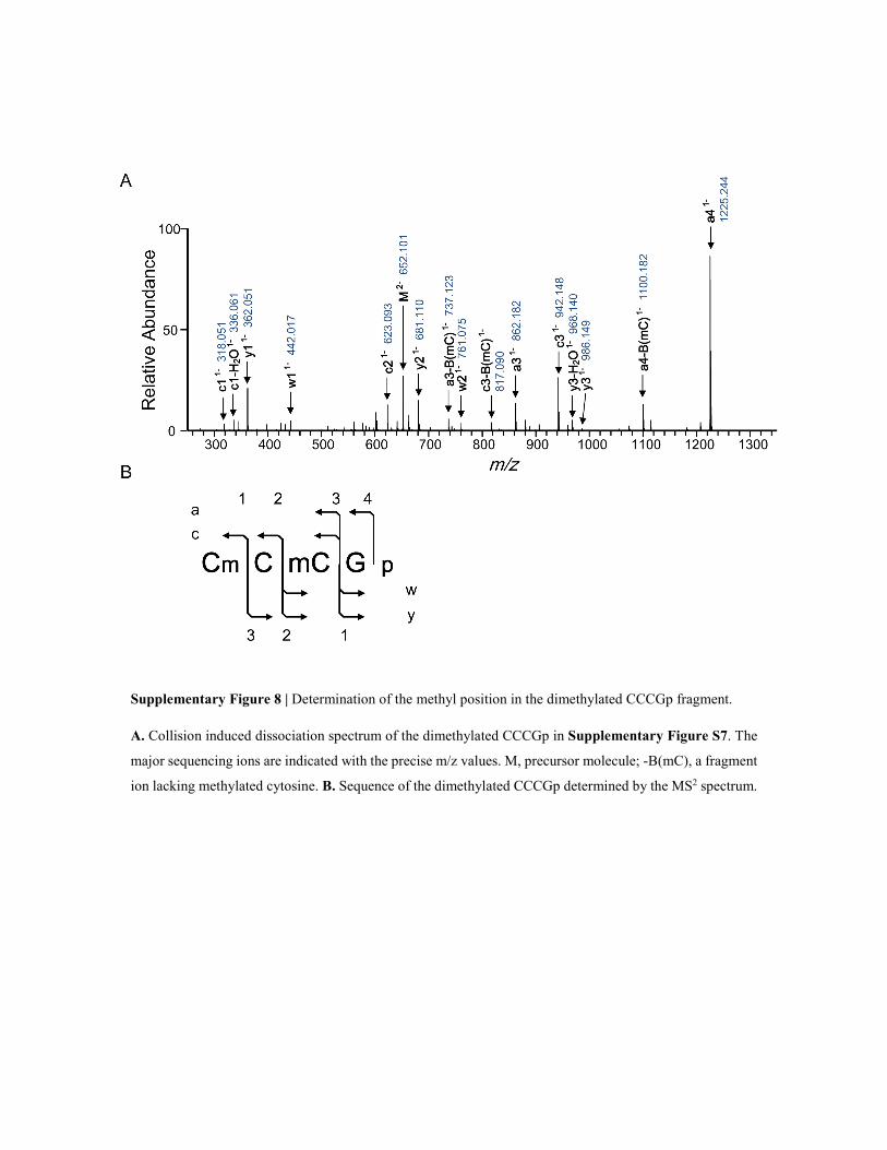
Supplementary Figure 8 | Determination of the methyl position in the dimethylated CCCGp fragment.
A. Collision induced dissociation spectrum of the dimethylated CCCGp in Supplementary Figure S7. The
major sequencing ions are indicated with the precise m/z values. M, precursor molecule; -B(mC), a fragment
ion lacking methylated cytosine. B. Sequence of the dimethylated CCCGp determined by the MS2 spectrum.
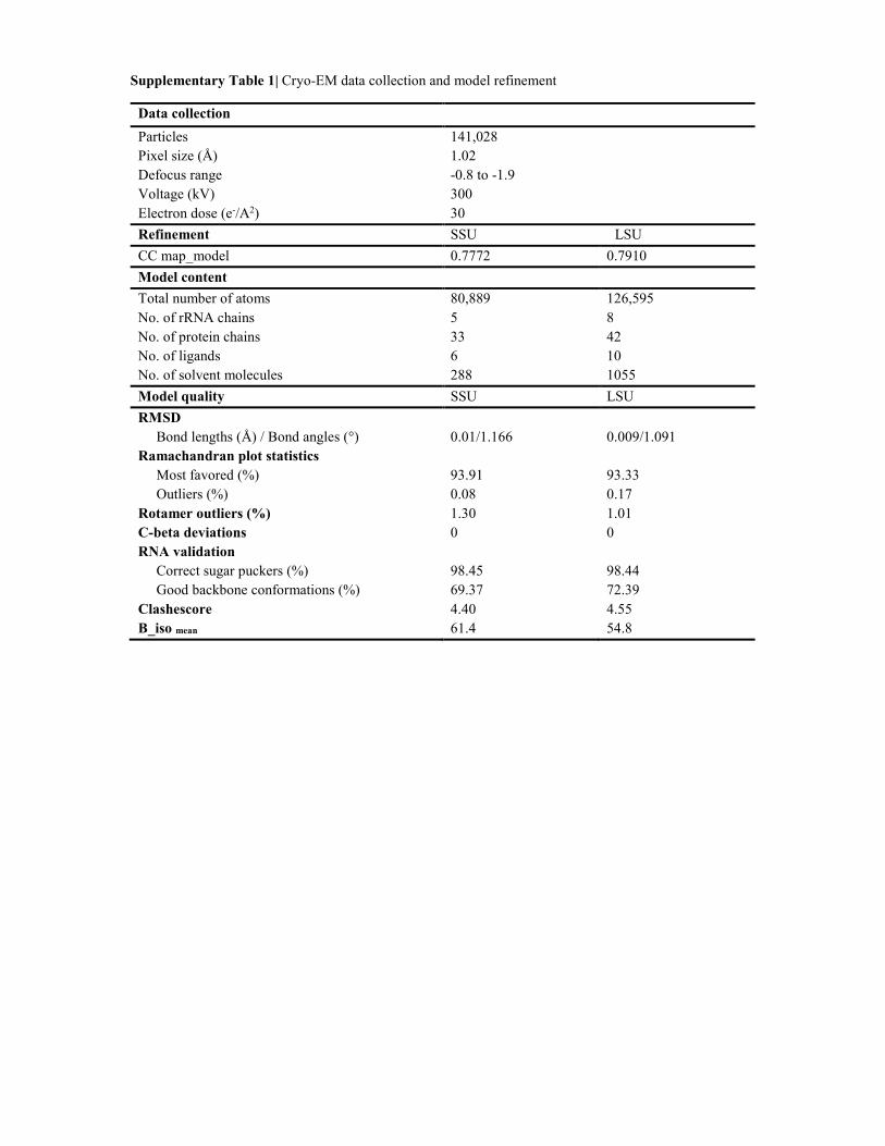
Supplementary Table 1| Cryo-EM data collection and model refinement
Data collection
Particles 141,028
Pixel size (Å) 1.02
Defocus range -0.8 to -1.9
Voltage (kV) 300
Electron dose (e-/A2) 30
Refinement SSU LSU
CC map_model 0.7772 0.7910
Model content
Total number of atoms 80,889 126,595
No. of rRNA chains 5 8
No. of protein chains 33 42
No. of ligands 6 10
No. of solvent molecules 288 1055
Model quality SSU LSU
RMSD
Bond lengths (Å) / Bond angles (°) 0.01/1.166 0.009/1.091
Ramachandran plot statistics
Most favored (%) 93.91 93.33
Outliers (%) 0.08 0.17
Rotamer outliers (%) 1.30 1.01
C-beta deviations 0 0
RNA validation
Correct sugar puckers (%) 98.45 98.44
Good backbone conformations (%) 69.37 72.39
Clashescore 4.40 4.55
B_iso mean 61.4 54.8
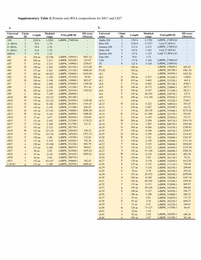
Supplementary Table 2| Proteins and rRNA compositions for SSUa and LSUb
Universal
name
Chain
ID Length Modeled
range TritrypDB ID MS score
(Mascot)
Alpha 26S 1 1782 b 3-1782 LdBPK_27rRNA4 - Beta 26S 2 1526 b 2-1527 LinJ.27.rRNA1 - Gamma 26S 3 213 b 2-213 LdBPK_27rRNA3 - Delta 26S 4 183 b 1-183 LinJ.27.rRNA2 - Epsilon 26S 5 133 b 1-132 LmjF.27.rRNA.43 - Zeta 26S 6 76 b 3-73 - - 5.8S 7 171 b 1-169 LdBPK_27rRNA5 - 5S 8 121 b 3-120 LdBPK_23rRNA1 - P0 - 108 aa - LdBPK_150430.1 602.82 P1,P2 - 105 aa - LdBPK_303780.1 1981.39 uL1 - 78 aa - LdBPK_363950.1 1425.56 uL2 A 260 aa 2-257 LdBPK_351450.1 1100.8 uL3 B 419 aa 3-403 LdBPK_323320.1 461.9 uL4 C 373 aa 4-305 LdBPK_291160.1 4788.7 uL5 D 188 aa 8-175 LdBPK_220004.1 2077.3 uL6 E 190 aa 2-187 LdBPK_211290.1 2815.1 eL6 F 195 aa 20-195 LdBPK_330770.1 537.9 eL8 G 348 aa 111-341 LdBPK_070550.1 4773.5 uL11 - 164 aa - LdBPK_352230.1 2766.7 uL13 H 222 aa 5-222 LdBPK_340910.1 393.67 eL13 I 220 aa 2-207 LdBPK_292580.1 163.52 uL14 J 139 aa 10-139 LdBPK_353840.1 5275.24 eL14 K 233 aa 61-209 LdBPK_221370.1 974.92 uL15 L 145 aa 2-145 LdBPK_353810.1 723.71 eL15 M 204 aa 2-204 LdBPK_303710.1 2914.74 uL16 N 213 aa 1-203 LdBPK_040750.1 1565.46 uL18 O 305 aa 4-266 LdBPK_351870.1 1299.83 eL18 P 198 aa 4-198 LdBPK_364730.1 1538.97 eL19 Q 245 aa 2-189 LdBPK_060410.1 1214.43 eL20 R 179 aa 3-141 LdBPK_350600.1 1363.47 eL21 S 159 aa 2-158 LdBPK_343440.1 1711.16 uL22 T 166 aa 3-155 LdBPK_240040.1 2024.69 eL22 U 122 aa 2-122 LdBPK_363430.1 1319.34 uL23 V 145 aa 27-144 LdBPK_060600.1 2286.36 uL24 W 143 aa 2-124 LdBPK_242140.1 602.39 eL24 X 124 aa 2-65 LdBPK_361130.1 55.22 eL27 Y 134 aa 3-134 LdBPK_322850.1 1612.83 eL28 Z 147 aa 2-135 LdBPK_111110.1 758.34 uL29 a 127 aa 3-126 LdBPK_262350.1 269.04 eL29 b 70 aa 2-70 LdBPK_363550.1 292.42 uL30 c 252 aa 26-252 LdBPK_260160.1 2059.04 eL30 d 104 aa 8-100 LdBPK_350240.1 1647.18 eL31 e 183 aa 64-182 LdBPK_353340.1 1929.41 eL32 f 133 aa 2-131 LdBPK_212090.1 805.97 eL33 g 145 aa 20-144 LdBPK_342240.1 500.68 eL34 h 168 aa 3-127 LdBPK_363930.1 296.77 eL36 i 106 aa 3-100 LdBPK_210800.1 866.33 eL37 j 83 aa 2-81 LdBPK_332070.1 171.75 eL38 k 83 aa 2-76 LdBPK_262230.1 604.23 eL39 l 51 aa 2-51 LdBPK_161220.1 104.41 eL40 m 128 aa 77-127 LdBPK_311930.1 96.93 eL41 n 34 aa 1-34 - - eL43 o 92 aa 2-92 LdBPK_362020.1 688.30 eL44 p 106 aa 2-97 LdBPK_333380.1 301.46
a. b.
Universal
name
Chain
ID Length Modeled
range TritrypDB ID MS score
(Mascot)
18S 1 2203 b 1-2203 LdBPK_27rRNA6 - A- tRNA 2 76 b 1-76 - - P- tRNA 3 76 b 1-76 - - E- tRNA 4 76 b 1-76 - - mRNA 5 13 b 1-12 - - eS1 A 264 aa 22-246 LdBPK_350410.1 3881.33 uS2 B 246 aa 1-211 LdBPK_365240.1 323.07 uS3 C 219 aa 2-213 LdBPK_330960.1 3250.67 uS4 D 190 aa 1-182 LdBPK_361310.1 1513.88 eS4 E 273 aa 2-261 LdBPK_131120.1 4960.24 uS5 F 265 aa 44-263 LdBPK_320460.1 4183.04 eS6 G 249 aa 1-238 LdBPK_151530.1 59.95 uS7 H 190 aa 2-190 LdBPK_110960.1 802.47 eS7 I 200 aa 1-200 LdBPK_010440.1 1349.30 uS8 J 130 aa 2-130 LdBPK_111180.1 971.14 eS8 K 220 aa 2-216 LdBPK_242160.1 1392.02 uS9 L 149 aa 7-149 LdBPK_260840.1 - uS10 M 116 aa 14-115 LdBPK_281100.1 - eS10 N 153 aa 32-122 LdBPK_361050.1 1528.40 uS11 O 144 aa 8-144 LdBPK_281050.1 1191.47 uS12 P 143 aa 1-142 LdBPK_211300.1 826.25 eS12 Q 141 aa 27-141 LdBPK_130460.1 2096.50 uS13 R 153 aa 2-143 LdBPK_360990.1 1088.92 uS14 S 57 aa 4-57 LdBPK_282650.1 239.69 uS15 T 151 aa 2-143 LdBPK_333300.1 1170.22 uS17 U 173 aa 5-160 LdBPK_211790.1 121.31 eS17 V 143 aa 2-123 LdBPK_282750.1 - uS19 W 152 aa 22-135 LdBPK_220340.1 229.15 eS19 X 179 aa 25-176 LdBPK_342620.1 1473.72 eS21 Y 159 aa 3-90 LdBPK_110780.1 132.65 eS24 Z 137 aa 3-129 LdBPK_363020.1 743.18 eS25 a 120 aa 35-106 LdBPK_251220.1 892.79 eS26 b 112 aa 2-104 LdBPK_280570.1 969.61 eS27 c 86 aa 2-85 LdBPK_363940.1 1695.62 eS28 d 87 aa 21-85 LdBPK_261610.1 2982.01 eS30 e 66 aa 2-66 LdBPK_300710.1 - eS31 f 152 aa 83-147 LdBPK_360660.1 362.43 LACK1 g 312 aa 1-309 LdBPK_282970.1 4409.62
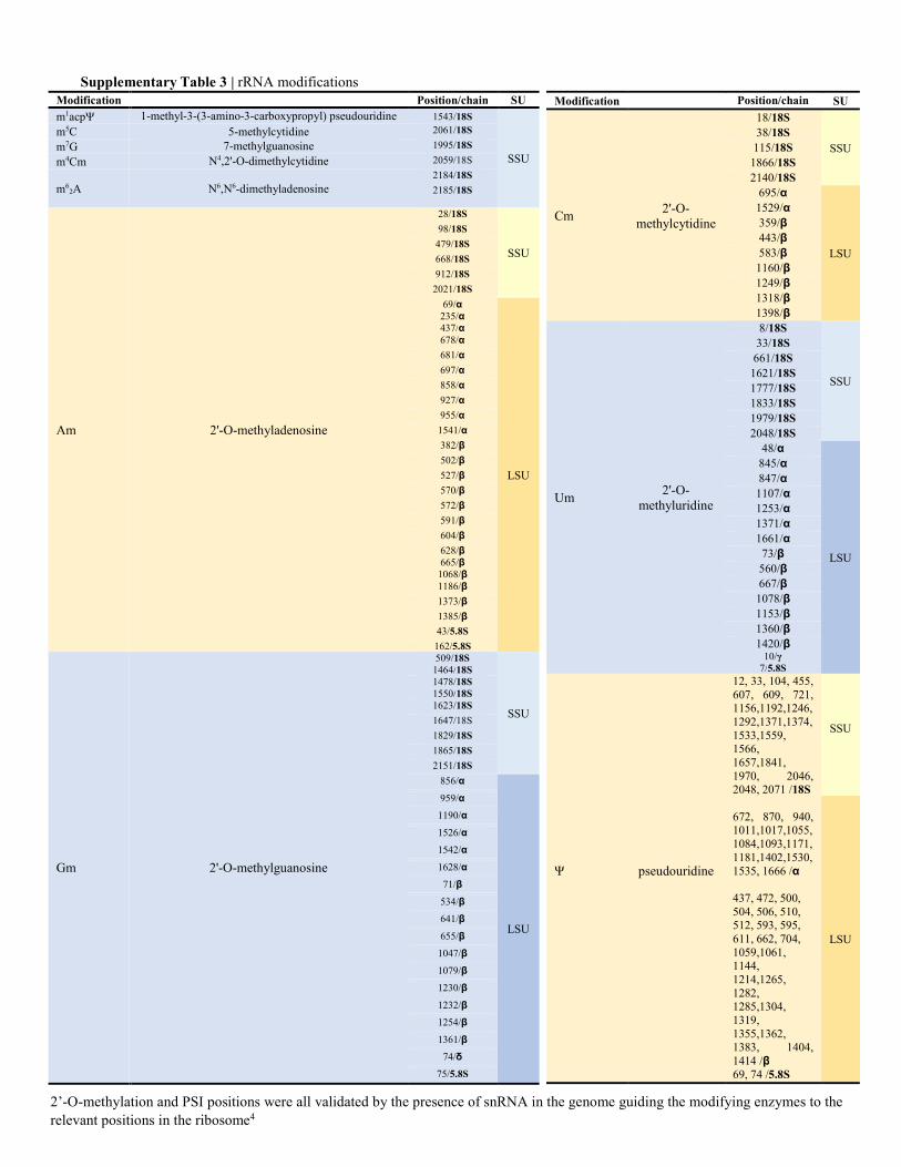
Modification Position/chain SU
Cm 2'-O-
methylcytidine
18/18S
SSU
38/18S 115/18S
1866/18S 2140/18S
695/α
LSU
1529/α 359/β 443/β 583/β
1160/β 1249/β 1318/β 1398/β
Um 2'-O-
methyluridine
8/18S
SSU
33/18S 661/18S
1621/18S 1777/18S 1833/18S 1979/18S 2048/18S
48/α
LSU
845/α 847/α 1107/α 1253/α 1371/α 1661/α 73/β 560/β 667/β
1078/β 1153/β 1360/β 1420/β
10/γ
7/5.8S
Ψ pseudouridine
12, 33, 104, 455, 607, 609, 721, 1156,1192,1246, 1292,1371,1374, 1533,1559, 1566, 1657,1841, 1970, 2046, 2048, 2071 /18S
SSU
672, 870, 940, 1011,1017,1055, 1084,1093,1171, 1181,1402,1530, 1535, 1666 /α
LSU
437, 472, 500, 504, 506, 510, 512, 593, 595, 611, 662, 704, 1059,1061, 1144, 1214,1265, 1282, 1285,1304, 1319, 1355,1362, 1383, 1404, 1414 /β
69, 74 /5.8S
Modification Position/chain SU
m1acpΨ 1-methyl-3-(3-amino-3-carboxypropyl) pseudouridine 1543/18S
SSU
m5C 5-methylcytidine 2061/18S
m7G 7-methylguanosine 1995/18S
m4Cm N4,2'-O-dimethylcytidine 2059/18S
m62A N6,N6-dimethyladenosine
2184/18S
2185/18S
Am 2'-O-methyladenosine
28/18S
SSU
98/18S
479/18S
668/18S
912/18S
2021/18S
69/α 235/α 437/α
LSU
678/α
681/α
697/α
858/α
927/α
955/α
1541/α
382/β
502/β
527/β
570/β
572/β
591/β
604/β
628/β 665/β
1068/β 1186/β
1373/β
1385/β
43/5.8S
162/5.8S
Gm 2'-O-methylguanosine
509/18S 1464/18S
SSU
1478/18S
1550/18S 1623/18S
1647/18S
1829/18S
1865/18S
2151/18S
856/α
LSU
959/α
1190/α
1526/α
1542/α
1628/α
71/β
534/β
641/β
655/β
1047/β
1079/β
1230/β
1232/β
1254/β
1361/β
74/δ
75/5.8S
Supplementary Table 3 | rRNA modifications
2’-O-methylation and PSI positions were all validated by the presence of snRNA in the genome guiding the modifying enzymes to the relevant positions in the ribosome4
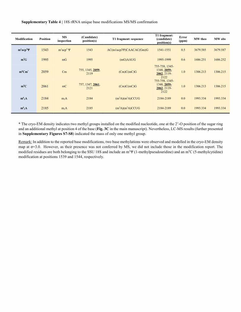
Supplementary Table 4 | 18S rRNA unique base modifications MS/MS confirmation
* The cryo-EM density indicates two methyl groups installed on the modified nucleotide, one at the 2’-O position of the sugar ring and an additional methyl at position 4 of the base (Fig. 3C in the main manuscript). Nevertheless, LC-MS results (further presented in Supplementary Figures S7-S8) indicated the mass of only one methyl group.
Remark: In addition to the reported base modifications, two base methylations were observed and modelled in the cryo-EM density map at σ=3.0. However, as their presence was not conferred by MS, we did not include those in the modification report. The modified residues are both belonging to the SSU 18S and include an m1Ψ (1-methylpseudouridine) and an m5C (5-methylcytidine) modification at positions 1539 and 1544, respectively.
Modification Position MS
inspection
(Candidate)
position(s) T1 fragment: sequence
T1 fragment:
(candidate)
position(s)
Error
(ppm) MW theo MW obs
m1acp3Ψ 1543 m1acp3 Ψ 1543 AC(m1acp3Ψ)CAACAC(Gm)G 1541-1551 0.5 3679.585 3679.587
m7G 1995 mG 1995 (mG)AAUG 1995-1999 0.6 1686.251 1686.252
m4Cm* 2059 Cm 755, 1345, 2059,
2119 (Cm)C(mC)G
755-758, 1345-1348, 2059-
2062, 2119-2122
1.0 1306.213 1306.215
m5C 2061 mC 757, 1347, 2061,
2121 (Cm)C(mC)G
755-758, 1345-1348, 2059-
2062, 2119-2122
1.0 1306.213 1306.215
m62A 2184 m2A 2184 (m2A)(m2A)CCUG 2184-2189 0.0 1993.334 1993.334
m62A 2185 m2A 2185 (m2A)(m2A)CCUG 2184-2189 0.0 1993.334 1993.334
Supplementary References
1 Borovinskaya, M. A. et al. Structural basis for aminoglycoside inhibition of bacterial ribosome recycling.
Nature structural & molecular biology 14, 727-732, doi:10.1038/nsmb1271 (2007).
2 Wasserman, M. R. et al. Chemically related 4,5-linked aminoglycoside antibiotics drive subunit rotation in
opposite directions. Nature communications 6, 7896, doi:10.1038/ncomms8896 (2015).
3 Matt, T., et al. Dissociation of antibacterial activity and aminoglycoside ototoxicity in the 4-monosubstituted
2-deoxystreptamine apramycin. Proceedings of the National Academy of Sciences of the United States of
America 109, 10984-10989, doi:10.1073/pnas.1204073109 (2012).
4 Eliaz, D., et al. Genome-wide analysis of small nucleolar RNAs of Leishmania major reveals a rich repertoire
of RNAs involved in modification and processing of rRNA. RNA Biol. 12(11), 1222-55, doi:
10.1080/15476286.2015.1038019 (2015).