Culicoides fauna and bluetongue virus serotype 8 infection...
168
Transcript of Culicoides fauna and bluetongue virus serotype 8 infection...


Bibliografische Informationen der Deutschen Bibliothek
Die Deutsche Bibliothek verzeichnet diese Publikation in der Deutschen
Nationalbibliografie;
Detaillierte bibliografische Daten sind im Internet über http://dnb.ddb.de abrufbar.
1. Auflage 2012
© 2012 by Verlag: Deutsche Veterinärmedizinische Gesellschaft Service GmbH,
Gießen
Printed in Germany
ISBN 978-3-86345-134-9
Verlag: DVG Service GmbH
Friedrichstraße 17
35392 Gießen
0641/24466
www.dvg.net
Aus dem
Institut für Parasitologie
Justus-Liebig-Universität Gießen
und
Institut für Virusdiagnostik
Friedrich-Loeffler-Institut
Bundesforschungsinstitut für Tiergesundheit, Insel Riems
und
Department für Nutztierwissenschaften
Produktionssysteme der Nutztiere
Georg-August-Universität Göttingen
Betreuer: Prof. Dr. Christoph G. Grevelding
Culicoides Fauna and Bluetongue Virus Serotype 8 Infection in
South American Camelid Herds in Germany
INAUGURAL-DISSERTATION
zur Erlangung des Grades eines
Dr. med. vet.
beim Fachbereich Veterinärmedizin
der Justus-Liebig-Universität Gießen
Eingereicht von
Claudia Schulz
Tierärztin aus Mainz
Gießen 2012
Mit Genehmigung des Fachbereichs Veterinärmedizin
der Justus-Liebig-Universität
Dekan: Prof. Dr. Dr. h.c. Martin Kramer
Gutachter: Prof. Dr. Christoph G. Grevelding
Prof. Dr. Dr. Matthias Gauly
Tag der Disputation: 17. Dezember 2012

Meinen Eltern
EIDESSTATTLICHE ERKLÄRUNG
Hiermit erkläre ich, Claudia Schulz, geboren am 18.08.1981 in Mainz, dass ich die vorgelegte
Dissertation selbständig und ohne unerlaubte fremde Hilfe und nur mit den Hilfen angefertigt
habe, die ich in der Dissertation angegeben habe. Alle Textstellen, die wörtlich oder
sinngemäß aus veröffentlichten oder nicht veröffentlichten Schriften entnommen sind, und
alle Angaben, die auf mündlichen Auskünften beruhen, sind als solche kenntlich gemacht. Bei
den von mir durchgeführten und in der Dissertation erwähnten Untersuchungen habe ich die
Grundsätze guter wissenschaftlicher Praxis, wie sie in der „Satzung der Justus -Liebig-
Universität Gießen zur Sicherung guter wissenschaftlicher Praxis“ niedergelegt sind,
eingehalten.

“One reason why research is so important is precisely that it can surprise you and tell you
that your subjective convictions are wrong. If research always found what we expected,
there wouldn't be much point in doing research.”
(Eugene T. Gendlin 1981)

"The most important thing is not to stop questioning."
(Albert Einstein 1955)
Teile dieser Dissertation wurden bereits veröffentlicht
(in chronologischer Reihenfolge)
Eschbaumer M, Schulz C 1, Wäckerlin R, Gauly M, Beer M, Hoffmann B. Limitations of
sandwich ELISAs for bluetongue virus antibody detection, erschienen in Veterinary Record
2011; 168(24):643
Schulz C, Eschbaumer M 1, Rudolf M, König P, Keller M, Gauly M, Bauer C, Grevelding
CG, Beer M, Hoffmann B. Experimental infection of South American camelids with
bluetongue virus serotype 8, erschienen in Veterinary Microbiology 2011; 154(3-4):257-265
Schulz C 2, Eschbaumer M, Ziller M, Wäckerlin R, Beer M, Gauly M, Grevelding CG,
Hoffmann B; Bauer C. Cross-sectional study of bluetongue virus serotype 8 infection in South
American camelid herds in Germany (2008/2009), erschienen in Veterinary Microbiology
2012; 160(1-2):35-42.
Werner D, Bauer C, Schulz C, and Kampen H. The breeding habitat preferences of Obsoletus
complex Culicoides species (Diptera: Ceratopogonidae), erschienen in Mitteilungen der
Deutschen Gesellschaft für allgemeine und angewandte Entomologie 2012. 18:323-329
1 „contributed equally“
2 „corresponding author“

C o n t e n t s I
CONTENTS
I. Introduction .................................................................................................................... 1
II. Literature review ............................................................................................................. 3
1. Bluetongue virus 3
1.1. Taxonomy and virion structure of BTV ................................................................... 3
1.2. Virus replication ....................................................................................................... 5
1.3. History, distribution and economic consequences ................................................... 6
1.3.1. BTV in Europe .................................................................................................. 7
1.3.2. Transmission and overwintering mechanisms of BTV .................................. 10
2. Culicoides biting midges 11
2.1. Taxonomy and morphology of ceratopogonids ..................................................... 11
2.2. Biology and breeding habitats of ceratopogonids .................................................. 13
2.3. Methods of ceratopogonid collection ..................................................................... 14
2.4. Molecular identification of Culicoides................................................................... 15
2.5. Culicoides as vectors and nuisance ........................................................................ 15
3. BTV in the mammalian host 19
3.1. Pathogenesis of BTV infection in ruminants ......................................................... 19
3.2. Clinical signs and post-mortem lesions in ruminants ............................................ 21
3.3. Particularities of camelids ...................................................................................... 23
3.3.1. BTV infection in camelids .............................................................................. 25
4. Laboratory diagnosis of BTV 26
4.1. BTV diagnosis in camelids .................................................................................... 28
5. Prevention and control of BT 28
5.1. Vector control......................................................................................................... 29
5.2. BTV vaccination .................................................................................................... 29
5.2.1. Types of BTV vaccines .................................................................................. 31
5.2.2. Safety and efficacy of live-attenuated and inactivated BTV vaccines ........... 31
5.2.3. BTV vaccination of camelids ......................................................................... 32
III. Objectives ..................................................................................................................... 34
IV. Material and methods ................................................................................................... 35
1. Entomological monitoring 35
1.1. Trap locations ......................................................................................................... 35
1.2. Trapping protocol ................................................................................................... 35
1.3. Morphological identification of ceratopogonids .................................................... 37
1.4. Weather data........................................................................................................... 37
2. Cross-sectional study 38
2.1. Study design and sampling .................................................................................... 38
2.2. Questionnaire ......................................................................................................... 39
3. Animal experiment 39
3.1. Animals and experimental design .......................................................................... 39
3.2. Clinical and haematological parameters ................................................................ 40
II C o n t e n t s
3.3. Post-mortem examination ...................................................................................... 40
3.4. Blood-cell binding experiment .............................................................................. 41
4. Bluetongue vaccination 41
4.1. Vaccination study: Animals and study design ....................................................... 41
4.2. Tolerance of BTV-8 vaccines ................................................................................ 42
4.3. Colostral transfer of maternal BTV antibodies ...................................................... 43
5. Serology 43
5.1. Enzyme-linked immunosorbent assay ................................................................... 43
5.2. Serum neutralisation test ........................................................................................ 44
6. Virological analyses 45
6.1. Extraction of viral RNA ......................................................................................... 45
6.2. Detection of BTV RNA ......................................................................................... 46
6.3. Virus isolation in tissue culture and embryonated chicken eggs ........................... 47
6.4. Virus isolation in IFNAR-/-
mice ........................................................................... 48
7. Statistical analyses 48
V. Results .......................................................................................................................... 50
1. Entomological monitoring 50
1.1. Abundance of ceratopogonid females.................................................................... 50
1.2. Abundance of ceratopogonid males ....................................................................... 54
1.3. Annual deviations .................................................................................................. 55
1.4. Phenology of ceratopogonids................................................................................. 55
1.5. Association between daily number of Culicoides and farm, temperature and
humidity ................................................................................................................. 56
1.6. Virological results .................................................................................................. 58
1.7. Ceratopogonids in emergence traps ....................................................................... 58
2. Cross-sectional study 59
2.1.1. Study population ............................................................................................. 59
2.1.2. Prevalence of BTV infection .......................................................................... 60
2.1.1. Questionnaire .................................................................................................. 61
2.1.1.1. Natural BTV infection in SAC ................................................................... 61
2.1.1.2. Animal movements ..................................................................................... 62
2.1.1.3. Association between explanatory variables and BTV infection ................. 63
3. Animal experiment 64
3.1. Clinical and haematological parameters ................................................................ 64
3.1. Serology ................................................................................................................. 64
3.1. Detection of BTV RNA in blood samples ............................................................. 66
3.2. Post-mortem examination and detection of BTV RNA ......................................... 68
3.3. Virus isolation ........................................................................................................ 68
3.4. Blood-cell binding experiment .............................................................................. 68
4. Bluetongue vaccination 69
4.1. Vaccination study .................................................................................................. 70
4.2. Tolerance of BTV-8 vaccines ................................................................................ 72
4.3. Maternal BTV antibodies in crias .......................................................................... 72
C o n t e n t s III
VI. Discussion ..................................................................................................................... 74
1. Entomological monitoring 74
1.1. Influences on ceratopogonid abundance and BTV infection rates in SAC herds .. 74
1.2. Phenology of ceratopogonids ................................................................................. 77
1.3. Absence of BTV RNA ........................................................................................... 77
1.4. Breeding habitats and occurrence of males ............................................................ 78
2. Cross-sectional study 79
3. Animal experiment 82
4. Bluetongue vaccination 87
4.1. Vaccination study ................................................................................................... 87
4.2. Tolerance of BTV-8 vaccines ................................................................................ 88
4.3. Colostral BTV-8 antibody transfer......................................................................... 89
VII. Collective discussion .................................................................................................... 91
VIII. Conclusions and outlook .............................................................................................. 93
IX. Summary ....................................................................................................................... 95
X. Zusammenfassung ........................................................................................................ 97
XI. Appendix ...................................................................................................................... 99
List of tables ............................................................................................................... 111
List of figures .............................................................................................................. 112
XII. References .................................................................................................................. 113
XIII. Abbreviations .............................................................................................................. 145
XIV. Acknowledgements .................................................................................................... 147

I . I n t r o d u c t i o n 1
I. INTRODUCTION
Bluetongue (BT) is an infectious, non-contagious, notifiable disease caused by bluetongue
virus (BTV), an Orbivirus in the Reoviridae family. Certain species of Culicoides biting
midges are potential vectors of this virus infection (Mellor et al. 2000). BTV serotype 8
(BTV-8) was the first-ever serotype detected in Northern Europe. From 2006 to 2010, the
BTV-8 epizootic in Europe (EU-BTNET system 2012) had substantial impact on animal
welfare and probably caused greater economic damage than any previous single-serotype
outbreak (Conraths et al. 2009, Wilson and Mellor 2009, Gethmann et al. 2010). BTV
primarily causes severe disease in sheep, while other ruminant species and camelids usually
exhibit subclinical disease. BTV-8, however, also affected cattle, several wild ruminant
species and an alpaca. The manifestation of BT depends on virus strain, host species as well
as breed, age, sex, individual resistance and fitness of the mammalian host (Ward et al. 1994,
Brodie et al. 1998, Darpel et al. 2007, MacLachlan et al. 2009, Falconi et al. 2011).
BT monitoring and surveillance programmes have to include serological, virological and
entomological surveillance in Member States within and outside of restricted zones
(Commission regulation (EC) No 1266/2007, Caporale and Giovannini 2010). Although
South American camelids (SAC) are susceptible to BTV, these species have not been
included in the monitoring programmes. Before 2007, SAC were considered resistant to BT
disease and the pathogenesis of BTV infection had so far not been investigated in these
species (Rivera et al. 1987, Mattson 1994). However, fatalities related to BTV infection were
reported in a few SAC during the recent BTV-8 and BTV-1 epizootics in Germany and
France (Henrich et al. 2007, Meyer et al. 2009). This raised concern about their role in the
epidemiology of BTV. To evaluate whether SAC are potential BTV reservoirs and a risk for
the ruminant population, studies of BTV infection in SAC are required. The investigation of
pathogenesis, diagnosis and frequency of BTV-8 infection in SAC as well as abundance of
Culicoides vector species on SAC farms have been part of this work.
From 2008 to 2010, mandatory vaccination was successfully implemented in domestic
ruminants to control the further spread of BTV-8 in Europe (Wilson and Mellor 2009,
Zientara et al. 2010). Vaccination of over 80% of a susceptible population is considered the
most effective strategy to control BT (Giovannini et al. 2004b, 2004c, Commission regulation
(EC) No 1266/2007). SAC were generally not included in the compulsory vaccination
programmes in Europe, and veterinary authorities were confronted with the question whether
SAC should be vaccinated against BTV-8 at all. However, no information was available on

2 I . I n t r o d u c t i o n
immunogenicity and safety of BTV vaccines or the vaccine dose to be used in camelids. In the
presented work, long-term progression and colostral transfer of BTV antibodies as well as
tolerance of BTV-8 vaccines were monitored after vaccination of SAC with inactivated BTV-
8 vaccines.
I I . L i t e r a t u r e r e v i e w 3
II. LITERATURE REVIEW
1. Bluetongue virus
1.1. Taxonomy and virion structure of BTV
Bluetongue virus (BTV) is the prototype virus of the Orbivirus (orbis = lat. ring) species that
belong to the Reoviridae family (Hewat et al. 1992, Verwoerd and Erasmus 2004). The
orbiviruses BTV, African horse sickness virus (AHSV), Epizootic hemorrhagic disease virus
(EHDV) and Equine encephalosis virus (EEV) can cause severe and economically important
diseases of domestic and wild animals (Attoui et al. 2009). BTV as well as the
phylogenetically closely related orbiviruses AHSV and EHDV are transmitted by the same
Culicoides species (spp.) (Roy 1992, Hewat et al. 1992, Verwoerd and Erasmus 2004, Purse
et al. 2005, Kampen and Kiel 2006, Meiswinkel et al. 2007, Mertens and Attoui 2009, Attoui
et al. 2009).
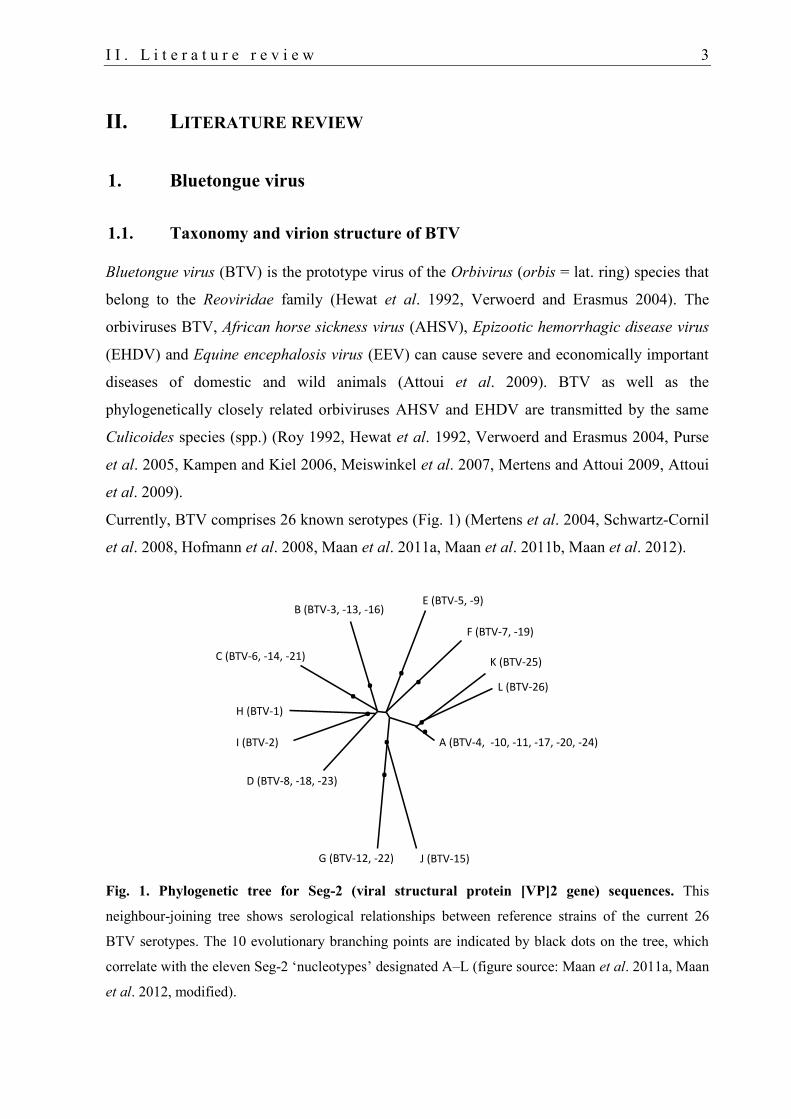
Currently, BTV comprises 26 known serotypes (Fig. 1) (Mertens et al. 2004, Schwartz-Cornil
et al. 2008, Hofmann et al. 2008, Maan et al. 2011a, Maan et al. 2011b, Maan et al. 2012).
Fig. 1. Phylogenetic tree for Seg-2 (viral structural protein [VP]2 gene) sequences. This
neighbour-joining tree shows serological relationships between reference strains of the current 26
BTV serotypes. The 10 evolutionary branching points are indicated by black dots on the tree, which
correlate with the eleven Seg-2 ‘nucleotypes’ designated A–L (figure source: Maan et al. 2011a, Maan
et al. 2012, modified).
E (BTV-5, -9)
F (BTV-7, -19)
K (BTV-25)
L (BTV-26)
A (BTV-4, -10, -11, -17, -20, -24)
J (BTV-15)G (BTV-12, -22)
D (BTV-8, -18, -23)
I (BTV-2)
H (BTV-1)
C (BTV-6, -14, -21)
B (BTV-3, -13, -16)
....
..
...
4 I I . L i t e r a t u r e r e v i e w
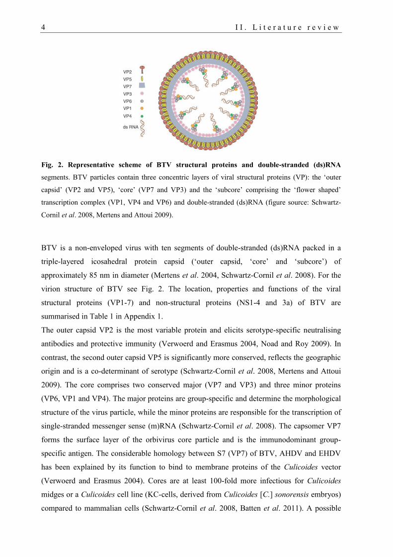
Fig. 2. Representative scheme of BTV structural proteins and double-stranded (ds)RNA
segments. BTV particles contain three concentric layers of viral structural proteins (VP): the ‘outer
capsid’ (VP2 and VP5), ‘core’ (VP7 and VP3) and the ‘subcore’ comprising the ‘flower shaped’
transcription complex (VP1, VP4 and VP6) and double-stranded (ds)RNA (figure source: Schwartz-
Cornil et al. 2008, Mertens and Attoui 2009).
BTV is a non-enveloped virus with ten segments of double-stranded (ds)RNA packed in a
triple-layered icosahedral protein capsid (‘outer capsid, ‘core’ and ‘subcore’) of
approximately 85 nm in diameter (Mertens et al. 2004, Schwartz-Cornil et al. 2008). For the
virion structure of BTV see Fig. 2. The location, properties and functions of the viral
structural proteins (VP1-7) and non-structural proteins (NS1-4 and 3a) of BTV are
summarised in Table 1 in Appendix 1.
The outer capsid VP2 is the most variable protein and elicits serotype-specific neutralising
antibodies and protective immunity (Verwoerd and Erasmus 2004, Noad and Roy 2009). In
contrast, the second outer capsid VP5 is significantly more conserved, reflects the geographic
origin and is a co-determinant of serotype (Schwartz-Cornil et al. 2008, Mertens and Attoui
2009). The core comprises two conserved major (VP7 and VP3) and three minor proteins
(VP6, VP1 and VP4). The major proteins are group-specific and determine the morphological
structure of the virus particle, while the minor proteins are responsible for the transcription of
single-stranded messenger sense (m)RNA (Schwartz-Cornil et al. 2008). The capsomer VP7
forms the surface layer of the orbivirus core particle and is the immunodominant group-
specific antigen. The considerable homology between S7 (VP7) of BTV, AHDV and EHDV
has been explained by its function to bind to membrane proteins of the Culicoides vector
(Verwoerd and Erasmus 2004). Cores are at least 100-fold more infectious for Culicoides
midges or a Culicoides cell line (KC-cells, derived from Culicoides [C.] sonorensis embryos)
compared to mammalian cells (Schwartz-Cornil et al. 2008, Batten et al. 2011). A possible
I I . L i t e r a t u r e r e v i e w 5
explanation has been an evolutionary adaptation to mammalian cells acquiring the outer
capsid ring (Ross- Smith et al. 2009a).
The significant variations in the pathogenicity of BTV strains within a serotype (Schwartz-
Cornil et al. 2008, Saegerman et al. 2008) are based on genetic differences that still remain
uncharacterised (MacLachlan and Guthrie 2010). Comparison of strains of BTV from two
regions showed that they could readily be distinguished by sequence analysis of their genome
segment (S)10 (NS3/3A), but not their S2 (serotype-specific VP2) genes, which explains the
diversification of BTV into distinct topotypes worldwide (Bonneau and MacLachlan 2004). In
general, eastern and western topotypes can be distinguished, which are derived from different
geographical areas (Schwartz-Cornil et al. 2008, Saegerman et al. 2008). The high genetic
heterogeneity of BTV field strains is a consequence of genetic drift and, particularly, shift
(Bonneau and MacLachlan 2004, He et al. 2010, Carpi et al. 2010).
1.2. Virus replication
The replication cycle of BTV was reviewed in detail by several authors (Roy 1992, Mertens et
al. 2004, Schwartz-Cornil et al. 2008, Mertens and Attoui 2009, Noad and Roy 2009,
Belhouchet et al. 2011, Ratinier et al. 2011). Briefly, after binding to the cell surface
receptors via VP2 of the outer capsid, the BTV particle is internalised in the cell by receptor-
mediated endocytosis (the core particle can also bind to insect cells via VP7, see II.1.1). The
‘uncoated’ core is released into the cytoplasm of the host cell. Subsequently, mRNAs are
produced in the core by transcription complexes (VP1, VP4 and VP6). RNA synthesis takes
place within the core to avoid recognition of RNA by the host cell, which would initialize
antiviral defence mechanisms including apoptosis, interferon (IFN) production and RNA
silencing. Recently, a fifth NS protein (NS4) has been detected (Belhouchet et al. 2011,
Ratinier et al. 2011), which contributes to counteract the antiviral response of the host, but is
dispensable for replication. Interestingly, although BTV replicates exclusively in the
cytoplasm, recent investigations revealed that NS4 is also localized in nucleoli (Ratinier et al.
2011).
Synthesised mRNA copies are extruded in the cytoplasm for protein synthesis. Synthesis of
negative-strand RNA and the assembly of dsRNA and progeny core particles takes place
within the viral inclusion body (VIB) formed by NS2. The outer capsid proteins VP2 and VP5
are added as the core leaves the VIB. Mature virions are released by budding (mediated by
NS3/NS3A), by direct cell membrane penetration (NS3) or (consequently) cell lyses. In
contrast, BTV particles are released by budding in Culicoides cells and are therefore not
6 I I . L i t e r a t u r e r e v i e w
causing cell lysis. Released and enveloped virions lose the membrane and subsequently re-
infect the same or other cells.
1.3. History, distribution and economic consequences
BT has been included in the list of notifiable diseases (formerly List A) of the OIE (Office
International des Epizooties; syn. World Organisation for Animal Health) (MacLachlan 2011)
as it is one of the most important diseases of domestic livestock worldwide causing
substantial impact on economy and animal welfare (Mellor and Wittmann 2002, Saegerman et
al. 2007, Gethmann et al. 2010). The disease was first recognised in South Africa and has
probably occurred in this area for about one hundred years before it was described for the first
time in the late 19th
century by Hutcheon in 1880, particularly in imported (merino) sheep
(Hateley 2009, MacLachlan and Guthrie 2010, MacLachlan 2011). Indigenous domestic and
wild ruminants are usually highly resistant to clinical BT disease (Mellor and Wittmann 2002,
Dal Pozzo et al. 2009b). Historically, African antelopes were probably the primary host
species in the epidemiological cycle with Culicoides midges (Erasmus 1990). Today, the role
of wild game has been taken over largely by cattle due to extensive farming (Erasmus 1990).
Over the last 60 years, the distribution of BTV has changed significantly, and virus isolations
have successfully been conducted on every continent except Antarctica (MacLachlan and
Guthrie 2010, MacLachlan 2011).
BTV is usually actively spread short distances by flying Culicoides or is disseminated (semi-)
passively by Culicoides on prevailing winds over long distances (Purse et al. 2005, Burgin et
al. 2012, Sedda et al. 2012). Other possible introduction routes of BTV in a region are:
Movement of viraemic animals, transport of animal products (semen, embryos) or infected
Culicoides vectors (Saegerman et al. 2008, Mintiens et al. 2008, Sedda et al. 2012).
Nowadays, enhanced global networking between countries and continents facilitates the
incursion of new viruses and vectors in BTV-free areas and in naïve ruminant populations.
Consequently, the risk for an encounter between serotypes and the possibility of reassortment
have increased (Saegerman et al. 2008, Perry et al. 2011). Interestingly, BTV only remains
enzootic for many years in regions where several serotypes are circulating. In contrast, a
single virus serotype typically only occurs transiently in a region as for example BTV-8 in
northern and central Europe (Gethmann et al. 2010, MacLachlan 2011, LAVES 2012).
I I . L i t e r a t u r e r e v i e w 7
1.3.1. BTV in Europe
Prior to 1998, BTV was considered an exotic disease in Europe. Only brief periodic
incursions of the disease occurred in Southern Europe (Purse et al. 2005, Saegerman et al.
2008, Hendrickx 2009). The disease was restricted to parts of the world between latitudes of
approximately 40 to 50° N and 35° S where Culicoides vector species occurred (MacLachlan
and Guthrie 2010). In the following 7 years, five serotypes (BTV-1, -2, -4, -9 and -16)
belonging to six strains have almost simultaneously invaded at least 12 countries in Europe
from two or more directions. These invasions caused the most severe outbreak of BT on
record (Purse et al. 2005).
The occurrence of BT in northern regions where C. imicola is rare or absent indicated that
unknown vectors might play a role in the transmission of this virus (Mellor and Wittmann
2002, Saegerman et al. 2008). BTV isolation from indigenous Palearctic species belonging to
the extremely abundant C. obsoletus and C. pulicaris complexes (Purse et al. 2005, De
Liberato et al. 2005, Carpenter et al. 2009) dramatically expanded the risk of BTV
transmission to whole Europe.
Period 2006-2012
BTV-8
Before 2006, the spread of BTV was mainly associated with the distribution of the tropical
midge C. imicola forming an invisible border for BTV emergence to northern temperate
regions in Europe. Therefore, the incursion of BTV-8 to north(-western) Europe was totally
unexpected. This serotype was the first-ever BTV detected latitude >51°N, about 900 km
further north than the previous European BTV incursions (Mellor and Wittmann 2002,
Carpenter et al. 2009, Hendrickx 2009). BTV-8 was first detected in the Netherlands in
August 2006 and subsequently in the surrounding countries Belgium, Germany, Luxemburg
and France until the end of the year (Saegerman et al. 2008, Conraths et al. 2009, Mellor et al.
2009a, Gethmann et al. 2010).
Retrospective studies suggested that BTV has already been introduced during spring 2006,
near to the National Park of Hautes Fagnes and Eifel in Belgium when Culicoides became
active (Szmaragd et al. 2010, Saegerman et al. 2010). However, many other routes of
introduction were possible (Mintiens et al. 2008). BTV-8 probably originated from sub-
Sahara Africa. However, only a small number of reference strains were available, and
sequence analyses did not point to a particular origin or introduction route. Consequently, the
origin remains unknown (Maan et al. 2008, Mintiens et al. 2008, Carpenter et al. 2009). The
8 I I . L i t e r a t u r e r e v i e w
isolation of infectious BTV from non-engorged parous C. obsoletus confirmed earlier findings
elsewhere, showing that species of this complex are potential BTV vectors (Saegerman et al.
2008).
Due to insufficient entomological, virological and serological surveillance data (Mellor et al.
2009c) and the unavailability of inactivated vaccines before spring 2008, the implementation
of appropriate control measures was complicated (Carpenter et al. 2009, Caporale and
Giovannini 2010). Imposed movement restrictions and administration of insecticides were not
efficiently enough to contain the epizootic (Mehlhorn et al. 2008, Schmahl et al. 2009,
Caporale and Giovannini 2010, Oura 2011). In contrast to previous experiences with the
exophilic and exophagic main Afro-Asian vector C. imicola, stabling did not protect livestock
against bites of the endophilic and endophagic Palearctic BTV vectors but even facilitated
BTV transmission (Clausen et al. 2009).
The first BTV-8 case in Germany in 2008 was already detected in February, before the
mandatory vaccination programmes in Europe were started (Hoffmann et al. 2008).
Consequently, a high even though reduced number of outbreaks reoccurred in Germany in
2008. Furthermore, a dramatic increase of outbreaks and wide dissemination of BTV occurred
in Europe in 2007 (Carpenter et al. 2009, Conraths et al. 2009, Caporale and Giovannini
2010, Oura 2011). Since then, the infection has spread from northwestern Europe as far as the
Iberian Peninsula in the South, Scandinavia in the North and Israel in the East. Fig. 3 shows
the restriction zones in Europe in January 2009, after the peak of BTV outbreaks (European
Commission 2010, EU-BTNET system 2010b).
Approximately 89,000 BTV-8 outbreaks were registered in Europe from July 2006 to October
2010 (ADNS: EU-BTNET system 2008, 2009, 2010b). Due to its emergence in a region with
ruminants that were naïve to any BTV serotype, this epizootic had devastating consequences
on economy and animal welfare, probably causing greater economic damage than any
previous single-serotype outbreak (Wilson and Mellor 2009). After the beginning of the
compulsory vaccination campaigns in 2008, the number of cases decreased tremendously in
the same year (~75% in Germany). However, owing to the late start of the vaccination
regimens, the epizootic did not abate until 2009 (Conraths et al. 2009, Oura 2011).
In Germany, the last cases of BTV-8 infection were recorded in 2009. In whole Europe
throughout 2010, circulation of BTV-8 was only reported in southern Spain and Italy (Oura
2011). As of 12 June 2010, Great Britain’s BTV-8 status was changed from a protection zone
to a lower risk zone (Gibbens 2010), and Britain was officially declared free from Bluetongue
in July 2011 (DEFRA 2012). On 15 February 2012, Germany and the Benelux countries were
I I . L i t e r a t u r e r e v i e w 9
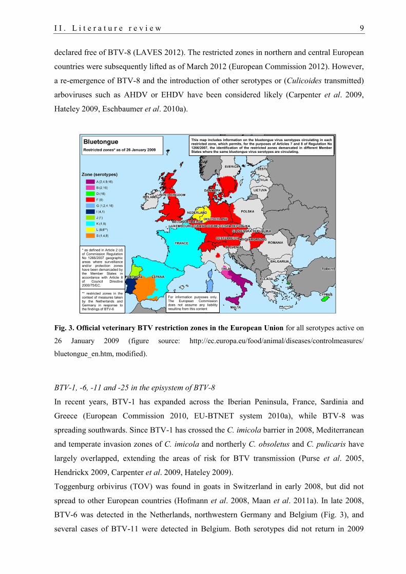
declared free of BTV-8 (LAVES 2012). The restricted zones in northern and central European
countries were subsequently lifted as of March 2012 (European Commission 2012). However,
a re-emergence of BTV-8 and the introduction of other serotypes or (Culicoides transmitted)
arboviruses such as AHDV or EHDV have been considered likely (Carpenter et al. 2009,
Hateley 2009, Eschbaumer et al. 2010a).
Fig. 3. Official veterinary BTV restriction zones in the European Union for all serotypes active on
26 January 2009 (figure source: http://ec.europa.eu/food/animal/diseases/controlmeasures/
bluetongue_en.htm, modified).
BTV-1, -6, -11 and -25 in the episystem of BTV-8
In recent years, BTV-1 has expanded across the Iberian Peninsula, France, Sardinia and
Greece (European Commission 2010, EU-BTNET system 2010a), while BTV-8 was
spreading southwards. Since BTV-1 has crossed the C. imicola barrier in 2008, Mediterranean
and temperate invasion zones of C. imicola and northerly C. obsoletus and C. pulicaris have
largely overlapped, extending the areas of risk for BTV transmission (Purse et al. 2005,
Hendrickx 2009, Carpenter et al. 2009, Hateley 2009).
Toggenburg orbivirus (TOV) was found in goats in Switzerland in early 2008, but did not
spread to other European countries (Hofmann et al. 2008, Maan et al. 2011a). In late 2008,
BTV-6 was detected in the Netherlands, northwestern Germany and Belgium (Fig. 3), and
several cases of BTV-11 were detected in Belgium. Both serotypes did not return in 2009
This map includes information on the bluetongue virus serotypes circulating in each restricted zone, which permits, for the purposes of Articles 7 and 8 of Regulation No 1266/2007, the identification of the restricted zones demarcated in different Member States where the same bluetongue virus serotypes are circulating.
* as defined in Article 2 (d) of Commission Regulation No 1266/2007 geographic areas where surveillance and/or protection zones have been demarcaded by the Member States in accordance with Article 8 of Council Directive 2000/75/EC.
** restricted zones in the context of measures taken by the Netherlands and Germany in response to the findings of BTV-6
For information purposes only. The European Commission does not assume any liability resulting from this content
10 I I . L i t e r a t u r e r e v i e w
although no control measures were implemented (Hateley 2009, Eschbaumer et al. 2010a, van
Rijn et al. 2012).
1.3.2. Transmission and overwintering mechanisms of BTV
BTV is almost exclusively transmitted by certain Culicoides spp. In general, BTV
transmission by contact or by animal products such as meat or milk does not occur
(Papadopoulos et al. 2009). However, other routes of transmission are possible. Ticks and
Melophagus ovinus were suggested as mechanical vectors (Wilson et al. 2008, Bouwknegt et
al. 2010). Transplacental transmission was repeatedly reported during the BTV-8 epizootic in
Europe (Menzies et al. 2008, De Clercq et al. 2008a, De Clercq et al. 2008b, Backx et al.
2009, Darpel et al. 2009a, Williamson et al. 2010, van der Sluijs et al. 2011). In the past,
transplacental infection of BTV was associated with laboratory-adapted BTV strains, in
particular live attenuated vaccine virus strains, but not with wild-type viruses. Recently,
transplacental transmission to a llama was suggested for a wild-type BTV-1 for the first time
(Meyer et al. 2009, EFSA Report 2011). The detection and isolation of BTV from calves born
from a BTV seropositive but BTV RNA negative cow demonstrated that pregnant dams that
were infected during gestation pose a risk when transported to BT-free regions. Therefore,
more severe trade restrictions for pregnant dams are required (see II.5) (De Clercq et al.
2008a). Regarding the significance of BTV shedding in semen, opinions are contradictory
(Wrathall et al. 2006, Napp et al. 2011).
Possible overwintering mechanisms of BTV include the transmission paths already
mentioned in this section. Purse et al. (2005) provided an overview of possible overwintering
mechanisms of BTV. Whether transovarial transmission is possible in the vector is still under
debate (Mellor et al. 2000, White et al. 2005, Purse et al. 2005).
The prolonged viraemia characteristic for BTV infection in ruminants facilitates the
transmission of BTV to Culicoides after the cold winter months when no or only a few adult
Culicoides are active. Unlike former opinions, a vector-free period does generally not exist in
temperate regions, especially in warm winters with a short cold period such as the first winter
after the emergence of BTV-8 to Northern Europe (2006/2007) (Wilson et al. 2008). Warmer
temperatures in stables additionally increase and protract the risk of BTV transmission during
cold weather conditions.
A possible role of the spleen for BTV persistence in ruminants was ruled out as virus isolation
and immunofluorescence labelling, which indicate virus replication, were markedly reduced
or failed (Mahrt and Osburn 1986, reviewed by MacLachlan et al. 2009, Darpel et al. 2009b,
I I . L i t e r a t u r e r e v i e w 11
Worwa et al. 2010, Darpel et al. 2012). Vascular endothelial cells (ECs) and agranular
leukocytes in the skin were suggested as a major source of BTV infection for Culicoides
(Darpel et al. 2012). Therefore, persistent infection of γδ T-cells in the skin could be an
efficient overwintering mechanism, but has so far not been confirmed (Takamatsu et al. 2003,
Purse et al. 2005, Lunt et al. 2006, Wilson et al. 2008, López-Olvera et al. 2010, Darpel et al.
2012).
2. Culicoides biting midges
2.1. Taxonomy and morphology of ceratopogonids
Ceratopogonidae comprise some 125 genera with about 5,500 species (Mellor et al. 2000). In
Germany, four subfamilies (Palpomyiinae, Ceratopogoninae, Dasyheleinae and
Forcipomyiinae) with 332 ceratopogonid species were identified by Havelka and Aguilar
(1999; cited by Werner and Kampen 2007). Culicoides are 0.5 to 3 mm small biting midges
that belong to the family Ceratopogonidae (Diptera) (Mellor et al. 2000, Werner and Kampen
2007). Over 1,400 species belonging to the genus Culicoides (subfamily Ceratopogoninae)
are known to occur worldwide, except in the extreme polar regions, New Zealand, Patagonia
and the Hawaiian Islands (Mellor et al. 2000, Borkent 2012).
Ceratopogonids can be identified by their piercing-sucking mandibles, veining of the (often
hairy) wings, genitalia, shaping of legs and habitat preferences. Males can usually be
differentiated from females by their bushy antennae (Werner and Kampen 2007, Mehlhorn et
al. 2009c). Species are morphologically differentiated from each other by wing patterns. For
further details on morphological characteristics, see more specialist literature (Werner and
Kampen 2007). Wings of females of sibling/complex species look very similar or cannot be
morphologically discriminated from each other. Two species complexes of Palearctic
Culicoides predominantly occur in Germany (Hoffmann et al. 2009a) and in some southern
and eastern European countries (Purse et al. 2005): The C. obsoletus sensu lato (Obsoletus
complex) and C. pulicaris s.l. (Pulicaris complex) comprising three (C. obsoletus sensu
stricto, C. scoticus, C. chiopterus) and six (C.pulicaris s.s., C. punctatus, C. impunctatus, C.
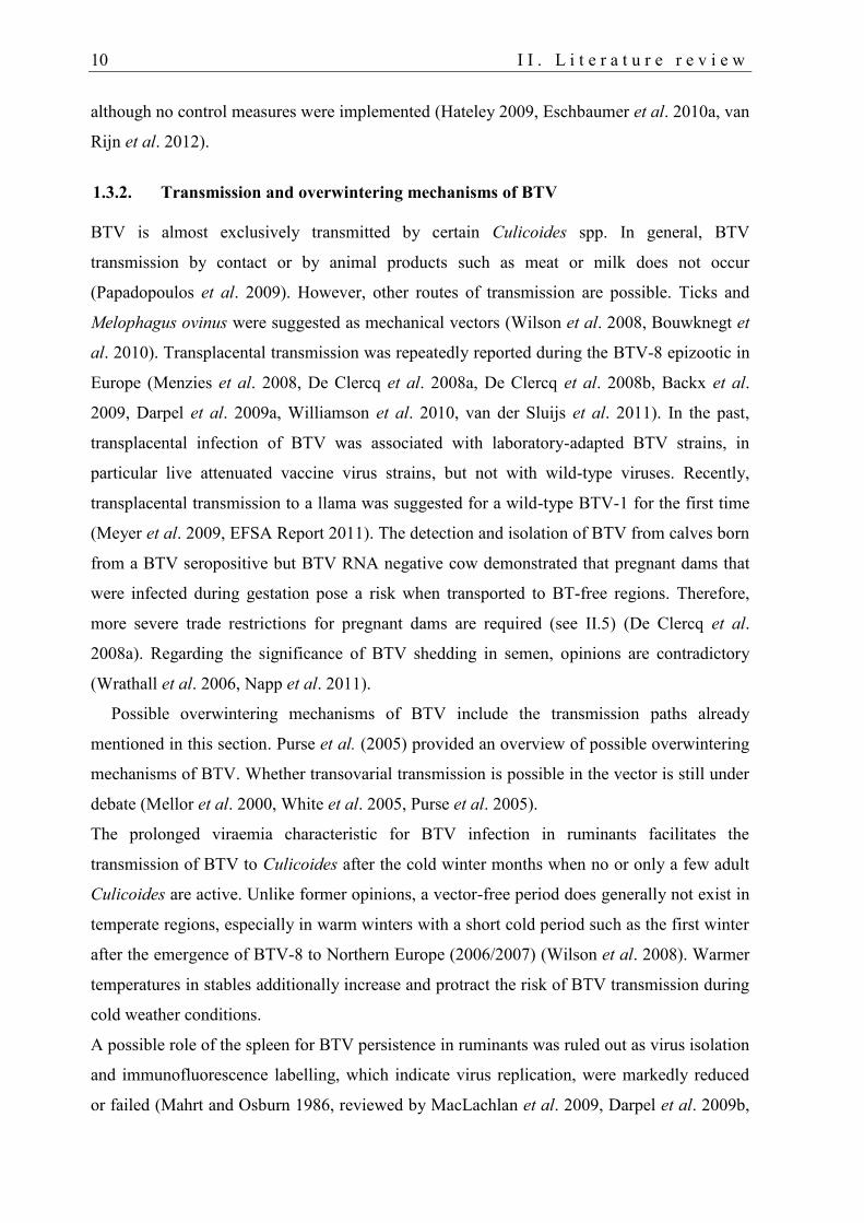
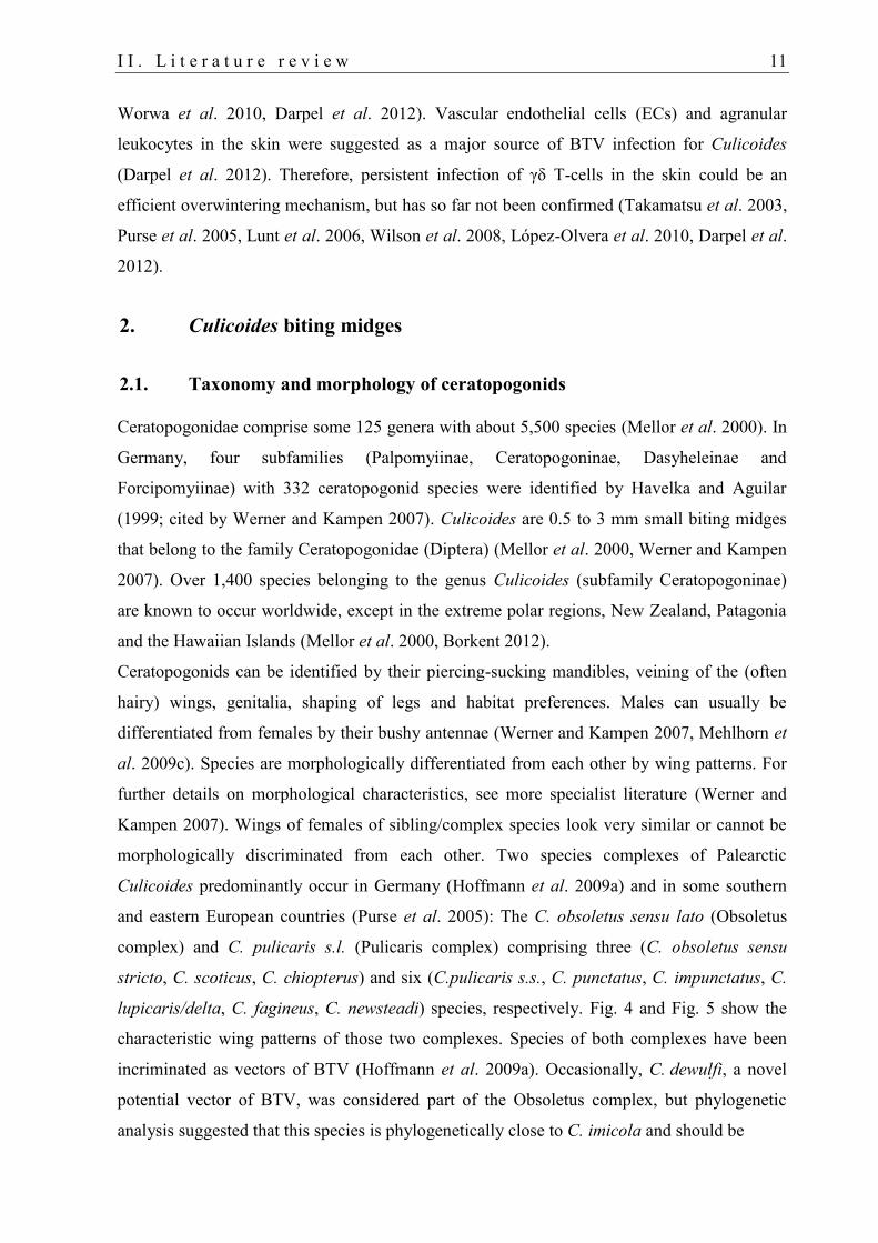
lupicaris/delta, C. fagineus, C. newsteadi) species, respectively. Fig. 4 and Fig. 5 show the
characteristic wing patterns of those two complexes. Species of both complexes have been
incriminated as vectors of BTV (Hoffmann et al. 2009a). Occasionally, C. dewulfi, a novel
potential vector of BTV, was considered part of the Obsoletus complex, but phylogenetic
analysis suggested that this species is phylogenetically close to C. imicola and should be
12 I I . L i t e r a t u r e r e v i e w
Fig. 4. Culicoides obsoletus female, a potential vector of BTV, engorged with fresh blood.
Fig. 5. Culicoides wings. Light micrographs (left side) and diagrammatic representations of the wings
of C. obsoletus and C. pulicaris (figure source: Mehlhorn et al. 2007, modified). In both complexes
the second radial cell of the wing is partly or completely pale (A). C. obsoletus s.l. has wings with
indistinct pale markings or pale areas, while species belonging to the Pulicaris complex present wings
with distinct dark markings on a pale ground, an hourglass-shaped marking after the costa (B) and a
dark spot in the cubital cell (C) (www.culicoides.net).
treated as a separate taxonomic group (Dewulfi complex) (Meiswinkel et al. 2004,
Schwenkenbecher et al. 2009a). The major Old World vector of BTV, the C. imicola species
complex, is currently represented by 10 species (Sebastiani et al. 2001, Purse et al. 2005).
This complex mainly occurs in Africa, Asia and also in southern Europe, but has so far not
I I . L i t e r a t u r e r e v i e w 13
been detected north of the Alps (Purse et al. 2005, Kaufmann et al. 2009, Hoffmann et al.
2009a).
To identify females to species level, cytotaxonomic, biochemical or molecular biological
methods are used (Werner and Kampen 2007) (see also II.2.4). In contrast, males can be
determined to species by inspection of their genitalia (Boorman 1986, Pili et al. 2010, Vilar et
al. 2011).
2.2. Biology and breeding habitats of ceratopogonids
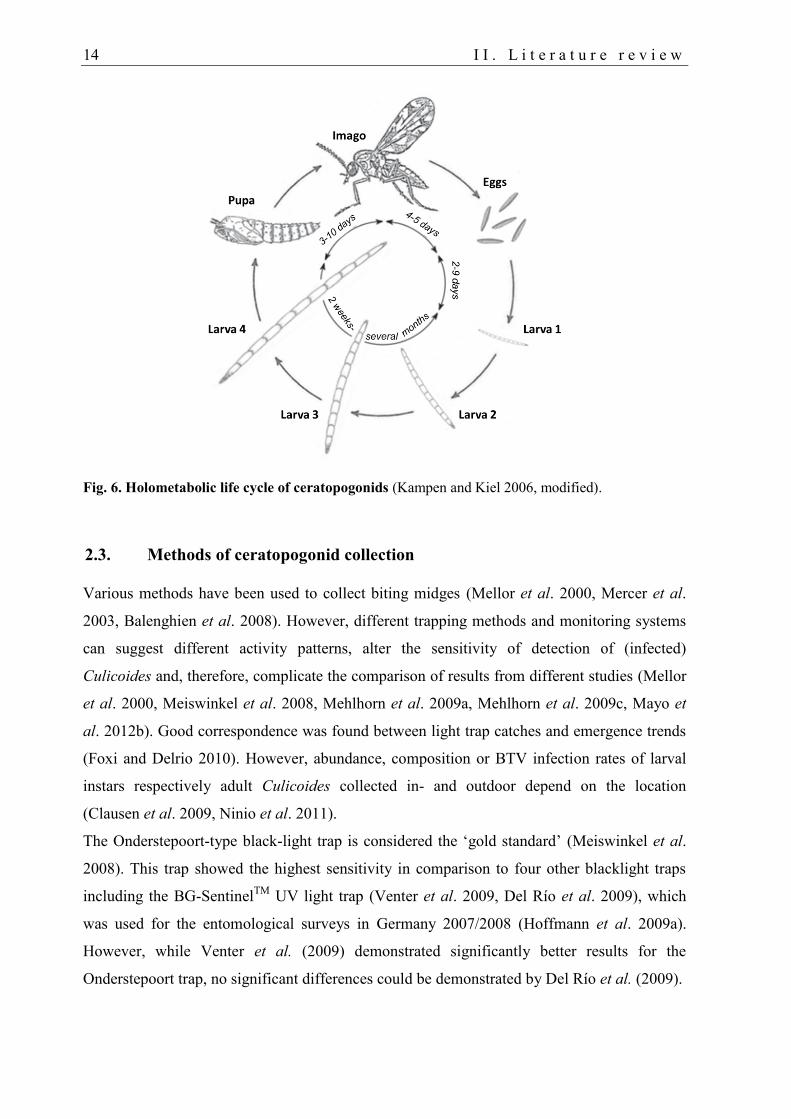
The development of ceratopogonids from egg to imago depends on temperature and takes at
least three weeks in temperate regions, but can be prolonged to more than seven months
during winter. See Fig. 6 for the life cycle (Kampen and Kiel 2006, Mehlhorn et al. 2007,
Kampen et al. 2007). Similar to many other insect families, overwintering takes place in the
fourth larval stage (Werner and Kampen 2007).
The four larval instars and the pupal stage require a certain amount of moisture to avoid
desiccation and to facilitate fast development. For the same reason, adult Culicoides are
mainly active from dusk until dawn when temperatures are lower and humidity is higher. The
incidence of Culicoides can be affected by nearby surface water or marshland as Culicoides
use this for breeding (Kampen and Kiel 2006, Kampen et al. 2007, Werner and Kampen 2007,
Purse et al. 2012). Habitats and larval developmental substrates depend upon species and
range widely, from the tropics to the tundra and from sea level up to 4000 m (Mellor et al.
2000, Mercer et al. 2003, Kaufmann et al. 2009, Tschuor et al. 2009, Foxi and Delrio 2010,
Ninio et al. 2011). Sibling species are morphologically identical or very similar (see II.2.1).
However, their behaviour, (breeding) habitat preferences and other biological characteristics
can be very distinct (Werner and Kampen 2007, Mullen 2009).
Culicoides usually stay a few hundred meters around their breeding habitats, if not transported
as aerial plankton over much greater distances by winds (Mellor 2001, Alba et al. 2004, Purse
et al. 2005, Gloster et al. 2007, Hendrickx et al. 2008, Hendrickx 2009, García-Lastra et al.
2012). Since females of most (haematophagous) species (96%) need blood for egg
production, habitats are regularly near vertebrate hosts (Mellor et al. 2000). For example, the
taxons C. dewulfi and C. chiopterus breed and overwinter in cattle and horse dung, C.
obsoletus and C. scoticus in maize silage residues (Meiswinkel et al. 2008, Balenghien et al.
2008, Zimmer et al. 2008).
14 I I . L i t e r a t u r e r e v i e w
Fig. 6. Holometabolic life cycle of ceratopogonids (Kampen and Kiel 2006, modified).
2.3. Methods of ceratopogonid collection
Various methods have been used to collect biting midges (Mellor et al. 2000, Mercer et al.
2003, Balenghien et al. 2008). However, different trapping methods and monitoring systems
can suggest different activity patterns, alter the sensitivity of detection of (infected)
Culicoides and, therefore, complicate the comparison of results from different studies (Mellor
et al. 2000, Meiswinkel et al. 2008, Mehlhorn et al. 2009a, Mehlhorn et al. 2009c, Mayo et
al. 2012b). Good correspondence was found between light trap catches and emergence trends
(Foxi and Delrio 2010). However, abundance, composition or BTV infection rates of larval
instars respectively adult Culicoides collected in- and outdoor depend on the location
(Clausen et al. 2009, Ninio et al. 2011).
The Onderstepoort-type black-light trap is considered the ‘gold standard’ (Meiswinkel et al.
2008). This trap showed the highest sensitivity in comparison to four other blacklight traps
including the BG-SentinelTM
UV light trap (Venter et al. 2009, Del Río et al. 2009), which
was used for the entomological surveys in Germany 2007/2008 (Hoffmann et al. 2009a).
However, while Venter et al. (2009) demonstrated significantly better results for the
Onderstepoort trap, no significant differences could be demonstrated by Del Río et al. (2009).
I I . L i t e r a t u r e r e v i e w 15
2.4. Molecular identification of Culicoides
Morphological identification of Culicoides spp. is laborious using morphological keys, which
mainly refer to wing patterns, reproductive organs or papal segments (see II.2.1). Therefore,
various molecular methods have been developed to facilitate and speed up the accurate
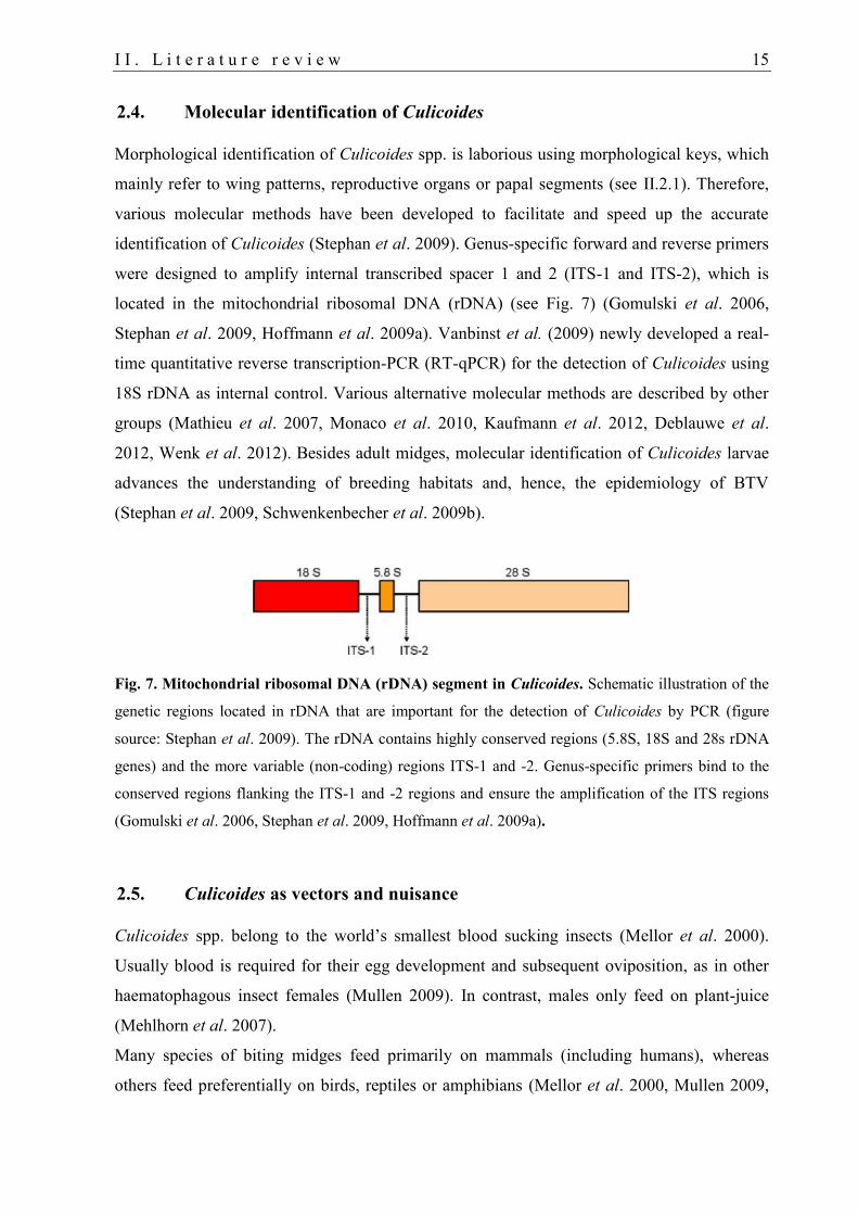
identification of Culicoides (Stephan et al. 2009). Genus-specific forward and reverse primers
were designed to amplify internal transcribed spacer 1 and 2 (ITS-1 and ITS-2), which is
located in the mitochondrial ribosomal DNA (rDNA) (see Fig. 7) (Gomulski et al. 2006,
Stephan et al. 2009, Hoffmann et al. 2009a). Vanbinst et al. (2009) newly developed a real-
time quantitative reverse transcription-PCR (RT-qPCR) for the detection of Culicoides using
18S rDNA as internal control. Various alternative molecular methods are described by other
groups (Mathieu et al. 2007, Monaco et al. 2010, Kaufmann et al. 2012, Deblauwe et al.
2012, Wenk et al. 2012). Besides adult midges, molecular identification of Culicoides larvae
advances the understanding of breeding habitats and, hence, the epidemiology of BTV
(Stephan et al. 2009, Schwenkenbecher et al. 2009b).
Fig. 7. Mitochondrial ribosomal DNA (rDNA) segment in Culicoides. Schematic illustration of the
genetic regions located in rDNA that are important for the detection of Culicoides by PCR (figure
source: Stephan et al. 2009). The rDNA contains highly conserved regions (5.8S, 18S and 28s rDNA
genes) and the more variable (non-coding) regions ITS-1 and -2. Genus-specific primers bind to the
conserved regions flanking the ITS-1 and -2 regions and ensure the amplification of the ITS regions
(Gomulski et al. 2006, Stephan et al. 2009, Hoffmann et al. 2009a).
2.5. Culicoides as vectors and nuisance
Culicoides spp. belong to the world’s smallest blood sucking insects (Mellor et al. 2000).
Usually blood is required for their egg development and subsequent oviposition, as in other
haematophagous insect females (Mullen 2009). In contrast, males only feed on plant-juice
(Mehlhorn et al. 2007).
Many species of biting midges feed primarily on mammals (including humans), whereas
others feed preferentially on birds, reptiles or amphibians (Mellor et al. 2000, Mullen 2009,
16 I I . L i t e r a t u r e r e v i e w
Lassen et al. 2012). Some species are quite host-specific, while others are considered
generalists and feed on alternative hosts of different species or classes.
Culicoides are pool feeders as they lacerate the skin to feed on the accruing effusion including
blood, lymph and skin cells (Darpel et al. 2012). Their bites can be very painful (Mullen
2009) and may cause strong allergic dermatitis in horses (known as ‘sweet itch’) (Heimann et
al. 2011, Schaffartzik et al. 2011) and in ruminants (Connan and Lloyd 1988, Yeruham et al.
1993, Corrêa et al. 2007). The appearance of a huge number of Culicoides can also be a
serious nuisance for humans (Santamaría et al. 2008, Logan et al. 2009).
Only about 50 species of the approximately 1,400 Culicoides spp. are potential vectors of
various pathogens, including filaria and protozoa. However, they attain greatest importance as
vectors of viruses. To date, over 60 viruses have been isolated worldwide (Mellor et al. 2000,
Purse et al. 2005, Werner and Kampen 2010). Eight viruses belonging to the Bunyaviridae,
Rhabdoviridae and Reoviridae families are important for ruminants (Stephan et al. 2009,
Reeves 2010). Recently, a new Orthobunyavirus, “Schmallenberg virus” (SBV), has been
discovered in ruminants and in the species C. obsoletus and C. dewulfi in northern Europe
(Hoffmann et al. 2012, ECDC 2012, Rasmussen et al. 2012). Camelids are also susceptible to
infection with SBV and other orthobunyaviruses (Jack et al. 2012, Schulz et al. 2012a).
BTV in Culicoides vectors
The distribution and seasonality of BT depends on the presence, activity and vector capacity
of Culicoides (Losson et al. 2007, Gloster et al. 2007, Wilson et al. 2007, Baldet et al. 2008,
Hoffmann et al. 2008, Hoffmann et al. 2009a). On a world-wide scale, the most important
BTV vectors are C. sonorensis in North America, C. insignis in Central and South America,
C. imicola and possibly C. bolitinos in Africa, C. brevetarsis, C. fulvus, C. wadai, C. actoni in
Australia, C. imicola and the latter four species in Asia, and C. imicola, C. obsoletus, C.
pulicaris in Europe, (Tabachnick 2004, Hoffmann et al. 2009a, Mellor et al. 2009b). C.
dewulfi was found to be a potential new vector of BTV in northern Europe (Meiswinkel et al.
2007, Stephan et al. 2009).
BTV is not or very rarely transmitted during the winter months (Losson et al. 2007, Gloster et
al. 2007, Wilson et al. 2007, Baldet et al. 2008, Hoffmann et al. 2008, Hoffmann et al.
2009a). According to recent studies, a vector-free period in winter does not exist in Germany
(Fig. 8) and western adjacent countries, but may occur at certain locations, for example in
central Germany (Meiswinkel et al. 2008, Clausen et al. 2009, Hoffmann et al. 2009a).
I I . L i t e r a t u r e r e v i e w 17
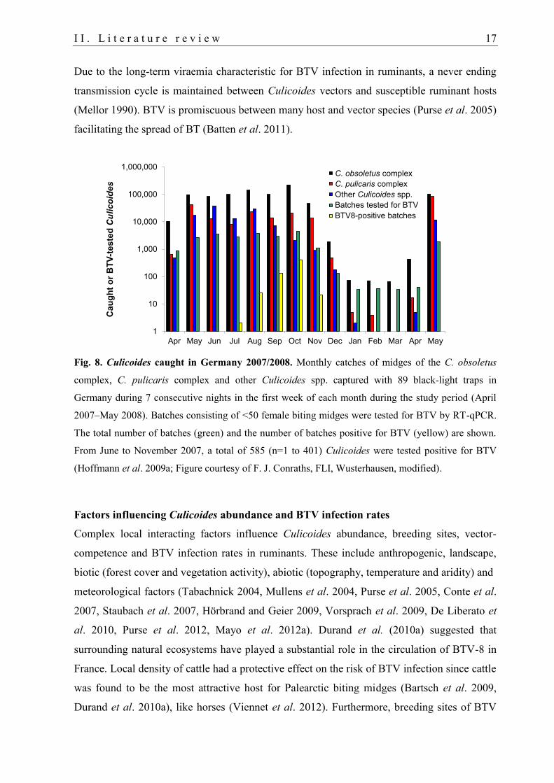
Due to the long-term viraemia characteristic for BTV infection in ruminants, a never ending
transmission cycle is maintained between Culicoides vectors and susceptible ruminant hosts
(Mellor 1990). BTV is promiscuous between many host and vector species (Purse et al. 2005)
facilitating the spread of BT (Batten et al. 2011).
Fig. 8. Culicoides caught in Germany 2007/2008. Monthly catches of midges of the C. obsoletus
complex, C. pulicaris complex and other Culicoides spp. captured with 89 black-light traps in
Germany during 7 consecutive nights in the first week of each month during the study period (April
2007–May 2008). Batches consisting of <50 female biting midges were tested for BTV by RT-qPCR.
The total number of batches (green) and the number of batches positive for BTV (yellow) are shown.
From June to November 2007, a total of 585 (n=1 to 401) Culicoides were tested positive for BTV
(Hoffmann et al. 2009a; Figure courtesy of F. J. Conraths, FLI, Wusterhausen, modified).
Factors influencing Culicoides abundance and BTV infection rates
Complex local interacting factors influence Culicoides abundance, breeding sites, vector-
competence and BTV infection rates in ruminants. These include anthropogenic, landscape,
biotic (forest cover and vegetation activity), abiotic (topography, temperature and aridity) and
meteorological factors (Tabachnick 2004, Mullens et al. 2004, Purse et al. 2005, Conte et al.
2007, Staubach et al. 2007, Hörbrand and Geier 2009, Vorsprach et al. 2009, De Liberato et
al. 2010, Purse et al. 2012, Mayo et al. 2012a). Durand et al. (2010a) suggested that
surrounding natural ecosystems have played a substantial role in the circulation of BTV-8 in
France. Local density of cattle had a protective effect on the risk of BTV infection since cattle
was found to be the most attractive host for Palearctic biting midges (Bartsch et al. 2009,
Durand et al. 2010a), like horses (Viennet et al. 2012). Furthermore, breeding sites of BTV
1
10
100
1,000
10,000
100,000
1,000,000
Apr May Jun Jul Aug Sep Oct Nov Dec Jan Feb Mar Apr May
Cau
gh
t o
r B
TV
-teste
d Culicoides
C. obsoletus complex
C. pulicaris complex
other Culicoides spp.
batches tested for BTV
BTV8-positive batches
C. obsoletus complex
C. pulicaris complex
Other Culicoides spp.
Batches tested for BTV
BTV8-positive batches
18 I I . L i t e r a t u r e r e v i e w
vectors in northern Europe were found mainly in anthropogenic environments, close to farms
(Zimmer et al. 2008, Meiswinkel et al. 2008).
Activity and number of Culicoides are positively correlated with temperature (Mellor et al.
2000, Kiel et al. 2009, Vorsprach et al. 2009). Adult Culicoides are short-lived. Most
individuals probably survive for about 10 to 20 days and during this time may take multiple
blood meals (Mellor et al. 2000). Exceptionally they may live for much longer periods. At
temperatures below about 10°C, the activity of Culicoides is generally suppressed (Mellor et
al. 2000).
BTV transmission becomes possible after the extrinsic incubation period (EIP), which is
the interval between ingestion of BTV and its release in the saliva (Mellor et al. 2009b). After
this period, vectors usually remain infected for life (Mellor 1990). A single bite of an infected
culicoid midge suffices for BTV transmission (Baylis et al. 2009). The successful completion
and time of the EIP is influenced by several internal and external factors (Mellor et al. 2009b)
and is restricted to a relatively small number of species (Mellor 1990). C. obsoletus/scoticus
occurs at comparable low temperatures (Kampen et al. 2007) and was caught all year round at
some locations in Germany, independently from their position above sea level (Hoffmann et
al. 2009a, Mehlhorn et al. 2009c). Nevertheless, the number of C. obsoletus correlated with
temperature, which could explain their significantly lower abundance at higher places
(Mehlhorn et al. 2009c). C. pulicaris incidence depends on a high, stable relative humidity,
prevails with increasing altitude, and shows a bimodal seasonal dynamic (maximum reached
in spring and autumn) in some regions (Kampen et al. 2007, Kaufmann et al. 2009, Tschuor
et al. 2009). In Europe, seasonal activity and number of peaks vary and can be caused by
different generations (Boorman 1986, Vorsprach et al. 2009).
Virus transmission and replication in BTV vectors (vector competence) is crucially
affected by temperature (Wittmann et al. 2002, Paweska et al. 2002, Carpenter et al. 2011).
The minimum temperature for BTV replication in vectors lies between approximately 10 and
15 °C, while at a temperature of 25 °C transmission becomes possible at 7 to 15 days post
infection (dpi) (Mellor et al. 2000, Kampen et al. 2007, Mullen 2009, Mellor et al. 2009b).
Interestingly, the gut wall of the non-vector species C. nubeculosus got permeable for BTV
when it was exposed to a temperature of 33°C (Mellor et al. 2000, Mellor et al. 2009b). Thus,
the unusually warm weather conditions in Europe may have caused an increase in vector-
competence of C. obsoletus and C. pulicaris (Mellor et al. 2000, Mellor et al. 2009b). A
marked increase of BT incidence was found in regions in Europe where climate warming was
greatest (Purse et al. 2005).
I I . L i t e r a t u r e r e v i e w 19
3. BTV in the mammalian host
3.1. Pathogenesis of BTV infection in ruminants
Pathogenesis and clinical signs are generally similar in ruminants infected with virulent BTV
strains and have been thoroughly reviewed by several authors (Verwoerd and Erasmus 2004,
MacLachlan et al. 2009, MacLachlan and Gard 2009, Darpel et al. 2009b, Eschbaumer et al.
2010a, MacLachlan 2011).
After inoculation of BTV in ruminant skin with saliva by a single bite of a Culicoides midge
(Wilson and Mellor 2008, Baylis et al. 2009), BTV primarily targets conventional dendritic
cells in skin lymph, which are contributing to the primary dissemination of BTV from the skin
to draining lymph nodes (Hemati et al. 2009). Early replication of BTV simultaneously (≤ 2-3
dpi) takes place in regional capillary ECs of the skin and in agranular mononuclear leukocytes
(peripheral blood mononuclear cells, PBMC) of lymphatic organs (Darpel et al. 2012). IFN-α
is induced by BTV and plays an essential role in the antiviral innate immune response leading
to the decrease after the first viraemic peak (Foster et al. 1991, Calvo-Pinilla et al. 2009a).
The second and higher viraemic peak was previously explained by massive secondary
replication in PBMC and ECs of several organs resulting in a generalised viraemia
(MacLachlan et al. 1990, Barratt-Boyes and MacLachlan 1994). However, tissue tropism and
“organ-manifestation” were recently suggested to explain virus replication in skin, lymph
nodes of the head, tonsils and labia in the early stage of infection. In contrast, ECs of less
susceptible organs (for example heart, muscle and liver) may be infected when viraemia
reaches a high level (Darpel et al. 2012). The reduction of the second peak is probably due to
a combination of IFN activity and seroconversion together with T-cell mediated immune
response (Foster et al. 1991, Channappanavar et al. 2012).
Since Culicoides lacerate the skin to feed from a blood pool mixed with skin cells and lymph,
they may already be infected 2-3 dpi when systemic viraemia is still low in the mammalian
host.
BTV is highly cell-associated and replicates in a wide range of cell types and at various
temperatures, facilitating infection and replication in both the Culicoides and the mammalian
host (Wilson et al. 2007, MacLachlan et al. 2009, Darpel et al. 2009b, Sánchez-Cordón et al.
2010). In contrast, BTV does not replicate in red blood cells (RBC) but persists within
invaginations of the cell membranes throughout viraemia. This mechanism protects the
adsorbed virions from immune clearance (Barratt-Boyes and MacLachlan 1994, Brewer and
MacLachlan 1994). Prolonged viraemia and co-circulation with neutralising antibodies is
20 I I . L i t e r a t u r e r e v i e w
characteristic for BTV infection in ruminants. The duration of viraemia corresponds to the
lifespan of RBC (Brewer and MacLachlan 1994, Katz et al. 1994, Bonneau et al. 2002).
Infectious virus can be isolated up to about 60 dpi (occasionally up to 100 dpi), while BTV
RNA was detected about 160 up to 222 dpi using RT-PCR (Katz et al. 1994, Barratt-Boyes
and MacLachlan 1994, Bonneau et al. 2002, Mertens et al. 2009). Adsorption of the virus to
RBC over several weeks increases the likelihood of BTV infection of haematophagous
Culicoides vectors and facilitates onward transmission (Barratt-Boyes and MacLachlan 1994,
MacLachlan et al. 2009). In general, the time of a possible BTV infection of Culicoides is in
accordance with the duration of viraemia - when infectious virus is detectable - in ruminant
blood (Bonneau et al. 2002).
BTV induces apoptosis and/or necrosis in mammalian cell lines, microvascular ECs and
monocytes, but not in insect cells, γδ T-cells and activated blood lymphocytes (DeMaula et al.
2001, Takamatsu et al. 2003, Mortola et al. 2004, Schwartz-Cornil et al. 2008, Drew et al.
2010a). The damage of the endothelial lining in small blood vessels results in haemorrhages
and tissue infarction (thrombosis). In white-tailed deer, BTV occasionally causes consumptive
coagulopathy (Vosdingh et al. 1968, Howerth et al. 1988, DeMaula et al. 2002, Verwoerd and
Erasmus 2004, MacLachlan et al. 2009, MacLachlan and Gard 2009). Additionally, activation
of ECs and macrophages induces the production of vasoactive mediators, which can promote
cell death or cause vascular permeability with subsequent oedema, in particularly pulmonary
oedema (DeMaula et al. 2001, MacLachlan et al. 2009, Mortola and Larsen 2010, Drew et al.
2010b, Sánchez-Cordón et al. 2012). BTV-infected monocytes and lymphocytes also respond
with inflammatory and antiviral responses (MacLachlan and Gard 2009, Ross- Smith et al.
2009b). Lymphopenia, a common sign of BT, possibly occurs due to destruction or
sequestration of lymphocytes at virus replication sites (Darpel et al. 2009b).
Neutralising antibodies (NAbs) recognise epitopes of the serotype-specific outer capsid
proteins and, therefore, protect against challenge with the homologous serotype. However,
other factors contribute to the protection against BTV re-infection. Cross-protection against
heterologous serotypes is mediated by cytotoxic T-cells (CTL), which recognise intracellular
antigens of the highly conserved NSs and viral core proteins via the major histocompatibility
complex (MHC) pathway (Jeggo et al. 1984, Darpel et al. 2009b, Sánchez-Cordón et al.
2010, Hund et al. 2012). Therefore, ruminants that have previously been infected or
immunised can be protected against BTV re-infection, even if they are negative for NAbs
(Savini et al. 2004b, Eschbaumer et al. 2009, Savini et al. 2009, Oura et al. 2010).
I I . L i t e r a t u r e r e v i e w 21
3.2. Clinical signs and post-mortem lesions in ruminants
Host range
BT manifestation depends on species, breed, age, sex, individual resistance and fitness of the
mammalian host and on virus strain (Sellers 1984, Ward et al. 1994, Brodie et al. 1998, Thiry
et al. 2006, reviewed by MacLachlan et al. 2009, Savini et al. 2010, Linden et al. 2010,
García-Bocanegra et al. 2011). The host range susceptible to BTV infection includes various
domestic (sheep, goats and cattle) and wild ruminant species (deer, mouflon, ibex, yak,
European and American bison and musk ox) as well as camelids (House et al. 1982, Erasmus
1990, Tessaro and Clavijo 2001, Mauroy et al. 2008, Ludwig and Silinski 2008, reviewed by
MacLachlan et al. 2009, García et al. 2009). BTV primarily causes disease in sheep. In
contrast, cattle, goats, various species of wild ruminants and camelids usually exhibit
subclinical infection (Rivera et al. 1987, Mattson 1994, Darpel et al. 2007, Backx et al. 2007,
MacLachlan et al. 2008, Ruiz-Fons et al. 2008, Fernandez-Pacheco et al. 2008, Mauroy et al.
2008, Wernery et al. 2008, Elbers et al. 2008b, MacLachlan et al. 2009, Dal Pozzo et al.
2009a, 2009b, López-Olvera et al. 2010, Linden et al. 2010, Batten et al. 2011). However, the
pathogenesis of BTV infection is similar in sheep and cattle. Inherent species-specific
differences in the production and activities of EC-derived mediators were incriminated to
contribute to the sensitivity of sheep and deer to BTV-induced microvascular injury
(DeMaula et al. 2001, MacLachlan and Gard 2009). Differences in the susceptibility of
mammalian hosts to BT disease probably depend on the variability of toll-like receptor 3
(TLR3) expression in tissues (Vos et al. 2009). A significantly higher ratio of thromboxane to
prostacyclin has been found in sheep indicating enhanced coagulation (disseminated
intravascular coagulation, DIC) and a subsequent bleeding tendency (DeMaula et al. 2002,
MacLachlan and Gard 2009).
BTV infection in non-artiodactyls was reported in domestic and wild carnivores and
rhinoceros (Akita et al. 1994, Evermann et al. 1994, Osburn 1994, Alexander et al. 1994,
Ianconescu et al. 1996, Fischer-Tenhagen et al. 2000, Jauniaux et al. 2008, Oura and El
Harrak 2010). Carnivores may be infected by oral ingestion of infected meat or meat products
or through vector feeding (Alexander et al. 1994, Oura and El Harrak 2010). To date, their
role in the transmission cycle of BTV remains unknown (Oura and El Harrak 2010), but it is
unlikely that carnivores play a significant role in the epidemiology of BTV (MacLachlan
2011).
22 I I . L i t e r a t u r e r e v i e w
Clinical signs
Clinical signs and lesions reflect virus-mediated vascular injury and subsequent immune and
repair responses after BTV infection. None of the clinical signs are pathognomonic for BT
(Mertens et al. 2009). Animals with acute BT can have any combination of signs
(MacLachlan and Gard 2009), and secondary infection such as bacterial pneumonia may
aggravate BT disease and subsequently promote fatality (MacLachlan and Gard 2009).
Therefore, other diseases with similar clinical signs should be considered (Mertens et al.
2009). Suspicion of BTV infection and disease has to be confirmed by diagnostic test
procedures (see II.4). A list of possible differential diagnoses was given by several authors
(Verwoerd and Erasmus 2004, Bexiga et al. 2007, Williamson et al. 2008, Mertens et al.
2009, OIE 2011).
In contrast to previously known BTV strains, a remarkable high proportion of cattle
showed overt clinical signs to infection with the European BTV-8, indicating an increased
virulence of this strain (Thiry et al. 2006, Elbers et al. 2008a, Dal Pozzo et al. 2009a,
reviewed by Dal Pozzo et al. 2009b). Nevertheless, clinical signs and lesions in sheep were
much more prominent and different than in cattle (Elbers et al. 2008b). Clinical signs
indicative for BTV-8 infection in affected sheep flocks were erosions of the oral mucosa,
fever, salivation, facial and mandibular oedema, apathy and tiredness, oedema of the lips,
lameness and dysphagia were among the clinical signs most frequently recorded. In contrast,
the most prominent clinical signs in affected cattle herds included crusts/lesions of the nasal
mucosa, erosions of lips/crusts in or around the nostrils, erosions of the oral mucosa,
salivation, fever, conjunctivitis, coronitis, muscle necrosis and stiffness in limbs (Elbers et al.
2008b).
Besides, BTV-8 can cause abortion, congenital deformities and cerebral abnormalities leading
to congenital neurological signs, dullness or weakness (Wouda et al. 2008, De Clercq et al.
2008a, Wouda et al. 2009). Congenital deformities in live-born calves mostly occurred after
BTV-8 infection of dams in early gestation (Wouda et al. 2009). However, the highest
transmission rate was found at mid-term gestation (69%) (van der Sluijs et al. 2011).
Post-mortem lesions
Lesions typically found in BTV-infected ruminants at post-mortem examination are
consequences of the damages to the vascular system. These include widespread, but often
localised, hyperaemia, petechiation, haemorrhages, vascular congestion, oedema and
infiltration by inflammatory cells in various tissues (Verwoerd and Erasmus 2004, Darpel et
I I . L i t e r a t u r e r e v i e w 23
al. 2007, MacLachlan et al. 2009, Worwa et al. 2010). Affected organs include lymph nodes,
spleen, musculature, lungs, heart and the digestive tract. Pulmonary oedema and petechiae or
sub-intimal haemorrhages in the tunica of the pulmonary artery near its base are characteristic
for fulminant BT but are not pathognomonic (Verwoerd and Erasmus 2004, Darpel et al.
2007, MacLachlan et al. 2008, MacLachlan et al. 2009, Worwa et al. 2010). Furthermore,
pleural and pericardial effusion, ulcerations and erosions of the mucosa of the upper digestive
tract and aspiration pneumonia associated with oesophageal myonecrosis are possible findings
of a manifest BTV infection (Verwoerd and Erasmus 2004, Darpel et al. 2007, MacLachlan et
al. 2008, Antoniassi et al. 2010).
3.3. Particularities of camelids
Taxonomy and global distribution of South American camelids
Camelidae are even-toed ungulates (Artiodactyla) as are ruminants and pigs, but they belong
to the suborder Tylopoda. This family comprises the genera Camelus (Old World camelids,
OWC), Lama and Vicugna (South American camelids, SAC) (Fowler 2010a).
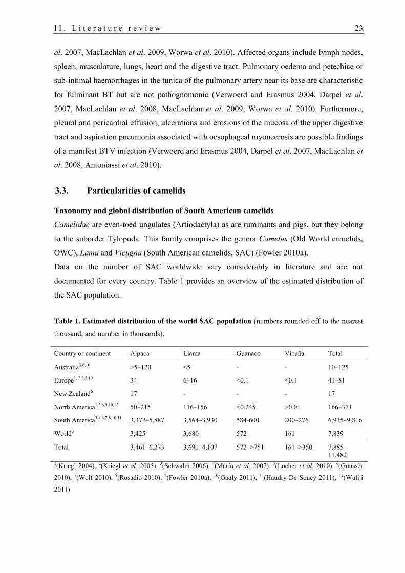
Data on the number of SAC worldwide vary considerably in literature and are not
documented for every country. Table 1 provides an overview of the estimated distribution of
the SAC population.
Table 1. Estimated distribution of the world SAC population (numbers rounded off to the nearest
thousand, and number in thousands).
Country or continent Alpaca Llama Guanaco Vicuña Total
Australia3,6,10
>5–120 <5 - - 10–125
Europe1, 2,3,5,10
34 6–16 <0.1 <0.1 41–51
New Zealand6 17 - - - 17
North America1,3,6,9,10,12
50–215 116–156 <0.245 >0.01 166–371
South America3,4,6,7,8,10,11
3,372–5,887 3,564–3,930 584-600 200–276 6,935–9,816
World3 3,425 3,680 572 161 7,839
Total 3,461–6,273 3,691–4,107 572–>751 161–>350 7,885–
11,482
1(Kriegl 2004),
2(Kriegl et al. 2005),
3(Schwalm 2006),
4(Marín et al. 2007),
5(Locher et al. 2010),
6(Gunsser
2010), 7(Wolf 2010),
8(Rosadio 2010),
9(Fowler 2010a),
10(Gauly 2011),
11(Haudry De Soucy 2011),
12(Wuliji
2011)
24 I I . L i t e r a t u r e r e v i e w
Their global population is estimated about 7.9 to 11.5 million SAC. Most animals are kept in
South America (Table 1). Peru is the major alpaca producer in the world keeping about 3.0 to
5.5 million alpacas and 900,000 to 1.2 million llamas (Schwalm 2006, Gunsser 2010, Rosadio
2010, Wolf 2010, Gauly 2011). In Germany, SAC were first established after 1980 (Fowler
2010a, Gauly 2011). Currently, their number is estimated at 5,000 to 7,000 and 15,000
animals according to Locher et al. (2010) and Gauly et al. (2011), respectively.
Physiological characteristics
Camelids have a unique physiology due to their adaption to hostile environments. SAC
became adapted to South American habitats, especially to the high altitude lands of the
Andes, while OWC adapted to deal with heat and dehydration in a semi-desert environment
(Fowler 2010a).
Camelid erythrocytes are different from RBC of other mammals as they are smaller, very
stable, ellipsoid cells without biconcavity (Smith et al. 1979, Foster et al. 2009, Timm et al.
2011). Differences in the erythrocyte membrane proteins and in the organisation of the
cytoskeleton have been found, indicating that proteins play an important role in stabilising the
camelid elliptocyte (Eitan et al. 1976, Omorphos et al. 1989). The survival time of SAC
erythrocytes is up to 235 days (dependent on the applied method), which is longer than the
lifespan of bovine and ovine RBC (Cornelius and Kaneko 1962, Reynafarje et al. 1968).
In contrast to ruminants, which have a lymphocytic haemogram (>50% lymphocytes)
(Gassmann and Lutz 2010), camelids have a granulocytic haemogram (>50% neutrophil
granulocytes) (Wernery et al. 1999, Fowler 2010b).
Camelids generally have a reduced susceptibility and a rare clinical outcome of bovine and
ovine viral diseases (Wernery et al. 1999, Wernery and Kaaden 2004, Kapil et al. 2009). They
play a minor or negligible role as carriers for important diseases such as foot-and-mouth
disease (reviewed by Wernery and Kaaden 2004) or bovine herpesvirus 1 infection (P. König,
FLI, unpublished observations). Compared to other mammals, camelids have an exceptional
immune system (Wernery et al. 1999, Wernery 2001, Conrath et al. 2003, Wernery and
Kaaden 2004, Vanlandschoot et al. 2011) that might play a role in the outcome of diseases.
Besides conventional immunoglobulin G (IgG), camelids own heavy-chain antibodies
(HCAb; IgG2 and IgG3), special subclasses of IgG that lack the light chains and the first
constant domain of the heavy chain (CH1) but have a normal FC region.
The thick layered epitheliochorial placenta of camelids prevents the transplacental transfer of
immunoglobulins (Ghazi et al. 1994, Wernery 2001, Timm et al. 2011). The newborn cria
I I . L i t e r a t u r e r e v i e w 25
obtains passive immunity by intestinal absorption of primarily IgG antibodies from the
colostrum immediately after birth. Since IgG is predominantly found in camelid colostrum, a
selective transfer of IgG similar to that in bovines was assumed (reviewed by Wernery 2001).
Stable serum IgG concentrations were found around 4 months after birth, indicating that the
immune system has matured (Wernery 2001).
3.3.1. BTV infection in camelids
In camelids, serological evidence for a previous BTV infection was reported on various
continents and for different serotypes. Field studies, case reports and animal experiments on
BTV infection in camelids are listed in Tables 1 and 2 in Appendix 3. Historically, SAC were
considered resistant to BT disease (Rivera et al. 1987, Mattson 1994, Afshar et al. 1995a)
(Table 1 in Appendix 3), while stiffness, dermatitis or trypanosomiasis was occasionally
associated with higher seroprevalences in OWC (Table 1 in Appendix 3). The first suspicion
of clinical BT disease in SAC was reported by Fowler (1998c). Abortion in a llama
concurrently occurred with an episode of respiratory distress, which was followed by a
fourfold increase in BTV antibody titre. During the recent BTV-8 and BTV-1 epizootics in
Europe, BTV RNA and antibodies were found in a few alpacas and llamas in Germany and
France (Henrich et al. 2007, Meyer et al. 2009, Locher et al. 2010), respectively.
Furthermore, clinical disease with subsequent fatality or abortion related to BTV infection
was reported in a few of the latter SAC and in the USA at about the same time (Ortega et al.
2010a). All fatal cases had a brief history of lethargy or weakness, recumbency, anorexia (3 of
4) and acute respiratory distress within 24 hours before death. Necropsy consistently revealed
severe congestion and oedema of the lungs (Henrich et al. 2007, Meyer et al. 2009, Ortega et
al. 2010a). Additionally, ulcers and erosions in the oral cavity (Henrich et al. 2007) and
hydrothorax, hydropericardium and acute subendocardial haemorrhages (Ortega et al. 2010a)
were found in the two alpacas perished in Germany and the USA, respectively. In summary,
clinical signs and post-mortem findings were all suggestive of BT (MacLachlan et al. 2009,
Ortega et al. 2010a) (see also II.3.2). BTV RNA was detected in blood and in various tissue
samples from the two perished llamas and BTV-1 was isolated (Meyer et al. 2009) from lungs
and spleens. Interestingly, virus isolation was also isolated from two llama foetuses
suggesting transplacental transmission of the BTV-1 field strain, which has never been
reported before (EFSA Report 2011).
Despite the high pressure of the two virulent BTV strains in Europe, only these three cases of
BTV-infection have been reported in SAC, indicating that BTV-related fatalities occur rather
26 I I . L i t e r a t u r e r e v i e w
sporadically in SAC. The negative serological results in a survey of BTV-8 in 354 SAC in
Switzerland can be explained by the relatively few BTV cases in ruminants at the time
(Zanolari et al. 2010b). On the other hand, experimental infection of dromedaries with BTV-1
suggested that Old World camelids can play a potential role in the transmission to Culicoides.
Hence, OWC could act as reservoirs to carry BTV over long distances and across boarders
(Batten et al. 2011); similar to wild ruminants (Falconi et al. 2011). Whether SAC may play a
similar role as OWC remained unknown.
4. Laboratory diagnosis of BTV
Antibody detection
BTV antibodies are generally detectable in blood of domestic and wild ruminants, camels and
llamas about 1 to 2 weeks after experimental BTV infection. However, in OWC peak
antibody levels were reached relatively late (after 41 dpi) compared to ruminants (10 dpi in
sheep, 18 dpi in cattle and 17-28 dpi in red deer) (Afshar et al. 1995a, Darpel et al. 2007, Dal
Pozzo et al. 2009a, López-Olvera et al. 2010, Batten et al. 2011).
Agar gel immunodiffusion (AGID) test have mostly been used for international regulatory
purposes (Verwoerd and Erasmus 2004, OIE 2011). However, indirect, double antigen
(double recognition, DR) and competitive (c) ELISAs are superior to AGID. They are robust
and rapid assays for the detection of group-specific (VP7) antibodies in blood and milk
samples with a high specificity and sensitivity (Verwoerd and Erasmus 2004, Kramps et al.
2008, Oura et al. 2009, Eschbaumer et al. 2009, Mertens et al. 2009, Mars et al. 2010).
Furthermore, they have the advantage of species-independent testing of sera. For the detection
of vaccinated animals, the cELISA has been considered less sensitive than the DR ELISAs
(Oura et al. 2009, Eschbaumer et al. 2009).
In contrast to the group-specific ELISAs, serum neutralisation tests (SNT) detect serotype-
specific (VP2) antibodies against a homologous reference strain. NAbs are thought to be the
unique measure to demonstrate protective immunity against re-infection with the homologous
BTV serotype in vitro after BTV vaccination or natural infection (see also II.5.2.2) (Verwoerd
and Erasmus 2004). However, no reports exist about vaccinated animals that were negative in
a DR ELISA but were still protected against challenge infection. Furthermore, SNT often
yielded false negative results for animals exposed to BTV-8, particularly after vaccination and
experimental infection (Worwa et al. 2012).
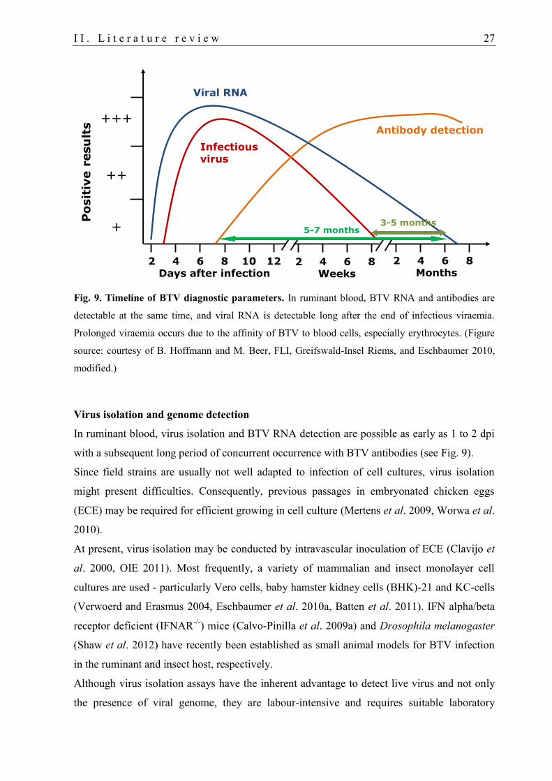
I I . L i t e r a t u r e r e v i e w 27
Fig. 9. Timeline of BTV diagnostic parameters. In ruminant blood, BTV RNA and antibodies are
detectable at the same time, and viral RNA is detectable long after the end of infectious viraemia.
Prolonged viraemia occurs due to the affinity of BTV to blood cells, especially erythrocytes. (Figure
source: courtesy of B. Hoffmann and M. Beer, FLI, Greifswald-Insel Riems, and(Eschbaumer 2010,
modified.)
Virus isolation and genome detection
In ruminant blood, virus isolation and BTV RNA detection are possible as early as 1 to 2 dpi
with a subsequent long period of concurrent occurrence with BTV antibodies (see Fig. 9).
Since field strains are usually not well adapted to infection of cell cultures, virus isolation
might present difficulties. Consequently, previous passages in embryonated chicken eggs
(ECE) may be required for efficient growing in cell culture (Mertens et al. 2009, Worwa et al.
2010).
At present, virus isolation may be conducted by intravascular inoculation of ECE (Clavijo et
al. 2000, OIE 2011). Most frequently, a variety of mammalian and insect monolayer cell
cultures are used - particularly Vero cells, baby hamster kidney cells (BHK)-21 and KC-cells
(Verwoerd and Erasmus 2004, Eschbaumer et al. 2010a, Batten et al. 2011). IFN alpha/beta
receptor deficient (IFNAR-/-
) mice (Calvo-Pinilla et al. 2009a) and Drosophila melanogaster
(Shaw et al. 2012) have recently been established as small animal models for BTV infection
in the ruminant and insect host, respectively.
Although virus isolation assays have the inherent advantage to detect live virus and not only
the presence of viral genome, they are labour-intensive and requires suitable laboratory
2 4 6 8 10 12Days after infection
2 4 6 8 Weeks
2 4 6 8 Months
+++
++
+
Infectiousvirus
Antibody detection
Viral RNA
3-5 months5-7 months
Posit
ive r
esu
lts
28 I I . L i t e r a t u r e r e v i e w
conditions (Mertens et al. 2009). Therefore, more sensitive, specific and standardised RT-
qPCR or RT-nested PCR assays are now widely used for rapid (automated high-throughput)
detection and quantification of very low amounts of BTV RNA (Bonneau et al. 2002,
Toussaint et al. 2007, Hoffmann et al. 2009b, Hoffmann et al. 2009c). Specific probe designs
allow the differentiation of all currently known 26 BTV serotypes (targeting S2) and related
topotypes or group specific genome sequences (targeting S5) to distinguish members of other
Orbivirus species (Toussaint et al. 2007, Vandenbussche et al. 2009, Hoffmann et al. 2009c,
Maan et al. 2012).
Novel approaches are pushed forward to develop pen-side diagnostic tests enabling on-spot
(in vivo) diagnosis in the field to improve decision making on preventive measures in case of
a disease outbreak (Mertens et al. 2009, Saxena et al. 2012).
4.1. BTV diagnosis in camelids
Diagnosis of BTV antibodies in camelids and other non-ruminant species is possible using
methods that are not species-specific, including cELISA, DR ELISA, neutralisation test (NT)
and PCR. Serological (cELISA and NT) and molecular (RT-qPCR and virus isolation on KC-
cells) assays have just recently been validated for OWC (Batten et al. 2011). However, there
is no evidence for a validation of methods to detect BTV infection in SAC, except for Afshar
et al. (1995a), who validated a cELISA.
5. Prevention and control of BT
BT is a notifiable disease (formerly on List A) of the World Organisation for Animal Health
(OIE 2000), according to Council Directive 82/894/EEC (1982) on the notification of animal
diseases within the Community, as it can have substantial economic impact (Schwartz-Cornil
et al. 2008, commission regulation (EC) No 1266/2007). Economic losses are estimated at
approximately $3 billion per year (Mellor and Wittmann 2002).
BT monitoring and surveillance programmes have to include serological, virological and
entomological surveillance in Member States within and outside of restricted zones
(Commission regulation (EC) No 1266/2007, Caporale and Giovannini 2010). However,
control strategies differ depending on whether BT disease is endemic in a region or not. While
eradication is the usual goal in the latter case, this is not possible in areas where BTV is
endemic because of the widely distributed Culicoides vectors and the ubiquitous distribution
of mammalian reservoir hosts (Verwoerd and Erasmus 2004, Dungu et al. 2004).
I I . L i t e r a t u r e r e v i e w 29
BTV-8 inheres three particularities compared to other ‘wild-type’ BTVs (Backx et al.
2007, Dal Pozzo et al. 2009a, Dal Pozzo et al. 2009b) resulting in additional regulations
implemented by the European Commission (EC). This serotype may cause serious disease not
only in sheep but also in the usually subclinically infected cattle and goats. Frozen stored
semen has to be tested free of BTV-8 (Commission regulation (EC) No 1266/2007, Vanbinst
et al. 2010). The risk of transplacental transmission and the possible spread of the disease by
newborn animals in the next vector season have to be precluded. Therefore, all dams have to
be immune to BTV before insemination or mating (Commission Regulation (EC) No
384/2008). Furthermore, pregnant animals are effectively restricted in their movement (EFSA
Report 2011).
5.1. Vector control
Arbovirus-vector relationships are highly dynamic and extremely difficult to combat
(Carpenter et al. 2009). The incursion of any BTV serotype in a region is not predictable as
Culicoides can be transported over long-distances. Furthermore, its establishment in an area
depends on the presence of competent vectors, vertebrate hosts and reservoirs. Possible means
of protection of hosts from Culicoides bites include adulticides such as pour-on insecticides,
which are directly administered on livestock (Mellor and Wittmann 2002, Verwoerd and
Erasmus 2004, Carpenter et al. 2009). However, the application of pyrethroides does not
guarantee protection against Culicoides bites and subsequent BTV infection (Mehlhorn et al.
2008, Schmahl et al. 2009). To control Culicoides vectors (Verwoerd and Erasmus 2004),
modification of habitats, sterilisation of males by irradiation, insecticide-treated netting,
residual insecticides and biological agents may be used (Sampson and Gooday 1998, Mellor
and Wittmann 2002, Carpenter et al. 2009, Calvete et al. 2010). Methods for larvae
destruction include clearance of potential larval development sites and the use of chemical
(American Cyanamid, pyrethroid-based products) or biological (entomopathogenic fungi)
larvicides (Mellor and Wittmann 2002, Carpenter et al. 2009, Ansari et al. 2011).
5.2. BTV vaccination
Vaccination of species susceptible to BTV is considered the tool of choice to control BT and
to facilitate safe trade and movement of live ruminants between BTV-affected and BTV-free
zones (Giovannini et al. 2004b, Savini et al. 2008, Gethmann et al. 2009, commission
regulation (EC) No 1266/2007).
30 I I . L i t e r a t u r e r e v i e w
BTV-8 vaccination
Emergency mass vaccination with any available vaccine has been recognised as the most
efficient measure for BT control by the European Commission and permits safe trade in live
ruminants under certain conditions (Commission regulation (EC) No 1266/2007, Commission
Decision 2008/655/EC, Gethmann et al. 2009, Caporale and Giovannini 2010). However,
considering the numerous drawbacks of modified-live virus (MLV) vaccines discussed in
section II.5.2.2, there is disagreement among experts whether MLV vaccines should be used
in non-endemic areas at all (Caporale and Giovannini 2010). In non-endemic regions, the
common control strategy is eradication (Verwoerd and Erasmus 2004, Dungu et al. 2004).
Therefore, to preclude natural dissemination of the South African BTV-8 MLV vaccine strain,
it was not used during the BTV-8 epizootic in Europe, although this strain has shown a high
efficiency against challenge infection with the European BTV-8 (Dungu et al. 2008, Caporale
and Giovannini 2010).
From spring 2008, the mandatory vaccination programs were successfully implemented using
five monovalent inactivated BTV-8 vaccines: Bluevac®-8, CZ Veterinaria, Spain;
BTVPUR® AlSap 8, Merial, Lyon, France; Zulvac® 8 Bovis and Ovis, Fort Dodge, The
Netherlands and Bovilis® BTV 8, Intervet, Boxmeer, The Netherlands (FLI 2009, Zientara et
al. 2010, Gethmann et al. 2010, EU-BTNET system 2010b, Oura et al. 2012). In Germany, all
but the Bovilis vaccine were used. An overview of vaccination campaigns against BTV-8 in
Europe from 2008 to 2011 is given in Table 1 in Appendix 2.
Vaccination coverage of over 80% of the susceptible ruminant population significantly
reduces BTV circulation, and allows safe movement of ruminants from BTV-affected to
BTV-free areas (Caporale et al. 2004, Patta et al. 2004, Giovannini et al. 2004a, Giovannini et
al. 2004b, 2004c). In Germany, a vaccination coverage of over 80% had been achieved by the
end of 2008 (Gethmann et al. 2010).
Most countries in Europe have reverted to voluntary vaccination campaigns in 2011 (Table 1
in Appendix 2) (Oura 2011), or vaccination was prohibited in areas declared free of BTV
(Anon. 2010b). Since that time, no major BTV outbreaks have been recorded in Europe,
although vaccine coverage has dramatically decreased (Oura 2011). Hence, the risk of a re-
introduction of BTV-8 has been assessed as equally high than the risk of introduction of other
BTV serotypes (FLI 2012). Vaccination before a BTV incursion occurs into a naïve
population has various advantages. Therefore, the EU recently allowed the use of inactivated
vaccines within a BT free area, given that the Member States authorises its use (DEFRA
2012).
I I . L i t e r a t u r e r e v i e w 31
5.2.1. Types of BTV vaccines
In general, three types of vaccines against BTV can be distinguished: live attenuated vaccines,
live recombinant virus-vector vaccines and non-replicating vaccine/adjuvant combinations.
The latter are either inactivated whole virus preparations, virus-like particles (VLP), core-like
particles (CLP) or subunit vaccines (Roy et al. 1990, Lobato et al. 1997, Boone et al. 2007,
Savini et al. 2008, Alpar et al. 2009, Calvo-Pinilla et al. 2009b, Franceschi et al. 2011). The
five BTV-8 vaccines that have been used in Europe are inactivated, monovalent BTV-8
preparations in aqueous solution. The seed viruses for these vaccines were European BTV-8
isolates from the 2006 outbreak in Northern Europe, which were replicated in BHK cell
cultures. To enhance the immune response, aluminium hydroxide and saponin were used as
adjuvants (Gethmann et al. 2009, Eschbaumer et al. 2009, Bruckner et al. 2009b).
Vaccination programs for the control of BT in ruminants have limitations due to difficulties in
differentiating infected from vaccinated animals. To overcome this problem, recent
developments target marker vaccines and test systems that differentiate infected from
vaccinated animals (DIVA). This is possible as vaccinated animals do not elicit a detectable
antibody response to NS of BTV (Alpar et al. 2009, Barros et al. 2009).
5.2.2. Safety and efficacy of live-attenuated and inactivated BTV vaccines
Currently, only MLV and some inactivated vaccines are available under European
Community approved national disease control programs (Savini et al. 2008). MLV as well as
inactivated BTV vaccines have a high level of efficacy to challenge infection, preventing
clinical disease and viraemia in sheep, goats, cattle and red deer (Di Emidio B. et al. 2004,
Savini et al. 2004b, 2008, Eschbaumer et al. 2009, Savini et al. 2009, Wäckerlin et al. 2010,
Bréard et al. 2011, Lorca-Oró et al. 2012). In MLV, however, the level of safety varies
considerably according to the used formulation, BTV serotypes and number of serotypes
included in the vaccine (Savini et al. 2008). Adverse effects include clinical BT disease
(Veronesi et al. 2010) and depressed milk production (Savini et al. 2004c).
Abortion/embryonic death and teratogenesis can occur when these vaccines are used in
pregnant females (Flanagan and Johnson 1995, Savini et al. 2008). Viraemia is a common
finding after vaccination with MLV (Savini et al. 2008), and natural dissemination of vaccine
strains of BTV-16 (Monaco et al. 2006) and BTV-2 (Ferrari et al. 2005) has already been
documented in Europe. Furthermore, MLV have the potential of reassortment (Batten et al.
2008, Maan et al. 2010) and an increase of or reversion to virulence (Batten et al. 2008, Alpar
et al. 2009, Veronesi et al. 2010).
32 I I . L i t e r a t u r e r e v i e w
Therefore, inactivated vaccines have been chosen for the massive compulsory vaccination
programs against BTV-8 in Europe – despite high production costs and the need for booster
vaccination to elicit and maintain a protective immunity, especially in cattle (EFSA Report
2007, Savini et al. 2008, Gethmann et al. 2009, Zientara et al. 2010).
No or only minor adverse effects and a high level of efficacy (antibody response, prevention
of clinical signs and viraemia) to challenge infection with the homologous BTV serotype were
observed in domestic ruminants up to three years after vaccination with inactivated BTV
vaccines against BTV-2, -4, -8, and -16 (Di Emidio B. et al. 2004, Savini et al. 2007, Savini
et al. 2008, Eschbaumer et al. 2009, Gethmann et al. 2009, Savini et al. 2009, Oura et al.
2009, Bruckner et al. 2009a, Hamers et al. 2009a, Hamers et al. 2009b, Wäckerlin et al. 2010,
Tschuor et al. 2010, Bréard et al. 2011, Oura et al. 2012). Adverse effects mainly included a
transient rise in body temperature and local swelling at the injection site (Gethmann et al.
2009, Bruckner et al. 2009a, Bréard et al. 2011).
Bivalent inactivated vaccines against BTV serotypes 1 and 8 have only recently been
introduced to the market (Veronesi et al. 2010). Good tolerance was also found for the
Zulvac®1+8 Bovis vaccine in a safety study in 3-month-old calves (EMA 2010a, 2010b). The
granting of marketing authorisation has been recommended by the European Medicines
Agency, and the vaccine is currently pending the decisions of the European Commission
(EMA 2012). Currently, inactivated vaccines against the BTV serotypes 1, 2, 4 and 8 have
marketing authorisation at the EU level (DEFRA 2012).
NAbs play a key role in protecting animals from disease and viraemia following infection
with the homologous wild-type BTV. Therefore, vaccines target the NAb response as a major
tool of protective immunity (Alpar et al. 2009). However, protective immunity has also been
observed in vaccinated animals without detectable NAbs (Savini et al. 2004b, Eschbaumer et
al. 2009, Savini et al. 2009). Therefore, the degree of protection depends on several
interacting factors and possibly includes cell-mediated (T-cell determinants and CTL)
immune response (Jeggo et al. 1984, Andrew et al. 1995, Alpar et al. 2009, Savini et al. 2009,
Hund et al. 2012).
5.2.3. BTV vaccination of camelids
Although SAC are susceptible to BTV (Rivera et al. 1987, Mattson 1994, Afshar et al. 1995b,
Fowler 1998b, Henrich et al. 2007, Meyer et al. 2009, Ortega et al. 2010b), they were
generally not included in the mandatory vaccination programs in Europe. Data about BT in
SAC were sparsely documented (Mattson 1994). However, fatalities in SAC and the detection
I I . L i t e r a t u r e r e v i e w 33
of BTV upon post-mortem examination were recently reported (Henrich et al. 2007, Meyer et
al. 2009, Ortega et al. 2010b). Therefore, veterinary authorities were confronted with the
question whether SAC should better be vaccinated after all. To reduce the number of naïve
susceptible animals and to minimize the risk of BTV spread and virus circulation, vaccination
of SAC should be considered in vaccination campaigns. However, manufacturers did not give
recommendations for SAC, and no information about dosage, immunogenicity and safety of
any BTV vaccine was available. In a recent study, good safety and immunogenicity have been
documented in SAC. Similar to ruminants, SAC (n=42) vaccinated with any one of the two
inactivated BTV-8 vaccines BTVPUR® AlSap 8 or Bovilis® BTV8 only presented moderate
local swellings at the injection site and a slight increased body temperature in all animals after
the first and the booster vaccination (after 21 days) (Gethmann et al. 2009, Zanolari et al.
2010a). BTV-vaccination of two dromedaries resulted in seroconversion after subcutaneous
(s.c.) vaccination with twice 4 ml of BTVPUR® AlSap 2-4 OWC (2008). However, the long-
term progression of BTV antibodies until booster vaccination one year after initial
immunisation has so far not been evaluated in SAC.
Registered vaccines cannot be used off-label in Germany. During the time of the study the
BTV-8 vaccines had not yet obtained marketing authorisation (Directive 2001/82/EC,
Saegerman et al. 2007, BMELV 2010) and could therefore be used for SAC. With BTV
vaccines now authorised (see II.5.2.2), the use of those vaccines in SAC is no longer possible.
For a successful vaccination regimen it is fundamental to know how long colostral
immunity persists (Savini et al. 2004a, Vitour et al. 2011). Interference of colostrum-derived
neutralising BTV-8 antibodies with antibody production after challenge infection and
vaccination was reported in lambs and calves, respectively, from BTV-8 vaccinated dams
(Oura et al. 2010, Vitour et al. 2011). However, passive immunity against BTV has not been
monitored in crias.
34 I I I . O b j e c t i v e s
III. OBJECTIVES
BTV-8, an arbovirus causing severe haemorrhagic disease in ruminants, was the first-ever
BTV serotype that emerged to northern Europe. Approximately 89,000 BTV-8 outbreaks have
been registered in Europe from 2006 until the end of 2010. Since its incursion, many studies
have been carried out to investigate the epidemiology and pathogenesis of BTV-8 infection in
ruminants as well as the Culicoides fauna including vectors of BTV. Safety and
immunogenicity of inactivated BTV-8 vaccines have been examined, and diagnostic methods
have been developed and optimised for BTV-diagnosis in ruminants. However, no studies
were conducted to investigate the pathogenesis and prophylaxis of BTV infection in SAC
although they are susceptible to BTV.
For about a decade, SAC have become increasingly popular in Germany. They are frequently
kept next to ruminants on the same farm. Hence, there was a particular risk for transmission
of this disease between ruminant and camelid species. However, the role of SAC in the
epidemiology of BTV - as possible reservoir or risk factor for ruminant livestock - was
unknown.
The objectives of the presented work were to examine the epidemiology, pathogenesis and
immunogenicity of BTV-8 infection in SAC and to investigate the immune response elicited
in SAC after BTV-8 vaccination. Therefore, the following studies were conducted:
(i) Entomological monitoring on three SAC farms in Germany (2008-2009) with emphasis
on the abundance, seasonal dynamic and breeding habitats of Culicoides spp.
incriminated as BTV vectors. Analysis of selected midge samples for BTV genome.
(ii) Large-scale serological and virological cross-sectional study of BTV infection in SAC
herds all over Germany to document prevalence, morbidity and lethality rates in SAC at
the end of the BTV-8 epizootic in Germany (2008/2009).
(iii) Experimental BTV-8 infection of llamas and alpacas and a blood-cell binding experiment
to obtain new insights in the pathogenesis of BTV-8 infection and the role of SAC in the
epidemiology of BTV. Validation of serological and virological methods for BTV
diagnosis in SAC with samples obtained from the animal experiment.
(iv) Vaccination study under field conditions to evaluate vaccine tolerance, immune response
and long-term antibody development in SAC vaccinated with inactivated BTV-8
vaccines. Monitoring of colostral BTV-antibodies in crias from BTV-8 vaccinated dams.
I V . M a t e r i a l a n d m e t h o d s 35
IV. MATERIAL AND METHODS
1. Entomological monitoring
1.1. Trap locations
The population dynamic of ceratopogonids was monitored on three SAC farms in Germany.
The farms were located in areas where BTV prevalence in ruminants was either high
(southern Hesse and southern Lower Saxony in central Germany) or low (southern Bavaria,
region Allgäu, in southeastern Germany) in the years 2006 to 2008 (Conraths et al. 2009).
The selection of the study farms was conducted before knowing the BTV infection status of
the herds. In the cross-sectional study, several SAC in these herds were found positive for
BTV antibodies but negative for BTV RNA in autumn 2008 (Fig. 10). The highest BTV
seroprevalence was found on the Hessian farm (farm A) (Table 2). See Fig. 10 for trap
locations in Germany and Table 2 for information on the surrounding landscape and animal
husbandry on the three farms. Farm names are labelled according to Table 2.
1.2. Trapping protocol
Biting midges were caught in black-light traps with a suction fan (Fig. 11; BG-SentinelTM
UV
light, Biogent, Regensburg, Germany) from July 2008 to June (all farms) or November 2009
(farms B and C). On each farm, one trap was set up on pasture (“outdoor”) and in the stable
(“indoor”) each, at a height of about 1.5 to 2 meters.
All traps were activated in the first 7 consecutive nights of each month by a photosensitivity
switch from dusk until dawn at a light intensity below 20 lx (Clausen et al. 2009, Hoffmann et
al. 2009a). In winter, outdoor traps were not activated from December 2008 to March 2009.
Biting midges were caught in catch beakers with 70% ethanol and were harvested once a day
during the respective catching period. Midges were preserved in 70% ethanol until further
analyses.
On farm A, an emergence trap (Fig. 12; Bodeneklektor: 100 x 100 cm, Fiebig Lehrmittel,
Berlin, Germany) was set up on fresh to several-day-old llama faeces (‘faeces trap’) from
April to September 2009 Llamas generally defecate and urinate on the same dung pile
(Fowler 1998d). The selected dung pile has been used and regularly cleaned for around 10
years. For comparison, a second emergence trap was set up on a dung-free area on the same
pasture (‘pasture trap’) from May to September 2009. Hatched biting midges attempting to
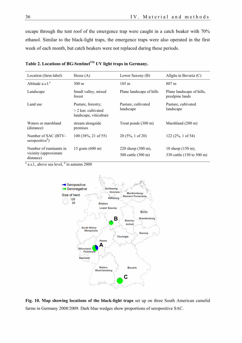
36 I V . M a t e r i a l a n d m e t h o d s
escape through the tent roof of the emergence trap were caught in a catch beaker with 70%
ethanol. Similar to the black-light traps, the emergence traps were also operated in the first
week of each month, but catch beakers were not replaced during these periods.
Table 2. Locations of BG-SentinelTM
UV light traps in Germany.
Location (farm label) Hesse (A) Lower Saxony (B) Allgäu in Bavaria (C)
Altitude a.s.l.a 300 m 185 m 807 m
Landscape Small valley, mixed
forest
Plane landscape of hills Plane landscape of hills,
prealpine lands
Land use Pasture, forestry;
> 2 km: cultivated
landscape, viticulture
Pasture, cultivated
landscape
Pasture, cultivated
landscape
Waters or marshland
(distance)
stream alongside
premises
Trout ponds (300 m) Marshland (200 m)
Number of SAC (BTV-
seropositiveb)
100 (38%, 21 of 55) 20 (5%, 1 of 20) 122 (2%, 1 of 54)
Number of ruminants in
vicinity (approximate
distance)
15 goats (600 m) 220 sheep (300 m),
300 cattle (300 m)
10 sheep (150 m),
330 cattle (150 to 500 m)
a a.s.l., above sea level,
b in autumn 2008
Fig. 10. Map showing locations of the black-light traps set up on three South American camelid
farms in Germany 2008/2009. Dark blue wedges show proportions of seropositive SAC.
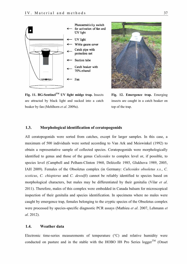
I V . M a t e r i a l a n d m e t h o d s 37
Fig. 11. BG-SentinelTM
UV light midge trap. Insects
are attracted by black light and sucked into a catch
beaker by fan (Mehlhorn et al. 2009a).
Fig. 12. Emergence trap. Emerging
insects are caught in a catch beaker on
top of the trap.
1.3. Morphological identification of ceratopogonids
All ceratopogonids were sorted from catches, except for larger samples. In this case, a
maximum of 500 individuals were sorted according to Van Ark and Meiswinkel (1992) to
obtain a representative sample of collected species. Ceratopogonids were morphologically
identified to genus and those of the genus Culicoides to complex level or, if possible, to
species level (Campbell and Pelham-Clinton 1960, Delécolle 1985, Glukhova 1989, 2005,
IAH 2009). Females of the Obsoletus complex (in Germany: Culicoides obsoletus s.s., C.
scoticus, C. chiopterus and C. dewulfi) cannot be reliably identified to species based on
morphological characters, but males may be differentiated by their genitalia (Vilar et al.
2011). Therefore, males of this complex were embedded in Canada balsam for microscopical
inspection of their genitalia and species identification. In specimens where no males were
caught by emergence trap, females belonging to the cryptic species of the Obsoletus complex
were processed by species-specific diagnostic PCR assays (Mathieu et al. 2007, Lehmann et
al. 2012).
1.4. Weather data
Electronic time-series measurements of temperature (°C) and relative humidity were
conducted on pasture and in the stable with the HOBO H8 Pro Series loggerTM
(Onset
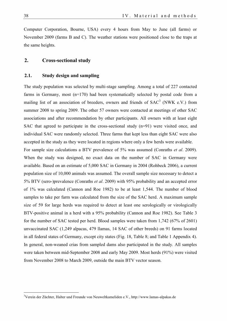
38 I V . M a t e r i a l a n d m e t h o d s
Computer Corporation, Bourne, USA) every 4 hours from May to June (all farms) or
November 2009 (farms B and C). The weather stations were positioned close to the traps at
the same heights.
2. Cross-sectional study
2.1. Study design and sampling
The study population was selected by multi-stage sampling. Among a total of 227 contacted
farms in Germany, most (n=170) had been systematically selected by postal code from a
mailing list of an association of breeders, owners and friends of SAC3 (NWK e.V.) from
summer 2008 to spring 2009. The other 57 owners were contacted at meetings of other SAC
associations and after recommendation by other participants. All owners with at least eight
SAC that agreed to participate in the cross-sectional study (n=91) were visited once, and
individual SAC were randomly selected. Three farms that kept less than eight SAC were also
accepted in the study as they were located in regions where only a few herds were available.
For sample size calculations a BTV prevalence of 5% was assumed (Conraths et al. 2009).
When the study was designed, no exact data on the number of SAC in Germany were
available. Based on an estimate of 5,000 SAC in Germany in 2004 (Rohbeck 2006), a current
population size of 10,000 animals was assumed. The overall sample size necessary to detect a
5% BTV (sero-)prevalence (Conraths et al. 2009) with 95% probability and an accepted error
of 1% was calculated (Cannon and Roe 1982) to be at least 1,544. The number of blood
samples to take per farm was calculated from the size of the SAC herd. A maximum sample
size of 59 for large herds was required to detect at least one serologically or virologically
BTV-positive animal in a herd with a 95% probability (Cannon and Roe 1982). See Table 3
for the number of SAC tested per herd. Blood samples were taken from 1,742 (67% of 2601)
unvaccinated SAC (1,249 alpacas, 479 llamas, 14 SAC of other breeds) on 91 farms located
in all federal states of Germany, except city states (Fig. 18, Table 8; and Table 1 Appendix 4).
In general, non-weaned crias from sampled dams also participated in the study. All samples
were taken between mid-September 2008 and early May 2009. Most herds (91%) were visited
from November 2008 to March 2009, outside the main BTV vector season.
3Verein der Züchter, Halter und Freunde von Neuweltkameliden e.V., http://www.lamas-alpakas.de
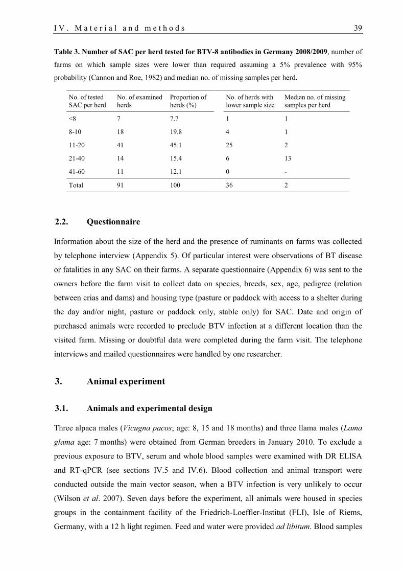
I V . M a t e r i a l a n d m e t h o d s 39
Table 3. Number of SAC per herd tested for BTV-8 antibodies in Germany 2008/2009, number of
farms on which sample sizes were lower than required assuming a 5% prevalence with 95%
probability (Cannon and Roe, 1982) and median no. of missing samples per herd.
No. of tested
SAC per herd
No. of examined
herds
Proportion of
herds (%)
No. of herds with
lower sample size
Median no. of missing
samples per herd
<8 7 7.7 1 1
8-10 18 19.8 4 1
11-20 41 45.1 25 2
21-40 14 15.4 6 13
41-60 11 12.1 0 -
Total 91 100 36 2
2.2. Questionnaire
Information about the size of the herd and the presence of ruminants on farms was collected
by telephone interview (Appendix 5). Of particular interest were observations of BT disease
or fatalities in any SAC on their farms. A separate questionnaire (Appendix 6) was sent to the
owners before the farm visit to collect data on species, breeds, sex, age, pedigree (relation
between crias and dams) and housing type (pasture or paddock with access to a shelter during
the day and/or night, pasture or paddock only, stable only) for SAC. Date and origin of
purchased animals were recorded to preclude BTV infection at a different location than the
visited farm. Missing or doubtful data were completed during the farm visit. The telephone
interviews and mailed questionnaires were handled by one researcher.
3. Animal experiment
3.1. Animals and experimental design
Three alpaca males (Vicugna pacos; age: 8, 15 and 18 months) and three llama males (Lama
glama age: 7 months) were obtained from German breeders in January 2010. To exclude a
previous exposure to BTV, serum and whole blood samples were examined with DR ELISA
and RT-qPCR (see sections IV.5 and IV.6). Blood collection and animal transport were
conducted outside the main vector season, when a BTV infection is very unlikely to occur
(Wilson et al. 2007). Seven days before the experiment, all animals were housed in species
groups in the containment facility of the Friedrich-Loeffler-Institut (FLI), Isle of Riems,
Germany, with a 12 h light regimen. Feed and water were provided ad libitum. Blood samples
40 I V . M a t e r i a l a n d m e t h o d s
were taken one day before the experiment to evaluate haematological parameters and to prove
them free of BTV antibodies and BTV RNA (see respective sections below).
For BTV infection, the SAC were subcutaneously injected at multiple sites in the freshly
clipped shoulder and dorsal thorax regions. Each animal received 105 TCID50 (50% tissue
culture infective doses) of a recent German BTV-8 isolate (Eschbaumer et al. 2009) in 4 ml of
tissue culture supernatant diluted in phosphate-buffered saline (PBS). Briefly, a Holstein calf
had been inoculated with pooled blood from a BT outbreak in sheep. Virus was isolated from
the calf and passaged twice on Vero cells (RIE15, Collection of Cell Lines in Veterinary
Medicine, FLI Insel Riems).
The inoculum used for the SAC contained 5 x 10⁸ BTV-8 genome copies per ml
(quantification cycle [Cq] value 18.8). It was proven to be free of any bacterial contamination.
After inoculation, whole blood and serum samples were taken at regular intervals. EDTA
blood was stored at 4 °C, and serum samples were harvested within 4 h of collection,
inactivated at 56 °C for 30 min, and stored at 4 °C until analysis.
Only three animals per species were available. Because three replicates were considered the
absolute minimum for a meaningful experiment, it was decided to inoculate all animals and
not to use a control group.
3.2. Clinical and haematological parameters
During the entire study, rectal body temperatures and close clinical monitoring were
performed daily from one day before to 15 dpi (Fowler 1998a, Hengrave Burri et al. 2005).
This included examination of the eyes, conjunctival membranes, oral and nasal mucosa,
auscultation of the heart and lungs, palpation of mandibular, axillar, cervical, inguinal and
popliteal lymph nodes, examination of the skin of the coronary band and interdigital spaces
for lesions and increased temperature. Blood samples taken on days -1, 2, 6, 8, 10, 13, 16, 20,
24, 28 and 35 were tested in a Cell-dyn® 3700 Hematology Analyzer (Abbott Laboratories,
Abbott Park, IL, USA) using the 'goat' analysis profile.
3.3. Post-mortem examination
Euthanasia and post-mortem examination of all SAC was performed on 106 dpi. All animals
were sedated by intramuscular (i.m.) application of 0.5 ml (llamas) or 0.3 ml (alpacas) of 1%
detomidine (Cepesedan® RP, CP-Pharma, Burgdorf, Germany) and led to the necropsy room.
For anaesthesia, each animal received an i.m. injection of 250 mg xylazine powder
(Rompun® TS, Bayer HealthCare, Leverkusen, Germany) dissolved in 2 ml of 10% ketamine
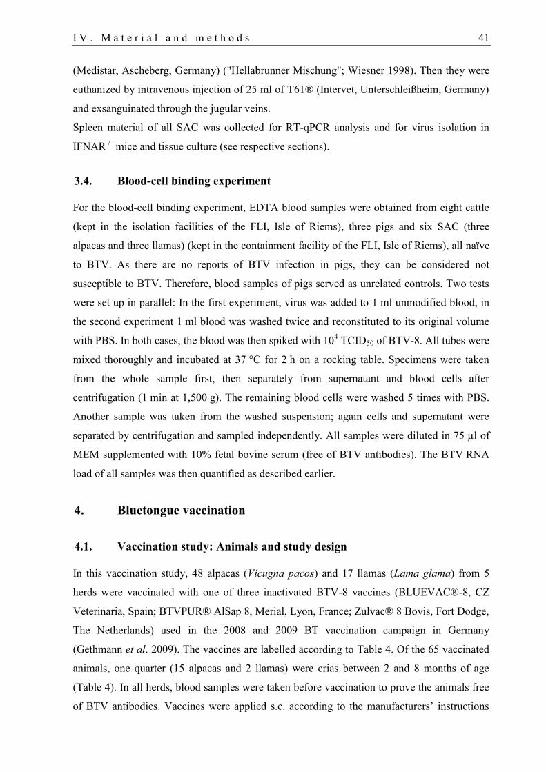
I V . M a t e r i a l a n d m e t h o d s 41
(Medistar, Ascheberg, Germany) ("Hellabrunner Mischung"; Wiesner 1998). Then they were
euthanized by intravenous injection of 25 ml of T61® (Intervet, Unterschleißheim, Germany)
and exsanguinated through the jugular veins.
Spleen material of all SAC was collected for RT-qPCR analysis and for virus isolation in
IFNAR-/-
mice and tissue culture (see respective sections).
3.4. Blood-cell binding experiment
For the blood-cell binding experiment, EDTA blood samples were obtained from eight cattle
(kept in the isolation facilities of the FLI, Isle of Riems), three pigs and six SAC (three
alpacas and three llamas) (kept in the containment facility of the FLI, Isle of Riems), all naïve
to BTV. As there are no reports of BTV infection in pigs, they can be considered not
susceptible to BTV. Therefore, blood samples of pigs served as unrelated controls. Two tests
were set up in parallel: In the first experiment, virus was added to 1 ml unmodified blood, in
the second experiment 1 ml blood was washed twice and reconstituted to its original volume
with PBS. In both cases, the blood was then spiked with 104 TCID50 of BTV-8. All tubes were
mixed thoroughly and incubated at 37 °C for 2 h on a rocking table. Specimens were taken
from the whole sample first, then separately from supernatant and blood cells after
centrifugation (1 min at 1,500 g). The remaining blood cells were washed 5 times with PBS.
Another sample was taken from the washed suspension; again cells and supernatant were
separated by centrifugation and sampled independently. All samples were diluted in 75 µl of
MEM supplemented with 10% fetal bovine serum (free of BTV antibodies). The BTV RNA
load of all samples was then quantified as described earlier.
4. Bluetongue vaccination
4.1. Vaccination study: Animals and study design
In this vaccination study, 48 alpacas (Vicugna pacos) and 17 llamas (Lama glama) from 5
herds were vaccinated with one of three inactivated BTV-8 vaccines (BLUEVAC®-8, CZ
Veterinaria, Spain; BTVPUR® AlSap 8, Merial, Lyon, France; Zulvac® 8 Bovis, Fort Dodge,
The Netherlands) used in the 2008 and 2009 BT vaccination campaign in Germany
(Gethmann et al. 2009). The vaccines are labelled according to Table 4. Of the 65 vaccinated
animals, one quarter (15 alpacas and 2 llamas) were crias between 2 and 8 months of age
(Table 4). In all herds, blood samples were taken before vaccination to prove the animals free
of BTV antibodies. Vaccines were applied s.c. according to the manufacturers’ instructions

42 I V . M a t e r i a l a n d m e t h o d s
(see Table 4). Fort Dodge recommends i.m. application of their vaccine, but herd 5 was also
vaccinated s.c. Vaccination was conducted between mid-September and December 2008. The
first post vaccination (p.v.) samples were collected from mid-November 2008 to the end of
January 2009, outside the main BTV vector season (Baldet et al. 2008, Hoffmann et al.
2009a). All farms were located in regions of Germany where BTV-8 cases had never been
registered (Conraths et al. 2009, EU-BTNET system 2009).
In herd 3, six unvaccinated alpacas served as negative controls. Crias in herd 2 and 5 that
became seronegative after the elimination of detectable colostrum-derived antibodies served
as sentinels for vectorborne introduction of BTV into the herds.
Our influence on the study design was limited because only 5 of 227 owners contacted by
telephone agreed to participate. The vaccinating veterinarian was chosen by the owner, the
used vaccine was selected by the local veterinary authorities or by the veterinarian, and the
dosage was decided by the veterinarian. Negative controls or sentinel crias were only
available in two herds. Nevertheless, natural BTV infections after vaccination are very
unlikely based on the location of the farm in a BTV low risk area.
Table 4. Animals, vaccines and dosages used in the BTV-8 vaccination study.
Herd
#
Species No. of
SAC
Vaccinea Vaccine
label
Dosage
(s.c.)
Relative
dosage
Second
dose
Age (y) Sex
(m/f)
1 Alpaca 14 BLUEVAC®-8 C 2 x 2 ml 2 x sheep dose After 4w 3 - 12 5/9
2 Alpaca 15 BLUEVAC®-8 C 2 x 2 ml 2 x sheep dose After 3w 0.2 - 5 6/9
3 Alpaca 19 BLUEVAC®-8 C 1 x 2 ml Sheep dose None 0.3 - 12 11/8
4 Llama 9 BTVPUR®
AlSap 8
M 2 x 1 ml Cattle dose After 4w 1.5 - 15 3/6
5 Llama 8 Zulvac® 8
Bovis
F 2 x 2 ml Cattle dose After 3w 0.6 - 7 3/5
a Lot numbers available upon request; s.c., subcutaneous; m, male; f, female; p.v., post vaccination (after the
completed vaccination); w, weeks; y, years; C, CZ Veterinaria; M, Merial; F, Fort Dodge.
4.2. Tolerance of BTV-8 vaccines
Age, sex and species were recorded for the 65 vaccinated (Table 4) and 6 unvaccinated SAC
that participated in this study. Furthermore, data on vaccination regimens and vaccines used
for another 210 alpacas and 72 llamas on 26 farms were collected by telephone questionnaire.
The owners of all 347 vaccinated SAC were asked retrospectively to report any adverse
effects after BTV-8 vaccination including fatalities, swelling at the injection site, other
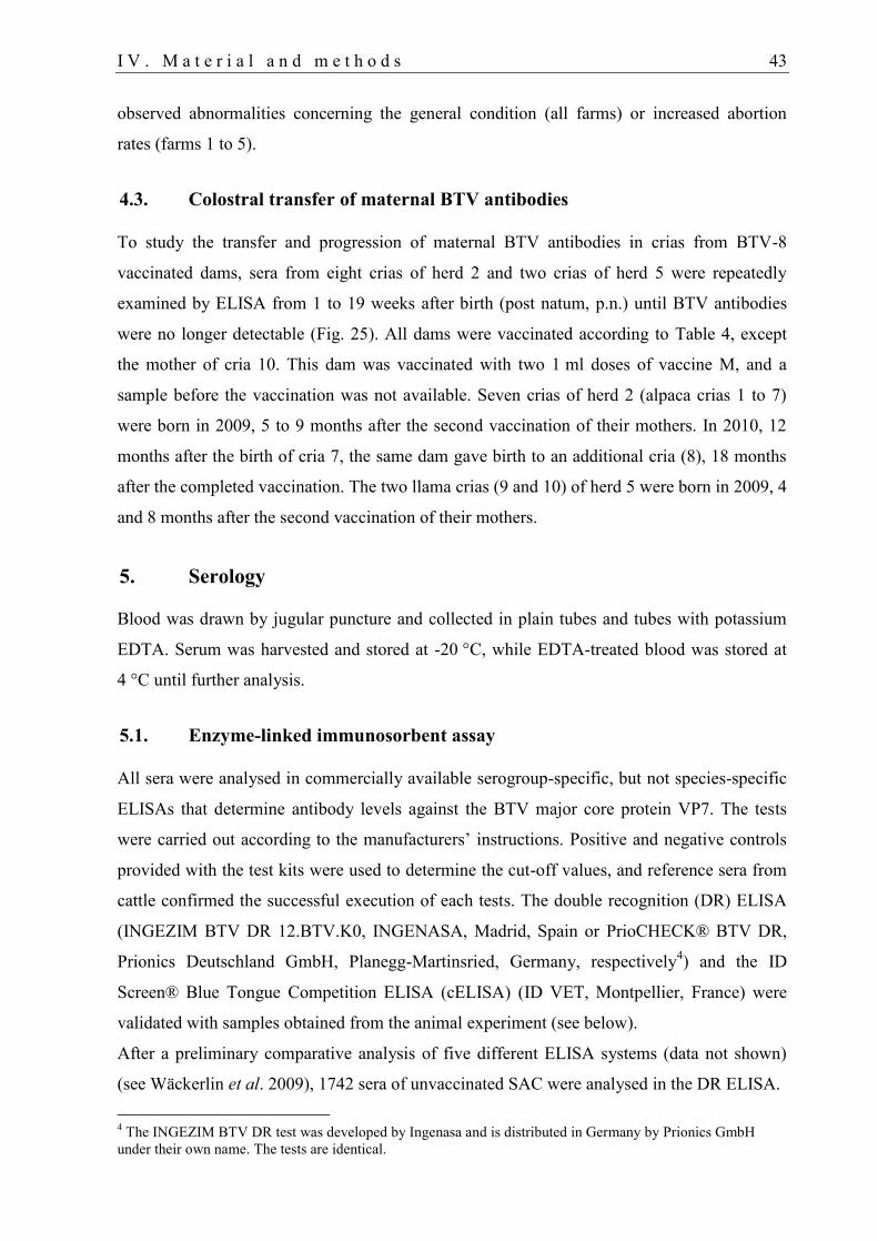
I V . M a t e r i a l a n d m e t h o d s 43
observed abnormalities concerning the general condition (all farms) or increased abortion
rates (farms 1 to 5).
4.3. Colostral transfer of maternal BTV antibodies
To study the transfer and progression of maternal BTV antibodies in crias from BTV-8
vaccinated dams, sera from eight crias of herd 2 and two crias of herd 5 were repeatedly
examined by ELISA from 1 to 19 weeks after birth (post natum, p.n.) until BTV antibodies
were no longer detectable (Fig. 25). All dams were vaccinated according to Table 4, except
the mother of cria 10. This dam was vaccinated with two 1 ml doses of vaccine M, and a
sample before the vaccination was not available. Seven crias of herd 2 (alpaca crias 1 to 7)
were born in 2009, 5 to 9 months after the second vaccination of their mothers. In 2010, 12
months after the birth of cria 7, the same dam gave birth to an additional cria (8), 18 months
after the completed vaccination. The two llama crias (9 and 10) of herd 5 were born in 2009, 4
and 8 months after the second vaccination of their mothers.
5. Serology
Blood was drawn by jugular puncture and collected in plain tubes and tubes with potassium
EDTA. Serum was harvested and stored at -20 °C, while EDTA-treated blood was stored at
4 °C until further analysis.
5.1. Enzyme-linked immunosorbent assay
All sera were analysed in commercially available serogroup-specific, but not species-specific
ELISAs that determine antibody levels against the BTV major core protein VP7. The tests
were carried out according to the manufacturers’ instructions. Positive and negative controls
provided with the test kits were used to determine the cut-off values, and reference sera from
cattle confirmed the successful execution of each tests. The double recognition (DR) ELISA
(INGEZIM BTV DR 12.BTV.K0, INGENASA, Madrid, Spain or PrioCHECK® BTV DR,
Prionics Deutschland GmbH, Planegg-Martinsried, Germany, respectively4) and the ID
Screen® Blue Tongue Competition ELISA (cELISA) (ID VET, Montpellier, France) were
validated with samples obtained from the animal experiment (see below).
After a preliminary comparative analysis of five different ELISA systems (data not shown)
(see Wäckerlin et al. 2009), 1742 sera of unvaccinated SAC were analysed in the DR ELISA.
4 The INGEZIM BTV DR test was developed by Ingenasa and is distributed in Germany by Prionics GmbH
under their own name. The tests are identical.
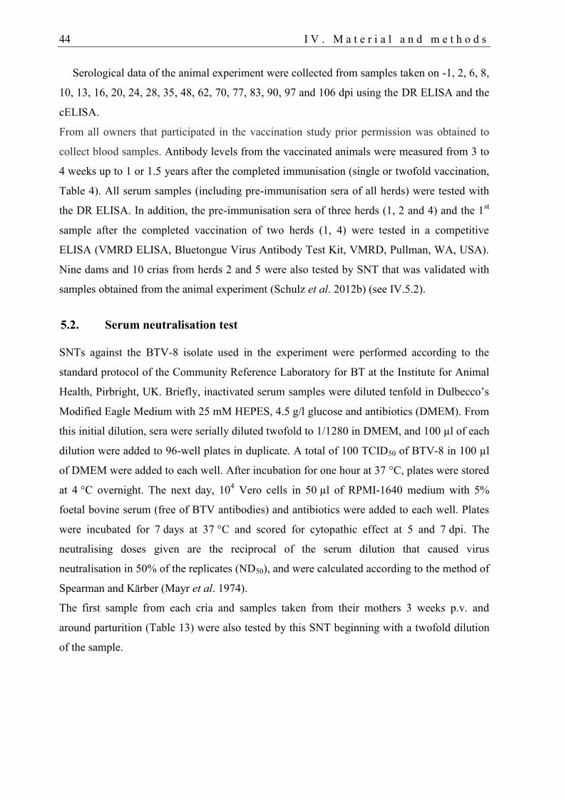
44 I V . M a t e r i a l a n d m e t h o d s
Serological data of the animal experiment were collected from samples taken on -1, 2, 6, 8,
10, 13, 16, 20, 24, 28, 35, 48, 62, 70, 77, 83, 90, 97 and 106 dpi using the DR ELISA and the
cELISA.
From all owners that participated in the vaccination study prior permission was obtained to
collect blood samples. Antibody levels from the vaccinated animals were measured from 3 to
4 weeks up to 1 or 1.5 years after the completed immunisation (single or twofold vaccination,
Table 4). All serum samples (including pre-immunisation sera of all herds) were tested with
the DR ELISA. In addition, the pre-immunisation sera of three herds (1, 2 and 4) and the 1st
sample after the completed vaccination of two herds (1, 4) were tested in a competitive
ELISA (VMRD ELISA, Bluetongue Virus Antibody Test Kit, VMRD, Pullman, WA, USA).
Nine dams and 10 crias from herds 2 and 5 were also tested by SNT that was validated with
samples obtained from the animal experiment (Schulz et al. 2012b) (see IV.5.2).
5.2. Serum neutralisation test
SNTs against the BTV-8 isolate used in the experiment were performed according to the
standard protocol of the Community Reference Laboratory for BT at the Institute for Animal
Health, Pirbright, UK. Briefly, inactivated serum samples were diluted tenfold in Dulbecco’s
Modified Eagle Medium with 25 mM HEPES, 4.5 g/l glucose and antibiotics (DMEM). From
this initial dilution, sera were serially diluted twofold to 1/1280 in DMEM, and 100 µl of each
dilution were added to 96-well plates in duplicate. A total of 100 TCID50 of BTV-8 in 100 µl
of DMEM were added to each well. After incubation for one hour at 37 °C, plates were stored
at 4 °C overnight. The next day, 104 Vero cells in 50 µl of RPMI-1640 medium with 5%
foetal bovine serum (free of BTV antibodies) and antibiotics were added to each well. Plates
were incubated for 7 days at 37 °C and scored for cytopathic effect at 5 and 7 dpi. The
neutralising doses given are the reciprocal of the serum dilution that caused virus
neutralisation in 50% of the replicates (ND50), and were calculated according to the method of
Spearman and Kärber (Mayr et al. 1974).
The first sample from each cria and samples taken from their mothers 3 weeks p.v. and
around parturition (Table 13) were also tested by this SNT beginning with a twofold dilution
of the sample.
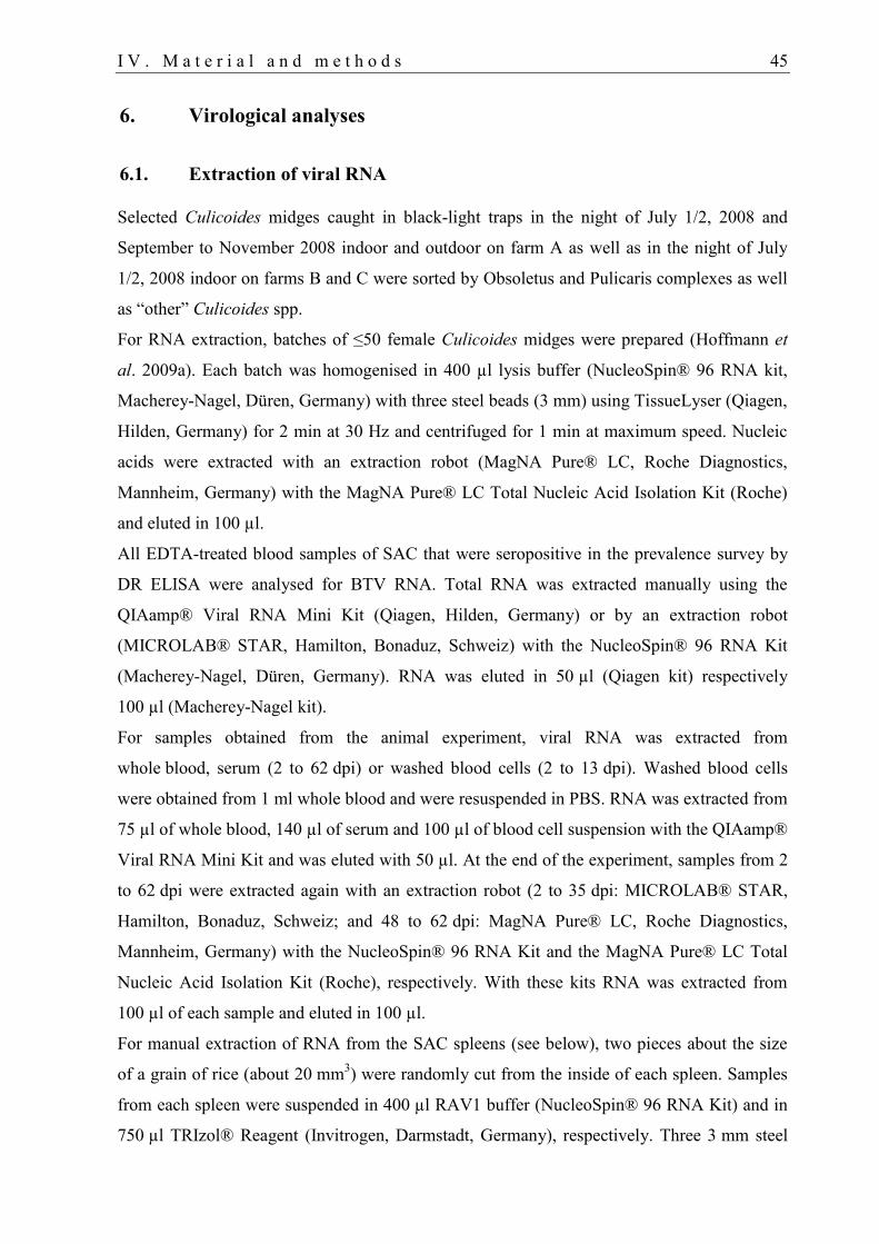
I V . M a t e r i a l a n d m e t h o d s 45
6. Virological analyses
6.1. Extraction of viral RNA
Selected Culicoides midges caught in black-light traps in the night of July 1/2, 2008 and
September to November 2008 indoor and outdoor on farm A as well as in the night of July
1/2, 2008 indoor on farms B and C were sorted by Obsoletus and Pulicaris complexes as well
as “other” Culicoides spp.
For RNA extraction, batches of ≤50 female Culicoides midges were prepared (Hoffmann et
al. 2009a). Each batch was homogenised in 400 µl lysis buffer (NucleoSpin® 96 RNA kit,
Macherey-Nagel, Düren, Germany) with three steel beads (3 mm) using TissueLyser (Qiagen,
Hilden, Germany) for 2 min at 30 Hz and centrifuged for 1 min at maximum speed. Nucleic
acids were extracted with an extraction robot (MagNA Pure® LC, Roche Diagnostics,
Mannheim, Germany) with the MagNA Pure® LC Total Nucleic Acid Isolation Kit (Roche)
and eluted in 100 µl.
All EDTA-treated blood samples of SAC that were seropositive in the prevalence survey by
DR ELISA were analysed for BTV RNA. Total RNA was extracted manually using the
QIAamp® Viral RNA Mini Kit (Qiagen, Hilden, Germany) or by an extraction robot
(MICROLAB® STAR, Hamilton, Bonaduz, Schweiz) with the NucleoSpin® 96 RNA Kit
(Macherey-Nagel, Düren, Germany). RNA was eluted in 50 µl (Qiagen kit) respectively
100 µl (Macherey-Nagel kit).
For samples obtained from the animal experiment, viral RNA was extracted from
whole blood, serum (2 to 62 dpi) or washed blood cells (2 to 13 dpi). Washed blood cells
were obtained from 1 ml whole blood and were resuspended in PBS. RNA was extracted from
75 µl of whole blood, 140 µl of serum and 100 µl of blood cell suspension with the QIAamp®
Viral RNA Mini Kit and was eluted with 50 µl. At the end of the experiment, samples from 2
to 62 dpi were extracted again with an extraction robot (2 to 35 dpi: MICROLAB® STAR,
Hamilton, Bonaduz, Schweiz; and 48 to 62 dpi: MagNA Pure® LC, Roche Diagnostics,
Mannheim, Germany) with the NucleoSpin® 96 RNA Kit and the MagNA Pure® LC Total
Nucleic Acid Isolation Kit (Roche), respectively. With these kits RNA was extracted from
100 µl of each sample and eluted in 100 µl.
For manual extraction of RNA from the SAC spleens (see below), two pieces about the size
of a grain of rice (about 20 mm3) were randomly cut from the inside of each spleen. Samples
from each spleen were suspended in 400 µl RAV1 buffer (NucleoSpin® 96 RNA Kit) and in
750 µl TRIzol® Reagent (Invitrogen, Darmstadt, Germany), respectively. Three 3 mm steel
46 I V . M a t e r i a l a n d m e t h o d s
beads were used for homogenization in a TissueLyser (Qiagen) for 2 min at 30 Hz.
Afterwards, samples were centrifuged for 1 min at maximum speed in a microcentrifuge.
From 140 µl supernatant of the homogenate, viral RNA was extracted using the QIAamp®
Viral RNA Mini Kit. For the TRIzol® extraction 200 µl chloroform was added to the
homogenate, mixed, incubated for 10 min at room temperature and centrifuged (10 min at
maximum speed in a microcentrifuge cooled to 4 °C). The aqueous phase (up to 500 µl) was
mixed with 600 µl ethanol. The lysate was then transferred to a RNeasy Mini column
(Qiagen) and total RNA was extracted further according to the manufacturer’s instructions.
For the blood-cell binding experiment, RNA was extracted from 100 µl of each sample by
extraction robot (MICROLAB® STAR) with the NucleoSpin® 96 RNA kit and was eluted in
100 µl.
6.2. Detection of BTV RNA
Two different commercial RT-qPCR kits were used to detect BTV RNA: the iScript™ One-
Step RT-PCR Kit (BIO-RAD, Hercules, CA, USA) and the Ambion AgPath-ID™ One-Step
RT-PCR Kit (Applied Biosystems, Carlsbad, CA, USA). The iScript™ Kit was used as
previously described (Hoffmann et al. 2006, Toussaint et al. 2007, Hoffmann et al. 2009c);
the RT-qPCR thermal profile for the AgPath-ID™ Kit was 10 min at 45 °C and 10 min at
95 °C, followed by 42 cycles of 15 sec at 95 °C, 20 sec at 56 °C, and 30 sec at 72 °C in a
Mx3005P® QPCR system (Stratagene, La Jolla, CA, USA). A total of 5 µl of eluate was used
for RT-qPCR.
The group-specific “pan-BTV” assay detects the 24 BTV serotypes established at the time of
the study by amplification of a highly conserved sequence on genome segment 5 (Toussaint et
al. 2007), while the BTV-8 assay is directed to BTV genome segment 2, which encodes the
highly variable serotype-specific outer shell protein VP2 (Hoffmann et al. 2009c).
Culicoides batches were analysed with the AgPath-ID™ Kit. RNA was amplified in a duplex
RT-qPCR combining the detection of all confirmed 24 BTV serotypes ("pan-BTV" assay,
Toussaint et al. 2007) and, as an internal control, the detection of all members of the genus
Culicoides ("pan-Culicoides" assay, Hoffmann et al. 2009a). A total of 5 µl of eluate was
used for RT-qPCR.
Blood samples of the cross-sectional study were analysed by using the iScript™ Kit and the
group-specific “pan-BTV” assay.

I V . M a t e r i a l a n d m e t h o d s 47
All samples obtained from the animal experiment were tested with the “pan-BTV“ and BTV-8
assay on the same PCR plate for cross-confirmation using the iScript™ Kit and the AgPath-
ID™ Kit.
In vitro transcribed segment 5 standard RNA was serially diluted tenfold in RNA-safe buffer
(Hoffmann et al. 2009c, Hoffmann, unpublished) and used for absolute quantification by
external calibration. The resulting standard curves in all RT-qPCR runs were highly correlated
(R2 consistently >0.99) and demonstrated good amplification efficiency (slopes between -3.43
and -3.20) (data not shown). For all samples, BTV RNA quantification assays performed with
the AgPath-ID™ Kit and the iScript™ Kit yielded very similar results (data not shown).
BTV RNA isolated from spleens with the QIAamp® Viral RNA Mini Kit was analysed with
the “pan-BTV” segment 5 assay (Toussaint et al. 2007), the BTV-8 segment 2 assay
(Hoffmann et al. 2009c), the commercially available group-specific Virotype® BTV Kit
(Labor Diagnostik Leipzig, Leipzig, Germany; segment 10), and the group-specific cador
BTV RT-PCR Kit (Qiagen; segment 1). RNA isolated with TRIzol® Reagent was tested with
the “pan-BTV“ and the BTV-8 assay using the AgPath-ID™ Kit as described earlier. An
absolute quantification assay of the latter extracts was conducted with the in vitro transcribed
and tenfold serially diluted standard RNA as previously described.
Samples of the blood-cell binding experiment were tested with the “pan-BTV“ using the
AgPath-ID™ Kit. The BTV RNA load was quantified as previously described.
6.3. Virus isolation in tissue culture and embryonated chicken eggs
Virus isolation from washed and ultrasonically disrupted (see below) blood cells was
attempted for samples taken between 2 and 10 dpi, but no virus could be isolated (data not
shown). RT-qPCR analysis of samples from the experimental infection and the blood-cell
binding experiment revealed significantly higher amounts of BTV RNA in the plasma fraction
than in cell pellets (see below). Therefore, virus isolation was repeated without prior washing.
Whole blood samples were selected for lowest Cq value. Serum samples taken on the same
day were negative in at least the SNT and cELISA. Virus isolation from twelve samples was
attempted on Vero cells (6 to 10 dpi) and in ECE (2 to 6 dpi) by methods adapted from
Clavijo et al. (2000). Vero cell cultures were inoculated with whole blood, blood cell pellets
and plasma, while only whole blood was used for inoculation of 10-day-old ECE. Before
inoculation, whole blood and blood cell pellets were ultrasonically disrupted on wet ice (full
output, 90% duty cycle, 10 pulses, up to 3 times; Sonifier 450, Branson Ultrasonics Corp.,
Danbury, CT, USA) and centrifuged (1 min at maximum speed in a microcentrifuge). ECE
48 I V . M a t e r i a l a n d m e t h o d s
were then intravenously inoculated with 100 µl of supernatant and incubated for 7 days. The
chicken embryos were harvested, and internal organs were homogenised in the TissueLyser
(2 min at 30 Hz). Organ supernatants were used for virus isolation on 75% confluent Vero cell
monolayers as described by Clavijo et al. (2000). After 5 days of incubation at 37 °C, 1 ml of
cell culture supernatant was blindly passaged and incubated another 7 days. Organ
supernatants and cell culture passages were tested for BTV RNA by RT-qPCR.
Virus isolation on Vero cells was also carried out with the supernatants of the homogenised
spleens that were used for the inoculation of IFNAR-/-
mice.
6.4. Virus isolation in IFNAR-/-
mice
Virus isolation in IFNAR-/-
mice (Calvo-Pinilla et al. 2009a) was attempted from whole blood
taken between 2 and 6 dpi and spleens collected post-mortem at day 106.
For each SAC, whole blood samples from days 2 to 6 were mixed (250 µl each). For llama
#5, only blood from 4 and 6 dpi was used (Cq values between 34.6 and 38.5; blood from llama
#4 was negative in the RT-qPCR). The blood samples were processed as described above for
ECE inoculation.
Spleen samples taken post-mortem had been stored at -80 °C. They were thawed, two rice
grain-sized pieces were cut from each spleen as described above and suspended in 500 µl of
Eagle’s Minimal Essential Medium with antibiotics (MEM). The pieces were homogenised in
the TissueLyser (2 min at 30 Hz) with one 5 mm steel bead and centrifuged for 1 min at
maximum speed. The supernatants were pooled for each animal, and two more pools were
prepared by mixing all alpaca and all llama samples, respectively. The samples were diluted
with 1 ml of MEM and eight IFNAR-/-
mice were then inoculated with 300 µl of sample each.
The inoculation and further assessment of the mice was performed as described previously
(Eschbaumer et al. 2010b).
7. Statistical analyses
The total number of female Culicoides caught on each of the farms were comparatively
analysed by Kruskal-Wallis test (Kruskal and Wallis 1952) as well as paired two by two with
Wilcoxon rank-sum tests (McCullagh and Nelder 1989, Hastie and Pregibon 1992, Venables
and Ripley 2002) using Bonferroni correction to adjust the α-level. Furthermore, Wilcoxon
rank-sum tests were used to determine significance between the total number of female
Culicoides caught (i) indoor and outdoor on each farm, (ii) in each group (Obsoletus and
Pulicaris complexes and “other” Culicoides) on each farm using Bonferroni correction, and
I V . M a t e r i a l a n d m e t h o d s 49
(iii) to compare daily Culicoides catches on farms B and C from July to November in the
years 2008 and 2009.
The effects of the explanatory variables farm, temperature and humidity on the response
variable (daily number of Culicoides caught in black-light traps) were modelled using
multivariable Poisson regression in combination with (data not shown) and without an
autoregressive model (time series analysis). See Table 7 for the definition of variables. All
variables were analysed by automated stepwise regression using the Akaike information
criterion (AIC; Sakamoto et al. 1986). Subsequently, the variables were tested for interaction.
The same procedure (excluding the farm variable) was repeated separately for each farm.
For all analyses, only the number of Culicoides caught from July 2008 and June 2009 were
considered, except for the comparison between the number of Culicoides collected on farms B
and C from July to November in the years 2008 and 2009.
For all analyses, only the number of Culicoides caught from July 2008 and June 2009 were
considered, except for the comparison between the number of Culicoides collected on farms B
and C from July to November in the years 2008 and 2009.
For risk factor analysis, two statistical test series were set up in parallel: in the first series, all
91 farms and in the second, only the 43 farms with seropositive SAC were included in the
analysis. In both cases, possible risk factors associated with BTV infection (dichotomous
outcome variable) were analysed by Fisher exact tests (Fay 2010a) to determine their
significance. For the age variable, the α-level was adjusted using Bonferroni correction. All
explanatory variables were included in two multiple logistic regression models to analyse
individual and herd variables separately. Variables were picked by automated stepwise
backward and forward selection. The remaining variables were subsequently tested for
interaction (McCullagh and Nelder 1989, Hastie and Pregibon 1992, Venables and Ripley
2002). For the definition of explanatory variables see Table 9.
Differences in the amount of viral RNA found in whole blood, serum and washed blood cell
pellets in the experimental infection as well as in the blood-cell binding experiment were
examined by Wilcoxon rank-sum tests. The relative sensitivity of the cELISA, DR ELISA
and SNT to detect seroconversion after infection was compared in McNemar tests. The
correlation between seroconversion and elimination of circulating viral RNA was examined
with Cohen's kappa method.
The variances of antibody levels in SAC at different points in time (6 to 12 months) after
vaccination were compared with two-sided F-tests. Results of both ELISA systems (VMRD
ELISA and DR) (herd 1 and 4) were compared using a McNemar test (Agresti 1990, Fay
50 I V . M a t e r i a l a n d m e t h o d s
2010b). The level of agreement between the two tests was examined with Cohen’s kappa
method (Szklo and Nieto 2007).
All test results with p-values below an α-level of 0.05 were considered statistically significant.
All statistical analysis was performed with the “R” software package (R Development Core
Team 2010).
V. RESULTS
1. Entomological monitoring
Certain Culicoides spp. (Diptera, Ceratopogonidae) are vectors of various human and animal
pathogens (Mellor et al. 2000). During the recent epizootic of BTV-8 in northern and central
Europe, species belonging to the Obsoletus and Pulicaris complexes and C. dewulfi have been
incriminated as the main vectors of this virus in ruminant herds (Meiswinkel et al. 2008,
Hoffmann et al. 2009a, Eschbaumer et al. 2010a).
This is the first study investigating the fauna of Culicoides and other ceratopogonid genera on
SAC farms. To this end, ceratopogonids were collected in black-light and emergence traps on
three SAC farms in Germany, and selected Culicoides specimens were tested for BTV RNA.
Furthermore, the factors location, temperature and humidity, which have been shown to
influence the abundance of Culicoides and BTV infection rates on ruminant farms (Mellor et
al. 2000, Purse et al. 2012), were analysed using multivariable Poisson regression.
1.1. Abundance of ceratopogonid females
About 266,100 female ceratopogonids were collected with the six black-light traps from July
2008 to June 2009; 98.8% belonged to species in the genus Culicoides (95.6% of about
281,400 from July 2008 to November 2009). The largest proportion belonged to species of the
Obsoletus (77.4%) and Pulicaris (16.0%) complexes (Fig. 13 and Table 5), which have been
suggested as the main potential BTV vectors in northern Europe (Hoffmann et al. 2009a).
Details on proportions of Culicoides groups by trap location are shown in Table 6. Of the
taxon C. dewulfi, which has recently been incriminated as novel potential vector for BTV
(Meiswinkel et al. 2007, Meiswinkel et al. 2008), only a few males were collected on farm A
(Table 5). The classical Afro-Asian BTV vector C. imicola KIEFFER 1913 (Meiswinkel et al.
2004) was not detected in any of the sorted specimens.
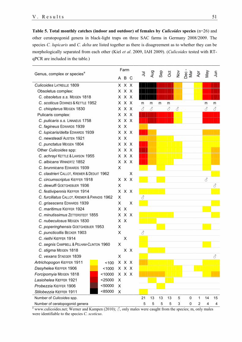
V . R e s u l t s 51
Table 5. Total monthly catches (indoor and outdoor) of females by Culicoides species (n=26) and
other ceratopogonid genera in black-light traps on three SAC farms in Germany 2008/2009. The
species C. lupicaris and C. delta are listed together as there is disagreement as to whether they can be
morphologically separated from each other (Kiel et al. 2009, IAH 2009). (Culicoides tested with RT-
qPCR are included in the table.)
Genus, complex or speciesa
Farm
Jul
Aug
Sep
Oct
Nov
Dec -
Mar
Apr
May
Jun
A B C
Culicoides LATREILLE 1809 X X X
Obsoletus complex: X X X
C. obsoletus s.s. MEIGEN 1818 X X X
C. scoticus DOWNES & KETTLE 1952 X X X m m m m
m m
C. chiopterus MEIGEN 1830 X X X ♂ ♂
♂ ♂
♂ ♂
Pulicaris complex: X X X
C. pulicaris s.s. LINNAEUS 1758 X X X
C. fagineus EDWARDS 1939 X X
C. lupicaris/delta EDWARDS 1939 X X X
C. newsteadi AUSTEN 1921 X X
C. punctatus MEIGEN 1804 X X X
Other Culicoides spp: X X X
C. achrayi KETTLE & LAWSON 1955 X X X
C. albicans WINNERTZ 1852 X X X
C. brunnicans EDWARDS 1939 X
C. clastrieri CALLOT, KREMER & DEDUIT 1962 X
C. circumscriptus KIEFFER 1918 X X X ♂
C. dewulfi GOETGHEBUER 1936 X ♂
C. festivipennis KIEFFER 1914 X X X
C. furcillatus CALLOT, KREMER & PARADIS 1962 X ♂
C. grisescens EDWARDS 1939 X X
C. maritimus KIEFFER 1924 X X
C. minutissimus ZETTERSTEDT 1855 X X X
C. nubeculosus MEIGEN 1830 X X
C. poperinghensis GOETGHEBUER 1953 X
C. puncticollis BECKER 1903 X ♂
C. riethi KIEFFER 1914 X
C. segnis CAMPBELL & PELHAM-CLINTON 1960 X
C. stigma MEIGEN 1818 X X
C. vexans STAEGER 1839 X ♂
Artrichopogon KIEFFER 1911
X X X
Dasyhelea KIEFFER 1906 X X X
Forcipomyia MEIGEN 1818 X X X
Lasiohelea KIEFFER 1921 X
Probezzia KIEFFER 1906 X
Stilobezzia KIEFFER 1911 X
Number of Culicoides spp. 21 13 13 13 5 0 1 14 15
Number of ceratopogonid genera 5 5 5 5 3 0 2 4 4 a www.culicoides.net; Werner and Kampen (2010); ♂, only males were caught from the species; m, only males
were identifiable to the species C. scoticus.
99 <100
999 <1000
### <10000
### <25000
### <50000
### <85000
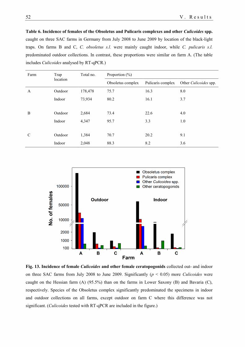
52 V . R e s u l t s
Table 6. Incidence of females of the Obsoletus and Pulicaris complexes and other Culicoides spp.
caught on three SAC farms in Germany from July 2008 to June 2009 by location of the black-light
traps. On farms B and C, C. obsoletus s.l. were mainly caught indoor, while C. pulicaris s.l.
predominated outdoor collections. In contrast, these proportions were similar on farm A. (The table
includes Culicoides analysed by RT-qPCR.)
Farm
Trap
location
Total no. Proportion (%)
Obsoletus complex Pulicaris complex Other Culicoides spp.
A Outdoor 178,478 75.7 16.3 8.0
Indoor 73,934 80.2 16.1 3.7
B Outdoor 2,684 73.4 22.6 4.0
Indoor 4,347 95.7 3.3 1.0
C Outdoor 1,384 70.7 20.2 9.1
Indoor 2,048 88.3 8.2 3.6
Fig. 13. Incidence of female Culicoides and other female ceratopogonids collected out- and indoor
on three SAC farms from July 2008 to June 2009. Significantly (p < 0.05) more Culicoides were
caught on the Hessian farm (A) (95.5%) than on the farms in Lower Saxony (B) and Bavaria (C),
respectively. Species of the Obsoletus complex significantly predominated the specimens in indoor
and outdoor collections on all farms, except outdoor on farm C where this difference was not
significant. (Culicoides tested with RT-qPCR are included in the figure.)
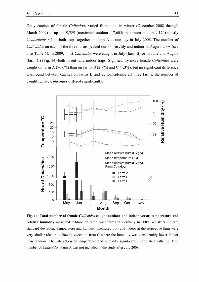
V . R e s u l t s 53
Daily catches of female Culicoides varied from none in winter (December 2008 through
March 2009) to up to 19,789 (maximum outdoor: 17,605; maximum indoor: 9,174) mostly
C. obsoletus s.l. in both traps together on farm A at one day in July 2008. The number of
Culicoides on each of the three farms peaked outdoor in July and indoor in August 2008 (see
also Table 5). In 2009, most Culicoides were caught in July (farm B) or in June and August
(farm C) (Fig. 14) both in out- and indoor traps. Significantly more female Culicoides were
caught on farm A (96.0%) than on farms B (2.7%) and C (1.3%), but no significant difference
was found between catches on farms B and C. Considering all three farms, the number of
caught female Culicoides differed significantly.
Fig. 14. Total number of female Culicoides caught outdoor and indoor versus temperature and
relative humidity measured outdoor on three SAC farms in Germany in 2009. Whiskers indicate
standard deviation. Temperature and humidity measured out- and indoor at the respective farm were
very similar (data not shown), except at farm C where the humidity was considerably lower indoor
than outdoor. The interaction of temperature and humidity significantly correlated with the daily
number of Culicoides. Farm A was not included in the study after July 2009.
54 V . R e s u l t s
On farm A, the abundance of female Culicoides was higher outdoor (70.7%) than indoor,
while on the other two farms this proportion was inversed: about 39% were caught outdoor
(Fig. 13). A significant difference between outdoor and indoor collections was found for farm
B but not for farms A and C. The proportion of ceratopogonids of other genera caught on
farms A and C was higher outdoor (62.4% respectively 79.3%), but on farm B indoor
collections (69.6%) exceeded the number of other ceratopogonids caught outdoor (Fig. 13).
C. obsoletus s.l. were caught during the whole study period (not in winter), except outdoor on
farms B and C in April 2009 (Table 5). Similarly, no C. pulicaris s.l. were caught from
December 2008 to April 2009 in- and outdoor on any of the farms. Additionally, no C.
pulicaris s.l. were collected outdoor in Oktober 2008 and June 2009 (farm B) or in November
2008 (farms B and C).
1.2. Abundance of ceratopogonid males
A total of 0.8% of about 268,300 ceratopogonids caught from July 2008 to June 2009 were
males. The overall proportion of males was considerably lower in the genus Culicoides (0.5%
of about 264,100) than in other genera (23.6% of 4207).
The number of Obsoletus males peaked between July and October 2008 and in May or June
2009, but their abundance varied considerably between trap locations and farms. C. pulicaris
s.l. males were predominantly caught outdoor from July to September 2008 (farms A and B)
and in June 2009 (farm A), but none was collected on farm C. Only single individuals were
sporadically caught indoor between July and October 2008 and in June 2009.
The proportion of male Culicoides by trap location varied in each group on each farm from
0.0 to 5.6%. Similar to the collections of females on farms B and C (Fig. 13), a trend towards
a higher proportion of C. obsoletus s.l. males indoor and C. pulicaris s.l. males outdoor was
noted (Fig. 15). Furthermore, proportions were higher indoor on farms B and C (1.3 vs. 5%),
while this was very similar outdoor and indoor on farm A (about 0.4%).
Monthly proportions of Culicoides males of both complexes were between 0.0 and 1.6% on
farm A. On the other two farms, the proportion of Obsoletus males mostly varied from 0.0 to
13.5% indoor and from 0 to 6.5% outdoor. Exceptions were found outdoor on farm B in
November 2008 (20.0%, 1 of 5), indoor on farms B and C in April (50%, n=2 and 4) and on
farm C in May 2009 (36.1%, 44 of 122) when only a few midges were caught.
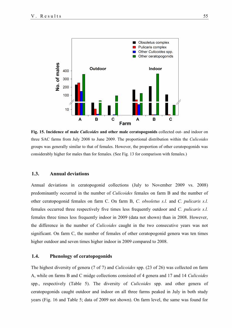
V . R e s u l t s 55
Fig. 15. Incidence of male Culicoides and other male ceratopogonids collected out- and indoor on
three SAC farms from July 2008 to June 2009. The proportional distribution within the Culicoides
groups was generally similar to that of females. However, the proportion of other ceratopogonids was
considerably higher for males than for females. (See Fig. 13 for comparison with females.)
1.3. Annual deviations
Annual deviations in ceratopogonid collections (July to November 2009 vs. 2008)
predominantly occurred in the number of Culicoides females on farm B and the number of
other ceratopogonid females on farm C. On farm B, C. obsoletus s.l. and C. pulicaris s.l.
females occurred three respectively five times less frequently outdoor and C. pulicaris s.l.
females three times less frequently indoor in 2009 (data not shown) than in 2008. However,
the difference in the number of Culicoides caught in the two consecutive years was not
significant. On farm C, the number of females of other ceratopogonid genera was ten times
higher outdoor and seven times higher indoor in 2009 compared to 2008.
1.4. Phenology of ceratopogonids
The highest diversity of genera (7 of 7) and Culicoides spp. (23 of 26) was collected on farm
A, while on farms B and C midge collections consisted of 4 genera and 17 and 14 Culicoides
spp., respectively (Table 5). The diversity of Culicoides spp. and other genera of
ceratopogonids caught outdoor and indoor on all three farms peaked in July in both study
years (Fig. 16 and Table 5; data of 2009 not shown). On farm level, the same was found for
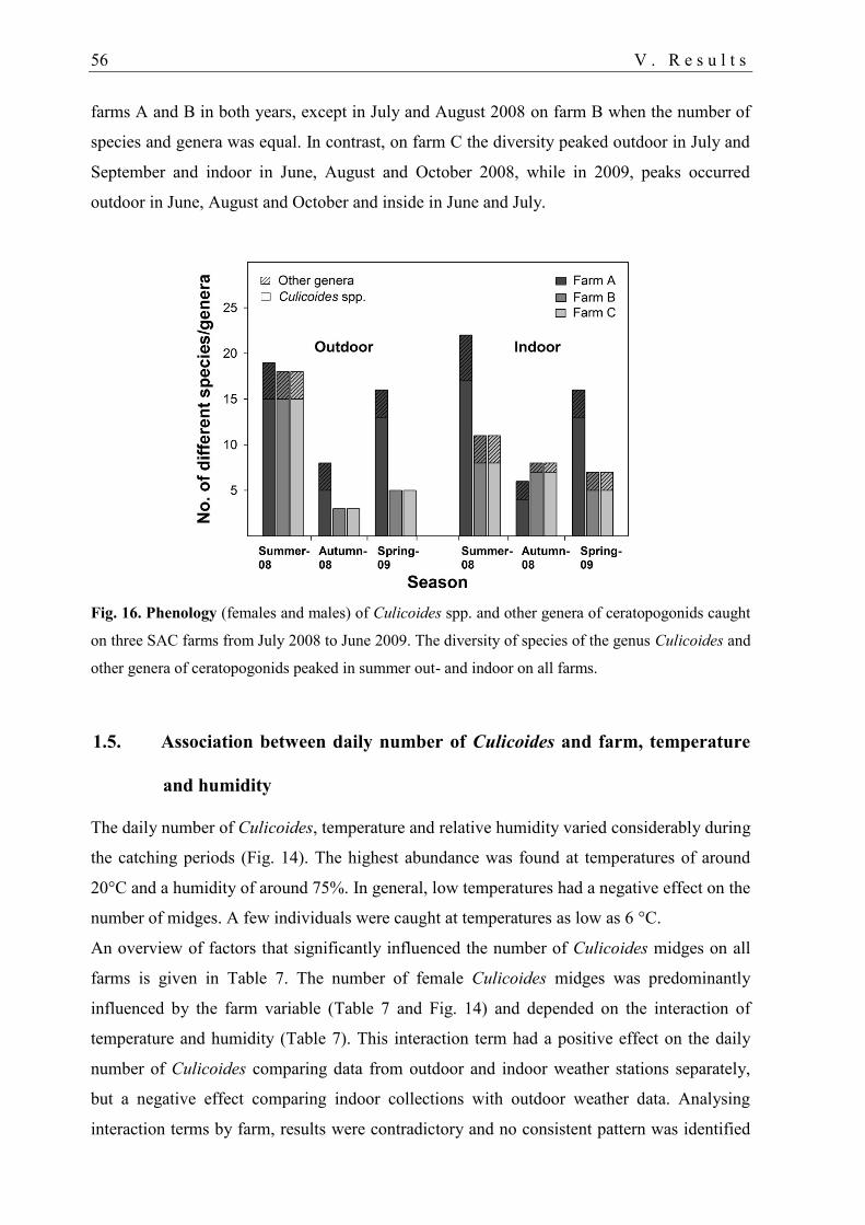
56 V . R e s u l t s
farms A and B in both years, except in July and August 2008 on farm B when the number of
species and genera was equal. In contrast, on farm C the diversity peaked outdoor in July and
September and indoor in June, August and October 2008, while in 2009, peaks occurred
outdoor in June, August and October and inside in June and July.
Fig. 16. Phenology (females and males) of Culicoides spp. and other genera of ceratopogonids caught
on three SAC farms from July 2008 to June 2009. The diversity of species of the genus Culicoides and
other genera of ceratopogonids peaked in summer out- and indoor on all farms.
1.5. Association between daily number of Culicoides and farm, temperature
and humidity
The daily number of Culicoides, temperature and relative humidity varied considerably during
the catching periods (Fig. 14). The highest abundance was found at temperatures of around
20°C and a humidity of around 75%. In general, low temperatures had a negative effect on the
number of midges. A few individuals were caught at temperatures as low as 6 °C.
An overview of factors that significantly influenced the number of Culicoides midges on all
farms is given in Table 7. The number of female Culicoides midges was predominantly
influenced by the farm variable (Table 7 and Fig. 14) and depended on the interaction of
temperature and humidity (Table 7). This interaction term had a positive effect on the daily
number of Culicoides comparing data from outdoor and indoor weather stations separately,
but a negative effect comparing indoor collections with outdoor weather data. Analysing
interaction terms by farm, results were contradictory and no consistent pattern was identified
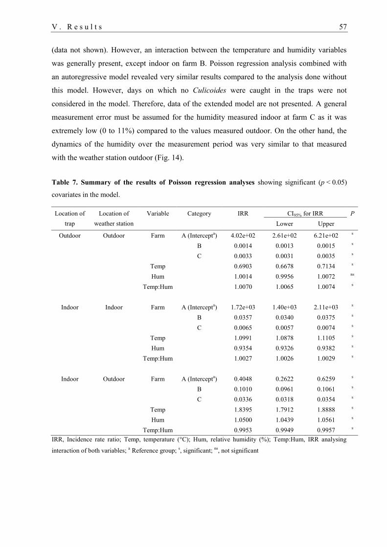
V . R e s u l t s 57
(data not shown). However, an interaction between the temperature and humidity variables
was generally present, except indoor on farm B. Poisson regression analysis combined with
an autoregressive model revealed very similar results compared to the analysis done without
this model. However, days on which no Culicoides were caught in the traps were not
considered in the model. Therefore, data of the extended model are not presented. A general
measurement error must be assumed for the humidity measured indoor at farm C as it was
extremely low (0 to 11%) compared to the values measured outdoor. On the other hand, the
dynamics of the humidity over the measurement period was very similar to that measured
with the weather station outdoor (Fig. 14).
Table 7. Summary of the results of Poisson regression analyses showing significant (p < 0.05)
covariates in the model.
Location of
trap
Location of
weather station
Variable Category IRR CI95% for IRR P
Lower Upper
Outdoor Outdoor Farm A (Intercepta) 4.02e+02 2.61e+02 6.21e+02
s
B 0.0014 0.0013 0.0015
s
C 0.0033 0.0031 0.0035
s
Temp
0.6903 0.6678 0.7134
s
Hum
1.0014 0.9956 1.0072
ns
Temp:Hum
1.0070 1.0065 1.0074
s
Indoor Indoor Farm A (Intercept
a) 1.72e+03 1.40e+03 2.11e+03
s
B 0.0357 0.0340 0.0375
s
C 0.0065 0.0057 0.0074
s
Temp
1.0991 1.0878 1.1105
s
Hum
0.9354 0.9326 0.9382
s
Temp:Hum
1.0027 1.0026 1.0029
s
Indoor Outdoor Farm A (Intercept
a) 0.4048 0.2622 0.6259
s
B 0.1010 0.0961 0.1061
s
C 0.0336 0.0318 0.0354
s
Temp
1.8395 1.7912 1.8888
s
Hum
1.0500 1.0439 1.0561
s
Temp:Hum 0.9953 0.9949 0.9957 s
IRR, Incidence rate ratio; Temp, temperature (°C); Hum, relative humidity (%); Temp:Hum, IRR analysing
interaction of both variables; a Reference group;
s, significant;
ns, not significant
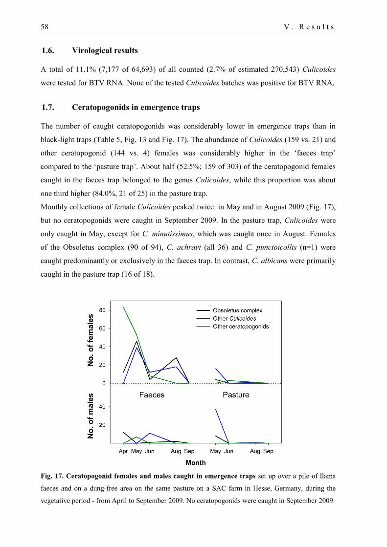
58 V . R e s u l t s
1.6. Virological results
A total of 11.1% (7,177 of 64,693) of all counted (2.7% of estimated 270,543) Culicoides
were tested for BTV RNA. None of the tested Culicoides batches was positive for BTV RNA.
1.7. Ceratopogonids in emergence traps
The number of caught ceratopogonids was considerably lower in emergence traps than in
black-light traps (Table 5, Fig. 13 and Fig. 17). The abundance of Culicoides (159 vs. 21) and
other ceratopogonid (144 vs. 4) females was considerably higher in the ‘faeces trap’
compared to the ‘pasture trap’. About half (52.5%; 159 of 303) of the ceratopogonid females
caught in the faeces trap belonged to the genus Culicoides, while this proportion was about
one third higher (84.0%, 21 of 25) in the pasture trap.
Monthly collections of female Culicoides peaked twice: in May and in August 2009 (Fig. 17),
but no ceratopogonids were caught in September 2009. In the pasture trap, Culicoides were
only caught in May, except for C. minutissimus, which was caught once in August. Females
of the Obsoletus complex (90 of 94), C. achrayi (all 36) and C. punctoicollis (n=1) were
caught predominantly or exclusively in the faeces trap. In contrast, C. albicans were primarily
caught in the pasture trap (16 of 18).
Fig. 17. Ceratopogonid females and males caught in emergence traps set up over a pile of llama
faeces and on a dung-free area on the same pasture on a SAC farm in Hesse, Germany, during the
vegetative period - from April to September 2009. No ceratopogonids were caught in September 2009.

V . R e s u l t s 59
About two thirds (n=71) of all male Culicoides were caught in the pasture trap. Inversely,
considering C. obsoletus s.l. males separately about two thirds (n=23) were collected in the
faeces trap. Comparing proportions of Obsoletus males and females caught in emergence
traps with those collected in the outdoor black-light trap on farm A, the proportion of males
was considerably higher in the faeces trap (14.3% of 105) than in the black-light trap (0.2% of
about 138,400). In the pasture trap, Culicoides males (68.2% of 66) and Obsoletus complex
males (66.7% of 12) even predominated the collections (Fig. 17).
2. Cross-sectional study
The potentially devastating consequences of BT (Conraths et al. 2009), the increasing
popularity of SAC (Schulz et al., manuscript in preparation) together with the recent BTV-
related fatalities (Henrich et al. 2007, Meyer et al. 2009, Ortega et al. 2010b) prompted the
investigation of BTV infection in SAC. Therefore, a large-scale serological and virological
survey was conducted. Blood samples were tested for BTV antibodies by ELISA, and all
seropositive samples for BTV RNA with RT-qPCR. Since none of the serological systems
were validated for the detection of BTV antibodies in SAC at the time, five different
commercially available ELISAs were compared with some sera of unvaccinated SAC. All
serological results of this study were based on the results obtained by PrioCHECK® ELISA.
Data on species, sex, age, parentage, and BTV-related disease, fatalities and abortions in SAC
in Germany was collected by questionnaire. Furthermore, factors influencing BTV infection
in ruminant husbandry such as the use of insecticides, housing or animal movement were also
evaluated for SAC. Various statistical analyses were conducted to identify risk factors for
BTV infection in SAC.
2.1.1. Study population
The response rate of the telephone interview was 100% (n=227). The majority of SAC owners
(91 of 97, 94%) who generally kept at least 8 unvaccinated animals agreed to participate in
the cross-sectional study. Of these, 51 (56%) were members of the NWK e.V. Of 2601 SAC
on 91 farms, 1742 (67%) were tested for BTV antibodies. The species, sex and age ratios as
well as the regional distribution of the 91 tested herds were representative for almost 4,000
SAC kept on the 227 contacted farms (data not shown). Information on the number of SAC
tested per herd is given in Table 3. Unfortunately, a high proportion of owners (36 of 91,
39.6%) only allowed sampling in a lower number of animals than required when assuming a
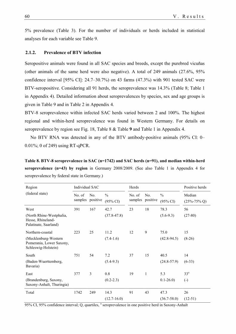
60 V . R e s u l t s
5% prevalence (Table 3). For the number of individuals or herds included in statistical
analyses for each variable see Table 9.
2.1.2. Prevalence of BTV infection
Seropositive animals were found in all SAC species and breeds, except the purebred vicuñas
(other animals of the same herd were also negative). A total of 249 animals (27.6%, 95%
confidence interval [95% CI]: 24.7–30.7%) on 43 farms (47.3%) with 901 tested SAC were
BTV-seropositive. Considering all 91 herds, the seroprevalence was 14.3% (Table 8; Table 1
in Appendix 4). Detailed information about seroprevalences by species, sex and age groups is
given in Table 9 and in Table 2 in Appendix 4.
BTV-8 seroprevalence within infected SAC herds varied between 2 and 100%. The highest
regional and within-herd seroprevalence was found in Western Germany. For details on
seroprevalence by region see Fig. 18, Table 8 & Table 9 and Table 1 in Appendix 4.
No BTV RNA was detected in any of the BTV antibody-positive animals (95% CI: 0–
0.01%; 0 of 249) using RT-qPCR.
Table 8. BTV-8 seroprevalence in SAC (n=1742) and SAC herds (n=91), and median within-herd
seroprevalence (n=43) by region in Germany 2008/2009. (See also Table 1 in Appendix 4 for
seroprevalence by federal state in Germany.)
Region
(federal state)
Individual SAC Herds Positive herds
No. of
samples
No.
positive
%
(95% CI)
No. of
samples
No.
positive
%
(95% CI)
Median
(25%-75% Q)
West
(North Rhine-Westphalia,
Hesse, Rhineland-
Palatinate, Saarland)
391 167 42.7
(37.8-47.8)
23 18 78.3
(5.6-9.3)
56
(27-80)
Northern-coastal
(Mecklenburg-Western
Pomerania, Lower Saxony,
Schleswig-Holstein)
223 25 11.2
(7.4-1.6)
12 9 75.0
(42.8-94.5)
15
(8-26)
South
(Baden-Wuerttemberg,
Bavaria)
751 54 7.2
(5.4-9.3)
37 15 40.5
(24.8-57.9)
14
(6-33)
East
(Brandenburg, Saxony,
Saxony-Anhalt, Thuringia)
377 3 0.8
(0.2-2.3)
19 1 5.3
0.1-26.0)
33a
(-)
Total 1742 249 14.3
(12.7-16.0)
91 43 47.3
(36.7-58.0)
26
(12-51)
95% CI, 95% confidence interval; Q, quartiles, a seroprevalence in one positive herd in Saxony-Anhalt
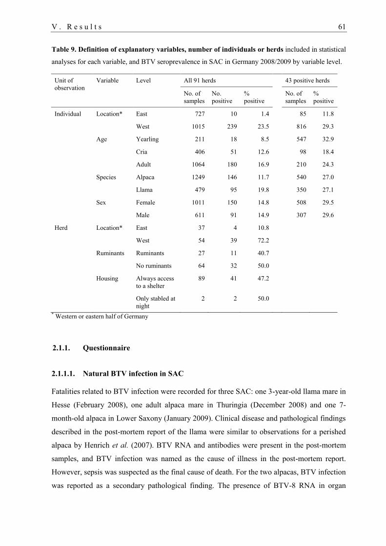
V . R e s u l t s 61
Table 9. Definition of explanatory variables, number of individuals or herds included in statistical
analyses for each variable, and BTV seroprevalence in SAC in Germany 2008/2009 by variable level.
Unit of
observation
Variable Level All 91 herds 43 positive herds
No. of
samples
No.
positive
%
positive
No. of
samples
%
positive
Individual Location* East 727 10 1.4 85 11.8
West 1015 239 23.5 816 29.3
Age Yearling 211 18 8.5 547 32.9
Cria 406 51 12.6 98 18.4
Adult 1064 180 16.9 210 24.3
Species Alpaca 1249 146 11.7 540 27.0
Llama 479 95 19.8 350 27.1
Sex Female 1011 150 14.8 508 29.5
Male 611 91 14.9 307 29.6
Herd Location* East 37 4 10.8
West 54 39 72.2
Ruminants Ruminants 27 11 40.7
No ruminants 64 32 50.0
Housing Always access
to a shelter
89 41 47.2
Only stabled at
night
2 2 50.0
* Western or eastern half of Germany
2.1.1. Questionnaire
2.1.1.1. Natural BTV infection in SAC
Fatalities related to BTV infection were recorded for three SAC: one 3-year-old llama mare in
Hesse (February 2008), one adult alpaca mare in Thuringia (December 2008) and one 7-
month-old alpaca in Lower Saxony (January 2009). Clinical disease and pathological findings
described in the post-mortem report of the llama were similar to observations for a perished
alpaca by Henrich et al. (2007). BTV RNA and antibodies were present in the post-mortem
samples, and BTV infection was named as the cause of illness in the post-mortem report.
However, sepsis was suspected as the final cause of death. For the two alpacas, BTV infection
was reported as a secondary pathological finding. The presence of BTV-8 RNA in organ
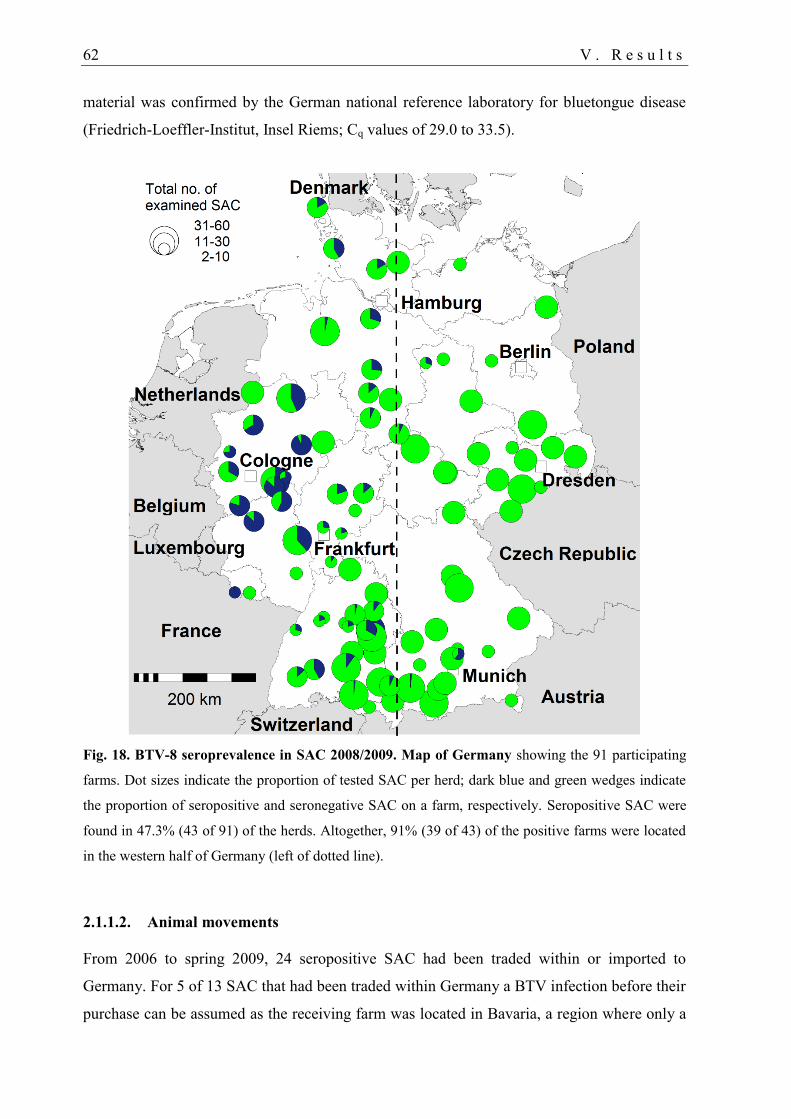
62 V . R e s u l t s
material was confirmed by the German national reference laboratory for bluetongue disease
(Friedrich-Loeffler-Institut, Insel Riems; Cq values of 29.0 to 33.5).
Fig. 18. BTV-8 seroprevalence in SAC 2008/2009. Map of Germany showing the 91 participating
farms. Dot sizes indicate the proportion of tested SAC per herd; dark blue and green wedges indicate
the proportion of seropositive and seronegative SAC on a farm, respectively. Seropositive SAC were
found in 47.3% (43 of 91) of the herds. Altogether, 91% (39 of 43) of the positive farms were located
in the western half of Germany (left of dotted line).
2.1.1.2. Animal movements
From 2006 to spring 2009, 24 seropositive SAC had been traded within or imported to
Germany. For 5 of 13 SAC that had been traded within Germany a BTV infection before their
purchase can be assumed as the receiving farm was located in Bavaria, a region where only a
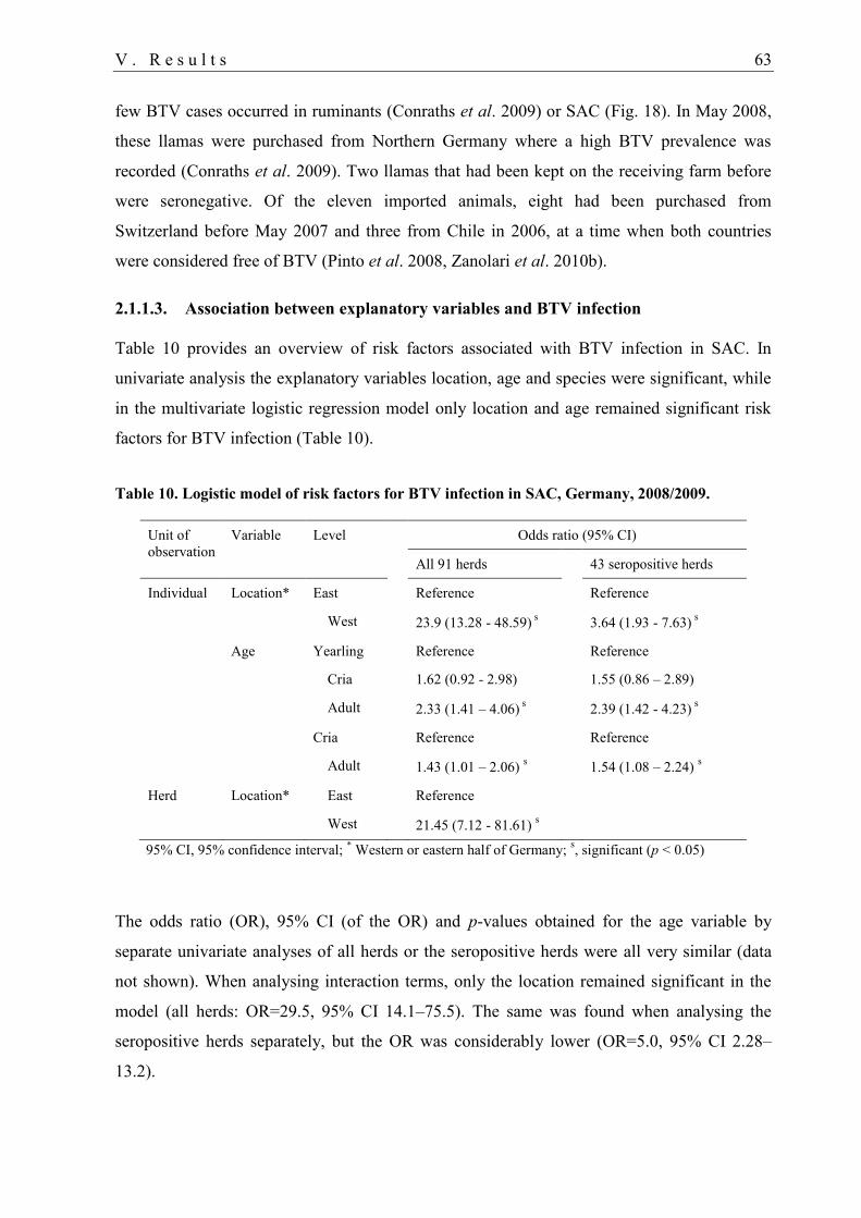
V . R e s u l t s 63
few BTV cases occurred in ruminants (Conraths et al. 2009) or SAC (Fig. 18). In May 2008,
these llamas were purchased from Northern Germany where a high BTV prevalence was
recorded (Conraths et al. 2009). Two llamas that had been kept on the receiving farm before
were seronegative. Of the eleven imported animals, eight had been purchased from
Switzerland before May 2007 and three from Chile in 2006, at a time when both countries
were considered free of BTV (Pinto et al. 2008, Zanolari et al. 2010b).
2.1.1.3. Association between explanatory variables and BTV infection
Table 10 provides an overview of risk factors associated with BTV infection in SAC. In
univariate analysis the explanatory variables location, age and species were significant, while
in the multivariate logistic regression model only location and age remained significant risk
factors for BTV infection (Table 10).
Table 10. Logistic model of risk factors for BTV infection in SAC, Germany, 2008/2009.
Unit of
observation
Variable Level Odds ratio (95% CI)
All 91 herds 43 seropositive herds
Individual Location* East Reference Reference
West 23.9 (13.28 - 48.59) s 3.64 (1.93 - 7.63)
s
Age Yearling Reference Reference
Cria 1.62 (0.92 - 2.98) 1.55 (0.86 – 2.89)
Adult 2.33 (1.41 – 4.06) s 2.39 (1.42 - 4.23)
s
Cria Reference Reference
Adult 1.43 (1.01 – 2.06) s 1.54 (1.08 – 2.24)
s
Herd Location* East Reference
West 21.45 (7.12 - 81.61) s
95% CI, 95% confidence interval; * Western or eastern half of Germany;
s, significant (p < 0.05)
The odds ratio (OR), 95% CI (of the OR) and p-values obtained for the age variable by
separate univariate analyses of all herds or the seropositive herds were all very similar (data
not shown). When analysing interaction terms, only the location remained significant in the
model (all herds: OR=29.5, 95% CI 14.1–75.5). The same was found when analysing the
seropositive herds separately, but the OR was considerably lower (OR=5.0, 95% CI 2.28–
13.2).
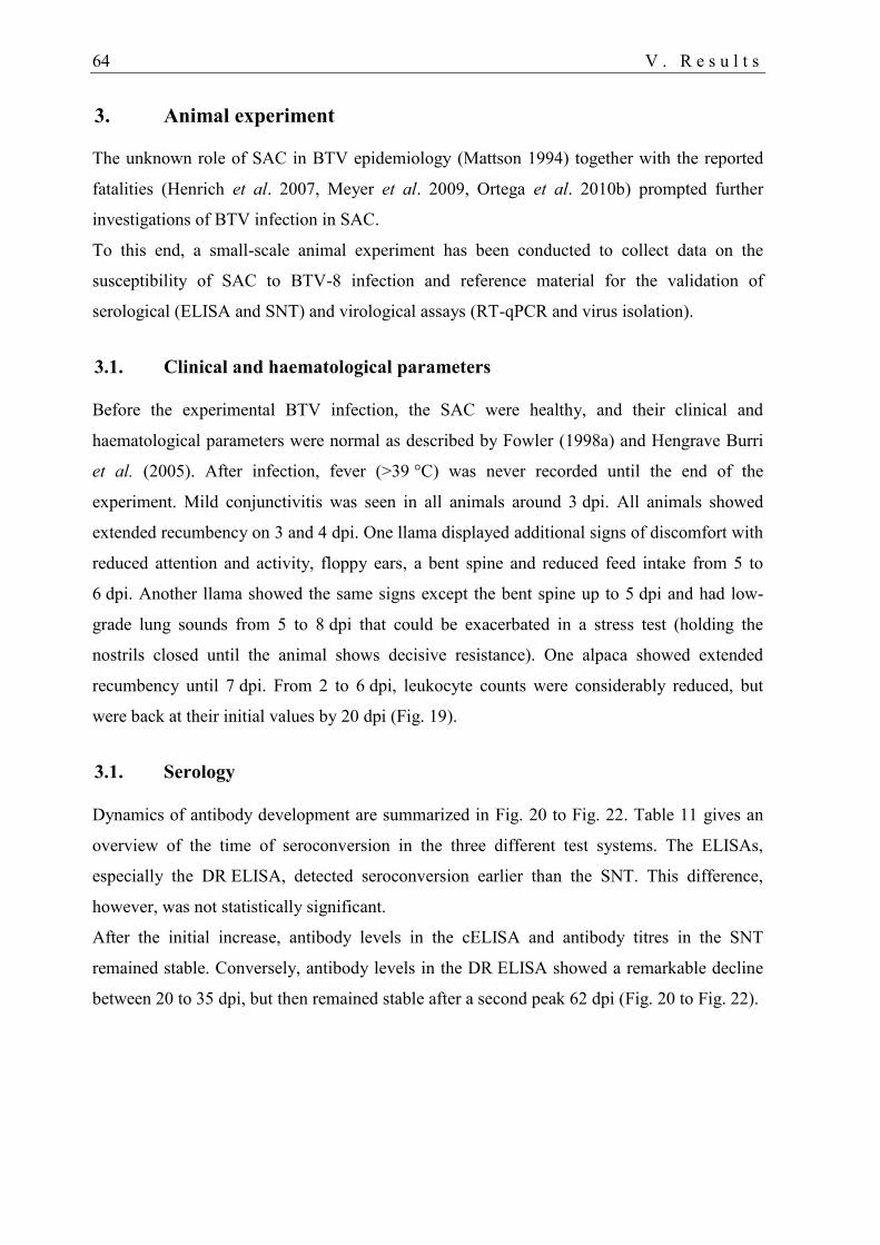
64 V . R e s u l t s
3. Animal experiment
The unknown role of SAC in BTV epidemiology (Mattson 1994) together with the reported
fatalities (Henrich et al. 2007, Meyer et al. 2009, Ortega et al. 2010b) prompted further
investigations of BTV infection in SAC.
To this end, a small-scale animal experiment has been conducted to collect data on the
susceptibility of SAC to BTV-8 infection and reference material for the validation of
serological (ELISA and SNT) and virological assays (RT-qPCR and virus isolation).
3.1. Clinical and haematological parameters
Before the experimental BTV infection, the SAC were healthy, and their clinical and
haematological parameters were normal as described by Fowler (1998a) and Hengrave Burri
et al. (2005). After infection, fever (>39 °C) was never recorded until the end of the
experiment. Mild conjunctivitis was seen in all animals around 3 dpi. All animals showed
extended recumbency on 3 and 4 dpi. One llama displayed additional signs of discomfort with
reduced attention and activity, floppy ears, a bent spine and reduced feed intake from 5 to
6 dpi. Another llama showed the same signs except the bent spine up to 5 dpi and had low-
grade lung sounds from 5 to 8 dpi that could be exacerbated in a stress test (holding the
nostrils closed until the animal shows decisive resistance). One alpaca showed extended
recumbency until 7 dpi. From 2 to 6 dpi, leukocyte counts were considerably reduced, but
were back at their initial values by 20 dpi (Fig. 19).
3.1. Serology
Dynamics of antibody development are summarized in Fig. 20 to Fig. 22. Table 11 gives an
overview of the time of seroconversion in the three different test systems. The ELISAs,
especially the DR ELISA, detected seroconversion earlier than the SNT. This difference,
however, was not statistically significant.
After the initial increase, antibody levels in the cELISA and antibody titres in the SNT
remained stable. Conversely, antibody levels in the DR ELISA showed a remarkable decline
between 20 to 35 dpi, but then remained stable after a second peak 62 dpi (Fig. 20 to Fig. 22).
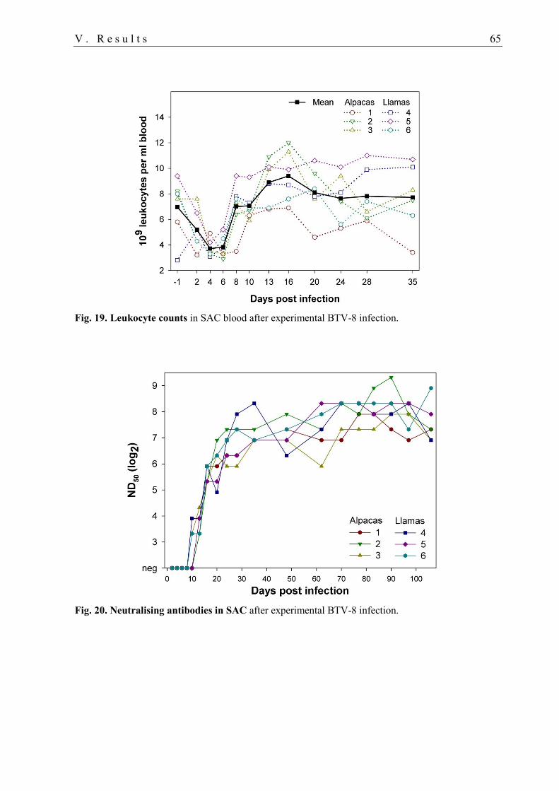
V . R e s u l t s 65
Fig. 19. Leukocyte counts in SAC blood after experimental BTV-8 infection.
Fig. 20. Neutralising antibodies in SAC after experimental BTV-8 infection.
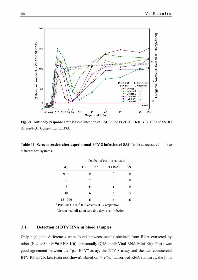
66 V . R e s u l t s
Fig. 21. Antibody response after BTV-8 infection of SAC in the PrioCHECK® BTV DR and the ID
Screen® BT Competition ELISA.
Table 11. Seroconversion after experimental BTV-8 infection of SAC (n=6) as measured in three
different test systems.
Number of positive animals
dpi DR ELISAa cELISA
b SNT
0 - 4 0 0 0
6 2 0 0
8 5 1 0
10 6 5 3
13 - 106 6 6 6 a PrioCHECK®;
b ID Screen® BT Competition;
c Serum neutralisation test; dpi, days post-infection
3.1. Detection of BTV RNA in blood samples
Only negligible differences were found between results obtained from RNA extracted by
robot (NucleoSpin® 96 RNA Kit) or manually (QIAamp® Viral RNA Mini Kit). There was
good agreement between the “pan-BTV” assay, the BTV-8 assay and the two commercial
BTV RT-qPCR kits (data not shown). Based on in vitro transcribed RNA standards, the limit
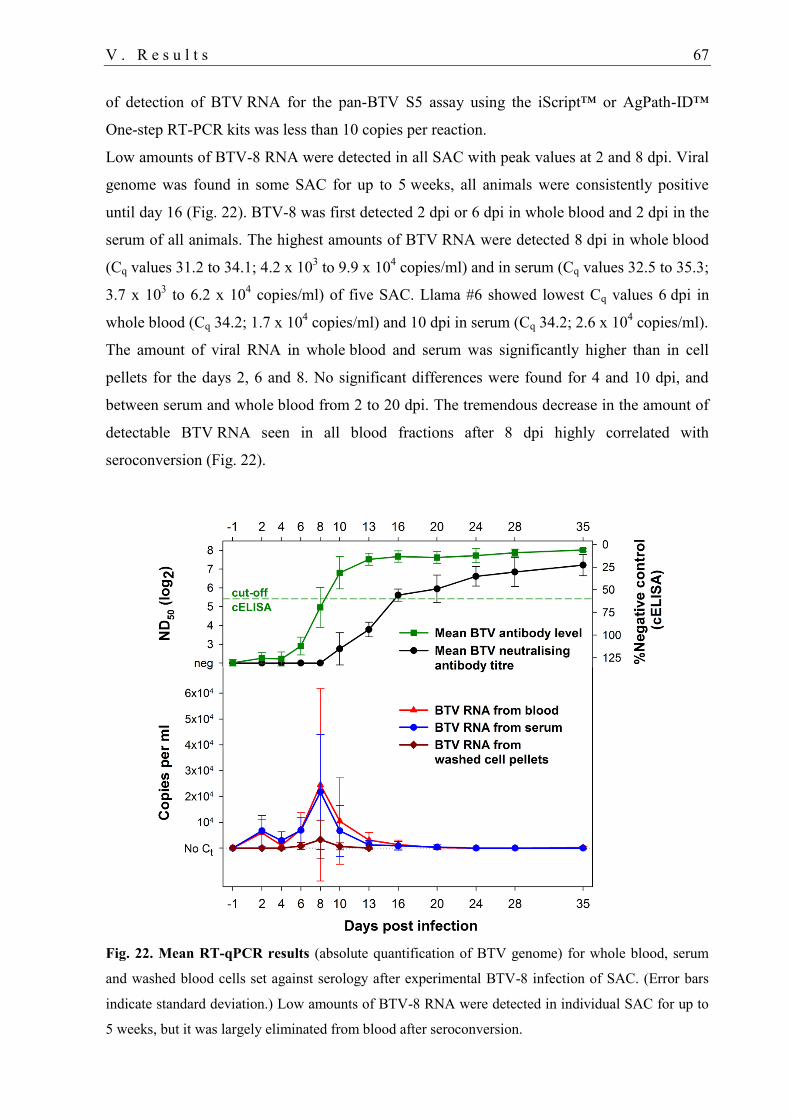
V . R e s u l t s 67
of detection of BTV RNA for the pan-BTV S5 assay using the iScript™ or AgPath-ID™
One-step RT-PCR kits was less than 10 copies per reaction.
Low amounts of BTV-8 RNA were detected in all SAC with peak values at 2 and 8 dpi. Viral
genome was found in some SAC for up to 5 weeks, all animals were consistently positive
until day 16 (Fig. 22). BTV-8 was first detected 2 dpi or 6 dpi in whole blood and 2 dpi in the
serum of all animals. The highest amounts of BTV RNA were detected 8 dpi in whole blood
(Cq values 31.2 to 34.1; 4.2 x 10³ to 9.9 x 10
4 copies/ml) and in serum (Cq values 32.5 to 35.3;
3.7 x 10³ to 6.2 x 10
4 copies/ml) of five SAC. Llama #6 showed lowest Cq values 6 dpi in
whole blood (Cq 34.2; 1.7 x 10⁴ copies/ml) and 10 dpi in serum (Cq 34.2; 2.6 x 10
4 copies/ml).
The amount of viral RNA in whole blood and serum was significantly higher than in cell
pellets for the days 2, 6 and 8. No significant differences were found for 4 and 10 dpi, and
between serum and whole blood from 2 to 20 dpi. The tremendous decrease in the amount of
detectable BTV RNA seen in all blood fractions after 8 dpi highly correlated with
seroconversion (Fig. 22).
Fig. 22. Mean RT-qPCR results (absolute quantification of BTV genome) for whole blood, serum
and washed blood cells set against serology after experimental BTV-8 infection of SAC. (Error bars
indicate standard deviation.) Low amounts of BTV-8 RNA were detected in individual SAC for up to
5 weeks, but it was largely eliminated from blood after seroconversion.
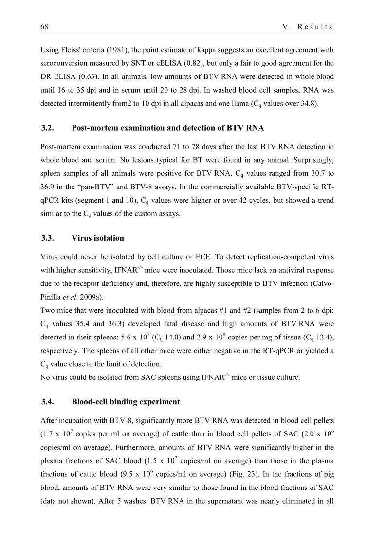
68 V . R e s u l t s
Using Fleiss' criteria (1981), the point estimate of kappa suggests an excellent agreement with
seroconversion measured by SNT or cELISA (0.82), but only a fair to good agreement for the
DR ELISA (0.63). In all animals, low amounts of BTV RNA were detected in whole blood
until 16 to 35 dpi and in serum until 20 to 28 dpi. In washed blood cell samples, RNA was
detected intermittently from2 to 10 dpi in all alpacas and one llama (Cq values over 34.8).
3.2. Post-mortem examination and detection of BTV RNA
Post-mortem examination was conducted 71 to 78 days after the last BTV RNA detection in
whole blood and serum. No lesions typical for BT were found in any animal. Surprisingly,
spleen samples of all animals were positive for BTV RNA. Cq values ranged from 30.7 to
36.9 in the “pan-BTV” and BTV-8 assays. In the commercially available BTV-specific RT-
qPCR kits (segment 1 and 10), Cq values were higher or over 42 cycles, but showed a trend
similar to the Cq values of the custom assays.
3.3. Virus isolation
Virus could never be isolated by cell culture or ECE. To detect replication-competent virus
with higher sensitivity, IFNAR-/-
mice were inoculated. Those mice lack an antiviral response
due to the receptor deficiency and, therefore, are highly susceptible to BTV infection (Calvo-
Pinilla et al. 2009a).
Two mice that were inoculated with blood from alpacas #1 and #2 (samples from 2 to 6 dpi;
Cq values 35.4 and 36.3) developed fatal disease and high amounts of BTV RNA were
detected in their spleens: 5.6 x 10⁷ (Cq 14.0) and 2.9 x 10
8 copies per mg of tissue (Cq 12.4),
respectively. The spleens of all other mice were either negative in the RT-qPCR or yielded a
Cq value close to the limit of detection.
No virus could be isolated from SAC spleens using IFNAR-/-
mice or tissue culture.
3.4. Blood-cell binding experiment
After incubation with BTV-8, significantly more BTV RNA was detected in blood cell pellets
(1.7 x 10⁷ copies per ml on average) of cattle than in blood cell pellets of SAC (2.0 x 10
6
copies/ml on average). Furthermore, amounts of BTV RNA were significantly higher in the
plasma fractions of SAC blood (1.5 x 10⁷ copies/ml on average) than those in the plasma
fractions of cattle blood (9.5 x 10⁶ copies/ml on average) (Fig. 23). In the fractions of pig
blood, amounts of BTV RNA were very similar to those found in the blood fractions of SAC
(data not shown). After 5 washes, BTV RNA in the supernatant was nearly eliminated in all
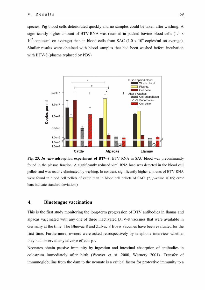
V . R e s u l t s 69
species. Pig blood cells deteriorated quickly and no samples could be taken after washing. A
significantly higher amount of BTV RNA was retained in packed bovine blood cells (1.1 x
107 copies/ml on average) than in blood cells from SAC (1.0 x 10
6 copies/ml on average).
Similar results were obtained with blood samples that had been washed before incubation
with BTV-8 (plasma replaced by PBS).
Fig. 23. In vitro adsorption experiment of BTV-8: BTV RNA in SAC blood was predominantly
found in the plasma fraction. A significantly reduced viral RNA load was detected in the blood cell
pellets and was readily eliminated by washing. In contrast, significantly higher amounts of BTV RNA
were found in blood cell pellets of cattle than in blood cell pellets of SAC. (*, p-value <0.05; error
bars indicate standard deviation.)
4. Bluetongue vaccination
This is the first study monitoring the long-term progression of BTV antibodies in llamas and
alpacas vaccinated with any one of three inactivated BTV-8 vaccines that were available in
Germany at the time. The Bluevac 8 and Zulvac 8 Bovis vaccines have been evaluated for the
first time. Furthermore, owners were asked retrospectively by telephone interview whether
they had observed any adverse effects p.v.
Neonates obtain passive immunity by ingestion and intestinal absorption of antibodies in
colostrum immediately after birth (Weaver et al. 2000, Wernery 2001). Transfer of
immunoglobulins from the dam to the neonate is a critical factor for protective immunity to a
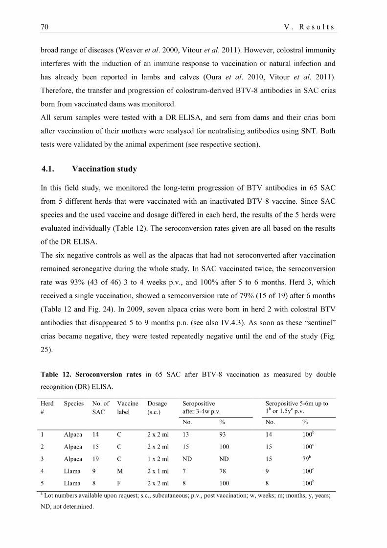
70 V . R e s u l t s
broad range of diseases (Weaver et al. 2000, Vitour et al. 2011). However, colostral immunity
interferes with the induction of an immune response to vaccination or natural infection and
has already been reported in lambs and calves (Oura et al. 2010, Vitour et al. 2011).
Therefore, the transfer and progression of colostrum-derived BTV-8 antibodies in SAC crias
born from vaccinated dams was monitored.
All serum samples were tested with a DR ELISA, and sera from dams and their crias born
after vaccination of their mothers were analysed for neutralising antibodies using SNT. Both
tests were validated by the animal experiment (see respective section).
4.1. Vaccination study
In this field study, we monitored the long-term progression of BTV antibodies in 65 SAC
from 5 different herds that were vaccinated with an inactivated BTV-8 vaccine. Since SAC
species and the used vaccine and dosage differed in each herd, the results of the 5 herds were
evaluated individually (Table 12). The seroconversion rates given are all based on the results
of the DR ELISA.
The six negative controls as well as the alpacas that had not seroconverted after vaccination
remained seronegative during the whole study. In SAC vaccinated twice, the seroconversion
rate was 93% (43 of 46) 3 to 4 weeks p.v., and 100% after 5 to 6 months. Herd 3, which
received a single vaccination, showed a seroconversion rate of 79% (15 of 19) after 6 months
(Table 12 and Fig. 24). In 2009, seven alpaca crias were born in herd 2 with colostral BTV
antibodies that disappeared 5 to 9 months p.n. (see also IV.4.3). As soon as these “sentinel”
crias became negative, they were tested repeatedly negative until the end of the study (Fig.
25).
Table 12. Seroconversion rates in 65 SAC after BTV-8 vaccination as measured by double
recognition (DR) ELISA.
Herd
#
Species No. of
SAC
Vaccine
label
Dosage
(s.c.)
Seropositive
after 3-4w p.v.
Seropositive 5-6m up to
1b or 1.5y
c p.v.
No. % No. %
1 Alpaca 14 C 2 x 2 ml 13 93 14 100b
2 Alpaca 15 C 2 x 2 ml 15 100 15 100c
3 Alpaca 19 C 1 x 2 ml ND ND 15 79b
4 Llama 9 M 2 x 1 ml 7 78 9 100c
5 Llama 8 F 2 x 2 ml 8 100 8 100b
a Lot numbers available upon request; s.c., subcutaneous; p.v., post vaccination; w, weeks; m; months; y, years;
ND, not determined.
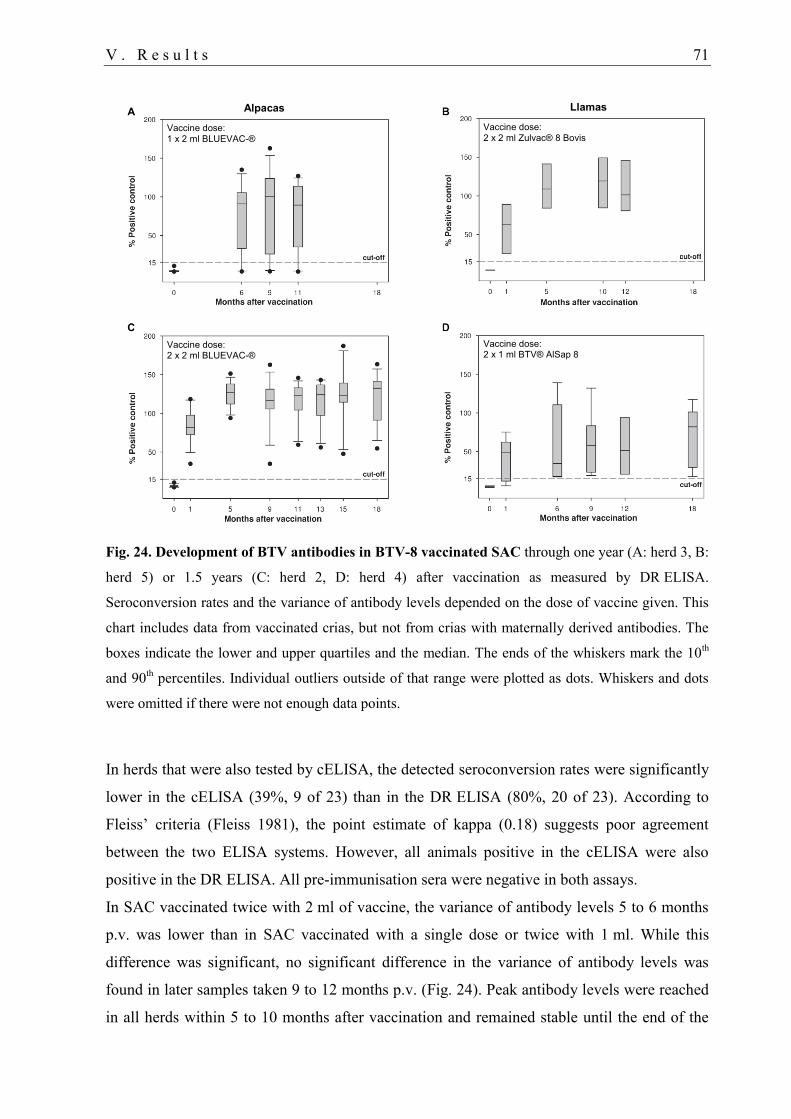
V . R e s u l t s 71
Fig. 24. Development of BTV antibodies in BTV-8 vaccinated SAC through one year (A: herd 3, B:
herd 5) or 1.5 years (C: herd 2, D: herd 4) after vaccination as measured by DR ELISA.
Seroconversion rates and the variance of antibody levels depended on the dose of vaccine given. This
chart includes data from vaccinated crias, but not from crias with maternally derived antibodies. The
boxes indicate the lower and upper quartiles and the median. The ends of the whiskers mark the 10th
and 90th percentiles. Individual outliers outside of that range were plotted as dots. Whiskers and dots
were omitted if there were not enough data points.
In herds that were also tested by cELISA, the detected seroconversion rates were significantly
lower in the cELISA (39%, 9 of 23) than in the DR ELISA (80%, 20 of 23). According to
Fleiss’ criteria (Fleiss 1981), the point estimate of kappa (0.18) suggests poor agreement
between the two ELISA systems. However, all animals positive in the cELISA were also
positive in the DR ELISA. All pre-immunisation sera were negative in both assays.
In SAC vaccinated twice with 2 ml of vaccine, the variance of antibody levels 5 to 6 months
p.v. was lower than in SAC vaccinated with a single dose or twice with 1 ml. While this
difference was significant, no significant difference in the variance of antibody levels was
found in later samples taken 9 to 12 months p.v. (Fig. 24). Peak antibody levels were reached
in all herds within 5 to 10 months after vaccination and remained stable until the end of the
Llamas Alpacas
Vaccine dose: 2 x 2 ml BLUEVAC-®
Vaccine dose: 1 x 2 ml BLUEVAC-®
Vaccine dose: 2 x 2 ml Zulvac® 8 Bovis
Vaccine dose: 2 x 1 ml BTV® AlSap 8
72 V . R e s u l t s
study (11 to 18 months after vaccination) (Fig. 24). Eight seropositive SAC could not be
followed until the end of the study because they had been sold or had died of causes unrelated
to BTV infection.
4.2. Tolerance of BTV-8 vaccines
Apart from the animals that participated in the vaccination study, vaccinations in additional
SAC were documented retrospectively by telephone questionnaire. No adverse effects were
reported in 97.7% (339 of 347) SAC on 27 farms (including animals of the vaccination
study), which were vaccinated with any one of four inactivated BTV-8 vaccines. The
maximum vaccine dose used for alpacas was 2 x 2 ml of vaccine C or twice 1 ml of vaccine
M. In llamas, the maximum dose was 2 x 4 ml of vaccine C, 2 x 2 ml of vaccine F, or two
1 ml doses of Bovilis® BTV8 (Intervet, Boxmeer, The Netherlands). Booster vaccinations in
two consecutive years were well tolerated by 46 alpacas and 29 llamas.
No irregularities concerning pregnancies, birth rates or health status of over 95 crias born
from 2008 to 2010 were observed by the owners of herds 1 to 5.
For the herds that did not participate in this vaccination study, one owner reported local
swelling at the injection site in 5 alpacas. On two other farms, two fatalities after vaccination
occurred in one 3-year-old alpaca male and one 1.3-year-old alpaca female. Both animals
were vaccinated once with 2 ml of vaccine C. The 3-year-old alpaca male died suddenly
during the night following vaccination. According to the post-mortem report, a causal link to
vaccination has not been identified. The 1.3-year-old alpaca female perished four weeks p.v.
A 1-year-old alpaca male on the same farm showed a reduced general condition and buccal
ulcerations but recovered after 6 weeks. On the latter two farms, other alpacas (33 overall)
were vaccinated but did not show any adverse effects.
4.3. Maternal BTV antibodies in crias
Maternally-derived BTV antibodies were found in all crias from vaccinated dams except in
one cria (10) that was not tested before 19 weeks of age – at a time when about half of the
other crias were already negative in the ELISA (Fig. 25).
Positive results in the DR ELISA were obtained from 1 week p.n. (cria 1) for at least 7 weeks
(cria 2) up to a maximum of 30 weeks (cria 6) (median age at last positive sample: 14 weeks).
Of all initially seropositive crias born in 2009, 5 of 8 (63%) became seronegative between
week 20 and 27 p.n. (crias 1, 2, 4, 5, 7), and 3 (27%) between week 30 and 39 (crias 3, 6, 9)
(median age: 26 weeks) (Fig. 25).

V . R e s u l t s 73
NAbs were only detected in llama cria 9 (six weeks p.n.), which was born 3 months after the
mother had been vaccinated with vaccine F. Compared to the other dams, this dam displayed
the highest NAb titre 3 weeks p.v. and around parturition (Table 13). All other crias born over
5 months p.v. of their mothers were found negative irrespective of the NAb status of the dam.
Three weeks after the 2nd
vaccination, 7 of 9 dams had NAb, while only four were positive
around parturition 3 to 11 months p.v. No correlation was found between NAb titres and
antibody levels measured by DR ELISA (Table 13).
Fig. 25. Transfer of colostral BTV-8 antibodies to crias. Antibody levels of BTV-8 vaccinated
dams at the time of parturition (“Dam”), and their crias after birth as measured by DR ELISA. The
progression of colostrum-derived BTV antibody levels was similar in all crias with a high initial level.
Blood samples from the dam of crias 7 and 8 were taken in 2009 and 2010, respectively. While
antibody levels of dam 7 remained stable within the year, the antibody levels in her crias diverged
considerably (2009: ○ dams, ● crias; 2010: □ dam 7, ■ cria 8).
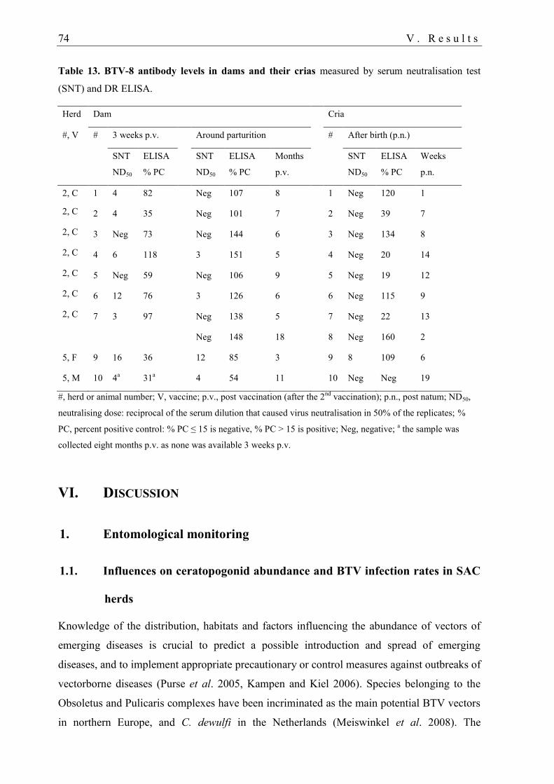
74 V . R e s u l t s
Table 13. BTV-8 antibody levels in dams and their crias measured by serum neutralisation test
(SNT) and DR ELISA.
Herd Dam Cria
#, V # 3 weeks p.v. Around parturition # After birth (p.n.)
SNT
ND50
ELISA
% PC
SNT
ND50
ELISA
% PC
Months
p.v.
SNT
ND50
ELISA
% PC
Weeks
p.n.
2, C 1 4 82 Neg 107 8 1 Neg 120 1
2, C 2 4 35 Neg 101 7 2 Neg 39 7
2, C 3 Neg 73 Neg 144 6 3 Neg 134 8
2, C 4 6 118 3 151 5 4 Neg 20 14
2, C 5 Neg 59 Neg 106 9 5 Neg 19 12
2, C 6 12 76 3 126 6 6 Neg 115 9
2, C 7 3 97 Neg 138 5 7 Neg 22 13
Neg 148 18 8 Neg 160 2
5, F 9 16 36 12 85 3 9 8 109 6
5, M 10 4a 31
a 4 54 11 10 Neg Neg 19
#, herd or animal number; V, vaccine; p.v., post vaccination (after the 2nd
vaccination); p.n., post natum; ND50,
neutralising dose: reciprocal of the serum dilution that caused virus neutralisation in 50% of the replicates; %
PC, percent positive control: % PC ≤ 15 is negative, % PC > 15 is positive; Neg, negative; a the sample was
collected eight months p.v. as none was available 3 weeks p.v.
VI. DISCUSSION
1. Entomological monitoring
1.1. Influences on ceratopogonid abundance and BTV infection rates in SAC
herds
Knowledge of the distribution, habitats and factors influencing the abundance of vectors of
emerging diseases is crucial to predict a possible introduction and spread of emerging
diseases, and to implement appropriate precautionary or control measures against outbreaks of
vectorborne diseases (Purse et al. 2005, Kampen and Kiel 2006). Species belonging to the
Obsoletus and Pulicaris complexes have been incriminated as the main potential BTV vectors
in northern Europe, and C. dewulfi in the Netherlands (Meiswinkel et al. 2008). The
V I . D i s c u s s i o n 75
proportions of C. obsoletus s.l. and C. pulicaris s.l. were similarly high compared to those
reported by other groups in northeastern Europe (Meiswinkel et al. 2008, Bartsch et al. 2009,
Hörbrand and Geier 2009, Vorsprach et al. 2009, Mehlhorn et al. 2009a, Mehlhorn et al.
2009c). However, C. pulicaris s.l. never exceeded the proportion of C. obsoletus s.l. in the
collections, which was occasionally observed at other locations in Germany in May/spring
(Kiel et al. 2009, Clausen et al. 2009, Bartsch et al. 2009, Hoffmann et al. 2009a, Mehlhorn
et al. 2009b) and frequently around outbreak sites in Belgium (de Deken et al. 2008). In the
present study, Culicoides abundance and BTV seroprevalence in herd A were considerably
higher than in herd B, although both herds were located in regions where the risk for BTV-8
infection was high at the time (Conraths et al. 2009) (Fig. 18) (Schulz et al. 2012c). Statistical
analysis revealed a strong effect of the location variable on the number of Culicoides on the
farms. Anthropogenic, landscape, meteorological, biotic and abiotic factors can have an
influence on vector abundance and prevalence of BTV infection in ruminants (Conte et al.
2007, Hörbrand and Geier 2009, Vorsprach et al. 2009, De Liberato et al. 2010, Purse et al.
2012, Mayo et al. 2012a). Seasonal and meteorological effects (especially temperature,
humidity and wind speed) belong to the key factors influencing the abundance and diversity
of Culicoides (Mellor et al. 2000, Mellor et al. 2009b, Purse et al. 2012). Similar to results of
other groups (Mellor et al. 2000, Kiel et al. 2009, Vorsprach et al. 2009), the interaction of
temperature and humidity also had a significant effect on the number of Culicoides in the
present study. Hence, the association with the listed factors can explain the annual deviations
in the abundance and composition of Culicoides and other ceratopogonid genera observed on
farms B and C. Culicoides abundance peaked in July and August 2008 in the present study.
However, the highest incidence of vectors and of BT disease in temperate areas is usually
recorded in late summer and autumn (Hoffmann et al. 2009a, Mellor et al. 2009b). Cool
weather conditions in late summer might have influenced the earlier decrease in the number
of Culicoides (Hörbrand and Geier 2009). On the other hand, results were contradictory
analysing the three SAC farms separately, indicating that additional factors were not included
in the regression model that might have influenced the results. Factors that possibly had a
positive effect on the number of Culicoides on farm A and that differed from local conditions
on farms B and C (Table 2) included a high land cover with natural-wood, location at about
300 m a.s.l. and in a valley (with light winds) (Bartsch et al. 2009, Mehlhorn et al. 2009c, De
Liberato et al. 2010).
Besides, the high number of cattle kept near SAC herds could have negatively influenced
the number of Culicoides and seroprevalence in SAC on farms B and C. Cattle was found to
76 V I . D i s c u s s i o n
have a ‘dilution effect’ on the risk of BTV infection (Durand et al. 2010a) and to be the most
attractive host for Palearctic biting midges (Bartsch et al. 2009). On the other hand, the daily
maximum of Culicoides caught in one trap on farm A was similar to the maximum reported
on a dairy farm in central Germany (Clausen et al. 2009). De Liberato et al. (2010) suggested
that the number of farmed animals does not affect C. obsoletus densities and/or that the
vectors use alternative hosts, but that the abundance of C. obsoletus probably rather depends
on the availability of shady vegetation. Hence, other factors than the animal species probably
had a stronger influence on Culicoides abundance and, therefore, BTV infection rates.
The absence of Culicoides in winter found in the present study can be explained by the colder
winter 2008/2009 compared to the winters in the two previous years (www.dwd.de).
According to recent studies, a vector-free period in winter does not exist in Germany and
western adjacent countries (Mehlhorn 2009, Meiswinkel 2008, Hoffmann 2009, Clausen
2009), but is possible at certain locations (Clausen et al. 2009). A lack of sensitivity of the
monitoring systems (Meiswinkel et al. 2008) can be precluded since we used the same traps
as in the previous monitoring programmes in Germany (Hoffmann et al. 2009a).
In the present study, C. obsoletus was found to be the only species that occurred during the
whole study period (except during the winter months), while C. pulicaris was less frequently
collected on farms B and C. C. obsoletus was named as the most robust Culicoides spp. as it
can occur in Germany throughout the year (Kiel et al. 2009, Vorsprach et al. 2009). In
contrast, the phase of activity of C. pulicaris s.l. is shorter during the year (Mehlhorn et al.
2009c).
Only a few individuals of the taxon C. dewulfi were collected on farm A. This species
exclusively breeds in cattle or horse dung (Meiswinkel et al. 2008). However, no cattle were
kept on or nearby farm A. To the best of our knowledge, no studies of Culicoides instars in
SAC dung have been conducted. Therefore, this species possibly also breeds in other habitats,
such as dung of SAC, or it has been transported to the valley by winds (Mellor et al. 2000).
C. imicola, the main Afro-Asian vector of BTV (Meiswinkel et al. 2004), has so far never
been detected north of the Alps (Hoffmann et al. 2009a), and it was nor collected on the SAC
farms in this study.
The high number of C. obsoletus s.l. and C. pulicaris s.l. collected in indoor traps indicated
that the risk for BTV infection is high in stables, which was already reported elsewhere
(Clausen et al. 2009). Although the number of ceratopogonid genera, Culicoides spp. and
proportion of males and females in indoor and outdoor collections depended on the farm, C.
obsoletus s.l. were generally predominantly collected indoor, while a higher number of C.
V I . D i s c u s s i o n 77
pulicaris s.l. were caught outdoor. This is in accordance with the strong endophilic and
exophilic behaviour of C. obsoletus/C. scoticus and C. pulicaris s.s., respectively, reported in
Europe (Meiswinkel et al. 2008).
The abundance and species composition of Culicoides collected for this study was probably
biased due to the fact that the traps were only run from dusk until dawn, and a significant
number of midges might have gone undetected because they are biting during the day
(Meiswinkel et al. 2008, Balenghien et al. 2008, Clausen et al. 2009). On the other hand,
many Culicoides are active from dusk until dawn as nocturnal activity may reduce the risk of
desiccation (lower temperatures, higher humidity) (Mellor et al. 2000).
1.2. Phenology of ceratopogonids
The diversity of species found on the three study farms corresponded to the number of species
reported by the groups that monitored Culicoides in nearby regions in the previous year (Kiel
et al. 2009, Vorsprach et al. 2009, Hörbrand and Geier 2009). Similar to those studies, peaks
in the diversity of species were found between June and October, and the number of species
caught by months depended on the location (Kiel et al. 2009, Vorsprach et al. 2009) (Table 5)
and year.
1.3. Absence of BTV RNA
None of the analysed Culicoides were found positive for BTV RNA, although most of the
tested midges were collected on the farm with the highest seroprevalence, and in the same
months when the highest number of BTV infected Culicoides (obsoletus s.l.) and BT cases in
ruminants were recorded in Germany in the previous two years (Hoffmann et al. 2009a,
Mehlhorn et al. 2009a). Furthermore, the BTV-8 epizootic was still ongoing at the time of the
study (Conraths et al. 2009). However, positive batches were only found on farms where the
proportion of BTV-infected ruminants was very high (Hoffmann, unpublished observations).
On the other hand, the infection of the SAC might have already occurred earlier in the year or
during the peak of the epizootic in 2007.
Differences in the pathogenesis of BTV-8 in SAC compared to ruminants could also explain
the negative virological results. In contrast to the long-term viraemia characteristic for BTV
infection in ruminants, only low amounts of BTV RNA were detected for a short time in SAC
experimentally infected with BTV-8 (maximum 5 weeks) (Fig. 22) (Schulz et al. 2012b). In
the cross-sectional study (Schulz et al. 2012c), none of 249 seropositive SAC (including herds
78 V I . D i s c u s s i o n
A-C) were BTV RNA positive at the time of the present study. Therefore, the transmission
cycle between SAC and Culicoides was possibly not maintained in the herds.
1.4. Breeding habitats and occurrence of males
For targeted vector control, it is important to know the Culicoides breeding habitats (Werner
et al. 2012) and where swarming and mating takes place. Culicoides males do not feed on
blood but on plant juice. Thus, they are not directly important in the transmission cycle of
BTV. Peaks in the proportion of Obsoletus complex males caught in black-light traps were
also found in April and November by Vorsprach et al. (2009). Males typically emerge a short
time before females (Mullen 2009), which was also found for C. obsoletus males caught in
emergence traps in the present study (Fig. 17). In black-light traps, a proportion of generally 0
to 5% of Culicoides males (Mehlhorn et al. 2007, Bartsch et al. 2009, Vorsprach et al. 2009,
Kiel et al. 2009) and an unbalanced monthly occurrence of C. pulicaris s.l. males were also
recorded by other authors (Kiel et al. 2009). Possible reasons for the huge differences in
gender distribution can be that black light predominantly attracts females and that the mating
and swarming places are not close to the feeding places (hosts) (Mehlhorn et al. 2007,
Vorsprach et al. 2009). In contrast, those places might be close to the breeding habitats, which
would explain the considerably higher proportion of males collected in the emergence traps.
Furthermore, males usually travel much shorter distances than females (Mullen 2009).
We have no suitable explanation for the considerably lower proportion of Culicoides males
(14.1% of 185) and Obsoletus complex males (14.3%) collected in the faeces trap than in the
pasture trap (68.2% respectively 66.7%). A distortion of data by female midges escaping from
traps in search of a host or slipping in from outside to lay their eggs should be taken into
consideration. The markedly higher number of Culicoides caught in the faeces trap indicates
that adult Culicoides prefer faeces (or the subjacent ground) for breeding and their larvae as
feeding substrate, which was already described by Bishop et al. (1996) and Boorman et al.
(1986). On the other hand, substrate preferences depend on the Culicoides spp. (Werner et al.
2012). In the present study, C. albicans were predominantly caught in the pasture trap, while
C. obsoletus s.l. and C. achrayi presented the dominant taxa in the faeces trap. Similar to
results of a previous study (Werner et al. 2012), Culicoides were most frequently collected in
late summer, but in this study, a peak was also recorded in May (Fig. 17). Those peaks
probably indicate that hatching of Culicoides (especially C. obsoletus s.l. and C. achrayi)
predominantly takes place during these months. The most frequent species caught in the
V I . D i s c u s s i o n 79
faeces trap belonged to the Obsoletus complex, which is in accordance with results reported
by Werner et al. (2012).
2. Cross-sectional study
The epidemiological background of BTV infection, associated risk factors and its clinical
expression have so far only been investigated in ruminants, but never in any detail in SAC.
The fatalities in a few SAC that occurred during the recent BTV-8 and BTV-1 epizootics in
Germany and France, respectively, prompted the investigation of their role in the
epidemiology of BTV.
A total of 14.3% of tested SAC were positive for BTV antibodies in the cross-sectional study
in Germany from autumn 2008 to spring 2009. The spatial distribution of BTV-8 infection in
SAC was similar to the BTV-8 outbreaks registered in ruminants in Germany between 2006
and 2008 (Conraths et al. 2009). However, BTV-8 infection in domestic ruminants in
Germany had not been thoroughly investigated. Therefore, a direct comparison with our data
is not possible. The estimated proportion of BTV infection in exposed domestic ruminants in
Germany in 2007 was considerably lower (below 6.4%) (Conraths et al. 2009) than in
exposed SAC (in seropositive herds) in the present study (27.6%). In contrast, markedly
higher seroprevalences were reported in domestic ruminants (cattle, sheep, goat) and red deer
in northwestern Europe (34–70%) (van Schaik et al. 2008, Lutz 2008, Elbers et al. 2008c,
Linden et al. 2010) where the BTV-8 epizootic had initially started in 2006 (Conraths et al.
2009). This is consistent with the BTV-8 seroprevalence found in SAC in the west of
Germany (42.7%, Table 8 and Table 1 in Appendix 4). BTV antibodies and BTV-1 RNA
have been detected in a few llamas during an outbreak in France (Meyer et al. 2009), but no
BTV antibodies have been detected in 354 SAC in Switzerland. This can be explained by the
higher pressure of BTV infection on SAC populations in France and Germany as more BTV
outbreaks occurred in those countries than in Switzerland (Zanolari et al. 2010b).
BTV-8 seroprevalence in infected SAC herds varied widely (2–100%) as it was also described
in ruminant herds (cattle, sheep, goats) in the Netherlands (van Schaik et al. 2008, Elbers et
al. 2008c). Variations in BTV infection rates occur regularly in different ruminant species and
SAC (Rivera et al. 1987, Mattson 1994, Conraths et al. 2009, Meyer et al. 2009, Falconi et al.
2011). Seroprevalence in SAC herds generally decreased with distance from the region where
the first BTV-8 outbreaks were recorded, and location was significantly associated with BTV
infection. The location variable can be considered a proxy for BTV exposure in the respective
regions. Geographical variation in BTV seroprevalence was reported by several groups (Ruiz-
80 V I . D i s c u s s i o n
Fons et al. 2008, Elbers et al. 2008c, Durand et al. 2010b, García-Bocanegra et al. 2011).
Different ecosystems and vector/pathogen distribution were suggested to have a substantial
influence on BTV diffusion and exposure rate, respectively, while the amplification of BTV
circulation in affected herds played a rather limited role (Ruiz-Fons et al. 2008, Durand et al.
2010b). Nevertheless, the pressure of infection on naïve animals in areas closer to those
regions where BTV has occurred first will be higher than in regions that are farther away from
those hot spots.
As of 2011, the population of SAC in Germany is estimated at about 7,000 animals (Locher et
al. 2010, Schulz et al., manuscript in preparation). Most (94%) of the contacted owners that
met the requirements participated in this study, and the sample reflects the SAC population
recorded in 227 herds in Germany (Schulz et al., manuscript in preparation). Therefore, the
random sample is considered representative. The overall sample size was overfulfilled, but for
samples taken within herds, sample sizes in almost 40% of the herds were smaller than
required to detect at least one seropositive animal in the herd (Table 3). This might have
resulted in an underestimation of the number of positive herds. On the other hand, (i) the
overall seroprevalence in Germany was higher than 5%, (ii) the within-herd seroprevalence
varied considerably (2–100%; Table 8), and (iii) seroprevalence was generally lower in the
eastern than in the western half of Germany (Fig. 18, Table 8 and Table 1 in Appendix 4). As
described earlier, other factors apparently had an influence on seroprevalence in a herd.
Furthermore, the table used to determine the sample size on herd level is designed for
contagious and not for vectorborne diseases, which have a different distribution pattern
(Wilson et al. 2007). Thus, it is particularly questionable whether sample sizes were generally
sufficient at the edges of regions that had been affected by the BTV-8 epizootic wave and
where within-herd seroprevalences were probably lower than the assumed 5%. Therefore, it
seems likely that the number of positive herds was generally underestimated in eastern
regions.
A possible distortion of our data due to animal movement was identified for five seropositive
animals in one herd that had been shipped to Bavaria in 2008. Missing information due to an
interview bias cannot be excluded, but a relevant distortion of data is unlikely as the spatial
distribution of BTV outbreaks in ruminants in the same period was very similar (Conraths et
al. 2009). The used ELISA does not discriminate between serotypes, and a positive result due
to BTV infection with another serotype cannot be ruled out. During the time of the study,
BTV-6 had been detected in cattle in a field survey in northwestern Germany in late 2008
(Eschbaumer et al. 2010a). Only 16 blood samples of SAC from that area were seropositive
V I . D i s c u s s i o n 81
(Table 1 in Appendix 4), and no BTV RNA was found in those samples, which would have
indicated a recent infection with any other BTV serotype at the time. Furthermore, the
proportion of cattle positive for BTV-6 antibodies was very low (Eschbaumer et al. 2010a).
Therefore, a measurement bias for the overall proportion of BTV-8 antibodies detected in
SAC is considered negligible.
Location and age were the only risk factors for BTV infection that remained significant in the
logistic regression model (Table 10). Analysing interaction terms, only the location remained
a significant factor. This means that the univariate results were confounded by the location
variable because the majority of seropositive herds were located in the western half of
Germany where the risk of BTV exposure was significantly higher than in the eastern half
(Fig. 18, Table 8). The effect of age on BTV infection was not significant in the analysis of
interaction terms. However, we assume that there still is an association with the outcome
variable because the OR and 95% CI were almost identical in the logistic regression and the
univariate analyses (data not shown). A significant association between age and BTV
seroprevalence was reported by several groups (Ward et al. 1994, Linden et al. 2010, García-
Bocanegra et al. 2011), and can be explained by a longer time of exposure for adults (García-
Bocanegra et al. 2011, reviewed by Falconi et al. 2011). Interestingly, a high proportion of
seropositive dams (35 of 55) also had seropositive crias (data not shown), and we assume that
their crias have passively obtained immunity by the colostral transfer of BTV-8 antibodies.
Colostral transfer of BTV antibodies to neonates was already recorded by other authors, and
care must be taken when interpreting the presence of antibodies in serum samples from
juveniles (Falconi et al. 2011) (see VI.4.3). The study design generally included non-weaned
crias from tested dams, which has probably caused a distortion towards a higher proportion of
seropositive crias and a higher overall seroprevalence.
Species, sex, housing type and presence of other ruminants are risk factors that can influence
BTV infection rates in ruminants (Ward et al. 1994, Ruiz-Fons et al. 2008, Santman-Berends
et al. 2010, Durand et al. 2010b, Falconi et al. 2011). However, in SAC no significant
association with BTV infection was found for any risk factors other than location and age.
The risk of BTV infection in a SAC population is obviously due to BTV exposure, pressure of
infection and age rather than other risk factors.
The absence of BTV RNA in the field samples of SAC despite the high seroprevalence and
the ongoing BTV-8 epizootic can be explained by the collection of samples late in the season,
after vectorborne exposure had mostly abated (Hoffmann et al. 2009a), and by the lower
pressure of BTV-8 infection in 2008 compared to 2007 (Conraths et al. 2009). Furthermore,
82 V I . D i s c u s s i o n
BTV RNA in SAC blood can only be detected for a considerably shorter time after BTV-8
infection (up to 5 weeks) than in ruminant blood as BTV-8 adsorbs less efficiently to SAC
erythrocytes than to bovine RBC (Fig. 23) (Schulz et al. 2012b). Seronegative samples were
not analysed for BTV RNA, and theoretically some BTV RNA-positive animals might have
been missed. However, the underestimation of BTV RNA prevalence is considered low
because in SAC the time period between BTV infection and seroconversion is only about one
week (Afshar et al. 1995a, Schulz et al. 2012b) (Table 11), and most samples (91%) were
taken outside the main vector season (Hoffmann et al. 2009a).
Severe disease was reported in domestic ruminants – particularly sheep – during the BTV-8
epizootic (Dal Pozzo et al. 2009b). In wild ruminants, BTV infection can also result in severe
clinical signs, but this depends on the susceptiblity of the affected species. For example, no
clinical signs were recorded in red deer experimentally infected with European BTV-8
(López-Olvera et al. 2010, Falconi et al. 2011). In the present study, clinical signs of BT were
not observed by the owners in any of the tested SAC. This is consistent with earlier studies by
Rivera et al. (1987) and Mattson et al. (1994). If there was BT disease in the herds in the
present study, the owners might have failed to recognise it because the clinical signs were
unspecific and mild (Schulz et al. 2012b). Differences in the manifestation of BT disease in
various ruminant species and Old World camelids were also reported in other studies
(Conraths et al. 2009, MacLachlan et al. 2009, Batten et al. 2011). On the other hand, in the
three recorded BT cases presented here, BTV RNA was detected at post-mortem examination,
and BT disease was assumed in one perished llama. BTV-related disease and consequent
fatalities in alpacas and llamas were recently reported during the BTV-8 and BTV-1
epizootics in Germany and France, respectively (Henrich et al. 2007, Meyer et al. 2009), and
in the USA (Ortega et al. 2010b). However, whether BTV infection in the three fatal cases
exacerbated concurrent diseases or vice versa, or whether the detection of BTV RNA was
only a secondary pathological finding remains unclear. Immunosuppression can aggravate
BTV infection in ruminants (Brodie et al. 1998). All three cases ocurred in winter when
infectious midges are unlikely to be found (Wilson et al. 2007, Hoffmann et al. 2009a).
3. Animal experiment
The range of hosts susceptible to BTV infection includes domestic and wild ruminants as well
as camelids. In SAC, serological evidence for a previous BTV infection was detected in Peru
(20%, n=114, Rivera et al. 1987), Oregon, USA (1.5%, n=270, Mattson 1994), France (78%,
n=9, Meyer et al. 2009) and in Germany 2008/2009 (14.3%, n=1742) (Schulz et al. 2012c).

V I . D i s c u s s i o n 83
However viraemia, clinical signs, abortion and fatalities related to BTV infection have been
reported only rarely (Fowler 1998c, Henrich et al. 2007, Meyer et al. 2009, Ortega et al.
2010b).
Experimental BTV infection of camelids was conducted in two llamas – to evaluate antibody
response to BTV infection in a cELISA – (Afshar et al. 1995a) and recently in three
dromedary camels (Batten et al. 2011). Based on the results, camels were suggested to have a
potential role in the transmission of BTV-1 to local Culicoides vectors in Morocco, and they
were named as possible reservoirs to carry the virus over long distances (Batten et al. 2011).
However, to date no experimental infections had been conducted to evaluate viraemia and
infection dynamics in SAC after BTV infection.
Mild unspecific clinical signs such as anorexia, arched backs or lung sounds, which were
found in two llamas, as well as extended recumbency, mild conjunctivitis and leukopenia,
which were found in all SAC between 2 to 8 dpi, were also recorded for ruminants after BTV
infection (Ellis et al. 1990, Foster et al. 1991, Darpel et al. 2007, Darpel et al. 2009b)
(reviewed by MacLachlan et al. 2009, Dal Pozzo et al. 2009a). Reports of fatal BTV infection
in SAC have described a short period of lethargy or weakness, recumbency, respiratory
distress and anorexia (Henrich et al. 2007, Meyer et al. 2009, Ortega et al. 2010b). However,
the severity of BT in the field can be influenced by secondary bacterial infection (Verwoerd
and Erasmus 2004, Darpel et al. 2007), immune suppression (Brodie et al. 1998), or external
factors such as stress or sunlight. Therefore, high-containment animal housing might have
reduced the displayed clinical signs of BT in this experiment (Verwoerd and Erasmus 2004).
On the other hand, no clinical signs were observed in seropositive SAC in Peru (Rivera et al.
1987) and Oregon, USA (Mattson 1994).
In the presented animal experiment, the white blood cell count was considerably reduced as
long as detectable BTV RNA increased in blood, but had returned to normal values by 20 dpi
(Fig. 19). Leukopenia during the first days after BTV infection was also recorded in domestic
(Ellis et al. 1990, Foster et al. 1991, Verwoerd and Erasmus 2004, Martinelle et al. 2011) and
wild ruminants (Vosdingh et al. 1968, Howerth et al. 1988). It was suggested that a BTV-
specific lymphocyte destruction or lymphocyte sequestering at virus replication sites (e.g.
lymph nodes) might be responsible for the lymphopenia (reviewed by Darpel et al. 2009b).
Whether the leukopenia detected here resulted from a similar mechanism is unknown.
All SAC seroconverted, indicating that they had mounted an immune response to BTV-8
infection. In the DR ELISA, BTV antibodies were detected earlier than in the cELISA and the
84 V I . D i s c u s s i o n
SNT (Table 11). Due to the small number of animals in the experiment, this difference was
not statistically significant. A similar trend, however, has been reported for ruminants
(Eschbaumer et al. 2009, Oura et al. 2009). In general, seroconversion within two weeks after
BTV infection is in agreement with data recorded for experimentally infected domestic and
wild ruminants (Foster et al. 1991, Darpel et al. 2007, Eschbaumer et al. 2009, Dal Pozzo et
al. 2009a, López-Olvera et al. 2010, Eschbaumer et al. 2010b), camels (Batten et al. 2011)
and llamas (Afshar et al. 1995a). However, in the cELISA, peak antibody levels were reached
relatively late (35 to 48 dpi) compared to experimentally infected sheep (10 dpi), cattle
(18 dpi) (Darpel et al. 2007) and red deer (17 to 28 dpi) (López-Olvera et al. 2010), but at a
similar time as in camels (after 41 dpi) (Batten et al. 2011). About one month after BTV-8
infection, mean neutralising antibody titres in SAC (35 dpi: 7.2 log2 ND50; Fig. 20 and Fig.
22) were similar to those found in sheep (Eschbaumer et al. 2009, 2010b) and cattle
(Martinelle et al. 2011). All animals remained positive in all assays until the end of the
experiment (106 dpi) (Table 11, Fig. 20 to Fig. 22).
Interestingly, a significant decrease of antibody levels almost to the detection limit was
observed in the DR ELISA between 20 to 35 dpi (Fig. 21). A similar decline has been
observed in experimentally infected sheep and cattle (Eschbaumer et al. 2011). DR ELISAs
can detect the antibody response to a BTV infection earlier than competitive tests, and are
highly sensitive for vaccine-induced antibodies (Eschbaumer et al. 2009, Oura et al. 2009).
Their dependence on immunoglobulin M (IgM) (Anon. 2010a), however, might compromise
their sensitivity after its peak early in infection. The observed effect is probably related to
immunoglobulin class switching. Early in infection, IgM is produced in high amounts, but is
soon replaced by other immunoglobulins (Murphy et al. 2008). After experimental infection
of naïve sheep with Rift Valley Fever virus, specific IgM peaked on day 10, rapidly decreased
between days 14 and 28 and had almost disappeared by day 42. Specific IgG increased until
day 21 and then remained quite stable (Paweska et al. 2003).
Both kinds of commercial BTV ELISAs use plates coated with recombinant BTV antigen. In
the sandwich (double-antigen) assays, captured multimeric immunoglobulins from the
specimen can bind more soluble antigen conjugate and will give a stronger signal than
molecules with only two antigen binding sites close together such as IgG. A low sensitivity
for IgG could explain the weak signal three to four weeks after infection. The reason for its
eventual recovery, however, presently remains unclear and requires further investigation. A
possible role of serum IgA has been suspected.
V I . D i s c u s s i o n 85
Signal strength in cELISAs, on the other hand, depends on the availability of unblocked BTV
antigen on the plate, independent of the class of the blocking antibodies in the sample.
Therefore, cELISAs do not react recognisably stronger to IgM than to IgG, even though the
larger IgM molecules probably block antigen by steric interference in addition to specific
binding.
For BTV diagnosis, DR ELISAs have clear advantages. They can detect an immune response
very early after infection, and are highly sensitive for antibodies induced by vaccination with
inactivated vaccines. If they are used exclusively, however, there is a risk of false-negative
results for animals that were infected and might still be viraemic. For a safe serological
diagnosis, it is recommended that samples should be tested with a cELISA first. If the
cELISA is negative, a DR ELISA can be used for further analysis.
Using RT-qPCR, BTV RNA was first detected at 2 dpi in every animal, with the lowest Cq
values occurring around 8 dpi (Fig. 22). This is consistent with findings in sheep (Foster et al.
1991), cattle (Dal Pozzo et al. 2009a) and red deer (López-Olvera et al. 2010). In
experimentally infected camels, BTV-1 RNA was first detected several days later (5 to 8 dpi),
and the lowest Cq values were reached inconsistently around 7 to 15 dpi (Batten et al. 2011).
Possible explanations for the two-peak dynamics of viral genome load in peripheral blood
(Fig.22) are (i) a biphasic viraemia characterised by a first peak due to virus release from local
replication sites, and a second peak owing to massive virus production at secondary
replication sites (mostly ECs and peripheral blood mononuclear cells) (reviewed by
MacLachlan et al. 2009); or (ii) a short-time reduction of viral genome corresponding to an
interferon-induced reduction of the viral titre as described for sheep (Foster et al. 1991).
Interestingly, the amount of BTV RNA detected by RT-qPCR was considerably lower in SAC
than in ruminants – even at the peak around 8 dpi. Similarly high Cq values were reported for
camels (Batten et al. 2011). Seroconversion correlated with the decrease of BTV RNA in
SAC blood (Fig. 22), which was finally cleared after 28 to at most 35 dpi. No infectious virus
was detected by Vero cell culture or ECE from whole blood, plasma or blood cell pellets at
any time point. This includes samples taken before seroconversion. However, using the highly
sensitive IFNAR-/-
mouse model (Calvo-Pinilla et al. 2009a), replication-competent virus
could be demonstrated in peripheral blood of two experimentally inoculated alpacas between
2 to 6 dpi; before the first detection of BTV antibodies. In camels, BTV-1 isolation had been
successful on KC cells (from Culicoides sonorensis) at 7 and 8 dpi (Batten et al. 2011).
86 V I . D i s c u s s i o n
The findings of reduced viral load in SAC are considerably different from those in ruminants,
as one of the main characteristics of BTV infection is a prolonged viraemia with co-
circulation of virus and neutralising antibodies. Infectious virus could be reisolated from the
blood of ruminants for up to three months, and BTV RNA was detectable for up to 7 months
after infection (Brewer and MacLachlan 1992, Bonneau et al. 2002).
Spleens collected at post-mortem examination were positive for BTV RNA using RT-qPCR,
although SAC blood had already turned negative 71 to 78 days before. Similar results have
been reported by Worwa et al. (2010). Spleens of sheep euthanised 151 dpi were positive in a
RT-qPCR. However, those necropsies were conducted only 11 to 25 days after the last
detection of BTV RNA in blood. No infectious virus could be isolated from the SAC spleens
in Vero cells or highly susceptible IFNAR-/-
mice. This was also found for the sheep spleens
by Worwa et al. (2010) using KC and Vero cells.
The seeming persistence of BTV RNA in sheep spleens has been explained as an
accumulation of viral remnants due to the clearance of senescent virus-carrying erythrocytes
(Worwa et al. 2010). SAC spleens, however, were positive for BTV RNA with similar Cq
values as sheep spleens, even though BTV does not efficiently bind to SAC blood cells (Fig.
23). Therefore, erythrocyte clearance probably cannot explain the prolonged presence of viral
RNA in the spleen in this case. More likely, it is due to the association of BTV with EC,
macrophages and lymphocytes as demonstrated in ruminant spleens by immunolabelling
(Darpel et al. 2009b, Sánchez-Cordón et al. 2010).
BTV RNA levels detected in whole blood samples of SAC were similar to those detected in
the serum fraction. Furthermore, a very low amount of BTV RNA or none at all was detected
in the blood cell fraction. This contrasts to results from infected ruminants where BTV RNA
is generally found in considerably lower amounts in the plasma fraction compared to packed
blood cells (B. Hoffmann, unpublished observations). BTV has a high affinity to RBC of
ruminants (Brewer and MacLachlan 1992, Brewer and MacLachlan 1994). Differences in the
camelid erythrocyte morphology and structure (Omorphos et al. 1989) could influence the
binding capacity and thus explain the resulting weak viral genome load of the cellular fraction
of SAC blood observed both in vivo and in vitro (Fig. 22 and Fig. 23).
V I . D i s c u s s i o n 87
4. Bluetongue vaccination
4.1. Vaccination study
Bluetongue vaccination of more than 80% of the susceptible ruminant population in BTV-
infected areas significantly reduces virus circulation and the risk of spreading the infection
when moving animals to BTV-free areas (Giovannini et al. 2004b). SAC are susceptible to
BTV infection (Rivera et al. 1987, Mattson 1994, Afshar et al. 1995a, Fowler 1998c, Henrich
et al. 2007, Meyer et al. 2009, Ortega et al. 2010b, Schulz et al. 2012b), but their role in BTV
epidemiology remains unknown. To minimize the risk of BTV spread and virus circulation,
vaccination of SAC should therefore be considered in vaccination campaigns. Inactivated
BTV-8 vaccines were developed for domestic ruminants, and were successfully used in
mandatory vaccination programs (Gethmann et al. 2010). However, manufacturers did not
give recommendations for SAC, and no information about immunogenicity and safety of any
BTV vaccine was available. Therefore, doses for the vaccines used in this study were chosen
by the vaccinating veterinarians. We had no influence on the vaccines and doses used in the
study resulting in a relatively low number of animals in some study groups. However, a
general trend can be evaluated.
All SAC that were vaccinated twice seroconverted, while a lower seroconversion rate was
found in animals that were vaccinated with a single dose (Table 12). On the other hand, three
SAC were seronegative three weeks after the 2nd
vaccination, but were positive in the next
sample taken 6 months p.v. In two of those animals, a slight humoral immune response below
the cut-off value of the ELISA had already been detected in the three-week sample (data not
shown). In contrast, cattle vaccinated twice (same three vaccines as used in this study)
showed no obvious differences in seroconversion rates three weeks (Gethmann et al. 2009) or
one year (Wäckerlin et al. 2010) after vaccination. However, lower seroconversion rates were
recorded for SAC (52%) (Zanolari et al. 2010a) and ruminants (29 to 100%) (Gethmann et al.
2009) three weeks after the 1st vaccination, just as for sheep one year after a single
vaccination compared to sheep vaccinated twice (Wäckerlin et al. 2010). On the other hand,
those surveys were conducted with a cELISA (VMRD) that is considered less sensitive for
antibodies in vaccinated animals compared to DR ELISAs (Eschbaumer et al. 2009, Oura et
al. 2009, Oura et al. 2010). In the present study, no serum samples were collected between
vaccinations, however, significantly lower seroconversion rates were found with the same
cELISA (VMRD) than with the DR ELISA four weeks after the 2nd
vaccination of SAC.
Therefore, the results are not directly comparable.
88 V I . D i s c u s s i o n
Similar to the results of our study, Wäckerlin et al. (Wäckerlin et al. 2010) observed a
significantly lower variance of antibody levels in sheep vaccinated twice compared to animals
that received a single dose of an inactivated BTV-8 vaccine. The delayed or reduced
seroconversion and heterogeneity in antibody levels can possibly be explained by a reduced
responsiveness to vaccination of individual animals as observed in a llama by Zanolari et al.
(2010a) and in several studies with ruminants (Oura et al. 2009, Eschbaumer et al. 2009).
Bartram et al. (2010) suggested that immunogenicity is influenced by differences in the
vaccine formulation. Furthermore, our data indicate that alpacas and llamas require a higher
dose of BTV-8 vaccine per kilogram of body weight than sheep and cattle for a high
seroconversion rate and homogeneous antibody levels. The use of higher doses in SAC to
ensure successful treatment was already recommended for other drugs (Zanolari et al. 2008).
Accordingly, for initial immunization we recommend to administer a minimum dose of twice
2 ml of vaccine to obtain a solid and homogenous serological response.
The likelihood of a distortion of the data due to vectorborne introduction of BTV in the study
herds after BTV-8 vaccination is considered low. The vaccine study was performed in regions
with low risk of BTV infection (Conraths et al. 2009, EU-BTNET system 2009), and a
sudden increase in antibody levels that could have indicated a natural infection during the
study was not recorded. A marked increase in the antibody level was observed in BTV-8
vaccinated ruminants after challenge infection with live virus (Wäckerlin et al. 2010) and in a
llama that had been seropositive before vaccination (data not shown). Controls were only
available in some herds, and all remained seronegative. Vaccinations of the study animals
without our knowledge are very unlikely as we stayed in close contact with the owners during
the whole study period and the owners were well-informed and called upon to report if the
study animals were vaccinated again.
4.2. Tolerance of BTV-8 vaccines
In general, no or only mild adverse effects have been observed for the used vaccines and
doses in most SAC. This is in line with the findings for SAC (Zanolari et al. 2010a) and
ruminants reported by other groups (Gethmann et al. 2009, Bruckner et al. 2009b, Tschuor et
al. 2010, Vitour et al. 2011, Bréard et al. 2011). Booster vaccination of SAC in subsequent
years was well tolerated and the reproduction rate did not change. However, no close
surveillance other than observation of the animals by the owners was conducted and some
adverse effects might have been missed. In neither of the two fatal cases a direct link to
V I . D i s c u s s i o n 89
vaccination was identified, but in the case of the alpaca male a type I (immediate)
hypersensitivity reaction to vaccination cannot be excluded.
4.3. Colostral BTV-8 antibody transfer
Interference of colostral immunity with the induction of an immune response to vaccination
during the refractory period was described for SAC and ruminants by several groups (Ernst
and Butler 1983, Wernery and Kaaden 2002b, Savini et al. 2004a, Oura et al. 2010, Vitour et
al. 2011). Thus, for a successful vaccination regimen it is fundamental to know how long
colostral immunity persists (Savini et al. 2004a). The transfer of colostrum-derived BTV
antibodies to crias born from vaccinated dams was documented here for the first time.
No correlation was found between the antibody levels of the BTV-8 vaccinated dams and
their crias measured by ELISA (Fig. 26). A possible explanation is that the uptake of
immunoglobulins by newborns is influenced by various other factors such as the time and
extent of colostrum intake p.n. (Dominguez et al. 2001, Wernery 2001). This is illustrated by
two crias (7 and 8) that have been born from the same dam in consecutive years (Fig. 25). The
antibody level of the mother remained stable within the year, but the antibody levels found in
the two crias at 13 and 2 weeks p.n. diverged considerably.
The progression and persistence of detectable BTV antibodies in crias obviously depended on
the initial antibody level. A similar parallel decrease in maternal antibody levels has been
found in juvenile ruminants (Wernery 2001, Dominguez et al. 2001, Vitour et al. 2011) and in
crias (Weaver et al. 2000, Wernery 2001) - especially in animals with a high initial level. The
median interval p.n. for crias to become negative in the ELISA was similar (>14 to 26 weeks)
to that reported for calves (16 to 25 weeks, depending on the cut-off value) (Vitour et al.
2011). In contrast, NAbs have only been detected in one cria (9) at six weeks p.n., while
NAbs were detectable in calves (Vitour et al. 2011) and lambs (Oura et al. 2010) for over 10
weeks p.n. Furthermore, no correlation has been found between antibody levels measured by
ELISA and SNT in dams and crias (Table 13). This was also observed in calves with colostral
BTV antibodies depending on the time p.n. (Vitour et al. 2011). A possible reason is that the
ELISA and the SNT target distinct antibody populations: antibodies to the core protein VP7
and the outer capsid proteins VP2 and VP5, respectively (Huismans and Erasmus 1981,
Vitour et al. 2011). Possible explanations why this particular cria had NAbs p.n. are that (i)
the cria was born considerably earlier (3 months) after the vaccination of the dam compared to
the other crias (5 months), (ii) the dam had the highest NAb titre around parturition, and (iii)
only this dam was vaccinated with vaccine F. Differences in the vaccine formulations
90 V I . D i s c u s s i o n
(Bartram et al. 2010) together with an ineffective antigen stimulation (Savini et al. 2009)
were suggested to have an influence on the production of different antibody populations and,
therefore, their transfer to newborns (Vitour et al. 2011). Booster vaccination of females
shortly before parturition to ensure a maximum level of specific immunoglobulin G in the
colostrum is recommended for SAC (Wernery and Kaaden 2002b, Fowler and Bravo 2010)
and for ruminants (Doll 2006). In a recent study, NAbs were found in only a few lambs from
ewes vaccinated once with an inactivated BTV-8 vaccine, but in all lambs from ewes
vaccinated twice (Oura et al. 2010). Therefore, the amount of neutralising BTV-8 antibodies
transferred to neonates in the present study was probably influenced by the dosage and the
type of BTV-8 vaccine used as well as the time of vaccination of the dams before parturition.
However, a higher number of animals are required to properly evaluate this observation for
inactivated BTV-8 vaccines in SAC.
Oura et al. (2010) and Vitour et al. (Vitour et al. 2011) reported interference of colostrum-
derived BTV-8 NA with antibody production after challenge and vaccination of lambs and
calves, respectively. Failure of passive antibody transfer occurs regularly in neonates (Weaver
et al. 2000, Vitour et al. 2011). Thus, it was recommended to vaccinate calves (Vitour et al.
2011) and lambs (Alpar et al. 2009) around the time of weaning (5 to >6 months p.n.) when
outside the high-risk season of BTV transmission. During the vector season, however, they
should be vaccinated as early as possible and receive a booster vaccination around the age of
six months (Alpar et al. 2009, Vitour et al. 2011). Similarly, it appears reasonable to
vaccinate crias late or twice around the time of weaning, especially those born from dams that
had been vaccinated shortly before parturition (Wernery and Kaaden 2002b, Fowler and
Bravo 2010).
In ruminants, NAbs are generally considered protective against viraemia and clinical
disease after challenge with the homologous BTV serotype (Savini et al. 2008). However, the
DR ELISA is also considered an appropriate means of verifying successful vaccination
against BTV-8 (Eschbaumer et al. 2009, Oura et al. 2009). In this study, NAbs were only
analysed for a few SAC, and we cannot exclude that an animal that is negative in the DR
ELISA has a protective NAb response (Oura et al. 2009). We considered SNT as a possible
confounder of the results obtained by DR ELISA due to the following reasons. A challenge
infection was not conducted in the present study, but protective immunity has been reported
for BTV-vaccinated ruminants (Savini et al. 2004b, Eschbaumer et al. 2009, Savini et al.
2009) and for some lambs with colostrum-derived BTV antibodies (Oura et al. 2010) that
were positive in an ELISA but negative for NAbs. The BTV SNT has a considerably lower
V I . D i s c u s s i o n 91
sensitivity, even in naturally infected animals (Savini et al. 2004d, Bulut et al. 2006). A high
proportion of false negative results in the SNT were reported for animals exposed to the
Northern European BTV-8 strain, particularly after vaccination and experimental infection
(Worwa et al. 2012). Furthermore, animals negative by SNT can still be protected against
challenge infection (Eschbaumer et al. 2009). To date, there are no reports of animals that had
been negative in a DR ELISA after vaccination with an inactivated BTV-8 vaccine but were
still protected against challenge infection.
VII. COLLECTIVE DISCUSSION
BT is a major disease of ruminant livestock that can cause substantial impact on income and
animal welfare (Wilson and Mellor 2009). In general, data on the susceptibility of SAC to
BTV infection were sparsely documented. BTV-related fatalities in a few SAC during the
recent BTV-8 and BTV-1 epizootics in Germany (Henrich et al. 2007) and France (Meyer et
al. 2009), respectively, raised concern about the role of camelids in the epidemiology of BTV.
This prompted the investigation of BTV-8 infection in SAC and the Culicoides fauna in SAC
herds in Germany and a vaccination study to monitor antibody development and tolerance to
BTV-8 vaccines in SAC and their offspring.
This is the first entomological monitoring conducted on SAC farms. The obtained results
revealed that a high number of Culicoides spp. that have been incriminated as potential
vectors of BTV in ruminant herds also occur on SAC farms in Germany. The higher incidence
of BTV vectors at the Hessian farm together with various local factors that can positively
influence the abundance of Culicoides (Mayo et al. 2012a) are possible reasons for the higher
BTV seroprevalence found in this herd.
Analysis of risk factors collected in the cross-sectional study demonstrated that the risk for
BTV infection in a SAC population was only associated with BTV exposure, pressure of
infection and probably with age. However, additional investigations with BTV-8 and other
BTV serotypes are necessary to generalise these observations. Higher infection rates in
regions where BTV outbreaks have occurred first and an association between BTV infection
and age have also been reported by other groups (Ward et al. 1994, Conraths et al. 2009,
Linden et al. 2010, Durand et al. 2010b, Falconi et al. 2011).
Furthermore, we have demonstrated for the first time that SAC can be experimentally infected
with BTV-8. No differences were found between llamas and alpacas concerning clinical signs
and serological and virological analyses. After BTV infection, the humoral immune response
92 I X . S u m m a r y
of SAC was similar to that of domestic and wild ruminants, camels and llamas (Foster et al.
1991, Afshar et al. 1995a, Darpel et al. 2007, Eschbaumer et al. 2009, Dal Pozzo et al. 2009a,
López-Olvera et al. 2010, Eschbaumer et al. 2010b, Batten et al. 2011). Viral RNA, on the
other hand, was detected by RT-qPCR for only 28 to 35 dpi (Schulz et al. 2012b), which
contrasts the long-term viraemia characteristic for BTV infection in domestic and wild
ruminants (MacLachlan et al. 2009, López-Olvera et al. 2010). Similar to SAC, Cq values in
camels experimentally infected with BTV-1 were at the detection limit. However, BTV RNA
was detected several days later and for a longer period (up to 68 dpi) than in SAC. As an
exceptional result, infectivity of BTV in SAC could only be demonstrated by inoculation of
highly susceptible IFNAR-/-
mice, but not with methods regularly used for BTV isolation from
bovine blood (see II.4) (Verwoerd and Erasmus 2004, Eschbaumer et al. 2010a). In contrast,
virus was successfully isolated from dromedary blood on KC-cells (Batten et al. 2011).
The blood-cell binding experiment revealed for the first time that BTV-8 adsorbs less
efficiently to SAC erythrocytes than to bovine RBC, which might be the reason for the
different progression of viraemia in BTV-infected SAC (Schulz et al. 2012b). In contrast to
ruminants (Verwoerd and Erasmus 2004), whole blood, serum or plasma should be used for
BTV detection in SAC blood, but not blood cell pellets (Schulz et al. 2012b). What is more,
the samples obtained from the animal experiment allowed the validation of various virological
and serological test systems for BTV diagnosis in SAC.
The results of the animal experiment together with those of the cross-sectional study provide
further evidence for the susceptibility of SAC to BTV (Schulz et al. 2012b, 2012c). In
addition, the presented BTV-related cases together with those reported by other groups
(Henrich et al. 2007, Meyer et al. 2009, Ortega et al. 2010b) have demonstrated that the
susceptibility of SAC to BT disease has to be reconsidered. However, none of the 249
seropositive animals showed obvious clinical signs of disease on the study farms, and the
clinical signs in the experimentally infected SAC were unspecific and mild. Therefore, this
disease is obviously of minor importance for SAC, even if sporadic cases may occur. On the
other hand, mild and unspecific clinical signs, such as those observed after experimental BTV
infection of SAC, may be missed or misinterpreted by owners and veterinarians in the field.
The vaccination study revealed that for initial immunisation with an inactivated BTV-8
vaccine SAC should be vaccinated twice at an interval of 3 to 4 weeks to obtain a solid and
homogenous humoral immune response that persists for at least one year p.v. Adequate
antibody development was demonstrated for the first time for BLUEVAC®-8 and Zulvac® 8
Bovis in SAC. A higher seroprevalence and a more homogenous antibody response for at
V I I . C o l l e c t i v e d i s c u s s i o n 93
least one year in animals that received a booster vaccination were also shown by Wäckerlin et
al. (2010). Good tolerance for the used vaccines were in accordance with results obtained
after BTV-8 vaccination with inactivated vaccines of SAC (Zanolari et al. 2010a) as well as
of domestic and wild ruminants (Gethmann et al. 2009, 2010, Probst et al. 2011, Lorca-Oró et
al. 2012).
The transfer of maternal BTV antibodies to crias born from BTV-vaccinated dams was
demonstrated for the first time. To avoid failure of passive antibody transfer due to
interference of colostral immunity with the induction of an immune response, it appears
reasonable to vaccinate crias late after colostrum-derived antibodies have disappeared or at
least considerably waned (~6 months p.n.) (Ernst and Butler 1983, Wernery and Kaaden
2002b, Savini et al. 2004a, Alpar et al. 2009, Oura et al. 2010, Vitour et al. 2011). Late
vaccination p.n. is specifically applicable to crias from dams that were immunised shortly
before parturition, to induce a high level of specific BTV antibodies (Wernery and Kaaden
2002b, Doll 2006, Fowler and Bravo 2010). However, early vaccination followed by a
booster vaccination at an age of about six months should be implemented during the vector
season (Alpar et al. 2009, Vitour et al. 2011).
VIII. CONCLUSIONS AND OUTLOOK
The negative virological results obtained by the cross-sectional study and the entomological
monitoring - despite of the on-going BTV-8 - epizootic can be explained by the low amounts
of virus occurring only for a short time in SAC blood. On the other hand, the cross-sectional
study and the entomological monitoring were conducted after the peak of the BTV-8 epizootic
in Germany in 2007 (Conraths et al. 2009), reducing the chance to find BTV-positive
samples. The animal experiment revealed remarkable differences in the pathogenesis of BTV
infection in SAC. The low amounts of BTV RNA detected for a short time in SAC blood
contrasted the long-term viraemia characteristic for BTV infection in ruminants. Therefore,
these three studies indicated that SAC play no important role in the epidemiology of BTV.
Further research will be necessary to evaluate the capacity of SAC as a source of infection for
Culicoides midges taking a blood meal in the viraemic period. Investigation of breeding
habitats of Culicoides on SAC farms would contribute to improve targeted vector control
(Werner et al. 2012).
The vaccination study provides vaccination recommendations to initiate an appropriate BTV-
8 antibody response in SAC. Challenge experiments are needed to evaluate the protective
94 I X . S u m m a r y
immunity in BTV-vaccinated SAC and their offspring during the passive immunity. Of
particularly interest would be to assess whether passive immunity in crias interferes with the
induction of an immune response to BTV infection or BTV vaccination.
Compared to other mammals, camelids have an exceptional immune system (Omorphos et al.
1989, Wernery et al. 1999, Wernery 2001, Conrath et al. 2003, Wernery and Kaaden 2004).
The susceptibility of SAC to important animal diseases like foot-and-mouth disease (reviewed
by Wernery and Kaaden 2004) and bovine herpesvirus 1 infection (P. König, unpublished
observations) is much reduced, and they only play a minor or negligible role as carriers.
Based on the results of the presented work, SAC play a negligible role in the epidemiology of
BTV. In light of the phylogenetic differences between camelids and ruminants, SAC should
be considered separately when policy decisions in epidemiology and disease control are made.
I X . S u m m a r y 95
IX. SUMMARY
Bluetongue (BT) is a Culicoides-born infectious disease caused by bluetongue virus (BTV).
From 2006 to 2010, BTV serotype 8 (BTV-8) spread throughout Europe, causing severe
disease in domestic and some wild ruminant species and in an alpaca. Compulsory
vaccination of susceptible animals was the most effective strategy to control and eradicate the
BTV-8 epizootic in Europe. However, South American camelids (SAC) were not included in
the BTV-8 vaccination programmes in Europe.
The presented work elucidates the potential role of SAC in the epidemiology of BTV as
reservoirs and risk for the ruminant population as well as the outcome of BTV-8 vaccination
in SAC. The investigation of BTV-8 vectors, pathogenesis, epizootiology, diagnosis and
immunoprophylaxis in SAC herds comprised four approaches:
The entomological monitoring on SAC farms in Germany from 2008 to 2009 revealed a
significantly high abundance of Culicoides belonging to the C. obsoletus and C. pulicaris
complexes - potential vectors of BTV. At the same time, a high seroprevalence was found in
BTV-8 exposed SAC herds in Germany. However, no BTV RNA was detected in the
analysed Culicoides midges and SAC blood although the BTV-8 epizootic was still going on.
The unexpected virological results of these two studies prompted the investigation of BTV
pathogenesis in SAC.
Experimental infection of SAC with BTV-8 revealed considerable differences in their
pathogenesis compared to ruminants. SAC displayed only very mild, unspecific clinical signs.
BTV genome load was significantly lower than in bovine blood, was only transiently detected
in SAC blood and rapidly declined after seroconversion. This matched the results of the
blood-cell binding experiment, which demonstrated that significantly lower amounts of BTV-
8 adsorbed to SAC blood cells compared to bovine blood cells. Therefore, whole blood,
serum or plasma should be used for BTV diagnosis in SAC, but not blood cell pellets.
Within the scope of this work, available serological and virological test systems were
validated for BTV diagnosis in SAC. The crucial outcome of these three studies was the
virological results indicating that SAC obviously play a negligible role in the epidemiology of
this virus infection. Furthermore, BT is apparently not an important disease of SAC although
fatalities may sporadically occur.
The vaccination study demonstrated for the first time that SAC vaccinated twice with any
one of the three inactivated BLUEVAC®-8, BTVPUR® AlSap 8 and Zulvac® 8 Bovis
96 I X . S u m m a r y
vaccines remain BTV-seropositive for over one year. However, seroconversion was only
found in 79% of SAC vaccinated with a single dose. Furthermore, this is the first study
demonstrating that colostral BTV-8 antibodies occur in crias from vaccinated dams for 2 to 7
months after birth. Good tolerance of the studied vaccines was reported retrospectively by
SAC owners. This work provides vaccination recommendations to initiate an appropriate
BTV-8 antibody response in SAC.
The new insights obtained by the presented work provide an important basis for further
research on particularities of BTV infection in SAC and on their protective immunity after
BTV-vaccination. Additionally, these results will facilitate risk assessment and the design of
policies for the control of BT in SAC herds in case of a BTV outbreak in the future.
X . Z u s a m m e n f a s s u n g 97
X. ZUSAMMENFASSUNG
Die Blauzungenkrankheit (engl. Bluetongue, BT) ist eine von Culicoides übertragene
Infektionskrankheit, welche durch das Blauzungenvirus (engl. bluetongue virus, BTV)
verursacht wird. Das BTV Serotyp 8 (BTV-8) breitete sich in den Jahren 2006 bis 2010 über
ganz Europa aus. Dabei verursachte dieses Virus schwerwiegende Erkrankungen bei Haus-
und einigen Wildwiederkäuerarten sowie bei einem Alpaka. Die Pflichtimpfung
empfänglicher Tiere erwies sich als die wirksamste Strategie diese Tierseuche zu bekämpfen.
Allerdings wurden Neuweltkameliden (NWK) nicht in die Impfprogramme einbezogen.
Die vorliegende Arbeit gibt Aufschluss über die potentielle Rolle von NWK in der BTV-
Epidemiologie als Reservoire und Risikofaktoren für Wiederkäuerpopulationen und über die
Wirkung von BTV-8-Impfungstoffen bei NWK. Die Untersuchungen zu Vektoren,
Pathogenese, Epizootiologie und Diagnose von BTV-8 sowie die Immunprophylaxe gegen
BTV-8 in NWK-Herden umfasste vier Herangehensweisen.
Das entomologische Monitoring auf deutschen NWK-Betrieben in den Jahren 2008 und
2009 ergab eine signifikant hohe Abundanz von den Komplexen C. obsoletus und C. pulicaris
angehörigen Culicoides, welche potentiell BTV-Vektoren sind. Zur gleichen Zeit wurde eine
hohe Seroprävalenz in BTV-8 exponierten NWK-Herden in Deutschland festgestellt, obwohl
die Tierseuche immer noch andauerte. Aufgrund der unerwarteten virologischen Ergebnisse
dieser zwei Studien wurden Untersuchungen zur Pathogenese BTV-infizierter NWK
durchgeführt.
In einem Tierversuch zeigten sich beträchtliche Unterschiede zwischen der Pathogenese
BTV-8-infizierter NWK und jener der Hauswiederkäuer. Bei NWK traten nur sehr milde,
unspezifische klinische Symptome auf. Die BTV-Genomlast war signifikant geringer als in
Rinderblut, wurde nur vorübergehend in NWK-Blut festgestellt und nahm nach der
Serokonversion schnell ab. Dies stimmte mit den Ergebnissen des
Blutzellbindungsexperiments überein, mit welchem demonstriert wurde, dass signifikant
weniger BTV-8 an Blutzellen von NWK als an jene von Rindern adsorbierten. Aus diesem
Grund sollte Serum oder Plasma für die BTV-Diagnose bei NWK verwendet werden, jedoch
keine Blutzellen.
Im Rahmen dieser Arbeit wurden serologische und virologische Testsysteme für die
Diagnose einer BTV-Infektion bei NWK validiert. Die entscheidendste Erkenntnis wurde
anhand der virologischen Ergebnisse dieser drei Studien gewonnen. Diese wiesen darauf hin,
98 X . Z u s a m m e n f a s s u n g
dass NWK in der Epidemiologie dieser Virusinfektion eine vernachlässigbare Rolle spielen.
Zudem ist BT offensichtlich keine bedeutsame Erkrankung für NWK, auch wenn sporadische
Todesfälle auftreten können. Weitere Studien sind erforderlich, um die Möglichkeit einer
BTV-Infektion von Culicoides-Vektorspezies zu untersuchen, die Blut virämischer NWK
aufnehmen.
In der Impfstoffstudie wurde erstmals demonstriert, dass NWK nach zweifacher Impfung
mit einer der drei inaktivierten BLUEVAC®-8, BTVPUR® AlSap 8 and Zulvac® 8 Bovis
Impfstoffe für die Dauer von über einem Jahr BTV-seropositiv bleiben. Dagegen war die
Serokonversionsrate bei NWK, die nur eine Impfstoffdosis erhielten, deutlich geringer (79%).
Des Weiteren wurden erstmals kolostrale BTV-8-Antikörper bei Fohlen von geimpften
Muttertieren 2 bis 7 Monate nach der Geburt nachgewiesen. In einer retrospektiven Umfrage
berichteten NWK-Besitzern eine gute Verträglichkeit der untersuchten BTV-8-Impfstoffe.
Die meisten NWK-Besitzer berichteten retrospektiv eine gute Verträglichkeit der
untersuchten BTV-8-Impfstoffe. Diese Arbeit liefert Impfempfehlungen, um eine
angemessene BTV-8-Antikörperantwort bei NWK zu induzieren.
Die neu gewonnen Erkenntnisse aus der vorliegenden Arbeit liefern eine wichtige
Grundlage für weitere Untersuchungen zu den Besonderheiten der BTV-Infektion bei NWK
und deren schützende Immunität nach einer BTV-Impfung. Zudem werden die Ergebnisse
eine Risikobewertung und den Entwurf von Richtlinien für die BTV-Bekämpfung in NWK-
Herden im Falle eines BTV-Ausbruches in Zukunft erleichtern.
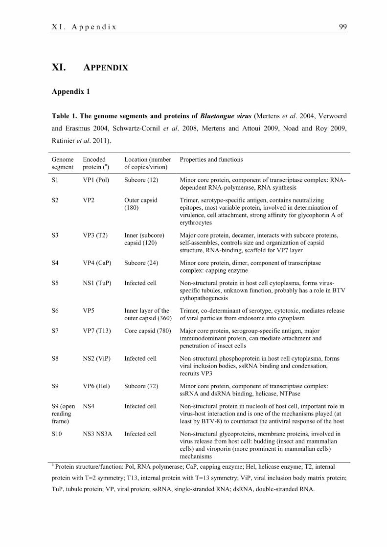
X I . A p p e n d i x 99
XI. APPENDIX
Appendix 1
Table 1. The genome segments and proteins of Bluetongue virus (Mertens et al. 2004, Verwoerd
and Erasmus 2004, Schwartz-Cornil et al. 2008, Mertens and Attoui 2009, Noad and Roy 2009,
Ratinier et al. 2011).
Genome
segment
Encoded
protein (a)
Location (number
of copies/virion)
Properties and functions
S1 VP1 (Pol) Subcore (12) Minor core protein, component of transcriptase complex: RNA-
dependent RNA-polymerase, RNA synthesis
S2 VP2 Outer capsid
(180)
Trimer, serotype-specific antigen, contains neutralizing
epitopes, most variable protein, involved in determination of
virulence, cell attachment, strong affinity for glycophorin A of
erythrocytes
S3 VP3 (T2) Inner (subcore)
capsid (120)
Major core protein, decamer, interacts with subcore proteins,
self-assembles, controls size and organization of capsid
structure, RNA-binding, scaffold for VP7 layer
S4 VP4 (CaP) Subcore (24) Minor core protein, dimer, component of transcriptase
complex: capping enzyme
S5 NS1 (TuP) Infected cell Non-structural protein in host cell cytoplasma, forms virus-
specific tubules, unknown function, probably has a role in BTV
cythopathogenesis
S6 VP5 Inner layer of the
outer capsid (360)
Trimer, co-determinant of serotype, cytotoxic, mediates release
of viral particles from endosome into cytoplasm
S7 VP7 (T13) Core capsid (780) Major core protein, serogroup-specific antigen, major
immunodominant protein, can mediate attachment and
penetration of insect cells
S8 NS2 (ViP) Infected cell Non-structural phosphoprotein in host cell cytoplasma, forms
viral inclusion bodies, ssRNA binding and condensation,
recruits VP3
S9 VP6 (Hel) Subcore (72) Minor core protein, component of transcriptase complex:
ssRNA and dsRNA binding, helicase, NTPase
S9 (open
reading
frame)
NS4 Infected cell Non-structural protein in nucleoli of host cell, important role in
virus-host interaction and is one of the mechanisms played (at
least by BTV-8) to counteract the antiviral response of the host
S10 NS3 NS3A Infected cell Non-structural glycoproteins, membrane proteins, involved in
virus release from host cell: budding (insect and mammalian
cells) and viroporin (more prominent in mammalian cells)
mechanisms a Protein structure/function: Pol, RNA polymerase; CaP, capping enzyme; Hel, helicase enzyme; T2, internal
protein with T=2 symmetry; T13, internal protein with T=13 symmetry; ViP, viral inclusion body matrix protein;
TuP, tubule protein; VP, viral protein; ssRNA, single-stranded RNA; dsRNA, double-stranded RNA.
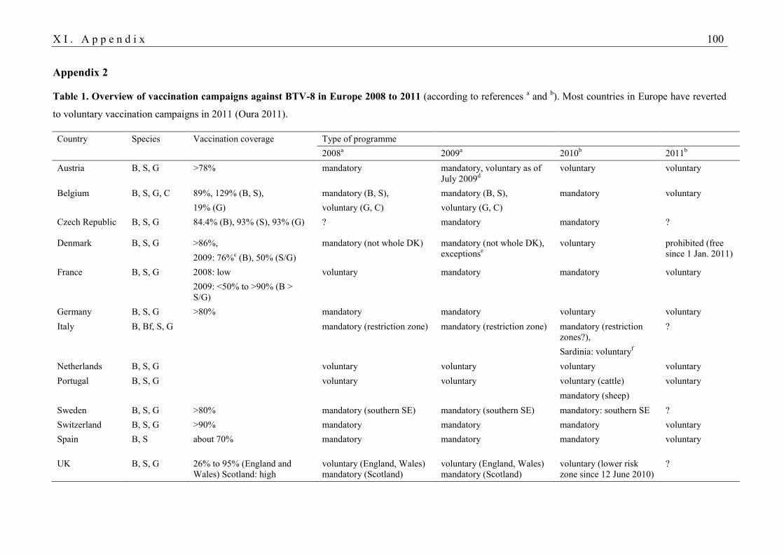
X I . A p p e n d i x 100
Appendix 2
Table 1. Overview of vaccination campaigns against BTV-8 in Europe 2008 to 2011 (according to references a and
b). Most countries in Europe have reverted
to voluntary vaccination campaigns in 2011 (Oura 2011).
Country Species Vaccination coverage Type of programme
2008
a 2009
a 2010
b 2011
b
Austria B, S, G >78% mandatory mandatory, voluntary as of
July 2009d
voluntary voluntary
Belgium B, S, G, C 89%, 129% (B, S), mandatory (B, S), mandatory (B, S), mandatory voluntary
19% (G) voluntary (G, C) voluntary (G, C)
Czech Republic B, S, G 84.4% (B), 93% (S), 93% (G) ? mandatory mandatory ?
Denmark B, S, G >86%, mandatory (not whole DK) mandatory (not whole DK),
exceptionse
voluntary prohibited (free
since 1 Jan. 2011) 2009: 76%c (B), 50% (S/G)
France B, S, G 2008: low voluntary mandatory mandatory voluntary
2009: <50% to >90% (B >
S/G)
Germany B, S, G >80% mandatory mandatory voluntary voluntary
Italy B, Bf, S, G mandatory (restriction zone) mandatory (restriction zone) mandatory (restriction
zones?),
?
Sardinia: voluntaryf
Netherlands B, S, G voluntary voluntary voluntary voluntary
Portugal B, S, G voluntary voluntary voluntary (cattle) voluntary
mandatory (sheep)
Sweden B, S, G >80% mandatory (southern SE) mandatory (southern SE) mandatory: southern SE ?
Switzerland B, S, G >90% mandatory mandatory mandatory voluntary
Spain B, S about 70% mandatory mandatory mandatory voluntary
UK B, S, G 26% to 95% (England and
Wales) Scotland: high
voluntary (England, Wales)
mandatory (Scotland)
voluntary (England, Wales)
mandatory (Scotland)
voluntary (lower risk
zone since 12 June 2010)
?
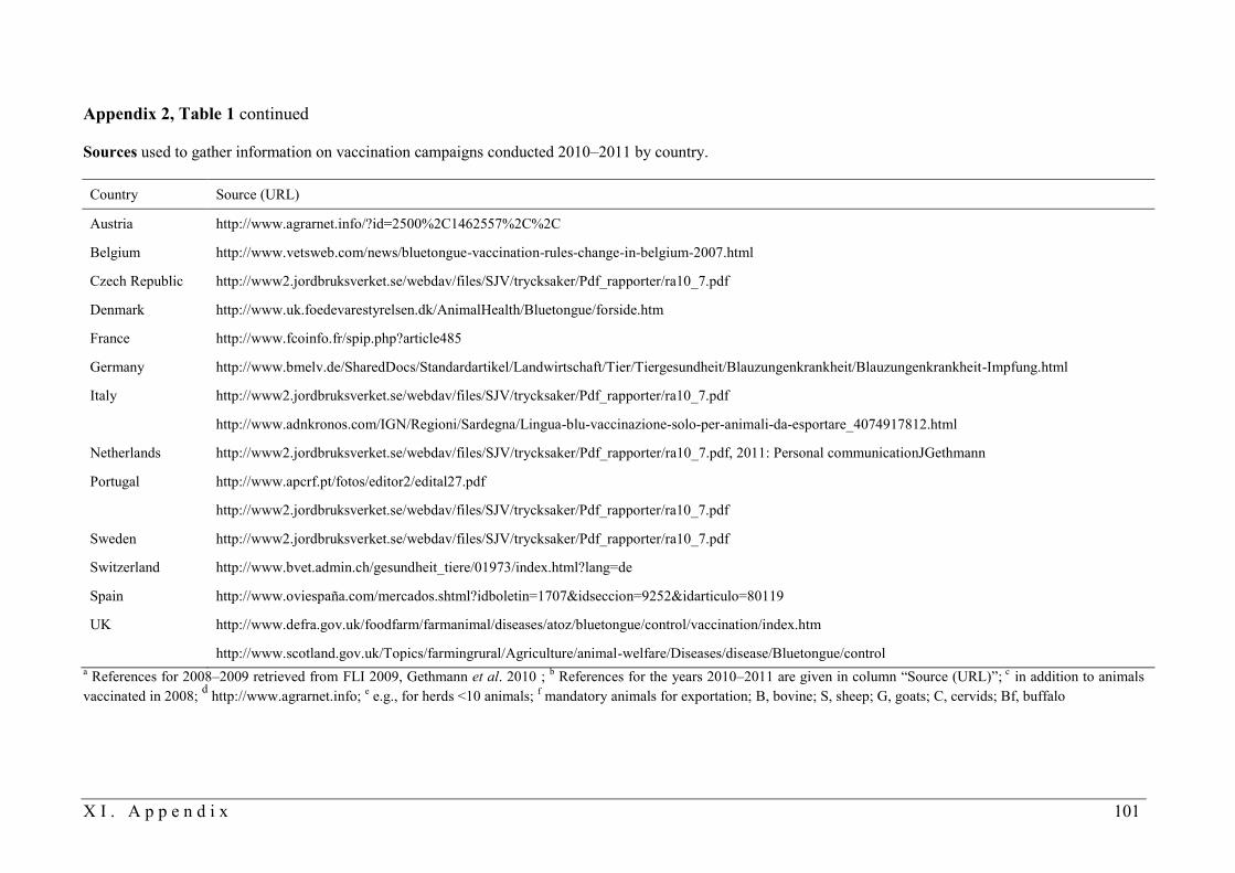
X I . A p p e n d i x 101
Appendix 2, Table 1 continued
Sources used to gather information on vaccination campaigns conducted 2010–2011 by country.
Country Source (URL)
Austria http://www.agrarnet.info/?id=2500%2C1462557%2C%2C
Belgium http://www.vetsweb.com/news/bluetongue-vaccination-rules-change-in-belgium-2007.html
Czech Republic http://www2.jordbruksverket.se/webdav/files/SJV/trycksaker/Pdf_rapporter/ra10_7.pdf
Denmark http://www.uk.foedevarestyrelsen.dk/AnimalHealth/Bluetongue/forside.htm
France http://www.fcoinfo.fr/spip.php?article485
Germany http://www.bmelv.de/SharedDocs/Standardartikel/Landwirtschaft/Tier/Tiergesundheit/Blauzungenkrankheit/Blauzungenkrankheit-Impfung.html
Italy http://www2.jordbruksverket.se/webdav/files/SJV/trycksaker/Pdf_rapporter/ra10_7.pdf
http://www.adnkronos.com/IGN/Regioni/Sardegna/Lingua-blu-vaccinazione-solo-per-animali-da-esportare_4074917812.html
Netherlands http://www2.jordbruksverket.se/webdav/files/SJV/trycksaker/Pdf_rapporter/ra10_7.pdf, 2011: Personal communicationJGethmann
Portugal http://www.apcrf.pt/fotos/editor2/edital27.pdf
http://www2.jordbruksverket.se/webdav/files/SJV/trycksaker/Pdf_rapporter/ra10_7.pdf
Sweden http://www2.jordbruksverket.se/webdav/files/SJV/trycksaker/Pdf_rapporter/ra10_7.pdf
Switzerland http://www.bvet.admin.ch/gesundheit_tiere/01973/index.html?lang=de
Spain http://www.oviespaña.com/mercados.shtml?idboletin=1707&idseccion=9252&idarticulo=80119
UK http://www.defra.gov.uk/foodfarm/farmanimal/diseases/atoz/bluetongue/control/vaccination/index.htm
http://www.scotland.gov.uk/Topics/farmingrural/Agriculture/animal-welfare/Diseases/disease/Bluetongue/control a References for 2008–2009 retrieved from(FLI 2009, Gethmann et al. 2010);
b References for the years 2010–2011 are given in column “Source (URL)”;
c in addition to animals
vaccinated in 2008; d http://www.agrarnet.info;
e e.g., for herds <10 animals;
f mandatory animals for exportation; B, bovine; S, sheep; G, goats; C, cervids; Bf, buffalo
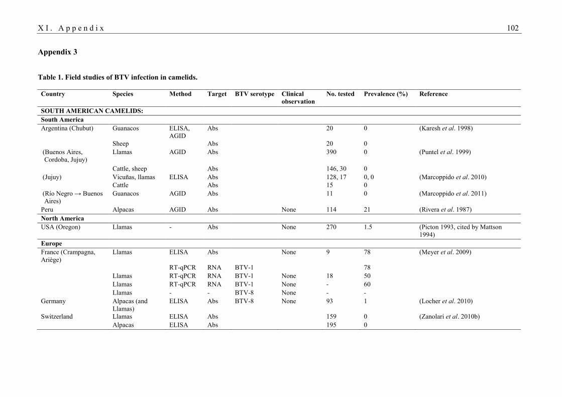
X I . A p p e n d i x 102
Appendix 3
Table 1. Field studies of BTV infection in camelids.
Country Species Method Target BTV serotype Clinical
observation
No. tested Prevalence (%) Reference
SOUTH AMERICAN CAMELIDS:
South America
Argentina (Chubut) Guanacos ELISA,
AGID
Abs 20 0 (Karesh et al. 1998)
Sheep Abs 20 0
(Buenos Aires,
Cordoba, Jujuy)
Llamas AGID Abs 390 0 (Puntel et al. 1999)
Cattle, sheep Abs 146, 30 0
(Jujuy) Vicuñas, llamas ELISA Abs 128, 17 0, 0 (Marcoppido et al. 2010)
Cattle Abs 15 0
(Río Negro → Buenos
Aires)
Guanacos AGID Abs 11 0 (Marcoppido et al. 2011)
Peru Alpacas AGID Abs None 114 21 (Rivera et al. 1987)
North America
USA (Oregon) Llamas - Abs None 270 1.5 (Picton 1993, cited by Mattson
1994)
Europe
France (Crampagna,
Ariège)
Llamas ELISA Abs None 9 78 (Meyer et al. 2009)
RT-qPCR RNA BTV-1 78
Llamas RT-qPCR RNA BTV-1 None 18 50
Llamas RT-qPCR RNA BTV-1 None - 60
Llamas - - BTV-8 None - -
Germany Alpacas (and
Llamas)
ELISA Abs BTV-8 None 93 1 (Locher et al. 2010)
Switzerland Llamas ELISA Abs 159 0 (Zanolari et al. 2010b)
Alpacas ELISA Abs 195 0
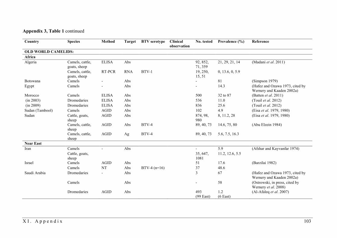
X I . A p p e n d i x 103
Appendix 3, Table 1 continued
Country Species Method Target BTV serotype Clinical
observation
No. tested Prevalence (%) Reference
OLD WORLD CAMELIDS:
Africa
Algeria Camels, cattle,
goats, sheep
ELISA Abs 92, 852,
71, 359
21, 29, 21, 14 (Madani et al. 2011)
Camels, cattle,
goats, sheep
RT-PCR RNA BTV-1 19, 250,
15, 51
0, 13.6, 0, 5.9
Botswana Camels - Abs - 81 (Simpson 1979)
Egypt Camels - Abs - 14.3 (Hafez and Ozawa 1973, cited by
Wernery and Kaaden 2002a)
Morocco Camels ELISA Abs 500 32 to 87 (Batten et al. 2011)
(in 2003) Dromedaries ELISA Abs 536 11.0 (Touil et al. 2012)
(in 2009) Dromedaries ELISA Abs 836 25.6 (Touil et al. 2012)
Sudan (Tambool) Camels AGID Abs 102 4.9 (Eisa et al. 1979, 1980)
Sudan Cattle, goats,
sheep
AGID Abs 874, 98,
980
8, 11.2, 28 (Eisa et al. 1979, 1980)
Camels, cattle,
sheep
AGID Abs BTV-4 89, 40, 73 14.6, 75, 80 (Abu Elzein 1984)
Camels, cattle,
sheep
AGID Ag BTV-4 89, 40, 73 5.6, 7.5, 16.3
Near East
Iran Camels - Abs - 5.9 (Afshar and Kayvanfar 1974)
Cattle, goats,
sheep
35, 647,
1081
11.2, 12.6, 5.5
Israel Camels AGID Abs 51 17.6 (Barzilai 1982)
Camels NT Abs BTV-4 (n=16) 37 48.6
Saudi Arabia Dromedaries - Abs 3 67 (Hafez and Ozawa 1973, cited by
Wernery and Kaaden 2002a)
Camels Abs - 58 (Ostrowski, in press, cited by
Wernery et al. 2008)
Dromedaries AGID Abs 493
(99 East)
1.2
(6 East)
(Al-Afaleq et al. 2007)
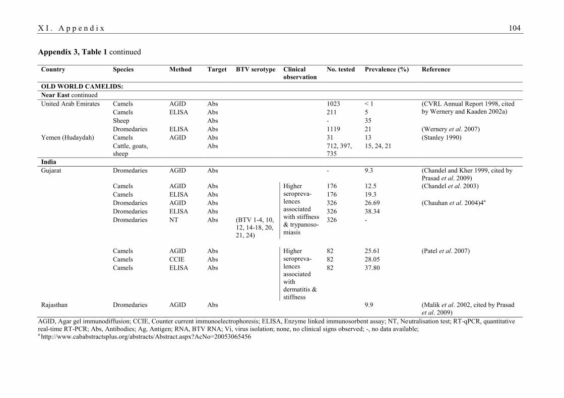
X I . A p p e n d i x 104
Appendix 3, Table 1 continued
Country Species Method Target BTV serotype Clinical
observation
No. tested Prevalence (%) Reference
OLD WORLD CAMELIDS:
Near East continued
United Arab Emirates Camels AGID Abs 1023 < 1 (CVRL Annual Report 1998, cited
by Wernery and Kaaden 2002a) Camels ELISA Abs 211 5
Sheep Abs - 35
Dromedaries ELISA Abs 1119 21 (Wernery et al. 2007)
Yemen (Hudaydah) Camels AGID Abs 31 13 (Stanley 1990)
Cattle, goats,
sheep
Abs 712, 397,
735
15, 24, 21
India
Gujarat Dromedaries AGID Abs - 9.3 (Chandel and Kher 1999, cited by
Prasad et al. 2009)
Camels AGID Abs Higher
seropreva-
lences
associated
with stiffness
& trypanoso-
miasis
176 12.5 (Chandel et al. 2003)
Camels ELISA Abs 176 19.3
Dromedaries AGID Abs 326 26.69 (Chauhan et al. 2004)4a
Dromedaries ELISA Abs 326 38.34
Dromedaries NT Abs (BTV 1-4, 10,
12, 14-18, 20,
21, 24)
326 -
Camels AGID Abs Higher
seropreva-
lences
associated
with
dermatitis &
stiffness
82 25.61 (Patel et al. 2007)
Camels CCIE Abs 82 28.05
Camels ELISA Abs 82 37.80
Rajasthan Dromedaries AGID Abs 9.9 (Malik et al. 2002, cited by Prasad
et al. 2009)
AGID, Agar gel immunodiffusion; CCIE, Counter current immunoelectrophoresis; ELISA, Enzyme linked immunosorbent assay; NT, Neutralisation test; RT-qPCR, quantitative
real-time RT-PCR; Abs, Antibodies; Ag, Antigen; RNA, BTV RNA; Vi, virus isolation; none, no clinical signs observed; -, no data available; a http://www.cababstractsplus.org/abstracts/Abstract.aspx?AcNo=20053065456
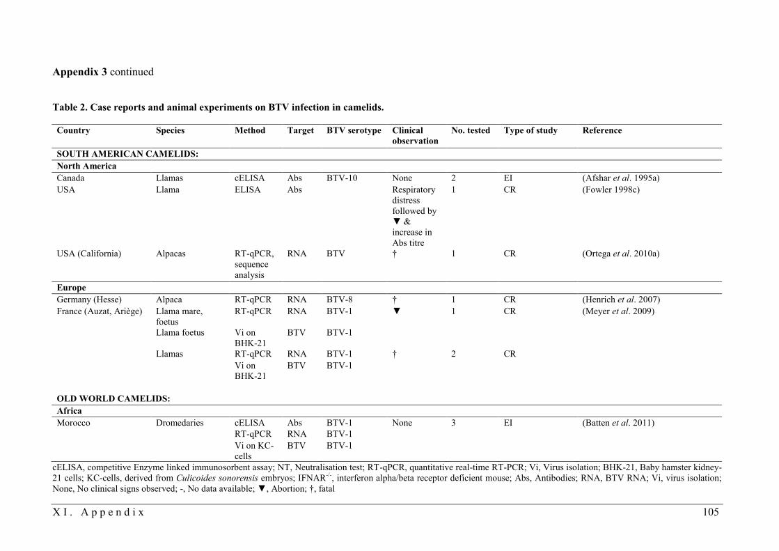
X I . A p p e n d i x 105
Appendix 3 continued
Table 2. Case reports and animal experiments on BTV infection in camelids.
Country Species Method Target BTV serotype Clinical
observation
No. tested Type of study Reference
SOUTH AMERICAN CAMELIDS:
North America
Canada Llamas cELISA Abs BTV-10 None 2 EI (Afshar et al. 1995a)
USA Llama ELISA Abs Respiratory
distress
followed by
▼ &
increase in
Abs titre
1 CR (Fowler 1998c)
USA (California) Alpacas RT-qPCR,
sequence
analysis
RNA BTV † 1 CR (Ortega et al. 2010a)
Europe
Germany (Hesse) Alpaca RT-qPCR RNA BTV-8 † 1 CR (Henrich et al. 2007)
France (Auzat, Ariège) Llama mare,
foetus
RT-qPCR RNA BTV-1 ▼ 1 CR (Meyer et al. 2009)
Llama foetus Vi on
BHK-21
BTV BTV-1
Llamas RT-qPCR RNA BTV-1 † 2 CR
Vi on
BHK-21
BTV BTV-1
OLD WORLD CAMELIDS:
Africa
Morocco Dromedaries cELISA Abs BTV-1 None 3 EI (Batten et al. 2011)
RT-qPCR RNA BTV-1
Vi on KC-
cells
BTV BTV-1
cELISA, competitive Enzyme linked immunosorbent assay; NT, Neutralisation test; RT-qPCR, quantitative real-time RT-PCR; Vi, Virus isolation; BHK-21, Baby hamster kidney-
21 cells; KC-cells, derived from Culicoides sonorensis embryos; IFNAR-/-
, interferon alpha/beta receptor deficient mouse; Abs, Antibodies; RNA, BTV RNA; Vi, virus isolation;
None, No clinical signs observed; -, No data available; ▼, Abortion; †, fatal
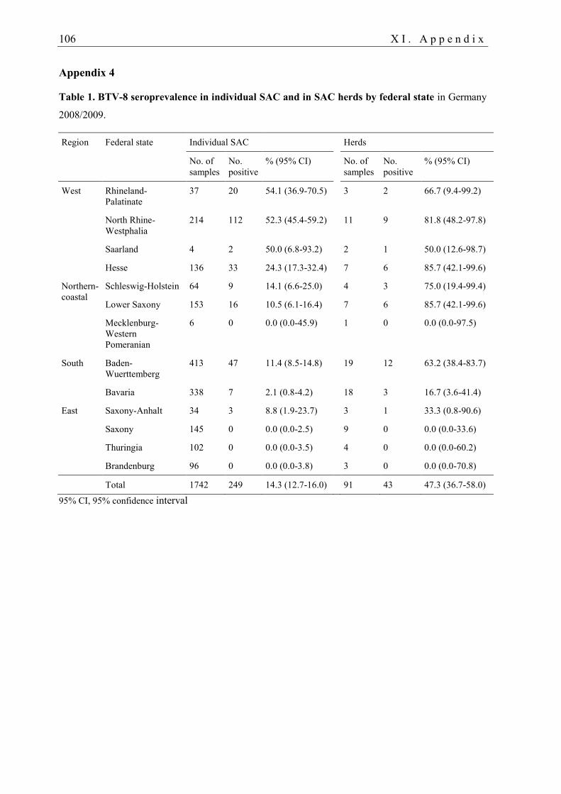
106 X I . A p p e n d i x
Appendix 4
Table 1. BTV-8 seroprevalence in individual SAC and in SAC herds by federal state in Germany
2008/2009.
Region Federal state Individual SAC Herds
No. of
samples
No.
positive
% (95% CI) No. of
samples
No.
positive
% (95% CI)
West Rhineland-
Palatinate
37 20 54.1 (36.9-70.5) 3 2 66.7 (9.4-99.2)
North Rhine-
Westphalia
214 112 52.3 (45.4-59.2) 11 9 81.8 (48.2-97.8)
Saarland 4 2 50.0 (6.8-93.2) 2 1 50.0 (12.6-98.7)
Hesse 136 33 24.3 (17.3-32.4) 7 6 85.7 (42.1-99.6)
Northern-
coastal
Schleswig-Holstein 64 9 14.1 (6.6-25.0) 4 3 75.0 (19.4-99.4)
Lower Saxony 153 16 10.5 (6.1-16.4) 7 6 85.7 (42.1-99.6)
Mecklenburg-
Western
Pomeranian
6 0 0.0 (0.0-45.9) 1 0 0.0 (0.0-97.5)
South Baden-
Wuerttemberg
413 47 11.4 (8.5-14.8) 19 12 63.2 (38.4-83.7)
Bavaria 338 7 2.1 (0.8-4.2) 18 3 16.7 (3.6-41.4)
East Saxony-Anhalt 34 3 8.8 (1.9-23.7) 3 1 33.3 (0.8-90.6)
Saxony 145 0 0.0 (0.0-2.5) 9 0 0.0 (0.0-33.6)
Thuringia 102 0 0.0 (0.0-3.5) 4 0 0.0 (0.0-60.2)
Brandenburg 96 0 0.0 (0.0-3.8) 3 0 0.0 (0.0-70.8)
Total 1742 249 14.3 (12.7-16.0) 91 43 47.3 (36.7-58.0)
95% CI, 95% confidence interval
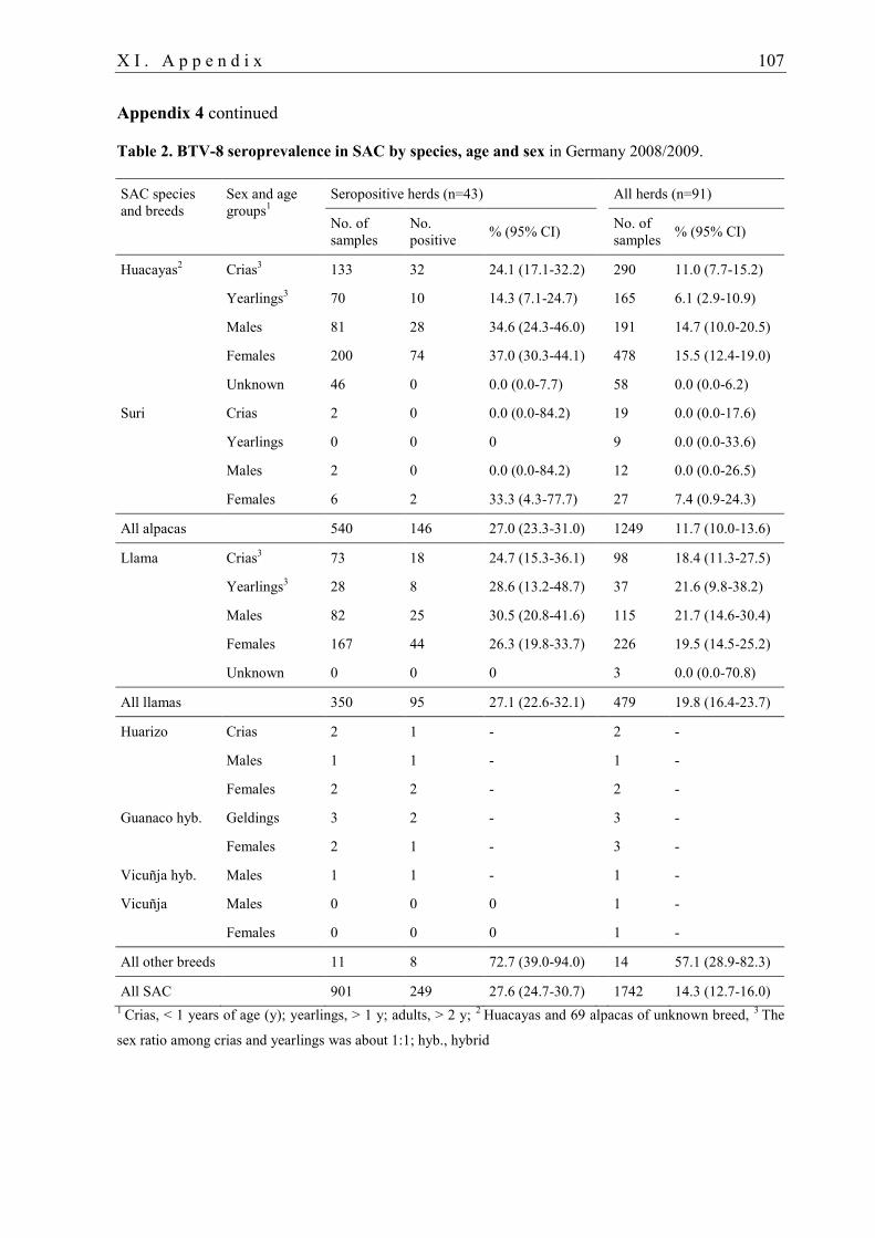
X I . A p p e n d i x 107
Appendix 4 continued
Table 2. BTV-8 seroprevalence in SAC by species, age and sex in Germany 2008/2009.
SAC species
and breeds
Sex and age
groups1
Seropositive herds (n=43) All herds (n=91)
No. of
samples
No.
positive % (95% CI)
No. of
samples % (95% CI)
Huacayas2 Crias
3 133 32 24.1 (17.1-32.2) 290 11.0 (7.7-15.2)
Yearlings3 70 10 14.3 (7.1-24.7) 165 6.1 (2.9-10.9)
Males 81 28 34.6 (24.3-46.0) 191 14.7 (10.0-20.5)
Females 200 74 37.0 (30.3-44.1) 478 15.5 (12.4-19.0)
Unknown 46 0 0.0 (0.0-7.7) 58 0.0 (0.0-6.2)
Suri Crias 2 0 0.0 (0.0-84.2) 19 0.0 (0.0-17.6)
Yearlings 0 0 0 9 0.0 (0.0-33.6)
Males 2 0 0.0 (0.0-84.2) 12 0.0 (0.0-26.5)
Females 6 2 33.3 (4.3-77.7) 27 7.4 (0.9-24.3)
All alpacas 540 146 27.0 (23.3-31.0) 1249 11.7 (10.0-13.6)
Llama Crias3 73 18 24.7 (15.3-36.1) 98 18.4 (11.3-27.5)
Yearlings3 28 8 28.6 (13.2-48.7) 37 21.6 (9.8-38.2)
Males 82 25 30.5 (20.8-41.6) 115 21.7 (14.6-30.4)
Females 167 44 26.3 (19.8-33.7) 226 19.5 (14.5-25.2)
Unknown 0 0 0 3 0.0 (0.0-70.8)
All llamas 350 95 27.1 (22.6-32.1) 479 19.8 (16.4-23.7)
Huarizo Crias 2 1 - 2 -
Males 1 1 - 1 -
Females 2 2 - 2 -
Guanaco hyb. Geldings 3 2 - 3 -
Females 2 1 - 3 -
Vicuñja hyb. Males 1 1 - 1 -
Vicuñja Males 0 0 0 1 -
Females 0 0 0 1 -
All other breeds 11 8 72.7 (39.0-94.0) 14 57.1 (28.9-82.3)
All SAC 901 249 27.6 (24.7-30.7) 1742 14.3 (12.7-16.0)
1 Crias, < 1 years of age (y); yearlings, > 1 y; adults, > 2 y;
2 Huacayas and 69 alpacas of unknown breed,
3 The
sex ratio among crias and yearlings was about 1:1; hyb., hybrid
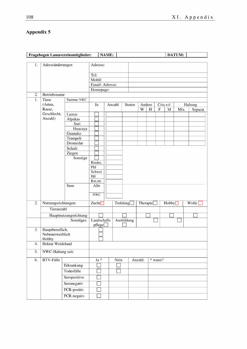
108 X I . A p p e n d i x
Appendix 5
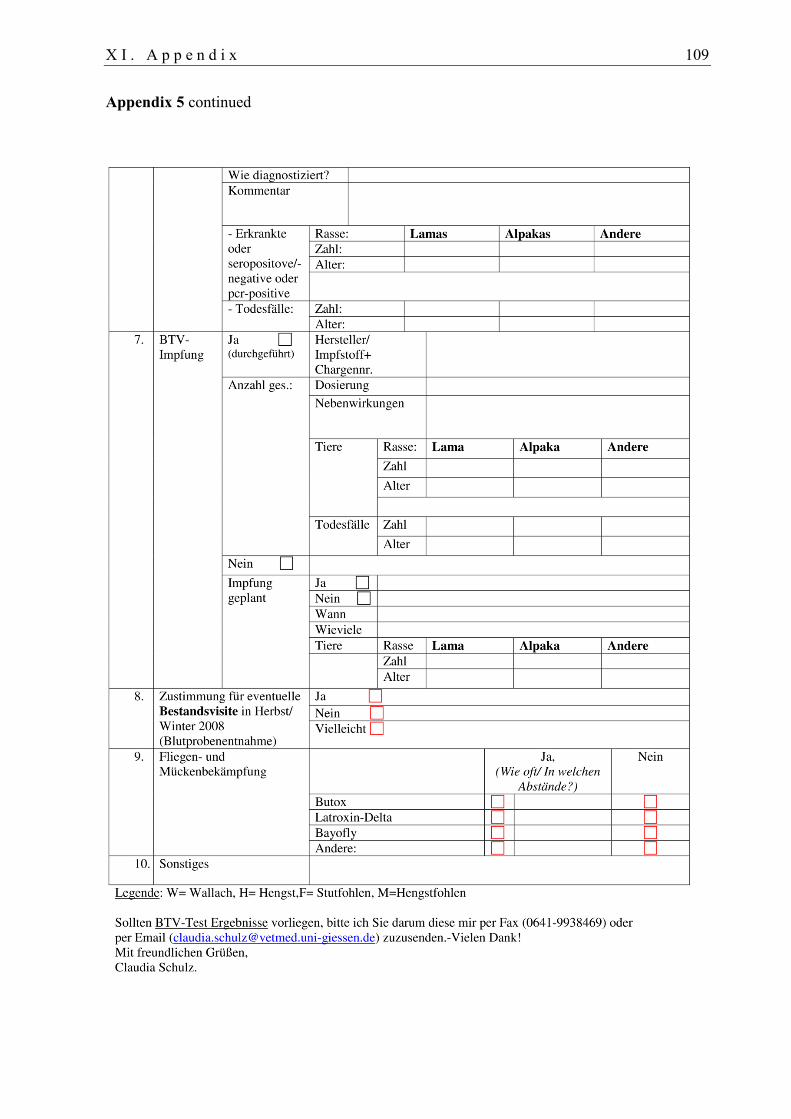
X I . A p p e n d i x 109
Appendix 5 continued

110 X I . A p p e n d i x
Appendix 6
X I A p p e n d i x 111
LIST OF TABLES
Table 1. Estimated distribution of the world SAC population ________________________ 23
Table 2. Locations of BG-SentinelTM
UV light traps in Germany _____________________ 36
Table 3. Number of SAC per herd tested for BTV-8 antibodies in Germany 2008/2009 ___ 39
Table 4. Animals, vaccines and dosages used ____________________________________ 42
Table 5. Total monthly catches (indoor and outdoor) of females by Culicoides species ____ 51
Table 6. Incidence of females of the Obsoletus and Pulicaris complexes and other _______
______ Culicoides spp. _____________________________________________________ 52
Table 7. Summary of the results of Poisson regression analyses ______________________ 57
Table 8. BTV-8 seroprevalence _______________________________________________ 60
Table 9. Definition of explanatory variables, number of individuals or herds ____________ 61
Table 10. Logistic model of risk factors for BTV infection in SAC, Germany ___________ 63
Table 11. Seroconversion after experimental BTV-8 infection of SAC ________________ 66
Table 12. Seroconversion rates _______________________________________________ 70
Table 13. BTV-8 antibody levels in dams and their crias ___________________________ 74
Appendix 1
Table 1. The genome segments and proteins of Bluetongue virus __________________ 99
Appendix 2
Table 1. Overview of vaccination campaigns against BTV-8 in Europe 2008 to 2011 _ 100
Appendix 3
Table 1. Field studies of BTV infection in camelids ____________________________ 102
Table 2. Case reports and animal experiments on BTV infection in camelids ________ 105
Appendix 4
Table 1. BTV-8 seroprevalence in individual SAC and in SAC herds by federal state _ 106
Table 2. BTV-8 seroprevalence in SAC by species, age and sex __________________ 107
112 X I . A p p e n d i x
LIST OF FIGURES
Fig. 1. Phylogenetic tree for Seg-2 (VP2 gene) sequences ___________________________ 3
Fig. 2. Representative scheme of BTV structural proteins and double-stranded RNA ______ 4
Fig. 3. Official veterinary BTV restriction zones in the European Union ________________ 9
Fig. 4. Culicoides obsoletus female ____________________________________________ 12
Fig. 5. Culicoides wings _____________________________________________________ 12
Fig. 6. Holometabolic life cycle of ceratopogonids ________________________________ 14
Fig. 7. Mitochondrial ribosomal DNA (rDNA) segment in Culicoides _________________ 15
Fig. 8. Culicoides caught in Germany 2007/2008 _________________________________ 17
Fig. 9. Timeline of BTV diagnostic parameters ___________________________________ 27
Fig. 10. Map showing locations of the black-light traps ____________________________ 36
Fig. 11. BG-SentinelTM
UV light midge trap _____________________________________ 37
Fig. 12. Emergence trap _____________________________________________________ 37
Fig. 13. Incidence of female Culicoides and other female ceratopogonids ______________ 52
Fig. 14. Total number of female Culicoides caught outdoor and indoor versus temperature and
______ relative humidity ____________________________________________________ 53
Fig. 15. Incidence of male Culicoides and other male ceratopogonids _________________ 55
Fig. 16. Phenology of Culicoides spp. and other genera of ceratopogonids _____________ 56
Fig. 17. Ceratopogonid females and males caught in emergence traps _________________ 58
Fig. 18. BTV-8 seroprevalence in SAC 2008/2009. Map of Germany _________________ 62
Fig. 19. Leukocyte counts in SAC blood after experimental BTV-8 infection ___________ 65
Fig. 20. Neutralising antibodies in SAC after experimental BTV-8 infection____________ 65
Fig. 21. Antibody response after BTV-8 infection of SAC __________________________ 66
Fig. 22. Mean RT-qPCR results set against serology after experimental BTV-8
infection_____ ______ of SAC _______________________________________________ 67
Fig. 23. In vitro adsorption experiment of BTV-8 _________________________________ 69
Fig. 24. Development of BTV antibodies in BTV-8 vaccinated SAC __________________ 71
Fig. 25. Transfer of colostral BTV-8 antibodies to crias ____________________________ 73
X I I . R e f e r e n c e s 113
XII. REFERENCES
Anon. (2010a). Bluetongue. Retrieved 30 August 2012 from http://www.id-
vet.com/English/elisa_diagnostic_kit/ruminants/blue_tongue_serum_and_milk.htm.
Anon. (2010b). Proposal to change bluetongue vaccination rules. Vet Rec. 167(22):840.
Abu Elzein EME (1984). Rapid detection of bluetongue virus antigen in the sera and plasma
of camels, sheep and cattle in the Sudan, using the gel immunodiffusion test. Brief report.
Arch Virol. 79(1-2):131-4.
Afshar A, Heckert RA, Dulac GC, Trotter HC, and Myers DJ (1995a). Application of a
competitive ELISA for the detection of bluetongue virus antibodies in llamas and wild
ruminants. J Wildl Dis. 31(3):327-30.
Afshar A, Heckert RA, Dulac GC, Trotter HC, and Myers DJ (1995b). Application of a
competitive ELISA for the detection of bluetongue virus antibodies in llamas and wild
ruminants. J Wildl Dis. 31(3):327-30.
Afshar A and Kayvanfar H (1974). Occurrence of precipitating antibodies to bluetongue virus
in sera of farm animals in Iran. Vet Rec. 94(11):233-5.
Agresti A (1990). Categorical data analysis. New York: Wiley;350.
Akita GY, Ianconescu M, MacLachlan NJ, and Osburn BI (1994). Bluetongue disease in dogs
associated with contaminated vaccine. Vet Rec. 134(11):283-4.
Al-Afaleq AI, Abu Elzein EM, Hegazy AA, and Al-Naeem A (2007). Serosurveillance of
Camels (Camelus dromedarius) to detect antibodies against viral diseases in Saudi Arabia.
J Camel Practice Research. 14(2):91-6.
Alba A, Casal J, and Domingo M (2004). Possible introduction of bluetongue into the
Balearic Islands, Spain, in 2000, via air streams. Vet Rec. 155(15):460-1.
Alexander KA, MacLachlan NJ, Kat PW, House C, O'Brien SJ, Lerche NW, Sawyer M,
Frank LG, Holekamp K, Smale L, McNutt JW, Laurenson MK, Mills MGL, and Osburn
BI (1994). Evidence of natural bluetongue virus infection among African carnivores. Am J
Trop Med Hyg. 51(5):568-76.
Alpar HO, Bramwell VW, Veronesi E, Darpel KE, Pastoret P-P, and Mertens PPC (2009).
Bluetongue virus vaccines past and present. In: Mellor PS, Baylis M, Mertens PPC (eds).
Bluetongue. London: Elsevier Academic Press, United Kingdom; pp. 397-428.
Andrew M, Whiteley P, Janardhana V, Lobato Z, Gould A, and Coupar B (1995). Antigen
specificity of the ovine cytotoxic T lymphocyte response to bluetongue virus. Vet Immunol
Immunopathol. 47(3-4):311-22.
Ansari MA, Pope EC, Carpenter S, Scholte EJ, and Butt TM (2011). Entomopathogenic
fungus as a biological control for an important vector of livestock disease: the Culicoides
biting midge. PLoS One. 6(1):e16108.
114 X I I . R e f e r e n c e s
Antoniassi NA, Pavarini SP, Henzel A, Flores EF, and Driemeier D (2010). Aspiration
pneumonia associated with oesophageal myonecrosis in sheep due to BTV infection in
Brazil. Vet Rec. 166(2):52-3.
Attoui H, Maan S, Anthony SJ, and Mertens PPC (2009). Bluetongue virus, other orbiviruses
and other reoviruses: Their relationships and taxonomy. In: Mellor PS, Baylis M, Mertens
PPC (eds). Bluetongue. London: Elsevier Academic Press, United Kingdom; pp. 23-52.
Backx A, Heutink CG, van Rooij EM, and van Rijn PA (2007). Clinical signs of bluetongue
virus serotype 8 infection in sheep and goats. Vet Rec. 161(17):591-2.
Backx A, Heutink R, van RE, and van RP (2009). Transplacental and oral transmission of
wild-type bluetongue virus serotype 8 in cattle after experimental infection. Vet Microbiol.
138(3-4):235-43.
Baldet T, Delécolle JC, Cêtre-Sossah C, Mathieu B, Meiswinkel R, and Gerbier G (2008).
Indoor activity of Culicoides associated with livestock in the bluetongue virus (BTV)
affected region of northern France during autumn 2006. Prev Vet Med. 87(1-2):84-97.
Balenghien T, Cêtre-Sossah C, Grillet C, Delécolle JC, Mathieu B, and Baldet T (2008).
Diurnal activity of potential bluetongue vectors in northern Europe. Vet Rec. 162(10):323-
4.
Barratt-Boyes SM and MacLachlan NJ (1994). Dynamics of viral spread in bluetongue virus
infected calves. Vet Microbiol. 40(3-4):361-71.
Barros SC, Cruz B, Luis TM, Ramos F, Fagulha T, Duarte M, Henriques M, and Fevereiro M
(2009). A DIVA system based on the detection of antibodies to non-structural protein 3
(NS3) of bluetongue virus. Vet Microbiol. 137(3-4):252-9.
Bartram DJ, Heasman L, Batten CA, Oura CA, Plana-Duran J, Yuen HM, and Wylie AD
(2010). Neutralising antibody responses in cattle and sheep following booster vaccination
with two commercial inactivated bluetongue virus serotype 8 vaccines. Vet J. 188(2):193-
6.
Bartsch S, Bauer B, Wiemann A, Clausen P-H, and Steuber S (2009). Feeding patterns of
biting midges of the Culicoides obsoletus and Culicoides pulicaris groups on selected
farms in Brandenburg, Germany. Parasitol Res. 105(2):373-80.
Barzilai E (1982). Bluetongue antibodies in Camels' sera in Israel. Refuah Vet. 39(3):90-3.
Batten CA, Harif B, Henstock MR, Ghizlane S, Edwards L, Loutfi C, Oura CAL, and El
Harrak M (2011). Experimental infection of camels with bluetongue virus. Res Vet Sci.
90(3):533-55.
Batten CA, Maan S, Shaw AE, Maan NS, and Mertens PP (2008). A European field strain of
bluetongue virus derived from two parental vaccine strains by genome segment
reassortment. Virus Res. 137(1):56-63.
Baylis M, O'Connell L, and Mellor PS (2009). Rates of bluetongue virus transmission
between Culicoides sonorensis and sheep. In: Mellor PS, Baylis M, Mertens PPC (eds).
Bluetongue. London: Elsevier Academic Press, United Kingdom; pp. 321-41.
X I I . R e f e r e n c e s 115
Belhouchet M, Mohd JF, Firth AE, Grimes JM, Mertens PP, and Attoui H (2011). Detection
of a fourth orbivirus non-structural protein. PLoS One. 6(10):e25697.
Bexiga R, Guyot H, Saegerman C, Mauroy A, Rollin F, Thiry E, Philbey AW, Logue DN,
Mellor DJ, Barrett DC, and Ellis K (2007). Clinical differentiation of malignant catarrhal
fever, mucosal disease and bluetongue. Vet Rec. 161(25):858-9.
Bishop AL, McKenzie HJ, Barchia IM, Murison R, and Spohr LJ (1996). Position of juvenile
stages of Culicoides brevitarsis Kieffer (Diptera: Ceratopogonidae) and four other flies in
bovine dung. Aust J Entomol. 35:209-12.
BMELV (2010). Bundesministerium für Ernährung, Landwirtschaft und Verbraucherschutz.
Impfung gegen das Virus der Blauzungenkrankheit Serotyp 8 (BTV-8). Die wichtigsten
Informationen zur Impfung (Stand: 15. Oktober 2010). Retrieved 1 March 2011 from
http://www.bmelv.de/SharedDocs/Standardartikel/Landwirtschaft/Tier/Tiergesundheit/Bla
uzungenkrankheit/Blauzungenkrankheit-Impfung.html.
Bonneau KR, DeMaula CD, Mullens BA, and MacLachlan NJ (2002). Duration of viraemia
infectious to Culicoides sonorensis in bluetongue virus-infected cattle and sheep. Vet
Microbiol. 88(2):115-25.
Bonneau KR and MacLachlan NJ (2004). Genetic diversification of field strains of
bluetongue virus. Vet Ital. 40(4):446-7.
Boone JD, Balasuriya UB, Karaca K, Audonnet JC, Yao J, He L, Nordgren R, Monaco F,
Savini G, Gardner IA, and MacLachlan NJ (2007). Recombinant canarypox virus vaccine
co-expressing genes encoding the VP2 and VP5 outer capsid proteins of bluetongue virus
induces high level protection in sheep. Vaccine. 25(4):672-8.
Boorman J (1986). British Culicoides (Diptera: Ceratopogonidae): notes on distribution and
biology. Entomologist's Gazette. 37:253-66.
Borkent A (2012). World species of biting midges (Diptera: Ceratopogonidae). Royal British
Columbia Museum, American Museum of Natural History and Instituto Nacional de
Biodiversidad. Retrieved 30 August 2012 from
http://www.inhs.uiuc.edu/research/FLYTREE/CeratopogonidaeCatalog.pdf.
Bouwknegt C, van Rijn PA, Schipper JJ, Holzel D, Boonstra J, Nijhof AM, van Rooij EM,
and Jongejan F (2010). Potential role of ticks as vectors of bluetongue virus. Exp Appl
Acarol. 52(2):183-92.
Bréard E, Belbis G, Hamers C, Moulin V, Lilin T, Moreau F, Millemann Y, Montange C,
Sailleau C, Durand B, Desprat A, Viarouge C, Hoffmann B, de SH, Goutebroze S, Hudelet
P, and Zientara S (2011). Evaluation of humoral response and protective efficacy of two
inactivated vaccines against bluetongue virus after vaccination of goats. Vaccine.
29(13):2495-502.
Brewer AW and MacLachlan NJ (1992). Ultrastructural characterization of the interaction of
bluetongue virus with bovine erythrocytes in vitro. Vet Pathol. 29(4):356-9.
Brewer AW and MacLachlan NJ (1994). The pathogenesis of bluetongue virus infection of
bovine blood cells in vitro: ultrastructural characterization. Arch Virol. 136(3-4):287-98.
116 X I I . R e f e r e n c e s
Brodie SJ, Wilson WC, O'Hearn PM, Muthui D, Diem K, and Pearson LD (1998). The effects
of pharmacological and lentivirus-induced immune suppression on orbivirus pathogenesis:
assessment of virus burden in blood monocytes and tissues by reverse transcription-in situ
PCR. J Virol. 72(7):5599-609.
Bruckner L, Fricker R, Hug M, Hotz R, Muntwyler J, Iten C, and Griot C (2009a). Impfung
gegen die Blauzungenkrankheit: Verträglichkeit und Immunantwort in der Praxis
[Vaccination against bluetongue: safety and immune response in the field]. Schweiz Arch
Tierheilkd. 151(3):101-8.
Bruckner L, Fricker R, Hug M, Hotz R, Muntwyler J, Iten C, and Griot C (2009b).
Vaccination against bluetongue: safety and immune response in the field. Schweiz Arch
Tierheilkd. 151(3):101-8.
Bulut O, Yavru S, Yapkiç O, Simsek A, Kale M, and Avci O (2006). Serological
investigation of bluetongue virus infection by serum neutralisation test and ELISA in sheep
and goats. Bull Vet Inst Pulawy. 50:305-7.
Burgin LE, Gloster J, Sanders C, Mellor PS, Gubbins S, and Carpenter S (2012). Investigating
incursions of bluetongue virus using a model of long-distance Culicoides biting midge
dispersal. Transbound Emerg Dis. In press. DOI: 10.1111/j.1865-1682.2012.01345.x.
Calvete C, Estrada R, Miranda MA, Del Rio R, Borrás D, Beldron FJ, Martínez A, Calvo AJ,
and Lucientes J (2010). Protection of livestock against bluetongue virus vector Culicoides
imicola using insecticide-treated netting in open areas. Med Vet Entomol. 24(2):169-75.
Calvo-Pinilla E, Rodríguez-Calvo T, Anguita J, Sevilla N, and Ortego J (2009a).
Establishment of a bluetongue virus infection model in mice that are deficient in the
alpha/beta interferon receptor. PLoS One. 4(4):e5171.
Calvo-Pinilla E, Rodriguez-Calvo T, Sevilla N, and Ortego J (2009b). Heterologous prime
boost vaccination with DNA and recombinant modified vaccinia virus Ankara protects
IFNAR(-/-) mice against lethal bluetongue infection. Vaccine. 28(2):437-45.
Campbell JA and Pelham-Clinton EC (1960). A taxonomic review of the British species of
Culicoides Latreille (Diptera: Ceratopogonidae). Proc R Soc Edinburgh.
Cannon RM and Roe RT (1982). Livestock Disease Surveys: A Field Manual for
Veterinarians. Canberra: Australian Government Publishing Services.
Caporale V and Giovannini A (2010). Bluetongue control strategy, including recourse to
vaccine: a critical review. Rev Sci Tech. 29(3):573-91.
Caporale V, Giovannini A, Patta C, Calistri P, Nannini D, and Santucci U (2004). Vaccination
in the control strategy of bluetongue in Italy. Dev Biol (Basel). 119:113-27.
Carpenter S, Wilson A, Barber J, Veronesi E, Mellor P, Venter G, and Gubbins S (2011).
Temperature dependence of the extrinsic incubation period of orbiviruses in Culicoides
biting midges. PLoS One. 6(11):e27987.
Carpenter S, Wilson A, and Mellor PS (2009). Culicoides and the emergence of bluetongue
virus in northern Europe. Trends Microbiol. 17(4):172-8.
X I I . R e f e r e n c e s 117
Carpi G, Holmes EC, and Kitchen A (2010). The evolutionary dynamics of bluetongue virus.
J Mol Evol. 70(6):583-92.
Chandel BS, Chauhan HC, and Kher HN (2003). Comparison of the standard AGID test and
competitive ELISA for detecting bluetongue virus antibodies in camels in Gujarat, India.
Trop Anim Health Prod. 35(2):99-104.
Chandel BS and Kher HN (1999). Seroprevalence of bluetongue in dromedary camels in
Gujarat, India. Indian J Camel Pract Res. 6:83-5.
Channappanavar R, Singh KP, Singh R, Umeshappa CS, Ingale SL, and Pandey AB (2012).
Enhanced proinflammatory cytokine activity during experimental bluetongue virus-1
infection in Indian native sheep. Vet Immunol Immunopathol. 145(1-2):485-92.
Chauhan HC, Chandel BS, Gerdes T, Vasava KA, Patel AR, Kher HN, Singh V, and Dongre
RA (2004). Seroepidemiology of bluetongue in dromedary camels in Gujarat, India. J
Camel Pract Res. 11:141-5.
Clausen P-H, Stephan A, Bartsch S, Jandowsky A, Hoffmann-Koehler P, Schein E, Mehlitz
D, and Bauer B (2009). Seasonal dynamics of biting midges (Diptera: Ceratopogonidae,
Culicoides spp.) on dairy farms of Central Germany during the 2007/2008 epidemic of
bluetongue. Parasitol Res. 105(2):381-6.
Clavijo A, Heckert RA, Dulac GC, and Afshar A (2000). Isolation and identification of
bluetongue virus. J Virol Methods. 87(1-2):13-23.
Commission Decision 2008/655/EC (2008). Commission decision of 24 July 2008 approving
the emergency vaccination plans against bluetongue of certain Member States and fixing
the level of the Community's financial contribution for 2007 and 2008 (notified under
document number C(2008) 3757) (2008/655/EC).
Commission regulation (EC) No 1266/2007 (2007). Commission regulation (EC) No
1266/2007 of 26 October 2007 on implementing rules for Council Directive
2000/75/EC as regards the control, monitoring, surveillance and restrictions on
movements of certain animals of susceptible species in relation to bluetongue.
Retrieved 18 June 2012 from http://eur-
lex.europa.eu/LexUriServ/site/en/oj/2007/l_283/l_28320071027en00370052.pdf.
Commission Regulation (EC) No 384/2008 (2008). Commission Regulation (EC) No
384/2008 of 29 April 2008 amending Regulation (EC) No 1266/2007 as regards the
conditions for exempting pregnant animals from the exit ban provided for in Council
Directive 2000/75/EC. Retrieved 30 August 2012 from http://eur-
lex.europa.eu/LexUriServ/LexUriServ.do?uri=CELEX:32008R0384:EN:NOT.
Connan RM and Lloyd S (1988). Seasonal allergic dermatitis in sheep. Vet Rec. 123(13):335-
7.
Conrath KE, Wernery U, Muyldermans S, and Nguyen VK (2003). Emergence and evolution
of functional heavy-chain antibodies in Camelidae. Dev Comp Immunol. 27(2):87-103.
Conraths FJ, Gethmann JM, Staubach C, Mettenleiter TC, Beer M, and Hoffmann B (2009).
Epidemiology of bluetongue virus serotype 8, Germany. Emerg Infect Dis. 15(3):433-5.
118 X I I . R e f e r e n c e s
Conte A, Goffredo M, Ippoliti C, and Meiswinkel R (2007). Influence of biotic and abiotic
factors on the distribution and abundance of Culicoides imicola and the Obsoletus complex
in Italy. Vet Parasitol. 150(4):333-44.
Cornelius CE and Kaneko JJ (1962). Erythrocyte Life Span in the Guanaco. Science.
137(3531):673-4.
Corrêa TG, Ferreira JM, Riet-Correa G, Ruas JL, Schild AL, Riet-Correa F, Guimarães A,
and Felippe-Bauer ML (2007). Seasonal allergic dermatitis in sheep in southern Brazil
caused by Culicoides insignis (Diptera: Ceratopogonidae). Vet Parasitol. 145(1-2):181-5.
Council Directive 82/894/EEC (1982). Council Directive 82/894/EEC of 21 December 1982
on the notification of animal diseases within the Community. Retrieved 30 August 2012
from http://eur-
lex.europa.eu/LexUriServ/LexUriServ.do?uri=CELEX:31982L0894:EN:NOT.
CVRL Annual Report (1998). CVRL. 1998. Annual Report. Central Veterinary Research
Laboratory, Dubai. U.A.E.:19.
Dal Pozzo F, De Clercq K, Guyot H, Vandemeulebroucke E, Sarradin P, Vandenbussche F,
Thiry E, and Saegerman C (2009a). Experimental reproduction of bluetongue virus
serotype 8 clinical disease in calves. Vet Microbiol. 136(3-4):352-8.
Dal Pozzo F, Saegerman C, and Thiry E (2009b). Bovine infection with bluetongue virus with
special emphasis on European serotype 8. Vet J. 182(2):142-51.
Darpel KE, Batten CA, Veronesi E, Shaw AE, Anthony S, Bachanek-Bankowska K, Kgosana
L, Bin-Tarif A, Carpenter S, Müller-Doblies UU, Takamatsu H-H, Mellor PS, Mertens
PPC, and Oura CAL (2007). Clinical signs and pathology shown by British sheep and
cattle infected with bluetongue virus serotype 8 derived from the 2006 outbreak in northern
Europe. Vet Rec. 161(8):253-61.
Darpel KE, Batten CA, Veronesi E, Williamson S, Anderson P, Dennison M, Clifford S,
Smith C, Philips L, Bidewell C, Bachanek-Bankowska K, Sanders A, Bin-Tarif A, Wilson
AJ, Gubbins S, Mertens PP, Oura CA, and Mellor PS (2009a). Transplacental
Transmission of Bluetongue Virus 8 in Cattle, UK. Emerg Infect Dis. 15(12):2025-8.
Darpel KE, Monaghan P, Anthony SJ, Takamatsu H-H, and Mertens PPC (2009b).
Bluetongue in the mammalian host and the induced immune response. In: Mellor PS,
Baylis M, Mertens PPC (eds). Bluetongue. London: Elsevier Academic Press, United
Kingdom; pp. 265-84.
Darpel KE, Monaghan P, Simpson J, Anthony SJ, Veronesi E, Brooks HW, Elliott H,
Brownlie J, Takamatsu HH, Mellor PS, and Mertens PP (2012). Involvement of the skin
during bluetongue virus infection and replication in the ruminant host. Vet Res. 43(1):40.
De Clercq K, De L, I, Verheyden B, Vandemeulebroucke E, Vanbinst T, Herr C, Meroc E,
Bertels G, Steurbaut N, Miry C, De BK, Maquet G, Bughin J, Saulmont M, Lebrun M,
Sustronck B, De DR, Hooyberghs J, Houdart P, Raemaekers M, Mintiens K, Kerkhofs P,
Goris N, and Vandenbussche F (2008a). Transplacental infection and apparently
immunotolerance induced by a wild-type bluetongue virus serotype 8 natural infection.
Transbound Emerg Dis. 55(8):352-9.
X I I . R e f e r e n c e s 119
De Clercq K, Vandenbussche F, Vandemeulebroucke E, Vanbinst T, De L, I, Verheyden B,
Goris N, Mintiens K, Méroc E, Herr C, Hooybergs J, Houdart P, Sustronck B, De Deken
R, Maquet G, Bughin J, Saulmont M, Lebrun M, Bertels G, and Miry C (2008b).
Transplacental bluetongue infection in cattle. Vet Rec. 162(17):564.
de Deken G, Madder M, Deblauwe I, De Clercq K, Fassotte C, Losson B, Haubruge E, and
De Deken R (2008). Vector monitoring at Belgian outbreak sites during the bluetongue
epidemic of 2006. Prev Vet Med. 87(1-2):64-73.
De Liberato C, Farina F, Magliano A, Rombolà P, Scholl F, Spallucci V, and Scaramozzino P
(2010). Biotic and abiotic factors influencing distribution and abundance of Culicoides
obsoletus group (Diptera: Ceratopogonidae) in central Italy. J Med Entomol. 47(3):313-8.
De Liberato C, Scavia G, Lorenzetti R, Scaramozzino P, Amaddeo D, Cardeti G, Scicluna M,
Ferrari G, and Autorino GL (2005). Identification of Culicoides obsoletus (Diptera:
Ceratopogonidae) as a vector of bluetongue virus in central Italy. Vet Rec. 156(10):301-4.
Deblauwe I, de Witte JC, de Deken G, De Deken R, Madder M, van Erk S, Hoza FA,
Lathouwers D, and Geysen D (2012). A new tool for the molecular identification of
Culicoides species of the Obsoletus group: the glass slide microarray approach. Med Vet
Entomol. 26(1):83-91.
DEFRA (2012). Department for Environment, Food and Rural Affairs. Bluetongue
vaccination - Stakeholder update and guidance. Retrieved 11 July 2012 from
http://www.defra.gov.uk/animal-diseases/.
Del Río R, Monerris M, Miranda M-A, Calvete C, Borrás D, Miquel M, Estrada R, and
Lucientes J (2009). Two years of Culicoides trap comparison in the Balearctic Islands.
Revue Élev Méd vét Pays trop. 62(2-4):81-180.
Delécolle J-C (1985). Nouvelle contribution à l'étude systématique et iconographique des
espèces du genre Culicoides (Diptera: Ceratopogonidae) du Nord-Est de la France.
Dissertation, University of Strasbourg.
DeMaula CD, Jutila MA, Wilson DW, and MacLachlan NJ (2001). Infection kinetics,
prostacyclin release and cytokine-mediated modulation of the mechanism of cell death
during bluetongue virus infection of cultured ovine and bovine pulmonary artery and lung
microvascular endothelial cells. J Gen Virol. 82(Pt 4):787-94.
DeMaula CD, Leutenegger CM, Bonneau KR, and MacLachlan NJ (2002). The role of
endothelial cell-derived inflammatory and vasoactive mediators in the pathogenesis of
bluetongue. Virology. 296(2):330-7.
Di Emidio B., Nicolussi P, Patta C, Ronchi GF, Monaco F, Savini G, Ciarelli A, and Caporale
V (2004). Efficacy and safety studies on an inactivated vaccine against bluetongue virus
serotype 2. Vet Ital. 40(4):640-4.
Doll K (2006). Eigenständige infektionsbedingte Darmentzündungen. Neugeborenendiarrhoe.
In: Dirksen G, Gründer H-D, Stöber M (eds). Innere Medizin und Chirurgie des Rindes,
5th ed. Stuttgart: Parey in MVS Medizinverlag Stuttgart GmbH & Co; pp. 561-72.
120 X I I . R e f e r e n c e s
Dominguez E, Perez MD, Puyol P, Sanchez L, and Calvo M (2001). Specific
immunoglobulins in serum of newborn lambs fed with a single dose of colostrum
containing anti-peroxidase IgG. Res Vet Sci. 70(3):275-9.
Drew CP, Gardner IA, Mayo CE, Matsuo E, Roy P, and MacLachlan NJ (2010a). Bluetongue
virus infection alters the impedance of monolayers of bovine endothelial cells as a result of
cell death. Vet Immunol Immunopathol. 136(1-2):108-15.
Drew CP, Heller MC, Mayo C, Watson JL, and MacLachlan NJ (2010b). Bluetongue virus
infection activates bovine monocyte-derived macrophages and pulmonary artery
endothelial cells. Vet Immunol Immunopathol. 136(3-4):292-6.
Dungu B, Gerdes T, and Smit T (2004). The use of vaccination in the control of bluetongue in
southern Africa. Vet Ital. 40(4):616-22.
Dungu BK, Louw I, Potgieter C, and von Teichman BF (2008). Attenuated live bluetongue
virus 8 vaccine protects sheep from challenge with the European BTV-8. Open Vet Sci J.
2:130-3.
Durand B, Zanella G, Biteau-Coroller F, Locatelli C, Baurier F, Simon C, Le Dréan E,
Delaval J, Prengère E, Beauté V, and Guis H (2010a). Anatomy of BTV-8 Epizootic Wave,
France, 2007-2008. Emerg Infect Dis. 16(12):1861-8.
Durand B, Zanella G, Biteau-Coroller F, Locatelli C, Baurier F, Simon C, Le Dréan E,
Delaval J, Prengère E, Beauté V, and Guis H (2010b). Anatomy of BTV-8 epizootic wave,
France, 2007-2008. Emerg Infect Dis. 16(12):1861-8.
ECDC (2012). European Centre for Disease Prevention and Control. Joint ECDC/RIVM/RKI
rapid risk assessment. New Orthobunyavirus isolated from cattle and small livestock -
potential implications for human health. Retrieved 30 August 2012 from
http://ecdc.europa.eu/en/publications/Publications/TER-Joint-ECDC-RIVM-RKI-Rapid-
Risk-Assessment-Schmallenberg-virus-May-2012.pdf.
EFSA Report (2007). Scientific Opinion of the Scientific Panel on Animal Health and
Welfare on request from the European Commission on bluetongue vectors and vaccines.
Question number: EFSA-Q-2006-311. The EFSA Journal (2007). 479:1-29.
EFSA Report (2011). Scientific Opinion on bluetongue serotype 8 - Scientific Opinion of the
EFSA Panel on Animal Health and Welfare. EFSA Journal 2011. 9(5):2189.
Eisa M, Karrar AE, and Abd Elrahim AH (1979). Incidence of bluetongue virus precipitating
antibodies in sera of some domestic animals in the Sudan. J Hyg (Lond). 83(3):539-45.
Eisa M, Osman OM, Karrar AE, and Abdel Rahim AH (1980). An outbreak of bluetongue in
sheep in the Sudan. Vet Rec. 106(23):481-2.
Eitan A, Aloni B, and Livne A (1976). Unique properties of the camel erythrocyte membrane,
II. Organization of membrane proteins. Biochim Biophys Acta. 426(4):647-58.
Elbers AR, Backx A, Ekker HM, van der Spek AN, and van Rijn PA (2008a). Performance of
clinical signs to detect bluetongue virus serotype 8 outbreaks in cattle and sheep during the
2006-epidemic in The Netherlands. Vet Microbiol. 129(1-2):156-62.
X I I . R e f e r e n c e s 121
Elbers AR, Backx A, Meroc E, Gerbier G, Staubach C, Hendrickx G, van der Spek A, and
Mintiens K (2008b). Field observations during the bluetongue serotype 8 epidemic in
2006. I. Detection of first outbreaks and clinical signs in sheep and cattle in Belgium,
France and the Netherlands. Prev Vet Med. 87(1-2):21-30.
Elbers AR, Popma J, Oosterwolde S, van Rijn PA, Vellema P, and van Rooij EM (2008c). A
cross-sectional study to determine the seroprevalence of bluetongue virus serotype 8 in
sheep and goats in 2006 and 2007 in the Netherlands. BMC Vet Res. 4:33.
Ellis JA, Luedke AJ, Davis WC, Wechsler SJ, Mecham JO, Pratt DL, and Elliott JD (1990). T
lymphocyte subset alterations following bluetongue virus infection in sheep and cattle. Vet
Immunol Immunopathol. 24(1):49-67.
EMA (2010a). European Medicines Agency. Scientific Discussion: Safety of the
administration of an overdose of the vaccine Zulvac® 1 Bovis in 2.5-months-old calves.
Retrieved 30 August 2012 from
http://www.ema.europa.eu/docs/en_GB/document_library/EPAR_-
_Scientific_Discussion/veterinary/000145/WC500063124.pdf.
EMA (2010b). European Medicines Agency. Scientific discussion: Safety study of the
administration of a repeated single dose of Zulvac®1+8 Bovis vaccine in 3-month-old
calves. Retrieved 30 August 2012 from
http://www.ema.europa.eu/docs/en_GB/document_library/EPAR_-
_Scientific_Discussion/veterinary/000145/WC500063124.pdf.
EMA (2012). European Medicines Agency. Zulvac 1+8 Bovis. Retrieved 30 August 2012
from
http://www.ema.europa.eu/ema/index.jsp?curl=pages/medicines/veterinary/medicines/0024
73/smops/Positive/vet_smop_000074.jsp&mid=WC0b01ac058008d7aa.
Erasmus BJ (1990). Bluetongue virus. In: Dinter Z, Morein B (eds). Virus Infections of
Ruminants. Vol. 3. Amsterdam, The Netherlands: Elsevier Science Publishers B.V.; pp.
227-37.
Ernst PB and Butler DG (1983). A bovine virus diarrhea calfhood vaccination trial in a
persistently infected herd: effects on titres, health and growth. Can J Comp Med.
47(2):118-23.
Eschbaumer M (2010). Recent developments in immunoprophylaxis, diagnosis and
epizootiology of bluetongue virus in Germany. Dissertation, Universität München.
Eschbaumer M, Hoffmann B, König P, Teifke JP, Gethmann JM, Conraths FJ, Probst C,
Mettenleiter TC, and Beer M (2009). Efficacy of three inactivated vaccines against
bluetongue virus serotype 8 in sheep. Vaccine. 27(31):4169-75.
Eschbaumer M, Hoffmann B, Moss A, Savini G, Leone A, König P, Zemke J, Conraths F,
and Beer M (2010a). Emergence of bluetongue virus serotype 6 in Europe - German field
data and experimental infection of cattle. Vet Microbiol. 143(2-4):189-95.
Eschbaumer M, Schulz C, Wackerlin R, Gauly M, Beer M, and Hoffmann B (2011).
Limitations of sandwich ELISAs for bluetongue virus antibody detection. Vet Rec.
168(24):643.
122 X I I . R e f e r e n c e s
Eschbaumer M, Wäckerlin R, Rudolf M, Keller M, König P, Zemke J, Hoffmann B, and Beer
M (2010b). Infectious blood or culture-grown virus: A comparison of bluetongue virus
challenge models. Vet Microbiol. 146(1-2):150-4.
EU-BTNET system (2008). Bluetongue epidemiological situation in EU member states and
third countries. Period 1st July 2006 - 30
th April 2008. Surveillance network for
Bluetongue. Retrieved 30 August 2012 from
http://eubtnet.izs.it/btnet/reports/historical/EpidemReport_2006_2008.pdf.
EU-BTNET system (2009). Bluetongue epidemiological situation in EU member states and
third countries. Period 1st May 2008 - 21
st December 2009. Surveillance network for
Bluetongue. Retrieved 30 August 2012 from
http://eubtnet.izs.it/btnet/reports/historical/EpidemReport_2009_1May_21December.pdf.
EU-BTNET system (2010a). Epidemic of serotype 1 (BTV1). Retrieved 30 August 2012 from
http://eubtnet.izs.it/btnet/reports/BTV1.html.
EU-BTNET system (2010b). Epidemic of serotype 8 (BTV8). Retrieved 30 August 2012 from
http://eubtnet.izs.it/btnet/reports/BTV8.html.
EU-BTNET system (2012). Bluetongue epidemiological situation in EU member states and
third countries. Retrieved 29 September 2012 from
http://eubtnet.izs.it/btnet/reports/EpidemiologicalSituation.html.
European Commission (2010). bt_restrictedzones_map. Retrieved 16 September 2010 from
http://ec.europa.eu/food/animal/diseases/controlmeasures/bt_restrictedzones-map.jpg.
European Commission (2012). Bluetongue restricted zones as of 1 March 2012. Retrieved 30
August 2012 from
http://ec.europa.eu/food/animal/diseases/controlmeasures/bt_restrictedzones-
map_2012.jpg.
Evermann JF, McKeirnan AJ, Wilbur LA, Levings RL, Trueblood ES, Baldwin TJ, and
Hughbanks FG (1994). Canine fatalities associated with the use of a modified live vaccine
administered during late stages of pregnancy. J Vet Diagn Invest. 6(3):353-7.
Falconi C, López-Olvera JR, and Gortázar C (2011). BTV infection in wild ruminants, with
emphasis on red deer: A review. Vet Microbiol. 151(3-4):209-19.
Fay MP (2010a). Confidence intervals that match Fisher's exact or Blaker's exact tests.
Biostatistics. 11(2):373-4.
Fay MP (2010b). Two-sided Exact Tests and Matching Confidence Intervals for Discrete
Data. R Journal. 2(1):53-8.
Fernandez-Pacheco P, Fernandez-Pinero J, Aguero M, and Jimenez-Clavero MA (2008).
Bluetongue virus serotype 1 in wild mouflons in Spain. The Veterinary Record.
162(20):659-60.
Ferrari G, De LC, Scavia G, Lorenzetti R, Zini M, Farina F, Magliano A, Cardeti G, Scholl F,
Guidoni M, Scicluna MT, Amaddeo D, Scaramozzino P, and Autorino GL (2005). Active
circulation of bluetongue vaccine virus serotype-2 among unvaccinated cattle in central
Italy. Prev Vet Med. 68(2-4):103-13.
X I I . R e f e r e n c e s 123
Fischer-Tenhagen C, Hamblin C, Quandt S, and Frölich K (2000). Serosurvey for selected
infectious disease agents in free-ranging black and white rhinoceros in Africa. J Wildl Dis.
36(2):316-23.
Flanagan M and Johnson SJ (1995). The effects of vaccination of Merino ewes with an
attenuated Australian bluetongue virus serotype 23 at different stages of gestation. Aust Vet
J. 72(12):455-7.
Fleiss J (1981). Statistical Methods for Rates and Proportions, 2nd ed. New York: John
Wiley and Sons.
FLI (2009). Friedrich-Loeffler-Institut. Qualitative Risikobewertung zur Aufhebung der
Impfpflicht gegen die Blauzungenkrankheit, Serotyp 8, im Jahr 2010. Retrieved 30 August
2012 from
http://www.fli.bund.de/fileadmin/dam_uploads/tierseuchen/Risikobewertung_Impfung_BT
V-8_091007.pdf.
FLI (2012). Friedrich-Loeffler-Institut. Bluetongue Disease in Germany. Risk assessment for
the re-occurence of Bluetongue disease in Germany (update March 2012). Retrieved 30
August 2012 from http://www.fli.bund.de/en/startseite/current-news/animal-disease-
situation/bluetongue-disease.html.
Foster A, Bidewell C, Barnett J, and Sayers R (2009). Haematology and biochemistry in
alpacas and llamas. In Practice. 31:276-81.
Foster NM, Luedke AJ, Parsonson IM, and Walton TE (1991). Temporal relationships of
viremia, interferon activity, and antibody responses of sheep infected with several
bluetongue virus strains. Am J Vet Res. 52(2):192-6.
Fowler ME (1998a). Clinical diagnosis: Examination and procedures. In: Fowler ME (ed).
Medicine and Surgery of South American Camelids: Llama, Alpaca, Vicuña, Guanaco, 2nd
ed. Ames, IA: Iowa State University Press; pp. 69-88.
Fowler ME (1998b). Infectious disease. In: Fowler ME (ed). Medicine and Surgery in South
American Camelids: Llama, Alpaca, Vicuña, Guanaco, 2nd ed. Vol. 2nd. Ames, IA: Iowa
State University Press; pp. 151-2.
Fowler ME (1998c). Infectious diseases. In: Fowler ME (ed). Medicine and Surgery in South
American Camelids: Llama, Alpaca, Vicuña, Guanaco, 2nd ed. Vol. 2nd. Ames, IA: Iowa
State University Press; pp. 151-2.
Fowler ME (1998d). Restraint and handling. In: Fowler ME (ed). Medicine and Surgery of
South American Camelids: Llama, Alpaca, Vicuña, Guanaco, 2nd ed. Ames, IA: Iowa
State University Press; pp. 49-68.
Fowler ME (2010a). General Biology and Evolution. In: Fowler ME (ed). Medicine and
surgery of camelids, 3rd ed. Ames,IA: Blackwell Publishing; pp. 3-16.
Fowler ME (2010b). Hemic and Lymphatic System. In: Fowler ME (ed). Medicine and
surgery of camelids, 3rd ed. Ames, IA: Blackwell Publishing; pp. 407-22.
Fowler ME and Bravo PW (2010). Neonatology. In: Fowler ME (ed). Medicine and Surgery
of Camelids, 3rd ed. Ames, IA: Blackwell Publishing; pp. 507-24.
124 X I I . R e f e r e n c e s
Foxi C and Delrio G (2010). Larval habitats and seasonal abundance of Culicoides biting
midges found in association with sheep in northern Sardinia, Italy. Med Vet Entomol.
24(2):199-209.
Franceschi V, Capocefalo A, Calvo-Pinilla E, Redaelli M, Mucignat-Caretta C, Mertens P,
Ortego J, and Donofrio G (2011). Immunization of knock-out alpha/beta interferon
receptor mice against lethal bluetongue infection with a BoHV-4-based vector expressing
BTV-8 VP2 antigen. Vaccine. 29(16):3074-82.
García I, Napp S, Casal J, Perea A, Allepuz A, Alba A, Carbonero A, and Arenas A (2009).
Bluetongue epidemiology in wild ruminants from Southern Spain. Eur J Wildl Res.
55:173-8.
García-Bocanegra I, Arenas-Montes A, Lorca-Oró C, Pujols J, González MA, Napp S,
Gómez-Guillamón F, Zorrilla I, San ME, and Arenas A (2011). Role of wild ruminants in
the epidemiology of bluetongue virus serotypes 1, 4 and 8 in Spain. Vet Res. 42(1):88.
García-Lastra R, Leginagoikoa I, Plazaola JM, Ocabo B, Aduriz G, Nunes T, and Juste RA
(2012). Bluetongue virus serotype 1 outbreak in the Basque Country (Northern Spain)
2007-2008. Data support a primary vector windborne transport. PLoS One. 7(3):e34421.
Gassmann M and Lutz TA (2010). Zelluläre Bestandteile. In: von Engelhardt W (ed).
Physiologie der Haustiere, 3rd ed. Stuttgart: Enke Verlag; pp. 202-12.
Gauly M (2011). Zoologie, Domestikation und Verbreitung von Neuweltkameliden. In: Gauly
M, Vaughan J, Cebra C (eds). Neuweltkameliden: Haltung, Zucht, Erkrankungen, 3rd ed.
Stuttgart: Enke Verlag; pp. 1-6.
Gethmann J, Hoffmann B, Probst C, Beer M, Conraths FJ, and Mettenleiter TC (2010). A
survey on three years with bluetongue disease serotype 8 in Germany. Tierärztl Umschau.
65:4-12.
Gethmann J, Hüttner K, Heyne H, Probst C, Ziller M, Beer M, Hoffmann B, Mettenleiter TC,
and Conraths FJ (2009). Comparative safety study of three inactivated BTV-8 vaccines in
sheep and cattle under field conditions. Vaccine. 27(31):4118-26.
Ghazi SR, Oryan A, and Pourmirzaei H (1994). Some aspects of macroscopic studies of the
placentation in the camel (Camelus dromedarius). Anat Histol Embryol. 23(4):337-42.
Gibbens N (2010). Change in Great Britain's bluetongue serotype 8 status. Vet Rec.
166(24):765.
Giovannini A, Calistri P, Nannini D, Paladini C, Santucci U, Patta C, and Caporale V
(2004a). Bluetongue in Italy: Part II. Vet Ital. 40(3):252-9.
Giovannini A, Conte A, Calistri P, Di FC, and Caporale V (2004b). Risk analysis on the
introduction into free territories of vaccinated animals from restricted zones. Vet Ital.
40(4):697-702.
Giovannini A, MacDiarmid S, Calistri P, Conte A, Savini L, Nannini D, and Weber S
(2004c). The use of risk assessment to decide the control strategy for bluetongue in Italian
ruminant populations. Risk Anal. 24(6):1737-53.

X I I . R e f e r e n c e s 125
Gloster J, Mellor PS, Burgin L, Sanders C, and Carpenter S (2007). Will bluetongue come on
the wind to the United Kingdom in 2007? Vet Rec. 160(13):422-6.
Glukhova VM (1989). Blood-sucking Midges of the Genera Culicoides and Forcipomyia
(Ceratopogonidae) of the Fauna of the U.S.S.R. (in Russian). Leningrad: Nauka
Publishers; 139, 3 (5a):408 pp.
Glukhova VM (2005). Culicoides (Diptera, Ceratopogonidae) of Russia and adjacent lands.
Int J of Dipterol Res. 16:3-75.
Gomulski LM, Meiswinkel R, Delecolle JC, Goffredo M, and Gasperi G (2006). Phylogeny
of the subgenus Culicoides and related species in Italy, inferred from internal transcribed
spacer 2 ribosomal DNA sequences. Med Vet Entomol. 20(2):229-38.
Gunsser I (2010). International Alpaca Congress 2010. AllesPaka. 7:8.
Hafez SM and Ozawa Y (1973). Serological survey of Bluetongue in Egypt. Bull Epizoot Dis
Afr. 21(3):297-303.
Hamers C, Galleau S, Chery R, Blanchet M, Besancon L, Cariou C, Werle-Lapostolle B,
Hudelet P, and Goutebroze S (2009a). Use of inactivated bluetongue virus serotype 8
vaccine against virulent challenge in sheep and cattle. Vet Rec. 165(13):369-73.
Hamers C, Rehbein S, Hudelet P, Blanchet M, Lapostolle B, Cariou C, Duboeuf M, and
Goutebroze S (2009b). Protective duration of immunity of an inactivated bluetongue
(BTV) serotype 2 vaccine against a virulent BTV serotype 2 challenge in sheep. Vaccine.
27(21):2789-93.
Hastie TJ and Pregibon D (1992). Statistical Models in S. In: Chambers JM, Hastie TJ (eds).
Generalized linear models. Wadsworth & Brooks/Cole.
Hateley G (2009). Bluetongue in northern Europe: the story so far. In Practice. 31:202-9.
Haudry De Soucy R (2011). Investing in the developement of South American campesino
camelid economies: the experience of the International Fund for Agricultural Development
(IFAD). In: Pérez-Cabal MÁ, Gutiérrez JP, Cervantes I, Alcalde MJ (eds). Fibre
production in South American camelids and other fibre animals. Wagening: Wagening
Academic Publishers; pp. 195-9.
He CQ, Ding NZ, He M, Li SN, Wang XM, He HB, Liu XF, and Guo HS (2010). Intragenic
recombination as a mechanism of genetic diversity in bluetongue virus. J Virol.
84(21):11487-95.
Heimann M, Janda J, Sigurdardottir OG, Svansson V, Klukowska J, von TC, Doherr M,
Brostrom H, Andersson LS, Einarsson S, Marti E, and Torsteinsdottir S (2011). Skin-
infiltrating T cells and cytokine expression in Icelandic horses affected with insect bite
hypersensitivity: a possible role for regulatory T cells. Vet Immunol Immunopathol. 140(1-
2):63-74.
Hemati B, Contreras V, Urien C, Bonneau M, Takamatsu HH, Mertens PP, Breard E, Sailleau
C, Zientara S, and Schwartz-Cornil I (2009). Bluetongue virus targets conventional
dendritic cells in skin lymph. J Virol. 83(17):8789-99.
126 X I I . R e f e r e n c e s
Hendrickx G (2009). The spread of blue tongue in Europe. Small Ruminant Research. 86:34-
9.
Hendrickx G, Gilbert M, Staubach C, Elbers A, Mintiens K, Gerbier G, and Ducheyne E
(2008). A wind density model to quantify the airborne spread of Culicoides species during
north-western Europe bluetongue epidemic, 2006. Prev Vet Med. 87(1-2):162-81.
Hengrave Burri I, Tschudi P, Martig J, Liesegang A, and Meylan M (2005). South American
camelids in Switzerland. II. Reference values for blood parameters. Schweiz Arch
Tierheilkd. 147(8):335-43.
Henrich M, Reinacher M, and Hamann HP (2007). Lethal bluetongue virus infection in an
alpaca. Vet Rec. 161(22):764.
Hewat EA, Booth TF, and Roy P (1992). Structure of bluetongue virus particles by
cryoelectron microscopy. J Struct Biol. 109(1):61-9.
Hoffmann B, Bauer B, Bauer C, Bätza HJ, Beer M, Clausen PH, Geier M, Gethmann JM,
Kiel E, Liebisch G, Liebisch A, Mehlhorn H, Schaub GA, Werner D, and Conraths FJ
(2009a). Monitoring of putative vectors of bluetongue virus serotype 8, Germany. Emerg
Infect Dis. 15(9):1481-4.
Hoffmann B, Beer M, Reid SM, Mertens P, Oura CA, van Rijn PA, Slomka MJ, Banks J,
Brown IH, Alexander DJ, and King DP (2009b). A review of RT-PCR technologies used
in veterinary virology and disease control: Sensitive and specific diagnosis of five
livestock diseases notifiable to the World Organisation for Animal Health. Vet Microbiol.
139(1-2):1-23.
Hoffmann B, Depner K, Schirrmeier H, and Beer M (2006). A universal heterologous internal
control system for duplex real-time RT-PCR assays used in a detection system for
pestiviruses. J Virol Methods. 136(1-2):200-9.
Hoffmann B, Eschbaumer M, and Beer M (2009c). Real-time quantitative reverse
transcription-PCR assays specifically detecting bluetongue virus serotypes 1, 6, and 8. J
Clin Microbiol. 47(9):2992-4.
Hoffmann B, Sasserath M, Thalheim S, Bunzenthal C, Strebelow G, and Beer M (2008).
Bluetongue virus serotype 8 reemergence in Germany, 2007 and 2008. Emerg Infect Dis.
14(9):1421-3.
Hoffmann B, Scheuch M, Höper D, Jungblut R, Holsteg M, Schirrmeier H, Eschbaumer M,
Goller KV, Wernike K, Fischer M, Breithaupt A, Mettenleiter TC, and Beer M (2012).
Novel orthobunyavirus in cattle, Europe, 2011. Emerg Infect Dis. 18(3):469-72.
Hofmann MA, Renzullo S, Mader M, Chaignat V, Worwa G, and Thuer B (2008). Genetic
characterization of toggenburg orbivirus, a new bluetongue virus, from goats, Switzerland.
Emerg Infect Dis. 14(12):1855-61.
Hörbrand T and Geier M (2009). Monitoring of Culicoides at nine locations in Southern
Germany (2007-2008). Parasitol Res. 105(2):387-92.
House JA, Groocock CM, and Campbell CH (1982). Antibodies to bluetongue viruses in
animals imported into United States zoological gardens. Can J Comp Med. 46(2):154-9.
X I I . R e f e r e n c e s 127
Howerth EW, Greene CE, and Prestwood AK (1988). Experimentally induced bluetongue
virus infection in white-tailed deer: coagulation, clinical pathologic, and gross pathologic
changes. Am J Vet Res. 49(11):1906-13.
Huismans H and Erasmus BJ (1981). Identification of the serotype-specific and group-
specific antigens of bluetongue virus. Onderstepoort J Vet Res. 48(2):51-8.
Hund A, Gollnick N, Sauter-Louis C, Neubauer-Juric A, Lahm H, and Buttner M (2012). A
two year BTV-8 vaccination follow up: Molecular diagnostics and assessment of humoral
and cellular immune reactions. Vet Microbiol. 154(3-4):247-56.
IAH (2009). Institute for Animal Health, Pirbright. www.culicoides.net.
Ianconescu M, Akita GY, and Osburn BI (1996). Comparative susceptibility of a canine cell
line and bluetongue virus susceptible cell lines to a bluetongue virus isolate pathogenic for
dogs. In Vitro Cell Dev Biol Anim. 32(4):249-54.
Jack C, Anstaett O, Adams J, Noad R, Brownlie J, and Mertens P (2012). Evidence of
seroconversion to SBV in camelids. Vet Rec. 170(23):603.
Jauniaux TP, De Clercq KE, Cassart DE, Kennedy S, Vandenbussche FE,
Vandemeulebroucke EL, Vanbinst TM, Verheyden BI, Goris NE, and Coignoul FL (2008).
Bluetongue in Eurasian lynx. Emerg Infect Dis. 14(9):1496-8.
Jeggo MH, Wardley RC, and Taylor WP (1984). Role of neutralising antibody in passive
immunity to bluetongue infection. Res Vet Sci. 36(1):81-6.
Kampen H and Kiel E (2006). Ceratopogoniden in Deutschland aus (veterinär)medizinischer-
entomologischer Sicht - mit besonderer Berücksichtigung ihrer Rolle als Überträger der
Blauzungenvirus. Retrieved 30 August 2012 from
http://www.ava1.de/pdf/artikel/sonstiges/2006_19_kampen.pdf.
Kampen H, Kiel E, and Schröder W (2007). Blauzungenkrankheit in Deutschland 2006.
Umweltwissenschaften und Schadstoff-Forschung. 19(1):37-46.
Kapil S, Yeary T, and Everman JF (2009). Viral diseases of New World camelids. Vet Clin
North Am Food Anim Pract. 25(2):323-37.
Karesh WB, Uhart MM, Dierenfeld ES, Braselton WE, Torres A, House C, Puche H, and
Cook RA (1998). Health evaluation of free-ranging guanaco (Lama guanicoe). J Zoo Wildl
Med. 29(2):134-41.
Katz J, Alstad D, Gustafson G, and Evermann J (1994). Diagnostic analysis of the prolonged
bluetongue virus RNA presence found in the blood of naturally infected cattle and
experimentally infected sheep. J Vet Diagn Invest. 6(2):139-42.
Kaufmann C, Schaffner F, and Mathis A (2009). Monitoring of biting midges (Culicoides
spp.), the potential vectors of the bluetongue virus, in the 12 climatic regions of
Switzerland. Schweiz Arch Tierheilkd. 151(5):205-13.
Kaufmann C, Schaffner F, Ziegler D, Pfluger V, and Mathis A (2012). Identification of field-
caught Culicoides biting midges using matrix-assisted laser desorption/ionization time of
flight mass spectrometry. Parasitology. 139(2):248-58.
128 X I I . R e f e r e n c e s
Kiel E, Liebisch G, Focke R, Liebisch A, and Werner D (2009). Monitoring of Culicoides at
20 locations in northwest Germany. Parasitol Res. 105(2):351-7.
Kramps JA, van MK, Mars MH, Popma JK, and van Rijn PA (2008). Validation of a
commercial ELISA for the detection of bluetongue virus (BTV)-specific antibodies in
individual milk samples of Dutch dairy cows. Vet Microbiol. 130(1-2):80-7.
Kriegl K (2004). Zur Bedeutung der Neuweltkamelidenhaltung in Österreich. Dissertation,
Veterinärmedizinsche Universität Wien.
Kriegl K, Klein D, Kofler J, Fuchs K, and Baumgartner W (2005). Haltungs- und
Gesundheitsaspekte bei Neuweltkameliden. Vet Med Austria / Wien Tierärztl Mschr.
92:119-25.
Kruskal WH and Wallis WA (1952). Use of ranks in one-criterion variance analysis. J Am
Stat Assoc. 47(260):583-621.
Lassen SB, Nielsen SA, and Kristensen M (2012). Identity and diversity of blood meal hosts
of biting midges (Diptera: Ceratopogonidae: Culicoides Latreille) in Denmark. Parasit
Vectors. 5:143.
LAVES (2012). Niedersächsiches Landesamt für Verbaucherschutz und
Lebensmittelsicherheit. Blauzungenkrankheit (update 22 February 2012). Retrieved 30
August 2012 from
http://www.tierseucheninfo.niedersachsen.de/portal/live.php?navigation_id=7700&article_
id=21712&_psmand=24.
Lehmann K, Werner D, Hoffmann B, and Kampen H (2012). PCR identification of culicoid
biting midges (Diptera, Ceratopogonidae) of the Obsoletus complex including putative
vectors of bluetongue and Schmallenberg viruses. Parasit Vectors. 5(1):213.
Linden A, Grégoire F, Nahayo A, Hanrez D, Mousset B, Massart L, De Leeuw I,
Vandemeulebroucke E, Vandenbussche F, and De Clercq K (2010). Bluetongue virus in
wild deer, Belgium, 2005-2008. Emerg Infect Dis. 16(5):833-6.
Lobato ZI, Coupar BE, Gray CP, Lunt R, and Andrew ME (1997). Antibody responses and
protective immunity to recombinant vaccinia virus-expressed bluetongue virus antigens.
Vet Immunol Immunopathol. 59(3-4):293-309.
Locher L, Nieper H, Volkery J, Fürll M, and Wittek T (2010). Testing for BTV, BVDV and
BHV-1 in blood samples of new world camelids kept in middle Germany. Berl Münch
Tierärztl Wochenschr. 123(11-12):482-7.
Logan JG, Seal NJ, Cook JI, Stanczyk NM, Birkett MA, Clark SJ, Gezan SA, Wadhams LJ,
Pickett JA, and Mordue AJ (2009). Identification of human-derived volatile chemicals that
interfere with attraction of the Scottish biting midge and their potential use as repellents. J
Med Entomol. 46(2):208-19.
López-Olvera JR, Falconi C, Férnandez-Pacheco P, Fernández-Pinero J, Sánchez MA, Palma
A, Herruzo I, Vicente J, Jiménez-Clavero MA, Arias M, Sánchez-Vizcaíno JM, and
Gortázar C (2010). Experimental infection of European red deer (Cervus elaphus) with
bluetongue virus serotypes 1 and 8. Vet Microbiol. 145(1-2):148-52.
X I I . R e f e r e n c e s 129
Lorca-Oró C, López-Olvera JR, Fernández-Sirera L, Solanes D, Navarro N, García-
Bocanegra I, Lavín S, Domingo M, and Pujols J (2012). Evaluation of the efficacy of
commercial vaccines against bluetongue virus serotypes 1 and 8 in experimentally infected
red deer (Cervus elaphus). Vet Microbiol. 154(3-4):240-6.
Losson B, Mignon B, Paternostre J, Madder M, De DR, De DG, Deblauwe I, Fassotte C, Cors
R, Defrance T, Delecolle JC, Baldet T, Haubruge E, Frederic F, Bortels J, and Simonon G
(2007). Biting midges overwintering in Belgium. Vet Rec. 160(13):451-2.
Ludwig C and Silinski S (2008). Fatal BTV-8 infection in a European bison (Bison Bonasus) -
a case report. Proceedings of the European Association of Zoo- and Wildlife Veterinarians
(EAZWV) 7th scientific meeting, Leipzig, Germany.30 April-3 May 2008. 7th Congress of
the European Association of Zoo and Wildlife Veterinarians (EAZWV), 30 April-3 May
2008, Leipzig, Germany.
Lunt RA, Melville L, Hunt N, Davis S, Rootes CL, Newberry KM, Pritchard LI, Middleton
D, Bingham J, Daniels PW, and Eaton BT (2006). Cultured skin fibroblast cells derived
from bluetongue virus-inoculated sheep and field-infected cattle are not a source of late
and protracted recoverable virus. J Gen Virol. 87(Pt 12):3661-6.
Lutz W (2008). Fallwildbericht. Auswertung der im Jagdjahr 2007/2008 durchgeführten
Fallwilduntersuchungen im Land Nordrhein-Westfalen. Retrieved 30 August 2012 from
http://www.wald-und-holz.nrw.de/fileadmin/media/Fallwildbericht_NRW_Jagdjahr_2007-
2008.pdf.
Maan NS, Maan S, Belaganahalli MN, Ostlund EN, Johnson DJ, Nomikou K, and Mertens PP
(2012). Identification and differentiation of the twenty six bluetongue virus serotypes by
RT-PCR amplification of the serotype-specific genome segment 2. PLoS One.
7(2):e32601.
Maan S, Maan NS, Nomikou K, Batten C, Antony F, Belaganahalli MN, Samy AM, Reda
AA, Al-Rashid SA, El BM, Oura CA, and Mertens PP (2011a). Novel bluetongue virus
serotype from Kuwait. Emerg Infect Dis. 17(5):886-9.
Maan S, Maan NS, Nomikou K, Veronesi E, Bachanek-Bankowska K, Belaganahalli MN,
Attoui H, and Mertens PP (2011b). Complete genome characterisation of a novel 26th
bluetongue virus serotype from kuwait. PLoS One. 6(10):e26147.
Maan S, Maan NS, Ross-smith N, Batten CA, Shaw AE, Anthony SJ, Samuel AR, Darpel
KE, Veronesi E, Oura CA, Singh KP, Nomikou K, Potgieter AC, Attoui H, van RE, van
RP, De CK, Vandenbussche F, Zientara S, Breard E, Sailleau C, Beer M, Hoffman B,
Mellor PS, and Mertens PP (2008). Sequence analysis of bluetongue virus serotype 8 from
the Netherlands 2006 and comparison to other European strains. Virology. 377(2):308-18.
Maan S, Maan NS, van Rijn PA, van Gennip RG, Sanders A, Wright IM, Batten C, Hoffmann
B, Eschbaumer M, Oura CA, Potgieter AC, Nomikou K, and Mertens PP (2010). Full
genome characterisation of bluetongue virus serotype 6 from the Netherlands 2008 and
comparison to other field and vaccine strains. PLoS One. 5(4):e10323.
MacLachlan NJ (2011). Bluetongue: History, global epidemiology, and pathogenesis. Prev
Vet Med. 102(2):107-11.
130 X I I . R e f e r e n c e s
MacLachlan NJ, Crafford JE, Vernau W, Gardner IA, Goddard A, Guthrie AJ, and Venter EH
(2008). Experimental reproduction of severe bluetongue in sheep. Vet Pathol. 45(3):310-5.
MacLachlan NJ, Drew CP, Darpel KE, and Worwa G (2009). The pathology and pathogenesis
of bluetongue. J Comp Pathol. 141(1):1-16.
MacLachlan NJ and Gard G (2009). Clinical signs and pathology. In: Mellor P, Baylis M,
Mertens P (eds). Bluetongue. London: Elsevier; pp. 285-93.
MacLachlan NJ and Guthrie AJ (2010). Re-emergence of bluetongue, African horse sickness,
and other Orbivirus diseases. Vet Res. 41(6):35.
MacLachlan NJ, Jagels G, Rossitto PV, Moore PF, and Heidner HW (1990). The
pathogenesis of experimental bluetongue virus infection of calves. Vet Pathol. 27(4):223-9.
Madani H, Casal J, Alba A, Allepuz A, Cetre-Sossah C, Hafsi L, Kount-Chareb H, Bouayed-
Chaouach N, Saadaoui H, and Napp S (2011). Animal diseases caused by orbiviruses,
algeria. Emerg Infect Dis. 17(12):2325-7.
Mahrt CR and Osburn BI (1986). Experimental bluetongue virus infection of sheep; effect of
vaccination: pathologic, immunofluorescent, and ultrastructural studies. Am J Vet Res.
47(6):1198-203.
Malik Y, Minakshi DS, Ramesh K, Pawan K, and Prasad G (2002). Bluetongue virus
antibodies in domestic camels (Camelus dromedarius) in northern regions of Rajasthan,
India. Indian J Anim Sci. 72:551-2.
Marcoppido G, Olivera V, Bok K, and Parreño V (2011). Study of the kinetics of antibodies
titres against viral pathogens and detection of rotavirus and parainfluenza 3 infections in
captive crias of guanacos (Lama guanicoe). Transbound Emerg Dis. 58(1):37-43.
Marcoppido G, Parreño V, and Vilá B (2010). Antibodies to pathogenic livestock viruses in a
wild vicuña (Vicugna vicugna) population in the Argentinean Andean altiplano. J Wildl
Dis. 46(2):608-14.
Marín JC, Casey CS, Kadwell M, Yaya K, Hoces D, Olazabal J, Rosadio R, Rodriguez J,
Spotorno A, Bruford MW, and Wheeler JC (2007). Mitochondrial phylogeography and
demographic history of the vicuna: implications for conservation. Heredity. 99(1):70-80.
Mars MH, van MC, Vellema P, Kramps JA, and van Rijn PA (2010). Evaluation of an
indirect ELISA for detection of antibodies in bulk milk against bluetongue virus infections
in the Netherlands. Vet Microbiol. 146(3-4):209-14.
Martinelle L, Dal Pozzo F, Sarradin P, De Leeuw I, De Clercq K, Thys C, Ziant D, Thiry E,
and Saegerman C (2011). Two alternative inocula to reproduce bluetongue virus serotype 8
disease in calves. Vaccine. 29(19):3600-9.
Mathieu B, Perrin A, Baldet T, Delécolle JC, Albina E, and Cêtre-Sossah C (2007). Molecular
identification of western European species of Obsoletus complex (Diptera:
Ceratopogonidae) by an internal transcribed spacer-1 rDNA multiplex polymerase chain
reaction assay. J Med Entomol. 44(6):1019-25.
X I I . R e f e r e n c e s 131
Mattson DE (1994). Update on llama medicine. Viral diseases. Vet Clin North Am Food Anim
Pract. 10(2):345-51.
Mauroy A, Guyot H, De CK, Cassart D, Thiry E, and Saegerman C (2008). Bluetongue in
captive yaks. Emerg Infect Dis. 14(4):675-6.
Mayo CE, Gardner IA, Mullens BA, Barker CM, Gerry AC, Guthrie AJ, and MacLachlan NJ
(2012a). Anthropogenic and meteorological factors influence vector abundance and
prevalence of bluetongue virus infection of dairy cattle in California. Vet Microbiol. 155(2-
4):158-64.
Mayo CE, Mullens BA, Gerry AC, Barker CM, Mertens PP, Maan S, Maan N, Gardner IA,
Guthrie AJ, and MacLachlan NJ (2012b). The combination of abundance and infection
rates of Culicoides sonorensis estimates risk of subsequent bluetongue virus infection of
sentinel cattle on California dairy farms. Vet Parasitol. 187(1-2):295-301.
Mayr A, Bachmann PA, Bibrack B, and Wittmann G (1974). Quantitative Bestimmung der
Virusinfektiosität (Virustitration). In: Mayr A, Bachmann PA, Bibrack B, Wittmann G
(eds). Virologische Arbeitsmethoden. Band I. Jena: VEB Gustav Fischer Verlag; pp. 35-9.
McCullagh P and Nelder JA (1989). Generalized Linear Models. London: Chapman and Hall.
Mehlhorn H, Schmahl G, D'Haese J, and Schumacher B (2008). Butox 7.5 pour on: a
deltamethrin treatment of sheep and cattle: pilot study of killing effects on Culicoides
species (Ceratopogonidae). Parasitol Res. 102(3):515-8.
Mehlhorn H, Walldorf V, Klimpel S, Jahn B, Jaeger F, Eschweiler J, Hoffmann B, and Beer
M (2007). First occurrence of Culicoides obsoletus-transmitted bluetongue virus epidemic
in Central Europe. Parasitol Res. 101(1):219-28.
Mehlhorn H, Walldorf V, Klimpel S, Schaub G, Kiel E, Focke R, Liebisch G, Liebisch A,
Werner D, Bauer C, Clausen H, Bauer B, Geier M, Horbrand T, Batza HJ, Conraths FJ,
Hoffmann B, and Beer M (2009a). Bluetongue disease in Germany (2007-2008):
monitoring of entomological aspects. Parasitol Res. 105(2):313-9.
Mehlhorn H, Walldorf V, Klimpel S, Schaub G, Kiel E, Focke R, Liebisch G, Liebisch A,
Werner D, Bauer C, Clausen H, Bauer B, Geier M, Horbrand T, Batza HJ, Conraths FJ,
Hoffmann B, and Beer M (2009b). Bluetongue disease in Germany (2007-2008):
monitoring of entomological aspects. Parasitol Res. 105(2):313-9.
Mehlhorn H, Walldorf V, Klimpel S, Schmahl G, Al-Quraishy S, Walldorf U, Mehlhorn B,
and Batza HJ (2009c). Entomological survey on vectors of Bluetongue virus in Northrhine-
Westfalia (Germany) during 2007 and 2008. Parasitol Res. 105(2):321-9.
Meiswinkel R, Baldet T, De DR, Takken W, Delecolle JC, and Mellor PS (2008). The 2006
outbreak of bluetongue in northern Europe - the entomological perspective. Prev Vet Med.
87(1-2):55-63.
Meiswinkel R, Gomulski LM, Delécolle JC, Goffredo M, and Gasperi G (2004). The
taxonomy of Culicoides vector complexes - unfinished business. Vet Ital. 40(3):151-9.
Meiswinkel R, van RP, Leijs P, and Goffredo M (2007). Potential new Culicoides vector of
bluetongue virus in northern Europe. Vet Rec. 161(16):564-5.
132 X I I . R e f e r e n c e s
Mellor PS (1990). The replication of bluetongue virus in Culicoides vectors. Curr Top
Microbiol Immunol. 162:143-61.
Mellor PS (2001). Bluetongue virus. In: Service MW (ed). The encyclopedia of arthropod-
transmitted infections. Oxon, New York: CABI Publishing; pp. 78-83.
Mellor PS, Boorman J, and Baylis M (2000). Culicoides biting midges: their role as arbovirus
vectors. Annu Rev Entomol. 45:307-40.
Mellor PS, Carpenter S, Harrup L, Baylis M, Wilson A, and Mertens PPC (2009a).
Bluetongue in Europe and the Mediterranean Basin. In: Mellor PS, Baylis M, Mertens PPC
(eds). Bluetongue. London: Elsevier Academic Press, United Kingdom; pp. 235-63.
Mellor PS, Carpenter S, and White DM (2009b). Bluetongue virus in the insect host. In:
Mellor PS, Baylis M, Mertens PPC (eds). Bluetongue. London: Elsevier Academic Press,
United Kingdom; pp. 295-320.
Mellor PS, Mertens PPC, and Baylis M (2009c). Conclusion. In: Mellor P, Baylis M, Mertens
P (eds). Bluetongue. London: Elsevier; pp. 453-7.
Mellor PS and Wittmann EJ (2002). Bluetongue virus in the Mediterranean Basin 1998-2001.
Vet J. 164(1):20-37.
Menzies FD, McCullough SJ, McKeown IM, Forster JL, Jess S, Batten C, Murchie AK,
Gloster J, Fallows JG, Pelgrim W, Mellor PS, and Oura CA (2008). Evidence for
transplacental and contact transmission of bluetongue virus in cattle. Vet Rec. 163(7):203-
9.
Mercer DR, Spinelli GR, Watts DM, and Tesh RB (2003). Biting rates and developmental
substrates for biting midges (Diptera: Ceratopogonidae) in Iquitos, Peru. J Med Entomol.
40(6):807-12.
Mertens P and Attoui H (2009). Orbiviruses and Bluetongue Virus. Encyclopedia of Life
Sciences (ELS). Ltd: Chichester: John Wiley & Sons; pp. 1-17.
Mertens PP, Diprose J, Maan S, Singh KP, Attoui H, and Samuel AR (2004). Bluetongue
virus replication, molecular and structural biology. Vet Ital. 40(4):426-37.
Mertens PPC, Maan S, Batten C, Darpel KE, Shaw A, Maan NS, et al. (2009). Bluetongue
virus diagnosis. In: Mellor P, Baylis M, Mertens P (eds). Bluetongue. London: Elsevier;
pp. 365-95.
Meyer G, Lacroux C, Léger S, Top S, Goyeau K, Deplanche M, and Lemaire M (2009).
Lethal bluetongue virus serotype 1 infection in llamas. Emerg Infect Dis. 15(4):608-10.
Mintiens K, Meroc E, Mellor PS, Staubach C, Gerbier G, Elbers AR, Hendrickx G, and De
CK (2008). Possible routes of introduction of bluetongue virus serotype 8 into the
epicentre of the 2006 epidemic in north-western Europe. Prev Vet Med. 87(1-2):131-44.
Monaco F, Benedetto L, Di Marcello V, Lelli R, and Goffredo M (2010). Development and
preliminary evaluation of a real-time polymerase chain reaction for the identification of
Culicoides obsoletus sensu strictu, C. scoticus and C. montanus in the Obsoletus Complex
in Italy. Vet Ital. 46(2):209-20.
X I I . R e f e r e n c e s 133
Monaco F, Camma C, Serini S, and Savini G (2006). Differentiation between field and
vaccine strain of bluetongue virus serotype 16. Vet Microbiol. 116(1-3):45-52.
Mortola E and Larsen A (2010). Bluetongue virus infection: Activation of the MAP kinase-
dependent pathway is required for apoptosis. Res Vet Sci. 89(3):460-4.
Mortola E, Noad R, and Roy P (2004). Bluetongue virus outer capsid proteins are sufficient to
trigger apoptosis in mammalian cells. J Virol. 78(6):2875-83.
Mullen GR (2009). Biting Midges (Ceratopogonidae). In: Mullen GR, Durden LA (eds).
Medical and veterinary entomology, 2nd ed. London: Academic Press; pp. 169-88.
Mullens BA, Gerry AC, Lysyk TJ, and Schmidtmann ET (2004). Environmental effects on
vector competence and virogenesis of bluetongue virus in Culicoides: interpreting
laboratory data in a field context. Vet Ital. 40(3):160-6.
Murphy K, Travers P, and Walport M (2008). The generation of lymphocyte antigen
receptors. Janeway's Immunobiology, 7th ed. Garland Science, Taylor & Francis Group; p.
172.
Napp S, Allepuz A, Garcia-Bocanegra I, Alba A, Vilar MJ, and Casal J (2011). Quantitative
assessment of the probability of bluetongue virus transmission by bovine semen and
effectiveness of preventive measures. Theriogenology. 75(5):920-32.
Ninio C, Augot D, Dufour B, and Depaquit J (2011). Emergence of Culicoides obsoletus from
indoor and outdoor breeding sites. Vet Parasitol. 183(1-2):125-9.
Noad R and Roy P (2009). Bluetongue virus replication and assembly. In: Mellor PS, Baylis
M, Mertens PPC (eds). Bluetongue. London: Elsevier Academic Press, United Kingdom;
pp. 53-76.
OIE (2000). World Organisation for Animal Health. Appendix 1. Animal disease status
world-wide. I. List A diseases. Retrieved 30 August 2012 from http://www.oie.int/en/for-
the-media/press-releases/2004-2000/appendix-1-animal-disease-status-world-wide/.
OIE (2011). World Organisation for Animal Health. Bluetongue. Retrieved 30 August 2012
from
http://www.oie.int/fileadmin/Home/eng/Animal_Health_in_the_World/docs/pdf/BLUETO
NGUE_FINAL.pdf.
Omorphos SA, Hawkey CM, and Rice-Evans C (1989). The elliptocyte: a study of the
relationship between cell shape and membrane structure using the camelid erythrocyte as a
model. Comp Biochem Physiol B. 94(4):789-95.
Ortega J, Crossley B, Dechant JE, Drew CP, and MacLachlan NJ (2010a). Fatal bluetongue
virus infection in an alpaca (Vicugna pacos) in California. J Vet Diagn Invest. 22(1):134-6.
Ortega J, Crossley B, Dechant JE, Drew CP, and MacLachlan NJ (2010b). Fatal Bluetongue
virus infection in an alpaca (Vicugna pacos) in California. J Vet Diagn Invest. 22(1):134-6.
Osburn BI (1994). The impact of bluetongue virus on reproduction. Comp Immunol Microbiol
Infect Dis. 17(3-4):189-96.
134 X I I . R e f e r e n c e s
Ostrowski S (1999). Health management of the Arabian oryx (Oryx lencoryx) reintroduction.
Proc 1st Abu Dhabi Int Arabian Oryx Conf Feb 27-Mar 1.
Oura C (2011). Bluetongue: new insights and lessons learnt. Vet Rec. 168(14):375-6.
Oura CA, Edwards L, and Batten CA (2012). Evaluation of the humoral immune response in
adult dairy cattle three years after vaccination with a bluetongue serotype 8 inactivated
vaccine. Vaccine. 30(2):112-5.
Oura CAL and El Harrak M (2010). Midge-transmitted bluetongue in domestic dogs.
Epidemiol Infect.:1-5.
Oura CAL, Wood JLN, Floyd T, Sanders AJ, Bin-Tarif A, Henstock M, Edwards L, Simmons
H, and Batten CA (2010). Colostral antibody protection and interference with immunity in
lambs born from sheep vaccinated with an inactivated Bluetongue serotype 8 vaccine.
Vaccine. 28(15):2749-53.
Oura CAL, Wood JLN, Sanders AJ, Bin-Tarif A, Henstock M, Edwards L, Floyd T, Simmons
H, and Batten CA (2009). Seroconversion, neutralising antibodies and protection in
bluetongue serotype 8 vaccinated sheep. Vaccine. 27(52):7326-30.
Papadopoulos O, Mellor PS, and Mertens PPC (2009). Bluetongue control strategies. In:
Mellor P, Baylis M, Mertens P (eds). Bluetongue. London: Elsevier; pp. 429-52.
Patel AR, Chauhan HC, Chandel BS, Dadawala AI, Patel NP, Smital Patel, Agrawal SM, and
Kher HN (2007). Seroprevalence of bluetongue virus antibodies in camels in organised
farms of Gujarat. J Camel Practice Research. 14(2):97-100.
Patta C, Giovannini A, Rolesu S, Nannini D, Savini G, Calistri P, Santucci U, and Caporale V
(2004). Bluetongue vaccination in Europe: the Italian experience. Vet Ital. 40(4):601-10.
Paweska JT, Burt FJ, Anthony F, Smith SJ, Grobbelaar AA, Croft JE, Ksiazek TG, and
Swanepoel R (2003). IgG-sandwich and IgM-capture enzyme-linked immunosorbent assay
for the detection of antibody to Rift Valley fever virus in domestic ruminants. J Virol
Methods. 113(2):103-12.
Paweska JT, Venter GJ, and Mellor PS (2002). Vector competence of South African
Culicoides species for bluetongue virus serotype 1 (BTV-1) with special reference to the
effect of temperature on the rate of virus replication in C. imicola and C. bolitinos. Med Vet
Entomol. 16(1):10-21.
Perry BD, Grace D, and Sones K (2011). Current drivers and future directions of global
livestock disease dynamics. Proc Natl Acad Sci USA. In press. DOI:
http://www.pnas.org/cgi/doi/10.1073/pnas.1012953108.
Picton R (1993). Serologic Survey of Llamas in Oregon for Antibodies to Viral Disease of
Livestock. Dissertation, Oregon State University, Corvallis.
Pili E, Carcangiu L, Oppo M, and Marchi A (2010). Genetic structure and population
dynamics of the biting midges Culicoides obsoletus and Culicoides scoticus: implications
for the transmission and maintenance of bluetongue. Med Vet Entomol. 24(4):441-8.
X I I . R e f e r e n c e s 135
Pinto J, Bonacic C, Hamilton-West C, Romero J, and Lubroth J (2008). Climate change and
animal diseases in South America. Rev sci tech Off int Epiz . 27(2):599-613.
Prasad G, Sreenivasulu D, Singh KP, Mertens PPC, and Maan S (2009). Bluetongue in the
Indian subkontinent. In: Mellor PS, Baylis M, Mertens PPC (eds). Bluetongue. London:
Elsevier Academic Press, United Kingdom; pp. 167-96.
Probst C, Gethmann JM, Horeth-Bontgen D, Cussler K, and Conraths FJ (2011). Lack of
evidence for claims of farmers in south-eastern Germany regarding adverse reactions
ascribed to BTV-8 vaccines. Berl Münch Tierärztl Wochenschr. 124(7-8):282-7.
Puntel M, Fondevila NA, Blanco VJ, O'Donnell VK, Marcovecchio JF, Carrillo BJ, and
Schudel AA (1999). Serological survey of viral antibodies in llamas (Lama glama) in
Argentina. Zentralbl Veterinarmed B. 46(3):157-61.
Purse BV, Falconer D, Sullivan MJ, Carpenter S, Mellor PS, Piertney SB, Mordue (Luntz)
AJ, Albon S, Gunn GJ, and Blackwell A (2012). Impacts of climate, host and landscape
factors on Culicoides species in Scotland. Med Vet Entomol. 26(2):168-77.
Purse BV, Mellor PS, Rogers DJ, Samuel AR, Mertens PP, and Baylis M (2005). Climate
change and the recent emergence of bluetongue in Europe. Nat Rev Microbiol. 3(2):171-
81.
R Development Core Team (2010). R: A Language and Environment for Statistical
Computing. R Foundation for Statistical Computing, Vienna, Austria. ISBN: 3-900051-07-
0, Version 2.12.0, URL: http://www.R-project.org.
Rasmussen LD, Kristensen B, Kirkeby C, Rasmussen TB, Belsham GJ, Bodker R, and Botner
A (2012). Culicoids as vectors of Schmallenberg virus. Emerg Infect Dis. 18(7):1204-6.
Ratinier M, Caporale M, Golder M, Franzoni G, Allan K, Nunes SF, Armezzani A, Bayoumy
A, Rixon F, Shaw A, and Palmarini M (2011). Identification and characterization of a
novel non-structural protein of bluetongue virus. PLoS Pathog. 7(12):e1002477.
Reeves WK (2010). Age-grading the biting midge Culicoides sonorensis using near-infrared
spectroscopy. Medical and Veterinary Entomology. 24(1):32-7.
Reynafarje C, Faura J, Paredes A, and Villavicencio D (1968). Erythrokinetics in high-
altitude-adapted animals (llama, alpaca, and vicuna). J Appl Physiol. 24(1):93-7.
Rivera H, Madewell BR, and Ameghino E (1987). Serologic survey of viral antibodies in the
Peruvian alpaca (Lama pacos). Am J Vet Res. 48(2):189-91.
Rohbeck S (2006). Parasitosen des Verdauungstrakts und der Atemwege bei
Neuweltkameliden: Untersuchungen zu ihrer Epidemiologie und Bekämpfung in einer
südhessischen Herde sowie zur Biologie von Eimeria macusaniensis. Dissertation, Justus
Liebig University Giessen, Institute of Parasitology, Wettenberg.
Rosadio RA (2010). Mortalidad neonatal en alpacas. In: Dolores Cid Vázquez M (ed).
Sanidad de alpacas en la etapa neonatal. Manual para estudiantes y profesionales de
veterinaria. Madrid: Editorial Complutense; pp. 77-87.
136 X I I . R e f e r e n c e s
Ross- Smith N, Darpel KE, Monaghan P, and Mertens PPC (2009a). Bluetongue virus: cell
biology. In: Mellor PS, Baylis M, Mertens PPC (eds). Bluetongue. London: Elsevier
Academic Press, United Kingdom; pp. 77-100.
Ross- Smith N, Darpel KE, Monaghan P, and Mertens PPC (2009b). Bluetongue virus: cell
biology. In: Mellor PS, Baylis M, Mertens PPC (eds). Bluetongue. London: Elsevier
Academic Press, United Kingdom; pp. 77-100.
Roy P (1992). Bluetongue virus proteins. J Gen Virol. 73(Pt 12):3051-64.
Roy P, Urakawa T, Van Dijk AA, and Erasmus BJ (1990). Recombinant virus vaccine for
bluetongue disease in sheep. J Virol. 64(5):1998-2003.
Ruiz-Fons F, Reyes-García AR, Alcaide V, and Gortázar C (2008). Spatial and temporal
evolution of bluetongue virus in wild ruminants, Spain. Emerg Infect Dis. 14(6):951-3.
Saegerman C, Berkvens D, and Mellor PS (2008). Bluetongue epidemiology in the European
Union. Emerg Infect Dis. 14(4):539-44.
Saegerman C, Hubaux M, Urbain B, Lengele L, and Berkvens D (2007). Regulatory issues
surrounding the temporary authorisation of animal vaccination in emergency situations: the
example of bluetongue in Europe. Rev Sci Tech. 26(2):395-413.
Saegerman C, Mellor P, Uyttenhoef A, Hanon JB, Kirschvink N, Haubruge E, Delcroix P,
Houtain JY, Pourquier P, Vandenbussche F, Verheyden B, De CK, and Czaplicki G
(2010). The most likely time and place of introduction of BTV8 into Belgian ruminants.
PLoS One. 5(2):e9405.
Sakamoto Y, Ishiguro M, and Kitagawa G (1986). Akaike Information Criterion Statistics.
Dordrecht: D. Reidel Publishing Company.
Sampson MN and Gooday GW (1998). Involvement of chitinases of Bacillus thuringiensis
during pathogenesis in insects. Microbiology. 144(Pt 8):2189-94.
Sánchez-Cordón PJ, Pedrera M, Risalde MA, Molina V, Rodríguez-Sánchez B, Núñez A,
Sánchez-Vizcaíno JM, and Gómez-Villamandos JC (2012). Potential role of
proinflammatory cytokines in the pathogenetic mechanisms of vascular lesions in goats
naturally infected with bluetongue virus serotype 1. Transbound Emerg Dis. In press. DOI:
10.1111/j.1865-1682.2012.01343.x.
Sánchez-Cordón PJ, Rodríguez-Sánchez B, Risalde MA, Molina V, Pedrera M, Sánchez-
Vizcaíno JM, and Gómez-Villamandos JC (2010). Immunohistochemical detection of
bluetongue virus in fixed tissue. J Comp Pathol. 143(1):20-8.
Santamaría E, Cabrera OL, Zipa Y, Ferro C, Ahumada ML, and Pardo RH (2008).
Preliminary evaluation of the Culicoides biting nuisance (Diptera: Ceratopogonidae) in the
province of Boyaca, Colombia. Biomedica. 28(4):497-509.
Santman-Berends IM, Bartels CJ, van SG, Stegeman JA, and Vellema P (2010). The increase
in seroprevalence of bluetongue virus (BTV) serotype 8 infections and associated risk
factors in Dutch dairy herds, in 2007. Vet Microbiol. 142(3-4):268-75.
X I I . R e f e r e n c e s 137
Savini G, Cannas A, Casaccia C, Di GL, Leone A, Patta C, and Nicolussi P (2010). Risk
factors associated with the occurrence of undesired effects in sheep and goats after field
vaccination with modified-live vaccine against bluetongue virus serotypes 2, 4 and 16. Vet
Microbiol. 146(1-2):44-50.
Savini G, Hamers C, Conte A, Migliaccio P, Bonfini B, Teodori L, Di VM, Hudelet P,
Schumacher C, and Caporale V (2009). Assessment of efficacy of a bivalent BTV-2 and
BTV-4 inactivated vaccine by vaccination and challenge in cattle. Vet Microbiol. 133(1-
2):1-8.
Savini G, MacLachlan NJ, Sánchez-Vizcaíno JM, and Zientara S (2008). Vaccines against
bluetongue in Europe. Comp Immunol Microbiol Infect Dis. 31(2-3):101-20.
Savini G, Monaco F, Calistri P, Panichi G, Ruiu A, Leone A, and Caporale V (2004a).
Neutralising antibody response in cattle after vaccination with monovalent modified-live
vaccine against bluetongue virus serotype 2. Vet Ital. 40(4):668-70.
Savini G, Monaco F, Citarella R, Calzetta G, Panichi G, Ruiu A, and Caporale V (2004b).
Monovalent modified-live vaccine against bluetongue virus serotype 2: immunity studies
in cows. Vet Ital. 40(4):664-7.
Savini G, Monaco F, Facchinei A, Pinoni C, Salucci S, Cofini F, and Di VM (2004c). Field
vaccination of sheep with bivalent modified-live vaccine against bluetongue virus
serotypes 2 and 9: effect on milk production. Vet Ital. 40(4):627-30.
Savini G, Ronchi GF, Leone A, Ciarelli A, Migliaccio P, Franchi P, Mercante MT, and Pini A
(2007). An inactivated vaccine for the control of bluetongue virus serotype 16 infection in
sheep in Italy. Vet Microbiol. 124(1-2):140-6.
Savini G, Tittarelli M, Bonfini B, Zaghini M, Di VM, and Monaco F (2004d). Serological
response in cattle and sheep following infection or vaccination with bluetongue virus. Vet
Ital. 40(4):645-7.
Saxena VK, Deb R, Srivatava S, Kantaraja C, Kumar A, and Kumar S (2012). Functionalizing
gold nanoparticles with bluetongue virus multiple peptide antigens utilizing gold-thiol
interaction: A novel approach to develop pen side test. Res Vet Sci. 93(3):1531-6.
Schaffartzik A, Marti E, Torsteinsdottir S, Mellor PS, Crameri R, and Rhyner C (2011).
Selective cloning, characterization, and production of the Culicoides nubeculosus salivary
gland allergen repertoire associated with equine insect bite hypersensitivity. Vet Immunol
Immunopathol. 139(2-4):200-9.
Schmahl G, Klimpel S, Walldorf V, Al-Quraishy S, Schumacher B, Jatzlau A, and Mehlhorn
H (2009). Pilot study on deltamethrin treatment (Butox 7.5, Versatrine) of cattle and sheep
against midges (Culicoides species, Ceratopogonidae). Parasitol Res. 104(4):809-13.
Schulz C, Beer M, and Hoffmann B (2012a). Schmallenberg-Virus: ein Risiko für
Kameliden? LAMAS. 2:6-10.
Schulz C, Eschbaumer M, Rudolf M, König P, Keller M, Gauly M, Bauer C, Grevelding CG,
Beer M, and Hoffmann B (2012b). Experimental infection of South American camelids
with bluetongue virus serotype 8. Vet Microbiol. 154(3-4):257-65.
138 X I I . R e f e r e n c e s
Schulz C, Eschbaumer M, Ziller M, Wäckerlin R, Beer M, Gauly M, Grevelding CG,
Hoffmann B, and Bauer C (2012c). Cross-sectional study of bluetongue virus serotype 8
infection in South American camelid herds in Germany (2008/2009). Vet Microbiol.
160(1-2):35-42.
Schwalm A (2006). Einfluss von Hitzestress auf Parameter der Reproduktion,
Thermoregulation und das Verhalten männlicher Lamas (Lama glama) unter
Berücksichtigung der Bewollung der Tiere. Dissertation, Institut für Tierzucht und
Haustiergenetik, Justus-Liebig-Universität Giessen, VVB Laufersweiler Verlag, Giessen.
Schwartz-Cornil I, Mertens PPC, Contreras V, Hemati B, Pascale F, Bréard E, Mellor PS,
MacLachlan NJ, and Zientara S (2008). Bluetongue virus: virology, pathogenesis and
immunity. Vet Res. 39(5):46.
Schwenkenbecher JM, Mordue AJ, and Piertney SB (2009a). Phylogenetic analysis indicates
that Culicoides dewulfi should not be considered part of the Culicoides obsoletus complex.
Bull Entomol Res. 99(4):371-5.
Schwenkenbecher JM, Mordue AJ, Switek K, and Piertney SB (2009b). Discrimination of
Culicoides midge larvae using multiplex polymerase chain reaction assays based on DNA
sequence variation at the mitochondrial cytochrome C oxidase I gene. J Med Entomol.
46(3):610-4.
Sebastiani F, Meiswinkel R, Gomulski LM, Guglielmino CR, Mellor PS, Malacrida AR, and
Gasperi G (2001). Molecular differentiation of the Old World Culicoides imicola species
complex (Diptera, Ceratopogonidae), inferred using random amplified polymorphic DNA
markers. Mol Ecol. 10(7):1773-86.
Sedda L, Brown HE, Purse BV, Burgin L, Gloster J, and Rogers DJ (2012). A new algorithm
quantifies the roles of wind and midge flight activity in the bluetongue epizootic in
northwest Europe. Proc Biol Sci. 279(1737):2354-62.
Sellers RF (1984). Bluetongue in Africa, the mediterranean region and near east - disease,
virus and vectors.pdf. Prev Vet Med. 2:371-8.
Shaw AE, Veronesi E, Maurin G, Ftaich N, Guiguen F, Rixon F, Ratinier M, Mertens P,
Carpenter S, Palmarini M, Terzian C, and Arnaud F (2012). Drosophila melanogaster as a
model organism for bluetongue virus replication and tropism. J Virol. 86(17):9015-24.
Simpson VR (1979). Bluetongue antibody in Botswana's domestic and game animals. Trop
Anim Health Prod. 11(1):43-9.
Smith JE, Mohandas N, and Shohet SB (1979). Variability in erythrocyte deformability
among various mammals. Am J Physiol. 236(5):H725-H730.
Stanley MJ (1990). Prevalence of bluetongue precipitating antibodies in domesticated animals
in Yemen Arab Republic. Trop Anim Health Prod. 22(3):163-4.
Staubach C, Conte A, Meiswinkel R, Conraths FJ, Gethmann J, Unger F, et al. (2007).
Epidemiological analysis of the 2006 bluetongue virus serotype 8 epidemic in north-
western Europe. Role of environmental factors - small scale environmental analysis.
Retrieved 30 August 2012 from
http://www.efsa.europa.eu/EFSA/DocumentSet/appx6_bluetongue_S8_en.pdf.
X I I . R e f e r e n c e s 139
Stephan A, Clausen PH, Bauer B, and Steuber S (2009). PCR identification of Culicoides
dewulfi midges (Diptera: Ceratopogonidae), potential vectors of bluetongue in Germany.
Parasitol Res. 105(2):367-71.
Szklo M and Nieto FJ (2007). Epidemiology. Beyond the Basics, 2nd ed. Boston: Jones and
Bartlett.
Szmaragd C, Wilson AJ, Carpenter S, Wood JL, Mellor PS, and Gubbins S (2010). The
spread of bluetongue virus serotype 8 in Great Britain and its control by vaccination. PLoS
One. 5(2):e9353.
Tabachnick WJ (2004). Culicoides and the global epidemiology of bluetongue virus infection.
Vet Ital. 40(3):145-50.
Takamatsu H, Mellor PS, Mertens PPC, Kirkham PA, Burroughs JN, and Parkhouse RME
(2003). A possible overwintering mechanism for bluetongue virus in the absence of the
insect vector. J Gen Virol. 84(Pt 1):227-35.
Tessaro SV and Clavijo A (2001). Duration of bluetongue viremia in experimentally infected
American bison. J Wildl Dis. 37(4):722-9.
Thiry E, Saegerman C, Guyot H, Kirten P, Losson B, Rollin F, Bodmer M, Czaplicki G,
Toussaint JF, De CK, Dochy JM, Dufey J, Gilleman JL, and Messeman K (2006).
Bluetongue in northern Europe. Vet Rec. 159(10):327.
Timm K, Smith B, and Gauly M (2011). Anatomie und Physiologie. In: Gauly M, Vaughan J,
Cebra C (eds). Neuweltkameliden: Haltung, Zucht, Erkrankungen, 3rd ed. pp. 7-21.
Touil N, Cherkaoui Z, Lmrabih Z, Loutfi C, Harif B, and El HM (2012). Emerging viral
diseases in dromedary camels in the southern Morocco. Transbound Emerg Dis.
59(2):177-82.
Toussaint JF, Sailleau C, Bréard E, Zientara S, and De Clercq K (2007). Bluetongue virus
detection by two real-time RT-qPCRs targeting two different genomic segments. J Virol
Methods. 140(1-2):115-23.
Tschuor AC, Kaufmann C, Schaffner F, and Mathis A (2009). Occurrence of biting midges
(Culicoides spp.) at three different altitudes in an alpine region of Switzerland. Schweiz
Arch Tierheilkd. 151(5):215-21.
Tschuor AC, Kaufmann Th, Strabel D, and Hässig M (2010). Investigation of abortions and
other animal health problems in relation to vaccination against Bluetongue virus in 2009.
Schweiz Arch Tierheilkd. 152(11):501-6.
Van Ark H and Meiswinkel R (1992). Subsampling of large light trap catches of Culicoides
(Diptera: Ceratopogonidae). Onderstepoort J Vet Res. 59(3):183-9.
van der Sluijs M, Timmermans M, Moulin V, Noordegraaf CV, Vrijenhoek M, Debyser I, de
Smit AJ, and Moormann R (2011). Transplacental transmission of Bluetongue virus
serotype 8 in ewes in early and mid gestation. Vet Microbiol. 149(1-2):113-25.
140 X I I . R e f e r e n c e s
van Rijn PA, Geurts Y, van der Spek AN, Veldman D, and Gennip RG (2012). Bluetongue
virus serotype 6 in Europe in 2008-Emergence and disappearance of an unexpected non-
virulent BTV. Vet Microbiol. 158(1-2):23-32.
van Schaik G, Berends IMGA, van Langen H, Elbers ARW, and Vellema P (2008).
Seroprevalence of bluetongue serotype 8 in cattle in the Netherlands in spring 2007, and its
consequences. Vet Rec. 163(15):441-4.
Vanbinst T, Vandenbussche F, Dernelle E, and De CK (2010). A duplex real-time RT-PCR
for the detection of bluetongue virus in bovine semen. J Virol Methods. 169(1):162-8.
Vanbinst T, Vandenbussche F, Vandemeulebroucke E, De L, I, Deblauwe I, De DG, Madder
M, Haubruge E, Losson B, and De CK (2009). Bluetongue virus detection by real-time
RT-PCR in Culicoides captured during the 2006 epizootic in Belgium and development of
an internal control. Transbound Emerg Dis. 56(5):170-7.
Vandenbussche F, De L, I, Vandemeulebroucke E, and De CK (2009). Emergence of
Bluetongue Serotypes in Europe, Part 1: Description and Validation of Four Real-Time
RT-PCR Assays for the Serotyping of Bluetongue Viruses BTV-1, BTV-6, BTV-8 and
BTV-11. Transbound Emerg Dis. 56(9-10):346-54.
Vanlandschoot P, Stortelers C, Beirnaert E, Ibanez LI, Schepens B, Depla E, and Saelens X
(2011). Nanobodies®: new ammunition to battle viruses. Antiviral Res. 92(3):389-407.
Venables WN and Ripley BD (2002). Modern Applied Statistics with S. New York: Springer.
Venter G, Labuschagne K, Hermanides I, Majatladi D, and Boikanyo S (2009). Culicoides
trap comparison in South Africa. Revue Élev Méd vét Pays trop. 62(2-4):81-180.
Veronesi E, Darpel KE, Hamblin C, Carpenter S, Takamatsu HH, Anthony SJ, Elliott H,
Mertens PP, and Mellor PS (2010). Viraemia and clinical disease in Dorset Poll sheep
following vaccination with live attenuated bluetongue virus vaccines serotypes 16 and 4.
Vaccine. 28(5):1397-403.
Verwoerd DW and Erasmus BJ (2004). Bluetongue. In: Coetzer JAW, Tustin RC (eds).
Infectious Disease of Livestock, 2nd ed. Southern Africa, Cape Town, South Africa:
Oxford University Press; pp. 1201-20.
Viennet E, Garros C, Gardes L, Rakotoarivony I, Allene X, Lancelot R, Crochet D, Moulia C,
Baldet T, and Balenghien T (2012). Host preferences of Palaearctic Culicoides biting
midges: implications for transmission of orbiviruses. Med Vet Entomol.
Vilar MJ, Guis H, Krzywinski J, Sanderson S, and Baylis M (2011). Culicoides vectors of
bluetongue virus in Chester Zoo. Vet Rec. 168(9):242.
Vitour D, Guillotin J, Sailleau C, Viarouge C, Desprat A, Wolff F, Belbis G, Durand B,
Bakkali-Kassimi L, Breard E, Zientara S, and Zanella G (2011). Colostral antibody
induced interference of inactivated bluetongue serotype-8 vaccines in calves. Vet Res.
42(1):18.
Vorsprach B, Meiser CK, Werner D, Balczun C, and Schaub GA (2009). Monitoring of
Ceratopogonidae in southwest Germany. Parasitol Res. 105(2):337-44.
X I I . R e f e r e n c e s 141
Vos SM, Yabsley MJ, and Howerth EW (2009). Initial sequencing and tissue distribution of
Toll-like receptor 3 mRNA in white-tailed deer (Odocoileus virginianus). J Wildl Dis.
45(3):785-90.
Vosdingh RA, Trainer DO, and Easterday BC (1968). Experimental bluetongue disease in
white-tailed deer. Can J Comp Med Vet Sci. 32(1):382-7.
Wäckerlin R, Eschbaumer M, König P, Hoffmann B, and Beer M (2010). Evaluation of
humoral response and protective efficacy of three inactivated vaccines against bluetongue
virus serotype 8 one year after vaccination of sheep and cattle. Vaccine. 28(27):4348-55.
Wäckerlin R, Eschbaumer M, Schulz C, Beer M, and Hoffmann B (2009). Vergleichende
Untersuchungen zugelassener serologischer Methoden zum Nachweis von Antikörpern
gegen das Bluetongue Virus. LabLoeffler. 2:2-4.
Ward MP, Carpenter TE, and Osburn BI (1994). Host factors affecting seroprevalence of
bluetongue virus infections of cattle. Am J Vet Res. 55(7):916-20.
Weaver DM, Tyler JW, Scott MA, Wallace LM, Marion RS, and Holle JM (2000). Passive
transfer of colostral immunoglobulin G in neonatal llamas and alpacas. Am J Vet Res.
61(7):738-41.
Wenk CE, Kaufmann C, Schaffner F, and Mathis A (2012). Molecular characterization of
Swiss Ceratopogonidae (Diptera) and evaluation of real-time PCR assays for the
identification of Culicoides biting midges. Vet Parasitol. 184(2-4):258-66.
Werner D, Bauer C, Schulz C, and Kampen H (2012). The breeding habitat preferences of
Obsoletus complex Culicoides species (Diptera: Ceratopogonidae). Mitteilungen der
Deutschen Gesellschaft für allgemeine und angewandte Entomologie. 18:323-9.
Werner D and Kampen H (2007). Gnitzen (Diptera: Ceratopogonidae). An information
pamphlet on the morphology, life history and distribution of biting midges together with
data on the damage and epidemiology caused by bluetongue disease in Germany. Studia
dipterologica. 14(1):231-57.
Werner D and Kampen H (2010). Gnitzen (Diptera, Ceratopogonidae) und ihre medizinische
Bedeutung. In: Aspöck H (ed). Krank durch Arthropoden. Denisia 30. Linz: Land
Oberösterreich, Biologiezentrum/Oberösterreichische Landesmuseen; pp. 245-60.
Wernery U (2001). Camelid immunoglobulins and their importance for the new-born - a
review. J Vet Med B Infect Dis Vet Public Health. 48(8):561-8.
Wernery U, Fowler ME, and Wernery R (1999). Introduction. Color Atlas of Camelid
Hematology. Berlin; Vienna: Blackwell Wissenschafts-Verlag; pp. 1-20.
Wernery U and Kaaden OR (2004). Foot-and-mouth disease in camelids: a review. Vet J.
168(2):134-42.
Wernery U and Kaaden O-R (2002a). Bluetongue. Infectious diseases in camelids, 2nd ed.
Blackwell Wissenschafts-Verlag GmbH; pp. 214-7.
142 X I I . R e f e r e n c e s
Wernery U and Kaaden O-R (2002b). Vaccination Programs. In: Wernery U, Kaaden O-R
(eds). Infectious diseases in camelids, 2nd ed. Berlin-Vienna: Blackwell Wissenschafts-
Verlag GmbH; pp. 261-5.
Wernery U, Thomas R, Raghavan G, Syriac G, Joseph S, and Georgy N (2008).
Seroepidemiological studies for the detection of antibodies against 8 infectious diseases in
dairy dromedaries of the United Arab Emirates using modern Laboratory techniques-Part
II. J Camel Pract Res. 15(2):139-45.
Wernery U, Thomas R, Syriac G, Raghavan G, and Kletzka S (2007). Seroepidemiological
studies for the detection of antibodies against nine infectious diseases in dairy dromedaries
(Part-I). J Camel Practice Research. 14(2):85-90.
White DM, Wilson WC, Blair CD, and Beaty BJ (2005). Studies on overwintering of
bluetongue viruses in insects. J Gen Virol. 86(Pt 2):453-62.
Wiesner H (1998). Recent developments relevant to animal welfare for the optimization of
distance immobilization. Tierärztl Prax Ausg G Grosstiere Nutztiere. 26(4):225-33.
Williamson S, Woodger N, and Darpel K (2008). Differential diagnosis of bluetongue in
cattle and sheep. In Practice. 30:242-51.
Williamson SM, Scholes SFE, Welchmann DdB, Dennison M, Batten CA, Williams DL,
Mertens PPC, Mellor PS, and Darpel KE (2010). Bluetongue virus serotype 8-associated
hydranencephaly in two calves south-eastern England. Vet Rec. 167(6):216-8.
Wilson A, Carpenter S, Gloster J, and Mellor P (2007). Re-emergence of bluetongue in
northern Europe in 2007. Vet Rec. 161(14):487-9.
Wilson A, Darpel K, and Mellor PS (2008). Where does bluetongue virus sleep in the winter?
PLoS Biol. 6(8):e210.
Wilson A and Mellor P (2008). Bluetongue in Europe: vectors, epidemiology and climate
change. Parasitol Res. 103 Suppl 1:S69-S77.
Wilson AJ and Mellor PS (2009). Bluetongue in Europe: past, present and future. Philos
Trans R Soc Lond, B, Biol Sci. 364(1530):2669-81.
Wittmann EJ, Mello PS, and Baylis M (2002). Effect of temperature on the transmission of
orbiviruses by the biting midge, Culicoides sonorensis. Med Vet Entomol. 16(2):147-56.
Wolf D (2010). Untersuchungen zur Seroprävalenz von zystenbildenden Kokzidien und zur
Gastrointestinalparasitosen bei Neuweltkameliden in Peru. Dissertation, Fachbereich
Veterinärmedizin, Justus-Liebig-Universität Gießen.
Worwa G, Chaignat V, Feldmann J, and Thur B (2012). Detection of neutralizing antibodies
against bluetongue virus serotype 8 by an optimized plasma neutralization test. J Virol
Methods. In press. DOI: 10.1016/j.jviromet.2012.08.027.
Worwa G, Hilbe M, Chaignat V, Hofmann MA, Griot C, Ehrensperger F, Doherr MG, and
Thür B (2010). Virological and pathological findings in Bluetongue virus serotype 8
infected sheep. Vet Microbiol. 144(3-4):264-73.
X I I . R e f e r e n c e s 143
Wouda W, Peperkamp NH, Roumen MP, Muskens J, van RA, and Vellema P (2009).
Epizootic congenital hydranencephaly and abortion in cattle due to bluetongue virus
serotype 8 in the Netherlands. Tijdschr Diergeneeskd. 134(10):422-7.
Wouda W, Roumen MP, Peperkamp NH, Vos JH, van GE, and Muskens J (2008).
Hydranencephaly in calves following the bluetongue serotype 8 epidemic in the
Netherlands. Vet Rec. 162(13):422-3.
Wrathall AE, Simmons HA, and Van SA (2006). Evaluation of risks of viral transmission to
recipients of bovine embryos arising from fertilisation with virus-infected semen.
Theriogenology. 65(2):247-74.
Wuliji T (2011). Alpaca breeding and production propects in the United States. In: Pérez-
Cabal MÁ, Gutiérrez JP, Cervantes I, Alcalde MJ (eds). Fibre production in South
American camelids and other fibre animals. Wagening: Wagening Academic Publishers; p.
233.
Yeruham I, Braverman Y, and Orgad U (1993). Field observations in Israel on
hypersensitivity in cattle, sheep and donkeys caused by Culicoides. Aust Vet J. 70(9):348-
52.
Zanolari P, Bruckner L, Fricker R, Kaufmann C, Mudry M, Griot C, and Meylan M (2010a).
Humoral response to 2 inactivated bluetongue virus serotype-8 vaccines in South American
camelids. J Vet Intern Med. 24(4):956-9.
Zanolari P, Chaignat V, Kaufmann C, Mudry M, Griot C, Thuer B, and Meylan M (2010b).
Serological survey of bluetongue virus serotype-8 infection in South American camelids in
Switzerland (2007-2008). J Vet Intern Med. 24(2):426-30.
Zanolari P, Meylan M, Sager H, Herrli-Gygi M, Rüfenacht S, and Roosje P (2008).
Dermatology in South American camelids. Part 2: Review of the most important skin
diseases. Tierärztl Prax Ausg G Grosstiere Nutztiere. 6:421-7.
Zientara S, MacLachlan NJ, Calistri P, Sánchez-Vizcaíno JM, and Savini G (2010).
Bluetongue vaccination in Europe. Expert Rev Vaccines. 9(9):989-91.
Zimmer JY, Haubruge E, Francis F, Bortels J, Simonon G, Losson B, Mignon B, Paternostre
J, De DR, De DG, Deblauwe I, Fassotte C, Cors R, and Defrance T (2008). Breeding sites
of bluetongue vectors in northern Europe. Vet Rec. 162(4):131.

X I I I . A b b r e v i a t i o n s 145
XIII. ABBREVIATIONS
ADNS
AGID
AHSV
BHK
BMELV
BT
BTV
C.
s.s.
s.l.
CLP
CTL
Cq
Cria
DEFRA
DIC
DIVA
(r)DNA
dpi
EC
ECs
ECE
EFSA
EIP
EHDV
(c)ELISA
DR ELISA
EMA
EU
FLI
HCAb
Animal Disease Notification System
Agar gel immunodiffusion
African horse sickness virus
Baby hamster kidney
Bundesministerium für Ernährung, Landwirtschaft und Verbraucherschutz
Bluetongue
Bluetongue virus
Culicoides
sensu stricto (species)
sensu lato (complex)
Core-like particle
Cytotoxic T-lymphocyte/ -cell
Quantification cycle
Offspring/calve of South American camelids
Department for Environment, Food and Rural Affairs
Disseminated intravascular coagulation
Differentiating infected from vaccinated animals
(Ribosomal) deoxyribonucleic acid
Days post-infection
European Commission
Endothelial cells
Embryonated chicken eggs
European Food Safety Authority
Extrinsic incubation period
Epizootic hemorrhagic disease virus
(Competitive) Enzyme-linked immunosorbent assay
Double recognition/double antigen ELISA
European Medicines Agency
European Union
Friedrich-Loeffler-Institut
Heavy-chain antibodies
146 X I I I . A b b r e v i a t i o n s
IAH
IFN(-α)
IFNAR-/-
Ig(G)
i.m.
KC-cells
MLV
NAbs
NS
OIE
OWC
PBMC
PBS
p.n.
p.v.
RBC
(ds)RNA
mRNA
ssRNA
RT-qPCR
S(2)
SAC
SBV
s.c.
SNT
spp.
TOV
TLR
VIB
ViP
VLP
VP
Institute for Animal Health
Interferon(-alpha)
Interferon alpha/beta receptor deficient mice
Immunoglobulin (G)
Intramuscular
Cultured cells derived from Culicoides sonorensis embryos
Modified-live virus
Neutralising antibodies
Non-structural protein
World Organisation for Animal Health (Office International des Epizooties)
Old World camelids
Peripheral blood mononuclear cells
Phosphate buffered saline
Post natum
Post vaccination
Red blood cells
(Double-stranded) ribonucleic acid
Messenger RNA
Single-stranded RNA
Real-time quantitative reverse transcription polymerase chain reaction
Genome segment (2)
South American camelids
Schmallenberg virus
Subcutaneous
Serum neutralisation test
Species pluralis, several species
Toggenburg orbivirus
Toll-like receptor
Virus inclusion body
Viral inclusion body matrix protein
Virus-like particle
Viral structural protein
X I V . A c k n o w l e d g e m e n t s 147
XIV. ACKNOWLEDGEMENTS
This last chapter is dedicated to all the wonderful human beings that accompanied and
supported me during the creation of this work. I gratefully acknowledge their undaunted
confidence, patience and encouragements. This work was built on the curiosity, motivation
and engagement of people with highly different backgrounds essential for its completion.
Prof. Christoph G. Grevelding and Prof. Matthias Gauly are gratefully acknowledged for
their assessment of this work. I would like to express my deep appreciation goes to my
doctorate supervisor, Prof. Grevelding, for his patient guidance and mentorship he provided to
me from the application through to completion of the doctoral degree. My special thanks goes
to my external supervisor, Prof. Matthias Gauly, for initiating the thesis, his many helpful
advices in camelid issues, and for his trust, confidence and encouragements at all times.
Additionally, I highly appreciate the endorsement and confidence of my supervisors to
conduct additional research at the Institute of Virology (FLI) - which finally revealed essential
scientific knowledge. Dr. Christian Bauer is acknowledged for enabling and supporting the
realisation of my thesis at the Institute of Parasitology as well as for his careful reviews of the
manuscripts.
My heartfelt thanks and deep gratitude goes to Dr. Michael Eschbaumer, Dr. Bernd
Hoffmann and PD Dr. Martin Beer, for their warm-hearted affiliation in their team, the
excellent supervision and the research opportunities provided at the Institute of Diagnostic
Virology. Michael’s never-ending patience, guidance, advice and proof reading of drafts at
any time gave me the confidence and competence to complete this work. Thank you Michael!
Dr. Doreen Werner and PD Dr. Helge Kampen are gratefully acknowledged for
introducing me to the art of morphological characterisation of biting midges and their many
helpful advices concerning entomological issues.
I highly appreciate the excellent statistical assistance provided by Dr. Mario Ziller. My
cordial thanks also go to Prof. Marcus Doherr, Prof. Gertraud Schüpbach-Regula, Dr. Martin
Reist and Dr. Sonja Hartnack, who imparted me with the background, passion and fascination
needed to understand and solve various biostatistical and epidemiological issues. Many thanks
148 X I V . A c k n o w l e d g e m e n t s
Dr. Dirk Höper for your introduction to the R software package and your great support with
data processing.
I would like to express my sincere thanks to Dr. Patricia König and Dr. Miriam Rudolf for
their assistance with the animal experiment, Dr. Regula Wäckerlin for supporting me with the
ELISA comparison test, as well as Dr. Donata Kalthoff and anonymous reviewers for their
careful and constructive reviews of the studies that have already been published.
I cordially thank Karin Lisske and Christian Korthase for their excellent technical
assistance, their great introduction in laboratory work and their patience in answering all my
technical questions. The staff of the BSL3 facility on Insel Riems is appreciated for their
dedicated animal care. Klaus Becker, the good soul of the Institute of Parasitology, is highly
appreciated for solving various technical and organisational questions concerning the field
studies.
I would like to gratefully acknowledge the families Egen and Messing as well as to Knut
Salzmann with colleagues for their excellent compliance and technical assistance to run the
black-light traps, the families Andersen-Derr, Gunsser, Schmid, Schmidt and Wohlfarth
families for granting repeated access to their animals for the vaccination study, the
veterinarians Dr. Ilona Gunsser, Ivonne von Kobilinski, Dr. Christine Lendl and Rainer
Matzke for supporting the vaccination study, and all SAC owners for their participation in the
cross-sectional study and their hospitality. I thank Dr. Angelika Freitag for her help with the
procurement of the alpacas for the animal experiment.
The “Verein der Züchter, Halter und Freunde von Neuweltkameliden e.V.“ and the
„Alpakazuchtverband Deutschland e.V.“, EPIZONE and the EU Seventh Framework
Programme project ORBIVAC are acknowledged for co-financing.
My cordial thank go to my colleagues from the Institute of Parasitology, the Institute of
Diagnostic Virology and the Department of Animal Science for the friendly, cosy atmosphere
and all the exhilarating talks in the offices and labs at every day and night time. I thank you
for fruitful, borderless and enlightened exchange of experiences and knowledge. The time of
the thesis would have only been half of the value without you.
I would also like to thank my beloved flatmates in Giessen and Riems and all my friends
that hosted me in every part of Germany. You provided me and my dog the homey
background to always feel at home, although travelling for many months approximately
40,000 km.

X I V . A c k n o w l e d g e m e n t s 149
My whole-hearted thanks go to my great friends Christiane Riedel, Silke Leutner, Denis
Wolf, Diana Griesinger, Kathrin Kesselring, Barbara Wärther, Jutta Weißl, Nina Wolf and
Andrea Wich as well as to Dr. Harald Posinisky, who have vitally accompanied me through
all ups and downs during the creation of my thesis. Thank you for sharing the times of
pleasure, but in particular your incessantly amicable advices, encouraging words and mental
support in difficult times.
Most important of all, I would like to express my deepest gratitude to my parents, my
family and my partner Thorsten for their unconditional love and faith in me, their continuous
encouragements and financial support that gave me the freedom to thoroughly write and finish
the present work.