Das Pankreas bei Diabetes mellitus...©M. Löhr 2012 Blutfluß in & aus den Inseln Nyman et al., J...
47
Das Pankreas bei Diabetes mellitus Morphologie und mehr Matthias Löhr Professor of Gastroenterology Gastrocentrum Karolinska University Hospital
Transcript of Das Pankreas bei Diabetes mellitus...©M. Löhr 2012 Blutfluß in & aus den Inseln Nyman et al., J...

Das Pankreas bei Diabetes mellitusMorphologie und mehr
Matthias LöhrProfessor of Gastroenterology
Gastrocentrum
Karolinska University Hospital
©M. Löhr 2012
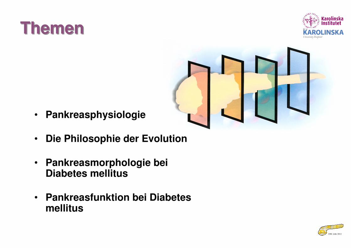
ThemenThemen
• Pankreasphysiologie
• Die Philosophie der Evolution
• Pankreasmorphologie bei Diabetes mellitus
• Pankreasfunktion bei Diabetes mellitus

©M. Löhr 2012Löhr & Klöppel in Hahn/Riemann, Gastroenterologie, Thieme 1994
Die morphologische GrundlageDie morphologische Grundlage• Intime „geographische“
Beziehung zwischen exokrinem und endokrinem Pankreas
• Blutfluß arteriell => Insel => exokrines Pankreas
©M. Löhr 2012
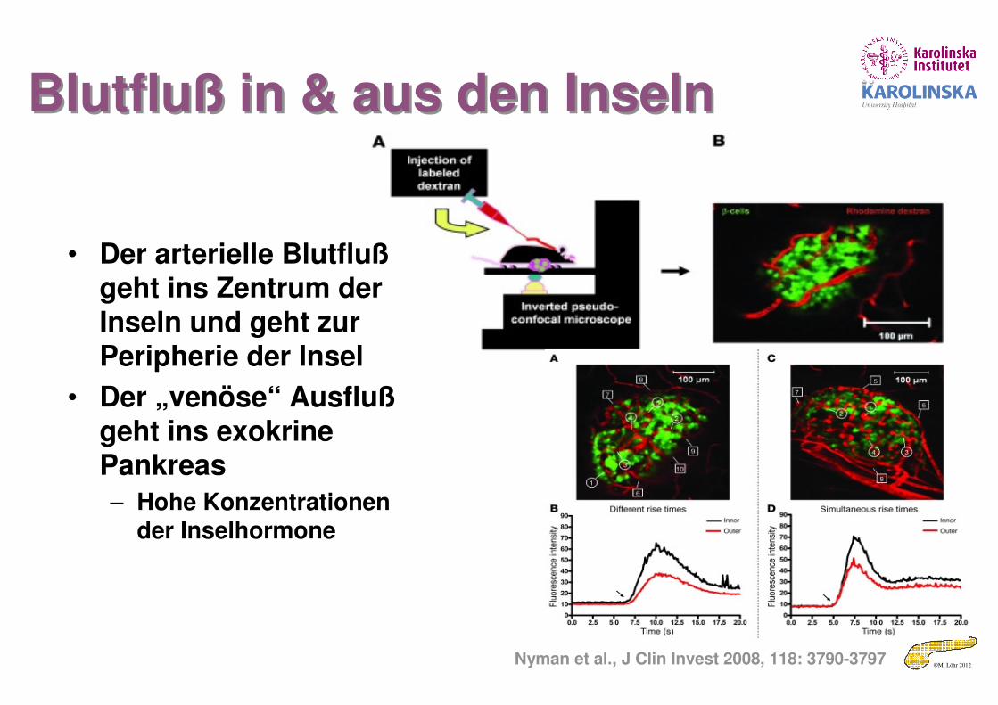
Blutfluß in & aus den InselnBlutfluß in & aus den Inseln
Nyman et al., J Clin Invest 2008, 118: 3790-3797
• Der arterielle Blutflußgeht ins Zentrum der Inseln und geht zur Peripherie der Insel
• Der „venöse“ Ausflußgeht ins exokrine Pankreas
– Hohe Konzentrationen der Inselhormone
©M. Löhr 2012
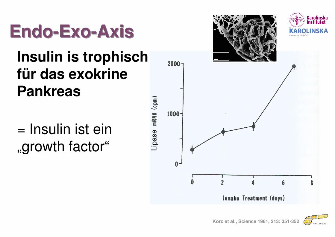
Endo-Exo-AxisEndo-Exo-Axis
Korc et al., Science 1981, 213: 351-352
Insulin is trophisch für das exokrine Pankreas
= Insulin ist ein „growth factor“ L
ipase
100 µm

©M. Löhr 2012
On the origin of species… and the exocrine pancreas
On the origin of species… and the exocrine pancreas
Roach et al., J Mol Evol 1997; 45:640–652

©M. Löhr 2012
The Worm thingThe Worm thing
• Cells with zymogens (digestive enzymes) interspersed in the lining o the primitive digestive tube
www.bio.sunyorange.edu
©M. Löhr 2012
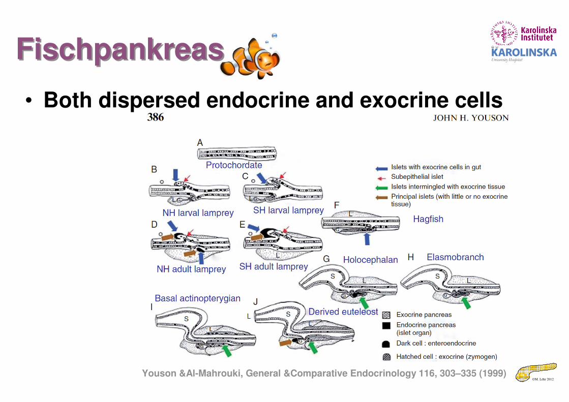
FischpankreasFischpankreas
• Both dispersed endocrine and exocrine cells
Youson &Al-Mahrouki, General &Comparative Endocrinology 116, 303–335 (1999)

©M. Löhr 2012
Pankreas in SäugetierenPankreas in Säugetieren
Fig. 2 Pancreas models in vertebrate embryos. (a) 5.5-mm pig embryo. (b) 20-mm pig embryo. (c) 11-mm rabbit
embryo. (d) 10.7-mm cat embryo. (e) 7.5-mm human embryo. (f) 13.6-mm human embryo. (After F. W. Thyng,
Models of the pancreas in embryos of the pig, rabbit, cat and man, Amer. J. Anat., 7:488–503, 1907)
©M. Löhr 2012
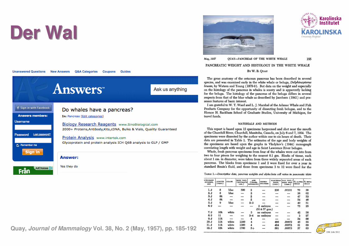
Der WalDer Wal
Quay, Journal of Mammalogy Vol. 38, No. 2 (May, 1957), pp. 185-192

©M. Löhr 2012
Endo-Exo Achse & EntwicklungEndo-Exo Achse & Entwicklung• Um sich vom Frosch und Neunauge
zu Menschen und Mäusen (& Wal) zu entwickeln, müssen
– Genügend Verdauuungsenzyme zum Aufschluß der Nahrung (speziell Fetten) zur Verfügung stehen
• Muss das exokrine Pankreas genug großsein
– Wofür ein Wachstumsfaktor (Insulin) gebraucht wird
• KONSEQUENZ:
Das ENDOKRINE Pankreas sitzt im EXOKRINEN Pankreas
Roach et al., J Mol Evol 1997; 45:640–652

©M. Löhr 2012
Superiority of vertebratesWhy they increase in growth and capabilities
Superiority of vertebratesWhy they increase in growth and capabilities
Species Pancreas/BW
Islets
Human 0.001 ✔
Whale 0.0008 ✔
Fish 0.00001
Xenopus 0.0001
Horse 0.001 ✔
Mouse 0.01 ✔
Dog 0.002 ✔
©M. Löhr 2012
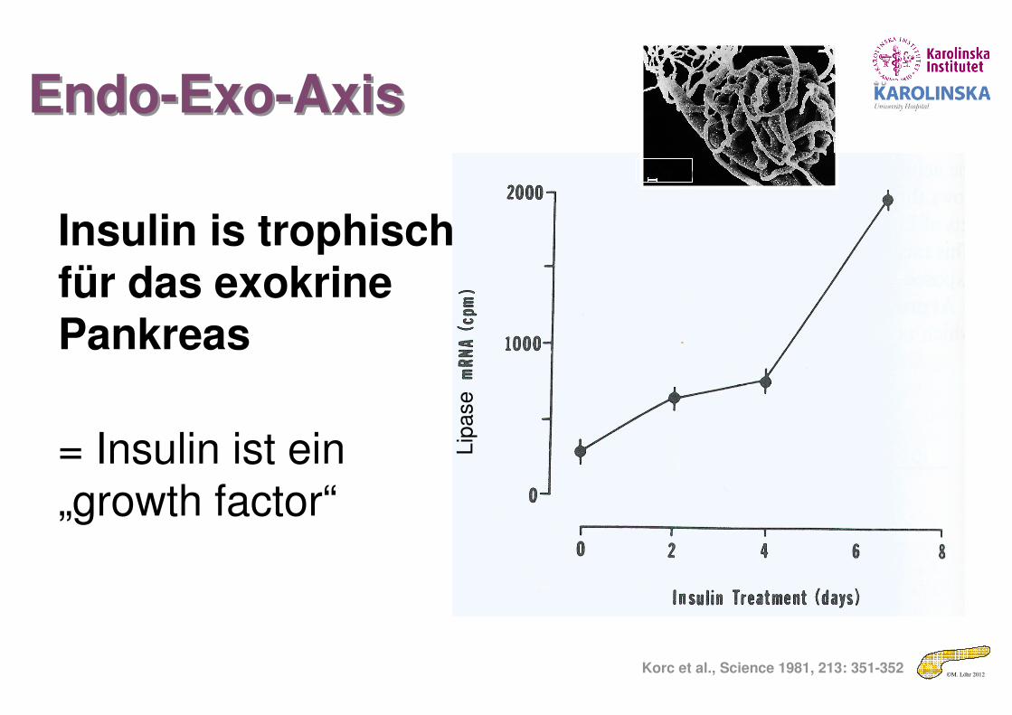
Endo-Exo-AxisEndo-Exo-Axis
Korc et al., Science 1981, 213: 351-352
Insulin is trophisch für das exokrine Pankreas
= Insulin ist ein
„growth factor“Lip
ase
100 µm

©M. Löhr 2012
No insulin – no growth?!No insulin – no growth?!

©M. Löhr 2012
Insulo-acinar portal system: Exocrine pancreas exposed to high concentrations of islet hormones
100 µm
Insulin
Trophic effects
Halo phenomenon
Enzyme releasein response to
stimulants ↑
Glucagon, SST, PP
Atrophic effects
Inhibition of exocrine
function
Healthy subjectsHealthy subjects
� Atrophy � Loss of halos
� Exocrine insufficiencyExocrine insufficiency
- +
DiabeticsDiabetics
Islet-acinar axis in diabetesIslet-acinar axis in diabetes
Keller & Layer Gut 2005;54:Suppl 6: vi9-29
©M. Löhr 2012
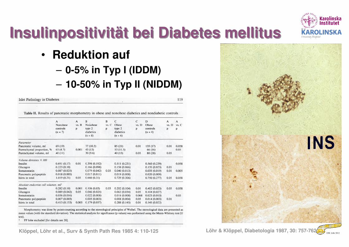
Insulinpositivität bei Diabetes mellitusInsulinpositivität bei Diabetes mellitus
• Reduktion auf
– 0-5% in Typ I (IDDM)
– 10-50% in Typ II (NIDDM)
Löhr & Klöppel, Diabetologia 1987, 30: 757-762Klöppel, Löhr et al., Surv & Synth Path Res 1985 4: 110-125
©M. Löhr 2012
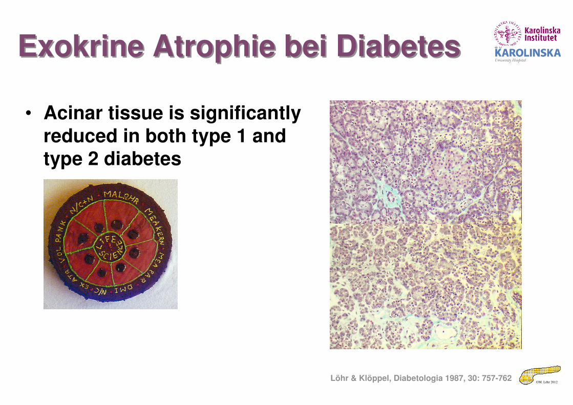
Exokrine Atrophie bei DiabetesExokrine Atrophie bei Diabetes
Löhr & Klöppel, Diabetologia 1987, 30: 757-762
• Acinar tissue is significantly reduced in both type 1 and type 2 diabetes
©M. Löhr 2012
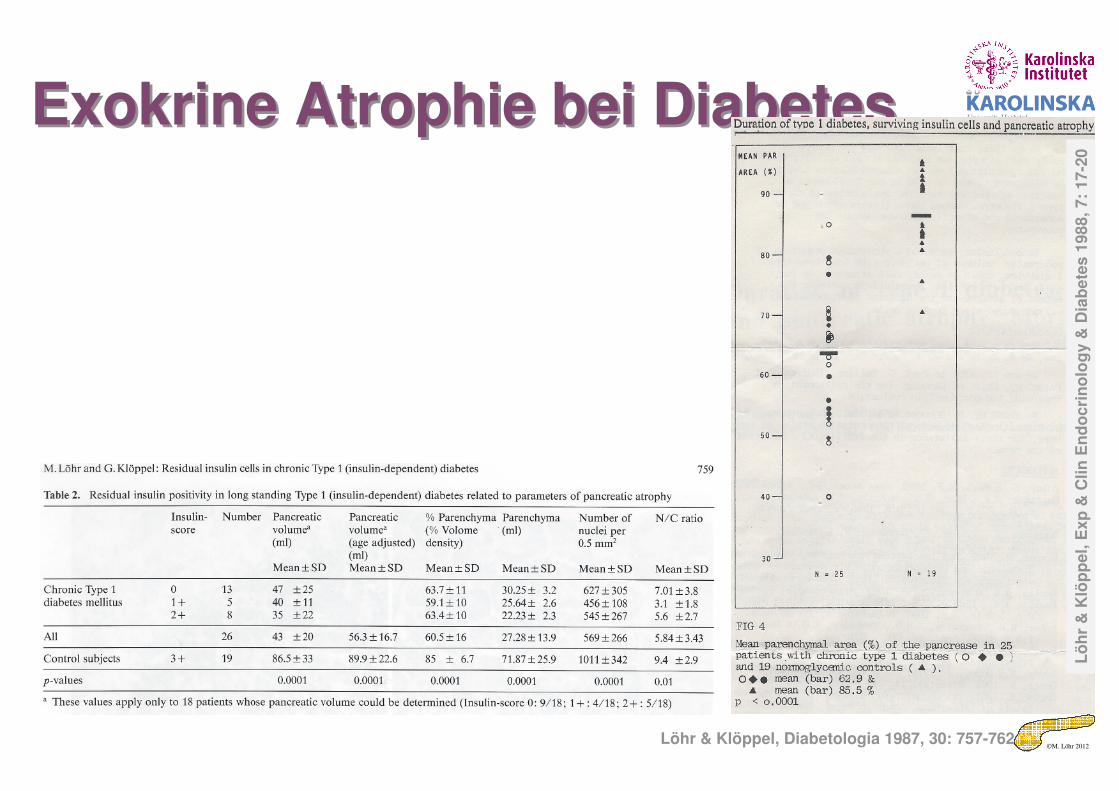
Exokrine Atrophie bei DiabetesExokrine Atrophie bei Diabetes
Löhr & Klöppel, Diabetologia 1987, 30: 757-762
Lö
hr
& K
löp
pe
l, E
xp
& C
lin
En
do
cri
no
log
y &
Dia
be
tes
198
8, 7:
17
-20
©M. Löhr 2012
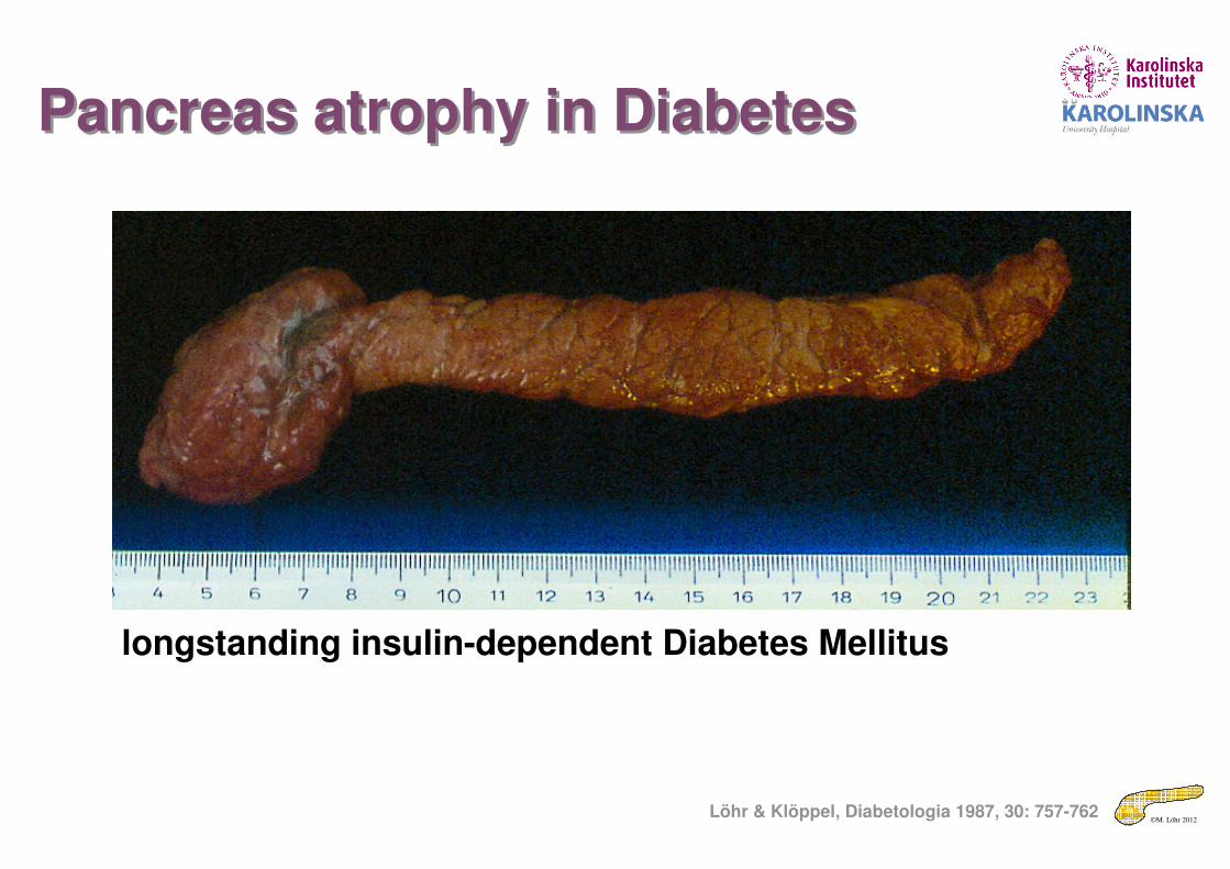
Pancreas atrophy in DiabetesPancreas atrophy in Diabetes
longstanding insulin-dependent Diabetes Mellitus
Löhr & Klöppel, Diabetologia 1987, 30: 757-762

©M. Löhr 2012
Pancreas volume in DiabetesPancreas volume in Diabetes
Löhr & Klöppel, Diabetologia 1987, 30: 757-762
©M. Löhr 2012
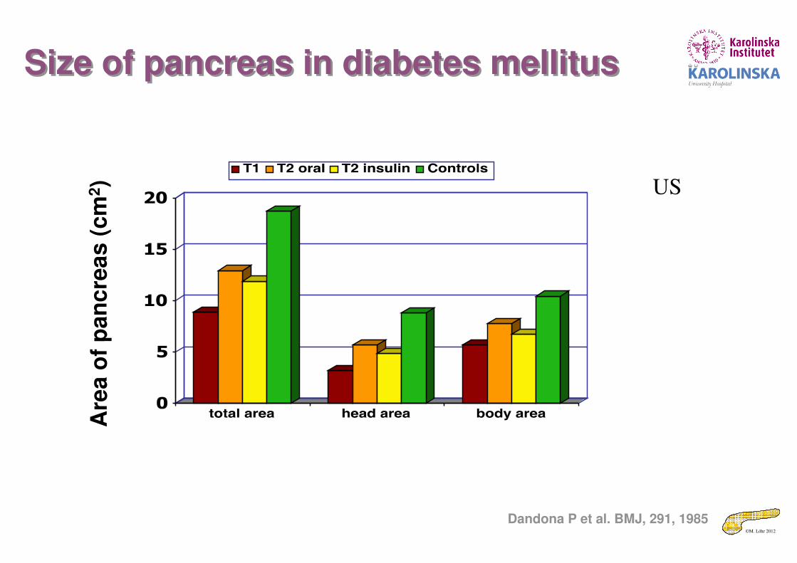
Size of pancreas in diabetes mellitusSize of pancreas in diabetes mellitusA
rea o
f p
an
cre
as (
cm
2)
Dandona P et al. BMJ, 291, 1985
US
©M. Löhr 2012
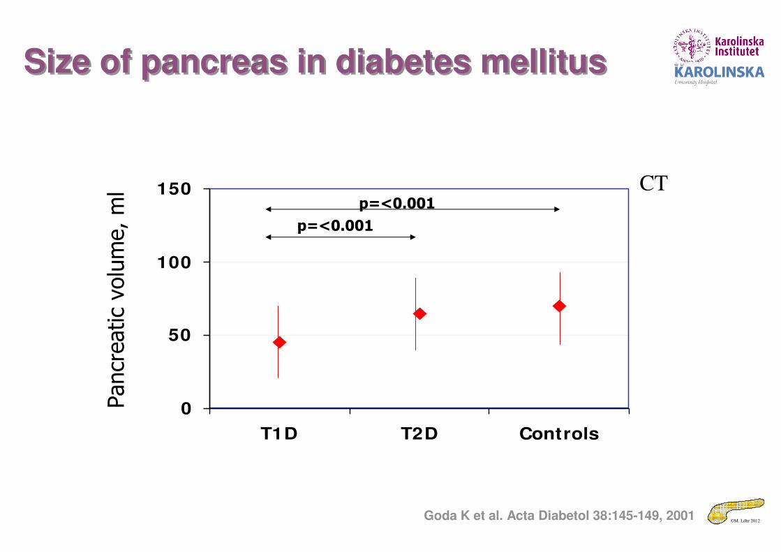
Size of pancreas in diabetes mellitusSize of pancreas in diabetes mellitus
0
50
100
150
T1D T2D Controls
Pancreatic volume, ml
p=<0.001
p=<0.001
CT
Goda K et al. Acta Diabetol 38:145-149, 2001
©M. Löhr 2012
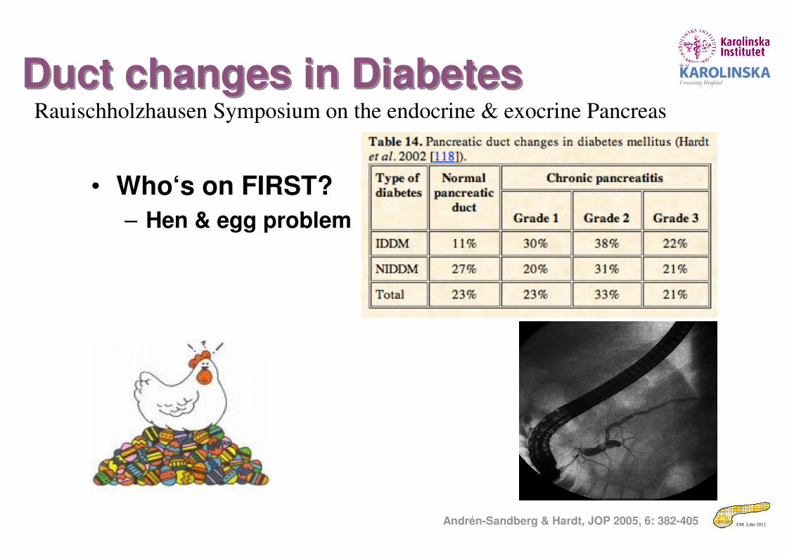
Duct changes in DiabetesDuct changes in Diabetes
• Who‘s on FIRST?
– Hen & egg problem
Andrén-Sandberg & Hardt, JOP 2005, 6: 382-405
Rauischholzhausen Symposium on the endocrine & exocrine Pancreas
©M. Löhr 2012
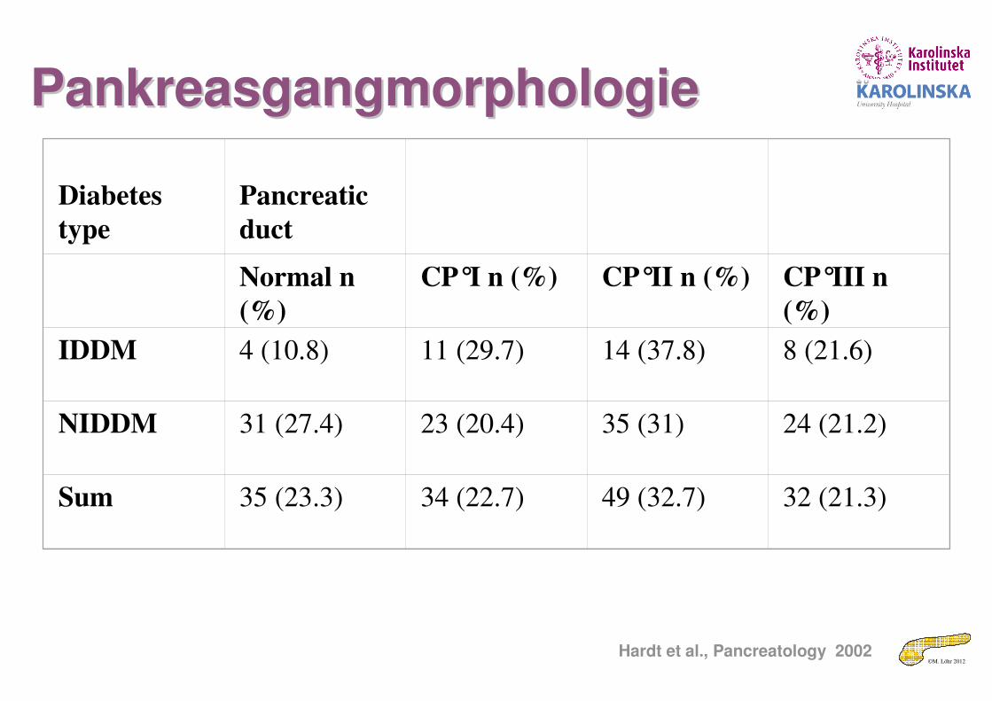
Diabetes
type
Pancreatic
duct
Normal n
(%)
CP°I n (%) CP°II n (%) CP°III n
(%)
IDDM 4 (10.8) 11 (29.7) 14 (37.8) 8 (21.6)
NIDDM 31 (27.4) 23 (20.4) 35 (31) 24 (21.2)
Sum 35 (23.3) 34 (22.7) 49 (32.7) 32 (21.3)
Hardt et al., Pancreatology 2002
PankreasgangmorphologiePankreasgangmorphologie
©M. Löhr 2012
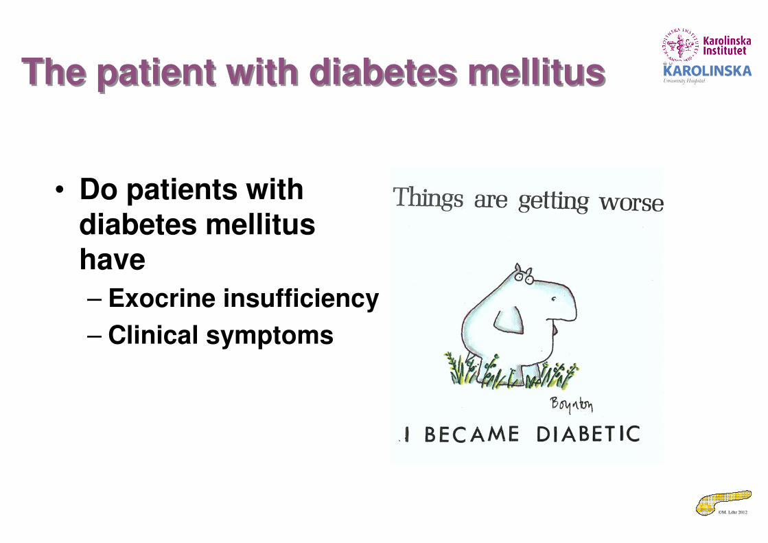
The patient with diabetes mellitusThe patient with diabetes mellitus
• Do patients with diabetes mellitushave
– Exocrine insufficiency
– Clinical symptoms
©M. Löhr 2012
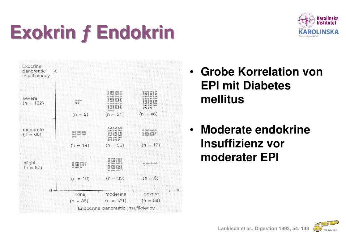
• Grobe Korrelation von EPI mit Diabetes mellitus
• Moderate endokrine Insuffizienz vor moderater EPI
Lankisch et al., Digestion 1993, 54: 148
Exokrin ƒ EndokrinExokrin ƒ Endokrin
©M. Löhr 2012
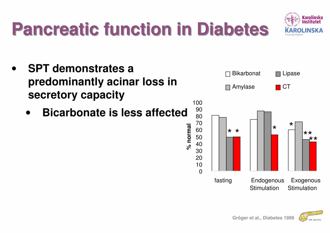
• SPT demonstrates a predominantly acinar loss in secretory capacity
• Bicarbonate is less affected
01020304050
60708090
100
fasting Endogenous
Stimulation
Exogenous
Stimulation%
no
rma
l
Bikarbonat
Amylase
Lipase
CT
* * * *****
Gröger et al., Diabetes 1999
Pancreatic function in DiabetesPancreatic function in Diabetes
©M. Löhr 2012
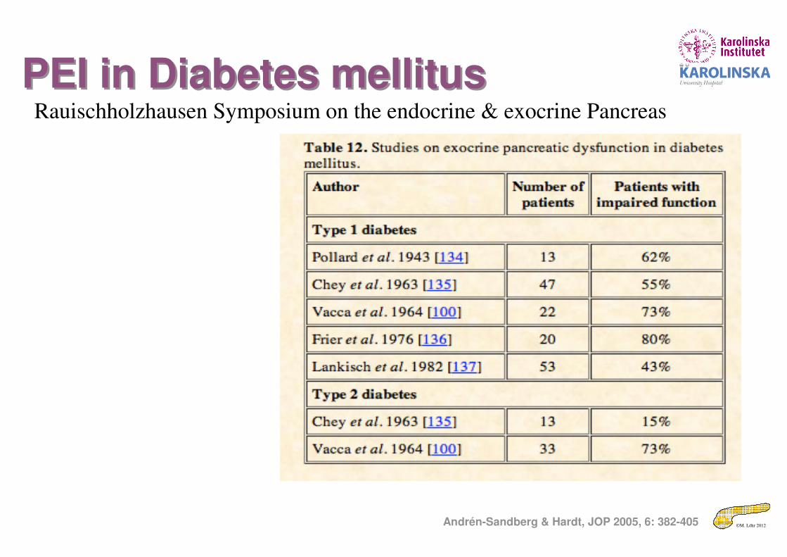
PEI in Diabetes mellitusPEI in Diabetes mellitus
Andrén-Sandberg & Hardt, JOP 2005, 6: 382-405
Rauischholzhausen Symposium on the endocrine & exocrine Pancreas
©M. Löhr 2012Löhr & Klöppel, Diabetologia 1987, 30: 757
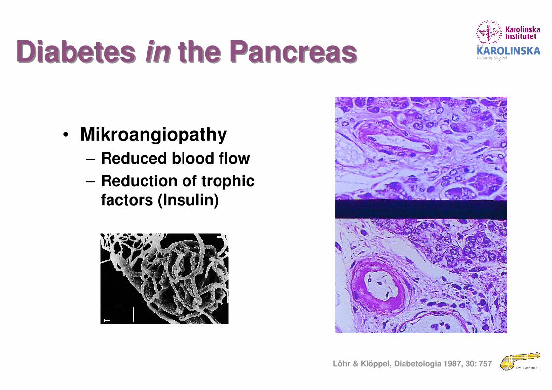
Diabetes in the PancreasDiabetes in the Pancreas
• Mikroangiopathy
– Reduced blood flow
– Reduction of trophic factors (Insulin)
100 µm

©M. Löhr 2012
Diarrhea in Diabetes MellitusDiarrhea in Diabetes Mellitus
• Patients with long-standing Diabetes mellitus may NOT show all the symptoms of PEI because of
– Diabetic gastropathy
– Autonomic neuropathy
el Newihi et al., Dig Dis Sci. 1988; 33(6): 705-710

©M. Löhr 2012
The patient with diabetes mellitusThe patient with diabetes mellitus
• Natural course and consequences of exocrine insufficiency

©M. Löhr 2012
PEI in DMPEI in DM
• Some patients may progress, others don’t
Creutzfeld et al., Digestion 2005;72:71–75

©M. Löhr 2012
PEI in Diabetes mellitusPEI in Diabetes mellitus
Andrén-Sandberg & Hardt, JOP 2008, 9: 541-575
Rauischholzhausen Symposium on the endocrine & exocrine Pancreas

©M. Löhr 2012
Disorders affecting multiple endocrine systems
Disorders affecting multiple endocrine systems
Syndrome Endocrine Pancreas Exocrine PancreasShwachman syndrome ? Atrophy/ 1° EPI
Ataxia-teleangiectasia Diabetes Mellitus 2° EPI
Pseudohypoparathyroidism Diabetes Mellitus 2° EPI
Myotonic dystrophy Diabetes Mellitus 2° EPI
Fanconi syndrome Diabetes Mellitus 2° EPI
Werner syndrome Diabetes Mellitus 2° EPI
Schmidt syndrome Diabetes Mellitus (Type 1b) 2° EPI
Wolfram syndrome Diabetes Mellitus (IDDM) 2° EPI
Laurence-Moon-Biedl syndr Diabetes Mellitus 2° EPI
Alström syndrome Diabetes Mellitus 2° EPI
Klinefelter syndrome Diabetes Mellitus 2° EPI
©M. Löhr 2012
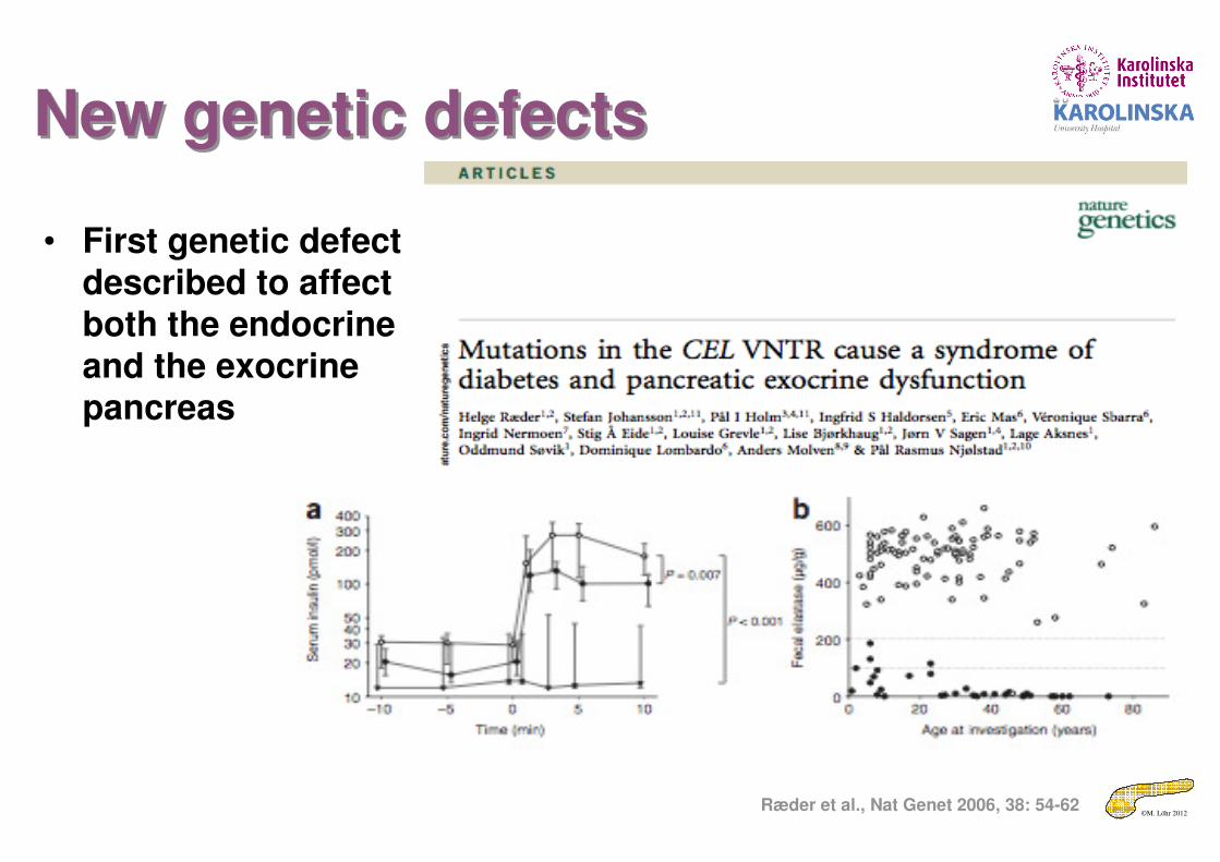
New genetic defectsNew genetic defects
• First genetic defect described to affect both the endocrine and the exocrine pancreas
Ræder et al., Nat Genet 2006, 38: 54-62
©M. Löhr 2012
BücherBücher
Thank you very much for your attention

©M. Löhr 2012
Epidemiology of chronic pancreatitisEpidemiology of chronic pancreatitis
• Incidence (new diagnoses)
– 5/100.000 inhabitants
• Prevalence (number of patients with disease)
– 25/100.000 inhabitants
• For comparison
– Diabetes: 4000/100.000 inhabitants
– Pancreatic cancer: 10/100.000 inhabitants
©M. Löhr 2012
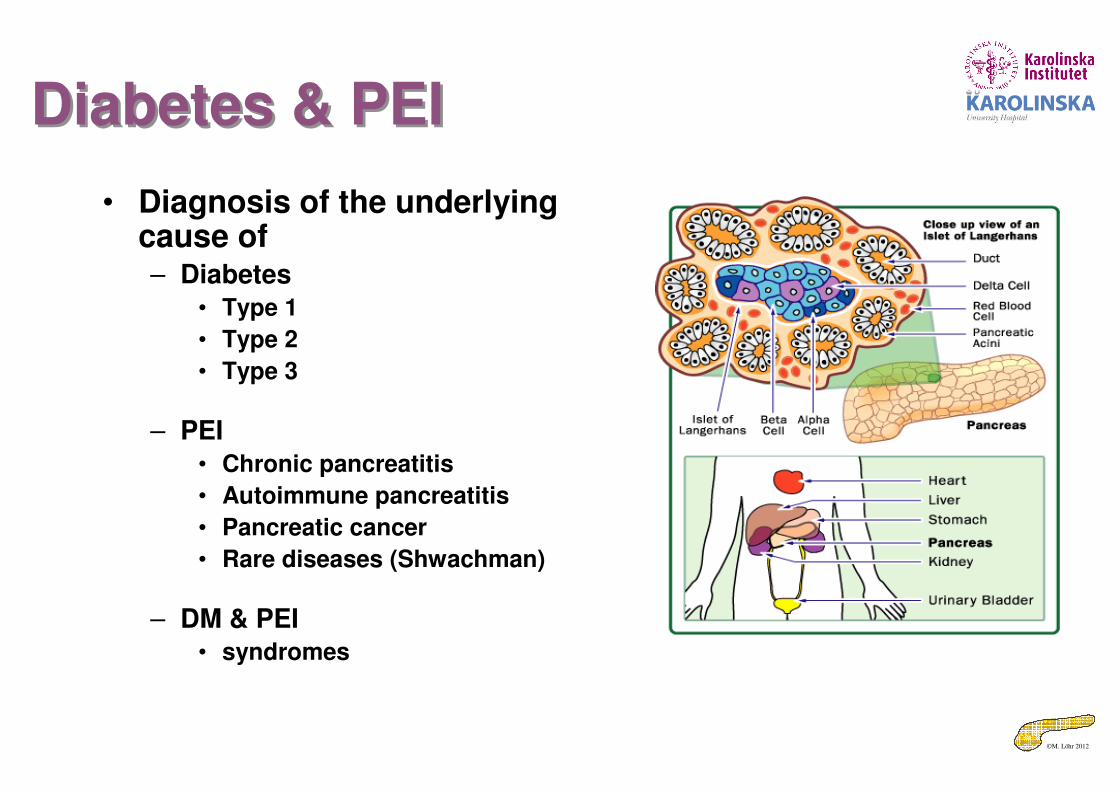
Diabetes & PEIDiabetes & PEI
• Diagnosis of the underlying cause of– Diabetes
• Type 1
• Type 2
• Type 3
– PEI• Chronic pancreatitis
• Autoimmune pancreatitis
• Pancreatic cancer
• Rare diseases (Shwachman)
– DM & PEI• syndromes
©M. Löhr 2012
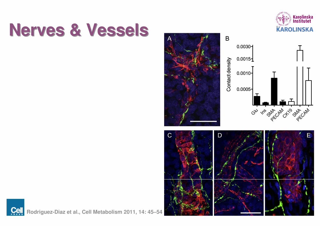
Nerves & VesselsNerves & Vessels
Rodriguez-Diaz et al., Cell Metabolism 2011, 14: 45–54

©M. Löhr 2012
Diagnostic value of fecal elastase-1Diagnostic value of fecal elastase-1
• Test of choice– Best of the available
• Relatively speaking!
– Highly practicable• Small amount of stool• Easy assay (ELISA)
– quick
– Reasonable cost/effort factor
25-83 % 33-100% 75-100%
Siegmund & Löhr: Meta-analysis of pancreatic function tests. Z Gastroenterol 2004, 42: 1117-1128
©M. Löhr 2012
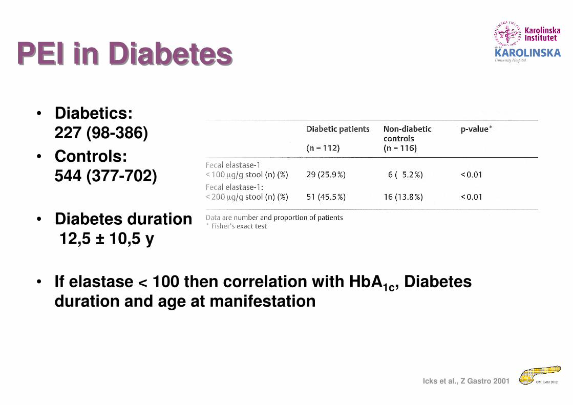
• Diabetics:227 (98-386)
• Controls:544 (377-702)
• Diabetes duration12,5 ± 10,5 y
• If elastase < 100 then correlation with HbA1c, Diabetes duration and age at manifestation
Icks et al., Z Gastro 2001
PEI in DiabetesPEI in Diabetes
©M. Löhr 2012
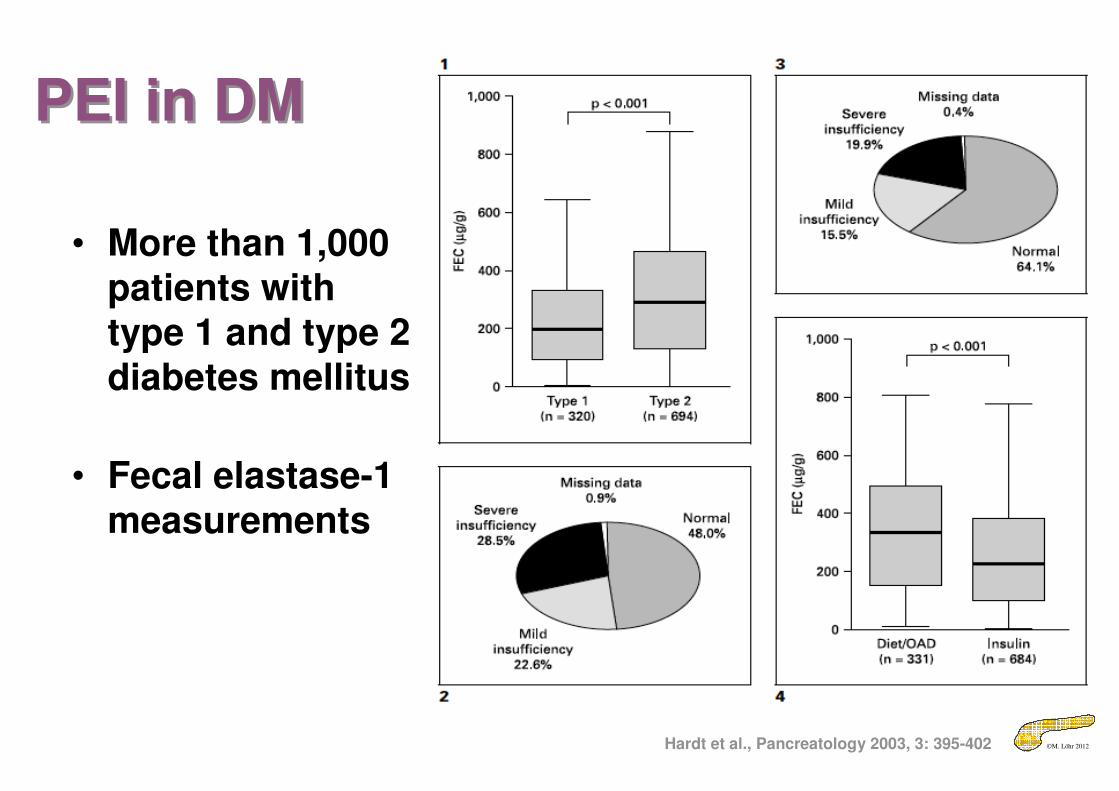
PEI in DMPEI in DM
Hardt et al., Pancreatology 2003, 3: 395-402
• More than 1,000 patients with type 1 and type 2 diabetes mellitus
• Fecal elastase-1 measurements

©M. Löhr 2012
Pancreas volume & function in DiabetesPancreas volume & function in Diabetes
Löhr & Klöppel, Diabetologia 1987, 30: 757-762Hardt et al., Pancreatology 2003, 3: 395-402

©M. Löhr 2012
There is an elephant in the roomThere is an elephant in the room
• Animal also suffered from diarrhea

©M. Löhr 2012
PEI in Diabetes MellitusPEI in Diabetes Mellitus
• Mild to moderate PEI may have a clinical impact– Clinical context
• Patients with Diabetes mellitus have an increased risk to develop PEI– Pancreatic function tests should
be employed
Evidence 2b
©M. Löhr 2012
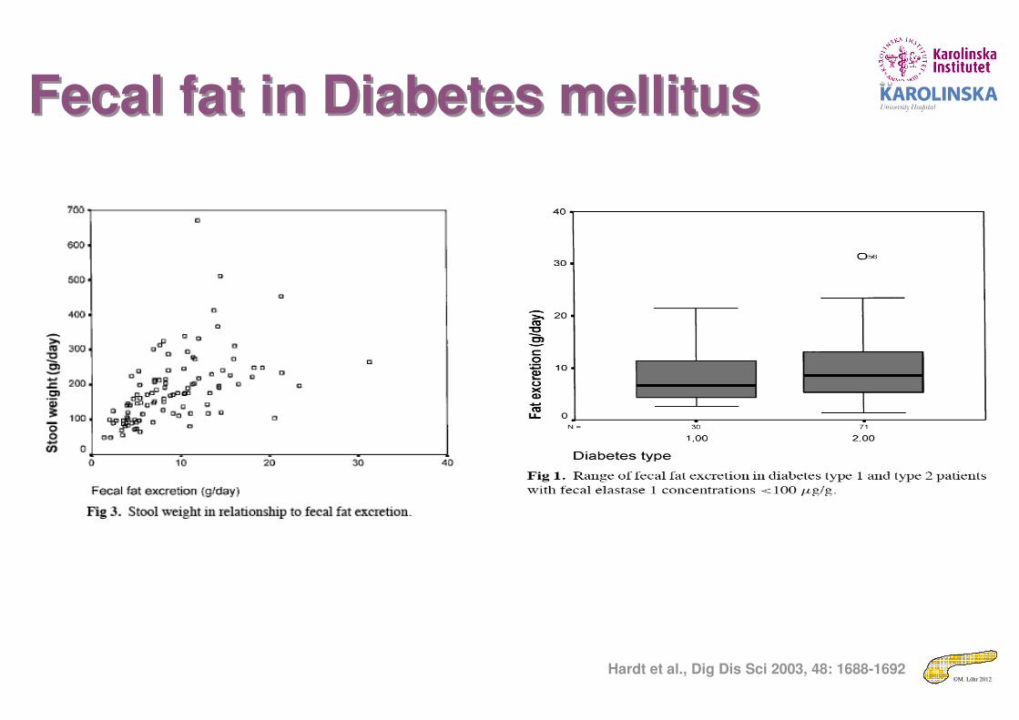
Fecal fat in Diabetes mellitusFecal fat in Diabetes mellitus
Hardt et al., Dig Dis Sci 2003, 48: 1688-1692

©M. Löhr 2012
Pathologic pancreatic duct morphology in ERCP:
40% of patients with IDDM (n=43)
58% of ICA-positive NIDDM (n=12)
9% of ICA-negative NIDDM (n=22)
PankreasgangmorphologiePankreasgangmorphologie
[Nakanishi et al, 1994]


![[ger] KOHLENWASSERSTOFFE : Monatlich 11-1985 [eng] …aei.pitt.edu/80080/1/1985_-_11.pdf · 2016. 9. 30. · 501 665 371 490 438 483 347 198 486 326 434 353 3790 3065 19,1 12135 12119](https://static.fdokument.com/doc/165x107/61232bd9c63bc323454ab026/ger-kohlenwasserstoffe-monatlich-11-1985-eng-aeipittedu8008011985-11pdf.jpg)