Development of a click chemistry-based proximity ligation ...
142
Dissertation zur Erlangung des Doktorgrades der Fakultät für Chemie und Pharmazie der Ludwig-Maximilians-Universität München Development of a click chemistry-based proximity ligation assay and Synthesis and incorporation of 5-carboxycytidine phosphoramidite in synthetic RNA Bastien Viverge aus Digne-les-Bains, France 2021
Transcript of Development of a click chemistry-based proximity ligation ...
Dissertation zur Erlangung des Doktorgrades
der Fakultät für Chemie und Pharmazie
der Ludwig-Maximilians-Universität München
Development of a click chemistry-based proximity ligation assay
and
Synthesis and incorporation of 5-carboxycytidine
phosphoramidite in synthetic RNA
Bastien Viverge
aus
Digne-les-Bains, France
2021
Erklärung
Diese Dissertation wurde im Sinne von § 7 der Promotionsordnung vom 28. November 2011 von Herrn Prof. Dr; Thomas Carell betreut.
Eidesstattliche Versicherung
Diese Dissertation wurde eigenständig und ohne unerlaubte Hilfe erarbeitet.
Lenzburg, May 18th 2021
Bastien Viverge
Dissertation eingereicht am 29.04.2021
1. Gutachter: Prof. Dr. Thomas Carell
2. Gutachterin: Dr. Sabine Schneider
Mündliche Prüfung am 14.07.2021

To Maria and my parents

“It was one of those events which at a crucial stage in one's development arrive
to challenge and stretch one to the limit of one's ability and beyond, so that
thereafter one has a new standard by which to judge oneself.”
Kazuo Ishiguro, The Remains of the Day
Part of this work was published or presented at conferences:
Publications
· N. Raddaoui, S. Croce, F. Geiger, A. Borodavka, L. Möckl, S. Stazzoni, B. Viverge, C.
Bräuchle, T. Frischmuth, H. Engelke, T. Carell. ChemBioChem. 2020, Accepted Article.
Super-sensitive multi-fluorophore RNA-FISH for early virus detection and flow-FISH using
click chemistry.
· I. N. Michaelides, N. Tago, B. Viverge, T. Carell. Chem. Eur. J. 2017, 23, 15894-15898.
Synthesis of RNA Containing 5-Hydroxymethyl-, 5-Formyl-, and 5-Carboxycytidine.
· N. Raddaoui, S. Stazzoni, L. Möckl, B. Viverge, F. Geiger, H. Engelke, C. Bräuchle, T.
Carell. ChemBioChem. 2017, 18, 1716-1720.
Dendrimer-Based Signal Amplification of Click-Labelled DNA in Situ.
· M. Ehrlich, M. Gattner, B. Viverge, J. Bretzler, D. Eisen, M. Stadlmeier, M. Vrabel, T. Carell.
Chem. Eur. J. 2015, 21, 7701–7704.
Orchestrating the biosynthesis of an unnatural pyrrolysine amino acid for its direct
incorporation into proteins inside living cells.
Conference presentation
· Lecture presentation:
„Synthesis of click-chemistry based probes for the detection of specific gene sequences”
COST Action 1201 (April 2016, Grenoble, France).
Acknowledgments
Herewith, I would like to express my sincere gratitude to all of those who have made this work
possible through their help and support.
First and foremost, I am grateful to Prof. Dr. Thomas Carell who offered me the opportunity to
join his group and carry the research projects presented in the following pages within the best
possible conditions. I greatly appreciated the support and guidance received while benefiting
from a high level of trust and independence.
I want to thank Frau Slava Gärtner for her kindness, helpfulness and availability when it came
to administrative matters.
I would like to warmly thank Frau Sabine Voß, Kerstin Kurz and Kristof Hufnagel for the
fantastic technical support.
I am grateful to Dr. Markus Müller for his scientific inputs and general supports dealing with
the everyday challenges of the lab.
I would like to thank Dr. Iacovos Michaelides and Dr. Nobuhiro Tago for their excellent
collaboration on the project aiming at the synthesis of a new RNA phosphoramidite.
Special acknowledgments to my former lab mates Kristof Hufnagel, Dr. Charlotte Ebert, Dr.
Michael Stadlmeier, Dr. Hidenori Okamura and Dr. Gengo Kashiwasaki. I will always
remember the pleasure to work in such a friendly environment and how valuable our
collaborations and support to each other have been.
For their careful proofreading and critical comments of my thesis, I am grateful to Dr. Samuele
Stazzoni, Dr. Michael Staddlmeier, Dr. Nobuhiro Tago and Leander Runtsch.
I would like to thank the rest of the Carell Group for the wonderful time and the excellent
working atmosphere.

Eventually, I would like to express my gratitude to my parents for their constant support in all
my decisions and through all the challenges, I have encountered. Likewise, I feel enormously
grateful towards my partner and wife-to-be Maria for her love and encouragements.

Table of contents
I
TABLE OF CONTENTS
TABLE OF CONTENTS ............................................................................................................... I
SUMMARY ........................................................................................................................... IV
1. INTRODUCTION................................................................................................................... 1
1.1 DNA functionalisation by click chemistry .................................................................... 1
1.1.1 DNA ..................................................................................................................... 1
1.1.2 Cu(I)-catalysed azide-alkyne cycloaddition ........................................................... 3
1.1.3 CuAAC reaction on nucleic acids .......................................................................... 7
1.2 Bioassays based on nucleic acid amplification ............................................................. 9
1.2.1 Real-time Polymerase Chain Reaction ................................................................... 9
1.2.2 Proximity Ligation Assay .................................................................................... 14
2. DEVELOPMENT OF A CLICK CHEMISTRY-BASED PROXIMITY LIGATION ASSAY ...................... 16
2.1 Objectives .................................................................................................................. 16
2.2 Synthesis of azide functionalized ODN ...................................................................... 18
2.2.1 Strategy ............................................................................................................... 18
2.2.2 Synthesis of unsymmetrical picolyl azide/azide linkers ........................................ 20
2.2.3 Click experiments ................................................................................................ 24
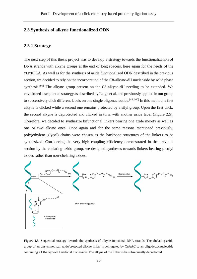
2.3 Synthesis of alkyne functionalized ODN .................................................................... 28
2.3.1 Strategy ............................................................................................................... 28
2.3.2 Synthesis of picolyl azide/alkyne-TMS linkers .................................................... 29
2.3.3 Click experiments ................................................................................................ 30
2.3.4 Synthesis of picolyl azide/alkyne-TES linkers ..................................................... 32
2.3.5 Click experiments and deprotection ..................................................................... 35
2.4 ODN design and synthesis ......................................................................................... 40
2.4.1 Design parameters ............................................................................................... 40

Table of contents
II
2.4.2 ODN synthesized ................................................................................................ 41
2.4.3 Melting temperature (Tm) measurement .............................................................. 44
2.4.4 Click test with dyes ............................................................................................. 45
2.5 CLICKPLA experiments .............................................................................................. 46
2.5.1 Double conjugation experiments.......................................................................... 46
2.5.2 Concentration experiments .................................................................................. 49
2.5.3 Reaction time experiments................................................................................... 50
2.6 Conclusions and Outlook ........................................................................................... 52
3. SYNTHESIS AND INCORPORATION OF 5-CARBOXYCYTIDINE PHOSPHORAMIDITE IN SYNTHETIC
RNA ................................................................................................................................... 55
3.1 Objectives .................................................................................................................. 55
3.2 Design and protecting group strategy ......................................................................... 57
3.3 Synthesis of a caC RNA phosphoramidite building block .......................................... 59
3.4 Solid phase synthesis of RNA strands containing caC ................................................ 61
3.5 Solid phase synthesis of a RNA strand containing all the C modifications .................. 63
3.6 Enzymatic digestion and uHPLC-MS analysis ........................................................... 65
3.7 Conclusions and Outlook ........................................................................................... 66
4. EXPERIMENTAL ................................................................................................................ 67
4.1 General Methods and Materials for Synthesis ............................................................ 67
4.2 Chemical synthesis .................................................................................................... 69
4.2.1 Synthesis of the picolyl azide-PEG10-azide linker (17) ......................................... 69
4.2.2 Synthesis of the picolyl azide/bis-azide linker (19) .............................................. 73
4.2.3 Synthesis of the picolyl azide-PEG11-TMS protected alkyne linker (34) .............. 80
4.2.4 Synthesis of the picolyl azide-PEG11-TES protected alkyne linker (37) ............... 84
4.2.5 Synthesis of the picolyl azide/bis-TES protected alkyne linker (40) ..................... 86
4.2.6 Synthesis of C8-alkyne-dU .................................................................................. 93
4.2.7 Synthesis of the caC phosphoramidite building block (42) ................................... 96

Table of contents
III
4.3 Oligonucleotide synthesis ........................................................................................ 105
4.3.1 General information .......................................................................................... 105
4.3.2 Synthesis and deprotection of oligodeoxynucleotides containing C8-alkyne-dU 105
4.3.3 Synthesis and deprotection of oligonucleotides containing caC .......................... 106
4.4 Click protocols......................................................................................................... 108
4.4.1 Oligodeoxyribonucleotide functionalization, characterisation and purification ... 108
4.4.2 Template-assisted double conjugation ............................................................... 109
4.5 Biochemical experiments ......................................................................................... 110
4.5.1 Melting Curve Experiments ............................................................................... 110
4.5.2 DNA PAGE ...................................................................................................... 110
4.5.3 Enzymatic digestion of ON................................................................................ 111
4.6- UHPLC-MS analysis .............................................................................................. 112
5. ABBRIEVATIONS ............................................................................................................. 113
6. REFERENCES .................................................................................................................. 117
Summary
IV
SUMMARY
Proximity ligation assays are powerful tools for sensitive detections of proteins. These assays
are usually performed with two DNA-tagged aptamers or antibodies binding at proximity to
each other on the same protein or interacting proteins. Thanks to the proximity between these
two probes, the DNA tags can be ligated and the resulting sequence can be amplified by PCR.
Many copies of the target sequence are thus generated and the labelling of this sequence will
result in a signal high enough to enable sensitive and robust detection of proteins or/and protein
complexes.[1, 2]
The aim of the first project described in the present thesis was to investigate whether the so-
called click reaction, could contribute to the development of the existing PLA methods.
Therefore, we imagined as a proof of concept a model where three click functional
oligonucleotide probes are hybridized next to each other on a single longer oligonucleotide.
Upon hybridization on the latter one thus designated as the template, the probes could be
conjugated by clicking with each other.
The first part of the project was to envision the synthesis of oligonucleotide probes with the
desired click functional groups with reliable, effective and easy to perform protocols. As a
starting point and based on previous work achieved in the Carell group, we used DNA strand
with one alkyne-modified nucleoside incorporated by automated solid phase synthesis.[3] In
order to subsequently functionalize oligonucleotides with one or two azido-groups from the
single alkyne modification, we decided to rely on a chemoselective strategy (Figure 1A). It has
been reported that chelating azides such as picolyl azide are significantly more reactive than
non-chelating azides towards CuAAC reactions.[4] This difference in reactivity is such that on
small molecules bearing both a picolyl azide and a normal azide, the picolyl azide can be
clicked selectively to an alkyne molecule.[5] The non-chelating azide is thus left unreacted and
available for a second click reaction. Here, we examined if this strategy could be applied to
longer linkers to match our needs. Therefore, we synthesized two unsymmetrical linkers; a first
one with one picolyl azide and one non-chelating azide and a second one with one picolyl azide
and two non-chelating azide. These linkers were effectively and with very good selectivity
clicked to alkyne-modified oligonucleotides thus providing DNA strands functionalized with

Summary
V
one or two azido-groups. Next, we applied a sequential strategy to obtain oligonucleotides with
long alkyne functions. The idea here was to synthesize unsymmetrical linkers with one azide
at one extremity and one or two protected alkynes on the other extremities. Following the
conjugation of these linkers by CuAAC to alkyne modified DNA strands, the protecting
group(s) of the alkyne of the linkers was removed leaving it free for further reactions (Figure
1B). This method also proved to be fast, reliable and did not require an extra purification step
after the deprotection of the alkyne.
Figure 1: DNA functionalization strategies.
Based on these results, we designed and synthesized two sets of oligonucleotide probes of
respectively 11- and 15- mer strands. Each set featured three probes, two single alkyne
functionalized side probes and one double azide middle probe as well as a complementary
oligonucleotide template. Both sets of probes were hybridized on their respective template and
conjugated to each other by CuAAC. The reactions were analysed on denaturing PAGE and
showed the successful formation of the double conjugated product between the three probes
thanks to the proximity effect provided by the template (Figure 2).
Summary
VI
Figure 2: Schematic presentation of the click-chemistry based proximity ligation assay workflow.
Epigenetics could be defined in simple words as the regulations of the gene expression which
do not involve changes of the DNA sequence.[6] Different mechanisms co-exist in order to
control the gene activities across several layers of regulatory information. At the DNA level,
the canonical base cytosine undergoes a chemical reaction in which a methyl group is added
on the position C(5). The 5-methylcytosine modification is an epigenetic mark and its presence
onto a gene promoter results in the repression of the gene transcription.[7] The discovery during
the last years of oxidized derivatives of 5-methylcytosine suggests that the DNA demethylation
occurs through a cascade of oxidation reactions at the C(5) position. Thus, 5-methylcytosine is
successively transformed into 5-hydroxymethylcytosine (hmC), 5-formylcytosine (fC) and 5-
carboxycytosine (caC).[8]
Recently, these modifications have also been discovered in the mRNA of mammalian cells.[9,
10, 11, 12, 13] While hypotheses are being formulated regarding their role within RNA, their exact
functions remain elusive and are still to be elucidated. In order to study the structural and
functional roles of hmC, fC and caC in the context of RNA, it is essential to access synthetic
RNA material containing one or several of these bases at defined positions.
Within the frame of this PhD thesis and as a team-work from the Carell Group, we aimed at
developing synthesis towards new RNA phosphoramidite building block the three modified
bases. We designed these phosphoramidite building blocks in order to be compatible with

Summary
VII
standard automated solid phase synthesis conditions so that they could be routinely used in
RNA synthesis.
The present thesis focused on the development of the caC building block. The first step was to
design a suitable protecting group strategy. Indeed, the corresponding phosphoramidite needed
protecting groups able to resist the solid phase synthesis conditions while being easily removed
after the synthesis and without arming the newly generated synthetic RNA strand. As RNA is
inherently less stable than DNA, notably towards hydrolysis, it required the use of protecting
groups cleavable in mild conditions. Once the design established, we envisioned and performed
the synthesis of the caC RNA building block. The new phosphoramidite was successfully
incorporated by automated solid phase synthesis and subsequent deprotection steps allowed the
synthesis of two RNA strands of different length, respectively 13-mer and 21-mer, containing
the caC-modified base at a defined site. In a similar workflow, new RNA phosphoramidite of
the modified bases hmC and fC were developed in parallel. In order to prove that the three
modified-cytosine RNA phosphoramidites are compatible with regular canonical bases
phosphoramidites as well as with each other, we ultimately synthesised a RNA strand
containing all the three modified bases.
Figure 3: Depiction of the phosphoramidite building blocks developed for the synthesis of RNA containing the
modified bases hmC, fC and caC.

Introduction
1
1. INTRODUCTION
1.1 DNA functionalisation by click chemistry
1.1.1 DNA
DNA is crucial to every living organism for the storage and the transmission of the genetic
information from one generation to the next.[14] Like other biomolecules, DNA is a polymer
made of the repetition of similar monomer building blocks. In the case of DNA, there are four
monomer units so-called nucleotides. These are composed of a deoxyribose sugar unit, a
nitrogenous base (adenine (A), cytosine (C), guanine (G) or thymine (T)) and a phosphate
group. In the DNA polymer, the nucleotides are linked to each other via phosphodiester bonds
at the 5’- and 3’- hydroxyl group of the deoxyribose. The nucleotide sequence defines the
genetic information. DNA is therefore well representative of the biochemical principle stating
the close relation between molecular structure and function of the biomolecules.[14]
In 1869, DNA was isolated for the first time from white blood cells by Friedrich Miescher.[15]
As he isolated it from the nuclei of the cell, Miescher named the novel substance nuclein. This
name remains in today’s designation deoxyribonucleic acid. The components of DNA, as well
as the nature of the phosphate-sugar bond linking them together, were identified by Phoebus
Levene in 1919.[16] It is only in 1944 that the role of carrier of the genetic information was
assigned to DNA by Avery et al.[17] After isolating and purifying DNA from a strain of
pneumococcus, they transferred it to a different strain of bacteria, which was transformed.
Thus, they showed that DNA was responsible for the transmission of the genetic information
and not proteins as believed until then. James Watson and Francis Crick eventually deduced
the structure of DNA in 1953 from X-ray diffraction data obtained by Rosalind Franklin.[18, 19,
20] The structure is an antiparallel double helix composed of two strands. Although several
conformations of the double helix exist, genomic DNA is mostly found in the right-handed B-
form.[21] In this conformation, the helix makes a full turn every ten bases. The sugar-phosphate
backbone of the DNA polymer is facing outside while the nitrogenous bases are in the inside
of the double helix. The two strands are held together by hydrogen bonds between the bases of
the opposite strands. A forms a specific base pair with opposite in the double helix T by two
hydrogen bonds while C and opposite G are specifically paired by three hydrogen bonds. This

Introduction
2
specific base pairing is responsible for the conservation of the genetic information.[20] Indeed,
the sequence of bases in one strand determines the sequence of the other strand. Therefore,
when the double helix is split into two strands, each strand can act as a template for the
formation of its complementary strand thus allowing the replication of the parent double helix
into two identical copies.[22]
Figure 1.1: Three-dimensional structure of the DNA double helix and representation of the four nucleotide
chemical structures, phosphate-sugar bond and selective base pairs between A:T and C:G (hydrogen bonds
represented as dotted line).
The genetic information carried by a DNA molecule is encoded by the nucleotide sequence. In
order to generate this information, the DNA serves as the template for the formation of a
messenger RNA (mRNA) during the so-called transcription process. This mRNA is an
intermediate of the protein synthesis and is further translated into a sequence of amino acids.
The relationship between the sequence of bases of the mRNA and the sequence of amino acids
is defined by the genetic code.[23, 24] It consists of three bases, called a codon, specifying one
amino acid and it is universal for all organisms.
Introduction
3
1.1.2 Cu(I)-catalysed azide-alkyne cycloaddition
The Cu(I)-catalysed azide-alkyne cycloaddition, abbreviated CuAAC, is a 1,3-dipolar
cycloaddition reaction resulting in the formation of a 1,2,3-triazole. The CuAAC reaction is
considered to be a catalysed version of the thermally induced reaction originally described at
the end of the 19th century by Michael.[25] As its mechanism and synthetic applications were
extensively studied by Huigsen in the 1960s, it became known as the Huigsen cycloaddition.[26,
27] In 2001, the use of Cu(I) to catalyse the reaction was reported for the first time by Tornøe
and Meldal in the context of solid-phase peptide synthesis.[28] One year later, the reaction
gained significant attention when two independent publications by Meldal and Sharpless
described the dramatic acceleration on the reaction rate provided by the Cu(I) catalysis.[29, 30]
Additionally, the catalysed cycloaddition selectively produced the 1,4-disubstituted triazole as
the only regioisomer whereas the original reaction provided a mixture of 1,4- 1 and 1,5-
regioisomers 2 (Scheme 1.1) thus limiting its practical scope.
Scheme 1.1: Schematic representation of the Huigsen cycloaddition and the CuAAC.
The reaction in the absence of catalyst has a very high activation barrier (~25 kcal/mol for
propyne with methyl azide) and consequently requires high temperatures and long reaction
times in order to proceed.[31] The addition of Cu(I) catalyst results in an alternative mechanism
during which the reaction proceeds in a stepwise manner via a pathway with notably lower
activation barrier. The rate of the reaction is thus increased by a factor superior to 107.[31]

Introduction
4
The mechanism of the CuAAC reaction has been the subject of much debate and studied since
the first mononuclear model proposed by Fokin, Sharpless and co-workers.[30, 31, 32, 33, 34, 35] The
commonly accepted current model relies on the formation of dinuclear copper intermediates.
Indeed, in 2013 Fokin demonstrated by real-time monitoring of the reaction that dinuclear
copper intermediates are involved.[33] Based on these results, Fokin proposed the following
mechanism (Scheme 1.2). The first step consists in the formation of a Cu(I)-alkyne π-complex
3. The addition of a second copper gives a σ, π-di(copper) acetylide intermediate 4 which
allows the coordination of the azide 5 via the internal nitrogen to the σ-coordinated copper.
The complex 6 thus formed undergoes a stepwise cycloaddition. At first, a metalocyclic
intermediate 7 containing an endo- and an exocyclic copper center is formed via C-N bond
formation. Elimination of the endocyclic copper forms the triazolide intermediate 8. The
protonation of the latter regenerates the copper catalyst and free the triazole 9.
Scheme 1.2: Proposed mechanism of the CuAAC reaction according to Fokin et al.[33] R1 and R2 = any residue
except H.
Since its discovery, the CuAAC reaction is considered the prototypical example of the term
“click chemistry”. Sharpless formulated this term in 2001 and defined a click chemistry
reaction as a reaction with some of the following features:[36] modularity, wide in scope, very
high yields, few and inert byproducts, stereospecific and stable in physiological conditions.
Introduction
5
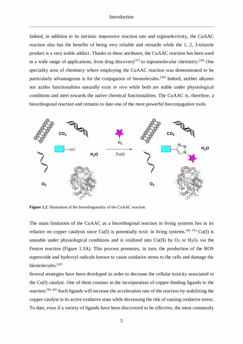
Indeed, in addition to its intrinsic impressive reaction rate and regioselectivity, the CuAAC
reaction also has the benefits of being very reliable and versatile while the 1, 2, 3-triazole
product is a very stable adduct. Thanks to these attributes, the CuAAC reaction has been used
in a wide range of applications, from drug discovery[37] to supramolecular chemistry.[38] One
speciality area of chemistry where employing the CuAAC reaction was demonstrated to be
particularly advantageous is for the conjugation of biomolecules.[39] Indeed, neither alkynes
nor azides functionalities naturally exist in vivo while both are stable under physiological
conditions and inert towards the native chemical functionalities. The CuAAC is, therefore, a
bioorthogonal reaction and remains to date one of the most powerful bioconjugation tools.
Figure 1.2: Illustration of the bioorthogonality of the CuAAC reaction.
The main limitation of the CuAAC as a bioorthogonal reaction in living systems lies in its
reliance on copper catalysis since Cu(I) is potentially toxic in living systems.[40, 41] Cu(I) is
unstable under physiological conditions and is oxidized into Cu(II) by O2 or H2O2 via the
Fenton reaction (Figure 1.3A). This process promotes, in turn, the production of the ROS
superoxide and hydroxyl radicals known to cause oxidative stress to the cells and damage the
biomolecules.[42]
Several strategies have been developed in order to decrease the cellular toxicity associated to
the Cu(I) catalyst. One of them consists in the incorporation of copper-binding ligands to the
reaction.[43, 44] Such ligands will increase the acceleration rate of the reaction by stabilizing the
copper catalyst in its active oxidative state while decreasing the risk of causing oxidative stress.
To date, even if a variety of ligands have been discovered to be effective, the most commonly

Introduction
6
used ones are the ligands from the tris((triazolyl)methyl) amine class: TBTA 10[43], THPTA
11[44], BTTAA 12[45] and TABTA 13[46] (Figure 1.3B).
A second strategy for decreasing the toxic effect of the Cu(I) is to use copper-chelating
azides.[4] Such chelating groups bind the copper directly at the reaction center and are reported
to significantly increase the reaction rate while using lower catalyst concentrations without
compromising the CuAAC efficiency. For example, in a comparative study with the
corresponding non-chelating azide, the picolyl azide group (Figure 1.3C), in combination with
BTTAA, provided a 25-fold enhancement in labelling proteins.[47]
Figure 1.3: A) Cu(I)-promoted generation of ROS; B) Tris((triazolyl)methyl) amine ligands used in the CuAAC
reaction; C) CuAAC reaction with a picolyl azide moiety.
Introduction
7
1.1.3 CuAAC reaction on nucleic acids
Thanks to its fast reaction kinetics and bioorthogonality, the CuAAC found a significant
number of applications in the context of DNA and RNA modifications. Examples of such
applications range from the labelling of oligonucleotides with various fluorescent reporters [48,
49, 50], ligation of several DNA strands[51, 52] or surface functionalization in microarrays [53].
The three components of the nucleotide unit, the (deoxy)ribose sugar unit[54], the nucleobase
and the phosphate group[55], have been subjected to chemical modifications towards the
introduction of click reactive functions. However, the most common type of modifications
towards the generation of clickable nucleotide analogues are located on the nitrogenous base
moiety[3].
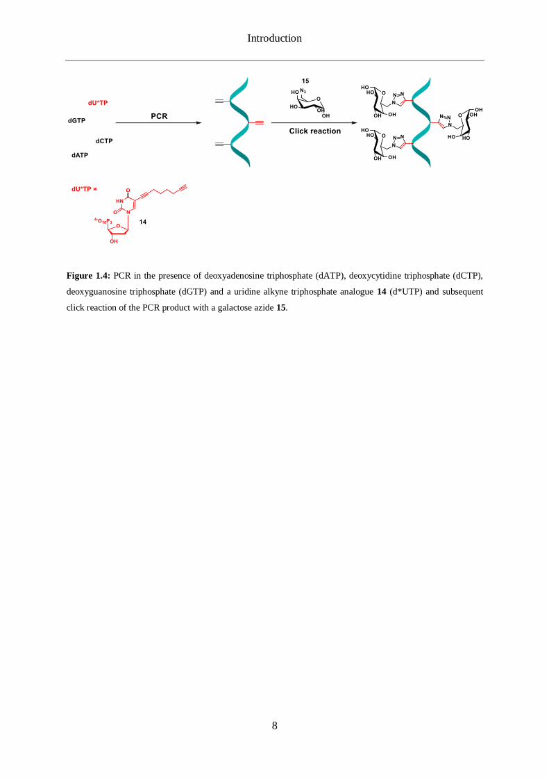
The development of such modified nucleosides as well as the methodologies to efficiently label
DNA by click chemistry were initiated by the Carell Group. Alkyne nucleosides analogues
have been introduced by both automated solid phase synthesis and enzymatic reaction. For the
first approach, the synthesis of an alkyne-uridine phosphoramidite enabled the site-specific
introduction of alkynes functions inside oligonucleotides[56]. These modifications proved to be
compatible with the reaction conditions of solid phase synthesis and the obtained
oligonucleotides could be post-synthetically labelled with a high efficiency. In parallel, the
corresponding alkyne triphosphates were also synthesized and used in polymerase chain
reactions.[57] Not only the modified triphosphates 14 were accepted as substrate for the
enzymatic reactions, but also the subsequent click reaction on the PCR product permitted a
high-density functionalization while no DNA damages were detected. Indeed, RT-PCR
analysis shown that the 887 cytidines of a 2000 base pairs DNA strand had been replaced by
their alkyne-modified analogue. Following click reaction with a sugar azide 15, the DNA
products were digested and analysed by HPLC revealing an impressive and almost quantitative
efficiency as most alkynes had been converted into the click product.
Introduction
8
Figure 1.4: PCR in the presence of deoxyadenosine triphosphate (dATP), deoxycytidine triphosphate (dCTP),
deoxyguanosine triphosphate (dGTP) and a uridine alkyne triphosphate analogue 14 (d*UTP) and subsequent
click reaction of the PCR product with a galactose azide 15.

Introduction
9
1.2 Bioassays based on nucleic acid amplification
DNA and RNA amplification methods represent a great tool for the detection of defined nucleic
acid sequences. Indeed, they provide an excellent specificity thanks to the strong affinity
between two complementary oligonucleotide strands. Moreover, the intrinsic exponential
amplification enables the generation of a large number of identical copies of a target sequence
and therefore a potentially very high detection sensitivity. This is particularly important when
very small amount of material available to analyse. This can be the case in clinical diagnostics
where human samples are being analysed and nucleic acid amplification represents thus a
valuable detection method in many areas.
1.2.1 Real-time Polymerase Chain Reaction
Several strategies have been described towards the amplification of DNA or RNA such as
polymerase chain reaction (PCR)[58, 59], strand displacement amplification[60] or self-sustaining
sequence amplification[61]. PCR was the first of these methods to be developed in the 1980s by
Kary Mullis and remains to date the most commonly used one. A PCR assay is performed
according to the following steps: first, the reaction solution is heated above the melting point
of the complementary DNA strands of the target to be amplified and detected. Once the strands
are separated during this so-called denaturing step, the temperature is decreased to allow the
primers to bind specifically to the target sequence. This is the annealing step, which is followed
by a new increase of temperature enabling the DNA polymerase to extend the primers by
adding the nucleoside triphosphates to the developing DNA strand complementary to the target
sequence. This completes once cycle of PCR, which is repeated numerous times. At the end of
each step, the number of identical copies of the target sequence is doubled and within a few
hours, millions of copies are generated.
Table 1.1: Reagents and equipment required for a PCR assay.
Reagents and equipment Description
Target sequence (or template) Segment of nucleic acid to be amplified
Deoxynucleoside triphosphates Building blocks for the construction of the
PCR products
Introduction
10
Table 1.1: Continued.
The first PCR application was reported in 1985 and performed for the diagnosis of the genetic
disorder sickle cell anemia.[63] The DNA polymerase used was an enzyme isolated from E. coli,
Klenow fragment DNA polymerase I. However, this enzyme was destroyed at the temperature
of the denaturing step and it was necessary to add fresh enzyme for each cycle. An important
development for the PCR assays was the introduction of a thermostable DNA polymerase from
the bacteria Thermus aquaticus (Taq).[64, 65] The latter belongs to a species of bacteria tolerating
high temperatures and its enzymes can survive and sustain their activities accordingly. The Taq
DNA polymerase can survive the incubation temperature of the denaturing step and it is mostly
active at 70 °C. Thus, it circumvented the necessity of adding fresh enzyme at each PCR cycle
making the whole assay easier to perform and more rapid and efficient.
A second important advancement for PCR assays was the demonstration of real-time PCR by
Higuchi et al. at Roche Molecular Systems at the beginning of the 1990s.[66, 67] Indeed, PCR
products were formerly detected and visualized by agarose gel electrophoresis. The
introduction of real-time PCR permitted the coupling of the tremendous intrinsic sensitivity of
PCR to the precision gained from real-time monitoring and detection of amplification products
as they are generated. The initial real-time PCR described relied on the double-stranded DNA
intercalating fluorescent dye ethidium bromide and the reaction was run under UV light. As
the ethidium bromide was intercalated in the increasing amount of DNA produced at each
cycle, it induced an increase of fluorescence upon irradiation of the UV light inside the
thermocycler. Moreover, by measuring the increase of fluorescence after each cycle, it also
allowed the accurate calculation of the initial amount of target DNA. In this regard, the terms
real-time and quantitative PCR are often used in combination or as interchangeable. The first
commercial instrument was made available on the market in 1996 by Applied Biosystems and
it was followed by numerous other companies.[68] The influence of the assay is also reflected
Primer Short DNA strand complementary to a
sequence of the target to be amplified
DNA polymerase Enzyme synthesizing new complementary
copies of the target sequence
Thermocycler
Equipment in which the PCR assay is
performed. Able to raise and lower the
temperature of the samples in precise and
pre-programmed steps.[62]

Introduction
11
in the number of scientific publications citing real-time quantitative PCR as their number went
through an exponential growth over about 10 years following its commercialization.[69]
Since its first iteration by Higuchi et al., many methods for the detection of the PCR product
have been described. They can be divided into two categories depending on whether they detect
only specific or both specific and non-specific amplification products. For the latter category,
double-stranded DNA intercalating agents such as the previously-mentioned ethidium
bromide,[66, 67] SYBRGreen,[70] or EvaGreen are used.[71] Specific probes, however, are based
on oligonucleotides linked with a fluorescent reporter. Here as well, the specificity of such
probes lies in the complementarity between the probe strand and a sequence of the DNA target.
These probes can induce the emission of fluorescence upon hybridization by diverse
mechanism of actions. The different types of probes thus existing can be distinguished as
following: primer-probes acting as a primer with the target sequence; hybridization probes
emitting fluorescence upon hybridization on the target sequence; hydrolysis probes emitting
fluorescence upon degradation of the probe after hybridization to the target sequence;
analogues of nucleic acids with mechanisms of action similar to the previous probes but whose
structure is not a conventional oligonucleotide. The table 1.2 provides examples of the different
types of specific probes.
Table 1.2: Real-time PCR specific probes.
Primer probe – Scorpions[72]
Structure Mechanism of action
Hairpin structure with a reporter at the 5′-end and a
quencher at the 3′-end of the hairpin. The latter is
attached to the 5′-end of the primer by a short PEG
blocker blocking its extension by the polymerase.
When in solution, the reporter fluorescence is
quenched by the proximity with the quencher by
FRET-quenching. Once the primer-probe is
hybridized on the target DNA, the polymerase
amplifies the sequence from the 3′-end of the primer.
During the denaturation step, the specific sequence
of the probe binds to the complementary region
within the same strand of newly amplified DNA.
This hybridisation event opens the hairpin loop so
that fluorescence is no longer quenched.

Introduction
12
Table 1.2: Continued.
Hybridization probe – Molecular Beacon probe[73]
Structure Mechanism of action
Hairpin structure with a reporter at the 5′-end and a
quencher at the 3′-end of the hairpin.
During the annealing phase, this probe unfolds and
binds to the target, emitting fluorescence since the
reporter is not quenched any longer. If the Molecular
Beacon probe and target DNA sequences are not
perfectly complementary, there will be no emission of
fluorescence as the hairpin structure prevails over the
hybridization.
Hydrolysis probe – TaqMan[74]
Structure Mechanism of action
Oligonucleotide with a fluorescent reporter at the 5′-
end and a quencher at its 3′-end.
When in solution, the reporter fluorescence is
quenched by the proximity with the quencher by
FRET-quenching. During the amplification phase, the
probe is hydrolysed by the exonuclease activity of the DNA polymerase. This leads to the departure of the
reporter, which is no longer in proximity of the
quencher and emits a fluorescence signal.
Introduction
13
Table 1.2: Continued.
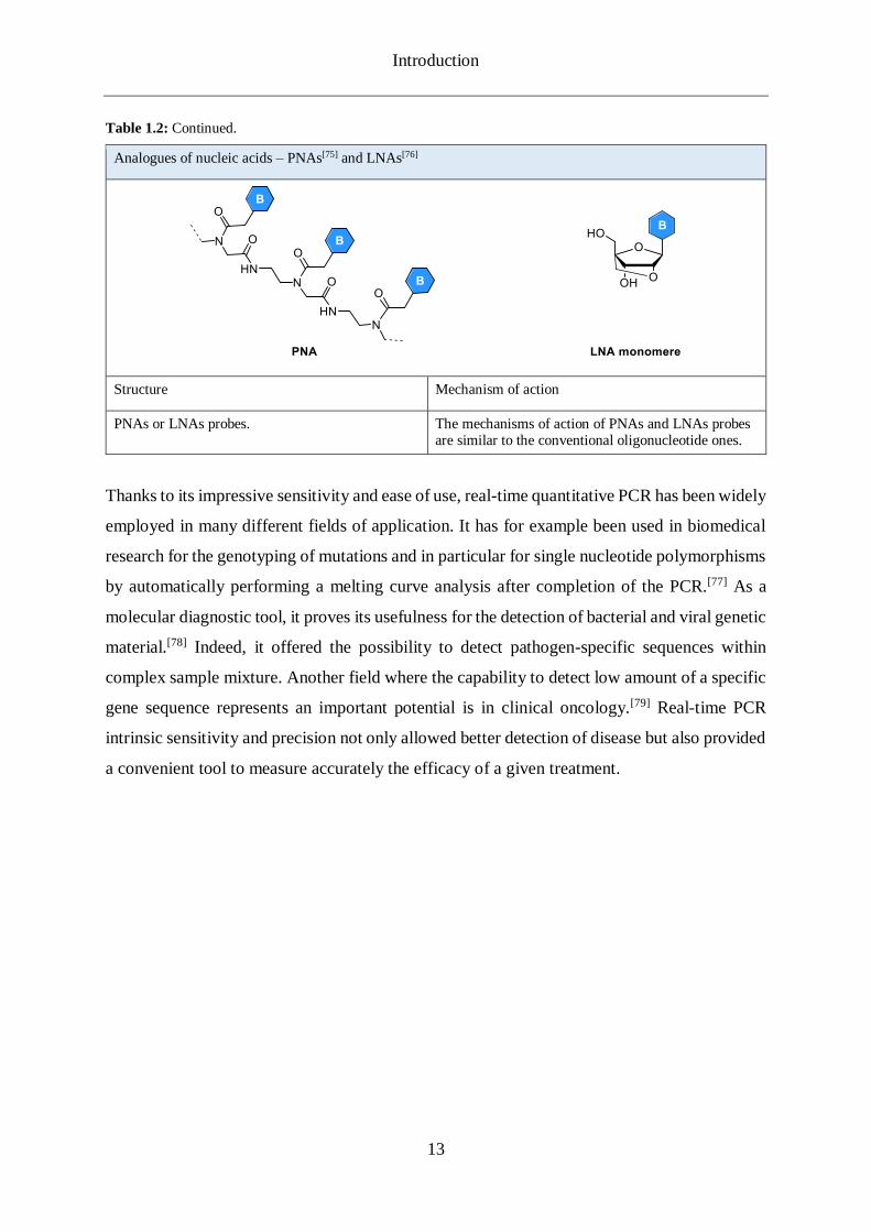
Analogues of nucleic acids – PNAs[75] and LNAs[76]
Structure Mechanism of action
PNAs or LNAs probes. The mechanisms of action of PNAs and LNAs probes are similar to the conventional oligonucleotide ones.
Thanks to its impressive sensitivity and ease of use, real-time quantitative PCR has been widely
employed in many different fields of application. It has for example been used in biomedical
research for the genotyping of mutations and in particular for single nucleotide polymorphisms
by automatically performing a melting curve analysis after completion of the PCR.[77] As a
molecular diagnostic tool, it proves its usefulness for the detection of bacterial and viral genetic
material.[78] Indeed, it offered the possibility to detect pathogen-specific sequences within
complex sample mixture. Another field where the capability to detect low amount of a specific
gene sequence represents an important potential is in clinical oncology.[79] Real-time PCR
intrinsic sensitivity and precision not only allowed better detection of disease but also provided
a convenient tool to measure accurately the efficacy of a given treatment.

Introduction
14
1.2.2 Proximity Ligation Assay
One successful method based on nucleic acid amplification and developed for in vitro detection
and visualisation of proteins is the proximity ligation assay (PLA). PLA was first demonstrated
in 2002 by Landegren et al.[1] While the method has been developed since its introduction and
several variations have been described, the basic principle remains unchanged.[80] The PLA
relies on a pair of affinity probes for a protein target. The latter can be either a single protein
or a protein complex.[81] In the first demonstration of the assay, the proximity probes used were
a pair of DNA aptamers with extended sequences required for the proximity ligation reactions.
Alternatively, an assay based on antibodies as affinity probes has also been developed.[82] In
this case, the antibodies are attached to DNA strands. Once the two probes are close to each
other by binding the same target, an oligonucleotide is hybridized on both probes at the same
time. The two probes will then serve as a guide to promote the formation of circular structure
from the linear oligonucleotide by joining its two extremities in an enzymatic reaction.
Subsequently, one of the DNA strand probes acts as a primer in a rolling circular amplification
(RCA) where the previously ligated oligonucleotide is used as a circular template. RCA is an
alternative amplification method to PCR, which on the contrary is an isothermal process and
does not require a thermocycler. In RCA, the polymerase continuously extends the primer due
to the circularity of the template. Therefore, the amplification product is a long DNA single
strand remaining attached to the primer and with thousands of successive repetitions of the
complementary sequence of the circular template.[83] The amplification product can be detected
by fluorescence in situ hybridization (FISH) with complementary oligonucleotide probes and
its signal can be easily observed with a microscope. Thanks to the extremely low amount of
proteins that the assay is able to detect, the PLA became a valuable technology for a large
variety of proteomic studies.[84]
Introduction
15
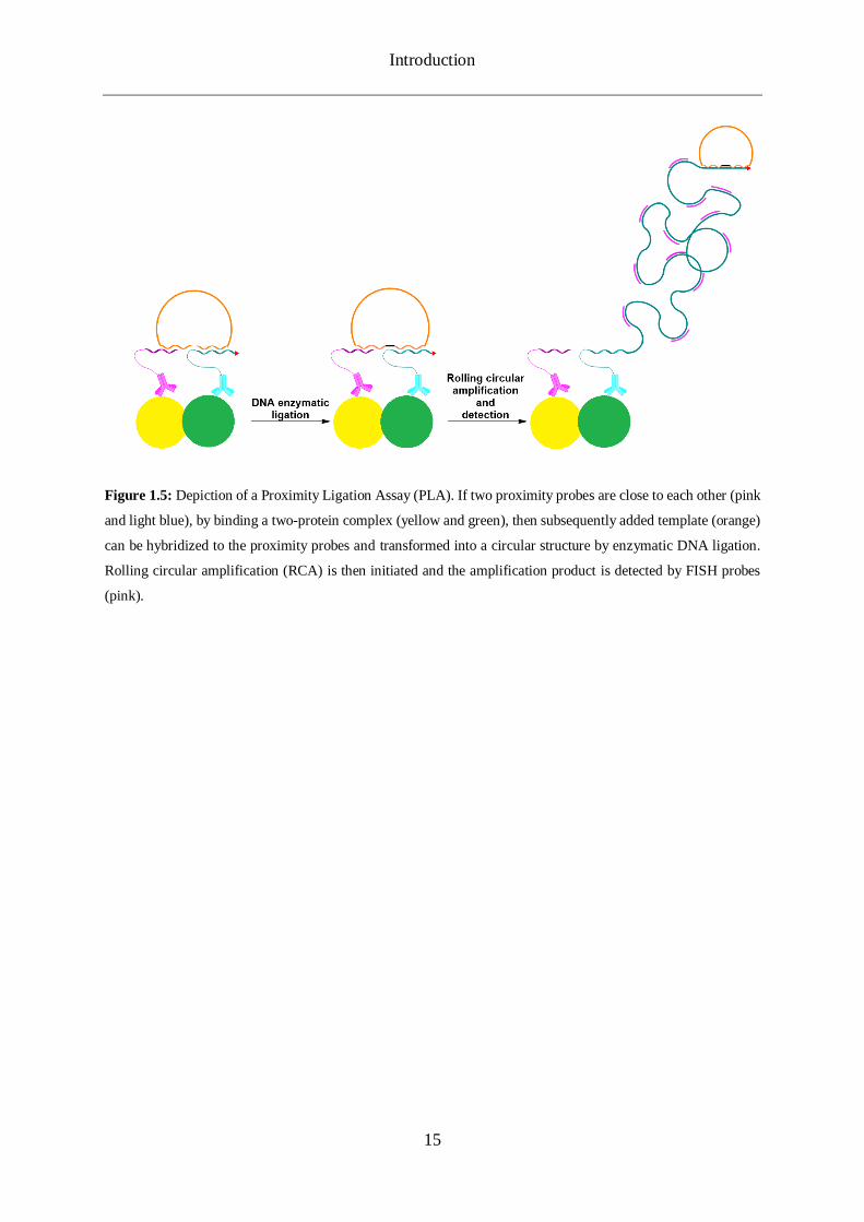
Figure 1.5: Depiction of a Proximity Ligation Assay (PLA). If two proximity probes are close to each other (pink
and light blue), by binding a two-protein complex (yellow and green), then subsequently added template (orange)
can be hybridized to the proximity probes and transformed into a circular structure by enzymatic DNA ligation.
Rolling circular amplification (RCA) is then initiated and the amplification product is detected by FISH probes
(pink).

Part I - Development of a click chemistry-based proximity ligation assay
16
2. DEVELOPMENT OF A CLICK CHEMISTRY-BASED PROXIMITY
LIGATION ASSAY
2.1 Objectives
One of the goal of the present thesis was to investigate if the Cu(I)-catalyzed azide-alkyne
cycloadditon (CuAAC) could be used in the context of a proximity ligation assay in order to
push further the potential of this method. Thanks to its efficiency and its bioorthogonality, we
believed that the so-called click reaction can contribute to the development of a more precise
and sensitive PLA. Towards this purpose, we developed the following design as a proof of
concept: Three oligonucleotide probes, functionalized with alkyne or azide functions, can
hybridize with a single non-modified oligonucleotide template. Thanks to the proximity
brought by the hybridization on the template, the three probes are then further conjugated to
each other by click chemistry providing a single covalently bound product. The template aims
therefore in this design at mimicking the proximity of the PLA to react probes when placed
close to each other. This design also makes use of three probes instead of two in the PLA,
adding thus one degree of precision. We therefore named this assay proof of concept click
Proximity Ligation Assay (CLICKPLA). Two versions of this design have been envisioned
(Figure 2.1).
One, where the middle probe bears two alkyne functions and the side probes one azide function
each. A second one where the middle probe is functionalized with two azide functions and the
side ones with one alkyne function each. This project involved therefore the development of
the corresponding oligonucleotide functionalization chemistry as well as the synthesis of the
linkers required.

Part I - Development of a click chemistry-based proximity ligation assay
17
Figure 2.1: Schematic representation of the two CLICKPLA versions. The oligonucleotide probes are represented
in green. The oligonucleotide templates are in purple. At first, three probes are hybridized to the complementary
template. In a second time, the click reaction can be initiated and the three probes are conjugated to each other.
Part I - Development of a click chemistry-based proximity ligation assay
18
2.2 Synthesis of azide functionalized ODN
2.2.1 Strategy
The first part of this project was to develop a functional and reliable strategy towards the
synthesis of DNA strands modified with one or two azide functions. Even if a few examples of
direct incorporation of azide functions during DNA solid phase synthesis have been reported
[85, 86], they are usually not compatible with with P(III) as they are prone to Staudinger-type
side reactions.[87, 88]. However, for DNA strands, a post oligo synthesis functionalization step
is usually performed to introduce azide functionalities and the most common approach consists
in reacting an activated ester with an amino modified oligonucleotide.[89] Using this strategy,
bifunctional NHS-azide linkers have been reacted with oligonucleotides carrying a modified
amine nucleoside or with a 5’ or 3’ amino modifier.[51, 90] However, this method is not
chemoselective and lacks in efficiency as NHS esters can react with other nucleophiles present
on DNA. Contrary to azide moieties, alkyne ones are easily introduced during solid phase
synthesis of oligonucleotides enabling post-synthetic labelling of the modified strands with
various azide reporters by CuAAC.[48, 91] Thus, an alternative method to generate a DNA strand
with an azide moiety is to click a symmetrical bis-azide linker on an oligonucleotide with an
alkyne modified nucleobase.[92] Here again, the level of selectivity of this method is relative as
the second azide group of the linker might react with another strand yet unreacted thus leading
to a cross-linked side product.[93, 94] In 2009, Zhu et al. discovered that chelating azides can
react in CuAAC reactions with only a small amount of copper (II) acetate (down to 1 mol %)
and in the absence of a reducing agent such as sodium ascorbate.[4] This superior reactivity in
comparison with non-chelating azide is explained by the chelation of the copper catalyst
directly to the catalytic center of the reaction. After testing several auxiliary ligands near the
azido group, they reported the pyridyl group as the best capable copper chelating group.[95] The
reactivity difference between chelating and non-chelating azide is such that under specific
conditions (copper (II) acetate as catalyst, no reducing agent), when both a chelating and a non-
chelating azide are present, only the so-called picolyl azide is going to react with an alkyne.
Based on these results, they designed unsymmetrical bis-azide (one chelating, one non-
chelating) linkers and proved that both azido groups can be reacted in a sequential and
chemoselective procedure with two distinct alkynes.[5] Seela et al. successfully applied this
method on alkyne nucleosides and oligonucleotides with 2,5-bis(azidomethyl)pyridine as
Part I - Development of a click chemistry-based proximity ligation assay
19
unsymmetrical bis-azide linker.[96] We decided to adopt a similar approach tailored for the
CLICKPLA. In our proof of concept design, the alkyne and azide functionalities to be conjugated
with each other needs to be carried at the end of long and flexible linkers. Indeed, these
chemical groups will each be incorporated in different strands and thus, short linkers would not
allow the click reactions to take place as the CuAAC reactive group could not be nearby each
other. Consequently, as a backbone for the linkers to synthesize, we used poly(ethylene-glycol)
(PEG) chains. These polymers, contrary to alkyl chains, have the advantages to be non-rigid
and water-soluble.[97] Also, they are commercially available in a wide range of lengths allowing
us, had it been necessary, to modulate the length of the linkers. As the alkyne component to be
incorporated within the oligonucleotides during the solid-phase synthesis, we chose the C8-
alkyne-dU nucleoside developed in our group.[91] Eventually, our strategy towards the
synthesis of azide functional DNA strands consisted in:
1- Synthesis of oligodeoxyribonucleotides containing one alkyne group;
2- Synthesis of unsymmetrical picolyl azide/azide long and water-soluble linkers;
3- Chemoselective CuAAC conjugation between the alkyne DNA strand and the chelating
azido group of the unsymmetrical bis-azide linker leaving the non-chelating azide free and
functional for further CuAAC.
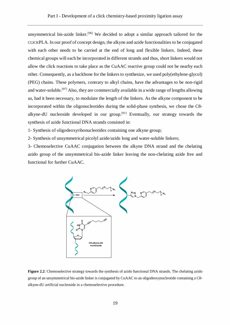
Figure 2.2: Chemoselective strategy towards the synthesis of azido functional DNA strands. The chelating azido
group of an unsymmetrical bis-azide linker is conjugated by CuAAC to an oligodeoxynucleotide containing a C8-
alkyne-dU artificial nucleoside in a chemoselective procedure.

Part I - Development of a click chemistry-based proximity ligation assay
20
2.2.2 Synthesis of unsymmetrical picolyl azide/azide linkers
The initial step in order to synthesize unsymmetrical picolyl azide/azide linkers was to obtain
a picolyl azide building block able to be further conjugated in a compatible and orthogonal
reaction with the azide function. We followed the published procedure from Uttamapinant et
al. with minor adjustments giving access to a carboxylic acid picolyl azid (6-
azidomethylnicotinic acid) 16.[47] The carboxylic acid, upon activation, was further reacted
with the amino group of different molecules via amid coupling. In order to generate an amine
reactive ester from the carboxylic acid we used HATU as a reagent along with Hünig’s base,
this coupling being known to provide high coupling efficiencies and fast rates.[98] The first
unsymmetrical picolyl azide-PEG10-azide linker 17 was readily available in one step from
coupling the carboxylic acid picolyl azid to a commercial azido-PEG-amine reactant. We chose
as starting material an azido-PEG-amine with 10 poly(ethylene oxide) monomer units 18 as we
assumed that it would provide a sufficient long linker for the CLICKPLA.
Scheme 2.1: Synthesis of the unsymmetrical picolyl azide-PEG10-azide linker 17.
In order to prepare a second linker bearing one picolyl azide and two non-chelating azido
groups 19, we imagined a synthesis based on methyl 3,5-dihydroxybenzoate 20 (Scheme 2.2).
This starting building block offers the advantage of having two identical chemical groups (two
hydroxyl groups) as well as a third protected one (carboxylic acid protected as methyl ester)
and thus can be used as a bifunctional core.[99] Therefore, the two hydroxyl groups eventually
bear the two azido groups of the final linker whereas the carboxylic acid was coupled to a
picolyl azide/amine linker 21.

Part I - Development of a click chemistry-based proximity ligation assay
21
Scheme 2.2: Retrosynthetic analysis of picolyl azide/bis-azide linker 19.
In order to functionalize the two hydroxyl groups we synthesized a linker based on
tetraethylene glycol 22 and bearing on its two respective free alcohol moieties an azido group
and a tosylate group 23 (Scheme 2.3). The first hydroxyl group was selectively tosylated by
using tosyl chloride. In order to minimize the formation of the double tosylated unwanted
product, the tosyl chloride was firstly dissolved in tetrahydrofuran and this solution was
subsequently added very carefully (drip in over one hour) to a solution of tetraethylene glycol
at 0 °C. The two reactants were finally in stoichiometric rate and the mono tosylated product
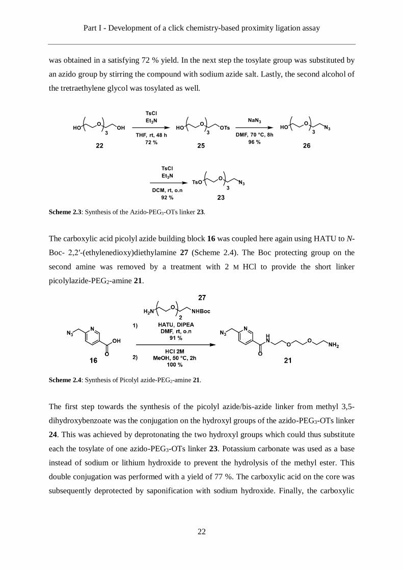
Part I - Development of a click chemistry-based proximity ligation assay
22
was obtained in a satisfying 72 % yield. In the next step the tosylate group was substituted by
an azido group by stirring the compound with sodium azide salt. Lastly, the second alcohol of
the tretraethylene glycol was tosylated as well.
Scheme 2.3: Synthesis of the Azido-PEG3-OTs linker 23.
The carboxylic acid picolyl azide building block 16 was coupled here again using HATU to N-
Boc- 2,2′-(ethylenedioxy)diethylamine 27 (Scheme 2.4). The Boc protecting group on the
second amine was removed by a treatment with 2 M HCl to provide the short linker
picolylazide-PEG2-amine 21.
Scheme 2.4: Synthesis of Picolyl azide-PEG2-amine 21.
The first step towards the synthesis of the picolyl azide/bis-azide linker from methyl 3,5-
dihydroxybenzoate was the conjugation on the hydroxyl groups of the azido-PEG3-OTs linker
24. This was achieved by deprotonating the two hydroxyl groups which could thus substitute
each the tosylate of one azido-PEG3-OTs linker 23. Potassium carbonate was used as a base
instead of sodium or lithium hydroxide to prevent the hydrolysis of the methyl ester. This
double conjugation was performed with a yield of 77 %. The carboxylic acid on the core was
subsequently deprotected by saponification with sodium hydroxide. Finally, the carboxylic

Part I - Development of a click chemistry-based proximity ligation assay
23
acid was activated by HATU and was further coupled to the picolylazide-PEG2-amine linker
to provide the Picolyl azide/bis-azide linker 19.
Scheme 2.5: Synthesis of the picolyl azide/bis-azide linker 19.

Part I - Development of a click chemistry-based proximity ligation assay
24
2.2.3 Click experiments
The ability of the picolyl azide-PEG10-azide linker 17 to be clicked in a chemoselective
protocol was assessed with a 11-mer oligodeoxyribonucleotide containing one C8-alkyne-dU
nucleoside. Several stoichiometric ratios of copper catalyst/alkyne-carrying ODN and linkers/
alkyne-carrying ODN were tested in order to find the best conditions for:
1- Performing the reaction until full completion;
2- Achieve the highest possible chemoselectivity and prevent side reaction on the non-chelating
azido group.
The different conditions tested are listed in Table 2.1. The reactions were performed in parallel,
over the same time and at the same temperature. The final concentration of the DNA strand
was identical in each sample.
Table 2.1: Click screening with the picolyl azide-PEG10-azide linker 17. [*] The percentage of
completion of the reactions were determined by integration of the HPLC trace of the crude product at 260 nm.
Sample
entry Sequence
Stoichiometric
ratio
linker/alkyne
Stoechiometric
ratio
copper/alkyne
Calcd
[M-H]-
Found
[M-H]-
Completion
[%]*
1
5' GGCCGCT-MonoAz-
TTG 3'
10 2
4114.5 4111.1
84.9
2 20 2 97.4
3 50 2 98.8
4 10 5 90.2
5 20 5 97.0
6 50 5 98.8
7 10 10 87.8
8 20 10 96.7
9 50 10 97.6
Part I - Development of a click chemistry-based proximity ligation assay
25
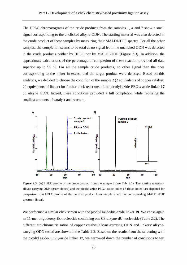
The HPLC chromatograms of the crude products from the samples 1, 4 and 7 show a small
signal corresponding to the unclicked alkyne-ODN. The starting material was also detected in
the crude product of these samples by measuring their MALDI-TOF spectra. For all the other
samples, the completion seems to be total as no signal from the unclicked ODN was detected
in the crude products neither by HPLC nor by MALDI-TOF (Figure 2.3). In addition, the
approximate calculations of the percentage of completion of these reaction provided all data
superior up to 95 %. For all the sample crude products, no other signal than the ones
corresponding to the linker in excess and the target product were detected. Based on this
analytics, we decided to choose the condition of the sample 2 (2 equivalents of copper catalyst;
20 equivalents of linker) for further click reactions of the picolyl azide-PEG10-azide linker 17
on alkyne ODN. Indeed, these conditions provided a full completion while requiring the
smallest amounts of catalyst and reactant.
Figure 2.3: (A) HPLC profile of the crude product from the sample 2 (see Tab. 2.1). The starting materials,
alkyne-carrying ODN (green dotted) and the picolyl azide-PEG10-azide linker 17 (blue dotted) are depicted for
comparison. (B) HPLC profile of the purified product from sample 2 and the corresponding MALDI-TOF
spectrum (inset).
We performed a similar click screen with the picolyl azide/bis-azide linker 19. We chose again
an 11-mer oligodeoxyribonucleotide containing one C8-alkyne-dU nucleoside (Table 2.2). The
different stoichiometric ratios of copper catalyst/alkyne-carrying ODN and linkers/ alkyne-
carrying ODN tested are shown in the Table 2.2. Based on the results from the screening with
the picolyl azide-PEG10-azide linker 17, we narrowed down the number of conditions to test
A B
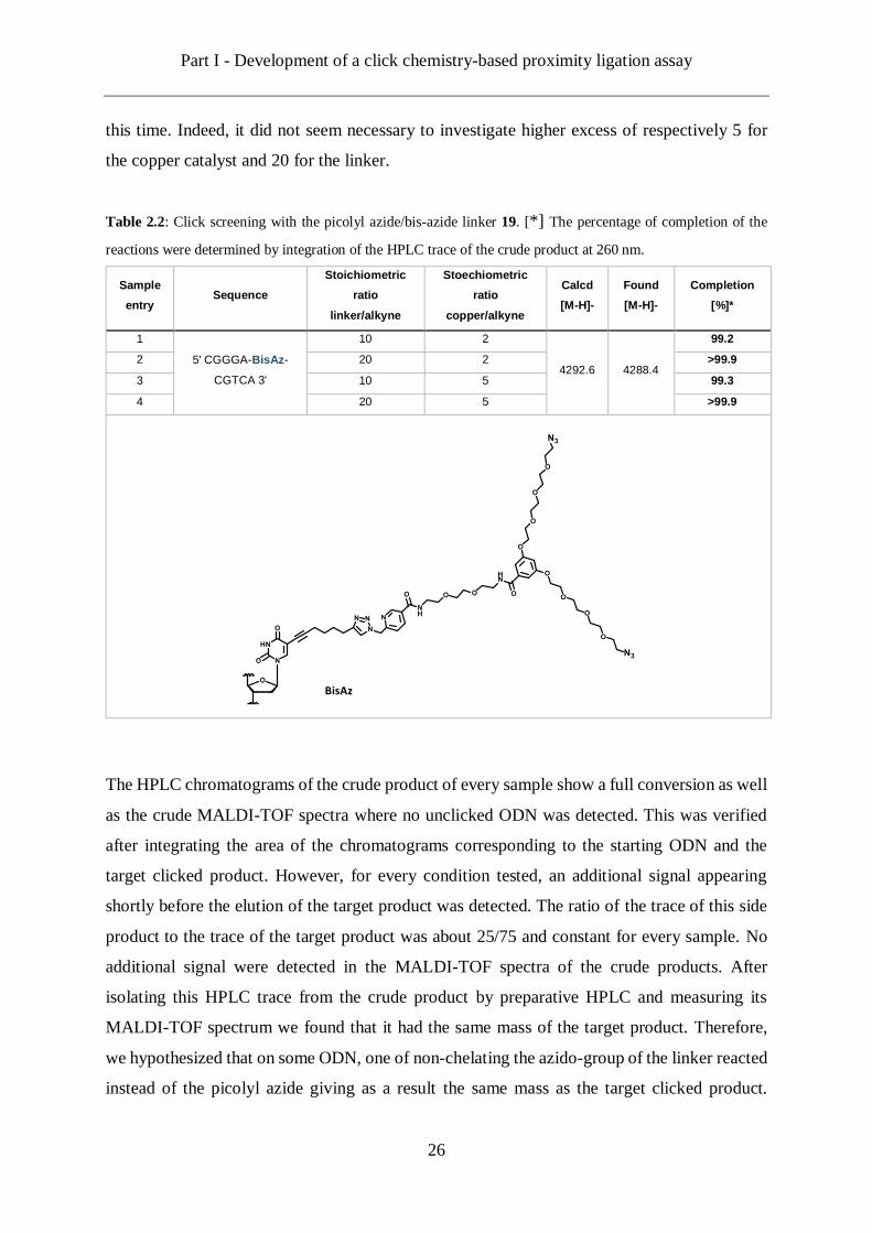
Part I - Development of a click chemistry-based proximity ligation assay
26
this time. Indeed, it did not seem necessary to investigate higher excess of respectively 5 for
the copper catalyst and 20 for the linker.
Table 2.2: Click screening with the picolyl azide/bis-azide linker 19. [*] The percentage of completion of the
reactions were determined by integration of the HPLC trace of the crude product at 260 nm.
Sample
entry Sequence
Stoichiometric
ratio
linker/alkyne
Stoechiometric
ratio
copper/alkyne
Calcd
[M-H]-
Found
[M-H]-
Completion
[%]*
1
5' CGGGA-BisAz-
CGTCA 3'
10 2
4292.6 4288.4
99.2
2 20 2 >99.9
3 10 5 99.3
4 20 5 >99.9
The HPLC chromatograms of the crude product of every sample show a full conversion as well
as the crude MALDI-TOF spectra where no unclicked ODN was detected. This was verified
after integrating the area of the chromatograms corresponding to the starting ODN and the
target clicked product. However, for every condition tested, an additional signal appearing
shortly before the elution of the target product was detected. The ratio of the trace of this side
product to the trace of the target product was about 25/75 and constant for every sample. No
additional signal were detected in the MALDI-TOF spectra of the crude products. After
isolating this HPLC trace from the crude product by preparative HPLC and measuring its
MALDI-TOF spectrum we found that it had the same mass of the target product. Therefore,
we hypothesized that on some ODN, one of non-chelating the azido-group of the linker reacted
instead of the picolyl azide giving as a result the same mass as the target clicked product.
Part I - Development of a click chemistry-based proximity ligation assay
27
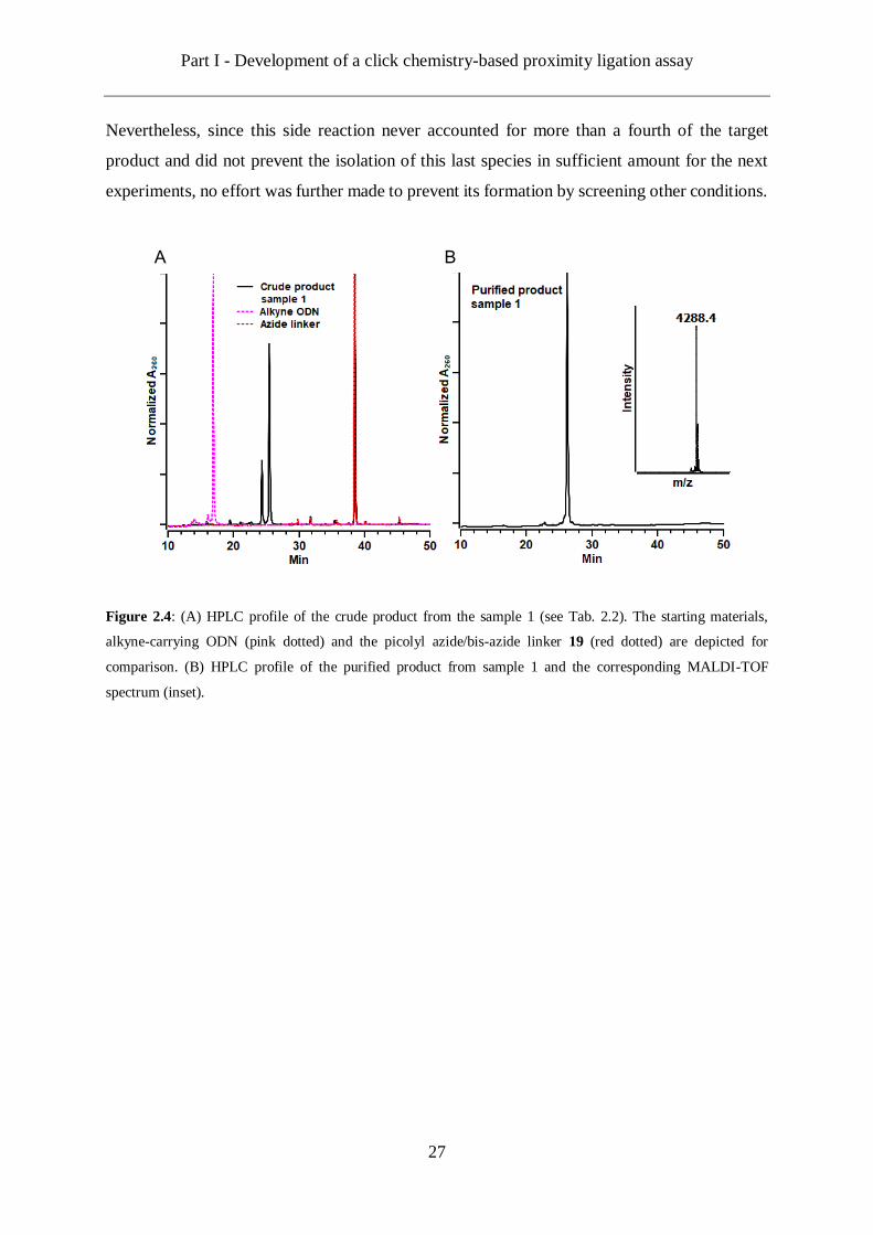
Nevertheless, since this side reaction never accounted for more than a fourth of the target
product and did not prevent the isolation of this last species in sufficient amount for the next
experiments, no effort was further made to prevent its formation by screening other conditions.
Figure 2.4: (A) HPLC profile of the crude product from the sample 1 (see Tab. 2.2). The starting materials,
alkyne-carrying ODN (pink dotted) and the picolyl azide/bis-azide linker 19 (red dotted) are depicted for
comparison. (B) HPLC profile of the purified product from sample 1 and the corresponding MALDI-TOF
spectrum (inset).
A B
Part I - Development of a click chemistry-based proximity ligation assay
28
2.3 Synthesis of alkyne functionalized ODN
2.3.1 Strategy
The next step of this thesis project was to develop a strategy towards the functionalization of
DNA strands with alkyne groups at the end of long spacers, here again for the needs of the
CLICKPLA. As well as for the synthesis of azide functionalized ODN described in the previous
section, we decided to rely on the incorporation of the C8-alkyne-dU nucleoside by solid phase
synthesis.[91] The alkyne group present on the C8-alkyne-dU needing to be extended. We
envisioned a sequential strategy as described by Leigh et al. and previously applied in our group
to successively click different labels on one single oligonucleotide.[48, 100] In this method, a first
alkyne is clicked while a second one remains protected by a silyl group. Upon the first click,
the second alkyne is deprotected and clicked in turn, with another azide label (Figure 2.5).
Therefore, we decided to synthesize bifunctional linkers bearing one azide moiety as well as
one or two alkyne ones. Once again and for the same reasons mentioned previously,
poly(ethylene glycol) chains were chosen as the backbone structures of the linkers to be
synthesized. Considering the very high coupling efficiency demonstrated in the previous
section by the chelating azido group, we designed syntheses towards linkers bearing picolyl
azides rather than non-chelating azides.
Figure 2.5: Sequential strategy towards the synthesis of alkyne functional DNA strands. The chelating azido
group of an unsymmetrical azide/protected alkyne linker is conjugated by CuAAC to an oligodeoxynucleotide
containing a C8-alkyne-dU artificial nucleoside. The alkyne of the linker is be subsequently deprotected.

Part I - Development of a click chemistry-based proximity ligation assay
29
2.3.2 Synthesis of picolyl azide/alkyne-TMS linkers
At first we considered the synthesis of a linker bearing a trimethylsilyl (TMS) protected alkyne.
Indeed this protecting group presents the advantage that it can be removed under mild
conditions.[48] This property of the TMS protecting group to be easily cleaved off also makes
the synthesis of molecules attached to it challenging. Indeed, TMS are unstable towards many
conditions.[101] Thus, once introduced during a synthesis, the protected molecule should not
undergo any harsh synthetic step in order to keep the TMS in place. The most reliable method,
in our opinion, is to introduce the TMS group during the last step of the synthesis of the
molecule of interest.
Scheme 2.6: Synthesis of 5-(Trimethylsilyl)pent-4-ynoic anhydride 28.
Based on a procedure described by Boons et al., we synthesized a symmetric acid anhydride
with two TMS protected alkynes 28 (Scheme 2.6).[102] This molecule can readily react with an
amino group under conditions in which the TMS is stable. The next step was then the synthesis
of a long unsymmetrical PEG linker respectively terminated on its two respective extremities
by a picolyl azide and a free amino group 31 (Scheme 2.7).
Scheme 2.7: Synthesis of the picolyl azide-PEG11-TMS protected alkyne linker 34.
Part I - Development of a click chemistry-based proximity ligation assay
30
The synthesis of such linker was already achieved as described during the section 2.2.2
(Scheme 2.4) with a short linker. Here we substituted the Boc-PEG2-amine 27 by a Boc-PEG11-
amine 32 and applied the same synthetic procedure (Scheme 2.7). First, the PEG linker was
conjugated to the picolyl azide carboxylic acid reactant 16 via an amide coupling. The second
amine of the PEG chain was then deprotected from its Boc group by acid hydrolysis. At last,
the acid anhydride 28 was coupled to the linker 33 here again through an amide linkage
providing the picolyl azide-PEG11-TMS protected alkyne linker 34 in an excellent yield (98
%).
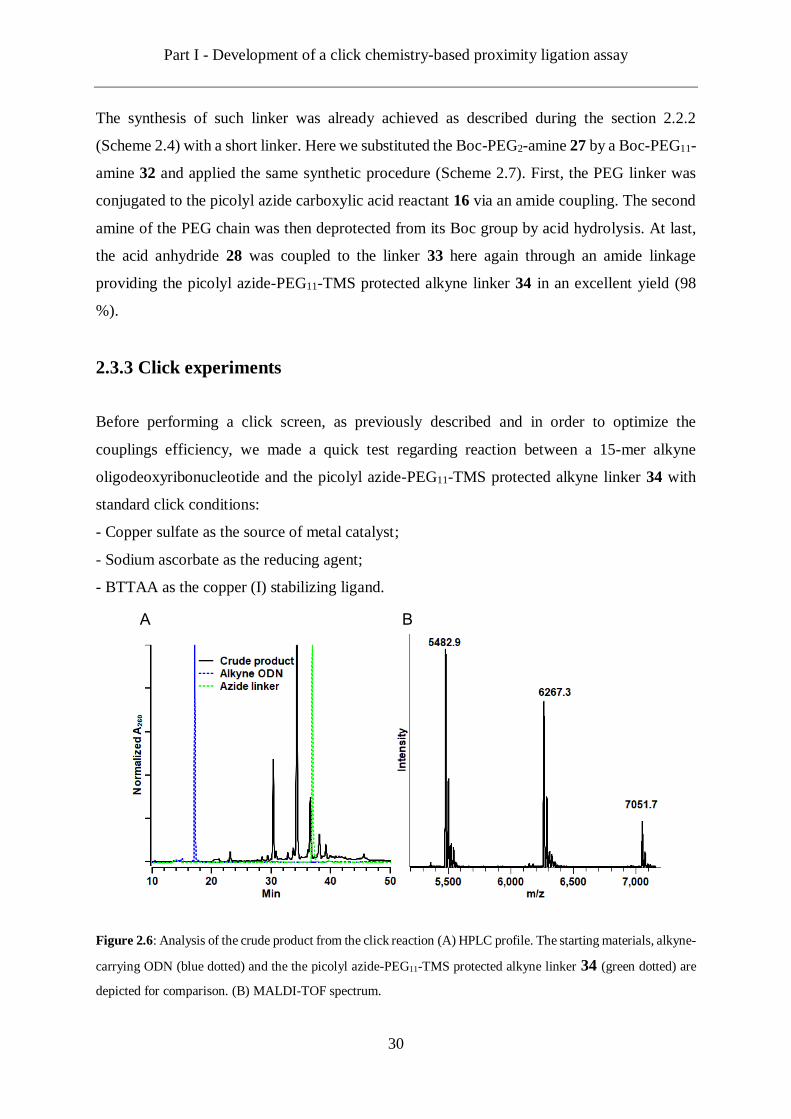
2.3.3 Click experiments
Before performing a click screen, as previously described and in order to optimize the
couplings efficiency, we made a quick test regarding reaction between a 15-mer alkyne
oligodeoxyribonucleotide and the picolyl azide-PEG11-TMS protected alkyne linker 34 with
standard click conditions:
- Copper sulfate as the source of metal catalyst;
- Sodium ascorbate as the reducing agent;
- BTTAA as the copper (I) stabilizing ligand.
Figure 2.6: Analysis of the crude product from the click reaction (A) HPLC profile. The starting materials, alkyne-
carrying ODN (blue dotted) and the the picolyl azide-PEG11-TMS protected alkyne linker 34 (green dotted) are
depicted for comparison. (B) MALDI-TOF spectrum.
A B
Part I - Development of a click chemistry-based proximity ligation assay
31
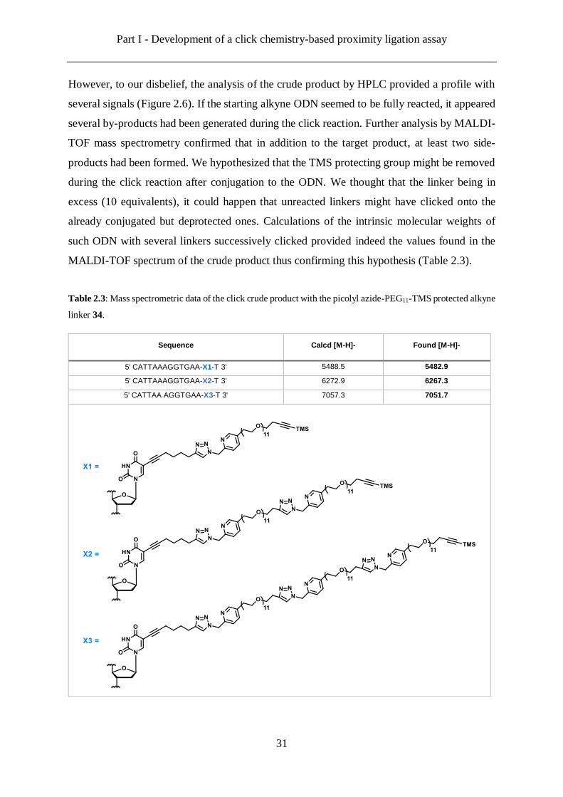
However, to our disbelief, the analysis of the crude product by HPLC provided a profile with
several signals (Figure 2.6). If the starting alkyne ODN seemed to be fully reacted, it appeared
several by-products had been generated during the click reaction. Further analysis by MALDI-
TOF mass spectrometry confirmed that in addition to the target product, at least two side-
products had been formed. We hypothesized that the TMS protecting group might be removed
during the click reaction after conjugation to the ODN. We thought that the linker being in
excess (10 equivalents), it could happen that unreacted linkers might have clicked onto the
already conjugated but deprotected ones. Calculations of the intrinsic molecular weights of
such ODN with several linkers successively clicked provided indeed the values found in the
MALDI-TOF spectrum of the crude product thus confirming this hypothesis (Table 2.3).
Table 2.3: Mass spectrometric data of the click crude product with the picolyl azide-PEG11-TMS protected alkyne
linker 34.
Sequence Calcd [M-H]- Found [M-H]-
5' CATTAAAGGTGAA-X1-T 3' 5488.5 5482.9
5' CATTAAAGGTGAA-X2-T 3' 6272.9 6267.3
5' CATTAA AGGTGAA-X3-T 3' 7057.3 7051.7
Part I - Development of a click chemistry-based proximity ligation assay
32
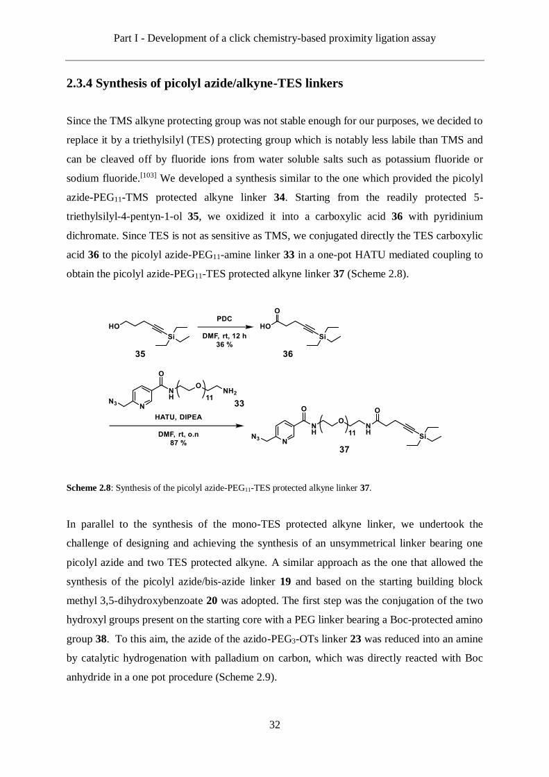
2.3.4 Synthesis of picolyl azide/alkyne-TES linkers
Since the TMS alkyne protecting group was not stable enough for our purposes, we decided to
replace it by a triethylsilyl (TES) protecting group which is notably less labile than TMS and
can be cleaved off by fluoride ions from water soluble salts such as potassium fluoride or
sodium fluoride.[103] We developed a synthesis similar to the one which provided the picolyl
azide-PEG11-TMS protected alkyne linker 34. Starting from the readily protected 5-
triethylsilyl-4-pentyn-1-ol 35, we oxidized it into a carboxylic acid 36 with pyridinium
dichromate. Since TES is not as sensitive as TMS, we conjugated directly the TES carboxylic
acid 36 to the picolyl azide-PEG11-amine linker 33 in a one-pot HATU mediated coupling to
obtain the picolyl azide-PEG11-TES protected alkyne linker 37 (Scheme 2.8).
Scheme 2.8: Synthesis of the picolyl azide-PEG11-TES protected alkyne linker 37.
In parallel to the synthesis of the mono-TES protected alkyne linker, we undertook the
challenge of designing and achieving the synthesis of an unsymmetrical linker bearing one
picolyl azide and two TES protected alkyne. A similar approach as the one that allowed the
synthesis of the picolyl azide/bis-azide linker 19 and based on the starting building block
methyl 3,5-dihydroxybenzoate 20 was adopted. The first step was the conjugation of the two
hydroxyl groups present on the starting core with a PEG linker bearing a Boc-protected amino
group 38. To this aim, the azide of the azido-PEG3-OTs linker 23 was reduced into an amine
by catalytic hydrogenation with palladium on carbon, which was directly reacted with Boc
anhydride in a one pot procedure (Scheme 2.9).

Part I - Development of a click chemistry-based proximity ligation assay
33
Scheme 2.9: Synthesis of the Boc-PEG3-OTs 38.
The two hydroxyl groups of methyl 3,5-dihydroxybenzoate 20 were deprotonated with
potassium carbonate in order to substitute the tosyl group of the linkers 38 (Scheme 2.10). The
double conjugated product was obtained with a 70 % yield. The methyl ester on the core was
hydrolysed with sodium hydroxide. This deprotection proceeded orthogonally and the two Boc
protecting groups remained on the molecule 39. Once free, the carboxylic acid of the core was
activated by the tandem HATU/DIPEA and coupled to the picolylazide-PEG2-amine linker 21.
Eventually, the two Boc protecting group were removed by a hydrochloric acid solution in
dioxane and the two amino groups were reacted in an amide coupling to the TES carboxylic
acid 36. The picolyl azide/bis-TES protected alkyne linker 40 was provided in a quasi-
quantitative yield.
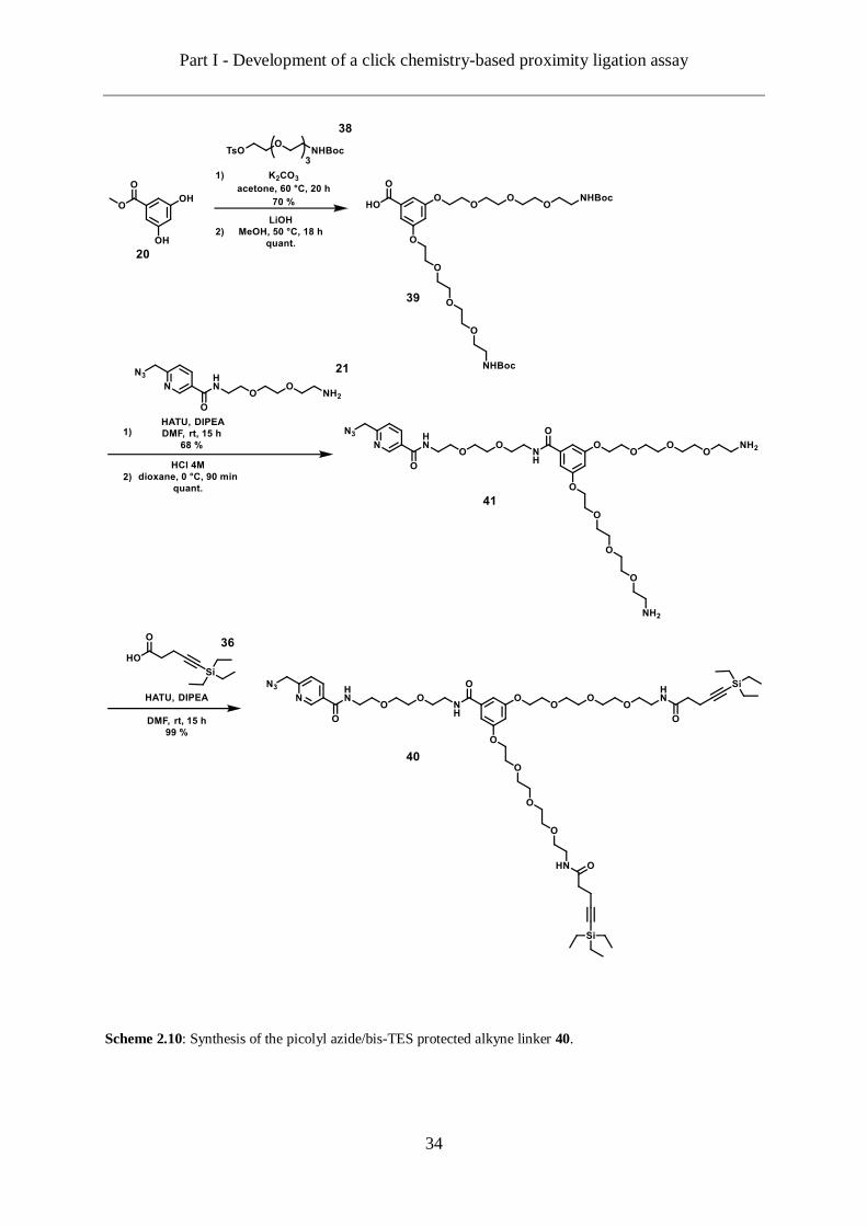
Part I - Development of a click chemistry-based proximity ligation assay
34
Scheme 2.10: Synthesis of the picolyl azide/bis-TES protected alkyne linker 40.
Part I - Development of a click chemistry-based proximity ligation assay
35
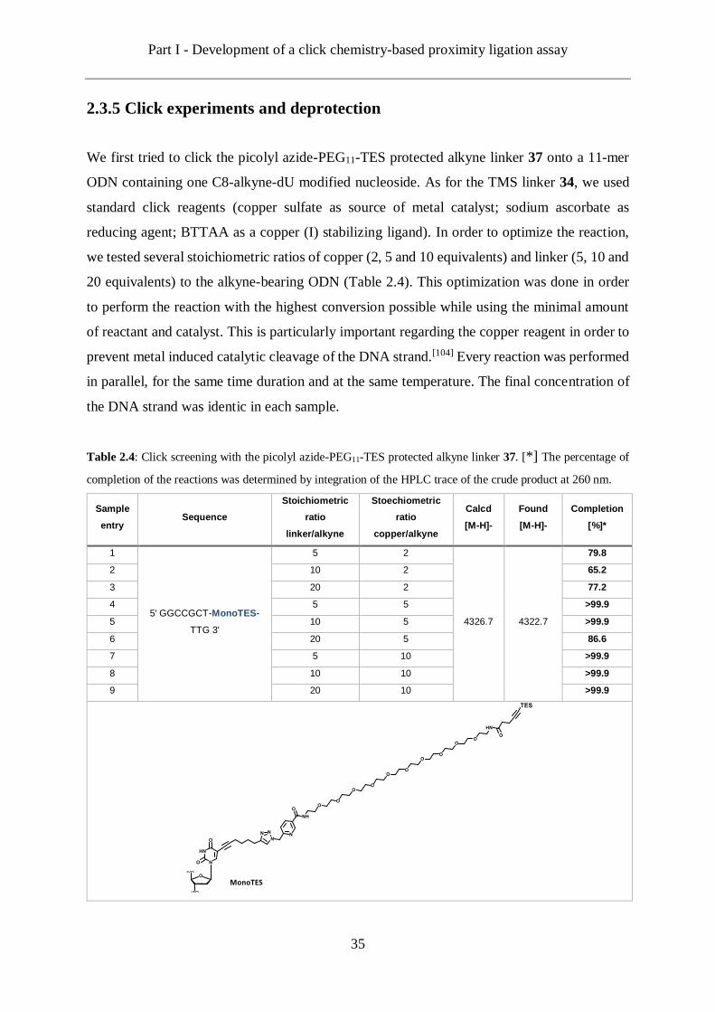
2.3.5 Click experiments and deprotection
We first tried to click the picolyl azide-PEG11-TES protected alkyne linker 37 onto a 11-mer
ODN containing one C8-alkyne-dU modified nucleoside. As for the TMS linker 34, we used
standard click reagents (copper sulfate as source of metal catalyst; sodium ascorbate as
reducing agent; BTTAA as a copper (I) stabilizing ligand). In order to optimize the reaction,
we tested several stoichiometric ratios of copper (2, 5 and 10 equivalents) and linker (5, 10 and
20 equivalents) to the alkyne-bearing ODN (Table 2.4). This optimization was done in order
to perform the reaction with the highest conversion possible while using the minimal amount
of reactant and catalyst. This is particularly important regarding the copper reagent in order to
prevent metal induced catalytic cleavage of the DNA strand.[104] Every reaction was performed
in parallel, for the same time duration and at the same temperature. The final concentration of
the DNA strand was identic in each sample.
Table 2.4: Click screening with the picolyl azide-PEG11-TES protected alkyne linker 37. [*] The percentage of
completion of the reactions was determined by integration of the HPLC trace of the crude product at 260 nm.
Sample
entry Sequence
Stoichiometric
ratio
linker/alkyne
Stoechiometric
ratio
copper/alkyne
Calcd
[M-H]-
Found
[M-H]-
Completion
[%]*
1
5' GGCCGCT-MonoTES-
TTG 3'
5 2
4326.7 4322.7
79.8
2 10 2 65.2
3 20 2 77.2
4 5 5 >99.9
5 10 5 >99.9
6 20 5 86.6
7 5 10 >99.9
8 10 10 >99.9
9 20 10 >99.9

Part I - Development of a click chemistry-based proximity ligation assay
36
From 5 equivalents of copper to the alkyne ODN, almost every reaction proceeded in a
quantitative manner with percentages of conversion higher than 95 %. In this regard, the
stoichiometric rates of the sample 4 were chosen as the references since they were the one using
the least amount of linker (5 equivalents as well). While the HPLC profile of the crude products
show some additional signals than the one corresponding to the target product, this last one
was nevertheless easily isolated and purified by preparative HPLC (Figure 2.7).
Figure 2.7: (A) HPLC profile of the crude product from the sample 4 (see Tab. 2.4). The starting materials,
alkyne-carrying ODN (red dotted) and the the picolyl azide-PEG11-TES protected alkyne linker 37 (green dotted)
are depicted for comparison. (B) HPLC profile of the purified product from sample 4 and the corresponding
MALDI-TOF spectrum (inset).
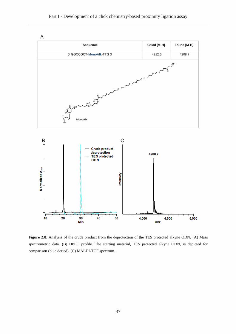
Once purified, the DNA strand clicked with the picolyl azide-PEG11-TES protected alkyne
linker 37 was subsequently treated with a sodium fluoride aqueous solution in order to free the
alkyne from its protecting group. The deprotection was completed in 48 hours at 40 °C and
provided the deprotected ODN as the only product of the reaction (Figure 2.8). This was
confirmed by analysing the crude product by MALDI-TOF mass spectrometry. The spectrum
obtained, shows only one signal corresponding to the target product. The HPLC profile of the
crude product showing a quite clean product, we decided that this procedure does not require
an additional purification by preparative HPLC. The functionalized ODN was only desalted on
a centrifugal membrane filter to get rid of the salts from the deprotection reaction before further
experiments.
A B
Part I - Development of a click chemistry-based proximity ligation assay
37
Sequence Calcd [M-H]- Found [M-H]-
5' GGCCGCT-MonoAlk-TTG 3' 4212.6 4208.7
Figure 2.8: Analysis of the crude product from the deprotection of the TES protected alkyne ODN. (A) Mass
spectrometric data. (B) HPLC profile. The starting material, TES protected alkyne ODN, is depicted for
comparison (blue dotted). (C) MALDI-TOF spectrum.
B C
A

Part I - Development of a click chemistry-based proximity ligation assay
38
We subsequently investigated the clicking ability of the picolyl azide/bis-TES protected alkyne
linker 40. As previously, the linker was clicked to a 11-mer ODN containing one C8-alkyne-
dU modified nucleoside with standard click reagents (copper sulfate as source of metal catalyst;
sodium ascorbate as reducing agent; BTTAA as a copper (I) stabilizing ligand). For the
screening of this linker we kept a stoichiometric ratio of copper (I) to alkyne ODN constant
and equal to 10 equivalents for all the samples. Only the effect of the ratio of linker (from 5 to
50 equivalents) on the reaction was studied in this test (Table 2.5). Again, every reaction was
performed in parallel, for the same time duration and at the same temperature. The final
concentration of DNA strand was also identical in each sample.
Table 2.5: Click screening with the picolyl azide/bis-TES protected alkyne linker 40. [*] The percentage of
completion of the reactions were determined by integration of the HPLC trace of the crude product at 260 nm.
Sample
entry Sequence
Stoichiometric
ratio
linker/alkyne
Stoechiometric
ratio
copper/alkyne
Calcd
[M-H]-
Found
[M-H]-
Completion
[%]*
1
5' CGGGA-BisTES-CGTCA
3'
5 10
4628.9 4627.0
2.9
2 10 10 18.9
3 20 10 33.1
4 50 10 45.2
Unfortunately, the coupling efficiency of this linker upon click reaction remained relatively
low despite the higher stoichiometric ratios tried. The highest percentage of conversion
obtained was of 45 % and it required 50 equivalents of linker. Indeed, analysis of the HPLC
profile of the crude product shows that most of the alkyne ODN remained unclicked. (Figure
2.9). Likewise, if the MALDI-TOF spectrum of the crude product enables the detection of the
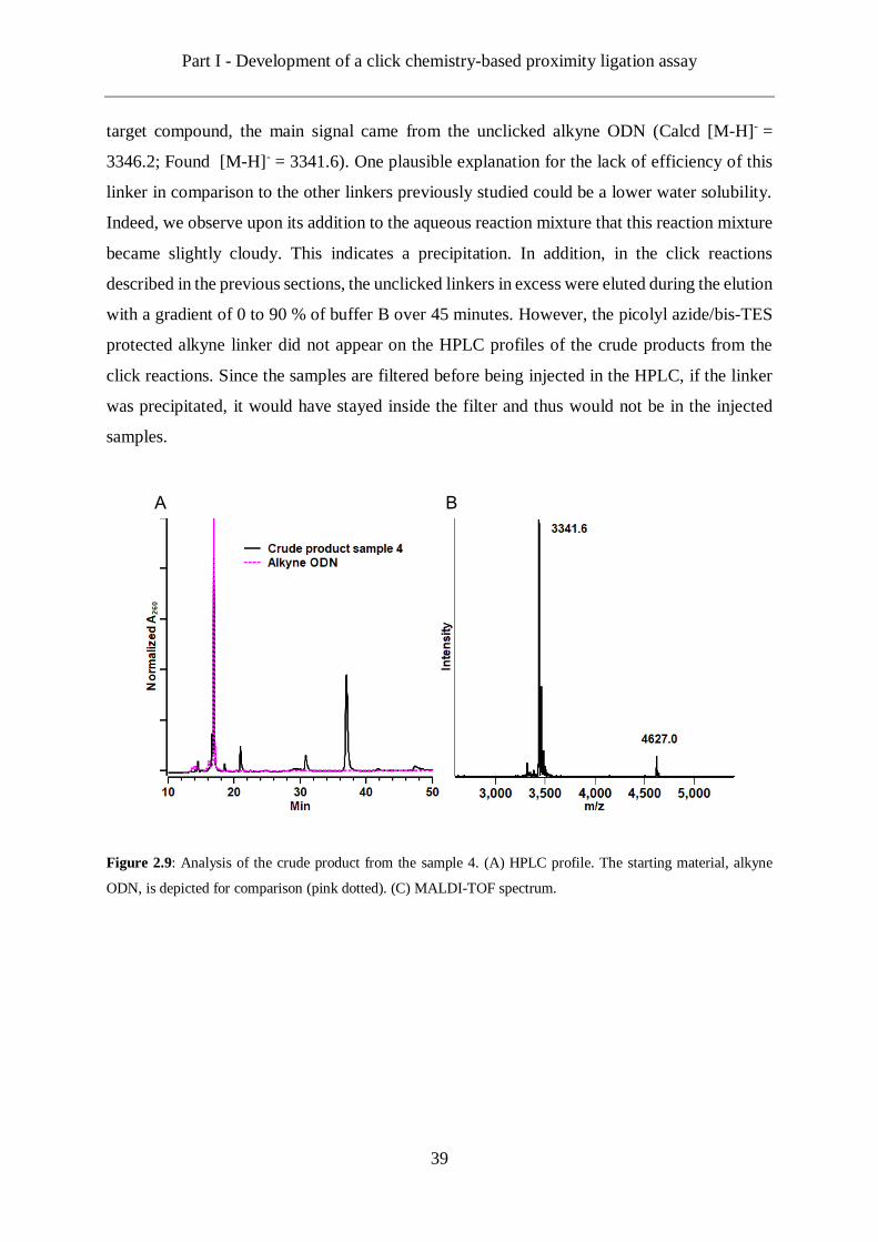
Part I - Development of a click chemistry-based proximity ligation assay
39
target compound, the main signal came from the unclicked alkyne ODN (Calcd [M-H]- =
3346.2; Found [M-H]- = 3341.6). One plausible explanation for the lack of efficiency of this
linker in comparison to the other linkers previously studied could be a lower water solubility.
Indeed, we observe upon its addition to the aqueous reaction mixture that this reaction mixture
became slightly cloudy. This indicates a precipitation. In addition, in the click reactions
described in the previous sections, the unclicked linkers in excess were eluted during the elution
with a gradient of 0 to 90 % of buffer B over 45 minutes. However, the picolyl azide/bis-TES
protected alkyne linker did not appear on the HPLC profiles of the crude products from the
click reactions. Since the samples are filtered before being injected in the HPLC, if the linker
was precipitated, it would have stayed inside the filter and thus would not be in the injected
samples.
Figure 2.9: Analysis of the crude product from the sample 4. (A) HPLC profile. The starting material, alkyne
ODN, is depicted for comparison (pink dotted). (C) MALDI-TOF spectrum.
A B

Part I - Development of a click chemistry-based proximity ligation assay
40
2.4 ODN design and synthesis
2.4.1 Design parameters
As described in the objectives section, the CLICKPLA features four oligodeoxyribonucleotides:
one long template and three smaller strands. The first step of the CLICKPLA being the
simultaneous hybridization of the three probes on the template, we had to design
complementary DNA strands with optimal hybridization potentials. In this regard, several
parameters such as the length of the oligonucleotides needed to be optimal.[105] Too short
probes would result in unspecific hybridization. Too long probes might require longer
hybridization time. Too long probes could also increase the risk of having complementary
regions within the probes themselves, which could result in the formation of hairpin structures
inhibiting the hybridization with the target strand. The base composition of the oligonucleotides
should be about 40 to 60 % of G-C to keep the duplex more stable. Eventually, sequences with
long stretches of a same base should be avoided. The step following the hybridization of the
three probes on their template is to conjugate them by click chemistry. In order to make this
conjugation likely to take place and since the modifications bearing the clickable chemical
functions will be remoted from each other, the modifications need to be smartly placed within
each probes. The double helix formed by the hybridization of DNA strands is a three-
dimensional structure with a pitch of 10.5 base pairs.[21] Therefore, it has been shown that
coupling across one complete turn of the double helix is possible since the modifications will
be placed on the same side of the helix (Figure 2.10).[106, 107]
Figure 2.10: Coupling across the DNA double helix.

Part I - Development of a click chemistry-based proximity ligation assay
41
2.4.2 ODN synthesized
Based on the parameters previously described, we designed two sets of oligonucleotides for
the CLICKPLA with each set including three functionalized probes and one template strand
(Figure 2.11). We chose 11-mer probes for the first set of oligonucleotides (ODN-1, ODN-2
and ODN-3) and a 39 base pairs long complementary template (ODN-4). For the second set,
three 15-mer probes were designed (ODN-5, ODN-6 and ODN-7) and a corresponding
template strand of 45-mer (ODN-8). The sequences of the template were taken from the single
stranded plasmid M13mp18. The incorporation site of the C8-alkyne-dU nucleoside within the
sequence of each probe was chosen in order to have an 8 bp distance until the next modification
once the probes hybridized in order to increase the chances of conjugation between the probes.
Figure 2.11: Sequences of the two sets of DNA strands designed for the CLICKPLA. The base N (in red) is the
modified nucleoside.
In the objectives part of this chapter, two versions of the CLICKPLA had been envisioned. In
the first one, the two side probes would each carry an azide function and the middle probe
would bear two alkynes. In the second version, the side probes would carry the alkyne functions
whereas the middle probe would be the azides bearing one. Considering the coupling
efficiencies of the different linkers synthesized, we decided to focus on the latter one (Figure
2.12).
ODN2
ODN1 3' CGACGACNCCCACTGCNAGGGCGTTNTCGCCGG 5‘ ODN3
5' TTCGCTGCTGAGGGTGACGATCCCGCAAAAGCGGCCTTT 3'
ODN4
ODN6
ODN5 3' AGGGTTTACCGAGNTCAGCCACNGCCACTATNAAGTGGAAATTAC 5‘ ODN7 5' TCCCAAATGGCTCAAGTCGGTGACGGTGATAATTCACCTTTAATG 3'
ODN8

Part I - Development of a click chemistry-based proximity ligation assay
42
Figure 2.12: Schematic representation of the CLICKPLA version selected. The oligonucleotide probes are
represented in green. The oligonucleotide templates is in purple. At first, three probes are hybridized to the
complementary template. In a second time, the click reaction between the alkyne side probes and azide middle
probe can be initiated and the three probes are conjugated to each other.
Indeed, both the picolyl azide-PEG11-TES protected alkyne linker 37 and the picolyl azide/bis-
azide linker 19 provided high coupling efficiencies upon click reaction with alkyne-modified
oligonucleotides. Therefore, we decided to functionalize the side side probes of each set
(respectively ODN-1and ODN-3 for the first set and ODN-5 and ODN-7 for the second set)
with the picolyl azide-PEG11-TES protected alkyne linker 37. The middle probes were
functionalized with the picolyl azide/bis-azide linker 19 in order to bear two azides. All the
oligonucleotides synthesized and functionalized for both sets are recapitulated in the following
table (Table 2.6)
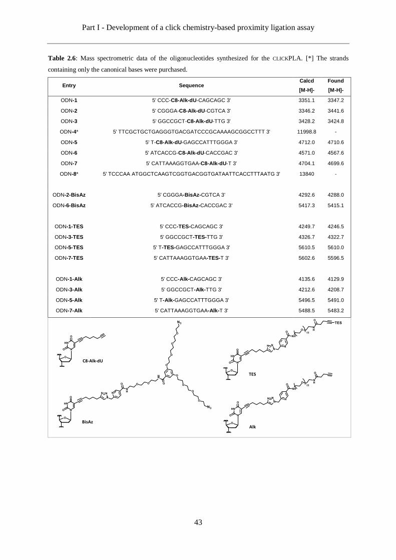
Part I - Development of a click chemistry-based proximity ligation assay
43
Table 2.6: Mass spectrometric data of the oligonucleotides synthesized for the CLICKPLA. [*] The strands
containing only the canonical bases were purchased.
Entry Sequence Calcd
[M-H]-
Found
[M-H]-
ODN-1 5' CCC-C8-Alk-dU-CAGCAGC 3' 3351.1 3347.2
ODN-2 5' CGGGA-C8-Alk-dU-CGTCA 3' 3346.2 3441.6
ODN-3 5' GGCCGCT-C8-Alk-dU-TTG 3' 3428.2 3424.8
ODN-4* 5' TTCGCTGCTGAGGGTGACGATCCCGCAAAAGCGGCCTTT 3' 11998.8 -
ODN-5 5' T-C8-Alk-dU-GAGCCATTTGGGA 3' 4712.0 4710.6
ODN-6 5' ATCACCG-C8-Alk-dU-CACCGAC 3' 4571.0 4567.6
ODN-7 5' CATTAAAGGTGAA-C8-Alk-dU-T 3' 4704.1 4699.6
ODN-8* 5' TCCCAA ATGGCTCAAGTCGGTGACGGTGATAATTCACCTTTAATG 3' 13840 -
ODN-2-BisAz 5' CGGGA-BisAz-CGTCA 3' 4292.6 4288.0
ODN-6-BisAz 5' ATCACCG-BisAz-CACCGAC 3' 5417.3 5415.1
ODN-1-TES 5' CCC-TES-CAGCAGC 3' 4249.7 4246.5
ODN-3-TES 5' GGCCGCT-TES-TTG 3' 4326.7 4322.7
ODN-5-TES 5' T-TES-GAGCCATTTGGGA 3' 5610.5 5610.0
ODN-7-TES 5' CATTAAAGGTGAA-TES-T 3' 5602.6 5596.5
ODN-1-Alk 5' CCC-Alk-CAGCAGC 3' 4135.6 4129.9
ODN-3-Alk 5' GGCCGCT-Alk-TTG 3' 4212.6 4208.7
ODN-5-Alk 5' T-Alk-GAGCCATTTGGGA 3' 5496.5 5491.0
ODN-7-Alk 5' CATTAAAGGTGAA-Alk-T 3' 5488.5 5483.2
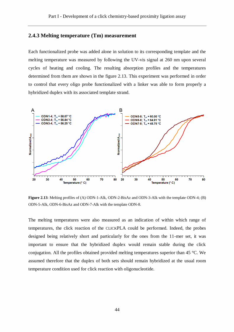
Part I - Development of a click chemistry-based proximity ligation assay
44
2.4.3 Melting temperature (Tm) measurement
Each functionalized probe was added alone in solution to its corresponding template and the
melting temperature was measured by following the UV-vis signal at 260 nm upon several
cycles of heating and cooling. The resulting absorption profiles and the temperatures
determined from them are shown in the figure 2.13. This experiment was performed in order
to control that every oligo probe functionalized with a linker was able to form properly a
hybridized duplex with its associated template strand.
Figure 2.13: Melting profiles of (A) ODN-1-Alk, ODN-2-BisAz and ODN-3-Alk with the template ODN-4; (B)
ODN-5-Alk, ODN-6-BisAz and ODN-7-Alk with the template ODN-8.
The melting temperatures were also measured as an indication of within which range of
temperatures, the click reaction of the CLICKPLA could be performed. Indeed, the probes
designed being relatively short and particularly for the ones from the 11-mer set, it was
important to ensure that the hybridized duplex would remain stable during the click
conjugation. All the profiles obtained provided melting temperatures superior than 45 °C. We
assumed therefore that the duplex of both sets should remain hybridized at the usual room
temperature condition used for click reaction with oligonucleotide.
A B

Part I - Development of a click chemistry-based proximity ligation assay
45
2.4.4 Click test with dyes
In order to test that the synthesized and functionalized oligonucleotides are indeed functional
towards further click reactions, we conjugated them with organic dyes. Thus, coumarin azide
41 and fluorescein alkyne 42 were clicked respectively on the strands bearing the alkyne linker
and the strands bearing the bis-azide linker. This test had no other purpose than assessing the
formation of the clicked adduct. We therefore investigated the crude reaction products only by
MALDI-TOF mass spectrometry (Table 2.7). Indeed, every functional oligonucleotide, only
the target product was detected confirming so the expected reactivity of the probes synthesized
for the CLICKPLA.
Table 2.7: Mass spectrometric data of the oligonucleotides synthesized for the CLICKPLA.
Entry Sequence Calcd
[M-H]-
Found
[M-H]-
ODN-1 Coumarine 5' CCC-AlkCoumarin-CAGCAGC 3' 4338.6 4334.5
ODN-3-Coumarine 5' GGCCGCT-AlkCoumarin-TTG 3' 4415.6 4411.3
ODN-5-Coumarine 5' T-AlkCoumarin-GAGCCATTTGGGA 3' 5699.5 5695.3
ODN-7-Coumarine 5' CATTAAAGGTGAA-AlkCoumarin-T 3' 5691.5 5686.5
ODN-2-BisFluoresceine 5' CGGGA-BisAzFluoresceine-CGTCA 3' 5118.8 5116.4
ODN-6-BisFluoresceine 5' ATCACCG-BisAzFluoresceine-CACCGAC 3' 6243.5 6239.6
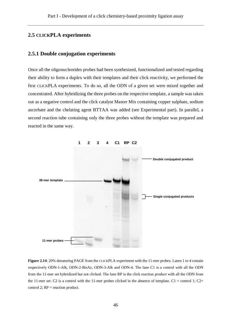
Part I - Development of a click chemistry-based proximity ligation assay
46
2.5 CLICKPLA experiments
2.5.1 Double conjugation experiments
Once all the oligonucleotides probes had been synthesized, functionalized and tested regarding
their ability to form a duplex with their templates and their click reactivity, we performed the
first CLICKPLA experiments. To do so, all the ODN of a given set were mixed together and
concentrated. After hybridizing the three probes on the respective template, a sample was taken
out as a negative control and the click catalyst Master Mix containing copper sulphate, sodium
ascorbate and the chelating agent BTTAA was added (see Experimental part). In parallel, a
second reaction tube containing only the three probes without the template was prepared and
reacted in the same way.
Figure 2.14: 20% denaturing PAGE from the CLICKPLA experiment with the 11-mer probes. Lanes 1 to 4 contain
respectively ODN-1-Alk, ODN-2-BisAz, ODN-3-Alk and ODN-4. The lane C1 is a control with all the ODN
from the 11-mer set hybridized but not clicked. The lane RP is the click reaction product with all the ODN from
the 11-mer set. C2 is a control with the 11-mer probes clicked in the absence of template. C1 = control 1; C2=
control 2; RP = reaction product.
11-mer probes
39-mer template
Single conjugated products
Double conjugated product
1 2 3 4 C1 RP C2

Part I - Development of a click chemistry-based proximity ligation assay
47
The click reactions were analysed by denaturing urea polyacrylamide gel electrophoresis.
Figure 2.13 shows the results for the 11-mer set consisting of ODN-1-Alk, ODN-2-BisAz,
ODN3-Alk and ODN-4. Lane C1 contains the sample that was taken out of the reaction tube
containing all the ODN of the set prior to the addition of the click catalyst Master Mix. As no
click reaction took place and since the reaction products were analysed on a denaturing gel, we
can expectedly observe the three probes and the template separated from each other at their
respective running distances as verified by the individual strands applied in lanes 1-4. In the
lane RP, which corresponds to the complete 11-mer set after the click, we can again observe a
band from the template ODN-4. However, the bands corresponding to the three probes have
significantly faded and three new bands are visible instead. The first two new bands have
similar electrophoretic mobilities, which are slightly higher than the 39-mer template and lower
than the ones of the 11-mer probes. Therefore, we assumed that these two bands correspond to
the single conjugated products of ODN-1-Alk/ODN-2-BisAz and ODN-3-Alk/ODN-2-BisAz.
More interestingly, the third new band in the reaction product lane has an even lower mobility
than the template indicating a higher size. The modified ODNs being bulkier than normal DNA,
we expected that their conjugate would run significantly slower on the gel. Thus, we assigned
this band as the target double conjugated product of the three probes and thus that the template
assisted click conjugation between the three functionalized oligonucleotides was successful.
Eventually, the lane C2 shows the product of the control reaction in the absence of template.
While the bands assigned to the single conjugated products and more important the one
assigned to the target double conjugated product can still be observed, their intensity is lower,
even though approximately the same amount of material was loaded in each lane. This indicates
that although the double click reaction can happen in the absence of template and within the
conditions employed here ([ODN] = 10 µM; reaction time: 3 hours at room temperature), the
presence of a template provides a major enhancement to the efficiency of the reaction.
The same experiments were also performed with the 15-mer set of probes consisting of ODN-
5-Alk, ODN-6-BisAz, ODN-7-Alk and ODN-8. The analysis of these samples using
denaturing PAGE is shown in Figure 2.15.
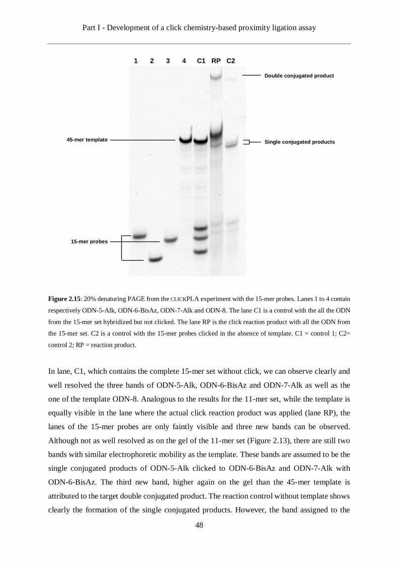
Part I - Development of a click chemistry-based proximity ligation assay
48
Figure 2.15: 20% denaturing PAGE from the CLICKPLA experiment with the 15-mer probes. Lanes 1 to 4 contain
respectively ODN-5-Alk, ODN-6-BisAz, ODN-7-Alk and ODN-8. The lane C1 is a control with the all the ODN
from the 15-mer set hybridized but not clicked. The lane RP is the click reaction product with all the ODN from
the 15-mer set. C2 is a control with the 15-mer probes clicked in the absence of template. C1 = control 1; C2=
control 2; RP = reaction product.
In lane, C1, which contains the complete 15-mer set without click, we can observe clearly and
well resolved the three bands of ODN-5-Alk, ODN-6-BisAz and ODN-7-Alk as well as the
one of the template ODN-8. Analogous to the results for the 11-mer set, while the template is
equally visible in the lane where the actual click reaction product was applied (lane RP), the
lanes of the 15-mer probes are only faintly visible and three new bands can be observed.
Although not as well resolved as on the gel of the 11-mer set (Figure 2.13), there are still two
bands with similar electrophoretic mobility as the template. These bands are assumed to be the
single conjugated products of ODN-5-Alk clicked to ODN-6-BisAz and ODN-7-Alk with
ODN-6-BisAz. The third new band, higher again on the gel than the 45-mer template is
attributed to the target double conjugated product. The reaction control without template shows
clearly the formation of the single conjugated products. However, the band assigned to the
15-mer probes
45-mer template Single conjugated products
Double conjugated product
1 2 3 4 C1 RP C2
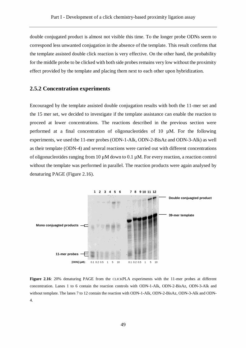
Part I - Development of a click chemistry-based proximity ligation assay
49
double conjugated product is almost not visible this time. To the longer probe ODNs seem to
correspond less unwanted conjugation in the absence of the template. This result confirms that
the template assisted double click reaction is very effective. On the other hand, the probability
for the middle probe to be clicked with both side probes remains very low without the proximity
effect provided by the template and placing them next to each other upon hybridization.
2.5.2 Concentration experiments
Encouraged by the template assisted double conjugation results with both the 11-mer set and
the 15 mer set, we decided to investigate if the template assistance can enable the reaction to
proceed at lower concentrations. The reactions described in the previous section were
performed at a final concentration of oligonucleotides of 10 µM. For the following
experiments, we used the 11-mer probes (ODN-1-Alk, ODN-2-BisAz and ODN-3-Alk) as well
as their template (ODN-4) and several reactions were carried out with different concentrations
of oligonucleotides ranging from 10 µM down to 0.1 µM. For every reaction, a reaction control
without the template was performed in parallel. The reaction products were again analysed by
denaturing PAGE (Figure 2.16).
Figure 2.16: 20% denaturing PAGE from the CLICKPLA experiments with the 11-mer probes at different
concentration. Lanes 1 to 6 contain the reaction controls with ODN-1-Alk, ODN-2-BisAz, ODN-3-Alk and
without template. The lanes 7 to 12 contain the reaction with ODN-1-Alk, ODN-2-BisAz, ODN-3-Alk and ODN-
4.
11-mer probes
39-mer template
Mono conjuagted products
Double conjuagted product
1 2 3 4 5 6 7 8 9 10 11 12
[ODN] (µM): 0.1 0.2 0.5 1 5 10 10 5 1 0.5 0.1 0.2

Part I - Development of a click chemistry-based proximity ligation assay
50
In lanes 11 and 12, which correspond to the reactions performed at 5 and 10 µM respectively,
we can observe a very strong band for the target double conjugated product. At lower
concentration than 5 µM, this band can still be observed but its intensity decreases along with
the concentration. The same is observed for the reaction controls, although as expected the
target double conjugated product band is less strong than the one corresponding to the reactions
in the presence of template.
2.5.3 Reaction time experiments
After testing different final concentrations of oligonucleotides, we envisioned to perform the
double click conjugation with different durations. Indeed, so far every reaction performed had
been ran for 3 hours. Here, we decided to test whether the reaction could be achieved within
15, 30 or 60 minutes. In addition, we decided to use a different reaction temperature for these
tests by heating the reaction at 40 °C whereas the previous reactions had been performed room
temperature. As shown in section 2.4.3, all the ODN probes have a melting temperature higher
than 45 °C. Therefore, we assumed that a reaction at 40 °C should not cause stability issue to
the duplex formed by the probes with the template. On the other hand, we were curious to test
if slightly increasing the reaction temperature while running the reaction for shorter times could
prevent the formation of the double conjugated product in the absence of template. For these
experiments, we used the 15-mer probes (ODN-5-Alk, ODN-6-BisAz and ODN-7-Alk) and
the corresponding template (ODN-8). The reactions were ran in parallel and stopped at
different intervals. In order to stop the reactions at defined times, the reaction mixtures were
diluted in water by a factor 100 and directly desalted on a centrifuge membrane filter in order
to wash the copper catalyst away. For every reaction, a control was performed simultaneously
without the template. The ODN concentration was the same in every reaction tube and equal
to 10 µM. The denaturing PAGE obtained from analysis of these samples is shown in figure
2.17. In the lane 4 to 6, where the reaction mixtures in presence of template were applied, we
can observe an intense band for the double conjugated product. Quite impressively, it seems
that the double click reaction is already effective after 15 minutes. This is even more striking
considering the low concentration at which the reactions took place. Moreover, in the lanes
where the products from the reaction controls without template were applied, we could barely
observe, if at all, the formation of the double conjugated product. On the other hand, the
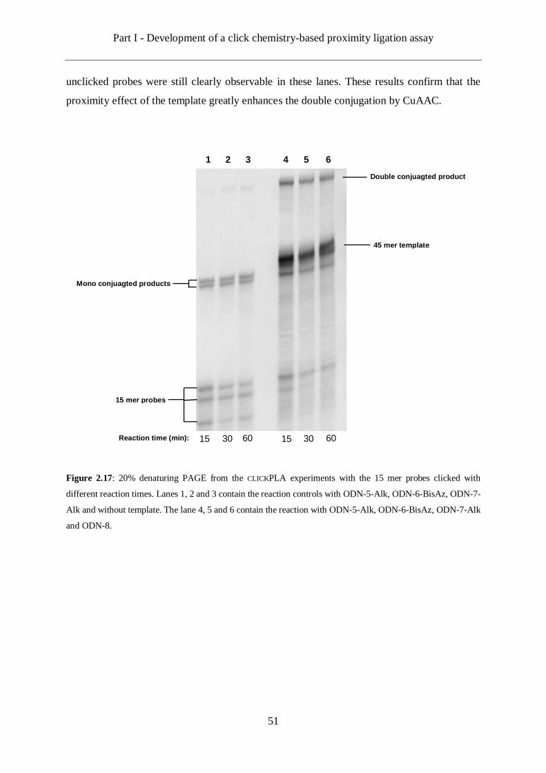
Part I - Development of a click chemistry-based proximity ligation assay
51
unclicked probes were still clearly observable in these lanes. These results confirm that the
proximity effect of the template greatly enhances the double conjugation by CuAAC.
Figure 2.17: 20% denaturing PAGE from the CLICKPLA experiments with the 15 mer probes clicked with
different reaction times. Lanes 1, 2 and 3 contain the reaction controls with ODN-5-Alk, ODN-6-BisAz, ODN-7-
Alk and without template. The lane 4, 5 and 6 contain the reaction with ODN-5-Alk, ODN-6-BisAz, ODN-7-Alk
and ODN-8.
15 mer probes
45 mer template
Mono conjuagted products
Double conjuagted product
1 2 3 4 5 6
15 30 60 60 30 15 Reaction time (min):
Part I - Development of a click chemistry-based proximity ligation assay
52
2.6 Conclusions and Outlook
Proximity ligation assay is a very sensitive method developed for the detection of proteins and
based on DNA amplification.[1] Since the original proof of concept reported by Landegren et
al. in 2002, the PLA and variation of the original concept have been employed in a wide range
of applications.[80]
The project presented in this chapter aimed at investigating if the CuAAC reaction could in
theory enhance the existing PLA methods. As a proof of concept, we designed a model where
three click functional oligonucleotide probes are hybridized next to each other on a single
longer oligonucleotide. The latter one, acting as a template, would enable the probes to be
hybridized next to each other and facilitate further conjugation by click chemistry.
The first part of the project was to synthesize linkers enabling the desired functionalization of
oligonucleotides. At first, we synthesized two unsymmetrical linkers with respectively one
picolyl azide/one non-chelating azide and one picolyl azide/two non-chelating azides. These
linkers could be chemoselectively clicked on alkyne modified DNA strand thanks to the picolyl
azide and thus leaving the non-chelating azide(s) free for subsequent reactions. Next, we
synthesized unssymetrical linkers with a picolyl azide and one or two protected alkyne groups.
After clicking these linkers to alkyne modified DNA strands thanks to their picolyl azide
moiety, the alkyne of the linker was freed from its protecting group rendering it functional
towards further click reactions.
The strategies thus developed enabled the synthesis of two sets of oligonucleotide probes with
11- and 15- mer strands respectively. Each set featured three probes, two single alkyne
functionalized side probes and one double azide middle probe as well as a complementary
oligonucleotide template. Both sets of probes were hybridized on their respective template and
conjugated to each other by click reaction. Analysis by denaturing PAGE showed the
successful formation of the double conjugated product with both sets of probes. Moreover, we
demonstrated that the proximity effect created by the hybridization of the probes on their
template was an essential component to the double conjugation between the three probes.
In conclusion, an oligonucleotide template was used to mimic the proximity effect found in
PLAs. After hybridization on this template, three beforehand functionalized oligonucleotide
probes were conjugated to each other by click chemistry. This model based on three probes
could be applied in a Triple Proximity Ligation Assay in the future as following. After
recognition of a three-protein complex by three proximity probes, three click functional
Part I - Development of a click chemistry-based proximity ligation assay
53
oligonucleotides are added and hybridized to the oligonucleotides on the proximity probes.
After washing the non-hybridized clickable oligonucleotides, addition of a copper catalyst
solution will result in the formation of a double conjugated product between the three
hybridized proximity probes. This double conjugated product will then serve as a template in
its turn for the circularization of three linear oligonucleotide connectors by enzymatic ligation.
In theory, the circular strand thus generated could be formed only in the presence of the double
conjugated product which itself accounts for the presence of the protein (or protein complex)
of interest. One of the extremities of the double conjugated product will serve as a primer and
the circular strand will be amplified by RCA upon addition of a DNA polymerase. The
amplification product will eventually be detected by fluorescence in situ hybridization (FISH)
with complementary fluorescence-labelled oligonucleotides.

Part I - Development of a click chemistry-based proximity ligation assay
54
Scheme 2.11: Presentation of a triple proximity ligation assay based on click chemistry. If three proximity probes
are close to each other (pink, blue, red), by binding a same three-protein complex (green, yellow, bright blue),
then subsequently added click functional ODN probes (green) can be hybridized to the proximity probes and
conjugated to each others by CuAAC. The double conjugated product can further act as a template to linear ODN
connector (orange) to form a circular structure covalently joined by enzymatic DNA ligation. RCA is then initiated
by using the double conjugated product as a primer. The RCA product is detected by FISH.
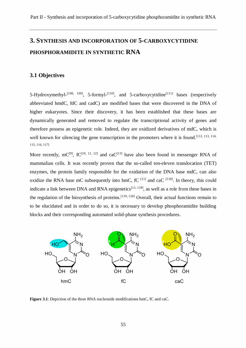
Part II - Synthesis and incorporation of 5-carboxycytidine phosphoramidite in synthetic RNA
55
3. SYNTHESIS AND INCORPORATION OF 5-CARBOXYCYTIDINE
PHOSPHORAMIDITE IN SYNTHETIC RNA
3.1 Objectives
5-Hydroxymethyl-[108, 109], 5-formyl-[110], and 5-carboxycytidine[111] bases (respectively
abbreviated hmdC, fdC and cadC) are modified bases that were discovered in the DNA of
higher eukaryotes. Since their discovery, it has been established that these bases are
dynamically generated and removed to regulate the transcriptional activity of genes and
therefore possess an epigenetic role. Indeed, they are oxidized derivatives of mdC, which is
well known for silencing the gene transcription in the promoters where it is found.[112, 113, 114,
115, 116, 117]
More recently, mC[9], fC[10, 11, 12] and caC[13] have also been found in messenger RNA of
mammalian cells. It was recently proven that the so-called ten-eleven translocation (TET)
enzymes, the protein family responsible for the oxidation of the DNA base mdC, can also
oxidize the RNA base mC subsequently into hmC, fC [11] and caC [118]. In theory, this could
indicate a link between DNA and RNA epigenetics[11, 118], as well as a role from these bases in
the regulation of the biosynthesis of proteins.[119, 120] Overall, their actual functions remain to
to be elucidated and in order to do so, it is necessary to develop phosphoramidite building
blocks and their corresponding automated solid-phase synthesis procedures.
Figure 3.1: Depiction of the three RNA nucleoside modifications hmC, fC and caC.

Part II - Synthesis and incorporation of 5-carboxycytidine phosphoramidite in synthetic RNA
56
Indeed, this would provide access to synthetic RNA strands with one or several of these
modifications, which could in turn be used in studies aiming at deciphering the functional
consequences of hmC, fC and caC in RNA. Phosphoramidite building blocks for hmC and fC
have already been reported by respectively Micura[121, 122] and Deiters[123]. However, these
building blocks require specific conditions for their incorporation into RNA strands. Therefore,
phosphoramidite building blocks for hmC, fC as well as caC able to be incorporated with
standard solid-phase RNA synthesis conditions still needed to be developed. Moreover, such
building blocks would enable the incorporation of the three modifications in a one single strand.
The goal of this project was therefore the development of a synthesis route towards a caC RNA
phosphoramdite building block compatible with standard RNA solid-phase synthesis. In
parallel to the synthesis of the caC building block, described in the present chapter, the
synthesis routes towards a fC RNA phosphoramidite building block as well as a hmC one were
developed by co-members of the Carell Group, Dr. Nobuhiro Tago and Dr. Iacovos
Michaelides. In a joint effort, we further aimed at incorporating all the three C modification
phosphoramidite building blocks in a single synthetic RNA strand.

Part II - Synthesis and incorporation of 5-carboxycytidine phosphoramidite in synthetic RNA
57
3.2 Design and protecting group strategy
In order to synthesize a caC RNA phosphoramidite, we needed to develop an alternative
protecting group strategy to the one employed for the corresponding DNA building block.
The RNA molecule is intrinsically more sensitive than the DNA one due to the 2’-OH on the
ribose. The latter, upon deprotonation, can attack as a nucleophile the adjacent phosphorous in
the phosphodiester bond leading to the cleavage of the RNA backbone. Because of this
chemical instability, silyl groups usually protect the 2’-OH RNA phosphoramidite building
blocks and their removal by a fluoride source (commonly with the trimethylamine
trihydrofluoride reagent[124]) is the last treatment that the synthetic RNA strand undergoes. Here
we chose to use a TBS group for the protection of 2’-OH position of the ribose. For the
protection of the exocyclic amine N4, we decided to use a p-methoxybenzoyl group, which can
be easily removed at the same time as the cleavage of the strands from the solid support during
the ammonolysis step. The carboxylic acid on the position 5 of the caC DNA phosphoramidite
is generally protected by alkyl ester groups such as trifluoroethyl ester[125] or methyl ester[126].
Once the building block is incorporated inside the synthetic strand, these protecting groups are
removed by hydrolysis with aqueous NaOH instead of ammonolysis, which could result in the
formation a 5-carboxamide side product. However, we assumed that such harsh condition are
not compatible with RNA synthesis. The silyl protecting group of the 2’-OH on the ribose
could be removed as well during this treatment, resulting in the cleavage of the synthetic RNA
strand. Therefore, we chose instead to protect the carboxylic acid group with a p-
nitrophenylethylester group. The latter can be cleaved in milder condition using DBU in THF
at room temperature prior to the ammonolysis and thus preventing the formation of a 5-
carboxyamide side product. The caC RNA phosphoramidite 42 thus envisioned is depicted in
scheme 3.1.

Part II - Synthesis and incorporation of 5-carboxycytidine phosphoramidite in synthetic RNA
58
Scheme 3.1: Structure of the caC RNA phosphoramidite building block 42 designed.
After the incorporation of one or several caC phosphoramidite by automatic solid phase
synthesis, we imagined the deprotection of the different moieties according to the following
sequence:
1- Phosphodiester bonds;
2- 5-carboxilic acid;
3- N4-amine deprotection // cleavage from the solid support;
4- 2’-OH deprotection.
A non-nucleophilic base could simultaneously remove the cyanoethyl group and the p-
nitrophenylethylester group, respectively protecting the phosphodiester bond and the 5-
carboxilic acid. However, in order to avoid potential Michael-type addition, the 5-carboxilic
acid was deprotected after the phosphodiester linkage. Indeed the deprotection of the latter
generates acrylonitrile, which could add itself to the base of the nucleoside, and the Michael-
System in the nucleobase will be stronger once the carboxylic acid unmasked.[127, 128, 129, 130, 131,
132]
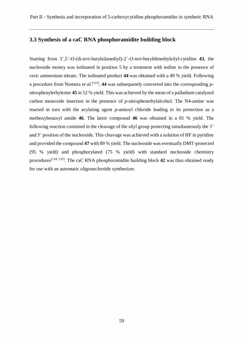
Part II - Synthesis and incorporation of 5-carboxycytidine phosphoramidite in synthetic RNA
59
3.3 Synthesis of a caC RNA phosphoramidite building block
Starting from 3´,5´-O-(di-tert-butylsilanediyl)-2´-O-tert-butyldimethylsilyl-cytidine 43, the
nucleoside moiety was iodinated in position 5 by a treatment with iodine in the presence of
ceric ammonium nitrate. The iodinated product 44 was obtained with a 49 % yield. Following
a procedure from Nomura et al.[133], 44 was subsequently converted into the corresponding p-
nitrophenylethylester 45 in 52 % yield. This was achieved by the mean of a palladium catalyzed
carbon monoxide insertion in the presence of p-nitrophenethylalcohol. The N4-amine was
reacted in turn with the acylating agent p-anisoyl chloride leading to its protection as a
methoxybenzoyl amide 46. The latter compound 46 was obtained in a 81 % yield. The
following reaction consisted in the cleavage of the silyl group protecting simultaneously the 3’
and 5’ position of the nucleoside. This cleavage was achieved with a solution of HF in pyridine
and provided the compound 47 with 89 % yield. The nucleoside was eventually DMT-protected
(95 % yield) and phosphorylated (75 % yield) with standard nucleoside chemistry
procedures[134, 135]. The caC RNA phosphoramidite building block 42 was thus obtained ready
for use with an automatic oligonucleotide synthesizer.
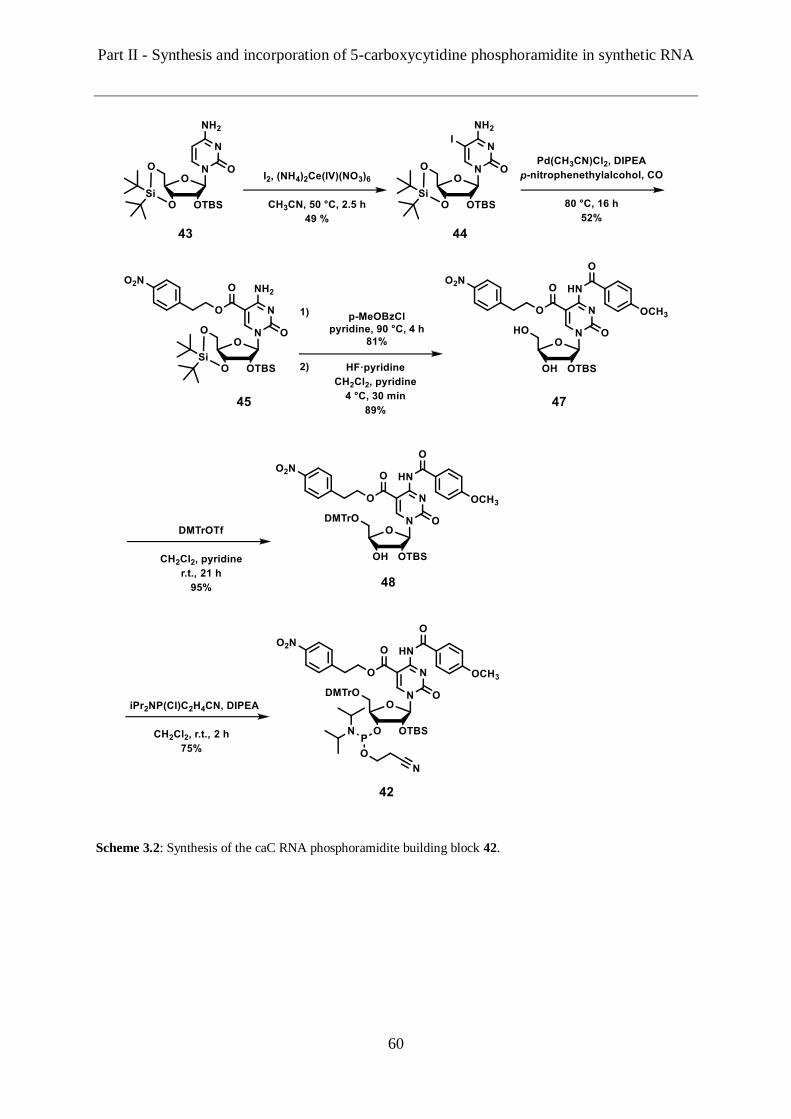
Part II - Synthesis and incorporation of 5-carboxycytidine phosphoramidite in synthetic RNA
60
Scheme 3.2: Synthesis of the caC RNA phosphoramidite building block 42.

Part II - Synthesis and incorporation of 5-carboxycytidine phosphoramidite in synthetic RNA
61
3.4 Solid phase synthesis of RNA strands containing caC
Following the synthesis of the caC RNA phosphoramidite buiding bock 42, we aimed at testing
its compatibility with automated solid phase synthesis and validating the deprotection protocol
designed for this building block. Therefore, we incorporated the caC phosphoramidite
synthesized 42 in two different RNA strands. A short one (13mer) and a long one (21mer)
respectively designated ON-1 and ON-2 (Table 3.1).
Table 3.1: Mass spectrometric data of the oligonucleotides synthesized containing the caC modification.
Entry Sequence Calcd
[M-H]-
Found
[M-H]-
ON-1 5' CCUACcaCGCAUUAC 3' 4061.6 4057.7
ON-2 5' UCUGAGUCCCUAUUAcaCAAGAG 3' 6696.9 6690.9
During the solid phase synthesis, the coupling time of the special caC phosphoramidite 42 was
extended to 20 minutes (instead of 10 minutes for the standard RNA phosphoramidites) in
order to ensure the highest possible coupling yield. According to the deprotection strategy
envisioned in the section 3.2, once the synthesis achieved, the solid support was treated with
DBU in acetonitrile for 1 to 3 minutes to cleave the cyanoethyl group protecting the
phosphodiester bound. This solution was then replaced by one with DBU in THF and the
deprotection of the carboxyl group was achieved after 2 hours. The oligonucleotides were
further treated with standard deprotection conditions to remove the remaining labile groups.
Thus, cleavage from the solid support and cleavage of the N4 amino protecting group were
achieved by exposure to an ammonium hydroxide solution in ethanol. Eventually, the TBS
group was removed from the 2’-OH with a trimethylamine trihydrofluoride solution in DMSO.
The crude products of ON-1 and ON-2 synthesis were analysed by MALDI-TOF mass
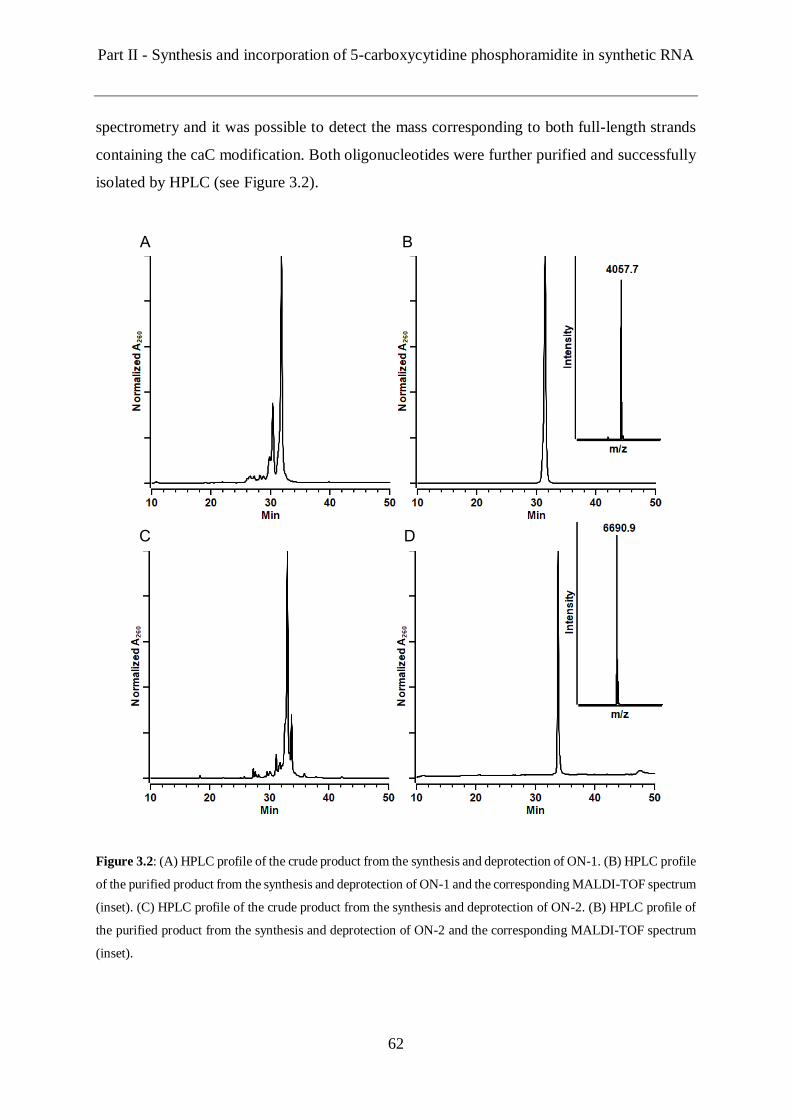
Part II - Synthesis and incorporation of 5-carboxycytidine phosphoramidite in synthetic RNA
62
spectrometry and it was possible to detect the mass corresponding to both full-length strands
containing the caC modification. Both oligonucleotides were further purified and successfully
isolated by HPLC (see Figure 3.2).
Figure 3.2: (A) HPLC profile of the crude product from the synthesis and deprotection of ON-1. (B) HPLC profile
of the purified product from the synthesis and deprotection of ON-1 and the corresponding MALDI-TOF spectrum
(inset). (C) HPLC profile of the crude product from the synthesis and deprotection of ON-2. (B) HPLC profile of
the purified product from the synthesis and deprotection of ON-2 and the corresponding MALDI-TOF spectrum
(inset).
A B
C D
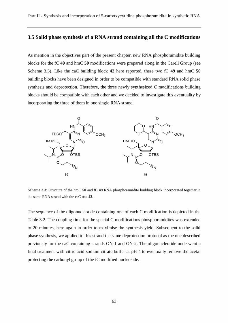
Part II - Synthesis and incorporation of 5-carboxycytidine phosphoramidite in synthetic RNA
63
3.5 Solid phase synthesis of a RNA strand containing all the C modifications
As mention in the objectives part of the present chapter, new RNA phosphoramidite building
blocks for the fC 49 and hmC 50 modifications were prepared along in the Carell Group (see
Scheme 3.3). Like the caC building block 42 here reported, these two fC 49 and hmC 50
building blocks have been designed in order to be compatible with standard RNA solid phase
synthesis and deprotection. Therefore, the three newly synthesized C modifications building
blocks should be compatible with each other and we decided to investigate this eventuality by
incorporating the three of them in one single RNA strand.
Scheme 3.3: Structure of the hmC 50 and fC 49 RNA phosphoramidite building block incorporated together in
the same RNA strand with the caC one 42.
The sequence of the oligonucleotide containing one of each C modification is depicted in the
Table 3.2. The coupling time for the special C modifications phosphoramidites was extended
to 20 minutes, here again in order to maximise the synthesis yield. Subsequent to the solid
phase synthesis, we applied to this strand the same deprotection protocol as the one described
previously for the caC containing strands ON-1 and ON-2. The oligonucleotide underwent a
final treatment with citric acid-sodium citrate buffer at pH 4 to eventually remove the acetal
protecting the carbonyl group of the fC modified nucleoside.

Part II - Synthesis and incorporation of 5-carboxycytidine phosphoramidite in synthetic RNA
64
Table 3.2: Mass spectrometric data of the oligonucleotide synthesized containing hmC, fC and caC modifications.
Entry Sequence Calcd [M-H]- Found [M-H]-
ON-3 5' UCUGAGUcaCCfCUAUUAhmCAAGAG 3' 6754.9 6749.8
MALDI-TOF analysis of the crude product from the deprotection of ON-3 allowed the
detection of the correct mass of the full-length synthetic RNA strand with the three C modified
nucleosides. Moreover, analysis of the crude product by HPLC provided a relatively clean
profile (Figure 3.3). This can be attributed to an effective synthesis and incorporation of all the
three special phosphoramidite building blocks as well as an effective deprotection protocol
preventing the formation of any major side products. Finally, the synthetic RNA strand
containing caC, fC and hmC nucleosides was successfully purified and isolated by HPLC (see
Figure 3.3).
Figure 3.3: (A) HPLC profile of the crude product from the synthesis and deprotection of ON-3. (B) HPLC profile
of the purified product from the synthesis and deprotection of ON-3 and the corresponding MALDI-TOF spectrum
(inset).
A B

Part II - Synthesis and incorporation of 5-carboxycytidine phosphoramidite in synthetic RNA
65
3.6 Enzymatic digestion and uHPLC-MS analysis
In order to confirm the identity and the integrity of the C modifications incorporated by solid
phase synthesis, the RNA strand ON-3 containing hmC, fC and caC was enzymatically digested
to cleave the strand into single nucleosides. The resulting mixture was subsequently analysed
by UHPLC-MS and allowed the detection of all the nucleosides present in the strand (Figure
3.4). In addition to the four canonical nucleosides, we were able to detect the three C modified
nucleosides. Moreover, the HPLC profile obtained shows only the four signals corresponding
to the canonical nucleosides and the three signals corresponding to hmC, fC and caC. Thus, the
absence of additional signal confirms that the incorporation of the special phosphoramidites
and deprotection of the resulting RNA strand proceeded effectively and without the formation
of side-product or degradation of the oligonucleotide.
Figure 3.4: UHPLC profile of the enzymatically digested ON-3 allowing the detection of hmC, fC and caC in
addition to the four canonical nucleosides A, C, G and U.
Part II - Synthesis and incorporation of 5-carboxycytidine phosphoramidite in synthetic RNA
66
3.7 Conclusions and Outlook
In a team effort from the Carell Group, we synthesized new RNA phosphoramidite building
block of hmC 50, fC 49 and caC 42. Well-designed deprotection strategies enabled efficient
incorporation of these building blocks by automated solid phase synthesis inside RNA strands
of different lengths. The newly developed modified cytosine building blocks have indeed
allowed the synthesis of a strand containing all the three epigenetic bases at specific sites.
It will now be possible to start investigating the role of these modifications inside RNA by
performing biochemical studies based on synthetic RNA material containing one or several of
the C(5) modified cytosine bases. We envisioned for example similar experiments to those
previously performed in our group to study the role of hmC, fC and caC in the context of
DNA.[136] In short, we could incubate nuclear extracts from selected cell-line with RNA strands
containing the modified bases. The proteins interacting with the modified bases could be
further isolated in a pull-down assay and analysed by quantitative mass spectrometry. The
identification and study of the proteins recognizing hmC, fC and caC in RNA could thus lead
to decipher how these bases are dynamically formed and their intrinsic exact functions.
Experimental – General Methods and Materials for Synthesis
67
4. EXPERIMENTAL
4.1 General Methods and Materials for Synthesis
Chemicals were purchased from Sigma Aldrich, ABCR, Alfa Aesar, Acros Organics, Tokyo
Chemical Industry Co. or Carbosynth and used without further purification. The solvents for
organic synthesis were of reagent grade and purified by distillation. Dry solvents were bought
from Sigma Aldrich and Acros Organics. Acetonitrile for HPLC purification was purchased
from VWR. ddH2O refers to double distilled water and was obtained by a Milli-Q® Plus
purification system from Merck Millipore using a QPAK® 2 cartridge.
All reactions were carried out with Heidolph MR3001K magnetic stirring hotplates, and if
moisture and air sensitive, in oven-dried glassware (>12 h, 110 °C) under nitrogen or argon.
The temperature of reactions (except room temperature) was adjusted with an acetone/dry ice
mixture, a water/ice-mixture or an oil bath and the temperature monitored with a Heidolph ETK
3001 contact thermometer. In vacuo solvent removals and solution reconcentrations were
performed with a Heidolph Laborota 4000 rotary evaporator with a Vario PC2001 diaphragm
pump from Vacuumbrand. For the removal of water and acetonitrile by freeze-drying, a Christ
Alpha 2-4 LD plus lyophilizer was used. Alternatively, a centrifugal evaporator Savant
SpeedVac® Plus SC110A was used.
Chromatographic purification of products was accomplished using flash column
chromatography on Merck Geduran Si 60 (40 – 63 μm) silica gel (normal phase) or by
reversed-phase high-performance liquid chromatography (RP-HPLC). Thin layer
chromatography (TLC) was performed on Merck 60 (silica gel F254) plates and visualized under
UV light (λ = 254 and 366 nm) and staining with ninhydrin solution (0.3 g ninhydrin, 3 mL
acetic acid in 100 mL n-Butanol), potassium permanganate (1.5 g KMnO4, 10.0 g K2CO3, 125
mg NaOH in 200 mL water) or ceric ammonium molybdate (10.0 g ammonium molybdate
tetrahydrate, 2 g Ce(SO4)2∙4H2O, 180 mL ddH2O, 20 mL conc. H2SO4).
1H-, 13C-, and 31P-NMR spectra were recorded in deuterated solvents purchased from Euriso-
top on Varian Oxford 200, Bruker ARX 300, Varian VXR400S, Varian Inova 400, Brucker
Avance III (cryoprobe) 400, Brucker AMX 600 and Brucker Avance III HD (cryoprobe) 800
spectrometers and calibrated to the residual solvent peak using reported values.[137] The
chemical shifts (d) are given in ppm, the coupling constants (J) in Hz. Multiplicities are
Experimental – General Methods and Materials for Synthesis
68
abbreviated as follows: s = singlet, d = doublet, t = triplet, q = quartet, m = multiplet, br = broad
and combinations of these. For assignment of the structures, additional 2D NMR spectra
(COSY, HSQC and HMBC) were measured. High resolution electrospray ionization mass
spectra (HRMS-ESI) were recorded on a Thermo Finnigan LTQ-FT (ESI-FTICR), and high
resolution electron impact ionization mass spectra (HRMS-EI) were recorded on a Thermo
Finnigan MAT 95. Matrix-assisted laser desorption/ionization-time-of-flight (MALDI-TOF)
mass spectra were recorded on a Brucker Autoflex II. For MALDI-TOF measurements, the
samples were dialyzed on a 0.025 μm VSWP filter (Merck Millipore) against ddH2O for 1 h
and then co-crystallized in a 3-hydroxypicolinic acid matrix (HPA: 25 mg 3-hydroxypicolinic
acid, 5 mg ammonium citrate, 5 μL 15-crown-5 in 0.5 mL H2O/MeCN = 1:1). IR spectra were
recorded on a Perkin Elmer spectrum BX instrument and are reported as follows: wavenumber
ῦ in cm–1. The pH values of buffers were adjusted using a MP 220 pH meter (Mettler Toledo).
UV spectra and melting profiles were measured on a Jasco V-650 spectrometer using quartz
glass cuvettes with 1 cm path length. The concentration of purified oligonucleotide was
determined in triplicate with a NanoDrop ND-1000 spectrophotometer from Thermo Scientific
using calculated extinction coefficient from OligoAnalyzer 3.0 (Integrated DNA Technologies:
https://eu.idtdna.com/calc/analyzer). For strands containing artificial bases, the extinction
coefficient of their corresponding control strand was employed without correction.
Reversed-Phase High-Performance Liquid Chromatography (RP-HPLC)
The following devices were used for the analysis and purification of the synthetic DNA/RNA strands
and the linkers synthesized. In order to remove insoluble particles, samples for preparative HPLC were
filtered through a 0.2 μm GHP filter membrane (Acrodisc®), and samples for analytical HPLC were
centrifuged at maximum speed, prior to loading.
Analytical RP-HPLC was performed on an analytical HPLC Waters Alliance (2695 Separation Module,
2996 Photodiode Array Detector) equipped with the column Nucleosil 120-3 C18 from Macherey
Nagel. Using a flow of 0.5 mL/min, gradients of 0–50 % B in 45 min or 0–90 % B over 45 minutes
were applied. Preparative RP-HPLC was performed on a HPLC Waters Breeze (2487 Dual λ Array
Detector, 1525 Binary HPLC Pump) equipped with the columns Nucleosil 100-7 C18, VP 250/10 C18
from Macherey Nagel. Using a flow of 5 mL/min, a gradient of 0–90 % B over 45 minutes was applied
Buffer systems: Buffer A: 0.1 M triethylammonium acetate in water;
Buffer B: 0.1 M triethylammonium acetate in 80% (v/v) acetonitril

Experimental – Chemical Synthesis
69
4.2 Chemical synthesis
4.2.1 Synthesis of the picolyl azide-PEG10-azide linker (17)
6-Hydroxymethyl-nicotinic acid methyl ester[47]
2,5-pyridinedicarboxylic acid dimethyl ester (5.23 g, 26.8 mmol, 1.0 eq.) was dissolved in dry
MeOH/dry THF (2:1, 300 mL) and cooled down to 0 °C. NaBH4 (2.03 g, 53.6 mmol, 2.0 eq.)
was then added portionwise over 20 minutes. The reaction was stirred at 0 °C for 3 hours before
being quenched by the addition of a saturated NH4Cl-solution (~ 100 mL). The solvent was
removed in vacuo and the aqueous layer was extracted with DCM (3 × 100 mL). The combined
organic layers were dried over MgSO4, filtered and the solvent was removed in vacuo. 6-
hydroxymethyl-nicotinic acid methyl ester was obtained as a colorless solid (3.95 g, 23.6
mmol, 88 %).
Rf (iHex/EtOAc, 1/1) = 0.3.
1H-NMR (400 MHz, CDCl3): δ (ppm): 9.15 (d, J = 2 Hz, 1H), 8.29 (dd, J = 8.1, 1.9 Hz, 1H),
7.36 (d, J = 8.1 Hz, 1H), 4.83 (s, 2H), 3.95 (s, 3H).
13C-NMR (101 MHz, CDCl3): δ (ppm): 165.6, 163.4, 149.9, 137.8, 125.0, 120.0, 64.2, 52.5.
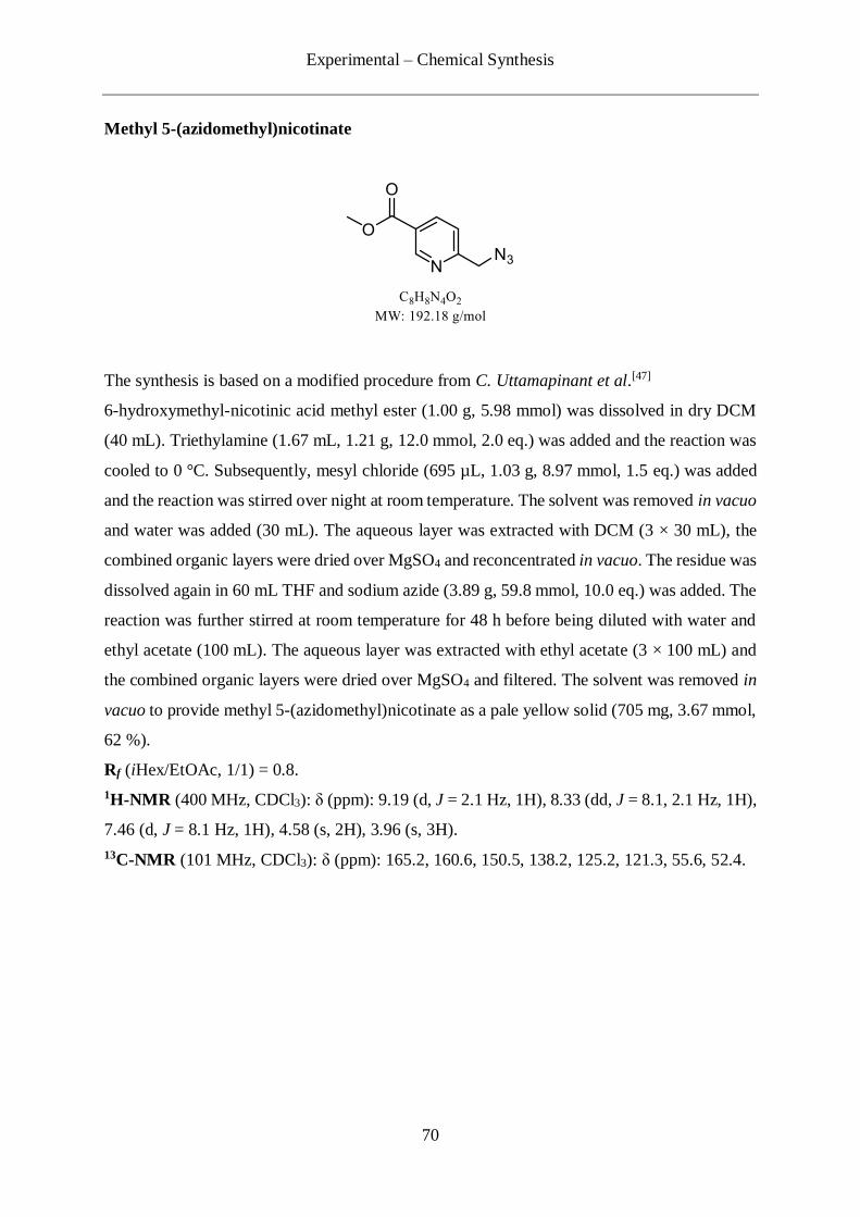
Experimental – Chemical Synthesis
70
Methyl 5-(azidomethyl)nicotinate
The synthesis is based on a modified procedure from C. Uttamapinant et al.[47]
6-hydroxymethyl-nicotinic acid methyl ester (1.00 g, 5.98 mmol) was dissolved in dry DCM
(40 mL). Triethylamine (1.67 mL, 1.21 g, 12.0 mmol, 2.0 eq.) was added and the reaction was
cooled to 0 °C. Subsequently, mesyl chloride (695 µL, 1.03 g, 8.97 mmol, 1.5 eq.) was added
and the reaction was stirred over night at room temperature. The solvent was removed in vacuo
and water was added (30 mL). The aqueous layer was extracted with DCM (3 × 30 mL), the
combined organic layers were dried over MgSO4 and reconcentrated in vacuo. The residue was
dissolved again in 60 mL THF and sodium azide (3.89 g, 59.8 mmol, 10.0 eq.) was added. The
reaction was further stirred at room temperature for 48 h before being diluted with water and
ethyl acetate (100 mL). The aqueous layer was extracted with ethyl acetate (3 × 100 mL) and
the combined organic layers were dried over MgSO4 and filtered. The solvent was removed in
vacuo to provide methyl 5-(azidomethyl)nicotinate as a pale yellow solid (705 mg, 3.67 mmol,
62 %).
Rf (iHex/EtOAc, 1/1) = 0.8.
1H-NMR (400 MHz, CDCl3): δ (ppm): 9.19 (d, J = 2.1 Hz, 1H), 8.33 (dd, J = 8.1, 2.1 Hz, 1H),
7.46 (d, J = 8.1 Hz, 1H), 4.58 (s, 2H), 3.96 (s, 3H).
13C-NMR (101 MHz, CDCl3): δ (ppm): 165.2, 160.6, 150.5, 138.2, 125.2, 121.3, 55.6, 52.4.
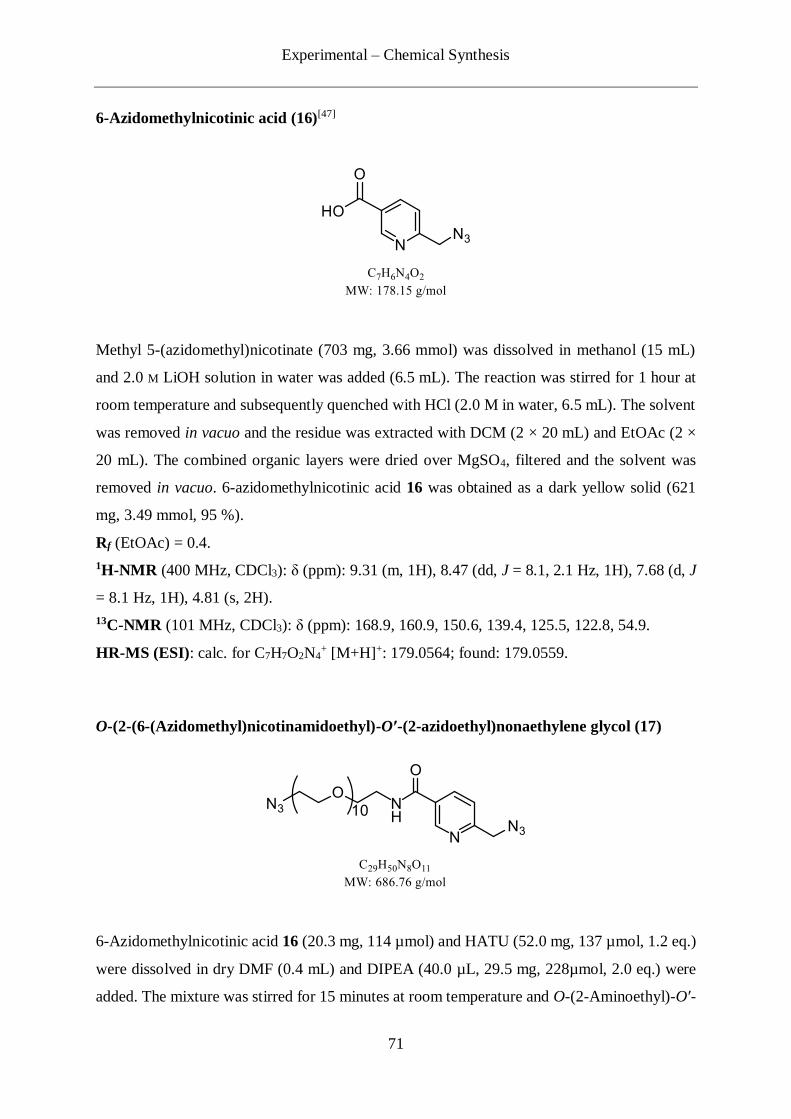
Experimental – Chemical Synthesis
71
6-Azidomethylnicotinic acid (16)[47]
Methyl 5-(azidomethyl)nicotinate (703 mg, 3.66 mmol) was dissolved in methanol (15 mL)
and 2.0 M LiOH solution in water was added (6.5 mL). The reaction was stirred for 1 hour at
room temperature and subsequently quenched with HCl (2.0 M in water, 6.5 mL). The solvent
was removed in vacuo and the residue was extracted with DCM (2 × 20 mL) and EtOAc (2 ×
20 mL). The combined organic layers were dried over MgSO4, filtered and the solvent was
removed in vacuo. 6-azidomethylnicotinic acid 16 was obtained as a dark yellow solid (621
mg, 3.49 mmol, 95 %).
Rf (EtOAc) = 0.4.
1H-NMR (400 MHz, CDCl3): δ (ppm): 9.31 (m, 1H), 8.47 (dd, J = 8.1, 2.1 Hz, 1H), 7.68 (d, J
= 8.1 Hz, 1H), 4.81 (s, 2H).
13C-NMR (101 MHz, CDCl3): δ (ppm): 168.9, 160.9, 150.6, 139.4, 125.5, 122.8, 54.9.
HR-MS (ESI): calc. for C7H7O2N4+ [M+H]+: 179.0564; found: 179.0559.
O-(2-(6-(Azidomethyl)nicotinamidoethyl)-O′-(2-azidoethyl)nonaethylene glycol (17)
6-Azidomethylnicotinic acid 16 (20.3 mg, 114 µmol) and HATU (52.0 mg, 137 µmol, 1.2 eq.)
were dissolved in dry DMF (0.4 mL) and DIPEA (40.0 µL, 29.5 mg, 228µmol, 2.0 eq.) were
added. The mixture was stirred for 15 minutes at room temperature and O-(2-Aminoethyl)-O′-

Experimental – Chemical Synthesis
72
(2-azidoethyl)nonaethylene glycol (60.0 mg, 114 µmol, 1.0 eq.) was added. The reaction was
stirred over night at room temperature and the solvent was removed in vacuo. The residue was
taken up in DCM (15 mL) and washed with brine (15 mL). The aqueous layer was reextracted
one time with DCM (15 mL) and the combined organic layers were dried over MgSO4 and
filtered. The solvent was removed in vacuo and the residue was purified by flash column
chromatography (Silica, DCM/MeOH, 100/0 → 95/5). 17 was obtained as a colourless oil (63
mg, 92 µmol, 81 %).
Rf (DCM/MeOH, 10/1) = 0.6.
1H-NMR (400 MHz, CDCl3): δ (ppm): 9.01 (d, J = 2.3 Hz, 1H, CHarom), 8.20 (dd, J = 8.1, 2.3
Hz, 1H, CHarom), 7.42 (d, J = 8.1 Hz, 1H, CHarom), 4.53 (s, 2H, CaromCH2N3), 3.78 – 3.46 (m,
42H, 21×ClinkerH2), 3.37 (t, J = 5.1 Hz, 2H, CH2CH2N3).
13C-NMR (101 MHz, CDCl3): δ (ppm): 165.2 (C=O), 158.4 (Carom), 148.3 (Carom), 136.4
(CHarom), 129.5 (CHarom), 121.5 (CHarom), 70.7 (ClinkerH2), 70.7 (ClinkerH2), 70.6 (ClinkerH2), 70.6
(ClinkerH2), 70.5 (ClinkerH2), 70.3 (ClinkerH2), 70.0 (ClinkerH2), 69.6 (ClinkerH2), 55.3 (CaromCH2N3),
50.7 (CH2CH2N3), 40.0 (CH2CH2NH).
HR-MS (ESI): calc. for C29H51O11N8+ [M+H]+: 687.3673; found: 687.3682.
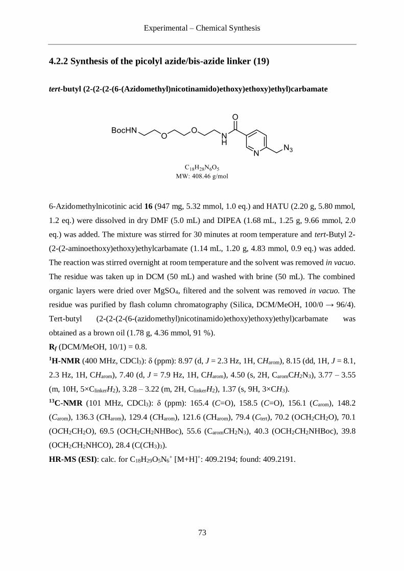
Experimental – Chemical Synthesis
73
4.2.2 Synthesis of the picolyl azide/bis-azide linker (19)
tert-butyl (2-(2-(2-(6-(Azidomethyl)nicotinamido)ethoxy)ethoxy)ethyl)carbamate
6-Azidomethylnicotinic acid 16 (947 mg, 5.32 mmol, 1.0 eq.) and HATU (2.20 g, 5.80 mmol,
1.2 eq.) were dissolved in dry DMF (5.0 mL) and DIPEA (1.68 mL, 1.25 g, 9.66 mmol, 2.0
eq.) was added. The mixture was stirred for 30 minutes at room temperature and tert-Butyl 2-
(2-(2-aminoethoxy)ethoxy)ethylcarbamate (1.14 mL, 1.20 g, 4.83 mmol, 0.9 eq.) was added.
The reaction was stirred overnight at room temperature and the solvent was removed in vacuo.
The residue was taken up in DCM (50 mL) and washed with brine (50 mL). The combined
organic layers were dried over MgSO4, filtered and the solvent was removed in vacuo. The
residue was purified by flash column chromatography (Silica, DCM/MeOH, 100/0 → 96/4).
Tert-butyl (2-(2-(2-(6-(azidomethyl)nicotinamido)ethoxy)ethoxy)ethyl)carbamate was
obtained as a brown oil (1.78 g, 4.36 mmol, 91 %).
Rf (DCM/MeOH, 10/1) = 0.8.
1H-NMR (400 MHz, CDCl3): δ (ppm): 8.97 (d, J = 2.3 Hz, 1H, CHarom), 8.15 (dd, 1H, J = 8.1,
2.3 Hz, 1H, CHarom), 7.40 (d, J = 7.9 Hz, 1H, CHarom), 4.50 (s, 2H, CaromCH2N3), 3.77 – 3.55
(m, 10H, 5×ClinkerH2), 3.28 – 3.22 (m, 2H, ClinkerH2), 1.37 (s, 9H, 3×CH3).
13C-NMR (101 MHz, CDCl3): δ (ppm): 165.4 (C=O), 158.5 (C=O), 156.1 (Carom), 148.2
(Carom), 136.3 (CHarom), 129.4 (CHarom), 121.6 (CHarom), 79.4 (Ctert), 70.2 (OCH2CH2O), 70.1
(OCH2CH2O), 69.5 (OCH2CH2NHBoc), 55.6 (CaromCH2N3), 40.3 (OCH2CH2NHBoc), 39.8
(OCH2CH2NHCO), 28.4 (C(CH3)3).
HR-MS (ESI): calc. for C18H29O5N6+ [M+H]+: 409.2194; found: 409.2191.

Experimental – Chemical Synthesis
74
N-(2-(2-(2-Aminoethoxy)ethoxy)ethyl)-6-(azidomethyl)nicotinamide (21)
Tert-butyl (2-(2-(2-(6-(azidomethyl)nicotinamido)ethoxy)ethoxy)ethyl)carbamate (1.78 g,
4.36 mmol, 1.0 eq.) was dissolved in methanol (6mL) and an aqueous solution of HCl was
added (2.0 M, 3.0 mL, 20.0 eq.). The reaction was stirred at 50 °C for 2 hours before being
quenched by addition of a solution of NaOH (1.0 M in water, 6 mL). The solvent was removed
in vacuo and the aqueous layer was extracted with DCM (2 × 15 mL) and EtOAc (1 × 15 mL).
The organic layers were combined, dried over MgSO4 and filtered. The solvent was removed
in vacuo and 21 was obtained as an orange oil (955 mg, 3.10 mmol, 72 %).
Rf (DCM/MeOH, 10/1) = 0.3.
1H-NMR (599 MHz, CDCl3): δ (ppm): 9.01 (d, J = 2.3 Hz, 1H, CHarom), 8.19 (dd, 1H, J = 8.1,
2.3 Hz, 1H, CHarom), 7.42 (d, J = 8.1 Hz, 1H, CHarom), 4.53 (s, 2H, CaromCH2N3), 3.72 – 2.75
(m, 12H, 6×ClinkerH2).
13C-NMR (151 MHz, CDCl3): δ (ppm): 165.3 (C=O), 158.5 (Carom), 148.2 (Carom), 136.3
(CHarom), 129.5 (CHarom), 121.5 (CHarom), 73.0 (OCH2CH2NH2), 70.3 (OCH2CH2O), 70.0
(OCH2CH2O), 69.7 (OCH2CH2NHCO), 55.3 (CaromCH2N3), 41.5 ((OCH2CH2NH2), 39.9
(OCH2CH2NHCO).
HR-MS (ESI): calc. for C13H21O3N6+ [M+H]+: 309.1673; found: 309.1673.

Experimental – Chemical Synthesis
75
14-Hydroxy-3,6,9,12-tetraoxatetradecyl-4'-methylbenzolsulfonat (25)
Tetraethylenglycole 22 (29.25 g, 150.6 mmol, 1.0 eq.) was dissolved in dry THF (100 mL) and
triethylamine (122 g, 1.20 mol, 8.0 eq.) was added. The reaction was cooled down to 0 °C and
a solution of tosylchloride (28.71 g, 150.6 mmol, 1.0 eq.) in dry THF (50 mL) was added over
a period of 1 hour. After 48-hour stirring at room temperature the solvent was removed in
vacuo. The mixture was taken up in HCl (2 M, 150 mL) and extracted with DCM (4 × 150 mL).
The combined organic layers were dried over MgSO4 and filtered. After removal of the solvent
in vacuo the crude product was purified by flash column chromatography (Silica, DCM →
DCM/MeOH (99 /1 → 0/1)). The product 25 was obtained as a colourless oil (48.27 g, 138.6
mmol, 92 %).
1H-NMR (400 MHz, CDCl3): δ (ppm): 7.82-7.76 (m, 2H, CHarom.), 7.37-7.30 (m, 2H, CHarom.),
4.20-4.12 (m, 2H, CH2), 3.73-3.52 (m, 14H, 7×CH2), 2.42 (s, 3H, CH3), 2.38-2.31 (s, 1H, OH).
13C-NMR (101 MHz, CDCl3): δ (ppm): 144.9 (Carom.), 133.2 (Carom.), 130.0 (2×CHarom.), 128.1
(2×CHarom.), 72.6 (CH2), 70.9 (CH2), 70.8 (CH2), 70.6 (CH2), 70.5 (CH2), 69.4 (CH2), 68.9
(CH2), 61.9 (CH2), 21.8 (CH3).
HR-MS (ESI): C15H25O7S+ [M+H]+, calc.: 349.1315, found: 349.1316.
2-(2-(2-(2-Azidoethoxy)ethoxy)ethoxy)ethan-1-ol (26)
To 25 (8.08 g, 23.19 mmol, 1.0 eq.) in solution in dry DMF (100 mL) was added NaN3 (7.54
g, 116 mmol, 5.0 eq.). The mixture was stirred at 70 °C for 8 hours and the solvent was removed
in vacuo. The residue was retaken in water (100 mL), extracted with DCM (2 × 100 mL) and

Experimental – Chemical Synthesis
76
the combined organic layers were dried over MgSO4 and filtered. The solvent was removed in
vacuo and 2-(2-(2-(2-azidoethoxy)ethoxy)ethoxy)ethan-1-ol 26 was obtained as a colourless
oil (4.87 g, 22.2 mmol, 96 %).
1H-NMR (400 MHz, CDCl3): δ (ppm): 3.76 – 3.68 (m, 2H, CH2), 3.68 – 3.62 (m, 10H, 5×CH2),
3.63 – 3.58 (m, 2H, CH2), 3.39 (t, J = 5.2 Hz, 2H, CH2).
13C-NMR (101 MHz, CDCl3): δ (ppm): 72.5 (CH2), 70.7 (CH2), 70.7 (CH2), 70.6 (CH2), 70.4
(CH2), 70.1 (CH2), 61.7 (CH2), 50.7 (CH2).
HR-MS (ESI): C8H18O4N3+ [M+H]+, calc.: 220.1292, found: 220.1293.
2-(2-(2-(2-Azidoethoxy)ethoxy)ethoxy)ethyl 4-methylbenzenesulfonate (23)
26 (4.75 g, 21.67 mmol) was dissolved in dry DCM (50 mL). Triethylamine (8.92 mL, 6.51 g,
65.0 mmol, 3.0 eq.) was added and the reaction was cooled to 0 °C. Subsequently tosylchloride
(12.39 g, 65.00 mmol, 3.0 eq.) was added and the reaction was stirred over night at room
temperature. The solvent was removed in vacuo and the residue was taken up in HCl (2.0 M in
water, 50 mL). After extraction with DCM (5 × 50 mL), the combined organic layers were
dried over MgSO4, filtered and the solvent was remved in vacuo. The crude product was
purified by flash column chromatography (Silica, i-Hex/EtOAc 1/0 → 3/7). 2-(2-(2-(2-
azidoethoxy)ethoxy)ethoxy)ethyl 4-methylbenzenesulfonate 23 was obtained as a colourless
oil (7.42 g, 20.0 mmol, 92 %).
1H-NMR (400 MHz, CDCl3): δ (ppm): 7.80 (d, J = 7.9 Hz, 2H, CHarom), 7.34 (d, J = 7.9 Hz,
2H, CHarom), 4.16 (t, J = 4.9 Hz, 2H, CH2), 3.72 – 3.58 (m, 12H, 6×CH2), 3.38 (t, J = 5.0 Hz,
2H, CH2), 2.45 (s, 3H, CH3).
13C-NMR (101 MHz, CDCl3): δ (ppm): 144.9 (Carom.), 133.2 (Carom.), 130.0 (2×CHarom.), 128.1
(2×CHarom.), 70.9 (CH2), 70.8 (CH2), 70.8 (CH2), 70.2 (CH2), 69.4 (CH2), 68.9 (CH2), 50.8
(CH2), 21.8 (CH3).
HR-MS (ESI): C15H27O6N4S+ [M+NH4]
+, calc.: 391.1646, found: 391.1649.

Experimental – Chemical Synthesis
77
Methyl-3, 5-bis(2-(2-(2-(2-azidoethoxy)ethoxy)ethoxy)ethoxy)benzoate
To a solution of methyl 3,5-dihydroxybenzoate (30 mg, 178.41 µmol, 1.0 eq.) 20 in acetone
(1.7 mL), 23 (147 mg, 392.51 µmol, 2.2 eq.) and K2CO3 (123.29 mg, 892.07 µmol, 5.0 eq.)
were added. The reaction was stirred at 55 °C for 30 hours and the solvent was removed. The
residue was taken up in water (5 mL) and DCM (5 mL) and extracted with DCM (3 × 5 mL).
The combined organic layers were dried over MgSO4, filtered and the solvent removed in
vacuo. The crude product was purified by flash column chromatography (Silica, DCM/MeOH
100/0 → 96/4) and the product was obtained as a yellow oil (78.0 mg, 137 µmol, 77 %).
1H-NMR (400 MHz, CDCl3): δ (ppm): 7.19 – 7.05 (m, 2H, 2×CHarom), 7.12 – 7.05 (m, 1H,
CHarom), 4.17 – 4.05 (m, 4H, 2×CH2), 3.90 – 3.79 (m, 7H, 2×CH2+CH3), 3.75 – 3.56 (m, 20H,
10×CH2), 3.36 (t, J = 5.0 Hz, 4H, 2×CH2).
13C-NMR (101 MHz, CDCl3): δ (ppm): 166.8 (C=O), 159.7 (2×Carom.), 131.8 (Carom.), 108.0
(2×CHarom), 106.9 (CHarom), 70.8 (CH2), 70.7 (CH2), 70.7 (CH2), 70.6 (CH2), 70.0 (CH2), 70.0
(CH2), 69.6 (CH2), 69.6 (CH2), 67.7 (CH2), 67.7 (CH2), 52.2 (CH3), 50.7 (CH2N3).
HR-MS (ESI): C24H42O10N7+ [M+NH4]
+, calc.: 588.2987, found: 588.2976.
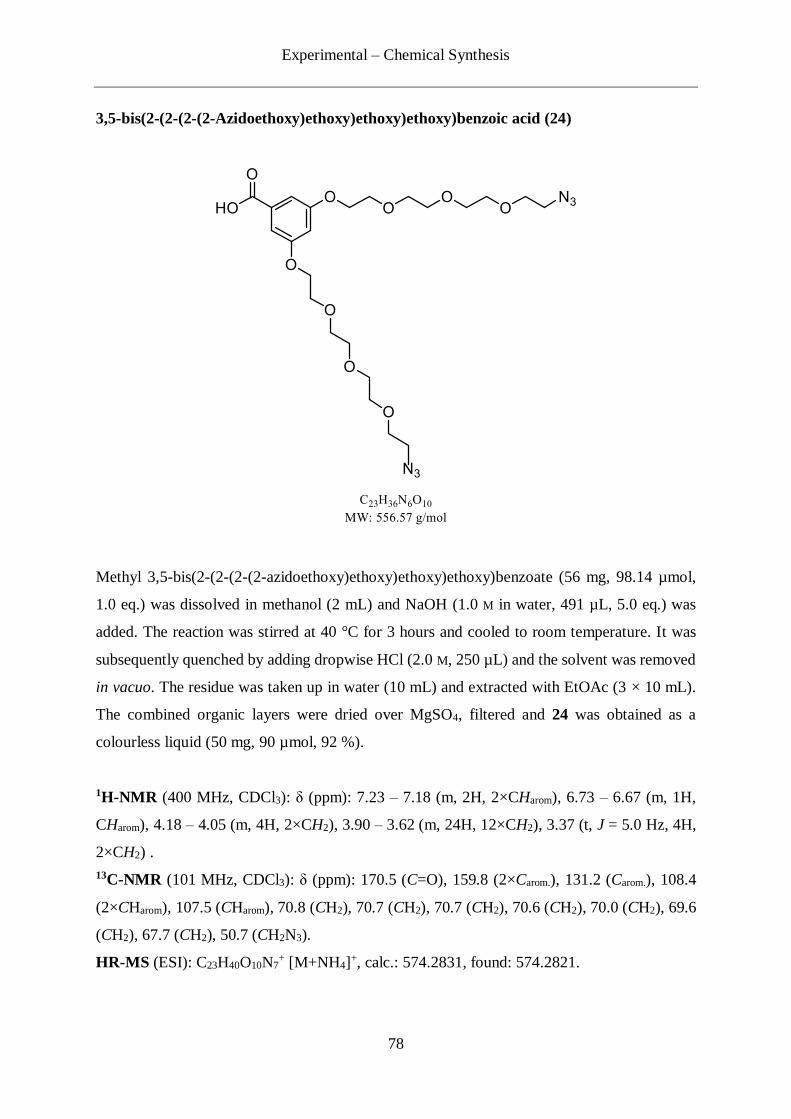
Experimental – Chemical Synthesis
78
3,5-bis(2-(2-(2-(2-Azidoethoxy)ethoxy)ethoxy)ethoxy)benzoic acid (24)
Methyl 3,5-bis(2-(2-(2-(2-azidoethoxy)ethoxy)ethoxy)ethoxy)benzoate (56 mg, 98.14 µmol,
1.0 eq.) was dissolved in methanol (2 mL) and NaOH (1.0 M in water, 491 µL, 5.0 eq.) was
added. The reaction was stirred at 40 °C for 3 hours and cooled to room temperature. It was
subsequently quenched by adding dropwise HCl (2.0 M, 250 µL) and the solvent was removed
in vacuo. The residue was taken up in water (10 mL) and extracted with EtOAc (3 × 10 mL).
The combined organic layers were dried over MgSO4, filtered and 24 was obtained as a
colourless liquid (50 mg, 90 µmol, 92 %).
1H-NMR (400 MHz, CDCl3): δ (ppm): 7.23 – 7.18 (m, 2H, 2×CHarom), 6.73 – 6.67 (m, 1H,
CHarom), 4.18 – 4.05 (m, 4H, 2×CH2), 3.90 – 3.62 (m, 24H, 12×CH2), 3.37 (t, J = 5.0 Hz, 4H,
2×CH2) .
13C-NMR (101 MHz, CDCl3): δ (ppm): 170.5 (C=O), 159.8 (2×Carom.), 131.2 (Carom.), 108.4
(2×CHarom), 107.5 (CHarom), 70.8 (CH2), 70.7 (CH2), 70.7 (CH2), 70.6 (CH2), 70.0 (CH2), 69.6
(CH2), 67.7 (CH2), 50.7 (CH2N3).
HR-MS (ESI): C23H40O10N7+ [M+NH4]
+, calc.: 574.2831, found: 574.2821.

Experimental – Chemical Synthesis
79
6-(Azidomethyl)-N-(2-(2-(2-(3,5-bis(2-(2-(2-(2-
azidoethoxy)ethoxy)ethoxy)ethoxy)benzamido)ethoxy)ethoxy)ethyl)nicotinamide (19)
24 (25 mg, 44.92 µmol, 1.0 eq.) was dissolved in dry DMF (0.5 mL). HATU (21 mg, 54 µmol,
1.2 eq.) and DIPEA (15.7µL, 11.6 mg, 89.8 µmol, 2 eq.) were added and the mixture was
stirred 45 minutes at room temperature. A solution of 21 (15 mg, 49.4 µmol, 1.1 eq.) in DMF
(0.5 mL) was added and the reaction was stirred 12 hours at room temperature. The solvent
was removed in vacuo and the residue was taken up in DCM (10 mL) and washed with brine
(10 mL). The aqueous layer was reextracted one time with DCM (15 mL) and the combined
organic layers were dried over MgSO4 and filtered. The solvent was removed in vacuo and the
residue was purified by flash column chromatography (Silica, DCM/MeOH, 100/0 → 95/5).
19 was obtained as an orange oil (31 mg, 37 µmol, 82 %).
1H-NMR (400 MHz, CDCl3): δ (ppm): 8.98 (d, J = 2.1 Hz, 1H, CHaromNarom), 8.15 (dd, J = 8.1,
2.1 Hz, 1H, CHarom), 7.40 (d, J = 8.0 Hz, 1H, CHarom), 7.14 – 7.04 (m, 1H, CHarom), 6.91 (d, J
= 2.3 Hz, 2H), 4.53 (s, 2H, CaromCH2N3), 4.26 – 3.20 (m, 40H, 20×CH2), 1.58 – 1.16 (m, 4H,
2×CH2).
13C-NMR (101 MHz, CDCl3): δ (ppm): 167.3 (C=O), 165.2 (C=O), 159.8 (2×Carom), 158.4
(CaromHNarom), 147.8 (CHaromNarom), 136.6 (CHarom), 129.5 (Carom), 121.7 (CHarom), 106.0
(CHarom), 104.3 (CHarom), 77.4 (CH2), 77.0 (CH2), 76.7 (CH2), 70.7 (CH2), 70.7 (CH2), 70.6
(CH2), 70.3 (CH2), 70.2 (CH2), 70.0 (CH2), 69.7 (CH2), 69.6 (CH2), 69.5 (CH2), 67.6 (CH2),
55.1 (CaromCH2N3), 50.7 (CH2CH2N3), 39.8 (OCH2CH2NH), 39.7 (OCH2CH2NH).
HR-MS (ESI): C36H55O12N12+ [M+H]+, calc.: 847.4057, found: 847.4056.

Experimental – Chemical Synthesis
80
4.2.3 Synthesis of the picolyl azide-PEG11-TMS protected alkyne linker (34)
5-(Trimethylsilyl)pent-4-ynoic acid (30)[102]
5-(Trimethylsilyl)pent-4-yn-1-ol 29 (4.97 g, 31.8 mmol) was dissolved in dry DMF (40 mL).
PDC (29.91 g, 79.49 mmol, 2.5 eq.) was added and the reaction was stirred 24 hours at room
temperature. The residue was diluted with water (180 mL) and extracted with diethyl ether
(3×100 mL). The combined organic layers were washed with HCl (1 M in water, 2×100 mL),
brine (100 mL), dried over MgSO4 and filtered. The solvent was removed in vacuo and the
crude product was purified by flash column chromatography (Silica, i-Hex/EtOAc 1/0 → 2:3).
5-(Trimethylsilyl)pent-4-ynoic acid 30 was obtained as a white oil (2.2 g, 12.9 mmol, 41 %).
1H-NMR (400 MHz, CDCl3): δ (ppm): 2.68 – 2.48 (m, 4H, CH2), 0.14 (s, 9H, Si(CH3)3).
13C-NMR (101 MHz, CDCl3): δ (ppm): 176.7 (C=O), 104.9 (CH2C≡C), 85.4 (CH2C≡C), 33.4
(CH2CH2C≡C), 15.6 (CH2C≡C), 0.0 (Si(CH3)3).
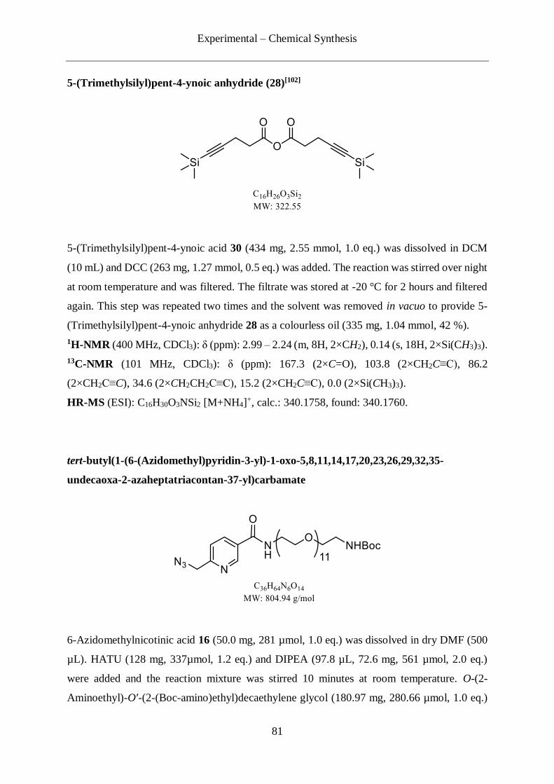
Experimental – Chemical Synthesis
81
5-(Trimethylsilyl)pent-4-ynoic anhydride (28)[102]
5-(Trimethylsilyl)pent-4-ynoic acid 30 (434 mg, 2.55 mmol, 1.0 eq.) was dissolved in DCM
(10 mL) and DCC (263 mg, 1.27 mmol, 0.5 eq.) was added. The reaction was stirred over night
at room temperature and was filtered. The filtrate was stored at -20 °C for 2 hours and filtered
again. This step was repeated two times and the solvent was removed in vacuo to provide 5-
(Trimethylsilyl)pent-4-ynoic anhydride 28 as a colourless oil (335 mg, 1.04 mmol, 42 %).
1H-NMR (400 MHz, CDCl3): δ (ppm): 2.99 – 2.24 (m, 8H, 2×CH2), 0.14 (s, 18H, 2×Si(CH3)3).
13C-NMR (101 MHz, CDCl3): δ (ppm): 167.3 (2×C=O), 103.8 (2×CH2C≡C), 86.2
(2×CH2C≡C), 34.6 (2×CH2CH2C≡C), 15.2 (2×CH2C≡C), 0.0 (2×Si(CH3)3).
HR-MS (ESI): C16H30O3NSi2 [M+NH4]+, calc.: 340.1758, found: 340.1760.
tert-butyl(1-(6-(Azidomethyl)pyridin-3-yl)-1-oxo-5,8,11,14,17,20,23,26,29,32,35-
undecaoxa-2-azaheptatriacontan-37-yl)carbamate
6-Azidomethylnicotinic acid 16 (50.0 mg, 281 µmol, 1.0 eq.) was dissolved in dry DMF (500
µL). HATU (128 mg, 337µmol, 1.2 eq.) and DIPEA (97.8 µL, 72.6 mg, 561 µmol, 2.0 eq.)
were added and the reaction mixture was stirred 10 minutes at room temperature. O-(2-
Aminoethyl)-O′-(2-(Boc-amino)ethyl)decaethylene glycol (180.97 mg, 280.66 µmol, 1.0 eq.)
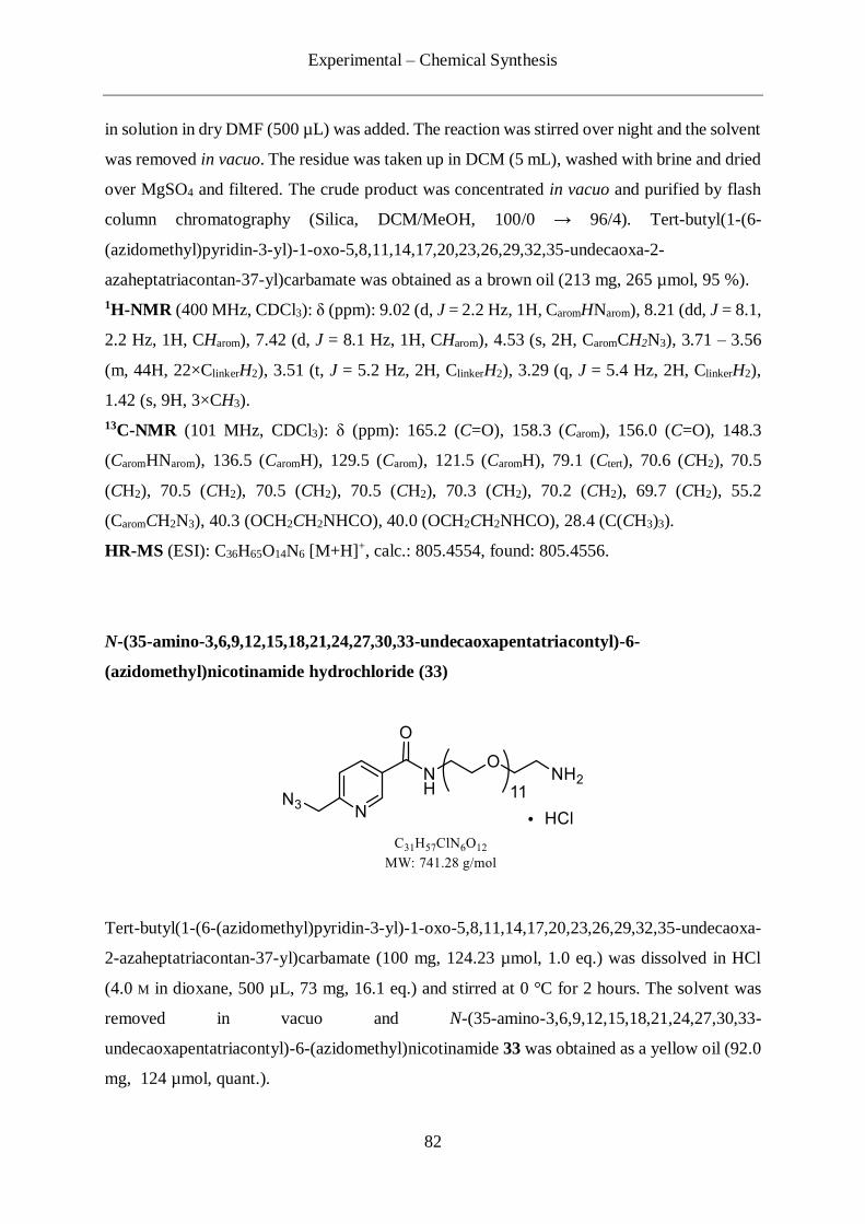
Experimental – Chemical Synthesis
82
in solution in dry DMF (500 µL) was added. The reaction was stirred over night and the solvent
was removed in vacuo. The residue was taken up in DCM (5 mL), washed with brine and dried
over MgSO4 and filtered. The crude product was concentrated in vacuo and purified by flash
column chromatography (Silica, DCM/MeOH, 100/0 → 96/4). Tert-butyl(1-(6-
(azidomethyl)pyridin-3-yl)-1-oxo-5,8,11,14,17,20,23,26,29,32,35-undecaoxa-2-
azaheptatriacontan-37-yl)carbamate was obtained as a brown oil (213 mg, 265 µmol, 95 %).
1H-NMR (400 MHz, CDCl3): δ (ppm): 9.02 (d, J = 2.2 Hz, 1H, CaromHNarom), 8.21 (dd, J = 8.1,
2.2 Hz, 1H, CHarom), 7.42 (d, J = 8.1 Hz, 1H, CHarom), 4.53 (s, 2H, CaromCH2N3), 3.71 – 3.56
(m, 44H, 22×ClinkerH2), 3.51 (t, J = 5.2 Hz, 2H, ClinkerH2), 3.29 (q, J = 5.4 Hz, 2H, ClinkerH2),
1.42 (s, 9H, 3×CH3).
13C-NMR (101 MHz, CDCl3): δ (ppm): 165.2 (C=O), 158.3 (Carom), 156.0 (C=O), 148.3
(CaromHNarom), 136.5 (CaromH), 129.5 (Carom), 121.5 (CaromH), 79.1 (Ctert), 70.6 (CH2), 70.5
(CH2), 70.5 (CH2), 70.5 (CH2), 70.5 (CH2), 70.3 (CH2), 70.2 (CH2), 69.7 (CH2), 55.2
(CaromCH2N3), 40.3 (OCH2CH2NHCO), 40.0 (OCH2CH2NHCO), 28.4 (C(CH3)3).
HR-MS (ESI): C36H65O14N6 [M+H]+, calc.: 805.4554, found: 805.4556.
N-(35-amino-3,6,9,12,15,18,21,24,27,30,33-undecaoxapentatriacontyl)-6-
(azidomethyl)nicotinamide hydrochloride (33)
Tert-butyl(1-(6-(azidomethyl)pyridin-3-yl)-1-oxo-5,8,11,14,17,20,23,26,29,32,35-undecaoxa-
2-azaheptatriacontan-37-yl)carbamate (100 mg, 124.23 µmol, 1.0 eq.) was dissolved in HCl
(4.0 M in dioxane, 500 µL, 73 mg, 16.1 eq.) and stirred at 0 °C for 2 hours. The solvent was
removed in vacuo and N-(35-amino-3,6,9,12,15,18,21,24,27,30,33-
undecaoxapentatriacontyl)-6-(azidomethyl)nicotinamide 33 was obtained as a yellow oil (92.0
mg, 124 µmol, quant.).

Experimental – Chemical Synthesis
83
1H-NMR (400 MHz, CDCl3): δ (ppm): 8.98 (d, J = 1.8 Hz, 1H, CaromHNarom), 8.82 (dd, J = 8.4,
1.8 Hz, 1H, CHarom), 7.53 (d, J = 8.3 Hz, 1H, CHarom), 4.71 (s, 2H, CaromCH2N3), 3.38 – 2.97
(m, 46H, 23×ClinkerH2), 2.75 – 2.63 (m, 2H, ClinkerH2).
13C-NMR (101 MHz, CDCl3): δ (ppm): 161.6 (C=O), 152.5 (Carom), 145.5 (CaromHNarom), 141.5
(CaromH), 133.0 (Carom), 125.2 (CaromH), 70.5 (CH2), 70.4 (CH2), 70.3 (CH2), 70.3 (CH2), 70.2
(CH2), 70.2 (CH2), 70.2 (CH2), 70.1 (CH2), 70.0 (CH2), 70.0 (CH2), 70.0 (CH2), 69.9 (CH2),
69.2 (CH2), 66.9 (CH2), 49.9 (CaromCH2N3), 40.2 (OCH2CH2NH2), 40.1 (OCH2CH2NHCO).
HR-MS (ESI): C31H57O12N6 [M+H]+, calc.: 705.4028, found: 705.4026.
6-(Azidomethyl)-N-(2,2-dimethyl-7-oxo-11,14,17,20,23,26,29,32,35,38,41-undecaoxa-8-
aza-2-silatritetracont-3-yn-43-yl)nicotinamide (34)
N-(35-amino-3,6,9,12,15,18,21,24,27,30,33-undecaoxapentatriacontyl)-6-
(azidomethyl)nicotinamide 33 (40 mg, 51 µmol, 1.0 eq.) was dissolved in dry DCM (1 mL).
Pyridine (83 µL, 81 mg, 1.0 mmol, 20.0 eq.) and DMAP (6 mg, 51.4 µmol, 1.0 eq) were added
and the reaction mixture was cooled to 0 °C. A solution of 5-(Trimethylsilyl)pent-4-ynoic
anhydride (50 mg, 154.3 µmol, 3.0 eq.) in DCM (1 mL) was added dropwise and the reaction
was stirred over night while being allowed to warm to room temperature. Subsequently, the
residue was dissolved in DCM (20 mL) and washed with water (20 mL), NaHCO3 saturated
aqueous solution (20 mL) and CuSO4 saturated aqueous solution (20 mL). The organic layer
was dried over MgSO4, filtered and concentrated in vacuo. The crude product was purified by
flash column chromatography (Silica, DCM/MeOH, 100/0 → 85/15) and the product 34 was
obtained as a yellow oil (43 mg, 50 µmol, 98 %).
1H-NMR (400 MHz, CDCl3): δ (ppm): 9.05 (s, 1H, CaromHNarom), 8.23 (dd, J = 8.0, 1.8 Hz,
1H, CHarom), 7.41 (d, J = 6.9 Hz, 1H, CHarom), 4.52 (s, 2H, CaromCH2N3), 3.68 – 3.50 (m, 46H,

Experimental – Chemical Synthesis
84
23×ClinkerH2), 3.46 – 3.40 (m, 2H, ClinkerH2), 2.57 – 2.49 (m, 2H, CH2CH2C≡C), 2.43 – 2.34
(m, 2H, CH2CH2C≡C), 0.11 (s, 9H, Si(CH3)3).
13C-NMR (101 MHz, CDCl3): δ (ppm): 171.2 (C=O), 165.3 (C=O), 158.2 (Carom), 148.4
(CaromHNarom), 140.4 (Carom), 136.4 (CaromH), 121.4 (CaromH), 105.9 (CH2C≡C), 85.3
(CH2C≡C), 70.5 (CH2), 70.5 (CH2), 70.5 (CH2), 70.5 (CH2), 70.5 (CH2), 70.5 (CH2), 70.4
(CH2), 70.4 (CH2), 70.2 (CH2), 70.2 (CH2), 69.9 (CH2), 69.8 (CH2), 55.3 (CaromCH2N3), 40.0
(OCH2CH2NHCO), 39.3 (OCH2CH2NHCO), 35.4 (CH2CH2C≡C), 16.3 (CH2CH2C≡C), 0.1
(Si(CH3)3).
HR-MS (ESI): C39H72O13N7Si [M+NH4]+, calc.: 874.4952, found: 874.4957.
4.2.4 Synthesis of the picolyl azide-PEG11-TES protected alkyne linker (37)
5-(Triethylsilyl)pent-4-ynoic acid (36)
5-Triethylsilyl-4-pentyn-1-ol 35 (4.55 g, 22.9 mmol, 1.0 eq.) was dissolved in dry DMF (50
mL). PDC (21.57 g, 57.34 mmol, 2.5 eq.) was added and the reaction was stirred overnight at
room temperature. Diethyl ether was added and the reaction mixture was filtered over celite.
The residue was concentrated in vacuo and purified by flash column chromatography (Silica,
i-Hex/EtOAc 1/0 → 1/1). 5-(triethylsilyl)pent-4-ynoic acid 36 was obtained as colourless oil
(1.74 g, 8.19 mmol, 36 %).
1H-NMR (400 MHz, CDCl3): δ (ppm): 2.66 – 2.51 (m, 4H, 2×CH2), 0.97 (t, J = 7.9 Hz, 9H,
Si(CH2CH3)3), 0.56 (q, J = 7.8 Hz, 6H, Si(CH2CH3)3).
13C-NMR (101 MHz, CDCl3): δ (ppm): 178.0 (C=O), 105.6 (CH2C≡C), 82.9 (CH2C≡C), 33.6
(CH2CH2C≡C), 15.6 (CH2CH2C≡C), 7.4 (Si(CH2CH3)3), 4.4 (Si(CH2CH3)3).
HR-MS (ESI): C11H19O2Si [M-H]-, calc.: 211.1160, found: 211.1160.

Experimental – Chemical Synthesis
85
6-(Azidomethyl)-N-(42,42-diethyl-37-oxo-3,6,9,12,15,18,21,24,27,30,33-undecaoxa-36-
aza-42-silatetratetracont-40-yn-1-yl)nicotinamide (37)
5-(triethylsilyl)pent-4-ynoic acid 36 (6.63 mg, 31.2 µmol, 1.1 eq.) was dissolved in dry DMF
(300 µL). DIPEA (9.9 µL, 7.3 mg, 57 µmol, 2.0 eq.) and HATU (12.95 mg, 34.05 µmol, 1.2
eq.) were added and the reaction mixture was stirred 30 minutes at room temperature. A
solution of N-(35-amino-3,6,9,12,15,18,21,24,27,30,33-undecaoxapentatriacontyl)-6-
(azidomethyl)nicotinamide 33 (20 mg, 28 µmol in DMF (300 µL) was added and the reaction
was stirred overnight at room temperature. The solvent was removed in vacuo and the residue
was taken up in DCM (10 mL) and washed with brine (10 mL). The organic layer was dried
over MgSO4, filtered, concentrated in vacuo and purified by flash column chromatography
(Silica, DCM/MeOH, 100/0 → 90/10). The product 37 was obtained as a pale yellow oil (22
mg, 24 µmol, 87 %).
1H-NMR (400 MHz, CDCl3): δ (ppm): 9.03 (dd, J = 2.3, 0.8 Hz, 1H, CaromHNarom), 8.22 (dd,
J = 8.1, 2.3 Hz, 1H, CHarom), 7.41 (d, J = 8.1 Hz, 1H, CHarom), 4.52 (s, 2H, CaromCH2N3), 3.71
– 3.54 (m, 44H, 22×ClinkerH2), 3.52 (dd, J = 5.6, 4.5 Hz, 2H, ClinkerH2), 3.46 – 3.38 (m, 2H,
ClinkerH2), 2.59 – 2.34 (m, 4H, 2×CH2), 0.94 (t, J = 7.9 Hz, 9H, Si(CH2CH3)3), 0.53 (q, J = 7.9
Hz, 6H, Si(CH2CH3)3).
13C-NMR (101 MHz, CDCl3): δ (ppm): 171.2 (C=O), 165.3 (C=O), 158.2 (Carom), 148.4
(CaromHNarom), 136.5 (CaromH), 129.5 (Carom), 121.5 (CaromH), 106.9 (CH2C≡C), 82.4
(CH2C≡C), 70.5 (CH2), 70.5 (CH2), 70.5 (CH2), 70.5 (CH2), 70.4 (CH2), 70.4 (CH2), 70.2
(CH2), 70.2 (CH2), 69.9 (CH2), 69.8 (CH2), 55.2 (CaromCH2N3), 39.9 (OCH2CH2NHCO), 39.2
(OCH2CH2NHCO), 35.7 (CH2CH2C≡C), 16.3 (CH2CH2C≡C), 7.5 (Si(CH2CH3)3), 4.4
(Si(CH2CH3)3).
HR-MS (ESI): C42H75O13N6Si [M+H]+, calc.: 899.5156, found: 899.5135.
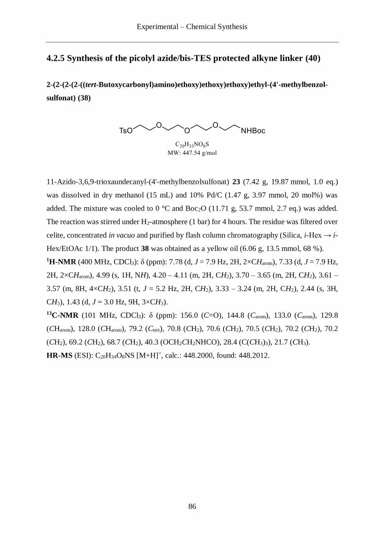
Experimental – Chemical Synthesis
86
4.2.5 Synthesis of the picolyl azide/bis-TES protected alkyne linker (40)
2-(2-(2-(2-((tert-Butoxycarbonyl)amino)ethoxy)ethoxy)ethoxy)ethyl-(4'-methylbenzol-
sulfonat) (38)
11-Azido-3,6,9-trioxaundecanyl-(4'-methylbenzolsulfonat) 23 (7.42 g, 19.87 mmol, 1.0 eq.)
was dissolved in dry methanol (15 mL) and 10% Pd/C (1.47 g, 3.97 mmol, 20 mol%) was
added. The mixture was cooled to 0 °C and Boc2O (11.71 g, 53.7 mmol, 2.7 eq.) was added.
The reaction was stirred under H2-atmosphere (1 bar) for 4 hours. The residue was filtered over
celite, concentrated in vacuo and purified by flash column chromatography (Silica, i-Hex → i-
Hex/EtOAc 1/1). The product 38 was obtained as a yellow oil (6.06 g, 13.5 mmol, 68 %).
1H-NMR (400 MHz, CDCl3): δ (ppm): 7.78 (d, J = 7.9 Hz, 2H, 2×CHarom), 7.33 (d, J = 7.9 Hz,
2H, 2×CHarom), 4.99 (s, 1H, NH), 4.20 – 4.11 (m, 2H, CH2), 3.70 – 3.65 (m, 2H, CH2), 3.61 –
3.57 (m, 8H, 4×CH2), 3.51 (t, J = 5.2 Hz, 2H, CH2), 3.33 – 3.24 (m, 2H, CH2), 2.44 (s, 3H,
CH3), 1.43 (d, J = 3.0 Hz, 9H, 3×CH3).
13C-NMR (101 MHz, CDCl3): δ (ppm): 156.0 (C=O), 144.8 (Carom), 133.0 (Carom), 129.8
(CHarom), 128.0 (CHarom), 79.2 (Ctert), 70.8 (CH2), 70.6 (CH2), 70.5 (CH2), 70.2 (CH2), 70.2
(CH2), 69.2 (CH2), 68.7 (CH2), 40.3 (OCH2CH2NHCO), 28.4 (C(CH3)3), 21.7 (CH3).
HR-MS (ESI): C20H34O8NS [M+H]+, calc.: 448.2000, found: 448.2012.
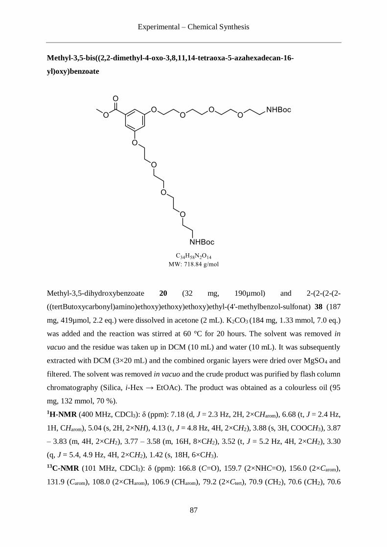
Experimental – Chemical Synthesis
87
Methyl-3,5-bis((2,2-dimethyl-4-oxo-3,8,11,14-tetraoxa-5-azahexadecan-16-
yl)oxy)benzoate
Methyl-3,5-dihydroxybenzoate 20 (32 mg, 190µmol) and 2-(2-(2-(2-
((tertButoxycarbonyl)amino)ethoxy)ethoxy)ethoxy)ethyl-(4'-methylbenzol-sulfonat) 38 (187
mg, 419µmol, 2.2 eq.) were dissolved in acetone (2 mL). K2CO3 (184 mg, 1.33 mmol, 7.0 eq.)
was added and the reaction was stirred at 60 °C for 20 hours. The solvent was removed in
vacuo and the residue was taken up in DCM (10 mL) and water (10 mL). It was subsequently
extracted with DCM (3×20 mL) and the combined organic layers were dried over MgSO4 and
filtered. The solvent was removed in vacuo and the crude product was purified by flash column
chromatography (Silica, i-Hex → EtOAc). The product was obtained as a colourless oil (95
mg, 132 mmol, 70 %).
1H-NMR (400 MHz, CDCl3): δ (ppm): 7.18 (d, J = 2.3 Hz, 2H, 2×CHarom), 6.68 (t, J = 2.4 Hz,
1H, CHarom), 5.04 (s, 2H, 2×NH), 4.13 (t, J = 4.8 Hz, 4H, 2×CH2), 3.88 (s, 3H, COOCH3), 3.87
– 3.83 (m, 4H, 2×CH2), 3.77 – 3.58 (m, 16H, 8×CH2), 3.52 (t, J = 5.2 Hz, 4H, 2×CH2), 3.30
(q, J = 5.4, 4.9 Hz, 4H, 2×CH2), 1.42 (s, 18H, 6×CH3).
13C-NMR (101 MHz, CDCl3): δ (ppm): 166.8 (C=O), 159.7 (2×NHC=O), 156.0 (2×Carom),
131.9 (Carom), 108.0 (2×CHarom), 106.9 (CHarom), 79.2 (2×Ctert), 70.9 (CH2), 70.6 (CH2), 70.6
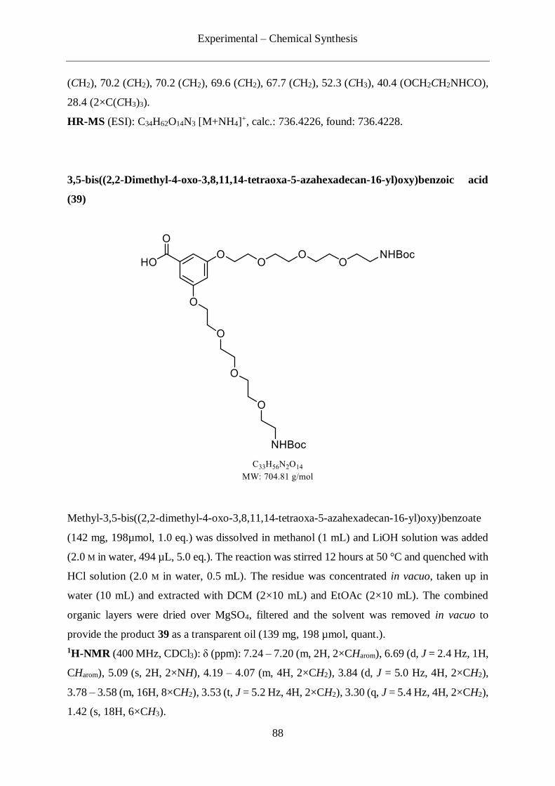
Experimental – Chemical Synthesis
88
(CH2), 70.2 (CH2), 70.2 (CH2), 69.6 (CH2), 67.7 (CH2), 52.3 (CH3), 40.4 (OCH2CH2NHCO),
28.4 (2×C(CH3)3).
HR-MS (ESI): C34H62O14N3 [M+NH4]+, calc.: 736.4226, found: 736.4228.
3,5-bis((2,2-Dimethyl-4-oxo-3,8,11,14-tetraoxa-5-azahexadecan-16-yl)oxy)benzoic acid
(39)
Methyl-3,5-bis((2,2-dimethyl-4-oxo-3,8,11,14-tetraoxa-5-azahexadecan-16-yl)oxy)benzoate
(142 mg, 198µmol, 1.0 eq.) was dissolved in methanol (1 mL) and LiOH solution was added
(2.0 M in water, 494 µL, 5.0 eq.). The reaction was stirred 12 hours at 50 °C and quenched with
HCl solution (2.0 M in water, 0.5 mL). The residue was concentrated in vacuo, taken up in
water (10 mL) and extracted with DCM (2×10 mL) and EtOAc (2×10 mL). The combined
organic layers were dried over MgSO4, filtered and the solvent was removed in vacuo to
provide the product 39 as a transparent oil (139 mg, 198 µmol, quant.).
1H-NMR (400 MHz, CDCl3): δ (ppm): 7.24 – 7.20 (m, 2H, 2×CHarom), 6.69 (d, J = 2.4 Hz, 1H,
CHarom), 5.09 (s, 2H, 2×NH), 4.19 – 4.07 (m, 4H, 2×CH2), 3.84 (d, J = 5.0 Hz, 4H, 2×CH2),
3.78 – 3.58 (m, 16H, 8×CH2), 3.53 (t, J = 5.2 Hz, 4H, 2×CH2), 3.30 (q, J = 5.4 Hz, 4H, 2×CH2),
1.42 (s, 18H, 6×CH3).

Experimental – Chemical Synthesis
89
13C-NMR (101 MHz, CDCl3): δ (ppm): 170.0 (C=O), 159.8 (2×NHC=O), 156.1 (2×Carom),
131.5 (Carom), 108.5 (2×CHarom), 107.4 (CHarom), 79.3 (2×Ctert), 70.9 (CH2), 70.6 (CH2), 70.6
(CH2), 70.2 (CH2), 69.6 (CH2), 67.7 (CH2), 40.3 (OCH2CH2NHCO), 28.4 (2×C(CH3)3).
HR-MS (ESI): C33H60O14N3 [M+NH4]+, calc.: 722.4070, found: 722.4069.
Di-tert-butyl(((((((((5-((2-(2-(2-(6-
(azidomethyl)nicotinamido)ethoxy)ethoxy)ethyl)carbamoyl)-1,3-
phenylene)bis(oxy))bis(ethane-2,1-diyl))bis(oxy))bis(ethane-2,1-
diyl))bis(oxy))bis(ethane-2,1-diyl))bis(oxy))bis(ethane-2,1-diyl))dicarbamate
39 (90 mg, 128 µmol, 1.0 eq.) was dissolved in dry DMF (1 mL) and DIPEA (44.5 µL, 33 mg,
255 µmol, 2.0 eq.) and HATU (58 mg, 153 µmol, 1.2 eq.) were added. The reaction mixture
was stirred 30 minutes at room temperature and 21 (43 mg, 140 µmol, 1.1 eq.) was added. The
reaction was stirred overnight and the solvent was removed in vacuo. The residue was taken
up in DCM (10 mL) and washed with brine (10 mL). The organic layer was dried over MgSO4,
filtered, concentrated in vacuo and purified by flash column chromatography (Silica, EtOAc
→ EtOAc/MeOH 98/2). The product was obtained as an orange oil (86 mg, 86 µmol, 68 %).
1H-NMR (400 MHz, CDCl3): δ (ppm): 9.01 (d, J = 2.2 Hz, 1H, CHarom), 8.17 (dd, J = 8.1, 2.2
Hz, 1H CHarom), 7.40 (d, J = 8.1 Hz, 1H, CHarom), 6.93 (d, J = 2.2 Hz, 2H, 2×CHarom), 6.55 (t,
J = 2.3 Hz, 1H, CHarom), 5.11 (s, 2H, 2×NH), 4.53 (s, 2H, CaromCH2N3), 4.10 (t, J = 4.7 Hz, 4H,

Experimental – Chemical Synthesis
90
2×CH2), 3.83 (t, J = 4.7 Hz, 4H, 2×CH2), 3.75 – 3.58 (m, 30H, 15×CH2), 3.52 (t, J = 5.2 Hz,
4H, 2×CH2), 3.28 (q, J = 5.4 Hz, 4H, 2×CH2), 1.42 (s, 18H, 6×CH3).
13C-NMR (101 MHz, CDCl3): δ (ppm): 167.3 (C=O), 165.2 (C=O), 159.8 (2×Carom), 156.1
(CaromHNarom), 147.9 (2×NHC=O), 136.7 (CaromH), 130.5 (Carom), 129.5 (Carom), 121.7 (CaromH),
105.9 (2×CaromH), 104.3 (CaromH), 79.2 (2×Ctert), 70.8 (CH2), 70.6 (CH2), 70.6 (CH2), 70.3
(CH2), 70.2 (CH2), 69.7 (CH2), 69.6 (CH2), 69.6 (CH2), 67.6 (CH2), 55.1 (CaromCH2N3), 40.4
(OCH2CH2NHCO), 39.8 (OCH2CH2NHCO), 28.4 (2×C(CH3)3).
HR-MS (ESI): C46H75O16N8 [M+H]+, calc.: 995.5296, found: 995.5299.
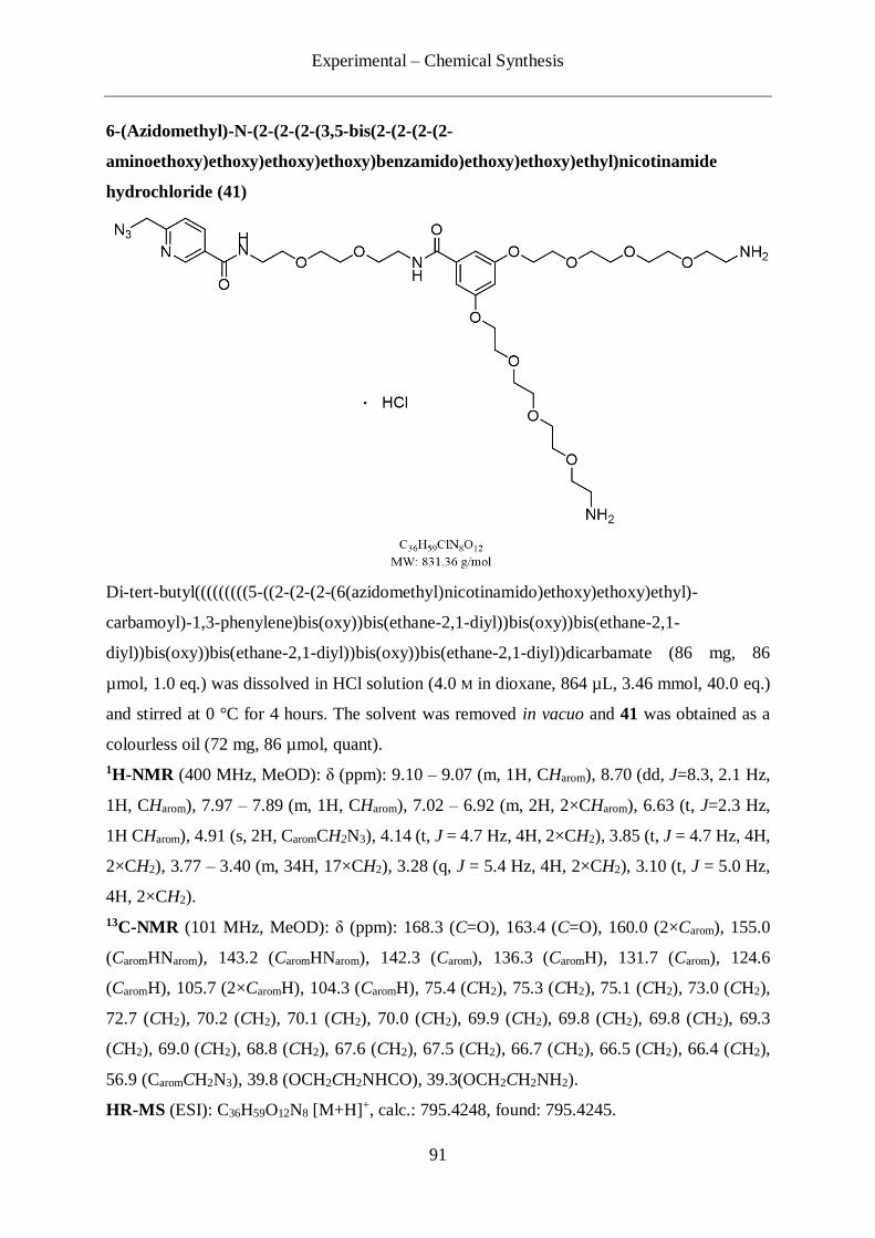
Experimental – Chemical Synthesis
91
6-(Azidomethyl)-N-(2-(2-(2-(3,5-bis(2-(2-(2-(2-
aminoethoxy)ethoxy)ethoxy)ethoxy)benzamido)ethoxy)ethoxy)ethyl)nicotinamide
hydrochloride (41)
Di-tert-butyl(((((((((5-((2-(2-(2-(6(azidomethyl)nicotinamido)ethoxy)ethoxy)ethyl)-
carbamoyl)-1,3-phenylene)bis(oxy))bis(ethane-2,1-diyl))bis(oxy))bis(ethane-2,1-
diyl))bis(oxy))bis(ethane-2,1-diyl))bis(oxy))bis(ethane-2,1-diyl))dicarbamate (86 mg, 86
µmol, 1.0 eq.) was dissolved in HCl solution (4.0 M in dioxane, 864 µL, 3.46 mmol, 40.0 eq.)
and stirred at 0 °C for 4 hours. The solvent was removed in vacuo and 41 was obtained as a
colourless oil (72 mg, 86 µmol, quant).
1H-NMR (400 MHz, MeOD): δ (ppm): 9.10 – 9.07 (m, 1H, CHarom), 8.70 (dd, J=8.3, 2.1 Hz,
1H, CHarom), 7.97 – 7.89 (m, 1H, CHarom), 7.02 – 6.92 (m, 2H, 2×CHarom), 6.63 (t, J=2.3 Hz,
1H CHarom), 4.91 (s, 2H, CaromCH2N3), 4.14 (t, J = 4.7 Hz, 4H, 2×CH2), 3.85 (t, J = 4.7 Hz, 4H,
2×CH2), 3.77 – 3.40 (m, 34H, 17×CH2), 3.28 (q, J = 5.4 Hz, 4H, 2×CH2), 3.10 (t, J = 5.0 Hz,
4H, 2×CH2).
13C-NMR (101 MHz, MeOD): δ (ppm): 168.3 (C=O), 163.4 (C=O), 160.0 (2×Carom), 155.0
(CaromHNarom), 143.2 (CaromHNarom), 142.3 (Carom), 136.3 (CaromH), 131.7 (Carom), 124.6
(CaromH), 105.7 (2×CaromH), 104.3 (CaromH), 75.4 (CH2), 75.3 (CH2), 75.1 (CH2), 73.0 (CH2),
72.7 (CH2), 70.2 (CH2), 70.1 (CH2), 70.0 (CH2), 69.9 (CH2), 69.8 (CH2), 69.8 (CH2), 69.3
(CH2), 69.0 (CH2), 68.8 (CH2), 67.6 (CH2), 67.5 (CH2), 66.7 (CH2), 66.5 (CH2), 66.4 (CH2),
56.9 (CaromCH2N3), 39.8 (OCH2CH2NHCO), 39.3(OCH2CH2NH2).
HR-MS (ESI): C36H59O12N8 [M+H]+, calc.: 795.4248, found: 795.4245.

Experimental – Chemical Synthesis
92
N,N'-(((((((((5-((2-(2-(2-(6-(Azidomethyl)nicotinamido)ethoxy)ethoxy)ethyl)carbamoyl)-
1,3-phenylene)bis(oxy))bis(ethane-2,1-diyl))bis(oxy))bis(ethane-2,1-
diyl))bis(oxy))bis(ethane-2,1-diyl))bis(oxy))bis(ethane-2,1-diyl))bis(5-(triethylsilyl)pent-
4-ynamide) (40)
5-(triethylsilyl)pent-4-ynoic acid 36 (12 mg, 57µmol, 3.0 eq.) was dissolved in dry DMF (200
µL). DIPEA (13 µL, 9.8 mg, 76 µmol, 4.0 eq.) and HATU (17.22 mg, 45.29 µmol, 2.4 eq.)
were added and the reaction mixture was stirred 30 minutes at room temperature. A solution
of 41 (15 mg, 19 µmol, 1.0 eq.) in DMF (200 µL) was added and the reaction was stirred
overnight at room temperature. The solvent was removed in vacuo and the residue was taken
up in DCM (10 mL) and washed with brine (10 mL). The organic layer was dried over MgSO4,
filtered, concentrated in vacuo and purified by flash column chromatography (Silica, DCM →
DCM/MeOH 90:10). The product 40 was obtained as a clear yellow oil (22 mg, 19 µmol, 99
%).
1H-NMR (599 MHz, CDCl3): δ (ppm): 9.10 – 9.07 (m, 1H, CHarom), 8.31 (dd, J=8.3, 2.1 Hz,
1H, CHarom), 8.03 – 7.95 (m, 1H, CHarom), 7.54 (d, J = 8.1 Hz, 1H, CHarom), 6.96 – 6.84 (m, 2H,
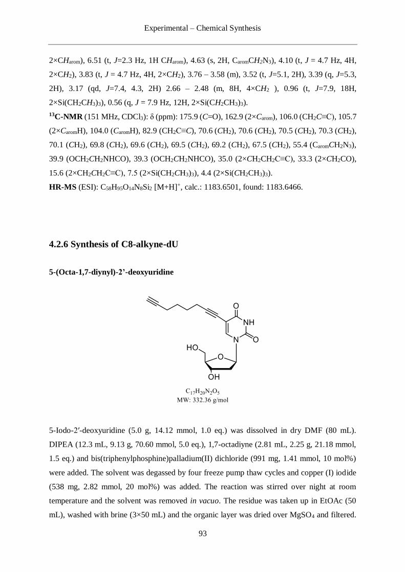
Experimental – Chemical Synthesis
93
2×CHarom), 6.51 (t, J=2.3 Hz, 1H CHarom), 4.63 (s, 2H, CaromCH2N3), 4.10 (t, J = 4.7 Hz, 4H,
2×CH2), 3.83 (t, J = 4.7 Hz, 4H, 2×CH2), 3.76 – 3.58 (m), 3.52 (t, J=5.1, 2H), 3.39 (q, J=5.3,
2H), 3.17 (qd, J=7.4, 4.3, 2H) 2.66 – 2.48 (m, 8H, 4×CH2 ), 0.96 (t, J=7.9, 18H,
2×Si(CH2CH3)3), 0.56 (q, J = 7.9 Hz, 12H, 2×Si(CH2CH3)3).
13C-NMR (151 MHz, CDCl3): δ (ppm): 175.9 (C=O), 162.9 (2×Carom), 106.0 (CH2C≡C), 105.7
(2×CaromH), 104.0 (CaromH), 82.9 (CH2C≡C), 70.6 (CH2), 70.6 (CH2), 70.5 (CH2), 70.3 (CH2),
70.1 (CH2), 69.8 (CH2), 69.6 (CH2), 69.5 (CH2), 69.2 (CH2), 67.5 (CH2), 55.4 (CaromCH2N3),
39.9 (OCH2CH2NHCO), 39.3 (OCH2CH2NHCO), 35.0 (2×CH2CH2C≡C), 33.3 (2×CH2CO),
15.6 (2×CH2CH2C≡C), 7.5 (2×Si(CH2CH3)3), 4.4 (2×Si(CH2CH3)3).
HR-MS (ESI): C58H95O14N8Si2 [M+H]+, calc.: 1183.6501, found: 1183.6466.
4.2.6 Synthesis of C8-alkyne-dU
5-(Octa-1,7-diynyl)-2’-deoxyuridine
5-Iodo-2′-deoxyuridine (5.0 g, 14.12 mmol, 1.0 eq.) was dissolved in dry DMF (80 mL).
DIPEA (12.3 mL, 9.13 g, 70.60 mmol, 5.0 eq.), 1,7-octadiyne (2.81 mL, 2.25 g, 21.18 mmol,
1.5 eq.) and bis(triphenylphosphine)palladium(II) dichloride (991 mg, 1.41 mmol, 10 mol%)
were added. The solvent was degassed by four freeze pump thaw cycles and copper (I) iodide
(538 mg, 2.82 mmol, 20 mol%) was added. The reaction was stirred over night at room
temperature and the solvent was removed in vacuo. The residue was taken up in EtOAc (50
mL), washed with brine (3×50 mL) and the organic layer was dried over MgSO4 and filtered.
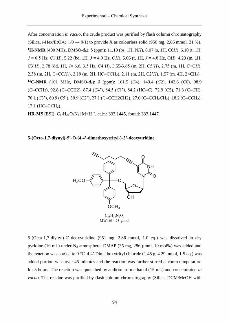
Experimental – Chemical Synthesis
94
After concentration in vacuo, the crude product was purified by flash column chromatography
(Silica, i-Hex/EtOAc 1/0 → 0/1) to provide X as colourless solid (950 mg, 2.86 mmol, 21 %).
1H-NMR (400 MHz, DMSO-d6): δ (ppm): 11.10 (bs, 1H, NH), 8.07 (s, 1H, C6H), 6.10 (t, 1H,
J = 6.5 Hz, C1’H), 5.22 (bd, 1H, J = 4.0 Hz, OH), 5.06 (t, 1H, J = 4.8 Hz, OH), 4.23 (m, 1H,
C3’H), 3.78 (dd, 1H, J= 6.6, 3.5 Hz, C4’H), 3.55-3.65 (m, 2H, C5’H), 2.75 (m, 1H, C≡CH),
2.38 (m, 2H, C≡CCH2), 2.19 (m, 2H, HC≡CCH2), 2.11 (m, 2H, C2’H), 1.57 (m, 4H, 2×CH2).
13C-NMR (101 MHz, DMSO-d6): δ (ppm): 161.5 (C4), 149.4 (C2), 142.6 (C6), 98.9
(C≡CCH2), 92.8 (C≡CCH2), 87.4 (C4’), 84.5 (C1’), 84.2 (HC≡C), 72.9 (C5), 71.3 (C≡CH),
70.1 (C3’), 60.9 (C5’), 39.9 (C2’), 27.1 (C≡CCH2CH2), 27.0 (C≡CCH2CH2), 18.2 (C≡CCH2),
17.1 (HC≡CCH2).
HR-MS (ESI): C17H21O5N2 [M+H]+, calc.: 333.1445, found: 333.1447.
5-(Octa-1,7-diynyl)-5’-O-(4,4’-dimethoxytrityl-)-2’-deoxyuridine
5-(Octa-1,7-diynyl)-2’-deoxyuridine (951 mg, 2.86 mmol, 1.0 eq.) was dissolved in dry
pyridine (10 mL) under N2 atmosphere. DMAP (35 mg, 286 µmol, 10 mol%) was added and
the reaction was cooled to 0 °C. 4,4′-Dimethoxytrityl chloride (1.45 g, 4.29 mmol, 1.5 eq.) was
added portion-wise over 45 minutes and the reaction was further stirred at room temperature
for 5 hours. The reaction was quenched by addition of methanol (15 mL) and concentrated in
vacuo. The residue was purified by flash column chromatography (Silica, DCM/MeOH with
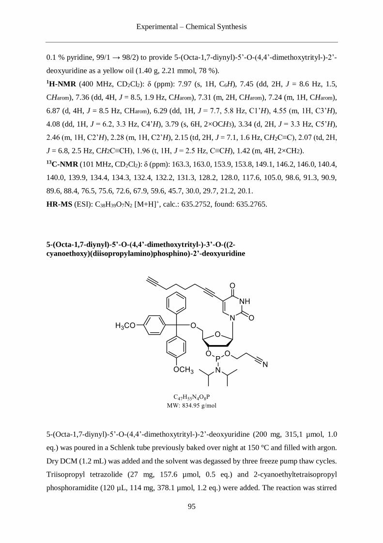
Experimental – Chemical Synthesis
95
0.1 % pyridine, 99/1 → 98/2) to provide 5-(Octa-1,7-diynyl)-5’-O-(4,4’-dimethoxytrityl-)-2’-
deoxyuridine as a yellow oil (1.40 g, 2.21 mmol, 78 %).
1H-NMR (400 MHz, CD2Cl2): δ (ppm): 7.97 (s, 1H, C6H), 7.45 (dd, 2H, J = 8.6 Hz, 1.5,
CHarom), 7.36 (dd, 4H, J = 8.5, 1.9 Hz, CHarom), 7.31 (m, 2H, CHarom), 7.24 (m, 1H, CHarom),
6.87 (d, 4H, J = 8.5 Hz, CHarom), 6.29 (dd, 1H, J = 7.7, 5.8 Hz, C1’H), 4.55 (m, 1H, C3’H),
4.08 (dd, 1H, J = 6.2, 3.3 Hz, C4’H), 3.79 (s, 6H, 2×OCH3), 3.34 (d, 2H, J = 3.3 Hz, C5’H),
2.46 (m, 1H, C2’H), 2.28 (m, 1H, C2’H), 2.15 (td, 2H, J = 7.1, 1.6 Hz, CH2C≡C), 2.07 (td, 2H,
J = 6.8, 2.5 Hz, CH2C≡CH), 1.96 (t, 1H, J = 2.5 Hz, C≡CH), 1.42 (m, 4H, 2×CH2).
13C-NMR (101 MHz, CD2Cl2): δ (ppm): 163.3, 163.0, 153.9, 153.8, 149.1, 146.2, 146.0, 140.4,
140.0, 139.9, 134.4, 134.3, 132.4, 132.2, 131.3, 128.2, 128.0, 117.6, 105.0, 98.6, 91.3, 90.9,
89.6, 88.4, 76.5, 75.6, 72.6, 67.9, 59.6, 45.7, 30.0, 29.7, 21.2, 20.1.
HR-MS (ESI): C38H39O7N2 [M+H]+, calc.: 635.2752, found: 635.2765.
5-(Octa-1,7-diynyl)-5’-O-(4,4’-dimethoxytrityl-)-3’-O-((2-
cyanoethoxy)(diisopropylamino)phosphino)-2’-deoxyuridine
5-(Octa-1,7-diynyl)-5’-O-(4,4’-dimethoxytrityl-)-2’-deoxyuridine (200 mg, 315,1 µmol, 1.0
eq.) was poured in a Schlenk tube previously baked over night at 150 °C and filled with argon.
Dry DCM (1.2 mL) was added and the solvent was degassed by three freeze pump thaw cycles.
Triisopropyl tetrazolide (27 mg, 157.6 µmol, 0.5 eq.) and 2-cyanoethyltetraisopropyl
phosphoramidite (120 µL, 114 mg, 378.1 µmol, 1.2 eq.) were added. The reaction was stirred

Experimental – Chemical Synthesis
96
at room temperature for 24 hours and the solvent was removed in vacuo. The crude mixture
was purified by flash column chromatography with degassed eluent (Silica, i-Hex/EtOAc 1/3
with 1 % pyridine) under argon atmosphere to provide X as white solid (143 mg, 171.27 µmol,
54 %).
13P NMR (80.9 MHz, CD2Cl2): δ (ppm): 149.9, 149.6.
HR-MS (ESI): C47H56O8N4P [M+H]+, calc.: 838.3831, found: 835.3824.
4.2.7 Synthesis of the caC phosphoramidite building block (42)
5-Iodo-3´,5´-O-(di-tert-butylsilanediyl)-2´-O-tert-butyldimethylsilyl-cytidine (44)
3´,5´-O-(di-tert-butylsilanediyl)-2´-O-tert-butyldimethylsilyl-cytidine 43 (5.0 g, 10.04 mmol,
1.0 eq.) was dissolved in dry acetonitrile (100 mL). To the solution were added iodine (5.10 g,
20.09 mmol, 2.0 eq.) and Ammonium Cerium(IV) Nitrate (11.01 g, 20.09 mmol, 2.0 eq.) at
room temperature. After being stirred for 2.5 hours at 50 °C, the reaction mixture was cooled
down to room temperature. To the solution was added ethyl acetate (200 mL) and it was
subsequently washed with Na2S2O3 saturated aqueous solution/NaHCO3 saturated aqueous
solution (200 mL, 1/1). The organic phase was dried over Na2SO4, filtered and concentrated in
vacuo. The residue was purified by flash column chromatography (Silica, i-Hex/EtOAc 1/0 →
7/3) to provide 5-iodo-3´,5´-O-(di-tert-butylsilanediyl)-2´-O-tert-butyldimethylsilyl-cytidine
44 (3.07 g, 4.92 mmol, 49 %).

Experimental – Chemical Synthesis
97
1H-NMR (400 MHz, CDCl3): δ (ppm): 7.88 (s, 1H), 5.65 (s, 1H), 4.50 (dd, J = 8.4, 4.2 Hz,
1H), 4.42 (d, J = 4.7 Hz, 1H), 4.17 – 4.01 (m, 3H), 1.09 (s, 9H), 1.06 (s, 9H), 0.96 (s, 9H), 0.22
(s, 3H), 0.17 (s, 3H).
13C-NMR (101 MHz, CDCl3): δ (ppm): 165.6, 155.3, 148.8, 96.4, 77.1, 76.3, 75.9, 68.7, 57.6,
28.0, 27.7, 26.5, 23.6, 21.3, 19.2, -4.0, -4.6.
IR: νmax (cm-1): 2933, 2858, 1646, 1616, 1472, 1058, 828, 776 cm-1.
HR-MS (ESI): C23H43IN3O5Si2+ [M+H]+, calc.: 624.1780, found: 624.1784.
5-(2-(para-Nitrophenyl)ethoxycarbonyl)-3´,5´-O -(di-tert-butylsilanediyl)-2´-O-tert-butyl
dimethylsilyl-cytidine (45)
5-iodo-3´,5´-O-(di-tert-butylsilanediyl)-2´-O-tert-butyldimethylsilyl-cytidine 44 (1.50 g, 2.41
mmol, 1.0 eq.), p-nitrophenethyl alcohol (2.01 mg, 12.03 mmol, 5.0 eq.), Pd2(CH3CN)2Cl2 (63
mg, 0.24 mmol, 10 mol%) and diisopropylethylamine (840 µL, 4.82 mmol, 2.0 eq.) were added
in a high pressure glass autoclave. The autoclave was flushed with CO three times to remove
residual air and subsequently the reaction was stirred at a CO pressure of 3.5 bar at 80 °C for
16 hours. The CO was expelled and the residue was purified by flash column chromatography
(Silica, DCM/MeOH, 100/0 → 98/2) to afford compound 5-(2-(para-
nitrophenyl)ethoxycarbonyl)-3´,5´-O -(di-tert-butylsilanediyl)-2´-O-tert-butyldimethylsilyl-
cytidine 45 (876 mg, 1.26 mmol, 52 %).
1H-NMR (599 MHz, CDCl3): δ (ppm): 8.28 (s, 1H), 8.24 – 8.19 (m, 2H), 7.88 – 7.80 (s, br,
1H), 7.41 (d, J = 8.5 Hz, 2H), 6.61 – 6.41 (s, br, 1H), 5.69 (s, 1H), 4.58 – 4.48 (m, 2H), 4.32
(d, J = 4.0 Hz, 1H), 4.31 – 4.24 (m, 1H), 3.81 (dd, J = 9.3 Hz, 1H), 3.74 (dd, J = 9.7, 4.1 Hz,
Experimental – Chemical Synthesis
98
1H), 3.14 (t, J = 6.9 Hz, 2H), 1.04 (s, 1H), 1.02 (s, 9H), 1.01 (s, 9H), 0.95 (s, 9H), 0.24 (s, 3H),
0.16 (s, 3H).
13C-NMR (151 MHz, CDCl3): δ (ppm): 164.5, 163.5, 153.6, 147.3, 147.2, 144.8, 129.6, 124.2,
95.3, 94.4, 75.6, 75.3, 74.8, 68.0, 64.4, 35.1, 27.6, 27.4, 27.1, 26.0, 22.9, 20.5, 18.3, -4.2, -4.7.
IR: νmax (cm-1): 3403, 3187, 2934, 2860, 1714, 1677, 1643, 1516, 1472, 1343, 1311, 1250,
1156, 1130, 1118, 1081, 1059, 830, 780 cm-1.
HR-MS (ESI): C32H51N4O9Si2+ [M+H]+, calc.: 691.3189, found: 691.3190.
N4-para-Methoxybenzoyl-5-(2-(para-nitrophenyl)ethoxycarbonyl)-3´,5´-O -(di-tert-butyls
ilanediyl)-2´-O-tert-butyldimethylsilyl-cytidine (46)
5-(2-(para-nitrophenyl)ethoxycarbonyl)-3´,5´-O-(di-tert-butylsilanediyl)-2´-O-tert-
butyldimethylsilyl-cytidine 45 (350 mg, 505.1 µmol, 1.0 eq.) was dissolved in dry pyridine
(10.5 mL). To the solution was added p-methoxybenzoyl chloride (140 µL, 1.03 mmol, 2.0 eq.)
at room temperature. The reaction mixture was put on a pre-heated oil bath at 90 °C and stirred
for 4 hours. The reaction mixture was cooled down to room temperature and EtOAc (70 mL)
was added. The residue was washed with brine (3×15 mL) and the organic phase was dried
over Na2SO4, filtered and concentrated in vacuo. The crude product was purified by flash
column chromatography (Silica, iHex/DCM, 50/50 → 0/100 and then DCM/MeOH, 100/0 →
90/10). All the fractions were collected and concentrated in vacuo. The residue was dissolved
again in DCM (5 mL) and washed with NaHCO3 saturated aqueous solution (3×5 mL). The
aqueous phase was retro-extracted with DCM (1×5 mL). The combined organic layers were
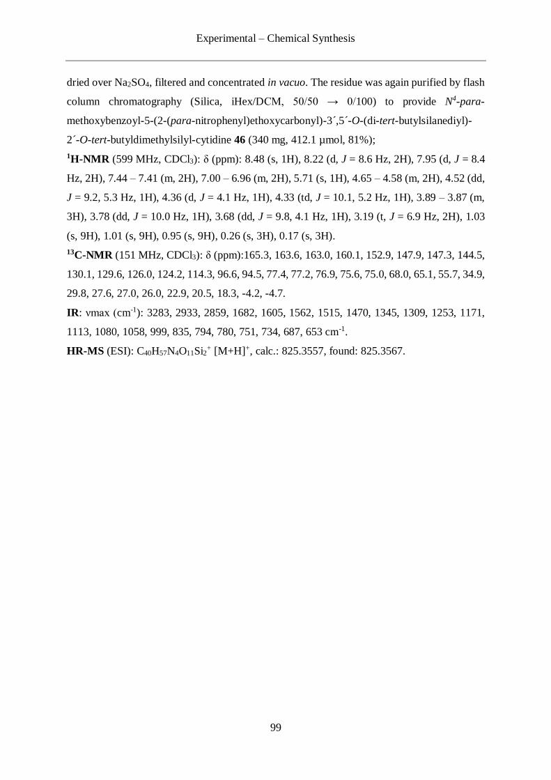
Experimental – Chemical Synthesis
99
dried over Na2SO4, filtered and concentrated in vacuo. The residue was again purified by flash
column chromatography (Silica, iHex/DCM, 50/50 → 0/100) to provide N4-para-
methoxybenzoyl-5-(2-(para-nitrophenyl)ethoxycarbonyl)-3´,5´-O-(di-tert-butylsilanediyl)-
2´-O-tert-butyldimethylsilyl-cytidine 46 (340 mg, 412.1 µmol, 81%);
1H-NMR (599 MHz, CDCl3): δ (ppm): 8.48 (s, 1H), 8.22 (d, J = 8.6 Hz, 2H), 7.95 (d, J = 8.4
Hz, 2H), 7.44 – 7.41 (m, 2H), 7.00 – 6.96 (m, 2H), 5.71 (s, 1H), 4.65 – 4.58 (m, 2H), 4.52 (dd,
J = 9.2, 5.3 Hz, 1H), 4.36 (d, J = 4.1 Hz, 1H), 4.33 (td, J = 10.1, 5.2 Hz, 1H), 3.89 – 3.87 (m,
3H), 3.78 (dd, J = 10.0 Hz, 1H), 3.68 (dd, J = 9.8, 4.1 Hz, 1H), 3.19 (t, J = 6.9 Hz, 2H), 1.03
(s, 9H), 1.01 (s, 9H), 0.95 (s, 9H), 0.26 (s, 3H), 0.17 (s, 3H).
13C-NMR (151 MHz, CDCl3): δ (ppm):165.3, 163.6, 163.0, 160.1, 152.9, 147.9, 147.3, 144.5,
130.1, 129.6, 126.0, 124.2, 114.3, 96.6, 94.5, 77.4, 77.2, 76.9, 75.6, 75.0, 68.0, 65.1, 55.7, 34.9,
29.8, 27.6, 27.0, 26.0, 22.9, 20.5, 18.3, -4.2, -4.7.
IR: νmax (cm-1): 3283, 2933, 2859, 1682, 1605, 1562, 1515, 1470, 1345, 1309, 1253, 1171,
1113, 1080, 1058, 999, 835, 794, 780, 751, 734, 687, 653 cm-1.
HR-MS (ESI): C40H57N4O11Si2+ [M+H]+, calc.: 825.3557, found: 825.3567.
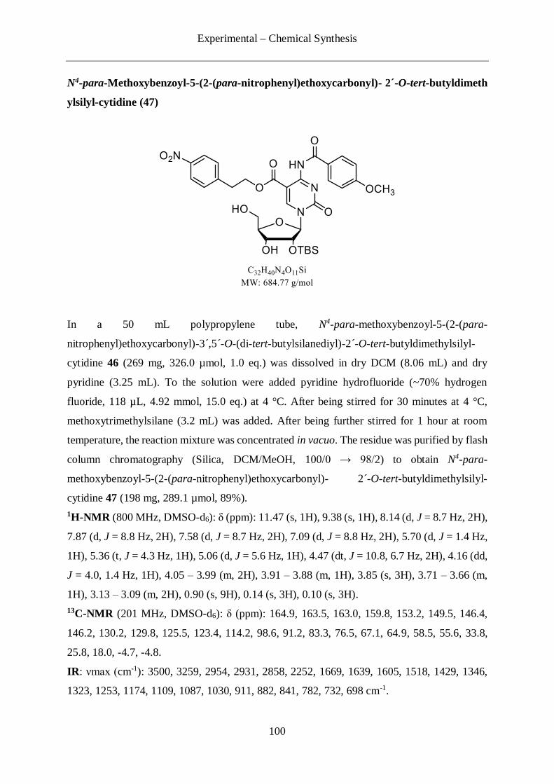
Experimental – Chemical Synthesis
100
N4-para-Methoxybenzoyl-5-(2-(para-nitrophenyl)ethoxycarbonyl)- 2´-O-tert-butyldimeth
ylsilyl-cytidine (47)
In a 50 mL polypropylene tube, N4-para-methoxybenzoyl-5-(2-(para-
nitrophenyl)ethoxycarbonyl)-3´,5´-O-(di-tert-butylsilanediyl)-2´-O-tert-butyldimethylsilyl-
cytidine 46 (269 mg, 326.0 µmol, 1.0 eq.) was dissolved in dry DCM (8.06 mL) and dry
pyridine (3.25 mL). To the solution were added pyridine hydrofluoride (~70% hydrogen
fluoride, 118 µL, 4.92 mmol, 15.0 eq.) at 4 °C. After being stirred for 30 minutes at 4 °C,
methoxytrimethylsilane (3.2 mL) was added. After being further stirred for 1 hour at room
temperature, the reaction mixture was concentrated in vacuo. The residue was purified by flash
column chromatography (Silica, DCM/MeOH, 100/0 → 98/2) to obtain N4-para-
methoxybenzoyl-5-(2-(para-nitrophenyl)ethoxycarbonyl)- 2´-O-tert-butyldimethylsilyl-
cytidine 47 (198 mg, 289.1 µmol, 89%).
1H-NMR (800 MHz, DMSO-d6): δ (ppm): 11.47 (s, 1H), 9.38 (s, 1H), 8.14 (d, J = 8.7 Hz, 2H),
7.87 (d, J = 8.8 Hz, 2H), 7.58 (d, J = 8.7 Hz, 2H), 7.09 (d, J = 8.8 Hz, 2H), 5.70 (d, J = 1.4 Hz,
1H), 5.36 (t, J = 4.3 Hz, 1H), 5.06 (d, J = 5.6 Hz, 1H), 4.47 (dt, J = 10.8, 6.7 Hz, 2H), 4.16 (dd,
J = 4.0, 1.4 Hz, 1H), 4.05 – 3.99 (m, 2H), 3.91 – 3.88 (m, 1H), 3.85 (s, 3H), 3.71 – 3.66 (m,
1H), 3.13 – 3.09 (m, 2H), 0.90 (s, 9H), 0.14 (s, 3H), 0.10 (s, 3H).
13C-NMR (201 MHz, DMSO-d6): δ (ppm): 164.9, 163.5, 163.0, 159.8, 153.2, 149.5, 146.4,
146.2, 130.2, 129.8, 125.5, 123.4, 114.2, 98.6, 91.2, 83.3, 76.5, 67.1, 64.9, 58.5, 55.6, 33.8,
25.8, 18.0, -4.7, -4.8.
IR: νmax (cm-1): 3500, 3259, 2954, 2931, 2858, 2252, 1669, 1639, 1605, 1518, 1429, 1346,
1323, 1253, 1174, 1109, 1087, 1030, 911, 882, 841, 782, 732, 698 cm-1.

Experimental – Chemical Synthesis
101
HR-MS (ESI): C32H41N4O11Si+ [M+H]+, calc.: 685.2536, found: 685.2545.
N4-para-Methoxybenzoyl-5-(2-(para-nitrophenyl)ethoxycarbonyl)- 5´-O-(4,4´-dimethoxy
trityl)-2´-O-tert-butyldimethylsilyl-cytidine (48)
4,4´-dimethoxytritylchloride (2.03 g, 6.00 mmol, 1.0 eq.) was dissolved in dry DCM (20 mL).
To the solution was added silver triflate (1.54 g, 6.00 mmol, 1.0 eq.) at room temperature to
prepare 4,4´-dimethoxytrityl triflate salt. After being stirred for 1.5 hours, the 4,4´-
dimethoxytrityl triflate salt solution (1.20 mL, 0.36 mmol, 1.5 eq.) was added to a solution of
N4-para-methoxybenzoyl-5-(2-(para-nitrophenyl)ethoxycarbonyl)- 2´-O-tert-
butyldimethylsilyl-cytidine 47 (165 mg, 241.0 µmol) in dry DCM/dry pyridine (4.8 mL, 1/1)
at room temperature. After being stirred for 1.5 hours, additional solution of 4,4´-
dimethoxytrityl triflate salt (0.4 mL, 0.12 mmol, 0.5 eq.) was added to the reaction mixture and
further stirred for 19.5 hours. DCM (20 mL) was added to the solution and washed with
NaHCO3 saturated aqueous solution (3×20 mL). The aqueous phase was extracted again with
DCM (1×20 mL). The combined organic layers were dried over Na2SO4, filtered and
concentrated in vacuo. The residue was purified by flash column chromatography (Silica, i-
Hex/EtOAc 1/0 → 1/1) to afford compound N4-para-methoxybenzoyl-5-(2-(para-
nitrophenyl)ethoxycarbonyl)- 5´-O-(4,4´-dimethoxytrityl)-2´-O-tert-butyldimethylsilyl-
cytidine 48 (226 mg, 228.9 µmol, 95%).
1H-NMR (800 MHz, DMSO-d6): δ (ppm): 11.44 (s, 1H), 8.70 (s, 1H), 8.07 (d, J = 8.7 Hz, 2H),
7.88 (d, J = 8.9 Hz, 2H), 7.43 (d, J = 7.6 Hz, 2H), 7.31 – 7.26 (m, 6H), 7.23 – 7.20 (m, 3H),
7.09 (d, J = 8.9 Hz, 2H), 6.88 – 6.84 (m, 4H), 5.77 (d, J = 1.9 Hz, 1H), 5.11 (d, J = 6.4 Hz,
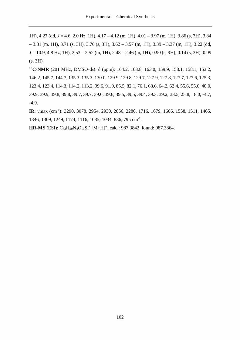
Experimental – Chemical Synthesis
102
1H), 4.27 (dd, J = 4.6, 2.0 Hz, 1H), 4.17 – 4.12 (m, 1H), 4.01 – 3.97 (m, 1H), 3.86 (s, 3H), 3.84
– 3.81 (m, 1H), 3.71 (s, 3H), 3.70 (s, 3H), 3.62 – 3.57 (m, 1H), 3.39 – 3.37 (m, 1H), 3.22 (dd,
J = 10.9, 4.8 Hz, 1H), 2.53 – 2.52 (m, 1H), 2.48 – 2.46 (m, 1H), 0.90 (s, 9H), 0.14 (s, 3H), 0.09
(s, 3H).
13C-NMR (201 MHz, DMSO-d6): δ (ppm): 164.2, 163.8, 163.0, 159.9, 158.1, 158.1, 153.2,
146.2, 145.7, 144.7, 135.3, 135.3, 130.0, 129.9, 129.8, 129.7, 127.9, 127.8, 127.7, 127.6, 125.3,
123.4, 123.4, 114.3, 114.2, 113.2, 99.6, 91.9, 85.5, 82.1, 76.1, 68.6, 64.2, 62.4, 55.6, 55.0, 40.0,
39.9, 39.9, 39.8, 39.8, 39.7, 39.7, 39.6, 39.6, 39.5, 39.5, 39.4, 39.3, 39.2, 33.5, 25.8, 18.0, -4.7,
-4.9.
IR: νmax (cm-1): 3290, 3078, 2954, 2930, 2856, 2280, 1716, 1679, 1606, 1558, 1511, 1465,
1346, 1309, 1249, 1174, 1116, 1085, 1034, 836, 795 cm-1.
HR-MS (ESI): C53H59N4O13Si+ [M+H]+, calc.: 987.3842, found: 987.3864.
Experimental – Chemical Synthesis
103
N4-para-Methoxybenzoyl-5-(2-(para-nitrophenyl)ethoxycarbonyl)-5´-O-(4,4´-
dimethoxytrityl)-2´-O-tert-butyldimethylsilyl-cytidine 3´-(2-cyanoethyl)-N,N-
diisopropylphosphoramidite (42)
N4-para-methoxybenzoyl-5-(2-(para-nitrophenyl)ethoxycarbonyl)- 5´-O-(4,4´-
dimethoxytrityl)-2´-O-tert-butyldimethylsilyl-cytidine 48 (152 mg, 154 µmol, 1.0 eq.) was
dissolved in degassed dry DCM (1.5 mL). To the solution were added diisopropylethylamine
(107 µL, 61.6 µmol, 4.0 eq.) and 2-cyanoethyl N,N-diisopropylchlorophosphoramidite (138
µL, 61.6 µmol, 4.0 eq.) at room temperature. After being stirred for 2 hours, DCM (10 mL)
was added and the residue was washed with NaHCO3 saturated aqueous solution (2×10 mL)
and brine (1×20 mL). The organic phase was dried over Na2SO4, filtered and concentrated in
vacuo. The residue was purified by flash column chromatography with degassed eluent (Silica,
i-Hex/EtOAc 1/1) under argon atmosphere to afford caC amidite 42 (137 mg, 115.4 µmol,
75%) as a diastereo mixture.
1H-NMR (400 MHz, C6D6): δ (ppm): 12.00 (d, J = 55.4 Hz, 1H), 9.25 (s, 1H), 8.13 (d, J = 8.5
Hz, 2H), 7.84 – 7.78 (m, 2H), 7.70 – 7.63 (m, 2H), 7.50 (dt, J = 8.7, 6.4 Hz, 4H), 7.23 (t, J =
7.7 Hz, 2H), 7.10 – 7.03 (m, 1H), 6.90 – 6.79 (m, 4H), 6.62 – 6.49 (m, 4H), 6.01 (s, 1H), 5.02
– 4.88 (m, 1H), 4.72 – 4.50 (m, 2H), 3.98 (dd, J = 22.0, 10.8 Hz, 1H), 3.85 – 3.60 (m, 2H),
3.45 (dt, J = 10.3, 6.8 Hz, 1H), 3.37 (t, J = 3.6 Hz, 7H), 3.35 – 3.23 (m, 2H), 3.13 (d, J = 3.0
Hz, 4H), 3.00 – 2.68 (m, 2H), 2.21 (d, J = 31.5 Hz, 2H), 1.15 (d, J = 11.1 Hz, 9H), 1.04 – 0.91
(m, 12H), 0.88 (d, J = 6.8 Hz, 3H), 0.61 (s, 3H).

Experimental – Chemical Synthesis
104
13C-NMR (101 MHz, C6D6): δ (ppm): 159.3, 159.3, 159.3, 147.1, 145.3, 144.9, 136.3, 135.9,
131.0, 130.9, 130.9, 130.3, 129.9, 129.8, 129.7, 129.1, 127.3, 123.7, 123.6, 123.6, 114.2, 113.7,
113.7, 113.6, 113.5, 76.0, 64.4, 55.0, 55.0, 54.9, 54.9, 54.8, 54.8, 43.2, 43.1, 34.4, 26.5, 26.4,
24.8, 24.8, 24.7, 24.7, 24.7, 18.6, 18.5, 1.4, -3.3, -4.8.
31P-NMR (162 MHz, C6D6): δ (ppm): 153.0, 149.3.
IR: νmax (cm-1): 2964, 2961, 1681, 1606, 1560, 1511, 1469, 1346, 1306, 1252, 1176, 1118,
4082, 1034, 836, 794.
HR-MS (ESI): C62H76N6O24PSi+ [M+H]+, calc.: 1187.4921, found: 1187.494

Experimental – Oligonucleotide Synthesis
105
4.3 Oligonucleotide synthesis
4.3.1 General information
The oligodeoxynucleotide strands containing only the canonical bases (d)A, (d)C, (d)G, d(T)
and U were purchased from Sigma Aldrich and were used without further purification. The
synthesis of both DNA and RNA strands containing special bases was performed on an Applied
Biosystems Incorporated 394 automated synthesizer by the phosphoramidite method. All the
strands were synthesized on a 1 μmol scale using CPG resin. The synthesis and the coupling
efficiency were assessed by monitoring the trityl cation absorbance (498 nm) during the
deblocking step. Dry acetonitrile (<10 ppm H2O, Roth) was used as solvent for all the
phosphoramidites. The identification of the synthesized strands was achieved by MALDI-TOF
mass spectrometry and the purity was assessed by analytical RP-HPLC.
4.3.2 Synthesis and deprotection of oligodeoxynucleotides containing C8-
alkyne-dU
The ODN syntheses were performed on pre-loaded CPG resin (N6-Bz-dA or N2-dmf-dG or N4-
Bz-C or dT) from Link Technologies. Phosphoramidites (N6-Bz-dA, N2-dmf-dG, N4-Bz-C and
dT ) were obtained from Sigma Aldrich and the coupling time was 5 minutes. The C8-alkyne-
dU phosphoramidite was coupled for 10 minutes.
The synthesizer was equipped with the following solutions and reagents:
Phosphoramidites: 0.10 M in acetonitrile;
Activator: 0.25 M 5-[3, 5-bis(trifluoromethyl)phenyl]-1H-tetrazole in acetonitrile (solution
from Sigma-Aldrich);
Deblock: 3% (v/v) dichloroacetic acid in DCM;
Oxidation: 25 mM iodine in 65:30:5 (v/v) acetonitrile: H2O:lutidine;
Capping A: 20:50:30 (v/v) acetic anhydride:acetonitrile:lutidine;
Capping B: 20% (v/v) N-methylimidazol in acetonitrile.
Experimental – Oligonucleotide Synthesis
106
After the synthesis, the solid support was dried under high vacuum for 10 minutes and
transferred in a 1.5 mL Eppendorf tube. The cleavage of the strands off the solid support and
the removal of all the protecting groups was achieved by incubation in 500 µL of a freshly
prepared 1:1 mixture of 40% aqueous ammonia and 28% aqueous methylamine (AMA) at 65
°C for 10 minutes. After cooling to room temperature, the resulting solution was centrifuged
for 15 minutes at the speed of 13000 rpm. The supernatant was removed and the pellet was
washed with ddH2O, centrifuged again for 15 minutes at the speed of 13000 rpm and its
supernatant was also transferred. This operation was repeated one more time and the combined
supernatant were concentrated by centrifugal evaporation. The resulting pellet was dissolved
in ddH2O and was proceeded to HPLC analysis, purification, or stored at -20 °C freezer.
4.3.3 Synthesis and deprotection of oligonucleotides containing caC
The ON synthesis were performed on pre-loaded CPG resin (N4-Ac-C or N2-dmf-G) from Link
Technologies. Phosphoramidites (N6-Bz-A, N2-dmf-G, N4-Ac-C and U) were obtained from
Glen Research or Link Technologies and the coupling time was 10 min. The caC
phosphoramidite 42 was coupled for 20 minutes. The synthesizer was equipped with the
following solutions and reagents:
- Phosphoramidites: 0.15 M in acetonitrile;
- Activator: 0.25 M 5-[3,5-bis(trifluoromethyl)phenyl]-1H-tetrazole in acetonitrile (solution
from Sigma-Aldrich);
- Deblock:15% (v/v) dichloroacetic acid in DCM;
- Oxidation: 25 mM iodine in 65:30:5 (v/v) acetonitrile:H2O:lutidine;
- Capping A: 20:50:30 (v/v) acetic anhydride:acetonitrile:pyrdine;
- Capping B: 20% (v/v) N-methylimidazol in acetonitrile.
After the synthesis, the solid support was dried in high vacuum for at least 30 min. Then the
pre-dried solution of DBU-ACN (1 mL, 1:9, v/v) was flew through the solid support over 1-3
minutes using a disposable syringe followed by washing with d acetonirile and dry in high
vacuum. Then the solid support was transferred into a 1.5 mL Eppendorf tube. To the tube,
pre-dried DBU-THF (1 mL, 1:9, v/v) solution was added and reacted for 2 hours on a preheated
thermomixer (45 °C) followed by washing with dry THF. The supernatant was removed and
Experimental – Oligonucleotide Synthesis
107
the solid support was dried at room temperature. To the tube, absolute ethanol (200 µL) and
28% NH4OH aqueous solution (600 µL) were added at room temperature. The tube was then
placed on a preheated thermomixer (55 °C) and mixed for 18 hours. The reaction was then
cooled down to room temperature and the supernatant was transferred to a 2.0 mL Eppendorf
tube followed by the washing of the CPG resins with ddH2O-EtOH (3:1, v/v) solution (3×200-
300 µL). The resulting solution was dried down using a centrifugal evaporator and
lyophilisation. Into the tube, 100 µL of DMSO and 125 µL of triethylamine trihydrofluoride
solution were added. The tube was then placed on a preheated thermomixer (65 °C) and mixed
for 1.5 hours. The tube was placed on ice and 25 µL of 3 M NaOAc aqueous solution was
added followed by the addition of n-butanol (1 mL). The resulting solution was then stored in
-80 °C over night. The resulting solution was centrifuged for 1 hour at 4 °C at the speed of
21130 rpm. The supernatant was removed and the residue was washed once with EtOH-ddH2O
(4:1, v/v) and centrifuged again for 30 minutes with the same conditions. The supernatant was
removed and the residue was dried at room temperature for at least 30 minutes. The residue
was dissolved in 0.5 M triethylammonium acetate buffer (pH 7.0) and was proceeded to HPLC
analysis, purification, or stored at -20 °C freezer.

Experimental – Click protocols
108
4.4 Click protocols
4.4.1 Oligodeoxyribonucleotide functionalization, characterisation and
purification
Click reaction on ODN
The desired amount (in nmol) of oligodeoxynucleotide in solution was aliquoted in a 1.5 mL
Eppendorf tube and concentrated until dryness with a Speedvac. The ODN was subsequently
resuspended in ddH2O with a volume corresponding to a concentration of 10 mM. The linker
(in solution in tBuOH/DMSO 3:1) to be conjugated with the ODN and all the reagents were
added and the mixture was degassed using argon. The tube was placed in a thermomixer (25
°C) and shaken for 4 hours at 950 rpm. The reaction was stopped by diluting the mixture in
ddH2O, frozen in liquid nitrogen and immediately lyophilized. The residue was resuspended in
ddH2O in order to be analysed (MALDI-ToF and RP-HPLC) and purified (preparative RP-
HPLC). The percentage of completion of the reaction was determined by integration of the
HPLC trace of the crude product at 260 nm.
TES alkyne protected group removal
The ODN conjugated as described in the previous section to a linker bearing an alkyne
protected by a triethylsilyl protecting group were resuspended in a NaF solution (400 mM in
water) in a 1.5 mL Eppendorf tube and incubated in a thermomixer for 48 hours, at 950 rpm
and 40 °C. After cooling down, the mixture was filtered and washed with ddH2O (3×500 µL)
on an Amicon Ultra-0.5 mL centrifugal membrane filter with a molecular weight cut-off of 3
kDa. The residue was resuspended in ddH2O, analysed (MALDI-ToF and RP-HPLC),
lyophilized and stored at -20 °C.
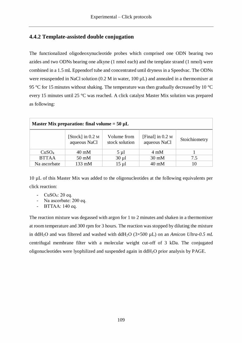
Experimental – Click protocols
109
4.4.2 Template-assisted double conjugation
The functionalized oligodeoxynucleotide probes which comprised one ODN bearing two
azides and two ODNs bearing one alkyne (1 nmol each) and the template strand (1 nmol) were
combined in a 1.5 mL Eppendorf tube and concentrated until dryness in a Speedvac. The ODNs
were resuspended in NaCl solution (0.2 M in water, 100 µL) and annealed in a thermomixer at
95 °C for 15 minutes without shaking. The temperature was then gradually decreased by 10 °C
every 15 minutes until 25 °C was reached. A click catalyst Master Mix solution was prepared
as following:
Master Mix preparation: final volume = 50 µL
[Stock] in 0.2 M
aqueous NaCl
Volume from
stock solution
[Final] in 0.2 M
aqueous NaCl Stoichiometry
CuSO4 40 mM 5 µl 4 mM 1
BTTAA 50 mM 30 µl 30 mM 7.5
Na ascorbate 133 mM 15 µl 40 mM 10
10 µL of this Master Mix was added to the oligonucleotides at the following equivalents per
click reaction:
- CuSO4: 20 eq.
- Na ascorbate: 200 eq.
- BTTAA: 140 eq.
The reaction mixture was degassed with argon for 1 to 2 minutes and shaken in a thermomixer
at room temperature and 300 rpm for 3 hours. The reaction was stopped by diluting the mixture
in ddH2O and was filtered and washed with ddH2O (3×500 µL) on an Amicon Ultra-0.5 mL
centrifugal membrane filter with a molecular weight cut-off of 3 kDa. The conjugated
oligonucleotides were lyophilized and suspended again in ddH2O prior analysis by PAGE.

Experimental – Biochemical experiments
110
4.5 Biochemical experiments
4.5.1 Melting Curve Experiments
Melting profiles were measured on a Jasco V-650 spectrometer using quartz glass cuvettes
with 10.0 mm path length. Final samples contained the two oligonucleotide of which the
dissociation-characteristics are to be assessed each at 2 µM concentration, 0.2 M NaCl aqueous
solution, 10 mM aqueous sodium phosphate buffer in a final volume of 400 µL. Measurements
were repeated at least three times per sample. Before the measurement, the oligonucleotides
were hybridized at 95 °C for 5 minutes followed by cooling down to 4 °C over a period of 40
minutes. For the UV-measurements the solutions were covered with silicon oil and tightly
plugged. Absorbance was recorded in the forward and reverse direction at temperatures from
15 °C to 85 °C at 0.1 °C/3 seconds. At least three denaturing and renaturing ramps were
performed and averaged for evaluation of the melting temperature. Tm values were calculated
as the zero-crossing of 2nd derivate of the 349 nm background-corrected change in
hyperchromicity at 260 nm.
4.5.2 DNA PAGE
ODN strands and products from the click conjugations were analysed by 20% denaturing
polyacrylamide gel electrophoresis on 20 x 20 cm gels of 1 mm thickness. The samples to
analyse were prepared in order to contain approximately 300 ng of ODN for the 11 mer strands
and 150 ng for the 15 mer strands in 5 µL of ddH2O. To each sample was added 5 µL of loading
buffer prepared as described in the recipe table. The gels were run in TBE buffer (see below
recipe table) at 40 °C applying a constant current of 40 mA per gel (maximum 1000 V) for
about 5-6 hours. The gels were stained with SYBR® green II for 30 minutes (in ddH2O, 100
mL) and then visualized using a LAS-3000 imaging system (Raytest).
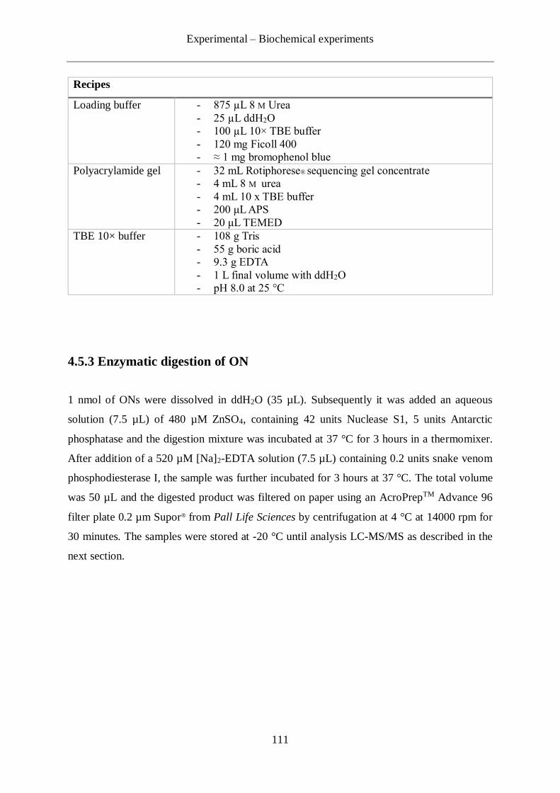
Experimental – Biochemical experiments
111
Recipes
Loading buffer - 875 µL 8 M Urea
- 25 µL ddH2O
- 100 µL 10× TBE buffer
- 120 mg Ficoll 400
- ≈ 1 mg bromophenol blue
Polyacrylamide gel - 32 mL Rotiphorese® sequencing gel concentrate
- 4 mL 8 M urea
- 4 mL 10 x TBE buffer
- 200 μL APS
- 20 μL TEMED TBE 10× buffer - 108 g Tris
- 55 g boric acid
- 9.3 g EDTA
- 1 L final volume with ddH2O
- pH 8.0 at 25 °C
4.5.3 Enzymatic digestion of ON
1 nmol of ONs were dissolved in ddH2O (35 µL). Subsequently it was added an aqueous
solution (7.5 µL) of 480 µM ZnSO4, containing 42 units Nuclease S1, 5 units Antarctic
phosphatase and the digestion mixture was incubated at 37 °C for 3 hours in a thermomixer.
After addition of a 520 µM [Na]2-EDTA solution (7.5 µL) containing 0.2 units snake venom
phosphodiesterase I, the sample was further incubated for 3 hours at 37 °C. The total volume
was 50 µL and the digested product was filtered on paper using an AcroPrepTM Advance 96
filter plate 0.2 µm Supor® from Pall Life Sciences by centrifugation at 4 °C at 14000 rpm for
30 minutes. The samples were stored at -20 °C until analysis LC-MS/MS as described in the
next section.
Experimental – UHPLC-MS analysis
112
4.6- UHPLC-MS analysis
The analysis of the digested RNAs were performed by LC-ESI-MS on a Thermo Finnigan LTQ
Orbitrap XL and were chromatographed by a Dionex Ultimate 3000 HPLC system with a flow
of 0.15 mL/min over an Interchim Uptisphere120A-3µm-HDO C18 column. The column
temperature was maintained at 30 °C. Eluting buffers were buffer A (2 mM HCOONH4 in
ddH2O (pH 5.5)) and buffer B (2 mM HCOONH4 in ddH2O/MeCN 20/80 (pH 5.5)). The
gradient for all samples was from 100% to 94% buffer A over 55 min. The elution was
monitored at 260 nm (Dionex Ultimate 3000 Diode Array Detector). The chromatographic
eluent was directly injected into the ion source without prior splitting. Ions were scanned by
use of a positive polarity mode over a full-scan range of m/z 120-1000 with a resolution of
30000.
Abbreviations
113
5. ABBRIEVATIONS
(d)A (deoxy)Adenine/Adenosine (depending on context)
(d)C (deoxy)Cytosine/Cytidine (depending on context)
(d)G (deoxy)Guanine/Guanosine (depending on context)
(d)T (deoxy)Thymine/Thymidine (depending on context)
(d)U (deoxy)Uracil/Uridine (depending on context)
°C Degree Celsius (temperature)
Ac Acetyl
ACN Acetonitrile
AMA 1:1 mixture of 40% aqueous ammonia/28% aqueous methylamine
APS Ammonium persulfate
Boc2O Di-tert-butyl dicarbonate
bp Base pair
br Broad
BTTAA 2-(4-((bis((1-(tert-butyl)-1H-1,2,3-triazol-4-
yl)methyl)amino)methyl)-1H-1,2,3-triazol-1-yl)acetic acid
Bz Benzoyl
caC 5-carboxy cytosine
CAN Ceric ammonium nitrate
COSY Correlation spectroscopy (2D NMR)
CPG Controlled pore glass
CuAAC Cu(I)-catalyzed alkyne azide cycloadditions
d Doublet
DBU 1,8-Diazabicyclo[5.4.0]undec-7-ene
DCM Dichloromethane
ddH2O Double-distilled water
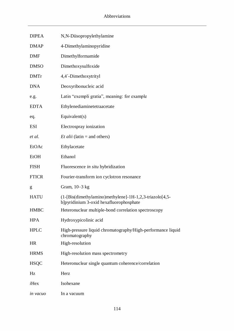
Abbreviations
114
DIPEA N,N-Diisopropylethylamine
DMAP 4-Dimethylaminopyridine
DMF Dimethylformamide
DMSO Dimethoxysulfoxide
DMTr 4,4´-Dimethoxytrityl
DNA Deoxyribonucleic acid
e.g. Latin “exempli gratia”, meaning: for example
EDTA Ethylenediaminetetraacetate
eq. Equivalent(s)
ESI Electrospray ionization
et al. Et alii (latin = and others)
EtOAc Ethylacetate
EtOH Ethanol
FISH Fluorescence in situ hybridization
FTICR Fourier-transform ion cyclotron resonance
g Gram, 10–3 kg
HATU (1-[Bis(dimethylamino)methylene]-1H-1,2,3-triazolo[4,5-
b]pyridinium 3-oxid hexafluorophosphate
HMBC Heteronuclear multiple-bond correlation spectroscopy
HPA Hydroxypicolinic acid
HPLC High-pressure liquid chromatography/High-performance liquid
chromatography
HR High-resolution
HRMS High-resolution mass spectrometry
HSQC Heteronuclear single quantum coherence/correlation
Hz Herz
iHex Isohexane
in vacuo In a vacuum

Abbreviations
115
IR Infrared
J Coupling constant
kDa Kilodalton
L Liter(s)
LC Liquid chromatography
m Meter(s)
M Molar concentration
m Multiplet
m/z Mass to charge ratio
MALDI-TOF Matrix assisted laser desorption/ionization time-of-flight
MeCN Acetonitrile
MeOH Methanol
mol Mole(s)
mRNA Messenger RNA
MS Mass spectrometry
NaOAc Sodium acetate
NMR Nuclear magnetic resonance
O(D)N Oligo(deoxyribo)nucleotide
PAGE Polyacrylamide gel electrophoresis
PCR Polymerase chain reaction
PDC Pyridinium dichromate
pH The negative decimal logarithm of the hydrogen ion activity
PLA Proximity ligation assay
ppm Parts per million
q Quartet
Rf Retention factor (thin-layer chromatography)
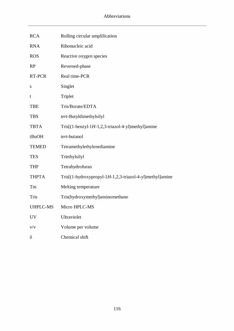
Abbreviations
116
RCA Rolling circular amplification
RNA Ribonucleic acid
ROS Reactive oxygen species
RP Reversed-phase
RT-PCR Real time-PCR
s Singlet
t Triplet
TBE Tris/Borate/EDTA
TBS tert-Butyldimethylsilyl
TBTA Tris[(1-benzyl-1H-1,2,3-triazol-4-yl)methyl]amine
tBuOH tert-butanol
TEMED Tetramethylethylenediamine
TES Triethylsilyl
THF Tetrahydrofuran
THPTA Tris[(1-hydroxypropyl-1H-1,2,3-triazol-4-yl)methyl]amine
Tm Melting temperature
Tris Tris(hydroxymethyl)aminomethane
UHPLC-MS Micro HPLC-MS
UV Ultraviolet
v/v Volume per volume
δ Chemical shift

References
117
6. REFERENCES
[1] S. Fredriksson, M. Gullberg, J. Jarvius, C. Olsson, K. Pietras, S. M. Gustafsdottir, A.
Ostman, U. Landegren, Nat. Biotechnol. 2002, 20, 473-477. Protein detection using
proximity-dependent DNA ligation assays.
[2] O. Söderberg, M. Gullberg, M. Jarvius, K. Ridderstråle, K.-J. Leuchowius, J. Jarvius,
K. Wester, P. Hydbring, F. Bahram, L.-G. Larsson, U. Landegren, Nat. Methods 2006,
3, 995. Direct observation of individual endogenous protein complexes in situ by
proximity ligation.
[3] P. M. E. Gramlich, C. T. Wirges, A. Manetto, T. Carell, Angew. Chem. Int. Ed. 2008,
47, 8350-8358. Postsynthetic DNA Modification through the Copper-Catalyzed Azide–
Alkyne Cycloaddition Reaction.
[4] W. S. Brotherton, H. A. Michaels, J. T. Simmons, R. J. Clark, N. S. Dalal, L. Zhu, Org.
Lett. 2009, 11, 4954-4957. Apparent copper(II)-accelerated azide-alkyne
cycloaddition.
[5] Z. Yuan, G.-C. Kuang, R. J. Clark, L. Zhu, Org. Lett. 2012, 14, 2590-2593.
Chemoselective Sequential “Click” Ligation Using Unsymmetrical Bisazides.
[6] C. Deans, K. A. Maggert, Genetics 2015, 199, 887-896. What Do You Mean,
“Epigenetic”?
[7] R. Jaenisch, A. Bird, Nat. Genet. 2003, 33, 245. Epigenetic regulation of gene
expression: how the genome integrates intrinsic and environmental signals.
[8] C. Thomas, K. M. Q., M. Markus, R. Martin, S. Fabio, Angew. Chem. Int. Ed. 2018,
57, 4296-4312. Non‐canonical Bases in the Genome: The Regulatory Information
Layer in DNA.
[9] J. E. Squires, H. R. Patel, M. Nousch, T. Sibbritt, D. T. Humphreys, B. J. Parker, C. M.
Suter, T. Preiss, Nucleic Acids Res. 2012, 40, 5023-5033. Widespread occurrence of 5-
methylcytosine in human coding and non-coding RNA.
[10] S. M. Huber, P. van Delft, L. Mendil, M. Bachman, K. Smollett, F. Werner, E. A.
Miska, S. Balasubramanian, Chembiochem 2015, 16, 752-755. Formation and
abundance of 5‐hydroxymethylcytosine in RNA.
[11] L. Fu, C. R. Guerrero, N. Zhong, N. J. Amato, Y. Liu, S. Liu, Q. Cai, D. Ji, S.-G. Jin,
L. J. Niedernhofer, J. Am. Chem. Soc 2014, 136, 11582-11585. Tet-mediated formation
of 5-hydroxymethylcytosine in RNA.
[12] H.-Y. Zhang, J. Xiong, B.-L. Qi, Y.-Q. Feng, B.-F. Yuan, Chem. Commun. 2016, 52,
737-740. The existence of 5-hydroxymethylcytosine and 5-formylcytosine in both DNA
and RNA in mammals.

References
118
[13] W. Huang, M.-D. Lan, C.-B. Qi, S.-J. Zheng, S.-Z. Wei, B.-F. Yuan, Y.-Q. Feng,
Chemical Science 2016, 7, 5495-5502. Formation and determination of the oxidation
products of 5-methylcytosine in RNA.
[14] J. M. Berg, J. L. Tymoczko, L. Stryer, Biochemistry, W.H. Freeman, Basingstoke,
2012.
[15] R. Dahm, Hum. Genet. 2008, 122, 565-581. Discovering DNA: Friedrich Miescher and
the early years of nucleic acid research.
[16] P. A. Levene, J. Biol. Chem. 1919, 40, 415-424. The Stucture of Yeast Nucleic Acid:
IV. Ammonnia Hydrolysis.
[17] O. T. Avery, C. M. MacLeod, M. McCarty, J. Exp. Med. 1944, 79, 137-158. Studies on
the chemical nature of the substance inducing transformation of pneumonocal types :
induction of transformation by a desoxyribonucleic acid fraction isolated from
pneumonococcus type III.
[18] J. D. Watson, F. H. C. Crick, Nature 1953, 171, 737. Molecular Structure of Nucleic
Acids: A Structure for Deoxyribose Nucleic Acid.
[19] M. H. F. Wilkins, A. R. Stokes, H. R. Wilson, Nature 1953, 171, 738. Molecular
Structure of Nucleic Acids: Molecular Structure of Deoxypentose Nucleic Acids.
[20] J. D. Watson, F. H. C. Crick, Nature 1953, 171, 964. Genetical Implications of the
Structure of Deoxyribonucleic Acid.
[21] R. Wing, H. Drew, T. Takano, C. Broka, S. Tanaka, K. Itakura, R. E. Dickerson, Nature
1980, 287, 755. Crystal structure analysis of a complete turn of B-DNA.
[22] M. Meselson, F. W. Stahl, Proc. Natl. Acad. Sci. USA 1958, 44, 671-682. The
Replication of DNA in Escherichia Coli.
[23] M. W. Nirenberg, J. H. Matthaei, Proc. Natl. Acad. Sci. USA 1961, 47, 1588-1602. The
dependence of cell-free protein synthesis in E. coli upon naturally occurring or
synthetic polyribonucleotides.
[24] M. Nirenberg, P. Leder, Science 1964, 145, 1399-1407. RNA Codewords and Protein
Synthesis. The Effect of Trinucleotides upon the Biding of sRNA to Ribosomes.
[25] A. Michael, J. Prakt. Chem. 1893, 48, 94-95. Ueber die Einwirkung von
Diazobenzolimid auf Acetylendicarbonsäuremethylester.
[26] R. Huigsen, Proc. Chem. Soc. 1961, 357-396. 1,3‐Dipolar Cycloadditions.
[27] R. Huisgen, Angew. Chem. Int. Ed. 1963, 2, 565-598. 1,3‐Dipolar Cycloadditions. Past
and Future.
References
119
[28] C. W. Tornøe, M. Meldal, in Peptides: The Wave of the Future: Proceedings of the
Second International and the Seventeenth American Peptide Symposium, June 9–14,
2001, San Diego, California, U.S.A. (Eds.: M. Lebl, R. A. Houghten), Springer
Netherlands, Dordrecht, 2001, pp. 263-264.
[29] C. W. Tornoe, C. Christensen, M. Meldal, J. Org. Chem. 2002, 67, 3057-3064.
Peptidotriazoles on solid phase: [1,2,3]-triazoles by regiospecific copper(i)-catalyzed
1,3-dipolar cycloadditions of terminal alkynes to azides.
[30] V. V. Rostovtsev, L. G. Green, V. V. Fokin, K. B. Sharpless, Angew. Chem. Int. Ed.
2002, 41, 2596-2599. A stepwise huisgen cycloaddition process: copper(I)-catalyzed
regioselective "ligation" of azides and terminal alkynes.
[31] F. Himo, T. Lovell, R. Hilgraf, V. V. Rostovtsev, L. Noodleman, K. B. Sharpless, V.
V. Fokin, J. Am. Chem. Soc. 2005, 127, 210-216. Copper(I)-Catalyzed Synthesis of
Azoles. DFT Study Predicts Unprecedented Reactivity and Intermediates.
[32] R. Berg, B. F. Straub, Beil. J. Org. Chem. 2013, 9, 2715-2750. Advancements in the
mechanistic understanding of the copper-catalyzed azide–alkyne cycloaddition.
[33] B. T. Worrell, J. A. Malik, V. V. Fokin, Science 2013, 340, 457. Direct Evidence of a
Dinuclear Copper Intermediate in Cu(I)-Catalyzed Azide-Alkyne Cycloadditions.
[34] L. Jin, D. R. Tolentino, M. Melaimi, G. Bertrand, Sci. Adv. 2015, 1. Isolation of
bis(copper) key intermediates in Cu-catalyzed azide-alkyne “click reaction”.
[35] Z. Lei, B. C. J., Z. Xiaoguang, G. P. M., C. R. J., Chem. Rec. 2016, 16, 1501-1517. On
the Mechanism of Copper(I)‐Catalyzed Azide–Alkyne Cycloaddition.
[36] H. C. Kolb, M. G. Finn, K. B. Sharpless, Angew. Chem. Int. Ed. 2001, 40, 2004-2021.
Click Chemistry: Diverse Chemical Function from a Few Good Reactions.
[37] X. Wang, B. Huang, X. Liu, P. Zhan, Drug Discovery Today 2016, 21, 118-132.
Discovery of bioactive molecules from CuAAC click-chemistry-based combinatorial
libraries.
[38] X. Liang, L. Yongjun, L. Yuliang, Asian J. Org. Chem. 2014, 3, 582-602. Application
of “Click” Chemistry to the Construction of Supramolecular Functional Systems.
[39] L. Li, Z. Zhang, Molecules 2016, 21, 1393. Development and Applications of the
Copper-Catalyzed Azide-Alkyne Cycloaddition (CuAAC) as a Bioorthogonal Reaction.
[40] V. Hong, N. F. Steinmetz, M. Manchester, M. G. Finn, Bioconjugate Chem. 2010, 21,
1912-1916. Labeling Live Cells by Copper-Catalyzed Alkyne−Azide Click Chemistry.
[41] D. C. Kennedy, C. S. McKay, M. C. B. Legault, D. C. Danielson, J. A. Blake, A. F.
Pegoraro, A. Stolow, Z. Mester, J. P. Pezacki, J. Am. Chem. Soc. 2011, 133, 17993-
18001. Cellular Consequences of Copper Complexes Used To Catalyze Bioorthogonal
Click Reactions.
References
120
[42] G. J. Brewer, Chem. Res. Toxicol. 2010, 23, 319-326. Risks of Copper and Iron Toxicity
during Aging in Humans.
[43] T. R. Chan, R. Hilgraf, K. B. Sharpless, V. V. Fokin, Org. Lett. 2004, 6, 2853-2855.
Polytriazoles as Copper(I)-Stabilizing Ligands in Catalysis.
[44] V. Hong, S. I. Presolski, C. Ma, M. G. Finn, Angew. Chem. Int. Ed. 2009, 48, 9879-
9883. Analysis and optimization of copper-catalyzed azide-alkyne cycloaddition for
bioconjugation.
[45] C. Besanceney-Webler, H. Jiang, T. Zheng, L. Feng, D. Soriano del Amo, W. Wang,
L. M. Klivansky, F. L. Marlow, Y. Liu, P. Wu, Angew. Chem. Int. Ed. 2011, 50, 8051-
8056. Raising the Efficacy of Bioorthogonal Click Reactions for Bioconjugation: A
Comparative Study.
[46] G. C. Rudolf, S. A. Sieber, ChemBioChem 2013, 14, 2447-2455. Copper-assisted click
reactions for activity-based proteomics: fine-tuned ligands and refined conditions
extend the scope of application.
[47] C. Uttamapinant, A. Tangpeerachaikul, S. Grecian, S. Clarke, U. Singh, P. Slade, K. R.
Gee, A. Y. Ting, Angew. Chem. Int. Ed. 2012, 51, 5852-5856. Fast, Cell-Compatible
Click Chemistry with Copper-Chelating Azides for Biomolecular Labeling.
[48] P. M. Gramlich, S. Warncke, J. Gierlich, T. Carell, Angew. Chem. Int. Ed. 2008, 47,
3442-3444. Click-click-click: single to triple modification of DNA.
[49] E. Paredes, S. R. Das, ChemBioChem 2011, 12, 125-131. Click Chemistry for Rapid
Labeling and Ligation of RNA.
[50] M.-L. Winz, A. Samanta, D. Benzinger, A. Jäschke, Nucleic Acids Res. 2012, 40, e78-
e78. Site-specific terminal and internal labeling of RNA by poly(A) polymerase tailing
and copper-catalyzed or copper-free strain-promoted click chemistry.
[51] R. Kumar, A. El-Sagheer, J. Tumpane, P. Lincoln, L. M. Wilhelmsson, T. Brown, J.
Am. Chem. Soc. 2007, 129, 6859-6864. Template-Directed Oligonucleotide Strand
Ligation, Covalent Intramolecular DNA Circularization and Catenation Using Click
Chemistry.
[52] I. Manuguerra, S. Croce, A. H. El-Sagheer, A. Krissanaprasit, T. Brown, K. V. Gothelf,
A. Manetto, Chem. Commun. 2018, 54, 4529-4532. Gene assembly via one-pot
chemical ligation of DNA promoted by DNA nanostructures.
[53] S. Y. Lim, W. Y. Chung, H. K. Lee, M. S. Park, H. G. Park, Biochem. Biophys. Res.
Commun. 2008, 376, 633-636. Direct and nondestructive verification of PNA
immobilization using click chemistry.

References
121
[54] K. Fauster, M. Hartl, T. Santner, M. Aigner, C. Kreutz, K. Bister, E. Ennifar, R. Micura,
ACS Chem. Biol. 2012, 7, 581-589. 2′-Azido RNA, a Versatile Tool for Chemical
Biology: Synthesis, X-ray Structure, siRNA Applications, Click Labeling.
[55] J. Lietard, A. Meyer, J.-J. Vasseur, F. Morvan, J. Org. Chem. 2008, 73, 191-200. New
Strategies for Cyclization and Bicyclization of Oligonucleotides by Click Chemistry
Assisted by Microwaves.
[56] J. Gierlich, G. A. Burley, P. M. Gramlich, D. M. Hammond, T. Carell, Org. Lett. 2006,
8, 3639-3642. Click chemistry as a reliable method for the high-density postsynthetic
functionalization of alkyne-modified DNA.
[57] J. Gierlich, K. Gutsmiedl, P. M. Gramlich, A. Schmidt, G. A. Burley, T. Carell,
Chemistry 2007, 13, 9486-9494. Synthesis of highly modified DNA by a combination of
PCR with alkyne-bearing triphosphates and click chemistry.
[58] K. B. Mullis, Sci. Am. 1990, 262, 56-65. The Unusual Origin of the Polymerase Chain
Reaction.
[59] K. B. Mullis, F. A. Faloona, Methods Enzymol. 1987, 155, 335-350. Specific synthesis
of DNA in vitro via a polymerase-catalyzed chain reaction.
[60] G. T. Walker, M. S. Fraiser, J. L. Schram, M. C. Little, J. G. Nadeau, D. P. Malinowski,
Nucleic Acids Res. 1992, 20, 1691-1696. Strand displacement amplification—an
isothermal, in vitro DNA amplification technique.
[61] J. C. Guatelli, K. M. Whitfield, D. Y. Kwoh, K. J. Barringer, D. D. Richman, T. R.
Gingeras, Proc. Natl. Acad. Sci. USA 1990, 87, 1874-1878. Isothermal, in vitro
amplification of nucleic acids by a multienzyme reaction modeled after retroviral
replication.
[62] H. U. Weier, J. W. Gray, DNA (Mary Ann Liebert, Inc.) 1988, 7, 441-447. A
programmable system to perform the polymerase chain reaction.
[63] R. K. Saiki, S. Scharf, F. Faloona, K. B. Mullis, G. T. Horn, H. A. Erlich, N. Arnheim,
Science 1985, 230, 1350-1354. Enzymatic amplification of beta-globin genomic
sequences and restriction site analysis for diagnosis of sickle cell anemia.
[64] J. S. Chamberlain, R. A. Gibbs, J. E. Rainer, P. N. Nguyen, C. Thomas, Nucleic Acids
Res. 1988, 16, 11141-11156. Deletion screening of the Duchenne muscular dystrophy
locus via multiplex DNA amplification.
[65] T. D. Brock, H. Freeze, J. Bacteriol. 1969, 98, 289-297. Thermus aquaticus gen. n. and
sp. n., a nonsporulating extreme thermophile.
[66] R. Higuchi, G. Dollinger, P. S. Walsh, R. Griffith, Bio/technology 1992, 10, 413-417.
Simultaneous amplification and detection of specific DNA sequences.
References
122
[67] R. Higuchi, C. Fockler, G. Dollinger, R. Watson, Bio/technology 1993, 11, 1026-1030.
Kinetic PCR analysis: real-time monitoring of DNA amplification reactions.
[68] B. J. Bassam, T. Allen, S. Flood, J. Stevens, P. Wyatt, K. J. Livak, Nucleic acid
sequence detection systems: Revolutionary automation for monitoring and reporting
PCR products, Vol. 6, 1996.
[69] H. D. VanGuilder, K. E. Vrana, W. M. Freeman, Biotechniques 2008, 44, 619-626.
Twenty-five years of quantitative PCR for gene expression analysis.
[70] K. M. Ririe, R. P. Rasmussen, C. T. Wittwer, Anal. Biochem. 1997, 245, 154-160.
Product differentiation by analysis of DNA melting curves during the polymerase chain
reaction.
[71] W. Wang, K. Chen, C. Xu, Anal. Biochem. 2006, 356, 303-305. DNA quantification
using EvaGreen and a real-time PCR instrument.
[72] D. Whitcombe, J. Theaker, S. P. Guy, T. Brown, S. Little, Nat. Biotechnol. 1999, 17,
804-807. Detection of PCR products using self-probing amplicons and fluorescence.
[73] S. Tyagi, F. R. Kramer, Nat. Biotechnol. 1996, 14, 303-308. Molecular beacons: probes
that fluoresce upon hybridization.
[74] P. M. Holland, R. D. Abramson, R. Watson, D. H. Gelfand, Proc. Natl. Acad. Sci. USA
1991, 88, 7276-7280. Detection of specific polymerase chain reaction product by
utilizing the 5'----3' exonuclease activity of Thermus aquaticus DNA polymerase.
[75] P. E. Nielsen, M. Egholm, R. H. Berg, O. Buchardt, Science 1991, 254, 1497-1500.
Sequence-selective recognition of DNA by strand displacement with a thymine-
substituted polyamide.
[76] H. Kaur, A. Arora, J. Wengel, S. Maiti, Biochemistry 2006, 45, 7347-7355.
Thermodynamic, Counterion, and Hydration Effects for the Incorporation of Locked
Nucleic Acid Nucleotides into DNA Duplexes.
[77] G. H. Reed, C. T. Wittwer, Clin. Chem. 2004, 50, 1748-1754. Sensitivity and specificity
of single-nucleotide polymorphism scanning by high-resolution melting analysis.
[78] M. Louie, L. Louie, A. E. Simor, Can. Med. Assoc. J. 2000, 163, 301-309. The role of
DNA amplification technology in the diagnosis of infectious diseases.
[79] P. S. Bernard, C. T. Wittwer, Clin. Chem. 2002, 48, 1178-1185. Real-time PCR
technology for cancer diagnostics.
[80] C. Greenwood, D. Ruff, S. Kirvell, G. Johnson, H. S. Dhillon, S. A. Bustin, Biomol.
Detect. Quantif. 2015, 4, 10-16. Proximity assays for sensitive quantification of
proteins.
References
123
[81] O. Soderberg, M. Gullberg, M. Jarvius, K. Ridderstrale, K. J. Leuchowius, J. Jarvius,
K. Wester, P. Hydbring, F. Bahram, L. G. Larsson, U. Landegren, Nat. Methods 2006,
3, 995-1000. Direct observation of individual endogenous protein complexes in situ by
proximity ligation.
[82] M. Gullberg, S. M. Gústafsdóttir, E. Schallmeiner, J. Jarvius, M. Bjarnegård, C.
Betsholtz, U. Landegren, S. Fredriksson, Proc. Natl. Acad. Sci. USA 2004, 101, 8420.
Cytokine detection by antibody-based proximity ligation.
[83] M. M. Ali, F. Li, Z. Zhang, K. Zhang, D. K. Kang, J. A. Ankrum, X. C. Le, W. Zhao,
Chem. Soc. Rev. 2014, 43, 3324-3341. Rolling circle amplification: a versatile tool for
chemical biology, materials science and medicine.
[84] I. Weibrecht, K.-J. Leuchowius, C.-M. Clausson, T. Conze, M. Jarvius, W. M. Howell,
M. Kamali-Moghaddam, O. Söderberg, Expert Rev. Proteomics 2010, 7, 401-409.
Proximity ligation assays: a recent addition to the proteomics toolbox.
[85] C. Coppola, L. Simeone, L. De Napoli, D. Montesarchio, Eur. J. Org. Chem. 2011,
2011, 1155-1165. On the Compatibility of Azides in Phosphoramidite-Based
Couplings: Synthesis of a Novel, Convertible Azido-Functionalized CyPLOS Analogue.
[86] M. A. Fomich, M. V. Kvach, M. J. Navakouski, C. Weise, A. V. Baranovsky, V. A.
Korshun, V. V. Shmanai, Org. Lett. 2014, 16, 4590-4593. Azide phosphoramidite in
direct synthesis of azide-modified oligonucleotides.
[87] T. Wada, A. Mochizuki, S. Higashiya, H. Tsuruoka, S.-i. Kawahara, M. Ishikawa, M.
Sekine, Tetrahedron Lett. 2001, 42, 9215-9219. Synthesis and properties of 2-
azidodeoxyadenosine and its incorporation into oligodeoxynucleotides.
[88] G. Pourceau, A. Meyer, J.-J. Vasseur, F. Morvan, J. Org. Chem. 2009, 74, 6837-6842.
Azide Solid Support for 3′-Conjugation of Oligonucleotides and Their Circularization
by Click Chemistry.
[89] L. M. Smith, S. Fung, M. W. Hunkapiller, T. J. Hunkapiller, L. E. Hood, Nucleic Acids
Res. 1985, 13, 2399-2412. The synthesis of oligonucleotides containing an aliphatic
amino group at the 5' terminus: synthesis of fluorescent DNA primers for use in DNA
sequence analysis.
[90] P. Kočalka, A. H. El-Sagheer, T. Brown, ChemBioChem 2008, 9, 1280-1285. Rapid
and Efficient DNA Strand Cross-Linking by Click Chemistry.
[91] J. Gierlich, G. A. Burley, P. M. Gramlich, D. M. Hammond, T. Carell, Org. Lett. 2006,
8, 3639-3642. Click chemistry as a reliable method for the high-density postsynthetic
functionalization of alkyne-modified DNA.
[92] K. Onizuka, T. Chikuni, T. Amemiya, T. Miyashita, K. Onizuka, H. Abe, F. Nagatsugi,
Nucleic Acids Res. 2017, 45, 5036-5047. Pseudorotaxane formation via the slippage
process with chemically cyclized oligonucleotides.
References
124
[93] S. S. Pujari, H. Xiong, F. Seela, J. Org. Chem. 2010, 75, 8693-8696. Cross-linked DNA
generated by "bis-click" reactions with bis-functional azides: site independent ligation
of oligonucleotides via nucleobase alkynyl chains.
[94] S. S. Pujari, F. Seela, J. Org. Chem. 2013, 78, 8545-8561. Parallel stranded DNA
stabilized with internal sugar cross-links: synthesis and click ligation of
oligonucleotides containing 2'-propargylated isoguanosine.
[95] G.-C. Kuang, H. A. Michaels, J. T. Simmons, R. J. Clark, L. Zhu, J. Org. Chem. 2010,
75, 6540-6548. Chelation-Assisted, Copper(II) Acetate-Accelerated Azide-Alkyne
Cycloaddition.
[96] S. A. Ingale, F. Seela, J. Org. Chem. 2013, 78, 3394-3399. Stepwise click
functionalization of DNA through a bifunctional azide with a chelating and a
nonchelating azido group.
[97] C. Ö. Di̇nç, G. Ki̇barer, A. Güner, J. Appl. Polym. Sci. 2010, 117, 1100-1119. Solubility
profiles of poly(ethylene glycol)/solvent systems. II. comparison of thermodynamic
parameters from viscosity measurements.
[98] L. A. Carpino, J. Am. Chem. Soc. 1993, 115, 4397-4398. 1-Hydroxy-7-
azabenzotriazole. An efficient peptide coupling additive.
[99] M. S. Wendland, S. C. Zimmerman, J. Am. Chem. Soc. 1999, 121, 1389-1390. Synthesis
of Cored Dendrimers.
[100] V. Aucagne, D. A. Leigh, Org. Lett. 2006, 8, 4505-4507. Chemoselective formation of
successive triazole linkages in one pot: "click-click" chemistry.
[101] T. W. Greene, P. G. M. Wuts, in Protective Groups in Organic Synthesis, John Wiley
& Sons, Inc., 2002, pp. 654-659.
[102] P. A. Ledin, F. Friscourt, J. Guo, G. J. Boons, Chemistry 2011, 17, 839-846. Convergent
assembly and surface modification of multifunctional dendrimers by three consecutive
click reactions.
[103] G. Hayashi, N. Kamo, A. Okamoto, Chem. Commun. 2017, 53, 5918-5921. Chemical
synthesis of dual labeled proteins via differently protected alkynes enables
intramolecular FRET analysis.
[104] S. Thyagarajan, N. N. Murthy, A. A. Narducci Sarjeant, K. D. Karlin, S. E. Rokita, J.
Am. Chem. Soc. 2006, 128, 7003-7008. Selective DNA Strand Scission with Binuclear
Copper Complexes: Implications for an Active Cu(2)-O(2) Species.
[105] G. H. Keller, M. M. Manak, DNA probes, Macmillan Publishers Ltd, Basingstoke,
1989.
References
125
[106] Y. Liu, A. Kuzuya, R. Sha, J. Guillaume, R. Wang, J. W. Canary, N. C. Seeman, J. Am.
Chem. Soc. 2008, 130, 10882-10883. Coupling across a DNA Helical Turn Yields a
Hybrid DNA/Organic Catenane Doubly Tailed with Functional Termini.
[107] K. Onizuka, F. Nagatsugi, Y. Ito, H. Abe, J. Am. Chem. Soc. 2014, 136, 7201-7204.
Automatic pseudorotaxane formation targeting on nucleic acids using a pair of reactive
oligodeoxynucleotides.
[108] S. Kriaucionis, N. Heintz, Science 2009, 324, 929-930. The nuclear DNA base 5-
hydroxymethylcytosine is present in Purkinje neurons and the brain.
[109] M. Tahiliani, K. P. Koh, Y. Shen, W. A. Pastor, H. Bandukwala, Y. Brudno, S.
Agarwal, L. M. Iyer, D. R. Liu, L. Aravind, A. Rao, Science 2009, 324, 930-935.
Conversion of 5-methylcytosine to 5-hydroxymethylcytosine in mammalian DNA by
MLL partner TET1.
[110] T. Pfaffeneder, B. Hackner, M. Truß, M. Münzel, M. Müller, C. A. Deiml, C.
Hagemeier, T. Carell, Angew. Chem. Int. Ed. 2011, 50, 7008-7012. The Discovery of 5‐
Formylcytosine in Embryonic Stem Cell DNA.
[111] S. Ito, L. Shen, Q. Dai, S. C. Wu, L. B. Collins, J. A. Swenberg, C. He, Y. Zhang,
Science 2011, 333, 1300. Tet Proteins Can Convert 5-Methylcytosine to 5-
Formylcytosine and 5-Carboxylcytosine.
[112] S. Kriaucionis, N. Heintz, Science 2009, 324, 929-930. The nuclear DNA base 5-
hydroxymethylcytosine is present in Purkinje neurons and the brain.
[113] M. Tahiliani, K. P. Koh, Y. Shen, W. A. Pastor, H. Bandukwala, Y. Brudno, S.
Agarwal, L. M. Iyer, D. R. Liu, L. Aravind, Science 2009, 324, 930-935. Conversion
of 5-methylcytosine to 5-hydroxymethylcytosine in mammalian DNA by MLL partner
TET1.
[114] Y.-F. He, B.-Z. Li, Z. Li, P. Liu, Y. Wang, Q. Tang, J. Ding, Y. Jia, Z. Chen, L. Li,
Science 2011, 333, 1303-1307. Tet-mediated formation of 5-carboxylcytosine and its
excision by TDG in mammalian DNA.
[115] R. M. Kohli, Y. Zhang, Nature 2013, 502, 472-479. TET enzymes, TDG and the
dynamics of DNA demethylation.
[116] C. G. Spruijt, F. Gnerlich, A. H. Smits, T. Pfaffeneder, P. W. Jansen, C. Bauer, M.
Münzel, M. Wagner, M. Müller, F. Khan, Cell 2013, 152, 1146-1159. Dynamic readers
for 5-(hydroxy) methylcytosine and its oxidized derivatives.
[117] T. Pfaffeneder, B. Hackner, M. Truss, M. Munzel, M. Muller, C. A. Deiml, C.
Hagemeier, T. Carell, Angew. Chem. Int. Ed. 2011, 50, 7008-7012. The discovery of 5-
formylcytosine in embryonic stem cell DNA.

References
126
[118] M. Basanta‐Sanchez, R. Wang, Z. Liu, X. Ye, M. Li, X. Shi, P. F. Agris, Y. Zhou, Y.
Huang, J. Sheng, ChemBioChem 2017, 18, 72-76. TET1‐Mediated Oxidation of 5‐
Formylcytosine (5fC) to 5‐Carboxycytosine (5caC) in RNA.
[119] C. He, Nature chemical biology 2010, 6, 863-865. Grand challenge commentary: RNA
epigenetics?
[120] C. Willyard, Nature 2017, 542, 406-408. An epigenetics gold rush: new controls for
gene expression.
[121] C. Riml, R. Micura, Synthesis 2016, 48, 1108-1116. Synthesis of 5-
Hydroxymethylcytidine- and 5-Hydroxymethyl-uridine-Modified RNA.
[122] C. Riml, A. Lusser, E. Ennifar, R. Micura, J. Org. Chem. 2017, 82, 7939-7945.
Synthesis, Thermodynamic Properties, and Crystal Structure of RNA Oligonucleotides
Containing 5-Hydroxymethylcytosine.
[123] H. Lusic, E. M. Gustilo, F. A. Vendeix, R. Kaiser, M. O. Delaney, W. D. Graham, V.
A. Moye, W. A. Cantara, P. F. Agris, A. Deiters, Nucleic Acids Res. 2008, 36, 6548-
6557. Synthesis and investigation of the 5-formylcytidine modified, anticodon stem and
loop of the human mitochondrial tRNAMet.
[124] E. Westmanu, R. Stromberg, Nucleic Acids Res. 1994, 22, 2430-2431. Removal of f-
butyldimethylsilyl protection in RNA-synthesis. Triethylamine trihydrofluoride (TEA,
3HF) is a more reliable alternative to tetrabutylammonium fluoride (TBAF).
[125] M. Sumino, A. Ohkubo, H. Taguchi, K. Seio, M. Sekine, Biorg. Med. Chem. Lett. 2008,
18, 274-277. Synthesis and properties of oligodeoxynucleotides containing 5-carboxy-
2'-deoxycytidines.
[126] M. Münzel, U. Lischke, D. Stathis, T. Pfaffeneder, A. Gnerlich Felix, A. Deiml
Christian, C. Koch Sandra, K. Karaghiosoff, T. Carell, Chem. Eur. J. 2011, 17, 13782-
13788. Improved Synthesis and Mutagenicity of Oligonucleotides Containing 5‐
Hydroxymethylcytosine, 5‐Formylcytosine and 5‐Carboxylcytosine.
[127] R. W. Chambers, Biochemistry 1965, 4, 219-226. The chemistry of pseudouridine. IV.
Cyanoethylation.
[128] K. K. Ogilvie, S. L. Beaucage, Nucleic Acids Res. 1979, 7, 805-823. Fluoride ion
promoted deprotection and transesterification in nucleotide triesters.
[129] J. Solomon, I. Cote, M. Wortman, K. Decker, A. Segal, Chem. Biol. Interact. 1984, 51,
167-190. In vitro alkylation of calf thymus DNA by acrylonitrile. Isolation of
cyanoethyl-adducts of guanine and thymine and carboxyethyl-adducts of adenine and
cytosine.
[130] B. Prokopczyk, P. Bertinato, D. Hoffmann, Carcinogenesis 1988, 9, 2125-2128.
Synthesis and kinetics of decomposition of 7-(2-cyanoethyl) guanine and O 6-(2-

References
127
cyanoethy) guanine, markers for reaction of acrylonitrile and 3-(methylnitrosamino)
propionitrile with DNA.
[131] M. Mag, J. W. Engels, Nucleic Acids Res. 1988, 16, 3525-3543. Synthesis and structure
assignments of amide protected nucleosides and their use as phosphoramidites in
deoxyoligonucleotide synthesis.
[132] D. C. Capaldi, H. Gaus, A. H. Krotz, J. Arnold, R. L. Carty, M. N. Moore, A. N.
Scozzari, K. Lowery, D. L. Cole, V. T. Ravikumar, Org. Process Res. Dev. 2003, 7,
832-838. Synthesis of High-Quality Antisense Drugs. Addition of Acrylonitrile to
Phosphorothioate Oligonucleotides: Adduct Characterization and Avoidance.
[133] Y. Nomura, N. Haginoya, Y. Ueno, A. Matsuda, Biorg. Med. Chem. Lett. 1996, 6, 2811-
2816. Nucleosides and nucleotides. 161. Incorporation of 5-(N-aminoalkyl)carbamoyl-
2′-deoxycytidines into oligodeoxyribonucleotides by a convenient post-synthetic
modification method.
[134] B. Willi, T. Arnold, Helv. Chim. Acta 1987, 70, 175-186. A Simple and Effective
Chemical Phosphorylation Procedure for Biomolecules.
[135] M. H. Caruthers, Acc. Chem. Res. 1991, 24, 278-284. Chemical synthesis of DNA and
DNA analogs.
[136] Cornelia G. Spruijt, F. Gnerlich, Arne H. Smits, T. Pfaffeneder, Pascal W. T. C. Jansen,
C. Bauer, M. Münzel, M. Wagner, M. Müller, F. Khan, H. C. Eberl, A. Mensinga,
Arie B. Brinkman, K. Lephikov, U. Müller, J. Walter, R. Boelens, H. van Ingen, H.
Leonhardt, T. Carell, M. Vermeulen, Cell 2013, 152, 1146-1159. Dynamic Readers for
5-(Hydroxy)Methylcytosine and Its Oxidized Derivatives.
[137] H. E. Gottlieb, V. Kotlyar, A. Nudelman, J. Org. Chem. 1997, 62, 7512-7515. NMR
Chemical Shifts of Common Laboratory Solvents as Trace Impurities.


![Click-Chemie - uni-saarland.de · Die Idee hinter Click-Chemie K. Barry Sharpless 2001: ... [13] A. B. Lowe, Polymer Chemistry 2014, 5, 4820–4870 02.11.16 . Orthogonale Click-Chemie](https://static.fdokument.com/doc/165x107/5b1453487f8b9a397c8c8070/click-chemie-uni-die-idee-hinter-click-chemie-k-barry-sharpless-2001-.jpg)