
Dickkopf proteins and their receptors in the adult lung...
90
Dickkopf proteins and their receptors in the adult lung and in idiopathic pulmonary fibrosis Inauguraldissertation zur Erlangung des Grades eines Doktors der Medizin des Fachbereichs Medizin der Justus-Liebig-Universität Gießen vorgelegt von Eva-Maria Pfaff aus Herborn Gießen 2013
Transcript of Dickkopf proteins and their receptors in the adult lung...

Dickkopf proteins and their receptors in the adult lung
and in idiopathic pulmonary fibrosis
Inauguraldissertation
zur Erlangung des Grades eines Doktors der Medizin
des Fachbereichs Medizin
der Justus-Liebig-Universität Gießen
vorgelegt von
Eva-Maria Pfaff
aus Herborn
Gießen 2013

Aus dem Zentrum für Innere Medizin
Medizinische Klinik II
Direktor: Prof. Dr. Werner Seeger
der Universitätsklinikum Gießen und Marburg GmbH
Standort Gießen
Gutachter: Prof. Dr. Oliver Eickelberg
Gutachter: Prof. Dr. Lienhard Schmitz
Tag der Disputation: 10.07.2015

Table of contents
1 Introduction ............................................................................................................. 1
1.1 Idiopathic pulmonary fibrosis (IPF) .................................................................. 1
1.1.1 Classification ................................................................................................. 1
1.1.2 Diagnostic criteria ......................................................................................... 2
1.1.3 Clinical, histological and radiological characteristics ................................... 2
1.1.4 Pathogenesis .................................................................................................. 4
1.1.5 Course of disease and treatment .................................................................... 5
1.2 WNT signaling .................................................................................................. 6
1.2.1 WNT proteins ................................................................................................ 6
1.2.2 WNT signal transduction .............................................................................. 6
1.2.2.1 WNT/β-catenin signaling pathway........................................................ 6
1.2.3 WNT in the lung – embryogenesis and diseases of the adult ........................ 8
1.2.3.1 WNT in IPF ........................................................................................... 8
1.2.4 Modulators of WNT signaling ...................................................................... 9
1.2.4.1 Dickkopf proteins and their interference with WNT ............................ 9
1.2.4.2 Dickkopf in the lung and in fibrosing diseases ................................... 11
1.3 Aim of the study .............................................................................................. 12
2 Material and Methods ........................................................................................... 13
2.1 Material ........................................................................................................... 13
2.1.1 Human lung tissue ....................................................................................... 13
2.1.2 Human bronchoalveolar lavage fluid (BALF) ............................................ 13
2.1.3 Cell lines ...................................................................................................... 14
2.1.4 Antibodies ................................................................................................... 14
2.1.4.1 Primary antibodies ............................................................................... 14
2.1.4.2 Secondary antibodies ........................................................................... 15
2.1.5 Recombinant proteins .................................................................................. 15
2.1.6 Chemicals and reagents ............................................................................... 15
2.1.7 Equipment and Software ............................................................................. 17
2.2 Methods ........................................................................................................... 18
2.2.1 Quantitative reverse transcription polymerase chain reaction (qRT-PCR) . 18
2.2.1.1 RNA extraction from tissue ................................................................. 18
2.2.1.2 RNA extraction from cells .................................................................. 18

2.2.1.3 RNA quantification ............................................................................. 19
2.2.1.4 cDNA synthesis by reverse transcription ............................................ 19
2.2.1.5 Quantitative reverse transcription polymerase chain reaction ............ 20
2.2.1.6 Primers ................................................................................................ 21
2.2.1.7 Data evaluation .................................................................................... 21
2.2.1.8 Melting curve analysis ........................................................................ 22
2.2.1.9 DNA agarose gel electrophoresis ........................................................ 22
2.2.2 Western blot analysis .................................................................................. 22
2.2.2.1 Protein extraction and quantification .................................................. 22
2.2.2.2 SDS polyacrylamide gel electrophoresis (SDS-PAGE) ...................... 23
2.2.2.3 Immunoblotting ................................................................................... 24
2.2.2.4 Protein detection .................................................................................. 24
2.2.2.5 Densitometry ....................................................................................... 25
2.2.3 Immunohistochemistry ................................................................................ 25
2.2.4 Cell culture .................................................................................................. 26
2.2.5 Enzyme-linked immunosorbent assay (ELISA) .......................................... 27
2.2.6 Statistical analysis ....................................................................................... 27
3 Results .................................................................................................................... 28
3.1 Expression of Dickkopf proteins and their receptors in the lung .................... 28
3.1.1 mRNA expression of Dickkopf proteins and their receptors ...................... 28
3.1.2 Protein expression of Dickkopf and Kremen .............................................. 29
3.2 Localization of Dickkopf proteins and their receptors in the lung .................. 31
3.3 WNT/β-catenin pathway components in bronchial epithelial cells ................ 42
3.4 DKK1 protein in bronchoalveolar lavage fluids ............................................. 44
4 Discussion ............................................................................................................... 45
4.1 Altered expression of DKK proteins and their receptors in IPF ..................... 45
4.2 Localization of DKK proteins and their receptors in the lung tissue .............. 46
4.3 DKK in the conducting airways ...................................................................... 48
4.4 DKK proteins and their receptors in the lung epithelium ............................... 49
4.4.1 DKK proteins and their receptors in alveolar epithelial cells ..................... 49
4.4.2 DKK proteins and their receptors in the bronchial epithelium ................... 51
4.4.2.1 Proliferation and repair of the airway epithelium ............................... 51
4.4.2.2 DKK1 in basal cells of the bronchial system ...................................... 52
4.4.2.3 Basal bronchial epithelial cells and malignancies ............................... 54

4.5 Conclusions and future perspectives ............................................................... 55
5 Summary ................................................................................................................ 57
6 Zusammenfassung ................................................................................................. 59
7 Abbreviations ......................................................................................................... 61
8 List of Figures ........................................................................................................ 64
9 References .............................................................................................................. 65
10 Appendix ................................................................................................................ 75
10.1 Table 1 Lung tissue biopsies .......................................................................... 75
10.2 Table 2 Bronchoalveolar lavage fluids (BALF) ............................................. 75
10.3 Table 3 qRT-PCR primer ............................................................................... 76
10.4 Immunohistochemistry - supplements ............................................................ 77
11 Publications and presentations ............................................................................ 83
11.1 Publications ..................................................................................................... 83
11.2 Oral presentation ............................................................................................. 83
12 Erklärung zur Dissertation .................................................................................. 84
13 Danksagung............................................................................................................ 85

1
1. Introduction
1 Introduction
1.1 Idiopathic pulmonary fibrosis (IPF)
1.1.1 Classification
Idiopathic pulmonary fibrosis (IPF) is a fatal interstitial lung disease. It leads to a
destruction of the lungs’ architecture by accumulation of scar tissue and causes
respiratory failure 1. By classification, IPF belongs to the diffuse parenchymal lung
diseases (DPLDs), more precisely to the idiopathic interstitial pneumonias (IIPs), a
subset of the DLPDs with unknown etiology 2. Seven IIP entities were specified in a
Consensus Classification by the American Thoracic Society and the European
Respiratory society in 2002 2: idiopathic pulmonary fibrosis (IPF), nonspecific
interstitial pneumonia (NSIP), cryptogenic organizing pneumonia (COP), acute
interstitial pneumonia (AIP), respiratory bronchiolitis-associated interstitial lung disease
(RB-ILD), desquamative interstitial pneumonia (DIP) and lymphocytic interstitial
pneumonia (LIP). A scheme of the DPLDs is depicted in figure 1.
Figure 1. Diffuse parenchymal lung diseases 2
The scheme depicts the group of diffuse parenchymal lung diseases (DPLDs), including the
idiopathic interstitial pneumonias (IIPs). The IIPs can be divided into idiopathic pulmonary
fibrosis (IPF) and IIPs other than IPF 2. Reprinted with permission of the American Thoracic
Society. Copyright © 2013 American Thoracic Society.

2
1. Introduction
All IIPs affect the lung interstitium and cause respiratory deterioration 2. However, IPF
is the most frequent entity, as it is responsible for more than half of all cases 13,81
. It fur-
thermore has a worse prognosis than other IIPs 2,13
. For precise diagnostic classification
and assessment of therapeutic options, these several entities have to be distinguished by
specific criteria on high-resolution computed tomography (HRCT) or lung biopsy 2.
1.1.2 Diagnostic criteria
The new, evidence-based guidelines implemented by cooperation of the American Tho-
racic Society (ATS), the European Respiratory Society (ERS), the Japanese Respiratory
Society (JRS) and the Latin American Thoracic Association (ALAT) in 2011 define IPF
as ‘a specific form of chronic, progressive fibrosing interstitial pneumonia of unknown
cause, occurring primarily in older adults, limited to the lungs, and associated with the
histopathologic and / or radiologic pattern of UIP (usual interstitial pneumonia)’ 133
. The
revised diagnostic criteria for IPF demand the following 133
:
1. The exclusion of other known causes of interstitial lung disease, such as domestic
and occupational environmental exposures, connective tissue disease and drug
toxicity.
2. The presence of a UIP pattern on HRCT in patients not subjected to surgical lung
biopsy.
3. Specific combinations of HRCT and surgical lung biopsy pattern in patients
subjected to surgical lung biopsy 133
.
It is emphasized that a multidisciplinary approach involving pulmonologists, radiolo-
gists and pathologists with experience in the field of interstitial lung diseases improves
the validity of the diagnosis 133
.
1.1.3 Clinical, histological and radiological characteristics
IPF can be found worldwide. The age of occurrence is usually 50 years or older, with a
typical manifestation in the sixth and seventh decade of life 133
. Its incidence is estimat-
ed by 7 to 11 cases per 100000 persons a year, is higher in male than in female gender
and increases with age 36,73,161
. There are cases of familial IPF that are considered to
represent less than 4 % of all IPF cases 67,107
. These cases show similar characteristics
and a similar outcome as the more frequent sporadic cases 14,93
.

3
1. Introduction
Clinical symptoms of IPF patients include dyspnea, nonproductive coughing, digital
clubbing and “Velcro”-type inspiratory crackling at lung auscultation 2,133
. The respira-
tory function is impaired, especially under exertion, and pulmonary function testing
reveals a restrictive pattern with deterioration of gas exchange. Pulmonary hypertension
and cor pulmonale can develop in the course of the disease 1.
The histopathological equivalent of IPF is the usual interstitial pneumonia (UIP) pat-
tern, which summarizes alterations of the lungs’ architecture 2. Prominent findings are
an enhanced deposition of extracellular matrix components with formation of dense
scars, honeycomb cysts (dilated bronchioles), an impaired alveolar epithelium and a
mild interstitial inflammation 2,75,133,167
. The histological hallmark lesions of IPF are
fibroblast foci, aggregates of activated myofibroblasts and fibroblasts 36
typically found
in close relation to the alveolar septa 75,167
(figure 2A). In some studies their quantity
has been associated with a worse prognosis 41,84,120
, but deviating observations have
been reported as well 43
. The pathological changes of lung architecture are predominant-
ly located in the peripheral, subpleural and paraseptal parts of the lung tissue, arranged
in heterogeneous patterns 133
. According to this, severely altered regions can be found
next to areas with normal structural appearance 133
.
Typical findings on HRCT are a bilateral, mainly basal and subpleural localized reticu-
lar pattern and structural alterations as honeycomb cysts, bronchiectasis and focal
ground glass opacities 2,133
(figure 2B).
A. B.
Figure 2. Histological and radiological characteristics of IPF.
(A) Histological alterations of the lung tissue in IPF, the arrows indicate a fibroblast focus 75
.
(B) An IPF lung on HRCT 2. Reprinted with permission of the American Thoracic Society.
Copyright © 2013 American Thoracic Society.

4
1. Introduction
1.1.4 Pathogenesis
Genetic predisposition as well as the influence of environmental factors have been in-
vestigated for identifying possible causes of IPF 14,36,57,133
. Thus, cigarette smoking has
been associated with an increased risk to develop IPF 9,133
and several other potential
risk factors such as a shortened telomere length, medication, chronic aspiration or viral
infections are still under discussion 3,14,24,36,83,133
. However, there is no specific risk fac-
tor that can be considered as sole trigger for disease initiation and IPF still has to be
classified as “idiopathic” 14,36
.
The mechanisms underlying disease initiation and progression on the cellular and mo-
lecular level, leading to extensive fibrosis and destruction of the lungs’ architecture, are
not fully understood either 11,157
. More recent hypotheses about IPF pathophysiology
have questioned its former understanding as a result of chronic inflammation 47,56,142
.
These theories are supported by findings that inflammation in IPF lung tissues is not
very prominent 75,142
and patients do not adequately respond to anti-inflammatory treat-
ment 36,56
. It has rather been suggested that repetitive epithelial injuries in combination
with a malfunction of repair processes, whether an inflammatory response may be in-
cluded or not, are the key mechanisms of IPF pathogenesis 22,142
. An impaired epithelial
function with disturbed re-epithelialization and hyperplasia of alveolar epithelial cells as
well as an enhanced fibroblast activation are components of this process 142,160
. Fibro-
blast foci are seen as regions of an ongoing fibroblast / myofibroblast activation with
the myofibroblast cell-type mainly responsible for extensive accumulation of extracellu-
lar matrix and therefore representing a key component of disease progres-
sion 75,128,141,171
. The origin of activated myofibroblasts is still under discussion. Hy-
potheses include the proliferation and activation of resident pulmonary fibroblasts, the
recruitment of circulating progenitor cells that originate from the bone marrow and epi-
thelial-mesenchymal transition (EMT), a process in which alveolar epithelial type II
(ATII) cells can shift to a mesenchymal, fibroblast-like phenotype 39,141,157
. As fibroblast
foci are primarily found in areas of impaired alveolar epithelium 75,167
, epithelial-
mesenchymal interactions via growth factors and cytokines are assumed to drive the
fibrosing process 69,143
. However, despite the large number of investigations on underly-
ing mechanisms and mediators, the exact nature of IPF pathogenesis has not been fully
elucidated yet 11,42,123,157
.
5
1. Introduction
1.1.5 Course of disease and treatment
With a median survival between 2.5 and 3.5 years after diagnosis, the prognosis of IPF
is poor 2,36
. It is a progressive disease that leads to the destruction of the lungs’ architec-
ture and causes respiratory failure 1,112
. Yet, the course of disease for an individual pa-
tient seems to be variable, with periods that appear fairly stable and periods of acute
exacerbations that have a poor outcome 28,81,83,108,133
. Acute or subacute deterioration of
respiratory function as well as respiratory infections or right heart failure were identi-
fied as disease related causes of death 1,108
.
To date, a multitude of treatment studies for IPF have been initiated, including
immunomodulatory, immunosuppressant, antioxidant, antifibrotic, vascular resistance
modulatory and anticoagulatory approaches 36,38,48,58,124
. However, in almost all cases
they either did not show a prominent effect on IPF progression or even turned out to be
harmful, their therapeutic value has to be considered as unclear or they are still in the
phase of clinical trials 38,45,58,112,168
. Therefore, for the majority of IPF patients, none of
these treatment strategies has been recommended by the current ATS/ERS/JRS/ALAT
guidelines 133
. There are some pharmacological treatments that may be considered as a
reasonable choice for a minority of IPF patients, including pirfenidone 133
. Pirfenidone
possesses antifibrotic, anti-inflammatory and antioxidant properties and can antagonize
cellular mechanisms induced by transforming growth factor-β (TGF-β) 3,133
. It has re-
cently been approved in Europe, India, Japan and China for the treatment of patients
with mild to moderate IPF 64
. In a Cochrane review assessing several clinical trials,
pirfenidone seemed to improve the progression-free survival of IPF patients by about
30 % 64,155
. Reliable data on the overall survival of patients treated with pirfenidone are
still missing 64,155
.
Lung transplantation is a therapeutical option with a proven and considerable benefit on
survival of IPF patients 45,124,133,168
. However, access to donor organs is restricted and a
comparatively high age or associated comorbidities of IPF patients represent limiting
factors for a successful referral to transplantation 64,172
. Further recommendations in-
clude corticosteroid therapy in the event of acute exacerbations, the treatment of coex-
isting gastroesophageal reflux and long-term oxygen therapy 133
.
Since the effects of present pharmaceutical treatments are limited, it emphasizes the
need for a better understanding of the pathomechanisms that underlie disease initiation
and progression. Hence, many recent studies have focused on molecular processes that

6
1. Introduction
might be involved. Some proteins and signaling pathways have obtained particular at-
tention, including WNT signaling 24,141
.
1.2 WNT signaling
1.2.1 WNT proteins
WNTs are secreted glycoproteins, related by their amino acid sequences 101,114
. Their
name derived from a combination of the gene names wingless and int-1, which were
described independently, but were later identified to be homologous 135
. In mammals,
19 different WNT genes have been detected 101,114
. The signaling cascades of WNT pro-
teins are of importance during embryogenesis and in the homeostasis of adult tissues 101
.
Stem cell behavior, proliferation and differentiation processes as well as cell polarity are
influenced by WNT 114,126
. Alterations of its signaling cascade were detected in various
diseases and especially the role of WNT in cancer has been closely investigated 50,114
.
Colorectal cancer, hepatocellular carcinoma and lung cancer are only some of the neo-
plasms that have been associated with mutations of WNT pathway components 27,50
.
1.2.2 WNT signal transduction
WNT signaling is a complex process that involves several pathways with a number of
ligands and receptors. There are different and so far incompletely understood crosslinks
between the single participants and other transmitter cascades, as well as an extensive
regulatory network 54,102,111,165
.
At least three pathways of WNT signaling have been described 27,79,87
:
WNT/β-catenin (canonical) pathway
WNT/JNK planar cell polarity pathway
WNT/Ca2+
pathway
Up to now, although several details of the molecular mechanisms still have to be clari-
fied, the canonical pathway is the best studied WNT signaling cascade 5,7,27
.
1.2.2.1 WNT/β-catenin signaling pathway
β-catenin, a protein also involved in cell adhesion, is the key molecule of signal trans-
duction via the canonical pathway 16
. Under unstimulated conditions (figure 3A), cyto-
plasmic β-catenin is continuously phosphorylated by a complex consisting of glycogen
synthase kinase 3β (GSK3β), casein kinase 1 (CK1), adenomatous polyposis coli (APC)
and Axin 5,102
. Phosphorylated β-catenin is ubiquitinated and degraded by the pro-

7
1. Introduction
teasome, with the result that cytoplasmic β-catenin levels are low 101,114
. WNT stimula-
tion leads to changes in the β-catenin metabolism (figure 3B). For initiation of signal
transduction, WNT has to interact with two different kinds of cell surface receptors. On
the one hand, it binds to Frizzled proteins (FZD1-10) 125
, a family of seven-
transmembrane-spanning WNT receptors. On the other hand, it has to bind to single-
pass transmembranous WNT coreceptors of the low density lipoprotein receptor-related
protein family (LRP5 and LRP6) 29,101,114
. Following this interaction, LRP is phosphory-
lated by GSK-3β and casein kinase 1γ (CK1γ) at its intracytoplasmic portion 27,54,180
.
Dishevelled (DSH) is phosphorylated and Axin is recruited, which directly binds to
LRP 18,54,106
. The exact molecular mechanisms still have to be elucidated, but as a result
of DSH phosphorylation and of the interaction of LRP and Axin, the GSK3β/APC/Axin
complex is inhibited and therefore β-catenin phosphorylation and its degradation are
prevented 18,54
. β-catenin accumulates in the cytoplasm and translocates to the nucleus,
where it interacts with members of the T cell-specific transcription factor / lymphoid
enhancer-binding factor (TCF/LEF) protein family, thereby modifying target gene ex-
pression 18,54,114
.
A. B.
Figure 3. WNT/β-catenin signaling pathway.
(A) In the absence of WNT, β-catenin is degraded. (B) Changes induced by WNT stimulation.
Inspired by an illustration by Michael D. Gordon and Roel Nusse 54
.

8
1. Introduction
1.2.3 WNT in the lung – embryogenesis and diseases of the adult
In mice, WNT signaling was found essential for several basic processes of lung devel-
opment, including foregut specification 55
, embryonic branching morphogenesis and
distal airway formation 95,117
. A significant impact of WNT proteins on proper devel-
opment and differentiation of the epithelial, mesenchymal, and vascular compartment of
the lung has been stated 95,116,117,152
.
Lung cancer has been related to alterations in WNT signaling, particularly non-small-
cell lung carcinomas (NSCLCs) 87,109,166
. In detail, an overexpression of the proteins
WNT1 61
and WNT2 177
was reported for NSCLC tissues and WNT1 overexpression
has been associated with NSCLC proliferation 71
. An inhibition of these mediators was
linked with apoptosis of cancer cells 61,177
. Furthermore, non-malignant lung diseases
have been examined for an involvement of WNT signaling, such as pulmonary arterial
hypertension and IPF 87
.
1.2.3.1 WNT in IPF
By now there has been good evidence for an abnormal activation of the WNT/β-catenin
pathway in IPF. An increase in nuclear β-catenin accumulation has been reported for
bronchiolar lesions, atypical (cuboidal/hyperplastic) alveolar epithelial type II (ATII)
cells and fibroblasts in the lung tissue of IPF patients, indicating an increase in active
WNT/β-catenin signaling 26
. While WNT/β-catenin pathway activation in hyperplastic
type II pneumocytes could also be demonstrated for other lung diseases involving al-
veolar damage and regeneration, the aberrant activation of WNT/β-catenin signaling in
bronchiolar proliferative lesions and fibroblast foci was specific for IPF when compared
to donor lungs and other interstitial lung diseases 26
. Further WNT/β-catenin pathway
components were predominantly found in the bronchial and alveolar epithelium and
some exhibited a significantly increased mRNA expression in lung tissue homogenates
as well as in isolated ATII cells of IPF patients 86
. The presumed enhancement of path-
way activation was supported by an increase in protein levels of phosphorylated
GSK3β, phosphorylated LRP6 and β-catenin in IPF lung tissue homogenates 86
. Fur-
thermore, an elevated expression of WNT/β-catenin target genes and their encoded pro-
teins like Cyclin D1 150
or matrix metalloproteinase 7 (MMP7) 15
was reported for IPF
lung tissues by several studies 26,86,145,183
. The expression of WNT1-inducible-signaling
pathway protein 1 (WISP1), another target gene of WNT signaling, was found increased

9
1. Introduction
in alveolar epithelial type II cells of IPF patients 88
. WISP1 has been linked to the pro-
liferation and epithelial-mesenchymal transition of ATII cells and moreover to an en-
hanced expression of extracellular matrix components by human lung fibroblasts 88
.
In the mouse model of bleomycin-induced pulmonary fibrosis it could be demonstrated
that MMP7-knockout 183
as well as WNT/β-catenin pathway inhibition via β-catenin
small interfering RNA 82
or specific inhibition of WNT/β-catenin target gene transcrip-
tion 63
are able to protect mice from fibrosis. The latter study actually reported a rever-
sal of existing fibrotic changes 63
.
It has been concluded that WNT signaling represents a developmental mechanism that
is abnormally reactivated in IPF, a principle known from cancer pathogenesis 26,87,146
.
1.2.4 Modulators of WNT signaling
Several proteins are able to modulate WNT activity, such as Dickkopf (DKK) 51
,
secreted frizzled-related protein (SFRP) 164
, WNT inhibitory factor (WIF) 70
or Wise 72
.
Two main mechanisms of interference with WNT signaling have been demonstrated.
Thus, these factors either have the capability of direct interaction with WNT or to
interfere with its receptors 76,87,102
. DKK proteins belong to the second category since
they bind LRP receptors and therefore particularly affect the WNT/β-catenin
pathway 102,121
.
1.2.4.1 Dickkopf proteins and their interference with WNT
There are four known Dickkopf proteins in vertebrates, DKK1-4 91,121
. They are secret-
ed proteins sharing two cysteine rich domains, but they depict heterogeneity in structure
and function 91,121
. DKK1 was initially discovered as WNT inhibitor and head-inducer
in Xenopus embryos, where microinjection of DKK1 mRNA led to formation of en-
larged heads 51
. DKK2 and DKK4 are capable of WNT inhibition as well, whereas
DKK2 can additionally act as a WNT activator, depending on molecular environment
and cellular context 17,91,96,103,174
.
Modulation of the WNT/β-catenin pathway is possible due to the ability of DKKs to
bind LRP receptors. This has been demonstrated for DKK1 and DKK2 8,17,105,147
. Addi-
tionally it has been discovered that DKK1 and DKK2 have high affinity for the Kremen
(KRM) receptors KRM1 and KRM2 104
, single transmembrane-spanning proteins with a
kringle domain in their extracellular region 119
. Although it seems that KRM proteins

10
1. Introduction
are not necessarily required for WNT regulation via DKK 121,169
, the presence of KRM
receptors enhances the ability of DKK proteins 1, 2 and 4 to inhibit WNT signal-
ing 103,104
. DKK2 with its context-dependent repressive or activating ability can actually
be converted into a pure WNT inhibitor via interaction with KRM2 103
.
DKK3 demonstrates the highest discrepancy of the four family members in structure as
well as in function 91,103
. It seems neither capable of binding LRP 105
, nor KRM 103,104
,
nor was it considered to interfere with WNT signaling 91,103,174
. However, recent studies
have reported WNT modulatory capabilities for DKK3, as it inhibited β-catenin accu-
mulation in an osteosarcoma cell line 66
and reduced WNT/β-catenin target gene tran-
scription in lung cancer cell lines 178
. So far, the exact mode of action remains unknown.
As mechanism of interference with WNT signaling it has been suggested that DKK
proteins 1, 2 and 4 are able to form a ternary complex with LRP and KRM, which is
removed from the cell surface via endocytosis 104,138
. Since LRP is no longer available
for WNT binding, activation of the WNT/β-catenin pathway is prevented. However,
there is contradictory data concerning the LRP internalization upon DKK stimulation.
Authors of a more recent work criticize the usage of LRP6 overexpression in previous
studies 148
. They favor the mere binding of DKK to LRP as sufficient mechanism for
WNT modulation when experiments are performed at endogenous LRP6 levels 148
.
A. B.
Figure 4. Mechanism of DKK interference with WNT.
(A) WNT interacts with FZD and LRP receptors to initiate canonical WNT signaling.
(B) DKK inhibits canonical WNT signaling by binding to LRP and KRM.

11
1. Introduction
1.2.4.2 Dickkopf in the lung and in fibrosing diseases
Dickkopf proteins have been related to several diseases, for example DKK1 has been
demonstrated to impair bone formation and to influence joint remodeling in rheumatoid
arthritis 37,115
. It is overexpressed in several types of cancer 44,140
and it is also thought to
promote osteolytic bone metastases 132
.
Concerning the lung, DKK proteins seem to influence organ development and have
been related to malignancies. In detail, it has been demonstrated that DKK1 is able to
inhibit WNT/β-catenin signaling in the embryonic mouse lung and causes defects in
distal airway and pulmonary vasculature formation 34,151
. DKK1 and DKK3 have both
been related to lung cancer. More precisely, DKK1 expression was found increased in
several lung cancer samples and it has been considered as a biomarker for lung carci-
noma 149,175
. Anti-DKK1 is able to inhibit the growth of lung cancer cell lines in vitro
and in a murine tumor graft model 140
. DKK3 expression was shown to be decreased in
many human non-small-cell lung carcinoma (NSCLC) tissues and it has recently been
suggested to prevent lung cancer cell growth by inhibition of the WNT/β-catenin path-
way in vitro 109,178
.
The role of DKK proteins in IPF had not been addressed before, but DKK1 has already
been related to other fibrosing processes. Investigations on rat hepatic stellate cells and
on a mouse model of cholestatic liver fibrosis have revealed an antifibrotic effect of
DKK1 via WNT inhibition 23
. DKK1 also inhibited β-catenin accumulation in irradiated
primary mouse fibroblasts, which is of importance since β-catenin is suggested to
promote irradiation-induced fibrosis 59
. Furthermore, DKK1 mediated WNT inhibition
was suggested to prevent renal fibrosis, because it decreased myofibroblast activation
and collagen deposition in a mouse model of obstructive nephropathy 62
. A recent study
has demonstrated that overexpression of DKK1 in transgenic mouse models can inhibit
experimentally induced skin fibrosis, a fibrosing process suggested to result from an
activated crosstalk between the transforming growth factor-β (TGF-β) and the WNT/β-
catenin signaling pathway 4.

12
1. Introduction
1.3 Aim of the study
The pathogenetic mechanisms underlying idiopathic pulmonary fibrosis are still not
clarified. Impairment of the lung epithelium and epithelial-mesenchymal interactions
are assumed to drive disease progression, but the exact mechanisms and mediators have
to be further elucidated. A better understanding of the cellular and molecular processes
would certainly contribute to an improvement in therapeutic strategies for this fatal
disease.
The abnormal activation of the WNT/β-catenin pathway in the lung tissue of IPF pa-
tients seems to be a promising research subject. Even more since inhibition of its target
genes’ transcription has been demonstrated to prevent bleomycin-induced pulmonary
fibrosis in mice 63,82,183
. Therefore regulators of WNT/β-catenin signaling are of particu-
lar interest.
Dickkopf proteins can effectively modulate the WNT/β-catenin signal transduction cas-
cade and they have already demonstrated antifibrotic capabilities in mouse models of
cholestatic liver fibrosis, renal fibrosis and skin fibrosis 4,23,62
. However, scarcely any-
thing is known about their expression in healthy adult lungs, even less in IPF lungs.
Thus, the aim of this study was to reveal and to compare the expression and localization
of DKK proteins and their receptors in unaffected adult lungs and IPF lungs to provide a
more detailed picture of WNT signaling and its modulation in IPF.

13
2. Material and Methods
2 Material and Methods
2.1 Material
2.1.1 Human lung tissue
Lung tissue biopsies were obtained from 15 IPF patients with histological usual intersti-
tial pneumonia (UIP) pattern (4 females, 11 males; age (mean ± standard deviation
(stdev)): 58 years ± 10 years, vital capacity (VC) (mean ± stdev): 48 % ± 10 %, total
lung capacity (TLC) (mean ± stdev): 50 % ± 7 %, diffusing capacity of the lung for CO
per unit of alveolar volume (DLCO/VA) (mean ± stdev): 23 % ± 4 % (all in % predicted),
additional nasal oxygen supplementation (O2): 2-4 l/min, partial pressure (Pa) of O2 /
CO2 in the arterialized ear lobe blood sample: PaO2: 36–71 mmHg, PaCO2: 33-65 mmHg)
and 13 organ donors (6 females, 7 males; age (mean ± stdev): 42 years ± 19 years). For
individual patient characteristics see table 1 (chapter 10.1). After explantation, tissue
samples were immediately snap-frozen or placed in 4 % (w/v) paraformaldehyde. The
study protocol was approved by the Ethics Committee of the Justus-Liebig-University
School of Medicine, Giessen, Germany (AZ 31/93). Informed consent in written form
was obtained from each subject.
2.1.2 Human bronchoalveolar lavage fluid (BALF)
Flexible fiber-optic bronchoscopy with extraction of bronchoalveolar lavage fluid was
performed in a standardized manner by a physician at the Department of Medicine of
the Justus-Liebig-University Giessen, Germany, in 2006 and 2007. Informed consent
was obtained from each subject. The group of IPF patients consisted of 9 males (age
(mean ± stdev): 68 years ± 6 years, VC (mean ± stdev): 59 % ± 19 %, TLC (mean ±
stdev): 59 % ± 14 %, DLCO/VA (mean ± stdev): 55 % ± 15 %, additional nasal oxygen
supplementation (O2): 0-5 l/min, PaO2: 45-90 mmHg, PaCO2: 34-46 mmHg). Individual
patient characteristics are shown in table 2 (chapter 10.2). The control group consisted
of 4 healthy, non-smoking volunteers without a history of cardiac or lung disease (med-
ical students from the Medical School of the Justus-Liebig-University Giessen, Germa-
ny).
14
2. Material and Methods
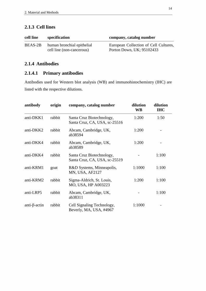
2.1.3 Cell lines
cell line specification company, catalog number
BEAS-2B human bronchial epithelial
cell line (non-cancerous)
European Collection of Cell Cultures,
Porton Down, UK; 95102433
2.1.4 Antibodies
2.1.4.1 Primary antibodies
Antibodies used for Western blot analysis (WB) and immunohistochemistry (IHC) are
listed with the respective dilutions.
antibody origin company, catalog number dilution
WB
dilution
IHC
anti-DKK1 rabbit Santa Cruz Biotechnology,
Santa Cruz, CA, USA, sc-25516
1:200 1:50
anti-DKK2 rabbit Abcam, Cambridge, UK,
ab38594
1:200 -
anti-DKK4 rabbit Abcam, Cambridge, UK,
ab38589
1:200 -
anti-DKK4 rabbit Santa Cruz Biotechnology,
Santa Cruz, CA, USA, sc-25519
- 1:100
anti-KRM1 goat R&D Systems, Minneapolis,
MN, USA, AF2127
1:1000 1:100
anti-KRM2 rabbit Sigma-Aldrich, St. Louis,
MO, USA, HP A003223
1:200 1:100
anti-LRP5 rabbit Abcam, Cambridge, UK,
ab38311
- 1:100
anti-β-actin rabbit Cell Signaling Technology,
Beverly, MA, USA, #4967
1:1000 -

15
2. Material and Methods
2.1.4.2 Secondary antibodies
antibody origin company, catalog number dilution
anti-rabbit IgG,
HRP conjugated
goat Pierce Protein Research Products,
Rockford, IL, USA,#31460
1:3000
anti-goat IgG,
HRP conjugated
rabbit Pierce Protein Research Products,
Rockford, IL, USA, #31402
1:3000
biotinylated
anti-rabbit IgG
goat Invitrogen, Carlsbad, CA, USA,
Histostain-Plus Kit
ready to use
biotinylated
anti-goat IgG
rabbit Invitrogen, Carlsbad, CA, USA,
Histostain-Plus Kit
ready to use
2.1.5 Recombinant proteins
recombinant protein company, catalog number
recombinant human DKK1 R&D Systems, Minneapolis, MN, USA,
1096-DK
2.1.6 Chemicals and reagents
product company
Acrylamide solution, Rotiphorese® Gel 30 Carl Roth GmbH, Karlsruhe, Germany
Agarose Promega, Madison, WI, USA
APS Promega, Madison, WI, USA
β-Mercaptoethanol Sigma-Aldrich, Saint Louis, MO, USA
Bromphenol Blue Sigma-Aldrich, Saint Louis, MO, USA
BSA Sigma-Aldrich, Saint Louis, MO, USA
Chemiluminescent Substrate
SuperSignal® West Pico
Pierce Protein Research Products, USA
Citrate Buffer 20 x Invitrogen, Carlsbad, CA, USA
Complete™ Protease Inhibitor Roche, Basel, Switzerland
Dkk-1 DuoSet ELISA, human, DY1906 R&D Systems, Minneapolis, MN, USA
DNA Ladder 100 bp Promega, Madison, WI, USA
DNA loading dye Blue/Orange, 6 x Promega, Madison, WI, USA
dNTP PCR Nucleotide Mix 10 mM Promega, Madison, WI, USA
EDTA Promega, Madison, WI, USA

16
2. Material and Methods
EGTA Sigma-Aldrich, Saint Louis, MO, USA
Ethanol absolute Sigma-Aldrich, St. Louis, MO, USA
Ethidium bromide Carl Roth GmbH, Karlsruhe, Germany
Glycerol Merck Biosciences, Darmstadt, Germany
Glycerol gelatine, Kaiser’s Merck Biosciences, Darmstadt, Germany
Glycine Carl Roth GmbH, Karlsruhe, Germany
H2O2 30 % (w/w) Perdrogen®
Sigma-Aldrich, St. Louis, MO, USA
Hematoxylin, Mayer’s Sigma-Aldrich, St. Louis, MO, USA
HEPES Buffer solution 1 M PAA Laboratories, Pasching, Austria
Histostain-Plus Kit Invitrogen, Carlsbad, CA, USA
LHC-9 Invitrogen, Carlsbad, CA, USA
Methanol Sigma-Aldrich, Saint Louis, MO, USA
MgCl2 (25 mM) Applied Biosystems, Wellesley, MA, USA
MgCl2 (50 mM) Invitrogen, Carlsbad, CA, USA
Milk powder (blotting grade) Carl Roth GmbH, Karlsruhe, Germany
NaCl Merck Biosciences, Darmstadt, Germany
Paraformaldehyde Sigma-Aldrich, Saint Louis, MO, USA
PBS PAA Laboratories, Pasching, Austria
PCR Buffer 10 x (without MgCl2) Applied Biosystems, Wellesley, MA, USA
Platinum® SYBR
® Green qPCR
SuperMix-UDG
Invitrogen, Carlsbad, CA, USA
Protein Standards Precision Plus,
prestained
Bio-Rad, Hercules, CA, USA
Quick Start™ Bradford 1 x dye reagent Bio-Rad, Hercules, CA, USA
Quick Start™ Bradford Protein Assay Bio-Rad, Hercules, CA, USA
Random Hexamers (50 µM) Applied Biosystems, Wellesley, MA, USA
Reverse Transcriptase MuLV RT (50 U/µl) Applied Biosystems, Wellesley, MA, USA
RNase Inhibitor Applied Biosystems, Wellesley, MA, USA
RNase-Free DNase Set Qiagen, Venlo, Netherlands
RNase-Free Water Qiagen, Venlo, Netherlands
RNeasy Mini Kit Qiagen, Venlo, Netherlands
Roti®-Quick-Kit Carl Roth GmbH, Karlsruhe, Germany

17
2. Material and Methods
SDS Solution 10 % (w/v) Promega, Madison, WI, USA
Streptavidin Invitrogen, Carlsbad, CA, USA
TAE Buffer, Rotiphorese® 10 x Carl Roth GmbH, Karlsruhe, Germany
TEMED Bio-Rad, Hercules, CA, USA
Tris Carl Roth GmbH, Karlsruhe, Germany
Triton® X-100 Promega, Madison, WI, USA
Trypsin EDTA 1 x PAA Laboratories, Pasching, Austria
Tween® 20 Sigma-Aldrich, Saint Louis, MO, USA
Xylene Sigma-Aldrich, St. Louis, MO, USA
2.1.7 Equipment and Software
product company
Calibrated Densitometer GS-800TM
Bio-Rad, Hercules, CA, USA
Chromatography paper 3MM CHR Whatman International Ltd, Maidstone, UK
Developing machine; X Omat 2000 Kodak, Rochester, NY, USA
Electrophoresis and Western blot
chambers
Bio-Rad, Hercules, CA, USA
Fusion A153601 microplate reader Packard BioScience/PerkinElmer,
Waltham, MA, USA
Light microscope Leica DMIL Leica Microsystems, Wetzlar, Germany
Light microscope Olympus BX 51 Olympus, Hamburg, Germany
Microsoft Office Word/Excel/PowerPoint
2007
Microsoft Corp., Unterschleißheim,
Germany
NanoDrop ND-100 Spectrophotometer Peqlab, Erlangen, Germany
Nitrocellulose membrane Bio-Rad, Hercules, CA, USA
PCR-Thermocycler MJ Research, Waltham, MA, USA
Quantity One 1-D analysis software Bio-Rad, Hercules, CA, USA
Sequence Detection System Fast 7500
and Software
Applied Biosystems, Wellesley, MA, USA
UV transilluminator UVP, Upland, CA, USA
X-ray film AGFA Curix HT1000G Plus AGFA-Gevaert, Mortsel, Belgium

18
2. Material and Methods
2.2 Methods
2.2.1 Quantitative reverse transcription polymerase chain reaction
(qRT-PCR)
2.2.1.1 RNA extraction from tissue
RNA was extracted from lung tissue homogenates with the Roti-Quick-Kit. Tissue
samples were homogenized in liquid nitrogen with a pestle, 2 ml of the guanidinium
thiocyanate containing solution were added per 0.2 g of tissue. After 20 min of incuba-
tion, samples were additionally homogenized with a syringe with a 0.9 mm needle. 2 ml
of the phenol and chloroform containing solution were added per sample. Samples were
incubated on ice for 10 min and vortexed every 2 min. After centrifugation at 13000 rpm
for 15 min at a temperature of 4 °C, two phases were separated. The upper, RNA-
containing phase was incubated with an equal volume of the isopropanol containing
solution for 1 h, at a temperature of -80 °C. The centrifugation step was repeated and an
RNA-containing pellet could be extracted. It was dissolved in a mixture of 150 µl of the
guanidinium thiocyanate containing solution and 150 µl of the isopropanol containing
solution and incubated for 1 h at -80 °C. Centrifugation and dissolving step were repeat-
ed. After that, samples were centrifuged at 13000 rpm for 20 min at a temperature of
4 °C. The supernatant was removed and 500 µl of ethanol (70 % v/v in RNase-free wa-
ter) were added. Another centrifugation step was performed for 10 min and the superna-
tant was discarded. The RNA pellet was resuspended in 50 µl of RNase-free water and
stored at a temperature of -80 °C.
2.2.1.2 RNA extraction from cells
Medium was removed from cell culture flasks and cells were washed with PBS twice.
Cells were disrupted and detached from the plates by scraping with a rubber policeman
after addition of β-mercaptoethanol containing RNeasy Lysis Buffer (RNeasy Mini Kit,
preparation according to manufacturer’s protocol) and they were homogenized with a
syringe with 0.9 mm needle. RNA extraction was performed with the silica-membrane
column system of the RNeasy Mini Kit according to the manufacturer´s instructions.
This included an on-column DNase digestion using the RNase-free DNase Set to addi-
tionally remove DNA contaminant. Finally the RNA isolate was eluted from the column
by adding 35 µl of RNase free water twice and then stored at a temperature of -80 °C.

19
2. Material and Methods
2.2.1.3 RNA quantification
Quantification of the samples’ RNA concentration was performed with the NanoDrop
spectrophotometer. Therefore the optical density (OD) of 1.5 µl of the sample was de-
termined at a wavelength of 260 nm, at which absorption correlates to the concentration
of nucleic acids within the sample. Since the absorption at a wavelength of 280 nm cor-
relates with protein concentration, sample purity could be verified by determination of
the RNA/protein ratio (OD260nm/OD280nm). This ratio had to lie in between 1.8 and 2.0 to
exclude a relevant protein contamination.
2.2.1.4 cDNA synthesis by reverse transcription
For analysis of the respective mRNA levels in a sample by qRT-PCR, the RNA had to
be transcribed in complementary DNA (cDNA) by an RNA-dependent DNA polymer-
ase (reverse transcriptase). For preparation of cDNA synthesis, 1 µg of total RNA was
diluted with RNase-free water to a total volume of 20 µl. RNA denaturation was per-
formed by a thermocycler at a temperature of 70 °C for 10 min, followed by cooling the
samples down for 5 min at a temperature of 4 °C. For reverse transcription, 20 µl of the
following RT Mastermix was added to each RNA sample:
component volume final concentration
10 x PCR Buffer (without MgCl2) 4 µl 1 x
MgCl2 (25 mM) 8 µl 5 mM
dNTP PCR Nucleotide Mix (10 mM) 2 µl 0.5 mM
Random Hexamers (50 µM) 2 µl 2.5 µM
RNase Inhibitor (20 U/µl) 1 µl 0.5 U/µl
Reverse Transcriptase MuLV RT (50 U/µl) 2 µl 2.5 U/µl
ddH2O 1 µl
Reverse transcription was performed with a thermocycler by the following steps at the
indicated temperatures and durations:
step temperature duration
attachment of random hexamers 20 °C 10 min
reverse transcription 43 °C 75 min
inactivation of reverse transcriptase 99 °C 5 min
cooling down 4 °C

20
2. Material and Methods
The synthesized cDNA samples were stored at a temperature of -20 °C.
2.2.1.5 Quantitative reverse transcription polymerase chain reaction
Quantitative reverse transcription polymerase chain reaction (qRT-PCR) was performed
using fluorogenic SYBR Green. 2 µl of the previously synthesized cDNA were trans-
ferred to the wells of a 96 well plate, each well filled with 23 µl of the following qRT-
PCR Mastermix:
component volume final concentration
Platinum® SYBR
® Green qPCR SuperMix-UDG 13 µl
MgCl (50 mM) 1 µl 2 mM
forward primer (10 µM) 0.5 µl 200 nM
reverse primer (10 µM) 0.5 µl 200 nM
ddH2O 8 µl
Amplification and detection were carried out with the Sequence Detection System Fast
7500 via performance of 45 cycles of the following steps at the indicated temperatures
and durations:
step temperature duration
activation of polymerase enzyme 50 °C 2 min
first denaturation 95 °C 5 min
second denaturation 95 °C 5 s
annealing 59 °C 5 s
elongation 72 °C 30 s
dissociation step 1 95 °C 15 s
dissociation step 2 60 °C 1 min
dissociation step 3 95 °C 15 s
dissociation step 4 60 °C 15 s
By denaturation, double-stranded DNA gets separated into single strands. During the
annealing phase, primers bind to the respective sequences at the single DNA strands. A
new DNA strand is synthesized by the DNA-dependent DNA polymerase enzyme dur-
ing the elongation step.

21
2. Material and Methods
2.2.1.6 Primers
GenBank from the National Center for Biotechnology Information (NCBI) and the
primer express 3.0 software were used to create adequate primer sequences for target
and reference genes. Detailed information on primer sequences are listed in table 3
(chapter 10.3). Each primer was tested with an undiluted, a 1:8 and a 1:64 diluted sam-
ple to determine primer efficiency for a range of template concentrations. All primers
were utilized at a final concentration of 200 nM.
2.2.1.7 Data evaluation
Since SYBR Green binds sequence independently to double stranded DNA and leads to
an increase in fluorescence, the amount of synthesized DNA is proportional to the fluo-
rescent signal. Fluorescence intensity is measured at the end of the elongation step of
each cycle and a curve depicting the increase in DNA is created. Each sample was
measured twice and the values were averaged. Empty controls containing the respective
qRT-PCR Mastermix without addition of cDNA were measured at each plate.
Target-DNA levels of each sample were normalized to DNA levels of a reference gene
within the respective sample amplified at the same plate. As reference gene, the
constitutively and ubiquitously expressed hypoxanthine-guanine phosphoribosyltrans-
ferase 1 (HPRT1) 33,99
was used in all qRT-PCR reactions. Comparing measurement of
target and reference DNA levels was performed in the exponentially increasing segment
of the curve before saturation had occurred and a threshold was set in this area. For
target and reference gene, the same threshold was used for all samples. The cycle
number at which the fluorescent signal reached the threshold was expressed as Ct
(threshold cycle). These Ct values are in inverse proportion to the initial amount of
RNA within the sample. The relative transcript abundance in a sample is expressed as
Ct value (Ct = Ctreference
– Cttarget
). Therefore positive Ct values of the present study
represent an elevated expression of the target gene compared to the reference gene,
while negative Ct values depict a target gene expression lower than the expression of
the reference gene. Relative changes of transcript levels in IPF samples compared to
donor samples are given as Ct values (Ct = CtIPF
– Ctdonor
). The Ct values
approximately correspond to the binary logarithm of the fold change.
The purity and length of the DNA products were verified by melting curve analysis and
agarose gel electrophoresis.

22
2. Material and Methods
2.2.1.8 Melting curve analysis
Melting curve analysis by the Fast 7500 System was used to verify the purity of the
specific DNA product, since the melting temperature depends on product length and the
amount of GC basepairs in the double stranded DNA. After 45 cycles, the melting point
was identified by slowly increasing heat. The peak of fluorescence-intensity-change,
induced by the release of SYBR Green during the melting process, indicated the spe-
cific melting temperature of one product.
2.2.1.9 DNA agarose gel electrophoresis
qRT-PCR products were displayed by agarose gel electrophoresis in order to check the
product size to ensure that the correct template had been amplified. Therefore agarose
gels were prepared:
2 % agarose gel: 1 x TAE buffer
2 % agarose (w/v)
0.5 µg/ml ethidium bromide
PCR product samples were mixed 5:1 with 6 x DNA loading dye Blue/Orange and
transferred onto the gel. For each PCR product, two different samples as well as one
empty control sample were applied. A 100 bp DNA ladder was run at the same gel. Gels
were run in an electrophoresis chamber filled with 1x TAE buffer (Tris, acetic acid and
EDTA) at 100 V for about 1 h. DNA bands were analyzed under ultraviolet lighting
conditions.
2.2.2 Western blot analysis
Western blot analysis was performed on total protein extracts of lung tissue homoge-
nates from donors and IPF patients.
2.2.2.1 Protein extraction and quantification
The lung tissue was homogenized in liquid nitrogen with a pestle and 1 ml lysis buffer
was added per 0.1 g of tissue. After 5 min of incubation, a syringe with 0.9 mm needle
was used for further homogenization. Samples were kept on ice for 30 min and vortexed
every 5 min. By centrifugation at 12.000*g at a temperature of 4 °C for 10 min, the pro-

23
2. Material and Methods
tein containing supernatant was separated from the tissue homogenate. The Supernatant
was collected and stored at a temperature of -20 °C.
Lysis buffer: 20 mM Tris pH 7.5
150 mM NaCl
1 mM EDTA
1 mM EGTA
1 % Triton X-100
2 mM Na3VO4
1:25 Complete™, protease inhibitor mix
Protein quantification of each sample using the Quick Start Bradford Protein Assay was
performed via spectrophotometric measurement with a Fusion A153601 Reader at a
wavelength of 570 nm. Wells of a 96 well plate were filled with 200 µl of Quick Start
Bradford dye reagent and 10 µl of the 1:20 diluted protein sample were added to a
respective well. Additionally, six diluted BSA samples (0.05, 0.1, 0.2, 0.3, 0.4 and
0.5 µg/µl) as well as negative controls containing Bradford dye and 1:20 diluted protein
lysis buffer were measured at the same plate. Measurement was done after an
incubation period of 15 min. Duplicates of BSA standards, protein samples and controls
were measured and values were averaged. By comparing protein sample values to the
BSA standard curve via interpolation, the protein concentration of each sample was
calculated.
2.2.2.2 SDS polyacrylamide gel electrophoresis (SDS-PAGE)
To separate proteins by size, SDS polyacrylamide gel electrophoresis was performed.
Therefore, the required volume of each protein sample was calculated to reach a total
amount of 25 µg protein for every loading sample. Equivalent volumes of SDS contain-
ing 2 x Sample Buffer were added to the proteins and samples were denaturated at a
temperature of 95 °C for 10 min. Samples were loaded onto a polymerized gel, consist-
ing of a 15-well stacking gel on top of a resolving gel. As molecular weight size mark-
ers, 5 µl of protein standards were run at the same gel. Proteins were kept on ice in be-
tween the working steps. Gels were run in an electrophoresis chamber filled with Run-
ning Buffer at 110 V.

24
2. Material and Methods
Resolving gel: Stacking gel:
10 % acrylamide/bisacrylamide 5 % acrylamide/bisacrylamide
375 mM Tris-HCl pH 8.8 125 mM Tris-HCl pH 6.8
0.1 % (w/v) SDS 0.1 % (w/v) SDS
0.1 % (w/v) APS 0.1 % (w/v) APS
0.1 % (v/v) TEMED 0.1 % (v/v) TEMED
The used acrylamide solution contained acrylamide : bisacrylamide in a 37.5 : 1 ratio.
2 x Sample Buffer: Running Buffer:
100 mM Tris-HCl pH 6.8 25 mM Tris
4 % (w/v) SDS 250 mM glycine
0.2 % (w/v) bromphenol blue 0.1 % (w/v) SDS
20 % (v/v) glycerol
100 nM DTT
2.2.2.3 Immunoblotting
For further analyses, the separated proteins had to be transferred onto a nitrocellulose
membrane by using a Western blot electrophoresis chamber. Packing of the gel and ni-
trocellulose membrane into a cassette with chromatography papers and sponges was
performed in Transfer Buffer to avoid air bubbles. The cassette was put into an electro-
phoresis chamber filled with Transfer Buffer and the transfer was done at 120 V for 1 h.
An icebox was used to cool the chamber during the blotting process.
2.2.2.4 Protein detection
Nitrocellulose membranes were washed in Washing Buffer (2 x 5 min) and kept in
Blocking Buffer for about 1 h. Incubation with the primary antibody was performed
overnight at a temperature of 4 °C with a specific dilution of each antibody in Blocking
Buffer (primary antibodies and their dilutions are listed in chapter 2.1.4.1). After a
washing step (3 x 10 min with Washing Buffer), the membrane was incubated with a
dilution of the adequate horseradish peroxidase (HRP)-conjugated secondary antibody
in Blocking Buffer for 1 h (secondary antibodies and their dilutions are listed in chapter
2.1.4.2). By performing these steps, primary and secondary antibodies got attached to
the specific protein bands. After a second washing step (5 x 10 min), the membrane was

25
2. Material and Methods
ready for protein detection. For visualization of protein bands, the Chemiluminescent
Substrate SuperSignal West Pico was applied according to the manufacturer’s instruc-
tions. An X-ray film was exposed to the membrane and developed. All incubation and
washing steps were performed at room temperature unless it is indicated otherwise.
Before reusing the nitrocellulose membrane for an additional protein detection, they
were incubated with Stripping Buffer in a water bath at a temperature of 52 °C for 8 min
and were washed with Washing Buffer afterwards. Subsequent detection of β-actin at
each membrane served as a control for an equal protein loading.
Transfer Buffer: Washing Buffer:
25 mM Tris PBS (1 x)
192 mM glycine 0.1 % (v/v) Tween 20
20 % (v/v) methanol
Blocking Buffer: Stripping Buffer:
PBS (1 x) 100 mM β-mercaptoethanol
0.1 % (v/v) Tween 20 62.5 mM Tris pH 6.8
5 % (w/v) skim milk powder 2 % (w/v) SDS
2.2.2.5 Densitometry
Densitometric analysis of the developed X-ray films was performed using a GS-800
calibrated densitometer and the Quantity One 1-D analysis software. Optical density of
β-actin loading control was used to equalize differences in total protein loading. There-
fore the relative expression level of an indicated protein is calculated as relative optical
density (OD) (optical density of indicated protein / optical density of β-actin) for each
sample. Changes in expression levels between the indicated groups were calculated as
fold change of the means (OD(IPF) / OD(donor)) and were expressed as fold change ±
SEM.
2.2.3 Immunohistochemistry
Paraffin embedded human lung tissue samples were cut to 3 µm sections and were
mounted on slides. Immunohistochemical staining was performed with the Histostain
Plus Kit. To remove the paraffin, slides were kept in an oven at a temperature of 48 °C

26
2. Material and Methods
over night and were transferred to Xylene (3 x 10 min) afterwards. Incubation with de-
creasing ethanol dilutions (100 %, 95 %, 70 % of ethanol, 2 x 5 min each) and a washing
step with PBS (2 x 5 min) followed, before tissue sections were treated for antigen re-
trieval. Therefore slides were placed in citrate buffer (1 x) and cooked in a water bath
for 25 min. After cooling down, slides were washed with PBS again. Quenching of en-
dogenous peroxidase activity was performed by incubation with 3 % (v/v) H2O2 for
20 min. After another washing step, the slides were incubated with serum blocking solu-
tion (Histostain Plus Kit, derived from a species different than the source of the in-
tended primary antibody) for 10 min. Subsequently the primary antibody was prepared
and applied over night at a temperature of 4 °C.
Primary antibody dilution:
PBS 1 x
1.5 % (v/v) serum blocking solution
1 % or 2 % (v/v) primary antibody solution*
*depending on the respective antibody dilution; primary antibodies and their dilutions
are listed in chapter 2.1.4.1.
After 30 min at room temperature and a washing step with PBS, slides were incubated
with the species-appropriate secondary biotinylated antibody (Histostain plus Kit, sec-
ondary biotinylated antibodies are listed in chapter 2.1.4.2) for 10 min. Another washing
step and incubation with HRP-conjugated Streptavidin for 10 min followed. After re-
moval of Streptavidin, the staining reaction was performed by incubating the slides with
chromogen solution (Histostain Plus Kit) until red color staining was clearly visible
under a microscope. Another washing step with PBS and counterstaining with hema-
toxylin for 8 min were performed subsequently. After washing the slides in running tap
water for 10 min, coverslips were mounted via glycerol gelatine. The stained sections
were examined using an Olympus BX51 microscope and pictures were taken in 3 dif-
ferent magnifications as indicated.
2.2.4 Cell culture
The human bronchial epithelial cell line BEAS-2B was maintained in 250 ml culture
flasks filled with 10 ml LHC-9 medium in a 5 % CO2 containing atmosphere of 95-

27
2. Material and Methods
100 % air humidity at a temperature of 37 °C. The medium was changed every 3 days at
least and passaging was performed at a confluence of 80-90 %. Therefore the medium
was removed, cells were gently washed with PBS (1 x) and incubated with 3 ml of
Trypsin EDTA. After detachment, 7 ml of fresh medium were added and 20 % of the
cell suspension were transferred to a new culture flask. Then medium was added for a
total amount of 10 ml suspension.
2.2.5 Enzyme-linked immunosorbent assay (ELISA)
A human DKK1 enzyme-linked immunosorbent assay was performed on BAL fluids
(BALF) following the company’s instructions. Wells of a 96-well microplate were
coated with 100 µl of the diluted Capture Antibody and incubated for 12 h. Blocking
was performed with 300 µl of Reagent Diluent for 1 h before incubating the wells with
100 µl of the BALF samples for 2 h. Additionally, seven 2-fold serial dilutions of re-
combinant DKK1 protein standard with a high standard of 4000 pg/ml were applied at
the same plate. Each BALF sample and the standard dilutions were tested twice. After
incubation with 100 µl of the Detection Antibody for 2 h, Streptavidin-HRP was added
to each well for 20 min. Then 100 µl of Substrate Solution were applied to each well for
another 20 min. Color reaction was stopped by adding 50 µl of Stop Solution. In be-
tween the working steps, washing steps were carried out following the company’s in-
struction. Optical density was determined with a microplate reader at a wavelength of
450 nm. To minimize the optical influence of the plate, a second measurement was done
at 570 nm and values were subtracted from the measurement at 450 nm. Double values
of samples and standards were finally averaged and the DKK1 protein content of BAL
fluids was calculated by interpolation on the basis of the seven point DKK1 protein
standard curve.
2.2.6 Statistical analysis
All numerical data are presented as mean ± SEM. All experimental values were tested
for normal distribution and the indicated groups were compared using a two-tailed, un-
paired two-sample t-test for experiments with two groups of independent samples. Re-
sults were considered statistically significant when p < 0.05.

28
3. Results
3 Results
3.1 Expression of Dickkopf proteins and their receptors
in the lung
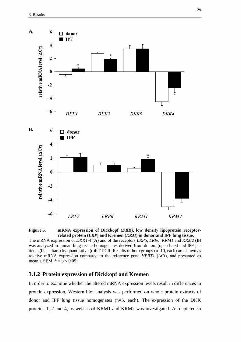
3.1.1 mRNA expression of Dickkopf proteins and their receptors
In order to quantify the relative Dickkopf (DKK) mRNA expression levels in donor and
IPF lungs, quantitative (q)RT-PCR was performed on whole RNA-isolates of human
lung tissue homogenates. Samples from donors and IPF patients were compared regard-
ing their expression of DKK1-4 (n=10, each). As demonstrated in figure 5A, the mRNA
of all four DKK proteins was detected in donor as well as in IPF specimens. While
DKK3 presented the highest relative mRNA expression level, DKK4 showed the lowest
mRNA expression in both, donor and IPF tissue samples. Comparison of IPF and donor
lungs revealed significant differences in the relative transcript levels. While DKK1 and
DKK4 presented a significantly increased mRNA expression in the fibrotic lung tissues
(∆∆Ct (mean ± SEM): DKK1: 0.85 ± 0.36 and DKK4: 2.09 ± 0.88), DKK2 mRNA was
significantly decreased (∆∆Ct: -0.94 ± 0.34). The transcript levels of DKK3 did not
show a significant alteration (∆∆Ct: 0.02 ± 0.85).
The relative mRNA expression levels of the DKK receptors low density lipoprotein
receptor-related protein (LRP) 5 and 6 and Kremen (KRM) 1 and 2 were examined
likewise. As depicted in figure 5B, all examined receptors were detected in donor as
well as in IPF tissues. In both groups, KRM2 presented a lower basal expression level
than KRM1 and the LRP receptors. While the expression of KRM1 mRNA in IPF sam-
ples was significantly increased compared to the donor tissues (∆∆Ct: 1.31 ± 0.28), ex-
pression levels of KRM2, LRP5 and LRP6 were not significantly altered (∆∆Ct: KRM2:
1.19 ± 0.72, LRP5: 0.06 ± 0.63 and LRP6: 0.03 ± 0.37).
29
3. Results
A.
B.
Figure 5. mRNA expression of Dickkopf (DKK), low density lipoprotein receptor-
related protein (LRP) and Kremen (KRM) in donor and IPF lung tissue.
The mRNA expression of DKK1-4 (A) and of the receptors LRP5, LRP6, KRM1 and KRM2 (B)
was analyzed in human lung tissue homogenates derived from donors (open bars) and IPF pa-
tients (black bars) by quantitative (q)RT-PCR. Results of both groups (n=10, each) are shown as
relative mRNA expression compared to the reference gene HPRT1 (ΔCt), and presented as
mean ± SEM, * = p < 0.05.
3.1.2 Protein expression of Dickkopf and Kremen
In order to examine whether the altered mRNA expression levels result in differences in
protein expression, Western blot analysis was performed on whole protein extracts of
donor and IPF lung tissue homogenates (n=5, each). The expression of the DKK
proteins 1, 2 and 4, as well as of KRM1 and KRM2 was investigated. As depicted in

30
3. Results
figure 6A, Western blotting revealed enhanced levels of all investigated DKK proteins
in IPF. These results were confirmed by densitometric quantification of the respective
protein immunoblots (figure 6B). Accordingly, DKK1 and DKK2 protein expression
was significantly increased in IPF samples compared to donor samples (fold change
(OD(IPF) / OD(donor)) ± SEM: DKK1: 1.45 ± 0.09 and DKK2: 1.99 ± 0.13). DKK4 protein
was not detected in donor tissue samples at all.
A.
B.
Figure 6. Protein expression of DKK in donor and IPF lung tissue.
(A) Expression of the proteins DKK1, DKK2 and DKK4 in human lung tissue homogenates
derived from donors and IPF patients (n=5, each) was determined by Western blot analysis of
whole protein extracts. Antibodies were used as indicated, β-actin served as loading control.
Protein immunoblots were carried out twice, a representative blot is shown. (B) The densitomet-
ric analyses of the respective protein immunoblots are shown. The relative optical density (OD)
(optical density of indicated protein / optical density of β-actin loading control) is presented for
donor (open bars) and IPF (black bars) tissues as mean ± SEM. DKK4 protein was not detected
in donor tissue samples, the respective OD was not available (n/a). * = p < 0.05.
The DKK receptors KRM1 and KRM2 were detected in lung tissue homogenates of
both, donors and IPF patients (figure 7A). Both proteins depicted a high intra-group
variability of expression levels. While the densitometric analysis of KRM1 protein re-
vealed a significantly increased expression in IPF tissue samples ((OD(IPF) / OD(donor)):

31
3. Results
1.92 ± 0.06), the expression of KRM2 protein was not significantly altered ((OD(IPF) /
OD(donor)): 1.37 ± 0.40) (figure 7B).
A.
B.
Figure 7. Protein expression of KRM in donor and IPF lung tissue.
(A) Expression of the proteins KRM1 and KRM2 in human lung tissue homogenates derived
from donors and IPF patients (n=5, each) was determined by Western blot analysis of whole
protein extracts. Antibodies were used as indicated, β-actin served as loading control. Protein
immunoblots were carried out twice, a representative blot is shown. (B) The densitometric
analyses of the respective protein immunoblots are shown. The relative optical density (OD)
(optical density of indicated protein / optical density of β-actin loading control) is presented for
donor (open bars) and IPF (black bars) tissues as mean ± SEM, * = p < 0.05.
3.2 Localization of Dickkopf proteins and their receptors
in the lung After investigations concerning the expression profile of Dickkopf proteins and recep-
tors on mRNA and protein level, their actual localization in the human lung was exam-
ined. Immunohistochemical stainings were performed on donor and IPF lung tissue sec-
tions in order to identify cells that are actually expressing these proteins. Antibodies
with reasonable and reproducible staining results could be found for DKK1 and DKK4
as well as for the receptors LRP5, KRM1 and KRM2. Stainings were performed at least
twice using three different donor and IPF lung tissues for each antibody. Representative
bronchial (A) and alveolar (B) regions are presented. As demonstrated in figure 8A,

32
3. Results
DKK1 was mainly located in bronchial epithelial cells in donor (left panel) and IPF
lungs (right panel), with a pronounced accumulation of DKK1 in basal bronchial epithe-
lial cells (figure 8A, arrows). In IPF lungs, DKK1 was furthermore located in hyper-
plastic alveolar epithelial cells (figure 8B, right panel, arrows). Besides, granulocytes
presented staining of DKK1 protein in both, donor and IPF lung tissue sections (best
recognizable in figure 8B, left panel).
A.
Figure 8A. Localization of DKK1 protein in donor and IPF lung tissue –
bronchial regions.
Immunohistochemical staining for DKK1 protein was performed on lung tissue sections of do-
nors and IPF patients. Stainings were performed at least twice using three different donor and
IPF lung tissues. Representative bronchial regions of donor (left panel) and IPF (right panel)
lungs are shown in three magnifications as indicated. Arrows indicate basal bronchial epithelial
cells.

33
3. Results
B.
Figure 8B. Localization of DKK1 protein in donor and IPF lung tissue –
alveolar regions.
Immunohistochemical staining for DKK1 protein was performed on lung tissue sections of do-
nors and IPF patients. Stainings were performed at least twice using three different donor and
IPF lung tissues. Representative alveolar regions of donor (left panel) and IPF (right panel)
lungs are shown in three magnifications as indicated. Arrows indicate hyperplastic alveolar
epithelial cells.

34
3. Results
DKK4 protein was largely located in bronchial epithelial cells (figure 9A) and to a
slighter extent in interstitial cells, in donor as well as in IPF lung tissues (figure 9A and
9B). In comparison to DKK1, DKK4 expression exhibited a more equal basal-apical
intensity in the bronchial epithelium (figure 9A), whereas its general expression pattern
was scattered. As depicted in figure 9B, DKK4 was strongly expressed in hyperplastic
alveolar epithelial cells in IPF (figure 9B, right panel, arrows).
A.
Figure 9A. Localization of DKK4 protein in donor and IPF lung tissue –
bronchial regions.
Immunohistochemical staining for DKK4 protein was performed on lung tissue sections of do-
nors and IPF patients. Stainings were performed at least twice using three different donor and
IPF lung tissues. Representative bronchial regions of donor (left panel) and IPF (right panel)
lungs are shown in three magnifications as indicated.

35
3. Results
B.
Figure 9B. Localization of DKK4 protein in donor and IPF lung tissue –
alveolar regions.
Immunohistochemical staining for DKK4 protein was performed on lung tissue sections of do-
nors and IPF patients. Stainings were performed at least twice using three different donor and
IPF lung tissues. Representative alveolar regions of donor (left panel) and IPF (right panel)
lungs are shown in three magnifications as indicated. Arrows indicate hyperplastic alveolar
epithelial cells.

36
3. Results
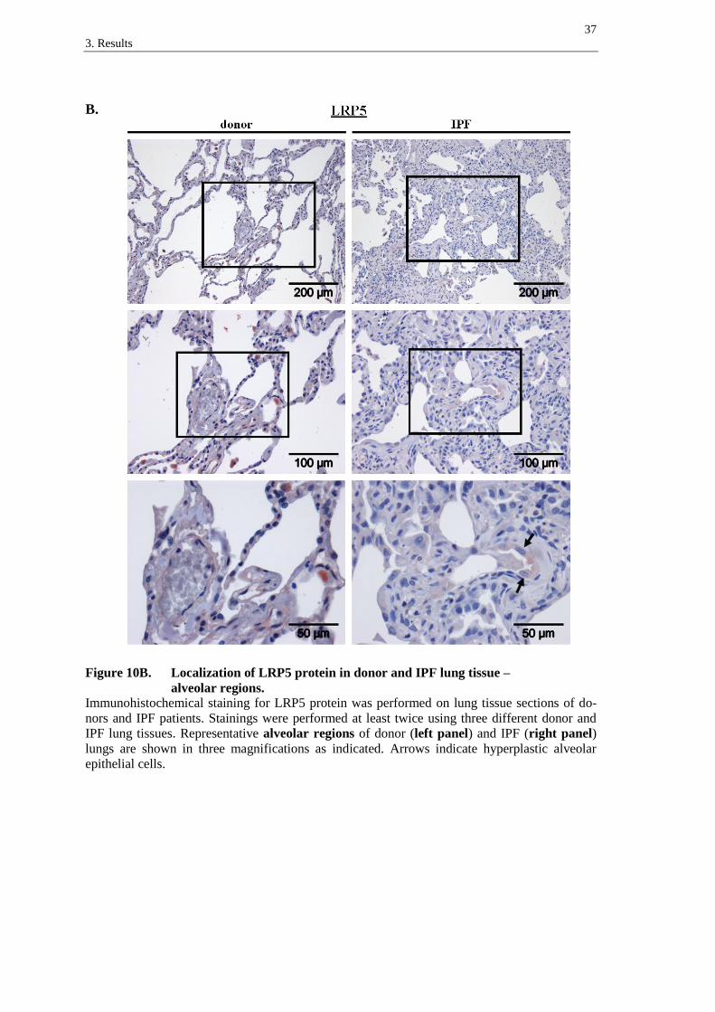
LRP5 receptor was detected in the bronchial epithelium of donor (figure 10A, left
panel) and IPF tissues (figure 10A, right panel), with predominant staining of supranu-
clear regions in columnar bronchial epithelial cells (figure 10A, left and right panel,
arrows). In IPF, LRP5 was slightly expressed in hyperplastic alveolar epithelial cells
(figure 10B, right panel, arrows). Moreover, immune cells presented staining of LRP5
in donor and IPF lung tissue sections (best recognizable in figure 10B, left panel).
A.
Figure 10A. Localization of LRP5 protein in donor and IPF lung tissue –
bronchial regions.
Immunohistochemical staining for LRP5 protein was performed on lung tissue sections of do-
nors and IPF patients. Stainings were performed at least twice using three different donor and
IPF lung tissues. Representative bronchial regions of donor (left panel) and IPF (right panel)
lungs are shown in three magnifications as indicated. Arrows indicate supranuclear staining in
columnar bronchial epithelial cells.
37
3. Results
B.
Figure 10B. Localization of LRP5 protein in donor and IPF lung tissue –
alveolar regions.
Immunohistochemical staining for LRP5 protein was performed on lung tissue sections of do-
nors and IPF patients. Stainings were performed at least twice using three different donor and
IPF lung tissues. Representative alveolar regions of donor (left panel) and IPF (right panel)
lungs are shown in three magnifications as indicated. Arrows indicate hyperplastic alveolar
epithelial cells.

38
3. Results
The DKK-binding receptors KRM1 and KRM2 could also be detected in the lung tissue
sections. In detail, KRM1 protein was expressed in bronchial epithelial (figure 11A)
and smooth muscle cells (best recognizable in figure 11A, right panel, arrow) in donor
and in IPF lungs. In IPF, the bronchial epithelium displayed a heterogeneous staining
pattern (figure 11A, right panel) and hyperplastic alveolar epithelial cell regions showed
expression of KRM1 protein (figure 11B, right panel, arrow). KRM1 was also found in
alveolar macrophages (best recognizable in figure 11A and 11B, left panels).
A.
Figure 11A. Localization of KRM1 protein in donor and IPF lung tissue –
bronchial regions.
Immunohistochemical staining for KRM1 protein was performed on lung tissue sections of do-
nors and IPF patients. Stainings were performed at least twice using three different donor and
IPF lung tissues. Representative bronchial regions of donor (left panel) and IPF (right panel)
lungs are shown in three magnifications as indicated. An arrow indicates smooth muscle cells.
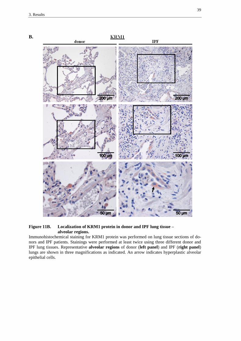
39
3. Results
B.
Figure 11B. Localization of KRM1 protein in donor and IPF lung tissue –
alveolar regions.
Immunohistochemical staining for KRM1 protein was performed on lung tissue sections of do-
nors and IPF patients. Stainings were performed at least twice using three different donor and
IPF lung tissues. Representative alveolar regions of donor (left panel) and IPF (right panel)
lungs are shown in three magnifications as indicated. An arrow indicates hyperplastic alveolar
epithelial cells.
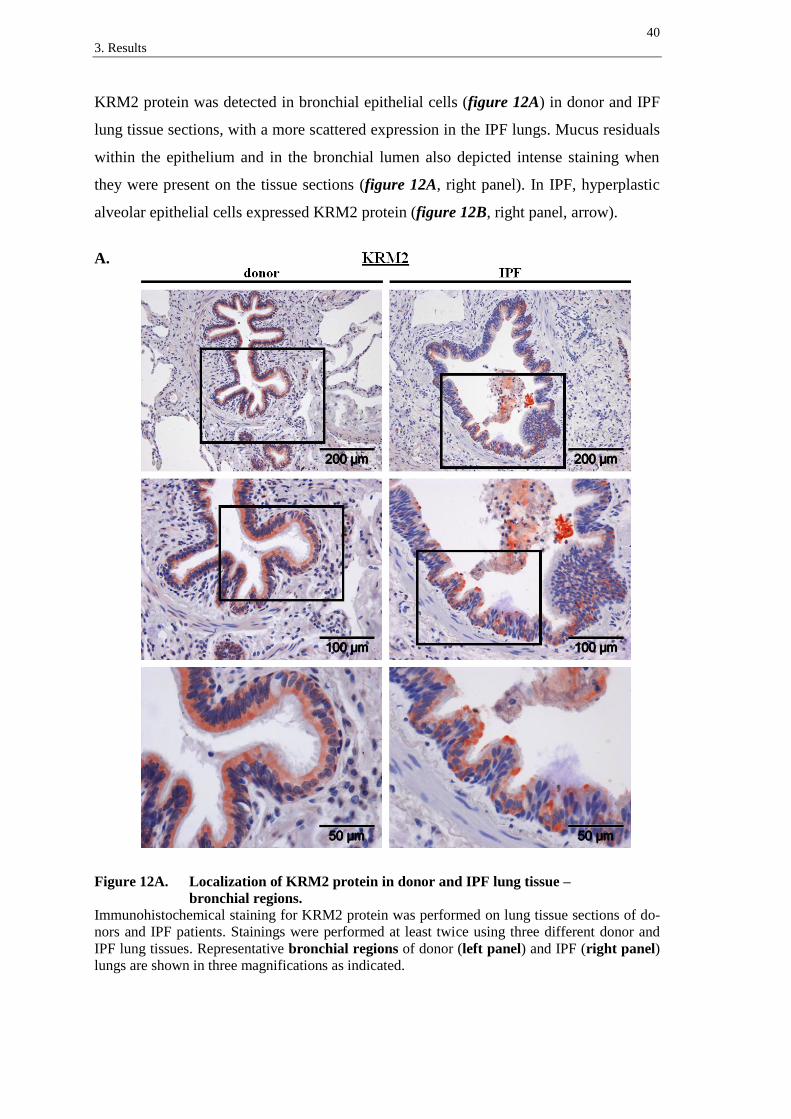
40
3. Results
KRM2 protein was detected in bronchial epithelial cells (figure 12A) in donor and IPF
lung tissue sections, with a more scattered expression in the IPF lungs. Mucus residuals
within the epithelium and in the bronchial lumen also depicted intense staining when
they were present on the tissue sections (figure 12A, right panel). In IPF, hyperplastic
alveolar epithelial cells expressed KRM2 protein (figure 12B, right panel, arrow).
A.
Figure 12A. Localization of KRM2 protein in donor and IPF lung tissue –
bronchial regions.
Immunohistochemical staining for KRM2 protein was performed on lung tissue sections of do-
nors and IPF patients. Stainings were performed at least twice using three different donor and
IPF lung tissues. Representative bronchial regions of donor (left panel) and IPF (right panel)
lungs are shown in three magnifications as indicated.

41
3. Results
B.
Figure 12B. Localization of KRM2 protein in donor and IPF lung tissue –
alveolar regions.
Immunohistochemical staining for KRM2 protein was performed on lung tissue sections of do-
nors and IPF patients. Stainings were performed at least twice using three different donor and
IPF lung tissues. Representative alveolar regions of donor (left panel) and IPF (right panel)
lungs are shown in three magnifications as indicated. An arrow indicates hyperplastic alveolar
epithelial cells.
Immunohistochemical stainings of tissue sections from further donor and IPF lungs as
supplement to the pictures illustrated here are depicted in chapter 10.4 for the respective
antibodies.

42
3. Results
3.3 WNT/β-catenin pathway components in bronchial
epithelial cells
Immunohistochemistry detected DKK1 and DKK4 as well as their LRP and KRM re-
ceptors to be largely located in the bronchial epithelium of donor and IPF lungs. In or-
der to investigate whether human bronchial epithelial cells can also express other key
molecules of the WNT/β-catenin pathway and would therefore be basically capable of
responding to WNT and DKK stimulation, the human bronchial epithelial cell line
BEAS-2B was examined. RNA isolates of this cell line were analyzed for their mRNA
expression of WNT/β-catenin signaling pathway components and DKK receptors by
qRT-PCR (figure 13A, B). Additionally, the expression of WNT and DKK ligands was
examined (figure 13C, D). Two flasks of BEAS-2B cells were tested and values were
averaged. As depicted in figure 13A and 13B, mRNA of the examined WNT receptors
FZD1-4, of the WNT and DKK binding receptors LRP5 and LRP6, as well as of the
DKK receptors KRM1 and KRM2 was expressed by BEAS-2B cells (figure 13A). LRP5
and LRP6 displayed the highest expression levels of these receptors. Key components of
the WNT/β-catenin signaling cascade were expressed as well, with a high relative
mRNA expression of β-catenin (β-CAT) (figure 13B). Of the examined ligands (figure
13C, D), WNT1, WNT7b, WNT10b, as well as DKK1 and DKK3 could be detected.
WNT1 and WNT10b mRNA was only expressed on low levels. WNT3a, DKK2 and
DKK4 did not show an evaluable mRNA expression in the BEAS-2B RNA isolates
(figure 13C, D).

43
3. Results
A.
B.
C. D.
Figure 13. mRNA expression of WNT/β-catenin pathway components
in the BEAS-2B cell line.
The mRNA expression of the receptors FZD1-4, LRP5, LRP6, KRM1 and KRM2 (A), of the
WNT/β-catenin pathway components β-catenin (β-CAT), GSK3β, LEF1, TCF3 and TCF4 (B)
and of the ligands WNT1, WNT3a, WNT7b, WNT10b (C) and DKK1-4 (D) was analyzed in
RNA isolates of the human bronchial epithelial cell line BEAS-2B by quantitative (q)RT-PCR.
Averaged results of 2 isolates are shown as relative mRNA expression compared to the refer-
ence gene HPRT1 (ΔCt) and are presented as mean ± SEM. WNT3a, DKK2 and DKK4 mRNA
was not expressed, respective ΔCt values were not available (n/a).

44
3. Results
3.4 DKK1 protein in bronchoalveolar lavage fluids
The present study demonstrated that DKK1 mRNA and protein expression are increased
in IPF compared to donor lung tissues and that DKK1 is largely located in basal bron-
chial epithelial cells. To investigate whether the epithelial localization leads to occur-
rence of DKK1 in the bronchial lumen and whether there are measurable differences
between IPF patients and healthy controls in vivo, bronchoalveolar lavage fluids
(BALF) were analyzed. Therefore, DKK1 protein was quantified in BAL fluids of
healthy volunteers (n=4) and IPF patients (n=9) using an enzyme-linked immunosorbent
assay (ELISA). As depicted in figure 14, DKK1 was expressed in the BAL fluids of
both groups. While DKK1 levels in the BAL fluids of healthy volunteers showed a
comparatively small divergence, the levels found in BAL fluids of IPF patients were
widespread, varying up to a triplication of the absolute protein value between the lowest
and the highest DKK1 concentration. However, the concentration of DKK1 protein in
BAL fluids from IPF patients (mean ± SEM: 456 ± 55 pg/ml) was significantly in-
creased compared to the healthy controls (266 ± 11 pg/ml).
Figure 14. DKK1 protein concentration in bronchoalveolar lavage fluid.
The DKK1 protein concentration in bronchoalveolar lavage fluids (BALF) of healthy volunteers
(control group, open bar, n=4) and IPF patients (black bar, n=9) was quantified using an en-
zyme-linked immunosorbent assay (ELISA). The concentration of DKK1 protein is given in
pg/ml and is presented as mean ± SEM. * = p < 0.05.

45
4. Discussion
4 Discussion
Idiopathic pulmonary fibrosis is a progressive and fatal lung disease that induces struc-
tural remodeling of the lungs’ architecture, causes respiratory impairment and limits
quality of life 36,133,158
. Due to the restricted response to pharmaceutical treatments, lung
transplantation is currently the only option proven to notably prolong survival of IPF
patients 112,168
. The mechanisms of IPF have not been fully elucidated yet and new in-
sights into the pathogenesis may promote improvements of therapeutic strategies.
Recent studies have highlighted an involvement of WNT/β-catenin signaling in IPF. So
far, an aberrant reactivation of this developmental pathway in IPF lung tissue has been
demonstrated 26,86
. WNT/β-catenin signaling may contribute to disease progression,
since knockout of the WNT target gene MMP7 183
as well as specific inhibition of
WNT/β-catenin target gene transcription 63
or canonical pathway inhibition via β-
catenin small interfering RNA 82
in the bleomycin mouse model attenuated pulmonary
fibrosis. Although limitations concerning the transferability of this animal model to
some aspects of IPF pathobiology have to be taken into account 113
, modulators of WNT
signaling represent an attractive target in IPF research. Dickkopf proteins with their
ability to bind WNT coreceptors of the LRP family belong to the best studied regulators
of WNT/β-catenin signal transduction 101
.
An antifibrotic effect of DKK1 via WNT/β-catenin inhibition has already been reported
in mouse models of cholestatic liver fibrosis 23
and renal fibrosis 62
. However, the role
of DKK proteins in IPF had not been addressed before. Therefore, the present study
focused on the expression and localization of DKK proteins and their receptors in the
healthy adult lung and in the lungs of IPF patients.
4.1 Altered expression of DKK proteins and their receptors
in IPF
The present study reveals a regulation of DKK and KRM expression in IPF. The mRNA
expression of DKK1 and DKK4 was found to be increased in IPF compared to donor
lung tissues, consistent with elevated protein levels of DKK1 and DKK4 detected by
Western blot analysis. The increase in DKK2 protein expression however did not match
the decreased DKK2 mRNA levels in IPF lungs, suggesting differences in translational
control mechanisms or protein stability. At first appearance, an increase of DKK1 and

46
4. Discussion
DKK4 in IPF lungs seems to contradict the reported activation of WNT/β-catenin sig-
naling in IPF, considering their WNT-inhibitory capability. But actually, this finding is
consistent with an increased WNT activation, since DKK1 21,53,122
and DKK4 10,130
have
been identified as downstream target genes of the WNT/β-catenin pathway, in terms of
a negative feedback mechanism. For DKK2 and DKK3, feedback-mechanisms have not
been demonstrated yet.
Since availability of DKK receptors in the lung tissue represents a basic requirement for
modulation of WNT/β-catenin signaling via secreted DKK proteins, the present study
focused on the expression profile of these proteins as well. In both, donor and IPF
lungs, mRNA expression of LRP5 and LRP6 could be confirmed by qRT-PCR. Addi-
tionally, LRP5 protein expression and localization could be demonstrated by immuno-
histochemistry. The presence of LRP6 protein in its phosphorylated form in homoge-
nates of IPF and donor lungs has been described before by Western blot analysis 86
.
KRM1 and KRM2 mRNA and the respective proteins KRM1 and KRM2 could also be
detected, with an increased expression of KRM1 in IPF samples.
When interpreting the data, it has to be taken into account that mRNA and protein ex-
pression analyses were performed on lung tissue homogenates. As presence of DKK
proteins and their receptors is cell type dependent, expression levels are subject to the
cellular composition of each donor and IPF sample.
4.2 Localization of DKK proteins and their receptors in the
lung tissue
Immunohistochemical analyses supplement the data derived from lung homogenates
with the actual, cell-type dependent localization pattern. They revealed DKK1 and
DKK4 to be largely located in the bronchial epithelium in donor as well as in IPF lung
tissue sections. Additionally, hyperplastic alveolar epithelial cells in IPF exhibited in-
tense staining for these DKK proteins. Regarding the WNT-target-gene character of
DKK1 and DKK4, this observation is compatible with the report on nuclear β-catenin
accumulation and therefore with active WNT/β-catenin signaling within these cells 26
. It
has to be mentioned that the observed expression of DKK4 protein is inconsistent with
the protein expression pattern detected by Western blot analysis, which did not depict
DKK4 protein expression in the donor samples. However, it has to be taken into consid-
eration that samples of different donors had to be used for the two experiments, more-
47
4. Discussion
over different DKK4 antibodies. For interpretation of the immunohistochemical data
presented in the present study, it has to be noted that stainings of donor and IPF tissue
sections are not quantitatively comparable, since exposure to the chromogen solution
was performed until red color staining for each section was clearly visible under a mi-
croscope.
Interestingly, immunohistochemical staining for the receptors LRP5, KRM1 and KRM2
revealed striking similarities to the expression pattern of their own ligands. All of the
investigated receptors were predominantly located in the bronchial epithelium in both,
donor and IPF lungs and additionally in hyperplastic alveolar epithelial cells in the fi-
brotic lungs. This corresponds to the observed expression pattern of DKK1 and DKK4
in the present study. Furthermore, these observations are in line with the previously re-
ported expression patterns of WNT1, WNT3a and GSK3β, whose presence in lung tis-
sue also included the expression by bronchial epithelial cells and by hyperplastic ATII
cells in IPF lungs 86
. Additionally, the analysis of the human bronchial epithelial cell
line BEAS-2B in the present study demonstrated that human bronchial epithelial cells
are able to express other key components of the WNT/β-catenin signaling cascade, as
well. Taken together, these data indicate that autocrine and paracrine effects of the se-
creted DKK proteins are most likely the major type of interference with WNT in the
lung epithelium.
The detection of KRM proteins in the lung epithelium has to be evaluated in the context
of recent findings. While some authors support the theory of DKK, LRP and KRM
forming a ternary complex that undergoes endocytosis 104,138
, other authors favor the
mere binding of DKK to LRP without enhanced removal of LRP from the plasma
membrane as sufficient mechanism of WNT inhibition 148
. Additionally, recent investi-
gations in KRM-knockout mice and cultured cells have highlighted that KRMs are not
necessarily required for WNT modulation via DKK 40,169
. Another study in Xenopus
even suggested a DKK-independent role for KRM in WNT modulation via direct bind-
ing to LRP, as KRM was able to activate WNT in the absence of DKK1 60
. Neverthe-
less, KRM receptors bind DKK proteins 104,169
and their ability to enforce the WNT in-
hibitory function of DKK proteins has been demonstrated 103,104
. On that account it has
been suggested that the exact impact of KRM on WNT modulation might depend on the
level of LRP expression within the respective cells 169
. The present study detected both,
KRM and LRP, in the lung epithelium. Therefore an involvement of KRM receptors in

48
4. Discussion
WNT modulation of the lungs’ epithelial cells seems probable, but further studies are
needed to elucidate this topic in detail.
Notably, the bronchial epithelium partially depicted a heterogenous expression pattern
for KRM1 and KRM2, and hyperplastic alveolar epithelial cells in IPF only exhibited a
slight staining of LRP5. This emphasizes the importance of the microenvironmental
composition for WNT modulation in vivo. LRP5 additionally revealed a distinct
supranuclear accumulation in bronchial epithelial cells. This may be indicative of LRP5
containing vesicles and is possibly linked to internalization processes which have al-
ready been reported for LRP6. Besides the presumed LRP6 endocytosis upon DKK
stimulation 12,104,139
in the context of WNT inhibition, further regulatory mechanisms of
the WNT/β-catenin pathway that are based on alterations of LRP6 internalization pro-
cesses have been described. Recent studies reported the internalization of LRP6 78,97,176
as well as the inhibition of its transcription 78
upon stimulation with WNT3a. Further-
more, the endocytosis of LRP6 in Xenopus was suggested to be inhibited by KRM 60
,
but there are converse observations in transfected human cell lines 97
.
Based on the distinct supranuclear localization of LRP5 demonstrated in the present
study, similar internalization and regulation processes should be taken into considera-
tion for LRP5 in the bronchial epithelium.
4.3 DKK in the conducting airways
Since DKK proteins were largely located in the lung epithelium, the present study in-
vestigated whether they appear in BAL fluids. DKK1 protein content was assessed us-
ing an enzyme-linked immunosorbent assay (ELISA) and could be detected in all sam-
ples tested. Interestingly, DKK1 expression was significantly increased in BAL fluids of
IPF patients when compared to BAL fluids of healthy volunteers. This result is in line
with the demonstrated increase of DKK1 mRNA and protein levels in IPF lungs. Never-
theless, it has to be mentioned that the study has limitations that have to be considered
when interpreting the data. Since total cell number and protein content of the BAL flu-
ids is an uncontrolled factor in this experiment, the increase in detected DKK1 could
also reflect a higher occurrence of bronchial epithelial cell debris. Another important
concern arises from the DKK1 expression pattern revealed by immunohistochemistry,
where besides in the epithelium, staining was also present in neutrophil granulocytes. It
remains unclear whether this finding depicts an endogenous expression, is a conse-

49
4. Discussion
quence of their phagocytic capability or is just a remnant of endogenous peroxidase
activity. These considerations are of interest, as neutrophils are known to be increased
in BAL fluids of IPF patients 1. In some of the other immunohistochemical experiments
performed in the present study, immonureactive cells depicted staining as well (for ex-
ample localization of LRP5 (figure 10B, left panel) and KRM1 (figure 11A and 11B,
left panels)). Therefore an insufficient quenching of peroxidase activity in the study
protocol might after all be the cause for a staining of the neutrophils. More detailed
studies are required for definite evaluation of these issues.
So far, BAL fluids are of subordinate use for the diagnosis of IPF. In some cases the
examination of BAL fluid can contribute to the exclusion of differential diagnoses 1,133
.
Interestingly, previous studies reported that the insulin-like growth factor binding pro-
tein 4 (IGFBP4), which has recently been identified as inhibitor of the WNT/β-catenin
pathway 182
, and MMP7, another WNT/β-catenin target 30
, are also increased in BAL
fluids of IPF patients 137,145
. It is further prominent that both proteins are located in basal
bronchial / bronchiolar epithelial cells, according to the localization pattern of DKK1 in
the present study 26,145
.
4.4 DKK proteins and their receptors in the lung epithelium
The lung epithelium is of crucial interest for pathogenesis and disease progression in
IPF, since repetitive epithelial microinjuries and inadequate repair mechanisms are con-
sidered as essential mechanisms of the fibrosing process 22,142
. Altered phenotypes of
alveolar epithelial cells as well as of the bronchiolar epithelium are frequent findings in
IPF lung tissues 25,144
and they have been associated with abnormal WNT/β-catenin sig-
naling 26
. The present study revealed the expression of DKK proteins and their receptors
in the lung epithelium and raises interesting questions about their influence on the cell-
type dependent increase of WNT/β-catenin pathway activation in IPF.
4.4.1 DKK proteins and their receptors in alveolar epithelial cells
The present study identified expression of the WNT modulators DKK and KRM in hy-
perplastic alveolar epithelial cells of IPF patients. Alveolar epithelial cells (AEC) can be
divided in type I cells (ATI), that account for 95 % of the AEC cells and type II cells
(ATII), that account for 5 % of the AEC cells 170
. The function of ATII cells, which

50
4. Discussion
among other capabilities are able to serve as progenitor cells for damaged ATI cells 163
,
is impaired in IPF 142
. Abnormal ATII cell proliferation, ATII cell apoptosis next to
fibroblast foci and a hyperplastic phenotype of ATII cells are common findings in IPF
lung histology 69,142,144
. Furthermore, the alveolar epithelium and especially ATII cells
are an important source of cytokines, including profibrotic mediators 127,143,144
. Together
with a dysregulation of apoptotic processes and a failure in alveolar re-epithelialization,
these mediators are suggested to contribute to the fibrosing process 128,159
. An enhanced
ATII cell proliferation has been reported in a mouse model of bleomycin induced pul-
monary fibrosis 88
and in another recent study, the targeted injury of ATII cells in trans-
genic mice induced the occurrence of lung fibrosis 153
.
For the abnormal hyperplastic ATII cell type in IPF, a nuclear accumulation of β-
catenin indicating an activation of the WNT/β-catenin pathway has already been report-
ed 26
. This observation seems to be rather specific to alveolar injury than disease-
specific, since nuclear β-catenin accumulation also occurs in other lung diseases that go
along with alveolar damage 26
. Additionally, components of the WNT β-catenin path-
way as well as some of the WNTs themselves show an increased expression in primary
ATII cells of IPF patients 86
.
In the first place, the expression of DKK proteins in the hyperplastic cell type observed
by the present study corresponds to the previously described activation of the WNT/β-
catenin pathway within these cells, considering the target-gene-character of DKK1 and
DKK4. But the DKK proteins may additionally be of interest in the autocrine or para-
crine regulation of alveolar epithelial cell proliferation, as they were able to influence
the proliferation of the human alveolar epithelial cell line A549 in a recent study per-
formed by our workgroup 131
. A549 cells possess characteristics of alveolar epithelial
type II cells 98
and WNT3a-responsiveness of A549 cells has been demonstrated be-
fore 86,94
. The proliferation of this cell line was increased upon WNT3a stimulation 86
.
These data were supplemented by detection of a dose dependant inhibition of WNT-
induced A549 cell proliferation by DKK1 131
. Interpretation of these results is limited
by the usage of a cell line. Especially A549 epithelial cells with their adenocarcino-
matous origin may depict an altered cellular response upon WNT and DKK stimulation.
A good possibility to clarify the impact of DKK proteins on alveolar epithelial cell pro-
liferation would certainly be the stimulation of isolated primary ATII cells.

51
4. Discussion
4.4.2 DKK proteins and their receptors in the bronchial epithelium
4.4.2.1 Proliferation and repair of the airway epithelium
In contrast to other epithelia like those of gut and skin, which exhibit a high cellular
turnover, the unimpaired lung epithelium is renewing slowly 31,90,154
. However, the pro-
liferative activity can be markedly increased in case of epithelial injury 31,136
. The re-
generating strategies of the airway epithelium are still not fully elucidated. Most of the
current knowledge is based on injury models performed in mice 32,52,100
. It is assumed
that several resident cell populations can serve as potential progenitors for the lung epi-
thelium and possibly, but controversially debated, also some non-local cell types such
as bone marrow derived stem cells 52,90,100
.
The resident progenitor cells include several cell types that are located at different com-
partments of the lung. As mentioned above, ATII cells are progenitor cells located in
the alveoli 90,100,163
. But besides the repair mechanisms of the alveolar cell compartment,
epithelial proliferation and repair processes of the conducting airways are of interest for
IPF pathogenesis as well. Bronchiolar lesions and basal cell abnormalities including
basal cell hyperplasia and abnormal bronchiolar proliferation are frequent and specific
findings in IPF when compared to other diffuse parenchymal lung diseases 25
. In the
bronchioles, specific types of Clara cells seem to possess proliferative capacity after
epithelial injury 49,90
. At the bronchoalveolar junction, further cells that express Clara
cell and ATII cell markers (bronchioalveolar stem cells, BASC) have been suggested to
serve as epithelial progenitors 80
, but their existence is critically discussed 90
. Addition-
ally, multipotent resident stem cells have recently been identified in the distal airways
of adult human lungs. These cells were able to generate bronchioles, alveoli and vascu-
lature after injection into cryoinjured mouse lungs 74
. In the tracheal and bronchial epi-
thelium, basal cells and cells that originate from submucosal airway glands are seen as
potential progenitor populations 52,100
. It was proposed that ciliated cells themselves
could serve for epithelial regeneration by transdifferentiation 129
, but there are converse
findings 134
.
The present study identified Dickkopf proteins and their receptors to be largely located
in the bronchial epithelium. An impact of WNT/β-catenin signaling on regeneration and
proliferation of the bronchial and bronchiolar epithelium is recently under discussion.
Concerning the bronchiolar epithelium, one recent study reported the WNT/β-catenin
pathway to be activated during epithelial regeneration and to promote an expansion of

52
4. Discussion
bronchoalveolar stem cells at the bronchoalveolar junction 181
. Conversely, another pub-
lication stated that β-catenin pathway activation is not necessary for bronchiolar epithe-
lial proliferation or repair mechanisms 179
. Concerning the bronchial epithelium, an in-
creased nuclear accumulation of β-catenin indicating an activated WNT/β-catenin sig-
naling pathway was detected in an in vitro model of bronchial epithelial repair 156
. The-
se studies were performed using human bronchial epithelial cell lines as well as primary
human bronchial epithelial cells 156
.
Therefore DKK proteins as modulators of the WNT/β-catenin signaling cascade are of
interest concerning the regulation of maintenance and regeneration processes in the
bronchial and bronchiolar epithelium. In the present study, all of the investigated DKK
proteins and receptors were located in bronchial epithelial cells and interestingly, DKK1
depicted a pronounced and distinct accumulation in basal bronchial epithelial cells, one
of the cell populations considered as epithelial progenitors.
4.4.2.2 DKK1 in basal cells of the bronchial system
The function of basal cells as bronchial epithelial progenitors was examined in
mice 68,136
, as well as in an in vitro assay of primary human basal bronchial epithelial
cells 136
. A subset of them is considered to be capable of generating a differentiated air-
way epithelium after epithelial injury 68,136
. Basal bronchial epithelial cells can be found
throughout the conducting airways of the human lung including the bronchioles, but
occur less frequently in the distal parts of the bronchial tree 118,136
. Particularly with re-
gard to the small airways, this cell population has gained interest in IPF histopathology.
Proliferative bronchiolar abnormalities were demonstrated to be specific findings in IPF
lungs when compared to other IIP entities and healthy lung tissue 25
. Those abnormali-
ties included atypical and hyperplastic basal bronchiolar epithelial cells as well as an
abnormal occurrence and superficial location of basal cells at the bronchoalveolar junc-
tions 25,26
. These areas have been connected to the formation of characteristic
histomorphological changes in IPF lungs like honeycomb cysts (dilated bronchioles)
and bronchiolization (ectopic localization of bronchiolar epithelial cells in the alveolar
space) 25,26
. Furthermore, those lesions and especially the basal cell population present-
ed an accumulation of nuclear β-catenin and an elevated expression of the WNT/β-
catenin target gene MMP7, indicating an activation of this signaling cascade 26
.
53
4. Discussion
Immunohistochemical stainings of the present study revealed an accumulation of DKK1
protein in basal epithelial cells of donor and IPF lungs and brought up the question on a
possible influence of DKK1 on bronchial epithelial cell proliferation. It could be
demonstrated that the human bronchial epithelial cell line BEAS-2B expresses mRNA
of major components of the WNT/β-catenin pathway and of the DKK receptors. There-
fore bronchial epithelial cells seem to be able to react on WNT and DKK stimulation. In
consequence of the findings of the present study, our workgroup performed experiments
on the proliferation of BEAS-2B cells and could demonstrate a dose-dependent regula-
tion of WNT-induced BEAS-2B cell proliferation by DKK1 131
. Stimulation with
Wnt3a caused an increase of BEAS-2B cell proliferation and this effect could be inhib-
ited by high concentrations of DKK1. Unexpectedly, low concentrations of DKK1 alone
led to an increase in proliferation of BEAS-2B cells 131
. Whether this effect is accom-
plished by β-catenin pathway induced proliferation or is a WNT/β-catenin-independent
action of DKK1 remains unclear. It was shown that DKK1 possesses WNT/β-catenin
independent functions in the embryogenesis of Xenopus 89
and can also modulate non-
canonical WNT signaling, what could be demonstrated in Xenopus embryos 19,20
and in
human mesothelioma cell lines 92
.
The results suggest that DKK proteins may be of interest for the molecular mechanisms
underlying proliferation and maintenance of the bronchial epithelium and especially for
IPF histopathology, where aberrantly activated canonical WNT signaling is found at
sites of atypical and proliferative basal epithelial cell phenotypes 26
. However, interpre-
tation of the results is limited by the usage of a cell line. As demonstrated by the present
study, the mRNA expression profile of BEAS-2B cells does not exactly reflect the ex-
pression of WNT/β-catenin and DKK components in donor lungs revealed by immuno-
histochemistry. BEAS-2B cells express LRP5, KRM1 and KRM2 mRNA according to
the immunohistochemical localization of the respective proteins in the bronchial epithe-
lium. They also express mRNA of key components of the WNT/β-catenin pathway like
β-catenin and GSK3β, consistent with a previous report on the localization of the re-
spective proteins in bronchial epithelial cells 86
. But BEAS-2B cells exhibit some differ-
ences concerning the expression pattern of the ligands when compared to the bronchial
epithelium of an adult lung. WNT3a was not detected in BEAS-2B RNA isolates in the
present study, but WNT3a protein could be identified in selected bronchial epithelial
cells of donor lung tissues by immunohistochemistry in a previous experiment 86
. Like-
wise, BEAS-2B cells did not express DKK4 mRNA, while immunohistochemical
54
4. Discussion
stainings of the present study identified DKK4 protein to be located in the bronchial
epithelium of the adult lung. Basically, these findings should not influence the capabil-
ity of BEAS-2B cells to react on WNT or DKK stimulation, but they indicate differ-
ences between this cell line and an entire bronchial epithelium. Therefore, further stud-
ies will have to be performed to elucidate the influence of DKK1 on bronchial and
bronchiolar epithelial cell proliferation. Proliferation assays on primary basal bronchial
epithelial cells would certainly contribute to a better understanding of WNT and DKK
function in epithelial cell proliferation in vivo.
4.4.2.3 Basal bronchial epithelial cells and malignancies
Besides their physiological proliferative capacity, basal cells are considered as origin of
squamous metaplasia and dysplasia, which are regarded as facultative preneoplastic
lesions for the squamous cell carcinoma (SCC) of the lung 173
. Therefore, the distinct
expression of DKK1 in basal bronchial epithelial cells and its influence on bronchial
epithelial cell proliferation might also represent interesting aspects for lung cancer re-
search. Especially since non-small-cell lung carcinomas (NSCLC), including SCC, have
already been associated with an activation of WNT signaling 87,109,166
. WNT1 and
WNT2 were found overexpressed in some NSCLCs / SCCs 61,177
and WNT1 was con-
nected to NSCLC proliferation 71
. Other components of the WNT/β-catenin pathway
like Dishevelled 3 (DSH3) and lymphoid enhancer-binding factor 1 (LEF1) were over-
expressed as well 35,162
, while the WNT inhibitors WNT inhibitory factor 1 (WIF1) and
secreted frizzled-related protein 1 (SFRP1) were frequently found down-regulated in
NSCLC tissues 35,46,109,110,166
. Furthermore, an inhibition of WNT1 and WNT2 proteins
could induce apoptosis of NSCLC cell lines 61,177
. DKK1 expression has already been
found increased in several lung cancer samples including SCC 175
. So DKK1 as inhibi-
tor of WNT/β-catenin signaling might be of particular interest for the formation and
growth of dysplasia and malignant transformations of the basal bronchial progenitor cell
population.
Interestingly, metaplastic epithelia and in particular the squamous metaplasia are fre-
quent findings in IPF lungs 25,65,85
and the squamous cell lung carcinoma is a frequently
observed histological type of lung cancer in IPF patients 6,77
. Squamous metaplasia and
invasive carcinoma in IPF patients are often located peripherally, in areas of fibrosis
and honeycombing 6,25,65
, a structural abnormality that has already been related to the

55
4. Discussion
occurrence of atypical basal cells that depict an abnormal activation of WNT/β-catenin
signaling 26
.
4.5 Conclusions and future perspectives
Aim of the present study was to examine the expression and localization of the WNT
modulatory DKK proteins and their LRP and KRM receptors in the adult lung and in
IPF patients. Expression of these proteins could be confirmed in both examined groups.
DKK1, DKK4 and KRM1 expression was significantly increased in IPF lung tissue
homogenates on the mRNA as well as on the protein level. The enhanced expression of
the DKK proteins underlines an aberrant WNT/β-catenin pathway activation in IPF, as
they are known target genes of this signal transduction cascade 10,53,122,130
. However, this
established negative-feedback-loop does not seem to be sufficient to effectively inhibit
the abnormal activation of WNT/β-catenin signaling in IPF. Since DKK proteins and
their receptors are mainly expressed in the lung epithelium, the homogenate derived
data are subject to the cellular composition of the samples. As a next step, the results of
the present study should be supplemented by expression analyses on isolated pulmonary
epithelial cells.
The lung epithelium is of essential interest for the pathogenesis and disease progression
in IPF 25,143,160
and it seems to be the center of DKK induced WNT modulation, as it
expresses DKK ligands and receptors. Therefore, autocrine and paracrine effects appear
to be the major type of DKK interference with WNT in the lung.
The distinct accumulation of DKK1 in basal bronchial epithelial cells and its influence
on bronchial epithelial cell proliferation 131
underline this protein as an interesting re-
search target concerning the maintenance and repair of the bronchial epithelium. Future
experiments should focus on DKK1 influence in IPF, where aberrantly activated
WNT/β-catenin signaling is present at proliferative lesions of atypical basal airway
cells 26
. Moreover, the influence of DKK1 on formation and growth of squamous dys-
plasia and squamous cell carcinoma of the lung should be addressed. Future in vitro
studies should focus on primary basal cells of the bronchial epithelium to elucidate the
effects of DKK1 on bronchial epithelial cell proliferation.
Therapeutic antifibrotic capabilities of DKK1 via inhibition of WNT have already been
stated in animal models of cholestatic liver fibrosis 23
and renal fibrosis 62
. Furthermore,
the inhibition of WNT target gene transcription was able to suppress lung fibrosis in the

56
4. Discussion
bleomycin mouse model 63,82,183
. Future in vivo studies about DKK1 in mice with
bleomycin induced pulmonary fibrosis could possibly reveal whether this protein pos-
sesses therapeutic capabilities for IPF, while limitations of this animal model concern-
ing the transferability to some aspects of IPF pathobiology 113
will have to be taken into
account.

57
5. Summary
5 Summary
Idiopathic pulmonary fibrosis (IPF) is a severe interstitial lung disease that causes de-
struction of the lungs’ architecture by accumulation of scar tissue. The prognosis of IPF
is poor and currently the benefit of pharmaceutical treatment is very limited. The
pathogenetic mechanisms underlying initiation and progression of the disease are still
not completely elucidated and are subject of current investigations.
An aberrant activation of the WNT/β-catenin signaling pathway in the lung tissue of IPF
patients has recently been reported and an inhibition of this signal transduction cascade
was demonstrated to be able to attenuate bleomycin induced pulmonary fibrosis in mice.
The present study focused on Dickkopf (DKK) proteins, potent modulators of the
WNT/β-catenin pathway. An antifibrotic effect of these proteins via inhibition of
WNT/β-catenin signaling has already been demonstrated in mouse models of other fi-
brosing diseases. However, the role of DKK proteins in IPF had not been addressed
before. Therefore the aim of the present study was to investigate and to compare the
expression and localization of DKK proteins and their receptors low density lipoprotein
receptor-related protein (LRP) and Kremen (KRM) in the unaffected adult human lung
and in IPF.
By quantitative reverse transcription polymerase chain reaction (qRT-PCR) and West-
ern blot analysis, DKK proteins as well as their LRP and KRM receptors were found
expressed in unaffected donor and in IPF lung tissues, with a significantly enhanced
expression of DKK1, DKK4 and KRM1 in the fibrotic lungs.
Immunohistochemistry identified the bronchial epithelium of donors and IPF patients as
well as hyperplastic alveolar epithelial cells of IPF patients as major sources for DKK
proteins and their receptors in the lung tissue. A pronounced and distinct accumulation
of DKK1 was observed in basal bronchial epithelial cells, one of the cell populations
considered as epithelial progenitors.
Additionally, an enhanced DKK1 protein content was detected in bronchoalveolar lav-
age fluids of IPF patients by an enzyme-linked immunosorbent assay (ELISA).
Analysis of the human bronchial epithelial cell line BEAS-2B by qRT-PCR demonstrat-
ed that besides the DKK receptors, other key components of the WNT/β-catenin signal-
ing cascade can be expressed by human bronchial epithelial cells as well.
Taken together, these data indicate an autocrine or paracrine mode of action for DKK at
the bronchial epithelium.

58
5. Summary
In summary, DKK proteins and their receptors are largely located in the lung epithelium
and their expression is altered in IPF. Since an impairment of epithelial functions is a
key feature of IPF, DKK proteins may be of interest for the understanding of IPF patho-
genesis and for treatment strategies of this fatal lung disease.

59
6. Zusammenfassung
6 Zusammenfassung
Die idiopathische Lungenfibrose (IPF) ist eine schwerwiegende interstitielle Lungener-
krankung, bei welcher durch Anhäufung von Narbengewebe die Lungenarchitektur zer-
stört wird. Die IPF hat eine schlechte Prognose und derzeit ist der Nutzen von medika-
mentösen Behandlungen sehr begrenzt. Die Pathomechanismen, welche dem Beginn
sowie dem Fortschreiten der Erkrankung zugrunde liegen, sind immer noch nicht voll-
ständig aufgeklärt und sind Gegenstand der aktuellen Forschung.
Es wurde bereits über eine abnorme Aktivierung des WNT/β-catenin-Signalwegs im
Lungengewebe von IPF Patienten berichtet und es konnte gezeigt werden, dass die Un-
terbindung dieser Signaltransduktionskaskade die Bleomycin-induzierte Lungenfibrose
in Mäusen hemmt.
Die vorliegende Arbeit hat die Dickkopf (DKK) Proteine betrachtet, bei welchen es sich
um potente Modulatoren des WNT/β-catenin-Signalwegs handelt. Ein antifibrotischer
Effekt dieser Proteine mittels Hemmung der WNT/β-catenin-Signaltransduktion konnte
bereits in Maus-Modellen anderer fibrosierender Erkrankungen gezeigt werden. Die
Rolle von DKK Proteinen in der IPF war zuvor jedoch noch nicht untersucht worden.
Ziel der gegenwärtigen Studie war es daher, die Expression und Lokalisation von DKK
Proteinen und deren Rezeptoren Low Density Lipoprotein Receptor-related Protein
(LRP) und Kremen (KRM) in der nicht betroffenen Erwachsenenlunge und in der IPF
Lunge zu untersuchen und zu vergleichen.
Mittels quantitativer Real Time Polymerase-Kettenreaktion (qRT-PCR) und Western
Blot Analyse konnte die Expression von DKK Proteinen und deren Rezeptoren im
Lungengewebe von nicht betroffenen Spendern und IPF Patienten aufgezeigt werden,
wobei DKK1, DKK4 und KRM1 in den fibrotischen Lungen signifikant verstärkt
exprimiert wurden.
Immunhistochemische Untersuchungen identifizierten das Bronchialepithel von Spen-
dern und IPF Patienten sowie hyperplastische Alveolarepithelzellen von IPF Patienten
als Hauptquellen von DKK Proteinen und deren Rezeptoren im Lungengewebe. Es
wurde eine deutlich verstärkte Anhäufung von DKK1 in basalen Bronchialepithelzellen
beobachtet, eine der Zellpopulationen welche als epitheliale Vorläuferzellen angesehen
werden.

60
6. Zusammenfassung
Außerdem wurde mittels eines enzymgekoppelten Immunadsorptionstests (ELISA) ein
erhöhter Gehalt an DKK1 Protein in bronchoalveolären Lavageflüssigkeiten von IPF
Patienten festgestellt.
Die Untersuchung der humanen Bronchialepithelzelllinie BEAS-2B mittels qRT-PCR
zeigte, dass außer den DKK-Rezeptoren auch weitere Schlüssel-Moleküle des WNT/β-
catenin Signalwegs von humanen Bronchialepithelzellen exprimiert werden können.
In der Zusammenschau weisen diese Erkenntnisse auf eine autokrine oder parakrine
Wirkungsweise von DKK am Bronchialepithel hin.
Zusammengefasst zeigen die Ergebnisse der vorliegenden Arbeit, dass DKK Proteine
und deren Rezeptoren überwiegend im Lungenepithel vorkommen und dass ihre Ex-
pression in der IPF verändert ist. Da die Beeinträchtigung von epithelialen Funktionen
ein Schlüsselmerkmal der IPF ist, könnten DKK Proteine für das Verständnis der IPF-
Pathogenese und für Behandlungsstrategien dieser schwerwiegenden Lungenerkran-
kung von Interesse sein.

61
7. Abbreviations
7 Abbreviations
A ampere
AEC alveolar epithelial cells
AIP acute interstitial pneumonia
APC adenomatous polyposis coli
APS ammonium persulfate
ALAT Latin American Thoracic Association
ATI cells alveolar epithelial type I cells
ATII cells alveolar epithelial type II cells
ATS American Thoracic Society
BAL bronchoalveolar lavage
BALF bronchoalveolar lavage fluid
bp base pair
BSA bovine serum albumin
C cytosine
°C degree Celsius
Ca2+
calcium
cDNA complementary deoxyribonucleic acid
CK casein kinase
CO carbon monoxide
CO2 carbon dioxide
COP cryptogenic organizing pneumonia
Ct threshold cycle
delta
dd double distilled
DIP desquamative interstitial pneumonia
DKK Dickkopf
DLCO diffusing capacity of the lung for CO
DNA deoxyribonucleic acid
dNTP deoxynucleoside triphosphate
DPLD diffuse parenchymal lung disease
DSH Dishevelled
DTT dithiothreitol
EDTA ethylenediaminetetraacetic acid
EGTA ethylene glycol tetraacetic acid
ELISA enzyme-linked immunosorbent assay
EMT epithelial-mesenchymal transition
ERS European Respiratory Society
FEV1 forced expiratory volume in 1 second
for forward
FVC forced vital capacity
FZD Frizzled
g gram
G guanine
GSK glycogen synthase kinase
h hour
H2O2 hydrogen peroxide
HEPES 2-[4-(2-hydroxyethyl)piperazin-1-yl]ethanesulfonic acid
HPRT hypoxanthine-guanine phosphoribosyltransferase

62
7. Abbreviations
HRCT high-resolution computed tomography
HRP horseradish peroxidase
IgG immunoglobulin G
IGFBP insulin-like growth factor-binding protein
IHC immunohistochemistry
IIP idiopathic interstitial pneumonia
ILD interstitial lung disease
IPF idiopathic pulmonary fibrosis
JNK c-Jun N-terminal kinase
JRS Japanese Respiratory Society
kDA kilo (103) Dalton
KRM Kremen
l liter
LEF lymphoid enhancer-binding factor
LIP lymphoid interstitial pneumonia
LRP low density lipoprotein receptor-related protein
m milli (10-3
)
mm millimeter
M mol/l
µ micro (10-6
)
MgCl2 magnesium chloride
mmHg millimeter of mercury
min minutes
MMP matrix metalloproteinase
mRNA messenger RNA
MuLV murine leukemia virus
n nano (10-9
)
n/a not available
NaCl sodium chloride
NCBI National Center for Biotechnology Information
no. number
NSCLC non-small-cell lung carcinoma
NSIP nonspecific interstitial pneumonia
O2 (di)oxygen
OD optical density
p pico (10-12
)
PaO2 / PaCO2 partial pressure of O2 / CO2 in the arterialized ear lobe blood sample
PBS phosphate buffered saline
PCR polymerase chain reaction
q quantitative
qRT-PCR quantitative reverse transcription PCR
RB-ILD respiratory bronchiolitis-associated interstitial lung disease
rev reverse
RNA ribonucleic acid
rpm rounds per minute
RT reverse transcription / reverse transcriptase
s seconds
SCC squamous cell carcinoma
SDS sodium dodecyl sulfate
SDS-PAGE SDS polyacrylamide gel electrophoresis
63
7. Abbreviations
SEM standard error of the mean
SFRP secreted frizzled-related protein
stdev standard deviation
TAE Tris, acetic acid and EDTA
TCF T-cell-specific transcription factor
TGF transforming growth factor
TEMED tetramethylethylenediamine
TLC total lung capacity
Tris trishydroxymethylaminomethane
U enzyme unit
UDG uracil-DNA glycosylase
UIP usual interstitial pneumonia
UV ultraviolet
V volt
VA alveolar volume
VC vital capacity
v/v volume fraction
WB Western blot
WIF WNT inhibitory factor
WISP WNT1-inducible-signaling pathway protein
w/v mass concentration (mass / volume percentage)

64
8. List of figures
8 List of Figures
Figure 1. Diffuse parenchymal lung diseases .................................................................................. 1
Figure 2. Histological and radiological characteristics of IPF ......................................................... 3
Figure 3. WNT/β-catenin signaling pathway.................................................................................... 7
Figure 4. Mechanism of DKK interference with WNT .................................................................. 10
Figure 5. mRNA expression of DKK, LRP and KRM in donor and IPF lung tissue ...................... 29
Figure 6. Protein expression of DKK in donor and IPF lung tissue ............................................... 30
Figure 7. Protein expression of KRM in donor and IPF lung tissue............................................... 31
Figure 8A. Localization of DKK1 protein in donor and IPF lung tissue – bronchial regions .......... 32
Figure 8B. Localization of DKK1 protein in donor and IPF lung tissue – alveolar regions ............. 33
Figure 9A. Localization of DKK4 protein in donor and IPF lung tissue – bronchial regions .......... 34
Figure 9B. Localization of DKK4 protein in donor and IPF lung tissue – alveolar regions ............. 35
Figure 10A. Localization of LRP5 protein in donor and IPF lung tissue – bronchial regions ............ 36
Figure 10B. Localization of LRP5 protein in donor and IPF lung tissue – alveolar regions .............. 37
Figure 11A. Localization of KRM1 protein in donor and IPF lung tissue – bronchial regions .......... 38
Figure 11B. Localization of KRM1 protein in donor and IPF lung tissue – alveolar regions ............ 39
Figure 12A. Localization of KRM2 protein in donor and IPF lung tissue – bronchial regions .......... 40
Figure 12B. Localization of KRM2 protein in donor and IPF lung tissue – alveolar regions ............ 41
Figure 13. mRNA expression of WNT/β-catenin pathway components in the BEAS-2B cell line . 43
Figure 14. DKK1 protein concentration in bronchoalveolar lavage fluid ........................................ 44
Figure 15. Localization of DKK1 protein in donor and IPF lung tissue ........................................ 788
Figure 16. Localization of DKK4 protein in donor and IPF lung tissue ........................................ 799
Figure 17. Localization of LRP5 protein in donor and IPF lung tissue ............................................ 80
Figure 18. Localization of KRM1 protein in donor and IPF lung tissue ........................................ 811
Figure 19. Localization of KRM2 protein in donor and IPF lung tissue ........................................ 822

65
9. References
9 References
1. American Thoracic Society. Idiopathic pulmonary fibrosis: diagnosis and treat-
ment. International consensus statement. American Thoracic Society (ATS), and
the European Respiratory Society (ERS). Am J Respir Crit Care Med 161:646-
664, 2000
2. American Thoracic Society/European Respiratory Society International Multi-
disciplinary Consensus Classification of the Idiopathic Interstitial Pneumonias.
This joint statement of the American Thoracic Society (ATS), and the European
Respiratory Society (ERS) was adopted by the ATS board of directors, June
2001 and by the ERS Executive Committee, June 2001. Am J Respir Crit Care
Med 165:277-304, 2002
3. Adamali HI, Maher TM: Current and novel drug therapies for idiopathic pulmo-
nary fibrosis. Drug Des Devel Ther 6:261-271, 2012
4. Akhmetshina A, Palumbo K, Dees C, et al: Activation of canonical Wnt signal-
ling is required for TGF-beta-mediated fibrosis. Nat Commun 3:735, 2012
5. Angers S, Moon RT: Proximal events in Wnt signal transduction. Nat Rev Mol
Cell Biol 10:468-477, 2009
6. Aubry MC, Myers JL, Douglas WW, et al: Primary pulmonary carcinoma in
patients with idiopathic pulmonary fibrosis. Mayo Clin Proc 77:763-770, 2002
7. Baarsma HA, Konigshoff M, Gosens R: The WNT signaling pathway from lig-
and secretion to gene transcription: molecular mechanisms and pharmacological
targets. Pharmacol Ther 138:66-83, 2013
8. Bafico A, Liu G, Yaniv A, et al: Novel mechanism of Wnt signalling inhibition
mediated by Dickkopf-1 interaction with LRP6/Arrow. Nat Cell Biol 3:683-686,
2001
9. Baumgartner KB, Samet JM, Stidley CA, et al: Cigarette smoking: a risk factor
for idiopathic pulmonary fibrosis. Am J Respir Crit Care Med 155:242-248,
1997
10. Bazzi H, Fantauzzo KA, Richardson GD, et al: The Wnt inhibitor, Dickkopf 4,
is induced by canonical Wnt signaling during ectodermal appendage morpho-
genesis. Dev Biol 305:498-507, 2007
11. Behr J, Thannickal VJ: Update in diffuse parenchymal lung disease 2008. Am J
Respir Crit Care Med 179:439-444, 2009
12. Binnerts ME, Tomasevic N, Bright JM, et al: The first propeller domain of
LRP6 regulates sensitivity to DKK1. Mol Biol Cell 20:3552-3560, 2009
13. Bjoraker JA, Ryu JH, Edwin MK, et al: Prognostic significance of
histopathologic subsets in idiopathic pulmonary fibrosis. Am J Respir Crit Care
Med 157:199-203, 1998
14. Borchers AT, Chang C, Keen CL, et al: Idiopathic Pulmonary Fibrosis-an Epi-
demiological and Pathological Review. Clin Rev Allergy Immunol 40:117-134,
2011
15. Brabletz T, Jung A, Dag S, et al: beta-catenin regulates the expression of the
matrix metalloproteinase-7 in human colorectal cancer. Am J Pathol 155:1033-
1038, 1999
16. Brembeck FH, Rosario M, Birchmeier W: Balancing cell adhesion and Wnt sig-
naling, the key role of beta-catenin. Curr Opin Genet Dev 16:51-59, 2006
17. Brott BK, Sokol SY: Regulation of Wnt/LRP signaling by distinct domains of
Dickkopf proteins. Mol Cell Biol 22:6100-6110, 2002
66
9. References
18. Cadigan KM, Liu YI: Wnt signaling: complexity at the surface. J Cell Sci
119:395-402, 2006
19. Caneparo L, Huang YL, Staudt N, et al: Dickkopf-1 regulates gastrulation
movements by coordinated modulation of Wnt/beta catenin and Wnt/PCP activi-
ties, through interaction with the Dally-like homolog Knypek. Genes Dev
21:465-480, 2007
20. Cha SW, Tadjuidje E, Tao Q, et al: Wnt5a and Wnt11 interact in a maternal
Dkk1-regulated fashion to activate both canonical and non-canonical signaling
in Xenopus axis formation. Development 135:3719-3729, 2008
21. Chamorro MN, Schwartz DR, Vonica A, et al: FGF-20 and DKK1 are transcrip-
tional targets of beta-catenin and FGF-20 is implicated in cancer and develop-
ment. Embo J 24:73-84, 2005
22. Chapman HA: Disorders of lung matrix remodeling. J Clin Invest 113:148-157,
2004
23. Cheng JH, She H, Han YP, et al: Wnt antagonism inhibits hepatic stellate cell
activation and liver fibrosis. Am J Physiol Gastrointest Liver Physiol 294:G39-
49, 2008
24. Chilosi M, Doglioni C, Murer B, et al: Epithelial stem cell exhaustion in the
pathogenesis of idiopathic pulmonary fibrosis. Sarcoidosis Vasc Diffuse Lung
Dis 27:7-18, 2010
25. Chilosi M, Poletti V, Murer B, et al: Abnormal re-epithelialization and lung re-
modeling in idiopathic pulmonary fibrosis: the role of deltaN-p63. Lab Invest
82:1335-1345, 2002
26. Chilosi M, Poletti V, Zamo A, et al: Aberrant Wnt/beta-catenin pathway activa-
tion in idiopathic pulmonary fibrosis. Am J Pathol 162:1495-1502, 2003
27. Clevers H: Wnt/beta-catenin signaling in development and disease. Cell
127:469-480, 2006
28. Collard HR, Moore BB, Flaherty KR, et al: Acute exacerbations of idiopathic
pulmonary fibrosis. Am J Respir Crit Care Med 176:636-643, 2007
29. Cong F, Schweizer L, Varmus H: Wnt signals across the plasma membrane to
activate the beta-catenin pathway by forming oligomers containing its receptors,
Frizzled and LRP. Development 131:5103-5115, 2004
30. Crawford HC, Fingleton BM, Rudolph-Owen LA, et al: The metalloproteinase
matrilysin is a target of beta-catenin transactivation in intestinal tumors. Onco-
gene 18:2883-2891, 1999
31. Crosby LM, Waters CM: Epithelial repair mechanisms in the lung. Am J Physiol
Lung Cell Mol Physiol 298:L715-731, 2010
32. Crystal RG, Randell SH, Engelhardt JF, et al: Airway epithelial cells: current
concepts and challenges. Proc Am Thorac Soc 5:772-777, 2008
33. de Kok JB, Roelofs RW, Giesendorf BA, et al: Normalization of gene expres-
sion measurements in tumor tissues: comparison of 13 endogenous control
genes. Lab Invest 85:154-159, 2005
34. De Langhe SP, Sala FG, Del Moral PM, et al: Dickkopf-1 (DKK1) reveals that
fibronectin is a major target of Wnt signaling in branching morphogenesis of the
mouse embryonic lung. Dev Biol 277:316-331, 2005
35. Dehan E, Ben-Dor A, Liao W, et al: Chromosomal aberrations and gene expres-
sion profiles in non-small cell lung cancer. Lung Cancer 56:175-184, 2007
36. Dempsey OJ: Clinical review: idiopathic pulmonary fibrosis--past, present and
future. Respir Med 100:1871-1885, 2006

67
9. References
37. Diarra D, Stolina M, Polzer K, et al: Dickkopf-1 is a master regulator of joint
remodeling. Nat Med 13:156-163, 2007
38. du Bois RM: Strategies for treating idiopathic pulmonary fibrosis. Nat Rev Drug
Discov 9:129-140, 2010
39. Eickelberg O, Laurent GJ: The quest for the initial lesion in idiopathic pulmo-
nary fibrosis: gene expression differences in IPF fibroblasts. Am J Respir Cell
Mol Biol 42:1-2, 2010
40. Ellwanger K, Saito H, Clement-Lacroix P, et al: Targeted disruption of the Wnt
regulator Kremen induces limb defects and high bone density. Mol Cell Biol
28:4875-4882, 2008
41. Enomoto N, Suda T, Kato M, et al: Quantitative analysis of fibroblastic foci in
usual interstitial pneumonia. Chest 130:22-29, 2006
42. Fernandez IE, Eickelberg O: New cellular and molecular mechanisms of lung
injury and fibrosis in idiopathic pulmonary fibrosis. Lancet 380:680-688, 2012
43. Flaherty KR, Colby TV, Travis WD, et al: Fibroblastic foci in usual interstitial
pneumonia: idiopathic versus collagen vascular disease. Am J Respir Crit Care
Med 167:1410-1415, 2003
44. Forget MA, Turcotte S, Beauseigle D, et al: The Wnt pathway regulator DKK1
is preferentially expressed in hormone-resistant breast tumours and in some
common cancer types. Br J Cancer 96:646-653, 2007
45. Frankel SK, Schwarz MI: Update in idiopathic pulmonary fibrosis. Curr Opin
Pulm Med 15:463-469, 2009
46. Fukui T, Kondo M, Ito G, et al: Transcriptional silencing of secreted frizzled
related protein 1 (SFRP 1) by promoter hypermethylation in non-small-cell lung
cancer. Oncogene 24:6323-6327, 2005
47. Gauldie J: Pro: Inflammatory mechanisms are a minor component of the patho-
genesis of idiopathic pulmonary fibrosis. Am J Respir Crit Care Med 165:1205-
1206, 2002
48. Gharaee-Kermani M, Hu B, Thannickal VJ, et al: Current and emerging drugs
for idiopathic pulmonary fibrosis. Expert Opin Emerg Drugs 12:627-646, 2007
49. Giangreco A, Reynolds SD, Stripp BR: Terminal bronchioles harbor a unique
airway stem cell population that localizes to the bronchoalveolar duct junction.
Am J Pathol 161:173-182, 2002
50. Giles RH, van Es JH, Clevers H: Caught up in a Wnt storm: Wnt signaling in
cancer. Biochim Biophys Acta 1653:1-24, 2003
51. Glinka A, Wu W, Delius H, et al: Dickkopf-1 is a member of a new family of
secreted proteins and functions in head induction. Nature 391:357-362, 1998
52. Gomperts BN, Strieter RM: Stem cells and chronic lung disease. Annu Rev Med
58:285-298, 2007
53. Gonzalez-Sancho JM, Aguilera O, Garcia JM, et al: The Wnt antagonist
DICKKOPF-1 gene is a downstream target of beta-catenin/TCF and is
downregulated in human colon cancer. Oncogene 24:1098-1103, 2005
54. Gordon MD, Nusse R: Wnt signaling: multiple pathways, multiple receptors,
and multiple transcription factors. J Biol Chem 281:22429-22433, 2006
55. Goss AM, Tian Y, Tsukiyama T, et al: Wnt2/2b and beta-catenin signaling are
necessary and sufficient to specify lung progenitors in the foregut. Dev Cell
17:290-298, 2009
56. Gross TJ, Hunninghake GW: Idiopathic pulmonary fibrosis. N Engl J Med
345:517-525, 2001
68
9. References
57. Grutters JC, du Bois RM: Genetics of fibrosing lung diseases. Eur Respir J
25:915-927, 2005
58. Gunther A, Markart P, Eickelberg O, et al: [Pulmonary fibrosis--a therapeutic
dilemma?]. Med Klin (Munich) 101:308-312, 2006
59. Gurung A, Uddin F, Hill RP, et al: Beta-catenin is a mediator of the response of
fibroblasts to irradiation. Am J Pathol 174:248-255, 2009
60. Hassler C, Cruciat CM, Huang YL, et al: Kremen is required for neural crest
induction in Xenopus and promotes LRP6-mediated Wnt signaling. Develop-
ment 134:4255-4263, 2007
61. He B, You L, Uematsu K, et al: A monoclonal antibody against Wnt-1 induces
apoptosis in human cancer cells. Neoplasia 6:7-14, 2004
62. He W, Dai C, Li Y, et al: Wnt/beta-catenin signaling promotes renal interstitial
fibrosis. J Am Soc Nephrol 20:765-776, 2009
63. Henderson WR, Jr., Chi EY, Ye X, et al: Inhibition of Wnt/beta-catenin/CREB
binding protein (CBP) signaling reverses pulmonary fibrosis. Proc Natl Acad Sci
U S A 107:14309-14314, 2010
64. Hilberg O, Simonsen U, du Bois R, et al: Pirfenidone: significant treatment ef-
fects in idiopathic pulmonary fibrosis. Clin Respir J 6:131-143, 2012
65. Hironaka M, Fukayama M: Pulmonary fibrosis and lung carcinoma: a compara-
tive study of metaplastic epithelia in honeycombed areas of usual interstitial
pneumonia with or without lung carcinoma. Pathol Int 49:1060-1066, 1999
66. Hoang BH, Kubo T, Healey JH, et al: Dickkopf 3 inhibits invasion and motility
of Saos-2 osteosarcoma cells by modulating the Wnt-beta-catenin pathway.
Cancer Res 64:2734-2739, 2004
67. Hodgson U, Laitinen T, Tukiainen P: Nationwide prevalence of sporadic and
familial idiopathic pulmonary fibrosis: evidence of founder effect among multi-
plex families in Finland. Thorax 57:338-342, 2002
68. Hong KU, Reynolds SD, Watkins S, et al: Basal cells are a multipotent progeni-
tor capable of renewing the bronchial epithelium. Am J Pathol 164:577-588,
2004
69. Horowitz JC, Thannickal VJ: Epithelial-mesenchymal interactions in pulmonary
fibrosis. Semin Respir Crit Care Med 27:600-612, 2006
70. Hsieh JC, Kodjabachian L, Rebbert ML, et al: A new secreted protein that binds
to Wnt proteins and inhibits their activities. Nature 398:431-436, 1999
71. Huang CL, Liu D, Ishikawa S, et al: Wnt1 overexpression promotes tumour pro-
gression in non-small cell lung cancer. Eur J Cancer 44:2680-2688, 2008
72. Itasaki N, Jones CM, Mercurio S, et al: Wise, a context-dependent activator and
inhibitor of Wnt signalling. Development 130:4295-4305, 2003
73. Johnston ID, Prescott RJ, Chalmers JC, et al: British Thoracic Society study of
cryptogenic fibrosing alveolitis: current presentation and initial management.
Fibrosing Alveolitis Subcommittee of the Research Committee of the British
Thoracic Society. Thorax 52:38-44, 1997
74. Kajstura J, Rota M, Hall SR, et al: Evidence for human lung stem cells. N Engl J
Med 364:1795-1806, 2011
75. Katzenstein AL, Myers JL: Idiopathic pulmonary fibrosis: clinical relevance of
pathologic classification. Am J Respir Crit Care Med 157:1301-1315, 1998
76. Kawano Y, Kypta R: Secreted antagonists of the Wnt signalling pathway. J Cell
Sci 116:2627-2634, 2003
69
9. References
77. Kawasaki H, Nagai K, Yokose T, et al: Clinicopathological characteristics of
surgically resected lung cancer associated with idiopathic pulmonary fibrosis. J
Surg Oncol 76:53-57, 2001
78. Khan Z, Vijayakumar S, de la Torre TV, et al: Analysis of endogenous LRP6
function reveals a novel feedback mechanism by which Wnt negatively regu-
lates its receptor. Mol Cell Biol 27:7291-7301, 2007
79. Kikuchi A, Yamamoto H, Kishida S: Multiplicity of the interactions of Wnt pro-
teins and their receptors. Cell Signal 19:659-671, 2007
80. Kim CF, Jackson EL, Woolfenden AE, et al: Identification of bronchioalveolar
stem cells in normal lung and lung cancer. Cell 121:823-835, 2005
81. Kim DS, Collard HR, King TE, Jr.: Classification and natural history of the idi-
opathic interstitial pneumonias. Proc Am Thorac Soc 3:285-292, 2006
82. Kim TH, Kim SH, Seo JY, et al: Blockade of the Wnt/beta-catenin pathway at-
tenuates bleomycin-induced pulmonary fibrosis. Tohoku J Exp Med 223:45-54,
2011
83. King TE, Jr., Pardo A, Selman M: Idiopathic pulmonary fibrosis. Lancet
378:1949-1961, 2011
84. King TE, Jr., Schwarz MI, Brown K, et al: Idiopathic pulmonary fibrosis: rela-
tionship between histopathologic features and mortality. Am J Respir Crit Care
Med 164:1025-1032, 2001
85. Konigshoff M: Lung cancer in pulmonary fibrosis: tales of epithelial cell plastic-
ity. Respiration 81:353-358, 2011
86. Konigshoff M, Balsara N, Pfaff EM, et al: Functional Wnt signaling is increased
in idiopathic pulmonary fibrosis. PLoS One 3:e2142, 2008
87. Konigshoff M, Eickelberg O: WNT signaling in lung disease: a failure or a re-
generation signal? Am J Respir Cell Mol Biol 42:21-31, 2010
88. Konigshoff M, Kramer M, Balsara N, et al: WNT1-inducible signaling protein-1
mediates pulmonary fibrosis in mice and is upregulated in humans with idio-
pathic pulmonary fibrosis. J Clin Invest 119:772-787, 2009
89. Korol O, Gupta RW, Mercola M: A novel activity of the Dickkopf-1 amino ter-
minal domain promotes axial and heart development independently of canonical
Wnt inhibition. Dev Biol 324:131-138, 2008
90. Kotton DN, Fine A: Lung stem cells. Cell Tissue Res 331:145-156, 2008
91. Krupnik VE, Sharp JD, Jiang C, et al: Functional and structural diversity of the
human Dickkopf gene family. Gene 238:301-313, 1999
92. Lee AY, He B, You L, et al: Dickkopf-1 antagonizes Wnt signaling independent
of beta-catenin in human mesothelioma. Biochem Biophys Res Commun
323:1246-1250, 2004
93. Lee HL, Ryu JH, Wittmer MH, et al: Familial idiopathic pulmonary fibrosis:
clinical features and outcome. Chest 127:2034-2041, 2005
94. Li C, Chen H, Hu L, et al: Ror2 modulates the canonical Wnt signaling in lung
epithelial cells through cooperation with Fzd2. BMC Mol Biol 9:11, 2008
95. Li C, Xiao J, Hormi K, et al: Wnt5a participates in distal lung morphogenesis.
Dev Biol 248:68-81, 2002
96. Li L, Mao J, Sun L, et al: Second cysteine-rich domain of Dickkopf-2 activates
canonical Wnt signaling pathway via LRP-6 independently of dishevelled. J Biol
Chem 277:5977-5981, 2002
97. Li Y, Lu W, King TD, et al: Dkk1 stabilizes Wnt co-receptor LRP6: implication
for Wnt ligand-induced LRP6 down-regulation. PLoS One 5:e11014, 2010

70
9. References
98. Lieber M, Smith B, Szakal A, et al: A continuous tumor-cell line from a human
lung carcinoma with properties of type II alveolar epithelial cells. Int J Cancer
17:62-70, 1976
99. Liu DW, Chen ST, Liu HP: Choice of endogenous control for gene expression in
nonsmall cell lung cancer. Eur Respir J 26:1002-1008, 2005
100. Liu X, Engelhardt JF: The glandular stem/progenitor cell niche in airway devel-
opment and repair. Proc Am Thorac Soc 5:682-688, 2008
101. Logan CY, Nusse R: The Wnt signaling pathway in development and disease.
Annu Rev Cell Dev Biol 20:781-810, 2004
102. MacDonald BT, Tamai K, He X: Wnt/beta-catenin signaling: components,
mechanisms, and diseases. Dev Cell 17:9-26, 2009
103. Mao B, Niehrs C: Kremen2 modulates Dickkopf2 activity during Wnt/LRP6
signaling. Gene 302:179-183, 2003
104. Mao B, Wu W, Davidson G, et al: Kremen proteins are Dickkopf receptors that
regulate Wnt/beta-catenin signalling. Nature 417:664-667, 2002
105. Mao B, Wu W, Li Y, et al: LDL-receptor-related protein 6 is a receptor for
Dickkopf proteins. Nature 411:321-325, 2001
106. Mao J, Wang J, Liu B, et al: Low-density lipoprotein receptor-related protein-5
binds to Axin and regulates the canonical Wnt signaling pathway. Mol Cell
7:801-809, 2001
107. Marshall RP, Puddicombe A, Cookson WO, et al: Adult familial cryptogenic
fibrosing alveolitis in the United Kingdom. Thorax 55:143-146, 2000
108. Martinez FJ, Safrin S, Weycker D, et al: The clinical course of patients with
idiopathic pulmonary fibrosis. Ann Intern Med 142:963-967, 2005
109. Mazieres J, He B, You L, et al: Wnt signaling in lung cancer. Cancer Lett 222:1-
10, 2005
110. Mazieres J, He B, You L, et al: Wnt inhibitory factor-1 is silenced by promoter
hypermethylation in human lung cancer. Cancer Res 64:4717-4720, 2004
111. McNeill H, Woodgett JR: When pathways collide: collaboration and connivance
among signalling proteins in development. Nat Rev Mol Cell Biol 11:404-413,
2010
112. Meltzer EB, Noble PW: Idiopathic pulmonary fibrosis. Orphanet J Rare Dis 3:8,
2008
113. Moeller A, Ask K, Warburton D, et al: The bleomycin animal model: a useful
tool to investigate treatment options for idiopathic pulmonary fibrosis? Int J
Biochem Cell Biol 40:362-382, 2008
114. Moon RT, Kohn AD, De Ferrari GV, et al: WNT and beta-catenin signalling:
diseases and therapies. Nat Rev Genet 5:691-701, 2004
115. Morvan F, Boulukos K, Clement-Lacroix P, et al: Deletion of a single allele of
the Dkk1 gene leads to an increase in bone formation and bone mass. J Bone
Miner Res 21:934-945, 2006
116. Mucenski ML, Nation JM, Thitoff AR, et al: Beta-catenin regulates differentia-
tion of respiratory epithelial cells in vivo. Am J Physiol Lung Cell Mol Physiol
289:L971-979, 2005
117. Mucenski ML, Wert SE, Nation JM, et al: beta-Catenin is required for specifica-
tion of proximal/distal cell fate during lung morphogenesis. J Biol Chem
278:40231-40238, 2003
118. Nakajima M, Kawanami O, Jin E, et al: Immunohistochemical and
ultrastructural studies of basal cells, Clara cells and bronchiolar cuboidal cells in
normal human airways. Pathol Int 48:944-953, 1998

71
9. References
119. Nakamura T, Aoki S, Kitajima K, et al: Molecular cloning and characterization
of Kremen, a novel kringle-containing transmembrane protein. Biochim Biophys
Acta 1518:63-72, 2001
120. Nicholson AG, Fulford LG, Colby TV, et al: The relationship between individu-
al histologic features and disease progression in idiopathic pulmonary fibrosis.
Am J Respir Crit Care Med 166:173-177, 2002
121. Niehrs C: Function and biological roles of the Dickkopf family of Wnt modula-
tors. Oncogene 25:7469-7481, 2006
122. Niida A, Hiroko T, Kasai M, et al: DKK1, a negative regulator of Wnt signaling,
is a target of the beta-catenin/TCF pathway. Oncogene 23:8520-8526, 2004
123. Noble PW, Homer RJ: Back to the future: historical perspective on the patho-
genesis of idiopathic pulmonary fibrosis. Am J Respir Cell Mol Biol 33:113-
120, 2005
124. Noth I, Martinez FJ: Recent advances in idiopathic pulmonary fibrosis. Chest
132:637-650, 2007
125. Nusse R: The Wnt Homepage, 1997-2013, Vol 2013
126. Nusse R, Fuerer C, Ching W, et al: Wnt signaling and stem cell control. Cold
Spring Harb Symp Quant Biol 73:59-66, 2008
127. Pan LH, Yamauchi K, Uzuki M, et al: Type II alveolar epithelial cells and inter-
stitial fibroblasts express connective tissue growth factor in IPF. Eur Respir J
17:1220-1227, 2001
128. Pardo A, Selman M: Idiopathic pulmonary fibrosis: new insights in its patho-
genesis. Int J Biochem Cell Biol 34:1534-1538, 2002
129. Park KS, Wells JM, Zorn AM, et al: Transdifferentiation of ciliated cells during
repair of the respiratory epithelium. Am J Respir Cell Mol Biol 34:151-157,
2006
130. Pendas-Franco N, Garcia JM, Pena C, et al: DICKKOPF-4 is induced by
TCF/beta-catenin and upregulated in human colon cancer, promotes tumour cell
invasion and angiogenesis and is repressed by 1alpha,25-dihydroxyvitamin D3.
Oncogene 27:4467-4477, 2008
131. Pfaff EM, Becker S, Gunther A, et al: Dickkopf proteins influence lung epitheli-
al cell proliferation in idiopathic pulmonary fibrosis. Eur Respir J 37:79-87,
2011
132. Pinzone JJ, Hall BM, Thudi NK, et al: The role of Dickkopf-1 in bone develop-
ment, homeostasis, and disease. Blood 113:517-525, 2009
133. Raghu G, Collard HR, Egan JJ, et al: An official ATS/ERS/JRS/ALAT state-
ment: idiopathic pulmonary fibrosis: evidence-based guidelines for diagnosis
and management. Am J Respir Crit Care Med 183:788-824, 2011
134. Rawlins EL, Ostrowski LE, Randell SH, et al: Lung development and repair:
contribution of the ciliated lineage. Proc Natl Acad Sci U S A 104:410-417,
2007
135. Rijsewijk F, Schuermann M, Wagenaar E, et al: The Drosophila homolog of the
mouse mammary oncogene int-1 is identical to the segment polarity gene wing-
less. Cell 50:649-657, 1987
136. Rock JR, Onaitis MW, Rawlins EL, et al: Basal cells as stem cells of the mouse
trachea and human airway epithelium. Proc Natl Acad Sci U S A 106:12771-
12775, 2009
137. Rosas IO, Richards TJ, Konishi K, et al: MMP1 and MMP7 as potential periph-
eral blood biomarkers in idiopathic pulmonary fibrosis. PLoS Med 5:e93, 2008

72
9. References
138. Rothbacher U, Lemaire P: Creme de la Kremen of Wnt signalling inhibition. Nat
Cell Biol 4:E172-173, 2002
139. Sakane H, Yamamoto H, Kikuchi A: LRP6 is internalized by Dkk1 to suppress
its phosphorylation in the lipid raft and is recycled for reuse. J Cell Sci 123:360-
368, 2010
140. Sato N, Yamabuki T, Takano A, et al: Wnt inhibitor Dickkopf-1 as a target for
passive cancer immunotherapy. Cancer Res 70:5326-5336, 2010
141. Scotton CJ, Chambers RC: Molecular targets in pulmonary fibrosis: the
myofibroblast in focus. Chest 132:1311-1321, 2007
142. Selman M, King TE, Pardo A: Idiopathic pulmonary fibrosis: prevailing and
evolving hypotheses about its pathogenesis and implications for therapy. Ann
Intern Med 134:136-151, 2001
143. Selman M, Pardo A: Idiopathic pulmonary fibrosis: an epithelial/fibroblastic
cross-talk disorder. Respir Res 3:3, 2002
144. Selman M, Pardo A: Role of epithelial cells in idiopathic pulmonary fibrosis:
from innocent targets to serial killers. Proc Am Thorac Soc 3:364-372, 2006
145. Selman M, Pardo A, Barrera L, et al: Gene expression profiles distinguish idio-
pathic pulmonary fibrosis from hypersensitivity pneumonitis. Am J Respir Crit
Care Med 173:188-198, 2006
146. Selman M, Pardo A, Kaminski N: Idiopathic pulmonary fibrosis: aberrant reca-
pitulation of developmental programs? PLoS Med 5:e62, 2008
147. Semenov MV, Tamai K, Brott BK, et al: Head inducer Dickkopf-1 is a ligand
for Wnt coreceptor LRP6. Curr Biol 11:951-961, 2001
148. Semenov MV, Zhang X, He X: DKK1 antagonizes Wnt signaling without pro-
motion of LRP6 internalization and degradation. J Biol Chem 283:21427-21432,
2008
149. Sheng SL, Huang G, Yu B, et al: Clinical significance and prognostic value of
serum Dickkopf-1 concentrations in patients with lung cancer. Clin Chem
55:1656-1664, 2009
150. Shtutman M, Zhurinsky J, Simcha I, et al: The cyclin D1 gene is a target of the
beta-catenin/LEF-1 pathway. Proc Natl Acad Sci U S A 96:5522-5527, 1999
151. Shu W, Guttentag S, Wang Z, et al: Wnt/beta-catenin signaling acts upstream of
N-myc, BMP4, and FGF signaling to regulate proximal-distal patterning in the
lung. Dev Biol 283:226-239, 2005
152. Shu W, Jiang YQ, Lu MM, et al: Wnt7b regulates mesenchymal proliferation
and vascular development in the lung. Development 129:4831-4842, 2002
153. Sisson TH, Mendez M, Choi K, et al: Targeted injury of type II alveolar epithe-
lial cells induces pulmonary fibrosis. Am J Respir Crit Care Med 181:254-263,
2010
154. Snyder JC, Teisanu RM, Stripp BR: Endogenous lung stem cells and contribu-
tion to disease. J Pathol 217:254-264, 2009
155. Spagnolo P, Del Giovane C, Luppi F, et al: Non-steroid agents for idiopathic
pulmonary fibrosis. Cochrane Database Syst Rev:CD003134, 2010
156. Steel MD, Puddicombe SM, Hamilton LM, et al: Beta-catenin/T-cell factor-
mediated transcription is modulated by cell density in human bronchial epitheli-
al cells. Int J Biochem Cell Biol 37:1281-1295, 2005
157. Strieter RM: Pathogenesis and natural history of usual interstitial pneumonia:
the whole story or the last chapter of a long novel. Chest 128:526S-532S, 2005
73
9. References
158. Swigris JJ, Kuschner WG, Jacobs SS, et al: Health-related quality of life in pa-
tients with idiopathic pulmonary fibrosis: a systematic review. Thorax 60:588-
594, 2005
159. Thannickal VJ, Horowitz JC: Evolving concepts of apoptosis in idiopathic pul-
monary fibrosis. Proc Am Thorac Soc 3:350-356, 2006
160. Thannickal VJ, Toews GB, White ES, et al: Mechanisms of pulmonary fibrosis.
Annu Rev Med 55:395-417, 2004
161. Turner-Warwick M, Burrows B, Johnson A: Cryptogenic fibrosing alveolitis:
clinical features and their influence on survival. Thorax 35:171-180, 1980
162. Uematsu K, He B, You L, et al: Activation of the Wnt pathway in non small cell
lung cancer: evidence of dishevelled overexpression. Oncogene 22:7218-7221,
2003
163. Uhal BD: Cell cycle kinetics in the alveolar epithelium. Am J Physiol
272:L1031-1045, 1997
164. Uren A, Reichsman F, Anest V, et al: Secreted frizzled-related protein-1 binds
directly to Wingless and is a biphasic modulator of Wnt signaling. J Biol Chem
275:4374-4382, 2000
165. van Amerongen R, Nusse R: Towards an integrated view of Wnt signaling in
development. Development 136:3205-3214, 2009
166. Van Scoyk M, Randall J, Sergew A, et al: Wnt signaling pathway and lung dis-
ease. Transl Res 151:175-180, 2008
167. Visscher DW, Myers JL: Histologic spectrum of idiopathic interstitial pneumo-
nias. Proc Am Thorac Soc 3:322-329, 2006
168. Walter N, Collard HR, King TE, Jr.: Current perspectives on the treatment of
idiopathic pulmonary fibrosis. Proc Am Thorac Soc 3:330-338, 2006
169. Wang K, Zhang Y, Li X, et al: Characterization of the Kremen-binding site on
Dkk1 and elucidation of the role of Kremen in Dkk-mediated Wnt antagonism. J
Biol Chem 283:23371-23375, 2008
170. Welsch U: Lehrbuch Histologie: Urban & Fischer Verlag, 2003, Vol 1
171. White ES, Lazar MH, Thannickal VJ: Pathogenetic mechanisms in usual inter-
stitial pneumonia/idiopathic pulmonary fibrosis. J Pathol 201:343-354, 2003
172. Williams TJ, Wilson JW: Challenges in pulmonary fibrosis: 7--Novel therapies
and lung transplantation. Thorax 63:277-284, 2008
173. Wistuba, II, Gazdar AF: Lung cancer preneoplasia. Annu Rev Pathol 1:331-348,
2006
174. Wu W, Glinka A, Delius H, et al: Mutual antagonism between dickkopf1 and
dickkopf2 regulates Wnt/beta-catenin signalling. Curr Biol 10:1611-1614, 2000
175. Yamabuki T, Takano A, Hayama S, et al: Dikkopf-1 as a novel serologic and
prognostic biomarker for lung and esophageal carcinomas. Cancer Res 67:2517-
2525, 2007
176. Yamamoto H, Sakane H, Yamamoto H, et al: Wnt3a and Dkk1 regulate distinct
internalization pathways of LRP6 to tune the activation of beta-catenin signal-
ing. Dev Cell 15:37-48, 2008
177. You L, He B, Xu Z, et al: Inhibition of Wnt-2-mediated signaling induces pro-
grammed cell death in non-small-cell lung cancer cells. Oncogene 23:6170-
6174, 2004
178. Yue W, Sun Q, Dacic S, et al: Downregulation of Dkk3 activates beta-
catenin/TCF-4 signaling in lung cancer. Carcinogenesis 29:84-92, 2008

74
9. References
179. Zemke AC, Teisanu RM, Giangreco A, et al: beta-Catenin is not necessary for
maintenance or repair of the bronchiolar epithelium. Am J Respir Cell Mol Biol
41:535-543, 2009
180. Zeng X, Tamai K, Doble B, et al: A dual-kinase mechanism for Wnt co-receptor
phosphorylation and activation. Nature 438:873-877, 2005
181. Zhang Y, Goss AM, Cohen ED, et al: A Gata6-Wnt pathway required for epithe-
lial stem cell development and airway regeneration. Nat Genet 40:862-870, 2008
182. Zhu W, Shiojima I, Ito Y, et al: IGFBP-4 is an inhibitor of canonical Wnt signal-
ling required for cardiogenesis. Nature 454:345-349, 2008
183. Zuo F, Kaminski N, Eugui E, et al: Gene expression analysis reveals matrilysin
as a key regulator of pulmonary fibrosis in mice and humans. Proc Natl Acad
Sci U S A 99:6292-6297, 2002

75
10. Appendix
10 Appendix
10.1 Table 1 Lung tissue biopsies
Characteristics of IPF patients.
VC = vital capacity, TLC = total lung capacity, DLCO/VA = diffusing capacity of the
lung for CO per unit of alveolar volume (all in % predicted), O2 = additional nasal oxy-
gen supplementation during blood gas analysis, PaO2 / PaCO2 = partial pressure of O2 /
CO2 in the arterialized ear lobe blood sample, n/a = not available.
no. diagnosis gender age
(years)
VC
(%)
TLC
(%)
DLCO/VA
(%)
O2
(l/min)
PaO2
(mmHg)
PaCO2
(mmHg)
1 IPF/UIP male 63 56 48 33 3 52 33
2 IPF/UIP male 62 50 52 26 3 49 38
3 IPF/UIP male 58 49 n/a n/a n/a n/a n/a
4 IPF/UIP male 65 59 42 20 3 53 38
5 IPF/UIP male 65 59 42 20 4 69 41
6 IPF/UIP male 43 48 51 27 n/a n/a n/a
7 IPF/UIP male 71 40 46 24 n/a n/a n/a
8 IPF/UIP male 64 59 52 22 2 58 38
9 IPF/UIP male 60 51 49 18 2 59 39
10 IPF/UIP male 65 51 66 20 2 53 38
11 IPF/UIP male 44 47 55 25 2 36 35
12 IPF/UIP female 43 40 n/a n/a 2 54 35
13 IPF/UIP female 42 50 58 17 3 52 36
14 IPF/UIP female 66 29 45 23 4 56 45
15 IPF/UIP female 62 27 48 n/a 4 71 65
10.2 Table 2 Bronchoalveolar lavage fluids (BALF)
Characteristics of IPF patients.
VC = vital capacity, TLC = total lung capacity, DLCO/VA = diffusing capacity of the
lung for CO per unit of alveolar volume (all in % predicted), O2 = additional nasal oxy-
gen supplementation during blood gas analysis, PaO2 / PaCO2 = partial pressure of O2 /
CO2 in the arterialized ear lobe blood sample, n/a = not available.

76
10. Appendix
no. diagnosis gender age
(years)
VC
(%)
TLC
(%)
DLCO/VA
(%)
O2
(l/min)
PaO2
(mmHg)
PaCO2
(mmHg)
1 IPF/UIP male 66 86 78 56 2 90 41
2 IPF/UIP male 76 41 47 73 - 79 38
3 IPF/UIP male 68 57 55 37 - 51 34
4 IPF/UIP male 60 33 42 n/a 5 69 41
5 IPF/UIP male 64 69 71 54 - 70 35
6 IPF/UIP male 79 81 75 42 - 45 37
7 IPF/UIP male 65 60 62 48 - 61 34
8 IPF/UIP male 65 64 58 75 - 78 35
9 IPF/UIP male 69 36 41 n/a n/a 71 46
10.3 Table 3 qRT-PCR primer
Sequences were taken from the NCBI GenBank. All accession numbers are listed.
gene accession sequences ( 5´ → 3´ ) length amplicon
β-CAT
human NM001904
for AAGTGGGTGGTATAGAGGCTCTTG 24bp 77bp
rev GATGGCAGGCTCAGTGATGTC 21bp
DKK1
human NM012242
for CGCCGAAAACGCTGCAT 17bp 109bp
rev TTTCCTCAATTTCTCCTCGGAA 22bp
DKK2
human NM014421
for TCAGGCCGCCAATCGA 16bp 85bp
rev GTAGGCCTGCCCCAGGTT 18bp
DKK3
human NM015881
for GCTTCTGGACCTCATCACCTG 21bp 119bp
rev TCGGCTTGCACACATACACC 20bp
DKK4
human NM014420
for GAAGGGCTCACAGTGCCTGT 20bp 131bp
rev AGCACATGGCATCTCGCTG 19bp
FZD1
human NM003505
for AGCGCCGTGGAGTTCGT 17bp 64bp
rev CGAAAGAGAGTTGTCTAGTGAGGAAAC 27bp
FZD2
human NM001466
for CACGCCGCGCATGTC 15bp 63bp
rev ACGATGAGCGTCATGAGGTATTT 23bp
FZD3
human NM017412
for GGTGTTCCTTGGCCTGAAGA 20bp 72bp
rev CACAAGTCGAGGATATGGCTCAT 23bp
FZD4
human NM12193
for GACAACTTTCACACCGCTCATC 22bp 164bp
rev CCTTCAGGACGGGTTCACA 19bp

77
10. Appendix
GSK3β
human NM002093
for CTCATGCTCGGATTCAAGCA 20bp 86bp
rev GGTCTGTCCACGGTCTCCAGTA 22bp
HPRT1
human NM000194
for AAGGACCCCACGAAGTGTTG 20bp 157bp
rev GGCTTTGTATTTTGCTTTTCCA 22bp
KRM1
human NM001039570
for TGGAAGCCACAGAGTTGAAGG 21bp 146bp
rev GACAATCCCTAAGGTCCCCTG 21bp
KRM2
human NM172229
for CTGGCGCTACTGCGACATC 19bp 62bp
rev AGTCCACAAAGCATCCCAGGTA 22bp
LEF1
human NM016269
for CATCAGGTACAGGTCCAAGAATGA 24bp 93bp
rev GTCGCTGCCTTGGCTTTG 18bp
LRP5
human NM002335
for GACCCAGCCCTTTGTTTTGAC 21bp 134bp
rev TGTGGACGTTGATGGTATTGGT 22bp
LRP6
human NM002336
for GATTCAGATCTCCGGCGAATT 21bp 83bp
rev GGCTGCAAGATATTGGAGTCTTCT 24bp
TCF3
human NM031283
for ACCATCTCCAGCACACTTGTCTAATA 26bp 71bp
rev GAGTCAGCGGATGCATGTGA 20bp
TCF4
human NM030756
for GCGCGGGATAACTATGGAAAG 21bp 89bp
rev GGATTTAGGAAACATTCGCTGTGT 24bp
WNT1
human NM005430
for CTCATGAACCTTCACAACAACGA 23bp 80bp
rev ATCCCGTGGCACTTGCA 17bp
WNT3a
human NM033131
for GCCCCACTCGGATACTTCTTACT 23bp 98bp
rev GAGGAATACTGTGGCCCAACA 21bp
WNT7b
human NM058238
for GCAAGTGGATTTTCTACGTGTTTCT 25bp 65bp
rev TGACAGTGCTCCGAGCTTCA 20bp
WNT10b
human NM003394
for GCGCCAGGTGGTAACTGAA 19bp 59bp
rev TGCCTGATGTGCCATGACA 19bp
10.4 Immunohistochemistry - supplements
Immunohistochemical stainings of tissue sections from further donor and IPF lungs are
presented on the following pages. Representative bronchial and alveolar regions of
lungs other than depicted in the results chapter (chapter 3.2) are shown for each anti-
body.
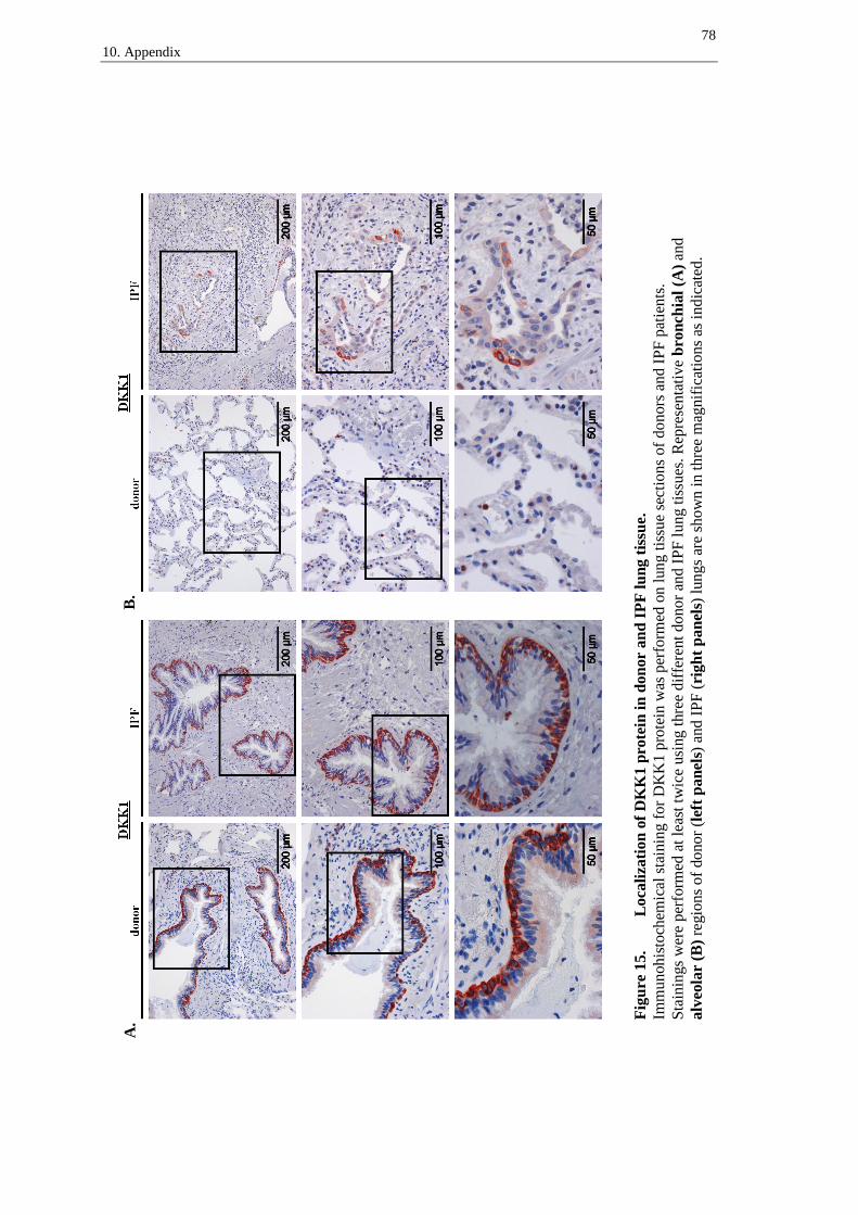
78
10. Appendix
Fig
ure
15.
L
oca
liza
tion
of
DK
K1
pro
tein
in
don
or
an
d I
PF
lu
ng t
issu
e.
Imm
un
oh
isto
chem
ical
sta
inin
g f
or
DK
K1
pro
tein
was
per
form
ed o
n l
ung t
issu
e se
ctio
ns
of
do
nors
an
d I
PF
pat
ien
ts.
Sta
inin
gs
wer
e p
erfo
rmed
at
leas
t tw
ice
usi
ng t
hre
e dif
fere
nt
donor
and I
PF
lung t
issu
es.
Rep
rese
nta
tiv
e b
ron
chia
l (A
) an
d
alv
eola
r (
B)
regio
ns
of
do
no
r (l
eft
pan
els)
and I
PF
(ri
gh
t p
an
els)
lungs
are
show
n i
n t
hre
e m
agnif
icat
ion
s as
in
dic
ated
.
B.
A.

79
10. Appendix
Fig
ure
16.
L
oca
liza
tion
of
DK
K4
pro
tein
in
don
or
an
d I
PF
lu
ng t
issu
e.
Imm
un
oh
isto
chem
ical
sta
inin
g f
or
DK
K4
pro
tein
was
per
form
ed o
n l
ung t
issu
e se
ctio
ns
of
do
nors
an
d I
PF
pat
ien
ts.
Sta
inin
gs
wer
e p
erfo
rmed
at
leas
t tw
ice
usi
ng t
hre
e dif
fere
nt
donor
and I
PF
lung t
issu
es.
Rep
rese
nta
tiv
e b
ron
chia
l (A
) an
d
alv
eola
r (
B)
regio
ns
of
do
no
r (l
eft
pan
els)
and I
PF
(ri
gh
t p
an
els)
lungs
are
show
n i
n t
hre
e m
agnif
icat
ion
s as
in
dic
ated
.
B.
A.
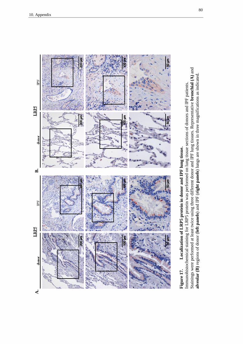
80
10. Appendix
Fig
ure
17.
L
oca
liza
tion
of
LR
P5 p
rote
in i
n d
on
or
an
d I
PF
lu
ng t
issu
e.
Imm
un
oh
isto
chem
ical
sta
inin
g f
or
LR
P5 p
rote
in w
as p
erfo
rmed
on l
ung t
issu
e se
ctio
ns
of
do
nors
an
d I
PF
pat
ien
ts.
Sta
inin
gs
wer
e p
erfo
rmed
at
leas
t tw
ice
usi
ng t
hre
e dif
fere
nt
donor
and I
PF
lung t
issu
es.
Rep
rese
nta
tiv
e b
ron
chia
l (A
) an
d
alv
eola
r (
B)
regio
ns
of
do
no
r (l
eft
pan
els)
and I
PF
(ri
gh
t p
an
els)
lungs
are
show
n i
n t
hre
e m
agnif
icat
ion
s as
in
dic
ated
.
B.
A.

81
10. Appendix
Fig
ure
18.
L
oca
liza
tion
of
KR
M1
pro
tein
in
don
or
an
d I
PF
lu
ng t
issu
e.
Imm
un
oh
isto
chem
ical
sta
inin
g f
or
KR
M1 p
rote
in w
as p
erfo
rmed
on l
ung t
issu
e se
ctio
ns
of
do
nors
an
d I
PF
pat
ien
ts.
Sta
inin
gs
wer
e p
erfo
rmed
at
leas
t tw
ice
usi
ng t
hre
e dif
fere
nt
donor
and I
PF
lung t
issu
es.
Rep
rese
nta
tiv
e b
ron
chia
l (A
) an
d
alv
eola
r (
B)
regio
ns
of
do
no
r (l
eft
pan
els)
and I
PF
(ri
gh
t p
an
els)
lungs
are
show
n i
n t
hre
e m
agnif
icat
ion
s as
in
dic
ated
.
B.
A.

82
10. Appendix
Fig
ure
19.
L
oca
liza
tion
of
KR
M2
pro
tein
in
don
or
an
d I
PF
lu
ng t
issu
e.
Imm
un
oh
isto
chem
ical
sta
inin
g f
or
KR
M2 p
rote
in w
as p
erfo
rmed
on l
ung t
issu
e se
ctio
ns
of
do
nors
an
d I
PF
pat
ien
ts.
Sta
inin
gs
wer
e p
erfo
rmed
at
leas
t tw
ice
usi
ng t
hre
e dif
fere
nt
donor
and I
PF
lung t
issu
es.
Rep
rese
nta
tiv
e b
ron
chia
l (A
) an
d
alv
eola
r (
B)
regio
ns
of
do
no
r (l
eft
pan
els)
and I
PF
(ri
gh
t p
an
els)
lungs
are
show
n i
n t
hre
e m
agnif
icat
ion
s as
in
dic
ated
.
B.
A.

83
11. Publications and Presentations
11 Publications and presentations
11.1 Publications
1. Pfaff EM, Becker S, Gunther A, et al: Dickkopf proteins influence lung epithelial
cell proliferation in idiopathic pulmonary fibrosis. Eur Respir J 37:79-87, 2011
2. Konigshoff M, Balsara N, Pfaff EM, et al: Functional Wnt signaling is increased in
idiopathic pulmonary fibrosis. PLoS One 3:e2142, 2008
11.2 Oral presentation
Freier Vortrag, Sektion Zellbiologie, 49. Kongress der Deutschen Gesellschaft für
Pneumologie und Beatmungsmedizin e.V., Lübeck, April 2008. E. Pfaff/Gießen, N.
Balsara, O. Eickelberg, M. Königshoff. Expression und Lokalisation von Wnt-
Inhibitoren in der idiopathischen Lungenfibrose.

84
12. Erklärung zur Dissertation
12 Erklärung zur Dissertation
„Hiermit erkläre ich, dass ich die vorliegende Arbeit selbständig und ohne unzulässige
Hilfe oder Benutzung anderer als der angegebenen Hilfsmittel angefertigt habe. Alle
Textstellen, die wörtlich oder sinngemäß aus veröffentlichten oder nichtveröffentlichten
Schriften entnommen sind, und alle Angaben, die auf mündlichen Auskünften beruhen,
sind als solche kenntlich gemacht. Bei den von mir durchgeführten und in der Disserta-
tion erwähnten Untersuchungen habe ich die Grundsätze guter wissenschaftlicher Pra-
xis, wie sie in der „Satzung der Justus-Liebig-Universität Gießen zur Sicherung guter
wissenschaftlicher Praxis“ niedergelegt sind, eingehalten sowie ethische, datenschutz-
rechtliche und tierschutzrechtliche Grundsätze befolgt. Ich versichere, dass Dritte von
mir weder unmittelbar noch mittelbar geldwerte Leistungen für Arbeiten erhalten haben,
die im Zusammenhang mit dem Inhalt der vorgelegten Dissertation stehen, oder habe
diese nachstehend spezifiziert. Die vorgelegte Arbeit wurde weder im Inland noch im
Ausland in gleicher oder ähnlicher Form einer anderen Prüfungsbehörde zum Zweck
einer Promotion oder eines anderen Prüfungsverfahrens vorgelegt. Alles aus anderen
Quellen und von anderen Personen übernommene Material, das in der Arbeit verwendet
wurde oder auf das direkt Bezug genommen wird, wurde als solches kenntlich gemacht.
Insbesondere wurden alle Personen genannt, die direkt und indirekt an der Entstehung
der vorliegenden Arbeit beteiligt waren. Mit der Überprüfung meiner Arbeit durch eine
Plagiatserkennungssoftware bzw. ein internetbasiertes Softwareprogramm erkläre ich
mich einverstanden.“
______________________ _______________________
Ort, Datum Unterschrift

85
14. Curriculum vitae
13 Danksagung
Besonders bedanken möchte ich mich bei Dr. Dr. Melanie Königshoff für die Betreuung
während der Zeit in der ehemaligen Arbeitsgruppe und auch bei der nachfolgenden
Ausarbeitung. Ich bin ihr sehr dankbar für die Unterstützung in den vielen theoretischen
und praktischen Fragen sowie für ihre Begeisterungsfähigkeit, womit sie mich immer
wieder motiviert hat.
Besonderer Dank geht auch an Prof. Dr. Oliver Eickelberg. Er hat mir ermöglicht, in
einer engagierten Arbeitsgruppe das wissenschaftliche Arbeiten zu erlernen. Ich danke
ihm für das Schaffen eines besonders angenehmen und motivierenden Arbeitsumfeldes
mit vielen Möglichkeiten zur produktiven Diskussion.
Bei Simone Becker möchte ich mich herzlich für die geduldige Unterstützung bei den
praktischen Schwierigkeiten und ihre stete Hilfsbereitschaft bedanken.
Anna Lühring, Nisha Balsara und Monika Kramer danke ich für freundliche Worte an
anstrengenden Tagen und fürs Zuhören.
Der gesamten ehemaligen Arbeitsgruppe gilt ein großer Dank für ihre Hilfsbereitschaft,
für anregende Diskussionen und für das freundliche Miteinander.