Food and Chemical Toxicology - uniroma1.it
11
Phytochemical analysis and effects on ingestive behaviour of a Caralluma fimbriata extract Annabella Vitalone a , Antonella Di Sotto a, * , Caterina Loredana Mammola b , Rosemarie Heyn b , Selenia Miglietta b , Paola Mariani c , Fabio Sciubba d , Francesca Passarelli e , Paola Nativio e , Gabriela Mazzanti a a Department of Physiology and Pharmacology “V. Erspamer”, Sapienza University of Rome, P.le Aldo Moro 5, 00185 Rome, Italy b Department of Anatomical, Histological, Forensic and Orthopedic Sciences, Sapienza University of Rome, Via A. Borelli 50, 00161 Rome, Italy c Department of General and Specialized Surgery “P. Stefanini”, Sapienza University of Rome, V.le Del Policlinico 155, 00161 Rome, Italy d Department of Chemistry, Sapienza University of Rome, P.le Aldo Moro 5, 00185 Rome, Italy e Department of Molecular Medicine and of Medical Surgical Sciences and Biotechnology, Sapienza University of Rome, P.le Aldo Moro 5, 00185 Rome, Italy article info Article history: Received 31 January 2017 Received in revised form 16 June 2017 Accepted 12 July 2017 Available online 13 July 2017 Keywords: Pregnane glycosides Slimming products Metabolomic analysis Water intake Neuropeptide Y Orexin abstract Caralluma fimbriata Wall. is currently used as a “natural slimming” food supplement, likely due to its content in pregnane glycosides. In the present study, a commercially available Caralluma fimbriata extract (Slimaluma ® ; CFE, 100 mg/kg) has been evaluated for its ability to affect the ingestive behaviour in fe- male rats, also with reference to the modulation of the brain neuropeptides NPY and ORX.The inter- ference of CFE with a-amylase and lipase enzymes has been investigated in vitro, as possible peripheral mechanism of action. Also, the chemical composition of CFE has been assessed by NMR and spectro- photometric analysis. Results from in vivo study showed that CFE induced effects neither on blood parameters, nor on liver and gut histomorphology. Interestingly, a reduction in body weight gain with an increase in water intake and hypothalamic levels of NPY and ORX peptides were found. Phytochemical analysis, showed CFE contained about 12% of pregnane glycosides and 1.3% of polyphenols. Present results suggest possible effects of C. fimbriata on ingestive behaviour, likely mediated by central and peripheral mechanisms. © 2017 Elsevier Ltd. All rights reserved. 1. Introduction Obesity and overweight are growing public health problems and their management is becoming of great clinical importance (Karatsoreos et al., 2013). Preliminary approaches in the manage- ment of overweight patients are preventive measures (diet, eating habits, etc.) and lifestyle changes (e.g., physical activity, specific exercises). The pharmacological approaches intended to counteract overweight include two main classes of drugs: the appetite sup- pressants (sibutramine, amphetamine derivatives, etc.) and the inhibitors of nutrient absorption (i.e., orlistat). However, pharma- cological treatments are often accompanied by serious adverse effects (Astell et al., 2013), so that it is not surprising that con- sumers commonly turn to herbal dietary supplements. Among natural agents used as appetite suppressants, Caralluma fimbriata Wall. [syn. Caralluma adscendens var. fimbriata (Wall.) Gravely & Mayur. e Fam. Apocynaceae] is one of the less studied. It is an edible succulent cactus used for centuries by Western Indians as both a food and an appetite suppressant (Yuliana et al., 2011; Gooda Sahib et al., 2012). Nowadays it is used as a “natural” slim- ming food supplement, and a standardized extract of C. fimbriata (Slimaluma ® ) has been patented (Yuliana et al., 2011; Dutt et al., 2012). Pregnane glycosides seem to be responsible for the slim- ming properties of this plant (Kuriyan et al., 2007; Astell et al., 201). These compounds were also found in other medicinal plants such as Hoodia gordonii, that are reported to affect food intake (Gooda Sahib et al., 2012). Moreover, other phytochemicals, including fla- vonoids, saponins etc. have been found in C. fimbriata (Astell et al., 2013). In spite of its traditional use, the scientific evidence on the C. fimbriata efficacy is poor, and the majority of the available * Corresponding author. E-mail address: [email protected] (A. Di Sotto). Contents lists available at ScienceDirect Food and Chemical Toxicology journal homepage: www.elsevier.com/locate/foodchemtox http://dx.doi.org/10.1016/j.fct.2017.07.027 0278-6915/© 2017 Elsevier Ltd. All rights reserved. Food and Chemical Toxicology 108 (2017) 63e73
Transcript of Food and Chemical Toxicology - uniroma1.it
lable at ScienceDirect
Food and Chemical Toxicology 108 (2017) 63e73
Contents lists avai
Food and Chemical Toxicology
journal homepage: www.elsevier .com/locate/ foodchemtox
Phytochemical analysis and effects on ingestive behaviour of aCaralluma fimbriata extract
Annabella Vitalone a, Antonella Di Sotto a, *, Caterina Loredana Mammola b,Rosemarie Heyn b, Selenia Miglietta b, Paola Mariani c, Fabio Sciubba d,Francesca Passarelli e, Paola Nativio e, Gabriela Mazzanti a
a Department of Physiology and Pharmacology “V. Erspamer”, Sapienza University of Rome, P.le Aldo Moro 5, 00185 Rome, Italyb Department of Anatomical, Histological, Forensic and Orthopedic Sciences, Sapienza University of Rome, Via A. Borelli 50, 00161 Rome, Italyc Department of General and Specialized Surgery “P. Stefanini”, Sapienza University of Rome, V.le Del Policlinico 155, 00161 Rome, Italyd Department of Chemistry, Sapienza University of Rome, P.le Aldo Moro 5, 00185 Rome, Italye Department of Molecular Medicine and of Medical Surgical Sciences and Biotechnology, Sapienza University of Rome, P.le Aldo Moro 5, 00185 Rome, Italy
a r t i c l e i n f o
Article history:Received 31 January 2017Received in revised form16 June 2017Accepted 12 July 2017Available online 13 July 2017
Keywords:Pregnane glycosidesSlimming productsMetabolomic analysisWater intakeNeuropeptide YOrexin
* Corresponding author.E-mail address: [email protected] (A.
http://dx.doi.org/10.1016/j.fct.2017.07.0270278-6915/© 2017 Elsevier Ltd. All rights reserved.
a b s t r a c t
Caralluma fimbriata Wall. is currently used as a “natural slimming” food supplement, likely due to itscontent in pregnane glycosides. In the present study, a commercially available Caralluma fimbriata extract(Slimaluma®; CFE, 100 mg/kg) has been evaluated for its ability to affect the ingestive behaviour in fe-male rats, also with reference to the modulation of the brain neuropeptides NPY and ORX.The inter-ference of CFE with a-amylase and lipase enzymes has been investigated in vitro, as possible peripheralmechanism of action. Also, the chemical composition of CFE has been assessed by NMR and spectro-photometric analysis.
Results from in vivo study showed that CFE induced effects neither on blood parameters, nor on liverand gut histomorphology. Interestingly, a reduction in body weight gain with an increase in water intakeand hypothalamic levels of NPY and ORX peptides were found. Phytochemical analysis, showed CFEcontained about 12% of pregnane glycosides and 1.3% of polyphenols.
Present results suggest possible effects of C. fimbriata on ingestive behaviour, likely mediated bycentral and peripheral mechanisms.
© 2017 Elsevier Ltd. All rights reserved.
1. Introduction
Obesity and overweight are growing public health problems andtheir management is becoming of great clinical importance(Karatsoreos et al., 2013). Preliminary approaches in the manage-ment of overweight patients are preventive measures (diet, eatinghabits, etc.) and lifestyle changes (e.g., physical activity, specificexercises). The pharmacological approaches intended to counteractoverweight include two main classes of drugs: the appetite sup-pressants (sibutramine, amphetamine derivatives, etc.) and theinhibitors of nutrient absorption (i.e., orlistat). However, pharma-cological treatments are often accompanied by serious adverseeffects (Astell et al., 2013), so that it is not surprising that con-sumers commonly turn to herbal dietary supplements.
Di Sotto).
Among natural agents used as appetite suppressants, Carallumafimbriata Wall. [syn. Caralluma adscendens var. fimbriata (Wall.)Gravely &Mayur. e Fam. Apocynaceae] is one of the less studied. Itis an edible succulent cactus used for centuries by Western Indiansas both a food and an appetite suppressant (Yuliana et al., 2011;Gooda Sahib et al., 2012). Nowadays it is used as a “natural” slim-ming food supplement, and a standardized extract of C. fimbriata(Slimaluma®) has been patented (Yuliana et al., 2011; Dutt et al.,2012). Pregnane glycosides seem to be responsible for the slim-ming properties of this plant (Kuriyan et al., 2007; Astell et al., 201).These compounds were also found in other medicinal plants suchas Hoodia gordonii, that are reported to affect food intake (GoodaSahib et al., 2012). Moreover, other phytochemicals, including fla-vonoids, saponins etc. have been found in C. fimbriata (Astell et al.,2013).
In spite of its traditional use, the scientific evidence on theC. fimbriata efficacy is poor, and the majority of the available
A. Vitalone et al. / Food and Chemical Toxicology 108 (2017) 63e7364
literature should be examined critically since a high proportion ofstudies are sponsorized and/or funded just by the companyinvolved in preparation and/or marketing of the C. fimbriata com-mercial extracts (Kuriyan et al., 2007; Odendaal et al., 2013;Lakshmi et al., 2014; Rajendran et al., 2014; Sudhakara et al., 2014).
Furthermore, taking into account the well-known problems ofstandardization of the herbal preparations marketed as food sup-plements (Wolsko et al., 2005; Garg et al., 2012), and consideringthat a poor quality can affect the safety and efficacy of herbal drugs(Chan, 2003), ascertaining the true composition of end productsrepresents an important goal.
Based on the above considerations and without any conflict ofinterest, present study was aimed to characterize the chemicalcomposition of a commercially available extract of C. fimbriata(CFE), by modern analytical techniques as suggested by EMA(EMEA/HMPC/253629/2007). Furthermore, in order to investigatethe activity of this extract in feeding behaviour, we tested the effectof CFE on food and water intake in a non-obese rat model, after asub-chronic treatment. Being pregnane glycosides recognized tohave appetite-suppressant effects, probably via enhanced hypo-thalamic signaling (MacLean and Luo, 2004), and considering thekey role of hypothalamus on the regulation of energy homeostasis(Williams et al., 2001), we also evaluated the effect of C. fimbriataon the hypothalamic expression of neuropetide Y (NPY) and orexin(ORX), two factors playing a crucial role on feeding attitude (Fickand Belsham, 2010). Finally, we assessed the capability ofC. fimbriata extract to inhibit the a-amylase and lipase enzymes, aspossible peripheral mechanisms of action.
2. Material and methods
2.1. Herbal extract
CFE (a dry ethanolic extract from the aerial parts of C. fimbriatanamed Slimaluma®, lot no. FAIT120503613) was purchased by theFAGRONCompany (Bologna, Italy). It appears as a brown andwater-soluble powder. According to the technical data sheet, the extract isreported to contain 27.5% of total pregnane glycosides. The qualityassessment of the product, carried out by the Company, excludedthe presence of germs (e.g., Escherichia coli, Salmonella spp, Pseu-domonas aeruginosa, Staphylococcus aureus), molds, fungi andpesticides, while the amount of unavoidable heavy metals (i.e., Pb,Cd, As, Hg) was lower than the limits allowed.
2.2. Chemicals
Tannic acid (CAS 1401-55-4; 99.9% purity), sodium carbonate(Na2CO3; CAS 497-19-8; 99.9% purity), Folin-Ciocalteu's phenolreagent, and aluminum chloride hexahydrate (AlCl3 x 6 H2O; CAS7784-13-6; Ph. Eur. purity) were purchased from Merck (Darm-stadt, Germany), while a-amylase, a-glucosidase, lipase, potatostarch, 4-nitrophenyl a-D-glucopyranoside (PNG), p-nitro-phenylpalmitate (PNP), polyvinylpyrrolidone (PVPP), 3,5-dinitrosalicylic acid (DNSA), acarbose, orlistat, quercetin, 3-(tri-methylsilyl)propionic-2,2,3,3-d4 acid sodium salt (TSP), ethanol(EtOH), deuterium dioxide (DO2), deuterated methanol (CD3OD),the polyclonal rabbit IgG antibodies against NPY and ORX, thebiotinilated goat IgG and all other chemicals, included those forhistomorphological study, were provided by Sigma-Aldrich (Milan,Italy). Deuterium oxide (D-99%; Cat. No. DLM4-100) was fromCambridge Isotope Laboratorie Inc., Andover, MA 01810 USA.Chemicals for ultrastructural study were: glutaraldehyde, uranylacetate and lead citrate (SIC, Rome, Italy) osmium tetroxide (AgarScientific, Stansted, UK), propylene oxide (BDH Italia, Milan, Italy)and epoxy resin (Electron Microscopy Sciences, Hatfield, PA, USA).
Products for clinical chemistry, including GLUC3 4483/190, GGT22721/122, ASTLP 7493/190, ALTLP 7388/190, ALP2 3752/190, CHOL29773/190, HDLC3 9803/190, TRIGL 7107/322, AMYL2 3742/122, LIPC9590/322, TP2 3734/190 and LDHI2 4732/122 were provided byRoche Diagnostics, CH. Insulin rat and streptavidin peroxidaseconjugate were from Demeditec Diagnostics (Kiel, Germany) andGE Healthcare (UK), respectively.
2.3. Phytochemical analysis
Owing to the variability of commercial preparations, the firststep of the present studywas to chemically characterize the extract.At this aim, nuclear magnetic resonance (NMR) spectroscopy wasemployed to evaluate the total pregnane (Tomassini et al., 2014)content, while colorimetric assays were performed to determinethe amount of polyphenol compounds.
2.3.1. NMR spectroscopy10 mg of CFE extract were dissolved in 600 ml D2O/CD3OD
mixture (2: 1 ratio, respectively) containing TSP at the final con-centration of 2 mM, as internal standard for chemical shift. All theNMR experiments were performed on a Bruker Avance III spec-trometer operating at a Larmor frequency of 400,13 MHz for 1H and100 MHz for 13C. Signal assignments were achieved by standardhomonuclear 1H�1H TOCSY and heteronuclear 1He13C HSQC andHMBC bidimensional experiments. 1D (one-dimensional) 1H NMRspectrum was acquired with a presat pulse sequence for solventsuppression, a spectral width of 15 ppm, 64 k data points, 5.5 s ofacquisition time,128 scans and a repetition delay of 9.5 s in order toachieve full relaxation for all protons. 2D (two-dimensional) 1H e1H TOCSY (Total Correlation Spectroscopy) spectrum was acquiredwith a data matrix of 8 k x 256 data points, a spectral width of15 ppm in both dimensions, 80 scans and a mixing time of 110 ms.2D 1He13C HSQC (Heteronuclear Single Quantum Correlation)spectrum was acquired with a data matrix of 8 k x 256 data pointsfor hydrogen and carbon respectively, a spectral width of 15 ppmfor hydrogen dimension and 200 for the carbon one, 128 scans andan average coupling constant of 145 Hz. Spectra processing wasperformed using Bruker software TOPSPIN for 2D experiments andACD 12.0 for 1D ones.
2.3.2. Colorimetric determination of total polyphenols, tannins andflavonoids
Total polyphenols were determined according to Rababah et al.(2005), with minor changes. Each sample (20 ml of a solution 1 mg/ml of C. fimbriata extract in EtOH 50% v/v) was mixed with 100 ml ofthe Folin-Ciocalteau reagent (10% v/v) and incubated for 5 min.After addition of 80 ml of a sodium carbonate solution (7.5% w/v),the mixture was shaken and incubated for 2 h, then the absorbancewas measured at 765 nm by a microplate reader (Bio-Rad, Hercules,CA, USA). The content of total polyphenols was expressed as tannicacid equivalents (TAE) per milligram of sample.
To determine the amount of tannins, 100 mg PVPP were addedto 1 ml of an extract solution (1 mg/ml in EtOH 50% v/v). PVPPinduces the formation of an insoluble complex with tannins, whichprecipitate. 20 ml of supernatant were used to determine thepolyphenol content, as described above, while the tannin amountwas obtained by the difference between total polyphenols and thepolyphenols contained in the supernatant, and was expressed asTAE per milligram of sample.
Flavonoids were measured by the spectrophotometric methoddescribed by Meda et al. (2005) with minor changes. Briefly, theextract (50 ml of a solution 1 mg/ml), aluminum trichloride (20 ml ofa solution 10% w/v in methanol), NaNO2 (10 ml of a solution 5% w/vin deionized water) and NaOH (60 ml of a solution 1 M) were added
A. Vitalone et al. / Food and Chemical Toxicology 108 (2017) 63e73 65
to 70 ml of deionized water, then were mixed and incubated for10 min. The absorbance was measured at 510 nm by a microplatereader and the total flavonoids were determined and expressed asquercetin equivalents (QE) per milligram of sample. Each assay wascarried out twice and each sample was tested in triplicate. Theresults were expressed as mean ± SEM.
2.4. Animal treatment protocol
The protocol used in this study was approved by the ItalianMinistry of Health. All experiments were performed in accordancewith the International Animal Welfare Legislation (Directive 2010/63/EU, 2010).
The study has been carried out on 18 Sprague Dawley femalerats (Charles River, Lecco, Italy) randomly assigned to two experi-mental groups: control group (drinking water) and treated group(100 mg/kg of CFE in drinking water); the treatment lasted 58 days.Female rats were chosen as women are the major users of theslimming products (Vitalone et al., 2011). The tested dose waschosen on the basis of previous published data obtained with aC. fimbriata extract (Kamalakkannan et al., 2010). During the 58-daytreatment period, rat body weight, water consumption and foodintake were recorded 3 times a week. At the end of the treatment,the animals were sacrificed in accordance with the approvedguidelines and regulations. Biological samples were collected forbiochemical, histological and immunohistochemical analysis.Particular attention has been devoted to the liver examination,considering that early studies on pregnane glycosides of Hoodiagordonii were discontinued due to liver toxicity (Dara et al., 2008).
2.5. Clinical chemistry
The blood samples were obtained by intra-cardiac puncture. Theclotted blood was centrifuged at 3000 rpm for 20 min in a refrig-erated centrifuge (4 �C). Serum was separated from plasma, andboth were used to perform biochemical-clinical analysis, by usingautomated methods (COBAS 6000 system, Roche Diagnostics, CH),as previously published by our group (Mazzanti et al., 2009).
Particularly, the following parameters were measured: insulin,glycemia, total cholesterol, high-density lipoprotein (HDL), totaltriglycerides, total proteins, amylase, lipase, alanine aminotrans-ferase (ALT), aspartate aminotransferase (AST), gamma-glutamyltransferase (GGT), alkaline phosphatise (ALP) and lactate dehy-drogenase (LDH).
Insulin assay was carried out by ELISA with monoclonal anti-bodies against rat hormone.
The homeostasis model of assessment insulin resistance(HOMA-IR) index was calculated using the following internationalformula: [fasting serum glucose (mmol/l) x fasting serum insulin(mU/l)/22.5].
2.6. Histomorphological and ultrastructural analysis of gut andliver
Gut fragments were obtained from duodenum (immediatelyafter the pylorus sphincter), jejunum (30 cm beyond the pylorussphincter), ileum (immediately before the ileocecal valve), andcolon (5 cm above the anus), and were immediately fixed in 10%buffered formalin, at room temperature for at most 48 h. Liversamples were obtained from the right lobe and fixed in bufferedformalin for 24 h. The samples of intestinal segments were dehy-drated with alcohol, cleared in xylene and embedded in paraffinwax (melting point 55e57 �C). Afterwards, the tissues were seriallysectioned using a rotary microtome Pabisch Top Automat S-140,obtaining 3 mm thick sections that were stained with
haematoxylin-eosin and Masson's trichromic. Histomorphologicalobservations were carried out using a light microscope (Leica DM4500B), connected to a Videocam (ProgRes C10 plus) and providedwith an Image Analysis System, Delta Sistemi (Rome, Italy).
To perform the ultrastructural examination, the specimens werefixed by immersion in a solution of 2.5% glutaraldehyde in 0.1 Mphosphate buffer for not less than one week; then they were post-fixed in a buffered solution of osmium tetroxide 1,33% for 2 h,dehydrated in increasing concentrations of ethyl alcohol and thenembedded in Epon 812. After resin polymerization in an oven at60 �C for 48 h, semi-thin (about 1 mm thick) and thin (70e90 nm)sections were obtained using an EMUC 6 Leica ultramicrotomewithglass and diamond knives. Semi-thin sections were mounted onglass slides and stained with toluidine blue. The observations werecarried out with an optical microscope Zeiss Axioskop 40 usingMRGrab 1.0 as image processing software. Ultrathin sectionscollected on 400 mesh copper nets, counterstained with lead cit-rate and uranyl acetate (Reynolds, 1963) were studied and photo-graphed with a Zeiss EM 10 transmission electron microscope,equipped with a digital camera model 782 ES500W ErlangshenCCD (Gatan DigitalMicrograph, CA) for image acquisition.
2.7. Immunohistochemistry of rat brain and optical density analysis
Brain specimens were removed, rapidly transferred to frozenisopentane (- 40 �C) for 20 s and then stored at - 80 �C. The NPYimmunoreactivity experiments were conducted on 15 mm cryostatrat brain sections using a conventional streptavidin-biotin tech-nique. The coronal sections (- 2.8 bregma) were fixed in 4% ofparaformaldehyde/0.1 M phosphate buffer for 15 min. Sectionswere stained using a polyclonal rabbit IgG antibody raised againstNPYand against ORX at a dilution of 1: 400 and 1: 500 respectively,which was determined in preliminary experiments.
Briefly, the sections were incubated for 30 min firstly with 3%hydrogen peroxide, thenwith 10% goat serum, to block non-specificprotein binding. Negative control sections were treated with non-immunized goat immunoglobulin serum under the same condi-tions. Following careful washing with PBS, biotinylated goat IgG(dilution 1: 1000) was applied for 2 h, followed by streptavidinperoxidase conjugate for 90 min. The color reaction was developedwith a solution of 0.02% diaminobenzidine in 0.003% hydrogenperoxide for 15 min. The sections were mounted onto gelatin-chrome alum-coated glass slides and examined through a whitelight microscope (Leitz, Germany). The immunohistochemistryprocedures were simultaneously carried out on a number of brainsections (at least 4 sections per animal) from each experimentalgroup.
The positive NPY/ORX staining was evaluated as optical density(O.D.) values, according to the methods described by Nativio et al.(2012). The brain areas of interest (i.e. hypothalamus) wereconsidered according to the stereotaxic coordinates of the rat brainatlas (Pellegrino et al., 1979). Standard outline digital images werecaptured using a SPOT-RT CCD video camera (SPOT-RT ImageSoftware V 3.0 SPOT Diagnostic Instruments Inc., Sterling Height,Michigan) for each region of interest. A semiquantitative analysis ofthe positive immunoreactivity was performed on brain multiplesections/region. The final optical density values (O.D.) were calcu-lated as a function of the specific signal relative to the externalbackground measure (taken from outside the section). Constantoptical conditions were maintained throughout the morphometricand densitometric evaluation.
2.8. In vitro metabolic enzyme inhibition
The ability of CFE to inhibit in vitro the a-amylase and lipase
A. Vitalone et al. / Food and Chemical Toxicology 108 (2017) 63e7366
enzymes was measured by spectrophotometric assays. For deter-mining a-amylase activity, the DNSA method was applied, ac-cording to Oboh et al. (2012), with minor changes. To perform theassay, serial dilutions of the sample (250 ml) were pre-incubatedwith a-amylase (0.5 mg/ml in phosphate buffer solution, corre-sponding to 25 U/ml; 250 ml) for 10 min at 37 �C. Then, afteraddition of the potato starch solution (0.5% w/v in acetate buffer0.1 M, pH ¼ 4.5; 250 ml), the mixture was incubated for 10 min at37 �C. The reaction was stopped by adding 500 ml of DNSA reagent(25 ml of 96 mM DNSA solution in water, 8 ml of 5.3 M sodiumpotassium tartrate solution in 2 M sodium hydroxide and 12 ml ofwater). The mixtures were then incubated in a water bath at 100 �Cfor 5 min, cooled to room temperature, thus plated in 96-multiwellmicroplates. For each treatment, the presence of reducing sugarswas determined by the turning of the solution to red and the DNSabsorbance was measured at 540 nm by a microplate reader (EpochMicroplate Spectrophotometer, BioTeK).
Lipase activity was assayed by measuring the enzymatic hy-drolysis of p-nitrophenyl palmitate to p-nitrophenol, according toCostamagna et al. (2016), with minor changes. Serial dilutions ofthe samples (16 ml) were mixed with a lipase solution in water(5.0 mg/ml; 12 ml) and TRIS HCl (75 mM, pH 8.5; 162 ml). Themixture was supplemented with PNP (10 mM; 25 ml) and pre-incubated on ice for 5 min. For each treatment, the p-nitrophenolabsorbance was determined at 405 nm by a microplate reader(Epoch Microplate Spectrophotometer, BioTeK).
Acarbose (250 mg/ml) and orlistat (25 mg/ml) were included inall the experiments as standard inhibitors (100% enzyme inhibi-tion) for a-amylase and lipase, respectively, while the vehicle rep-resented the maximum enzyme activity. Additional treatments, inwhich enzyme solution was replaced by buffer solution, wereincluded in order to evaluate the interfering absorbance producedby the extract. The experiments were performed at least in tripli-cate and in each experiment about six replicates were prepared.
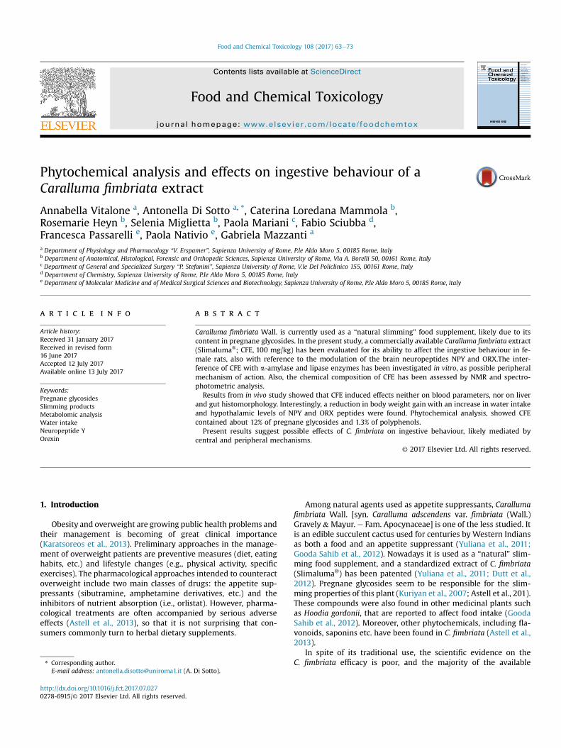
Fig. 1. 1D 1H NMR spectrum of the Caralluma fimbriata hydroalcoholic extract. Key: Ile, isoacetic acid; A-G, alpha-glucose; B-G, beta-glucose; S, sucrose; Trg, trigonelline; Prg, pregpregnane beta-glycoside; Prg benzoyl moiety; Prg p-aromatic moiety.
Data obtained from at least two experiments were pooled in thestatistical analysis. The inhibitory activity was calculated as per-centage of inhibition with respect to the vehicle control.
2.9. Statistical analysis
Data are reported as mean ± SEM. The Student's t-test was usedto compare paired data, while the one-way analysis of variance(ANOVA) followed by the Bonferroni Post-Test was applied toanalyze differences from three or more experimental groups. Thelevel of statistical significance was p < 0.05.
3. Results
3.1. Phytochemical analysis
The resonance assignment of 1H NMR CFE spectra (Fig. 1) wasperformed by the analysis of 2D NMR experiments (TOCSY, HSQCand HHMBC; Supplementary material, Figs. 1Se5S) and bycomparing the observed chemical shifts with ones from literaturedata (Abe and Yamauchi, 2000). From the analysis of NMR spectra,several molecule classes, including aminoacids (leucine, isoleucine,alanine, glutamine and tryptophan); organic acids (lactate, acetateand formate), carbohydrates (glucose and sucrose), trigonelline andpregnanes, were identified (Table 1).
Pregnanes were univocally identified on the basis of theirdiagnostic TOCSY correlations among the sterol moiety protons (8resonances between 0.9 and 1.9 ppm as well as the hydrogens inposition 3 at 3.52 ppm, 6 at 5.34 ppm and 12 at 4.06 ppm). Theyrepresent the main components of the hydro-alcoholic extract, asestimated on the basis of integral of the CH3 protons at 21 position(resonance at 2,30 ppm; Table 1) compared toTSP and their amountwas about 120 mg/g expressed as pregnane equivalents. Thepregnanes differ from each other for the glycoside moiety in
leucine; Leu, leucine; Thr, threonine; Ala, alanine; Glu, glutamate; LA, lactic acid; AA,nane; Prg-Glc, pregnane glycoside; Prg A-Glc, pregnane alpha-glycoside; Prg B-Glc,
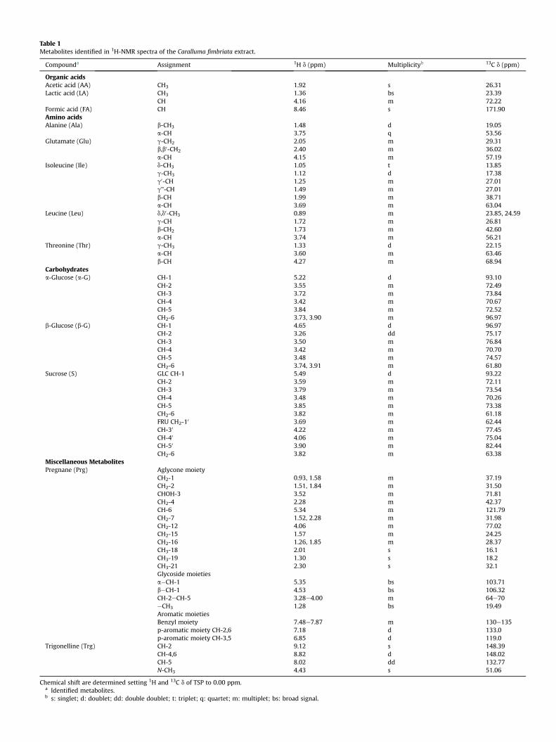
Table 1Metabolites identified in 1H-NMR spectra of the Caralluma fimbriata extract.
Compounda Assignment 1H d (ppm) Multiplicityb 13C d (ppm)
Organic acidsAcetic acid (AA) CH3 1.92 s 26.31Lactic acid (LA) CH3 1.36 bs 23.39
CH 4.16 m 72.22Formic acid (FA) CH 8.46 s 171.90Amino acidsAlanine (Ala) b-CH3 1.48 d 19.05
a-CH 3.75 q 53.56Glutamate (Glu) g-CH2 2.05 m 29.31
b,b0-CH2 2.40 m 36.02a-CH 4.15 m 57.19
Isoleucine (Ile) d-CH3 1.05 t 13.85g-CH3 1.12 d 17.38g0-CH 1.25 m 27.01g’’-CH 1.49 m 27.01b-CH 1.99 m 38.71a-CH 3.69 m 63.04
Leucine (Leu) d,d0-CH3 0.89 m 23.85, 24.59g-CH 1.72 m 26.81b-CH2 1.73 m 42.60a-CH 3.74 m 56.21
Threonine (Thr) g-CH3 1.33 d 22.15a-CH 3.60 m 63.46b-CH 4.27 m 68.94
Carbohydratesa-Glucose (a-G) CH-1 5.22 d 93.10
CH-2 3.55 m 72.49CH-3 3.72 m 73.84CH-4 3.42 m 70.67CH-5 3.84 m 72.52CH2-6 3.73, 3.90 m 96.97
b-Glucose (b-G) CH-1 4.65 d 96.97CH-2 3.26 dd 75.17CH-3 3.50 m 76.84CH-4 3.42 m 70.70CH-5 3.48 m 74.57CH2-6 3.74, 3.91 m 61.80
Sucrose (S) GLC CH-1 5.49 d 93.22CH-2 3.59 m 72.11CH-3 3.79 m 73.54CH-4 3.48 m 70.26CH-5 3.85 m 73.38CH2-6 3.82 m 61.18FRU CH2-10 3.69 m 62.44CH-30 4.22 m 77.45CH-40 4.06 m 75.04CH-50 3.90 m 82.44CH2-6 3.82 m 63.38
Miscellaneous MetabolitesPregnane (Prg) Aglycone moiety
CH2-1 0.93, 1.58 m 37.19CH2-2 1.51, 1.84 m 31.50CHOH-3 3.52 m 71.81CH2-4 2.28 m 42.37CH-6 5.34 m 121.79CH2-7 1.52, 2.28 m 31.98CH2-12 4.06 m 77.02CH2-15 1.57 m 24.25CH2-16 1.26, 1.85 m 28.37CH3-18 2.01 s 16.1CH3-19 1.30 s 18.2CH3-21 2.30 s 32.1Glycoside moietiesa�CH-1 5.35 bs 103.71b�CH-1 4.53 bs 106.32CH-2eCH-5 3.28e4.00 m 64e70eCH3 1.28 bs 19.49Aromatic moietiesBenzyl moiety 7.48e7.87 m 130e135p-aromatic moiety CH-2,6 7.18 d 133.0p-aromatic moiety CH-3,5 6.85 d 119.0
Trigonelline (Trg) CH-2 9.12 s 148.39CH-4,6 8.82 d 148.02CH-5 8.02 dd 132.77N-CH3 4.43 s 51.06
Chemical shift are determined setting 1H and 13C d of TSP to 0.00 ppm.a Identified metabolites.b s: singlet; d: doublet; dd: double doublet; t: triplet; q: quartet; m: multiplet; bs: broad signal.
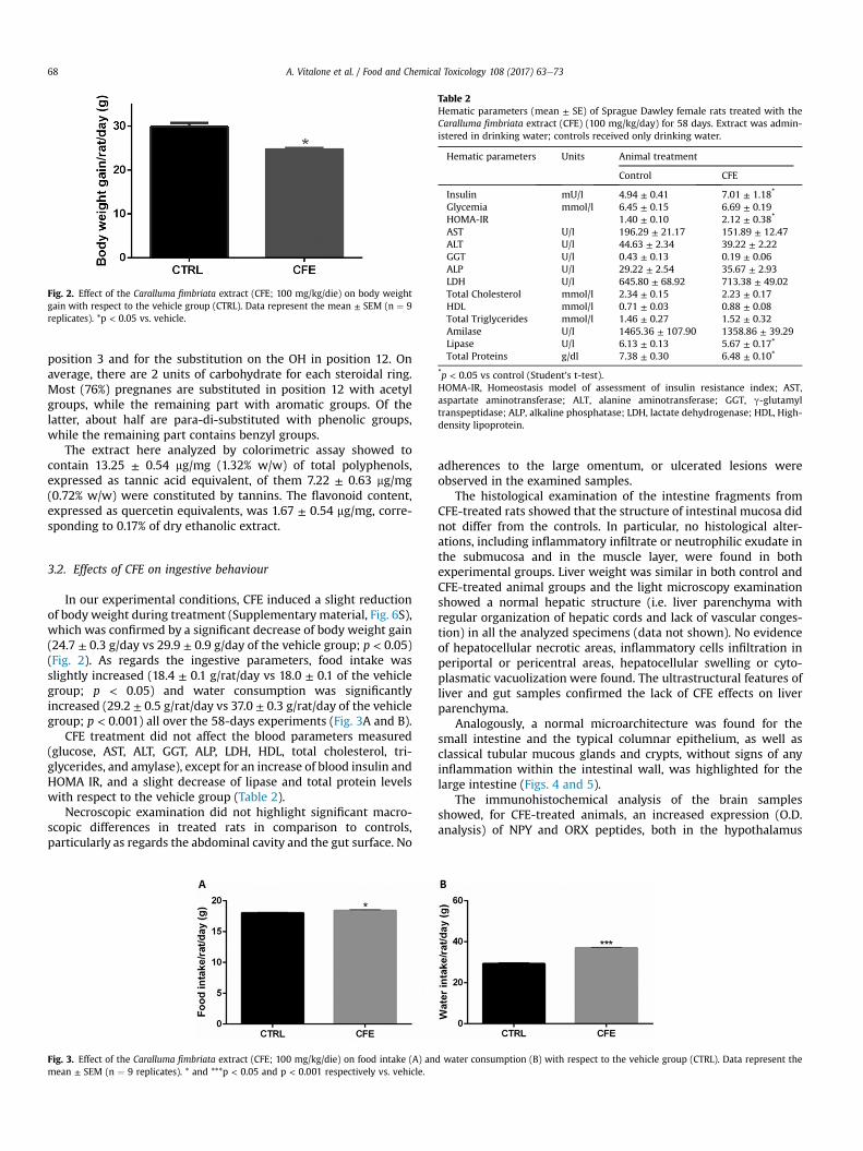
Fig. 2. Effect of the Caralluma fimbriata extract (CFE; 100 mg/kg/die) on body weightgain with respect to the vehicle group (CTRL). Data represent the mean ± SEM (n ¼ 9replicates). *p < 0.05 vs. vehicle.
Table 2Hematic parameters (mean ± SE) of Sprague Dawley female rats treated with theCaralluma fimbriata extract (CFE) (100 mg/kg/day) for 58 days. Extract was admin-istered in drinking water; controls received only drinking water.
Hematic parameters Units Animal treatment
Control CFE
Insulin mU/l 4.94 ± 0.41 7.01 ± 1.18*
Glycemia mmol/l 6.45 ± 0.15 6.69 ± 0.19HOMA-IR 1.40 ± 0.10 2.12 ± 0.38*
AST U/l 196.29 ± 21.17 151.89 ± 12.47ALT U/l 44.63 ± 2.34 39.22 ± 2.22GGT U/l 0.43 ± 0.13 0.19 ± 0.06ALP U/l 29.22 ± 2.54 35.67 ± 2.93LDH U/l 645.80 ± 68.92 713.38 ± 49.02Total Cholesterol mmol/l 2.34 ± 0.15 2.23 ± 0.17HDL mmol/l 0.71 ± 0.03 0.88 ± 0.08Total Triglycerides mmol/l 1.46 ± 0.27 1.52 ± 0.32Amilase U/l 1465.36 ± 107.90 1358.86 ± 39.29Lipase U/l 6.13 ± 0.13 5.67 ± 0.17*
Total Proteins g/dl 7.38 ± 0.30 6.48 ± 0.10*
*p < 0.05 vs control (Student's t-test).HOMA-IR, Homeostasis model of assessment of insulin resistance index; AST,aspartate aminotransferase; ALT, alanine aminotransferase; GGT, g-glutamyltranspeptidase; ALP, alkaline phosphatase; LDH, lactate dehydrogenase; HDL, High-density lipoprotein.
A. Vitalone et al. / Food and Chemical Toxicology 108 (2017) 63e7368
position 3 and for the substitution on the OH in position 12. Onaverage, there are 2 units of carbohydrate for each steroidal ring.Most (76%) pregnanes are substituted in position 12 with acetylgroups, while the remaining part with aromatic groups. Of thelatter, about half are para-di-substituted with phenolic groups,while the remaining part contains benzyl groups.
The extract here analyzed by colorimetric assay showed tocontain 13.25 ± 0.54 mg/mg (1.32% w/w) of total polyphenols,expressed as tannic acid equivalent, of them 7.22 ± 0.63 mg/mg(0.72% w/w) were constituted by tannins. The flavonoid content,expressed as quercetin equivalents, was 1.67 ± 0.54 mg/mg, corre-sponding to 0.17% of dry ethanolic extract.
3.2. Effects of CFE on ingestive behaviour
In our experimental conditions, CFE induced a slight reductionof body weight during treatment (Supplementary material, Fig. 6S),which was confirmed by a significant decrease of body weight gain(24.7 ± 0.3 g/day vs 29.9 ± 0.9 g/day of the vehicle group; p < 0.05)(Fig. 2). As regards the ingestive parameters, food intake wasslightly increased (18.4 ± 0.1 g/rat/day vs 18.0 ± 0.1 of the vehiclegroup; p < 0.05) and water consumption was significantlyincreased (29.2 ± 0.5 g/rat/day vs 37.0 ± 0.3 g/rat/day of the vehiclegroup; p < 0.001) all over the 58-days experiments (Fig. 3A and B).
CFE treatment did not affect the blood parameters measured(glucose, AST, ALT, GGT, ALP, LDH, HDL, total cholesterol, tri-glycerides, and amylase), except for an increase of blood insulin andHOMA IR, and a slight decrease of lipase and total protein levelswith respect to the vehicle group (Table 2).
Necroscopic examination did not highlight significant macro-scopic differences in treated rats in comparison to controls,particularly as regards the abdominal cavity and the gut surface. No
Fig. 3. Effect of the Caralluma fimbriata extract (CFE; 100 mg/kg/die) on food intake (A) anmean ± SEM (n ¼ 9 replicates). * and ***p < 0.05 and p < 0.001 respectively vs. vehicle.
adherences to the large omentum, or ulcerated lesions wereobserved in the examined samples.
The histological examination of the intestine fragments fromCFE-treated rats showed that the structure of intestinal mucosa didnot differ from the controls. In particular, no histological alter-ations, including inflammatory infiltrate or neutrophilic exudate inthe submucosa and in the muscle layer, were found in bothexperimental groups. Liver weight was similar in both control andCFE-treated animal groups and the light microscopy examinationshowed a normal hepatic structure (i.e. liver parenchyma withregular organization of hepatic cords and lack of vascular conges-tion) in all the analyzed specimens (data not shown). No evidenceof hepatocellular necrotic areas, inflammatory cells infiltration inperiportal or pericentral areas, hepatocellular swelling or cyto-plasmatic vacuolization were found. The ultrastructural features ofliver and gut samples confirmed the lack of CFE effects on liverparenchyma.
Analogously, a normal microarchitecture was found for thesmall intestine and the typical columnar epithelium, as well asclassical tubular mucous glands and crypts, without signs of anyinflammation within the intestinal wall, was highlighted for thelarge intestine (Figs. 4 and 5).
The immunohistochemical analysis of the brain samplesshowed, for CFE-treated animals, an increased expression (O.D.analysis) of NPY and ORX peptides, both in the hypothalamus
d water consumption (B) with respect to the vehicle group (CTRL). Data represent the
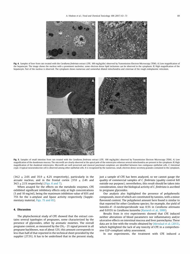
Fig. 4. Samples of liver from rats treated with the Caralluma fimbriata extract (CFE; 100 mg/kg/die) observed by Transmission Electron Microscopy (TEM). A) Low magnification ofthe hepatocyte. The image shows the nucleus with a prominent nucleolus; some electron dense lipid inclusions can be observed in the cytoplasm. B) High magnification of thehepatocyte. Part of the nucleus is observed. The cytoplasm shows numerous and somewhat dilated mitochondria and cisternae of the rough endoplasmic reticulum.
Fig. 5. Samples of small intestine from rats treated with the Caralluma fimbriata extract (CFE; 100 mg/kg/die) observed by Transmission Electron Microscopy (TEM). A) Lowmagnification of the duodenum mucosa. The microvilli are clearly observed at the apical pole of the enterocytes whereas several mitochondria are present in the cytoplasm. B) Highmagnification of the duodenal enterocytes. Microvilli are well preserved and classical junctional complexes are identified between two contiguous epithelial cells. C) Intestinalcrypt. A typical neuroendocrine cell is observed among other epithelial cells. It is recognized by the numerous, small, electron dense secretory granules contained in the cytoplasm.
A. Vitalone et al. / Food and Chemical Toxicology 108 (2017) 63e73 69
(34.2 ± 2.6% and 35.9 ± 4.2% respectively), particularly in thearcuate nucleus, and in the frontal cortex (37.0 ± 2.4% and34.5 ± 2.1% respectively) (Figs. 6 and 7).
When assayed for the effects on the metabolic enzymes, CFEexhibited significant inhibitory effects only at high concentrations(5 and 10 mg/ml), being the maximum inhibition value of 65% and75% for the a-amylase and lipase activity respectively (Supple-mentary material, Figs. 7S and 8S).
4. Discussion
The phytochemical study of CFE showed that the extract con-tains several typologies of pregnanes, some characterized by thepresence of glycosides, other by aromatic moieties. The overallpregnane content, as measured by the CH3 - 21 signal present in allpregnane backbones, was of about 12%; this amount corresponds toless than half of that reported in the technical sheet provided by thesupplier (27.5%). It has to be underlined that in the present study,
just a sample of CFE has been analyzed, so we cannot gauge thequality of commercial samples of C. fimbriata (quality control felloutside our purpose); nevertheless, this result should be taken intoconsideration, since the biological activity of C. fimbriata is ascribedto pregnane glycosides.
Our analysis also highlighted the presence of polyphenoliccompounds, most of which are constituted by tannins, with aminorflavonoid content. The polyphenol amount here found is similar tothat reported for other Caralluma species; for example, the yield ofluteolin-40eO-neohesperidoside was 0.5% in Caralluma attenuataand 0.015% in Caralluma lasiantha (Ramesh et al., 1999).
Results from in vivo experiments showed that CFE inducedneither alterations of blood parameters nor inflammatory and/orulcerative effects on intestinal mucosa and liver parenchyma. Thesedata are in line with the results obtained by Odendaal et al. (2013),which highlighted the lack of any toxicity of CFE in a comprehen-sive GLP-compliant safety assessment.
In our experiments, the treatment with CFE induced a
Fig. 6. Expression of NPY and ORX peptides in the arcuate nucleus from rats treated with vehicle (A, C) and Caralluma fimbriata extract (CFE; 100 mg/kg/die) (B, D).
A. Vitalone et al. / Food and Chemical Toxicology 108 (2017) 63e7370
statistically significant reduction in the body weight gain. This ef-fect was not accompanied, as expected, by a decrease in food intakewhich, on the contrary was slightly increased, but, strangely, by asignificant increase in water consumption. In addition, the CFEtreatment increased the brain expression of the orexigenic factorsNPY and ORX in hypothalamus and frontal cortex of treated rats.
On the basis of these evidences, our results suggest a possiblerole of CFE in the regulation of ingestive behaviour, probably due topregnane glycosides, although the contribution of other compo-nents of the phytocomplex, cannot be excluded. Literature evi-dences report that pregnane glycosides are able to modulate theingestive behaviour by possibly affecting the hypothalamic feedingcircuits (MacLean and Luo, 2004; Komarnytsky et al., 2013).Particularly ikemagenin, a pregnane glycoside isolated from Ascle-pias incarnata, exhibited in vivo appetite-regulating effects byaffecting the melanocortin signaling pathway and the secretion ofbrain-derived neurotropic factor (BDNF) in the hypothalamus, thussuggesting that the substance can be uptaken through the bloodbrain barrier, so acting at central level (Komarnytsky et al., 2013).On the other hand, the ability of the pregnane hoodigogenin A topenetrate the blood brain barrier has been highlighted in a MDR1-MDCK cell model (Madgula et al., 2010).
Hypothalamus represents one of the main nutrient sensor ofbrain and plays a pivotal role in the regulation of energy balance, bythe involvement of different chemical messengers, among whichNPY represents the earliest recognized and the most potentorexigenic one. NPY has been found overexpressed during fastingas well as in case of energy deprivation, energy demand or hyper-phagic conditions, while a gradually decrease occurs during foodintake (Mercer et al., 2011). There is a close interaction among NPYand other brain factors, both orexigenic (e.g. Agouti-related
peptide, AgRP) and anorexigenic (e.g. a-melanocyte-stimulating, a-MSH; brain-derived neurotrophic factor, BDNF). Particularly, ORXseems to elicit increased levels of NPY mRNA expression in thehypothalamus, through the modulation of the CREB (cAMPresponse element) transcription factor, thus suggesting the pivotalrole of orexin-NPY neuronal circuit on the feeding control(Kageyama et al., 2012; �Alvarez-Crespo et al., 2013). Orexin neuronsin the lateral hypothalamus and NPY neurons in the arcuate nu-cleus have reported to be stimulated by fasting and low glucoselevels, thus activating feeding regulatory pathways (Muroya et al.,2004). NPY is also strictly involved in the regulation of waterconsumption (Pau et al., 1988; Pich et al., 1992). Analogously, ORXhas been found to produce drinking stimulant effects by interactingwith specific receptors (Karasawa et al., 2014).
The increased ingestive behaviour could represent a physio-logical response to the increased levels of NPY and ORX, but why inrats treated with CFE water consumption is increased more thanfood consumption? Furthermore, why rats who received CFE do notgainweight even though they eat as much or more than the vehicletreated?
The control of feeding behaviour also occurs at peripheral level.We can hypothesize that the CFE treatment could affect the pe-ripheral absorption of some nutrients. In this regard, the inhibitionof the metabolic enzymes a-amylase and lipase seems to poorlycontribute to the CFE effects, due to the high concentrationseffective in our in vitro experiments.
Venkatesh et al. (2003) found that extracts from C. attenuatawere able to reduce the blood glucose levels in diabetic rats andhypothesized the involvement of extrapancreatic mechanisms.Furthermore, a C. tuberculata extract has been reported to improvethe glucose utilization likely by increasing the insulin secretion and
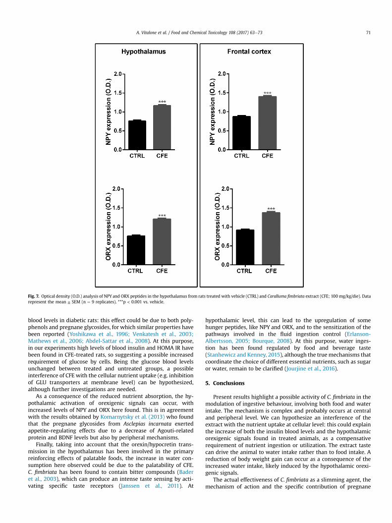
Fig. 7. Optical density (O.D.) analysis of NPY and ORX peptides in the hypothalamus from rats treated with vehicle (CTRL) and Caralluma fimbriata extract (CFE; 100 mg/kg/die). Datarepresent the mean ± SEM (n ¼ 9 replicates). ***p < 0.001 vs. vehicle.
A. Vitalone et al. / Food and Chemical Toxicology 108 (2017) 63e73 71
blood levels in diabetic rats: this effect could be due to both poly-phenols and pregnane glycosides, for which similar properties havebeen reported (Yoshikawa et al., 1996; Venkatesh et al., 2003;Mathews et al., 2006; Abdel-Sattar et al., 2008). At this purpose,in our experiments high levels of blood insulin and HOMA IR havebeen found in CFE-treated rats, so suggesting a possible increasedrequirement of glucose by cells. Being the glucose blood levelsunchanged between treated and untreated groups, a possibleinterference of CFE with the cellular nutrient uptake (e.g. inhibitionof GLU transporters at membrane level) can be hypothesized,although further investigations are needed.
As a consequence of the reduced nutrient absorption, the hy-pothalamic activation of orexigenic signals can occur, withincreased levels of NPY and ORX here found. This is in agreementwith the results obtained by Komarnytsky et al. (2013) who foundthat the pregnane glycosides from Asclepias incarnata exertedappetite-regulating effects due to a decrease of Agouti-relatedprotein and BDNF levels but also by peripheral mechanisms.
Finally, taking into account that the orexin/hypocretin trans-mission in the hypothalamus has been involved in the primaryreinforcing effects of palatable foods, the increase in water con-sumption here observed could be due to the palatability of CFE.C. fimbriata has been found to contain bitter compounds (Baderet al., 2003), which can produce an intense taste sensing by acti-vating specific taste receptors (Janssen et al., 2011). At
hypothalamic level, this can lead to the upregulation of somehunger peptides, like NPY and ORX, and to the sensitization of thepathways involved in the fluid ingestion control (Erlanson-Albertsson, 2005; Bourque, 2008). At this purpose, water inges-tion has been found regulated by food and beverage taste(Stanhewicz and Kenney, 2015), although the truemechanisms thatcoordinate the choice of different essential nutrients, such as sugaror water, remain to be clarified (Jourjine et al., 2016).
5. Conclusions
Present results highlight a possible activity of C. fimbriata in themodulation of ingestive behaviour, involving both food and waterintake. The mechanism is complex and probably occurs at centraland peripheral level. We can hypothesize an interference of theextract with the nutrient uptake at cellular level: this could explainthe increase of both the insulin blood levels and the hypothalamicorexigenic signals found in treated animals, as a compensativerequirement of nutrient ingestion or utilization. The extract tastecan drive the animal to water intake rather than to food intake. Areduction of body weight gain can occur as a consequence of theincreased water intake, likely induced by the hypothalamic orexi-genic signals.
The actual effectiveness of C. fimbriata as a slimming agent, themechanism of action and the specific contribution of pregnane
A. Vitalone et al. / Food and Chemical Toxicology 108 (2017) 63e7372
glycosides, polyphenols and other bitter compounds requires to befurther investigated.
Funding
This study was supported by grants from Sapienza University ofRome (University Project 2012; prot. C26A128C78), and by “Enricoand Enrica Sovena Foundation” (Rome, Italy). The study sponsorshad no involvement in a study design, in the collection, analysis,and interpretation of data, in the writing of the report, and in thedecision to submit the paper for publication.
Author contributions
Study design: Mazzanti G. and Vitalone A.; NMR and phyto-chemical analysis: Sciubba F, Di Sotto A.; in vivo and in vitro ex-periments: Mazzanti G., Vitalone A., Di Sotto A.; analysis of gut andliver specimens: Mammola C.L., Heyn R., Miglietta S.; analysis ofbrain specimens: Passarelli F. and Nativio P.; Clinical chemistry:Mariani P.; writing the manuscript: Mazzanti G., Vitalone A., DiSotto A. Final approval: all authors.
Conflict of interest
The authors declare no conflict of interest.
Acknowledgements
We are grateful to Prof. Maurizio Delfini for supervision in NMRexperiments.
Appendix A. Supplementary data
Supplementary data related to this article can be found at http://dx.doi.org/10.1016/j.fct.2017.07.027.
Transparency document
Transparency document related to this article can be foundonline at http://dx.doi.org/10.1016/j.fct.2017.07.027.
References
Abdel-Sattar, E., Harraz, F.M.H., Al-Ansari, S.M.A., El-Mekkawy, S., Ichino, C.,Kiyohara, H., 2008. Acylated pregnane glycosides from Caralluma tuberculataand their antiparasitic activity. Phytochem 69, 2180e2186.
Abe, F., Yamauchi, T., 2000. Pregnane Glycosides from the roots of Asclepias tuberose.Chem. Pharm. Bull. 48, 1017e1022.
�Alvarez-Crespo, M., Martínez-S�anchez, N., Ruíz-Pino, F., Garcia-Lavandeira, M.,Alvarez, C.V., Tena-Sempere, M., Nogueiras, R., Di�eguez, C., L�opez, M., 2013. Theorexigenic effect of orexin-A revisited: dependence of an intact growth hor-mone axis. Endocrinol 154, 3589e3598.
Astell, K.J., Mathai, M.L., Su, X.Q., 2013. Plant extracts with appetite suppressingproperties for body weight control: a systematic review of double blind ran-domized controlled clinical trials. Plant Foods Hum. Nutr. 68, 213e221.
Bader, A., Braca, A., De Tommasi, N., Morelli, I., 2003. Further constituents fromCaralluma negevensis. Phytochemistry 62, 1277e1281.
Bourque, C.W., 2008. Central mechanisms of osmosensation and systemic osmo-regulation. Nat. Rev. Neurosci. 9, 519e531.
Chan, 2003. Some aspects of toxic contaminants in herbal medicines. Chemosphere52, 1361e1371.
Costamagna, M.S., Zampini, I.C., Alberto, M.R., Cuello, S., Torres, S., P�erez, J.,Quispe, C., Schmeda-Hirschmann, G., Isla, M.I., 2016. Polyphenols rich fractionfrom Geoffroea decorticans fruits flour affects key enzymes involved in meta-bolic syndrome, oxidative stress and inflammatory process. Food Chem. 190,392e402.
Dara, L., Hewett, J., Lim, J.K., 2008. Hydroxycut hepatotoxicity: a case series andreview of liver toxicity from herbal weight loss supplements. World J. Gastro-enterol. 14, 6999e7004.
Dutt, H.C., Singh, S., Avula, B., Khan, I.A., Bedi, Y.S., 2012. Pharmacological review ofCaralluma R.Br. with special reference to appetite suppression and anti-obesity.
J. Med. Food 15, 108e119.EMEA/HMPC/253629/2007. Reflection paper on references used for quantitative
and qualitative analysis of herbal medicinal products and traditional herbalmedicinal products. Available at http://www.gmp-compliance.org/guidemgr/files/25362907ENFIN.PDF.
Erlanson-Albertsson, C., 2005. Appetite regulation and energy balance. Acta Pae-diatr. Suppl. 94, 40e41.
Fick, L.J., Belsham, D.D., 2010. Nutrient sensing and insulin signaling inneuropeptide-expressing immortalized, hypothalamic neurons: a cellularmodel of insulin resistance. Cell Cycle 9, 3186e3193.
Garg, V., Dhar, V.J., Sharma, A., Dutt, R., 2012. Facts about standardization of herbalmedicine: a review. Zhong Xi Yi Jie He Xue Bao 10, 1077e1083.
Gooda Sahib, N., Saari, N., Ismail, A., Khatib, A., Mahomoodally, F., Abdul Hamid, A.,2012. Plants' metabolites as potential antiobesity agents. Sci. World J. 436039.
Janssen, S., Laermans, J., Verhulst, P.J., Thijs, T., Tack, J., Depoortere, I., 2011. Bittertaste receptors and a-gustducin regulate the secretion of ghrelin with func-tional effects on food intake and gastric emptying. Proc. Natl. Acad. Sci. U. S. A.108, 2094e2099.
Jourjine, N., Mullaney, B.C., Mann, K., Scott, K., 2016. Coupled sensing of hunger andthirst signals balances sugar and water consumption. Cell 166, 855e866.
Kageyama, H., Takenoya, F., Hirako, S., Wada, N., Kintaka, Y., Inoue, S., Ota, E.,Ogawa, T., Shioda, S., 2012. Neuronal circuits involving neuropeptide Y in hy-pothalamic arcuate nucleus-mediated feeding regulation. Neuropeptides 46,285e289.
Kamalakkannan, S., Rajendran, R., Venkatesh, R.V., Clayton, P., Akbarsha, M.A., 2010.Antiobesogenic and antiatherosclerotic properties of Caralluma fimbriataextract. J. Nutr. Metab. 2010, 285301.
Karasawa, H., Yakabi, S., Wang, L., Tach�e, Y., 2014. Orexin-1 receptor mediates theincreased food and water intake induced by intracerebroventricular injection ofthe stable somatostatin pan-agonist, ODT8-SST in rats. Neurosci. Lett. 576,88e92.
Karatsoreos, I.N., Thaler, J.P., Borgland, S.L., Champagne, F.A., Hurd, Y.L., Hill, M.N.,2013. Food for thought: hormonal, experiential, and neural influences onfeeding and obesity. J. Neurosci. 33, 17610e17616.
Komarnytsky, S., Esposito, D., Rathinasabapathy, T., Poulev, A., Raskin, I., 2013. Ef-fects of pregnane glycosides on food intake depend on stimulation of themelanocortin pathway and BDNF in an animal model. J. Agric. Food Chem. 61,1841e1849.
Kuriyan, R., Raj, T., Srinivas, S.K., Vaz, M., Rajendran, R., Kurpad, A.V., 2007. Effect ofCaralluma fimbriata extract on appetite, food intake and anthropometry in adultIndian men and women. Appetite 48, 338e344.
Lakshmi, T., Rajendran, R., Raghavan, V., Silvester, A., 2014. HPTLC fingerprintingmethod to Distinguish total extract of Caralluma fimbriata from the modifiedextracts of Caralluma fimbriata. Biosci. Biotechnol. Res. Asia 11, 785e789.
MacLean, D.B., Luo, L., 2004. Increased ATP content/production in the hypothalamusmay be a signal for energy-sensing of satiety: studies of the anorectic mecha-nism of a plant steroidal glycoside. Brain Res. 1020, 1e11.
Madgula, V.L.M., Avula, B., Pawar, R.S., Shukla, Y.J., Khan, I.A., Walker, L.A., Khan, S.I.,2010. Characterization of in vitro pharmacokinetic properties of hoodigogeninA from Hoodia gordonii. Planta Med. 76, 62e69.
Mathews, J.N., Flatt, P.R., Abdel-Wahab, Y.H., 2006. Asparagus adscendens (Shwetamusali) stimulates insulin secretion, insulin action and inhibits starch digestion.Brit. J. Nutr. 95, 576e581.
Mazzanti, G., Di Sotto, A., Franchitto, A., Mammola, C.L., Mariani, P., Mastrangelo, S.,Menniti-Ippolito, F., Vitalone, A., 2009. Chelidonium majus is not hepatotoxic inWistar rats, in a 4 week feeding experiment. J. Ethnopharmacol. 126, 518e524.
Meda, A., Lamien, C.E., Romito, M., Millogo, J., Nacoulma, O.G., 2005. Determinationof the total phenolic, flavonoid and proline contents in Burkina Fasan honey, aswell as their radical scavenging activity. Food Chem. 91, 571e577.
Mercer, R.E., Chee, M.J., Colmers, W.F., 2011. The role of NPY in hypothalamicmediated food intake. Front. Neuroendocrinol 32, 398e415.
Muroya, S., Funahashi, H., Yamanaka, A., Kohno, D., Uramura, K., Nambu, T.,Shibahara, M., Kuramochi, M., Takigawa, M., Yanagisawa, M., Sakurai, T.,Shioda, S., Yada, T., 2004. Orexins (hypocretins) directly interact with neuro-peptide Y, POMC and glucose-responsive neurons to regulate Ca 2þ signaling ina reciprocal manner to leptin: orexigenic neuronal pathways in the mediobasalhypothalamus. Eur. J. Neurosci. 19, 1524e1534.
Nativio, P., Pascale, E., Maffei, A., Scaccianoce, S., Passarelli, F., 2012. Effect of stresson hippocampal nociceptin expression in rat. Stress 15, 378e384.
Oboh, G., Akinyemi, A.J., Ademiluyi, A.O., 2012. Inhibition of a-amylase and a-glucosidase activities by ethanolic extract of Telfairia occidentalis (flutedpumpkin) leaf. Asian Pac. J. Trop. Biomed. 2, 733e738.
Odendaal, A.Y., Deshmukh, N.S., Marx, T.K., Schauss, A.G., Endres, J.R., Clewell, A.E.,2013. Safety assessment of a hydroethanolic extract of Caralluma fimbriata. Int. J.Toxicol. 32, 385e394.
Pau, M.Y., Pau, K.Y., Spies, H.G., 1988. Characterization of central actions of neuro-peptide Y on food and water intake in rabbits. Physiol. Behav. 44, 797e802.
Pellegrino, L.J., Pellegrino, A.S., Cushman, A.J., 1979. A Stereotaxic Atlas of the RatBrain. Plenum Press, New York.
Pich, E.M., Messori, B., Zoli, M., Ferraguti, F., Marrama, P., Biagini, G., Fuxe, K.,Agnati, L.F., 1992. Feeding and drinking responses to neuropeptide Y injectionsin the paraventricular hypothalamic nucleus of aged rats. Brain Res. 575,265e271.
Rababah, T.M., Ereifej, K.I., Howard, L., 2005. Effect of ascorbic acid and dehydrationon concentrations of total phenolics, antioxidant capacity, anthocyanins, and
A. Vitalone et al. / Food and Chemical Toxicology 108 (2017) 63e73 73
color in fruits. J. Agric. Food Chem. 53, 4444e4447.Rajendran, R., Ambikar, D.B., Khandare, R.A., Sennapuri, V.D., Vyawahare, N.S.,
Clayton, P., 2014. Nootropic activity of Caralluma fimbriata extract in mice. FoodNutr. Sci. 5, 147e152.
Ramesh, M., Rao, Y.N., Kumar, M.R., Mohan, G.K., Kumar, B.R., Roa, A.V.N.A.,Krishna, M.R., Reddy, B.M., 1999. Flavone glycoside from three Caralluma spe-cies. Biochem. Syst. Ecol. 27, 85e86.
Reynolds, E.S., 1963. The use of lead citrate at high pH as an electron-opaque stain inelectron microscopy. J. Cell. Biol. 17, 208e212.
Stanhewicz, A.E., Kenney, W.L., 2015. Determinants of water and sodium intake andoutput. Nutr. Rev. 73 (Suppl. 2), 73e82.
Sudhakara, G., Mallaiah, P., Sreenivasulu, N., SasiBhusana Rao, B., Rajendran, R.,Saralakumari, D.J., 2014. Beneficial effects of hydro-alcoholic extract of Car-alluma fimbriata against high-fat diet-induced insulin resistance and oxidativestress in Wistar male rats. Physiol. Biochem. 70, 311e320.
Tomassini, A., Vitalone, A., Marini, F., Pratic�o, G., Sciubba, F., Bevilacqua, M.,Delfini, M., Di Sotto, A., Di Giacomo, S., Mariani, P., Mammola, C.L., Gaudio, E.,Miccheli, A., Mazzanti, G., 2014. 1H NMR-based urinary metabolic profilingreveals changes in nicotinamide pathway intermediates due to postnatal stressmodel in rat. J. Proteome Res. 13, 5848e5859.
Venkatesh, S., Reddy, G.D., Reddy, B.M., Ramesh, M., Rao, A.V., 2003. Anti-hyperglycemic activity of Caralluma attenuata. Fitoterapia 74, 274e279.
Vitalone, A., Menniti-Ippolito, F., Moro, P.A., Firenzuoli, F., Raschetti, R., Mazzanti, G.,2011. Suspected adverse reactions associated with herbal products used forweight loss: a case series reported to the Italian National Institute of Health.Eur. J. Clin. Pharmacol. 67, 215e224.
Williams, G., Bing, C., Cai, X.J., Harrold, J.A., King, P.J., Liu, X.H., 2001. The hypo-thalamus and the control of energy homeostasis: different circuits, differentpurposes. Physiol. Behav. 74, 683e701.
Wolsko, P.M., Solondz, D.K., Phillips, R.S., Schachter, S.C., Eisenberg, D.M., 2005. Lackof herbal supplement characterization in published randomized controlledtrials. Am. J. Med. 118, 1087e1093.
Yoshikawa, M., Murakami, T., Kadoya, M., Matsuda, H., Muraoka, O., Yamahara, J.,1996. Medicinal foodstuff. III. Sugar beet. Hypoglycemic oleanolic acid oligo-glycosides, betavulgarosides I, II, III, and IV, from the root of Beta vulgaris L.(Chenopodiaceae). Chem. Pharm. Bull. 44, 1212e1217.
Yuliana, N.D., Jahangir, M., Korthout, H., Choi, Y.H., Kim, H.K., Verpoorte, R., 2011.Comprehensive review on herbal medicine for energy intake suppression. Obes.Rev. 12, 499e514.