Interaction of Transient Receptor Potential Vanilloid 1 ...
109
Interaction of Transient Receptor Potential Vanilloid 1 (TRPV1) with G-protein coupled receptors and TRP ion channels Inauguraldissertation zur Erlangung des Grades eines Doktors der Naturwissenschaften (Dr. rer. nat.) am Fachbereich Biologie, Chemie, Pharmazie der Freien Universität Berlin vorgelegt von Viola Spahn Berlin 2011
Transcript of Interaction of Transient Receptor Potential Vanilloid 1 ...

Interaction of Transient Receptor Potential Vanilloid 1
(TRPV1) with G-protein coupled receptors and
TRP ion channels
Inauguraldissertation
zur Erlangung des Grades eines
Doktors der Naturwissenschaften
(Dr. rer. nat.)
am Fachbereich Biologie, Chemie, Pharmazie
der Freien Universität Berlin
vorgelegt von
Viola Spahn
Berlin 2011

Erstgutachter: Herr Prof. Dr. Christian Zöllner
Zweitgutachterin: Frau Prof. Dr. Monika Schäfer-Korting
Tag der Disputation: 28.01.2011
Table of contents
Table of contents
Abbreviations…………………...……………………...1
1. Introduction……….………………...………………....4
1.1. Pain………...……………………….………………...….……..5
1.2. Transient receptor potential ion channel family………...…..6
1.2.1. TRPV1…………………………………………...……………………….7
1.2.2. Sensitization of TRPV1………………………………..………………...9
1.2.3. TRPA1………………...……………………………...…………………11
1.3. Opioids……………………….…………………...…………...14
1.3.1. µ-opioid receptor……………………………..………………………...15
1.3.2. Opioid withdrawal-induced hyperalgesia………….…...…………….17
2. Objectives……………………………...……………...19
3. Animals, material and methods………….…………..20
3.1. Materials……………..……………………………….…….…20
3.1.1. Cell lines and bacteria…………………………………..…...…………20
3.1.2. Animals and animal housing………………………………….……….20
3.1.3. Chemicals…………………………………...…………………………..20
3.1.4. Media, buffer………………………..………………………………….22
3.1.5. Reaction systems………………………..………………………….…...23
3.1.6. Expendable materials……………………………………….…….……24
3.1.7. Technical equipment………………………………………………..….24
3.1.8. Antibodies……………………………………………………………….25
3.2. Methods…………………….………………………………....25
3.2.1. Experimental procedure of animals………………...………………...25
Culture of dorsal root ganglion (DRG) neurons………………..……25
Behavioural experiments……………………….……………………...26
Table of contents
3.2.2. Cell biological techniques…………………………….………….…….27
Culture of HEK 293 and HEK 293 Tet-On cells…………..….……...27
Transient transfection…………………………………………....…….27
Transformation and amplification of plasmid-DNA…….…….….….30
Small interference RNA………………………….………………….....31
3.2.3. Calcium Imaging experiments………………………....……………...31
3.2.4. Electrophysiology………………………………………..……………..33
Patch Clamp experiments………………………….…………………..33
3.2.5. Radioligand receptor binding studies…………………………………35
3.2.6. Immunoprecipitation / co-immunoprecipitation………………..……37
3.2.7. Western Blot analysis……………………………..……………………38
3.2.8. cAMP Enzyme-linked Immunosorbant Assay (ELISA)……….…….39
3.2.9. Statistical analysis……………………………...……...………………..40
4. Results…………………………………………………42
4.1. Interaction of TRPV1 and µ-opioid receptor during
opioid withdrawal……………………………………….……42
4.1.1. TRPV1 activity and expression during opioid withdrawal …………42
Phosphorylation of TRPV1 during opioid withdrawal………...…….44
4.1.2. Mutant TRPV1 activity during opioid withdrawal…………………..44
4.1.3. Role of adenylylcyclases during opioid withdrawal………………….47
4.1.4. Effects of opioid withdrawal in vivo……………………………..…….49
Thermal hypersensitivity during opioid withdrawal………..……….49
Nocifensive behaviour during opioid withdrawal…………...……….50
4.2. Interaction of TRPV1 and TRPA1……………………….…51
4.2.1. Physical interaction of TRPV1 and TRPA1………………….……….51
4.2.2. Interaction of TRPV1 and TRPA1 via signalling pathways……...….53
Modulation of TRPV1 activity after MuO induced
TRPA1 stimulation……………………………………………………..54
Table of contents
Change of the intracellular cAMP concentration after TRPA1
activation…………………………………………………….…...……..56
Phosphorylation of TRPV1………………..….……………………….56
Modulation of mutant TRPV1 activity after MuO indu ced TRPA1
activation………………………………………………………………..57
Modulation of TRPV1 activity after MuO pretreatment in DRG
neurons………………………………………………...……………….58
5. Discussion……………………………………………..60
5.1. Hypothesis 1: Increased TRPV1 activity during opioid
withdrawal is dependent on the presence of adenylylcyclases
and on phosphorylation of TRPV1 at specific
phosphorylation sites……………………………………...….61
5.1.1. Increased TRPV1 activity during opioid withdrawal………..………61
5.1.2. TRPV1 expression and opioid withdrawal ………………..…………63
5.1.3. Increased phosphorylation of TRPV1 during opioid withdrawal.….64
5.1.4. Mutation of threonine 144 and serine 774, but not serine 116 and
serine 502, resulted in a loss of increased TRPV1 activity during
opioid withdrawal....................................................................................65
5.1.5. Downregulation of AC 3, but not 5, led to a reversal of the enhanced
TRPV1 activity during opioid withdrawal………………………… …66
5.1.6. Paw withdrawal latency and nocifensive behaviour during opioid
withdrawal in male Wistar rats…………………………………...…..68
5.2. Hypothesis 2: TRPA1 stimulation modulates TRPV1
activity……………………………………………………..….70
5.2.1. TRPA1 stimulation does not alter the expression of TRPV1…...…..70
5.2.2. TRPV1 and TRPA1 do not form complexes in transfected
HEK Tet-On cells………………………………..……………………..71

Table of contents
5.2.3. TRPA1 stimulation increases TRPV1 activity in a calcium and
cAMP dependent manner …………………………………….……….72
5.2.4. TRPV1 is phosphorylated after TRPA1 stimulation …………..…....73
5.2.5. Mutation of TRPV1 phophorylation sites reversed the increased
TRPV1 activity after TRPA1 activation ………………………..…....74
5.2.6. TRPA1 stimulation enhanced TRPV1 currents in native DRG
neurons in a calcium and PKA-dependent manner…….……………75
5.3. Limitations, future prospects and clinical relevance …...…75
6. Summary……….……………...………………………79
7. References……………………….…………………….82
8. Curriculum vitae……………………………...………99
9. Publications and presentations………… ………….100
Acknowledgment……………………………...…………..102
Selbstständikeitserklärung...…………………………….103
Abbreviations
1
Abbreviation
A alanine
AC adenylylcyclase(s)
ASIC acid sensing ion channel
AgCl silver chloride
AKAP A kinase anchoring protein
AMPA α-amino-3-hydroxy-5-methyl-4-isoxazolepropionic acid
ANKTM1 / p120 old names for TRPA1
ANOVA analysis if variance
ATP adenosine triphosphate
BSA bovine serum albumin
CA cinnamaldehyde
CaCl2 calcium chloride
cAMP cyclic adenosine monophosphate
capsa capsaicin
CB cannabinoid receptor
Cdk5 cyclin-dependent kinase 5
cDNA copy desoxyribonucleic acid
CFA Complete Freund´s Adjuvant
CGRP calcitonin gene-related peptide
CIB Calcium Imaging Buffer
CREB cAMP response element binding protein
CTRL control
Da dalton
DAG diacylglycerol
DAMGO D-Ala2, N-MePhe4, Gly5-ol-enkephalin
DMSO dimethyl sulphoxide
DNA desoxyribonucleic acid
DOPA dihydroxyphenylalanine
DOR δ-opioid receptor
DRG dorsal root ganglion
DTT dithiothreitol
E. coli Escherichia coli
Abbreviations
2
ECS extracellular buffer
EDTA ethylene diamine tetraacetic acid
EGTA ethylene glycol tetraacetic acid
ERK extracellular signal regulated kinase
F340/F380 ratio of fluorescence at 340 nm to that at 380 nM
f femto (10-15)
FBS fetal bovine serum
g gram (s)
GFP green fluorescent protein
GPCR G-protein coupled receptor
HEK 293 human embryonic kidney cells 293
HEPES 4-2hydroxyethyl-1-piperazineethanesulfonic acid
I current
IB4 isolectin B4
IBMX isobutylmethylxanthin
ICS intracellular buffer
IP3 inositol triphosphate
JNK c-Jun N-terminal kinase
k kilo
KD dissociation constant
KCl potassium chloride
LB Luria-Bertani
LC Locus coeruleus
LTP long-term potentiation
M molar
MAPK mitogen activated protein kinase
MgCl2 magnesium chloride
min minute
ml milliliter
mM millimolare
MOR µ-opioid receptor
mRNA messenger RNA
MuO mustard oil
mV millivolt
Abbreviations
3
n number
nA nanoampere
NA nucleus accumbens
NGF nerve growth factor
nM nanomolar
nm nanometer
n.s. not significant
NLX nalaxone
NMDA N-methyl-D-asparate
OIH opioid induced hyperalgesia
OWIH opioid withdrawal induced hyperalgesia
pA picoampere
PBS phosphate buffered saline
PIP2 phosphatidylinositol bisphosphate
PKA protein kinase A
PKC protein kinase C
PLC phospholipase C
PTX pertussis toxin
PUFA polyunsaturated fatty acid
PVD polyvinylidene fluoride
RTX resiniferatoxin
s second
S serine
SP substance P
TG trigeminal ganglion
TM transmembrane domain
TNFα tumor necrosis factor alpha
TRIS tris (hydroxymethyl) amino-methane
TRP transient receptor potential
TRPA1 transient receptor potential ankyrin 1
TRPV1 transient receptor potential vanilloid 1
V volt
YFP yellow fluorescent protein
1. Introduction
4
1. Introduction
Injury and inflammation of peripheral tissue stimulates electrical activity of sensory dorsal
root ganglion (DRG) neurons (“nociceptors”). These impulses can be modulated by excitatory
and inhibitory ion channels and receptors, and are eventually transmitted to the central
nervous system where they are translated into the perception of “pain”. Among the most
prominent nociceptor membrane proteins are excitatory transient receptor potential (TRP)
channels and inhibitory opioid receptors. The interplay between these membrane proteins and
their signalling pathways shall be elucidated here.
The aims of this doctoral thesis are, first, to investigate the involvement of the excitatory ion
channel TRPV1 (Transient Receptor Potential Vanilloid 1) during withdrawal from inhibitory
(analgesic) drugs (opioids) and second, the influence of a related ion channel TRPA1
(Transient Receptor Potential Ankyrin 1) on TRPV1 activity. Besides inflammatory
mediators, both channels are activated by pungent components such as capsaicin (TRPV1)
and mustard oil (TRPA1), and play a critical role in pain sensation and in the development of
enhanced sensitivity to painful stimuli (“hyperalgesia”) typically associated with tissue injury.
Both channels are co-expressed in nociceptors.
Opioids produce analgesia (pain inhibition) by activation of Gi-protein-coupled opioid
receptors and subsequent dampening of neuronal excitability. However, after prolonged
opioid treatment and abrupt withdrawal, paradoxical hyperalgesia can arise. Although the
precise molecular mechanism is not yet fully understood, this is generally thought to result
from neuroplastic changes in the peripheral and central nervous systems that lead to
sensitization of pronociceptive pathways. In the following, the role of TRPV1 in opioid
withdrawal-induced hyperalgesia will be investigated in the peripheral nervous system.
Behavioural studies indicated that, in addition to TRPV1, the TRPA1 channel also plays a key
role in pain transduction, especially during pathological conditions triggered by tissue damage
and inflammation. TRPV1-mediated responses in neurons have a characteristic voltage
dependency that is influenced by extracellular Ca2+ and by the type and concentration of
TRPV1-specific agonists. Because of the prominent role of both TRP channels in
inflammatory pain, we decided to investigate the functional relevance of interactions between
TRPA1 and TRPV1, and whether TRPV1-mediated responses can be modulated by TRPA1.
1. Introduction
5
1.1. Pain
Pain is generally defined as “an unpleasant sensory and emotional experience associated with
actual or potential tissue damage, or described in terms of such damage” (Loeser and Treede
2008). Major pain syndromes are classified into nociceptive, inflammatory and neuropathic
pain (Patapoutian et al. 2009). Temporal classification distinguishes between acute and
chronic pain. Nociceptive pain includes somatic and visceral pain and is generated by noxious
stimuli that act on peripheral nociceptors. Nociceptive pain, which occurs clinically in the
settings of acute trauma, is protective and functions to prevent further tissue damage.
Inflammatory pain develops with damaged or inflamed tissue. Chemical mediators produced
and released from the primary sensory terminal and from non-neuronal cells (e.g., fibroblasts,
mast cells, neutrophils and platelets) directly stimulate and/or sensitize nociceptors to
chemical and mechanical stimuli. The latter phenomenon leads to behaviourally observable
hyperalgesia and is classified into primary (sensitization that occurs directly at the site of
tissue injury) and secondary hyperalgesia (sensitization that occurs in surrounding undamaged
tissues). Hyperalgesia results from sensitization of ion channels in the membrane of
nociceptors and an alteration in nociceptor excitability (Julius and Basbaum 2001). Molecular
mechanisms regarding the sensitization of the capsaicin receptor TRPV1 will be introduced in
more detail in chapter 1.2. A special case of hyperalgesia is allodynia, where normally
innocuous stimuli induce pain sensation. Besides peripheral sensitization of nociceptors,
central changes are induced, which may even result in an activation of normally non-
nociceptive neurons by noxious stimuli (Patapoutian et al. 2009).
Neuropathic pain results from injury / dysfunction of the peripheral, autonomic or central
nervous system (Backonja 2003). Neuropathic pain is associated with abnormal sensations
like spontaneous pain and pain hypersensitivity, which occur both centrally and peripherally.
One mechanism implicated in the development of opioid withdrawal-induced hyperalgesia
shares similarities with mechanisms thought to underly the development of neuropathic pain.
Activation of µ-opioid receptors in the dorsal horn of the spinal cord can lead to hyperalgesia
via stimulation of the excitatory amino acid neurotransmitters system. While the stimulation
of a µ-opioid receptor initially hyperpolarizes central neurons by activating inwardly
rectifying potassium channels, ongoing stimulation of the µ-receptor can result in
upregulation of intracellular messengers (e.g. cAMP, phosphokinase C), activation of the N-
methyl-D aspartate receptor system, and result in enhanced neuronal excitability (Mao et al.

1. Introduction
6
1994, 1995). In the present work we demonstrate the existence of another molecular
mechanism for opioid withdrawal-induced hyperalgesia in peripheral sensory neurons.
1.2. Transient receptor potential ion channel family
One of the major classes of membrane proteins detecting noxious stimuli is the Transient
Receptor Potential (TRP) ion channel family (Clapham 2003; Dhaka et al. 2006; Julius and
Basbaum 2001). In the following chapter two members of the TRP channel family, TRPV1
and TRPA1, will be introduced.
All members of the TRP ion channel family share the common features of six transmembrane
domains with diverse extents of sequence homology and permeability to cations. They play
critical roles in response to all major classes of external stimuli, including light, sound,
chemicals, temperature and touch. Some are also able to detect alterations of osmolarity.
Furthermore, they can be considered as multiple signal integrators, due to their ability to
modify responses of one signal by another (Venkatachalam and Montell 2007).
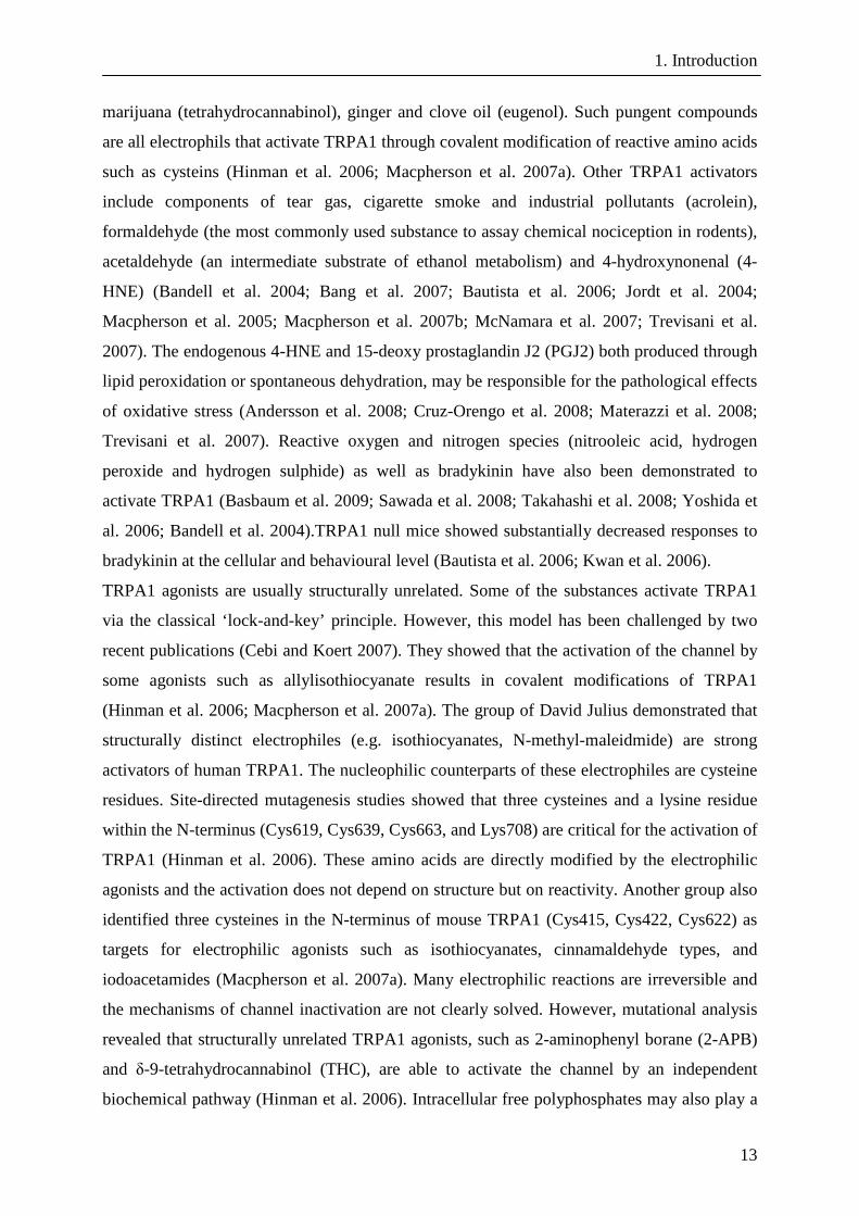
All currently known members of the TRP channel superfamily and their evolutionary
relationships are presented in Figure. 1.1.
1. Introduction
7
Figure 1.1. Phylogenic tree showing the relatedness of TRP proteins. The dendrogram of vertebrate
TRPs includes mostly human TRPs, except for mouse TRPC2 (cartoon of a mouse) and zebrafish
TRPN1 (cartoon of a zebrafish). White text and cartoons highlight the TRP proteins from worms and
flies. One C. elegans and one Drosophila member of each subfamily are included (modified from
Venkatachalam (Venkatachalam and Montell 2007).
A common feature of all TRP channels is the formation of homo- or heterotetramers of four
single subunits (Clapham 2003). Each subunit has six transmembrane (TM) domains with
intracellular amino (N-) - and carboxy (C-) - terminal domains and a pore loop between
domain five and six. Except for the TRPM subfamily, TRP channels contain multiple N-
terminal ankyrin repeats, a 33- residue sequence motif which is known to mediate protein-
protein interactions (Mosavi et al. 2004).
1.2.1. TRPV1
The first discovered temperature-sensitive ion channel was TRPV1, formerly known as VR1.
TRPV1, initially detected in small to medium sized neurons in sensory ganglia, is also found
in many other regions in the central nervous system (Mezey et al. 2000; Roberts et al. 2004)
and in some non-neuronal tissues such as epidermal keratinocytes of human skin (Southall et
al. 2003), gastric epithelial cells (Kato et al. 2003), and in epithelial cells of the urothelium
and smooth muscle (Birder et al. 2001). Besides the rat TRPV1 cloned in 1997 by Caterina et
al. (rVR1, GenBank AY445519), partially homologous human (AJ277028; homology 86 %),
guinea pig (85%), rabbit (86%), chicken (65%) and pig (84%) TRPV1 sequences were later
identified (Correll et al. 2004; Gavva et al. 2004; Hayes et al. 2000; Jordt and Julius 2002;
Ohta et al. 2005; Phelps et al. 2005; Savidge et al. 2002).
TRPV1 is activated by numerous stimuli such as noxious heat (>43°C), capsaicin (pungent
compound of hot chilli pepper) (Caterina et al. 2000; Caterina et al. 1997; Davis et al. 2000)
and many other chemicals, including endocannabinoids (anandamide) (Zygmunt et al. 1999),
camphor (Xu et al. 2005), and the pungent compounds present in black pepper (piperine)
(McNamara et al. 2005), garlic (allicin) (Macpherson et al. 2005), ginger (gingerol) (Dedov et
al. 2002) and clove oil (eugenol) (Yang et al. 2003). TRPV1-mediated cation influx initiated
by the application of noxious chemicals or temperatures is further enhanced by low pH
(Caterina et al. 1997). An acidic pH ≤ 5.9, which is characteristic for injured tissue, induces a
shift in thermal threshold activation from > 43°C to 20°C (Montell 2005). In addition, TRPV1
is activated by venoms from cnidarians and spiders (Cuypers et al. 2006; Siemens et al. 2006).

1. Introduction
8
Activation of TRPV1 results in an influx of mainly Ca2+ but also other cations like Na+, K+
and Mg2+ can enter the cell through this channel. Its permeability for calcium ions is ten times
higher than for sodium ions. The influx of cations provokes membrane depolarisation and
subsequent release of inflammatory neuropeptides, most notably substance P (SP) and
calcitonin gene related peptide (CGRP), which play a fundamental role in the development of
neurogenic inflammation and generation of electrical impulses (Tominaga 2007).
In agreement with other TRP ion channels, TRPV1 has six TM domains and a short, pore-
forming hydrophobic stretch between the fifth and sixth TM domains (Fig. 1.2.) (Caterina et
al. 1997). Its N-terminus contains three ankyrin-repeat domains and its C-terminus has been
proposed to serve as a determinant of subunit tetramerisation and to contribute to important
aspects of channel function (Garcia-Sanz et al. 2007). The ankyrin repeat consists of a ~ 33-
residue motif and binds many cytosolic proteins (Sedgwick and Smerdon 1999). Calmodulin,
a calcium-binding protein, binds to the first ankyrin repeat domain of TRPV1 (Rosenbaum et
al. 2004). TRPV1 is proposed to have a tetrameric structure with homotetramers as a
predominant form (Kedei et al. 2001), although a heterooligomerisation with TRPV3, which
is coexpressed with TRPV1 in DRG neurons, was observed in heterologous expression
systems using co-immunoprecipitation (see chapter 3.2.6.).
A) B)A) B)
Fig. 1.2. A) Proposed tetrameric structure of TRPV1 in the plasma membrane. B) Regions and amino
acids involved in TRPV1 function. Residues reported to be involved in vanilloid binding are presented
in grey. “TRP” in a box indicates a TRP domain (tetramerization and transduction domain that
stabilizes channel subunits and couples stimuli sensing to channel gating). Phosphatidylinositol 4,5-
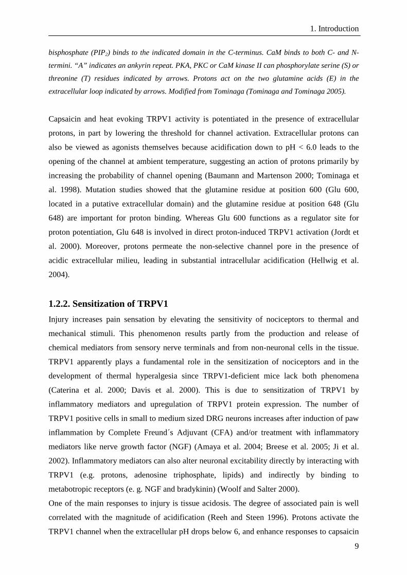
1. Introduction
9
bisphosphate (PIP2) binds to the indicated domain in the C-terminus. CaM binds to both C- and N-
termini. “A” indicates an ankyrin repeat. PKA, PKC or CaM kinase II can phosphorylate serine (S) or
threonine (T) residues indicated by arrows. Protons act on the two glutamine acids (E) in the
extracellular loop indicated by arrows. Modified from Tominaga (Tominaga and Tominaga 2005).
Capsaicin and heat evoking TRPV1 activity is potentiated in the presence of extracellular
protons, in part by lowering the threshold for channel activation. Extracellular protons can
also be viewed as agonists themselves because acidification down to pH < 6.0 leads to the
opening of the channel at ambient temperature, suggesting an action of protons primarily by
increasing the probability of channel opening (Baumann and Martenson 2000; Tominaga et
al. 1998). Mutation studies showed that the glutamine residue at position 600 (Glu 600,
located in a putative extracellular domain) and the glutamine residue at position 648 (Glu
648) are important for proton binding. Whereas Glu 600 functions as a regulator site for
proton potentiation, Glu 648 is involved in direct proton-induced TRPV1 activation (Jordt et
al. 2000). Moreover, protons permeate the non-selective channel pore in the presence of
acidic extracellular milieu, leading in substantial intracellular acidification (Hellwig et al.
2004).
1.2.2. Sensitization of TRPV1
Injury increases pain sensation by elevating the sensitivity of nociceptors to thermal and
mechanical stimuli. This phenomenon results partly from the production and release of
chemical mediators from sensory nerve terminals and from non-neuronal cells in the tissue.
TRPV1 apparently plays a fundamental role in the sensitization of nociceptors and in the
development of thermal hyperalgesia since TRPV1-deficient mice lack both phenomena
(Caterina et al. 2000; Davis et al. 2000). This is due to sensitization of TRPV1 by
inflammatory mediators and upregulation of TRPV1 protein expression. The number of
TRPV1 positive cells in small to medium sized DRG neurons increases after induction of paw
inflammation by Complete Freund´s Adjuvant (CFA) and/or treatment with inflammatory
mediators like nerve growth factor (NGF) (Amaya et al. 2004; Breese et al. 2005; Ji et al.
2002). Inflammatory mediators can also alter neuronal excitability directly by interacting with
TRPV1 (e.g. protons, adenosine triphosphate, lipids) and indirectly by binding to
metabotropic receptors (e. g. NGF and bradykinin) (Woolf and Salter 2000).
One of the main responses to injury is tissue acidosis. The degree of associated pain is well
correlated with the magnitude of acidification (Reeh and Steen 1996). Protons activate the
TRPV1 channel when the extracellular pH drops below 6, and enhance responses to capsaicin
1. Introduction
10
and heat (pH 6-8), resulting in an increase of nociceptor excitability even at normal body
temperature (Jordt et al. 2000; Welch et al. 2000).
In addition, several bioactive peptides are produced and released from non-neuronal cells or
derived from plasma proteins at the site of injury. Bradykinin, a nonapeptide, induces
immediate membrane depolarization as well as sensitization to other noxious stimuli when
applied to nociceptors (Burgess et al. 1989). Bradykinin binds to G-protein-coupled receptors
(bradykinin receptor 2; BK2) to stimulate phospholipase C (PLC)-catalyzed hydrolysis of
phospho-inositol phosphate 2 (PIP2) into inositol-phosphate 3 (IP3) and diacylglycerol
(DAG). IP3 and DAG activate protein kinase C (PKC), which directly phosphorylates TRPV1
preferentially at the serine residues 502 and 800 (Ser502 and Ser800) (Bhave et al. 2003;
Numazaki et al. 2002). Both residues are also involved in re-phosphorylation of TRPV1 after
calcium-dependent desensitization (Mandadi et al. 2004). Other inflammatory mediators like
adenosine triphosphate (ATP), prostaglandins, trypsin or tryptase also activate Gq-coupled
receptors followed by a downstream activation of PKC (Dai et al. 2004; Moriyama et al.
2005; Moriyama et al. 2003; Sugiura et al. 2002; Tominaga et al. 2001). Phosphorylation of
TRPV1 by PKC results in a potentiation of capsaicin- or proton-evoked responses and in a
reduction of the thermal threshold for TRPV1 activation at body temperature. Activation of
proteinase-activated (PAR2)-receptors also leads to a PKC-mediated sensitization of TRPV1
(Dai et al. 2004).
Prostaglandins may also modulate capsaicin- or heat-sensitivity of TRPV1 by activating the
protein kinase A (PKA)-pathway. Serine at position 116 (Ser 116) and threonine at position
370 (Thr 370) are reportedly phosphorylated by PKA. Phosphorylation of Ser 116 inhibits
dephosphorylation of TRPV1 caused by capsaicin. Phosphorylation of Thr 370, Thr 144 and
Ser 502 are thought to be involved in heat-evoked TRPV1 responses (Bhave et al. 2002;
Mohapatra and Nau 2003; Rathee et al. 2002).
Besides PKC and PKA, TRPV1 is phosphorylated and sensitized by the Ca2+/CaM-dependent
kinase II (CaMKII), the tyrosine kinase Src, and the cyclin-dependent kinase 5 (CdK5)
(Bhave et al. 2002; Jung et al. 2004; Lee et al. 2005; Mohapatra and Nau 2003; Numazaki et
al. 2002; Olah et al. 2002; Pareek et al. 2007; Premkumar and Ahern 2000).
The TRPV1 channel is also activated by the membrane-derived lipids anandamide,
oleoylethanolamide (OEA) and some lipoxygenase products (Ahern 2003; Hwang et al. 2000;
Zygmunt et al. 1999). Another important lipid is PIP2, which possibly interacts in an
inhibitory way with amino acids 777-820. PLC-induced PIP2 hydrolysis into DAG and IP3
results in TRPV1 activation (Chuang et al. 2001). However, other studies have shown an
1. Introduction
11
activation of TRPV1 by PIP2 in excised patches (Stein et al. 2006), leading to a controversial
discussion (Rohacs et al. 2008).
PKA activation is not solely a downstream effect of inflammatory mediators, but can also
result from an increase of intracellular cAMP, caused by a compensatory upregulation of
adenylylcyclase (AC) activity during withdrawal of chronically applied opioids (Levine and
Taiwo 1989; Sharma et al. 1975). In this context, the first part of the thesis investigates the
sensitization of TRPV1 during opioid withdrawal. Opioid mediated effects during acute
application and after withdrawal of chronic application and their underlying molecular
mechanisms will be introduced in more detail in chapter 1.3.
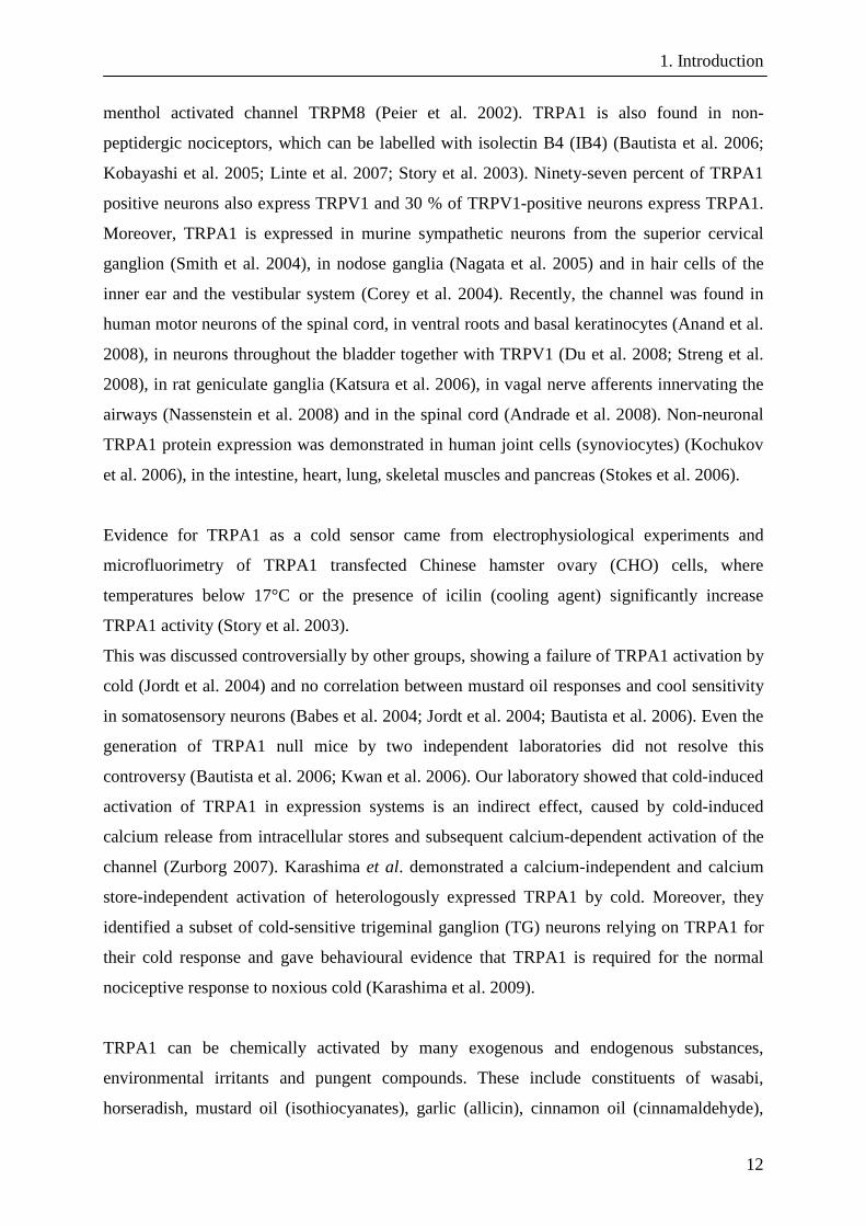
1.2.3. TRPA1
TRPA1, formerly known as ANKTM1 or p120 was first isolated in 1999 by Jaquemar et al. in
a screen for transformation-sensitive proteins in cultured fibroblasts (Jaquemar et al. 1999).
TRPA1 is homologous in sequence to other proteins belonging to the TRP channel family and
was identified as a novel thermo TRP in 2003 (Story et al. 2003). Mouse TRPA1 has fourteen
predicted N-terminal ankyrin domains followed by six TM domains (see Figure 1.4.) and is
probably the sole mammalian member of a distant subfamily of TRP channels. After
extensive analysis concerning the expression and function of TRPA1, it was proposed to be a
candidate receptor for noxious cold temperature (Caspani and Heppenstall 2009).
Fig. 1.4. Structure of one TRPA1 subunit, modified from Caterina (Caterina 2007).
TRPA1 is expressed in non-myelinated C-or lightly myelinated Aδ-fibres of DRG neurons
that sense temperature and/or noxious stimuli. Similar to TRPV1 it is expressed in CGRP-
and SP-positive neurons (peptidergic nociceptors), but rarely co-localized with the cool and
1. Introduction
12
menthol activated channel TRPM8 (Peier et al. 2002). TRPA1 is also found in non-
peptidergic nociceptors, which can be labelled with isolectin B4 (IB4) (Bautista et al. 2006;
Kobayashi et al. 2005; Linte et al. 2007; Story et al. 2003). Ninety-seven percent of TRPA1
positive neurons also express TRPV1 and 30 % of TRPV1-positive neurons express TRPA1.
Moreover, TRPA1 is expressed in murine sympathetic neurons from the superior cervical
ganglion (Smith et al. 2004), in nodose ganglia (Nagata et al. 2005) and in hair cells of the
inner ear and the vestibular system (Corey et al. 2004). Recently, the channel was found in
human motor neurons of the spinal cord, in ventral roots and basal keratinocytes (Anand et al.
2008), in neurons throughout the bladder together with TRPV1 (Du et al. 2008; Streng et al.
2008), in rat geniculate ganglia (Katsura et al. 2006), in vagal nerve afferents innervating the
airways (Nassenstein et al. 2008) and in the spinal cord (Andrade et al. 2008). Non-neuronal
TRPA1 protein expression was demonstrated in human joint cells (synoviocytes) (Kochukov
et al. 2006), in the intestine, heart, lung, skeletal muscles and pancreas (Stokes et al. 2006).
Evidence for TRPA1 as a cold sensor came from electrophysiological experiments and
microfluorimetry of TRPA1 transfected Chinese hamster ovary (CHO) cells, where
temperatures below 17°C or the presence of icilin (cooling agent) significantly increase
TRPA1 activity (Story et al. 2003).
This was discussed controversially by other groups, showing a failure of TRPA1 activation by
cold (Jordt et al. 2004) and no correlation between mustard oil responses and cool sensitivity
in somatosensory neurons (Babes et al. 2004; Jordt et al. 2004; Bautista et al. 2006). Even the
generation of TRPA1 null mice by two independent laboratories did not resolve this
controversy (Bautista et al. 2006; Kwan et al. 2006). Our laboratory showed that cold-induced
activation of TRPA1 in expression systems is an indirect effect, caused by cold-induced
calcium release from intracellular stores and subsequent calcium-dependent activation of the
channel (Zurborg 2007). Karashima et al. demonstrated a calcium-independent and calcium
store-independent activation of heterologously expressed TRPA1 by cold. Moreover, they
identified a subset of cold-sensitive trigeminal ganglion (TG) neurons relying on TRPA1 for
their cold response and gave behavioural evidence that TRPA1 is required for the normal
nociceptive response to noxious cold (Karashima et al. 2009).
TRPA1 can be chemically activated by many exogenous and endogenous substances,
environmental irritants and pungent compounds. These include constituents of wasabi,
horseradish, mustard oil (isothiocyanates), garlic (allicin), cinnamon oil (cinnamaldehyde),
1. Introduction
13
marijuana (tetrahydrocannabinol), ginger and clove oil (eugenol). Such pungent compounds
are all electrophils that activate TRPA1 through covalent modification of reactive amino acids
such as cysteins (Hinman et al. 2006; Macpherson et al. 2007a). Other TRPA1 activators
include components of tear gas, cigarette smoke and industrial pollutants (acrolein),
formaldehyde (the most commonly used substance to assay chemical nociception in rodents),
acetaldehyde (an intermediate substrate of ethanol metabolism) and 4-hydroxynonenal (4-
HNE) (Bandell et al. 2004; Bang et al. 2007; Bautista et al. 2006; Jordt et al. 2004;
Macpherson et al. 2005; Macpherson et al. 2007b; McNamara et al. 2007; Trevisani et al.
2007). The endogenous 4-HNE and 15-deoxy prostaglandin J2 (PGJ2) both produced through
lipid peroxidation or spontaneous dehydration, may be responsible for the pathological effects
of oxidative stress (Andersson et al. 2008; Cruz-Orengo et al. 2008; Materazzi et al. 2008;
Trevisani et al. 2007). Reactive oxygen and nitrogen species (nitrooleic acid, hydrogen
peroxide and hydrogen sulphide) as well as bradykinin have also been demonstrated to
activate TRPA1 (Basbaum et al. 2009; Sawada et al. 2008; Takahashi et al. 2008; Yoshida et
al. 2006; Bandell et al. 2004).TRPA1 null mice showed substantially decreased responses to
bradykinin at the cellular and behavioural level (Bautista et al. 2006; Kwan et al. 2006).
TRPA1 agonists are usually structurally unrelated. Some of the substances activate TRPA1
via the classical ‘lock-and-key’ principle. However, this model has been challenged by two
recent publications (Cebi and Koert 2007). They showed that the activation of the channel by
some agonists such as allylisothiocyanate results in covalent modifications of TRPA1
(Hinman et al. 2006; Macpherson et al. 2007a). The group of David Julius demonstrated that
structurally distinct electrophiles (e.g. isothiocyanates, N-methyl-maleidmide) are strong
activators of human TRPA1. The nucleophilic counterparts of these electrophiles are cysteine
residues. Site-directed mutagenesis studies showed that three cysteines and a lysine residue
within the N-terminus (Cys619, Cys639, Cys663, and Lys708) are critical for the activation of
TRPA1 (Hinman et al. 2006). These amino acids are directly modified by the electrophilic
agonists and the activation does not depend on structure but on reactivity. Another group also
identified three cysteines in the N-terminus of mouse TRPA1 (Cys415, Cys422, Cys622) as
targets for electrophilic agonists such as isothiocyanates, cinnamaldehyde types, and
iodoacetamides (Macpherson et al. 2007a). Many electrophilic reactions are irreversible and
the mechanisms of channel inactivation are not clearly solved. However, mutational analysis
revealed that structurally unrelated TRPA1 agonists, such as 2-aminophenyl borane (2-APB)
and δ-9-tetrahydrocannabinol (THC), are able to activate the channel by an independent
biochemical pathway (Hinman et al. 2006). Intracellular free polyphosphates may also play a
1. Introduction
14
crucial role by keeping TRPA1 in the needed conformation for channel gating by pungent
chemicals such as AITC and allicin (Kim and Cavanaugh 2007).
Besides the activation by pungent compounds and, possibly, cold temperatures, the multiple
ankyrin repeats of TRPA1 may form a gating spring capable of transducing mechanical force
and thereby facilitating channel opening (Corey et al. 2004; Howard and Bechstedt 2004).
Hill et al. detected mammalian TRPA1 activation by membrane crenations in heterologous
expression systems (Hill and Schaefer 2007) and a worm ortholog of TRPA was sensitive to
mechanical pressure applied via a suction pipette (Kindt et al. 2007). However, TRPA1
deficient mice display only weak deficits in mechanosensory behaviour and results remain
inconsistent (Bautista et al. 2006; Kwan et al. 2006; Petrus et al. 2007).
Approximately 30-50 % TRPV1 expressing small to medium sized sensory neurons co-
express TRPA1 and almost all TRPA1 positive neurons also express TRPV1. Moreover,
currents induced by mustard oil- and WIN55,212 (TRPA1 agonists) were almost exclusively
detected in TRPV1 positive cells, suggesting an interaction between TRPV1 and TRPA1.
Such interactions may lead to potentiated nociceptor excitability and pain sensation.
Therefore, I investigated the modulation of TRPV1 activity through TRPA1 stimulation.
1.3. Opioids
Opioids are the most powerful drugs for treatment of severe pain (Zollner and Stein 2007).
The alkaloid morphine was isolated by Friedrich Wilhelm A. Sertürner two hundred years ago
from the opium poppy plant Papaver somniferum (Papaveraceae). Opium use was first
documented 4000 b.C. by the Sumerians. The latex (raw opium) was extracted by incision of
the opium poppy capsule. Besides morphine, other analgesic alkaloids like codeine,
noscapine, papaverine and thebaine are ingredients of the opium poppy latex (Friderichs and
Strassburger 2002). Opioids mediate their analgesic action through G-protein coupled opioid
receptors. The opioid receptors are divided into three main groups, the µ-, δ- and κ-opioid
receptors, based on results of early binding studies and bioassays (Lord et al. 1977; Martin et
al. 1976; Pert and Snyder 1973; Stein and Zollner 2009). The existence and classification of
the three opioid receptor types was confirmed by cloning. The δ-opioid receptor was cloned
simultaneously by two independent research groups in 1992 (Evans et al. 1992; Kieffer et al.
1992). Later on, the µ- and κ-opioid receptors were cloned (Meng et al. 1993; Wang et al.
1993).
1. Introduction
15
According to their ability to activate opioid receptors, opioids are classified as agonists,
partial agonists, and antagonists. The reversible binding of an agonist causes a conformational
change of the receptor followed by an activation of G-proteins and intracellular signalling
pathways. They eventually provoke a measurable biological response (e.g. analgesia) (Hulme
et al. 1999). Synthetic fentanyl is one of the most potent opioid agonists with a high affinity to
µ-opioid receptors (MOR). Another synthetic highly potent MOR agonist is the peptide D-
Ala2-N-MePhe4-Gly5-ol-enkephalin (DAMGO), which is widely used in pharmacological
research. Buprenorphine acts as a partial MOR agonist but as an antagonist at κ-opioid
receptors (KOR). Other agonist-antagonists have different affinities at the three receptor types
(Huang et al. 2001). Antagonists do not induce a biological response after reversible binding
to receptors. The most common opioid receptor antagonists are naloxone and naltrexone, both
binding to three opioid receptors but with a preference to MOR.
Besides the numerous exogenous agonists, endogenous opioid receptor agonists have also
been identified. The first endogenous opioid peptides were Met-and Leu- enkephalin (Hughes
et al. 1975), followed by β-endorphin and dynorphin, which were discovered in the late
seventies (Goldstein et al. 1979; Li and Chung 1976; Li et al. 1976). β-Endorphin consists of
31 amino acids and is processed from the precursor protein proopiomelanocortin (POMC). It
is an agonist at MOR and DOR. Endomorphin 1 (Tyr-Pro-Trp-Phe-NH2) and 2 (Tyr-Pro-Phe-
Phe-NH2), discovered in the late nineties, are selective agonists of MOR (Zadina et al. 1997).
Prodynorphin and proenkephalin are precursors of dynorphin (KOR agonist) and Met-and
Leu-enkephalin (DOR agonists), respectively. Endogenous opioid peptides are predominantly
expressed in the brain, in the dorsal horn of the spinal cord and in immune cells (Endres-
Becker 2007).
1.3.1. µ-opioid receptor
µ-opioid receptors (MOR) are expressed in the central (cortex, thalamus, hypothalamus,
limbic system, brainstem) and peripheral nervous system, by neuroendocrine (pituitary,
adrenals), immune, and ectodermal cells (Duncan 1999; Stein and Zollner 2009). In the
periphery, they are synthesized in cell bodies of DRG neurons and intra-axonally transported
via microtubules to central and peripheral terminals (Stein et al. 2003). Moreover, they occur
in high concentrations in the gastrointestinal tract and in the urinary bladder, where they
mediate a reduction in intestinal motility and micturition.
1. Introduction
16
Opioid receptors are G-protein coupled receptors (GPCR) which are classified into four
groups concerning their interacting G-proteins: Gs (“stimulatory”), Gi/o (“inhibitory”/”other”),
G12/13 and Gq/11. Heterotrimeric G-proteins consist of an α-subunit (Gα), which binds
GDP/GTP, a β-subunit and a γ-subunit, which form a non-dissociable complex (Gβγ). The
activation of an opioid receptor by an agonist induces conformational changes, allowing
intracellular coupling of mainly Gi/o proteins to the C-terminus (Stein and Zollner 2009).
Thereby the receptor functions as a guanine nucleotide exchange factor (GEF) that exchanges
guanosine diphosphate (GDP) for guanosine triphosphate (GTP) on the Gα-subunit. This is
followed by the dissociation of the Gα, (binding GTP) from the Gβγ dimer and the receptor. Gα
and Gβγ can then activate different signalling cascades and effector proteins. The Gβγ-subunit
directly interacts with voltage dependent ion channels (Clapham and Neer 1997). Gβγ
presynaptically suppresses the activity of N- (“neuron”), P/Q- (“purkinje cell”) and R-type
(“remaining”) calcium channels, resulting in an inhibition of the generation and transmission
of electrical stimuli in nociceptive peripheral and/or central nervous system (CNS) neurons
(Akins and McCleskey 1993; Borgland et al. 2001; Irnaten et al. 2003; Schroeder and
McCleskey 1993).
Moreover, Gβγ inhibits purinergic P2X receptors and tetrodotoxin (TTX) resistant sodium
channels, which are mainly expressed on nociceptors and important in nociception.
Postsynaptically, Gβγ-subunits activate voltage-dependent and G-protein-gated inwardly
rectifying (GIRK) potassium channels in the CNS, causing postsynaptic hyperpolarisation
(North et al. 1987; Torrecilla et al. 2002) and thereby preventing generation and/or
propagation of action potentials (Zollner and Stein 2007). The extracellular signal-regulated
kinase (ERK)- and mitogen-activated protein kinase (MAPK)-system is also activated by Gβγ
(Belcheva et al. 1998; Li and Chang 1996). In addition, the release of proinflammatory and
pronociceptive neuropeptides (e.g. substance P; SP) from central and peripheral terminals of
sensory neurons is inhibited (Kondo et al. 2005; Yaksh 1988).
Through Gαi opioids cause a reduction in adenylylcyclase (AC) activity. ACs are lyase
enzymes that catalyze the conversion of adenosine-5’-triphosphate (ATP) to 3’, 5’-cyclic
adenosine-monophosphate (cAMP) (Law et al. 2000). The second messenger cAMP regulates
other proteins like cAMP-dependent protein kinase A (PKA) or cyclic-nucleotide gated ion
channels. PKA consists of two regulatory subunits (R) that bind to two catalytic subunits (C).
Each regulatory subunit possesses two binding pockets for cAMP-molecules. If cAMP-
molecules bind to the regulatory subunit, the R-C complex dissociates and the catalytic
subunit is released, now able to transfer phospho-groups to the amino acids serine and
1. Introduction
17
threonine (serine/threonine-kinase). The guiding of PKA to the target protein is accomplished
by A-kinase anchoring proteins (AKAP) (Dell'Acqua and Scott 1997). Phosphorylation of
proteins is an important control mechanism in signal transduction and in the regulation of
enzyme- or transcription factor-activity. PKA is involved in a wide range of processes such as
transcription, metabolism, cell cycle progression and apoptosis (Gjertsen and Doskeland
1995; Hubbard and Cohen 1993; Huggenvik et al. 1991; Matten et al. 1994; Smith et al.
1993). One target of phosphorylation by PKA in the nucleus is the cAMP Response Element
Binding Protein (CREB), which increases transcription in its phosphorylated state. In the
current thesis I investigated whether TRPV1, whose phosphorylation by PKA can lead to
channel sensitization/resensitization can occur during opioid withdrawal. Previous studies
from our laboratory have shown that opioids inhibit the activity of TRPV1 in a naloxone- and
pertussis toxin (PTX)-sensitive manner via the cAMP/PKA pathway (Endres-Becker 2007).
Coexpression of TRPV1 and MOR in small to medium diameter-sized neurons was shown,
and the application of morphine or DAMGO significantly decreased capsaicin-induced
TRPV1 currents in whole cell patch clamp experiments. These effects were reversed by
naloxone, PTX, forskolin (FSK) and the stable cAMP-analogon 8-Br-cAMP. Washout
experiments revealed that additional capsaicin applications progressively increased TRPV1
activity after removal of morphine (Endres-Becker et al. 2007).
1.3.2. Opioid withdrawal-induced hyperalgesia
Withdrawal of opioids can result in hyperalgesia (Drdla et al. 2009), which has been well
documented both in animal studies (Mao et al. 1995; Nestler and Aghajanian 1997) and
clinical reports (Angst et al. 2003; Compton et al. 2003; Doverty et al. 2001). Despite
intensive work, the neurobiological mechanisms of opioid withdrawal induced hyperalgesia
(OWIH) are not fully clarified. Early in vitro studies in cell lines showed that continuous
(“chronic”) morphine administration induces a compensatory increase in AC activity and
intracellular cAMP concentrations (Brandt et al. 1976; Sharma et al. 1975). Increased cAMP
leads to activation of cAMP-dependent PKA (Avidor-Reiss et al. 1997; Bie et al. 2005). In
addition, the expression of the catalytic subunit of PKA is upregulated during opioid
withdrawal (Lane-Ladd et al. 1997). PKA in turn phosphorylates and thereby sensitizes
receptor proteins. In vitro studies identified cAMP-mediated increased synaptic transmission
and augmented hyperpolarization-activated currents in central neurons (Williams et al. 2001).
Firing rates of locus coeruleus (LC) neurons were reduced by acutely applied opioids,
whereas normal levels were reached during prolonged opioid administration. Withdrawal of

1. Introduction
18
opioid agonists or application of opioid antagonists resulted in higher firing rates due to
cAMP-upregulation (Kogan et al. 1992). In addition, Drdla et al. showed a “long-term
potentiation” (LTP) of synaptic strength in spinal cord pain pathways after abrupt withdrawal
of opioids. Under physiological conditions LTP is a mechanism for learning and memory in
the brain (Drdla et al. 2009).
Opioid withdrawal can also induce an upregulation of transcription factors, particularly CREB
(cAMP response element binding protein) and ∆FosB (Nestler 2004), which are responsible
for enhanced expression of neuropeptides, neurotransmitter synthesizing enzymes,
neurotransmitter receptors, signalling proteins, and other transcription factors leading to
increased neuronal excitability (Lonze and Ginty 2002; Mayr and Montminy 2001).
Continuously applied opioids and their withdrawal also influence MAPK-/ERK signal
transduction (Asensio et al. 2006; Ferrer-Alcon et al. 2004). While ERK1-/ERK2-activity is
reduced during chronic application of opioids (Muller and Unterwald 2004), it is dramatically
increased during opioid withdrawal (Schulz and Hollt 1998). On the spinal level opioid
withdrawal causes an activation of calcium-dependent PKC, inducing phosphorylation and
sensitization of spinal NMDA receptors (Mao et al. 1994). Furthermore, dynorphins, which
activate pronociceptive signalling cascades, are released (Vanderah et al. 2000). In the
periphery adrenergic and adenosine receptors are phosphorylated and sensitized by kinases
(Aley et al. 1995; Aley and Levine 1997c).
Numerous animal and clinical studies have described hyperalgesia to mechanical and thermal
stimuli during opioid withdrawal (Angst and Clark 2006). These stimuli were applied
peripherally and are known to activate TRPV1 which plays a fundamental role in the
development of inflammatory hyperalgesia. Therefore, I hypothesized that TRPV1
participates in the development of hyperalgesia during opioid withdrawal.
2. Objectives
19
2. Objectives
My overall hypothesis is that TRPV1 can be sensitized by interactions both with inhibitory
opioid receptors and with excitatory TRP channels.
Until now, opioid withdrawal-induced hypersensitivity, often associated with thermal
hyperalgesia, was mostly explained by enhanced neuronal excitability via activation of
NMDA receptors at the central/spinal level. Since TRPV1 plays a fundamental role in thermal
hyperalgesia, the current work investigates the role of peripheral sensory neurons and TRPV1
in opioid withdrawal-induced hyperalgesia.
Hypothesis 1: TRPV1 sensitization underlies opioid withdrawal-
induced hyperalgesia. This sensitization is mediated via PKA and
phosphorylation at specific TRPV1 phosphorylation sites.
TRPV1 and TRPA1 are co-expressed in sensory neurons, they can be activated by similar
chemical compounds and are involved in increased pain sensitivity during inflammation. In
the second part of the thesis I investigated signalling pathways and direct protein-protein
interactions between TRPV1 and TRPA1.
Hypothesis 2: TRPV1 is sensitized by interaction with TRPA1 via
PKA signalling pathways.
3. Animals, material and methods
20
3. Animals, material and methods
3.1. Materials
3.1.1. Cell lines and bacteria
Escherichia coli (E.coli) DH5α Invitrogen, Karlsruhe, Deutschland
HEK 293 (human embryonic
kidney cells) German collection of microorganisms and cell cultures
(DSMZ), Braunschweig, Deutschland
HEK 293 Tet - On Kind gift of Prof. Paul Heppenstall
3.1.2. Animals and animal housing
Male Wistar rats (140-200 g) were individually housed, maintained in a 12 h light/dark cycle
with a temperature controlled environment, and given food ad libitum. The animal protocol
was approved by the state animal care and use committee and the guidelines on ethical
standards for investigations of experimental pain in animals were followed (Zimmermann
1983).
3.1.3. Chemicals
Rotiphorese 40 (Acryl amide) Carl Roth, Karlsruhe, GER
Ammonium persulfate (APS) Sigma-Aldrich, Steinheim, GER
BAPTA-AM Sigma-Aldrich, Steinheim, GER
Bovine serum albumin (BSA) Sigma-Aldrich, Steinheim, GER
Bromphenolblue Sigma-Aldrich, Steinheim, GER
Calcium chloride (CaCl2) Sigma-Aldrich, Steinheim, GER
Capsaicin Sigma-Aldrich, Steinheim, GER
Complete-Mini Roche Diagnostics, Mannheim, GER
Deoxycholat (Doc) Sigma-Aldrich, Steinheim, GER
Dithiothreitol (DTT) Roche Diagnostics, Mannheim, GER
Doxycycline Sigma-Aldrich, Steinheim, GER
Ethanol Mallinckrodt Baker, Deventer, NL
Ethylendiamine-tetraacetat (EDTA) Sigma-Aldrich, Steinheim, GER
3. Animals, material and methods
21
Ethylenglycol-bis-(2-aminoethylethyl)-
tetraacetic acid (EGTA) Sigma-Aldrich, Steinheim, GER
Forskolin Sigma-Aldrich, Steinheim, GER
Fura-2/AM Invitrogen, Karlsruhe, GER
G 418 Disulfat Sigma-Aldrich, Steinheim, GER
Glucose Sigma-Aldrich, Steinheim, GER
Glycerine Sigma-Aldrich, Steinheim, GER
Glycine Sigma-Aldrich, Steinheim, GER
α-1-Glycoprotein Sigma-Aldrich, Steinheim, GER
H-89 Sigma-Aldrich, Steinheim, GER
HEPES Sigma-Aldrich, Steinheim, GER
Hydrochloric acid (HCL) Sigma-Aldrich, Steinheim, GER
3-Isobutyl-1-methylxanthin (IBMX) Sigma-Aldrich, Steinheim, GER
Isopropanol Sigma-Aldrich, Steinheim, GER
Magnesium chloride (MgCl2) Sigma-Aldrich, Steinheim, GER
β-2-Mercaptoethanol Sigma-Aldrich, Steinheim, GER
Methanol Mallinckrodt Baker, Deventer, NL
Milk Carl Roth, Karlsruhe, GER
Phosphatase inhibitor cocktail Sigma-Aldrich, Steinheim, GER
Phospho-Stop Roche Diagnostics, Mannheim, GER
Pluronic F-127 Invitrogen, Karlsruhe, GER
Polyethylenimmine (PEI) Sigma-Aldrich, Steinheim, GER
Poly-L-lysine Sigma Aldrich, Steinheim, GER
Polyoxyethylenesorbitan monolaurat
(Tween® 20) Sigma Aldrich, Steinheim, GER
Potassium chloride (KCl) Sigma-Aldrich, Steinheim, GER
Potassium hydroxide (KOH) Sigma-Aldrich, Steinheim, GER
Protein G-agarose Roche Diagnostics, Mannheim, GER
Resiniferatoxin (RTX) Sigma-Aldrich, Steinheim, GER
[3H]-Resiniferatoxin ( [3H]-RTX) Perkin Elmer LAS, Rodgau-Jugesheim,
GER
Saccharose Carl Roth, Karlsruhe, GER
Sodium chloride (NaCl) Carl Roth, Karlsruhe, GER
3. Animals, material and methods
22
Sodium deoxycholat monohydrate Sigma-Aldrich, Steinheim, GER
Sodium dodecyl sulfate (SDS) Sigma-Aldrich, Steinheim, GER
Sodium hydroxide (NaOH) Sigma-Aldrich, Steinheim, GER
Sodium orthovanadate (Na3VO4) Sigma-Aldrich, Steinheim, GER
SQ 22,536 Sigma-Aldrich, Steinheim, GER
TEMED Carl Roth, Karlsruhe, GER
Tris-(hydroxymethyl)-amino methane (TRIS) Roche Diagnostics, Mannheim, GER
Triton X 100 Sigma-Aldrich, Steinheim, GER
Trizma® Pre-set crystals Roche Diagnostics, Mannheim, GER
3.1.4. Media, buffer
Dimethyl sulphoxide (DMSO) Merck, Darmstadt, GER
Dulbecco’s Modified Eagle Medium (DMEM) Sigma-Aldrich, Steinheim, GER
Foetal bovine serum (FBS) Biochrom, Berlin, GER
Foetal bovine serum Tet-On (FBS Tet-On) Clontech Laboratories, Mountain View,
USA
L-glutamine GIBCO Invitrogen, Paisley, GB
Horse serum Biochrom AG, Berlin, GER
LB agar Invitrogen, Karlsruhe, GER
LB medium Invitrogen, Karlsruhe, GER
Minimal essential medium (MEM) alpha medium GIBCO Invitrogen, Paisley, GB
MEM Earle’s medium Biochrom, Berlin, GER
Penicillin (10000U)/Streptomycin (10000µg/ml) Biochrom, Berlin, GER
Phosphate buffered saline (PBS); 0,1 M Biochrom, Berlin, GER
Trypsin (0,05 %) / EDTA (0,02 %) in PBS Biochrom, Berlin, GER
Calcium Imaging Buffer (CIB) 2 mM CaCl2; 10 mM Glucose; 20 mM HEPES; 5 mM
KCl; 140 mM NaCl; adjusted at pH 7,4 with NaOH
3. Animals, material and methods
23
Extracellular solution (ECS) 2 mM CaCl2; 10 mM Glucose; 10 mM HEPES; 5 mM
KCl; 2 mM MgCl2; 140 mM NaCl; adjusted at pH 7,4
with NaOH
ECS calcium free 10 mM Glucose; 10 mM HEPES; 5 mM KCl; 2 mM
MgCl2; 140 mM NaCl; adjusted at pH 7,4 with NaOH
Fura-2/AM solution 50 µg Fura-2/AM diluted in 10 µl Pluronic solution and
50 µl DMSO
Intra cellular solution (ICS) 5 mM EGTA; 10 mM HEPES; 140 mM KCl; 2 mM
MgCl2; adjusted at pH 7,4 with KOH
Pluronic solution 20 % Pluronic F-127 in DMSO
2 X SDS sample buffer 126 mM TRIS-HCl; 4 % SDS; 20 % Glycerin; 0,02 %
Bromphenolblau; 10 % β-2-Mercaptoethanol
SDS running buffer 192 mM Glycin; 25 mM TRIS; 0,1% SDS, pH 8,3 - 8,8
Lysis buffer (Ripa) 1 mM EDTA; 150 mM NaCl; 0,5 % Natrium-
Deoxycholat; 0,1% SDS; 50 mM TRIS; 0,5% Triton X
100; 1:1000 DTT, 1:10 Phospho-Stop; 1:7 Complete-
Mini
TBS 7,7 mM TRIS-HCl; 150 mM NaCl, pH 7,6
TBS-Tween TBS Puffer, 0,01% Tween 20
Blotting buffer 25 mM TRIS; 192 mM Glycin; 20% Methanol
Binding buffer Trizma® Pre-set crystals; BSA 0,25 mg/ml
3.1.5. Reaction systems
Bio-Rad protein assay Bio-Rad Laboratories, München, GER
cAMP Biotrak Enzymimmunoassay (EIA) system GE Healthcare, Buckinghamshire, GB

3. Animals, material and methods
24
Enhanced chemiluminescent and chemifluorescent
labelling reagents (ECL) GE Healthcare, Buckinghamshire, GB
Developing and fixing solution for Western Blot Adefo Chemie, Dützenbach, GER
Fugene® 6 Roche Diagnostics, Mannheim, GER
QiafilterTMPlasmid Maxi Kit Qiagen, Hilden, GER
Precision Plus Protein Standard Bio-Rad Laboratories, Munich, GER
3.1.6. Expendable materials
Borosilicate glass capillaries Hilgenberg, Malsfeld, GER
Cell culture flasks (75 cm2 growth area) BD Bioscience, Palo Alto, USA
Cell culture plates (9,2 cm2 growth area) TPP®, Trasadingen, Schweiz
Cell culture plates (60,1 cm2 growth area) TPP®, Trasadingen, Schweiz
Cell scraper TPP®, Trasadingen, Schweiz
GF/B glasfiber filter Whatman, Brentford, GB
QIA Shredder Qiagen, Hilden, GER
Silver wire (0,25 mm) World Precision Instruments, Saratosa,
USA
3.1.7. Technical equipment
Microscope Eclipse TE 2000-S Nikon, Japan
Objective S Fluor 40-fach 1.3 oil Nikon, Japan
SensiCam PCO, Kehlheim, GER
Microscope Axiovert 25 Carl Zeiss, Göttingen, GER
Microscope Axiovert 200 Carl Zeiss, Göttingen, GER
Objective A Plan 10-fach 7,025 Ph1 Carl Zeiss, Göttingen, GER
Biofuge fresco Heraeus, Kleinostheim, GER
Multifuge K4 Heraeus, Kleinostheim, GER
Centrifuge Avanti TM J-25 Beckmann, Munich, GER
CO2 cell incubator Heraeus, Kleinostheim, GER
Electrophoresis chamber Power Pac 1000 Bio-Rad Laboratories, Munich, D
Fuji X-Ray Film Processor RG II Fuji Photo Film, Düsseldorf, GER
1414 Liquid Scintillation Counter Perkin Elmer Wallac, Freiburg, GER
Amplifier EPC-10 HEKA, Lambrecht, GER
HS 18 Laminar Airflow Heraeus, Kleinostheim, GER

3. Animals, material and methods
25
Micromanipulator 5171 Eppendorf, Hamburg, GER
Micropipette puller P-97 Sutter Instrument, Novato, USA
Photometer Gene Quant II Pharmacia Biotech, Piscataway, USA
Pump SCI Q 323 Watson Marlow, Rommerskirchen, GER
Polychrome Till Photonics, Gräfeling, GER
SPECTRAmax® spectrophometer Molecular Devices, Sunnyvale, USA
Thermo block Eppendorf, Hamburg, GER
UV 1601 spectrophometer Shimadzu, Duisburg, GER
Varifuge 3.OR Heraeus, Kleinostheim, GER
Homogenizer Ultra-Turrax T8 IKA®-Werke, Staufen, GER
3.1.8. Antibodies
TRPV1 (VR1) N-terminus (rabbit) Neuromics, Edina, USA
GFP (mouse monoclonal) Abcam, Cambridge, UK
A cyclase V/VI (rabbit polyclonal) Santa Cruz, Edina, USA
Phosphoserine (rabbit, polyclonal) Abcam, Cambridge, UK
Phosphothreonine (rabbit, polyclonal) Abcam, Cambridge, UK
TRPA1 (ANKTM1) (goat, polyclonal) Santa Cruz, Edina, USA
ADCY 3 (rabbit, polyclonal) Abcam, Cambridge, UK
TRPA1-Mix (rabbit, polyclonal) Kindly provided by Prof. Heppenstall
Peroxidase-conjugated Affini Pure Jackson Immuno Research Lab., INC,
Rabbit Anti-mouse IgG Suffolk, UK
Peroxidase-conjugated Affini Pure Jackson Immuno Research Lab., INC,
Goat Anti-rabbit IgG Suffolk, UK
Rabbit Anti-goat IgG HRP Santa Cruz, Edina, USA
3.2. Methods
3.2.1. Experimental procedures with animals
Cultures of dorsal root ganglion (DRG) neurons
Rats were killed by isoflurane anaesthesia and DRG neurons were removed. Tissues were
placed immediately on ice in 1 ml cold sterile Modified Eagle Medium (MEM)
complemented with 1% penicillin and streptomycin. DRG were digested with rat collagenase
3. Animals, material and methods
26
type 2 (3mg/ml) in MEM for 50 min at 37° C. Subsequently, 1 mg/ml trypsin type 1 was
added for 10 min at 37° C. After digestion DRG were carefully dissociated by mechanical
agitation (pipetted up and down 20 times) and filtered carefully through a 40 µm filter to
remove impurities. Eighty mg of BSA in 4 ml MEM were added and the solution was
centrifuged at 500 g for 5 min at 4 °C. The cell pellet was resuspended in 5 ml
MEM/penicillin/streptomycin by hand shaking and centrifuged again at 300 g for 5 min at 4
°C. The pellet was mechanically resuspended in 3 ml medium and 300 µl of the mixture were
transferred to polylysine coated culture dishes. After 1 h incubation at 37 °C, 1.7 ml MEM
complemented with 10 % horse serum and 1% penicillin/streptomycin were added.
Electrophysiological experiments were performed 24 to 48 h after the culture.
Behavioural experiments
Thermal hyperalgesia during opioid withdrawal was analyzed using the Hargreaves test.
Behavioural experiments were performed by Oliver Fischer (Dep. of Anaesthesiology and
Intensive Care Medicine, Charité, Campus Benjamin Franklin). The time necessary for the
animal to remove its hindpaw after thermal stimulation was measured to determine the pain
sensitivity (paw withdrawal latency [PWL]) (Hargreaves et al. 1988). The animal was placed
in a transparent plastic chamber with a glass floor (Analgesia Meter; model 336; IITC Life
Science, Woodland Hills, USA). Thermal stimuli were applied to the hindpaw by a heat-
emitting lamp, which was directed at the bottom of the chamber floor. The experiment was
stopped at the latest after 35 sec to avoid tissue damage (cut-off). PWL was measured twice
per paw in an interval of 30 sec. The mean of both values was used for statistical analysis.
Prior to the experiment, fentanyl was injected into the right hindpaw (0.05 – 0.9 µg in 20 µl
H2O and 0.7 µg in 20 µl H2O, respectively) three times every 10 minutes. PWL was
investigated 5 and 60 min after the last fentanyl injection. In separate experiments naloxone,
H89 (100, 500 and 1000 ng) and capsazepine (0.1, 1, 10, 100 and 1000 ng) were injected
simultaneously with fentanyl (0.7 µg).
Additionally, protecting and flinching behaviour of rats after intraplantar (i.pl.) capsaicin
injection (30 µg in 10 µl ethanol) during opioid application or withdrawal was ascertained for
20 min.
3. Animals, material and methods
27
3.2.2. Cell biological techniques
Culture of HEK 293 and HEK 293 Tet-On cells
Experiments were performed in HEK 293 cells because they do not constitutively express
TRPV1, TRPA1 or MOR (Endres-Becker 2007) (control experiments, data not shown). To
investigate the interaction of TRPV1 and MOR, wild type HEK 293 cells were used. In the
second part the HEK 293 Tet-On® Advanced cell line was chosen because it stably expresses
a reverse tetracycline-controlled transactivator protein. An important advantage of Tet-On
systems is the ability of high expression levels after addition of doxycycline to the culture
medium. This is important, because TRPA1 seems to be toxic when constitutively expressed
in HEK293 cells and because TRPA1 is downregulated in a time dependent manner (Story et
al. 2003). HEK 293 cells were maintained in Dulbecco’s Modified Eagle Medium (DMEM)
supplemented with 10 % fetal bovine serum (FBS) and 1 % penicillin/streptomycin at 37° C
and 5 % CO2 in a cell incubator. They were splitted 1:3 - 1:10 every second to third day
depending on the confluence. The adherent cells were rinsed in DMEM from the culture flask
ground, transferred to a 50 ml cell culture tube and centrifuged for 10 min at 400 g and room
temperature. Afterwards the supernatant was removed and the cell pellet was resuspended in
fresh media. Culture of HEK 293 Tet-On Advanced cells followed the same procedure as
described above, except for using MEM alpha medium supplemended with 10 % Tet system
approved FBS, 1 % penicillin/streptomycin, 4 mM L-glutamine and 200 µg/ ml geneticin (G
418).
Transient transfection
Transfection aims at the incorporation of foreign DNA into eukaryotic cells (Lottspeich 2006)
and is classified as transient or stable. Stably transfected cells permanently express, in a
constitutive or inducible manner, an exogenous DNA that has been introduced into the
genome of the host cells. In transient transfection the exogenous cDNA is present as a
plasmid within the cell and persists up to several days. Transfection of cDNA needs a carrier
system. A common method uses the transport of nucleic acid through the membrane via
lipofection. Liposomes are vesicles that can easily merge with the cell membrane since they
both consist of a phospholipid bilayer. Negatively charged DNA molecules bind to positively
charged liposomes and form DNA-lipid-complexes that can easily penetrate the cell
membrane by endocytosis.
3. Animals, material and methods
28
Usually the cDNA of interest is inserted into a vector system which can carry a gene resistant
against antibiotics that acts as a marker for further selection in case of stable transfection.
Vectors used in this study were pcDNA 3.1 (Invitrogen) and pTRE 2 (Clontech). The pcDNA
3.1 is a 5.4 kb vector designed for high level stable and transient expression in mammalian
cells. It contains a human cytomegalovirus (CMV) immediate-early promoter for high-level
constitutive expression and multiple cloning sites (MCS) in the forward and reverse
orientations to facilitate cloning of ampicillin and neomycin resistance genes. The plasmid
pTRE 2 has a Tet-responsive promoter. It carries an MCS immediately downstream of the
Tet-responsive promoter (PhCMV-1) and contains an ampicillin resistance gene. The Tet-system
provides efficient, precise and reversible control over time and level of gene expression in
eukaryotic cells. It comprises two complementary circuits, described as the Tet-Off and the
Tet-On system. In each system a recombinant tetracycline controlled transcription factor (tTA
or rtTA) interacts with a responsive promotor (Ptet) to drive expression of the gene of interest.
Thus, expression is regulated by tetracycline or its derivates activating DNA binding of tTA
and rtTA transcription factors. In Tet-On systems rtTA requires a tetracycline ligand (e.g
doxycycline) for DNA binding and transcription (Gossen and Bujard 1992). The cDNA of
TRPV1 was labelled with yellow fluorescent protein (YFP) to enable selection of TRPV1-
expressing HEK 293 and HEK Tet-On by exposure to blue light.
All utilized vectors, the embedded plasmids and their origin and source are listed below:
Plasmid Vector Source Species
wt MOR pcDNA 3.1 Christian Zöllner, Charité Berlin, Anaesthesiol. Rat
wt TRPV1-YFP pcDNA 3.1 Michael Schaefer, Charité Berlin, Pharmacology Rat
S116A TRPV1-YFP pcDNA 3.1 Christian Zöllner, Charité Berlin, Anaesthesiol. Rat
S502A TRPV1-YFP pcDNA 3.1 Christian Zöllner, Charité Berlin, Anaesthesiol. Rat
S774A TRPV1-YFP pcDNA 3.1 Christian Zöllner, Charité Berlin, Anaesthesiol. Rat
T144A TRPV1-YFP pcDNA 3.1 Christian Zöllner, Charité Berlin, Anaesthesiol. Rat
wt TRPA1 pTRE 2 Paul Heppenstall, Charité Berlin, Anaesthesiol. Human
si RNA CTRL plasmide Christian Zöllner, Charité Berlin, Anaesthesiol.
si RNA AC 3 Christian Zöllner, Charité Berlin, Anaesthesiol.
si RNA AC 5 Christian Zöllner, Charité Berlin, Anaesthesiol.
HEK 293 or HEK Tet-On cells were cultured in special culture plates corresponding to the
experimental method (see Table 1-6). After 1-2 d, at a cell confluence of approximately 80%,
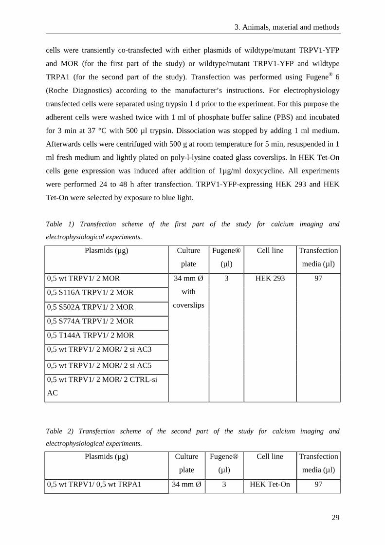
3. Animals, material and methods
29
cells were transiently co-transfected with either plasmids of wildtype/mutant TRPV1-YFP
and MOR (for the first part of the study) or wildtype/mutant TRPV1-YFP and wildtype
TRPA1 (for the second part of the study). Transfection was performed using Fugene® 6
(Roche Diagnostics) according to the manufacturer’s instructions. For electrophysiology
transfected cells were separated using trypsin 1 d prior to the experiment. For this purpose the
adherent cells were washed twice with 1 ml of phosphate buffer saline (PBS) and incubated
for 3 min at 37 °C with 500 µl trypsin. Dissociation was stopped by adding 1 ml medium.
Afterwards cells were centrifuged with 500 g at room temperature for 5 min, resuspended in 1
ml fresh medium and lightly plated on poly-l-lysine coated glass coverslips. In HEK Tet-On
cells gene expression was induced after addition of 1µg/ml doxycycline. All experiments
were performed 24 to 48 h after transfection. TRPV1-YFP-expressing HEK 293 and HEK
Tet-On were selected by exposure to blue light.
Table 1) Transfection scheme of the first part of the study for calcium imaging and
electrophysiological experiments.
Plasmids (µg) Culture
plate
Fugene®
(µl)
Cell line Transfection
media (µl)
0,5 wt TRPV1/ 2 MOR
0,5 S116A TRPV1/ 2 MOR
0,5 S502A TRPV1/ 2 MOR
0,5 S774A TRPV1/ 2 MOR
0,5 T144A TRPV1/ 2 MOR
0,5 wt TRPV1/ 2 MOR/ 2 si AC3
0,5 wt TRPV1/ 2 MOR/ 2 si AC5
0,5 wt TRPV1/ 2 MOR/ 2 CTRL-si
AC
34 mm Ø
with
coverslips
3 HEK 293 97
Table 2) Transfection scheme of the second part of the study for calcium imaging and
electrophysiological experiments.
Plasmids (µg) Culture
plate
Fugene®
(µl)
Cell line Transfection
media (µl)
0,5 wt TRPV1/ 0,5 wt TRPA1 34 mm Ø 3 HEK Tet-On 97
3. Animals, material and methods
30
0,5 S116A TRPV1/ 0,5 wt TRPA1
0,5 S502A TRPV1/ 0,5 wt TRPA1
0,5 S774A TRPV1/ 0,5 wt TRPA1
0,5 T144A TRPV1/ 0,5 wt TRPA1
0,5 wt TRPV1/ 0,5 Ptre2 (mock)
0,5 pcDNA3.1/0,5 wt TRPA1 (mock)
with
coverslips
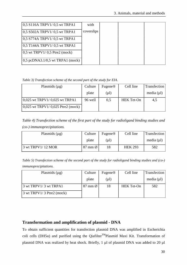
Table 3) Transfection scheme of the second part of the study for EIA.
Plasmids (µg) Culture
plate
Fugene®
(µl)
Cell line Transfection
media (µl)
0,025 wt TRPV1/ 0,025 wt TRPA1
0,025 wt TRPV1/ 0,025 Ptre2 (mock)
96 well 0,5 HEK Tet-On 4,5
Table 4) Transfection scheme of the first part of the study for radioligand binding studies and
(co-) immunoprecipitations.
Plasmids (µg) Culture
plate
Fugene®
(µl)
Cell line Transfection
media (µl)
3 wt TRPV1/ 12 MOR 87 mm Ø 18 HEK 293 582
Table 5) Transfection scheme of the second part of the study for radioligand binding studies and (co-)
immunoprecipitations.
Plasmids (µg) Culture
plate
Fugene®
(µl)
Cell line Transfection
media (µl)
3 wt TRPV1/ 3 wt TRPA1
3 wt TRPV1/ 3 Ptre2 (mock)
87 mm Ø 18 HEK Tet-On 582
Transformation and amplification of plasmid - DNA
To obtain sufficient quantities for transfection plasmid DNA was amplified in Escherichia
coli cells (DH5α) and purified using the QiafilterTMPlasmid Maxi Kit. Transformation of
plasmid DNA was realized by heat shock. Briefly, 1 µl of plasmid DNA was added to 20 µl

3. Animals, material and methods
31
DH5α suspension and incubated for 20 min on ice. The cells were heat-shocked for 30 sec at
42 °C and then cooled on ice for 2 min. Afterwards 300 µl SOC medium was subjoined and
the mixture was centrifuged at 300 rpm and 37 °C for 1 h. Finally, transformed bacteria were
plated on Luria-Bertani (LB)-agar plates containing 50 µg/ml ampicillin and incubated at 37
°C overnight. On the next day one single colony of bacteria was picked with a sterile
Eppendorf tip, transferred to 100 ml ampicillin-containing (50 µg/ml) LB-medium and
incubated with 225 rpm overnight. On the next morning 700 µl of the media was
supplemented with 300 µl 50 % glycerol and immediately stored at -80 °C.
Plasmid purification using the QiafilterTMPlasmid Maxi Kit is based on a modified alkaline
lysis procedure, followed by binding of plasmid DNA to an anion-exchange resin under
appropriate low-salt and pH conditions. RNA, proteins, dyes, and low-molecular-weight
impurities were removed by a medium-salt wash. Plasmid DNA was eluted in a high-salt
buffer and then concentrated and desalted by isopropanol precipitation. Plasmid DNA was
then washed with 70% ethanol and centrifuged for 10 min at 15000 g. The pellet was air-dried
and redissolved in 1 ml of distilled water. Plasmid DNA concentration was determined by
UV spectrophotometry at 260 nm.
Small interference RNA
Small interference RNA (siRNA) are small (21-23 nucleotides) double-stranded RNAs that
are homologous to a target gene and are used to silence gene expression in animals and plants
(Bantounas et al. 2004; Elbashir et al. 2001; Fire et al. 1998). Gene silencing through siRNA
requires two main steps: First, double-stranded RNA is recognised by an enzyme named
Dicer (member of RNase III nucleases) and cleaved into small double-stranded molecules
(siRNA). Second, the siRNAs are bound by the RNA-induced silencing complex (RISC). The
RISC is a multi-protein complex that guides the targeted RNA to degradation. Activation of
RISC is accompanied by the unwinding of the siRNA duplex and one strand of the siRNA
directs RISC to the target mRNA. Nuclease activity of RISC cleaves the target mRNA. In this
study synthetic siRNAs homologous to genes encoding AC 3 and 5 were transfected into
HEK 293 cells 48 h prior the experiment. Two µg of siRNA were applied to transfect
confluent HEK 293 in 34 cm diameter culture plates with coverslips.
3. Animals, material and methods
32
3.2.3. Calcium Imaging Experiments
This technique is used for measuring calcium signals in cultured cells. Because of its very
strong concentration gradient across the plasma membrane (intracellular Ca2+ concentration ~
30-150 nm; extracellular Ca2+ concentration ~ 10000 times higher), very short bursts of
calcium entry will generate relatively large signals and can play an important role as a
communicator and regulator of cell functions and activities. Calcium indicators can be
classified into three groups: 1) phosphoproteins are luminescent indicators that emit light in a
Ca2+- dependent manner (e.g. aequorin); 2) fluorescent dyes change their spectral properties
in response to binding of calcium ions and 3) fluorescent protein Ca2+ indicators are
conjugates between calmodulin and fluorescent proteins resulting in conformational changes
and altered fluorescent properties upon Ca2+ binding. Fluoerescent dyes can be subdivided
into ratio-metric (e.g. Fura-2 and Indo-1) and single-wavelength dyes (e.g. Fluo-4). Ratio-
metric dyes change either their excitation or emission spectra in response to calcium (Barreto-
Chang and Dolmetsch 2009; Brownlee 2000). In the current work the ratio-metric fluorescent
dye Fura-2 was used. It has an emission peak at 505 nm and changes its excitation peak from
340 nm to 380 nm following calcium binding. The intracellular calcium concentration is
derived by calculating the ratio of fluorescence emission or excitation at distict wavelengths.
Calcium imaging data were analysed with Tillvision software (Till Photonics, Gräfeling,
GER).
For the first part of the study, HEK 293 cells were cultured in 34 mm culture plates with
coverslips and transfected as shown in Tab. 1. To investigate the activity of wildtype or
mutant TRPV1 (wt/m TRPV1) during opioid withdrawal, cells were treated for at least 6 h
with 10 µM morphine which was then withdrawn by a strong wash out (withdrawal group).
Wt/m TRPV1 activity of the withdrawal group was compared to a control group of HEK 293
cells transfected with wt/m TRPV1 and MOR, but without opioid treatment and withdrawal
(CTRL). To examine the involvement of AC, siRNAs of AC 3 and 5 were co-transfected with
wt TRPV1 and MOR. Twenty-four to 48 h after transfection medium was rinsed off by
washing the cells twice with 700 µl Calcium Imaging Buffer (CIB) and cells were loaded with
3 µM Fura-2/AM for 30 min at 37 °C. Cells were washed 3 times with 1 ml CIB to remove
extracellular Fura-2/AM and incubated for 10 min with 10 µM FSK and 2 mM IBMX in CIB
to stimulate the production of cAMP and to inhibit the degradation of cAMP by
phosphodiesterases. Thereafter, cells were placed in a recording chamber containing CIB.
Pairs of images were collected every second at alternating exposures of 340 nm and 380 nm
3. Animals, material and methods
33
(exposure time 100 ms) using a Polychrome V monochromator and a CCD camera
(SensiCam). Coverslips were perfused with CIB and TRPV1 was activated by application of 1
µM capsaicin. CIB and capsaicin were removed with a pump (SCI Q 323, Watson Marlow).
The change of the fluorescence ratio at 340 nm and 380 nm was calculated following
subtraction of background fluorescence.
For the second part of the study HEK Tet-On cells were cultured in 34 mm culture plates with
coverslips and transfected as shown in Tab. 1. Expression of the cDNA of interest was
induced by addition of 1 µM doxycycline 12 h prior the experiment. Cells were washed and
loaded as described above, but without incubation with FSK/IBMX. To examine the change
in activity of TRPV1 with and without mustard oil pre-incubation, two groups of
TRPV1/TRPA1 expressing HEK Tet-On cells were generated. Cells of the control group were
loaded with Fura-2/AM, placed in the recording chamber and the increase of intracellular
calcium was evoked by addition of 100 nM - 1 µM capsaicin. Cells of the second group were
loaded with Fura-2/AM and also placed in the recording chamber. However, prior to the
experiment, 20 µM mustard oil (MuO) was added for 2-3 min. TRPV1 was activated by
capsaicin and the change of the fluorescence ratio was measured.
3.2.4. Electrophysiology
Patch Clamp experiments
This technique allows the study of single or multiple ion channels in membranes and can be
applied to a wide variety of cells. It is especially useful to study exitable cells such as
neurons, cardiomyocytes, muscle fibres and pancreatic beta cells. The patch clamp technique
is a refinement of the voltage clamp and was developed by Neher in 1976. This discovery
made it possible to record currents of single ion channels for the first time, proving their
involvement in fundamental cell processes such as action potential conduction. In 1991 the
establishment of the patch clamp technique by Neher and Sakmann was honoured with the
Nobel Prize in Physiology and Medicine.
In this study, the experimental setup was arranged as follows: an inverse microscope with a
micromanipulator, pre-amplifier, electrode holding and an automatized perfusion system was
placed on a vibration-cushioned metal table. The setup was located in a Faraday cage to
facilitate electrical isolation. The pre-amplifier was connected with the main-amplifier (EPC-
10, serial 520136-E, HEKA, Lambrecht) and signals were digitalized with an analog/digital
3. Animals, material and methods
34
converter (AD/DA-converter). Recording and analysis of the signals were realized with the
Pulse-software of HEKA, Lambrecht. Additionally it was possible to transmit voltage
commands to the amplifier. Measuring and reference electrodes were made of silver wire
(Ag/AgCl electrode), which were chlorided with potassium chloride periodically. A flexible
plastic tube was affixed to the electrode holding to produce low or high pressures at the
pipette tip during the experiment. The micromanipulator was necessary to position the pipette
close to the cell. Patch pipettes were made of borosilicate glass capillaries with filament (Nr.
1103240, Øoutside: 1.5 mm, Øinside: 0.87 mm, Hilgenberg, Malsfeld) using a pipette puller.
Pipette resistance, which gives information about the diameter of the pipette tip, was 3-6 MΩ.
The whole cell patch clamp technique was used to analyze TRPV1 specific ion currents in
cultured DRG neurons or transfected HEK 293/Tet-On cells. Experiments were performed
12-24 h after DRG culture or transient transfection of HEK 293/Tet-On cells (see Tab. 1 and
2). Cells which were not in contact with other cells were chosen preferably and perfused with
extracellular buffer (ECS, see 3.1.3.) during the experiment. The pipette was filled with
intracellular solution (ICS, see 3.1.3.) and all experiments were performed at room
temperature. The whole-cell-mode was established according to Current Protocols (Gerfen
2001). The cell-attached configuration is achieved after formation of the gigaseal. After a
short suck the cell membrane perforates and the cell-attached mode transforms into the whole-
cell configuration. This configuration allows the measurement of all ion currents in the
patched cell.
The holding potential was set at -60 mV after switching to the whole cell mode and cells were
directly activated by capsaicin (100 nM - 1 µM). For “withdrawal experiments” FSK (10 µM)
and IBMX (2mM) were applied through the bath solution. The cell-permeable AC inhibitor
SQ 22,536 (100 µM) was added 20 minutes, whereas morphine (10µM) and the protein
kinase A inhibitor H89 (10 µM) were added for at least 6 h prior to the experiment. Opioid
withdrawal was induced by a strong wash out of morphine.
For the “TRPV1/TRPA1-interaction” experiments mustard oil (20 µM) was added to the cells
2-3 min before activating TRPV1 with capsaicin. The resulting inward current was compared
to DRG neurons or transfected cells, which were not pre-treated with mustard oil.
To simulate calcium free conditions, cells were incubated and measured in Ca2+-free bath
solution complemented with EGTA (100 µM) (ECS Ca2+-free). PKA was inhibited by the
addition of H89 (10 µM) for at least 6 h prior to the experiment.
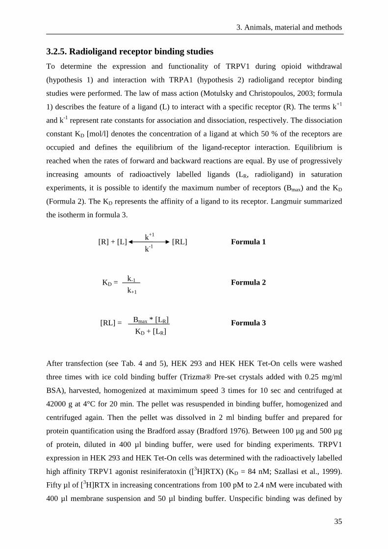
3. Animals, material and methods
35
3.2.5. Radioligand receptor binding studies
To determine the expression and functionality of TRPV1 during opioid withdrawal
(hypothesis 1) and interaction with TRPA1 (hypothesis 2) radioligand receptor binding
studies were performed. The law of mass action (Motulsky and Christopoulos, 2003; formula
1) describes the feature of a ligand (L) to interact with a specific receptor (R). The terms k+1
and k-1 represent rate constants for association and dissociation, respectively. The dissociation
constant KD [mol/l] denotes the concentration of a ligand at which 50 % of the receptors are
occupied and defines the equilibrium of the ligand-receptor interaction. Equilibrium is
reached when the rates of forward and backward reactions are equal. By use of progressively
increasing amounts of radioactively labelled ligands (LR, radioligand) in saturation
experiments, it is possible to identify the maximum number of receptors (Bmax) and the KD
(Formula 2). The KD represents the affinity of a ligand to its receptor. Langmuir summarized
the isotherm in formula 3.
[R] + [L] [RL] Formula 1
KD = Formula 2
[RL] = Formula 3
After transfection (see Tab. 4 and 5), HEK 293 and HEK HEK Tet-On cells were washed
three times with ice cold binding buffer (Trizma® Pre-set crystals added with 0.25 mg/ml
BSA), harvested, homogenized at maximimum speed 3 times for 10 sec and centrifuged at
42000 g at 4°C for 20 min. The pellet was resuspended in binding buffer, homogenized and
centrifuged again. Then the pellet was dissolved in 2 ml binding buffer and prepared for
protein quantification using the Bradford assay (Bradford 1976). Between 100 µg and 500 µg
of protein, diluted in 400 µl binding buffer, were used for binding experiments. TRPV1
expression in HEK 293 and HEK Tet-On cells was determined with the radioactively labelled
high affinity TRPV1 agonist resiniferatoxin ([3H]RTX) (KD = 84 nM; Szallasi et al., 1999).
Fifty µl of [3H]RTX in increasing concentrations from 100 pM to 2.4 nM were incubated with
400 µl membrane suspension and 50 µl binding buffer. Unspecific binding was defined by
k+1
k-1
k-1
k+1
Bmax * [L R]
KD + [LR]
3. Animals, material and methods
36
using 10 µM cold (not radioactively labelled) RTX. The binding reaction was initiated by
transferring the assay mixtures into a 37 °C water bath and terminated after 60 min incubation
by cooling the tubes on ice. Additionally, non-specific RTX binding was reduced by adding
bovine α1-acid glycoprotein (100 µg per tube) after the binding reaction was stopped.
Membrane-bound RTX was separated from the α1-acid glycoprotein-bound RTX by pelleting
the membranes in a centrifuge at 3600 rpm for 60 min following a resuspension of the pellet
in 200 µl binding buffer. Separation of membrane-bound and free radioactivity was achieved
by vacuum filtration (Brandel-Harvester) through polyethylenimin (0.1 % in 50 mM Tris)
soaked glass fiber filters.
Tritium-labelled ligands emit low-energy beta rays detectable indirectly via scintillation
spectroscopy (Yamamura and Hulme, 1992). Filters were placed in tubes and filled with 3 ml
scintillation solution which contains a primary and a secondary scintillator. The emission
spectrum of the secondary scintillator is detectable by a photomultiplier and measured with
the aid of a scintillation counter as decay/counts per min (CPM). Only a very small part (3-6
%) of the energy of the radioactive rays is detected due to the high number of energy transfer
processes.
Twenty-four h after filtration the radioactivity of the sample was measured with a liquid
scintillation counter. Its efficiency amounted to 69 % (counter efficiency = CPMspecific x 100 /
decays/min [DPM]). Specific binding was calculated substracting the CPM of unspecific
binding from the CPM of total binding:
CPMspecific = CPMtotal - CPMunspecific
The specific activity of the ligand was assessed with aid of the specific radioactivity. One
Ci/mmol is equal to 2.2 x 10¹² DPM:
Specific activity [cpm/fmol] =
specific radioactivity [Ci/mmol] x 2.2 x 10¹² x counter efficiency / 1012 [fmol]
Bmax was obtained using following equation:
Bmax [fmol/mg] = CPMspecific / specific activity [cpm/fmol] x protein content [mg]
The KD and the hyperbole saturation curve were calculated with GraphPad Prism 5 (San
Diego, USA) using nonlinear regression.
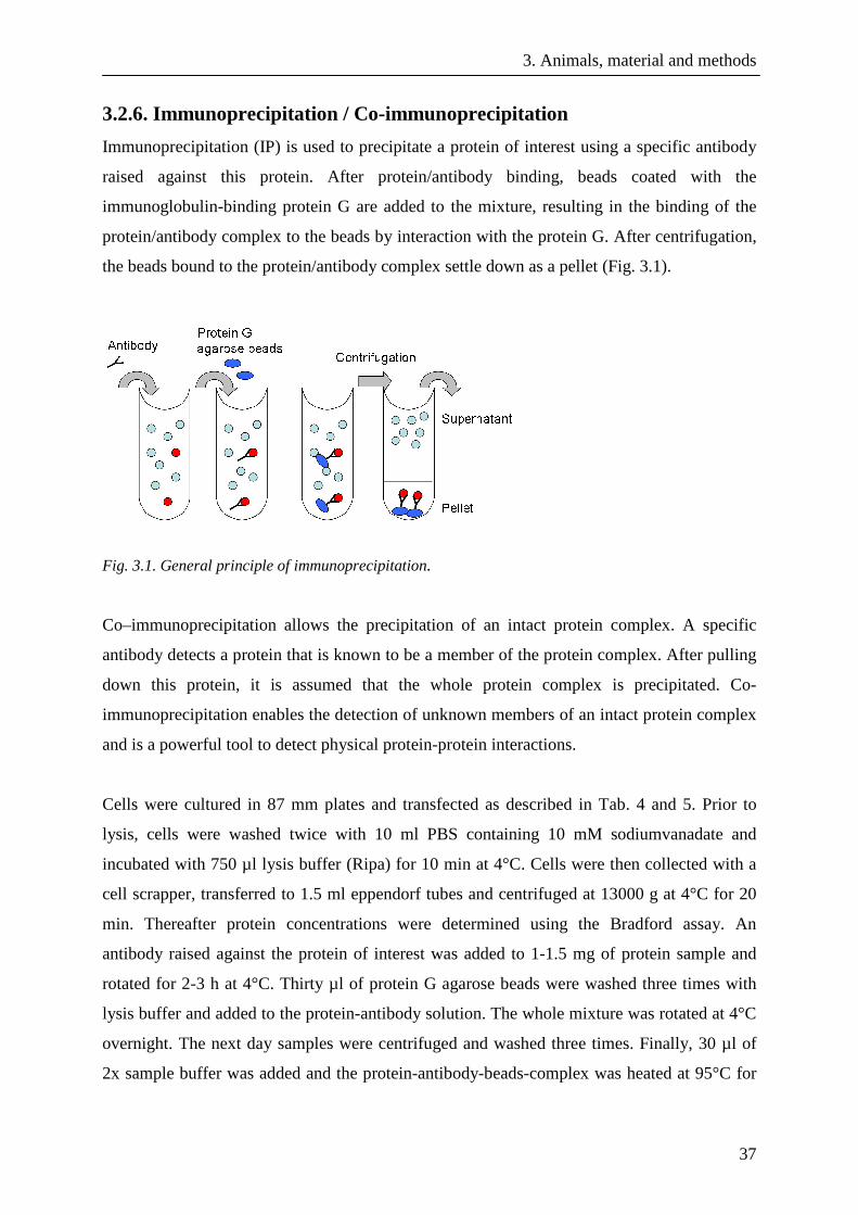
3. Animals, material and methods
37
3.2.6. Immunoprecipitation / Co-immunoprecipitation
Immunoprecipitation (IP) is used to precipitate a protein of interest using a specific antibody
raised against this protein. After protein/antibody binding, beads coated with the
immunoglobulin-binding protein G are added to the mixture, resulting in the binding of the
protein/antibody complex to the beads by interaction with the protein G. After centrifugation,
the beads bound to the protein/antibody complex settle down as a pellet (Fig. 3.1).
Fig. 3.1. General principle of immunoprecipitation.
Co–immunoprecipitation allows the precipitation of an intact protein complex. A specific
antibody detects a protein that is known to be a member of the protein complex. After pulling
down this protein, it is assumed that the whole protein complex is precipitated. Co-
immunoprecipitation enables the detection of unknown members of an intact protein complex
and is a powerful tool to detect physical protein-protein interactions.
Cells were cultured in 87 mm plates and transfected as described in Tab. 4 and 5. Prior to
lysis, cells were washed twice with 10 ml PBS containing 10 mM sodiumvanadate and
incubated with 750 µl lysis buffer (Ripa) for 10 min at 4°C. Cells were then collected with a
cell scrapper, transferred to 1.5 ml eppendorf tubes and centrifuged at 13000 g at 4°C for 20
min. Thereafter protein concentrations were determined using the Bradford assay. An
antibody raised against the protein of interest was added to 1-1.5 mg of protein sample and
rotated for 2-3 h at 4°C. Thirty µl of protein G agarose beads were washed three times with
lysis buffer and added to the protein-antibody solution. The whole mixture was rotated at 4°C
overnight. The next day samples were centrifuged and washed three times. Finally, 30 µl of
2x sample buffer was added and the protein-antibody-beads-complex was heated at 95°C for

3. Animals, material and methods
38
5 min to denature the protein. Subsequently, samples were ready for SDS gel-electrophoresis
(described in the following chapter).
All IP-antibodies and their applied concentrations are listed in Tab. 6.
3.2.7. Western Blot analysis
Western Blot analysis is used for the detection, identification and quantification of proteins
out of a complex protein mixture.
By use of gel electrophoresis, native or denatured proteins are separated by their molecular
weight (denatured proteins) or by their 3 dimensional structures (native or denatured
proteins). After electrophoresis, proteins are transferred to a nitrocellulose or polyvinylidene
fluoride (PVD) membrane, which makes proteins accessible to antibody detection. After
addition of the specific antibody against the protein of interest (primary antibody), the
membrane is incubated with a secondary antibody conjugated with horseradish peroxidase
that cleaves a chemiluminescent reagent. The reaction product emits luminescence correlated
positively with the amount of protein. Exposure of the membrane to a photosensitive film
reveals the conjugated secondary antibody bound to the primary antibody which binds the
protein of interest (Broome and Gilbert 1978; Burnette 1981; Hawkes et al. 1982; Towbin et
al. 1979) .
In my experiments, the SDS-polyacrylamide gel (resolving gel 8 %: 5.3 ml H2O; 2 ml 40 %
acryl amide mix; 2.5 ml 1.5 M Tris pH 8.8; 0.1 ml 10 % SDS; 0.1 ml 10 % APS; 0.006 ml
TEMED and stacking gel: 1.5 ml H2O; 0.25 ml 40 % acryl amid mix; 0.25 ml 1 M Tris pH
6.8; 0.02 ml 10 % SDS; 0.02 ml 10 % APS; 0.002 ml TEMED) was placed in a
electrophoresis chamber filled with running buffer. Prestained protein standards and samples
were carefully loaded into the gel wells. Electrophoresis was started at 60 mV until samples
reached the resolving gel. Then, the voltage was raised up to 150 mV until proteins were
completely separated (as seen in the protein standards well). The gel was then transferred into
blotting solution. For transfer, gel was placed on top of a blotting paper and a blotting sponge
and covered with a PVDF membrane which was also covered by a blotting paper and blotting
sponge. All layers were clamped in a blotting chamber and transfered from the gel to the
membrane at 350 mA for 1 h. Immediately after transfer, the membrane was incubated with
blocking solution (2.5 - 5 % BSA and 2.5 - 5 % low-fat milk, respectively) for 1 h at room
temperature (RT) at gentle agitation followed by incubation of the primary antibody overnight
at 4 °C. On the next day the membrane was washed 3 times with TBS-Tween (3 x 10 min at
room temperature under gentle agitation) and the secondary antibody was applied for 1 h at
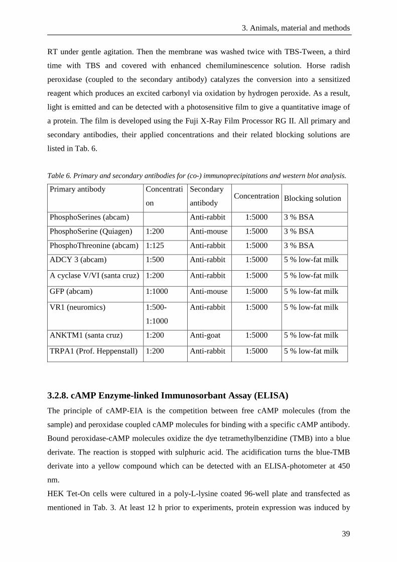
3. Animals, material and methods
39
RT under gentle agitation. Then the membrane was washed twice with TBS-Tween, a third
time with TBS and covered with enhanced chemiluminescence solution. Horse radish
peroxidase (coupled to the secondary antibody) catalyzes the conversion into a sensitized
reagent which produces an excited carbonyl via oxidation by hydrogen peroxide. As a result,
light is emitted and can be detected with a photosensitive film to give a quantitative image of
a protein. The film is developed using the Fuji X-Ray Film Processor RG II. All primary and
secondary antibodies, their applied concentrations and their related blocking solutions are
listed in Tab. 6.
Table 6. Primary and secondary antibodies for (co-) immunoprecipitations and western blot analysis.
Primary antibody Concentrati
on
Secondary
antibody Concentration
Blocking solution
PhosphoSerines (abcam) Anti-rabbit 1:5000 3 % BSA
PhosphoSerine (Quiagen) 1:200 Anti-mouse 1:5000 3 % BSA
PhosphoThreonine (abcam) 1:125 Anti-rabbit 1:5000 3 % BSA
ADCY 3 (abcam) 1:500 Anti-rabbit 1:5000 5 % low-fat milk
A cyclase V/VI (santa cruz) 1:200 Anti-rabbit 1:5000 5 % low-fat milk
GFP (abcam) 1:1000 Anti-mouse 1:5000 5 % low-fat milk
VR1 (neuromics) 1:500-
1:1000
Anti-rabbit 1:5000 5 % low-fat milk
ANKTM1 (santa cruz) 1:200 Anti-goat 1:5000 5 % low-fat milk
TRPA1 (Prof. Heppenstall) 1:200 Anti-rabbit 1:5000 5 % low-fat milk
3.2.8. cAMP Enzyme-linked Immunosorbant Assay (ELISA)
The principle of cAMP-EIA is the competition between free cAMP molecules (from the
sample) and peroxidase coupled cAMP molecules for binding with a specific cAMP antibody.
Bound peroxidase-cAMP molecules oxidize the dye tetramethylbenzidine (TMB) into a blue
derivate. The reaction is stopped with sulphuric acid. The acidification turns the blue-TMB
derivate into a yellow compound which can be detected with an ELISA-photometer at 450
nm.
HEK Tet-On cells were cultured in a poly-L-lysine coated 96-well plate and transfected as
mentioned in Tab. 3. At least 12 h prior to experiments, protein expression was induced by
3. Animals, material and methods
40
application of 1 µM doxycycline. Cells were stimulated by 2 mM IBMX and 10 µM forskolin
to avoid cAMP-degradation by phosphodiesterases and to exite cAMP-production by AC. 20
µM mustard oil was added 5 min prior to membrane preparation. To investigate the role of
calcium in the accumulation of cAMP, calcium was removed from one sample group. The
following groups of transfected HEK Tet-On cells were investigated:
TRPV1/TRPA1:
• stimulated with FSK/IBMX (w/o MuO)
• stimulated with FSK/IBMX and pretreated with 20 µM MuO (MuO)
• stimulated with FSK/IBMX in calcium free medium (w/o MuO; w/o Ca2+)
• stimulated with FSK/IBMX and pretreated with 20 µM MuO in calcium free medium
(MuO; w/o Ca2+)
TRPV1/Ptre2 (mock):
• stimulated with FSK/IBMX (w/o MuO mock)
• stimulated with FSK/IBMX and pretreated with 20 µM MuO (MuO mock)
EIA experiments were performed following manufacturer’s instructions using the non-
acetylation protocol. After stimulation transfected HEK Tet-On cells were lysed and 100 µl of
the lysate were transferred to an antigen-coated (donkey anti-rabbit IgG) ELISA plate.
Simultaneously, a cAMP standard curve and blanks were prepared. All samples were
arranged in duplicates and incubated for 2 h with an antiserum (rabbit anti-cAMP) followed
by an 1 h incubation with cAMP peroxidase. Afterwards the plate was washed and TMB
substrate was added. Approximately 20 min later a blue dye was developed which changed to
a yellow colour by addition of H2SO4. The intensity of the colour was detected with an
ELISA-reader at λ=450 nm. Data were analyzed using SOFTmax® Pro software (Molecular
Devices, Sunnyvale, USA). The cAMP content was calculated with the aid of a standard
curve, where the fluorescence was plotted versus the concentration in a semi logarithmic
manner.
3.2.9. Statistical analysis
Statistical and graphical analysis was performed with GraphPad Prism 5 (GraphPad Software,
San Diego, USA), Microsoft Excel (Microsoft Corporation, Unterschleißheim, Germany) and
Image J. Statistical significance was determined with Student´s unpaired t-test, 1 way
ANOVA with Dunnet post-hoc test or 2 way ANOVA with Bonferroni post-hoc test. To

3. Animals, material and methods
41
calculate KD and Bmax non-linear regression curve fits were performed.Statistical significance
was denoted as p<0.05 (*), p<0.01 (**) and p<0.001 (***).
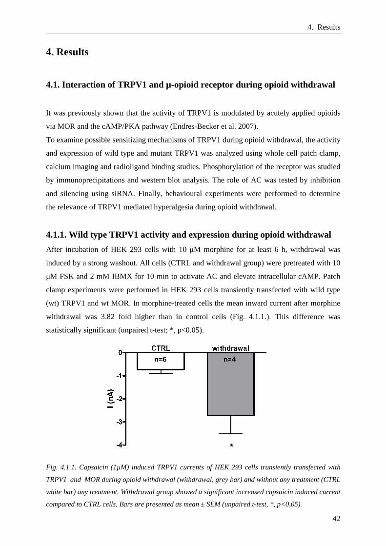
4. Results
42
4. Results
4.1. Interaction of TRPV1 and µ-opioid receptor during opioid withdrawal
It was previously shown that the activity of TRPV1 is modulated by acutely applied opioids
via MOR and the cAMP/PKA pathway (Endres-Becker et al. 2007).
To examine possible sensitizing mechanisms of TRPV1 during opioid withdrawal, the activity
and expression of wild type and mutant TRPV1 was analyzed using whole cell patch clamp,
calcium imaging and radioligand binding studies. Phosphorylation of the receptor was studied
by immunoprecipitations and western blot analysis. The role of AC was tested by inhibition
and silencing using siRNA. Finally, behavioural experiments were performed to determine
the relevance of TRPV1 mediated hyperalgesia during opioid withdrawal.
4.1.1. Wild type TRPV1 activity and expression during opioid withdrawal
After incubation of HEK 293 cells with 10 µM morphine for at least 6 h, withdrawal was
induced by a strong washout. All cells (CTRL and withdrawal group) were pretreated with 10
µM FSK and 2 mM IBMX for 10 min to activate AC and elevate intracellular cAMP. Patch
clamp experiments were performed in HEK 293 cells transiently transfected with wild type
(wt) TRPV1 and wt MOR. In morphine-treated cells the mean inward current after morphine
withdrawal was 3.82 fold higher than in control cells (Fig. 4.1.1.). This difference was
statistically significant (unpaired t-test; *, p<0.05).
Fig. 4.1.1. Capsaicin (1µM) induced TRPV1 currents of HEK 293 cells transiently transfected with
TRPV1 and MOR during opioid withdrawal (withdrawal, grey bar) and without any treatment (CTRL
white bar) any treatment. Withdrawal group showed a significant increased capsaicin induced current
compared to CTRL cells. Bars are presented as mean ± SEM (unpaired t-test, *, p<0,05).
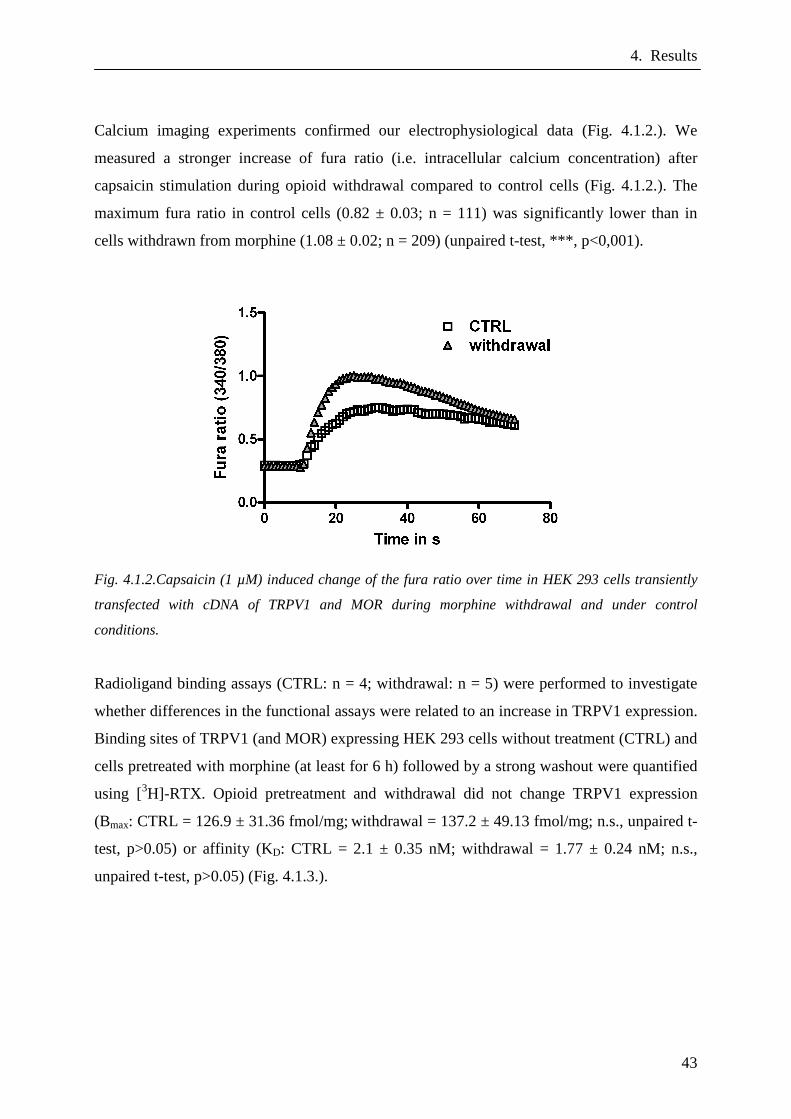
4. Results
43
Calcium imaging experiments confirmed our electrophysiological data (Fig. 4.1.2.). We
measured a stronger increase of fura ratio (i.e. intracellular calcium concentration) after
capsaicin stimulation during opioid withdrawal compared to control cells (Fig. 4.1.2.). The
maximum fura ratio in control cells (0.82 ± 0.03; n = 111) was significantly lower than in
cells withdrawn from morphine (1.08 ± 0.02; n = 209) (unpaired t-test, ***, p<0,001).
Fig. 4.1.2.Capsaicin (1 µM) induced change of the fura ratio over time in HEK 293 cells transiently
transfected with cDNA of TRPV1 and MOR during morphine withdrawal and under control
conditions.
Radioligand binding assays (CTRL: n = 4; withdrawal: n = 5) were performed to investigate
whether differences in the functional assays were related to an increase in TRPV1 expression.
Binding sites of TRPV1 (and MOR) expressing HEK 293 cells without treatment (CTRL) and
cells pretreated with morphine (at least for 6 h) followed by a strong washout were quantified
using [3H]-RTX. Opioid pretreatment and withdrawal did not change TRPV1 expression
(Bmax: CTRL = 126.9 ± 31.36 fmol/mg; withdrawal = 137.2 ± 49.13 fmol/mg; n.s., unpaired t-
test, p>0.05) or affinity (KD: CTRL = 2.1 ± 0.35 nM; withdrawal = 1.77 ± 0.24 nM; n.s.,
unpaired t-test, p>0.05) (Fig. 4.1.3.).
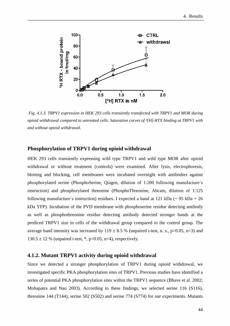
4. Results
44
Fig. 4.1.3. TRPV1 expression in HEK 293 cells transiently transfected with TRPV1 and MOR during
opioid withdrawal compared to untreated cells. Saturation curves of ³[H]-RTX binding at TRPV1 with
and without opioid withdrawal.
Phosphorylation of TRPV1 during opioid withdrawal
HEK 293 cells transiently expressing wild type TRPV1 and wild type MOR after opioid
withdrawal or without treatment (controls) were examined. After lysis, electrophoresis,
blotting and blocking, cell membranes were incubated overnight with antibodies against
phosphorylated serine (PhosphoSerine, Qiagen, dilution of 1:200 following manufacture´s
instruction) and phosphorylated threonine (PhosphoThreonine, Abcam, dilution of 1:125
following manufacture´s instruction) residues. I expected a band at 121 kDa (~ 95 kDa + 26
kDa YFP). Incubation of the PVD membrane with phosphoserine residue detecting antibody
as well as phosphothreonine residue detecting antibody detected stronger bands at the
prediced TRPV1 size in cells of the withdrawal group compared to the control group. The
average band intensity was increased by 119 ± 8.5 % (unpaired t-test, n. s., p>0.05, n=3) and
130.5 ± 12 % (unpaired t-test, *, p<0.05, n=4), respectively.
4.1.2. Mutant TRPV1 activity during opioid withdrawal
Since we detected a stronger phosphorylation of TRPV1 during opioid withdrawal, we
investigated specific PKA phosphorylation sites of TRPV1. Previous studies have identified a
series of potential PKA phosphorylation sites within the TRPV1 sequence (Bhave et al. 2002;
Mohapatra and Nau 2003). According to these findings, we selected serine 116 (S116),
threonine 144 (T144), serine 502 (S502) and serine 774 (S774) for our experiments. Mutants
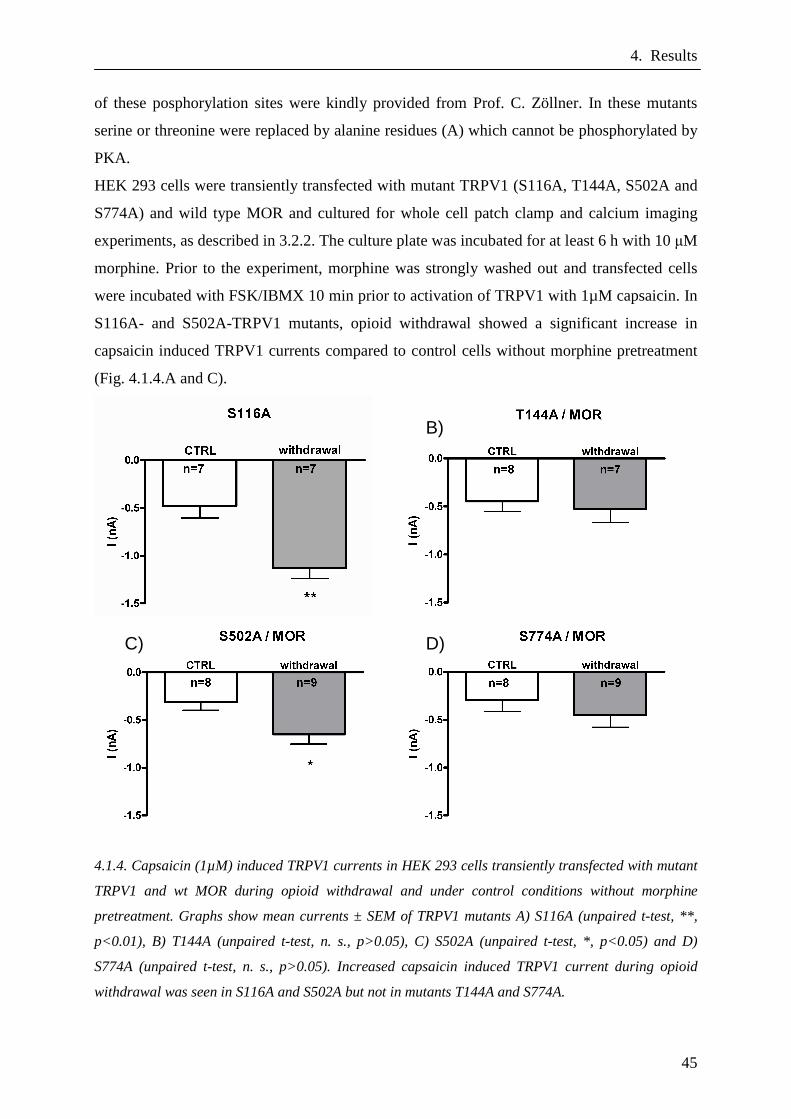
4. Results
45
of these posphorylation sites were kindly provided from Prof. C. Zöllner. In these mutants
serine or threonine were replaced by alanine residues (A) which cannot be phosphorylated by
PKA.
HEK 293 cells were transiently transfected with mutant TRPV1 (S116A, T144A, S502A and
S774A) and wild type MOR and cultured for whole cell patch clamp and calcium imaging
experiments, as described in 3.2.2. The culture plate was incubated for at least 6 h with 10 µM
morphine. Prior to the experiment, morphine was strongly washed out and transfected cells
were incubated with FSK/IBMX 10 min prior to activation of TRPV1 with 1µM capsaicin. In
S116A- and S502A-TRPV1 mutants, opioid withdrawal showed a significant increase in
capsaicin induced TRPV1 currents compared to control cells without morphine pretreatment
(Fig. 4.1.4.A and C).
4.1.4. Capsaicin (1µM) induced TRPV1 currents in HEK 293 cells transiently transfected with mutant
TRPV1 and wt MOR during opioid withdrawal and under control conditions without morphine
pretreatment. Graphs show mean currents ± SEM of TRPV1 mutants A) S116A (unpaired t-test, **,
p<0.01), B) T144A (unpaired t-test, n. s., p>0.05), C) S502A (unpaired t-test, *, p<0.05) and D)
S774A (unpaired t-test, n. s., p>0.05). Increased capsaicin induced TRPV1 current during opioid
withdrawal was seen in S116A and S502A but not in mutants T144A and S774A.
A) B)
C) D)

4. Results
46
In T144A- and S774A-TRPV1 mutants opioid withdrawal did not induce a significant
increase in capsaicin mediated TRPV1 activity (Fig. 4.1.4.). Similar results were obtained
using calcium imaging experiments (Fig. 4.1.5.): S116A and S502A TRPV1 mutants showed
a significant increase in capsaicin-induced [Ca2+] i increase during opioid withdrawal
compared with control cells. In T144A- and S774A-TRPV1 mutants, no significant increase
in capsaicin–induced [Ca2+] i increase was detected during opioid withdrawal in comparison to
CTRL cells.
Fig. 4.1.5. Capsaicin (1µM) induced maximum fura ratio in HEK 293 cells transfected with mutant
TRPV1 and wt MOR under control conditions without morphine pretreatment and during opioid
withdrawal. A) S116A vs. CTRL (unpaired t-test, **, p<0,01), B) T144A vs.CTRL (unpaired t-test, n.
s., p>0,05), C) S502A vs.CTRL (unpaired t-test, ***, p<0,001) and D) S774A vs.CTRL (unpaired t-
test, n. s., p>0,05).
A) B)
C) D)

4. Results
47
4.1.3. Role of adenylylcyclases 3 and 5 during opioid withdrawal
Opioid pretreatment induces a compensatory upregulation of AC activity that becomes
manifest during withdrawal (Nestler and Aghajanian 1997). Previous studies in our laboratory
also showed a significantly elevated mRNA level of AC isoforms 3 and 5 (Endres-Becker
2007) in DRG during opioid withdrawal. The nonselective AC inhibitor SQ 22,536 was
applied (for 20 min) to block the activity of AC 1 - 9. Figure 4.1.6. shows the capsacin-
induced TRPV1 current in HEK 293 cells expressing wt TRPV1 and MOR with and without
SQ 22,536 pretreatment with and without morphine treatment. The enhanced capsaicin-
induced TRPV1 current during opioid withdrawal was reversed in the presence of the AC
inhibitor.
Fig. 4.1.6. Capsaicin (1µM) induced TRPV1 currents in HEK 293 cells transiently transfected with wt
TRPV1 and wt MOR without (CTR;, withdrawal) and with (CTRL SQ 22,53, withdrawal SQ 22,536)
SQ 22,535 treatment . Graphs show mean ± SEM of capsaicin-induced (1µM) TRPV1 currents (1way
ANOVA, Dunnet post hoc test, CTRL versus withdrawal, ***, p<0,001; CTRL versus CTRL SQ
22,536, n. s., p>0,05; CTRL versus withdrawal SQ 22,536, n. s., p>0,05). Pretreatment with SQ
22,536 reversed the increased TRPV1 current during withdrawal.
To further delineate the role of different AC in opioid withdrawal we investigated the role of
AC 3 and 5. Using small interference RNA (siRNA) we silenced AC 3 and 5 gene expression
in HEK 293 cells transiently transfected with TRPV1 and MOR.
Downregulation of AC 3 or 5 expression was verified by western blot analysis. Band intensity
of AC 3 in siRNA AC 3 transfected cells was 45.87 ± 7,41 % of the band intensity of
untreated HEK 293 cells (unpaired t-test, **, p<0.01, n=3). Band intensity of AC 5 in
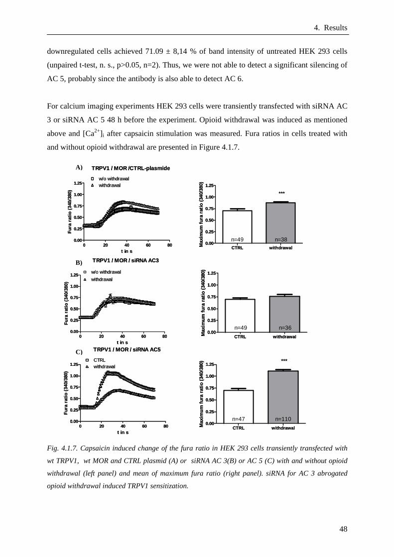
4. Results
48
downregulated cells achieved 71.09 ± 8,14 % of band intensity of untreated HEK 293 cells
(unpaired t-test, n. s., p>0.05, n=2). Thus, we were not able to detect a significant silencing of
AC 5, probably since the antibody is also able to detect AC 6.
For calcium imaging experiments HEK 293 cells were transiently transfected with siRNA AC
3 or siRNA AC 5 48 h before the experiment. Opioid withdrawal was induced as mentioned
above and [Ca2+] i after capsaicin stimulation was measured. Fura ratios in cells treated with
and without opioid withdrawal are presented in Figure 4.1.7.
Fig. 4.1.7. Capsaicin induced change of the fura ratio in HEK 293 cells transiently transfected with
wt TRPV1, wt MOR and CTRL plasmid (A) or siRNA AC 3(B) or AC 5 (C) with and without opioid
withdrawal (left panel) and mean of maximum fura ratio (right panel). siRNA for AC 3 abrogated
opioid withdrawal induced TRPV1 sensitization.
CTRL withdrawal0.00
0.25
0.50
0.75
1.00
1.25
***
n=49 n=38
Max
imum
fur
a ra
tio
(340
/380
)
0 20 40 60 800.00
0.25
0.50
0.75
1.00
1.25w/o withdrawal
withdrawal
TRPV1 / MOR / siRNA AC3
t in s
Fu
ra r
atio
(34
0/38
0)
CTRL withdrawal0.00
0.25
0.50
0.75
1.00
1.25
n=49 n=36
Max
imum
fur
a ra
tio
(340
/380
)
0 20 40 60 800.00
0.25
0.50
0.75
1.00
1.25CTRLwithdrawal
TRPV1 / MOR / siRNA AC5
t in s
Fura
rat
io (
340/
380)
CTRL withdrawal0.00
0.25
0.50
0.75
1.00
1.25***
n=47 n=110
Max
imu
m f
ura
rat
io (
340/
380)
0 20 40 60 800.00
0.25
0.50
0.75
1.00
1.25w/o withdrawal
TRPV1 / MOR /CTRL-plasmide
withdrawal
t in s
Fu
ra r
atio
(34
0/38
0)
CTRL withdrawal0.00
0.25
0.50
0.75
1.00
1.25
***
n=49 n=38
Max
imum
fur
a ra
tio
(340
/380
)
0 20 40 60 800.00
0.25
0.50
0.75
1.00
1.25w/o withdrawal
withdrawal
TRPV1 / MOR / siRNA AC3
t in s
Fu
ra r
atio
(34
0/38
0)
CTRL withdrawal0.00
0.25
0.50
0.75
1.00
1.25
n=49 n=36
Max
imum
fur
a ra
tio
(340
/380
)
0 20 40 60 800.00
0.25
0.50
0.75
1.00
1.25CTRLwithdrawal
TRPV1 / MOR / siRNA AC5
t in s
Fura
rat
io (
340/
380)
CTRL withdrawal0.00
0.25
0.50
0.75
1.00
1.25***
n=47 n=110
Max
imu
m f
ura
rat
io (
340/
380)
0 20 40 60 800.00
0.25
0.50
0.75
1.00
1.25w/o withdrawal
TRPV1 / MOR /CTRL-plasmide
withdrawal
t in s
Fu
ra r
atio
(34
0/38
0)
A)
B)
C)
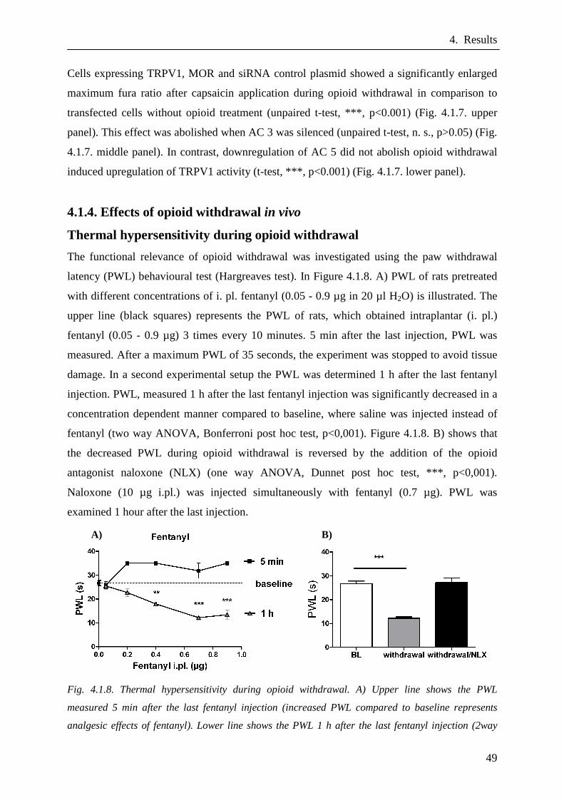
4. Results
49
Cells expressing TRPV1, MOR and siRNA control plasmid showed a significantly enlarged
maximum fura ratio after capsaicin application during opioid withdrawal in comparison to
transfected cells without opioid treatment (unpaired t-test, ***, p<0.001) (Fig. 4.1.7. upper
panel). This effect was abolished when AC 3 was silenced (unpaired t-test, n. s., p>0.05) (Fig.
4.1.7. middle panel). In contrast, downregulation of AC 5 did not abolish opioid withdrawal
induced upregulation of TRPV1 activity (t-test, ***, p<0.001) (Fig. 4.1.7. lower panel).
4.1.4. Effects of opioid withdrawal in vivo
Thermal hypersensitivity during opioid withdrawal
The functional relevance of opioid withdrawal was investigated using the paw withdrawal
latency (PWL) behavioural test (Hargreaves test). In Figure 4.1.8. A) PWL of rats pretreated
with different concentrations of i. pl. fentanyl (0.05 - 0.9 µg in 20 µl H2O) is illustrated. The
upper line (black squares) represents the PWL of rats, which obtained intraplantar (i. pl.)
fentanyl (0.05 - 0.9 µg) 3 times every 10 minutes. 5 min after the last injection, PWL was
measured. After a maximum PWL of 35 seconds, the experiment was stopped to avoid tissue
damage. In a second experimental setup the PWL was determined 1 h after the last fentanyl
injection. PWL, measured 1 h after the last fentanyl injection was significantly decreased in a
concentration dependent manner compared to baseline, where saline was injected instead of
fentanyl (two way ANOVA, Bonferroni post hoc test, p<0,001). Figure 4.1.8. B) shows that
the decreased PWL during opioid withdrawal is reversed by the addition of the opioid
antagonist naloxone (NLX) (one way ANOVA, Dunnet post hoc test, ***, p<0,001).
Naloxone (10 µg i.pl.) was injected simultaneously with fentanyl (0.7 µg). PWL was
examined 1 hour after the last injection.
Fig. 4.1.8. Thermal hypersensitivity during opioid withdrawal. A) Upper line shows the PWL
measured 5 min after the last fentanyl injection (increased PWL compared to baseline represents
analgesic effects of fentanyl). Lower line shows the PWL 1 h after the last fentanyl injection (2way
B) A)
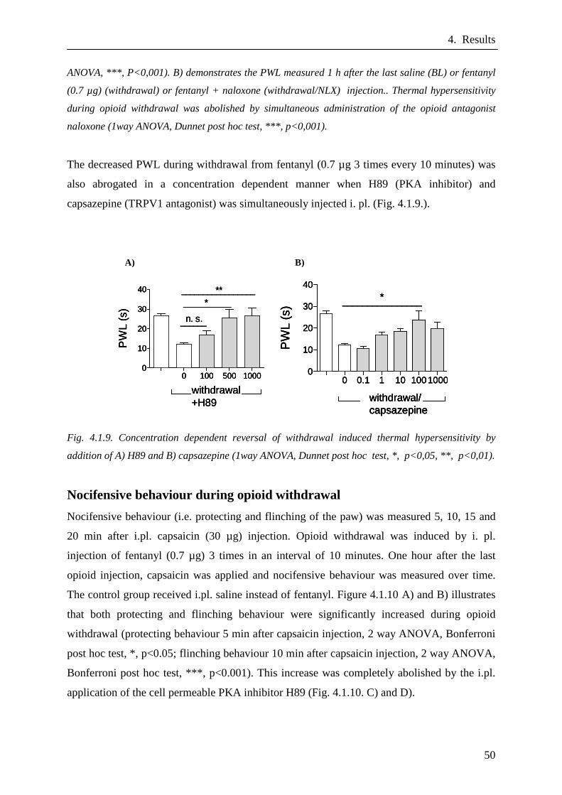
4. Results
50
ANOVA, ***, P<0,001). B) demonstrates the PWL measured 1 h after the last saline (BL) or fentanyl
(0.7 µg) (withdrawal) or fentanyl + naloxone (withdrawal/NLX) injection.. Thermal hypersensitivity
during opioid withdrawal was abolished by simultaneous administration of the opioid antagonist
naloxone (1way ANOVA, Dunnet post hoc test, ***, p<0,001).
The decreased PWL during withdrawal from fentanyl (0.7 µg 3 times every 10 minutes) was
also abrogated in a concentration dependent manner when H89 (PKA inhibitor) and
capsazepine (TRPV1 antagonist) was simultaneously injected i. pl. (Fig. 4.1.9.).
Fig. 4.1.9. Concentration dependent reversal of withdrawal induced thermal hypersensitivity by
addition of A) H89 and B) capsazepine (1way ANOVA, Dunnet post hoc test, *, p<0,05, **, p<0,01).
Nocifensive behaviour during opioid withdrawal
Nocifensive behaviour (i.e. protecting and flinching of the paw) was measured 5, 10, 15 and
20 min after i.pl. capsaicin (30 µg) injection. Opioid withdrawal was induced by i. pl.
injection of fentanyl (0.7 µg) 3 times in an interval of 10 minutes. One hour after the last
opioid injection, capsaicin was applied and nocifensive behaviour was measured over time.
The control group received i.pl. saline instead of fentanyl. Figure 4.1.10 A) and B) illustrates
that both protecting and flinching behaviour were significantly increased during opioid
withdrawal (protecting behaviour 5 min after capsaicin injection, 2 way ANOVA, Bonferroni
post hoc test, *, p<0.05; flinching behaviour 10 min after capsaicin injection, 2 way ANOVA,
Bonferroni post hoc test, ***, p<0.001). This increase was completely abolished by the i.pl.
application of the cell permeable PKA inhibitor H89 (Fig. 4.1.10. C) and D).
0 0.1 1 10 10010000
10
20
30
40
________________*P
WL
(s)
withdrawal/capsazepine
0 100 500 10000
10
20
30
40 _________________**___________*
______n. s.
PW
L (s
)
withdrawal+H89
0 0.1 1 10 10010000
10
20
30
40
________________*P
WL
(s)
withdrawal/capsazepine
0 0.1 1 10 10010000
10
20
30
40
________________*P
WL
(s)
withdrawal/capsazepinewithdrawal/capsazepine
0 100 500 10000
10
20
30
40 _________________**___________*
______n. s.
PW
L (s
)
withdrawal+H89
0 100 500 10000
10
20
30
40 _________________**___________*
______n. s.
PW
L (s
)
withdrawal+H89
A) B)

4. Results
51
Fig. 4.1.10. Increased protecting (A) and fliching (B) behaviour during opioid withdrawal was
reversed by the addition of H89 (C) and (D) (2way ANOVA and Bonferroni post hoc test, *, p<0.05,
**, p<0.01, ***, p<0.001).
4.2. Interaction of TRPV1 and TRPA1
The second part of my thesis investigated TRPV1 sensitization after stimulation of cells with
TRPA1 ligands.
4.2.1. Direct physical interaction of TRPV1 and TRPA1
The expression of TRPV1 ion channels after activation of TRPA1 was investigated using
radioligand binding assays with [3H] labelled resiniferatoxin ([3H]-RTX). HEK Tet-On cells
were transiently transfected with wild type TRPV1 and wild type TRPA1, or with wt TRPV1
and the empty vector Ptre2 (mock transfection). In Figure 4.2.1 saturation curves of [3H]-RTX
binding are presented. No changes in the number of TRPV1 binding sites or their dissociation
constants (KD) were detected in the presence of TRPA1 (TRPV1/TRPA1: Bmax = 283.1 ±
61.54 fmol/mg, KD = 2.77 ± 0.94 nM, n = 7; TRPV1/Ptre2: Bmax = 315.9 ± 133.6 fmol/mg, KD
= 4.2 ± 2.51 nM, n = 5, unpaired t-test, n. s., p>0.05).
0 5 10 15 20 250
1
2
3
4CTRL
withdrawal
*
Time after Capsaicin (min)
Pro
tect
ing
beha
viou
r(m
in)
0 5 10 15 20 250
5
10
15
20
25
withdrawal
CTRL
***
Time after Capsaicin (min)
Num
ber
of f
linch
es
0 5 10 15 20 250
1
2
3
4withdrawal
withdrawal+H89
Time after Capsaicin (min)
Pro
tect
ing
beha
viou
r(m
in)
**
*
0 5 10 15 20 250
5
10
15
20
25withdrawal
withdrawal+H89
Time after Capsaicin (min)
Num
ber
of f
linch
es**
0 5 10 15 20 250
1
2
3
4CTRL
withdrawal
*
Time after Capsaicin (min)
Pro
tect
ing
beha
viou
r(m
in)
0 5 10 15 20 250
5
10
15
20
25
withdrawal
CTRL
***
Time after Capsaicin (min)
Num
ber
of f
linch
es
0 5 10 15 20 250
1
2
3
4withdrawal
withdrawal+H89
Time after Capsaicin (min)
Pro
tect
ing
beha
viou
r(m
in)
**
*
0 5 10 15 20 250
5
10
15
20
25withdrawal
withdrawal+H89
Time after Capsaicin (min)
Num
ber
of f
linch
es**
A) B)
C) D)
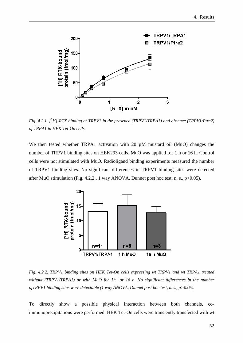
4. Results
52
Fig. 4.2.1. [3H]-RTX binding at TRPV1 in the presence (TRPV1/TRPA1) and absence (TRPV1/Ptre2)
of TRPA1 in HEK Tet-On cells.
We then tested whether TRPA1 activation with 20 µM mustard oil (MuO) changes the
number of TRPV1 binding sites on HEK293 cells. MuO was applied for 1 h or 16 h. Control
cells were not stimulated with MuO. Radioligand binding experiments measured the number
of TRPV1 binding sites. No significant differences in TRPV1 binding sites were detected
after MuO stimulation (Fig. 4.2.2., 1 way ANOVA, Dunnet post hoc test, n. s., p>0.05).
Fig. 4.2.2. TRPV1 binding sites on HEK Tet-On cells expressing wt TRPV1 and wt TRPA1 treated
without (TRPV1/TRPA1) or with MuO for 1h or 16 h. No significant differences in the number
ofTRPV1 binding sites were detectable (1 way ANOVA, Dunnet post hoc test, n. s., p>0.05).
To directly show a possible physical interaction between both channels, co-
immunoprecipitations were performed. HEK Tet-On cells were transiently transfected with wt
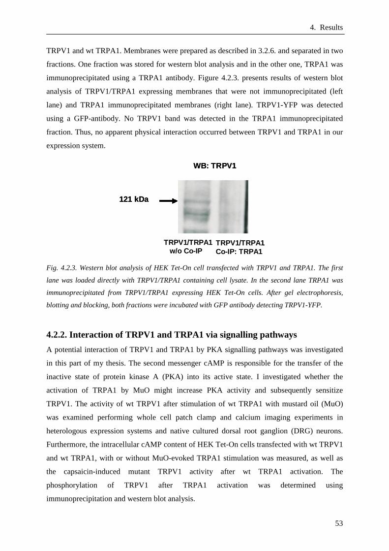
4. Results
53
TRPV1 and wt TRPA1. Membranes were prepared as described in 3.2.6. and separated in two
fractions. One fraction was stored for western blot analysis and in the other one, TRPA1 was
immunoprecipitated using a TRPA1 antibody. Figure 4.2.3. presents results of western blot
analysis of TRPV1/TRPA1 expressing membranes that were not immunoprecipitated (left
lane) and TRPA1 immunoprecipitated membranes (right lane). TRPV1-YFP was detected
using a GFP-antibody. No TRPV1 band was detected in the TRPA1 immunoprecipitated
fraction. Thus, no apparent physical interaction occurred between TRPV1 and TRPA1 in our
expression system.
Fig. 4.2.3. Western blot analysis of HEK Tet-On cell transfected with TRPV1 and TRPA1. The first
lane was loaded directly with TRPV1/TRPA1 containing cell lysate. In the second lane TRPA1 was
immunoprecipitated from TRPV1/TRPA1 expressing HEK Tet-On cells. After gel electrophoresis,
blotting and blocking, both fractions were incubated with GFP antibody detecting TRPV1-YFP.
4.2.2. Interaction of TRPV1 and TRPA1 via signalling pathways
A potential interaction of TRPV1 and TRPA1 by PKA signalling pathways was investigated
in this part of my thesis. The second messenger cAMP is responsible for the transfer of the
inactive state of protein kinase A (PKA) into its active state. I investigated whether the
activation of TRPA1 by MuO might increase PKA activity and subsequently sensitize
TRPV1. The activity of wt TRPV1 after stimulation of wt TRPA1 with mustard oil (MuO)
was examined performing whole cell patch clamp and calcium imaging experiments in
heterologous expression systems and native cultured dorsal root ganglion (DRG) neurons.
Furthermore, the intracellular cAMP content of HEK Tet-On cells transfected with wt TRPV1
and wt TRPA1, with or without MuO-evoked TRPA1 stimulation was measured, as well as
the capsaicin-induced mutant TRPV1 activity after wt TRPA1 activation. The
phosphorylation of TRPV1 after TRPA1 activation was determined using
immunoprecipitation and western blot analysis.
TRPV1/TRPA1w/o Co-IP
TRPV1/TRPA1Co-IP: TRPA1
WB: TRPV1
121 kDa
TRPV1/TRPA1w/o Co-IP
TRPV1/TRPA1Co-IP: TRPA1
WB: TRPV1
121 kDa
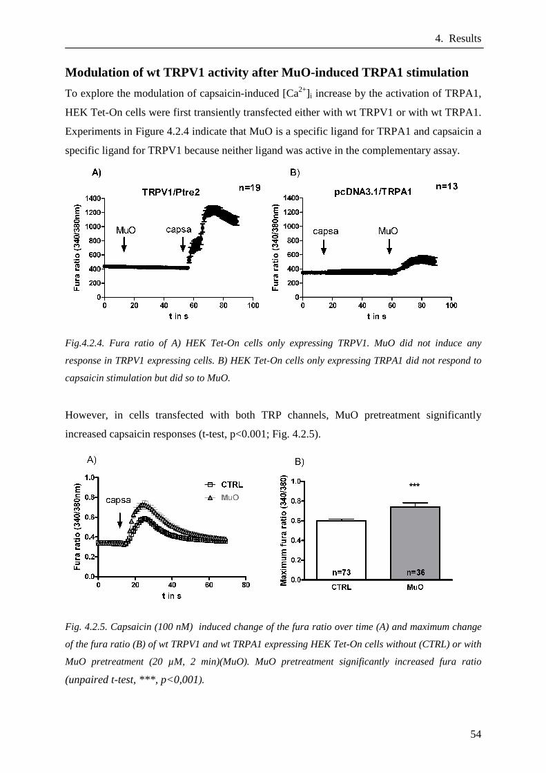
4. Results
54
Modulation of wt TRPV1 activity after MuO-induced T RPA1 stimulation
To explore the modulation of capsaicin-induced [Ca2+] i increase by the activation of TRPA1,
HEK Tet-On cells were first transiently transfected either with wt TRPV1 or with wt TRPA1.
Experiments in Figure 4.2.4 indicate that MuO is a specific ligand for TRPA1 and capsaicin a
specific ligand for TRPV1 because neither ligand was active in the complementary assay.
Fig.4.2.4. Fura ratio of A) HEK Tet-On cells only expressing TRPV1. MuO did not induce any
response in TRPV1 expressing cells. B) HEK Tet-On cells only expressing TRPA1 did not respond to
capsaicin stimulation but did so to MuO.
However, in cells transfected with both TRP channels, MuO pretreatment significantly
increased capsaicin responses (t-test, p<0.001; Fig. 4.2.5).
Fig. 4.2.5. Capsaicin (100 nM) induced change of the fura ratio over time (A) and maximum change
of the fura ratio (B) of wt TRPV1 and wt TRPA1 expressing HEK Tet-On cells without (CTRL) or with
MuO pretreatment (20 µM, 2 min)(MuO). MuO pretreatment significantly increased fura ratio
(unpaired t-test, ***, p<0,001).
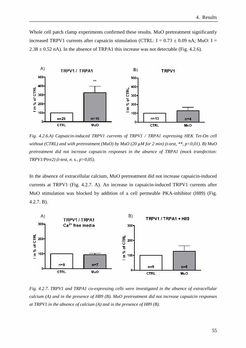
4. Results
55
Whole cell patch clamp experiments confirmed these results. MuO pretreatment significantly
increased TRPV1 currents after capsaicin stimulation (CTRL: I = 0.73 ± 0.09 nA; MuO: I =
2.38 ± 0.52 nA). In the absence of TRPA1 this increase was not detectable (Fig. 4.2.6).
Fig. 4.2.6.A) Capsaicin-induced TRPV1 currents of TRPV1 / TRPA1 expressing HEK Tet-On cell
without (CTRL) and with pretreatment (MuO) by MuO (20 µM for 2 min) (t-test, **, p<0,01). B) MuO
pretreatment did not increase capsaicin responses in the absence of TRPA1 (mock transfection:
TRPV1/Ptre2) (t-test, n. s., p>0,05).
In the absence of extracellular calcium, MuO pretreatment did not increase capsaicin-induced
currents at TRPV1 (Fig. 4.2.7. A). An increase in capsaicin-induced TRPV1 currents after
MuO stimulation was blocked by addition of a cell permeable PKA-inhibitor (H89) (Fig.
4.2.7. B).
Fig. 4.2.7. TRPV1 and TRPA1 co-expressing cells were investigated in the absence of extracellular
calcium (A) and in the presence of H89 (B). MuO pretreatment did not increase capsaicin responses
at TRPV1 in the absence of calcium (A) and in the presence of H89 (B).

4. Results
56
Change of the intracellular cAMP concentration after TRPA1 activation
The amount of cAMP was measured after MuO stimulation using an enzyme linked
immunosorbant assay (ELISA). The accumulation of cAMP was normalized to cells without
MuO pretreatment. cAMP increased significantly after MuO pretreatment. However, this
increase was not detectable in the absence of calcium or TRPA1 (Fig. 4.2.8.).
Fig. 4.2.8. Increase of cAMP in TRPV1/TRPA1 and TRPV1/Ptre2 expressing cells after MuO
pretreatment. cAMP increased significantly after MuO stimulation, however, not without extracellular
calcium or TRPA1 (1way ANOVA, Dunnet post hoc test, *, P<0.05).
Phosphorylation of TRPV1
An increase in cAMP, as shown in hypothesis 1, increases PKA activity and subsequently
sensitizes TRPV1 via specific PKA phosphorylation sites. Therefore, phosphorylation of
TRPV1 was investigated using immunoprecipitation and western blot analyses. In the
presence and absence of MuO treatment, cultured and transfected cells were lysed and
immunoprecipitated with an antibody directed against TRPV1. Western blots were incubated
with antibodies directed against phosphorylated serine and threonine residues. Band intensity
of MuO pretreated cells incubated with the Phosphoserine antibody showed an increase
compared to control cells (Fig. 4.2.9 A). Band intensity of MuO pretreated cells incubated
with the Phosphothreonine antibody increased significantly compared to control cells
(unpaired t-test, p<0.01, n=3) (Fig. 4.2.9 B) indicating a stronger phosphorylation of TRPV1.
4. Results
57
Fig.4.2.9. Band intensities of TRPV1 immunoprecipitated TRPV1/TRPA1 expressing cells without
(CTRL) and with MuO pretreatment (MuO) using antibodies against phosphorylated serine (A) and
threonine (B) residues.
Modulation of mutant TRPV1 activity after MuO pretr eatment
To further delineate the TRPV1 amino acid residues involved, the capsaicin-induced activity
of mutant TRPV1 was investigated using calcium imaging and whole cell patch clamp
experiments. HEK Tet-On cells were transiently co-transfected with mutant S116A-, T144A-,
S502A- or S774A-TRPV1 and wild type TRPA1. The capsaicin-induced increase of
intracellular calcium, indicated by the maximum increase of the fura ratio, is shown in Fig.
4.2.10. MuO pretreatment significantly increased capsaicin-induced TRPV1 activity in
T144A and S502A mutants but not in S116A and S774A mutants (Fig. 4.2.10).
Fig. 4.2.10. Capsaicin-induced maximum fura ratio in % of CTRL of cells transfected with mutant
TRPV1 (S116A (A), T144A (B), S502A (C) and S774A (D) and wt TRPA1) after MuO
pretreatment.Unpaired t-test, n. s., p>0.05 (A and D) and ***, p<0.001 (B and C).
4. Results
58
In whole cell patch clamp experiments, MuO pretreatment increased capsaicin-induced
TRPV1 activity significantly in T144A mutants but not in S116A, S502 and S774A mutants
(Fig. 5.2.11).
Fig. 4.2.11.Capsaicin-induced TRPV1 currents in cells co-transfected with mutant TRPV1 (S116A (A),
T144A (B), S502A (C) and S774A (D)) and wt TRPA1. MuO pretreatment significantly increased
capsaicin-induced TRPV1 activity in T144A mutants (unpaired t-test, ***, P<0.001), however not in
S116A, S502A and S774A mutants.
Modulation of TRPV1 activity after MuO pretreatment in native sensory
neurons
Whole cell patch clamp experiments were performed to determine the relevance of these
TRPV1 sensitizing mechanisms in cells endogenously expressing TRPV1 and TRPA1.
Without MuO pretreatment, 100 nM capsaicin induced a mean TRPV1 current of I = -0.43 ±
0.15 nA. MuO pretreatment significantly increased capsaicin-induced TRPV1 activity (I = -
1.28 ± 0.4 nA) (unpaired t-test, *, p<0.05). This effect was blocked by removal of
extracellular calcium or addition of the PKA inhibitor H89 (Fig. 4.2.12).
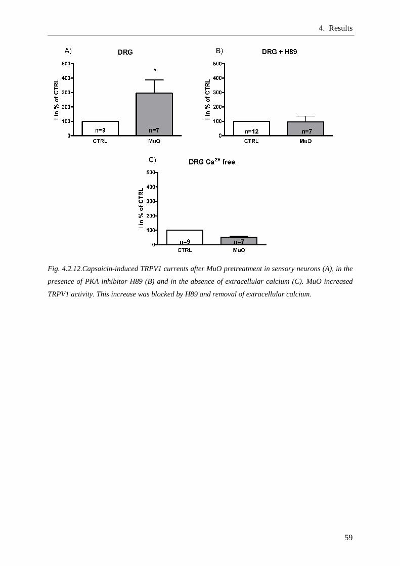
4. Results
59
Fig. 4.2.12.Capsaicin-induced TRPV1 currents after MuO pretreatment in sensory neurons (A), in the
presence of PKA inhibitor H89 (B) and in the absence of extracellular calcium (C). MuO increased
TRPV1 activity. This increase was blocked by H89 and removal of extracellular calcium.

5. Discussion
60
5. Discussion
The TRPV1 channel plays a pivotal role in the excitation of peripheral sensory neurons during
painful injury and inflammation of peripheral tissues. In the present studies I investigated two
different scenarios leading to the sensitization of this channel: opioid withdrawal and
interaction with TRPA1. The main findings were:
Hypothesis 1: Opioid withdrawal sensitizes TRPV1 in a
cAMP/PKA dependent manner.
1. I found a significant increase of capsaicin-induced TRPV1 activity during opioid
withdrawal.
2. Opioid withdrawal did not change the expression of TRPV1.
3. Phosphorylation of serine and threonine residues at TRPV1 was enhanced during opioid
withdrawal. Phosphorylation sites include threonine 144 and serine 774 but not serine 116
and serine 502.
4. Inhibition of adenylylcyclases and downregulation of isoform 3, but not 5, reversed the
enhanced TRPV1 activity during opioid withdrawal.
5. Both opioid withdrawal- and capsaicin-induced hyperalgesia were apparently mediated by
TRPV1 and PKA in vivo.
Hypothesis 2: TRPA1 stimulation sensitizes TRPV1 in a
cAMP/PKA dependent manner.
6. TRPA1 activation increased capsaicin-induced TRPV1 activity in a cAMP/PKA
dependent manner.
7. An increase in cAMP/PKA after TRPA1 stimulation resulted in an increase in TRPV1
phosphorylation.
8. Phosphorylation sites at TRPV1 include serine 116 and serine 774 but not threonine 144.
9. Similar mechanisms underly TRPV1 sensitization in transfected HEK cells and in native
sensory neurons.
5. Discussion
61
5.1. Hypothesis 1: Opioid withdrawal sensitizes TRPV1 in a
cAMP/PKA dependent manner.
5.1.1. Increased TRPV1 activity during opioid withdrawal
In the first part of my thesis, I was able to show a significant increase in capsaicin induced
TRPV1 activity during opioid withdrawal using whole cell patch clamp and calcium imaging
experiments. We identified enhanced phosphorylation of TRPV1 during opioid withdrawal
using immunoprecipitation and western blot analysis, and detected a crucial role for PKA
phosphorylation of residues threonine 144 and serine 774 at TRPV1. Out of 9 known
adenylylcyclase isoforms I found isoform 3 to be the most important in our expression
system. Behavioural experiments revealed the physiological relevance of TRPV1 sensitization
during opioid withdrawal in vivo.
Administration of opioids is common for treatment of moderate to severe pain. Paradoxically,
the chronic intake of opioids may lead to a phenomenon termed “opioid induced hyperalgesia
(OIH)”. However, this term has caused confusion in the literature. What has mostly been
described is, in fact, a long known phenonemon in animals (Celerier et al. 2000; Laulin et al.
1998; Mao et al. 2002a, 2002b), volunteers (Angst and Clark 2006; Angst et al. 2003;
Compton et al. 2003; Hood et al. 2003; Koppert et al. 2003) and patients (Davis et al. 2007;
Joly et al. 2005; Singla et al. 2007) occurring during withdrawal from opioids. The earliest
reports appeared during the nineteenth century. It was recognized that a potent analgesic such
as morphine could actually cause an enhancement of pain (Rossbach 1880). This was
replicated in numerous pre-clinical and clinical studies (Baron and McDonald 2006; Celerier
et al. 2001; Celerier et al. 2000; Compton et al. 2001; Guignard et al. 2000; Mao 2002; Mao et
al. 1995). Although the precise molecular mechanisms are not yet understood, hyperalgesia is
generally thought to result from neuroplastic changes in the peripheral and central nervous
systems that lead to sensitization of pronociceptive pathways. Examples are changes of the
central glutaminergic system, increased levels of spinal dynorphin, elevated release of
excitatory neuropeptides such as CGRP from primary afferents and activity of a subset of
neurons within the rostral ventromedial medulla that facilitate spinal nociceptive processing
(Chu et al. 2008; Gardell et al. 2002; Mao et al. 1994; Mao et al. 2002a; Morgan et al. 1992;
Narita et al. 2001; Zeitz et al. 2001).
Opioid induced hyperalgesia is still discussed controversially. So far pain hypersensitivity
was only measured either in the presence of extraordinarily high opioid concentrations or
5. Discussion
62
after removal of opioids (opioid withdrawal) (Davis et al. 2007; Fishbain et al. 2009; Pud et
al. 2006; Singla et al. 2007). We found hyperalgesia in behavioural animal experiments only
during opioid withdrawal, not in the presence of an opioid. Therefore, in the current thesis this
phenonemon is termed opioid withdrawal-induced hyperalgesia (OWIH).
Abrupt termination of chronic opioid use produces several intense withdrawal syndromes in
animal models and humans. Besides hyperalgesia, withdrawal syndroms include tachycardia,
hypertension, nausea, vomiting, hyperthermia, diarrhoea, piloerection and dysphoria. In
extreme cases some of them can be life-threatening.
Potential mechanisms of OWIH include an upregulation of PKC and subsequent increased
activity of NMDA-receptors (Mao et al. 1994) or release of spinal dynorphin, which can
cause the activation of pronociceptive signalling pathways (Vanderah et al. 2000) as well as
phosphorylation and sensitization of α2-adrenergic and adenosine receptors (Aley et al. 1995;
Aley and Levine 1997c). Opioid withdrawal might also reveal a compensatory upregulation
of AC activity (Nestler 1992; Sharma et al. 1975). Whereas acute opioid exposure inhibits AC
activity resulting in a reduction of intracellular cAMP, repeated or prolonged opioid exposure
leads to a compensatory upregulation of the cAMP pathway, including increased
concentrations of ACs and PKA. Although this has not been shown so far, it is hypothesized
that an increased PKA activity might counteract the inhibitory effect of acute opioid
application or might increase pain sensitivity. A potential mechanism to explain enhanced
cAMP levels after opioid withdrawal has been postulated by Chakrabarti and colleagues: a
shift of MOR-coupled signalling from predominantly Gαi-inhibitory to Gβγ-stimulatory might
mitigate the persistent inhibition of AC by chronic opioid treatment. These authors also
showed that chronic morphine augmented association of protein phosphatase 2A and Gαs,
resulting in a dephosphorylation of the endogenous phosphoprotein Gαs. Dephosphorylated
Gαs increases the association of Gs with MOR leading to an enhanced availability of Gβγ,
which in turn activates Gβγ sensitive AC isoform resulting in an elevated cAMP synthesis and
subsequent phosphorylation of proteins via PKA (Chakrabarti and Gintzler 2007; Gintzler and
Chakrabarti 2006).
Up-regulation of the cAMP pathway effects cell signalling in several ways for example the
transcription factor cAMP response element binding protein (CREB). Many genes consists of
consensus cAMP response element (CRE) sites in their promotors to which CREB dimers
bind, including neuropeptides, neurotransmitter synthesizing enzymes, neurotransmitter
receptors, signalling proteins, and other transcription factors (Lonze and Ginty 2002; Mayr
and Montminy 2001; Chao and Nestler 2004).
5. Discussion
63
The response to repeated morphine and its withdrawal was extensively investigated in the
locus coeruleus (LC) (Nestler 2001). Upon chronic opioid administration and withdrawal,
CREB expression was increased in the LC, resulting in an enhanced expression of AC 8 and
tyrosine hydroxylase (Lane-Ladd et al. 1997; Chao et al. 2002). Tyrosine hydroxylase
catalyzes the conversion of L-tyrosine to dihydroxyphenylalanine (DOPA), a precursor for
dopamine, which is in turn a precursor for noradrenaline and adrenaline (Kaufman 1995).
Another, opioid withdrawal affected and investigated area in the brain is the nucleus
accumbens (NA), which is thought to play an important role in the reward system. Up-
regulation of cAMP and CREB in this region may also contribute to states of dysphoria seen
in early withdrawal (Hyman and Malenka 2001; Nestler 2001), because a target of CREB in
the NA is dynorphin, whose release is contributed to dysphoria through a negative feedback
loop to ventral tegmental area dopamine neurons (Hyman and Malenka 2001; Shippenberg
and Rea 1997).
Immediate early genes, which encode transcription factors, also play a role in addiction and
opioid withdrawal. The Fos family include ∆FosB, which acts as either a transcriptional
inducer or repressor (Chao and Nestler 2004). ∆FosB isoforms are very stable and their levels
gradually accumulate with repeated drug exposure and persist for weeks after the drug is
withdrawn (Andersson et al. 2003; Chen et al. 1997; Kelz and Nestler 2000; Moratalla et al.
1996). Overexpression of ∆FosB using transgenic mice showed increased AMPA glutamate
receptor subunit GluR2 expression (Kelz et al. 1999).
Despite the abundant information of central neuronal modulations during opioid withdrawal,
very little is known about modulations in the peripheral nervous system. Since we know that
OWIH is associated with different forms of hyperalgesia (e.g. burning sensation), that TRPV1
can mediate hyperalgesia and that TRPV1 is a target for cAMP/PKA mediated
phosphorylation (Bhave et al. 2002; Mohapatra and Nau 2003; Rathee et al. 2002; Vetter et al.
2008), we postulated an increased TRPV1 activity during opioid withdrawal.
5.1.2. TRPV1 expression and opioid withdrawal
Reasons for increased TRPV1 activity during opioid withdrawal might include enhanced
expression of the protein on the membrane surface, rapid translocation of inactive monomers
to the membrane or sensitization of the channel via phosphorylation. Chen et al. found
increased TRPV1 mRNA levels in the spinal cord and sciatic nerve in rats after prolonged
treatment with morphine. However, total TRPV1 protein levels were unaltered (Chen et al.
2008). This was similar in my experiments. TRPV1 binding sites expressed on cell
5. Discussion
64
membranes did not change during opioid withdrawal. However, Chen and others
demonstrated that chronic morphine treatment can result in an increase in the phosphorylation
of p38, ERK and JNK in DRG neurons (Cui et al. 2006; Ma et al. 2001) which can
subsequently regulate gene products such as c-fos, brain derived neurotrophic factor (BDNF),
neurokinin 1, and CGRP through phosphorylation of CREB (Lonze and Ginty 2002;
McClung and Nestler 2003). Thus, an upregulation of TRPV1 gene products without an
increase in TRPV1 protein expression at the cell surface is still conceivable. Future
experiments should exclude the possibility that intracellular stores of inactive TRPV1
monomers might increase during chronic opioid therapy. However, as our experiments have
shown, opioid withdrawal seems to not increase the number of functionally active TRPV1
channels at the cell membrane.
5.1.3. Increased phosphorylation of TRPV1 during opioid withdrawal
Sensitization of ion channels can occur rapidly by posttranslational regulation via
phosphorylation (Woolf and Costigan 1999). TRPV1 is known to have multiple
phosphorylation sites for several protein kinases including PKA, PKC, CaMKII, Src kinase
and Cyclin-dependent kinase 5 (Cdk5). Potential target sites of Src kinase and Cdk5 are Y200
and T407, respectively (Pareek et al. 2007; Zhang et al. 2005). CAMKII is reportedly
involved in TRPV1 phosphorylation at S502 and T704 (Jung et al. 2004). PKC is implicated
in phosphorylation of T370, S502, T704 and S800, and PKA in phosphorylation of S6, S116,
T144, S502, S774 and S820 (Bhave and Gereau 2004; Bhave et al. 2002).
In this study, I concentrated on PKA phosphorylation sites because the cAMP/PKA pathway
is known to be upregulated during prolonged opioid treatment. I was able to identify
enhanced phosphorylation of serine and threonine residues of TRPV1 using
immunoprecipitation and western blot analysis during opioid withdrawal. These findings are
consistent with studies exploring 32P-orthophosphate incorporation by TRPV1 following pre-
incubation with 8-Br-cAMP (a membrane-permeable activator of PKA) (Jeske et al. 2008)
and in vivo phosphorylation (Lee et al. 2005). This and another group identified the
scaffolding protein A-kinase Ankyrin Protein 150 (AKAP150) as indispensable for PKA
translocation to TRPV1 on the plasma membrane, resulting in phosphorylation and
sensitization of TRPV1 (Jeske et al. 2008; Schnizler et al. 2008). They also found that
AKAP150 co-localized with regulatory subunit II of PKA (RII) and TRPV1, consistent with a
mechanism first proposed in 2002 by Rathee and colleagues, who demonstrated that
administration of AKAP-inhibitors attenuated FSK-stimulated translocation of the PKA
5. Discussion
65
catalytic subunit to the plasma membrane in DRG neurons (Jeske et al. 2008; Rathee et al.
2002). Thus, my findings are consistent with the notion that prolonged opioid treatment
upregulates cAMP/PKA and subsequent PKA-mediated phosphorylation of TRPV1 at the cell
membrane.
5.1.4. Mutation of threonine 144 and serine 774, but not serine 116 and
serine 502, resulted in a loss of increased TRPV1 activity during opioid
withdrawal
So far, posttranslational modulations of TRPV1 resulting in sensitization were mainly
attributed to phosphorylation by PKC (Bhave et al. 2003; Cesare and McNaughton 1996;
Crandall et al. 2002; Liang et al. 2001; Numazaki et al. 2002; Sugiura et al. 2002; Vellani et
al. 2001), while phosphorylation by PKA was thought to underly prevention or reduction of
TRPV1 desensitization. Capsaicin-induced TRPV1 currents exhibit complex desensitization
patterns with several kinetic components. Acute desensitization occurs during sustained
capsaicin application, whereas decreasing currents with multiple capsaicin applications have
been termed tachyphylaxis (Bhave et al. 2002; Koplas et al. 1997; Liu and Simon 1996). It
was postulated that prevention of acute desensitization is accomplished through
phosphorylation by CamKII and hindrance of tachyphylaxis by PKA (Bhave et al. 2002; Jung
et al. 2004; Mohapatra and Nau 2003). Additionally, phosphorylation at serine 116 was
mentioned as the major event preventing tachyphylaxis. However, Mohapatra and colleagues
also identified threonine 370 as an important site regarding prevention of tachyphylaxis,
which was inconsistent with findings of Bhave and colleagues (Bhave et al. 2002; Mohapatra
and Nau 2003).
Although, phosphorylation of TRPV1 is usually linked with prevention of desensitization,
several studies also described a PKA mediated sensitization of TRPV1 at different
phosphorylation sites such as threonine 144, threonine 370 and serine 502 (Lopshire and
Nicol 1998; Rathee et al. 2002; Pitchford and Levine 1991). Since we found different
phosphorylation residues of TRPV1 during opioid withdrawal, we investigated mutations
where serine 116, threonine 144, serine 502 and serine 774 were replaced by alanins. We
chose S116, T144 and S502 because numerous studies provided evidence that these are
functional PKA phosphorylation sites. T144 and S502 are thought to be involved in
potentiation of heat currents and S116 in prevention of tachyphylaxis. Since less is known
about the functional role of S774 we also decided to include it in our investigations.
5. Discussion
66
Using whole cell patch clamp and calcium imaging experiments, we found an impaired
TRPV1 activity in TRPV1 mutants T144A and S774A during opioid withdrawal compared to
wild type TRPV1. This indicates that both PKA phosphorylation sites are involved in
sensitization during opioid withdrawal. However, it does not exclude the possibility that
additional phosphorylation sites, e.g. T370 or T704, play a role as well. The involvement of
T144, T370 and S502 in PKA mediated sensitization was shown by Rathee and colleagues
(Rathee et al. 2002). We were unable to show a functional role of S502 in our assay. This
might be related to different isoforms of PKA activated by opioid withdrawal. Another
explanation might be that Rathee et al. replaced S502 not only by alanine but also by
aspartate, which represents a constitutively phosphorylated form of TRPV1. Additionally,
they used heat as TRPV1 activator. Other studies reported phosphorylation of S502 by PKC
and CaMKII (Bhave et al. 2003; Jung et al. 2004) or detected T144 and T370 as target
residues for calcineurin, a phosphatase responsible for dephosphorylation of TRPV1 (Jeske et
al. 2006).
We also found a functional role for S774 during opioid withdrawal. This is the first
demonstration of involvement of S774 in PKA-mediated sensitization of TRPV1, albeit other
studies described its role in prevention of tachyphylaxis and acute desensitization (Bhave et
al. 2002; Mohapatra and Nau 2003). So far, phosphorylation of S116 by PKA was not
reported to contribute to TRPV1 sensitization. Consistently, we found enhanced capsaicin-
evoked S116A TRPV1 activity during opioid withdrawal, suggesting that S116 is not directly
involved in TRPV1 sensitization.
In summary, TRPV1 is apparently phosphorylated under basal conditions
(autophosphorylation) and dephosphorylated via calcium dependent activation of calcineurin
upon activation. Rephosphorylation of TRPV1 by PKA, PKC and CaMKII not only prevents
desensitization, but also sensitizes TRPV1 and reduces its activation thresholds (Lee et al.
2005). We identified PKA phosphorylation sites T144 and S774 as important targets
mediating the sensitization of TRPV1 during opioid withdrawal.
5.1.5. Downregulation of adenylylcyclase isoform 3, but not 5, reverses the
enhanced TRPV1 activity during opioid withdrawal
Nine AC isoforms have been described and are classified into 4 groups. Group 1 (AC 1, 3 and
8) are stimulated by calmodulin in a calcium-dependent manner. Group 2 (AC 2, 4 and 7) are
conditionally stimulated by Gβγ-subunits and activated by PKC. Group 3 (AC 5 and 6) are
stimulated by Gαs and inhibited by Gαi-subunits, PKA and calcium. Group 4 (AC 9) is
5. Discussion
67
relatively insensitive to FSK (Hacker et al. 1998) and inhibited by calcineurin and PKC
(Cumbay and Watts 2004; Paterson et al. 2000).
Using the the nonspecific AC inhibitor SQ 22,356 we were able to block an increase in
capsaicin-induced TRPV1 activity during opioid withdrawal. Previous experiments in our
laboratory have shown that mRNA transcripts of AC 3 and 5 are significantly up-regulated
during opioid withdrawal (Endres-Becker 2007). In calcium imaging experiments, increased
TRPV1 activity during opioid withdrawal was prevented by down-regulating AC 3 but not
AC 5. This may be due to a lower expression of isoform 5 during opioid withdrawal. Hence,
AC 3 might play a more prominent role than AC 5 in the sensitization of TRPV1 during
opioid withdrawal. Indeed, in our prior experiments mRNA levels of AC 3 were 4-fold
increased compared to a 1.7-fold increase of AC 5 transcripts. Expression levels of isoforms
2, 6 and 9 were unchanged and AC 1, 4, 7 and 8 were not detectable (Endres-Becker 2007).
Emerging evidence suggests that cAMP superactivation can be caused by enhanced Gαs-
receptor coupling, G-protein dissociation and Gαs-AC interactions (Chakrabarti and Gintzler
2007; Shy et al. 2008; Watts and Neve 2005). However, the molecular mechanisms still
remain unclear. Recent evidence suggests that signalling via G-proteins can be regulated by
receptor independent accessory proteins (Takesono et al. 1999). A member of this protein
class is an activator of G-protein signalling 3 (AGS3). AGS3 binds to Gαi-GDP, enhances
unbound free Gβγ stimulation of AC 2 and 4, and/or diminishes Gαi-GTP inhibition of AC
(Blumer et al. 2007; De Vries et al. 2000; Kimple et al. 2002; Takesono et al. 1999; Yao et al.
2005). Knockdown of AGS3 expression or inhibition of Gβγ resulted in a blockade of
morphine-induced cAMP/PKA signalling in primary nucleus accumbens/striatal neurons (Yao
et al. 2005).
A possible shift from inhibitory to stimulatory G-proteins during opioid withdrawal can also
result in an activation of the PLC/PKC pathway. Several AC isoforms including AC 3 (weak)
and 5 are positively regulated by PKC (Watts and Neve 2005). Furthermore, PKC is known to
activate voltage-dependent calcium channels, leading to an augmentation of intracellular
calcium which may activate calcium-sensitive AC such as AC 3 but is known to inhibit AC 5.
PKA activity is able to enhance PKCε activity via PLC and its anchoring protein εRACK
(Yao et al. 2008), promoting PKCε stimulation of AC 5 and AC 7, resulting in a positive
feedback in cAMP production.
In contrast to our results, a prevention of withdrawal-induced cAMP-superactivation was
shown using siRNA against AC 5 (Fan et al. 2009). However, we did not directly investigate
cAMP-superactivation but TRPV1 activity as a downstream effect. Moreover, Fan and

5. Discussion
68
colleagues investigated nucleus accumbens/striatal neurons, where different AC isoforms are
expressed. In other cell types AC 1, 5, 6 and 8 (Avidor-Reiss et al. 1997; Nevo et al. 1998),
AC 6 and 7 (Bie et al. 2005) or AC 4 and 7 (Rivera and Gintzler 1998) were upregulated
during prolonged opioid treatment or withdrawal. Thus, expression and regulation of AC
isoforms seem to strongly depend on the expression system (tissue type, cell line, species),
duration of treatment and opioid ligand used (Defer et al. 2000; Hanoune and Defer 2001).
5.1.6. Nociceptive behaviour during opioid withdrawal
To delineate the physiological relevance of TRPV1 sensitization during opioid withdrawal in
vivo, we investigated the paw withdrawal latency (PWL) evoked by a radiant heat source, and
capsaicin-induced nocifensive behaviour in rats. We measured a significantly decreased PWL
during opioid withdrawal (thermal hyperalgesia), which was prevented in a concentration
dependent manner by the MOR antagonist naloxone (NLX), the PKA inhibitor H89 and the
TRPV1 antagonist capsazepine. Furthermore, opioid withdrawal significantly enhanced and
H89 abolished capsaicin-induced flinching and protecting behaviour. Thus, consistent with
my in vitro studies, opioid withdrawal apparently induced TRPV1-mediated hyperalgesia via
PKA activation in vivo.
Numerous clinical and experimental studies identified that the development and magnitude of
opioid withdrawal induced hyperalgesia differ for different types of pain, including the
development of thermal hyperalgesia (reviewed in Angst et al. (Angst and Clark 2006)).
Lipman and colleagues observed that study patients undergoing withdrawal had a
significantly lower tolerance to heat-evoked pain (outside the 95% confidence interval)
compared with healthy volunteers (Lipman and Blumenkopf 1989). In the early seventies
several animal studies followed underlining the formation of thermal hyperalgesia during
opioid withdrawal which is line with results of our behavioural experiments however the
involvement of peripheral TRPV1 was not investigated until now (Kayan et al. 1971; Mao et
al. 1994; Tilson et al. 1973; VonVoigtlander and Lewis 1983). Mao et al. carried out one of
the first and most complete studies investigating the role of spinal cord in the genesis of
thermal hyperalgesia during opioid withdrawal, which involve both NMDA/non-NMDA
receptors and PKC translocation/activation (Mao et al. 1994). Animal studies exploring the
development of hyperalgesia during opioid withdrawal in the periphery delineate the
development of mechanical hyperalgesia, which was also dependent on PKC activity as well
as the activation of guanosine triphosphate binding proteins (Aley et al. 1995; Aley and
Levine 1997a, 1997b, 1997c; Arts et al. 1991; Khasar et al. 1995).
5. Discussion
69
In my docotoral thesis I could show that the thermo-sensitive unselective cation channel
TRPV1 might play an important role in the development of thermal hyperalgesia during
opioid withdrawal. These findings might play a role in the future in the development of new
therapeutic strategies to prevent or to treat opioid withdrawal induced hyperalgesia using e.g.
specific antagonists against TRPV1.
5. Discussion
70
5.2. Hypothesis 2: TRPA1 stimulation modulates TRPV1 activity
in a cAMP/PKA dependent manner
Approximately 30-50 % TRPV1 expressing small to medium sized peripheral sensory
neurons co-express TRPA1 and almost all TRPA1 positive neurons co-express TRPV1
(Hjerling-Leffler et al. 2007; Kobayashi et al. 2005; Linte et al. 2007; Story et al. 2003).
Furthermore, currents induced by the TRPA1 agonists MuO and WIN55,212 were almost
exclusively detected in TRPV1 positive cells (Diogenes et al. 2007; Jordt et al. 2004; Story et
al. 2003) and both channels are activated by compounds which cause a pungent burning
sensation. Thus, I hypothesized that TRPV1 sensitization can result from physical and/or
functional interactions of both channels.
Numerous studies identified physical interactions of different members of the TRP ion
channel family through formation of heteromultimeric complexes, whereas the most
complexes are made up of monomers from different members of the same TRP channel
subfamily (Bai et al. 2008; Dietrich et al. 2005; Hellwig et al. 2005; Park et al. 2008;
Schilling and Goel 2004). However, there is recent evidence that members of different TRP
channel subfamilies are able to form heteromultimeric complexes forming a new channel with
new biophysical properties and modes of activation distinct from that of the individual
channel (Bai et al. 2008; Park et al. 2008).
Additionally, there is the possibility of interactions between TRPV1 and TRPA1 via
signalling pathways. Several studies were published with some inconsistent results. One
group identified inhibition of TRPV1 activity by TRPA1 stimulation via calcineurin-mediated
desensitization of TRPV1 (Akopian et al. 2007; Jeske et al. 2006). Calcium-dependent
sensitization of TRPV1 can also occur through activation of CAMKII and PKC (Jung et al.
2004; Mandadi et al. 2004) or via activation of calcium sensitive ACs and subsequent PKA
activation. Anand and colleagues detected enhanced capsaicin responses after pre-incubation
with low-concentrations of the TRPA1 agonist cinnamaldehyde (CA) at 225 µM which is
consistant to my results but not at higher concentrations (Anand et al. 2008).
5.2.1. TRPA1 stimulation does not alter the expression of TRPV1
Akopian and colleagues showed that TRPA1 desensitization is influenced by TRPV1 co-
expression in sensory neurons and transfected CHO cells (Akopian et al. 2007). They
explained this phenomenon with a stabilizing effect of TRPV1 on the membrane expression
5. Discussion
71
of TRPA1. Internalization of membrane-bound channels can be suppressed by interactions
with other proteins, including subunits of the channels (Bernstein and Jones 2007). Another
possible mechanism for the modulation of functional TRPV1 tetramers is the cAMP-
dependent release and translocation of TRPV1 monomers from intracellular pools to the cell
membrane (Vetter et al. 2008). Given that TRPA1 activation can stimulate calcium-sensitive
ACs and intracellular concentration of cAMP, we examined TRPV1 membrane expression
after TRPA1 stimulation of co-transfected HEK293 cells. However, neither the affinity nor
the number of TRPV1 binding sites changed during TRPA1 activation. These experiments
indicate that TRPV1 is not recruited from intracellular pools to the cell membrane or
internalized in the presence of a TRPA1 agonist in our assay.
5.2.2. TRPV1 and TRPA1 do not form complexes in transfected HEK Tet-
On cells
One of the most interesting properties of the TRP ion channel family is the ability to form
heteromultimeric complexes. Recently, heteromeric complexes between members of different
TRP channel subfamilies were identified (Bai et al. 2008; Kobori et al. 2009; Park et al. 2008;
Zhang et al. 2009). These heteromeric complexes form a channel with unique and new
biophysical properties
A heteromeric formation between members of the same TRP channel subfamily was shown
for the TRPV subfamily. Heteromeric channel formation have been proposed for TRPV1,
TRPV3, TRPV5 and TRPV6 channel subunits (Hoenderop et al. 2003; Kedei et al. 2001;
Smith et al. 2002). A study investigating homo- and heteromeric assembly of HEK 293 cells
transfected with TRPV1-6 channels identified heteromeric formation of TRPV1 and TRPV2
subunits and TRPV5 and TRPV6, using Fluorescent Resonance Energy Transfer (FRET) and
co-immunoprecipitation (Hellwig et al. 2005). However, heteromeric assembly of TRPV1 and
TRPV2 seem to be without physiological relevance, since both channels are expressed in
different tissues and cell types (Birder et al. 2002; Caterina et al. 1999; Caterina et al. 1997;
Tominaga et al. 1998). TRPV1 is also thought to form heteromeric complexes with TRPV3
monomers based on the observation that TRPV3 is transcribed from a gene adjacent to
TRPV1, is co-expressed in DRG neurons with TRPV1, co-precipitates with TRPV1 in
heterologous expression systems and may reduce TRPV1 responsiveness to capsaicin (Smith
et al. 2002). They also mentioned that except TRPV5 and TRPV6, TRPV channel subunit
preferentially form homomeric pore complexes (Hellwig et al. 2005). A very recent study
showed physical interaction of TRPV1 and TRPA1 subunits. These differing results might be
5. Discussion
72
caused by the distict TRPA1 origin because Staruschenko et al. used cDNA encoding for
mouse TRPA1 (Staruschenko et al. 2010).
One accepted method to prove biophysical interactions is co-immunoprecipitation. Using this
method we failed to detect potential heteromeric complexes between TRPV1 and TRPA1
monomers. Together with our binding affinity studies, which did not indicate differences in
the binding affinities if both proteins were expressed, we conclude that there is no direct
physical interaction between TRPV1 and TRPA1.
5.2.3. TRPA1 stimulation increases TRPV1 activity in a calcium and cAMP
dependent manner
Using calcium imaging and whole cell patch clamp experiments, we detected a significantly
increased TRPV1 activity after pre-stimulation of the cells with MuO. We hypothesized that
TRPA1 activation with MuO causes an influx of cations including calcium ions. This would
elevate the activity of calcium sensitive AC, resulting in an increase of cAMP and subsequent
PKA stimulation, to eventually phosphorylate and sensitize TRPV1. Indeed, the increased
TRPV1 activity was not apparent in the absence of TRPA1 and extracellular calcium, or when
PKA was inhibited by H89.
These findings were not in agreement with results of another group. They discovered that
TRPV1 (co-expressed with TRPA1 in CHO cells or natively expressed in trigeminal ganglion
neurons) was cross-desensitized through activation of TRPA1. In their studies TRPA1
activation resulted in a recruitment of the calcium-dependent calcineurin pathway, whereby
calcineurin mainly dephosphorylated threonine 144 and 370 at TRPV1, causing
desensitization of the channel (Akopian et al. 2007; Akopian et al. 2008; Jeske et al. 2006;
Ruparel et al. 2008; Patwardhan et al. 2006).
On the other hand, MuO is known to induce neurogenic inflammation resulting in
hypersensitivity to mechanical and thermal stimuli in vivo (Caterina et al. 2000).
Additionally, in vivo and in vitro studies showed thermal sensitization by an elevation of
intracellular calcium (Distler et al. 2003; Guenther et al. 1999; Kress and Guenther 1999). A
recent study measured enhanced capsaicin responses of rat DRG neurons pre-incubated with
low concentrations of cinnamaldehyde (CA) (225 µM), while high concentrations (450 µM
and 2mM) caused inhibition (Anand et al. 2008). These findings are in line with our results
showing that low concentrations of TRAP1 ligands increased TRPV1 activity. These effects
seem to be concentration-dependent and higher concentrations of TRPA1 agonists apparently
result in a switch from sensitizing PKA-mediated TRPV1 phosphorylation to desensitizing

5. Discussion
73
calcineurin-mediated dephosphorylation of TRPV1. Additionally, calcium-dependent
sensitization of TRPV1 can also occur through other kinases like CaMKII and PKC (Jung et
al. 2004; Mandadi et al. 2004). The balance between calcium-stimulated kinase and
phosphatase activities seems to result in a tightly regulated system responsible for modulating
TRPV1 activity. Thus, future studies will have to determine the crucial concentrations of
TRPA1 agonists and the duration of the agonist application eliciting this switch. These
findings are interesting for the clinical applications since highly concentrated TRPA1 agonists
might have antinociceptive effects while lower concentrations might increase pain.
The activation of TRPA1 by MuO leads to an increase of intracellular calcium since TRPA1
is a non-selective cation channel. Our laboratory has shown that calcium directly activates
TRPA1 through an intracellular EF-hand domain (Zurborg et al. 2007), possibly resulting in
an (auto)amplification of TRPA1 activity. We found that in the absence of extracellular
calcium, TRPA1 activation failed to induce TRPV1 sensitization. In line with our findings,
Distler and colleagues have shown that rat sensory neurons expressAC 1 and AC 8, which
were activated by an elevation of intracellular calcium leading to an increase of cAMP and
translocation of the catalytic PKA subunit. This subsequently increased TRPV1
phosphorylation (Distler et al. 2003). We also measured increased cAMP levels after MuO
pre-stimulation, which were abrogated in the absence of extracellular calcium and of TRPA1
(Fig. 4.2.8.).
5.2.4. TRPV1 is phosphorylated after TRPA1 stimulation
Using immunoprecipitation of TRPV1, we detected higher band intensities in MuO pre-
stimulated TRPV1/TRPA1 expressing cells utilizing phosphoserine and phosphothreonine
specific antibodies (Fig. 4.2.9.). This supports the hypothesis that TRPA1 activation can
increase TRPV1 phosphorylation and subsequently sensitize TRPV1. Studies of another
group showed contrary results. They identified dephosphorylated TRPV1 after TRPA1
stimulation with cannabinoids investigating sensory neurons and transfected CHO cells with
autoradiography and 32P-incorporation assays (Jeske et al. 2006; Patwardhan et al. 2006). The
differing results may be explained by the different expression systems and the longer duration
of TRPA1 stimulation (10 min versus 2 min in our experiments). The latter point may be
particularly important because the amount of calcium entering the cell is dependent on the
duration of TRPA1 channel activation. Thus, intracellular calcium may switch from
sensitizing to desensitizing concentrations. Other reasons may be the varying expression
levels of calcium sensitive AC isoforms in sensory neurons, CHO cells and HEK Tet-On
5. Discussion
74
cells. Future experiments will have to validate the presented phosphorylation results using
another method, for example radioimmunoassay, and the investigation of PKA inhibitors.
5.2.5. Mutation of TRPV1 phophorylation sites reversed the increased
TRPV1 activity after TRPA1 activation
To identify potential PKA phosphorylation sites at TRPV1 after TRPA1 stimulation we used
TRPV1 mutants S116A, T144A, S502A and S774A. We found an abrogation of the increased
TRPV1 activity in mutants S116A and S774A. Mutant T144A showed even stronger
capsaicin-induced currents after MuO stimulation compared to wild type TRPV1. An
explanation may be that this phosphorylation residue is an important target for calcineurin
such that its mutation negatively influences the dephosphorylation of the receptor. This was
already shown in a study delineating an important role for T144 regarding dephosphorylation
by phosphatases (Jeske et al. 2006).
Results of calcium imaging and whole cell patch clamp experiments in serine 502 mutants
were inconsistent in our study. I still measured an increased mutant TRPV1 activity after
MuO pre-treatment using calcium imaging experiments, which was not detectable in whole
cell patch experiments. A possible explanation may be the difference between the two
experimental procedures. Calcium imaging depicts the entry of calcium into the cytosol not
only from the extracellular space but also from intracellular stores. This is an indirect
indicator for the activity of the stimulated membrane channel and it does not exclude that
additional channels are involved in the calcium entry or that calcium is released from the
endoplasmatic reticulum (ER). In a recent study the stimulation of ectopic but functional
TRPV1 channels on the ER lead to an increase of cytosolic calcium (Gallego-Sandin et al.
2009). This issue needs to be clarified in further experiments in our assay. Whole cell patch
clamp experiments are a more direct method for the investigation of membrane channel
activities, since they directly record changes of currents after agonist administration.
Replacing serine 116 by alanine abolished the enhanced TRPV1 activity after MuO pre-
treatment in both calcium imaging and whole cell patch clamp experiments. This is in
accordance with previous studies that identified S116 as a fundamental PKA phosphorylation
site for PKA-dependent TRPV1 sensitization (Bhave et al. 2002; Mohapatra and Nau 2005).
To our knowledge an important role for serine 774 as PKA phosphorylation site in TRPV1
sensitization was shown here for the first time. Previous investigations using ESI mass
spectroscopy and tandem MS/MS sequencing delineated S774 as a PKA phosphorylation site,
5. Discussion
75
but did not assign functional effects completely consistent with phosphorylation (Bhave et al.
2002).
Our studies concerning PKA phosphorylation sites at TRPV1 may be extended by use of
phosphorylation assays or the incorporation of 32P in the future. Additionally, TRPV1 double
or triple mutants should be designed and investigated.
5.2.6. TRPA1 stimulation enhanced TRPV1 currents in native DRG
neurons in a calcium and PKA-dependent manner
To corroborate our results obtained in transfected cell lines, we decided to investigate natively
TRPA1 and TRPV1 expressing DRG neurons. We also found a significant increase of
capsaicin-induced TRPV1 activity after TRPA1 activation in DRG neurons. As mentioned
above, contrasting results were obtained by the group of Hargreaves, investigating cross-
desensitization of TRPA1 and TRPV1 in trigeminal sensory neurons pre-stimulated with
TRPA1 agonists (Akopian et al. 2007; Akopian et al. 2008; Jeske et al. 2006; Ruparel et al.
2008; Patwardhan et al. 2006). Again, the differing experimental procedures, AC expression
patterns and the tightly (calcium concentration dependent) balanced system concerning
calcium-dependent kinase-mediated sensitization or calcium-dependent calcineurin-mediated
desensitization of TRPV1 may account for the divergent results.
5.3. Limitations, future prospects and clinical relevance
My thesis examined the sensitization of TRPV1 in two independent settings: first during
opioid withdrawal and second via activation of TRPA1. Both scenarios share the same
intracellular signalling pathway, i.e. activation of AC, increased cAMP levels, subsequent
translocation and activation of PKA, and phosphorylation of TRPV1 at several residues (Figs.
5.1. and 5.2.). Thermal and inflammatory hyperalgesia also involves phosphorylation and
sensitization of TRPV1, e.g. PGE2 or PGI2 - and bradykinin-receptor signalling (Sugiura et al.
2002). Thus, modulation of TRPV1 via phosphorylation apparently play a central role in
sensitization, regardless of the triggering events.
My initial results were obtained in cell lines and were then confirmed in native neurons and in
vivo experiments. The latter indicate an important role of TRPV1 phosphorylation in the
enhanced pain sensitivity occurring during opioid withdrawal. The ultimate goal is to translate
these findings into clinical studies and to derive novel therapeutic approaches.
5. Discussion
76
Many in vivo experiments and clinical examinations of opioid addicts described the clinical
relevance of hyperalgesia associated with the use and withdrawal of opioids (reviewed in
(Chu et al. 2008). However, the experimental and/or clinical settings and methodologies were
very heterogeneous, e.g. investigations of (i) former opioid addicts on methadone
maintenance therapy (Compton 1994; Compton et al. 2001; Doverty et al. 2001; Schall et al.
1996), (ii) perioperative exposure to opioids in patients undergoing surgery (Chia et al. 1999;
Cooper et al. 1997; Guignard et al. 2000), (iii) healthy human volunteers after acute opioid
exposure using human experimental pain testing (Angst et al. 2003; Koppert et al. 2001;
Koppert et al. 2003; Troster et al. 2006; Chu et al. 2006), or (iv) prospective observational
studies in opioid-naïve pain patients undergoing initiation of chronic opioid therapy (Chu et
al. 2006). About 90 publications describe OWIH leading to thermal or mechanical
hyperalgesia in various animal models (Angst and Clark 2006). So far, most studies support
the notion that neural plasticity in the central nervous system is most important during opioid
withdrawal.
Some studies investigated peripheral mechanisms of OWIH/OIH. By using small peripherally
acting doses of the mu-opioid agonist DAMGO they observed the development of tolerance
and mechanical hyperalgesia. Adenosine A1 and A2 agonists lead to similar findings (Aley
and Levine 1997c). Later the involvement of PKC and ACs in modulating this phenomenon
was reported (Aley and Levine 1997a, 1997b) as well as the involvement of β2-adrenergic
receptors (Liang et al. 2006).
As already mentioned above the participation of NMDA and non-NMDA excitatory amino
acid receptors and also PKC was mostly contributed to mediate OWIH/OIH on the spinal
level, which is until now the main explanation for OWIH/OIH (Dunbar and Pulai 1998; Mao
et al. 1994). Due to these findings numerous studies were designed investigating the
pharmacologic modulation of OWIH/OIH targeting spinal NMDA receptors (reviewed in
(Chu et al. 2008).
My thesis provides a new mechanism underlying OWIH at the level of the peripheral nervous
system. These findings indicate that both centrally and peripherally acting opioids bear the
potential to induce hyperalgesia during withdrawal. Thus, even if new opioid drugs become
available which act peripherally avoiding central side effects, abrupt withdrawal of such drugs
must be avoided since we showed that peripheral TRPV1 receptors are sensitized and
contribute to thermal hyperalgesia (Fig. 5.1.). Importantly, however, my studies have not
considered the influence of an inflammatory environment, which is a major determinant for
the efficacy of peripherally acting opioids (Zollner and Stein 2007). Further studies need to
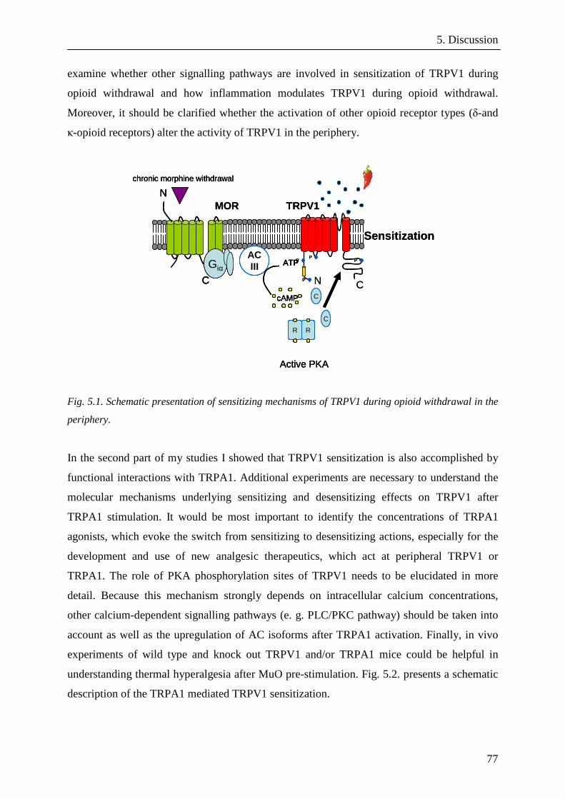
5. Discussion
77
examine whether other signalling pathways are involved in sensitization of TRPV1 during
opioid withdrawal and how inflammation modulates TRPV1 during opioid withdrawal.
Moreover, it should be clarified whether the activation of other opioid receptor types (δ-and
κ-opioid receptors) alter the activity of TRPV1 in the periphery.
N C
N
C
Giα
chronic morphine withdrawal
PP P
P
ACIII ATP
cAMP
MOR TRPV1
C
C
Active PKA
R R
Sensitization
N C
N
C
Giα
N
C
N
C
GiαGiα
chronic morphine withdrawalchronic morphine withdrawal
PPPP PP
PP
ACIII ATP
cAMP
ACIII ATP
ACIII ATPATP
cAMPcAMP
MOR TRPV1
C
C
Active PKA
R R
C
C
Active PKA
R RR RR R
Sensitization
Fig. 5.1. Schematic presentation of sensitizing mechanisms of TRPV1 during opioid withdrawal in the
periphery.
In the second part of my studies I showed that TRPV1 sensitization is also accomplished by
functional interactions with TRPA1. Additional experiments are necessary to understand the
molecular mechanisms underlying sensitizing and desensitizing effects on TRPV1 after
TRPA1 stimulation. It would be most important to identify the concentrations of TRPA1
agonists, which evoke the switch from sensitizing to desensitizing actions, especially for the
development and use of new analgesic therapeutics, which act at peripheral TRPV1 or
TRPA1. The role of PKA phosphorylation sites of TRPV1 needs to be elucidated in more
detail. Because this mechanism strongly depends on intracellular calcium concentrations,
other calcium-dependent signalling pathways (e. g. PLC/PKC pathway) should be taken into
account as well as the upregulation of AC isoforms after TRPA1 activation. Finally, in vivo
experiments of wild type and knock out TRPV1 and/or TRPA1 mice could be helpful in
understanding thermal hyperalgesia after MuO pre-stimulation. Fig. 5.2. presents a schematic
description of the TRPA1 mediated TRPV1 sensitization.
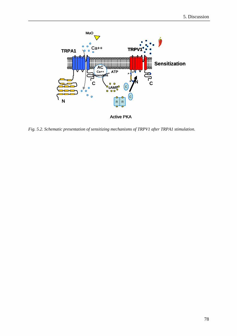
5. Discussion
78
Fig. 5.2. Schematic presentation of sensitizing mechanisms of TRPV1 after TRPA1 stimulation.
Ca++TRPA1
C N C
TRPV1
N
MuO
ACCa++
dependentP
P P
P
C
C
Active PKA
R R
ATP
cAMP
Sensitization
Ca++TRPA1
C N C
TRPV1
N
C N C
TRPV1
N
C N CN CN C
TRPV1
NN
MuOMuO
ACCa++
dependentP
P P
P
PPPP PP
PP
C
C
Active PKA
R R
C
C
Active PKA
R RR RR R
ATP
cAMP
ATPATP
cAMPcAMP
Sensitization
6. Summary
79
6. Summary
Transient Receptor Potential Vanilloid 1 (TRPV1) is a ligand-gated ion channel expressed on
sensory nerves that responds to noxious heat, protons, and chemical stimuli such as capsaicin.
TRPV1 plays a critical role in the development of tissue injury, inflammation or nerve
lesions. Transient Receptor Potential Ankyrin 1 (TRPA1), strongly co-expressed with
TRPV1, is also activated by compounds that cause a pungent burning sensation. TRPA1
functioned as a downstream target for components of the inflammatory milieu that elicit
nociceptor excitation and pain hypersensitivity.
Opioids such as morphine have been used widely for the treatment of many types of acute and
chronic pain. Application of morphine leads to a dissociation of G-proteins and causes a
reduced activity of adenylylcyclases (AC), resulting in a lower amount of cAMP. However,
opioid withdrawal following chronic activation of the µ opioid receptor (MOR) induces AC
superactivation and subsequently an increase in cAMP and Protein Kinase A (PKA) activity.
The aims of this doctoral thesis were, first, to investiagate the modulation of the activity of
TRPV1 during opioid withdrawal and second, the influence of TRPA1 stimulation on TRPV1
activity.
The activity of TRPV was significantly increased during opioid in a cAMP and PKA sensitive
manner. This sensitization was reversed by inhibiting ACs, silencing AC 3 and mutating PKA
phosphorylation sites threonine 144 and serine 774. Furthermore, the paw withdrawal latency
of male Wistar rats was significantly decreased and the nocifensive behaviour was
significantly enhanced during opioid withdrawal. These results indicate a new mechanism
underlying hyperalgesia during opioid withdrawal in the peripheral nervous system, which
might offer new possibilities to treat withdrawal-induced painful sensations in patients.
In the second part of the study, it was proven that TRPA1 activation sensitizes TRPV1
activity in a calcium and cAMP/PKA dependent manner. TRPA1 stimulation enhanced
TRPV1 phosphorylation and the involvement of PKA phosphorylation sites serine 116 and
serine 774 was shown. Finally, we also detected a calcium sensitiv increased TRPV1 activity
after TRPA1 activation in dorsal root ganglion neurons.
In summary, the current study delineates the sensitization of TRPV1 by two independent
experimental and clinical settings. First, the sensitization of TRPV1 during opioid withdrawal
and second, the sensitization of TRPV1 via activation of TRPA1. Both topics share the same
intracellular signalling pathway: The action of AC, subsequent translocation and activation of
6. Summary
80
PKA due to increased cAMP levels and phosphorylation of TRPV1 at several PKA
phosphorylation residues.
Zusammenfassung
Der Ionenkanal Transient Receptor Potential Vanilloid 1 (TRPV1) ist auf sensorischen
Nervenfasern exprimiert, welche auf Hitze, Protonen und chemische Reize wie Capsaicin
reagieren. TRPV1 ist entscheidend an der Entstehung von Schmerz und der thermalen
Hyperalgesie unter Entzündungsbedingungen beteiligt. Der Ionenkanal Transient Receptor
Potential Ankyrin 1 (TRPA1) ist mit TRPV1 stark ko-exprimiert und wird ebenfalls durch
Stoffe aktiviert, die als scharf und brennend empfunden werden. Er ist sowohl an der
Nozizeptorerregung als auch an der Schmerzsensitivierung während einer Entzündung
beteiligt.
Der Einsatz von Opioiden wie Morphin wird vielfach zur Linderung akuter und chronischer
Schmerzsyndrome eingesetzt. Opioide bewirken die Aktivierung der Opioidrezeptoren, was
die Dissoziation des gekoppelten G – Proteins hervorruft. Nachfolgend werden die
Adenylatzyklasen (AC) inhibiert, die cAMP-Bildung gehemmt und demzufolge die cAMP-
abhängige Protein Kinase A (PKA) nicht aktiviert. Die PKA ist entscheidend an der
Sensitivierung und Resensitivierung des TRPV1 beteiligt. Ihre Hemmung resultiert in einer
geringeren Aktivität des TRPV1. Andererseits induziert der Entzug von chronisch
applizierten Opioiden eine „Superaktivierung“ der AC und somit einen Anstieg des cAMP-
Gehaltes und Protein Kinase A (PKA) Aktivität.
In dieser Dissertation sollte untersucht werden, ob und wie 1) die Aktivität von TRPV1
während des Opioidentzugs moduliert wird und 2) ob die TRPA1 Stimulation einen Einfluss
auf die TRPV1 Aktivität hat.
Es wurde gezeigt, dass die Aktivität von TRPV1 während des Opioidentzug signifikant erhöht
ist. Dieser Mechanismus war cAMP und PKA abhängig. Die Sensitivierung konnte durch das
Inhibieren der AC Aktivität insbesondere der Isoform 3 aufgehoben werden. Des Weiteren
führte die Mutation der PKA Phosphorylierungsstellen Threonin 144 und Serin 774 zu einer
Aufhebung der Sensitivierung. Durch Verhaltensexperimente an männlichen Wistar Ratten
konnte weiterführend gezeigt werden, dass TRPV1 eine fundamentale Rolle an der peripheren
Schmerzüberempfindlichkeit während des Opioidentzugs spielt.

6. Summary
81
Im zweiten Teil der Arbeit wurde gezeigt, dass die TRPA1 Aktivierung zu einer TRPV1
Sensitivierung führt. Die Aktivierung des TRPA1 führt zu einem Einstrom von insbesondere
Kalziumionen, die kalzium-sensitive AC aktivieren können. Die erhöhte AC Aktivität
resultierte in einem erhöhten cAMP-Spiegel, somit zur Translokation der PKA und
schlussendlich zur Phosphorylierung und Sensitivierung des TRPV1.
Zusammenfassend beschreibt die vorliegende Arbeit die Sensitivierung von TRPV1 in zwei
unabhängigen experimentellen und klinischen Situationen. Erstens, die Sensitivierung von
TRPV1 während des Opioidentzugs und zweitens, die Sensitivierung des TRPV1 nach
TRPA1 Aktivierung. Beide Themengebiete teilen den gleichen intrazellulären Signalweg: Die
Aktivität der AC mit anschließender Translokation and Aktivierung der PKA, welche TRPV1
an bestimmten PKA Phosphorylierungsstellen phosphoryliert und somit sensitiviert. Diese
Beobachtungen zeigen neue Erklärungsmöglichkeiten zur Entstehung thermaler Hyperalgesie
im peripheren Nervensystem während des Opioidentzugs und nach TRPA1 Stimulation.
7. References
82
7. References
Ahern, G. P. 2003. Activation of TRPV1 by the satiety factor oleoylethanolamide. J Biol Chem 278 (33):30429-30434.
Akins, P. T., and E. W. McCleskey. 1993. Characterization of potassium currents in adult rat sensory neurons and modulation by opioids and cyclic AMP. Neuroscience 56 (3):759-769.
Akopian, A. N., N. B. Ruparel, N. A. Jeske, and K. M. Hargreaves. 2007. Transient receptor potential TRPA1 channel desensitization in sensory neurons is agonist dependent and regulated by TRPV1-directed internalization. J Physiol 583 (Pt 1):175-193.
Akopian, A. N., N. B. Ruparel, A. Patwardhan, and K. M. Hargreaves. 2008. Cannabinoids desensitize capsaicin and mustard oil responses in sensory neurons via TRPA1 activation. J Neurosci 28 (5):1064-1075.
Aley, K. O., P. G. Green, and J. D. Levine. 1995. Opioid and adenosine peripheral antinociception are subject to tolerance and withdrawal. J Neurosci 15 (12):8031-8038.
Aley, K. O., and J. D. Levine. 1997a. Different mechanisms mediate development and expression of tolerance and dependence for peripheral mu-opioid antinociception in rat. J Neurosci 17 (20):8018-8023.
———. 1997b. Dissociation of tolerance and dependence for opioid peripheral antinociception in rats. J Neurosci 17 (10):3907-3912.
———. 1997c. Multiple receptors involved in peripheral alpha 2, mu, and A1 antinociception, tolerance, and withdrawal. J Neurosci 17 (2):735-744.
Amaya, F., G. Shimosato, M. Nagano, M. Ueda, S. Hashimoto, Y. Tanaka, H. Suzuki, and M. Tanaka. 2004. NGF and GDNF differentially regulate TRPV1 expression that contributes to development of inflammatory thermal hyperalgesia. Eur J Neurosci 20 (9):2303-2310.
Anand, U., W. R. Otto, P. Facer, N. Zebda, I. Selmer, M. J. Gunthorpe, I. P. Chessell, M. Sinisi, R. Birch, and P. Anand. 2008. TRPA1 receptor localisation in the human peripheral nervous system and functional studies in cultured human and rat sensory neurons. Neurosci Lett 438 (2):221-227.
Andersson, D. A., C. Gentry, S. Moss, and S. Bevan. 2008. Transient receptor potential A1 is a sensory receptor for multiple products of oxidative stress. J Neurosci 28 (10):2485-2494.
Andersson, M., J. E. Westin, and M. A. Cenci. 2003. Time course of striatal DeltaFosB-like immunoreactivity and prodynorphin mRNA levels after discontinuation of chronic dopaminomimetic treatment. Eur J Neurosci 17 (3):661-666.
Andrade, E. L., A. P. Luiz, J. Ferreira, and J. B. Calixto. 2008. Pronociceptive response elicited by TRPA1 receptor activation in mice. Neuroscience 152 (2):511-520.
Angst, M. S., and J. D. Clark. 2006. Opioid-induced hyperalgesia: a qualitative systematic review. Anesthesiology 104 (3):570-587.
Angst, M. S., W. Koppert, I. Pahl, D. J. Clark, and M. Schmelz. 2003. Short-term infusion of the mu-opioid agonist remifentanil in humans causes hyperalgesia during withdrawal. Pain 106 (1-2):49-57.
Arts, K. S., B. B. Holmes, and J. M. Fujimoto. 1991. Differential contribution of descending serotonergic and noradrenergic systems to central Tyr-D-Ala2-Gly-NMePhe4-Gly-ol5 (DAMGO) and morphine-induced antinociception in mice. J Pharmacol Exp Ther 256 (3):890-896.
Asensio, V. J., A. Miralles, and J. A. Garcia-Sevilla. 2006. Stimulation of mitogen-activated protein kinase kinases (MEK1/2) by mu-, delta- and kappa-opioid receptor agonists in
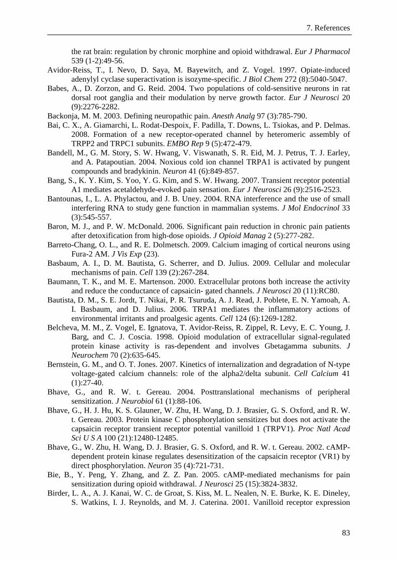
7. References
83
the rat brain: regulation by chronic morphine and opioid withdrawal. Eur J Pharmacol 539 (1-2):49-56.
Avidor-Reiss, T., I. Nevo, D. Saya, M. Bayewitch, and Z. Vogel. 1997. Opiate-induced adenylyl cyclase superactivation is isozyme-specific. J Biol Chem 272 (8):5040-5047.
Babes, A., D. Zorzon, and G. Reid. 2004. Two populations of cold-sensitive neurons in rat dorsal root ganglia and their modulation by nerve growth factor. Eur J Neurosci 20 (9):2276-2282.
Backonja, M. M. 2003. Defining neuropathic pain. Anesth Analg 97 (3):785-790. Bai, C. X., A. Giamarchi, L. Rodat-Despoix, F. Padilla, T. Downs, L. Tsiokas, and P. Delmas.
2008. Formation of a new receptor-operated channel by heteromeric assembly of TRPP2 and TRPC1 subunits. EMBO Rep 9 (5):472-479.
Bandell, M., G. M. Story, S. W. Hwang, V. Viswanath, S. R. Eid, M. J. Petrus, T. J. Earley, and A. Patapoutian. 2004. Noxious cold ion channel TRPA1 is activated by pungent compounds and bradykinin. Neuron 41 (6):849-857.
Bang, S., K. Y. Kim, S. Yoo, Y. G. Kim, and S. W. Hwang. 2007. Transient receptor potential A1 mediates acetaldehyde-evoked pain sensation. Eur J Neurosci 26 (9):2516-2523.
Bantounas, I., L. A. Phylactou, and J. B. Uney. 2004. RNA interference and the use of small interfering RNA to study gene function in mammalian systems. J Mol Endocrinol 33 (3):545-557.
Baron, M. J., and P. W. McDonald. 2006. Significant pain reduction in chronic pain patients after detoxification from high-dose opioids. J Opioid Manag 2 (5):277-282.
Barreto-Chang, O. L., and R. E. Dolmetsch. 2009. Calcium imaging of cortical neurons using Fura-2 AM. J Vis Exp (23).
Basbaum, A. I., D. M. Bautista, G. Scherrer, and D. Julius. 2009. Cellular and molecular mechanisms of pain. Cell 139 (2):267-284.
Baumann, T. K., and M. E. Martenson. 2000. Extracellular protons both increase the activity and reduce the conductance of capsaicin- gated channels. J Neurosci 20 (11):RC80.
Bautista, D. M., S. E. Jordt, T. Nikai, P. R. Tsuruda, A. J. Read, J. Poblete, E. N. Yamoah, A. I. Basbaum, and D. Julius. 2006. TRPA1 mediates the inflammatory actions of environmental irritants and proalgesic agents. Cell 124 (6):1269-1282.
Belcheva, M. M., Z. Vogel, E. Ignatova, T. Avidor-Reiss, R. Zippel, R. Levy, E. C. Young, J. Barg, and C. J. Coscia. 1998. Opioid modulation of extracellular signal-regulated protein kinase activity is ras-dependent and involves Gbetagamma subunits. J Neurochem 70 (2):635-645.
Bernstein, G. M., and O. T. Jones. 2007. Kinetics of internalization and degradation of N-type voltage-gated calcium channels: role of the alpha2/delta subunit. Cell Calcium 41 (1):27-40.
Bhave, G., and R. W. t. Gereau. 2004. Posttranslational mechanisms of peripheral sensitization. J Neurobiol 61 (1):88-106.
Bhave, G., H. J. Hu, K. S. Glauner, W. Zhu, H. Wang, D. J. Brasier, G. S. Oxford, and R. W. t. Gereau. 2003. Protein kinase C phosphorylation sensitizes but does not activate the capsaicin receptor transient receptor potential vanilloid 1 (TRPV1). Proc Natl Acad Sci U S A 100 (21):12480-12485.
Bhave, G., W. Zhu, H. Wang, D. J. Brasier, G. S. Oxford, and R. W. t. Gereau. 2002. cAMP-dependent protein kinase regulates desensitization of the capsaicin receptor (VR1) by direct phosphorylation. Neuron 35 (4):721-731.
Bie, B., Y. Peng, Y. Zhang, and Z. Z. Pan. 2005. cAMP-mediated mechanisms for pain sensitization during opioid withdrawal. J Neurosci 25 (15):3824-3832.
Birder, L. A., A. J. Kanai, W. C. de Groat, S. Kiss, M. L. Nealen, N. E. Burke, K. E. Dineley, S. Watkins, I. J. Reynolds, and M. J. Caterina. 2001. Vanilloid receptor expression
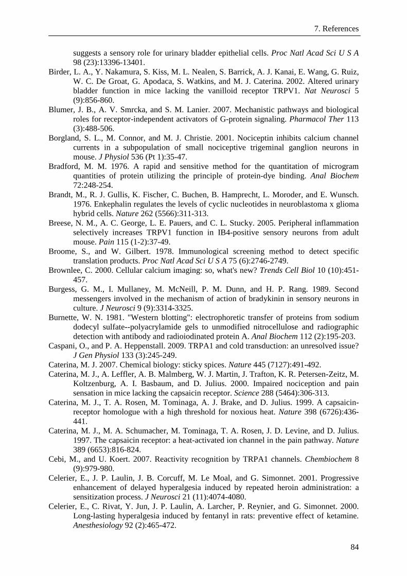
7. References
84
suggests a sensory role for urinary bladder epithelial cells. Proc Natl Acad Sci U S A 98 (23):13396-13401.
Birder, L. A., Y. Nakamura, S. Kiss, M. L. Nealen, S. Barrick, A. J. Kanai, E. Wang, G. Ruiz, W. C. De Groat, G. Apodaca, S. Watkins, and M. J. Caterina. 2002. Altered urinary bladder function in mice lacking the vanilloid receptor TRPV1. Nat Neurosci 5 (9):856-860.
Blumer, J. B., A. V. Smrcka, and S. M. Lanier. 2007. Mechanistic pathways and biological roles for receptor-independent activators of G-protein signaling. Pharmacol Ther 113 (3):488-506.
Borgland, S. L., M. Connor, and M. J. Christie. 2001. Nociceptin inhibits calcium channel currents in a subpopulation of small nociceptive trigeminal ganglion neurons in mouse. J Physiol 536 (Pt 1):35-47.
Bradford, M. M. 1976. A rapid and sensitive method for the quantitation of microgram quantities of protein utilizing the principle of protein-dye binding. Anal Biochem 72:248-254.
Brandt, M., R. J. Gullis, K. Fischer, C. Buchen, B. Hamprecht, L. Moroder, and E. Wunsch. 1976. Enkephalin regulates the levels of cyclic nucleotides in neuroblastoma x glioma hybrid cells. Nature 262 (5566):311-313.
Breese, N. M., A. C. George, L. E. Pauers, and C. L. Stucky. 2005. Peripheral inflammation selectively increases TRPV1 function in IB4-positive sensory neurons from adult mouse. Pain 115 (1-2):37-49.
Broome, S., and W. Gilbert. 1978. Immunological screening method to detect specific translation products. Proc Natl Acad Sci U S A 75 (6):2746-2749.
Brownlee, C. 2000. Cellular calcium imaging: so, what's new? Trends Cell Biol 10 (10):451-457.
Burgess, G. M., I. Mullaney, M. McNeill, P. M. Dunn, and H. P. Rang. 1989. Second messengers involved in the mechanism of action of bradykinin in sensory neurons in culture. J Neurosci 9 (9):3314-3325.
Burnette, W. N. 1981. "Western blotting": electrophoretic transfer of proteins from sodium dodecyl sulfate--polyacrylamide gels to unmodified nitrocellulose and radiographic detection with antibody and radioiodinated protein A. Anal Biochem 112 (2):195-203.
Caspani, O., and P. A. Heppenstall. 2009. TRPA1 and cold transduction: an unresolved issue? J Gen Physiol 133 (3):245-249.
Caterina, M. J. 2007. Chemical biology: sticky spices. Nature 445 (7127):491-492. Caterina, M. J., A. Leffler, A. B. Malmberg, W. J. Martin, J. Trafton, K. R. Petersen-Zeitz, M.
Koltzenburg, A. I. Basbaum, and D. Julius. 2000. Impaired nociception and pain sensation in mice lacking the capsaicin receptor. Science 288 (5464):306-313.
Caterina, M. J., T. A. Rosen, M. Tominaga, A. J. Brake, and D. Julius. 1999. A capsaicin-receptor homologue with a high threshold for noxious heat. Nature 398 (6726):436-441.
Caterina, M. J., M. A. Schumacher, M. Tominaga, T. A. Rosen, J. D. Levine, and D. Julius. 1997. The capsaicin receptor: a heat-activated ion channel in the pain pathway. Nature 389 (6653):816-824.
Cebi, M., and U. Koert. 2007. Reactivity recognition by TRPA1 channels. Chembiochem 8 (9):979-980.
Celerier, E., J. P. Laulin, J. B. Corcuff, M. Le Moal, and G. Simonnet. 2001. Progressive enhancement of delayed hyperalgesia induced by repeated heroin administration: a sensitization process. J Neurosci 21 (11):4074-4080.
Celerier, E., C. Rivat, Y. Jun, J. P. Laulin, A. Larcher, P. Reynier, and G. Simonnet. 2000. Long-lasting hyperalgesia induced by fentanyl in rats: preventive effect of ketamine. Anesthesiology 92 (2):465-472.

7. References
85
Cesare, P., and P. McNaughton. 1996. A novel heat-activated current in nociceptive neurons and its sensitization by bradykinin. Proc Natl Acad Sci U S A 93 (26):15435-15439.
Chakrabarti, S., and A. R. Gintzler. 2007. Phosphorylation of Galphas influences its association with the micro-opioid receptor and is modulated by long-term morphine exposure. Mol Pharmacol 72 (3):753-760.
Chao, J., and E. J. Nestler. 2004. Molecular neurobiology of drug addiction. Annu Rev Med 55:113-132.
Chao, J. R., Y. G. Ni, C. A. Bolanos, Z. Rahman, R. J. DiLeone, and E. J. Nestler. 2002. Characterization of the mouse adenylyl cyclase type VIII gene promoter: regulation by cAMP and CREB. Eur J Neurosci 16 (7):1284-1294.
Chen, J., M. B. Kelz, B. T. Hope, Y. Nakabeppu, and E. J. Nestler. 1997. Chronic Fos-related antigens: stable variants of deltaFosB induced in brain by chronic treatments. J Neurosci 17 (13):4933-4941.
Chen, Y., C. Geis, and C. Sommer. 2008. Activation of TRPV1 contributes to morphine tolerance: involvement of the mitogen-activated protein kinase signaling pathway. J Neurosci 28 (22):5836-5845.
Chia, Y. Y., K. Liu, J. J. Wang, M. C. Kuo, and S. T. Ho. 1999. Intraoperative high dose fentanyl induces postoperative fentanyl tolerance. Can J Anaesth 46 (9):872-877.
Chu, L. F., M. S. Angst, and D. Clark. 2008. Opioid-induced hyperalgesia in humans: molecular mechanisms and clinical considerations. Clin J Pain 24 (6):479-496.
Chu, L. F., D. J. Clark, and M. S. Angst. 2006. Opioid tolerance and hyperalgesia in chronic pain patients after one month of oral morphine therapy: a preliminary prospective study. J Pain 7 (1):43-48.
Chuang, H. H., E. D. Prescott, H. Kong, S. Shields, S. E. Jordt, A. I. Basbaum, M. V. Chao, and D. Julius. 2001. Bradykinin and nerve growth factor release the capsaicin receptor from PtdIns(4,5)P2-mediated inhibition. Nature 411 (6840):957-962.
Clapham, D. E. 2003. TRP channels as cellular sensors. Nature 426 (6966):517-524. Clapham, D. E., and E. J. Neer. 1997. G protein beta gamma subunits. Annu Rev Pharmacol
Toxicol 37:167-203. Compton, M. A. 1994. Cold-pressor pain tolerance in opiate and cocaine abusers: correlates
of drug type and use status. J Pain Symptom Manage 9 (7):462-473. Compton, P., P. Athanasos, and D. Elashoff. 2003. Withdrawal hyperalgesia after acute
opioid physical dependence in nonaddicted humans: a preliminary study. J Pain 4 (9):511-519.
Compton, P., V. C. Charuvastra, and W. Ling. 2001. Pain intolerance in opioid-maintained former opiate addicts: effect of long-acting maintenance agent. Drug Alcohol Depend 63 (2):139-146.
Cooper, D. W., S. L. Lindsay, D. M. Ryall, M. S. Kokri, S. S. Eldabe, and G. A. Lear. 1997. Does intrathecal fentanyl produce acute cross-tolerance to i.v. morphine? Br J Anaesth 78 (3):311-313.
Corey, D. P., J. Garcia-Anoveros, J. R. Holt, K. Y. Kwan, S. Y. Lin, M. A. Vollrath, A. Amalfitano, E. L. Cheung, B. H. Derfler, A. Duggan, G. S. Geleoc, P. A. Gray, M. P. Hoffman, H. L. Rehm, D. Tamasauskas, and D. S. Zhang. 2004. TRPA1 is a candidate for the mechanosensitive transduction channel of vertebrate hair cells. Nature 432 (7018):723-730.
Correll, C. C., P. T. Phelps, J. C. Anthes, S. Umland, and S. Greenfeder. 2004. Cloning and pharmacological characterization of mouse TRPV1. Neurosci Lett 370 (1):55-60.
Crandall, M., J. Kwash, W. Yu, and G. White. 2002. Activation of protein kinase C sensitizes human VR1 to capsaicin and to moderate decreases in pH at physiological temperatures in Xenopus oocytes. Pain 98 (1-2):109-117.
7. References
86
Cruz-Orengo, L., A. Dhaka, R. J. Heuermann, T. J. Young, M. C. Montana, E. J. Cavanaugh, D. Kim, and G. M. Story. 2008. Cutaneous nociception evoked by 15-delta PGJ2 via activation of ion channel TRPA1. Mol Pain 4:30.
Cui, Y., Y. Chen, J. L. Zhi, R. X. Guo, J. Q. Feng, and P. X. Chen. 2006. Activation of p38 mitogen-activated protein kinase in spinal microglia mediates morphine antinociceptive tolerance. Brain Res 1069 (1):235-243.
Cumbay, M. G., and V. J. Watts. 2004. Novel regulatory properties of human type 9 adenylate cyclase. J Pharmacol Exp Ther 310 (1):108-115.
Cuypers, E., A. Yanagihara, E. Karlsson, and J. Tytgat. 2006. Jellyfish and other cnidarian envenomations cause pain by affecting TRPV1 channels. FEBS Lett 580 (24):5728-5732.
Dai, Y., T. Moriyama, T. Higashi, K. Togashi, K. Kobayashi, H. Yamanaka, M. Tominaga, and K. Noguchi. 2004. Proteinase-activated receptor 2-mediated potentiation of transient receptor potential vanilloid subfamily 1 activity reveals a mechanism for proteinase-induced inflammatory pain. J Neurosci 24 (18):4293-4299.
Davis, J. B., J. Gray, M. J. Gunthorpe, J. P. Hatcher, P. T. Davey, P. Overend, M. H. Harries, J. Latcham, C. Clapham, K. Atkinson, S. A. Hughes, K. Rance, E. Grau, A. J. Harper, P. L. Pugh, D. C. Rogers, S. Bingham, A. Randall, and S. A. Sheardown. 2000. Vanilloid receptor-1 is essential for inflammatory thermal hyperalgesia. Nature 405 (6783):183-187.
Davis, M. P., L. A. Shaiova, and M. S. Angst. 2007. When opioids cause pain. J Clin Oncol 25 (28):4497-4498.
De Vries, L., T. Fischer, H. Tronchere, G. M. Brothers, B. Strockbine, D. P. Siderovski, and M. G. Farquhar. 2000. Activator of G protein signaling 3 is a guanine dissociation inhibitor for Galpha i subunits. Proc Natl Acad Sci U S A 97 (26):14364-14369.
Dedov, V. N., V. H. Tran, C. C. Duke, M. Connor, M. J. Christie, S. Mandadi, and B. D. Roufogalis. 2002. Gingerols: a novel class of vanilloid receptor (VR1) agonists. Br J Pharmacol 137 (6):793-798.
Defer, N., M. Best-Belpomme, and J. Hanoune. 2000. Tissue specificity and physiological relevance of various isoforms of adenylyl cyclase. Am J Physiol Renal Physiol 279 (3):F400-416.
Dell'Acqua, M. L., and J. D. Scott. 1997. Protein kinase A anchoring. J Biol Chem 272 (20):12881-12884.
Dhaka, A., V. Viswanath, and A. Patapoutian. 2006. Trp ion channels and temperature sensation. Annu Rev Neurosci 29:135-161.
Dietrich, A., M. Mederos y Schnitzler, H. Kalwa, U. Storch, and T. Gudermann. 2005. Functional characterization and physiological relevance of the TRPC3/6/7 subfamily of cation channels. Naunyn Schmiedebergs Arch Pharmacol 371 (4):257-265.
Diogenes, A., A. N. Akopian, and K. M. Hargreaves. 2007. NGF up-regulates TRPA1: implications for orofacial pain. J Dent Res 86 (6):550-555.
Distler, C., P. K. Rathee, K. S. Lips, O. Obreja, W. Neuhuber, and M. Kress. 2003. Fast Ca2+-induced potentiation of heat-activated ionic currents requires cAMP/PKA signaling and functional AKAP anchoring. J Neurophysiol 89 (5):2499-2505.
Doverty, M., J. M. White, A. A. Somogyi, F. Bochner, R. Ali, and W. Ling. 2001. Hyperalgesic responses in methadone maintenance patients. Pain 90 (1-2):91-96.
Drdla, R., M. Gassner, E. Gingl, and J. Sandkuhler. 2009. Induction of synaptic long-term potentiation after opioid withdrawal. Science 325 (5937):207-210.
Du, S., I. Araki, H. Kobayashi, H. Zakoji, N. Sawada, and M. Takeda. 2008. Differential expression profile of cold (TRPA1) and cool (TRPM8) receptors in human urogenital organs. Urology 72 (2):450-455.

7. References
87
Dunbar, S. A., and I. J. Pulai. 1998. Repetitive opioid abstinence causes progressive hyperalgesia sensitive to N-methyl-D-aspartate receptor blockade in the rat. J Pharmacol Exp Ther 284 (2):678-686.
Duncan, J. S. 1999. Positron emission tomography receptor studies. Adv Neurol 79:893-899. Elbashir, S. M., J. Harborth, W. Lendeckel, A. Yalcin, K. Weber, and T. Tuschl. 2001.
Duplexes of 21-nucleotide RNAs mediate RNA interference in cultured mammalian cells. Nature 411 (6836):494-498.
Endres-Becker, j. 2007. Modulation des Transient Receptor Potential Vanilloid 1 (TRPV1) Ionenkanals durch µ-Rezeptor-Agonisten, Institut für Biologie, Chemie, Pharmazie, Freie Universität Berlin, Berlin.
Endres-Becker, J., P. A. Heppenstall, S. A. Mousa, D. Labuz, A. Oksche, M. Schafer, C. Stein, and C. Zollner. 2007. Mu-opioid receptor activation modulates transient receptor potential vanilloid 1 (TRPV1) currents in sensory neurons in a model of inflammatory pain. Mol Pharmacol 71 (1):12-18.
Evans, C. J., D. E. Keith, Jr., H. Morrison, K. Magendzo, and R. H. Edwards. 1992. Cloning of a delta opioid receptor by functional expression. Science 258 (5090):1952-1955.
Fan, P., Z. Jiang, I. Diamond, and L. Yao. 2009. Up-regulation of AGS3 during morphine withdrawal promotes cAMP superactivation via adenylyl cyclase 5 and 7 in rat nucleus accumbens/striatal neurons. Mol Pharmacol 76 (3):526-533.
Ferrer-Alcon, M., M. J. Garcia-Fuster, R. La Harpe, and J. A. Garcia-Sevilla. 2004. Long-term regulation of signalling components of adenylyl cyclase and mitogen-activated protein kinase in the pre-frontal cortex of human opiate addicts. J Neurochem 90 (1):220-230.
Fire, A., S. Xu, M. K. Montgomery, S. A. Kostas, S. E. Driver, and C. C. Mello. 1998. Potent and specific genetic interference by double-stranded RNA in Caenorhabditis elegans. Nature 391 (6669):806-811.
Fishbain, D. A., B. Cole, J. E. Lewis, J. Gao, and R. S. Rosomoff. 2009. Do opioids induce hyperalgesia in humans? An evidence-based structured review. Pain Med 10 (5):829-839.
Friderichs, E., and W. Strassburger. 2002. [The key-lock model of molecular biological characterization. Opiate receptors]. Pharm Unserer Zeit 31 (1):32-39.
Gallego-Sandin, S., A. Rodriguez-Garcia, M. T. Alonso, and J. Garcia-Sancho. 2009. The endoplasmic reticulum of dorsal root ganglion neurons contains functional TRPV1 channels. J Biol Chem 284 (47):32591-32601.
Garcia-Sanz, N., P. Valente, A. Gomis, A. Fernandez-Carvajal, G. Fernandez-Ballester, F. Viana, C. Belmonte, and A. Ferrer-Montiel. 2007. A role of the transient receptor potential domain of vanilloid receptor I in channel gating. J Neurosci 27 (43):11641-11650.
Gardell, L. R., R. Wang, S. E. Burgess, M. H. Ossipov, T. W. Vanderah, T. P. Malan, Jr., J. Lai, and F. Porreca. 2002. Sustained morphine exposure induces a spinal dynorphin-dependent enhancement of excitatory transmitter release from primary afferent fibers. J Neurosci 22 (15):6747-6755.
Gavva, N. R., L. Klionsky, Y. Qu, L. Shi, R. Tamir, S. Edenson, T. J. Zhang, V. N. Viswanadhan, A. Toth, L. V. Pearce, T. W. Vanderah, F. Porreca, P. M. Blumberg, J. Lile, Y. Sun, K. Wild, J. C. Louis, and J. J. Treanor. 2004. Molecular determinants of vanilloid sensitivity in TRPV1. J Biol Chem 279 (19):20283-20295.
Gerfen, J. N. C. a. C., ed. 2001. Current protocols in neuroscience Edited by J. N. C. a. C. Gerfen. New York, N.Y. : J. Wiley, 2002-
Gintzler, A. R., and S. Chakrabarti. 2006. Post-opioid receptor adaptations to chronic morphine; altered functionality and associations of signaling molecules. Life Sci 79 (8):717-722.
7. References
88
Gjertsen, B. T., and S. O. Doskeland. 1995. Protein phosphorylation in apoptosis. Biochim Biophys Acta 1269 (2):187-199.
Goldstein, A., S. Tachibana, L. I. Lowney, M. Hunkapiller, and L. Hood. 1979. Dynorphin-(1-13), an extraordinarily potent opioid peptide. Proc Natl Acad Sci U S A 76 (12):6666-6670.
Gossen, M., and H. Bujard. 1992. Tight control of gene expression in mammalian cells by tetracycline-responsive promoters. Proc Natl Acad Sci U S A 89 (12):5547-5551.
Guenther, S., P. W. Reeh, and M. Kress. 1999. Rises in [Ca2+]i mediate capsaicin- and proton-induced heat sensitization of rat primary nociceptive neurons. Eur J Neurosci 11 (9):3143-3150.
Guignard, B., A. E. Bossard, C. Coste, D. I. Sessler, C. Lebrault, P. Alfonsi, D. Fletcher, and M. Chauvin. 2000. Acute opioid tolerance: intraoperative remifentanil increases postoperative pain and morphine requirement. Anesthesiology 93 (2):409-417.
Hacker, B. M., J. E. Tomlinson, G. A. Wayman, R. Sultana, G. Chan, E. Villacres, C. Disteche, and D. R. Storm. 1998. Cloning, chromosomal mapping, and regulatory properties of the human type 9 adenylyl cyclase (ADCY9). Genomics 50 (1):97-104.
Hanoune, J., and N. Defer. 2001. Regulation and role of adenylyl cyclase isoforms. Annu Rev Pharmacol Toxicol 41:145-174.
Hargreaves, K., R. Dubner, F. Brown, C. Flores, and J. Joris. 1988. A new and sensitive method for measuring thermal nociception in cutaneous hyperalgesia. Pain 32 (1):77-88.
Hawkes, R., E. Niday, and J. Gordon. 1982. A dot-immunobinding assay for monoclonal and other antibodies. Anal Biochem 119 (1):142-147.
Hayes, P., H. J. Meadows, M. J. Gunthorpe, M. H. Harries, D. M. Duckworth, W. Cairns, D. C. Harrison, C. E. Clarke, K. Ellington, R. K. Prinjha, A. J. Barton, A. D. Medhurst, G. D. Smith, S. Topp, P. Murdock, G. J. Sanger, J. Terrett, O. Jenkins, C. D. Benham, A. D. Randall, I. S. Gloger, and J. B. Davis. 2000. Cloning and functional expression of a human orthologue of rat vanilloid receptor-1. Pain 88 (2):205-215.
Hellwig, N., N. Albrecht, C. Harteneck, G. Schultz, and M. Schaefer. 2005. Homo- and heteromeric assembly of TRPV channel subunits. J Cell Sci 118 (Pt 5):917-928.
Hellwig, N., T. D. Plant, W. Janson, M. Schafer, G. Schultz, and M. Schaefer. 2004. TRPV1 acts as proton channel to induce acidification in nociceptive neurons. J Biol Chem 279 (33):34553-34561.
Hill, K., and M. Schaefer. 2007. TRPA1 is differentially modulated by the amphipathic molecules trinitrophenol and chlorpromazine. J Biol Chem 282 (10):7145-7153.
Hinman, A., H. H. Chuang, D. M. Bautista, and D. Julius. 2006. TRP channel activation by reversible covalent modification. Proc Natl Acad Sci U S A 103 (51):19564-19568.
Hjerling-Leffler, J., M. Alqatari, P. Ernfors, and M. Koltzenburg. 2007. Emergence of functional sensory subtypes as defined by transient receptor potential channel expression. J Neurosci 27 (10):2435-2443.
Hoenderop, J. G., T. Voets, S. Hoefs, F. Weidema, J. Prenen, B. Nilius, and R. J. Bindels. 2003. Homo- and heterotetrameric architecture of the epithelial Ca2+ channels TRPV5 and TRPV6. EMBO J 22 (4):776-785.
Hood, D. D., R. Curry, and J. C. Eisenach. 2003. Intravenous remifentanil produces withdrawal hyperalgesia in volunteers with capsaicin-induced hyperalgesia. Anesth Analg 97 (3):810-815.
Howard, J., and S. Bechstedt. 2004. Hypothesis: a helix of ankyrin repeats of the NOMPC-TRP ion channel is the gating spring of mechanoreceptors. Curr Biol 14 (6):R224-226.
Huang, P., G. B. Kehner, A. Cowan, and L. Y. Liu-Chen. 2001. Comparison of pharmacological activities of buprenorphine and norbuprenorphine: norbuprenorphine is a potent opioid agonist. J Pharmacol Exp Ther 297 (2):688-695.
7. References
89
Hubbard, M. J., and P. Cohen. 1993. On target with a new mechanism for the regulation of protein phosphorylation. Trends Biochem Sci 18 (5):172-177.
Huggenvik, J. I., M. W. Collard, R. E. Stofko, A. F. Seasholtz, and M. D. Uhler. 1991. Regulation of the human enkephalin promoter by two isoforms of the catalytic subunit of cyclic adenosine 3',5'-monophosphate-dependent protein kinase. Mol Endocrinol 5 (7):921-930.
Hughes, J., T. W. Smith, H. W. Kosterlitz, L. A. Fothergill, B. A. Morgan, and H. R. Morris. 1975. Identification of two related pentapeptides from the brain with potent opiate agonist activity. Nature 258 (5536):577-580.
Hulme, E. C., Z. L. Lu, S. D. Ward, K. Allman, and C. A. Curtis. 1999. The conformational switch in 7-transmembrane receptors: the muscarinic receptor paradigm. Eur J Pharmacol 375 (1-3):247-260.
Hwang, S. W., H. Cho, J. Kwak, S. Y. Lee, C. J. Kang, J. Jung, S. Cho, K. H. Min, Y. G. Suh, D. Kim, and U. Oh. 2000. Direct activation of capsaicin receptors by products of lipoxygenases: endogenous capsaicin-like substances. Proc Natl Acad Sci U S A 97 (11):6155-6160.
Hyman, S. E., and R. C. Malenka. 2001. Addiction and the brain: the neurobiology of compulsion and its persistence. Nat Rev Neurosci 2 (10):695-703.
Irnaten, M., S. A. Aicher, J. Wang, P. Venkatesan, C. Evans, S. Baxi, and D. Mendelowitz. 2003. Mu-opioid receptors are located postsynaptically and endomorphin-1 inhibits voltage-gated calcium currents in premotor cardiac parasympathetic neurons in the rat nucleus ambiguus. Neuroscience 116 (2):573-582.
Jaquemar, D., T. Schenker, and B. Trueb. 1999. An ankyrin-like protein with transmembrane domains is specifically lost after oncogenic transformation of human fibroblasts. J Biol Chem 274 (11):7325-7333.
Jeske, N. A., A. Diogenes, N. B. Ruparel, J. C. Fehrenbacher, M. Henry, A. N. Akopian, and K. M. Hargreaves. 2008. A-kinase anchoring protein mediates TRPV1 thermal hyperalgesia through PKA phosphorylation of TRPV1. Pain 138 (3):604-616.
Jeske, N. A., A. M. Patwardhan, N. Gamper, T. J. Price, A. N. Akopian, and K. M. Hargreaves. 2006. Cannabinoid WIN 55,212-2 regulates TRPV1 phosphorylation in sensory neurons. J Biol Chem 281 (43):32879-32890.
Ji, R. R., T. A. Samad, S. X. Jin, R. Schmoll, and C. J. Woolf. 2002. p38 MAPK activation by NGF in primary sensory neurons after inflammation increases TRPV1 levels and maintains heat hyperalgesia. Neuron 36 (1):57-68.
Joly, V., P. Richebe, B. Guignard, D. Fletcher, P. Maurette, D. I. Sessler, and M. Chauvin. 2005. Remifentanil-induced postoperative hyperalgesia and its prevention with small-dose ketamine. Anesthesiology 103 (1):147-155.
Jordt, S. E., D. M. Bautista, H. H. Chuang, D. D. McKemy, P. M. Zygmunt, E. D. Hogestatt, I. D. Meng, and D. Julius. 2004. Mustard oils and cannabinoids excite sensory nerve fibres through the TRP channel ANKTM1. Nature 427 (6971):260-265.
Jordt, S. E., and D. Julius. 2002. Molecular basis for species-specific sensitivity to "hot" chili peppers. Cell 108 (3):421-430.
Jordt, S. E., M. Tominaga, and D. Julius. 2000. Acid potentiation of the capsaicin receptor determined by a key extracellular site. Proc Natl Acad Sci U S A 97 (14):8134-8139.
Julius, D., and A. I. Basbaum. 2001. Molecular mechanisms of nociception. Nature 413 (6852):203-210.
Jung, J., J. S. Shin, S. Y. Lee, S. W. Hwang, J. Koo, H. Cho, and U. Oh. 2004. Phosphorylation of vanilloid receptor 1 by Ca2+/calmodulin-dependent kinase II regulates its vanilloid binding. J Biol Chem 279 (8):7048-7054.
7. References
90
Karashima, Y., K. Talavera, W. Everaerts, A. Janssens, K. Y. Kwan, R. Vennekens, B. Nilius, and T. Voets. 2009. TRPA1 acts as a cold sensor in vitro and in vivo. Proc Natl Acad Sci U S A 106 (4):1273-1278.
Kato, S., E. Aihara, A. Nakamura, H. Xin, H. Matsui, K. Kohama, and K. Takeuchi. 2003. Expression of vanilloid receptors in rat gastric epithelial cells: role in cellular protection. Biochem Pharmacol 66 (6):1115-1121.
Katsura, H., K. Tsuzuki, K. Noguchi, and M. Sakagami. 2006. Differential expression of capsaicin-, menthol-, and mustard oil-sensitive receptors in naive rat geniculate ganglion neurons. Chem Senses 31 (7):681-688.
Kaufman, S. 1995. Tyrosine hydroxylase. Adv Enzymol Relat Areas Mol Biol 70:103-220. Kayan, S., L. A. Woods, and C. L. Mitchell. 1971. Morphine-induced hyperalgesia in rats
tested on the hot plate. J Pharmacol Exp Ther 177 (3):509-513. Kedei, N., T. Szabo, J. D. Lile, J. J. Treanor, Z. Olah, M. J. Iadarola, and P. M. Blumberg.
2001. Analysis of the native quaternary structure of vanilloid receptor 1. J Biol Chem 276 (30):28613-28619.
Kelz, M. B., J. Chen, W. A. Carlezon, Jr., K. Whisler, L. Gilden, A. M. Beckmann, C. Steffen, Y. J. Zhang, L. Marotti, D. W. Self, T. Tkatch, G. Baranauskas, D. J. Surmeier, R. L. Neve, R. S. Duman, M. R. Picciotto, and E. J. Nestler. 1999. Expression of the transcription factor deltaFosB in the brain controls sensitivity to cocaine. Nature 401 (6750):272-276.
Kelz, M. B., and E. J. Nestler. 2000. deltaFosB: a molecular switch underlying long-term neural plasticity. Curr Opin Neurol 13 (6):715-720.
Khasar, S. G., J. F. Wang, Y. O. Taiwo, P. H. Heller, P. G. Green, and J. D. Levine. 1995. Mu-opioid agonist enhancement of prostaglandin-induced hyperalgesia in the rat: a G-protein beta gamma subunit-mediated effect? Neuroscience 67 (1):189-195.
Kieffer, B. L., K. Befort, C. Gaveriaux-Ruff, and C. G. Hirth. 1992. The delta-opioid receptor: isolation of a cDNA by expression cloning and pharmacological characterization. Proc Natl Acad Sci U S A 89 (24):12048-12052.
Kim, D., and E. J. Cavanaugh. 2007. Requirement of a soluble intracellular factor for activation of transient receptor potential A1 by pungent chemicals: role of inorganic polyphosphates. J Neurosci 27 (24):6500-6509.
Kimple, R. J., M. E. Kimple, L. Betts, J. Sondek, and D. P. Siderovski. 2002. Structural determinants for GoLoco-induced inhibition of nucleotide release by Galpha subunits. Nature 416 (6883):878-881.
Kindt, K. S., V. Viswanath, L. Macpherson, K. Quast, H. Hu, A. Patapoutian, and W. R. Schafer. 2007. Caenorhabditis elegans TRPA-1 functions in mechanosensation. Nat Neurosci 10 (5):568-577.
Kobayashi, K., T. Fukuoka, K. Obata, H. Yamanaka, Y. Dai, A. Tokunaga, and K. Noguchi. 2005. Distinct expression of TRPM8, TRPA1, and TRPV1 mRNAs in rat primary afferent neurons with adelta/c-fibers and colocalization with trk receptors. J Comp Neurol 493 (4):596-606.
Kobori, T., G. D. Smith, R. Sandford, and J. M. Edwardson. 2009. The transient receptor potential channels TRPP2 and TRPC1 form a heterotetramer with a 2:2 stoichiometry and an alternating subunit arrangement. J Biol Chem 284 (51):35507-35513.
Kochukov, M. Y., T. A. McNearney, Y. Fu, and K. N. Westlund. 2006. Thermosensitive TRP ion channels mediate cytosolic calcium response in human synoviocytes. Am J Physiol Cell Physiol 291 (3):C424-432.
Kogan, J. H., E. J. Nestler, and G. K. Aghajanian. 1992. Elevated basal firing rates and enhanced responses to 8-Br-cAMP in locus coeruleus neurons in brain slices from opiate-dependent rats. Eur J Pharmacol 211 (1):47-53.

7. References
91
Kondo, I., J. C. Marvizon, B. Song, F. Salgado, S. Codeluppi, X. Y. Hua, and T. L. Yaksh. 2005. Inhibition by spinal mu- and delta-opioid agonists of afferent-evoked substance P release. J Neurosci 25 (14):3651-3660.
Koplas, P. A., R. L. Rosenberg, and G. S. Oxford. 1997. The role of calcium in the desensitization of capsaicin responses in rat dorsal root ganglion neurons. J Neurosci 17 (10):3525-3537.
Koppert, W., S. K. Dern, R. Sittl, S. Albrecht, J. Schuttler, and M. Schmelz. 2001. A new model of electrically evoked pain and hyperalgesia in human skin: the effects of intravenous alfentanil, S(+)-ketamine, and lidocaine. Anesthesiology 95 (2):395-402.
Koppert, W., R. Sittl, K. Scheuber, M. Alsheimer, M. Schmelz, and J. Schuttler. 2003. Differential modulation of remifentanil-induced analgesia and postinfusion hyperalgesia by S-ketamine and clonidine in humans. Anesthesiology 99 (1):152-159.
Kress, M., and S. Guenther. 1999. Role of [Ca2+]i in the ATP-induced heat sensitization process of rat nociceptive neurons. J Neurophysiol 81 (6):2612-2619.
Kwan, K. Y., A. J. Allchorne, M. A. Vollrath, A. P. Christensen, D. S. Zhang, C. J. Woolf, and D. P. Corey. 2006. TRPA1 contributes to cold, mechanical, and chemical nociception but is not essential for hair-cell transduction. Neuron 50 (2):277-289.
Lane-Ladd, S. B., J. Pineda, V. A. Boundy, T. Pfeuffer, J. Krupinski, G. K. Aghajanian, and E. J. Nestler. 1997. CREB (cAMP response element-binding protein) in the locus coeruleus: biochemical, physiological, and behavioral evidence for a role in opiate dependence. J Neurosci 17 (20):7890-7901.
Laulin, J. P., A. Larcher, E. Celerier, M. Le Moal, and G. Simonnet. 1998. Long-lasting increased pain sensitivity in rat following exposure to heroin for the first time. Eur J Neurosci 10 (2):782-785.
Law, P. Y., Y. H. Wong, and H. H. Loh. 2000. Molecular mechanisms and regulation of opioid receptor signaling. Annu Rev Pharmacol Toxicol 40:389-430.
Lee, S. Y., J. H. Lee, K. K. Kang, S. Y. Hwang, K. D. Choi, and U. Oh. 2005. Sensitization of vanilloid receptor involves an increase in the phosphorylated form of the channel. Arch Pharm Res 28 (4):405-412.
Levine, J. D., and Y. O. Taiwo. 1989. Involvement of the mu-opiate receptor in peripheral analgesia. Neuroscience 32 (3):571-575.
Li, C. H., and D. Chung. 1976. Isolation and structure of an untriakontapeptide with opiate activity from camel pituitary glands. Proc Natl Acad Sci U S A 73 (4):1145-1148.
Li, C. H., D. Chung, and B. A. Doneen. 1976. Isolation, characterization and opiate activity of beta-endorphin from human pituitary glands. Biochem Biophys Res Commun 72 (4):1542-1547.
Li, L. Y., and K. J. Chang. 1996. The stimulatory effect of opioids on mitogen-activated protein kinase in Chinese hamster ovary cells transfected to express mu-opioid receptors. Mol Pharmacol 50 (3):599-602.
Liang, D. Y., G. Liao, J. Wang, J. Usuka, Y. Guo, G. Peltz, and J. D. Clark. 2006. A genetic analysis of opioid-induced hyperalgesia in mice. Anesthesiology 104 (5):1054-1062.
Liang, Y. F., B. Haake, and P. W. Reeh. 2001. Sustained sensitization and recruitment of rat cutaneous nociceptors by bradykinin and a novel theory of its excitatory action. J Physiol 532 (Pt 1):229-239.
Linte, R. M., C. Ciobanu, G. Reid, and A. Babes. 2007. Desensitization of cold- and menthol-sensitive rat dorsal root ganglion neurones by inflammatory mediators. Exp Brain Res 178 (1):89-98.
Lipman, J. J., and B. Blumenkopf. 1989. Comparison of subjective and objective analgesic effects of intravenous and intrathecal morphine in chronic pain patients by heat beam dolorimetry. Pain 39 (3):249-256.

7. References
92
Liu, L., and S. A. Simon. 1996. Capsaicin-induced currents with distinct desensitization and Ca2+ dependence in rat trigeminal ganglion cells. J Neurophysiol 75 (4):1503-1514.
Loeser, J. D., and R. D. Treede. 2008. The Kyoto protocol of IASP Basic Pain Terminology. Pain 137 (3):473-477.
Lonze, B. E., and D. D. Ginty. 2002. Function and regulation of CREB family transcription factors in the nervous system. Neuron 35 (4):605-623.
Lopshire, J. C., and G. D. Nicol. 1998. The cAMP transduction cascade mediates the prostaglandin E2 enhancement of the capsaicin-elicited current in rat sensory neurons: whole-cell and single-channel studies. J Neurosci 18 (16):6081-6092.
Lord, J. A., A. A. Waterfield, J. Hughes, and H. W. Kosterlitz. 1977. Endogenous opioid peptides: multiple agonists and receptors. Nature 267 (5611):495-499.
Ma, W., W. H. Zheng, K. Powell, K. Jhamandas, and R. Quirion. 2001. Chronic morphine exposure increases the phosphorylation of MAP kinases and the transcription factor CREB in dorsal root ganglion neurons: an in vitro and in vivo study. Eur J Neurosci 14 (7):1091-1104.
Macpherson, L. J., A. E. Dubin, M. J. Evans, F. Marr, P. G. Schultz, B. F. Cravatt, and A. Patapoutian. 2007a. Noxious compounds activate TRPA1 ion channels through covalent modification of cysteines. Nature 445 (7127):541-545.
Macpherson, L. J., B. H. Geierstanger, V. Viswanath, M. Bandell, S. R. Eid, S. Hwang, and A. Patapoutian. 2005. The pungency of garlic: activation of TRPA1 and TRPV1 in response to allicin. Curr Biol 15 (10):929-934.
Macpherson, L. J., B. Xiao, K. Y. Kwan, M. J. Petrus, A. E. Dubin, S. Hwang, B. Cravatt, D. P. Corey, and A. Patapoutian. 2007b. An ion channel essential for sensing chemical damage. J Neurosci 27 (42):11412-11415.
Mandadi, S., M. Numazaki, M. Tominaga, M. B. Bhat, P. J. Armati, and B. D. Roufogalis. 2004. Activation of protein kinase C reverses capsaicin-induced calcium-dependent desensitization of TRPV1 ion channels. Cell Calcium 35 (5):471-478.
Mao, J. 2002. Opioid-induced abnormal pain sensitivity: implications in clinical opioid therapy. Pain 100 (3):213-217.
Mao, J., D. D. Price, and D. J. Mayer. 1994. Thermal hyperalgesia in association with the development of morphine tolerance in rats: roles of excitatory amino acid receptors and protein kinase C. J Neurosci 14 (4):2301-2312.
———. 1995. Mechanisms of hyperalgesia and morphine tolerance: a current view of their possible interactions. Pain 62 (3):259-274.
Mao, J., B. Sung, R. R. Ji, and G. Lim. 2002a. Chronic morphine induces downregulation of spinal glutamate transporters: implications in morphine tolerance and abnormal pain sensitivity. J Neurosci 22 (18):8312-8323.
———. 2002b. Neuronal apoptosis associated with morphine tolerance: evidence for an opioid-induced neurotoxic mechanism. J Neurosci 22 (17):7650-7661.
Martin, W. R., C. G. Eades, J. A. Thompson, R. E. Huppler, and P. E. Gilbert. 1976. The effects of morphine- and nalorphine- like drugs in the nondependent and morphine-dependent chronic spinal dog. J Pharmacol Exp Ther 197 (3):517-532.
Materazzi, S., R. Nassini, E. Andre, B. Campi, S. Amadesi, M. Trevisani, N. W. Bunnett, R. Patacchini, and P. Geppetti. 2008. Cox-dependent fatty acid metabolites cause pain through activation of the irritant receptor TRPA1. Proc Natl Acad Sci U S A 105 (33):12045-12050.
Matten, W., I. Daar, and G. F. Vande Woude. 1994. Protein kinase A acts at multiple points to inhibit Xenopus oocyte maturation. Mol Cell Biol 14 (7):4419-4426.
Mayr, B., and M. Montminy. 2001. Transcriptional regulation by the phosphorylation-dependent factor CREB. Nat Rev Mol Cell Biol 2 (8):599-609.
7. References
93
McClung, C. A., and E. J. Nestler. 2003. Regulation of gene expression and cocaine reward by CREB and DeltaFosB. Nat Neurosci 6 (11):1208-1215.
McNamara, C. R., J. Mandel-Brehm, D. M. Bautista, J. Siemens, K. L. Deranian, M. Zhao, N. J. Hayward, J. A. Chong, D. Julius, M. M. Moran, and C. M. Fanger. 2007. TRPA1 mediates formalin-induced pain. Proc Natl Acad Sci U S A 104 (33):13525-13530.
McNamara, F. N., A. Randall, and M. J. Gunthorpe. 2005. Effects of piperine, the pungent component of black pepper, at the human vanilloid receptor (TRPV1). Br J Pharmacol 144 (6):781-790.
Meng, F., G. X. Xie, R. C. Thompson, A. Mansour, A. Goldstein, S. J. Watson, and H. Akil. 1993. Cloning and pharmacological characterization of a rat kappa opioid receptor. Proc Natl Acad Sci U S A 90 (21):9954-9958.
Mezey, E., Z. E. Toth, D. N. Cortright, M. K. Arzubi, J. E. Krause, R. Elde, A. Guo, P. M. Blumberg, and A. Szallasi. 2000. Distribution of mRNA for vanilloid receptor subtype 1 (VR1), and VR1-like immunoreactivity, in the central nervous system of the rat and human. Proc Natl Acad Sci U S A 97 (7):3655-3660.
Mohapatra, D. P., and C. Nau. 2003. Desensitization of capsaicin-activated currents in the vanilloid receptor TRPV1 is decreased by the cyclic AMP-dependent protein kinase pathway. J Biol Chem 278 (50):50080-50090.
———. 2005. Regulation of Ca2+-dependent desensitization in the vanilloid receptor TRPV1 by calcineurin and cAMP-dependent protein kinase. J Biol Chem 280 (14):13424-13432.
Montell, C. 2005. The TRP superfamily of cation channels. Sci STKE 2005 (272):re3. Moratalla, R., B. Elibol, M. Vallejo, and A. M. Graybiel. 1996. Network-level changes in
expression of inducible Fos-Jun proteins in the striatum during chronic cocaine treatment and withdrawal. Neuron 17 (1):147-156.
Morgan, M. M., M. M. Heinricher, and H. L. Fields. 1992. Circuitry linking opioid-sensitive nociceptive modulatory systems in periaqueductal gray and spinal cord with rostral ventromedial medulla. Neuroscience 47 (4):863-871.
Moriyama, T., T. Higashi, K. Togashi, T. Iida, E. Segi, Y. Sugimoto, T. Tominaga, S. Narumiya, and M. Tominaga. 2005. Sensitization of TRPV1 by EP1 and IP reveals peripheral nociceptive mechanism of prostaglandins. Mol Pain 1:3.
Moriyama, T., T. Iida, K. Kobayashi, T. Higashi, T. Fukuoka, H. Tsumura, C. Leon, N. Suzuki, K. Inoue, C. Gachet, K. Noguchi, and M. Tominaga. 2003. Possible involvement of P2Y2 metabotropic receptors in ATP-induced transient receptor potential vanilloid receptor 1-mediated thermal hypersensitivity. J Neurosci 23 (14):6058-6062.
Mosavi, L. K., T. J. Cammett, D. C. Desrosiers, and Z. Y. Peng. 2004. The ankyrin repeat as molecular architecture for protein recognition. Protein Sci 13 (6):1435-1448.
Muller, D. L., and E. M. Unterwald. 2004. In vivo regulation of extracellular signal-regulated protein kinase (ERK) and protein kinase B (Akt) phosphorylation by acute and chronic morphine. J Pharmacol Exp Ther 310 (2):774-782.
Nagata, K., A. Duggan, G. Kumar, and J. Garcia-Anoveros. 2005. Nociceptor and hair cell transducer properties of TRPA1, a channel for pain and hearing. J Neurosci 25 (16):4052-4061.
Narita, M., H. Mizoguchi, H. Nagase, T. Suzuki, and L. F. Tseng. 2001. Involvement of spinal protein kinase Cgamma in the attenuation of opioid mu-receptor-mediated G-protein activation after chronic intrathecal administration of [D-Ala2,N-MePhe4,Gly-Ol(5)]enkephalin. J Neurosci 21 (11):3715-3720.
Nassenstein, C., K. Kwong, T. Taylor-Clark, M. Kollarik, D. M. Macglashan, A. Braun, and B. J. Undem. 2008. Expression and function of the ion channel TRPA1 in vagal afferent nerves innervating mouse lungs. J Physiol 586 (6):1595-1604.
7. References
94
Nestler, E. J. 1992. Molecular mechanisms of drug addiction. J Neurosci 12 (7):2439-2450. ———. 2001. Molecular basis of long-term plasticity underlying addiction. Nat Rev Neurosci
2 (2):119-128. ———. 2004. Historical review: Molecular and cellular mechanisms of opiate and cocaine
addiction. Trends Pharmacol Sci 25 (4):210-218. Nestler, E. J., and G. K. Aghajanian. 1997. Molecular and cellular basis of addiction. Science
278 (5335):58-63. Nevo, I., T. Avidor-Reiss, R. Levy, M. Bayewitch, E. Heldman, and Z. Vogel. 1998.
Regulation of adenylyl cyclase isozymes on acute and chronic activation of inhibitory receptors. Mol Pharmacol 54 (2):419-426.
North, R. A., J. T. Williams, A. Surprenant, and M. J. Christie. 1987. Mu and delta receptors belong to a family of receptors that are coupled to potassium channels. Proc Natl Acad Sci U S A 84 (15):5487-5491.
Numazaki, M., T. Tominaga, H. Toyooka, and M. Tominaga. 2002. Direct phosphorylation of capsaicin receptor VR1 by protein kinase Cepsilon and identification of two target serine residues. J Biol Chem 277 (16):13375-13378.
Ohta, T., R. Komatsu, T. Imagawa, K. Otsuguro, and S. Ito. 2005. Molecular cloning, functional characterization of the porcine transient receptor potential V1 (pTRPV1) and pharmacological comparison with endogenous pTRPV1. Biochem Pharmacol 71 (1-2):173-187.
Olah, Z., L. Karai, and M. J. Iadarola. 2002. Protein kinase C(alpha) is required for vanilloid receptor 1 activation. Evidence for multiple signaling pathways. J Biol Chem 277 (38):35752-35759.
Pareek, T. K., J. Keller, S. Kesavapany, N. Agarwal, R. Kuner, H. C. Pant, M. J. Iadarola, R. O. Brady, and A. B. Kulkarni. 2007. Cyclin-dependent kinase 5 modulates nociceptive signaling through direct phosphorylation of transient receptor potential vanilloid 1. Proc Natl Acad Sci U S A 104 (2):660-665.
Park, J. Y., E. M. Hwang, O. Yarishkin, J. H. Seo, E. Kim, J. Yoo, G. S. Yi, D. G. Kim, N. Park, C. M. Ha, J. H. La, D. Kang, J. Han, U. Oh, and S. G. Hong. 2008. TRPM4b channel suppresses store-operated Ca2+ entry by a novel protein-protein interaction with the TRPC3 channel. Biochem Biophys Res Commun 368 (3):677-683.
Patapoutian, A., S. Tate, and C. J. Woolf. 2009. Transient receptor potential channels: targeting pain at the source. Nat Rev Drug Discov 8 (1):55-68.
Paterson, J. M., S. M. Smith, J. Simpson, O. C. Grace, A. A. Sosunov, J. E. Bell, and F. A. Antoni. 2000. Characterisation of human adenylyl cyclase IX reveals inhibition by Ca(2+)/Calcineurin and differential mRNA plyadenylation. J Neurochem 75 (4):1358-1367.
Patwardhan, A. M., N. A. Jeske, T. J. Price, N. Gamper, A. N. Akopian, and K. M. Hargreaves. 2006. The cannabinoid WIN 55,212-2 inhibits transient receptor potential vanilloid 1 (TRPV1) and evokes peripheral antihyperalgesia via calcineurin. Proc Natl Acad Sci U S A 103 (30):11393-11398.
Peier, A. M., A. Moqrich, A. C. Hergarden, A. J. Reeve, D. A. Andersson, G. M. Story, T. J. Earley, I. Dragoni, P. McIntyre, S. Bevan, and A. Patapoutian. 2002. A TRP channel that senses cold stimuli and menthol. Cell 108 (5):705-715.
Pert, C. B., and S. H. Snyder. 1973. Opiate receptor: demonstration in nervous tissue. Science 179 (77):1011-1014.
Petrus, M., A. M. Peier, M. Bandell, S. W. Hwang, T. Huynh, N. Olney, T. Jegla, and A. Patapoutian. 2007. A role of TRPA1 in mechanical hyperalgesia is revealed by pharmacological inhibition. Mol Pain 3:40.
7. References
95
Phelps, P. T., J. C. Anthes, and C. C. Correll. 2005. Cloning and functional characterization of dog transient receptor potential vanilloid receptor-1 (TRPV1). Eur J Pharmacol 513 (1-2):57-66.
Pitchford, S., and J. D. Levine. 1991. Prostaglandins sensitize nociceptors in cell culture. Neurosci Lett 132 (1):105-108.
Premkumar, L. S., and G. P. Ahern. 2000. Induction of vanilloid receptor channel activity by protein kinase C. Nature 408 (6815):985-990.
Pud, D., D. Cohen, E. Lawental, and E. Eisenberg. 2006. Opioids and abnormal pain perception: New evidence from a study of chronic opioid addicts and healthy subjects. Drug Alcohol Depend 82 (3):218-223.
Rathee, P. K., C. Distler, O. Obreja, W. Neuhuber, G. K. Wang, S. Y. Wang, C. Nau, and M. Kress. 2002. PKA/AKAP/VR-1 module: A common link of Gs-mediated signaling to thermal hyperalgesia. J Neurosci 22 (11):4740-4745.
Reeh, P. W., and K. H. Steen. 1996. Tissue acidosis in nociception and pain. Prog Brain Res 113:143-151.
Rivera, M., and A. R. Gintzler. 1998. Differential effect of chronic morphine on mRNA encoding adenylyl cyclase isoforms: relevance to physiological sequela of tolerance/dependence. Brain Res Mol Brain Res 54 (1):165-169.
Roberts, J. C., J. B. Davis, and C. D. Benham. 2004. [3H]Resiniferatoxin autoradiography in the CNS of wild-type and TRPV1 null mice defines TRPV1 (VR-1) protein distribution. Brain Res 995 (2):176-183.
Rohacs, T., B. Thyagarajan, and V. Lukacs. 2008. Phospholipase C mediated modulation of TRPV1 channels. Mol Neurobiol 37 (2-3):153-163.
Rosenbaum, T., A. Gordon-Shaag, M. Munari, and S. E. Gordon. 2004. Ca2+/calmodulin modulates TRPV1 activation by capsaicin. J Gen Physiol 123 (1):53-62.
Rossbach, M. J. 1880. Ueber die Gewoehnung an Gifte. Pflugers Archieve Gesamte. Physiologie des Menschen 21:13.
Ruparel, N. B., A. M. Patwardhan, A. N. Akopian, and K. M. Hargreaves. 2008. Homologous and heterologous desensitization of capsaicin and mustard oil responses utilize different cellular pathways in nociceptors. Pain 135 (3):271-279.
Savidge, J., C. Davis, K. Shah, S. Colley, E. Phillips, S. Ranasinghe, J. Winter, P. Kotsonis, H. Rang, and P. McIntyre. 2002. Cloning and functional characterization of the guinea pig vanilloid receptor 1. Neuropharmacology 43 (3):450-456.
Sawada, Y., H. Hosokawa, K. Matsumura, and S. Kobayashi. 2008. Activation of transient receptor potential ankyrin 1 by hydrogen peroxide. Eur J Neurosci 27 (5):1131-1142.
Schall, U., T. Katta, E. Pries, A. Kloppel, and M. Gastpar. 1996. Pain perception of intravenous heroin users on maintenance therapy with levomethadone. Pharmacopsychiatry 29 (5):176-179.
Schilling, W. P., and M. Goel. 2004. Mammalian TRPC channel subunit assembly. Novartis Found Symp 258:18-30; discussion 30-43, 98-102, 263-106.
Schnizler, K., L. P. Shutov, M. J. Van Kanegan, M. A. Merrill, B. Nichols, G. S. McKnight, S. Strack, J. W. Hell, and Y. M. Usachev. 2008. Protein kinase A anchoring via AKAP150 is essential for TRPV1 modulation by forskolin and prostaglandin E2 in mouse sensory neurons. J Neurosci 28 (19):4904-4917.
Schroeder, J. E., and E. W. McCleskey. 1993. Inhibition of Ca2+ currents by a mu-opioid in a defined subset of rat sensory neurons. J Neurosci 13 (2):867-873.
Schulz, S., and V. Hollt. 1998. Opioid withdrawal activates MAP kinase in locus coeruleus neurons in morphine-dependent rats in vivo. Eur J Neurosci 10 (3):1196-1201.
Sedgwick, S. G., and S. J. Smerdon. 1999. The ankyrin repeat: a diversity of interactions on a common structural framework. Trends Biochem Sci 24 (8):311-316.
7. References
96
Sharma, S. K., W. A. Klee, and M. Nirenberg. 1975. Dual regulation of adenylate cyclase accounts for narcotic dependence and tolerance. Proc Natl Acad Sci U S A 72 (8):3092-3096.
Shippenberg, T. S., and W. Rea. 1997. Sensitization to the behavioral effects of cocaine: modulation by dynorphin and kappa-opioid receptor agonists. Pharmacol Biochem Behav 57 (3):449-455.
Shy, M., S. Chakrabarti, and A. R. Gintzler. 2008. Plasticity of adenylyl cyclase-related signaling sequelae after long-term morphine treatment. Mol Pharmacol 73 (3):868-879.
Siemens, J., S. Zhou, R. Piskorowski, T. Nikai, E. A. Lumpkin, A. I. Basbaum, D. King, and D. Julius. 2006. Spider toxins activate the capsaicin receptor to produce inflammatory pain. Nature 444 (7116):208-212.
Singla, A., M. P. Stojanovic, L. Chen, and J. Mao. 2007. A differential diagnosis of hyperalgesia, toxicity, and withdrawal from intrathecal morphine infusion. Anesth Analg 105 (6):1816-1819, table of contents.
Smith, G. D., M. J. Gunthorpe, R. E. Kelsell, P. D. Hayes, P. Reilly, P. Facer, J. E. Wright, J. C. Jerman, J. P. Walhin, L. Ooi, J. Egerton, K. J. Charles, D. Smart, A. D. Randall, P. Anand, and J. B. Davis. 2002. TRPV3 is a temperature-sensitive vanilloid receptor-like protein. Nature 418 (6894):186-190.
Smith, J. A., S. H. Francis, and J. D. Corbin. 1993. Autophosphorylation: a salient feature of protein kinases. Mol Cell Biochem 127-128:51-70.
Smith, M. P., D. Beacham, E. Ensor, and M. Koltzenburg. 2004. Cold-sensitive, menthol-insensitive neurons in the murine sympathetic nervous system. Neuroreport 15 (9):1399-1403.
Southall, M. D., T. Li, L. S. Gharibova, Y. Pei, G. D. Nicol, and J. B. Travers. 2003. Activation of epidermal vanilloid receptor-1 induces release of proinflammatory mediators in human keratinocytes. J Pharmacol Exp Ther 304 (1):217-222.
Staruschenko, A., N. A. Jeske, and A. N. Akopian. 2010. Contribution of TRPV1-TRPA1 interaction to the single channel properties of the TRPA1 channel. J Biol Chem 285 (20):15167-15177.
Stein, A. T., C. A. Ufret-Vincenty, L. Hua, L. F. Santana, and S. E. Gordon. 2006. Phosphoinositide 3-kinase binds to TRPV1 and mediates NGF-stimulated TRPV1 trafficking to the plasma membrane. J Gen Physiol 128 (5):509-522.
Stein, C., M. Schafer, and H. Machelska. 2003. Attacking pain at its source: new perspectives on opioids. Nat Med 9 (8):1003-1008.
Stein, C., and C. Zollner. 2009. Opioids and sensory nerves. Handb Exp Pharmacol (194):495-518.
Stokes, A., C. Wakano, M. Koblan-Huberson, C. N. Adra, A. Fleig, and H. Turner. 2006. TRPA1 is a substrate for de-ubiquitination by the tumor suppressor CYLD. Cell Signal 18 (10):1584-1594.
Story, G. M., A. M. Peier, A. J. Reeve, S. R. Eid, J. Mosbacher, T. R. Hricik, T. J. Earley, A. C. Hergarden, D. A. Andersson, S. W. Hwang, P. McIntyre, T. Jegla, S. Bevan, and A. Patapoutian. 2003. ANKTM1, a TRP-like channel expressed in nociceptive neurons, is activated by cold temperatures. Cell 112 (6):819-829.
Streng, T., H. E. Axelsson, P. Hedlund, D. A. Andersson, S. E. Jordt, S. Bevan, K. E. Andersson, E. D. Hogestatt, and P. M. Zygmunt. 2008. Distribution and function of the hydrogen sulfide-sensitive TRPA1 ion channel in rat urinary bladder. Eur Urol 53 (2):391-399.
Sugiura, T., M. Tominaga, H. Katsuya, and K. Mizumura. 2002. Bradykinin lowers the threshold temperature for heat activation of vanilloid receptor 1. J Neurophysiol 88 (1):544-548.

7. References
97
Takahashi, N., Y. Mizuno, D. Kozai, S. Yamamoto, S. Kiyonaka, T. Shibata, K. Uchida, and Y. Mori. 2008. Molecular characterization of TRPA1 channel activation by cysteine-reactive inflammatory mediators. Channels (Austin) 2 (4):287-298.
Takesono, A., M. J. Cismowski, C. Ribas, M. Bernard, P. Chung, S. Hazard, 3rd, E. Duzic, and S. M. Lanier. 1999. Receptor-independent activators of heterotrimeric G-protein signaling pathways. J Biol Chem 274 (47):33202-33205.
Tilson, H. A., R. H. Rech, and S. Stolman. 1973. Hyperalgesia during withdrawal as a means of measuring the degree of dependence in morphine dependent rats. Psychopharmacologia 28 (3):287-300.
Tominaga, M. 2007. Nociception and TRP channels. Handb Exp Pharmacol (179):489-505. Tominaga, M., M. J. Caterina, A. B. Malmberg, T. A. Rosen, H. Gilbert, K. Skinner, B. E.
Raumann, A. I. Basbaum, and D. Julius. 1998. The cloned capsaicin receptor integrates multiple pain-producing stimuli. Neuron 21 (3):531-543.
Tominaga, M., and T. Tominaga. 2005. Structure and function of TRPV1. Pflugers Arch 451 (1):143-150.
Tominaga, M., M. Wada, and M. Masu. 2001. Potentiation of capsaicin receptor activity by metabotropic ATP receptors as a possible mechanism for ATP-evoked pain and hyperalgesia. Proc Natl Acad Sci U S A 98 (12):6951-6956.
Torrecilla, M., C. L. Marker, S. C. Cintora, M. Stoffel, J. T. Williams, and K. Wickman. 2002. G-protein-gated potassium channels containing Kir3.2 and Kir3.3 subunits mediate the acute inhibitory effects of opioids on locus ceruleus neurons. J Neurosci 22 (11):4328-4334.
Towbin, H., T. Staehelin, and J. Gordon. 1979. Electrophoretic transfer of proteins from polyacrylamide gels to nitrocellulose sheets: procedure and some applications. Proc Natl Acad Sci U S A 76 (9):4350-4354.
Trevisani, M., J. Siemens, S. Materazzi, D. M. Bautista, R. Nassini, B. Campi, N. Imamachi, E. Andre, R. Patacchini, G. S. Cottrell, R. Gatti, A. I. Basbaum, N. W. Bunnett, D. Julius, and P. Geppetti. 2007. 4-Hydroxynonenal, an endogenous aldehyde, causes pain and neurogenic inflammation through activation of the irritant receptor TRPA1. Proc Natl Acad Sci U S A 104 (33):13519-13524.
Troster, A., R. Sittl, B. Singler, M. Schmelz, J. Schuttler, and W. Koppert. 2006. Modulation of remifentanil-induced analgesia and postinfusion hyperalgesia by parecoxib in humans. Anesthesiology 105 (5):1016-1023.
Vanderah, T. W., L. R. Gardell, S. E. Burgess, M. Ibrahim, A. Dogrul, C. M. Zhong, E. T. Zhang, T. P. Malan, Jr., M. H. Ossipov, J. Lai, and F. Porreca. 2000. Dynorphin promotes abnormal pain and spinal opioid antinociceptive tolerance. J Neurosci 20 (18):7074-7079.
Vellani, V., S. Mapplebeck, A. Moriondo, J. B. Davis, and P. A. McNaughton. 2001. Protein kinase C activation potentiates gating of the vanilloid receptor VR1 by capsaicin, protons, heat and anandamide. J Physiol 534 (Pt 3):813-825.
Venkatachalam, K., and C. Montell. 2007. TRP channels. Annu Rev Biochem 76:387-417. Vetter, I., W. Cheng, M. Peiris, B. D. Wyse, S. J. Roberts-Thomson, J. Zheng, G. R.
Monteith, and P. J. Cabot. 2008. Rapid, opioid-sensitive mechanisms involved in transient receptor potential vanilloid 1 sensitization. J Biol Chem 283 (28):19540-19550.
VonVoigtlander, P. F., and R. A. Lewis. 1983. A withdrawal hyperalgesia test for physical dependence: evaluation of mu and mixed-partial opioid agonists. J Pharmacol Methods 10 (4):277-282.
Wang, J. B., Y. Imai, C. M. Eppler, P. Gregor, C. E. Spivak, and G. R. Uhl. 1993. mu opiate receptor: cDNA cloning and expression. Proc Natl Acad Sci U S A 90 (21):10230-10234.

7. References
98
Watts, V. J., and K. A. Neve. 2005. Sensitization of adenylate cyclase by Galpha i/o-coupled receptors. Pharmacol Ther 106 (3):405-421.
Welch, J. M., S. A. Simon, and P. H. Reinhart. 2000. The activation mechanism of rat vanilloid receptor 1 by capsaicin involves the pore domain and differs from the activation by either acid or heat. Proc Natl Acad Sci U S A 97 (25):13889-13894.
Williams, J. T., M. J. Christie, and O. Manzoni. 2001. Cellular and synaptic adaptations mediating opioid dependence. Physiol Rev 81 (1):299-343.
Woolf, C. J., and M. Costigan. 1999. Transcriptional and posttranslational plasticity and the generation of inflammatory pain. Proc Natl Acad Sci U S A 96 (14):7723-7730.
Woolf, C. J., and M. W. Salter. 2000. Neuronal plasticity: increasing the gain in pain. Science 288 (5472):1765-1769.
Xu, H., N. T. Blair, and D. E. Clapham. 2005. Camphor activates and strongly desensitizes the transient receptor potential vanilloid subtype 1 channel in a vanilloid-independent mechanism. J Neurosci 25 (39):8924-8937.
Yaksh, T. L. 1988. Substance P release from knee joint afferent terminals: modulation by opioids. Brain Res 458 (2):319-324.
Yang, B. H., Z. G. Piao, Y. B. Kim, C. H. Lee, J. K. Lee, K. Park, J. S. Kim, and S. B. Oh. 2003. Activation of vanilloid receptor 1 (VR1) by eugenol. J Dent Res 82 (10):781-785.
Yao, L., P. Fan, Z. Jiang, A. Gordon, D. Mochly-Rosen, and I. Diamond. 2008. Dopamine and ethanol cause translocation of epsilonPKC associated with epsilonRACK: cross-talk between cAMP-dependent protein kinase A and protein kinase C signaling pathways. Mol Pharmacol 73 (4):1105-1112.
Yao, L., K. McFarland, P. Fan, Z. Jiang, Y. Inoue, and I. Diamond. 2005. Activator of G protein signaling 3 regulates opiate activation of protein kinase A signaling and relapse of heroin-seeking behavior. Proc Natl Acad Sci U S A 102 (24):8746-8751.
Yoshida, T., R. Inoue, T. Morii, N. Takahashi, S. Yamamoto, Y. Hara, M. Tominaga, S. Shimizu, Y. Sato, and Y. Mori. 2006. Nitric oxide activates TRP channels by cysteine S-nitrosylation. Nat Chem Biol 2 (11):596-607.
Zadina, J. E., L. Hackler, L. J. Ge, and A. J. Kastin. 1997. A potent and selective endogenous agonist for the mu-opiate receptor. Nature 386 (6624):499-502.
Zeitz, K. P., A. B. Malmberg, H. Gilbert, and A. I. Basbaum. 2001. Reduced development of tolerance to the analgesic effects of morphine and clonidine in PKC gamma mutant mice. Pain 94 (3):245-253.
Zhang, P., Y. Luo, B. Chasan, S. Gonzalez-Perrett, N. Montalbetti, G. A. Timpanaro, R. Cantero Mdel, A. J. Ramos, W. H. Goldmann, J. Zhou, and H. F. Cantiello. 2009. The multimeric structure of polycystin-2 (TRPP2): structural-functional correlates of homo- and hetero-multimers with TRPC1. Hum Mol Genet 18 (7):1238-1251.
Zhang, X., J. Huang, and P. A. McNaughton. 2005. NGF rapidly increases membrane expression of TRPV1 heat-gated ion channels. EMBO J 24 (24):4211-4223.
Zimmermann, M. 1983. Ethical guidelines for investigations of experimental pain in conscious animals. Pain 16 (2):109-110.
Zollner, C., and C. Stein. 2007. Opioids. Handb Exp Pharmacol (177):31-63. Zurborg, S., B. Yurgionas, J. A. Jira, O. Caspani, and P. A. Heppenstall. 2007. Direct
activation of the ion channel TRPA1 by Ca2+. Nat Neurosci 10 (3):277-279. Zygmunt, P. M., J. Petersson, D. A. Andersson, H. Chuang, M. Sorgard, V. Di Marzo, D.
Julius, and E. D. Hogestatt. 1999. Vanilloid receptors on sensory nerves mediate the vasodilator action of anandamide. Nature 400 (6743):452-457.

8. Curriculum Vitae
99
8. Curriculum Vitae
Der Lebenslauf ist in der Online-Version aus Gründen des Datenschutzes nicht enthalten.
9. Publications and presentations
100
9. Publications and presentations
9.1. Publications
Spahn V, Endres-Becker J, Fischer O, Stein C and Zöllner C. Opioid withdrawal increases
TRPV1 activity in a PKA dependent manner. In preparation.
Spahn V, and Zöllner C. TRPV1 activity is modulated by interaction with TRPA1 via PKA
signalling pathways. In preparation.
9.2. Presentations
Poster presentations
Spahn V, Endres-Becker J, Fischer O, Schäfer M, Stein C and Zöllner C. Interaction of
Transient Receptor Potential Vanilloid 1 (TRPV1) with G-protein coupled receptors
and TRP ion channels. 13th World Congress On Pain, 28th August - 3rd September,
2010, Montréal, Canada
Spahn V, Fischer O, Endres-Becker J, Stein C, Schäfer M, and Zöllner C. Modulation of
TRPV1 activity during opioid withdrawal. 38th annual meeting of the Society for
Neuroscience, November 15-19, 2008 Washington D.C., USA
Spahn V, and Zöllner C. Opioid withdrawal increases TRPV1 activity in a PKA dependent
manner. Berlin Neuroscience Forum 2008, June 5-7, 2008, Liebenwalde, Germany
Abstracts
Spahn V, Endres-Becker J, Fischer O, Stein C and Zöllner C. Interaction of Transient
Receptor Potential Vanilloid (TRPV1) with G-protein coupled receptors and TRP ion
channels. Presentation-No PH 158. Abstract Viewer/ Itenary Planner, Montreal:
13th World Congress On Pain, 2010. Online
9. Publications and presentations
101
Spahn V, Fischer O, Endres-Becker J, Stein C, Schäfer M, and Zöllner C. Modulation of
TRPV1 activity during Opioid withdrawal. Programm-No 265.9. Abstract
Viewer/Itenary Planner. Washington, DC: Society for Neuroscience, 2008. Online
Spahn V, and Zöllner C. Opioid withdrawal increases TRPV1 activity in a PKA dependent
manner. Programm-No 95. Abstract USB-stick. Liebenwalde, Germany: Berlin
Neuroscience Forum, 2008.
Oral presentations
Spahn V. Interaction of Transient Receptor Potential Vanilloid 1 (TRPV1) with G-protein
coupled receptors and TRP ion channels. Modeling of Pain Switches-Meeting.
17. August 2009, Cottbus, Deutschland.
Spahn V . Aktuelle Ergebnisse einer industrieanthropologischen Körperbautypologie für das
junge Erwachsenenalter. 7. Kongress der Gesellschaft für Anthropologie e. V.: Eine
Wissenschaft in der Öffentlichkeit. September 10-14, 2007, Freiburg im Breisgau,
Germany

102
Acknowledgment
First of all, I would like to thank Prof. Christian Zöllner and Prof. Monika Schäfer-Korting for
their willingness to appraise this thesis. Moreover, I thank Prof. Christian Zöllner for his
guidance and supervision during this work and particularly, for the very interesting topic he
proposed for my thesis.
Special thanks go to Prof. Christoph Stein for proof-reading this manuscript, providing
helpful suggestions for improving this manuscript and for giving me the chance to work on
my PhD project at the Department of Anaesthesiology.
I thank the Deutsche Forschungsgemeinschaft for the financial support of this project.
I thank so much all the colleagues for the great time I had in the lab. Most notably I thank the
girls from the ‘upper office’ for their friendship, help, and advice in every respect.
Last but not least, I appreciate my family and friends for their confidence, patience, love, and
constant support in any kind of way.
103
Selbstständigkeitserklärung
Ich versichere, dass ich die vorliegende Arbeit selbstständig verfasst und keine anderen als die
angegebenen Hilfsmittel verwendet habe. Stellen, die anderen Werken im Wortlaut oder Sinn
entnommen sind, wurden durch Quellenangaben kenntlich gemacht. Dies gilt ebenfalls für
bildliche Darstellungen.
Berlin, Viola Spahn