
ISOLATION AND CHARACTERIZATION OF HUMAN … · from placental villous tissue. All current methods...
74
Frauenklinik und Poliklinik der Technischen Universität München Klinikum rechts der Isar (Kommissarischer Direktor: Univ.-Prof. Dr. med. Henner Graeff, em.) ISOLATION AND CHARACTERIZATION OF HUMAN CYTOTROPHOBLAST CELLS FROM FIRST TRIMESTER PLACENTA Raluca Hera Vollständiger Abdruck der von der Fakultät für Medizin der Technischen Universität München zur Erlangung des akademischen Grades eines Doktors der Medizin genehmigten Dissertation. Vorsitzender: Univ.-Prof. Dr. D. Neumeier Prüfer der Dissertation: 1. Univ.-Prof. Dr. M. Schmitt 2. Univ.-Prof. Dr. M. Werner Die Dissertation wurde am 15.12.1999 bei der Technischen Universität München eingereicht und durch die Fakultät für Medizin am 14.06.2000 angenommen.
Transcript of ISOLATION AND CHARACTERIZATION OF HUMAN … · from placental villous tissue. All current methods...
Frauenklinik und Poliklinik der Technischen Universität München Klinikum rechts der Isar
(Kommissarischer Direktor: Univ.-Prof. Dr. med. Henner Graeff, em.)
ISOLATION AND CHARACTERIZATION OF HUMAN
CYTOTROPHOBLAST CELLS FROM FIRST TRIMESTER PLACENTA
Raluca Hera
Vollständiger Abdruck der von der Fakultät für Medizin der Technischen Universität
München zur Erlangung des akademischen Grades eines
Doktors der Medizin
genehmigten Dissertation.
Vorsitzender:
Univ.-Prof. Dr. D. Neumeier
Prüfer der Dissertation:
1. Univ.-Prof. Dr. M. Schmitt
2. Univ.-Prof. Dr. M. Werner
Die Dissertation wurde am 15.12.1999 bei der Technischen Universität München
eingereicht und durch die Fakultät für Medizin am 14.06.2000 angenommen.
2
TABLE OF CONTENTS
1 INTRODUCTION 4
2 MATERIALS AND METHODS 14
2.1 Chemicals 14
2.2 Isolation of human cytotrophoblast cells 15 2.2.1 Materials 15 2.2.2 Protocol 16
2.3 Cell fixation 19 2.3.1 Principle 19 2.3.2 Materials 19 2.3.3 Protocol 20
2.4 Immunocytochemical staining of trophoblast cells 20 2.4.1 Principle of the hematoxylin – eosin staining technique 20 2.4.2 Principle of immunocytochemical staining 21 2.4.3 Protocol 21
2.5 Confocal Laser Scanning Microscopy (CLSM) 23 2.5.1 Principle 23 2.5.2 Protocol 24
2.6 Flow cytofluorometric analysis 26 2.6.1 Principle 26 2.6.2 Protocol 28
2.7 Cell testing for viability 30 2.7.1 Principle 30
2.7.1.1 Trypan Blue exclusion 30 2.7.1.2 DNA analysis 30
2.7.2 Protocol 30
3 RESULTS 31
3.1 Anti-CD45 immunomagnetic separation 31
3.2 Characterization of isolated first trimester cytotrophoblast cells 37
4 DISCUSSION 47
5 CONCLUSION 53
6 PERSPECTIVES 54
7 ABSTRACT 55
8 REFERENCES 56

3
9 FIGURE LEGENDS 63
10 TABLE LEGENDS 65
11 ABBREVIATIONS 66
12 ACKNOWLEDGEMENTS 67
13 GRANT SOURCES 68
14 CURRICULUM VITAE 69
4
1 INTRODUCTION
Embryo implantation and haemochorial placentation in humans are complex processes
involving trophoblast interaction with the endometrial stroma and the vasculature (Aplin,
1996). They represent a biological paradox which cannot be explained easily with our
present knowledge of cell biology (Denker, 1993). They involve formation of trophoblast,
motility and proliferation states, changes in adhesive properties and differentiation
processes (Aplin, 1991).
The objective of placentation in mammals with the haemochorial type of placenta is to
bring fetal and maternal circulations into close proximity to each other. Placental
development starts with the process of implantation, which involves a series of events
(Schlafke and Enders, 1975). The first stage involves the establishment of position of the
blastocyst within the uterus, or attachment. This includes appositional and adhesional
events in which the blastocyst first "finds" its implantation site and then anchors itself to
the apical surface of the epithelium. The human blastocyst usually attaches via its
embryonic pole to the posterior wall of the uterus on the sixth day after fertilization
(reviewed in Moore, 1988). In the following step, the blastocyst penetrates and displaces
the uterine epithelium. After this stage has been accomplished, the trophoblast pauses at
the residual basal lamina of the displaced uterine luminal epithelium before progressing
into the endometrial stroma (Schlafke and Enders, 1975). After penetration of the basal
lamina, the trophoblast cells invade the uterine stroma and finally breach the wall of
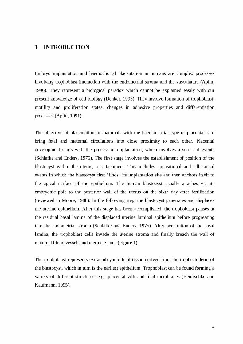
maternal blood vessels and uterine glands (Figure 1).
The trophoblast represents extraembryonic fetal tissue derived from the trophectoderm of
the blastocyst, which in turn is the earliest epithelium. Trophoblast can be found forming a
variety of different structures, e.g., placental villi and fetal membranes (Benirschke and
Kaufmann, 1995).
5
Figure 1 Schematic representation of a human implantation site at approximately nine days after conception (adapted from Aplin, 1991). The outer layer of the embryo consists of STB. Adjacent to the outer STB surface, at its interface with the maternal uterine stroma, is a zone of tissue degradation. Beneath the syncytium is the CTB layer.
Legend: CTB - cytotrophoblast; STB - syncytiotrophoblast; UE - uterine epithelium; US - uterine stroma
As implantation in the human proceeds (Figure 2), the trophoblast cells surrounding the
blastocyst differentiate into a peripheral layer of syncytiotrophoblast and an inner layer of
cytotrophoblast (Hertig and Rock, 1945). Cells from the cytotrophoblast contribute to the
syncytial mass by proliferation and fusion. Trabeculae of proliferating cytotrophoblast
cells between fluid-filled and later maternal blood-filled spaces or lacunae form the
primary villi, which become secondary villi after they are penetrated by fetally derived
extraembryonic mesoderm. These are transformed into tertiary villi after angiogenesis has
taken place within the mesodermal core of the villi (Figure 2).
CTB
STB
6
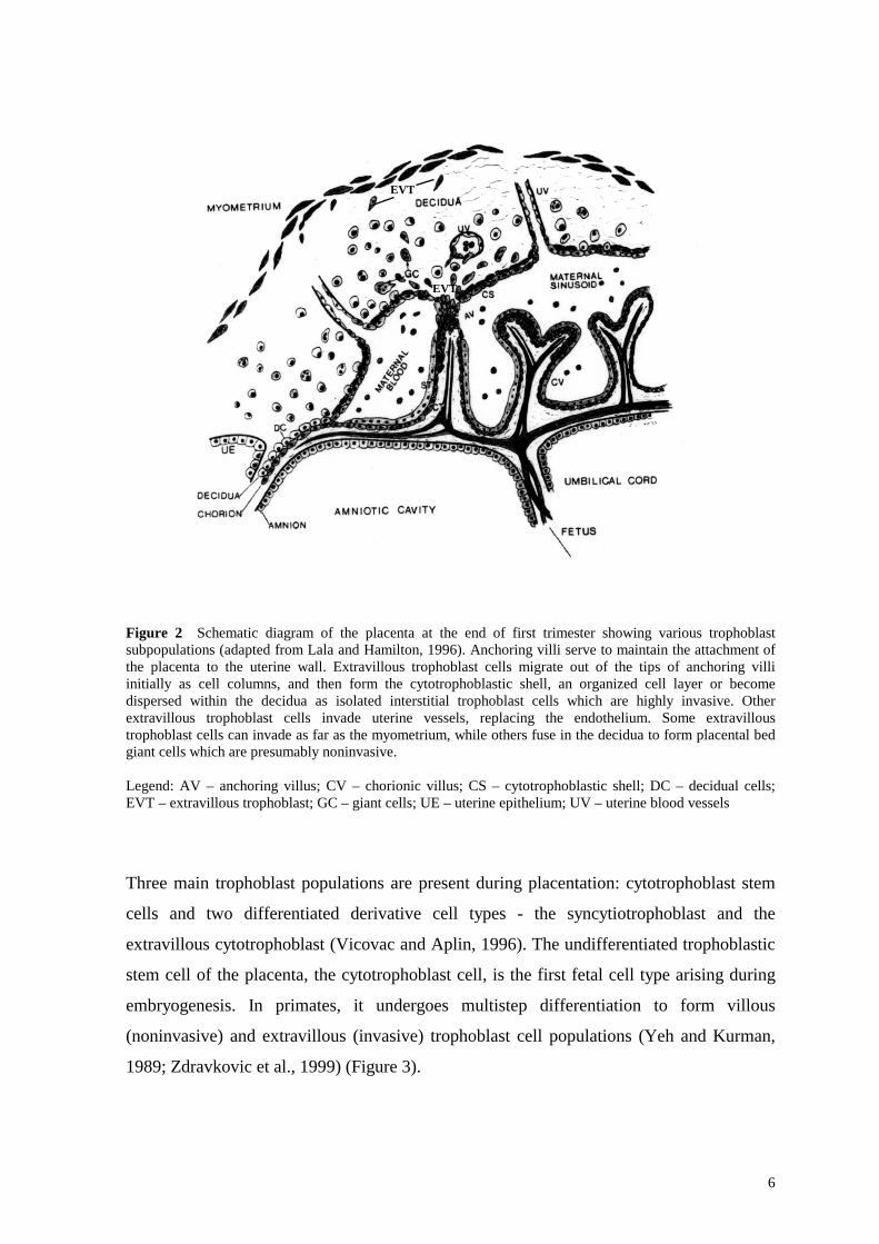
Figure 2 Schematic diagram of the placenta at the end of first trimester showing various trophoblast subpopulations (adapted from Lala and Hamilton, 1996). Anchoring villi serve to maintain the attachment of the placenta to the uterine wall. Extravillous trophoblast cells migrate out of the tips of anchoring villi initially as cell columns, and then form the cytotrophoblastic shell, an organized cell layer or become dispersed within the decidua as isolated interstitial trophoblast cells which are highly invasive. Other extravillous trophoblast cells invade uterine vessels, replacing the endothelium. Some extravillous trophoblast cells can invade as far as the myometrium, while others fuse in the decidua to form placental bed giant cells which are presumably noninvasive. Legend: AV – anchoring villus; CV – chorionic villus; CS – cytotrophoblastic shell; DC – decidual cells; EVT – extravillous trophoblast; GC – giant cells; UE – uterine epithelium; UV – uterine blood vessels
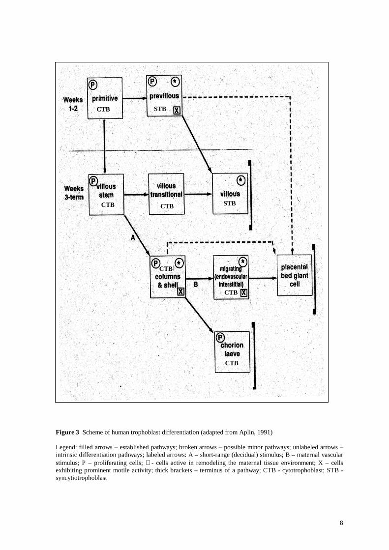
Three main trophoblast populations are present during placentation: cytotrophoblast stem
cells and two differentiated derivative cell types - the syncytiotrophoblast and the
extravillous cytotrophoblast (Vicovac and Aplin, 1996). The undifferentiated trophoblastic
stem cell of the placenta, the cytotrophoblast cell, is the first fetal cell type arising during
embryogenesis. In primates, it undergoes multistep differentiation to form villous
(noninvasive) and extravillous (invasive) trophoblast cell populations (Yeh and Kurman,
1989; Zdravkovic et al., 1999) (Figure 3).
EVT
EVT
EVT
7
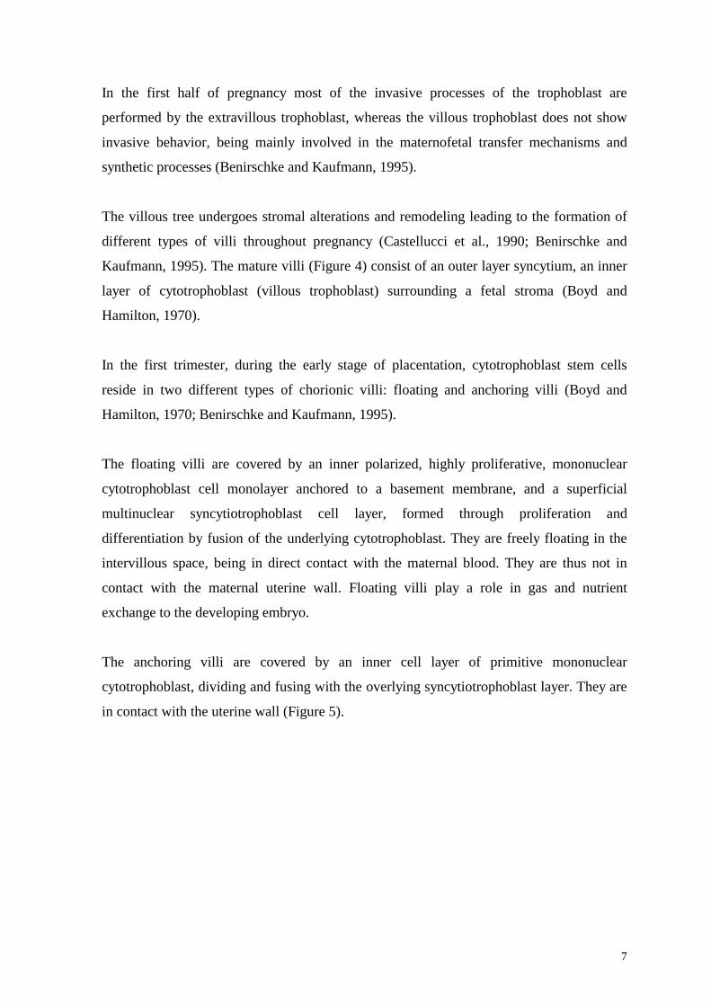
In the first half of pregnancy most of the invasive processes of the trophoblast are
performed by the extravillous trophoblast, whereas the villous trophoblast does not show
invasive behavior, being mainly involved in the maternofetal transfer mechanisms and
synthetic processes (Benirschke and Kaufmann, 1995).
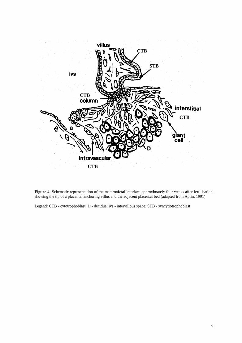
The villous tree undergoes stromal alterations and remodeling leading to the formation of
different types of villi throughout pregnancy (Castellucci et al., 1990; Benirschke and
Kaufmann, 1995). The mature villi (Figure 4) consist of an outer layer syncytium, an inner
layer of cytotrophoblast (villous trophoblast) surrounding a fetal stroma (Boyd and
Hamilton, 1970).
In the first trimester, during the early stage of placentation, cytotrophoblast stem cells
reside in two different types of chorionic villi: floating and anchoring villi (Boyd and
Hamilton, 1970; Benirschke and Kaufmann, 1995).
The floating villi are covered by an inner polarized, highly proliferative, mononuclear
cytotrophoblast cell monolayer anchored to a basement membrane, and a superficial
multinuclear syncytiotrophoblast cell layer, formed through proliferation and
differentiation by fusion of the underlying cytotrophoblast. They are freely floating in the
intervillous space, being in direct contact with the maternal blood. They are thus not in
contact with the maternal uterine wall. Floating villi play a role in gas and nutrient
exchange to the developing embryo.
The anchoring villi are covered by an inner cell layer of primitive mononuclear
cytotrophoblast, dividing and fusing with the overlying syncytiotrophoblast layer. They are
in contact with the uterine wall (Figure 5).
8
Figure 3 Scheme of human trophoblast differentiation (adapted from Aplin, 1991) Legend: filled arrows – established pathways; broken arrows – possible minor pathways; unlabeled arrows – intrinsic differentiation pathways; labeled arrows: A – short-range (decidual) stimulus; B – maternal vascular stimulus; P – proliferating cells; ∗ - cells active in remodeling the maternal tissue environment; X – cells exhibiting prominent motile activity; thick brackets – terminus of a pathway; CTB - cytotrophoblast; STB - syncytiotrophoblast
CTB STB
CTB CTB STB
CTB
CTB
CTB
9
Figure 4 Schematic representation of the maternofetal interface approximately four weeks after fertilisation, showing the tip of a placental anchoring villus and the adjacent placental bed (adapted from Aplin, 1991)
Legend: CTB - cytotrophoblast; D - decidua; ivs - intervillous space; STB - syncytiotrophoblast
CTB
CTB
STB
CTB
CTB

10
Figure 5 The anchoring villus model of the human placenta (adapted from Denker, 1993) 1 cytotrophoblast cells in contact with the basal membrane (integrin α6β4, polarized) 2 detaching extravillous cytotrophoblast (intermediate cytotrophoblast; integrin α6β4,
unpolarized) 3 invasive extravillous cytotrophoblast (integrins α5β1 and α1β1) 4 extravillous cytotrophoblast within maternal spiral artery (integrin α5β1)
The cytotrophoblast cells at the tip of the anchoring tertiary villi proliferate during the third
post-ovulatory week and penetrate the syncytium, becoming nonpolarized cells, losing
their basement membrane, forming the cytotrophoblast multilayered cell columns and
making contact with the underlying maternal decidual tissue (Figure 6); this
cytotrophoblast is termed the extravillous or intermediate cytotrophoblastic shell or lineage
1
2
3
4
11
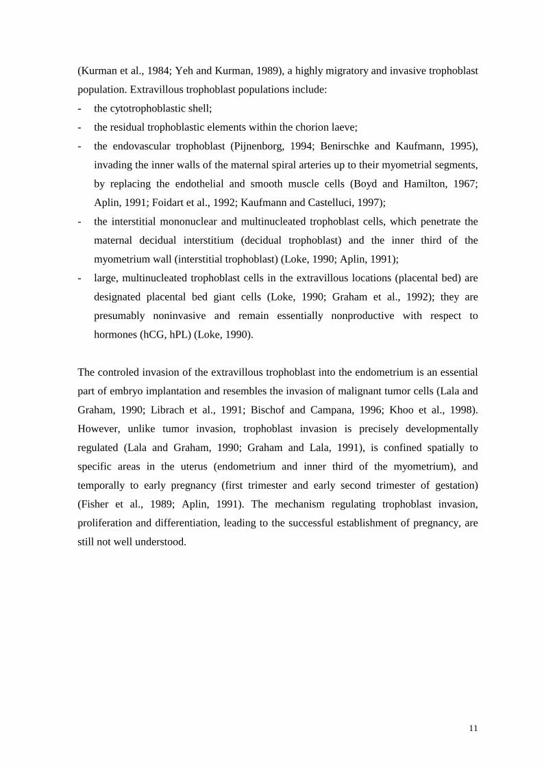
(Kurman et al., 1984; Yeh and Kurman, 1989), a highly migratory and invasive trophoblast
population. Extravillous trophoblast populations include:
- the cytotrophoblastic shell;
- the residual trophoblastic elements within the chorion laeve;
- the endovascular trophoblast (Pijnenborg, 1994; Benirschke and Kaufmann, 1995),
invading the inner walls of the maternal spiral arteries up to their myometrial segments,
by replacing the endothelial and smooth muscle cells (Boyd and Hamilton, 1967;
Aplin, 1991; Foidart et al., 1992; Kaufmann and Castelluci, 1997);
- the interstitial mononuclear and multinucleated trophoblast cells, which penetrate the
maternal decidual interstitium (decidual trophoblast) and the inner third of the
myometrium wall (interstitial trophoblast) (Loke, 1990; Aplin, 1991);
- large, multinucleated trophoblast cells in the extravillous locations (placental bed) are
designated placental bed giant cells (Loke, 1990; Graham et al., 1992); they are
presumably noninvasive and remain essentially nonproductive with respect to
hormones (hCG, hPL) (Loke, 1990).
The controled invasion of the extravillous trophoblast into the endometrium is an essential
part of embryo implantation and resembles the invasion of malignant tumor cells (Lala and
Graham, 1990; Librach et al., 1991; Bischof and Campana, 1996; Khoo et al., 1998).
However, unlike tumor invasion, trophoblast invasion is precisely developmentally
regulated (Lala and Graham, 1990; Graham and Lala, 1991), is confined spatially to
specific areas in the uterus (endometrium and inner third of the myometrium), and
temporally to early pregnancy (first trimester and early second trimester of gestation)
(Fisher et al., 1989; Aplin, 1991). The mechanism regulating trophoblast invasion,
proliferation and differentiation, leading to the successful establishment of pregnancy, are
still not well understood.
12
Figure 6 Diagram of a longitudinal section of an anchoring chorionic villus at the fetal-maternal interface at approximately 10 weeks gestational age (adapted from Zhou et al., 1997). The anchoring villus functions as a bridge between the fetal and maternal compartments, whereas floating villi are suspended in the intervillous space and are bathed by maternal blood. CTB in anchoring villi (Zone I) form cell columns (Zones II and III). CTB then invade the uterine interstitium - decidua and first third of the myometrium (Zone IV) and maternal vasculature (Zone V). Zones designations mark areas in which CTB have distinct patterns of adhesion receptor expression as described in the Discussion section.
Legend: AV – anchoring villus; FV – floating villus
For the isolation of cytotrophoblast cells from human placenta, various methods using
different techniques have been reported (reviewed in Bloxam et al., 1997). Criteria for a
successful model for the study of implantation include substantially pure trophoblast cells
from placental villous tissue. All current methods of isolating CTB, the precursor of STB,
derive from the original tissue trypsinization method of Thiede (1960).
All the reported methods only resulted in an enriched but rather heterogeneous trophoblast
cell population. Contaminating cells include placental macrophages (Hofbauer cells),
lymphocytes, monocytes, granulocytes, and other blood elements, endothelial cells,
fibroblasts, giant cells, and decidual cells. Therefore, reported cytotrophoblast cell purity

13
varies from 40 to 95 %, depending on the isolation procedure applied (Douglas and King,
1989).
The objective of this thesis is to report on a reproducible method for simultaneous isolation
of human villous and extravillous cytotrophoblast cells from first trimester placenta,
applying mechanical and enzymatic dissociation, Percoll gradient centrifugation, and
immunomagnetic separation. The availability of highly purified, competent cytotrophoblast
cells is greatly increasing the possibility of studying placenta development and the factors
controling trophoblast cell invasion into the uterine wall.
14
2 MATERIALS AND METHODS
2.1 Chemicals BSA fraction V: Serva, Boehringer Ingelheim Bioproducts, Heidelberg, Germany Collagenase type IV: Sigma, Munich, Germany Cytospin slides: Shandon, Pittsburgh, USA Density marker beads: Pharmacia Biotech, Uppsala, Sweden DMEM H-21: Gibco Life Technology, Paisley, UK DNAse I type IV: Sigma, Munich, Germany Dynabeads M-450 CD45: Dynal, Oslo, Norway EDTA: Sigma, Munich, Germany Ethanol: Merck, Darmstadt, Germany FCS: Gibco Life Technology, Paisley, UK Fix & Perm Cell Permeabilization Kit: Caltag Laboratories, San Francisco, USA Gentamycin: Seromed, Berlin, Germany HBSS: Gibco Life Technology, Paisley, UK Hyaluronidase type I-S: Sigma, Munich, Germany Percoll: Pharmacia Biotech AB, Uppsala, Sweden PFA: Serva, Boehringer Ingelheim Bioproducts, Heidelberg, Germany Propidium iodide: Sigma, Munich, Germany Sodium azide (NaN3): Merck, Darmstadt, Germany Trypsin type XIII: Sigma, Munich, Germany
15
2.2 Isolation of human cytotrophoblast cells
2.2.1 Materials
Phosphate - buffered saline (PBS)
81.8 g NaCl
13.8 g NaH2PO4 • H2O
Filling up with bidistilled water to one liter (stock solution)
Dilution 1:10 with bidistilled water (use solution)
Setting to pH 7.3 with 2N NaOH
(Reagents from Merck, Darmstadt, Germany)
Erythrocyte lysis buffer
8.29 g NH4Cl
1 g Na2CO3
0.038 g EDTA (Titriplex III)
Filling up with bidistilled water to one liter
Setting to pH 7.2 with 2N NaOH
(Reagents from Merck, Darmstadt, Germany)
Discontinuous Percoll gradient
The six layer Percoll gradient (22 – 78 %) was prepared in Hanks' balanced salt
solution (HBSS, pH 7.4) as follows:
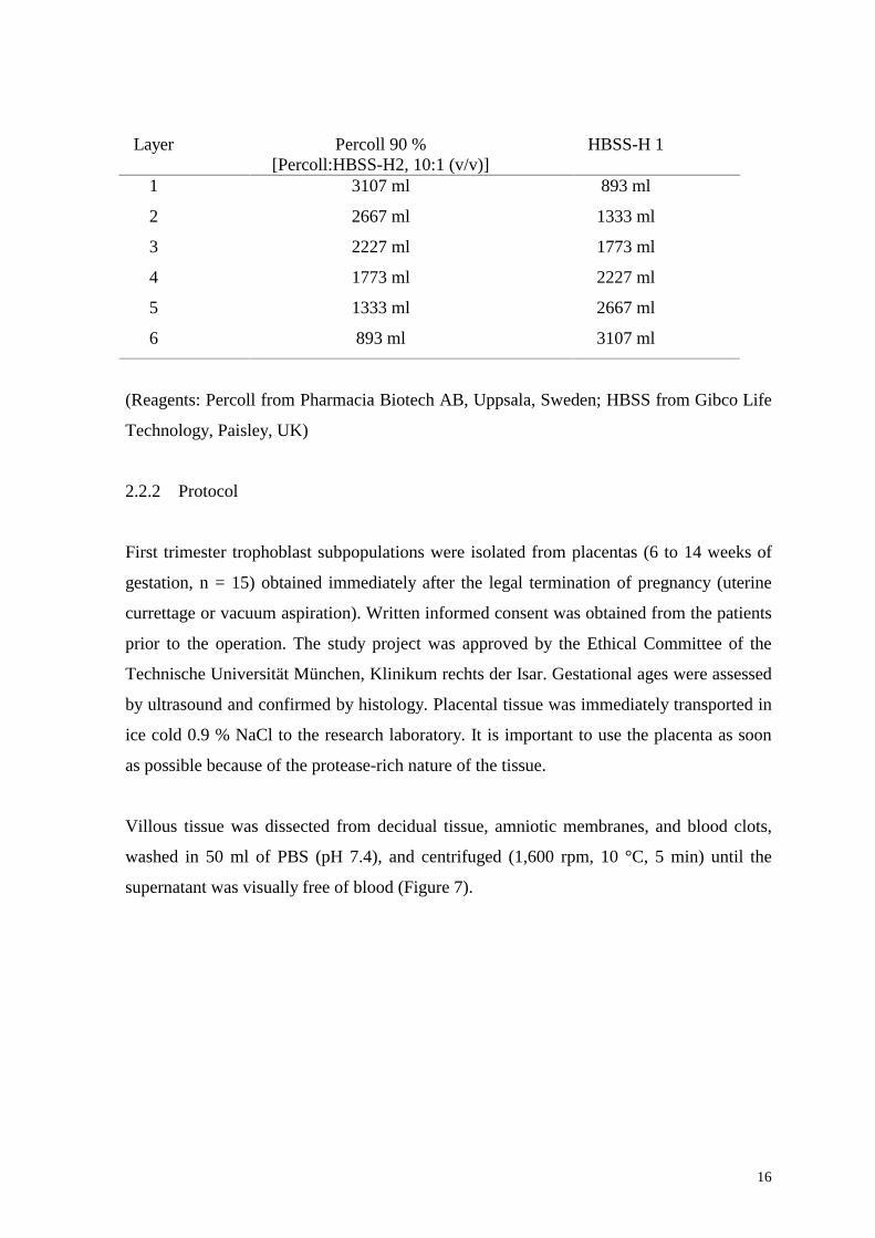
16
Layer Percoll 90 % [Percoll:HBSS-H2, 10:1 (v/v)]
HBSS-H 1
1 3107 ml 893 ml
2 2667 ml 1333 ml
3 2227 ml 1773 ml
4 1773 ml 2227 ml
5 1333 ml 2667 ml
6 893 ml 3107 ml
(Reagents: Percoll from Pharmacia Biotech AB, Uppsala, Sweden; HBSS from Gibco Life
Technology, Paisley, UK)
2.2.2 Protocol
First trimester trophoblast subpopulations were isolated from placentas (6 to 14 weeks of
gestation, n = 15) obtained immediately after the legal termination of pregnancy (uterine
currettage or vacuum aspiration). Written informed consent was obtained from the patients
prior to the operation. The study project was approved by the Ethical Committee of the
Technische Universität München, Klinikum rechts der Isar. Gestational ages were assessed
by ultrasound and confirmed by histology. Placental tissue was immediately transported in
ice cold 0.9 % NaCl to the research laboratory. It is important to use the placenta as soon
as possible because of the protease-rich nature of the tissue.
Villous tissue was dissected from decidual tissue, amniotic membranes, and blood clots,
washed in 50 ml of PBS (pH 7.4), and centrifuged (1,600 rpm, 10 °C, 5 min) until the
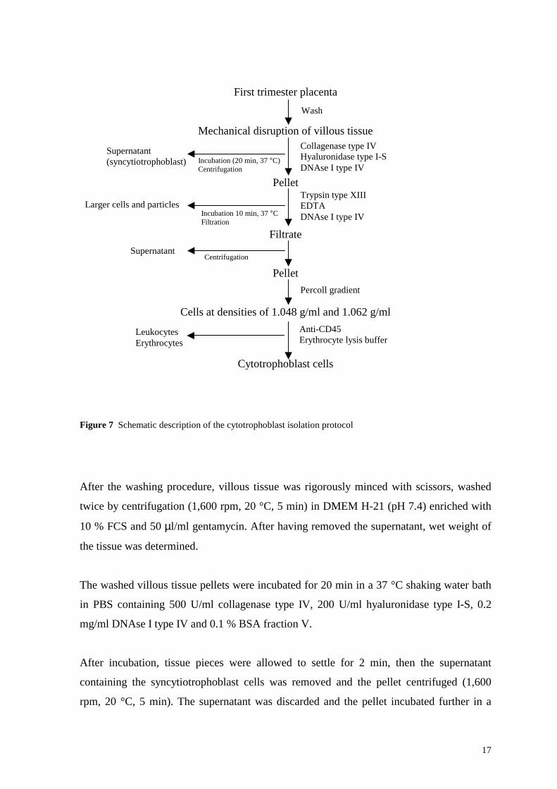
supernatant was visually free of blood (Figure 7).
17
Wash
Collagenase type IV Hyaluronidase type I-S DNAse I type IV
Trypsin type XIII EDTA DNAse I type IV
Centrifugation
Percoll gradient
Anti-CD45 Erythrocyte lysis buffer
Larger cells and particles
Leukocytes Erythrocytes
First trimester placenta
Mechanical disruption of villous tissue
Pellet
Filtrate
Pellet
Cells at densities of 1.048 g/ml and 1.062 g/ml
Cytotrophoblast cells
Figure 7 Schematic description of the cytotrophoblast isolation protocol
After the washing procedure, villous tissue was rigorously minced with scissors, washed
twice by centrifugation (1,600 rpm, 20 °C, 5 min) in DMEM H-21 (pH 7.4) enriched with
10 % FCS and 50 µl/ml gentamycin. After having removed the supernatant, wet weight of
the tissue was determined.
The washed villous tissue pellets were incubated for 20 min in a 37 °C shaking water bath
in PBS containing 500 U/ml collagenase type IV, 200 U/ml hyaluronidase type I-S, 0.2
mg/ml DNAse I type IV and 0.1 % BSA fraction V.
After incubation, tissue pieces were allowed to settle for 2 min, then the supernatant
containing the syncytiotrophoblast cells was removed and the pellet centrifuged (1,600
rpm, 20 °C, 5 min). The supernatant was discarded and the pellet incubated further in a
Supernatant (syncytiotrophoblast)
Supernatant
Incubation (20 min, 37 °C) Centrifugation
Incubation 10 min, 37 °CFiltration
18
shaking water bath at 37 °C for 10 min in PBS containing 0.25 % trypsin type XIII, 2 mM
EDTA, and 0.2 mg/ml DNAse I type IV.
After incubation, the suspension of villous cores and dissociated cells was diluted with an
equal volume of DMEM H-21 containing 10 % FCS to inhibit trypsin activity. The
suspension was filtered through 50 µm gaze in order to remove larger tissue fragments, the
filtrate was then centrifuged (1,600 rpm, 20 °C, 5 min) and the supernatant removed. The
cell pellet was resuspended in 4 ml of DMEM H-21 containing 10 % FCS.
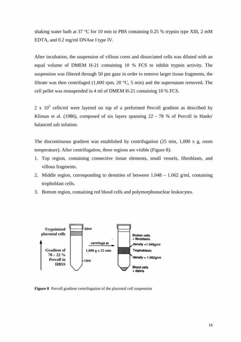
2 x 10⁵ cells/ml were layered on top of a preformed Percoll gradient as described by
Kliman et al. (1986), composed of six layers spanning 22 - 78 % of Percoll in Hanks'
balanced salt solution.
The discontinuous gradient was established by centrifugation (25 min, 1,000 x g, room
temperature). After centrifugation, three regions are visible (Figure 8):
1. Top region, containing connective tissue elements, small vessels, fibroblasts, and
villous fragments.
2. Middle region, corresponding to densities of between 1.048 – 1.062 g/ml, containing
trophoblast cells.
3. Bottom region, containing red blood cells and polymorphonuclear leukocytes.
Figure 8 Percoll gradient centrifugation of the placental cell suspension
Trypsinized placental cells
Gradient of78 – 22 %Percoll in
HBSS
1,000 g x 25 min
19
Cells at a density of 1.048 and 1.062 g/ml corresponding to bands 3 and 4, respectively,
containing the cytotrophoblast cells, were precauciously collected using a Pasteur pipette
and the cell suspension washed twice with PBS (1,600 rpm, 20 °C, 5 min). To lyse
contaminating erythrocytes, the cell suspension was subjected to 5 ml of erythrocyte lysis
buffer, centrifuged (1,600 rpm, 20 °C, 5 min) and the supernatant discarded.
96 - 98 % of the cells were alive, as tested by propidium iodide exclusion.
2.3 Cell fixation
2.3.1 Principle
Fixing cells in suspension and permeabilizing them gives antibodies access to intracellular
structures and leaves the morphological scatter characteristics of the cells intact. Specific
formulation of the reagents reduce background staining and allow simultaneous addition of
permeabilization medium and fluorochrome labeled antibodies.
Cell fixation and permeabilization is an established technique designed for use in flow
cytometry. They allow intracellular antigen analysis as easy as analysis of surface antigens.
The only prerequisit is the availability of suitable antibody conjugates, because some
determinants are sensitive to the fixation step.
2.3.2 Materials
Fix & Perm Cell Permeabilization Kit
FIX & PERM reagents are intended for fixing cells in suspension with Reagent A and then
permeabilizing the cells with Reagent B:
1. addition of 50 µl of cell suspension in a 5 ml tube;
2. addition of 100 µl of Reagent A (Fixation Medium, stored at room temperature);
3. incubation for 15 minutes at room temperature;
4. addition of 5 ml PBS; centrifugation for 5 min at 300 g; removal of the supernatant;
5. addition of 100 µl of Reagent B (Permeabilization Medium) to cell pellet; vortex at low
speed for 1-2 seconds;
20
6. incubation for 15 minutes at room temperature;
7. washing cells with PBS as described above;
8. removal of the supernatant and resuspension of cells in sheath fluid for immediate
analysis or in 1 % PFA; storage of cells at 2-8 °C in the dark.
(Reagent: Caltag Laboratories, San Francisco, USA)
Paraformaldehyde (1 %)
10 g paraformaldehyde are solubilized in 200 ml bidistilled water with 2N NaOH on a
warm plate. The solution is then cooled down to 4 °C. 25 ml PBS are added and the
solution filled up to 250 ml with bidistilled water. The solution is filtered through filter
paper. Freshly prepared solution is needed for each experiment.
(Reagents: PFA from Serva, Boehringer Ingelheim Bioproducts, Heidelberg, Germany)
2.3.3 Protocol
Cell suspensions were suspended in PBS, fixed and permeabilized for flow
cytofluorometry by use of the Fix & Perm Cell Permeabilization Kit, according to the
manufacturer’s instructions. Cells were finally subjected to a second fixation procedure by
resuspension in 1 % PFA and stored at 4 °C in the dark. If fixed cells were not analyzed
within 24 hours, cell suspensions were resuspended in 0.01 % NaN3 and stored at 4 °C in
the dark.
2.4 Immunocytochemical staining of trophoblast cells
2.4.1 Principle of the hematoxylin – eosin staining technique
The hematoxylin – eosin staining technique is a relatively simple, reliable method using
the acidic dye anyline. Eosin stains the cell cytoplasm red, while hematoxylin stains the
cell nucleus blue after a washing procedure with distilled water and tap water (causing an
alkaline pH change). Raising the pH to the alkaline range determines a permanent staining
of the cell nucleus as well, because hematoxylin is not water soluble at neutral or alkaline
pH. Destaining is performed using an acidic solution (2 % acetic acid). The sample is then
21
dehydrated by addition of increasing concentrations of alcohol:water, and finally
embedded in paraffin.
2.4.2 Principle of immunocytochemical staining
The alkaline phosphatase anti-alkaline phosphatase (APAAP) method is one of the most
sensitive and widely used immunologic – enzymatic staining techniques for light
microscopy. The staining sequence is as follows: primary antibody – secondary bridging
antibody – soluble APAAP complex – substrate solution. The primary antibody and the
APAAP complex belong to the same animal species (e.g., mouse), in order to allow the
secondary antibody to bind both.
The bridging antibody must fulfil two requirements:
1. it has to be directed toward immunoglobulins of the same species in which primary
antibody and APAAP complex were produced;
2. an excess amount of this antibody has to be added in order to still have a free Fab
fragment available for binding the APAAP complex, in case one of the Fab fragments
bound the primary antibody.
A soluble enzyme – anti-enzyme immune complex (APAAP) is used, in which two
molecules of alkaline phosphatase correspond to a molecule of antibody directed toward
this enzyme. During incubation of the alkaline phosphatase substrate, the alkaline
phosphatase hydrolyzes the naphtholphosphate-ester, resulting in the production of
phenoles and phosphates. Phenoles combine to chromogenous diazonium-salts, yielding
the production of soluble azo-dyes. Levamisol contained in the substrate solution blocks
the endogenous alkaline phosphatase activity.
2.4.3 Protocol
Cells, at a concentration of 5 x 10⁴ / ml PBS - 1 % BSA, were centrifuged onto single well
slides at room temperature (500 rpm, 5 min). Cytospins were air-dried at room temperature
and then fixed in 70 % ethanol. Hematoxylin - eosin staining was performed.
22
Cytospins were analyzed using an inverted microscope (Axiovert, Zeiss, Oberkochen,
Germany) equipped with a 40 x objective. Photomicrographs were taken using the attached
Contax 167 MT camera (Kyocera Corporation, Japan) with a Kodak Elite II day-light film,
ISO 100 / 21° and a grey filter.
Placental tissue as well as minced tissue pieces and cell suspensions collected after first
and second enzymatic digestion, and also cells isolated by gradient centrifugation and after
immunomagnetic separation, were embedded in fibrin clots for immunocytochemical
analysis as previously described by Luther et al. (1997). Cells embedded in fibrin clots
were fixed in phosphate-buffered 3.7 % formalin (12 h, room temperature), and embedded
in paraffin. Sections were cut (5 µm), deparaffinized, and rehydrated according to standard
procedures (Luther et al., 1997). Primary antibodies were added for staining (Table 1).
Table 1 Primary antibodies used in immunocytochemistry (ICC)
Antibody Concentration (µg/µl)
Source
anti-CK 8 (mAb, mouse IgG2a) 0.5 Dako, Hamburg, Germany
anti-CK 18 (mAb, mouse IgG2a) 0.5 Dako, Hamburg, Germany
anti-E-cadherin (mAb, mouse Ig) 0.25 Dianova, Hamburg, Germany
anti-β-hCG (pAb, rabbit Ig) 2.5 Dako, Hamburg, Germany
anti-hPL (pAb, rabbit Ig) 2.5 Dako, Hamburg, Germany
anti-CD45 (mAb, mouse IgG1) 0.5 Dako, Hamburg, Germany
After incubation with the primary antibodies (1 h, room temperature), sections were
washed three times (5 min each, room temperature) with TBS and processed by adding
secondary antibodies (30 min, room temperature) and detection systems:
- for CK 8,18, β-hCG, hPL: Dako ChemMate detection-kit, APAAP, mouse (Dako
Diagnostika, Hamburg, Germany);
- for CD45: Dako ChemMate detection-kit, alkaline phosphatase/RED, rabbit/mouse
(Dako Diagnostika, Hamburg, Germany) and Vectastain Elite ABC-Kit (Camon,
Wiesbaden, Germany);
23
- for E-cadherin: Vectastain Elite ABC-Kit.
Kit instructions were followed with regard to dilutions of reactants.
Cell nuclei were counterstained with hematoxylin. Positive stains gave a red color. Finally,
slides were mounted on microscope slides with glycerol-gelatine and covered. The
peroxidase activity was detected with the diaminobenzidine reaction. No antibody was
evaluated on fewer than three samples.
Sections not exposed to primary antibodies or in which the primary antibody was replaced
by irrelevant mouse immunoglobulin or normal rabbit serum served as control slides. None
of the controls exhibited any significant staining.
2.5 Confocal Laser Scanning Microscopy (CLSM)
2.5.1 Principle
Cell-bound fluorescence can be assessed, among other techniques, by CLSM (Knebel et
al., 1990; Schmitt et al., 1991). CLSM is a specific technique for the study of cell
morphology, using antibodies directed to intracellular or membrane bound cell
components. In order to reveal the binding site of the specific primary antibody, a second
fluorescence-labeled antibody is added, which will bind to the first antibody. The
fluorescent molecule on the second antibody is excited by light with a lower wave length,
and subsequently emits light with a higher wave length. Special filters separate absorbance
and emission lights, resulting in fluorescence of the labeled cell structures visualized by a
fluorescence microscope. This technique is called indirect immunofluorescence technique
because only the second antibody is fluorescence-labeled.
Many biological structures are complex and thick, resulting in superposition of the
fluorescent components, thus impeding sample visualization in a single layer when using a
conventional fluorescence microscope. The same problem is encountered when a three-
dimensional sample is analyzed. CLSM allows emission identifying off the focus and is
able to perform "optical slices" of a sample. It performs rapid scanning while moving the

24
cell in the z-axis in order to measure total cellular fluorescence and to minimize photo-
bleaching of the fluorescent label (Schmitt et al., 1991).
The principle of CLSM can be explained as follows: The light source is a laser beam,
focused by a lens and deviated by a scanning mirror in the microscope objective and the
sample, respectively. The scanning mirror is turned in two axes. This arrangement
considerably reduces the number of optical components, since no relaying optics for two-
scanner mirrors are necessary. The high fluorescence yields a good image to be constructed
from one scan. While passing through the sample, the laser beam excites fluorescent
molecules to a certain wave length, resulting in a few nanoseconds in the emission of light
with a higher wave length in all directions. The objective partially absorbs the emitted light
and focuses it toward another mirror and a detector. The mirror is light splicing, i.e. it
reflects light in a specific wave length range (e.g., blue light), while it is permissive for
other wave lengths (e.g., yellow or green light). The detector in turn converts the light
signal into an electrical signal.
The main advantage of CLSM resides in its high selectivity, i.e. only the labeled structures
are visualized. High resolution of the technique is achieved by combining a fluorescence
image (specific contrast image) with its transmission light image (unspecific phase
interference contrast image). CLSM not only provides alternatives to the use of
fluorescence-labeled ligands, but also allows real-time single cell analysis of living
competent cells (Schmitt et al., 1991).
2.5.2 Protocol
Single cell-associated fluorescence was measured with the confocal laser scanning
microscope. CLSM was performed on cells fixed with 1 % PFA in PBS. Cytospins (30,000
cells/slide) were prepared as described above.
Primary antibodies were applied for 30 min, at room temperature (Table 2).
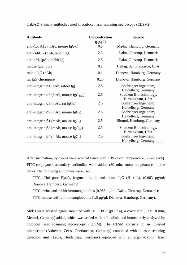
25
Table 2 Primary antibodies used in confocal laser scanning microscopy (CLSM)
Antibody Concentration (µg/µl)
Source
anti-CK 8,18 (mAb, mouse IgG2a) 0.5 Medac, Hamburg, Germany
anti-β-hCG (pAb, rabbit Ig) 2.5 Dako, Glostrup, Denmark
anti-hPL (pAb, rabbit Ig) 2.5 Dako, Glostrup, Denmark
mouse IgG1 pure 0.1 Caltag, San Francisco, USA
rabbit IgG (pAb) 0.1 Dianova, Hamburg, Germany
rat IgG chrompure 0.25 Dianova, Hamburg, Germany
anti-integrin α1 (pAb, rabbit Ig) 2.5 Boehringer Ingelheim, Heidelberg, Germany
anti-integrin α5 (mAb, mouse IgG2bK) 2.5 Southern Biotechnology, Birmingham, USA
anti-integrin α6 (mAb, rat IgG2a) 2.5 Boehringer Ingelheim, Heidelberg, Germany
anti-integrin αv (mAb, mouse IgG1) 2.5 Boehringer Ingelheim, Heidelberg, Germany
anti-integrin β1 (mAb, mouse IgG1) 2.5 Biomol, Hamburg, Germany
anti-integrin β3 (mAb, mouse IgG1K) 2.5 Southern Biotechnology, Birmingham, USA
anti-integrin β4 (mAb, mouse IgG1) 2.5 Boehringer Ingelheim, Heidelberg, Germany
After incubation, cytospins were washed twice with PBS (room temperature, 5 min each).
FITC-conjugated secondary antibodies were added (30 min, room temperature, in the
dark). The following antibodies were used:
- FITC-affini pure F(ab’)2 fragment rabbit anti-mouse IgG (H + L), (0.063 µg/ml;
Dianova, Hamburg, Germany);
- FITC-swine anti-rabbit immunoglobulins (0.063 µg/ml; Dako, Glostrup, Denmark);
- FITC-mouse anti-rat immunoglobulins (1.5 µg/µl; Dianova, Hamburg, Germany).
Slides were washed again, mounted with 50 µl PBS (pH 7.4), a cover slip (18 x 18 mm,
Menzel, Germany) added, which was sealed with nail polish, and immediately analyzed by
confocal laser scanning microscopy (CLSM). The CLSM consists of an inverted
microscope (Axiovert, Zeiss, Oberkochen, Germany) combined with a laser scanning
detection unit (Leica, Heidelberg, Germany) equipped with an argon-krypton laser
26
allowing separate or simultaneous detection of fluorochromes excited at 488 nm and 568
nm. Transmission (differential interference contrast microscopy) and fluorescence images
were recorded employing a 63 oil immersion objective.
Washing steps in each case were designed to be sufficient to eliminate background
staining. Second antibody controls were performed in each run by using PBS in place of
the first antibody and were negative in all cases.
2.6 Flow cytofluorometric analysis
2.6.1 Principle
Laser-based fluorometry has found wide application in cell biology and medicine to
investigate structure – function relationships of ligands with their receptors on normal and
malignant cells. Cell-bound fluorescence can be quantified, among other techniques, by
flow cytofluorometry (Shapiro, 1988).
The principle of flow cytofluorometry resides in the simultaneous assessment of different
physical characteristics of single fluorescence-labeled cells or particles, while passing
through a directed laser beam (argon laser, wave length 488 nm). Fluorescence intensity
and light deviation are measured using different detectors. Light deviation is attributed to
differences in physical properties, e.g., cell size, cell shape, cell membrane, nuclear shape,
number and type of intracellular components, and type of cell membrane surface. Thus,
flow cytofluorometric analysis is a useful technique providing information relative to cell
size, granularity, and antigenicity.
The laser beam induces emission of fluorescence by endogenous or exogenous molecules
present either intracellularly or bound to the cell membrane. The forward light scatter
(FSC) is associated with cell size and cell aggregation. Attenuation of the laser beam
subsequent to cell passing is measured – transmission attenuation. The side scatter (SSC) is
associated with cell density, cell granularity and cell membrane surface, and it represents
the deviated right angle light signal. Fluorescence intensities can be assessed
simultaneously for three different fluorescence emission spectra.
27
Immunologic pattern of cells can be assessed using specific fluorescence-labeled
antibodies. Most cells show a certain degree of fluorescence even without being
fluorescence-labeled; this characteristic is called autofluorescence, and it is attributed to
fluorescent cell components. For this purpose, fluorescence of labeled cell samples is
compared to the autofluorescence of unlabeled cells (negative control), above which cells
can be considered to be positive for expressing the antigens tested.
Negative controls are considered:
- autofluorescence of unlabeled cells;
- fluorescence of the labeled monoclonal antibody (e.g., FITC);
- fluorescence of the unspecific antibody (e.g., mouse IgG2a);
- fluorescence of labeled monoclonal antibody plus unspecific antibody.
Fluorescence values above fluorescence of negative controls are considered positive and
specific.
The fluorescent dyes are coupled to the Fc fragment of immunoglobulins. A fluorescent
antibody can be directed either toward an antigenic cell structure or an unlabeled primary
antibody. The latter method is preferred due to cost effectiveness.
The direction, intensity, and deviation of the emitted light by every single cell are recorded
and analyzed by a computer system and represented graphically as histograms or dot plots,
which can be eventually interpreted. The five parameters mentioned above (two light
scatters and three fluorescence patterns) can be paired sequentially, resulting in a
representation called dot plot.
The advantage of flow cytofluorometry compared to the classic fluorescence microscopy
resides in the quality of cell analysis: different cell types in a sample are quantified
separately, reliable, objective, and very fast (up to 3000 cells/second). However, no
correlation to intracellular components is possible.
In the experiments presented following fluorescence intensities were used:
Fl1 - green; fluorescent dye: FITC, 514 nm;
Fl2 - red; fluorescent dyes: PE, 578 nm; and propidium iodide, 630 nm.
28
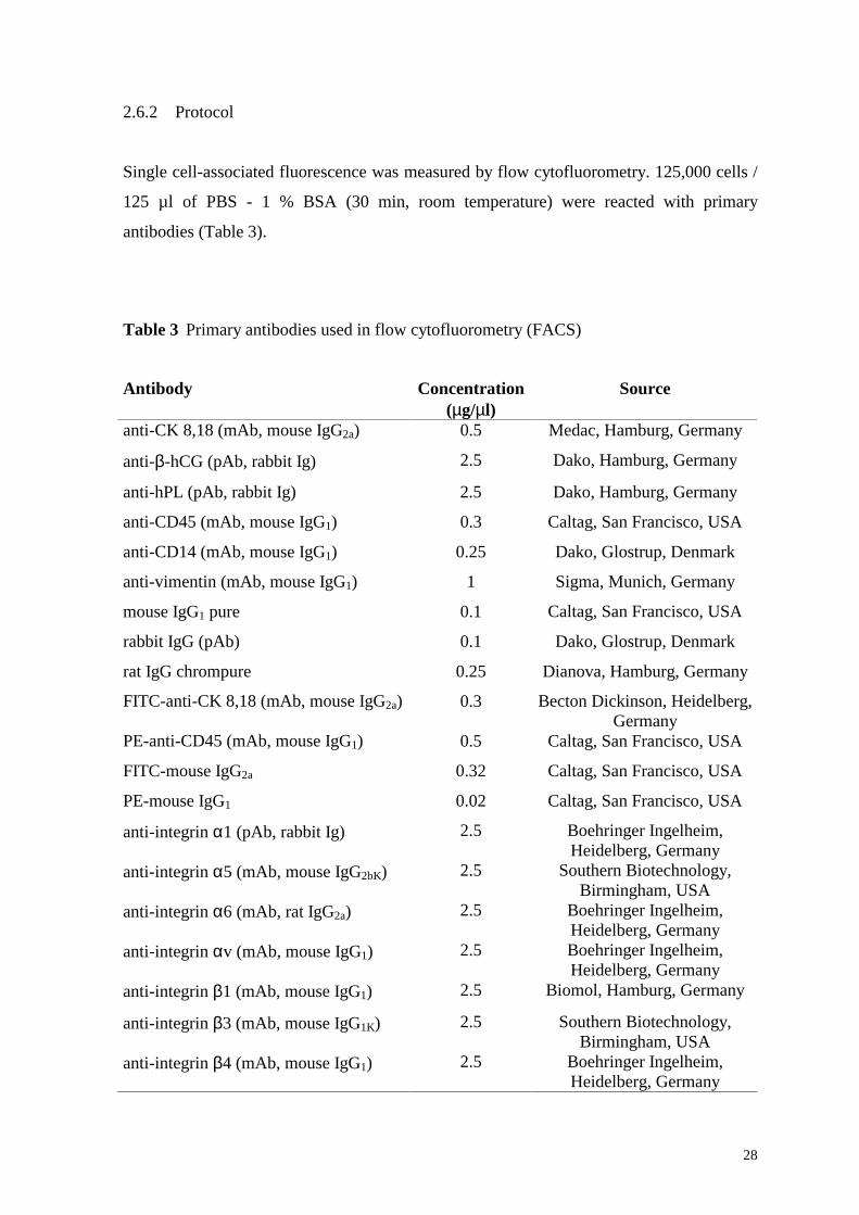
2.6.2 Protocol
Single cell-associated fluorescence was measured by flow cytofluorometry. 125,000 cells /
125 µl of PBS - 1 % BSA (30 min, room temperature) were reacted with primary
antibodies (Table 3).
Table 3 Primary antibodies used in flow cytofluorometry (FACS)
Antibody Concentration (µg/µl)
Source
anti-CK 8,18 (mAb, mouse IgG2a) 0.5 Medac, Hamburg, Germany
anti-β-hCG (pAb, rabbit Ig) 2.5 Dako, Hamburg, Germany
anti-hPL (pAb, rabbit Ig) 2.5 Dako, Hamburg, Germany
anti-CD45 (mAb, mouse IgG1) 0.3 Caltag, San Francisco, USA
anti-CD14 (mAb, mouse IgG1) 0.25 Dako, Glostrup, Denmark
anti-vimentin (mAb, mouse IgG1) 1 Sigma, Munich, Germany
mouse IgG1 pure 0.1 Caltag, San Francisco, USA
rabbit IgG (pAb) 0.1 Dako, Glostrup, Denmark
rat IgG chrompure 0.25 Dianova, Hamburg, Germany
FITC-anti-CK 8,18 (mAb, mouse IgG2a) 0.3 Becton Dickinson, Heidelberg, Germany
PE-anti-CD45 (mAb, mouse IgG1) 0.5 Caltag, San Francisco, USA
FITC-mouse IgG2a 0.32 Caltag, San Francisco, USA
PE-mouse IgG1 0.02 Caltag, San Francisco, USA
anti-integrin α1 (pAb, rabbit Ig) 2.5 Boehringer Ingelheim, Heidelberg, Germany
anti-integrin α5 (mAb, mouse IgG2bK) 2.5 Southern Biotechnology, Birmingham, USA
anti-integrin α6 (mAb, rat IgG2a) 2.5 Boehringer Ingelheim, Heidelberg, Germany
anti-integrin αv (mAb, mouse IgG1) 2.5 Boehringer Ingelheim, Heidelberg, Germany
anti-integrin β1 (mAb, mouse IgG1) 2.5 Biomol, Hamburg, Germany
anti-integrin β3 (mAb, mouse IgG1K) 2.5 Southern Biotechnology, Birmingham, USA
anti-integrin β4 (mAb, mouse IgG1) 2.5 Boehringer Ingelheim, Heidelberg, Germany
29
In order to determine the appropriate antibody concentrations, preliminary antibody
dilution experiments were performed and analyzed by flow cytofluorometry.
Cell suspensions were washed twice with PBS - 1 % BSA and, where applicable, the
respective conjugated secondary antibody added (30 min, room temperature, in the dark).
The following FITC- or PE-conjugated antibodies were used:
- FITC-affini pure F(ab’)2 fragment rabbit anti-mouse IgG (H + L) (0.125 µg/ml;
Dianova, Hamburg, Germany);
- FITC-swine anti-rabbit immunoglobulins (0.125 µg/ml; Dako, Glostrup, Denmark);
- FITC-mouse anti-rat immunoglobulins (1.5 µg/µl; Dianova, Hamburg, Germany);
- PE-mouse anti-human CD45 (0.25 µg/ml; Caltag Laboratories, San Francisco, USA).
Cell suspensions were washed with PBS - 1 % BSA, resuspended in 125 µl PBS - 0.1 %
BSA and immediately analyzed by flow cytofluorometry using the FACS Calibur flow
cytometer (Becton Dickinson, Heidelberg, Germany) equipped with a 488 nm argon laser.
Data were collected and analyzed by the Cell Quest Program (Becton Dickinson,
Heidelberg, Germany). Cells were visualized by analyzing their light scatter properties
(forward versus side scatter signal) and fluorescence signals. Twenty thousand cells were
analyzed for each sample. "Negative" controls were used to set the detectors and marker
settings on fluorescence histograms, above which cells were considered to be positive for
expressing the antigens tested. Detector and amplifier were set as specified in the Results
section.
Controls included:
(a) replacement of the primary antibody with native immunoglobulins of the species in
which the first antibody was raised;
(b) omission of the primary antibody;
(c) untreated cells only for determination of cytotrophoblast cell autofluorescence.
All these "negative" controls provided fluorescence profiles indistinguishable from one
another. The appropriate negative control value was always subtracted from the test sample
before reporting on the percentage of positive cells labeled.

30
2.7 Cell testing for viability
2.7.1 Principle
2.7.1.1 Trypan Blue exclusion
Cell viability is determined by mixing two drops of the cell suspension with two drops of
0.5 % Trypan Blue and after 2 – 5 min, counted in a Neubauer chamber. The average
number of cells that exclude the dye in each large square is multiplied by 2 x 10⁴ to give
the number of cells/ml. Those cells that take up the dye are dead cells.
2.7.1.2 DNA analysis
Flow cytofluorometric analysis can be performed on fixed cells (in PFA or ethanol) or on
living cells. Analysis on living cells allows detection of cell membrane bound antigens
only, while the fixation procedure allows detection of both intracellular and extracellular
antigens. As the cytoplasmic membrane of dead cells becomes permeable to antibodies, it
is necessary to exclude dead cells from the overall population of cells to be analyzed, in
order to detect only extracellular antigens. For this purpose, cells are labeled first with
propidium iodide, a fluorescent dye which binds to cellular DNA. Dead cells are
subsequently excluded by selective gating of the cell population which does not emit
fluorescence at the characteristic wave length for propidium iodide emission.
2.7.2 Protocol
The isolated cells were tested for viability by Trypan blue exclusion and by DNA analysis
of freshly isolated, unfixed cells, labeled with propidium iodide; briefly, 8 µl of 1 mg/ml
propidium iodide were added for 200 µl sample volume; samples were incubated on ice, in
the dark, for 10 min, and then assessed by flow cytofluorometry.
31
3 RESULTS
3.1 Anti-CD45 immunomagnetic separation
In order to purify and characterize cytotrophoblast cells of first trimester placentas from
normal pregnancies a protocol was developed which involves enzymatic dissection and
immunomagnetic separation of trophoblast cells. The cytotrophoblast-enriched fraction
harvested from the Percoll gradient was mixed with Dynabeads conjugated with a
monoclonal antibody (mAb) to pan-leukocyte antigen CD45, in order to remove
contaminating leukocytes.
Freshly dissected trophoblast cells were subjected to Percoll gradient centrifugation, bands
3 and 4 collected (as described in the Materials and Methods section) and then incubated
with 62.5 µl (25 x 106) of Dynabeads M-450 CD45 in 1.25 ml of PBS. The number of
Dynabeads per target cell was ten. The suspension was incubated at 4 °C for 30 min, with
occasional gentle shaking. The test tube was placed on a magnetic particle concentrator
(Dynal MPC-1, Dynal, Oslo, Norway) for 2 min. The cells that interacted with the
Dynabeads (rosetted cells) and nonreacting Dynabeads were collected using the magnetic
collector (Figure 9), and the nonrosetted cell fraction, containing trophoblast cells,
collected. The trophoblast cell containing supernatant devoid of CD45-reactive cells, was
kept for further analyses.
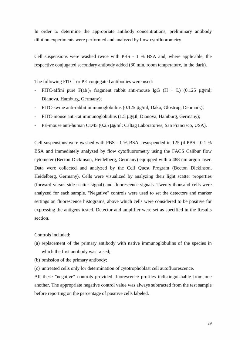
32
Figure 9 Immunocytochemical detection of cells that interact with the Dynabeads, conjugated with mAb to CD45 (rosetted cells)
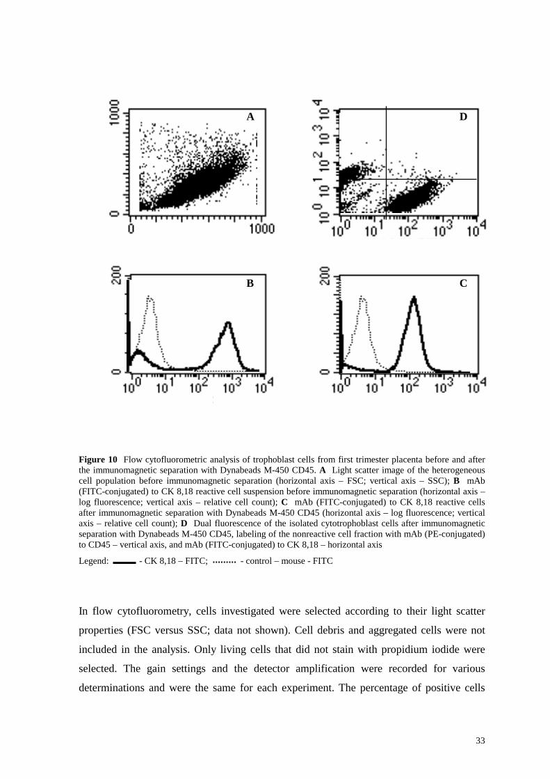
The cell density gradient routinely yielded a cytotrophoblast cell suspension with 65 to 90
% purity before immunomagnetic separation, as judged by cytokeratin 8,18 and E-cadherin
positivity. This cell population was then subjected to "negative" selection applying the
described anti-CD45 immunomagnetic purification protocol. This additional purification
step routinely yielded > 95 % CK 8,18-positive cells. In Figure 10 cell suspensions
investigated by flow cytofluorometry before and after immunomagnetic separation are
shown.
33
Figure 10 Flow cytofluorometric analysis of trophoblast cells from first trimester placenta before and after the immunomagnetic separation with Dynabeads M-450 CD45. A Light scatter image of the heterogeneous cell population before immunomagnetic separation (horizontal axis – FSC; vertical axis – SSC); B mAb (FITC-conjugated) to CK 8,18 reactive cell suspension before immunomagnetic separation (horizontal axis – log fluorescence; vertical axis – relative cell count); C mAb (FITC-conjugated) to CK 8,18 reactive cells after immunomagnetic separation with Dynabeads M-450 CD45 (horizontal axis – log fluorescence; vertical axis – relative cell count); D Dual fluorescence of the isolated cytotrophoblast cells after immunomagnetic separation with Dynabeads M-450 CD45, labeling of the nonreactive cell fraction with mAb (PE-conjugated) to CD45 – vertical axis, and mAb (FITC-conjugated) to CK 8,18 – horizontal axis
Legend: - CK 8,18 – FITC; - control – mouse - FITC
In flow cytofluorometry, cells investigated were selected according to their light scatter
properties (FSC versus SSC; data not shown). Cell debris and aggregated cells were not
included in the analysis. Only living cells that did not stain with propidium iodide were
selected. The gain settings and the detector amplification were recorded for various
determinations and were the same for each experiment. The percentage of positive cells
A D
B C
34
was calculated from the number of events falling within a marker set on the negative
controls.
The success rate of obtaining purities over 95 % cytokeratin-positive cells after negative
selection for anti-CD45 was over 90 %. The present successful isolation of reasonable
numbers of highly purified cytotrophoblast cells from human placentas depended on initial
crude dissection of the chorionic villi, gentle trypsinization to cleave the cytotrophoblast
and syncytiotrophoblast layers from the mesenchyme, and separation of the
cytotrophoblast aggregates by sedimentation at 1,000 x g through a discontinuous Percoll
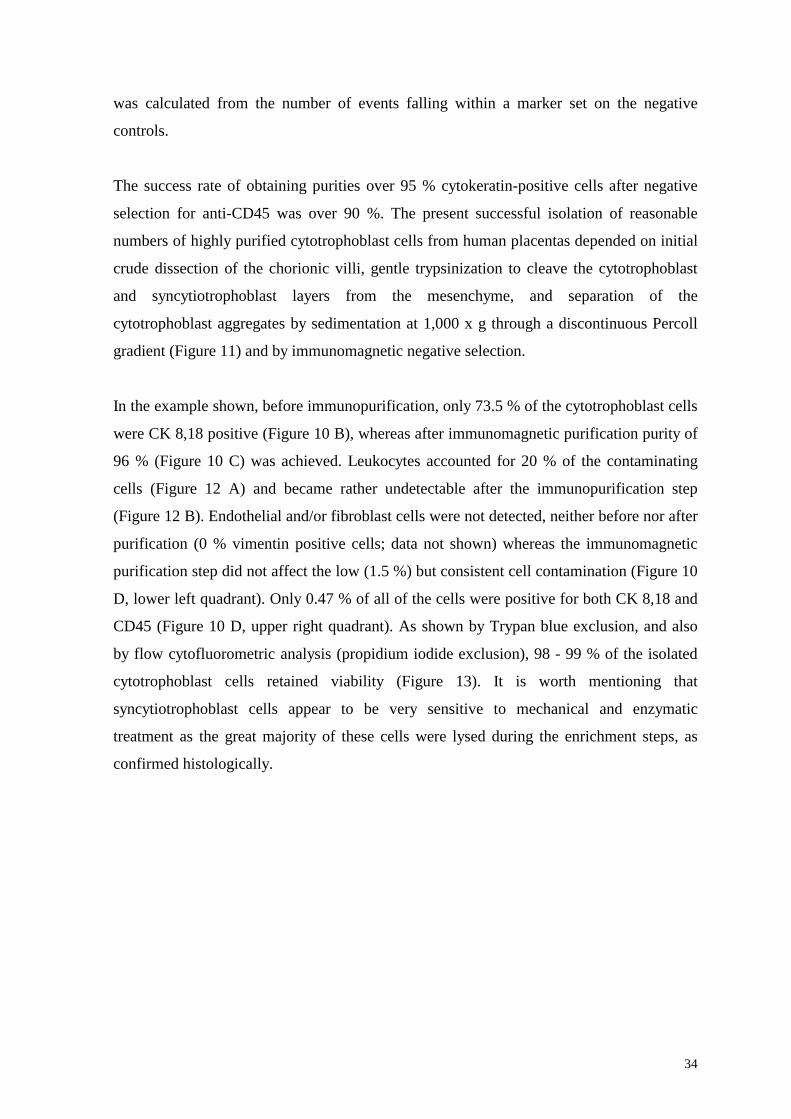
gradient (Figure 11) and by immunomagnetic negative selection.
In the example shown, before immunopurification, only 73.5 % of the cytotrophoblast cells
were CK 8,18 positive (Figure 10 B), whereas after immunomagnetic purification purity of
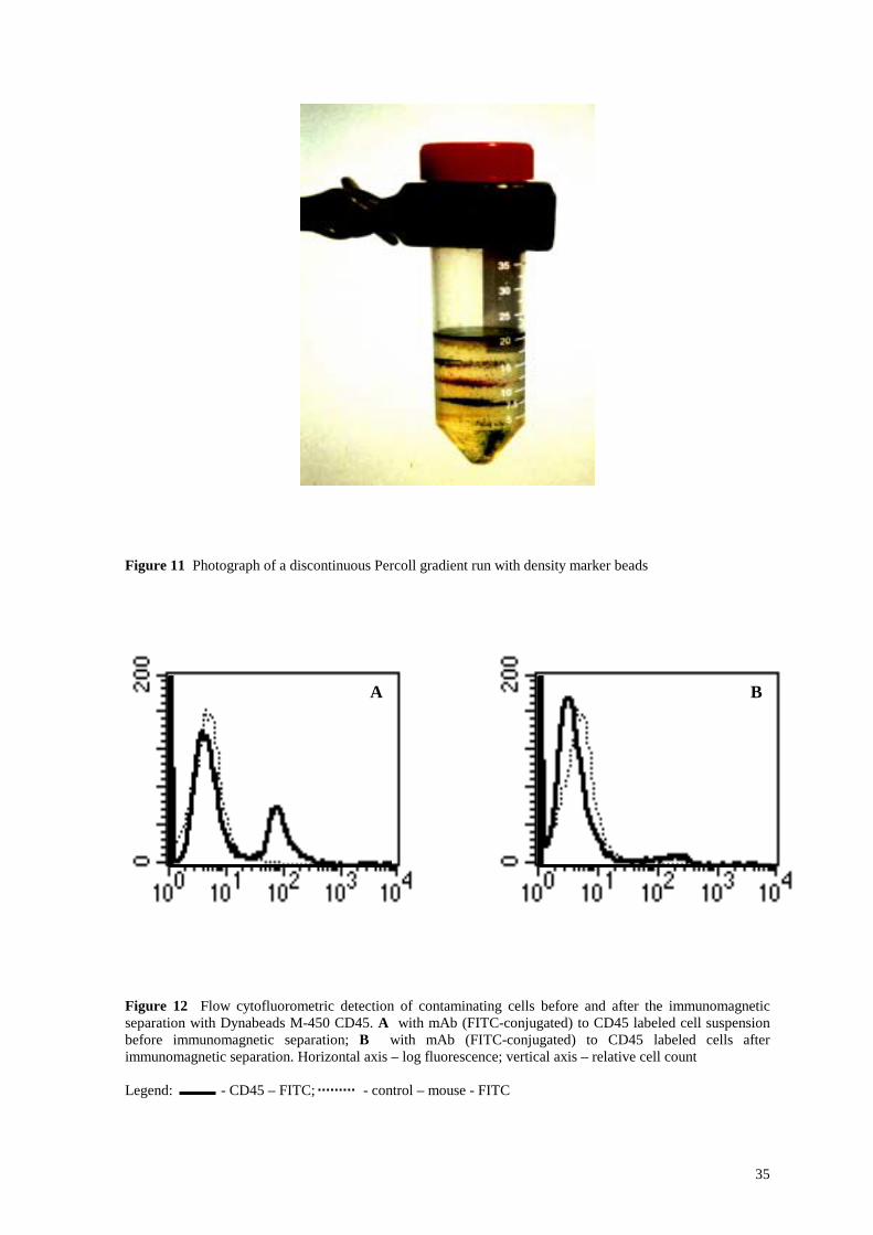
96 % (Figure 10 C) was achieved. Leukocytes accounted for 20 % of the contaminating
cells (Figure 12 A) and became rather undetectable after the immunopurification step
(Figure 12 B). Endothelial and/or fibroblast cells were not detected, neither before nor after
purification (0 % vimentin positive cells; data not shown) whereas the immunomagnetic
purification step did not affect the low (1.5 %) but consistent cell contamination (Figure 10
D, lower left quadrant). Only 0.47 % of all of the cells were positive for both CK 8,18 and
CD45 (Figure 10 D, upper right quadrant). As shown by Trypan blue exclusion, and also
by flow cytofluorometric analysis (propidium iodide exclusion), 98 - 99 % of the isolated
cytotrophoblast cells retained viability (Figure 13). It is worth mentioning that
syncytiotrophoblast cells appear to be very sensitive to mechanical and enzymatic
treatment as the great majority of these cells were lysed during the enrichment steps, as
confirmed histologically.
35
Figure 11 Photograph of a discontinuous Percoll gradient run with density marker beads
Figure 12 Flow cytofluorometric detection of contaminating cells before and after the immunomagnetic separation with Dynabeads M-450 CD45. A with mAb (FITC-conjugated) to CD45 labeled cell suspension before immunomagnetic separation; B with mAb (FITC-conjugated) to CD45 labeled cells after immunomagnetic separation. Horizontal axis – log fluorescence; vertical axis – relative cell count Legend: - CD45 – FITC; - control – mouse - FITC
B A

36
A
R = 1.92 %
B
R = 1.92 %
Eve
nts
PI
stai
ning
inte
nsit
y
Figure 13 Absence of propidium iodide fluorescence staining in freshly isolated cytotrophoblast cells assessed by flow cytofluorometry indicate living cells. A Dot plot (horizontal axis - linear fluorescence; vertical axis - propidium iodide staining intensity); B Histogram (horizontal axis - linear fluorescence; vertical axis - relative cell count)
37
One of the difficulties associated with the use of "negative" selection procedure for
targeting the CD45 antigen by antibodies to this epitope was the adhesion of some of the
trophoblast cells to the immunomagnetic beads, although they did not bind to the antibody.
This was demonstrated by control experiments using the two step immunomagnetic
separation: first incubation with mouse mAb to CD45 (Caltag Laboratories, San Francisco,
USA) for 30 min at room temperature, followed by a second incubation with Dynabeads
M-450 sheep anti-mouse IgG (Dynal, Oslo, Norway) for 30 min at 4 °C. In almost all cases
this resulted in the loss of some trophoblast cells during the immunomagnetic separation.
Similar findings were reported by Aboagye-Mathiesen et al., 1996.
3.2 Characterization of isolated first trimester cytotrophoblast cells
The isolated cytotrophoblast cells were extensively characterized by routine histology,
conventional immunocytochemistry, fluorescence confocal laser scanning microscopy, and
flow cytofluorometry, showing that they express in situ markers of trophoblast cells:
cytokeratin 8,18; E-cadherin; β-hCG; hPL (Zhou et al., 1997; Khoo et al., 1998; Hamilton
et al., 1998); and integrin subunits α1, α5, α6, αv, β1, β3, β4 (Haynes et al., 1997;
Douglas et al., 1999; Thirkill and Douglas, 1999).
To identify the isolated cells, we used two different monoclonal antibodies directed to
trophoblast cells: antigens cytokeratin 8,18 (mAb CAM 5.2) and E-cadherin (mAb
120/80). Immunocytochemistry showed a strong positive cytoplasmic staining of cells
reacting with mAb to CK 8,18 (Figure 14 A) and E-cadherin (Figure 15), respectively.
Already in the cytotrophoblast epithelial monolayer obtained after first enzymatic
dissociation, cytotrophoblast cells stained strongly for E-cadherin, showing a polarized
pattern. Staining was even stronger on the surface of cytotrophoblast cells still in contact
with each other and on their apical surface, and was absent at the basal surface of
cytotrophoblast in contact with the basement membrane.

38
Figure 14 Immunocytochemical labeling of highly enriched first trimester cytotrophoblast cells stained with: A Anti-CK 8,18; B Anti-β-hCG; C Anti-hPL. Please, note the strong cytoplasmic staining of cells with antibodies to CK 8,18 (A) and the focal staining for β-hCG (B). There is a uniform but moderate to weak staining present in the plasma membrane with antibodies to hPL (C). Hematoxylin counterstain of nuclei (x 40)
A
B
C
39
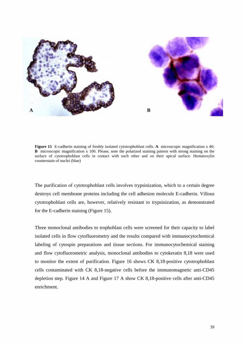
Figure 15 E-cadherin staining of freshly isolated cytotrophoblast cells. A microscopic magnification x 40; B microscopic magnification x 100. Please, note the polarized staining pattern with strong staining on the surface of cytotrophoblast cells in contact with each other and on their apical surface. Hematoxylin counterstain of nuclei (blue)
The purification of cytotrophoblast cells involves trypsinization, which to a certain degree
destroys cell membrane proteins including the cell adhesion molecule E-cadherin. Villous
cytotrophoblast cells are, however, relatively resistant to trypsinization, as demonstrated
for the E-cadherin staining (Figure 15).
Three monoclonal antibodies to trophoblast cells were screened for their capacity to label
isolated cells in flow cytofluorometry and the results compared with immunocytochemical
labeling of cytospin preparations and tissue sections. For immunocytochemical staining
and flow cytofluorometric analysis, monoclonal antibodies to cytokeratin 8,18 were used
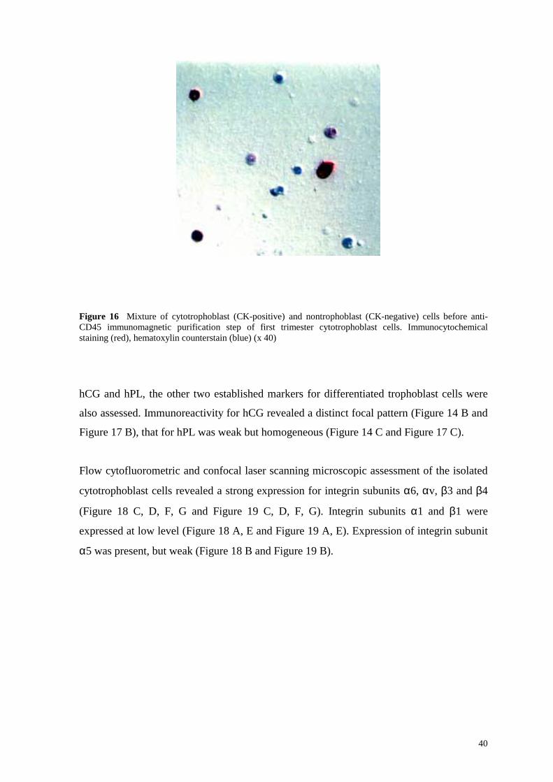
to monitor the extent of purification. Figure 16 shows CK 8,18-positive cytotrophoblast
cells contaminated with CK 8,18-negative cells before the immunomagnetic anti-CD45
depletion step. Figure 14 A and Figure 17 A show CK 8,18-positive cells after anti-CD45
enrichment.
B A
40
Figure 16 Mixture of cytotrophoblast (CK-positive) and nontrophoblast (CK-negative) cells before anti-CD45 immunomagnetic purification step of first trimester cytotrophoblast cells. Immunocytochemical staining (red), hematoxylin counterstain (blue) (x 40)
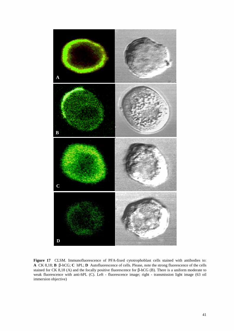
hCG and hPL, the other two established markers for differentiated trophoblast cells were
also assessed. Immunoreactivity for hCG revealed a distinct focal pattern (Figure 14 B and
Figure 17 B), that for hPL was weak but homogeneous (Figure 14 C and Figure 17 C).
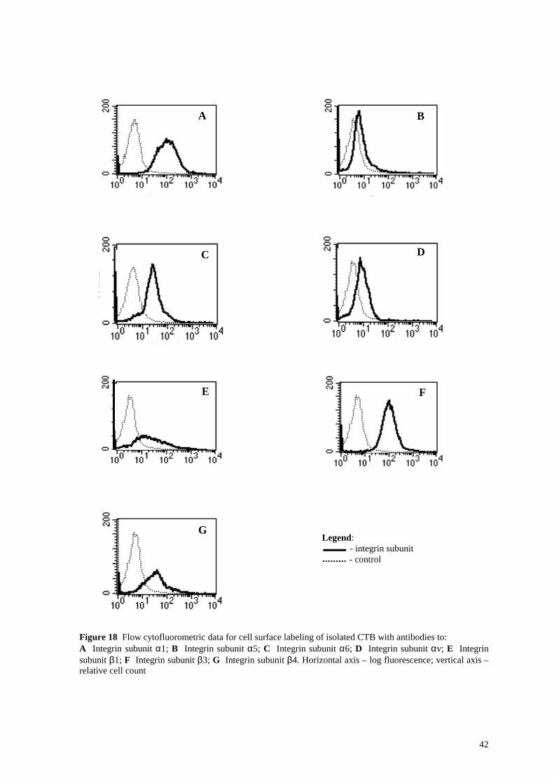
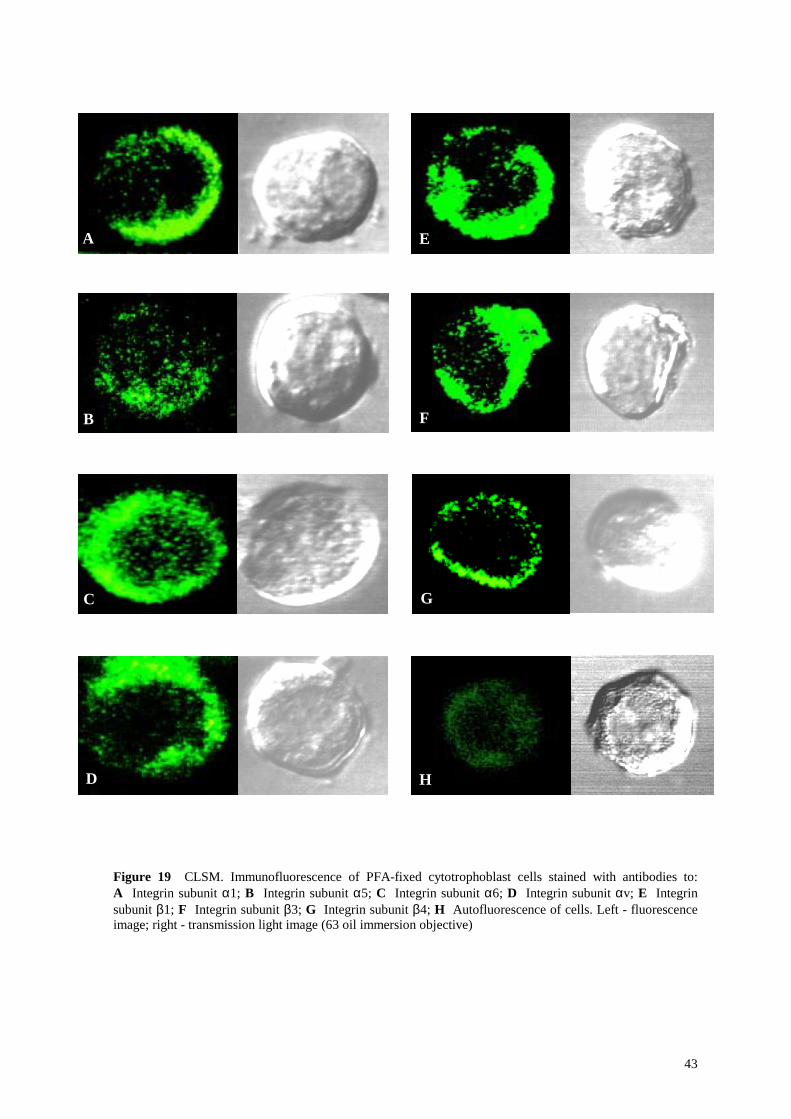
Flow cytofluorometric and confocal laser scanning microscopic assessment of the isolated
cytotrophoblast cells revealed a strong expression for integrin subunits α6, αv, β3 and β4
(Figure 18 C, D, F, G and Figure 19 C, D, F, G). Integrin subunits α1 and β1 were
expressed at low level (Figure 18 A, E and Figure 19 A, E). Expression of integrin subunit
α5 was present, but weak (Figure 18 B and Figure 19 B).
41
Figure 17 CLSM. Immunofluorescence of PFA-fixed cytotrophoblast cells stained with antibodies to: A CK 8,18; B β-hCG; C hPL; D Autofluorescence of cells. Please, note the strong fluorescence of the cells stained for CK 8,18 (A) and the focally positive fluorescence for β-hCG (B). There is a uniform moderate to weak fluorescence with anti-hPL (C). Left - fluorescence image; right - transmission light image (63 oil immersion objective)
A
D
C
B
A
42
Figure 18 Flow cytofluorometric data for cell surface labeling of isolated CTB with antibodies to: A Integrin subunit α1; B Integrin subunit α5; C Integrin subunit α6; D Integrin subunit αv; E Integrin subunit β1; F Integrin subunit β3; G Integrin subunit β4. Horizontal axis – log fluorescence; vertical axis – relative cell count
Legend: - integrin subunit - control
A B
C D
E F
G
43
Figure 19 CLSM. Immunofluorescence of PFA-fixed cytotrophoblast cells stained with antibodies to: A Integrin subunit α1; B Integrin subunit α5; C Integrin subunit α6; D Integrin subunit αv; E Integrin subunit β1; F Integrin subunit β3; G Integrin subunit β4; H Autofluorescence of cells. Left - fluorescence image; right - transmission light image (63 oil immersion objective)
A
B
C
D
E
F
G
H
44
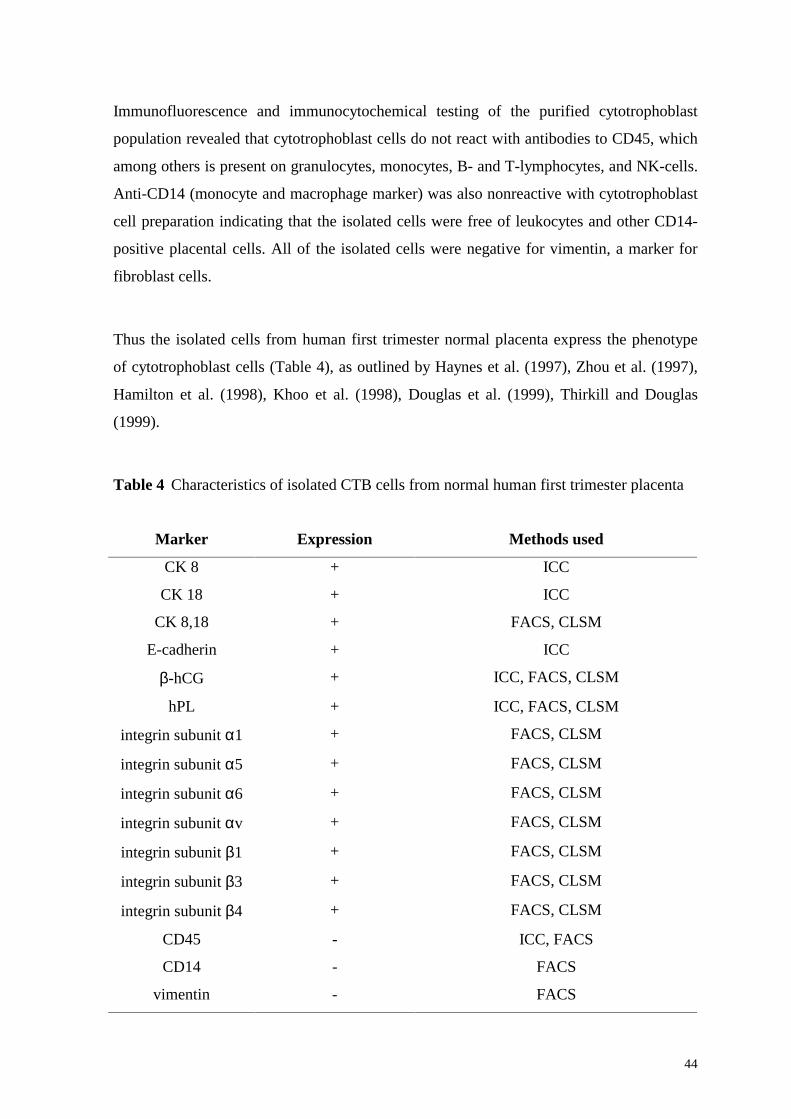
Immunofluorescence and immunocytochemical testing of the purified cytotrophoblast
population revealed that cytotrophoblast cells do not react with antibodies to CD45, which
among others is present on granulocytes, monocytes, B- and T-lymphocytes, and NK-cells.
Anti-CD14 (monocyte and macrophage marker) was also nonreactive with cytotrophoblast
cell preparation indicating that the isolated cells were free of leukocytes and other CD14-
positive placental cells. All of the isolated cells were negative for vimentin, a marker for
fibroblast cells.
Thus the isolated cells from human first trimester normal placenta express the phenotype
of cytotrophoblast cells (Table 4), as outlined by Haynes et al. (1997), Zhou et al. (1997),
Hamilton et al. (1998), Khoo et al. (1998), Douglas et al. (1999), Thirkill and Douglas
(1999).
Table 4 Characteristics of isolated CTB cells from normal human first trimester placenta
Marker Expression Methods used
CK 8 + ICC
CK 18 + ICC
CK 8,18 + FACS, CLSM
E-cadherin + ICC
β-hCG + ICC, FACS, CLSM
hPL + ICC, FACS, CLSM
integrin subunit α1 + FACS, CLSM
integrin subunit α5 + FACS, CLSM
integrin subunit α6 + FACS, CLSM
integrin subunit αv + FACS, CLSM
integrin subunit β1 + FACS, CLSM
integrin subunit β3 + FACS, CLSM
integrin subunit β4 + FACS, CLSM
CD45 - ICC, FACS
CD14 - FACS
vimentin - FACS
45
The cytomorphology of the isolated placenta cells showed classic criteria of
cytotrophoblast cells (Boyd and Hamilton, 1970): irregularly ovoid, mononuclear, with
large nuclei and a pale, faintly staining cytoplasm (Figure 17 D and Figure 20 C).
Morphological control studies were performed after each step of the isolation method,
using light microscopy, in order to prove that each time the syncytiotrophoblast cells were
removed and a mixture of cytotrophoblast cells and other placental cells was further
analyzed (Figure 20). The chorionic villus syncytium was found to consist of a layer of
basophilic syncytioplasm largely without cell boundaries, and large nuclei. The
multinuclear syncytium is markedly vacuolated and has a foamy appearance (Figure 20 B).
This cytotrophoblast isolation procedure was applied on 15 different first trimester
placentas and similar results have been obtained each time.

46
Figure 20 Transmission light microscopy of trophoblast cells. A Before enzymatic treatment of the chorionic villi; B After enzymatic treatment of the chorionic villi (before the immunopurification step); C Finally isolated cytotrophoblast cells . HE staining (x 40)
B
C
A

47
4 DISCUSSION
In recent years, in vivo and in vitro studies of trophoblast cells have gained increasing
attention because very little is known about factors controling trophoblast proliferation,
differentiation, and invasion. Without any question, for in vitro characterization at the cell
and molecular level, highly purified trophoblast cells are needed. To fulfil these needs,
several attempts to isolate human trophoblast cells have been reported. In essence, these
methods include:
- physical separation methods (Kliman et al., 1986);
- physical and/or immunomagnetic separation methods (Douglas and King, 1989);
- cell attachment procedures (Loke et al., 1989a);
- sequential enzymatic digestion and Percoll gradient centrifugation (Kliman et al., 1986;
Fisher et al., 1989);
- selective culture conditions (Loke and Burland, 1988; Yeger et al., 1989);
- sedimentation methods (Kliman et al., 1986; Douglas and King, 1990; Kliman and
Feinberg, 1990; Nelson et al., 1990; Shorter et al., 1990);
- immunological or receptor-binding methods (Contractor and Sooranna, 1988; Douglas
and King, 1989; Loke et al., 1989b; Shorter et al., 1990; Schmon et al., 1991; Bischof
et al., 1991; Morrish et al., 1991; Caulfield et al., 1992; Tse et al., 1994);
- selective disaggregation conditions (Bax et al., 1989; Bullen et al., 1990);
- immuno - flow cytometric cell sorting methods (Bloxam et al., 1997).
The difficulty in obtaining a preparation of pure trophoblast cells for culture can be
appreciated by understanding the structure of the placenta. The outer surface of the
chorionic villi is covered by the syncytiotrophoblast, underlying which is a single layer of
cytotrophoblast cells that lies on the basement membrane. A microvascular network
connects this cell layer to the umbilical arteries and vein. The apical membrane of the
syncytiotrophoblast is folded into numerous microvilli, and this layer forms a syncytium.
Disaggregation of this villous tissue results in a broken syncytial membrane, releasing not
only the cytotrophoblast cells, but also the rest of the villous cell population (macrophages,
fibroblasts, giant cells, some adhering decidual and endothelial cells) as well as DNA from
48
the nuclei of the syncytium. Separation of cytotrophoblast from this heterogeneous cell
population has proven to be a challenge.
The present protocol to enrich a reasonable number of highly purified cytotrophoblast cells
from human first trimester placenta depends on the initial crude mechanical dissection of
the chorionic villi, gentle enzymatic treatment to separate the cytotrophoblast and
syncytiotrophoblast layers from the mesenchyme, enrichment of the cytotrophoblast cells
by Percoll gradient sedimentation and by an immunomagnetic separation step employing
monoclonal antibodies to CD45 to remove blood cells.
CD45 is a tyrosine phosphatase present on bone marrow-derived cells (Charbonneau et al.,
1989; Shimonovitz et al., 1998), but is absent on cytotrophoblast cells (Librach et al.,
1991).
The specific type and number of cytotrophoblast cells isolated by this method depends on
the age of the placenta and the time of enzymatic digestion. In the present study,
incubation times necessary to remove the layer of syncytiotrophoblast cells covering the
chorionic villi were assessed histologically for first trimester placentas of different ages
and for every lot of enzymes. Thus the use of sequential enzymatic dissociation as the first
step enabled the sequential dissociation of CTB and STB in a stepwise manner from the
first trimester placenta.
Proteolytic enzymes other than trypsin used to digest the placental villous tissue have been
tried, such as collagenase (Matsuzaki et al., 1992), collagenase with hyaluronidase (Fisher
et al., 1990), hyaluronidase with trypsin (Ungar et al., 1987), collagenase and pronase
(Morrish and Siy, 1986), without improvement or a reduction in the proportion of viable
CTB extracted. On the other hand, protease XV (from B. polymyxa) (Richards et al., 1994)
and dispase (from B. polymyxa) (Eis et al., 1995) appear to have been used successfully
and may be more reliable than trypsin. Karl et al. (1992) have suggested that when a high
yield of cells is required, or when an initial digestion with trypsin or dispase is
unsatisfactory, serial digestion with dispase, collagenase and trypsin can overcome variable
enzyme effectiveness.
49
hCG, hPL, and CK 8,18 as markers of the trophoblastic tissue is well documented in the
literature (Kurman et al., 1984; Sabet et al., 1989; Khoo et al., 1998); immunocytological
techniques have promoted many studies on diverse villous and/or extravillous trophoblast
cell populations of uteroplacental tissues (Bulmer and Sunderland, 1984; Hsi et al., 1984;
Redman et al., 1984; Wells et al., 1984; Bulmer and Johnson, 1985).
Cytokeratins are a family of intermediate filament proteins with molecular weights of 40 -
68 kD, among others expressed by epithelial cells. The type of cytokeratin synthesized
depends on the origin of the epithelial cell and its stage of differentiation. In tissue sections
it was shown that the villous cytotrophoblast cell staines heavily with hematoxylin - eosin,
whereas syncytiotrophoblast cells are faintly labeled, only (Loke and Butterworth, 1987).
Cytokeratin 8,18 is a very sensitive and reliable marker for all types of trophoblastic
tissues but not for villous stromal components (Fisher et al., 1989; Daya and Sabet, 1991;
Hamilton et al., 1998). hCG and hPL are positive in differentiated trophoblast -
syncytiotrophoblast and extravillous trophoblast (Daya and Sabet, 1991). These data
correlate with our immunocytochemical findings: isolated cytotrophoblast cells were not
uniform in their positivity regarding hCG and showed a low positivity for hPL.
E-cadherin is a calcium-dependent cell-cell adhesion molecule, e.g., expressed on
differentiated epithelia, which, in the placenta, is specific for cytotrophoblasts. In the
placenta, antibodies to E-cadherin stain trophoblast cells, only (Fisher et al., 1989;
Coutifaris et al., 1991a) and in a polarized pattern (Zhou et al., 1997). In contrast,
antibodies to CD45 stain granulocytes, monocytes, B- and T-lymphocytes, and NK-cells,
but not trophoblast cells. All of the isolated cells were negative for vimentin, an
intermediate filament protein present in endothelial cells and fibroblasts (Morgan et al.,
1998).
Integrins are a subfamily of cell adhesion molecules regulating calcium-dependent cell –
substrate interactions. They are widely expressed cell – surface adhesion receptors. They
are all α β heterodimers.
Trophoblast cells reaching the endometrial basement membrane express integrins (α6β4),
which anchor them into the basement membrane and induce the secretion of matrix

50
metalloproteinases (MMPs), in particular MMP-2 and MMP-9. MMPs digest the basement
membrane allowing the embryo to make contact with the endometrial extracellular matrix.
Further integrins (α5β1) anchor the embryo into the extracellular matrix and induce its
secretion of collagenases (MMP-1 and MMP-8). These MMPs in turn digest the
extracellular matrix allowing the embryo to nidate in the endometrium (Bischof and
Campana, 1996).
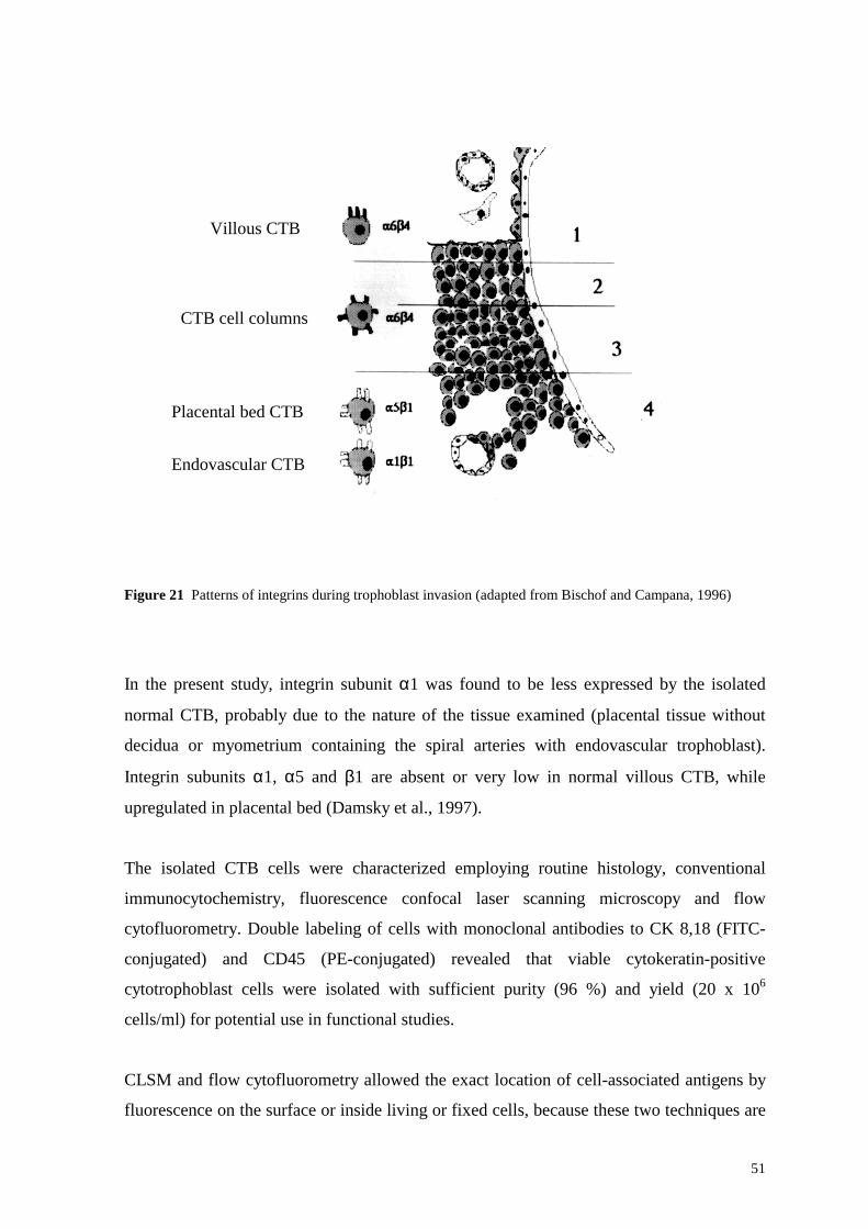
Villous CTB expresses the laminin receptor integrin α6β4 clustered toward the basement
membrane (Lala and Hamilton, 1996; Zhou et al., 1997) (Figure 21; see also Figure 6), and
the extravillous CTB expresses the fibronectin receptor integrin α5β1 (Lala and Hamilton,
1996; Zhou et al., 1997). Endovascular trophoblast expresses the collagen receptor integrin
α1β1. Integrin α6β4 is associated with invasive properties of CTB (Bischof and Campana,
1996; Zhou et al., 1997). Integrin αvβ5 is characteristic of villous CTB, αvβ6 is only
expressed on cells at the base of the invasive cell columns, while αvβ3 characterizes the
CTB of the placental bed (Zhou et al., 1997; Coutifaris et al., 1998). Expression of
particular subsets of integrins has been associated with invasive cell behavior (Albelda,
1993; Douglas et al., 1999; Crowe and Shuler, 1999).
51
Figure 21 Patterns of integrins during trophoblast invasion (adapted from Bischof and Campana, 1996)
In the present study, integrin subunit α1 was found to be less expressed by the isolated
normal CTB, probably due to the nature of the tissue examined (placental tissue without
decidua or myometrium containing the spiral arteries with endovascular trophoblast).
Integrin subunits α1, α5 and β1 are absent or very low in normal villous CTB, while
upregulated in placental bed (Damsky et al., 1997).
The isolated CTB cells were characterized employing routine histology, conventional
immunocytochemistry, fluorescence confocal laser scanning microscopy and flow
cytofluorometry. Double labeling of cells with monoclonal antibodies to CK 8,18 (FITC-
conjugated) and CD45 (PE-conjugated) revealed that viable cytokeratin-positive
cytotrophoblast cells were isolated with sufficient purity (96 %) and yield (20 x 106
cells/ml) for potential use in functional studies.
CLSM and flow cytofluorometry allowed the exact location of cell-associated antigens by
fluorescence on the surface or inside living or fixed cells, because these two techniques are
Villous CTB
CTB cell columns
Placental bed CTB
Endovascular CTB
52
designed for rapid laser scanning of the cell and immediate data processing at real-time
(Knebel et al., 1990). Both techniques are relatively easy to perform. Compared to
transmission light microscopy, CLSM has the main advantage to achieve high resolution of
cell structures (transmission and fluorescence light images) together with a powerful
computer and image analysis system. The advantage of flow cytofluorometry on the other
hand resides in the possibility of detection and quantitation of surface antigens. Moreover,
using different fluorochromes (double labeling), simultaneous co-localization of various
antigens (receptors) in the same cell is possible.
Isolated cytotrophoblast cells do not proliferate in vitro (Lewis et al., 1993), suggesting
that these cells are not suited to study cytotrophoblast cell proliferation at the feto-maternal
interface (Babawale et al., 1996). These findings indicate that either the process of tissue
dissociation, when obtaining isolated trophoblast cells, does impair the cell or that the loss
of the mesodermal core of the placental villi as a source of growth factors is responsible for
the inability of isolated cytotrophoblast cells to proliferate ex vivo (Babawale et al., 1996).

53
5 CONCLUSION
High purity isolation of living cytotrophoblast cells represents an important step toward
understanding the process of human embryo implantation since nidation is considerably
morphologically different in humans than in experimental or domestic animals (Weitlauf,
1994). These morphologic variations presumably rely upon fundamental differences at the
cellular and molecular levels (Coutifaris et al., 1991b). For ethical reasons, in vivo human
experimentation to study the steps of implantation is not feasible. However, elucidation of
the mechanisms of human implantation is gaining strong importance, given the recent
advances in assisted reproductive technologies and the search for new methods of
contraception. Thus, the recent development of new techniques for isolation of purified
living placental cytotrophoblast cells should allow further characterization of their
structural, morphologic, and functional differentiation at the cellular and molecular level.
Advances in understanding the process of implantation and placental development will
also lend insights into clinically important trophoblast-related disorders of first trimester
pregnancy, such as embryonic mortality, spontaneous abortion, abnormal placentation, or
gestational trophoblastic disease. The presented method of cytotrophoblast isolation should
facilitate ongoing in vitro studies of trophoblast adhesion, differentiation, migration,
proteolytic activity, and invasion, and clarify how these events differ from those of
malignant cells. The study of adhesive interactions between trophoblast cells and
trophoblast and decidual cells and their role in embryo implantation and development of
the placenta is a step toward outlining the events leading to the successful establishment
and maintenance of early human pregnancy.
54
6 PERSPECTIVES
Developments in methods of isolation and culture of human placental trophoblast have
opened up a new era in the study of placental function and of the role of trophoblast both in
the normal physiology and the pathology of human reproduction. As a result of these
developments, rapid advances are being made in the areas of trophoblast - endometrial
interactions in implantation, reproductive immunology, placental endocrinology,
metabolism, and pathology, trophoblast function as well as basic mechanisms of cell
differentiation. Another key area of placental function whose investigation should benefit
from the ability to isolate pure placental trophoblast is maternal – fetal transfer of
substances needed by the fetus for its development and growth, and, in the opposite
direction, of substances for excretion or for use by the maternal organism.
55
7 ABSTRACT
A protocol for the preparation of highly purified human placental cytotrophoblast cells
from normal first trimester placental villi is described. Cytotrophoblast cells were isolated
from placentas (6 - 14 weeks of gestation) by a process of mincing, sequential two-step
enzymatic treatment of placental villi by collagenase, hyaluronidase, DNAse, and trypsin,
followed by discontinuous Percoll gradient centrifugation, and immunomagnetic
separation employing antibodies to CD45 to remove contaminating leukocytes. A
population of 96 % pure living mononuclear cytotrophoblast cells (identified by positive
cytokeratin 8,18 and E-cadherin staining) was obtained at a density of 1.048 and 1.062
g/ml Percoll. The isolated cells were characterized employing routine histology,
conventional immunocytochemistry, fluorescence confocal laser scanning microscopy and
flow cytofluorometry for the cell antigens CK 8,18, β-hCG, hPL, E-cadherin, integrin
subunits α1, α5, α6, αv, β1, β3, β4, and possess the morphological and structural
characteristics of cytotrophoblast cells. The presented method of cytotrophoblast isolation
should facilitate ongoing in vitro studies of trophoblast adhesion, differentiation,
migration, proteolytic activity, and invasion, and clarify how these events differ from those
of malignant cells.

56
8 REFERENCES
1. Aboagye-Mathiesen, G., Laugesen, J., Zdravkovic, M., Ebbesen, P. Isolation and
characterization of human placental trophoblast subpopulations from first trimester
chorionic villi. Clin. Diagn. Lab. Immunol. 3 (1996) 14 - 22.
2. Albelda, S.M. Role of integrins and other cell adhesion molecules in tumor progression
and metastasis. Lab. Invest. 68 (1993) 4 - 17.
3. Aplin, J.D. Implantation, trophoblast differentiation and haemochorial placentation:
mechanistic evidence in vivo and in vitro. J. Cell Sci. 99 (1991) 681 - 692.
4. Aplin, J.D. The cell biology of human implantation. Placenta 17 (1996) 269 - 275.
5. Babawale, M.O., van Noorden, S., Pignatelli, M., Stamp, G.W.H., Elder, M.G.,
Sullivan, M.H.F. Morphological interaction of human first trimester placental villi co-
cultured with decidual explants. Hum. Reprod. 11 (1996) 444 - 450.
6. Bax, C.M.R., Ryder, T.A., Mobberley, M.A., Tyms, A.S., Taylor, D.L., Bloxam, D.L.
Ultrastructural changes and immunocytochemical analysis of human placental
trophoblast during short-term culture. Placenta 10 (1989) 179 - 194.
7. Benirschke, K., Kaufmann, P. "Pathology of the Human Placenta". Springer, New
York, 1995.
8. Bischof, P., Campana, A. A biochemical model for implantation of the human
blastocyst. Obstet. Ginecol. 3 (1996) 91 - 101.
9. Bischof, P., Friedli, E., Martelli, M., Campana, A. Expression of extracellular matrix-
degrading metalloproteinases by cultured human cytotrophoblast cells: effects of cell
adhesion and immunopurification. Am. J. Obstet. Gynecol. 165 (1991) 1791 - 1801.
10. Bloxam, D.L., Bax, C.M.R., Bax, B.E. Culture of syncytiotrophoblast for the study of
human placental transfer. Part I: Isolation and purification of cytotrophoblast. Placenta
18 (1997) 93 - 98.
11. Boyd, J.D., Hamilton, W.J. Development and structure of the human placenta from the
end of the third month of gestation. J. Obstet. Gynaecol. Br. Commonw. 74 (1967) 161
- 226.
12. Boyd, J.D., Hamilton, W.J. "The Human Placenta". W. Heffer & Sons Ltd.,
Cambridge, 1970.
57
13. Bullen, B.E., Bloxam, D.L., Ryder, T.A., Mobberley, M.A., Bax, C.M.R. Two-sided
culture of human placental trophoblast. Morphology, immunocytochemistry and
permeability properties. Placenta 11 (1990) 431 - 450.
14. Bulmer, J.N., Sunderland, C.A. Immunohistological characterization of lymphoid
populations in the early human placental bed. Immunology 52 (1984) 349 - 357.
15. Bulmer, J.N., Johnson, P.M. Antigen expression by trophoblast populations in the
human placenta and their possible immunobiological relevance. Placenta 6 (1985) 127
- 140.
16. Castellucci, M., Scheper, M., Scheffen, I., Celona, A., Kaufmann, P. The development
of the human placental villous tree. Anat. Embryol. 181 (1990) 117 - 128.
17. Caulfield, J.J., Sargent, I.L., Ferry, B.L., Starkey, P.M., Redman, C.W.G. Isolation and
characterization of a subpopulation of human chorionic cytotrophoblast using a
monoclonal anti-trophoblast antibody (NDOG2) in flow cytometry. J. Reprod.
Immunol. 21 (1992) 71 - 85.
18. Charbonneau, H., Tonks, N.K., Kumar, S., Diltz, C.D., Harrylock, M., Cool, D.E.,
Krebs, E.G., Fischer, E.H., Walsh, K.A. Human placenta protein-tyrosine-phosphatase:
amino acid sequence and relationship to a family of receptor-like proteins. Proc. Natl.
Acad. Sci. U S A 86 (1989) 5252 - 5256.
19. Contractor, S.F., Sooranna, S.R. Human placental cells in culture: a panning technique
using trophoblast-specific monoclonal antibody for cell separation. J. Dev. Physiol. 10
(1988) 47 - 51.
20. Coutifaris, C., Omigbodun, A., Coukos, G. Integrins, endometrial maturation, & human
embryo implantation. Semin. Reprod. Endocrinol. 16 (1998) 219 - 229.
21. Coutifaris, C., Kao, L., Sehder, H.M., Chin, U., Babalola, G.O. E-cadherin expression
during the differentiation of human trophoblasts. Development 113 (1991a) 767 - 777.
22. Coutifaris, C., Babalola, G.O., Abisogun, A.O., Kao, L.C., Chin, U., Vadillo-Ortega,
F., Osheroff, J., Kliman, H.J., Strauss III, J.F. In vitro systems for the study of human
trophoblast implantation. Ann. N Y Acad. Sci. 622 (1991b) 191 - 201.
23. Crowe, D.L., Shuler, C.F. Regulation of tumor cell invasion by extracellular matrix.
Histol. Histopathol. 14 (1999) 665 - 671.
24. Damsky, C.H., Schick, S.F., Klimanskaya, I., Stephens, L., Zhou, Y., Fisher, S.
Adhesive interactions in peri-implantation morphogenesis and placentation. Reprod.
Toxicol. 11 (1997) 367 - 375.
58
25. Daya, D., Sabet, L. The use of cytokeratin as a sensitive and reliable marker for
trophoblastic tissue. Am. J. Clin. Pathol. 95 (1991) 137 - 141.
26. Denker, H.W. Implantation: a cell biological paradox. J. Exp. Zool. 266 (1993) 541 -
558.
27. Douglas, G.C., King, B.F. Isolation of pure villous cytotrophoblast from term human
placenta using immunomagnetic microspheres. J. Immunol. Methods 119 (1989) 259 -
268.
28. Douglas, G.C., King, B.F. Differentiation of human trophoblast cells in vitro as
revealed by immunocytochemical staining of desmoplakin and nuclei. J. Cell Sci. 96
(1990) 131 - 141.
29. Douglas, G.C., Thirkill, T.L., Blankenship, T.N. Vitronectin receptors are expressed by
macaque trophoblast cells and play a role in migration and adhesion to endothelium.
Biochim. Biophys. Acta 1452 (1999) 36 - 45.
30. Eis, A.L.W., Brockman, D.E., Pollock, J.S., Myatt, L. Immunohistochemical
localization of endothelial nitric oxide synthase in human villous and extravillous
trophoblast populations and expression during syncytiotrophoblast formation in vitro.
Placenta 16 (1995) 113 - 126.
31. Fisher, S.J., Cui, T.Y., Zhang, L., Hartman, L., Grahl, K., Zhang, G.Y., Tarpey, J.,
Damsky, C.H. Adhesive and degradative properties of human placental cytotrophoblast
cells in vitro. J. Cell Biol. 109 (1989) 891 - 902.
32. Fisher, S.J., Sutherland, A., Moss, L., Hartman, L., Crowley, E., Bernfield, M.,
Calarco, P., Damsky, C. Adhesive interactions of murine and human trophoblast cells.
Trophoblast Research 4 (1990) 115 - 139.
33. Foidart, J.N., Hustin, J., Dubois, M., Schaaps, J.P. The human placenta becomes
haemochorial at the 13th week of pregnancy. Int. J. Dev. Biol. 36 (1992) 451 - 453.
34. Graham, C.H., Lala, P.K. Mechanism of control of trophoblast invasion in situ. J. Cell.
Physiol. 148 (1991) 228 - 234.
35. Graham, C.H., Lysiak, J.J., McCrae, K.R., Lala, P.K. Localization of transforming
growth factor-β at the human fetal-maternal interface: role in trophoblast growth and
differentiation. Biol. Reprod. 46 (1992) 561 - 572.
36. Hamilton, G.S., Lysiak, J.J., Watson, A.J., Lala, P.K. Effects of colony stimulating
factor-1 on human extravillous trophoblast growth and invasion. J. Endocrinol. 159
(1998) 69 - 77.

59
37. Haynes, M.K., Wapner, R.L., Jackson, L.G., Smith, J.B. Phenotypic analysis of
adhesion molecules in first-trimester decidual tissue from chorion villus samples. Am.
J. Reprod. Immunol. 38 (1997) 423 - 430.
38. Hertig, A.T., Rock, J. Two human ova of the previllous stage, having a developmental
age of about seven and nine days respectively. Contrib. Embryol. Carnegie Inst. 31
(1945) 65.
39. Hsi, B.L., Yeh, C.J.G., Faulk, W.P. Class I antigens of the major histocompatibility
complex on cytotrophoblast of human chorion laeve. Immunology 52 (1984) 621 - 629.
40. Karl, P.I., Alpy, K.L., Fisher, S.E. Serial enzymatic digestion method for isolation of
human placental trophoblasts. Placenta 13 (1992) 385 - 387.
41. Kaufmann, P., Castelluci, M. Extravillous trophoblast in the human placenta.
Trophoblast Research 10 (1997) 21 - 62.
42. Khoo, N.K.S., Bechberger, J.F., Shepherd, T., Bond, S.L., McCrae, K.R., Hamilton,
G.S., Lala, P.K. SV40 Tag transformation of the normal invasive trophoblast results in
a premalignant phenotype. I. Mechanisms responsible for hyperinvasiveness and
resistance to anti-invasive action of TGFβ. Int. J. Cancer 77 (1998) 429 - 439.
43. Kliman, H.J., Feinberg, R.F. Human trophoblast-extracellular matrix (ECM)
interactions in vitro: ECM thickness modulates morphology and proteolytic activity.
Proc. Natl. Acad. Sci. U S A 87 (1990) 3057 - 3061.
44. Kliman, H.J., Nestler, J.E., Sermasi, E., Sanger, J.M., Strauss III, J.F. Purification,
characterization, and in vitro differentiation of cytotrophoblasts from human term
placentae. Endocrinology 118 (1986) 1567 - 1582.
45. Knebel, W., Quader, H., Schnepf, E. Mobile and immobile endoplasmatic reticulum in
onion bulb epidermis cells: short- and long-term observations with a confocal laser
scanning microscope. Eur. J. Cell Biol. 52 (1990) 328 - 340.
46. Kurman, R.J., Young, R.H., Norris, H.J., Main, C.S., Laurence, M.D., Scully, R.E.
Immunocytochemical localization of placental lactogen and chorionic gonadotropin in
the normal placenta and trophoblastic tumors with emphasis on intermediate
trophoblast and placental site trophoblastic tumor. Int. J. Gynecol. Pathol. 3 (1984) 101
- 102.
47. Lala, P.K., Graham, C.H. Mechanisms of trophoblast invasiveness and their control:
the role of proteases and protease inhibitors. Cancer Metastasis Rev. 9 (1990) 369 -
379.
60
48. Lala, P.K., Hamilton, G.S. Growth factors, proteases and protease inhibitors in the
maternal-fetal dialogue. Placenta 17 (1996) 545 - 555.
49. Lewis, M.P., Morlese, J.F., Sullivan, M.H., Elder, M.G. Evidence for decidua-
trophoblast interactions in early human pregnancy. Hum. Reprod. 8 (1993) 965 - 968.
50. Librach, C.L., Werb, Z., Fitzgerald, M.L., Chiu, K., Corwin, N.M., Esteves, R.A.,
Grobelny, D., Galardy, R., Damsky, C.H., Fisher, S.J. 92-kD type IV collagenase
mediates invasion of human cytotrophoblasts. J. Cell Biol. 113 (1991) 437 - 449.
51. Loke, Y.W. Experimenting with human extravillous trophoblast: a personal view. Am.
J. Reprod. Immunol. 24 (1990) 22 - 28.
52. Loke, Y.W., Butterworth, B.H. Heterogeneity of human trophoblast populations. In:
"Immunoregulation and Fetal Survival", Gill, T.J., Wegmann, T.G. (Ed.), Oxford
University Press, New York, 1987, 197 - 209.
53. Loke, Y.W., Burland, K. Human trophoblast cells cultured in modified medium and
supported by extracellular matrix. Placenta 9 (1988) 173 - 182.
54. Loke, Y.W., Gardner, L., Grabowska, A. Isolation of human extravillous trophoblast
cells by attachment to laminin-coated magnetic beads. Placenta 10 (1989a) 407 - 415.
55. Loke, Y.W., Gardner, L., Burland, K., King, A. Laminin in human trophoblast-decidua
interaction. Hum. Reprod. 4 (1989b) 457 - 463.
56. Luther, T., Magdolen, V., Albrecht, S., Kasper, M., Riemer, C., Kessler, H., Graeff, H.,
Müller, M., Schmitt, M. Epitope-mapped monoclonal antibodies as tools for functional
and morphological analyses of the human urokinase receptor in tumor tissue. Am. J.
Pathol. 150 (1997) 1231 - 1244.
57. Matsuzaki, N., Li, Y., Masuhiro, K., Jo, T., Shimoya, K., Taniguchi, T., Saji, F.,
Tanizawa, O. Trophoblast-derived transforming growth factor-β1 suppresses cytokine-
induced, but not gonadotropin-releasing hormone-induced, release of human chorionic
gonadotropin by normal human trophoblast. J. Clin. Endocrinol. Metab. 74 (1992) 211
- 216.
58. Moore, K.L. "The developing human". W. B. Saunders, Philadelphia, 1988, 33 - 38.
59. Morgan, M., Kniss, D., McDonnell, S. Expression of metalloproteinases and their
inhibitors in human trophoblast continuous cell lines. Exp. Cell Res. 242 (1998) 18 -
26.
60. Morrish, D.W., Siy, D. Critical factors in establishing monolayer cultures of normal
human placental cells in serum-free medium. Endocr. Res. 12 (1986) 229 - 253.

61
61. Morrish, D.W., Shaw, A.R.E., Seehafer, J., Bhardwaj, D., Paras, M.T. Preparation of
fibroblast-free cytotrophoblast cultures utilizing differential expression of the CD9
antigen. In Vitro Cell. Dev. Biol. 27 (1991) 303 - 306.
62. Nelson, D.M., Crouch, E.C., Curran, E.M., Farmer, D.R. Trophoblast interaction with
fibrin matrix. Epithelialization of perivillous fibrin deposits as a mechanism for villous
repair in the human placenta. Am. J. Pathol. 136 (1990) 855 - 865.
63. Pijnenborg, R. Trophoblast invasion. J. Reprod. Med. 3 (1994) 53 - 73.
64. Redman, C.W.G., McMichael, A.J., Stirrat, G.M., Sunderland, C.A., Ting, A. Class I
major histocompatibility complex antigens on human extravillous trophoblast.
Immunology 52 (1984) 457 - 468.
65. Richards, R.G., Hartman, S.M., Handwerger, S. Human cytotrophoblast cells cultured
in maternal serum progress to a differentiated syncytial phenotype expressing both
human chorionic gonadotropin and human placental lactogen. Endocrinology 135
(1994) 321 - 329.
66. Sabet, L., Daya, D., Stead, R., Richmond, H., Jimeneze, C.L. Significance and value of
immunohistochemical localization of pregnancy specific proteins in feto-maternal
tissue throughout pregnancy. Mod. Pathol. 2 (1989) 227 - 232.
67. Schlafke, S., Enders, A.C. Cellular basis of interaction between trophoblast and uterus
at implantation. Biol. Reprod. 12 (1975) 41 - 65.
68. Schmitt, M., Chucholowski, N., Busch, E., Hellmann, D., Wagner, B., Goretzki, L.,
Jänicke, F., Günzler, W.A., Graeff, H. Fluorescent probes as tools to assess the receptor
for the urokinase-type plasminogen activator on tumor cells. Semin. Thromb. Hemost.
17 (1991) 291 - 302.
69. Schmon, B., Hartmann, M., Jones, C.J., Desoye, G. Insulin and glucose do not affect
the glycogen content in isolated and cultured trophoblast cells of human term placenta.
J. Clin. Endocrinol. Metab. 73 (1991) 888 - 893.
70. Shapiro, H.M. "Practical Flow Cytometry". Alan R. Liss Inc., New York, 1988.
71. Shimonovitz, S., Hurwitz, A., Hochner-Celnikier, D., Dushnik, M., Anteby, E., Yagel,
S. Expression of gelatinase B by trophoblast cells: down-regulation by progesterone.
Am. J. Obstet. Gynecol. 178 (1998) 457 - 461.
72. Shorter, S.C., Jackson, M.C., Sargent, I.L., Redman, C.W.G., Starkey, P.M.
Purification of human cytotrophoblast from term amniochorion by flow cytometry.
Placenta 11 (1990) 505 - 513.
62
73. Thiede, H.A. Studies of the human trophoblast in tissue culture. I. Cultural methods
and histochemical staining. Am. J. Obstet. Gynecol. 79 (1960) 636 - 647.
74. Thirkill, T.L., Douglas, G.C. The vitronectin receptor plays a role in the adhesion of
human cytotrophoblast cells to endothelial cells. Endothelium 6 (1999) 277 - 290.
75. Tse, D.B., Anderson, P., Goldbard, S., Gown, A.M., Hawes, C.S., Donnenfeld, A.
Characterization of trophoblast-reactive monoclonal antibodies by flow cytometry and
their application for fetal cell isolation. Ann. N Y Acad. Sci. 731 (1994) 162 - 169.
76. Ungar, L., Csonka, E., Kazy, Z., Siklos, P., Hercz, P. The use of pregnancy serum to
obtain trophoblastic cell cultures. Placenta 8 (1987) 639 - 646.
77. Vicovac, L., Aplin, J.D. Epithelial-mesenchymal transition during trophoblast
differentiation. Acta Anat. 156 (1996) 202 - 216.
78. Weitlauf, H.M. Biology of implantation. In: "The Physiology of Reproduction",
Knobil, E., Neill, J. (Ed.), Raven Press, New York, 1994, 231 - 262.
79. Wells, M., Hsi, B.L., Yeh, C.Y., Faulk, W.P. Spiral (uteroplacental) arteries of the
human placental bed show the presence of amniotic basement membrane antigens. Am.
J. Obstet. Gynecol. 150 (1984) 973 - 977.
80. Yeger, H., Lines, L.D., Wong, P.Y., Silver, M.M. Enzymatic isolation of human
trophoblast and culture on various substrates: comparison of first trimester with term
trophoblast. Placenta 10 (1989) 137 - 151.
81. Yeh, I.T., Kurman, R.J. Functional and morphologic expressions of trophoblast. Lab.
Invest. 61 (1989) 1 - 4.
82. Zdravkovic, M., Aboagye-Mathiesen, G., Guimond, M.J., Hager, H., Ebbesen, P., Lala,
P.K. Susceptibility of MHC class I expressing extravillous trophoblast cell lines to
killing by natural killer cells. Placenta 20 (1999) 431 - 440.
83. Zhou, Y., Fisher, S.J., Janatpour, M., Genbacev, O., Dejana, E., Wheelock, M.,
Damsky, C.H. Human cytotrophoblast adopt a vascular phenotype as they differentiate.
J. Clin. Invest. 99 (1997) 2139 - 2151.
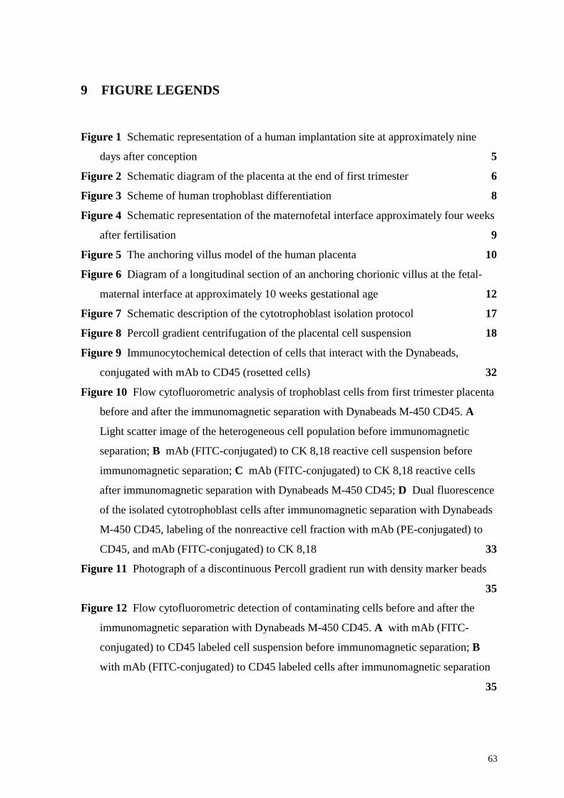
63
9 FIGURE LEGENDS
Figure 1 Schematic representation of a human implantation site at approximately nine
days after conception 5
Figure 2 Schematic diagram of the placenta at the end of first trimester 6
Figure 3 Scheme of human trophoblast differentiation 8
Figure 4 Schematic representation of the maternofetal interface approximately four weeks
after fertilisation 9
Figure 5 The anchoring villus model of the human placenta 10
Figure 6 Diagram of a longitudinal section of an anchoring chorionic villus at the fetal-
maternal interface at approximately 10 weeks gestational age 12
Figure 7 Schematic description of the cytotrophoblast isolation protocol 17
Figure 8 Percoll gradient centrifugation of the placental cell suspension 18
Figure 9 Immunocytochemical detection of cells that interact with the Dynabeads,
conjugated with mAb to CD45 (rosetted cells) 32
Figure 10 Flow cytofluorometric analysis of trophoblast cells from first trimester placenta
before and after the immunomagnetic separation with Dynabeads M-450 CD45. A
Light scatter image of the heterogeneous cell population before immunomagnetic
separation; B mAb (FITC-conjugated) to CK 8,18 reactive cell suspension before
immunomagnetic separation; C mAb (FITC-conjugated) to CK 8,18 reactive cells
after immunomagnetic separation with Dynabeads M-450 CD45; D Dual fluorescence
of the isolated cytotrophoblast cells after immunomagnetic separation with Dynabeads
M-450 CD45, labeling of the nonreactive cell fraction with mAb (PE-conjugated) to
CD45, and mAb (FITC-conjugated) to CK 8,18 33
Figure 11 Photograph of a discontinuous Percoll gradient run with density marker beads
35
Figure 12 Flow cytofluorometric detection of contaminating cells before and after the
immunomagnetic separation with Dynabeads M-450 CD45. A with mAb (FITC-
conjugated) to CD45 labeled cell suspension before immunomagnetic separation; B
with mAb (FITC-conjugated) to CD45 labeled cells after immunomagnetic separation
35
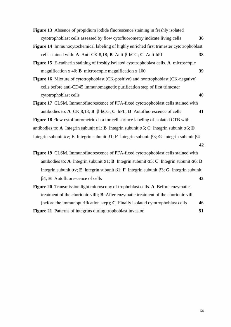
64
Figure 13 Absence of propidium iodide fluorescence staining in freshly isolated
cytotrophoblast cells assessed by flow cytofluorometry indicate living cells 36
Figure 14 Immunocytochemical labeling of highly enriched first trimester cytotrophoblast
cells stained with: A Anti-CK 8,18; B Anti-β-hCG; C Anti-hPL 38
Figure 15 E-cadherin staining of freshly isolated cytotrophoblast cells. A microscopic
magnification x 40; B microscopic magnification x 100 39
Figure 16 Mixture of cytotrophoblast (CK-positive) and nontrophoblast (CK-negative)
cells before anti-CD45 immunomagnetic purification step of first trimester
cytotrophoblast cells 40
Figure 17 CLSM. Immunofluorescence of PFA-fixed cytotrophoblast cells stained with
antibodies to: A CK 8,18; B β-hCG; C hPL; D Autofluorescence of cells 41
Figure 18 Flow cytofluorometric data for cell surface labeling of isolated CTB with
antibodies to: A Integrin subunit α1; B Integrin subunit α5; C Integrin subunit α6; D
Integrin subunit αv; E Integrin subunit β1; F Integrin subunit β3; G Integrin subunit β4
42
Figure 19 CLSM. Immunofluorescence of PFA-fixed cytotrophoblast cells stained with
antibodies to: A Integrin subunit α1; B Integrin subunit α5; C Integrin subunit α6; D
Integrin subunit αv; E Integrin subunit β1; F Integrin subunit β3; G Integrin subunit
β4; H Autofluorescence of cells 43
Figure 20 Transmission light microscopy of trophoblast cells. A Before enzymatic
treatment of the chorionic villi; B After enzymatic treatment of the chorionic villi
(before the immunopurification step); C Finally isolated cytotrophoblast cells 46
Figure 21 Patterns of integrins during trophoblast invasion 51
65
10 TABLE LEGENDS
Table 1 Primary antibodies used in immunocytochemistry (ICC) 22
Table 2 Primary antibodies used in confocal laser scanning microscopy (CLSM) 25
Table 3 Primary antibodies used in flow cytofluorometry (FACS) 28
Table 4 Characteristics of isolated CTB cells from normal human first trimester placenta
44
66
11 ABBREVIATIONS
APAAP Alkaline phosphatase anti-alkaline phosphatase
β-hCG Beta - human chorionic gonadotropin
BSA Bovine serum albumin
CK Cytokeratin
CLSM Confocal Laser Scanning Microscopy
CTB Cytotrophoblast
DMEM Dulbecco’s modified eagle medium
FACS Fluorescence activated cell sorter
FCS Fetal calf serum
FITC Fluoresceinisothiocyanat
FSC Forward light scatter
HBSS Hanks’ balanced salt solution
HE Hematoxylin – eosin
hPL Human placental lactogen
ICC Immunocytochemistry
Ig Immunoglobulin
log Logarithmic
mAb Monoclonal antibody
MMP Matrixmetalloproteinase
pAb Polyclonal antibody
PBS Phosphate - buffered saline
PE Phycoerythrin
PFA Paraformaldehyde
PI Propidium iodide
SSC Side scatter
STB Syncytiotrophoblast
TBS Tris - buffered saline

67
12 ACKNOWLEDGEMENTS
I wish to gratefully acknowledge
Univ.-Prof. Dr. med. Henner Graeff - Frauenklinik und Poliklinik der TU München, Klinikum
rechts der Isar, and Univ.-Prof. Dr. rer. nat. Dr. med. habil. Manfred Schmitt - Clinical
Research Group of the Frauenklinik der TU München, Klinikum rechts der Isar, for allowing me to
research and work in the Clinical Research Group of the Frauenklinik der Technischen Universität
München, Klinikum rechts der Isar. I am especially grateful for their invaluable insight and
assistance, for promoting many interesting and helpful discussions, and for their outstanding
support during my stay and work in the clinic;
Univ.-Prof. Dr. med. Martin Werner - Institut für Allgemeine Pathologie und Pathologische
Anatomie der TU München, Klinikum rechts der Isar, for the assistance in and the supervision of
the performance of immunocytochemistry;
Univ.-Prof. Dr. Decebal Hudita - Carol Davila University of Medicine and Pharmacy Bucharest,
Clinic of Obstetrics and Gynecology, Dr. I. Cantacuzino Clinical Hospital, for promoting and
allowing my stay in Germany and the fulfillment of my PhD;
Priv.-Doz. Dr. med. Nadia Harbeck - Frauenklinik und Poliklinik der TU München, Klinikum
rechts der Isar, for her permanent and distinguished support and active help in the fulfillment of my
study project;
Dr. med. Berit Thieme - Frauenklinik der Universität Dresden, for her technical advice during our
cooperation to establish the cytotrophoblast cell isolation protocol.
I would very much like to thank the whole technical staff of the Clinical Research Group of the
Frauenklinik der TU München, Klinikum rechts der Isar, especially Dominik Helmecke - BTA,
for the efficient cooperation and the eminent support in all my experiments.
Last but not least, I am very grateful to my husband, Eng. Cristian Marius Hera, PhD, my son
Cristian Adrian Hera, my mother Eng. Aurica Privighitorita and my entire family, for their
continuous consistent and loving help, support, and understanding.
68
13 GRANT SOURCES
My stay in Germany, at the Klinikum rechts der Isar, Frauenklinik und Poliklinik der
Technischen Universität München, and the fulfillment of the present study project was
made possible through the award of following grants:
- DAAD (Deutscher Akademischer Austauschdienst) research grant no. 324-
A/96/10919: 1.10.1996. – 31.07.1997.
- UICC (Union Internationale contre le Cancer) ICRETT research grant no. 47:
1.10.1999. – 31.12.1999.
69
14 CURRICULUM VITAE
Name: Raluca HERA
Date of Birth: February 25, 1968
Place of Birth: Bucharest - Romania
Civil state: married; 1 child
Citizenship: Romanian
Education
Date attended
Institution and area of study
1982 - 1986 German Highschool of Bucharest
June 1986 International Baccalaureate
Graduation Grade: 9.69 (out of 10)
1986 - 1992 Study of Medicine at the Faculty of Medicine - Carol Davila University of
Medicine and Pharmacy Bucharest
Graduation Grade: 9.73 (out of 10)
September 1992 Graduation Exam, Diploma thesis entitled: "Pregnancy and Viral
Hepatitis – Clinical and Management Principles"
Mark: 10 (out of 10)
March 1993 Assistant Professor at the Carol Davila University of Medicine and
Pharmacy Bucharest, Clinic of Obstetrics and Gynecology, Dr. I.
Cantacuzino Clinical Hospital
November 1993 Resident Physician Obstetrics and Gynecology - Clinic of Obstetrics and
Gynecology, Dr. I. Cantacuzino Clinical Hospital Bucharest
October 1996 Specialist Physician Obstetrics and Gynecology - Dr. I. Cantacuzino
Clinic of Obstetrics and Gynecology, Clinical Hospital Bucharest
70
1.10.1996. -
31.07.1997.
DAAD (Deutscher Akademischer Austauschdienst) Annual Grant for
clinical and scientific research at the Frauenklinik und Poliklinik der
Technischen Universität Munich, Germany - Klinikum rechts der Isar;
MD PhD student; MD PhD thesis entitled: "Isolation and characterization
of human cytotrophoblast cells from first trimester placenta".
March 1999 Ultrasound in obstetrics and gynecology – proficiency achieved
Mark: 8 (out of 10)
1.10.1999. –
31.12.1999.
UICC (Union Internationale contre le Cancer) ICRETT Grant for clinical
and scientific research at the Frauenklinik und Poliklinik der Technischen
Universität Munich, Germany - Klinikum rechts der Isar
Dominant Study Fields
1. Fetal Assessment
2. Ultrasound in obstetrics and gynecology
3. Diabetes mellitus and pregnancy
4. Menopause
5. Gynecologic Oncology
Working Experience
Dates
Hospitals and Duties
currently - October 1996
Carol Davila University for Medicine and Pharmacy
Bucharest, Dr. I. Cantacuzino Clinical Hospital, Clinic of
Obstetrics and Gynecology – MD, Assistant Professor,
Specialist Physician Obstetrics and Gynecology
1.10.1999. – 31.12.1999. UICC ICRETT Grant for clinical and scientific research at
the Frauenklinik und Poliklinik der Technischen Universität
Munich, Germany - Klinikum rechts der Isar
71
1.10.1996. – 31.07.1997. DAAD Annual Grant for clinical and scientific research at
the Frauenklinik und Poliklinik der Technischen Universität
Munich, Germany - Klinikum rechts der Isar
March 1993 – Sept. 1996 Carol Davila University of Medicine and Pharmacy
Bucharest, Dr. I. Cantacuzino Clinical Hospital, Clinic of
Obstetrics and Gynecology - MD, Assistant Professor,
Resident Physician Obstetrics and Gynecology
December 1992 – March 1993 Polizu Clinical Hospital of Obstetrics and Gynecology of the
Carol Davila University of Medicine and Pharmacy
Bucharest - Physician internship
May 1990 Study visit at the Philips University Marburg – Germany
Publications
1. Hera, R., Balteanu, M., Esanu, S., Banceanu, G. (1992) Pregnancy and viral hepatitis -
clinical and management principles. Obstetrica Ginecologia, 12, 25-27.
2. Hudita, D., Russu, M., Popescu, M., Grama, M., Hera, R. (1993) Maternal
complications in pregnancy induced hypertension. Obstetrica Ginecologia, 61, 61-65.
3. Hudita, D., Russu, M., Grama, M., Hera, R., Constantin, A., Gheorghiu, M. (1993)
Fetal risk in pregnancy induced hypertension. Obstetrica Ginecologia, 61, 66-69.
4. Hudita, D., Russu, M., Hera, R., Ciulcu, P., Condor, A., Ghinda, E., Grama, M.,
Popescu, M., Sgarbura, Z., Subtirelu, G.P., Tufan, A., Constantin, A., Gheorghiu, M.,
Târlea, A. (1993) Pelvic suppurative trombophlebitis - a postpartum complication.
Proceedings of the XIth National Congress on Obstetrics and Gynecology, Timisoara,
Romania, September 22-24, 1993.
5. Hudita, D., Popa, P.M., Russu, M., Vaduva, A., Dumitrescu, R., Marinescu, S., Hera,
R. (1994) Proposal for prenatal assessment file. Proceedings of the IIIrd Edition
"Medical Days V. Dobrovici", Suceava, Romania, September 29-30, 1994.
6. Hudita, D., Irimia, V., Russu, M., Hera, R., Dumitrescu, R. (1994) Clinical
considerations on incidence and results of cesarean sections in diabetic pregnancies.
Proceedings of the IIIrd Edition "Medical Days V. Dobrovici", Suceava, Romania,
September 29-30, 1994. Proceedings of the Annual Session of the National Mother and
Child Institute, Bucharest, Romania, November 11, 1994.
72
7. Hudita, D., Russu, M., Irimia, V., Hera, R., Dumitrescu, R., Dima, A.M. (1994)
Indications for cesarean section in premature gestations. Proceedings of the IIIrd
Edition "Medical Days V. Dobrovici", Suceava, Romania, September 29-30, 1994.
8. Hudita, D., Irimia, V., Hera, R. (1994) A retrospective study on cesarean sections
performed during 1989 - 1994 in the Clinic of Obstetrics and Gynecology Dr. I.
Cantacuzino Bucharest. Proceedings of the IIIrd Edition "Medical Days V. Dobrovici",
Suceava, Romania, September 29-30, 1994.
9. Hudita, D., Russu, M., Hera, R., Enescu, M. (1994) Congenital malformations with
clinical manifestation in the neonate and their influence on the perinatal mortality rate.
Proceedings of the IIIrd Edition "Medical Days V. Dobrovici", Suceava, Romania,
September 29-30, 1994.
10. Hudita, D., Subtirelu, G.P., Popescu, M., Russu, M., Hera, R., Popa, P.M., Irimia, V.
(1995) Correlation radiotherapy - surgery to the anatomical and clinical stage of
cervical neoplasia. Proceedings of the IVth Edition "Medical Days V. Dobrovici", Iasi,
Romania, June 1-3, 1995.
11. Hudita, D., Russu, M., Hera, R., Dumitrescu, R., Popescu, M., Ciuca, S. (1995) A
retrospective study on total vaginal hysterectomies among vaginal operations.
Proceedings of the IVth Edition "Medical Days V. Dobrovici", Iasi, Romania, June 1-3,
1995.
12. Hudita, D., Subtirelu, G.P., Popescu, M., Russu, M., Hera, R., Popa, P.M. (1995)
Carcinoma of the residual cervix - a diagnostic and therapeutical error. Proceedings of
the IVth Edition "Medical Days V. Dobrovici", Iasi, Romania, June 1-3, 1995.
13. Hudita, D., Russu, M., Hera, R., Duta, M. (1995) Bacterial vaginosis and premature
birth. Proceedings of the First National Conference of the Romanian Association of
Perinatal Medicine, Baia Mare, Romania, September 21-23, 1995. Proceedings of the
Romanian - German Symposium on Obstetrics and Gynecology, Suceava, Romania,
September 28-29, 1995.
14. Hudita, D., Constantinescu, L., Rosca, A., Russu, M., Hera, R. (1995) The third degree
premature neonate - an obstetrical and neonatological analysis. Obstetrica Ginecologia,
63, 85-87.
15. Hudita, D., Russu, M., Constantinescu, L., Rosca, A., Hera, R. (1995) Congenital
anomalies with clinical neonatal manifestation. Obstetrica Ginecologia, 63, 88-89.
16. Hudita, D., Russu, M., Hera, R., Dumitrescu, R., Secara, D., Draghici, M. (1995)
Family planning desire in puerperium. Proceedings of the Romanian - German
73
Symposium on Obstetrics and Gynecology, Suceava, Romania, September 28-29,
1995.
17. Hera, R. (1995) Genital herpes - a sexually transmitted disease. Spitalul, 3, 48-51.
18. Hudita, D., Russu, M., Hera, R. (1996) Pregnancy induced hypertension in pregnant
women with diabetes mellitus – a clinical study. Proceedings of the Session of the
Romanian Society of Obstetrics and Gynecology, Bucharest, February 27, 1996.
19. Hudita, D., Russu, M., Hera, R. (1996) Clinical study of hypertensive disorders in
diabetic pregnancies. Proceedings of the 28th International Congress on
Pathophysiology of Pregnancy, Timisoara, Romania, May 22-25, 1996.
20. Hudita, D., Russu, M., Hera, R. (1996) Hypertensive disorders in diabetic pregnancies.
Proceedings of the 28th International Congress on Pathophysiology of Pregnancy,
Timisoara, Romania, May 22-25, 1996.
21. Hudita, D., Russu, M., Hera, R., Dumitrescu, R. (1996) Pregnancy induced
hypertension and fetal risk. Proceedings of the 28th International Congress on
Pathophysiology of Pregnancy, Timisoara, Romania, May 22-25, 1996.
22. Hudita, D., Russu, M., Hera, R., Dumitrescu, R. (1996) Pregnancy induced
hypertension and maternal complications. Proceedings of the 28th International
Congress on Pathophysiology of Pregnancy, Timisoara, Romania, May 22-25, 1996.
23. Thieme, B., Hera, R., Koop, F., Harbeck, N., Kolben, M., Schneider, K.T.M., Graeff,
H., Schmitt, M. (1997) Flow cytofluorometric characterization of isolated human
trophoblast cells from normal and preeclamptic placental tissue. Proceedings of the XIth
Workshop on Applications of Flow Cytofluorometry in Clinical Cell Diagnosis,
Regensburg, Germany, March 13-14, 1997.
24. Hudita, D., Russu, M., Dumitrescu, R., Hera, R., Ceausu, I., Posea, C., Filipescu, A.
(1998) A retrospective study on endometrial cancer in the Clinic of Obstetrics and
Gynecology – Clinical Hospital Dr. I. Cantacuzino. Proceedings of the XIIth National
Congress on Obstetrics and Gynecology, Iasi, Romania, May 27-30, 1998.
25. Hudita, D., Irimia, V., Russu, M., Hera, R., Ceausu, I., Malincenco, M., Vaduva, A.,
Popa, P.M. (1998) On the actual obstetrical risk of the diabetic pregnant woman.
Proceedings of the XIIth National Congress on Obstetrics and Gynecology, Iasi,
Romania, May 27-30, 1998.
26. Hudita, D., Russu, M., Hera, R. (1998) Hormone replacement therapy in menopause:
experience of the Clinic of Obstetrics and Gynecology – Clinical Hospital Dr. I.
Cantacuzino. Proceedings of the Symposium "From Puberty to Postmenopause" and

74
the First National Conference of the Romanian Society of Gynecological
Endocrinology, Sinaia, Romania, September 18-20, 1998.
27. Hudita, D., Hera, R., Svasta, S. (1998) A retrospective study on indications for
cesarean section in the Clinic of Obstetrics and Gynecology – Clinical Hospital Dr. I.
Cantacuzino Bucharest. Proceedings of the Romanian – Hungarian Days of Obstetrics
and Gynecology, Arad, October 29-31, 1998.
28. Hudita, D., Hera, R., Toma, A. (1998) Vaginal birth after cesarean section: a
retrospective study in the Clinic of Obstetrics and Gynecology – Clinical Hospital Dr.
I. Cantacuzino Bucharest, and review of the literature. Proceedings of the Romanian –
Hungarian Days of Obstetrics and Gynecology, Arad, October 29-31, 1998.
29. Hudita, D., Russu M.C., Hera, R., Ceausu, I. (1999) Shoulder dystocia – a
retrospective clinical study, and review of the literature. Proceedings of the 3rd National
Conference on Perinatal Medicine, Timisoara, October 7-9, 1999.
Languages: Romanian – native language
German – native proficiency achieved
English – fluent
French – good
Member of
Romanian Society of Obstetrics and Gynecology
Romanian Society of Menopause (founding member)
Romanian Society of Perinatal Medicine
FIGO, UPIGO, EAGO, UEMS / Ob. Gyn.
Munich, 8.12.1999.
Raluca Hera, MD, Ass. Prof.


![Referat Peter Reichardt Vor Ort.ppt [Kompatibilitätsmodus] · • in cases of clinical suspicion of soft tissue sarcoma, primary imaging is mandatory (MRI method of choice) ... comp](https://static.fdokument.com/doc/165x107/5c9c4b6509d3f2c12d8b9ce9/referat-peter-reichardt-vor-ortppt-kompatibilitaetsmodus-in-cases-of.jpg)