Novel fatty acid methyl esters from the actinomycete Micromonospora aurantiaca · 2011-12-20 ·...
16
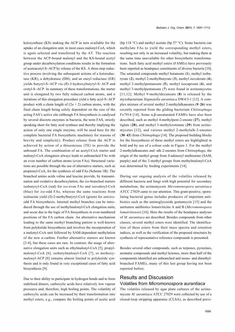
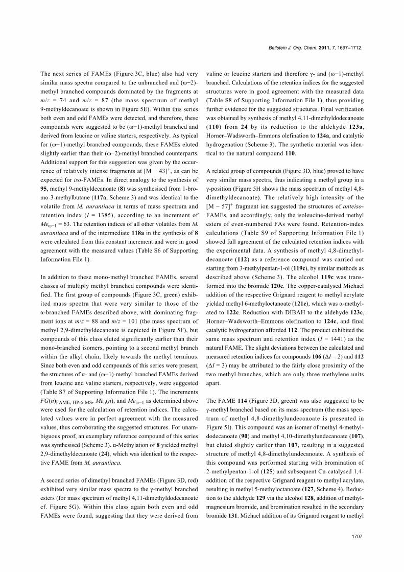
1697 Novel fatty acid methyl esters from the actinomycete Micromonospora aurantiaca Jeroen S. Dickschat * , Hilke Bruns and Ramona Riclea Full Research Paper Open Access Address: Institut für Organische Chemie, Technische Universität Braunschweig, Hagenring 30, 38106 Braunschweig, Germany Email: Jeroen S. Dickschat * - [email protected] * Corresponding author Keywords: actinomycetes; FAMEs; fatty acid biosynthesis; GC–MS; volatiles Beilstein J. Org. Chem. 2011, 7, 1697–1712. doi:10.3762/bjoc.7.200 Received: 20 August 2011 Accepted: 28 November 2011 Published: 20 December 2011 This article is part of the Thematic Series "Biosynthesis and function of secondary metabolites". Associate Editor: S. Flitsch © 2011 Dickschat et al; licensee Beilstein-Institut. License and terms: see end of document. Abstract The volatiles released by Micromonospora aurantiaca were collected by means of a closed-loop stripping apparatus (CLSA) and analysed by GC–MS. The headspace extracts contained more than 90 compounds from different classes. Fatty acid methyl esters (FAMEs) comprised the major compound class including saturated unbranched, monomethyl and dimethyl branched FAMEs in diverse structural variants: Unbranched, α-branched, γ-branched, (ω−1)-branched, (ω−2)-branched, α- and (ω−1)-branched, γ- and (ω−1)-branched, γ- and (ω−2)-branched, and γ- and (ω−3)-branched FAMEs. FAMEs of the last three types have not been described from natural sources before. The structures for all FAMEs have been suggested based on their mass spectra and on a retention index increment system and verified by the synthesis of key reference compounds. In addition, the structures of two FAMEs, methyl 4,8-dimethyldodecanoate and the ethyl-branched compound methyl 8-ethyl-4-methyldodecanoate were deduced from their mass spectra. Feeding experiments with isotopically labelled [ 2 H 10 ]leucine, [ 2 H 10 ]isoleucine, [ 2 H 8 ]valine, [ 2 H 5 ]sodium propionate, and [methyl- 2 H 3 ]methionine demonstrated that the responsible fatty acid synthase (FAS) can use different branched and unbranched starter units and is able to incorporate methylmalonyl-CoA elongation units for internal methyl branches in various chain positions, while the methyl ester function is derived from S-adenosyl methionine (SAM). 1697 Introduction Lipids in general, and particularly fatty acids (FAs), belong to the most important building blocks of biological systems. They fulfill various physiological functions, such as cell-membrane assembly or, as highly reduced carbon compounds, energy storage, and are therefore found in every single living cell on earth. In bacteria the cell membranes are mainly formed from phospholipids such as phosphatidylcholines that contain a FA diglyceride, a phosphate, and a phophate-bound choline. The simplest type of phospholipid is made up from unbranched satu- rated FAs with typical chain lengths of 16 or 18 carbon atoms,
Transcript of Novel fatty acid methyl esters from the actinomycete Micromonospora aurantiaca · 2011-12-20 ·...

1697
Novel fatty acid methyl esters from the actinomyceteMicromonospora aurantiaca
Jeroen S. Dickschat*, Hilke Bruns and Ramona Riclea
Full Research Paper Open Access
Address:Institut für Organische Chemie, Technische Universität Braunschweig,Hagenring 30, 38106 Braunschweig, Germany
Email:Jeroen S. Dickschat* - [email protected]
* Corresponding author
Keywords:actinomycetes; FAMEs; fatty acid biosynthesis; GC–MS; volatiles
Beilstein J. Org. Chem. 2011, 7, 1697–1712.doi:10.3762/bjoc.7.200
Received: 20 August 2011Accepted: 28 November 2011Published: 20 December 2011
This article is part of the Thematic Series "Biosynthesis and function ofsecondary metabolites".
Associate Editor: S. Flitsch
© 2011 Dickschat et al; licensee Beilstein-Institut.License and terms: see end of document.
AbstractThe volatiles released by Micromonospora aurantiaca were collected by means of a closed-loop stripping apparatus (CLSA) and
analysed by GC–MS. The headspace extracts contained more than 90 compounds from different classes. Fatty acid methyl esters
(FAMEs) comprised the major compound class including saturated unbranched, monomethyl and dimethyl branched FAMEs in
diverse structural variants: Unbranched, α-branched, γ-branched, (ω−1)-branched, (ω−2)-branched, α- and (ω−1)-branched, γ- and
(ω−1)-branched, γ- and (ω−2)-branched, and γ- and (ω−3)-branched FAMEs. FAMEs of the last three types have not been
described from natural sources before. The structures for all FAMEs have been suggested based on their mass spectra and on a
retention index increment system and verified by the synthesis of key reference compounds. In addition, the structures of two
FAMEs, methyl 4,8-dimethyldodecanoate and the ethyl-branched compound methyl 8-ethyl-4-methyldodecanoate were deduced
from their mass spectra. Feeding experiments with isotopically labelled [2H10]leucine, [2H10]isoleucine, [2H8]valine, [2H5]sodium
propionate, and [methyl-2H3]methionine demonstrated that the responsible fatty acid synthase (FAS) can use different branched and
unbranched starter units and is able to incorporate methylmalonyl-CoA elongation units for internal methyl branches in various
chain positions, while the methyl ester function is derived from S-adenosyl methionine (SAM).
1697
IntroductionLipids in general, and particularly fatty acids (FAs), belong to
the most important building blocks of biological systems. They
fulfill various physiological functions, such as cell-membrane
assembly or, as highly reduced carbon compounds, energy
storage, and are therefore found in every single living cell on
earth. In bacteria the cell membranes are mainly formed from
phospholipids such as phosphatidylcholines that contain a FA
diglyceride, a phosphate, and a phophate-bound choline. The
simplest type of phospholipid is made up from unbranched satu-
rated FAs with typical chain lengths of 16 or 18 carbon atoms,

Beilstein J. Org. Chem. 2011, 7, 1697–1712.
1698
Scheme 1: Fatty acid biosynthesis.
but sometimes also shorter or longer FAs can be found. The
fluidity of bacterial cell membranes can be tuned, e.g., by the
introduction of methyl branches or olefinic double bonds [1].
The biosynthesis of FAs is a repetitive chain elongation process
catalysed in animals and fungi by multifunctional megasyn-
thases, and in plants or bacteria by a set of discrete enzymes
with equal functions to the individual and respective megasyn-
thase domains. In both cases a starter unit, usually acetyl-CoA,
is selected by the acetyl transferase (AT) and loaded onto an
acyl-carrier-protein (ACP), or, more precisely, onto the thiol
end of a phosphopantetheinyl linker that is attached to a highly
conserved serine residue of the ACP (Scheme 1A). The acetyl
moiety is then taken over by a conserved cystein residue of the
Beilstein J. Org. Chem. 2011, 7, 1697–1712.
1699
ketosynthase (KS) making the ACP in turn available for the
uptake of an elongation unit, in most cases malonyl-CoA, which
is again selected and transferred by the AT. The reaction
between the ACP-bound malonyl and the KS-bound acetyl
group under decarboxylation conditions results in the formation
of acetoacetyl-S~ACP by release of the KS. A three-step reduc-
tive process involving the subsequent actions of a ketoreduc-
tase (KR), a dehydratase (DH), and an enoyl reductase (ER)
yields butyryl-S~ACP via (R)-3-hydroxybutyryl-S~ACP and
crotyl-S~ACP. In summary of these transformations, the starter
unit is elongated by two fully reduced carbon atoms, and n
iterations of this elongation procedure yield a fatty acyl-S~ACP
product with a chain length of (2n + 2) carbon atoms, with the
final chain length being solely dependent on the size of the
acting FAS’s active site (although FA biosynthesis is catalysed
by several discrete enzymes in bacteria, the term FAS, strictly
speaking short for fatty acid synthase and thereby implying the
action of only one single enzyme, will be used here for the
complete bacterial FA biosynthetic machinery for reasons of
brevity and simplicity). Product release from the ACP is
achieved by action of a thioesterase (TE) to provide the
unbound FA. The combination of an acetyl-CoA starter and
malonyl-CoA elongators always leads to unbranched FAs with
an even number of carbon atoms (even FAs). Structural varia-
tions are possible through the use of alternative starters, such as
propionyl-CoA, for the synthesis of odd FAs (Scheme 1B). The
branched amino acids valine and leucine provide, by transami-
nation and oxidative decarboxylation, the iso-branched starters
isobutyryl-CoA (red) for iso-even FAs and isovaleryl-CoA
(blue) for iso-odd FAs, whereas the same reactions from
isoleucine yield (S)-2-methylbutyryl-CoA (green) for anteiso-
odd FA biosynthesis. Internal methyl branches can be intro-
duced through the use of methylmalonyl-CoA elongation units,
and occur due to the logic of FA biosynthesis in even-numbered
positions of the FA carbon chain. An alternative mechanism
leading to the same methyl branching pattern is well-known
from polyketide biosynthesis and involves the incorporation of
a malonyl-CoA unit followed by SAM-dependent methylation
of the new α-carbon. Further alternative starters are known
[2-4], but these cases are rare. In contrast, the usage of alter-
native elongation units such as ethylmalonyl-CoA [5], propyl-
malonyl-CoA [6], isobutylmalonyl-CoA [7], or methoxy-
malonyl-ACP [8] remains almost limited to polyketide syn-
thesis and is only found in very exceptional cases of fatty acid
biosynthesis [9].
Due to their ability to participate in hydrogen bonds and to form
stabilised dimers, carboxylic acids have relatively low vapour
pressures and, therefore, high boiling points. The volatility of
carboxylic acids can be increased by their transformation into
methyl esters, e.g., compare the boiling points of acetic acid
(bp 118 °C) and methyl acetate (bp 57 °C). Some bacteria can
methylate FAs to yield the corresponding methyl esters,
resulting not only in an increased volatility, but making them at
the same time unavailable for other biosynthetic transforma-
tions. Such fatty acid methyl esters (FAMEs) have previously
been reported as headspace constituents of diverse bacteria [10].
The saturated compounds methyl butanoate (1), methyl isobu-
tyrate (2), methyl 2-methylbutyrate (3), methyl isovalerate (4),
methyl 2-methylpentanoate (5), methyl isocaproate (6), and
methyl 3-methylpentanoate (7) were found in actinomycetes
[11,12]. Methyl 9-methyldecanoate (8) is released by the
myxobacterium Stigmatella aurantiaca DW4/3-1 [13]. A com-
plex mixture of several methyl 2-methylalkanoates (9–26) was
recently reported from the gliding bacterium Chitinophaga
Fx7914 [14]. Some α,β-unsaturated FAMEs have also been
described, such as methyl 4-methylpent-2-enoate (27), methyl
tiglate (28), and methyl 3-methylcrotonate (29) from actino-
mycetes [12], and various methyl 2-methylalk-2-enoates
(30–43) from Chitinophaga [14]. The proposed building blocks
for the biosynthesis of these methyl esters are highlighted in
bold and by use of a colour code in Figure 1. For the methyl
2-methylalkanoates and -alk-2-enoates from Chitinophaga, the
origin of the methyl group from S-adenosyl methionine (SAM,
purple) and of the 2-methyl groups from methylmalonyl-CoA
was determined by feeding experiments [14].
During our ongoing analysis of the volatiles released by
different bacteria and fungi with high potential for secondary
metabolism, the actinomycete Micromonospora aurantiaca
ATCC 27029 came to our attention. This gram-positive, sporu-
lating bacterial genus includes producers of important anti-
biotics such as the aminoglycoside gentamycin [15] and the
antitumor antibiotics lomaiviticins A and B (Micromonospora
lomaivitiensis) [16]. Here the results of the headspace analyses
of M. aurantiaca are described. Besides compounds from other
classes, several methyl esters were identified. The identifica-
tion of these esters from their mass spectra and retention
indices, as well as the verification of the proposed structures by
synthesis of representative reference compounds is presented.
Besides several other compounds, such as terpenes, pyrazines,
aromatic compounds and methyl ketones, more than half of the
components identified are unbranched and mono- and dimethyl-
branched FAMEs, many of this last group having not been
reported before.
Results and DiscussionVolatiles from Micromonospora aurantiacaThe volatiles released by agar plate cultures of the actino-
mycete M. aurantiaca ATCC 27029 were collected by use of a
closed-loop stripping apparatus (CLSA), as described previ-
Beilstein J. Org. Chem. 2011, 7, 1697–1712.
1700
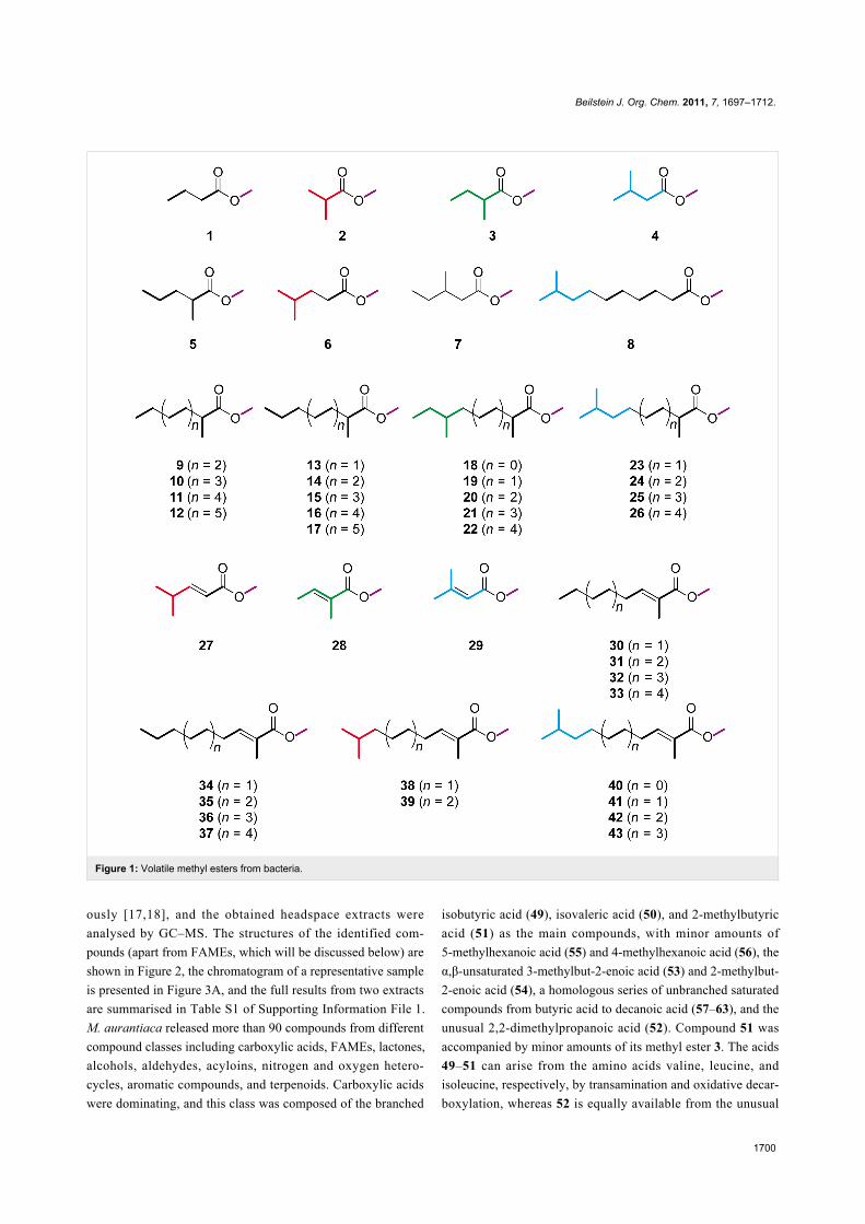
Figure 1: Volatile methyl esters from bacteria.
ously [17,18], and the obtained headspace extracts were
analysed by GC–MS. The structures of the identified com-
pounds (apart from FAMEs, which will be discussed below) are
shown in Figure 2, the chromatogram of a representative sample
is presented in Figure 3A, and the full results from two extracts
are summarised in Table S1 of Supporting Information File 1.
M. aurantiaca released more than 90 compounds from different
compound classes including carboxylic acids, FAMEs, lactones,
alcohols, aldehydes, acyloins, nitrogen and oxygen hetero-
cycles, aromatic compounds, and terpenoids. Carboxylic acids
were dominating, and this class was composed of the branched
isobutyric acid (49), isovaleric acid (50), and 2-methylbutyric
acid (51) as the main compounds, with minor amounts of
5-methylhexanoic acid (55) and 4-methylhexanoic acid (56), the
α,β-unsaturated 3-methylbut-2-enoic acid (53) and 2-methylbut-
2-enoic acid (54), a homologous series of unbranched saturated
compounds from butyric acid to decanoic acid (57–63), and the
unusual 2,2-dimethylpropanoic acid (52). Compound 51 was
accompanied by minor amounts of its methyl ester 3. The acids
49–51 can arise from the amino acids valine, leucine, and
isoleucine, respectively, by transamination and oxidative decar-
boxylation, whereas 52 is equally available from the unusual
Beilstein J. Org. Chem. 2011, 7, 1697–1712.
1701
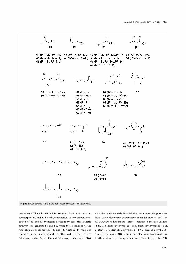
Figure 2: Compounds found in the headspace extracts of M. aurantiaca.
tert-leucine. The acids 53 and 54 can arise from their saturated
counterparts 50 and 51 by dehydrogenation. A two-carbon elon-
gation of 50 and 51 by means of the fatty acid biosynthetic
pathway can generate 55 and 56, while their reduction to the
respective alcohols provides 47 and 48. Acetoin (44) was also
found as a major compound, together with its derivatives
3-hydroxypentan-2-one (45) and 2-hydroxypentan-3-one (46).
Acyloins were recently identified as precursors for pyrazines
from Corynebacterium glutamicum in our laboratory [19]. The
M. aurantiaca headspace extracts contained methylpyrazine
(64), 2,5-dimethylpyrazine (65), trimethylpyrazine (66),
2-ethyl-3,6-dimethylpyrazine (67) , and 2-ethyl-3,5-
dimethylpyrazine (68), which may also arise from acyloins.
Further identified compounds were 2-acetylpyrrole (69),

Beilstein J. Org. Chem. 2011, 7, 1697–1712.
1702
Figure 3: Total ion chromatograms of the headspace extract from M. aurantiaca (A), and expansions of the total ion chromatogram (18–36 min)showing the relevant peaks of the FAMEs (B–D, for clarity the same expansion is shown in three replicates for FAMEs from different series). Thecolour code used for these series of FAMEs is given in the respective figures.
2-phenylethanol (70), phenylacetone (71), 1-phenylbutan-2-one
(72), methyl phenylacetate (73), methyl salicylate (74), methyl
furan-2-carboxylate (75), 2-acetyl-5-methylfuran (76), decanal
(77), 7-methyloctan-4-olide (78), nonan-4-olide (79), and the
terpenoids linalool (80) and geranyl acetone (81).
Besides the compounds mentioned above, several saturated
FAMEs were present in the headspace extracts (Figure 4). All
the identified FAMEs were divided into groups according to
their pattern of methyl branchings. Unbranched FAMEs
(Figure 3B, red) included all even and odd members of the

Beilstein J. Org. Chem. 2011, 7, 1697–1712.
1703
Figure 4: FAMEs identified in the headspace extracts from M. aurantiaca.
homologous series from methyl nonanoate to methyl tetrade-
canoate, in addition to methyl hexadecanoate. These com-
pounds were readily identified from their mass spectra by com-
parison to library spectra and subsequent GC–MS analysis of
synthetic standards. Mass spectra of unbranched FAMEs (for
mass spectrum of methyl dodecanoate see Figure 5A) are char-
acterised by fragment ions at m/z = 74 (McLafferty rearrange-
ment, Scheme 2), m/z = 87 (β-cleavage), and [M − 31]+ (loss of
OMe). Further fragment ions [M − CnH2n+1]+ arise from
cleavage of the saturated unbranched alkyl chain.
All other FAMEs were isomers of these unbranched com-
pounds and were assumed to be methyl branched FAMEs due to
biosynthetic considerations as outlined above. The structures of
these branched compounds have been suggested based on
careful analysis of their mass spectra and on a modified reten-

Beilstein J. Org. Chem. 2011, 7, 1697–1712.
1704
Figure 5: Mass spectra of (A) methyl dodecanoate (83), (B) methyl 2-methyldodecanoate (10), (C) methyl 4-methyldodecanoate (90), (D) methyl8-methyldecanoate (95), (E) methyl 9-methyldecanoate (8), (F) methyl 2,9-dimethyldecanoate (24), (G) methyl 4,11-dimethyldodecanoate (110), (H)methyl 4,8-dimethyldecanoate (112), and (I) methyl 4,8-dimethylundecanoate (114).
tion-index increment system [20]. Following this system, the
retention index Icalc. of a methyl branched compound can be
calculated (Equation 1) by
(1)
The increment N(n) depends on the number of carbon atoms n
in the longest alkyl chain and is N(n) = 100 n, FG is an incre-
ment for the functional group, and the increments Mei have to
be considered for methyl branches depending on the positions i
of branching. The increments FG and Mei have to be deter-
mined for each type of GC column. In a first approximation,

Beilstein J. Org. Chem. 2011, 7, 1697–1712.
1705
Scheme 2: McLafferty fragmentation of FAMEs.
these increments can be assumed to be constants, but as will be
discussed below both FG(n) and Mei(n) are slightly dependent
on the length of the alkyl chain, giving better results for the
calculated retention indices if this dependency is considered.
For all of the following analyses the functional group incre-
ment for FAMEs on a HP-5 MS column (FG(n)FAME, HP-5 MS)
was determined from the homologous series of unbranched
FAMEs (Table S2 and Figure S1 of Supporting Information
File 1). By linear regression (Figure 6) the functional group
increment (Equation 2) was
(2)
resulting in a modified Equation 1
(3)
Figure 6: The functional group increment FG(n)FAME, HP-5 MS.
A series of compounds (Figure 3A and Figure 3B, blue) exhib-
ited mass spectra with two significant fragment ions at m/z = 88
as the base peak and at m/z = 101, indicating a methyl
branching in a α-position (Scheme 2, cf. Figure 5B for mass
spectrum of methyl 2-methyldecanoate). For the determination
of the increment Meα the reference compound methyl
2-methyldecanoate (10) was synthesised by α-alkylation of 82
(Scheme 3). The mass spectrum and retention index (I = 1357)
of the product were identical to those of the natural compound.
By using Equation 3 the increment for a methyl branching in a
α-position was determined as Meα = 35, resulting in the calcu-
lated retention indices for the α-methyl branched FAMEs as
listed in Table S3 of Supporting Information File 1, column 4.
The calculated retention indices fitted perfectly for compounds
with a chain length of around 10 carbon atoms, which is not
surprising since Meα was determined from 10, but the indices
deviated slightly from the measured values for shorter (n = 7:
Inat. − Icalc. = 4) or longer (n = 15: Inat. − Icalc. = −2) FAMEs. In
other words, Meα was dependent on the chain length. A linear
regression analysis gave
(4)
Recalculation of the retention indices of the α-methyl branched
FAMEs, taking into account the dependency of Meα on the
chain length, resulted in the values listed in Table S3 of
Supporting Information File 1, column 5, which perfectly fitted
the measured retention indices.
The next class of compounds (Figure 3B, green) showed char-
acteristic fragment ions at m/z = 87 and m/z = 74 similar to the
unbranched FAMEs, but in contrast the β-cleavage was more
important than the McLafferty fragmentation (cf. Figure 5C for
mass spectrum of methyl 4-methyldodecanoate). This, together
with an almost completely missing fragment ion at m/z = 101,
accounting for a γ-cleavage in unbranched FAMEs, suggested
the presence of a methyl group in a γ-position, which leads to a
γ-fragmentation with m/z = 115. A synthesis of methyl
4-methyldodecanoate (90) was performed starting from 10
(Scheme 3). Reduction to the aldehyde 123d and subsequent
Horner–Wadsworth–Emmons reaction gave the α,β-unsatu-
rated ester 124d, which upon catalytic hydrogenation yielded
90. The synthetic compound was identical to the natural FAME
as judged by mass spectrum and retention index (I = 1572). The
retention index of this reference compound was used for to
determine that Meγ = 51, resulting in the calculated retention
indices for all γ-methyl branched FAMEs as summarised in
Table S4 of Supporting Information File 1. Correction of the
increment Meγ by linear regression gave
(5)
The corrected calculated retention indices, taking Equation 5
into consideration, were in good agreement with the measured
retention indices.
Another group of FAMEs (Figure 3C, red) showed mass spectra
with significant fragment ions at m/z = 74 and m/z = 87, like the
unbranched compounds (for mass spectrum of methyl
Beilstein J. Org. Chem. 2011, 7, 1697–1712.
1706
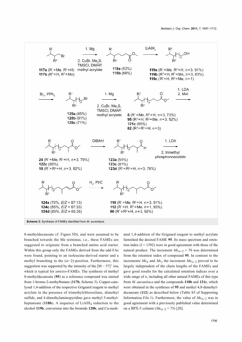
Scheme 3: Synthesis of FAMEs identified from M. aurantiaca.
8-methyldecanoate cf. Figure 5D), and were assumed to be
branched towards the Me terminus, i.e., these FAMEs are
suggested to originate from a branched amino acid starter.
Within this group only the FAMEs derived from the odd FAs
were found, pointing to an isoleucine-derived starter and a
methyl branching in the (ω−2)-position. Furthermore, this
suggestion was supported by the intensity of the [M − 57]+ ion,
which is typical for anteiso-FAMEs. The synthesis of methyl
8-methyldecanoate (95) as a reference compound was started
from 1-bromo-2-methylbutane (117b, Scheme 3). Copper-cata-
lysed 1,4-addition of the respective Grignard reagent to methyl
acrylate in the presence of trimethylchlorosilane, dimethyl
sulfide, and 4-dimethylaminopyridine gave methyl 5-methyl-
heptanoate (118b). A sequence of LiAlH4 reduction to the
alcohol 119b, conversion into the bromide 120b, and Cu-medi-
ated 1,4-addition of the Grignard reagent to methyl acrylate
furnished the desired FAME 95. Its mass spectrum and reten-
tion index (I = 1392) were in good agreement with those of the
natural product. The increment Meω−2 = 70 was determined
from the retention index of compound 95. In contrast to the
increments Meα and Meγ the increment Meω−2 proved to be
largely independent of the chain lengths of the FAMEs and
gave good results for the calculated retention indices over a
wide range of n, including all other natural FAMEs of this type
from M. aurantiaca and the compounds 118b and 121c, which
were obtained in the syntheses of 95 and methyl 4,8-dimethyl-
decanoate (112) as described below (Table S5 of Supporting
Information File 1). Furthermore, the value of Meω−2 was in
good agreement with a previously published value determined
on a BPX-5 column (Meω−2 = 73) [20].
Beilstein J. Org. Chem. 2011, 7, 1697–1712.
1707
The next series of FAMEs (Figure 3C, blue) also had very
similar mass spectra compared to the unbranched and (ω−2)-
methyl branched compounds dominated by the fragments at
m/z = 74 and m/z = 87 (the mass spectrum of methyl
9-methyldecanoate is shown in Figure 5E). Within this series
both even and odd FAMEs were detected, and therefore, these
compounds were suggested to be (ω−1)-methyl branched and
derived from leucine or valine starters, respectively. As typical
for (ω−1)-methyl branched compounds, these FAMEs eluted
slightly earlier than their (ω−2)-methyl branched counterparts.
Additional support for this suggestion was given by the occur-
rence of relatively intense fragments at [M − 43]+, as can be
expected for iso-FAMEs. In direct analogy to the synthesis of
95, methyl 9-methyldecanoate (8) was synthesised from 1-bro-
mo-3-methylbutane (117a, Scheme 3) and was identical to the
volatile from M. aurantiaca in terms of mass spectrum and
retention index (I = 1385), according to an increment of
Meω−1 = 63. The retention indices of all other volatiles from M.
aurantiaca and of the intermediate 118a in the synthesis of 8
were calculated from this constant increment and were in good
agreement with the measured values (Table S6 of Supporting
Information File 1).
In addition to these mono-methyl branched FAMEs, several
classes of multiply methyl branched compounds were identi-
fied. The first group of compounds (Figure 3C, green) exhib-
ited mass spectra that were very similar to those of the
α-branched FAMEs described above, with dominating frag-
ment ions at m/z = 88 and m/z = 101 (the mass spectrum of
methyl 2,9-dimethyldecanoate is depicted in Figure 5F), but
compounds of this class eluted significantly earlier than their
mono-branched isomers, pointing to a second methyl branch
within the alkyl chain, likely towards the methyl terminus.
Since both even and odd compounds of this series were present,
the structures of α- and (ω−1)-methyl branched FAMEs derived
from leucine and valine starters, respectively, were suggested
(Table S7 of Supporting Information File 1). The increments
FG(n)FAME, HP-5 MS, Meα(n), and Meω−1 as determined above
were used for the calculation of retention indices. The calcu-
lated values were in perfect agreement with the measured
values, thus corroborating the suggested structures. For unam-
biguous proof, an exemplary reference compound of this series
was synthesised (Scheme 3). α-Methylation of 8 yielded methyl
2,9-dimethyldecanoate (24), which was identical to the respec-
tive FAME from M. aurantiaca.
A second series of dimethyl branched FAMEs (Figure 3D, red)
exhibited very similar mass spectra to the γ-methyl branched
esters (for mass spectrum of methyl 4,11-dimethyldodecanoate
cf. Figure 5G). Within this class again both even and odd
FAMEs were found, suggesting that they were derived from
valine or leucine starters and therefore γ- and (ω−1)-methyl
branched. Calculations of the retention indices for the suggested
structures were in good agreement with the measured data
(Table S8 of Supporting Information File 1), thus providing
further evidence for the suggested structures. Final verification
was obtained by synthesis of methyl 4,11-dimethyldodecanoate
(110) from 24 by its reduction to the aldehyde 123a,
Horner–Wadsworth–Emmons olefination to 124a, and catalytic
hydrogenation (Scheme 3). The synthetic material was iden-
tical to the natural compound 110.
A related group of compounds (Figure 3D, blue) proved to have
very similar mass spectra, thus indicating a methyl group in a
γ-position (Figure 5H shows the mass spectrum of methyl 4,8-
dimethyldecanoate). The relatively high intensity of the
[M − 57]+ fragment ion suggested the structures of anteiso-
FAMEs, and accordingly, only the isoleucine-derived methyl
esters of even-numbered FAs were found. Retention-index
calculations (Table S9 of Supporting Information File 1)
showed full agreement of the calculated retention indices with
the experimental data. A synthesis of methyl 4,8-dimethyl-
decanoate (112) as a reference compound was carried out
starting from 3-methylpentan-1-ol (119c), by similar methods as
described above (Scheme 3). The alcohol 119c was trans-
formed into the bromide 120c. The copper-catalysed Michael
addition of the respective Grignard reagent to methyl acrylate
yielded methyl 6-methyloctanoate (121c), which was α-methyl-
ated to 122c. Reduction with DIBAH to the aldehyde 123c,
Horner–Wadsworth–Emmons olefination to 124c, and final
catalytic hydrogenation afforded 112. The product exhibited the
same mass spectrum and retention index (I = 1441) as the
natural FAME. The slight deviations between the calculated and
measured retention indices for compounds 106 (ΔI = 2) and 112
(ΔI = 3) may be attributed to the fairly close proximity of the
two methyl branches, which are only three methylene units
apart.
The FAME 114 (Figure 3D, green) was also suggested to be
γ-methyl branched based on its mass spectrum (the mass spec-
trum of methyl 4,8-dimethylundecanoate is presented in
Figure 5I). This compound was an isomer of methyl 4-methyl-
dodecanoate (90) and methyl 4,10-dimethylundecanoate (107),
but eluted slightly earlier than 107, resulting in a suggested
structure of methyl 4,8-dimethylundecanoate. A synthesis of
this compound was performed starting with bromination of
2-methylpentan-1-ol (125) and subsequent Cu-catalysed 1,4-
addition of the respective Grignard reagent to methyl acrylate,
resulting in methyl 5-methyloctanoate (127, Scheme 4). Reduc-
tion to the aldehyde 129 via the alcohol 128, addition of methyl-
magnesium bromide, and bromination resulted in the secondary
bromide 131. Michael addition of its Grignard reagent to methyl

Beilstein J. Org. Chem. 2011, 7, 1697–1712.
1708
Scheme 4: Synthesis of the γ- and (ω−3)-methyl branched FAME 114.
Figure 7: Mass spectra of tentatively identified methyl 4,8-dimethyldodecanoate (115) and methyl 8-ethyl-4-methyldodecanoate (116).
acrylate was less efficient than for primary bromides, but gave
the desired product 114 in low yield. The identity to the natural
product was confirmed by comparison of GC–MS data.
The structures of two additional FAMEs were suggested based
on their mass spectra, but these were not verified by synthesis
and therefore only tentatively identified (Figure 7). Both com-
pounds were suggested to be γ-methyl branched due to the rela-
tive intensities of the fragment ions at m/z = 87 and m/z = 74.
The first compound (Figure 7A) showed a molecular ion at
m/z = 242 and further fragment ions at m/z = 157 and m/z = 185
resulting from the loss of C4H9 or C6H13, whereas no fragment
ion accounting for the loss of C5H11 (m/z = 171) was observed.
This pattern is in accordance with the structure of methyl 4,8-
dimethyldodecanoate (115). The second compound (Figure 7B)
exhibited a molecular ion at m/z = 256 and fragment ions at
m/z = 157 and m/z = 199 according to the loss of C4H9 and
C7H15, but not at m/z = 171 and m/z = 185 (loss of C5H11 and
C6H13, respectively). This pattern suggested an ethyl branch in
the 8-position corresponding to the structure of methyl 8-ethyl-
4-methyldodecanoate (116). This compound is interesting in
terms of its biosynthesis, because it may be formed by incorpor-
ation of an ethylmalonyl-CoA elongation unit. However, since
no further compounds with ethyl branches were found, another
biosynthetic option seems more likely. The compound 2-ethyl-
hexanol is a widespread pollutant originating from plasticisers,
and this compound could have been oxidised to 2-ethylhexa-
noic acid, transformed into its CoA derivative, and used as an
unusual starter unit by the FAS to make 116.
Several of the FAMEs emitted by M. aurantiaca are chiral,
including, e.g., the α-, γ-, and (ω−2)-methyl branched FAMEs.
In addition, compounds such as the γ- and (ω−2)-methyl
branched FAMEs exist in two different diastereomers.
However, these diastereomers, which were both contained in
the synthetic material, e.g., of compounds 112 and 114, were

Beilstein J. Org. Chem. 2011, 7, 1697–1712.
1709
not separated on the HP5-MS column used for GC–MS
analyses, and therefore the elucidation of the relative configura-
tions was not possible, at least not by our GC approach. The
only very small differences in the physical properties of these
compounds were also reflected by the occurrence of only one
set of signals in the NMR spectra of these mixtures of dia-
stereomers. The separation of enantiomers of the chiral com-
pounds described here on chiral GC columns is also a very hard
task, especially for internally methyl branched FAMEs, and was
beyond the scope of our work.
Feeding experimentsTo investigate the biosynthetic origin of the FAMEs, several
feeding experiments with deuterated precursors were
performed. These were directly added to the agar plate cultures
and the headspace extracts were prepared by CLSA after ca.
2–3 days of growth. The CLSA extracts were then analysed by
GC–MS. Incorporation of deuterated precursors was observ-
able through the increased molecular masses and m/z ratios of
certain fragment ions that could be used to localise the
deuterium incorporation. One advantage of using deuterated
precursors is that deuterium incorporation results in a decrease
in the retention time of the analyte with respect to its unlabelled
counterpart, i.e., the deuterated isotopomers are separated and
their mass spectra can easily be interpreted [21].
Feeding of [2H10]isoleucineOne possible pathway to (ω−2)-methyl branched FAMEs is
through the use of 2-methylbutyryl-CoA as a starting unit,
which is available from isoleucine by transamination and oxida-
tive decarboxylation. The alternative would be to use an acetyl-
CoA starter followed by incorporation of a methylmalonyl-CoA
elongation unit. The question as to which of these two alter-
natives is operative in M. aurantiaca was addressed by feeding
of [2H10]isoleucine. In this feeding experiment M. aurantiaca
produced large amounts of [2H9]-2-methylbutyric acid ([2H9]-
51) and its respective methyl ester [2H9]methyl 2-methylbu-
tyrate ([2H9]-3), both with incorporation rates >70%. The
transamination of [2H10]isoleucine to 2-oxo-3-methylpentanoic
acid proceeds with the loss of one deuterium, and accordingly,
nine deuterium atoms were incorporated into 3, as indicative by
a shift of the molecular ion of 3 from m/z = 116 to m/z = 125
(compare Figures S2A and S2B of Supporting Information
File 1). The fragment ion at m/z = 101, arising through the loss
of a methyl group, shifted to m/z = 107, whereas no signal was
detected at m/z = 110 in the mass spectrum of [2H9]-3. There-
fore, the respective fragment ion only arises by methyl cleavage
from the acyl moiety, and not by loss of the methyl group from
the ester function. Further diagnostic fragment ions formed by
α-cleavage (m/z = 57) and McLafferty rearrangement (m/z = 88)
shifted to m/z = 66 and m/z = 93, in full agreement with the
structure of [2H9]-3. For the respective free acid 51 no molec-
ular ion is visible, but the fragment ion at m/z = 101, formed by
loss of one hydrogen from the carboxylic acid function, was
detected at m/z = 110 for [2H9]-51, indicating the incorporation
of nine deuteriums (Figures S2C and S2D of Supporting Infor-
mation File 1). Further fragment ions were observed at m/z = 66
(α-cleavage, + 9 amu), m/z = 79 (McLafferty rearrangement, + 5
amu), and m/z = 101 (loss of methyl group, + 6 amu), clearly
establishing the identity of [2H9]-51. The uptake of isoleucine
in the (ω−2)-methyl branched FAMEs was also observed with
high incorporation rates (>90%) for methyl 12-methyltetrade-
canoate (97, Figures S2E and S2F of Supporting Information
File 1), methyl 10-methyldodecanoate (96), and methyl
14-methylhexadecanoate (not shown). The mass spectrum of
[2H9]-97 is characterised by a molecular ion at m/z = 265
showing the incorporation of nine deuteriums, whereas the frag-
ment ions at m/z = 74 and m/z = 87 indicative of the structure of
a methyl ester are not shifted relative to the unlabelled material.
The compound methyl 14-methylhexadecanoate was not found
under the original experimental conditions without feeding of
[2H9]isoleucine, demonstrating that the production of (ω−2)-
methyl branched FAMEs by M. aurantiaca can be activated by
isoleucine supply. Unfortunately, the γ- and (ω−2)-methyl
branched compounds 112 and 113 were not produced under the
conditions of isoleucine feeding, and therefore, their biosyn-
thetic origin remained elusive.
Feeding of [2H10]leucineFeeding of [2H10]leucine was performed to investigate the
biosynthetic origin of the (ω−1)-methyl branched FAMEs.
Incorporation was observed for a series of (ω−1)-methyl
branched FAs including isovaleric acid, 5-methylhexanoic acid,
7-methyloctanoic acid, 9-methyldecanoic acid (Figures S3A
and S3B of Supporting Information File 1), and 11-methyldode-
canoic acid, all with high incorporation rates (>70%). The
uptake of deuterated leucine for the last compound was observ-
able by a shift of the molecular ion from m/z = 186 to
m/z = 195. The fragment ions of the McLafferty rearrangement
(m/z = 60) and the β-cleavage (m/z = 73) remained unchanged,
whereas fragment ions arising from cleavage of the terminal
isopropyl group (m/z = 43 and m/z =143) shifted to m/z = 50 and
m/z = 145 in agreement with the deuterium labelling of this
portion of the molecule.
The labelling was also introduced into the iso-odd FAMEs 102
and 103 (Figures S3C to S3F of Supporting Information File 1)
and the higher homologue methyl 15-methylhexadecanoate (not
shown). Methyl 15-methylhexadecanoate was only found
during feeding of [2H10]leucine, similar to the formation of
methyl 14-methylhexadecanoate found only during feeding of
[2H10]isoleucine.

Beilstein J. Org. Chem. 2011, 7, 1697–1712.
1710
Feeding of [2H8]valineFeeding of [2H8]valine resulted in the incorporation of the
isotopic labelling into its transamination-oxidative decarboxyla-
tion product isobutyric acid (49) and the iso-even FAMEs
methyl 12-methyltridecanoate (100) and methyl 14-methyl-
pentadecanoate (101) with high incorporation rates (>50%), as
indicated by the increase of the molecular ions by 7 amu in
combination with the overall expected fragmentation pattern
(Figure S4 of Supporting Information File 1). The transamina-
tion product of valine, 2-oxoisovaleric acid, can be used by
most organisms for the biosynthesis of the leucine precursor
2-oxoisocaproic acid. The enzymes for catalysis of this pathway
are encoded in the genome of M. aurantiaca, but the pathway
seemed not to be active under the experimental conditions,
judged by the fact that no incorporation of [2H8]valine into the
iso-odd FAMEs was observed. Furthermore, no uptake of
labelling from [2H10]leucine into the iso-even FAMEs was
found, which also rules out a similar formation of the higher
homologue 2-oxo-5-methylcaproic acid from 2-oxoisocaproic
acid. These results also demonstrate that the FAs in M. auran-
tiaca are not subject to α-oxidation, a process in which FAs are
oxidatively degraded by one carbon.
Feeding of [2H5]sodium propionateTo investigate the biosynthetic origin of the α- and γ-methyl
branches, a feeding experiment with [2H5]sodium propionate
was performed. This compound was expected to be carboxy-
lated to yield methylmalonyl-CoA, which would serve as an
elongation unit for the introduction of methyl branches. Alter-
native mechanisms could include the chain elongation with
malonyl-CoA, followed by methylation with SAM by catalysis
of a C-methyltransferase. Indeed the incorporation of isotopic
labelling into the α- and γ-methyl branched FAMEs was
observed with high incorporation rates (>90%, Figure S5 of
Supporting Information File 1). Incorporation into methyl 2,11-
dimethyldodecanoate (25) and its higher homologue 26 was
observable by an increase of the molecular ion by 3 amu, while
the fragment ions at m/z = 88 and m/z = 101, indicative of an
α-methyl branch, shifted to m/z = 91 and m/z = 104. The
incorporation of only three deuterium atoms from [2H5]sodium
propionate is in agreement with the biosynthesis of FAs
(Scheme 1): One deuterium is lost during carboxylation of
propionyl-CoA to yield methylmalonyl-CoA, the second is
possibly exchanged with water due to the C,H-acidity of
malonyl-CoA derivatives, but it is lost, at the latest, in the dehy-
dration of the 3-hydroxy-2-methylacyl-S~ACP to the respec-
tive 2-methyl-2-enoyl-S~ACP intermediate. The incorporation
of [2H5]sodium propionate was also observed for the γ-methyl
branched compounds represented by methyl 4,11-dimethyldo-
decanoate (110). The uptake of deuterium was in the first
instance visible by an increase of the molecular ion from
m/z = 242 to m/z = 245, whereas the McLafferty rearrangement
and β-cleavage fragment ions were detected at m/z = 74 and
m/z = 87, as for the unlabelled compound. The deuterium
labelling of the γ-methyl group was indicated by a shift of the
fragment ion at m/z = 115 (γ-cleavage) to m/z = 118. No
incorporation was observed into the (ω−2)-methyl branches of
the respective FAMEs, ruling out their alternative formation
from an acetate starter in combination with an initial methyl-
malonyl-CoA elongation unit, instead of from isoleucine.
Feeding of [methyl-2H3]methionineFeeding of [methyl-2H3]methionine was performed, first as a
control experiment with respect to the biosynthetic origin of the
α- and γ-methyl branches, and second, to investigate the biosyn-
thetic origin of the methyl ester moiety of the FAMEs. The
feeding experiment resulted in the incorporation into 103
(Figure S6 of Supporting Information File 1) and 97 (not
shown), as indicated by a shift of the molecular ion from
m/z = 256 to m/z = 259, of the McLafferty ion from m/z = 74 to
m/z = 77, and of the β-cleavage fragment ion from m/z = 87 to
m/z = 90. Further FAMEs were not produced during the course
of this experiment. The results clearly demonstrate the forma-
tion of FAMEs from methionine via SAM as the methyl donor
for the methylation of FAs.
ConclusionIn summary, the headspace extracts from M. aurantiaca contain
unbranched even and odd FAMEs that are made from an acetyl-
CoA or propionyl-CoA starter. In particular the even FAMEs
are very widespread in nature. Several α-methyl branched
FAMEs were also present that were previously described from
Chitinophaga [14]. These compounds can be synthesised by
incorporation of a final methylmalonyl-CoA elongation unit.
When methylmalonyl-CoA is used as the penultimate building
block followed by malonyl-CoA, the synthesis results in the for-
mation of γ-methyl branched FAs that upon O-methylation
yield the respective FAMEs. Only one such compound has
previously been found in nature represented by methyl
4-methyloctanoate, and this is a constituent of the pheromone
blend of the date palm fruit stalk borer Oryctes elegans [22],
whereas the related FAMEs 89–94 emitted by M. aurantiaca
are all new natural products. The use of alternative starter units
from branched amino acids for the biosynthesis of FAMEs was
demonstrated in feeding experiments. Although the respective
FAs are widespread in nature, only a few reports of these
FAMEs exist, e.g., the isoleucine-derived compounds 95 and 96
occur in the volatile fraction from Medicago sativa [23], the
valine-derived FAME 99 is known from Capsicum annuum
[24], and the leucine-derived compound 8 was previously found
in Stigmatella aurantiaca [13], whereas the compounds 102 and
103 (from leucine), and 98 (from valine) have not been

Beilstein J. Org. Chem. 2011, 7, 1697–1712.
1711
described before. The same is true for all FAMEs described
here that are derived from a branched amino acid and that are in
addition α- or γ-methyl-branched, and there is only one report
on such unusual FAs with a similar methyl branching pattern
from Bacillus [25]. The biosynthesis of these compounds was
established in feeding experiments with [2H5]sodium pro-
pionate and [methyl-2H3]methionine, which allowed us to
distinguish between two possible pathways, i.e., the incorpor-
ation of methylmalonyl-CoA elongation units, or the alter-
native incorporation of malonyl-CoA elongations units fol-
lowed by methylation with SAM. The experiments clearly
demonstrated the usage of methylmalonyl-CoA building blocks,
whereas the feeding experiment with [methyl-2H3]methionine
gave proof for the origin of the methyl ester functions by SAM-
dependent methylation of the respective FAs.
Supporting InformationSupporting Information contains experimental details for
the syntheses and analytical data of all synthesized
compounds, the tabulated full results of the headspace
analyses, and mass spectra of labelled FAMEs obtained
during feeding experiments together with the mass spectra
of the unlabelled compounds for comparison.
Supporting Information File 1Experimental details and analytical data.
[http://www.beilstein-journals.org/bjoc/content/
supplementary/1860-5397-7-200-S1.pdf]
AcknowledgementsThis work was supported by the Deutsche Forschungs-
gemeinschaft with an Emmy Noether fellowship (to J. S. D.,
DI1536/1-1).
References1. Kaneda, T. Microbiol. Rev. 1991, 55, 288–302.2. Wang, J.-F.; Dai, H.-Q.; Wei, Y.-L.; Zhu, H.-J.; Yan, Y.-M.; Wang, Y.-H.;
Long, C.-L.; Zhong, H.-M.; Zhang, L.-X.; Cheng, Y.-X.Chem. Biodiversity 2010, 7, 2046–2053. doi:10.1002/cbdv.201000072
3. Hamilton, J. T. G.; Harper, D. B. Phytochemistry 1997, 44, 1129–1132.doi:10.1016/S0031-9422(96)00697-8
4. Cropp, T. A.; Wilson, D. J.; Reynolds, K. A. Nat. Biotechnol. 2000, 18,980–983. doi:10.1038/79479
5. Hutchinson, C. R.; Sherman, M. M.; McInnes, A. G.; Walter, J. A.;Vederas, J. C. J. Am. Chem. Soc. 1981, 103, 5956–5959.doi:10.1021/ja00409a076
6. Liu, Y.; Hazzard, C.; Eustaquio, A. S.; Reynolds, K. A.; Moore, B. S.J. Am. Chem. Soc. 2009, 131, 10376–10377. doi:10.1021/ja9042824
7. Xu, Z.; Ding, L.; Hertweck, C. Angew. Chem., Int. Ed. 2011, 50,4667–4670. doi:10.1002/anie.201008265
8. Carroll, B. J.; Moss, S. J.; Bai, L.; Kato, Y.; Tölzer, S.; Yu, T.-W.;Floss, H. G. J. Am. Chem. Soc. 2002, 124, 4176–4177.doi:10.1021/ja0124764
9. Smith, A.; Calder, A. G. Biomed. Mass Spectrom. 1979, 6, 347–349.doi:10.1002/bms.1200060808
10. Schulz, S.; Dickschat, J. S. Nat. Prod. Rep. 2007, 24, 814–842.doi:10.1039/b507392h
11. Schöller, C. E. G.; Gürtler, H.; Pedersen, R.; Molin, S.; Wilkins, K.J. Agric. Food Chem. 2002, 50, 2615–2621. doi:10.1021/jf0116754
12. Wilkins, K.; Schöller, C. Actinomycetologica 2009, 23, 27–33.doi:10.3209/saj.SAJ230202
13. Dickschat, J. S.; Bode, H. B.; Wenzel, S. C.; Müller, R.; Schulz, S.ChemBioChem 2005, 6, 2023–2033. doi:10.1002/cbic.200500174
14. Nawrath, T.; Gerth, K.; Müller, R.; Schulz, S. Chem. Biodiversity 2010,7, 2228–2253. doi:10.1002/cbdv.201000190
15. Weinstein, M. J.; Luedemann, G. M.; Oden, E. M.; Wagman, G. H.;Rosselet, J. P.; Marquez, J. A.; Coniglio, C. T.; Charney, W.;Herzog, H. L.; Black, J. J. Med. Chem. 1963, 6, 463–464.doi:10.1021/jm00340a034
16. He, H.; Ding, W. D.; Bernan, V. S.; Richardson, A. D.; Ireland, C. M.;Greenstein, M.; Ellestad, G. A.; Carter, G. T. J. Am. Chem. Soc. 2001,123, 5362–5363. doi:10.1021/ja010129o
17. Dickschat, J. S.; Wenzel, S. C.; Bode, H. B.; Müller, R.; Schulz, S.ChemBioChem 2004, 5, 778–787. doi:10.1002/cbic.200300813
18. Dickschat, J. S.; Zell, C.; Brock, N. L. ChemBioChem 2010, 11,417–425. doi:10.1002/cbic.200900668
19. Dickschat, J. S.; Wickel, S.; Bolten, C. J.; Nawrath, T.; Schulz, S.;Wittmann, C. Eur. J. Org. Chem. 2010, 2687–2695.doi:10.1002/ejoc.201000155
20. Schulz, S.; Toft, S. Tetrahedron 1993, 49, 6805–6820.doi:10.1016/S0040-4020(01)80424-5
21. Dickschat, J. S.; Citron, C. A.; Brock, N. L.; Riclea, R.; Kuhz, H.Eur. J. Org. Chem. 2011, 3339–3346. doi:10.1002/ejoc.201100188
22. Rochat, D.; Mohammadpoor, K.; Malosse, C.; Avand-Faghih, A.;Lettere, M.; Beauhaire, J.; Morin, J.-P.; Pezier, A.; Renou, M.;Abdollahi, G. A. J. Chem. Ecol. 2004, 30, 387–407.doi:10.1023/B:JOEC.0000017984.26917.52
23. Core, R. J.; Henning, J. A.; Gardea-Torresdey, J. J. Agric. Food Chem.1994, 42, 2932–2936. doi:10.1021/jf00048a054
24. Guadayol, J. M.; Caixach, J.; Ribé, J.; Cabañas, J.; Rivera, J.J. Agric. Food Chem. 1997, 45, 1868–1872. doi:10.1021/jf960266i
25. Carballeira, N. M.; Miranda, C.; Lozano, C. M.; Nechev, J. T.;Ivanova, A.; Ilieva, M.; Tzvetkova, I.; Stefanov, K. J. Nat. Prod. 2001,64, 256–259. doi:10.1021/np000494d

Beilstein J. Org. Chem. 2011, 7, 1697–1712.
1712
License and TermsThis is an Open Access article under the terms of the
Creative Commons Attribution License
(http://creativecommons.org/licenses/by/2.0), which
permits unrestricted use, distribution, and reproduction in
any medium, provided the original work is properly cited.
The license is subject to the Beilstein Journal of Organic
Chemistry terms and conditions:
(http://www.beilstein-journals.org/bjoc)
The definitive version of this article is the electronic one
which can be found at:
doi:10.3762/bjoc.7.200

![Amide-Based Surfactants from Methyl Glucoside as Potential ... · Amide-based surfactants from methyl glucoside can utilize the sugar either as uronic acid [13] or as amino [14] component.](https://static.fdokument.com/doc/165x107/5ea69f03bb5f8824165ae65d/amide-based-surfactants-from-methyl-glucoside-as-potential-amide-based-surfactants.jpg)

![سامى عبد الشكور... · 134 [1973BCSJ3625] Synthesis and rearrangement of oxanilic esters arylhydrazones. Shawali, A. Sami; Ahmad, M. Kamal. Fac. Sci., Univ. Cairo, Giza,](https://static.fdokument.com/doc/165x107/5f5ff49368fbf70cf43cd86f/-f-134-1973bcsj3625-synthesis-and-rearrangement.jpg)


![Studies on genome size estimation, chromosome number ......Suaeda salsa seeds is also edible [15], and it is rich in fatty acids. 90.7% of Suaeda salsa fatty acid is unsaturated. Fur-thermore,](https://static.fdokument.com/doc/165x107/60dae74bd1043175cd03d952/studies-on-genome-size-estimation-chromosome-number-suaeda-salsa-seeds.jpg)