PML Nuclear Bodies -...
18
PML Nuclear Bodies Vale ´ rie Lallemand-Breitenbach 1 and Hugues de The ´ 1,2 1 INSERM/CNRS/Universite ´ Paris Diderot/Institut Universitaire He ´matologie U944/ UMR7212, Laboratoire associe ´ de la Ligue Nationale contre le Cancer, Ho ˆpital St. Louis, 1, Av. C. Vellefaux 75475 Paris Cedex 10, France 2 Service de Biochimie, Ho ˆpital St. Louis, 1, Av. C. Vellefaux 75475 Paris Cedex 10, France Correspondence: [email protected] PML nuclear bodies are matrix-associated domains that recruit an astonishing variety of seemingly unrelated proteins. Since their discovery in the early 1960s, PML bodies have fascinated cell biologists because of their beauty and their tight association with cellular disorders. The identification of PML, a gene involved in an oncogenic chromosomal trans- location, as the key organizer of these domains drew instant interest onto them. The multiple levels of PML body regulation bya specific posttranslationalmodification, sumoylation, have raised several unsolved issues. Functionally, PML bodies may sequester, modify or degrade partner proteins, but in many ways, PML bodies still constitute an enigma. PML BODIES, AN INTRODUCTION P ML nuclear bodies (NBs) are spheres of 0.1 – 1.0 mm in diameter found in most cell-lines and many tissues. They belong to the nuclear matrix, an ill-defined super-structure of the nucleus proposed to anchor and regulate many nuclear functions, including DNA repli- cation, transcription, or epigenetic silencing (Stuurman et al. 1990). The PML protein is the key organizer of these domains that recruits an ever-growing number of proteins, whose only common known feature to date is their ability to be sumoylated (Bernardi and Pandolfi 2007). PML and NBs were proposed to fine-tune a wide variety of processes, through facilitation of partner protein posttranslational modifications (notably sumoylation itself ) resulting in partner sequestration, activation, or degradation. Several NBs subtypes have been defined on morpholog- ical bases, which all contain an electron-dense shell and an inner core. PML NBs came to the forefront with the observation that the oncogenic PML/RARA protein disrupts them in a treatment-reversible manner (Daniel et al. 1993; Dyck et al. 1994; Koken et al. 1994; Weis et al. 1994; Zhu et al. 1997). PML NBs are regulated by cellular stress: viral infection (Everett 2006), DNA-damage, transformation (Koken et al. 1995; Terris et al. 1995; Gurrieri et al. 2004), and oxidative stress (Yamada et al. 2001a; Villagra et al. 2006). Moreover, transcription of PML and several genes encoding partner proteins is dramatically enhanced by interferons. Yet, pml-/- mice, which cannot assemble NBs, develop normally and live well, demonstrating that NBs are dis- pensable for most basic biological functions. Editors: David Spector and Tom Misteli Additional Perspectives on The Nucleus available at www.cshperspectives.org Copyright # 2010 Cold Spring Harbor Laboratory Press; all rights reserved; doi: 10.1101/cshperspect.a000661 Cite this article as Cold Spring Harb Perspect Biol 2010;2:a000661 1 on March 15, 2020 - Published by Cold Spring Harbor Laboratory Press http://cshperspectives.cshlp.org/ Downloaded from
Transcript of PML Nuclear Bodies -...
PML Nuclear Bodies
Valerie Lallemand-Breitenbach1 and Hugues de The1,2
1INSERM/CNRS/Universite Paris Diderot/Institut Universitaire Hematologie U944/ UMR7212, Laboratoireassocie de la Ligue Nationale contre le Cancer, Hopital St. Louis, 1, Av. C. Vellefaux 75475 Paris Cedex 10,France
2Service de Biochimie, Hopital St. Louis, 1, Av. C. Vellefaux 75475 Paris Cedex 10, France
Correspondence: [email protected]
PML nuclear bodies are matrix-associated domains that recruit an astonishing variety ofseemingly unrelated proteins. Since their discovery in the early 1960s, PML bodies havefascinated cell biologists because of their beauty and their tight association with cellulardisorders. The identification of PML, a gene involved in an oncogenic chromosomal trans-location, as the key organizer of these domains drew instant interest onto them. The multiplelevels of PML body regulation bya specific posttranslational modification, sumoylation, haveraised several unsolved issues. Functionally, PML bodies may sequester, modify or degradepartner proteins, but in many ways, PML bodies still constitute an enigma.
PML BODIES, AN INTRODUCTION
PML nuclear bodies (NBs) are spheres of0.1–1.0 mm in diameter found in most
cell-lines and many tissues. They belong to thenuclear matrix, an ill-defined super-structureof the nucleus proposed to anchor and regulatemany nuclear functions, including DNA repli-cation, transcription, or epigenetic silencing(Stuurman et al. 1990). The PML protein isthe key organizer of these domains that recruitsan ever-growing number of proteins, whoseonly common known feature to date is theirability to be sumoylated (Bernardi and Pandolfi2007). PML and NBs were proposed to fine-tunea wide variety of processes, through facilitation ofpartner protein posttranslational modifications(notably sumoylation itself ) resulting in partnersequestration, activation, or degradation. Several
NBs subtypes have been defined on morpholog-ical bases, which all contain an electron-denseshell and an inner core.
PML NBs came to the forefront with theobservation that the oncogenic PML/RARAprotein disrupts them in a treatment-reversiblemanner (Daniel et al. 1993; Dyck et al. 1994;Koken et al. 1994; Weis et al. 1994; Zhu et al.1997). PML NBs are regulated by cellular stress:viral infection (Everett 2006), DNA-damage,transformation (Koken et al. 1995; Terris et al.1995; Gurrieri et al. 2004), and oxidative stress(Yamada et al. 2001a; Villagra et al. 2006).Moreover, transcription of PML and severalgenes encoding partner proteins is dramaticallyenhanced by interferons. Yet, pml-/- mice,which cannot assemble NBs, develop normallyand live well, demonstrating that NBs are dis-pensable for most basic biological functions.
Editors: David Spector and Tom Misteli
Additional Perspectives on The Nucleus available at www.cshperspectives.org
Copyright # 2010 Cold Spring Harbor Laboratory Press; all rights reserved; doi: 10.1101/cshperspect.a000661
Cite this article as Cold Spring Harb Perspect Biol 2010;2:a000661
1
on March 15, 2020 - Published by Cold Spring Harbor Laboratory Press http://cshperspectives.cshlp.org/Downloaded from
Nevertheless, recent data has implicated PML inthe control of cellular senescence and stem cellself-renewal, extending the fields of investiga-tion of PML function (Ferbeyre et al. 2000;Pearson et al. 2000; Salomoni and Pandolfi2002; Ito et al. 2008).
HISTORICAL OVERVIEW
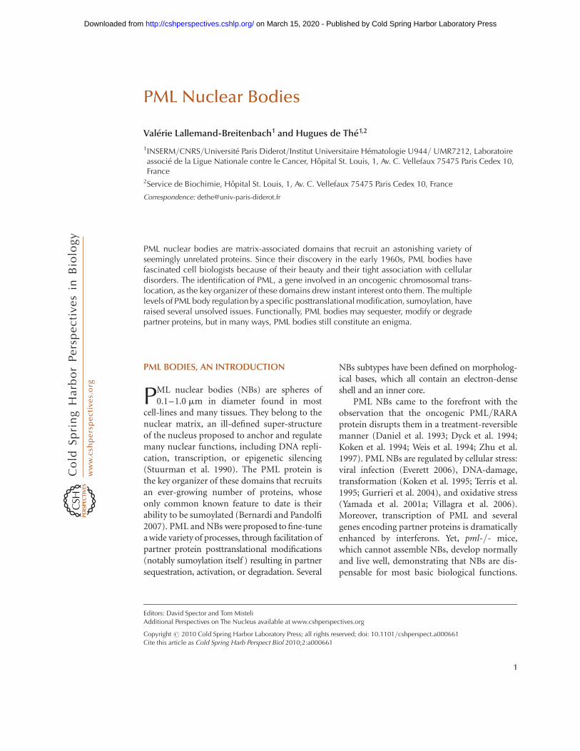
As for several other nuclear structures, electronmicroscopy and auto-antibodies were the twofounding fathers of the field. Work from severalpioneers in the early 1960s noted the presence ofdense spherical objects by electron microscopy(de The et al. 1960). Two classes were morpho-logically described: Empty ones (fibrillar) andgranular ones, with an inner microgranularmaterial proposed to be ribonucleoproteins(Fig. 1). PML NBs were later observed byimmunofluorescence using autoimmune serafrom primary billiary cirrhosis patients. Theseallowed the identification of the first NB-associ-ated protein, SP100 (Szostecki et al. 1990), and
an initial characterization of these structures(Ascoli and Maul 1991).
Identification of an anti-nuclear matrixantibody, which labeled the same structures asSP100 (Stuurman et al. 1992), drew the firstlink between these bodies and the nuclearmatrix. The localization of PML, a protein fusedto the retinoic acid receptor a(RARA) in thePML/RARA oncoprotein of acute promyelo-cytic leukemia (APL), to the same nuclear dotsas SP100, renewed interest for these domains.The observation that in APL cells PML/RARAdisrupted these domains drew instant excite-ment from the scientific community (Danielet al. 1993; Dyck et al. 1994; Koken et al. 1994;Weis et al. 1994). Moreover, PML bodies wererestored by two different anti-APL therapies,retinoic acid and arsenic trioxide, later shownto trigger PML/RARA degradation (Quignonet al. 1997; Zhu et al. 2001), identifying the firststriking parallel between the status of the bodiesand that of the cell. Many further studiesshowed that PML bodies altered in stress condi-tions, notably viral infections, heat shock, and
Figure 1. Different types of PML bodies. Immuno-gold electron microscopy of PML NBs in CHO cell line stablyoverexpressing PML. (A, B, C) “Classical” PML NBs. PML is distributed on a dense electron shell (A, B) or onlight halo (C), that can contain a microgranular inner core (B, C) or not (A). (D, E) PML body in arsenictrioxide-treated cell. Note the absence of the inner core and the enrolled fibrillar aspect of the body (D). (F)PML-II targets the inner membrane region of the nuclear envelope in pml-/- MEF cells. Images courtesy ofEdmond and Francine Puvion (CNRS, Villejuif, France).
V. Lallemand-Breitenbach and H. de The
2 Cite this article as Cold Spring Harb Perspect Biol 2010;2:a000661
on March 15, 2020 - Published by Cold Spring Harbor Laboratory Press http://cshperspectives.cshlp.org/Downloaded from
exposure to heavy metals (Everett 2001; Dellaireand Bazett-Jones 2004; Everett 2006).
PML bodies recruit an ever-increasing num-ber of partner proteins (now in the range of100), one of the most studied being DAXX, apotent repressor of transcription and modula-tor of apoptosis. Critically, PML is the actualorganizer of the bodies (Ishov et al. 1999; Zhonget al. 2000a; Lallemand-Breitenbach et al. 2001).Among these recruited proteins, one deservesa special mention: an ubiquitin-like proteinnamed SUMO, as PML conjugation by SUMOplays a critical role in recruitment of partners,many of which are sumoylated themselves.Recent studies have focused on more fundamen-tal cell-biology issues: Dynamics of the bodies(Eskiw et al. 2003, Muratani et al., 2002, Chenet al., 2008), relation to other nuclear compo-nents (Wang et al. 2004; Batty et al. 2009; Russellet al. 2009), mode of assembly (Shen et al. 2006),and determinants of partner recruitment andactual function in cellulo and in vivo.
THE PML PROTEINS
PML is a member of the TRIM/RBCC family ofproteins, many members of which are ubiquitinligases that generate subcellular structuresthrough autoassembly (Reymond et al. 2001;Meroni and Diez-Roux 2005). Transcriptionof the PML gene is tightly controlled by interfer-ons a/b or g, but also by p53 (Stadler et al.1995; de Stanchina et al. 2004), which both yielda dramatic increase in the number and the sizeof the bodies. PML harbors an amino-terminalRING finger that directly binds the SUMO E2ligase UBC9 (Duprez et al. 1999), two RING-like domains, the B boxes (Tao et al. 2008),and a coiled-coil mediating homodimerization(Kastner et al. 1992). For other RBCC/TRIMfamily members, partner binding-specificityoften relies on the carboxyl terminus. ForPML, a variety of carboxy-terminal domainsgenerated by alternative splicing yield isoforms(Jensen et al. 2001). When expressing singlePML isoform in pml-/- cells, distinct types ofPML NBs were observed, implying that isoform-specific sequences contact different nuclear con-stituents that influence morphogenesis (Beech
et al. 2005; Condemine et al. 2006; Weidtkamp-Peters et al. 2008) (Table 1). Yet, because of thecoiled-coil, all endogenous isoforms colocalize.The most abundant (but perhaps least studied)isoform, PML-I, harbors an exonuclease-IIIdomain, that targets PML to nucleolar capsin stressed or senescent cells (Condemine et al.2007). In addition to the nuclear localizationsignal (NLS) present in all PML isoforms,PML-I harbors a nuclear export signal (NES)that allows shuttling of all isoforms betweenthe two compartments through heterodimerformation (Henderson and Eleftheriou 2000;Beech et al. 2005; Condemine et al. 2006). Themost extensively studied isoform, PML-IV,induces senescence in primary human fibro-blasts (Bischof et al. 2002) and apoptosis inmany other cellular settings, at least in partthrough p53 activation (Guo et al. 2000).
PML undergoes several critical posttransla-tional modifications, notably phosphorylationand sumoylation. PML sumoylation has beenimplicated in NB-morphogenesis. DNA damage-orstress-activatedkinases like ATM, ATR, CHK2,HIPK2, CK2, or ERK phosphorylate PML, pos-sibly regulatingPML stability,NB biogenesis andpartner association (Engelhardt et al. 2003; Hay-akawa and Privalsky 2004; Scaglioni et al. 2006;Gresko et al. 2009) and contributing to DNArepair or apoptosis control.
MORE THAN ONE PML BODY!
It is generally assumed that “PML NBs” desig-nates a single object. However, there is consider-able evidence that PML bodies are diverse.
Table 1. PML isoforms with specific localizations.
PML isoform: Specific localization in pml -/- cells:
PML I Nucleolar upon stress, cytoplasmPML II Fibrillar/nuclear envelope (lamina?)PML IV Nucleolar upon stressPML V Thick shell/nuclear matrix tetheringPML VII Cytoplasmic/early endosomes
The expression of PML isoforms in pml-/- cells reveals
specific localizations, suggesting that the carboxy-terminus
drives interactions with specific unidentified partners. From
(Beech et al. 2005; Condemine et al. 2006; Weidtkamp-
Peters et al. 2008).
PML Nuclear Bodies
Cite this article as Cold Spring Harb Perspect Biol 2010;2:a000661 3
on March 15, 2020 - Published by Cold Spring Harbor Laboratory Press http://cshperspectives.cshlp.org/Downloaded from
Indeed, PML aggregates onto different sites tocreate an unsuspected repertoire of PML-accu-mulating domains in response to a variety ofstresses (Eskiw et al. 2003; Bernardi and Pandolfi2007). Before engaging in the catalog of struc-tures that contain PML, one has to realize thatcontrarily to the common appreciation, the over-whelming majority of the PML protein pool in acell is actually not NB-bound. In most cell lines,even in vivo, more than 90% of PML has a diffusenuclear localization, not associated with thenuclear matrix or NBs (Lallemand-Breitenbachet al. 2001) (P. Hemmerich, personal communi-cation). The most extensively studied factormodulating PML distribution has been arsenictrioxide (see later discussion), although sev-eral DNA-damage-activated kinases are also
important. Stress-induced aggregation may pro-mote aggregation of typical NBs or converselydisperse them into microspeckles (see Table 2).The differences among these bodies can be basedon morphology or content, yielding a muchmore dynamic view of PML than previouslythought.
Structure of the Classical Body
The classical PML body is a spherical objectwith a diameter of 0.1–1 mm, which may ormay not have a micro-granular centre. Thesebodies, from five to 15 per nucleus in cell-lines,are mostly proteinaceous in nature and do notin general contain RNA or DNA (Boisvertet al. 2000). PML forms the outer shell of the
Table 2. PML NBs are sensitive to cellular stress.
Stress
PML NB
phenotypes
SUMO
colocalization Cell types Ref
IFNsIncreased number
and sizeIncreased All Stadler, 1995
As2O3Large PML shell,
decreased numberIncreased All
Zhu, 1997
Lallemand-
Breitenbach, 2001
CdCl2/heat shockDispersed
micro-bodiesNo All Eskiw, 2003
Proteasome
Inhibition
Nucleolus Yes PrimaryCondemine, 2007
Mattsson K, 2001
Increased number
and sizeIncreased Transformed
Lallemand-
Breitenbach, 2001
Everett, 1999
Actinomycin-D
Large,
Peri-nucleolar?
Primary þtransformed
Janderova-
Rossmeislova, 2007
Condemine, 2007
Dispersed
micro-bodiesYes Transformed Eskiw, 2004
DNA
Damage
Doxo, IRgamma,
UVcLarge, perinucleolar Yes
Primary
transformed
Bernardi, 2004
Condemine, 2007
Kurki, 2003
UVc,
alkylators,
staurosporine
DNase
Dispersed
micro-bodiesYes
Primary
transformed
Salomoni, 2005,
Condemine, 2007
Eskiw, 2003
Eskiw, 2004
List of exogenous agents that perturb PML NB organization; the status of PML sumoylation followed by NB localization is
indicated. The phenotype of PML NBs is dependent on the type of stress and cellular context (transformed cell lines or primary
cells).
V. Lallemand-Breitenbach and H. de The
4 Cite this article as Cold Spring Harb Perspect Biol 2010;2:a000661
on March 15, 2020 - Published by Cold Spring Harbor Laboratory Press http://cshperspectives.cshlp.org/Downloaded from
bodies and partners are usually inside, which iseasily shown after PML/partner overexpression(Guiochon-Mantel et al. 1995) (Figs. 1, 2). Likeseveral other bodies, PML NBs are present inthe interchromosomal space (Bridger et al.1998), likely explaining why they are oftenfound close or adjacent to other bodies (Wanget al. 2004; Sun et al. 2005; Batty et al. 2009;Russell et al. 2009). Although devoid of DNA,PML NBs may be associated with some specificchromosomal loci, like MHC class I gene clusterregion for which PML NBs were proposed tomodulate chromatin architecture and transcrip-tion (Shiels et al. 2001; Wang et al. 2004; Kumaret al. 2007). One elegant study has found a PMLbody constantly juxtaposed to a repressor locus,underscoring the links with transcriptional regu-lation (Tsukamoto et al. 2000). Conversely, chro-matin changes occurring during transcription orthe cell cycle may modulate PML NBs structureand number (Eskiw et al. 2004; Wang et al. 2004;
Ching et al. 2005). PML NBs are profoundlymodified during many virus infections (Everett2006). They may, for example, accumulate viralgenomes at their periphery or within their centralcore during infection of quiescent cells (Everettet al. 2007).
PML NBs, Telomeres and DNA Damage
Alternative lengthening of telomeres (ALT)-asso-ciated PML bodies (APBs) are larger structuresobserved in cell-lines that do not express telomer-ase and maintain telomere length by homologousrecombination. APBs contain two types of DNAdouble-strand break repair and homologousrecombination factors, the Rad50/Mre11/NBS1complex and Rad51/Rad52, together with thereplication factor A (RPA), the helicase BLMand the telomeric repeat-binding factors TRF1and TRF2 (Wu et al. 2000; Wu et al. 2003). APBsharbor actively replicating telomeres in the S/G2
SIM (SUMOinteraction motif)
PML PMLdimerization
PML NBnucleation
PMLsumoylation
MaturePML NBs
SUMO
Sumoylatedpartners
SIM-containingpartners
Partners containing both a SIMand SUMO
As2O3
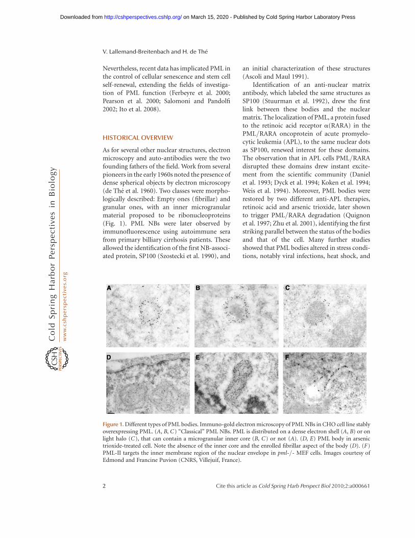
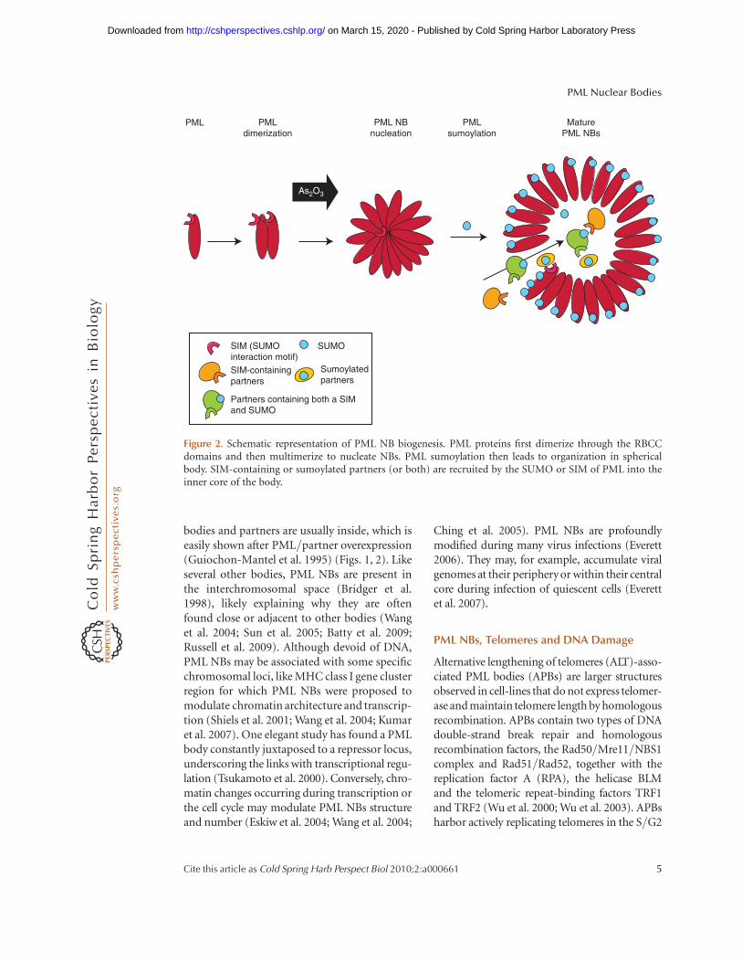
Figure 2. Schematic representation of PML NB biogenesis. PML proteins first dimerize through the RBCCdomains and then multimerize to nucleate NBs. PML sumoylation then leads to organization in sphericalbody. SIM-containing or sumoylated partners (or both) are recruited by the SUMO or SIM of PML into theinner core of the body.
PML Nuclear Bodies
Cite this article as Cold Spring Harb Perspect Biol 2010;2:a000661 5
on March 15, 2020 - Published by Cold Spring Harbor Laboratory Press http://cshperspectives.cshlp.org/Downloaded from
phase of the cell cycle. Almost all these proteinsare sumoylated. Association of the SUMO ligaseSMC5/6 complex with APBs is required forTFR1/2 sumoylation and cell survival (Pottsand Yu 2007). Recent studies have argued for arole of PML in facilitating these processes (Jianget al. 2007; Draskovic et al. 2009).
PML is phosphorylated by several DNA-damage activated kinases, including ATM, ATR,HIPK2, or CHK2. Several studies have implicatedPML in DNA-repair, through the recruitment toor release from NBs of many proteins like BLM(Bischof et al. 2001), the Mre11 complex (Car-bone et al. 2002), WRN, or TOPBP1 (Dellaireand Bazett-Jones 2004). Some of these proteinsare localized in NBs in unstressed conditions,whereas others are only associated with NBs afterDNA-damage. Although NBs partially overlapwith gH2AX foci, their possible localization onthe damage sites is still disputed (Boe et al.2006). Initial studies localized NBs with BLM onBrdU-positiveputativeDNArepairsitesafterirra-diation (Bischof et al. 2001). Yet, in later reports,PML-NBs were proposed to retain BLM awayfrom DSB through BLM sumoylation, whichcould be released by phosphorylation (Eladadet al. 2005; Rao et al. 2005). In response to DNAdamage, PML NBs appear to sense the damagedsites, yielding an increased number of microbod-ies by a fission mechanism. The exact role of PML(bystander or actor) in theses diverse processes isstill unclear. Yet, pml-/- cells have a very highrate of sister chromatin exchange (Zhong et al.1999), implying that PML regulates at least someaspects of homologous recombination.
Nucleolus and RNAs
Electron microscopy studies noted that a subsetof bodies contains a granular core, typical forthe presence of ribonucleoproteins (Dupuy-Coinet al. 1972). Some of these granular bodies wereproposed to bud from the nucleolus (Dupuy-Coin and Bouteille 1972). This most likely reflectsthe presence of specific RNA-binding proteinsand is often observed in hormone-stimulatedprimary cells (Vagner-Capodano et al. 1982).What determines the presence of the micro-granular core inside PML NBs is unknown. We
have observed this granular core in a subset ofPML-labeled structures (Fig. 1). The nature ofthis micro-granular core deserves a renewedexploration. Some studies showed the presenceof nascent RNA within a subset of the bodies(LaMorte et al. 1998; Fuchsova et al. 2002; Kies-slich et al. 2002), whereas others did not (Boisvertet al. 2000). Finally, a series of papers has linkedPML bodies and mRNA export and translationthrough eukaryotic translation initiation factor4E (Lai and Borden 2000; Cohen et al. 2001;Culjkovic et al. 2006; Borden and Culjkovic2009). Heterogeneity of the results may reflectanalysis of different cellular systems, but theseissues clearly deserve additional studies.
In primary or senescent cells, others and wehave observed larger structures that containnucleolar elements, senescence-associated NBs(SANBs) (Condemine et al. 2007; Janderova-Rossmeislova et al. 2007). Interestingly, in thissetting, NB-associated proteins are not presentwithin the inner core, but directly on the PMLshell (Condemine et al. 2007). Formation ofthese domains requires PML-I, which containsa nucleolar targeting domain, possibly the exo-nuclease-III homology domain (Condemineet al. 2007). The nucleolar constituent recog-nized by this domain and the consequence ofPML delocalization for nucleolar function areunknown. Yet, upon genotoxic stress or inhibi-tion of transcription, ATR-mediated PMLphosphorylation relocalizes PML to nucleolarcaps, where PML was proposed to sequesterthe p53 ubiquitin ligase MDM2 (Bernardiet al. 2004; Shav-Tal et al. 2005) (Table 2).
Other Subnuclear Compartments
Studies performed in human embryonic stemcells (HESC) have revealed a further unsus-pected heterogeneity in PML NBs (Butleret al. 2009). PML NBs assemble in “rosettes”surrounding DNA centromeres or are distrib-uted in tracks bridging two centromeres. Thesetracks can cross the nucleus and were frequentlyobserved filing lamina gaps at the nuclear enve-lope. These PML structures do not containSUMO, SP100, or DAXX proteins and their rolein stem cell fate remains to be explored. The
V. Lallemand-Breitenbach and H. de The
6 Cite this article as Cold Spring Harb Perspect Biol 2010;2:a000661
on March 15, 2020 - Published by Cold Spring Harbor Laboratory Press http://cshperspectives.cshlp.org/Downloaded from
PML-II isoform is associated with the inner mem-braneofnuclearenvelope in somaticcells (Conde-mine et al. 2006) (Fig. 1). NBs are associated withcentromeres in proteasome inhibitor-treated cellsin the G2 phase of the cell cycle (Everett et al.1999a). Finally, NBs containing pericentromericsatellite DNA together with HP1, BRCA1, ATRX,and DAXX were proposed to play a role in there-establishment of the condensed hetero-chromatic state of late-replicating satellite DNAin ICF syndrome and possibly in normal cells(Luciani et al. 2006).
Cytoplasmic PML
Among PML isoforms some are devoid of NLSand yield purely cytoplasmic bodies, whenexpressed in pml-/- cells. Similarly, becausePML-I has a nuclear export signal, it formssome cytoplasmic PML-labeled bodies (Con-demine et al. 2006). Cytoplasmic PML was pro-posed to regulate TGFb signaling by controllingthe association of Smad2/3 with SARA andaccumulation of SARA and TGFb-receptor inthe early endosomes (Lin et al. 2004) (Table 1).
NB DYNAMICS
Basal Status
NBs have been the focus of several studies usingimaging technologies such as FRET or FRAP(Tsukamoto et al. 2000). These have shownthat PML is a stable component of the bodiesand that partner proteins are more mobile,although they are transiently retained in NBs(Boisvert et al. 2001; Weidtkamp-Peters et al.2008). The exchange rates of the different PMLisoforms between NBs and nucleoplasm showeda clear difference for PML-V, which forms pecu-liar thick-shelled NBs and might anchor NBsonto the nuclear matrix (Condemine et al.2006; Weidtkamp-Peters et al. 2008). The bodiesthemselves are not very mobile, althoughfusions and fissions can be observed throughthe cell cycle progression (see later discussion).Some bodies labeled by GFP-SP100 are smallerand more dynamic than typical PML NBs(Muratani et al. 2002; Wiesmeijer et al. 2002).
Telomeres quickly get in and out of APBs,whereas their PML shell is quite immobile(Molenaar et al. 2003; Jegou et al. 2009), similarto that of SANBs (Condemine et al. 2007). Wheninterpreting these studies, one has to rememberthat some were performed in transiently trans-fected cells where posttranslational modifica-tions, which are essential determinants of partnerresidence, are unlikely to be complete.
Cell-Cycle Analysis
Analyses of NBs during the cell cycle haveprovided evidence for duplication by a fissionmechanism during S phase (Dellaire et al.2006c) and have dissected NB reformation dur-ing the M/G1 transition (Dellaire et al. 2006b;Chen et al. 2008). During mitosis, PML proteinsremain aggregated, but are phosphorylated,become desumoylated and release partners(Everett et al. 1999b). Before nuclear membranebreakdown in prometaphase, PML NBs losetheir chromatin-tethering, resulting in in-creased mobility (Chen et al. 2008). Yet, FRAPperformed during mitosis showed that PMLproteins did not exchange, demonstrating thatmitotic bodies are highly stable (Dellaire et al.2006c). PML associates with nuclear mem-branes and nucleoporins during mitosis, facili-tating reformation of the nuclear envelopeduring the telophase/G1 transition (Puvion-Dutilleul et al. 1995; Jul-Larsen et al. 2009).Finally, during telophase to G1 transition,SP100 and DAXX re-enter the nucleus andthen bind preformed NBs, SP100 first and laterDAXX, consistent with the critical role of PMLin NB-nucleation.
Stress Response
Apart from arsenic trioxide that promotes NBformation (see below), heat shock or heavymetals induce reversible NB fragmentation bybudding of highly mobile micro-bodies thatare devoid of SUMO and most partners (Table 2).In contrast, transcriptional inhibition or DNAdamage induce formation of microspeckles ornucleolar caps depending on the type of cells(transformed or primary), with a maintained
PML Nuclear Bodies
Cite this article as Cold Spring Harb Perspect Biol 2010;2:a000661 7
on March 15, 2020 - Published by Cold Spring Harbor Laboratory Press http://cshperspectives.cshlp.org/Downloaded from
SUMO-colocalization (Table 2) (Eskiw et al.2003; Nefkens et al. 2003; Dellaire et al. 2006a).Single cell studies after stress recovery haveshown that the initial size, location, and numberof NBs was somehow restored, suggesting thatNBs may assemble on predetermined sites. Afull exploration of the role of PML phosphoryla-tion for the regulation of PML mobility and NBformation would be important to clarify theseissues.
IS SUMO THE NB-ASSEMBLY GLUE?
SUMO is conjugated to target proteins on theside chain of lysine residues, creating a branchedpeptide and significantly changing the bindingproperties of the protein. SUMO has been impli-cated in multiple pathways, mostly as a regulatorof protein interactions (Hay 2005). ConjugatedSUMO may interact with a short motif, knownas the SUMO interaction motif (SIM) (Mintyet al. 2000; Hecker et al. 2006). SUMO-1 was firstidentified as a PML partner in a two-hybridscreen (Boddy et al. 1996). PML was shown tobe sumoylated on three lysine residues: K65 inthe RING finger, K160 in the B1 box, and K490in the NLS. PML also contains a SIM. Accord-ingly, intermolecular interactions between thePML SUMO and SIM were proposed to underlieNB biogenesis (Matunis et al. 2006; Shen et al.2006). Sumoylation-defective Ubc9-/- cellsindeed show NB defects (Nacerddine et al.2005). Moreover, because most partner proteinsassociated with classical PML NBs are sumoy-lated and many contain a SIM, SIM/SUMOinteractions may also account for partnerrecruitment as well as sequestration.
Studies on NB-biogenesis have been greatlyfacilitated by arsenic trioxide. Arsenic trioxide,a very potent therapeutic agent in APL (Zhuet al. 2002; Kogan 2009), was first shown totarget PML/RARA for degradation through itsPML moiety (Chen et al. 1997; Zhu et al. 1997;Muller et al. 1998; Shao et al. 1998). Arsenic tri-oxide is the only factor that regulates the parti-tioning of PML between the nucleoplasm andthe nuclear matrix, promoting in a sequentialmanner NB-formation, PML sumoylation,partner recruitment, and PML degradation
(Lallemand-Breitenbach et al. 2001; Lallemand-Breitenbach et al. 2008) (Fig. 2). Critically, PMLsumoylation on K160/K65 follows matrix-tar-geting and is required for partner recruitment(Ishovet al. 1999; Zhong et al. 2000a; Lallemand-Breitenbachet al. 2001). Yet, howarsenic trioxideinitiates the SUMO-independent transfer fromthe soluble diffuse nucleoplasmic form towardthe insoluble matrix fraction and why matrixassociation is followed by sumoylation of thesetwo sites remains unexplained to date.
SUMO/SIM interactions were initially pro-posed to underlie both formation of the PMLmesh and recruitment of partners. Althoughappealing, this model has been significantlychallenged by recent evidence. Specific PMLisoforms that do not harbor the SIM yield nor-mal bodies (Weidtkamp-Peters et al. 2008).Conversely, analysis of several partner proteinshas shown that their SIM is essential, whereastheir sumoylation is dispensable for NB-targeting (Takahashi et al. 2005; Lin et al.2006; Cho et al. 2009). Taken together, SIM/SUMO are unlikely to play a fundamental rolein PML aggregation to create the PML mesh,but may be critical for partner recruitment.
WHAT ARE THE FUNCTIONS OF NBs?
PML influences or regulates key processes such astranscription, apoptosis, senescence, response toDNA-damage or resistance to micro-organisms,which have all been extensively reviewed else-where (Salomoni and Pandolfi 2002; Everett2006; Bernardi and Pandolfi 2007; Bernardiet al. 2008). Two novelties have emerged: (1)the protective role of PML, DAXX, and SP100against many viral infections (Everett et al.2006; Tavalai et al. 2006; Everett et al. 2008; Tava-lai et al. 2008; Kyratsous and Silverstein 2009), atleast in the absence of the viral proteins that dis-rupt NBs; (2) theroleof PML innormal orcancerstem cell fate (Ito et al. 2008; Li et al. 2009; Regadet al. 2009). PML-enforced stem cell self-renewalmay relyon its ability to modulate the AKT path-way through regulation of AKT phosphorylationor through the localization of its regulator PTEN(Trotman et al. 2006; Song et al. 2008; Ito et al.2009). TR2, a modulator of the critical Oct4
V. Lallemand-Breitenbach and H. de The
8 Cite this article as Cold Spring Harb Perspect Biol 2010;2:a000661
on March 15, 2020 - Published by Cold Spring Harbor Laboratory Press http://cshperspectives.cshlp.org/Downloaded from
stem cell transcriptional regulator, is dependenton PML for its sumoylation (Park et al. 2007;Gupta et al. 2008). The importance of PML in“stemness” highlights the distinctly uncommonmorphologyof NBs in ES cells (Butleret al. 2009)and questions the nature of the PML isoformsthat they actually express.
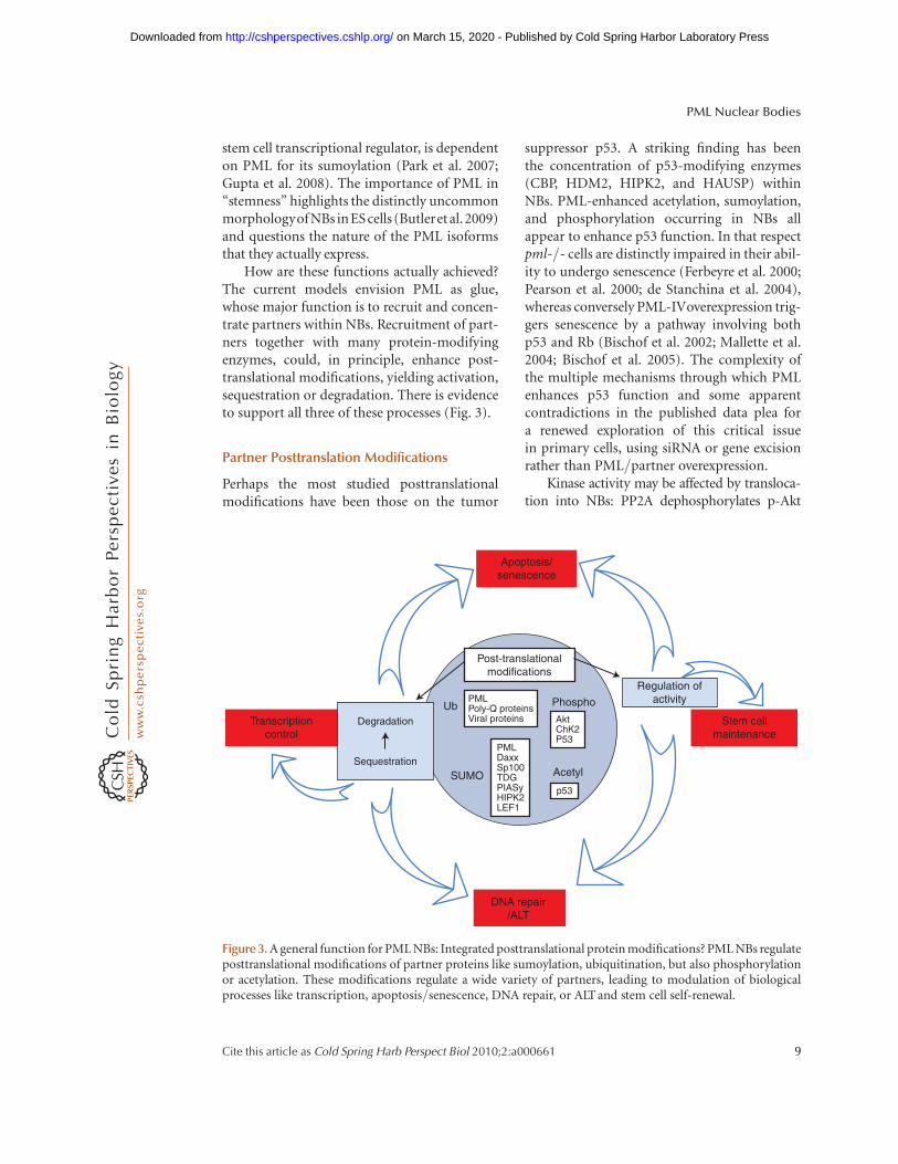
How are these functions actually achieved?The current models envision PML as glue,whose major function is to recruit and concen-trate partners within NBs. Recruitment of part-ners together with many protein-modifyingenzymes, could, in principle, enhance post-translational modifications, yielding activation,sequestration or degradation. There is evidenceto support all three of these processes (Fig. 3).
Partner Posttranslation Modifications
Perhaps the most studied posttranslationalmodifications have been those on the tumor
suppressor p53. A striking finding has beenthe concentration of p53-modifying enzymes(CBP, HDM2, HIPK2, and HAUSP) withinNBs. PML-enhanced acetylation, sumoylation,and phosphorylation occurring in NBs allappear to enhance p53 function. In that respectpml-/- cells are distinctly impaired in their abil-ity to undergo senescence (Ferbeyre et al. 2000;Pearson et al. 2000; de Stanchina et al. 2004),whereas conversely PML-IVoverexpression trig-gers senescence by a pathway involving bothp53 and Rb (Bischof et al. 2002; Mallette et al.2004; Bischof et al. 2005). The complexity ofthe multiple mechanisms through which PMLenhances p53 function and some apparentcontradictions in the published data plea fora renewed exploration of this critical issuein primary cells, using siRNA or gene excisionrather than PML/partner overexpression.
Kinase activity may be affected by transloca-tion into NBs: PP2A dephosphorylates p-Akt
Post-translationalmodifications
DNA repair/ALT
Transcriptioncontrol
Apoptosis/senescence
Stem cellmaintenance
Regulation ofactivity
Degradation
Sequestration
Ub
SUMO Acetyl
PMLDaxxSp100TDGPIASyHIPK2LEF1
p53
AktChK2P53
PMLPoly-Q proteinsViral proteins
Phospho
Figure 3. A general function for PML NBs: Integrated posttranslational protein modifications? PML NBs regulateposttranslational modifications of partner proteins like sumoylation, ubiquitination, but also phosphorylationor acetylation. These modifications regulate a wide variety of partners, leading to modulation of biologicalprocesses like transcription, apoptosis/senescence, DNA repair, or ALT and stem cell self-renewal.
PML Nuclear Bodies
Cite this article as Cold Spring Harb Perspect Biol 2010;2:a000661 9
on March 15, 2020 - Published by Cold Spring Harbor Laboratory Press http://cshperspectives.cshlp.org/Downloaded from
within PML NBs (Trotman et al. 2006), whereasPP1A was proposed to dephosphorylate RB(Regad et al. 2009). In contrast, recruitment intoNBs may favor auto-phosphorylation of somekinases, such as CHK2 (Yang et al. 2002; Yanget al. 2006).
There are also some indications that PMLcan directly enhance global protein sumoyla-tion in yeast (Quimby et al. 2006) and NBswere proposed to enhance sumoylation of spe-cific partner proteins (Park et al. 2007). Becausemany NB-associated proteins contain a SIM,this may enhance partner sequestration withinNBs.
Partner Sequestration
Sequestration or “depot” was the first proposedfunction of PML NBs (Negorev and Maul2001). This sequestration is evident by the rela-tive accumulation of the nucleoplasmic and theNB-associated form of PML-partners, whichvery significantly varies between individualpartners and levels of PML expression, as wellas sumoylation. A well-studied sequesteredpartner is DAXX, a potent repressor that parti-tions between sumoylated proteins, includingPML and many transcription factors. Seques-tration of DAXX by NB-associated, sumoylatedPML releases transcriptional repression byDNA-bound sumoylated transcription factors(Li et al. 2000; Lehembre et al. 2001; Lin et al.2003; Lin et al. 2006). Sequestration of DAXXalso regulates apoptosis. In particular, DAXXenhances Fas-triggered caspase activation, pos-sibly through release of ASK1 kinase activity(Torii et al. 1999; Zhong et al. 2000b; Salomoniand Khelifi 2006), although opposite resultshave been observed in other cell-types (Mei-necke et al. 2007). Sequestration of DAXX mayalso modulate other critical regulators, such asHAUSP, ultimately deubiquitinating the PTENtumor suppressor (Song et al. 2008). In anycase, DAXX, which exerts pro- or anti-apoptoticfunctions, is a critical partner of PML involvedin many of it properties (Maul et al. 2000).
A related situation is the concentration ofhistones and their chaperones in senescent cells.Formation of a specific type of heterochromatin
that silences proliferation-associated genes,senescence-associated heterochromatin foci(SAHF), is one of the first known senescence-associated events. The latter is initiated bythe concentration of two histone chaperonesHIRA and ASF1a, together with the heterochro-matin protein HP1, within PML NBs (Zhanget al. 2005; Ye et al. 2007a; Ye et al. 2007b).What initiates their translocation into NBsand how transient concentration of heterochro-matin-associated histones and their chaperonesinto NBs later favor SAHF formation is notunderstood.
Partner Degradation
Several unstable proteins have been localized toPML bodies (Anton et al. 1999; Smith et al.2004), whereas proteins that are impaired intheir degradation aggregate with PML, SUMO,and ubiquitin (Yamada et al. 2001b). NBs con-centrate proteasomes and ubiquitin (Fabunmiet al. 2001; Lallemand-Breitenbach et al. 2001;Lafarga et al. 2002; Lallemand-Breitenbachet al. 2008). Arsenic trioxide-induced PML deg-radation has linked PML bodies and protein deg-radation. Arsenic trioxide triggers an initialsumoylation of K160, followed by proteasome-dependent degradation, a very controversialproposal at the time this was discovered (Zhuet al. 1997; Lallemand-Breitenbach et al. 2001).Yet, identification of a yeast SUMO-dependentubiquitin ligase (Geoffroy and Hay 2009) andrealization that its human ortholog RNF4 local-izes to NBs (Hakli et al. 2005) led to renewedinterest in these findings. The initial arsenic tri-oxide-induced K160 sumoylation triggers asecondary polyubiquitination by RNF4 andproteasome-mediated degradation in NBs (Lal-lemand-Breitenbach et al. 2008; Tatham et al.2008). There is also some evidence for partnerdegradation within NBs (Kitagawa et al. 2006;Qin et al. 2006; St-Germain et al. 2008), buthow general this is and what the specific signalsare that promote partner degradation withinNBs is currently unknown.
Note that for the same pathway, a combina-tion of those different mechanisms may cooper-ate. For example, modulation of p53 function
V. Lallemand-Breitenbach and H. de The
10 Cite this article as Cold Spring Harb Perspect Biol 2010;2:a000661
on March 15, 2020 - Published by Cold Spring Harbor Laboratory Press http://cshperspectives.cshlp.org/Downloaded from
may involve both enhanced p53 modificationsand HDM2 sequestration. Similarly, regulationof AKT function involves both dephosphoryla-tion in NBs and sequestration of the DAXX/HAUSP regulators of PTEN activity, to controlthe critical AKT/TOR/FOXO pathway for stemcell fate (Ito et al. 2009).
CONCLUDING REMARKS AND OPENQUESTIONS
Because PML can aggregate with so many cellu-lar components, forced overexpression can yieldmany artifacts, notably by titration of partnersor aggregation of unstable proteins (Takahashiet al. 2004; Bernardi and Pandolfi 2007). Para-doxically, there are only few studies in whichPML functions were investigated in stable trans-fectants or in pml-/- cells or in which siRNAwere used to inactivate the endogenous PMLproteins. Even these experimental approachespreclude assessing the role of the bodies per se,compared with that of the diffuse nucleo-plasmic fraction of PML. For example, PML-IV-initiated senescence is unaffected by the viralprotein CMV IE1, which disrupts NBs (Ahnand Hayward 1997; Bischof et al. 2002). Sepa-rating the PML NB functions from those ofunassembled PML may be difficult until adhoc mutants are described.
Another critical issue concerns isoforms.The vast majority of studies have used thePML-IV isoform, which is expressed at verylow levels in most cell lines (Stuurman et al.1992; Condemine et al. 2006), questioning theiractual relevance. Finally, future studies will haveto address the paradoxical critical role of PMLin multiple key processes and the surprisinglymodest phenotype of pml-/- mice. Perhapsone clue to this issue is that stress response hasnot been fully explored in these animals, despitesome important contributions (Wang et al.1998) and that stem cell defects may have rela-tively few manifestations in adults.
The PML field has been extremely active,yielding over the past years a large number ofremarkable results. Yet, some very fundamentalissues remain to be explained. What is thebiochemical and physiological basis for NB-
assembly? What is the micro-granular core?What are the specific functions of different iso-forms? Why are so many NB-associated proteinstranscriptionally controlled by interferons,including PML itself (Stadler et al. 1995; Grot-zinger et al. 1996; Engelhardt et al. 2001; Fab-unmi et al. 2001)? These issues will certainly beclarified in the years to come, maintaining a vividand exciting field.
ACKNOWLEDGMENTS
We apologize to our many colleagues whosework could not be cited because of space restric-tions. We warmly thank Edmond and FrancinePuvion for their electron microscopy imagesand many discussions at various stages of thisproject, Stan Fakan for helpful comments,Morgane Le Bras and Julien Ablain for criticalreading of the manuscript. Work in the labo-ratory is supported by Ligue Nationale contrele Cancer, INSERM, CNRS, University ParisDiderot, Institut Universitaire de France, Insti-tut National du Cancer and Canceropoleprograms.
REFERENCES
Ahn JH, Hayward GS. 1997. The major immediate-earlyproteins IE1 and IE2 of human cytomegalovirus colocal-ize with and disrupt PML-associated nuclear bodies atvery early times in infected permissive cells. J Virol 71:4599–4613.
Anton LC, Schubert U, Bacik I, Princiotta MF, Wearsch PA,Gibbs J, Day P, Realini C, Rechsteiner M, Bennink J, et al.1999. Intracellular localization of proteasomal degrada-tion of a viral antigen. J Cell Biol 146: 113–124.
Ascoli CA, Maul GG. 1991. Identification of a novel nucleardomain. J Cell Biol 112: 785–795.
Batty E, Jensen K, Freemont P. 2009. PML nuclear bodiesand their spatial relationships in the mammalian cellnucleus. Front Biosci 14: 1182–1196.
Beech SJ, Lethbridge KJ, Killick N, McGlincy N, LeppardKN. 2005. Isoforms of the promyelocytic leukemia pro-tein differ in their effects on ND10 organization. ExpCell Res 307: 109–117.
Bernardi R, Pandolfi PP. 2007. Structure, dynamics andfunctions of promyelocytic leukaemia nuclear bodies.Nat Rev Mol Cell Biol 8: 1006–1016.
Bernardi R, Papa A, Pandolfi PP. 2008. Regulation of apop-tosis by PML and the PML-NBs. Oncogene 27:6299–6312.
Bernardi R, Scaglioni PP, Bergmann S, Horn HF, VousdenKH, Pandolfi PP. 2004. PML regulates p53 stability by
PML Nuclear Bodies
Cite this article as Cold Spring Harb Perspect Biol 2010;2:a000661 11
on March 15, 2020 - Published by Cold Spring Harbor Laboratory Press http://cshperspectives.cshlp.org/Downloaded from
sequestering Mdm2 to the nucleolus. Nat Cell Biol 6:665–672.
Bischof O, Kim SH, Irving J, Beresten S, Ellis NA, Campisi J.2001. Regulation and localization of the bloom syndromeprotein in response to dna damage. J Cell Biol 153:367–380.
Bischof O, Kirsh O, Pearson M, Itahana K, Pelicci PG,Dejean A. 2002. Deconstructing PML-induced prema-ture senescence. Embo J 21: 3358–3369.
Bischof O, Nacerddine K, Dejean A. 2005. Human papillo-mavirus oncoprotein E7 targets the promyelocytic leuke-mia protein and circumvents cellular senescence via theRb and p53 tumor suppressor pathways. Mol Cell Biol25: 1013–1024.
Boddy MN, Howe K, Etkin LD, Solomon E, Freemont PS.1996. PIC1, a novel ubiquitin-like protein which interactswith the PML component of a multiprotein complex thatis disrupted in acute promyelocytic leukaemia. Oncogene13: 971–982.
Boe SO, Haave M, Jul-Larsen A, Grudic A, Bjerkvig R,Lonning PE. 2006. Promyelocytic leukemia nuclearbodies are predetermined processing sites for damagedDNA. J Cell Sci 119: 3284–3295.
Boisvert FM, Hendzel MJ, Bazett-Jones DP. 2000. Promyelo-cytic leukemia (PML) nuclear bodies are protein struc-tures that do not accumulate RNA. J Cell Biol 148:283–292.
Boisvert FM, Kruhlak MJ, Box AK, Hendzel MJ, Bazett-Jones DP. 2001. The transcription coactivator CBP is adynamic component of the promyelocytic leukemianuclear body. J Cell Biol 152: 1099–1106.
Borden KL, Culjkovic B. 2009. Perspectives in PML: A uni-fying framework for PML function. Front Biosci 14:497–509.
Bridger J, Herrmann H, Munkel C, Lichter P. 1998. Identifi-cation of an interchromosomal compartment by poly-merization of nuclear-targeted vimentin. J Cell Sci 111:1241–1253.
Butler JT, Hall LL, Smith KP, Lawrence JB. 2009. Changingnuclear landscape and unique PML structures duringearly epigenetic transitions of human embryonic stemcells. J Cell Biochem 107: 609–621.
Carbone R, Pearson M, Minucci S, Pelicci PG. 2002. PMLNBs associate with the hMre11 complex and p53 at sitesof irradiation induced DNA damage. Oncogene 21:1633–1640.
Chen YC, Kappel C, Beaudouin J, Eils R, Spector DL. 2008.Live cell dynamics of promyelocytic leukemia nuclearbodies upon entry into and exit from mitosis. Mol BiolCell 19: 3147–3162.
Chen GQ, Shi XG, Tang W, Xiong SM, Zhu J, Cai X, Han ZG,Ni JH, Shi GY, Jia PM, et al. 1997. Use of arsenic trioxide(As2O3) in the treatment of acute promyelocytic leuke-mia (APL): I. As2O3 exerts dose-dependent dual effectson APL cells. Blood 89: 3345–3353.
Ching RW, Dellaire G, Eskiw CH, Bazett-Jones DP. 2005.PML bodies: A meeting place for genomic loci? J CellSci 118: 847–854.
Cho G, Lim Y, Golden JA. 2009. SUMO interaction motifs inSIZN1 are required for PML-NB localization and for
transcriptional activation. J Biol Chem 284: 19592–19600.
Cohen N, Sharma M, Kentsis A, Perez JM, Strudwick S,Borden KL. 2001. PML RING suppresses oncogenictransformation by reducing the affinity of eIF4E formRNA. Embo J 20: 4547–4559.
Condemine W, Takahashi Y, Le Bras M, de The H. 2007. Anucleolar targeting signal in PML-I addresses PML tonucleolar caps in stressed or senescent cells. J Cell Sci120: 3219–3227.
Condemine W, Takahashi Y, Zhu J, Puvion-Dutilleul F,Guegan S, Janin A, de The H. 2006. Characterization ofendogenous human promyelocytic leukemia isoforms.Cancer Res 66: 6192–6198.
Culjkovic B, Topisirovic I, Skrabanek L, Ruiz-Gutierrez M,Borden KL. 2006. eIF4E is a central node of an RNA reg-ulon that governs cellular proliferation. J Cell Biol 175:415–426.
Daniel M-T, Koken M, Romagne O, Barbey S, Bazarbachi A,Stadler M, Guillemin M-C, Degos L, Chomienne C, deThe H. 1993. PML protein expression in hematopoieticand acute promyelocytic leukemia cells. Blood 82:1858–1867.
de Stanchina E, Querido E, Narita M, Davuluri RV, PandolfiPP, Ferbeyre G, Lowe SW. 2004. PML is a direct p53 targetthat modulates p53 effector functions. Mol Cell 13:523–535.
de The G, Riviere M, Bernhard W. 1960. Examen au micro-scope electronique de la tumeur VX2 du lapin domesti-que derivee du papillome de Shope. Bull Cancer 47:570–584.
Dellaire G, Bazett-Jones DP. 2004. PML nuclear bodies:Dynamic sensors of DNA damage and cellular stress.Bioessays 26: 963–977.
Dellaire G, Ching RW, Ahmed K, Jalali F, Tse KC, BristowRG, Bazett-Jones DP. 2006a. Promyelocytic leukemianuclear bodies behave as DNA damage sensors whoseresponse to DNA double-strand breaks is regulated byNBS1 and the kinases ATM, Chk2, and ATR. J Cell Biol175: 55–66.
Dellaire G, Ching RW, Dehghani H, Ren Y, Bazett-Jones DP.2006b. The number of PML nuclear bodies increases inearly S phase by a fission mechanism. J Cell Sci 119:1026–1033.
Dellaire G, Eskiw CH, Dehghani H, Ching RW, Bazett-JonesDP. 2006c. Mitotic accumulations of PML protein con-tribute to the re-establishment of PML nuclear bodiesin G1. J Cell Sci 119: 1034–1042.
Draskovic I, Arnoult N, Steiner V, Bacchetti S, Lomonte P,Londono-Vallejo A. 2009. Probing PML body functionin ALT cells reveals spatiotemporal requirements fortelomere recombination. Proc Natl Acad Sci 106:15726–15731.
Duprez E, Saurin AJ, Desterro JM, Lallemand-BreitenbachV, Howe K, Boddy MN, Solomon E, de The H, Hay RT,Freemont PS. 1999. SUMO-1 modification of the acutepromyelocytic leukaemia protein PML: Implications fornuclear localisation. J Cell Sci 112: 381–393.
Dupuy-Coin AM, Bouteille M. 1972. Developmentalpathway of granular and beaded nuclear bodies fromnucleoli. J Ultrastruct Res 40: 55–67.
V. Lallemand-Breitenbach and H. de The
12 Cite this article as Cold Spring Harb Perspect Biol 2010;2:a000661
on March 15, 2020 - Published by Cold Spring Harbor Laboratory Press http://cshperspectives.cshlp.org/Downloaded from
Dupuy-Coin AM, Kalifat SR, Bouteille M. 1972. Nuclearbodies as proteinaceous structures containing ribo-nucleoproteins. J Ultrastruct Res 38: 174–187.
Dyck JA, Maul GG, Miller WH, Chen JD, Kakizuka A, EvansRM. 1994. A novel macromolecular structure is a target ofthe promyelocyte- retinoic acid receptor oncoprotein.Cell 76: 333–343.
Eladad S, Ye TZ, Hu P, Leversha M, Beresten S, Matunis MJ,Ellis NA. 2005. Intra-nuclear trafficking of the BLM heli-case to DNA damage-induced foci is regulated by SUMOmodification. Hum Mol Genet 14: 1351–1365.
Engelhardt OG, Boutell C, Orr A, Ullrich E, Haller O, Ever-ett RD. 2003. The homeodomain-interacting kinasePKM (HIPK-2) modifies ND10 through both its kinasedomain and a SUMO-1 interaction motif and alters theposttranslational modification of PML. Exp Cell Res283: 36–50.
Engelhardt OG, Ullrich E, Kochs G, Haller O. 2001.Interferon-induced antiviral Mx1 GTPase is associatedwith components of the SUMO-1 system and promyelo-cytic leukemia protein nuclear bodies. Exp Cell Res 271:286–295.
Eskiw CH, Dellaire G, Bazett-Jones DP. 2004. Chromatincontributes to structural integrity of promyelocytic leu-kemia bodies through a SUMO-1-independent mecha-nism. J Biol Chem 279: 9577–9585.
Eskiw CH, Dellaire G, Mymryk JS, Bazett-Jones DP. 2003.Size, position and dynamic behavior of PML nuclearbodies following cell stress as a paradigm for supramole-cular trafficking and assembly. J Cell Sci 116: 4455–4466.
Everett RD. 2001. DNAviruses and viral proteins that inter-act with PML nuclear bodies. Oncogene 20: 7266–7273.
Everett RD. 2006. Interactions between DNAviruses, ND10and the DNA damage response. Cell Microbiol 8:365–374.
Everett RD, Earnshaw WC, Pluta AF, Sternsdorf T, AinszteinAM, Carmena M, Ruchaud S, Hsu WL, Orr A. 1999a. Adynamic connection between centromeres and ND10proteins. J Cell Sci 112: 3443–3454.
Everett RD, Lomonte P, Sternsdorf T, van Driel R, Orr A.1999b. Cell cycle regulation of PML modification andND10 composition. J Cell Sci 112: 4581–4588.
Everett RD, Murray J, Orr A, Preston CM. 2007. Herpes sim-plex virus type 1 genomes are associated with ND10nuclear substructures in quiescently infected humanfibroblasts. J Virol 81: 10991–11004.
Everett RD, Parada C, Gripon P, Sirma H, Orr A. 2008. Rep-lication of ICP0-null mutant herpes simplex virus type 1is restricted by both PML and Sp100. J Virol 82:2661–2672.
Everett RD, Rechter S, Papior P, Tavalai N, Stamminger T,Orr A. 2006. PML contributes to a cellular mechanismof repression of herpes simplex virus type 1 infectionthat is inactivated by ICP0. J Virol 80: 7995–8005.
Fabunmi RP, Wigley WC, Thomas PJ, DeMartino GN. 2001.Interferon g regulates accumulation of the proteasomeactivator PA28 and immunoproteasomes at nuclearPML bodies. J Cell Sci 114: 29–36.
Ferbeyre G, de Stanchina E, Querido E, Baptiste N, Prives C,Lowe SW. 2000. PML is induced by oncogenic ras and
promotes premature senescence. Genes Dev 14:2015–2027.
Fuchsova B, Novak P, Kafkova J, Hozak P. 2002. NuclearDNA helicase II is recruited to IFN-a-activated tran-scription sites at PML nuclear bodies. J Cell Biol 158:463–473.
Geoffroy MC, Hay RT. 2009. An additional role for SUMOin ubiquitin-mediated proteolysis. Nat Rev Mol CellBiol 10: 564–568.
Gresko E, Ritterhoff S, Sevilla-Perez J, Roscic A, Frobius K,Kotevic I, Vichalkovski A, Hess D, Hemmings BA,Schmitz ML. 2009. PML tumor suppressor is regulatedby HIPK2-mediated phosphorylation in response toDNA damage. Oncogene 28: 698–708.
Grotzinger T, Jensen K, Will H. 1996. The interferon (IFN)-stimulated gene Sp100 promoter contains an IFN-g acti-vation site and an imperfect IFN-stimulated responseelement which mediate type I IFN inducibility. J BiolChem 271: 25253.
Guiochon-Mantel A, Savouret JF, Quignon F, Delabre K,Milgrom E, de The H. 1995. Effect of PML andPML-RAR on the transactivation properties and subcel-lular distribution of steroid hormone receptors. MolEndocrinol 9: 1791–1803.
Guo A, Salomoni P, Luo J, Shih A, Zhong S, Gu W, PandolfiPP. 2000. The function of PML in p53-dependent apop-tosis. Nat Cell Biol 2: 730–736.
Gupta P, Ho PC, Huq MM, Ha SG, Park SW, Khan AA, TsaiNP, Wei LN. 2008. Retinoic acid-stimulated sequentialphosphorylation, PML recruitment, and SUMOylationof nuclear receptor TR2 to suppress Oct4 expression.Proc Natl Acad Sci 105: 11424–11429.
Gurrieri C, Capodieci P, Bernardi R, Scaglioni PP, Nafa K,Rush LJ, Verbel DA, Cordon-Cardo C, Pandolfi PP.2004. Loss of the tumor suppressor PML in human can-cers of multiple histologic origins. J Natl Cancer Inst 96:269–279.
Hakli M, Karvonen U, Janne OA, Palvimo JJ. 2005. SUMO-1promotes association of SNURF (RNF4) with PMLnuclear bodies. Exp Cell Res 304: 224–233.
Hay RT. 2005. SUMO: A history of modification. Mol Cell18: 1–12.
Hayakawa F, Privalsky ML. 2004. Phosphorylation of PMLby mitogen-activated protein kinases plays a key role inarsenic trioxide-mediated apoptosis. Cancer Cell 5:389–401.
Hecker CM, Rabiller M, Haglund K, Bayer P, Dikic I. 2006.Specification of SUMO1- and SUMO2-interactingMotifs. J Biol Chem 281: 16117–16127.
Henderson BR, Eleftheriou A. 2000. A comparison of theactivity, sequence specificity, and CRM1-dependence ofdifferent nuclear export signals. Exp Cell Res 256:213–224.
Ishov AM, Sotnikov AG, Negorev D, Vladimirova OV, NeffN, Kamitani T, Yeh E, Strauss J, Maul G. 1999. PML iscritical for ND10 formation and recruits the PML-inter-acting protein Daxx to this nuclear structure when modi-fied by SUMO-1. J Cell Biol 147: 221–234.
Ito K, Bernardi R, Pandolfi PP. 2009. A novel signalingnetwork as a critical rheostat for the biology and
PML Nuclear Bodies
Cite this article as Cold Spring Harb Perspect Biol 2010;2:a000661 13
on March 15, 2020 - Published by Cold Spring Harbor Laboratory Press http://cshperspectives.cshlp.org/Downloaded from
maintenance of the normal stem cell and the cancer-initiating cell. Curr Opin Genet Dev 19: 51–59.
Ito K, Bernardi R, Morotti A, Matsuoka S, Saglio G, Ikeda Y,Rosenblatt J, Avigan DE, Teruya-Feldstein J, Pandolfi PP.2008. PML targeting eradicates quiescent leukaemia-initiating cells. Nature 453: 1072–1078.
Janderova-Rossmeislova L, Novakova Z, Vlasakova J, Phili-monenko V, Hozak P, Hodny Z. 2007. PML protein asso-ciation with specific nucleolar structures differs innormal, tumor and senescent human cells. J Struct Biol159: 56–70.
Jegou T, Chung I, Heuvelman G, Wachsmuth M, GorischSM, Greulich-Bode KM, Boukamp P, Lichter P, RippeK. 2009. Dynamics of telomeres and promyelocytic leu-kemia nuclear bodies in a telomerase-negative humancell line. Mol Biol Cell 20: 2070–2082.
Jensen K, Shiels C, Freemont PS. 2001. PML proteinisoforms and the RBCC/TRIM motif. Oncogene 20:7223–7233.
Jiang WQ, Zhong ZH, Henson JD, Reddel RR. 2007. Identi-fication of candidate alternative lengthening of telomeresgenes by methionine restriction and RNA interference.Oncogene 26: 4635–4647.
Jul-Larsen A, Grudic A, Bjerkvig R, Boe SO. 2009. Cell-cycleregulation and dynamics of cytoplasmic compartmentscontaining the promyelocytic leukemia protein andnucleoporins. J Cell Sci 122: 1201–1210.
Kastner P, Perez A, Lutz Y, Rochette-Egly C, Gaub M-P,Durand B, Lanotte M, Berger R, Chambon P. 1992. Struc-ture, localization and transcriptional properties of twoclasses of retinoic acid receptora fusion proteins in acutepromyelocytic leukemia (APL): Structural similaritieswith a new family of oncoproteins. EMBO J 11: 629–642.
Kiesslich A, von Mikecz A, Hemmerich P. 2002. Cell cycle-dependent association of PML bodies with sites of activetranscription in nuclei of mammalian cells. J Struct Biol140: 167–179.
Kitagawa D, Kajiho H, Negishi T, Ura S, Watanabe T, Wada T,Ichijo H, Katada T, Nishina H. 2006. Release of RASSF1Cfrom the nucleus by Daxx degradation links DNA dam-age and SAPK/JNK activation. EMBO J 25: 3286–3297.
Kogan SC. 2009. Curing APL: Differentiation or destruc-tion? Cancer Cell 15: 7–8.
Koken MHM, Linares-Cruz G, Quignon F, Viron A,Chelbi-Alix MK, Sobczak-Thepot J, Juhlin L, Degos L,Calvo F, de The H. 1995. The PML growth-suppressorhas an altered expression in human oncogenesis. Onco-gene 10: 1315–1324.
Koken MHM, Puvion-Dutilleul F, Guillemin MC, Viron A,Linares-Cruz G, Stuurman N, de Jong L, Szostecki C,Calvo F, Chomienne C, et al. 1994. The t(15;17) translo-cation alters a nuclear body in a RA-reversible fashion.EMBO J 13: 1073–1083.
Kumar PP, Bischof O, Purbey PK, Notani D, Urlaub H,Dejean A, Galande S. 2007. Functional interactionbetween PML and SATB1 regulates chromatin-looparchitecture and transcription of the MHC class I locus.Nat Cell Biol 9: 45–56.
Kyratsous CA, Silverstein SJ. 2009. Components of nucleardomain 10 bodies regulate varicella-zoster virus replica-tion. J Virol 83: 4262–4274.
Lafarga M, Berciano MT, Pena E, Mayo I, Castano JG, Boh-mann D, Rodrigues JP, Tavanez JP, Carmo-Fonseca M.2002. Clastosome: A subtype of nuclear body enrichedin 19S and 20S proteasomes, ubiquitin, and protein sub-strates of proteasome. Mol Biol Cell 13: 2771–2782.
Lai HK, Borden KL. 2000. The promyelocytic leukemia(PML) protein suppresses cyclin D1 protein productionby altering the nuclear cytoplasmic distribution of cyclinD1 mRNA. Oncogene 19: 1623–1634.
Lallemand-Breitenbach V, Jeanne M, Benhenda S, Nasr R,Lei M, Peres L, Zhou J, Zhu J, Raught B, de The H.2008. Arsenic degrades PML or PML-RARa through aSUMO-triggered RNF4/ubiquitin-mediated pathway.Nat Cell Biol 10: 547–555.
Lallemand-Breitenbach V, Zhu J, Puvion F, Koken M, Hon-ore N, Doubeikovsky A, Duprez E, Pandolfi PP, Puvion E,Freemont P, et al. 2001. Role of promyelocytic leukemia(PML) sumolation in nuclear body formation, 11S pro-teasome recruitment, and As(2)O(3)-induced PML orPML/retinoic acid receptor a degradation. J Exp Med193: 1361–1372.
LaMorte VJ, Dyck JA, Ochs RL, Evans RM. 1998. Localiza-tion of nascent RNA and CREB binding protein with thePML-containing nuclear body. Proc Natl Acad Sci 95:4991–4996.
Lehembre F, Muller S, Pandolfi PP, Dejean A. 2001. Regula-tion of Pax3 transcriptional activity by SUMO-1-modified PML. Oncogene 20: 1–9.
Li W, Ferguson BJ, Khaled WT, Tevendale M, Stingl J, Poli V,Rich T, Salomoni P, Watson CJ. 2009. PML depletion dis-rupts normal mammary gland development and skewsthe composition of the mammary luminal cell progenitorpool. Proc Natl Acad Sci 106: 4725–4730.
Li H, Leo C, Zhu J, Wu X, O’Neil J, Park EJ, Chen JD. 2000.Sequestration and inhibition of daxx-mediated trans-criptional repression by PML. Mol Cell Biol 20:1784–1796.
Lin HK, Bergmann S, Pandolfi PP. 2004. Cytoplasmic PMLfunction in TGF-b signalling. Nature 431: 205–211.
Lin DY, Huang YS, Jeng JC, Kuo HY, Chang CC, Chao TT,Ho CC, Chen YC, Lin TP, Fang HI, et al. 2006. Role ofSUMO-interacting motif in Daxx SUMO modification,subnuclear localization, and repression of sumoylatedtranscription factors. Mol Cell 24: 341–354.
Lin DY, Lai MZ, Ann DK, Shih HM. 2003. Promyelocyticleukemia protein (PML) functions as a glucocorticoidreceptor co-activator by sequestering Daxx to the PMLoncogenic domains (PODs) to enhance its transacti-vation potential. J Biol Chem 278: 15958–15965.
Luciani JJ, Depetris D, Usson Y, Metzler-Guillemain C,Mignon-Ravix C, Mitchell MJ, Megarbane A, Sarda P,Sirma H, Moncla A, et al. 2006. PML nuclear bodiesare highly organised DNA-protein structures with a func-tion in heterochromatin remodelling at the G2 phase.J Cell Sci 119: 2518–2531.
Mallette FA, Goumard S, Gaumont-Leclerc MF, MoiseevaO, Ferbeyre G. 2004. Human fibroblasts require the Rbfamily of tumor suppressors, but not p53, for PML-induced senescence. Oncogene 23: 91–99.
Matunis MJ, Zhang XD, Ellis NA. 2006. SUMO: The gluethat binds. Dev Cell 11: 596–597.
V. Lallemand-Breitenbach and H. de The
14 Cite this article as Cold Spring Harb Perspect Biol 2010;2:a000661
on March 15, 2020 - Published by Cold Spring Harbor Laboratory Press http://cshperspectives.cshlp.org/Downloaded from
Maul GG, Negorev D, Bell P, Ishov AM. 2000. Review: Prop-erties and assembly mechanisms of ND10, PML bodies,or PODs. J Struct Biol 129: 278–287.
Meinecke I, Cinski A, Baier A, Peters MA, Dankbar B, WilleA, Drynda A, Mendoza H, Gay RE, Hay RT, et al. 2007.Modification of nuclear PML protein by SUMO-1 regu-lates Fas-induced apoptosis in rheumatoid arthritis syno-vial fibroblasts. Proc Natl Acad Sci 104: 5073–5078.
Meroni G, Diez-Roux G. 2005. TRIM/RBCC, a novel classof ‘single protein RING finger’ E3 ubiquitin ligases. Bio-essays 27: 1147–1157.
Minty A, Dumont X, Kaghad M, Caput D. 2000. Covalentmodification of p73a by SUMO-1. Two-hybrid screeningwith p73 identifies novel SUMO-1-interacting proteinsand a SUMO-1 interaction motif. J Biol Chem 275:36316–36323.
Molenaar C, Wiesmeijer K, Verwoerd NP, Khazen S, Eils R,Tanke HJ, Dirks RW. 2003. Visualizing telomere dynam-ics in living mammalian cells using PNA probes. EMBO J22: 6631–6641.
Muller S, Matunis MJ, Dejean A. 1998. Conjugation with theubiquitin-related modifier SUMO-1 regulates the parti-tioning of PML within the nucleus. EMBO J 17: 61–70.
Muratani M, Gerlich D, Janicki SM, Gebhard M, Eils R,Spector DL. 2002. Metabolic-energy-dependent move-ment of PML bodies within the mammalian cell nucleus.Nat Cell Biol 4: 106–110.
Nacerddine K, Lehembre F, Bhaumik M, Artus J, Cohen-Tannoudji M, Babinet C, Pandolfi PP, Dejean A. 2005.The SUMO pathway is essential for nuclear integrityand chromosome segregation in mice. Dev Cell 9:769–779.
Nefkens I, Negorev DG, Ishov AM, Michaelson JS, Yeh ET,Tanguay RM, Muller WE, Maul GG. 2003. Heat shockand Cd2þ exposure regulate PML and Daxx releasefrom ND10 by independent mechanisms that modifythe induction of heat-shock proteins 70 and 25 differ-ently. J Cell Sci 116: 513–524.
Negorev D, Maul GG. 2001. Cellular proteins localized atand interacting within ND10/PML nuclear bodies/PODs suggest functions of a nuclear depot. Oncogene20: 7234–7242.
Park SW, Hu X, Gupta P, Lin YP, Ha SG, Wei LN. 2007.SUMOylation of Tr2 orphan receptor involves Pml andfine-tunes Oct4 expression in stem cells. Nat Struct MolBiol 14: 68–75.
Pearson M, Carbone R, Sebastiani C, Cioce M, Fagioli M,Saito S, Higashimoto Y, Appella E, Minucci S, PandolfiPP, et al. 2000. PML regulates p53 acetylation and prema-ture senescence induced by oncogenic Ras. Nature 406:207–210.
Potts PR, Yu H. 2007. The SMC5/6 complex maintains telo-mere length in ALT cancer cells through SUMOylationof telomere-binding proteins. Nat Struct Mol Biol 14:581–590.
Puvion-Dutilleul F, Chelbi-Alix MK, Koken M, Quignon F,Puvion E, de The H. 1995. Adenovirus infection inducesrearrangements in the intranuclear distribution of thenuclear body-associated PML protein. Exp Cell Res 218:9–16.
Qin Q, Inatome R, Hotta A, Kojima M, Yamamura H, HiraiH, Yoshizawa T, Tanaka H, Fukami K, Yanagi S. 2006. A
novel GTPase, CRAG, mediates promyelocytic leukemiaprotein-associated nuclear body formation and degrada-tion of expanded polyglutamine protein. J Cell Biol 172:497–504.
Quignon F, Chen Z, de The H. 1997. Retinoic acid andarsenic: towards oncogene targeted treatments of acutepromyelocytic leukaemia. Biochim Biophys Acta 1333:M53–M61.
Quimby BB, Yong-Gonzalez V, Anan T, Strunnikov AV,Dasso M. 2006. The promyelocytic leukemia proteinstimulates SUMO conjugation in yeast. Oncogene 25:2999–3005.
Rao VA, Fan AM, Meng L, Doe CF, North PS, Hickson ID,Pommier Y. 2005. Phosphorylation of BLM, dissociationfrom topoisomerase IIIa, and colocalization withg-H2AX after topoisomerase I-induced replicationdamage. Mol Cell Biol 25: 8925–8937.
Regad T, Bellodi C, Nicotera P, Salomoni P. 2009. The tumorsuppressor Pml regulates cell fate in the developingneocortex. Nat Neurosci 12: 132–140.
Reymond A, Meroni G, Fantozzi A, Merla G, Cairo S, Luzi L,Riganelli D, Zanaria E, Messali S, Cainarca S, et al. 2001.The tripartite motif family identifies cell compartments.Embo J 20: 2140–2151.
Russell RA, Adams NM, Stephens DA, Batty E, Jensen K,Freemont PS. 2009. Segmentation of fluorescence micro-scopy images for quantitative analysis of cell nucleararchitecture. Biophys J 96: 3379–3389.
Salomoni P, Khelifi AF. 2006. Daxx: Death or survivalprotein? Trends Cell Biol 16: 97–104.
Salomoni P, Pandolfi PP. 2002. The role of PML in tumorsuppression. Cell 108: 165–170.
Scaglioni PP, Yung TM, Cai LF, Erdjument-Bromage H,Kaufman AJ, Singh B, Teruya-Feldstein J, Tempst P, Pan-dolfi PP. 2006. A CK2-dependent mechanism for degra-dation of the PML tumor suppressor. Cell 126: 269–283.
Shao W, Fanelli M, Ferrara FF, Riccioni R, Rosenauer A,Davison K, Lamph WW, Waxman S, Pelicci PG, LoCoco F, et al. 1998. Arsenic trioxide as an inducer of apop-tosis and loss of PML/RARa protein in acute promyelo-cytic leukemia cells. J Natl Cancer Inst 90: 124–133.
Shav-Tal Y, Blechman J, Darzacq X, Montagna C, Dye BT,Patton JG, Singer RH, Zipori D. 2005. Dynamic sortingof nuclearcomponents into distinct nucleolarcaps duringtranscriptional inhibition. Mol Biol Cell 16: 2395–2413.
Shen TH, Lin HK, Scaglioni PP, Yung TM, Pandolfi PP. 2006.The mechanisms of PML-nuclear body formation. MolCell 24: 331–339.
Shiels C, Islam SA, Vatcheva R, Sasieni P, Sternberg MJ, Free-mont PS, Sheer D. 2001. PML bodies associate specifi-cally with the MHC gene cluster in interphase nuclei.J Cell Sci 114: 3705–3716.
Smith KP, Byron M, O’Connell BC, Tam R, Schorl C, GuneyI, Hall LL, Agrawal P, Sedivy JM, Lawrence JB. 2004.c-Myc localization within the nucleus: evidence for asso-ciation with the PML nuclear body. J Cell Biochem 93:1282–1296.
Song MS, Salmena L, Carracedo A, Egia A, Lo-Coco F,Teruya-Feldstein J, Pandolfi PP. 2008. The deubiquitiny-lation and localization of PTEN are regulated by aHAUSP-PML network. Nature 455: 813–817.
PML Nuclear Bodies
Cite this article as Cold Spring Harb Perspect Biol 2010;2:a000661 15
on March 15, 2020 - Published by Cold Spring Harbor Laboratory Press http://cshperspectives.cshlp.org/Downloaded from
St-Germain JR, Chen J, Li Q. 2008. Involvement of PMLnuclear bodies in CBP degradation through theubiquitin-proteasome pathway. Epigenetics 3: 342–349.
Stadler M, Chelbi-Alix MK, Koken MHM, Venturini L,Lee C, Saıb A, Quignon F, Pelicano L, Guillemin M-C,Schindler C, et al. 1995. Transcriptional induction ofthe PML growth suppressor gene by interferons ismediated through an ISRE and a GAS element. Oncogene11: 2565–2573.
Stuurman N, de Graaf A, Floore A, Josso A, Humbel B, deJong L, van Driel R. 1992. A monoclonal antibody recog-nizing nuclear matrix-associated nuclear bodies. J Cell Sci101: 773–784.
Stuurman N, Meijne AML, van der Pol AJ, de Jong L, vanDriel R, van Renswoude J. 1990. The nuclear matrixfrom cells of different origin. Evidence for a commonset of matrix proteins. J Biol Chem 265: 5460–5465.
Sun J, Xu H, Subramony SH, Hebert MD. 2005. Interactionsbetween coilin and PIASy partially link Cajal bodies toPML bodies. J Cell Sci 118: 4995–5003.
Szostecki C, Guldner H, Netter H, Will H. 1990. Isolationand characterization of cDNA encoding a human nuclearantigen predominantly rrecognized by autoantibodies ofpatients with primary biliary cirrhosis. J Immunol 145:4338–4347.
Takahashi H, Hatakeyama S, Saitoh H, Nakayama KI. 2005.Noncovalent SUMO-1 binding activity of thymine DNAglycosylase (TDG) is required for its SUMO-1 modifica-tion and colocalization with the promyelocytic leukemiaprotein. J Biol Chem 280: 5611–5621.
Takahashi Y, Lallemand-Breitenbach V, Zhu J, de The H.2004. PML nuclear bodies and apoptosis. Oncogene 23:2819–2824.
Tao H, Simmons BN, Singireddy S, Jakkidi M, Short KM,Cox TC, Massiah MA. 2008. Structure of the MID1 tan-dem B-boxes reveals an interaction reminiscent of inter-molecular ring heterodimers. Biochemistry 47: 2450–2457.
Tatham MH, Geoffroy MC, Shen L, Plechanovova A, Hat-tersley N, Jaffray EG, Palvimo JJ, Hay RT. 2008. RNF4 isa poly-SUMO-specific E3 ubiquitin ligase required forarsenic-induced PML degradation. Nat Cell Biol 10:538–546.
Tavalai N, Papior P, Rechter S, Leis M, Stamminger T. 2006.Evidence for a role of the cellular ND10 protein PML inmediating intrinsic immunity against human cytomega-lovirus infections. J Virol 80: 8006–8018.
Tavalai N, Papior P, Rechter S, Stamminger T. 2008. Nucleardomain 10 components promyelocytic leukemia proteinand hDaxx independently contribute to an intrinsic anti-viral defense against human cytomegalovirus infection.J Virol 82: 126–137.
Terris B, Baldin V, Dubois S, Degott C, Flejou JF, Henin D,Dejean A. 1995. PML nuclear bodies are general targetsfor inflammation and cell proliferation. Cancer Res 55:1590–1597.
Torii S, Egan DA, Evans RA, Reed JC. 1999. Human Daxxregulates Fas-induced apoptosis from nuclear PMLoncogenic domains (PODs). EMBO J 18: 6037–6049.
Trotman LC, Alimonti A, Scaglioni PP, Koutcher JA,Cordon-Cardo C, Pandolfi PP. 2006. Identification of a
tumour suppressor network opposing nuclear Aktfunction. Nature 441: 523–527.
Tsukamoto T, Hashiguchi N, Janicki SM, Tumbar T, Bel-mont AS, Spector DL. 2000. Visualization of gene activityin living cells. Nat Cell Biol 2: 871–878.
Vagner-Capodano AM, Bouteille M, Stahl A. 1982. Nucleo-lar ribonucleoprotein release into the nucleoplasmas nuclear bodies in cultured thyrotropin-stimulatedthyroid cells: Autoradiographic kinetics. J UltrastructRes 78: 13–25.
Villagra NT, Navascues J, Casafont I, Val-Bernal JF, LafargaM, Berciano MT. 2006. The PML-nuclear inclusionof human supraoptic neurons: A new compartmentwith SUMO-1- and ubiquitin-proteasome-associateddomains. Neurobiol Dis 21: 181–193.
Wang Z-G, Ruggero D, Ronchetti S, Zhong S, Gaboli M, RiviR, Pandolfi PP. 1998. PML is essential for multipleapoptotic pathways. Nature Genet 20: 266–272.
Wang J, Shiels C, Sasieni P, Wu PJ, Islam SA, Freemont PS,Sheer D. 2004. Promyelocytic leukemia nuclear bodiesassociate with transcriptionally active genomic regions.J Cell Biol 164: 515–526.
Weidtkamp-Peters S, Lenser T, Negorev D, Gerstner N,Hofmann TG, Schwanitz G, Hoischen C, Maul G,Dittrich P, Hemmerich P. 2008. Dynamics of componentexchange at PML nuclear bodies. J Cell Sci 121:2731–2743.
Weis K, Rambaud S, Lavau C, Jansen J, Carvalho T,Carmo-Fonseca M, Lamond A, Dejean A. 1994. Retinoicacid regulates aberrant nuclear localization of PML/RARa in acute promyelocytic leukemia cells. Cell 76:345–356.
Wiesmeijer K, Molenaar C, Bekeer IM, Tanke HJ, Dirks RW.2002. Mobile foci of Sp100 do not contain PML: PMLbodies are immobile but PML and Sp100 proteins arenot. J Struct Biol 140: 180–188.
Wu G, Lee WH, Chen PL. 2000. NBS1 and TRF1 colocalizeat promyelocytic leukemia bodies during late S/G2phases in immortalized telomerase-negative cells. Impli-cation of NBS1 in alternative lengthening of telomeres.J Biol Chem 275: 30618–30622.
Wu G, Jiang X, Lee WH, Chen PL. 2003. Assembly of func-tional ALT-associated promyelocytic leukemia bodiesrequires Nijmegen Breakage Syndrome 1. Cancer Res63: 2589–2595.
Yamada M, Sato T, Shimohata T, Hayashi S, Igarashi S, TsujiS, Takahashi H. 2001a. Interaction between neuronalintranuclear inclusions and promyelocytic leukemiaprotein nuclear and coiled bodies in CAG repeat diseases.Am J Pathol 159: 1785–1795.
Yamada M, Wood JD, Shimohata T, Hayashi S, Tsuji S, RossCA, Takahashi H. 2001b. Widespread occurrence of intra-nuclear atrophin-1 accumulation in the central nervoussystem neurons of patients with dentatorubral-pallidoluysian atrophy. Ann Neurol 49: 14–23.
Yang S, Jeong JH, Brown AL, Lee CH, Pandolfi PP, ChungJH, Kim MK. 2006. Promyelocytic leukemia activatesChk2 by mediating Chk2 autophosphorylation. J BiolChem 281: 26645–26654.
Yang S, Kuo C, Bisi JE, Kim MK. 2002. PML-dependentapoptosis after DNA damage is regulated by the check-point kinase hCds1/Chk2. Nat Cell Biol 4: 865–870.
V. Lallemand-Breitenbach and H. de The
16 Cite this article as Cold Spring Harb Perspect Biol 2010;2:a000661
on March 15, 2020 - Published by Cold Spring Harbor Laboratory Press http://cshperspectives.cshlp.org/Downloaded from
Ye X, Zerlanko B, Kennedy A, Banumathy G, Zhang R, AdamsPD. 2007a. Downregulation of Wnt signaling is a triggerfor formation of facultative heterochromatin and onsetof cell senescence in primary human cells. Mol Cell 27:183–196.
Ye X, Zerlanko B, Zhang R, Somaiah N, Lipinski M,Salomoni P, Adams PD. 2007b. Definition of pRB- andp53-dependent and -independent steps in HIRA/ASF1a-mediated formation of senescence-associated hetero-chromatin foci. Mol Cell Biol 27: 2452–2465.
Zhang R, Poustovoitov MV, Ye X, Santos HA, Chen W,Daganzo SM, Erzberger JP, Serebriiskii IG, CanutescuAA, Dunbrack RL, et al. 2005. Formation of MacroH2A-containing senescence-associated heterochromatin fociand senescence driven by ASF1a and HIRA. Dev Cell 8:19–30.
Zhong S, Hu P, Ye TZ, Stan R, Ellis NA, Pandolfi PP. 1999.A role for PML and the nuclear body in genomic stability.Oncogene 18: 7941–7947.
Zhong S, Muller S, Ronchetti S, Freemont PS, Dejean A,Pandolfi PP. 2000a. Role of SUMO-1-modified PML innuclear body formation. Blood 95: 2748–2752.
Zhong S, Salomoni P, Ronchetti S, Guo A, Ruggero D, Pan-dolfi PP. 2000b. Promyelocytic leukemia protein (PML)and Daxx participate in a novel nuclear pathway forapoptosis. J Exp Med 191: 631–639.
Zhu J, Lallemand-Breitenbach V, de The H. 2001. Pathwaysof retinoic acid- or arsenic trioxide-induced PML/RARacatabolism, role of oncogene degradation in diseaseremission. Oncogene 20: 7257–7265.
Zhu J, Chen Z, Lallemand-Breitenbach V, de The H. 2002.How acute promyelocytic leukemia revived arsenic.Nature Reviews on Cancer 2: 705–713.
Zhu J, Koken MHM, Quignon F, Chelbi-Alix MK, Degos L,Wang ZY, Chen Z, de The H. 1997. Arsenic-induced PMLtargeting onto nuclear bodies: implications for the treat-ment of acute promyelocytic leukemia. Proc Natl Acad Sci94: 3978–3983.
PML Nuclear Bodies
Cite this article as Cold Spring Harb Perspect Biol 2010;2:a000661 17
on March 15, 2020 - Published by Cold Spring Harbor Laboratory Press http://cshperspectives.cshlp.org/Downloaded from
21, 20102010; doi: 10.1101/cshperspect.a000661 originally published online AprilCold Spring Harb Perspect Biol
Valérie Lallemand-Breitenbach and Hugues de Thé PML Nuclear Bodies
Subject Collection The Nucleus
Developments in RNA Splicing and Disease
Charizanis, et al.Michael G. Poulos, Ranjan Batra, Konstantinos
DNA Damage Response
VermeulenGiuseppina Giglia-Mari, Angelika Zotter and Wim
Nuclear Stress BodiesGiuseppe Biamonti and Claire Vourc'h
Gene Positioning
Lavitas, et al.Carmelo Ferrai, Inês Jesus de Castro, Liron
Nuclear Functions of ActinNeus Visa and Piergiorgio Percipalle
Lamin-binding ProteinsKatherine L. Wilson and Roland Foisner
Organization of DNA Replication
CardosoVadim O. Chagin, Jeffrey H. Stear and M. Cristina
The Nucleus IntroducedThoru Pederson
The NucleolusThoru Pederson
Nuclear SpecklesDavid L. Spector and Angus I. Lamond
RNA Processing and Export
GrünwaldSami Hocine, Robert H. Singer and David
Biogenesis of Nuclear BodiesMiroslav Dundr and Tom Misteli
DiseaseHigher-order Genome Organization in Human
Tom Misteli
The Budding Yeast Nucleus
GasserAngela Taddei, Heiko Schober and Susan M.
Organization of TranscriptionLyubomira Chakalova and Peter Fraser
Orphan Nuclear Bodies
Miguel LafargaMaria Carmo-Fonseca, Maria T. Berciano and
http://cshperspectives.cshlp.org/cgi/collection/ For additional articles in this collection, see
Copyright © 2010 Cold Spring Harbor Laboratory Press; all rights reserved
on March 15, 2020 - Published by Cold Spring Harbor Laboratory Press http://cshperspectives.cshlp.org/Downloaded from