(Fraud Risk Management) · กับเทคโนโลยีสมัยใหม่นอกจากนี้การทุจริตที่ตรวจพบมักท
6Risk of benign Meningioma after childhood cancer in the DcoG-laTeR cohort: contributions of Radiation Dose, exposed cranial volume, and age
Jop C. Teepen*, Judith L. Kok*, Flora E. van Leeuwen, Wim J.E. Tissing, Sebastian J.C.M.M Neggers, Helena J. van der Pal, Jacqueline J. Loonen, Dorine Bresters, Birgitta Versluys, Marry M. van den Heuvel-Eibrink, Eline van Dulmen-den Broeder, Margriet van der Heiden-van der Loo, Berthe M.P. Aleman, Laurien A. Daniels, Cornelis J.A. Haasbeek, Bianca Hoeben, Geert O. Janssens, John H. Maduro, Foppe Oldenburger, Caroline van Rij, Robbert J.H.A. Tersteeg, Michael Hauptmann, the DCOG-LATER Study Group, Leontien C.M. Kremer, Cécile M. Ronckers
*These authors contributed equally to this work.
Neuro Oncol. 2018 Aug 7. doi: 10.1093/neuonc/noy124. [Epub ahead of print]
156
Chapter 6
abstract
background: Pediatric cranial radiotherapy (CrRT) markedly increases risk of meningiomas. We studied meningioma risk factors with emphasis on independent and joint effects of CrRT dose, exposed cranial volume, exposure age, and chemotherapy.
Methods: The DCOG-LATER cohort includes five-year childhood cancer survivors (CCSs) di-agnosed 1963-2001. Histologically confirmed benign meningiomas were identified from the population-based Dutch Pathology Registry (PALGA; 1990-2015). We calculated cumulative meningioma incidence and used multivariable Cox regression and linear excess relative risk (ERR) modelling.
Results: Among 5,843 CCSs (median follow-up: 23.3 years, range: 5.0-52.2 years), 97 developed a benign meningioma, including 80 after full- and 14 after partial-volume CrRT. Compared to CrRT doses of 1-19 Gy, no CrRT was associated with a low meningioma risk (HR=0.04,95%CI:0.01-0.15), while increased risks were observed for CrRT doses 20-39 Gy (HR=1.66,95%CI:0.83-3.33) and 40+ Gy (HR=2.81, 95% CI: 1.30-6.08). CCSs diagnosed before age 5 vs 10-17 years showed significantly increased risks (HR=2.38, 95% CI: 1.39-4.07). In this dose-adjusted model, volume was not significantly associated with increased risk (HR full vs. partial=1.66, 95% CI: 0.86-3.22). Overall, the ERR/Gy was 0.30 (95% CI: 0.03-unknown). Dose effects did not vary significantly according to exposure age nor CrRT volume. Cumulative incidence after any CrRT was 12.4% (95% CI: 9.8%-15.2%) 40 years after primary cancer diagnosis. Among chemotherapy agents (including methotrexate and cisplatin), only carboplatin (HR=3.55, 95%CI: 1.62-7.78) appeared associated with meningioma risk. However, we saw no carboplatin dose-response and all nine exposed cases had high-dose CrRT.
conclusion: After cranial radiotherapy one in eight survivors developed late meningioma by age 40 years, associated with radiation dose- and exposure age, relevant for future treatment protocols and awareness among survivors and physicians.
157
Risk of Benign Meningioma After Childhood Cancer in the DCOG-LATER Cohort
6
InTRoDucTIon
Among childhood cancer survivors (CCSs) who had cranial radiotherapy (CrRT), a markedly el-evated incidence of subsequent central nervous system (CNS) neoplasms has been established.1 Meningiomas represent the most common type and, although mostly benign, meningiomas can cause serious neurologic morbidity.2 Meningiomas typically occur beyond 10 years after treatment; median/mean interval from primary cancer diagnosis to meningioma diagnosis of more than 20 years have been reported in large cohort studies among CCSs.2, 3 Furthermore, the excess risk does not seem to plateau over time.2, 4 Meningioma risk appears to increase with increasing radiation dose,2-5 while the role of exposed cranial volume has not been studied. Some studies reported that a lower age at childhood cancer diagnosis was associated with an increased risk of meningioma4, 6, 7 which may reflect a higher sensitivity to radiation, as observed for other tissues (e.g. the thyroid gland).8 However, studies that evaluated this hypothesis directly, by evaluating meningioma risk among CCSs3, 5 and among children treated for tinea capitis,9 found no clear variation in the strength of the radiation dose-response by exposure age.
Of all chemotherapy drugs evaluated in two large cohorts of CCSs, platinum agents and intrathecal methotrexate were the only ones for which some evidence of excess meningioma risk was reported.3, 6, 7 However, these initial findings have not been replicated.
Finding the right balance between benefits and drawbacks of active surveillance for CNS tumors, in particular meningioma, among asymptomatic individuals who had CrRT, is challeng-ing.2, 10 Adequate risk stratification is one of several key elements to enable balanced decision making on surveillance recommendations, as currently ongoing by the International Guideline Harmonization Group (IGHG).11
We examined the independent and joint effects of CrRT dose, exposed cranial volume, and age at childhood cancer treatment to determine excess risk of meningiomas in the Dutch Cancer Oncology Group – Long-Term Effects after Childhood Cancer (DCOG-LATER) cohort of five-year CCSs.
methods
The full DCOG-LATER cohort includes 6,165 individuals who were treated for childhood cancer between 1/1/1963 and 12/31/2001 in one of the seven Dutch pediatric oncology and stem cell transplant centers before age 18 years and who survived at least five years after diagnosis. The study protocol was exempted from review by institutional review boards of all participating centers. More details were reported elsewhere.12
158
Chapter 6
Cancer diagnosis, treatment informationInformation on prior cancer diagnosis, treatments for primary tumor and all recurrences, and cancer predisposition syndromes was collected by dedicated data managers.12 The 1440 survivors who received radiotherapy directed to the head – including those who received total body irradiation (TBI) – were assigned to one of three subgroups: full-cranial volume (full-CrRT; defined as 100% of the cranium in field), partial-cranial volume (partial-CrRT; defined as any CrRT with less than 100% of the cranium in field), and radiotherapy to the head without cranial involvement (no brain tissue in the field; not considered CrRT). For leukemia, CNS tumors, and retinoblastoma survivors (77.4% of 1440), two experienced radiation technologists (JLK and AvE) reviewed treatment protocols; for other childhood cancer types (19.1%) simulation films or anatomical diagrams in radiotherapy charts were used when available. When the radiother-apy record was missing or uninformative, volume was assigned by childhood cancer type and protocol (3.5%). The total dose for primary tumor and recurrences, including boost dose, was determined. We calculated the total maximum prescribed CrRT dose (in case of multiple CrRT treatments for primary tumor or recurrences) as follows: Dose was summed when the same location was irradiated (maximum dose to smallest CrRT field was assessed). In case of two or more non-overlapping CrRT fields, the dose to the field with the highest dose was assigned.
Definition and ascertainment of subsequent meningiomasHistologically confirmed subsequent benign meningiomas diagnosed between 1/1/1990-5/1/2015 were identified by linkage based on family name, gender, and date of birth with the nationwide network and registry of histo- and cytopathology in the Netherlands (PALGA), which reached nationwide coverage in 1990.13 All pathology reports are summarized into short digital excerpts which contain one or more codes to classify the result of the pathologist review. These were manually reviewed by one author (JLK) to identify eligible cases (morphology codes M9530-9539 and brain topography codes (Supplementary Table 1)). In case of doubt, excerpts were discussed with two experts, including a late effects outpatient clinic doctor (CMR, HvdP). Cohort members were traced for vital status and emigration status as reported previously.12
Sibling comparison groupBecause no reference rates on histologically confirmed benign meningiomas are available in the general population for this predominantly young population, we included a sibling comparison group to parallel the meningioma incidence in CCSs to the incidence in the general population. CCSs who participated in a 2013-2014 questionnaire survey (N=3,172) were asked to invite their siblings. These siblings (N=1663) were approached and after consent 883 (53%) siblings were linked with PALGA as described above.
159
Risk of Benign Meningioma After Childhood Cancer in the DCOG-LATER Cohort
6
Statistical analysesSurvivors who declined -usage of health care data (N=152; 2.5%) and those who died, emi-grated, or were lost-to-follow-up prior to 1990 (N=170; 2.8%) were excluded. Follow-up started five years after childhood cancer diagnosis or 1/1/1990, whichever came last, and ended on the date of diagnosis of the first histologically confirmed meningioma, death, last known vital status (emigration/lost-to-follow-up), or end-of-study (5/1/2015), whichever came first.
Cumulative incidence of benign meningiomas was estimated, considering death as a competing risk.14 Multivariable Cox regression models were used to estimate meningioma risks associated with prescribed CrRT dose (no CrRT, 1-19 Gy, 20-39 Gy, 40+ Gy). To construct a multivariable model, we first tested binary indicators for hematopoietic cell transplantation (HCT) and single chemotherapeutic agents with at least 5 exposed meningioma cases (n=15) in univariable models. Those with a univariable P-value <0.1 were separately tested in mod-els with CrRT dose, exposed cranial volume, and basic demographic factors. The final model included, in addition to CrRT dose, exposed cranial volume, and basic demographic factors, those binary indicators for HCT and single chemotherapeutic agents that remained significantly associated with meningioma risk (P<0.05) or that considerably changed the effect of the CrRT dose risk estimate if removed. In addition, we calculated the overall linear excess relative risk per Gy (ERR/Gy) among exposed individuals (Supplementary Methods). Joint effect of CrRT and other characteristics were assessed in two ways. First, we estimated the joint effects of CrRT dose (≤25, >25 Gy) with exposed cranial volume (full-CrRT, partial-CrRT), and with age at child-hood cancer diagnosis as surrogate for CrRT age (<5, ≥5 years). Since the prescribed CrRT doses show scattered peaks at standard-protocol doses (e.g. 18-25 Gy, 50-54 Gy, etc.) we classified CCSs simultaneously according to CrRT dose (≤25 Gy vs. >25 Gy) and age at childhood cancer diagnosis (<5 vs. 5+ yrs), and CrRT dose and exposed cranial volume. Second, we evaluated whether the effects of continuous CrRT dose is modified by age at diagnosis, volume, and sex by estimating separate ERRs/Gy for strata of the hypothesized effect modifiers; heterogene-ity of ERRs/Gy was evaluated with likelihood ratio tests. Non-linearity of the dose-response relationship was evaluated by testing whether a loglinear modification term for linear dose was significantly different from zero. We evaluated proportionality of hazards for each variable in the multivariable Cox regression model by adding interaction terms with attained age (the time scale) and found no evidence of non-proportionality. P-values < .05 were considered statisti-cally significant and all statistical tests were two-sided. STATA (StataCorp. 2013. Stata Statistical Software: Release 13. College Station, TX: StataCorp LP) and Epicure software (Risk Sciences International, Ottawa) were used.
160
Chapter 6
ResulTs
This analysis includes 5,843 five-year CCSs contributing 102,937 person-years at risk during 1/1/1990-5/1/2015. Median time since childhood cancer diagnosis was 23.3 years (range: 5.0-52.2 years) and median attained age at end of follow-up was 30.6 years (range: 5.8-67.5 years). Nearly half of the cohort was treated for either leukemia (33.2%) or CNS tumors (13.0%) (Table 1). In total, 1277 survivors received CrRT, including 956 full-CrRT and 321 partial-CrRT, and another 163 survivors received radiotherapy to the head without cranial involvement (Supplementary Figures 1a,1b,1c). The proportion of cohort members treated with CrRT strongly decreased over time, i.e. 36.2%, 18.7%, and 12.5% for those diagnosed in 1963-1984, 1985-1994, and 1995-2001, respectively, largely attributed to a strong decline in proportion of patients, mainly leukemia survivors, treated with 20-39 Gy CrRT; 20.9% in 1963-1984, 2.4% in 1985-1994, 1.2% in 1995-2001 (data not shown).
Characteristics of survivors with subsequent meningiomaIn total, 97 survivors (1.7%) developed at least one histologically confirmed benign meningio-ma. Among meningioma cases, median time since childhood cancer diagnosis was 24.9 years (range: 8.5-44.5 years, interquartile range (IQR): 20.6-30.6) and median age at first meningioma diagnosis was 31.7 years (range: 15.5-49.9 years, IQR: 27.3-36.6). All but three patients with a subsequent benign meningioma had a history of CrRT, including 80/94 with full-CrRT for either acute lymphoblastic leukemia (ALL; n=48), medulloblastoma (n=19), non-Hodgkin lymphoma (NHL; n=7), acute myeloid leukemia (AML; n=3), or a germ cell tumor (n=3) (Table 1). Another 14/94 CrRT patients with meningioma had received partial-CrRT for other types of CNS tumors (n=13), or soft tissue sarcoma (STS; n=1). Among meningioma patients who received any CrRT, 45.4% received a dose of 40 Gy or more compared to 10.6% of CrRT-treated patients in the total cohort. The dose distribution varied between the full-CrRT and partial-CrRT group; the major-ity of partial-CrRT (90.0%) received a dose of 40 Gy or more, while full-CrRT individuals were more equally distributed over the dose categories (Supplementary Figure 2). Two survivors who developed a meningioma after partial-CrRT had a confirmed neurofibromatosis diagnosis: one diagnosed with meningioma 13 years after a nerve sheath tumor and one diagnosed 25 years after a glioma. Three patients developed an intervening subsequent malignant neoplasm (SMN) before the meningioma was detected; only one received CrRT for the SMN and that patient had already received full-CrRT for the childhood cancer. Our record linkages with the pathology registry (this paper) and the national cancer registry12, revealed five malignant meningioma, including four survivors with a preceding benign meningioma and included in the analyses presented here.
161
Risk of Benign Meningioma After Childhood Cancer in the DCOG-LATER Cohort
6
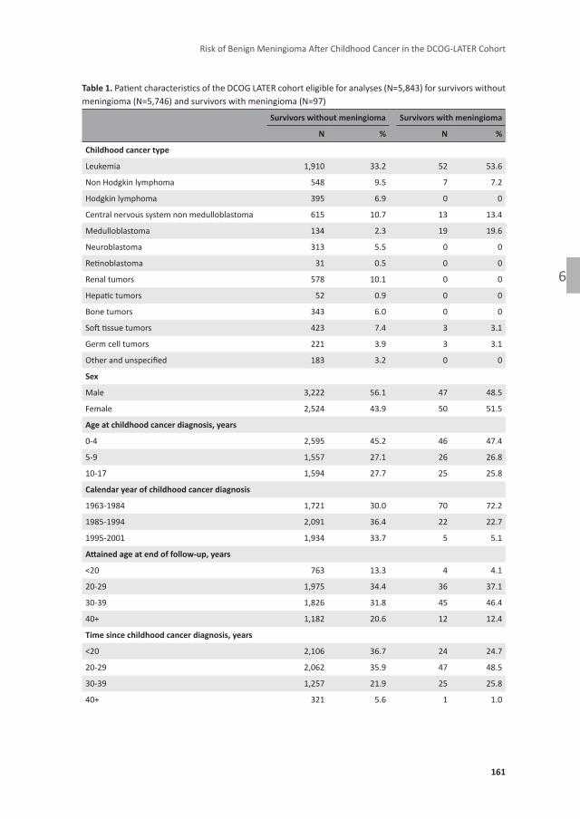
Table 1. Patient characteristics of the DCOG LATER cohort eligible for analyses (N=5,843) for survivors without meningioma (N=5,746) and survivors with meningioma (N=97)
survivors without meningioma survivors with meningioma
n % n %
childhood cancer type
Leukemia 1,910 33.2 52 53.6
Non Hodgkin lymphoma 548 9.5 7 7.2
Hodgkin lymphoma 395 6.9 0 0
Central nervous system non medulloblastoma 615 10.7 13 13.4
Medulloblastoma 134 2.3 19 19.6
Neuroblastoma 313 5.5 0 0
Retinoblastoma 31 0.5 0 0
Renal tumors 578 10.1 0 0
Hepatic tumors 52 0.9 0 0
Bone tumors 343 6.0 0 0
Soft tissue tumors 423 7.4 3 3.1
Germ cell tumors 221 3.9 3 3.1
Other and unspecified 183 3.2 0 0
sex
Male 3,222 56.1 47 48.5
Female 2,524 43.9 50 51.5
age at childhood cancer diagnosis, years
0-4 2,595 45.2 46 47.4
5-9 1,557 27.1 26 26.8
10-17 1,594 27.7 25 25.8
calendar year of childhood cancer diagnosis
1963-1984 1,721 30.0 70 72.2
1985-1994 2,091 36.4 22 22.7
1995-2001 1,934 33.7 5 5.1
attained age at end of follow-up, years
<20 763 13.3 4 4.1
20-29 1,975 34.4 36 37.1
30-39 1,826 31.8 45 46.4
40+ 1,182 20.6 12 12.4
Time since childhood cancer diagnosis, years
<20 2,106 36.7 24 24.7
20-29 2,062 35.9 47 48.5
30-39 1,257 21.9 25 25.8
40+ 321 5.6 1 1.0
162
Chapter 6
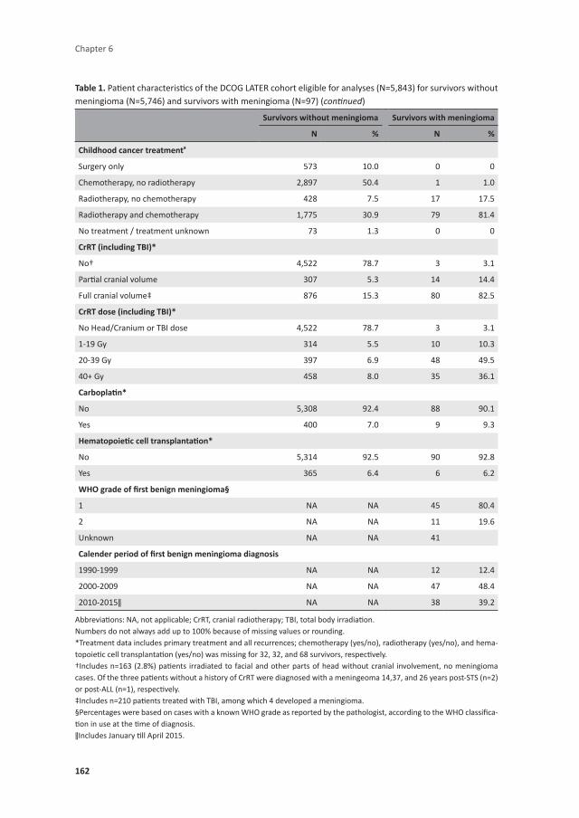
Table 1. Patient characteristics of the DCOG LATER cohort eligible for analyses (N=5,843) for survivors without meningioma (N=5,746) and survivors with meningioma (N=97) (continued)
survivors without meningioma survivors with meningioma
n % n %
childhood cancer treatmenta
Surgery only 573 10.0 0 0
Chemotherapy, no radiotherapy 2,897 50.4 1 1.0
Radiotherapy, no chemotherapy 428 7.5 17 17.5
Radiotherapy and chemotherapy 1,775 30.9 79 81.4
No treatment / treatment unknown 73 1.3 0 0
crRT (including TbI)*
No† 4,522 78.7 3 3.1
Partial cranial volume 307 5.3 14 14.4
Full cranial volume‡ 876 15.3 80 82.5
crRT dose (including TbI)*
No Head/Cranium or TBI dose 4,522 78.7 3 3.1
1-19 Gy 314 5.5 10 10.3
20-39 Gy 397 6.9 48 49.5
40+ Gy 458 8.0 35 36.1
carboplatin*
No 5,308 92.4 88 90.1
Yes 400 7.0 9 9.3
hematopoietic cell transplantation*
No 5,314 92.5 90 92.8
Yes 365 6.4 6 6.2
who grade of first benign meningioma§
1 NA NA 45 80.4
2 NA NA 11 19.6
Unknown NA NA 41
calender period of first benign meningioma diagnosis
1990-1999 NA NA 12 12.4
2000-2009 NA NA 47 48.4
2010-2015ǁ NA NA 38 39.2
Abbreviations: NA, not applicable; CrRT, cranial radiotherapy; TBI, total body irradiation.Numbers do not always add up to 100% because of missing values or rounding.*Treatment data includes primary treatment and all recurrences; chemotherapy (yes/no), radiotherapy (yes/no), and hema-topoietic cell transplantation (yes/no) was missing for 32, 32, and 68 survivors, respectively.†Includes n=163 (2.8%) patients irradiated to facial and other parts of head without cranial involvement, no meningioma cases. Of the three patients without a history of CrRT were diagnosed with a meningeoma 14,37, and 26 years post-STS (n=2) or post-ALL (n=1), respectively.‡Includes n=210 patients treated with TBI, among which 4 developed a meningioma.§Percentages were based on cases with a known WHO grade as reported by the pathologist, according to the WHO classifica-tion in use at the time of diagnosis.ǁIncludes January till April 2015.
163
Risk of Benign Meningioma After Childhood Cancer in the DCOG-LATER Cohort
6
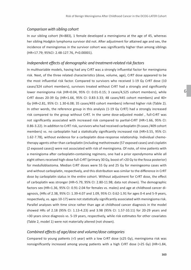
Comparison with sibling cohortIn our sibling cohort (N=883), 1 female developed a meningioma at the age of 45, whereas her sibling Hodgkin lymphoma survivor did not. After adjustment for attained age and sex, the incidence of meningiomas in the survivor cohort was significantly higher than among siblings (HR=17.79; 95%CI: 2.48-127.76, P<0.00001).
Independent effects of demographic and treatment-related risk factorsIn multivariable models, having had any CrRT was a strongly influential factor for meningioma risk. Next, of the three related characteristics (dose, volume, age), CrRT dose appeared to be the most influential risk factor. Compared to survivors who received 1-19 Gy CrRT dose (10 cases/324 cohort members), survivors treated without CrRT had a strongly and significantly lower meningioma risk (HR=0.04, 95% CI: 0.01-0.15; 3 cases/4,525 cohort members), while CrRT doses 20-39 Gy (HR=1.66, 95% CI: 0.83-3.33; 48 cases/445 cohort members) and 40+ Gy (HR=2.81, 95% CI: 1.30-6.08; 35 cases/493 cohort members) inferred higher risk (Table 2). In other words, the reference group in this analysis (1-19 Gy CrRT) had a strongly increased risk compared to the group without CrRT. In the same dose-adjusted model , full-CrRT was not significantly associated with increased risk compared to partial-CrRT (HR=1.66, 95% CI: 0.86-3.22). In addition to CrRT risk, survivors who had received carboplatin (9 cases /409 cohort members) vs. no carboplatin had a statistically significantly increased risk (HR=3.55, 95% CI: 1.62-7.78), without evidence for a carboplatin dose-response relationship. Individual chemo-therapy agents other than carboplatin (including methotrexate (57 exposed cases) and cisplatin (2 exposed cases)) were not associated with risk of meningioma. Of note, of nine patients with a meningioma after carboplatin-containing regimens, one had a prior ependymoma while all eight others received high-dose full-CrRT (primary 30 Gy, boost of >20 Gy to the fossa posterior) for medulloblastoma. Median CrRT doses were 55 Gy and 25 Gy for meningeoma cases with and without carboplatin, respectively, and this distribution was similar to the difference in CrRT dose by carboplatin status in the entire cohort. Without adjustment for CrRT dose, the effect of carboplatin was stronger (HR=5.79, 95% CI: 2.80-11.98; data not shown). The demographic factors sex (HR=1.36, 95% CI: 0.91-2.04 for females vs. males) and age at childhood cancer di-agnosis, (HRs of 2.38, 95% CI: 1.39-4.07 and 1.09, 95% CI: 0.62-1.91 for ages 0-4 and 5-9 years, respectively, vs. ages 10-17) were not statistically significantly associated with meningioma risk. Parallel analyses with time since rather than age at childhood cancer diagnosis in the model showed HRs of 2.18 (95% CI: 1.13-4.23) and 3.98 (95% CI: 1.57-10.11) for 20-29 years and >30 years since diagnosis vs. 5-19 years, respectively, while risk estimates for other covariates (Table 2, model 1) were not materially altered (not shown).
Combined effects of age/dose and volume/dose categoriesCompared to young patients (<5 year) with a low CrRT dose (≤25 Gy), meningioma risk was nonsignificantly increased among young patients with a high CrRT dose (>25 Gy) (HR=1.84,
164
Chapter 6
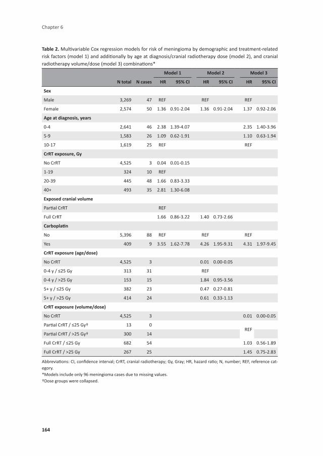
Table 2. Multivariable Cox regression models for risk of meningioma by demographic and treatment-related risk factors (model 1) and additionally by age at diagnosis/cranial radiotherapy dose (model 2), and cranial radiotherapy volume/dose (model 3) combinations*
n total n cases
Model 1 Model 2 Model 3
hr 95% cI hr 95% cI hr 95% cI
sex
Male 3,269 47 REF REF REF
Female 2,574 50 1.36 0.91-2.04 1.36 0.91-2.04 1.37 0.92-2.06
age at diagnosis, years
0-4 2,641 46 2.38 1.39-4.07 2.35 1.40-3.96
5-9 1,583 26 1.09 0.62-1.91 1.10 0.63-1.94
10-17 1,619 25 REF REF
crRT exposure, Gy
No CrRT 4,525 3 0.04 0.01-0.15
1-19 324 10 REF
20-39 445 48 1.66 0.83-3.33
40+ 493 35 2.81 1.30-6.08
exposed cranial volume
Partial CrRT REF
Full CrRT 1.66 0.86-3.22 1.40 0.73-2.66
carboplatin
No 5,396 88 REF REF REF
Yes 409 9 3.55 1.62-7.78 4.26 1.95-9.31 4.31 1.97-9.45
crRT exposure (age/dose)
No CrRT 4,525 3 0.01 0.00-0.05
0-4 y / ≤25 Gy 313 31 REF
0-4 y / >25 Gy 153 15 1.84 0.95-3.56
5+ y / ≤25 Gy 382 23 0.47 0.27-0.81
5+ y / >25 Gy 414 24 0.61 0.33-1.13
crRT exposure (volume/dose)
No CrRT 4,525 3 0.01 0.00-0.05
Partial CrRT / ≤25 Gy† 13 0REF
Partial CrRT / >25 Gy† 300 14
Full CrRT / ≤25 Gy 682 54 1.03 0.56-1.89
Full CrRT / >25 Gy 267 25 1.45 0.75-2.83
Abbreviations: CI, confidence interval; CrRT, cranial radiotherapy; Gy, Gray; HR, hazard ratio; N, number; REF, reference cat-egory.*Models include only 96 meningioma cases due to missing values.†Dose groups were collapsed.
165
Risk of Benign Meningioma After Childhood Cancer in the DCOG-LATER Cohort
6
95%CI: 0.95-3.56); those treated at older ages (5+ years), regardless of CrRT dose, had sig-nificantly lower meningioma risk (HR=0.47, 95% CI: 0.27-0.81 for age 5+, ≤25 Gy and HR=0.61, 95%CI: 0.33-1.13 for age 5+, >25 Gy) (Table 2, model 2). In contrast, there was no clear effect of exposed cranial volume with HRs for survivors treated with full-CrRT at doses ≤25 Gy (HR=1.03, 95% CI: 0.56-1.89) and full CrRT at doses >25 Gy (HR =1.45, 95% CI: 0.75-2.83) in comparison with any partial-CrRT (Table 2, model 3).
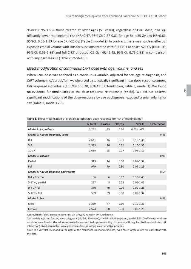
Effect modification of continuous CrRT dose with age, volume, and sexWhen CrRT dose was analyzed as a continuous variable, adjusted for sex, age at diagnosis, and CrRT volume (no/partial/full) we observed a statistically significant linear dose-response among CrRT-exposed individuals (ERR/Gy of 0.30, 95% CI: 0.03-unknown; Table 3, model 1). We found no evidence for nonlinearity of the dose-response relationship (p=.62). We did not observe significant modifications of the dose-response by age at diagnosis, exposed cranial volume, or sex (Table 3, models 2-5).
Table 3. Effect modification of cranial radiotherapy dose-response for risk of meningioma*
n total n cases eRR/Gy 95% cI P interaction
Model 1: All patients 1,262 93 0.30 0.03-UNK†
Model 2: Age at diagnosis, years 0.86
0-4 2,641 46 0.31 0.10-1.36
5-9 1,583 26 0.31 0.10-1.35
10-17 1,619 25 0.27 0.08-1.19
Model 3: Volume 0.98
Partial 313 14 0.30 0.09-1.32
Full 979 79 0.30 0.09-1.29
Model 4: Age at diagnosis and volume 0.55
0-4 y / partial 86 6 0.52 0.13-2.49
5-17 y / partial 227 8 0.22 0.05-1.06
0-4 y / full 380 40 0.29 0.09-1.28
5-17 y / full 569 39 0.30 0.09-1.32
Model 5: Sex 0.96
Male 3,269 47 0.30 0.10-1.29
Female 2,574 50 0.30 0.09-1.28
Abbreviations: ERR, excess relative risk; Gy, Gray; N, number; UNK, unknown.*All models adjusted for sex, age at diagnosis (<5, 5-9, 10+ years), cranial radiotherapy (no, partial, full). Coefficients for those variables were fixed at the values estimated in model 1 to improve stability of the model fitting. For likelihood ratio tests (P interaction), fixed parameters were counted as free, resulting in conservative p-values.†Due to a very flat likelihood to the right of the maximum likelihood estimate, even much larger values are consistent with the data.
166
Chapter 6
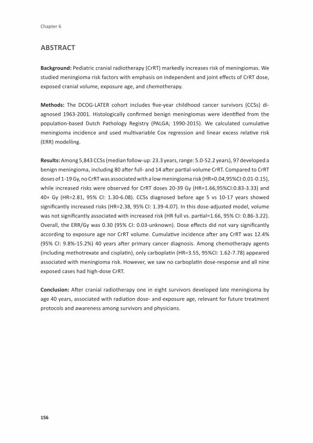
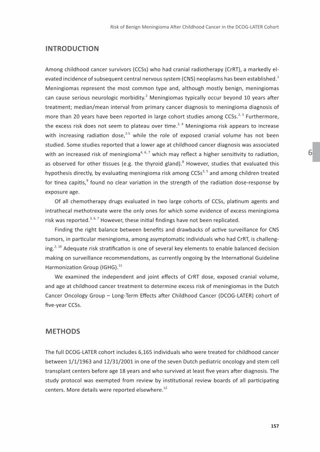
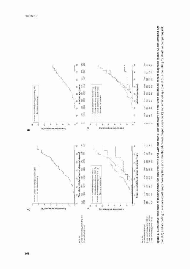
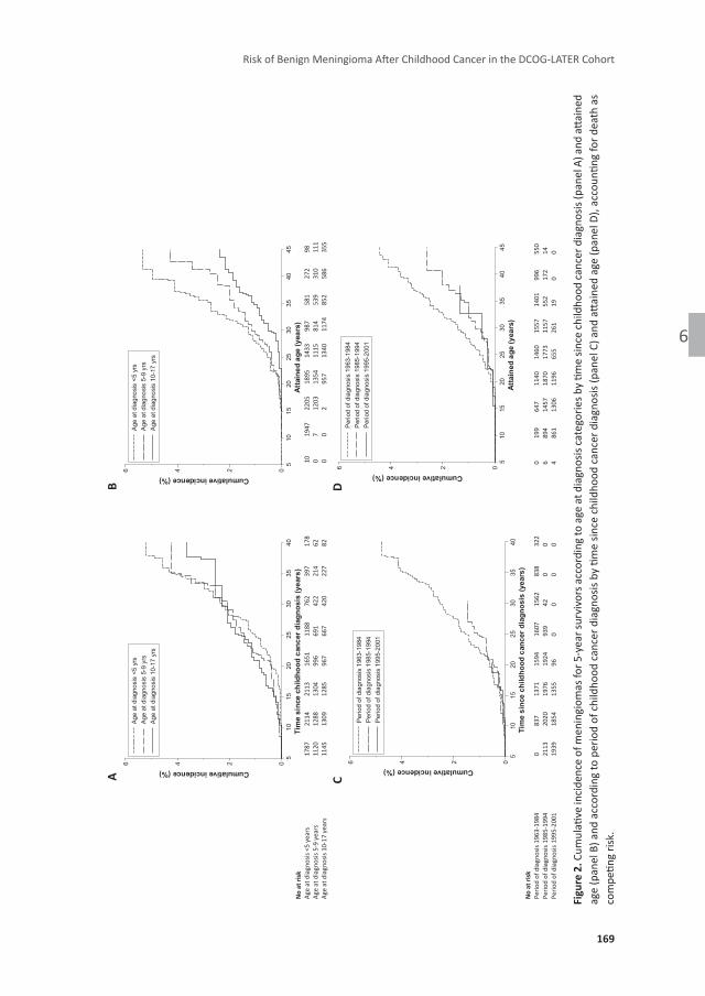
Cumulative incidence of subsequent meningiomaThe cumulative incidence of benign meningiomas varied according to CrRT characteristics. For survivors treated with CrRT the cumulative incidence was 12.4% (95% CI: 9.8%-15.2%) 40 years after diagnosis (Figure 1, panel A) and 7.3% (95% CI: 4.5%-10.8%) by age 45 (Figure 1, Panel B). For survivors without CrRT the cumulative incidence was much lower (0.3%, 95% CI: 0.1%-1.2% 40 years after diagnosis (Figure 1, panel A) and 0.3%, 95% CI: 0.1-1.2 by age 45 (Figure 1, panel B)). By CrRT doses, the cumulative incidence 40 years after diagnosis were 5.6% (95% CI: 2.3%-11.0%), 13.1% (95% CI: 9.6%-17.1%) , and 9.4% (95% CI: 6.3%-13.3%) for 1-19 Gy, 20-39 Gy, and 40+ Gy, respectively (Figure 1, panel C). Similar patterns were observed by attained age (Figure 1, panel D). When evaluated separately by age at childhood cancer diagnosis, the cumulative incidences of meningioma for survivors diagnosed at age 0-4 years, age 5-9 years, and age 10-17 years were 5.2% (95% CI: 3.7%-7.1%), 4.3% (95% CI: 2.6%-6.4%), and 3.7% (95% CI: 2.1%-5.9%) 40 years after diagnosis (Figure 2, panel A) and 5.3% (95% CI: 3.7%-7.3%), 4.3% (95% CI: 2.6%-6.6%), and 2.4% (95% CI: 1.5%-3.5%) by age 45 (Figure 2, panel B), respectively. Fifteen-year cumulative incidences was similar across periods of diagnosis (0.2%, 95% CI: 0.1%-0.7% for 1963-1984, 0.1%, 95% CI: 0.0%-0.4% for 1985-1994, and 0.1%, 95% CI: 0.0%-0.4% for 1995-2001, Figure 2, panel C).
DIscussIon
Our nationwide study with complete information on histologically confirmed benign menin-giomas after childhood cancer shows that, after cranial radiotherapy, one in eight survivors developed a late meningioma 40 years after primary cancer diagnosis. We found evidence for increased risk by radiation dose and among patients treated at the youngest ages, however, no significant modification of the radiation dose-response by age nor by radiation-exposed cranial volume.
CrRT is the most important risk factor for meningioma risk among CCSs; nearly all cases (97%) occurred among survivors who were treated with CrRT. Consistent with our findings of a dose-related excess risk, CrRT has frequently been reported to linearly increase risk of meningioma in a dose-related fashion among young CCSs,3-5 atomic bomb survivors,15 and children treated for tinea capitis,9 as summarized in Supplementary Table 2. While a main effect of age at childhood cancer diagnosis has been reported by others,4, 6, 7 data are inconsistent as to whether age modifies the radiation dose-response curve. We hypothesized that younger exposure age, as crude indicator of vulnerability during brain development, is related to higher radiation sensitivity, in other words, that the effect of radiation dose on meningioma risk var-ies by age at radiotherapy. Our results did not firmly support this hypothesis, consistent with earlier reports by Neglia et al. (66 meningioma cases) and Taylor et al. (137 meningioma cases) for the US/Canadian and UK childhood cancer survivor studies.3, 5 Our analyses are based on
167
Risk of Benign Meningioma After Childhood Cancer in the DCOG-LATER Cohort
6
prescribed CrRT dose, whereas the latter two studies3, 5 used absorbed CrRT dose in a case-control setting. Although it can be assumed that the prescribed CrRT dose is almost similar to the absorbed dose at the organ at risk, the meninges, for meningioma cases treated with full-CrRT (n=80 cases), the prescribed CrRT dose may represent an overestimation of the true absorbed dose at the meningioma location for partial-CrRT treated patients (n=14 cases). A new aspect of our study is that we analyzed the potential modifying effect of exposed cranial volume on the relation between radiation dose and meningioma risk. Our second hypothesis was that a higher volume of brain tissue exposed, will increase the radiation dose-related risk of meningioma. We did, however, not find evidence of a stronger dose-response among patients treated with full-CrRT compared to those treated with partial-CrRT; there was a sug-gestive, but nonsignificant main effect of full-CrRT (HR=1.66 for full vs partial). Of note, these results need to be interpreted with caution owing to a combination of factors: (1) the lack of statistical significance; (2) the probability that CrRT-volume may be a surrogate of other patient characteristics that influence meningioma risk (e.g. other risk factors such as NF1 status) or early detection (e.g. head MRIs or CTs for other side effects of CNS tumors or their treatment) not covered in our study variables. Also, there is a strong correlation between CrRT dose and exposed cranial volume (higher doses with partial volumes) as such that no meningioma cases were observed among the few patients treated with less than 40 Gy partial CrRT, as illustrated in Supplementary Figure 2.
Owing to clinical reality the radiation exposure metrics we applied tend to overestimate the volume of the brain exposed to the summed doses of main field and boost, since the sum of doses is assigned to the entire cranium. This is particularly true for medulloblastoma patients. Therefore, the true risk may be slightly overestimated in this study. Sensitivity analyses based on the full CrRT dose of approximately 30 Gy (i.e. disregarding the additional boost dose of around 24 Gy used in medulloblastoma protocols) provide a lower boundary for the estimated dose-related risk. Of note, CCS cohorts include individuals with several characteristics (e.g. radiotherapy, volume, and age, and demographic characteristics) that are quite correlated. For example, the proportion of survivors who were treated with CrRT declined over time, (from 36% prior to 1985 to 13% during 1995-2001); since 1985 prophylactic CrRT was eliminated from the DCOG-ALL protocols.16 Moreover, although CrRT remained indicated for ALL patients with CNS involvement up to 2004,17 the dose was reduced from 24 to 18 Gy in 1988.18
We did not find an effect of sex on meningioma risk, nor was there significant variation in the radiation dose-response according to sex, unlike some other studies reporting higher risks among women, after adjustment for CrRT dose.2, 4, 6
Only three previous studies showed some effects of chemotherapy: Two CCSS reports in-dicated increased meningioma risk after treatment with platinum agents without a clear dose-response.6, 7 At face value, these findings seem consistent with our finding of elevated risks associated with carboplatin (but not for cisplatin). However, the multivariable models from the cited studies6, 7 included only a radiotherapy yes/no indicator, which is likely insufficient to
168
Chapter 6
figu
re 1
. Cum
ulati
ve in
cide
nce
of m
enin
giom
as fo
r su
rviv
ors
with
and
with
out c
rani
al ra
diot
hera
py b
y tim
e sin
ce c
hild
hood
can
cer
diag
nosis
(pan
el A
) and
atta
ined
age
(p
anel
B) a
nd a
ccor
ding
to c
rani
al ra
diot
hera
py d
ose
by ti
me
since
chi
ldho
od c
ance
r dia
gnos
is (p
anel
C) a
nd a
ttain
ed a
ge (p
anel
D),
acco
untin
g fo
r dea
th a
s com
petin
g ris
k.
184
A
B
C
D
N
o at
risk
Cran
ial r
adio
ther
apy
(incl
udin
g TB
I) 63
2 85
6 99
8 90
3 70
2 54
2 27
8 79
N
o cr
ania
l rad
ioth
erap
y 33
88
3826
36
77
2698
18
33
1055
55
7 24
1
0
199
583
929
964
816
615
378
146
9 17
46
2813
32
56
2903
21
42
1347
78
3 41
4
0
1704
27
41
3154
27
85
2039
12
65
721
370
9 81
18
2 26
2 25
5 20
0 12
6 57
15
0
44
184
367
419
402
350
238
90
0 11
3 28
0 39
1 39
6 30
8 21
7 14
4 85
No
at ri
sk
N
o cr
ania
l rad
ioth
erap
y 32
94
3712
35
47
2579
17
37
979
510
223
Cran
ial r
adio
ther
apy
dose
1-1
9 G
y 21
5 27
0 27
5 21
9 15
7 11
0 28
6
Cran
ial r
adio
ther
apy
dose
20-
39 G
y 93
23
9 40
2 42
8 39
5 34
1 20
8 54
Cr
ania
l rad
ioth
erap
y do
se 4
0+ G
y 40
3 44
4 43
8 36
5 24
2 16
5 89
37
02468101214
Cumulative incidence (%)
510
1520
2530
3540
Tim
e si
nce
child
hood
can
cer d
iagn
osis
(yea
rs)
Cra
nial
radi
othe
rapy
(inc
ludi
ng T
BI)
No
cran
ial r
adio
ther
apy
02468101214
Cumulative incidence (%)
510
1520
2530
3540
45At
tain
ed a
ge (y
ears
)
Cra
nial
radi
othe
rapy
(inc
ludi
ng T
BI)
No
cran
ial r
adio
ther
apy
02468101214
Cumulative incidence (%)
510
1520
2530
3540
45A
ttain
ed a
ge (y
ears
)
Cra
nial
radi
othe
rapy
dos
e 40
+ G
yC
rani
al ra
diot
hera
py d
ose
20-3
9 G
yC
rani
al ra
diot
hera
py d
ose
1-19
Gy
No
cran
ial r
adio
ther
apy
02468101214
Cumulative incidence (%)
510
1520
2530
3540
Tim
e si
nce
child
hood
can
cer d
iagn
osis
(yea
rs)
Cra
nial
radi
othe
rapy
dos
e 40
+ G
yC
rani
al ra
diot
hera
py d
ose
20-3
9 G
yC
rani
al ra
diot
hera
py d
ose
1-19
Gy
No
cran
ial r
adio
ther
apy
cd
169
Risk of Benign Meningioma After Childhood Cancer in the DCOG-LATER Cohort
6
figu
re 2
. Cum
ulati
ve in
cide
nce
of m
enin
giom
as fo
r 5-y
ear s
urvi
vors
acc
ordi
ng to
age
at d
iagn
osis
cate
gorie
s by
time
since
child
hood
canc
er d
iagn
osis
(pan
el A
) and
atta
ined
ag
e (p
anel
B) a
nd a
ccor
ding
to p
erio
d of
chi
ldho
od c
ance
r dia
gnos
is by
tim
e sin
ce c
hild
hood
can
cer d
iagn
osis
(pan
el C
) and
atta
ined
age
(pan
el D
), ac
coun
ting
for d
eath
as
com
petin
g ris
k.
186
A
B
N
o at
risk
Age
at d
iagn
osis
<5 y
ears
17
87
2114
21
13
1651
11
88
762
397
178
Age
at d
iagn
osis
5-9
year
s 11
20
1288
13
04
996
691
422
214
62
Age
at d
iagn
osis
10-1
7 ye
ars
1145
13
09
1285
96
7 66
7 42
0 22
7 82
10
19
47
2205
18
95
1433
98
7 58
1 27
2 98
0
7 12
03
1354
11
15
814
539
310
111
0 0
2 95
7 13
40
1174
85
2 58
6 35
5
0
199
647
1140
14
60
1557
14
01
996
550
6 89
4 14
57
1870
17
73
1157
55
2 17
2 14
4
861
1306
11
96
655
261
19
0 0
No
at ri
sk
Pe
riod
of d
iagn
osis
1963
-198
4 0
837
1371
15
94
1607
15
62
838
322
Perio
d of
dia
gnos
is 19
85-1
994
2113
20
20
1976
19
24
939
42
0 0
Perio
d of
dia
gnos
is 19
95-2
001
1939
18
54
1355
96
0
0 0
0
0246
Cumulative incidence (%)
510
1520
2530
3540
Tim
e si
nce
child
hood
can
cer d
iagn
osis
(yea
rs)
Age
at d
iagn
osis
<5
yrs
Age
at d
iagn
osis
5-9
yrs
Age
at d
iagn
osis
10-
17 y
rs
0246
Cumulative incidence (%)
510
1520
2530
3540
45At
tain
ed a
ge (y
ears
)
Age
at d
iagn
osis
<5
yrs
Age
at d
iagn
osis
5-9
yrs
Age
at d
iagn
osis
10-
17 y
rs
0246
Cumulative incidence (%)
510
1520
2530
3540
Tim
e si
nce
child
hood
can
cer d
iagn
osis
(yea
rs)
Perio
d of
dia
gnos
is 1
963-
1984
Perio
d of
dia
gnos
is 1
985-
1994
Perio
d of
dia
gnos
is 1
995-
2001
0246
Cumulative incidence (%)
510
1520
2530
3540
45At
tain
ed a
ge (y
ears
)
Perio
d of
dia
gnos
is 1
963-
1984
Perio
d of
dia
gnos
is 1
985-
1994
Perio
d of
dia
gnos
is 1
995-
2001
cd
170
Chapter 6
fully adjust for the radiotherapy dose effects. Importantly, carboplatin can be part of medul-loblastoma protocols, a patient group considered at highest risk for meningioma because they all received full-CrRT and boosts up to total doses exceeding 50 Gy. We question the causality of the carboplatin-meningioma association, due to lack of dose-response relation, collinearity with high-risk radiotherapy characteristics, no observed meningioma cases among survivors with carboplatin exposure without CrRT, no relation of cisplatin with meningioma risk, and no clear evidence on carboplatin carcinogenicity from in vitro and in vivo studies. Nonetheless, we cannot entirely discard a true, albeit small, effect of carboplatin either.
In the British Childhood Cancer Survivor Study cohort meningioma risk among individuals receiving 1-39, 40-69, and 70 or more mg/m² of intrathecal methotrexate was increased by 15-fold, 11-fold, and 36-fold, respectively, compared to unexposed survivors.3 The authors added a strong cautionary note to their findings: few survivors were treated with intrathecal methotrexate without CrRT and no effects were observed for non-intrathecal methotrexate. These findings have not been confirmed by other studies, including our results reported here.
Strengths of our study are the large cohort size with detailed individual treatment informa-tion and objective and near complete data on histologically confirmed benign meningiomas from linkage to the nationwide registry of histo- and cytopathology (PALGA)13 for more than 95% of the total cohort. Other studies relied on initial self-report and/or linkage with tumor registries.2, 3 In addition, a comparison with a sibling group was performed, to obtain more insight into the incidence of meningiomas, which enabled us to parallel the meningioma incidence in CCSs to the general population. PALGA is an internationally unique resource to as-certain benign meningiomas, because benign CNS tumors are not typically recorded completely in cancer registries, although this is changing in more recent years.
Several weaknesses of our approach deserve attention as well. PALGA has complete national coverage from 1990 onwards; tumors occurring during 1968-1989 were not recorded reliably. By including only follow-up time since 1990 and the fact that most meningiomas occur >20 years after childhood cancer, a follow-up interval which nearly all surviving cohort members completed after 1990, we are confident that the underestimation of the true cumulative inci-dence caused by left truncation is minimal. Secondly, as in most studies on meningioma, true incidence is likely not captured; we report on histologically confirmed benign meningiomas while a certain proportion of such tumors can remain asymptomatic/indolent for some time. Other factors may have increased the meningioma detection rate: medical care has changed, including access to and indications for brain imaging as well as indications for surgery of cranial masses suspect for meningioma. Also, between 1996 and 2006, Dutch late effects outpatient clinics were implemented in which CCSs are followed-up according to evidence-based Dutch guidelines. These guidelines do not recommend active screening for CNS tumors among asymptomatic individuals. Nevertheless, all survivors received guideline-based follow-up at fixed intervals, which is more frequent for those with high-intensity treatment (including those who received radiotherapy). It is quite possible that more intensive medical attention in
171
Risk of Benign Meningioma After Childhood Cancer in the DCOG-LATER Cohort
6
the outpatient clinics slightly increased the detection rates of asymptomatic meningioma, for example among patients with a history of seizures, headaches, or other neurologic problems owing to a brain tumor or CNS metastases, but also brain surgery, hydrocephalus, or high-dose radiotherapy. Longer follow-up of available cohorts with complete treatment information and adequate follow-up methods to detect these benign tumors, as well as pooled analyses of these studies are needed to shed light on the influence of these issues on meningioma incidence rates.
The results of this study can be used to inform surveillance recommendations for long-term CCSs who had CrRT, such as those currently being formulated by the International Guideline Harmonization Group.19 Although risk estimation in terms of prescribed CrRT dose is neces-sary in light of surveillance guideline development, further studies are forthcoming to express excess risk in terms of cranial-volume-based absorbed dose distributions. These will be useful to inform future pediatric radiotherapy treatment guidelines. To fully disentangle effects of dose, volume, age, and the potential role of chemotherapy agents, an international pooling effort is warranted to achieve sufficient statistical power.
In conclusion, one in eight CCSs exposed to cranial radiotherapy develop a late meningioma 40 years after childhood cancer diagnosis and this risk is dose- and exposure age-related. We did not find significant modifications of the radiation dose-response by age or by exposed cranial volume. While the proportion of patients in need of (full) CrRT with curative intent has decreased over time, this treatment cannot be abolished without compromising cancer survival for children with intracranial tumors or other indications for CrRT. These findings are important to raise awareness among survivors, their parents, and care-providers about these long-term sequelae, and to support ongoing efforts to reduce the radiation exposure to healthy tissues, where feasible, without compromising treatment efficacy.
acknowleDGeMenTs
The DCOG-LATER Study Group for benign CNS tumors includes the listed authors and the fol-lowing persons: MH van den Berg (VU University Medical Center, Amsterdam), AH Bruggink (PALGA Foundation, Houten), HN Caron (Emma Children’s Hospital/Academic Medical Center, Amsterdam), WV Dolsma (University of Groningen/University Medical Center Groningen), MA Grootenhuis (Emma Children’s Hospital/Academic Medical Center, Amsterdam, and Princess Máxima Center for Pediatric Oncology, Utrecht), JG den Hartogh (Dutch Childhood Cancer Parent Organisation (VOKK), Nieuwegein), N Hollema (Dutch Childhood Oncology Group, The Hague), MC Jongmans (Radboud University Medical Center, Nijmegen, University Medical Center Utrecht, Utrecht, and Princess Máxima Center for Pediatric Oncology, Utrecht), MWM Jaspers (Academic Medical Center, Amsterdam), A Postma (Dutch Childhood Oncology Group, The Hague), and MJ van de Vijver (Academic Medical Center, Amsterdam). We thank all data

172
Chapter 6
managers in the seven participating centers and Aslihan Mantici and Anja van Eggermond for obtaining the data for this study. Furthermore, we thank the following other members of the DCOG-LATER group for their contributions: Lilian Batenburg, Margreet Veening, Marloes Lou-werens, Gea Huizinga, Lideke van der Steeg, Hanneke de Ridder-Sluiter, and Andrica de Vries. We thank the staff of the PALGA Foundation for providing record linkage data on meningiomas from their registry.
173
RefeRences
1. Bowers DC, Nathan PC, Constine L et al. Subsequent neoplasms of the CNS among survivors of childhood cancer: a systematic review. Lancet Oncol 2013; 14: e321-328.
2. Bowers DC, Moskowitz CS, Chou JF et al. Morbidity and mortality associated with meningioma after cranial radiotherapy: a report from the Childhood Cancer Survivor Study. J Clin Oncol 2017; 35: 1570-1576.
3. Taylor AJ, Little MP, Winter DL et al. Population-based risks of CNS tumors in survivors of childhood cancer: the British Childhood Cancer Survivor Study. J Clin Oncol 2010; 28: 5287-5293.
4. Patterson BC, Chen Y, Sklar CA et al. Growth hormone exposure as a risk factor for the develop-ment of subsequent neoplasms of the central nervous system: a report from the childhood cancer survivor study. J Clin Endocrinol Metab 2014; 99: 2030-2037.
5. Neglia JP, Robison LL, Stovall M et al. New primary neoplasms of the central nervous system in survivors of childhood cancer: a report from the Childhood Cancer Survivor Study. J Natl Cancer Inst 2006; 98: 1528-1537.
6. Friedman DL, Whitton J, Leisenring W et al. Subsequent neoplasms in 5-year survivors of childhood cancer: the Childhood Cancer Survivor Study. J Natl Cancer Inst 2010; 102: 1083-1095.
7. Turcotte LM, Liu Q, Yasui Y et al. Temporal trends in treatment and subsequent neoplasm risk among 5-year survivors of childhood cancer, 1970-2015. JAMA 2017; 317: 814-824.
8. Ronckers CM, Sigurdson AJ, Stovall M et al. Thyroid cancer in childhood cancer survivors: a detailed evaluation of radiation dose response and its modifiers. Radiat Res 2006; 166: 618-628.
9. Sadetzki S, Chetrit A, Freedman L et al. Long-term follow-up for brain tumor development after childhood exposure to ionizing radiation for tinea capitis. Radiat Res 2005; 163: 424-432.
10. Sugden E, Taylor A, Pretorius P et al. Meningiomas occurring during long-term survival after treat-ment for childhood cancer. JRSM Open 2014; 5: 1-4.
11. International Guideline Harmonization Group. International Guideline Harmonization Group for late effects of childhood cancer. http://www.ighg.org/international-guideline-harmonization-group/.
12. Teepen JC, van Leeuwen FE, Tissing WJ et al. Long-term risk of subsequent malignant neoplasms after treatment of childhood cancer in the DCOG LATER study cohort: role of chemotherapy. J Clin Oncol 2017; 35: 2288-2298.
13. Casparie M, Tiebosch AT, Burger G et al. Pathology databanking and biobanking in The Netherlands, a central role for PALGA, the nationwide histopathology and cytopathology data network and archive. Cell Oncol 2007; 29: 19-24.
14. Gooley TA, Leisenring W, Crowley J, Storer BE. Estimation of failure probabilities in the presence of competing risks: new representations of old estimators. Stat Med 1999; 18: 695-706.
15. Preston DL, Ron E, Yonehara S et al. Tumors of the nervous system and pituitary gland associated with atomic bomb radiation exposure. J Natl Cancer Inst 2002; 94: 1555-1563.
16. Veerman AJ, Hahlen K, Kamps WA et al. High cure rate with a moderately intensive treatment regi-men in non-high-risk childhood acute lymphoblastic leukemia. Results of protocol ALL VI from the Dutch Childhood Leukemia Study Group. J Clin Oncol 1996; 14: 911-918.
174
17. Veerman AJ, Kamps WA, van den Berg H et al. Dexamethasone-based therapy for childhood acute lymphoblastic leukaemia: results of the prospective Dutch Childhood Oncology Group (DCOG) protocol ALL-9 (1997-2004). Lancet Oncol 2009; 10: 957-966.
18. Kamps WA, Bokkerink JP, Hahlen K et al. Intensive treatment of children with acute lymphoblastic leukemia according to ALL-BFM-86 without cranial radiotherapy: results of Dutch Childhood Leuke-mia Study Group Protocol ALL-7 (1988-1991). Blood 1999; 94: 1226-1236.
19. Kremer LC, Mulder RL, Oeffinger KC et al. A worldwide collaboration to harmonize guidelines for the long-term follow-up of childhood and young adult cancer survivors: a report from the International Late Effects of Childhood Cancer Guideline Harmonization Group. Pediatr Blood Cancer 2013; 60: 543-549.
175
Risk of Benign Meningioma After Childhood Cancer in the DCOG-LATER Cohort
6
suPPleMenTaRY MaTeRIals
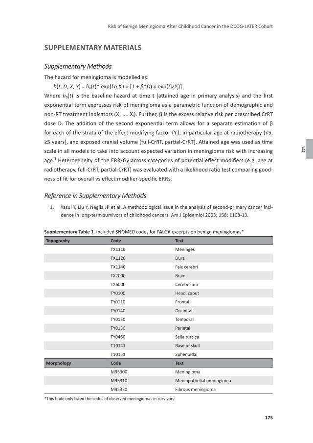
Supplementary MethodsThe hazard for meningioma is modelled as:
h(t, D, X, Y) = h0(t)* exp(ΣαiXi) × [1 + β*D) × exp(ΣγjYj)]Where h0(t) is the baseline hazard at time t (attained age in primary analysis) and the first exponential term expresses risk of meningioma as a parametric function of demographic and non-RT treatment indicators (X1 .... Xi). Further, β is the excess relative risk per prescribed CrRT dose D. The addition of the second exponential term allows for a separate estimation of β for each of the strata of the effect modifying factor (Yj), in particular age at radiotherapy (<5, ≥5 years), and exposed cranial volume (full-CrRT, partial-CrRT). Attained age was used as time scale in all models to take into account expected variation in meningioma risk with increasing age.1 Heterogeneity of the ERR/Gy across categories of potential effect modifiers (e.g. age at radiotherapy, full-CrRT, partial-CrRT) was evaluated with a likelihood ratio test comparing good-ness of fit for overall vs effect modifier-specific ERRs.
Reference in Supplementary Methods 1. Yasui Y, Liu Y, Neglia JP et al. A methodological issue in the analysis of second-primary cancer inci-
dence in long-term survivors of childhood cancers. Am J Epidemiol 2003; 158: 1108-13.
supplementary Table 1. Included SNOMED codes for PALGA excerpts on benign meningiomas*
Topography code Text
TX1110 Meninges
TX1120 Dura
TX1140 Falx cerebri
TX2000 Brain
TX6000 Cerebellum
TY0100 Head, caput
TY0110 Frontal
TY0140 Occipital
TY0150 Temporal
TY0130 Parietal
TY0460 Sella turcica
T10141 Base of skull
T10151 Sphenoidal
Morphology code Text
M95300 Meningioma
M95310 Meningothelial meningioma
M95320 Fibrous meningioma
*This table only listed the codes of observed meningiomas in survivors.
176
Chapter 6
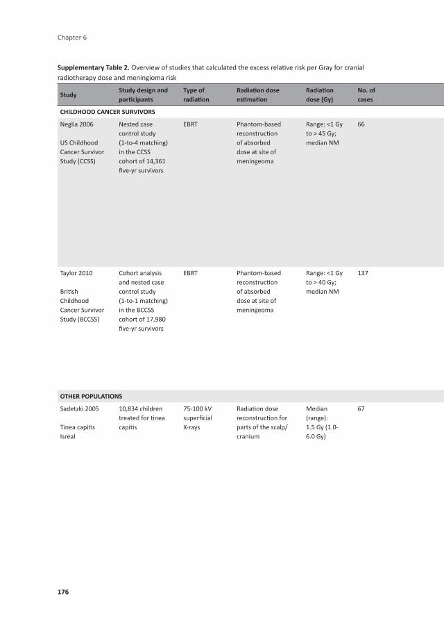
supplementary Table 2. Overview of studies that calculated the excess relative risk per Gray for cranial radiotherapy dose and meningioma risk
studystudy design and participants
Type of radiation
Radiation dose estimation
Radiation dose (Gy)
no. of cases
stratification factor
linear eRR/Gy
95% cI P-value* curvature† covariate adjustment
chIlDhooD canceR suRvIvoRs
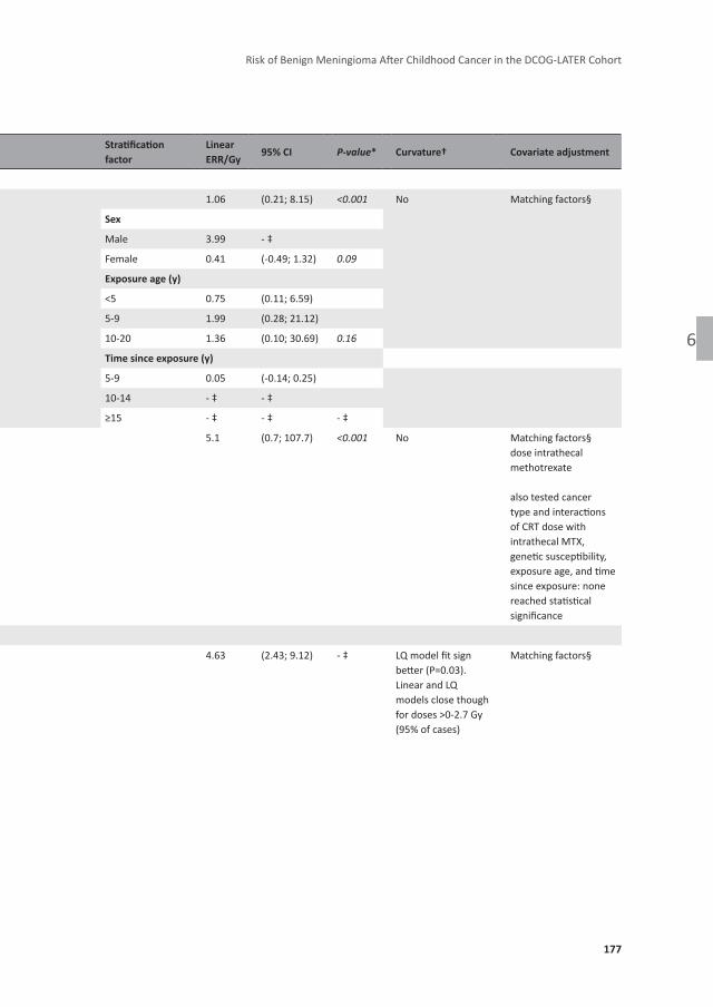
Neglia 2006
US Childhood Cancer Survivor Study (CCSS)
Nested case control study (1-to-4 matching) in the CCSS cohort of 14,361 five-yr survivors
EBRT Phantom-based reconstruction of absorbed dose at site of meningeoma
Range: <1 Gy to > 45 Gy; median NM
66 1.06 (0.21; 8.15) <0.001 No Matching factors§
sex
Male 3.99 - ‡
Female 0.41 (-0.49; 1.32) 0.09
exposure age (y)
<5 0.75 (0.11; 6.59)
5-9 1.99 (0.28; 21.12)
10-20 1.36 (0.10; 30.69) 0.16
Time since exposure (y)
5-9 0.05 (-0.14; 0.25)
10-14 - ‡ - ‡
≥15 - ‡ - ‡ - ‡
Taylor 2010
British Childhood Cancer Survivor Study (BCCSS)
Cohort analysis and nested case control study (1-to-1 matching) in the BCCSS cohort of 17,980 five-yr survivors
EBRT Phantom-based reconstruction of absorbed dose at site of meningeoma
Range: <1 Gy to > 40 Gy; median NM
137 5.1 (0.7; 107.7) <0.001 No Matching factors§ dose intrathecal methotrexate
also tested cancer type and interactions of CRT dose with intrathecal MTX, genetic susceptibility, exposure age, and time since exposure: none reached statistical significance
oTheR PoPulaTIons
Sadetzki 2005
Tinea capitis Isreal
10,834 children treated for tinea capitis
75-100 kV superficial X-rays
Radiation dose reconstruction for parts of the scalp/cranium
Median (range): 1.5 Gy (1.0-6.0 Gy)
67 4.63 (2.43; 9.12) - ‡ LQ model fit sign better (P=0.03).Linear and LQ models close though for doses >0-2.7 Gy (95% of cases)
Matching factors§
177
Risk of Benign Meningioma After Childhood Cancer in the DCOG-LATER Cohort
6
supplementary Table 2. Overview of studies that calculated the excess relative risk per Gray for cranial radiotherapy dose and meningioma risk
studystudy design and participants
Type of radiation
Radiation dose estimation
Radiation dose (Gy)
no. of cases
stratification factor
linear eRR/Gy
95% cI P-value* curvature† covariate adjustment
chIlDhooD canceR suRvIvoRs
Neglia 2006
US Childhood Cancer Survivor Study (CCSS)
Nested case control study (1-to-4 matching) in the CCSS cohort of 14,361 five-yr survivors
EBRT Phantom-based reconstruction of absorbed dose at site of meningeoma
Range: <1 Gy to > 45 Gy; median NM
66 1.06 (0.21; 8.15) <0.001 No Matching factors§
sex
Male 3.99 - ‡
Female 0.41 (-0.49; 1.32) 0.09
exposure age (y)
<5 0.75 (0.11; 6.59)
5-9 1.99 (0.28; 21.12)
10-20 1.36 (0.10; 30.69) 0.16
Time since exposure (y)
5-9 0.05 (-0.14; 0.25)
10-14 - ‡ - ‡
≥15 - ‡ - ‡ - ‡
Taylor 2010
British Childhood Cancer Survivor Study (BCCSS)
Cohort analysis and nested case control study (1-to-1 matching) in the BCCSS cohort of 17,980 five-yr survivors
EBRT Phantom-based reconstruction of absorbed dose at site of meningeoma
Range: <1 Gy to > 40 Gy; median NM
137 5.1 (0.7; 107.7) <0.001 No Matching factors§ dose intrathecal methotrexate
also tested cancer type and interactions of CRT dose with intrathecal MTX, genetic susceptibility, exposure age, and time since exposure: none reached statistical significance
oTheR PoPulaTIons
Sadetzki 2005
Tinea capitis Isreal
10,834 children treated for tinea capitis
75-100 kV superficial X-rays
Radiation dose reconstruction for parts of the scalp/cranium
Median (range): 1.5 Gy (1.0-6.0 Gy)
67 4.63 (2.43; 9.12) - ‡ LQ model fit sign better (P=0.03).Linear and LQ models close though for doses >0-2.7 Gy (95% of cases)
Matching factors§
178
Chapter 6
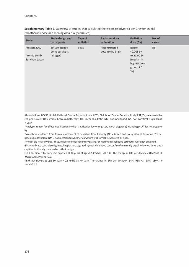
supplementary Table 2. Overview of studies that calculated the excess relative risk per Gray for cranialradiotherapy dose and meningioma risk (continued)
studystudy design and participants
Type of radiation
Radiation dose estimation
Radiation dose (Gy)
no. of cases
stratification factor
linear eRR/Gy
95% cI P-value* curvature† covariate adjustment
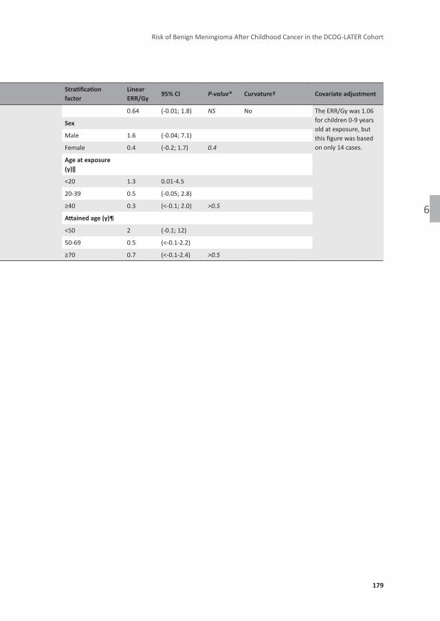
Preston 2002
Atomic Bomb Survivors Japan
80,160 atomic boms survivors (all ages)
γ-ray Reconstructed dose to the brain
Range: <0.005 Sv to ≥1.00 Sv (median in highest dose group: 7.5 Sv)
88
0.64 (-0.01; 1.8) NS No The ERR/Gy was 1.06 for children 0-9 years old at exposure, but this figure was based on only 14 cases.
sex
Male 1.6 (-0.04; 7.1)
Female 0.4 (-0.2; 1.7) 0.4
age at exposure (y)ǁ
<20 1.3 0.01-4.5
20-39 0.5 (-0.05; 2.8)
≥40 0.3 (<-0.1; 2.0) >0.5
attained age (y)¶
<50 2 (-0.1; 12)
50-69 0.5 (<-0.1-2.2)
≥70 0.7 (<-0.1-2.4) >0.5
Abbreviations: BCCSS, British Chilhood Cancer Survivor Study; CCSS, Childhood Cancer Survivor Study; ERR/Gy, excess relative risk per Gray; EBRT, external beam radiotherapy; LQ, linear Quadratic; NM, not mentioned; NS, not statistically significant; Y, year.*Analyses to test for effect modification by the stratification factor (e.g. sex, age at diagnosis) including p-LRT for heterogene-ity.†Was there evidence from formal assessment of deviation from linearity (No = tested and no significant deviation; Yes de-notes sign deviation; NM = not mentioned whether curvature was formally evaluated or not) .‡Model did not converge. Thus, reliable confidence intervals and/or maximum likelihood estimates were not obtained.§Matched case control study; matching factors: age at diagnosis childhood cancer / sex/ minimally equal follow-up time; tinea capitis additionally matched on ethnic origin.ǁERR per sievert for survivors exposed at 30 years of age=0.5 (95% CI: <0; 1.8). The change in ERR per decade=38% (95% CI: -95%; 60%). P trend=0.3.¶ERR per sievert at age 60 years= 0.6 (95% CI: <0; 2.3). The change in ERR per decade= -54% (95% CI: -95%; 130%). P trend=0.12.
179
Risk of Benign Meningioma After Childhood Cancer in the DCOG-LATER Cohort
6
supplementary Table 2. Overview of studies that calculated the excess relative risk per Gray for cranialradiotherapy dose and meningioma risk (continued)
studystudy design and participants
Type of radiation
Radiation dose estimation
Radiation dose (Gy)
no. of cases
stratification factor
linear eRR/Gy
95% cI P-value* curvature† covariate adjustment
Preston 2002
Atomic Bomb Survivors Japan
80,160 atomic boms survivors (all ages)
γ-ray Reconstructed dose to the brain
Range: <0.005 Sv to ≥1.00 Sv (median in highest dose group: 7.5 Sv)
88
0.64 (-0.01; 1.8) NS No The ERR/Gy was 1.06 for children 0-9 years old at exposure, but this figure was based on only 14 cases.
sex
Male 1.6 (-0.04; 7.1)
Female 0.4 (-0.2; 1.7) 0.4
age at exposure (y)ǁ
<20 1.3 0.01-4.5
20-39 0.5 (-0.05; 2.8)
≥40 0.3 (<-0.1; 2.0) >0.5
attained age (y)¶
<50 2 (-0.1; 12)
50-69 0.5 (<-0.1-2.2)
≥70 0.7 (<-0.1-2.4) >0.5
Abbreviations: BCCSS, British Chilhood Cancer Survivor Study; CCSS, Childhood Cancer Survivor Study; ERR/Gy, excess relative risk per Gray; EBRT, external beam radiotherapy; LQ, linear Quadratic; NM, not mentioned; NS, not statistically significant; Y, year.*Analyses to test for effect modification by the stratification factor (e.g. sex, age at diagnosis) including p-LRT for heterogene-ity.†Was there evidence from formal assessment of deviation from linearity (No = tested and no significant deviation; Yes de-notes sign deviation; NM = not mentioned whether curvature was formally evaluated or not) .‡Model did not converge. Thus, reliable confidence intervals and/or maximum likelihood estimates were not obtained.§Matched case control study; matching factors: age at diagnosis childhood cancer / sex/ minimally equal follow-up time; tinea capitis additionally matched on ethnic origin.ǁERR per sievert for survivors exposed at 30 years of age=0.5 (95% CI: <0; 1.8). The change in ERR per decade=38% (95% CI: -95%; 60%). P trend=0.3.¶ERR per sievert at age 60 years= 0.6 (95% CI: <0; 2.3). The change in ERR per decade= -54% (95% CI: -95%; 130%). P trend=0.12.
180
Chapter 6
199
A
B
C
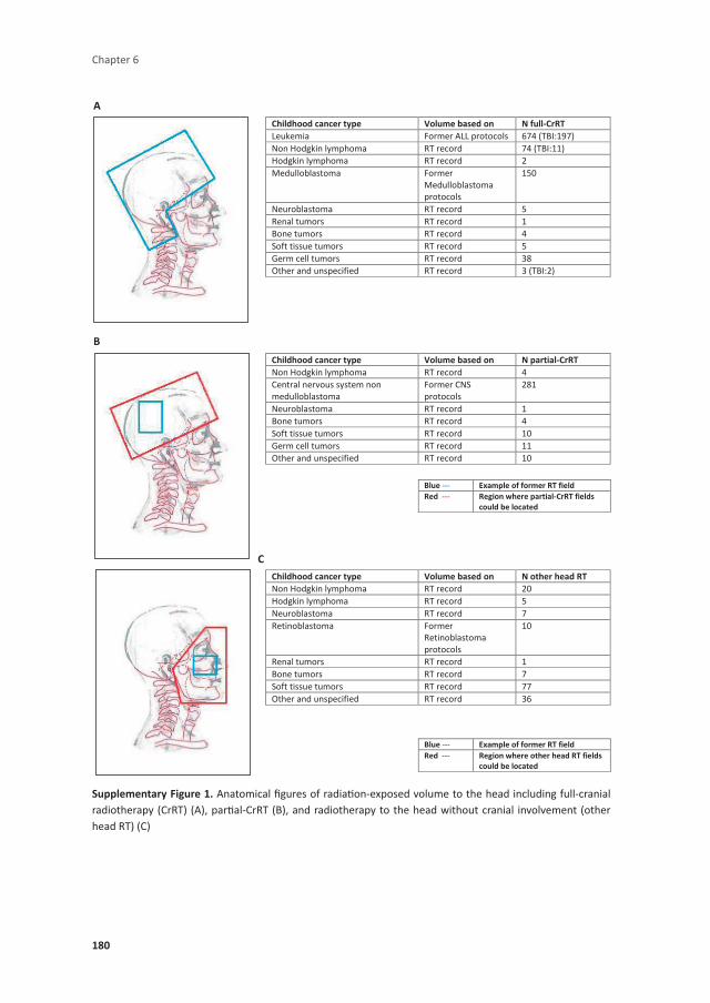
Supplementary Figure 1. Anatomical figures of radiation-exposed volume to the head including full-cranial radiotherapy (CrRT) (A), partial-CrRT (B), and radiotherapy to the head without cranial involvement (other head RT) (C)
Childhood cancer type Volume based on N full-CrRT Leukemia Former ALL protocols 674 (TBI:197) Non Hodgkin lymphoma RT record 74 (TBI:11) Hodgkin lymphoma RT record 2 Medulloblastoma Former
Medulloblastoma protocols
150
Neuroblastoma RT record 5 Renal tumors RT record 1 Bone tumors RT record 4 Soft tissue tumors RT record 5 Germ cell tumors RT record 38 Other and unspecified RT record 3 (TBI:2)
Childhood cancer type Volume based on N partial-CrRT Non Hodgkin lymphoma RT record 4 Central nervous system non medulloblastoma
Former CNS protocols
281
Neuroblastoma RT record 1 Bone tumors RT record 4 Soft tissue tumors RT record 10 Germ cell tumors RT record 11 Other and unspecified RT record 10
Blue --- Example of former RT field Red --- Region where partial-CrRT fields
could be located
Childhood cancer type Volume based on N other head RT Non Hodgkin lymphoma RT record 20 Hodgkin lymphoma RT record 5 Neuroblastoma RT record 7 Retinoblastoma Former
Retinoblastoma protocols
10
Renal tumors RT record 1 Bone tumors RT record 7 Soft tissue tumors RT record 77 Other and unspecified RT record 36
Blue --- Example of former RT field Red --- Region where other head RT fields
could be located
supplementary figure 1. Anatomical figures of radiation-exposed volume to the head including full-cranial radiotherapy (CrRT) (A), partial-CrRT (B), and radiotherapy to the head without cranial involvement (other head RT) (C)
181
Risk of Benign Meningioma After Childhood Cancer in the DCOG-LATER Cohort
6
200
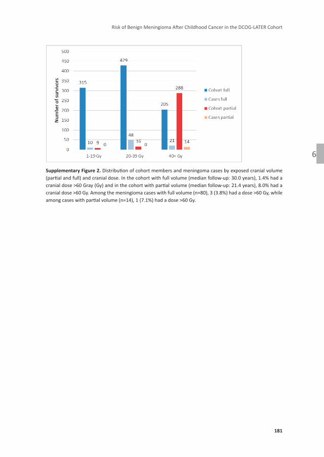
Supplementary Figure 2. Distribution of cohort members and meningoma cases by exposed cranial volume (partial and full) and cranial dose. In the cohort with full volume (median follow-up: 30.0 years), 1.4% had a cranial dose >60 Gray (Gy) and in the cohort with partial volume (median follow-up: 21.4 years), 8.0% had a cranial dose >60 Gy. Among the meningioma cases with full volume (n=80), 3 (3.8%) had a dose >60 Gy, while among cases with partial volume (n=14), 1 (7.1%) had a dose >60 Gy.
supplementary figure 2. Distribution of cohort members and meningoma cases by exposed cranial volume (partial and full) and cranial dose. In the cohort with full volume (median follow-up: 30.0 years), 1.4% had a cranial dose >60 Gray (Gy) and in the cohort with partial volume (median follow-up: 21.4 years), 8.0% had a cranial dose >60 Gy. Among the meningioma cases with full volume (n=80), 3 (3.8%) had a dose >60 Gy, while among cases with partial volume (n=14), 1 (7.1%) had a dose >60 Gy.