Role of interleukin-6 during infection with the filarial...
117
Role of interleukin-6 during infection with the filarial nematode Litomosoides sigmodontis Dissertation zur Erlangung des Doktorgrades (Dr. rer. nat.) der Mathematisch-Naturwissenschaftlichen Fakultät der Rheinischen Friedrich-Wilhelms-Universität Bonn vorgelegt von Muhsin aus Langsa, Indonesien Bonn, 2013
Transcript of Role of interleukin-6 during infection with the filarial...

Role of interleukin-6 during infection with the filarial nematode
Litomosoides sigmodontis
Dissertation
zur
Erlangung des Doktorgrades (Dr. rer. nat.)
der
Mathematisch-Naturwissenschaftlichen Fakultät
der
Rheinischen Friedrich-Wilhelms-Universität Bonn
vorgelegt von
Muhsin
aus
Langsa, Indonesien
Bonn, 2013

ii
Angefertigt mit Genehmigung der Mathematisch-Naturwissenschaftlichen Fakultät
der Rheinischen Friedrich-Wilhelms-Universität Bonn
1. Gutachter: Prof. Dr. med. Achim Hörauf
2. Gutachter: Prof. Dr. med. Joachim L. Schultze
Tag der Promotion : 20.11.2013
Erscheinungsjahr : 2013

Contents
iii
Contents
1. Introduction .............................................................................................................. 1
1.1. Lymphatic Filariasis ................................................................................................ 1
1.1.1. Etiology, epidemiology and impact ...................................................................... 1
1.1.2. Pathophysiology and clinical findings.................................................................. 3
1.1.3. Diagnosis and treatment ....................................................................................... 4
1.1.4. The worm´s endosymbiont: Wolbachia ................................................................ 5
1.1.5. Murine model of lymphatic filariasis ................................................................... 6
1.2. Immune response during filariasis ........................................................................... 8
1.2.1. Neutrophils ........................................................................................................... 8
1.2.2. Eosinophils ........................................................................................................... 10
1.2.3. Macrophages ......................................................................................................... 11
1.2.4. Mast cells .............................................................................................................. 13
1.2.5. CD4 T cell and its subsets .................................................................................... 14
1.2.5.1. T helper cells type 1 (Th1) and T helper cells type 2 (Th2) .............................. 15
1.2.5.2. Interleukin-17 (IL-17)-producing helper T cells (Th17) ................................... 16
1.2.5.3. Regulatory T cells (Treg) .................................................................................... 16
1.2.6. B cells ................................................................................................................... 18
1.3. Interleukin-6 ............................................................................................................ 20
1.3.1. Sources ................................................................................................................. 20
1.3.2. Signaling .............................................................................................................. 22
1.3.3. Effector cells ......................................................................................................... 23
1.3.3.1. Neutrophils ........................................................................................................ 23
1.3.3.2. Eosinophils ........................................................................................................ 24
1.3.3.3. Monocytes and macrophages ............................................................................ 24
1.3.3.4. Mast cells ........................................................................................................... 25
1.3.3.5. T cells ................................................................................................................ 25
1.3.3.6. B cells ................................................................................................................ 26
1.3.4. Interleukin-6 deficient mice.................................................................................. 27
1.3.5. Interleukin-6 and filariasis .................................................................................... 28
1.4. Aims of study .......................................................................................................... 30
2. Material and methods .............................................................................................. 31
2.1. Mice....................................................................................................................... 31
2.2. The life cycle of Litomosoides sigmodontis .......................................................... 31
2.3. Experimental L. sigmodontis infection ................................................................. 31
2.4. Euthanasia of mice ................................................................................................ 32
2.5. Pleura and blood cell preparation .......................................................................... 33
2.6. Fluorescence-activated cell sorting (FACS) ......................................................... 33
2.7. Spleen cell preparation and culture ....................................................................... 35
2.8. Splenocytes intracellular staining ......................................................................... 35
2.9. Worm and microfilaria counting ........................................................................... 36
2.10. Enzyme-linked immunosorbent assay (ELISA) .................................................... 36

Contents
iv
2.11. Neutrophil purification and stimulation ................................................................ 37
2.12. Macrophage purification and stimulation .............................................................. 39
2.13. Eosinophil purification and stimulation ................................................................ 39
2.14. Interleukin-6 intracellular staining ........................................................................ 40
2.15. Dendritic cell (DC) generation and stimulation .................................................... 40
2.16. Phagocytosis assay ................................................................................................ 41
2.17. Vascular permeability assay .................................................................................. 41
2.18. Mast cells stabilizing assay ................................................................................... 42
2.19. Histamine neutralizing assay ................................................................................. 42
2.20. IL-5 depletion in vivo ............................................................................................ 42
2.21. Statistics ................................................................................................................. 43
2.22. Funding .................................................................................................................. 43
3. Results ........................................................................................................................ 44
3.1. Comparison of susceptibility BALB/c and resistant C57BL/6 mice during
Litomosoides sigmodontis infection ...................................................................... 44
3.2. IL-6-/-
mice have a higher worm burden than BALB/c controls at 7 and 15 days
post infection ........................................................................................................ 46
3.3. No difference in microfilarial levels between BALB/c and IL-6-/-
mice .............. 48
3.4. Increased number of eosinophils in the pleural cavity, but not in the blood,
correlates with increased worm burden in IL-6-/-
mice ......................................... 48
3.5. Increased pleural eosinophilia in IL-6-/-
mice is associated with increased
IL-5 levels ............................................................................................................. 51
3.6. Depletion of IL-5 does not affect the worm burden in IL-6-/-
mice ...................... 54
3.7. Increased IFNγ production in IL-6-/-
mice 15 days post L. sigmodontis
infection................................................................................................................. 57
3.8. Neutrophil migration and activation is independent of IL-6 ................................. 58
3.9. Incresed levels of soluble IL-6 receptor in BALB/c mice after
L. sigmodontis infection ........................................................................................ 60
3.10. Macrophage numbers and activation are comparable between BALB/c
and IL-6-/-
mice ...................................................................................................... 62
3.11. Decreased relative, but not absolute numbers of B cells in IL-6-/-
mice
at early time points of L. sigmodontis infection .................................................... 65
3.12. Decreased percentage of B2 cells in IL-6-/-
mice at early times of infection ........ 65
3.13. Increased IgE production in IL-6-/-
mice ................................................................ 68
3.14. IgG production is independent of IL-6 .................................................................. 69
3.15. IL-6 deficiency does not affect CD4 T cell numbers and frequencies
in the pleural cavity during L. sigmodontis infection ........................................... 70
3.16. IL-6 deficiency does not affect the number of regulatory T cells (Treg)
and IL-17 production during L. sigmodontis infection ......................................... 72
3.17. L. sigmodontis infected IL-6-/-
mice have an increased worm burden
despite blockade of mast cell degranulation ......................................................... 74
3.18. Increased worm burden in IL-6-/-
mice is not due to increased vascular
permeability caused by histamine release or reduction of CCL17 levels ............. 75
3.19. Subcutaneous L3 inoculation leads to a similar worm recovery at 15 dpi............ 78
Contents
v
4. Discussion .................................................................................................................. 80
4.1. Immune response against L. sigmodontis ............................................................... 80
4.1.1. IL-6 signaling during L. sigmodontis infection ................................................... 80
4.1.2. Neutrophils .......................................................................................................... 81
4.1.3. Macrophages ........................................................................................................ 82
4.1.4. B cells ................................................................................................................... 82
4.1.5. CD4 T cells ........................................................................................................... 83
4.1.5.1. Treg cells ............................................................................................................. 83
4.1.5.2. IL-17 producing Th17 cells ............................................................................... 84
4.1.5.3. Th2 responses .................................................................................................... 84
4.2. Worm survival during early L. sigmodontis infection ............................................. 87
4.3. Model of IL-6 during L. sigmodontis infection ....................................................... 90
4.4. Outlook .................................................................................................................... 91
5. Summary ................................................................................................................... 92
6. References.................................................................................................................. 96

List of abbreviations
vi
List of abbreviations
AAM alternatively activated macrophage
APC antigen-presenting cell
also used for the commercially available fluorochrome "allophycocyanine"
BCSF B cell stimulatory factor
BSA bovine serum albumin
BW body weight
CAM classically activated macrophage
CCL chemokine C-C motif ligand
CD cluster of differentiation
CNTF cilliary neurotropic factor
ConA concanavalin A
CO2 carbon dioxide
CTLA cytotoxic T-lymphocyte antigen
CXCL chemokine C-X-C motif ligand
DAMP damage-associated molecular pattern
DC dendritic cell
DEC diethylcarbamazine
DNA deoxyribonucleic acid
dpi day(s) post infection
E/S excretory/secretory
EDTA ethylenediaminetetraacetic acid
ELISA enzyme-linked immunosorbent assay
EPO eosinophil peroxidase
FACS fluorescence-activated cell sorting
FBS fetal bovine serum
FcεR Fc-epsilon receptor
FITC fluorescein-isothiocyanate
FMO fluorescence minus one
FoxP3 forkhead box protein P3
FSC forward scatter
G-CSF granulocyte colony stimulating factor
GITR glucocorticoid-induced TNFR family related gene
GM-CSF granulocyte / monocyte-colony stimulating factor
gp130 glycoprotein 130 kDa
h hour(s)
HRP horseradish peroxidase
ICAM intercellular adhesion molecule
i.c. intracutaneously
i.p. intraperitoneally
i.v. intravenously
IFN interferon
Ig immunoglobulin
IL interleukin
iTreg inducible regulatory T cells
JAK Janus family tyrosin kinase
L1 first larval stage
L3 third larval stage
L4 fourth larval stage

List of abbreviations
vii
LC Langerhans cell
LF lymphatic filariasis
LIF leukemia inhibitory factor
LN lymph node
LPS lipopolysaccharide
LsAg L. sigmodontis extract
MAPK mitogen-activated protein kinase
MBP major basic protein
MCP monocyte chemotactic protein
M-CSF macrophage colony-stimulating factor
MDA mass drug administration
Mf microfilariae = L1
MFI mean fluorescence intensity
MHC major histocompatibility complex
mIL-6R membrane-bound IL-6 receptor
min minute(s)
MIP macrophage inflammatory protein
mMCP mouse mast cell protease
NO nitric oxide
nTreg natural regulatory T cells
OD optical density
P3C tripalmitoyl-S-glycerylcysteine
PAMP pathogen-associated molecular pattern
PBMC peripheral blood mononuclear cell
PBS phosphate-buffered saline
PCR polymerase chain reaction
PE phycoerythrine
PFA paraformaldehyde
PRR pattern recognition receptor
RAG recombination activating genes
RELM resistin-like molecule
rIL-6 recombinant mouse IL-6
RBC red blood cell
RNA ribonucleic acid
ROI reactive oxygen intermediate
ROS reactive oxygen species
RT room temperature
s.c. subcutaneously
SEM standard error of mean
sIL-6R soluble IL-6 receptor
SOCS suppressor of cytokine signaling
SSC side scatter
STAT signal transducers and activators of transcription
TGF tumor growth factor
Th1, Th2 helper cell response of type 1 or 2
Th17 helper cell response of type 17
TLR Toll-like receptor
TNF tumor necrosis factor
Treg regulatory T cells
UV ultraviolet
List of abbreviations
viii
VCAM vascular cell adhesion molecule
VEGF vascular endothelial growth factor
WHO World Health Organization
WoLP Wolbachia lipoproteins
WSP Wolbachia surface protein

Introduction
1
1. Introduction
1.1. Lymphatic filariasis
Infections with helminths, especially nematodes, still remain a public health concern
worldwide. One disease that is due to nematode infection and causes high morbidity is
lymphatic filariasis (LF). LF is a chronic tropical disease that affects lymph vessels and
lymph nodes predominantly of the extremities (WHO 2013). Although LF is the second
most common vector borne disease after malaria (Upadhyayula et al. 2012), it is
considered a neglected tropical disease due to the fact that the disease can cause significant
morbidity worldwide but still received limited attention from health organizations and
research programs (WHO 2010). Although no direct mortality is caused by the disease, it
can lead to lifelong morbidity (Upadhyayula et al. 2012). Thus, LF is for example the most
frequent cause of secondary lymphedema (Pfarr et al. 2009).
1.1.1. Etiology, epidemiology and impact
LF is commonly found in tropical regions especially in Asia, Africa and South America
(WHO 2012). Two thirds of the LF cases are found in India, Indonesia and Nigeria
(Michael and Bundy 1997). Three species of filarial nematodes are known to cause LF in
humans: Wuchereria bancrofti, Brugia malayi and Brugia timori. W. bancrofti causes more
than 90% of total LF infections. B. malayi caused LF mainly occurs in Southeast Asian
countries like Indonesia and Malaysia, whereas B. timori is only found in eastern Indonesia
and Timor Leste (Taylor et al. 2010; WHO 2012).
It is estimated that 1.3 billion people in 73 countries (65% in South and Southeast Asia and
30% in Africa) live in endemic areas. Amongst them 120 million are currently infected
with LF (WHO 2012). The World Health Organization (WHO) reported that LF is the
second largest cause for chronic disability worldwide. Currently, 15 million people, mostly
women, live with lymphedema and 25 million people suffer from urogenital swelling,
mainly scrotal hydrocele (WHO 2012).

Introduction
2
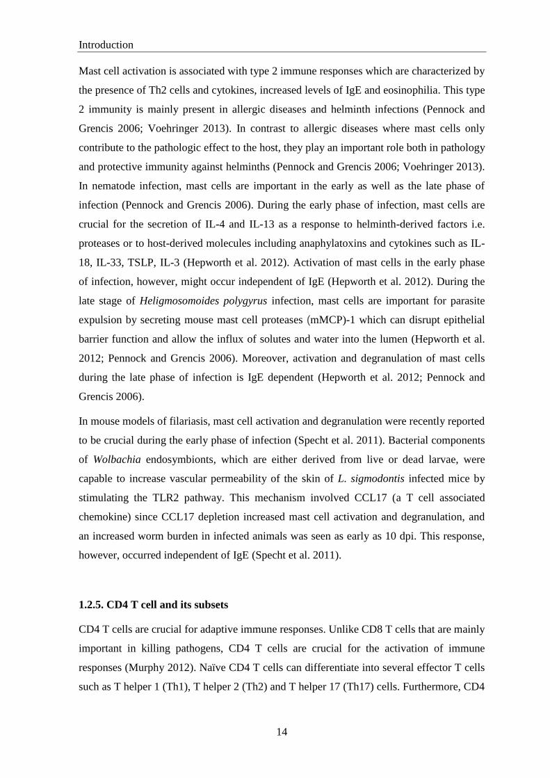
Figure 1.1. Areas endemic for lymphatic filariasis. LF is spread in the tropical regions of Asia,
Africa and South America as shown in blue. Adapted from WHO 2006: http://www.bvgh.org
/Portals/0/disease_maps/LF_map.gif)
The disease is transmitted by mosquitoes of the genera Culex, Aedes, Anopheles or
Mansonia (Pfarr et al. 2009). In Africa, LF is mostly transmitted by Anopheles, which also
transmits malaria (Hopkins 2013). In mosquitoes, microfilariae (larvae stage 1, L1 or Mf)
shed their sheaths, penetrate into the mosquito‟s midgut and infiltrate the thoracic muscles
of mosquitoes. After 2 molting steps in the mosquitoes, the infectious stage 3 larvae (L3)
are transferred to humans by mosquito bites. In humans, L3 penetrate the skin and reside in
the lymphatics. In the lymphatics the L3 molt into the larvae stage 4 (L4) and mature to
adult worms. Adult worms can live for many years in the lymphatics and produce their
offspring, the Mf, that have a lifespan of 3-36 months (Nutman and Weller 2005). The Mf
migrate to the circulation and can be taken up by blood feeding mosquitoes and complete
the parasite‟s life cycle (Specht and Hoerauf 2012; Taylor et al. 2010). The complete life
cycle of W. bancrofti is shown in figure 1.2.
Although no direct mortality caused by LF has been observed, the disease has a huge
economic, psychological and social impact (WHO 2013). Alone in India the economic loss
caused by LF annually reaches an estimated US$ 1.5 billion due to temporary and
permanent disabilities (Ramaiah et al. 2000). Those costs will further increase since the
disease is chronic and lasts until the death of the patients (WHO 2013).

Introduction
3
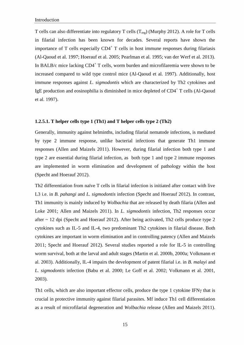
Figure 1.2. Life cycle of W. bancrofti. The left part of the panel shows the life cycle of W.
bancrofti in the mosquito, whereas in the right part the development in the human is displayed.
Adapted from: http://www.cdc.gov/parasites/ lymphaticfilariasis/biology_w_bancrofti.html.
1.1.2. Pathophysiology and clinical findings
Pathogenesis of the disease is divided into the asymptomatic (or subclinical), acute and
chronic stages (Nutman and Weller 2005; Taylor et al. 2010). Most patients are
asymptomatic and even though they are positive for Mf, no external signs or symptoms are
visible (Taylor et al. 2010; WHO 2013). Additionally, damage to the lymphatic system and
the kidneys as well as alterations of the immune system can be observed in patients (WHO
2013).
Acute episodes of local inflammation involve skin, lymph nodes and lymphatic vessels in
the extremities, as well as breasts and genitals (WHO 2013). Most of these conditions are
caused by the immune response against the parasite and result in bacterial infections due to
the impairment of host immunity and lymphatic damage (WHO 2013). Acute
adenolymphangitis is a common feature found in acute infection which is characterized by
high fever, lymphatic inflammation (lymphangitis and lymphadenitis), and transient local
edema (Nutman and Weller 2005; Taylor et al. 2010). Lymph vessel dilatation occurs
when adult worms are still alive (Nutman and Weller 2005). Acute symptoms can also
include dermatolymphangioadenitis. High fever, chills, myalgias and headache can
accompany the acute infections (Nutman and Weller 2005; Pfarr et al. 2009). Both features

Introduction
4
A B
of acute conditions are mostly caused by immune responses against death worms or
bacteria as secondary infection (Nutman and Weller 2005; Taylor et al. 2010).
Acute inflammatory reactions can develop into chronic lymphedema (tissue swelling) or
elephantiasis (skin/tissue thickening) (Taylor et al. 2010). In these conditions, lymphedema
or elephantiasis of limbs and hydrocele (fluid accumulation) are common features
observed in patients (Taylor et al. 2010). Similar to acute immune responses, chronic
inflammation is predominantly due to dying worms caused by antifilarial drugs or
spontaneous worm death (Pfarr et al. 2009; Taylor et al. 2010).
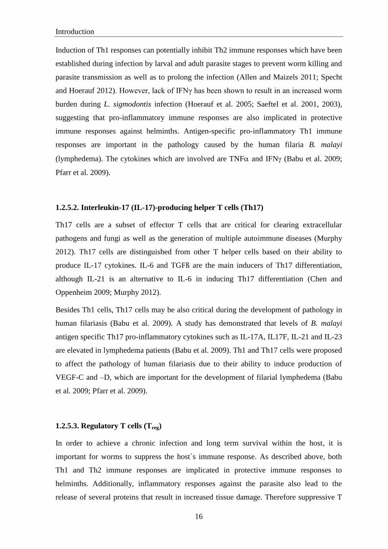
Figure 1.3. Manifestations of chronic lymphatic filariasis. Chronic manifestations of lymphatic
filariasis: Chronic filarial lymphedema (elephantiasis) (A) and filarial hydrocele (B). Adapted from
http://puskesmaspebayuran. blogspot.com/2010/08/filariasis.html (A) and http://www.filariasis.org/
press_centre/hydrocele.html (B).
1.1.3. Diagnosis and treatment
Definitive diagnosis is made when parasites are detected, either in lymphatic fluid or in the
blood (Nutman and Weller 2005; Taylor et al. 2010). Since L3 and L4 as well as adult
worms reside in the lymphatic vessels or sinuses of lymph nodes, it is difficult to detect the
larvae or adult worms (Nutman and Weller 2005). Alternatively, diagnosis is made by
detecting Mf which circulate in the peripheral blood (Nutman and Weller 2005; Taylor et
al. 2010). Periodicity of Mf in the peripheral blood during day (diurnal) or night
(nocturnal) depends on the filarial species and the involved vector (e.g. nocturnal
periodicity in W. bancrofti) (Nutman and Weller 2005; Taylor et al. 2010). Mf can also be
detected in hydrocele fluid, or rarely, in other body fluids (Nutman and Weller 2005;
Taylor et al. 2010). Detection of circulating worm antigen by either enzyme-linked
Introduction
5
immunosorbent assay (ELISA) or a rapid-format immunochromatographic card test is
another option to diagnose bancroftian and brugian filariasis (Rahmah et al. 1998; Taylor
et al. 2010). Assays for detecting DNA of W. bancrofti and B. timori by polymerase chain
reaction (PCR) have been developed (Albers 2011; Rao et al. 2006). Recently, a new
method to visualize motile worms in dilated lymphatics was established by detecting worm
movement by ultrasonography (Dreyer et al. 1998; Mand et al. 2005).
Diethylcarbamazine (DEC) which acts as a potent microfilaricide and weak macrofilaricide
has been used as the drug of choice for LF since its discovery in 1948 (Nutman and Weller
2005; Taylor et al. 2010). In Africa where onchocerciasis is coendemic, ivermectin is used
as alternative treatment as DEC causes significant adverse reactions in onchocerciasis
patients (Taylor et al. 2010). Another antiparasitic drug is albendazole which has a modest
macrofilaricidal property and is used in combination with DEC or ivermectin for Mass
Drug Administration (MDA) program organized by the WHO since 2000 (Nutman and
Weller 2005; Taylor et al. 2010).
Ongoing research is aimed to discover new and improved drugs which are more effective
and cause minimal adverse reactions (Hoerauf 2008). Drugs used for ongoing MDA by the
WHO such as DEC, ivermectin and albendazole are reported to have no or little effect in
killing adult worms. Although the MDA program is implemented for many years, the
drugs could not completely stop the transmission and alleviate pathology of the disease
such as lymphedema and hydrocele (Hoerauf 2008). Therefore drugs that have adulticidal
activity are required in order to reduce the pathology (Hoerauf 2008). An established
alternative drug that is still being optimized for human treatment is doxycycline which
targets the endosymbiotic bacterium found in the worms which is named Wolbachia
(Hoerauf 2008) and will be discussed in the following section.
1.1.4. The worm's endosymbiont: Wolbachia
In recent years, it has been shown that endosymbiotic Wolbachia bacteria are important in
the pathogenesis of filariasis (Pfarr and Hoerauf 2006; Taylor 2003). They are found in the
oocytes, embryos and larval stages and in the hypodermis of male and female filarial
nematodes. Wolbachia are essential for filarial growth, development, embryogenesis and
survival (Tamarozzi et al. 2011; Taylor et al. 2010). Accordingly, Wolbachia are a suitable
target for the development of new anti-filarial drugs (Hoerauf 2008). Treatment of

Introduction
6
filariasis with antibiotics depleting Wolbachia leads to long-term sterility and eventual
death of adult worms and causes significant improvements in lymphatic pathology, i.e. the
severity of lymphedema and hydrocele (Debrah et al. 2006; Pfarr et al. 2009).
Besides being required by the worm for survival, Wolbachia bacteria have also been
described to activate the host‟s immune system (Pfarr and Hoerauf 2006; Turner et al.
2009). This results in the production of IL-6, TNFα and IL-1β (Taylor et al. 2001). This is
important in the context of classical antihelminthic treatment (e.g. DEC) where worms are
killed and release Wolbachia (Cross et al. 2001; Taylor et al. 2001; Turner et al. 1994).
Cross and coworkers showed that administration of DEC can cause death of adult worms at
a modest degree, which leads to release of Wolbachia (Cross et al. 2001). Wolbachia was
previously thought to contain lipopolysaccharide (LPS) in their membranes (Cross et al.
2001; Taylor et al. 2001). This LPS then triggers inflammation via Toll-Like Receptor
(TLR) 4 and leads to secretion of various cytokines, including IL-6 by macrophages and
endothelial cells (Saint André et al. 2002; Taylor et al. 2001). However, studies then
emphasized the role of Wolbachia lipoproteins (WoLP) and Wolbachia surface protein
(WSP) in stimulating innate and adaptive immunity (including increased production of IL-
6) and this mechanism occurs through TLR2/6. A role of TLR4 in Wolbachia-mediated
immune responses in filariasis is still debatable (Brattig et al. 2004; Daehnel et al. 2007;
Hise et al. 2007; Turner et al. 2009).
1.1.5. Murine model of lymphatic filariasis
Since human-pathogenic filariae such as W. bancrofti cannot patently infect laboratory
mice, a filarial strain that can fully develop in mice is required. Similar genotype and
phenotype with human filariae is beneficial (Hoffmann et al. 2000; Hörauf and Fleischer
1997). Although some Brugia spp can infect laboratory mice, the survival time of each
stage of these worms is limited in mice (Hörauf and Fleischer 1997; Lawrence 1996).
Therefore, Brugia spp are still not suitable for a murine LF model, notably when
comparing stage-specific immune responses during the complete course of filarial infection
(Hoffmann et al. 2000; Hörauf and Fleischer 1997; Lawrence 1996).
The rodent filaria Litomosoides sigmodontis which can infect laboratory mice has been
established as a model of LF for decades (Hoffmann et al. 2000; Petit et al. 1992). It
belongs to the same family as human filariae. Moreover, L. sigmodontis shares similar

Introduction
7
features with human filariae such as larval migration, genomic and biochemical structure
(Hoffmann et al. 2000). It shows extensive immunological cross-reactivity with Brugia spp
and W. bancrofti and fully develops in BALB/c mice (Hoffmann et al. 2000; Hörauf and
Fleischer 1997; Lawrence 1996). Many studies have shown the benefits of this model in
filarial research by analyzing immune responses and parasitological parameters at different
stages of the worm‟s life cycle (Hoffmann et al. 2000; Maréchal et al. 1997; Pfaff et al.
2000). In this model, mice are infected with L3 through the bite of the tropical rat mite
Ornithonyssus bacoti. Larvae migrate to the pleural cavity where they molt twice,
becoming adult worms at about 4 weeks post infection. Around 8 weeks post infection;
female worms start to release Mf. The complete life cycle of L. sigmodontis is shown in
figure 1.4.
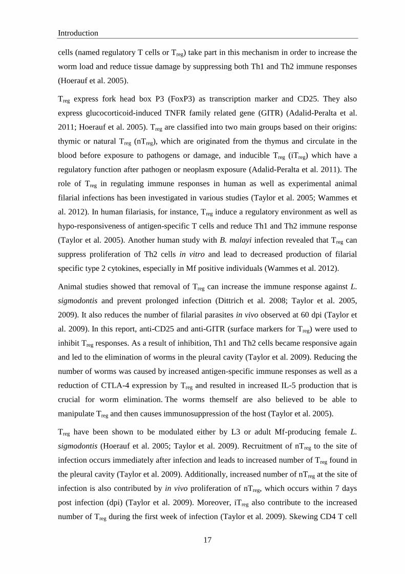
Figure 1.4. Life cycle of L. sigmodontis. The top panel shows life cycle of L. sigmodontis in
permanent hosts, whereas bottom panel shows life cycle in vector. Adapted from Hübner et al.
2009.
Microfilariae (Mf/L1) in peripheral blood 50 dpi
Adult Worms
25-30 dpi in pleural cavity
Ornithonyssus bacoti Mf molt to L2 and L3
stages
L3 infective-stage larva
in mite 12-14 days after ingestion of Mf
L3 transmitted to mouse by bite or sc injection, migration to pleural cavity days 2-6 dpi
Molt to L4 starting 8 dpi
BALB/c mouse

Introduction
8
1.2. Immune response during filariasis
The role of host immunity in reducing inflammation, number of worms, circulating Mf and
in preventing disability still leaves a big question mark and requires further research
(Nutman and Weller 2005; Taylor et al. 2010). Both types of human immunity, innate and
adaptive immunity, are believed to take an important part in controlling or even worsening
the disease (Nutman and Weller 2005; Specht and Hoerauf 2012). Local and systemic
inflammation which involves various cell types and molecules has long been recognized to
occur in patients with filariasis, especially in early phases of infection as well as in the
chronic stage (Nutman and Weller 2005; Specht and Hoerauf 2012; Taylor et al. 2010).
Inflammation which occurred in the acute phase of filarial disease is usually
indistinguishable from other inflammation-related diseases causing misdiagnosis by many
health professionals and leading to inappropriate treatment (Nutman and Weller 2005;
WHO 2013).
Several cell types are involved in the immune response against filarial infection and are
important for worm clearance. Additionally, these cells play a crucial role in disease-
related pathology (Specht and Hoerauf 2012; Taylor et al. 2010). In the following section
some of these cell types which are well known to be important during filarial infection, are
discussed.
1.2.1. Neutrophils
Neutrophils are effector cells of the innate immune system with a short life span and are
important in immunity against extracellular pathogens and during the early phase of filarial
infection (Makepeace et al. 2012; Murphy 2012). They are first recruited to the site of
infection during an acute phase of inflammation (Murphy 2012). Their ability to act as
effectors against pathogens uses several mechanisms: phagocytic activity, release of lytic
enzymes from their granules, and production of anti-microbial substances such as reactive
oxygen intermediates (ROI) (Mantovani et al. 2011; Murphy 2012). Neutrophil activation
which is mediated by bacteria (Mantovani et al. 2011) and release of pro-inflammatory
cytokines, together with activation of other resident cells at the site of infection such as
macrophages and mast cells lead to the induction of acute inflammation and recruitment of
additional neutrophils and other cells i.e. monocytes and lymphocytes (Murphy 2012).
During type 2 immune responses, which occur during helminth infections including LF

Introduction
9
also leads to the migration of eosinophils and basophils to the site of infection (Makepeace
et al. 2012; Murphy 2012).
Several studies have shown a role for neutrophils in filarial infection (Al-Qaoud et al.
2000; Saeftel et al. 2001, 2003). Neutrophils are important for protection (mainly by worm
killing) as well as pathology of the infection (Al-Qaoud et al. 2000; Saeftel et al. 2001,
2003). Neutrophils are involved in parasite killing at least in two ways: directly by
phagocytic activity or indirectly by encapsulation within granulomata (Makepeace et al.
2012; Saeftel et al. 2001). Several studies showed the importance of neutrophils in
controlling adult worms by both ways which occurs during the chronic phase of L.
sigmodontis infection (Al-Qaoud et al. 2000; Saeftel et al. 2001, 2003), and impacts larval
stages, too (Porthouse et al. 2006).
Porthouse and colleagues showed in Mogolian jirds that neutrophils are found 3 hours post
B. pahangi L3 inoculation at the site of injection within the skin. This study revealed an
80% reduction of larval recovery and large numbers of neutrophil-rich inflammatory foci
at this time point. They argued that this increased activation and accumulation of
neutrophils was due to increased Wolbachia released after or prior to larval death
(Porthouse et al. 2006).
The neutrophil‟s capability in adult worm killing in vivo is IFN dependent. IFN leads to
an increased production of TNFα which mediates neutrophil activation (Saeftel et al. 2001,
2003). This activation may promote chemotaxis and phagocytic activities that lead to an
improved worm killing by neutrophils. Accordingly, in IFN deficient mice, for instance,
L. sigmodontis worm burden was increased compared to wild type mice at 80 dpi (Saeftel
et al. 2001, 2003).
Furthermore, neutrophils are required for granuloma formation that also leads to parasite
killing (Al-Qaoud et al. 2000). In a study using L. sigmodontis infection in BALB/c mice it
was shown that reducing neutrophils in the granulomata by injecting anti-granulocyte
colony stimulating factor (G-CSF) led to impaired worm killing in those animals although
eosinophils were still present in the granulomata (Al-Qaoud et al. 2000).
Introduction
10
1.2.2. Eosinophils
Eosinophils are granulocytes that develop in the bone marrow. They play a role as effector
cells, as „not professional‟ antigen-presenting cells (APC), in promoting humoral immune
responses as well as in inducing pathology in the host (Rosenberg et al. 2013). Eosinophils
are mainly involved in the effector mechanisms during helminth infections and allergic
diseases (Rosenberg et al. 2013). The role of eosinophils in host immune responses against
parasites is still under debate and probably dependent on the helminth species (Meeusen
and Balic 2000). Studies showed that eosinophils do not have a role in immunity to
Schistosoma mansoni or the induction of pathology in the host (Sher et al. 1990; Swartz et
al. 2006). In contrast, depleting eosinophils in Strongyloides stercoralis and
Angiostrongylus cantonesis infection leads to an increased parasite survival (Rosenberg et
al. 2013). In vitro studies showed that eosinophils are able to kill early stages of S. mansoni
as well as other nematodes like Haemonchus contortus, both in human and mouse
(Meeusen and Balic 2000).
In addition, the role of eosinophils in filarial infection remains unclear as contradictory
results were found in several studies. Some studies showed that IL-5 and eosinophils are
rather important in worm elimination and in the protective immunity against filarial
infection in vaccinated and primary infection (Le Goff et al. 2000; Martin et al. 2000b,
2000a). In these studies, eosinophil migration into subcutaneous tissue was detected to be
increased in vaccinated mice prior to L. sigmodontis injection (Le Goff et al. 2000; Martin
et al. 2000a). This led to a decreased number of worms in vaccinated mice (Le Goff et al.
2000; Martin et al. 2000a). Furthermore, these studies also elucidated the role of
eosinophils during primary L. sigmodontis infection (Le Goff et al. 2000; Martin et al.
2000b, 2000a). They showed that eosinophilia in primary infection occurs after 3 weeks
post infection therefore eosinophils are less important during the first week of primary
infection as in vaccinated mice. Thus, eosinophils are implemented in the worm
destruction at the chronic phase of primary infection, but not at the early time points (Le
Goff et al. 2000; Martin et al. 2000b, 2000a). In contrast, to the proposed protective role of
eosinophils during filarial infection, a study by Babayan and colleagues showed that IL-5
driven eosinophilia accelerates early filarial growth. This study revealed that mice which
lack eosinophils showed an accelerated worm development and molting (Babayan et al.
2010).

Introduction
11
The protective role of eosinophils against filarial infection is potentially by mediating
direct or indirect worm killing induced by releasing several attractant proteins, although
the exact mechanism in how eosinophils can kill the filariae is unknown (Makepeace et al.
2012; Meeusen and Balic 2000). A mechanism that has been proposed recently is the
release of eosinophil granule proteins that may act as proteases such as eosinophil
peroxidase (EPO) and major basic protein (MBP) (Makepeace et al. 2012; Specht et al.
2006). In a study using L. sigmodontis infections, worm burden in EPO- and MBP-
deficient mice showed a significant increase compared to wild type mice (Specht et al.
2006).
For activation and infiltration into the site of infection, eosinophils need appropriate
stimuli, including IL-5 and eotaxin chemokines (Rosenberg et al. 2013). Moreover, IL-5 is
also important in development and survival of eosinophils (Rosenberg et al. 2013). It is a
major cytokine that is produced by Th2 cells as effector response during filarial infection
(Babayan et al. 2010).
1.2.3. Macrophages
There are two types of macrophages that are important during infection: the classically
activated macrophage (CAM or M1 macrophage), is induced by IFN and TNF as well as
by stimulants of TNF like LPS. The second type of macrophages is the alternatively
activated macrophage (AAM or M2 macrophage) which is induced by IL-4 and IL-13 that
are produced by Th2 cells, mast cells, basophils, eosinophils, natural killer T (NKT) cells,
lymphocytes and macrophages (Allen and Loke 2001; Mosser 2003; Murphy 2012). CAMs
are important in pro-inflammatory immune responses, whereas AAMs have anti-
inflammatory properties (Allen and Loke 2001; Mosser 2003). CAMs are crucial during
acute infections that require a protective type 1 immune, e.g. bacterial infections, due to
their improved phagocytic activity and their ability to promote Th1 immune responses by
secretion of pro-inflammatory cytokines such as TNF, IL-6 and nitric oxide (NO) (Allen
and Loke 2001; Mosser 2003). In contrast, AAMs are crucial for immune suppression and
tissue repair. Moreover, they are also suggested to be important for the host‟s immunity
against the chronic phase of helminth, fungal and viral infections, as well as in allergic and
autoimmune diseases (Gordon and Martinez 2010; Mosser 2003). AAMs produce IL-10
which is crucial in immune suppression. They are also important in mediating wound

Introduction
12
healing, angiogenesis, and extracellular matrix deposition (Gordon and Martinez 2010;
Mosser 2003; Rodríguez-sosa et al. 2002).
The impact of macrophages during filariasis is generally divided into their role as effector
and suppressor cells. CAMs mainly act as effector cells and AAMs have a role as
suppressor cells. As effector cells, macrophages have three different roles: in parasite
killing, induction of pathology, as well as natural resistance (Allen and Loke 2001). Since
worms are multicellular organisms, engulfing the worm is a difficult way for the
macrophage to kill the parasite. Alternatively, macrophages release toxic radicals such as
reactive oxygen species (ROS) and NO derivates. The role of IFN-mediated-macrophage
killing of worms by releasing NO has been reviewed by Allen and Loke previously (Allen
and Loke 2001). In vitro studies showed that macrophages which were activated by IFN
kill B. malayi Mf by releasing NO (Taylor et al. 1996; Thomas et al. 1997). Similarly,
peritoneal macrophages kill Mf of B. pahangi in the presence of antimicrofilarial sera in
vitro (Karavodin and Ash 1982; Oxenham et al. 1984). However, another study showed
that IFN and NO are not required for immune mediated clearance of B. malayi Mf (Gray
and Lawrence 2002). Furthermore, macrophage killing ability seems to only target larval
stages and not adult stages of filariae potentially due to the antioxidant enzyme catalase
that is produced by adult worms (Ou et al. 1995). During B. pahangi infection in vivo,
macrophage activation was shown to be induced by L3 which then led to significantly
increased phagocytic and microbicidal capacity of the macrophages (Jeffers et al. 1984).
Besides their role in larval killing, macrophage products are able to cause pathology for the
host as is shown by inflammatory lesions that can be found in lymphatic vessels both in
animal models and human filariasis (Rao et al. 1996). In addition, dying worms are usually
surrounded by pathological granulomata which mostly consist of CAMs as a rich source of
inflammatory cytokines, toxic radicals and inflammatory lipid mediators which cause
tissue damage in the host (Allen and Loke 2001; Mosser 2003).
AAMs have also been demonstrated to be important in filarial disease [reviewed in (Allen
and Loke 2001)]. After i.p. implantation in mice, AAMs are induced by
excretory/secretory (E/S) products from live adult B. malayi worms (Allen and MacDonald
1998), whereas CAMs are induced by Wolbachia that are released by dead worms (Taylor
et al. 2000, 2001). AAMs were shown to suppress proliferation of blood lymphocytes in
vitro (Schebesch et al. 1997). Furthermore, AAMs were also shown to induce Th2
differentiation from naïve T cells in this model (Goerdt and Orfanos 1999). Later, AAMs
Introduction
13
inhibit immunity to the disease and prevent helminth-induced pathology in the host (Allen
and Loke 2001; Kreider et al. 2007; Taylor et al. 2009; van der Werf et al. 2013).
Since CAMs can mediate worm killing and induce Th1 responses that lead to pathology of
the host, and AAMs may inhibit these responses, the balance between CAMs and AAMs
may determine the severity of the disease (Allen and Loke 2001). This phenomenon can be
seen more clearly in elephantiasis patients: while increased numbers of CAMs during LF
may result in increased parasite clearance, it may also lead to higher pathology (Allen and
Loke 2001). In contrast, asymptomatic Mf positive individuals have more AAMs that
impair parasite clearance but lead to less pathology. However, in endemic normal a
successful balance between CAMs and AAMs has been achieved that leads to the
resolution of the disease (Allen and Loke 2001).
1.2.4. Mast cells
Mast cells are multifunctional granulated hematopoietic cells that reside in nearly all
tissues and are typically found throughout barrier tissues such as skin and mucosa as well
as in a perivascular location in the tissue. Due to these locations and their ability to rapidly
release prestored inflammatory mediators, mast cells contribute to the first line of response
against pathogens (Marshall 2004; Murphy 2012). Additionally, they express several cell
surface receptors such as the high-affinity receptor (FcεRI) for IgE, FcγRIII for IgG,
complement component receptors and various Toll-like receptors (TLRs) (Marshall 2004).
This demonstrates the capability of mast cells to respond to a wide range of endogenous
and exogenous stimuli such as allergens, tissue injury, viral, fungal, parasite and bacterial
antigens (Marshall 2004; Murphy 2012). Cross-linking of cell surface IgE bound to FcεRI
by antigen leads to activation and degranulation of mast cells that results in releasing
granule proteins and secretion of lipid inflammatory mediators and cytokines and
chemokines such as histamine, prostaglandins, leukotrienes, bradykinine, vascular
endothelial growth factors (VEGFs) and pro-inflammatory cytokines like TNFα and IL-6,
although cytokine secretion can also occur without degranulation (Marshall 2004; Murphy
2012). Histamine and leukotrienes are important for vascular permeability while
leukotrienes have an additional function in eosinophil recruitment to the site of
inflammation. Prostaglandins and VEGFs are further important for angiogenesis (Kunder
et al. 2011; Marshall 2004).
Introduction
14
Mast cell activation is associated with type 2 immune responses which are characterized by
the presence of Th2 cells and cytokines, increased levels of IgE and eosinophilia. This type
2 immunity is mainly present in allergic diseases and helminth infections (Pennock and
Grencis 2006; Voehringer 2013). In contrast to allergic diseases where mast cells only
contribute to the pathologic effect to the host, they play an important role both in pathology
and protective immunity against helminths (Pennock and Grencis 2006; Voehringer 2013).
In nematode infection, mast cells are important in the early as well as the late phase of
infection (Pennock and Grencis 2006). During the early phase of infection, mast cells are
crucial for the secretion of IL-4 and IL-13 as a response to helminth-derived factors i.e.
proteases or to host-derived molecules including anaphylatoxins and cytokines such as IL-
18, IL-33, TSLP, IL-3 (Hepworth et al. 2012). Activation of mast cells in the early phase
of infection, however, might occur independent of IgE (Hepworth et al. 2012). During the
late stage of Heligmosomoides polygyrus infection, mast cells are important for parasite
expulsion by secreting mouse mast cell proteases (mMCP)-1 which can disrupt epithelial
barrier function and allow the influx of solutes and water into the lumen (Hepworth et al.
2012; Pennock and Grencis 2006). Moreover, activation and degranulation of mast cells
during the late phase of infection is IgE dependent (Hepworth et al. 2012; Pennock and
Grencis 2006).
In mouse models of filariasis, mast cell activation and degranulation were recently reported
to be crucial during the early phase of infection (Specht et al. 2011). Bacterial components
of Wolbachia endosymbionts, which are either derived from live or dead larvae, were
capable to increase vascular permeability of the skin of L. sigmodontis infected mice by
stimulating the TLR2 pathway. This mechanism involved CCL17 (a T cell associated
chemokine) since CCL17 depletion increased mast cell activation and degranulation, and
an increased worm burden in infected animals was seen as early as 10 dpi. This response,
however, occurred independent of IgE (Specht et al. 2011).
1.2.5. CD4 T cell and its subsets
CD4 T cells are crucial for adaptive immune responses. Unlike CD8 T cells that are mainly
important in killing pathogens, CD4 T cells are crucial for the activation of immune
responses (Murphy 2012). Naïve CD4 T cells can differentiate into several effector T cells
such as T helper 1 (Th1), T helper 2 (Th2) and T helper 17 (Th17) cells. Furthermore, CD4
Introduction
15
T cells can also differentiate into regulatory T cells (Treg) (Murphy 2012). A role for T cells
in filarial infection has been known for decades. Several reports have shown the
importance of T cells especially CD4+ T cells in host immune responses during filariasis
(Al-Qaoud et al. 1997; Hoerauf et al. 2005; Pearlman et al. 1995; van der Werf et al. 2013).
In BALB/c mice lacking CD4+ T cells, worm burden and microfilaremia were shown to be
increased compared to wild type control mice (Al-Qaoud et al. 1997). Additionally, host
immune responses against L. sigmodontis which are characterized by Th2 cytokines and
IgE production and eosinophilia is diminished in mice depleted of CD4+ T cells (Al-Qaoud
et al. 1997).
1.2.5.1. T helper cells type 1 (Th1) and T helper cells type 2 (Th2)
Generally, immunity against helminths, including filarial nematode infections, is mediated
by type 2 immune response, unlike bacterial infections that generate Th1 immune
responses (Allen and Maizels 2011). However, during filarial infection both type 1 and
type 2 are essential during filarial infection, as both type 1 and type 2 immune responses
are implemented in worm elimination and development of pathology within the host
(Specht and Hoerauf 2012).
Th2 differentiation from naïve T cells in filarial infection is initiated after contact with live
L3 i.e. in B. pahangi and L. sigmodontis infection (Specht and Hoerauf 2012). In contrast,
Th1 immunity is mainly induced by Wolbachia that are released by death filaria (Allen and
Loke 2001; Allen and Maizels 2011). In L. sigmodontis infection, Th2 responses occur
after ~ 12 dpi (Specht and Hoerauf 2012). After being activated, Th2 cells produce type 2
cytokines such as IL-5 and IL-4, two predominant Th2 cytokines in filarial disease. Both
cytokines are important in worm elimination and in controlling patency (Allen and Maizels
2011; Specht and Hoerauf 2012). Several studies reported a role for IL-5 in controlling
worm survival, both at the larval and adult stages (Martin et al. 2000b, 2000a; Volkmann et
al. 2003). Additionally, IL-4 impairs the development of patent filarial i.e. in B. malayi and
L. sigmodontis infection (Babu et al. 2000; Le Goff et al. 2002; Volkmann et al. 2001,
2003).
Th1 cells, which are also important effector cells, produce the type 1 cytokine IFN that is
crucial in protective immunity against filarial parasites. Mf induce Th1 cell differentiation
as a result of microfilarial degeneration and Wolbachia release (Allen and Maizels 2011).
Introduction
16
Induction of Th1 responses can potentially inhibit Th2 immune responses which have been
established during infection by larval and adult parasite stages to prevent worm killing and
parasite transmission as well as to prolong the infection (Allen and Maizels 2011; Specht
and Hoerauf 2012). However, lack of IFNγ has been shown to result in an increased worm
burden during L. sigmodontis infection (Hoerauf et al. 2005; Saeftel et al. 2001, 2003),
suggesting that pro-inflammatory immune responses are also implicated in protective
immune responses against helminths. Antigen-specific pro-inflammatory Th1 immune
responses are important in the pathology caused by the human filaria B. malayi
(lymphedema). The cytokines which are involved are TNF and IFN (Babu et al. 2009;
Pfarr et al. 2009).
1.2.5.2. Interleukin-17 (IL-17)-producing helper T cells (Th17)
Th17 cells are a subset of effector T cells that are critical for clearing extracellular
pathogens and fungi as well as the generation of multiple autoimmune diseases (Murphy
2012). Th17 cells are distinguished from other T helper cells based on their ability to
produce IL-17 cytokines. IL-6 and TGFß are the main inducers of Th17 differentiation,
although IL-21 is an alternative to IL-6 in inducing Th17 differentiation (Chen and
Oppenheim 2009; Murphy 2012).
Besides Th1 cells, Th17 cells may be also critical during the development of pathology in
human filariasis (Babu et al. 2009). A study has demonstrated that levels of B. malayi
antigen specific Th17 pro-inflammatory cytokines such as IL-17A, IL17F, IL-21 and IL-23
are elevated in lymphedema patients (Babu et al. 2009). Th1 and Th17 cells were proposed
to affect the pathology of human filariasis due to their ability to induce production of
VEGF-C and –D, which are important for the development of filarial lymphedema (Babu
et al. 2009; Pfarr et al. 2009).
1.2.5.3. Regulatory T cells (Treg)
In order to achieve a chronic infection and long term survival within the host, it is
important for worms to suppress the host`s immune response. As described above, both
Th1 and Th2 immune responses are implicated in protective immune responses to
helminths. Additionally, inflammatory responses against the parasite also lead to the
release of several proteins that result in increased tissue damage. Therefore suppressive T
Introduction
17
cells (named regulatory T cells or Treg) take part in this mechanism in order to increase the
worm load and reduce tissue damage by suppressing both Th1 and Th2 immune responses
(Hoerauf et al. 2005).
Treg express fork head box P3 (FoxP3) as transcription marker and CD25. They also
express glucocorticoid-induced TNFR family related gene (GITR) (Adalid-Peralta et al.
2011; Hoerauf et al. 2005). Treg are classified into two main groups based on their origins:
thymic or natural Treg (nTreg), which are originated from the thymus and circulate in the
blood before exposure to pathogens or damage, and inducible Treg (iTreg) which have a
regulatory function after pathogen or neoplasm exposure (Adalid-Peralta et al. 2011). The
role of Treg in regulating immune responses in human as well as experimental animal
filarial infections has been investigated in various studies (Taylor et al. 2005; Wammes et
al. 2012). In human filariasis, for instance, Treg induce a regulatory environment as well as
hypo-responsiveness of antigen-specific T cells and reduce Th1 and Th2 immune response
(Taylor et al. 2005). Another human study with B. malayi infection revealed that Treg can
suppress proliferation of Th2 cells in vitro and lead to decreased production of filarial
specific type 2 cytokines, especially in Mf positive individuals (Wammes et al. 2012).
Animal studies showed that removal of Treg can increase the immune response against L.
sigmodontis and prevent prolonged infection (Dittrich et al. 2008; Taylor et al. 2005,
2009). It also reduces the number of filarial parasites in vivo observed at 60 dpi (Taylor et
al. 2009). In this report, anti-CD25 and anti-GITR (surface markers for Treg) were used to
inhibit Treg responses. As a result of inhibition, Th1 and Th2 cells became responsive again
and led to the elimination of worms in the pleural cavity (Taylor et al. 2009). Reducing the
number of worms was caused by increased antigen-specific immune responses as well as a
reduction of CTLA-4 expression by Treg and resulted in increased IL-5 production that is
crucial for worm elimination. The worms themself are also believed to be able to
manipulate Treg and then causes immunosuppression of the host (Taylor et al. 2005).
Treg have been shown to be modulated either by L3 or adult Mf-producing female L.
sigmodontis (Hoerauf et al. 2005; Taylor et al. 2009). Recruitment of nTreg to the site of
infection occurs immediately after infection and leads to increased number of Treg found in
the pleural cavity (Taylor et al. 2009). Additionally, increased number of nTreg at the site of
infection is also contributed by in vivo proliferation of nTreg, which occurs within 7 days
post infection (dpi) (Taylor et al. 2009). Moreover, iTreg also contribute to the increased
number of Treg during the first week of infection (Taylor et al. 2009). Skewing CD4 T cell

Introduction
18
responses towards iTreg by L3 in the pleural cavity, which also occurs within 7 days, even
increased Treg numbers at site of infection (Hoerauf et al. 2005; Taylor et al. 2005, 2009).
1.2.6. B cells
B cells are part of the humoral immune response (Murphy 2012). The B cell population is
divided into at least 5 subsets: B1, B2, regulatory, plasma and memory B cells (Montecino-
Rodriguez et al. 2006; Murphy 2012). B cells are involved in humoral immune responses
as they are able to produce antibodies, B1 cells produce IgM and B2 cells produce other Ig
isotypes (Murphy 2012). They are also thought to be important in innate responses against
viral and bacterial infection as they act as APC (Murphy 2012). B2 cells are conventional
B cells that act as effector cells in adaptive immunity (Montecino-Rodriguez et al. 2006;
Murphy 2012). Activated B2 cells can produce IgE in some settings after CD23 (low
affinity IgE receptor) which is expressed in B2 cells, is cross linked with IgE. In S.
mansoni infections, B2 cells are associated with the development of resistance to
reinfection (Mwinzi et al. 2009).
In filariasis, B cells were shown to be important for protection against B. malayi since lack
of B cells using different types of knockout mice such as µMT (B cell deficiency) and
RAG-1 (deficient in generation of mature B and T cells) mice resulted in increased worm
burdens (Babu et al. 1999). B cells protect against worms potentially by acting as early
APC to initiate T cell responses (Paciorkowski et al. 2000). Additionally, B cells produce
immunoglobulins that contribute to worm elimination (Babu et al. 1999; Paciorkowski et
al. 2000). B1 cells have been reported to be responsible for host protection against B.
malayi (Paciorkowski et al. 2000). A role of B cells in B. pahangi infection has been
described previously (Gillan et al. 2005). Depletion of B cells in vitro and usage of μMT
mice led to a reduced antigen-specific proliferation of splenocytes (Gillan et al. 2005).
The role of B cells in eliminating L. sigmodontis has been demonstrated in a study using
BALB/c µMT mice in two different settings: vaccination with irradiated L3 and primary
infection (Martin et al. 2001). The authors found a significantly decreased number of
worms after challenge infection in vaccinated wild type mice at 28 dpi compared to
vaccinated µMT mice (Martin et al. 2001). This was correlated with impaired
degranulation of eosinophils in vaccinated µMT mice compared to wild type controls
during early infections (Martin et al. 2001). The authors argued that the eosinophil-

Introduction
19
antibody-B cell complex was diminished in vaccinated µMT mice and caused the observed
reduced vaccine protection (Martin et al. 2001). In primary infection, the survival and
growth of filariae was not changed in µMT mice, although the authors found that mice did
not develop patent infections with microfilaraemia (Martin et al. 2001). In contrast, Al-
Qaoud and colleagues reported that the lack of B1 cells in BALB/c Xid mice resulted in a
higher filarial recovery rate and Mf load in the periphery compared to wild type mice (Al-
Qaoud et al. 1998).
Introduction
20
1.3. Interleukin-6
Interleukin-6 is a pleiotropic cytokine that has several effects and acts as a pro-
inflammatory as well as an anti-inflammatory cytokine. It has a wide range of biological
activities in inflammation, immune regulation, hematopoiesis and oncogenesis (Kishimoto
2010; Scheller et al. 2011). It was discovered in 1986 when the Kishimoto group at Osaka
University Japan first cloned the complementary DNA encoding B cell stimulatory factor-
2 which became later known as IL-6 (Kishimoto 1989, 2010). In the same year, IFN2 and
another identical protein were also cloned by the Hirano group and found to be identical
with IL-6 (Hirano 1998, 2010; Kishimoto 2010). Other identical proteins that have similar
function with IL-6 are leukemia inhibitory factor (LIF), cilliary neurotropic factor (CNTF),
IL-11 and oncostatin M. The name “interleukin-6” was officially proposed in a
nomenclature meeting in New York in 1988 (Hirano 2010; Kishimoto 2010).
1.3.1. Sources
Several stimuli including ultraviolet (UV), irradiation, ROS, microbial products, viruses, or
other pro-inflammatory cytokines are known to induce IL-6 production by leukocytes and
non-leukocyte cells (Kishimoto 2010; Rincon and Irvin 2012). In acute infections,
monocytes and CAMs are the main sources of IL-6. Immediately after TLR stimulation by
specific pathogen-associated molecular patterns (PAMPs), activated monocytes and
macrophages produce IL-6. The above described AAMs, however, produce negligible
amounts of IL-6 in vitro (Lolmede et al. 2009). Other cells of the innate immune system
such as dendritic cells (DCs) and neutrophils can also produce IL-6 during acute infection.
DCs secrete small amounts of IL-6 in the beginning of infection as they are not yet fully
activated. When encountering pathogens, DCs are fully activated and produce high
amounts of IL-6 (Hirano 1998; Murphy 2012; Tanaka and Kishimoto 2012).
Other innate immune cells, eosinophils and mast cells, are also known to have the ability to
produce IL-6 following their activation. IL-6 is secreted in vitro by mouse eosinophils
following respiratory virus infection (Dyer et al. 2009; Rosenberg et al. 2013) and by
human eosinophils in atopic asthma disease (Hamid et al. 1992; Lacy et al. 1998; Spencer
et al. 2009). Eosinophils also express IL-6 mRNA in different diseases in vivo (Rothenberg
and Hogan 2006). Moreover, IL-6 may be constitutively synthesized and stored in granules
of unstimulated eosinophils as IL-6 positive eosinophils have been found in blood from
Introduction
21
healthy donors although the site of IL-6 storage in eosinophils could not be determined by
immunocytochemical staining (Lacy et al. 1998; Spencer et al. 2009). This preformed IL-6
in eosinophils is mobilized into budding vesicles which travel to the plasma membrane and
is then released into the extracellular space. This mechanism of granule-stored cytokine
secretion is named piecemeal degranulation (Melo et al. 2005; Spencer et al. 2009).
Mast cells are also able to produce IL-6 in vitro in response to gram-negative bacteria and
their cell wall component LPS by secretion without degranulation. In contrast,
peptidoglycan, a cell wall component of gram-positive bacteria induces IL-6 production
from mast cells via degranulation (Leal-Berumen et al. 1994; Supajatura et al. 2002).
Furthermore, IL-6 is produced by mast cells in response to IL-1ß, prostaglandin E2 and
leukotriene C4 stimulation (Marshall 2004). Cells from the adaptive immune system such
as T cells and B cells are also able to produce IL-6 following stimulation with different
stimuli (Murphy 2012).
In helminths, many studies showed IL-6 production from several cell types. During
infection with the helminth Taenia crassiceps, F4/80+ peritoneal macrophages produce low
levels of IL-6 during the acute phase, whereas during the chronic phase high levels of IL-6
are produced (Rodríguez-Sosa et al. 2002). In filarial disease, IL-6 is secreted by activated
macrophages, DCs and endothelial cells due to Wolbachia release by worms dying either
naturally or after anti-filarial DEC treatment that stimulates TLR2 and 6 (Daehnel et al.
2007; Hise et al. 2007; Taylor et al. 2001). Furthermore, IL-6 is produced by eosinophils
and macrophages in vitro after stimulation with worm antigen during L. sigmodontis
infection (Gentil et al. 2013, resubmission) as well as by CD4+ and CD8
+ T cells in
response to L3 of B. malayi (Babu and Nutman 2003). Another study reported that intact
granulomata of B. pahangi were also able to spontaneously produce IL-6 in vitro (Rao and
Klei 2006). Stimulation of the granulomata with worm antigen led to even increased IL-6
production. Although not investigated in this study, it seems that IL-6 was produced by
macrophages and eosinophils as predominant cells that form the granulomata (Rao and
Klei 2006).
However, IL-6 can also be secreted by non-leukocyte cells such as endothelial cells,
fibroblasts, astrocytes, epithelial cells and some malignant cells (Hirano 1998). Damaged
or dying cells from non-infectious inflammation, i.e. burn wounds and other injury, can
also secrete IL-6 following stimulation of TLRs via damage-associated molecular patterns
(DAMPs) (Tanaka and Kishimoto 2012).
Introduction
22
1.3.2. Signaling
The pleiotropic and redundant capability of IL-6 is characterized by two functional
receptors: IL-6 specific receptor (IL-6R) which has a molecular weight of 80 kDa and
glycoprotein 130 kDa (gp130) as a signal transducer for cytokines related to IL-6.
Furthermore, signal transduction of IL-6 via gp130 is mediated by two pathways: Janus
family tyrosin kinase-signal transducers and activators of transcription (JAK-STAT) and
mitogen-activated protein kinase (MAPK) pathway (Kishimoto 2010).
IL-6 signaling in the target cells occurs mainly via STAT3 and to a lesser degree via
STAT1 (Kishimoto 2010). On target cells, IL-6 protein first binds to membrane-bound IL-
6 (mIL-6R) receptor with a nanomolar affinity (Rose-John 2012). Several cells express
mIL-6R on their surface, including neutrophils, monocytes, naive CD4 and CD8 T cells, B
cells and hepatocytes (Scheller et al. 2011). This IL-6/IL-6R complex then binds to gp130
and leads to IL-6-signal transduction via JAK1 and JAK2 (Scheller et al. 2011). When
excessive production of IL-6 occurs, a molecule named suppressor of cytokine signaling
(SOCS) negatively regulates the signal. SOCS binds to JAKs and inhibits their activities
(Kishimoto 2010; Scheller et al. 2011). Role of SOCS in IL-6 signaling, for instance, is
involved in T cell differentiation. Up-regulation of SOCS1 occurs in Th2 differentiation
and suppression of IFN signaling (Scheller et al. 2011). Furthermore, lower SOCS3
signaling is important in Th17 differentiation from naïve T cell thus inducing IL-6
production (Barnes et al. 2011; Kishimoto 2010; Scheller et al. 2011).
Previously, it was believed that membrane IL-6 receptor (mIL-6R) was the only receptor
that could bind to IL-6. Recently, studies have shown that soluble IL-6 receptor (sIL-6R)
which is found in body fluids such as urine and blood can also bind to IL-6 and then
stimulates the target cells without requiring mIL-6R (Rose-John 2012; Scheller et al.
2011). sIL-6R is generated by two mechanisms; first by proteolytic cleavage of mIL-6R
mainly after apoptosis of neutrophils and second by secretion from neutrophils and
monocytes of an alternatively sliced mIL-6R product lacking transmembrane and cytosolic
domains (Barnes et al. 2011; Scheller et al. 2011). Additionally, naïve and memory CD4 T
cells produce sIL-6R when the T cell receptor is activated.
The sIL-6R/IL-6 complexes then bind to gp130 which activates and phosphorylates JAK
and STAT3 that lead to IL-6 trans-signaling (Rose-John 2012). Since gp130 can be
expressed by almost all cells and tissues, sIL-6R expands IL-6 responding cells to a
Introduction
23
broader variety of cells such as eosinophils, endothelial cells and T cells. This trans-
signaling pathway is important in pro-inflammatory responses, different from classic
signaling that involves mIL-6R and is crucial to induce anti-inflammatory responses
(Scheller et al. 2011).
1.3.3. Effector cells
A wide range of biological activities makes IL-6 important in various conditions including
inflammatory responses, autoimmune and malignant diseases. In this part, I only focus on
its role during inflammatory responses that involves several cell types.
1.3.3.1. Neutrophils
During the acute phase of infection, IL-6 is important for neutrophil mobilization from the
bone marrow to the site of infection (Murphy 2012). After being activated by pathogens
via pattern recognition receptors (PRRs) like Toll-like receptors (TLRs), macrophages and
DCs secrete IL-6 together with other pro-inflammatory cytokines such as TNFα and IL-1ß
(Murphy 2012). These cytokines initiate the acute-phase response against pathogens by
inducing neutrophil mobilization from bone marrow into the circulation and release of
acute phase proteins in the liver. They also activate endothelial cells to produce
chemokines e.g. IL-8 and monocyte chemotactic protein (MCP)-1 and IL-6 to attract
neutrophils to the site of infection (Murphy 2012). Additionally, IL-6 trans-signaling leads
to activation of endothelial cells and results in up-regulation of cell adhesion molecules
such as intercellular adhesion molecule (ICAM)-1, vascular cell adhesion molecule
(VCAM)-1 and E-selectin (CD62E) on endothelial cells which then increases neutrophil
adherence thus leading to increased neutrophil migration to the site of infection (Murphy
2012; Scheller et al. 2011).
At the site of infection, cleavage of mIL-6R into sIL-6R after neutrophil apoptosis leads to
an increase of the IL-6 trans-signaling mechanism (Scheller et al. 2011). However, the role
of IL-6 in mediating neutrophil apoptosis in situ is still under debate. Some studies showed
that IL-6 is required to promote neutrophil apoptosis (Mcloughlin et al. 2003), while others
showed anti-apoptotic activity of IL-6 on neutrophils (Colotta et al. 1992). Nevertheless,
IL-6 mediates neutrophil clearance at the site of infection by inhibiting production of
neutrophil attracting chemokines such as CXCL1, CXCL8/IL-8 and CX3CL1 and inducing
Introduction
24
production of monocyte-associated chemokines such as CCL2/MCP-1, CCL8/MCP-2,
CXCL5, and CXCL-6. This results in transition from neutrophils to monocytes leading to
successful resolution of an inflammation (Scheller et al. 2011). Moreover, the transition
between both immune responses is important in order to reduce tissue damage caused by
neutrophils through releasing their proteases and ROS at the site of infection (Barnes et al.
2011; Fielding et al. 2008; Scheller et al. 2011).
1.3.3.2. Eosinophils
The capability of eosinophils to produce IL-6 has been mentioned in several studies in
addition to previous descriptions above (Dyer et al. 2009; Hamid et al. 1992; Lacy et al.
1998; Spencer et al. 2009). Cytokine release, including IL-6, is also a sign for eosinophil
activation. Nevertheless, few studies investigated the role of IL-6 in eosinophil activation
or cytokine release as a result of its activation (Dyer et al. 2009, 2010). However, one
study showed that IL-6 is able to activate mouse eosinophils cultured from bone marrow
progenitor cells (Dyer et al. 2010). Following stimulation of recombinant mouse IL-6 (rIL-
6), the eosinophils produced significant amounts of cytokines including IFNγ, IL-1ß, IL-9,
IL-12, MCP-1/CCL2 and TNFα (Dyer et al. 2010). This data suggests that either
eosinophils express mIL-6R which is not suggested by any review related to eosinophils
(Dombrowicz and Capron 2001; Rosenberg et al. 2013; Rothenberg and Hogan 2006) or
activation of eosinophils is mIL-6R independent and occurs primarily by the trans-
signaling mechanism that involves sIL-6R (Scheller et al. 2011).
1.3.3.3. Monocytes and macrophages
As described above, IL-6 trans-signaling plays an important role in transition between
innate immunity, which is dominated by neutrophils, to adaptive immunity by inducing
monocyte associated chemokines which lead to monocyte migration to the site of infection.
Furthermore, IL-6 trans-signaling is also crucial to increase the expression of macrophage
colony-stimulating factor (M-CSF) receptor on monocytes which skew their differentiation
towards macrophages (Scheller et al. 2011).
In chronic inflammation, the role of IL-6 in mediating macrophage activation especially
AAM activation is still not elucidated yet. Some studies showed that IL-6 induces
macrophage activation and differentiation into AAMs as was shown by a study in human
Introduction
25
prostate cancer (Roca et al. 2009). Similarly, it was shown that IL-6 is important to
promote survival of CD11b+ peripheral blood mononuclear cells (PBMCs) and induce
AAM polarization (Roca et al. 2009). In contrast, another study using a mouse model of
spinal cord injury suggested that blockade of IL-6 signaling (potentially classic-signaling)
leads to inhibition of CAMs and induces AAM polarization (Guerrero et al. 2012).
1.3.3.4. Mast cells
It is unknown whether mast cells express mIL-6R on their surface. However, several
reports showed that IL-6 is important for mast cell development and survival (Cruse et al.
2008; Saito et al. 1996). In a study by Saito and colleagues, IL-6 has been shown to
support mast cell development in the presence of prostaglandin E2 (Saito et al. 1996). They
showed that IL-6 induced mast cell growth after culturing umbilical cord blood
mononuclear cells and was also able to enhance mast cell growth from purified CD34+ (a
cell surface glycoprotein which is important in cell to cell adhesion) cells (Saito et al.
1996). Furthermore, Cruse and colleagues showed that the survival of cultured human lung
mast cells was reduced after addition of anti-IL-6 (Cruse et al. 2008). Moreover,
production of IL-6 in this culture was significantly increased in the presence of IgE. This
data suggested that survival of mast cells was mediated by IL-6 in an autocrine manner
(Cruse et al. 2008).
1.3.3.5. T cells
As previously described, IL-6 induces transition from innate immunity to adaptive
immunity by inducing migration of monocytes and lymphocytes (Scheller et al. 2011). To
induce T cell infiltration to the site of infection, IL-6 induces, via trans-signaling, up-
regulation of T cell attracting molecules including CCL17 i.e. by peritoneal cells
(McLoughlin et al. 2005). McLoughlin and colleagues showed that Staphylococcus
epidermidis infection led to decreased T cell migration into the peritoneal space of IL-6-/-
mice. This was due to a decreased production of T cell attracting molecules in the
peritoneal lavage (McLoughlin et al. 2005). Additionally, IL-6 also increases the
production of adhesion molecules like CD62L (L-selectin) on the T cell surface that
enhances the capability of T cells to trespass onto endothelial junctions (Scheller et al.
2011). Furthermore, IL-6 is responsible for T cell survival and proliferation. IL-6 helps T

Introduction
26
cells to escape from apoptosis in an anti-apoptotic STAT3-dependent pathway (Scheller et
al. 2011).
IL-6 is involved in T cell differentiation of naïve T cells into Th1, Th2, Treg and IL-17-
producing Th17 cells. Via trans-signaling, IL-6 skews the Th1/Th2 balance towards Th2
(Dienz and Rincon 2009; Scheller et al. 2011). Th17 is induced by IL-6 together with
TGF, whereas IL-6 negatively regulates TGF induced Treg differentiation. This
significant function of IL-6 during T cell differentiation increases the spectrum of IL-6 to
several diseases including autoimmunity, cancer and parasite infection (Kimura and
Kishimoto 2010; Kishimoto 2010). In an animal model of systemic lupus erythematosus,
an autoimmune disease, IL-6 has been described to inhibit the generation of Treg (Tackey
et al. 2004).
1.3.3.6. B cells
In acute infections, infiltration of B cells into the site of infection is also mediated by IL-6
through a mechanism similar to those attracting T cells (Scheller et al. 2011). Other
functions of IL-6 involve B cell differentiation and survival (Hirano 1998). Several studies
have shown that IL-6 promotes B cell differentiation into plasma cells that enhance
antibody production (Kishimoto 2010; Scheller et al. 2011; Yoshizaki et al. 1984).
Recently, several studies reported that IL-6 is not a single player in this mechanism which
also involves the cytokine IL-21 (Dienz et al. 2009; Gottenberg et al. 2012; Karnowski et
al. 2012). Since IL-6 induces IL-21 production by CD4+ T cells, the role of IL-6 in plasma
cell differentiation and antibody secretion becomes significant (Barnes et al. 2011; Dienz
and Rincon 2009). However, Eto and colleagues showed that plasma cell numbers were
only modestly reduced in IL-6-/-
compared to C57BL/6 wild-type mice; but absence of IL-
21 significantly reduced their numbers (Eto et al. 2011). Moreover, they could show that
drastic reduction of plasma cell frequencies occurred in the absence of both cytokines.
However, IL-6 is as important in antibody production as IL-21 as in the absence of IL-6
antibody production is as reduced as in IL-21-deficient mice (Eto et al. 2011).
Furthermore, IL-6 is not only important in plasma cell differentiation but also in its
survival as an in vitro study showed that IL-6 which was released by eosinophils is
important to support plasma cell survival (Chu et al. 2011). Another effect of IL-6 on B
cells includes the regulation of B cell immune responses. In an animal model of systemic
Introduction
27
lupus erythematosus, IL-6 has been described to induce B cell hyperactivity (Tackey et al.
2004).
1.3.4. Interleukin-6 deficient mice
Manfred Kopf and Georges Kohler (Max Planck Institut für Immunbiologie, Freiburg
Germany) developed an interleukin-6 gene targeted mutation in the mouse genome in 1994
(Kopf et al. 1994). They generated IL-6 knockout (IL-6-/-
) mice by disrupting the IL-6 gene
in the first coding exon (exon 2) and inserted a neo cassette into the target vector. IL-6-/-
mice were first developed on C57BL/6 background which was then followed by
developing IL-6 deficiency in other strains of mice including BALB/c (Kopf et al. 1994).
In the same year, Valeria Polil and co-workers from the Department of Genetics and
Development at the Columbia University, New York, USA also developed IL-6-/-
mice by
replacing a 2.1 kb fragment with the new cassette [MC1-Neo poly(A)+] (Polil et al. 1994).
The 2.1 kb fragment contains the proximal promoter region and the first three exons of the
gene (Polil et al. 1994).
Using these IL-6-/-
mice, Kopf and colleagues then investigated immune responses in IL-6
deficiency (Kopf et al. 1994) . They found a reduced bactericidal ability of macrophages in
IL-6-/-
compared to wild type mice which is probably due to a reduced NO intermediates
released, although they could not find decreased nitrite released in IL-6-/-
mice after
macrophages stimulation in vitro (Kopf et al. 1994). Furthermore, they also showed that
thymocytes and peripheral T cells were reduced in IL-6-/-
mice suggesting that IL-6 is
essential for T cell proliferation, whereas B220, CD23, IgM and IgD expression of bone
marrow and splenic B cells were normal compared to wild type mice (Kopf et al. 1994).
Nowadays, there are many studies using IL-6-/-
mice to investigate the role of IL-6 in
various diseases and pathologies (Jones et al. 2011; McLoughlin et al. 2005; Ohshima et al.
1998; Wang et al. 2000). IL-6-/-
mice have been shown to be protected against many
autoimmune diseases such as antigen and collagen-induced arthritis, experimental
autoimmune encephalomyelitis and multicentric Castleman disease, a lymphoproliferative
disorder affecting the lymph nodes and related tissues (Jones et al. 2011; Ohshima et al.
1998). In antigen-induced arthritis, lymph nodes of C57BL/6 IL-6-/-
has been shown to
produce more Th2 (IL-4 and IL-10) in vitro than IL-6+/+
mice, thus preventing the Th1-
associated disease (Ohshima et al. 1998). IL-6-/-
mice were also used to investigate chronic

Introduction
28
inflammatory and allergic diseases like asthma as well as metabolic diseases like diabetes
mellitus (Chida et al. 2006; Qiu et al. 2004; Wang et al. 2000). Increased eosinophilia, lung
Th2 cytokines (IL-4, IL-5 and IL-13) and eotaxins in IL-6-/-
mice were found in the
ovalbumin model of asthma, suggesting a protective role of IL-6 against allergies (Wang et
al. 2000).
Moreover, studies to investigate the role of IL-6 in several infectious diseases caused by
bacteria, fungi, viruses and helminths also used IL-6-/-
mice (Jones et al. 2011; Scheller et
al. 2011). In a model of staphylococcal microbial keratitis, for instance, a more severe
disease with increased bacterial burden and PMNs was found in IL-6-/-
compared to wild
type controls (Hume et al. 2006). Furthermore, in a fungal antigen-exposure model of
allergic airway inflammation, reduced IgG1 levels and increased IgE levels were found in
IL-6-/-
mice (Neveu et al. 2009). In viral infections with vesicular stomatitis virus and
vaccinia virus, 5-10 fold reduced levels of IgG were found in IL-6-/-
mice (Kopf et al.
1994).
1.3.5. Interleukin-6 and filariasis
Previous studies have shown increased pro-inflammatory cytokines during the acute and
chronic phase of filariasis, both in human patients and in murine models of LF (Babu and
Nutman 2003; Rao and Klei 2006; Satapathy et al. 2006; Taylor et al. 2001; Turner et al.
1994). This pro-inflammatory response is mainly mediated by Wolbachia, but excretory-
secretory products of filariae can also induce production of pro-inflammatory cytokines
(Babu and Nutman 2003; Rao and Klei 2006; Satapathy et al. 2006; Taylor et al. 2001).
Among the cytokines that are involved in Wolbachia-induced inflammation, IL-6 has been
identified to be involved in the pathogenesis of filariasis (Taylor et al. 2001; Turner et al.
1994). Increased levels of IL-6 were found in filariasis patients after administration of
DEC during the chronic phase of the disease, most likely induced by the release of
Wolbachia (Taylor et al. 2001; Turner et al. 1994). In a rodent model of filariasis using
gerbils infected with B. pahangi, IL-6 was elevated in the blood as well as at the site of
infection (Porthouse et al. 2006; Rao and Klei 2006). IL-6 mRNA peaked at all infection
sites in gerbils 3 hours after intracutaneous inoculation with L3 of B. pahangi and also in
the spleen after 28 dpi (Porthouse et al. 2006).
Introduction
29
As previously described, IL-6 is important during the pathogenesis of LF. IL-6 induces a
pro-inflammatory response during acute and chronic infection LF (Babu and Nutman
2003; Cross et al. 2001; Taylor et al. 2001). This response is mainly mediated by
Wolbachia that are released by dying worms (Taylor et al. 2001). IL-6 maybe also induces
pathology by inducing several proteins such as VEGFs (Pfarr et al. 2009). Besides its role
during pathogenesis of LF, it has been shown that IL-6 is also involved in adverse
reactions caused by anti-filarial drugs (Cross et al. 2001; Turner et al. 1994). Additionally,
a study by our group previously showed that eosinophils and macrophages from Eotaxin-1
deficient (Eotaxin-1-/-
) mice have a reduced propensity to produce IL-6 in vitro. While
increased worm burden was found in those mice at later time points of infection, it was
proposed that increased worm burden in Eotaxin-1-/-
mice may be due to decreased IL-6
production and reduced local immune responses in these mice (Gentil et al. 2013,
resubmission). Although IL-6 has been suggested to play several roles during LF infection,
however, the precise role of IL-6 has not been identified.

Introduction
30
1.4. Aims of study
Lymphatic filariasis remains a public health problem worldwide. It has a huge economic,
social and psychological impact on the patients. Pro-inflammatory immune responses
characterized by increased pro-inflammatory cytokines including IL-6 are found in the
pathogenesis of acute as well as chronic stages of the disease. Moreover, IL-6 was also
increased during antifilarial treatment with DEC due to the release of Wolbachia from
dying worms. Several studies have suggested a role for IL-6 in LF both in human and
mouse models of disease. The main aim of this study was to investigate whether IL-6
contributes to protective immune responses during the rodent L. sigmodontis model.
The first aim of this thesis was to investigate whether IL-6 affects the number of adult
worms and Mf during acute and chronic infection. As it has been previously described that
IL-6 is involved in numerous immunological aspects, the impact of neutrophils,
eosinophils, macrophages, CD4 T cells, B cells and mast cells during L. sigmodontis
infection in IL-6-/-
and BALB/c controls was further planned to be analyzed. This should
include the investigation of neutrophil and macrophage numbers as well as their activation
in L. sigmodontis infected IL-6-/-
and control mice. The role of IL-6 in eosinophil-mediated
worm clearance was another aspect that should be addressed in this study by investigating
the number of eosinophils and their activation as well as the effect of eosinophil depletion
in IL-6-/-
mice during L. sigmodontis infection. Furthermore, IL-6 deficiency affecting B
cell numbers and antibody production as well as Treg and Th17 differentiation may present
additional responses determining immunity to L. sigmodontis infection and were therefore
aimed to be investigated. Protective immune responses at the site of infection (skin) and
modulation of the vascular permeability that may facilitate the migration of the larvae to
the pleural cavity, the site where the adult worms reside, were further intended to be
investigated in order to determine the role of IL-6 in early immunity.
In summary, the aim of this thesis was to determine the impact of IL-6 on L. sigmodontis
infection and the induction of protective immune responses.

Material and methods
31
2. Material and methods
2.1. Mice
BALB/c and C57BL/6 wild type mice which were purchased from Janvier (Janvier, La
Genest St. Isle, France) or bred in the animal facility of the Institute of Medical
Microbiology Immunology and Parasitology (IMMIP) at the University Hospital Bonn,
Germany were used in this study. To investigate the role of IL-6 in a mouse model of
lymphatic filariasis, IL-6 deficient (IL-6-/-
) mice on a BALB/c background were used
together with immunocompetent BALB/c mice as control. IL-6-/-
mice were a gift by
Manfred Kopf (Zürich, Switzerland) and bred in the animal facility of the IMMIP. Female
and male mice of 6-9 weeks of age were used in this study and animals were kept under
specific pathogen-free conditions, according to the animal protocols. All animal protocols
were approved by the Landesamt für Natur, Umwelt und Verbraucherschutz Nordrhein-
Westfalen (Recklinghausen, Germany).
2.2. The life cycle of Litomosoides sigmodontis
The life cycle of Litomosoides sigmodontis was maintained in cotton rats (Sigmodon
hispidus). Ornithonyssus bacoti mites were allowed to feed on infected cotton rats and the
transferred larvae molted from L1 to L3 within 12 days. L3 then migrated to the salivary
glands of the O. bacoti mites and were transmitted to other cotton rats to maintain the life
cycle or to mice during experimental infections.
2.3. Experimental L. sigmodontis infection
Mice were naturally infected by the bite of infected O. bacoti mites that contain L3 in their
salivary glands. While the mites suck the blood of mice, L3 are transferred into the mouse
skin and the life cycle continues. Alternatively, mice were infected by injecting 40 L3
subcutaneous. The L3 were derived by dissection from infective mites. Within 3-8 days
post infection (dpi), L3 enter the pleural cavity and then one week later they become L4
(approximately 8-14 dpi) and reach adulthood in two more weeks (around 30 dpi). Adult
female worms start to release Mf at around 56 dpi.
Based on the L. sigmodontis life cycle, infections were terminated and the mice were
euthanized at different time points post infection: 14-15 (when worms are in the pleural

Material and methods
32
cavity and molted to L4), 30 (worms are molting into adult worms), 60 (beginning of Mf
release) and 90 (chronic phase and dying of worms) dpi. To determine immune responses
that involve the very early time points when worms reach the pleural cavity, mice were
also euthanized at 7 dpi.
To generate a time course of infection, susceptible BALB/c and resistant C57BL/6 mice
were infected. The mice were euthanized every week at 1, 7, 14, 21, 28, 35, 42, 49, 56, 63,
70 and 77 dpi.
L. sigmodontis extract (LsAg) was obtained from adult worms derived from the pleural
cavity of chronically infected cotton rats. Worms were homogenized in RPMI medium
(PAA, Cölbe, Germany) using a 15 ml glass homogenizer. LsAg protein concentration was
then measured using the Advanced Protein Assay (Cytoskeleton, Denver, USA) and stored
at -80 C at a concentration of 1 mg/ml.
2.4. Euthanasia of mice
As described above, after different time points post infection, experiments were performed.
Mice were anesthetized using isoflurane (Abbot, Wiesbaden, Germany). Blood was then
taken from the retro-orbital vein using micro-haematocrit capillaries (Brand, Wertheim,
Germany) and transferred into disodium ethylenediaminetetraacetic acid (EDTA)-
containing tubes (Kabe Labortechnik, Nümbrect-Elsenroth, Germany) and clear tubes
(Eppendorf, Hamburg, Germany). Up to 200 µl of fresh blood was added into EDTA-
containing tubes for blood leukocyte analysis, and the remaining blood was collected for
serum analysis. For experiments which were terminated later than 60 dpi, 10 µl of blood
was added into 300 µl Hinkelmann‟s solution (0.5 % [wt/vol] eosin Y, 0.5 % [wt/vol]
phenol (both Merck, Darmstadt, Germany) and 0.185 % [vol/vol] formaldehyde [Sigma-
Aldrich, Munich, Germany] in distilled water) for microfilarial examination.
Mice were then killed using an overdose of isoflurane anaesthesia and were fixed on a
small Styrofoam board. The skin and peritoneum were carefully dissected and the spleen
was removed and transferred into sterile phosphate-buffered saline (PBS) (PAA). The
pleural cavity was then opened and washed with 10 ml of PBS using sterile plastic Pasteur
pipettes (Ratiolab, Dreieich, Germany) for determining worms, cytokine and chemokine
concentration as well as leukocyte composition in the pleural cavity. For cytokine analysis,
the first 1 ml of pleural wash was collected in separate tubes. To separate worms, the
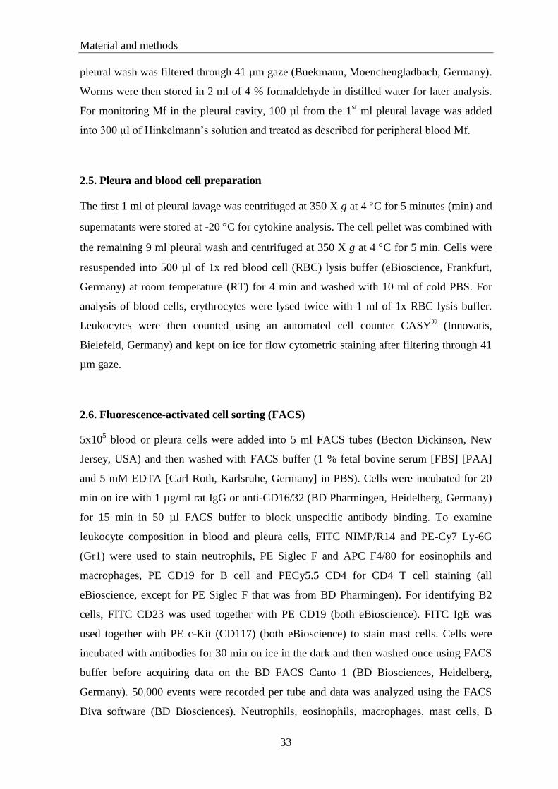
Material and methods
33
pleural wash was filtered through 41 µm gaze (Buekmann, Moenchengladbach, Germany).
Worms were then stored in 2 ml of 4 % formaldehyde in distilled water for later analysis.
For monitoring Mf in the pleural cavity, 100 µl from the 1st ml pleural lavage was added
into 300 µl of Hinkelmann‟s solution and treated as described for peripheral blood Mf.
2.5. Pleura and blood cell preparation
The first 1 ml of pleural lavage was centrifuged at 350 X g at 4 C for 5 minutes (min) and
supernatants were stored at -20 C for cytokine analysis. The cell pellet was combined with
the remaining 9 ml pleural wash and centrifuged at 350 X g at 4 C for 5 min. Cells were
resuspended into 500 µl of 1x red blood cell (RBC) lysis buffer (eBioscience, Frankfurt,
Germany) at room temperature (RT) for 4 min and washed with 10 ml of cold PBS. For
analysis of blood cells, erythrocytes were lysed twice with 1 ml of 1x RBC lysis buffer.
Leukocytes were then counted using an automated cell counter CASY® (Innovatis,
Bielefeld, Germany) and kept on ice for flow cytometric staining after filtering through 41
µm gaze.
2.6. Fluorescence-activated cell sorting (FACS)
5x10⁵ blood or pleura cells were added into 5 ml FACS tubes (Becton Dickinson, New
Jersey, USA) and then washed with FACS buffer (1 % fetal bovine serum [FBS] [PAA]
and 5 mM EDTA [Carl Roth, Karlsruhe, Germany] in PBS). Cells were incubated for 20
min on ice with 1 µg/ml rat IgG or anti-CD16/32 (BD Pharmingen, Heidelberg, Germany)
for 15 min in 50 µl FACS buffer to block unspecific antibody binding. To examine
leukocyte composition in blood and pleura cells, FITC NIMP/R14 and PE-Cy7 Ly-6G
(Gr1) were used to stain neutrophils, PE Siglec F and APC F4/80 for eosinophils and
macrophages, PE CD19 for B cell and PECy5.5 CD4 for CD4 T cell staining (all
eBioscience, except for PE Siglec F that was from BD Pharmingen). For identifying B2
cells, FITC CD23 was used together with PE CD19 (both eBioscience). FITC IgE was
used together with PE c-Kit (CD117) (both eBioscience) to stain mast cells. Cells were
incubated with antibodies for 30 min on ice in the dark and then washed once using FACS
buffer before acquiring data on the BD FACS Canto 1 (BD Biosciences, Heidelberg,
Germany). 50,000 events were recorded per tube and data was analyzed using the FACS
Diva software (BD Biosciences). Neutrophils, eosinophils, macrophages, mast cells, B
Material and methods
34
cells, B2 cells, T cells and their subsets from both pleural cavity and blood were gated by
first gating total live leukocytes by side scatter (SSC) and forward scatter (FSC) as shown
in figure 2.1.
A. B.
Figure 2.1. Gating strategy for leukocytes from the pleural cavity and blood. Total live
leukocytes from pleural cavity (A) and blood (B) were gated by SSC and FSC.
To determine the number of alternatively activated macrophages (AAMs), 1x10⁶ pleura
cells were added into 5 ml FACS tubes and washed once with 1 ml of PBS/1 % bovine
serum albumin (BSA) (PAA) followed by a centrifugation step at 400 X g for 6 min at 4
°C. After all supernatant was discarded, 300 µl of 1x fixation/permeabilization buffer
(eBioscience) was added to the cells and the cells were incubated for a minimum of 1 hour
(h) at 4 °C. Cells were then washed and blocked with 1 µg/ml rat IgG in 1 ml of PBS/1 %
BSA at 4 °C overnight. The following day, cells were washed, all supernatant discarded
and 300 µl of 1x permeabilization buffer (eBioscience) were added. The cells were then
incubated for 20 min at RT. After another washing step, rabbit anti-murine RELMα
antibody (Peprotech, New Jersey, USA) in 100 µl of 1x permeabilization buffer was added
to the cells and incubated for 1 h at 4 °C. Cells were washed and stained for AAMs by
adding Alexa Fluor® 488 goat anti-rabbit IgG (H+L) (Invitrogen, Darmstadt, Germany),
PE Siglec F and APC F4/80 followed by incubating for 30 min at 4 °C in the dark. Cells
were washed once before acquiring data on the BD FACS Canto 1. 50,000 events were
recorded per tube and data was analyzed using the FACS Diva software. AAMs were gated
by first gating total leukocytes by SSC and FSC as shown in figure 2.1.

Material and methods
35
2.7. Spleen cell preparation and culture
Spleens were meshed on metal sieves using the plunger of a 5 ml syringe (BD, Heidelberg,
Germany) followed by adding 3 ml of cold PBS and then filtered using a 100 µm metal
sieve. Splenocytes were transferred into 15 ml falcon tubes (Greiner Bio-One,
Frickenhausen, Germany) and centrifuged at 350 X g at 4 C for 5 min. The supernatant
was discarded and cells were resuspended in 1 ml of 1x RBC lysis buffer as described
before. Splenocytes were counted using an automated cell counter after washing once with
sterile PBS.
Using splenocyte medium (10 % FBS and 1 % Penicillin/Streptomycin in high glucose
DMEM [all PAA]), 1x10⁶ cells were plated in 96 well round-bottom plates (Greiner Bio-
One) and stimulated at 37 C, 5 % CO2 for 72 h with medium, LsAg at a concentration of
25 g/ml, recombinant mouse IL-6 (rIL-6) (PeproTech) at a concentration of 5 ng/ml,
LsAg together with rIL-6 and 2.5 g/ml concanavalin-A (ConA) (Sigma-Aldrich) as
positive control. After incubation, plates were centrifuged at 350 X g at 4 C for 5 min, and
supernatant was transferred into new plates and stored at -20 C for cytokine analysis.
2.8. Splenocytes intracellular staining
2x10⁶ spleen cells in 1 ml of splenocyte medium were plated into 12-well plates (Greiner
Bio-One). The cells were stimulated with LsAg. For positive control, 2 µg/ml of anti-
CD28 (eBioscience) was added into wells which had been coated with 5 µg/ml of anti-
CD3 (eBioscience) in PBS at 4 C overnight. The cells were then incubated at 37 C, 5 %
CO2 for 2 h. To inhibit protein secretion, 2 µg/ml of GolgiStop (BD Biosciences) which
contains monensin was added into each well, followed by additional 4 h of incubation.
After a total of 6 h incubation time, the cells were transferred into 15 ml falcon tubes and
centrifuged in 2 ml of PBS at 350 X g at 4 C for 10 min. The cells were fixed and
permeabilized using 500 µl of freshly prepared 1x fixation/permeabilization buffer at 4 C
after all supernatant had been discarded overnight.
The following day, the fixed and permeabilized cells were washed with 2 ml of PBS/1 %
BSA and 1x permeabilization buffer was added to the cells followed by centrifugation at
780 X g at 4 C for 10 min. After discarding the supernatant, splenocytes were incubated
with rat IgG in a concentration of 1 µg/ml in 1 ml of PBS/1 % BSA for 1 h to prevent

Material and methods
36
unspecific antibody binding. Cells were washed by centrifugation at 350 X g at 4 C for 5
min and surface stained with PerCPCy5.5 CD4 and APC CD25, and intracellularly stained
with FITC FoxP3, PE IL-17A and APC IL-6 (all eBioscience). Data was acquired using a
BD FACS Canto 1 and analyzed using FACS Diva software after first gating on
splenocytes as shown in figure 2.2 followed by gating of CD4+ T cells then
CD4+CD25
+FoxP3
+ for regulatory T cells (Treg), CD4
+IL-17A
+ for IL-17-producing Th17
cells and CD4+IL-6
+ for IL-6-producing T cells.
Figure 2.2. Gating strategy for splenocytes. Splenocytes were gated by SSC and FCS
2.9. Worm and microfilaria counting
L3 and L4 were counted using a binocular microscope at 16x or 40x magnification. Adult
worms were counted using light microscopy and differentiated into male and female
worms.
Mf in Hinkelmann‟s solution were centrifuged for 5 min at 250 X g and the supernatant
was removed leaving only about 20 µl liquid for counting the worms using light
microscopy at 40x or 100x magnification.
2.10. Enzyme-linked immunosorbent assay (ELISA)
IL-6, Eotaxin-1, Eotaxin-2, RANTES, sIL-6R, IL-17, TNF, MIP-2, CCL17 (all DuoSet
R&D Systems, Wiesbaden, Germany), IL-5, IgE (both BD Pharmingen) and IFN
(eBioscience) concentrations were measured by two-sided cytokine ELISA according to
the manufacturers‟ direction. ELISA plates (Greiner Bio-One) were coated with primary
Material and methods
37
antibodies in PBS or coating buffer (Na2HPO4 0.1 M [Merck] in distilled aqua pH 9.0)
overnight. Plates were then blocked with PBS/1 % BSA for a minimum of 2 h at RT and
washed three times with PBS/0.05 % Tween20
(Sigma-Aldrich). 50 μl of sample were
added for a minimum of 2 h at RT or at 4 C overnight. The detection antibody was added
for 2 h. Following 20 min incubation with horseradish peroxidase (HRP)-streptavidin
(R&D Systems) and tetramethylene benzidine (TMB) (Carl Roth), the color reaction was
stopped with 1 M H2SO4 (Merck) and optical density (OD) was measured at 450 nm using
a plate reader SpectraMAX 340 Pc (Molecular Devices, Ismaning, Germany). The data
was analyzed with Softmax Pro software (Molecular Devices). The limit of detection were
4 pg/ml (IFN), 5.8 pg/ml (IgE), 7.8 pg/ml (Eotaxin-1, Eotaxin-2, TNFα, sIL-6R, MIP-2),
12 pg/ml (IL-5), 15.6 pg/ml (IL-6, IL-17) and 31.2 pg/ml (RANTES, CCL17)
For L. sigmodontis-specific IgG1 and IgG2a ELISA, polysorb ELISA plates (Nunc,
Roskilde, Denmark) were first coated with 10 µg/ml of L. sigmodontis antigen extract in
PBS at pH 9 overnight. Plates were then blocked with PBS/1 % BSA for a minimum of 2 h
at RT and washed three times with PBS/0.05 % Tween20
(Sigma-Aldrich). A series of
1:100 to 1:100,000 serum dilutions were added for a minimum of 2 h at RT or at 4 C
overnight following by adding secondary antibodies (BD Pharmingen) for 2 h at RT. The
plates were then treated as described for the cytokine ELISA.
2.11. Neutrophil purification and stimulation
To induce neutrophil migration into the peritoneal cavity, mice were injected
intraperitoneally with casein solution. To prepare 9 % w/v of casein solution, 2.25 gram of
casein powder (Carl Roth) was combined with 20 ml of pure water and 1 ml of 1 M NaOH
(Carl Roth) and then mixed well for 1 h at 70 C. After casein dissolved, 2.5 ml of MgCl2 5
mM (Sigma Aldrich) and 2.25 ml of CaCl2 10 mM (Carl Roth) were added into the
solution.
Mice were injected with 1 ml of casein solution 18 and 2 h before euthanasia and then
killed using an overdose of isoflurane. Abdominal skin was carefully dissected and the
peritoneum was exposed. 10 ml of sterile cold PBS was carefully injected into the
peritoneum and gently agitated during injection. The peritoneal wash was withdrawn
slowly and transferred into sterile 50 ml falcon tubes (Greiner Bio-One). The peritoneal
wash from the same strain of mice was pooled into one tube and then centrifuged at 350 X

Material and methods
38
g at 4 C for 5 min. Supernatant was discarded and RBC lysis buffer was added to remove
erythrocytes as described above.
Neutrophils were also generated from bone marrow. Femurs and tibias were dissected and
transferred into sterile PBS. After muscles and ligament were dissected from the bones,
both ends of the bone were cut off. The bones were set into 0.6 ml tubes (Eppendorf) with
a small hole at the bottom. After setting this tube into a larger tube (1.5 ml), the tubes were
centrifuged at 2000 X g for 30 seconds and bone marrow was flushed into the larger tube.
Bone marrow was then resuspended with 100 µl PBS. Bone marrow from the same strain
was pooled in 50 ml conical tubes and 10 ml of PBS was added followed by centrifugation
at 350 X g at 4 C for 10 min. After discarding the supernatant, erythrocytes were lysed
with 3 ml of RBC lysis buffer as described previously and bone marrow was washed with
20 ml of PBS by centrifugation at 300 X g at 4 C for 10 min.
Finally, neutrophils were also purified from the peripheral blood. 200 µl fresh blood which
was acquired from the retro-orbital vein as described above was resuspended twice with 1
ml of 1x RBC lysis buffer to remove erythrocytes. After washing with sterile PBS, cells
were then counted using an automated cell counter.
Cells from peritoneal lavage, bone marrow or blood were then transferred into a 15 ml
falcon tube after adding 4 ml of sterile PBS. Using 19 G lumbar puncture needles, 3 ml of
53.4 % of Percoll (Sigma-Aldrich) in PBS was carefully underlayered into the tube
followed by underlayering of 62.3 % and 71.7 %. The tubes were centrifuged at 500 X g at
15 C for 20 min. The cells from the second layer from the top containing primarily
neutrophils were carefully aspirated using an 18G needle (BD) and transferred into a new
tube. Neutrophils were washed twice with 10 ml cold sterile PBS. To determine the purity
of neutrophils, FACS analysis was carried out as described above. The purity was between
90-95 %.
1x10⁵
neutrophils were stimulated at 37 C, 5 % CO2 for 18 h with medium (10 % FBS and
1 % Penicillin/Streptomycin in RPMI), LsAg, rIL-6, LsAg together with rIL-6 and 500
ng/ml tripalmitoyl-S-glycerylcysteine (P3C) (InvivoGen, Toulouse, France), for positive
control. After 18 h incubation, plates were centrifuged and supernatants were stored at -20
C for cytokine analysis.

Material and methods
39
2.12. Macrophages purification and stimulation
Macrophage accumulation in the peritoneal cavity was elicited by intraperitoneal injection
with 2 ml of sterile thioglycollate solution (Heipha, Eppelheim, Germany) 4 days prior to
sacrificing. Mice were killed with an overdose of isofluran anesthesia and abdominal skin
was carefully dissected. Cells were harvested from the peritoneal cavity by injecting 10 ml
of sterile PBS as described for neutrophil purification. Peritoneal wash from the same
mouse strain were collected in 50 ml conical tubes and washed twice with 10 ml of sterile
cold PBS by centrifuging at 300 X g at 4 C for 5 min. If necessary, erythrocytes were
lysed with 1 ml of 1x RBC lysis buffer.
1x10⁵ cells in 100 µl of medium (10 % FBS and 1 % Penicillin/Streptomycin in RPMI)
were plated into 96-wells flat-bottom plates (Greiner Bio-One). The cells were then rested
at 37 C, 5 % CO2 overnight. The supernatant was removed and adherent cells were used
for further experiments. 1x10⁵ macrophages were restimulated at 37 C, 5 % CO2 for 18 h
with LsAg, rIL-6, rIL-6 together with LsAg and P3C.
2.13. Eosinophil purification and stimulation
Mice were infected with L. sigmodontis as described previously. At 15 dpi, mice were
euthanized and the pleural cavity was washed with 10 ml sterile cold PBS. Cells were
washed with PBS by centrifugation at 350 X g at 4 C for 10 min and the supernatant was
discarded. Erythrocytes were lysed with 1x RBC lysis buffer as described above followed
by washing with 10 ml of sterile cold PBS. Eosinophils were purified by negative selection
using magnetic beads of the MACS
cell separation system (Miltenyi Biotech, Bergisch
Gladbach, Germany) per manufacturer‟s instruction. Briefly, up to 107
cells were
resuspended into 3 ml of MACS
buffer and washed by centrifugation at 300 X g for 10
min. After all supernatant was discarded completely, 90 μl of MACS buffer were added to
107 total cells and then cells were labeled with 10 μl each of anti-MHCII, anti-CD45R, and
anti-CD90.2 microbeads. After incubation at 4 °C for 15 min and washing with 1-2 ml of
MACS buffer per 107 total cells. LD columns were applied to the magnetic field followed
by rinsing the columns with 3 ml of MACS buffer. The cells were then transferred into the
LD columns. Eosinophils were collected as unlabelled cells that pass through and then
washed once with sterile cold PBS. Purity of eosinophils was determined by flow
Material and methods
40
cytometry and generally reached a purity of 70-80 %. Purified eosinophils were then
stimulated with LsAg, rIL-6, LsAg with IL-6, and P3C.
2.14. Interleukin-6 intracellular staining
One million of pleural leukocytes or purified eosinophils were incubated in 500 µl of
medium (10 % FBS and 1 % Penicillin/Streptomycin in RPMI) in 24-well plates (Greiner
Bio One) and stimulated with either LsAg, 500 ng/ml of Lipopolysaccharide (LPS)
(InvivoGen, Toulouse, France) or medium alone. In order to inhibit protein transport into
the extracellular compartment, GolgiPlug (BD Biosciences) which contains Brefeldin A
solution was added into the culture followed by incubation at 37 C, 5 % CO2 for 5 h. Cells
were then transferred into FACS tubes and 2 ml of FACS Buffer was added, followed by
centrifugation at 350 X g for 5 min at 4 °C. Cells were incubated with rat IgG for 15 min to
reduce unspecific binding. Eosinophils and macrophages were stained with PE Siglec-F
and APC F4/80 for 30 min. The cells were then washed with 2 ml of FACS buffer by
centrifugation at 350 X g at 4 °C and fixed with 500 µl of 2 % paraformaldehyde (PFA)
solution (PFA [Merck] in FACS buffer) followed by incubation for 7 min on ice. Cells
were then washed once with 500 µl FACS buffer by centrifugation at 790 X g for 5 min.
After supernatant was discarded, 120 µl of saponin buffer (0.5 % Saponin [Sigma Aldrich]
in PBS/2 % BSA) was added to the cells for 15 min at RT. IL-6 and isotype control for
intracellular staining were then carried out by adding FITC IL-6 and isotype antibodies in
90 µl of saponin buffer for 30 min at RT. Data was acquired using a BD FACS Canto 1
machine and analyzed using FACS Diva software after first gating on Siglec-F+F4/80
lowIL-
6+ and Siglec-F
lowF4/80
+IL-6
+ cells for IL-6-producing eosinophils and macrophages
respectively.
2.15. Dendritic Cell (DC) generation and stimulation
Bone marrow from murine femurs and tibias was prepared as described for the generation
of bone marrow neutrophils. Cells were counted and resuspended into 5 ml of DC growth
medium (10 % FBS, 1 % sodium pyruvate, 1 % 4-[2-hydroxyethyl]-1-piperazineethane-
sulfonic acid [HEPES], 1 % L-Glutamin, 1 % penicillin/streptomycin [all PAA] and 0.1 %
2-mercaptoethanol in RPMI). 2.5x10⁶
of cells in medium containing 20 ng/ml of GM-CSF
(PeproTech) were plated into 6-wells plates and incubated at 37 C, 5 % CO2. After 72 h, 2
Material and methods
41
ml of medium with GM-CSF was added followed by changing the medium 48 h later.
After 7 days incubation time, loosely adherent cells were harvested and washed once using
PBS. 1x10⁶
of cells were stimulated with LsAg, rIL-6 and LPS for positive control at 37
C, 5 % CO2 for 18 h. FACS analysis was conducted to determine purity of DC. The purity
was between 80-90 %.
2.16. Phagocytosis assay
Cells from the pleural wash of L. sigmodontis infected mice were acquired at 15 dpi as
described previously. 2x10⁶ cells were incubated with 30x10
6 of Fluoresbite Plain YG 1.0
micron microspheres (Polysciences, Eppelheim, Germany) at 37 C for 1 h. As negative
control, cells were stored at 4 C for 1 h. Phagocytosis of macrophages and eosinophils
was determined using FACS. Cells were stained with PE Siglec-F and APC F4/80 for
eosinophils and macrophages as described above. Data was acquired using FACS Diva
software and the percentages of phagocytosed eosinophils and macrophages were
calculated as percent of total eosinophils and macrophages.
2.17. Vascular permeability assay
An in vivo vascular permeability assay was conducted to examine the vascular
permeability in either the skin at the lumbar area or in the ears. For measuring vascular
permeability in the skin of mice, at first, the injected lumbar area was shaved a week
before the injection in order to reduce skin irritation of the mice that could change the
result. Mice were then injected subcutaneously with 10 µg of LsAg in 100 µl of sterile
PBS or PBS alone. 6 h later, mice were injected intravenously with 30 mg/kg body weight
(BW) of Evans Blue (Sigma Aldricht) in 100 µl of NaCl 0.9 % (Fresenius Kabi, Bad
Homburg, Germany). After 1 h, mice were killed by an overdose of isoflurane anesthesia
followed by cutting off the skin at the injected areas approximately 1 cm in diameter. The
skin was weighed and transferred into 3 ml of >99 % formamide (Sigma Aldricht).
Alternatively, 10 µg each of PBS, LsAg or anti-mouse FcεRIα (MAR-1) (eBiosciences)
were injected intracutaneously (i.c.) in the ears of mice. After 3 min, 30 mg/kg BW of
Evans Blue in 200 µl of NaCl 0.9 % were injected i.v.. 10 min later, the complete ears
were cut off and transferred into 500 µl of >99 % formamide. Evans Blue leakage into the

Material and methods
42
tissues both in the skin or ears were quantified after 48-72 h of incubation at 56 °C by
determining optical density on a plate reader at 620 nm and normalized to the weight of the
tissue.
2.18. Mast cells stabilizing assay
To stabilize mast cells, mice were injected intraperitoneally with 8 mg/kg BW of sodium
cromoglycate (hereafter cromolyn) (Sigma Aldricht) in 100 µl of sterile NaCl 0.9 % or
NaCl 0.9 % alone daily starting 6 days prior to infection until euthanasia. Mice were
euthanized at 14 dpi and parasitological as well as immunological changes were examined
as described previously.
2.19. Histamine neutralizing assay
Histamine receptor-1 and -2 antagonists‟ mepyramine and cimetidine respectively (both
Sigma Aldrich) were used to block histamine activity in infected mice. 20 mg/kg BW of
mepyramine and 40 mg/kg BW of cimetidine in 100 μl of PBS or PBS alone were injected
intraperitoneally at 1 h prior and 2 h after L. sigmodontis infection, followed daily at 1 until
5 dpi. Worm burden, leukocyte composition in pleural cavity and blood and other
immunological changes were determined after 14 dpi.
2.20. IL-5 depletion in vivo
Anti-IL-5 (TRFK5) was used to deplete IL-5 in vivo. TRFK5 and the isotype control
antibody TRFK4 or rat IgG1 were either purified in our laboratory or purchased. TRFK5
and TRFK4 were acquired from hybridoma cells (Lopez, Strath, & Sanderson, 1984)
which were cultured with hybridoma medium (10 % FBS, 1 % sodium pyruvate, 1 % L-
Glutamin, 1 % penicillin/streptomycin and 1.9 % 2-mercaptoethanol in RPMI). The
antibodies were harvested every other day (TRFK5) and every 3 days (TRFK4).
Supernatant was then purified using a protein purifier ÄKTAprime plus (GE Healthcare,
Munich, Germany) and dialyzed using a protein membrane Slide-A-Lyzer
3.5 dialysis
cassette (Thermo Scientific, Rockford, USA). The purity of antibodies was then examined
with dotblot, westernblot, Comassie staining and ELISA.
Material and methods
43
Using antibodies which were purified in our lab, mice were injected intraperitoneally with
100 µg of TRFK5 or TRFK4 in 300 µl of PBS 5 dpi until euthanasia at 14 dpi.
Alternatively, mice were injected with 50 µg of commercial antibodies TRFK5 or isotype
control rat IgG1 (BioXcell, New Hampshire, USA) in 100 µl of sterile PBS at 1 day prior
infection and followed at 3, 7, 10, 13 dpi, and euthanized at 14 dpi.
IL-5 depletion at a later time point of infection was also conducted to investigate the effect
of IL-5 between day 14 and day 28 post infection. Commercial antibodies TRFK5 or rat
IgG1 were injected i.p. at 12, 17, 23 and 28 dpi. Mice were then sacrificed at 30 dpi. Worm
burdens as well as immunological changes were determined as previously described.
2.21 Statistics
Statistical analyses were performed with GraphPad Prism 5.0 software (GraphPad
Software, La Jolla, California, USA), using the Mann Whitney t-test (u test) for non-
parametric data. All data are shown as median except for in vitro stimulation and time
course data that are shown as means ± standard error of mean (SEM). P values ≤ 0.05 were
considered as significant.
2.22. Funding
My study was funded by the Government of Aceh (Indonesia) in collaboration with the
German Academic Exchange Service (DAAD) and intramural grant by the University
Hospital Bonn. The funders had no role in study design, data collection and analysis,
decision to publish, or preparation of the manuscript.

Results
44
14 21 28 35 42 49 56 63 70 770
10
20
30 BALB/c
C57BL/6
days post infection (dpi)
wo
rm b
urd
en
1 7 14 21 28 35 42 49 56 63 700
20
40
60 BALB/c
C57BL/6
days post infection (dpi)
tota
l cells [
x10
6]
3. Results
3.1. Comparison of susceptible BALB/c and resistant C57BL/6 mice during
Litomosoides sigmodontis infection
To determine the progress of filarial infection and examine parasitological and
immunological variables during infection, susceptible BALB/c and resistant C57BL/6 wild
type mice were infected with L. sigmodontis and euthanized every week starting at 1 day
post infection (dpi), followed at 1 week until 11 weeks post infection. Worms in the pleural
cavity were counted starting at 2 weeks post infection. As previously reported by several
studies (Babayan et al. 2003; Le Goff et al. 2002), BALB/c mice had a higher worm
burden compared to C57BL/6 wild type mice throughout the infection (Figure 3.1.1). Total
leukocytes in the pleural cavity of infected mice were counted to determine whether total
cells correlated with the worm burden at those time points. However, there was no
correlation between worm burden and total cell numbers at the site of infection (figure
3.1.1).
Figure 3.1.1. Increased worm burden and total pleural cavity cell numbers in L. sigmodontis
infected susceptible BALB/c mice compared to resistant C57BL/6 mice. L. sigmodontis worm
burden (left) and total cells derived from the pleural cavity (right) of susceptible BALB/c and
resistant C57BL/6 mice. (Time course experiments were done with 5 mice per group. The
experiment was conducted once. Data is shown as means ± SEM).
Next, cellular composition in the pleural cavity was investigated for both types of mice by
flow cytometry. Interestingly, relative and absolute number of neutrophils in BALB/c mice
showed a significant increase after 4 weeks post infection, and reached its peak at 8 weeks
post infection. In contrast, neutrophil numbers in C57BL/6 mice were maintained at low
levels during the infection (figure 3.1.2 A). Parallel with neutrophil numbers, eosinophil
numbers in BALB/c mice became prominent after 4 weeks post infection; although there
was no clear peak in eosinophil numbers as with neutrophils. Eosinophils in C57BL/6 mice

Results
45
also tended to increase after 4 weeks post infection; although the numbers were lower than
in BALB/c mice (figure 3.1.2 B). Relative and absolute numbers of macrophages were
higher in C57BL/6 mice than in BALB/c mice, and their numbers were maintained at high
levels throughout the infection in both C57BL/6 and BALB/c mice (figure 3.1.2 C). No
difference of both T cell and B cell or total lymphocytes were found in BALB/c compared
to C57BL/6 mice during L. sigmodontis infection (figure 3.1.2 D, E and F).
A. B.
C. D.
E. F.
Figure 3.1.2. Increased frequencies of neutrophils and eosinophils during the chronic phase of
L. sigmodontis infection in susceptible BALB/c mice. Percentage of leukocytes in the pleura
cavity of L. sigmodontis infected BALB/c and C57BL/6 mice at different time points post
infection. Frequencies of neutrophils (A), eosinophils (B), macrophages (C), T cells (D), B cells
(E) and total lymphocytes (F) were measured by FACS and are indicated as percentages of living
cells. (Time course experiments were conducted with 5 mice per group. The experiment was
carried out once. Data is shown as means ± SEM)
1 7 14 21 28 35 42 49 56 63 70 770
5
10
15
20 BALB/c
C57BL/6
days post infection (dpi)
neu
tro
ph
il [
%]
1 7 14 21 28 35 42 49 56 63 70 770
20
40
60
80BALB/c
C57BL/6
days post infection (dpi)
macro
ph
ag
e [
%]
1 7 14 21 28 35 42 49 56 63 70 770
10
20
30
40
50 BALB/c
C57BL/6
days post infection (dpi)
eo
sin
op
hil [
%]
1 7 14 21 28 35 42 49 56 63 70 770
5
10
15
20 BALB/c
C57BL/6
days post infection (dpi)
T c
ell [
%]
1 7 14 21 28 35 42 49 56 63 70 770
20
40
60 BALB/c
C57BL/6
days post infection (dpi)
B c
ell [
%]
1 7 14 21 28 35 42 49 56 63 70 770
20
40
60
80 BALB/c
C57BL/6
days post infection (dpi)
lym
ph
ocyte
s [
%]
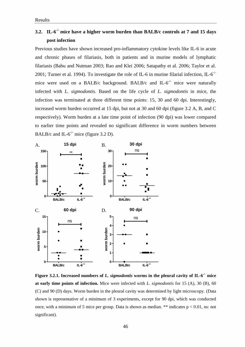
Results
46
3.2. IL-6-/-
mice have a higher worm burden than BALB/c controls at 7 and 15 days
post infection
Previous studies have shown increased pro-inflammatory cytokine levels like IL-6 in acute
and chronic phases of filariasis, both in patients and in murine models of lymphatic
filariasis (Babu and Nutman 2003; Rao and Klei 2006; Satapathy et al. 2006; Taylor et al.
2001; Turner et al. 1994). To investigate the role of IL-6 in murine filarial infection, IL-6-/-
mice were used on a BALB/c background. BALB/c and IL-6-/-
mice were naturally
infected with L. sigmodontis. Based on the life cycle of L. sigmodontis in mice, the
infection was terminated at three different time points: 15, 30 and 60 dpi. Interestingly,
increased worm burden occurred at 15 dpi, but not at 30 and 60 dpi (figure 3.2 A, B, and C
respectively). Worm burden at a late time point of infection (90 dpi) was lower compared
to earlier time points and revealed no significant difference in worm numbers between
BALB/c and IL-6-/-
mice (figure 3.2 D).
A. B.
C. D.
Figure 3.2.1. Increased numbers of L. sigmodontis worms in the pleural cavity of IL-6-/-
mice
at early time points of infection. Mice were infected with L. sigmodontis for 15 (A), 30 (B), 60
(C) and 90 (D) days. Worm burden in the pleural cavity was determined by light microscopy. (Data
shown is representative of a minimum of 3 experiments, except for 90 dpi, which was conducted
once, with a minimum of 5 mice per group. Data is shown as median. ** indicates p < 0.01, ns: not
significant).
15 dpi
BALB/c IL-6-/-
0
50
100
150 **
wo
rm b
urd
en
30 dpi
BALB/c IL-6-/-
0
10
20
30 ns
wo
rm b
urd
en
60 dpi
BALB/c IL-6-/-
0
5
10
15
ns
wo
rm b
urd
en
90 dpi
BALB/c IL-6-/-
0
1
2
3
4
5 ns
wo
rm b
urd
en
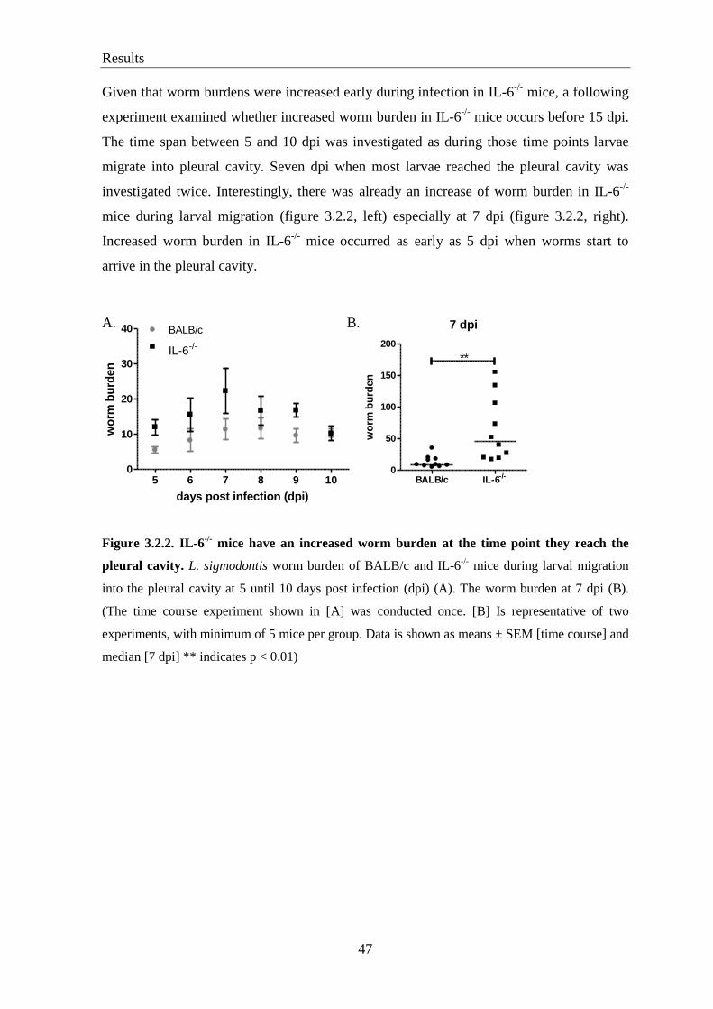
Results
47
Given that worm burdens were increased early during infection in IL-6-/-
mice, a following
experiment examined whether increased worm burden in IL-6-/-
mice occurs before 15 dpi.
The time span between 5 and 10 dpi was investigated as during those time points larvae
migrate into pleural cavity. Seven dpi when most larvae reached the pleural cavity was
investigated twice. Interestingly, there was already an increase of worm burden in IL-6-/-
mice during larval migration (figure 3.2.2, left) especially at 7 dpi (figure 3.2.2, right).
Increased worm burden in IL-6-/-
mice occurred as early as 5 dpi when worms start to
arrive in the pleural cavity.
A. B.
Figure 3.2.2. IL-6-/-
mice have an increased worm burden at the time point they reach the
pleural cavity. L. sigmodontis worm burden of BALB/c and IL-6-/-
mice during larval migration
into the pleural cavity at 5 until 10 days post infection (dpi) (A). The worm burden at 7 dpi (B).
(The time course experiment shown in [A] was conducted once. [B] Is representative of two
experiments, with minimum of 5 mice per group. Data is shown as means ± SEM [time course] and
median [7 dpi] ** indicates p < 0.01)
5 6 7 8 9 100
10
20
30
40 BALB/c
IL-6-/-
days post infection (dpi)
wo
rm b
urd
en
7 dpi
BALB/c IL-6-/-
0
50
100
150
200
**w
orm
bu
rden

Results
48
3.3. No difference in microfilaria levels between BALB/c and IL-6-/-
mice
To determine the role of IL-6 on the microfilarial burden during L. sigmodontis infection,
circulating Mf and Mf levels in the pleural cavity were counted 60 and 90 dpi. No
difference in Mf levels was found between both strains of mice neither at 60 dpi (figure 3.3
A) nor 90 dpi (figure 3.3 B) both in the blood and pleural cavity.
A.
B.
Figure 3.3. Microfilaria load is comparable between BALB/c and IL-6-/-
mice. Mice were
infected with L. sigmodontis. After 60 dpi (A) and 90 dpi (B), Mf in the peripheral blood (left) and
in the pleural cavity (right) were counted using light microscopy. (Data shown as representative of
a minimum of 2 experiments except for 90 dpi, which was conducted only once, with minimum of
5 mice per group. Data is shown as median).
3.4. Increased number of eosinophils in the pleural cavity, but not in the blood,
correlates with increased worm burden in IL-6-/-
mice
Eosinophilia is a hallmark of helminth infections including lymphatic filariasis.
Eosinophils have been reported to play a significant role in protective immunity during
filarial infection potentially by secreting their granule contents (Makepeace et al. 2012). To
determine the role of IL-6 in eosinophilia during filarial infection, the eosinophil
population was investigated by FACS at different time points post infection in both
60 dpi, blood
BALB/c IL-6-/-
0
2
4
6
8
10ns
mic
rofi
lari
a
60 dpi, pleura
BALB/c IL-6-/-
0
50
100
150 nsm
icro
fila
ria
90 dpi, blood
BALB/c IL-6-/-
0
5
10
15
ns
mic
rofi
lari
a
90 dpi, pleura
BALB/c IL-6-/-
0
50
100
150ns
mic
rofi
lari
a

Results
49
BALB/c and IL-6-/-
mice. Eosinophils were gated as SiglecF+F4/80
low cells and
differentiated from macrophages which were defined as SiglecFlow
F4/80high
cells (figure
3.4.1 A). Uninfected mice were found to have low numbers of eosinophils (approximately
1 % of total leukocytes found in the pleural cavity). Number of eosinophils started to rise
at 7 dpi with significantly higher levels at 15 dpi. Interestingly, significantly increased
eosinophil numbers (relative and absolute) were found in IL-6-/-
mice compared to BALB/c
mice at 7 and 15 dpi (figure 3.4.1 B). However, this increase was not observed at later time
points when there was no difference in worm burden (data not shown). Increased number
of eosinophil correlated with increased worm burden in IL-6-/-
mice at early time points
post infection (figure 3.4.1 C).
A. FMO Siglec F BALB/c IL-6-/-
B.
naive, pleura
BALB/c IL-6-/-0.0
0.5
1.0
1.5
2.0
2.5
eo
sin
op
hil [
%]
7 dpi, pleura
BALB/c IL-6-/-
0
2
4
6
8 **
eo
sin
op
hil [
%]
15 dpi, pleura
BALB/c IL-6-/-
0
10
20
30
40
50**
eo
sin
op
hil [
%]
ns
naive, pleura
BALB/c IL-6-/-
0.000
0.002
0.004
0.006
tota
l eo
sin
op
hil [
x10
6]
7 dpi, pleura
BALB/c IL-6-/-
0.0
0.5
1.0
1.5
2.0
*
tota
l eo
sin
op
hil [
x10
6]
15 dpi, pleura
BALB/c IL-6-/-
0
2
4
6
8
10
**
tota
l eo
sin
op
hil [
x10
6]
ns
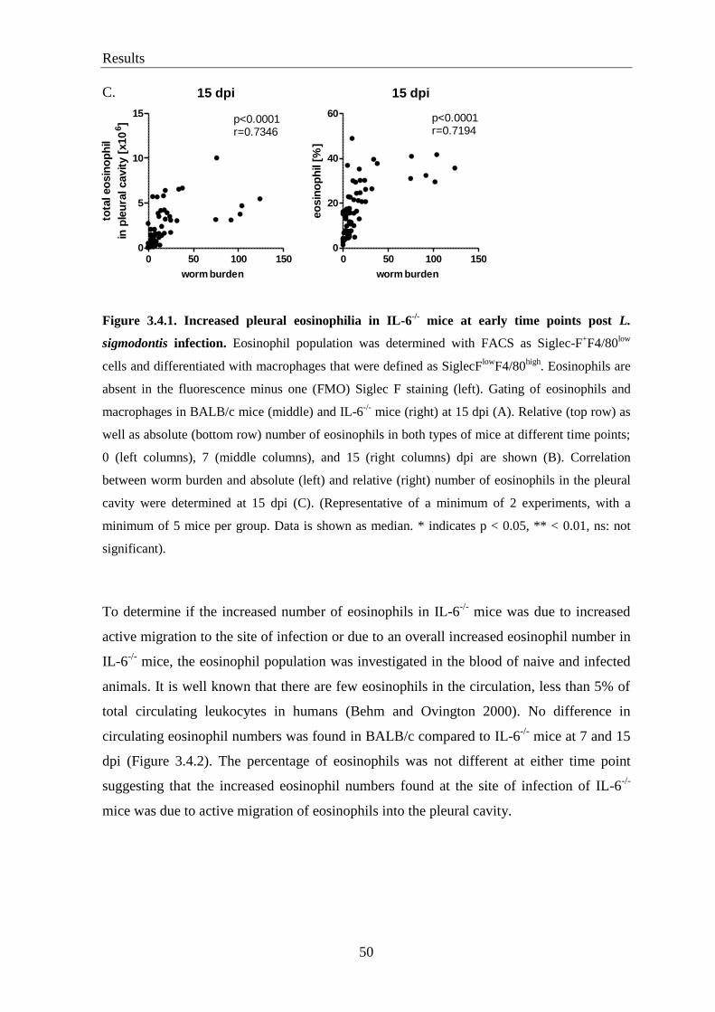
Results
50
C.
Figure 3.4.1. Increased pleural eosinophilia in IL-6-/-
mice at early time points post L.
sigmodontis infection. Eosinophil population was determined with FACS as Siglec-F+F4/80
low
cells and differentiated with macrophages that were defined as SiglecFlow
F4/80high
. Eosinophils are
absent in the fluorescence minus one (FMO) Siglec F staining (left). Gating of eosinophils and
macrophages in BALB/c mice (middle) and IL-6-/-
mice (right) at 15 dpi (A). Relative (top row) as
well as absolute (bottom row) number of eosinophils in both types of mice at different time points;
0 (left columns), 7 (middle columns), and 15 (right columns) dpi are shown (B). Correlation
between worm burden and absolute (left) and relative (right) number of eosinophils in the pleural
cavity were determined at 15 dpi (C). (Representative of a minimum of 2 experiments, with a
minimum of 5 mice per group. Data is shown as median. * indicates p < 0.05, ** < 0.01, ns: not
significant).
To determine if the increased number of eosinophils in IL-6-/-
mice was due to increased
active migration to the site of infection or due to an overall increased eosinophil number in
IL-6-/-
mice, the eosinophil population was investigated in the blood of naive and infected
animals. It is well known that there are few eosinophils in the circulation, less than 5% of
total circulating leukocytes in humans (Behm and Ovington 2000). No difference in
circulating eosinophil numbers was found in BALB/c compared to IL-6-/-
mice at 7 and 15
dpi (Figure 3.4.2). The percentage of eosinophils was not different at either time point
suggesting that the increased eosinophil numbers found at the site of infection of IL-6-/-
mice was due to active migration of eosinophils into the pleural cavity.
15 dpi
0 50 100 1500
5
10
15
worm burden
p<0.0001r=0.7346
tota
l eo
sin
op
hil
in p
leu
ral cavit
y [
x10
6]
15 dpi
0 50 100 1500
20
40
60
worm burden
p<0.0001r=0.7194
eo
sin
op
hil
[%]

Results
51
Figure 3.4.2. IL-6 deficiency does not alter blood eosinophilia during L. sigmodontis infection.
Blood eosinophilia which was determined using FACS, in naive (left), 7 dpi (middle) and 15 dpi
(right). (Representative of a minimum of 2 experiments, with a minimum 5 mice per group. Data is
shown as median. ns: not significant).
3.5. Increased pleural eosinophilia in IL-6-/-
mice is associated with increased IL-5
levels
Increased eosinophil numbers which were found in the pleural cavity of IL-6-/-
mice is
potentially due to an increased migration or survival of eosinophils. Activation and
migration of eosinophils involve several chemokines and cytokines. Eotaxin-1, Eotaxin-2
[reviewed in (Bandeira-Melo et al. 2001; Rosenberg et al. 2007)] and RANTES [reviewed
in (Appay and Rowland-Jones 2001; Levy 2009)] are well known as eosinophil attracting
chemokines. The levels of these chemokines were measured in the serum as well as in the
pleural lavage of naïve and infected animals. Low levels of Eotaxin-1 were found in the
pleural lavage of infected animals at 7 (figure 3.5.1 A) and 15 dpi (figure 3.5.1 B).
Eotaxin-1 levels did not differ in IL-6-/-
mice compared to BALB/c mice at 7 and 15 dpi,
neither in the pleural lavage nor in the serum. There was also no difference in the levels of
Eotaxin-2 and RANTES, both in the pleural lavage and serum for both types of mice.
naive, blood
BALB/c IL-6-/-0.0
0.5
1.0
1.5
2.0
2.5
ns
eo
sin
op
hil [
%]
7 dpi, blood
BALB/c IL-6-/-0.0
0.5
1.0
1.5
2.0
2.5 ns
eo
sin
op
hil [
%]
15 dpi, blood
BALB/c IL-6-/-
0.0
0.5
1.0
1.5
2.0
2.5
ns
eo
sin
op
hil [
%]

Results
52
A.
B.
Figure 3.5.1. Similar concentrations of eosinophil-associated chemokines between L.
sigmodontis infected BALB/c and IL-6-/-
mice. Levels of eosinophil associated chemokines
(Eotaxin-1, Eotaxin-2, and RANTES) measured by ELISA in serum (top) and pleural cavity
(bottom) of 7 (A) and 15 days (B) L. sigmodontis infected animals. (Representative of a minimum
of 2 experiments, with a minimum 8 mice per group. Data is shown as median. ns: not significant).
15 dpi, serum
BALB/c IL-6-/-
0
100
200
300ns
Eo
taxin
-1 [
pg
/ml]
15 dpi, serum
BALB/c IL-6-/-
0
200
400
600
800
ns
Eo
taxin
-2 [
pg
/ml]
15 dpi, serum
BALB/c IL-6-/-
0
200
400
600
800 nsR
AN
TE
S [
pg
/ml]
15 dpi, pleura
BALB/c IL-6-/-
0
10
20
30
40
ns
Eo
taxin
-1 [
pg
/ml]
15 dpi, pleura
BALB/c IL-6-/-
0
200
400
600
800 ns
Eo
taxin
-2 [
pg
/ml]
15 dpi, pleura
BALB/c IL-6-/-
0
50
100
150 ns
RA
NT
ES
[p
g/m
l]
7 dpi, pleura
BALB/c IL-6-/-
0
10
20
30 ns
Eo
tax
in-1
[p
g/m
l]
7 dpi, pleura
BALB/c IL-6-/-
0
50
100
150
ns
Eo
taxin
-2 [
pg
/ml]
7 dpi, pleura
BALB/c IL-6-/-
0
50
100
150
200
250 ns
RA
NT
ES
[p
g/m
l]
7 dpi, serum
BALB/c IL-6-/-
0
200
400
600 nsE
ota
xin
-1 [
pg
/ml]
7 dpi, serum
BALB/c IL-6-/-
0
200
400
600
800
ns
Eo
taxin
-2 [
pg
/ml]
7 dpi, serum
BALB/c IL-6-/-
0
200
400
600 ns
RA
NT
ES
[p
g/m
l]

Results
53
IL-5 is important in eosinophilia [reviewed in (Takatsu and Nakajima 2008)]. To
determine levels of IL-5 in IL-6-/-
mice, IL-5 production was investigated in vivo and in
vitro after splenocyte stimulation with LsAg as well as co-stimulation with LsAg and rIL-6
in IL-6-/-
mice at 7 and 15 dpi. IL-5 secretion in vivo (in serum and pleural cavity) during
L. sigmodontis infection in BALB/c as well as in IL-6-/-
mice was not detectable (below
15.6 pg/ml) (data not shown). Interestingly, increased IL-5 secretion by splenocytes from
IL-6-/-
mice were observed in vitro. Production of IL-5 was increased in IL-6-/-
mice in the
supernatant of cultured splenocytes after stimulation with LsAg, at 7 and 15 dpi (figure
3.5.2). Increased IL-5 production was also found at 30 dpi (data not shown) but not in
naïve mice, or after 60 dpi (figure 3.5.2 and data not shown). However, co-stimulation with
rIL-6 did not have a synergistic effect on cytokine production.
A. B.
C.
Figure 3.5.2. Increased in vitro IL-5 production by LsAg-stimulated splenocytes from IL-6-/-
mice. Animals were infected with L. sigmodontis. After 7 dpi (A) and 15 dpi (B), spleen cells were
isolated and restimulated with different stimuli for 72 hours. Spleen cells from naïve mice were
also restimulated with rIL-6, LsAg, rIL-6+LsAg and ConA (C). IL-5 secretion into the supernatant
was measured by ELISA. (Representative of a minimum of 2 experiments, with a minimum of 9
mice per group. Each stimulation was conducted in triplicate. Data is shown as means ± SEM. **
p<0.01 ns: not significant).
7 dpi
med
ium
rIL-6
LsAg
rIL-6
+LsA
g
Con A
0
200
400
600
800
1000120014001600 BALB/c
IL-6-/-
**
ns
IL-5
[p
g/m
l]
15 dpi
med
ium
rIL-6
LsAg
rIL-6
+LsA
g
Con A
0
200
400
600
800800
1200
1600 BALB/c
IL-6-/-
**ns
IL-5
[p
g/m
l]
naive
med
ium
rIL-6
LsAg
rIL-6
+LsA
g
Con A
0
200
400
600
800 BALB/c
IL-6-/-
ns
IL-5
[p
g/m
l]

Results
54
3.6. Depletion of IL-5 does not affect the worm burden in IL-6-/-
mice
To investigate whether increased levels of IL-5 in vitro were relevant in vivo and resulted
in the increased worm burden observed in IL-6-/-
mice, IL-5 signaling was inhibited by
injecting anti-IL-5 antibodies (TRFK5) in vivo. Experiments were conducted in two
different settings. In the first setting, purified antibodies were generated in our lab. Mice
were injected with 300 μg TRFK5 or isotype control antibody (TRFK4) in 100 μl sterile
PBS i.p. daily starting at 5 dpi until the day of sacrifice (14 dpi). The second setting used
commercial antibodies. The mice were injected with 50 μg TRFK5 or isotype control
antibody (rat IgG1) in 100 μl sterile PBS 24 hours prior to and at 3, 7, 10, 13 dpi.
However, in vivo administration of TRFK5 by both settings did not reduce the worm
burden in IL-6-/-
mice at 14 dpi. The isotype treated control groups showed the expected
increase in worm numbers in IL-6-/-
mice (figure 3.6.1 A). Inhibition of IL-5 was
successful as drastically reduced eosinophil numbers were found in the pleural cavity after
TRFK5 injection, both in IL-6-/-
and BALB/c mice (figure 3.6.1 B).
As there was a trend to increased worm burden in IL-6-/-
mice after IL-5 depletion, it was
hypothesized that increased IL-5 levels in IL-6-/-
mice may affect the worm burden at later
time points of infection, as similar worm burden were observed between both BALB/c and
IL-6-/-
mice at 30, 60 and 90 dpi. Therefore, anti-IL-5 was injected starting at 12 dpi and
followed at 17, 23 and 28 dpi and mice were analyzed for worm burden at 30 dpi. As
expected, no different worm burden was found between isotype treated BALB/c and IL-6-/-
mice at 30 dpi. However, IL-5 depletion did not increase the worm burden in IL-6-/-
mice
compared to isotype treated BALB/c controls (figure 3.6.2 A) suggesting that IL-5 and
eosinophils are not mediating the worm clearance in IL-6-/-
mice at that time point of
infection.

Results
55
A.
B.
.
Figure 3.6.1. Administration of anti-IL-5 does not reduce the worm burden in IL-6-/-
mice at
early time points of infection. Mice were injected with anti-IL-5 (TRFK5) and isotype control (rat
IgG1 or TRFK4) starting at 5 dpi until sacrificing, or at day 1 prior to and day 3, 7, 10 and 13 after
infection. Worm burden at 14 dpi was determined by light microscopy (A). Frequency of
eosinophils in the pleural cavity of BALB/c (top row) and IL-6-/-
mice (bottom row), in control
mice (left column) and after anti-IL-5 injection (right column) determined by flow cytometry (B).
(Representative of 3 experiments using different settings with a minimum 5 mice per group. Data is
shown as median. * p < 0.05 ns: not significant).
BALB
/cIL
-5
BALB
/c
-/-
IL-6 IL
-5
-/-
IL-6
0
10
20
30
40
80
120
*
ns
wo
rm b
urd
en
Contr
ol
Control anti-IL-5
BALB/c
IL-6-/-
13.0%
%
3.1%
%
27.9%
% 3.0%

Results
56
BALB
/cIL
-5
BALB
/c
-/-
IL-6 IL
-5
-/-
IL-6
0
10
20
30
40406080
100ns
ns
wo
rm b
urd
en
nsA.
B.
Figure 3.6.2. Depletion of IL-5 does not maintain increased worm numbers in IL-6-/-
mice at
30 dpi. Mice were injected with anti-IL-5 (TRFK5) and isotype control (rat IgG1) at 12, 17, 23,
and 28 dpi. Worm burden at 30 dpi was determined by light microscopy (A). Eosinophil population
in the pleural cavity of BALB/c (top row) and IL-6-/-
mice (bottom row), in control mice (left
column) and after anti-IL-5 injection (right column) as determined by flow cytometry (B).
(Representative of 1 experiment with a minimum of 12 mice per group. Data is shown as median.
ns: not significant).
control anti-IL-5
BALB/c
IL-6-/-
29.7% 2.0%
36.8% 1.5%

Results
57
3.7. Increased IFNγ production in IL-6-/-
mice 15 days post L. sigmodontis infection
Saeftel and colleagues have shown that IFNγ is important for the encapsulation of adult
worms by increasing neutrophil activation and granuloma formation during chronic
infection with L. sigmodontis (Saeftel et al. 2001).
To investigate the role of type 1 cytokines, mainly IFN, IFN was measured in the
supernatant of cultured splenocytes. Interestingly, increased amounts of parasite-specific
IFN were found in IL-6-/-
mice compared to BALB/c mice at 15 dpi (figure 3.8).
However, this increase was not found at later time points of infection (data not shown).
Levels of in vivo IFN in serum and pleural lavage during L. sigmodontis infection were
below the limit of detection (data not shown).
Figure 3.7. Increased in vitro IFN production by splenocytes of IL-6-/-
mice 15 days post L.
sigmodontis infection. Splenocytes from L. sigmodontis infected BALB/c and IL-6-/-
mice at 15
dpi were removed and restimulated with medium, LsAg and ConA for 72 hours. IFNγ in the
supernatant was measured by ELISA. (Representative of a minimum of 2 experiments with 6 mice
per group. Each stimulation was conducted in triplicate. Data is shown as means ± SEM. **
p<0.01).
15 dpi
medium LsAg Con A0
200
400
600
800
1000
2000400060008000
**
BALB/c
IL-6-/-
IFN [
pg
/ml]

Results
58
3.8. Neutrophil migration and activation is independent of IL-6
It has been described that IL-6 is involved in neutrophil migration from the bone marrow
to the peripheral blood (Murphy 2012). Therefore, neutrophil numbers were determined in
the peripheral blood and pleural cavity of naive mice and mice infected with L.
sigmondontis for 7 and 15 days, as well as at later time points. FACS plots for the
determination of neutrophil numbers are shown in figure 3.8.1 A. Neutrophils were found
in low numbers in the pleura cavity before infection (figure 3.8.1 B, left columns) and few
days after infection. This is consistent with previous data showing that neutrophil numbers
started to increase after 4 weeks post infection and peaked at ~2 months post infection
(figure 3.1.2 A). However, no differences in neutrophil frequencies were found in blood as
well as in the pleural cavity between BALB/c and IL-6-/-
mice before infection and at 7 dpi
(figure 3.8.1 B, C and D). Neutrophil numbers in the peripheral blood at 15 dpi were
significantly increased in IL-6-/-
mice compared to BALB/c controls, although no
differences were observed in the pleural cavity. The number of neutrophils at later time
points (30, 60 and 90 dpi) was not different for either type of mouse, neither in blood nor
in the pleural lavage (data not shown).

Results
59
naive, pleura
BALB/c IL-6-/-
0.0
0.5
1.0
1.5
2.0
ns
ne
utr
op
hil
[%
]
7 dpi, pleura
BALB/c IL-6-/-
0.0
0.5
1.0
1.5
2.0 ns
ne
utr
op
hil
[%
]
15 dpi, pleura
BALB/c IL-6-/-
0.0
0.5
1.0
1.5
2.0
ns
ne
utr
op
hil
[%
]
A. BLOOD PLEURA
B.
C.
D.
Figure 3.8.1. Pleural cavity neutrophil numbers are comparable between BALB/c and IL-6-/-
mice during L. sigmodontis infection. Gating strategy for neutrophils in the blood (left) and
pleural cavity (right). Neutrophils were determined as Gr1+NIMP
high cells (A). Frequency of
neutrophils in the blood of naïve (left) and 7 (middle) and 15 dpi (right) infected BALB/c and
IL-6-/-
mice (B). Relative (C) and absolute (D) numbers of neutrophils in the pleural cavity of naive
mice (left), 7 (middle) and 15 dpi (right). (Representative of 2 experiments with a minimum of 5
mice per group. Data is shown as median. * p < 0.05, ns: not significant).
naive, blood
BALB/c IL-6-/-
0
10
20
30
40
50
ns
neu
tro
ph
il [
%]
7 dpi, blood
BALB/c IL-6-/-
0
10
20
30
40
50ns
neu
tro
ph
il [
%]
15 dpi, blood
BALB/c IL-6-/-
0
10
20
30
40
50*
neu
tro
ph
il [
%]
naive, pleura
BALB/c IL-6-/-
0.0000
0.0005
0.0010
0.0015
ns
tota
l n
eu
tro
ph
il [
x10
6]
7 dpi, pleura
BALB/c IL-6-/-
0.00
0.05
0.10
0.15
0.20
ns
tota
l n
eu
tro
ph
il [
x10
6]
15 dpi, pleura
BALB/c IL-6-/-
0.00
0.01
0.02
0.03
0.04
0.05ns
tota
l n
eu
tro
ph
il [
x10
6]

Results
60
Furthermore, neutrophil activation was analyzed by measuring neutrophil-activating
cytokines released in vivo and in vitro. Levels of the neutrophil activation-associated
cytokines TNFα and MIP-2 were not different in the pleural cavity of BALB/c and IL-6-/-
mice (data not shown). Neutrophil activation as determined by TNFα and MIP-2
production in vitro after neutrophil stimulation was also comparable in both groups of
mice. Interestingly, stimulation with LsAg led to increased IL-6 secretion by neutrophils in
the supernatant (figure 3.8.2).
Figure 3.8.2. Levels of neutrophil activation associated cytokines are similar between BALB/c
and IL-6-/-
mice. Neutrophils from the peritoneal lavage of naive mice were enriched with casein
solution. Neutrophils were then purified by gradient density using Percoll and stimulated for 18
hours with medium, LsAg and Pam3CSK. Secretion of TNFα (left), MIP-2 (middle) and IL-6
(right) in the supernantant was measured by ELISA. (Representative of 2 experiments with a
minimum of 3 mice per group. Data is shown as means ± SEM. ns: not significant).
3.9. Increased levels of soluble IL-6 receptor in BALB/c mice after L. sigmodontis
infection.
Soluble IL-6 receptor (sIL-6R), but not membrane bound IL-6R (mIL-6R), is important in
the pro-inflammatory response against pathogens. Since not every leukocyte expresses
mIL-6R, sIL-6R expands the function of IL-6 to other cells which do not express mIL-6R,
such as eosinophils (Scheller et al. 2011).
To determine the role of sIL-6R in IL-6 deficiency during L. sigmodontis infection, sIL-6R
was measured by ELISA in serum and in the pleural lavage of naive and infected mice as
well as in vitro after splenocyte and neutrophil stimulation. No significant different levels
of sIL-6R were found in the serum or the pleural lavage of infected mice at 7 and 15 dpi
between BALB/c and IL-6-/-
mice (figure 3.9 A). Moreover, levels of sIL-6R in the
supernatant of LsAg stimulated splenocytes and neutrophils were not different between
BALB/c and IL-6-/-
mice, either (figure 3.9 B), although levels of sIL-6R in IL-6-/-
mice
medium LsAg P3C0
500
1000
1500
2000 BALB/c
IL-6-/-
ns
MIP
-2 [
pg
/ml]
medium LsAg P3C0
200
400
600 BALB/c
IL-6-/-
ns
TN
F-
[p
g/m
l]
medium LsAg P3C0
500
1000
1500
2000 BALB/c
IL-6-/-
IL-6
[p
g/m
l]

Results
61
were significantly increased after splenocyte stimulation with ConA (figure 3.9 B, left).
However, L. sigmodontis infection increased sIL-6R levels in BALB/c mice in vivo but not
in vitro as less sIL-6R secretion was measured in naïve BALB/c mice (data not shown).
Additionally, no different production of sIL-6R was observed after LsAg stimulation
compared to unstimulated cells, both in splenocyte (figure 3.9.B left) and neutrophil
cultures (figure 3.9 B right).
A.
B.
Figure 3.9. Levels of sIL-6R are not different between BALB/c and IL-6-/-
mice. Level of
soluble interleukin-6 receptor (sIL-6R) measured in serum (top row) and pleura (bottom row) in
BALB/c and IL-6-/-
mice at 7 (left) and 15 (right) dpi (A). sIL-6R levels measured in the
supernatant after splenocyte culture for 72 hours (left) and neutrophil stimulation for 18 hours
(right) (B). (Representative of a minimum of two experiments with a minimum 6 mice per group.
Data is shown as median [sIL-6R level in vivo] and means ± SEM [sIL-6R level in vitro]. ns: not
significant).
medium LsAg Con A0
200
400
600
800 BALB/c
IL-6-/-
ns
ns
*
sIL
-6R
[p
g/m
l]
medium LsAg P3C0
500
1000
1500ns
ns
sIL
-6R
[p
g/m
l]
7 dpi, serum
BALB/c IL-6-/-
0
5000
10000
15000ns
sIL
-6R
[p
g/m
l]
7 dpi, pleura
BALB/c IL-6-/-
0
500
1000
1500
2000
ns
sIL
-6R
[p
g/m
l]
15 dpi, serum
BALB/c IL-6-/-
0
5000
10000
15000 ns
sIL
-6R
[p
g/m
l]
15 dpi, pleura
BALB/c IL-6-/-
0
500
1000
1500
2000
ns
sIL
-6R
[p
g/m
l]
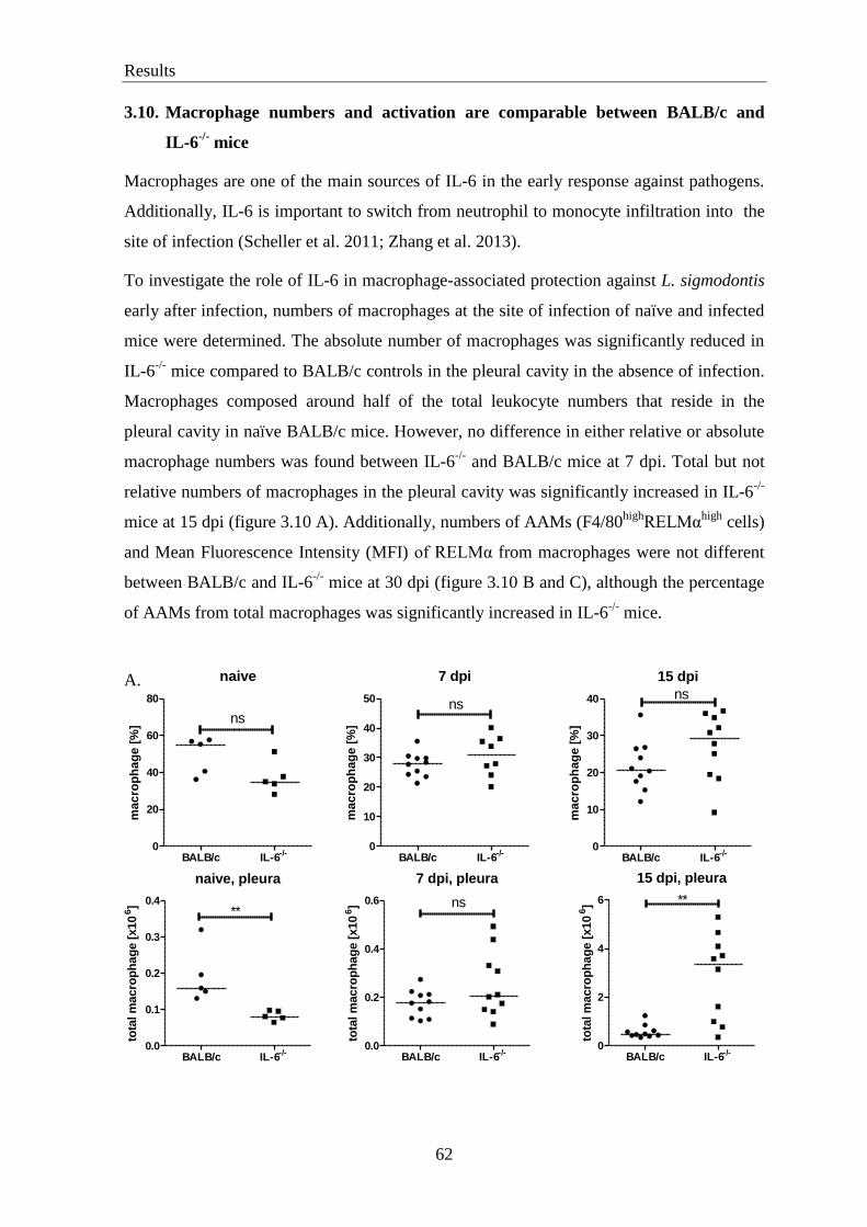
Results
62
3.10. Macrophage numbers and activation are comparable between BALB/c and
IL-6-/-
mice
Macrophages are one of the main sources of IL-6 in the early response against pathogens.
Additionally, IL-6 is important to switch from neutrophil to monocyte infiltration into the
site of infection (Scheller et al. 2011; Zhang et al. 2013).
To investigate the role of IL-6 in macrophage-associated protection against L. sigmodontis
early after infection, numbers of macrophages at the site of infection of naïve and infected
mice were determined. The absolute number of macrophages was significantly reduced in
IL-6-/-
mice compared to BALB/c controls in the pleural cavity in the absence of infection.
Macrophages composed around half of the total leukocyte numbers that reside in the
pleural cavity in naïve BALB/c mice. However, no difference in either relative or absolute
macrophage numbers was found between IL-6-/-
and BALB/c mice at 7 dpi. Total but not
relative numbers of macrophages in the pleural cavity was significantly increased in IL-6-/-
mice at 15 dpi (figure 3.10 A). Additionally, numbers of AAMs (F4/80high
RELMαhigh
cells)
and Mean Fluorescence Intensity (MFI) of RELMα from macrophages were not different
between BALB/c and IL-6-/-
mice at 30 dpi (figure 3.10 B and C), although the percentage
of AAMs from total macrophages was significantly increased in IL-6-/-
mice.
A.
naive
BALB/c IL-6-/-
0
20
40
60
80
ns
ma
cro
ph
ag
e [
%]
7 dpi
BALB/c IL-6-/-
0
10
20
30
40
50 ns
ma
cro
ph
ag
e [
%]
15 dpi
BALB/c IL-6-/-
0
10
20
30
40 ns
ma
cro
ph
ag
e [
%]
naive, pleura
BALB/c IL-6-/-
0.0
0.1
0.2
0.3
0.4**
tota
l m
acro
ph
ag
e [
x10
6]
7 dpi, pleura
BALB/c IL-6-/-
0.0
0.2
0.4
0.6 ns
tota
l m
acro
ph
ag
e [
x10
6]
15 dpi, pleura
BALB/c IL-6-/-0
2
4
6 **
tota
l m
acro
ph
ag
e [
x10
6]

Results
63
BALB/c IL-6-/-
0
10
20
30
40
50 ns
Alt
ern
ati
vely
Acti
vate
d
Macro
ph
ag
e (
AA
M)
[%]
BALB/c IL-6-/-
0
10000
20000
30000
40000
nsA
ltern
ati
vely
Acti
vate
d
Macro
ph
ag
e (
AA
M)
[MF
I]
BALB/c IL-6-/-
0
20
40
60
80
100**
AA
M f
rom
to
tal
macro
ph
ag
es [
%]
B. FMO RELMα BALB/c IL-6-/-
C.
Figure 3.10.1. Numbers of macrophages are not different between BALB/c and IL-6-/-
mice.
Relative (top row) and absolute (bottom row) numbers of macrophages which were determined in
the pleural cavity of naïve (left), 7 (middle) and 15 days (right) L. sigmodontis infected BALB/c
and IL-6-/-
mice (A). AAMs in BALB/c (middle) and IL-6-/-
(right) mice at 30 dpi were determined
in the pleural cavity using FACS by firstly gating on F4/80high
RELMαhigh
using florescence minus
one (FMO) approach (left) (B). Frequency (left) and MFI (middle) of AAMs as well as and
percentage of AAMs from total pleural macrophages (right) at 30 dpi were determined with FACS
(C). (Representative of 2 experiments with minimum of 5 mice per group. Data is shown as
median, ns: not significant, ** p < 0.01).
In order to investigate whether IL-6 deficiency affected macrophage activation,
macrophages were purified from the peritoneal cavity after thioglycollate enrichment and
stimulated with different stimuli. No difference in TNFα (figure 3.10.2 A) and MIP-2
(figure 3.10.2 B) production after stimulation with LsAg was found in IL-6-/-
and BALB/c
mice. As expected, IL-6 production by macrophages following stimulation with LsAg and
P3C was determined in BALB/c but not IL-6-/-
mice (figure 3.10.2 C).
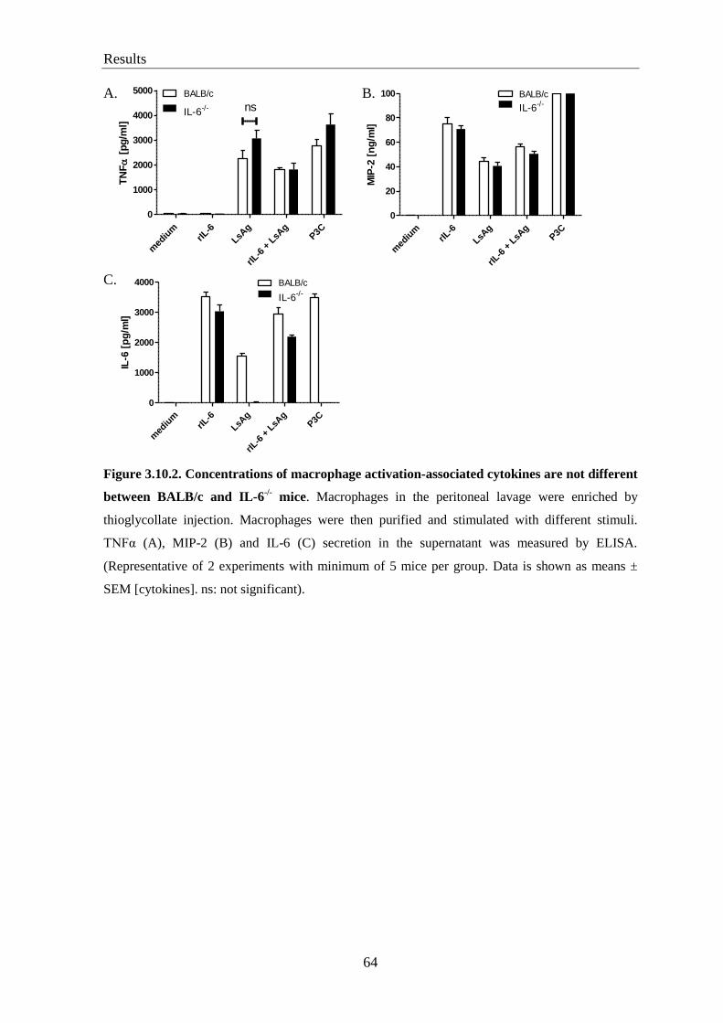
Results
64
med
ium
rIL-6
LsAg
rIL-6
+ L
sAg
P3C
0
1000
2000
3000
4000 BALB/c
IL-6-/-
IL-6
[p
g/m
l]
A. B.
C.
Figure 3.10.2. Concentrations of macrophage activation-associated cytokines are not different
between BALB/c and IL-6-/-
mice. Macrophages in the peritoneal lavage were enriched by
thioglycollate injection. Macrophages were then purified and stimulated with different stimuli.
TNFα (A), MIP-2 (B) and IL-6 (C) secretion in the supernatant was measured by ELISA.
(Representative of 2 experiments with minimum of 5 mice per group. Data is shown as means ±
SEM [cytokines]. ns: not significant).
med
ium
rIL-6
LsAg
rIL-6
+ L
sAg
P3C
0
1000
2000
3000
4000
5000 BALB/c
IL-6-/- ns
TN
F
[p
g/m
l]
med
ium
rIL-6
LsAg
rIL-6
+ L
sAg
P3C
0
20
40
60
80
100 BALB/c
IL-6-/-
MIP
-2 [
ng
/ml]
Results
65
3.11. Decreased relative, but not absolute numbers of B cells in IL-6-/-
mice at early
time points of L. sigmodontis infection
IL-6 was first described as a B cell stimulatory factor (BCSF) which was originally
identified as a B cell differentiation factor. IL-6 supports the development and maturation
of B cells (Ishihara and Hirano 2002; Scheller et al. 2011). In addition, although B cells are
not important for worm survival and growth in L. sigmodontis infection, it was reported
that lack of B-cells prevents the development of filarial patency in primary infection
(Martin et al. 2001).
To investigate the role of IL-6 on B cells during L. sigmodontis infection, B cell
populations were determined in the blood as well as in the pleural cavity by flow
cytometry. Significantly decreased relative but not absolute numbers of total B cells in IL-
6-/-
mice were observed at 7 and 15 dpi, both in the circulation (figure 3.11 A) and in the
pleural cavity (figure 3.11 B and C). However, no significant differences in B cell numbers
between both groups occurred in naive mice (figure 3.11 A, B and C, left row), as well as
at 30, 60 and 90 dpi (data not shown).
3.12. Decreased percentage of B2 cells in IL-6-/-
mice at early time points of infection
Since B2 cells (CD19+CD23
+ B cells) are important for the occurrence of microfilaraemia
in L. sigmodontis infection (Martin et al. 2001), it was investigated whether the B2 cell
subset was normal in IL-6-/-
mice. The relative number of B2 cells was significantly
decreased in the peripheral blood (figure 3.12 A) and the pleural cavity (figure 3.12 B) of
IL-6-/-
mice at 7 (top rows) and 15 (bottom rows) dpi but not at later time points (data not
shown). However, absolute numbers of B2 cells in the pleural cavity were not different
between BALB/c and IL-6-/-
mice at 7 and 15 dpi (figure 3.12 C).
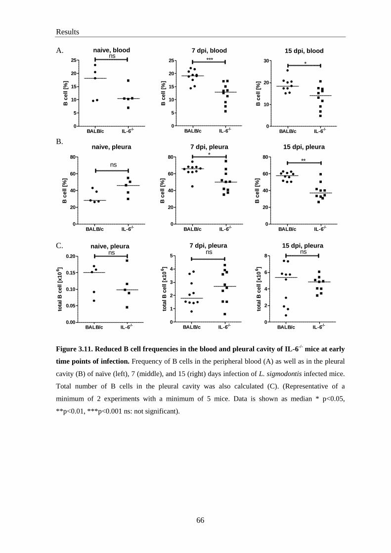
Results
66
A.
B.
C.
Figure 3.11. Reduced B cell frequencies in the blood and pleural cavity of IL-6-/-
mice at early
time points of infection. Frequency of B cells in the peripheral blood (A) as well as in the pleural
cavity (B) of naïve (left), 7 (middle), and 15 (right) days infection of L. sigmodontis infected mice.
Total number of B cells in the pleural cavity was also calculated (C). (Representative of a
minimum of 2 experiments with a minimum of 5 mice. Data is shown as median * p<0.05,
**p<0.01, ***p<0.001 ns: not significant).
naive, pleura
BALB/c IL-6-/-
0
20
40
60
80
ns
B c
ell [
%]
7 dpi, pleura
BALB/c IL-6-/-
0
20
40
60
80 *B
ce
ll [
%]
15 dpi, pleura
BALB/c IL-6-/-
0
20
40
60
80**
B c
ell [
%]
naive, pleura
BALB/c IL-6-/-
0.00
0.05
0.10
0.15
0.20ns
tota
l B
cell [
x10
6]
7 dpi, pleura
BALB/c IL-6-/-
0
1
2
3
4
5ns
tota
l B
cell [
x10
6]
15 dpi, pleura
BALB/c IL-6-/-
0
2
4
6
8ns
tota
l B
cell [
x10
6]
naive, blood
BALB/c IL-6-/-
0
5
10
15
20
25ns
B c
ell [
%]
7 dpi, blood
BALB/c IL-6-/-
0
5
10
15
20
25 ***
B c
ell
[%
]
15 dpi, blood
BALB/c IL-6-/-
0
10
20
30*
B c
ell
[%
]
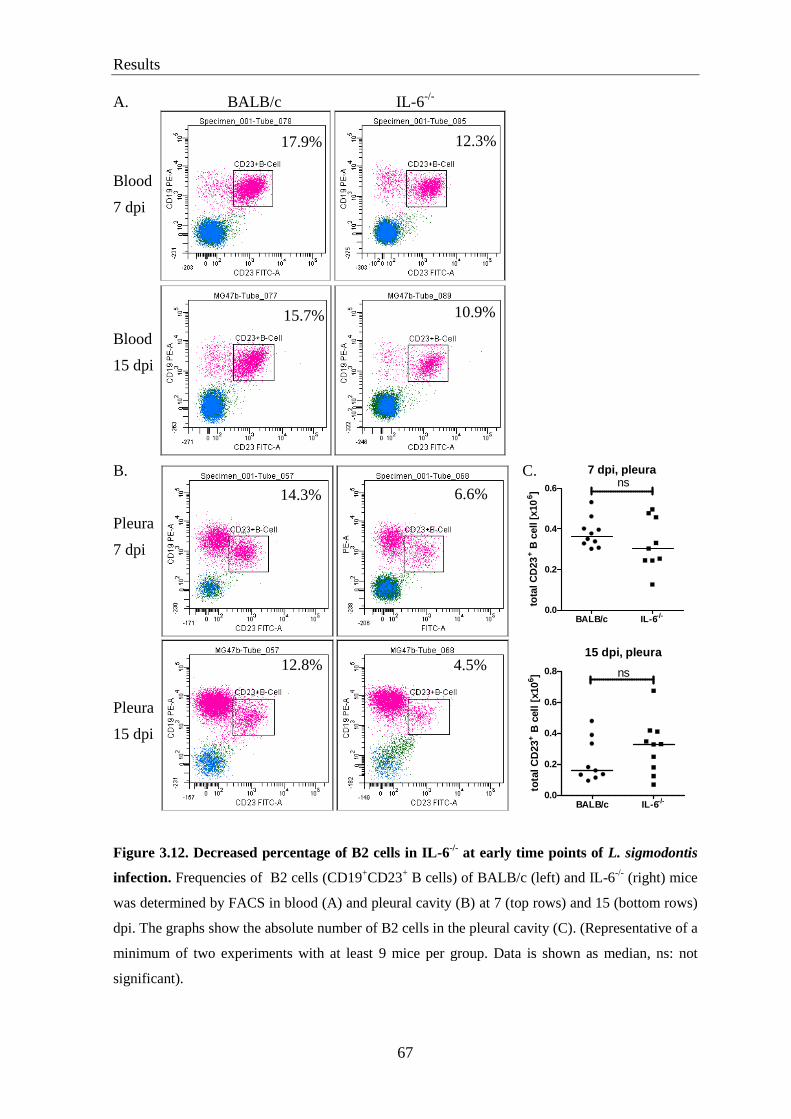
Results
67
A. BALB/c IL-6-/-
Blood
7 dpi
Blood
15 dpi
B. C.
Pleura
7 dpi
Pleura
15 dpi
Figure 3.12. Decreased percentage of B2 cells in IL-6-/-
at early time points of L. sigmodontis
infection. Frequencies of B2 cells (CD19+CD23
+ B cells) of BALB/c (left) and IL-6
-/- (right) mice
was determined by FACS in blood (A) and pleural cavity (B) at 7 (top rows) and 15 (bottom rows)
dpi. The graphs show the absolute number of B2 cells in the pleural cavity (C). (Representative of a
minimum of two experiments with at least 9 mice per group. Data is shown as median, ns: not
significant).
17.9% 12.3%
15.7%
10.9%
7 dpi, pleura
BALB/c IL-6-/-0.0
0.2
0.4
0.6ns
tota
l C
D23
+ B
cell [
x10
6]
15 dpi, pleura
BALB/c IL-6-/-
0.0
0.2
0.4
0.6
0.8 ns
tota
l C
D23
+ B
cell
[x10
6]
14.3% 6.6%
12.8% 4.5%
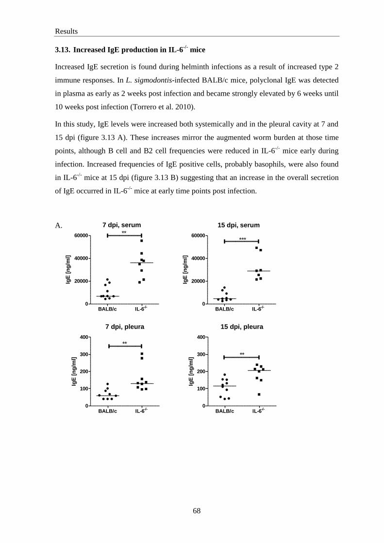
Results
68
3.13. Increased IgE production in IL-6-/-
mice
Increased IgE secretion is found during helminth infections as a result of increased type 2
immune responses. In L. sigmodontis-infected BALB/c mice, polyclonal IgE was detected
in plasma as early as 2 weeks post infection and became strongly elevated by 6 weeks until
10 weeks post infection (Torrero et al. 2010).
In this study, IgE levels were increased both systemically and in the pleural cavity at 7 and
15 dpi (figure 3.13 A). These increases mirror the augmented worm burden at those time
points, although B cell and B2 cell frequencies were reduced in IL-6-/-
mice early during
infection. Increased frequencies of IgE positive cells, probably basophils, were also found
in IL-6-/-
mice at 15 dpi (figure 3.13 B) suggesting that an increase in the overall secretion
of IgE occurred in IL-6-/-
mice at early time points post infection.
A.
7 dpi, serum
BALB/c IL-6-/-
0
20000
40000
60000 **
IgE
[n
g/m
l]
7 dpi, pleura
BALB/c IL-6-/-
0
100
200
300
400
**
IgE
[n
g/m
l]
15 dpi, serum
BALB/c IL-6-/-
0
20000
40000
60000***
IgE
[n
g/m
l]
15 dpi, pleura
BALB/c IL-6-/-
0
100
200
300
400
**
IgE
[n
g/m
l]

Results
69
B.
Figure 3.13. Increased production of IgE in IL-6-/-
mice at early time points of L. sigmodontis
infection. Concentration of total IgE in the serum (top) and pleural lavage (bottom) of infected
BALB/c and IL-6-/-
mice at 7 (left) and 15 (right) days post infection (A). IgE+ cells were identified
by FACS in the blood of 15 days infected BALB/c (left) and IL-6-/-
(middle) mice, and their
percentages (right) (B). (Representative of a minimum of 2 experiments with a minimum of 8 mice
per group. Data is shown as median. ** p<0.01 ***p<0.001).
3.14. IgG production is independent of IL-6
IL-6 is known to promote B cell differentiation into plasma cells (Kishimoto 1989).
Additionally, IL-6 is reported to be crucial for promoting IgG1 production by B cells by
inducing IL-21 production (Neveu et al. 2009). Therefore, IgG1 and IgG2a production
were investigated in the serum of mice at 15, 30 and 60 days post infection. No difference
in the production of IgG was found at any of those time points between the two groups
(figure 3.14, middle and right columns).
BALB/c IL-6-/-
0.0
0.5
1.0
1.5
2.0
2.5**
IgE
+cel
l[%
]

Results
70
Figure 3.14. IL-6 deficiency does not lead to a decreased IgG production during L.
sigmodontis infection. Levels of L. sigmodontis specific IgG1 (top row) and IgG2a (bottom row)
in the serum of infected animals at 15 (left column), 30 (middle column) and 60 (right column) dpi
were measured by ELISA. (Representative of two experiments with a minimum of 6 mice per
group).
3.15. IL-6 deficiency does not affect CD4 T cell numbers and frequencies in the
pleural cavity during L. sigmodontis infection
IL-6 is important for lymphocyte infiltration into the infected tissues by increasing the
expression of cell adhesion molecules (Scheller et al. 2011). Additionally, CD4 T cells are
crucial for L. sigmodontis protection as the depletion of CD4 T cells with anti-CD4
antibodies in BALB/c mice led to an increased worm burden and microfilaremia 8 weeks
post infection (Al-Qaoud et al. 1997).
The number of CD4 T cells was investigated prior to infection, 7 and 15 dpi as well as
during chronic infection. Absolute, but not relative numbers of CD4 T cells in the pleural
cavity were decreased in naive IL-6-/-
mice (figure 3.15 B, left row). No difference in CD4
T cell numbers were found at 7 and 15 dpi in the blood (figure 3.15 A) as well as in pleural
cavity (figure 3.15 B) between BALB/c and IL-6-/-
mice. CD4 T cell numbers were also
not different at later time points of infection (data not shown).
15 dpi, IgG1
100 400 1600 6400 25000 100000
0.1
0.2
0.3
0.4
0.5 BALB/c
IL-6-/-
dilution factor
OD
45
0n
m
30 dpi, IgG1
100 400 1600 6400 25000 100000
0.2
0.4
0.6 BALB/c
IL-6-/-
dilution factor
OD
45
0n
m
60 dpi, IgG1
100 400 1600 6400 25000 100000
0.5
1.0
1.5 BALB/c
IL-6-/-
dilution factor
OD
45
0n
m
15 dpi, IgG2a
100 400 1600 6400 25000 100000
0.2
0.4
0.6
0.8 BALB/c
IL-6-/-
dilution factor
OD
45
0n
m
30 dpi, IgG2a
100 400 1600 6400 25000 100000
0.05
0.10
0.15
0.20
0.25 BALB/c
IL-6-/-
dilution factor
OD
45
0n
m
60 dpi, IgG2a
100 400 1600 6400 25000 100000
0.05
0.10
0.15
0.20 BALB/c
IL-6-/-
dilution factor
OD
45
0n
m

Results
71
A.
B.
Figure 3.15. CD4 T cell numbers and frequencies are similar between BALB/c and IL-6-/-
mice. BALB/c and IL-6-/-
mice were infected with L. sigmodontis. Percentage of CD4 T cells in the
blood (A) and pleural cavity (B, top row) of naive (left), and 7 (middle) and 15 (right) days infected
animals were analyzed using FACS. Total CD4 T cells are also given (B, bottom row).
(Representative of a minimum of 2 experiments with a minimum of 5 mice per group. Data is
shown as median. * p<0.05 ns: not significant).
naive, blood
BALB/c IL-6-/-
0
10
20
30
40
ns
CD
4 T
ce
ll [
%]
7 dpi, blood
BALB/c IL-6-/-
0
10
20
30
40ns
CD
4 T
ce
ll [
%]
15 dpi, blood
BALB/c IL-6-/-
0
10
20
30 ns
CD
4 T
ce
ll [
%]
naive, pleura
BALB/c IL-6-/-
0
5
10
15
20
ns
CD
4 T
ce
ll [
%]
7 dpi, pleura
BALB/c IL-6-/-
0
5
10
15
20 nsC
D4
T c
ell [
%]
15 dpi, pleura
BALB/c IL-6-/-
0
5
10
15
20ns
CD
4 T
ce
ll [
%]
naive, pleura
BALB/c IL-6-/-
0.00
0.01
0.02
0.03
0.04*
tota
l C
D4 T
cell
[x10
6]
7 dpi, pleura
BALB/c IL-6-/-
0.0
0.1
0.2
0.3
0.4 ns
tota
l C
D4 T
cell
[x10
6]
15 dpi, pleura
BALB/c IL-6-/-
0.0
0.5
1.0
1.5
ns
tota
l C
D4 T
cell
[x10
6]

Results
72
3.16. IL-6 deficiency does not affect the number of regulatory T cells (Treg) and IL-17
production during L. sigmodontis infection
Several studies have demonstrated the importance of IL-6 for CD4 T cell differentiation
(Dienz and Rincon 2009; Scheller et al. 2011). IL-6 is involved in the differentiation of Treg
and Th17 cells (Kimura and Kishimoto 2010), which were both shown to be important for
filarial immunity (Specht and Hoerauf 2012). Treg can suppress host protective immune
responses during L. sigmodontis infection and lead to an increased worm burden during
chronic infection (Taylor et al. 2005).
The numbers of Treg and Th17 cells in spleens of infected mice were determined by FACS
after 6 hours of in vitro culture with LsAg and anti-CD3/CD28 as a positive control. Due
to the low number of CD4+IL-17
+ T cells which were detected by FACS, IL-17 levels were
measured in the supernatant after splenocyte culture for 3 days. Treg were defined by FACS
as CD4+FoxP3
+ cells (figure 3.16 A). Interestingly, there was no difference in Treg (figure
3.16 B and C) frequencies and IL-17 levels (figure 3.16 D) in LsAg-stimulated splenocytes
of BALB/c and IL-6-/-
mice at 7 and 15 dpi. Levels of IL-17 in the pleural lavage of
infected mice were below the limit of detection (15.6 pg/ml; data not shown).
A.

Results
73
B. medium LsAg CD3/CD28
C.
D.
Figure 3.16. IL-6 deficiency does not affect Treg numbers and IL-17 levels during L.
sigmodontis infection. Splenocytes from L. sigmodontis infected IL-6-/-
and BALB/c mice were
incubated with medium, LsAg and αCD3/CD28 for 4 hours with monensin and intracellularly
stained for FoxP3 and IL-17A. Population of CD4+
was determined by FACS (top), followed by
gating of FoxP3+ (second row, left) and IL-17A
+ (second row, right) cells using the FMO approach
(A). Gating of CD4+FoxP3
+ and CD4
+IL-17A
+ cells after stimulation with medium (left), LsAg
(middle) and αCD3/CD28 (right) (B). Frequency of CD4+FoxP3
+ cells at 7 (left) and 15 (right) dpi
(C). IL-17 secretion from splenocyte culture after stimulation for 72 hours (D). (Representative of a
minimum of 2 experiments with at least 10 mice per group. Data is shown as means ± SEM. ns: not
significant).
7dpi
medium LsAg CD3+CD280
5
10
15 BALB/c
IL-6-/-
ns
Fo
xP
3+
of
CD
4+
[%
]
15 dpi
medium LsAg CD3+CD280
5
10
15 BALB/c
IL-6-/-
ns
Fo
xP
3+
of
CD
4+
[%
]
7 dpi
med
ium
rIL-6
LsAg
rIL-6
+LsA
g
ConA
0
10
20
30
40
50
500
1000
1500 BALB/c
IL-6-/-
ns
IL-1
7 [
pg
/ml]
15 dpi
med
ium
rIL-6
LsAg
rIL-6
+LsA
g
ConA
0
20
40
60
80
100
1000200030004000
ns
BALB/c
IL-6-/-
IL-1
7 [
pg
/ml]
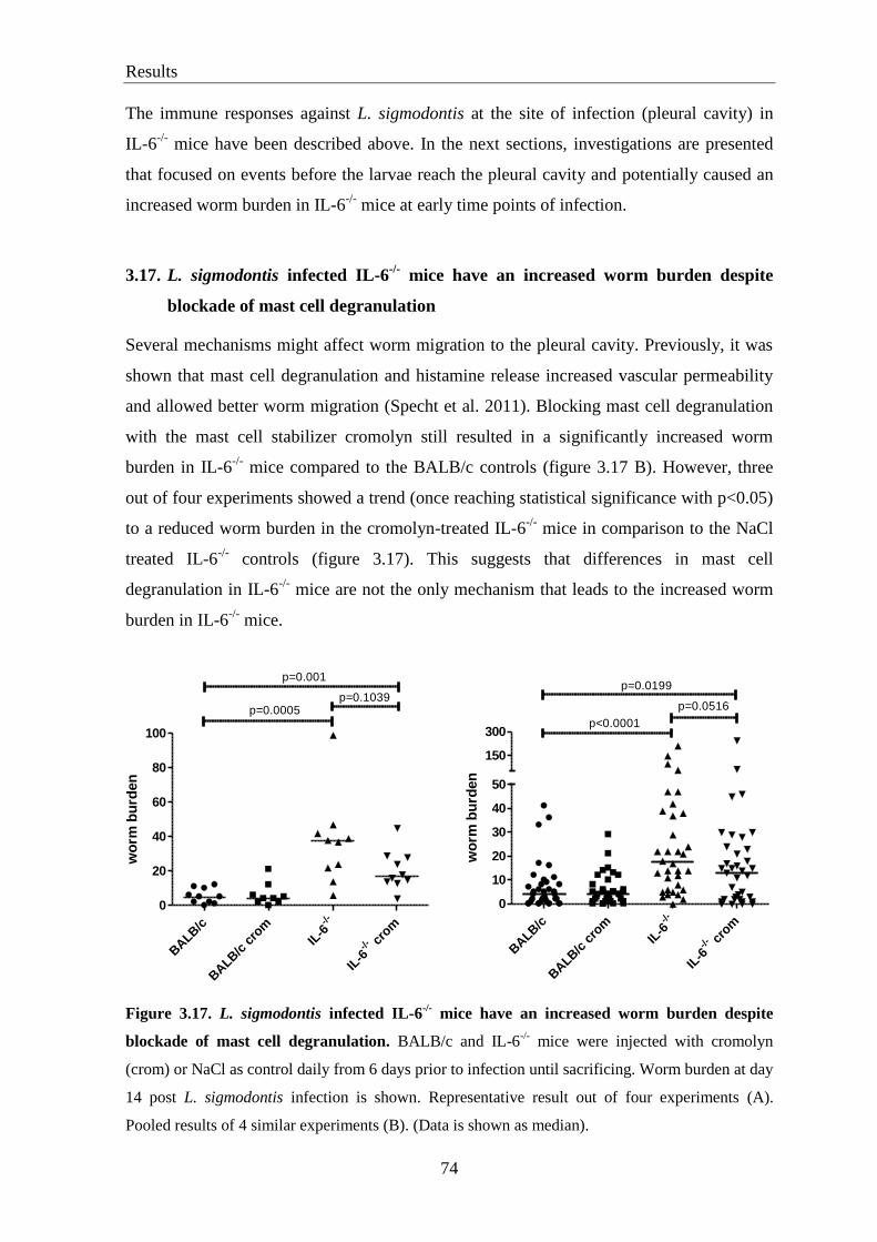
Results
74
The immune responses against L. sigmodontis at the site of infection (pleural cavity) in
IL-6-/-
mice have been described above. In the next sections, investigations are presented
that focused on events before the larvae reach the pleural cavity and potentially caused an
increased worm burden in IL-6-/-
mice at early time points of infection.
3.17. L. sigmodontis infected IL-6-/-
mice have an increased worm burden despite
blockade of mast cell degranulation
Several mechanisms might affect worm migration to the pleural cavity. Previously, it was
shown that mast cell degranulation and histamine release increased vascular permeability
and allowed better worm migration (Specht et al. 2011). Blocking mast cell degranulation
with the mast cell stabilizer cromolyn still resulted in a significantly increased worm
burden in IL-6-/-
mice compared to the BALB/c controls (figure 3.17 B). However, three
out of four experiments showed a trend (once reaching statistical significance with p<0.05)
to a reduced worm burden in the cromolyn-treated IL-6-/-
mice in comparison to the NaCl
treated IL-6-/-
controls (figure 3.17). This suggests that differences in mast cell
degranulation in IL-6-/-
mice are not the only mechanism that leads to the increased worm
burden in IL-6-/-
mice.
Figure 3.17. L. sigmodontis infected IL-6-/-
mice have an increased worm burden despite
blockade of mast cell degranulation. BALB/c and IL-6-/-
mice were injected with cromolyn
(crom) or NaCl as control daily from 6 days prior to infection until sacrificing. Worm burden at day
14 post L. sigmodontis infection is shown. Representative result out of four experiments (A).
Pooled results of 4 similar experiments (B). (Data is shown as median).
BALB
/c
BALB
/c c
rom
-/-
IL-6
cro
m
-/-
IL-6
0
20
40
60
80
100
p=0.0005p=0.1039
p=0.001
wo
rm b
urd
en
BALB
/c
BALB
/c c
rom
-/-
IL-6
cro
m
-/-
IL-6
0
10
20
30
40
50
150
300p<0.0001
p=0.0516
p=0.0199
wo
rm b
urd
en
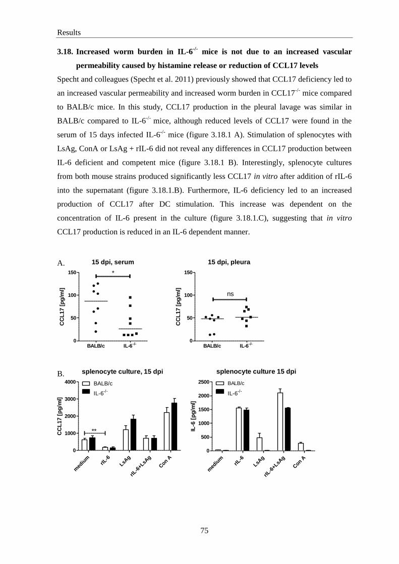
Results
75
3.18. Increased worm burden in IL-6-/-
mice is not due to an increased vascular
permeability caused by histamine release or reduction of CCL17 levels
Specht and colleagues (Specht et al. 2011) previously showed that CCL17 deficiency led to
an increased vascular permeability and increased worm burden in CCL17-/-
mice compared
to BALB/c mice. In this study, CCL17 production in the pleural lavage was similar in
BALB/c compared to IL-6-/-
mice, although reduced levels of CCL17 were found in the
serum of 15 days infected IL-6-/-
mice (figure 3.18.1 A). Stimulation of splenocytes with
LsAg, ConA or LsAg + rIL-6 did not reveal any differences in CCL17 production between
IL-6 deficient and competent mice (figure 3.18.1 B). Interestingly, splenocyte cultures
from both mouse strains produced significantly less CCL17 in vitro after addition of rIL-6
into the supernatant (figure 3.18.1.B). Furthermore, IL-6 deficiency led to an increased
production of CCL17 after DC stimulation. This increase was dependent on the
concentration of IL-6 present in the culture (figure 3.18.1.C), suggesting that in vitro
CCL17 production is reduced in an IL-6 dependent manner.
A.
B.
splenocyte culture 15 dpi
med
ium
rIL-6
LsAg
rIL-6
+LsAg
Con A
0
500
1000
1500
2000
2500 BALB/c
IL-6-/-
IL-6
[p
g/m
l]
splenocyte culture, 15 dpi
med
ium
rIL-6
LsAg
rIL-6
+LsAg
Con A
0
1000
2000
3000
4000
**
BALB/c
IL-6-/-
CC
L17 [
pg
/ml]
15 dpi, serum
BALB/c IL-6-/-
0
50
100
150 *
CC
L17 [
pg
/ml]
15 dpi, pleura
BALB/c IL-6-/-
0
50
100
150
ns
CC
L17 [
pg
/ml]

Results
76
C.
Figure 3.18.1. In vitro CCL17 production is reduced in an IL-6 dependent manner. Levels of
CCL17 in the serum (left) and pleural lavage (right) 15 days post L. sigmodontis infection (A).
Splenocytes from 15 days infected mice were stimulated with rIL-6, LsAg, rIL-6 + LsAg and
ConA for 72 hours and CCL17 (left) and IL-6 (right) production is given (B). DCs were derived
from bone marrow of naive mice and cultured for 18 hours with different stimuli and CCL17 (left)
and IL-6 (right) production is shown (C). (Representative of 2 experiments with a minimum of 6
(A and C) or 3 (B) mice per group. Each stimulation was conducted in triplicate. Data is shown as
median [in vivo] or means ± SEM [in vitro]. * p< 0.05, ** p< 0.01, ns: not significant).
med
ium
LsAg
rIL-6
5 n
g/ml
rIL-6
1 n
g/ml
rIL-6
200
pg/m
l
LsAg+r
IL-6
5 n
g/ml
LsAg+r
IL-6
1 n
g/ml
LsAg+r
IL-6
200
pg/m
lLPS
0
10
20
30
40
50100150
IL-6-/-
BALB/c
CC
L17 [
ng
/ml]
med
ium
LsAg
rIL-6
5 n
g/ml
rIL-6
1 n
g/ml
rIL-6
200
pg/m
l
LsAg+r
IL-6
5 n
g/ml
LsAg+r
IL-6
1 n
g/ml
LsAg+r
IL-6
200
pg/m
lLPS
0
500
1000
1500
2000
5000
10000
IL-6
[p
g/m
l]

Results
77
Given that histamine mediates vasodilatation that may facilitate the migration of L.
sigmodontis larvae to the pleural cavity (Specht et al. 2011), the role of histamine was
analyzed by blocking histamine receptors in vivo. Usage of histamine receptor antagonists
1 and 2 did not reduce the worm burden in IL-6-/-
mice compared to the control groups
(figure 3.18.2).
Figure 3.18.2. Blocking of histamine receptors does not decrease L. sigmodontis worm burden
in IL-6-/-
mice. BALB/c and IL-6-/-
mice were injected with histamine receptor antagonists
pyrilamine and cimetidine in PBS (AH1/2) or PBS alone two hours prior to and 1 hour post
infection, followed by daily injections until 5 dpi. Worm burden was determined at 14 dpi.
(Representative of 2 experiments with a minimum of 9 animals per group. Data is shown as
median. * p<0.05 ns: not significant).
In order to investigate a role of vascular permeability in increasing the worm burden in IL-
6-/-
mice at an early time post infection, the role of IL-6 in vascular permeability of the skin
was analyzed. BALB/c and IL-6-/-
mice were injected with LsAg i.c. followed by i.v.
Evans Blue injection and dye leakage from blood vessels into the tissue was quantified.
There was no difference of Evans Blue leakage in IL-6-/-
compared to BALB/c mice.
However, significantly increased levels of Evans Blue were observed after LsAg and anti-
FcεR1 (MAR-1) treatment compared to PBS injection in both types of mice (figure
3.18.3).
BALB
/c
BALB
/c A
H1/
2 -/-
IL-6
AH1/
2
-/-
IL-6
0
20
40
60
*
ns
wo
rm b
urd
en

Results
78
BALB
/c P
BS
PBS
-/-
IL-6
BALB
/c L
sAg
LsA
g
-/-
IL-6
BALB
/c M
AR-1
MAR-1
-/-
IL-6
0
5000
10000
15000
20000
ns
ns
**
Evan
s b
lue [
ng
/ml]
Figure 3.18.3. Vascular permeability is similar between BALB/c and IL-6-/-
mice. BALB/c and
IL-6-/-
mice were injected i.c. into the ears with LsAg or MAR-1 in PBS or PBS alone, followed by
Evans Blue injection i.v. 3 minutes later. After 10 minutes, the complete ears were cut off and
Evans Blue leakage was quantified at 620 nm using a plate reader. (Representative of 3
experiments with different settings and a minimum of 6 animals per group. Data is shown as
median. ** p<0.01 ns: not significant).
3.19. Subcutaneous L3 inoculation leads to a similar worm recovery at 15 dpi
It has been shown above that increased vascular permeability is not the cause for the
increased worm burden in IL-6-/-
mice. Due to the fact that the increased worm burden in
IL-6-/-
mice occurred at a very early time point post infection, it was hypothesized that the
increased worm burden in IL-6-/-
mice may be due to an increased worm survival or
migration in IL-6-/-
mice when they pass the first barrier within the mouse, the skin. To
investigate if the skin plays a major role in the increased worm burden in IL-6-/-
mice, mice
were inoculated with 40 L3 subcutaneously. While after natural infection IL-6-/-
mice had a
higher worm burden at 15 dpi, s.c. injection did not reveal a difference in worm burden
between IL-6-/-
and BALB/c mice (figure 3.19). This suggests that immune responses that
occur in the epidermis and dermis may play a major role for protection and lead to
increased worm survival or migration in IL-6-/-
mice.

Results
79
Figure 3.19. Circumventing the skin barrier by s.c. injection results in a similar worm load in
BALB/c and IL-6-/-
at 15 dpi. Mice were infected with L. sigmodontis by injecting 40 L3 s.c.
Infection was terminated at 15 dpi and worm burden in the pleural cavity is displayed. (Pooled
results from two independent experiments. Data is shown as median. ns: not significant).
15 dpi
BALB/c IL-6-/-
0
2
4
6
8
10ns
wo
rm b
urd
en

Discussion
80
4. Discussion
This study showed an increased worm burden in IL-6-/-
mice during the early phase of
infection with L. sigmodontis in comparison to BALB/c mice, but not during the chronic
phase of infection. Potential mechanisms leading to an increased worm burden early in
infection and similar worms in later times post infection are: (1) dampening of the host
immune response in IL-6-/-
mice during acute infection, (2) increased host immune
response in IL-6-/-
mice during later time points post infection, (3) increased
survival/reduced destruction of L3 in IL-6-/-
mice during the early phase of infection, (4)
reduced survival/increased destruction of L4 or adult worms in IL-6-/-
during the late phase
of infection, or (5) increased migration of L3 into the pleural cavity.
4.1. Immune responses against L. sigmodontis
4.1.1. IL-6 signaling during L. sigmodontis infection
This study shows for the first time that during acute L. sigmodontis filarial infection
signaling of IL-6 involves trans-signaling. As was previously shown in a review by
Scheller and colleagues, IL-6 trans-signaling is mediated by sIL-6R, which is essential for
pro-inflammatory immune responses, and differs from mIL-6R-signaling that is important
during anti-inflammatory responses (Scheller et al. 2011). Although this has been shown
for several pathogens (Rose-John 2012; Scheller et al. 2011), the occurrence of IL-6 trans-
signaling during nematode infection was unknown. sIL-6R was produced in low amounts
in the pleural lavage of naive mice but increased during acute infection with L.
sigmodontis. This is may be due to increased neutrophil apoptosis in the pleural cavity
following infection, which correlates with slightly reduced neutrophil numbers in the
pleural cavity during acute infection compared to naive animals. As was previously
described (Scheller et al. 2011), sIL-6R is generated by two mechanisms; first by
proteolytic cleavage of mIL-6R mainly after apoptosis of neutrophils and second by
secretion from neutrophils and monocytes of an alternatively sliced mIL-6R product. In
vitro stimulation of neutrophils from naive mice with LsAg increased the level of sIL-6R
compared to unstimulated neutrophils, although this difference did not reach statistical
significance. This suggests that sIL-6R may be involved in immune responses during acute
L. sigmodontis infection. Several proteins that are crucial in the IL-6 trans-signaling
pathway such as gp130 and STAT3 should be addressed in the future to obtain a complete

Discussion
81
overview of IL-6 trans-signaling during filarial infection. Differences in sIL-6R between
BALB/c and IL-6-/-
mice both in vivo and in vitro were not observed due to the fact that IL-
6-/-
was generated by disrupting the IL-6 gene, but not the IL-6R gene (Kopf et al. 1994).
4.1.2. Neutrophils
The role of neutrophils in worm survival both in the early and late phase of infection has
been previously described (Makepeace et al. 2012; Martin et al. 2000a; Saeftel et al. 2001,
2003). In the acute stage of infection, neutrophils are reported to be the main cell type that
is crucial for worm destruction (Martin et al. 2000a). In contrast, Saeftel and colleagues
(Saeftel et al. 2001, 2003) showed that neutrophils are crucial for the encapsulation process
at the late stage of infection in an IFNγ-dependent manner (50 and 80 dpi). Since, IL-6 is
required for neutrophil mobilization from the bone marrow to the periphery and to the site
of infection during acute inflammation (Murphy 2012; Scheller et al. 2011), it is important
to determine the role of IL-6 on neutrophil migration during filarial infection and their
ability in worm destruction both at early and late phases of infection.
In the current study IL-6 deficiency had no impact on neutrophil migration to the periphery
and pleural cavity at 7 and 15 dpi as well as at the late stage of L. sigmodontis infection.
Neutrophil migration into the pleural cavity was low after the first week of infection, both
in BALB/c and IL-6-/-
mice. This was confirmed by a time course experiment in L.
sigmodontis-infected BALB/c mice showing increased neutrophil numbers in the pleural
cavity after approximately 28 dpi. Additionally, neutrophil activation was not affected by
IL-6 deficiency as the production of the neutrophil associated cytokines TNFα and MIP-2
was similar between BALB/c and IL-6-/-
mice.
IFNγ, which induces neutrophil activation and phagocytosis (Saeftel et al. 2001, 2003) was
not detectable during infection in vivo, both in serum and pleural lavage, although IFNγ
secretion in vitro was increased at early but not later time points in IL-6-/-
mice. Given that
IFNγ is implemented in neutrophil-mediated worm destruction during chronic infection but
not early time points post infection (Al-Qaoud et al. 2000; Saeftel et al. 2001, 2003) it is
unlikely that IFNγ led to the increased worm elimination in IL-6-/-
mice after 15 dpi.
Therefore depleting IFNγ before 15 dpi until 30 dpi should be conducted in order to
address the role of IFNγ in neutrophil-mediated worm destruction in IL-6-/-
mice at 30 dpi.
Discussion
82
4.1.3. Macrophages
Another phagocytic cell population that might be involved in worm killing at early
infection time points is macrophage. As previously described, macrophages are capable to
kill filarial larvae but not adult worms by releasing toxic radicals (Ou et al. 1995). As L3 of
B. pahangi induce macrophage activation in vitro (Jeffers et al. 1984), it can be assumed
that an increased worm burden during acute infection and reduced survival of adult worms
at the chronic stage in IL-6-/-
mice might be mediated by macrophage modulation.
However the total number of macrophages at the site of infection did not differ between
IL-6-/-
and BALB/c mice. Similarly, the total number of AAMs as determined at 30 dpi
was not different between BALB/c and IL-6-/-
mice. Additionally, activation of
macrophages was not affected by the lack of IL-6 suggesting that IL-6 is not important for
macrophage function during filarial infection. This may be further confirmed by measuring
the production of toxic radicals such as ROS and NO derivates by macrophages in
response to L. sigmodontis larvae or antigen in BALB/c and IL-6-/-
mice.
4.1.4. B cells
B cells, as reported with µMT mice, are not involved in worm elimination (Martin et al.
2001), although a study by Al-Qaoud et al. showed that worm burden in B1 deficient
BALB/c mice (Xid mice) was significantly increased compared to BALB/c controls at 28
dpi, probably due to a lack of IgM -producing B1 cells (Al-Qaoud et al. 1998). In IL-6-/-
mice, I observed a reduced relative but not absolute number of B cells in the blood and
pleural cavity during early infection. Additionally, the percentage of B2 cells was
decreased in IL-6-/-
mice. Reduced frequencies of B1 cells in IL-6-/-
mice may mediate the
increased worm burden due to a reduced production of IgM. Reduction of B2 cells in
IL-6-/-
mice may also facilitate worm survival, as a study by Mwinzi and colleagues
revealed that B2 cells are important for resistance against S. mansoni by regulating IgE
production (Mwinzi et al. 2009). However, whereas IgM is produced early during infection
induction of IgE does not appear before 4 weeks post infection (Torrero et al. 2010)
suggesting that B2 cells may have an effect on late infection time points, but do not cause
the difference of L. sigmodontis worm burden observed at 15 dpi.
Another variable which is important for immunity against helminth infection is IgG
production (Specht and Hoerauf 2012). L. sigmodontis-specific IgG1 and IgG2a were

Discussion
83
detected before L3 challenge (0 dpi) and increased until 10 dpi in vaccinated mice
compared to primary infected mice (Babayan et al. 2006; Le Goff et al. 2000b). At later
time points of infection (60 dpi), production of L. sigmodontis-specific IgG1 and IgG2a
peaked and no difference of their levels were measured between vaccinated and primary
infected mice (Le Goff et al. 2000b).
As IL-6 is known to be crucial in the end stage of plasma cell differentiation (Kishimoto
2010; Scheller et al. 2011) and IgG production by promoting IL-21 production (Neveu et
al. 2009), it is important to address whether IL-6 deficiency may affect IgG production
during L. sigmodontis infection. Production of both L. sigmodontis-specific IgG1 and
IgG2a was not different between BALB/c and IL-6-/-
mice at 30 and 60 dpi, when no
difference in worm burden and B cells was observed. This is probably due to the fact that
the function of IL-6 in plasma cell differentiation and IgG production can be substituted by
IL-21 as previously described (Eto et al. 2011). Therefore, it would be interesting to further
investigate the role of IL-21 during L. sigmodontis infection of IL-6-/-
mice.
4.1.5. CD4 T cells
CD4 T cells play a significant role in protective immunity against filarial parasites
(Hoerauf et al. 2005; Specht and Hoerauf 2012). Al-Qaoud and colleagues showed a role
for CD4 T cells in protection against filarial infection, as depletion of CD4 T cells led to
increased worm burdens at 28 dpi (Al-Qaoud et al. 1997). The presented study did not
observe a different number of CD4 T cells between BALB/c and IL-6-/-
mice, neither in the
blood nor in the pleural cavity. Since activation, cytokine production and proliferation of
CD4 T cells in IL-6 deficiency were not determined in this study, the role of IL-6 in
activation and proliferation as well as its exact role in CD4 T cell-mediated protection
against filarial worms may be elucidated in further studies.
4.1.5.1. Treg cells
Although the role of IL-6 in inhibiting TGFß-induced Treg differentiation has been reported
several years ago (Bettelli et al. 2006), its role in Treg initiation during filarial infection is
unknown. This study demonstrated that in the absence of IL-6, the number of splenic Treg
after re-stimulation with LsAg was not different compared to BALB/c controls. This
suggests that IL-6 signaling during L. sigmodontis infection has little or no impact on the
Discussion
84
number of Treg in BALB/c mice, which is probably due to the low amount of IL-6
produced during the infection in BALB/c mice, or that IL-6 deficiency did not result in
increased Treg numbers during filarial infection due to another Treg inhibitory mechanism.
Another reason for the similar number of Treg in both types of mice might be due to the
level of TGFß that was secreted during infection and restimulation. As has been previously
described, in certain environmental conditions including filarial infection and presence of
TGFß, naive T cells differentiate into Treg (Belkaid 2007; Hoerauf et al. 2005). In the
presence of IL-6, however, this differentiation shifts towards the Th17 lineage (Kimura and
Kishimoto 2010).
4.1.5.2. IL-17-producing Th17 cells
The role of Th17 differentiation during filariasis is not yet completely known, although a
study by Babu and co-workers showed that IL-17 is one of the cytokines that is responsible
for lymphedema in human B. malayi infection (Babu et al. 2009). Despite the role of IL-6
in IL-17 production, IL-6 deficiency did not affect IL-17 responses in this study.
Accordingly, a study by Kimura and colleagues revealed that differentiation of IL-17
producing Th17 cells can occur in the absence of IL-6 (Kimura et al. 2007). They showed
that lack of IL-6 signaling either by using IL-6-/-
or administration of anti-gp130 or anti-IL-
6R did not prevent the production of IL-17 by naive T cells stimulated with conditioned
medium from LPS-stimulated bone marrow-derived DCs. They suggested another factor
than IL-6, TGFß and IL-23 to regulate Th17 differentiation; however, they could not
address this factor in their paper (Kimura et al. 2007). A study by Yang and colleagues
revealed that IL-21 may substitute IL-6 to induce Th17 differentiation (Yang et al. 2008).
Therefore, IL-21 may compensate for the lack of IL-6 during L. sigmodontis infection and
induce Th17 cells and IL-17 production.
4.1.5.3. Th2 responses
There are two phases of IL-5-mediated worm reduction which are previously described
during murine filarial infection; the first phase occurs in the first two days after infection
and the second phase occurs after three weeks post infection when specific IgG starts to be
produced (Babayan et al. 2005; Martin et al. 2000b, 2000a). Worm destruction in the first
phase tends to be fast, whereas in the second phase, the destruction is slow because the

Discussion
85
worm size increased (Martin et al. 2000a). Optimal destruction and killing of the worms
requires adequate levels of IL-5, eosinophils and antibodies (Le Goff et al. 2000b, 2000a;
Martin et al. 2000b, 2000a; Specht and Hoerauf 2012).
Effective destruction and killing of the worms during the first phase, for instance, occurred
in mice vaccinated with L3. Vaccinated mice have a lower worm rate compared to primary
infected mice as early as 2 dpi due to increased worm destruction and killing, and the rate
was stable for several weeks (Babayan et al. 2006; Martin et al. 2000a). Several studies
have shown the role of IL-5, eosinophils and IgE in this worm destruction (Le Goff et al.
2000b; Martin et al. 2000b, 2000a; Specht and Hoerauf 2012). Production of IL-5 was
increased after splenocyte stimulation with adult worm extract prior to worm inoculation (0
dpi) in vaccinated mice compared to primary infected mice (Le Goff et al. 2000b).
Additionally, IL-5 production by splenocytes was also increased after either adult, L3 and
microfilaria extracts challenge in vaccinated mice at 2 dpi (Babayan et al. 2006).
Furthermore, IL-5 levels were found in higher concentrations in the pleural lavage of
vaccinated mice than in the pleural cavity of primary infected mice after worm challenge at
10 dpi, although levels of IL-5 in the serum is independent of vaccination (Babayan et al.
2006; Martin et al. 2000a).
As the numbers of eosinophils in the pleural cavity, the levels of IL-5 in vitro after
splenocyte culture and the IgE concentrations in the pleural lavage and serum were
significantly increased in IL-6-/-
mice at early time points during infection in this study, it is
unlikely that this result is due to reduced larval destruction in the pleural cavity by
eosinophils, IL-5 and IgE. In fact, increased IL-5, eosinophils and IgE in this study were
probably just an effect of, not a cause for the increased worm burden in IL-6-/-
mice. This
was confirmed by the depletion of IL-5 before and during infection. Depletion of IL-5
resulted in reduced eosinophil numbers in the pleural cavity but did not reduce the worm
burden in IL-6-/-
mice at 15 dpi. This suggests that the increased worm burden in IL-6-/-
mice during early infection is not mediated by eosinophils, IL-5 and IgE.
Elevated IL-5 levels in vitro early during infection were probably due to an increased
polarization towards Th2 in IL-6-/-
caused by the higher worm burden. Babu and Nutman
reported in an in vitro B. malayi study that activation of naive T cell occurred already
within 1 dpi, mainly towards a Th1 response (Babu and Nutman 2003). After 7 dpi, host
immune polarization shifts towards Th2 dominance due to incoming L3 as was shown in a
study by Porthouse and colleagues using B. pahangi (Porthouse et al. 2006). Synergistic
Discussion
86
responses of Th1/Th2, however, are needed during infection in order to inhibit L3
establishment and adult L. sigmodontis survival (Saeftel et al. 2003; Specht and Hoerauf
2012). This may explain the increased levels of both IL-5 and IFNγ in vitro in IL-6-/-
mice
at early infection, but not during later time points.
Moreover, increased eosinophil numbers in the pleural cavity of IL-6-/-
mice might be due
to an increased migration of eosinophils from the blood, but did not correlate with overall
increased eosinophilia, as eosinophil numbers in the blood of IL-6-/-
mice were similar to
BALB/c mice. A more pronounced up-regulation of cell adhesion molecules such as
intercellular adhesion molecule (ICAM)-1, vascular cell adhesion molecule (VCAM)-1 and
CD62E on endothelial cells in IL-6-/-
mice may be the reason for the increased numbers of
eosinophils entering the pleural cavity, as similar mechanisms for eosinophil recruitment
into the cornea were described in an Onchocerca volvulus study by Kaifi and colleagues
(Kaifi et al. 2001). A second cause for increased numbers of eosinophils in IL-6-/-
mice is
increased survival of eosinophils in the pleural cavity of IL-6-/-
mice. As has been shown
by Ohnmacht and colleagues, Nippostrongylus brasiliensis infection increased the number
of eosinophils at the site of infection, although their production in bone marrow of infected
mice was not different compared to naïve mice. This suggests that parasite-induced
eosinophilia is caused by an improved survival and recruitment of eosinophils rather than
by an increased generation of eosinophils (Ohnmacht et al. 2007).
In the second phase of IL-5-mediated worm destruction (after 3 weeks post infection), the
essential role of IL-5 has been studied using IL-5 transgenic CBA/Ca mice. The study
suggested that IL-5 induced clearance of worms occurred after day 10 and before day 30
post challenge (Martin et al. 2000b). Another study suggested that the protective role of IL-
5 in primary infection occurs at the late phase (80 dpi), as they found significantly
increased pleural cavity eosinophils in primary infected mice at 28 dpi, but this did not
result in a reduced worm burden at this time point, but 8 weeks later (Martin et al. 2000a).
Similarly, a study by Saeftel and colleagues using L. sigmodontis showed an increased
worm burden in IL-5-/-
mice at 50 and 80 dpi (Saeftel et al. 2003).
In this study depletion of IL-5 during early infection did not reduce the worm burden in IL-
6-/-
mice, but tended to further increase the worm burden in IL-6-/-
mice. The increased
levels of IL-5 observed in vitro and the elevated eosinophil numbers at the site of infection
in IL-6-/-
mice at 15 dpi might lead to increased worm destruction in those mice, not at that
point but rather during the later stage of infection when worms molt to adults and the
Discussion
87
differences between BALB/c and IL-6-/-
mice disappear. However, this hypothesis could
not be confirmed as depleting IL-5 and eosinophils starting at 12 dpi when the L3 molt into
L4 until 30 dpi did not maintain the increased worm burden in IL-6-/-
mice at 30 dpi. This
might be due to the fact that the role of IL-5 induced eosinophils in worm destruction
occurs at a later phase of infection (Martin et al. 2000a).
4.2. Worm survival during early L. sigmodontis infection
Mechanisms on how the larvae migrate from the skin into the subcutaneous tissue during
natural infection and their migration from the subcutaneous tissue into blood vessels and
then to the pleural cavity have been previously described by several studies (Babayan et al.
2005; Le Goff et al. 1997; Martin et al. 2000b, 2000a). During natural L. sigmodontis
infection infective L3 pass the skin, the subcutaneous tissue, blood vessels and finally the
lymphatic system until reaching the permanent site of infection: the pleural cavity. The L3
of L. sigmodontis usually migrate through the skin within one day and then reach the
subcutaneous tissue and pleural space within 2 and 4 days, respectively. However, during
s.c. inoculation, the majority of L3 migrate into the lymphatic system within 2 days due to
a reduced time needed to pass the skin (Babayan et al. 2005; Martin et al. 2000a, 2001).
Therefore, events that affect entry or exit of those tissues play an important role in worm
establishment during L. sigmodontis infection (Babayan et al. 2005; Le Goff et al. 1997,
2000a; Martin et al. 2000a, 2001).
In the skin, increased migration and worm survival mainly involve vascular permeability
(Specht et al. 2011) and protective immune responses mediated by phagocytic cells (Le
Goff et al. 2000b; Martin et al. 2000a; Specht and Hoerauf 2012). Additionally, the
complement system may play a role in worm establishment as was shown by several
studies using B. malayi (Carter et al. 2007; Rees-Roberts et al. 2010; Thomas et al. 2012).
Increased survival of L3 in the skin depends on the ability of the larvae to escape the
destruction which is mediated by the early inflammatory host responses mainly involving
eosinophils and neutrophils. In the subcutaneous tissue, blood vessels and lymphatic
system, dilatation of blood and lymphatic vessels as well as active movement of worms to
enter the vessels on their way to the pleural cavity might play a significant role (Babayan et
al. 2005; Le Goff et al. 1997, 2000a; Martin et al. 2000a, 2001).

Discussion
88
Increased migration of worms from the skin of L. sigmodontis infected mice into the blood
vessels due to increased permeability has been described by Specht and colleagues (Specht
et al. 2011). They demonstrated that in CCL17-/-
mice, the worm burden was increased at
10 dpi due to increased vascular permeability caused by increased mast cell degranulation.
Wolbachia released by L3 stimulate TLR2 which induces mast cell degranulation and
histamine release. That in turn increases vascular permeability in the skin of infected mice
which facilitates worm migration into subcutaneous tissue and, thus, increases the worm
burden in the pleural cavity of CCL17-/-
mice (Specht et al. 2011).
Since IL-6 is important for triggering production of CCL17 via trans-signaling, IL-6
deficiency might result in decreased CCL17 production (Scheller et al. 2011) and increased
vascular permeability and worm burden at early time points. It was therefore hypothesized
that a similar phenotype of IL-6-/-
compared with CCL17-/-
mice exists that is due to a
similar mechanism. However, CCL17 production was not decreased in IL-6-/-
mice
compared to BALB/c mice neither in vivo in the pleural cavity nor in vitro after DC
stimulation. CCL17 production in vitro even increased in DCs derived from IL-6-/-
mice
and was reduced when rIL-6 was added to the stimulation. This suggests that the increased
worm burden in IL-6-/-
mice at early time points post infection is independent of CCL17.
Accordingly, in this study vascular permeability and histamine release were not found to
be affected in IL-6-/-
mice. Evans blue leakage as a surrogate marker of vascular
permeability was not altered in IL-6-/-
mice. Additionally, worm burden in IL-6-/-
mice was
not reverted to normal levels after blocking histamine receptors. However, an increased
vascular permeability after LsAg injection in both BALB/c and IL-6-/-
mice could be
confirmed suggesting that filariae and their antigens increase vascular permeability of the
skin thus facilitating their migration within the host.
Although vascular permeability and histamine release are not as important in IL-6-/-
mice
compared to CCL17-/-
mice, mast cell degranulation has been affected in this study. This
could be demonstrated by the reduction of worm burden at 14 dpi when IL-6-/-
mice were
repeatedly treated with the mast cell stabilizer cromolyn. When activated, mast cells
degranulate and release several mediators such as like histamine and serotonin, lipid
inflammatory mediators like prostaglandins and leukotrienes, chemokines and pro-
inflammatory cytokines like TNFα and IL-6 (Marshall 2004; Voehringer 2013).
Discussion
89
Mast cells are probably important during migration from the subcutaneous tissue into the
lymphatic system. Mast cells also promote lymphangiogenesis in several diseases by
releasing TNFα and VEGFs (Baluk et al. 2009; Syväranta et al. 2012; Tammela and
Alitalo 2010; Utrera-Barillas et al. 2010), although another study in murine and human
arthritis showed that TNFα can inhibit lympangiogenesis (Polzer et al. 2008). Additionally,
mast cell-derived TNFα modulates B and T cell migration from endothelial venules to the
lymph nodes (LN) and causes LN enlargement (Kunder et al. 2011).
Furthermore, in filarial infection, depletion of Wolbachia, which reduces the level of
VEGF-C in plasma, leads to the reduction of lymph vessel dilatation (Debrah et al. 2006).
This is probably due to increased production of pro-inflammatory cytokines such as TNFα
and IL-1ß that is mediated by Wolbachia. Up-regulation of TNFα and IL-1ß production
then leads to increased production of VEGFs (Pfarr et al. 2009). VEGFs have long been
known to be crucial in filarial lymphedema by inducing angiogenesis and
lympangiogenesis that lead to increased lymph vessel dilatation (Bennuru and Nutman
2009; Pfarr et al. 2009). Dilation of lymph vessels may increase migration of worms from
subcutaneous tissues to the lymphatic system (pleural cavity). Taken together, it is
hypothesized that the release of Wolbachia results in mast cell degranulation and the
release of mediators. This could be further examined by stimulating mast cells from L.
sigmodontis infected BALB/c and IL-6-/-
mice with LsAg in vitro and measuring the
release of different mediators in vitro. Once critical mediators are identified, the relevance
could be tested in vivo during L. sigmodontis infection. Mast cell cytokine release (e.g.
TNFa, IL-1ß) may lead to lymph vessel dilatation that facilitates worm migration in our
model and could result in increased worm burden in the pleural cavity of IL-6-/-
mice.
However, this study did not find an increased TNFα production by neutrophils and
macrophages from IL-6-/-
compared to BALB/c mice in vitro. Additionally, production of
TNFα and IL-1ß could not be detected in either serum or pleural lavage of L. sigmodontis
infected mice. This suggests that TNFα and IL-1ß are probably not the essential mediators
affecting the establishment of filarial infection in this study.
Another possibility of increased worm migration/survival within the skin is due to a
reduced number and activation of Langerhans cells (LCs) in IL-6-/-
mice (Semnani et al.
2004; Specht and Hoerauf 2012). In a study using B. malayi, LCs were shown to take up
soluble L3 antigens from live L3 and led to increased LC migration from the epidermis to
the LN. This migration involved the production of pro-inflammatory cytokines including
Discussion
90
TNFα and IL-1ß. Furthermore, the authors showed that live L3 decreased MHCI and
MHCII expression on LC as well as IL-8 production which reduced the ability of LC to
induce CD4 T cell proliferation and IFNγ and IL-10 production (Semnani et al. 2004).
As previously described by Porthouse and colleagues, the skin barrier plays an important
role during early worm establishment in B. pahangi infection (Porthouse et al. 2006). As
vascular permeability is not altered in IL-6-/-
mice, increased worm burden is potentially
determined immediately when the worms enter the host via the skin during the bite of
mites. Such an impaired skin barrier function was suggested in this study to cause the
increased worm burden in IL-6-/-
mice, as inoculation of L3 led to a similar worm number
in IL-6-/-
compared to BALB/c mice at 15 dpi. Protection via the skin barrier is probably
mediated by several innate cells such as neutrophils and eosinophils as well as the
complement system thus impairing the passage of worms through the skin and
subcutaneous tissue. Lack of IL-6 may therefore impair protective immune responses in
the skin and improve the larval migration through the skin and subcutaneous tissues thus
leading to increased worm numbers in the pleural cavity. Immune responses in the skin
may either delay the worm migration in immunocompetent mice or improve larval
elimination thus causing the observed difference in pleural worm burdens at early time
points of infection. Porthouse and colleagues reported that neutrophils are the main cell
type that is found around the larvae at the site of B. pahangi inoculation after 3 and 24
hours (Porthouse et al. 2006). They also observed that expression of IL-6 and TNFα
mRNA peaked at those time points (Porthouse et al. 2006). Accordingly, an increased
worm burden in IL-6-/-
mice in this study may be due to an impaired neutrophil-mediated
destruction of invading larvae within the skin during the first days post infection. Although
IL-6 deficiency did not affect neutrophil activation in vitro as determined after casein-
induced peritoneal neutrophil stimulation, it has to be elucidated in the future if neutrophil
activation in the skin differs or whether recruitment of neutrophils to the site of infection is
delayed or impaired.
4.3. Model of IL-6 during L. sigmodontis infection
Invading larvae have to migrate from the site of infection within the skin through the
subcutaneous tissues via the lymphatics to the pleural cavity. As was shown in this thesis,
IL-6 is essential for early protective immune responses. Recruitment and activation of
Discussion
91
neutrophils and eosinophils to the site of infection within the skin are probably essential for
the clearance of invading larvae and I hypothesize that this is impaired by the lack of IL-6.
Thus, the entrance of larvae to the subcutaneous tissues is facilitated. Following barriers
that have to be passed by the L3 are not affected by the lack of IL-6, as vascular
permeability and histamine release were not observed to be different. Thus, numbers of
larvae in IL-6-/-
mice were increased already at the time point when they reached the
pleural cavity and bypassing the skin barrier abolished this effect. Mast cell degranulation
is further contributing to the increased worm burden at early time points. Differences
between IL-6-/-
mice and controls in the immune response within the pleural cavity are
unlikely to mediate the increased worm burden in the IL-6-/-
mice during early infection
since worm loads were already increased when the larvae enter the pleural cavity.
Accordingly, differences in adult worm burdens vanished after the molt to adult worms and
microfilarial production was similar between IL-6-/-
and control mice at those later time
points.
4.4. Outlook
To further elucidate whether the increased worm burden in IL-6-/-
mice is due to an
impaired immune response within the skin, local natural infection and. i.c. inoculation of
infective L3 will be established. Recruitment of neutrophils, eosinophils, macrophages,
mast cells, and LCs to the site of the infection will be determined within the first six hours
post local infection in IL-6-/-
mice and controls by histology. Additionally, neutrophil
recruitment to the infection site will be compared in vivo using an IVIS fluorescence
imaging system and a neutrophil specific fluorescent imaging reagent.

Summary
92
5. Summary
Lymphatic filariasis is a neglected tropical disease that is caused by parasitic helminths and
affects 120 million people worldwide, while 1.3 billion people live in endemic areas. Three
species of tissue dwelling nematodes cause this disease: Wuchereria bancrofti, Brugia
malayi and Brugia timori. Endosymbiotic Wolbachia bacteria are implicated in disease
pathology as they activate the host‟s immune system and induce the production of pro-
inflammatory cytokines like IL-6.
In this study, I investigated the role of IL-6 using the rodent filarial nematode Litomosoides
sigmodontis model that closely resembles immune responses caused during human filarial
infection.
IL-6-deficient (IL-6-/-
) and immunocompetent control BALB/c mice were infected via with
L. sigmodontis the natural vector and parasitological and immunological analysis were
performed at different time points post infection. Infectious third stage larvae (L3) are
transferred by infected mites during the blood meal, pass the skin barrier and subcutaneous
tissue and migrate via the lymphatics to the pleural cavity where they develop into adult
worms and reside. The worm burden in the pleural cavity of IL-6-/-
mice was found to be
increased at 7 and 15 days post infection (dpi), but not after the molt to adult worms (30
dpi) and during chronic infection. This increased worm recovery during early infection in
IL-6-/-
mice correlated with a reduced relative but not absolute number of total B and B2
cells in the pleural cavity, whereas the number of regulatory T cells and Th17 cells was not
altered by IL-6 deficiency during L. sigmodontis infection. Eosinophils that are known to
be involved in the clearance of filarial infection were increased in the pleural cavity of
infected IL-6-/-
mice during those early time points. This increase in eosinophil numbers
did not mediate the enhanced clearance of L. sigmodontis larvae after 14 dpi in IL-6-/-
mice, as depletion of IL-5 and therefore reduction of eosinophil numbers, did not lead to
the maintenance of an increased worm burden after the molt to adult worms. Increased
vascular permeability as induced by augmented mast cell degranulation (e.g. release of
histamine) that might allow a better worm migration to the pleural cavity in IL-6-/-
mice,
was not the mechanism that resulted in an inceased worm burden in IL-6-/-
mice at an early
time point of infection. Thus, blocking of histamine receptors did not reduce the worm
burden in IL-6-/-
mice and measurement of vascular permeability in response to parasite
antigens revealed no difference between IL-6-/-
and BALB/c mice. However, stabilizing
mast cells using cromolyn sodium salt reduced the worm burden partially in IL-6-/-
Summary
93
indicating that mast cells of IL-6-/-
mice may facilitate worm migration, although not via
the release of histamine or by increasing the vascular permeability.
Those results indicate that protective immune responses that are impaired by the IL-6-
deficiency are likely to occur before the entrance of the infectious larvae into the vascular
system and the pleural cavity. Accordingly, bypassing the skin barrier by inoculating
infectious L3 subcutaneously resulted in a worm recovery at 15 dpi that was comparable
between the BALB/c and IL-6-/-
mice. This suggests that during natural infection,
protective immune responses in the skin against infectious L3, e.g. neutrophil and
eosinophil responses are potentially impaired by the lack of IL-6 and facilitate the
migration of the larvae to the pleural cavity. This hypothesis is now being tested by local
infections and analysis of the cell recruitment to the site of infection.

Summary
94
Zusammenfassung
Elephantiasis gehört zu den „vernachlässigten Tropenkrankheiten - neglected tropical
diseases“, die durch parasitäre Würmer verursacht wird. Derzeit sind weltweit 120
Millionen Menschen mit deren Erregern infiziert und weitere 1,3 Milliarden Menschen
leben in Endemiegebieten. Elephantiasis wird hautpsächlich von drei Arten von
Nematoden verursacht: Wuchereria brancrofti, Brugia malayi und Brugia timori.
Endosymbiotische Wolbachia Bakterien sind dabei an der Ausbildung des
Krankheitsbildes beteiligt, da sie das Immunsystem des Wirts aktivieren und die
Produktion von pro-inflammatorischen Zytokinen wie IL-6 induzieren.
In dieser Arbeit wurde die Rolle von IL-6 auf eine Infektion mit der Nagetierfilarie
Litomosoides sigmodontis untersucht. L. sigmodontis ist hierfür ein exzellentes Modell, da
suszeptible BALB/c Mäuse eine chronische Infektion entwickeln, deren Immunreaktion
mit derer von humanen Filarieninfektionen vergleichbar ist.
IL-6-defizienten (IL-6-/-
) und immunkompetente BALB/c Mäuse wurden über den
natürlichen Vektor, der tropischen Rattenmilbe, mit L. sigmodontis infiziert und
parasitologische sowie immunologische Analysen zu verschiedenen Zeitpunkten nach der
Infektion durchgeführt. Die infektiösen L3 Larven werden dabei durch infizierte Milben
mit der Blutmahlzeit auf den Endwirt übertragen. Hier müssen sie die Hautbarriere und das
subkutanen Gewebe passieren, um über die Lymphgefäße in die Pleurahöhle zu gelangen,
in der sie sich zu Adultwürmern entwickeln. Wie in dieser Arbeit gezeigt werden konnte,
war die Wurmlast in IL-6-/-
Mäusen 7 und 15 Tage nach der Infektion erhöht, wobei dieser
Unterschied nach der Häutung zu adulten Würmern (30 Tage nach Infektion) und im
Verlauf einer chronischen Infektion nicht mehr gegeben war. Diese erhöhte Wurmlast in
IL-6-/-
Mäuse korrelierte während der frühen Infektionsphase mit einer verringerten
relativen Anzahl an B-Zellen am Ort der Infektion, der Pleurahöhle. Die Anzahl
regulatorischer T- und Th17-Zellen war hingegen nicht in der Abwesenheit von IL-6
während einer L. sigmodontis Infektion verändert. Eosinophile, die bekanntermaßen bei
der Beseitigung von Filarieninfektionen beteiligt sind, waren in der Pleurahöhle von
infizierten IL-6-/-
Mäuse zu frühen Zeitpunkten der Infektion erhöht. Dieser Anstieg der
Eosinophilen führte jedoch nicht zu einer erhöhten Eliminierung von L. sigmodontis zu
späteren Zeitpunkten der Infektion in IL-6-/-
Mäusen, da die Depletion von IL-5 und die
damit einhergehende Reduktion der Eosinophilen nicht zu einer länger anhaltenden
erhöhten Wurmlast in IL-6-/-
Mäusen führte. Da die IL-6-/-
Mäuse bereits nach dem
Summary
95
Erreichen der Pleurahöhle eine erhöhte Wurmlast zeigten, könnte eine verbesserte
vaskuläre Permeabilität durch verstärkte Mastzelldegranulation (z.B. durch Freisetzung
von Histamin), die Migration der Würmer zur Pleurahöhle in den IL-6-/-
Mäusen erleichtert
haben. Diese Hypothese wurde jedoch widerlegt, da sowohl die Blockierung von
Histamin-Rezeptoren die Wurmlast in IL-6-/-
Mäusen nicht reduzierte als auch die
vaskuläre Permeabilität als Reaktion auf Parasitenantigene keinen Unterschiede zwischen
IL-6-/-
und BALB/c Mäusen aufwies. Mastzellen erleichterten jedoch zu einem gewissen
Grade die Migration der Larven in die Pleurahöhle, da eine Mastzell-Stabilisierung mit
Cromoglicinsäure (Cromolyn) die Wurmlast partiell in IL-6-/-
Mäusen reduzierte.
Diese Ergebnisse lassen vermuten, dass die IL-6-Defizienz protektive Immunantworten
beeinträchtigt, die vor dem Eintreten der infektiösen Larvenstadien in das
Lymphgefäßsystem und in die Pleurahöhle auftreten. Dementsprechend resultierte das
Umgehen der Hautbarriere durch subkutane Injektion von infektiösen L3 Larven in einer
vergleichbaren Wurmlast zwischen BALB/c und IL-6-/-
Mäusen zum frühen Zeitpunkt der
Infektion. Dies deutet darauf hin, dass während einer natürlichen Infektion protektive
Immunantworten in der Haut gegen die infektiösen L3 Larven, z. B. durch Neutrophile und
Eosinophile, durch das Fehlen von IL-6 beeinträchtigt werden und zu einer erhöhten
Wurmlast führen können. Diese Hypothese wird nun durch lokale Infektionen und Analyse
der Zellrekrutierung zum Infektionsort untersucht.

96
6. References
Adalid-Peralta L, Fragoso G, Fleury A, Sciutto E. 2011. Mechanisms Underlying the
Induction of Regulatory T cells and Its Relevance in the Adaptive Immune Response
in Parasitic Infections. International journal of biological sciences 7: 1412–1426.
Albers AC. 2011. Evaluation of Wolbachia depletion after antibiotic treatment using real-
time PCR in Onchocerciasis and Lymphatic Filariasis, and analysis of genetic
associations in Lymphatic Filariasis. University of Bonn.
Allen JE, Loke P. 2001. Divergent roles for macrophages in lymphatic filariasis. Parasite
immunology 23: 345–52.
Allen JE, MacDonald AS. 1998. Profound suppression of cellular proliferation mediated
by the secretions of nematodes. Parasite immunology 20: 241–7.
Allen JE, Maizels RM. 2011. Diversity and dialogue in immunity to helminths. Nature
reviews. Immunology 11:375–88.
Al-Qaoud KM, Fleischer B, Hoerauf A. 1998. The Xid defect imparts susceptibility to
experimental murine filariosis-association with a lack of antibody and IL-10
production by B cells in response to phosphorylcholine. International immunology 10:
17–25.
Al-Qaoud KM, Pearlman E, Hartung T, Klukowski J, Fleischer B, Hoerauf A. 2000. A
new mechanism for IL-5-dependent helminth control: neutrophil accumulation and
neutrophil-mediated worm encapsulation in murine filariasis are abolished in the
absence of IL-5. International immunology 12: 899–908.
Al-Qaoud KM, Taubert A, Zahner H, Fleischer B, Hoerauf A. 1997. Infection of BALB/c
mice with the filarial nematode Litomosoides sigmodontis: role of CD4+ T cells in
controlling larval development. Infection and immunity 65: 2457–61.
André A V Saint, Blackwell NM, Hall LR, Hoerauf A, Brattig NW, Volkmann L, et al.
2002. The role of endosymbiotic Wolbachia bacteria in the pathogenesis of river
blindness. Science (New York, N.Y.) 295:1892–5.
Appay V, Rowland-Jones SL. 2001. RANTES: a versatile and controversial chemokine.
Trends in Immunology 22:83–87.
Babayan SA, Attout T, Harris A, Taylor MD, Goff L Le, Vuong PN, et al. 2006.
Vaccination against filarial nematodes with irradiated larvae provides long-term
protection against the third larval stage but not against subsequent life cycle stages.
International journal for parasitology 36:903–14.
Babayan SA, Attout T, Vuong PN, Goff L Le, Gantier J-C, Bain O. 2005. The
subcutaneous movements of filarial infective larvae are impaired in vaccinated hosts
in comparison to primary infected hosts. Filaria journal 4:3.

97
Babayan SA, Martin C, Attout T, Belnoue E, Snounou G, Re L, et al. 2003. Resistance and
Susceptibility to Filarial Infection with Litomosoides sigmodontis Are Associated
with Early Differences in Parasite Development and in Localized Immune Reactions.
71:6820–6829.
Babayan SA, Read AF, Lawrence RA, Bain O, Allen JE. 2010. Filarial parasites develop
faster and reproduce earlier in response to host immune effectors that determine
filarial life expectancy. PLoS biology 8:e1000525.
Babu S, Bhat SQ, Pavan Kumar N, Lipira AB, Kumar S, Karthik C, et al. 2009. Filarial
lymphedema is characterized by antigen-specific Th1 and th17 proinflammatory
responses and a lack of regulatory T cells. PLoS neglected tropical diseases 3:e420.
Babu S, Ganley LM, Klei TR, Shultz LD, Rajan T V. 2000. Role of Gamma Interferon and
Interleukin-4 in Host Defense against the Human Filarial Parasite Brugia malayi.
Infect 68:3034–3036.
Babu S, Nutman TB. 2003. Proinflammatory cytokines dominate the early immune
response to filarial parasites. Journal of immunology (Baltimore, Md.: 1950) 171:
6723–32.
Babu S, Shultz LD, Klei TR, Rajan T V. 1999. Immunity in experimental murine filariasis:
roles of T and B cells revisited. Infection and immunity 67: 3166–7.
Baluk P, Yao L, Feng J, Romano T, Jung SS, Schreiter JL, et al. 2009. TNF-α drives
remodeling of blood vessels and lymphatics in sustained airway inflammation in
mice. The Journal of clinical investigation 119:2954–2964.
Bandeira-Melo C, Herbst A, Weller PF. 2001. Eotaxins. Contributing to the diversity of
eosinophil recruitment and activation. American journal of respiratory cell and
molecular biology 24: 653–7.
Barnes TC, Anderson ME, Moots RJ. 2011. The many faces of interleukin-6: the role of
IL-6 in inflammation, vasculopathy, and fibrosis in systemic sclerosis. International
journal of rheumatology 2011:721608.
Behm CA, Ovington KS. 2000. The role of eosinophils in parasitic helminth infections:
insights from genetically modified mice. Parasitology today (Personal ed.) 16: 202–9.
Belkaid Y. 2007. Regulatory T cells and infection: a dangerous necessity. Nature reviews.
Immunology 7:875–88.
Bennuru S, Nutman TB. 2009. Lymphangiogenesis and lymphatic remodeling induced by
filarial parasites: implications for pathogenesis. PLoS pathogens 5:e1000688.
Bettelli E, Carrier Y, Gao W, Korn T, Strom TB, Oukka M, et al. 2006. Reciprocal
developmental pathways for the generation of pathogenic effector TH17 and
regulatory T cells. Nature 441:235–8.
98
Brattig NW, Bazzocchi C, Kirschning CJ, Reiling N, Büttner DW, Ceciliani F, et al. 2004.
The major surface protein of Wolbachia endosymbionts in filarial nematodes elicits
immune responses through TLR2 and TLR4. Journal of immunology (Baltimore,
Md.: 1950) 173: 437–45.
Carter T, Sumiya M, Reilly K, Ahmed R, Sobieszczuk P, Summerfield JA, et al. 2007.
Mannose-Binding Lectin A-Deficient Mice Have Abrogated Antigen-Specific IgM
Response and Increased Susceptibility to a Nematode Infection. The Journal of
immunology (Baltimore, Md.: 1950) 178: 5116–5123.
Chen X, Oppenheim JJ. 2009. Regulatory T Cells, Th17 cells, and TLRs: Crucial Roles in
Inflammation, Autoimmunity, and Cancer. Pathways.
Chida D, Osaka T, Hashimoto O, Iwakura Y. 2006. Combined Interleukin-6 and
Interleukin-1 Deficiency Causes Obesity in Young Mice. Diabetes 55: 971–977.
Chu VT, Fröhlich A, Steinhauser G, Scheel T, Roch T, Fillatreau S, et al. 2011.
Eosinophils are required for the maintenance of plasma cells in the bone marrow.
Nature immunology 12:151–9.
Colotta F, Re F, Polentarutti N, Sozzani S, Mantovani A. 1992. Modulation of granulocyte
survival and programmed cell death by cytokines and bacterial products. Blood 80:
2012–20.
Cross HF, Haarbrink M, Egerton G, Yazdanbakhsh M, Taylor MJ. 2001. Severe reactions
to filarial chemotherapy and release of Wolbachia endosymbionts into blood. Lancet
358: 1873–1875.
Cruse G, Cockerill S, Bradding P. 2008. IgE alone promotes human lung mast cell survival
through the autocrine production of IL-6. BMC immunology 9:2.
Daehnel K, Gillette-Ferguson I, Hise a G, Diaconu E, Harling MJ, Heinzel FP, et al. 2007.
Filaria/Wolbachia activation of dendritic cells and development of Th1-associated
responses is dependent on Toll-like receptor 2 in a mouse model of ocular
onchocerciasis (river blindness). Parasite immunology 29:455–65.
Debrah AY, Mand S, Specht S, Marfo-Debrekyei Y, Batsa L, Pfarr K, et al. 2006.
Doxycycline reduces plasma VEGF-C/sVEGFR-3 and improves pathology in
lymphatic filariasis. PLoS pathogens 2:e92.
Dienz O, Eaton SM, Bond JP, Neveu W, Moquin D, Noubade R, et al. 2009. The induction
of antibody production by IL-6 is indirectly mediated by IL-21 produced by CD4+ T
cells. The Journal of experimental medicine 206:69–78.
Dienz O, Rincon M. 2009. The effects of IL-6 on CD4 T cell responses. Clinical
immunology (Orlando, Fla.) 130:27–33.
99
Dittrich AM, Erbacher A, Specht S, Krokowski M, Avagyan A, Ahrens B, et al. 2008.
Helminth Infection with Litomosoides sigmodontis Induces Regulatory T-Cells and
Inhibits Allergic Sensitization, Airway Inflammation, and Hyperreactivity in a Murine
Asthma Model. The Journal of immunology (Baltimore, Md.: 1950) 180: 1792–1799.
Dombrowicz D, Capron M. 2001. Eosinophils, allergy and parasites. Current opinion in
immunology 13: 716–20.
Dreyer G, Santos A, Noroes J, Amaral F, Addiss D. 1998. Ultrasonographic detection of
living adult Wuchereria bancrofti using a 3.5-MHz transducer. The American journal
of tropical medicine and hygiene 59: 399–403.
Dyer KD, Percopo CM, Fischer ER, Gabryszewski SJ, Rosenberg HF. 2009.
Pneumoviruses infect eosinophils and elicit MyD88-dependent release of
chemoattractant cytokines and interleukin-6. Blood 114:2649–56.
Dyer KD, Percopo CM, Xie Z, Yang Z, Kim JD, Davoine F, et al. 2010. Mouse and human
eosinophils degranulate in response to platelet-activating factor (PAF) and lysoPAF
via a PAF-receptor-independent mechanism: evidence for a novel receptor. Journal of
immunology (Baltimore, Md. : 1950) 184:6327–34.
Eto D, Lao C, DiToro D, Barnett B, Escobar TC, Kageyama R, et al. 2011. IL-21 and IL-6
are critical for different aspects of B cell immunity and redundantly induce optimal
follicular helper CD4 T cell (Tfh) differentiation. PloS one 6:e17739.
Fielding CA, McLoughlin RM, McLeod L, Colmont CS, Najdovska M, Grail D, et al.
2008. IL-6 regulates neutrophil trafficking during acute inflammation via STAT3.
Journal of immunology (Baltimore, Md.: 1950) 181: 2189–95.
Gentil K, Lentz CS, Rai R, Muhsin M, Kamath AD, Muetluer Ö, et al. 2013. Eotaxin-1 is
required for parasite clearance during chronic filarial infection. Parasite immunology
(resubmission).
Gillan V, Lawrence RA, Devaney E. 2005. B cells play a regulatory role in mice infected
with the L3 of Brugia pahangi. International immunology 17:373–82.
Goerdt S, Orfanos CE. 1999. Other functions, other genes: alternative activation of
antigen-presenting cells. Immunity 10: 137–42.
Goff L Le, Lamb TJ, Graham AL, Harcus Y, Allen JE. 2002. IL-4 is required to prevent
filarial nematode development in resistant but not susceptible strains of mice.
International journal for parasitology 32: 1277–84.
Goff L Le, Loke P, Ali HF, Taylor DW, Allen JE. 2000a. Interleukin-5 is essential for
vaccine-mediated immunity but not innate resistance to a filarial parasite. Infection
and immunity 68: 2513–7.
Goff L Le, Marechal P, Petit P, Taylor DW, Hoffmann W, Bain O. 1997. Early reduction
of the challenge recovery rate following immunization with irradiated infective larvae
in a filaria mouse system. Tropical medicine & international health: 2: 1170–1174.

100
Goff L Le, Martin C, Oswald IP, Vuong PN, Petit G, Ungeheuer MN, et al. 2000b.
Parasitology and immunology of mice vaccinated with irradiated Litomosoides
sigmodontis larvae. Parasitology 120. Pt 3: 271–80.
Gordon S, Martinez FO. 2010. Alternative activation of macrophages: mechanism and
functions. Immunity 32:593–604.
Gottenberg JE, Dayer JM, Lukas C, Ducot B, Chiocchia G, Cantagrel A, et al. 2012.
Serum IL-6 and IL-21 are associated with markers of B cell activation and structural
progression in early rheumatoid arthritis: results from the ESPOIR cohort. Annals of
the rheumatic diseases 71:1243–1248.
Gray CA, Lawrence RA. 2002. Interferon-gamma and nitric oxide production are not
required for the immune-mediated clearance of Brugia malayi microfilariae in mice.
Parasite immunology 24: 329–36.
Guerrero AR, Uchida K, Nakajima H, Watanabe S, Nakamura M, Johnson WE, et al. 2012.
Blockade of interleukin-6 signaling inhibits the classic pathway and promotes an
alternative pathway of macrophage activation after spinal cord injury in mice. Journal
of neuroinflammation 9:40.
Hamid Q, Barkans J, Meng Q, Ying S, Abrams JS, Kay AB, et al. 1992. Human
eosinophils synthesize and secrete interleukin-6, in vitro. Blood 80: 1496–501.
Hepworth MR, Daniłowicz-luebert E, Rausch S, Metz M, Klotz C. 2012. Mast cells
orchestrate type 2 immunity to helminths through regulation of tissue-derived
cytokines. PNAS 109:6644–6649.
Hirano T. 1998. Interleukin 6 and its receptor: ten years later. International reviews of
immunology 16:249–84.
Hirano T. 2010. Interleukin 6 in autoimmune and inflammatory diseases: a personal
memoir. Proceedings of the Japan Academy, Series B 86:717–730.
Hise AG, Daehnel K, Gillette-Ferguson I, Cho E, Mcgarry HF, Taylor MJ, et al. 2007.
Innate Immune Responses to Endosymbiotic Wolbachia Bacteria in Brugia malayi
and Onchocerca volvulus Are Dependent on TLR2, TLR6, MyD88, and Mal, but Not
TLR4, TRIF, or TRAM. Journal of immunology (Baltimore, Md.: 1950) 178: 1068–
1076.
Hoerauf A. 2008. Filariasis: new drugs and new opportunities for lymphatic filariasis and
onchocerciasis. Current opinion in infectious diseases 21:673–81.
Hoerauf A, Satoguina J, Saeftel M, Specht S. 2005. Immunomodulation by filarial
nematodes. Parasite immunology 27:417–29.
Hoffmann W, Petit G, Schulz-Key H, Taylor D, Bain O, Goff L Le. 2000. Litomosoides
sigmodontis in mice: reappraisal of an old model for filarial research. Parasitology
today (Personal ed.) 16: 387–9.

101
Hopkins DR. 2013. Disease eradication. The New England journal of medicine 368:54–63.
Hörauf A, Fleischer B. 1997. Immune responses to filarial infection in laboratory mice.
Medical microbiology and immunology 185: 207–15.
Hume EBH, Cole N, Garthwaite LL, Khan S, Willcox MDP. 2006. A protective role for
IL-6 in staphylococcal microbial keratitis. Investigative ophthalmology & visual
science 47:4926–30.
Ishihara K, Hirano T. 2002. IL-6 in autoimmune disease and chronic inflammatory
proliferative disease. Cytokine & growth factor reviews 13: 357–68.
Jeffers GW, Klei TR, Enright FM. 1984. Activation of jird (Meriones unguiculatus)
macrophages by the filarial parasite Brugia pahangi. Infection and immunity 43: 43–
8.
Jones SA, Scheller J, Rose-john S. 2011. Therapeutic strategies for the clinical blockade of
IL-6 / gp130 signaling. The Journal of clinical investigation 121:3375–3383.
Kaifi JT, Diaconu E, Pearlman E. 2001. Distinct roles for PECAM-1, ICAM-1, and
VCAM-1 in recruitment of neutrophils and eosinophils to the cornea in ocular
onchocerciasis (river blindness). Journal of immunology (Baltimore, Md.: 1950) 166:
6795–801.
Karavodin LM, Ash LR. 1982. Inhibition of adherence and cytotoxicity by circulating
immune complexes formed in experimental filariasis. Parasite immunology 4: 1–12.
Karnowski A, Chevrier S, Belz GT, Mount A, Emslie D, D‟Costa K, et al. 2012. B and T
cells collaborate in antiviral responses via IL-6, IL-21, and transcriptional activator
and coactivator, Oct2 and OBF-1. The Journal of experimental medicine 209:2049–
64.
Kimura A, Kishimoto T. 2010. IL-6: regulator of Treg/Th17 balance. European journal of
immunology 40:1830–1835.
Kimura A, Naka T, Kishimoto T. 2007. IL-6-dependent and -independent pathways in the
development of interleukin 17-producing T helper cells. Proceedings of the National
Academy of Sciences of the United States of America 104:12099–104.
Kishimoto T. 2010. IL-6: from its discovery to clinical applications. International
immunology 22:347–52.
Kishimoto T. 1989. The biology of interleukin-6. Blood 74: 1–10.
Kopf M, Baumannn H, Freer G, Freundenberg M, Lamers M, Kishimoto T, et al. 1994.
Impaired immune and acute-phase responses in interleukin-6-deficient mice. Nature
368: 339–342.
Kreider T, Anthony RM, Urban JF, Gause WC. 2007. Alternatively activated macrophages
in helminth infections. Current opinion in immunology 19:448–53.

102
Kunder CA, St John AL, Abraham SN. 2011. Mast cell modulation of the vascular and
lymphatic endothelium. Blood 118: 5383–93.
Lacy P, Levi-Schaffer F, Mahmudi-Azer S, Bablitz B, Hagen SC, Velazquez J, et al. 1998.
Intracellular localization of interleukin-6 in eosinophils from atopic asthmatics and
effects of interferon gamma. Blood 91: 2508–16.
Lawrence RA. 1996. Lymphatic Filariasis: What Mice Can Tell Us. Parasitology today
(Personal ed.) 12: 267–271.
Leal-Berumen I, Conlon P, Marshall JS. 1994. IL-6 Production by Rat Peritoneal Mast
Cells Is Not Necessarily Preceded by Histamine Release and Can Be Induced by
Bacterial Lipopolysaccharide. Journal of immunology (Baltimore, Md.: 1950) 152:
5468–5476.
Levy JA. 2009. The Unexpected Pleiotropic Activities of RANTES. The Journal of
immunology (Baltimore, Md.: 1950) 182:3945–3946.
Lolmede K, Campana L, Vezzoli M, Bosurgi L, Tonlorenzi R, Clementi E, et al. 2009.
Inflammatory and alternatively activated human macrophages attract vessel-
associated stem cells, relying on separate HMGB1- and MMP-9-dependent pathways.
Journal of leukocyte biology 85:779–87.
Lopez AF, Strath M, Sanderson CJ. 1984. Differentiation antigens on mouse eosinophils
and neutrophils identified by monoclonal antibodies. British journal of haematology
57: 489–94.
Makepeace BL, Martin C, Turner JD, Specht S. 2012. Granulocytes in helminth infection -
- who is calling the shots? Current medicinal chemistry 19: 1567–86.
Mand S, Marfo-Debrekyei Y, Debrah A, Buettner M, Batsa L, Pfarr K, et al. 2005.
Frequent detection of worm movements in onchocercal nodules by ultrasonography.
Filaria journal 4:1.
Mantovani A, Cassatella MA, Costantini C, Jaillon S. 2011. Neutrophils in the activation
and regulation of innate and adaptive immunity. Nature reviews. Immunology
11:519–31.
Maréchal P, Goff L Le, Hoffman W, Rapp J, Oswald IP, Ombrouck C, et al. 1997. Immune
response to the filaria Litomosoides sigmodontis in susceptible and resistant mice.
Parasite immunology 19: 273–9.
Marshall JS. 2004. Mast-cell responses to pathogens. Nature reviews. Immunology 4:787–
99.
Martin C, Al-Qaoud KM, Ungeheuer MN, Paehle K, Vuong PN, Bain O, et al. 2000a. IL-5
is essential for vaccine-induced protection and for resolution of primary infection in
murine filariasis. Medical microbiology and immunology 189: 67–74.
103
Martin C, Goff L Le, Ungeheuer MN, Vuong PN, Bain O. 2000b. Drastic reduction of a
filarial infection in eosinophilic interleukin-5 transgenic mice. Infection and immunity
68: 3651–6.
Martin C, Saeftel M, Vuong PN, Babayan SA, Fischer K, Bain O, et al. 2001. B-Cell
Deficiency Suppresses Vaccine-Induced Protection against Murine Filariasis but Does
Not Increase the Recovery Rate for Primary Infection. Infection and immunity
69:7067–7073.
McLoughlin RM, Jenkins BJ, Grail D, Williams AS, Fielding CA, Parker CR, et al. 2005.
IL-6 trans-signaling via STAT3 directs T cell infiltration in acute inflammation.
Proceedings of the National Academy of Sciences of the United States of America
102:9589–94.
Mcloughlin RM, Witowski J, Robson RL, Wilkinson TS, Hurst SM, Williams AS, et al.
2003. Interplay between IFN-γ and IL-6 signaling governs neutrophil trafficking and
apoptosis during acute inflammation. The Journal of clinical investigation 112:598–
607.
Meeusen EN, Balic A. 2000. Do eosinophils have a role in the killing of helminth
parasites? Parasitology today (Personal ed.) 16: 95–101.
Melo RCN, Perez SAC, Spencer LA, Dvorak AM, Weller PF. 2005. Intragranular
Vesivotubular Compartments are Involved in Piecemal Degranulation by Activated
Human Eosinophils. Traffic 6:866–879.
Michael E, Bundy DA. 1997. Global mapping of lymphatic filariasis. Parasitology today
(Personal ed.) 13: 472–6.
Montecino-Rodriguez E, Leathers H, Dorshkind K. 2006. Identification of a B-1 B cell-
specified progenitor. Nature immunology 7:293–301.
Mosser DM. 2003. The many faces of macrophage activation. Journal of Leukocyte
Biology 73:209–212.
Murphy KM. 2012. Janeway’s Immunobiology. 8th ed. Garland Science, New York.
Mwinzi PNM, Ganley-Leal L, Black CL, Secor WE, Karanja DMS, Colley DG. 2009.
Circulating CD23+ B cell subset correlates with the development of resistance to
Schistosoma mansoni reinfection in occupationally exposed adults who have
undergone multiple treatments. The Journal of infectious diseases 199:272–9.
Neveu WA, Allard JB, Dienz O, Wargo MJ, Ciliberto G, Whittaker LA, et al. 2009. IL-6 is
required for airway mucus production induced by inhaled fungal allergens. The
Journal of immunology (Baltimore, Md.: 1950) 183:1732–1738.
Nutman TB, Weller PF. 2005. Filarial and related infections. In Harrison’s Principles of
Internal Medicine, pp. 1260–1266, McGraw-Hill, New York.
104
Ohnmacht C, Pullner A, Rooijen N Van, Voehringer D. 2007. Analysis of Eosinophil
Turnover In Vivo Reveal Their Active Recruitment to and Prolonged Survival in the
Peritoneal Cavity. The Journal of immunology (Baltimore, Md.: 1950) 179: 4766–
4774.
Ohshima S, Saeki Y, Mima T, Sasai M, Nishioka K, Nomura S, et al. 1998. Interleukin 6
plays a key role in the development of antigen-induced arthritis. Proceedings of the
National Academy of Sciences of the United States of America 95: 8222–6.
Ou X, Thomas GR, Chacon MR, Tang L, Selkirk ME. 1995. Brugia malayi: Differential
Susceptibility to and Metabolism of Hydrogen Peroxide in Adults and Microfilariae.
Experimental parasitology 80: 530–540.
Oxenham SL, Mackenzie CD, Denham DA. 1984. Increased activity of macrophages from
mice infected with Brugia pahangi: in vitro adherence to microfilariae. Parasite
immunology 6: 141–56.
Paciorkowski N, Porte P, Shultz LD, Rajan T V. 2000. B1 B lymphocytes play a critical
role in host protection against lymphatic filarial parasites. The Journal of experimental
medicine 191: 731–6.
Pearlman E, Lass JH, Bardenstein DS, Kopf M, Hazlett FE, Diaconu E, et al. 1995.
Interleukin 4 and T helper type 2 cells are required for development of experimental
onchocercal keratitis (river blindness). The Journal of experimental medicine 182:
931–40.
Pennock JL, Grencis RK. 2006. The mast cell and gut nematodes: damage and defence.
Chemical immunology and allergy 90:128–40.
Petit G, Diagne M, Maréchal P, Owen D, Taylor D, Bain O. 1992. Maturation of the filaria
Litomosoides sigmodontis in BALB/c mice; comparative susceptibility of nine other
inbred strains. Annales de parasitologie humaine et comparée 67: 144–50.
Pfaff AW, Schulz-Key H, Soboslay PT, Geiger SM, Hoffmann WH. 2000. Litomosoides
sigmodontis: dynamics of the survival of microfilariae in resistant and susceptible
strains of mice. Experimental parasitology 94:67–74.
Pfarr KM, Debrah AY, Specht S, Hoerauf A. 2009. Filariasis and lymphoedema. Parasite
immunology 31:664–672.
Pfarr KM, Hoerauf A. 2006. Antibiotics which Target the Wolbachia Endosymbionts of
Filarial Parasites: A New Strategy for Control of Filariasis and Amelioration of
Pathology. Medicinal Chemistry 6: 203–210.
Polil V, Balena R, Fattori E, Markatos A, Yamamoto M, Tanaka H, et al. 1994.
Interleukin-6 deficient mice are protected from bone loss caused by estrogen
depletion. The EMBO Journal 13: 1189–1196.

105
Polzer K, Baeten D, Soleiman A, Distler J, Gerlag DM, Tak PP, et al. 2008. Tumour
necrosis factor blockade increases lymphangiogenesis in murine and human arthritic
joints. Annals of the rheumatic diseases 67:1610–6.
Porthouse KH, Chirgwin SR, Coleman SU, Taylor HW, Klei TR. 2006. Inflammatory
Responses to Migrating Brugia pahangi Third-Stage Larvae. Infection and immunity
74:2366–2372.
Qiu Z, Fujimura M, Kurashima K, Nakao S, Mukaida N. 2004. Enhanced airway
inflammation and decreased subepithelial fibrosis in interleukin 6-deficient mice
following chronic exposure to aerosolized antigen. Clinical and experimental allergy:
journal of the British Society for Allergy and Clinical Immunology 34:1321–8.
Rahmah N, Anuar AK, Ariff RH, Zurainee MN, A‟shikin AN, Fadzillah A, et al. 1998.
Use of antifilarial IgG4-ELISA to detect Brugia malayi infection in an endemic area
of Malaysia. Tropical medicine & international health: 3: 184–8.
Ramaiah KD, Das PK, Michael E, Guyatt H. 2000. The economic burden of lymphatic
filariasis in India. Parasitology today (Personal ed.) 16: 251–3.
Rao RU, Atkinson LJ, Ramzy RMR, Helmy H, Farid HA, Bockarie MJ, et al. 2006. A real-
time PCR-based assay for detection of Wuchereria bancrofti DNA in blood and
mosquitoes. The American journal of tropical medicine and hygiene 74: 826–32.
Rao RU, Klei TR. 2006. Cytokine profiles of filarial granulomas in jirds infected with
Brugia pahangi. Filaria journal 5:3.
Rao UR, Nasarre C, Coleman SU, Bakeer M, Dennis VA, Horohov DW, et al. 1996.
Cellular immune responses of jirds to extracts of life cycle stages and adult excretory
secretory products during the early development of Brugia pahangi. Experimental
parasitology 82:255–66.
Rees-Roberts D, Mullen LM, Gounaris K, Selkirk ME. 2010. Inactivation of the
complement anaphylatoxin C5a by secreted products of parasitic nematodes.
International journal for parasitology 40:527–32.
Roca H, Varsos ZS, Sud S, Craig MJ, Ying C, Pienta KJ. 2009. CCL2 and interleukin-6
promote survival of human CD11b+ peripheral blood mononuclear cells and induce
M2-type macrophage polarization. The Journal of biological chemistry 284:34342–
54.
Rodríguez-Sosa M, Satoskar AR, Calderon R, Gomez-garcia L, Saavedra R, Bojalil R, et
al. 2002. Chronic Helminth Infection Induces Alternatively Activated Macrophages
Expressing High Levels of CCR5 with Low Interleukin-12 Production and Th2-
Biasing Ability. Infection and immunity 70:3656–3664.
Rose-John S. 2012. IL-6 Trans-Signaling via the Soluble IL-6 Receptor: Importance for the
Pro-Inflammatory Activities of IL-6. International journal of biological sciences
8:1237–47.

106
Rosenberg HF, Dyer KD, Foster PS. 2013. Eosinophils: changing perspectives in health
and disease. Nature reviews. Immunology 13:9–22.
Rosenberg HF, Phipps S, Foster PS. 2007. Eosinophil trafficking in allergy and asthma.
Journal of Allergy and Clinical Immunology 119:1303–1310.
Rothenberg ME, Hogan SP. 2006. The eosinophil. Annual review of immunology 24:147–
74.
Saeftel M, Arndt M, Specht S, Volkmann L, Hoerauf A. 2003. Synergism of Gamma
Interferon and Interleukin-5 in the Control of Murine Filariasis. Infection and
immunity 71:6978–6985.
Saeftel M, Volkmann L, Korten S, Brattig N, Al-Qaoud K, Fleischer B, et al. 2001. Lack
of interferon-gamma confers impaired neutrophil granulocyte function and imparts
prolonged survival of adult filarial worms in murine filariasis. Microbes and infection
/ Institut Pasteur 3: 203–13.
Saito H, Ebisawa M, Tachimoto H, Shichijo M, Fukagawa K, Matsumoto K, et al. 1996.
Selective Growth of Human Mast Cells Induced by Steel Factor, IL-6, and
Prostaglandin E2 from Cord Blood Mononuclear Cells. The Journal of immunology
(Baltimore, Md.: 1950) 157: 343–350.
Satapathy AK, Sartono E, Sahoo PK, Dentener MA, Michael E, Yazdanbakhsh M, et al.
2006. Human bancroftian filariasis: immunological markers of morbidity and
infection. Microbes and infection / Institut Pasteur 8:2414–23.
Schebesch C, Kodelja V, Müller C, Hakij N, Bisson S, Orfanos CE, et al. 1997.
Alternatively activated macrophages actively inhibit proliferation of peripheral blood
lymphocytes and CD4+ T cells in vitro. Immunology 92: 478–86.
Scheller J, Chalaris A, Schmidt-Arras D, Rose-John S. 2011. The pro- and anti-
inflammatory properties of the cytokine interleukin-6. Biochimica et biophysica acta
1813:878–88.
Semnani RT, Law M, Kubofcik J, Nutman TB. 2004. Filaria-induced immune evasion:
suppression by the infective stage of Brugia malayi at the earliest host-parasite
interface. Journal of immunology (Baltimore, Md.: 1950) 172: 6229–38.
Sher A, Coffman RL, Hieny S, Cheever AW. 1990. Ablation of Eosinophil and IgE
Response with Anti-IL-5 or anti-IL-4 Antibodies Fails to Affcet Immunity Against
Schistosoma mansoni In the Mouse. The Journal of immunology (Baltimore, Md.:
1950) 143: 3911–3916.
Specht S, Frank JK, Alferink J, Dubben B, Layland LE, Denece G, et al. 2011. CCL17
controls mast cells for the defense against filarial larval entry. Journal of immunology
(Baltimore, Md.: 1950) 186:4845–52.
Specht S, Hoerauf A. 2012. Nematode: Filarial Nematodes. In Immunity to Parasitic
Infection (T. Lamb ed.), pp. 201–230, John Wiley & Son, Ltd, Chicester.
107
Specht S, Saeftel M, Arndt M, Endl E, Dubben B, Lee NA, et al. 2006. Lack of eosinophil
peroxidase or major basic protein impairs defense against murine filarial infection.
Infection and immunity 74:5236–43.
Spencer LA, Szela CT, Perez SAC, Kirchhoffer CL, Neves JS, Radke AL, et al. 2009.
Human eosinophils constitutively express multiple Th1, Th2, and immunoregulatory
cytokines that are secreted rapidly and differentially. Journal of leukocyte biology
85:117–23.
Supajatura V, Ushio H, Nakao A, Akira S, Ko O, Ra C, et al. 2002. Differential responses
of mast cell Toll-like receptors 2 and 4 in allergy and innate immunity. The Journal of
clinical investigation 109:1351–1359.
Swartz JM, Dyer KD, Cheever AW, Ramalingam T, Pesnicak L, Domachowske JB, et al.
2006. Schistosoma mansoni infection in eosinophil lineage-ablated mice. Blood
108:2420–7.
Syväranta S, Helske S, Lappalainen J, Kupari M, Kovanen PT. 2012. Lymphangiogenesis
in aortic valve stenosis--novel regulatory roles for valvular myofibroblasts and mast
cells. Atherosclerosis 221:366–74.
Tackey E, Lipsky P, Illei G. 2004. Rationale for Interleukin-6 blockade in systemic lupus.
Lupus 13: 339–343.
Takatsu K, Nakajima H. 2008. IL-5 and eosinophilia. Current opinion in immunology
20:288–94.
Tamarozzi F, Halliday A, Gentil K, Hoerauf A, Pearlman E, Taylor MJ. 2011.
Onchocerciasis: the Role of Wolbachia Bacterial Endosymbionts in Parasite Biology,
Disease Pathogenesis and Treatment. Clinical microbiology reviews 24:459–468.
Tammela T, Alitalo K. 2010. Lymphangiogenesis: Molecular mechanisms and future
promise. Cell 140:460–76.
Tanaka T, Kishimoto T. 2012. Targeting interleukin-6: all the way to treat autoimmune and
inflammatory diseases. International journal of biological sciences 8:1227–36.
Taylor M, Cross H, Mohammed A, Trees A, Bianco A. 1996. Susceptibility of Brugia
malayi and Onchocerca lienalis microfilariae to nitric oxide and hydrogen peroxide in
cell-free culture and from IFN gamma-activated macrophages. Parasitology 112 ( Pt
3: 315–22.
Taylor MD, LeGoff L, Harris A, Malone E, Allen JE, Maizels RM. 2005. Removal of
regulatory T cell activity reverses hyporesponsiveness and leads to filarial parasite
clearance in vivo. Journal of immunology (Baltimore, Md.: 1950) 174: 4924–33.
Taylor MD, Werf N van der, Harris A, Graham AL, Bain O, Allen JE, et al. 2009. Early
recruitment of natural CD4+ Foxp3+ Treg cells by infective larvae determines the
outcome of filarial infection. European journal of immunology 39:192–206.

108
Taylor MJ. 2003. Wolbachia in the inflammatory pathogenesis of human filariasis. Annals
of the New York Academy of Sciences 990: 444–9.
Taylor MJ, Cross HF, Bilo K. 2000. Inflammatory responses induced by the filarial
nematode Brugia malayi are mediated by lipopolysaccharide-like activity from
endosymbiotic Wolbachia bacteria. The Journal of experimental medicine 191: 1429–
36.
Taylor MJ, Cross HF, Ford L, Makunde WH, Prasad GB, Bilo K. 2001. Wolbachia
bacteria in filarial immunity and disease. Parasite immunology 23: 401–9.
Taylor MJ, Hoerauf A, Bockarie M. 2010. Lymphatic filariasis and onchocerciasis. Lancet
376:1175–85.
Thomas GD, Rückerl D, Maskrey BH, Whitfield PD, Blaxter ML, Allen JE. 2012. The
biology of nematode- and IL4Rα-dependent murine macrophage polarization in vivo
as defined by RNA-Seq and targeted lipidomics. Blood 120:e93–e104.
Thomas GR, McCrossan M, Selkirk ME. 1997. Cytostatic and cytotoxic effects of
activated macrophages and nitric oxide donors on Brugia malayi. Infection and
immunity 65: 2732–9.
Torrero MN, Hübner MP, Larson D, Karasuyama H, Mitre E. 2010. Basophils amplify
type 2 immune responses, but do not serve a protective role, during chronic infection
of mice with the filarial nematode Litomosoides sigmodontis. Journal of immunology
(Baltimore, Md.: 1950) 185:7426–34.
Turner JD, Langley RS, Johnston KL, Gentil K, Ford L, Wu B, et al. 2009. Wolbachia
lipoprotein stimulates innate and adaptive immunity through Toll-like receptors 2 and
6 to induce disease manifestations of filariasis. The Journal of biological chemistry
284:22364–78.
Turner PF, Rockett KA, Ottesen EA, Francis H, Awadzi K, Clark IA. 1994. Interleukin-6
and Tumor Necrosis Factor in the Pathogenesis of Adverse Reactions after Treatment
of Lymphatic Filariasis and Onchocerciasis. The Journal of Infectious Diseases 169
(5):1071-1075.
Upadhyayula SM, Mutheneni SR, Kadiri MR, Kumaraswamy S, Nelaturu SCB. 2012. Data
base management system for lymphatic filariasis - a neglected tropical disease. PloS
one 7:e39970.
Utrera-Barillas D, Castro-Manrreza M, Castellanos E, Gutiérrez-Rodríguez M, Arciniega-
Ruíz de Esparza O, García-Cebada J, et al. 2010. The role of macrophages and mast
cells in lymphangiogenesis and angiogenesis in cervical carcinogenesis. Experimental
and molecular pathology 89:190–6.
Voehringer D. 2013. Protective and pathological roles of mast cells and basophils. Nature
reviews. Immunology 1–14.
109
Volkmann L, Bain O, Saeftel M, Specht S, Fischer K, Brombacher F, et al. 2003. Murine
filariasis: interleukin 4 and interleukin 5 lead to containment of different worm
developmental stages. Medical microbiology and immunology 192:23–31.
Volkmann L, Saeftel M, Bain O, Fischer K, Fleischer B, Hoerauf A. 2001. Interleukin-4 Is
Essential for the Control of Microfilariae in Murine Infection with the Filaria
Litomosoides sigmodontis. Infection and immunity 69:2950–2956.
Wammes LJ, Hamid F, Wiria AE, Wibowo H, Sartono E, Maizels RM, et al. 2012.
Regulatory T cells in human lymphatic filariasis: stronger functional activity in
microfilaremics. PLoS neglected tropical diseases 6:e1655.
Wang J, Homer RJ, Chen Q, Elias JA. 2000. Endogenous and exogenous IL-6 inhibit
aeroallergen-induced Th2 inflammation. Journal of immunology (Baltimore, Md.:
1950) 165: 4051–61.
Werf N van der, Redpath SA, Azuma M, Yagita H, Taylor MD. 2013. Th2 Cell-Intrinsic
Hypo-Responsiveness Determines Susceptibility to Helminth Infection. P. Lokeed.
PLoS Pathogens 9:e1003215.
WHO. 2010. Working to overcome the global impact of neglected tropical diseases - First
WHO report on neglected tropical diseases.
WHO. 2012. Weekly epidemiological record. 345–356.
WHO. 2013. WHO Fact sheet No. 102 Lymphatic filariasis.
Yang L, Anderson DE, Baecher-Allan C, Hastings WD, Bettelli E, Oukka M, et al. 2008.
IL-21 and TGF-beta are required for differentiation of human TH17 cells. Nature
454:350–2.
Yoshizaki K, Nakagawa T, Fukunaga K. 1984. Isolation and characterization of B Cell
Differentiation Factor (BCDF) secreted from A human B lymphoblastoid cell line.
The Journal of immunology (Baltimore, Md.: 1950) 132: 2948–2954.
Zhang C, Li Y, Wu Y, Wang L, Wang X, Du J. 2013. Interleukin-6/signal transducer and
activator of transcription 3 (STAT3) pathway is essential for macrophage infiltration
and myoblast proliferation during muscle regeneration. The Journal of biological
chemistry 288:1489–99.