Sealing ability of ProRoot MTA when placed as an apical ...
84
1 Aus der Abteilung für Präventive Zahnmedizin und Kinderzahnheilkunde (Leiter: Prof. Dr. Christian H. Splieth) und aus der Poliklinik für Zahnerhaltung, Parodontologie und Endodontologie (Direktor: Prof. Dr. Dr. h. c. G. Meyer) im Zentrum für Zahn-, Mund- und Kieferheilkunde (Geschäftsführender Direktor: Prof. Dr. Dr. h. c. G. Meyer) der Univerisitätsmedizin der Ernst-Moritz-Arndt-Universität Greifswald Sealing ability of ProRoot MTA when placed as an apical barrier using three different techniques Inaugural-Dissertation zur Erlangung des akademischen Grades Doktor der Zahnmedizin (Dr. med. dent.) der Univerisitätsmedizin der Ernst-Moritz-Arndt-Universität Greifswald 2013 vorgelegt von: Fadi Alhaddad Alhamoui geb. am: 01.01.1982 in: Damaskus / Syrien
Transcript of Sealing ability of ProRoot MTA when placed as an apical ...

1
Aus der Abteilung für Präventive Zahnmedizin und Kinderzahnheilkunde
(Leiter: Prof. Dr. Christian H. Splieth)
und aus der Poliklinik für Zahnerhaltung,
Parodontologie und Endodontologie
(Direktor: Prof. Dr. Dr. h. c. G. Meyer)
im Zentrum für Zahn-, Mund- und Kieferheilkunde
(Geschäftsführender Direktor: Prof. Dr. Dr. h. c. G. Meyer)
der Univerisitätsmedizin der Ernst-Moritz-Arndt-Universität Greifswald
Sealing ability of ProRoot MTA when placed as an apical barrier using
three different techniques
Inaugural-Dissertation zur
Erlangung des akademischen Grades
Doktor der Zahnmedizin (Dr. med. dent.)
der Univerisitätsmedizin der Ernst-Moritz-Arndt-Universität
Greifswald 2013
vorgelegt von: Fadi Alhaddad Alhamoui
geb. am: 01.01.1982
in: Damaskus / Syrien

2
Dekan: Prof. Dr. med. dent. Reiner Biffar 1. Gutachter: Prof. Dr. C. Splieth 2. Gutachter: Prof. Dr. C. Hannig Ort, Raum: Greifswald, Hörsaal ZZMK, Walter-Rathenaustr. 42 Tag der Disputation: 09.10.2013

3
List of contents
1 Introduction+++++++++++++++++++...++++++++++++8
2 Literature review+++++++++++++++++++++++++++++..9
2.1 Structure and chemical composition of MTA;;;;;;;;;;;;;;;;;;;..11
2.2 pH of MTA;;;;;;;;;;;;;;;;;;;;;;;;;;;;;;;;;;.13
2.3 Setting time and compressive strength of MTA;;;;;;;;;;;;;;;;;;..14
2.4 Solubility and radiopacity of MTA;;;;;;;;;;;;;;;;;;;;;;;;..16
2.5 Antibacterial activity and bacterial leakage of MTA;;;;;;;;;;;;;;;;;17
2.6 Sealing ability and marginal adaptation of MTA;;;;;;;;;;;;;;;;;;.20
2.7 Biocompatibility of MTA;;;;;;;;;;;;;;;;;;;;;;;;;;;;..22
2.7.1 in Vitro;;;;;;;;;;;;;;;;;;;;;;;;;;;;;;;;;;.22
2.7.2 in Vivo;;;;;;;;;;;;;;;;;;;;;;;;;;;;;;;;;;.26
2.8 Clinical application of MTA;;;;;;;;;;;;;;;;;;;;;;;;;;;.29
2.8.1 Pulp-capping;;;;;;;;;;;;;;;;;;;;;;;;;;;;;;;..29
2.8.2 Pulpotomy;;;;;;;;;;;;;;;;;;;;;;;;;;;;;;;;..30
2.8.3 Root-end filling material and repair of root perforations;;;;;;;;;;;;;.32
3 Materials and Methods++++++++++++++++++++++++.++.34
3.1 Selection of teeth;;;;;;;;;;;;;;;;;;;;;.;;;;;;;;;;34
3.2 Canal instrumentation;;;;;;;;;;;;;;;;;;;;;;;;;;;;;.37
3.3 Assignment of teeth and root canal filling;;;;;;;;;;;;;;;;;;;;... 38
3.4 Measurement of dye leakage;;;;;;;;;;;;;;;;;;;;;;;;;;46
3.5 Statistical analysis;;;;...;;;;;;;;;;;;;;;;;;;;;;;;;;47
4 Results+++++++++++++++++++++++++++++++++..48
4
5 Discussion+++++++++++++++++++++++++++++++...54
5.1 Single visit apexification with MTA;;;;;;;;;;;;;;;;;;;;;;;..54
5.2 Methodological considerations;;;;;;;;;;;;;;;;;;;;;;;;....56
5.3 Discussion of results ..........................................................................................................58
6 Conclusion and recommendations...........................................................................59
7 Summary.....................................................................................................................60
8 Zusammenfassung (Deutsch)...................................................................................61
9 References..................................................................................................................62
10 Eidesstattliche Erklärung++++++++++++.+++++++.+++++83
11 Danksagung++++++++++++++++++++++++++++++..84

5
List of Figures:
Fig.1: Sixty single-rooted human teeth selected for this study;;;;;;;;;;;;;;...34
Fig.2: Scaling of debris from external root surface;;;;;;;;;;;;;;;;;;;..35
Fig.3: Digital radiograph of tooth in proximal view to ensure one-root canal system;;;;;.35
Fig.4: Inspection of external root surface to screen for cracks at X5 magnification;;;;;;36
Fig.5: Inspection of external root surface to screen for cracks at X20 magnification;;;;;.36
Fig.6: Handpiece for sonic activation (EndoActivator, Dentsply Tulsa Dental Specialties,
Tulsa, OK, USA);;;;;;;;;;;;;;;;;;;;;;;;;.;;;;;;;;...37
Fig.7: Hand pluggers for condensing the MTA into the apical region
(Dentsply, Maillefer, Switzerland) ;;;;;;;;;;;;;;;;;;;;;;;;;;..38
Fig.8: Paper points for condensing the MTA into the apical region
(Roeko, Coltène/Whaledent, Germany) ;;;;;;;;;;;;;;;;;;;;;;;;39
Fig.9: Endodontic ultrasonic tips for condensing the MTA into the apical region
(Start-X™ # 4 & 5, Dentsply, Maillefer, Switzerland);;;;;;;;;;;;;;;;;;...39
Fig.10: MTA carrier for dispencing the MTA into the apical region
(Dentsply, Maillefer, Switzerland);;;;;;;;;;;;;;;;;;;;;;;;;;;40
Fig.11: Endodontic ultrasonic apparatus for condensing the MTA in the apical region (EMS Piezon® Master 400, EMS SA, Switzerland);;;;;;;;;;;;;;.;;;;;.41
Fig.12: An apical plug of MTA deposited in the apical 5 mm of the root canal;;;;;;;;41
Fig.13: Radiograph showing placement of apical barriers;;;;;;;;;;;;;;;;..42
Fig.14: Obtura System for obturation the canals with vertically condensed heated
gutta-percha (Obtura Spartan Endodontics, Fenton, USA;;;;;;;;;;;;;;;;.42
Fig.15: Root canal filling by warm compaction of gutta-percha;;;;;;;;;;;;;;.43
Fig.16: Root segments after sealing with Cavit;;;;;;;;;;;;;;;;;;;;;43
Fig.17: Teeth after coating with nail varnish;;;;;;;;;;;;;;;;;;;;;;.44

6
Fig.18: Teeth after removing from the methylene blue dye (Merck KGaA, Darmstadt,
, Germany);;;;;;;; ;;;;;;;;;;;;;;;;;;;;;;;;.;;;;44
Fig.19: Teeth after embedding in the cold-curing resin testing (Technovit 4071,
Heraeus Kulzer GmbH, Hanau, Germany);;;;;;;;;;;;;;;;;;;;;;;45
Fig.20: Leica SP1600 saw microtome for sectioning the teeth in 0.4mm thick slices (Leica
SP1600, Leica Microsystem, Wetzlar, Germany);;;;;;;;;;;;;;;;;;;;.45
Fig.21: Tooth during sectioning by Leica SP1600;;;;;;;;;;;;;;;;;;;;46
Fig.22: Stereomicroscope for measuring the dye penetration (SZH-10, Olympus Optical Co.
GmbH, Hamburg, Germany);;;;;;;;;;;;;;;;;;;;;;;;;;;;;46
Fig.23: Microscopic image from group 1 showing dye leakage (tooth 53);;;;;;;;;...49
Fig.24: Microscopic image from group 2 showing dye leakage (tooth 26);;;;;;;;;...50
Fig.25: Microscopic image from group 3 showing dye leakage (tooth 1);;;;;;;;;;.51
Fig.26: Distribution of apical dye penetration in µm for the three experimental groups for MTA application with hand pluggers, paper points, and ultrasonic endodontic tips..............................52
7
List of Tables:
Table 1: The highest score of dye leakage (µm) along the wall of root end preparation in the group 1.........................................................................................................................................48
Table 2: The highest score of dye leakage (µm) along the wall of root end preparation in the group 2.........................................................................................................................................49
Table 3: The highest score of dye leakage (µm) along the wall of root end preparation in the group 3.........................................................................................................................................50
Table 4: Descriptive statistics analysis of apical dye penetration in µm for the three experimental groups.....................................................................................................................51
Table 5: Statistical analysis and independent samples test between groups 1 and 2.................52
Table 6: Statistical analysis and independent samples test between groups 1 and 3.................53
Table 7: Statistical analysis and independent samples test between groups 2 and 3.................53

8
1 Introduction
Endodontic treatment of immature teeth remains a challenge due to open apices and flaring root
canals accompanied by thin immature dentinal walls. The major challenges are achieving
complete debridement, canal disinfection, and optimal sealing of the root canal system.
In case of pulp necrosis in immature teeth, apexification is the treatment of choice inducing a
calcified barrier at the apex allowing a more favorable condition for conventional root canal filling
with gutta-percha and a root canal sealer.
The aim of the apexification procedure is to limit bacterial infection and create an environment
conducive to the production of mineralized tissue in the apical region. Most endodontic failures
occur as a result of leakage of irritants into the periapical tissues (Hoen and Pink 2002; Ng et al.,
2008; Siqueira and Rocas 2008). An ideal orthograde filling material should seal the pathways of
communication between the root canal system and its surrounding tissues. It should also be
nontoxic, nongenotoxic, noncarcinogenic, biocompatible with the host tissues, insoluble in tissue
fluids, and dimensionally stable (Torabinejad and Pitt Ford 1996; Ribeiro 2008).
Furthermore, the presence of moisture should not affect its sealing ability; it should be easy to
use and be radiopaque for recognition on radiographs (Torabinejad and Pitt Ford 1996).
There are many reports that disclose successful treatment of teeth with necrotic pulps and open
apexes by using Mineral Trioxide Aggregate (MTA) as an apical barrier (Giuliani et al., 2002;
Maroto et al., 2003; Hayashi et al., 2004; Villa and Fernández, 2005; Ghaziani et al., 2007;
Gaitonde and Bishop, 2007).
However, it was not clearly known how well MTA adapts to root canal walls when placed using
an orthograde approach. To date, there have been no studies comparing the full density of MTA
when placed from an orthograde approach using three different placement methods.
Therefore, the purpose of this study was to comparatively evaluate the root canal sealing
efficiency of ProRoot MTA (Dentsply Tulsa Dental Specialties, Tulsa, OK, USA) produced by
three different placement techniques:
a) hand condensation with pluggers.
b) hand condensation with paper points.
c) condensation with ultrasonic sound apparatus.

9
2 Literature Review
Several materials have been used as an apical plug to prevent extrusion of filling materials
during obturation of the teeth with open apexes (Shabahang et al., 1999).
Calcium hydroxide pastes have become the material of choice to induce apexification (Leonardo
et al., 1993; Estrela et al., 2001; Felippe et al., 2005; Rafter 2005).
Despite its efficacy, this chemical has several disadvantages, such as financial concerns,
psychological status, or aesthetic demands of the patient (Schumacher and Rutledge, 1993),
difficulty in patient follow-up, delayed treatment (Shabahang et al., 1999), variability of treatment
time with an average 12.19 months, and number of appointments and radiographs (Dominguez
Reyes et al., 2005).
The calcium hydroxide is renewed periodically until an apical barrier is formed (Leonardo et al.,
1993). The time needed to form an apical barrier is unpredictable and depends on the size of the
apical foramen, the presence of infection and the host (Leonardo et al., 1993; Felippe et al.,
2005). Mostly, a long-term treatment is required and success is depending on the cooperation of
the young patient (Sheehy and Roberts, 1997).
It has been pointed out by Cvek (1992) that endodontically treated immature teeth have a
relative high incidence of cervical root fracture either spontaneously or due to minor impacts.
According to White et al. (2002) a 5-week exposure to calcium hydroxide results in a 32%
decrease in strength to bovine dentin.
Andreasen et al. (2002, 2006) in an in vitro experiment with sheep mandibular incisors with open
apices demonstrated that long-term calcium hydroxide dressings had a significant negative
effect on the strength of the root, but up to 4 weeks of calcium hydroxide did not adversely affect
the fracture resistance. Another in vitro experiment demonstrated that a previous traumatic event
reduced the resistance to fracture of a tooth by up to 85% (Schatz et al., 2001).
Furthermore, root filling with calcium hydroxide reduced the micro tensile fracture resistance of
teeth by 43.9% between 7 and 84 days (Rosenberg et al., 2006), and when immersed in a
saturated solution of calcium hydroxide for 1 week, a reduction in the flexural strength of human
dentin was demonstrated (Grigoratos et al., 2001).

10
Alternatives to calcium hydroxide have been proposed, the most promising being Mineral
Trioxide Aggregate (MTA) (Shabahang et al., 1999; Shabahang and Torabinejad 2000;
Witherspoon and Ham, 2001; Steinig et al., 2003; Andreasen et al., 2006).
MTA is a powder consisting of fine hydrophilic particles that bind in the presence of moisture
(Torabinejad et al., 1995a). MTA has shown to be biocompatible, promoting tissue regeneration
in the apical and periapical zone. So it has been recommended for apexification (Torabinejad et
al., 1997; Torabinejad and Chivian, 1999).
An in vitro study using bovine incisors has shown that teeth with MTA root filling showed higher
fracture resistance in comparison to teeth that had calcium hydroxide placed as an intra-canal
medicament (Bortoluzzi et al., 2007).
An investigation on immature sheep roots used various time intervals from 2 weeks to 1 year for
evaluating fracture resistance; results showed that after 1 year, the teeth filled with MTA had
significantly more resistance to fracture compared with those filled with calcium hydroxide or the
controls (Hatibović-Kofman et al., 2008).
The effect of MTA on the apexification of teeth with incomplete root information and the
necessity of employing calcium hydroxide paste before using MTA were evaluated in premolars
teeth of dogs. The apical calcified tissue barrier was present in all specimens but the percentage
of complete barriers was 53.8% for the teeth treated with MTA appose to 16.7% for specimens
treated first with calcium hydroxide. The barrier was formed in the interior of the canal in 69.2%
of roots from MTA group only, while because of MTA extrusion in specimens treated first with
calcium hydroxide, 75% of the barriers occurred beyond the limits of the root canal walls
(Felippe et al., 2006).
In an investigation on dog’s teeth with immature apexes, Shabahang et al. (1999) reported that
the teeth treated with MTA showed a higher incidence of apical closure and fewer inflammatory
cells in comparison to calcium hydroxide. In an experiment on monkey’s teeth with infected root
canals and open apexes, Ham et al. (2005) reported that root canals filled with MTA had the
highest amount of hard tissue formation and the lowest level of inflammation after 90 days when
compared to calcium hydroxide.
Two separated studies compared the apexification and the apexogenesis with MTA and calcium
hydroxide for 12 months (El-Meligy and Avery, 2006a; 2006b). None of the MTA-treated teeth
11
showed any clinical or radiographic pathology while 2 teeth treated with calcium hydroxide in
each study revealed failure; therefore the authors concluded that MTA was a suitable alternative
to calcium hydroxide.
Simon et al. (2007) reported a decrease in the size of the pre-existing periapical lesion in 81% of
the cases with at least 12 months follow-up when MTA was used in one-visit apexification
treatment. Another study evaluated the clinical efficacy of MTA as an apexification material when
used in non-vital immature teeth. The results showed clinical success in 94.1% of the cases with
mean follow-up 12.53 months (Sarris et al., 2008).
Mente et al. (2009) reported in a retrospective study with a mean follow-up 30.9 months that
84% of the treated open apices teeth were healed. It was also showed that teeth without or with
preoperative periapical radiolucency had a healed rate of 100% and 78% respectively.
Set MTA provides a good seal and excellent marginal adaptation (Fernández-Yáñez Sánchez et
al., 2008).
2.1 Structure and chemical composition of MTA
MTA was developed at Loma Linda University, California, USA and the first literature about the
material appeared in 1993 (Lee et al., 1993). It has been approved by the U.S. Food and Drug
Administration in the year 1998 (Schwartz et al., 1999). Some of the commercially available MTA
are ProRoot MTA (GMTA), White ProRoot MTA (WMTA), MTA-Angelus (Solucoes
Odontologicas) MTA-Angelus Blanco (Solucoes Odontologicas), and MTA Bio (Solucoes
Odontologicas) (Srinivasan et al., 2009). Several investigations have reported that the main
elemental components of MTA are calcium and silica, as well as bismuth oxide (Asgary et al.,
2005; Camilleri et al., 2005a; Asgary et al., 2006; Belío-Reyes et al., 2009). MTA is a mixture of
a refined Portland Cement and bismuth oxide (Bi2O3), and contains trace amounts of SiO2, CaO,
MgO, K2SO4, and Na2SO4. Portland Cement is a mixture of dicalcium silicate (2CaO.SiO2),
tricalcium silicate (3CaO.SiO2), tricalcium aluminate (3CaO.Al2O3), gypsum (CaSO4.4H2O), and
tetracalcium aluminoferrite (4CaO.Al2O3.Fe2O3) (Dammaschke et al., 2005; Sarkar et al., 2005).
The powder of MTA was composed mainly of tricalcium and dicalcium silicate with bismuth oxide
also present for radiopacity (Camilleri et al., 2005a). Another study performed by Islam et al.,
(2006a) reported that the main constituents present in ProRoot MTA and Portland Cement are

12
tricalcium silicate, tricalcium aluminate, calcium silicate, and tetracalcium aluminoferrite. The
actual composition of GMTA is 75% Portland Cement, 5% dehydrated calcium sulfate, and 20%
bismuth oxide, while MTA-Angelus is composed of 80% Portland Cement and 20% bismuth
oxide (Duarte et al., 2003; Oliveira et al., 2007). By using energy dispersive spectroscopy to
compare GMTA and MTA-Angelus, it was showed that the amount of calcium in MTA-Angelus is
higher than in GMTA, whereas the amounts of carbon, oxygen, bismuth, and silica are higher in
GMTA (Oliveira et al., 2007). The authors reported also the presence of aluminum and the
absence of iron in MTA-Angelus; conversely, GMTA exhibited the presence of iron and an
absence of aluminum. No difference was found in the presence of 14 elements between MTA
and Portland Cement except for the bismuth which was present in MTA (Funteas et al., 2003).
Song et al. (2006) reported that the primary differences among GMTA, WMTA, and Portland
Cement are the lack of potassium and the presence of bismuth oxide. Bismuth oxide is present
in both hydrated and non-hydrated MTA and is also a part of calcium silicate hydrate (Camilleri,
2007). The amount of gypsum in ProRoot MTA is approximately half of the amount found in
Portland Cement (Dammaschke et al., 2005). Torabinejad et al. (1995a) reported that the main
molecules present in MTA are calcium and phosphorous ion. However, Asgary et al. (2005)
using energy dispersive analysis with X-ray (EDAX) could not detect the presence of
phosphorus. Another study showed also that MTA did not contain phosphorous (Camilleri et al.
2005a). Modifying the gypsum content in MTA, result a significant reduction of setting time,
thereby reducing the number of treatment visits (Camilleri et al., 2005b). The differences
between Portland Cement and ProRoot MTA, by comparing the same chemical, physical
surface, and bulk material characteristics were described by Dammaschke et al. (2005). They
concluded that ProRoot MTA contains lower level of potentially toxic heavy metals (Cu, Mn, Sr),
chromophores (Fe3+), aluminum, iron, magnesium, and sulfur, but contains about 2 atom%
bismuth, also ProRoot MTA showed an equal and smaller particle size, whereas Portland
Cement is composed of particles with a wide range of sizes. Two studies reported more
homogenous chemical composition as well as particle sizes and shapes for GMTA in
comparison to MTA-Angelus (Song et al., 2006; Komabayashi and Spangberg, 2008). MTA is
prepared as a mixture of powder and water and is used in a slurry form, which gradually hardens
in the oral environment (Sarkar et al., 2005). When MTA powder is mixed with water, calcium
hydroxide and calcium silicate hydrate are initially formed and eventually transform into a poorly
crystallized and porous solid gel (Camilleri, 2007). Dammaschke et al. (2005) reported that
calcium hydroxide is a product of tricalcium aluminate hydrogenation, while Camilleri (2008)
13
believed that calcium hydroxide is formed from dicalcium and tricalcium silicate after mixing MTA
powder with water. Bismuth affects calcium hydroxide precipitation after MTA hydration,
(Camilleri, 2007). The differences between GMTA and WMTA are the concentrations of Al2O3,
MgO, and FeO (Dammaschke et al., 2005; Islam et al., 2006b). Another study has reported that
GMTA contained a significant amount of iron when compared with WMTA, and gray MTA-
Angelus had a lower content of bismuth oxide than ProRoot MTA (Song et al., 2006). WMTA has
54.9% less Al2O3, 56.5% less MgO, 90.8% less FeO which leads to the conclusion that the FeO
reduction is most likely the cause for the color change (Asgary et al., 2005). The WMTA lacks
the aluminoferrite phase that imparts the grey color to GMTA (Camilleri et al., 2005a).While it
was also suggested that the reduction in iron and manganese could also contribute to the lighter
color of WMTA (Dammaschke et al., 2005). A study comparing WMTA to white Portland Cement
(WPC) showed the cements to have similar constituent elements except for the bismuth oxide in
the MTA (Asgary et al., 2004). Comparing the particle size and shape of WMTA, GMTA, it was
reported that WMTA has finer particle size than GMTA (Camilleri et al., 2005a; Asgary et al.,
2006; Komabayashi and Spangberg, 2008a). Another study showed that the particle size of
GMTA powder ranging from 1-10 µm (Lee et al., 2004), whereas Camilleri (2007) reported that
the WMTA powder has particles less than 1 mm to 30 µm before hydration. ProRoot MTA
contains fewer large particles than MTA-Angelus; in addition, MTA-Angelus contains a higher
number of small particles than ProRoot MTA (Komabayashi and Spangberg, 2008a). Portland
Cement has a cumulative percentage of particles diameter between 0.5 and 3 µm, which may be
able to penetrate dentine tubuli (Komabayashi and Spangberg, 2008b). Since bismuth oxide
dissolves in an acidic environment, it has been suggested that placing MTA in an acidic
environment such as inflammatory tissues might result in the release of bismuth oxide (Camilleri,
2007). This might decrease the biocompatibility of MTA because bismuth oxide does not
encourage cell proliferation in cell culture (Camilleri et al., 2004).
2.2 pH of MTA
Hydrated MTA products have an initial pH of 10.2 which rises to 12.5 after 3 hours of setting
which is almost similar to calcium hydroxide (Torabinejad et al., 1995a; Camilleri et al., 2005a;
Dammaschke et al., 2005). Another study reported that the pH level of the solution of MTA was
highly alkaline, ranging between 11.94 and 11.99 (Fridland and Rosado 2003). Comparing pH
values at different periods of time, both GMTA and WMTA exhibit significantly higher pH values

14
than 2 types of Portland Cement immediately after mixing, while at 60 minutes, GMTA has a
significantly lower pH value than WMTA and both types of Portland Cement (Islam et al., 2006b).
MTA has kept its high pH value for an extended period of time (Fridland and Rosado, 2005).
Because of the high pH level of MTA, many studies reported that the biologic activity is due to
the formation of calcium hydroxide (Duarte et al., 2003; Fridland and Rosado 2003). An
investigation assessed pH value and calcium release for ProRoot MTA and MTA-Angelus;
results showed that MTA-Angelus produced a slightly higher pH value and calcium release than
ProRoot MTA (Duarte et al., 2003). Santos et al. (2005) reported that calcium and hydroxyl ions
may be released from MTA-Angelus during storage in moist condition for periods up to 360
hours. It was also reported that the presence of CaCl2 increased significantly the pH of MTA
immediately after mixing, and increased the release of calcium ions in the 24-hour period
(Antunes Bortoluzzi et al., 2006).
2.3 Setting time and compressive strength of MTA
MTA is prepared by mixing its powder with sterile water in a 3:1 powder-to-liquid ratio
(Torabinejad et al., 1993a). The mixing time of MTA is crucial. If the mixing of MTA is prolonged,
it results in dehydration of the mix. Sluyk et al. (1998) reported in their study that the mixing time
should be less than 4 minutes. Although moisture is needed for MTA to set, excess moisture can
result in a mix that is soupy and difficult to use (Schwartz et al., 1999; Naik and Hegde 2005).
The setting process is described as a hydration reaction of tricalcium silicate (3CaO.SiO2) and
dicalcium silicate (2CaO.SiO2) which is responsible for the development of material strength
(Dammaschke et al., 2005). The mean setting time of GMTA was reported by Torabinejad et al.
(1995a) as 2 hours and 45 minutes, which is longer than amalgam, Intermediate Restorative
Material (IRM), and Super-EBA (reinforced zinc oxide-eugenol cement). GMTA exhibits
significantly higher initial and final setting times than WMTA (Chng et al., 2005; Islam et al.,
2006b). The manufacturers' data sheets for MTA-Angelus indicate that the absence of
dehydrated calcium sulfate lowers the material setting time to 10 minutes (Oliveira et al., 2007).
Another study showed that the setting time for MTA-Angelus is 14.28 ± 0.49 minutes (Santos et
al., 2008), which is lower than GMTA and WMTA on the basis on previous studies (Torabinejad
et al., 1995a; Chng et al., 2005). The longer setting time of WMTA in comparison with Portland
Cement is attributed to the lower levels of sulfur and tricalcium aluminate in WMTA
(Dammaschke et al., 2005). The effect of mixing MTA powder with different liquids and additives

15
has shown that the choice of preparation liquid can have an effect on setting time and
compressive strength. Saline and 2% lidocaine anesthetic solution increased setting time and
compressive strength as compared to the sterile water. Preparation with five percent calcium
chloride (CaCl2) solutions, a water based lubricant, and sodium hypochlorite gels decreased
setting time and the final compressive strength was significantly lower than that obtained
prepared with sterile water. It was also reported that when MTA powder was mixed with
chlorhexidine gel (CHX gel), this mixture had not set 7 days after mixing, and hence a
compressive strength measurement was not obtainable for this material (Kogan et al., 2006). In
addition, it has been shown that the setting time of MTA reduced when it is mixed with Na2HPo4
buffer solution (Ding et al., 2008). Another study showed that MTA sets one third faster when it
is mixed with 1% methylcellulose and 2% calcium chloride (Ber et al., 2007). It was reported that
the placement of Glass-Ionomer Cement (GIC) used as permanent filling material does not
affect the setting of MTA when placed over it (Nandini et al., 2007). It was also concluded that
the setting of both GIC and MTA was not affected when GIC was layered over partially set MTA
after 45 minutes (Ballal et al., 2008). At 24 hours MTA has the lowest compressive strength (40
MPa) in comparisons to amalgam, Super-EBA, and IRM, but it increased after 21 days to 67
MPa which is comparable to that of IRM and Super-EBA but significantly less than that of
amalgam (Torabinejad et al., 1995a). GMTA has greater compressive strength in comparison to
WMTA and Portland Cement in a similar in vitro study (Islam et al., 2006b). In contrast, two other
investigations reported more compressive strength for WMTA (Watts et al., 2007; Holt et al.,
2007). The setting reaction of MTA products is a hydration reaction; therefore sufficient water in
potential preparation liquids must be present for reaction (Gancedo and Garcia, 2006). However,
Walker et al. (2006) reported that the flexural strength of WMTA was higher when a moistened
pellet placed on the intra-canal surface for 24 hours under a temporary restoration. The authors
suggested that cotton pellets should be removed after 24 hours because the flexural strength
decreases 72 hours after WMTA receives moisture from both sides. Nekoofar et al. (2007)
examined the effect of condensation pressure of WMTA on the compressive strength and
surface hardness. They reported no statistically significant effect of condensation pressure on
the compressive strength of WMTA, but there was a significant reduction in surface hardness. A
recent investigation reported significantly lower compressive strength for WMTA when the
material was etched by phosphoric acid 37%, therefore, it was suggested that the restoration
with acid-etch composite after MTA placement should be postponed for at least 96 hours
(Kayahan et al., 2009). Tunc et al. (2008) evaluated the bond strength of a composite and a
16
compomer to WMTA using two different bonding systems. The authors concluded that the total-
etch one-bottle adhesive system exhibited a stronger bond to WMTA than the self-etch one-step
system. The retentive strength of GIC, zinc-phosphate cement and MTA was compared. The
results showed that the prefabricated posts luted with GMTA provided significantly less retentive
strength than glass-ionomer and zinc-phosphate luting cements (Vargas et al., 2004).
2.4 Solubility and radiopacity of MTA
MTA solubility levels have been reported to be stable over time (Fridland and Rosado, 2005).
Earlier study proved no signs of solubility of ProRoot MTA in water when tested under modified
International Organization for Standardization (ISO) and American Dental Association
specification (Torabinejad et al., 1995a). The solubility and porosity of MTA showed significantly
increasing trend that follows the amount of water used when preparing the mix under ISO
specifications (Fridland and Rosado, 2003). Budig and Eleazer (2008) studied the potential of
dry ProRoot MTA powder to set solely by moisture absorbed through the root, they found a
statistical significant difference of roots in which dry MTA powder set at 72 hours versus 12
hours. When comparing the physical properties of WMTA with those of GMTA, the former
material demonstrates significantly more solubility (Islam et al., 2006b). WMTA solubility, micro
hardness, and radiopacity have been compared to two Portland cements, reported that WMTA
was significantly less soluble, exhibited higher micro hardness, and was more radiopaque
(Danesh et al., 2006), which can be attributed to the different chemical and physical properties of
the tested materials (Dammaschke et al., 2005; Song et al., 2006; Komabayashi and
Spangberg, 2008). In contrast, another study reported that both Ordinary Portland Cement
(OPC) and WPC exhibit significantly less solubility than WMTA (Islam et al., 2006b). An
investigation on the effect of an alkaline environment on the micro hardness of WMTA has
shown significantly lower micro hardness in normal 7.4 and high 10.4 pH values in comparison
to 8.4 and 9.4 pH values (Saghiri et al., 2009). A recent in vitro study showed that 2%
chlorhexidine gluconate has significantly reduced the surface hardness within 24 hours of
WMTA placement, while Ethylenediaminetetraacetic acid (EDTA) has no effect on surface
hardness of WMTA (Nandini et al., 2010). It was reported that MTA is more radiopaque than
IRM, and Super EBA. Also the mean radiopacity of MTA is 17.7 mm of equivalent thickness of
aluminum, which is sufficient to make it easy to visualize radiographically (Torabinejad et al.,
1995a). Shah et al., (1996) recommended that the root-end filling materials should have a
17
radiopacity greater than that for root canal sealers. The authors also found that the radiopacity of
MTA is higher than that reported for Super-EBA or IRM. In contrast, another study compared the
same materials and reported more radiopacity for Super-EBA (9.9 mm Al) and IRM (9.3 mm Al)
than MTA (6.4 mm Al), but in the same range as zinc oxide-eugenol based root canal sealers
(Laghios et al., 2000). A positive correlation between bismuth oxide concentration and
radiopacity was observed (Coutinho-Filho et al., 2008). Comparing the radiopacity of WMTA with
that of GMTA has reported more radiopacity for WMTA (Chng et al., 2005; Islam et al., 2006b). It
was also reported that the radiopacity of Portland Cement is lower than of ProRoot MTA (Islam
et al., 2006b; Coutinho-Filho et al., 2008). The push-out strength of MTA was similar to Super-
EBA and IRM when exposed to sodium hypochlorite or saline when used as a perforation repair
material, but MTA was more susceptible to oxidizing agent (Loxley et al., 2003). Another report
proved that a hydrogen peroxide-based canal preparatory agent has significantly reduced the
push-out strength of MTA to dentin, whereas 5.25% sodium hypochlorite and 2% chlorhexidine
did not (Yan et al., 2006). It was also reported that the humidity has significantly increased the
push-out strength of MTA obturations (Gancedo and Garcia, 2006). In contrast, Sluyk et al.
(1998) reported no significant difference in MTA retention in the presence of dry or moist cotton
pellets placed over the material after its insertion in perforation sites. The perforation retention
strength was not affected when MTA mixed with either saline, sterile water, or lidocaine but the
bond strength to blood contaminated root dentin was significantly less in comparison to
uncontaminated dentin (Vandeweele et al., 2006).
2.5 Antibacterial activity and bacterial leakage of MTA
ProRoot MTA cements and IRM showed generally greater antibacterial activity than other tested
root end filling materials, completely inhibited Pseudomonas aeruginosa, and either delayed or
limited the growth of Enterococcus faecalis (Eldeniz et al., 2006). Substituting 0.12%
chlorhexidine gluconate has provided more antibacterial activity against Actinomyces
odontolyticus, Fusobacterium nucleatum, Streptococcus sanguis, Enterococcus faecalis,
Staphylococcus aureus, Pseudomonas aeruginosa, Escherichia coli, and Candida albicans, than
WMTA prepared with sterile water alone (Stowe et al., 2004). In another study 2% chlorhexidine
mixed with MTA powders has reported a significant increase in the antibacterial effect of WMTA
and GMTA against Enterococcus faecalis (Holt et al., 2007). GMTA and WMTA in
concentrations of 50 and 25 mg/ml respectively were equally inhibitive against Candida albicans

18
for up to 7 days, but at lower concentrations only GMTA was effective (Al-Hezaimi et al., 2006a).
In another in vitro study the antibacterial effects of GMTA and WMTA against Enterococcus
faecalis and Streptococcus sanguis was assessed and the result to be reached was that lower
concentrations of GMTA were required than the WMTA to exert the same antibacterial effect (Al-
Hezaimi et al., 2006b). However, a recent investigation reported similar antibacterial properties
for both types of GMTA and WMTA (Asgary and Kamrani, 2008). Torabinejad et al. (1995b)
reported that MTA shows no antimicrobial activity against any of the anaerobes but did have
some effect on five (Streptococcus mitis, Streptococcus mutans, Streptococcus salivarius,
Lactobacillus and Staphylococcus epidermidis) of the nine facultative bacteria. In an
antimicrobial study on MTA and Portland Cement against Staphylococcus aureus, Enterococcus
faecalis, Pseudomonas aeruginosa, Bacillus subtilis, Candida albicans, a wild fungus, and a
mixture of these bacterial and fungal species, both materials exhibited diffusion in agar without
inhibition of microbial growth (Estrela et al., 2000). A similar study showed that MTA and
Portland Cement have no antimicrobial activity against Candida albicans, Enterococcus faecalis,
Escherichia coli and Staphylococcus aureus (Miyagak et al., 2006). While Sipert et al. (2005)
reported that both MTA and Portland Cement showed antimicrobial activity for six of the seven
strains tested except Escherichia coli. One experiment showed that, both the freshly mixed and
the 24-hour set GMTA have an antifungal effect on Candida albicans (Al-Nazhan and Al-Judai,
2003). A study comparing the effect of MTA and Portland Cement on Candida albicans,
Staphylococcus aureus, and Escherichia coli showed no antimicrobial effect for either of the
tested materials (Al-Hezaimi et al., 2005a). Another investigation reported antimicrobial activity
of GMTA, WPC, and OPC on Micrococcus luteus, Staphylococcus aureus, Pseudomonas
aeruginosa, Escherichia coli, Candida albicans, and Enterococcus faecalis (Tanomaru-Filho et
al., 2007). However, recent investigation has reported similar suitable antibacterial activity in
WMTA and MTA-Angelus against the standard strains Streptococcus mutans, Streptococcus
sanguis and Streptococcus salivarius (Luczaj-Cepowicz et al., 2008). The microleakage of MTA
materials has also been evaluated using bacterial penetration methods. GMTA was found to
have significantly more resistance to Staphylococcus epidermis penetration than amalgam and
ZOE preparations, and no leakage evident for the next 90 days, while the other materials
exhibited bacterial penetration within the next 6 to 57 days (Torabinejad et al., 1995c). Another
study found that GMTA resisted Serratia marcescens penetration for up to 49 days after
inoculation, while zinc-free amalgam and ZOE materials showed trends for more bacterial
penetration (Fischer et al., 1998). GMTA was found to have the same bacterial penetration
19
resistance as a ZOE preparation, amalgam, a bonded resin composite, and bonded amalgam
during a 12-week evaluation using streptococcus salivarius (Adamo et al., 1999). ProRoot MTA
and a bonded polymer based material were found to exhibit similar root-end bacterial leakage
resistance using Streptococcus salivarius model, with both materials having significantly less
bacterial leakage than a ZOE preparation (Maltezos et al., 2006). In an endotoxin leakage study,
MTA was more resistant to leakage than amalgam, Super EBA, and IRM in most time intervals
tested (Tang et al., 2002). By using prevotella nigrescens, GMTA was found to have the same
bacterial penetration resistance as polyacid-modified resin composite and a ZOE preparation
after 47 days (Scheerer et al., 2001). It was shown that WMTA root-end fillings contaminated
with either blood, saline, or saliva during placement has displayed varying resistance to
staphylococcus epidermidis. The study showed that saliva contamination has caused
significantly more bacterial leakage than the uncontaminated WMTA (Montellano et al., 2006).
GMTA did not demonstrate any bacterial leakage during a 45-day evaluation when used as
perforation repair materials, while approximately half of the amalgam-repaired furcations allowed
penetration and transmission of Fusobacterium nucleatum (Adamo et al., 1999). Nakata et al.
(1998) compared the effectiveness of MTA and amalgam in repairing furcal perforations using a
dual-chambered anaerobic bacterial leakage model. The results of this study showed that MTA
has superior seal compared with amalgam in preventing leakage of Fusobacterium nucleatum.
Furthermore, no significant difference was found between GMTA and WMTA in the resistance to
Fusobacterium nucleatum penetration after 60 days when they were used as furcation repair
materials (Ferris and Baumgartner, 2004). When GMTA is used in the treatment of immature
apices, it has been reported to provide resistance to bacterial penetration by Enterococcus
faecalis and Staphylococcus epidermis but not Enterobacter aerogenes (Hachmeister et al.,
2002). A similar study reinforced GMTA resistance to Enterococcus faecalis penetration with no
leakage after 10 days (de Leimburg et al., 2004). GMTA was also evaluated against
Actinomyces viscosus microleakage in simulated immature apices which had received 2 or 5
mm apical GMTA restoration. The reached result was that only the 5 mm thick restoration
resisted the microleakage for the entire evaluation and exhibited significantly less leakage
compared to the positive control and other GMTA groups (Al-Kahtani et al., 2005). When
evaluated as a coronal barrier, no difference against human saliva bacterial penetration was
found among GMTA, WMTA, or resin-modified glass-ionomer restorative material (Tselnik et al.,
2004).
20
2.6 Sealing ability and marginal adaptation of MTA
The success of an endodontic material may largely depend on its sealing ability, because most
of the post-treatment endodontic disease is thought to occur due to tissue and other materials in
uncleaned and/or unobturated areas of the root canal system that egress into the surrounding
tissues (Torabinejad et al., 1995c). GMTA has been reported to have less microleakage than
amalgam (Lee et al., 1993; Torabinejad et al., 1993; Torabinejad et al., 1994; Bates et al., 1996;
Yatsushiro et al., 1998; Wu et al., 1998; Aqrabawi 2000; Fogel and Peikoff, 2001; Pereira et al.,
2004) zinc-oxide-eugenol preparations (Lee et al., 1993; Torabinejad et al., 1993; Torabinejad et
al., 1994; Wu et al., 1998; Aqrabawi 2000; Martell and Chandler, 2002; Pereira et al., 2004;
Asgary et al., 2008) a conventional glass ionomer material (De Bruyne et al., 2005) and a resin
modified glass ionomer cement (Daoudi and Saunders, 2002). While other studies reported no
difference in leakage among MTA materials, zinc-oxide-eugenol preparations (Bates et al., 1996;
Roy et al., 2001; Davis et al., 2003; De Bruyne et al., 2005) and conventional glass ionomer
materials (Wu et al., 1998). According to Torabinejad et al. (1995d) MTA seals very superiorly
and no gaps were found in any of the experimental specimen, while amalgam, Super-EBA and
IRM exhibited gaps ranging from 3.8 to 14.9 microns. Presence or absence of blood had no
significant effect on the amount of dye leakage (Torabinejad et al., 1994), while the addition of
calcium chloride enhances the sealing ability of both GMTA and WMTA (Bortoluzzi et al., 2006).
Roy et al. (2001) reported that an acid environment did not hinder the sealing ability of MTA. In
one study, MTA samples were placed in normal saline, distilled water, or EDTA. The samples
that were placed in EDTA had poor cell adhesion; which had led the authors to conclude that
poor cell adhesion to the surface of MTA might be due to poor hydration of the material, which
leads to a higher concentration of toxic ions on the surface of EDTA-stored MTA (Lee et al.,
2007). Total GMTA-dentin bond strength is heightened by increased surface area, as one report
states that 4 mm of GMTA has been reported to afford more resistance to displacement than 1
mm thick applications, and was not affected by previous calcium hydroxide placement
(Hachmeister et al., 2002). A study performed by Valois and Costa (2004) by using protein
leakage showed that 4 mm thick MTA was significantly more effective than others (1, 2, 3 mm) in
preventing apical leakage. While another fluid filtration investigation showed that the minimal
thickness for MTA to effectively seal the apical area was at least 3 mm (Lamb et al., 2003).
However, Matt et al. (2004) reported that the thickness of 5 mm of MTA as apical barrier allowed
less leakage than 2 mm, and the obturation with gutta-percha after 24 hours showed less
microleakage than the immediately obturation. In contrast, Rahimi et al. (2008) reported that the

21
dye penetration was not significantly different between 1 mm, 2 mm, or 3 mm thickness of MTA
when used as a root-end filling material. De Leimburg et al. (2004) used a polymerase chain
reaction for bacterial leakage detection. They showed no significant difference between 1 mm, 2
mm, and 3 mm thickness of MTA as an apical barrier in teeth with open apexes. It was found by
using visual topography evaluations of MTA and ZOE as root-end filling materials that the
marginal adaptation of MTA was good with or without finishing procedures (Gondim et al., 2003),
and the finishing technique did not significantly affect the incidence of microleakage (Gondim et
al., 2005). Shipper et al., (2004) in vitro study compared MTA with amalgam as a root-end filling
material and the results showed that MTA demonstrated better marginal adaptation to the root
end cavity wall than amalgam. The authors suggested that the expansion of the material during
the hydration setting reaction contributed to the superior adaptation to dentine. A scanning
electron microscopic (SEM) study showed that GMTA root-end marginal adaptation might be
slightly affected after occlusal loading (Peters and Peters, 2002). Camilleri and Pitt Ford (2008)
showed that MTA has better marginal adaptation than GIC. A comparative SEM study of the
marginal adaptation of WMTA, GMTA and Portland Cement showed no significant difference
among the tested materials, although GMTA showed better marginal adaptation (Bidar et al.,
2007). The microleakage and sealing ability of MTA and Portland Cement has been evaluated
by using bacterial penetration showing no statistically significant difference between both
materials (De-Deus et al., 2006). An SEM investigation has reported similar marginal adaptation
when WPC was compared with white MTA-Angelus as root-end filling materials (Costa et al.,
2009). Another investigation showed no significant difference between GIC and MTA-Angelus as
root-end filling materials in a dye leakage model (de Martins et al., 2009). Another investigation
compared WMTA and White MTA-Angelus as apical barrier for open apex teeth. Results
showed no significant difference in dye penetration (Lolayekar et al., 2009). For repair of
furcation preparation, it was reported that Super EBA provided a better seal than GMTA only at
the 24-hour observation period, after which no difference in leakage was observed (Weldon et
al., 2002). Another investigation compared sealing furcation perforations by using MTA, One-Up
Bond or Super EBA. After 24 hours, MTA demonstrated significantly more leakage than the
other groups. However, after 1 month there was no significant difference between the test
materials (Hardy et al., 2004). Another study showed that when MTA is used to repair furcation
perforations, less strain penetration has been exhibited in comparison to the other tested
materials (Tsatsas et al., 2005). Hamad et al. (2006) found no difference in leakage between
GMTA and WMTA when used to repair the furcation perforation, and also found that the
22
microleakage from the orthograde direction was significantly more than the retrograde direction.
Another study compared MTA-Angelus with IRM and GMTA as perforation repair materials.
GMTA samples showed the least amount of dye leakage (Hashem and Hassanien, 2008). The
microleakage of MTA materials used for root canal obturation has been reported by Vizgirda et
al. (2004). They suggested that MTA displayed more apical microleakage than laterally-
condensed and thermoplasticized gutta-percha. However in another report, both GMTA and
WMTA allowed less saliva microleakage than vertically condensed gutta-percha (Al-Hezaimi et
al., 2005). It was also reported that root resection of canals obturated with MTA did not affect its
sealing ability (Andelin et al., 2002). Calcium hydroxide intra-canal medication has been shown
to affect the sealing ability of WMTA, and it was also shown that residual calcium hydroxide
intra-canal medication could interfere with the adaptation of MTA to the root canal walls by being
a mechanical obstacle, and by chemically reacting with MTA, thus influencing its surface
characteristics (Stefopoulos et al., 2008). Zou et al. (2008) investigated the use of the internal
matrix concept for repair furcal perforations to limit the flow of the MTA material and improve its
sealing ability. They found that calcium sulphate provided a successful barrier against over
extension of MTA, but decreased significantly its sealing ability, and also found that collagen
plug did not prevent over extension or improve its sealing ability.
2.7 Biocompatibility of MTA
The biocompatibility of MTA has been reported widely in vitro and in vivo studies.
2.7.1 in Vitro
Kettering and Torabinejad (1995) have evaluated the mutagenicity of GMTA, ZOE-based, root-
end filling materials as well as positive and negative controls by using Salmonella typhimurium
LT-2 strains. They concluded that none of the root end filling materials including GMTA would be
considered mutagens as measured by Ames Test. Cytomorphology of in vitro osteosarcoma
cells and quantification for cytokine by ELISA showed very similar results for MTA and Portland
Cement, as well as Portland Cement accelerated by calcium chloride (CaCl2) (Abdullah et al.,
2002). The biological compatibility of MTA was examined by testing the adherence, viability,
proliferation and secretion of collagen of osteoblasts-like cells. The cells on MTA were viable,
grew, and released some collagen even at 72 hours (Pelliccioni et al., 2004). Nakayama et al.
(2005) evaluated the behavior of rat bone marrow cells on MTA by using Scanning (SEM) and

23
Transmission Electron Microscopy (TEM). It was concluded that MTA has a low toxicity and the
material does not inhibit cell growth but suppresses differentiation of osteoblast-like cells. Other
evaluations have reported no genotoxic effects of MTA or WPC by single cell gel assay on
human peripheral lymphocytes (Braz et al., 2006), mouse lymphoma cells (Riberio et al., 2005),
cell proliferation (Camilleri et al., 2005b) and Chinese hamster ovary cells (Riberio et al., 2006).
No genotoxic effects with ProRoot MTA, MTA-Angelus and Portland Cement have been reported
when using human ECV 304 endothelial cells (De Deus et al., 2005). By using a cell viability
assay for mitochondrial dehydrogenase activity in human periodontal ligament fibroblasts after
24 hours exposures to extracts of varying concentrations of MTA, Super-EBA, amalgam, in both
freshly mixed and 24-hours set states. The cytotoxicity was measured and it was found that in
the freshly mixed state, the sequence of toxicity was amalgam>Super-EBA>MTA. In the 24-hr
set state, the sequence of toxicity at a low extract concentrations was Super-
EBA>MTA>amalgam, while at a higher extract concentrations was Super-EBA>amalgam>MTA
(Keiser et al., 2000). The reactions of periodontal ligament fibroblasts and gingival fibroblasts to
MTA were evaluated by using Fluorescence method (Camp et al., 2003) and enzyme assay
method (Pistorius et al., 2003). Both methods showed that MTA was biocompatible. Another
report reinforced that MTA showed little effect on mitochondrial dehydrogenase activity of human
periodontal ligament fibroblast, while GIC and amalgam showed mild cytotoxicity (Lin et al.,
2004). Balto (2004) found by using Scanning Electron Microscope (SEM) analysis that
periodontal ligament fibroblast showed normal morphology and exhibited growth and attachment
to 24 set MTA surfaces. He reported also that set MTA appears to be less cytotoxic than fresh
MTA if the quality and quantity of cell attachment to the root-end filling materials considered as a
criterion to evaluate materials toxicity. In a comparable study involving resected root surfaces,
periodontal ligament cell attachment was observed on MTA but was absent on gutta-percha
(Fayad et al., 2004). Another study indicated that MTA induced a general osteogenic phenotype
in periodontal ligament fibroblasts, with induction of alkaline phosphatase activity, as well as
production of osteonidogen, osteonectin, and osteopontin (Bonson et al., 2004). In contrast,
Haglund et al. (2003) showed that MTA was cytotoxic to both macrophages and fibroblasts and
no cytokine production was observed. The cytotoxicity of amalgam, ZOE preparations, and
GMTA was reported by two methods. The first method was via agar overlay which showed that
freshly mixed and set amalgam were significantly less toxic compared to the other materials,
while fresh and set GMTA was significantly less toxic than the ZOE preparations. The
radiochromium method suggested that both fresh and set GMTA was significantly least toxic

24
followed by amalgam, ZOE preparations (Torabinejad et al., 1995e). Osorio et al. (1998)
assessed the cytotoxic effects of MTA on human gingival fibroblasts and L929 cells by using
enzyme assay. They concluded that MTA was not cytotoxic, but was biocompatible. GMTA was
shown not to affect the cell viability or the prostaglandin E2 synthesis of macrophages and
fibroblasts (Melegari et al., 2006). The percentage of toxicity was compared when MTA mixed
with chlorhexidine or sterile water. It was suggested that the potentially beneficial antimicrobial
effect of chlorhexidine may increase the cytotoxicity of MTA based-materials (Hernandez et al.,
2005). It was concluded that GMTA causes osteoblast adhesion with release of cytokines from
the attached osteoblasts, and Osteocalcin production has been increased when cells were
grown on MTA (Koh et al., 1997). Osteocalcin levels were also increased in the presence of
MTA (Thomson et al., 2003). By using Scanning Electron Microscope, osteoblasts were found to
demonstrate good adhesion and spreading on GMTA surface, but did not demonstrate the same
ultrastructural characteristics when exposed to a ZOE preparation and amalgam (Zhu et al.,
2000). To assess the osteoblast biocompatibility of MTA by observing the expression of
Interleukin (IL)-1alpha, IL-6, IL-8, IL-11 and macrophage colony stimulating factor (M-CSF),
ELISA assays have been used. The growth of osteoblast cell was reported, production of (IL)-
1alpha and IL-11 were not detected from the cells exposed to the GMTA, osteoblastic IL-6 and
IL-8 were detected as well as M-CSF (Mitchell et al., 1999). Conversely, Koh et al. (1997, 1998)
showed a rise of both IL-1alpha and IL-1 beta together with IL-6 after the cells were in contact
with the material for 6 days. Another investigation on mouse preosteoblasts exhibited production
of IL-6 in the presence of both types of GMTA and WMTA (Deller-Quinn and Perinpanayagam,
2009). Another study showed that in the presence of MTA, cells grow faster and produce more
mineralized matrix gene expression in osteoblasts (Tani-Ishii et al., 2007). The antiproliferative
activity of MTA was the lowest one in comparison to GIC and ZOE when the materials were
used against three fibroblastic cell lines (Koulaouzidou et al., 2005). It was reported that MTA
induced proliferation and not apoptosis of pulp cells in vitro (Moghaddame-Jafari et al., 2005).
The biocombatibilty of MTA, 4META/MMA-TBB resin (Super-bond), IRM on osteoblast adhesion,
proliferation, and matrix formation was compared. The results showed that MTA and Super-bond
have good biocompatibility and allow hard- tissue forming cells to create a matrix layer which
might enhance apical tissue regeneration, while the adhered cells to IRM decreased with time
(Yoshimine et al., 2007). By using X-ray diffraction to compare the biocompatibility of calcium
and silicate cement (CS) and MTA in bone cells (MG63), it was found that both showed the
evident type I collagen, osteocalcin, alkaline phosphatase, bone sialoprotein, and osteopontin

25
expression (Chen et al., 2009). Another investigation has confirmed cementoconductivity,
cementoinductivity, and osteoconductivity of WMTA (Hakki et al., 2009). Both GMTA and WMTA
displayed biocompatibility and exhibited reduced cell growth when exposed to a Saos-2
osteosarcoma cell line (Camilleri et al., 2004). In contrast, Perez et al. (2003) using a different
type of cell showed that WMTA was not as biocompatible as GMTA and postulated that the
difference might be due to surface morphology of the materials. Duarte et al. (2005) compared
the amount of arsenic ion release for ProRoot MTA, MTA-Angelus, and Portland Cement. They
concluded that the materials release similar amounts of arsenic at values that were not harmful
to the human body. In contrast, another study compared the amount of arsenic in GMTA, OPC,
and WPC showed that OPC contains more than 6 times the amount of arsenic compared with
GMTA (Monteiro Bramante et al., 2008). Another investigation showed that GMTA and gray
MTA-Angelus did not show any trace of arsenic; while WMTA showed the minimum amount of
arsenic (De-Deus et al., 2009). However, a recent review concluded that both MTA and Portland
Cement exhibit no genotoxicity (Ribeiro, 2008). Huang et al. (2003) reported that a good cell
growth was demonstrated on material extracts when tested using methyltetrazolium (MTT)
assay. However, another study investigated the effects of calcium hydroxide-based, ZOE-based
(Super-EBA), and MTA-based, on a human osteosarcoma cell line (U2OS). The results showed
both cell attachment and IL-4 and IL-10 cytokine production for the MTA group (Huang et al.,
2005). Freshly mixed or set GMTA has been reported to display little to no neurotoxicity of fetal
mice, while freshly mixed or set amalgam, ZOE preparation (Super-EBA), and a resin
endodontic sealer exhibited significantly neuronal death (Asrari and Lobner, 2003). WMTA was
found to be more biocompatible than GMTA in supporting human cementoblast and keratinocyte
growth, and both cell types showed significantly higher proliferation when grown on 12-day set
MTA compared to 24-hour set MTA (Oviir et al., 2006). It was also suggested that MTA had
more of a stimulating effect on human dental pulp cells than a commercial calcium hydroxide
preparation (Dycal), and the number of calcium ions released from MTA was significantly higher
than that released from Dycal (Takita et al., 2006). The amount of calcium release in WMTA is
reported to be much more than that released by WPC, and the mechanism of hydration is
different in these materials (Camilleri, 2008). An in vitro study compared the toxicity of primary
teeth pulpotomy medicaments. MTA showed the lower toxicity in comparison to calcium
hydroxide, ferric sulfate and formocresol (de Menezes et al., 2009). An experiment comparing
the cytotoxicity of GMTA and calcium hydroxide demonstrated that calcium hydroxide produces
more cytotoxicity and decreases cell metabolic activity approximately 3 times more than GMTA

26
(de Souza Costa et al., 2008). A recent investigation examined the effect of MTA and calcium
hydroxide on 3T3 fibroblast cells and showed that MTA has a significantly shorter duration of
cytotoxicity in comparison to calcium hydroxide (Sepet et al., 2009). Yasuda et al. (2008)
compared MTA and Dycal by using rat dental pulp cell cultures. They concluded that MTA has
no cytotoxicity after 72 hours, and it not only significantly increases mineralization by simulating
dental pulp cells but also increases the amount of bone morphogenetic protein-2 (BMP-2)
production. In contrast, Dycal decreases BMP-2 production and increases cell death.
2.7.2 in Vivo
The tissue reaction of Super-EBA and GMTA was examined when implanted in the mandibles of
guinea pigs. It was recorded that the tissue reaction to MTA implantation was slightly milder than
that observed in Super-EBA (Torabinejad et al., 1995f). Another study reported a more favorable
tissue reaction to GMTA in comparison to amalgam and two ZOE preparations (Super-EBA,
IRM) that were implanted in guinea pigs tibias and mandibles with direct bone apposition
observed on some MTA samples (Torabinejad et al., 1998). Sousa et al. (2004) compared the
osseous reaction of guinea pigs to MTA and ZOE. Four weeks after implantation, MTA response
was rated as none to slight, ZOE showed bone resorption, necrosis, and infiltration of
mononuclear inflammatory and foreign body giant cells. After 12 weeks, MTA and ZOE exhibited
biocompatible characteristics. However, a different study found no difference in rat bone tissue
response between GMTA and a calcium hydroxide root canal sealer (Cintra et al., 2006).
Implantation of MTA and Portland Cement in rat connective tissue showed that both materials
were biocompatible (Holland et al., 2001 a). Also it has been reported that both GMTA and
Portland Cement demonstrated similar tissue reactions with bone healing and minimal
inflammatory response on the materials surfaces (Saidon et al., 2003). Another study found no
inflammatory response difference in rat connective tissue when exposed to both GMTA and
Portland Cement with iodoform (de Morais et al., 2006). MTA, pure Portland Cement and a
mixture of Portland Cement and bismuth oxide have been compared in subcutaneous
connective tissue reactions. Histological evaluation suggested that all materials were
biocompatible (Coutinho-Filho et al., 2008). On direct contact MTA produce minimal or no
inflammatory reaction in soft tissues and are capable of inducing tissue regeneration (Sumer et
al., 2006). Histological evaluation of tissue reaction to MTA has been evaluated by
subcutaneous implantation of the materials in test animals. Subcutaneous implantation in rats
27
showed that MTA initially elicited severe reactions with coagulation necrosis and dystrophic
calcification (Moretton et al., 2000). Another study showed that GMTA induced calcification in a
rat connective tissue (Yaltirik et al., 2004). It was reported that WMTA, GMTA, and amalgam
have similar inflammatory cell after 3 weeks, although GMTA was more biocompatible than
WMTA and amalgam after 1 week (Shahi et al., 2006). Another study showed that there were no
significantly differences on inflammatory cells after 15 days between WMTA and GMTA when
implanted into subcutaneous connective tissue of rats (Vosoughhosseini et al., 2008). A recent
investigation showed similar mild to moderate tissue reaction when Portland Cement and MTA
were implanted in the subcutaneous tissue of rats (Hwang et al., 2009). An improved rabbit ear
chamber was used to evaluate the biocompatibility of MTA and calcium hydroxide-containing
root canal sealer (Sealapex). Both of them showed revascularization of connective tissue with
complete recovery of microcirculation (Masuda et al., 2005). Another study used a rat aortic ring
model to simulate the pulpal vessels smooth muscle contraction. Results showed that MTA
induces vessel contraction in a dose-dependent manner (Tunca et al., 2007). GMTA was
reported to be associated with significantly less periapical inflammation and more fibrous
capsules than amalgam when used as root-end filling materials in a canine model. In addition,
almost all of the GMTA specimens exhibited new cementum tissue growth on the GMTA surface
(Torabinejad et al., 1995g). Regan et al. (2002) reported that both of GMTA and an epoxy-based
root canal cement exhibited excellent canine periradicular tissue response at 60 days with no
statistically significant difference between the materials for new cementum, bone, or periodontal
ligament formation. While another study found a presence of periodontal ligament formation and
a hard tissue growth on the GMTA surface compared to a ZOE preparation (Economides et al.,
2003). The periapical tissue response of monkeys to GMTA and amalgam as root-end filling
materials was evaluated histologically after 5 months. Periradicular tissues adjacent to the
amalgam restorations displayed inflammation, while only one tissue specimen adjacent to the
GMTA displayed inflammation. However, cementoblast activity associated with thick cementum
was observed on the GMTA surface with five of six specimens, but no cementum was observed
on the amalgam surface (Torabinejad et al., 1997). A Microscopic analysis in monkeys showed
that both of MTA and calcium hydroxide were good root canal filling materials for immediately
reimplanted teeth, providing organized periodontal ligament with no inflammation (Panzarini et
al., 2007). MTA showed after 5 months the most favorable periapical tissue response; with
formation of cemental coverage over MTA compared to Super-EBA and amalgam as a root-end
filling materials on endodontically treated dog premolars and molars (Baek et al., 2005). Neither

28
freshly prepared nor fully-set GMTA was found to make a difference in periapical healing, with
new cementum deposition and bone healing observed in both groups (Apaydin et al., 2004).
Another study evaluated the effect of calcium sulfate (CS) and GMTA placement on the healing
of periapical tissue following periradicular surgery. After 4 months it was found that the use of
calcium sulfate and GMTA does not significantly affect periradicular healing (Apaydin and
Torabinejad, 2004). Use of MTA in combination with calcium hydroxide in one study has shown
that the periodontium may regenerate more quickly than either material used on its own in
apexification procedures (Ham et al., 2005). The pulpal reactions were reported when MTA was
used for pulp capping or pulpotomy. MTA-capped pulps showed complete bridge formation with
no signs of inflammation (Faraco and Holland 2001; Tziafas et al., 2002; Andelin et al., 2003).
The same results were obtained when MTA was placed over pulp stumps following pulpotomy
(Holland et al., 2001b). MTA has been evaluated in rats as a pulpotomy agent. In comparison to
bioactive glass, formocresol, and ferric sulfate, it was reported that MTA performed ideally as a
pulpotomy medicament causing dentine bridge formation and simultaneously maintaining normal
pulpal histology (Salako et al., 2003). For furcation repair, Ford et al. (1995) examined the
histologic response of intentional perforations in the furcations of mandibular premolars of dogs
repaired with MTA or amalgam. Their results showed the presence of cementum formation and
very little inflammation in the immediately repaired samples with MTA, while the samples
repaired with amalgam showed an absence of cementum and the presence of many
inflammatory cells in the repaired areas. GMTA was compared to a resin-based calcium
hydroxide root canal sealer (Sealapex) repairing lateral root perforation. At 30 days, GMTA–
treated samples displayed no inflammation and deposition of cementum, while the root canal
sealer exhibited chronic inflammation. At 180 days, GMTA- repaired specimens showed no
ankylosis with most specimens exhibiting cementum formation with periodontal ligament. In
contrast, sealer-repaired specimens exhibited some cementum formation but were associated
with chronic inflammation (Holland et al., 2001c). The histologic response of periapical tissues
was reported comparing GMTA and a glass-ionomer material (Ketac-Endo) as a sealer. All root
canals obturated with GMTA exhibited no inflammation with apical closure, while a minority of
glass-ionomer sealer showed partial closure with different degrees of chronic inflammatory
reaction (Holland et al., 1999). It was found that the uncontaminated lateral root perforations
which sealed immediately showed better reparation than the contaminated, and the addition of
bacterial agent such as calcium hydroxide-based paste did not improve the repair of
contaminated perorations (Holland et al., 2007). The physicochemical basis for the biological

29
properties of MTA was attributed to the production precipitates with a composition and structure
similar to that of hydroxyapatite when the calcium ions released by the MTA came into contact
with tissue fluid (Sarkar et al., 2005). This report was reinforced by Bozeman et al. (2006) who
reported that the crystal precipitates on GMTA and WMTA materials were chemically and
structurally similar to hydroxyapatite. It was also found that GMTA produced twice as much
hydroxyapatite crystals as WMTA which leads to speculation that GMTA and WMTA may not
possess the same level of bioactivity (Bozeman et al., 2006). Min et al. (2008) capped human
third molar with MTA or Dycal and examined dentin bridge formation. Results indicated the
presence of significantly more positive immunostaining in the MTA group than in the Dycal group
for dentin sialoprotien (DSP) and heme oxygenase-1.
2.8 Clinical application of MTA
MTA has many clinical applications such as pulp capping, pulpotomy, root-end filling material,
repair of root perforations, and apexification (Torabinejad, 2004).
2.8.1 Pulp-capping
GMTA has been compared with calcium hydroxide as a pulp-capping medicament using
cynomolgus monkeys. After 5 months all of the pulps capped with MTA showed thick and
continuous dentin bridge formation and all but one were free of inflammation. In contrast, only
two samples capped with the calcium hydroxide preparation had dentin bridges, and all samples
had severe pulpal inflammation (Ford et al., 1996). Abedi et al. (1996) found a significantly
higher frequency of calcific bridge formation and less inflammation with MTA compared with
calcium hydroxide in an animal study. Faraco and Holland (2001) emphasized the advantages of
MTA over calcium hydroxide for pulp capping. Thirty teeth of three dogs were capped with either
calcium hydroxide or MTA. More inflammation and less frequent dentinal bridging were observed
in the calcium hydroxide group in addition to material resorption and microorganisms. Another
study reported that GMTA when used as pulp-capping material induced deposition of hard tissue
of osteotypic form at 3 weeks that typically observed with reparative dentin (Tziafas et al., 2002).
Both GMTA and WMTA showed a calcified bridge formation at 2 weeks when used as a pulp-
capping medicament in dog's teeth (Parirokh et al., 2005). It was also reported that GMTA
induced complete bridge formation at 2 weeks that stained positive for DSP (Andelin et al.,
30
2003). The properties of GMTA were compared with calcium hydroxide in human tooth pulp-
capping treatment. Samples capped with calcium hydroxide showed necrosis, chronic
inflammation with absence of odontoblastic layer, and 0.15 mm thickness dentinal bridge at 6
months. In Contrast, GMTA samples displayed no necrosis, no pulp tissue inflammation with a
near-regular odontoblastic layer, and 0.43 mm thickness dentinal bridge at the same period
(Aeinehchi et al., 2003). Another study evaluated the pulpal response to direct pulp capping with
MTA or calcium hydroxide cement in human third molars. The mean thickness of the dentin
bridges observed in the MTA group was statistically greater than that of calcium hydroxide group
(Min et al., 2008). A similar study compared WMTA and a calcium hydroxide preparation as
direct pulp capping material in 48 third molars at 30 days and 136 days. At both evaluation
periods, no significant difference was found between the groups in regards to the clinical
presentation as well as the histological status (Iwamoto et al., 2006). Nair et al. (2008) showed
that MTA resulted in less pulpal inflammation and more predictable hard tissue barrier formation
in permanent teeth in comparison to hard setting calcium hydroxide. It was also reported that
MTA healed the pulp tissue at a faster rate than calcium hydroxide, although after 2 months both
materials were successful for pulp capping in human teeth (Accorinte et al., 2008). Bogen et al.
(2008) reported an observational study where MTA was placed over carious exposures in
permanent teeth with reversible pulpitis over 9 years. The authors concluded that 97.6% of the
samples showed favorable outcomes based on radiographic assessment, clinical examination,
and cold testing. In addition, all immature teeth in younger patients showed subsequent
complete root formation. Another study showed that the clinical and radiographic success rate
for using MTA as a pulp capping material in young permanent teeth was 93% over 2 years, with
evidence of continued root growth (Farsi et al., 2006). A prospective study comparing MTA to
calcium hydroxide when used as a pulp capping material in primary teeth was showed that MTA
was as successful as calcium hydroxide after two years follow up (Tuna and Olmez, 2008).
Another study showed no statistically significant differences were found between MTA and
calcium hydroxide when used as a pulp capping agent in rats’ teeth (Dammaschke et al., 2010).
2.8.2 Pulpotomy
A histological study showed that the pulps capped with MTA showed a homogenous dentin
bridge formation and less inflammation when compared to the pulps capped with calcium
hydroxide (Chacko and Kurikose, 2006). Histological evaluation of pulpotomies in dogs using
ProRoot MTA, MTA-Angelus, and Portland Cement showed hard tissue bridge over the pulp and
31
all the materials were equally effective as pulp protection materials (Menezes et al., 2004). A
randomized controlled trial study showed no significant difference between MTA and Portland
Cement when they are used as a pulp capping agent in primary molar pulpotomies, and all the
pulpotomised teeth were clinically and radiographically successful over 2 years follow up (Sakai
et al., 2009). MTA and formocresol were compared as pulpotomy dressings in primary molars
with carious pulp exposures over 6-30 months. Only one failure with internal resorption was
reported in the teeth treated with formocresol, while none of the MTA-treated teeth showed
radiographic or clinical pathology. Pulp canal obliteration was noted at a higher frequency in
MTA-treated samples (Eidelaman et al., 2001). A similar study with a mean follow-up 38 months
showed 97% success rate for MTA and 83% for formocresol, with higher pulp canal obliteration
in MTA group, although both groups did not induce undesirable responses (Holan et al., 2005).
Another study compared GMTA, WMTA and formocresol as pulpotomy dressings in primary
teeth. Histological evaluation showed that both types of MTA induced thick dentin bridge
formation, while formocresol induced thin, poorly calcified dentin. The authors concluded that
GMTA was found to provide significantly better clinical and radiographical outcomes than WMTA
and formocresol (Agamy et al., 2004). A randomized prospective study showed that none of the
MTA treated teeth showed any clinical or radiographic pathology over 2 years when used in
pulpotomized primary molars, while formocresol-treated teeth demonstrated approximately 13%
radiographic and 2% clinical failure (Farsi et al., 2005). Percinoto et al. (2006) compared MTA to
calcium hydroxide as a pulpotomy dressing for one year follow-up, and reported both materials
to be equally effective in primary teeth. Two studies have compared formocresol to GMTA and
WMTA, and reported MTA to be an acceptable alternative to formocresol as a wound dressing in
the pulpotomy of primary teeth (Aeinehchi et al., 2007; Noorollahian 2008). Another study
showed that the success rate of formocresol as a pulp capping agent for primary tooth
pulpotomy over 2 years follow up was 85%, whereas MTA showed 95% success (Subramaniam
et al., 2009). Maroto et al. (2005, 2006, and 2007) have reported longitudinal and observational
clinical studies on GMTA and WMTA. In these studies no statistical difference was found in the
rate of pulp canal obliteration between GMTA and WMTA whereas GMTA was found to produce
significantly more dentin bridges. MTA has also been compared to formocresol and calcium
hydroxide as a pulpotomy dressing in primary molars over 2 years with 100% clinical and
radiographic success, whereas internal resorption was a common radiographic finding in teeth
treated with calcium hydroxide. Although MTA and formocresol were equally effective, it was
suggested that MTA is a suitable replacement for formocresol concerns about cytotoxicity and

32
potential mutagenicity of formocresol (Moretti et al., 2008). An evidence based assessment was
used to compare MTA, formocresol, ferric sulfate and calcium hydroxide as a pulpotomy agent in
primary molars. The clinical and radiographic data showed that MTA was significantly more
successful than formocresol, ferric sulfate and calcium hydroxide (Ng and Messer, 2008).
Another study showed that outcomes for MTA pulpotomy were superior to ferric sulfate after a
median follow-up of 2 years (Doyle et al., 2010). A pulpotomy study evaluated and compared
WMTA, WPC, beta-tricalcium phosphate (Beta-TCP), ferric sulfate and formocresol when used
in primary pig teeth. It showed equally good results in WMTA, WPC and Beta-TCP with normal
pulp tissue preservation, while formocresol and ferric sulfate showed irritated pulp tissue and
more inflammatory pulp response (Shayegan et al., 2008). Discoloration of teeth was observed
in 60% of the deciduous molars treated with MTA, but this was not of significance since the tooth
was later restored with a stainless steel crown (Naik and Hegde, 2005). The results of a meta-
analysis comparing success rate of pulpotomies using formocresol or MTA showed that MTA
has significantly fewer failures compared with formocresol (Peng et al., 2006). The success of
using MTA for partial pulpotomy in cariously exposed young permanent molars was evaluated
by two observational studies. More than 75% of the samples in both studies showed favorable
clinical outcomes (Barrieshi-Nusair and Qudeimat, 2006; Witherspoon et al., 2006). The
histological success of MTA when used as a pulpotomy agent in human permanent molars with
irreversible pulpitis was evaluated. All the samples showed complete dentin bridge formation
and the pulps were vital and free of inflammation (Eghbal et al., 2009). Dentinogenesis of MTA
can be due to its sealing ability, biocompatibility, alkalinity or induction of osteogenic transcripts,
such as alkaline phosphates, osteonidogen, osteonectin and osteopontin (Bonson et al., 2004).
2.8.3 Root-end filling material and repair of root perforations
A prospective clinical study compared GMTA to IRM as root-end filling materials in 122 adult
patients. At the 12-month review the results for both materials were good but the healing
dynamics were more rapid with MTA compared with IRM. At the 24-month review the success
rate for MTA was higher (92%) compared with that of IRM (87%). However, statistical analysis
showed no significant difference in success rate between both materials (Chong et al., 2003).
The same results were found by Lindeboom et al. (2005). Kim and Kratchman (2006) reported
that MTA is the most biocompatible root-end filling material and can be used with predictable
outcomes in endodontic surgery. Saunders (2008) reported 88.8% clinical and radiographic
33
success after 4-72 months when MTA was used as a root-end filling material. A meta-analysis
showed that MTA has a high clinical success rate, provides the best seal, shows superior
biocompatibility, and is the only root-end filling material that promotes tissue regeneration when
compared with amalgam, IRM, and Super-EBA (Fernández-Yáñez Sánchez et al., 2008). A
long-term study concerning GMTA used for root perforation repair in 16 cases has been
reported. All treatments showed clinical and radiographic signs of healing with return of normal
radiographic architecture to the repair sites (Main et al., 2004). Many clinical case reports in
which MTA has been used to repair perforation showed clinical and radiographic healing signs
(Hembrough et al., 2003; Bargholz 2005; Menezes et al., 2005; Yildirim and Dalci, 2006; Pace et
al., 2008). Ghoddusi et al. (2007) repaired mechanical or strip perforations with MTA and
followed them clinically and radiographically for 6-12 months. They reported that more than 82%
of treated teeth displayed radiographic success, whereas all cases were symptom-free.

34
3 Materials and Methods
3.1 Selection of teeth
Sixty freshly extracted, human single-rooted teeth, stored in 0.2% thymol, were used in this
study (Fig.1). They were cleaned of calculus, soft tissue tags, attached bone or other debris by
hand-scaling instruments (Fig.2).
Preoperative radiographs showed an absence of multiple canals, calcifications, or severe apical
curvatures (Fig.3).
Teeth were inspected for root surface cracks using the surgical operating microscope at X5 and
X20 magnification (Fig.4 & 5, SZH-10, Olympus Optical Co. GmbH, Hamburg, Germany).
Standardized radiographs were taken prior to instrumentation with the initial root canal
instrument of size 10 inserted into the canal. The inclusion criteria selected:
1- Sound teeth.
2- Teeth without any resorption due to periodontal reasons.
3- Teeth without root fractures.
4- Teeth with complete root formation.
Fig.1: Sixty single-rooted human teeth selected for this study

35
Fig.2: Scaling of debris from external root surface
Fig.3: Digital radiograph of tooth in proximal view to ensure one-root canal system

36
Fig.4: Inspection of external root surface to screen for cracks at X5 magnification
Fig.5: Inspection of external root surface to screen for cracks at X20 magnification
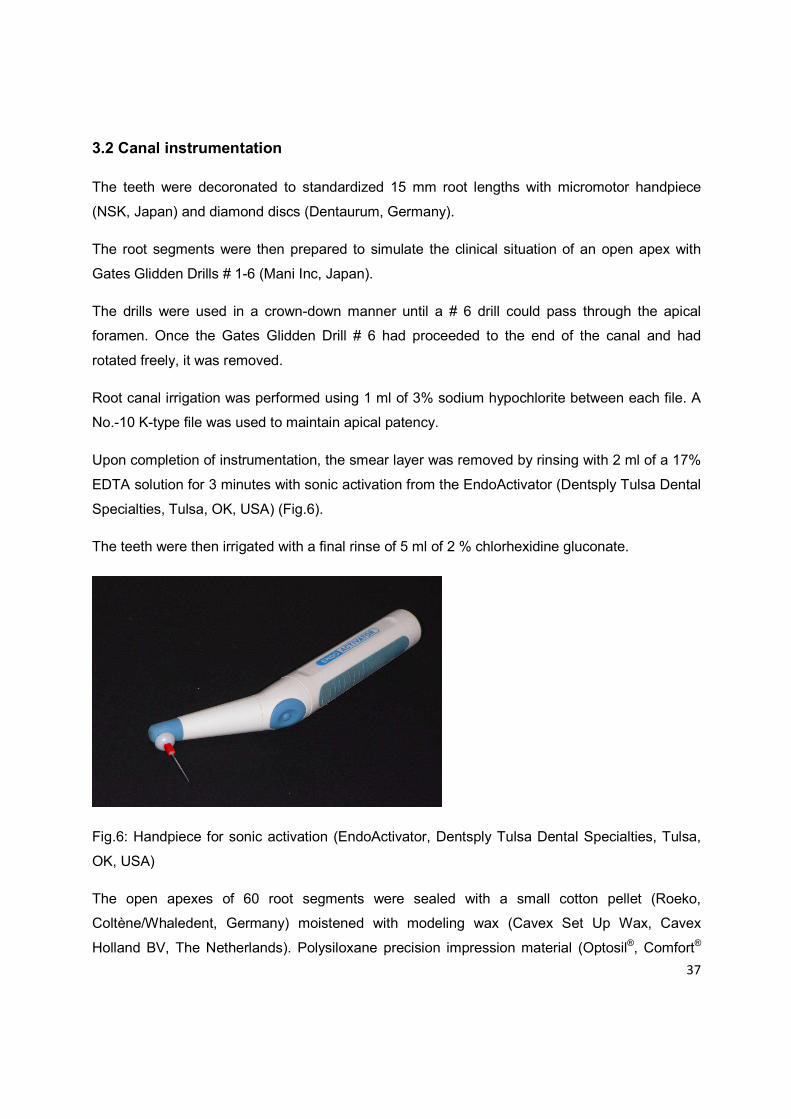
37
3.2 Canal instrumentation
The teeth were decoronated to standardized 15 mm root lengths with micromotor handpiece
(NSK, Japan) and diamond discs (Dentaurum, Germany).
The root segments were then prepared to simulate the clinical situation of an open apex with
Gates Glidden Drills # 1-6 (Mani Inc, Japan).
The drills were used in a crown-down manner until a # 6 drill could pass through the apical
foramen. Once the Gates Glidden Drill # 6 had proceeded to the end of the canal and had
rotated freely, it was removed.
Root canal irrigation was performed using 1 ml of 3% sodium hypochlorite between each file. A
No.-10 K-type file was used to maintain apical patency.
Upon completion of instrumentation, the smear layer was removed by rinsing with 2 ml of a 17%
EDTA solution for 3 minutes with sonic activation from the EndoActivator (Dentsply Tulsa Dental
Specialties, Tulsa, OK, USA) (Fig.6).
The teeth were then irrigated with a final rinse of 5 ml of 2 % chlorhexidine gluconate.
Fig.6: Handpiece for sonic activation (EndoActivator, Dentsply Tulsa Dental Specialties, Tulsa,
OK, USA)
The open apexes of 60 root segments were sealed with a small cotton pellet (Roeko,
Coltène/Whaledent, Germany) moistened with modeling wax (Cavex Set Up Wax, Cavex
Holland BV, The Netherlands). Polysiloxane precision impression material (Optosil®, Comfort®
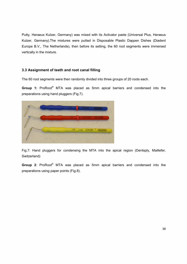
38
Putty, Heraeus Kulzer, Germany) was mixed with its Activator paste (Universal Plus, Heraeus
Kulzer, Germany).The mixtures were putted in Disposable Plastic Dappen Dishes (Diadent
Europe B.V., The Netherlands), then before its setting, the 60 root segments were immersed
vertically in the mixture.
3.3 Assignment of teeth and root canal filling
The 60 root segments were then randomly divided into three groups of 20 roots each.
Group 1: ProRoot® MTA was placed as 5mm apical barriers and condensed into the
preparations using hand pluggers (Fig.7).
Fig.7: Hand pluggers for condensing the MTA into the apical region (Dentsply, Maillefer,
Switzerland)
Group 2: ProRoot® MTA was placed as 5mm apical barriers and condensed into the
preparations using paper points (Fig.8).
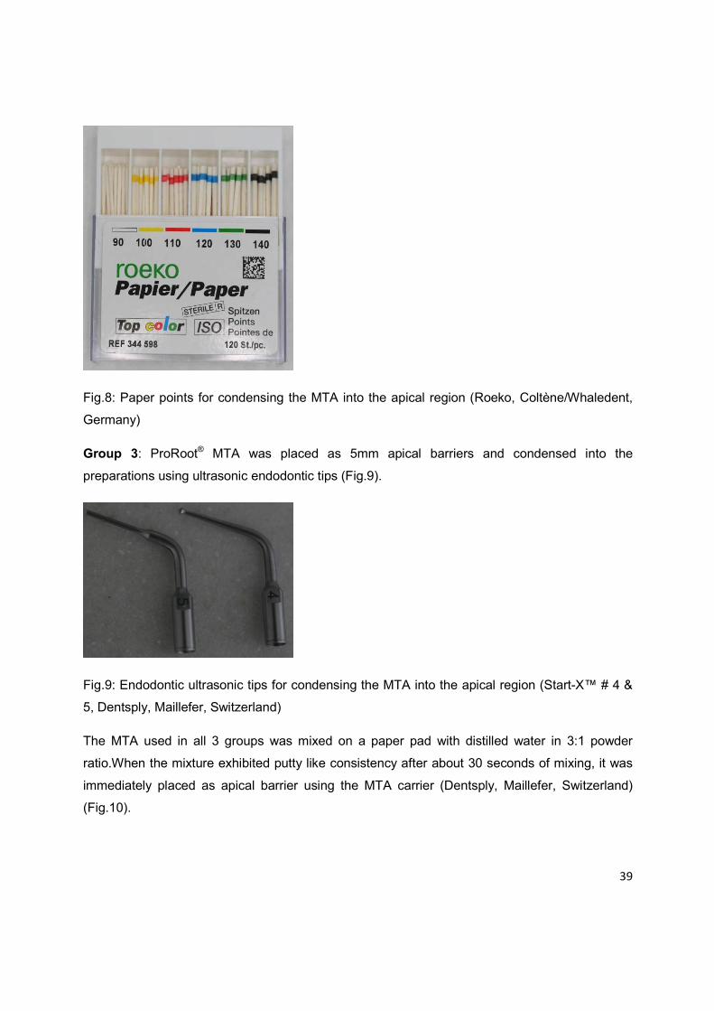
39
Fig.8: Paper points for condensing the MTA into the apical region (Roeko, Coltène/Whaledent,
Germany)
Group 3: ProRoot® MTA was placed as 5mm apical barriers and condensed into the
preparations using ultrasonic endodontic tips (Fig.9).
Fig.9: Endodontic ultrasonic tips for condensing the MTA into the apical region (Start-X™ # 4 &
5, Dentsply, Maillefer, Switzerland)
The MTA used in all 3 groups was mixed on a paper pad with distilled water in 3:1 powder
ratio.When the mixture exhibited putty like consistency after about 30 seconds of mixing, it was
immediately placed as apical barrier using the MTA carrier (Dentsply, Maillefer, Switzerland)
(Fig.10).

40
Fig.10: MTA carrier for dispencing the MTA into the apical region (Dentsply, Maillefer,
Switzerland)
Group1: Using the MTA carrier, ProRoot® MTA was dispensed into the apical region as 5mm
apical barrier by the help of endodontic microscope (Carl Zeiss OPMI, PROergo, Zeiss,
Germany).
Hand pluggers (Dentsply, Maillefer, Switzerland) with sizes 11, 11.5, 12 were used to condense
the MTA into the appropriate length in the apical third.
Group 2: Using the MTA carrier, ProRoot® MTA was dispensed into the apical region as 5mm
apical barrier by the help of endodontic microscope.
Paper Points (Roeko, Coltène/Whaledent, Germany) with sizes 120, 130, 140 were used to
condense the MTA into the appropriate length in the apical third.
Group 3: Using the MTA carrier, ProRoot® MTA was dispensed into the apical region as 5mm
apical barrier by the help of endodontic microscope.
The ultrasonic endodontic tips (Start-X™ # 4 & 5, Dentsply, Maillefer, Switzerland) connected to
endodontic scaler (EMS Piezon® Master 400, EMS SA, Switzerland) was used to condense MTA
to the apical third (Fig.11).

41
Fig.11: Endodontic ultrasonic apparatus for condensing the MTA in the apical region (EMS
Piezon® Master 400, EMS SA, Switzerland)
The MTA was packed into the apical portion of the roots by activating the ultrasonic endodontic
tip and slowly moving the MTA material apically using a 1- to 2-mm vertical packing motion
(Fig.12).
The packing procedure was accomplished in 45 seconds for each root.
Fig.12: An apical plug of MTA deposited in the apical 5 mm of the root canal
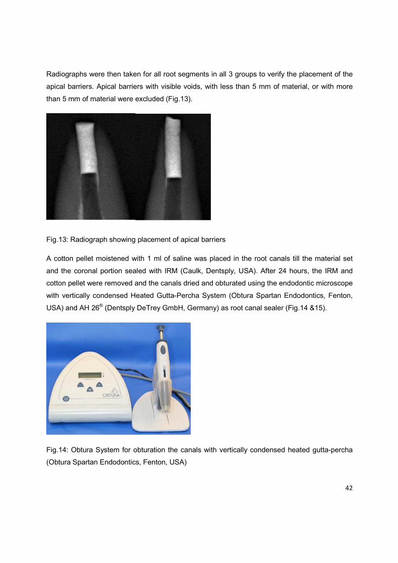
42
Radiographs were then taken for all root segments in all 3 groups to verify the placement of the
apical barriers. Apical barriers with visible voids, with less than 5 mm of material, or with more
than 5 mm of material were excluded (Fig.13).
Fig.13: Radiograph showing placement of apical barriers
A cotton pellet moistened with 1 ml of saline was placed in the root canals till the material set
and the coronal portion sealed with IRM (Caulk, Dentsply, USA). After 24 hours, the IRM and
cotton pellet were removed and the canals dried and obturated using the endodontic microscope
with vertically condensed Heated Gutta-Percha System (Obtura Spartan Endodontics, Fenton,
USA) and AH 26® (Dentsply DeTrey GmbH, Germany) as root canal sealer (Fig.14 &15).
Fig.14: Obtura System for obturation the canals with vertically condensed heated gutta-percha
(Obtura Spartan Endodontics, Fenton, USA)

43
Fig.15: Root canal filling by warm compaction of gutta-percha.
Coronal portions of all samples were then sealed with Cavit™ G (Temporary Filling Material 3M
ESPE, Seefeld, Germany) (Fig.16).
Fig.16: Root segments after sealing with Cavit™ G
All root segments in all 3 groups were stored at 37ᵒC and 100% humidity for 4 weeks.
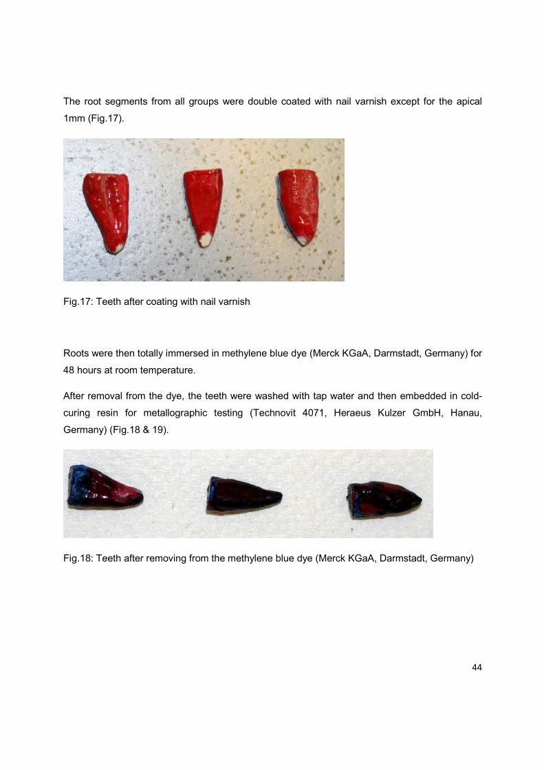
44
The root segments from all groups were double coated with nail varnish except for the apical
1mm (Fig.17).
Fig.17: Teeth after coating with nail varnish
Roots were then totally immersed in methylene blue dye (Merck KGaA, Darmstadt, Germany) for
48 hours at room temperature.
After removal from the dye, the teeth were washed with tap water and then embedded in cold-
curing resin for metallographic testing (Technovit 4071, Heraeus Kulzer GmbH, Hanau,
Germany) (Fig.18 & 19).
Fig.18: Teeth after removing from the methylene blue dye (Merck KGaA, Darmstadt, Germany)

45
Fig.19: Teeth after embedding in the cold-curing resin testing (Technovit 4071, Heraeus Kulzer
GmbH, Hanau, Germany)
The teeth were then longitudinally sectioned with saw microtome (Leica SP1600, Leica
Microsystem, Wetzlar, Germany) along the long axis of the teeth with the sections passing
through the obturated root canals and apical barriers in 0.4 mm thick slices (Fig.20 & 21).
Fig.20: Leica SP1600 saw microtome for sectioning the teeth in 0.4mm thick slices (Leica
SP1600, Leica Microsystem, Wetzlar, Germany)
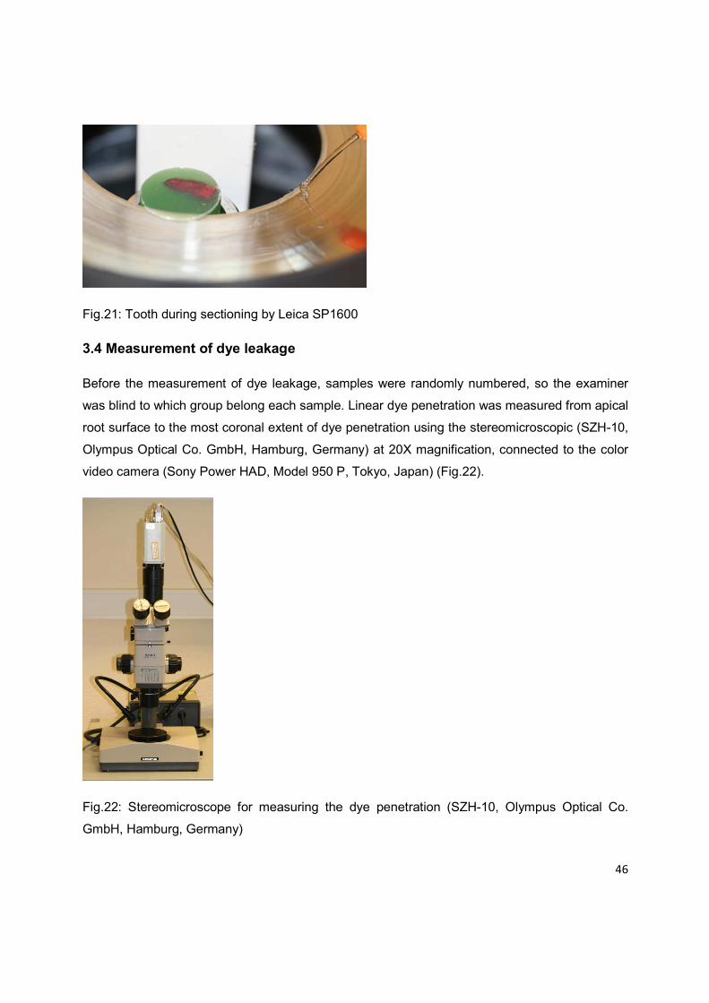
46
Fig.21: Tooth during sectioning by Leica SP1600
3.4 Measurement of dye leakage
Before the measurement of dye leakage, samples were randomly numbered, so the examiner
was blind to which group belong each sample. Linear dye penetration was measured from apical
root surface to the most coronal extent of dye penetration using the stereomicroscopic (SZH-10,
Olympus Optical Co. GmbH, Hamburg, Germany) at 20X magnification, connected to the color
video camera (Sony Power HAD, Model 950 P, Tokyo, Japan) (Fig.22).
Fig.22: Stereomicroscope for measuring the dye penetration (SZH-10, Olympus Optical Co.
GmbH, Hamburg, Germany)

47
The dye penetration was then measured with an image analysis program (analysis 3.0, Soft
Imaging System, Münster, Germany). All procedures were completed by one operator, whilst the
scanning electron microscope evaluations were carried out by a second examiner who was blind
in respect of all experimental groups and who underwent a training process with reference to the
scoring system of the scanning electron microscope evaluations.
3.5 Statistical analysis
The leakage in all roots was examined. The linear extent of dye penetration from the apical end
of the canal preparation to the coronal direction was measured by means of a light
stereomicroscope at (20 ×) magnification.
The mean values and the standard deviations were calculated by using Microsoft Excel 2010.
Quantitative data were tabulated and analyzed using SPSS 16 (SPSS, Inc., Chicago, IL, USA)
software. Level of significance was set at P less than or equal to 0.05.
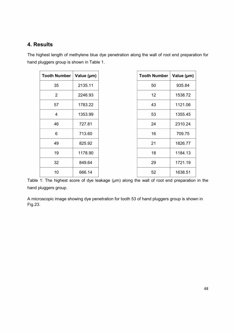
48
4. Results
The highest length of methylene blue dye penetration along the wall of root end preparation for
hand pluggers group is shown in Table 1.
Tooth Number Value (µm)
35 2135.11
2 2246.93
57 1783.22
4 1353.99
46 727.81
6 713.60
49 825.92
19 1178.90
32 849.64
10 666.14
Tooth Number Value (µm)
50 935.84
12 1538.72
43 1121.06
53 1355.45
24 2310.24
16 709.75
21 1826.77
18 1184.13
29 1721.19
52 1638.51
Table 1: The highest score of dye leakage (µm) along the wall of root end preparation in the
hand pluggers group.
A microscopic image showing dye penetration for tooth 53 of hand pluggers group is shown in Fig.23.
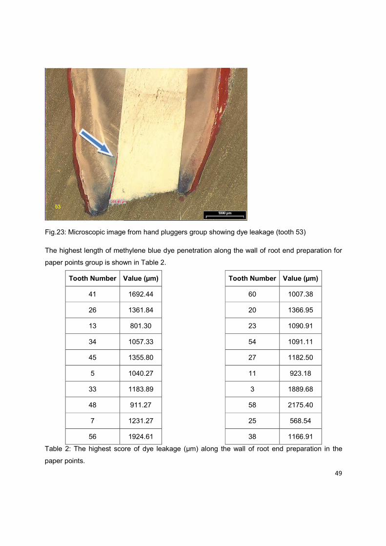
49
Fig.23: Microscopic image from hand pluggers group showing dye leakage (tooth 53)
The highest length of methylene blue dye penetration along the wall of root end preparation for
paper points group is shown in Table 2.
Tooth Number Value (µm)
41 1692.44
26 1361.84
13 801.30
34 1057.33
45 1355.80
5 1040.27
33 1183.89
48 911.27
7 1231.27
56 1924.61
Tooth Number Value (µm)
60 1007.38
20 1366.95
23 1090.91
54 1091.11
27 1182.50
11 923.18
3 1889.68
58 2175.40
25 568.54
38 1166.91
Table 2: The highest score of dye leakage (µm) along the wall of root end preparation in the
paper points.

50
A microscopic image showing dye penetration for tooth 26 of paper points group is shown in Fig.24.
Fig.24: Microscopic image from paper points group showing dye leakage (tooth 26)
The highest length of methylene blue dye penetration along the wall of root end preparation for ultrasonic tips group is shown in Table 3.
Tooth Number Value (µm)
17 1637.65
22 1275.91
47 1330.90
14 1300.87
59 1110.55
42 979.02
55 729.99
28 1356.66
8 713.15
30 720.59
Tooth Number Value (µm)
31 545.63
9 1223.80
36 585.49
44 1469.60
1 832.29
51 1472.79
37 986.63
15 2029.18
39 783.50
40 1618.35
Table 3: The highest score of dye leakage (µm) along the wall of root end preparation in the
ultrasonic tips group.

51
A microscopic image showing dye penetration for tooth 1 of ultrasonic tips group is shown in
Fig.25.
Fig.25: Microscopic image from ultrasonic tips group showing dye leakage (tooth 1)
The summary of mean values and the standard deviations, and maximum and minimum values
of apical dye penetration for three groups is listed in Table 4.
This table shows that group 1 with an MTA application via hand pluggers had the highest mean
value of apical dye penetration with 1.34mm ±0.5mm followed by group 2 using paper points
with 1.25mm ±0.4mm. Group 3 with the ultrasonic endodontic tips showed the lowest mean
value of apical dye penetration of 1.14mm ±0.4mm.
Groups N Minimum Maximum Mean (µm) Standard deviation
1 hand pluggers
20 666.14 2246.93 1341.14 537.46
2 paper points
20 568.54 2175.40 1251.12 400.50
3 ultrasonic endodntic tips
20 545.63 2029.18 1135.12 402.61
Table 4: Descriptive statistical analysis of apical dye penetration in µm for the three experimental
groups of MTA application with hand pluggers, paper points, and ultrasonic endodontic tips

52
Still, all three techniques exhibited considerable variation ranging from about 0.5 to well over
2mm dye penetration and leakage (Table 4, Fig.26). Using paper points for the application of
MTA resulted in the most consistent results around a mean value of about 1.25mm.
Fig.26: Distribution of apical dye penetration in µm for the three experimental groups for MTA
application with hand pluggers, paper points, and ultrasonic endodontic tips.
According to the results obtained, it was analyzed statistically using the Student's t-tests for
comparison (Table 5, 6 and 7).
Group 1-2
Levene test for
equality of variance t-test for Equality of Means
F Significance t df P-value
Mean Difference
Std. Error Difference
95% Confidence Interval of the Difference
Lower Upper
Variable
Equal variances assumed
3.150 0.084 0.601 38 0.552 90.017 149.87 -213.39 393.43
Not equal variances assumed
0.601 35.12 0.552 90.017 149.87 -214.21 394.24
Table 5: Statistical analysis and independent samples test between group 1 (hand pluggers) and
group 2 (paper points).
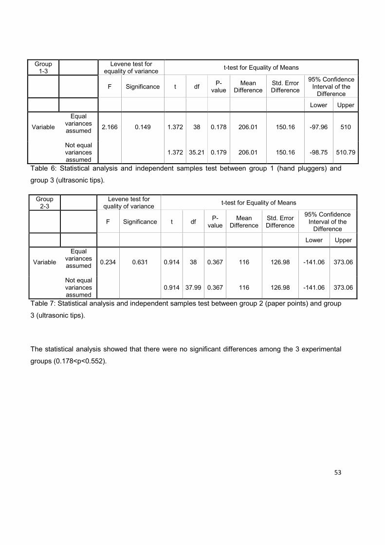
53
Group 1-3
Levene test for
equality of variance t-test for Equality of Means
F Significance t df P-value
Mean Difference
Std. Error Difference
95% Confidence Interval of the Difference
Lower Upper
Variable
Equal variances assumed
2.166 0.149 1.372 38 0.178 206.01 150.16 -97.96 510
Not equal variances assumed
1.372 35.21 0.179 206.01 150.16 -98.75 510.79
Table 6: Statistical analysis and independent samples test between group 1 (hand pluggers) and
group 3 (ultrasonic tips).
Group 2-3
Levene test for
quality of variance t-test for Equality of Means
F Significance t df P-value
Mean Difference
Std. Error Difference
95% Confidence Interval of the Difference
Lower Upper
Variable
Equal variances assumed
0.234 0.631 0.914 38 0.367 116 126.98 -141.06 373.06
Not equal variances assumed
0.914 37.99 0.367 116 126.98 -141.06 373.06
Table 7: Statistical analysis and independent samples test between group 2 (paper points) and group
3 (ultrasonic tips).
The statistical analysis showed that there were no significant differences among the 3 experimental
groups (0.178<p<0.552).
54
5 Discussion
5.1 Single visit apexification with MTA
In children, dental caries and trauma are the most common challenges to the integrity of a tooth
as it matures. If the dental pulp is damaged before development of the root length and closure of
the apical foramen, normal root development is altered or halted completely (Ham et al., 2005).
In such teeth, due to lack of apical constriction, hermetic seal of the root canal system during
obturation is not possible. For such cases, the best management technique is apexification.
Apexification is defined as a method to induce a calcified barrier in a root with an open apex or
the continued apical development of an incomplete root in teeth with necrotic pulp (Rafter 2005).
The advantage of a material that promotes the immediate formation of an artificial apical plug
and that maintains the capability to induce apexification with time means that the definitive root
filling can be placed immediately after the material sets (Felippe et al., 2006).
Single visit techniques using apical barriers have been proposed as alternatives to the long term
calcium hydroxide apexification procedure which enables immediate obturation of the root canal
(Witherspoon and Ham 2001; Linsuwanont 2003; Andreasen et al., 2006; Simon et al., 2007).
The idea of single visit apexification is not new and has been discussed and tested for many
years. Several case reports have been published. Biological apical closure appears later, i.e.
after the filling of the root canal, contrary to the technique with calcium hydroxide, where
obtaining the apical barrier is necessary to complete the root canal treatment.
Pradhan et al. (2006) showed that the time taken to complete the treatment of non-vital teeth
with unformed apices and for obtaining an apical biological barrier formation by using MTA was
significantly less than that for calcium hydroxide.
It was found that medication with calcium hydroxide had no significant effect on grey MTA
leakage (Hachmeister et al., 2002; Stefopoulos et al., 2008).

55
Other studies dealing with the use of MTA as apical barrier (de Leimburg et al., 2004; Al-Kahtani
et al., 2005) have not addressed that problem since they did not consider a calcium hydroxide
medication.
Another study revealed that white MTA sealed better when used without previous intracanal
medication with calcium hydroxide (Stefopoulos et al., 2008).
The in vivo findings of Felippe et al (2006) suggest that the calcium hydroxide premedication is
not a prerequisite, as the mere presence of MTA as an apical plug led to favorable results in
terms of periapical inflammation and hard tissue formations in dogs’ teeth.
MTA is osteoconductive, which may help the periapical tissue to adapt and heal. Its
effectiveness has been shown in many case reports (Torabinejad and Pitt Ford, 1996).
Felippe et al. (2006) showed that the application of MTA immediately after root canal preparation
favored the establishment of a normal periodontal ligament and formation of new bone and
cementum. The histological responses observed in this study indicate that MTA is a reliable
material and should be considered effective in teeth with incomplete root formation.
Its application results in predictable apical closure and reduction of the treatment time, number of
appointments and radiographs, particularly in young patients.
Another study suggested MTA for apexification because it provides an adequate seal in the root
canal, and it appears to offer a biological active substrate that stimulates periodontal cell
production (Villa and Fernández, 2005).
Because of potential discoloration effect of grey MTA, white MTA has been introduced into
endodontic treatment for the same purpose (Parirokh et al., 2005).
56
5.2 Methodological considerations
Bozeman et al (2006) showed that white MTA had a significantly higher compressive strength
than grey MTA at 24 hours and that grey MTA had a significantly longer time to final setting time
compared to white MTA. Based on this study, the results suggest that white MTA is an effective
substitute for grey MTA. Hence white MTA was used for this study.
Natural teeth were used in this experiment because it would certainly better simulate the actual
clinical situation than the plastic tubes.
Matt et al. (2004) and Al-Kahtani et al. (2005) reported that 5-mm MTA apical plug provided
better microleakage resistance than 2-mm MTA apical plug. Considering these results, 5-mm
apical plug was used in this study to provide sufficient volume of MTA for reduction of dye
leakage.
The open apexes of the segments were sealed with a small cotton pellet moistened with
modeling wax to simulate a periapical environment. MTA was condensed into root-end
preparation against a cotton pellet absorbed with wax for support.
The root segments were immersed vertically in a mixture of polysiloxane precision impression
material putted in disposable plastic dappen dishes to enable the application of MTA for all
groups in the same position and to avoid additional variables.
Orthograde delivery is a sensitive technique. Placement must be verified with radiographs and
condensation is limited because of minimal resistance of the open apex. In addition to the
difficulty in delivering the material to the apex, the irregularities and divergent nature of the
anatomy may limit adaptation to the dentinal walls, creating marginal gaps at the dentinal
interface (Al-Kahtani et al. 2005).
Therefore after placement of MTA, radiographs were taken to verify the thickness of the barrier
and its placement at the apex.
The goal of a 5 mm thickness barrier was not easily achieved because of the difficulties of
handling the material of the apex. The use of an operating microscope may facilitate placement
and handling at the apex. Hence ProRoot® MTA was dispensed into the apical region as 5mm
apical barrier by the help of endodontic microscope.

57
Due to the prolonged setting time of ProRoot MTA, these segments were obturated with gutta-
percha in 24 hours. Matt et al. (2004) reported also that the obturation with gutta-percha after 24
hours showed less apical dye leakage than the immediately obturation.
These findings concur with the manufacturers’ recommendation of placing a moist cotton pellet
in the canal, and allowing the material to set for a minimum of 4 hours.
The long hardening time of ProRoot MTA is said to reduce internal tensions and the incidence of
marginal infiltrations, but it forces to definitively fill the tooth in the following sitting (Casella and
Ferlito, 2006).
After obturation, the coronal portions of all samples were sealed with Cavit to prevent any
chances of dye penetration from the coronal aspect. They were stored at 37˚C and 100%
humidity for 4 weeks to simulate a periapical environment.
Many techniques have been advised to test the sealing properties of restorative materials in vitro
and in vivo. These techniques include use of dyes, chemical tracers, radioactive isotopes,
neutron activation analysis, electrical conductivity, and bacterial penetration.
In the present study, methylene blue dye penetration method was used for assessing the degree
of microleakage; because it is easy and economic to use. It has a high degree of staining and
has a molecular weight even lower than that of bacterial toxins (Ahlberg et al., 1995; Tamse et
al., 1998; Kontakiotis et al., 2001). Dye studies, however, usually employed to screen new
restorative filling materials. When a filling material does not allow penetration of small molecules,
it has the potential to prevent leakage of larger substances such as bacteria and their by-
products (Aqrabawi 2000; Oliver and Abbott 2001).
The limitation of dye leakage studies is that they measure the degree of leakage in only one
plane, making it impossible to evaluate the total amount of leakage (Wu and Wesselink 1993;
Ahlberg et al., 1995; Tamse et al., 1998).
Before placing the prepared samples in methylene blue dye, all the root segments except the
apical 1mm were double coated with nail varnish to prevent dye penetration into the lateral and
accessory canals.
58
5.3 Discussion of results
Maximum linear extent of dye penetration was defined as the most coronal level of dye visible on
the root canal walls or gutta-percha, regardless of whether it was continuous with the apical
area.
It has generally been considered that a potential root end filling material should set as it is placed
in the root end cavity without significant shrinkage. This condition would allow dimensional
stability of the material after placement and less time for an unset material to be in contact with
vital tissues.
In general terms, the quicker a material sets the more it shrinks. This phenomenon may explain
why ProRoot MTA in previous studies had significantly less dye and bacterial leakage than other
materials used as root end filling materials (Torabinejad et al., 1995a).
The benefit to use ultrasonic activation for placement of MTA apical barriers is still in question.
When orthograde MTA is placed in teeth with open apices, it has been shown that a good seal
and adequate retention is difficult to achieve (Hachmeister et al., 2002).
Another study has shown that ultrasonic energy applied while placing the barrier may improve
the seal of the set cement (Lawley et al., 2004). Our findings do not support this hypothesis as
handpluggers and paper points achieved the same results as ultrasonic application of MTA.

59
6 Conclusion & recommendations
This study compared the fill density of white ProRoot MTA in extracted single-rooted teeth, using
three placement methods: hand condensation with pluggers (group 1), hand condensation with
paper points (group 2), and direct ultrasonic endodontic tips (group 3).
The results showed considerable variation, but mean values and variability were very similar for
all three application methods. Paper points achieved the most consistent results with few
minimal values for dye penetration, but also few high values.
Although ultrasonic vibration showed less dye leakage compared with Group 1 and 2, but the
statistical analysis showed that there were no significant differences among the 3 experimental
groups (0.178<p<0.552).
In a one-visit apexification technique, it must keep in mind that an apical barrier is used mainly to
produce an apical stop against which gutta-percha will be adequately condensed.
The obturation of the canal with gutta-percha and sealer will play the key role for a bacteria tight
apical seal as in every non-surgical endodontic treatment.

60
7 Summary
A one-visit apexification protocol with Mineral Trioxide Aggregate (MTA) can be seen as an
alternative to the traditional treatment practices with calcium hydroxide. The aim of this study
was to investigate the sealing ability of ProRoot MTA when placed as an apical barrier using
three different techniques.
Sixty freshly extracted single-rooted human teeth were decoronated and standardized to a root
length of 15mm. The root segments were prepared with Gates Glidden burs (size 1-6) to
simulate the clinical situation of an open apex and randomly assigned into 3 experimental groups
of 20 samples each. 5mm MTA was placed by pluggers (Group 1) paper points (Group 2) or
ultrasonic tips (Group 3). Radiographs were taken to verify the placement of the apical barriers.
After obturation of all samples with warm gutta-percha (Obtura) and AH26 sealer, the coronal
portion of all samples was sealed with Cavit. The root segments were then double coated with
nail varnish except for the open apex and were exposed to methylene blue dye for 48 hours at
room temperature. Afterwards, the samples were sectioned longitudinally and the extent of dye
penetration was measured with a stereomicroscope.
The mean depth of dye leakage for Group 1 was 1.34mm (±0.5 SD) Group 2 1.25mm (±0.4 SD)
and Group 3 1.14mm (± 0.4 SD). Statistical analysis showed that there were no significant
differences among the 3 experimental groups (0.178<p<0.552).
In conclusion, ProRoot MTA has a similar sealing ability when placed as an apical barrier with
pluggers, paper points or ultrasonic tips.
61
8 Zusammenfassung
Die einzeitige Apexifikationstechnik mit Mineral Trioxid Aggregat (MTA) kann als Alternative zu
den herkömmlichen, langwierigen Behandlungsmethoden mit Calciumhydroxid gesehen werden.
Ziel der Studie war es, die Dichtigkeit von ProRoot MTA als apikale Barriere bei drei
verschiedenen Applikationstechniken zu untersuchen.
Bei sechzig, frisch extrahierten, einwurzligen, humane Zähnen wurden die klinischen Kronen
abgetrennt, um eine standardisierte Wurzellänge von 15 mm zu erhalten. Die Wurzeln wurden
mit Gates-Bohrern (Größe 1-6) erweitert, um die klinische Situation eines offenen Apex zu
simulieren. Anschließend wurden jeweils 20 Proben nach dem Zufallsprinzip den drei
Versuchsgruppen zugeordnet. 5 mm MTA wurde mit einem Plugger (Gruppe 1), Papierspitze
(Gruppe 2) oder Ultraschallspitzen (Gruppe 3) gestellt. Die Röntgenbilder wurde die korrekte
Platzierung der apikalen Barrieren überprüft. Nach Verschluss aller Proben mit warmer
Guttapercha (Obtura, Sealer AH26) wurde der koronale Anteil aller Proben mit Cavit
abgedichtet. Mit Ausnahme des Apex (ca. 1 mm) wurden die Proben dann doppelt mit
Nagellack beschichtet und für 48 Stunden in eine Methylenblaufärbung bei Raumtemperatur
getaucht. Abschließend wurden in den Längsschnitten der Proben (Schichtstärke 0.4 mm) das
Ausmaß der Farbpenetration mit einem Stereomikroskop (Vergrößerung 20X) gemessen.
Die mittlere Farbpenetrationstiefe betrug 1,34 mm (± 0,5 SD) für Gruppe 1, 1,25 mm (± 0,4 SD)
für Gruppe 2, und 1,14 mm (± 0,4 SD) für Gruppe 3. Die statistische Analyse zeigte, dass es
keine signifikanten Unterschiede zwischen den drei experimentellen Gruppen (0,178 < p
<0,552).
Schlussfolgernd ergibt sich, dass ProRoot MTA eine fast identische Farbpenetration und damit
Abdichtungsfähigkeit bei der Applikation als apikale Barriere mit Plugger, Papier- oder
Ultraschallspitzen hat.

62
9 References
Abdullah D, Ford TR, Papaioannou S, Nicholson J, McDonald F. An evaluation of accelerated Portland cement as a restorative material. Biomaterials 2002;23:4001-4010. Abedi HR, Torabinejad M, Pitt Ford TR, Bakland LK. The use of mineral trioxide aggregate cement (MTA) as a direct pulp capping agent (Abstract no. 44). J Endod 1996;22:199. Accorinte Mde L, Holland R, Reis A, Bortoluzzi MC, Murata SS, Dezan E Jr, Souza V, Alessandro LD. Evaluation of mineral trioxide aggregate and calcium hydroxide cement as pulp-capping agents in human teeth. J Endod 2008;34:1-6. Adamo HL, Buruiana R, Schertzer L, Boylan RJ. A comparison of MTA, Super-EBA, composite, and amalgam as root-end filling materials using a bacterial microleakage model. Int Endod J 1999;32:197-203. Aeinehchi M, Eslami B, Ghanbariha M, Saffar AS. Mineral trioxide aggregate (MTA) and calcium hydroxide as pulp-capping agents in human teeth: a preliminary report. Int Endod J 2003;36:225-231. Aeinehchi M, Dadvand S, Fayazi S, Bayat-Movahed S. Randomized controlled trial of mineral trioxide aggregate and formocresol for pulpotomy in primary molar teeth. Int Endod J 2007;40:261-267. Agamy HA, Bakry NS, Mounir MM, Avery DR. Comparison of mineral trioxide aggregate and formocresol as pulp-capping agents in pulpotomized primary teeth. Pediatr Dent 2004;26:302-309. Ahlberg KM, Assavanop P, Tay WM. A comparison of the apical dye penetration patterns shown by methylene blue and india ink in root-filled teeth. Int Endod J 1995; 28:30-34.
Al-Hezaimi K, Al-Hamdan K, Naghshbandi J, Oglesby S, Simon JH, Rotstein I. Effect of white-colored mineral trioxide aggregate in different concentrations on Candida albicans in vitro. J Endod 2005a;31:684-686.
Al-Hezaimi K, Naghshbandi J, Oglesby S, Simon JH, Rotstein I. Human saliva penetration of root canals obturated with two types of mineral trioxide aggregate cements. J Endod 2005b;31:453-456. Al-Hezaimi K, Naghshbandi J, Oglesby S, Simon JHS, Rotstein I. Comparison of antifungal activity of white-colored and gray-colored mineral trioxide aggregate (MTA) at similar concentrations against Candida albicans. J Endod 2006a;32:365-367. Al-Hazaimi K, Al-Shalan TA, Naghshbandi J, Oglesby S, Simon JH, Rotstein I. Antibacterial effect of two mineral trioxide aggregate (MTA) preparations against Enterococcus faecalis and Streptococcus sanguis in vitro. J Endod 2006b;32:1053-1056. Al-Kahtani A, Shostad S, Schifferle R, Bhambhani S. In-vitro evaluation of microleakage of an orthograde apical plug of mineral trioxide aggregate in permanent teeth with simulated immature apices. J Endod 2005;31:117-119.

63
Al-Nazhan S, Al-Judai A. Evaluation of antifungal activity of mineral trioxide aggregate. J Endod 2003;29:826-827.
Andelin WE, Browning DF, Hsu GH, Roland DD, Torabinejad M. Microleakage of resected MTA. J Endod 2002;28:573-574. Andelin WE, Shabahang S, Wright K, Torabinejad M. Identification of hard tissue after experimental pulp capping using dentin sialoprotein (DSP) as a marker. J Endod 2003;29:646-650.
Andreasen JO, Farik B, Munksgaard EC. Long-term calcium hydroxide as a root canal dressing may increase risk of root fracture. Dent Traumatol 2002;18:134-137.
Andreasen JO, Munksgaard EC, Bakland LK. Comparison of fracture resistance in root canals of immature sheep teeth after filling with calcium hydroxide or MTA. Dent Traumatol 2006;22:154-156.
Antunes Bortoluzzi E, Juárez Broon N, Antonio Hungaro Duarte M, de Oliveira Demarchi AC, Monteiro Bramante C. The use of a setting accelerator and its effect on pH and calcium ion release of mineral trioxide aggregate and white Portland cement. J Endod 2006;32:1194-1197.
Apaydin ES, Shabahang S, Torabinejad M. Hard-tissue healing after application of fresh or set MTA as root-end-filling material. J Endod 2004;30:21-24. Apaydin ES, Torabinejad M. The effect of calcium sulfate on hard-tissue healing after periradicular surgery. J Endod 2004;30:17-20. Aqrabawi J. Sealing ability of amalgam, Super EBA cement, and MTA when used as retrograde filling materials. Br Dent J 2000;188:266-268. Asgary S, Parirokh M, Eghbal MJ, Brink F. A comparative study of white mineral trioxide aggregate and white Portland cements using X-ray microanalysis. Aust Endod J 2004;30:89-92. Asgary S, Parirokh M, Eghbal MJ, Brink F. Chemical differences between white and gray mineral trioxide aggregate. J Endod 2005;31:101-103.
Asgary S, Parirokh M, Eghbal MJ, Stowe S, Brink F. A qualitative X-ray analysis of white and grey mineral trioxide aggregate using compositional imaging. J Mater Sci Mater Med 2006;17:187-191.
Asgary S, Eghbal MJ, Parirokh M. Sealing ability of a novel endodontic cement as a root-end filling material. J Biomed Mater Res A 2008;87:706-709.
Asgary S, Kamrani FA. Antibacterial effects of five different root canal sealing materials. J Oral Sci 2008;50:469-474.
Asrari M, Lobner D. In vitro neurotoxic evaluation of root-end filing materials. J Endod 2003;29:743-746.
64
Baek SH, Plenk H Jr, Kim S. Periapical tissue responses and cementum regeneration with amalgam, SuperEBA, and MTA as root-end filling materials. J Endod 2005;31: 444-449. Ballal S, Venkateshbabu N, Nandini S, Kandaswamy D. An in vitro study to assess the setting and surface crazing of conventional glass ionomer cement when layered over partially set mineral trioxide aggregate. J Endod 2008;34:478-480. Balto HA. Attachment and morphological behavior of human periodontal ligament fibroblasts to mineral trioxide aggregate: a scanning electron microscope study. J Endod 2004;30:25-29.
Bargholz C. Perforation repair with mineral trioxide aggregate: a modified matrix concept. Int Endod J 2005;38:59-69.
Barrieshi-Nusair KM, Qudeimat MA. A prospective clinical study of mineral trioxide aggregate for partial pulpotomy in cariously exposed permanent teeth. J Endod 2006;32:731-735. Bates CF, Carnes DL, del Rio CE. Longitudinal sealing ability of mineral trioxide aggregate as a root-end filling material. J Endod 1996;22:575-578.
Belío-Reyes IA, Bucio L, Cruz-Chavez E. Phase composition of ProRoot mineral trioxide aggregate by X-ray powder diffraction. J Endod 2009;35:875-878.
Ber BS, Hatton JF, Stewart GP. Chemical modification of proroot mta to improve handling characteristics and decrease setting time. J Endod 2007;33:1231-1234.
Bidar M, Moradi S, Jafarzadeh H, Bidad S. Comparative SEM study of the marginal adaptation of white and grey MTA and Portland cement. Aust Endod J 2007;33:2-6.
Bogen G, Kim JS, Bakland LK. Direct pulp capping with mineral trioxide aggregate: an observational study. J Am Dent Assoc 2008;139:305-315. Bonson S, Jeansonne BG, Lallier TE. Root-end filling materials alter fibroblast differentiation. J Dent Res 2004;83:408-413. Bortoluzzi EA, Broon NJ, Bramante CM, Garcia RB, de Moraes IG, Bernardineli N. Sealing ability of MTA and radiopaque Portland cement with or without calcium chloride for root-end filling. J Endod 2006;32:897-900.
Bortoluzzi EA, Souza EM, Reis JM, Esberard RM, Tanomaru-Filho M. Fracture strength of bovine incisors after intra-radicular treatment with MTA in an experimental immature tooth model. Int Endod J 2007;40:684-691.
Bozeman TB, Lemon RR, Eleazer PD. Elemental analysis of crystal precipitate from gray and white MTA. J Endod 2006;32:425-428. Braz MG, Camargo EA, Salvadori DM, Marques ME, Ribeiro DA. Evaluation of genetic damage in human peripheral lymphocytes exposed to mineral trioxide aggregate and Portland cement. J Oral Rehab 2006;33:234-239.

65
Budig CG, Eleazer PD. In vitro comparison of the setting of dry ProRoot MTA by moisture absorbed through the root. J Endod 2008;34:712-714.
Camilleri J, Montesin FE, Papaioannou S, McDonald F, Pitt Ford TR. Biocompatibility of two commercial forms of mineral trioxide aggregate. Int Endod J 2004;37:699-704.
Camilleri J, Montesin FE, Brady K, Sweeney R, Curtis RV, Ford TR. The constitution of mineral trioxide aggregate. Dent Mater 2005a;21:297-303. Camilleri J, Montesin FE, Di Silvio L, Pitt Ford TR. The chemical constitution and biocompatibility of accelerated Portland cement for endodontic use. Int Endod J 2005b;38:834-842.
Camilleri J. Hydration mechanisms of mineral trioxide aggregate. Int Endod J 2007 ;40:462-470.
Camilleri J. Characterization of hydration products of mineral trioxide aggregate. Int Endod J 2008;41:408-417.
Camilleri J, Pitt Ford TR. Evaluation of the effect of tracer pH on the sealing ability of glass ionomer cement and mineral trioxide aggregate. J Mater Sci Mater Med 2008 ;19:2941-2948.
Camp MA, Jeansonne BG, Lallier T. Adhesion of human fibroblasts to root-end-filling materials. J Endod 2003;29:602-607.
Casella G, Ferlito S. The use of mineral trioxide aggregate in endodontics. Minerva Stomatol 2006;55:123-143. Chacko V, Kurikose S. Human pulpal response to mineral trioxide aggregate (MTA): a histologic study. J Clin Pediatr Dent 2006;30:203-209. Chen CL, Huang TH, Ding SJ, Shie MY, Kao CT. Comparison of calcium and silicate cement and mineral trioxide aggregate biologic effects and bone markers expression in MG63 cells. J Endod 2009;35:682-685.
Chng HK, Islam I, Yap AU, Tong YW, Koh ET. Properties of a new root-end filling material. J Endod 2005;31:665-668.
Chong BS, Pitt Ford TR, Hudson MB. A prospective clinical study of Mineral Trioxide Aggregate and IRM when used as root-end filling materials in endodontic surgery. Int Endod J 2003;36:520-526.
Cintra LT, de Moraes IG, Estrada BP, Gomes-Filho JE, Bramante CM, Garcia RB, Bernardinelli N. Evaluation of the tissue response to MTA and MBPC: microscopic analysis of implants in alveolar bone of rats. J Endod 2006;32:556-559.
Costa AT, Konrath F, Dedavid B, Weber JB, de Oliveira MG. Marginal adaptation of root-end filling materials: an in vitro study with teeth and replicas. J Contemp Dent Pract 2009;10:75-82.
66
Coutinho-Filho T, De-Deus G, Klein L, Manera G, Peixoto C, Gurgel-Filho ED. Radiopacity and histological assessment of Portland cement plus bismuth oxide. Oral Surg Oral Med Oral Pathol Oral Radiol Endod 2008;106:e69-77.
Cvek M. Prognosis of luxated non-vital maxillary incisors treated with calcium hydroxide and filled with gutta-percha. A retrospective clinical study. Endod Dent Traumatol 1992;8:45-55.
Dammaschke T, Gerth HU, Züchner H, Schäfer E. Chemical and physical surface and bulk material characterization of white ProRoot MTA and two Portland cements. Dent Mater 2005;21:731-738. Dammaschke T, Wolff P, Sagheri D, Stratmann U, Schafer E. Mineral trioxide aggregate for direct pulp capping: a histologic comparison with calcium hydroxide in rat molars. Quintessence Int 2010;41:e20-30. Danesh G, Dammaschke T, Gerth HU, Zandbiglari T, Schäfer E. A comparative study of selected properties of ProRoot mineral trioxide aggregate and two Portland cements. Int Endod J 2006;39:213-219.
Daoudi MF, Saunders WP. In vitro evaluation of furcal perforation repair using mineral trioxide aggregate or resin modified glass lonomer cement with and without the use of the operating microscope. J Endod 2002;28:512-515.
Davis JL, Jeansonne BG, Davenport WD, Gardiner D. The effect of irrigation with doxycycline or citric acid on leakage and osseous would healing. J Endod 2003;29:31-35. De Bruyne MA, De Bruyne RJ, Rosiers L, De Moor RJ. Longitudinal study on microleakage of three root-end filling materials by the fluid transport method and by capillary flow porometry. Int Endod J 2005;38:129-136.
De Deus G, Ximenes R, Gurgel-Filho ED, Plotkowski MC, Coutinho-Filho T. Cytotoxicity of MTA and Portland cement on human ECV 304 endothelial cells. Int Endod J 2005;38:604-609.
De-Deus G, Petruccelli V, Gurgel-Filho E, Coutinho-Filho T. MTA versus Portland cement as repair material for furcal perforations: a laboratory study using a polymicrobial leakage model. Int Endod J 2006;39:293-298.
De-Deus G, de Souza MC, Sergio Fidel RA, Fidel SR, de Campos RC, Luna AS. Negligible expression of arsenic in some commercially available brands of Portland cement and mineral trioxide aggregate. J Endod 2009;35:887-890.
de Leimburg ML, Angeretti A, Ceruti P, Lendini M, Pasqualini D, Berutti E. MTA obturation of pulpless teeth with open apices: bacterial leakage as detected by polymerase chain reaction assay. J Endod 2004;30:883-886.
Deller-Quinn M, Perinpanayagam H. Osteoblast expression of cytokines is altered on MTA surfaces. Oral Surg Oral Med Oral Pathol Oral Radiol Endod 2009;108:302-307.
de Martins GR, Carvalho CA, Valera MC, de Oliveira LD, Buso L, Carvalho AS. Sealing ability of castor oil polymer as a root-end filling material. J Appl Oral Sci 2009;17:220-223.
67
de Menezes JV, Takamori ER, Bijella MF, Granjeiro JM. In vitro toxicity of MTA compared with other primary teeth pulpotomy agents. J Clin Pediatr Dent 2009 ;33:217-221.
de Morais CA, Bernardineli N, Garcia RB, Duarte MA, Guerisoli DM. Evaluation of tissue response to MTA and Portland cement with iodoform. Oral Surg Oral Med Oral Pathol Oral Radiol Endod 2006;102:417-421.
de Souza Costa CA, Duarte PT, de Souza PP, Giro EM, Hebling J. Cytotoxic effects and pulpal response caused by a mineral trioxide aggregate formulation and calcium hydroxide. Am J Dent 2008;21:255-261.
Ding SJ, Kao CT, Shie MY, Hung C Jr, Huang TH. The physical and cytological properties of white MTA mixed with Na2HPO4 as an accelerant. J Endod 2008;34:748-751.
Dominguez Reyes A, Muñoz Muñoz L, Aznar Martín T. Study of calcium hydroxide apexification in 26 young permanent incisors. Dent Traumatol 2005;21:141-145.
Doyle TL, Casas MJ, Kenny DJ, Judd PL. Mineral trioxide aggregate produces superior outcomes in vital primary molar pulpotomy. Pediatr Dent 2010;32:41-47.
Duarte MA, Demarchi AC, Yamashita JC, Kuga MC, Fraga Sde C. pH and calcium ion release of 2 root-end filling materials. Oral Surg Oral Med Oral Pathol Oral Radiol Endod 2003;95:345-347.
Duarte MA, De Oliveira Demarchi AC, Yamashita JC, Kuga MC, De Campos Fraga S. Arsenic release provided by MTA and Portland cement. Oral Surg Oral Med Oral Pathol Oral Radiol Endod 2005;99:648-650.
Economides N, Pantelidou O, Kokkas A, Tziafas D. Short-term periradicular tissue response to mineral trioxide (MTA) as root-end filling material. Int Endod J 2003;36:44-48.
Eghbal MJ, Asgary S, Baglue RA, Parirokh M, Ghoddusi J. MTA pulpotomy of human permanent molars with irreversible pulpitis. Aust Endod J 2009;35:4-8.
Eidelman E, Holan G, Fuks AB. Mineral trioxide aggregate vs. formocresol in pulpotomized primary molars: a preliminary report. Pediatr Dent 2001;23:15-18. Eldeniz AU, Hadimli HH, Ataoglu H, Orstavik D. Antibacterial effect of selected root-end filling materials. J Endod 2006;32:345-349.
El-Meligy OA, Avery DR. Comparison of apexification with mineral trioxide aggregate and calcium hydroxide. Pediatr Dent 2006a;28:248-253.
El-Meligy OA, Avery DR. Comparison of mineral trioxide aggregate and calcium hydroxide as pulpotomy agents in young permanent teeth (apexogenesis). Pediatr Dent 2006b;28:399-404.
Estrela C, Bammann LL, Estrela CR, Silva RS, Pécora JD. Antimicrobial and chemical study of MTA, Portland cement, calcium hydroxide paste, Sealapex and Dycal. Braz Dent J 2000;11:3-9.

68
Estrela C, Bammann LL, Pimenta FC, Pécora JD. Control of microorganisms in vitro by calcium hydroxide pastes. Int Endod J 2001;34:341-345.
Faraco IM Jr, Holland R. Response of the pulp of dogs to capping with mineral trioxide aggregate or a calcium hydroxide cement. Dent Traumatol 2001;17:163-166.
Farsi N, Alamoudi N, Balto K, Mushayt A. Success of mineral trioxide aggregate in pulpotomized primary molars. J Clin Pediatr Dent 2005;29:307-311. Farsi N, Alamoudi N, Balto K, Al Mushayt A. Clinical assessment of mineral trioxide aggregate (MTA) as direct pulp capping in young permanent teeth. J Clin Pediatr Dent 2006;31:72-76. Fayad MI, Hawkinson R, Daniel J, Hao J. The effect of CO2 laser irradiation on PDL cell attachment to resected root surfaces. Oral Surg Oral Med Oral Pathol Oral Radiol Endod 2004;97:518-523.
Felippe MC, Felippe WT, Marques MM, Antoniazzi JH. The effect of the renewal of calcium hydroxide paste on the apexification and periapical healing of teeth with incomplete root formation. Int Endod J 2005;38:436-442.
Felippe WT, Felippe MC, Rocha MJ. The effect of mineral trioxide aggregate on the apexification and periapical healing of teeth with incomplete root formation. Int Endod J 2006;39:2-9. Fernández-Yáñez Sánchez A, Leco-Berrocal MI, Martínez-González JM. Metaanalysis of filler materials in periapical surgery. Med Oral Patol Oral Cir Bucal 2008;13:E180-185. Ferris DM, Baumgartner JC. Perforation repair comparing two types of mineral trioxide aggregate. J Endod 2004;30:422-424. Fischer EJ, Arens DE, Miller CH. Bacterial leakage of mineral trioxide aggregate as compared with zinc-free amalgam, intermediate restorative material, and Super EBA as a root-end filling material. J Endod 1998;24:176-179.
Fogel HM, Peikoff MD. Microleakage of root-end filling materials. J Endod 2001 ;27:456-458.
Ford TR, Torabinejad M, McKendry DJ, Hong CU, Kariyawasam SP. Use of mineral trioxide aggregate for repair of furcal perforations. Oral Surg Oral Med Oral Pathol Oral Radiol Endod 1995;79:756-763. Ford TR, Torabinejad M, Abedi HR, Bakland LK, Kariyawasam SP. Using mineral trioxide aggregate as a pulp-capping material. J Am Dent Assoc 1996;127:1491-1494. Fridland M, Rosado R. Mineral trioxide aggregate (MTA) solubility and porosity with different water to- powder ratios. J Endod 2003;29:814-817. Fridland M, Rosado R. MTA solubility: a long term study. J Endod 2005;31:376-379.
Funteas UR, Wallace JA, Fochtman EW. A comparative analysis of Mineral Trioxide Aggregate and Portland cement. Aust Endod J 2003;29:43-44.

69
Gaitonde P, Bishop K. Apexification with mineral trioxide aggregate: an overview of the material and technique. Eur J Prosthodont Restor Dent 2007;15:41-45.
Gancedo-Caravia L, Garcia-Barbero E. Influence of humidity andsetting time on the push-out strength of mineral trioxide aggregate obturations. J Endod 2006;32:894-896.
Ghaziani P, Aghasizadeh N, Sheikh-Nezami M. Endodontic treatment with MTA apical plugs: a case report. J Oral Sci 2007;49:325-329.
Ghoddusi J, Sanaan A, Shahrami F. Clinical and radiographic evaluation of root perforation repair using MTA. N Y State Dent J 2007;73:46-49.
Giuliani V, Baccetti T, Pace R, Pagavino G. The use of MTA in teeth with necrotic pulps and open apices. Dent Traumatol 2002;18:217-221.
Gondim E, Zaia AA, Gomes BP, Ferraz CC, Teixeira FB, Souza-Filho FJ. Investigation of the marginal adaptation of root-end cavities prepared with ultrasonic tips. Int Endod J 2003;36:491-499. Gondim E Jr, Kim S, de Souza-Filho FJ. An investigation of microleakage from root-end fillings in ultrasonic retrograde cavities with or without finishing: a quantitative analysis. Oral Surg Oral Med Oral Pathol Oral Radiol Endod 2005;99:755-760.
Grigoratos D, Knowles J, Ng YL, Gulabivala K. Effect of exposing dentine to sodium hypochlorite and calcium hydroxide on its flexural strength and elastic modulus. Int Endod J 2001;34:113-119.
Hachmeister DR, Schindler WG, Walker WA 3rd, Thomas DD. The sealing ability and retention characteristics of mineral trioxide aggregate in a model of apexification. J Endod 2002;28:386-390.
Haglund R, He J, Jarvis J, Safavi KE, Spangberg LS, Zhu Q. Effects of root-end filling materials on fibroblasts and macrophages in vitro. Oral Surg Oral Med Oral Pathol Oral Radiol Endod 2003;95:739-745.
Hakki SS, Bozkurt SB, Hakki EE, Belli S. Effects of mineral trioxide aggregate on cell survival, gene expression associated with mineralized tissues, and biomineralization of cementoblasts. J Endod 2009;35:513-519.
Ham KA, Witherspoon DE, Gutmann JL, Ravindranath S, Gait TC, Opperman LA. Preliminary evaluation of BMP-2 expression and histological characteristics during apexification with calcium hydroxide and mineral trioxide aggregate. J Endod 2005;31:275-279. Hamad HA, Tordik PA, McClanahan SB. Furcation perforation repair comparing gray and white MTA: a dye extraction study. J Endod 2006;32:337-340. Hardy I, Liewehr FR, Joyce AP, Agee K, Pashley DH. Sealing ability of One-Up Bond and MTA and without a secondary seal as furcation perforation repair materials. J Endod 2004;30:658-661.

70
Hashem AA, Hassanien EE. ProRoot MTA, MTA-Angelus and IRM used to repair large furcation perforations: sealability study. J Endod 2008;34:59-61.
Hatibović-Kofman S, Raimundo L, Zheng L, Chong L, Friedman M, Andreasen JO. Fracture resistance and histological findings of immature teeth treated with mineral trioxide aggregate. Dent Traumatol 2008;24:272-276.
Hayashi M, Shimizu A, Ebisu S. MTA for obturation of mandibular central incisors with open apices: case report. J Endod 2004;30:120-122.
Hembrough MW, Meares WA, Cohen J, Steiman HR. Non-surgical post perforation repair with mineral trioxide aggregate: a case report. J Mich Dent Assoc 2003;85:36-38.
Hernandez EP, Botero TM, Mantellini MG, McDonald NJ, Nör JE. Effect of ProRoot MTA mixed with chlorihexidine on apoptosis and cell cycle of fibroblasts and macrophages in vitro. Int Endod J 2005;38:137-143.
Hoen MM, Pink FE. Contemporary endodontic retreatments: an analysis based on clinical treatment findings. J Endod 2002;28:834-836.
Holan G, Eidelman E, Fuks AB. Long-term evaluation of pulpotomy in primary molars using mineral trioxide aggregate or formocresol. Pediatr Dent 2005;27:129-136. Holland R, de Souza V, Nery MJ, Otoboni Filho JA, Bernabé PF, Dezan Júnior E. Reaction of dogs' teeth to root canal filling with mineral trioxide aggregate or a glass-ionomer sealer. J Endod 1999;25:728-730. Holland R, de Souza V, Nery MJ, Faraco Júnior IM, Bernabé PF, Otoboni Filho JA, Dezan Júnior E. Reaction of rat connective tissue to implanted dentin tube filled with mineral trioxide aggregate, Portland cement or calcium hydroxide. Braz Dent J 2001a;12:3-8. Holland R, de Souza V, Murata SS, Nery MJ, Bernabé PF, Otoboni Filho JA, Dezan Júnior E. Healing process of dog dental pulp after pulpotomy and pulp covering with mineral trioxide aggregate or Portland cement. Braz Dent J 2001b;12:109-113. Holland R, Filho JA, de Souza V, Nery MJ, Bernabé PF, Junior ED. Mineral trioxide aggregate repair of lateral root perforations. J Endod 2001c;27:281-284.
Holland R, Bisco Ferreira L, de Souza V, Otoboni Filho JA, Murata SS, Dezan E Jr. Reaction of the lateral periodontium of dogs' teeth to contaminated and noncontaminated perforations filled with mineral trioxide aggregate. J Endod 2007;33:1192-1197.
Holt DM, Watts JD, Beeson TJ, Kirkpatrick TC, Rutledge RE. The anti-microbial effect against enterococcus faecalis and the compressive strength of two types of mineral trioxide aggregate mixed with sterile water or 2% chlorhexidine liquid. J Endod 2007;33:844-847.
Huang TH, Ding SJ, Hsu TC, Kao CT. Effects of mineral trioxide aggregate (MTA) extracts on mitogen-activated protein kinase activity in human osteosarcoma cell line (U2OS). Biomaterials 2003;24:3909-3913.
71
Huang TH, Yang CC, Ding SJ, Yeng Y, Kao CT, Chou MY. Inflammatory cytokines reaction elicited by root-end filling materials. J Biomed Mater Res B Appl Biomater 2005;73:123-128.
Hwang YC, Lee SH, Hwang IN, Kang IC, Kim MS, Kim SH, Son HH, Oh WM. Chemical composition, radiopacity, and biocompatibility of Portland cement with bismuth oxide. Oral Surg Oral Med Oral Pathol Oral Radiol Endod 2009;107:e96-102.
Islam I, Chng HK, Yap AU. X-ray diffraction analysis of mineral trioxide aggregate and Portland cement. Int Endod J 2006a;39:220-225. Islam I, Chng HK, Yap AU. Comparison of the physical and mechanical properties of MTA and Portland cement. J Endod 2006b;32:193-197. Iwamoto CE, Adachi E, Pameijer CH, Barnes D, Romberg EE, Jefferies S. Clinical and histological evaluation of white ProRoot MTA in direct pulp capping. Am J Dent 2006;19:85-90.
Kayahan MB, Nekoofar MH, Kazandağ M, Canpolat C, Malkondu O, Kaptan F, Dummer PM. Effect of acid-etching procedure on selected physical properties of mineral trioxide aggregate. Int Endod J 2009;42:1004-1114.
Keiser K, Johnson CC, Tipton DA. Cytotoxicity of mineral trioxide aggregate using human periodontal ligament fibroblasts. J Endod 2000;26:288-291. Kettering JD, Torabinejad M. Investigation of mutagenicity of mineral trioxide aggregate and other commonly used root-end filling materials. J Endod 1995;21:537-542. Kim S, Kratchman S. Modern endodontic surgery concepts and practice: a review. J Endod 2006 ;32:601-623. Kogan P, He J, Glickman GN, Watanabe I. The effects of various additives on setting properties of MTA. J Endod 2006;32:569-572. Koh ET, Torabinejad M, Pitt Ford TR, Brady K, McDonald F. Mineral trioxide aggregate stimulates a biological response in human osteoblasts. J Biomed Mater Res 1997;37:432-439. Koh ET, McDonald F, Pitt Ford TR, Torabinejad M. Cellular response to Mineral Trioxide Aggregate. J Endod 1998;24:543-547.
Komabayashi T, Spångberg LS. Comparative analysis of the particle size and shape of commercially available mineral trioxide aggregates and Portland cement: a study with a flow particle image analyzer. J Endod 2008a;34:94-98.
Komabayashi T, Spångberg LS. Particle size and shape analysis of MTA finer fractions using Portland cement. J Endod 2008b;34:709-711.
Kontakiotis EG, Georgopoulou MK, Morfis AS. Dye penetration in dry and water-filled gaps along root fillings. Int Endod J 2001;34:133-136.
72
Koulaouzidou EA, Papazisis KT, Economides NA, Beltes P, Kortsaris AH. Antiproliferative effect of mineral trioxide aggregate, zinc oxide–eugenol cement, and glass– ionomer cement against three fibroblastic cell lines. J Endod 2005;31:44-46.
Laghios CD, Benson BW, Gutmann JL, Cutler CW. Comparative radiopacity of tetracalcium phosphate and other root-end filling materials. Int Endod J 2000;33:311-315.
Lamb EL, Loushine RJ, Weller RN, Kimbrough WF, Pashley DH. Effect of root resection on the apical sealing ability of mineral trioxide aggregate. Oral Surg Oral Med Oral Pathol Oral Radiol Endod 2003;95:732-735.
Lawley GR, Schindler WG, Walker WA 3rd, Kolodrubetz D. Evaluation of ultrasonically placed MTA and fracture resistance with intracanal composite resin in a model of apexification. J Endod 2004;30:167-172.
Lee SJ, Monsef M, Torabinejad M. Sealing ability of a mineral trioxide aggregate for repair of lateral root perforations. J Endod 1993;19:541-544.
Lee YL, Lee BS, Lin FH, Yun Lin A, Lan WH, Lin CP. Effects of physiological environments on the hydration behavior of mineral trioxide aggregate. Biomaterials 2004;25:787-793.
Lee YL, Lin FH, Wang WH, Ritchie HH, Lan WH, Lin CP. Effects of EDTA on the hydration mechanism of mineral trioxide aggregate. J Dent Res 2007;86:534-538.
Leonardo MR, da Silva LA, Leonardo Rde T, Utrilla LS, Assed S. Histological evaluation of therapy using a calcium hydroxide dressing for teeth with incompletely formed apices and periapical lesions. J Endod 1993;19:348-352.
Lin CP, Chen YJ, Lee YL, Wang JS, Chang MC, Lan WH, Chang HH, Chao WM, Tai TF, Lee MY, Lin BR, Jeng JH . Effects of root-end filling materials and eugenol on mitochondrial dehydrogenase activity and cytotoxicity to human periodontal ligament fibroblasts. J Biomed Mater Res B Appl Biomater 2004;15;71:429-440.
Lindeboom JA, Frenken JW, Kroon FH, van den Akker HP. A comparative prospective randomized clinical study of MTA and IRM as root-end filling materials in single-rooted teeth in endodontic surgery. Oral Surg Oral Med Oral Pathol Oral Radiol Endod 2005;100:495-500.
Linsuwanont P. MTA apexification combined with conventional root canal retreatment. Aust Endod J. 2003;29:45-49.
Lolayekar N, Bhat SS, Hegde S. Sealing ability of ProRoot MTA and MTA-Angelus simulating a one-step apical barrier technique--an in vitro study. J Clin Pediatr Dent 2009;33:305-310.
Loxley EC, Liewehr FR, Buxton TB, McPherson JC 3rd. The effect of various intracanal oxidizing agents on the push-out strength of various perforation repair materials. Oral Surg Oral Med Oral Pathol Oral Radiol Endod 2003;95:490-494.

73
Luczaj-Cepowicz E, Pawińska M, Marczuk-Kolada G, Leszczyńska K, Waszkiel D. Antibacterial activity of two Mineral Trioxide Aggregate materials in vitro evaluation. Ann Acad Med Stetin 2008;54:147-150.
Main C, Mirzayan N, Shabahang S, Torabinejad M. Repair of root perforations using mineral trioxide aggregate: a long-term study. J Endod 2004;30:80-83.
Maltezos C, Glickman GN, Ezzo P, He J. Comparison of the sealing of Resilon, Pro Root MTA, and Super-EBA as root-end filling materials: a bacterial leakage study. J Endod 2006;32:324-327.
Maroto M, Barbería E, Planells P, Vera V. Treatment of a non-vital immature incisor with mineral trioxide aggregate (MTA). Dent Traumatol 2003;19:165-169.
Maroto M, Barbería E, Planells P, García Godoy F. Dentin bridge formation after mineral trioxide aggregate (MTA) pulpotomies in primary teeth. Am J Dent 2005;18:151-154. Maroto M, Barbería E, Vera V, García Godoy F. Dentin bridge formation after white mineral trioxide aggregate (white MTA) pulpotomies in primary molars. Am J Dent 2006;19:75-79. Maroto M, Barbería E, Vera V, García Godoy F. Mineral trioxide aggregate as pulp dressing agent in pulpotomy treatment of primary molars: 42-month clinical study. Am J Dent 2007;20:283-286.
Martell B, Chandler NP. Electrical and dye leakage comparison of three root-end restorative materials. Quintessence Int 2002;33:30-34.
Masuda YM, Wang X, Hossain M, Unno A, Jayawardena JA, Saito K, Nakamura Y, Matsumoto K. Evaluation of biocompatibility of mineral trioxide aggregate with an improved rabbit ear chamber. J Oral Rehabil 2005;32:145-150. Matt GD, Thorpe JR, Strother JM, McClanahan SB. Comparative study of white and gray mineral trioxide aggregate (MTA) simulating a one- or two-step apical barrier technique. J Endod 2004;30:876-879. Melegari KK, Botero TM, Holland GR. Prostaglandin E2 production and viability of cells cultured in contact with freshly mixed endodontic materials. Int Endod J 2006;39:357-362. Menezes R, Bramante CM, Letra A, Carvalho VG, Garcia RB. Histologic evaluation of pulpotomies in dog using two types of mineral trioxide aggregate and regular and white Portland cements as wound dressings. Oral Surg Oral Med Oral Pathol Oral Radiol Endod 2004;98:376-379.
Menezes R, da Silva Neto UX, Carneiro E, Letra A, Bramante CM, Bernadinelli N. MTA repair of a supracrestal perforation: a case report. J Endod 2005;31:212-214.
Mente J, Hage N, Pfefferle T, Koch MJ, Dreyhaupt J, Staehle HJ, Friedman S. Mineral trioxide aggregate apical plugs in teeth with open apical foramina: a retrospective analysis of treatment outcome. J Endod 2009;35:1354-1358.
74
Min KS, Park HJ, Lee SK, Park SH, Hong CU, Kim HW, Lee HH, Kim EC. Effect of mineral trioxide aggregate on dentin bridge formation and expression of dentin sialoprotein and heme oxygenase-1 in human dental pulp. J Endod 2008;34:666-670.
Mitchell PJ, Pitt Ford TR, Torabinejad M, McDonald F. Osteoblast biocompatibility of mineral trioxide aggregate. Biomaterials 1999;20:167-173.
Miyagak DC, de Carvalho EM, Robazza CR, Chavasco JK, Levorato GL. In vitro evaluation of the antimicrobial activity of endodontic sealers. Braz Oral Res 2006 ;20:303-306.
Moghaddame-Jafari S, Mantellini MG, Botero TM, McDonald NJ, Nör JE. Effect of ProRoot MTA on pulp cell apoptosis and proliferation in vitro. J Endod 2005;31:387-391.
Monteiro Bramante C, Demarchi AC, de Moraes IG, Bernadineli N, Garcia RB, Spångberg LS, Duarte MA. Presence of arsenic in different types of MTA and white and gray Portland cement. Oral Surg Oral Med Oral Pathol Oral Radiol Endod 2008 ;106:909-913.
Montellano AM, Schwartz SA, Beeson TJ. Contamination oftooth-colored mineral trioxide aggregte used as a root-endfilling material: a bacterial leakage study. J Endod 2006;32:452-455. Moretti AB, Sakai VT, Oliveira TM, Fornetti AP, Santos CF, Machado MA, Abdo RC. The effectiveness of mineral trioxide aggregate, calcium hydroxide and formocresol for pulpotomies in primary teeth. Int Endod J 2008;41:547-555. Moretton TR, Brown CE Jr, Legan JJ, Kafrawy AH. Tissue reactions after subcutaneous and intraosseous implantation of mineral trioxide aggregate and ethoxybenzoic acid cement. J Biomed Mater Res 2000;52:528-533.
Naik S, Hegde AM. Mineral trioxide aggregate as a pulpotomy agent in primary molars: An in vivo study. J Indian Soc Pedod Prev Dent 2005;23:13-16.
Nair PN, Duncan HF, Pitt Ford TR, Luder HU. Histological, ultrastructural and quantitative investigations on the response of healthy human pulps to experimental capping with mineral trioxide aggregate: a randomized controlled trial. Int Endod J 2008;41:128-150.
Nakata TT, Bae KS, Baumgartner JC. Perforation repair comparing mineral trioxide aggregate and amalgam using an anaerobic bacterial leakage model. J Endod 1998;24:184-186.
Nakayama A, Ogiso B, Tanabe N, Takeichi O, Matsuzaka K, Inoue T. Behaviour of bone marrow osteoblast-like cells on mineral trioxide aggregate: morphology and expression of type I collagen and bone-related protein mRNAs. Int Endod J 2005;38:203-210.
Nandini S, Ballal S, Kandaswamy D. Influence of glass-ionomer cement on the interface and setting reaction of mineral trioxide aggregate when used as a furcal repair material using laser Raman spectroscopic analysis. J Endod 2007;33:167-172.
Nandini S, Natanasabapathy V, Shivanna S. Effect of various chemicals as solvents on the dissolution of set white mineral trioxide aggregate: an in vitro study. J Endod 2010;36:135-138.

75
Nekoofar MH, Adusei G, Sheykhrezae MS, Hayes SJ, Bryant ST, Dummer PM. The effect of condensation pressure on selected physical properties of mineraltrioxide aggregate. Int Endod J 2007;40:453-461.
Ng YL, Mann V, Rahbaran S, Lewsey J, Gulabivala K. Outcome of primary root canal treatment: systematic review of the literature -- Part 2. Influence of clinical factors. Int Endod J 2008;41:6-31.
Ng FK, Messer LB. Mineral trioxide aggregate as a pulpotomy medicament: an evidence-based assessment. Eur Arch Paediatr Dent 2008;9:58-73.
Noorollahain H. Comparison of mineral trioxide aggregate and formocresol as pulp medicaments for pulpotomies in primary molars. Br Dent J 2008;204:E20.
Oliveira MG, Xavier CB, Demarco FF, Pinheiro AL, Costa AT, Pozza DH. Comparative chemical study of MTA and Portland cements. Braz Dent J 2007;18:3-7.
Oliver CM, Abbott PV. Correlation between clinical success and apical dye penetration. Int Endod J 2001;34:637-644.
Osorio RM, Hefti A, Vertucci FJ, Shawley AL. Cytotoxicity of endodontic materials. J Endod 1998;24:91-96. Oviir T, Pagoria D, Ibarra G, Geurtsen W. Effects of gray and white mineral trioxide aggregate on the proliferation of oral keratinocytes and cementoblasts. J Endod 2006;32:210-213.
Pace R, Giuliani V, Pagavino G. Mineral trioxide aggregate as repair material for furcal perforation: case series. J Endod 2008;34:1130-1133.
Panzarini SR, Holland R, de Souza V, Poi WR, Sonoda CK, Pedrini D. Mineral trioxide aggregate as a root canal filling material in reimplanted teeth. Microscopic analysis in monkeys. Dent Traumatol 2007;23:265-272.
Parirokh M, Asgary S, Eghbal MJ, Stowe S, Eslami B, Eskandarizade A, Shabahang S. A comparative study of white and grey mineral trioxide aggregate as pulp capping in dog’s teeth. Dent Traumatol 2005;21:150-154. Pelliccioni GA, Ciapetti G, Cenni E, Granchi D, Nanni M, Pagani S, Giunti A. Evaluation of osteoblast-like cell response to Proroot MTA (mineral trioxide aggregate) cement. J Mater Sci Mater Med 2004;15:167-173.
Peng L, Ye L, Tan H, Zhou X. Evaluation of the formocresol versus mineral trioxide aggregate primary molar pulpotomy: a meta-analysis. Oral Surg Oral Med Oral Pathol Oral Radiol Endod 2006;10:e40-44.
Percinoto C, de Castro AM, Pinto LM. Clinical and radiographic evaluation of pulpotomies employing calcium hydroxide and trioxide mineral aggregate. Gen Dent 2006;54:258-261.
76
Pereira CL, Cenci MS, Demarco FF. Sealing ability of MTA, Super EBA, Vitremer and amalgam as root-end filling materials. Braz Oral Res 2004;18:317-321.
Pérez Al, Spears R, Gutmann JL, Opperman LA. Osteoblasts and MG-63 osteosarcoma cells behave differently when in contact with ProRoot MTA and White MTA. Int Endod J 2003;36:564-570.
Peters CI, Peters OA. Occlusal loading of EBA and MTA root-end fillings in a computer-controlled masticator: a scanning electron microscopic study. Int Endod J 2002;35:22-29.
Pistorius A, Willershausen B, Briseño Marroquin B. Effect of apical root-end filling materials on gingival fibroblasts. Int Endod J 2003;36:610-615.
Pradhan DP, Chawla HS, Gauba K, Goyal A. Comparative evaluation of endodontic management of teeth with unformed apices with mineral trioxide aggregate and calcium hydroxide. J Dent Child 2006;73:79-85.
Rafter M. Apexification: a review. Dent Traumatol 2005;21:1-8.
Rahimi S, Shahi S, Lotfi M, Yavari HR, Charehjoo ME. Comparison of microleakage with three different thicknesses of mineral trioxide aggregate as root-end filling material. J Oral Sci 2008;50:273-277.
Regan JD, Gutmann JL, Witherspoon DE. Comparison of Diaket and MTA when used as root-end filling materials to support regeneration of the periradicular tissues. Int Endod J 2002;35:840-847.
Riberio DA, Duarte MAH, Matsumoto MA, Marques MEA, Salvidori DMF. Biocompatibility in vitro tests of mineral trioxide aggregate and regular and white Portland cements. J Endod 2005;31:605-607. Riberio DA, Sugui MM, Matsumoto MA, Duarte MA, Marques ME, Salvidori DM. Genotoxicity and cytotoxicity of mineral trioxide aggregate and regular and white Portland cements on Chinese hamster ovary (CHO) cells in vitro. Oral Surg Oral Med Oral Pathol Oral Radiol Endod 2006;101:258-261.
Ribeiro DA. Do endodontic compounds induce genetic damage? A comprehensive review. Oral Surg Oral Med Oral Pathol Oral Radiol Endod 2008;105:251-256.
Rosenberg B, Murray PE, Namerow K. The effect of calcium hydroxide root filling on dentin fracture strength. Dent Traumatol 2007;23:26-29.
Roy CO, Jeansonne BG, Gerrets TF. Effect of an acid environment on leakage of root-end filling materials. J Endod 2001;27:7-8.
Saghiri MA, Lotfi M, Saghiri AM, Vosoughhosseini S, Aeinehchi M, Ranjkesh B. Scanning electron micrograph and surface hardness of mineral trioxide aggregate in the presence of alkaline pH. J Endod 2009;35:706-710.

77
Saidon J, He J, Zhu Q, Safavi K, Spangberg LS. Cell and tissue reactions to mineral trioxide aggregate and Portland cement. Oral Surg Oral Med Oral Pathol Oral Radiol Endod 2003;95:483-489.
Sakai VT, Moretti AB, Oliveira TM, Fornetti AP, Santos CF, Machado MA, Abdo RC. Pulpotomy of human primary molars with MTA and Portland cement: a randomised controlled trial. Br Dent J 2009;207:E5; discussion 128-129.
Salako N, Joseph B, Ritwik P, Salonen J, John P, Junaid TA. Comparison of bioactive glass, mineral trioxide aggregate, ferric sulfate, and formocresol as pulpotomy agents in rat molar. Dent Traumatol 2003;19:314-320.
Santos AD, Moraes JC, Araújo EB, Yukimitu K, Valério Filho WV. Physico-chemical properties of MTA and a novel experimental cement. Int Endod J 2005;38:443-447.
Santos AD, Araújo EB, Yukimitu K, Barbosa JC, Moraes JC. Setting time and thermal expansion of two endodontic cements. Oral Surg Oral Med Oral Pathol Oral Radiol Endod 2008;106:e77-79.
Sarkar NK, Caicedo R, Ritwik P, Moiseyeva R, Kawashima I. Physicochemical basis of the biologic properties of mineral trioxide aggregate. J Endod 2005;31:97-100. Sarris S, Tahmassebi JF, Duggal MS, Cross IA. A clinical evaluation of mineral trioxide aggregate for root-end closure of non-vital immature permanent incisors in children – a pilot study. Dent Traumatol 2008;24:79-85.
Saunders WP. A prospective clinical study of periradicular surgery using mineral trioxide aggregate as a root-end filling. J Endod 2008;34:660-665.
Schatz D, Alfter G, Göz G. Fracture resistance of human incisors and premolars: morphological and patho-anatomical factors. Dent Traumatol 2001;17:167-173.
Scheerer SQ, Steiman HR, Cohen J. A comparative evaluation of three root-end filling materials: an in vitro leakage study using Prevotella nigrescens. J Endod 2001;27:40-42. Schumacher JW, Rutledge RE. An alternative to apexification. J Endod 1993;19:529-531.
Schwartz RS, Mauger M, Clement DJ, Walker WA 3rd. Mineral trioxide aggregate: a new material for endodontics. J Am Dent Assoc 1999;130:967-975.
Sepet E, Pinar A, Ilhan B, Ulukapi I, Bilir A, Tuna S. Cytotoxic effects of calcium hydroxide and mineral trioxide aggregate on 3T3 fibroblast cell line in vitro. Quintessence Int 2009;40:e55-61.
Shabahang S, Torabinejad M, Boyne PP, Abedi H, McMillan P. A comparative study of root-end induction using osteogenic protein-1, calcium hydroxide, and mineral trioxide aggregate in dogs. J Endod 1999;25:1-5.
Shabahang S, Torabinejad M. Treatment of teeth with open apices using mineral trioxide aggregate. Pract Periodontics Aesthet Dent 2000;12:315-320.

78
Shah PM, Chong BS, Sidhu SK, Ford TR. Radiopacity of potential root end filling materials. Oral Surg Oral Med Oral Pathol Oral Radiol Endod 1996;81:476-479.
Shahi S, Rahimi S, Lotfi M, Yavari H, Gaderian A. A comparative study of the biocompatibility of three root-end filling materials in rat connective tissue. J Endod 2006;32:776-780. Shayegan A, Petein M, Abbeele AV. Beta-tricalcium phosphate, white mineral trioxide aggregate, white Portland cement, ferric sulfate, and formocresol used as pulpotomy agents in primary pig teeth. Oral Surg Oral Med Oral Pathol Oral Radiol Endod 2008;105:536-542.
Sheehy EC, Roberts GJ. Use of calcium hydroxide for apical barrier formation and healing in non-vital immature permanent teeth: a review. Br Dent J 1997;183:241-246.
Shipper G, Grossman ES, Botha AJ, Cleaton-Jones PE. Marginal adaptation of mineral trioxide aggregate (MTA) compared with amalgam as a root-end filling material: a low-vacuum (LV) versus high-vacuum (HV) SEM study. Int Endod J 2004;37:325-336. Simon S, Rilliard F, Berdal A, Machtou P. The use of mineral trioxide aggregate in one-visit apexification treatment: a prospective study. Int Endod J 2007;40:186-197. Sipert CR, Hussne RP, Nishiyama CK, Torres SA. In vitro antimicrobial activity of Fill Canal, Sealapex, MTA, Portland cement and EndoRZ. Int Endod J 2005;38:539-543.
Siqueira JF Jr, Rôças IN. Clinical implications and microbiology of bacterial persistence after treatment procedures. J Endod 2008;34:1291-1301.
Sluyk SR, Moon PC, Hartwell GR. Evaluation of setting properties and retention characteristics of mineral trioxide aggregate when used as a furcation perforation repair material. J Endod 1998;24:768-771.
Song JS, Mante FK, Romanow WJ, Kim S. Chemical analysis of powder and set forms of Portland cement, gray ProRoot MTA, white ProRoot MTA, and gray MTA-Angelus. Oral Surg Oral Med Oral Pathol Oral Radiol Endod 2006;102:809-815. Sousa CJ, Loyola AM, Versiani MA, Biffi JC, Oliveira RP, Pascon EA. A comparative histological evaluation of the biocompatibility of materials used in apical surgery. Int Endod J 2004;37:738-748.
Srinivasan V, Waterhouse P, Whitworth J. Mineral trioxide aggregate in paediatric dentistry. Int J Paediatr Dent 2009;19:34-47.
Stefopoulos S, Tsatsas DV, Kerezoudis NP, Eliades G. Comparative in vitro study of the sealing efficiency of white vs gray ProRoot mineral trioxide aggregate formulas as apical barriers. Dent Traumatol 2008;24:207-213.
Steinig TH, Regan JD, Gutmann JL. The use and predictable placement of Mineral Trioxide Aggregate in one-visit apexification cases. Aust Endod J 2003;29:34-42.
79
Stowe TJ, Sedgley CM, Stowe B, Fenno JC. The effects of chlorhexidine gluconate (0.12%) on the antimicrobial properties of tooth-colored ProRoot mineral trioxide aggregate. J Endod 2004;30:429-431.
Subramaniam P, Konde S, Mathew S, Sugnani S. Mineral trioxide aggregate as pulp capping agent for primary teeth pulpotomy: 2 year follow up study. J Clin Pediatr Dent 2009;33:311-314.
Sumer M, Muglali M, Bodrumlu E, Guvenc T. Reactions of connective tissue to amalgam, intermediate restorative material, mineral trioxide aggregate, and mineral trioxide aggregate mixed with chlorhexidine. J Endod 2006;32:1094-1096.
Takita T, Hayashi M, Takeichi O, Ogiso B, Suzuki N, Otsuka K, Ito K. Effect of mineral trioxide aggregate on proliferation of cultured human dental pulp cells. Int Endod J 2006;39:415-422. Tamse A, Katz A, Kablan F. Comparison of apical leakage shown by four different dyes with two evaluating methods. Int Endod J 1998;31:333-337. Tang HM, Torabinejad M, Kettering JD. Leakage evaluation of root end filling materials using endotoxin. J Endod 2002;28:5-7. Tani-Ishii N, Hamada N, Watanabe K, Tujimoto Y, Teranaka T, Umemoto T. Expression of bone extracellular matrix proteins on osteoblast cells in the presence of mineral trioxide. J Endod. 2007;33:836-839.
Tanomaru-Filho M, Tanomaru JM, Barros DB, Watanabe E, Ito IY. In vitro antimicrobial activity of endodontic sealers, MTA-based cements and Portland cement. J Oral Sci 2007;49:41-45.
Thomson TS, Berry JE, Somerman MJ, Kirkwood KL. Cementoblasts maintain expression of osteocalcin in the presence of mineral trioxide aggregate. J Endod 2003;29:407-412. Torabinejad M, Watson TF, Pitt Ford TR. Sealing ability of a mineral trioxide aggregate when used as a root-end filling material. J Endod 1993;19:591-595. Torabinejad M, Higa RK, McKendry DJ, Pitt Ford TR. Dye leakage of four root end filling materials: effect of blood contamination. J Endod 1994;20:159-163. Torabinejad M, Hong CU, McDonald F, Pitt Ford TR. Physical and chemical properties of a new root-end filling material. J Endod 1995a;21:349-353.
Torabinejad M, Hong CU, Pitt Ford TR, Kettering JD. Antibacterial effects of some root end filling materials. J Endod 1995b;21:403-406.
Torabinejad M, Rastegar AF, Kettering JD, Pitt Ford TR. Bacterial leakage of mineral trioxide aggregate as a root-end filling material. J Endod 1995c;21:109-112
Torabinejad M, Smith PW, Kettering JD, Pitt Ford TR. Comparative investigation of marginal adaptation of mineral trioxide aggregate and other commonly used root-end filling materials. J Endod. 1995d;21:295-299.

80
Torabinejad M, Hong CU, Pitt Ford TR, Kettering JD. Cytotoxicity of four root end filling materials. J Endod 1995e;21:489-492. Torabinejad M, Hong CU, Pitt Ford TR, Kariyawasam SP. Tissue reaction to implanted Super-EBA and mineral trioxide aggregate in the mandible of guinea pigs: a preliminary report. J Endod 1995f;21:569-571. Torabinejad M, Hong CU, Lee SJ, Monsef M, Pitt Ford TR. Investigation of mineral trioxide aggregate for root-end filling in dogs. J Endod 1995g;21:603-608. Torabinejad M, Pitt Ford TR. Root end filling materials: a review. Endod Dent Traumatol 1996;12:161-178.
Torabinejad M, Pitt Ford TR, McKendry DJ, Abedi HR, Miller DA, Kariyawasam SP. Histologic assessment of mineral trioxide aggregate as a root-end filling in monkeys. J Endod 1997;23:225-228.
Torabinejad M, Ford TR, Abedi HR, Kariyawasam SP, Tang HM. Tissue reaction to implanted root-end filling materials in the tibia and mandible of guinea pigs. J Endod 1998;24:468-471. Torabinejad M, Chivian N. Clinical applications of mineral trioxide aggregate. J Endod 1999;25:197-205.
Torabinejad M. Clinical applications of mineral trioxide aggregate. Alpha Omegan 2004;97:23-31.
Tsatsas DV, Meliou HA, Kerezoudis NP. Sealing effectiveness of materials used in furcation perforation in vitro. Int Dent J 2005;55:133-141.
Tselnik M, Baumgartner JC, Marshall JG. Bacterial leakage with mineral trioxide aggregate or a resin-modified glass-ionomer used as a coronal barrier. J Endod 2004;30:782-784. Tuna D, Olmez A. Clinical long-term evaluation of MTA as a direct pulp capping material in primary teeth. Int Endod J 2008;41:273-278.
Tunç ES, Sönmez IS, Bayrak S, Eğilmez T. The evaluation of bond strength of a composite and a compomer to white mineral trioxide aggregate with two different bonding systems. J Endod 2008;34:603-605.
Tunca YM, Aydin C, Ozen T, Seyrek M, Ulusoy HB, Yildiz O. The effect of mineral trioxide aggregate on the contractility of the rat thoracic aorta. J Endod 2007;33:823-826.
Tziafas D, Pantelidou O, Alvanou A, Belibasakis G, Papadimitriou S. The dentinogenic effect of mineral trioxide aggregate (MTA) in short-term capping experiments. Int Endod J 2002;35:245-254.
Valois CR, Costa ED Jr. Influence of the thickness of mineral trioxide aggregate on sealing ability of root end fillings in vitro. Oral Surg Oral Med Oral Pathol 2004;97:108-111.

81
Vandeweele RA, Schwartz SA, Beeson TJ. Effect of blood contamination on retention characteristics of MTA when mixed with different liquids. J Endod 2006;32:421-424.
Vargas JW, Liewehr FR, Joyce AP, Runner RR. A comparison of the in vitro retentive strength of glass-ionomer cement, zinc-phosphate cement, and mineral trioxide aggregate for the retention of prefabricated posts in bovine incisors. J Endod 2004;30:775-777.
Villa P, Fernández R. Apexification of a replanted tooth using mineral trioxide aggregate. Dent Traumatol 2005;21:306-308.
Vizgirda PJ, Liewehr FR, Patton WR, McPherson JC, Buxton TB. A comparison of laterally condensed gutta-percha, thermoplasticized gutta-percha, and mineral trioxide aggregate as root canal filling materials. J Endod 2004;30:103-106.
Vosoughhosseini S, Lotfi M, Shahi S, Baloo H, Mesgariabbasi M, Saghiri MA, Zand V, Rahimi S, Ranjkesh B. Influence of white versus gray mineral trioxide aggregate on inflammatory cells. J Endod 2008;34:715-717.
Walker MP, Diliberto A, Lee C. Effect of setting conditions on mineral trioxide aggregate flexural strength. J Endod 2006;32:334-336.
Watts JD, Holt DM, Beeson TJ, Kirkpatrick TC, Rutledge RE. Effects of pH and mixing agents on the temporal setting of tooth-colored and gray mineral trioxide aggregate. J Endod 2007;33:970-973.
Weldon JK Jr, Pashley DH, Loushine RJ, Weller RN, Kimbrough WF. Sealing ability of mineral trioxide aggregate when used a furcation repair materials: a longitudinal study. J Endod 2002;28:467-470.
White JD, Lacefield WR, Chavers LS, Eleazer PD. The effect of three commonly used endodontic materials on the strength and hardness of root dentin. J Endod 2002 ;28:828-830.
Witherspoon DE, Ham K. One-visit apexification: technique for inducing root-end barrier formation in apical closures. Pract Proced Aesthet Dent 2001;13:455-460.
Witherspoon DE, Small JC, Harris GZ. Mineral trioxide aggregate pulpotomies: a case series outcome assessment. J Am Dent Assoc 2006;137:610-618. Wu MK, Wesselink PR. Endodontic leakage studies reconsidered. Part I. Methodology, application and relevance. Int Endod J 1993;26:37-43. Wu MK, Kontakiotis EG, Wesselink PR. Long-term seal provided by some root-end filling materials. J Endod 1998;24:557-560. Yaltirik M, Ozbas H, Bilgic B, Issever H. Reactions of connective tissue to mineral trioxide aggregate and amalgam. J Endod 2004;30:95-99. Yan P, Peng B, Fan B, Fan M, Bian Z. The effects of sodium hypochlorite (5.25%), chlorhexidine (2%), and Glyde File Prep on the bond strength of MTA-dentin. J Endod 2006;32:58-60.

82
Yasuda Y, Ogawa M, Arakawa T, Kadowaki T, Saito T. The effect of mineral trioxide aggregate on the mineralization ability of rat dental pulp cells: an in vitro study. J Endod 2008;34:1057-1060.
Yatsushiro JD, Baumgartner JC, Tinkle JS. Longitudinal study of the microleakage of two root-end filling materials using a fluid conductive system. J Endod 1998;24:716-719.
Yildirim G, Dalci K. Treatment of lateral root perforation with mineral trioxide aggregate: a case report. Oral Surg Oral Med Oral Pathol Oral Radiol Endod 2006;102:e55-58.
Yoshimine Y, Ono M, Akamine A. In vitro comparison of the biocompatibility of mineral trioxide aggregate, 4META/MMA-TBB resin, and intermediate restorative material as root-end-filling materials. J Endod 2007;33:1066-1069.
Zhu Q, Haglund R, Safavi KE, Spangberg LS. Adhesion of human osteoblasts on root-end filling materials. J Endod 2000;26:404-406. Zou L, Liu J, Yin S, Li W, Xie J. In vitro evaluation of the sealing ability of MTA used for the repair of furcation perforations with and without the use of an internal matrix. Oral Surg Oral Med Oral Pathol Oral Radiol Endod 2008;105:e61-65.
83
10 Eidesstattliche Erklärung
Hiermit erkläre ich, dass ich die vorliegende Dissertation selbständig verfasst und keine anderen
als die angegebenen Hilfsmittel benutzt habe.
Die Dissertation ist bisher keiner anderen Fakultät, keiner anderen wissenschaftlichen
Einrichtung vorgelegt worden.
Ich erkläre, dass ich bisher kein Promotionsverfahren erfolglos beendet habe und dass eine
Aberkennung eines bereits erworbenen Doktorgrades nicht vorliegt.
Datum Unterschrift

84
11 Danksagung
Alles Lob gebührt dem allmächtigen Gott „Allah“ dem Herrn der Welten, und Segen und Frieden
auf dem letzten Propheten Muhammad (Allahs Frieden und Segen seien auf ihm) und alle
anderen Gesandten Gottes.
Herrn Prof. Christian H. Splieth gilt mein besonderer Dank für die helfreiche Betreuung der
Arbeit, unbegrenzte Unterstützung, und für seine vielen guten Ratschläge in allen Bereichen.
Bei Herren Prof. Georg Meyer, möchte ich mich für die Unterstützung bedanken.
Ich bedanke mich weiterhin bei Frau Dr. Heike Steffen für die freundliche Überlassung des
Themas und ihre Hilfestellungen beim Erstellen der vorliegenden Arbeit.
Besonders danke ich Frau Kerstin Scholz für die freundliche, gute Zusammenarbeit bei der
Auswertung der Experimentalergebnisse und allen Mitgliedern des Arbeitsteams unserer
Abteilung für die Unterstützung und die freundliche Atmosphäre.
Meine Eltern (Majed Alhaddad Alhamoui und Hind Almajzoub) danke ich für die ständige
Ermutigung und Unterstützung.
Ich danke auch meinen liebevollen Brüdern (Mazen und Amer) und meiner Schwester (Rana) für
ihre Unterstützung.
Mein großer Dank gilt meiner Frau Hiba Slik, ohne ihre Motivation wäre es nicht möglich, diese
Arbeit durchzuführen.