Beurteilung Strahlentherapie-induzierter Myokardschädigung ...

Zentr Steril 2007;
Schlüsselwörter • Aufbereitung• Restkontamination• Nachweismethoden
15 (1): 29-38
0 Sensitivität o Sterilisation 0 chirurgische Instrumente
Lbß];;;tii'lillM STERILISATION
Sensitivität von Nachweismethoden zur Beurteilung der Restl{ontamination
chirurgischer Instrumente nach der Aufbereitung
Hi11ter911111d:
Die EN DIN 15883 definiert Mindesrreini
gungsanforderungen bei der Aufuereitung
chirurgisd1er Insrrumente durch Reini
gungs- und Desinfektionsgeräte.
Ziel:
Erfassung von Nad1weisgrenzen für die
nad1folgenden Nad1weisverfahren für
Restversd1mutzung.
Methode11:
Bradford-, modifizierte OPA-, Biuret-, BCA-,
Ninhydrin-, Combur-, Hemo-Check-5- und Radionuklid Methode werden verglei
d1end anhand einer Verdünnungsreihe mit Blut untersucht.
Ergebnisse: Der Combur 9Test sowie der HemoChedc-S
(qualitativ/semiquantilativ) erzielten die sensitivsten Nad1weisgrenzen, gefolgt von
der modifizierten OPA- und BCA-Methode (quantitativ), der RNM (quantitativ), Nin
hydrin- (qualitativ) und schließlid1 der Biurerreaktion (semiquantitativ).
Einleitung
Die EN DIN 15883 konkretisiert und de
finiert die Anforderungen an Reini
gungsautomaten in der Aufbereitung
chirurgischen Instrumentariums. Hier
bei darf eine Restverschmutzung je In
strument innerhalb bestimmter Akzep
tanzkriterien toleriert werden. Die
Norm gibt als Kriterium für eine suffi
ziente Reinigung einen Restproteinge
halt <50µg Protein (bezogen auf Rin
derserumalbumin) pro mL Eluat vor.
Weniger konkret vorgeschrieben ist die
entsprechende Analysemethode. Eine
Auswertung erfolgt bei den Prüfinstru-
T. Friedrich', K. Roth'. J. Ga11er1, P. Heei
menten zunächst optisch und anschlie
ßend durch zumindest semiquantitative
Proteinnachweisverfahren wie z.B. die
Biuret/BCA-Methode. Jedoch können
„neben den o.g. proteinanalytischen
Methoden zur Rückstandsbestimmung
( ... ) andere physikalisch/chemische
Nachweisverfahren durchgeführt wer
den, die entsprechend empfindliche
quantitative Ergebnisse liefern" ( 1).
Material und Methoden
Zur Überprüfung der Nachweisgrenzen
werden die folgenden Analysemetho
den vergleichend untersucht:
Bradford-Methode
Die Proteinbestimmung nach Bradford
(Bradford et. al., 1976) beruht auf der
Eigenschaft des Farbstoffes Coomasie
Brilliant Blue G250, an Proteine zu bin
den. Diese Bindung verursacht eine
Verschiebung des Absorptionsmaxi
mums des Farbstoffes von rot (595 nm)
nach blau (465 nm). Die Erhöhung der
Absorption wird bei 595 nm gemessen.
Sie korreliert in einem bestimmten
Konzentrationsbereich mit der Protein
menge der Probe. Somit ist eine quanti
tative Aussage über die Proteinmenge
bezogen auf Rinderserumalbumin mög
lich.
Die Durchführung erfolgt laut Her
stellerangaben. die Auswertung spek
trophotometrisch in einer Halbmikro
küvette am Zweistrahlphotometer Nico
let evolution 100.
Modifizierte OPA-Methode
Die modifizierte ortho-Phthaldialdehyd
(OPA)-Methode ist eine quantitative
29
Methode zur-Bestimmung der freien ex
und n-terminalen NH2-Gruppen der
Proteine. Die OPA-Methode basiert auf
der chemischen Umsetzung von ortho
Phthaldialdehyd ,md freien Amino
gruppen in Gegenwart einer Thiolkom
ponente zu fluoreszierenden lsoindol
verbindungen, die spektrophotome
trisch bei 340 nm detektiert werden
können (2).
Die Hers/e/[11119 der OPA-Reage11zlös11119: 40 mg o-Phthaldialdehyd und 100 mg
N,N-Dimethylmercaptoethylammoni
umchlorid werden mit l mL Methanol
gelöst und mit 50 mL 0, I mol/L Natri
umtetraboratpuffer (pH 9,3) versetzt.
mit anschließendem Zusatz von
1,25 mL 20%iger Natriumdodecylsul
fatlösung (SOS).
Für die Proteinbestimmung mittels
modifizierter OPA-Methode werden die
Proteine von den Instrumenten mit
l %iger Natriumdodecylsulfatlösung
(SOS-Lösung) eluiert.
D11rclifiilm1119 1111d A11swert11119: 200µL des Eluates werden mit l mL der
frisch angesetzten OPA-Lösung in einer
Halbmikroküvette versetzt und ver
mischt und die Extinktion gegen reine
OPA-Lösung nach einer Reaktionszeit
von 3 min im Zweistrahlphotometer Ni
colet evolution 100 bei 340 11111 vermes
sen.
SMP GmbH, Paul-Ehrlich-Strasse 40, D-72076 Tübingen, E-mail: [email protected]
2 Universitätsklinikum Tübingen, Institut für Med. Mikrobiologie u. Hygiene, Elfriede-Aulhorn-Str. 6, D-72076 Tübingen
IZENTRALSTERILISATION 115. Jahr,qm1g 2007 I

?Zll��I.L STERILbATION
1
Die Fehlerbreite wird mit einem Abwei
chungskoeffizienten zwischen 0,31-
1,27% angegeben.
Biuret/BCA-Testkit für die
Proteinbestimmung
Das vorliegende Testkit ist ein Schnell
test zum semiquantitativen Nachweis
von Proteinen auf chirurgischen Instru
menten basierend auf der Biuret-Reak
tion und der BCA-Methode.
Reaktio11sab/a11f:
Proteine reagieren mit Reagenz A zu ei
nem Cu2+·Chelat-Komplex (Biuret-Re
aktion). Reagenz B reduziert anschlie
ßend überschüssiges Cu2+ zu cu 1+, wel
ches mit Reagenz C einen violettfarbe
nen Komplex bildet (BCA-Methode).
Dessen Konzentration ist umgekehrt
proportional zur Proteinkonzentration.
Die Durchführung des Tests erfolgt
nach Herstellerangaben.
Die semiquantitative Auswertung
erfolgt visuell anhand der mitgelieferten
Farbskala in einem Messbereich zwi
schen O und > I 00 µg/mL bezogen auf
Rinderserumalbumin. Eine explizite
Nachweisgrenze wird vom Hersteller
nicht angegeben.
Falsch positive Ergebnisse sind bei
messinghaltigen Instrumenten durch
Kupferabgabe möglich. Interferierende
Substanzen sind Reduktionsmittel wie
beispielsweise Ascorbinsäure.
BCA-Protein Assay Kit
Das vorliegende BCA Protein Assay Kit
ist ein Nachweisverfahren für die quan
titative Gesamtbestimmung von Protei
nen in Lösungsmitteln.
Reaktio11sablmtf:
Reduktion von Cu2+ zu cu 1+ durch Pro
teine im alkalischen Milieu (Biuret-Re
aktion), und Weiterreaktion mit Bicin
choninic acid (BCA) zu einem roten
Farbstoff, der proportional seiner Kon
zentration bei einer Wellenlänge von
562 11111 quantitativ bestimmt werden
kann.
Die Nachweisgrenze liegt laut Her
steller bei 5 µg/mL bezogen auf BSA
(Rinderserumalbumin) innerhalb eines
Arbeitsbereichs von 5-250µg/mL. Bei
höheren Konzentrationen muss eine
vorherige Verdünnung der Testlösung
mit anschließend entsprechender Kor-
ZENTRALSTERILISATION J 15. Ja/1,:qmt!J 20071
rekturmultiplikation durchgeführt wer
den.
Die Durchführung erfolgt entspre
chend „Enhanced Protocol" laut Her
stellerangaben.
Interferierende Substanzen wie bei
spielsweise Ascorbinsäure und Cystein
sind zu beachten.
Ninhydrinreaktion
Reaktio11sab/a11f Ninhydrin reagiert vor
zugsweise mit Aminosäuren, aber auch
mit Peptiden, unter Decarboxylierung
und C02-Abspaltung zum Ketoamin,
welches in einem zweiten Reaktions
schritt mit einem weiteren Ninhydrin
Molekül reagiert.
Um diese normalerweise langsam
ablaufende Reaktionskinetik zu be
schleunigen muss die Temperatur er
höht werden. Dabei entsteht die cha
rakteristische blauviolette Färbung (Ru
hemannsches Purpur).
Herste//11119 der Ni11hydri11-Spriihlös1111g:
0,15 g Ninhydrin p.a. werden mit 47,5
mL 2-Propanol und 2,5 mL Essigsäure
96% p.a. vermischt und in einen Zer
stäuber gefüllt.
D11rclifiilm111g 1111d A11swert1111g: jeweils
I 00 µL der zu untersuchenden Proben,
eingetrocknet auf sterilem Zellstoff,
werden gleichmäßig mit der Ninhydrin
Sprühlösung besprüht und anschlie
ßend für 30 min im Heizschrank bei
100 °c inkubiert. Die optische Auswer
tung erfolgt rein qualitativ anhand der
abgelaufenen Farbreaktion. Zu jedem
Versuchsablauf wurde sowohl eine Posi
tiv-, wie Negativ-Kontrolle durchge
führt.
Combur 9Test
Der Combur 9Test ist ein in der klini
schen Routine durchgeführter schnell
test zur Urindiagnostik. Neben Glucose,
Bilirubin und weiteren Parametern er
fasst der Teststreifen Hämoglobin, Pro
teine sowie den pH-Wert. Der Ablesebe
reich des pH-Wertes beträgt zwischen 5
und 9 und scheidet somit für Fragestel
lungen im weiteren Verlauf dieser Ar
beit aus. Ebenso hat die Messung des
Proteingehaltes mit der Nachweisgrenze
von 6 mg Albumin pro dl eine für diese
Versuchsreihe zu geringe Sensitivität.
Als relevanter Parameter ergibt sich der
30
T. Friedrich et al.
Nachweis von Hämoglobin. Die prakti
sche Nachweisgrenze wird für intakte
Erythrozyten mit 5 Ery/µL bzw. bei hä
molysierten Erythrozyten entsprechend
1 O Ery/µL angegeben. Bei den durchge
führten Versuchen liegen die Erythrozy
ten im hämolysierten Zustand vor.
Somit erfolgt die Auswertung ledig
lich durch die Ablesung der Hämoglob
intestfelder. Der Ablesebereich liegt zwi
schen Negativ bis ca. 250 Ery/pL. Die
Richtigkeit gibt der Hersteller mit 90%
zur Kammerzählung an.
Reaktio11sabla11f Hämoglobin bzw. Myo
globip katalysieren die Oxidation des
Indikators durch das im Testpapier ent
haltene organische I-Iydroperoxid.
Durch die Pseudoperoxidase-Wir
kung des Hämoglöbin wird das farblose
Tetramethylbenzidin in Gegenwart von
Kumolhydroperoxid zu einem Farbstoff
oxidiert.
D11rc/1fiilm111g: Der Teststreifen wird für
ca. 1 Sekunde in das Eluat eingetaucht
und die seitliche Kante beim Heraus
nehmen am Gefäßrand abgestreift.
Die visuelle Auswertung erfolgt se
miquantitativ anJ1and der mitgelieferten
Farbskala.
HemoCheck-S
Der I-IemoCheck-S ist ein spezifisches
Test Kit zum Nachweis von Blutrück
ständen auf Oberflächen. Sein Nach
weis erfolgt ebenfalls über die im Hä
moglobin emhaltene Peroxidase. Die
Nachweisgrenze liegt laut Hersteller bei
O, I µL Blut, ohne Angabe einer Fehler
breite.
Die Durchführung erfolgt nach Her
stellerangaben.
Die visuelle Auswertung erfolgt se
miquantitativ über eine Verfärbung der
lndikatorlösung.
Störfaktoren, die zu falsch negativen
Ergebnissen führen, sind Bleichmittel
wie Wasserstoffperoxid oder Peressig
säure.
Proteine werden nicht detektiert.
Radionuklidmethode als
Referenzmethode
Die Radionuklidmethode ermöglicht
über die quantitative, ortsaufgelöste
Messung von Gamma-Quanten aus
Tc"'1111-markierten Humanalbumin-Ma-
Sensitivität von Nachweismethoden zur Beurteilung der Restkontamination
kroaggregaten (MaA) eine Aussage über
die verbleibende Restverschmutzung
auf chirurgischen Instrumenten (3).
Dies ist prinzipiell nach jedem einzelnen
Schritt der Wiederaufbereitung möglich.
Hierzu wird heparinisiertes Schalblut
mit radioaktiv markiertem Pulmocis ho
mogen vermischt und direkt nach der
Zugabe von Protaminsulfat als standar
disierte Prüfanschmutzung auf die zu
untersuchenden Instrumente verbracht.
Die emmitierte Gammastrahlung wird
mit Hilfe einer Gamma-Kamera detek
tiert.
Aufgrund der Halbwertszeit des
Tc9''"' von 6,03 h müssen, um eine Ver
gleichbarkeit der Messdaten zu errei
chen, die Messergebnisse normiert wer
den.
Zr= ((Zg+ (Zg x 0,01 : 5 x tr)/10) x sf)- (nc x nHzl
Ein Wert von ::S S coums/s wird laut Li
teraturangaben ( 3) als Messwert für ein
restverschmutzungsarmes bzw. -freies
Prüfinstrument angegeben.
Dieser Grenzwert von S counts/s ba
siert auf Korrelationsbestimmungen mit
grenzflächenanalytischen Methoden
wie der Rasterelektronenmikroskopie
( REM), der Rasterelektronenmikrosko
pie mit energiedispersiver Röntgen
Mikroanalyse (EDX), sowie der Rönt
genphotoelektronenspektroskopie
(XPS) am Nawrwissenschaftlichen und
Medizinischen Institut (NMI) an der
Universität Tübingen in Reutlingen (4).
Versuchsdurchführung
l O mL des auf 100 MBq eingestellten
Schafbluts werden ohne Zusatz von
Protaminsulfat als Verdünnungsreihe
mit 1 %iger SDS-Lösung auf je 1 mL Ge
samtlösung eingestellt.
Die Verdünnungsreihe in Volumen
angabe radioaktiv markiertem Schafblut
pro mL Gesamt-lösung ergibt in aufstei
gender Konzentration O µL (entspricht
mit unverdünnter SDS-Lösung dem
Leerwert), 0,0001 µL, 0,0005 µL, 0,001
µL, 0,005 µL, 0,01 µL, 0,05 µL, 0, 1 µL,
0,5 µL, 1 µL, S µL, 10 µL, SO µL, 100
µL, 500 µL. 1000 µL (Schafblut ohne SOS-Zusatz).
Diese Verdi.innungsstufen werden
den einzelnen Analysemethoden zuge
führt und ermöglichen somit eine ver
gleichende Betrachtung bezüglich deren
Nachweisempfindlichkeit.
27.ffif::11� STERILbATION
l
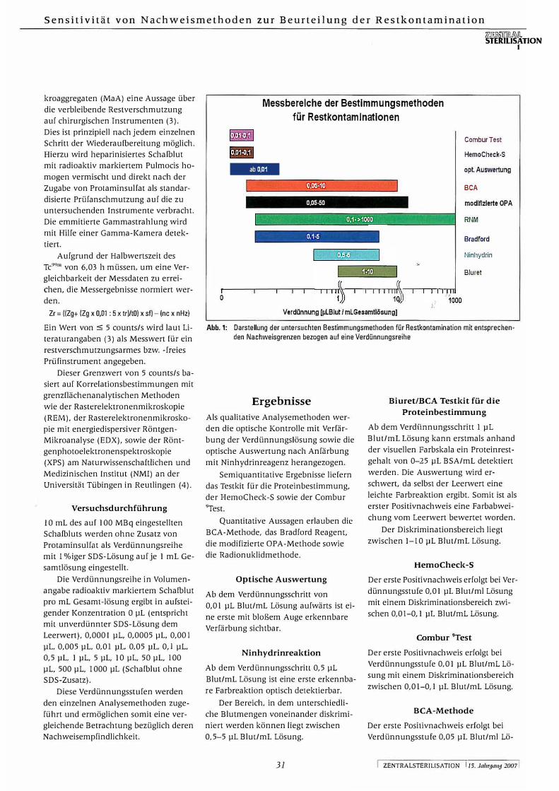
Messbereiche der Bestimmungsmethoden
für Restkontaminationen
1
0
-
0,05-50
0,1-5
1 1 II��
ComburTest
HemoCheck-S
opt. Auswertung
BCA
modifizierte OPA
RNM
Bradford
Ninhydrin
Biuret
Verdünnung [µLBlut / mLGesamtlösungJ
Abb. 1: Darstellung der untersuchten Bestimmungsmethoden für Restkontamination mit entsprechenden Nachweisgrenzen bezogen auf eine Verdünnungsreihe
Ergebnisse
Als qualitative Analysemethoden wer
den die optische Kontrolle mit Verfär
bung der Verdünnungslösung sowie die
optische Auswertung nach Anfärbung
mit Ninhydrinreagenz herangezogen.
Semiquantitative Ergebnisse liefern
das Testkit für die Proteinbestimmung,
der HemoCheck-S sowie der Combur
''Test.
Quantitative Aussagen erlauben die
BCA-Methode, das Bradford Reagent,
die modifizierte OPA-Methode sowie
die Radionuklidmethode.
Optische Auswertung
Ab dem Verdünnungsschritt von
0,01 µL Blut/mL Lösung aufwärts ist ei
ne erste mit bloßem Auge erkennbare
Verfärbung sichtbar.
Ninhydrinreaktion
Ab dem Verdünnungsschritl 0,5 µL
Blut/mL Lösung ist eine erste erkennba
re Farbreaktion optisch detektierbar.
Der Bereich, in dem unterschiedli
che Blutmengen voneinander diskrimi
niert werden können liegt zwischen
0,5-5 µL Blut/mL Lösung.
31
Biuret/BCA Testkit für die
Proteinbestimmung
Ab dem Verdi.innungsschritt 1 µL
Blut/mL Lösung kann erstmals anhand
der visuellen Farbskala ein Proteinrest
gehalt von 0-25 µL BSA/mL detektiert
werden. Die Auswertung wird er
schwert, da selbst der Leerwert eine
leichte Farbreaktion ergibt. Somit ist als
erster Positivnachweis eine Farbabwei
chung vom Leerwert bewertet worden.
Der Diskriminationsbereich liegt
zwischen 1-10 µL Blut/mL Lösung.
HemoCheck-S
Der erste Positivnachweis erfolgt bei Ver
di.innungsstufe 0,01 µL Blut/ml Lösung
mit einem Diskriminationsbereich zwi
schen 0,01-0, 1 µL Blut/mL Lösung.
Combur 9Test
Der erste Positivnachweis erfolgt bei
Verdünnungsstufe 0,01 µL Blut/mL Lö
sung mit einem Diskriminationsbereich
zwischen 0,01-0,l µL Blut/mL Lösung.
BCA-Methode
Der erste Positivnachweis erfolgt bei
Verdünnungsstufe 0,05 µL Blut/ml Lö-
ZENTRALSTERILISATION l 15. JaJir9a119 2007

Jz.ru.R:]'.ITlfüÄ\TI. STERILISATION
1
sung mit einem Diskriminationsbereich
zwischen 0,05-10 pL ßlul/mL Lösung.
Bradford Reagent
Durchführung ist nur in Verdünnung
mit bidestilliertem Wasser möglich, da
das Reagenz mit SOS interferiert und
somit zu falschen Ergebnissen führt.
Der erste Positivnachweis erfolgt bei
Verdünnungsstufe O, I pL ßlut/mL Lö
sung mit einem Diskriminationsbereich
zwischen 0, 1-5 pL Blut/mL Lösung.
Modifizierte OPA-Methode
Der erste Positivnachweis erfolgt bei
Verdünnungsstufe 0,05 pL ß\ut/mL Lö
sung mit einem Diskriminationsbereich
zwischen 0,05-50 pL Blut/mL Lösung.
Radionuklidmethode
Der erste Positivnachweis erfolgt bei
Verdünnungsstufe 0, 1 pL Blut/mL Lö
sung mit einem Diskriminationsbereich
ab 0, 1 pL Blut/mL Lösung ohne obere
Grenze.
In Abbildung I sind jeweils die Messbe
reiche der ßestimmungsmethnden vom
ersten Positivnachweis (hohe Verdün
nung mit geringem Blutanteil in der
Gesamtlösung) bis zu dem Verdi.in
nungsschritt, der noch sicher diskrimi
niert werden kann, dargestellt. Zum
Beispiel liefert der Combur Test bei
Blutvolumina oberhalb O, l pL Blut in
I mL Gesamtlösung jeweils 250 Ery/mL
als Testergebnis. Die auf photometri
schen Messungen basierenden Metho
den (BCA, modifizierte OPA und Brad
ford) zeigen bei steigendem Blutvolu
men schließlich keinen proportionalen
Extinktiunsanstieg zum Konzentrations
anstieg der Proteine bzw. Amine. Der
Biuret Test führt bei steigendem Pro
teingehalt zu einer Entfärbung der Test
lösung. Bei 10 pL Blut in I mL Gesamt
lösung kommt es zu einer vollständigen
Entfärbung, die keine weitere Diskrimi
nierung bei höheren Proteinkonzentra
tionen erlaubt.
Der Combur Test, der HemoCheck-S
sowie die optische Auswertung erwei
sen sich an den untersuchten Verdün
nungsreihen als sensitivste Nachweis
methoden. Jedoch liefern sie besten
falls semiquantitative Ergebnisse.
ZENTRALSTERILISATION j 15. JnlirfJll1tfJ 2007
T. Friedrich et al.
Analyt Nachweisgrenzen Messverfahren Probengewinnung
[pl Blut/ml)
Mod. OPA-Methode Proteine 0,05- 50 photometrisch Elution
BCA-Protein Assay Kit Proteine 0,05-10 photometrisch Elution
Bradford-Melhode Proteine 0,1-5 photometrisch Elution
Biuret, BCA-Testkil Proteine 1-10 visuell Elution
Ninhydrinreaktion Aminosäuren, 0,5-5 visuell Elution
Peptide
ComburTest Hämoglobin 0,01-0,1 visuell Elution
HemoCheck-S Hämoglobin 0,01-0,1 visuell Wischtest
Radionuklidmethode "mTc-Albumin- Ab 0,1
Komplex
Tab. 1: Gegenüberstellung der Nachweismethoden
Es folgen die quantitative ßCA- und die
modifizierte OPA-Methode vor der eben
falls quantitativen Radionuklidmethode
(RNM) und der Bradford-Methode.
Eine auf Ninhydrinfärbung basie
rende optische Auswertung folgt in ih
rer Empfindlichkeit schließlich vor dem
semiquantitativen Biuret Test.
Die entsprechenden Blutvolumina
in der Verdi.innungsreihe (in pL Blut
pro 1 mL Gesamtlösung) sind in der
Graphik dargestellt.
Vergleich der Methoden
Die Radionuklidmethode hat unter al
len Nachweismethoden den Vorteil un
abhängig von einer Elution zu sein. Ne
ben der direkten Messung der Instru
mente vor und nach einer Rückgewin
nung, sowie auch der Messung des Elu
ats ermöglicht die Radionuklidmethode
genaue Aussagen über Abreicherungs
bzw. Wiedergewinnungsraten. Die not
wendige radioaktive Markierung
schließt jedoch Instrumente aus, die
unter realen Operationsbedingungen
am Patienten kontaminiert wurden.
Die BCA-Methode benötigt für eine
gleichwertige Nachweisempfindlichkeit
gegenüber der ebenfalls quantitativen
modifizierten OPA-Methode einen weit
aus größeren Arbeitsaufwand. Durch ei
ne Inkubationszeit von 20 Minuten im
60 Grad warmen Wasserbad ist zudem
der Materialaufwand erhöht.
32
szintigrafisch Direkt am
Instrument
Die Bradford-Methode erweist sich
durch Inkompatibilität mit SDS für Fra
gestellungen der Eluatanalyse als un
brauchbar.
Das Biuret/BCA Testkit für die Pro
teinbestimmung besticht durch die ein
fache Anwendbarkeit ohne zusätzlichen
Laborbedarf. jedoch mit der Konse
quenz, nur semiquantitative Messergeb
nisse liefern zu können. Zudem gelingt
eine genaue Zuordnung der Probenfär
bung zur Farbskala oftmals nur sehr un
genau.
Der Combur 9Test, sowie der He
moCheck-S auf Basis der Pseudoperoxi
dase-Reaktion ermöglichen eine sehr
schnelle Eluatanalyse. Beide Methoden
liefern jedoch bestenfalls semiquantita
tive Ergebnisse.
Die Konzeption des HemoCheck-S
als Wischtest schalTt zusätzliche Voraus
setzungen für eine Untersuchung von
Instrumenten ohne eine vorherige Elu
tion. Das Test Kit ist allerdings auf die
Zugänglichkeit der untersuchten Ober
fläche angewiesen.
Die zwei auf Pseudoperoxidase rea
gierenden Nachweisverfahren Combur 9Test und Hemo-Check-S reagieren mit
einer Nachweisgrenze von O,OlmL
Schafblut pro I mL Gesamtlösung (bide
stilliertes Wasser) am empfindlichsten
auf heparinisiertes Schafblut. Es ist frag
lich, ob diese Empfindlichkeit auch
dann gewährleistet ist, wenn die zu
untersuchenden Instrumente zuvor ei-

Sensitivität von Nachweismethoden zur Beurteilung der Restkontamination
nem realen Wiederaufbereitungszyklus
unterworfen werden. Erythrozyten hä
molysieren unter diesen Bedingungen
und sind somit ein Blutbestandteil, der
aufgrund seiner Wasserlöslichkeit für
verbleibende Restkontamination weni
ger in Frage kommt.
Dies zeigt auch die methodische
Schwäche des Versuchsablaufes (ohne
vorgeschalteten Reinigungszyklus), die
aber billigend für eine vergleichende
Untersuchung mit quantitativen Ergeb
nissen in Kauf genommen werden
muss.
Interessanterweise hat auch die rein
qualitativ optische Bewertung eine unte
re Nachweisgrenze von 0,01 µL Blut/mL
Gesamtlösung. Allerdings ist auch hier
das farbbiJdende Hämoglobin ein gut
wasserlöslicher Bestandteil, der eine Fär
bung des Eluats bei Instrumenten nach
einem Reinigungszyklus erst bei hoher
Restkontamination erwarten lässt.
Die BCA- (Proteinnachweis), sowie
die modifizierte OPA-Methode (Amin
nachweis) haben als quantitative photo
metrische Bestimmungsmethoden eine
untere Nachweisgrenze bei 0,05ml
Blut/mL Gesamtlösung. Der Arbeits
und Zeitaufwand ist allerdings bei der
BCA-Methode im „enhanced protocol"
mit einer zusätzlichen Inkubation der
Probe bei 60 °C im Wasserbad deutlich
höher.
Die Radionuklidmethode und das
Bradford-Rcagenz (Verdünnungsreihe
mit bidestillienem Wasser) haben ihren
ersten Nachweis bei einer Verdün
nungsstufe von 0, 1 µL Blut/mL Gesamt
lösung. Der Nachweis von Proteinen
durch das Bradford-Reagenz gelingt je
doch nicht unter Verwendung von SDS
Lösung bei der Elution und scheidet so
mit für Rückgewinnungsverfahren nach
EN DIN 15883 aus. Die Radionuklidme
thode ist auf eine Prüfanschmutzung
mit radioaktiv markierten Albuminen
angewiesen und somit nur unter beson
deren Bedingungen einsetzbar. Eine
Untersuchung von real im Operations
saal verwendeten Instrumenten ist da
her nicht direkt möglich.
Die Radionuklidmethode hat als
einziges der untersuchten Prüfmetho
den einen weitestgehend unbegrenzten
Messbereich. Weiterhin ermöglicht die
Radionuklidmethode eine quantilcltive,
ortsaufgelöste und zerstörungsfreie Do
kumentation von Ausgangs- und Rest
kontamination der untersuchten Pro
ben. Für die Analyse schwer oder gar
nicht einsehbarer Oberflächen, wie zum
Beispiel Instrumente der minimal inva
siven Chirurgie ist daher keine Elution
notwendig. Die unwesentlich geringere
Empfindlichkeit im Vergleich zur BCA
sowie der modifizierten OPA-Methode
liegt in der Markierung der Albumine
durch ''9"'Technetium. Dabei ist nicht je
des durch chemische Nachweismetho
den detektierbare Protein durch das Iso
top markiert.
Der Auftrag von Ninhydrin als
Sprühlösung ermöglicht die Detektion
von Aminosäuren und Peptiden ab ei
ner Verdünnungsstufe von 0,5µL
Blut/mL Gesamtlösung aufgrund einer
Farbreaktion, die optisch quantitativ
ausgewertet wird. Erforderlich ist je
doch ein sorgfältiger Umgang mit Nin
hydrin, insbesondere bei Verwendung
von Ninhydrin als Aerosol, da dies ge
sundheitsschädlich ist. Dies erfordert ei
nen größeren Materialaufwand, insbe
sondere einen Arbeitsplatz mit Abzugs
möglichkeiten. Weiterhin ist zur Inku
bation der Proben ein Wärmeschrank
notwendig.
Die geringste Sensitivität mit einem
ersten Positivnachweis für Proteine bei
1 pL Blut/mL Gesamtlösung erbrachte
das Biuret/BCA Test Kit für die Protein
bestimmung. Dies jedoch bei einem ge
ringen Arbeits- und Zeitaufwand, ver
gleichbar mit dem Combur 9Test und
dem HemoCheck-S.
Unberücksichtigt bleibt die Tatsache,
dass nach der Aufbereitung von chirur
gischen Instrumenten nicht alle Blutbe
standteile die gleiche Bedeutung als
Restkontamination besitzen. Insbeson
dere Fibrin stellt als wasserunlösliches
Polymer höchste Anforderungen an ei
nen Reinigungsprozess. Unter diesem
Aspekt nimmt die Radionuklidmethode
eine Sonderstellung ein, da hierbei auch
durch Fibrin „maskierte" Restkontami
nation detektiert werden kann (Veröf
fentlichung in Vorbereitung).
Zusammenfassung
Unter den Nachweismethoden für Rest
kontamination ermiuelten wir für die
auf Pseudopernxidase-Reaktion basie-
33
zm�TIJ'ffi&.IL STERILISATION
1
renden qualitativen/semiquantitativen
Testverfohren die sensitivsten Nach
weisgrenzen, gefolgt von der modifizier
ten OPA- und BCA-Methode (quamita
tiv), der RNM (quantitativ), Ninhydrin
(qualitativ) und schliegJich der Biure
treaktion (serniquantitativ).
Alle genannten Nachweismethoden
sind durch ihre Sensitivität in der Lage,
die unterschiedlichen Akzeptanzkrite
rien der EN DlN l 5883 sicher zu diskri
minieren.
Literatur ( l) firEN ISO 15883 - Reinigungs-Desinfek
tionsgeräte; Anforderungen, Definitionen.Prüfmethoden (2005).
(2) Frister H, Meise! 1-1; Schlimme E: OPArnethod modiried by use of N,N-dimethyl-2-mercapto-ethylammonium-chloride as thiol componenl. Fresenius Z Anal Chcm 1988; 330: 631-633.
(3) Schrimm H. Sieben J-P, Heeg P, Roth K, Müller-Schauenburg W. Keller K-D, BuessG: Eine neue Methode zur Validierung undÜberprüfung der Reinigung von Rohrschaftinstrumenten. Zemr Steril 1994; 2: 313-324.
(4) Reich! R, Beckmann P, Dreher WF et al: Innovationen in der Medizintechnik durch grenzflächenanalytische Verfahren (Teil 2).Zentr Steril 1998; 6: 388-400.
i ZENTRALSTERILISATtON 15. J11lll'1Jm19 2007
Zentr Steril 2007; 15 (1): 29-38
Keywords • reprocessing• residual contamination11 detection methods
• sensitivityo sterilisationo surgical instruments
Sensitivity of Detection Methods for Assessment of Residual Contamination on
Reprocessed Surgical Instruments
Background:
EN DIN 15883 defines minimum cleaning re
quirements tor reprocessing surgical instru
ments in washer-disinfectors.
Objective:
To ascertain detection limits tor the following
residual-contamination detection methods.
Methods:
The Bradtord, modified DPA, biuret, BCA, nin
hydrin, Combur, HemoCheck-S and radionu
clide methods were compared with each oth
er using a blood dilution series.
Results:
The Combur9 test as weil as the HemoCheck
S test (qualitative/semi-quantitative) showed
the most sensitive detection limits, followed
by the modified OPA and BCA methods (quan
titativef, the radionuclide (RNMI (quantitative)
and ninhydrin (qualitative) methods and, last
ly, the biuret reaction (semi-quantitative).
lntroduction
EN DIN 15883 explains in concrete terms
and defines the requirements for auto
mated washing machines for decontam
inating surgical instruments. According to
this standard, residual contamination may
be tolerated on each instrument subject
to certain acceptance criteria. As criterion
for sufficient cleaning the standard spec
ifies a residual protein content of < 50µg
protein (referred to bovine serum albu
min) per ml eluate. However, the corre
sponding analytical method is described in
less concrete terms. To begin with, the
test instruments are visually inspected
and then analysed using at least semi
quantitative protein detection methods,
such as the biuret/BCA method. Howev
er, "apart from the aforementioned pro
tein-analytical methods for measuring
residues ( ... ) other physicochemical de-
1 CENTRAL SERVICE-! Vo/11111e 15 2007
T Friedrich', K. Roth', J. Gauer1, P Heer/
tection methods can be used which pro
duce correspondingly sensitive quantita
tive results" (1).
Materials and Methods
The following analytical methods were
compared with each other to elucidate
their detection limits.
Bradford Method
Protein measurement based on the Brad
ford (Bradford et. al., 1976) method derives
from the property of the dye Coomasie
Brilliant Blue G250 to bind to proteins.
This binding activity causes displacement
in the dye's absorption maximum from
red (595 nm) to blue (465 nm). The in
crease in absorption is measured at 595
nm and correlates within a particular con
centration range with the protein content
of the specimen. As such, it is possible to
gain a quantitative insight into the protein
content referred to bovine serum albumin
(BSA).
This test is performed as per the man
ufacturer's instructions, while the results
are evaluated using spectrophotometry
in a semi-microcuvette with the two-beam
photometer Nicolet evolution 100.
Modified OPA Method
The modified ortho-phthaldialdehyde (OPA)
method is a quantitative method for meas
urement of the free u and n terminal NH2
groups of proteins. The OPA method is
based on the chemical conversion of or
tho-phthaldialdehyde and free amino
groups in the presence of a thiol compo
nent into fluorescent indole compounds
that can be spectrophotometrically de
tected at 340 nm (2).
34
Prep,aration of_the OPA reagent solution: 40 mg o-phthaldialdehyde and 100 mg
N, N-dimethyl-2-merca pto-ethylammoni
um chloride are dissolved in 1 ml methanol
and 50 ml 0.1 mol/1 sodium tetraborate
buffer (pH 9.3) is added, then adding 1.25
ml 20% sodium dodecyl sulphate solu
tion (SDS).
For protein measurement by means of
the modified OPA method the proteins
are eluted from the instruments with 1 %
sodium dodecyl sulphate solution (SOS).
Conductance and evaluation: 200 µI of the eluate is added to 1 ml of the
freshly prepared OPA solution in a semi
microcuvette and mixed, and then the ex
tinction versus a pure OPA solution is
measured at 340 nm after a 3-min reaction
time in the two-beam photometer Nicolet
evolution 100.
The error margin is given with a devi
ation coefficient in the range 0.31-1.27%.
Biuret/BCA Test Kit for
Protein Measurement
This test kit presented here constitutes a
rapid test for semi-quantitative detection
of proteins on surgical instruments using
the biuret reaction and the BCA method.
SMP GmbH. Paul-Ehrlich-Strasse 40, 72076 Tübin· gen. Germany; E-mail: [email protected]
2 Universitätsklinikum Tübingen. Institut für Med. Mikrobiologie u. Hygiene, Elfriede-Aulhorn-Str. 6, 72076 Tübingen. Germany

Sensitivity of Detection Methods for Assessment of Residual Contamination
Reaction sequence:
Proteins react with reagent A giving rise to a Cu2• chelate complex (biuret reaction). Following this, reagent B reduces excess Cu2• to Cu 1+, which together withreagent C gives rise to a purple coloured complex (BCA method) whose concentration is inversely proportional to the protein concentration.
This test is carried out in accordance with the manufacturer's instructions.
Semi-quantitative evaluation is performed visually using the supplied colour scale in a measuring range between 0 and > 100 µg/ml, reterred to BSA. The manutacturer has not specified an explicit detection limit.
False-positive results can be caused by copper release from brass-containing instruments. Reducing agents, such as ascorbic acid, are substances that can cause interterence.
BCA Protein Assay Kit
This BCA Protein Assay Kit is a detection method tor quantitative measurement ot total protein load in solvents.
Reaction sequence:
Cu2• reduced to Cu 1• by proteins in an alkaline environment (biuret reaction). and further reaction with bicinchoninic acid (BCA) to form a red dye that can be quantitatively measured at a wavelength of 562 nm proportional to its concentration.
The manufacturer gives the detection limit as 5 µg/ml, referred to BSA within a working range of 5-250µg/ml. At higher concentrations the test solution must be first diluted, followed by corresponding correction multiplication.
The test is performed in accordance with the manufacturer's instructions using the "Enhanced Protocol".
Attention must be paid to interfering substances such as ascorbic acid and cysteine.
Ninhydrin Reaction
Reaction sequence:
Ninhydrin reacts preferentially with amino acids, but also with peptides, leading to decarboxylization and C02 cleavage to ketoamine, which in a second reaction step reacts with a further ninhydrin molecule.
The temperature must be increased in order to expedite the normally slow reac-
tion kinetics unfolding here, giving rise to a characteristic blue-purple colour (Ruhemann's purple).
Preparation ofthe ninhydrin spray so/ution:
0.15 g ninhydrin p.a. is mixed with 47.5 ml 2-propanol and 2.5 ml acetic acid 96%p.a. and filled into an atomizer.
Conductance and evaluation:
100 µI aliquots ot the test samples, dried on sterile cellulose, are evenly sprayed with the ninhydrin spray solution and then incubated for 30 min in a heating cabinet at 100 °C. Optical inspection is carried out purely qualitatively on the basis ot the colour reaction that has taken place. A positive as weil as a negative control is run tor each test batch.
Combur 9Test
The Combur 9Test is a rapid test used asa routine clinical measure in urine diagnostics. Apart from glucose, bilirubin and other parameters the test strip measures haemoglobin and proteins as well as the pH value. The pH read-out range is between 5 and 9 and is thus not suitable tor the further issues to be claritied in the context of this paper. Furthermore, measurement of the protein content with a detection limit of 6 mg albumin per dl is en-
'/l,51?.!i::TITIRL;,\ll, TERILISATION
1
dowed with too low a sensitivity for this series of tests. Detection of haemoglobin is a relevant parameter. The practical detection limit is given as 5 ery/µI tor intact erythrocytes and as 10 ery/µI for haemolysed erythrocytes. The erythrocytes were present in a haemolysed state for this series of tests.
Accordingly, evaluation was confined to readout of the haemoglobin test fields. The read-out range is between negative to approx. 250 ery/µI. The manufacturer gives accuracy as 90% versus the chamber counting method.
Reaction sequence:
Haemoglobin�and myoglobin catalyse oxidation of the indicator due to the organic hydroperoxide cont9ined in the test paper.
The colourless tetramethylbenzidine is oxidised in the presence of cumol hydroperoxide due to the pseudoperoxidase effect of the haemoglobin.
Conductance:
The test strip is immersed for about 1 second in the eluate and the lateral corner is peeled oft on the edge of the vessel on removal.
Visual inspection is performed semiquantitatively using the colour scale supplied.
Measuring ranges for residual contamination
detection methods
1 0
@@1111-
0.0!>--50
0.1- >1000
0.1-5
0.5-5
�( 1 1 1 1 1 ';)) 1 1 1 1 111��
Dilution [µI blood/ml total solution]
1000
ComburTest
HemoCheck-S
0 pt. evaluati on
BCA
Modified OPA
RNM
Bradford
Ninhydrin
Biuret
Fig. 1: Illustration of the residual contamination detection methods investigated, with corresponding
detection limits, referred to one dilution series
35 CENTRAL SERVICE 1 Volume 15 2007 1

HemoCheck-S
The HemoCheck-S is a specific test kit for detection of blood residues on surfaces. Detection is also based on the peroxidase contained in haemoglobin. The manufacturer gives the detection limit as 0.1 µI blood, and no error margin is specified.
The test is carried out as per the manufacturer's instructions.
Visual inspection is performed semiquantitatively based on a change of colour in the indicator solution. lnterference factors leading to false-negative results are bleaching agents such as hydrogen peroxide or peracetic acid.
Proteins are not detected.
Radionuclide Method as Reference Method
By means of quantitative, spatially resolved measurement of gamma quanta from Tc99"'-marked human albumin macroaggregates (MaA). the radionuclide method provides information on the residual contamination on surgical instruments (3). This is possible in principle after each individual reprocessing step. Here heparinised sheep blood is mixed with radioactively marked Pulmocis homogen and after addition of protamine sulphate is applied directly as a standardised test soil to the test instruments. The emitted gamma radiation is detected using a gamma camera.
In view of the half life of Tc99m of 6.03 h, the measurement results must be standardised to assure comparability of the measured data. Zr= ((Zg+ (Zg x 0,01 : 5 x tr)/tO) x sf) - (nc x nHz)
Based on the relevant literature (3). a value :s 5 counts/s is given as a measured value for a test instrument harbouring little or no residual contamination.
This limit value of 5 counts/s is based on correlation measurements with boundary surface analytical methods such as scanning electron microscopy (SEM). energy-dispersive X-ray microanalysis (EDX) as weil as on X-ray photoelectron spectroscopy (XPS) at the Natural Sciences and Medical Institute (Naturwissenschaftliches und Medizinisches Institut - NMI) at the Tübingen University in Reutlingen (4).
Test Procedure
1 O ml sheep blood set to 100 M Bq without addition of protamine sulphate was
CENTRAL SERVICE Vo/11111c 15 2007
used as a dilution series with 1 % SDS solution for each 1 ml total solution.
The dilution series in volumetric terms of radioactively marked sheep blood per ml total solution produced in ascending concentration O µI (corresponds, with undiluted SOS solution, to the blank value). 0.0001 µ1, 0.0005 µ1, 0.001 µ1, 0.005 µ1, 0.01 µI, 0.05 µI, 0.1 µI, 0.5 µ1, 1 µ1, 5 µI, 10 µI, 50 µI, 1 00 µI, 500 µI, 1000 µI (sheep blood without addition of SOS).
These dilution stages were used for the various analytical methods, thus assuring comparability of detection sensitivity.
Results
The qualitative analytical methods used were optical control with a change of colour in the dilution solution as weil as optical evaluation after coloration with the ninhydrin reagent. The test kit for protein measurement, HemoCheck-S as weil as the Combur9Test yielded semi-quantitative results.
The BCA method, Bradford reagent, modified OPA method as weil as the radionuclide method produced quantitative results.
Optical Evaluation
A change in colour can be detected with the naked eye only as from the dilution step of 0.01 µI blood/ml solution.
Ninhydrin Reaction
A visual change in colour can be detected only as from the dilution step 0.5 µI blood/ml solution.
The range within which different blood quantities can be discriminated is between 0.5-5 µI blood/ml solution.
Biuret/BCA Test Kit for Protein Measurement
Only as from a 1 µI blood /ml solution dilution step can a protein content of 0-25 µI BSNml be first detected with the visual colour scale. Evaluation is hampered by the fact that even the blank value itself causes a slight colour reaction. Hence an initial positive detection result is based on making a colour distinction versus the blank value. The discrimination range is between 1-10 µI blood/ml solution.
36
T. Friedrich et al.
HemoCheck-S
The first positive detection result is seen at a dilution stage of 0.01 µI blood/ml solution with a discrimination range between 0.01-0.1 µI blood/ml solution.
ComburTest
The first positive detection result is seen at a dilution level of 0.01 µI blood/ml solution with a discrimination range between 0.01-0.1 µI blood/ml solution.
BCA Method
The first positive detection result is seen at a 'dilution level of 0.05 µI b)ood/ml SOiution with a discrimination range between 0.05-10 µI blood/ml �9lution.
l.
Bradford Reagent
This test can only be performed in a dilution using double-distilled water since the reagent interferes with SOS, thus leading to false results.
The first positive detection result is seen at a dilution level of 0.1 µI blood/ml solution with a discrimination range between 0.1-5 µI blood/ml solution.
Modified OPA Method
The first positive detection result is seen at a dilution level of 0. 05 µI blood/ml solution with a discrimination range between 0.05-50 µI blood/ml solution.
Radionuclide Method
The first positive detection result is seen at a dilution level of 0.1 µI blood/ml solution with a discrimination range as from 0.1 µI blood/ml solution, with no upper limit.
Shown in all cases in figure 1 are the measuring ranges for the various detection methods, from the first positive detection result (high dilution with low blood content in the total solution) up to the dilution stage which can still be reliably discriminated. For example, in blood volumes above 0.1 µI blood in 1 ml total solution the Combur test yields 250 ery/ml as test result. With increasing blood volumes, the methods based on photometric measurements (BCA. modified OPA and Bradfordl ultimately do not show an increase in extinction proportional to the increase in the concentration of protein or amines.

Sensitivity of Detection Methods for Assessment of Residual Contamination
With an increasing protein content, the biuret test Jeads to discoloration of the test solution. Complete discoloration is seen at 10 µI blood in 1 ml total solution which does not permit further discrimination for higher protein concentrations.
The Combur test, HemoCheck-S as weil as optical evaluation have proven to be the most sensitive detection methods of the dilution series investigated. However, at most they produce semi-quantitative results.
Ranking next are the quantitative BCA and modified OPA method, before the likewise quantitative radionuclide method (RNM) and the Bradford method.
In terms of sensitivity optical evaluation based on a ninhydrin colour change ranks before the semi-quantitative biuret test.
The corresponding blood volumes used in the dilution series (in µJ blood per 1 ml total solution) are illustrated in the preceding graph.
Comparison of Methods
The radionuclide method, of all the detection methods, confers the advantage of being independent of the elution. Apart from direct measurement of instruments before and after recovery as weil as measurement of the eluate, the radionuclide method gives an accurate picture of the reduction or recovery rates. However, the need for radioactive marking rules out instru-ments contaminated when used for patient surgery under everyday conditions.
The BCA method calls for infinitely more laborious efforts to assure a detection sensitivity equivalent to that of the likewise quantitative modified OPA method. The 20-minute incubation time in a water bath heated to 60 degrees also calls for greater material investment.
The Bradford method does not lend itself to eluate analysis because of incompatibility with SOS.
The biuret/BCA Test Kit for Protein Measurement has the advantage of userfriendliness without the need for any additional laboratory measures but suffers the drawback of being able to produce only semi-quantitative measurement results. Furthermore, it is often very hard to evaluate the colour change in the specimen on the basis of the colour scale.
zz�.Rli'ii'ffih,\ll STERILISATION
l
Analyt Detection limits Measurement Sample recovery [111 blood/ml] method
Mod. DPA method Proteins 0.05- 50 Photometrie Elution
BCA protein assay kit Proteins 0.05-10 Photometrie Elution
Bradford melhod Proteins 0.1-5 Photo m etri c Elution
Biuret, BCA lest kit Proteins 1 -10 Visual Elution
Ni nhydrin reacti an Amino acids, 0.5-5 Visual Elution
peptides
Combur"Test Haemoglobin 0.01 -0.1 Visual Elution
HemoCheck-S Haemoglobin 0.01 -0.1 Visual Wipe lest
Radionuclide method 99mTc albumin As from 0.1 Scintigraphic Directly on
complex
Table 1: Comparison of the detection methods
The Combur9Test, as weil as the HemoCheck-S based on the pseudoperoxidase reaction provides for very rapid eluate analysis. But both methods produce at most semi-quantitative results.
Designing the HemoCheck-S as a wipe test provides greater opportunities for investigating instruments without prior elution. However, the test kit is dependent on accessibility to the test surfaces.
The two detection methods based on a pseudoperoxidase reaction, Combur9Test and Hemo-Check-S, show the most sensitive reaction at a detection limit 0.01 ml sheep blood per 1 ml total solution (double-distilled water) to heparinised sheep blood. lt is questionable whether this sensitivity would then be assured if the test instruments had first been subjected to a decontamination cycle conducted under everyday conditions. Under such conditions erythrocytes undergo haemolysis and as such embody a blood constituent that, thanks to its water solubility, is no longer likely to be seen as residual contamination.
This also highlights the methodological flaw in this experimental design (without a preceding cleaning cycle). but which must be borne in mind for a comparative investigation with quantitative results.
lnterestingly, the purely qualitative optical evaluation test has a lower detection limit of 0.01 µJ blood/ml total solution. But, here too, the colour-generating haemoglobin is a good water-soluble constituent which is likely to result in coloration of the eluate only in the presence of high
37
instrument
residual contami�ation from instruments after a cleaning cycle.
The BCA (protein detection) as weil as the modified OPA method (amine detectionJ, as quantitative photometric measurement methods, have a lower detection limit at 0.05 ml blood /ml total solution. But conductance of the BCA method as per the "enhanced protocol" with additional incubation of the sample in a water bath at 60 °C is markedly more laborious and time consuming.
The radionuclide method and the Bradford reagent (dilution series with doubledistilled waterJ show initial detection with a dilution stage of 0.1 µJ blood /ml total solution. But protein detection with the Bradford reagent is not possible on using an SOS solution for elution and is thus unsuitable as a recovery method as per EN DIN 15883. The radionuclide method is dependent on a test soil with radioactively marked albumin and thus can only be used subject to certain conditions. lt does not therefore lend itself to direct investigation of instruments used under everyday conditions in the operating theatre.
The radionuclide method is the only test method among those investigated to have a largely unlimited measuring range. Furthermore, the radionuclide method permits quantitative, spatially resolved and interference-free documentation of baseline and residual contamination of the test samples. Hence elution is not needed for surfaces that are difficult to access or cannot at all be inspected, such as minimally invasive surgical instruments. The negligibly lower sensitivity compared with
CENTRAL SERVICE f Vo/11111c 15 2007 j
T. Friedrich et al.-----
the BCA as weil as the modified OPA
method is attributable to marking of al
bumins with 99mTechnetium. Here not
every protein amenable to detection by
means of chemical detection methods is
marked with the isotope.
Application of ninhydrin as a spray
solution provides for detection of amino
acids and peptides as from a dilution lev
el of 0.5µ1 blood/ml total solution by virtue
of a colour reaction which permits opti
cal, quantitative evaluation. However,
caution is need when handling ninhydrin,
especially when using ninhydrin as an
aerosol because it poses a danger to
health. This test calls for greater materi
al investment, in particular for a working
environment with exhaust facilities. A
heating cabinet is also needed for sam
ple incubation.
The lowest sensitivity with first positive de
tection of proteins at 1 µI blood/ml total so
lution was shown by the biuret/ BCA test
kit for protein measurement. However,
CENTRAL SERVICE Vo/11111e 15 2007 f
these are less laborious and time-con
suming than the Combur9Test and the He
moCheck-S.
No attention was paid to the fact that
following decontamination of surgical in
struments, the role played by the various
blood constituents as potential residual
contaminants is not the same. For exam
ple, fibrin being a water-insoluble polymer
makes the highest demands on the clean
ing process. In this respect, the radionu
clide method differs from other methods
in that it can also detect residual contam
ination "masked" by fibrin (publication un
der preparation).
Summary
Of the residual contamination detection
methods investigated, those qualitative/
semi-quantitative test methods based on
the pseudoperoxidase reaction proved to
be endowed with the greatest sensitivity,
followed by the modified OPA and BCA
method (quantitative), the RNM (quanti-
38
tative), ninhydrin (qualitative) and, lastly, the
biuret reaction (semi-quantitative).
In terms of their sensitivity, all the
aforementioned detection methods are
capable of reliably discriminating the var
ious acceptance criteria specified by EN
DIN 15883. :J:
References
(1) prEN ISO 15883 - Reinigungs-Desinfektionsgeräte;Anforderungen, Definitionen, Prüfmethoden (2005)
(2) Frister H, Meisel H, Schlimme E: OPA-method mod·ified by use of N,N-dimethyl-2-mercapto·ethylam·monium-chloride as thiol component. Fresenius ZAnal Chem 1988; 330: 6_3,1-633
(3) Schrimm H, Siebe.r1 J-P, Heeg P. Roth K. MüllerSchauenburg W. Keller K-D. Buess G: Eine neueMethode zur Validierung und Überprüfung der Reinigung von Rohrschaltinstrumenten. Zentr Steril 1994;2: 313-324
(4) Reichl R, Beckmann P. DreherWF et al: Innovationenin der Medizintechnik durch grenzflächenanalytische Verfahren (Teil 21. Zentr Steril 1998; 6: 388-400


![1. WASSERHYGIENE [Kompatibilitätsmodus] · Da die Nachweismethoden für pathogene Mikroorganismen sehr langwierig sind, werden in der Trinkwasserhygiene diese Organismen nicht untersucht.](https://static.fdokument.com/doc/165x107/5d5c59d288c9934c3b8b829e/1-wasserhygiene-kompatibilitaetsmodus-da-die-nachweismethoden-fuer-pathogene.jpg)