The Role of the EUCLID Archive System in Distributed Data ...
1
Diplomarbeit
The role of sphingosine 1-phsophate in mouse hypersensitivity to noxious thermal
stimuli
zur Erlangung des akademischen Grades
Doktor der gesamten Heilkunde (Dr. med. univ.)
an der
Medizinischen Universität Innsbruck
ausgeführt am
Department für Physiologie und Medizinische Physik
Sektion für Physiologie
unter der Anleitung von
Univ.-Prof. Dr. med. Michaela Kress
eingereicht von
Maurice Selhorst
Geburtstag: 24.09.1983 Mat. Nr.: 0538416 Adresse: Poststraße 34 58675 Hemer Deutschland Innsbruck, den 20.6.2011 Maurice Selhorst

2
Eigenleistung
Hiermit versichere ich, dass ich diese Diplomarbeit selbständig verfasst und keine anderen
als die angegebenen Quellen und Hilfsmittel benutzt habe. Sämtliche Versuche und
Experimente dieser Arbeit wurden von mir selbstständig durchgeführt und ausgewertet. Die
Abbildungen und Tabellen dieser Arbeit wurden von mir selbst erstellt. Nicht von mir
persönlich erstellte Abbildungen wurden nur nach ausdrücklicher Erlaubnis der Autoren
benutzt und dann im Quellenverzeichnis vermerkt.
Innsbruck, im Juni 2011 Maurice Selhorst

3
Acknowledgements
First of all I thank Univ.-Prof. Dr. med. Michaela Kress for giving me the chance to work
with a nice work group on my diploma thesis and for her patience and support in her lab.
A special thank goes to Dr. Cristina Constantin for her patience, encouragement and her
special kind of humour and to Markus Doblander and Dr. Norbert Mair for their constant
advice and help.
I also sincerely thank my parents and my brothers Ramy and Marcel for their constant
support, love and their constant guidance through my non-academic and academic education.
Last, but not least I thank my friends in Germany, Austria, Luxembourg and Syria, who have
accompanied me for many years and who never doubt on my courage.

4
Table of Contents
Eigenleistung 2 Acknowledgements 3 Table of Contents 4 Abstract 5 Zusammenfassung 6 Abbreviations 7 1 Introduction 10
1.1 Pain processing in physiological and pathological conditions 10 1.2 Inflammatory pain and his mediators 12 1.3 Sphingosine1-phosphate synthesis (S1P) and possible sources of S1P 13 1.4 S1P as an extra- and intracellular mediator and its G-protein coupled receptors 14 1.5 S1P in nociception 16
2 Aims 17 3 Materials and Methods 18
3.1 Behavioural analysis of mechanical and heat sensitivity in sphingosine kinase 1 knock-out mice and wild type litter mates 18 3.2 Electrophysiology of primary afferent nociceptors in a skin-nerve preparation in vitro 19 3.3 Statistical analysis 21
4 Results 22 4.1 CFA induced inflammation in SphK1 null mutant and in C57BL/6J wt mice 22 4.2 Similar mechanical hypersensitivity of SphK1-/- and C57BL/6J wt mice after CFA-injection 23 4.3 Similar thermal hypersensitivity of SphK1-/- and C57BL/6J wt mice after CFA-injection 24 4.4 FTY720 does not alter nociceptor heat sensitivity in vitro 25
5 Discussion 27 6 References 30 Curriculum Vitae 38
5
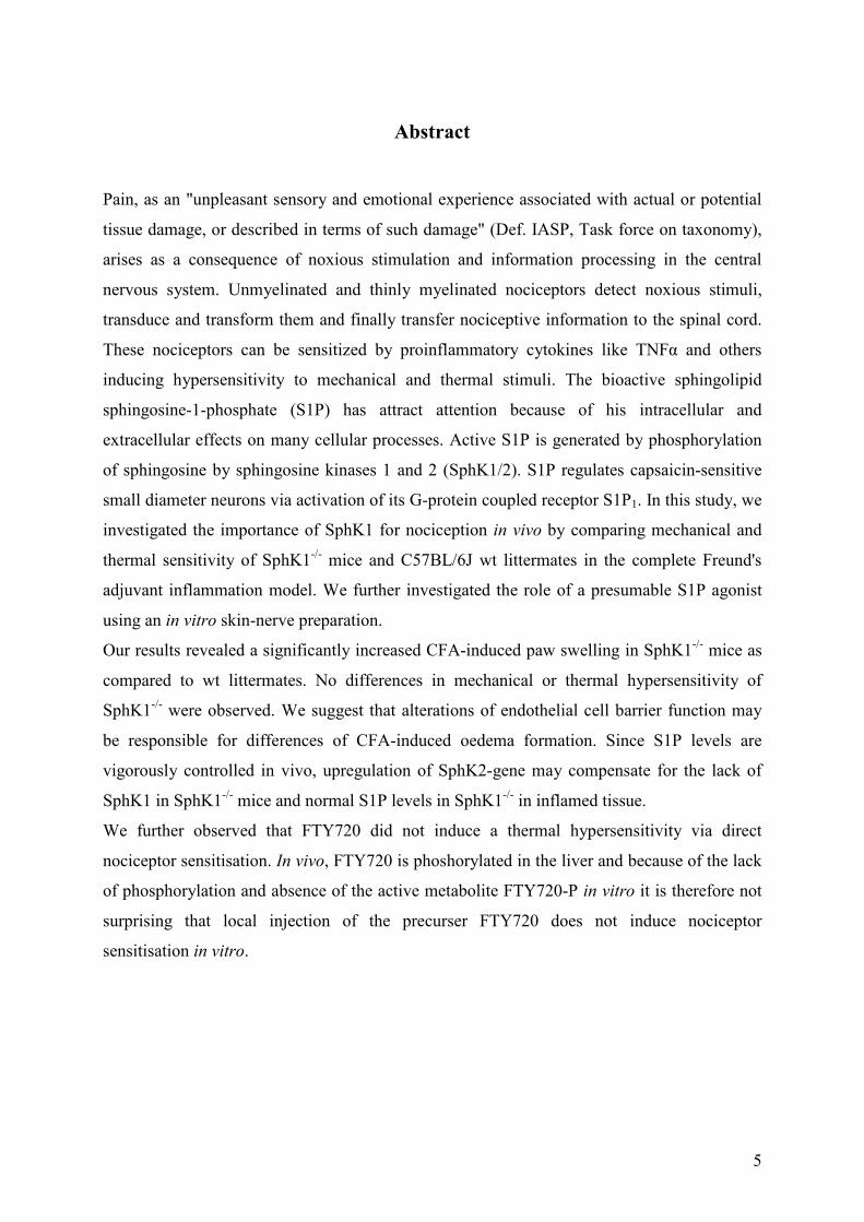
Abstract
Pain, as an "unpleasant sensory and emotional experience associated with actual or potential
tissue damage, or described in terms of such damage" (Def. IASP, Task force on taxonomy),
arises as a consequence of noxious stimulation and information processing in the central
nervous system. Unmyelinated and thinly myelinated nociceptors detect noxious stimuli,
transduce and transform them and finally transfer nociceptive information to the spinal cord.
These nociceptors can be sensitized by proinflammatory cytokines like TNFα and others
inducing hypersensitivity to mechanical and thermal stimuli. The bioactive sphingolipid
sphingosine-1-phosphate (S1P) has attract attention because of his intracellular and
extracellular effects on many cellular processes. Active S1P is generated by phosphorylation
of sphingosine by sphingosine kinases 1 and 2 (SphK1/2). S1P regulates capsaicin-sensitive
small diameter neurons via activation of its G-protein coupled receptor S1P1. In this study, we
investigated the importance of SphK1 for nociception in vivo by comparing mechanical and
thermal sensitivity of SphK1-/- mice and C57BL/6J wt littermates in the complete Freund's
adjuvant inflammation model. We further investigated the role of a presumable S1P agonist
using an in vitro skin-nerve preparation.
Our results revealed a significantly increased CFA-induced paw swelling in SphK1-/- mice as
compared to wt littermates. No differences in mechanical or thermal hypersensitivity of
SphK1-/- were observed. We suggest that alterations of endothelial cell barrier function may
be responsible for differences of CFA-induced oedema formation. Since S1P levels are
vigorously controlled in vivo, upregulation of SphK2-gene may compensate for the lack of
SphK1 in SphK1-/- mice and normal S1P levels in SphK1-/- in inflamed tissue.
We further observed that FTY720 did not induce a thermal hypersensitivity via direct
nociceptor sensitisation. In vivo, FTY720 is phoshorylated in the liver and because of the lack
of phosphorylation and absence of the active metabolite FTY720-P in vitro it is therefore not
surprising that local injection of the precurser FTY720 does not induce nociceptor
sensitisation in vitro.

6
Zusammenfassung
Schmerz, „ein unangenehmes Sinnes- oder Gefühlserlebnis, das mit tatsächlicher oder
potenzieller Gewebeschädigung einhergeht oder von betroffenen Personen so beschrieben
wird, als wäre eine solche Gewebeschädigung die Ursache“ (Def.: IASP, task force of
taxonomy), entsteht als Konsequenz von Schmerzwahrnehmung und
Informationsverarbeitung im Zentralnervensystem. Unmyelinisierte und dünn myelinisierte
freie Nervenendigungen nehmen Schmerzreize wahr, transduzieren und transformieren sie
und leiten die nozizeptiven Informationen letztendlich an das Rückenmark weiter. Diese
Nozizeptoren können durch proinflammatorische Zytokine wie TNFα für thermische und
mechanische Reize sensibilisiert werden. Das bioaktive Sphingolipid Sphingosin-1-Phosphat
erregte Aufmerksamkeit dadurch, dass es sowohl intrazelluläre als auch extrazelluläre Effekte
auf verschiedene Zellprozesse hat. Das aktive S1P wird durch die Phosphorylierung von
Sphingosin durch die Sphingosin Kinasen 1 und 2 (SphK1/2) gebildet. S1P reguliert über
Aktivierung seines G-Protein gebundenen Rezeptors S1P1 Capsaicin-sensitive afferente
Fasern. In dieser Studie erforschten wir in vivo die Rolle von SphK1 in der Nozizeption,
indem wir mechanische und thermische Sensibilisierung von SphK1-/- Mäusen und C57BL/6J
Wildtyp Littermates in einem „Complete Freund’s Adjuvant“ Entzündungsmodell verglichen.
Des Weiteren erforschten wir die Rolle eines mutmaßlichen S1P Agonisten an einem in vitro
Nerven-Haut-Präparat.
Unsere Ergebnisse zeigen, dass SphK-/--Mäuse eine stärkere Schwellung auf eine CFA-
Injektion entwickeln als Wildtyp-Mäuse. Es wurden ansonsten keine Unterschiede in der
mechanischen und thermischen Hitzwahrnehmung innerhalb von 72 Stunden festgestellt. Wir
vermuten, dass Veränderungen der endothelialen Zellbarriere verantwortlich sind für die
Unterschiede in der durch CFA ausgelösten Ödembildung. Da die S1P Spiegel in vivo stark
reguliert werden, könnte eine Hochregulierung des SphK2 Genes den Mangel an SphK1 in
SphK1-/- Mäusen kompensieren und so auch für normale S1P Spiegel in dem entzündeten
Gewebe sorgen.
Wir beobachteten außerdem, dass FTY720 keine Veränderung der Hitzesensibilisierung an
Nozizeptoren auslöst. In Vivo wird FTY720 in der Leber phosphoryliert und damit aktiviert.
Es ist deswegen nicht überraschend, dass ein lokales Verabreichen des Vorläufers FTY720 in
vitro keine Sensibilisierung von Nozizeptoren auslöst, da der Mangel an Phosphorylierung
von FTY720 eine Abwesenheit des aktiven FTY720-P bedeutet.

7
Abbreviations
A(HT)M High-Threshold Mechanosensitive Aδ-fibre
A(LT)M Low-Threshold Mechanosensitive Aδ-fibre
ABC-Transporter ATP-Binding Cassette Transporter
AC Adenylyl Cyclase
AM RA Rapid Adapting Mechanosensitive Aδ-fibre
AM SA Slow Adapting Mechanosensitive Aδ-fibre
AM Mechanosensitive Aδ-fibre
AMC Mechano-cold sensitive Aδ-fibre
AMH Mechano-heat sensitive Aδ-fibre
AMP, cAMP Adenosine Monophosphate, cyclic Adenosine Monophosphate
AMPA α-Amino-3-hydroxy-5-methyl-4-isoxazolepropionic acid
ASIC Acid-sensing ion channel
C(HT) High-threshold Mechanosensitive C-fibre
C(LT)M Low-threshold Mechanosensitive C-fibre
CC Cold-sensitive C-fibre
CCK Cholecystokinin
CFA Complete Freund's Adjuvant
CGRP Calcitonin gene-related peptide
CH Heat-sensitive C-fibre
CHiMi Mechano-insensitive and heat-insensitive C-fibre
CMC Mechano-cold sensitive nociceptor
CMH Mechano-heat sensitive nociceptor
CNS Central nervous system
COX, COX-1/2/3 Cyclooxygenases 1/2/3
CRPS Complex regional pain syndrome
DAG Diacylglycerol
DHS DL-threo-dihydrosphingosine
DMS Dimethylsphingosine
DNIC Diffuse noxiuos inhibitory control
DRG Dorsal root ganglion
EDG Endothelial differentiation gene
EGF Epidermal growth factor
8
ERK Extracellular signal-regulated Kinase
GABA γ-Amino-butyric acid
GPCR G-protein coupled receptors
IASP International Association of Study of Pain
IFN-γ Interferon γ
IGF Insulin-like growth factor
IL1β Interleukin 1β,
IL6 Interleukin 6
IP3 Inositoltriphosphate-3
LPA Lysophosphatidic acid
LPP Lipid phosphate phosphatase
LTP Long-term potentiation
MAPK Mitogen-activated protein kinase
MCP-1 Monocyte-chemoattractant protein 1
NGF Nerve growth factor
NMDA N-Methyl-D-aspartate
NNDS N,N-Dimethylsphingosine
NOS Nitric oxide synthase
NRS Numeric rating scale
PAG Periaqueductal grey
PDGF Platelet-derived growth factor
PGH2 , , Prostaglandin H2,
PGE2 Prostaglandin E2,
PGD2 Prostaglandin D2,
PGF2 Prostaglandin F2
PGI2 Prostacyclin
PI3K Phosphatidyl-inositol-triphosphate kinase
PKA Protein kinase A
PKC Protein kinase C
PLC Phospholipase C
RVM Rostral ventromedial medulla
S1P Sphingosine 1-phosphate
S1P1-5 S1P Receptor 1-5
SEM Standard error of the mean

9
SIF Synthetic interstitial fluid
SphK1/2 Sphingosine kinase 1/2
TNFα Tumour necrosis factor-α
TrkA Tyrosine kinase receptor A
TRPV1 Transient receptor potential, vanilloid subfamily, member 1
TTX-R Tetrodotoxin-resistant sodium channel
TXA2 Thromboxane A2
VEGF Vascular endothelial growth factor
VIP Vasoactive intestinal peptide
WDR Wide Dynamic Range Neurons

10
1 Introduction
1.1 Pain processing in physiological and pathological conditions
Pain is an "unpleasant sensory and emotional experience associated with actual or potential
tissue damage, or described in terms of such damage" (Def. IASP, Task force on taxonomy).
As reported by Breivik and coworkers, between 12 % and 30 % of the population of the
European countries are suffering from chronic pain defined as pain of more than six months
duration and a pain intensity of 5 or ore on a 10-point Numeric Rating Scale (NRS) with 1 =
no pain to 10 = the worst pain imaginable (Breivik et al., 2006). From this population nearly
60% report on pain lasting for 2 up to 15 years (Breivik et al, 2006). Pain arises as a
consequence of noxious stimulation and information processing in the spinal cord, thalamic
and cortical structures. The sensation of pain includes sensory-discriminative, affective as
well as motor and autonomic components (Casey, 2000). The nociceptive afferent pain
pathway starts at specialised nociceptive primary afferents that innervate skin and most other
tissues which were first discovered by Charles Sherrington (Sherrington, 1906). Noxious
stimuli are detected by free nerve endings (transduction), transformed into a series of action
potentials which then travel along unmyelinated or thinly myelinated axons towards the spinal
cord. The cell bodies of nociceptive primary afferents are localised in the dorsal root ganglia
(DRG) or trigeminal ganglia (TG) and their central processes terminate in superficial layers of
the spinal cord dorsal horn (Millan, 1999) or, in case of the trigeminal nociceptors, to the
trigeminal sensory nuclei in the brainstem (Millan, 1999). Primary afferent axons can be
classified according to diameter, nerve conduction velocity and myelination. Nociceptive
primary afferent axons are found among Aδ- and C-fibres. Aδ-fibres are thinly myelinated,
with diameters between 2 to 6 µm and conduction velocity of 4-20 m/s (Adriaensen et al.,
1983; for review see Lumpkin and Caterina, 2007; Zimmermann et al., 2009). Two main
classes of Aδ-fibres can be discriminated, both of them promote the perception of acute pain
and trigger the withdrawal reflex (for review see Zimmermann et al., 2009): slow-adapting
mechano-sensitive fibres with mechanical von Frey thresholds between 1-128 mN and are
sensitive to heat or intense cold stimuli with are termed mechano-heat sensitive Aδ-fibres
(AMH) or mechano-cold sensitive Aδ-fibres (AMC). The second are mechanoreceptors,
which only respond to mechanical stimuli (mechano-sensitive Aδ-fibres [AM]) and can be
further discriminated into slowly adapting (AM-SA) and rapidly adapting (AM-RA) fibres
with low (A-LTM; von Frey threshold <1-5.7mN) or high (A-HTM; von Frey threshold ~5.7-

11
128mN) threshold (for review see Zimmermann et al., 2009). Nearly 85% of all Aδ-fibres
found in skin of primates are AMH units (Xu et al., 2010). In the muscle, activation of Aδ-
fibres can produce an aching sensation without differentiation between the pain qualities and
with less quality of localisation (Millan, 1999). Their modalities range from innocuous
mechanical, thermal and chemical stimuli as well as noxious stimuli like painful pressure or
ischemic and hypoxic pain (for review see Julius et al., 2001; Zimmermann et al., 2009;
Djouhri and Lawson, 2004). C-fibres are unmyelinated, slowly adapting fibres with diameters
between 0.4 to 1.2 µm and conduction velocities of 0.5 to 2.0 m/s. They convey a more
diffuse, dull and delayed pain sensation. Depending on the stimuli they react on, they can be
classified into different types of C-fibres, including mechano-heat sensitive nociceptors
(CMH) which make up one third of the C-fibre population in most species (Schmidt et al.,
1995), mechano-sensitive receptors of low threshold (C[LT]M; Von Frey threshold <1-
5.7mN) and high threshold (C[HT]M; Von Frey threshold ~5.7-128mN), as well as thermo-
sensitive receptors just reacting of cooling ( Cold Receptors, CC) and heating (CH).
Approximately two thirds react to multiple types of stimuli including noxious heat, cold,
pressure or chemicals and are therefore termed polymodal (Reeh, 1988; Kress et al., 1992;
Koltzenburg et al., 1997; for review see Lumpkin and Caterina, 2007). Also polymodal
mechano-cold sensitive nociceptors (CMC) and C-fibres that are insensitive to mechanical
and heat stimuli (CHiMi), are members of the C- fibre class. They express transducer ion
channels and metabotropic receptors for chemical mediators like substance P, acetylcholine,
histamine, prostaglandins, serotonin and proteolytic enzymes. (for review see Zimmermann et
al., 2009; Schmidt et al., 1995; Julius et al., 2001; Garry et al., 2004). The cranial primary
afferent fibres reach the brain stem through the trigeminal ganglion and nucleus, while the
thoracolumbar and sacral primary afferent fibres are connected to the spinal cord via the
dorsal root and dorsal root ganglia. Their primary axons end in the dorsal horn within the grey
matter, a cluster of neuron cell bodies and glia cells, where the axons form synapses with
second-order neurons mainly in the superficial Rexed laminae I-II or in the deeper Rexed
laminae V, VI, VII and X ((Handwerker et al., 1975b; Handwerker et al., 1975a), Craig, 1991;
Craig et al., 2001; Zhang et al., 2000; Millan, 1999). The second-order neurons can be
discriminated into nociceptive specific neurons, which receive their information just by Aδ-
and C-Fibres and respond to intense mechanical, heat and chemical stimuli, into non-
nociceptive neurons, which react on peripheral stimuli like weak stimuli transmitted by Aβ-
and Aδ-fibres and into wide dynamic range (WDR) neurons. The WDR-neurons, mainly
located in lamina V, respond to low- and high-intensity peripheral stimulation of Aβ-, Aδ-
12
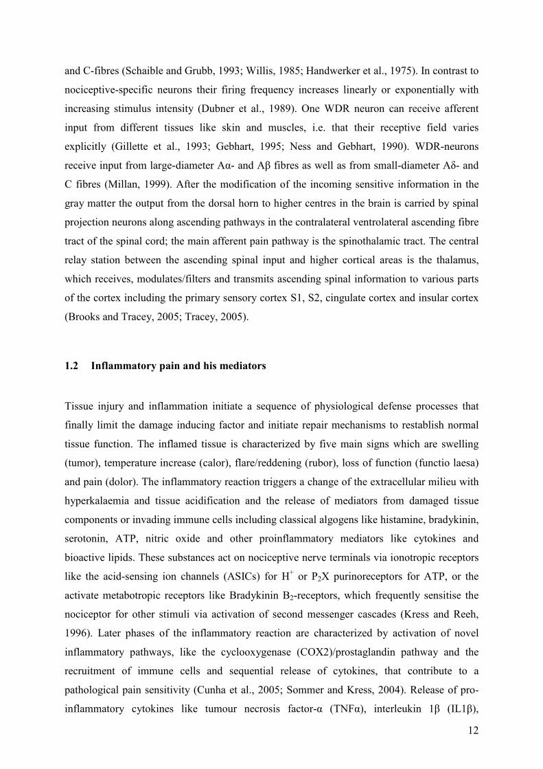
and C-fibres (Schaible and Grubb, 1993; Willis, 1985; Handwerker et al., 1975). In contrast to
nociceptive-specific neurons their firing frequency increases linearly or exponentially with
increasing stimulus intensity (Dubner et al., 1989). One WDR neuron can receive afferent
input from different tissues like skin and muscles, i.e. that their receptive field varies
explicitly (Gillette et al., 1993; Gebhart, 1995; Ness and Gebhart, 1990). WDR-neurons
receive input from large-diameter Aα- and Aβ fibres as well as from small-diameter Aδ- and
C fibres (Millan, 1999). After the modification of the incoming sensitive information in the
gray matter the output from the dorsal horn to higher centres in the brain is carried by spinal
projection neurons along ascending pathways in the contralateral ventrolateral ascending fibre
tract of the spinal cord; the main afferent pain pathway is the spinothalamic tract. The central
relay station between the ascending spinal input and higher cortical areas is the thalamus,
which receives, modulates/filters and transmits ascending spinal information to various parts
of the cortex including the primary sensory cortex S1, S2, cingulate cortex and insular cortex
(Brooks and Tracey, 2005; Tracey, 2005).
1.2 Inflammatory pain and his mediators
Tissue injury and inflammation initiate a sequence of physiological defense processes that
finally limit the damage inducing factor and initiate repair mechanisms to restablish normal
tissue function. The inflamed tissue is characterized by five main signs which are swelling
(tumor), temperature increase (calor), flare/reddening (rubor), loss of function (functio laesa)
and pain (dolor). The inflammatory reaction triggers a change of the extracellular milieu with
hyperkalaemia and tissue acidification and the release of mediators from damaged tissue
components or invading immune cells including classical algogens like histamine, bradykinin,
serotonin, ATP, nitric oxide and other proinflammatory mediators like cytokines and
bioactive lipids. These substances act on nociceptive nerve terminals via ionotropic receptors
like the acid-sensing ion channels (ASICs) for H+ or P2X purinoreceptors for ATP, or the
activate metabotropic receptors like Bradykinin B2-receptors, which frequently sensitise the
nociceptor for other stimuli via activation of second messenger cascades (Kress and Reeh,
1996). Later phases of the inflammatory reaction are characterized by activation of novel
inflammatory pathways, like the cyclooxygenase (COX2)/prostaglandin pathway and the
recruitment of immune cells and sequential release of cytokines, that contribute to a
pathological pain sensitivity (Cunha et al., 2005; Sommer and Kress, 2004). Release of pro-
inflammatory cytokines like tumour necrosis factor-α (TNFα), interleukin 1β (IL1β),

13
Interleukin 6 (IL6), nerve growths factor (NGF) and prostaglandins induce thermal and
mechanical hyperalgesia via sensitisation of nociceptors (Verri et al., 2006; Oprée and Kress,
2000). They can induce expression and protein biosynthesis, e.g. of COX2 and at the same
time regulate transducer channels like the nociceptor specific heat sensor transient receptor
potential vanilloid subfamily 1 (TRPV1) or voltage-gated sodium channels by activation of
second messenger cascades like cAMP/protein kinase A (PKA) or diacyl-glycerol/protein
kinase C (PKC). The TRPV1 transducer channel is generally accepted as one of the main
targets of different cytokines and pro-inflammatory mediators under conditions associated
with pathological pain (Tominaga et al., 1998) It is one member of the thermoTRP family of
ion channels that are activated by changes of temperature (Jordt et al., 2003; Talavera et al.,
2008). TRPV1 reacts on noxious heat with a threshold temperature above 42°C and responds
to capsaicin, the active pungent substance in red hot chilli peppers (8-methyl-N-vanillyl-6-
nonenamide) (Caterina et al., 1997). Especially nociceptive Aδ- and C-fibres express this
receptor, that is inactive at a normal body temperature. Phosphorylation in particular at serine
and threonine sites lowers the threshold and facilitates the activity. Specific consensus sites
for PKA, PKC and tyrosine kinases have been identified which are critical sites regulating the
channels kinetic properties (for review see Tominaga and Tominaga, 2005). A number of
receptor mediated intracellular pathways initiated by inflammatory mediators converge on
TRPV1 regulation in nociceptors (for review see Tominaga and Tominaga, 2005). Therefore
it is not surprising, that TRPV1 has been found essential for thermal hypersensitivity
associated with inflammation (Davis et al., 2000; Caterina et al., 2000).
1.3 Sphingosine1-phosphate synthesis (S1P) and possible sources of S1P
Sphingolipids have attracted great attention in the last few years. They are known as
important structural components of the cell membrane, are involved in formation of
membrane crafts and caveolae, and as important signalling molecules. In particular,
sphingosine 1-phosphate (S1P) is involved in many cellular functions including proliferation,
cell differentiation, apoptosis, lymphocyte trafficking, angiogenesis and inflammation (Hla et
al., 2008; Alvarez et al., 2007). S1P is generated from sphingomyelin that is degraded by
sphingomyelinase to form ceramide. This in turn is cleaved by ceramidase and sphingosine is
generated. Two isoforms of sphingosine kinases (SphKs) 1 or 2 are know which
phosphorylate sphingosine. S1P levels are tightly regulated and S1P is either degraded to
hexadecanal and phosphoetanolamine by S1P lyase or inactivated by dephosphorylation by

14
unspecific lipid phosphate phosphatase (LPP) or S1P phosphatase (Veldhoven and Mannaerts,
1991; Veldhoven and Mannaerts, 1993; Pyne and Pyne, 2000). SphK1 and SphK2 are located
in different cell compartments, expressed in different tissues and are differently regulated.
SphK1 occurs in blood, spleen, lung and kidney (Liu et al., 2000; Billich et al., 2003; Kihara
et al., 2006) whereas Sphk2 is expressed in liver, heart, brain and kidney (Liu et al., 2000;
Billich et al., 2003; Kihara et al., 2006). SphK2 is mainly found in cytosol and nucleus,
whereas SphK1 is translocated from cytosol to the plasma membrane and can be secreted into
the extracellular fluid (Marsolais and Rosen, 2009). A wide range of agonists can activate
SphK1, e.g. lysophosphatidic acid (LPA), NGF, IL-1β, TNFα , platelet-derived growth factor
(PDGF) or Interferon γ (IFN-γ), and via phosphorylation or translocation to the plasma
membrane induce different biological responses. Acetylcholine, for example, activates SphK1
and thereby increases intracellular S1P levels, which in turn leads to intracellular Ca2+-
mobilization (van Koppen et al., 2001). The regulation of SphK2 is less clear, but it seems,
that it can be stimulated by epidermal growth factor (EGF) and activates extracellular signal-
regulated kinase 1 (ERK1) (Alvarez et al., 2007). Sphingosine kinases are expressed
ubiquitously and nearly each cell can produce S1P (Hla et al., 2008). In mammals, S1P is
enriched in blood plasma and lymph fluid (Pappu et al., 2007). While the S1P concentration
in tissue interstitial fluid ranges just in nM concentration, the blood plasma contains S1P in
µM concentration and lymph S1P concentration is ~¼ of the plasma level (Lee et al., 2007;
Venkataraman, 2008). HDL and Albumin bind S1P in blood and therefore may decrease the
concentration of active free S1P (Okajima et al., 2002). The sources of S1P seem differ
between lymph fluid and blood. While the origin of lymphatic S1P is unknown, red blood
cells, platelets and endothelial cells produce S1P, and in case of endothelial cells, they liberate
it in response to physiological shear-stress (Hla et al., 2008). The mechanism of S1P extrusion
into the extracellular environment is not fully understood, but it seems to involve
transmembrane proteins from the family of ATP binding cassette transporter (ABC-
transporter) (Hla et al., 2008).
1.4 S1P as an extra- and intracellular mediator and its G-protein coupled receptors
Although intracellular S1P effects have been reported (Zhang, et al., 1991; Olivera and
Spiegel, 1993; Pyne et al., 1996; Su et al., 1994; Van Brocklyn et al., 1998), extracellular
effects of S1P largely depend on metabotropic G-protein coupled receptors (GPCRs). Five
members of the GPCR family of endothelial differentiation genes (EDGs) are known to

15
specifically bind S1P (Spiegel and Milstien, 2003; Brinkmann, 2007). The S1P-receptors
S1P1-5 couple to different G-proteins. S1P1 couples exclusively to Gi, S1P2 and S1P3 to Gi, Gq
and G12/13 and S1P4 and S1P5 utilize Gi and G12/13 (Spiegel and Milstien, 2002; Hla et al.,
2001). Accordingly, various second messenger pathways will be activated and specific
cellular responses initiated. While G12/13 activates the small GTPase Rho (Spiegel and
Milstien, 2003), Gi inhibits adenylyl cyclase (AC), and (via beta/gamma subunits) interacts
with Phospholipase C (PLC) or extracellular signal-regulated kinases (ERKs), that can be
activated by S1P1-4 (Siehler and Menning, 2001) or inhibited by S1P5 (Malek et al., 2000).
Fig. 1: Sphingosine-1-phosphate (S1P) is a ligand for five G-protein-coupled receptors of the endothelial
differentiation gene (edg) family. Stimulation of the G-protein coupled receptor hydrolyses trimeric G-proteins,
like Gi, Gq or G12/13 which in turn activate or inhibit downstream signalling pathways: AC: adenylyl cyclase;
cAMP: cyclic AMP; ERK: extracellular signal-regulated kinase; PI3K: phosphatidylinositol 3-kinase; PLC:
phospholipase C; Rho: small GTPase of the Rho family; PLC: phospholipase C; (modified from Spiegel and
Milstien, 2003)).
S1P receptors are found in many tissues and are involved in multiple physiological processes,
like vasculogenesis and angiogenesis, IgE mediated mast cell degranulation, regulation of
steroid hormone synthesis, chemotaxis and lymphocyte trafficking. (Alvarez et al., 2007). For
most, the detailed mechanisms of S1P action are still unclear. Vasculogenesis and

16
Angiogenesis, for example, involve the S1P1 receptor and SphK1, both upregulated by
vascular endothelial growth factor (VEGF), whereas inhibition of SphK1 activity reduces
adhesion molecule expression (Kim et al., 2001; Liu et al., 2000; Igarashi et al., 2003; Shu et
al., 2002). Even growth factor receptors can cross-talk with S1P signalling. The NGF-TrkA
pathway transactivates the S1P1 and S1P2 receptors by translocation of SphK to the cell
membrane (Toman et al., 2004). Insulin-like growth factor (IGF) can increase intra- and
extracellular levels of S1P and induces internalization of the S1P1 receptor. (Alvarez et al.,
2007). Sphingosine kinases and the paracrine or autocrine action of S1P are also involved in
many different reactions of the immune system including adaptive and innate immunity (Kee
et al., 2005; Baumruker and Prieschl, 2002). In lymphatic tissue S1P receptors are important
for lymphocyte egress from the lymphoid organs, e.g. S1P1 receptor activation leads to
lymphocyte migration, tissue homing and recirculation (Jolly et al., 2002; Mori et al., 2007;
Brinkmann et al., 2001). SphK activation in endothelial cells modulates expression of cell
adhesion molecules, thereby increases the recruitment of leukocytes like monocytes and
macrophages (Kee et al., 2005; Baumruker and Prieschl, 2002). The activation of mast cells
via their IgE-receptor (FcεRI) leads to an increase of S1P, that triggers intracellular Ca2+
mobilization, mast cell degranulation and expression of pro-inflammatory cytokines likes
TNFα and IL-5 (Kee et al., 2005). TNFα causes many processes and in sensory neurons,
TNFα-induced calcium transients at least partially involve S1P (Pollock et al., 2002). The
increase of intracellular S1P concentration leads to a receptor-independent Ca2+ mobilization
via an unknown pathway (Pollock et al., 2002).
1.5 S1P in nociception
Pronociceptive and antinociceptive effects have been reported for S1P, depending on its site
of action. Peripheral nociceptive stimulation reportedly lowers the S1P concentration of the
cerebrospinal fluid in rats (Coste et al., 2008). Inhibition of S1P synthesis, for example by
intrathecally administration of the SphK inhibitors N,N-dimethylsphingosine (NNDS) or DL-
threo-dihydrosphingosine (DHS), results in an decrease of pain thresholds in the hot plate test
in rats (Coste et al., 2008) and global SphK2 knock-out mice show a lower pain threshold in
the hot plate test in comparison to wt litter mates (Coste et al., 2008). In excitatory spinal
neurons of the laminae II and III S1P dose-dependently inhibits adenylyl cyclases through
activation of an inhibitory Gi-protein (Coste et al., 2008). The following decrease of cAMP
leads to a decrease of NMDA-receptor phosphorylation (Coste et al., 2008). On the other
17
hand, S1P enhances excitability and action potential frequency of capsaicin-sensitive small-
diameter sensory neurons via an intracellular site of action, but does not change membrane
potential or firing threshold (Zhang et al., 2006). This enhanced excitability is also initiated by
ceramide and sphingosine, and dimethylsphingosine (DMS), a competitive inhibitor of SphK,
blocks the increased firing rate by ceramide and sphingosine (Zhang et al., 2006). Sensory
neurons express S1P receptors (Chi and Nicol, 2010; Mair et al., 2011). More recently,
sensitization of small-diameter sensory neuron by S1P was associated with a S1P1 dependent
regulation of voltage-gated Na+ inward current (INa) and K+ outward current (IK) (Zhang et
al., 2006a). S1P decreases the peak of Ik by ~30% and increases the INa of TTX-resistent Na+-
channels (Zhang et al., 2006a). Thermal hypersensitivity is induced by S1P via activation of
S1P1 in nociceptors and a similar hypersensitivity to heat stimulation is also reported for
SEW2871, a selective agonist at the S1P1 receptor (Doyle 2010, Mair et al., 2011). The
pathway is partially involved in the generation of thermal hypersensitivity following
experimental inflammation since mice lacking S1P1 specifically in nociceptive primary
afferents are partially protected from CFA-induced thermal hypersensitivity (Mair et al.,
2011).
2 Aims
We investigated the role of SphK1 in inflammatory thermal hypersensitivity and the role of
S1P in nociceptor sensitization to experimental heat stimuli.
18
3 Materials and Methods
Two different models were used to test the role of SphK1 and S1P-receptors in vivo and in
vitro. Thermal and mechanical withdrawal reflexes were investigated before and after
experimental inflammation and the precursor of the S1P agonist FTY720 was used in a skin-
nerve preparation model.
.
3.1 Behavioural analysis of mechanical and heat sensitivity in sphingosine kinase 1
knock-out mice and wild type litter mates
Male C57BL/6J wild-type mice (older than 6 weeks) from an inbred colony were used in the
experiments. SphK1 null mice (Allende et al., 2004) were a generous gift of Dr. R. Proia
(NIDDK, US). Mice were single housed with free access to mouse chow and water. The room
was temperature and humidity controlled on a 12 h light/dark cycle. All procedures were
approved by the national Ethical Committee on Animal Care and Use (BMWF, Vienna,
Austria) and in compliance with international laws and policies. Mice were allowed to
accommodate to the behaviour testing room for four days prior to the actual experiment.
Standard testing procedures were used to quantify changes in thermal and mechanical
sensitivity. The area tested was the plantar side of the hind paw. Baseline measurements of
heat paw withdrawal latencies and mechanical paw withdrawal thresholds were taken on two
days before injection. For mechanical testing mice were placed in a plastic chamber (10.5 x
10.5 x 14 cm) with a metal grid floor and were allowed to habituate for at least one hour.
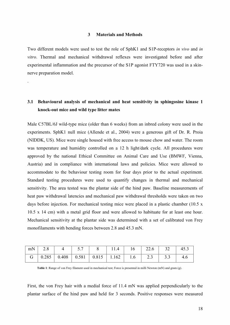
Mechanical sensitivity at the plantar side was determined with a set of calibrated von Frey
monofilaments with bending forces between 2.8 and 45.3 mN.
mN 2.8 4 5.7 8 11.4 16 22.6 32 45.3
G 0.285 0.408 0.581 0.815 1.162 1.6 2.3 3.3 4.6
First, the von Frey hair with a medial force of 11.4 mN was applied perpendicularly to the
plantar surface of the hind paw and held for 3 seconds. Positive responses were measured
Table 1: Range of von Frey filament used in mechanical test; Force is presented in milli Newton (mN) and gram (g).

19
when the paw was withdrawn and a flinching movement was noticed immediately after
removal of the von Frey hair. Other paw movements were rate as unclear response and were
followed by repeating the stimulus. In case of positive reaction a filament with lower force
was used, in case of negative response a filament with higher force was used. The withdrawal
threshold was determined by increasing and decreasing stimulus intensity on the basis of the
up–down method (Dixon, 1980; Chaplan et al., 1994). Trial was repeated six times from the
first change, independent if the change was a positive-negative or negative-positive response.
Afterwards the raw data were extracted using UDMAP V 3.0 program. Positive responses
were marked with "x" and negative with "o".
Heat sensitivity was assessed using the Hargreaves test (Hargreaves et al., 1988). Mice were
placed in a plastic chamber (10 x 9.5 x 12.x) and allowed to habituate for at least one hour. A
radiant heat source which delivered an increasing heat stimulus was focused on the plantar
surface of the hind paw; the time from the initiation of the heat stimulus until paw withdrawal
(paw withdrawal latency) was measured automatically (Ugo Basile, Italy). Each paw was
tested three times and mean withdrawal latency was calculated. The interval between two
trials on the same paw was at least one minute.
Complete Freund's Adjuvant (CFA) or vehicle was injected subcutaneously in a total volume
of 30 µl, 15 µl on each, dorsal and plantar side of the paw. The experimenter was initially
unaware of the nature of the treatment, however, after 24 h inflammation became obvious by
the increasing paw swelling. Sensitivity tests were obtained after 6h, 24h, 48 h and 72 h, then
the paw swelling was measured in medio-lateral and in dorso-plantar diameter.
3.2 Electrophysiology of primary afferent nociceptors in a skin-nerve preparation in
vitro
An in vitro skin-nerve preparation was used to investigate the properties of cutaneous afferent
nerve fibres (Koltzenburg et al., 1999; Kress et al., 1992). C57BL/6J mice were killed in a
CO2 inhalation chamber. After cutaneous incision, the saphenous nerve was dissected from
blood vessels and connective tissue. The proximal part of the nerve was cut close to the
lumbar spinal cord. Subsequently, the hairy skin of the paw was dissected subcutaneously
with functionally intact innervating saphenous nerve. The preparation was transferred to an
artificial bath solution (synthetic interstitial fluid (SIF), Bretag, 1969) consisting of : 108 mM
NaCl, 3.48 mM KCl, 3.5 mM MgSO4, 26 mM NaHCO3, 1.7 mM NaH2PO4, 1.5 mM CaCl2,
20
9.6 mM Na-Gluconat, 5.55 mM Glucose, 7.6 mM Saccharose with a pH of 7.40 ± 0.5. The
preparation was mounted with the corium-side up in an organ bath chamber and perfused with
carbogen-saturated (95% O2, 5% CO2) SIF at 31°C ± 1°C. The proximal part of the nerve
was pulled through a pinhole into a separate recording chamber filled with liquid paraffin. It
was placed on a mirror and standard teased fibre technique was used to isolated fine nerve
strands which were put on a gold-wire electrode (0.2 mm) for extracellular recording. The
receptive field of single units was identified by mechanical probing of the skin with a blunt
glass rod. The fibres were classified as unmyelinated fibres by conduction velocity (< 1.4m /s)
calculated from the latency of the action potential after electrical stimulation at the receptive
field and the distance between stimulation and recording electrodes, as well as by
oscilloscopic comparison of the distinct fibre shapes (Oscilloscope GOULD OS 4020,
Oscilloscope TEKTRONIX TPS 2024). Heat sensitivity was tested by focusing feedback
controlled radiant heat source (PhysioGirl, Hofmann Electronics, Erlangen) to the receptive
field which was separated from the bath by a self-sealing steel cylinder. A ramp-shaped heat
stimulus with linear rise of the intracutaneous temperature from 31°C to a maximum value of
48°C within 21 s was applied. The temperature on the corium side was feed-back controlled
with an external thermo sensor. A fibre was considered heat-sensitive if three or more action
potentials were evoked during the stimulus. Heat stimuli were applied every 5 minutes before,
during and after the restricted receptive area was perfused by conditioning stimulus solution
containing 1µM of FTY720 (Mo Bi Tec Molecular Biotechnology, Göttingen, Germany).
Action potentials were recorded, amplified (5000-fold), filtered (low pass 1 KHz, high pass
100 Hz), visualized on the above-mentioned oscilloscopes and stored on a PC-type computer
with Spike/Spidi software package (Forster et al., 1990). Skin flaps were discarded after
treatment with FTY720 to avoid contamination artefacts.
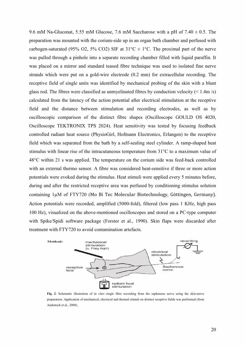
Fig. 2: Schematic illustration of in vitro single fibre recording from the saphenous nerve using the skin-nerve
preparation. Application of mechanical, electrical and thermal stimuli on distinct receptive fields was performed (from
Andratsch et al., 2008).

21
3.3 Statistical analysis
For statistical analysis SigmaStat 3.0 (SPSS Inc., Chicago, IL) was used. Data are presented
as mean ± standard error of the mean (S.E.M.). For interindividual comparison of different
populations the Mann-Whitney U-Test was calculated. Intraindividual comparisons before
and after treatment were performed using the Wilcoxon matched-pairs test. Differences were
considered statistically significant at p<0.05. Graphs were created with Origin Pro 8 (Origin
Lab Corporation, Northampton, MA) and GIMP Version 2.6 (GIMP Development Team,
Compay, Boston, MA)
22
4 Results
4.1 CFA induced inflammation in SphK1 null mutant and in C57BL/6J wt mice
Complete Freund's Adjuvant, a mixture of mineral oil and heat-inactivated mycobacterium
tuberculosis, induces an inflammatory response after subcutaneous injection. Thereby the
inflammation produces thermal and mechanical hyperalgesia. Since S1P acts as immune
modulator, we first addressed the question whether the degree of inflammation would be
different between wt and SphK1-/- mice. We measured the dorso-plantar and the medio-lateral
paw diameter before and after CFA injection at the ipsi- and contralateral paw with a calliper.
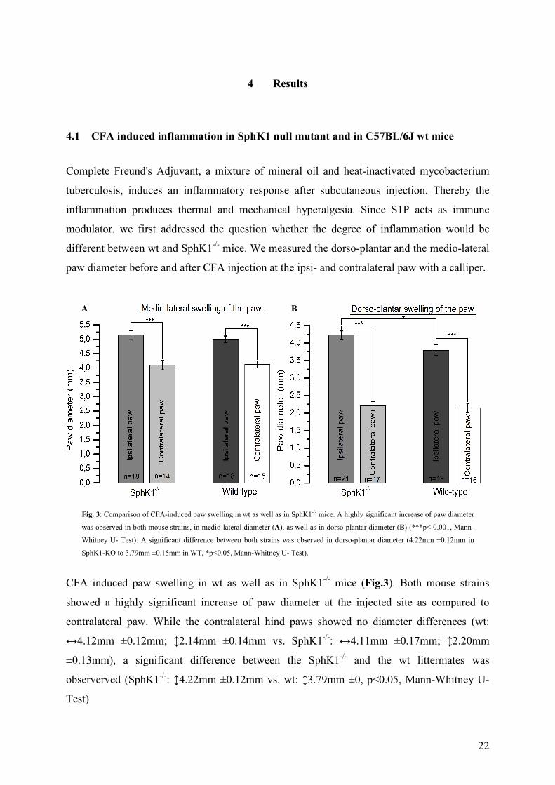
CFA induced paw swelling in wt as well as in SphK1-/- mice (Fig.3). Both mouse strains
showed a highly significant increase of paw diameter at the injected site as compared to
contralateral paw. While the contralateral hind paws showed no diameter differences (wt:
↔4.12mm ±0.12mm; ↕2.14mm ±0.14mm vs. SphK1-/-: ↔4.11mm ±0.17mm; ↕2.20mm
±0.13mm), a significant difference between the SphK1-/- and the wt littermates was
observerved (SphK1-/-: ↕4.22mm ±0.12mm vs. wt: ↕3.79mm ±0, p<0.05, Mann-Whitney U-
Test)
Fig. 3: Comparison of CFA-induced paw swelling in wt as well as in SphK1-/- mice. A highly significant increase of paw diameter
was observed in both mouse strains, in medio-lateral diameter (A), as well as in dorso-plantar diameter (B) (***p< 0.001, Mann-
Whitney U- Test). A significant difference between both strains was observed in dorso-plantar diameter (4.22mm ±0.12mm in
SphK1-KO to 3.79mm ±0.15mm in WT, *p<0.05, Mann-Whitney U- Test).
A B

23
4.2 Similar mechanical hypersensitivity of SphK1-/- and C57BL/6J wt mice after CFA-
injection
The SphK1-/- and C57BL/6J wt mice started with similar basal mechanical thresholds
(44.89mN±2.2 vs. 44.92mN ±1.9mN, n=20, Mann-Whitney Rank Sum Test). As expected,
mice developed signs of mechanical hypersensitivity at the injected side. Within 6 h after
injection of 30 µl CFA mice exhibited a mechanical hypersensitivity that did not recover
Fig. 4: Subcutaneous CFA-injection led to a decrease of mechanical pain thresholds in wt as well as in SphK1-/- mice. 6h after
CFA-injection the mechanical threshold of wt mice dropped from 45.15mN ±3.0mN to 7.15mN ±1.11mN. Within 72h no
recovery of was observed (A, n= 15, ***p<0.001, Mann-Whitney Rank Sum Test). The SphK1-/- mouse strain showed a
similar behaviour (from 46.48mN ±2.8mN to 7.38mN ±1.25mN (B, n=14, ***p<0.001, Mann-Whitney Rank Sum Test)
within the three days.
A B
Fig. 5: SphK1-/- and C57BL/6J wt mice showed a similar mechanical hypersensitivity after CFA-injection.
Within 72h no differences were observed (n=15, Mann-Whitney Rank Sum Test).
24
within 72h (Fig.4). The contralateral paws of SphK1-/- and C57BL/6J mice did not show any
signs of inflammation or mechanical hyperalgesia (Fig.4). No differences in mechanical pain
thresholds between the mouse strains could be measured within 72h (Fig.5).
4.3 Similar thermal hypersensitivity of SphK1-/- and C57BL/6J wt mice after CFA-
injection
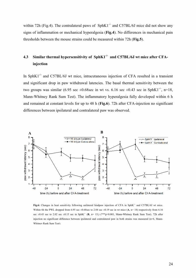
In SphK1-/- and C57BL/6J wt mice, intracutaneous injection of CFA resulted in a transient
and significant drop in paw withdrawal latencies. The basal thermal sensitivity between the
two groups was similar (6.95 sec ±0.68sec in wt vs. 6.16 sec ±0.43 sec in SphK1-/-, n=18,
Mann-Whitney Rank Sum Test). The inflammatory hyperalgesia fully developed within 6 h
and remained at constant levels for up to 48 h (Fig.6). 72h after CFA-injection no significant
differences between ipsilateral and contralateral paw was observed.
Fig.6: Changes in heat sensitivity following unilateral hindpaw injection of CFA in SphK-/- and C57BL/6J wt mice.
Within 6h the PWL dropped from 6.95 sec ±0.68sec to 2.04 sec ±0.19 sec in wt mice (A, n= 18) respectively from 6.16
sec ±0.43 sec to 2.02 sec ±0.15 sec in SphK-/- (B, n= 15;) (***p>0.001, Mann-Whitney Rank Sum Test). 72h after
injection no significant difference between ipsilateral and contralateral paw in both strains was measured (n=6, Mann-
Whitney Rank Sum Test).
A B
25
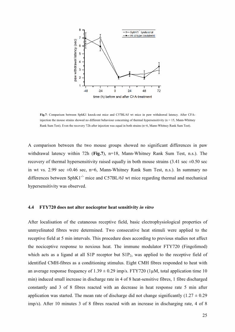
A comparison between the two mouse groups showed no significant differences in paw
withdrawal latency within 72h (Fig.7), n=18, Mann-Whitney Rank Sum Test, n.s.). The
recovery of thermal hypersensitivity raised equally in both mouse strains (3.41 sec ±0.50 sec
in wt vs. 2.99 sec ±0.46 sec, n=6, Mann-Whitney Rank Sum Test, n.s.). In summary no
differences between SphK1-/- mice and C57BL/6J wt mice regarding thermal and mechanical
hypersensitivity was observed.
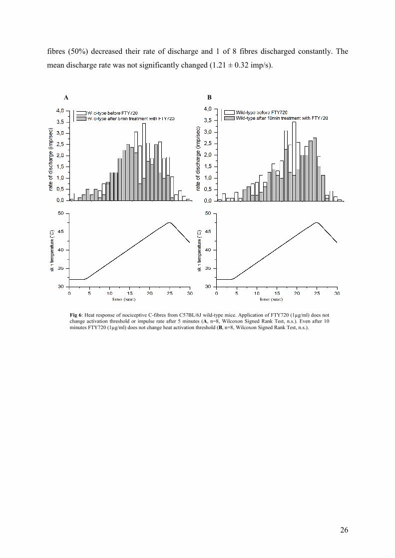
4.4 FTY720 does not alter nociceptor heat sensitivity in vitro
After localisation of the cutaneous receptive field, basic electrophysiological properties of
unmyelinated fibres were determined. Two consecutive heat stimuli were applied to the
receptive field at 5 min intervals. This procedure does according to previous studies not affect
the nociceptive response to noxious heat. The immune modulator FTY720 (Fingolimod)
which acts as a ligand at all S1P receptor but S1P2, was applied to the receptive field of
identified CMH-fibres as a conditioning stimulus. Eight CMH fibres responded to heat with
an average response frequency of 1.39 ± 0.29 imp/s. FTY720 (1µM, total application time 10
min) induced small increase in discharge rate in 4 of 8 heat-sensitive fibres, 1 fibre discharged
constantly and 3 of 8 fibres reacted with an decrease in heat response rate 5 min after
application was started. The mean rate of discharge did not change significantly (1.27 ± 0.29
imp/s). After 10 minutes 3 of 8 fibres reacted with an increase in discharging rate, 4 of 8
Fig.7: Comparison between SphK1 knock-out mice and C57BL/6J wt mice in paw withdrawal latency. After CFA-
injection the mouse strains showed no different behaviour concerning of thermal hypersensitivity (n = 15, Mann-Whitney
Rank Sum Test). Even the recovery 72h after injection was equal in both strains (n=6, Mann-Whitney Rank Sum Test).
26
fibres (50%) decreased their rate of discharge and 1 of 8 fibres discharged constantly. The
mean discharge rate was not significantly changed (1.21 ± 0.32 imp/s).
A B
Fig 6: Heat response of nociceptive C-fibres from C57BL/6J wild-type mice. Application of FTY720 (1µg/ml) does not change activation threshold or impulse rate after 5 minutes (A, n=8, Wilcoxon Signed Rank Test, n.s.). Even after 10 minutes FTY720 (1µg/ml) does not change heat activation threshold (B, n=8, Wilcoxon Signed Rank Test, n.s.).

27
5 Discussion
Our data revealed an unexpected difference in CFA-induced inflammatory swelling of the
hind paw in SphK-/- mice in which swelling was slightly but significantly increased compared
to wt littermates. In contrast to our hypothesis, no differences in inflammatory thermal and
mechanical hypersensitivity were observed between the two mouse strains within 72h after
CFA injection.
A variety of cellular processes involving activation of S1P kinases and autocrine or paracrine
S1P effects have been reported and may contribute to plasma extravasation and inflammation
induced swelling (for review see Spiegel and Milstien, 2003; Alvarez et al., 2007). S1P and its
receptors play a central role in endothelial cell barrier regulation. S1P induces reorganization
of the endothelial cytoskeleton (Dudek et al.., 2004) and the distribution, assembly, and
stabilization of adherent junction (Schaphorst et al, 2001) leading to a reduction in endothelial
permeability to fluid (McVerry and Garcia, 2004). Alterations of endothelial cell barrier
function followed by S1P and FTY 720 have been described (Sanchez et al., 2003; McVerry
and Garcia, 2004; McVerry and Garcia, 2005) and reduced levels of S1P might explain the
difference of inflammatory oedema of SphK1-/- mice.
Furthermore, differences in the cell composition within the inflamed tissue could be
responsible for the increased swelling. Sphingosine kinases influence many different immune
cells like lymphocyte and macrophages (Kee et al., 2005) and S1P and his receptor S1P1
promote the lymphocyte egress from thymus, spleen and lymph nodes (Pappu et al., 2007).
Direct activation of S1P1 with the lyase-insensitive agonist FTY720 inhibits this egress by an
unknown pathway (Allende et al., 2004; Rosen and Goetzl, 2005). In addition to an increased
plasma extravasation, an increase egress of vascular immune cells into the inflamed tissue
could be responsible for the increased tissue volume in SphK1-/- after CFA injection It would
be useful to determine the cell composition as well as the concentration of pro-inflammatory
cytokines and of S1P of the inflamed tissue to rule out a possibily the increased immune cell
invasion into the tissue under inflammatory conditions in SphK1-/- mice.
In recent studies, it has been shown, that S1P sensitizes capsaicin-sensitive small-diameter
primary afferent neurons and increases the action potential rate without modifying firing
threshold or membrane potential (Mair et al., 2010; Zhang et al., 2006a; Zhang et al., 2006b),
28
and that the S1P-induced hyperalgesia involves S1P1 (Doyle et al., 2010; Doyle et al. 2011).
In contrast to a number of resports suggesting pro-algesic S1P effects, antinociceptive effects
of S1P and sphingosine kinases in particular on spinal cord nociceptive transmision have been
proposed (Coste et al., 2008). Our data revealed no differences in the degree of thermal and
mechanical hypersensitivity in SphK1-/- and wt littermates induced by experimental
inflammation within 72h. Also other inflammatory models using SphK1-/- mice did not show
differences in acute or chronic inflammatory response (Allende et al., 2004, Michaud et al.,
2006). Supported by the observation, that SphK1-/- and SphK2-/- knock-out mice do not show
phenotypic abnormalities (Allende et al. 2004; Kharel et al., 2005; Mizugishi et al., 2007;
Lynch and MacDonald, 2008), it is assumed that loss of one SphK-gene leads to an
upregulation of the corresponding other sphingosine kinase and functional compensation of
the null mutation. Nevertheless increasing SphK2 mRNA expression levels could not be
proven yet (Allende et al., 2004; Kharel et al., 2005; Michaud et al., 2006). To avoid the
compensatory upregulation of SphK2, it would be necessary to use S1P1 -/- mice to investigate
the role of S1P in inflammatory hyperalgesia. Unfortunately S1P1 receptor knock-out mice
were created but died in utero due to vascular leakage as a consequence of deficient
angiogenesis and vasculogenesis (Liu et al., 2000). Therefore, conditional KO mice are being
generated and a first study of our group shows employing mice with a conditional deletion of
S1P1 receptor in nociceptive primary afferent neurons indeed suggests that S1P1 receptor is
critical for inflammatory thermal hypersensitivity (Mair et al., 2011).
FTY720 (Fingolimod), a ligand at S1P1 and S1P3-5 receptors, is a novel therapeutic drug, that
is in clinical trial for different autoimmune diseases. Because of its ability to activate and
quickly internalize its receptor, it acts as a functional antagonist on the S1P receptors
(Brinkmann, 2009). Because of his high affinity binding to S1P1 we wanted to see, if it
provokes an increase in action potential firing rate in an in-vitro skin nerve preparation. Our
data suggest that FTY720 does not induce a thermal hypersensitivity, in vitro. This is in
contrast to S1P and the selective S1P1 agonist SEW2871 which have recently been shown to
induce thermal hypersensitivity via direct nociceptor sensitisation (Mair et al., 2011; Doyle et
al., 2010). Also, tumour necrosis factor α sensitises nociceptors to heat and some of its effects
are mimicked by S1P (Constantin et al., 2008; Pollock et al., 2002). In addition, activation of
S1P1 induces an increase of excitability in primary afferent neurons by modification of
different Na+- and K+ channels, e.g. TTX-R NaV1.8. It has also been published, that S1P
sensitises TRPV1 -channels by phosphorylation. (Mair et al., 2010; Chi and Nicol, 2010,

29
Zhang et al., 2006a). In our experiments the non-phosphorylated FTY720 did not induce any
signs of hyperexcitability or sensitivity increase in primary afferent neurons. It is known, that
FTY720 needs to be phosphorylated by SphK2 to become biologically active as FTY720-P
(Brinkmann et al., 2002; Mandala et al., 2002). This process in particular is known to be
active in the liver (Billich et al., 2003). SphK2 is located in the cytosol and the nucleus
(Venkataraman, 2006). It is unlikely, that extracellularly applied FTY720 is phosphorylated in
vitro, however, as an inactive metabolite it does not bind to S1P receptors (Marsolais and
Rosen, 2009; Melendez, 2008). Further experiments will be necessary to study FTY720-P or
selective S1P1 receptor agonists like the synthetic chemical modulators AUY954 or CYM-
5442 (Marsolais and Rosen, 2009; Lynch and MacDonald, 2008) in this preparation.
Taken together, our data show that SphK1-/- mice do not exhibit any differences in pain like
behaviour under inflammatory conditions although the degree of paw swelling is significantly
increased. Furthermore, FTY720 did not induce nociceptor sensitisation in vitro. Nevertheless
they do not exclude a pivotal role of S1P and S1P receptor pathways in inflammatory
conditions. Further experiments are in progress to further elucidate the importance of S1P for
the generation of pain associated with inflammatory disease.

30
6 References
Adriaensen, H., Gybels, J., Handwerker, H. O., Van Hees, J. (1983). Response properties of thin myelinated (A-delta) fibers in human skin nerves. J Neurophysiol 49,111-122.
Allende, M. L., Sasaki, T., Kawai, H., Olivera, A., Mi, Y., Echten-Deckert, G. v., Hajdu, R., Rosenbach, M., Keohane, C. A., Mandala, S., Spiegel, S., Proia, R. L. (2004). Mice Deficient in Sphingosine Kinase 1 Are -Rendered Lymphopenic by FTY720. The journal of biological chemistry Vol. 279 No. 50, 52487-52492
Alvarez, S. E., Milstien, S., Spiegel, S. (2007). Autocrine and paracrine roles of sphingosine- 1-phosphate. Trends in Endocrinology and Metabolism Vol.18 No.8, 300-307.
Andratsch, M., Mair, N., Constantin, C. E., Scherbakov, N., Benetti, C., Quarta, S., Vogl, C., Sailer, C. A., Üceyler, N., Brockhaus, J., Martini, R., Sommer, C., Zeilhofer, H. U., Müller, W., Kuner, R., Davis, J. B., Rose-John, S., Kress, M. (2009). A key role for gp130 expressed on peripheral sensory nerves in pathological pain. J Neurosci 29, 13473-13483.
Baumruker, T., Prieschl, E. E. (2002).Sphingolipids and the regulation of the immune response. Semin. Immunol. 14, 57-63.
Billich, A., Bornancin, F., De´vay, P., Mechtcheriakova, D., Urtz,N., Baumruker, T. (2003). Phosphorylation of the Immunomodulatory Drug FTY720 by Sphingosine Kinases. J. Biol. Chem. Vol. 278, No. 48, 47408-47415.
Billich, A., Bornancin, F., Devay, P., Mechtcheriakova, D., Urtz, N., Baumruker, T. (2003). Phosphorylation of the immunomodulatory drug FTY720 by sphingosine kinases. J Biol Chem 278,47408-47415
Breivik, H., Collett, B., Ventafridda, V., Cohen, R., Gallacher, D. (2006). Survey of chronic pain in Europe:Prevalence, impact on daily life, and treatment. European Journal of Pain 10, 287-333.
Bretag, A. (1969). Synthetic interstitial fluid for isolated mammalian tissue. Life Sci 8,319- 329.
Brinkmann V (2009). FTY720 (fingolimod) in Multiple Sclerosis: therapeutic effects in the immune and the central nervous system. Br J Pharmacol 158:1173-1182.
Brinkmann, V. (2007). Sphingosine 1-phosphate receptors in health and disease: mechanistic insights from gene deletions studies and reverse pharmacology, Pharmacol. Therap. 115, 85-104.
Brinkmann, V., Davis, M. D., Heise, C. E., Albert, R., Cottens, S., Hof, R.,Bruns, C., Prieschl, E., Baumruker, T., Hiestand, P., Foster, C. A.,Zollinger, M., Lynch, K. R. (2002). The Immune Modulator FTY720 Targets Sphingosine 1-Phosphate Receptors. J. Biol. Chem. 277, 21453-21457

31
Brinkmann, V., Pinschewer, D. D., Feng, L., Chen, S. (2001). FTY720: altered lymphocyte traffic results in allograft protection. Transplantation 72, 764-769.
Brook, J., Tracey, I. (2005). From nociception to pain perception: imaging the spinal and supraspinal pathways. J. Anat. 207, 19-33.
Casey, K. L., Bushnell, M. C. (2000) The imaging of pain: background and rationale. Pain Imaging, 2000, pp. 1-29.
Caterina, M. J., Leffler, A., Malmberg, A. B., Martin, W. J., Trafton, J., Petersen-Zeitz, K. R., Koltzenburg, M., Basbaum, A. I., Julius, D. (2000). Impaired nociception and pain sensation in mice lacking the capsaicin receptor. Science 288,306-313.
Caterina, M. J., Schumacher, M. A., Tominaga, M., Rosen, T. A., Levine, J. D., Julius, D. (1997). The capsaicin receptor: a heat-activated ion channel in the pain pathway. Nature 389, 816-824.
Chaplan, S. R., Bach, F. W., Pogrel, J. W., Chung, J. M., Yaksh, T. L. (1994). Quantitative assessment of tactile allodynia in the rat paw. Journal of Neuroscience Methods 53, 55-63
Chi, X. X., Nicol, G. D. (2010). The Sphingosine 1-Phosphate Receptor, S1PR 1, Plays a Prominent But Not Exclusive Role in Enhancing the Excitability of Sensory Neurons. J Neurophysiol 104, 2741-2748.
Constantin, C. E., Mair, N., Sailer, C. A., Andratsch, M., Xu, Z. Z., Blumer, M. J., Scherbakov, N., Davis, J. B., Bluethmann, H., Ji, R. R., Kress, M. (2008). Endogenous necrosis factor alpha (TNFalpha) requires TNF receptor type 2 to generate heat hyperalgesia in a mouse cancer model. J Neurosci 28, 5072-5081.
Coste, O., Brenneis, C., Linke, B., Pierre, S., Maeurer, C., Becker, W., Schmidt, H., Gao, W., Geisslinger, G., Scholich, K. (2008). Sphingosine 1-Phosphate Modulates Spinal Nociceptive Processing. The Journal of Biological Chemistry Vol. 283, No. 47, 32442-32451.
Craig, A. D. (1991). Spinal distribution of ascending lamina I axons anterogradely labeled with phaseolus vulgaris leucoagglutinin (PHA-L) in the cat. J. Comp. Neurol. 313, 377– 393.
Craig, A. D., Krout, K., Andrew, D. (2001). Quantitative responses characteristics of thermoreceptive and nociceptive lamina I spinothalamic neurons in the cat. J. Neurophysiol. 86, 1459– 1480.
Cunha, T. M., Verri, W. A. Jr., Silva, J. S., Poole, S., Cunha, F. Q., Ferreira, S. H. (2005). A cascade of cytokines mediates mechanical inflammatory hypernociception in mice. Proc Nat Acad Sci U S A 102,1755-1760.
Davis, J. B., Gray, J, Gunthorpe, M. J., Hatcher, J. P., Davey, P. T., Overend, P., Harries, M. H., Latcham, J., Clapham, C., Atkinson, K., Hughes, S. A., Rance, K., Grau, E., Harper, A. J., Pugh, P. L., Rogers, D. C., Bingham, S., Randall, A., Sheardown, S. A. (2000). Vanilloid receptor-1 is essential for inflammatory thermal hyperalgesia. Letters to Nature, Nature Vol. 405, 183-187.

32
Dixon, W. J. (1980). Efficient analysis of experimental observations. Ann. Rev. Pharmacol. Toxicol. 20, 223-231
Djouhri, L., Lawson, S. N. (2004). Aβ-fiber nociceptive primary afferent neurons: a review of incidence and properties in relation to other afferent A-fiber neurons in mammals. Brain Research Reviews 46, 131-145.
Doyle, T. Chen, Z., Obeid, L. M., Salvemini, D. (2011). Sphingosine-1-phosphate acting via the S1P1 receptor is a downstream signalling pathway in ceramide-induced hyperalgesia. Neuroscience Letters, doi: 10.1016.
Doyle, T., Finley, A., Chen, Z., Salvemini, D. (2010). Role for peroxynitrite in sphingosine- 1-phosphate-induced hyperalgesia in rats. Pain 152, 643-648.
Dubner, R., Kenshalo, D. R., Maixner, W., Bushnell, M. C., Oliveras, J. L. (1989). The correlation of monkey medullary dorsal horn neuronal activity and the perceived intensity of noxious heat stimuli. J. Neurophysiol. 62, 450-457.
Dudek, S. M., Wang, P., Birukov, K. G., Zhan, X.,Garcia, J. G. N. (2004). Pulmonary Endothelial Cell Barrier Enhancement by Sphingosine 1-Phosphate. J. Biol.Chem. 279, 24692-24700.
Forster, C., Handwerker, H. O. (1990). Automatic classification and analysis of microneurographic spike data using a PC/AT. J. Neurosci. Methods 31, 109-118.
Garry, E. M., Jones, E., Fleetwood-Walker, S. M. (2004). Nociception in vertebrates: key receptors participating in spinal mechanisms of chronic pain in animals. Brain Research Reviews 46, 216-224.
Gebhart, G. F. (1995). Visceral Pain, p. 516, IASP Press, Seattle.
Gilette, R. G., Kramis, R. C., Roberts, W. J. (1993). Characterization of spinal somatosensory neurons having receptive fields in lumbar tissues of cats. Pain, Vol. 54, Issue 1, 85-98.
Handwerker, H. O., Iggo, A., Ogawa, H. (1975a). Dorsal horn neurones responding to cutaneous afferent input. J Physiol Lond 244, 1P-2P.
Handwerker, H. O., Iggo, A., Ogawa, H., Ramsey, R. L. (1975b). Input characteristics and rostral projection of dorsal horn neurones in the monkey. J Physiol Lond 244:76P-77P.
Hargreaves K., Dubner, R., Brown, F., Flores, C., Joris, J. (1988). A new and sensitive method for measuring thermal nociception in cutaneos hyperalgesia. Pain 32, 77-88.
Hla, T., Lee, M. J., Ancellin, N., Paik, J. H., Kluk, M. J. (2001). Lysophospholipids — receptor revelations. Science 294, 1875-1878.
Hla, T., Venkataraman, K., Michaud, J. (2008). The vascular S1P gradient - Cellular sources and biological significance. Biochimica et Biophysica Acta 1781, 477-482.
Igarashi, J., Erwin, P. A., Dantas, A. P., Chen, H., Michel, T. (2003) VEGF induces S1P1 receptors in endothelial cells: Implications for cross-talk between sphingolipid and growth factor receptors. Proc. Natl. Acad. Sci. U. S. A. 100, 10664-10669
33
Jolly, P. S., Rosenfeldt, H. M., Milstein, Spiegel, S. (2002). The roles ofsphingosine-1- phosphate in asthma. Mol. Immunol. 38, 1239-1245.
Jordt, S.-E., McKemy, D. D., Julius, D. (2003) Lessons from peppers and peppermint: the molecular logic of thermosensation. Curr Opin Neurobiol 13, 487-492.
Julius, D., Basbaum, A. I. (2001). Molecular mechanisms of nociception. Nature, Vol. 413, 203-210.
Kee, T. H., Vit, P., Melendez, A. J. (2005). Sphingosine kinase signalling in immune cells. Clinical and Experimental Pharmacology and Physiology 32, 153-161.
Kharel, Y., Lee, S., Snyder, A. H., Sheasley-O’Neill, S. L., Morris, M. A., Setiady, Y., Zhu, R., Zigler, M. A., Burcin, T. L., Ley, K., Tung, K. S. K., Engelhard, V. H., Macdonald, T. L., Pearson-White, S., Lynch, K. R. (2005). Sphingosine Kinase 2 Is Required for Modulation of Lymphocyte Traffic by FTY720. The Journal of biological chemistry, 80, No.: 44, 36865-36872.
Khasar, S. G., Lin, Y., Martin, A., Dadgar, J., McMahon, Wang, T. D., Hundle, B., K. O. Aley, K. O., Isenberg, W., McCarter, G., Green, P. G., Hodge, C. W., Levine, J. D., Messing, R. O. (1999). A Novel Nociceptor Signaling Pathway Revealed in Protein kinase C ε Mutant Mice. Neuron Vol. 24, 253-260.
Kihara, A., Anada, Y., Igarashi, Y. (2006). Mouse sphingosine kinase isoforms SPHK1a and SPHK1b differ in enzymatic traits including stability, localization, modification, and oligomerization. J Biol Chem 281, 4532–4539
Kim, I., Moon, S. O., Kim, S. H., Kim, H. J., Koh. Y. S., Koh, G. Y. (2001). Vascular endothelial growth factor expression ofintercellular adhesion molecule 1 (ICAM-1), vascular cell adhesionmolecule 1 (VCAM-1), and E-selectin through nuclear factor- kB activation in endothelial cells. J. Biol. Chem. 276, 7614-7620
Koltzenburg, M., Stucky, C. L., Lewin,G. R. (1997). Receptive properties of mouse sensory neurons innervating hairy skin. J. Neurophysiol. 78,1841-1850.
Kress, M., Koltzenburg, M., Reeh, P. W., Handwerker, H. O. (1992). Responsiveness and functional attributes of electrically localized terminals of cutaneous C-fibers in vivo and in vitro. J. Neurophysiol. 68,581-595.
Kress, M., Reeh, P. W. (1996). Chemical excitation and sensitization in nociceptors. In: Neurobiology of Nociceptors (Cervero F, Belmonte C, eds), pp 258-297. New York: Oxford University Press Inc.
Lee, Y. M., Venkataraman, K., Hwang, S., Han, D. K., Hla, T. (2007). A novel method to quantify sphingosine 1-phosphate by immobilized metal affinity chromatography (IMAC). Prostaglandins Other Lipid Mediat. 84(3-4), 154-162.
Liu, H., Sugiura, M., Nava, V. E., Edsall, L. C., Kono, K., Poulton, S., Milstien, S., Kohama, T., Spiegel, S. (2000). Molecular cloning and functional characterization of a novel mammalian sphingosine kinase type 2 isoform. J Biol Chem 275,19513–19520
Liu, Y., Wada, R., Yamashita, T., Mi, Y., Deng, C. X., Hobson, J. P., Rosenfeldt, H. M., Nava, V.E., Chae, S. S., Lee, M. J. (2000). Edg-1, the G protein-coupled receptor for
34
Sphingosine-1-phosphate, is essential for vascular maturation. J. Clin. Invest. 106, 951-961.
Lumpkin, Ea., Caterina, M. J. (2007). Mechanisms of sensory transduction in the skin. Nature 445, 858-865.
Lynch, K. R., MacDonald, T. L. (2008). Sphingosine 1-phosphate chemical biology. Biochimica et Biophysica Acta 1781, 508-512.
Mair, N., Leitner, M. G., Benetti, C., Quarta, S., Constantin, C. E., Schweigreiter, R., Garczarczyk, D., Biasio, W., Andratsch, M., Mandala, S., Hofmann, J., Proia, R., Gibbins, I.L., Kress, M., Haberberger, R. V. (2011). Genetic Evidence for Involvement of Neuronally Expressed S1P1 Receptor in Nociceptor Sensitization and Inflammatory Pain. PLoSOne Vol. 6 Issue 2, e17268
Malek, R. L., Toman , R. E., Edsall, R. E., Wong, S., Chiu, J., Letterlei, C. A., Van Brocklyn, J. R., Milstien, S., Spiegel, S., Lee, N. H. (2001). Nrg-1 Belongs to the Endothelial Differentiation Gene Family of G Protein-coupled Sphingosine-1-phosphate Receptors. The Journal of Biological Chemistry Vol. 276, No. 8, 5692-5699.
Mandala, S., Hajdu, R., Bergstrom, J., Quackenbush, E., Xie, J., Milligan, J.,Thornton, R., Shei, G. J., Card, D., Keohane, C., Rosenbach, M., Hale, J., Lynch, C. L., Rupprecht, K., Parsons, W., Rosen, H. (2002). Alteration of Lymphocyte Trafficking by Sphingosine 1-Phosphate Receptor Agonists. Science 296, 346-349.
Marsolais, D., Rosen, H. (2009). Chemical modulators of sphingosine-1 -phosphate receptors as barrieroriented therapeutic molecules. Nature Reviews Drug Discovery 8, 297-307.
McVerry, B. J., Garcia, J. G. N. (2004). Endothelial Cell Barrier Regulation by Sphingosine 1-Phosphate. Journal of Cellular Biochemistry 92, 1075-1085.
McVerry, B. J., Garcia, J. G. N. (2005). In vitro and in vivo modulation of vascular barrier integrity by Sphingosine 1-phosphate: mechanistic insights. Cellular Signalling 17, 131-139.
Michaud, J., Kohno, M., Proia, R. L., Hla, T. (2006). Normal acute and chronic inflammatory responses in Sphingosine kinase 1 knockout mice. FEBS Letters 580, 4607-4612.
Millan, M. J. (1999). The induction of pain: an integrative review, Progress in Neurobiology Vol. 57, 1-164.
Mizugishi, K., Li, C., Olivera, A., Bielawski, J., Bielawska, A., Deng, C., Proia, R. L. (2007). Maternal disturbance in activated sphingolipid metabolism causes pregnancy loss in mice. The Journal of Clinical Investigation 117, 2993-3006.
Mori, K., Itoi, M., Tsukamoto, N., Kubo, H., Amagai, T. (2007).The perivascular space as a path of hematopoietic progenitor cells and mature T cells between the blood circulation and the thymic parenchyma. Int. Immunol.19, 745-753.
Ness, T. J., Gebhart, G. F. (1990). Visceral pain: a review of experimental studies. Pain 41, 167-234.

35
Okajima, F. (2002). Plasma lipoproteins behave as carriers of extracellular sphingosine 1- phosphate: is this an atherogenic mediator or an anti-atherogenic mediator? Biochim Biophys Acta,Vol. 1582, Issue 1-3,132-137.
Olivera, A., Spiegel, S. (1993). Sphingosine-1-phosphate as a second messenger in cell proliferation induced by PDGF and FCS mitogens. Nature. 365, 557-560.
Oprée, A., Kress, M. (2000). Involvement of the proinflammatory cytokines tumor necrosis factor-a, IL-1b and IL-6 but not IL-8 in the development of heat hyperalgesia: effects on heat-evoked calcitonin gene-related peptide release from rat skin. J Neurosci 20, 6289-6293.
Pappu, R., Schwab, S. R., Cornelissen, I., Pereira, J. P., Regard, J. B., Xu, Y., Camerer, E., Zheng, Y.-W., Huang, Y., Cyster, J. G., Coughlin, S. R. (2007). Promotion of Lymphocyte Egress into Blood and Lymph by Distinct Sources of Sphingosine-1- Phosphate. Science Vol. 316, 295-298.
Pollock, J., McFarlane, S. M., Conell, M. C., Zehavi, U., Vandenabeele, P., MacEwan, D. J., Scott, R. H. (2002). TNF-alpha receptors simultaneously activate Ca2+ mobilisation and stress kinases in cultured sensory neurons. Neuropharmacology 42, 93-106.
Premkumar, L. S., Ahern, G. P. (2000). Induction of vanilloid receptor channel activity by protein kinase C. Nature Vol. 408, 985-990.
Pyne, S., Chapman, J., Steele, L., Pyne, N. J. (1996). Sphingomyelin-derived lipids differentially regulate the extracellular signal-regulated kinase 2 (ERK-2) and c- Jun N-terminal kinase (JNK) signal cascades in airway smooth muscle cells. Eur. J. Biochem. 237, 819-826.
Pyne, S., Pyne, N. J. (2000). Sphingosine 1-phosphate signalling in mammalian cells Biochem. J. 349, 385-402
Reeh, P. W. (1988). Sensory receptors in a mammalian skin - nerve in vitro preparation. In: Progress in Brain Research, Vol.74 (Hamann W, Iggo A, eds), pp 271-276. Amsterdam: Elsevier Science Publishers B.V: (Biomedical Division).
Rosen, H., Goetzl, E. J. (2005). Sphingosine 1-phosphate and its receptors: an autocrine and paracrine network. Nat. Rev. Immunol. 5, 560-570.
Sanchez, T., Estrada-Hernandez, T., Paik, J., Wu, M., Venkataraman, K., Brinkmann, V., Claffey, K., Hla, T. (2003). Phosphorylation and Action of the Immunomodulator FTY720 Inhibits Vascular Endothelial Cell Growth Factor-induced Vascular Permeability. The Journal of biological chemistry, 278, No.: 47, 47281-47290.
Schaible, H.G., Grubb, B.D. (1993). Afferent and spinal mechanisms of joint pain. Pain, Vol. 55 Issue 1, 5-54.
Schaphorst, K. L., Jacobs, K. N., Verin, A. D., Garcia, J. G. N. (2001). Sphingosine 1- phosphate increases F-actin/ adherins junction linkage and enhances barrier protection. Am. J.Respir. Crit. Care Med. 163, A615.
36
Schmidt, R., Schmelz, M., Forster, C., Ringkamp, M., Torebjörk, E., Handwerker, H. (1995). Novel Classes of Responsive and Unresponsive C Nociceptors in Human Skin. The Journal of Neuroscience 15 (l), 333-341.
Sherrington C. (1906). The Integrative Action of the Nervous System. Oxford: Oxford University Press.
Shu, X., Wu, W., Mosteller, R. D., Broek, D. (2002) Sphingosine kinase mediates vascular endothelial growth factor-induced activation of ras and mitogen-activated protein kinases. Mol. Cell. Biol. 22, 7758-7768
Siehler, S., Manning, D. R. (2002). Pathways of transduction engaged by sphingosine 1- phosphate through G protein-coupled receptors. Biochimica et Biophysica Acta 1582, 94- 99
Sommer, C., Kress, M. (2004). Recent findings on how proinflammatory cytokines cause pain: peripheral mechanisms in inflammatory and neuropathic hyperalgesia. Neuroscience Letters 361, 184-187.
Spiegel, S. & Milstien, S. (2002). Sphingosine 1-phosphate, a key cell signaling molecule. J. Biol. Chem. 277, 25851-25854.
Spiegel, S., Milstien, S. (2003). Sphingosine-1-Phosphate: An Enigmatic Signalling Lipid. Nature Reviews Molecular Cell Biology, Vol. 4, 397-407.
Su, Y., Rosenthal, D., Smulson, M., Spiegel, S. (1994). Sphingosine 1-phosphate, a novel signaling molecule, stimulates DNA binding activity of AP-1 in quiescent Swiss 3T3 fibroblasts. J. Biol. Chem. 269, 16512-16517.
Talavera, K., Nilius, B., Voets, T. (2008). Neuronal TRP channels: thermometers, pathfinders and life-savers. Trends Neursci 31, 287-295.
Toman, R. E., Payne, S. G., Watterson, K. R., Maceyka, M. Lee, N. H., Milstien, S., Bigbee, J. W., Spiegel, S. (2004). Differential transactivation of sphingosine-1-phosphate receptors modulates NGF-induced neurite extension. The Journal of Cell Biology, Volume 166, Number 3, 381-392
Tominaga, M., Caterina, M. J., Malmberg, A. B., Rosen, T. A., Gilbert, H., Skinner, K., Raumann, B. E., Basbaum, A. I., Julius, D. (1998). The cloned capsaicin receptor integrates multiple pain-producing stimuli. Biophys J 21, 531-543.
Tominaga, M., Tominaga, T. (2005). Structure and function of TRPV1. Pflugers Archiv - European Journal of Physiology 451,143-150.
Tracey, I. (2005). Nociceptive processing in the human brain. Current Opinion in Neurobiology 15, 478-487.
Van Brocklyn, J. R., Lee, M.-J., Menzeleev, R., Olivera, A., Edsall, L., Cuvillier, O., Thomas,D. M., Coopman, P. J. P., Thangada, S., Liu, C. H., Hla, T., Spiegel, S. (1998). Dual Actions of Sphingosine-1-Phosphate: Extracellular through the G- coupled Receptor Edg-1 and Intracellular to Regulate Proliferation and Survival. The Journal of Cell Biology, Volume 142, Number 1, 229-240.

37
Van Koppen, C. J., Meyer zu Heringdorf, D., Alemany, R., Jakobs, K. H. (2001). Sphingosine kinase-mediated calcium signaling by muscarinic acetylcholine receptors. Life Sci. 68, 2535-2540.
Van Veldhoven, P. P. ,Mannaerts, G. P. (1991). Subcellular localization and membrane topology of sphingosine-1-phosphate lyase in rat liver. J. Biol. Chem. 266,12502- 12507
Van Veldhoven, P. P., Mannaerts, G. P. (1993). Sphingosine-phosphate lyase. Adv. Lipid Res. 26, 69-98
Venkataraman, K, Lee, Y. M., Michaud, J., Thangada, S., Ai, Y., Bonkovsky, H. L., Parikh, N. S., Habrukowich, C., Hla, T. (2008). Vascular Endothelium As a Contributor of Plasma Sphingosine 1-Phosphate. Circulation Research, 669-676.
Venkataraman, K., Thangada, S., Michaud, J., OO, M. L., AI, Y., Lee, Y. M., Wu, M., Parikh, N. S., Khan, F., Proia, R. L., Hla, T (2006). Extracellular export of sphingosine kinase- 1a contributes to the vascular S1P gradient. Biochem. J. 397, 461-471.
Verri, W. A., Cunha, T. M., Parada, C. A., Poole, S., Cunha, F. Q., Ferreira, S. H. (2006). Hypernociceptive role of cytokines and chemokines: Targets for analgesic drug development? Pharmac Ther 112, 116-138.
Willis Jr., W. D. (1985). Pain pathways in the primate. Prog. Clin. Biol. Res.176, 117– 133.
Xu, J., Gu, H., Brennan, T. J. (2010). Increased sensitivity of group III and group IV afferents from incised muscle in vitro. Pain151, 744-755
Zhang, H., Desai, N. ., Olivera, A., Seki, T., Brooker, G., Spiegel, S. (1991).Sphingosine-1- phosphate, a novel lipid, involved in cellular proliferation. J.Cell Biol. 114, 155-167.
Zhang, X., Honda, C. N., Giesler Jr., G. J. (2000).Position of spinothalamic tract axons in upper cervical spinal cord of monkeys, J. Neurophysiol. 84, 1180-1185.
Zhang, Y. H., Fehrenbacher, J. C.,Vasko, M. R., Nicol, G. D. (2006). Sphingosine-1- Phosphate Via Activation of a G-Protein-Coupled Receptor(s) Enhances the Excitability of Rat Sensory Neurons. J Neurophysiol 96, 1042-1052.
Zhang, Y. H., Vasko, M. R., Nicol, G. D. (2006). Intracellular sphingosine 1-phosphate mediates the increased excitability produced by nerve growth factor in rat sensory neurons. J. Physiol. 575.1, 101-113.
Zimmermann, K., Hein, A., Hager, U., Kaczmarek, J. S., Turnquist, B. P., Clapham, D. E., Reeh, P. W. (2009). Phenotyping sensory nerve endings in vitro in the mouse. Nature Protocols Vol.4. No.2, 174-196

38
Curriculum Vitae
Innsbruck, September 2010
1. Personal Details:
Surname: Selhorst First name: Maurice Date of birth: 24. 09. 1983 Place of birth: Iserlohn, Germany Nationality: German Parents: Maher Tayar, (MD, Consultant for urology)
Annette Selhorst, (teacher) Siblings: Ramy Selhorst, (student for information technologie) Marcel Selhorst, (graduate engineer for IT-Security) Contact: Poststraße 34 58675 Hemer Germany
2. Education:
2005 - 2011: Study of medicine at the Innsbruck Medical University, Austria 2004 - 2005: Paramedical apprenticeship on the "Westfalenschulen" Dortmund, Germany 1993 - 2003: "Märkisches Gymnasium" Iserlohn, Germany general qualification for university entrance (A-levels) in June 1990 - 1993: Elementary school "Lichte Kammer" in Iserlohn, Germany
3. Work experiences: 01.07. - Internship in an emergency surgery and later 31.12.2003: in an urology department in "St. Elisabeth" hospital Iserlohn, Germany
2007 - 2010: Carer for handicapped people at the "Selbstbestimmt Leben Innsbruck"

39
4. Visit abroad: 27.03. – Visit abroad in syria 24.06. 2004: including an arabian language course
5. Additional skills:
extensive skills for computer systems english (fluent in spoken and written) arabic (elementary knowledge) class B, C1 and C driver licence
6. Personal Interests:
literature piano play dancing music computers