Untersuchungen zur mRNA-Expression von Adiponectin und …hss.ulb.uni-bonn.de/2010/1992/1992.pdf ·...
73
INSTITUT FÜR TIERWISSENSCHAFTEN ABTEILUNG PHYSIOLOGIE UND HYGIENE DER RHEINISCHEN FRIEDRICH-WILHELMS-UNIVERSITÄT BONN Untersuchungen zur mRNA-Expression von Adiponectin und anderen Adipokinen, ihren Rezeptoren sowie von fettsäurebindenden Rezeptoren im Fettgewebe von Wiederkäuern IAUGURAL-DISSERTATIO zur Erlangung des Grades Doktor der Agrarwissenschaften (Dr. agr.) der Hohen Landwirtschaftlichen Fakultät der Rheinischen Friedrich-Wilhelms-Universität zu Bonn vorgelegt am 31.07.2009 von Dipl.-Ing. agr. Anneka Lemor aus Bonn
Transcript of Untersuchungen zur mRNA-Expression von Adiponectin und …hss.ulb.uni-bonn.de/2010/1992/1992.pdf ·...
INSTITUT FÜR TIERWISSENSCHAFTEN
ABTEILUNG PHYSIOLOGIE UND HYGIENE
DER RHEINISCHEN FRIEDRICH-WILHELMS-UNIVERSITÄT BONN
Untersuchungen zur mRNA-Expression von Adiponectin und anderen Adipokinen,
ihren Rezeptoren sowie von fettsäurebindenden Rezeptoren im Fettgewebe von
Wiederkäuern
I�AUGURAL-DISSERTATIO�
zur
Erlangung des Grades
Doktor der Agrarwissenschaften
(Dr. agr.)
der
Hohen Landwirtschaftlichen Fakultät
der
Rheinischen Friedrich-Wilhelms-Universität
zu Bonn
vorgelegt am 31.07.2009
von Dipl.-Ing. agr. Anneka Lemor
aus Bonn

Referentin: Prof. Dr. Dr. Helga Sauerwein Korreferent: Prof. Dr. Karl-Heinz Südekum Tag der mündlichen Prüfung: 16.12.2009 Erscheinungsjahr 2010

Meinen Eltern
Untersuchungen zur mR�A-Expression von Adiponectin und anderen Adipokinen,
ihren Rezeptoren sowie von fettsäurebindenden Rezeptoren im Fettgewebe von
Wiederkäuern.
Ziel dieser Arbeit war die Untersuchung der mRNA-Expression verschiedener Adipokine, die
im Zusammenhang mit dem Energiehaushalt, dem Fettstoffwechsel und der
Glukosehomöostase stehen, deren Rezeptoren sowie ausgewählter G-Protein gekoppelter
Rezeptoren (GPRs) im Fettgewebe von Wiederkäuern. Dabei wurden zwei
Beobachtungsschwerpunkte gesetzt: Zum Einen während der Transitionsphase, die von
Bedeutung für die Tiergesundheit und damit die Produktivität und Wirtschaftlichkeit in der
Milchviehhaltung ist. Dafür wurden subkutane (s.c.) Fettproben ca. eine Woche ante partum
(a.p.) und ca. drei Wochen post partum (p.p.) aus der Nähe des Schwanzansatzes entnommen.
Zum Anderen wurden fünf verschiedene s.c. und viszerale (v.c.) Fettdepots beim Schaf
untersucht, um potentielle Unterschiede zwischen verschiedenen Fettdepots bezüglich ihrer
metabolischen Charakteristika in Bezug auf Lipolyse und Insulinsensitivität zu suchen. Die
drei verschiedenen s.c. Fettdepots stammten vom Brustbein, dem Widerrist und der
Schwanzbasis der Schafe, die v.c Depots waren perirenales sowie omentales Fett. Bei den
untersuchten Adipokinen handelte es sich um Adiponectin, Leptin und Visfatin und die
dazugehörigen Rezeptoren Adiponectinrezeptor 1 und 2 (AdipoR1/2), der langen Isoform des
Leptinrezeptors (ObRb), sowie den Insulinrezeptor (IR). Zudem wurden die Rezeptoren für
kurzkettige Fettsäuren GPR 41 und 43 sowie ein für das Rind putativer Rezeptor für
Nikotinsäure und ß-Hydroxybutyrat (BHB), GPR 109A studiert. Bei den Milchkühen war
eine Reduktion der mRNA-Expression von AdipoR1 und AdipoR2 p.p. sowie eine
tendenzielle Reduktion von Visfatin (P = 0,074) und putativen GPR 109A (P = 0,093)
festzustellen. Dagegen stieg die mRNA-Expression von ObRb und GPR 41 in der frühen
Laktation an. Im v.c. Fett der Schafe wurde die mRNA von AdipoR1, AdipoR2, IR und
putativem GPR 41 höher exprimiert als im s.c. Fett. Bei Leptin wurden Unterschiede
innerhalb der fünf Fettlokalisationen gefunden, tendenziell auch binnen der drei s.c. Depots.
Aufgrund der reduzierten mRNA-Expression beider Adiponectinrezeptoren p.p. leiten wir
eine reduzierte Adiponectinsensitivität des Fettgewebes zu diesem Zeitpunkt bei Milchrindern
ab, die im Zusammenhang mit der herabgesetzten Insulinsensitivität während der frühen
Laktation stehen könnte. In diesem metabolischen Gefüge könnten auch Visfatin, GPR 41 und
der putative GPR 109A eine Rolle spielen. Bisher ist über die mRNA-Expression der
verschiedenen Fettdepots beim Rind wenig bekannt. Unsere Ergebnisse in der Schafstudie
zeigen jedoch depotspezifische mRNA-Expressionen, die in Zukunft auch beim Rind
charakterisiert werden sollten.
Studies about the mR�A expression of adiponectin and other adipokines, their receptors
as well as fatty acid binding receptors in adipose tissue in ruminants.
The aim of this thesis was to study the mRNA expression of several adipokines, their
receptors and of selected G-protein coupled receptors (GPRs), which are involved in energy
supply, lipid metabolism, and glucose homeostasis, in adipose tissue in ruminants. We
focused on two aspects: First, the transition period is of relevance for animal health and thus
for efficiency and economy in dairy cows. Biopsies from subcutaneous (s.c.) fat were
obtained one week before and three weeks after calving. Second, we studied five different s.c.
and visceral (v.c.) fat depots in sheep because v.c. and s.c. depots may differ concerning their
metabolic characteristics regarding lipolysis and insulin sensitivity. Samples from 10 rams
were collected at slaughter from three s.c. depots, i.e. close to sternum, close to withers, and
at the base of tail, and from two v.c. depots, i.e. from perirenal and omental fat. The
adipokines examined were adiponectin, leptin, and visfatin, and the corresponding receptors
adiponectin receptors 1 and 2 (AdipoR1/2), the long form of leptin receptor (obRb), and
insulin receptor (IR). Moreover, we quantified the mRNA expression of the receptors for
short chain fatty acids GPR 41 and GPR 43, as well as the putative bovine receptor for
nicotinic acid and ß-hydroxybutyric acid (BHB); namely GPR 109A. For AdipoR1 and
AdipoR2 mRNA, a reduction in mRNA expression was observed postpartum (p.p.);
concerning visfatin and putative GPR 109A mRNA abundance in adipose tissue we noticed a
trend for a reduction (P = 0.074 and P = 0.093, respectively). In contrast, the mRNA
expression for GPR 41 and obRb was increased during early lactation. The mRNAs of both
adiponectin receptors, as well as IR and putative GPR 41, were higher expressed in v.c. fat
than in s.c. fat in sheep. For leptin, we observed divergent mRNA abundances between the
five measured fat depots and also as a trend inside the three s.c. fat depots. The reduced
mRNA expression of both adiponectin receptors indicates decreasing adiponectin sensitivity
in adipose tissue after calving which might be involved in the reduced insulin sensitivity of
adipose tissue during early lactation. In addition, visfatin, GPR 41, and putative GPR 109A
might be further modulated in this metabolic relationship. Less is known about different fat
localizations in dairy cows. Our results in sheep point to a divergent mRNA expression in
different fat depots, which should be proven in further studies in cattle.
Inhaltsverzeichnis
Kapitel 1………………………………………………………………1
Einleitung
1.1 Fettgewebe und Adipokine…………………………………………………….1
1.1.1 Subkutane und viszerale Fettlokalistionen…………………………… 1
1.1.2 Die Transitionsperiode…………………………………………………1
1.2 Adipokine und fettsäurebindende Rezeptoren…………………………………2
1.2.1 Leptin und die Subtypen des Leptinrezeptors………………………….4
1.2.2 Adiponectin und die Adiponectinrezeptoren 1 und 2………………….6
1.2.3 Visfatin…………………………………………………………………9
1.2.4 Am Fettmetabolismus beteiligte G-Protein gekoppelte Rezeptoren
(GPR 41, GPR 43, GPR 109A)……………………………………….10
Kapitel 2……………………………………………………………..15
Manuskript 1
(published in: Domestic Animal Endocrinology 2009; 37: 37-44)
Transition period-related changes in the abundance of the mRNAs of adiponectin and its
receptors, of visfatin, and of fatty acid binding receptors in adipose tissue of high-yielding
dairy cows
Kapitel 3……………………………………………………………..33
Manuskript 2
(accepted in: Journal of Animal Physiology and Animal Nutrition)
Abundance of the mRNAs of adiponectin and its receptors, of leptin, and visfatin, and of G-
protein coupled receptor 41 in five different fat depots from sheep
Kapitel 4……………………………………………………………..49
Schlussfolgerungen und Ausblick

Kapitel 5……………………………………………………………..51
Zusammenfassung
Kapitel 6……………………………………………………………..53
Summary
Literaturverzeichnis für den deutschsprachigen Teil der Arbeit…….55
ABKÜRZUNGSVERZEICHNIS i
Abkürzungsverzeichnis* Abb. Abbildung
Acc. Accession
Acrp30
Synonym für Adiponectin
(synonym for adiponectin)
AdipoQ
Synonym für Adiponectin
(synonym for adiponectin)
AdipoR1
Adiponectinrezeptor 1
(adiponectin receptor 1)
AdipoR2
Adiponectinrezeptor 2
(adiponectin receptor 2)
AMP Adenosinmonophosphat
AMPK 5´-AMP-aktivierte Proteinkinase
a.p.
ante partum
(antepartum)
apM1
Synonym für Adiponectin
(synonym for adiponectin)
APPL1
adaptor protein containing pleckstrin homology
domain, phosphotyrosine binding domain and
leucine zipper motif
AT adipose tissues
BHB
ß-Hydroxybutyrat
(ß-hydroxybutyric acid)
bp
Basenpaare
(base pairs)
cAMP cyklisches Adenosinmonophosphat
CB1 Cannabinoid Rezeptor 1
CRP C-reaktives Protein
cDNA complementary DNA
COOH Carboxyterminus
DNA deoxyribonucleic acid
DNase deoxyribonuclease
E Exon
ABKÜRZUNGSVERZEICHNIS ii
EDTA ethylenediaminetetraacetic acid
ELISA enzyme-linked immunosorbent assay
F Forward Primer
FFA freie Fettsäuren
Fig. Abbildung
g Erdbeschleunigung
Gi Unterfamilie i der G-Proteine
Gq Unterfamilie q der G-Proteine
Gs Unterfamilie s der G-Proteine
G12/13 Unterfamilie 12/13 der G-Proteine
GAPDH glyceraldehyde-phosphate-dehydrogenase
GBP28
Synonym für Adiponectin
(synonym for adiponectin)
GPR
G-Protein gekoppelter Rezeptor
(G-protein coupled receptor)
G-Protein Guaninnucleotid-bindendes Protein
HM74A
Synonym für GPR 109A
(synonym for GPR 109A)
HMW high-molecular-weight
I Intron
ICC Intraclass correlation coefficient
IL-6 Interleukin-6
IR
Insulinrezeptor
(insulin receptor)
JAK Janus-Kinase
Kd Equilibrium-Dissoziationskonstante
LRP 10
low density lipoprotein receptor-related
protein 10
mRNA messenger RNA
n number of animals
NEB negative Energiebilanz
NEFA nonesterified fatty acids
ABKÜRZUNGSVERZEICHNIS iii
NH2 Aminoterminus
NIH National Institutes of Health
NO Stickstoffmonoxid
no-RT no reverse transkriptase control
n.s. not significant
NTC no template control
ObRa kurze Isoform des Leptinrezeptors
ObRb
lange Isoform des Leptinrezeptors
(long isoform of leptin receptor)
ObRc Isoform c des Leptinrezeptors
ObRd Isoform d des Leptinrezeptors
ObRf Isoform f des Leptinrezeptors
P
Irrtumswahrscheinlichkeit
(significance level)
p Significance level
PAI-1 Plasminogen-Aktivator-Inhibitor Typ-1
PBEF pre-B cell colony-enhancing factor
PKA Proteinkinase A
p.p.
post partum
(postpartum)
PBMC Peripheral blood mononuclear cells
PCR
Polymerase-Ketten-Reaktion
(polymerase chain reaction)
PLC-ß2 Phosphoinositid-Phospholipase C Typ ß
PPARα Peroxisome-Proliferator-actived receptor α
PPARγ Peroxisome-Proliferator-actived receptor γ
PUFA mehrfach ungesättigte Fettsäure
PumaG
Synonym für GPR 109A
(synonym for GPR 109A)
r correlation coefficient
R Reverse Primer
RNA
Ribonukleinsäure
(ribonucleic acid)
ROS reaktive Sauerstoffspezies
ABKÜRZUNGSVERZEICHNIS iv
RT reverse transcriptase
RT-PCR
Reverse Transkriptase PCR
(reverse transcriptase PCR)
SAA Serum-Amyloid-A
s.c.
Subkutan
(subcutaneous)
s.c.S s.c. close to sternum
s.c.T s.c. base of tail
s.c.W s.c. close to withers
SCFA
kurzkettige Fettsäuren
(short-chain fatty acids)
SD standard deviation
SEM standard error of the mean
SP Signalpeptid
STAT
Signal Transducers and Activators of
Transcription
Tab. Tabelle
TNFα Tumornekrosefaktor α
TZD Thiazolidin
v.c.
Viszeral
(visceral)
v.c.O v.c. omental
v.c.P v.c. perirenal
vs. versus
3T3-L1 cells cell line derived from 3T3 cells
*Abkürzungen des deutschsprachigen Teils sind in deutsch erklärt; Abkürzungen der englischsprachigen Teile sind in englisch erklärt; Abkürzungen, die im deutschsprachigen und in den englischsprachigen Teilen vorkommen, sind in deutsch und englisch erklärt
KAPITEL 1 EINLEITUNG
1
Kapitel 1
Einleitung
1.1 Fettgewebe und Adipokine
1.1.1 Subkutane und viszerale Fettlokalisationen
Das weiße Fettgewebe kann nach seiner Lokalisation in subkutanes (s.c.) und viszerales (v.c.)
Fettgewebe unterteilt werden. Die unmittelbar unter der Haut liegende Fettschicht wird als
s.c. Fettschicht bezeichnet (Subkutis = Unterhaut) und ist von außen sichtbar. Unter dem
Begriff v.c. Fett wird vor allem das Fett im tieferen abdominalen Bereich, welches die Organe
umgibt, verstanden. Darunter fällt beispielsweise das perirenale, omentale oder auch das
mesenteriale Fett. Gerade die Anreicherung von v.c. Fett wird mittlerweile als ein
unabhängiger Risikofaktor für gängige Gesundheitsrisiken, die mit Fettleibigkeit in
Verbindung stehen, wie z.B. Herz-Kreislauf-Erkrankungen oder Diabetes mellitus Typ-2
(Bjorntorp, 1991; Emery et al., 1993), angesehen. Allgemein steht sie im Zusammenhang mit
Morbidität und Mortalität (Bjorntorp, 1991; Emery et al., 1993). Nichtsdestotrotz ist bisher
über die Verbindung zwischen der v.c. Fettanreicherung und den oben erwähnten
Gesundheitsproblemen nur sehr wenig bekannt, und auch dieser Zusammenhang ist kaum
verstanden. Allerdings besteht die Möglichkeit, dass bestimmte Anteile des Fetts (z. B. das
mesenteriale und das omentale Fett) in das venöse System abgeleitet werden, und somit
Fettsäuren und andere vom Fettgewebe sezernierte Produkte direkt in die Leber gelangen
(Bjorntorp, 1991) und diese belasten bzw. schädigen (Bsp. Fettlebersyndrom bei Kühen).
Diese Zusammenhänge sind v. a. in den Bereichen der Humanmedizin und durch
Tierversuche an Ratten und Mäusen beobachtet worden.
1.1.2 Die Transitionsperiode
Die Transitionsperiode bei der Milchkuh bezeichnet die Umstellung von der Gravidität zur
Laktation und wird als Zeitraum zwischen der 3. Woche ante partum (a.p.) und der 3. Woche
post partum (p.p.) definiert (Grummer, 1995; Drackley, 1999). Diese als kritisch anzusehende
Zeit, die von besonderer Bedeutung für Gesundheit, Milchproduktion und Profitabilität der
Kuh ist (Drackley, 1999), ist gekennzeichnet durch hormonelle Veränderungen in
KAPITEL 1 EINLEITUNG
2
Vorbereitung auf die bevorstehende Kalbung und Laktation (Grummer, 1995) sowie durch
hohe metabolische Anforderungen durch den Fötus und die Milchsynthese (Bell, 1995). Die
Grenze, bei der Energiebedarf über die Futteraufnahme nicht mehr gedeckt werden kann,
auch nicht bei entsprechender Energiedichte der Ration und optimalem Management, liegt bei
einer Laktationsleistung von ca. 10.000 kg, einem Wert, der bei Hochleistungskühen durchaus
erreicht wird (Breves & Rodehutscord, 2000). Da sich die Trockenmasseaufnahme jedoch
schon vor der Kalbung reduziert (Grummer et al., 1995) und p.p. nur allmählich ansteigt
(Rehage & Kaske, 2004), befinden sich die Tiere in der Regel zu Beginn der Laktation in
einer negativen Energiebilanz (NEB) (Drackley, 1999).
Gelingt es dem Tier nicht, seinen Energiestoffwechsel schnellstmöglich an den Bedarf
anzupassen, stellen sich metabolische und damit verschiedenste gesundheitliche Probleme
ein, wie beispielsweise Gebärparese, Acetonämie, Nachgeburtsverhaltung oder Metritis, die
für den Landwirt kostspielig sind (Drackley, 1999). Jedoch ist die NEB in einem gewissen
Umfang als physiologisch anzusehen (Bell, 1995) und ist somit nicht die primäre Ursache für
das erhöhte Risiko der Tiere infektiöse sowie nicht-infektiöse Erkrankungen während der
Transitionsperiode zu erleiden. Vielmehr ist eine unzureichende Adaption an die NEB der
Auslöser (Rehage & Kaske, 2004), vorausgesetzt die NEB bewegt sich in einem adäquaten
Bereich.
1.2 Adipokine und fettsäurebindende Rezeptoren
Als Hauptaufgabe des weißen Fettgewebes wurde lange Zeit die Speicherung von Energie
angesehen, darüber hinaus dient es als Stützgewebe oder auch als thermischer Isolator (Bsp.:
Schutz der Nieren vor Auskühlung). In den 90er Jahren des vergangenen Jahrhunderts wurde
jedoch entdeckt, dass das weiße Fettgewebe auch als endokrines Organ fungiert (Hotamisligl
et al., 1993; Zhang et al., 1994), welches eine ganze Reihe von Hormonen, die sog. Adipokine
oder auch Adipozytokine, sezerniert (Szendrödi & Roden, 2004). Die biologischen
Funktionen dieser Adipokine sind äußerst vielfältig. Sie können sowohl lokal
(autokrin/parakrin) als auch systemisch (endokrin) als Botenstoffe wirken und übernehmen
auch eine Reihe von Steuerungsfunktionen im Organismus. Die meisten Informationen über
die Wirkungsweisen und Aktivitäten dieser Adipokine sind bei Monogastriern, besonders bei
Menschen und Nagern, erforscht. Vor allem in der Humanmedizin werden Adipokine mit
großem Interesse untersucht, da ihnen eine bedeutende Rolle bei der Entstehung zahlreicher
Erkrankungen im Zusammenhang mit dem sog. metabolischen Syndrom zugesprochen wird.
KAPITEL 1 EINLEITUNG
3
Reaven (1988) fasste unter dem Begriff „Syndrom X“ (heute bekannt als metabolisches
Syndrom) eine Häufung verschiedener Risikofaktoren wie Dyslipidämie, Hypertonie und
Hyperglykämie zusammen. Das metabolische Syndrom, dem auch eine verringerte
Insulinsensitivität zugrunde liegt, ist ein multikomplexer Risikofaktor für Herz-Kreislauf-
Erkrankungen.
Die Dysregulation von Adipokinen bei Adipositas steht im Zusammenhang mit
Folgeerkrankungen, wie beispielsweise Diabetes mellitus Typ-2 und Arteriosklerose. Als
Vorstufe dieser unter dem Begriff „metabolische Erkrankungen“ zusammengefassten
negativen Veränderungen bei Adipositas tritt eine sich verändernde Insulinsensitivität auf, die
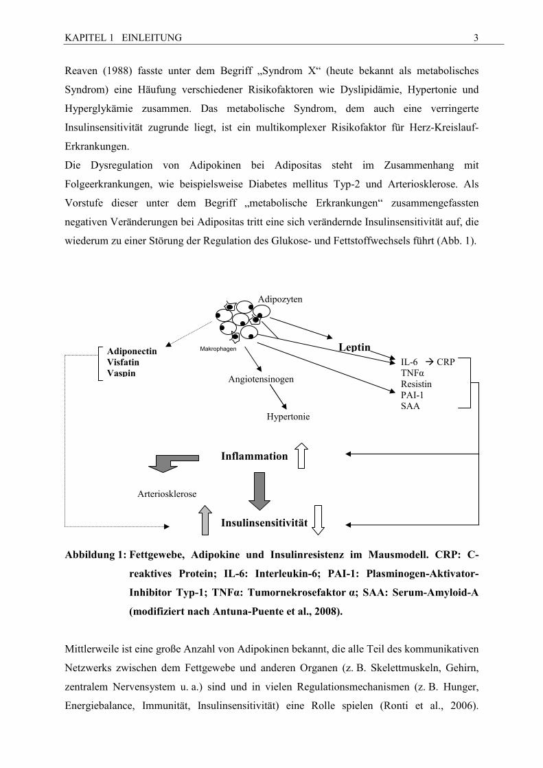
wiederum zu einer Störung der Regulation des Glukose- und Fettstoffwechsels führt (Abb. 1).
Abbildung 1: Fettgewebe, Adipokine und Insulinresistenz im Mausmodell. CRP: C-
reaktives Protein; IL-6: Interleukin-6; PAI-1: Plasminogen-Aktivator-
Inhibitor Typ-1; T�Fα: Tumornekrosefaktor α; SAA: Serum-Amyloid-A
(modifiziert nach Antuna-Puente et al., 2008).
Mittlerweile ist eine große Anzahl von Adipokinen bekannt, die alle Teil des kommunikativen
Netzwerks zwischen dem Fettgewebe und anderen Organen (z. B. Skelettmuskeln, Gehirn,
zentralem Nervensystem u. a.) sind und in vielen Regulationsmechanismen (z. B. Hunger,
Energiebalance, Immunität, Insulinsensitivität) eine Rolle spielen (Ronti et al., 2006).
Adipozyten
Leptin Adiponectin Visfatin Vaspin
Inflammation
Insulinsensitivität
Arteriosklerose
Angiotensinogen
Hypertonie
IL-6 � CRP TNFα Resistin PAI-1 SAA
Makrophagen
KAPITEL 1 EINLEITUNG
4
Besonders im Zusammenhang mit dem metabolischen Syndrom und Adipositas hat sich bei
Monogastriern gezeigt, dass v. a. die Adipokine Leptin und Adiponectin sehr stark, im
geringen Maße auch Visfatin, involviert sind und durchaus therapeutischen Charakter haben
können, so dass die Erforschung ihrer genauen Funktion und Regulation intensiviert wurde.
1.2.1 Leptin und die Subtypen des Leptinrezeptors
Sowohl bei Monogastriern als auch bei Wiederkäuern ist Leptin das bisher am intensivsten
untersuchte Adipokin, das in vielen Bereichen des Organismus tätig ist. Leptin ist ein
Proteohormon, welches aus 167 Aminosäuren besteht und ein Molekulargewicht von 16 kDa
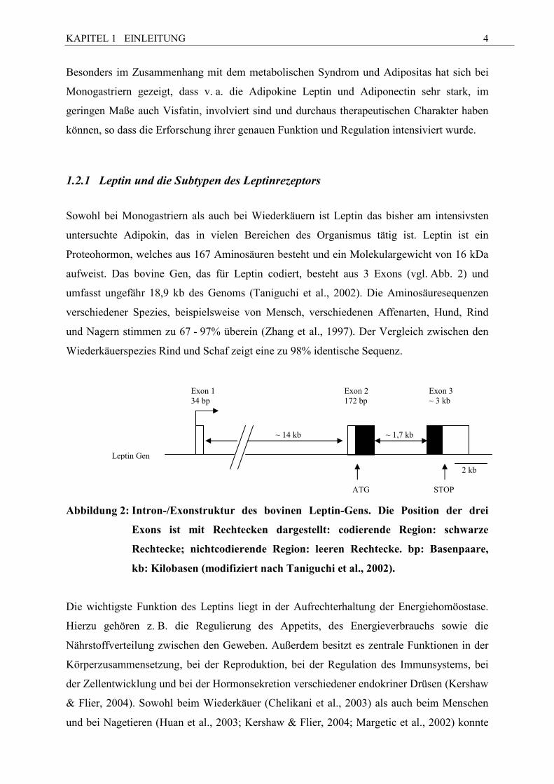
aufweist. Das bovine Gen, das für Leptin codiert, besteht aus 3 Exons (vgl. Abb. 2) und
umfasst ungefähr 18,9 kb des Genoms (Taniguchi et al., 2002). Die Aminosäuresequenzen
verschiedener Spezies, beispielsweise von Mensch, verschiedenen Affenarten, Hund, Rind
und Nagern stimmen zu 67 - 97% überein (Zhang et al., 1997). Der Vergleich zwischen den
Wiederkäuerspezies Rind und Schaf zeigt eine zu 98% identische Sequenz.
Abbildung 2: Intron-/Exonstruktur des bovinen Leptin-Gens. Die Position der drei
Exons ist mit Rechtecken dargestellt: codierende Region: schwarze
Rechtecke; nichtcodierende Region: leeren Rechtecke. bp: Basenpaare,
kb: Kilobasen (modifiziert nach Taniguchi et al., 2002).
Die wichtigste Funktion des Leptins liegt in der Aufrechterhaltung der Energiehomöostase.
Hierzu gehören z. B. die Regulierung des Appetits, des Energieverbrauchs sowie die
Nährstoffverteilung zwischen den Geweben. Außerdem besitzt es zentrale Funktionen in der
Körperzusammensetzung, bei der Reproduktion, bei der Regulation des Immunsystems, bei
der Zellentwicklung und bei der Hormonsekretion verschiedener endokriner Drüsen (Kershaw
& Flier, 2004). Sowohl beim Wiederkäuer (Chelikani et al., 2003) als auch beim Menschen
und bei Nagetieren (Huan et al., 2003; Kershaw & Flier, 2004; Margetic et al., 2002) konnte
Exon 1 34 bp
Exon 2 172 bp
Exon 3 ~ 3 kb
~ 14 kb ~ 1,7 kb
ATG STOP
Leptin Gen
2 kb
KAPITEL 1 EINLEITUNG
5
nachgewiesen werden, dass Leptin über einen für Leptin spezifischen Rezeptoren wirkt. Diese
Wirkung erfolgt nicht nur endokrin, beispielsweise im Gehirn, sondern auch autokrin oder
parakrin innerhalb der Gewebe, in denen es gebildet wurde, beispielsweise im Fettgewebe.
Eine Eigenschaft des Leptins, die bei der Betrachtung des Krankheitsbildes Adipositas als
besonders interessant erscheint, ist seine Fähigkeit, die Lipolyse im Fettgewebe zu hemmen
und gleichzeitig die Lipogenese zu fördern. Leptin ist Teil des Rückkopplungssystems, das
die Fettspeicher im Körper reguliert und eng mit dem Anteil des Körperfetts und mit
Gewichtsschwankungen korreliert. Aus diesem Grund gibt der Leptinspiegel im Blut unter
Bedingungen des Fließgleichgewichts Aufschluss über die Gesamtfettmasse des Körpers,
sofern dabei gleichbleibende Bedingungen in Bezug auf die Energiebilanz stehen. Dieser
Zusammenhang zwischen Leptin und Fettgehalt wird auch bei den landwirtschaftlichen
Nutztieren untersucht. Die Gruppen um Ehrhardt (2000) und Geary (2003) beobachten beim
Rind eine Korrelation zwischen Plasma-Leptin und Körperfett bzw. der s.c. Fettdicke. Diese
Zusammenhänge finden sich beim Schwein als auch beim Rind wieder (Altmann et al., 2006;
Berg et al., 2003; Bispham et al., 2002; Blache et al., 2000; Daniel et al., 2002; Delavaud et
al., 2000; Thomas et al., 2001). Beim Schaf sind jedoch bisher nur ausgewachsene Tiere oder
neonatale Lämmer untersucht worden.
Die Signaltransduktion/Signalweiterleitung von Leptin erfolgt über membranständige
Rezeptoren. Beim Nager sind bisher sechs Isoformen dieser Rezeptoren bekannt; diese sind
die Produkte des Leptinrezeptorgens und resultieren aus alternativem mRNA Splicing
und/oder proteolytischer Verarbeitung (Chua et al., 1997; Tartaglia, 1997). Alle Isoformen
bestehen aus einer einheitlichen extrazellulären Ligandenbindungs-Domäne, die aus über 800
Aminosäuren zusammengesetzt ist, einer transmembranen Domäne aus 34 Aminosäuren und
einer für jede Isoform spezifischen variablen intrazellulären Domäne (Anubhuti & Arora,
2008). Die längste Form, ObRb, kann alle Eigenschaften, die dem Leptin zugeschrieben
werden, weiterleiten (Ahima & Flier, 2000; Myers 2004). Vier Isoformen (ObRa, ObRc,
ObRd, ObRf) fehlt die interzelluläre Domäne, die für die STAT3 Aktivierung benötigt wird
(Ahima & Flier, 2000; Myers 2004). Die wichtigste Signaltransduktionskette, die vom Leptin
über den ObRb, nicht aber über die anderen Rezeptorisoformen, aktiviert wird, ist
der„JAK/STAT-Pathway“. Daneben gibt es noch weitere, allerdings weniger wichtige
Signaltransduktionsketten, die von Leptin aktiviert werden.
Bisher sind sowohl beim Rind als auch beim Schaf die Isoformen ObRa und ObRb
nachgewiesen worden, wobei ObRb, im Gegensatz zu anderen Spezies, ubiquitär gefunden
wurde (Boisclair et al., 2006). Auch der ObRa ist beim Rind bislang in Leber, Milz,
KAPITEL 1 EINLEITUNG
6
Hypophyse, Nebennierenrinde und Hirnstamm nachgewiesen worden (Chelikani et al., 2003;
Silva et al., 2002).
1.2.2 Adiponectin und die Adiponectinrezeptoren 1 und 2
Adiponectin, das auch als AdipoQ, Acrp30, apM1 oder GBP28 bekannt ist, wird seit den
1990er Jahren untersucht. Bovines Adiponectin wurde erstmals im Jahr 2001 isoliert (Sato et
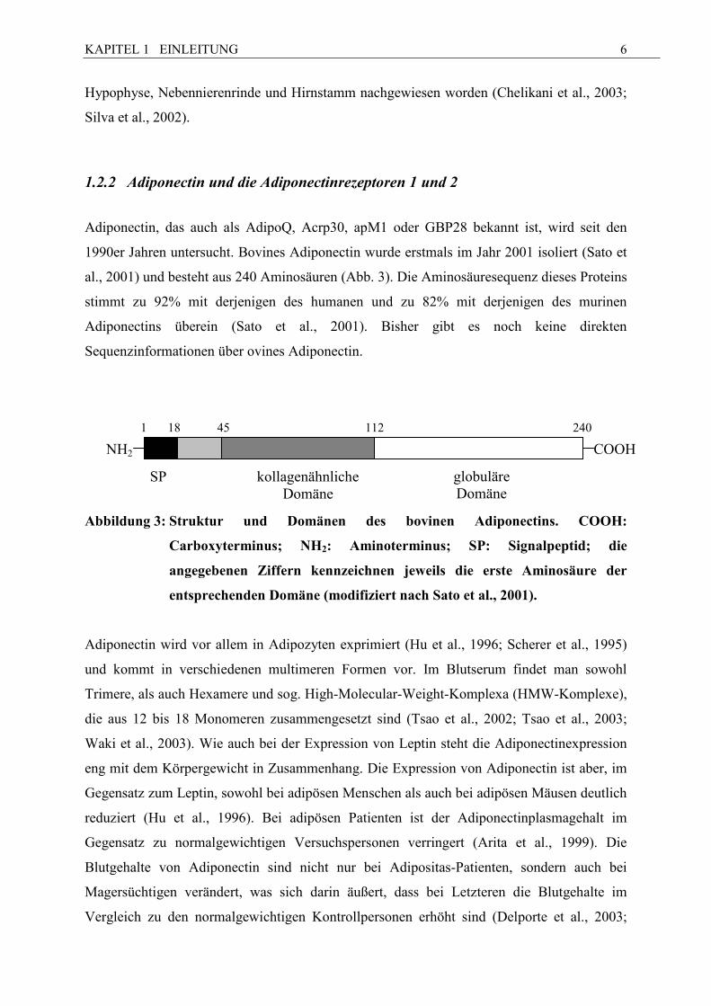
al., 2001) und besteht aus 240 Aminosäuren (Abb. 3). Die Aminosäuresequenz dieses Proteins
stimmt zu 92% mit derjenigen des humanen und zu 82% mit derjenigen des murinen
Adiponectins überein (Sato et al., 2001). Bisher gibt es noch keine direkten
Sequenzinformationen über ovines Adiponectin.
Abbildung 3: Struktur und Domänen des bovinen Adiponectins. COOH:
Carboxyterminus; �H2: Aminoterminus; SP: Signalpeptid; die
angegebenen Ziffern kennzeichnen jeweils die erste Aminosäure der
entsprechenden Domäne (modifiziert nach Sato et al., 2001).
Adiponectin wird vor allem in Adipozyten exprimiert (Hu et al., 1996; Scherer et al., 1995)
und kommt in verschiedenen multimeren Formen vor. Im Blutserum findet man sowohl
Trimere, als auch Hexamere und sog. High-Molecular-Weight-Komplexa (HMW-Komplexe),
die aus 12 bis 18 Monomeren zusammengesetzt sind (Tsao et al., 2002; Tsao et al., 2003;
Waki et al., 2003). Wie auch bei der Expression von Leptin steht die Adiponectinexpression
eng mit dem Körpergewicht in Zusammenhang. Die Expression von Adiponectin ist aber, im
Gegensatz zum Leptin, sowohl bei adipösen Menschen als auch bei adipösen Mäusen deutlich
reduziert (Hu et al., 1996). Bei adipösen Patienten ist der Adiponectinplasmagehalt im
Gegensatz zu normalgewichtigen Versuchspersonen verringert (Arita et al., 1999). Die
Blutgehalte von Adiponectin sind nicht nur bei Adipositas-Patienten, sondern auch bei
Magersüchtigen verändert, was sich darin äußert, dass bei Letzteren die Blutgehalte im
Vergleich zu den normalgewichtigen Kontrollpersonen erhöht sind (Delporte et al., 2003;
COOH NH2
SP kollagenähnliche Domäne
1 18 45 112 240
globuläre Domäne
KAPITEL 1 EINLEITUNG
7
Pannacciulli et al., 2003). Bei Gewichtsreduktion (beispielsweise nach einer Diät) bei
Übergewichtigen und auch bei krankhaft fettleibigen Patienten nehmen sowohl die
Blutspiegel als auch die Expression von Adiponectin im Fettgewebe wieder zu (Bruun et al.,
2003; Kopp et al., 2005).
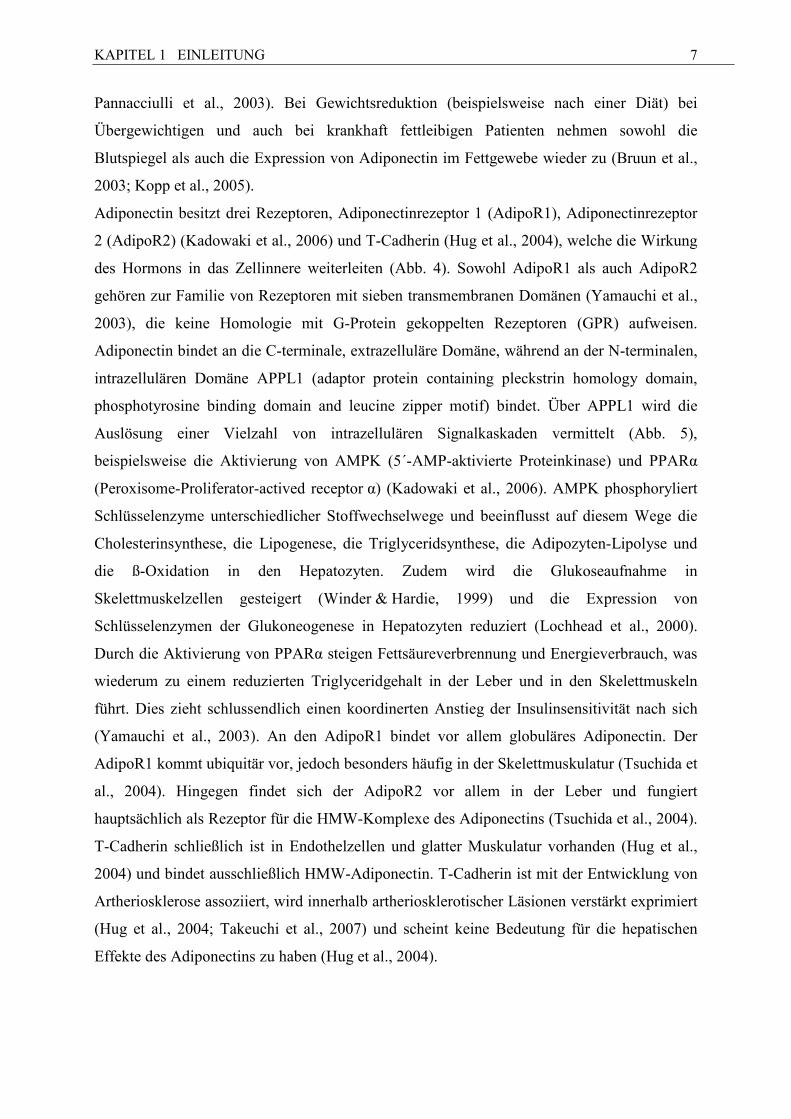
Adiponectin besitzt drei Rezeptoren, Adiponectinrezeptor 1 (AdipoR1), Adiponectinrezeptor
2 (AdipoR2) (Kadowaki et al., 2006) und T-Cadherin (Hug et al., 2004), welche die Wirkung
des Hormons in das Zellinnere weiterleiten (Abb. 4). Sowohl AdipoR1 als auch AdipoR2
gehören zur Familie von Rezeptoren mit sieben transmembranen Domänen (Yamauchi et al.,
2003), die keine Homologie mit G-Protein gekoppelten Rezeptoren (GPR) aufweisen.
Adiponectin bindet an die C-terminale, extrazelluläre Domäne, während an der N-terminalen,
intrazellulären Domäne APPL1 (adaptor protein containing pleckstrin homology domain,
phosphotyrosine binding domain and leucine zipper motif) bindet. Über APPL1 wird die
Auslösung einer Vielzahl von intrazellulären Signalkaskaden vermittelt (Abb. 5),
beispielsweise die Aktivierung von AMPK (5´-AMP-aktivierte Proteinkinase) und PPARα
(Peroxisome-Proliferator-actived receptor α) (Kadowaki et al., 2006). AMPK phosphoryliert
Schlüsselenzyme unterschiedlicher Stoffwechselwege und beeinflusst auf diesem Wege die
Cholesterinsynthese, die Lipogenese, die Triglyceridsynthese, die Adipozyten-Lipolyse und
die ß-Oxidation in den Hepatozyten. Zudem wird die Glukoseaufnahme in
Skelettmuskelzellen gesteigert (Winder & Hardie, 1999) und die Expression von
Schlüsselenzymen der Glukoneogenese in Hepatozyten reduziert (Lochhead et al., 2000).
Durch die Aktivierung von PPARα steigen Fettsäureverbrennung und Energieverbrauch, was
wiederum zu einem reduzierten Triglyceridgehalt in der Leber und in den Skelettmuskeln
führt. Dies zieht schlussendlich einen koordinerten Anstieg der Insulinsensitivität nach sich
(Yamauchi et al., 2003). An den AdipoR1 bindet vor allem globuläres Adiponectin. Der
AdipoR1 kommt ubiquitär vor, jedoch besonders häufig in der Skelettmuskulatur (Tsuchida et
al., 2004). Hingegen findet sich der AdipoR2 vor allem in der Leber und fungiert
hauptsächlich als Rezeptor für die HMW-Komplexe des Adiponectins (Tsuchida et al., 2004).
T-Cadherin schließlich ist in Endothelzellen und glatter Muskulatur vorhanden (Hug et al.,
2004) und bindet ausschließlich HMW-Adiponectin. T-Cadherin ist mit der Entwicklung von
Artheriosklerose assoziiert, wird innerhalb artheriosklerotischer Läsionen verstärkt exprimiert
(Hug et al., 2004; Takeuchi et al., 2007) und scheint keine Bedeutung für die hepatischen
Effekte des Adiponectins zu haben (Hug et al., 2004).
KAPITEL 1 EINLEITUNG
8
Abbildung 4: Schematische Darstellung der Adiponectinregulation mit den bisher
bekannten metabolischen und vaskulären Mechanismen. AdipoR1:
Adiponectinrezeptor 1; AdipoR2: Adiponectinrezeptor 2; AMPK: 5’-
AMP-aktivierte Proteinkinase; APPL1: a pleckstrin homology domain-
containing adaptor protein; HMW: high-molecular weight; �O:
Stickstoffmonoxid; n-3 PUFA: n-3 mehrfach ungesättigte Fettsäuren;
ROS: reaktive Sauerstoffspezies (modifiziert nach Guerro-Millo, 2008).
n-3 PUFA
Hexamere, HMW Form
Adiponectin
Trimere, globulärer Kopf
AdipoR2 T-Cadherin
AMPK
Glukoseproduktion Adhäsionsproteine und inflammatorische Zytokine
NO Produktion
Fettsäureoxidation Glukoseaufnahme
Metabolischer Insulinsensitivitäts-
prozess
Vaskulärer Inflammations-
prozess
?
Pro-inflammatorische Zytokine, ROS
Skelettmuskeln Fettgewebe
Endothelzellen Leber
AdipoR1
APPL1
KAPITEL 1 EINLEITUNG
9
1.2.3 Visfatin
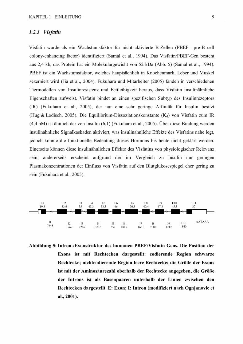
Visfatin wurde als ein Wachstumsfaktor für nicht aktivierte B-Zellen (PBEF = pre-B cell
colony-enhancing factor) identifiziert (Samal et al., 1994). Das Visfatin/PBEF-Gen besteht
aus 2,4 kb, das Protein hat ein Molekulargewicht von 52 kDa (Abb. 5) (Samal et al., 1994).
PBEF ist ein Wachstumsfaktor, welches hauptsächlich in Knochenmark, Leber und Muskel
sezerniert wird (Jia et al., 2004). Fukuhara und Mitarbeiter (2005) fanden in verschiedenen
Tiermodellen von Insulinresistenz und Fettleibigkeit heraus, dass Visfatin insulinähnliche
Eigenschaften aufweist. Visfatin bindet an einen spezifischen Subtyp des Insulinrezeptors
(IR) (Fukuhara et al., 2005), der nur eine sehr geringe Affinität für Insulin besitzt
(Hug & Lodisch, 2005). Die Equilibrium-Dissoziationskonstante (Kd) von Visfatin zum IR
(4,4 nM) ist ähnlich der von Insulin (6,1) (Fukuhara et al., 2005). Über diese Bindung werden
insulinähnliche Signalkaskaden aktiviert, was insulinähnliche Effekte des Visfatins nahe legt,
jedoch konnte die funktionelle Bedeutung dieses Hormons bis heute nicht geklärt werden.
Einerseits können diese insulinähnlichen Effekte des Visfatins von physiologischer Relevanz
sein; andererseits erscheint aufgrund der im Vergleich zu Insulin nur geringen
Plasmakonzentrationen der Einfluss von Visfatin auf den Blutglukosespiegel eher gering zu
sein (Fukuhara et al., 2005).
Abbildung 5: Intron-/Exonstruktur des humanen PBEF/Visfatin Gens. Die Position der
Exons ist mit Rechtecken dargestellt: codierende Region schwarze
Rechtecke; nichtcodierende Region leere Rechtecke; die Größe der Exons
ist mit der Aminosäurezahl oberhalb der Rechtecke angegeben, die Größe
der Introns ist als Basenpaaren unterhalb der Linien zwischen den
Rechtecken dargestellt. E: Exon; I: Intron (modifiziert nach Ognjanovic et
al., 2001).
E1
19,3
E2
53,6
E3 35
E4
43,3
E5
53,3
E6 46
E7
76,3
E8
40,4
E9
47,3
E10 45,3
E11 37
I6
4845
I1
7645
I2
1969
I3
2286
I4
3216
I5
552
I7
1681
I8
7082
I10
1840
I9
1212
AATAAA

KAPITEL 1 EINLEITUNG
10
1.2.4 Am Fettmetabolismus beteiligte G-Protein gekoppelte Rezeptoren (GPR 41,
GPR 43, GPR 109A)
Im Zusammenhang mit dem oben dargestellten, potentiellen Einfluss der o. g. Adipokine auf
den Fettstoffwechsel ist auch die Familie der in den Fettmetabolismus involvierten G-Protein
gekoppelten Rezeptoren (GPRs) zu erwähnen (Abb. 6). Besonders wichtig sind in diesem
Zusammenhang die fettsäurebindenden Membranproteine für kurzkettige Fettsäuren (SCFA)
GPR 41 und GPR 43, sowie der Rezeptor für ß-Hydroxybutyrat (BHB) und Nikotinsäure
GPR 109A, auch bekannt als HM74A oder PumaG.
G-Protein gekoppelte Rezeptoren haben eine besondere Bedeutung im Bereich der
Zellfunktion. Bei ihnen handelt es sich um membranübergreifende Proteine mit sieben
transmembranen Domänen, die auf verschiedenste Stimuli, wie beispielsweise Licht,
Geschmack, Geruch oder auch Hormone, reagieren (Hendriks-Balk et al., 2008). Über die
GPRs werden extrazellulare Signale über G-Proteine durch die Plasmamembran hindurch zu
intrazellularen Effektoren vermittelt. G-Proteine sind heterotrimer und lassen sich in vier
Familien (Gs, Gi, Gq, G12/13) unterteilen (Hamm, 1998), die zu unterschiedlichen
Signalkaskaden führen. Die Liganden für die verschiedenen GPRs sind äußerst vielfältig und
spezifisch (Ge et al., 2008).
KAPITEL 1 EINLEITUNG
11
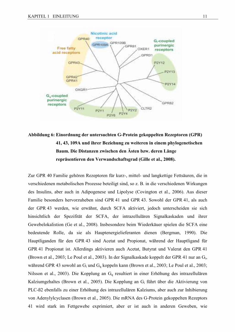
Abbildung 6: Einordnung der untersuchten G-Protein gekoppelten Rezeptoren (GPR)
41, 43, 109A und ihrer Beziehung zu weiteren in einem phylogenetischen
Baum. Die Distanzen zwischen den Ästen bzw. deren Länge
repräsentieren den Verwandschaftsgrad (Gille et al., 2008).
Zur GPR 40 Familie gehören Rezeptoren für kurz-, mittel- und langkettige Fettsäuren, die in
verschiedenen metabolischen Prozesse beteiligt sind, so z. B. in die verschiedenen Wirkungen
des Insulins, aber auch in Adipogenese und Lipolyse (Covington et al., 2006). Aus dieser
Familie besonders hervorzuheben sind GPR 41 und GPR 43. Sowohl der GPR 41, als auch
der GPR 43 werden, wie erwähnt, durch SCFA aktiviert, jedoch unterscheiden sie sich
hinsichtlich der Spezifität der SCFA, der intrazellulären Signalkaskaden und ihrer
Gewebelokalistion (Ge et al., 2008). Insbesondere beim Wiederkäuer spielen die SCFA eine
bedeutende Rolle, da sie als Hauptenergielieferanten dienen (Bergman, 1990). Die
Hauptliganden für den GPR 43 sind Acetat und Propionat, während der Hauptligand für
GPR 41 Propionat ist. Allerdings aktivieren auch Acetat, Butyrat und Valerat den GPR 41
(Brown et al., 2003; Le Poul et al., 2003). In der Signalkaskade koppelt der GPR 41 nur an Gi,
während GPR 43 sowohl an Gi und Gq koppeln kann (Brown et al., 2003; Le Poul et al., 2003;
Nilsson et al., 2003). Die Kopplung an Gq resultiert in einer Erhöhung des intrazellulären
Kalziumgehaltes (Brown et al., 2005). Die Kopplung an Gi führt über die Aktivierung von
PLC-ß2 ebenfalls zu einer Erhöhung des intrazellulären Kalziums, aber auch zur Inhibierung
von Adenylylcyclasen (Brown et al., 2005). Die mRNA des G-Protein gekoppelten Rezeptors
41 wird stark im Fettgewebe exprimiert, aber er ist auch in anderen Geweben, wie
KAPITEL 1 EINLEITUNG
12
beispielsweise in Milz, Blutzellen, Pankreas oder Lunge, exprimiert (Brown et al., 2003; Le
Poul et al., 2003).
Die höchste GPR 43 mRNA-Expression wurde bisher in Immunzellen, speziell in
polymorphkernigen Zellen (PBMC = peripheral blood mononuclear cells) beobachtet (Brown
et al., 2003; Le Poul et al., 2003; Nilsson et al., 2003), aber er wird auch in anderen Geweben,
inklusive Adipozyten, exprimiert. Im Fettgewebe führt die Aktivierung von GPR 43 über die
Gi-Leitungspfad zu Inhibierung der Lipolyse und zur Reduzierung des Niveaus der freien
Fettsäuren (FFA) (Ge et al., 2008). In Immunzellen spielt der GPR 43 sowohl in der
Immunantwort als auch in der inflammatorischen Antwort eine Rolle (Brown et al., 2003; Le
Poul et al., 2003; Nilsson et al., 2003). Unabhängig von verschiedenen Expressionsorten
beider GPRs wird beiden eine Rolle in der Kontrolle der Energiespeicherung zugesprochen
(Covington et al., 2006; Ge et al., 2008; Hong et al., 2005; Wang et al., 2009).
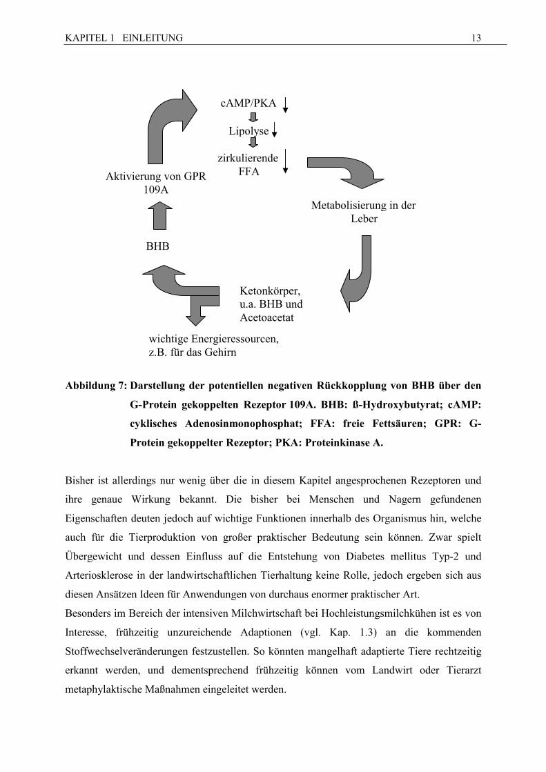
Im Zusammenhang mit der Lipolyse ist auch der GPR 109A von Interesse. Bei diesem
handelt es sich, wie schon erwähnt, u.a. um den Rezeptor von Nikotinsäure (Wise et al., 2003;
Tunaru et al., 2003), die ihrerseits fettreduzierende Wirkungen aufweist, wobei diese
fettreduzierende Wirkung allerdings erst in pharmakologischen Dosen erreicht wird. Als ein
endogener Ligand für GPR 109A, der für uns von weit größerem Interesse ist, agiert BHB,
das bei Rindern p.p. im Vergleich zu a.p. ansteigt (u. a. Doepel et al., 2002; Hachenberg et al.,
2007) und lipolysehemmende Wirkungen im bovinen Fettgewebe besitzt (Metz et al., 1974).
Ob dieser Rezeptor auch eine physiologische Rolle in den Immunzellen spielt, ist bisher
unklar (Gille et al., 2008). Besonders während der Transitionsperiode kann dieser Rezeptor
eine wichtige Rolle in der Rückkopplung von BHB und den nicht veresterten Fettsäuren
(NEFA) einnehmen (Abb. 7).
KAPITEL 1 EINLEITUNG
13
Abbildung 7: Darstellung der potentiellen negativen Rückkopplung von BHB über den
G-Protein gekoppelten Rezeptor 109A. BHB: ß-Hydroxybutyrat; cAMP:
cyklisches Adenosinmonophosphat; FFA: freie Fettsäuren; GPR: G-
Protein gekoppelter Rezeptor; PKA: Proteinkinase A.
Bisher ist allerdings nur wenig über die in diesem Kapitel angesprochenen Rezeptoren und
ihre genaue Wirkung bekannt. Die bisher bei Menschen und Nagern gefundenen
Eigenschaften deuten jedoch auf wichtige Funktionen innerhalb des Organismus hin, welche
auch für die Tierproduktion von großer praktischer Bedeutung sein können. Zwar spielt
Übergewicht und dessen Einfluss auf die Entstehung von Diabetes mellitus Typ-2 und
Arteriosklerose in der landwirtschaftlichen Tierhaltung keine Rolle, jedoch ergeben sich aus
diesen Ansätzen Ideen für Anwendungen von durchaus enormer praktischer Art.
Besonders im Bereich der intensiven Milchwirtschaft bei Hochleistungsmilchkühen ist es von
Interesse, frühzeitig unzureichende Adaptionen (vgl. Kap. 1.3) an die kommenden
Stoffwechselveränderungen festzustellen. So könnten mangelhaft adaptierte Tiere rechtzeitig
erkannt werden, und dementsprechend frühzeitig können vom Landwirt oder Tierarzt
metaphylaktische Maßnahmen eingeleitet werden.
wichtige Energieressourcen, z.B. für das Gehirn
zirkulierende FFA
BHB
Metabolisierung in der Leber
cAMP/PKA
Lipolyse
Aktivierung von GPR 109A
Ketonkörper, u.a. BHB und Acetoacetat
KAPITEL 1 EINLEITUNG
14
In Bezug auf die Schlachtkörperqualität, die die Grundlage für die Gestaltung des
Fleischpreises darstellt, wäre ein Parameter, mit dem sich vor allem der v.c. Fettanteil
schätzen ließe, eine Erleichterung zur Bestimmung des idealen Schlachtzeitpunktes.
Da sich jedoch die gewonnenen Erkenntnisse bezüglich der Expression, Sekretion und
physiologischen Rolle der oben genannten Adipokine und Rezeptoren nicht ohne weiteres
von Monogastriern auf Wiederkäuer übertragen lassen (Hishikawa et al., 2005), ist es nötig,
diese auch beim Wiederkäuer zu erforschen. Dazu haben wir zwei Ansätze gewählt:
1. Vergleich der mRNA-Expression der beschriebenen Gene im subkutanen Fettgewebe
von Hochleistungskühen während der Transitionsperiode mit zwei Probennahmen ante
und post partum.
2. Vergleich der mRNA-Expression der beschriebenen Gene in fünf unterschiedlichen
s.c. und v.c. Fettdepots beim Schaf
Ziel des ersten Ansatzes waren Grundlagenuntersuchungen, wie sich die metabolische und
physiologische Umstellung während der Transitionsperiode auf die mRNA-Expression im s.c.
Fettgewebe auswirkt.
Ziel des zweiten 2. Ansatzes war die Klärung, ob sich die verschiedenen Fettdepots bezüglich
ihrer mRNA-Expression unterscheiden.
KAPITEL 2 MANUSKRIPT 1
15
Kapitel 2
Manuskript 1 (published in: Domestic Animal Endocrinology 2009; 37: 37-44)
Transition period-related changes in the abundance of the mR/As of
adiponectin and its receptors, of visfatin, and of fatty acid binding receptors in
adipose tissue of high-yielding dairy cows
Running title: adipokine mR�A expression in dairy cow adipose tissue
Anneka Lemor, Afshin Hosseini, Helga Sauerwein, Manfred Mielenz*
Institute for Animal Science, Physiology & Hygiene Unit, University of Bonn,
Katzenburgweg 7 – 9, D-53115 Bonn, Germany
*Corresponding author at: Institute for Animal Science, Physiology & Hygiene Unit,
University of Bonn, Katzenburgweg 7 – 9, D-53115 Bonn, Germany. Tel., +49 228
732462, fax +49 229 737938.
E-mail address: [email protected]
KAPITEL 2 MANUSKRIPT 1
16
Abstract
Adipose tissue expresses adipokines, which are involved in regulation of energy expenditure,
lipid metabolism, and insulin sensitivity. To adapt for the transition from pregnancy to
lactation, particularly in high-yielding dairy cows, adipokines, their receptors, and particular
G-protein coupled receptors (GPRs) are of potential importance. Signaling by GPR 41
stimulates leptin release via activation by short-chain fatty acids; GPR 43/ 109A inhibits
lipolysis, and GPR 109A thereby mediates the lipid-lowering effects of nicotinic acid and
ß-hydroxybutyrate. The aim of this study was to compare the mRNA expression of
adiponectin and visfatin, adiponectin receptors 1 and 2 (AdipoR1/2), leptin receptor (obRb),
insulin receptor as of the aforementioned GPRs during the transition period in high-yielding
dairy cows. Biopsies from subcutaneous fat and blood samples were obtained from 10 dairy
cows 1 week before and 3 weeks after calving. For AdipoR1 and AdipoR2 mRNA abundance
as well as for leptin concentrations in plasma, a reduction (P ≤ .05) was observed postpartum;
for visfatin and putative GPR 109A mRNA abundance in adipose tissue, there was a trend
(P < .1) for analogous changes. In contrast, the mRNA content of obRb and GPR 41 in
adipose tissue was higher (P ≤ .05) in samples from early lactation than in those from late
gestation. Our results indicate decreasing adiponectin sensitivity in adipose tissue after
calving, which might be involved in the reduced insulin sensitivity of adipose tissue during
early lactation. In addition, visfatin, GPR 41 and GPR 109A may further modulate insulin
sensitivity.
Key words: Dairy cow, Adipose tissue, Adipokines, Adiponectin, G-protein coupled receptors
1. Introduction
During the transition period, that is, the last 3 weeks before parturition and the first 3 weeks
thereafter, extensive metabolic and endocrine changes occur in the dairy cow to accommodate
parturition and lactogenesis [1]. Energy needs during early lactation cannot be met entirely
through dietary intake, and therefore energy has to be mobilized from body reserves, mainly
from fat. This process results in increased circulating concentrations of nonesterified fatty
acids (NEFA) and formation of ketone bodies [2]. Besides its role as an energy store, adipose
KAPITEL 2 MANUSKRIPT 1
17
tissue (AT) is increasingly gaining interest as an active regulatory gland secreting multiple
metabolically important proteins that are called adipokines [3-4].
Adipokines like adiponectin and visfatin play an important role in insulin sensitivity, glucose
homeostasis, and lipid metabolism [5-9]. The role of both adipokines in these contexts is not
entirely clear and, at least for visfatin, controversial [5,8,10,11]. To our knowledge, no
information is available about the mRNA expression of adiponectin receptor 1 (AdipoR1) and
adiponectin receptor 2 (AdipoR2) antepartum (a.p.) and postpartum (p.p.) in women or
rodents, although hepatic AdipoR2 mRNA is reportedly increased p.p. in dairy cows [12].
Insulin resistance in muscle and AT develops during late pregnancy and is continues
postpartum to support the partitioning of glucose towards the mammary glands [13,14].
In context with the increased NEFA concentrations towards early lactation, the family of fatty
acid-binding membrane receptors, that is, G-protein coupled receptors (GPRs) [15], is of
particular interest. G-protein coupled receptors 41 plays a role in monitoring energy storage
[16], and it is directly involved in the stimulatory effects of short-chain fatty acids (SCFA) on
leptin production [17]. G-protein coupled receptor 43 participates in different cell processes
[18,19], but recent studies also report that GPR 43 is involved in monitoring energy storage
and regulating plasma lipid profiles by reducing lipolysis [20,21].
One of the ketone bodies increasingly produced in dairy cows during negative energy balance
is ß-hydroxybutyric acid (BHB). ß-Hydroxybutyric acid and NEFA exert inhibitory effects on
leukocytes and may thus contribute to the immunocompromised situation in early lactation,
leading to an increased susceptibility for infectious diseases [22,23]. Although the inhibiting
action of BHB and of butyrate on lipolysis in bovine AT has been known for decades [24], it
was only recently that BHB was identified to act as a ligand of GPR 109A (HM74A / PUMA-
G) [25,26]. GPR 109A was already known to be a receptor for nicotinic acid, mediating its
lipid-lowering effects [26,27]. All GPRs are expressed in AT and blood cells from
monogastric species [15,27].
Currently only limited data are available about the regulation of adiponectin and visfatin
during the transition period in ruminants. To our knowledge, regulation of the aforementioned
GPRs during the transition period in cattle has not been characterized. Therefore, the
objective of this study was to detect potential changes between a.p. and p.p. as a first step to
evaluating the association between these adipokines and receptors and metabolic alterations
during the transition period in high-yielding dairy cows.
KAPITEL 2 MANUSKRIPT 1
18
2. Materials and methods
The messenger RNAs (mRNA) of adiponectin, AdipoR1, AdipoR2, leptin, long isoform of
leptin receptor (obRb), insulin receptor, visfatin, GPR 41, GPR 43 and putative GPR 109A
were quantified in subcutaneous (s.c.) AT. In addition, we measured the mRNA concentration
of GPR 41, GPR 43 and putative GPR 109A mRNA in isolated leukocytes. Accompanying
records of the circulating concentrations of leptin, NEFA, and BHB were included.
2.1 Animals and tissue collection
The experiment and treatment of the animals were approved by the competent authority
(Regional Board, Cologne, 50.203.2-BN, 14/05). Subcutaneous fat was obtained twice from
10 Holstein-Frisian cows during the transition period at the research farm Frankenforst,
University of Bonn, Germany. All cows were fed a mixed ration based on grass and corn
silage ad libitum and concentrate according to the recommendations of the German Society of
Nutrition Physiology [28]. The average milk yield from the preceding lactation was 10.720 kg
of energy-corrected milk. Two biopsies were taken from each animal, the first between days
13 and 2 a.p. and the second between days 20 and 23 after calving. The samples were excised
through a V-shaped incision 10 cm laterally from the medial line and 10 cm cranially from the
base of the tail, after anesthesia with 2% procaine hydrochloride (Procasel® 2%, Selectavet,
Dr. Otto Fischer, Weyarn-Holzolling, Germany). The tissue samples were cut into pieces in
0.9% NaCl and immediately snap-frozen in liquid nitrogen and stored at -80°C until analysis.
2.2 Blood samples
Before the biopsies, whole and EDTA blood samples (1.6 mg/ml EDTA) were taken by
venipuncture and then transported in ice water for immediate processing. Plasma and serum
were prepared by centrifugation (20 min, 1.200 x g, 4°C). ß-Hydroxybutyric acid was
immediately measured in serum using a kit from Randox (kit-no. RB 1008, Randox
Laboratories, Crumlin, County Antrim, Ireland). Plasma was stored at - 20°C and used for the
analysis of leptin [29] and NEFA (enzymatic test kit, no. 1 383 175, Roche, Mannheim,
Germany). The concentrations of leptin were measured by ELISA [29] using ovine
recombinant leptin [30] as the standard. The intra- and interassay coefficients of variation
were 6.3 % and 13.9 %, respectively, and the mean recovery was 101.4 %.
KAPITEL 2 MANUSKRIPT 1
19
Leukocytes were isolated as previously described [31], with minor modifications, namely,
EDTA blood (1.6 mg EDTA/mL) was used, lysis of erythrocytes was repeated 3 times, and
total RNA was extracted immediately.
2.3 R�A extraction
Total RNA was extracted from isolated leukocytes using the NucleoSpin RNA II kit
(Macherey-Nagel, Düren, Germany), according the manufacturer’s instructions. Total RNA
was extracted from the AT samples using the single-step method [32,33]. Total RNA
purification was performed using spin columns including DNase I digestion (Nucleo Spin
RNA II, Macherey & Nagel). Quantification of RNA was performed using the RiboGreen
RNA Assay kit (Quant-iT RiboGreen, Invitrogen, Karlsruhe, Germany) in a Mx3000P
(Strategene, Amsterdam, Netherlands). The integrity of the RNA was assessed using SYBR
Green II (Invitrogen, Karlsruhe, Germany) denaturing RNA electrophoresis.
2.4 cD�A synthesis
The synthesis of cDNA was performed using 1 µg of total RNA with Revert Aid reverse
transcriptase (RT) (Fermentas, St. Leon-Rot, Germany) and 200 pmol random hexamer
primers (Sigma-Aldrich, Nümbrecht, Germany). No-RT controls were created by omitting
reverse transcription. No-template-controls were created by adding of nuclease-free water.
2.5 Quantification of mR�A
The amount of the mRNA of the different candidate genes was quantified using real-time
polymerase chain reaction (PCR) (cycler Mx 3000PTM Stratagene). The features of the
primers used for quantification are presented in Table 1.
Copy numbers for each transcript were calculated from standard curves, based on purified and
sequenced amplicons. Melting curve analysis confirmed the presence of a single product for
each real-time PCR assay.
The reaction was performed in a total volume of 10 µL comprosed of 2 µL of template diluted
1:4 (exceptions: adiponectin, obRb, GPR 41, GPR 43, and GPR 109A were diluted 1:3), 1 µL
of the assay specific primer mix, 5 µL of the SYBR Green JumpStart Taq Readymix (Sigma-
Aldrich) and 2 µL water. The annealing temperature was 60°C; exceptions were putative
GPR 109A and low density lipoprotein receptor-related protein 10 (LRP 10) at 61°C,
KAPITEL 2 MANUSKRIPT 1
20
glyceraldehyde-phosphate-dehydrogenase (GAPDH) at 59°C, and RNA polymerase II at
58°C. All samples were measured in triplicate. No-template-controls were performed using
nuclease-free water. In addition, no-RT and no-template-controls from the cDNA synthesis
were used to check for contamination.
The geometric mean of housekeeping gene abundance was used for normalization. Data were
expressed as ratios of the genes of interest to the geometric mean of the corresponding
internal controls. Low-density lipoprotein receptor-related protein 10 and RNA polymerase II
were used as internal controls for the AT samples and LRP 10, and GAPDH was used for
leukocytes.
2.6 Statistical Analyses
Analyses were performed with the statistical software package SPSS 15.0 (SPSS Inc.,
Chicago, IL, USA). Two samples per animal were compared using the paired Wilcoxon test.
Statistical significance was set at P ≤ .05, and significant correlation was assessed. A
significance of P ≤ .10 was considered a trend. Spearman correlations were calculated and
were considered significant at P ≤ .01.
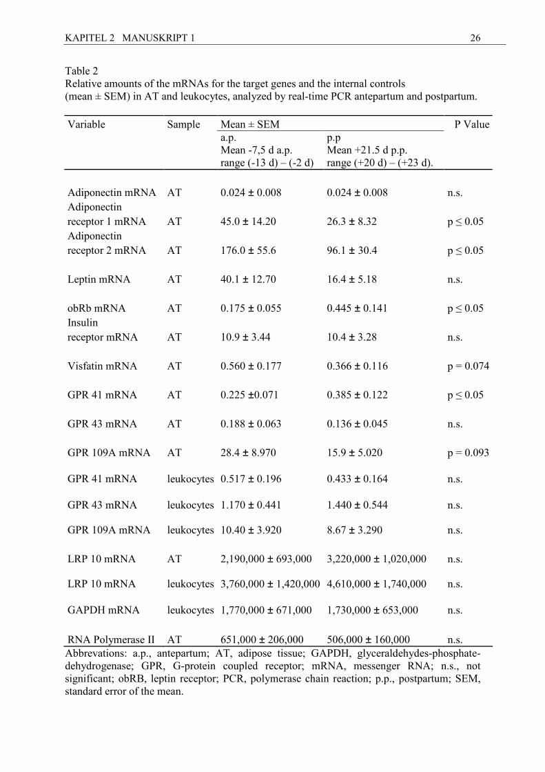
3. Results
Due to technical reasons, the number of samples varied in some assays, as specified below.
3.1 mR�A data and leptin, �EFA and BHB concentrations in blood
Results of mRNA expression of the target genes and the internal controls a.p. and p.p. (mean
± standard error of the mean [SEM]) are shown in Table 2. For adiponectin mRNA (n = 9),
time-related changes were not detectable in s.c. AT when comparing a.p. and p.p. samples. In
contrast, for both adiponectin receptors AdipoR1 and AdipoR2, the p.p. mRNA concentration
was significantly lower than a.p.
The decrease in leptin mRNA expression from pre- to postpartum sampling did not reach the
level of significance. For the obRb mRNA, the p.p. values were higher than those taken a.p.
In the case of visfatin mRNA, there was a trend toward a decrease in the p.p. samples.
For insulin receptor mRNA, no time-related variation of mRNA concentration was observed.
The mRNA of GPR 41, GPR 43 (n = 9), and putative GPR 109A was measured both in AT
and in isolated leukocytes. For GPR 41 mRNA in AT, higher values were recorded p.p. than
KAPITEL 2 MANUSKRIPT 1
21
a.p. Moreover, for putative GPR 109A in AT there was a trend toward decreased p.p. values,
whereas no time-related differences were found for the other GPRs in s.c. AT as well as for
all GPRs in isolated leukocytes.
Plasma leptin decreased p.p. (mean ± standard deviation [SD] [ng/mL]: a.p. 7.83 ± 2.84, p.p.
4.64 ± 2.24). For NEFA, an increase was found p.p. (mean ± SD [mM]: a.p. 0.09 ± 0.03, p.p.
0.23 ± 0.13), whereas no change was observed for BHB.
3.2 Correlations
AdipoR1 and AdipoR2 mRNA were positively correlated with each other (r = 0.82), with
leptin mRNA expression (r = 0.65 and r = 0.63, respectively), and with leptin plasma
concentrations (r = 0.57 and r = 0.58, respectively). Both receptors were negatively correlated
with obRb mRNA expression (r = -0.66 and r = -0.61, respectively). In addition, leptin mRNA
expression was negatively correlated with obRb mRNA expression (r = -0.82).
A positive correlation was observed between GPR 41 mRNA and obRb mRNA abundance
(r = 0.71). Negative correlations were found between NEFA and visfatin mRNA (r = -0.57).
Moreover, visfatin mRNA and BHB serum concentrations were negatively correlated
(r = -0.66).
4. Discussion
4.1 Regulation of adiponectin and its receptors mR�A in adipose tissue
To our knowledge, this is the first report about the mRNA expression of adiponectin and its
receptors in subcutaneous AT during the transition period in dairy cows. No alterations of
adiponectin mRNA expression were observed during the transition period. In humans, several
studies report adiponectin blood concentrations throughout pregnancy, but the results are
conflicting. Both decreasing as well as unchanged adiponectin blood concentrations during
pregnancy have been reported in women [34-38]. Less is known about circulating adiponectin
concentrations after parturition; similar levels as in the third trimester [34] and decreased
levels [39], respectively, were found. Limited data are available on adiponectin mRNA
expression during pregnancy. Decreasing adiponectin mRNA expression has been observed in
late pregnancy compared with prepregnancy or early pregnancy [36]. Similarly, controversial
results have been reported on adiponectin during pregnancy in rodents. In mice, adiponectin
KAPITEL 2 MANUSKRIPT 1
22
serum levels as well as adiponectin mRNA expression in AT decrease in late pregnancy
compared with control virgin mice [40], whereas no significant differences have been
reported in rats [41]. Thus, controversy exists about adiponectin concentrations during a.p.
and p.p. in different monogastric species; in previous studies the intervals of observation vary,
and only 1 group discussed relevant differences [39].
For dairy cows, there are some preliminary data indicating that circulating adiponectin
concentrations may increase from the first to the fourth week after calving [42], but the
validity of the human assay system applied remains to be substantiated. However, our data
comprised only 1 fixed time point before and 1 time point after calving without any
differences at the level of adiponectin mRNA expression. The information on adiponectin
mRNA does not necessarily reflect the circulating adiponectin concentrations [43,44] and thus
our results on adiponectin mRNA expression may not apply for the concentrations in blood
during the transition period.
Adiponectin receptors are related to the improvement of insulin sensitivity [6,7]. Due to our
observation of decreased AdipoR1 and AdipoR2 mRNA abundance in p.p. compared to a.p.
samples, reduced adiponectin sensitivity may be expected in s.c. AT. This finding could be
linked to the reduced insulin sensitivity p.p. in dairy cattle. In the liver, for which reduced
insulin sensitivity is indicated by decreased hepatic gluconeogenesis during the transition
period in ruminants [45], AdipoR2 mRNA content was increased p.p. if cows were fed
restrictively before calving, but not if they had ad libitum access to feed prior to calving [12].
These results together with our results from subcutaneous AT point to a differential regulation
between the liver and s.c. AT during the transition period. This finding could be related to
reduced insulin sensitivity at the level of the AT, leading to reduced glucose uptake and
increased lipolysis p.p. In our study of AT, both adiponectin receptors were positively
correlated with each other, whereas no such relationship is reported for the liver [12]. To our
knowledge, AdipoR1/2 mRNA expression in fat has not been compared before and after
delivery in humans.
Leptin and its mRNA were positively correlated to AdipoR1/2 mRNA content, but only leptin
mRNA was negatively correlated to obRb. Expression of obRb mRNA in s.c. AT of dairy
cows increased p.p., confirming other results in cows, which indicate that hypoinsulinemia
may be partly responsible for the induction of obRb in AT [46]. Expression of obRb mRNA
was negatively correlated with the amount of both adiponectin receptor mRNAs and
positively correlated with GPR 41 mRNA abundance. Thus both the adiponectin system and
signaling by GPR 41 might be involved in the regulation of obRb mRNA abundance, in
KAPITEL 2 MANUSKRIPT 1
23
addition to insulin, as discussed by Thorn et al. [46]. The observed decrease in plasma leptin
might have been compensated by an increase of the obRb level in AT.
4.2 G-protein coupled receptors 41, 43 and 109A
To our knowledge, this is the first report about the mRNA expression of GPR 41, GPR 43 and
putative GPR 109A in AT during the transition period in dairy cows. For GPR 41 mRNA, the
increased levels we observed during early lactation compared to late gestation might have
been expected, since GPR 41 is involved in monitoring energy storage [16] and related to
leptin synthesis [17]. Increasing GPR 41 mRNA expression in AT p.p. might be an option to
respond in the long term to the negative energy balance when insulin and leptin levels are low
[47,48]. In the short term, energy supply by propionate infusion increases GPR 41 mRNA in
s.c., but not in perirenal AT, of castrated male goats [49]. These divergent results might also
be attributable to the physiological situation, in particular the energetic conditions, and the
species. G-protein coupled receptor 43 is involved in the reduction of lipolysis in 3T3-L1
cells in vitro and in the disease pattern of the metabolic syndrome [20,21]. Interestingly, in
contrast to GPR 41 mRNA, GPR 43 mRNA showed no alteration during our study.
The concentrations of BHB in plasma were < 1.2 mmol/L (median: 0.485 mmol/L) in our
study and can be judged as normal values well below the thresholds defined for ketonemic
conditions [50]. In contrast to other studies [51,52], in this study we found no significant
increase of BHB concentrations during the transition period, a finding that might be explained
by the variance of the sampling time before calving. ß-Hydroxybutyric acid is the endogenous
ligand for GPR 109A [25] that mediates the lipid-lowering effects of nicotinic acid in humans
and rodents and possibly also in cattle [26,27,53]. Nicotinic acid stimulates circulating
adiponectin in humans [54,55]. According to published results [56], we expected a decrease
of GPR 109A in p.p. samples, since increasing BHB concentrations would imply an important
feedback mechanism to regulate lipolysis. Indeed, we observed a trend for a decrease. The
impact of GPR 109A on lipolysis seems to be of relevance during transition in AT.
No differences of the mRNA contents of GPR 41, GPR 43, and putative GPR 109A in
leukocytes were found between the 2 sampling. Accordingly, the negative energy balance p.p.
might not be relevant for the mRNA expression of these GPRs in immune cells, but
potentially, existing changes might have been leveled off through the use of total leukocytes
or the small number of animals used.
KAPITEL 2 MANUSKRIPT 1
24
4.3 Visfatin
As previously described, visfatin is involved in glucose homeostasis and mimics some insulin
effects [8,10,11]. Insulin decreases visfatin mRNA expression in 3T3-L1 adipocytes [57].
Visfatin mRNA expression in s.c. AT and plasma visfatin in humans are reportedly inversely
related [10]. Based on this information, our observation of a trend for decreasing visfatin
mRNA abundance in p.p. samples might rather indicate increasing visfatin plasma
concentrations after calving. Increasing visfatin plasma concentrations can concur with
decreasing insulin secretion p.p. [47] in the presence of equal concentrations of insulin
receptor mRNA in AT, as anticipated [58] and as observed in this study. In addition, the
negative correlations we found between visfatin mRNA expression in s.c. AT and NEFA and
BHB concentrations, respectively, also support this assumption. Besides the low plasma
concentration of visfatin compared to insulin [59], the importance on insulin-mediated effects
is expected to be small. Based on our results, we consider that visfatin might mitigate the
reduced insulin sensitivity in s.c. AT of dairy cows.
In summary, we showed that GPR 41, GPR 43, and putative GPR 109A mRNA is expressed
in bovine s.c. AT and leukocytes. During the transition period, we observed regulation of
GPR 41 and putative GPR 109A mRNA in AT, but the role of all of these 3 receptors in
ruminants remains to be clarified. We speculate that adiponectin sensitivity decreases after
parturition in dairy cows, mediated by reduced adiponectin receptor mRNA abundance, which
might be related to the insulin sensitivity during transition period. Further, our results might
indicate that the role of visfatin in adopting insulin signaling and glucose uptake could be
more important than discussed so far. In further studies, adiponectin and visfatin protein
concentrations should be measured in blood during the transition period in dairy cows to
investigate the adiponectin and the visfatin system.
Acknowledgments
The donation of scholarships by the H. Wilhelm-Schaumann Foundation, Hamburg, Germany
to A. Lemor and A. Hosseini is gratefully acknowledged. We also thank Dr. Michael Hölker,
Institute of Animal Science; Animal Breeding and Husbandry Unit, University of Bonn,
Germany; Dr. Christiane Theune for performing the biopsies; and Inga Hofs, Isabella Israel,
and Birgit Mielenz for their excellent technical assistance.
KAPITEL 2 MANUSKRIPT 1
25
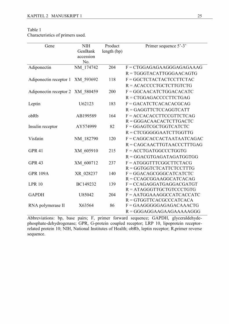
Table 1 Characteristics of primers used.
Gene NIH GenBank accession
No.
Product length (bp)
Primer sequence 5’-3’
F = CTGGAGAGAAGGGAGAGAAAG Adiponectin NM_174742 204 R = TGGGTACATTGGGAACAGTG
F = GGCTCTACTACTCCTTCTAC Adiponectin receptor 1 XM_593692 118 R = ACACCCCTGCTCTTGTCTG F = GGCAACATCTGGACACATC Adiponectin receptor 2 XM_580459 200 R = CTGGAGACCCCTTCTGAG
F = GACATCTCACACACGCAG Leptin U62123 183 R = GAGGTTCTCCAGGTCATT
obRb AB199589 164 F = ACCACACCTTCCGTTCTCAG R = GGGACAACACTCTTGACTC F = GGAGTCGCTGGTCATCTC Insulin receptor AY574999 82
R = CTCGGGGGAATCTTGGTTG
F = CAGGCACCACTAATAATCAGAC Visfatin NM_182790 120 R = CAGCAACTTGTAACCCTTTGAG
F = ACCTGATGGCCCTGGTG GPR 41 XM_605910 215
R = GGACGTGAGATAGATGGTGG
GPR 43 XM_600712 237 F = ATGGGTTTCGGCTTCTACG R = GGTGGTCTCATTCTCCTTTG
GPR 109A XR_028237 140 F = GGACAGCGGGCATCATCTC R = CCAGCGGAAGGCATCACAG
LPR 10
BC149232 139 F = CCAGAGGATGAGGACGATGT R = ATAGGGTTGCTGTCCCTGTG
GAPDH
U85042 204 F = AATGGAAAGGCCATCACCATC R = GTGGTTCACGCCCATCACA F = GAAGGGGGAGAGACAAACTG RNA polymerase II X63564 86
R = GGGAGGAAGAAGAAAAAGGG
Abbreviations: bp, base pairs; F, primer forward sequence; GAPDH, glyceraldehyde-phosphate-dehydrogenase; GPR, G-protein coupled receptor; LRP 10, lipoprotein receptor-related protein 10; NIH, National Institutes of Health; obRb, leptin receptor; R,primer reverse sequence.
KAPITEL 2 MANUSKRIPT 1
26
Table 2 Relative amounts of the mRNAs for the target genes and the internal controls (mean ± SEM) in AT and leukocytes, analyzed by real-time PCR antepartum and postpartum.
Variable Sample Mean ± SEM P Value
a.p. Mean -7,5 d a.p. range (-13 d) – (-2 d)
p.p Mean +21.5 d p.p. range (+20 d) – (+23 d).
Adiponectin mRNA AT 0.024 ± 0.008 0.024 ± 0.008 n.s. Adiponectin
receptor 1 mRNA AT 45.0 ± 14.20 26.3 ± 8.32 p ≤ 0.05 Adiponectin receptor 2 mRNA AT 176.0 ± 55.6 96.1 ± 30.4 p ≤ 0.05
Leptin mRNA AT 40.1 ± 12.70 16.4 ± 5.18 n.s.
obRb mRNA AT 0.175 ± 0.055 0.445 ± 0.141 p ≤ 0.05 Insulin
receptor mRNA AT 10.9 ± 3.44 10.4 ± 3.28 n.s.
Visfatin mRNA AT 0.560 ± 0.177 0.366 ± 0.116 p = 0.074
GPR 41 mRNA AT 0.225 ±0.071 0.385 ± 0.122 p ≤ 0.05
GPR 43 mRNA AT 0.188 ± 0.063 0.136 ± 0.045 n.s.
GPR 109A mRNA AT 28.4 ± 8.970 15.9 ± 5.020 p = 0.093
GPR 41 mRNA leukocytes 0.517 ± 0.196 0.433 ± 0.164 n.s.
GPR 43 mRNA leukocytes 1.170 ± 0.441 1.440 ± 0.544 n.s.
GPR 109A mRNA leukocytes 10.40 ± 3.920 8.67 ± 3.290 n.s.
LRP 10 mRNA AT 2,190,000 ± 693,000 3,220,000 ± 1,020,000 n.s.
LRP 10 mRNA leukocytes 3,760,000 ± 1,420,000 4,610,000 ± 1,740,000 n.s.
GAPDH mRNA leukocytes 1,770,000 ± 671,000 1,730,000 ± 653,000 n.s.
RNA Polymerase II AT 651,000 ± 206,000 506,000 ± 160,000 n.s. Abbrevations: a.p., antepartum; AT, adipose tissue; GAPDH, glyceraldehydes-phosphate-dehydrogenase; GPR, G-protein coupled receptor; mRNA, messenger RNA; n.s., not significant; obRB, leptin receptor; PCR, polymerase chain reaction; p.p., postpartum; SEM, standard error of the mean.
KAPITEL 2 MANUSKRIPT 1
27
References
[1] Grummer RR. Impact of changes in organic nutrient metabolism on feeding the
transition dairy cow. J Anim Sci 1995;73:2820-33.
[2] Goff JP, Horst RL. Physiological changes at parturition and their relationship to
metabolic disorders. J Dairy Sci 1997;80:1260-8.
[3] Bastard JP, Maachi M, Lagathu C, Kim MJ, Caron M, Vidal H, Capeau J, Feve B.
Recent advances in the relationship between obesity, inflammation, and insulin
resistance. Eur Cytokine Netw 2006;17:4-12.
[4] Trayhurn P, Wood IS. Adipokines: inflammation and the pleiotropic role of white
adipose tissue. Br J Nutr 2004;92:347-55.
[5] Guerre-Millo M. Adiponectin: an update. Diabetes Metab 2008; 34:12-8.
[6] Minokoshi Y, Kim YB, Peroni OD, Fryer LGD, Müller C, Carling D, Kahn BB.
Leptin stimulates fatty-acid oxidation by activating AMP-activated protein kinase.
Nature 2002;415:339-43.
[7] Yamauchi T, Kamon J, Minokoshi Y, Ito Y, Waki H, Uchida S, Yamashita S, Noda M,
Kita S, Ueki K, Eto K, Akanuma Y, Froguel P, Foufelle F, Ferre P, Carling D, Kimura
S, Nagai R, Kahn BB, Kadowaki T. Adiponectin stimulates glucose utilization and
fatty-acid oxidation by activating AMP-activated protein kinase. Nat Med
2002;8:1288-95.
[8] Fukuhara A, Matsuda M, Nishizawa M, Segawa K, Tanaka M, Kishimoto K, Matsuki
Y, Murakami M, Ichisaka T, Murakami H, Watanabe E, Takagi T, Akiyoshi M,
Ohtsubo T, Kihara S, Yamashita S, Makishima M, Funahashi T, Yamanaka S,
Hiramatsu R, Matsuzawa Y, Shimornura I. Visfatin: a protein secreted by visceral fat
that mimics the effect of insulin. Science 2005;307:426-30.
[9] Moschen AR, Kaser A, Enrich B, Mosheimer B, Theurl M, Niederegger H, Tilg H.
Visfatin, an adipocytokine with proinflammatory and immunomodulating properties. J
Immunol 2007;178:1748-58.
[10] Berndt J, Klöting N, Kralisch S, Kovacs P, Fasshauer M, Schön MR, Stumvoll M,
Blüher M. Plasma visfatin concentrations and fat depot-specific mRNA expression in
humans. Diabetes 2005;54:2911-6.
[11] Varma V, Yao-Borengasser A, Rasouli N, Bodles AM, Phanavanh B, Lee MJ, Starks
T, Kern LM, Spencer III HJ, McGehee Jr. RE, Fried SK, Kern PA. Human visfatin
KAPITEL 2 MANUSKRIPT 1
28
expression: relationship to insulin sensitivity, intramyocellular lipids, and
inflammation. J Clin Endocrinol Metab 2007;92:666-72.
[12] Loor JJ, Dann HM, Janovick Guretzky NA, Everts RE, Oliveira R, Green CA,
Litherlands NB, Rodriguez-Zas SL, Lewin HA, Drackley JK. Plane of nutrition
prepartum alters hepatic gene expression and function in dairy cows as assessed by
longitudinal transcript and metabolic profiling. Physiol Genomics 2006,27: 29-41.
[13] Bell AW. Regulation of organic nutrient metabolism during transition from late
pregnancy to early lactation. J Anim Sci 1995;73:2804-19.
[14] Boisclair YR, Wesolowski SR, Kim JW, Ehrhardt RA. Roles of growth hormone and
leptin in the periparturient dairy cow. In: Ruminant Physiology: Digestion,
metabolism, and impact of nutrition on gene expression, immunology, and stress,
Sejrsen K, Hvelplund T, Nielsen MO (Eds), Wageningen Academic Publishers, 2006,
pp. 327-46.
[15] Brown AJ, Jupe S, Briscoe CP. A family of fatty acids binding receptors. DNA Cell
Biol 2005;24:54-61.
[16] Covington DK, Briscoe CA, Brown AJ, Jayawickreme CK. The G-protein-coupled
receptor 40 family (GPR40-GPR43) and its role in nutrient sensing. Biochem Soc
Trans 2006;34:770-3.
[17] Xiong Y, Miyamoto N, Shibata K, Valasek MA, Motoike T, Kedzierski RM,
Yanagisawa M. Short-chain fatty acids stimulate leptin production in adipocytes
through the G protein-coupled receptor GPR41. Proc Natl Acad Sci USA
2004;101:1045-50.
[18] Brown AJ, Goldsworthy SM, Barnes AA, Eilert MM, Tcheang L, Daniels D, Miur AI,
Wigglesworth MJ, Kinghorn I, Fraser NJ, Pike NB, Strum JC, Steplewski KM,
Murdock PR, Holder JC, Marshall FH, Szekeres PG, Wilson S, Ignar DM, Foord SM,
Wise A, Dowell SJ. The orphan G protein-coupled receptors GPR41 and GPR43 are
activated by propionate and other short chain carboxylic acids. J Biol Chem
2003;278:11312-9.
[19] Le Poul E, Loison C, Struyf S, Springael JY, Lannoy V, Decobecq ME, Brezillon S,
Dupriez V, Vassart G, Van Damme J, Parmentier M, Detheux M. Functional
characterization of human receptors for short chain fatty acids and their role in
polymorphonuclear cell activation. J Biol Chem 2003;278:25481-9.
KAPITEL 2 MANUSKRIPT 1
29
[20] Ge H, Li X, Weiszmann J, Wang P, Baribault H, Chen JL, Tian H, Li Y. Activation of
G protein-coupled receptor 43 in adipocytes leads to inhibition of lipolysis and
suppression of plasma free fatty acids. Endocrinology 2008;149:4519-26.
[21] Hong YH, Nishimura Y, Hishikawa D, Tsuzuki H, Miyahara H, Gotoh C, Choi KC,
Feng DD, Chen C, Lee HG, Katoh K, Roh SG, Sasaki S. Acetate and propionate short
chain fatty acids stimulate adipogenesis via GPCR43. Endocrinology 2005;146:5092-9.
[22] Lacetera N, Franci O, Scalia D, Bernabucci U, Ronchi B, Nardone A. Effects of
nonesterified fatty acids and beta-hydroxybutyrate on functions of mononuclear cells
obtained from ewes. Am J Vet Res, 2002;63:414-8.
[23] Lacetera N, Scalia D, Franci O, Bernabucci U, Ronchi B, Nardone A. Short
communication: Effects of nonesterified fatty acids on lymphocyte function in dairy
heifers. J Dairy Sci, 2004;87:1012-4.
[24] Metz HM, Lopes-Cardozo M, Van Den Bergh G. Inhibition of lipolysis in bovine
adipose tissue by butyrate and ß-hydroxybutyrate. FEBS Lett 1974;47:19-22.
[25] Taggart AKP, Kero J, Gan X, Cai TQ, Cheng K, Ippolito M, Ren N, Kaplan R, Wu K,
Wu TJ, Jin L, Liaw C, Chen R, Richman J, Connolly D, Offermanns S, Wright SD,
Waters MG. (D)-ß-hydroxybutyrate inhibits adipocyte lipolysis via the nicotinic acid
receptor PUMA-G. J Biol Chem 2005;280:26649-52.
[26] Wise A, Foord SM, Fraser NJ, Barnes AA, Elshourbagy N, Eilert M, Ignar DM,
Murdock PR, Steplewski K, Green A, Brown AJ, Dowell SJ, Szekeres PG, Hassall
DG, Marshall FH, Wilson S, Pike NB. Molecular identification of high and low affinity
receptors for nicotinic acid. J Biol Chem 2003;278:9869-74.
[27] Tunaru S, Kero J, Schaub A, Wufka C, Blaukat A, Pfeffer K, Offermanns S. PUMA-G
and HM74 are receptors for nicotinic acid and mediate its anti-lipolytic effect. Nat Med
2003;9:352-5.
[28] GfE (Gesellschaft für Ernährungsphysiologie). Empfehlungen zur Energie- und
Nährstoffversorgung der Milchkühe und Aufzuchtsrinder. In: Energie- und
Nährstoffbedarf landwirtschaftlicher Nutztiere Nr. 8, DLG Verlag 2001;46.
[29] Sauerwein H, Heintges U, Hennies M, Selhorst T, Daxenberger A. Growth hormone
induced alterations of leptin serum concentrations in dairy cows as measured by a
novel enzyme immunoassay. Livest Prod Sci 2004;87:189-95.
[30] Gertler A, Simmons J, Keisler DH. Large-scale preparation of biologically active
recombinant ovine obese protein (leptin). FEBS Lett 1998;422:137-40.
KAPITEL 2 MANUSKRIPT 1
30
[31] Thielen MA, Mielenz M, Hiss S, Sauerwein H. Qualitative detection of haptoglobin
mRNA in bovine and human blood leukocytes and bovine milk somatic cells. Vet Med
Czech 2005;12:515-20.
[32] Chomczynski P, Sacchi N. Single-step method of RNA isolation by acid guanidinium
thiocyanate-phenol-chloroform extraction. Anal Biochem 1987;162:156-9.
[33] Chomczynski P, Mackey K. Substitution of chloroform by bromo-chloropropane in the
single-step method of RNA isolation. Anal Biochem 1995;225:163-4.
[34] Fuglsang J, Skjærbæk C, Frystyk J, Flyvbjerg A, Ovesen P. A longitudinal study of
serum adiponectin during normal pregnancy. BJOG 2006;113:110-3.
[35] Mazaki-Tovi S, Kanety H, Pariente C, Hemi R, Wiser A, Schiff E, Sivan E. Maternal
serum adiponectin levels during human pregnancy. J Perinatol 2007;27:77-81.
[36] Catalano PM, Hoegh M, Minium J, Huston-Presley L, Bernard S, Kalhan S, Hauguel-
De Mouzon S. Adiponectin in human pregnancy: implications for regulation of glucose
and lipid metabolism. Diabetologia 2006;49:1677-85.
[37] Cseh K, Baranyi E, Melczer Z. Plasma adiponectin and pregnancy-induced insulin
resistance. Diabetes Care 2004;27:274-5.
[38] Nien JK, Mazaki-Tovi S, Romero R, Erez O, Kusanovic JP, Gotsch F, Pineles BL,
Gomez R, Edwin S, Mazor M, Espinoza J, Yoon BH, Hassan SS. Plasma adiponectin
concentrations in non-pregnant, normal and overweight pregnant women. J Perinat
Med 2007;35:522-31.
[39] Kajantie E, Kaaja R, Ylikorkala O, Andersson S, Laivuori H. Adiponectin
concentrations in maternal serum: elevated in preeclampsia but unrelated to insulin
sensitivity. J Soc Gynecol Investig 2005;12:433-9.
[40] Kondo E, Sugiyama T, Kusaka H, Toyoda N. Adiponectin mRNA levels in parametrial
adipose tissue and serum adiponectin levels are reduced in mice during late pregnancy.
Horm Metab Res 2004;36:465-9.
[41] Caja S, Torrente M, Martίnez I, Abelenda M, Puerta M. Adiponectin values are
unchanged during pregnancy in rats. J Endocrinol Invest 2005;28:609-15.
[42] Raddatz R, Elias AN, Whisnant CS. Measurement of adiponectin in lactating dairy
cow: J Anim Sci Vol 86, E-Suppl 2/J Dairy Sci Vol 91(E-Suppl 1).
[43] Bryzgalova G, Lundholm L, Portwood N, Gustafsson JA, Khan A, Efendic S,
Dahlman-Wright K. Mechanisms of antidiabetogenic and body weight-lowering effects
of estrogen in high fat diet-fed mice. Am J Physiol Endocrinol Metab 2008;295:E904-
E912.
KAPITEL 2 MANUSKRIPT 1
31
[44] Combs TP, Berg AH, Rajala MW, Klebanov S, Iyengar P, Jimenez-Chillaron JC, Patti
ME, Klein SL, Weinstein RS, Scherer PE. Sexual differentiation, pregnancy, calorie
restriction, and aging affect the adipocyte-specific secretory protein adiponectin.
Diabetes 2003;52:268-76.
[45] Herdt TH. Ruminant adaptation to negative energy balance. Vet Clin North Am Food
Anim Pract, 2000;16:215-30.
[46] Thorn SR, Ehrhardt RA, Butler WR, Quirk SM, Boisclair YR. Insulin regulates hepatic
leptin receptor expression in early lactating dairy cows. Am J Physiol Regul Integr
Comp Physiol 2008;295:R1455-62.
[47] Accorsi PA, Govoni N, Gaiani R, Pezzi C, Seren E, Tamanini C. Leptin, GH, PRL,
insulin and metabolic parameters throughout the dry period and lactation in dairy cows.
Reprod Domest Anim 2005;40:217-23.
[48] Block SS, Butler WR, Ehrhardt RA, Bell AW, Van Amburgh ME, Boiscair YR.
Decreased concentration of plasma leptin in periparturient dairy cows is caused by
negative energy balance. J Endocrinol 2001;171:339-48.
[49] Mielenz M, Seybold C, Sauerwein H. Effects of short-term infusion with propionate
on the mRNA expression of a putative G-protein coupled receptor 41 (GPR41) in
adipose tissue of goats. Livestock Science 2008;116:328-31.
[50] Enjalbert F, Nicot MC, Bayourthe C, Moncoulon R. Ketone bodies in milk and blood
of dairy cows: relationship between concentrations and utilization for detection of
subclinical ketosis. J Dairy Sci, 2001;84:583-9.
[51] Doepel L, Lapierre H, Kennelly JJ. Peripartum performance and metabolism of dairy
cows in response to prepartum energy and protein intake. J Dairy Sci 2002;85:2315-
34.
[52] Hachenberg S, Weinkauf C, Hiss S, Sauerwein H. Evaluation of classification modes
potentially suitable to identify metabolic stress in healthy dairy cows during the
peripartal period. J Anim Sci 2007;85:1923-32.
[53] Pires JAA, Grummer RR. The use of nicotinic acid to induce sustained low plasma
nonesterified fatty acids in feed-restricted holstein cows. J Dairy Sci, 2007;90:3725-
32.
[54] Plaisance EP, Grandjean PW, Brunson BL, Judd RL. Increased total and high-
molecular weight adiponectin after extended-release niacin. Metabolism 2008;57:404-
9.
KAPITEL 2 MANUSKRIPT 1
32
[55] Westphal S, Borucki K, Taneva E, Makarova R, Luley C. Extended-release niacin
raises adiponectin and leptin. Atherosclerosis 2007;193:361-5.
[56] Gille A, Bodor ET, Ahmed K, Offermanns S. Nicotinic acid: pharmacological effects
and mechanisms of action. Annu Rev Pharmacol Toxicol 2008;48:4.1-4.28.
[57] MacLaren R, Cui W, Cianflone K. Visfatin expression is hormonally regulated by
metabolic and sex hormones in 3T3-L1 pre-adipocytes and adipocytes. Diabetes Obes
Metab 2007;9:490-7.
[58] Vernon RG, Taylor E. Insulin, dexamethasone and their interactions in the control of
glucose metabolism in adipose tissue from lactating and nonlactating sheep. Biochem J
1988;256:509-14.
[59] Antuna-Puente B, Feve B, Fellahi S, Bastard JP. Adipokines: the missing link between
insulin resistance and obesity. Diabetes Metab, 2008;34:2-11.
KAPITEL 3 MANUSKRIPT 2
33
Kapitel 3
Manuskript 2 (accepted in: Journal of Animal Physiology and Animal �utrition)
mR/A abundance of adiponectin and its receptors, leptin and visfatin and of
G-protein coupled receptor 41 in five different fat depots from sheep
Running head: Adipokines mR�A expression in ovine fat depots
A. Lemor*, M. Mielenz*, M. Altmann†, E. von Borell†, and H. Sauerwein*
*Institute for Animal Science, Physiology & Hygiene Unit, University of Bonn,
Katzenburgweg 7 – 9, D-53115 Bonn, Germany
†Institute of Agricultural and Nutritional Sciences, Martin-Luther-University Halle-
Wittenberg, Halle
Corresponding author: M. Mielenz, Tel.: + 49 228 732462; Fax: + 49 228 737938;
E-mail: [email protected]
KAPITEL 3 MANUSKRIPT 2
34
ABSTRACT
Adipose tissue (AT) expresses adipokines which are involved in the regulation of energy
expenditure, lipid metabolism, and insulin sensitivity. Visceral (v.c.) and subcutaneous (s.c.)
depots largely differ concerning their metabolic characteristics as to the control of lipolysis
and the sensitivity to insulin. The adipokines adiponectin, leptin and visfatin influence
lipolysis and insulin sensitivity. Signaling by G-protein coupled receptor 41 (GPR 41)
stimulates leptin release via activation by short-chain fatty acids. We hypothesized that the
metabolic differences between v.c. and s.c. fat depots may also apply to the expression of
adiponectin, its receptors, leptin, visfatin, insulin receptor, and GPR 41. Therefore, we aimed
to compare the mRNA expression of adiponectin, leptin, and visfatin, of the adiponectin
receptors 1 and 2 (AdipoR1/2) and insulin receptor (IR) as well of GPR 41 between several
s.c. and v.c. fat depots in sheep. Samples from 10 rams were collected at slaughter (40 kg
BW) from 3 s.c. depots, i.e. close to sternum (s.c.S), close to withers (s.c.W), and at the base
of tail (s.c.T), and from 2 v.c. depots, i.e. from perirenal (v.c.P) and omental (v.c.O) fat. The
mRNAs of both adiponectin receptors, as well as IR and putative GPR 41, were higher
expressed in v.c. fat than in s.c. fat (P ≤ 0.05). Leptin mRNA abundance was greater in s.c.
than in v.c. fat (mean ± SEM: s.c.: 2.55 ± 0.81; v.c.: 0.66 ± 0.21) and also differed between
the 5 separately measured fat depots. Our results show differences in mRNA abundance for
leptin, AdipoR1 and R2, as well as for IR, and GPR 41 in s.c. compared with v.c. fat; thus
confirming the need of individual consideration of distinct fat depots, when aiming to
characterize adipose functions in ruminants.
Key words: adipokines, adiponectin system, adipose tissue, G-protein coupled receptor 41,
leptin, visfatin
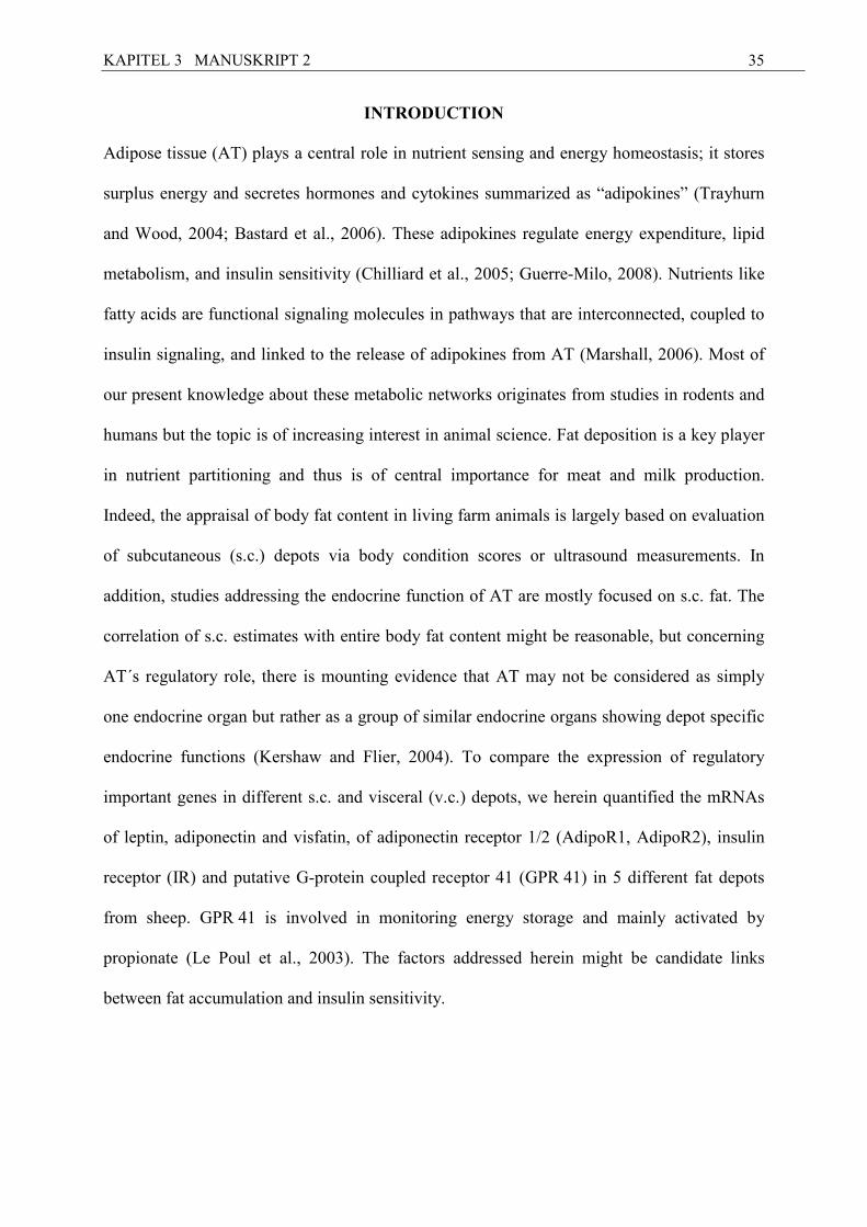
KAPITEL 3 MANUSKRIPT 2
35
I�TRODUCTIO�
Adipose tissue (AT) plays a central role in nutrient sensing and energy homeostasis; it stores
surplus energy and secretes hormones and cytokines summarized as “adipokines” (Trayhurn
and Wood, 2004; Bastard et al., 2006). These adipokines regulate energy expenditure, lipid
metabolism, and insulin sensitivity (Chilliard et al., 2005; Guerre-Milo, 2008). Nutrients like
fatty acids are functional signaling molecules in pathways that are interconnected, coupled to
insulin signaling, and linked to the release of adipokines from AT (Marshall, 2006). Most of
our present knowledge about these metabolic networks originates from studies in rodents and
humans but the topic is of increasing interest in animal science. Fat deposition is a key player
in nutrient partitioning and thus is of central importance for meat and milk production.
Indeed, the appraisal of body fat content in living farm animals is largely based on evaluation
of subcutaneous (s.c.) depots via body condition scores or ultrasound measurements. In
addition, studies addressing the endocrine function of AT are mostly focused on s.c. fat. The
correlation of s.c. estimates with entire body fat content might be reasonable, but concerning
AT´s regulatory role, there is mounting evidence that AT may not be considered as simply
one endocrine organ but rather as a group of similar endocrine organs showing depot specific
endocrine functions (Kershaw and Flier, 2004). To compare the expression of regulatory
important genes in different s.c. and visceral (v.c.) depots, we herein quantified the mRNAs
of leptin, adiponectin and visfatin, of adiponectin receptor 1/2 (AdipoR1, AdipoR2), insulin
receptor (IR) and putative G-protein coupled receptor 41 (GPR 41) in 5 different fat depots
from sheep. GPR 41 is involved in monitoring energy storage and mainly activated by
propionate (Le Poul et al., 2003). The factors addressed herein might be candidate links
between fat accumulation and insulin sensitivity.
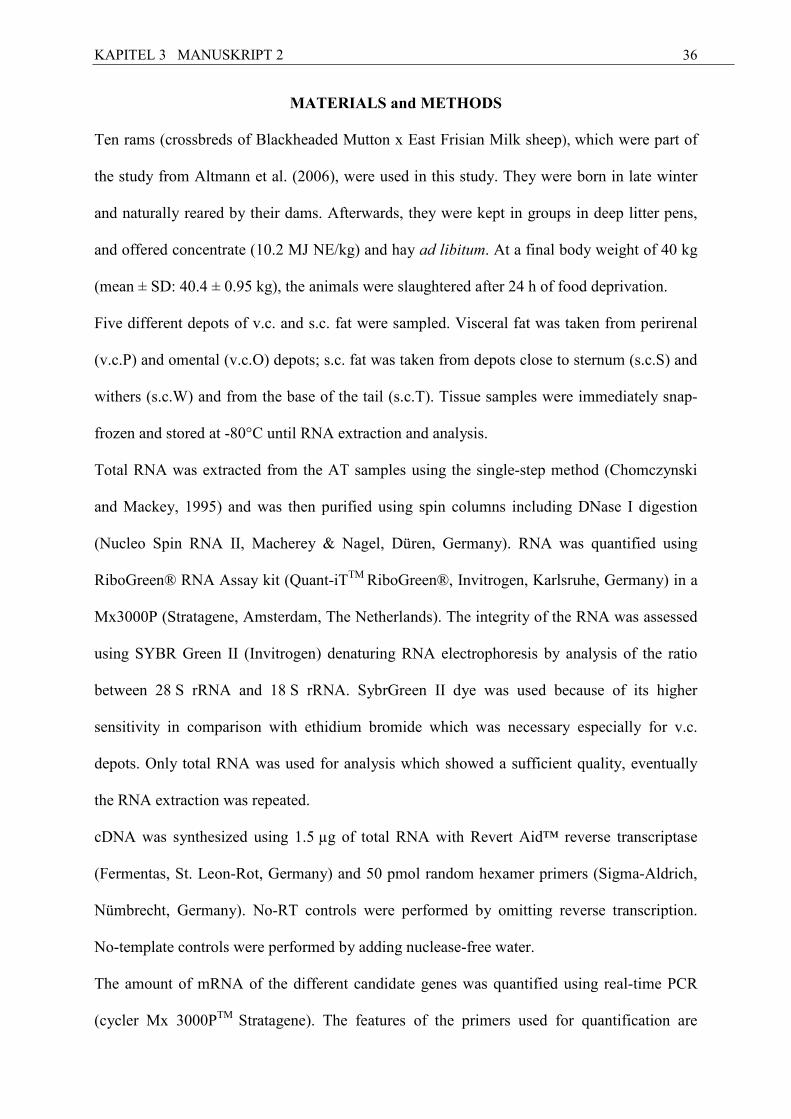
KAPITEL 3 MANUSKRIPT 2
36
MATERIALS and METHODS
Ten rams (crossbreds of Blackheaded Mutton x East Frisian Milk sheep), which were part of
the study from Altmann et al. (2006), were used in this study. They were born in late winter
and naturally reared by their dams. Afterwards, they were kept in groups in deep litter pens,
and offered concentrate (10.2 MJ NE/kg) and hay ad libitum. At a final body weight of 40 kg
(mean ± SD: 40.4 ± 0.95 kg), the animals were slaughtered after 24 h of food deprivation.
Five different depots of v.c. and s.c. fat were sampled. Visceral fat was taken from perirenal
(v.c.P) and omental (v.c.O) depots; s.c. fat was taken from depots close to sternum (s.c.S) and
withers (s.c.W) and from the base of the tail (s.c.T). Tissue samples were immediately snap-
frozen and stored at -80°C until RNA extraction and analysis.
Total RNA was extracted from the AT samples using the single-step method (Chomczynski
and Mackey, 1995) and was then purified using spin columns including DNase I digestion
(Nucleo Spin RNA II, Macherey & Nagel, Düren, Germany). RNA was quantified using
RiboGreen® RNA Assay kit (Quant-iTTM RiboGreen®, Invitrogen, Karlsruhe, Germany) in a
Mx3000P (Stratagene, Amsterdam, The Netherlands). The integrity of the RNA was assessed
using SYBR Green II (Invitrogen) denaturing RNA electrophoresis by analysis of the ratio
between 28 S rRNA and 18 S rRNA. SybrGreen II dye was used because of its higher
sensitivity in comparison with ethidium bromide which was necessary especially for v.c.
depots. Only total RNA was used for analysis which showed a sufficient quality, eventually
the RNA extraction was repeated.
cDNA was synthesized using 1.5 µg of total RNA with Revert Aid™ reverse transcriptase
(Fermentas, St. Leon-Rot, Germany) and 50 pmol random hexamer primers (Sigma-Aldrich,
Nümbrecht, Germany). No-RT controls were performed by omitting reverse transcription.
No-template controls were performed by adding nuclease-free water.
The amount of mRNA of the different candidate genes was quantified using real-time PCR
(cycler Mx 3000PTM Stratagene). The features of the primers used for quantification are
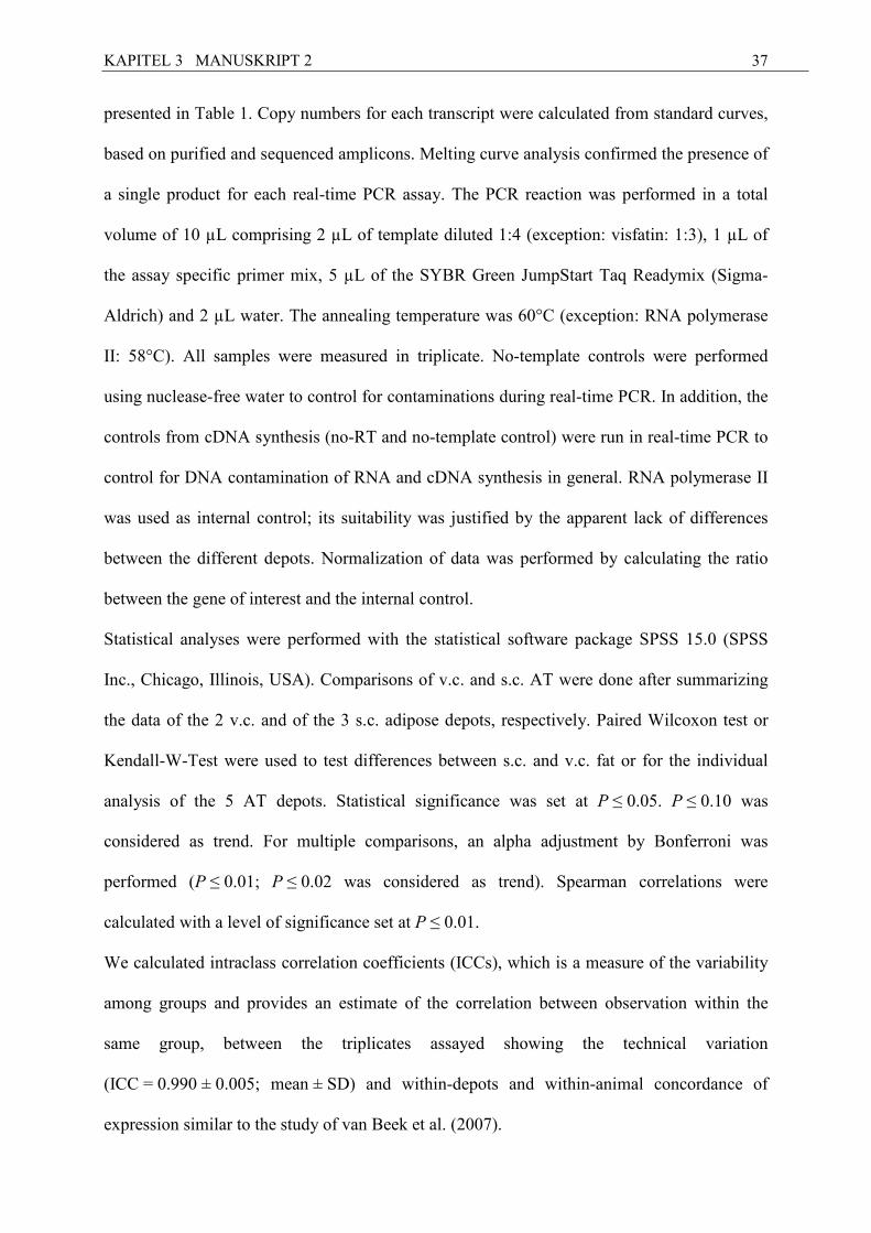
KAPITEL 3 MANUSKRIPT 2
37
presented in Table 1. Copy numbers for each transcript were calculated from standard curves,
based on purified and sequenced amplicons. Melting curve analysis confirmed the presence of
a single product for each real-time PCR assay. The PCR reaction was performed in a total
volume of 10 µL comprising 2 µL of template diluted 1:4 (exception: visfatin: 1:3), 1 µL of
the assay specific primer mix, 5 µL of the SYBR Green JumpStart Taq Readymix (Sigma-
Aldrich) and 2 µL water. The annealing temperature was 60°C (exception: RNA polymerase
II: 58°C). All samples were measured in triplicate. No-template controls were performed
using nuclease-free water to control for contaminations during real-time PCR. In addition, the
controls from cDNA synthesis (no-RT and no-template control) were run in real-time PCR to
control for DNA contamination of RNA and cDNA synthesis in general. RNA polymerase II
was used as internal control; its suitability was justified by the apparent lack of differences
between the different depots. Normalization of data was performed by calculating the ratio
between the gene of interest and the internal control.
Statistical analyses were performed with the statistical software package SPSS 15.0 (SPSS
Inc., Chicago, Illinois, USA). Comparisons of v.c. and s.c. AT were done after summarizing
the data of the 2 v.c. and of the 3 s.c. adipose depots, respectively. Paired Wilcoxon test or
Kendall-W-Test were used to test differences between s.c. and v.c. fat or for the individual
analysis of the 5 AT depots. Statistical significance was set at P ≤ 0.05. P ≤ 0.10 was
considered as trend. For multiple comparisons, an alpha adjustment by Bonferroni was
performed (P ≤ 0.01; P ≤ 0.02 was considered as trend). Spearman correlations were
calculated with a level of significance set at P ≤ 0.01.
We calculated intraclass correlation coefficients (ICCs), which is a measure of the variability
among groups and provides an estimate of the correlation between observation within the
same group, between the triplicates assayed showing the technical variation
(ICC = 0.990 ± 0.005; mean ± SD) and within-depots and within-animal concordance of
expression similar to the study of van Beek et al. (2007).
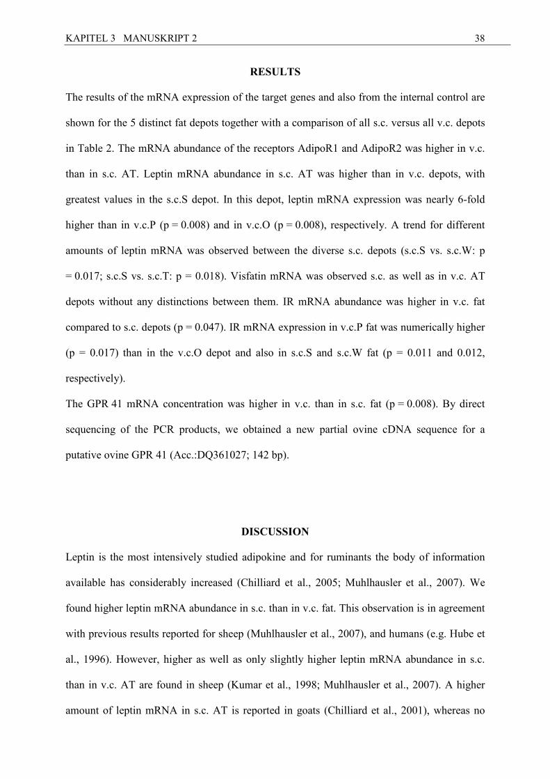
KAPITEL 3 MANUSKRIPT 2
38
RESULTS
The results of the mRNA expression of the target genes and also from the internal control are
shown for the 5 distinct fat depots together with a comparison of all s.c. versus all v.c. depots
in Table 2. The mRNA abundance of the receptors AdipoR1 and AdipoR2 was higher in v.c.
than in s.c. AT. Leptin mRNA abundance in s.c. AT was higher than in v.c. depots, with
greatest values in the s.c.S depot. In this depot, leptin mRNA expression was nearly 6-fold
higher than in v.c.P (p = 0.008) and in v.c.O (p = 0.008), respectively. A trend for different
amounts of leptin mRNA was observed between the diverse s.c. depots (s.c.S vs. s.c.W: p
= 0.017; s.c.S vs. s.c.T: p = 0.018). Visfatin mRNA was observed s.c. as well as in v.c. AT
depots without any distinctions between them. IR mRNA abundance was higher in v.c. fat
compared to s.c. depots (p = 0.047). IR mRNA expression in v.c.P fat was numerically higher
(p = 0.017) than in the v.c.O depot and also in s.c.S and s.c.W fat (p = 0.011 and 0.012,
respectively).
The GPR 41 mRNA concentration was higher in v.c. than in s.c. fat (p = 0.008). By direct
sequencing of the PCR products, we obtained a new partial ovine cDNA sequence for a
putative ovine GPR 41 (Acc.:DQ361027; 142 bp).
DISCUSSIO�
Leptin is the most intensively studied adipokine and for ruminants the body of information
available has considerably increased (Chilliard et al., 2005; Muhlhausler et al., 2007). We
found higher leptin mRNA abundance in s.c. than in v.c. fat. This observation is in agreement
with previous results reported for sheep (Muhlhausler et al., 2007), and humans (e.g. Hube et
al., 1996). However, higher as well as only slightly higher leptin mRNA abundance in s.c.
than in v.c. AT are found in sheep (Kumar et al., 1998; Muhlhausler et al., 2007). A higher
amount of leptin mRNA in s.c. AT is reported in goats (Chilliard et al., 2001), whereas no
KAPITEL 3 MANUSKRIPT 2
39
varieties between fat depots are found in cattle (e.g. Chelikani et al., 2003). The dependence
of leptin expression on fat cell size (Auwerx and Steals, 1998) and the greater sensitivity of
omental fat cells to catecholamines (Hube et al., 1996) are discussed as reasons for the
regional differences in leptin mRNA abundance in humans. Similar relationships might exist
in rodents and ruminants. Interestingly, we observed divergences between the 3 s.c. adipose
tissue depots, whereas the 2 localizations of the analyzed v.c. fat depots were not different
concerning leptin mRNA expression. The heterogeneity of the s.c. AT concerning leptin
mRNA expression is supported by the small ICC (0.018) and seems to be mainly attributable
to the s.c.S depot with the highest relative leptin mRNA abundance. The selection of several
sampling sites within the s.c. AT depots might be a reason for the inconsistent published
results on leptin mRNA expression values in s.c. and v.c. fat in ruminants and monogastric
species. This should be verified in detail by protein expression analysis in each location in
future.
No depot specific divergences, neither between the 5 fat depots, nor between s.c. and v.c.
adipose tissue were found for adiponectin mRNA expression. The ICC of 0.726 showed a
good correlation between the 5 measured fat depots. Species and age related differences in
depot specific adiponectin mRNA expression might exist. Greater adiponectin mRNA
abundance was observed in v.c. vs. s.c. fat of rats, swine and neonatal sheep, in particular in
perirenal or omental AT (Altomonte et al., 2003; Lord et al., 2005; Muhlhausler et al., 2007).
However, several studies in humans on adiponectin mRNA contents in s.c. vs. v.c. AT
revealed higher adiponectin mRNA and protein expression in s.c. compared to v.c. AT (e.g.
Nannipieri et al., 2007), or no depot varieties (Yang et al., 2003). Taken together, the depot
specific regulation of adiponectin mRNA expression is not entirely clear until now.
Compared to adiponectin itself, less is known about the expression of its receptors in diverse
fat depots. We observed higher mRNA expression in the subsummarized v.c. depots than in
the s.c. ones, but when comparing the individual depots separately, no differences were
KAPITEL 3 MANUSKRIPT 2
40
evident. The ICCs calculated for AdipoR1 and for AdipoR2 were rather low (0.18 and 0.29,
respectively) between the 5 measured fat depots, suggesting heterogeneity between the
measured depots.
In contrast to our results, both adiponectin receptors were observed to be higher expressed in
s.c. than in v.c. fat in humans (Nannipieri et al., 2007). Comparing the amounts of AdipoR1
and Adipo2 mRNA, we found a roughly 6-fold higher abundance of AdipoR2 mRNA in both,
s.c. and v.c. fat. This distinction is in line with earlier observations we made in bovine s.c. AT
(Lemor et al., 2009).
Visfatin has recently been identified as a v.c. fat-specific adipokine probably linking the
expansion of this depot to insulin resistance (Sethi and Vidal-Puig, 2005). It is known that
visfatin mRNA is expressed in bovine mammary epithelial cells and in the mammary gland,
and the protein is present in milk (Yonezawa et al., 2006). Visfatin mRNA is also present in
s.c. AT of cattle (Lemor et al., 2009). Concerning depot-specific varieties, the results
published are conflicting and are particularly limited to humans so far. Abundant visfatin
mRNA expression in v.c. fat (Fukuhara et al., 2005), as well as a higher visfatin mRNA
content in s.c. fat (Pagano et al., 2006), or no differences in expression between s.c. and v.c.
fat (Berndt et al., 2005), analogous to our study were reported.
We found a higher IR mRNA abundance in v.c. AT compared to s.c. fat pointing to a
differentially regulated insulin sensitivity of disparate AT depots. This might be explained by
increased insulin stimulated glucose uptake of v.c. AT in comparison to s.c. AT (Virtanen et
al., 2002).
For the first time a partial mRNA sequence of a putative GPR 41 was described in sheep. Its
expression was found in all analyzed ovine fat depots, thus confirming data from v.c. and s.c.
fat in other ruminant species (e.g. Mielenz et al., 2008; Lemor et al., 2009). Besides AT,
GPR 41 mRNA is expressed in several other bovine tissue (Wang et al., 2009; Yonezava et
al., 2009). We found a 2-fold higher putative GPR 41 mRNA abundance in v.c. compared to
KAPITEL 3 MANUSKRIPT 2
41
s.c AT. It is known that energy storage is differentially regulated between s.c. vs. v.c. fat
depots (Wajchenberg, 2000) and GPR 41 might also play a role in sheep as indicated by the
higher abundance in v.c. than in s.c. AT.
In summary, we observed divergences in the mRNA abundance of AdipoR1 and AdipoR2,
leptin, IR and GPR 41 between the subsummarized depots of v.c. or s.c. origin, and in part,
between the 5 individual depots. Besides, to the continued need for protein data and
expansion to animals showing a wider range of body fat contents, our results emphasize the
importance of sampling site when aiming to characterize the regulatory role of adipose tissue.
In addition, the depot specific expression observed in our study with sheep of comparable
physiological status might also be differentially affected during the dynamics of homeostatic
and homoerhetic adaptation.
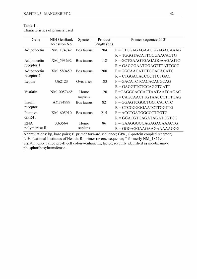
KAPITEL 3 MANUSKRIPT 2
42
Table 1. Characteristics of primers used
Gene NIH GenBank accession No.
Species Product length (bp)
Primer sequence 5’-3’
F = CTGGAGAGAAGGGAGAGAAAG Adiponectin NM_174742 Bos taurus 204
R = TGGGTACATTGGGAACAGTG F = GCTGAAGTGAGAGGAAGAGTC Adiponectin
receptor 1 XM_593692 Bos taurus 118
R = GAGGGAATGGAGTTTATTGCC
F = GGCAACATCTGGACACATC Adiponectin receptor 2
XM_580459 Bos taurus 200 R = CTGGAGACCCCTTCTGAG F = GACATCTCACACACGCAG Leptin U62123 Ovis aries 183 R = GAGGTTCTCCAGGTCATT
F =CAGGCACCACTAATAATCAGAC Visfatin NM_005746* Homo sapiens
120 R = CAGCAACTTGTAACCCTTTGAG F = GGAGTCGGCTGGTCATCTC Insulin
receptor AY574999 Bos taurus 82
R = CTCGGGGGAATCTTGGTTG
F = ACCTGATGGCCCTGGTG Putative GPR41
XM_605910 Bos taurus 215
R = GGACGTGAGATAGATGGTGG F = GAAGGGGGAGAGACAAACTG RNA
polymerase II X63564 Homo
sapiens 86
R = GGGAGGAAGAAGAAAAAGGG
Abbreviations: bp, base pairs; F, primer forward sequence; GPR, G-protein coupled receptor; NIH, National Institutes of Health; R, primer reverse sequence; * formerly NM_182790; visfatin, once called pre-B cell colony-enhancing factor, recently identified as nicotinamide phosphoribosyltransferase.
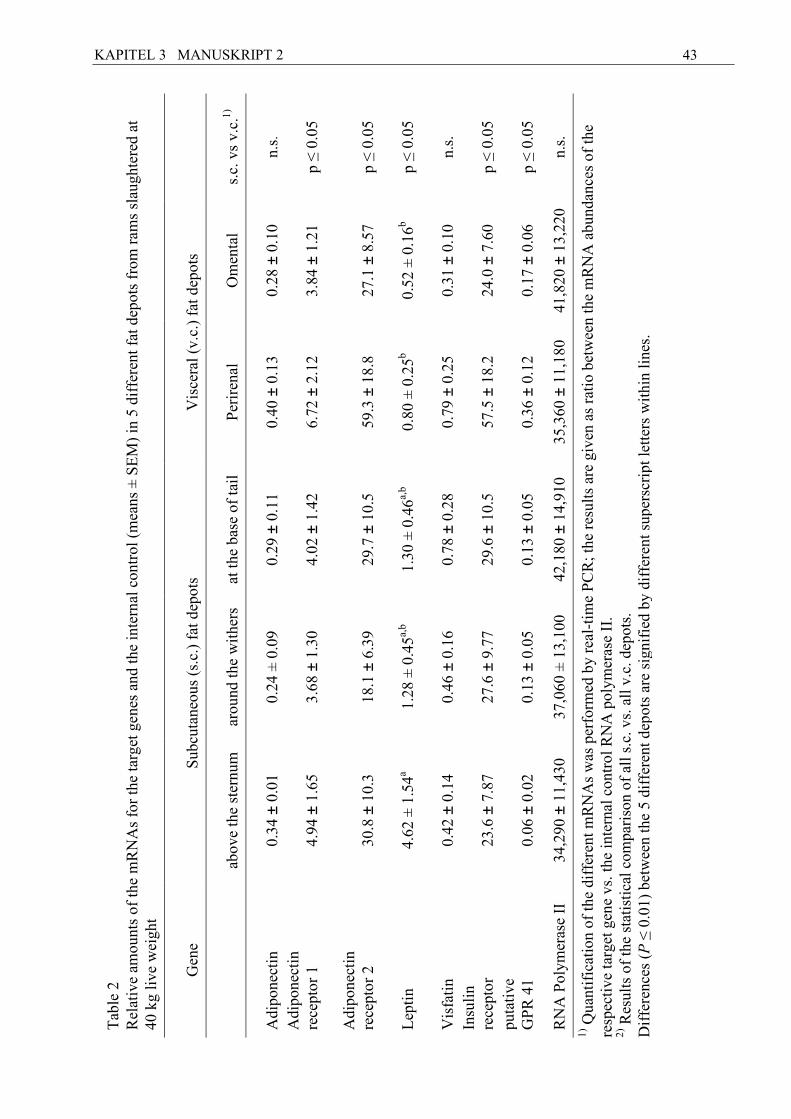
KAPITEL 3 MANUSKRIPT 2
43
s.c.
vs
v.c.
1)
n.s.
p ≤
0.0
5
p ≤
0.0
5
p ≤
0.0
5
n.s.
p ≤
0.0
5
p ≤
0.0
5
n.s.
Om
enta
l
0.28
± 0
.10
3.84
± 1
.21
27.1
± 8
.57
0.52
± 0
.16b
0.31
± 0
.10
24.0
± 7
.60
0.17
± 0
.06
41,8
20 ±
13,
220
Vis
cera
l (v
.c.)
fat
dep
ots
Per
iren
al
0.40
± 0
.13
6.72
± 2
.12
59.3
± 1
8.8
0.80
± 0
.25b
0.79
± 0
.25
57.5
± 1
8.2
0.36
± 0
.12
35,3
60 ±
11,
180
at t
he b
ase
of t
ail
0.29
± 0
.11
4.02
± 1
.42
29.7
± 1
0.5
1.30
± 0
.46a,
b
0.78
± 0
.28
29.6
± 1
0.5
0.13
± 0
.05
42,1
80 ±
14,
910
arou
nd t
he w
ithe
rs
0.24
± 0
.09
3.68
± 1
.30
18.1
± 6
.39
1.28
± 0
.45a,
b
0.46
± 0
.16
27.6
± 9
.77
0.13
± 0
.05
37,0
60 ±
13,
100
Sub
cuta
neou
s (s
.c.)
fat
dep
ots
abov
e th
e st
ernu
m
0.34
± 0
.01
4.94
± 1
.65
30.8
± 1
0.3
4.62
± 1
.54a
0.42
± 0
.14
23.6
± 7
.87
0.06
± 0
.02
34,2
90 ±
11,
430
Tab
le 2
R
elat
ive
amou
nts
of t
he m
RN
As
for
the
targ
et g
enes
and
the
int
erna
l co
ntro
l (m
eans
± S
EM
) in
5 d
iffe
rent
fat
dep
ots
from
ram
s sl
augh
tere
d at
40
kg
live
wei
ght
Gen
e
Adi
pone
ctin
Adi
pone
ctin
re
cept
or 1
Adi
pone
ctin
re
cept
or 2
Lep
tin
Vis
fati
n
Insu
lin
rece
ptor
puta
tive
G
PR
41
RN
A P
olym
eras
e II
1) Q
uant
ific
atio
n of
the
dif
fere
nt m
RN
As
was
per
form
ed b
y re
al-t
ime
PC
R; t
he r
esul
ts a
re g
iven
as
rati
o be
twee
n th
e m
RN
A a
bund
ance
s of
the
re
spec
tive
tar
get
gene
vs.
the
int
erna
l co
ntro
l R
NA
pol
ymer
ase
II.
2) R
esul
ts o
f th
e st
atis
tica
l co
mpa
riso
n of
all
s.c
. vs.
all
v.c
. dep
ots.
D
iffe
renc
es (
P ≤
0.0
1) b
etw
een
the
5 di
ffer
ent
depo
ts a
re s
igni
fied
by
diff
eren
t su
pers
crip
t le
tter
s w
ithi
n li
nes.
KAPITEL 3 MANUSKRIPT 2
44
References
Altmann, M.; Sauerwein H.; von Borell E., 2006: The relationships between leptin
concentrations and body fat reserves in lambs are reduced by short-term fasting. Journal of
Animal Physiology and Animal Nutrition 90, 407-13.
Altomonte, J.; Harbaran S.; Richter A.; Don H., 2003: Fat depot-specific expression of
adiponectin is impaired in zucker fatty rats. Metabolism 52, 958-963.
Auwerx, J.; Steals B., 1998: Leptin. The Lancet 351, 737-742.
Bastard, J.P.; Maachi M.; Lagathu C.; Kim M.J.; Caro M.;, Vidal H.; Capeau J.; Feve B.,
2006: Recent advances in the relationship between obesity, inflammation, and insulin
resistance. European Cytokine Network 17, 4-12.
Van Beek, E.A.; Bakker A.H.; Kruyt P.M.; Hofker M.H.; Saris W.H.; Keijer J., 2007: Intra-
and interindividual variation in gene expression in human adipose tissue. Pflugers Archive –
European Journal of Physiology 453, 851-861.
Berndt, J., Klöting N., Kralisch S.; Kovacs P.; Fasshauer M.;, Schön M.R.; Stumvoll M.; and
Blüher M., 2005: Plasma visfatin concentrations and fat depot-specific mRNA expression in
humans. Diabetes 54, 2911-2916.
Chelikani, P.K.; Glimm D.R.; Kennelly J.J., 2003: Short communication: Tissue distribution
of leptin and leptin receptor mRNA in the bovine. Journal of Dairy Science 86, 2369-2372.
KAPITEL 3 MANUSKRIPT 2
45
Chilliard, Y.; Bonnet M.; Delavaud C.; Faulconnier Y.; Leroux C.; Djiane J.;F. Bocquier F.,
2001: Leptin in ruminants. Gene expression in adipose tissue and mammary gland, and
regulation of plasma concentration. Domestic Animal Endocrinology 21, 271-295.
Chilliard, Y.; Delavaud C.; Bonnet M., 2005: Leptin expression in ruminants: Nutritional and
physiological regulations in relation with energy metabolism. Domestic Animal
Endocrinology 29, 3-22.
Chomczynski, P.; Mackey K., 1995: Substitution of chloroform by bromo-chloropropane in
the single-step method of RNA isolation. Analytical Biochemistry 225, 163-164.
Fukuhara, A.; Matsuda M.; Nishizawa M.; Segawa K.; Tanaka M.; Kishimoto K.; Matsuki Y.;
Murakami M.; Ichisaka T.; Murakami H.; Watanabe E.; Takagi T.; Akiyoshi M.; Ohtsubo T.;
Kihara S.; Yamashita S.; Makishima M.; Funahashi T.; Yamanaka S.; Hiramatsu R.;
Matsuzawa Y.; Shimornura I., 2005: Visfatin: a protein secreted by visceral fat that mimics
the effect of insulin. Science 307, 426-430.
Guerre-Millo, M., 2008: Adiponectin: an update. Diabetes and Metabolism 34, 12-18.
Hube, F.; Lietz U.; Igel M.; Jensen P.B.; Tornqvist H.; Joost H.G.; Hauner H., 1996:
Difference in leptin mRNA levels between omental and subcutaneous abdominal adipose
tissue from obese humans. Hormone and Metabolic Research 28, 690-693.
Kershaw, E.E.; Flier J.S., 2004: Adipose tissue as an endocrine organ. Journal of Clinical
Endocrinology and Metabolism 89, 2548-2556.
KAPITEL 3 MANUSKRIPT 2
46
Kumar, B., Francis S.M.; Suttie J.M.; Thompson M.P., 1998: Expression of obese mRNA in
genetically lean and fat selection lines of sheep. Comparative Biochemistry and Physiology -
Part B Biochemistry and Molecular Biology 120, 543-548.
Lemor, A.; Hosseini A.; Sauerwein H.; Mielenz M., 2009: Transition period-related changes
in the abundance of the mRNAs of adiponectin and its receptors, of visfatin, and of fatty acid
binding receptors in adipose tissue of high-yielding dairy cows. Domestic Animal
Endocrinology 37, 37-44.
Le Poul, E.; Loison C.; Struyf S.;Springael J.Y.; Lannoy V.; Decobecq M.E.; Brezillon S.;
Dupriez V.; Vassart G.; Van Damme J.; Parmentier M.; Detheux M., 2003: Functional
characterization of human receptors for short chain fatty acids and their role in
polymorphonuclear cell activation. Journal of Biological Chemistry 278, 25481-25489.
Lord, E.; Ledoux S.; Murphy B.D.; Beaudry D.; Palin M.F., 2005: Expression of adiponectin
and its receptors in swine. Journal of Animal Science 83, 565-578.
Marshall, S., 2006: Role of insulin, adipocyte hormones, and nutrient-sensing pathways in
regulating fuel metabolism and energy homeostasis: a nutritional perspective of diabetes,
obesity, and cancer. Sciences STKE. 346:re7.
Mielenz, M.; Seybold C.; Sauerwein H., 2008: Effects of short-term infusion with propionate
on the mRNA expression of a putative G-protein coupled receptor 41 (GPR41) in adipose
tissue of goats. Livestock Science 116, 328-331.
KAPITEL 3 MANUSKRIPT 2
47
Muhlhausler, B.S.; Duffield J.A.; McMillen I.C., 2007: Increased maternal nutrition increases
leptin expression in perirenal and subcutaneous adipose tissue in the postnatal lamb.
Endocrinology 148, 6157-6163.
Nannipieri, M.; Bonotti A.; Anselmino M.; Cecchetti F.; Madec S.; Mancini E.; Baldi S.;
Santini F.; Pinchera A.; Rossi M.; Ferrannini E., 2007: Pattern of expression of adiponectin
receptors in human adipose tissue depots and its relation to the metabolic state. International
Journal of Obesity 31, 1843-1848.
Pagano, C.; Pilon C.; Olivieri M.; Mason P.; Fabris R.; Serra R.; Milan G.; Rossato M.;
Federspil G.; Vettor R., 2006: Reduced plasma visfatin/pre-B cell colony-enhancing factor in
obesity is not related to insulin resistance in humans. Journal of Clinical Endocrinology and
Metabolism 91, 3165-3170.
Sethi, J.K.; Vidal-Puig A., 2005: Visfatin: the missing link between intra-abdominal obesity
and diabetes? Trends in Molecular Medicine 11, 344-347.
Trayhurn, P.;Wood I.S., 2004: Adipokines: inflammation and the pleiotropic role of white
adipose tissue. British Journal of Nutrition 92, 347-355.
Virtanen, K.A.; Lönnroth P.; Parkkola R.; Peltoniemi P.; Asola M.; Viljanen T.; Tolvanen T.;
Knuuti J.; Rönnemaa T.; Huupponen R.; Nuutila P., 2002: Glucose uptake and perfusion in
subcutaneous and visceral adipose tissue during insulin stimulation in nonobese and obese
humans. Journal of Clinical Endocrinology and Metabolism 87, 3902-3910.
KAPITEL 3 MANUSKRIPT 2
48
Wajchenberg, B.E., 2000: Subcutaneous and visceral adipose tissue: Their relation to the
metabolic syndrome. Endocrine Reviews 21, 697-738.
Wang, A., Gu Z., Heid B., Akers R.M., Jiang H., 2009: Identification and characterization of
the bovine G protein-coupled receptor GPR41 and GPR43 genes. Journal of Dairy Science
92, 2696-2705.
Yang, W.S.; Chen M.H.; Lee W.J.; Lee K.C.; Chao C.L.; Huang K.C.; Chen C.L.; Tai T.Y.;
Chuang L.M., 2003: Adiponectin mRNA levels in the abdominal adipose depots of
nondiabetic women. International Journal of Obesity 27, 896-900.
Yonezawa, T.; Haga S.; Kobayashi Y.; Takahashi T.; Obara Y., 2006: Visfatin is present in
bovine mammary epithelial cells, lactating mammary gland and milk, and its expression is
regulated by cAMP pathway. FEBS Letters 580, 6635-6643.
Yonezawa, T.; Haga S.; Kobayashi Y.; Katoh K.; Obara Y., 2009: Short-chain fatty acid
signaling pathways in bovine mammary epithelial cells. Regulatory Peptides 153, 30-36.
KAPITEL 4 SCHLUSSFOLGERUNGEN und AUSBLICK
49
Kapitel 4
Schlussfolgerungen und Ausblick Die Untersuchungen der mRNA-Expression verschiedener Adipokine im s.c. Fettgewebe a.p.
und p.p. beim Rind haben Unterschiede bezüglich ihres mRNA-Expressionsniveaus zwischen
den untersuchten Beprobungszeitpunkten ergeben. Aufgrund dieser Ergebnisse der
reduzierten mRNA-Expression beider Adiponectinrezeptoren p.p. schlussfolgern wir eine
geringere Adiponectinsensitivität auf Ebene des Fettgewebes p.p. beim Rind. Es ist bekannt,
dass Adiponectin positiv auf die Insulinsensitivität über die Aktivierung von AMPK wirkt
(Kadowaki et al., 2003; Yamauchi et al., 2002). Da zu Beginn der Laktation eine reduzierte
Insulinsensibilität vorteilhaft in Bezug auf die erleichterte Energiebereitstellung aus der
Fettmobilisation ist, passt der Baustein einer verringerten Adiponektinsensibilität p.p. in die
physiologischen Zusammenhänge der Transitionsphase. Zusammen mit den Ergebnissen der
Studie über die Leber vom Rind von Loor und Kollegen (2006) ergibt sich aufgrund der
unterschiedlichen mRNA-Expression der Adiponectinrezeptoren während der
Transitionsphase die Vermutung einer unterschiedlichen Regulation dieser Faktoren zwischen
Fettgewebe und Leber. Weitere Untersuchungen zu diesen Zusammenhängen, auch auf
Proteinebene, sind in Zukunft erforderlich, um diese Verknüpfungen aufzuklären.
Der Einfluss des Visfatins auf die Insulinsignalwege und die Glukoseaufnahme ist beim
Monogastrier als eher nebensächlich eingeschätzt worden, da die Visfatinblutkonzentration
im Vergleich zur Insulinblutkonzentration nur gering ist. Unsere Ergebnisse deuten darauf
hin, dass diese Rolle des Visfatins, das ein Ligand des Insulinrezeptors (IR) ist, zumindest
beim Wiederkäuer, doch von größerer Bedeutung sein könnte, da die von uns angenommenen
erhöhten Visfatinplasmakonzentrationen mit der reduzierten Insulinsensitivität
zusammenfallen. Zur Klärung des genauen Einflusses des Visfatins insbesondere auf die
Insulinsignalwege beim Wiederkäuer sind weiterführende Studien auch auf Proteinebene
erforderlich.
Wir konnten sowohl die mRNA der fettsäurebindenden Rezeptoren GPR 41 und die GPR 43
sowie die des putativen BHB- und Nikotinsäurebindenden Rezeptors GPR 109A im bovinen
s.c. Fettgewebe und in bovinen Leukozyten nachweisen. Der Nachweis der mRNA-
Expression von GPR 41 und 43 im bovinen Fettgewebe steht im Kontrast zu den Ergebnissen
von Wang und Kollegen (2009), die beide Rezeptoren mRNA in verschiedensten bovinen
Geweben, jedoch die GPR 43 mRNA nicht und die GPR 41 mRNA im Fettgewebe nur sehr
schwach, nachweisen konnten. Während der Transitionsphase haben wir Veränderungen
KAPITEL 4 SCHLUSSFOLGERUNGEN und AUSBLICK
50
bezüglich der mRNA-Expression des GPR 41 und des putativen GPR 109A im Fettgewebe
beobachtet, doch bisher ist noch vollkommen unbekannt, welche Rolle diese Regulation
spielen könnte, dies sollte daher in zukünftigen Studien intensiv untersucht werden.
Unsere Ergebnisse von unterschiedlichen mRNA-Expressionen verschiedener Adipokine,
deren Rezeptoren und des GPR 41 und des putativen GPR 109A während der
Transitionsphase beim Rind können darauf hinweisen, dass es nach weiteren Studien möglich
sein könnte, gefährdete d.h. ungenügend adaptierte Tiere frühzeitig zu erkennen und im
Folgenden Erkrankungen zu verhindern. Die Ergebnisse unserer Studie über die mRNA-
Expression ausgewählter Adipokine und Rezeptoren in verschiedenen Fettdepots beim
kleinen Wiederkäuer Schaf deuten auf individuelle mRNA-Expression der untersuchten
Faktoren in den Depots hin. Bisher ist beim Rind in diesem Zusammenhang nur wenig
bekannt, so dass sich zukünftige Studien auch mit den unterschiedlichen Fettgeweben und
deren Regulation beim Rind beschäftigen sollten.
KAPITEL 5 ZUSAMMENFASSUNG
51
Kapitel 5
Zusammenfassung
Anneka Lemor
Untersuchungen zur mRNA-Expression von Adiponectin und anderen Adipokinen,
ihren Rezeptoren sowie von fettsäurebindenden Rezeptoren im Fettgewebe von Wiederkäuern
In der Milchviehhaltung ist aus gesundheitlichen Gründen das erste Drittel der Laktation als
problematisch zu betrachten, da das bis zu einem bestimmten Grad physiologische
Energiedefizit zu lang anhaltenden, katabolen Stoffwechselveränderungen führt und
suppressiv auf die systemische Abwehr wirkt. Infolge kann es zu Stoffwechsel- und
Infektionskrankheiten, reduzierter Milchproduktion und zu Fruchtbarkeitsstörungen kommen.
Das Fettgewebe, das in dieser Zeit zur Gewährleistung des erhöhten Energiebedarfs der
Milchdrüse einem verstärkten Abbau unterworfen ist, hat dabei nicht nur als Energiespeicher
Bedeutung für den Gesamtorganismus; es nimmt darüber hinaus über die dort produzierten
Hormone und Zytokine (die als Adipokine zusammengefasst werden) Einfluss auf diverse
Körperfunktionen. Das Ziel der vorliegenden Studien war die mRNA-Expression
verschiedener, für den Energiehaushalt, Fettstoffwechsel und die Glukosehomöostase
relevante Adipokine und Rezeptoren im Fettgewebe von Wiederkäuern zu erfassen. Bei den
untersuchten Adipokinen handelte es sich um Adiponectin, Leptin und Visfatin, sowie deren
Rezeptoren Adiponectinrezeptor 1 und 2 (AdipoR1/2), die lange Isoform des Leptinrezeptors
(ObRb) und den Insulinrezeptor (IR). Des Weiteren wurde die mRNA-Expression der G-
Protein gekoppelten Rezeptoren (GPR) für kurzkettige Fettsäuren, d.h. für GPR 41 und
GPR 43, sowie des Rezeptors für Nikotinsäure und ß-Hydroxybutyrat, GPR 109A, gemessen.
Für die verschiedenen mRNAs wurden geeignete Protokolle zur validen Quantifizierung
etabliert. Die Studien wurden zum Einen im subkutanen Fettgewebe von Milchkühen
durchgeführt, wobei die Expressionswerte vor und nach der Geburt verglichen wurden. Ob
andere Fettdepots analoge Reaktionen zeigen, konnte an den Kühen nicht untersucht werden,
weshalb zunächst am Schaf als Wiederkäuermodell die Expression der genannten Faktoren in
verschiedenen viszeralen und subkutanen Fettdepots verglichen wurde.
Für die Untersuchungen an Milchkühen wurden subcutane Fettproben etwa eine Woche ante
partum (a.p.) sowie ca. drei Wochen post partum (p.p.) aus der Schwanzregion entnommen
KAPITEL 5 ZUSAMMENFASSUNG
52
und mittels Echtzeit-RT-PCR untersucht. Die mRNA-Expression beider
Adiponectinrezeptoren war nach der Kalbung im Vergleich zu a.p. reduziert; dies deutet auf
eine reduzierte Adiponectinsensitivität des Fettgewebes p.p. hin, die mit der in diesem
Zeitraum bekanntermaßen sinkenden Insulinsensitivität zusammen fällt. Für die mRNA-
Expression von Visfatin und putativem GPR 109A war p.p. im Vergleich zu a.p. eine
Tendenz (P = 0.074 bzw. P = 0.092) zu absinkenden Werten zu verzeichnen. Dagegen war die
mRNA-Expression von GPR 41 und ObRb p.p. verglichen mit a.p. erhöht. Für Visfatin,
GPR 41 und GPR 109A sind modulierende Effekte auf die Insulinsensitivität anzunehmen.
Für den Vergleich verschiedener Fettdepots wurden die Untersuchungen auf Mastlämmer
ausgedehnt und drei verschiedene subkutane sowie zwei viszerale Depots hinsichtlich der
mRNA-Expression der oben genannten Faktoren erfasst. Bei den subkutanen Depots handelte
es sich um Fett aus den Bereichen des Brustbeins, des Widerrists und der Schwanzbasis; bei
den viszeralen Lokalisationen wurden das perirenale und das mesenteriale Depot beprobt. Die
mRNA-Expression beider Adiponectinrezeptoren sowie des putativen GPR 41 und des IR war
in den viszeralen Depots höher als in den subkutanen. Aufgrund der tendenziellen
Unterschiede der Leptin mRNA-Expression innerhalb der subkutanen Depots, sollte in
künftigen Untersuchungen nicht zwingend von einer Homogenität des gesamten subkutanen
Fettgewebes in der basalen und der regulativ veränderten Expression ausgegangen werden.
Darüber hinaus sollten, um Aussagen für den Gesamtorganismus treffen zu können,
weiterführende Untersuchungen zur regulativen Bedeutung des Fettgewebes nicht auf die
subkutanen Depots beschränkt bleiben.
Die hier gezeigten Ergebnisse dienen als Basisarbeit für weitere Untersuchungen; sie belegen,
dass die mRNA verschiedener Adipokine und Rezeptoren in der Transitionsphase oder auch
zwischen den verschiedenen Fettdepots verschieden exprimiert wird. Fortführende Studien
sollten weitere Adipokine einschließen, auch die Proteinexpression dieser Faktoren erfassen
sowie nicht nur - wie gegenwärtig - physiologische Situationen charakterisieren, sondern auch
Veränderungen, die als pathologisch einzuordnen sind, umfassen.
KAPITEL 6 SUMMARY
53
Kapitel 6
Summary
Anneka Lemor
Studies about the mRNA expression of adiponectin and other adipokines, their receptors as
well as fatty acid binding receptors in adipose tissue in ruminants
In dairy farming, the first third of lactation is a critical period during which the incidence of
health distubances might be increased. The negative energy balance that is physiological to a
certain degree during early lactation leads to catabolism and immunosuppression. In
consequence, metabolic and infecitious diseases, reproductive disorders, and reduced milk
production might occur. The increased energy requirement of the mammary gland in early
lactation is fueled to a major extent by lipolysis; adipose tissue functions not only as energy
storage but also acts as an endocrine gland. Adipose tissue synthesizes and secretes various
hormones and cytokines (summarized as adipokines), and thereby affects several bodily
functions. The aim of the present study was to compare the mRNA expression of adipokines
and receptors involved in lipid metabolism and glucose homeostasis in adipose tissue of
ruminants. The measured adipokines were adiponectin, leptin, and visfatin, as well as the
adiponectin receptors 1 and 2 (AdipoR1/2), the long form of the leptin receptor (obRb), and
the insulin receptor (IR). In addition, the mRNA expression of the G-protein coupled
receptors (GPRs) for short chain fatty acids, GPR 41 and GPR 43, as well as the receptor for
nicotinic acid and ß-hydroxybutyric acid, GPR 109A were quantified. For these mRNA
quantifications, adequate protocols were established. The investigations were on the one hand
side performed in subcutaneous adipose tissue of dairy cows to compare the mRNA
abundances before and after calving. To clarify the expression in fat depots other than
subcutaneous fat, extended studies could not be done in cows. Therefore, the respective
investigations using several subcutaneous and visceral fat depots were done in another
ruminant species, i.e. in sheep.
For the study in dairy cows, biopsies from subcutaneous fat from the tailhead were obtained
from dairy cows one week antepartum (a.p.) and three weeks postpartum (p.p.), and the
mRNA abundance of the aforementioned factors was quantified using real-time PCR. The
mRNA expression of both adiponectin receptors was lower in p.p. samples than in samples
collected a.p. and thus indicates decreasing adiponectin sensitivity in adipose tissue after
KAPITEL 6 SUMMARY
54
calving. This reduced adiponectin sensitivity coincides with the knowingly decreased insulin
sensitivity in early lactation. For visfatin and putative GPR 109A, a trend for a decrease
(P = 0.074 and P = 0.092, respectively) p.p. compared with a.p. samples, and increasing
mRNA abundances for GPR 41 and obRb in p.p. samples contrasted with those taken a.p. was
observed. For visfatin, GPR 41, as well as for GPR 109A, modulating effects on insulin
sensitivity are assumed.
For the comparison of different fat depots, the study was extended to sheep. The mRNA
abundances of the aforementioned factors were measured in three subcutaneous and two
visceral depots. The subcutaneous depots were close to sternum, close to withers, and at the
base of the tail; in addition, two visceral depots, perirenal and omental, were sampled. The
mRNAs of both adiponectin receptors, as well as IR and putative GPR 41, were higher
expressed in visceral than in subcutaneous fat. Because of the trend for a differential mRNA
abundance of leptin inside the subcutaneous fat depots, homogeneity of the subcutaneous fat
depot can not necessarily be taken for granted. Moreover, for statements about the entire
organism, further studies about the regulatory importance of adipose tissue, should not be
limited to subcutaneous fat.
The present thesis serves as a basis for further studies showing divergent mRNA abundance
of several adipokines and receptors during the transition from pregnancy to lactation and also
between different fat depots in ruminants. Future studies should include further adipokines
and should also characterize these factors at the protein level. Moreover, not only
physiological situations – like in this study – but also pathological alterations should be
comprised.
LITERATURVERZEICHNIS
55
Literaturverzeichnis für den deutschsprachigen Teil der Arbeit Altmann M, Sauerwein H, von Borell E. The relationships between leptin concentrations and body fat reserves in lambs are reduced by short-term fasting. J Anim Physiol Anim Nutr 2006;90:407-13. Ahima RS, Flier JS. Leptin. Annu Rev Physiol 2000;62:413-37. Antuna-Puente B, Feve B, Fellahi S, Bastard J-P. Adipokines: The missing link between insulin resistance and obesity. Diabetes Metab, 2008;34:2-11. Arita Y, Kihara S, Ouchi N, Takahashi M, Maeda K, Miyagawa J, Hotta K, Shimomura I, Nakamura T, Miyaoka K, Kuriyama H, Nishida M, Yamashita S, Okubo K, Matsubara K, Muraguchi M, Ohmoto Y, Funahashi T, Matsuzawa Y. Paradoxical decrease of an adipose-specific protein, adiponectin, in obesity. Biochem Biophys Res Commun 1999;257:79-83. Anubhuti, Arora S. Leptin and its metabolic interactions – an update. Diabetes Obes Metab 2008;10:973-93. Bell AW. Regulation of organic nutrient metabolism during transition from late pregnancy to early lactation. J Anim Sci 1995;73:2804-19. Berg EP, McFadin EL, Maddock KR, Goodwin RN, Baas TJ, Keisler DH. Serum concentrations of leptin in six genetic lines of swine and relationship with growth and carcass characteristics. J Anim Sci 2003;81:167-71. Bergman EN. Energy contributions of volatile fatty acids from the gastrointestinal tract in various species. Physiol Rev 1990;70:567-90. Bjorntorp P. Metabolic implications of body fat distribution. Diabetes Care 1991;14:1132-43. Blache D, Tellam RL, Chagas LM, Blackberry MA, Vercoe PE, Martin GB. Level of nutrition affects leptin concentrations in plasma and cerebrospinal fluid in sheep. J Endocrinol 2000;165:625-37. Bispham J, Budge H, Mostyn A, Dandrea J, Clarke L, Keisler DH, Symonds ME, Stephenson T. Ambient temperature, maternal dexamethasone, and postnatal ontogeny of leptin in the neonatal lamb. Pediatr Res 2002;52:85-90. Boisclair YR, Wesolowski SR, Kim JW, Ehrhardt RA. Roles of growth hormone and leptin in the periparturient dairy cow. In: Ruminant Physiology: Digestion, metabolism, and impact of nutrition on gene expression, immunology, and stress, Sejrsen K, Hvelplund T, Nielsen MO (Eds), Wageningen Academic Publishers, 2006, pp. 327-46. Breves G, Rodehutscord M. Gibt es Grenzen in der Zucht auf Milchleistung? – Aus der Sicht der Physiologie -. In: 27. Viehwirtschaftliche Fachtagung, Bundesanstalt für alpenländische Landwirtschaft 2000, Gumpenstien, Österreich, 1-4. Brown AJ, Goldsworthy SM, Barnes AA, Eilert MM, Tcheang L, Daniels D, Muir AI, Wigglesworth MJ, Kinghorn I, Fraser NJ, Pike NB, Strum JC, Steplewski KM, Murdock PR,
LITERATURVERZEICHNIS
56
Holder JC, Marshall FH, Szekeres PG, Wilson S, Ignar DM, Foord SM, Wise A, Dowell SJ. The orphan G protein-coupled receptors GPR41 and GPR43 are activated by propionate and other short chain carboxylic acids. J Biol Chem 2003;278:11312-19. Brown AJ, Jupe S, Briscoe CP. A family of fatty acid binding receptors. DNA Cell Biol 2005;24:54-61. Bruun JM, Lihn AS, Verdich C, Pedersen SB, Toubro S, Astrup A, Richelsen B. Regulation of adiponectin by adipose tissue-derived cytokines: in vivo and in vitro investigations in humans. Am J Physiol Endocrinol Metab 2003;285:E527-E533. Chelikani PK, Glimm DR, Kennelly JJ. Short Communication: Tissue distribution of leptin and leptin receptor mRNA in the bovine. J Dairy Sci 2003:86:2369-72. Chua SC Jr, Koutras IK, Han L, Liu S-M, Kay J, Young SJ, Chung WK, Leibel RL. Fine structure of the murine leptin receptor gene: splice site suppression is required to form two alternatively spliced transcripts. Genomics 1997;45:264-70. Covington DK, Briscoe CA, Brown AJ, Jayawickreme CK. The G-protein-coupled receptor 40 family (GPR40-GPR43) and its role in nutrient sensing. Biochem Soc Trans 2006;34:770-3. Daniel JA, Whitlock BK, Baker JA, Steele B, Morrison CD, Keisler DH, Sartin JL. Effect of body fat mass and nutritional status on 24-hour leptin profiles in ewes. J Anim Sci 2002;80:1083-9. Delavaud C, Bocquier F, Chilliard Y, Keisler DH, Gertler A, Kann G. Plasma leptin determination in ruminants: effect of nutritional status and body fatness on plasma leptin concentration assessed by a specific RIA in sheep. J Endocrinol 2000;165:519-26. Delporte ML, Brichard SM, Hermans MP, Beguin C. Lambert M. Hyperadiponectinaemia in anorexia nervosa. Clin Endocrinol 2003;58:22-9. Doepel L, Lapierre H, Kennelly JJ. Peripartum performance and metabolism of dairy cows in response to prepartum energy and protein intake. J Dairy Sci 2002;85:2315-34. Drackley JK. Biology of Dairy Cows During the Transition period: the final frontier? J Dairy Sci 1999;82:2259-73. Ehrhardt RA, Slepetis RM, Siegal-Willott J, van Amburgh ME, Bell AW, Boisclair YR. Development of a specific radioimmunoassay to measure physiological changes of circulating leptin in cattle and sheep. J Endocrinol 2000;166:519-28. Emery EM, Schmid TL, Kahn HS, Filozof PP. A review of the association between abdominal fat distribution, health outcome measures, and modifiable risk factors. Am J Health Promot 1993;7:342-53. Fukuhara A, Matsuda M, Nishizawa M, Segawa K, Tanaka M, Kishimoto K, Matsuki Y, Murakami M, Ichisaka T, Murakami H, Watanabe E, Takagi T, Akiyoshi M, Ohtsubo T, Kihara S, Yamashita S, Makishima M, Funahashi T, Yamanaka S, Hiramatsu R, Matsuzawa
LITERATURVERZEICHNIS
57
Y, Shimomura I. Visfatin: a protein secreted by visceral fat that mimics the effects of insulin. Science 2005;307:426-30. Ge H, Li X, Weiszmann J, Wang P, Baribault H, Chen JL, Tian H, Li Y. Activation of G protein-coupled receptor 43 in adipocytes leads to inhibition of lipolysis and suppression of plasma free fatty acids. Endocrinology 2008;149:4519-26. Geary TW, McFadin EL, MacNeil MD, Grings EE, Short RE, Funston RN, Keisler DH. Leptin as a predictor of carcass composition in beef cattle. J Anim Sci 2003;81:1-8. Gille A, Bodor ET, Ahmed K, Offermanns S. Nicotinic acid: pharmacological effects and mechanisms of action. Annu Rev Pharmacol Toxicol 2008;48:79-106. Grummer RR. Impact of changes in organic nutrient metabolism on feeding the transition dairy cow. J Anim Sci 1995;73:2820-33. Guerre-Millo M. Adiponectin: an update. Diabetes Metab 2008;34:12-18. Hachenberg S, Weinkauf C, Hiss S, Sauerwein H. Evaluation of classification modes potentially suitable to identify metabolic stress in healthy dairy cows during the peripartal period. J Anim Sci 2007;85:1923-32. Hamm HE. The many faces of G protein signaling. J Biol Chem 1998;273:669-72. Hendriks-Balk MC, Peters SLM, Michel MC, Alewijnse AE. Regulation of G protein-coupled receptor signalling: Focus on the cardiovascular system and regulator of G protein signalling proteins. Eur J Pharmacol 2008;585:278-91. Hishikawa D, Hong Y-H, Roh S-G, Miyahara H, Nishimura Y, Tomimatsu A, Tsuzuki H, Gotoh C, Kuno M, Choi K-C, Lee H-G, Cho K-K, Hidari H, Sasaki S. Identification of genes expressed differentially in subcutaneous and visceral fat of cattle, pig, and mouse. Physiol Genomics 2005;21:343-50. Hong Y-H, Nishimura Y, Hishikawa D, Tsuzuki H, Miyahara H, Gotoh C, Choi K-C, Feng DD, Chen C, Lee H-G, Katoh K, Roh S-G, Sasaki S. Acetate and propionate short chain fatty acids stimulate adipogenesis via GPCR43. Endocrinology 2005;146:5092-99. Hotamisligil GS, Shargill NS, Spiegelman BM. Adipose expression of tumor necrosis factor-α: Direct role in obesity-linked insulin resistance. Science 1993;259:87-91. Hu E, Liang P, Spiegelman BM. AdipoQ is a novel adipose-specific gene dysregulated in obesity. J Biol Chem 1996:271:10697-703. Huan J-N, Li J, Han Y, Chen K, Wu N, Zhao AZ. Adipocyte-selective reduction of the leptin receptors induced by antisense RNA leads to increased adiposity, dyslipidemia, and insulin resistance. J Biol Chem 2003;278:45638-50. Hug C, Wang J, Ahmad NS, Bogan JS, Tsao T-S, Lodish HF. T-cadherin is a receptor for hexameric and high-molecular-weight forms of Acrp30/adiponectin. PNAS 2004;101:10308-13.
LITERATURVERZEICHNIS
58
Hug C, Lodish HF. Visfatin. a new adipokine. Science 2005;307:366-7. Jia SH, Li Y, Parodo J, Kapus A, Fan L, Rotstein OD, Marshall JC. Pre-B cell colony-enhancing factor inhibits neutrophil apoptosis in experimental inflammation and clinical sepsis. J Clin Invest 2004;113:1318-27. Kadowaki T, Yamauchi T, Kubota N, Hara K, Ueki K, Tobe K. Adiponectin and adiponectin receptors in insulin resistance, diabetes, and the metabolic syndrome. J Clin Invest 2006;116:1784-92. Kadowaki T, Hara K, Yamauchi T, Terauchi Y, Tobe K, Nagai R. Molecular mechanism of insulin resistance and obesity. Exp Biol Med 2003;228:1111-7. Kershaw EE, Flier JS. Adipose tissue as an endocrine organ. J Clin Endocrinol Metab 2004;89:2548-56. Kopp H-P, Krzyzanowska K, Möhlig M, Spranger J, Pfeiffer AFH, Schernthaner G. Effects of marked weight loss on plasma levels of adiponectin, markers of chronic subclinical inflammation and insulin resistance in morbidly obese women. Int J Obes 2005;29:766-71. Le Poul E, Loison C, Struyf S, Springael JY, Lannoy V, Decobecq M-E, Brezillon S, Dupriez V, Vassart G, van Damme J, Parmentier M, Detheux M. Functional characterization of human receptors for short chain fatty acids and their role in polymorphonuclear cell activation. J Biol Chem 2003;278:25481-9. Lochhead PA, Slat IP, Walker KS, Hardie DG, Sutherland C. 5-aminoimidazole-4-carboxamide riboside mimics the effects of insulin on the expression of the 2 key gluconeogenic genes PEPCK and glucose-6-phosphatase. Diabetes 2000;49:896-903. Loor JJ, Dann HM, Janovick Guretzky NA, Everts RE, Oliveira R, Green CA, Litherland NB, Rodriguez-Zas SL, Lewin HA, Drackley JK. Plane of nutrition prepartum alters hepatic gene expression and function in dairy cows as assessed by longitudinal transcript and metabolic profiling. Physiol Genomics 2006,27: 29-41. Margetic S, Gazzola C, Pegg GG, Hill RA. Leptin: a review of its peripheral actions and interactions. Int J Obes Relat Metab Disord 2002;26:1407-33. Metz HM, Lopes-Cardozo M, van den Bergh G. INHIBITION ON LIPOLYSIS IN BOVINE ADIPOSE TISSUE BY BUTYRATE AND ß-HYDROXYBUTYRATE:. FEBS Lett 1974;47:19-22. Myers MG Jr. Leptin receptor signaling and the regulation of mammalian physiology. Recent Prog Horm Res 2004;59:287-304. Nilsson NE, Kotarsky K, Owman C, Olde B. Identification of a free fatty acid receptor, FFA2R, expressed on leukocytes and activated by short-chain fatty acids. Biochem Biophys Res Commun 2003;303:1047-52. Ognjanovic S, Bao S, Yamamoto SY, Garibay-Tupas J, Samal B, Bryant-Geenmood GD. Genomic organisation of the gene coding for human pre-B-cell colony enhancing factor and expression in human fetal membranes. J Mol Endocrinol 2001;26:107-17.
LITERATURVERZEICHNIS
59
Pannacciulli N, Vettor R, Milan G, Granzotto M, Catucci A, Federspil G, de Giacomo P, Giorgino R, de Pergola G. Anorexia nervosa is characterized by increased adiponectin plasma levels and reduced nonoxidative glucose metabolism. J Clin Endocrinol Metab 2003;88:1748-52. Reaven GM. Banting lectures 1988. Role of insulin resistance in human disease. Diabetes 1988;37:1595-1607. Rehage J, Kaske M. Hohe Milchleistung und Tiergesundheit- Ein Widerspruch? Übers. Tierernährg 2004;32:203-219. Ronti T, Lupattelli G, Mannarino E. The endocrine function of adipose tissue: an update. Clin Endocrinol 2006;64:355-65. Samal B, Sun Y, Stearns G, Xie C, Suggs S, McNiece I. Cloning and characterization of the cDNA encoding a novel human pre-B-cell colony-enhancing factor. Moll Cell Biol 1994;14:1431-7. Sato C, Yasukawa Z, Honda N, Matsuda T, Kitajima K. Identification and adipocyte differentiation-dependent expression of the unique disialic acid residue in an adipose tissue-specific glycoprotein, Adipo Q. J Biol Chem 2001;276:28849-56. Scherer PE, Williams S, Fogliano M, Baldini G, Lodish HF. A novel serum protein similar to C1q, produced exclusively in adipocytes. J Biol Chem 1995;270:26746-9. Silva LFP, VandeHaar MJ, Weber Nielsen MS, Smith GW. Evidence for a local effect of leptin in bovine mammary gland. J Dairy Sci 2002;85:3277-86. Szendrödi J, Roden M. Das Fettgewebe als endokrines Organ. Acta Med Austriaca 2004;31: 98-111. Takeuchi T, Adachi Y, Ohtsuki Y, Furihata M. Adiponectin receptors, with special focus on the role of the third receptor, T-cadherin, in vascular disease. Med Mol Morphol 2007;40:115-20. Taniguchi Y, Itoh T, Yamada T, Sasaki Y. Genomic structure and promoter analysis of the bovine leptin gene. IUBMB Life 2002;53:131-5. Tartaglia LA. The leptin receptor. J Biol Chem 1997;272:6093-6. Thomas L, Wallace JM, Aitken RP, Mercer JG, Trayhurn P, Hoggard N. Circulating leptin during ovine pregnancy in relation to maternal nutrition, body composition and pregnancy outcome. J Endocrinol 2001;169:465-76. Tunaru S, Kero J, Schaub A, Wufka C, Blaukat A, Pfeffer K, Offermanns S. PUMA-G and HM74 are receptors for nicotinic acid and mediate its anti-lipolytic effect. Nat Med 2003;9:352-5. Tsao T-S, Lodish HF, Fruebis J. ACRP30, a new hormone controlling fat and glucose metabolism. Eur J Pharmacol 2002;440:213-21.
LITERATURVERZEICHNIS
60
Tsao T-S, Tomas E, Murrey HE, Hug C, Lee DH, Ruderman NB, Heuser JE, Lodish HF. Role of disulfide bonds in Acrp30/Adiponectin structure and signaling specifity. J Biol Chem 2003;278:50810-7. Tsuchida A, Yamauchi T, Ito Y, Hada Y, Maki T, Takekawa S, Kamon J, Kobayashi M, Suzuki R, Hara K, Kubota N, Terauchi Y, Froguel P, Nakae J, Kasuga M, Accili D, Tobe K, Ueki K, Nagai R, Kadowaki T. Insulin/Foxo1 pathway regulates expression levels of adiponectin receptors and adiponectin sensitivity. J Biol Chem 2004;279:30817-22. Waki H, Yamauchi T, Kamon J, Ito Y, Uchida S, Kita S, Hara K, Hada Y, Vasseur F, Froguel P, Kimura S, Nagai R, Kadowaki T. Impaired multimerization of human adiponectin mutants associated with diabetes. J Biol Chem 2003;278:40352-63. Wang A, Gu Z, Heid B, Akers RM, Jiang H. Identification and characterization of the bovine G protein-coupled receptor GPR41 and GPR43 genes. J Dairy Sci 2009;92:2696-2705. Winder WW, Hardie DG. AMP-activated protein kinase, a metabolic master switch: possible roles in Type 2 diabetes. Am J Physiol 1999;277:E1-E10. Wise A, Foord SM, Fraser NJ, Barnes AA, Elshourbagy N, Eilert M, Ignar DM, Murdock PR, Steplewski K, Green A, Brown AJ, Dowell SJ, Szekeres PG, Hassall DG, Marshall FH, Wilson S, Pike NB. Molecular identification of high and low affinity receptors for nicotinic acid. J Biol Chem 2003;278:9869-74. Yamauchi T, Kamon J, Minokoshi Y, Itoh Y, Waki H, Uchida S, Yamashita S, Noda M, Kita S, Ueki K, Eto K, Akanuma Y, Froguel P, Foufelle F, Ferre P, Carling D, Kimura S, Nagai R, Kahn BB, Kadowaki T. Adiponectin stimulates glucose utilization and fatty-acid oxidation by activating AMP-activated protein kinase. Nat Med 2002;8:1288-95. Yamauchi T, Kamon J, Ito Y, Tsuchida A, Yokomizo T, Kita S, Sugiyama T, Miyagishi M, Hara K, Tsunoda M, Murakami K, Ohteki T, Uchida S, Takekawa S, Waki H, Tsuno NH, Shibata Y, Terauchi Y, Froguel P, Tobe K, Koyasu S, Taira K, Kitamura T, Shimizu T, Nagai R, Kadowaki T. Cloning of adiponectin receptors that mediate antidiabetic metabolic effects. Nature 2003;423:762-9. Zhang Y, Proenca R, Maffei M, Barone M, Leopold L, Friedman JM. Positional cloning of the mouse obese gene and its human homologue. Nature 1994;372:425-432. Zhang F, Basinski MB, Beals JM, Briggs SL, Churgay LM, Clawson DK, DiMarchi RD, Furman TC, Hale JE, Hsiung HM, Schoner BE, Smith DP, Zhang XY, Wery J-P, Schevitz RW. Crystal structure of the obese protein leptin-E100. Nature 1997;387:206-9.

Danksagung
Ich möchte mich an dieser Stelle bei allen bedanken, die mich bei der Anfertigung dieser
Arbeit unterstützt haben.
Mein besonderer Dank gilt Frau Prof. Dr. Dr. Helga Sauerwein für die Überlassung des
Themas sowie die Unterstützung in allen Phasen meiner Promotion.
Herrn Prof. Dr. Karl-Heinz Südekum danke ich für das Interesse an meiner Arbeit und die
Übernahme des Korreferats.
Herr Dr. Manfred Mielenz danke ich für die Unterstützung bei allen wissenschaftlichen Frage
und die gute Betreuung. Zudem möchte ich auch Frau Dr. Ute Müller und Frau Dr. Petra
Regenhard für ihre Hilfe und Unterstützung danken.
Ein ganz besonderer Danke gilt Herrn Dr. Michael Hölker und Frau Dr. Christiane Theune für
die Durchführung der Probennahmen.
Dem gesamten Laborteam sei gedankt, namentlich Frau Inga Hofs und Frau Isabella Israel für
die Unterstützung bei der Entwicklung der Analysemethoden.
Der Lehr- und Forschungsstation Frankenforst und deren Mitarbeitern sei gedankt für die
Bereitstellung der Tiere sowie für allseits gewährte Hilfe.
Dank der H. Wilhelm-Schaumann Stiftung für die finanzielle und ideelle Unterstützung
während der Anfertigung meiner Doktorarbeit.
Mein größter Dank gilt meiner Mitdoktorandin und Freundin Kirsten Kemmerling, die mir
während der gesamten Promotionszeit mit Rat und Tat zur Seite stand, und die mit mir alle
Höhen und Tiefen durchschritt.
Für die stets gute Zusammenarbeit und Diskussion, vor allem in methodischen Fragen, danke
ich Afshin Hosseini.

Ein großer Dank gilt meinen Mitdoktorandinnen und Mitdoktoranden, so wie allen weiteren
Mitarbeitern für die nette Arbeitsatmosphäre und Hilfe bei allen auftretenden kleineren und
größeren Problemen.
Bedanken möchte ich mich auch bei Kirsten Kemmerling, Ina Hahn, Anika Ooster, Christina
Behrendt, Katharina Heindl und Esther-Luisa Schuster sowie meiner Mutter, die mir durch ihr
Korrekturlesen eine große Hilfe waren.
Nicht zuletzt möchte ich mich bei meinen Eltern, die mir das Studium ermöglicht haben und
mir auch während der Promotion stets eine sehr wichtige Stütze gewesen sind, bedanken.