©Zoologische Staatssammlung München;download: · PDF...
16
SPIXIANA ©Zoologische Staatssammlung München;download: http://www.biodiversitylibrary.org/; www.biologiezentrum.at
Transcript of ©Zoologische Staatssammlung München;download: · PDF...

SPIXIANA
©Zoologische Staatssammlung München;download: http://www.biodiversitylibrary.org/; www.biologiezentrum.at
Meteor
©Zoologische Staatssammlung München;download: http://www.biodiversitylibrary.org/; www.biologiezentrum.at

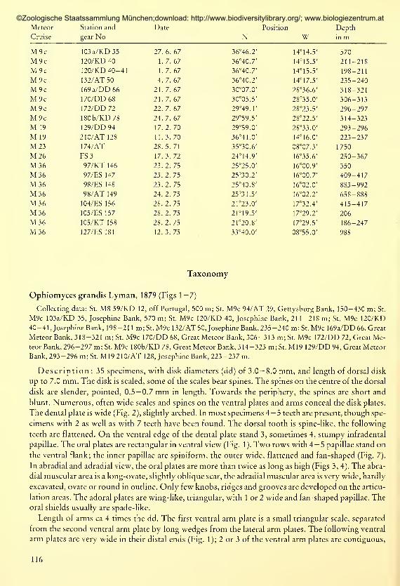
Figs 1 — 7: Ophiomyces grandis Lyman1. 4.5 mm dd, ventral disk; 2. 4.0 mmdd, dental plate; 3. 4.0 mm dd, oral plate, abradial; 4. 4.0 mm dd, oral plate,
adradial; 5. distal arm, dorsal; 6. distal arm, ventral; 7. oral papillae.
(1, 3— 7 each scale division = 1 mm; 2 scale division = 0.5 mm)
then a wedge from both lateral arm plates advances and the lateral arm plates meet on the ventral line.
In the peripheral end of the arms, the ventral plates are rounded, well separated from each other by
the lateral arm plates (Fig. 6). The dorsal plates at the arm base are short but wide; the plates become
fan-shaped toward the end of the arms (Fig. 5). In specimens of 3.0— 4.0 mm dd, 8 — 10 spines arepre-
sent at Segments IV-VII. In larger specimens, up to 13 arm spines have been found. Three to 4, some-
times even 6, of the dorsal spines are pointed, short, less than one arm segment in length; the next
spines are larger and slightly curved; the 2 or 3 ventralmost spines are short and blunt. The tentacle
pores are very wide, surrounded by numerous flat tentacle scales. At the basal arm segments,'there are
usually 1—2 scales on the lateral arm plate and 2— 3 on the ventral arm plate; from segment IV or Vonward, there are a small spines on the lateral arm plate and 1 large flattened scale on the ventral arm
plate. Towards the distal end of the arms the scales become more spine-like and decrease in numberdown to 1 spine on both lateral and ventral arm plate; finally, only 1 spine-like scale insert on the late-
ral arm plate.
In the stomach wall single, scythe-shaped ossicles of 30 jam length are present.
Distribution: Ophiomyces grandis was first described from the southeast Atlantic Ocean, from
Tristan da Cunha (Lyman 1879). The following records came from the northeast Atlantic Ocean: off
117
©Zoologische Staatssammlung München;download: http://www.biodiversitylibrary.org/; www.biologiezentrum.at

Britain (Gage et al. 1983), Bay of Biscay (Koehler 1906, Cherbonnier 1969, 1970), off Portugal and
Spain (Reys 1961 ;present collection), and off North Africa (present collection).
Ophiomyces grandis is known from a depth ränge from 150 m (present collection) to 1 800 m(Lyman1879).
Remarks : From the Josephine Bank another species of Ophiomyces, O. fructectosus (Lyman,
1869) is recorded, a species otherwise known from the Caribbean area. From the description O. fruc-
tectosus resembles O. grandis closely.
Ophiacantha abyssicola G. O. Sars, 1871 (Figs 8-10)
Collecting data: St. M8 59/KD 12, off Portugal, 500 m; St. M9c 90d/AT 26, off Portugal, 320-385 m; St. M3698/ES 148, off Spanish Sahara, 883-992 m; M36 98/AT 149, off Spanish Sahara, 658-888 m.
Description: Ca 100 specimens, with disk diameters of 1.0 to 6.0 mm.This species is easily recognized by the oral shields with the almost cross-shaped outline and the
deep grooves, and the cup-shaped dorsal arm plates. In the material studied, all specimens have 5 arms,
but specimens with 4 or 6 arms are known to occur (Mortensen 1933).
Figs 8—10: Ophiacantha ahyssicola G. O. Sars, 5 mm dd
8. oral plate, abradial; 9. oral plate, adradial; 10. dental plate.
(scale division = 0.5 mm)
The dental plate is elongate, more than 3 times longer than wide. The depressions for the uppermost
and lowermost tooth are deeply excavated, whereas the other 3 depressions are more or less inconspi-
cuous (Fig. 10). The lowermost tooth is stout, conical, the next teeth are flattened, with wide cutting
edges, the uppermost tooth is often spiniform. The oral plates are elongate, with their adoral sides
clearly higher than the aboral flank, the muscular area is rounded and slightly excavate, it occupies half
the length of the abradial flank (Fig. 8). The muscular scar on the adradial flank is a small, bean-shaped,
excavated area (Fig. 9). Only minute grooves and ridges are present on the abradial articular area, where-
as the adradial articular area has coarse ridges and grooves. Small fenestrated sclerites are embeddedin the stomach wall, and spiny sclerites in the bursal wall. The smallest specimen with gonads is
1.4 mm in dd.
Some of the 60 specimens opened had exoskeletons of planktonic Crustacea or large Foraminifera
in their stomachs ; but in most of the specimens only few minute forams and a little detritus were found
or the stomachs were completely empty.
Distribution : Ophiacantha ahyssicola is a common species in the North Atlantic Ocean. In the
north it is known from Northern Norway (Lofoten) and from between Norway and Bear Island
(Grieg 1902), off southern Greenland (Mortensen 1913) and off Cape Cod (Verrill 1880). Thesouthern-most record is from off Spanish Sahara (ca 25° N).
118
©Zoologische Staatssammlung München;download: http://www.biodiversitylibrary.org/; www.biologiezentrum.at

O. abyssicola lives in the abyssal as well as in the sublittoral, the depth distribution ranges from 35 m(Koehler 1909) to 3 500 m (Verrill 1885).
Ophiacantha angolensis Koehler, 1923
Collecting data: St. M9c 82/AT 19, off Morocco, 150-160 m; St. M9c 90/AT 22, off Portugal, 1 14-1 17 m; St.
M9c 90b/KT 24, off Portugal, 140-145 m; St. M36 104/ES 156, off Cape Blanc, 415-417 m.
Description: Ten specimens, with disk diameters of 3.0— 5.5 mm. Thediskis covered with short
stumps which end in 3 slender spinules. Many of the features in these specimens agree perfectly with
those of O. angolensis presented by Koehler (1923). However, the dorsal arm plates at the arm base
are wider than long, instead of being longer than wide as mentioned in Koehler (1. c.); the following
arm plates are cup-shaped, resembling those in O. abyssicola; 7 pairs of arm spines are present on the
first free arm segment, the second arm segment has 5 pairs of arm spines in larger, 4 in smaller speci-
mens; the third arm segment bears 4 pairs of spines. Denticulation is denser on the ventral than on the
dorsal arm spines.
Specimens collected in February (M36 104/ES 156) hold large oocytes, up to 0.2 mm in length.
Distribution: The type specimen was found off Angola, at 73 m depth (Koehler 1923). Further
records are both from south and north of the equator (Mortensen 1936, Cherbonnier 1962, Tommasi
1967, Mausen 1970), from between Angola and the Ivory Coast, from 55 to 200 m depth. The present
finding is the most northerly record of O. angolensis.
Ophiacantha aristata Koehler, 1896 (Figs 11 — 15)
Collecting data: St. M36 98/ES 148, off Spanish Sahara, 883-992 m; St. M36 98/AT 149, off Spanish Sahara,
658-888 m.
Figs 11 — 15: Ophiacantha aristata Koehler
11. 5.8 mm dd, dorsal disk; 12. sclerites instomach wall; 13. 4.5 mm dd, dorsal disk spines, lateral; 14. same,dor-
sal; 15. 4.5 mm dd, third ventral arm spine on second arm segment.
(11, 13— 15 each scale division = 0.5 mm; 12 scale division = 0.1 mm)
119
©Zoologische Staatssammlung München;download: http://www.biodiversitylibrary.org/; www.biologiezentrum.at

Description: Twenty-six specimens, 3.6— 10.0 mm in dd. The description in Koehler (1!
gives a good characterization of this species. Conspicuous are the stumps on the dorsal disk (Figs 13,
14; Koehler 1909: Fig. 6) with a straight, smooth stem and a wide almost horizontal crown. In eider
specimens, the spicules are arranged spherically.
The radial shields are seen as small triangulär plates (Fig. 11), beset with the characteristic stumps.
The oral papillae are very stout, 3 to 4 on each side of the jaws, all similar in shape, none is widened.
The stomach wall is lined with fenestrated platelets and multiple branched spiny stumps (Fig. 12), pro-
jecting into the stomach lumen.
The arms are wide at their base. Most of the arms are broken, but apparantly the arm length is 5—
6
times the dd. The dorsal arm plates are small, triangulär (Fig. 11) throughout the arm, always sepa-
rated by the lateral arm plates. The basal 2 — according to Koehler (1896), the basal 3 — ventral arm
plates are contiguous. The following plates are well separate, quadrangular in outline. The arm spines,
7—8 in number, are extremely serrate, increasing in length, the ventral obtuse spine equalling length
of an arm segment, the slender dorsal spines equalling that of 3 segments. The ventral and the lateral
spines are stout, often slightly flattened, while the dorsal spines are slender. The single tentacle scale
on each arm pore is flattened, serrate, and slightly pointed at the end.
Distribution: Opbiacantha aristata is known from the eastern North Atlantic Ocean, from off
the west coast of Ireland (Grieg 1921) to south of the Canary Islands (Koehler 1906). Most records
are from more than 1 000 m depth (Koehler 1906, 1909; Grieg 1921).
Ophiacantha brevispina Koehler, 1898 (Figs 16, 17)
Collecting data: St. M36 97/KT 146, off Spanish Sahara, 350 m; St. M36 97/ES 147, off Spanish Sahara,
409-417 m; St. M36 105/ES 157, off Cape Blanc, 206 m; St. M36 105/KT 158, off Cape Blanc, 186-247 m.
Description: Thirteen specimens, 3.0— 6.0 in dd. The dorsal disk is uniformly covered with
short, thorny stumps. In some specimens some scattered stumpy and blunt spines, only slightly longer
than the stumps, are present. The oral shields are diamond-shaped, with the peripheral edge slightly
protruding and the inner edge pointed (Fig. 1 7). The centre of the shield is slightly depressed. The ad-
oral plates are 2.5— 3.0 times wider than long. Three pairs of oral papillae are present, the 2 inner papil-
lae are conical, the outer papilla is wide and leaf-like.
Figs 16, 17: Opbiacantha brevispina Koehler
16. holotype, 7 mm dd, ventral disk; 17. 5 mm dd, ventral disk.
(each scale division = 1 mm)
120
©Zoologische Staatssammlung München;download: http://www.biodiversitylibrary.org/; www.biologiezentrum.at

The arms are at least 3— 4 times the length of the dd (all arms are broken). In the largest specimen
(6 mm dd), the 7 pairs of arm spines present on the first free arm segment form a continous row of dor-
sal arm spines across the segment; 5 pairs of arm spines stand on the following segments. The dorsal-
most spines are longer than 2 arm segments; then the arm spines rapidly decrease in length. The 2 ven-
tralmost pairs of spines are stout, usually less than an arm segment in length. All dorsal plates are sepa-
rate. The first dorsal plate is diamond-shaped, twice as wide as long. The following plates are triangu-
lär, wider than long and with a convex distal margin; the peripheral arm plates are as wide as long. All
ventral arm plates are separate. The first plate is conspicuous, as wide as long, the following plates are
distinctly wider than long, and, at a distance of a disk diameter, as wide as long. The arm papillae are
slender scales.
Remarks : The characteristics of these specimens agree well with those known for O. brevispina,
dredged in the Bay of Biscay. In the holotype and the present material, the ventral arm spines are short
and stout; the dorsal and ventral disk is covered with rather small, stumpy spines. But, in the present
material, the oral shields and adoral plates are 1.5 and 2.5 — 3.0 wider than long, respectively, whereas
in the holotype (7 mm dd), the oral shields and the adoral plates are twice as wide as long (Fig. 16).
Distribution: Ophiacantba brevispina is recorded from the Bay of Biscay (Koehler 1898) and
off Dakar and the Azores (Cadenat 1938). It is found in depths between 90 and 417 m.
Ophiacantha cuspidata Lyman, 1878
Collecting data: St. M23 174/AT, off Morocco, 1 750 m.
Descrip ion : Four specimens, of 4.8 to 8.0 mm dd. The oral shields are wider than in the speci-
mens figured by Mortensen (1933), with a straight distal margin and a depression in the middle of the
plate. Usually there are 3, rarely 4, oral papillae present on both sides of the jaws. The teeth are stout.
The peripheral portion of the radial shield is naked.
All dorsal arm plates are separate; slightly triangulär in outline with well rounded distal margins.
The first and second ventral arm plates are contiguous, the following ones usually separate. Seven arm
spines are present at the arm base of the large specimen (8.0 mm dd), 5 to 6 spines in specimens of
6 mm dd. The arm spines are blunt, with very fine denticulation. The tentacle scales are coarsely den-
ticulate. Up to 3 tentacle scales have been found at the basal arm pore, but usually, here as on the fol-
lowing segments only 1 pair of tentacle scales are present.
Distribution: The first records of Ophiacantha cuspidata are from off the Ascension Islands
from 785 m depth (Lyman 1878). All other records are from the North Atlantic Ocean, from 42°
N
(Koehler 1909) to 60°N (Mortensen 1933) from 1495 to 2460 m depth.
Ophiacantha densa Farran, 1913 (Figs 18, 19)
Collecting data: St. M26 FS 3, off Spanish Sahara, 250-367 m.
Description: Disk diameters of the 3 specimens present are 2.1, 3.5 and 3.6 mm, respectively,
The dorsal disk is covered with a dense coat of long and spiny stumps which end in 2 or 3 minute
points (Fig. 19). The radial shields are long and slender, sometimes raised, and completely hidden be-
neath the stumps. The oral shields are rhomboid, with their narrow lateral edges touching the lateral
arm plates (Fig. 18). The adoral plates are curved and wide. Three subequal oral papillae are present
on each side of the jaw. The denticulation on the oral papillae is coarser than on the arm spines. The
teeth are stouter than the oral papillae.
The arms are not conspicuously knotted. All dorsal arm plates are separate. The dorsal arm plates
are triangulär; the distal margin being slightly convex at the arm base. The plates are slightly longer
than wide in the small specimen, and in the distal portion of the 2 larger specimens; but they are
slightly wider than long on the basal segments of the large specimens. The ventral arm plates are sepa-
121
©Zoologische Staatssammlung München;download: http://www.biodiversitylibrary.org/; www.biologiezentrum.at

Figs 18, 19: Ophiacantha densa Farran, 3.5 mm dd
18. ventral disk; 19. spines on dorsal disk.
(18 each scale division = 1 mm; 17 scale division = 0.1 mm)
rate, pentagonal, with convex posterior margin; they are twice as wide as long at the arm base in the
larger specimens, only slightly wider than long in the small specimen and in the mid-arm portion of
the larger specimens, and as wide as long on the distal arm segments. The tentacle scales are small,
triangulär and pointed. The arm spines are blunt, slightly denticulate. The ventral arm spines are as
long as one arm segment, the dorsal spines are twice the length of an arm segment. Three to four spines
are present in the small specimen, 3— 5 in the larger specimens.
Remarks: In the specimens studied, the ventral arm plates are not as semicircular as figured by
Mortensen (1927: Fig. 108). Comparison with type material (United States National Museum, USA)proved the present material to be conspecific with Ophiacantha densa.
Distribution: Ophiacantha densa, hitherto, was known only from off Ireland (51°— 53°N,11°— 15°W) atdepthsof 1 150—1 330 m (Farran 1913). The present records extends the known ränge.
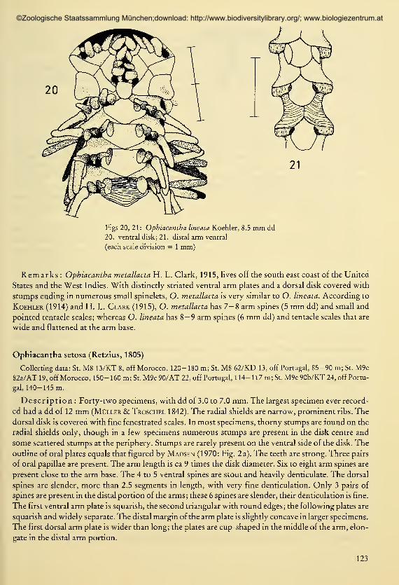
Ophiacantha lineata Koehler, 1896 (Figs 20, 21)
Collecting data: St. M36 98/ES 148, off Spanish Sahara, 883-992 m; St. M36 98/AT 149, off Spanish Sahara,
658-888 m.
Description: 16 specimens, 4. 1 — 8.5 mm dd. The alcohol-preserved specimens are pale, butinse-
veral specimens all ventral arm spines, tentacle scales, oral papillae and teeth are brown in their middle,
with the pigmentation slowly fainting towards their tips. The dorsal arm spines are pale. In the speci-
mens of the present collection the peripheral lobes of the oral shields are slightly depressed. Beside fen-
estrated platelets multibranched stumps are present in the stomach wall.
The ventral arm plates at the arm base are much wider than long in large specimens (Fig. 20), almost
as wide as long in the middle of the arm and slightly longer than wide at the arm tip (Fig. 21). The dor-
sal arm plates are small and triangulär throughout the arms. Number, length and arrangement of the
arm spines agree with those in the description by Koehler (1909).
Distribution: Ophiacantha lineata is known from the North Atlantic Ocean. Its geographical
ränge from 55°N (Cherbonnier & Sibuet 1972) to 30°N (Koehler 1914) is now emended southward
to 25° N. Its depth distribution ranges from 500 m (Koehler 1 9 1 4) to almost 3200 m (Cherbonnier &Sibuet 1972). Most records are from water depths beyond 1 000 m.
122
©Zoologische Staatssammlung München;download: http://www.biodiversitylibrary.org/; www.biologiezentrum.at
21
Figs 20, 21 : Opbiacantba lineata Koehler, 8.5 mm dd
20. ventral disk; 21. distal arm ventral
(each scale division = 1 mm)
Remarks : Opbiacantba metallacta H. L. Clark, 1915, lives off the south east coast of the United
States and the West Indies. With distinctly striated ventral arm plates and a dorsal disk covered with
stumps ending in numerous small spinelets, O. metallacta is very similar to O. lineata. According to
Koehler (1914) and H. L. Clark (1915), O. metallacta has 7—8 arm spines (5 mm dd) and small and
pointed tentacle scales; whereas O. lineata has 8— 9 arm spines (6 mm dd) and tentacle scales that are
wide and flattened at the arm base.
Ophiacantha setosa (Retzius, 1805)
Collecting data: St. M8 13/KT 8, off Morocco, 120-180 m; St. M8 62/KD 13, off Portugal, 85-90 m; St. M9c
82a/AT 19, off Morocco, 150-160 m; St. M9c90/AT 22, off Portugal, 114-117 m; St. M9c90b/KT 24, off Portu-
gal, 140-145 m.
Description : Forty-two specimens, with dd of 3.0 to 7.0 mm. The largest specimen ever record-
ed had a dd of 12 mm (Müller & Troschel 1842). The radial shields are narrow, prominent ribs. The
dorsal disk is covered with fine fenestrated scales. In most specimens, thorny stumps are found on the
radial shields only, though in a few specimens numerous stumps are present in the disk centre and
some scattered stumps at the periphery. Stumps are rarely present on the ventral side of the disk. The
outline of oral plates equals that figured by Madsen (1970: Fig. 2 a). The teeth are strong. Three pairs
of oral papillae are present. The arm length is ca 9 times the disk diameter. Six to eight arm spines are
present close to the arm base. The 4 to 5 ventral spines are stout and heavily denticulate. The dorsal
spines are slender, more than 2.5 segments in length, with very fine denticulation. Only 3 pairs of
spines are present in the distal portion of the arms ; these 6 spines are slender, their denticulation is fine.
The first ventral arm plate is squarish, the second triangulär with round edges; the following plates are
squarish and widely separate. The distal margin of the arm plate is slightly concave in larger specimens.
The first dorsal arm plate is wider than long; the plates are cup-shaped in the middle of the arm, elon-
gate in the distal arm portion.
123
©Zoologische Staatssammlung München;download: http://www.biodiversitylibrary.org/; www.biologiezentrum.at
Distribution: Ophiacantha setosa is known from the Eastern North Atlantic Ocean from 45°N(Koehler 1906) to 5°N (Madsen 1970) and from the eastern Mediterranean Sea, as far east as the Jonian
Sea (Kasparis & Tortonese 1982). O. setosa is found within depths ranging from 14 m (Monteiro-
Marques 1981) to 1480 m (Koehler 1906).
R em a r k s : Eight of 9 and 1 3 of 1 7 of the ophiuroids at St. M9c 82a/AT 1 9 and St. M9c 90/AT 22,
respectively, were infested with myzostomids. The myzostomids usually were found in the esopha-
gus, on the upper teeth, partly hidden beneath the contracted esophagal sphincter.
Ophiacantha simulans Koehler, 1896
Collecting data: St. M23 174/AT, off Morocco, 1 750 m.
Description: One specimenof 6.0 mm dd. The oral shields are very wide, 1.8 to 2.0timesas wide
as long, thus wider than in the specimens figured by Mortensen (1933 : Fig. 1 1 a), they are pentagonal,
with almost straight distal margins. Six to seven arm spines are present at the arm base, 5 on the follow-
ing segment, and 4 to 5 on the next segments. The arm spines are finely denticulate. Near the arm base,
the second (third) spine from below has strong dents, the other arm spines usually show only a fine
denticulation. The first ventral arm spine is bent from the sixth arm segment onward, with a coarser
denticulation on its ventral than on its dorsal flank. The stumps on the disk have an almost smooth
stem and a crown of 8 or more thorns.
Distribution: Ophiacantha simulans is common in deeper water — 1 480 to 3 020 (Koehler 1906,
1909) - of the North Atlantic Ocean. Itis found in the eastern (Koehler 1896, 1906, 1 909 ; Mortensen
1933; Gage et al. 1983) and in the northwestern Atlantic Ocean (Rowe 1971 ; Rowe & Menzies 1969).
Off the Carolinas, this species is centered along the 1 900 m depth line (Menzies et al. 1973).
Ophiacantha smitti Ljungman, 1871 (Figs 22— 24)
Collecting data: St. M8 8/AT 6, off Portugal, 1 370- 1 430 m; St. M8 19/AT 9, off Morocco, 1 300 m; St. M36 98/
ES 148, off Spanish Sahara, 883-992 m; St. M36 98/AT 149, off Spanish Sahara, 658-888 m.
Description : The 18 specimens present are small, of 2.6— 5.0 mm in dd. The characters of these
specimens agree with those of the type specimen (in Swedish Museum of Natural History). The dorsal
disk is covered with small stumps, with a wide basal platform and bifid or trifid tip. In most specimens,
moreover, some slender serrate spinelets are present in the centre of the disk. The radial shields are
long and slender, they form 2 raised slender ribs, concealed beneath minute scales and stumps, with
only naked peripheral ends. The oral and adoral plates and shields are set off the disk. The plates are
slightly rugose. In most specimens, the madreporit is almost as long as wide, with protruding lateral
edges and rounded periphery. The other oral shields are twice as wide as long (Fig. 24). In some radii,
the oral shields meet the lateral arm plates. The adoral plates are large, with curved radial and adradial
margins. The oral papillae, 3 — rarely 2 or 4 — in number, are stout and blunt. The peripheral papillae
are often slightly stouter, but not longer than the others. The teeth are flattened, wider than the oral
papillae, 4 — 5 arranged in a vertical row.
The arms are stout at their base, but very slender in their distal portions, coiled, and at least 5 to 6
times the length of the dd. All dorsal arm plates are well separated, triangulär, with rounded distal and
almost straight lateral margins (Fig. 23). The first dorsal arm plate is proximally obtuse, the following
arm plates are pointed. The dorsal arm plates are slightly wider than long close to the arm base, but as
long as or longer than wide at the arm tip (Fig. 22). The first ventral arm plate is pentagonal in outline;
the second is pentagonal in some arms, triangulär in others, with straight, rarely convex or concave,
distal margin. The third ventral arm plate is pentagonal, distinctly wider than long, in most specimens
twice as wide as long, with its distal margin straight or slightly concave, rarely convex. The following
ventral plates are either pentagonal or triangulär, only slightly wider than long. In the slender distal
124
©Zoologische Staatssammlung München;download: http://www.biodiversitylibrary.org/; www.biologiezentrum.at

Figs 22—24: Ophiacantha smitti Ljungman, 3.0 mm dd
22. distal arm, dorsal; 23. fifth to seventh arm segment, dorsal; 24. ventral disk
(each scale division = 1 mm)
portion of the arms, the ventral plates are quadrangular, as wide as long. The first and second arm
plates are contiguous at some arms, otherwise, all ventral arm plates are separate. The lateral arm plates
meet dorsally and ventrally. The arm spines are arranged in a continuous dorsal row only on the first
or second basal segments. Basal arm segments with 6 pairs of arm spines, the following segments with
5 pairs; beyond the basal half of the arms only 4, finally 3 pairs of arm spines present. One to 3 of the
ventral spines are stout, finely serrate, equalling an arm segment in length. The 4th to 6th arm spines
are slender, 2.5 to 3 times the length of a segment. Beyond the first or second basal arm segments, the
dorsal spines are as long or only slightly longer than a segment. At the arm tip, all spines are shorter
than one segment; 1 or 2 of the ventralmost spines are stronly denticulate along the ventral margin of
the spine (Fig. 22). Only one single tentacle scale present throughout the arm.
Distribution: The type specimens of Ophiacantha smitti were taken off Portugal, dredged from
1420 m depth (Ljungman 1871). Recently, O. smitti was recorded from the North Atlantic Ocean
from 47°N 8°W at 1 150-1 175 m depth (Cherbonnier & Sibuet 1972). Another record is from off
Sierra Leone from 118 m depth (Longhurst 1958). Most of the findings are from deep waters.
Ophiothamnus affinis Ljungman, 1871 (Figs 25, 26)
Collecting data: St. M8 8/AT 6, off Portugal, 1 370- 1 430 m; St. M8 19/AT 9, off Morocco, 1 300 m; St. M36 98/
ES 148, off Spanish Sahara, 883-992 m; St. M36 127/ES 181, off Morocco, 988 m.
125
©Zoologische Staatssammlung München;download: http://www.biodiversitylibrary.org/; www.biologiezentrum.at

Figs 25, 26: Ophiothamnus affinis Ljungman
25. 2 mm dd, dorsal disk; 26. 3 mm dd, dorsal disk
(each scale division = 1 mm)
Description : All specimens known are small; the 20 specimens on hand have 1.6 to 3.1 mm dd.
Large specimens (3 mm dd) have up to 9 pairs of arm spines at the basal arm segments. The dorsal arm
spines at the arm base are at least 3 times the length of an arm segment (all arm tips are broken).
Eight of the 20 specimens studied had lost their dorsal disks, 1 had just regenerated the dorsal disk.
Fig. 26 shows a portion of an unhurt dorsal disk, Fig. 25 a regenerated disk. The small pointed spines
and disk scales are lacking in the regenerated specimen; the radial shields are small, almost as long as
wide, contiguous for all its length, whereas they are longer than half the radius in the undamaged spe-
cimen. The regenerated dorsal disk overlap the first arm plate in some radii, while there are wide gaps
between the radial shields and the first dorsal arm plate in other radii.
Distribution: Ophiothamnus affinis is known from the North Atlantic Ocean. It has been found
within an area from 24°N to 30°N and at depths from 1 93 to 4 1 5 m along the American coast (Koehler
1914), within an area from 25°N (present collection) to 44°N (Koehler 1906) and at depth from 883 m(present collection) to 1 425 m (Koehler 1909) along the European coasts.
Ophiambix meteoris Bartsch, 1983
Collecting data: St. M9c 170/DD 68, Great Meteor Bank, 306-313 m.
Description: One specimen of 3.9 mm dd.
Distribution: Records of the genus Ophiambix were, hitherto, from deep sea areas only, e. g.
Ophiambix meteoris was known from the Iberian deep sea basin, from 5315m depth (Bartsch 1983).
The present record is considerably shallower than the previous finding.
Ophiacamax dominans Koehler, 1906 (Figs 27—33)
Collecting data: St. M36 98/ES 148, off Spanish Sahara, 883-992 m; St. M36 98/AT 149, off Spanish Sahara,
658-888 m.
126
©Zoologische Staatssammlung München;download: http://www.biodiversitylibrary.org/; www.biologiezentrum.at

Figs 27—33: Ophiocamax dominum Koehler, 16 mm dd
27. oral plate, abradial; 28. oralplate, adradial;29. ventral disk; 30. dental plate; 31. distal arm, ventral; 32. mid-
arm, dorsal; 33. crescent spines in stomach wall
(27—32 each scale division = 1 mm; 33 scale division = 0.1 mm)
Description : Twenty-nine specimens, of 8.0— 17.0 mm dd. The radial shields are large; in length
at least half the disk radius, in width the same or more than the interradial space. The radial shields are
contiguous troughout their length. The dorsal disk plates are large, irregulär in outline; most of them
with 1 or more spines. Some of the spines are slender, almost smooth, others are stout, with numerous
glassy spinelets. The radial shields are bare of spines, but in larger specimens often a row of spines in-
sert along the mid-line of 2 adjacent shields.
127
©Zoologische Staatssammlung München;download: http://www.biodiversitylibrary.org/; www.biologiezentrum.at

The ventral disk is dominated by numerous spines. The oral shields are arrowhead-shaped (Fig. 29);
the madreporit is bulbous in its central portion. The adoral plates broadly contact the first lateral arm
plates. Often, 1 or 2 spines are present along the line between oral shield and adoral plate, the spines
inserting either on the adoral plate or on the oral shield. Seven to 8 spines stand along the margin of
the oral plate, several of them included in the cluster of 6 — 8 spines around the tentacle pore. At the
ventral end of the dental plate insert 6— 7 spines, followed by cone-like teeth, andshort, flattened teeth
beyond the middle of the plate. The dental plate is slightly bent; it is 3.5 times longer than wide in a
specimen of 16 mm dd. The plate may be divided by a transverse fissure. Small round depressions for
the spines are present in the ventral portion, oval depressions for the row of flattened teeth in the
middle and dorsal portion (Fig. 30). The oral plate is slightly higher than long in a specimen of 16 mmdd. The abradial muscle area is a bowl-like depression (Fig. 27), the adradial one is an oval, deeply ex-
cavated, oblique area (Fig. 28). The adradial articular area, dorsal to the muscle scar, is oval, provided
with deep grooves. The abradial articular area along the central border of the oral plate has deep
grooves and narrow ridges. The bursal wall is lined with fenestrated plates. In the stomach wall cres-
cent ossicles are found, often divaricate at their ends (Fig. 33).
In larger specimens, the arms are 5 times the length of the dd. Six to 7 arm spines are present
throughout the arms. Most of the arm spines are broken. At the arm base and the mid-arm, the dor-
salmost spine is slender, 1.5 — 2 times the length of an arm segment. The second or third spine from
mid-dorsal line is the largest one; in the middle of the arm this spine is 5— 6 times longer than an arm
segment. The following spines are short. At the base and in the middle of the arm, they are 1.2— 2 times
longer than an arm segment. Some of the spines are almost smooth, others carry rows of glassy
spinelets. At the distal end of the arms, most of the arm spines are shorter than the length of an arm
segment; they are slightly hook-shaped and provided with a row of spinelets facing outward. The dor-
sal arm plates are as illustrated in Koehler (1906: Fig. 27), with the basal plates overlapping, and the
following ones separate. In the posterior half of the arms, usually 2 tiny spinelets insert near the distal
margin of the plate (Fig. 32). At the arm base, the ventral arm plates are wide, with narrow lateral
wedges, with their distal portions slightly raised, knob-like, while the basal portion forms a cavity be-
tween the raised walls of the arm pores. The arm pores are conspicuous at the arm base, the pores open
at the end of a wall formed by raised portions of lateral and ventral arm plates. At the distal end of the
arms, the ventral arm plates are triangulär, with rounded distal margins (Fig. 31); the arm pores are
small, the lateral and ventral plates form no wall around the arm pore. The basal arm pores are sur-
rounded by 3— 4 tentacle scales. The tentacle scales are elongate, triangulär, wide at their base. The
3 — 4 scales form together a tube. The number of tentacle scales quickly decrease in number. At a
distance equalling the dd, only 1 tentacle scale is present, inserted on the lateral arm plate. Terminally,
the tentacle scales are similar to the arm spines, they stand close together and form a continuous row
with the arm spines.
Several of the specimens of 1 1 — 14 mm dd had embryos or juveniles in the bursae. In larger speci-
mens, 15 — 17 mm dd, no juveniles were found, their bursae often were filled with sediment. The small
star-disk stages with spiny spicules and juveniles with up to 4 arm segments were often tightly cram-
med in the bursae, almost completely filling the body cavity. The stomachs were empty in specimens
with juveniles. Up to 25 juveniles per bursa were found, usually all of them at the same stage of devel-
opment. Some of the specimens with 4 arm segments, of 0.9 mm dd and 3.3 mm in diameter, have their
arms stretched out in a vertical piain. Probably, the juveniles leave the adults at this stage of develop-
ment.
The juveniles are very spiny. In those with 4-segmented arms, several long trifid spinelets stand on
the dorsal disk, each arising from a small platelet. Oral plates and adoral plates, the latter with a long
spinelet, are present on the ventral side. Teeth are inconspicuous or lacking. Tentacle scales are large
both within the disk and on the 4 arm segments. The first arm segment bears 4— 5 pairs of spines, the
next arm segments each 3 or 4 pairs of spines. The dorsal spines almost meet at the dorsal mid-line. The
128
©Zoologische Staatssammlung München;download: http://www.biodiversitylibrary.org/; www.biologiezentrum.at

spines on the first segments usually have 3 glassy thorns, the other spines are smooth. Dorsal armplates are present throughout the arms. The first ventral arm plate is large, slightly quadrangular, the
second is represented by a small, irregularly formed platelet, the third has the form of a minute, tetra-
radiate sclerite, the fourth is lacking. The lateral arm plates meet ventrally. The terminal plate is slen-
der, 0.6—0.8 mm long.
Distribution: The original description of Ophiocamax dominans was based on 2 specimens
(Koehler 1906). Both were taken at 25°39'N and 18°22'W at 882 m depth. The present records are
from the same area and from similar depth.
General remarks
Ophiacanthids were present in samples taken with trawls, dredges and sleds; samples taken with
grabs yielded no ophiacanthids. Ophiacanthids are epibenthic living species.
Ophiacantba abyssicola and O. setosa are abundant and widely distributed in the Atlantic Ocean,
both horizontally, from the African coast to northern Europe, and vertically, from shallow waters to
the bathyal. According to the present records, Ophiacantba aristata, O. cuspidata, O. densa, O. li-
neata, O. simulans, O. smitti, Ophiothamnus affinis and Ophiacamax dominans are inhabitants of ba-
thyal waters.
Acknowledgements
I am grateful to Dr. K. Hülsemann (Hamburg) for improvements of my English text, and to C. Carpine (Musee
Oceanographique Monaco), M. E. Downey (United States National Museum, Washington, D. C.) and R. Oleröd
(Swedish Museum of Natural History, Stockholm) for making type material available.
References
Bartsch, 1. 1983: Opbiambix meteoris n. sp., ein neuer Schlangenstern aus der Iberischen Tiefsee (Ophiacanthidae,
Ophiuroidea). — Spixiana6: 97—100
Cadenat, J. 1938: Liste des echinodermes recueillis pendant la cinquieme croisiere du navire de recherches Presi-
dent-Theodore-Tissier. — Revue Trav. Off. Pech, marit. 11: 349— 375
CHERBONNIER, G. 1962: Ophiurides. - Result. scient. Exped. Oceanogr. Beige Eaux Cot. Afr. Atlant. Sud. 3(8):
1-24
1969: Echinodermes recoltes par la »Thalassa« au large des cötes ouest de Bretagne et du Golfe de Gascogne
(3-12 Aoüt 1967). - Bull. Mus. natn. Hist. nat., Paris (2e Ser.) 41: 343-361
1970: Echinodermes recoltes par la »Thalassa« au large des cötes d'Espagne et du Golfe de Gascogne
(18-25 Octobre 1968). - Bull. Mus. natn. Hist. nat., Paris (2C Ser.) 41: 1266-1277
CHERBONNIER, G. & SlBUET, M. 1 972 : Resultats scientifiques de la campagne Noratlante : Asterides et Ophiurides.
- Bull. Mus. natn. Hist. nat., Paris (3e Ser.) 102: 1333-1394
Clark, H. L. 1915: Catalogue of recent Ophiurans. - Mem. Mus. comp. Zool. Harv. 25: 163-376, 20 Taf.
Farran, G. P. 1913: The deep-water Asteroidea, Ophiuroidea and Echinoidea of the west coast of Ireland. —
Scient. Invest. Fish. Brch. Ire. 1912(6): 1-66
Gage, J. P., Pearson, M., Clark, A. M., Paterson, G. L. J. & Tyler, P. A. 1983: Echinoderms of the Rockall
Trough and adjacent areas. I. Crinoidea, Asteroidea and Ophiuroidea. — Bull. Br. Mus. nat. Hist. (Zool) 45:
263-308
Grieg, J. A. 1902: Oversigt over det nordliga Norges echinodermer. — Bergens Mus. Ärb. 1902(1): 1—36
1921 : Echinodermata from the "Michael Sars" North Atlantic Deep-Sea Expedition 1910. — Rep. scient. Re-
sults Michael Sars N. Atlant, deep Sea Exped. 1910: 1—47, 5 Taf.
KASPARIS, P. & TORTONESE, E. 1 982 : Echinoderms from the western seas of Greece. - Thalassographica 2 : 27-32
KOEHLER, R. 1 896: Echinodermes. Resultats scientifiques de la Campagne du "Caudan" dans le Golfe de Gascogne.
- Annls Univ. Lyon 26: 33-127
129
©Zoologische Staatssammlung München;download: http://www.biodiversitylibrary.org/; www.biologiezentrum.at

1898: Echinides et Ophiures provenant des campagnes du yacht PHirondelle (Golfe de Gascogne, Agores,
Terre-Neuve). Result. Camp, scient. Prince Albert I 12: 1 — 78, 9 Taf.
1906: Ophiures. — Exped. scient. Travailleur Talisman 8: 245 — 311, Taf. 18 — 21
1909: Echinodermes provenent des campagnes du yacht Princesse-Alice (Asteries, Ophiures, Echinides et
Crinoides) — Result. Camp, scient. Prince Albert I 34: 1—317, Taf.
1914: A contribution to the study of Ophiurans of the United States National Museum. — Bull U. S. natn.
Mus. 84: 1-173
1923 : Sur quelques ophiures des cötes de l'Angola et du Cap. — Kungl. Vetensk. o. Vitterh. Samh. Handl. (IV)
15(3): 1-17, Taf.
LjUNGMAN, A. V. 1871: Förteckning öfver uti Vestindien af Dr. A. Goesunderkorvetten Josefines expeditioni At-
lantiska Oceanen samlade ophiurider. — Öfvers. K. Vetensk. Akad. Förh. 28(6): 615— 658
Longhurst, A. R. 1958: An ecological survey of the West African marine benthos. — Fishery Publs colon. Off.
11: 1-102
Lyman, T. 1878: Ophiuridae and Astrophytidae of the exploring voyage of H. M. S. "Challenger". Part I. — Bull.
Mus. comp. Zool. Harv. 5(7): 65-168, 10 Taf.
1879: Ophiuridae and Astrophytidae of the exploring voyage of H. M. S. "Challenger". Part II. — Bull. Mus.
comp. Zool. Harv. 6(2): 17-83, Taf. 11-19
MADSEN, F. J. 1970: West African Ophiuroids. - Atlantide Report 11: 151-243
Menzies, R. J., GEORGE, R. Y. & Rowe, G. T. 1973: Abyssal environment and ecology of the World Oceans.
488 pp.
MONTEIRO-MARQUES, V. 1981: Peuplements des planchers envases de trois grottes sous-marines de la region de
Marseille. Etüde preliminaire. — Tethys 10: 89— 96
MORTENSEN, T. 1913: Grönlands Echinodermer. - Meddr Grönland 23: 299-379
1927: Handbook of the echinoderms of the British Isles. 471 pp.
-- 1933:Ophiuroidea. - Dan. Ingolf-Exped. 4(8): 1-121, 3 Taf.
1936: Echinoidea and Ophiuroidea. — "Discovery" Rep. 12: 199-348, 8 Taf.
MÜLLER, J. &Troschel, F. H. 1842: System der Ästenden. II. Ophiuridae. 79-134, Taf. 7-10
Reys, J. F. 1961: Deux ophiures nouvelles. - Rec. Trav. St. Mar. End. 22: 153-157
ROWE, G. T. 1971 : Observations on bottom currents and epibenthic populations in Hatteras submarine canyon. —
Deep Sea Res. 18:569-581
ROWE, G. T. & MENZIES, R. J. 1969: Zonation of large benthic invertebrates in the deep-sea off the Carolinas. -
Deep Sea Res. 16:531-537
Thiel, H. 1970: Bericht über die Benthosuntersuchungen während der „Atlantischen Kuppenfahrten 1967" von F.
S. „Meteor". — „Meteor" Forsch. -Ergebnisse, D, 7: 23 — 42
1981 : Benthic investigations in the Northwest African upwelling area. Report on the cruises 26, 36, 44 and 53
of R. V. „Meteor". — „Meteor" Forsch. Ergebnisse, D, 33: 1 — 15
Tommasi, L. R. 1967: Ophiuroidea de la Cöte-d'Ivoire. - Bull. L. I. F. A. N. (A) 29: 521-585
Verrill, A. E. 1880: Notice on recent additions to the marine invertebrates of the North-East coast of America.
- Proc. U. S. Nat. Mus. 2: 165-205
1885: Results of the explorations madeby the steamer "Albatross", off the Northern Coast of the United Sta-
tes, in 1883. - Rep. U. S. Commnr Fish. 11: 503-699
Dr. Ilse Bartsch
Biologische Anstalt Helgoland
Notkestr. 31
2000 Hamburg 52
130
©Zoologische Staatssammlung München;download: http://www.biodiversitylibrary.org/; www.biologiezentrum.at

![[XLS] · Web view91 Bibliotheken, Archive, Museen, botanische und zoologische Gärten 92 Spiel-, Wett- und Lotteriewesen 93 Erbringung von Dienstleistungen des Sports, der Unterhaltung](https://static.fdokument.com/doc/165x107/5ae3d8767f8b9a5d648e7bb6/xls-view91-bibliotheken-archive-museen-botanische-und-zoologische-grten-92.jpg)