Eignung der high throughput Version des Comet Assays als...
115
Eignung der high throughput Version des Comet Assays als Screening-Verfahren Von der Fakultät für Mathematik und Naturwissenschaften der Carl von Ossietzky Universität Oldenburg zur Erlangung des Grades und Titels eines Doktors der Naturwissenschaften (Dr. rer. nat.) angenommene Dissertation von André Stang Geboren am 04.04.1984 in Schwerin Oldenburg, den 08.07.2009
Transcript of Eignung der high throughput Version des Comet Assays als...
Eignung der high throughput Version des Comet Assays als
Screening-Verfahren
Von der Fakultät für Mathematik und Naturwissenschaften der Carl von
Ossietzky Universität Oldenburg zur Erlangung des Grades und Titels eines
Doktors der Naturwissenschaften (Dr. rer. nat.)
angenommene Dissertation
von
André Stang
Geboren am 04.04.1984 in Schwerin
Oldenburg, den 08.07.2009

Erstgutachterin: Prof. Dr. rer. nat. Irene Witte Zweitgutachter: Prof. Dr. Karl-Wilhelm Koch Tag der Disputation: 28. August 2009

I
Inhaltsverzeichnis Seite Inhaltsverzeichnis I Verwendete Abkürzungen II 1.Zusammenfassung 1.1 in Deutsch 1 1.2 in English 3 2. Einleitung 5 3. Darstellung der Ergebnisse 14
3.1 Durchführung des high throughput Comet und Vergleich mit dem standardisierten Comet Assay 14
3.2 Automatische Auswertung von Kometen im high throughput
Comet Assay 15 3.3 Anwendbarkeit des high throughput Comet Assay unter der Verwendung 5 verschiedener Zelllinien 16
3.4 Strategie für das Screening von Umweltproben im Hochdurchsatzverfahren 17
4. Ausblick 18 5. Publikationen der Ergebnisse 23
5.1 Performance of the comet assay in a high-throughput version 24
5.2 Automatic Analysis of Comets in the High Throughput Version of the Comet Assay 43
5.3 Ability of the high throughput comet assay to measure comparatively the sensitivity of five cell lines toward methyl methanesulfonate, hydrogen peroxide and pentachlorophenol 59
5.4. A high-throughput genotoxicity testing strategy for screening of (drinking) water 76 Danksagung Erklärung

II
Abkürzungsverzeichnis
AT Ames-Test
CA Comet Assay
CT Chromosomenabberationstest
MCP Multichamberplate
MT Mikronukleustest
OECD Organisation for Economic Co-operation and Development
REACH Registration, Evaluation, Authorisation and Restriction of
Chemicals

Zusammenfassung
1
1. Zusammenfassung 1.1 Zusammenfassung
Es besteht ein hoher Bedarf, die steigende Anzahl an gentoxischen Verbindungen in
der Umwelt und bei der Entwicklung neuer Substanzen zu erfassen. Hierzu wurde
der häufig angewandte Comet Assay als high throughput-Verfahren weiterentwickelt
(Witte et al., 2007; Stang, 2006 (unveröffentlicht)). In der vorliegenden Arbeit wurde
untersucht, inwieweit sich dieses neue high throughput Verfahren für ein Screening
auf Gentoxizität von Umweltproben und Chemikalien während der
Wirkstoffentwicklung eignet.
Die high throughput Version des Comet Assay detektierte die DNA-schädigende
Wirkung von Mutagenen mit unterschiedlichen Wirkmechanismen sensitiv, mit
geringem Fehler und sehr guter Reproduzierbarkeit (A. Stang & I. Witte, Performance
of the comet assay in a high-throughput version. Mutat Res. 675 (2009) 5-10). Ein
Vergleich zum Standardverfahren des Comet Assay nach Tice et al. (2000) zeigte,
dass die Wirkung von Mutagenen konzentrationsabhängig
und vergleichbar sensitiv
nachgewiesen werden konnte. Die Integration eines Zytotoxizitätstests, der beim
konventionellen Verfahren separat durchgeführt werden muss, veränderte nicht die
Kometenbildung. Die high throughput Version des Comet Assay ermöglichte die
Steigerung des Probendurchsatzes ca. um das 20fache im Vergleich zum
konventionellen Comet Assay.
Eine weitere Erhöhung des Probendurchsatzes wurde durch eine schnellere
Datenauswertung möglich (A. Stang, M. Brend´amour, C. Schunck & I. Witte,
Automatic Analysis of Comets in the High Throughput Version of the Comet Assay.
(eingereicht)). Hierzu wurde in Zusammenarbeit mit der Firma Metasystems ein voll
automatisiertes Auswertungssystem entwickelt. Ein Vergleich mit interaktiven
(manuellen) sowie mit automatisierten Auswertungssystemen für den konventionellen
Comet Assay ergaben vergleichbare Ergebnisse mit geringem Fehler. Dadurch
ergab sich eine zusätzliche Steigerung der Durchsatzrate um den Faktor 10 im
Vergleich zur manuellen Auswertung.
Zusammenfassung
2
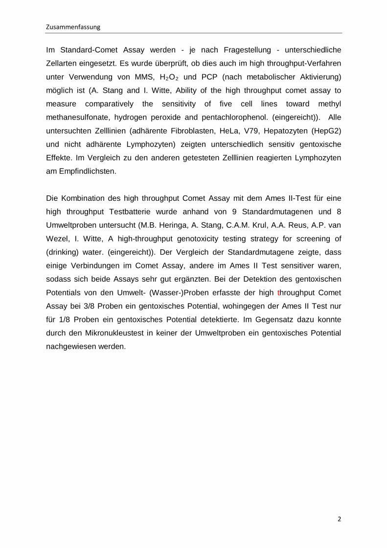
Im Standard-Comet Assay werden - je nach Fragestellung - unterschiedliche
Zellarten eingesetzt. Es wurde überprüft, ob dies auch im high throughput-Verfahren
unter Verwendung von MMS, H2O2
und PCP (nach metabolischer Aktivierung)
möglich ist (A. Stang and I. Witte, Ability of the high throughput comet assay to
measure comparatively the sensitivity of five cell lines toward methyl
methanesulfonate, hydrogen peroxide and pentachlorophenol. (eingereicht)). Alle
untersuchten Zelllinien (adhärente Fibroblasten, HeLa, V79, Hepatozyten (HepG2)
und nicht adhärente Lymphozyten) zeigten unterschiedlich sensitiv gentoxische
Effekte. Im Vergleich zu den anderen getesteten Zelllinien reagierten Lymphozyten
am Empfindlichsten.
Die Kombination des high throughput Comet Assay mit dem Ames II-Test für eine
high throughput Testbatterie wurde anhand von 9 Standardmutagenen und 8
Umweltproben untersucht (M.B. Heringa, A. Stang, C.A.M. Krul, A.A. Reus, A.P. van
Wezel, I. Witte, A high-throughput genotoxicity testing strategy for screening of
(drinking) water. (eingereicht)). Der Vergleich der Standardmutagene zeigte, dass
einige Verbindungen im Comet Assay, andere im Ames II Test sensitiver waren,
sodass sich beide Assays sehr gut ergänzten. Bei der Detektion des gentoxischen
Potentials von den Umwelt- (Wasser-)Proben erfasste der high throughput Comet
Assay bei 3/8 Proben ein gentoxisches Potential, wohingegen der Ames II Test nur
für 1/8 Proben ein gentoxisches Potential detektierte. Im Gegensatz dazu konnte
durch den Mikronukleustest in keiner der Umweltproben ein gentoxisches Potential
nachgewiesen werden.

Summary
3
1.2 Summary
The ever increasing number of foreign substances to be released into the
environment demands the development of reliable evaluation systems for genotoxic
assessment. One of the common tests for genotoxic measurement is the comet
assay, which was further developed to the high throughput comet assay (Witte et al.,
2007; Stang, 2006 (unpublished)). The aim of this dissertation was to examine if this
high throughput version is suitable for genotoxic screening of environmental
compounds and chemical development during drug design.
With the high throughput version of the comet assay DNA damages of mutagenic
agents were detected sensitively, with low standard deviations and high
reproducibility (A. Stang & I. Witte, Performance of the comet assay in a high-
throughput version. Mutat. Res. 675 (2009) 5-10). A comparison with the
conventional comet assay described by Tice et al. (2000) showed that the DNA
damaging effects were detected in a concentration dependent way with similar
sensitivity. The integration of a cytotoxicity assay, which has to be executed
separately in the conventional comet assay, did not influence the comet formation in
the high throughput comet assay. The high throughput version of the comet assay
increased the throughput of samples by about 20fold compared to the conventional
comet assay.
An additional enhancement of the throughput was gained by a new and faster
evaluation system of the comet data (A. Stang, M. Brend´amour, C. Schunck & I.
Witte, Automatic Analysis of Comets in the High Throughput Version of the Comet
Assay. (submitted)). In cooperation with the company Metasystems a fully automated
evaluation system was developed. A comparison with the interactive (manual) as well
as the existing automated evaluation systems for the conventional comet assay
showed similar results with low standard deviations and standard errors. Here, an
additional throughput enhancement was gained by a factor of ten compared to the
interactive evaluation.

Summary
4
A variety of different cell types are used in the standard comet assay depending on
the scientific question. The high throughput comet assay was evaluated for usage of
various cell types, using MMS, H2O2
and PCP (with metabolic activation system) as
mutagenic compounds (A. Stang and I. Witte, Ability of the high throughput comet
assay to measure comparatively the sensitivity of five cell lines toward methyl
methanesulfonate, hydrogen peroxide and pentachlorophenol. (submitted)). All cell
types used (adherent fibroblasts, HeLa, V79 cells and hepatocytes (HepG2) and non
adherent lymphocytes) were able to express genotoxic potential, although with
different degrees of sensitivity. The highest sensitivity was observed for human
lymphocytes.
9 standard mutagens and 8 environmental probes were tested for their genotoxic
potential with the high throughput comet assay, and simultaneously with the Ames II
test for high throughput screening (M.B. Heringa, A. Stang, C.A.M. Krul, A.A. Reus,
A.P. van Wezel, I. Witte, A high-throughput genotoxicity testing strategy for screening
of (drinking) water. (submitted)). The comparison of the 9 standard mutagens showed
that some agents were more sensitive in the comet assay and others in the Ames II
test, so that both assays complemented each other. The detection of the genotoxic
potential of 8 environmental water probes showed that the high throughput comet
assay detected genotoxic potential in 3 probes and the Ames II test detected 1
genotoxic potential. In contrast to the high throughput comet assay, the micronucleus
test did not detect any genotoxic potential in the environmental probes.
Einleitung
5
2. Einleitung
Der Mensch ist täglich bis zu 70.000 Chemikalien (EINECS, European Inventory of
Existing Commercial Chemical Substances, Datenbank), welche die
unterschiedlichsten Wirkungen auf den Menschen haben, ausgesetzt. Neben
Industriechemikalien wie Lösungsmitteln und Petrochemikalien handelt es sich vor
allem um pharmazeutische- und Pflegeprodukte bis hin zu Bioziden. Nur für ca. 4 %
dieser Umweltchemikalien liegen toxikologische Befunden bezüglich ihrer
Einzelwirkung vor (BUND 2008).
Insbesondere die Gruppe der gentoxischen Umweltchemikalien verfügt über ein
hohes schädigendes Potential, da sie in der Lage sind, das menschliche Genom
nachhaltig zu beeinflussen und so Mutationen oder Krebs auszulösen. Mit Hilfe der
REACH (Registrierung, Evaluierung, und Autorisierung von Chemikalien)
Verordnung (verabschiedet am 1. Juni 2007) soll dieses schwer zu kalkulierende
Gefahrenpotential für Umwelt und Mensch strengeren Richtlinien unterzogen werden.
Die Chemikalien werden in der REACH Verordnung in verschiedene Klassen, welche
sich an den Produktionsmengen (1-10 t, ≥10 t, ≥100 t und ≥1000 t) orientieren,
eingeteilt. Für jede dieser Klassen werden bestimmte Testverfahren hinsichtlich
toxikologischer Eigenschaften vorgeschrieben. Ein Problem zeigt sich jedoch darin,
dass in der REACH Verordnung kleine Produktionsmengen (< 1 t) aus dem
Testverfahren entfallen und auch bei größeren hergestellten Mengen nur die
Einzelwirkungen untersucht werden. Hömme et al. (2000) und Sommer (2006)
zeigten, dass zwischen den Einzelsubstanzen zytotoxische und gentoxische
Kombinationswirkungen auftreten können, obwohl die Konzentrationen der
Einzelsubstanzen unterhalb ihres NOECs (No Observed Effect Konzentration) liegen.
Als Ursache hierfür wird die „Türöffner-Hypothese“ angenommen (Witte et al., 2000;
Sommer, 2006; Henrichs, 2008). So konnten Sommer (2006) und Heinrichs (2008)
zeigen, dass Gemische aus gentoxischen hydrophilen Substanzen und nicht
gentoxischen lipophilen Substanzen zu einer erhöhten DNA Schädigung führen. Als
Ursache wird angenommen, dass die lipophilen Substanzen die Membranstruktur
verändern und die hydrophilen Substanzen dadurch stärker aufgenommen werden
und die gentoxische Wirkung verstärkt wird. Dieser Prozess wäre durch eine
Evaluierung der Einzelsubstanzen nicht nachweisbar.
Einleitung
6
Aufgrund der immensen quantitativen Möglichkeiten verschiedener
Kombinationswirkungen ist eine Untersuchung von Stoffgemischen sehr aufwendig.
Häufig finden hier chemisch analytische Verfahren Anwendung, die jedoch keine
Aussage über die toxische Wirkung des untersuchten Gemisches zu lassen. In
seltenen Fällen ist es möglich eine Aussage über die Einzelwirkung eines
Gemischbestandteils zu treffen, da für diesen Stoff ein Grenzwert vorliegt. Daher ist
die Entwicklung eines sensitiven, high throughput Testverfahrens wichtig, um sowohl
die Quantität, als auch Qualität der möglichen Kombinationswirkungen schnell und
sicher zu bestimmen.
Für die Untersuchung der gentoxischen Potenziale stehen der Toxikologie
verschiedene Methoden zur Verfügung. Hierzu zählen unter anderem der Ames Test
(AT), der Chromosomenaberrations Test (CT), der Mikronukleus Test (MT) und der
Comet Assay (CA), auch unter Einzel-Zell-Gel-Elektrophorese Test (Singel cell gel
electrophorese assay) bekannt. Diese Testverfahren können in zwei Gruppen
eingeteilt werden. Zum Einen in Mutagenitäts- und zum Anderem in
Indikatortestverfahren. Während Mutagenitätstests zeit- und arbeitsaufwändige
Untersuchungen der möglichen mutagenen Wirkung eines Xenobiotikums sind,
stellen die Indikatortestverfahren vereinfachte Testmethoden dar. Zu den
Mutagenitätstestverfahren zählen der MT, CT und der AT, wohingegen der Comet
Assay zu den Indikatortestverfahren gehört.
Der MT, CT und der AT detektieren fixierte DNA-Schäden in Form von Gen- oder
Chromosomenmutationen. Mit dem Mikronukleus Test (MT) können sowohl
chromosomenbrechende (klastogener Effekt) als auch chromosomenfehl-
verteilende (aneugener Effekt) Eigenschaften verschiedenster Xenobiotika
nachgewiesen werden (Miller et al. 1997). Der Chromosomenaberrationtest (CT)
weist wie der MT Mutationen durch Doppelstrangbrüche nach. Beide Testverfahren
können jedoch nur bei sich teilenden Säuger-Zellen eingesetzt werden und müssen
einen Zellzyklus durchlaufen, damit die gentoxische Wirkung sich als Mutation
manifestieren kann.
Der Ames Test (AT) ist eine der ältesten und etabliertesten Mutationstestverfahren
(Ames et al., 1973). Mit Hilfe des AT können anhand von Bakterienstämmen

Einleitung
7
gentoxische Potenziale festgestellt werden. Hierzu werden verschiedene sensitive
auxothrophe Salmonellen-Bakterienstämme, welche auf Grund einer Punktmutation
nicht in der Lage sind eine bestimmte Aminosäure zu synthetisieren, mit dem
Xenobiotikum behandelt und die resultierenden Revertanten erfasst.
Der Comet Assay (CA) detektiert keine Mutationen, sondern DNA-Schäden. Im CA
werden direkte Strangbrüche bzw. DNA-Schäden, die in Strangbrüche überführt
werden können, erfasst. Dies lässt jedoch noch keine direkten Rückschlüsse auf
mutagene Wirkungen eines Xenobiotikums zu, da erst eine Zellteilung die reversiblen
DNA-Schäden in eine Mutation überführt. Da jedoch die DNA Reparatur nicht immer
vollständig und fehlerfrei abläuft, ist davon auszugehen, dass im CA detektierte DNA
schädigende Eigenschaften in der Regel mit mutagenen Eigenschaften gleich
zusetzen sind. Der CA weist jedoch einige Vorteile gegenüber dem MT, CT und AT
auf. Zum Einen kann ein eventuell vorhandenes gentoxisches Potential (im
Gegensatz zu den anderen Methoden) direkt nachgewiesen werden, wobei bei den
anderen Methoden nur unmittelbar manifestierte Mutationen nachgewiesen werden
können. Dies schlägt sich in einer höheren Sensitivität nieder, da der DNA-Schaden
und dessen Reparatur einer manifestierten Mutation vorausgehen müssen. Zum
Anderen ist es mit dem CA möglich, nicht nur sich teilende Zellen, sondern auch sich
nicht teilende Zellen zu untersuchen, da nicht erst eine Zellteilung zur Manifestation
der Mutation stattgefunden haben muss. Daraus folgt, dass auch andere Zelllinien
wie Nervenzellen (Neuronen), welche nicht teilungsfähig sind, im CA untersucht
werden können. Da im CA keine Zellteilung notwendig ist, ist dessen Durchführung
im Gegensatz zum MT, CT und AT schneller, wodurch sich der CA sehr gut für die
Entwicklung zum high throughput Screeningverfahren eignet.
Für den Einsatz des CA als Screeningverfahren ist eine gute Korrelation mit
Mutagenitätstestverfahren wichtig. Hierzu zeigte Hartmann et al. (2001) in einer in
vitro Studie, dass die Ergebnisse des MT und des Comet-Assay gut miteinander
korrelieren. Es wurden 39 Substanzen, darunter 3 Standardmutagene im CA und MT
getestet. Substanzen bei denen im CA keine Wirkung zu detektieren war, zeigten
auch im MT keinen positiven Befund. Jedoch zeigten 9 im MT positive Substanzen
im CA keine DNA-Fragmentierung. Dies war auf den Effekt einer hohen Zytotoxizität
zurückzuführen, da der MT eine höhere Anzahl an falschen positiven Befunden

Einleitung
8
aufgrund hoher Zytotoxizität liefert als der CA, da sowohl über nekrotische als auch
apoptotische Ereignisse die DNA geschädigt bzw. abgebaut wird.
Weitere Studien, die den CA mit anderen Mutagenitätstestverfahren verglichen,
wurden von Hartmann et al. (2003) und Giannotti et al. (2002) durchgeführt. Beide
Studien zeigten ebenfalls eine gute Korrelation zwischen CA und dem CT.
Für das Screening einer Vielzahl von Chemikalien während der frühen Phase der
Wirkstoffentwicklung in der pharmazeutischen Industrie werden der MT und CT
eingesetzt, erscheinen jedoch ungeeignet, da sie zeitaufwändig sind (Hartmann et
al., 2001; Giannotti et al., 2002). Zudem werden große Mengen der meist begrenzt
vorliegenden Wirkstoffe benötigt, um das gentoxische Potential zu bestimmen
(Hartmann 2004).
So ist auch das Screening im Rahmen der REACH Verordnung, in der der MT und
CT als Säugertest vorgeschrieben sind, mit dem MT und CT nur unter hohem Zeit-
und Kostenaufwand zu bewerkstelligen. Neben den Säugertestverfahren wird in der
REACH Verordnung auch der bakterielle Ames Test vorgeschrieben. Der Ames Test
benötigt jedoch auch lange Versuchszeiten und ist daher auch nur unter hohem
Zeitaufwand durchführbar.
Der Comet Assay ist in seiner Durchführung deutlich schneller wie die anderen
Testverfahren (MT, CT und AT) und zeigte in Studien eine sehr gute Korrelation mit
dem MT, CT und AT. Jedoch ist bislang der Comet Assay nur als Ergänzung zu den
anderen Testverfahren in der REACH Verordnung vorgesehen. Aufgrund der guten
Korrelation zwischen DNA-Schäden und Mutagenität, der hohen Sensitivität und
einfachen Handhabung ist der Comet Assay jedoch sehr gut als Screening-Methode
in der Wirkstoffentwicklung oder im Rahmen der REACH Verordnung geeignet.
Diese Eigenschaften des Comet Assay führten dazu, dass in der Arbeitsgruppe Witte
der Universität Oldenburg im Rahmen eines EU-Projektes (Project EVK1-CT-2002-
30027) der Comet Assay im high throughput Verfahren entwickelt wurde (Witte et al.,
2007).

Einleitung
9
Der Comet Assay ist eine einfache, sensitive und schnelle Methode, um eine DNA-
Fragmentierung auf Einzelzellebene nachzuweisen (Singh et al. 1988; Tice et al.
2000) und wurde erstmalig von Östling und Johanson (1984) entwickelt, um
Doppelstrangbrüche in Säugerzellen zu detektieren. Dieser Assay unter neutralen
Bedingungen wurde von Singh et al. (1988) zur alkalischen Version weiter entwickelt.
Mit der alkalischen Methode können neben Doppelstrangbrüchen auch
Einzelstrangbrüche in der DNA nachgewiesen werden. Dies geschieht durch die
Überführung von alkalilabilen Stellen in der DNA in Einzelstrangbrüche. Der
zusätzliche Einsatz von DNA-Reparaturenzymen ermöglicht zudem eine Steigerung
der Sensitivität.
Aufgrund der einfachen Durchführung und hohen Sensitivität findet der
konventionelle Comet Assay Anwendung in der Genotoxizitätsprüfung von
Wirkstoffen (Witte et al. 2007). Weitere Anwendungsbereiche liegen in der
Grundlagenforschung zu DNA-Schäden und deren Reparatur (Collins 2004), in der
Umweltmutationsforschung (Lee und Steinert 2003) und in der Pharmaindustrie zur
Risikobewertung von Wirkstoffen (Hartmann 2004), sowie im Umweltmonitoring
(Møller P, 2005).
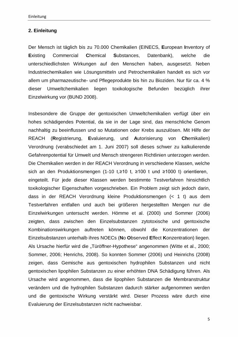
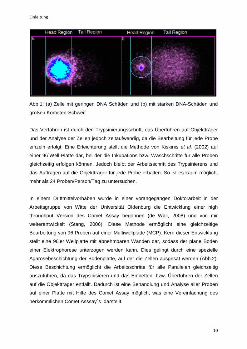
Im herkömmlichen Comet Assay nach den Richtlinien von Tice et al. (2000) werden
die Zellen in Petrischalen ausgesät und mit der zu testenden Substanz inkubiert.
Anschließend werden die adhärenten Zellen gewaschen, trypsiniert und in Low
Melting Agarose (LMP) aufgenommen und auf mit Agarose beschichtete Objektträger
überführt. Nach dem Erhärten der LMP werden die Zellen lysiert und einer
Elektrophorese unterzogen. Dies ermöglicht die Wanderung der DNA Fragmente
durch die Agarosematrix, wodurch der Kometenschweif entsteht. Die Analyse erfolgt
mit Hilfe einer Färbung der DNA und einer mikroskopischen Auswertung einzelner
Zellen. Der Unterschied zwischen dem Verhältnis der DNA im Kopf (Head (Nukleus) -
Region) und im Kometenschweif (Tail-Region]) stellen so die DNA schädigende
Wirkung einer Substanz auf Einzelzellebene dar (Abb.1).
Einleitung
10
Abb.1: (a) Zelle mit geringen DNA Schäden und (b) mit starken DNA-Schäden und
großen Kometen-Schweif
Das Verfahren ist durch den Trypsinierungsschritt, das Überführen auf Objektträger
und der Analyse der Zellen jedoch zeitaufwendig, da die Bearbeitung für jede Probe
einzeln erfolgt. Eine Erleichterung stellt die Methode von Kiskinis et al. (2002) auf
einer 96´Well-Platte dar, bei der die Inkubations bzw. Waschschritte für alle Proben
gleichzeitig erfolgen können. Jedoch bleibt der Arbeitsschritt des Trypsinierens und
das Auftragen auf die Objektträger für jede Probe erhalten. So ist es kaum möglich,
mehr als 24 Proben/Person/Tag zu untersuchen.
In einem Drittmittelvorhaben wurde in einer vorangegangen Doktorarbeit in der
Arbeitsgruppe von Witte der Universität Oldenburg die Entwicklung einer high
throughput Version des Comet Assay begonnen (de Wall, 2008) und von mir
weiterentwickelt (Stang, 2006). Diese Methode ermöglicht eine gleichzeitige
Bearbeitung von 96 Proben auf einer Multiwellplatte (MCP). Kern dieser Entwicklung
stellt eine 96’er Wellplatte mit abnehmbaren Wänden dar, sodass der plane Boden
einer Elektrophorese unterzogen werden kann. Dies gelingt durch eine spezielle
Agarosebeschichtung der Bodenplatte, auf der die Zellen ausgesät werden (Abb.2).
Diese Beschichtung ermöglicht die Arbeitsschritte für alle Parallelen gleichzeitig
auszuführen, da das Trypsinisieren und das Einbetten, bzw. Überführen der Zellen
auf die Objektträger entfällt. Dadurch ist eine Behandlung und Analyse aller Proben
auf einer Platte mit Hilfe des Comet Assay möglich, was eine Vereinfachung des
herkömmlichen Comet Asssay´s darstellt.

Einleitung
11
Abb.2: Darstellung einer MCP Platte mit angehobener beschichteter Bodenplatte
Eine Grundvoraussetzung für den Comet Assay ist die runde Form der Zellen, was
im konventionellen CA durch das Trypsinieren erreicht wird. Im high throughput
Verfahren wird die runde Form der Zellen dadurch erhalten, dass die Zellen sich zwar
leicht anhaften jedoch nicht ausbreiten, wie es die Zellen bei einer Aussaat in der
Petrischale tun würden. So werden die Arbeitsschritte des Trypsinierens und das
einzelne Übertragen der Zellen auf Objektträger vermieden. Es wird sowohl eine
Zeitersparnis (Präparation der Proben), als auch eine Senkung des
Chemikalienverbrauchs (Probensubstanz, Lyselösung, Elektrophoresepuffer und
DNA-Färbemittel) erzielt.
Zur Etablierung der Methode ist es jedoch unerlässlich, dass vergleichbare
Ergebnisse im Vergleich zu der herkömmlichen Methode erzielt werden.
Zur Sicherstellung eines einheitlichen Protokolls wurde eine Richtlinie für die
Durchführung des Comet Assay für Untersuchungen der Gentoxizität durch ein
Gremium verfasst (Tice et al., 2000). In der Richtlinie wurden sowohl die Methode
und deren Durchführung, sowie die Auswertung und Bewertung der Resultate
beschrieben. Dadurch war eine Durchführung des Comet Assay nach einer
einheitlichen Richtlinie möglich.
Dieser Richtlinie liegt der alkalische Comet Assay nach Singh et al. (1988) zu
Grunde, welcher ebenfalls die Basis des high throughput Verfahren darstellt. Hierzu
wurden die Arbeitsschritte der Lyse der Zellen, das alkalische Entwinden der DNA,
die Elektrophorese unter alkalischen Bedingungen, die Neutralisation der
Objektträger, sowie die Anfärbung und das Auswerten der Kometen adäquat zur

Einleitung
12
Richtlinie durchgeführt (Tice et al. 2000). Der so nach Tice et al. (2000)
durchgeführte Comet Assay kann somit als qualitativ hochwertiger und sehr gut
reproduzierbarer Gentoxizitätstest angesehen werden, was seine Einsatzmöglichkeit
in großen Studien und in der Pharmaindustrie ermöglicht.
Zusätzlich zu der Untersuchung der Gentoxizität einer Chemikalie mit Hilfe des
Comet Assay ermöglicht die high throughput Version auch eine Messung der
Zytotoxizität. Dies ist wichtig, da es bei einer Behandlung von Säugerzellen mit stark
zytotoxischen Substanzen zu einer DNA Schädigung aufgrund von nekrotischen und
apoptotischen Ereignissen kommen kann (Henderson et al., 1998). Es wird daher
empfohlen, bei einem Gentoxizitätstest eine parallele Bestimmung der Zytotoxizität
durchzuführen, um falsch positive Resultate durch zytotoxische Wirkungen
auszuschließen (Tice et al., 2000). Dies erfordert im konventionellen CA einen
weiteren Test und somit zusätzliche Testsubstanz, da die zytotoxischen
Testverfahren nicht in die Durchführung der Gentoxizitätstestverfahren integriert sind.
Zur Zytotoxizitätsbestimmung werden häufig die Testverfahren FDA-Assay, MTT-
Assay und der ATP-Assay angewandt, da sie schnell und einfach in ihrer
Durchführung sind.
Das high throughput Verfahren ermöglicht, mit identischen Zellen den zytotoxischen
Test (hier der FDA-Assay nach Rotman and Papermaster (1966)) und die
Gentoxizitätsuntersuchung durchzuführen. Dies gelingt dadurch, dass der
Zytotoxizitätstest in die Durchführung des Gentoxizitätstest integriert ist. Hierzu
erfolgt vor der Lyse der Zellen die Anfärbung mit Fluoreszeindiacetat (FDA), wobei
vitale Zellen das Membran-permeables, nicht fluoreszierendes Fluoreszeindiacetat
aufnehmen. Intrazellulär werden die Acetatgruppen des Moleküls durch Esterasen im
Cytosol zu Acetat und dem lipophilen, grün fluoreszierenden Xanthin-Farbstoff
Fluoreszein hydrolysiert. Das resultierende Fluoreszein akkumuliert in der Zelle mit
intakter Zellmembran, und das nunmehr geladene Molekül kann nur noch langsam
aus der Zelle diffundieren. Anschließend erfolgt die Messung der 96‘er Wellplatte im
Fluoreszenz-reader, um so die Vitalität der behandelten Zellen zu erfassen. Darauf
folgt die Demontage der Wände und es wird mit der Methode des Comet Assay
fortgefahren.

Einleitung
13
Ziel dieser Arbeit war es zu untersuchen, inwieweit der optimierte high throughput
Comet Assay den Anforderungen für ein Screening-Verfahren zum Nachweis der
Gentoxizität entspricht. Aus den erzielten Ergebnissen entstanden 4 Publikationen,
die im Folgenden zusammenfassend dargestellt werden.

Darstellung der Ergebnisse
14
3. Darstellung der Ergebnisse 3.1 Durchführung des high throughput Comet und Vergleich mit dem standardisierten Comet Assay (A. Stang and I. Witte, Performance of the comet assay in a high-throughput version. Mutat Res. 675 (2009) 5-10) Das high throughput Verfahren ermöglichte eine erhebliche Steigerung der zu
untersuchenden Probenanzahl pro Tag. In dieser Studie wurde die high throughput
Version des Comet Assay hinsichtlich ihrer Qualität und Anwendbarkeit in der Praxis
untersucht. Die Evaluierung mit Hilfe von 5 Standardmutagenen mit
unterschiedlichen DNA-schädigenden Potential (Methylmethansulfonat [MMS],
Ethylnitrosoharnstoff, 4-Nitroquinolin-1-oxide, Wasserstoffperoxid [H2O2] und Cis-
platin) zeigte, dass das high throughput Verfahren mit den vorgenommenen
Optimierungen, einschließlich integriertem Zytotoxizitätsverfahren, geeignet ist und
mit hoher Sensitivität und geringen Fehler konzentrationabhängige Effekte detektiert.
Zusätzlich zeigte das Verfahren im Vergleich zum Standardverfahren nach Tice et al.
(2000) bei der Untersuchung von MMS und H2O2
Der Comet Assay im high throughput Verfahren erzielte so eine Steigerung der
Durchsatzrate um den Faktor 20 im Vergleich zum Standardverfahren. Dies
ermöglichte eine Vereinfachung der Messungen großer Probenzahlen bei der
Untersuchung von Industriechemikalien, im Umweltbiomonitoring und Screening von
Verbindungen in der frühen Phase der Wirkstoffentwicklung in der Pharmaindustrie
vorkommen.
quantitativ gleiche Ergebnisse.

Darstellung der Ergebnisse
15
3.2 Automatische Auswertung von Kometen im high throughput Comet Assay
(A. Stang, M. Brend´amour, C. Schunck and I. Witte, Automatic Analysis of Comets in the High Throughput Version of the Comet Assay. Mutat Res. Submitted)
Die Methode des Comet Assay im high throughput Verfahren ist eine schnelle,
einfache und sensitive Methode zur Ermittlung der Gentoxizität. Jedoch bleibt der
limitierende Faktor die manuelle Auswertung der Kometen. In dieser Arbeit wurde in
Zusammenarbeit mit der Firma Metasystems, eine Automatisierung der Auswertung
für den Comet Assay im high throughput Verfahren entwickelt und die automatisierte
Auswertung mit 2 interaktiv arbeitenden Auswertungssystemen verglichen. Die
automatisierte Messung der Verbindungen MMS und H2O2
Die Automatisierung der Auswertung der Kometen ermöglicht eine zusätzliche
Steigerung der Probenzahl, wodurch eine Gesamtsteigerung des Proben-
durchsatzes in der high throughput Version um den Faktor von bis zu 180 im
Vergleich zum konventionellen Verfahren erreicht wird.
zeigte in geringen
Konzentrationsbereichen, eine vergleichbare Sensitivität zu den inter-aktiven
Messungen. Die automatisierte Auswertung erzielte so eine Steigerung der
Geschwindigkeit der Auswertung um den Faktor 10 im Vergleich zur interaktiven
Auswertung.

Darstellung der Ergebnisse
16
3.3 Anwendbarkeit des high throughput Comet Assay unter der Verwendung 5 verschiedener Zelllinien (A. Stang and I. Witte, Ability of the high throughput comet assay to measure comparatively the sensitivity of five cell lines toward methyl methanesulfonate, hydrogen peroxide and pentachlorophenol. Mutat Res. submitted)
Gentoxische Untersuchungen werden mit verschiedenen Zellarten durchgeführt.
Daher sollte ein high throughput Verfahren auch mit vielen Zellarten anwendbar sein.
Ziel dieser Arbeit war es, das high throughput Verfahren hinsichtlich der Verwendung
verschiedener Zellarten zu testen und ein Vergleich der Sensitivität der Zellarten zu
ermitteln. Hierzu wurden die adhärenten Fibroblasten, HeLa-, V79- und HepG2-
Zellen und nicht adhärente Lymphozyten verwendet. Alle Zellen wurden mit MMS,
H2O2
Die erweiterte Anwendung des high throughput Verfahrens für verschiedene,
spezialisierte Zellarten ist für die Forschung, das Screening und Monitoring
interessant. So ist es z.B. jetzt möglich bei Arbeitsplatzmonitoring, oder Unfällen mit
Chemikalien oder Strahlung größere Untersuchungen zur Auswirkung auf den
Menschen mit Hilfe geringen Mengen menschlicher Lymphozyten durchzuführen.
Pentachlorphenol (PCP), welche erst nach der Metabolisierung mit Cytochrom
P450 ein gentoxisches Potential aufweist, behandelt. Die Untersuchung zeigte, dass
unter Berücksichtigung der individuellen Anheftzeit alle getesteten Zellen im high
throughput Verfahren getestet werden können. Ebenso konnten unterschiedliche
Sensitivitäten zwischen den einzelnen Zellarten festgestellt werden, wobei
menschliche Lymphozyten am sensitivsten reagierten.
Darstellung der Ergebnisse
17
3.4 Strategie für das Screening von Umweltproben im Hochdurchsatzverfahren (Minne B. Heringa, Andre Stang, Cyrille A.M. Krul, Astrid A. Reus, Annemarie P. van Wezel, Irene Witte, A high-throughput genotoxicity testing strategy for screening of (drinking) water. Mutat Res. Submitted)
Für „Biomonitoring-Studien“ sind high throughput Verfahren für das Screening und
der Evaluierung von gentoxischen sowie mutagenen Wirkungen sinnvoll, da es eine
Vielzahl von Proben zu testen gilt. Aus diesem Grund wurde in dieser Arbeit
untersucht, ob das high throughput Verfahren des Comet Assay mit dem Ames II
Test korreliert. Zusätzlich wurde die Sensitivität der high throughput Version, des
Ames II Test und des Mikronukleustest verglichen. Dazu wurden Umweltproben mit
geringen gentoxischen Wirkungen untersucht.
Die Untersuchung von 9 verschiedenen gentoxisch wirkenden Substanzen ergab,
dass die Ergebnisse vom Ames II Test und Comet Assay sich sehr gut ergänzten.
Ebenso zeigte der Comet Assay bei der Untersuchung der Umweltproben eine
höhere Sensitivität als der Ames II Test (3 von 8 Proben positiv im Comet Assay
getestet, im Vergleich 1 Probe im Ames II Test positiv getestet), während mit dem
MT keine Mutationen festgestellt werden konnte.
Die Untersuchungen zeigte, dass der Ames II und der Comet Assay im high
throughput Verfahren sich ergänzten und so eine gute Kombination für eine
Testbatterie zur Untersuchung von Umweltproben im Rahmen des Biomonitoring
wäre. So ist es nun möglich, schnell und sensitiv gentoxisch wirkende Proben zu
identifizieren.
Ausblick
18
4. Ausblick
Die stetig wachsende Anzahl an Xenobiotika, die in die Umwelt gelangen, verursacht
ein erhebliches Risikopotential für den Menschen. Dieses Risikopotential entsteht
nicht nur durch die Einzelsubstanzen, sondern hauptsächlich durch
Kombinationseffekte. Bis heute stehen zur Ermittlung von Schadstoffen in
Umweltproben chemisch analytischen Verfahren im Vordergrund. Die Ergebnisse
hieraus erlauben jedoch noch keine Aussage über das gentoxische Potential des
Gemisches oder der Umweltprobe.
Mit dem in dieser Arbeit entwickelten und auf Praxistauglichkeit getesteten high
throughput Verfahren ist es möglich, mit vergleichsweise geringem Aufwand und
geringen Kosten Kombinationswirkungen zu untersuchen bzw. ein Screening auf
mögliche gentoxische Substanzen durchzuführen. Jedoch liegt die Durchführung des
Comet Assay noch nicht wie andere Testverfahren (Ames-, Mikronukleus- und
Chromosomenaberrations-Test) als OECD (Organisation for Economic Co-operation
and Development) Richtlinie vor. Möglicherweise liegt das daran, dass
Mutationstests höher bewertet werden als der Nachweis DNA-schädigender
Wirkungen. Da in verschiedenen Studien inzwischen gezeigt wurde, dass der Comet
Assay gut mit den anderen Testverfahren korreliert (Hartmann et al., 2003; Giannotti
et al., 2002), sollte dieses Argument entfallen. Da der Comet Assay sensitiver,
schneller in seiner Durchführung und breiter in seiner Anwendung als die genannten
Mutationstests ist, könnte der high throughput Comet Assay als Screening-Test den
Mutationstests vorangestellt werden. In den (seltenen) Fällen eines positiven
Befundes wäre der Einsatz von Mutationstests gefordert. Damit könnten alle
Substanzen schneller und sensitiver auf Gentoxizität, wie es in der REACH
Verordnung oder bei der Evaluierung der Phototoxizität notwendig ist, geprüft
werden.
Der AT, MT, CT und der konventionelle Comet Assay sind „hightech“ Verfahren und
benötigen zur Durchführung ein komplett eingerichtetes Zellzuchtlabor, wodurch eine
Anwendung in Entwicklungsländern oder Schwellenländern nur bedingt möglich ist.
Der high throughput Comet Assay hingegen ermöglicht die Anwendung in „lowtech“ -
Regionen, da er bei der Durchführung mit humanen Lymphozyten keine Zellzucht

Ausblick
19
benötigt und auch unter semi-sterilen Bedingungen funktioniert, da die Durchführung
innerhalb von Stunden erfolgt.
Die erweiterte Anwendung des high throughput Verfahrens auf verschiedene
Zellarten ermöglicht eine Vielzahl von Tests im Comet Assay. So könnte mit Hilfe des
high throughput Comet Assay organspezifische gentoxische Wirkungen an den
Zellarten des jeweiligen Zielorganes untersucht werden. Auch ermöglicht der high
throughput Comet Assay eine bessere Untersuchung der DNA-Reparaturkinetiken
gegenüber dem konventionellen Assay, da der Trypsinierungsschritt entfällt und
somit im Gegensatz zum konventionellen Comet Assay auch der Beginn der
Reparatur erfasst werden kann.

Literatur
20
Literatur
Ames BN, Lee FD, Durston WE (1973) An improved bacterial test system for the
detection and classification of mutagens and carcinogens. Proceedings of the
National Academy of Sciences of the United States of America, Vol. 70, 782-786
Collins A R (2004) The comet assay for DNA damage and repair: applications and
limitations. Molecular biotechnology, Vol. 26, 249-261
de Wall H (2008) Entwicklung eines in vitro High Throughput Comet Assay in einer
96 Multichamberplatte. Dissertation. Carl von Ossietzky Universitat Oldenburg.
Giannotti E, Vandin L, Repeto P, Comelli R (2002) A comparison of the in vitro
Comet assay with the in vitro chromosome aberration assay using whole human
blood or Chinese hamster lung cells: validation study using a range of novel
pharmaceuticals. Mutagenesis, Vol. 17, 163-170
Hartmann A, Elhajouji A, Kiskinis E, Poetter F, Martus H, Fjällman A, Frieauff W, Suter W (2001) Use of the alkaline comet assay for industrial genotoxicity screening:
comparative investigation with the micronucleus test. Food and chemical toxicology,
Vol. 39, 843-858
Hartmann A, Plappert U, Poetter F, Suter W (2003) Comparative study with the
alkaline Comet assay and the chromosome aberration test. Mutation Research, Vol.
536, 27-38
Hartmann A (2004) Evaluation des Comet Assay. Kumulative Habilitationsschrift für
das Fachgebiet Experimentelle Humangenetik, Medizinische Fakultät Ulm
Henderson L, Wolfreys A, Fedyk J, Bourner C, Windebank S (1998) The ability of
comet assay to discriminate between genotoxins and cytotoxins. Mutagenesis, Vol.
13, 89-94
Literatur
21
Henrichs K (2008) Ursachen und Folgen der toxischen Kombinationswirkungen von
oxidativem Stress und nicht gentoxischen Umweltchemikalien auf menschliche
Fibroblasten. Dissertation. Carl von Ossietzky Universitat Oldenburg.
Hömme M, Jacobi H, Juhl-Strauss U, Witte I (2000) Synergistic DNA damaging
effects of 4-nitroquinoline-1-oxide and non-effective concentrations of methyl
methanesulfonate in human fibroblasts. Mutation Research, Vol. 461, 211 – 219
Kiskinis E, Suter W, Hartmann A (2002) High throughput Comet assay using 96-
well plates. Mutagenesis, Vol. 17, 37-43
Lee R F, Steinert S (2003) Use of the single cell gel electrophoresis/comet assay
for detecting DNA damage in aquatic (marine and freshwater) animals. Mutation
Research, Vol. 544, 43-64
Miller B, Albertini S, Locher F, Thybaud V, Lorge E (1997) Comparative
evaluation of the in vitro micronucleus test and the in vitro chromosome aberration
test: industrial experience. Mutation Research, Vol. 392, 45-59, 187-208
Møller P (2005) Genotoxicity of environmental agents assessed by the alkaline
comet assay. Basic & clinical pharmacology & toxicology, Vol. 96, 1-42.
Östling O und Johanson K J (1984) Microelectrophoretic study of radiation-
induced DNA damages in individual mammalian cells. Biochemical and Biophysical
Research Communications, Vol. 123, 291-298
Rotman B and Papermaster B W (1966) Membrane properties of living mammalian
cells as studied by enzymatic hydrolysis of fluorogenic esters. Proceedings of the
National Academy of Sciences of the United States of America, Vol. 55, 134 – 141
Singh N P, McCoy M T, Tice R R und Schneider E L (1988) A simple technique for
quantitation of low levels of DNA damage in individual cells. Experimental Cell
Research, Vol. 175, 184-191
Literatur
22
Sommer H (2006) Zyto- bzw. gentoxische Wirkschwellen von Gemischen aus 2 – 8
Umweltchemikalien in Abhangigkeit von der Lipophilitat der Komponenten.
Dissertation. Carl von Ossietzky Universitat Oldenburg.
Stang A (2006) Optimierung einer Methode zur Messung der Gentoxizität (Comet
assay) im High Throughput Verfahren. Leistungsnachweis. Carl von Ossietzky
Universitat Oldenburg.
Tice R R, Agurell E, Anderson D, Burlinson B, Hartmann A, Kobayashi H, Miyamae Y, Rojas E, Ryu J-C, Sasaki Y F (2000) Single cell gel/comet assay:
Guidelines for in vitro and in vivo genetic toxicology testing. Environmental and
molocular mutagenesis, Vol. 35, 206-221.
Umweltbundesamt (2008) www.reach-info.de
Umweltbundesamt (2009) EINECS Datenbank: www.umweltbundesamt.at
Witte I, Jacobi H, Juhl-Strauss U (2000) Krebsrisiken durch Gemische aus
Umweltchemikalien und erbgutschädigenden Stoffen. Abschlussbericht BMBF
Vorhaben 07 GTX 03/6, 67 Seiten
Witte I, Plappert U, de Wall H, Hartmann A (2007) Genetic toxicity assessment:
employing the best science for human safety evaluation part III: the comet assay as
an alternative to in vitro clastogenicity tests for early drug candidate selection.
Toxicological sciences, Vol. 97, 21-26

Publikationen der Ergebnisse
23
5. Publikationen der Ergebnisse
5.1 Performance of the Comet Assay in a High Throughput Version
(Mutat. Res. 675 (2009) 5-10)
Stang, A. and Witte, I.*
Carl von Ossietzky Universität Oldenburg, IBU, Postfach 2503, D-26111 Oldenburg,
Germany
*corresponding author:
Irene Witte
Institut für Biologie und Umweltwissenschaften
AG Biochemie Umwelttoxikologie
Carl von Ossietzky Universität Oldenburg
Ammerländer Heerstraße 114-118
D-26129 Oldenburg
Germany
tel.: +49-441-7983628
E-mail: [email protected]

Performance of the Comet Assay in a High Throughput Version
24
Abstract
The high throughput comet assay was developed to reduce the processing time and
to increase sample throughput of the comet assay as described by Tice et al. [1].
This high throughput version allows for the processing of up to 400 samples per day.
The basis of the new assay is a 96 well plate (multichamber plate, MCP) suitable for
electrophoresis. After exposure of the cells to genotoxic agents, the walls of the MCP
are separated from the bottom plate. All 96 samples together then go through lysis,
alkaline unwinding, electrophoresis, neutralization, and staining.
In this study, the first concentration-dependent results obtained with the high
throughput version are shown and a comparison with the standard version of the
comet assay is made using five representative chemicals with different genotoxic
properties. These genotoxic chemicals were methylmethane sulfonate (MMS), and
ethylnitroso urea for small alkylation adducts, 4-nitroquinoline-1-oxide for bulky
adducts, cisplatin for DNA crosslinks, and H2O2 for direct DNA breakage. For
medium and high effective concentrations a standard deviation of 3-20 % for three
replicates (25 comets per sample) was determined. A comparison of the standard
assay and the high throughput version revealed similar results shown for MMS and
H2O2
. The integrated viability assay (FDA assay), which was performed after
chemical treatment and before detachment of the bottom from the walls of the MCP,
did not influence the outcome of the comet formation.
In conclusion, the high throughput version of the comet assay facilitates determining
genotoxicity in cases where large numbers of samples have to be measured, such as
testing industrial chemicals, biomonitoring of environmental samples, and early
genotoxicity/photogenotoxicity screening of drug candidates. For such applications
the cost- and time-saving of the high throughput method provides substantial
advantages over the standard comet assay.
Keywords
comet assay; high throughput; MCP; multichamber plate; adherent cells
Performance of the Comet Assay in a High Throughput Version
25
1. Introduction
Measuring genotoxicity is an important step in the complex procedure of determining
the carcinogenic potential of chemicals. In areas such as environmental monitoring or
in early drug candidate selection a high throughput method for screening genotoxicity
would be beneficial. However, among current methods frequently used for
determining genotoxicity (e.g. the micronucleus test (MNT), the chromosome
aberration (CA) test, the comet assay), no high throughput version exists. The
microscopical evaluations of these tests are very time consuming. In addition, in each
genotoxicity test, it is necessary to evaluate the cytotoxicity of the test compounds in
parallel to exclude false positive results. An excessive cytotoxicity leads in some
cases to positive outcomes in these assays [2].
The comet assay and the MNT are potential methods for increasing the screening
rate of substances. Automatically working evaluation systems exist for both methods
[3-5]. Nevertheless, the performance of these assays remains time consuming and is
not suitable for screening a large number of samples per day. The comet assay has
some advantages over the CA and MNT. The CA and MNT require proliferating cells,
while in the comet assay genotoxicity can be detected also in non-dividing cells. In
addition, the CA [6] and MNT [7] show higher frequencies of false positive results in
comparison with the comet assay. Therefore, these assays are either less specific, or
more sensitive towards positive effects associated with cytotoxicity than the comet
assay.
The standard procedure of the comet assay according to the recommendations of
Tice et al. [1] is not suitable for screening a large number of samples for genotoxicity.
Even if the throughput in the comet assay could be increased by using standard 96
well plates [8], time limiting steps like trypsinization of adherent cells of each sample
and the subsequent individual treatment of all samples remain. These include mixing
of the cells with agarose, spreading them on precoated slides, followed by lysis,
electrophoresis, and staining. To avoid these time- and sample number-limiting
steps, a high throughput version of the comet assay was developed [9].
Performance of the Comet Assay in a High Throughput Version
26
The basis of the high throughput version of the comet assay is a 96-well plate
(multichamber plate, MCP) suitable for electrophoresis. An agarose containing
surface on the bottom of the MCP allows the cells to attach. After chemical treatment
of the 96 samples, the walls of the MCP are removed from the bottom plate. The
subsequent steps can be performed for all 96 samples at the same time. In addition
to an assessment of DNA damage, the viability of the cells can be determined after
chemical treatment by staining with fluorescein diacetate and evaluation by a
fluorescence reader. Thereafter, the walls of the MCP are removed and the
procedure of the comet assay can be continued. Thus, cell viability can be measured
under identical conditions and with the same cells with which the comet formation is
determined.
In this study we present our first results of the comet assay performed with the MCP.
We determined the extent of comet formation in human fibroblasts induced by five
DNA damaging chemicals with different DNA damaging properties: direct strand
breakages (H2O2
), damages with small conformational changes of the DNA (MMS,
ethylnitroso urea ENU), large conformational changes of DNA caused by bulky
adducts (4-NQO), and crosslinking (cisplatin). We compared the conventional comet
assay with the new high throughput version. Further, we compared the comet
formation with and without integrated cytotoxicity testing.
2. Materials and methods
2.1 Cell culture Human fibroblasts cell line NHDF-p were purchased from Promochem (Heidelberg,
FRG). Monolayer cultures (passage 8-15) were grown in D-MEM at 37 °C in an
atmosphere of 5 % CO2
and 95 % air with > 95 % humidity.
2.2 Multichamber plate (MCP)
The MCP is a specially coated 96 well plate purchased from Intox, Oldenburg, FRG,
which allows the electrophoresis of cellular DNA. The surrounding walls of the wells
Performance of the Comet Assay in a High Throughput Version
27
can be separated from the plate of the MCP. The cells of 96 wells remain on the flat
plate and all samples together can be electrophorized.
2.3 Chemical treatment of the cells Cells were treated with MMS (> 99 %, from Sigma, Deisenhofen, FRG), ENU (from
Sigma, Deisenhofen, FRG), 4-NQO (98.2 %, from Sigma, Deisenhofen, FRG),
cisplatin (Medoc, Hamburg, FRG), or with H2O2 (37 % aqueous solution from Acros
Organics, NJ). All chemicals were used in non cytotoxic concentrations. For H2O2
maximum of DNA damage was observed after a 15 min incubation because of its
short half life. The other chemicals required an incubation of 1.5 hours to exert a high
level of DNA damage. MMS, ENU, cisplatin and 4-NQO were freshly dissolved in
serum free medium (sfm) at pH 7.2 and 37 °C directly before cell treatment. H2O2
was diluted from the 37 % aqueous solution with sfm also immediately before cell
treatment. DNA damage induced by the crosslinker cisplatin was measured indirectly.
It was detected according to Pfuhler and Wolf [10] by previous treatment with MMS
(2.1 mM for 1 hour) and subsequent addition of cisplatin for 1.5 hours. The %
reduction of DNA strand breaks induced by MMS, named “relative DNA damage [%]”
in the graphs, quantitatively reflects the DNA crosslinks induced by cisplatin.
2.4 Comet assay performed in the standard mode
The comet assay was performed according to Tice et al. [1]. In brief, 30,000 cells
seeded into Petri dishes (5 cm in diameter) were treated with chemicals as described
above. Thereafter the cells were washed with ice-cold phosphate buffered saline
(PBS), trypsinized (0.125 % trypsin), and resuspended in 100 µl ice-cold PBS. A
volume of 20 µl of the resuspended cells was mixed with 80 µl 0.5 % low melting
agarose at 37 °C and applied to pretreated microscope slides. Pretreatment of slides
involved coating with 1.5 % agarose, diluted in Ca2+ and Mg2+- free PBS, pH 7.4.
Each concentration was performed in duplicate. The slides mounted with cells were
covered with coverslips and kept in the refrigerator for 3-5 min to solidify the low
melting agarose. The following steps were performed under dim-light to prevent
additional UV-induced DNA damage. After removing the coverslips, slides were
immersed in 4 °C cold lysing solution pH 10.0 (2.5 M NaCl, 100 mM EDTA, 10 mM

Performance of the Comet Assay in a High Throughput Version
28
Tris, 1 % N-lauroyl sarcosine, 1 % Triton X100, 10 % DMSO; the last two compounds
were added freshly). Slides were kept at 4 °C for 1 h. After lysis, the slides were
placed on a horizontal electrophoresis box. The unit was filled with freshly prepared
alkaline buffer (300 mM NaOH, 1 mM EDTA, pH 13), until slides were completely
covered with buffer. After an incubation for 40 min at 4 °C in alkaline buffer, to allow
DNA unwinding and DNA breakage at alkali labile sites, DNA electrophoresis was
performed in an ice bath at 25 V and 300 mA for 20 min. After electrophoresis, the
slides were covered with neutralization buffer (0.4 M Tris HCl, pH 7.5) for 5 min. This
step was repeated twice. Thereafter, the slides were briefly dipped into water and
dried by air overnight. Finally, 40 µl ethidium bromide (20 µg/ml) was added to each
slide. Slides were covered with a coverslip and kept for 5 min in the dark for DNA
staining. DNA migration was analyzed by fluorescence microscopy (Nikon, Eclipse
E600W). The tail moment (tm) as well as % DNA tail was determined using the
software ”Lucia comet assay Single Stain” (Nicon). For each concentration, 100
randomly selected cells (50 cells each from two duplicate slides) were analyzed, and
the medians of the non-normally-distributed values were determined.
2.5 Comet assay in the high throughput version The comet assay in the high throughput version was performed on the basis of the
guidelines of Tice et al. [1] with two important differences. First, the treatment of the
cells with the genotoxic agents started 2-4 hours after seeding of the cells. Secondly,
trypsinization of adherent growing cells was avoided. In addition, cell viability was
measured with the same cells that were subsequently used for the comet assay.
Fibroblasts were seeded into the wells of the MCP (3,000 cells/well) two or four hours
before treatment of the cells. Thereafter, treatment with the genotoxic chemicals
followed as described above. For each concentration, 3-6 parallel replicates were
performed. Then, the walls surrounding the wells of the MCP were separated from
the bottom plate. The plate with the cells was covered with a 37 °C warm solution of
0.5 % low-melting agarose (type Sea Plaque from Biozym Diagnostik, Hessisch
Oldendorf, FRG). The plate was kept in the refrigerator for 5 minutes to solidify the
low melting agarose. The following steps were performed according the procedure
recommended by Tice et al. [1] (see section 2.4). In brief, the plate was covered with
Performance of the Comet Assay in a High Throughput Version
29
refrigerated lysing solution pH 10.0 and kept at 4 °C for 1 hour. After lysis the plate
was placed on a horizontal electrophoresis box. The box was filled with freshly
prepared alkaline buffer whereby the plate was completely covered with the buffer.
During 40 minutes at 4 °C the alkaline treatment allowed alkaline unwinding of the
DNA and DNA breakage at alkali labile sites. After electrophoresis the plate was
covered three times with neutralization buffer, washed with aqua bidest., and stained
with ethidium bromide. Fluorescence microscopy was used to determine the median
DNA migration in each of the 3-6 parallel samples per concentration (25
comets/well). From these 3-6 values the mean and standard deviation was
calculated. Statistical significance of the differences between the DNA migration (%
DNA tail) induced by the mutagens in the standard assay and the high throughput
method was determined by the non-parametric Mann-Whitney test (p < 0.01).
2.6 Measurement of cell viability integrated in the comet assay procedure For measurement of the cell viability, we used the FDA assay according to Rotman
and Papermaster [11]. The FDA assay measures the cell viability by the activity of
cytosolic esterases converting fluorescein diacetate (FDA) to the fluorescent dye
fluorescein.
After treatment of the cells with the genotoxic agent MMS, the fibroblasts were
incubated for 10 minutes with 72 µM FDA (from 12 mM stock solution in acetone,
freshly diluted with sfm) at 37 °C in the dark. Thereafter, the dye was removed and
the fluorescence of the enzymatically formed fluorescein was measured in a
fluorescence reader (FLUOstar, Offenbach, FRG) with an excitation of 485 nm and
an absorption at 520 nm. No significant differences between the untreated controls
and the MMS treated samples were found. Therefore the MMS treatment was not
cytotoxic. After fluorescence measurement the walls and the bottom plate of the MCP
were separated and the comet assay procedure was continued as described above.

Performance of the Comet Assay in a High Throughput Version
30
3. Results
3.1 Influence of the time interval between seeding of fibroblasts and experiment on comet formation
To perform the comet assay with adherent cells it is necessary that cells are
embedded in the coating layer in a rounded form. This is achieved in the standard
comet assay by trypsinization after chemical treatment of adherent cells. On the MCP
where trypsinization does not occur, the cells have to remain in their rounded
morphology after seeding. This is necessary because in the comet assay the intact
DNA is spread after lysis within the volume of the former cell. We seeded trypsinized
cells on the MCP and performed the comet assay of these non-treated cells 2-8
hours after seeding. In Fig.1a-1d the “comets” of the cells without chemical treatment
are shown. Up to 4 hours after seeding the DNA was found in a rounded shape. After
this time the DNA began to spread on the MCP. This means, chemical treatment
should occur 2-4 hours after seeding. During this period, statistical significant
differences in the comet formation induced by chemicals were not observed (data not
shown).
3.2 Comet formation on the MCP induced by DNA damaging chemicals
Human fibroblasts were seeded into the wells of the MCP and treated after 4 hours
with increasing concentrations of the DNA damaging agents MMS, ENU, 4-NQO, and
cisplatin for 1.5 hours. At least 5 parallel samples per concentration were made. In
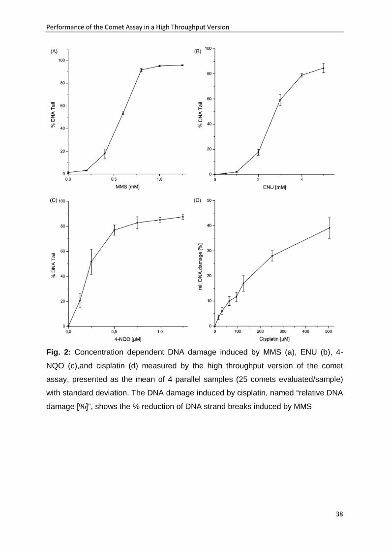
Fig. 2a-2d, the resulting comet formation is shown. For all chemicals a concentration
dependent increase in DNA damage was observed while the untreated controls did
not show any DNA migration. The lowest concentration of MMS to show DNA
damage was 0.2 mM, of ENU 0.5 mM, of 4-NQO 0.125 µM, and of cisplatin 15.8 µM.
In Table 1, the tail moments (tm) and the % DNA tail obtained after treatment of
fibroblasts with 4-NQO are listed. The standard deviations were calculated from 3-6
identically treated samples in the same MCP. 25 comets were evaluated in each well.
The standard deviations were similar regardless of the number of (3, 4, 5 or 6) wells
evaluated.
Performance of the Comet Assay in a High Throughput Version
31
3.3 Comparison of the comet formation with and without integrated viability assay
The comet assay in the high throughput version was quantitatively compared with
and without integrated FDA assay. After incubation with MMS, the fibroblasts were
treated for 10 minutes either with the staining agent FDA, or with FDA-free sfm. After
removing the FDA solution, or the sfm, the fluorescence was measured in the
fluorescence reader. None of the MMS concentrations tested, revealed any
cytotoxicity (data not shown). This is in accordance with data from literature where
highly genotoxic concentrations of MMS, measured in the standard comet assay
were not cytotoxic in human fibroblasts [12]. After fluorescence measurement the
bottom plate was demounted from the walls, covered with low melting agarose and
the comet assay procedure was continued. In Fig. 3 the results of comet formation
with and without FDA staining is shown. There were no differences between the two
approaches.
3.4 Comparison of the comet formation in the standard assay and on the MCP
The comet assay in the standard mode according to Tice et al. [1] and in the high
throughput version was directly compared using the genotoxic chemicals MMS and
hydrogen peroxide. The results of MMS are shown in Fig. 4, in Fig.5 those of H2O2.
Between both methods no statistical significance was measured by the non-
parametric Mann-Whitney test (p < 0.01) (exception: 20 µM H2O2
).
4. Discussion
One of the more time consuming steps in the standard comet assay with adherent
cells, except for scoring the comets, is the trypsinization step. To avoid this step a
necessary precondition for experimentation with adherent cells is that the cells
remain in their rounded shape after seeding. Fibroblasts, which assume an elongated
shape when spread on the bottom of a Petri dish, remained on the MCP in their
rounded form up to 4 hours after seeding. Thereafter, they slowly started to spread. It
was investigated, if a short time period of 2-4 hours of attachment would elevate the
sensitivity of untreated cells caused by damages via trypsin. This was not the case

Performance of the Comet Assay in a High Throughput Version
32
because the untreated cells on the MCP did not show any comet formation just as in
the standard assay where cells were seeded one day before the experiment. Singh
et al. [13] first reported the performance of the comet assay 4 hours after seeding the
cells. They successfully demonstrated DNA migration after irradiation with x-rays or
treatment with H2O2
. These and our observations show that seeding and
performance of the experiment on the MCP can be done on the same day.
Preliminary results with other adherent cell types suggest, the time between seeding
and chemical treatment may be vary dependent on the properties of the cell types to
adhere. This time has to be established for each cell type, individually. With non
adherent cells, like lymphocytes, the high throughput version of the comet assay can
also be performed. In this case the suspension cultures already pipetted into the
wells of the MCP have to be centrifuged before and after chemical treatment
(publication in preparation).
A concentration-dependent increase in comet formation was demonstrated with the
newly developed high throughput version of the assay. This was independent of
which kind of DNA damaging agent was used. A comparison of the standard assay
and the high throughput mode revealed similar results. This means that the extra
time needed for trypsinization in the standard assay is too small to detect additional
DNA repair resulting in reduced DNA damage.
The calculation of the standard deviation revealed the homogeneity of the parallel
samples on the MCP, because the values were similar between n=3 and n=6. This is
important because it means that on one MCP, 32 different samples can be measured
with a high degree of confidence.
It is known that the comet formation generally does not follow a Gaussian distribution
[14]. That means, that from one sample with 50 or more comets evaluated, the
medians (with percentiles) have to be calculated. The mean and the standard
deviation can only be obtained by measuring at least three, better more parallels. In
contrast to the standard assay, the performance of parallel samples in the high
throughput version is easy and rapid, and allows the determination of the mean with
standard deviation.

Performance of the Comet Assay in a High Throughput Version
33
The measurement of the cell viability by FDA as part of the high throughput
procedure did not influence the comet formation. This means that the vital dye
fluorescein diacetate does not possess genotoxic properties. Under our conditions
the lower detection limit was reached at about 300 untreated cells/well (data not
shown). Using 3000 cells/well, a reduction of cell viability > 50 % was detectable.
This is extremely helpful to know, because false positive results of genotoxicity due to
high cytotoxicity can be determined by the integrated cytotoxicity [15] measurement.
Most chemicals have to be metabolically activated to exert their genotoxic potential. It
is generally achieved in the in vitro comet assay by adding cofactor-supplemented
postmitochondrial (S9) fraction to the incubation mixture [1]. This can also be done in
the high throughput version of the comet assay. In first experiments with indirectly
acting carcinogens comet formation was observed in the presence of S9 mixture,
while S9 mixture alone did not provoke DNA migration (publication in preparation).
We have developed this high throughput assay for in vitro testing great numbers of
samples. But it is feasible to also apply it for in vivo experimentations. However, for in
vivo testing there are generally few samples available at the same time and
trypsinization is omitted, so that the advantages of the high throughput method are
reduced.
In conclusion, the high throughput version of the comet assay is useful for screening
large numbers of samples. A comparison with the standard assay yielded similar
results. An automatic evaluation system for the comets will further accelerate the
speed with which the assay can be done. Such a prototype is under investigation in
our laboratory. This evaluation system microscopically analyses the entire MCP
counting 50 comets/ well within about 2 hours (publication in preparation). The
combination of the MCP and the new automatically working evaluation system
enables the measurements of about 400 samples per day. No other mammalian test
system for genotoxicity permits a similar high throughput. Therefore, the high
throughput version of the comet assay presented here will be of great value for
screening genotoxicity.
Performance of the Comet Assay in a High Throughput Version
34
Acknowledgements The authors gratefully acknowledge the excellent technical assistance of Elke
Frahmann and Marita Weerts-Eden. We thank Dr. Juhl-Strauss for critical reading of
the manuscript.
This work was supported by the EC, project EVK1-CT-2002-30027.
Abbreviations CA chromosome aberration test
D-MEM Dulbecco's modified Eagle medium
ENU ethylnitroso urea
FDA fluorescein diacetate
MCP multichamber plate
MMS methyl methanesulfonate
MNT micronucleus test
4-NQO 4-nitroquinoline-1-oxide
sfm serum free medium
tm tail moment

Performance of the Comet Assay in a High Throughput Version
35
References
[1] RR. Tice, E. Agurell, D. Anderson, B. Burlinson, A. Hartmann, H. Kobayashi, Y. Miyamae, E. Rojas, JC. Ryu, YF. Sasaki, Single cell gel/comet assay: guidelines
for in vitro and in vivo genetic toxicology testing, Environ. Mol. Mutagen.35 (2000)
206-21
[2] DJ. Kirkland, L. Mueller, Interpretation of the biological relevance of genotoxicity
test results: the importance of thresholds, Mutat Res. 464 (2000) 137-47
[3] W. Frieauff, F. Pötter-Locher, A. Cordier, W. Suter, Automatic analysis of the
in vitro micronucleus test on V79 cells, Mutat. Res. 413 (1998) 57-68
[4] W. Frieauff, A. Hartmann, W. Suter, Automatic analysis of slides processed in
the Comet assay, Mutagenesis. 16 (2001) 133-7
[5] C. Schunck, T. Johannes, D. Varga, T. Lörch, A. Plesch, New developments in
automated cytogenetic imaging: unattended scoring of dicentric chromosomes,
micronuclei, single cell gel electrophoresis, and fluorescence signals, Cytogenet
Genome Res. 104 (2004) 383-9
[6] DJ. Kirkland, L. Henderson, D. Marzin, L. Müller, JM Parry, G. Speit, DJ. Tweats, GM. Williams, Testing strategies in mutagenicity and genetic toxicology: an
appraisal of the guidelines of the European Scientific Committee for Cosmetics and
Non-Food Products for the evaluation of hair dyes, Mutat Res. 588 (2005) 88-105
[7] A. Hartmann, A. Elhajouji, E. Kiskinis, F. Poetter, H. Martus, A. Fjällman, W. Frieauff, W. Suter, Use of the alkaline comet assay for industrial genotoxicity
screening: comparative investigation with the micronucleus test, Food Chem Toxicol.
39 (2001) 843-58
[8] E. Kiskinis, W. Suter, A. Hartmann, High throughput Comet assay using 96-well
plates, Mutagenesis. 17 (2002) 37-43

Performance of the Comet Assay in a High Throughput Version
36
[9] I. Witte, U. Plappert, H. de Wall, A. Hartmann, Genetic toxicity assessment:
employing the best science for human safety evaluation part III: the comet assay as
an alternative to in vitro clastogenicity tests for early drug candidate selection,
Toxicol.Sci. 97 (2007) 21-6
[10] S. Pfuhler, HU. Wolf, Detection of DNA-crosslinking agents with the alkaline
comet assay, Environ. Mol. Mutagen. 27 (1996) 196-201
[11] B. Rotman, BW. Papermaster, Membrane properties of living mammalian cells
as studied by enzymatic hydrolysis of fluorogenic esters, Proc. Natl. Acad. Sci. U S
A. 55 (1966) 134-41
[12] M. Hömme, H. Jacobi, U. Juhl-Strauss, I. Witte, Synergistic DNA damaging
effects of 4-nitroquinoline-1-oxide and non-effective concentrations of methyl
methanesulfonate in human fibroblasts, Mutat. Res. 461 (2000) 211-219
[13] NP Singh, RR Tice, RE Stephens, EL Schneider, A microgel electrophoresis
technique for the direct quantitation of DNA damage and repair in individual
fibroblasts cultured on microscope slides. Mutat. Res. 252 (1991) 289-296
[14] P. Duez, G. Dehon, A. Kumps, J. Dubois, Statistics of the comet assay: a key
to discriminate between genotoxic effects. Mutagenesis 18 (2003) 159-166
[15] L. Henderson, A. Wolfreys, J. Fedyk, C. Bourner, S. Windebank, The ability
of comet assay to discriminate between genotoxins and cytotoxins, Mutagenesis 13
(1998) 89-94

Performance of the Comet Assay in a High Throughput Version
37
Figures and Tables
Fig. 1: Shape of “comets” obtained from untreated cells a) 2h b) 4 h c) 6h d) 8 h after
seeding on the MCP and performance of the comet assay
Performance of the Comet Assay in a High Throughput Version
38
Fig. 2: Concentration dependent DNA damage induced by MMS (a), ENU (b), 4-
NQO (c),and cisplatin (d) measured by the high throughput version of the comet
assay, presented as the mean of 4 parallel samples (25 comets evaluated/sample)
with standard deviation. The DNA damage induced by cisplatin, named “relative DNA
damage [%]”, shows the % reduction of DNA strand breaks induced by MMS

Performance of the Comet Assay in a High Throughput Version
39
Fig. 3: High throughput comet formation induced by MMS with (black bars) and
without (white bars) integrated viability assay. The data represent the mean of 4
parallel samples (25 comets evaluated/sample) with standard deviations

Performance of the Comet Assay in a High Throughput Version
40
Fig. 4: DNA strand break induction by MMS determined in the standard assay
according to Tice et al. [1] (■, median of 100 comets) and on the MCP (▲, median of
100 comets in 4 parallel samples); between both methods, no statistical significance
was measured using the Mann-Whitney test (p < 0.01).

Performance of the Comet Assay in a High Throughput Version
41
Fig. 5: DNA strand breaks induction by H2O2 determined in the standard assay
according to Tice et al. [1] (■, median of 100 comets) and in the MCP (▲, median of
100 comets in 4 parallel samples); between both methods no statistical significance
was measured by the Mann-Whitney test (p < 0.01) except 20 µM H2O
2

Performance of the Comet Assay in a High Throughput Version
42
Tab. 1: Comet formation in the high throughput comet assay induced by 4-NQO
evaluated by the (a) tail moment (tm) or (b) % DNA tail with the standard deviations
in dependence on the number (n) of wells. In each well 25 comets were measured.
Automatic Analysis of Comets in the High Throughput Version of the Comet Assay
43
5.2 Automatic Analysis of Comets in the High Throughput Version of the Comet Assay
(submitted)
A. Stang1, M. Brend´amour2, C. Schunck2 and I. Witte1
*
1Carl von Ossietzky Universität Oldenburg, IBU, Postfach 2503, D-26111 Oldenburg,
Germany; 2 MetaSystems, Robert-Bosch-Str.6, D-68804 Altlussheim, Germany
*corresponding author:
Irene Witte
Institut für Biologie und Umweltwissenschaften
AG Biochemie Umwelttoxikologie
Carl von Ossietzky Universität Oldenburg
Ammerländer Heerstraße 114-118
D-26129 Oldenburg
Germany
Tel.: +49-441-7983628
E-mail: [email protected]
Automatic Analysis of Comets in the High Throughput Version of the Comet Assay
44
Abstract
Recently a high throughput version of the comet assay was developed using a
special 96-well plate (MCP, multichamber plate) [1]. In this version, the
electrophoresis is performed directly on the MCP which makes transferring of cells to
microscope slides unnecessary.
In order to facilitate the scoring procedure we adapted an automated slide scanning
system (Metafer MetaCyte with CometScan) to enable unattended analysis of comets
on the MCP. The results of the system were compared with the data obtained with
two interactive comet assay analysis systems. For induction of comets in human
fibroblasts methyl methanesulfonate (MMS), or H2O2 was used. The three systems
revealed similar, concentration dependent results for all parameters tested: tail
moment, % DNA tail and Olive tail moment. Near the detection limit of 5-6 % tail DNA
a significance of p ≤ 0.01 was obtained using 4 parallel samples. Additionally, after
evaluation of either 50 or 100 comets, the standard errors were similar for either
treatment with MMS, or H2O2
, thus showing that the method is suitable to reveal the
crucial low-dose effects with high precision. The results also showed that the time
needed for automatic evaluation of comets on the MCP was reduced by a factor of 10
when compared to the time required for interactive evaluation. In summary, the high
throughput version of the comet assay combined with the automated evaluating
system increased the output by a factor up to 180 compared to the standard method.
Keywords
comet assay, high throughput, automated analysis

Automatic Analysis of Comets in the High Throughput Version of the Comet Assay
45
1. Introduction
Biomonitoring of environmental probes, or examining the genotoxic potential of
chemicals according to REACH (Registration, Evaluation and Authorisation of
Chemicals), or pre-screening of pharmaceutical candidates demands the
measurement of large numbers of samples. Therefore, a high throughput method for
mammalian genotoxicity is desirable necessity for evaluation.
For determining DNA damages the comet assay is a well established genotoxicity
test, which enables the possibility of measuring in a high throughput mode. The
comet assay allows testing of a broad spectrum of DNA damages with high
sensitivity, in vitro as in vivo [3-4].The comet assay was first introduced by Östling
and Johanson [5] and was further refined by a number of laboratories. Singh et al. [6]
developed the more versatile alkaline method of the comet assay. Based on this
assay and the guidelines of Tice et al. [7] a high throughput version of the
conventional comet assay was recently developed [1-2]. This method enables to test
96 samples at one time by using a modified 96-well plate (MCP). The innovation of
the MCP allows to perform the electrophoresis directly on the plate, without
transferring the cells to slides [1-2].
So far, the evaluation of comets is a very time consuming step, which is done by
microscopic fluorescence analysis of individual comets, thus taking several hours for
each single experiment. In the past, some automated analyzing systems were
developed for the conventional comet assay [8-10], which reduced the comet scoring
time by approx. 50% compared to the manual evaluation, and made unattended
overnight evaluation possible.
To analyze the comets on the MCP we developed a method to score comet assay
samples using the fully automated slide scanning platform Metafer and the MetaCyte
CometScan software. In this publication we present data from the comparison of
scan results obtained by automatic analysis with the results obtained with two
interactive comet assay analysis systems. We measured the genotoxic effects of two
DNA damaging chemicals, methyl methanesulfonate (MMS) and hydrogen peroxide
(H2O2) in human fibroblasts.

Automatic Analysis of Comets in the High Throughput Version of the Comet Assay
46
2. Materials and methods
2.1 Cell cultures Human fibroblasts from the cell line NHDF-p were purchased from Promochem
(Heidelberg, FRG). The cells were grown in D-MEM, supplemented with 12 % fetal
calf serum, vitamins, non essential amino acids, 100 U/ml of penicillin, and 100 µg/ml
streptomycin at 37 °C in an atmosphere of 5 % CO2
and 95 % air with more than 95
% humidity. The human fibroblasts were used in passage 8-15.
2.2 Multichamber plate (MCP) The MCP is a specially coated 96 well plate purchased from Intox, Oldenburg, FRG.
This coating and its specific design makes it suitable for the electrophoresis of
cellular DNA. The surrounding walls of the wells can be separated from the flat base
plate of the MCP. The cells remain on the plate, and all samples can be
electrophorized together in a single step.
2.3 Chemical treatment of the cells Cells were treated with methyl methanesulfonate (MMS, > 99 %, from Sigma,
Deisenhofen, FRG), or H2O2 (37 % aqueous solution from Acros Organics, NJ).
MMS was freshly dissolved in serum free medium (sfm) at pH 7.2 and 37 °C directly
before cell treatment. H2O2
The cells were exposed to MMS for 1 hour, the treatment time was reduced to 15 min
for H
was diluted in serum free medium (sfm) also immediately
before cell treatment.
2O2
because of its short half life.
2.4 Comet assay in the high throughput version (MCP)
The comet assay in the high throughput version was performed on the basis of the
guidelines of Tice et al. [7], described by Stang and Witte [1] in detail. Before seeding
the cells, the base plate of the MCP was covered with poly-L-lysine for 30 minutes

Automatic Analysis of Comets in the High Throughput Version of the Comet Assay
47
and washed 3 times with D-PBS-buffer. 3000-5000 cells were seeded into each well
of the MCP. The adherent cells were allowed to attach to the bottom of the MCP for 4
hours. Afterwards the MCP was centrifuged (Labofuge 400; rotor: 8177; Heraeus®,
FRG) for 2 minutes at 900 rpm. The cells were washed with sfm, treated with the
genotoxic chemicals as described above, and centrifuged again.
The genotoxic chemicals were removed, and the walls surrounding the MCP were
separated from the plate. The plate with the cells was covered with a solution of 0.5
% low-melting agarose (type Sea Plaque agarose from Biozym Diagnostik, Hessisch
Oldendorf, FRG) preheated to 37 °C. The plate was kept in the refrigerator for 5
minutes to solidify the low melting agarose. The following steps were performed
according the procedure recommended by Tice et al. [7] and described in Stang and
Witte [1]. In brief, the plate was covered with refrigerated lysis solution, pH 10.0, and
kept at 4 °C for 1 hour. After lysis the plate was placed on a horizontal
electrophoresis box. The box was filled with freshly prepared alkaline buffer whereby
the plate was completely covered with the buffer. During 40 minutes at 4 °C the
alkaline treatment allowed alkaline unwinding of the DNA and DNA breakage at alkali
labile sites. After electrophoresis the plate was covered three times with
neutralization buffer, washed with aqua bidest. and stained with SYBR Green
(Sigma, Deisenhofen, FRG). Fluorescence microscopy was used to determine the
median DNA migration in each of up to 12 parallel samples per concentration (100
comets/well). The mean, the standard deviation (SD), and the standard error (SE) of
the medians of the parallel samples were calculated. Statistical significance of the
differences between the DNA migration induced by low concentrations of the
mutagens and the untreated controls was determined by the non-parametric Mann-
Whitney test and the parametric t-test if the basic assumption for parametric tests
were met.
2.5 Automated comet assay analysis
Automated analysis of the Comet assay was performed using a MetaCyte
CometScan system based on the slide scanning platform Metafer (MetaSystems,
Altlussheim, FRG). The system consists of a motorized microscope (AxioImager Z1,
Carl Zeiss, Jena, Germany) with fluorescence illumination, a motorized X/Y scanning
stage (Maerzhaeuser, Wetzlar, Germany) with a range of 225 x 76 mm, a high

Automatic Analysis of Comets in the High Throughput Version of the Comet Assay
48
resolution monochrome megapixel charge coupled device (CCD) camera (M4+; JAI
AS, Glostrup/Copenhagen, Denmark), and a WindowsTM
Parameters for slide scanning and automated Comet assay analysis were set
according to the experiment’s requirements by selecting the matching settings file
(classifiers) in the Metafer software. Classifiers define details on image acquisition,
number of captured fields at each well position, cell selection, image analysis
procedures, and more. In addition, layout settings files defining the number and
positions of wells to scan on the plate were created.
compatible PC (DELL,
Langen, Germany) running the Metafer software. Hardware components (e.g. the
microscope focusing motor, the fluorescence filter turret, and the motorized stage)
are directly driven by the software. The Maerzhaeuser stage has been modified to
adapt it to the 96 well plates used in this study.
For analysis the classifier and layout files were selected in the setup dialogue of the
Metafer software, and a file name for the results file was entered. Unattended MCP
scanning was started subsequently using a final magnification of 10x. The plane of
best focus was determined automatically at each captured field inside the wells. This
is done by automatically moving the stage in the z-direction, capturing images in
different focus planes, and analyzing the focus quality based on a local contrast
criterion. Subsequently the exposure time for the final image was automatically
adjusted avoiding saturated pixels. Each image was then analyzed for the presence
of target Comet cells. Target cells are defined in the classifier by morphology criteria
such as size, aspect ratio, concavities, and other parameters.
Cells being initially detected by the system were rejected if they were subject to the
following conditions: a) another object was present in the close neighborhood, that
might interfere with the measurements, b) the background around the candidate
comet showed significant inhomogeneities, and c) the tail intensity of the comet did
not decrease to the background level inside the measurement rectangle, indicating
that the comet is larger than the region of interest.
Once a comet was finally accepted by the system, its intensity profile was
automatically analyzed within a measurement rectangle defined by the software.
Head and tail of the comet were determined based on the intensity levels. The
background levels were subtracted from the intensity values obtained. Different
comet features (e.g. intensity of head and tail, comet shape, tail moment, Olive tail
moment) were measured, and an image of each cell was stored in a gallery. Overlays
Automatic Analysis of Comets in the High Throughput Version of the Comet Assay
49
within these cell images show borderlines between head and tail, and the head and
tail regions, as they were defined by the analysis algorithms. Depending on the
classifier setup, selected cell features (e.g. tail moment and percentage of DNA in the
tail) were displayed in the gallery image.
2.6 Manual comet assay analysis
Samples analyzed with the MetaCyte CometScan Software were subsequently
analyzed with conventional evaluation systems. Each sample on the MCP was
interactively evaluated using the Lucia Comet Assay Single Stain software
(Laboratory Imaging s.r.o, Czech Republic), which is a separate stand-alone imaging
system, and the MetaSystems CometImager, which represents the interactive
evaluation system of MetaCyte CometScan. The MetaSystems CometImager use the
same imaging hardware and scoring algorithms like Metafer. For the interactive
evaluation 100 comets / well were randomly selected and measured.
3. Results
The comet formation of identical samples was measured automatically (MetaCyte
CometScan Software) and compared to the measurements of the interactive
evaluation systems. In order to directly compare the impact of automation on the
results, we used the MetaSystems CometImager, which was installed on the Metafer
system working with the same imaging hardware and scoring algorithms like the
automated system. In addition, comets were interactively scored by the
independently working system Lucia Comet Assay Single Stain, which was installed
on a different hardware platform. Within 3 hours the MetaCyte Scan System
evaluated fully automatically a MCP with 96 samples, counting 100 comets per well,
which was equivalent to a scoring speed of 50 comets per minute.
To evaluate comets, human fibroblasts were treated with MMS for 1 hour, or with
H2O2 for 15 minutes on the MCP. The results of the parameters tail moment (tm), %
DNA tail, and Olive tail moment are shown in Fig. 1 for MMS, and in Fig. 2 for H2O2
.
The comet formation for both chemicals showed concentration dependent induction
of DNA damages. The quantitative results were similar for automatic and interactive

Automatic Analysis of Comets in the High Throughput Version of the Comet Assay
50
measurements either for MMS (Fig. 1), or for H2O2
treatment (Fig. 2). They were also
similar regarding the parameter tail moment (tm), % DNA tail, and Olive tail moment.
In the untreated control samples no or only a very low DNA migration was observed.
In early drug candidates selection or biomonitoring it is necessary to detect low DNA
damaging effects. Therefore the high throughput comet assay has to be reliable and
highly sensitive. In order to test the sensitivity of the assay we measured DNA
damage induced by H2O2, and MMS at low effective concentrations and compared
the standard errors in dependence to the number of analyzed samples. The results of
tm and % DNA tail are presented in Tab.1. In general, the standard error (SE) was
slightly lower for the parameter % DNA tail than for tm, especially at the lower
concentration of the two tested chemicals. In samples with about 7-8 % of DNA tail
(tm: 0.6-0.9), four parallels were sufficient to reach a significance level of p ≤ 0.01 or
p ≤ 0.001. This was shown for both DNA damaging agents (MMS, H2O2
The values shown in Tab.1 were the result of the analysis of 100 comets per sample.
Additionally, we tested the effect of reducing the number of comets evaluated in
respect to the standard error (SE). Tab. 2 shows the results for 25, 50, or 100 comets
per sample (4 parallels measured). The SE for 50 or 100 comets determined were
similar, both for, MMS and H
). The
evaluation of the tm of four identical samples gave a significance level of p ≤ 0.05.
2O2,
while scoring of 25 comets resulted in a higher SE.
4. Discussion The MCP-based comet assay clearly increases the throughput of number of samples
[1], when compared to the conventional standard assay [7]. The problem which still
remains is the time-consuming analysis of comets if no automated evaluation system
is available. To interactively analyze the huge volume of data produced by the high
throughput comet assay a technician usually has to invest more than one complete
working day to score a single MCP. In addition, interactive analysis compared to
automated systems results in a higher number of errors due to scoring biases [8].
Though automated analysis systems for conventional comet assay samples exist on
the market [10], the automated screening of MCPs has not yet been established. The
aim of our study was to develop a method for the automated and rapid analysis of
Automatic Analysis of Comets in the High Throughput Version of the Comet Assay
51
MCP based comet assay samples, and to test this method with focus on high
throughput screening of potentially genotoxic chemicals with high precision.
Typical values for interactive scoring of comets are in the range of about 5 comets /
minute. Automated analysis systems developed for conventional comet assay
samples were described to evaluate about 7 comets / min [8-9]. The highest time-
saving potential lies in the possibility to score samples unattended and overnight.
However, the requirements for real high-throughput analyses are not met with these
automated evaluation systems. The MetaCyte CometScan system, when used with
MCPs, reduced the evaluation time for comets dramatically by a factor of 10, so that
50 comets / min can be measured. Reasons for the higher efficiency of the
automated analysis of MCPs can be found in the layout of the samples. All 96
samples on the MCP are located in direct proximity to one another on the same plate,
while the automatically working systems of Frieauff et al. [9] and Böcker et al. [8]
were designed for microscope slides with 1-2 samples / slide. The automatic change
of the slides is time-consuming. In addition, the scan can be performed with lower
magnification compared to Frieauff et al. [9] and Böcker et al. [8]. Both, 96 samples
on one plate and the use of lower magnification, increase the scanning speed
dramatically.
Our results showed an excellent concordance of the fully automated analysis
MetaCyte CometScan software with the two interactive scoring systems
MetaSystems CometImager and the Lucia Comet Assay software. This was shown
for two carcinogens with different mutagenic pathways, either causing preferentially
single strand breaks (H2O2
), or methylations of the DNA (MMS). All three systems
revealed similar concentration-dependent results for all parameters tested: tail
moment, % DNA tail, or Olive tail moment. At low effective DNA damaging
concentrations the parameter % tail DNA showed lower standard deviations than the
parameter tail moment. The standard error of the samples was low (about 10%) even
when low effective genotoxic concentrations were used (5 % DNA tail). Thereby DNA
damage was detected using 4 parallel samples with a significance of p ≤ 0.01. This
demonstrates the high sensitivity of our automatic evaluation system for scoring low
levels of DNA damage which is especially of importance in biomonitoring studies or
pharmaceutical early drug selection where only low genotoxic effects are expected.

Automatic Analysis of Comets in the High Throughput Version of the Comet Assay
52
With the automatic evaluation system presented here at least 6 different compounds
can be tested on one MCP. In this case each sample could be measured with three
concentrations using 4 parallels per concentration. Under these conditions the
throughput on the MCP is accelerated by a factor of about 20 compared to the
conventional method. So 4-6 MCPs with 36 different compounds versus 2
compounds in the conventional comet assay along the guidelines of Tice et al. [7]
can be tested at one day. The automatic analysis further enhances the throughput of
samples by a factor of 10 compared to interactive evaluation or by a factor of 6-7
compared to automatic analysis systems developed for the standard comet assay. In
summary, the time needed to perform the comet assay and to evaluate the comets in
our high throughput version is reduced by a factor of 70-130 when compared to
previously existing automatic analysis methods, and by a factor of 120-180 when
compared to interactive analyses. Additionally, time can be saved by the high
throughput version due to integrated measurement of cytotoxicity [1], whereas in the
standard assay cytotoxicity has to be measured separately in a parallel experiment.
Thus, our data presented hear indicate clearly that genotoxic candidates can be
tested by a real high throughput.
Acknowledgements The authors thank Dr. Juhl-Strauss for critical reading of the manuscript.
Abbreviations D-MEM Dulbecco's modified Eagle medium
D-PBS-buffer Dulbecco’s Phosphate Buffered Saline buffer
FDA fluorescein diacetate
MCP multichamber plate
MMS methyl methanesulfonate
REACH Registration, Evaluation and Authorisation of Chemicals
SD standard deviation
SE standard error
sfm serum free medium
tm tail moment

Automatic Analysis of Comets in the High Throughput Version of the Comet Assay
53
References [1] A. Stang and I. Witte, Performance of the comet assay in a high-throughput
version, Mutat. Res. 675 (2009) 5-10
[2] I. Witte, U. Plappert, H. de Wall, A. Hartmann, Genetic toxicity assessment:
employing the best science for human safety evaluation part III: the comet assay as
an alternative to in vitro clastogenicity tests for early drug candidate selection,
Toxicol. Sci. 97 (2007) 21–26.
[3] D.W. Fairbairn, P.L. Olive and K.L. O’Neill, The comet assay: a comprehensive
review, Mutat. Res. 339 (1995) 37-59
[4] E. Rojas, M.C. Lopez and M. Valverde, Singel cell electrophoresis methodology
and applications, J. Chromatogr. 722 (1999) 225-254
[5] O. Östling and KJ. Johanson, Microelectrophoretic study of radiation-induced
DNA damages in individual mammalian cells, Biochem Biophys Res Commun 123
(1984) 315-319
[6] N.P. Singh, M.T. McCoy, R.R. Tice, E.L Schneider, A simple technique for
quantitation of low levels of DNA damage in individual cells, Exp Cell Res. 175 (1988)
184-91.
[7] R.R. Tice, E. Agurell, D. Anderson, B. Burlinson, A. Hartmann, H. Kobayashi, Y. Miyamae, E. Rojas, J.C. Ryu, Y.F. Sasaki, Single cell gel/comet assay:
guidelines for in vitro and in vivo genetic toxicology testing, Environ. Mol. Mutagen.
35 (2000) 206–221.
[8] W. Böcker, W. Rolf, T. Bauch, W.U. Müller and C. Streffer, Automated Comet
Assay Analysis, Cytometry 35 (1999) 134-144
[9] W. Frieauff, A. Hartmann and W. Suter, Automatic analysis of slides processed
in the Comet Assay, Mutagenesis 16 (2001) 133-137
Automatic Analysis of Comets in the High Throughput Version of the Comet Assay
54
[10] C. Schunck, T. Johannes, D. Varga, T. Lörch and A. Plesch, New
developments in automated cytogenetic imaging: unattended scoring of dicentric
chromosomes, micronuclei, single cell gel electrophoresis, and fluorescence signals,
Cytogenet. Genome Res. 104 (2004) 383-389

Automatic Analysis of Comets in the High Throughput Version of the Comet Assay
55
Figures and Tables
Fig. 1: Concentration dependent DNA damage in human fibroblasts induced by
MMS; (a) tail moment, (b) % DNA tail, and (c) Olive tail moment. Microscopic
analysis was performed by (■) Lucia Comet Assay Software (interactive), (▲)
MetaSystems CometImager Software (interactive), and (∆) MetaCyte CometScan
Software (automated). The medians were calculated from each of 4 parallel samples
per concentration (100 comets/well). From these 4 values the mean and standard
deviation was determined

Automatic Analysis of Comets in the High Throughput Version of the Comet Assay
56
Fig. 2: Concentration dependent DNA damage in human fibroblasts induced by
H2O2
; (a) tail moment, (b) %DNA tail, and (c) Olive tail moment. Microscopic analysis
was performed by (■) Lucia Comet Assay Software (interactive), (▲) MetaSystems
CometImager Software (interactive) and (∆) MetaCyte CometScan Software
(automated). The medians were calculated from each of 4 parallel samples per
concentration (100 comets/well). From these 4 values the mean and standard
deviation was determined

Automatic Analysis of Comets in the High Throughput Version of the Comet Assay
57
Tab.1: DNA damage induced by MMS, or H2O2 at low effective concentrations
dependent on the number of parallels; tm and % DNA tail of identical samples are
given 100 comets per well were evaluated; significant differences to the untreated
control were:* p ≤ 0.05; **p ≤ 0.01; *** p ≤ 0.001 1
a) tail moment [µm]
= p > 0,05; SE= standard error
number
of wells
MMS [mM]
H2O2 [µM]
0 0,25 0,5 0 20 30
mean±SE mean±SE mean±SE mean±SE mean±SE mean±SE
n=3 0.18±0.02 0.93±0.16 * 3.75±0.49* 0.02±0.02 0.49±0.16 6.06±1.03** 1
n=4 0.17±0.01 0.96±0.12* 3.76±0.35** 0.01±0.01 0.54±0.12* 5.88±0.75**
n=5 0.15±0.02 0.88±0.12** 3.79±0.27*** 0.01±0.01 0.62±0.13*** 6.01±0.60***
n=6 0.15±0.02 0.89±0.10*** 3.40±0.45** 0.01±0.01 0.61±0.10** 5.82±0.53***
n=9 0.18±0.05 0.91±0.12*** 3.70±0.41*** 0.01±0.01 0.72±0.18*** 6.63±1.04***
n=12 0.24±0.08 0.90±0.10*** 3.85±0.29*** 0,00±0.01 1.10±0.30*** 6.56±0.66***
b) %DNA tail
MMS
[mM]
MMS [mM]
H2O2 [µM]
0 0,25 0,5 0 20 30
mean±SE mean±SE mean±SE mean±SE mean±SE mean±SE
n=3 3.62±0.28 8.39±0.99* 17.86±1.50** 1.50±0.79 6.46±0.66** 22.74±1.62**
n=4 3.62±0.20 8.42±0.70** 17.60±1.09*** 1.44±0.57 6.55±0.48*** 22.02±1.36***
n=5 3.34±0.31 7.98±0.70*** 17.73±0.86*** 1.31±0.46 6.51±0.37*** 22.26±1.08***
n=6 3.25±0.27 8.17±0.60*** 16.39±1.52*** 1.43±0.39 6.72±0.37*** 21.83±0.98***
n=9 3.53±0.49 8.38±0.82*** 17.49±1.35*** 1.49±0.34 7.12±1.00*** 23.58±2.83***
n=12 3.81±0.67 8.25±0.64*** 17.85±1.18*** 1.34±0.25 8.54±1.05*** 23.39±1.28***

Automatic Analysis of Comets in the High Throughput Version of the Comet Assay
58
Tab. 2: DNA damage induced by MMS, or H2O2
tail moment [µm]
at low effective concentrations
dependent on the number of comets evaluated; the mean was given by the medians
of 4 parallels
evaluated
comets
MMS [mM]
H2O2 [µM]
0 0.25 0.5 0 20 30
mean±SE(%) mean±SE(%) mean±SE(%) mean±SE(%) mean±SE(%) mean±SE(%)
25 0.31±0.11(35) 1.33±0.19(14) 4.35±0.71(16) 0.00±0.00(0) 0.98±0.48(48) 5.47±1.11(20)
50 0.21±0.04(19) 1.20±0.20(17) 4.24±0.57(13) 0.00±0.00(0) 0.45±0.07(16) 4.09±1.20(29)
100 0.17±0.01(6) 0.96±0.12(13) 3.76±0.35(9) 0.01±0.01(100) 0.54±0.12(22) 5.88±0.75(13)
% DNA tail
evaluated
comets
MMS [mM]
H2O2 [µM]
0 0.25 0.5 0 20 30
mean±SE(%) mean±SE(%) mean±SE(%) mean±SE(%) mean±SE(%) mean±SE(%)
25 5.09±1.07(21) 10.58±1.24(12) 19.35±1.17(6) 0.83±0.50(60) 7.49±2.03(27) 22.19±3.80(17)
50 3.91±0.89(23) 9.52±1.25(13) 19.88±0.79(4) 1.08±0.64(59) 6.68±0.30(5) 20.74±3.28(16)
100 3.62±0.20(6) 8.42±0.70(8) 17.60±1.09(6) 1.44±0.57(40) 6.55±0.48(7) 22.02±1.36(6)
Ability of the high throughput comet assay to measure comparatively the sensitivity of five cell lines
toward methyl methanesulfonate, hydrogen peroxide and pentachlorophenol
59
5.3 Ability of the high throughput comet assay to measure comparatively the sensitivity of five cell lines toward methyl methanesulfonate, hydrogen
peroxide and pentachlorophenol (submitted)
André Stang and Irene Witte,
Carl von Ossietzky Universität Oldenburg, IBU, Postfach 2503, D-26111 Oldenburg,
Germany
*corresponding author: Irene Witte
Irene Witte
Institut für Biologie und Umweltwissenschaften
AG Biochemie Umwelttoxikologie
Carl von Ossietzky Universität Oldenburg
Ammerländer Heerstraße 114-118
D-26129 Oldenburg
Germany
Tel.: +49-441-7983628
E-mail: [email protected]
Ability of the high throughput comet assay to measure comparatively the sensitivity of five cell lines
toward methyl methanesulfonate, hydrogen peroxide and pentachlorophenol
60
Abstract
To test a newly developed high througput version of the comet assay human
fibroblasts were used (Stang and Witte, 2009). In this study we examined if this high
throughput comet assay works also with other adherent and non-adherent cell lines.
In addition to human fibroblasts we showed that V79, HeLa, Hep G2, and
lymphocytes can be used. The time intervals needed for attachment on the agarose
coated 96-well multichamber plate (MCP, specially developed for the high throughput
comet assay) was different for the adherent cell lines used. These were for V79 cells
6 h, for fibroblasts 2-4h, for Hep G2 18 h, and for HeLa 16 h. After attachment
chemical treatment occured. Non-adherent lymphocytes could be treated with the
chemicals directly after they had been pipetted into the wells of the MCP () and
centrifuged.
We compared the sensitivities of the cell lines toward the directly DNA damaging
compounds methyl methanesulfonate (MMS) and hydrogen peroxide (H2O2) and
toward the indirectly acting pentachlorophenol (PCP). Except of Hep G2 cells PCP
was metabolized in all tested cell lines in the presence of postmitochondrial S9
fraction. In all cell lines DNA damage occured in a concentration dependent manner.
Human lymphocytes were the most sensitive cells toward the three chemicals tested,
fibroblasts showed a similar sensitivity toward the directly acting MMS and H2O2
, but
were less sensitive toward PCP. HeLa, V79, and Hep G2 reacted with similar
sensitivity.
Keywords high throughput; comet assay; V79; Hep G2; HeLa; fibroblasts; lymphocytes

Ability of the high throughput comet assay to measure comparatively the sensitivity of five cell lines
toward methyl methanesulfonate, hydrogen peroxide and pentachlorophenol
61
1. Introduction
A high throughput version of the comet assay was developed for screening large
numbers of samples such as for screening industrial chemicals, for biomonitoring
tasks, or for prescreening of pharmaceutical candidates [1,2].The core of the high
throughput comet assay is a specific 96-well plate (multichamber plate, MCP) where
the treated cells are maintained throughout the whole comet assay procedure on the
MCP. So, the time-consuming steps of trypsinisation, transfer of cells to microscope
slides and individual processing of each sample is avoided. This is possible because
the walls of the MCP can be separated from the plate and thus the 96 samples of the
MCP plate can be electrophorized all together.
First experiments using the MCP were performed with human fibroblasts [2]. A short
time interval of 2-4 hours after seeding, the fibroblasts were slightly attached keeping
their rounded form (obtained by trypsinisation). During the comet assay procedure no
further spreading of the cells was observed with the consequence that after lysis the
heads of the comets were also in a round shape. This is a prerequisite for comet
evaluation. In this study we examined if other cell types than fibroblasts can also be
used in the high throughput version of the comet assay. We tested in addition to
fibroblasts non adherent lymphocytes, and cell lines often used in the standard comet
assay such as V 79, HeLa and Hep G2 cells.
The determination of genotoxicity in several cell lines in parallel enables the direct
comparison of the sensitivities of these cell lines in the comet assay. From our
knowledge this was not done systematically so far. We measured the comet
formation induced by the methylating methyl methanesulfonate (MMS), and the
hydroxyl radical producing hydrogen peroxide (H2O2
). In addition, we tested the
effect of pentachlorophenol (PCP). PCP needs metabolic activation via Cyt P450 to
be converted into the highly genotoxic metabolite tetrachlorophenol [3,4,5]. Here a
comparison of the metabolic competent Hep G2 cells and non-competent cell lines in
the presence of postmitochondrial S9 fraction was made.

Ability of the high throughput comet assay to measure comparatively the sensitivity of five cell lines
toward methyl methanesulfonate, hydrogen peroxide and pentachlorophenol
62
2. Materials and methods
2.1 Cell cultures Human fibroblasts, cell line NHDF-p were purchased from Promochem (Heidelberg,
FRG), hamster fibroblasts cell line V79 were a gift of Dr. Speit, University of Ulm,
FRG., human hepatocyte cell line Hep G2 and, HeLa cell were a gift from Dr.
Janssen-Bienhold, University of Oldenburg, FRG. The cells were grown in D-MEM,
supplemented with 12 % fetal calf serum, vitamins, non essential amino acids and
100 U/ml of penicillin, and 100 µg/ml streptomycin at 37 °C in an atmosphere of 5 %
CO2
and 95 % air with more than 95 % humidity. The human fibroblasts were used in
passage 8-15.
The donor of the lymphocytes was a non-smoking volunteer. Lymphocytes were
separated on a Ficoll gradient, resuspended in D-MEM, seeded into the wells of the
MCP and thereafter treated with the genotoxic agents.
2.2 Multichamber plate (MCP) The MCP is a specially coated 96 well plate purchased from Intox, Oldenburg, FRG,
which allows the electrophoresis of cellular DNA. The surrounding walls of the wells
can be separated from the plate of the MCP. The cells remain attached on the flat
agarose coated plate and all samples can be electrophorized together. The following
procedure can also be performed for all 96 samples in one [2].
2.3 Chemical treatment of the cells 3000-5000 cells were seeded in D-MEM into each well of the MCP. The adherent
cells were allowed to attach to the bottom of the MCP while the lymphocytes were
centrifuged (Labofuge 400; rotor: 8177; Heraeus, FRG) for 2 minutes at 900 rpm.
Then the cells were washed with sfm, centrifuged and treated with methyl
methanesulfonate (MMS, > 99 %, from Sigma, Deisenhofen, FRG),
pentachlorophenol (PCP, recrystallized, a gift of Dr. Butte University of Oldenburg,
Ability of the high throughput comet assay to measure comparatively the sensitivity of five cell lines
toward methyl methanesulfonate, hydrogen peroxide and pentachlorophenol
63
FRG), or H2O2 (37 % aqueous solution from Acros Organics, NJ) diluted to the
desired concentration with sfm. MMS and PCP were freshly dissolved in serum free
medium (sfm) at pH 7.2 and 37 °C directly before cell treatment. H2O2
The treatment with PCP requires metabolic activation by cytochrom P450 present in
Hep G2 cells. For all other cell lines postmitochondrial S9 fraction from Aroclor 1254
induced rat liver was used for metabolic activation. S9 mix was prepared immediately
before use. The S9-fraction (final concentration 0.8mg protein / ml) was mixed with a
solution containing 8 mM MgCl
was diluted
with sfm also immediately before cell treatment.
2, 33 mM KCl, 4 mM NADP, 5 mM glucose-6-
phopshate, pH 7. The treatment with MMS and PCP was 1 hour, with H2O2
15 min
because of its short half life. After chemical treatment the MCP was centrifuged, the
agents removed and the walls of the MCP detached.
2.4 Comet assay in the high throughput version (MCP) The comet assay in the high throughput version was performed on the basis of the
guidelines of Tice et al. [6], described by Stang and Witte [2] in detail.
After chemical treatment, as described above, the cytotoxicity of chemical exposure
was measured according to Stang and Witte (2009). Thereby the fluorescein
diacetate assay (FDA assay) was used [7]. The FDA assay measures the cell viability
by the activity of cytosolic esterases converting fluorescein diacetate to the
fluorescent dye fluorescein. The cells were incubated for 10 minutes with 72 µM FDA
(from 12 mM stock solution in acetone, freshly diluted with sfm) at 37 °C in the dark.
Thereafter the dye was removed and the fluorescence of the enzymatically formed
fluorescein was measured in a fluorescence reader (FLUOstar, Offenbach, FRG) with
an excitation of 485 nm and an absorption at 520 nm.Thereafter the walls and the
plate of the MCP were separated and the comet assay procedure was continued as
described in detail by Stang and Witte [2]. In brief, the plate with the cells was
covered with a 37 °C warm solution of 0.5 % low-melting agarose (type Sea Plaque
from Biozym Diagnostik, Hessisch Oldendorf, FRG) and kept in the refrigerator for 5
minutes to solidify the low melting agarose. Then the plate was covered with
refrigerated lysing solution pH 10.0 and kept at 4 °C for 1 hour. After lysis the plate
was placed on a horizontal electrophoresis box. The box was filled with freshly

Ability of the high throughput comet assay to measure comparatively the sensitivity of five cell lines
toward methyl methanesulfonate, hydrogen peroxide and pentachlorophenol
64
prepared alkaline buffer whereby the plate was completely covered with the buffer.
During 40 minutes at 4 °C the alkaline treatment allowed alkaline unwinding of the
DNA and DNA breakage at alkali labile sites. After electrophoresis the plate was
covered three times with neutralization buffer, washed with aqua bidest and the DNA
was stained with ethidium bromide. DNA migration was analyzed by fluorescence
microscopy (Nikon, Eclipse E600W) using the software ”Lucia comet assay Single
Stain” (Laboratory Imaging, Prague, Czech Republic). The median DNA migration
determined by the parameter “% DNA tail” was calculated from each of 4 parallel
samples per concentration (25 comets/well). From these 4 values the mean and
standard deviation were calculated.
3. Results
A precondition to perform the comet assay on the MCP is that the cells seeded
remain in their rounded form obtained after trypsinisation. Thereby the attachment of
the cells has to be strong enough that they do not detach from the bottom during
chemical treatment. The suitable time for slight attachment was individual for each
cell line as shown in Tab.1.
The cells were treated after their individual attachment time with increasing
concentrations of the directly acting DNA damaging agents MMS for 1 hour and
H2O2 for 15 minutes. At least 4 parallel samples per concentration were prepared.
None of the concentrations tested reduced cell viability by ≥ 30 % when determined
by the FDA assay (data not shown). So, comet formation caused by high cytotoxicity
could be excluded [8]. In Fig.1 and Fig.2 the resulting comet formation induced by
MMS and H2O2 is shown. For both chemicals a concentration dependent increase in
DNA damage was observed while the untreated controls show none or only a low
DNA migration. Human lymphocytes and the adherent human fibroblasts were the
most sensitive cell lines while the other three adherent cell lines V 79, HeLa and Hep
G2 were significant less sensitive. This was shown in Tab.2 for low effective
concentrations (10 % DNA tail), moderate (30 % DNA tail) and higher effective
concentrations (50 % DNA tail). V79, HeLa, and Hep G2 cells were similar sensitive
at least in low and moderate effective concentrations.

Ability of the high throughput comet assay to measure comparatively the sensitivity of five cell lines
toward methyl methanesulfonate, hydrogen peroxide and pentachlorophenol
65
The indirectly acting genotoxic PCP was tested in fibroblasts, V79, Hela and
lymphocytes in the presence of postmitochondrial S9 fraction and additionally in Cyt
P450 containing Hep G2 cells without S9 fraction. The results are shown in Fig.3 and
Tab.2. Again, lymphocytes were the most sensitive cells at least in low and moderate
concentrations. Human fibroblasts with the lowest sensitivity of all cell lines tolerated
the highest PCP concentrations up to 1mM without any DNA damage. In effective
concentrations PCP revealed similar DNA damage in V79, HeLa, fibroblasts and Hep
G2 shown by similar slopes of the curves. A direct comparison with the ultimate PCP
metabolite tetrachlorohydrochinone in fibroblasts showed the high DNA reactivity of
this metabolite (Fig. 3). The use of S9 fraction alone did not elevate comet formation
during the incubation time of one hour compared to the control.
4. Discussion In this study it was shown that the high throughput version of the comet assay can be
performed with cell lines which were often used in the conventional comet assay.
While non-adherent lymphocytes could be treated immediately after having been
pipetted into the wells of the MCP with subsequent centrifugation the adherent cells
had to attach on the surface of the wells up to 18 hours. The cell attachment is
mediated through different families of receptors such as integrins or proteoglycans
[9,10]. The expression of cell receptors depends on the particular type of cell [9]. So
time needed for attachment as well as for detachment via trypsin which cleaves
receptor proteins [11] are cell type specific. This time for attachment was either short
enough to perform the comet assay at the same day when cells had been seeded
(fibroblasts, V79) or seeding had to be performed the late afternoon before the
experiment (Hep G2, HeLa). For other cell lines than those used in our study, the
individual attachment time which is needed to avoid spreading had to be determined
before the comet experiment. For strongly attached cell lines where long
trypsinisation provoke non physiological conditions for the cells the high throughput
version of the comet assay may be of advantage compared to the conventional
assay.

Ability of the high throughput comet assay to measure comparatively the sensitivity of five cell lines
toward methyl methanesulfonate, hydrogen peroxide and pentachlorophenol
66
In all cell lines a concentration dependent DNA damage induced by MMS, H2O2
, or
PCP (after metabolic activation) was observed. S9 fraction as well as the Cyt P 450
containing Hep G2 cells transformed the non-genotoxic PCP to a highly DNA
damaging metabolite identified as tetrachlorophenol [3,5]. Directly acting genotoxic
chemicals provoked less DNA damage in Hep G2 cells than in the other cell lines
which we have tested. In the organism hepatocytes are generally more exposed to
xenobiotics than other cell types. A high level of scavenger molecules and repair
enzymes is necessary to ensure their survival.
Indirectly acting genotoxic chemicals were detected in Hep G 2 cells as well as in all
other cell lines in the present of S9 fraction of Aroclor 1254 induced rat liver. It cannot
be decided what kind of activation will be better for genotoxicity screening.
Metabolites of genotoxic chemicals produced by S9 fraction can be quite different
from those produced in Hep G2 cells [12]. Some of them are genotoxic in Hep G2
cells and non genotoxic using S9 fractions and vice versa. [13].
A variety of different cell types are used in the comet assay depending on the
scientific question. A comparison of the sensitivity of several cell lines in the comet
assay was not done so far. In some biomonitoring studies hepatocytes and metabolic
non competent cells were used to detect directly as well as indirectly acting genotoxic
agents [14, 15, 16]. For screening a high number of unknown chemicals or samples it
is necessary to use one or two cell lines with high sensitivity in the comet assay. In
addition an easy handling/cultivation of the cells would be of advantage. In our
experiments human lymphocytes were the most sensitive cells toward the three
genotoxic agents followed by human fibroblasts which were similar sensitive toward
the directly acting MMS and H2O2
but less sensitive toward PCP.
In summary, we could show that the high throughput comet assay can be performed
using adherent and non adherent cell lines. Thereby the effects of directly acting
genotoxic compounds as well as of indirectly acting compounds can be determined in
a concentration dependant way. It was shown that lymphocytes revealed the highest
sensitivity. The advantage of lymphocytes is that no time is needed for attachment of
the cells. This accelerates the comet assay. Additionally, time can be saved by the
Ability of the high throughput comet assay to measure comparatively the sensitivity of five cell lines
toward methyl methanesulfonate, hydrogen peroxide and pentachlorophenol
67
combination of the MCP with a newly developed automatically working evaluation
system for the comets on the MCP, which enhances the throughput of samples by a
factor of more than 150 compared to the conventional comet assay [17 (submitted)].
Acknowledgements The authors thank Dr. Berthe-Corti for critical reading of the manuscript.
Abbreviations D-MEM Dulbecco's modified Eagle medium
D-PBS-buffer Dulbecco’s Phosphate Buffered Saline buffer
FDA fluorescein diacetate
MCP multichamber plate
MMS methyl methanesulfonate
PCP pentachlorophenol
SD standard deviation
sfm serum free medium
tm tail moment
Ability of the high throughput comet assay to measure comparatively the sensitivity of five cell lines
toward methyl methanesulfonate, hydrogen peroxide and pentachlorophenol
68
References [1] I. Witte, U. Plappert, H. de Wall, A. Hartmann, Genetic toxicity assessment:
employing the best science for human safety evaluation part III: the comet assay as
an alternative to in vitro clastogenicity tests for early drug candidate selection,
Toxicol. Sci. 97 (2007) 21–26.
[2] A. Stang and I. Witte, Performance of the comet assay in a high-throughput
version, Mutat. Res. 675 (2009) 5-10
[3] U. Juhl, I. Witte, W. Butte, Metabolism of pentachlorophenol to
tetrachlorohydroquinone by human liver homogenate, Bull Environ Contam Toxicol.
35 (1985) 596-601.
[4] C.P. Carsten, J.K. Blum, I. Witte, The role of hydroxyl radicals in
tetrachlorohydroquinone induced DNA strand break formation in PM2 DNA and
human fibroblasts, Chem Biol Interact. 74 (1990) 305-14.
[5] M. Purschke, H. Jacobi, I. Witte, Differences in genotoxicity of H(2)O(2) and
tetrachlorohydroquinone in human fibroblasts, Mutat Res. 513 (2002) 159-67.
[6] R.R. Tice, E. Agurell, D. Anderson, B. Burlinson, A. Hartmann, H. Kobayashi, Y. Miyamae, E. Rojas, J.C. Ryu, Y.F. Sasaki, Single cell gel/comet assay:
guidelines for in vitro and in vivo genetic toxicology testing, Environ. Mol. Mutagen.
35 (2000) 206–221.
[7] B. Rotman and B.W. Papermaster, Membrane properties of living mammalian
cells as studied by enzymatic hydrolysis of fluorogenic esters, Biochemistry. 55
(1966) 134-141
[8] L. Henderson, A. Wolfreys, J. Fedyk, C. Bourner, S. Windebank, The ability of
comet assay to discriminate between genotoxins and cytotoxins, Mutagenesis. 13
(1998) 89-94

Ability of the high throughput comet assay to measure comparatively the sensitivity of five cell lines
toward methyl methanesulfonate, hydrogen peroxide and pentachlorophenol
69
[9] S.M. Albelda, C.A. Buck, Integrins and other cell adhesion molecules. FASEB J.
4 (1990) 2868-2880
[10] N.D. Gallant, A.J. García, Model of integrin-mediated cell adhesion
strengthening, J Biomech. 40 (2007) 1301-1309
[11] J.V. Olsen, S.E. Ong, M. Mann, Trypsin cleaves exclusively C-terminal to
arginine and lysine residues, Mol Cell Proteomics. 3 (2004) 608-614
[12] D.J. Kirkland, M. Aardema, N. Banduhn, P. Carmichael, R. Fautz, J.R. Meunier, S. Pfuhler, In vitro approaches to develop weight of evidence (WoE) and
mode of action (MoA) discussions with positive in vitro genotoxicity results,
Mutagenesis. 22 (2007) 161-75
[13] J. Rueff, C. Chiapella, J.K. Chipman, F. Darroudi, I.D Silva, M. Duverger-van Bogaert, E. Fonti, H.R. Glatt, P. Isern, A. Laires, A. Léonard, M. Llagostera, P. Mossesso, A.T. Natarajan, F. Palitti, A.S. Rodrigues, A. Schinoppi, G. Turchi, G. Werle-Schneider, Development and validation of alternative metabolic systems for
mutagenicity testing in short-term assays, Mutat Res. 353 (1996) 151-76
[14] D. Slamenová, E. Horváthová, M. Bartková, Nature of DNA lesions induced in
human hepatoma cells, human colonic cells and human embryonic lung fibroblasts
by the antiretroviral drug 3'-azido-3'-deoxythymidine. Mutat. Re. 593 (2006) 97-107
[15] D. Slamenová, E. Horváthová, M. Bartková, Z. Krajcovicová, J. Lábaj, B. Kosíková, I. Masterová, Reduction of DNA-damaging effects of anti-HIV drug 3'-
azido-3'-dideoxythymidine on human cells by ursolic acid and lignin biopolymer.
Neoplasma. 53 (2006) 485-491
[16] H. Al Zabadi, L. Ferrari, A.M. Laurent, A. Tiberguent, C. Paris, D. Zmirou-Navier, Biomonitoring of complex occupational exposures to carcinogens: the case
of sewage workers in Paris, BMC Cancer. 6 (2008) 8-67

Ability of the high throughput comet assay to measure comparatively the sensitivity of five cell lines
toward methyl methanesulfonate, hydrogen peroxide and pentachlorophenol
70
[17] A. Stang and I. Witte, Automatic Analysis of Comets in the High Throughput
Version of the Comet Assay, submitted

Ability of the high throughput comet assay to measure comparatively the sensitivity of five cell lines
toward methyl methanesulfonate, hydrogen peroxide and pentachlorophenol
71
Figures and Tables Tab.1: time needed for slight attachment of 5 cell lines on the bottom of the MCP
celltype time for attachment
human lymphocytes no attachment Hep G2 18 h
human fibroblasts 2-4 h Hela 16 h V79 6 h

Ability of the high throughput comet assay to measure comparatively the sensitivity of five cell lines
toward methyl methanesulfonate, hydrogen peroxide and pentachlorophenol
72
Fig. 1: Concentration dependent DNA damage induced by MMS in human fibroblasts
(■), human lymphocytes (▲), Hep G2 cells(◊), V79 (•) and HeLa cells (♦); the
standard deviation was calculated from the mean of 4 parallel samples
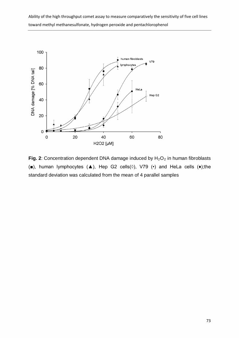
Ability of the high throughput comet assay to measure comparatively the sensitivity of five cell lines
toward methyl methanesulfonate, hydrogen peroxide and pentachlorophenol
73
Fig. 2: Concentration dependent DNA damage induced by H2O2
in human fibroblasts
(■), human lymphocytes (▲), Hep G2 cells(◊), V79 (•) and HeLa cells (♦);the
standard deviation was calculated from the mean of 4 parallel samples

Ability of the high throughput comet assay to measure comparatively the sensitivity of five cell lines
toward methyl methanesulfonate, hydrogen peroxide and pentachlorophenol
74
Fig. 3: concentration dependent DNA damage induced by PCP in human fibroblasts
(■), human lymphocytes (▲), Hep G2 cells(◊), V79 (•) and HeLa cells (♦). ∆ = effect
of the ultimate PCP metabolite tetrachlorhydroquinone (TCHQ) on human fibroblasts;
the standard deviation was calculated from the mean of 4 parallel samples

Ability of the high throughput comet assay to measure comparatively the sensitivity of five cell lines
toward methyl methanesulfonate, hydrogen peroxide and pentachlorophenol
75
Tab.2: Sensitivity of 5 cell lines toward MMS, H2O2
celltype
, and PCP; the data were picked
up from the curves in Fig.1-3
MMS H2O2 PCP % DNA Tail [µM] % DNA Tail [µM] % DNA Tail [µM]
10% 30% 50% 10% 30% 50% 10% 30% 50% lymphocytes 235 400 600 13 24 31 400 700 850 Hep G2 cells 500 1000 1450 36 55 nd 900 1100 1200 fibroblasts 350 650 800 20 28 36 1200 1250 1350
HeLa 600 900 1200 41 50 60 1100 1200 1250 V79 750 1200 1400 38 44 50 750 900 1050

A high-throughput genotoxicity testing strategy for screening of (drinking) water
76
5.4 A high-throughput genotoxicity testing strategy for screening of (drinking) water (submitted)
Minne B. Heringa1*, Andre Stang2, Cyrille A.M. Krul3, Astrid A. Reus3, Annemarie P. van
Wezel1, Irene Witte
2
1 KWR Watercycle Research Institute, PO Box 1072, 3430 BB Nieuwegein, the
Netherlands; 2 Carl von Ossietzky Universität Oldenburg, IBU, Postfach 2503, D-26111
Oldenburg, Germany; 3
TNO Quality of Life, PO Box 360, 3700 AJ Zeist, the Netherlands;
*
Dr. M.B. (Minne) Heringa
corresponding author:
Scientific Researcher I
Chemische Waterkwaliteit &Gezondheid (CWG)
The Netherlands visiting: Groningenhaven 7
P.O. Box 1072
3430 BB Nieuwegein
Netherland
Tel.: +31-30-6069539
Fax: +31-30-6061165
E-Mail: [email protected]

A high-throughput genotoxicity testing strategy for screening of (drinking) water
77
Abstract The sources of our drinking water can be polluted with anthropogenic chemicals. To
assess the health hazard of this pollution, in vitro genotoxicity tests are of great
importance. With the many systems available, it is still unclear which battery to choose. We
applied a strategy of high-throughput in vitro genotoxicity testing to nine water extracts from
different types of sources in the Netherlands with the Ames II, high-throughput comet
assay and the micronucleus test. In addition, a validation set of eight well-known
genotoxicants and one non-genotoxic carcinogen was tested with the Ames II and the
comet assay. The aims of this study were:
1. To compare the outcome of the Ames II with either the comet or the micronucleus
test with respect to their complementarity
2. To determine the suitability of using the high-throughput Ames II and comet assay
for water extracts;
3. To compare the comet assay and micronucleus test in the detection of genotoxic
compounds present in water.
4. To evaluate the occurrence of genotoxic compounds in Dutch waters.
We found that the comet assay and micronucleus test are complementary to the Ames II
test, as the Ames II was more sensitive for some tested compounds, but did not detect the
genotoxicity of all well-known genotoxins and of all water samples positive in the comet
assay. The micronucleus test did not detect any genotoxicity in the water samples. The
high-throughput Ames II and comet assay were selected for future testing strategy of water
samples.
Keywords Ames II, comet, micronucleus, monitoring

A high-throughput genotoxicity testing strategy for screening of (drinking) water
78
1. Introduction In the sources for drinking water, thousands of industrial chemicals can be present [1]. In
the EU, 30.000 to 70.000 chemicals are in daily use (EINECS database), varying from
industrial chemicals (such as solvents, petrochemicals), consumer chemicals (such as
pharmaceuticals, personal-care products) to biocides. Due to global trends such as a
growing and ageing population, increasing prosperity, and urbanization [2], there is an
increasing chemical pressure on drinking water sources. For example, only about 10% of
European river water samples could be classified as ‘very clean’ [3]. Contaminants have
been found throughout Europe up to high ng/L median concentrations [3], many entering
the water cycle via wastewater [4]. This occurrence of chemicals in sources of drinking
water is, depending on the treatment processes, sometimes reflected in (much lower)
occurrence of these chemicals in finished drinking water [5]. Thusfar, where there was
sufficient information, concentrations of individual contaminants in drinking water in the
Netherlands do not exceed their individual health-based guideline values (Schriks et al., in
preparation).
However, despite rapidly evolving chemical analytical techniques [6], it is impossible to
analyze and identify all different chemicals present in the aqueous environment. In
addition, for many identified chemicals no statutory health-based (drinking) water quality
guideline values have been established and no or scarce toxicological information is
available to estimate human health risks (e.g. [7]). Finally, the understanding of the
combined mixture effects in these complex mixtures of many different chemicals occurring
in low concentrations is still limited [8]. The use of in vitro effect-directed bioassays can be
an additional tool to interpret health risks of complex mixtures in drinking water and its
sources. In vitro bioassays do not determine the presence of single or groups of
compounds directly, but determine their collective effect in a biological system such as
cultured cells. Chemicals that cannot be revealed by analytical techniques but do attribute
to the toxicological effects are included in the bioassays, and thus, the assays give a clue
of the toxicity of the total mixture of chemicals present in the water sample.
As the concentrations in the water are relatively low, but drinking water is consumed life-
long, only effects occurring at relatively low, but chronic exposure are relevant. One of the
most relevant toxic effects in this case is therefore genotoxicity, as genotoxic compounds
can be effective at very low dosages. In vitro genotoxicity analyses on all types of water
have been performed for many years all around the world already, with a variety of test

A high-throughput genotoxicity testing strategy for screening of (drinking) water
79
systems (e.g. [9-14] [15]). With the many test systems available and applied, it is unclear
which to choose in order to best detect all genotoxic compounds present in a water
sample. Genotoxicity can be tested by measuring the formation of DNA interactions (DNA
adducts) themselves, DNA damage (i.e. gene mutations or chromosomal aberrations), or
the induction of the DNA repair. As DNA damage can occur through different mechanisms,
a battery of tests is necessary. However, for the DNA adducts and repair enzymes, no full
battery covering all adduct and repair types for the different types of damage is available.
Therefore, for genotoxicity testing of drinking water and its sources, we have chosen a
strategy applying an in vitro test battery consisting of a gene mutation test, and a test
detecting chromosomal damage (e.g. chromosomal breaks) in mammalian cells, following
the recommendations of e.g. [16-18].
For the choice of tests among the in vitro gene mutation and chromosomal damage tests,
we have considered whether the test could be used for Toxicity Identification and
Evaluation (TIE), among other common factors such as sensitivity, wide acceptance, costs,
etc.. TIE is applied when an environmental sample is found (geno)toxic and one wants to
identify the responsible compound(s) (e.g. [19]). The sample is then fractionated, and the
(geno)toxicity test is performed on the different fractions. Subsequently, the (geno)toxic
fraction is chemically analyzed, containing fewer compounds than the original sample,
leading to a simpler analysis. To be able to apply TIE if desired, we selected genotoxicity
tests that were preferably performed in multi-well plates to be able to handle large numbers
of fractions with relatively small volumes in the future. Additionally, a multi-well plate format
requires much less sample, which saves labour and costs in the sample preparation.
For the gene mutation test, we found only one method applying multi-well plates: the Ames
II. This modern version of the very well-known classic Ames test is performed in liquid
medium instead of agar plates, and uses a colour change as indicator of growth instead of
visual colony counting [20]. In principle, any Ames test strain can be applied with this
method, but the Ames II kit applies TAMix, a mixture of 6 base-pair mutated strains, instead
of TA100 [21,22].
For the test detecting chromosomal damage, a high throughput version of the comet assay
has recently been introduced [23,24], using a special 96-well plate of which the bottom can
be removed after the exposure step. Nesslany and Marzin [25] also developed a 96-well
version of the micronucleus test, where the exposure step took place in 96-well plates, but
the subsequent analysis steps were still performed on microscope slides in this method.

A high-throughput genotoxicity testing strategy for screening of (drinking) water
80
We have applied this strategy of high-throughput in vitro genotoxicity testing to nine water
extracts from different types of sources in the Netherlands with the Ames II, high-
throughput comet assay and the micronucleus test. These samples were taken from the
most polluted sources of drinking water (rivers and polluted ground waters) and a typical
sewage water treatment plant effluent including hospital wastewater, representing worst
case water samples relevant for the watercycle. In addition, a set of eight well-known
genotoxicants and one non-genotoxic carcinogen was tested with the Ames II and the
comet assay.
The aims of this study were:
1. To compare the outcome of the Ames II with either the comet or the micronucleus
test with respect to their theoretical complementarity
2. To determine the suitability of using the Ames II and high-throughput comet assay
for water extracts;
3. To compare the comet assay and micronucleus test in the detection of genotoxic
compounds present in water in order to make a choice which to use in future
monitoring.
4. To evaluate the occurrence of genotoxic compounds in Dutch waters.
2. Materials and Methods 2.1 Chemicals The following chemicals were used to test as well-known genotoxic chemicals and one
non-genotoxic carcinogen, covering different genotoxicity mechanisms: methyl
methanesulfonate (MMS, > 99 %, from Sigma, St. Louis), N-Nitroso-N-ethylurea (ENU from
Sigma, G), 4-nitroquinoline-1-oxide (4-NQO, 98.2 %, from Sigma), cisplatin (Cis, from
Medoc, Hamburg, FRG), cyclophosphamid (CP, ≥ 97 from Sigma,), benzo(a)pyrene
(B(a)P, ≥ 96 % from Sigma, 2-acetyl aminofluorene (2-AAF, ≥ 90 % from Sigma), estradiol
(E2, > 98 %, Sigma) and pentachlorophenol (PCP, recrystallized, a gift of Dr. Butte,
University of Oldenburg). For all experiments, the tested chemicals had identical lot
numbers. For the sample preparation, acetonitrile was purchased from Mallinckrodt Baker
B.V. (Deventer, the Netherlands) and dimethylsulfoxide (DMSO) was purchased from
Acros Organics (Geel, Belgium). For the Ames II, the positive control 2-aminoanthracene
(2-AA) was purchased from Sigma (St. Louis). For the micronucleus test, the positives

A high-throughput genotoxicity testing strategy for screening of (drinking) water
81
controls cyclophosphamide, colchicines and mitomycin C were obtained from Asta Werke
(CP) and Sigma (colchicines and mitC).
2.2 Preparation of water extracts (performed by KWR)
In May 2007, samples of 4 L were taken in extensively washed glass bottles from four
Dutch groundwater pump stations where the ground was known to be contaminated, from
three Dutch surface water sites where water is taken in to produce drinking water and from
one effluent of a Dutch municipal sewage treatment plant (STP) which also treats hospital
wastewater. Samples were cooled immediately and stored at 4 °C.
Within 24 hours after collection, the samples were filtered over 0.45 μm cellulose nitrate
membrane filters (Sartorius; Goettingen, Germany). Within another 24 hours, four
replicates of one liter of every sample were extracted by solid phase extraction (SPE) with
pre-washed 200 mg Oasis® HLB cartridges (Waters Corporation, Milford, USA) at ambient
pH. Elution was performed with 3 serial additions of 2.5 mL of acetonitrile. The 7.5-mL
eluates were evaporated and taken up in 50 μL of DMSO yielding 20,000-fold concentrated
extracts. All extracts were stored at -18°C until analysis.
2.3 S9 fraction Treatment of bacteria or mammalian cells occurred with the indirectly acting chemicals or
with water extracts in the presence of post mitochondrial S9 fraction from Aroclor 1254-
induced rat livers. For the Ames II and comet experiments the S9 fraction with the same
batch number was used (MP Biomedicals, Solon,U.S.A.), for the micronucleus test the S9
was produced in-house according to the procedure described by Ames et al. [26] and
Maron and Ames [27]. The S9-fraction (final concentration 0,8 mg protein / mL) was mixed
with a solution containing 8 mM MgCl2
, 33 mM KCl, 4 mM NADP, 5 mM glucose-6-
phopshate, pH 7. S9 mix was prepared immediately before use.
2.4 Ames II test (performed by KWR) The tested chemicals were dissolved and diluted in DMSO and added to form no higher
concentrations than the solubility limit. The water extracts were diluted to 100 μL (1:1) with
DMSO to obtain a sufficient amount of sample for all tests. The Ames II test strains (TA98

A high-throughput genotoxicity testing strategy for screening of (drinking) water
82
and TAMix) and media were purchased from Xenometrix (Basel, Switzerland). The test
procedure provided by Xenometrix, also described by Fluckiger-Isler et al. [20], was
followed, with minor modifications. Per well of a 24-well plate (Greiner Bio One), the
following was added: 6 μL of test sample (compound or water extract) in 100% DMSO, 30
μL overnight culture, 10 μL of S9-mix if applicable and 264 or 254 μL of Exposure Medium,
respectively, to obtain 300 μL in total. Thus, the bacteria were exposed to a 200-fold
concentration of the water samples. Water extracts were tested in triplicate at one
concentration, while compounds were tested in duplicate at nine different concentrations. A
triplicate negative control (DMSO only), a triplicate positive control for genotoxicity (table 1),
and a triplicate positive control for cytotoxicity (1 mg/mL 4-NQO in DMSO) were included
as well.
A custom cytotoxicity test was performed with subsamples of the exposure cultures in
medium with histidine, to check for possible artifacts due to effects on cell survival and
growth. After an incubation of 90 minutes at 37 °C and 250 rpm, 10 μL from each exposure
mixture was transferred to a well of a 96-well plate (Greiner Bio One) for a cytotoxicity
measurement. To each well of the 96-well plate, 90 μL of Exposure Medium (containing
histidine) was added and this was then left to incubate for another 3 hours at 37 °C and
250 rpm. Then, the OD at 595 nm of the 96-well plate was measured with an Opsys MR
platereader (Clindia; Leusden, the Netherlands). Samples with OD-values significantly
below that of the negative control (t-test, p=0.05), and below 90% of the OD-value of the
negative control, were considered cytotoxic.
To the remaining exposure mixture in the 24-well plate, 2.61 mL of purple Indicator Medium
(not containing histidine) was added. The total 2.9 mL was subsequently divided over 48
wells (50 μL per well) of a 384 well plate and left to incubate for 48 hours at 37 °C. Then,
the number of yellow wells per 48 wells of one sample were counted manually.
As Ames test responses are not normally distributed, but follow a Poisson distribution [28],
no standard statistical tests could be performed on the data. As an alternative, a water
extract was determined to be genotoxic if the number of yellow wells exceeded the
detection limit of the test. The detection limit (DLresponse
) was calculated using equation 1:
NCresponse sNCDL ⋅+= 3 equation 1
In this equation, NC is the average response of the negative control in the same test as the
sample and sNC is the standard deviation in the responses of the negative control.

A high-throughput genotoxicity testing strategy for screening of (drinking) water
83
2.5 Cell culture
For the comet assay human fibroblasts (cell line NHDF-p, passage 8-15, purchased from
Promochem, Heidelberg, FRG) and the permanent cell line HepG2 were used. Cells were
grown in D-MEM supplemented with 12% fetal calf serum at 37 °C in an atmosphere of 5
% CO2
For the micronucleus test human lymphocytes were used. The human lymphocytes were
obtained by venapuncture from a healthy, non-smoking male, not currently taking any
medication. The same donor was used for the first and second assay. The blood was
collected in sterile, heparinized vacutainer tubes and gently mixed to prevent clotting. The
cultures were set up within 2 hours after withdrawal of the blood. The medium for culturing
the human peripheral blood lymphocytes consisted of RPM1-1640 medium with Glutamax-
1, supplemented with heat-inactivated fetal calf serum (20%), penicillin (100 units/mL
medium), streptomycin (100 μg/mL medium) and phytohaemagglutinin (PHA-L, 10 (μL/mL
medium). Whole blood (0.5 mL) was added to sterile screw-capped tubes containing 4.5
mL culture medium. The blood cultures were incubated for 48 hours at ca. 37° C in
humidified air containing ca. 5% CO
and 95 % air with > 95 % humidity.
2
to ensure
2.6 High-throughput comet assay (performed by Carl von Ossietzky Universität Oldenburg) The principle of the high-throughput comet assay is a 96 well plate (multichamber plate,
MCP purchased from Intox, Oldenburg, FRG) suitable for electrophoresis. After exposure
of the cells to genotoxic agents, the walls of the MCP are separated from the bottom plate.
All 96 wells are subjected to lysis, alkaline unwinding, electrophoresis, neutralization, and
staining simultaneously.
The comet assay in the high-throughput version was performed according Stang and Witte
[23]. Either fibroblasts (3,000 cells/well) or HepG2 cells (4,000 cells/well) were seeded 2-4
h (fibroblasts) or 16-24 h (Hep G2) before treatment of the cells into the wells of the MCP.
Subsequently, the cells were treated with the genotoxic chemicals or the water extracts for
1.5 hours. The nine test chemicals were freshly dissolved directly in serum free medium
(sfm) at pH 7.2 at which concentration?. The lipophilic B(a)P and 2-AAF were dissolved in
DMSO/Cremophor EL® (1/1) and diluted with sfm directly before cell treatment (maximum
A high-throughput genotoxicity testing strategy for screening of (drinking) water
84
final concentration of DMSO/Cremophor: 2.5 %). The water extracts were diluted in sfm 50-
800.times directly before cell treatment, to obtain a 25 to 400-fold concentration of the
water samples in the assay. The solvents alone did not provoke any geno- or cytotoxicity.
After treatment of the cells with the mutagens or water extracts, the walls surrounding the
wells of the MCP were separated from the bottom plate. The plate with the cells was
covered with a 37 °C warm solution of 0.5 % low-melting agarose (type Sea Plaque from
Biozym Diagnostik, Hessisch Oldendorf, FRG). The plate was kept in the refrigerator for 5
minutes to solidify the low melting agarose, and thereafter covered with refrigerated lysing
solution at pH 10.0 (2.5M NaCl, 100mM EDTA, 10mM Tris, 1% N-lauroyl sarcosine, 1%
Triton X100, 10% DMSO; the last two compounds were added freshly) and kept at 4 °C for
1 hour. After lysis, the plate was placed on a horizontal electrophoresis box. The box was
filled with freshly prepared alkaline buffer whereby the plate was completely covered with
the buffer. The 40 minutes of the alkaline treatment at 4 °C allowed unwinding of the DNA
and DNA breakage at alkali labile sites. After electrophoresis (300 mA, 25 V for 20
minutes), the plate was covered three times with neutralization buffer, washed with aqua
bidest., and stained with ethidium bromide. Fluorescence microscopy was used to
determine the median tail moment (tm) in each of the 4 replicates per concentration (25
comets/well) using Lucia Comet Assay Single Stain software with an upper detection limit
of tm = 100. From these 4 values the mean and standard deviation was calculated.
Statistical significance of the differences between the DNA migration induced by the
mutagens and the untreated controls was determined by the non-parametric Mann-Whitney
test (p < 0.05 or p< 0.01).
DNA damage induced by the crosslinkers cis and CP was measured indirectly. It was
detected according to Pfuhler and Wolf [29] by previous treatment with MMS (2.1 mM for 1
hour) and subsequent addition of Cis or CP for 1.5 hours. The reduction of DNA strand
breaks induced by MMS quantitatively reflects the DNA crosslinks induced by cisplatin.
For measurement of the cell viability, we used the FDA assay according to Rotman and
Papermaster [30] during the comet assay, in the same cultures used for comet
determination. The FDA assay measures the cell viability by the activity of cytosolic
esterases converting fluorescein diacetate (FDA) to the fluorescent dye fluorescein. After
chemical treatment the cells were centrifuged in the MCP (2 min at 400 x g) and incubated
for 10 minutes with 72 µM FDA (from 12 mM stock solution in acetone, freshly diluted with
sfm) at 37 °C in the dark. Thereafter, the cells were shortly centrifuged again, the dye was
removed and the fluorescence of the enzymatically formed fluorescein was measured in a

A high-throughput genotoxicity testing strategy for screening of (drinking) water
85
fluorescence reader (FLUOstar, Offenbach, FRG) with an excitation of 485 nm and an
absorption at 520 nm. Cell viability was calculated as the percent ratio of the fluorescence
values of the samples to the referring control. After fluorescence measurement the walls
and the bottom plate of the MCP were separated and the comet assay procedure was
continued as described above.
2.7 Micronucleus test (performed by TNO Quality of Life)
For each culture, 5 mL of whole blood was incubated with PHA-L for 48 hours at ca.
37° C in humidified air containing ca. 5% CO2
For the test water samples’ preparation, DMSO was used as vehicle. Water extracts
were diluted 2-fold (1:1) using DMSO, to yield 10.000-fold concentrated stock
solutions. The final concentration of DMSO of water extracts in the cultures was 1%
(v/v) and corresponded to 100-fold concentrated extracts. Cells were exposed to water
extracts for 4 hours in the presence of S9-mix, or exposed for 24 hours in the absence
of S9-mix. In the absence of S9-mix, Mitomycin C (at 10 and 5 μg/mL), a known
clastogen, and colchicines (at 5 and 2.5 μg/mL), a known aneugen, were used as
positive controls. In the presence of S9-mix, Cyclophosphamide, a known clastogen
requiring metabolic activation, was used as positive control (at 2 and 1 mg/mL).
. After this incubation period, the cells
(which are then in the exponential stage of their growth) were exposed to test
substances, in both the absence and the presence of S9-mix. Single cultures were
used for each water extract for each exposure period as well as for the positive and
negative (vehicle) controls.
Micronucleus test in the absence of the S9-mix (24 hour exposure): The cells were
harvested by low speed centrifugation and resuspended in freshly prepared tissue
culture medium with fetal calf serum (20%). The test substance was added to the
cultures and the cells were exposed to the test substance for 24 hours (continuous
treatment). After the 24 hours treatment period, the cells were washed with phosphate-
buffered saline (pH 7.4) and subsequently supplied with culture medium (containing 20%
serum). Thereafter, Cytochalasin B was added to each culture, to a final concentration of 6
μg/mL. The cultures were incubated for an additional 20 hours at ca. 37 °C in humidified air
containing ca. 5% CO2
.

A high-throughput genotoxicity testing strategy for screening of (drinking) water
86
Micronucleus test in the presence of the S9-mix (4 hour exposure): The cells were
harvested by low speed centrifugation and resuspended in freshly prepared tissue culture
medium without fetal calf serum. Thereafter, the test substance was added to the cultures
and each culture was supplemented with 0.5 ml S9-mix (see paragraph 2.3). The cells
were exposed to the test substance for only 4 hours, because of the toxicity of the S9-mix
for the cells. After the 4 hours treatment period, the cells were washed with phosphate-
buffered saline (pH 7.4) and subsequently supplied with culture medium (containing 20%
serum). Thereafter, Cytochalasin B was added to each culture to a final concentration of 6
μg/mL. The cultures were incubated for an additional 20 hours at ca. 37 °C in humidified air
containing ca. 5% CO2
.
Harvesting and slide preparation: Each culture was harvested and processed separately.
The cells were harvested by low speed centrifugation, treated with a hypotonic solution
(0.075 M potassium chloride), fixed three times with a freshly prepared mixture of methanol
and acetic acid, spread on clean slides and air dried. All procedures were performed at
room temperature. Two slides were prepared from each culture of the test substance and
from the negative and positive controls. Slides were stained with a fluorescent DNA-
specific dye (acridin orange) and coded by a qualified person not involved in scoring of the
slides to enable "blind" scoring. Per slide,
1000 binucleated cells (2000 binucleated cells
per culture) were examined for the presence of micronuclei.
3. Results and Discussion 3.1 Testing of nine well known carcinogens As the Ames II and high-through put comet assay are relatively new, especially in the
application for water sample analysis, a simple validation of these assays was performed.
Comparing the classical Ames test as well as the conventional comet assay with their high-
throughput versions, similar results have been obtained before [22,23]. Eight other well
known standard mutagens and one non-genotoxic carcinogen (estradiol) were tested in the
high-throughput comet assay and in the Ames II test with the same exposure time. Five of
the eight genotoxins are on the recommended list of Kirkland et al. [31] for compounds for
performance assessment of new tests. The results are shown in Fig. 1 for directly acting
mutagens and in Fig. 2 for indirectly acting mutagens that need metabolic activation.

A high-throughput genotoxicity testing strategy for screening of (drinking) water
87
Indirectly acting mutagens were tested in the presence of S9 fraction, either in the Ames II
test or with fibroblasts in the comet assay. In addition, the metabolic competent cell line
Hep G2 was used in the comet assay.
In comparison to TA98, the TAMix was more effective in detecting mutations induced by
the directly acting mutagens ENU, MMS and 4-NQO (Fig. 1a-1c), whereby the alkylating
MMS and ENU even failed to show any mutagenicity in TA98 (Fig. 1a and 1b). This is also
in accordance with Brams et al. [32] and Eder et al. [33], where MMS was negative in
TA98, while positive in TA100. Alkylating substances such as MMS and ENU are also
known to create mainly base-pair substitutions [34], which are detected by the TAMix or
TA100. While in the comet assay DNA damage could be detected for all four directly acting
chemicals, in the Ames II test the crosslinker Cis was not mutagenic in either TA98 or
TAMix (Fig. 1d) in contrast to expectation. Also the indirectly acting crosslinker CP gave
unexpected negative results in the Ames II test at the concentrations tested (Fig. 2d). While
for the crosslinkers Cis and CP mostly positive results were described for the strains TA98
and TA100 in literature at similar concentrations [20,35-37], also negative results have
been reported [20,38].
In the comet assay, both in fibroblasts with S9 fraction and in HepG2 cells DNA crosslinks
were measured for CP. The DNA damaging properties of PCP were also detected in
fibroblasts + S9 and in Hep G2 but no positive response was found in the Ames II test (Fig.
2 b). The carcinogenic PCP [39] is transformed to the highly genotoxic metabolite
tetrachlorohydroquinone by S9 fraction enzymes [40]. The detection of the mutagenic
potential of PCP (with S9 fraction) in the Ames test was described by Gopalaswamy and
Nair [41], whereas Gichner et al. [42] as well as Moriya et al. [43] obtained negative results
using TA98. B(a)P was more effective in the Ames test than in the comet assay (Fig. 2a)
and 2-AAF only tested positive in the Ames II test (Fig. 2c). Thereby the strain TA98 was
more sensitive than TAMix, indicating these bulky adduct–forming compounds form mainly
frameshift mutations. 2-AAF was the only substance where no DNA damage was detected
in the comet assay after a 90 min treatment. It is known that for 2-AAF longer incubation
times of one day [44,45] or three days [46] are needed to detect genotoxicity in the comet
assay. Estradiol was negative in both the Ames II test and high-throughput comet test, as
expected (table 2), but appears to give positive results in the micronucleus test, due to
aneugenic action (e.g. [47,48]. Estradiol therefore seems to be a genotoxic carcinogen
after all. Despite some unexpected results, which also have been reported before,
A high-throughput genotoxicity testing strategy for screening of (drinking) water
88
however, we find the predictivity of the Ames II and high-throughput comet assay
satisfactory.
It is impossible to decide which cell line (metabolic competent HepG2 cells or with
fibroblasts in the presence of S9 fraction) may be the more effective one to detect indirect
genotoxic compounds in the comet assay. Only three of our compounds were tested and
detected in under these conditions, showing different trends. PCP was more effective in
HepG2 cells by the factor 8, while the detection limit of CP was 4 times lower in fibroblasts
with S9 fraction. This may be result of the difference between the metabolic system in both
cultures, with one being an isolated mix of rat enzymes and the other an innate human
enzyme system of the cell-line.
A comparison of the sensitivities of the Ames II test and the high-throughput comet assay
in detecting the lowest effective concentrations of the mutagens is given in Table 2, as well
as literature data on the sensitivity of the micronucleus test. The Ames II test was more
sensitive than the comet or micronucleus test for three of the eight genotoxic compounds
(MMS, ENU, 4-NQO). However, the Ames test did not pick up all compounds (CP, Cis,
PCP) which induced comet or micronucleus formation. Thus, this gene mutation test and
the tests for chromosomal damage complemented one another, as expected when testing
genotoxic compounds with different mechanisms of action and applying tests with different
genotoxicity endpoints.
3.2 Testing of water extracts The Ames II, the high-throughput comet assay, and the micronucleus test were applied to
nine extracts of water samples for genotoxic contaminants. In the Ames II (Fig. 3a), only
the STP effluent showed genotoxicity, for all other samples the response did not differ
significantly from that of the negative control. This is in concordance with past results of
classic Ames tests on the Dutch Rhine and Meuse rivers, where over the years a decline in
genotoxic activity was seen until very low, sometimes insignificant responses were
obtained [49]. The STP effluent was positive in TA98 + S9, not in the other conditions.
Filipic and Toman [50] found a mutagenic response in a municipal STP effluent in both
TA98 and TA100 (+/- S9).
In the comet assay, three of the nine water samples tested positive in a concentration
dependent way (Fig. 4 a-c and 3c). The DNA damage in these samples was low, but still
statistically significant. This was also due to the small values of the untreated control with
A high-throughput genotoxicity testing strategy for screening of (drinking) water
89
low standard deviations of four parallel samples in the high-throughput version of the comet
assay. Even when the cytotoxicity was high enough to be responsible for the genotoxic
effects at the highest concentration tested [51], at lower, non-cytotoxic concentrations
significant genotoxicity was also detected (Fig. 3c and 4a). In addition to the STP effluent,
also surface water 3 and groundwater 4 tested positive. In these three samples metabolic
activation reduced the genotoxicity, even though a significant DNA damage remained
either in fibroblasts with S9 fraction or in HepG2 cells. Interestingly, in the Ames II test the
metabolically activated STP sample was mutagenic while in the comet assay DNA damage
was more pronounced in the absence of S9 fraction. In contrast, Zegura et al. [52] found
genotoxicity in an effluent from a municipal STP with the SOS/umuC test (applying the
same bacteria as in the Ames test) in absence of S9, and not with the comet assay in
metabolically active HepG2 cells. Of course, STP effluents can differ in chemical load
between locations and especially between countries.
As these tests detect different types of DNA damage, different compounds could be
responsible for the different observed effects in these two assays. Another difference
between these two assays is the occurrence of damage repair: in contrast to the Ames II
test, in the comet assay the cells do not complete a cell division cycle including DNA
damage repair during the incubation time of 90 min. Therefore the damage remains
detectable by the comet assay, while it may be repaired in the Ames II test, where the
bacteria fulfil around 2 cell divisions in 90 min.
Dutch waters have only sparingly been analyzed with chromosomal damage tests, such as
the comet assay, before. Alink et al. [53] found a significantly increased response in the
comet assay and sister chromatid exchange (SCE) test in fish gill cells, after exposing the
fish for 11 days in Rhine water in 2005. Applying much shorter exposure times, we have
also found an increased comet assay response in one surface water, also for the first time
in a Dutch groundwater and STP effluent. These results show that further, more extensive
analysis of Dutch waters, with inclusion of a chromosomal damage test, are recommended
for safeguarding the quality of Dutch drinking water.
In the micronucleus test none of the water samples tested positive (Fig. 3b).This may also
be the result of a prolonged incubation time of 4 hours + 20 hours recovery time, allowing
complete repair of DNA damages in the micronucleus test (necessity to pass through a cell
cycle). As consequence, a lower sensitivity in comparison to the comet assay was
observed. As the water samples represent different water types that are relevant to monitor

A high-throughput genotoxicity testing strategy for screening of (drinking) water
90
(STP effluent, surface water and groundwater) and represent worst cases (i.e most
polluted) of these water types, the results are considered representative for all Dutch water
samples. Also in other studies, the comet assay proved to be the better tool to detect low
levels of genotoxic contaminants. This was shown for lake drinking water [9], river samples
[14,54], water soil leachates [15], river sediments [55], chlorinated drinking water samples
[9] and other aquatic environments [56]. Valentin-Severin et al. [44], however, found that
the micronucleus test was more sensitive than the comet assay for a limited set of four of
the five tested compounds in HepG2 cells.
It may be argued that the inclusion of the DNA repair step in the micronucleus test is an
advantage, as this mimics real life. However, presence of DNA damaging compounds,
causing DNA strand breaks as detected by the Comet assay, is not desirable for drinking
water. For hazard identification it is important to use sensitive assays, and cover all
genotoxic endpoints, therefore the comet assay and Ames II test are selected. The
argument that the micronucleus test can also detect aneugens, while the comet can not, is
of minor weight, as aneugens are rare [57]. Thus, high sensitivity is given priority and the
comet is therefore preferred.
The Ames (II) test and the comet assay have been successfully combined for
biomonitoring of environmental samples before (e.g. [55,58-60]). The Ames II is shown to
be suitable for water analysis. Further, the first analysis of water extracts with the high-
throughput comet assay has shown no practical problems and sensitivity for aqueous
genotoxic substances was demonstrated. Therefore, we conclude that the high-throughput
version of the comet assay is also suitable for water analysis.
Of course, the outcome of these in vitro tests do not provide a determination of a health
risk for consumers, as the bacteria of the Ames II test are very different from human cells,
and no information on absorption, distribution, metabolism and excretion in a human being
is included. Therefore, test results should be regarded as signalling values, with negative
results indicating no health hazard is expected, and positive results indicating there are
genotoxic compounds in the water, and further research is necessary to determine the
risks these compounds pose.
In summary, the Ames II test and the high-throughput comet assay are two sensitive
assays which complement one another because of their different sensitivities toward
different classes of mutagens. This sensitive complementary working of the two test
systems combined with easy, quick and economic handling should be a favourite

A high-throughput genotoxicity testing strategy for screening of (drinking) water
91
combination for screening environmental probes with low levels of contaminants. It is
recommended that Dutch drinking water and its sources are screened with this
combination of tests.
Acknowledgements We thank Paul Baggelaar for the advice on statistics, Rene van Doorn and Hans van
Beveren for the preparation of extracts at KWR, and Stefan Voost for performing the Ames
II tests at KWR. This study was supported by the Joint Research Programme of the Dutch
water utilities (BTO).
Abbreviations
2-AA aminoanthracene
2-AAF 2-acetyl aminofluorene
B(a)P benzo(a)pyrene
Cis cisplatin
CP cyclophosphamid
D-MEM Dulbecco's modified Eagle medium
DMSO dimethylsulfoxide
E2 estradiol
ENU ethylnitroso urea
EU European Union
FDA fluorescein diacetate
MCP multichamber plate
MMS methyl methanesulfonate
4-NQO 4-nitroquinoline-1-oxide
PCP pentachlorophenol
sfm serum free medium
STP sewage treatment plant
TIE Toxicity Identification and Evaluation
tm tail moment

A high-throughput genotoxicity testing strategy for screening of (drinking) water
92
References [1] R.P. Schwarzenbach, B.I. Escher, K. Fenner, T.B. Hofstetter, C.A. Johnson, U.
von Gunten and B. Wehrli, The challenge of micropollutants in aquatic systems,
Science 313 (2006) 1072-1077.
[2] M.L. Diamond and E. Hodge, Urban contaminant dynamics: from source to effect,
Environ. Sci. Technol. 41 (2007) 3796-3805.
[3] R. Loos, B.M. Gawlik, G. Locoro, E. Rimaviciute, S. Contini and G. Bidoglio, EU-
wide survey of polar organic persistent pollutants in European river waters, Environ.
Pollut. 157 (2009) 561-568.
[4] T. Reemtsma, S. Weiss, J. Mueller, M. Petrovic, S. Gonzalez, D. Barcelo, F. Ventura and T.P. Knepper, Polar pollutants entry into the water cycle by municipal
wastewater: a European perspective, Environ. Sci. Technol. 40 (2006) 5451-5458.
[5] M.J. Benotti, R.A. Trenholm, B.J. Vanderford, J.C. Holady, B.D. Stanford and S.A.
Snyder, Pharmaceuticals and endocrine disrupting compounds in U.S. drinking water,
Environ. Sci. Technol. 43 (2009) 597-603.
[6] S.D. Richardson, Environmental mass spectrometry: Emerging contaminants and
current issues, Anal. Chem. 80 (2008) 4373-4402. [7] J. van Genderen, M.N. Mons and J.A. van Leerdam, Inventory and toxicological
evaluation of organic micropollutants, revision 1999, Association of River Waterworks -
RIWA, Amsterdam, 2000.
[8] L.S. McCarty and C.J. Borgert, Review of the toxicity of chemical mixtures: Theory,
policy, and regulatory practice, Regul. Toxicol. Pharmacol. 45 (2006) 119-143.
[9] A. Buschini, P. Carboni, S. Frigerio, M. Furlini, L. Marabini, S. Monarca, P. Poli, S.
Radice and C. Rossi, Genotoxicity and cytotoxicity assessment in lake drinking water
produced in a treatment plant, Mutagenesis 19 (2004) 341-347.
A high-throughput genotoxicity testing strategy for screening of (drinking) water
93
[10] H. Dizer, E. Wittekindt, B. Fischer and P.D. Hansen, The cytotoxic and genotoxic
potential of surface water and wastewater effluents as determined by bioluminescence,
umu-assays and selected biomarkers, Chemosphere 46 (2002) 225-233.
[11] T. Ohe, T. Watanabe and K. Wakabayashi, Mutagens in surface waters: a review,
Mutat. Res. 567 (2004) 109-149.
[12] G. Reifferscheid and T. Grummt, Genotoxicity in German surface waters - results
of a collaborative study, Water Air Soil Poll. 123 (2000) 67-79.
[13] C. Zani, D. Feretti, A. Buschini, P. Poli, C. Rossi, L. Guzzella, F.D. Caterino and
S. Monarca, Toxicity and genotoxicity of surface water before and after various
potabilization steps, Mutat. Res. 587 (2005) 26-37.
[14] Y. Zhong, S.L. Feng, Y. Luo, G.D. Zhang and Z.M. Kong, Evaluating the
Genotoxicity of Surface Water of Yangzhong City Using the Vicia Faba Micronucleus
Test and the Comet Assay, Bull. Environ. Contam. Toxicol. 67 (2001) 217-224.
[15] B. Lah, T. Vidic, E. Glasencnik, T. Cepeljnik, G. Gorjanc and R. Marinsek-Logar,
Genotoxicity evaluation of water soil leachates by Ames test, comet assay, and
preliminary Tradescantia micronucleus assay, Environ. Monit. Assess.139 (2008) 107-
118.
[16] T. Grummt, Erprobung, Vergleich, Weiterentwicklung and Beurteilung von
Genotoxizitätstests für Oberflächenwasser, Wasser Abwasser 142 (2001) 346-355.
[17] S. Pfuhler, S. Albertini, R. Fautz, B. Herbold, S. Madle, D. Utesch and A. Poth,
Genetic toxicity assessment: employing the best science for human safety evaluation
part IV: Recommendation of a working group of the Gesellschaft fuer Umwelt-
Mutationsforschung (GUM) for a simple and straightforward approach to genotoxicity
testing, Toxicol. Sci. 97 (2007) 237-240.

A high-throughput genotoxicity testing strategy for screening of (drinking) water
94
[18] L. Verschaeve, Genotoxicity studies in groundwater, surface waters, and
contaminated soil, Sci.World J. 2 (2002) 1247-1253.
[19] C.J. Houtman, P. Booij, K.M. van der Valk, P.M. van Bodegom, F. van den
Ende, A.A. Gerritsen, M.H. Lamoree, J. Legler and A. Brouwer, Biomonitoring of
estrogenic exposure and identification of responsible compounds in bream from Dutch
surface waters, Environ. Toxicol. Chem. 26 (2007) 898-907.
[20] S. Fluckiger-Isler, M. Baumeister, K. Braun, V. Gervais, N. Hasler-Nguyen, R.
Reimann, J. Van Gompel, H.G. Wunderlich and G. Engelhardt, Assessment of the
performance of the Ames II assay: a collaborative study with 19 coded compounds,
Mutat. Res. 558 (2004) 181-197.
[21] P. Gee, D.M. Maron and B.N. Ames, Detection and classification of mutagens: a
set of base-specific Salmonella tester strains, Proc. Natl. Acad. Sci. U. S. A. 91 (1994)
11606-11610.
[22] P. Gee, C.H. Sommers, A.S. Melick, X.M. Gidrol, M.D. Todd, R.B. Burris, M.E.
Nelson, R.C. Klemm and E. Zeiger, Comparison of responses of base-specific
Salmonella tester strains with the traditional strains for identifying mutagens: the results
of a validation study, Mutat. Res. 412 (1998) 115-130. [23] A. Stang and I. Witte, Performance of the comet assay in a high-throughput
version, Mutat. Res. 675 (2009) 5-10.
[24] I. Witte, U. Plappert, H. de Wall and A. Hartmann, Genetic toxicity assessment:
employing the best science for human safety evaluation part III: the comet assay as an
alternative to in vitro clastogenicity tests for early drug candidate selection, Toxicol. Sci.
97 (2007) 21-26.
[25] F. Nesslany and D. Marzin, A micromethod for the in vitro micronucleus assay,
Mutagenesis 14 (1999) 403-410.
A high-throughput genotoxicity testing strategy for screening of (drinking) water
95
[26] B.N. Ames, J. McCann and E. Yamasaki, Methods for detecting carcinogens and
mutagens with the Salmonella/mammalian-microsome mutagenicity test, Mutat. Res.
31 (1975) 347-364.
[27] D.M. Maron and B.N. Ames, Revised methods for the Salmonella mutagenicity test,
Mutat. Res. 113 (1983) 173-215.
[28] W.W. Piegorsch, S.J. Simmons, B.H. Margolin, E. Zeiger, X.M. Gidrol and P.
Gee, Statistical modeling and analyses of a base-specific Salmonella mutagenicity
assay, Mutat. Res. 467 (2000) 11-19. [29] S. Pfuhler and H.U. Wolf, Detection of DNA-crosslinking agents with the alkaline
comet assay, Environ. Mol. Mutagen. 27 (1996) 196-201.
[30] B. Rotman and B.W. Papermaster, Membrane properties of living mammalian cells
as studied by enzymatic hydrolysis of fluorogenic esters, Proc. Natl. Acad. Sci. U. S. A.
55 (1966) 134-141.
[31] D. Kirkland, P. Kasper, L. Müller, R. Corvi and G. Speit, Recommended lists of
genotoxic and non-genotoxic chemicals for assessment of the performance of new or
improved genotoxicity tests: A follow-up to an ECVAM workshop, Mutat. Res. 653
(2008) 99-108.
[32] A. Brams, J.P. Buchet, M.C. Crutzen-Fayt, C. De Meester, R. Lauwerys and A.
Leonard, A comparative study, with 40 chemicals, of the efficiency of the Salmonella
assay and the SOS chromotest (kit procedure), Toxicol. Lett. 38 (1987) 123-133.
[33] E. Eder, M. Wiedenmann, C. Deininger and W. Kütt, The relationship between
mutagenicity in His G46 Salmonella and the O6
-guanine alkylation in bacterial DNA by
monofunctional methanesulphonates, Toxic. in Vitro 4 (1990) 167-174.
[34] J.P. O'Neill, DNA damage, DNA repair, cell proliferation, and DNA replication: how
do gene mutations result?, Proc. Nat.l Acad. Sci. U. S. A. 97 (2000) 11137-11139.

A high-throughput genotoxicity testing strategy for screening of (drinking) water
96
[35] M. Diehl and F. Fort, Spiral Salmonella assay: validation against the standard pour-
plate assay, Environ. Mol. Mutagen. 27 (1996) 227-236.
[36] M.A. Hannan, A.A. al-Dakan, S.S. Hussain and M.H. Amer, Mutagenicity of
cisplatin and carboplatin used alone and in combination with four other anticancer
drugs, Toxicology 55 (1989) 183-191.
[37] M. Vancurova, J. Prochazkova and V. Srb, Monitoring of effects of cis-
diamminedichloroplatinum II (Platidiam). Part II. Tests for mutagenic activity in the
indicator system of Salmonella typhimurium his- strains (Ames test), Neoplasma 32
(1985) 307-312.
[38] J. Le, R. Jung and M. Kramer, Effects of using liver fractions from different
mammals, including man, on results of mutagenicity assays in Salmonella typhimurium,
Food Chem. Toxicol. 23 (1985) 695-700. [39] P.A. Demers, H.W. Davies, M.C. Friesen, C. Hertzman, A. Ostry, R. Hershler and
K. Teschke, Cancer and occupational exposure to pentachlorophenol and
tetrachlorophenol (Canada), Cancer Causes Control 17 (2006) 749-758.
[40] M. Purschke, H. Jacobi and I. Witte, Differences in genotoxicity of H(2)O(2) and
tetrachlorohydroquinone in human fibroblasts, Mutat. Res. 513 (2002) 159-167.
[41] U.V. Gopalaswamy and C.K. Nair, DNA binding and mutagenicity of lindane and its
metabolites, Bull. Enviro.n Contam. Toxicol. 49 (1992) 300-305. [42] T. Gichner, E.D. Wagner and M.J. Plewa, Pentachlorophenol-mediated mutagenic
synergy with aromatic amines in Salmonella typhimurium, Mutat. Res. 420 (1998) 115-
124.
[43] M. Moriya, T. Ohta, K. Watanabe, T. Miyazawa, K. Kato and Y. Shirasu, Further
mutagenicity studies on pesticides in bacterial reversion assay systems, Mutat. Res.
116 (1983) 185-216.

A high-throughput genotoxicity testing strategy for screening of (drinking) water
97
[44] I. Valentin-Severin, L. Le Hegarat, J.-C. Lhuguenot, A.-M. Le Bon and M.-C. Chagnon, Use of HepG2 cell line for direct or indirect mutagens screening:
comparative investigation between comet and micronucleus assays, Mutat. Res. 536
(2003) 79-90.
[45] W. von der Hude, S. Kalweit, G. Engelhardt, S. McKiernan, P. Kasper, R. Slacik-
Erben, H.G. Miltenburger, N. Honarvar, R. Fahrig, B. Gorlitz, S. Albertini, S. Kirchner, D. Utesch, F. Potter-Locher, H. Stopper and S. Madle, In vitro
micronucleus assay with Chinese hamster V79 cells - results of a collaborative study
with in situ exposure to 26 chemical substances, Mutat. Res. 468 (2000) 137-163.
[46] A.T. Yusuf, L. Vian, R. Sabatier and J.-P. Cano, In vitro detection of indirect-acting
genotoxins in the comet assay using Hep G2 cells, Mutat. Res. 468 (2000) 227-234.
[47] M.A. Kayani and J.M. Parry, The detection and assessment of the aneugenic
potential of selected oestrogens, progestins and androgens using the in vitro
cytokinesis blocked micronucleus assay, Mutat. Res. 651 (2008) 40-45.
[48] E. Yared, T.J. McMillan and F.L. Martin, Genotoxic effects of oestrogens in breast
cells detected by the micronucleus assay and the Comet assay, Mutagenesis 17
(2002) 345-352.
[49] H. Veenendaal and J. Van Genderen, Mutageniteit in Rijn en Maas in 1998
[Mutagenicity in Rhine and Meuse in 1998], Kiwa, Nieuwegein, 1999.
[50] M. Filipic and M.J. Toman, Genotoxicity of influents and effluents of the wastewater
treatment plant, Wat. Sci. Tech. 34 (1996) 9-14.
[51] L. Henderson, A. Wolfreys, J. Fedyk, C. Bourner and S. Windebank, The ability
of the Comet assay to discriminate between genotoxins and cytotoxins, Mutagenesis
13 (1998) 89-94.
A high-throughput genotoxicity testing strategy for screening of (drinking) water
98
[52] B. Zegura, E. Heath, A. Cernosa and M. Filipic, Combination of in vitro bioassays
for the determination of cytotoxic and genotoxic potential of wastewater, surface water
and drinking water samples, Chemosphere 75 (2009) 1453-1460.
[53] G.M. Alink, J.T. Quik, E.J. Penders, A. Spenkelink, S.G. Rotteveel, J.L. Maas
and W. Hoogenboezem, Genotoxic effects in the Eastern mudminnow (Umbra
pygmaea L.) after exposure to Rhine water, as assessed by use of the SCE and Comet
assays: a comparison between 1978 and 2005, Mutat. Res. 631 (2007) 93-100.
[54] N. Avishai, C. Rabinowitz, E. Moiseeva and B. Rinkevich, Genotoxicity of the
Kishon River, Israel: the application of an in vitro cellular assay, Mutat.Res. 518 (2002)
21-37.
[55] A. Aouadene, C. Di Giorgio, L. Sarrazin, X. Moreau, L. De Jong, F. Garcia, A.
Thiery, A. Botta and M. De Meo, Evaluation of the genotoxicity of river sediments
from industrialized and unaffected areas using a battery of short-term bioassays,
Environ. Mol. Mutagen. 49 (2008) 283-299.
[56] G. Frenzilli, M. Nigro and B.P. Lyons, The Comet assay for the evaluation of
genotoxic impact in aquatic environments, Mutat. Res. 681 (2009) 80-92.
[57] A. Hartmann, A. Elhajouji, E. Kiskinis, F. Poetter, H.J. Martus, A. Fjällman, W.
Frieauff and W. Suter, Use of the alkaline comet assay for industrial genotoxicity
screening: comparative investigation with the micronucleus test, Food Chem. Toxicol.
39 (2001) 843-858.
[58] S.M. Elassouli, M.H. Alqahtani and W. Milaat, Genotoxicity of air borne
particulates assessed by comet and the Salmonella mutagenicity test in Jeddah, Saudi
Arabia, Int J Environ Res Public Health 4 (2007) 216-233.
[59] B. Lah, B. Zinko, M. Narat and R. Marinsek-Logar, Monitoring of genotoxicity in
drinking water using in vitro Comet assay and Ames test, Food Technol. Biotechnol. 43
(2005) 139-146.

A high-throughput genotoxicity testing strategy for screening of (drinking) water
99
[60] T. Vidic, B. Lah, M. Berden-Zrimec and R. Marinsek-Logar, Bioassays for
evaluating the water-extractable genotoxic and toxic potential of soils polluted by metal
smelters, Environ. Toxicol. (2008).
[61] J.E. Collins, P.C. Ellis, A.T. White, A.E.G. Booth, C.E. Moore, M. Burman, R.W.
Rees and A.M. Lynch, Evaluation of the Litron In Vitro MicroFlow® Kit for the flow
cytometric enumeration of micronuclei (MN) in mammalian cells, Mutat. Res. 654
(2008) 76-81.
[62] A. Brink, B. Schulz, H. Stopper and W.K. Lutz, Biological significance of DNA
adducts investigated by simultaneous analysis of different endpoints of genotoxicity in
L5178Y mouse lymphoma cells treated with methyl methanesulfonate, Mutat. Res. 625
(2007) 94-101.
[63] B.J. Majer, V. Mersch-Sundermann, F. Darroudi, B. Laky, K. de Wit and S.
Knasmüller, Genotoxic effects of dietary and lifestyle related carcinogens in human
derived hepatoma (HepG2, Hep3B) cells, Mutat. Res. 551 (2004) 153-166.
[64] A. Matsuoka, K. Matsuura, H. Sakamoto, M. Hayashi and T. Sofuni, A proposal
for a simple way to distinguish aneugens from clastogens in the in vitro micronucleus
test, Mutagenesis 14 (1999) 385-389.
[65] K. Müller-Tegethoff, B. Kersten, P. Kasper and L. Müller, Application of the in
vitro rat hepatocyte micronucleus assay in genetic toxicology testing, Mutat. Res. 392
(1997) 125-138.
[66] L. Vian, N. Bichet and D. Gouy, The in vitro micronucleus test on isolated human
lymphocytes, Mutat. Res. 291 (1993) 93-102. [67] A. Matsuoka, N. Yamazaki, T. Suzuki, M. Hayashi and T. Sofuni, Evaluation of
the micronucleus test using a Chinese hamster cell line as an alternative to the
conventional in vitro chromosomal aberration test, Mutat. Res. 272 (1992) 223-236.

A high-throughput genotoxicity testing strategy for screening of (drinking) water
100
[68] K. Müller, P. Kasper and L. Müller, An assessment of the in vitro hepatocyte
micronucleus assay, Mutat. Res. 292 (1993) 213-224.
[69] D. Diaz, A. Scott, P. Carmichael, W. Shi and C. Costales, Evaluation of an
automated in vitro micronucleus assay in CHO-K1 cells, Mutat. Res. 630 (2007) 1-13.

A high-throughput genotoxicity testing strategy for screening of (drinking) water
101
Figures and Tables Table 1. Positive controls for the different strains and S9-conditions in the Ames II test
Strain and S9-condition Positive control (in DMSO)
TA98 –S9 10 μg/mL 4-NQO
TA98 +S9 5 μg/mL 2-AA
TAMix –S9 5 μg/mL 4-NQO
TAMix +S9 100 μg/mL 2-AA

A high-throughput genotoxicity testing strategy for screening of (drinking) water
102
Table 2: detection limit of the genotoxicity/mutagenicity of 9 chemicals as tested in the
Ames II test and the comet assay and as found in literature for the micronucleus test; nd:
not determined; nf: not found; bold type: lowest detection limit of all test systems
Detection limit (μM)
Substance
Ames II 96-well Comet Micronucleus
TA98 TAmix Fibroblasts HepG2 Variable celltypes
0 0 0 0 0 0 0 0
4-NQO 0.21 < 11 0.053 <5.3 0.13 nd nd 0.12-0.62 55.611 1,11
MMS >18,000 nd 35 nd 200 nd nd 49.4-
18214811 1,2,3,8,11
ENU >
85,000 nd 171 nd 500 nd nd nf nf
Cis > 500 >500 >500 >500 16 nd nd nf nf
CP nd >7,200 nd >7,200 nd 125 500 100-
>38011.3-2006,9,11
7,8,11
PCP >
19,000
>
19,000
>
19,000
>
19,000 nd 1000 125 nf nf
BaP nd < 0,79 nd 0.79 nd 50 75 0.26-9112.5-
2001,4,5 7,8,10,11
2-AAF nd 02. Feb nd 0.0088 nd >600 cytotox 0.1-333 14010 1,6,9,11
estradiol >18 >18 >18 >18 nd >9.2 >9.2 0.001 -29 nf 12,13
1. Hep-G2; 4h; [44]. 2. L5178Y mouse lymphoma; 4 h; [61]. 3. L5178Y mouse
lymphoma; 4 h; [62]. 4. HepG2 and Hep3B; 24 h; [63]. 5. V79-MZ; 24 h; [64]. 6. rat
hepatocytes; variable exposure time; [65]. 7. human lymphocytes; 90 min; [66]. 8. CHL:
6 h; [67]. 9. rat hepatocytes; 48 h; [68]. 10. L5178Y mouse lymphoma; 4h; [25]. 11.
CHO-K1; 3 h; [69]. 12. MCL-5 lymphoblast and WILL3 human fibroblasts; 24 h; [47]. 13.
MCF-7 human breast cancer cells; 24 h; [48].
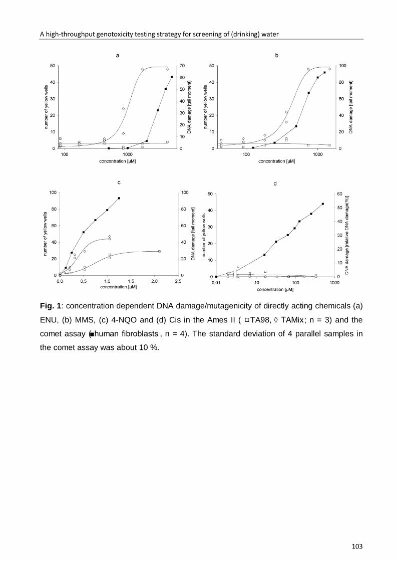
A high-throughput genotoxicity testing strategy for screening of (drinking) water
103
Fig. 1: concentration dependent DNA damage/mutagenicity of directly acting chemicals (a)
ENU, (b) MMS, (c) 4-NQO and (d) Cis in the Ames II ( TA98, ◊ TAMix; n = 3) and the
comet assay (■human fibroblasts , n = 4). The standard deviation of 4 parallel samples in
the comet assay was about 10 %.
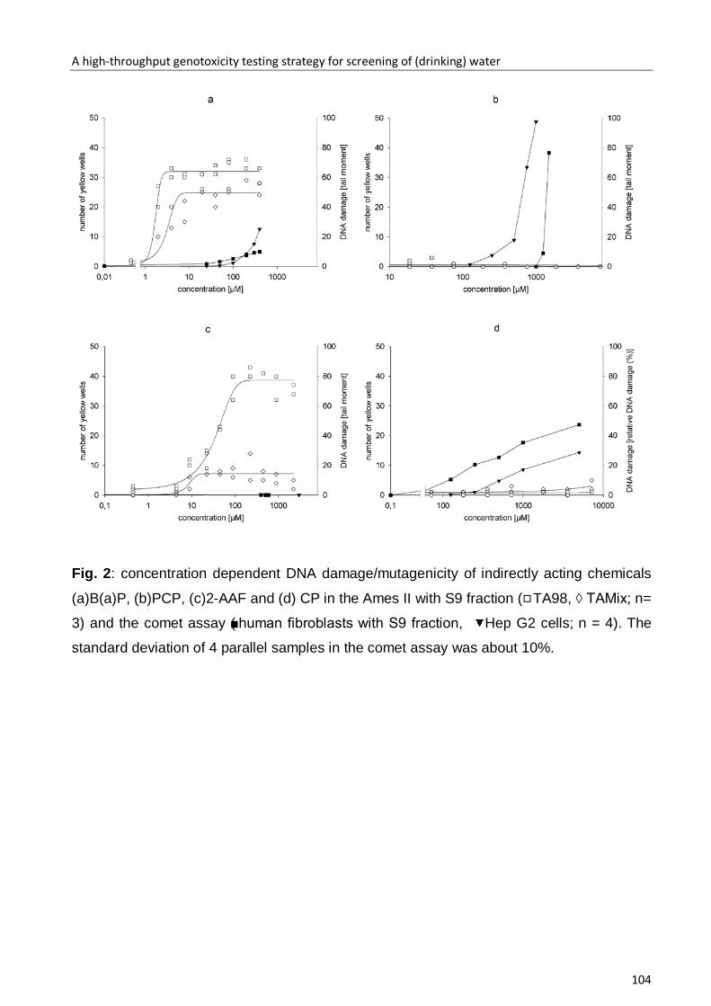
A high-throughput genotoxicity testing strategy for screening of (drinking) water
104
Fig. 2: concentration dependent DNA damage/mutagenicity of indirectly acting chemicals
(a)B(a)P, (b)PCP, (c)2-AAF and (d) CP in the Ames II with S9 fraction ( TA98, ◊ TAMix; n=
3) and the comet assay (■human fibroblasts with S9 fraction, Hep G2 cells; n = 4). The
standard deviation of 4 parallel samples in the comet assay was about 10%.

A high-throughput genotoxicity testing strategy for screening of (drinking) water
105

A high-throughput genotoxicity testing strategy for screening of (drinking) water
106
Fig. 3: mutagenicity/genotoxicity of environmental samples measured (a) in the Ames II
test (200-fold concentrated samples), (b) in the micronucleus test (100-fold concentrated
samples) and (c) in the comet test (400-fold concentrated samples); numbers above the
bars: % cytotoxicity in the micronucleus test and comet assay; in the Ames II test the
cytotoxicity did not exceed 8%; **significance level compared to the untreated control
p≤0.01

A high-throughput genotoxicity testing strategy for screening of (drinking) water
107
Fig. 4: Concentration dependent DNA damage induced by environmental samples
determined with the comet assay (a) TWW STP, (b) surface water 3 and (c) groundwater 4
( human fibroblast -S9, ■human fibroblast +S9, Hep G2 cells); significance level
compared to the control: ** p≤0.01, * p≤0.05; 1: cytotoxicity was ≥30 %

Danksagung
108
Danksagung
Ich möchte Frau Prof. Dr. Irene Witte für die Bereitstellung dieses weitreichenden
Themas, für die angenehme und fruchtbare Zusammenarbeit, die Bereitstellung
eines optimal eingerichteten Labors und die vielfältigen an mich gestellten
Herausforderungen ganz herzlich danken.
Herrn Prof. Dr. Karl-Wilhelm Koch danke ich für die freundliche Übernahme des
Zweitgutachtens.
Den Mitgliedern der AG Biochemie danke ich für das freundliche Arbeitsklima, in dem
das Arbeiten stets Freude gemacht hat. Ich danke Frau Marita Weerts-Eden und Elke
Frahmann für die tolle Einarbeitung in die Laborarbeit und für die Beantwortung
labortechnischen Fragen. Mein Dank gilt auch Frau Hannelore Kardelke, die jederzeit
große Hilfsbereitschaft gezeigt hat. Ich danke Heike Sommer und Kathrin Henrichs
für die Hilfe und Tipps, die mir stets angeboten wurden.
Marcus Nachtkamp danke ich für die vielen Späße und das Klima, dass du in W3
geschaffen hast.
Oliver May danke ich herzlich für den ständig „leckeren“ gekochten Kaffee, das
Korrekturlesen und die Aufmunterungen. Ebenfalls möchte ich auch Florian Unger
und Benjamin Kühnemut danken, die mir bei speziellen Fragen tatkräftig zur Seite
standen.
Ich danke Ursula Juhl-Strauss für die Geduld und das Korrekturlesen der englischen
Texte.
Ich danke Detlef Ulrich Egon Jahn für seine jahrelang grenzenlose Unterstützung, die
mich nicht nur sportlich erfolgreich sein lies, die Stunden auf dem Beifahrersitz und
die Erfahrungen die ich mit Ihm sammeln konnte.
Ich danke meinem Bruder René Stang für sein offenes Ohr und die Unterstützung in
schwierigen Zeiten. Meinem Großvater für die vielen handwerklichen Kniffe, die er
Danksagung
109
mir gezeigt hat und damit meinen Wissensdurst für die Wissenschaften und die Liebe
zur Natur geweckt hat. Meinen Eltern danke ich für die Unterstützung in allen
Lebenslagen und die Freiheit mir meinen eigenen Weg zu wählen.
Den erfolgreichen Abschluss dieser Arbeit verdanke ich meiner Lebensgefährten My
Dinh Tran, die mir über die Jahre immer den Rücken frei gehalten halt, mich wieder
aufbaute und mit mir durchs Leben schreitet. Du unterstützt mich in allem und hilfst
mir meine Träume zu verwirklichen.
Danke!

Erklärung
110
Erklärung
Hiermit versichere ich, dass ich diese Arbeit selbständig verfasst, keine anderen als
die angegebenen Hilfsmittel und Quellen benutzt und die den benutzten Werken
wörtlich oder inhaltlich entnommene Stellen als solche kenntlich gemacht habe.
Ferner versichere ich, dass die vorliegende Dissertation weder in ihrer Gesamtheit
noch in Teilen einer anderen wissenschaftlichen Hochschule zur Begutachtung in
einem Promotionsverfahren vorliegt oder vorgelegen hat
Oldenburg, den 08.07.09
___________________________________
André Stang
Curriculum vitea
111
Curiculum vitea
Persönliche Daten
Name André Stang
Geburtsdatum / -ort 04.04.1984 in Schwerin
Schulischer Werdegang
1990 – 2000 Mecklenburgisches Förderzentrum für
Körperbehinderte Schwerin
2000 – 2003 Schliemann Gymnasium
Wissenschaftlicher Werdegang
Okt 2003 – Mai 2008 Studium der Biologie an der Carl von Ossietzky
Universität Oldenburg
Abschluss: Diplom Biologe mit Schwerpunkt
Genetik und Biochemie
Mai 2008 – Juli 2009 Promotion in der Arbeitsgruppe Biochemie von
Prof. Dr. Irene Witte an der Carl von Ossietzky
Universität
Publikationen
2008 Lam C, Stang A, Harder T:
Planktonic epiphytic and fungi are selectively
eliminated by marine macroalgae
FEMS microbiology ecology. Vol. 63, 283-291