Genetic analysis of ALS cases in the isolated island ...
11
European Journal of Human Genetics https://doi.org/10.1038/s41431-020-00767-9 ARTICLE Genetic analysis of ALS cases in the isolated island population of Malta Rebecca Borg 1,2 ● Maia Farrugia Wismayer 1,2 ● Karl Bonavia 1 ● Andrew Farrugia Wismayer 1 ● Malcolm Vella 3 ● Joke J. F. A. van Vugt 4 ● Brendan J. Kenna 4 ● Kevin P. Kenna 4 ● Neville Vassallo 1,2 ● Jan H. Veldink 4 ● Ruben J. Cauchi 1,2 Received: 6 May 2020 / Revised: 16 October 2020 / Accepted: 23 October 2020 © The Author(s) 2020. This article is published with open access Abstract Genetic isolates are compelling tools for mapping genes of inherited disorders. The archipelago of Malta, a sovereign microstate in the south of Europe is home to a geographically and culturally isolated population. Here, we investigate the epidemiology and genetic profile of Maltese patients with amyotrophic lateral sclerosis (ALS), identified throughout a 2-year window. Cases were largely male (66.7%) with a predominant spinal onset of symptoms (70.8%). Disease onset occurred around mid-age (median age: 64 years, men; 59.5 years, female); 12.5% had familial ALS (fALS). Annual incidence rate was 2.48 (95% CI 1.59–3.68) per 100,000 person-years. Male-to-female incidence ratio was 1.93:1. Prevalence was 3.44 (95% CI 2.01–5.52) cases per 100,000 inhabitants on 31 st December 2018. Whole-genome sequencing allowed us to determine rare DNA variants that change the protein-coding sequence of ALS-associated genes. Interestingly, the Maltese ALS patient cohort was found to be negative for deleterious variants in C9orf72, SOD1, TARDBP or FUS genes, which are the most commonly mutated ALS genes globally. Nonetheless, ALS-associated repeat expansions were identified in ATXN2 and NIPA1. Variants predicted to be damaging were also detected in ALS2, DAO, DCTN1, ERBB4, SETX, SCFD1 and SPG11. A total of 40% of patients with sporadic ALS had a rare and deleterious variant or repeat expansion in an ALS- associated gene, whilst the genetic cause of two thirds of fALS cases could not be pinpointed to known ALS genes or risk loci. This warrants further studies to elucidate novel genes that cause ALS in this unique population isolate. Introduction Amyotrophic lateral sclerosis (ALS) is an adult-onset, rapidly progressing, neurodegenerative disease. Onset is typically accompanied by clinical signs of upper and/or lower motor neuron degeneration and patients usually pre- sent with weakness in the bulbar muscles, only the limbs, or both regions simultaneously. About 15–20% of persons with ALS experience progressive cognitive decline, leading ultimately to dementia. The condition known as fronto- temporal dementia results from the degeneration of the frontal and temporal lobes [1, 2]. The incidence of ALS in European populations is two to three cases per year per 100,000, whereas prevalence can reach 10/100,000 [3]. ALS is classified as familial (fALS) in the presence of a clear family history of the disease and sporadic (sALS) when this is absent. The rate of fALS among prospective population-based registries is about 5% [4]. Nonetheless, studies on twins and European case-control cohorts have found that heritability of ALS approximates 40% [5–7]. To date, variants in any of more than 40 genes have been reported to cause monogenic fALS with more than half of the cases explained by highly penetrant causal variants residing in C9orf72 (23%), SOD1 (19%), TARDBP (3%) or FUS (3%) [8]. Variants in these genes [8–10] and genetic * Ruben J. Cauchi [email protected] 1 Centre for Molecular Medicine and Biobanking, Biomedical Sciences Building, University of Malta, Msida, Malta 2 Department of Physiology and Biochemistry, Faculty of Medicine and Surgery, University of Malta, Msida, Malta 3 Department of Neuroscience, Mater Dei Hospital, Msida, Malta 4 Department of Neurology, Brain Center Rudolf Magnus, University Medical Center Utrecht, Utrecht, The Netherlands Supplementary information The online version of this article (https:// doi.org/10.1038/s41431-020-00767-9) contains supplementary material, which is available to authorised users. 1234567890();,: 1234567890();,:
Transcript of Genetic analysis of ALS cases in the isolated island ...

European Journal of Human Geneticshttps://doi.org/10.1038/s41431-020-00767-9
ARTICLE
Genetic analysis of ALS cases in the isolated island populationof Malta
Rebecca Borg1,2● Maia Farrugia Wismayer1,2 ● Karl Bonavia1 ● Andrew Farrugia Wismayer1 ● Malcolm Vella3 ●
Joke J. F. A. van Vugt4 ● Brendan J. Kenna4 ● Kevin P. Kenna4 ● Neville Vassallo1,2● Jan H. Veldink 4
●
Ruben J. Cauchi 1,2
Received: 6 May 2020 / Revised: 16 October 2020 / Accepted: 23 October 2020© The Author(s) 2020. This article is published with open access
AbstractGenetic isolates are compelling tools for mapping genes of inherited disorders. The archipelago of Malta, a sovereignmicrostate in the south of Europe is home to a geographically and culturally isolated population. Here, we investigate theepidemiology and genetic profile of Maltese patients with amyotrophic lateral sclerosis (ALS), identified throughout a 2-yearwindow. Cases were largely male (66.7%) with a predominant spinal onset of symptoms (70.8%). Disease onset occurredaround mid-age (median age: 64 years, men; 59.5 years, female); 12.5% had familial ALS (fALS). Annual incidence ratewas 2.48 (95% CI 1.59–3.68) per 100,000 person-years. Male-to-female incidence ratio was 1.93:1. Prevalence was 3.44(95% CI 2.01–5.52) cases per 100,000 inhabitants on 31st December 2018. Whole-genome sequencing allowed us todetermine rare DNA variants that change the protein-coding sequence of ALS-associated genes. Interestingly, the MalteseALS patient cohort was found to be negative for deleterious variants in C9orf72, SOD1, TARDBP or FUS genes, which arethe most commonly mutated ALS genes globally. Nonetheless, ALS-associated repeat expansions were identified in ATXN2and NIPA1. Variants predicted to be damaging were also detected in ALS2, DAO, DCTN1, ERBB4, SETX, SCFD1 andSPG11. A total of 40% of patients with sporadic ALS had a rare and deleterious variant or repeat expansion in an ALS-associated gene, whilst the genetic cause of two thirds of fALS cases could not be pinpointed to known ALS genes or riskloci. This warrants further studies to elucidate novel genes that cause ALS in this unique population isolate.
Introduction
Amyotrophic lateral sclerosis (ALS) is an adult-onset,rapidly progressing, neurodegenerative disease. Onset istypically accompanied by clinical signs of upper and/or
lower motor neuron degeneration and patients usually pre-sent with weakness in the bulbar muscles, only the limbs, orboth regions simultaneously. About 15–20% of personswith ALS experience progressive cognitive decline, leadingultimately to dementia. The condition known as fronto-temporal dementia results from the degeneration of thefrontal and temporal lobes [1, 2]. The incidence of ALS inEuropean populations is two to three cases per year per100,000, whereas prevalence can reach 10/100,000 [3].ALS is classified as familial (fALS) in the presence of aclear family history of the disease and sporadic (sALS)when this is absent. The rate of fALS among prospectivepopulation-based registries is about 5% [4]. Nonetheless,studies on twins and European case-control cohorts havefound that heritability of ALS approximates 40% [5–7]. Todate, variants in any of more than 40 genes have beenreported to cause monogenic fALS with more than half ofthe cases explained by highly penetrant causal variantsresiding in C9orf72 (23%), SOD1 (19%), TARDBP (3%) orFUS (3%) [8]. Variants in these genes [8–10] and genetic
* Ruben J. [email protected]
1 Centre for Molecular Medicine and Biobanking, BiomedicalSciences Building, University of Malta, Msida, Malta
2 Department of Physiology and Biochemistry, Faculty of Medicineand Surgery, University of Malta, Msida, Malta
3 Department of Neuroscience, Mater Dei Hospital, Msida, Malta4 Department of Neurology, Brain Center Rudolf Magnus,
University Medical Center Utrecht, Utrecht, The Netherlands
Supplementary information The online version of this article (https://doi.org/10.1038/s41431-020-00767-9) contains supplementarymaterial, which is available to authorised users.
1234
5678
90();,:
1234567890();,:

risk loci that include ATXN2 [11], UNC13A [12], SARM1[13], C21orf2, SCFD1 and MOBP [13] have also beenexposed in sALS cases. Genetic variants are estimated tocontribute to between 14 and 17% of European sALSpatients or those with a European ancestry [9, 10]. All thisunderscores the substantial contribution of genetic factors toALS disease aetiology.
Genetic discoveries often lead to novel insights into themolecular mechanisms of ALS. For instance, clusteringgenes according to their known physiological functionsidentified ribostasis, proteostasis and cytoskeletal dynamicsas key cellular pathways involved in ALS pathogenesis[14]. Importantly, discovery of genes is leading to thedevelopment of genotype-specific treatments [15]. Addi-tional genetic factors remain to be found for ALS. The useof geographically and/or culturally isolated populations formapping novel ALS genes is a strategy that remains rela-tively unexploited despite benefits that include foundereffects, reduced genetic diversity and minimal environ-mental heterogeneity [16]. This spurred us to study thenative population of Malta, a sovereign microstate in themiddle of the Mediterranean Sea. Consisting of an archi-pelago of three inhabited islands (total area 316 km2),Malta’s population presently numbers about 514,564, basedon Malta’s National Statistics Office (NSO) data in 2019.Population seeding events occurred more than 7000 yearsago by settlers coming from neighbouring Sicily and basedon mitochondrial DNA, Y-chromosome and autosomalDNA marker analyses, influences by colonisers in thecenturies that followed were minimal [17–19]. The Malteseare the only European population that speak a Semiticlanguage, further underscoring their relative isolation fromother communities inhabiting Europe. Previous successes ingene mapping of rare diseases in the population of Malta areencouraging [20, 21]. Here, we first investigate the inci-dence and prevalence of ALS in the Maltese islands, in a 2-year period. Second, we perform an initial genetic survey byreporting on rare variants in protein-coding regions andconsensus splice sites of all presently known monogenicALS genes and genetic risk loci in Maltese ALS patientscompared to matched controls.
Materials and methods
Participants
Participant surveillance and recruitment occurred through-out a 2-year window, from 2017 through 2018. Patientsdiagnosed with probable or definite ALS, referred by eitherthe national Motor Neuron Disease association, consultantneurologists, general practitioners and neurophysiologyunits were invited to participate in our study. Alternatively,
patients or their relatives made direct contact with ourlaboratory expressing willingness to participate in the study.Patient participants met the revised El Escorial criteria forALS [22, 23]. Patients with fALS were identified as havinga self-reported family history of ALS, or probable ALS,defined as the presence of at least one first-degree relative.In total, 24 patients were enrolled in this study. Bloodsampling was excluded for one sALS case in view of thepatient’s deteriorating condition. Affected family membersfor all fALS cases were deceased precluding us from sam-pling them. Controls, which were ascertained in a roughly2:1 case-control ratio, matched patients for age, sex andgeographical region. Ethical approval for the collection ofsamples, study design and the creation of the Malta ALS/MND Register and Biobank was given by the ResearchEthics Committee of the University of Malta. Writteninformed consent to participate was sought from all patientsand/or family members as well as controls.
Phenotypic information
Phenotypic information was gathered via a detailed ques-tionnaire in addition to a clinical examination. Each sampleis therefore accompanied by a core dataset that includes age,sex, occupation, site of onset, date of disease onset, familyhistory, ALS Functional Rating Scale-revised (ALSFRS-R)score, muscle tone for both upper and lower limbs, musclepower graded according to the MRC scale, and status ofreflexes. Information on possible environmental risk factorsincluding physical activity, cigarette smoking and alcoholconsumption was also gathered. A biochemical assay to testcreatine kinase (CK) levels at recruitment was also utilised.
Incidence and prevalence calculations
The denominator for the calculation of the incident rate wasthe sum of total population of Malta in 2017 and 2018.During the study period, the population of Malta increasedfrom 475,701 to 493,559. Separate incident rates for malesand females as well as specific age groups were also cal-culated. Population numbers were derived from NSO data.The prevalence rate was estimated on 31st December 2018.Confidence limits for incidence were calculated assuming aPoisson distribution.
Whole-genome sequencing
Extraction of DNA occurred from whole EDTA-containingvenous blood samples using the QIAamp DNA Mini QIA-cube Kit and DNA integrity was measured using the Quantusfluorometer. DNA was whole-genome sequenced by theBGISEQ-500 platform (BGI, Hong Kong, China) to generate100 bp paired-end reads with an average depth of 30×. Reads
R. Borg et al.

were aligned to the GRCh37 (HG19) reference genome usingBurrows–Wheeler Aligner software. Single nucleotide variant(SNV) and small insertion and deletion (indel) calling andquality filtering were performed using the Genome AnalysisToolkit (GATK). The ExpansionHunter tool was usedto analyse repeat sizes of ATXN2 (NM_002973.3:c.496_498CAG), C9orf72 (NM_001256054.2:c.-45+163GGGGCC) and NIPA1 (NM_144599.4:c.24_26GGC)[24]. In order to estimate the genetic ancestry in relation toreference maps of diverse populations, principal-componentanalysis (PCA) was performed on LASER with results plottedusing the LASER Server plot facility (https://laser.sph.umich.edu/). HomozygosityMapper (www.homozygositymapper.org) [25] was used to map runs of homozygosity (ROHs).
Variant analysis
We searched for and analysed protein-coding and splice-sitealtering variants and indels in 58 established ALS causative orrisk genes (Table 1). We restricted analyses to variants withEuropean minor allele frequency (MAF) ≤0.01, which corre-sponds roughly to the European frequency of the recentlydiscovered ALS risk NM_004984.2:c.2957C>T; p.(Pro986-Leu) allele in the KIF5A gene [26]. Where available, variantswere then annotated with information from the dbSNP data-base including European-specific MAF estimates from theGenome Aggregation Database (gnomAD). Allele frequenciesfor ALS cases and controls within the Project MinE datasetwere extracted from the Project MinE databrowser [27]. Todetermine variant pathogenicity, MetaSVM and MetaLR, twoensemble-based prediction methods integrating multiplescoring systems, were used in view of their superior predictiveability relative to other methods [28]. Variants were con-sidered as damaging if outcomes of both methods concurred.Indels and splice-site acceptor/donor variants were auto-matically classified as deleterious. Variants and the associatedphenotypes have been submitted to the ClinVar database(https://www.ncbi.nlm.nih.gov/clinvar/) with accessionnumbers SCV001426191, SCV001426206-SCV001426210,SCV001426221-SCV001426223 and SCV001437161-SCV001437192. New variants detected in the Maltese case-control cohort were submitted to the dbSNP database(https://www.ncbi.nlm.nih.gov/snp/) with the submission SNPIDs of ss2137544106, ss3986090479, ss3986090480, andss3986090481.
Statistics
Comparisons between means were made with the unpaired,two-tailed Student t test, whereas comparison betweencategorical variables was made with χ2 test. A p value <0.05was considered significant. Data were processed withGraphPad Prism v8.4.0 software.
Results
Baseline characteristics
The key characteristics of patients and controls are detailedin Table 2. ALS cases were largely male with a predominantspinal onset of symptoms. Bulbar onset ALS had a slightlyhigher occurrence in females (57.2%). Disease onsetoccurred around mid-age with median age at onset beinglower in females (59.5 years) compared to males (64 years).Only one male and female patient was under 45 years old(8.3%). Early ALS (≤55 years) was more frequently spinalat onset (spinal/bulbar ratio= 9:1). A family history of ALSwas recorded for a minority of cases (Table 2). However,considering their familial aggregation with ALS [29–31],the inclusion of both a family history of dementia andneuropsychiatric endophenotypes-like schizophrenia orpsychosis, increases the proportion of fALS to 37.5% (9/24patients). The mean duration of the illness was 44.5 ± 28.3SD months, with males having a faster progression com-pared to females (29.6 ± 8.8 SD months vs. 66.8 ± 33.4 SDmonths, p= 0.0065). Site of disease onset did not influencedisease duration (spinal onset= 43.5 ± 32.4 SD months,bulbar onset= 43.7 ± 19.8 SD months, p=NS).
One-third of the ALS patients recruited had a history ofheavy smoking and more than half reported an occupationassociated with strenuous activity, both of which have beenimplicated as environmental risk factors for ALS [32, 33].Alcohol abuse in the patient cohort was minimal. In viewthat a number of years have elapsed from onset up torecruitment, the levels of CK in the patient cohort were onaverage only slightly above the normal range. The meanALSFRS-R score in ALS recruits was nearly half thatrecorded for the control subjects with the score in the latternearing 48, the maximum expected in healthy individuals.The distribution of cases and controls throughout the Mal-tese islands are displayed in Fig. 1. A higher populationdensity in the southeast of mainland Malta, most probablyexplains the increase in the number of cases in this geo-graphic region relative to other regions.
Incidence and prevalence rates
The annual incidence rate for ALS in the 2017–2018 periodwas 2.48/100,000 person-years (95% CI 1.59–3.68). Themale-to-female incidence ratio was 1.93:1. Hence, inci-dence rate was higher for men (3.25, 95% CI 1.86–5.28)than for women (1.68, 95% CI 0.72–3.31), and this trendoccurred across all age groups after 49 years (Fig. 2). Forboth men and women, the incidence increased with age butdeclined after age 79. Peaks occurred in the 50 to 59 agegroup among men and in the 70 to 79 age group amongwomen. A total of 17 patients (male= 12, female= 5) were
Genetic analysis of ALS cases in the isolated island population of Malta
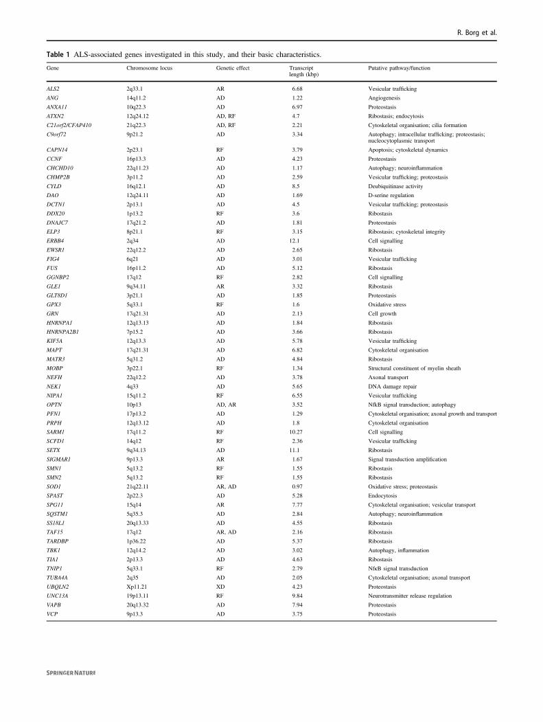
Table 1 ALS-associated genes investigated in this study, and their basic characteristics.
Gene Chromosome locus Genetic effect Transcriptlength (kbp)
Putative pathway/function
ALS2 2q33.1 AR 6.68 Vesicular trafficking
ANG 14q11.2 AD 1.22 Angiogenesis
ANXA11 10q22.3 AD 6.97 Proteostasis
ATXN2 12q24.12 AD, RF 4.7 Ribostasis; endocytosis
C21orf2/CFAP410 21q22.3 AD, RF 2.21 Cytoskeletal organisation; cilia formation
C9orf72 9p21.2 AD 3.34 Autophagy; intracellular trafficking; proteostasis;nucleocytoplasmic transport
CAPN14 2p23.1 RF 3.79 Apoptosis; cytoskeletal dynamics
CCNF 16p13.3 AD 4.23 Proteostasis
CHCHD10 22q11.23 AD 1.17 Autophagy; neuroinflammation
CHMP2B 3p11.2 AD 2.59 Vesicular trafficking; proteostasis
CYLD 16q12.1 AD 8.5 Deubiquitinase activity
DAO 12q24.11 AD 1.69 D-serine regulation
DCTN1 2p13.1 AD 4.5 Vesicular trafficking; proteostasis
DDX20 1p13.2 RF 3.6 Ribostasis
DNAJC7 17q21.2 AD 1.81 Proteostasis
ELP3 8p21.1 RF 3.15 Ribostasis; cytoskeletal integrity
ERBB4 2q34 AD 12.1 Cell signalling
EWSR1 22q12.2 AD 2.65 Ribostasis
FIG4 6q21 AD 3.01 Vesicular trafficking
FUS 16p11.2 AD 5.12 Ribostasis
GGNBP2 17q12 RF 2.82 Cell signalling
GLE1 9q34.11 AR 3.32 Ribostasis
GLT8D1 3p21.1 AD 1.85 Proteostasis
GPX3 5q33.1 RF 1.6 Oxidative stress
GRN 17q21.31 AD 2.13 Cell growth
HNRNPA1 12q13.13 AD 1.84 Ribostasis
HNRNPA2B1 7p15.2 AD 3.66 Ribostasis
KIF5A 12q13.3 AD 5.78 Vesicular trafficking
MAPT 17q21.31 AD 6.82 Cytoskeletal organisation
MATR3 5q31.2 AD 4.84 Ribostasis
MOBP 3p22.1 RF 1.34 Structural constituent of myelin sheath
NEFH 22q12.2 AD 3.78 Axonal transport
NEK1 4q33 AD 5.65 DNA damage repair
NIPA1 15q11.2 RF 6.55 Vesicular trafficking
OPTN 10p13 AD, AR 3.52 NfkB signal transduction; autophagy
PFN1 17p13.2 AD 1.29 Cytoskeletal organisation; axonal growth and transport
PRPH 12q13.12 AD 1.8 Cytoskeletal organisation
SARM1 17q11.2 RF 10.27 Cell signalling
SCFD1 14q12 RF 2.36 Vesicular trafficking
SETX 9q34.13 AD 11.1 Ribostasis
SIGMAR1 9p13.3 AR 1.67 Signal transduction amplification
SMN1 5q13.2 RF 1.55 Ribostasis
SMN2 5q13.2 RF 1.55 Ribostasis
SOD1 21q22.11 AR, AD 0.97 Oxidative stress; proteostasis
SPAST 2p22.3 AD 5.28 Endocytosis
SPG11 15q14 AR 7.77 Cytoskeletal organisation; vesicular transport
SQSTM1 5q35.3 AD 2.84 Autophagy; neuroinflammation
SS18L1 20q13.33 AD 4.55 Ribostasis
TAF15 17q12 AR, AD 2.16 Ribostasis
TARDBP 1p36.22 AD 5.37 Ribostasis
TBK1 12q14.2 AD 3.02 Autophagy, inflammation
TIA1 2p13.3 AD 4.63 Ribostasis
TNIP1 5q33.1 RF 2.79 NfκB signal transduction
TUBA4A 2q35 AD 2.05 Cytoskeletal organisation; axonal transport
UBQLN2 Xp11.21 XD 4.23 Proteostasis
UNC13A 19p13.11 RF 9.84 Neurotransmitter release regulation
VAPB 20q13.32 AD 7.94 Proteostasis
VCP 9p13.3 AD 3.75 Proteostasis
R. Borg et al.

alive at the prevalence date (31st December 2018), corre-sponding to a crude prevalence of 3.44/100,000 (95% CI2.01–5.52). Similarly, the prevalence in males (5.16, 95%CI 2.74–8.83) was higher than that in females (1.66, 95%CI 0.45–4.24), leading to a male-to-female prevalence ratioof 3.11:1. There was no difference in the mean age of onsetand site of onset between prevalent and incident patients.
Genetic ancestry
PCA analysis of the Maltese case-control cohort showedthat cases and controls were within 3 standard deviations of
the mean for the combined samples along principal com-ponents (PC) 1–4, hence ensuring adequate geneticmatching. Mapping the Maltese ALS patient and controlsamples to the reference PCA coordinates for samples fromthe Human Genome Diversity Panel (HGDP) shows overlapwith the portion of the Middle-Eastern cluster that neigh-bours the European cluster (Fig. 3). A genetic affinity withMiddle East populations is also apparent in Sicilians[34, 35], further supporting a common genetic ancestry forpopulations inhabiting the Mediterranean islands of Maltaand Sicily.
Repeat expansions in C9orf72, ATXN2 and NIPA1genes
Despite studies which show that C9orf72 is the major genethat is mutated in the European ALS population[8, 36, 37], we did not identify pathogenic hexanucleotide(GGGGCC) repeat expansions (≥24) in C9orf72 in eitherfALS or sALS cases (Fig. 4). The expansion repeat sizeranged from 2 to 10 in controls and from 2 to 13 in ALS
Table 2 Baseline characteristics of ALS patients and controls.
ALS(n= 24)
Controls(n= 13)
Sex
Male, % (n) 66.7 (16) 46.2 (6)
Female, % (n) 33.3 (8) 53.8 (7)
Type
Familial, % (n) 12.5 (3) –
Sporadic, % (n) 87.5 (21) –
Age at onset
Mean year ± SD (range) 59.3 ± 13.2(27–80)
–
Age at recruitment
Mean year ± SD (range) 63.5 ± 11.6(30–82)
71.2 ± 10.9(52–90)
ALSFRS-R score, mean ± SD(range)
25.6 ± 11.1(11.5–44)
47.9 ± 0.3(47–48)
Site of onset
Spinal, % (n) 70.8 (17) –
Bulbar, % (n) 25 (6) –
Both, % (n) 4.2 (1)
Survival, mean years ± SD(range)
5.5 ± 5.1 (2-20) –
Deceased, % (n) 62.5 (15) –
Cognitive status
Normal, % (n) 95.8 (23) 100 (13)
Impaired, % (n) 4.2 (1) 0 (0)
Environmental risk factors
Heavy smoking, % (n) 33.3 (8) 23.1 (3)
Strenuous activity, % (n) 54.2 (13) 23.1 (3)
Excessive alcoholconsumption, % (n)
8.3 (2) 7.8 (1)
CK levels at recruitment, IU/La
Mean in males ± SD (range) 311.2 ± 259.7(15–863)
–
Mean in females ± SD(range)
180.8 ± 117.5(38–336)
–
ALSFRS-R Amyotrophic Lateral Sclerosis Functional Rating Scale-Revised, CK creatine kinase.aNormal CK range= 39–308 U/L (males), 26–192 U/L (females).
Fig. 1 Geographical distribution of ALS cases and controls forindividuals born on the Maltese islands. Population size of the fourregional divisions (separated by dotted lines) is based on NSO datain 2017.
Fig. 2 Change in incidence rate of ALS with age in Malta. Incidencerate increases with age and was higher for males compared to femalesacross all age groups after 49 years. Peaks occur in the 50–59 age groupfor men and in the 70–79 age group for women.
Genetic analysis of ALS cases in the isolated island population of Malta

patients. Consistent with previous studies [36, 37], a repeatlength of 2 was the most predominant in either group(42.3% in controls and 52.2% in ALS cases). In addition toC9orf72, repeat expansions in other genes includingATXN2 and NIPA1 have been associated with increasedrisk of ALS [11, 38]. We identified one male patient withfALS that possessed ATXN2 ALS-associated trinucleotiderepeat expansions (28 repeats in length) in the homo-zygous state (Fig. 4). At the age of 67 years, this patientfirst experienced bilateral leg weakness that progressed.He subsequently developed dysarthria and succumbed tothe disease within 2 years of disease onset. This patientwas also the only one in our cohort that showed signs ofcognitive impairment. Family history was notable for adeceased sister who had ALS with early onset in her late30s, and a deceased mother who had dementia (DNAsamples were not available for study). The pedigree isshown in Supplementary Fig. S1. The maximum ATXN2repeat size observed in healthy controls was 25, encoun-tered in the heterozygous state, in one subject. Allremaining ALS patients and controls had repeat lengths≤23. As was reported previously [11, 39], a repeat lengthof 22, detected mostly in the homozygous state, was themost abundant ATXN2 allele (88.5% in controls and 84.8%
in ALS patients). Considering NIPA1, we found one malepatient with sALS who had an expanded [21] GCG repeatmotif in the heterozygous state. The patient had a late ageof onset (73 years), experiencing bilateral weakness first inthe lower limbs and then in the upper limbs. Survival wasshorter (<2.5 years) compared with the median survival(3.5 years) in our ALS cohort. In controls and in theremaining ALS cases, repeat length was variable rangingfrom 5 to 13. Similar to previous findings [38, 40], themost frequent alleles consisted of either 7 or 8 repeats,with respective allele frequencies being 26.1% and 67.4%in ALS patients, and 7.7% and 73.1% in controls.
Genetic variants in known ALS-associated genes
Interestingly, the Maltese ALS patient cohort was found tobe negative for non-synonymous or splice-site alteringSNVs in the SOD1, TARDBP or FUS genes, which are themost commonly mutated ALS genes, in that order, fol-lowing C9orf72 [8]. After examining 58 ALS-associatedgenes in our patient and control cohort, we identified 35 rare(European MAF ≤ 0.01) coding variants that were present inMaltese ALS patients and absent in controls (Table 3).Three SNVs in DDX20, EWSR1 or GLE1 were not found inthe dbSNP (v141) database. The NM_001003722.1:c.2078C>T; p.(Ser693Phe) variant in GLE1 was howeverreported in a recent study as a founder mutation in Maltesethat on homozygosis induces a motor dysfunction syndromethat presents at childhood [21]. Variants predicted to bedamaging by both MetaSVM and MetaLR were detectedin ALS2, DAO, DCTN1, ERBB4, SCFD1 and SPG11(Table 3). All were detected in patients with sALS. TheNM_001917.4:c.250G>A; p.(Ala84Thr) variant in DAOwas detected in two patients, whereas one female patientpossessed deleterious variants in more than one gene (DAOand DCTN1).
Fig. 3 Ancestry of Maltese ALS cases and controls compared to the HGDP reference panel. Left panel, Reference PCA coordinates forsamples from the HGDP reference panel. Middle panel, Maltese ALS cases mapped to the reference PCA coordinates. Right panel, Maltesecontrols mapped to the reference PCA coordinates.
Fig. 4 Expansion repeat size in ALS cases and controls for NIPA1,C9orf72 and ATXN2 genes. Scatter plot showing distribution andfrequency of repeat sizes, indicated by circles.
R. Borg et al.

Table3Rareno
n-syno
nymou
ssing
le-nucleotidevariantsandindelsfoun
din
Maltese
ALSpatients.
Prediction
Project
MinEAllele
Frequency
Gene
cDNA
change
Protein
change
dbSNP141ID
MetaSVM
MetaL
REuropeangnom
AD
MAF
ALScases
Controls
No.
ofpatients
ALStype
Zygosity
ALS2
NM_020919.3:c.3206G>A
p.(G
ly1069Glu)
rs200706696
Dam
aging
Dam
aging
0.0003
0.000376
01
sALS
het
ATXN2
NM_002973.3:c.2950A>C
p.(Ile984L
eu)
rs1338140819
Tolerated
Tolerated
0.00001
NA
NA
1sA
LS
het
C21orf2
NM_001271441.1:c.661G>A
p.(A
la221T
hr)
rs746114248
Tolerated
Tolerated
0.0000
NA
NA
1sA
LS
het
CAPN14
NM_001145122.1:c.1249C
>T
p.(Leu417P
he)
rs181906086
Tolerated
Tolerated
0.0183
0.004810
0.005259
1fA
LS
het
DAO
NM_001917.4:c.250G
>A
p.(A
la84Thr)
rs781658657
Dam
aging
Dam
aging
0.00001
NA
NA
2sA
LS
het
DCTN1
NM_004082.4:c.1484G>A
p.(A
rg495G
ln)
rs17721059
Tolerated
Tolerated
0.014
0.018945
0.019013
1fA
LS
het
DCTN1
NM_004082.4:c.586A
>G
p.(Ile196V
al)
rs55862001
Tolerated
Tolerated
0.0055
0.005720
0.006885
1sA
LS
hom
DCTN1
NM_004082.4:c.1864A>T
p.(Ile622P
he)
rs1328116832
Dam
aging
Dam
aging
0.00001a
NA
NA
1sA
LS
het
DDX20
NM_007204.4:c.2237T>C
p.(Leu746S
er)
NA
Tolerated
Tolerated
NA
NA
NA
1fA
LS
het
DNAJC
7NM_001144766.2:c.2T>C
p.(M
et1?)
rs371236469
Tolerated
Tolerated
0.00005
0.000075
01
sALS
het
ERBB4
NM_005235.2:c.3814G>A
p.(G
ly1272Arg)
rs371332509
Dam
aging
Dam
aging
0.00001
NA
NA
1sA
LS
het
ERBB4
NM_005235.2:c.1122T>G
p.(H
is374G
ln)
rs76603692
Tolerated
Tolerated
0.0009
0.001955
0.002429
1sA
LS
het
ERBB4
NM_005235.2:c.3176T>C
p.(M
et1059Thr)
rs373685875
Tolerated
Tolerated
0.00001
NA
NA
1sA
LS
het
EWSR
1NM_013986.3:c.1798G>A
p.(A
sp600A
sn)
NA
Tolerated
Tolerated
NA
NA
NA
1fA
LS
het
EWSR
1NM_013986.3:c.1408G>A
p.(G
ly470S
er)
rs41311143
Tolerated
Tolerated
0.0121
0.013158
0.013754
1sA
LS
het
GLE1
NM_001003722.1:c.2078C
>T
p.(Ser693P
he)
NA
Tolerated
Tolerated
NA
NA
NA
1sA
LS
het
KIF5A
NM_004984.2:c.2957C>T
p.(Pro986L
eu)
rs113247976
Tolerated
Tolerated
0.018
0.020069
0.014777
1sA
LS
het
MOBP
NM_001278322.1:c.586C>A
p.(A
rg196S
er)
rs1188260744
Tolerated
Tolerated
0.0000
NA
NA
1sA
LS
het
NEFH
NM_021076.3:c.2009T>A
p.(V
al670G
lu)
rs190692435
Tolerated
Tolerated
0.00676
NA
NA
2sA
LS,fA
LS
het
NEK1
NM_001199397.1:c.107A>G
p.(A
sn36Ser)
rs1404362599
Tolerated
Tolerated
NA
NA
NA
1sA
LS
het
SARM1
NM_015077.4:c.1501T>C
p.(Tyr501H
is)
rs144613221
Dam
aging
Tolerated
0.00228
0.003458
0.002832
1sA
LS
het
SCFD1
NM_016106.3:c.209T
>C
p.(Ile70Thr)
rs61754480
Dam
aging
Dam
aging
0.00403
0.004660
0.003843
1sA
LS
het
SCFD1
NM_016106.3:c.1297A>G
p.(Thr433A
la)
rs61754285
Tolerated
Tolerated
0.01788
0.018641
0.023058
2sA
LS
het
SETX
NM_015046.5:c.7640T>C
p.(Ile2547Thr)
rs151117904
Tolerated
Tolerated
0.0032
0.007066
0.004652
1sA
LS
het
SETX
NM_015046.5:c.2425A>G
p.(Ile809V
al)
rs906452681
Tolerated
Tolerated
0.00003
00.000202
1sA
LS
het
SETX
NM_015046.5:c.5308_5311d
elp.(G
lu1770Ilefs*15)
rs750959420
Dam
agingb
Dam
agingb
0.0000
0.000075
01
sALS
het
SPG11
NM_025137.3:c.1618C>T
p.(A
rg540C
ys)
rs758046989
Dam
aging
Dam
aging
0.00001
NA
NA
1sA
LS
het
SPG11
NM_025137.3:c.6759C>G
p.(A
sp2253Glu)
rs141818132
Tolerated
Tolerated
0.00003
00
1sA
LS
het
SPG11
NM_025137.3:c.1698T>G
p.(A
sp566G
lu)
rs79708848
Tolerated
Tolerated
0.01798
0.016165
0.014563
1sA
LS
het
SPG11
NM_025137.3:c.16G>A
p.(G
ly6A
rg)
rs200573434
Tolerated
Tolerated
0.00208
0.002330
0.001619
1sA
LS
het
SPG11
NM_025137.3:c.3037A>G
p.(Lys1013Glu)
rs111347025
Tolerated
Tolerated
0.01224
0.016912
0.017800
1sA
LS
het
SPG11
NM_025137.3:c.3425C>G
p.(Ser1142Cys)
rs201082396
Tolerated
Tolerated
0.0001
NA
NA
1fA
LS
het
SPG11
NM_025137.3:c.2656T>C
p.(Tyr886H
is)
rs139687202
Tolerated
Tolerated
0.0001
0.000225
0.000405
1sA
LS
het
SPG11
NM_025137.3:c.7256A>G
p.(Lys2419Arg)
rs76116949
Tolerated
Tolerated
0.0001
0.000075
01
sALS
het
TNIP1
NM_001252390.1:c.437C>T
p.(A
la146V
al)
rs2233289
Tolerated
Tolerated
0.0103
0.012103
0.013754
1sA
LS
het
dbSN
PSingle-NucleotidePolym
orph
ism
database,gn
omAD
Genom
eAgg
regatio
nDatabase,
MAFminor
allele
frequency,
NAno
tavailable,
hetheterozygo
te.
a Datafrom
Trans-O
micsforPrecision
Medicine(Top
Med)Program
.b Ind
elwas
automatically
considered
deleteriou
s.
Genetic analysis of ALS cases in the isolated island population of Malta
Analysis of indels allowed us to identify a rare deletionin SETX, detected in the heterozygous state in a sALS case(Table 3). This patient presented with upper limb weaknessat the age of 70. One year later, on follow-up, weaknessspread to the lower limbs. The deletion is predicted to resultin a frameshift, consequently producing a truncated SETXprotein lacking the helicase domain. It is noteworthy thatthe damaging ALS2, SCFD1, and SETX variants detected inMaltese sALS patients were found to have higher allelefrequencies in ALS patients within the Project MinE case-control dataset [27], thereby underscoring their probablepathogenicity (Table 3). Rare SNVs or indels that wereunique to controls or that were shared by ALS patients andcontrols are listed in Supplementary Table S1. One SNVs inFIG4 was not found in the dbSNP (v141) database. Nohomozygous stretches were overrepresented in sALSpatients compared to controls (Supplementary Fig. S2). Toestimate the genetic risk for ALS in the Maltese population,we determined that the percentage of sALS cases caused byrare and potentially deleterious variants (absent in controls)in at least one ALS-associated gene was 40% (8/20patients). Two fALS cases did not carry any mutations inknown ALS genes or risk loci, hence, warranting furtherstudies to elucidate novel genes that cause ALS.
Discussion
In our work, we investigated the characteristics of MalteseALS patients, described their genetic profile, and determinedthe incidence and prevalence of ALS on the Maltese islands. Itis interesting that the population-specific aspects of our ALScases overlap those reported for other neighbouring Europeanpopulations, especially those in the Mediterranean includingthe island of Sicily [41] and the southern region of Puglia[42, 43] in Italy, Tunisia [44] and Cyprus [45]. Although themale preponderance in ALS is virtually universal [46, 47], it isnoteworthy that in these specific populations as well as inMalta, age at disease onset is higher in males than in females.This is in contrast to northern populations of the Mediterra-nean basin including Catalonia in Spain [48], and the regionsof Emilia Romagna [49], Liguria [50] or Friuli-Venezia-Giulia[51] in Italy. Incidence and prevalence of ALS in Malta issimilar to the European median [3].
We report a higher percentage (12.5%) of fALS cases inMalta, close to that reported for the northern Italianpopulation of Liguria (10%) [50] but nearly halfthat reported for the island of Sardinia (26.7%) [52].Nonetheless, similar to previous studies [53], relaxing thestringent criteria by including neurological conditions inkindreds that have a genetic overlap with ALS [54], canincrease the fALS percentage in Malta by threefold(37.5%). In agreement, considering the Maltese sALS
subset, we showed that, compared to other Europeanpopulations [10] or populations of European ancestry [9], ahigher proportion of seemly sALS patients are probablygenetically determined.
Intriguingly, rare deleterious variants in the major ALSgenes, including C9orf72, SOD1, TARDBP and FUS, wereabsent in Maltese ALS patients. This finding confirms thepresence of a North–South gradient in the frequency ofmutations within these genes across Europe. Hence,C9orf72 or SOD1 mutations in fALS are highest in northernEuropean countries like Belgium and Finland, whereas arelatively low frequency is recorded in the south of Europeincluding Spain and mainland Italy [8]. A similar situationcan be observed for TARDBP and FUS [8]. Our study thusunderscores the marked differences that exists betweenethnic groups and geographical regions with respect to thegenes that are commonly implicated in ALS.
Maltese ALS patients nevertheless possessed deleteriousalleles in ‘minor’ ALS genes including ALS2, ATXN2,DAO, DCTN1, ERBB4, NIPA1, SETX, SCFD1 and SPG11.ALS2 and SPG11 have been associated with juvenile-onsetALS only under a recessive disease model [55–58]. In thiscontext, since we observed variants in the ALS2 and SPG11genes solely in heterozygous configurations and in patientswhich had adult-onset ALS, it is likely that these alleleswere not disease causing in the patients that possessed them.However, considering that ALS has an oligogenic basis[59], a modifying or additive effect cannot be excluded. Thesame can be said for SCFD1, which has only been recentlyidentified as a risk locus [13], and for which we report adamaging variant in an ALS patient with a young age ofonset (27 years) and whose disease progression isexceptionally slow.
ATXN2, DAO, DCTN1, ERBB4, NIPA1 and SETX haveall been previously associated with ALS having an auto-somal dominant mode of inheritance [11, 38, 40, 60–63].Damaging alleles discovered in our patient cohort, whichspecifically target these genes, are most probably causative.It is interesting to note that the ERBB4 c.3814G>A; p.(Gly1272Arg) variant reported in this study is extremelyclose to the one reported in a Japanese sALS individual[c.3823C>T; p.(Arg1275Trp)], both of which are located inthe C-terminal domain of the protein, close to multiplephosphorylation sites, which mediate downstream signal-ling pathways [61]. Although SETX variants have beeninitially discovered in juvenile-onset ALS patients [62],reports have since described damaging alleles in patientswith adult-onset ALS [10, 64]. This is in line with ourstudy, hence, the Maltese ALS patient possessing a SETXdeletion had a late age of onset similar to the one reported ina previous case study [64].
Our findings have important implications. Incidence andprevalence of ALS in Malta as well as patient population
R. Borg et al.

aspects overlap those of neighbouring countries. However,supported by genetic ancestry results, the genetic archi-tecture of ALS in Malta appears to be different from theEuropean average underscoring genetic isolation imposedby geography. This combined with the lack of an identifiedgenetic factor in two-thirds of Maltese fALS cases,encourages further studies aimed at discovering novel ALSgenes. Our ‘preliminary’ data excludes the possibility thatthese patients have deleterious variants in a set of genesassociated with other motor neuron disorders includinghereditary ataxias, and hereditary motor and sensory neu-ropathies (data not shown). Finally, variants described inthis work should spur the generation of animal models toconfirm causation and better understand disease mechan-isms [65]. This is imperative especially for ‘minor’ ALSgenes, given that they are relatively less studied than‘major’ genes, but which are nonetheless consequential inspecific populations.
Acknowledgements The authors are grateful to Prof. Richard Muscat,Wilfred Kenely and Dr. Graziella Zahra for their support of this work.Thanks also goes to Matthew Camilleri for unwavering technical andadministrative support. We remain indebted to all participants ofthis study.
Funding This work was supported by the University of MaltaResearch Excellence Fund to RJC, and the Malta Council for Sci-ence & Technology Internationalisation Partnership Award to RJC.RB was supported by an EMBO short-term fellowship and by aBjorn Formosa Scholarship for Advanced Research into ALS/MNDfunded by the non-profit organisation, ALS Malta Foundation,facilitated by the Research Trust (RIDT) of the University of Malta.ML was supported by an Endeavour Scholarship (Malta), part-financed by the EU—European Social Fund under OperationalProgramme II—Cohesion Policy 2014–2020, “Investing in humancapital to create more opportunities and promote the well-being ofsociety”.
Compliance with ethical standards
Conflict of interest The authors declare that they have no conflict ofinterest.
Publisher’s note Springer Nature remains neutral with regard tojurisdictional claims in published maps and institutional affiliations.
Open Access This article is licensed under a Creative CommonsAttribution 4.0 International License, which permits use, sharing,adaptation, distribution and reproduction in any medium or format, aslong as you give appropriate credit to the original author(s) and thesource, provide a link to the Creative Commons license, and indicate ifchanges were made. The images or other third party material in thisarticle are included in the article’s Creative Commons license, unlessindicated otherwise in a credit line to the material. If material is notincluded in the article’s Creative Commons license and your intendeduse is not permitted by statutory regulation or exceeds the permitteduse, you will need to obtain permission directly from the copyrightholder. To view a copy of this license, visit http://creativecommons.org/licenses/by/4.0/.
References
1. Brown RH, Al-Chalabi A. Amyotrophic lateral sclerosis. N Engl JMed. 2017;377:162–72.
2. van Es MA, Hardiman O, Chio A, Al-Chalabi A, Pasterkamp RJ,Veldink JH, et al. Amyotrophic lateral sclerosis. Lancet. 2017;390:2084–98.
3. Chio A, Logroscino G, Traynor BJ, Collins J, Simeone JC,Goldstein LA, et al. Global epidemiology of amyotrophic lateralsclerosis: a systematic review of the published literature. Neu-roepidemiology. 2013;41:118–30.
4. Byrne S, Walsh C, Lynch C, Bede P, Elamin M, Kenna K, et al.Rate of familial amyotrophic lateral sclerosis: a systematic reviewand meta-analysis. J Neurol Neurosurg Psychiatry. 2011;82:623–7.
5. Al-Chalabi A, Fang F, Hanby MF, Leigh PN, Shaw CE, Ye W,et al. An estimate of amyotrophic lateral sclerosis heritabilityusing twin data. J Neurol Neurosurg Psychiatry. 2010;81:1324–6.
6. Ryan M, Heverin M, McLaughlin RL, Hardiman O. Lifetime riskand heritability of amyotrophic lateral sclerosis. JAMA Neurol.2019;76:1367–74.
7. Trabjerg BB, Garton FC, van Rheenen W, Fang F, Henderson RD,Mortensen PB, et al. ALS in Danish registries: heritability and linksto psychiatric and cardiovascular disorders. Neurol Genet. 2020;6:e398.
8. Zou ZY, Zhou ZR, Che CH, Liu CY, He RL, Huang HP. Geneticepidemiology of amyotrophic lateral sclerosis: a systematic reviewand meta-analysis. J Neurol Neurosurg Psychiatry. 2017;88:540–9.
9. Gibson SB, Downie JM, Tsetsou S, Feusier JE, Figueroa KP,Bromberg MB, et al. The evolving genetic risk for sporadic ALS.Neurology. 2017;89:226–33.
10. Kenna KP, McLaughlin RL, Byrne S, Elamin M, Heverin M,Kenny EM, et al. Delineating the genetic heterogeneity of ALSusing targeted high-throughput sequencing. J Med Genet. 2013;50:776–83.
11. Elden AC, Kim HJ, Hart MP, Chen-Plotkin AS, Johnson BS,Fang X, et al. Ataxin-2 intermediate-length polyglutamineexpansions are associated with increased risk for ALS. Nature.2010;466:1069–75.
12. van Es MA, Veldink JH, Saris CG, Blauw HM, van Vught PW,Birve A, et al. Genome-wide association study identifies 19p13.3(UNC13A) and 9p21.2 as susceptibility loci for sporadic amyo-trophic lateral sclerosis. Nat Genet. 2009;41:1083–7.
13. van Rheenen W, Shatunov A, Dekker AM, McLaughlin RL,Diekstra FP, Pulit SL, et al. Genome-wide association analysesidentify new risk variants and the genetic architecture of amyo-trophic lateral sclerosis. Nat Genet. 2016;48:1043–8.
14. Taylor JP, Brown RH Jr., Cleveland DW. Decoding ALS: fromgenes to mechanism. Nature. 2016;539:197–206.
15. Ly CV, Miller TM. Emerging antisense oligonucleotide and viraltherapies for amyotrophic lateral sclerosis. Curr Opin Neurol.2018;31:648–54.
16. Arcos-Burgos M, Muenke M. Genetics of population isolates.Clin Genet. 2002;61:233–47.
17. Caruana J. Population genetics of Western MediterraneanIslands—Malta: a case study. Manchester: University ofManchester; 2012.
18. Capelli C, Redhead N, Romano V, Cali F, Lefranc G, Delague V,et al. Population structure in the Mediterranean basin: a Y chro-mosome perspective. Ann Hum Genet. 2006;70:207–25.
19. Cassar M, Farrugia C, Vidal C. Allele frequencies of 14 STR lociin the population of Malta. Leg Med. 2008;10:153–6.
20. Borg J, Papadopoulos P, Georgitsi M, Gutierrez L, Grech G,Fanis P, et al. Haploinsufficiency for the erythroid transcription
Genetic analysis of ALS cases in the isolated island population of Malta
factor KLF1 causes hereditary persistence of fetal hemoglobin.Nat Genet. 2010;42:801–5.
21. Said E, Chong JX, Hempel M, Denecke J, Soler P, Strom T, et al.Survival beyond the perinatal period expands the phenotypes causedby mutations in GLE1. Am J Med Genet A. 2017;173:3098–103.
22. Brooks BR, Miller RG, Swash M, Munsat TL. World Federationof Neurology Research Group on Motor Neuron D. El Escorialrevisited: revised criteria for the diagnosis of amyotrophic lateralsclerosis. Amyotroph Lateral Scler Other Mot Neuron Disord.2000;1:293–9.
23. Ludolph A, Drory V, Hardiman O, Nakano I, Ravits J, RobberechtW, et al. A revision of the El Escorial criteria - 2015. AmyotrophLateral Scler Frontotemporal Degener. 2015;16:291–2.
24. Dolzhenko E, van Vugt J, Shaw RJ, Bekritsky MA, van Blit-terswijk M, Narzisi G, et al. Detection of long repeat expansionsfrom PCR-free whole-genome sequence data. Genome Res.2017;27:1895–903.
25. Seelow D, Schuelke M. HomozygosityMapper2012-bridging thegap between homozygosity mapping and deep sequencing.Nucleic Acids Res. 2012;40:W516–20.
26. Nicolas A, Kenna KP, Renton AE, Ticozzi N, Faghri F, Chia R,et al. Genome-wide analyses identify KIF5A as a novel ALS gene.Neuron. 2018;97:1268–83 e6.
27. van der Spek RAA, van Rheenen W, Pulit SL, Kenna KP, van denBerg LH, Veldink JH, et al. The project MinE databrowser:bringing large-scale whole-genome sequencing in ALS toresearchers and the public. Amyotroph Lateral Scler Fronto-temporal Degener. 2019;20:432–40.
28. Dong C, Wei P, Jian X, Gibbs R, Boerwinkle E, Wang K, et al.Comparison and integration of deleteriousness prediction methodsfor nonsynonymous SNVs in whole exome sequencing studies.Hum Mol Genet. 2015;24:2125–37.
29. Byrne S, Heverin M, Elamin M, Bede P, Lynch C, Kenna K, et al.Aggregation of neurologic and neuropsychiatric disease inamyotrophic lateral sclerosis kindreds: a population-based case-control cohort study of familial and sporadic amyotrophic lateralsclerosis. Ann Neurol. 2013;74:699–708.
30. O’Brien M, Burke T, Heverin M, Vajda A, McLaughlin R, Gib-bons J, et al. Clustering of neuropsychiatric disease in first-degreeand second-degree relatives of patients with amyotrophic lateralsclerosis. JAMA Neurol. 2017;74:1425–30.
31. Longinetti E, Mariosa D, Larsson H, Ye W, Ingre C, Almqvist C,et al. Neurodegenerative and psychiatric diseases among familieswith amyotrophic lateral sclerosis. Neurology 2017;89:578–85.
32. Peters S, Visser AE, D’Ovidio F, Vlaanderen J, Portengen L,Beghi E, et al. Effect modification of the association between totalcigarette smoking and ALS risk by intensity, duration and time-since-quitting: Euro-MOTOR. J Neurol Neurosurg Psychiatry.2020;91:33–9.
33. Visser AE, Rooney JPK, D’Ovidio F, Westeneng HJ, VermeulenRCH, Beghi E, et al. Multicentre, cross-cultural, population-based, case-control study of physical activity as risk factor foramyotrophic lateral sclerosis. J Neurol Neurosurg Psychiatry.2018;89:797–803.
34. Di Gaetano C, Voglino F, Guarrera S, Fiorito G, Rosa F, Di BlasioAM, et al. An overview of the genetic structure within the Italianpopulation from genome-wide data. PLoS ONE. 2012;7:e43759.
35. Sarno S, Boattini A, Pagani L, Sazzini M, De Fanti S, Quagliar-iello A, et al. Ancient and recent admixture layers in Sicily andSouthern Italy trace multiple migration routes along the Medi-terranean. Sci Rep. 2017;7:1984.
36. Renton AE, Majounie E, Waite A, Simon-Sanchez J, Rollinson S,Gibbs JR, et al. A hexanucleotide repeat expansion in C9ORF72is the cause of chromosome 9p21-linked ALS-FTD. Neuron.2011;72:257–68.
37. Smith BN, Newhouse S, Shatunov A, Vance C, Topp S, JohnsonL, et al. The C9ORF72 expansion mutation is a common cause ofALS+/-FTD in Europe and has a single founder. Eur J HumGenet. 2013;21:102–8.
38. Blauw HM, van Rheenen W, Koppers M, Van Damme P, WaibelS, Lemmens R, et al. NIPA1 polyalanine repeat expansions areassociated with amyotrophic lateral sclerosis. Hum Mol Genet.2012;21:2497–502.
39. Van Damme P, Veldink JH, van Blitterswijk M, Corveleyn A, vanVught PW, Thijs V, et al. Expanded ATXN2 CAG repeat sizein ALS identifies genetic overlap between ALS and SCA2.Neurology. 2011;76:2066–72.
40. Tazelaar GHP, Dekker AM, van Vugt J, van der Spek RA,Westeneng HJ, Kool L, et al. Association of NIPA1 repeatexpansions with amyotrophic lateral sclerosis in a large interna-tional cohort. Neurobiol Aging. 2019;74:234 e9–15.
41. Ragonese P, Cellura E, Aridon P, D’Amelio M, Spataro R, TaielloAC, et al. Incidence of amyotrophic lateral sclerosis in Sicily: apopulation based study. Amyotroph Lateral Scler. 2012;13:284–7.
42. Logroscino G, Beghi E, Zoccolella S, Palagano R, Fraddosio A,Simone IL, et al. Incidence of amyotrophic lateral sclerosis insouthern Italy: a population based study. J Neurol NeurosurgPsychiatry. 2005;76:1094–8.
43. Zoccolella S, Beghi E, Palagano G, Fraddosio A, Samarelli V,Lamberti P, et al. Signs and symptoms at diagnosis of amyo-trophic lateral sclerosis: a population-based study in southernItaly. Eur J Neurol. 2006;13:789–92.
44. Kacem I, Sghaier I, Bougatef S, Nasri A, Gargouri A, Ajroud-Driss S, et al. Epidemiological and clinical features of amyo-trophic lateral sclerosis in a Tunisian cohort. Amyotroph LateralScler Frontotemporal Degener. 2020;21:131–9.
45. Demetriou CA, Hadjivasiliou PM, Kleopa KA, Christou YP,Leonidou E, Kyriakides T, et al. Epidemiology of amyotrophiclateral sclerosis in the Republic of Cyprus: A 25-year retrospectivestudy. Neuroepidemiology. 2017;48:79–85.
46. Logroscino G, Traynor BJ, Hardiman O, Chio A, Mitchell D,Swingler RJ, et al. Incidence of amyotrophic lateral sclerosis inEurope. J Neurol Neurosurg Psychiatry. 2010;81:385–90.
47. Marin B, Boumediene F, Logroscino G, Couratier P, Babron MC,Leutenegger AL, et al. Variation in worldwide incidence ofamyotrophic lateral sclerosis: a meta-analysis. Int J Epidemiol.2017;46:57–74.
48. Pradas J, Puig T, Rojas-Garcia R, Viguera ML, Gich I, LogroscinoG, et al. Amyotrophic lateral sclerosis in Catalonia: a populationbased study. Amyotroph Lateral Scler Frontotemporal Degener.2013;14:278–83.
49. Mandrioli J, Biguzzi S, Guidi C, Venturini E, Sette E, Terlizzi E,et al. Epidemiology of amyotrophic lateral sclerosis in EmiliaRomagna Region (Italy): a population based study. AmyotrophLateral Scler Frontotemporal Degener. 2014;15:262–8.
50. Scialo C, Novi G, Bandettini di Poggio M, Canosa A,Sormani MP, Mandich P. et al. Clinical epidemiology of amyo-trophic lateral sclerosis in Liguria, Italy: an update of LIGALSregister. Amyotroph Lateral Scler Frontotemporal Degener.2016;17:535–42.
51. Palese F, Sartori A, Verriello L, Ros S, Passadore P, ManganottiP, et al. Epidemiology of amyotrophic lateral sclerosis in Friuli-Venezia Giulia, North-Eastern Italy, 2002-2014: a retrospectivepopulation-based study. Amyotroph Lateral Scler FrontotemporalDegener. 2019;20:90–9.
52. Borghero G, Pugliatti M, Marrosu F, Marrosu MG, Murru MR,Floris G, et al. Genetic architecture of ALS in Sardinia. NeurobiolAging. 2014;35:2882 e7–12.
53. Ryan M, Heverin M, Doherty MA, Davis N, Corr EM, Vajda A,et al. Determining the incidence of familiality in ALS: A study of
R. Borg et al.
temporal trends in Ireland from 1994 to 2016. Neurol Genet.2018;4:e239.
54. McLaughlin RL, Schijven D, van Rheenen W, van Eijk KR,O’Brien M, Kahn RS, et al. Genetic correlation between amyo-trophic lateral sclerosis and schizophrenia. Nat Commun. 2017;8:14774.
55. Hadano S, Hand CK, Osuga H, Yanagisawa Y, Otomo A, DevonRS, et al. A gene encoding a putative GTPase regulator is mutated infamilial amyotrophic lateral sclerosis 2. Nat Genet. 2001;29:166–73.
56. Hentati A, Bejaoui K, Pericak-Vance MA, Hentati F, Speer MC,Hung WY, et al. Linkage of recessive familial amyotrophic lateralsclerosis to chromosome 2q33-q35. Nat Genet. 1994;7:425–8.
57. Daoud H, Zhou S, Noreau A, Sabbagh M, Belzil V, Dionne-Laporte A, et al. Exome sequencing reveals SPG11 mutationscausing juvenile ALS. Neurobiol Aging. 2012;33:839 e5–9.
58. Orlacchio A, Babalini C, Borreca A, Patrono C, Massa R, BasaranS, et al. SPATACSIN mutations cause autosomal recessivejuvenile amyotrophic lateral sclerosis. Brain. 2010;133:591–8.
59. van Blitterswijk M, van Es MA, Hennekam EA, Dooijes D, vanRheenen W, Medic J, et al. Evidence for an oligogenic basis ofamyotrophic lateral sclerosis. Hum Mol Genet. 2012;21:3776–84.
60. Mitchell J, Paul P, Chen HJ, Morris A, Payling M, Falchi M, et al.Familial amyotrophic lateral sclerosis is associated with a muta-tion in D-amino acid oxidase. Proc Natl Acad Sci USA. 2010;107:7556–61.
61. Takahashi Y, Fukuda Y, Yoshimura J, Toyoda A, Kurppa K,Moritoyo H, et al. ERBB4 mutations that disrupt the neuregulin-ErbB4 pathway cause amyotrophic lateral sclerosis type 19. Am JHum Genet. 2013;93:900–5.
62. Chen YZ, Bennett CL, Huynh HM, Blair IP, Puls I, Irobi J, et al.DNA/RNA helicase gene mutations in a form of juvenile amyotrophiclateral sclerosis (ALS4). Am J Hum Genet. 2004;74:1128–35.
63. Munch C, Sedlmeier R, Meyer T, Homberg V, Sperfeld AD, KurtA, et al. Point mutations of the p150 subunit of dynactin (DCTN1)gene in ALS. Neurology. 2004;63:724–6.
64. Tripolszki K, Torok D, Goudenege D, Farkas K, Sulak A, TorokN, et al. High-throughput sequencing revealed a novel SETXmutation in a Hungarian patient with amyotrophic lateral sclerosis.Brain Behav. 2017;7:e00669.
65. Aquilina B, Cauchi RJ. Modelling motor neuron disease in fruitflies: lessons from spinal muscular atrophy. J Neurosci Methods.2018;310:3–11.
Genetic analysis of ALS cases in the isolated island population of Malta