Inhibitor of apoptosis proteins are potential targets for ...
10
RESEARCH Open Access Inhibitor of apoptosis proteins are potential targets for treatment of granulosa cell tumors – implications from studies in KGN Konstantin Bagnjuk 1† , Verena Jasmin Kast 1† , Astrid Tiefenbacher 1 , Melanie Kaseder 1 , Toshihiko Yanase 2 , Alexander Burges 3 , Lars Kunz 4 , Doris Mayr 5 and Artur Mayerhofer 1* Abstract Background: Granulosa cell tumors (GCTs) are derived from proliferating granulosa cells of the ovarian follicle. They are known for their late recurrence and most patients with an aggressive form die from their disease. There are no treatment options for this slowly proliferating tumor besides surgery and chemotherapy. In a number of tumors, analogs of the second mitochondria-derived activator of caspases (SMAC), alone or in combination with other molecules, such as TNFα, are evolving as new treatment options. SMAC mimetics block inhibitor of apoptosis proteins (IAPs), which bind caspases (e.g. XIAP), or activate the pro-survival NF-κB pathway (e.g. cIAP1/2). Expression of IAPs by GCTs is yet not fully elucidated but recently XIAP and its inhibition by SMAC mimetics in a combination therapy was described to induce apoptosis in a GCT cell line, KGN. We evaluated the expression of cIAP1 in GCTs and elucidated the effects of the SMAC mimetic BV-6 using KGN as a model. Results: Employing immunohistochemistry, we observed cIAP1 expression in a tissue microarray (TMA) of 42 GCT samples. RT-PCR confirmed expression of cIAP1/2, as well as XIAP, in primary, patient-derived GCTs and in KGN. We therefore tested the ability of the bivalent SMAC mimetic BV-6, which is known to inhibit cIAP1/2 and XIAP, to induce cell death in KGN. A dose response study indicated an EC 50 ≈ 8 μM for both, early (< 8) and advanced (> 80) passages, which differ in growth rate and presumably aggressiveness. Quantitative RT-PCR showed upregulation of NF- κB regulated genes in BV-6 stimulated cells. Blocking experiments with the pan-caspase inhibitor Z-VAD-FMK indicated caspase-dependence. A concentration of 20 μM Z-VAD-FMK was sufficient to significantly reduce apoptosis. This cell death was further substantiated by results of Western Blot studies. Cleaved caspase 3 and cleaved PARP became evident in the BV-6 treated group. Conclusions: Taken together, the results show that BV-6 is able to induce apoptosis in KGN cells. This approach may therefore offer a promising therapeutic avenue to treat GCTs. Keywords: Cell death, Ovarian granulosa cell tumor, Apoptosis Background Apoptosis can be activated via two different pathways, the extrinsic death receptor pathway and the intrinsic mito- chondria associated pathway [1, 2]. Both will execute apop- tosis by cleaving and therefore activating caspases, which in turn degrade other proteins, based on their peptidase activ- ity [3]. The intrinsic pathway is induced by the release of pro-apoptotic molecules from mitochondria, for ex- ample cytochrome c, endonuclease G, apoptosis inducing factor (AIF), high temperature requirement protein A2 (HtrA2 also known as OMI) or the second mitochondria- derived activator of caspases (SMAC; or its murine homo- log, known as direct inhibitor of apoptosis protein binding protein with low PI (DIABLO)) [2, 4]. SMAC blocks inhibitor of apoptosis proteins (IAPs, e.g. baculoviral IAP repeat containing protein 1–8, BIRC1–8), which are highly expressed in various tumors [5]. There- fore, IAPs are potential targets in oncology. Different SMAC mimetics have been developed to target IAPs [6–8]. © The Author(s). 2019 Open Access This article is distributed under the terms of the Creative Commons Attribution 4.0 International License (http://creativecommons.org/licenses/by/4.0/), which permits unrestricted use, distribution, and reproduction in any medium, provided you give appropriate credit to the original author(s) and the source, provide a link to the Creative Commons license, and indicate if changes were made. The Creative Commons Public Domain Dedication waiver (http://creativecommons.org/publicdomain/zero/1.0/) applies to the data made available in this article, unless otherwise stated. * Correspondence: [email protected] Konstantin Bagnjuk and Verena Jasmin Kast share first authorship. 1 Biomedical Center Munich (BMC), Cell Biology, Anatomy III, Ludwig-Maximilians-University (LMU), Grosshaderner Strasse 9, 82152 Planegg, Germany Full list of author information is available at the end of the article Bagnjuk et al. Journal of Ovarian Research (2019) 12:76 https://doi.org/10.1186/s13048-019-0549-6
Transcript of Inhibitor of apoptosis proteins are potential targets for ...
RESEARCH Open Access
Inhibitor of apoptosis proteins are potentialtargets for treatment of granulosa celltumors – implications from studies in KGNKonstantin Bagnjuk1†, Verena Jasmin Kast1†, Astrid Tiefenbacher1, Melanie Kaseder1, Toshihiko Yanase2,Alexander Burges3, Lars Kunz4, Doris Mayr5 and Artur Mayerhofer1*
Abstract
Background: Granulosa cell tumors (GCTs) are derived from proliferating granulosa cells of the ovarian follicle. Theyare known for their late recurrence and most patients with an aggressive form die from their disease. There areno treatment options for this slowly proliferating tumor besides surgery and chemotherapy. In a number of tumors,analogs of the second mitochondria-derived activator of caspases (SMAC), alone or in combination with other molecules,such as TNFα, are evolving as new treatment options. SMAC mimetics block inhibitor of apoptosis proteins (IAPs), whichbind caspases (e.g. XIAP), or activate the pro-survival NF-κB pathway (e.g. cIAP1/2). Expression of IAPs by GCTs is yet notfully elucidated but recently XIAP and its inhibition by SMAC mimetics in a combination therapy was described to induceapoptosis in a GCT cell line, KGN. We evaluated the expression of cIAP1 in GCTs and elucidated the effects of the SMACmimetic BV-6 using KGN as a model.
Results: Employing immunohistochemistry, we observed cIAP1 expression in a tissue microarray (TMA) of 42 GCT samples.RT-PCR confirmed expression of cIAP1/2, as well as XIAP, in primary, patient-derived GCTs and in KGN. We therefore testedthe ability of the bivalent SMAC mimetic BV-6, which is known to inhibit cIAP1/2 and XIAP, to induce cell death in KGN. Adose response study indicated an EC50≈ 8 μM for both, early (< 8) and advanced (> 80) passages, which differ in growth rateand presumably aggressiveness. Quantitative RT-PCR showed upregulation of NF-κB regulated genes in BV-6 stimulated cells.Blocking experiments with the pan-caspase inhibitor Z-VAD-FMK indicated caspase-dependence. A concentration of 20 μMZ-VAD-FMK was sufficient to significantly reduce apoptosis. This cell death was further substantiated by results of WesternBlot studies. Cleaved caspase 3 and cleaved PARP became evident in the BV-6 treated group.
Conclusions: Taken together, the results show that BV-6 is able to induce apoptosis in KGN cells. This approach maytherefore offer a promising therapeutic avenue to treat GCTs.
Keywords: Cell death, Ovarian granulosa cell tumor, Apoptosis
BackgroundApoptosis can be activated via two different pathways, theextrinsic death receptor pathway and the intrinsic mito-chondria associated pathway [1, 2]. Both will execute apop-tosis by cleaving and therefore activating caspases, which inturn degrade other proteins, based on their peptidase activ-ity [3]. The intrinsic pathway is induced by the release of
pro-apoptotic molecules from mitochondria, for ex-ample cytochrome c, endonuclease G, apoptosis inducingfactor (AIF), high temperature requirement protein A2(HtrA2 also known as OMI) or the second mitochondria-derived activator of caspases (SMAC; or its murine homo-log, known as direct inhibitor of apoptosis protein bindingprotein with low PI (DIABLO)) [2, 4].SMAC blocks inhibitor of apoptosis proteins (IAPs, e.g.
baculoviral IAP repeat containing protein 1–8, BIRC1–8),which are highly expressed in various tumors [5]. There-fore, IAPs are potential targets in oncology. DifferentSMAC mimetics have been developed to target IAPs [6–8].
© The Author(s). 2019 Open Access This article is distributed under the terms of the Creative Commons Attribution 4.0International License (http://creativecommons.org/licenses/by/4.0/), which permits unrestricted use, distribution, andreproduction in any medium, provided you give appropriate credit to the original author(s) and the source, provide a link tothe Creative Commons license, and indicate if changes were made. The Creative Commons Public Domain Dedication waiver(http://creativecommons.org/publicdomain/zero/1.0/) applies to the data made available in this article, unless otherwise stated.
* Correspondence: [email protected] Bagnjuk and Verena Jasmin Kast share first authorship.1Biomedical Center Munich (BMC), Cell Biology, Anatomy III,Ludwig-Maximilians-University (LMU), Grosshaderner Strasse 9, 82152Planegg, GermanyFull list of author information is available at the end of the article
Bagnjuk et al. Journal of Ovarian Research (2019) 12:76 https://doi.org/10.1186/s13048-019-0549-6

Some of these compounds are monovalent and others arebivalent. The latter target two BIR domains simultaneouslyand have been shown to be more potent [6, 9].BIRC2 (cIAP1), BIRC3 (cIAP2), and BIRC4 (XIAP) are
expressed in granulosa cells of ovarian follicles [10]. Tumors,which arise from these cells (granulosa cell tumors (GCTs)),are often steroidogenic and produce estrogen in prepubertal(juvenile GCTs) and postmenopausal woman (adult GCTs)[11]. Adult GCTs usually bear the FOXL2(C134W) muta-tion. Although these tumors are steroidogenic, it remainsunknown whether they grow in a gonadotropin-dependentmanner, as shown for other tumors [12, 13].The majority of patients who suffer from aggressive or
recurrent GCTs, where the aggressiveness and probabilityof relapse is not reflected histologically, die from their dis-ease [11]. Due to the low proliferation speed, chemother-apy is often ineffective and therefore surgery is the onlypromising way to treat GCTs. In other ovarian malignan-cies, such as epithelial cancers, chemotherapy is more ef-fective. In these tumors it was shown that reoccurrencemight be due to reduced immune-surveillance or drug-re-sistant cells [14, 15]. In GCTs this option was never dis-cussed but might be of interest in the rare case ofeffective first line chemotherapy.To improve the situation for GCT-patients, it is im-
portant to develop alternative methods. A widely usedmodel to study this type of tumor is the KGN cell line[16]. These cells are steroidogenic and bear the FOXL2mutation. It was recently shown that BIRC2 (cIAP1) andBIRC4 (XIAP) are expressed in GCTs and in KGN [17].The examined samples were, however, only very weaklystained for BIRC2 (cIAP1) and reportedly negative forBIRC3 (cIAP2). Immunostaining results, as those men-tioned, may depend on fixation, antibody specificity andantibody concentration. Negative results do not neces-sarily rule out expression. Furthermore, in granulosacells of healthy woman BIRC2, BIRC3 and BIRC4 ex-pression levels vary during follicular development [18].Of note, GCTs are thought to stem from proliferatingGCs of unknown follicular stage. In the present studywe therefore examined expression of BIRC2 (cIAP1) in42 GCTs. We next evaluated the actions of the bivalentSMAC mimetic BV-6, which targets XIAP and cIAP1/2simultaneously, in studies employing KGN.
ResultscIAP1 protein expression in GCTsWe examined cIAP1 expression in 42 ovarian GCTs using aspecific anti-cIAP1 antibody. In general, three distinct stain-ing patterns were observed: strong, weak and heterogeneous(Fig. 1a-d). Classification of the tumor-staining by 6 individ-uals revealed 18.2 +/− 1.6 (43.3%) samples with homoge-nously strong cIAP1 expression (Fig. 1a,d), whereas weakexpression was observed in only 5.8 +/− 1.3 tumors (13.9%)
(Fig. 1b,d). Heterogeneous staining was found in 18.0 +/−0.9 samples (42.9%) (Fig. 1c,d). In these samples, some cellsshowed strong nuclear cIAP1 staining (indicated by arrows),some remained unstained, and others showed weak stainingin the cytoplasm. A significant difference (****p < 0.0001)was found between the number of strongly versus weaklystained tumors and between the number of heterogeneouslyversus weakly stained tumors. Negative controls (non-im-mune rabbit serum instead of antiserum) showed only mar-ginal and non-specific staining (Fig. 1a, Inset).
BIRC2, BIRC3 and BIRC4 mRNA is expressed in GCTs andKGNTo elucidate gene expression of IAPs in primary GCT-samples, we conducted a RT-PCR analysis using the listedprimers (Table 1). BIRC2, BIRC3, and BIRC4 gene expres-sion levels were analyzed in three human GCTs, as well asin KGN. All targets were found in the tested samples (Fig.1e). The band intensities varied across different tumors,indicating patient-specific expression levels.
BV-6 induces time-dependent cell death in KGNIAPs are potential targets for tumor treatment. Therefore,we tested the ability of BV-6, a bivalent SMAC mimetic, toinduce cell death in KGN [6]. This cell line is known to div-ide faster in high passages, implicating elevated aggressive-ness [19]. Therefore, we examined BV-6 actions in KGNfrom high (> 80, Fig. 2) and low passages (< 8). (A flowchartof the experiments can be found in the Additional file 1:Figure S1a). Using live cell imaging, we tested 4 concentra-tions of BV-6, ranging from 0.1 to 50 μM (Fig. 2a), in com-parison to the respective solvent controls (Fig. 2a insets). Atlow concentrations of BV-6 (0.1 μM, 1 μM) cells were notaffected within the timeframe of 24 h. At 10 μM, BV-6 in-duced cell death, as seen by detached and floating cells.This effect was intensified when 50 μM of BV-6 was used.The concentration-dependent effects were independent ofthe passage number (data not shown).To determine the half maximal effective concentration
(EC50) after 24 h, KGN (passages > 80) were treated withincreasing concentrations of BV-6 for 24 h andthen counted (Fig. 2b). As expected, at low concentrations(0.1–5.0 μM) cells remained unaffected. However, concen-trations above 5 μM reduced cell numbers. Nonlinear re-gression analysis described a sigmoidal curve andinterpolation revealed an EC50 for BV-6 ranging from7.4 μM to 8.2 μM (Fig. 2b, left graph). For the low passages,we found an EC50 ranging between 8.1 and 8.6 μM (Fig.2c). As the effective concentrations did not substantiallyvary between high and low passages, all further experi-ments were implemented on KGN at high passages (> 80).To further test the defined EC50, cell viability was ana-
lyzed using an ATP assay (Fig. 2b, right graph). For thispurpose, cells were treated with different concentrations
Bagnjuk et al. Journal of Ovarian Research (2019) 12:76 Page 2 of 10
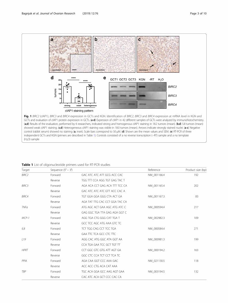
Fig. 1 BIRC2 (cIAP1), BIRC3 and BIRC4 expression in GCTs and KGN. Identification of BIRC2, BIRC3 and BIRC4 expression at mRNA level in KGN andGCTs and evaluation of cIAP1 protein expression in GCTs. (a-d) Expression of cIAP1 in 42 different samples of GCTs were analyzed by immunohistochemistry.(a,d) Results of the evaluation, performed by 6 researchers, indicated strong and homogenous cIAP1 staining in 18.2 tumors (mean). (b,d) 5.8 tumors (mean)showed weak cIAP1 staining. (c,d) Heterogeneous cIAP1 staining was visible in 18.0 tumors (mean). Arrows indicate strongly stained nuclei. (a-c) Negativecontrol (rabbit serum) showed no staining (a, inset). Scale bars correspond to 50 μM. (d) Shown are the mean values and SEM. (e) RT-PCR of threeindependent GCTs and KGN (primers are described in Table 1). Controls consisted of a no reverse transcription (−RT) sample and a no template(H2O) sample
Table 1 List of oligonucleotide primers used for RT-PCR studies
Target Sequence (5′ – 3′) Reference Product size (bp)
BIRC2 Forward GAC ATC ATC ATT GCG ACC CAC NM_001166.4 192
Reverse TGG TTT CCA AGG TGT GAG TAC T
BIRC3 Forward AGA ACA CCT GAG ACA TTT TCC CA NM_001165.4 202
Reverse GAC ATC ATC ATC GTT ACC CAC A
BIRC4 Forward TGT GGA GGA GGG CTA ACT GA NM_001167.3 83
Reverse AGA TAT TTG CAC CCT GGA TAC CA
TNFα Forward ATG AGC ACT GAA AGC ATG ATC C NM_000594.4 217
Reverse GAG GGC TGA TTA GAG AGA GGT C
MCP-1 Forward AGG TGA CTG GGG CAT TGA T NM_002982.3 109
Reverse GCC TCC AGC ATG AAA GTC TC
IL8 Forward TCT TGG CAG CCT TCC TGA NM_000584.4 271
Reverse GAA TTC TCA GCC CTC TTC
L19 Forward AGG CAC ATG GGC ATA GGT AA NM_000981.3 199
Reverse CCA TGA GAA TCC GCT TGT TT
HPRT Forward CCT GGC GTC GTG ATT AGT GA NM_000194.2 163
Reverse GGC CTC CCA TCT CCT TCA TC
PPIA Forward AGA CAA GGT CCC AAA GAC NM_021130.5 118
Reverse ACC ACC CTG ACA CAT AAA
TBP Forward TGC ACA GGA GCC AAG AGT GAA NM_003194.5 132
Reverse CAC ATC ACA GCT CCC CAC CA
Bagnjuk et al. Journal of Ovarian Research (2019) 12:76 Page 3 of 10

of BV-6 (0.1 to 50 μM) and evaluated after 24 h. In ac-cordance with the results in Fig. 2b, nonlinear regressionanalysis revealed a half-maximal reduction of viability by7.2 to 9.7 μM BV-6. Live cell imaging and subsequentmeasurement of confluency were assessed in BV-6(EC50, 8 μM) treated KGN. The space occupied by cells(confluency) relative to the maximally available space,was measured in 20min intervals. Confluency began todecline after 12 h and reached 0.58 after 24 h, as com-pared to the control (1.0). This result is in line with theother experiments. Taken together, we found an EC50
(24 h) of approximately 8 μM for BV-6 in KGN, which ispassage-independent but time-dependent.
BV-6 leads to NF-κB activation and apoptosis in KGNBV-6 is known to block XIAP, cIAP1 and cIAP2 andtherefore is able to activate apoptosis through differentpathways, including NF-κB activation and direct caspaseactivation [6]. To examine these possibilities in KGN, weexposed KGN to BV-6 (EC50) for 24 h and examined in-volvement of NF-κB pathway and caspase activation.
IL8, MCP-1, TNFα, BIRC2, BIRC3 and BIRC4 are knownNF-κB target genes [20–23]. Therefore, upregulation pointsto pathway activation. Indeed, all of the tested genes wereexpressed in BV-6 stimulated KGN (Fig. 3a). BIRC4, BIRC2,BIRC3 and IL8 were upregulated with a fold-change rate of1.52 +/− 0.04, 2.4 +/− 0.26, 64.9 +/− 2.57 and 133.5 +/−14.9, respectively (**p < 0.01 for BIRC2, ***p < 0.001 forBIRC4 and ****p < 0.0001 for BIRC3 and IL8). TNFα andMCP-1 were not found in untreated KGN but were presentin BV-6 treated samples, as shown in the representativeagarose gel (Fig. 3a).To test involvement of caspases, different concentra-
tions of Z-VAD-FMK in combination with BV-6 (EC50,Fig. 3b) were added to KGN cultures. Z-VAD-FMK is apan-caspase inhibitor and therefore functions as ablocker of apoptosis [24]. Cell viability was assessedusing ATP assays. At low concentrations (1 μM and5 μM) Z-VAD-FMK had no effect on BV-6 induced celldeath. Interestingly, viability was significantly improvedat higher doses (20 μM ***p < 0.001, 50 μM ****p <0.0001 and 200 μM ***p < 0.001).
Fig. 2 Effects of BV-6 treatment on KGN. The SMAC mimetic BV-6 was used in different cell viability assays to explore the effects on KGN.Different BV-6 concentrations (0.05–100 μM) were tested. (a) Live cell images of KGN (passages > 80) treated with BV-6 (0.1, 1, 10 and 50 μM) for24 h. Corresponding solvent controls are shown in insets. At 0.1 and 1 μM no indications for cell death were evident. At 10 μM detached cellswere visible. At 50 μM all cells detached. Scale bar indicates 50 μm. (b, left graph) Subsequent cell counting analysis of KGN (passages > 80)treated with BV-6 (0.05–100 μM) revealed an EC50 of 7.4–8.2 μM after nonlinear regression analysis. (n = 3, bars indicate SEM). (b, right graph) ATPassay with BV-6 (0.1–100 μM) confirmed the cell counting experiment. Nonlinear regression analysis revealed an EC50 ranging from 7.2 to 9.7 μM.(n = 4, error bars indicate SEM). (c) To examine a possible involvement of the passage number, KGN from early passages (< 8) were stimulatedwith BV-6 (0.05–100 μM) for 24 h and then counted. Nonlinear regression analysis revealed an EC50 of 8.1 - 8.6 μM. (d) time dependence wasevaluated by confluency measurement (20 min intervals) of BV-6 (EC50, 8 μM) treated KGN. Data were normalized to the solvent control. Declinein confluency started after 12 h and reached 0.58 (control =1.0) after 24 h. (n = 3, error bars indicate SEM, rel. = relative)
Bagnjuk et al. Journal of Ovarian Research (2019) 12:76 Page 4 of 10

Next we screened BV-6-treated KGN for apoptoticmarkers by Western Blot. We used a specific anti-cleavedcaspase 3 (α-clCASP3) antibody to detect the intermediatestep of apoptosis and anti-cleaved poly (ADP-ribose)-poly-merase (α-clPARP) antibody to detect the terminal step ofapoptosis [1, 25]. Both, clCASP3 (Fig. 3c) and clPARP(Fig. 3d) were present in three independent samples ofBV-6 (8 μM)-treated KGN but completely absent from thecorresponding controls. Using an anti-βActin antibody,equal loading was confirmed.
DiscussionA hallmark of cancer is resistance to cell death [26],which may be related to altered IAP expression profiles.cIAP1 and cIAP2, for example, have been shown to be
overexpressed in various tumors, including cervical can-cer, esophageal squamous cell carcinoma, hepatocarci-noma, medulloblastoma, several forms of lung cancer,pancreatic cancer and many more, as summarized byDubrez et al. [27]. SMAC mimetics, which target intracel-lular IAPs, are for this reason a hot topic in cancer biol-ogy. Some of these compounds are currently being testedin phase I and phase II clinical trials and show promisingresults [28]. To our knowledge (https://www.clinicaltrials.gov), there are several ongoing clinical studies with SMACmimetics in different solid tumors and lymphomas, how-ever not in GCTs.Which IAPs are expressed by GCTs is not fully known.
Yet a recent work described strong and homogenous ex-pression of XIAP but a weak cIAP1 expression in GCTs
Fig. 3 Classification of cell death induced by BV-6 in KGN. Results of qRT-PCR, ATP-assay and Western Blot experiments performed to classify BV-6induced cell death. (a) To test NF-κB pathway activation, qRT-PCR experiments of known target genes (BIRC4, BIRC2, BIRC3, IL8, TNFα and MCP-1)were performed. (a, upper graph) BIRC4, BIRC2, BIRC3 and IL8 levels were significantly increased after BV-6 stimulation (n = 5, geometric mean with95% confidence interval, **p < 0.01, ***p < 0.001, ****p < 0.0001). (a, lower panels) MCP-1 and TNFα, both were absent in untreated samples butexpressed in BV-6-treated samples (n = 5). Agarose gel shows a representative picture. The controls lacked template (H2O) or reverse transcriptase(−RT control). (b) ATP-assay was conducted in KGN that were stimulated with a combination of Z-VAD-FMK (1, 5, 20, 50, 200 μM) and BV-6 (EC50,8 μM) for 24 h. Afterwards luminescence was normalized to cells solely treated with BV-6 (EC50, 8 μM). 1 μM and 5 μM Z-VAD-FMK had no effecton BV-6 induced cell death. 20 μM, 50 μM and 200 μM Z-VAD-FMK significantly reduced BV-6 induced cell death. (n = 4, mean and SEM, ***p <0.001, ****p < 0.0001). (c,d) Western Blot analysis using specific α-clCASP3-, α-clPARP- and α-βActin -antibodies. KGN, treated with BV-6 (EC50, 8 μM)for 24 h were analyzed in comparison to controls. Western Blot revealed cleavage of (c) CASP3 and (d) PARP. The solvent treated controls lackedany apoptosis relevant signal but the loading control showed equal loading (n = 3, error bars indicate SEM)
Bagnjuk et al. Journal of Ovarian Research (2019) 12:76 Page 5 of 10

[17]. Immunostaining results may depend on fixation,antibody specificity, as well as many other factors. There-fore, we tested a validated and specific anti-cIAP1 anti-body in 42 tumor samples and employed primary GCTsamples and KGN for RT-PCR studies. While expressionof cIAP1 was strong in most tested GCT samples, thesecond largest group showed heterogeneous staining.Only a small fraction (13.9%) was weakly stained.Furthermore, we detected BIRC3 (cIAP2) mRNA, which
in the previous study was not described [17], but wasshown to be upregulated in many other tumors [27]. How-ever, in line with the previous study by Leung et al., BIRC4(XIAP) was detected. Thus, GCTs are endowed with severalIAPs, which render them targets for SMAC mimetics.Given that BIRC2, 3 and 4 are present, we hypothesized
that BV-6 may be of special interest [6]. BV-6 targetscIAP1/2 and XIAP, which block apoptosis by directly inhi-biting caspases (e.g. XIAP = BIRC4), and by activating thepro-survival canonical NF-κB pathway (e.g. cIAP1/2 =BIRC2/3) [6]. We tested BV-6 actions in KGN, a cell linethat is widely used for the study of GCTs [16]. NF-κB wasshown to play a role in BV-6 mediated apoptosis [6]. BV-6stimulation increased the levels of several target genes ofthis pathway, namely IL8, MCP-1, TNFα, BIRC2, BIRC3and BIRC4 in KGN [20–23]. Interestingly, the increase inexpression of the 3 tested IAPs varied, with BIRC3 (cIAP2)showing the strongest effect (fold change = 133.5). It maybe possible that upregulation of BIRC2 and BIRC3 are cel-lular attempts to counter apoptosis [21]. TNFα plays animportant role in BV-6 induced cell death in many tumorcell lines and is expressed upon BV-6 stimulation in KGN.Therefore, TNF receptor mediated activation of NF-κBand apoptosis are likely to be involved in BV-6 actions inKGN [6]. As we did not elucidate the whole pathway,e.g. RIP1 ubiquitination and cIAP1/2 degradation, we cannot conclude to the full mode of action of BV-6. However,it is likely that cIAP1/2, which ubiquitinate RIP1, are de-graded. In turn this may lead to deubiquitylation of RIP1and subsequently activation of apoptosis or necroptosisthrough a TNFα autocrine loop [6, 29, 30].Passaging KGN induces a change in proliferation speed,
which might be related to tumor aggressiveness [19]. Wetested low and advanced passages for their sensitivity toBV-6 but found no substantial differences. BV-6 -inducedcell death in KGN was concentration- (EC50 ≈ 8 μM) andtime-dependent. Low concentrations of BV-6 (e.g. 1 μM)induced cell death in KGN, but longer incubation times(72 h) were needed (Additional file 1: Figure S1b).In a recent study, a SMAC mimetic alone was reported
incapable to induce cell death in KGN [17], but co-appliedwith a PPARγ agonist it became effective. The PPARγpathway was shown to be relevant in cancer earlier [31, 32].In the previous study the bivalent SMAC mimetic “com-pound A”, originally developed by the pioneering group
around Vince [7] was employed. This compound was de-signed to bind the same IAP domains (BIR2-BIR3) as BV-6,but is chemically different, which could explain the dispar-ate potency [6, 7]. Our results obtained with BV-6 are inline with published work, verifying that some SMACmimetics induce cell death as single agents in variouscancers [6, 33–37].The Western Blot experiments of our study showed
cleaved caspase 3, indicating an intermediate step of apop-tosis and cleaved PARP, i.e. a terminal step of apoptosis inBV-6-treated KGN. Furthermore, blocking experimentswith the pan-caspase blocker Z-VAD-FMK improvedKGN viability [24]. Hence, we conclude that BV-6-inducedcell death is mainly apoptosis. Necroptosis (regulated ne-crosis) has also been described in literature to occur uponSMAC mimetic stimulation in some settings [38]. It is pos-tulated that different cell death pathways are interlinkedand might be triggered simultaneously [39]. We attemptedto determine a possible involvement of necroptosis andexamined phosphorylated MLKL (T357/S358) in BV-6-treated samples by Western Blot, using a validated phos-phospecific antibody [40]. Using this marker, we were notable to find signs for necroptotic cell death in KGN. MLKLwas evident in every sample but phosphorylation and sub-sequently oligomerization were not observed (Additionalfile 1: Figure S1c).The results obtained in cellular studies are in line with
reports showing that apoptosis is induced by BV-6 inmany cell types [6], but the effects of different SMACmimetics in distinct experiment settings and cells mayvary. For example, it was found that a SMAC mimeticmonotherapy is effective only in a small number of tumorcell lines (< 15%), whereas in combination with exogen-ously added TNFα or TRAIL, around 50% of the tumorcell lines died [41]. Further, the dependence on cytokineswas proposed. In mouse models, for example, it wasshown that next to the innate immune system the adap-tive immune system plays an important role in SMAC mi-metic-mediated cell death [42]. The fact that thesecompounds not only affect the tumor cell itself but alsotrigger effector cells like natural killer cells, was shownrecently [43]. Lecis and group further supported this hy-pothesis, as SMAC mimetics affected the tumor niche byexerting immunomodulatory effects on macrophages [44].Thus, next to their effect on tumor cells, SMAC mimeticshave a broad impact on the tumor microenvironment andthe immune system. These points can however not be ex-amined in cellular experiments with KGN cells.In the present work we focused on the SMAC mimetic
BV-6 as a potential inducer of apoptosis in KGN. Asdescribed, apoptosis is either induced extrinsically (deathreceptor pathway) or intrinsically (mitochondrial path-way). The extrinsic pathway is executed upon binding ofa death ligand (e.g. TNFα) to the corresponding receptor
Bagnjuk et al. Journal of Ovarian Research (2019) 12:76 Page 6 of 10

(e.g. TNFR1). Then initiator caspase (caspase 8) isactivated, which leads to further cascaded reactionsincluding caspase 3 activation and cleavage of crucialproteins, including PARP [25, 45]. These steps werereadily observed in BV-6-treated KGN and indicate thatBV-6 does induce apoptosis.Taken together, our results may imply that GCTs,
which as we found are often positive for cIAP1, albeit ina patient-depended manner, can possibly be treated byBV-6. The results further suggest that prior to treat-ment, testing for expression of IAPs may be useful andshould be performed.
ConclusionsThe SMAC mimetic BV-6 is able to induce apoptosis inKGN, which express XIAP and cIAP1/2. The resultsimply that these IAPs, if present in primary tumors, mayserve as targets for therapeutic approaches.
MethodsHuman GCT samplesThe ethical committee of the LMU has approved the study(project 390–15) and patients had agreed to the use of thetissue. Samples (approximately 0.5 cm3) from three patients(age 41, 53 and 66 years) undergoing surgery were ob-tained. Tumor cells were enriched by collagenase digestionin the presence of antibiotics (1% penicillin/streptomycin(Biochrom GmbH, Berlin, Germany)) and subsequent plat-ing on culture wells (HAM’s-F12 supplemented with 10%fetal calf serum (FCS)), as described for primary humangranulosa cell cultures [26]. RNA was extracted and sub-jected to RT-PCR.All patients were diagnosed at the Institute for Path-
ology (LMU, Munich). The diagnoses were confirmed byan experienced gynecologic pathologist (D.M.).In addition, two tissue microarrays (TMA) were as-
sembled and used. Archival material of 42 patients withGCT was available. All patients were treated surgicallyat the same institution (Department of Gynecology, Uni-versity of Munich). Tissue biopsies (n = 42) were takenfrom representative regions of paraffin-embedded tumorsamples (donor) and arrayed into a new recipient paraf-fin block by using MTA-1 (Micro Tissue Arrayer) fromBeecher Instruments, USA.
Culture and treatment of KGNThe human ovarian granulosa-like tumor cell line KGNwas obtained from the Riken BioResource ResearchCentre (Ibaraki, Japan) [16] and the use of this patentedcell line was approved by T. Yanase. KGN were culturedin Dulbecco’s Modified Eagle Medium/Nutrient MixtureF-12 (DMEM/F-12, Thermo Fisher Scientific, Waltham,MA, USA) supplemented with 10% FCS (Capricorn Sci-entific GmbH, Ebsdorfergrund, Germany) and 1%
penicillin/streptomycin (Biochrom GmbH, Berlin,Germany) in T75 flasks (Thermo Fisher Scientific, Wal-tham, MA, USA) under constant temperature (37 °C)and CO2 concentration (5%). As KGN change in aggres-siveness over culture time [19] we used low passages (<8) and high passages (> 80) for preliminary experiments.As a passage dependence was absent in terms of sensi-tivity to BV-6 we proceeded with high passages (> 80).To rule out serum effects, experiments were executed
in serum-free DMEM/F-12 media. Therefore, KGN werestimulated for 24 h either with the second mitochondria-derived activator of caspases (SMAC) mimetic BV-6(0.05–100 μM; Selleck Chemicals LLC, Houston, TX,USA) alone or in combination with the pan-caspase in-hibitor Z-VAD-FMK (1–200 μM; Selleck ChemicalsLLC, Houston, TX, USA). BV-6 and Z-VAD-FMK weredissolved in water and DMSO (Merck, Darmstadt,Germany), respectively. Solvent controls were includedin every experiment. To block caspases before cell deathinduction, Z-VAD-FMK was applied 2 h prior to BV-6administration. All experiments were carried out at least3 times, if not described otherwise.
RT-PCR and qRT-PCRRT-PCR was conducted as previously described [46, 47].In brief, total RNA was extracted using RNeasy Plus MicroKit (Qiagen, Hilden, Germany) and employed for reversetranscription. RT-PCR and qRT-PCR primers (Table 1)were designed using Primer3 and synthesized by metabioninternational AG (Planegg, Germany) [48]. The qRT-PCRanalyses were performed utilizing the QuantiFast SYBRGreen PCR Kit (Qiagen, Hilden, Germany). Samples (finalcDNA concentration 10 ng/reaction) were measured induplicates in a LightCycler® 96 System (Roche Diagnostics,Penzberg, Germany; melting at 95 °C for 5min, followedby 40 cycles of denaturation at 95 °C for 10 s and extensionat 60 °C for 30 s, and a final melting step with continuousheating (0.5 °C/s from 65 °C to 97 °C) and a cool-downstep at 37 °C for 30 s). Results were calculated using the2−ΔΔCq method and expression was normalized to riboso-mal protein L19 (L19), hypoxanthine Phosphoribosyltrans-ferase 1 (HPRT), peptidylprolyl isomerase A (PPIA) andTATA-box binding protein (TBP) as endogenous refer-ences. After PCR, products were separated and visualizedon a 2% agarose gel. Expected bands were cut out,sequenced (GATC Biotech AG, Konstanz, Germany) andanalyzed using BLAST tool [49].
Immunohistochemistry of ex vivo GCT using anti-cIAP1antibodyImmunohistochemical staining of cIAP1 proteins to exam-ine expression patterns in GCTs was done as described inprevious work [46, 47]. Two slides of the TMAs, containinga total of 42 tumors in duplicates, were used for this study.
Bagnjuk et al. Journal of Ovarian Research (2019) 12:76 Page 7 of 10

After deparaffinizing, the heat-induced epitope retrieval(HIER) method was applied to retrieve antigens. Afterwardsendogenous peroxidase was blocked with a methanol(10%)/H2O2 (3%) solution. Unspecific binding was reducedby 10% goat serum in PBS. To detect cIAP1 protein, a poly-clonal antibody was used (HPA005513, Sigma Aldrich, St.Louis, MO, USA). As a negative control rabbit serumreplaced the primary antiserum. As a secondary antibody abiotinylated goat anti-rabbit antibody (111–065-144, Jack-son Immuno Research, Cambridge, UK) was used. Aftercomplexing avidin with biotin (ABC reaction) and stainingby means of 3,3′ diaminobenzidine tetrahydrochloride(DAB), the slides were counterstained with hematoxylin.Images were captured using a Zeiss Axiovert microscopewith an Insight Camera (18.2 Color Mosaik). The TMAswere evaluated by 6 different researchers, who classifiedeach tumor into one of the three groups (homogenouslystrong staining, weak staining/no staining or heteroge-neous staining).
Confluency measurement and live cell imagingConfluency was measured as described before [40, 47]. Inbrief, 3 × 10⁵ KGN plated on a 60mm2 culture dish (Sar-stedt AG & Co. KG, Nümbrecht, Germany) were stimu-lated with BV-6 (EC50, 8 μM) or the solvent control andmonitored for 24 h using the JuLi™ Br Live Cell Analyzer(NanoEnTek Inc., Seoul, Korea). Pictures were capturedevery 20min and confluency was evaluated using the in-built algorithm. The experiments were repeated 3 times.Images of KGN, stimulated with 0.1, 1, 10, 50 μM BV-
6, were also captured using a cell culture microscopewith an 10X objective (Leica Biosystems, Wetzlar,Germany).
Viability measurements using cell counting and ATP-assayEC50 was identified using 2 different methods. First a cellcounting experiment was carried out with low passages(< 8) and high passages of KGN (> 80). Therefore 1.5 ×10⁵ cells were seeded on 6 well plates (Sarstedt AG &Co. KG, Nümbrecht, Germany) and incubated over nightat 37 °C and 5% CO2. On the next day the cells werestimulated with BV-6 concentrations ranging from 0.05to 100 μM or the corresponding solvent control for 24 h.Afterwards cells were trypsinized and counted using theCASY® counting system (OMNI Life Science GmbH &Co KG, Bremen, Germany). Cell counts were normalizedto solvent controls. Experiments were repeated at least 3times.The CellTiter-Glo® ATP assay Kit (Promega, Mannheim,
Germany) was perfomed following the manufacturer’s in-structions. In brief 1 × 10⁴ cells/well (KGN passages > 80)were seeded in 96-well plates (Sarstedt AG & Co. KG,Nümbrecht, Germany) and incubated at 37 °C and 5%CO2. The next day culture media was replaced by
colorless media without supplements. After 2 h, BV-6(0.1–50 μM) was added and cells were incubated for atime period of 24 h. Further, BV-6 (8 μM) treatment wasaccompanied by different Z-VAD-FMK concentrationsranging from 0 to 200 μM. Z-VAD-FMK was applied 2 hbefore BV-6 stimulation to block caspases. Luminescencewas measured using a plate reader (FLUOstar Optima,BMG Labtech, Ortenberg, Germany).
Verification of apoptosis hallmarks by Western blotApoptosis is a form of cell death that is executed by cas-pases. Cleavage and therefore activation of caspase 3 isknown to be an intermediate step, which leads to cleavageof proteins including PARP that is known to be a terminalstep of apoptosis. Therefore, we treated KGN (106 cells/60mm2 dish (Sarstedt AG & Co. KG, Nümbrecht,Germany)) with BV-6 (EC50, 8 μM) for 24 h. Afterwardswe generated crude protein extracts using RIPA buffer.Western Blot was carried out as described before [40, 46].20 μg/lane protein were loaded on 12% SDS-PAGE gels.To detect cleaved caspase 3 a specific α-clCASP3 antibody(#9664, Cell Signaling Technology, Denvers, MA, USA)was used. To detect a cleavage product of PARP the spe-cific α-clPARP antibody (#56255, Cell Signaling Technol-ogy, Denvers, MA, USA) was applied. As a loading controlan α-βActin antibody (A5441, Sigma Aldrich, St. Louis,MO, USA) was used. For decoration of bound primaryantibody horseradish peroxidase (HRP) conjugated goatα-rabbit or goat α-mouse antibody (Jackson Immuno Re-search, Cambridge, UK) were used.
Statistics and graphsCell culture and live cell imaging pictures were evaluatedusing FIJI [50]. Construction and statistical evaluation ofgraphs, including nonlinear regression analyses, were donein Prism 6 (GraphPad, San Diego, CA, USA). The statis-tical significance between different staining patterns of tu-mors was evaluated by one-way ANOVA (Tukey; Geisser-Greenhouse correction). Effectiveness of Z-VAD-FMK onBV-6-induced cell death and mRNA expression of NF-κBregulated genes were evaluated statistically by one samplet-tests. Final figures were constructed using PowerPointfor Mac 2013 (Microsoft, Redmond, WA, USA).
Additional file
Additional file 1: Figure S1. Workflow of experiments and effects ofBV-6 on KGN. (a) Schematic workflow of experiments: First EC50 after 24 hwas determined by cell counting and ATP assay in KGN (passage > 80)and by cell counting in a low passage of KGN (< 8). All furtherexperiments were carried out with the determined EC50 and with KGN ofhigher passages (> 80). Afterwards a Z-VAD-FMK dilution experiment wascarried out, using KGN that were treated with BV-6(EC50,). (b) Live cellimaging experiment of stimulated KGN (BV-6, 1 μM) versus thecorresponding control for 72 h. The low concentration caused a time-
Bagnjuk et al. Journal of Ovarian Research (2019) 12:76 Page 8 of 10

dependent effect by reducing number of attached cells. Scale bar =100 μm (c) Western Blot of BV-6 (EC50, 8 μM)-stimulated KGN and thecorresponding control. An antibody against phosphorylated (p)MLKL(T357/S358) (ab187091, Abcam, Cambridge, UK) and one againstMLKL (ab184718, Abcam, Cambridge, UK) were examined to explorepossible induction of necroptosis. MLKL bands were visible, whereas thenecroptosis marker (pMLKL) was absent. (TIFF 8189 kb)
AbbreviationsAIF: Apoptosis inducing factor; ATP: Adenosine triphosphate; BIR: BaculoviralIAP repeats; BIRC: Baculoviral IAP repeat containing protein; CASP8: Caspase8; cIAP: Cellular inhibitor of apoptosis protein; DAB: 3,3′ diaminobenzidinetetrahydrochloride; DIABLO: Direct inhibitor of apoptosis protein bindingprotein with low PI; EC50: Half maximal effective concentration; FCS: Fetal calfserum; FOXL2: Forkhead Box L2; GCTs: Granulosa cell tumors; HIER: Heat-induced epitope retrieval; HPRT: Hypoxanthine phosphoribosyltransferase 1;HRP: Horseradish peroxidase; HRP: Horseradish peroxidase; HtrA2: Hightemperature requirement protein A2; IAPs: Inhibitor of apoptosis proteins;IL8: Interleukin 8; KGN: Human ovarian granulosa-like tumor cell line;L19: ribosomal protein L19; MCP-1: Monocyte chemoattractant protein-1;MLKL: Mixed lineage kinase domain-like protein; mRNA: Messengerribonucleic acid; PPIA: Peptidylprolyl isomerase A; TBP: TATA-box bindingprotein; α-clCASP3: anti-cleaved caspase 3; α-clPARP: anti-cleaved poly (ADP-ribose)-polymerase
AcknowledgementsWe gratefully acknowledge the expert technical help of Kim Dietrich andCarola Herrmann.
Ethics approval and consent to participationThe ethical committee of the LMU has approved the study (project 390–15)and patients had agreed to the use of the tissue for scientific purposes.
Authors’ contributionsKonstantin Bagnjuk and Verena Jasmin Kast performed the cellular studiesand evaluated the results and together with Artur Mayerhofer drafted themanuscript. Astrid Tiefenbacher performed immunohistochemcial studies.Melanie Kaseder performed qRT-PCR experiments. Toshihiko Yanase providedKGN. Dr. Burges was the surgeon and obtained the consent of the patients.Doris Mayr provided TMA. Lars Kunz, Doris Mayr and Artur Mayerhofer con-ceived of the study and directed the work. All authors contributed to andagreed on the final manuscript.
FundingDeutsche Forschungsgemeinschaft (DFG), MA1080/19–2 (to AM) andMA4790/4–2 (to DM).
Availability of data and materialsUpon request.
Competing interestsNone.
Author details1Biomedical Center Munich (BMC), Cell Biology, Anatomy III,Ludwig-Maximilians-University (LMU), Grosshaderner Strasse 9, 82152Planegg, Germany. 2Department of Endocrinology and Diabetes Mellitus,Faculty of Medicine, Fukuoka University, 7-45-1 Nanakuma, Jonan-ku,Fukuoka City, Japan. 3Frauenklinik Campus Großhadern,Ludwig-Maximilians-University (LMU), Marchioninistraße 15, 81377 Munich,Germany. 4Division of Neurobiology, Department Biology II,Ludwig-Maximilians-University (LMU), Grosshaderner Strasse 9, 82152Planegg, Germany. 5Institute for Pathology, Ludwig-Maximilians-University(LMU), 80337 Munich, Germany.
Received: 26 April 2019 Accepted: 31 July 2019
References1. Hengartner MO. The biochemistry of apoptosis. Nature. 2000;407:770.
2. Fulda S, Debatin KM. Extrinsic versus intrinsic apoptosis pathways inanticancer chemotherapy. Oncogene. 2006;25:4798.
3. Degterev A, Boyce M, Yuan J. A decade of caspases. Oncogene. 2003;22:8543.4. Saelens X, Festjens N, Vande Walle L, van Gurp M, van Loo G, Vandenabeele
P. Toxic proteins released from mitochondria in cell death. Oncogene. 2004;23(16):2861–74.
5. Fulda S. Promises and challenges of Smac mimetics as Cancer therapeutics.Clin Cancer Res. 2015;21(22):5030.
6. Varfolomeev E, Blankenship JW, Wayson SM, Fedorova AV, Kayagaki N, GargP, et al. IAP antagonists induce autoubiquitination of c-IAPs, NF-kappaBactivation, and TNFalpha-dependent apoptosis. Cell. 2007;131(4):669–81.
7. Vince JE, Wong WW-L, Khan N, Feltham R, Chau D, Ahmed AU, et al. IAPantagonists target cIAP1 to induce TNFα-dependent apoptosis. Cell. 2007;131(4):682–93.
8. Petersen SL, Wang L, Yalcin-Chin A, Li L, Peyton M, Minna J, et al. AutocrineTNFα signaling renders human Cancer cells susceptible to Smac-mimetic-induced apoptosis. Cancer Cell. 2007;12(5):445–56.
9. Lu J, Bai L, Sun H, Nikolovska-Coleska Z, McEachern D, Qiu S, et al. SM-164: anovel, bivalent Smac mimetic that induces apoptosis and tumor regressionby concurrent removal of the blockade of cIAP-1/2 and XIAP. Cancer Res.2008;68(22):9384–93.
10. Vischioni B, van der Valk P, Span SW, Kruyt FAE, Rodriguez JA, Giaccone G.Expression and localization of inhibitor of apoptosis proteins in normalhuman tissues. Hum Pathol. 2006;37(1):78–86.
11. Jamieson S, Fuller PJ. Molecular pathogenesis of granulosa cell tumors ofthe ovary. Endocr Rev. 2012;33(1):109–44.
12. Zhou J, Chen Y, Huang Y, Long J, Wan F, Zhang S. Serum follicle-stimulatinghormone level is associated with human epidermal growth factor receptortype 2 and Ki67 expression in post-menopausal females with breast cancer.Oncol Lett. 2013;6(4):1128–32.
13. Gründker C, Emons G. The Role of Gonadotropin-Releasing Hormone in CancerCell Proliferation and Metastasis. Frontiers in Endocrinology. 2017;8(187).
14. Lagana AS, Colonese F, Colonese E, Sofo V, Salmeri FM, Granese R, et al.Cytogenetic analysis of epithelial ovarian cancer's stem cells: an overviewon new diagnostic and therapeutic perspectives. Eur J Gynaecol Oncol.2015;36(5):495–505.
15. Lagana AS, Sofo V, Vitale SG, Triolo O. Epithelial ovarian cancer inherentresistance: may the pleiotropic interaction between reducedimmunosurveillance and drug-resistant cells play a key role? Gynecologiconcology reports. 2016;18:57–8.
16. Nishi Y, Yanase T, Mu Y, Oba K, Ichino I, Saito M, et al. Establishment andcharacterization of a steroidogenic human granulosa-like tumor cell line,KGN, that expresses functional follicle-stimulating hormone receptor.Endocrinology. 2001;142(1):437–45.
17. Leung DTH, Nguyen T, Oliver EM, Matti J, Alexiadis M, Silke J, et al. CombinedPPARγ activation and XIAP inhibition as a potential therapeutic strategy forovarian granulosa cell tumors. Mol Cancer Ther. 2019;18(2):364–75.
18. Zhang Y, Yan Z, Qin Q, Nisenblat V, Chang HM, Yu Y, et al. TranscriptomeLandscape of Human Folliculogenesis Reveals Oocyte and Granulosa CellInteractions. Molecular cell. 2018;72(6):1021–34.e4.
19. Imai M, Muraki M, Takamatsu K, Saito H, Seiki M, Takahashi Y. Spontaneoustransformation of human granulosa cell tumours into an aggressivephenotype: a metastasis model cell line. BMC Cancer. 2008;8(1):319.
20. Stehlik C, De Martin R, Kumabashiri I, Schmid JA, Binder BR, Lipp J. Nuclearfactor (NF)-κB–regulated X-chromosome–linked iap gene expressionprotects endothelial cells from tumor necrosis factor α–induced apoptosis.J Exp Med. 1998;188(1):211–6.
21. Wang C-Y, Mayo MW, Korneluk RG, Goeddel DV, Baldwin AS. NF-κBantiapoptosis: induction of TRAF1 and TRAF2 and c-IAP1 and c-IAP2 tosuppress caspase-8 activation. Science. 1998;281(5383):1680–3.
22. Kang HB, Kim YE, Kwon HJ, Sok DE, Lee Y. Enhancement of NF-kappaBexpression and activity upon differentiation of human embryonic stem cellline SNUhES3. Stem Cells Dev. 2007;16(4):615–23.
23. Xing L, Remick DG. Promoter elements responsible for antioxidantregulation of MCP-1 gene expression. Antioxid Redox Signal. 2007;9(11):1979–89.
24. Van Noorden CJF. The history of Z-VAD-FMK, a tool for understanding thesignificance of caspase inhibition. Acta Histochem. 2001;103(3):241–51.
25. Chaitanya GV, Steven AJ, Babu PP. PARP-1 cleavage fragments: signatures ofcell-death proteases in neurodegeneration. Cell communication andsignaling : CCS. 2010;8:31.
Bagnjuk et al. Journal of Ovarian Research (2019) 12:76 Page 9 of 10

26. Hanahan D, Weinberg RA. Hallmarks of cancer: the next generation. Cell.2011;144(5):646–74.
27. Dubrez L, Berthelet J, Glorian V. IAP proteins as targets for drugdevelopment in oncology. OncoTargets and therapy. 2013;9:1285–304.
28. Infante JR, Dees EC, Olszanski AJ, Dhuria SV, Sen S, Cameron S, et al. Phase Idose-escalation study of LCL161, an oral inhibitor of apoptosis proteinsinhibitor, in patients with advanced solid tumors. Journal of clinicaloncology : official journal of the American Society of Clinical Oncology.2014;32(28):3103–10.
29. Bertrand Mathieu JM, Vandenabeele P. The Ripoptosome: death decision inthe cytosol. Mol Cell. 2011;43(3):323–5.
30. Wertz IE, Dixit VM. Signaling to NF-κB: regulation by ubiquitination. ColdSpring Harb Perspect Biol. 2010;2(3):a003350.
31. Lagana AS, Vitale SG, Nigro A, Sofo V, Salmeri FM, Rossetti P, et al.Pleiotropic Actions of Peroxisome Proliferator-Activated Receptors (PPARs)in Dysregulated Metabolic Homeostasis, Inflammation and Cancer: CurrentEvidence and Future Perspectives. Int J.Mol Sci. 2016;17(7).
32. Vitale SG, Lagana AS, Nigro A, La Rosa VL, Rossetti P, Rapisarda AM, et al.Peroxisome proliferator-activated receptor modulation during metabolicdiseases and cancers: master and minions. PPAR Res. 2016;2016:6517313.
33. Brumatti G, Ma C, Lalaoui N, Nguyen NY, Navarro M, Tanzer MC, et al. Thecaspase-8 inhibitor emricasan combines with the SMAC mimetic birinapantto induce necroptosis and treat acute myeloid leukemia. Sci Transl Med.2016;8(339):339ra69.
34. McComb S, Aguade-Gorgorio J, Harder L, Marovca B, Cario G, Eckert C,et al. Activation of concurrent apoptosis and necroptosis by SMACmimetics for the treatment of refractory and relapsed ALL. Sci TranslMed. 2016;8(339):339ra70.
35. Opel D, Schnaiter A, Dodier D, Jovanovic M, Gerhardinger A, Idler I, et al.Targeting inhibitor of apoptosis proteins by Smac mimetic elicits cell deathin poor prognostic subgroups of chronic lymphocytic leukemia. Int JCancer. 2015;137(12):2959–70.
36. Schirmer M, Trentin L, Queudeville M, Seyfried F, Demir S, Tausch E, et al.Intrinsic and chemo-sensitizing activity of SMAC-mimetics on high-riskchildhood acute lymphoblastic leukemia. Cell Death Dis. 2016;7:e2052.
37. Steinhart L, Belz K, Fulda S. Smac mimetic and demethylating agentssynergistically trigger cell death in acute myeloid leukemia cells andovercome apoptosis resistance by inducing necroptosis. Cell Death Dis.2013;4:e802.
38. Safferthal C, Rohde K, Fulda S. Therapeutic targeting of necroptosis by Smacmimetic bypasses apoptosis resistance in acute myeloid leukemia cells.Oncogene. 2017;36(11):1487–502.
39. Chen Q, Kang J, Fu C. The independence of and associations amongapoptosis, autophagy, and necrosis. Signal Transduction and TargetedTherapy. 2018;3(1):18.
40. Bagnjuk K, Stöckl JB, Fröhlich T, Arnold GJ, Behr R, Berg U, et al. Necroptosisin primate luteolysis: a role for ceramide. Cell Death Discovery. 2019;5(1):67.
41. Beug ST, Conrad DP, Alain T, Korneluk RG, Lacasse EC. Combinatorial cancerimmunotherapy strategies with proapoptotic small-molecule IAPantagonists. The International journal of developmental biology. 2015;59(1–3):141–7.
42. Beug ST, Beauregard CE, Healy C, Sanda T, St-Jean M, Chabot J, et al. Smacmimetics synergize with immune checkpoint inhibitors to promote tumourimmunity against glioblastoma. Nat Commun. 2017;8.
43. Fischer K, Tognarelli S, Roesler S, Boedicker C, Schubert R, Steinle A, et al.The Smac Mimetic BV6 Improves NK Cell-Mediated Killing ofRhabdomyosarcoma Cells by Simultaneously Targeting Tumor and EffectorCells. Front Immunol. 2017;8(202).
44. Lecis D, De Cesare M, Perego P, Conti A, Corna E, Drago C, et al. Smacmimetics induce inflammation and necrotic tumour cell death bymodulating macrophage activity. Cell death & disease. 2013;4(11):e920-e.
45. Fulda S. Smac mimetics to therapeutically target IAP proteins in Cancer. IntRev Cell Mol Biol. 2017;330:157–69.
46. Du Y, Bagnjuk K, Lawson MS, Xu J, Mayerhofer A. Acetylcholine andnecroptosis are players in follicular development in primates. Sci Rep. 2018;8(1):6166.
47. Blohberger J, Kunz L, Einwang D, Berg U, Berg D, Ojeda SR, et al.Readthrough acetylcholinesterase (AChE-R) and regulated necrosis:pharmacological targets for the regulation of ovarian functions? Cell death&. Disease. 2015;6:e1685.
48. Koressaar T, Remm M. Enhancements and modifications of primer designprogram Primer3. Bioinformatics. 2007;23(10):1289–91.
49. Altschul SF, Gish W, Miller W, Myers EW, Lipman DJ. Basic local alignmentsearch tool. J Mol Biol. 1990;215(3):403–10.
50. Rueden CT, Schindelin J, Hiner MC, DeZonia BE, Walter AE, Arena ET, et al.ImageJ2: ImageJ for the next generation of scientific image data. BMCbioinformatics. 2017;18(1):529.
Publisher’s NoteSpringer Nature remains neutral with regard to jurisdictional claims inpublished maps and institutional affiliations.
Bagnjuk et al. Journal of Ovarian Research (2019) 12:76 Page 10 of 10