Insulin like growth factor-I (IGF-I), its receptor (IGF-IR...
79
Insulin like growth factor-I (IGF-I), its receptor (IGF-IR) and Insulin receptor substrate 1 (IRS1) expression as an early reaction of PDL cells to experimental tooth movement in the rat Inaugural-Dissertation zur Erlangung des Doktorgrades der Hohen Medizinischen Fakultät Rheinische Friedrich-Wilhelms-Universität Bonn Yasser Kheralla aus Aleppo/ Syrien 2013
Transcript of Insulin like growth factor-I (IGF-I), its receptor (IGF-IR...
Insulin like growth factor-I (IGF-I), its receptor (IGF-IR) and Insulin
receptor substrate 1 (IRS1) expression as an early reaction of PDL
cells to experimental tooth movement in the rat
Inaugural-Dissertation
zur Erlangung des Doktorgrades
der Hohen Medizinischen Fakultät
Rheinische Friedrich-Wilhelms-Universität
Bonn
Yasser Kheralla
aus
Aleppo/ Syrien
2013
Anfertigung mit Genehmigung der
Medizinischen Fakultät der Universität Bonn
1. Gutachter: Prof. Dr. Werner Götz
2. Gutachter: Prof. Dr. Stephan Baader
Tag der Mündlichen Prüfung: 22.05.2013
Aus der Poliklinik für Kieferorthopädie des Zentrums für Zahn-, Mund-, und
Kieferheilkunde der Universität Bonn
Direktor: Professor Dr. A. Jäger
Labor für Oralbiologische Grundlagenforschung
Leiter: Professor Dr. Werner Götz
Meinen Eltern
meiner Frau
und meinen Kindern
in Dankbarkeit gewidmet

5
Inhaltsverzeichnis
1. Zusammenfassung ……………………………………………………... 7
1.1 Einleitung …………………………………………………………………. 7
1.2 Zielsetzungen und Hypothesen der vorliegenden Studie ……………….. 8
1.3 Material und Methoden …………………………………………………. 8
1.4 Ergebnisse und Diskussion ………………………………………………. 9 1.5 Schlussfolgerung ………………………………………………………… 10
2. Introduction …………………………………………………………… 11
2.1 Tooth movement …………………………………………………………. 11
2.1.1 The theories of tooth movement ……………………………………... 11
2.1.1.1 The pressure-tension theory …………………………………….... 11
2.1.1.2.1 The bone bending theory ……………………………………….. 12
2.1.1.2.2 Piezoelectricity …………………………………………………... 12
2.1.2 Phases of tooth movement ……………………………………………. 13
2.1.3 Optimal orthodontic force …………………………………………. 13
2.2 Mechanotransduction in bone and PDL ………………………………... 14
2.3 Insulin-like growth factor peptide family ………………………………. 17 2.3.2 Insulin-like growth factors …………………………………………... 18
2.3.3 Insulin-like growth factor receptors ………………………………… 19
2.3.4 IGF binding proteins and proteases ………………………………….. 20
2.3.5 The role of IGFs in bone and oral biology …………………………….. 21
2.3.6 IGF-I and mechanical loading ………………………………...………. 21
3. Aims and hypotheses …………………………………………………… 22
4. Materials and methods ………………………………………………... 23
4.1 Animals …………………………………………………………………….. 23
4.2 Experimental procedure ………………………………………………… 23
4.3 Histology …………………………………………………………………. 27
4.4 Immunohistochemistry ………………………………………………..... 27
4.5 Histomorphometry and statistical analysis …………………………….. 30
4.6 The used chemical, reactors and instruments ………………………….. 31
4.6.1 List of chemicals ………………………………………………………. 31
4.6.2 List if reactors …………………………………………………………. 32
4.6.3 List of instruments ……………………………………………………. 32
5. Results ………………………………………………………………… 33
5.1 Histology ………………………………………………………………….. 33
5.2 Immunohistochemistry and histomorphometry ……………………...... 35
5.2.1 Results of IGF-I ………………………………………………………. 35
5.2.2 Results of IGF-IR ……………………………………………………… 41
5.2.3 Results of IRS1 ……………………………………………………….. 46
6. Discussion ……………………………………………………………… 51
7. Summary ………………………………………………………………. 62

6
8. References ……………………………………………………………… 64
9. Danksagung ……………………………………………………………. 81
10. Lebenslauf ……………………………………………………………... 82
7
1. Zusammenfassung
1.1 Einleitung
Die Applikation einer Kraft auf einen Zahn im Rahmen einer kieferorthopädischen
induzierten Zahnbewegung führt zu einer initialen Auslenkung des Zahnes innerhalb
seiner Alveole, sodass einige Teile des Desmodontalspaltes sich verschmälern
(„Druckzone“) und gegenüberliegenden Teile sich verbreitern („Zugzone“).
Aufgrund von Zirkulationsveränderungen, Zelldeformation, oder Kompression der
Nervenenden erfolgt eine Freisetzung verschiedener Faktoren, die eine Resorption des
Alveolarknochens auf der Druckzone und eine Knochenapposition auf der Zugzone
verursachen.
IGFs stellen eine Familie von Wachstumsfaktoren dar, die aus 2 Liganden, IGF-I/II, 2
Rezeptoren, IGF-IR/IIR, und 6 Bindungsproteinen, IGFBP-1 bis IGFBP-6, bestehen.
IGF-I stellt einen wichtigen Faktor für postnatales Wachstum dar. Er wirkt sowohl
endokrin als auch para- und autokrin auf systemischer als auch auf lokaler Ebene. Im
Bindegewebe und im Knochen wird IGF-I von Osteoblasten und Fibroblasten
produziert und in die Matrix eingelagert. Es stimuliert Differenzierung und
Proliferation und hemmt die Apoptose dieser Zellen. Nach Bindung von IGF-I an IGF-
IR werden 3 intrazelluläre Signalwege aktiviert. IRS-1 ist dabei das wichtigste
Substrat für die Vermittlung der IGF-I-Signale.
Knochen und Bindegewebe empfangen mechanische Belastungen, die für ihre
Integrität und Ihr Remodelling von Bedeutung sind, was zur Freisetzung verschiedener
Faktoren führt. In vielen Geweben und Zellarten induziert die mechanische Belastung
auch eine IGF-I-Expression. In neuen Studien konnten alle Moleküle des IGF-Systems
im Parodont des Menschen und der Ratte nachgewiesen werden. Des Weiteren
konnten Studien zeigen, dass Dehnungen der Zellen eine Modulation der
Genexpression verschiedener IGF-Familienmitglieder zur Folge hatten.
8
1.2 Zielsetzungen und Hypothesen der vorliegenden Studie
In der vorliegenden Untersuchung sollte überprüft werden:
1. ob lokal produziertes IGF-I an der Mechanotransduktion von Zellen des Parodontal-
ligaments (PDL) während der frühen Phase der Zahnbewegung beteiligt ist,
2. ob es ein differenziertes Expressionsmuster von IGF-I, IGF-IR und IRS1 in Druck-
und Zugzonen gibt,
3. und ob die Veränderungen bzw. die Verteilung des IGF-I von der Größe der
angewandten Kräfte abhängig sind.
Darüberhinaus sollte die Bedeutung von IGF-I für die Umbauaktivitäten im Rahmen
der orthodontischen Zahnbewegung diskutiert werden.
1.3 Material und Methoden
Der rechte obere erste Molar von 20 anästhetisierten Ratten wurde mit einer Kraft
belastet, um den Zahn nach mesial zu bewegen. Konstante Kräfte von 0.1N, 0.25 N
und 0.5 N wurden über 4 Stunden an 4 Versuchstieren verwendet. Die Kräfte wurden
über einen Sensor für 4 Stunden gemessen. Weiterhin wurden konstante Kräfte von 0.1
N über 2 Stunden in 8 weiteren Tieren angewandt. Im Anschluss wurden der erste und
zweite Molar permanent mit Kunststoff separiert und die antagonistischen Molaren
beschliffen. Vier Ratten aus der Gruppe wurden nach 1 Tag und weitere 4 Ratten nach
2 Tagen geopfert. Die unbehandelte kontralaterale Seite wurde als Kontrolle
verwendet. Die präparierten Maxillae wurden in Paraffin eingebettet und schließlich in
dünne sagittale Schnitte geschnitten. Immunohistologische Färbungen wurden zur
Identifizierung von IGF-I, IGF-IR und IRS1 durchgeführt. Fotos wurden von der
Druckseite (mesiokoronale Region) und von der Zugseite (distokoronale Region)
aufgenommen und histomorphometrisch ausgewertet. Die statistische Auswertung
erfolgte mit SPSS-Software.
9
1.4 Ergebnisse und Diskussion
Alle untersuchten Faktoren waren im PDL der belasteten Gruppen und der
Kontrollgruppe nachweisbar. Die Ergebnisse zeigten eine Zunahme der Anzahl der
positiven Zellen aller untersuchten Faktoren auf der Zugzone. Im Vergleich mit der
Kontrollgruppe waren alle Unterschiede signifikant und von der Zeitdauer der
Belastung abhängig. Die höchste Zahl der IGF-IR-immunopositiven Zellen wurde
nach 24 Stunden von der Kraftapplikation festgestellt.
In der Druckzone fiel die Anzahl der positiven Zellen aller untersuchten Faktoren nach
4 Stunden von der Kraftapplikation ab. Diese Abnahme war signifikant von der Höhe
der applizierten Kräfte abhängig. Nach 24 bzw. 48 Stunden stieg die Anzahl der
positiven Zellen wieder auf den Level der Kontrollgruppe an.
Die Abnahme/Zunahme der positiven Zellen war tendenziell von der Kraftgröße
abhängig.
Diese Ergebnisse weisen auf eine Rolle von IGF-I in der Aufrechterhaltung der
parodontalen Gewebshomöostase sowie den Umbauprozessen während der
Zahnbewegung hin. Die Ergebnisse in der Zugzone stimmen mit denen anderer
Autoren überein, die für andere Gewebe gezeigt haben, dass nach einer kurzfristigen
Belastung die IGF-I-Expression in Sehnenzellen, Muskelzellen aber auch in
Osteoblasten und Osteozyten zunimmt.
Man kann auch davon ausgehen, dass IGF-I an Mechanotransduktionsvorgängen
während der frühen Phasen der Zahnbewegung beteiligt ist.
Diese IGF-I-Hochregulation könnte die Proliferation und die Differenzierung der
PDL-Zellen stimulieren und die Apoptose beeinflussen.
In der Druckzone zeigen die Ergebnisse, dass verminderte Expression der untersuchten
Faktoren zur Verringerung der Proliferation und schließlich zur Abnahme der Anzahl
der PDL-Zellen führen könnte.
10
1.5 Schlussfolgerung
In der vorliegenden Studie wurde in einem Rattenmodell die Verteilung von IGF-I,
IGF-IR und IRS1 während der frühen Phase der Zahnbewegung untersucht.
Die erhobenen Daten deuten darauf hin, dass die lokale auto- und parakine Freisetzung
von IGF-I eine frühe Reaktion der Zellen des PDLs auf eine orthodontische Belastung
darstellt, und dass IGF-I an Mechanotransduktionsvorgängen beteiligt ist. Diese
Reaktion ist tendenziell von der Größe der applizierten Kraft und eindeutig von
Belastungszeit abhängig. Dies könnte zur Aktivierung bzw. Hemmung der
Proliferation und Differenzierung der PDL-Zellen sowie zur Hemmung bzw.
Auslösung von Apoptose führen.
Die Ergebnisse dieser Studie tragen dazu bei, die Molekulare und zelluläre Grundlagen
der kieferorthopädischen Zahnbewegung besser zu verstehen und die mit den
klinischen Erfahrungen aus der Kieferorthopädie zu verknüpfen.
11
2. Introduction
2.1 Tooth movement
Orthodontic tooth movement has been defined as the result of a biologic response of
periodontal ligament to an externally applied force (Proffit, 2007).
The purpose of orthodontic treatment is to move teeth as efficiently as possible with
minimal adverse effects for the teeth and supporting tissue. The predictability of tooth
movement in humans is of clinical interest, particularly in terms of the nature and
speed of tooth movement in response to specific applied mechanics and the absence of
undesirable sequelae (Iwasaki et al., 2008).
Mainly based on histological research, a pressure and a tension side are distinguished
during orthodontic tooth movement. On the pressure side, the biological events are as
follows: disturbance of blood flow in the compressed PDL, cell death in the
compressed area of the PDL (hyalinization), resorption of the hyalinized tissue by
macrophages, and undermining bone resorption by osteoclasts beside the hyalinized
tissue, which ultimately results in tooth movement. On the tension side, blood flow is
activated where the PDL is stretched, which promotes osteoblastic activity and osteoid
deposition, which later mineralizes (Von Böhl and Kuijpers-Jagtman, 2009).
2.1.1 The theories of tooth movement
2.1.1.1 The pressure-tension theory
According to the classic histological research by Sandstedt (1904), Oppenheim (1911)
and Schwarz (1932), this theory states that a tooth moves in the periodontal space by
generating a “pressure side” and a “tension side” (Krihnan and Davidovitch, 2006).
In this theory, the blood flow decreases where the PDL is compressed, and maintains
or increases where the PDL is under tension. These alterations create quickly changes
in chemical environment (Proffit, 2007).
The force-subjected PDL progenitor cells differentiate into compression associated
osteoclasts and tension-associated osteoblasts, causing bone resorption and apposition,
respectively. Direct resorption is associated with light force (frontal) application.
12
Indirect (undermining) resorption and hyalinization are associated with bio-intolerant
heavy forces (Masella and Meister, 2006).
2.1.1.2.1 The bone-bending theory
According to Baumrind (1969) and Grimm (1972), this theory states that orthodontic
forces produce alveolar bone deflection. The active biologic processes that follow
bone bending involve bone turnover and renewal of cellular and inorganic fractions
and are accompanied by changes in the PDL. These authors could explain facts such as
(1) the relative slowness of en-masse tooth movement; (2) the velocity of tooth
movement toward an extraction site; and (3) the relative rapidity of tooth movement in
children, who have less heavily calcified and more flexible bones than adults. The
increase of convexity at the pressure side is associated with bone resorption, whereas
the increase of concavity at the tension side is associated with bone formation
(Kirshnan and Davidovitch, 2006).
2.1.1.2.2 Piezoelectricity
Piezoelectricity is a phenomenon observed in many crystalline materials, in which a
deformation of a crystal structure produces a flow of electric current (Kirshnan and
Davidovitch, 2006). After bone bending, the distortion of crystalline structures of
bone generates bioelectric charges and can regulate the remodeling of alveolar bone
during orthodontic tooth movement. Concave bone surfaces characterized by
osteoblastic activity are electronegative; convex bone surfaces characterized by
osteoclastic activity are electropositive or electrically neutral (Meikle, 2006).
It should be noted that these piezoelectric signals have two unusual characteristics: (1)
a quick decay rate and (2) the production of an equivalent signal, opposite in direction,
when the force released, therefore the sustained orthodontic tooth movement does not
produce prominent stress-generated signals (Proffit, 2007).
13
2.1.2 Phases of tooth movement
There are 4 phases of tooth movement. The first phase (initial phase) lasts 24 hours to
2 days, the cellular and tissue reactions start in this phase of tooth movement
immediately after force application. Depending on the magnitude of applied force, the
tooth movement stops in the second phase (lag phase) for 20 to 30 days because of the
induced necrosis of cellular elements within the PDL at the pressure side. The
phagocytic cells, which recruit from the adjacent marrow spaces and from the
direction of the viable PDL, remove the necrotic tissues from compressed PDL sites
and adjacent alveolar bone.
After the removal of necrotic tissue (hyalinization) formed during the second phase,
tooth movement is accelerated in the third phase (postlag phase or acceleration stage)
and continues into the fourth phase (linear stage) (Burstone, 1962; Pilon et al., 1996;
Van Leeuwen et al., 1999, Kirshnan and Davidovitch, 2006). Proffit refers that heavy
forces lead to undermining resorption while the lighter forces enable frontal resorption
without delay (Proffit, 2007).
It must be kept in mind that hyalinization zones at the pressure areas could form even
during third and fourth stage, especially in areas where high forces were applied (Von
Böhl et al., 2004).
2.1.3 Optimal orthodontic force
Schwarz defined optimal continuous force as „„the force leading to a change in tissue
pressure that approximated the capillary vessels‟ (Schwarz, 1932). Storey and Quinn
correlated optimal force to the surface area of the root (Storey and Smith, 1952; Quinn
and Yoshikawa, 1985).
Traditionally, orthodontic forces have been categorized as “light” or “heavy,” and it
was assumed that light forces are preferable, because of their ability to evoke frontal
resorption of bone and therefore more physiologic than heavy forces that cause
necrosis (hyalinization) of the PDL and undermining bone resorption (Kirshnan and
Davidovitch, 2006). Since some trauma is always associated with applied orthodontic
forces, even light ones, and precise measurement of force applied to roots or parts
14
thereof is impossible, a current concept of optimum force was performed. This concept
means that there is a force of certain magnitude and temporal characteristics
(continuous versus intermitted, constant versus declining) capable of producing a
maximal rate of tooth movement, without tissue damage, and with maximum patient
comfort. The optimal force for tooth movement may differ for each tooth and for each
individual patient (Ren et al., 2003).
These orthodontic forces deform the extracellular matrix and activate cells of the
paradental tissues in sequential cellular and molecular events that lead to convert the
mechanical energy to biochemical and/or electrical signals. This process is known as
mechanotransduction (Krishnan and Davidovitch, 2009).
2.2 Mechanotransduction in bone and PDL
In nearly all aspects of biology, forces are a relevant regulator of life‟s form and
function. More recently, science has established that cells are exquisitely sensitive to
forces of varying magnitudes and time scales, and they convert mechanical stimuli into
a chemical response. This phenomenon, termed mechanotransduction, is an integral
part of cellular physiology and has a profound impact on the development of the
organism. Furthermore, mal-functioning mechanical properties or
mechanotransduction often leads to pathological alterations of the organism (Kolahi
and Mofrad, 2010).
There are four essential interrelated steps in the transduction of mechanical signals by
tissues: sensing the mechanical signal by the cells, transduction of this mechanical
signal into biochemical one, transmission of the biochemical signal to the effector
cells, and the effectors cell response (Wise and King, 2008).
Mechanosensing is a process by which cells sense structural changes in the
extracellular matrix (ECM), caused by external mechanical loading. Each cellular
system in paradental tissues is equipped with mechanosensors; thus, each mechanical
stimulus may activate multiple mechanosensors, followed by downstream cellular
events (Krishnan and Davidovitch, 2009).
15
In bone, bone cells respond to fluid flow stimulation in vitro by the production of
mediators such as nitric oxide (NO) and prostaglandins (Klein-Nulend et al., 2005).
The signal might be transduced to the osteocytes through specific receptors or through
deformation of the cytoskeleton (Duncan and Turner, 1995). The cell deformation may
occur indirectly through fluid flow and/or directly through integrins (Hennemann et
al., 2008).
Many studies suggest that the mechanosensory molecules are: ion channels, integrins,
G-proteins, the Wnt surface receptor and the cytoskeleton (Sawakami et al., 2006;
Scott et al., 2008).
Gated ion channels are involved in hearing and touch as well as maintaining osmotic
homeostasis and regulating cell volume. In these contexts, tension created in the
cytoskeleton in response to loading can alter the shape of the membrane lipid bilayer,
resulting in changes in ion channel behavior (Hamill and Martinac, 2001; Jacobs et al.,
2010). There is also evidence of interplay between different channels and between
channels and other molecular mechanosensors. For example, annexin V is a calcium
channel that binds extracellular collagen and cytoskeletal actin. It is involved in the
intracellular calcium response to fluid flow in osteoblasts. This suggests the possibility
that mechanical force in the actin cytoskeleton may open this channel (Haut Donahue
et al., 2004, Jacobs et al., 2010).
Integrins, which are present on the cell membrane, connect the intracellular actin
filaments through a variety of cytoskeletal linker proteins to ECM. This focal adhesion
complex not only transduces matrix strain to the cytoskeleton but also activates protein
kinases that initiate a variety of intracellular signalling pathways (Wang and
Thampatty, 2006; Schwartz, 2011). There is cross-talk between integrins and growth
factors receptors occurs at multiple levels. For example, integrin αvß3 enhances
insulin-like growth factor-I receptor (IGF-IR) signaling through FAK-dependent
phosphorylation of IGF-IR. Mice that lack this gene in embryonic fibroblasts showed
impaired phosphorylation of IGF-IR in response to insulin-like growth factor-I (IGF-
I), and IGF-I-dependent cell proliferation is blocked (Liu et al., 2008; Legate et al.,
2009).
16
Guanine nucleotide–binding proteins or G-proteins are signaling GTPases that
alternate between an active GTP-bound state and an inactive GDP-bound state. A G-
protein-coupled receptor includes a transmembrane receptor and an intracellular G-
protein, which is activated (GDP replaced with GTP) owing to a conformation change
that occurs with ligand binding or mechanical stimulation (independent of ligand
binding) (Chachisvilis et al., 2006; Jacobs et al., 2010).
The Wnt surface receptor, low-density lipoprotein receptor related protein 5 (LRP5),
has been suggested as a key regulator of bone mass. Its signaling pathway may be
important for the osteogenic response to loading (Sawakami et al., 2006; Wise and
King, 2008).
The cytoskeleton represents a system of intracellular fibers and accounts for much of
the structural integrity of the cell, incorporating microtubules, intermediate filaments
and actin filaments. These networks are thought to maintain a constant, basal level of
tension, so that small, local deformations affect overall cytoskeletal tension, initiating
cellular responses (Myers et al., 2007).
Several in vitro studies demonstrated the involvement of extracellular and intracellular
signaling components in mechanotransduction (Liedert et al., 2005). The earliest
experiments showed that the immediate response of cells to mechanical strain was the
generation of prostaglandins (PGs), nitric oxide (NO), the second messenger cyclic
adenosine monophosphate (cAMP) and inositol phosphates. Changes in intracellular
[Ca 2+] were shown to occur via stretch-activated ion channels (Harell et al., 1977;
Yeh and Rodan, 1984; Sandy et al., 1989; Davidson et al, 1990; Klein-Nulend et al,
1995; McDonald et al., 1996; Meikle, 2006). Thus, the transduction of the mechanical
signals into biochemical signals begins.
These messengers will evoke a nuclear response which will either result in production
of factors responsible for osteoclast recruitment and activation, or bone forming
growth factors (Roberts-Harry and Sandy 2004).
The downstream signalling pathways during mechanical stretching appear to overlap
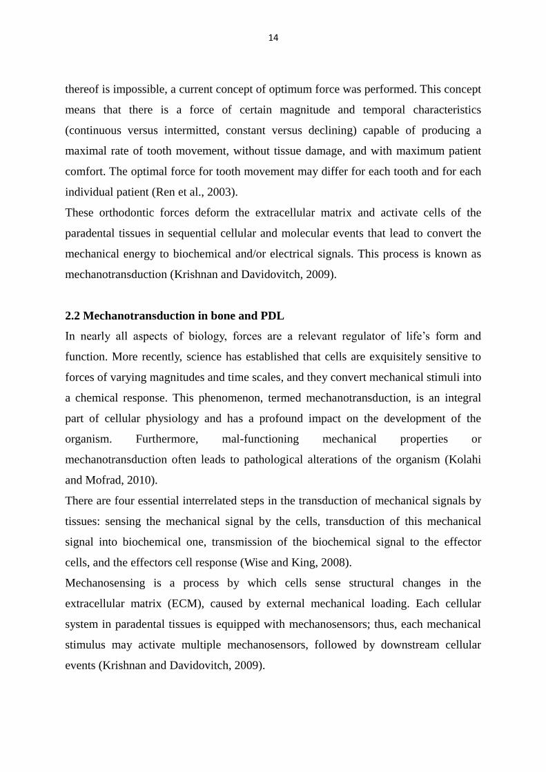
with known growth factor and hormone signals (Scott et al., 2008) (Fig. 1).
17
Figure 1: Schematic diagram of interactions of different signaling pathways during
mechanical stretching. Membrane-bound receptors such as Ca2+
channels, integrins,
Gproteins, IGF and TGF-b and/or BMP receptors are stimulated by mechanical forces,
resulting in induction of several transcription factors that regulate osteoblast differentiation
and formation. AP-1 and Runx2 are induced mainly through MAPK and SMADs pathways.
Runx2 is also stimulated via the Wnt pathway, involving b-catenin and TCF or LEF factors.
PLC–PKA pathway contributes to NF-kB, Cox-2 and CREB induction. Abbreviations: AP-1,
activator protein-1; b-cat, b-catenin; DAG, diacylglycerol; FAK, focal adhesion kinase; G, G-
protein; IP3, inositol (1,4,5)-trisphosphate; MEKK, MAPK kinase kinase; PKA, protein
kinase A; PKC, protein kinase C; PLC, phospholipase C; PYK2, proline-rich tyrosine kinase
2 (From Papachroni et al., 2009).
Discoveries in mechanobiology have illuminated sequential cellular and molecular
events, such as synthesis and secretion of specific products (Krishnan and
Davidovitch, 2009). One of these molecules is Insulin-like growth factors (IGFs).
2.3 Insulin-like growth factor peptide family
IGFs represent a family of endocrine, paracrine and autocrine polypeptides. The IGF
family is comprised of ligands (IGF-I, IGF-II, and insulin), six well characterized
binding proteins (IGFBP-1 though -6), and cell surface receptors that mediate the
actions of the ligands (IGF-I receptor, insulin receptor, and the IGF-II mannose-6-
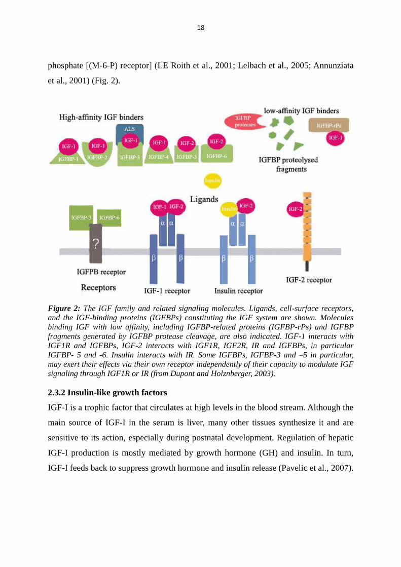
18
phosphate [(M-6-P) receptor] (LE Roith et al., 2001; Lelbach et al., 2005; Annunziata
et al., 2001) (Fig. 2).
Figure 2: The IGF family and related signaling molecules. Ligands, cell-surface receptors,
and the IGF-binding proteins (IGFBPs) constituting the IGF system are shown. Molecules
binding IGF with low affinity, including IGFBP-related proteins (IGFBP-rPs) and IGFBP
fragments generated by IGFBP protease cleavage, are also indicated. IGF-1 interacts with
IGF1R and IGFBPs, IGF-2 interacts with IGF1R, IGF2R, IR and IGFBPs, in particular
IGFBP- 5 and -6. Insulin interacts with IR. Some IGFBPs, IGFBP-3 and –5 in particular,
may exert their effects via their own receptor independently of their capacity to modulate IGF
signaling through IGF1R or IR (from Dupont and Holznberger, 2003).
2.3.2 Insulin-like growth factors
IGF-I is a trophic factor that circulates at high levels in the blood stream. Although the
main source of IGF-I in the serum is liver, many other tissues synthesize it and are
sensitive to its action, especially during postnatal development. Regulation of hepatic
IGF-I production is mostly mediated by growth hormone (GH) and insulin. In turn,
IGF-I feeds back to suppress growth hormone and insulin release (Pavelic et al., 2007).
19
The synthesis of IGF-I is controlled by a number of endogenous (genetic and
hormonal) and exogenous (nutrition and physical activity) factors. About one-third to
one-quarter of IGF-I expression is determined by exogenous factors (Zofkova, 2003).
The synthesis of IGF-II is relatively GH-independent. Its expression is much higher
during fetal development than in postnatal life. It acts as a regulatory peptide; it is
mitogenic for a number of cell types (Pavelic et al., 2007).
IGF-I and IGF-II are single-chain polypeptides, with sequences 62 % identical to that
of proinsulin. However, unlike insulin and other peptide hormones, they are not
produced and stored within the cells of a specific tissue, but may instead be produced
by almost any cell in the body (Dupont and Holzenberger 2003).
The ligands IGF-I and IGF-II are involved in various cellular processes including
differentiation, proliferation, morphogenesis, growth, apoptosis, control of metabolic
function and carcinogenesis (Clemmons AND Maile 2005; Danley et al., 2005).
2.3.3 Insulin-like growth factor receptors
Most of the cellular effects of the IGFs are mediated by binding to the IGF-IR. The
IGF-IR is a transmembrane tyrosine kinase structurally similar to the insulin receptor.
It is composed of two extracellular α-subunits and two intracellular β-subunits (Laron,
2001). The α-subunits bind IGF-IR to IGF-I, IGF-II and insulin at supraphysiological
concentration, while the β-subunits transmit the ligand-induced signals (Jones and
Clemmons, 1995; Mauro and Surmacz, 2004). Binding of the ligands to the IGF-IR
induces its autophosphorylation via tyrosine kinase activity. This, in turn, causes the
interaction with certain cellular substrates such as insulin receptor substrate 1 (IRS1),
adapter protein Shc and 14-3-3-proteins (Romano, 2003; Laviola et al., 2007).
A major intracellular substrate for the IGF-IR is IRS1 which, after its tyrosine
phosphorylation, initiates multiple signallings. It mediates the transmission of
mitogenic, metabolic and antiapoptotic signals (Jones and Clemmons, 1995; Ogata et
al., 2000).
IGF-II receptor is a single large transmembrane receptor which bind with IGF-II and,
with a much lower affinity, with IGF-I. The IGF-II receptor does not appear to act as a
20
traditional signaling receptor in response to IGF binding and is thought to act mainly
as a clearance receptor for IGF-II (Holly and Perks, 2006; Samani et al., 2007).
2.3.4 IGF binding proteins and proteases
IGFBPs are a family of secreted proteins that bind IGF with high affinities that are
greater than those of the IGF-IR. Six distinct IGFBPs, designated as IGFBP-1 to -6,
have been isolated and characterized from human and a variety of vertebrate species
(Firth and Baxter, 2002; Kelley et al., 2002).
IGFBPs function as carrier proteins for circulating IGFs and serve not only as a
reservoir for IGFs release, but also greatly increases the half-life of IGFs, and regulate
IGF turnover, transport of IGFs from the vascular space to target tissues, and tissue
distribution. IGFBPs expressed in many peripheral tissues. They have been shown to
inhibit or potentiate IGF actions in vitro. While IGFBP-4 and IGFBP-6 have been
consistently found to inhibit IGF actions, IGFBP-1, -2, -3, and -5 can both inhibit and
potentiate IGF actions, depending on cell type (Duan and Xu, 2005).
There is compelling evidence that some IGFBPs possess biological activities that are
ligand-independent. These include growth inhibition, direct induction of apoptosis, and
modulation of the effects of other non-IGF growth factors (Firth and Baxter, 2002;
Mohan and Baylink, 2002). For example, IGFBP-3 inhibits cell growth in the absence
of IGFs and in IGF-1R-null cells (Butt and Williams, 2001).
IGF-BP degrading proteases act as growth stimulators by increasing local IGF
availability. They fall into three major categories. Kallikrein-like serine proteases, the
second major category is cathepsins and intracellular proteinases, and the third
category involves matrix metalloproteinases. Proteolytic activity by proteases may
play a role in normal and abnormal tissue proliferation by cleaving IGF-BP into
fragments with lower affinity for IGFs, thereby increasing the levels of free IGFs to
activate IGF-IR (Cohen, 2006). These proteases are under the control of autocrine,
paracrine, and hormonal influences (Rosen, 1999).
21
2.3.5 The role of IGFs in bone and oral biology
In bone, many factors regulate homeostasis. IGF-I and IGFII are the most abundant
growth factors stored in bone and are locally produced by bone cells. IGFs increase
bone formation by regulating proliferation, differentiation, and apoptosis of osteoblasts
(Govoni et al., 2005).
Consistent with this role, patients with Laron syndrome, caused by IGF-I deficiency,
present marked osteoporosis (Laron et al., 1998).
Genetic modifications in specific components of the IGF system have provided
tremendous insights into the role of the IGFs and IGF-IR in vivo. For example, the
overproduction of IGF-I in the osteoblasts of transgenic mice increases bone formation
(Zhao et al., 2000), whereas the overproduction and deficiency of IGF-II in mice has
no major effect on skeletal size or bone turnover in mice (DeChiara et al., 1991; Wolf
et al., 1995). In addition, mice lacking IGF1R display marked organ hypoplasia and
delayed skeletal ossification (Yakar and Rosen, 2003)
The IGFs fulfills an important role in growth and development of teeth, mandible,
maxillae, and tongue. It has been postulated that IGF-I may be of great value in the
treatment of periodontal defects and in tissue healing (Werner and Katz, 2004).
The suitability of IGF-I, in combination with other growth factors, for periodontal
regeneration has been tested (Palioto et al., 2004, Sant'Ana et al., 2007).
It is believed, that IGFs behave as poliferative factors for cementoblasts (Grzesik and
Narayanan, 2002). They are involved in early root formation by regulating the mitotic
activity of the outer layer of the epithelial cells of Hertwig's root sheath (Fujiwara et
al., 2005).
Recently, the distribution pattern of IGFs components was investigated in both
deciduous and permanent teeth. Many of them were found to be localized in the
periodontium of human and rats (Götz et al., 2001; 2003; 2006a).
2.3.6 IGF-I and mechanical loading
Mechanical loading has been demonstrated to be an important regulatory factor in
alveolar bone homeostasis. It plays an essential role in maintaining the structure and
mass of the alveolar processes throughout life (Pavlin and Gluhak-Heinrich, 2001).
22
On the other hand, skeletal unloading reduces proliferation and differentiation of
osteoprogenitor cells in vitro (Kostenuik et al., 1997).
In vitro studies showed also that mechanical load increases the level of IGF-I in
osteoblasts (McCarthy and Centrella, 2001). In addition, gene expression of IGF-I is
detected during distraction osteogenesis and fracture healing (Bouletreau et al., 2002).
In a recent study, it was demonstrated that IGF-I mRNA is twofold up-regulated in
osteocytes of cortical bone after 6 h of mechanical loading, and thus supported the
hypothesis that osteocytes are most important cells for the translation of mechanical
stimuli into bone formation via IGF-I (Reijnders et al., 2007).
3. Aims and hypotheses
In this study should be investigated:
1. whether the locally produced IGF-I participate in the mechanotransduction process
in the PDL during the early phase of tooth movement,
2. whether there is a differentiated expression pattern of IGF-I, IGF-IR and IRS1 in
pressure and tensile sides,
3. and whether the distribution of IGF-I are dependent on the magnitude of the applied
forces.
Moreover, the possible role of IGF-I in the remodeling of PDL during the early phase
of orthodontic tooth movement should be also discussed.
23
4. Materials and methods
4.1 Animals
Twelve-week-old male Wistar rats each weighing 300–350 g (Harlan Winkelmann,
Borchen, Germany) were used as experimental animals. The animal use protocol was
reviewed and approved by the Institutional Animal Care and Use Committee of the
local district government (Cologne) and the Animal Care Commissioner of the
University of Bonn (Germany).
4.2 Experimental procedure
According to the experimental protocol of Kawarizadeh et al. (2004), rats were
anaesthetized with 0.01 ml Rompun (Bayer, Leverkusen, Germany) and 0.24 ml
Ketavet (Pharmacia and Upjohn, Erlangen, Germany). The animals were clamped into
a head-holding device, and the occlusal surface of the maxillary right first molar was
prepared by grinding a small hole with a dental diamond bur. The tooth surface was
then treated with self-etching bonding material (Xeno III, Dentsply DeTrey, Konstanz,
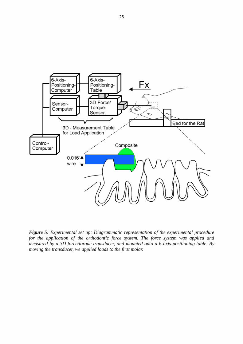
Germany) for 60 s. An orthodontic appliance consisting of a T-loop (0.016 _ 0.022-in.
stainless steel wire, Ormco Corp., Glendora, CA, USA) was placed between the molar
and a high-resolution 3D force/torque transducer (ATI, Industrial Automation, Garner,
NC, USA), which had a resolution of 0.0125 N for force and 0.0625 Nmm for torque.
The T-loop was fixed to the occlusal surface of the molar with light-curing composite
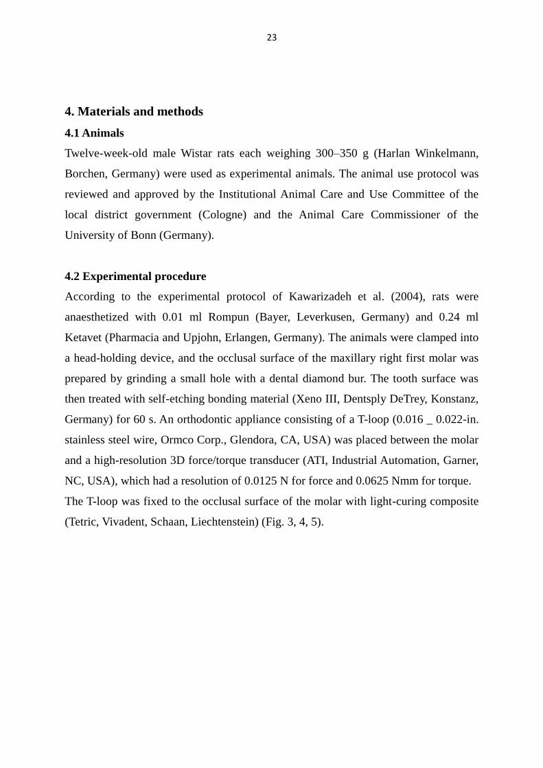
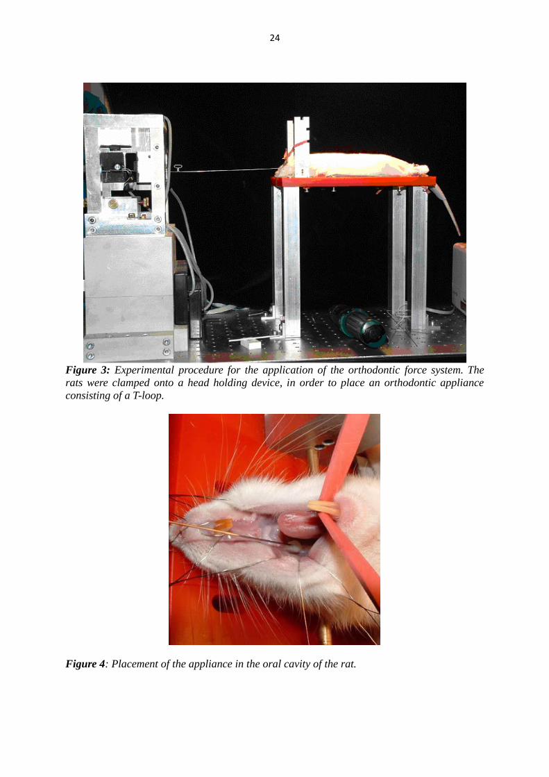
(Tetric, Vivadent, Schaan, Liechtenstein) (Fig. 3, 4, 5).
24
Figure 3: Experimental procedure for the application of the orthodontic force system. The
rats were clamped onto a head holding device, in order to place an orthodontic appliance
consisting of a T-loop.
Figure 4: Placement of the appliance in the oral cavity of the rat.
25
Figure 5: Experimental set up: Diagrammatic representation of the experimental procedure
for the application of the orthodontic force system. The force system was applied and
measured by a 3D force/torque transducer, and mounted onto a 6-axis-positioning table. By
moving the transducer, we applied loads to the first molar.
26
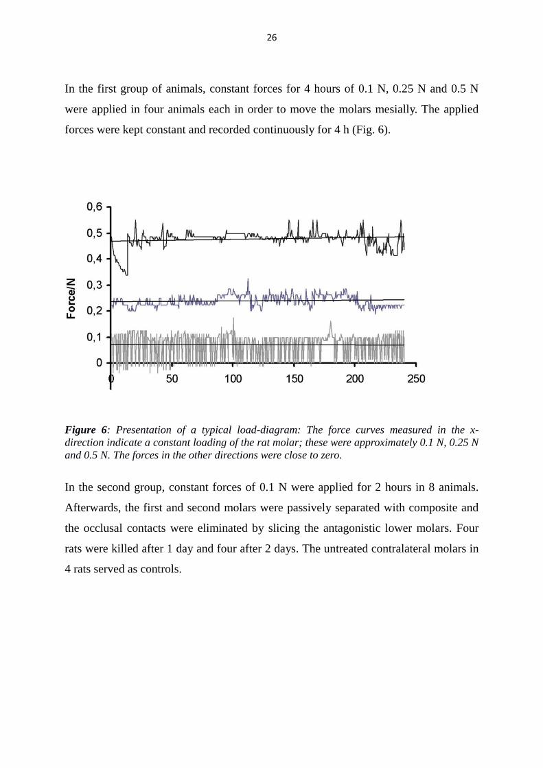
In the first group of animals, constant forces for 4 hours of 0.1 N, 0.25 N and 0.5 N
were applied in four animals each in order to move the molars mesially. The applied
forces were kept constant and recorded continuously for 4 h (Fig. 6).
Figure 6: Presentation of a typical load-diagram: The force curves measured in the x-
direction indicate a constant loading of the rat molar; these were approximately 0.1 N, 0.25 N
and 0.5 N. The forces in the other directions were close to zero.
In the second group, constant forces of 0.1 N were applied for 2 hours in 8 animals.
Afterwards, the first and second molars were passively separated with composite and
the occlusal contacts were eliminated by slicing the antagonistic lower molars. Four
rats were killed after 1 day and four after 2 days. The untreated contralateral molars in
4 rats served as controls.
27
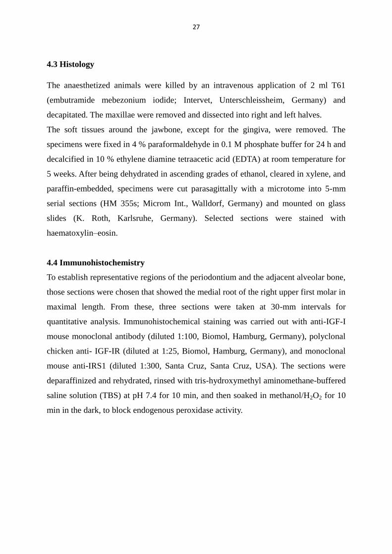
4.3 Histology
The anaesthetized animals were killed by an intravenous application of 2 ml T61
(embutramide mebezonium iodide; Intervet, Unterschleissheim, Germany) and
decapitated. The maxillae were removed and dissected into right and left halves.
The soft tissues around the jawbone, except for the gingiva, were removed. The
specimens were fixed in 4 % paraformaldehyde in 0.1 M phosphate buffer for 24 h and
decalcified in 10 % ethylene diamine tetraacetic acid (EDTA) at room temperature for
5 weeks. After being dehydrated in ascending grades of ethanol, cleared in xylene, and
paraffin-embedded, specimens were cut parasagittally with a microtome into 5-mm
serial sections (HM 355s; Microm Int., Walldorf, Germany) and mounted on glass
slides (K. Roth, Karlsruhe, Germany). Selected sections were stained with
haematoxylin–eosin.
4.4 Immunohistochemistry
To establish representative regions of the periodontium and the adjacent alveolar bone,
those sections were chosen that showed the medial root of the right upper first molar in
maximal length. From these, three sections were taken at 30-mm intervals for
quantitative analysis. Immunohistochemical staining was carried out with anti-IGF-I
mouse monoclonal antibody (diluted 1:100, Biomol, Hamburg, Germany), polyclonal
chicken anti- IGF-IR (diluted at 1:25, Biomol, Hamburg, Germany), and monoclonal
mouse anti-IRS1 (diluted 1:300, Santa Cruz, Santa Cruz, USA). The sections were
deparaffinized and rehydrated, rinsed with tris-hydroxymethyl aminomethane-buffered
saline solution (TBS) at pH 7.4 for 10 min, and then soaked in methanol/H2O2 for 10
min in the dark, to block endogenous peroxidase activity.
28
In details the sections were treated with the following sequence:
1. xylol 10 min.
2. xylol 10 min.
3. 100 % ethanol 5 min.
4. 100 % ethanol 5 min
5. 90 % ethanol 5 min
6. 70 % ethanol 5 min
7. aqua dest 5 min
8. TBS 10 min
9. methanol/H2O2 20 min
Sections for IRS1 immunostaining were pretreated with goat serum (DAKO, Glostrup,
Denmark) for 90 minutes. Antibodies were diluted in 1 % bovine serum albumin. The
IGF-I antibody was applied in a humidity chamber for 1 hour at room temperature, the
IGF-IR-and IRS1 antibodies overnight at 4 °C. Subsequently, sections were washed in
TBS and incubated with suitable Envision+/HRP anti-mouse for IGF-I, anti-rabbit for
IRS1 (Dako Cytomation, Hamburg, Germany) or anti-chicken for IGFIR (Rockland,
Gilbersville, USA) diluted in 1 % TBS–BSA at 1:450, as secondary antibodies for 30
minutes in a humidity chamber at room temperature.
29
Antibody complexes were visualized using diaminobenzidine (DAB) for 10 minutes
which yields a brown staining product. Thereafter, slides were rinsed, counterstained
with Mayer‟s haematoxylin for 5 seconds, rinsed again, and mounted.
10. Goat serum only for IRS1-sections 90 min
11. Primary antibodies:
Dilution
Monoclonal mouse anti-IGF-I 1:100 1Hour
Polyclonal chicken anti- IGF-IR 1:25 overnight
Monoclonal mouse anti-IRS1 1:300 overnight
12. TBS 10 min
13. Secondary antibodies:
Envision+/HRP anti-mouse 30 min
Anti-goat immune globulin/HRP 30 min
Anti-chicken for IGFIR 1:450 30 min
14. TBS 10 min
15. Diaminobenzidine (DAB) 10 min
16. TBS 10 min
17. Mayer‟s hematoxylin 1 sec
18. 100 % ethanol 2 min
19. 100 % ethanol 2 min
20. xylol 2 min
21. xylol 2 min
Negative controls were prepared by omission of the primary antibodies from the
staining procedures and replacing it with an isotype IgG using the identical
concentration.
30
4.5 Histomorphometry and statistical analysis
Sections were scanned by means of a scanner camera (Axio Cam MRC; Zeiss,
Göttingen, Germany) mounted on a light microscope (Axiophot 2; Zeiss, Göttingen,
Germany), and viewed with imaging software (Axiovision; Zeiss, Göttingen,
Germany) on a personal computer. Counting of the percentage of immuno-
histochemically positive cells was performed in two separate predefined areas in every
selected section. These areas were located mesio-coronally and disto-coronally to the
medial root. Counts were made at a magnification of 400.
Means and standard deviations were calculated for each group of four rats. Hundred
sections were randomly selected from the original samples and remeasured by the
same operator after a time interval of 3 months without reference to the previous
measurements.
The casual error was calculated according to Dahlberg‟s formula:
Where Sx is the error of the measurement, D is the difference between duplicated
measurements, and N is the number of double measurements.
The percentage of IGF-I-, IGF-IR- and IRS1-positive cells was calculated with Excel
software. The statistical analyses were performed with SPSS Software, version 17
(SPSS Inc. Chicago, IL). The normality test was used to estimate the normal
distribution of the data. The percentage of positive cells for all tested factors was
compared using post hoc test (Bonferroni). The level of statistical significance was set
at p < 0.05.
31
4.6 The used chemicals, reactors and instruments
4.6.1 List of the chemicals:
Acetone Otto Fischer, Saarbrücken, Germany
Ethanol (70-100 %) Merck, Münster, Germany
Embedding medium DePex Serra, Heidelberg, Germany
3,3-diaminobenzidin(DAB) Sigma, Steinheim, Germany
Ethylene diamine tetraacetic
acid (EDTA) Calbiochem, Darmstadt, germany
Eosin solution 1 % Merck, Darmstadt, Germany
Formic acid Merck, Darmstadt, Germany
Formaldehyde 40 % Merck, Darmstadt, Germany
Hematoxylin Merck, Darmstadt, Germany
Methanol Merck, Darmstadt, Germany
Sodium chloride Merck, Darmstadt, Germany
Sodiumsulfate solution Merck, Darmstadt, Germany
Paraffin-Histo-comp Vogel, Giessen, Germany
Hydrochloride 2 mol/l Merck, Darmstadt, Germany
Serumalbumin from bovine Paesel and Lor, Frankfurt, Germany
Tris ICN Biomedicals, Ohio, USA
Xylol original Merck, Darmstadt, Germany
Xylol substitute XEM-200 Vogel, Giessen, Germany
32
4.6.2 List of the reactors:
TBS-solution:
-9.0 gr NaCl
-6.0 gr Tris
-1000 ml aqua dest. + 2 ml HCl pH 7.6
TBS-BSA-solution:
-0.1 gr serumalbumin from bovine
-10 ml TBS-solution
EDTA:
-200 gr EDTA
-68 gr Tris
-2000 ml aqua dest
4.6.3 List of the instruments:
Analysis scale Sartorius, Göttingen, Germany
Thermo cupboard Memmert, Schwabach, Germany
Cover glass Engelbrecht, Edermunde, Germany
DigitalpH-meter 197 WTW, Vienna, Austria
Eppendorf pipettes Eppendorf, Hamburg, Germany
Light microscope Axioskop 2 Zeiss, Göttingen, Germany
Axio Cam MRC Zeiss, Göttingen, Germany
Rotation microtome HM 3559 Microtom, Walldorf, Germany
Superfrost plus slides Menzel, Braunschweig, Germany
Warm plate Medax Nagel, Kiel, Germany
33
5. Results
5.1 Histology
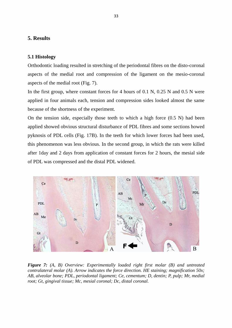
Orthodontic loading resulted in stretching of the periodontal fibres on the disto-coronal
aspects of the medial root and compression of the ligament on the mesio-coronal
aspects of the medial root (Fig. 7).
In the first group, where constant forces for 4 hours of 0.1 N, 0.25 N and 0.5 N were
applied in four animals each, tension and compression sides looked almost the same
because of the shortness of the experiment.
On the tension side, especially those teeth to which a high force (0.5 N) had been
applied showed obvious structural disturbance of PDL fibres and some sections howed
pyknosis of PDL cells (Fig. 17B). In the teeth for which lower forces had been used,
this phenomenon was less obvious. In the second group, in which the rats were killed
after 1day and 2 days from application of constant forces for 2 hours, the mesial side
of PDL was compressed and the distal PDL widened.
Figure 7: (A, B) Overview: Experimentally loaded right first molar (B) and untreated
contralateral molar (A). Arrow indicates the force direction. HE staining; magnification 50x;
AB, alveolar bone; PDL, periodontal ligament; Ce, cementum; D, dentin; P, pulp; Mr, medial
root; Gt, gingival tissue; Mc, mesial coronal; Dc, distal coronal.
34
Figures 15A and 19A show blood vessels in various sizes in the sections of PDL.
Osteoclasts were seen on the alveolar bone surface in the disto-coronal areas of
untreated rats (Fig. 10A, 19A), after 24 and 48 hrs from force application, no more
osteoclasts were found.
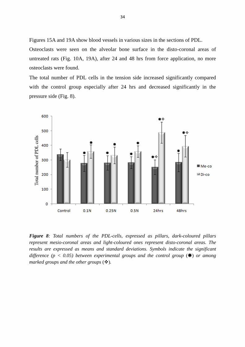
The total number of PDL cells in the tension side increased significantly compared
with the control group especially after 24 hrs and decreased significantly in the
pressure side (Fig. 8).
Figure 8: Total numbers of the PDL-cells, expressed as pillars, dark-coloured pillars
represent mesio-coronal areas and light-coloured ones represent disto-coronal areas. The
results are expressed as means and standard deviations. Symbols indicate the significant
difference (p < 0.05) between experimental groups and the control group () or among
marked groups and the other groups ().
35
5.2 Immunohistochemistry and Histomorphometry
In the control group, immunoreactivity for all investigated proteins was distributed
throughout the examined PDL-areas, but tended to be localized more on the mesial
side (12A, 17A, 21A) than on the distal side of the medial root (10A, 15A, 19A).
Immunoreactivity was observable in osteoblasts, fibroblasts, osteoclasts and
cementoblasts. According to Dahlberg‟s formula, the casual error was 3.65. The
unwanted background staining, which is one of the most common problems in
immunohistochemistry, was reduced by blocking the endogenous peroxidase activity
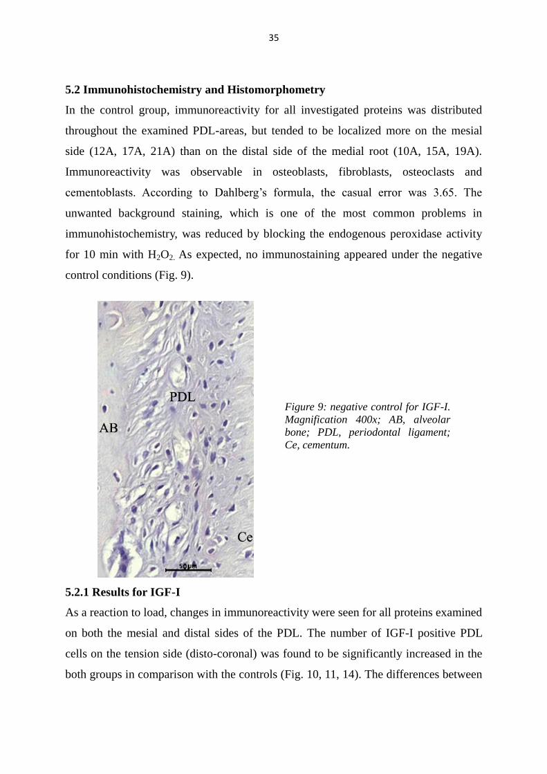
for 10 min with H2O2. As expected, no immunostaining appeared under the negative
control conditions (Fig. 9).
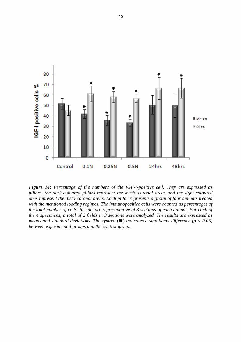
5.2.1 Results for IGF-I
As a reaction to load, changes in immunoreactivity were seen for all proteins examined
on both the mesial and distal sides of the PDL. The number of IGF-I positive PDL
cells on the tension side (disto-coronal) was found to be significantly increased in the
both groups in comparison with the controls (Fig. 10, 11, 14). The differences between
Figure 9: negative control for IGF-I.
Magnification 400x; AB, alveolar
bone; PDL, periodontal ligament;
Ce, cementum.
36
the groups, where constant forces for 4 hrs of 0.25 N and 0.5 N were applied, and the
group, where the rats were killed after 48 hrs, were significantly recognized. In the
pressure side (mesio-coronal), the number of IGF-I-positive cells was significantly
decreased in the 4 hrs group (Fig. 12, 13A, 14). This decrease tended to be dependent
on the force magnitude. After 24 and 48 hrs the number of the immunopositive cells
increased to the mean level of the control group in the mesio-coronal areas. Thus, the
differences between the first and second group were significant with one exception.
No significant difference was seen between the group, where 0.1N were applied for 4
hrs, and the 24 hrs group (Fig. 13B).
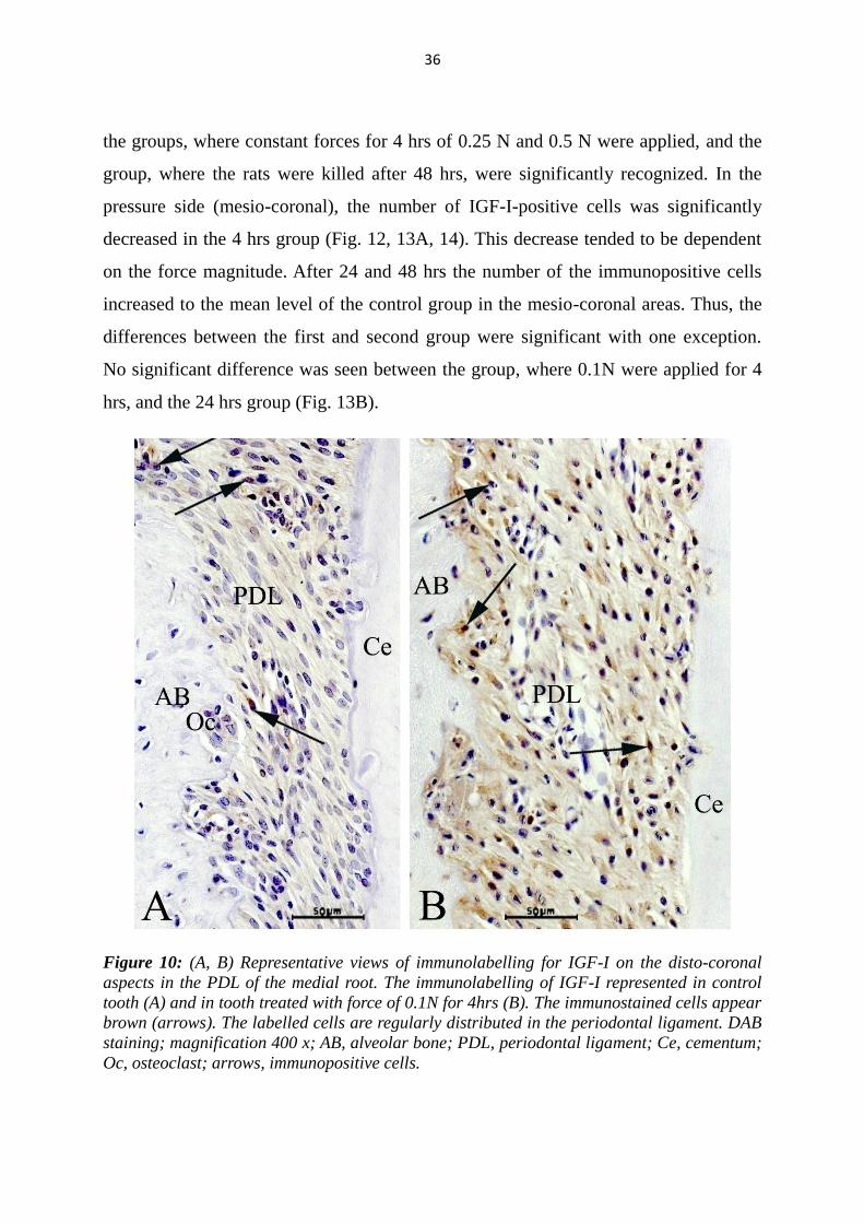
Figure 10: (A, B) Representative views of immunolabelling for IGF-I on the disto-coronal
aspects in the PDL of the medial root. The immunolabelling of IGF-I represented in control
tooth (A) and in tooth treated with force of 0.1N for 4hrs (B). The immunostained cells appear
brown (arrows). The labelled cells are regularly distributed in the periodontal ligament. DAB
staining; magnification 400 x; AB, alveolar bone; PDL, periodontal ligament; Ce, cementum;
Oc, osteoclast; arrows, immunopositive cells.
37
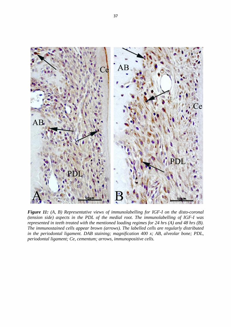
Figure 11: (A, B) Representative views of immunolabelling for IGF-I on the disto-coronal
(tension side) aspects in the PDL of the medial root. The immunolabelling of IGF-I was
represented in teeth treated with the mentioned loading regimes for 24 hrs (A) and 48 hrs (B).
The immunostained cells appear brown (arrows). The labelled cells are regularly distributed
in the periodontal ligament. DAB staining; magnification 400 x; AB, alveolar bone; PDL,
periodontal ligament; Ce, cementum; arrows, immunopositive cells.
38
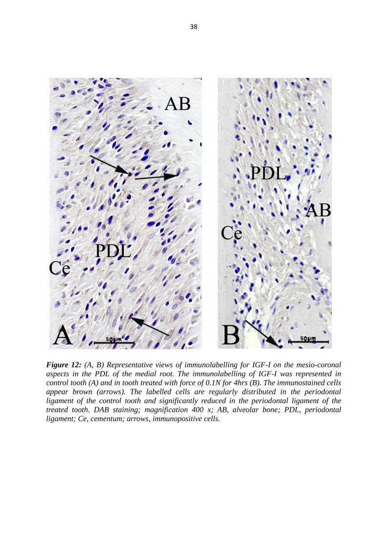
Figure 12: (A, B) Representative views of immunolabelling for IGF-I on the mesio-coronal
aspects in the PDL of the medial root. The immunolabelling of IGF-I was represented in
control tooth (A) and in tooth treated with force of 0.1N for 4hrs (B). The immunostained cells
appear brown (arrows). The labelled cells are regularly distributed in the periodontal
ligament of the control tooth and significantly reduced in the periodontal ligament of the
treated tooth. DAB staining; magnification 400 x; AB, alveolar bone; PDL, periodontal
ligament; Ce, cementum; arrows, immunopositive cells.
39
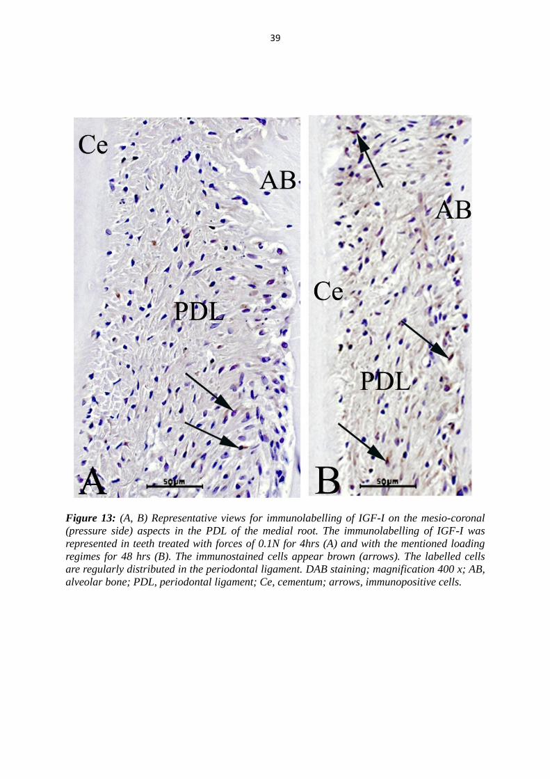
Figure 13: (A, B) Representative views for immunolabelling of IGF-I on the mesio-coronal
(pressure side) aspects in the PDL of the medial root. The immunolabelling of IGF-I was
represented in teeth treated with forces of 0.1N for 4hrs (A) and with the mentioned loading
regimes for 48 hrs (B). The immunostained cells appear brown (arrows). The labelled cells
are regularly distributed in the periodontal ligament. DAB staining; magnification 400 x; AB,
alveolar bone; PDL, periodontal ligament; Ce, cementum; arrows, immunopositive cells.
40
Figure 14: Percentage of the numbers of the IGF-I-positive cell. They are expressed as
pillars, the dark-coloured pillars represent the mesio-coronal areas and the light-coloured
ones represent the disto-coronal areas. Each pillar represents a group of four animals treated
with the mentioned loading regimes. The immunopositive cells were counted as percentages of
the total number of cells. Results are representative of 3 sections of each animal. For each of
the 4 specimens, a total of 2 fields in 3 sections were analyzed. The results are expressed as
means and standard deviations. The symbol () indicates a significant difference (p < 0.05)
between experimental groups and the control group.
41
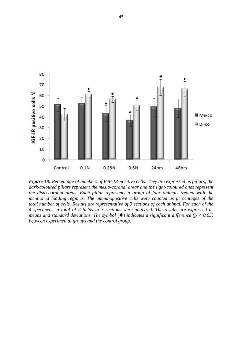
5.2.2 Results for IGF-IR
As seen by IGF-I, the number of immunopositive cells of IGF-IR on the tension side
(disto-coronal) was found to be significantly increased in the both groups in
comparison with the controls (Fig. 15, 16, 18). This increase of the immunopositive
cells in 4 hrs groups was negative dependent on the force magnitude. The difference
between groups, where forces of 0.1 N and 0.5 N for 4 hrs were applied, was
significant. After 24 hrs, the IGF-IR-positive cells reached their peak point and tend to
decrease slightly after 48hrs. Significant differences between the groups, where
constant forces for 4 hours of 0.25 N and 0.5 N were applied, and the 48 hrs group
were observed.
At the pressure side (mesio-coronal), the significant effect of the force application on
the number of IGF-IR-positive cells was not dependent on experimental time but on
the force magnitude. The number of IGF-IR-positive cells was significantly decreased
in groups treated with forces of 0.25N and 0.5N, whereas the group treated with light
force of 0.1N showed no significant differences compared to control group in the
mesio-coronal areas (Fig. 17, 18). Therefore, the differences between the groups,
where 0.5N for 4 hrs was applied, and the second group (24 hrs and 48 hrs) were also
significant.
42
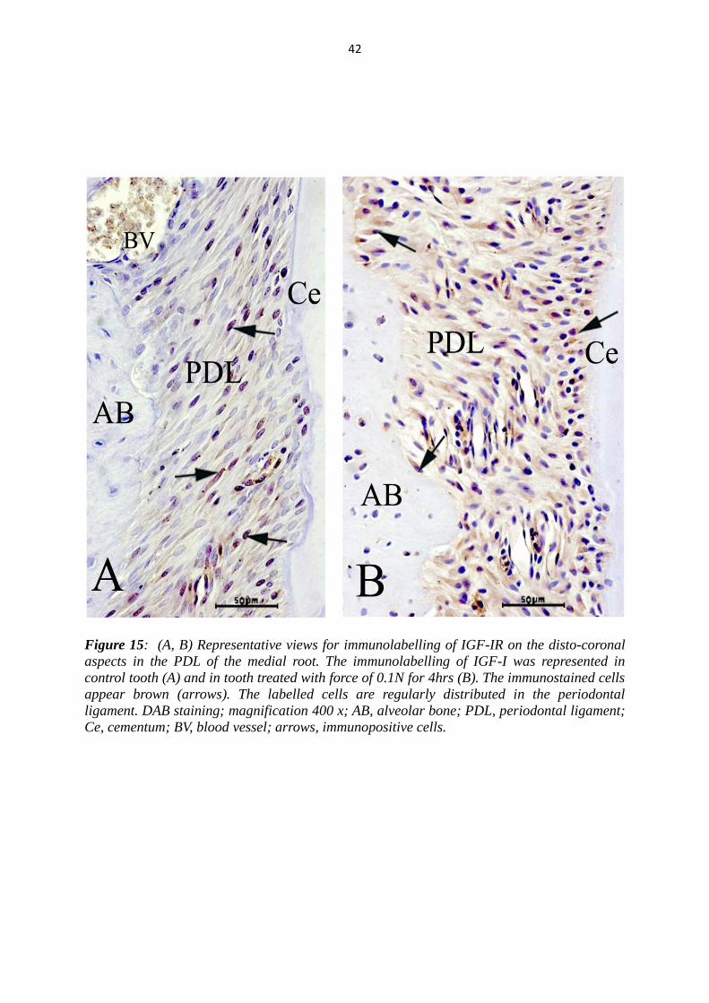
Figure 15: (A, B) Representative views for immunolabelling of IGF-IR on the disto-coronal
aspects in the PDL of the medial root. The immunolabelling of IGF-I was represented in
control tooth (A) and in tooth treated with force of 0.1N for 4hrs (B). The immunostained cells
appear brown (arrows). The labelled cells are regularly distributed in the periodontal
ligament. DAB staining; magnification 400 x; AB, alveolar bone; PDL, periodontal ligament;
Ce, cementum; BV, blood vessel; arrows, immunopositive cells.
43
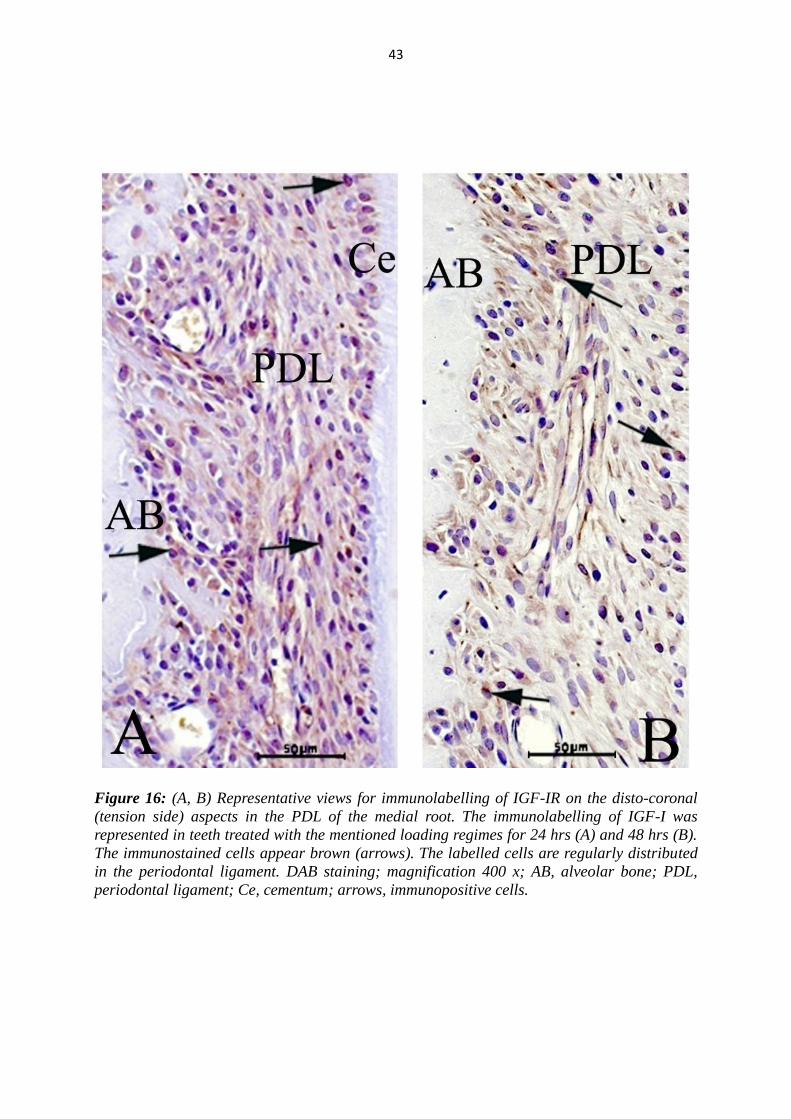
Figure 16: (A, B) Representative views for immunolabelling of IGF-IR on the disto-coronal
(tension side) aspects in the PDL of the medial root. The immunolabelling of IGF-I was
represented in teeth treated with the mentioned loading regimes for 24 hrs (A) and 48 hrs (B).
The immunostained cells appear brown (arrows). The labelled cells are regularly distributed
in the periodontal ligament. DAB staining; magnification 400 x; AB, alveolar bone; PDL,
periodontal ligament; Ce, cementum; arrows, immunopositive cells.
44
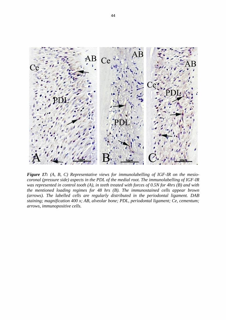
Figure 17: (A, B, C) Representative views for immunolabelling of IGF-IR on the mesio-
coronal (pressure side) aspects in the PDL of the medial root. The immunolabelling of IGF-IR
was represented in control tooth (A), in teeth treated with forces of 0.5N for 4hrs (B) and with
the mentioned loading regimes for 48 hrs (B). The immunostained cells appear brown
(arrows). The labelled cells are regularly distributed in the periodontal ligament. DAB
staining; magnification 400 x; AB, alveolar bone; PDL, periodontal ligament; Ce, cementum;
arrows, immunopositive cells.
45
Figure 18: Percentage of numbers of IGF-IR-positive cells. They are expressed as pillars, the
dark-coloured pillars represent the mesio-coronal areas and the light-coloured ones represent
the disto-coronal areas. Each pillar represents a group of four animals treated with the
mentioned loading regimes. The immunopositive cells were counted as percentages of the
total number of cells. Results are representative of 3 sections of each animal. For each of the
4 specimens, a total of 2 fields in 3 sections were analyzed. The results are expressed as
means and standard deviations. The symbol () indicates a significant difference (p < 0.05)
between experimental groups and the control group.
46
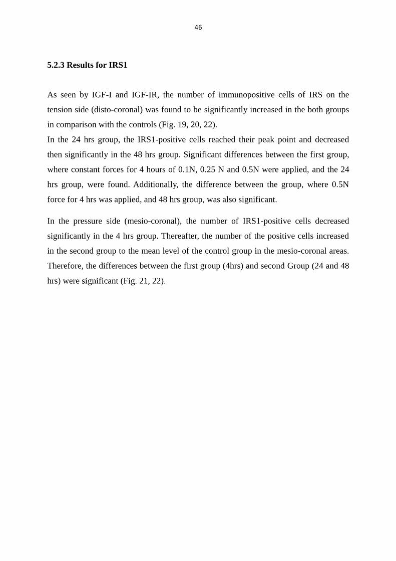
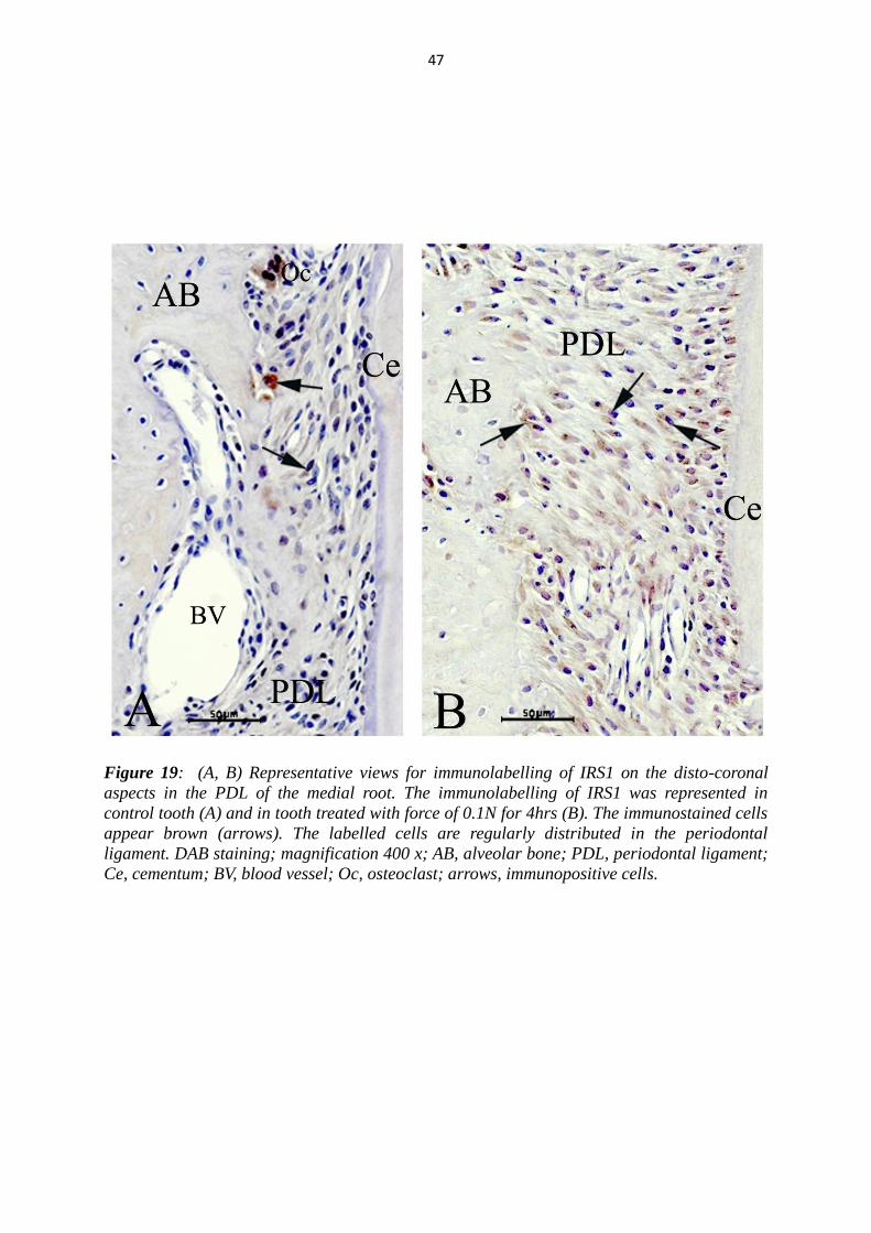
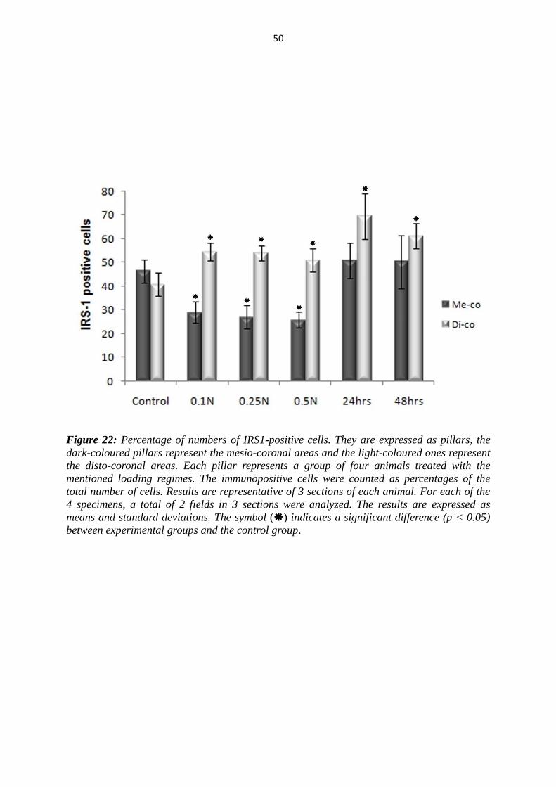
5.2.3 Results for IRS1
As seen by IGF-I and IGF-IR, the number of immunopositive cells of IRS on the
tension side (disto-coronal) was found to be significantly increased in the both groups
in comparison with the controls (Fig. 19, 20, 22).
In the 24 hrs group, the IRS1-positive cells reached their peak point and decreased
then significantly in the 48 hrs group. Significant differences between the first group,
where constant forces for 4 hours of 0.1N, 0.25 N and 0.5N were applied, and the 24
hrs group, were found. Additionally, the difference between the group, where 0.5N
force for 4 hrs was applied, and 48 hrs group, was also significant.
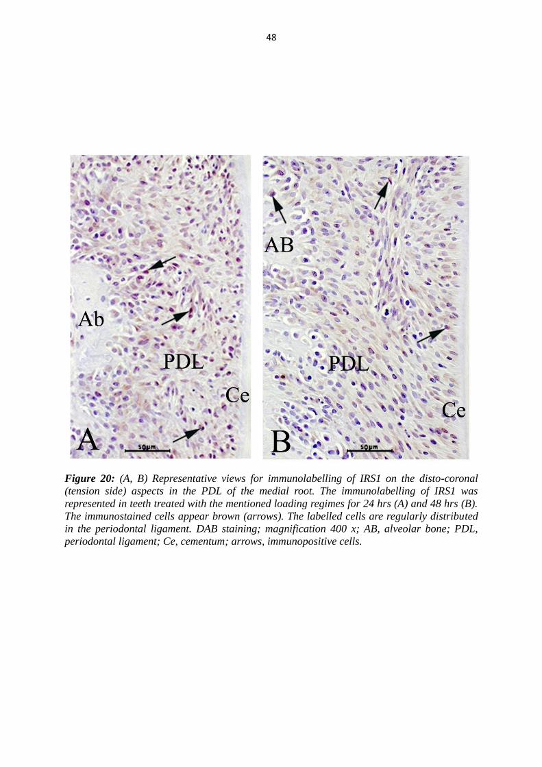
In the pressure side (mesio-coronal), the number of IRS1-positive cells decreased
significantly in the 4 hrs group. Thereafter, the number of the positive cells increased
in the second group to the mean level of the control group in the mesio-coronal areas.
Therefore, the differences between the first group (4hrs) and second Group (24 and 48
hrs) were significant (Fig. 21, 22).
47
Figure 19: (A, B) Representative views for immunolabelling of IRS1 on the disto-coronal
aspects in the PDL of the medial root. The immunolabelling of IRS1 was represented in
control tooth (A) and in tooth treated with force of 0.1N for 4hrs (B). The immunostained cells
appear brown (arrows). The labelled cells are regularly distributed in the periodontal
ligament. DAB staining; magnification 400 x; AB, alveolar bone; PDL, periodontal ligament;
Ce, cementum; BV, blood vessel; Oc, osteoclast; arrows, immunopositive cells.
48
Figure 20: (A, B) Representative views for immunolabelling of IRS1 on the disto-coronal
(tension side) aspects in the PDL of the medial root. The immunolabelling of IRS1 was
represented in teeth treated with the mentioned loading regimes for 24 hrs (A) and 48 hrs (B).
The immunostained cells appear brown (arrows). The labelled cells are regularly distributed
in the periodontal ligament. DAB staining; magnification 400 x; AB, alveolar bone; PDL,
periodontal ligament; Ce, cementum; arrows, immunopositive cells.
49
Figure 21: (A, B, C) Representative views for immunolabelling of IRS1 on the mesio-coronal
(pressure side) aspects in the PDL of the medial root. The immunolabelling of IRS1 was
represented in control tooth (A), in teeth treated with forces of 0.5N for 4hrs (B) and with the
mentioned loading regimes for 48 hrs (B). The immunostained cells appear brown (arrows).
The labelled cells are regularly distributed in the periodontal ligament. DAB staining;
magnification 400 x; AB, alveolar bone; PDL, periodontal ligament; Ce, cementum; arrows,
immunopositive cells.
50
Figure 22: Percentage of numbers of IRS1-positive cells. They are expressed as pillars, the
dark-coloured pillars represent the mesio-coronal areas and the light-coloured ones represent
the disto-coronal areas. Each pillar represents a group of four animals treated with the
mentioned loading regimes. The immunopositive cells were counted as percentages of the
total number of cells. Results are representative of 3 sections of each animal. For each of the
4 specimens, a total of 2 fields in 3 sections were analyzed. The results are expressed as
means and standard deviations. The symbol () indicates a significant difference (p < 0.05)
between experimental groups and the control group.
51
6. Discussion
The present study was performed to investigate the occurrence and distribution of
insulin-like growth factor-I (IGF-I), IGF-IR and IRS1 in the periodontal ligament
(PDL) during the early stage of orthodontic tooth movement. The study was carried
out using forces of varying amount and different time periods in an animal model.
This study showed that IGF-I, IGF-IR and IRS1were distributed locally in the
control group throughout the PDL and tended to be localized more on the mesial side
than the distal side of the medial root.
In the PDL of human deciduous and permanent teeth as well as in the PDL of rats,
Götz et al. have studied the occurrence of the IGFs (Götz et al., 2001; 2006a; 2006 b).
They found that the PDL, in particular, contains mostly all IGF components.
They have suggested that IGFs might be stored in the PDL matrix via their binding
proteins linked to glycosaminoglycans and regulate through autocrine and paracrine
pathways the PDL cell survival, proliferation and matrix turnover.
Although the liver is the main source of IGF-I and the absence in its gene causes
growth retardation in mice and humans, many cell types produce and respond to IGF-I
via autocrine or paracrine actions (Woods et al., 1996, Bonapace et al., 2003; Barton
and Crowder, 2010; Netchine et al, 2011). For example, IGF-I is one of the best
characterized and the most abundant growth factors in the bone tissue (Mohan and
baylink, 1991; Canalis, 2009). This local production of IGF-I can even compensate the
lack of the liver IGF-I in genetically manipulated mice (andrew et al., 2001),
suggesting that autocrine/paracrine-produced IGF-I is the main determinate of
postnatal body growth (Ohlsson et al., 2000).
The previous investigations and the results of this study strength the crucial role of
autocrine and paracrine IGF-I and its impacts in the biology of PDL.
This study elucidates that the application of precise short term loading at teeth
in rats leads to an increase in IGF-I, IGF-IR and IRS1 expression at the tension side.
This observation is in accordance with other studies which showed that loading at
different tissues increases the production of IGF-I: Human movement occurs from the
52
force created by contracting muscles, which is transmitted to bone via tendon, all these
three tissues can respond to the mechanical loading by producing local IGF-I or its
isoform.
In skeletal muscles, insulin-like growth factor I (IGF-I) play a critical role in their
formation, maintenance, and regeneration. Longitudinal exercise studies have shown
that it is possible to increase muscle strength and performance without concomitant
and robust changes in circulating IGF-I. Such studies indicating that the effect of
exercise on skeletal muscles is mediated via paracrine/autocrine IGF-I rather than
endocrine IGF-I (Frystyk, 2010). Goldspink (Goldspink, 1999) showed that loading
increases the production of IGF-I in muscle cells, functioning in an autocrine/paracrine
mode, is an important mediator of skeletal muscle adaptation being sensitive to
increases in loading (Adams, 2002).
In human tendon, which resembles the PDL in its function, it can be demonstrated that
an increase in the interstitial concentration of IGF-I and its binding proteins takes
place after exercise. The increase in IGF-I expression in tendon includes the isoform
that has so far been thought only to exist in skeletal muscle (mechano-growth factor)
(Kjaer et al., 2009). In rats, it was suggested that the IGF-I and its splice variant
mechano-growth factor (MGF) could be involved in collagen synthesis in tendon in
response to mechanical loading (Olesen et al., 2006).
The transmitted mechanical stimulation to bone leads to increased bone formation and
plays an essential role in maintaining skeletal integrity. Growth factors and osteocytes,
which act as mechanosensors, play a key role during the bone formation after
mechanical stimulation. Recently, Reijnders et al. (2007) have found that IGF-I mRNA
is upregulated within endocortical osteocytes of the shaft of rat tibia 6 h after
mechanical loading. They suggested that IGF-I, which is located in osteocytes, is
involved in the translation of mechanical stimuli into bone formation. It has been also
demonstrated that rat osteoblasts respond to the mechanical forces which may regulate
their activities indirectly by promoting the autocrine effect of IGF-1 (Xian et al.,
2007).
53
The ability of IGF-I to translate the mechanical stimuli into bone formation was
investigated and verified in many experimental and clinical studies.
Distraction osteogenesis is currently a standard method of bone lengthening (Choi et
al., 2002). In distraction osteogenesis studies, gene expression of IGF-I was detected
during distraction (Farhadieh et al., 1999; Yates et al., 2002).
The application of tensile forces to the mid sagittal cranial suture during the growth
period cause an increase in the production of IGF-I mRNA and IGF-IR mRNA in
osteoblast-like and fibroblastic cells (Hirukaw et al., 2005).
Hajjar et al. (2002) observed that the expression of IGFs increases in condylar
cartilage in response to application of propulsive appliances.
In orthodontics, Wescott et al. have applied a 12 % uni-axial cyclic tensile strain to
cultured human PDL cells and analyzed the differential expression of 78 genes, 19
genes, including IGF-I, show differential expression. The treated/control (T/C) ratio of
IGF-I after 12 hrs was greater than of ± 2 (2.46) but not significant (Wescott et al.,
2007). Rath-Deschner et al. have found that IGF-I expression is significantly
increased, when cells were subjected to low continuous tensile strain for 4 h ( Rath-
Deschner et al., 2009). Saggese et al. found a fivefold increase of IGF-I in the
cervicular fluid after insertion of an orthodontic appliance to distalize canines in
patients aged between 8 and 15 years (Saggese et al., 2005). Toia et al. found that IGF-
I increases in the cervicular fluid as early as 4hrs after insertion of fixed appliances
(Toia et al., 2005). Therefore, this study indicates that IGF-I and its signals involved in
the mechanotransduction processes during the tooth movement.
In this study, the character of IGF-I over time was observed in the PDL and
found that IGF-I-positive cells were significantly increased at the tension side in a time
dependent manner. It was also recognized that this increase correlated with an
increase in the total number of PDL cells over time at the same side. The both IGF-I-
positive cells and the total number of the PDL cells reach their peak points after 24 hrs.
It has been observed that the mitotic activity of PDL cells increases markedly on the
tension side from 24 to 36 hrs after the initiation of tooth movement (Macapanpan et
54
al., 1954). This leads to increase the number of periodontal ligament cells, which can
be increased by stimulating cell proliferation or by inhibiting apoptosis (Suri and
Taneja, 2008). These cell functions can be affected through IGF-I production. IGF-I
stimulates proliferation of PDL fibroblast (Palioto et al., 2004), cementoblasts (Grzesik
and Narayanan, 2002) and osteoblasts (Wergedal et al., 1990).
The autocrine/paracrine secreted IGF-I is also an important component of the response
to injury. Multiple studies in several different animal models have shown that IGF-I is
synthesized after injury by the cell types that account for tissue regeneration. This
synthesis is necessary for normal tissue repair (Clemmons and Maile 2005), defends
against cellular stress and delays the onset of the apoptosis (Kumasheva and
Houghton, 2006). The antiapoptotic action of IGFs was recently also observed in PDL
cells that respond to IGF-I by up-regulation of anti-apoptotic pathways (Han and
Ammar, 2003). Therefore, one can presume that the up-regulation of IGF-I on the
tension side can increase the cell number of PDL through its impacts on the
proliferation and the apoptosis of the PDL cells during the tooth movement in a time-
dependent manner.
The mean values of immunopositive cells for all examined factors reach the
peak point after 24 hrs compared with those of the control teeth. These results are in
accordance with those described by other researchers for other factors. Transforming
growth factor-β1 (TGF-β1) regulates cell proliferation, differentiation, motility and
apoptosis (Kanaan and Kanaan, 2006). Osteoprotegerin (OPG) inhibits the
differentiation and stimulates the apoptosis of the osteoclasts (Tyrovola et al., 2008).
These both factors were observed with significant increase in stretched cells on the
bone surface after 24 hrs from force application and became more intense after 48 hrs
of force application (Kobayashi et al., 2000).
The expression of epidermal growth factor (EGF) and its receptor, which play an
important role in bone formation, were detected in periodontal tissues after 24 hours
and 168 hours of tooth movement. The expressions of EGF and EGFR were increased
55
in periodontal tissues at 168 hrs higher than those at 24 hrs and at tension side higher
than those at pressure side at the same time (Gao et al., 2002).
The expression patterns of Ki-67, a cellular marker for proliferation and osteoblast
precursors (Scholzen and Gerdes et al., 2000), and Runx2, an essential transcription
factor for osteoblast differentiation and chondrocyte maturation (Komori, 2011), were
investigated at 3 and 24 hours after appliance insertion. The results showed that the
expression of Ki-67 and Runx2 increase in the tension areas after 24 hours of force
application (Brooks et al., 2009).
Recently, the gingival crevicular fluid (GCF), which can be collected from the gingival
crevice surrounding the teeth, was used to determine possible changes in its
constituent in response to various pathological and physiological alterations in the
periodontium (Lamster et al., 2007). The presence/expression of regulatory proteins in
the GCF has been examined to illustrate the involvement of these proteins in
periodontal remodeling provoked by orthodontic stimuli. The most consistent result
was a peak of cytokine levels at 24 h (Ren and Vissink, 2008). For example, Nishijima
et al., have examined the secretion of RANKL and OPG from hPDL cells at the distal
cervical margins of the experimental and control teeth 0, 1, 24, and 168 hrs after the
retracting force was applied. After 24 hrs, the levels of RANKL were significantly
higher and the levels of OPG were significantly lower compared to the control teeth.
There were no such significant differences at 0, 1, or 168 hrs (Nishijima et al., 2006).
All these results support the statement that cellular and tissue reactions, including IGF-
I synthesis, start in the initial phase (1-2 days) of tooth movement, immediately after
force application. The complex processes of recruitment of osteoclast and osteoblast
progenitors begin in this early phase (Krishnan and Davidovitch, 2006).
The human PDL cells represent a heterogeneous cell population including stem
cells (Lekic et al., 2001; Silvério et al., 2010) and have the potential to differentiate
into various phenotypes, including osteoblasts (Gay et al., 2007) and cementoblasts
(Seo et al., 2004). IGF-I induces many differentiated functions, such as production of
56
collagen and matrix apposition (Grzesik and Narayanan, 2002; Minuto et al., 2005). It
is known that procollagen type I is an indicator of the early stage of bone formation.
The recombinant IGF-I can raise the serum levels of procollagen type I significantly
(Zofkova, 2003). Therefore, it is assumed that IGF-I activates especially the early
stages of osteoblastic formation in the PDL.
In periodontal regeneration, the reestablishment of the PDL is required together with
corresponding cementum and supporting alveolar bone. Thus, agents that promote
PDL cells proliferation and migration as well as collagen biosynthesis would appear to
be mediators for enhancing new PDL formation (Raja et al., 2009). Recently, in two in
vitro studies, IGF-I alone or in combination with other growth factors promoted
osteogenesis of the PDL cells. It can elevate the osteoblastic markers such alkaline
phosphatase (ALP) and ostetocalcin (OCN) (Chen et al., 2009; Li et al., 2011). IGFs
regulate, in diverse patterns, the differentiation functions of both osteoblasts and
osteoclasts (Conover, 2000). This can accelerate the differentiation of osteoclasts and
its disappearance in the tension side after force application (disto-coronal areas), which
tend to contain more osteoclasts in the PDL of untreated teeth due to the physiological
distal drift of the molars in the rats.
Therefore, one can speculate that the up-regulation of IGF-I could influence the
differentiation of the PDL cells at the tension side.
As previously mentioned in the introduction, there are four essential interrelated
steps in the transduction of mechanical signals by tissues: sensing the mechanical
signal by the cells, transduction of this mechanical signal into one that is biochemical,
transmission of the biochemical signal to the effector cells, and the effectors cell
response (Wise and King, 2008).
After force application both matrix strain and fluid flow in the PDL and the bone cause
deformation of cells. Through integrin signalling and other transduction pathways,
many mediators are produced leading to activate several types of cells (Hennemann et
al., 2008).
57
Ajubi et al. investigated the signal transduction pathways in osteocytes which were
subjected to pulsating fluid flow (PFF). They found that PFF raises intracellular Ca2+
.
Ca2+
and protein kinase C then stimulate phospholipase A2 activity, arachidonic acid
production, and ultimately prostaglandin E2 (PGE2) release (Ajubi et al., 1999; Wise
and King, 2008).
Studies which were performed in gingival crevicular fluid (GCF) during tooth
movement showed that the concentration of PGE2 in GCF increased significantly and
reaches peak point at 24th hour and generally was higher in tension sides than at the
compression sides (Yao et al., 2003; Dudic et al., 2006).
Release of PGE2 is a prominent load-induced response of osteoblast-like cells. PGE2 is
produced by osteoblasts in response to physiological stress, growth factors, hormones,
trauma or inflammatory cytokines and induces cAMP-dependent IGF-I expression by
osteoblasts (Papachroni et al., 2009).
In vitro studies showed that PGE2 increases rapidly at 5min after loading of teeth and
led to an increase of intracellular cyclic adenosine monophosphate (cAMP) which
peaks at 15 min. (Meikle, 2006).
The increased intracellular cAMP activates protein kinase A (PKA), which stimulates
IGF-I gene induction (McCarthy and Centrella, 2001). In addition, mechanical
interactions between integrins and their matrix/environment mediate increases in
intracellular Ca2+
levels and activate mitogen-activated protein (MAP) kinase
cascades. This leads to the activation of the activator protein 1 (AP-1) that is necessary
for a pro-growth response. The pro-bone growth response involves up-regulation of
the genes c-fos, osteocalcin, cyclooxygenase, and IGF-I (Iqbal and Zaidi, 2005).
and thus can man better understand the mechanism of IGF-I up regulation after tooth
movement as response to up regulation of other factors such as PGE2.
In this study, immunoreactivity for IGF-IR was generally more pronounced than
that for its ligand IGF-I. The biological actions of IGF-I are predominantly mediated
by the IGF-IR (LeRoith 2000; Romano, 2003; Annunziata et al., 2011).
58
Recently, it has been reported, that growth factor receptors can be also activated by
integrins in the absence of the growth factor ligands, due to the interaction between
integrins and growth factor receptors (Clemmons and Maile, 2003; Beattie et al.,
2010). The crosstalk between integrins and growth factor receptors in multiple cell
types may be the result of coclustering of these receptors on the surface of the cell in
focal adhesions or in association with the actin cytoskeleton (Eliceiri, 2001, Beattie et
al., 2010). For example, the occupation of the αVβ3 integrin receptor with ECM
proteins induces IGF-I-stimulated IGF-IR phosphorylation. Conversely, the presence
of the αVβ3-specific disintegrin echistatin inhibits the IGF-I-stimulated IGF-IR
activation (Kim et al., 2007). In this context, Kapur et al. found that echistatin reduced
not only the basal and shear stress-induced TE85 cell (osteosarcoma cell) proliferation
but also completely abolished the increase in cell proliferation induced by IGF-I alone
as well as that by the combination treatment. This suggests that the synergy between
shear stress and IGF-I in osteoblast proliferation involves integrin-dependent
recruitment of SHP-2 and -1 away from IGF-IR (Kapur et al., 2005).
Insulin receptor substrates (IRS) is a main target molecule of insulin/IGF-1
receptor signaling and plays important roles in maintaining normal bone turn-over
(Ogata and Kawaguchi, 2008). When the concentrations of IRS1 are high, the signal is
mitogenic and anti-apoptotic (Valentinis and Baserga, 2001). IRS1 deficient mice
exhibited severe low turnover osteopenia with decreased bone anabolic and catabolic
action (Akune, 2004).
Tyrosine phosphorylation of IRS1 by the IGF-I receptor tyrosine kinase leads to
activation of pathways such PI3 and extracellular signal-regulated kinase 1/2
(ERK1/2). The first pathway protects from apoptosis by blocking the pro-apoptotic
proteins and inducing the expression of antiapoptotic proteins. The second pathway,
ERK1/2, is required for optimal stimulation of cell division (proliferation) and gene
expression (Jones and Clemmons, 1995; Mauro and Surmacz, 2003; Yamagushi et al,
2005). The application of orthodontic loading within the same time interval and under
identical experimental circumstances induced a significant increase of ERK1/2-
positive PDL cells at the tension side (Kawarizadeh et al., 2005, Pavlidis et al., 2009).
59
It most kept in mind that ERK1/2 can be activated by other molecules like integrin
receptors (Iqbal and Zaidi, 2005; Meikle, 2006).
However, the ability of cells to respond to the autocrine/ paracrine secreted
IGF-I is dependent not only on the amount of growth factor that is secreted, but also
on the abundance of other proteins in the extracellular environment especially IGFBPs.
IGFBPs are a family of secreted proteins that bind IGF with high affinities that are
equal to or greater than those of the IGF-IR. In the extracellular environment the most,
if not all, of IGFs are bound to specific, high-affinity IGFBPs (Duan and Xu, 2005).
They can suppress or enhance the cellular effects of IGFI (Yu and Rohan, 2000; Firth
and Baxter, 2002). Anyway, the observed up-regulation of the IGF-IR and IRS1 in this
study strengthens the view that the biological action of IGF-I on PDL cells was
stimulated under this condition of force application.
At the pressure side, it was observed that all factors are significantly down
regulated in the 4 hrs groups, but not in 24 or 48 hrs groups, where the differences
between the groups were no more significant. This may be caused by virtue that PDL
cells subjected to tensional and compressive forces are able to perceive two different
forms of mechanical stimuli and responded in a different manner (He et al., 2004).
Other interpretation for down regulation of IGF-I at the pressure side could due to the
negative strain that induced within the PDL at the pressure side of the root
(Hennemann et al., 2008) and, consequently, the fibers are relaxed. This leads to
unloading of the PDL and alveolar bone (Melsen , 1999, 2001; Binderman et al.,
2002). The unloading reduces canalicular fluid flow and induces the apoptosis of
osteocytes and osteoblasts, which attracts osteoclasts to the pressure side followed by
bone resorption subsequently (Burger et al., 2003; Aguirre et al., 2006; Dufour et al.,
2007; Hennemann et al., 2008). Unloading could also inhibit the proliferation and
differentiation of osteoprogenitor cells in vitro (Kostenuik et al., 1997). Moreover,
skeletal unloading induces resistance to IGF-I on bone formation and inhibits the
activation of the IGF-I signaling pathway (Sakata et al., 2003, 2004).
60
Clinically, skeletal unloading during space flight and prolonged bed rest induces
osteopenia (Bikle and Hallaoran, 1999).
Thus, one can assume that there is a relation between the unloading of PDL cells, the
initial decrease of IGF-I-positive cells, and the decrease of the total cell number at the
pressure side.
The positive PDL-cells at the pressure side decreased in the 4 hrs group and
augmented subsequently in 24 and 48 hrs groups. This could be interpreted as result of
the remodeling activity of PDL tissue and alveolar bone at the pressure side that
degrade to create space for the moving tooth while new PDL tissue is simultaneously
formed to maintain the attachment (Hennemann et al. 2008).
At the cellular level, PDL cells in the pressure side become a round shape in the first
hours of treatment and regain a normal shape 2 or 3 days later (Kirshnan and
Davidovitch, 2006). In this regard, cells which are rounded up show catabolic changes
whereas flattened cells (under tension) have anabolic effects (Roberts-Harry and
Sandy, 2004).
In the present study, the effect of the force magnitude on the positive cells
number was only significant between the groups, where 0.1N and 0.5N were applied
for 4 hrs and only for IGF-IR. The results for IGF-I and IRS1 showed no significant
differences between applied forces but tend to be negative dependent on the force
magnitude. These results could indicate a relation between the expression of these
molecules, especially IGF-IR, and the magnitude of the applied forces.
In the past 70 years, the concept of optimal force has changed considerably. According
to Schwarz, who defined optimal continuous force as „the force leading to a change in
tissue pressure that approximated the capillary vessels‟, forces below the optimal level
cause no reaction in the periodontal ligament. Forces exceeding the optimal level
would lead to areas of tissue necrosis, preventing frontal bone resorption. Tooth
movement would thus be delayed until undermining resorption had eliminated the
necrotic tissue obstacle (Schwarz, 1932). Oppenheim and Reitan modified this

61
definition and advocated the use of the lightest force which is capable to achieve the
tooth movement (Oppenheim, 1942; Reitan, 1967). The current concept of optimal
force is based on the hypothesis, that a force of a certain magnitude and temporal
characteristics would be capable of producing a maximum rate of tooth movement
without tissue damage and with maximum patient comfort. The optimal force for tooth
movement may differ for each tooth and for each individual patient (Ren et al., 2003).
There are many reports about the effect of force magnitude on tooth movement. King
et al. demonstrated that the effective tooth movement of rat molars ranges from 20 g to
40 g and its velocity does not increase over 40 g (King et al., 1991). Gonzales et al.
found that the light forces produce more tooth movement and less root resorption
compared with heavier forces (Gonzales et al., 2007).
Considering the orthodontic tooth movement as the result of biological reactions to
externally applied mechanical stimuli, the application of 0.1 N may cause greater
biologic activity of IGF-I-signalling than the other forces, which can subsequently lead
to cellular responses aiming at adaptation to the applied forces.
62
7. Summary
The aim of this study was to characterize the role of IGF-I and its signalling in vivo as
an early reaction to the mechanotransduction process, and to analyse the changes of
the local expression of these factors related to the magnitude of the applied force.
The right upper first molars of 20 anesthetised rats were loaded with forces in order to
be moved mesially. In the first group of animals, constant forces for 4 hours of 0.1 N,
0.25 N and 0.5 N were applied in four animals each. The applied forces were kept
constant and recorded continuously for 4 hrs. After 4 hrs all 12 animals were
sacrificed. In the second group, constant forces of 0.1 N were applied for 2 hours in 8
animals. Afterwards, the first and second molars in the second group were passively
separated with composite and the occlusal contacts were eliminated by slicing the
antagonistic lower molars. Four rats were sacrificed after 24 hrs and four after 48 hrs.
The untreated contralateral molars in 4 rats served as controls. Paraffin-embedded
sections of the resected jaws were prepared for immunohistochemistry to localize
insulin-like growth factor-I (IGFI), its receptor (IGF-IR), and insulin receptor substrate
1 (IRS1). Histomorphometric analysis was performed to count the percentage of
immunoreactive cells in different parts of the periodontal ligament. The statistical
analyses were performed with SPSS Software.
The results showed that IGF-I-, IGF-IR- and IRS1-positive cells were observed in the
periodontal tissues of the control and loaded teeth. In the experimental group, the
number of IGF-I-, IGF-IR- and IRS1-positive cells increased significantly on the
tension side and decreased significantly on the compression side. The increase of
immunopositive cells at the tension side was in a time-dependent manner and tended
to be dependent on the force magnitude. The decrease in the immunopositive cells at
the pressure side was only observed in the first group (4 hrs) and tended to be
dependent on the force magnitude.
These data indicate a close relationship between mechanical loading of the PDL and
the autocrine/paracrine expression of IGF components as an early step in the

63
mechanotransduction process leading to an organized remodelling of the PDL and
alveolar bone in the long term.
Moreover, the local increase/decrease of IGF-I appears to provide a link between the
mechanical loading/unloading, the activation/inhibition of proliferation and
differentiation of PDL cells and the inhibition/induction of the antiapoptotic activity in
the periodontal ligament in the early phase of tooth movement. However, more studies
are required to confirm or rule out these hypotheses.
64
8. References
Adams GR. Autocrine/paracrine IGF-I and skeletal muscle adaptation. J Appl Physiol.
2002; 93: 1159-1167
Aguirre JI, Plotkin LI, Stewart SA, Weinstein RS, Parfitt AM, Manolagas SC, Bellido
T. Osteocyte apoptosis is induced by weightlessness in mice and precedes osteoclast
recruitment and bone loss. J Bone Miner Res. 2006; 21: 605-615
Akune T. The role of insulin receptor substrates in bone metabolism. Clin Calcium.
2004; 14: 289-292
Ajubi NE, Klein-Nulend J, Alblas MJ, Burger EH, Nijweide PJ. Signal transduction
pathways involved in fluid flow-induced PGE2 production by cultured osteocytes. Am
J Physiol. 1999; 276: 171-178
Annunziata M, Granata R, Ghigo E. The IGF system. Acta Diabetol. 2011; 48: 1-9
Barton ER, Crowder C. Growth Factor Targets for Orthodontic Treatments. Semin
Orthod. 2010; 16: 128-134
Baumrind S. A reconsideration of the property of the pressure tension hypothesis. Am
J Orthod. 1969; 55: 12-22
Beattie J, McIntosh L, van der Walle CF. Cross-talk between the insulin-like growth
factor (IGF) axis and membrane integrins to regulate cell physiology. J Cell Physiol.
2010; 224: 605-611
Bikle DD, Hallaoran BP. The response of bone to unloading. J Bone Miner Metab.
1999; 17: 233–244
Binderman I, Bahar H, Yaffe A. Strain relaxation of fibroblasts in the marginal
periodontium is the common trigger for alveolar bone resorption: a novel hypothesis. J
Periodontol. 2002; 73: 1210-1215
Bonapace G, Concolino D, Formicola S, Strisciuglio P. A novel mutation in a patient
with Insulin-like growth factor 1 (IGF1) deficiency. J Med Genet. 2003; 40: 913-917
Bouletreau PJ, Warren SM, Longaker MT. The molecular biology of distraction
65
osteogenesis. J Craniomaxillofac Surg. 2002; 30: 1-11
Brooks PJ, Nilforoushan D, Manolson MF, Simmons CA, Gong SG. Molecular
markers of early orthodontic tooth movement. Angle Orthod. 2009; 79:1108-1113.
Butler A, LE Roith D. Minireview: Tissue-specfic versus generalized gene targeting of
the igf1 and igf1r genes and their roles in insulin-like growth factor physiology.
Endocrinology 2001; 142:1685-1688
Burger EH, Klein-Nulend J, Smit TH. Strain-derived canalicular fluid flow regulates
osteoclast activity in a remodelling osteon a proposal. J Biomech. 2003; 36:1453-1459.
Burstone CJ. The biomechanics of tooth movement. In: Kraus BS, Riedel RA, editors.
Vistas in orthodontics. Philadelphia: Lea and Febiger; 1962
Butt AJ, Williams AC. IGFBP-3 and apoptosis-a licence to kill? Apoptosis 2001; 6:
199–205
Canalis E. Growth factor control of bone mass. J Cell Biochem. 2009; 108: 769-777
Chachisvilis M, Zhang YL, Frangos JA. G protein-coupled receptorssense fluid shear
stress in endothelial cells. Proc Natl Acad Sci USA. 2006; 103: 15463-15468
Chen FM, Chen R, Wang XJ, Sun HH, Wu ZF. In vitro cellular responses to scaffolds
containing two micro encapulated growth factors. Biomaterials 2009; 30: 5215-5224
Choi IH, Chung CY, Cho TJ, Yoo WJ. Angiogenesis and mineralization during
distraction osteogenesis. J Korean Med Sci. 2002; 17: 435-447
Clemmons DR, Maile LA. Interaction between Insulin-like growth factor-I receptor
and Vß3 integrin linked signaling pathways: cellular responses to changes in multiple
signaling inputs. Mol Endocrinol. 2005; 19: 1-11
Cohen P. Overview of the IGF-I system. Horm Res. 2006; 65: 3–8
Conover CA. In vitro studies of insulin-like growth factor I and bone. Growth Horm
IGF Res. 2000; 10: 107–110
Davidson RM, Tatakis DW, Auerbach AL. Multiple forms of mechanosensitive
66
channels in osteoblast-like cells. Pflugers Archiv. 1990; 416: 646-651
DeChiara TM, Robertson EJ, Efstratiadis A. Parental imprinting of the mouse insulin-
like growth factor II gene. Cell 1991; 64: 849–859
Denley A, Cosgrove LJ, Booker GW, Wallace JC, Forbes BE. Molecular interactions
of the IGF system. Cytokine Growth Factor Rev. 2005; 16: 421-439
Duan C, Xu Q. Roles of insulin-like growth factor (IGF) binding proteins in regulating
IGF actions. Gen Comp Endocrinol. 2005; 142: 44-52
Dudic A, Kiliaridis S, Mombelli A, Giannopoulou C. Composition changes in gingival
crevicular fluid during orthodontic tooth movement: comparisons between tension and
compression sides. Eur J Oral Sci. 2006; 114: 416-422
Dufour C, Holy X, Marie PJ. Skeletal unloading induces osteoblast apoptosis and
targets alpha5beta1-PI3K-Bcl-2 signaling in rat bone. Exp Cell Res. 2007; 15:394-403.
Duncan R L, Turner C H. Mechanotransduction and the functional response of bone to
mechanical strain. Calcif Tissue Int. 1995; 57: 344-358
Dupont J, Holzenberger M. Biology of insulin-like growth factors in development.
Birth Defects Res C Embryo Today. 2003; 69: 257-271
Eliceiri BP. Integrin and growth factor receptor crosstalk. Circ Res. 2001; 89: 1104-
1110
Farhadieh RD, Dickinson R, YU Y, Gianutsos MP, Walsh WR. The role of
transforming growth factor, insulin like growth factor I, and basic fibroblast growth
factor in distraction osteogenesis of the mandible. J Craniofac Surg. 1999; 10: 80-86
Firth SM, Baxter RC. Cellular actions of the insulin-like growth factor binding
proteins. Endocr Rev. 2002; 23: 824–854
Frystyk J. Exercise and the growth hormone-insulin-like growth factor axis. Med Sci
Sports Exerc. 2010; 42: 58-66
Fujiwara N, Tabata MJ, Endoh M, Ishizeki K, Nawa T. insulin-like growth factor-I
stimulates cell proliferation in the outer layer of Hertwig's epithelial root sheath and
67
elongation of the tooth root in mouse molars in vitro. Cell Tissue Res. 2005; 320: 69-
75
Gay IC, Chen S, MacDougall M. Isolation and characterization of multipotent human
periodontal ligament stem cells. Orthod Craniofac Res. 2007; 10: 149–160
Gao Q, Zhang S, Jian X, Zeng Q, Ren L. Expression of epidermal growth factor and
epidermal growth factor receptor in rat periodontal tissues during orthodontic tooth
movement. Zhonghua Kou Qiang Yi Xue Za Zhi 2002; 37: 294-296
Goldspink G. Changes in muscle mass and phenotype and the expression of autocrine
and systemic growth factors by muscle in response to stretch and overload. J Anat.
1999; 194: 323–334
Götz W, Krüger U, Ragotzki S, Lossdörfer S, Jäger A. Immunohistochemical
localization of components of insulin-like growth factor-system in human decidous
teeth. Connect Tissue Res. 2001; 42: 291-302
Götz W, Lossdörfer S, Krüger U, Braumann B, Jäger A. Immunhistochemical
localization of insulin-like growth factor-II and its binding protein-6 in human
epithelial cells of Malassez. Eur J Oral Sci. 2003; 11: 26-33
Götz W, Heinen M, Lossdörfer S, Jäger A. Immunohistochemical localization of
components of insulin-like growth factor-system in human permanent teeth. Arch Oral
Biol. 2006(a); 51: 387-395
Götz W, Kunert D, Zhang D, Kawarizadeh A, Lossdörfer S, Jäger A. Insulin-like
growth factor system components in the periodontium during tooth root resorption and
early repair processes in the rat. Eur J Oral Sci. 2006 (b);114: 318-327
Govoni E, Baylink DJ, Mohan S. The multi-functional role of insulin-like growth
factor binding proteins in bone. Pediatr Nephrol. 2005; 20: 261-268
Grimm FM. Bone bending, a feature of orthodontic tooth movement. Am J Orthod
1972; 62: 384-393
68
Grzesik WJ, Narayanan AS. Cementum and periodontal wound healing and
regeneration. Crit Rev Oral Biol Med. 2002, 13: 474-484
Hajjar D, Santos MF, Kimura IT. Propulsive appliance stimulates the synthesis of
insulin-like growth factors I and II in the mandibular condylar cartilage of young rats.
Arch Oral Biol. 2003; 48: 635-642
Han X, Amar S. IGF-1 signaling enhances cell survival in periodontal ligament
fibroblasts vs. gingival fibroblasts. J Dent Res. 2003; 82: 454-459
Hamill OP, Martinac B. Molecular basis of mechanotransduction in living cells.
Physiol Rev. 2001; 81: 685-740
Harell A, Dekel S, Binderman I. Biochemical effect of mechanical stress on cultured
bone cells. Calcif Tissue Res. 1977; 22: 202–207
Haut Donahue TL, Genetos DC, Jacobs CR, Donahue HJ, Yellowley CE. Annexin V
disruption impairs mechanically induced calcium signaling in osteoblastic cells. Bone
2004; 35: 656–663
He Y, Macarak EJ, Korostoff JM, Howard PS. Compression and tension: differential
effects on matrix accumulation by periodontal ligament fibroblasts in vitro. Connect
Tissue Res. 2004; 45: 28–39
Henneman S, Von den Hoff JW, Maltha JC. Mechanobiology of tooth movement. Eur J
Orthod. 2008; 30: 299-306
Hirukaw K, Miyazawa K, Maeda H, Kameyama Y, Goto S, Togari A. Effect of tensile
force on the expression of IGF-I and IGF-I receptor in the organ-cultured rat cranial
suture. Arch Oral Biol. 2005; 50: 367-372
Holly J, Perks C. The role of insulin-like growth factor binding proteins.
Neuroendocrinology 2006; 83: 154-160
69
Iqbal J, Zaidi M. Molecular regulation of mechanotransduction. Biochem Biophys Res
Commun. 2005; 328: 751-755
Iwasaki LR, Crouch LD, Nickel JC. Genetic factors and tooth movement. Semin
Orthod. 2008; 14: 135-145
Jones JI, Clemmons DR. Insulin-like growth factors and their binding proteins:
biological actions. Endocr Rev. 1995; 16: 3-34
Kanaan RA, Kanaan LA. Transforming growth factor beta1, bone connection. Med Sci
Monit. 2006; 12: 164-169
Kapur S, Mohan S, Baylink DJ, Lau KH. Fluid shear stress synergizes with insulin-
like growth factor-I (IGF-I) on osteoblast proliferation through integrin-dependent
activation of IGF-I mitogenic signaling pathway. J Biol Chem. 2005; 280: 20163-
20170
Kawarizadeh A, Bourauel C, Götz W, Jäger A. Early responses of periodontal ligament
cells to mechanical stimulus in vivo. J Dent Res 2005; 84: 902-906
Kelley KM, Schmidt KE, Berg L, Sak K, Galima MM, Gillespie C, Balogh L,
Hawayek A, Reyes JA, Jamison M. Comparative endocrinology of the insulin-like
growth factor-binding proteins. J Endocrinol. 2002; 175: 3-18
Kim SK, Kwon JY, Nam TJ. Involvement of ligand occupancy in Insulin-like growth
factor-I (IGF-I) induced cell growth in osteoblast like MC3T3-E1 cells. Biofactors
2007; 29: 187-202
Kjaer M, Langberg H, Heinemeier K, Bayer ML, Hansen M, Holm L, Doessing S,
Kongsgaard M, Krogsgaard MR, Magnusson SP. From mechanical loading to collagen
synthesis, structural changes and function in human tendon. Scand J Med Sci Sports.
2009; 19: 500-510
Klein-Nulend J, Semeins CM, Ajubi NE, Nijweide PJ, Burger EH. Pulsating fluid flow
increases nitric oxide (NO) synthesis by osteocytes but not periosteal fibroblasts
correlation with prostaglandin upregulation. Biochem Biophys Res Commun. 1995;
70
217: 640–648
Klein-Nulend J, Bacabac RG, Mullender MG. Mechanobiology of bone tissue. Pathol
Biol. 2005; 53: 576-580
Kobayashi Y, Hashimoto F, Miyamoto H, Kanaoka K, Miyazaki-Kawashita Y,
Nakashima T, Shibata M, Kobayashi K, Kato Y, Sakai H. Force-induced osteoclast
apoptosis in vivo is accompanied by elevation in transforming growth factor beta and
osteoprotegerin expression. J Bone Miner Res. 2000; 15: 1924-1934
Kolahi KS, Mofrad MR. Mechanotransduction: a major regulator of homeostasis and
development. Wiley Interdiscip Rev Syst Biol Med. 2010; 2: 625-639
Kostenuik PJ, Halloran BP, Morey-Halton ER, Bikle DD. Skeletal unloading inhibits
the in vitro poliferation and differentiation of rat osteoprogenitor cells. Am J Physiol.
1997; 273: 1133-1139
Krishnan V, Davidovitch Z. Cellular, molecular, and tissue-level reactions to
orthodontic force. Am J Orthod Dentofacial Orthop. 2006; 129: 1-32
Krishnan V, Davidovitch Z. On a path to unfolding the biological mechanisms of
orthodontic tooth movement. J Dent Res. 2009; 88: 597-608
Kumasheva RT, Houghton PT. IGF-I mediated survival pathways in normal and
malignant cells. Biochim Biophys Acta. 2006; 1766: 1-22
Lamster IB, Ahlo JK. Analysis of gingival crevicular fluid as applied to the diagnosis
of oral and systemic diseases. Ann N Y Acad Sci. 2007; 1098: 216-229
Laron Z, Klinger B, Silbergeld A. Patients with Laron syndrome have osteopenia/
osteoporosis. J Bone Miner Res. 1998; 14: 156–157
Laron Z. Insulin like growth factor 1 (IGF-1): a growth hormone. J Clin Pathol. 2001;
54: 311-316
Laviola L, Natalicchio A, Giorgino F. The IGF-I signaling pathway. Curr Pharm Des.
2007; 13: 663-669

71
Lekic P, Rojas J, Birek C, Tenenbaum H, McCulloch CA. Phenotypic comparison of
periodontal ligament cells in vivo and in vitro. J Periodontal Res. 2001; 36: 71-79
Legate KR, Wickström SA, Fässler R. Genetic and cell biological analysis of integrin
outside-in signaling. Genes Dev. 2009; 23: 397-418
LeRoith D. Insulin-like growth factor I receptor signaling-overlapping or redundant
pathways? Endocrinology. 2000; 141: 1287-1288
LE Roith D, Bondy C, Yakar S, Liu J L, Butler A. The somatomedin hypothesis.
Endocr Rev. 2001; 22: 53-74
Lelbach A, Muzes G, Feher J. The insulin-like growth factor system: IGFs, IGF-
binding proteins and IGFBP-proteases. Acta Physiol Hung. 2005; 92: 97-107
Li Y, Niu ZY, Tang CH, Si SY. Effect of insulin-like growth factor on the proliferation
and early stage osteogenesis of human periodontal ligament stem cells under three-
dimensional culture system. Zhonghua Kou Qiang Yi Xue Za Zhi 2011; 46: 143-147
Liedert A, Kaspar D, Augat P, Ignatius A, Claes L. Mechanobiology of Bone Tissue
and Bone Cells. In: Kamkin A, Kiseleva I, editors. Mechanosensitivity in Cells and
Tissues. Moscow: Academia; 2005
Liu W, Bloom DA, Cance WG, Kurenova EV, Golubovskaya VM, Hochwald SN. FAK
and IGF-IR interact to provide survival signals in human pancreatic adenocarcinoma
cells. Carcinogenesis 2008; 29: 1096–1107
Macapanpan L, Weinmann J, Brodie A. Early tissue changes following tooth
movement in rats. Angle Orthod. 1954; 24: 79–95
Masella RS, Meister M. Current concepts in the biology of orthodontic tooth
movement. Am J Orthod Dentofacial Orthop. 2006; 129: 458-468
Mauro L and Surmacz E. IGF-I receptor, cell-cell adhesion, tumor development and
progression. J Mol Histol. 2004; 35: 247-253
McCarthy T L, Centrella M. Local IGF-I expression and bone formation. Growth Horm IGF
Res. 2001; 11: 213–219
72
McDonald F, Somasundaram B, McCann TJ, Mason WT, Meikle MC. Calcium waves in fluid
flow stimulated osteoblasts are G protein mediated. Arch Biochem Biophys. 1996; 326: 31-38
Meikle MC. The tissue, cellular, and molecular regulation of orthodontic tooth movement:
100 years after Carl Sandstedt. Eur J Orthod. 2006; 28: 221-240
Melsen B. Biological reaction of alveolar bone to orthodontic tooth movement. Angle Orthod.
1999; 69: 151-158
Melsen B. Tissue reaction to orthodontic tooth movement: a new paradigm. Eur J Orthod.
2001; 23: 671-681
Mohan S, Baylink D. Bone growth factors. Clin Orthop Relat Res. 1991; 263:30-48.
Mohan S, Baylink D. IGF-binding proteins are multifunctional and act via IGF-dependent and
-independent mechanisms. J Endocrinol. 2002; 175: 19-31
Myers KA, Rattner JB, Shrive NG, Hart DA. Osteoblast-like cells and fluid flow:
cytoskeletondependent shear sensitivity. Biochem Biophys Res Commun. 2007; 364: 214–
219
Netchine I, Azzi S, Le Bouc Y, Savage MO. IGF1 molecular anomalies demonstrate its
critical role in fetal, postnatal growth and brain development. Best Pract Res Clin Endocrinol
Metab. 2011; 25: 181-190
Nishijima Y, Yamaguchi M, Kojima T, Aihara N, Nakajima R, Kasai K. Levels of RANKL
and OPG in gingival crevicular fluid during orthodontic tooth movement and effect of
compression force on releases from periodontal ligament cells in vitro. Orthod Craniofac Res.
2006; 9: 63-70
Ogata N, Chikazu D, Kubota N, Terauchi Y, Tobe K, Azuma Y, Ohta T, Kadowaki T,
Nakamura K, Kawaguchi H. Insulin receptor substrate-1 in Osteoblast is indispensable for
maintaining bone turnover. J Clin Invest. 2000; 105: 935-943
Ogata N, Kawaguchi H. Involvement of insulin and IGF-1 signaling molecules in bone
metabolism. Clin Calcium. 2008; 18: 614-622
Ohlsson C, Sjögren K, Jansson JO, Isaksson OG. The relative importance of endocrine versus
autocrine/paracrine insulin-like growth factor-I in the regulation of body growth Pediatr
Nephrol. 2000; 14: 541-543
73
Olesen JL, Heinemeier KM, Haddad F, Langberg H, Flyvbjerg A, Kjaer M, Baldwin KM.
Expression of insulin-like growth factor I, insulin-like growth factor binding proteins, and
collagen mRNA in mechanically loaded plantaris tendon. J Appl Physiol. 2006; 101: 183-188
Oppenheim A. Tissue changes, particularly of the bone, incident to tooth movement.
Am Orthod. 1911; 3: 57-67
Oppenheim A. Human tissue response to orthodontic intervention of short and long
duration. Am J Orthod Oral Surg. 1942; 28: 263–301
Palioto DB, Coletta RD, Graner E, Joly JC, de Lima AF. The influence of enamel
matrix derivative associated with insulin-like growth factor-I on periodontal ligament
fibroblasts. J Periodont. 2004; 75: 498-504
Papachroni KK, Karatzas DN, Papavassiliou KA, Basdra EK, Papavassiliou AG.
Mechanotransduction in osteoblast regulation and bone disease. Trends Mol Med.
2009; 15: 208-216
Pavelić J, Matijević T, Knezević J. Biological and physiological aspects of action of
insulin-like growth factor peptide family. Indian J Med Res. 2007; 125: 511-522
Pavlidis D, Bourauel C, Rahimi A, Götz W, Jäger A. Proliferation and differentiation
of periodontal ligament cells following short-term tooth movement in the rat using
different regimens of loading. Eur J Orthod. 2009; 31: 565-571
Pavlin D, Gluhak-Heinrich J. Effect of mechanical laodind on periodontal cells. Crit
Rev Oral Biol Med. 2001; 12: 414-424
Pilon AM, Kuijpers-Jagtman AM, Maltha JC. Magnitude of orthodontic forces and rate
of bodily tooth movement: an experimental study in beagle dogs. Am J Orthod
Dentofacial Orthop. 1996; 110: 16-23
Proffit WR. Biologic basis of orthodontic therapy. In: Proffit WR, Fields jr HW, Sarver
DM (eds). Contemporary orthodontics. 4th ed. Mosby-Elsevier; St. Louis 2007, 331-
358
Quinn RS, Yoshikawa K. A reassessment of force magnitude in orthodontics. Am J
Orthod. 1985; 88: 252–260
74
Raja S, Byakod G, Pudakalkatti P. Growth factors in periodontal regeneration. Int J
Dent Hyg. 2009; 7: 82-89
Rath-Deschner B, Deschner J, Reimann S, Jäger A, Götz W. Regulatory effects of
biomechanical strain on the insulin-like growth factor system in human periodontal
cells. J Biomech. 2009; 42: 2584-2589
Reijnders CM, Bravenboer N, Tromp AM, Blankenstein MA, Lips P. Effect of
mechanical loading on insulin-like growth factor-I gene expression in rat tibia. J
Endocrinol. 2007; 192: 131-140
Reitan K. Clinical and histologic observations on tooth movement during and after
orthodontic treatment. Am J Orthod. 1967; 53: 721–745
Ren Y, Maltha JC, Kuijpers-Jagtman AM. Optimum force magnitude for orthodontic
tooth movement: a systematic literature review. Angle Orthod. 2003; 73: 86-92
Ren Y, Vissink A. Cytokines in crevicular fluid and orthodontic tooth movement. Eur J
Oral Sci. 2008; 116: 89-97
Roberts-Harry D, Sandy J. Orthodontics. Part 11: Orthodontic tooth movement. Br
Dent J. 2004; 196: 391-394
Romano G. The complex biology of the receptor for the insulin-like growth factor-1.
Drug News Perspect. 2003; 16: 225-231
Rosen CJ. Serum Insulin-like growth factors and Insulin-like growth factors-binding
Proteins: clinical implications. Clin Chem. 1999; 45: 1384-1390
Saggese R, Federico G, Gandini P. The IGF-1-IGFBPs system in the crevicular fluid:
its changes during orthodontic movement. Prog Orthod. 2005; 6: 114-118
Sakata T, Halloran BP, Elalieh HZ, Munson SJ, Rudner L, Venton L, Ginzinger D,
Rosen CJ, Bikle DD. Skeletal unloading induces resistance to insulin-like growth
factor I on bone formation. Bone 2003; 32: 669-680
Sakata T, Wang Y, Halloran BP, Elalieh HZ, Cao J, Bikle DD. Skeletal unloading
induces resistance to Insulin-like growth factor-I (IGF-I) by inhibiting activation of the
IGF-I signalling pathways. J Bone Miner Res. 2004; 19: 436-446
75
Samani AA, Yakar S, LeRoith D, Brodt P. The role of the IGF system in cancer growth
and metastasis: overview and recent insights. Endocr Rev. 2007; 28: 20-47
Sandy JR, Meghji S, Farndale RW, Meikle MC. Dual elevation of cyclic AMP and
inositol phosphates in response to mechanical deformation of murine osteoblasts.
Biochim Biophys Acta. 1989; 1010: 265–169
Sandy JR. Signal transduction. Br J Orthod. 1998; 25: 269-274
Sandstedt C. Einige Beiträge zur Theorie der Zahnregulierung. Nord Tandlaeg Tidskr.
1904; 5: 236-256
Sant'Ana AC, Marques MM, Barroso TE, Passanezi E, de Rezende ML. Effects of
TGF-beta1, PDGF-BB, and IGF-1 on the rate of proliferation and adhesion of a
periodontal ligament cell lineage in vitro. J Periodontol. 2007; 78: 2007-2017
Sawakami K, Robling AG, Ai M, Pitner ND, Liu D, Warden SJ, Li J, Maye P, Rowe
DW, Duncan RL, Warman ML, Turner CH. The Wnt co-receptor LRP5 is essential for
skeletal mechanotransduction but not for the anabolic bone response to parathyroid
hormone treatment. J Biol Chem. 2006; 281: 23698-23711
Scholzen T, Gerdes J. The Ki-67 protein: from the known and the unknown. J Cell
Physiol. 2000; 182: 311–322
Schwarz AM. Tissue changes incident to orthodontic tooth movement. Int J Orthod.
1932; 18: 331-352
Schwartz MA. Integrins and extracellular matrix in mechanotransduction. Cold Spring
Harb Perspect Biol. 2010; 2: a005066
Scott A, Khan KM, Duronio V, Hart DA. Mechanotransduction in human bone: in
vitro cellular physiology that underpins bone changes with exercise. Sports Med. 2008;
38: 139–160
Seo BM, Miura M, Gronthos S, Bartold PM, Batouli S, Brahim J, Young M, Robey
PG, Wang CY, Shi S. Investigation of multipotent postnatal stem cells from human
periodontal ligament. Lancet 2004; 364: 149-155
76
Silvério KG, Rodrigues TL, Coletta RD, Benevides L, Da Silva JS, Casati MZ, Sallum
EA, Nociti FH Jr. Mesenchymal stem cell properties of periodontal ligament cells from
deciduous and permanent teeth. J Periodontol. 2010; 81: 1207-1215
Storey E, Smith R. Force in orthodontics and its relation to tooth movement. Aust Dent
J. 1952; 56: 11–18
Suri L and Taneja P. Surgically assisted rapid palatal expansion: A literature review.
AJO. 2008; 133: 290-302
Termsuknirandorn S, Hosomichi J, Soma K. Occlusal stimuli influence on the
expression of IGF-1 and the IGF-1 receptor in the rat periodontal ligament. Angle
Orthod. 2008; 78: 610-616
Toia M, Galazzo R, Maioli C, Granata R, Scarlatti F. The IGF-I/IGFBP-3 system in
gingival crevicular fluid and dependence on application of fixed force. J Endocrinol
Invest. 2005; 28: 1009–1014
Tyrovola JB, Spyropoulos MN, Makou M, Perrea D Root resorption and the
OPG/RANKL/RANK system: a mini review. J Oral Sci. 2008; 50: 367-376
Vas Leeuwen EJ, Maltha JC, Kuijpers-Jagtsman AM. Tooth movement with light
continuous and discontinuous forces in beagle dogs. Eur J Oral Sci. 1999; 107: 468-
474
Valentinis B, Baserga R. IGF-I receptor signalling in transformation and
differentiation. Mol Pathol. 2001; 54: 133-137
von Böhl M, Maltha JC, Von Den Hoff JW, Kuijpers-Jagtman AM. Focal hyalinization
during experimental tooth movement in beagle dogs. Am J Orthod Dentofacial Orthop.
2004; 125: 615-623
von Böhl M, Kuijpers-Jagtman AM. Hyalinization during orthodontic tooth
movement: a systematic review on tissue reactions. Eur J Orthod. 2009; 31: 30-36
Waddington RJ, Embery G. Proteoglycans and orthodontic tooth movement. J Orthod.
2001; 28: 281-290
Wang JH, Thampatty BP. An introductory review of cell mechanobiology. Biomech
Model Mechanobiol. 2006; 5: 1–16
77
Wergedal JE, Mohan S, Lundy M, Baylink DJ. Skeletal growth factor and other
growth factors known to be present in bone matrix stimulate proliferation and protein
synthesis in human bone cells. J Bone Miner Res. 1990; 5: 179-186
Werner H, Katz J. The emerging role of the Insulin-like growth factors in oral biology.
J Dent Res. 2004; 83: 832-836
Wescott DC, Pinkerton MN, Gaffey BJ, Beggs KT, Milne TJ and Meikle MC.
Osteogenic Gene Expression by Human Periodontal Ligament Cells under Cyclic
Tension. J Dent Res. 2007; 86: 1212-1216
Wise GE, King GJ. Mechanisms of tooth eruption and orthodontic tooth movement. J
Dent Res. 2008; 87: 414-434
Wolf E, Rapp K, Blum WF, Kolb H, Brem G. Skeletal growth of transgenic mice with
elevated levels of circulating insulin-like growth factor-II. Growth Regul. 1995; 5:
177–183
Woods KA, Camacho-Hubner C, Savage MO, Clark AJ. Intrauterine growth
retardation and postnatal growth failure associated with deletion of the insulin-like
growth factor I gene. N Engl J Med. 1996; 335: 1363–1367
Xian C, Wang Y, Zhang B, Tang L. Effects of mechanical strain on the proliferation
and expression of IGF-1 mRNA in rat osteoblasts. Sheng Wu Yi Xue Gong Cheng Xue
Za Zhi 2007; 24: 312-315
Yakar S, Rosen CJ. From mouse to man: redefining the role of insulin-like growth
factor-I in the acquisition of bone mass. Exp Biol Med. 2003; 228: 245-252
Yamaguchi M, Ogata N, Shinoda Y, Akune T, Kamekura S, Terauchi Y, Kadowaki T,
Hoshi K, Chung U, Nakamura K, Kawaguch H. Insulin Receptor Substrate-1 Is
Required for Bone Anabolic Function of Parathyroid Hormone in Mice.
Endocrinology 2005; 146: 2620-2628
Yao YL, Feng XP, Jing XZ. The correlation between tooth pain and bioactivators
changes in gingival crevicular fluid after applying orthodontic stress. Shanghai Kou
Qiang Yi Xue 2003; 12: 331–333
78
Yates KE, Troulis MJ, Kaban LB, Glowacki J. IGF-I, TGF-II, and BMP-4 are
expressed during distraction osteogenesis of the pig mandible. Int J Oral Maxillofac
Surg. 2002; 31: 173-178
Yeh CK, Rodan GA. Tensile forces enhance prostaglandin E2 synthesis in osteoblastic
cells grown on collagen ribbons. Calcif Tissue Int. 1984; 36: 67–71
Yu H, Rohan T. Role of the Insulin-like growth factor family in cancer development
and progression. JNCI. 2000; 92: 1472-1489
Zhao G, Monier-Faugere MC, Langub MC, Geng Z, Nakayama T, Pike JW,
Chernausek SD, Rosen CJ, Donahue LR, Malluche HH, Fagin JA, Clemens TL.
Targeted overexpression of insulin-like growth factor I to osteoblasts of transgenic
mice: increased trabecular bone volume without increased osteoblast proliferation.
Endocrinology 2000; 141: 2674-2682
Zofkova I. Pathophysiological and clinical importance of Insulin-like growth factor-1
with respect to bone metabolism. Physiol Res. 2003; 52: 657-679
79
9. Danksagung
An erster Stelle danke ich meinem Doktorvater Herrn Prof. Dr. med. Werner Götz für
die Überlassung des interessanten Dissertationsthemas, die Unterstützung und
Diskussionsbereitschaft. Durch seine hervorragende Betreuung und sein
kontinuierliches Interesse am Fortgang dieser Arbeit fühlte ich mich stets optimal
betreut.
Mein ganz besonderer Dank gilt Herrn Prof. Dr. med. dent andreas Jäger ganz
herzlich, weil er mir die Möglichkeit gegeben hat, die Fachausbildung der
Kieferorthopädie unter seiner Aufsicht machen zu können und diese Dissertation in der
kieferorthopädischen Abteilung der Zahnklinik in Bonn schreiben zu können.
Herrn Prof. Dr. Christoph Bourauel danke ich auch für die Entwicklung des
Versuchsaufbaus.
Ich danke ebenfalls Frau MTA Inka Bay-Müller für die sorgfältige Einweisung in die
immunhistochemische Laborarbeit.
Frau Dr. Klarmann aus dem Institut für Biometrie danke ich für die geduldige und
freundliche Einarbeitung in das Statistikprogramm SPSS.
Des Weiteren danke ich der Universität Aleppo für die finanzielle Unterstützung
meines Aufenthalts in Deutschland.
Diese Studie war Teil der geplanten Untersuchungen des Teilprojektes 7 der klinischen
Forschungsgruppe 208.
Schließlich möchte ich mich bei meinen Eltern und meiner Frau bedanken. Ohne die
liebevolle Unterstützung von ihnen wäre diese Arbeit in der erlebten Art und Weise
nicht möglich gewesen.