Phospholipase C4 in the Medial Septum Controls Cholinergic
11
Behavioral/Systems/Cognitive Phospholipase C 4 in the Medial Septum Controls Cholinergic Theta Oscillations and Anxiety Behaviors Jonghan Shin, 1,2 Gangadharan Gireesh, 2 Seong-Wook Kim, 2,3 Duk-Soo Kim, 2 Sukyung Lee, 4 Yeon-Soo Kim, 4 Masahiko Watanabe, 5 and Hee-Sup Shin 2 1 Neuroscience Research Institute, Gachon University of Medicine and Science, Incheon 405-760, Republic of Korea, 2 Center for Neural Science, Korea Institute of Science and Technology, Seoul 136-791, Republic of Korea, 3 Neuroscience Program, University of Science and Technology, Daejon 305-333, Republic of Korea, 4 Department of Smart Foods and Drugs and Indang Institute of Molecular Biology, Inje University, Seoul 100-032, Republic of Korea, and 5 Department of Anatomy, Hokkaido University School of Medicine, Sapporo 060-8638, Japan Anxiety is among the most prevalent and costly diseases of the CNS, but its underlying mechanisms are not fully understood. Although attenuated theta rhythms have been observed in human subjects with increased anxiety, no study has been done on the possible physi- ological link between these two manifestations. We found that the mutant mouse for phospholipase C 4 (PLC-4 / ) showed attenu- ated theta rhythm and increased anxiety, presenting the first animal model for the human condition. PLC-4 is abundantly expressed in the medial septum, a region implicated in anxiety behavior. RNA interference-mediated PLC-4 knockdown in the medial septum produced a phenotype similar to that of PLC-4 / mice. Furthermore, increasing cholinergic signaling by administering an acetylcho- linesterase inhibitor cured the anomalies in both cholinergic theta rhythm and anxiety behavior observed in PLC-4 / mice. These findings suggest that (1) PLC-4 in the medial septum is involved in controlling cholinergic theta oscillation and (2) cholinergic theta rhythm plays a critical role in suppressing anxiety. We propose that defining the cholinergic theta rhythm profile may provide guidance in subtyping anxiety disorders in humans for more effective diagnosis and treatments. Introduction Anxiety is both a normal emotion and a psychiatric disorder. Anxiety disorders lead to profound suffering and disability that can markedly disrupt family life as well as the life of the sufferer, especially when associated with avoidance behavior and agora- phobia (Gray and McNaughton, 2000; Nutt, 2005; Kim et al., 2010). Although attenuated theta rhythms have been observed in human subjects with increased anxiety (Mizuki et al., 1989; Su- etsugi et al., 2000), this phenomenon has not been replicated in animal models of anxiety disorders (Mitchell et al., 2008). In- stead, in rodent models of anxiety, it has been shown that (1) GABAergic and serotonergic anxiolytic drugs reduced the fre- quency of hippocampal theta rhythms elicited by brainstem stim- ulation (McNaughton et al., 2007), whereas cholinergic drugs did not (Kinney et al., 1999); and (2) increased hippocampal theta rhythms, generated during locomotion in the serotonergic recep- tor 5HT 1A knock-out mice, showed a positive correlation with an increased anxiety level (Gordon et al., 2005). Interestingly, the heterogeneity of hippocampal rhythmic oscillatory activities at the theta frequency (4 –12 Hz) has been associated with cholinergic, GABAergic, or serotonergic trans- mission (Bland, 1986; Leung, 1998; Buzsa ´ki, 2002; Shin et al., 2005). For example, (1) the muscarinic acetylcholine recep- tor(s)–phospholipase C 1 signaling pathway underlies cholin- ergic theta rhythms generated in some behavioral conditions such as undisturbed urethane anesthesia, alert immobility, and passive whole-body rotation (Shin, 2002, 2009; Shin et al., 2005); (2) hippocampal theta rhythms generated during locomotion was strongly reduced in genetically modified mice in which syn- aptic inhibition was ablated in parvalbumin (PV)-positive GABAergic interneurons (Wulff et al., 2009); and (3) hippocam- pal theta rhythms generated during locomotion was increased in the serotonergic 5HT 1A knock-out mice (Gordon et al., 2005). Therefore, the theta rhythm heterogeneity appears to be associ- ated with different biochemical and behavioral conditions, and may provide an insight into understanding the heterogeneity of anxiety disorders. Phospholipase C (PLC-) isozymes represent a family of molecules that link GPCRs (G-protein-coupled receptors) to an intracellular signaling network (Hwang et al., 2005). PLC-4, one of the four PLC- isoforms (PLC-1, PLC-2, PLC-3, and PLC-4), is activated via the metabotropic glutamate receptor 1 (mGluR1a) and is predominantly expressed in the soma and den- drites of neurons in the medial septum (Watanabe et al., 1998; Nakamura et al., 2004), one of the three brain regions (hip- pocampus, amygdala, and septum) implicated in anxiety behav- iors (Treit and Menard, 1997; Gray and McNaughton, 2000). The medial septum is also a nodal point involved in generating hip- pocampal theta rhythms (Bland, 1986; Leung, 1998; Buzsa ´ki, 2002). These observations present a possibility that PLC-4 may Received July 1, 2009; revised Oct. 4, 2009; accepted Oct. 9, 2009. This work was supported by 21C Frontier Proteomics Program of the Ministry of Education, Science and Technol- ogy (Korea), the National Honor Scientist Program of Korea, and the Center of Excellence Program of the Korea Institute of Science and Technology. We thank Yoon-Young Park and Sangmi Han for technical assistance. Correspondence should be addressed to Hee-Sup Shin, Center for Neural Science, Korea Institute of Science and Technology, 39-1 Hawolgok-dong, Seongbuk-ku, Seoul 136-791, Republic of Korea. E-mail: [email protected]. DOI:10.1523/JNEUROSCI.3126-09.2009 Copyright © 2009 Society for Neuroscience 0270-6474/09/2915375-11$15.00/0 The Journal of Neuroscience, December 9, 2009 • 29(49):15375–15385 • 15375
Transcript of Phospholipase C4 in the Medial Septum Controls Cholinergic
Behavioral/Systems/Cognitive
Phospholipase C �4 in the Medial Septum ControlsCholinergic Theta Oscillations and Anxiety Behaviors
Jonghan Shin,1,2 Gangadharan Gireesh,2 Seong-Wook Kim,2,3 Duk-Soo Kim,2 Sukyung Lee,4 Yeon-Soo Kim,4
Masahiko Watanabe,5 and Hee-Sup Shin2
1Neuroscience Research Institute, Gachon University of Medicine and Science, Incheon 405-760, Republic of Korea, 2Center for Neural Science, KoreaInstitute of Science and Technology, Seoul 136-791, Republic of Korea, 3Neuroscience Program, University of Science and Technology, Daejon 305-333,Republic of Korea, 4Department of Smart Foods and Drugs and Indang Institute of Molecular Biology, Inje University, Seoul 100-032, Republic of Korea,and 5Department of Anatomy, Hokkaido University School of Medicine, Sapporo 060-8638, Japan
Anxiety is among the most prevalent and costly diseases of the CNS, but its underlying mechanisms are not fully understood. Althoughattenuated theta rhythms have been observed in human subjects with increased anxiety, no study has been done on the possible physi-ological link between these two manifestations. We found that the mutant mouse for phospholipase C �4 (PLC-�4 �/�) showed attenu-ated theta rhythm and increased anxiety, presenting the first animal model for the human condition. PLC-�4 is abundantly expressed inthe medial septum, a region implicated in anxiety behavior. RNA interference-mediated PLC-�4 knockdown in the medial septumproduced a phenotype similar to that of PLC-�4 �/� mice. Furthermore, increasing cholinergic signaling by administering an acetylcho-linesterase inhibitor cured the anomalies in both cholinergic theta rhythm and anxiety behavior observed in PLC-�4 �/� mice. Thesefindings suggest that (1) PLC-�4 in the medial septum is involved in controlling cholinergic theta oscillation and (2) cholinergic thetarhythm plays a critical role in suppressing anxiety. We propose that defining the cholinergic theta rhythm profile may provide guidancein subtyping anxiety disorders in humans for more effective diagnosis and treatments.
IntroductionAnxiety is both a normal emotion and a psychiatric disorder.Anxiety disorders lead to profound suffering and disability thatcan markedly disrupt family life as well as the life of the sufferer,especially when associated with avoidance behavior and agora-phobia (Gray and McNaughton, 2000; Nutt, 2005; Kim et al.,2010). Although attenuated theta rhythms have been observed inhuman subjects with increased anxiety (Mizuki et al., 1989; Su-etsugi et al., 2000), this phenomenon has not been replicated inanimal models of anxiety disorders (Mitchell et al., 2008). In-stead, in rodent models of anxiety, it has been shown that (1)GABAergic and serotonergic anxiolytic drugs reduced the fre-quency of hippocampal theta rhythms elicited by brainstem stim-ulation (McNaughton et al., 2007), whereas cholinergic drugs didnot (Kinney et al., 1999); and (2) increased hippocampal thetarhythms, generated during locomotion in the serotonergic recep-tor 5HT1A knock-out mice, showed a positive correlation with anincreased anxiety level (Gordon et al., 2005).
Interestingly, the heterogeneity of hippocampal rhythmicoscillatory activities at the theta frequency (4 –12 Hz) has beenassociated with cholinergic, GABAergic, or serotonergic trans-
mission (Bland, 1986; Leung, 1998; Buzsaki, 2002; Shin et al.,2005). For example, (1) the muscarinic acetylcholine recep-tor(s)–phospholipase C �1 signaling pathway underlies cholin-ergic theta rhythms generated in some behavioral conditionssuch as undisturbed urethane anesthesia, alert immobility, andpassive whole-body rotation (Shin, 2002, 2009; Shin et al., 2005);(2) hippocampal theta rhythms generated during locomotionwas strongly reduced in genetically modified mice in which syn-aptic inhibition was ablated in parvalbumin (PV)-positiveGABAergic interneurons (Wulff et al., 2009); and (3) hippocam-pal theta rhythms generated during locomotion was increased inthe serotonergic 5HT1A knock-out mice (Gordon et al., 2005).Therefore, the theta rhythm heterogeneity appears to be associ-ated with different biochemical and behavioral conditions, andmay provide an insight into understanding the heterogeneity ofanxiety disorders.
Phospholipase C � (PLC-�) isozymes represent a family ofmolecules that link GPCRs (G-protein-coupled receptors) to anintracellular signaling network (Hwang et al., 2005). PLC-�4,one of the four PLC-� isoforms (PLC-�1, PLC-�2, PLC-�3, andPLC-�4), is activated via the metabotropic glutamate receptor 1�(mGluR1a) and is predominantly expressed in the soma and den-drites of neurons in the medial septum (Watanabe et al., 1998;Nakamura et al., 2004), one of the three brain regions (hip-pocampus, amygdala, and septum) implicated in anxiety behav-iors (Treit and Menard, 1997; Gray and McNaughton, 2000). Themedial septum is also a nodal point involved in generating hip-pocampal theta rhythms (Bland, 1986; Leung, 1998; Buzsaki,2002). These observations present a possibility that PLC-�4 may
Received July 1, 2009; revised Oct. 4, 2009; accepted Oct. 9, 2009.This work was supported by 21C Frontier Proteomics Program of the Ministry of Education, Science and Technol-
ogy (Korea), the National Honor Scientist Program of Korea, and the Center of Excellence Program of the KoreaInstitute of Science and Technology. We thank Yoon-Young Park and Sangmi Han for technical assistance.
Correspondence should be addressed to Hee-Sup Shin, Center for Neural Science, Korea Institute of Science andTechnology, 39-1 Hawolgok-dong, Seongbuk-ku, Seoul 136-791, Republic of Korea. E-mail: [email protected].
DOI:10.1523/JNEUROSCI.3126-09.2009Copyright © 2009 Society for Neuroscience 0270-6474/09/2915375-11$15.00/0
The Journal of Neuroscience, December 9, 2009 • 29(49):15375–15385 • 15375
be critically involved in linking anxiety behaviors and thetarhythm heterogeneity.
We have tested this possibility by analyzing PLC-�4-knock-out (PLC-�4�/�) mice. The results show that a global deletion ora medial septum-selective knockdown of PLC-�4 attenuatedcholinergic theta rhythms and increased anxiety behaviors inthe mouse. Furthermore, a treatment with rivastigmine, a drugknown to increase acetylcholine levels in the hippocampus, curedcholinergic theta rhythm anomalies and anxiety behaviors inPLC-�4�/� mice. These results reveal a critical role of PLC-�4 inthe relationship between anxiety behaviors and cholinergic thetarhythms, and suggest that measuring cholinergic theta rhythmscan provide effective guidance for subtyping anxiety disorders foroptimal therapeutic benefit.
Materials and MethodsPLC-�4-null (PLC-�4�/�) mutant animals. F1 homozygous miceand wild-type (WT) littermates were obtained by crossing C57BL/6J(N8)PLC-�4 �/� and 129S4/SvJae(N8)PLC-�4 �/� mice. The geno-types were determined using PCR analyses as described previously (Kimet al., 1997). Animal care and handling procedures followed institutionalguidelines (Korea Institute of Science and Technology, Seoul, Korea).Mice were maintained with ad libitum access to food and water under a12 h light/dark cycle with light beginning at 6:00 A.M.
Production of lentiviral vectors. Lentiviruses were produced by cotrans-fecting HEK293T cells with three plasmids: (1) a construct expressing theheterologous envelope protein, VSV-G (vesicular stomatitis virus G); (2)a packaging-defective helper construct expressing the gag-pol gene; and(3) a transfer vector harboring a PLC-�4-specific short hairpin RNA(shRNA) sequence. Cells were transfected using Lipofectamine Plus asdescribed by the manufacturer (Invitrogen). Forty-eight hours aftertransfection, virus-containing culture supernatants were collected, clar-ified by passing through a 0.45 �m membrane filter (Nalgene), andstored immediately at �70°C. Titers were determined using a p24 ELISA(PerkinElmer Life Science) or by Western blot analysis using a monoclo-nal anti-p24 antibody obtained through the AIDS Research and Refer-ence Reagent Program. The titer of our preparations was routinely�10 6–10 7 transduction units (TU)/ml before concentration. Infectiouslentivirus particles were concentrated by ultracentrifugation on a 20%sucrose cushion (2 h at 50,000 � g) at 4°C. Finally, titer of lentivirus forin vivo injection was 10 7 TU/�l.
shRNA expression and verification of shRNA-mediated knockdown ofPLC-�4. PLC-�4-specific and control shRNAs were expressed in thepLKO puromycin-resistance vector (MISSION TRC shRNA Target Set;Sigma-Aldrich). We tested five different pLKO lentiviral vectors encod-ing PLC-�4-specific shRNA expression cassettes for their ability to knockdown PLC-�4 expression in NIH3T3 cells; the pLKO-control (SHC002)served as a nontarget control shRNA contains nonhuman or mouseshRNA (5�-CAACAAGATGAAGAGCACCAA-3�).
To assess the efficacy of shRNAs, we transfected NIH3T3 cells withshRNA-expressing lentiviral vector constructs, and then determined thelevel of PLC-�4 expression in protein extracts by Western blot analysisusing rabbit anti-PLC-�4 (1:200; Santa Cruz Biotechnology). To in-crease the levels of PLC-�4 expression in these selection experiments,we transfected NIH3T3 cells with the PLC-�4 expression plasmid,pFLAG-CMV2-PLC-�4. Expression levels were normalized to trans-fection efficiency, determined by cotransfection with a luciferase ex-pression plasmid. Two of the PLC-�4-targeting shRNAs, shPLC-�4-1(TRCN0000076919: 5�-GCCTCTTCAAAGTAGATGAAT-3�) and shPLC-�4-3 (TRCN0000076921: 5�-CCGTCTCCTAATGACCTCAAA-3�), re-duced the level of PLC-�4 expression in cells cotransfected with lentiviralexpression vectors (supplemental Fig. S1, available at www.jneurosci.orgas supplemental material). shPLC-�4-1 was chosen for subsequent invivo experiments.
Lentivirus-mediated knockdown of PLC-�4 in the medial septum invivo. High-titer, concentrated lentiviral vectors (10 7 TU/�l) expressingshPLC-�4-1 or control shRNA were prepared and delivered into the
medial septum of 10-week-old wild-type mice by stereotaxic injections(supplemental Fig. S2, available at www.jneurosci.org as supplementalmaterial). Thirteen control shRNA mice and 16 mice injected withshPLC-�4-1 were used in this study. Four weeks after injections, micewere tested using the three anxiety tests, and then hippocampal EEGswere recorded as described below. After behavioral tests and EEG record-ing, mice were killed and evaluated immunohistochemically to assess thedecrease of endogenous PLC-�4 expression in the medial septum. Weobserved a substantial reduction in PLC-�4 staining in shPLC-�4-1-infected neuronal cells of the medial septum, whereas neuronal cells fromcontrol shRNA mice showed normal PLC-�4 expression, confirmingthat lentiviruses expressing shPLC-�4 substantially reduce endogenousPLC-�4 expression in medial septal neurons.
Immunohistochemistry of the medial septum area. Tissues were pro-cessed and immunostained as previously described (Kim et al., 2008).Briefly, animals were perfused transcardially with PBS followed by 4%
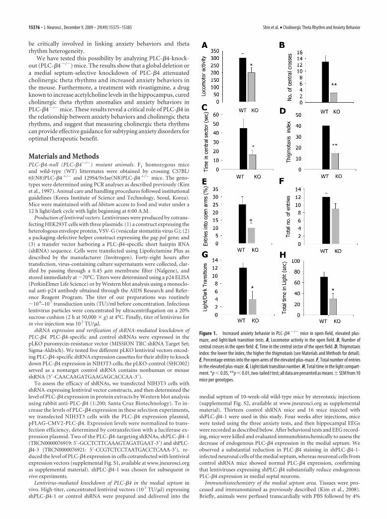
Figure 1. Increased anxiety behavior in PLC-�4 �/� mice in open-field, elevated plus-maze, and light/dark transition tests. A, Locomotor activity in the open field. B, Number ofcentral crosses in the open field. C, Time in the central sector of the open field. D, Thigmotaxisindex: the lower the index, the higher the thigmotaxis (see Materials and Methods for detail).E, Percentage entries into the open arms of the elevated plus-maze. F, Total number of entriesin the elevated plus-maze. G, Light/dark transition number. H, Total time in the light compart-ment. *p � 0.05, **p � 0.01, two-tailed t test; all data are presented as means� SEM from 10mice per genotypes.
15376 • J. Neurosci., December 9, 2009 • 29(49):15375–15385 Shin et al. • Cholinergic Theta Rhythm and Anxiety Behavior
paraformaldehyde in 0.1 M phosphate buffer (PB), pH 7.4, under ure-thane anesthesia (1.5 g/kg, i.p.). The brains were removed and postfixedin the same fixative for 4 h. Brain tissues were cryoprotected by infiltra-tion with 30% sucrose overnight. Thereafter, the entire medial septal areawas frozen and sectioned with a cryostat into 30 �m sections and con-secutive sections were placed in six-well plates containing PBS. Everysixth section in the series throughout the entire medial septal area fromselected animals was used for the immunofluorescence study. The pres-ence and absence of PLC-�4 expression in wild-type and PLC-�4 �/�
mice, respectively, and morphological changes induced by shPLC-�4 inPLC-�4-positive neurons of the medial septal area were evaluated bydouble immunofluorescence staining for both mouse anti-neuronal nu-clei (NeuN) IgG (1:100; Millipore Bioscience Research Reagents) andrabbit anti-PLC-�4 IgG (1:100; Millipore Bioscience Research Reagents).Brain tissues were incubated in the mixture of antisera overnight at roomtemperature. After washing three times with PBS (10 min each), sectionswere incubated in a mixture containing both Cy2-conjugated goat anti-mouse IgG (1:200; GE Healthcare) and Cy3-conjugated goat anti-rabbitIgG (1:200; GE Healthcare) for 1 h at room temperature. Sections weremounted in Vectashield mounting media with or without DAPI (4�,6�-diamidino-2-phenylindole) (Vector). Images were captured and ana-lyzed using an Olympus DP50 digital camera and Viewfinder Life,version 1.0, software, or an Olympus FluoView FV1000 Confocal Mi-croscope System. Figures were prepared using Adobe Photoshop 7.0.Manipulation of images was restricted to threshold and brightness ad-justments applied to the entire image.
For quantification of PLC-�4 immunofluorescence in RNA interfer-ence (RNAi)-mediated PLC-�4 knockdown mice, we have performedthe cell count in the medial septal area according to the previous descrip-tion (Kim et al., 2008, 2009a,b). Briefly, PLC-�4 immunofluorescentimages (10 sections/mice) were captured in the same region (500 � 500�m). Images were sampled from at least five different points within eachmedial septum section. Thereafter, the number of PLC-�4-positive cellswas actually counted within the sampled images. All immunoreactivecells were counted regardless the intensity of labeling. Cell counts wereperformed by two different investigators who were blind to the classifi-cation of tissues. The average percentage of PLC-�4 positive cells ( C) inthe medial septum of knock-down mice is estimated and shown as fol-lows: C � 100 � (�Qi/Ni)/K, where Qi is the actually counted numbers of
PLC-�4-positive cells in the PLC-�4 viralvector-injected medial septum, Ni is the num-ber of PLC-�4-positive cells in the controlshRNA viral vector-injected animal groups,and K is the number of sampling.
All data obtained from the quantitativemeasurements were analyzed using one-wayANOVA to determine statistical significance.Bonferroni’s test was used for post hoc compar-isons. A value of p � 0.05 was considered sta-tistically significant (Kim et al., 2008, 2009a,b).
Triple immunohistochemistry of the medialseptal neurons. The specificity of PLC-�4 andglutamic acid decarboxylase (GAD) antibodieshas been shown previously (Yamada et al.,2001; Nakamura et al., 2004). In the presentstudy, we produced goat antibodies to PV andmGluR1a using the same fusion proteins ashad been used for production of rabbit andguinea pig antibodies (Tanaka et al., 2000;Nakamura et al., 2004), and also to rat cholinetransporter 1 using C-terminal 50 aa sequencefused to glutathione S-transferase (Narushimaet al., 2007). The procedures for fusion pro-teins, immunization, and affinity purificationhave been reported previously (Nakamura etal., 2004). The specificity of all these antibodieswas checked by immunoblot detection of sin-gle protein bands and by selective immunohis-tochemical labeling of particular neuronalpopulations.
Animals were anesthetized by intraperitoneal injection with pentobar-bital (100 mg/kg of body weight), fixed transcardially with 4% parafor-maldehyde in 0.1 M sodium PB, pH 7.2, and subjected to preparation ofcoronal microslicer sections (50 �m in thickness; VT1000S, Leica). Be-fore blocking with 10% normal donkey serum, sections were dippedsuccessively in 30, 60, and 100% methanol or ethanol for 5 min each.Then they were incubated overnight in a mixture of guinea pigPLC-�4 antibody, rabbit antibody (GAD or mGluR1a), and goat an-tibody [mGluR1, PV, or choline transporter 1 (CHT1)], each beingdiluted to 1 �g/ml with PBS/0.1% Triton X-100 (TPBS). After wash-ing in TPBS, sections were incubated for 2 h with a mixture of species-specific secondary antibodies (1:200 dilution) linked to Alexa 488(Invitrogen), indocarbocyanine (Cy3), or indodicarbocyanine (Cy5)(Jackson ImmunoResearch). Images of single optical sections weretaken with a confocal laser-scanning microscope (FV1000; Olympus).All images were obtained by restricting filter windows enough toeliminate each other and by adopting the sequential mode of laserscanning to minimize the bleedthrough.
Rescue experiments using rivastigmine. Rivastigmine (kindly providedby Novartis Pharma) was dissolved in saline. Animals were injectedintraperitoneally with 0.5 mg/kg rivastigmine (total volume, 5 ml/kg)60 min before the start of anxiety behavioral tests. This optimal doseof rivastigmine was chosen based on previously published microdi-alysis studies (Van Dam et al., 2005; Cerbai et al., 2007). Both PLC-�4 �/� (WT) and PLC-�4 �/� (KO) mice were randomly assigned toone of the following three treatment groups: (1) WT-sham (wild-typemice with intraperitoneal injection of saline; n � 10); (2) KO-sham(PLC-�4 �/� mice with intraperitoneal injection of saline; n � 10);and (3) KO-rivastigmine (PLC-�4 �/� mice with intraperitoneal in-jection of rivastigmine; n � 10).
Local field potential recordings in vivo. Hippocampal electroencepha-logram (EEG) signals were recorded in mice (10 –14 weeks of age) usingpublished protocols (Shin and Talnov, 2001; Shin et al., 2005), with somemodifications. Briefly, for electrode implantation, the animals were anes-thetized with pentobarbital (50 ml/kg, i.p.) and held in a stereotaxicapparatus with bregma and lambda in the same horizontal plane. Hip-pocampal EEG recordings were performed using Teflon-coated tungstenelectrodes (150 �m) implanted in the hippocampal fissure (from
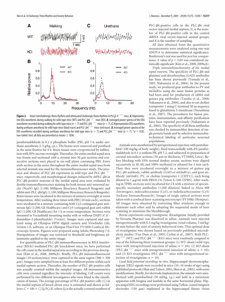
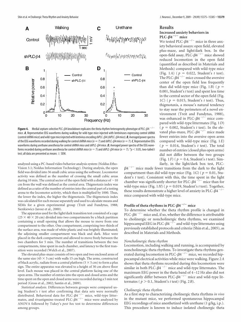
Figure 2. Intact noncholinergic theta rhythms and attenuated cholinergic theta rhythms in PLC�-4 �/� mice. A, Representa-tive EEG waveforms during walking for wild-type mice (WT) and PLC-�4 �/� mice (KO). B, Averaged power spectra of the EEGwaveforms recorded during walking for wild-type mice (n � 7) and PLC-�4 �/� mice (n � 7). C, Representative EEG waveformsduring urethane anesthesia for wild-type mice (black trace) and PLC-�4 �/� mice (red trace). D, Averaged power spectra of theEEG waveforms recorded during urethane anesthesia for wild-type mice (n � 7) and PLC-�4 �/� mice (n � 7). *p � 0.05,two-tailed t test; all data are presented as means � SEM.
Shin et al. • Cholinergic Theta Rhythm and Anxiety Behavior J. Neurosci., December 9, 2009 • 29(49):15375–15385 • 15377
bregma, 2.0 mm anteroposterior, 1.2 mm me-diolateral, and 1.8 mm dorsoventral) withgrounding over the cerebellum. The positionof the electrodes was verified by light micros-copy in Nissl-stained sections according topublished protocols (Shin and Talnov, 2001;Shin et al., 2005). Field potential was amplified(1000�), bandpass-filtered (0.1–100 Hz), dig-itized with 16-bit resolution continuously at 1kHz sampling, and recorded on a personalcomputer using Clampex 10 (MDS AnalyticalTechnologies).
Analysis of hippocampal electrical activitydata. EEG data were analyzed off-line accord-ing to published protocols (Shin and Talnov,2001; Shin et al., 2005) using Clampfit 10(MDS Analytical Technologies). Briefly, rawEEG data were collected in 4 s segments andfast-Fourier transformed off-line. The datawere continuously monitored for movementartifacts, and the recording of each segmentwas manually verified by the experimenter,blinded to the genotype of the animals. All seg-ments collected during mouse movements andthose in which the amplitude of amplified EEGsignals exceed a maximum of �1.25 V werediscarded to remove artifacts. To compare theEEG spectral characteristics of hippocampalelectrical activities, we calculated EEG spectralpower in 1 Hz bins using fast Fourier transfor-mations (Hamming window) of each 4 s epoch.These analyses were performed on recordingsfrom these intervals in 100 trials from eachgroup. Powers in the 0 –30 Hz range were aver-aged in groups across each behavioral state,and the mean values were plotted in 1 Hz bins.The averaging process for power spectrumsused a normalization procedure that involveddividing by the combined SD of EEG raw datafor the two comparison states (e.g., PLC-�4 �/� mice and wild-type littermates) accord-ing to the previously published protocol (Shinet al., 2005). We used the peak power underdifferent conditions for comparison purposesbecause the in vivo theta rhythm has a clear andsharp peak frequency that distinguished itfrom other in vivo EEG rhythms, such asdelta- and gamma-band activities (Shin et al., 2005), and peak powerprovides more accurate information than total power in the thetafrequency range.
Classification of cholinergic theta and noncholinergic theta rhythms as-sociated with different behaviors. Cholinergic and noncholinergic thetarhythms associated with different behaviors were recorded according topublished protocols (Shin and Talnov, 2001; Shin et al., 2005). Briefly,noncholinergic theta rhythms can be distinguished from cholinergictheta rhythms using muscarinic antagonists (e.g., atropine or scopol-amine), which can abolish cholinergic theta rhythms when injected intothe animal, but leave noncholinergic theta rhythms relatively unaffected(Bland, 1986). In addition, the use of different behaviors to induce thetarhythms can discriminate between noncholinergic theta rhythms andcholinergic theta rhythms. For example, cholinergic theta rhythms aregenerated normally during urethane anesthesia, alert immobility, andpassive whole-body rotation (Bland, 1986; Shin et al., 2005; Shin, 2009).In contrast, noncholinergic theta rhythms are observed during locomo-tion activities, such as walking or running (Bland, 1986; Shin and Talnov,2001). In addition, the two types of theta rhythms can usually be sepa-rated approximately (although not exclusively) into high- and low-frequency bands: 4 –7 Hz for cholinergic type 2 theta rhythm, and 7–12Hz for noncholinergic type 1 theta rhythm. However, several investiga-
tors have found that atropine-sensitive type 2 theta rhythms can have afrequency that is as high as the frequency limit of atropine-resistant type1 theta rhythms under certain recording conditions (Bland and Oddie,2002; Shin, 2009). Therefore, we did not use the high- and low-frequencytheta bands to discriminate between cholinergic theta rhythm and non-cholinergic theta rhythm.
In the present work, we have used urethane anesthesia, alert immobil-ity and passive whole-body rotation to record cholinergic theta rhythmsfrom wild-type and PLC-�4�/� mice, and from mice with medial septum-directed, shRNA-mediated PLC-�4 knockdown. Noncholinergictheta rhythms were recorded from mice walking in an open chamberor running on a wheel; the same mice were analyzed in both settings.
Anxiety behavioral tests. Three anxiety behavioral assays— open-fieldthigmotaxis, elevated plus-maze, and light/dark transition (see below)—were performed between 9:00 A.M. and 5:00 P.M. using adult (8- to16-week-old) mice.
Thigmotaxis represents a mouse’s natural tendency to stay near theperimeters of a novel environment (Treit and Fundytus, 1988). Open-field thigmotaxis was assessed by placing mice in the center of an open-field apparatus (40 � 40 � 40 cm) under dim lighting. During a 10 minobservation period, locomotor activity, number of central crosses, andtime spent in the central sector of the open field were recorded and
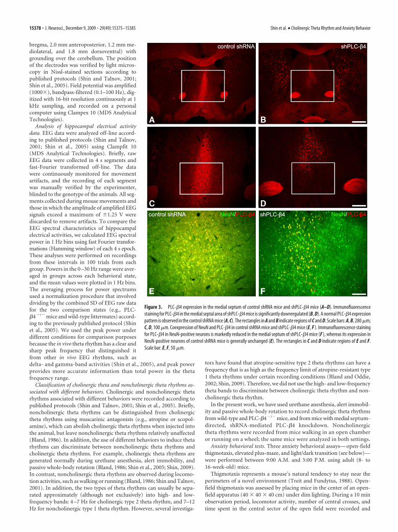
Figure 3. PLC-�4 expression in the medial septum of control shRNA mice and shPLC-�4 mice (A–D). Immunofluorescencestaining for PLC-�4 in the medial septal area of shPLC-�4 mice is significantly downregulated (B, D). A normal PLC-�4 expressionpattern is observed in the control shRNA mice (A, C). The rectangles in A and B indicate regions of C and D. Scale bars: A, B, 280 �m;C, D, 100 �m. Coexpression of NeuN and PLC-�4 in control shRNA mice and shPLC-�4 mice (E, F ). Immunofluorescence stainingfor PLC-�4 in NeuN-positive neurons is markedly reduced in the medial septum of shPLC-�4 mice (F ), whereas its expression inNeuN-positive neurons of control shRNA mice is generally unchanged (E). The rectangles in C and D indicate regions of E and F.Scale bar: E, F, 50 �m.
15378 • J. Neurosci., December 9, 2009 • 29(49):15375–15385 Shin et al. • Cholinergic Theta Rhythm and Anxiety Behavior
analyzed using a PC-based video behavior analysis system (Noldus Etho-Vision 3.1; Noldus Information Technology). During analysis, the openfield was divided into 36 small cubic areas using the software. Locomotoractivity was defined as the number of crossing the small cubic areasduring 10 min. The central sector of the open field with a distance of �10cm from the wall was defined as the central area. Thigmotaxis index wasdefined as a ratio of the number of entries into the central part of a testingarena to the locomotor activity, which then is multiplied by 1000. Thus,the lower the index, the higher the thigmotaxis. The thigmotaxis indexwas calculated for each mouse separately and used to calculate means andSEMs for a given experimental group (Treit and Fundytus, 1988;Sienkiewicz-Jarosz et al., 2000).
The apparatus used for the light/dark transition test consisted of a cage(25 � 40 � 20 cm) divided into two compartments by a black partitioncontaining a small opening that allows the mouse to move from onecompartment to the other. One compartment, comprising two-thirds ofthe surface area, was made of white plastic and was brightly illuminated;the adjoining smaller compartment was black and dark. Mice wereplaced in the dark compartment and allowed to move freely between thetwo chambers for 5 min. The number of transitions between the twocompartments, time spent in each chamber, and latency to the first tran-sition were recorded (Welch et al., 2007).
The elevated plus-maze consists of two open and two enclosed arms ofthe same size (45 � 5 cm) with walls 15 cm high. The arms, constructedof black acrylic, radiate from a central platform (5 � 5 cm) to form a plussign. The entire apparatus was elevated to a height of 30 cm above floorlevel. Each mouse was placed in the central platform facing one of theopen arms. The number of entries into the open and closed arms and thetime spent on the open and closed arms were recorded during a 5 min testperiod (Gross et al., 2002; Santin et al., 2009).
Statistical analysis. Differences between groups were compared us-ing Student’s t test after confirming that data sets were normallydistributed. Behavioral data for PLC-�4 �/� mice, wild-type litter-mates, and rivastigmine-treated PLC-�4 �/� mice were analyzed byANOVA followed by Tukey’s post hoc test to determine differencesamong groups.
ResultsIncreased anxiety behaviors inPLC-�4 �/� miceWe tested PLC-�4�/� mice in three anx-iety behavioral assays: open field, elevatedplus-maze, and light/dark box. In theopen-field assay, PLC-�4�/� mice showedreduced locomotion in the open field(quantified as described in Materials andMethods) compared with wild-type mice(Fig. 1A) ( p � 0.022, Student’s t test).The PLC-�4�/� mice crossed the aversivecenter of the open field less frequentlythan did wild-type mice (Fig. 1B) ( p �0.001, Student’s t test) and spent less timein the central sector of the open field (Fig.1C) ( p � 0.015, Student’s t test). Thus,thigmotaxis, a mouse’s natural tendencyto stay near the perimeters of a novel en-vironment (Treit and Fundytus, 1988),was enhanced in PLC-�4�/� mice com-pared with wild-type littermates (Fig. 1D)( p � 0.002, Student’s t test). In the ele-vated plus-maze, PLC-�4�/� mice madefewer entries into the aversive open armscompared with wild-type mice (Fig. 1E)( p � 0.014, Student’s t test). The totalnumber of entries (closed plus open arms)did not differ between the two groups(Fig. 1F) ( p � 0.4, Student’s t test). Sim-ilarly, in the light/dark box test, PLC-
�4�/� mice made fewer transitions from the dark to the lightcompartment than did wild-type mice (Fig. 1G) ( p � 0.01, Stu-dent’s t test). Consistent with this, the time spent in the lightchamber was significantly shorter for PLC-�4�/� mice than forwild-type mice (Fig. 1H) ( p � 0.019, Student’s t test). Together,these results demonstrate a higher level of anxiety in PLC-�4�/�
mice compared with wild-type mice.
Profile of theta rhythms in PLC-�4 �/� miceTo determine whether the theta rhythm profile is changed inPLC-�4�/� mice and, if so, whether the difference is attributableto cholinergic or noncholinergic theta rhythms, we examinedhippocampal EEG in PLC-�4�/� and wild-type littermates usingpreviously established protocols and criteria (Shin et al., 2005), asdescribed in Materials and Methods.
Noncholinergic theta rhythmLocomotion, including walking and running, is accompanied bynoncholinergic theta rhythms. To investigate theta rhythms gen-erated during locomotion in PLC-�4�/� mice, we recorded hip-pocampal electrical activities while mice were walking. Figure 2Ashows that theta rhythms recorded during this locomotion weresimilar in both PLC-�4�/� mice and wild-type littermates. Themaximum EEG power in the theta band of 4 –12 Hz also did notsignificantly differ between PLC-�4�/� mice and wild-type lit-termates ( p 0.1, Student’s t test) (Fig. 2B).
Cholinergic theta rhythmAs a first step to characterizing cholinergic theta rhythms in vivoin the mutant mice, we performed spontaneous hippocampalEEG recordings of mice anesthetized with urethane (1 g/kg, i.p.).This procedure is known to induce isolated cholinergic theta
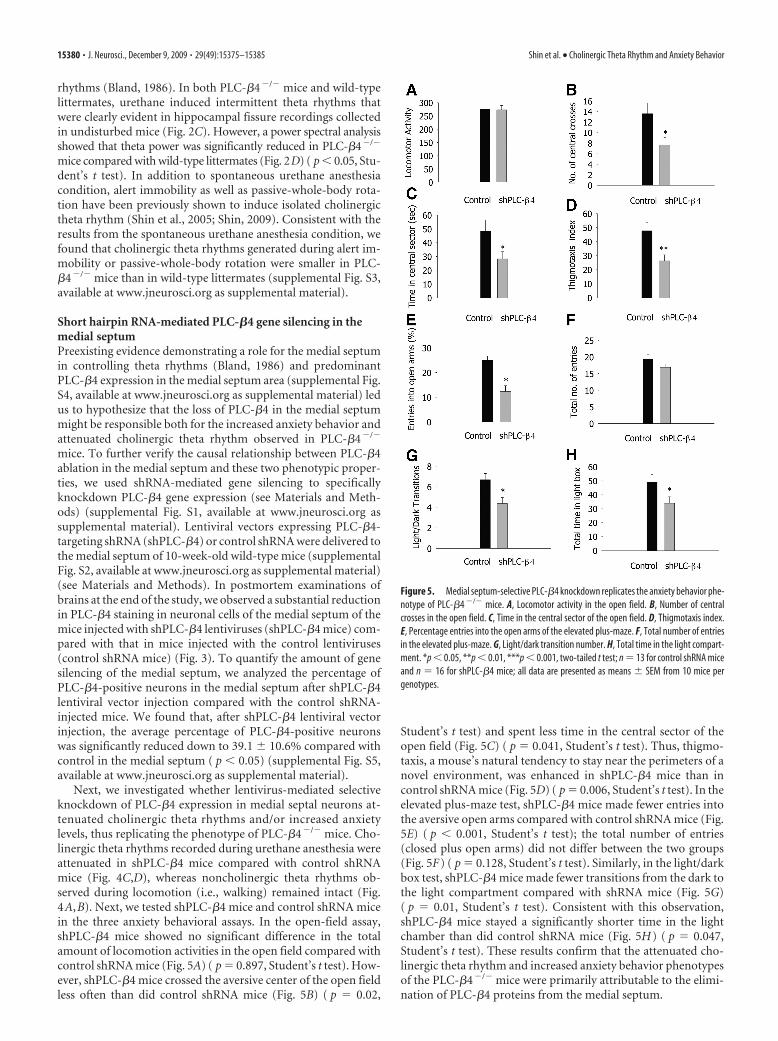
Figure 4. Medial septum-selective PLC-�4 knockdown replicates the theta rhythm heterogeneity phenotype of PLC-�4 �/�
mice. A, Representative EEG waveforms during walking for wild-type mice injected with lentiviruses expressing control shRNA(control shRNA mice) and wild-type mice injected with lentivirus encoding shPLC-�4 (shPLC-�4 mice). B, Averaged power spectraof the EEG waveforms recorded during walking for control shRNA mice (n � 7) and shPLC-�4 mice (n � 7). C, Representative EEGwaveforms during urethane anesthesia for control shRNA mice and shPLC-�4 mice. D, Averaged power spectra of the EEG wave-forms recorded during urethane anesthesia for control shRNA mice (n � 7) and shPLC-�4 mice (n � 7). *p � 0.05, two-tailed ttest; all data are presented as means � SEM.
Shin et al. • Cholinergic Theta Rhythm and Anxiety Behavior J. Neurosci., December 9, 2009 • 29(49):15375–15385 • 15379
rhythms (Bland, 1986). In both PLC-�4�/� mice and wild-typelittermates, urethane induced intermittent theta rhythms thatwere clearly evident in hippocampal fissure recordings collectedin undisturbed mice (Fig. 2C). However, a power spectral analysisshowed that theta power was significantly reduced in PLC-�4�/�
mice compared with wild-type littermates (Fig. 2D) ( p � 0.05, Stu-dent’s t test). In addition to spontaneous urethane anesthesiacondition, alert immobility as well as passive-whole-body rota-tion have been previously shown to induce isolated cholinergictheta rhythm (Shin et al., 2005; Shin, 2009). Consistent with theresults from the spontaneous urethane anesthesia condition, wefound that cholinergic theta rhythms generated during alert im-mobility or passive-whole-body rotation were smaller in PLC-�4�/� mice than in wild-type littermates (supplemental Fig. S3,available at www.jneurosci.org as supplemental material).
Short hairpin RNA-mediated PLC-�4 gene silencing in themedial septumPreexisting evidence demonstrating a role for the medial septumin controlling theta rhythms (Bland, 1986) and predominantPLC-�4 expression in the medial septum area (supplemental Fig.S4, available at www.jneurosci.org as supplemental material) ledus to hypothesize that the loss of PLC-�4 in the medial septummight be responsible both for the increased anxiety behavior andattenuated cholinergic theta rhythm observed in PLC-�4�/�
mice. To further verify the causal relationship between PLC-�4ablation in the medial septum and these two phenotypic proper-ties, we used shRNA-mediated gene silencing to specificallyknockdown PLC-�4 gene expression (see Materials and Meth-ods) (supplemental Fig. S1, available at www.jneurosci.org assupplemental material). Lentiviral vectors expressing PLC-�4-targeting shRNA (shPLC-�4) or control shRNA were delivered tothe medial septum of 10-week-old wild-type mice (supplementalFig. S2, available at www.jneurosci.org as supplemental material)(see Materials and Methods). In postmortem examinations ofbrains at the end of the study, we observed a substantial reductionin PLC-�4 staining in neuronal cells of the medial septum of themice injected with shPLC-�4 lentiviruses (shPLC-�4 mice) com-pared with that in mice injected with the control lentiviruses(control shRNA mice) (Fig. 3). To quantify the amount of genesilencing of the medial septum, we analyzed the percentage ofPLC-�4-positive neurons in the medial septum after shPLC-�4lentiviral vector injection compared with the control shRNA-injected mice. We found that, after shPLC-�4 lentiviral vectorinjection, the average percentage of PLC-�4-positive neuronswas significantly reduced down to 39.1 � 10.6% compared withcontrol in the medial septum ( p � 0.05) (supplemental Fig. S5,available at www.jneurosci.org as supplemental material).
Next, we investigated whether lentivirus-mediated selectiveknockdown of PLC-�4 expression in medial septal neurons at-tenuated cholinergic theta rhythms and/or increased anxietylevels, thus replicating the phenotype of PLC-�4�/� mice. Cho-linergic theta rhythms recorded during urethane anesthesia wereattenuated in shPLC-�4 mice compared with control shRNAmice (Fig. 4C,D), whereas noncholinergic theta rhythms ob-served during locomotion (i.e., walking) remained intact (Fig.4A,B). Next, we tested shPLC-�4 mice and control shRNA micein the three anxiety behavioral assays. In the open-field assay,shPLC-�4 mice showed no significant difference in the totalamount of locomotion activities in the open field compared withcontrol shRNA mice (Fig. 5A) ( p � 0.897, Student’s t test). How-ever, shPLC-�4 mice crossed the aversive center of the open fieldless often than did control shRNA mice (Fig. 5B) ( p � 0.02,
Student’s t test) and spent less time in the central sector of theopen field (Fig. 5C) ( p � 0.041, Student’s t test). Thus, thigmo-taxis, a mouse’s natural tendency to stay near the perimeters of anovel environment, was enhanced in shPLC-�4 mice than incontrol shRNA mice (Fig. 5D) ( p � 0.006, Student’s t test). In theelevated plus-maze test, shPLC-�4 mice made fewer entries intothe aversive open arms compared with control shRNA mice (Fig.5E) ( p � 0.001, Student’s t test); the total number of entries(closed plus open arms) did not differ between the two groups(Fig. 5F) ( p � 0.128, Student’s t test). Similarly, in the light/darkbox test, shPLC-�4 mice made fewer transitions from the dark tothe light compartment compared with shRNA mice (Fig. 5G)( p � 0.01, Student’s t test). Consistent with this observation,shPLC-�4 mice stayed a significantly shorter time in the lightchamber than did control shRNA mice (Fig. 5H) ( p � 0.047,Student’s t test). These results confirm that the attenuated cho-linergic theta rhythm and increased anxiety behavior phenotypesof the PLC-�4�/� mice were primarily attributable to the elimi-nation of PLC-�4 proteins from the medial septum.
Figure 5. Medial septum-selective PLC-�4 knockdown replicates the anxiety behavior phe-notype of PLC-�4 �/� mice. A, Locomotor activity in the open field. B, Number of centralcrosses in the open field. C, Time in the central sector of the open field. D, Thigmotaxis index.E, Percentage entries into the open arms of the elevated plus-maze. F, Total number of entriesin the elevated plus-maze. G, Light/dark transition number. H, Total time in the light compart-ment. *p � 0.05, **p � 0.01, ***p � 0.001, two-tailed t test; n � 13 for control shRNA miceand n � 16 for shPLC-�4 mice; all data are presented as means � SEM from 10 mice pergenotypes.
15380 • J. Neurosci., December 9, 2009 • 29(49):15375–15385 Shin et al. • Cholinergic Theta Rhythm and Anxiety Behavior
Rescue of the PLC-�4 �/� cholinergic theta rhythm andanxiety phenotypes by rivastigmineIn both PLC-�4�/� mice and shPLC-�4 mice, we found that theattenuated cholinergic theta rhythms are associated with increasedanxiety phenotype. To further confirm the interrelationship be-tween cholinergic theta rhythms and anxiety behavior, we designedan experiment to determine whether increasing cholinergic trans-mission in PLC-�4�/� mice could rescue the attenuated amplitudeof cholinergic theta rhythms, and thereby normalize the attendantanxiety behavior. To this end, we administered the cholinergic-enhancing drug, rivastigmine, an acetylcholinesterase inhibitor knownto increase the cholinergic transmission in the septohippocampal path-way (Wu et al., 2003b; Van Dam et al., 2005; Cerbai et al., 2007).
Restoration of normal cholinergic theta rhythms in PLC-�4�/�
mice by rivastigmineTo investigate whether rivastigmine treatment could rescue at-tenuated cholinergic theta rhythms in PLC-�4�/� mice, we re-corded EEGs from three groups of mice: (1) PLC-�4�/� miceinjected with rivastigmine, (2) PLC-�4�/� mice injected withsaline, and (3) wild-type littermates injected with saline, afterplacing them in a new environment. Human theta rhythm asso-ciated with anxiety has traditionally been recorded in patientsperforming mental activities without movement in a novel envi-ronment (Mizuki et al., 1989; Suetsugi et al., 2000). To recapitu-late this behavioral condition as closely as possible, we chose thenovel-environment model, which induces alert-immobility statesand evokes anxiety, among several conditions known to generatecholinergic theta rhythm in mice (see Materials and Methods).
Both noncholinergic and cholinergic theta rhythms appearedwhen mice were placed in a new environment. The amplitude of
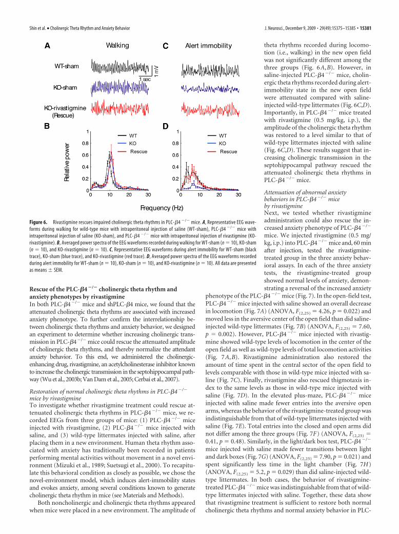
theta rhythms recorded during locomo-tion (i.e., walking) in the new open fieldwas not significantly different among thethree groups (Fig. 6A,B). However, insaline-injected PLC-�4�/� mice, cholin-ergic theta rhythms recorded during alert-immobility state in the new open fieldwere attenuated compared with saline-injected wild-type littermates (Fig. 6C,D).Importantly, in PLC-�4�/� mice treatedwith rivastigmine (0.5 mg/kg, i.p.), theamplitude of the cholinergic theta rhythmwas restored to a level similar to that ofwild-type littermates injected with saline(Fig. 6C,D). These results suggest that in-creasing cholinergic transmission in theseptohippocampal pathway rescued theattenuated cholinergic theta rhythms inPLC-�4�/� mice.
Attenuation of abnormal anxietybehaviors in PLC-�4�/� miceby rivastigmineNext, we tested whether rivastigmineadministration could also rescue the in-creased anxiety phenotype of PLC-�4�/�
mice. We injected rivastigmine (0.5 mg/kg, i.p.) into PLC-�4�/� mice and, 60 minafter injection, tested the rivastigmine-treated group in the three anxiety behav-ioral assays. In each of the three anxietytests, the rivastigmine-treated groupshowed normal levels of anxiety, demon-strating a reversal of the increased anxiety
phenotype of the PLC-�4�/� mice (Fig. 7). In the open-field test,PLC-�4�/� mice injected with saline showed an overall decreasein locomotion (Fig. 7A) (ANOVA, F(2,25) � 4.26, p � 0.022) andmoved less in the aversive center of the open field than did saline-injected wild-type littermates (Fig. 7B) (ANOVA, F(2,25) � 7.60,p � 0.002). However, PLC-�4�/� mice injected with rivastig-mine showed wild-type levels of locomotion in the center of theopen field as well as wild-type levels of total locomotion activities(Fig. 7A,B). Rivastigmine administration also restored theamount of time spent in the central sector of the open field tolevels comparable with those in wild-type mice injected with sa-line (Fig. 7C). Finally, rivastigmine also rescued thigmotaxis in-dex to the same levels as those in wild-type mice injected withsaline (Fig. 7D). In the elevated plus-maze, PLC-�4�/� miceinjected with saline made fewer entries into the aversive openarms, whereas the behavior of the rivastigmine-treated group wasindistinguishable from that of wild-type littermates injected withsaline (Fig. 7E). Total entries into the closed and open arms didnot differ among the three groups (Fig. 7F) (ANOVA, F(2,25) �0.41, p � 0.48). Similarly, in the light/dark box test, PLC-�4�/�
mice injected with saline made fewer transitions between lightand dark boxes (Fig. 7G) (ANOVA, F(2,25) � 7.90, p � 0.021) andspent significantly less time in the light chamber (Fig. 7H)(ANOVA, F(2,25) � 5.2, p � 0.029) than did saline-injected wild-type littermates. In both cases, the behavior of rivastigmine-treated PLC-�4�/� mice was indistinguishable from that of wild-type littermates injected with saline. Together, these data showthat rivastigmine treatment is sufficient to restore both normalcholinergic theta rhythms and normal anxiety behavior in PLC-
Figure 6. Rivastigmine rescues impaired cholinergic theta rhythms in PLC-�4 �/� mice. A, Representative EEG wave-forms during walking for wild-type mice with intraperitoneal injection of saline (WT-sham), PLC-�4 �/� mice withintraperitoneal injection of saline (KO-sham), and PLC-�4 �/� mice with intraperitoneal injection of rivastigmine (KO-rivastigmine). B, Averaged power spectra of the EEG waveforms recorded during walking for WT-sham (n � 10), KO-sham(n � 10), and KO-rivastigmine (n � 10). C, Representative EEG waveforms during alert immobility for WT-sham (blacktrace), KO-sham (blue trace), and KO-rivastigmine (red trace). D, Averaged power spectra of the EEG waveforms recordedduring alert immobility for WT-sham (n � 10), KO-sham (n � 10), and KO-rivastigmine (n � 10). All data are presentedas means � SEM.
Shin et al. • Cholinergic Theta Rhythm and Anxiety Behavior J. Neurosci., December 9, 2009 • 29(49):15375–15385 • 15381
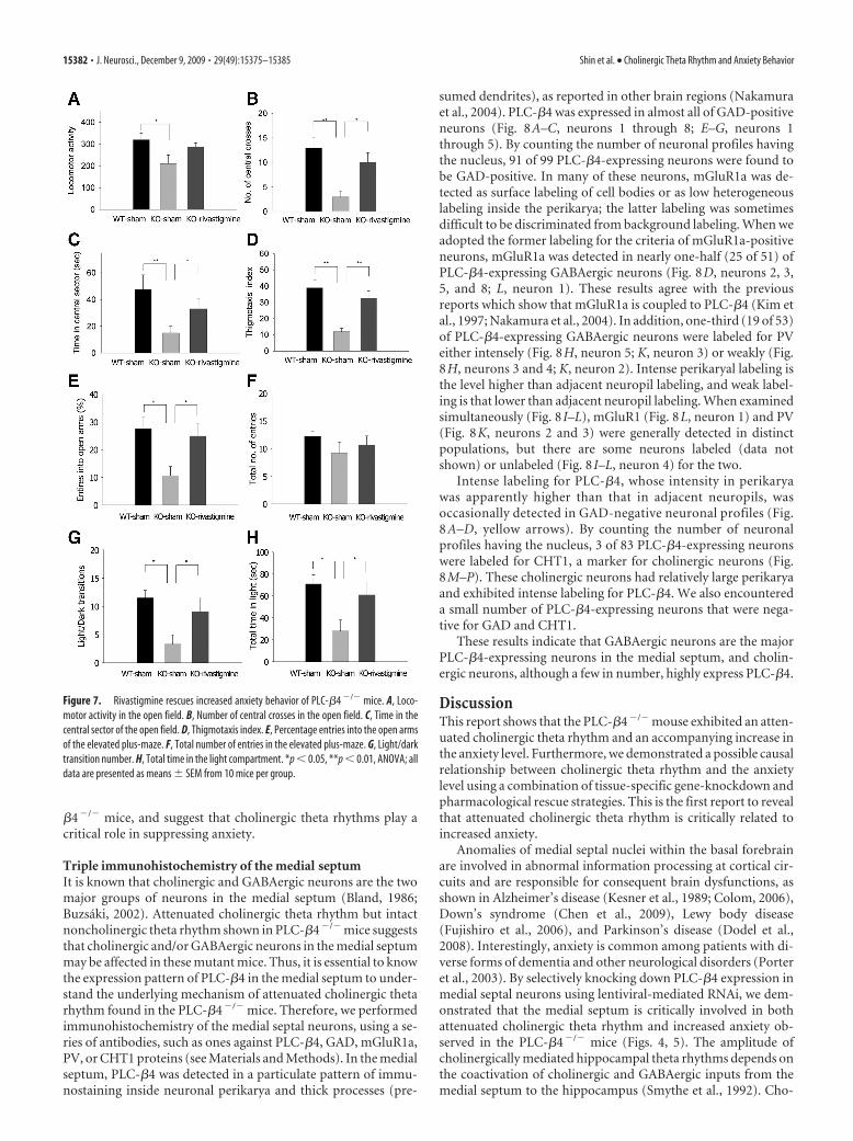
�4�/� mice, and suggest that cholinergic theta rhythms play acritical role in suppressing anxiety.
Triple immunohistochemistry of the medial septumIt is known that cholinergic and GABAergic neurons are the twomajor groups of neurons in the medial septum (Bland, 1986;Buzsáki, 2002). Attenuated cholinergic theta rhythm but intactnoncholinergic theta rhythm shown in PLC-�4�/� mice suggeststhat cholinergic and/or GABAergic neurons in the medial septummay be affected in these mutant mice. Thus, it is essential to knowthe expression pattern of PLC-�4 in the medial septum to under-stand the underlying mechanism of attenuated cholinergic thetarhythm found in the PLC-�4�/� mice. Therefore, we performedimmunohistochemistry of the medial septal neurons, using a se-ries of antibodies, such as ones against PLC-�4, GAD, mGluR1a,PV, or CHT1 proteins (see Materials and Methods). In the medialseptum, PLC-�4 was detected in a particulate pattern of immu-nostaining inside neuronal perikarya and thick processes (pre-
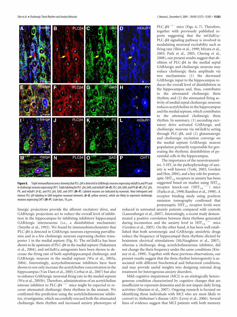
sumed dendrites), as reported in other brain regions (Nakamuraet al., 2004). PLC-�4 was expressed in almost all of GAD-positiveneurons (Fig. 8A–C, neurons 1 through 8; E–G, neurons 1through 5). By counting the number of neuronal profiles havingthe nucleus, 91 of 99 PLC-�4-expressing neurons were found tobe GAD-positive. In many of these neurons, mGluR1a was de-tected as surface labeling of cell bodies or as low heterogeneouslabeling inside the perikarya; the latter labeling was sometimesdifficult to be discriminated from background labeling. When weadopted the former labeling for the criteria of mGluR1a-positiveneurons, mGluR1a was detected in nearly one-half (25 of 51) ofPLC-�4-expressing GABAergic neurons (Fig. 8D, neurons 2, 3,5, and 8; L, neuron 1). These results agree with the previousreports which show that mGluR1a is coupled to PLC-�4 (Kim etal., 1997; Nakamura et al., 2004). In addition, one-third (19 of 53)of PLC-�4-expressing GABAergic neurons were labeled for PVeither intensely (Fig. 8H, neuron 5; K, neuron 3) or weakly (Fig.8H, neurons 3 and 4; K, neuron 2). Intense perikaryal labeling isthe level higher than adjacent neuropil labeling, and weak label-ing is that lower than adjacent neuropil labeling. When examinedsimultaneously (Fig. 8 I–L), mGluR1 (Fig. 8L, neuron 1) and PV(Fig. 8K, neurons 2 and 3) were generally detected in distinctpopulations, but there are some neurons labeled (data notshown) or unlabeled (Fig. 8 I–L, neuron 4) for the two.
Intense labeling for PLC-�4, whose intensity in perikaryawas apparently higher than that in adjacent neuropils, wasoccasionally detected in GAD-negative neuronal profiles (Fig.8 A–D, yellow arrows). By counting the number of neuronalprofiles having the nucleus, 3 of 83 PLC-�4-expressing neuronswere labeled for CHT1, a marker for cholinergic neurons (Fig.8M–P). These cholinergic neurons had relatively large perikaryaand exhibited intense labeling for PLC-�4. We also encountereda small number of PLC-�4-expressing neurons that were nega-tive for GAD and CHT1.
These results indicate that GABAergic neurons are the majorPLC-�4-expressing neurons in the medial septum, and cholin-ergic neurons, although a few in number, highly express PLC-�4.
DiscussionThis report shows that the PLC-�4�/� mouse exhibited an atten-uated cholinergic theta rhythm and an accompanying increase inthe anxiety level. Furthermore, we demonstrated a possible causalrelationship between cholinergic theta rhythm and the anxietylevel using a combination of tissue-specific gene-knockdown andpharmacological rescue strategies. This is the first report to revealthat attenuated cholinergic theta rhythm is critically related toincreased anxiety.
Anomalies of medial septal nuclei within the basal forebrainare involved in abnormal information processing at cortical cir-cuits and are responsible for consequent brain dysfunctions, asshown in Alzheimer’s disease (Kesner et al., 1989; Colom, 2006),Down’s syndrome (Chen et al., 2009), Lewy body disease(Fujishiro et al., 2006), and Parkinson’s disease (Dodel et al.,2008). Interestingly, anxiety is common among patients with di-verse forms of dementia and other neurological disorders (Porteret al., 2003). By selectively knocking down PLC-�4 expression inmedial septal neurons using lentiviral-mediated RNAi, we dem-onstrated that the medial septum is critically involved in bothattenuated cholinergic theta rhythm and increased anxiety ob-served in the PLC-�4�/� mice (Figs. 4, 5). The amplitude ofcholinergically mediated hippocampal theta rhythms depends onthe coactivation of cholinergic and GABAergic inputs from themedial septum to the hippocampus (Smythe et al., 1992). Cho-
Figure 7. Rivastigmine rescues increased anxiety behavior of PLC-�4 �/� mice. A, Loco-motor activity in the open field. B, Number of central crosses in the open field. C, Time in thecentral sector of the open field. D, Thigmotaxis index. E, Percentage entries into the open armsof the elevated plus-maze. F, Total number of entries in the elevated plus-maze. G, Light/darktransition number. H, Total time in the light compartment. *p � 0.05, **p � 0.01, ANOVA; alldata are presented as means � SEM from 10 mice per group.
15382 • J. Neurosci., December 9, 2009 • 29(49):15375–15385 Shin et al. • Cholinergic Theta Rhythm and Anxiety Behavior
linergic projections provide the afferent excitatory drive, andGABAergic projections act to reduce the overall level of inhibi-tion in the hippocampus by inhibiting inhibitory hippocampalGABAergic interneurons (i.e., a disinhibition mechanism)(Smythe et al., 1992). We found by immunohistochemistry thatPLC-�4 is detected in GABAergic neurons expressing parvalbu-min and in a few cholinergic neurons expressing choline trans-porter 1 in the medial septum (Fig. 8). The mGluR1a has beenshown to lie upstream of PLC-�4 in the medial septum (Nakamuraet al., 2004), and mGluR1a antagonists have been shown to de-crease the firing rate of both septohippocampal cholinergic andGABAergic neurons in the medial septum (Wu et al., 2003a,2004). Interestingly, acetycholinesterase inhibitors have beenshown to not only increase the acetylcholine concentration in thehippocampus (Van Dam et al., 2005; Cerbai et al., 2007) but alsoto enhance GABAergic neuronal firing rate in the medial septum(Wu et al., 2003b). Therefore, administration of an acetylcholineesterase inhibitor to PLC-�4�/� mice might be expected to re-cover attenuated cholinergic theta rhythms in the mutant. Weconfirmed this prediction using the acetylcholinesterase inhibi-tor, rivastigmine, which successfully rescued both the attenuatedcholinergic theta rhythm and increased anxiety phenotypes of
PLC-�4�/� mice (Figs. 6, 7). Therefore,together with previously published re-ports suggesting that the mGluR1a–PLC-�4 signaling pathway is involved inmodulating neuronal excitability such asfiring rate (Shin et al., 1999; Miyata et al.,2003; Park et al., 2003, Cheong et al.,2008), our present results suggest that ab-olition of PLC-�4 in the medial septalGABAergic and cholinergic neurons mayreduce cholinergic theta amplitude viatwo mechanisms: (1) the decreasedGABAergic input to the hippocampus re-duces the overall level of disinhibition inthe hippocampus and, thus, contributesto the attenuated cholinergic thetarhythm; and (2) the attenuated firing ac-tivity of medial septal cholinergic neuronsreduces acetylcholine in the hippocampusand the medial septum, which contributesto the attenuated cholinergic thetarhythm. In summary, (1) ascending exci-tatory drive activated GABAergic andcholinergic neurons via mGluR1a actingthrough PLC-�4, and (2) glutamatergicand cholinergic excitation converge onthe medial septum GABAergic neuronpopulation primarily responsible for gen-erating the rhythmic disinhibition of py-ramidal cells in the hippocampus.
The importance of the neurotransmit-ter, 5-HT, in the pathophysiology of anx-iety is well known (Toth, 2003; Gordonand Hen, 2004), and a key role for postsyn-aptic 5HT1A receptors in anxiety has beensuggested based on studies using 5HT1A
receptor knock-out (5HT1A�/�) mice
(Parks et al., 1998; Ramboz et al., 1998). Areceptor binding study using positronemission tomography confirmed thatpostsynaptic 5HT1A receptor levels were
reduced in untreated anxiety patients compared with controls(Lanzenberger et al., 2007). Interestingly, a recent study demon-strated a positive correlation between theta rhythms generatedduring locomotion and the anxiety level in 5HT1A
�/� mice(Gordon et al., 2005). On the other hand, it has been well estab-lished that both serotonergic and GABAergic anxiolytic drugsreduce the frequency of hippocampal theta rhythms elicited bybrainstem electrical stimulations (McNaughton et al., 2007),whereas a cholinergic drug, acetylcholinesterase inhibitor, didnot change the theta frequency under the same conditions (Kin-ney et al., 1999). Together with these previous observations, ourpresent results suggest that the theta rhythm heterogeneity is as-sociated with different biochemical and behavioral conditions,and may provide useful insights into designing rational drugtreatment for heterogenous anxiety disorders.
Mild cognitive impairment (MCI) is an etiologically hetero-geneous condition characterized by cognitive changes that areinsufficient to represent dementia and do not impair daily livingactivities (Mariani et al., 2007). Ongoing research is focused onidentifying those individuals with MCI who are most likely toconvert to Alzheimer’s disease (AD) (Levey et al., 2006). Severallines of evidence suggest that MCI patients with both memory
Figure 8. Triple immunofluorescence showing that PLC-�4 is detected in GABAergic neurons expressing mGluR1a and PV andin cholinergic neurons expressing CHT1. Triple labeling for PLC-�4, GAD, and mGluR1 (A–D), PLC-�4, GAD, and PV (E–H ), PLC-�4,PV, and mGluR1 (I–L), and PLC-�4, GAD, and CHT1 (M–P). Labeled neurons are indicated by numerals. Note infrequent andintense PLC-�4 labeling in GAD-negative neuronal elements (A–D, yellow arrows), which are likely to represent cholinergicneurons expressing CHT1 (M–P). Scale bars, 10 �m.
Shin et al. • Cholinergic Theta Rhythm and Anxiety Behavior J. Neurosci., December 9, 2009 • 29(49):15375–15385 • 15383
deficits and neuropsychiatric manifestations, such as anxiety, aremore prone to develop AD than patients without these features(Apostolova and Cummings, 2008). Our demonstration herethat attenuated cholinergic theta rhythms are accompanied byincreased anxiety, as well as preexisting evidence for a critical rolefor cholinergic theta rhythms in memory formation (Shin, 2002;Shin et al., 2005), suggest that measuring the cholinergic thetarhythm profile may be a potentially useful tool for identifyingwhich MCI patients are more prone to convert to AD.
ReferencesApostolova LG, Cummings JL (2008) Neuropsychiatric manifestations in
mild cognitive impairment: a systematic review of the literature. DementGeriatr Cogn Disord 25:115–126.
Bland BH (1986) The physiology and pharmacology of hippocampal for-mation theta rhythms. Prog Neurobiol 26:1–54.
Bland BH, Oddie SD (2002) Theta band oscillation and synchrony in thehippocampal formation and associated structures: the case for its role insensorimotor integration. Behav Brain Res 127:119 –136.
Buzsaki G (2002) Theta oscillations in the hippocampus. Neuron 33:325–340.
Cerbai F, Giovannini MG, Melani C, Enz A, Pepeu G (2007) N1phenethyl-norcymserine, a selective butyrylcholinesterase inhibitor, increases ace-tylcholine release in rat cerebral cortex: a comparison with donepezil andrivastigmine. Eur J Pharmacol 572:142–150.
Chen Y, Dyakin VV, Branch CA, Ardekani B, Yang D, Guilfoyle DN, PetersonJ, Peterhoff C, Ginsberg SD, Cataldo AM, Nixon RA (2009) In vivoMRI identifies cholinergic circuitry deficits in a Down syndromemodel. Neurobiol Aging 30:1453–1465.
Cheong E, Lee S, Choi BJ, Sun M, Lee CJ, Shin HS (2008) Tuning thalamicfiring modes via simultaneous modulation of T- and L-type Ca 2� chan-nels controls pain sensory gating in the thalamus. J Neurosci28:13331–13340.
Colom LV (2006) Septal networks: relevance to theta rhythm, epilepsy andAlzheimer’s disease. J Neurochem 96:609 – 623.
Dodel R, Csoti I, Ebersbach G, Fuchs G, Hahne M, Kuhn W, Oechsner M, JostW, Reichmann H, Schulz JB (2008) Lewy body dementia and Parkin-son’s disease with dementia. J Neurol 255:S39 –S47.
Fujishiro H, Umegaki H, Isojima D, Akatsu H, Iguchi A, Kosaka K (2006)Depletion of cholinergic neurons in the nucleus of the medial septum andthe vertical limb of the diagonal band in dementia with Lewy bodies. ActaNeuropathol 111:109 –114.
Gordon JA, Hen R (2004) The serotonergic system and anxiety. Neuromo-lecular Med 5:27– 40.
Gordon JA, Lacefield CO, Kentros CG, Hen R (2005) State-dependent al-terations in hippocampal oscillations in serotonin 1A receptor-deficientmice. J Neurosci 25:6509 – 6519.
Gray JA, McNaughton N (2000) The neuropsychology of anxiety, Ed 2. NewYork: Oxford UP.
Gross C, Zhuang X, Stark K, Ramboz S, Oosting R, Kirby L, Santarelli L, BeckS, Hen R (2002) Serotonin1A receptor acts during development to es-tablish normal anxiety-like behaviour in the adult. Nature 416:396 – 400.
Hwang JI, Shin KJ, Oh YS, Choi JW, Lee ZW, Kim D, Ha KS, Shin HS, Ryu SH,Suh PG (2005) Phospholipase C-beta3 mediates the thrombin-inducedCa 2� response in glial cells. Mol Cells 19:375–381.
Kesner RP, Adelstein TB, Crutcher KA (1989) Equivalent spatial locationmemory deficits in rats with medial septum or hippocampal formationlesions and patients with dementia of the Alzheimer’s type. Brain Cogn9:289 –300.
Kim D, Jun KS, Lee SB, Kang NG, Min DS, Kim YH, Ryu SH, Suh PG, Shin HS(1997) Phospholipase C isozymes selectively couple to specific neuro-transmitter receptors. Nature 389:290 –293.
Kim DS, Kim JE, Kwak SE, Choi KC, Kim DW, Kwon OS, Choi SY, Kang TC(2008) Spatiotemporal characteristics of astroglial death in the rathippocampo-entorhinal complex following pilocarpine-induced statusepilepticus. J Comp Neurol 511:581–598.
Kim JE, Kwak SE, Kang TC (2009a) Upregulated TWIK-related acid-sensitive K � channel-2 in neurons and perivascular astrocytes in the hip-pocampus of experimental temporal lobe epilepsy. Epilepsia 50:654 – 663.
Kim JE, Ryu HJ, Yeo SI, Seo CH, Lee BC, Choi IG, Kim DS, Kang TC (2009b)
Differential expressions of aquaporin subtypes in astroglia in the hip-pocampus of chronic epileptic rats. Neuroscience 163:781–789.
Kim SJ, Cho SJ, Jang JM, Shin J, Park PW, Lee YJ, Cho IH, Choi JE, Lee HJ(2010) Interaction between brain-derived neurotrophic factor Val66Metpolymorphism and recent negative stressor in harm avoidance. Neuro-psychobiology 61:19 –26.
Kinney GG, Patino P, Mermet-Bouvier Y, Starrett JE Jr, Gribkoff VK (1999)Cognition-enhancing drugs increase stimulated hippocampal thetarhythm amplitude in the urethane-anesthetized rat. J Pharmacol ExpTher 291:99 –106.
Lanzenberger RR, Mitterhauser M, Spindelegger C, Wadsak W, Klein N,Mien LK, Holik A, Attarbaschi T, Mossaheb N, Sacher J, Geiss-GranadiaT, Kletter K, Kasper S, Tauscher J (2007) Reduced serotonin-1A recep-tor binding in social anxiety disorder. Biol Psychiatry 61:1081–1089.
Leung LS (1998) Generation of theta and gamma rhythms in the hippocam-pus. Neurosci Biobehav Rev 22:275–290.
Levey A, Lah J, Goldstein F, Steenland K, Bliwise D (2006) Mild cognitiveimpairment: an opportunity to identify patients at high risk for progres-sion to Alzheimer’s disease. Clin Ther 28:991–1001.
Mariani E, Monastero R, Mecocci P (2007) Mild cognitive impairment: asystematic review. J Alzheimers Dis 12:23–35.
McNaughton N, Kocsis B, Hajos M (2007) Elicited hippocampal thetarhythm: a screen for anxiolytic and procognitive drugs through changesin hippocampal function? Behav Pharmacol 118:329 –346.
Mitchell DJ, McNaughton N, Flanagan D, Kirk IJ (2008) Frontal-midlinetheta from the perspective of hippocampal “theta.” Prog Neurobiol86:156 –185.
Miyata M, Kashiwadani H, Fukaya M, Hayashi T, Wu D, Suzuki T, WatanabeM, Kawakami Y (2003) Role of thalamic phospholipase C�4 mediatedby metabotropic glutamate receptor type 1 in inflammatory pain. J Neu-rosci 23:8098 – 8108.
Mizuki Y, Suetsugi M, Imai T, Kai S, Kajimura N, Yamada M (1989) Aphysiological marker for assessing anxiety level in humans: frontal mid-line theta activity. Jpn J Psychiatry Neurol 43:619 – 626.
Nakamura M, Sato K, Fukaya M, Araishi K, Aiba A, Kano M, Watanabe M(2004) Signaling complex formation of phospholipase Cbeta4 withmetabotropic glutamate receptor type 1alpha and 1,4,5-trisphosphate re-ceptor at the perisynapse and endoplasmic reticulum in the mouse brain.Eur J Neurosci 20:2929 –2944.
Narushima M, Uchigashima M, Fukaya M, Matsui M, Manabe T, Hashimoto K,Watanabe M, Kano M (2007) Tonic enhancement of endocannabinoid-mediatedretrogradesuppressionofinhibitionbycholinergicinterneuronactivityin the striatum. J Neurosci 27:496–506.
Nutt DJ (2005) Overview of diagnosis and drug treatments of anxiety dis-orders. CNS Spectr 10:49 –56.
Park D, Lee S, Jun K, Hong YM, Kim DY, Kim YI, Shin HS (2003) Transla-tion of clock rhythmicity into neural firing in suprachiasmatic nucleusrequires mGluR-PLCbeta4 signaling. Nat Neurosci 6:337–338.
Parks CL, Robinson PS, Sibille E, Shenk T, Toth M (1998) Increased anxietyof mice lacking the serotonin1A receptor. Proc Natl Acad Sci U S A95:10734 –10739.
Porter VR, Buxton WG, Fairbanks LA, Strickland T, O’Connor SM,Rosenberg-Thompson S, Cummings JL (2003) Frequency and charac-teristics of anxiety among patients with Alzheimer’s disease and relateddementias. J Neuropsychiatry Clin Neurosci 15:180 –186.
Ramboz S, Oosting R, Amara DA, Kung HF, Blier P, Mendelsohn M, Mann JJ,Brunner D, Hen R (1998) Serotonin receptor 1A knockout: an animalmodel of anxiety-related disorder. Proc Natl Acad Sci U S A95:14476 –14481.
Santin LJ, Bilbao A, Pedraza C, Matas-Rico E, Lopez-Barroso D, Castilla-Ortega E, Sanchez-Lopez J, Riquelme R, Varela-Nieto I, de la Villa P,Suardíaz M, Chun J, De Fonseca FR, Estivill-Torrus G (2009) Behavioralphenotype of maLPA-null mice: increased anxiety-like behavior and spa-tial memory deficits. Genes Brain Behav 8:772–784.
Shin J (2002) A unifying theory on the relationship between spike trains,EEG, and ERP based on the noise shaping/predictive neural coding hy-pothesis. Biosystems 67:245–257.
Shin J (2009) Passive rotation-induced theta rhythm and orientation ho-meostasis response. Synapse, in press.
Shin J, Talnov A (2001) A single trial analysis of hippocampal theta fre-quency during nonsteady wheel running in rats. Brain Res 897:217–221.
Shin J, Koch C, Douglas R (1999) Adaptive neural coding dependent on the
15384 • J. Neurosci., December 9, 2009 • 29(49):15375–15385 Shin et al. • Cholinergic Theta Rhythm and Anxiety Behavior
time-varying statistics of the somatic input current. Neural Comput11:1893–1913.
Shin J, Kim D, Bianchi R, Wong RK, Shin HS (2005) Genetic dissection oftheta rhythm heterogeneity in mice. Proc Natl Acad Sci U S A102:18165–18170.
Sienkiewicz-Jarosz H, Członkowska AI, Siemiatkowski M, Maciejak P,Szyndler J, Płaznik A (2000) The effects of physostigmine and cholinergicreceptor ligands on novelty-induced neophobia. J Neural Transm 107:1403–1412.
Smythe JW, Colom LV, Bland BH (1992) The extrinsic modulation of hip-pocampal theta depends on the coactivation of cholinergic and GABA-ergic medial septal inputs. Neurosci Biobehav Rev 16:289 –308.
Suetsugi M, Mizuki Y, Ushijima I, Kobayashi T, Tsuchiya K, Aoki T, Watanabe Y(2000) Appearance of frontal midline theta activity in patients with general-ized anxiety disorder. Neuropsychobiology 41:108–112.
Tanaka J, Nakagawa S, Kushiya E, Yamasaki M, Fukaya M, Iwanaga T, SimonMI, Sakimura K, Kano M, Watanabe M (2000) Gq protein alpha subunitsGalphaq and Galpha11 are localized at postsynaptic extra-junctional mem-brane of cerebellar Purkinje cells and hippocampal pyramidal cells. EurJ Neurosci 12:781–792.
Toth M (2003) 5-HT1A receptor knockout mouse as a genetic model ofanxiety. Eur J Pharmacol 463:177–184.
Treit D, Fundytus M (1988) Thigmotaxis as a test for anxiolytic activity inrats. Pharmacol Biochem Behav 31:959 –962.
Treit D, Menard J (1997) Dissociations among the anxiolytic effects of sep-tal, hippocampal, and amygdaloid lesions. Behav Neurosci 111:653– 658.
Van Dam D, Abramowski D, Staufenbiel M, De Deyn PP (2005) Symptom-atic effect of donepezil, rivastigmine, galantamine and memantine on
cognitive deficits in the APP23 model. Psychopharmacology (Berl) 180:177–190.
Watanabe M, Nakamura M, Sato K, Kano M, Simon MI, Inoue Y (1998)Patterns of expression for the mRNA corresponding to the four isoformsof phospholipase Cbeta in mouse brain. Eur J Neurosci 10:2016 –2025.
Welch JM, Lu J, Rodriguiz RM, Trotta NC, Peca J, Ding JD, Feliciano C, ChenM, Adams JP, Luo J, Dudek SM, Weinberg RJ, Calakos N, Wetsel WC,Feng G (2007) Cortico-striatal synaptic defects and OCD-like behav-iours in Sapap3-mutant mice. Nature 448:894 –900.
Wu M, Hajszan T, Leranth C, Alreja M (2003a) Nicotine recruits a localglutamatergic circuit to excite septohippocampal GABAergic neurons.Eur J Neurosci 18:1155–1168.
Wu M, Newton SS, Atkins JB, Xu C, Duman RS, Alreja M (2003b) Acetyl-cholinesterase inhibitors activate septohippocampal GABAergic neuronsvia muscarinic but not nicotinic receptors. J Pharmacol Exp Ther 307:535–543.
Wu M, Hajszan T, Xu C, Leranth C, Alreja M (2004) Group I metabotropicglutamate receptor activation produces a direct excitation of identifiedseptohippocampal cholinergic neurons. J Neurophysiol 92:1216 –1225.
Wulff P, Ponomarenko AA, Bartos M, Korotkova TM, Fuchs EC, Bahner F,Both M, Tort AB, Kopell NJ, Wisden W, Monyer H (2009) Hippocam-pal theta rhythm and its coupling with gamma oscillations require fastinhibition onto parvalbumin-positive interneurons. Proc Natl Acad SciU S A 106:3561–3566.
Yamada K, Fukaya M, Shimizu H, Sakimura K, Watanabe M (2001) NMDAreceptor subunits GluRepsilon1, GluRepsilon3 and GluRzeta1 are en-riched at the mossy fibre-granule cell synapse in the adult mouse cerebel-lum. Eur J Neurosci 13:2025–2036.
Shin et al. • Cholinergic Theta Rhythm and Anxiety Behavior J. Neurosci., December 9, 2009 • 29(49):15375–15385 • 15385