Untersuchung TRPC-modulierender Gestagene und Proteine
149
Untersuchung TRPC-modulierender Gestagene und Proteine Dissertation zur Erlangung des Doktorgrades der Naturwissenschaften vorgelegt beim Fachbereich für Biochemie, Chemie und Pharmazie der Johann Wolfgang Goethe-Universität in Frankfurt am Main von Susanne Miehe aus Rochlitz Frankfurt am Main 2008 (D30)
Transcript of Untersuchung TRPC-modulierender Gestagene und Proteine
Untersuchung TRPC-modulierender Gestagene und Proteine
Dissertation
zur Erlangung des Doktorgrades
der Naturwissenschaften
vorgelegt beim Fachbereich für Biochemie, Chemie und Pharmazie
der Johann Wolfgang Goethe-Universität
in Frankfurt am Main
von
Susanne Miehe aus Rochlitz
Frankfurt am Main 2008
(D30)
vom Fachbereich für Biochemie, Chemie und Pharmazie
der Johann Wolfgang Goethe-Universität als Dissertation angenommen.
Dekan: Prof. Dr. Harald Schwalbe
1. Gutachter: Prof. Dr. Dieter Steinhilber
2. Gutachter: Prof. Dr. Andreas Busch
Datum der Disputation: 04. Juli 2008
Investigation of TRPC channel-modulating progestins and proteins
Dissertation
for the Achievement of the Doctor’s Degree
of Natural Sciences
submitted to the Faculty of Biochemistry, Chemistry and Pharmacy
of the Johann Wolfgang Goethe-University
Frankfurt am Main
by
Susanne Miehe from Rochlitz
Frankfurt am Main 2008
(D30)

Table of contents
Table of contents
1 Introduction....................................................................................................................1 1.1 Calcium signalling.....................................................................................................1
1.1.1 Store- and receptor-operated Ca2+ influx.............................................................2 1.1.2 Activation of store-operated channels .................................................................4
1.2 The TRP channel superfamily ..................................................................................6 1.3 The TRPC family ......................................................................................................9
1.3.1 Structural features of TRPCs...............................................................................9 1.3.2 TRPC-interacting proteins .................................................................................11 1.3.3 Activation mechanisms ......................................................................................12 1.3.4 TRPC subfamilies ..............................................................................................14
1.4 Aims........................................................................................................................20
2 Materials and methods................................................................................................22 2.1 Materials .................................................................................................................22
2.1.1 Chemicals, enzymes, consumables ..................................................................22 2.1.2 Kits.....................................................................................................................24 2.1.3 Antibodies ..........................................................................................................25 2.1.4 Bacterial strains .................................................................................................25 2.1.5 Yeast strains ......................................................................................................25 2.1.6 Cell lines and primary cells ................................................................................25 2.1.7 Primers ..............................................................................................................26 2.1.8 siRNA.................................................................................................................27 2.1.9 Genetic constructs .............................................................................................27 2.1.10 Apparatus ..........................................................................................................28 2.1.11 Buffers, media and solutions .............................................................................29
2.2 Molecular biological methods .................................................................................32 2.2.1 Determination of nucleic acid concentrations and cell density ..........................32 2.2.2 Primer construction............................................................................................32 2.2.3 Polymerase chain reaction (PCR) .....................................................................32 2.2.4 DNA restriction digest ........................................................................................32 2.2.5 Dephosphorylation of linearized vectors............................................................33 2.2.6 DNA gel electrophoresis ....................................................................................33 2.2.7 Ligation ..............................................................................................................33 2.2.8 TOPO cloning ....................................................................................................33 2.2.9 Gateway cloning ................................................................................................34 2.2.10 Transformation of chemically competent bacteria .............................................34 2.2.11 Electroporation of bacteria.................................................................................34 2.2.12 Plasmid amplification and purification................................................................34 2.2.13 DNA sequencing................................................................................................35 2.2.14 Analysis of nucleotide and protein sequences...................................................35 2.2.15 Expression and purification of GST fusion proteins...........................................35
2.3 Yeast two-hybrid (Y2H) system ..............................................................................36 2.3.1 cDNA library titering and amplification...............................................................37 2.3.2 Transformation of yeast .....................................................................................38 2.3.3 ß-galactosidase assay .......................................................................................39 2.3.4 Plasmid preparation from yeast .........................................................................39
2.4 Culture of mammalian cells ....................................................................................40 2.4.1 Transfection of mammalian cells .......................................................................42 2.4.2 Generation of a HM1 cell line stably expressing mTRPC5-YFP........................42
Table of contents
2.5 Protein biochemical methods .................................................................................42 2.5.1 Preparation of cell lysates..................................................................................42 2.5.2 Determination of protein content........................................................................43 2.5.3 SDS-PAGE ........................................................................................................43 2.5.4 Western blot.......................................................................................................43 2.5.5 GST pulldown assay..........................................................................................44 2.5.6 Co-immunoprecipitation.....................................................................................44 2.5.7 Surface expression analysis ..............................................................................44 2.5.8 Peptidyl-prolyl cis-trans isomerization assay .....................................................45 2.5.9 Phospholipid overlay assay ...............................................................................46 2.5.10 Cova-PIP specificity plate assay........................................................................46 2.5.11 Immunofluorescence .........................................................................................46
2.6 Fluorometric [Ca2+]i measurements ........................................................................47 2.7 Patch clamp recordings ..........................................................................................49 2.8 In vitro vascular function.........................................................................................50 2.9 Statistics .................................................................................................................51
3 Results..........................................................................................................................52 3.1 Differential inhibition of TRPC channels by norgestimate.......................................52
3.1.1 FLIPR measurements........................................................................................52 3.1.2 Patch clamp recordings .....................................................................................58 3.1.3 Isometric tension recording of aortic rings .........................................................61
3.2 Physical interaction of SESTD1 and TRPC channels.............................................63 3.2.1 Y2H results ........................................................................................................63 3.2.2 Mapping of the TRPC4-SESTD1 interaction site...............................................64 3.2.3 Biochemical verification of SESTD1-TRPC4/5 binding by GST pulldown .........66 3.2.4 Co-immunoprecipitation.....................................................................................67 3.2.5 Interaction of SESTD1 and TRPC subfamilies ..................................................70
3.3 Functional interaction of SESTD1 and TRPC5.......................................................71 3.3.1 Characterization of a HM1 clone stably expressing mTRPC5-YFP...................71 3.3.2 Overexpression of SESTD1 in HM1-C5Y cells..................................................73 3.3.3 siRNA knock-down of SESTD1 .........................................................................75
3.4 SESTD1..................................................................................................................79 3.4.1 Expression .........................................................................................................79 3.4.2 Subcellular localization ......................................................................................80 3.4.3 Cis-trans isomerase signature ...........................................................................83 3.4.4 In vitro phospholipid binding ..............................................................................83 3.4.5 SESTD1 siRNA knock-down in HM1 cells changes β-catenin distribution ........86
4 Discussion ...................................................................................................................88 4.1 Norgestimate is a selective inhibitor of the TRPC3/6/7 subfamily ..........................88 4.2 Identification of SESTD1 – a novel TRPC-interacting protein ................................93
4.2.1 SESTD1 interacts with TRPC4 via the channel’s CIRB domain........................95 4.2.2 Functional effects of SESTD1 knock-down on TRPC5......................................98
4.3 Cell biology of SESTD1 ..........................................................................................99 4.3.1 Tissue expression and subcellular localization..................................................99 4.3.2 Enzymatic function of SESTD1........................................................................101 4.3.3 Regulation of ß-catenin....................................................................................104
5 Summary ....................................................................................................................106
Table of contents
6 Zusammenfassung....................................................................................................108
7 References .................................................................................................................113
8 Appendix ....................................................................................................................135 8.1 Vectors .................................................................................................................135 8.2 Constructs for expression in yeast .......................................................................135 8.3 Constructs for expression in bacteria ...................................................................136 8.4 Constructs for expression in mammalian cells .....................................................136 8.5 Abbreviations........................................................................................................137
9 Danksagung...............................................................................................................141
10 Curriculum vitae ........................................................................................................142
11 Eidesstattliche Erklärung .........................................................................................143
Introduction 1
1 Introduction
1.1 Calcium signalling
Since the first description of “animal electricity” by Luigi Galvani in the second half of the 18PP
thPP
century, electrical phenomena have been recognized as a basic principle of life. All cells
establish charge gradients to generate and store energy, to transduce information and to
maintain their structural integrity.
Underlying these electrical processes at the molecular level is an uneven distribution of ions
across the lipid-water interface of cellular membranes. To allow for the movement of charges
through the per se impermeable lipid bilayer, biological membranes contain specific proteins,
ion channels, which are essential for the generation and maintenance of the cell’s electrical
circuitry.
Classical studies of ion channel physiology have focused on neurons and muscle cells as
their functions, e.g. action potential generation, synaptic transmission, or contraction, are
largely dependent on ion channel activity. It has been gradually recognized, however, that by
regulating ion fluxes and membrane potentials, ion channels are involved in almost all
aspects of cellular physiology. In addition to the “fast and furious” electrical responses in
excitable cells there are many actions of ion channels that are more subtle, occur on a longer
time scale and ultimately control adaptive processes like proliferation, differentiation and cell
survival.
Calcium, a small ion, has emerged as a key messenger that accompanies development of an
organism from fertilization (acrosomal reaction) until death (apoptosis, necrosis). It translates
membrane potential changes and ion channel activity into diverse enzymatic processes
(Berridge, 1993). Calcium regulation is achieved by the Ca PP
2+PP-dependent function of
numerous proteins ranging from kinases, proteases and transcription factors to synaptic and
contractile proteins. These either interact directly with the ion or they are indirectly modulated
by specific Ca PP
2+PP-binding proteins, such as calmodulin (CaM). In accordance with its pivotal
role in signal transduction, the free intracellular cytosolic Ca PP
2+PP concentration, [Ca PP
2+PP] BBi BB,
temporally and spatially is tightly regulated by ion channels, transporters, adenosine
triphosphate (ATP)-driven pumps, and Ca PP
2+PP-binding proteins. In quiescent cells, [Ca PP
2+PP] BBi BB is
much lower (around 50-100 nM) compared to the Ca PP
2+PP concentration of internal stores (1 µM
– 3 mM; Meldolesi & Pozzan, 1998) and the extracellular fluid (~2 mM; Clapham et al.,
2001). Sustained elevation of [Ca PP
2+PP] BBi BB as observed under many pathophysiological conditions,
e.g. in cardiac hypertrophy, heart failure, and ischemia, can induce maladaptive remodelling
processes (Berridge, 2006; Dietrich et al., 2007) and will eventually lead to cell death
(Clapham, 1995; Bano & Nicotera, 2007).
Introduction 2
In line with the requirement for a tight control of [Ca PP
2+PP] BBi BB cells possess numerous Ca PP
2+PP-influx
channels with diverse structures, biophysical properties, and regulation mechanisms. In
excitable cells a main determinant of Ca PP
2+PP influx is the membrane potential. These cells
express voltage-dependent Ca PP
2+PP channels that allow a large, action potential-driven Ca PP
2+PP
entry. Another important class of Ca PP
2+PP permeable channels, mainly found in neuronal and
muscle cells, are ligand-gated cation channels which are directly activated by hormones and
neurotransmitters, thereby providing the basis for fast signal transduction at chemical
synapses.
Whereas the main Ca PP
2+PP channels in excitable cells are well characterized, the importance
and molecular identity of Ca PP
2+PP entry channels in non-excitable cells, such as immune cells,
endothelial and epithelial cells or hepatocytes, has long remained controversial. In general,
these cells do not express voltage-dependent Ca PP
2+PP channels and CaPP
2+PP influx is much smaller
than in neurons or muscle cells, making it difficult to functionally isolate and characterize the
proteins involved. Work pioneered by Putney, Berridge and others established that store-
and receptor-operated cation channels (SOCs and ROCs; reviewed by Parekh, 2006)
represent the predominant routes of Ca PP
2+PP entry into non-excitable cells. Despite great
progress in this field, many aspects of SOC/ROC function and their regulation still remain
poorly understood and continue to provide challenging topics for basic research as well as
drug discovery.
1.1.1 Store- and receptor-operated Ca PP
2+PP influx
SOCs initiate diverse cellular processes, e.g. enzyme activation (Fagan et al., 2000), gene
transcription (Lewis, 2001), and replenishment of intracellular Ca PP
2+ PPstores, mainly the
endoplasmic reticulum (ER; Putney, Jr. & Bird, 1993). The latter process, also referred to as
capacitative Ca PP
2+PP entry (CCE; Putney, Jr., 1986), is vital and ubiquitously present (Ambudkar
& Ong, 2007). Store repletion after release is important to maintain the many physiological
ER functions, e.g. protein folding, posttranslational modification and trafficking, stress
response and initiation of cell death (Burdakov et al., 2005). Contrary, ROCs mainly mediate
the integration of multiple extracellular stimuli, their amplification and translation into distinct
signalling cascades and finally specific physiological responses.
Receptor- and store-operated Ca PP
2+PP entry is triggered by the activation of receptor tyrosine
kinases (RTK) or G BBq/11 BB protein-coupled receptors (GPCR) which subsequently stimulate
phospholipase C (PLC). PLC cleaves phosphatidylinositol 4,5-bisphosphate (PIP BB2BB) into
soluble inositol 1,4,5-trisphosphate (IP BB3BB) and membrane-bound 1,2-diacylglycerol (DAG).
DAG is an important second messenger with diverse downstream effects such as activation
of protein kinase C (PKC; reviewed by Bell & Burns, 1991). This enzyme in turn can
phosphorylate channel proteins, thereby regulating their activity (Venkatachalam et al.,
Introduction 3
2003). IPBB3BB diffusion to the IPBB3BB-gated Ca PP
2+PP-channel (IPBB3BBR) in the ER membrane causes
opening of the IP BB3BBR and release of Ca PP
2+PP from the ER to the cytosol. These signalling events
are commonly termed the phosphatidylinositol (PI) response. The IP BB3BB-evoked depletion of
intracellular Ca PP
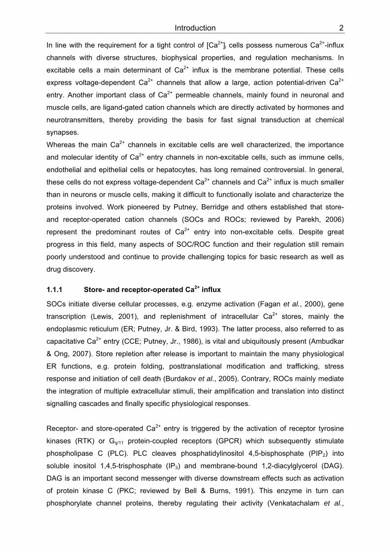
2+PP stores subsequently activates SOCs (Fig. 1; reviewed by Berridge, 1993).
(8)(8)
(7)
(1)
endoplasmic reticulum
agonist
receptor
PLC
PIP2 DAG
IP3
ROC SOC
(2)
(3)
(6)
PKC
(7)substratephosphorylation
Ca2+
PM
cytosol
IP3-gated Ca2+ channel
(4) (5)
Figure 1: Receptor- and store-operated CaPP
2+PP influx. Agonist stimulation of a receptor tyrosine
kinase or a G BBq/11BB protein-coupled receptor (1) leads to activation of phospholipase C (PLC) (2). The enzyme cleaves phosphatidylinositol 4,5-bisphosphate (PIPBB2BB) into membrane-bound diacylglycerol (DAG) and soluble inositol 1,4,5-trisphosphate (IPBB3BB) (3). IPBB3BB diffusion to the IP BB3BB-gated CaPP
2+PP-channel in
the ER membrane evokes depletion of intracellular CaPP
2+PP stores (4). In parallel, store depletion
activates SOCs (5). DAG may directly activate certain ROCs (6) and also, cooperatively with CaPP
2+TPTP,
PKTTC (7). PKC phosphorylates diverse substrates like channel proteins (8).
For a channel to be classified as store-operated, its direct activation by experimental store-
depletion has to be demonstrated (Bolotina & Csutora, 2005). Pharmacological agents like
cyclopiazonic acid and thapsigargin inhibit the sarcoplasmic/endoplasmic reticulum Ca PP
2+
PPpumps (SERCA; Favre et al., 1996) that normally transport Ca PP
2+PP against its electrochemical
gradient into the ER. Inhibition of this active, ATP-consuming process results in passive CaPP
2+PP
leakage. Ultimately, Ca PP
2+PP influx from the extracellular surrounding is mediated by SOCs, even
in the absence of receptor stimulation or generation of IP BB3BB.
ROCs are activated through the same signalling cascade but in contrast to SOCs they do not
require store depletion. The phospholipase-derived second messenger DAG has been
shown to directly activate ROCs (Hofmann et al., 1999). Furthermore, it is assumed that so
Introduction 4
far unknown messengers and PLC-dependent mechanisms are also involved in ROC
channel stimulation (Clapham et al., 2001).
Hence, ROCs and SOCs can be activated simultaneously following stimulation of one
receptor. But the underlying signalling cascades are definitely distinct.
1.1.2 Activation of store-operated channels
The elevation in [Ca PP
2+PP] BBi BB caused by store-depletion, the PI response, is not sufficient to initiate
store-operated Ca PP
2+PP entry (Parekh, 2006). How does a store-operated channel in the plasma
membrane then sense depletion of intracellular Ca PP
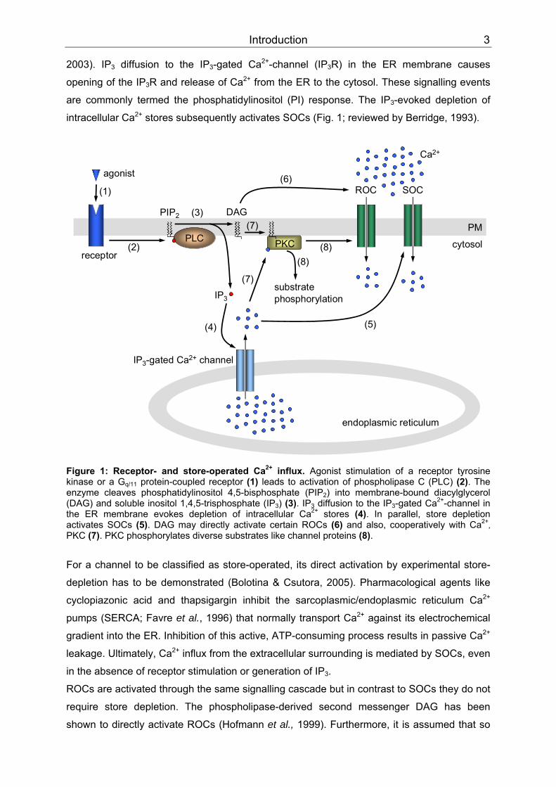
2+PP stores? At least three general models
were proposed (schematically depicted in Fig. 2) that strive to answer this question:
1. Conformational coupling model: The IPBB3BBR (located in the ER membrane) is in close
vicinity to the SOC (inserted in the plasma membrane) allowing direct protein-protein
interaction. IPBB3BBR activation results in “conformational-coupled” stimulation of the
channel (Irvine, 1990; Berridge, 1995). Since this model conflicts with the slow channel
activation kinetics after store-depletion, it was revised to the “secretion-like coupling”
hypothesis (Patterson et al., 1999). It is based on the assumption that SOCs and the
IPBB3BBR of quiescent cells are physically separated, but the ER can move towards the
channel following store depletion. Therefore, a temporal physical interaction between
the SOC and the IPBB3BBR is possible but requires some time to build up. It depends on the
peripheral cytoskeleton and stabilizing reagents might obstruct the coupling whereas
disaggregation could facilitate it (Rosado et al., 2000; Venkatachalam et al., 2002). The
participation of the IPBB3BBR at all stages of SOC activation in this model conflicts with the
definition of store-operation (see above; Bolotina & Csutora, 2005; Parekh, 2006), but
alternatively another ER component might interact with SOCs. An interesting candidate
is the stromal interaction molecule 1 (STIM1) that will be introduced in greater detail
below.
2. Calcium influx factor model: Store depletion is thought to release a so far unknown
“diffusible messenger”, termed calcium influx factor (CIF), from the ER. Alternatively, its
de novo synthesis could be initiated by store depletion. CIF might directly (Takemura et
al., 1989; Randriamampita & Tsien, 1993) and also indirectly activate SOCs. It is
proposed to stimulate membrane-bound Ca PP
2+PP-independent phospholipase A BB2 BB(iPLABB2BB) by
releasing it from binding to the inhibitory protein calmodulin. Subsequently,
lysophospholipids generated by iPLA BB2BB could activate the SOC directly (Smani et al.,
2003; Bolotina & Csutora, 2005). Besides CIF, other diffusible messengers have been
suggested, e.g. 5,6-epoxyeicosatrienoic acid, nitric oxide, and sphingosine-1-
Introduction 5
phosphate (Parekh & Putney, Jr., 2005) and are controversially discussed (Bolotina &
Csutora, 2005).
3. Vesicle-fusion model: Functional SOCs are stored in cytoplasmic vesicles and
recruited to and rapidly inserted into the plasma membrane following stimulation (Yao
et al., 1999; Alderton et al., 2000). Exocytotic channel insertion was also observed after
receptor stimulation (Cayouette et al., 2004; Bezzerides et al., 2004; Singh et al., 2004;
Odell et al., 2005) thus demonstrating that the mechanism might not be exclusive for
activation of SOCs but is important for TRP-mediated Ca PP
2+PP influx in general.
secretion-likecoupling
[Ca2+]ER
IP3
Ca2+
1.SOC
IP3R
[Ca2+]ERactin network
2.
diffusiblemessenger
[Ca2+]ER
[Ca2+]ER
Ca2+
3.
vesicularfusion
[Ca2+]ER
[Ca2+]ER
CIF
storedepletion
storedepletion
storedepletion
Ca2+
Figure 2: Proposed activation models for store-operated channels (adapted from Parekh, 2006). See text for detailed information.
Over the past two decades, the coupling mechanisms between the ER and store-operated
Ca PP
2+PP-influx channels and also the molecular identity of these channel proteins remained
elusive and were intensely investigated. SOCs do not form a uniform group but have diverse
biophysical properties. The best studied store-depletion responsive current is the calcium-
release-activated calcium (CRAC) current termed I BBCRACBB. First identified in mast cells (Hoth &
Penner, 1992), it is found in many cell types, and several genes have been proposed to code
for the CRAC channel-constituting proteins.
Most recently, crucial progress was made with the identification of two proteins that function
together in sensing store-depletion and subsequently mediating I BBCRACBB. Stromal interaction
molecule 1 (STIM1) was discovered in two independent RNA interference (RNAi) screens
Introduction 6
performed to elucidate the underlying signalling cascades of store-operated Ca PP
2+PP influx (Liou
et al., 2005; Roos et al., 2005). It has a single transmembrane domain and is found inserted
in the PM and the ER membrane (Soboloff et al., 2006). Originally identified as tumor
suppressor (Sabbioni et al., 1997), it is now also thought to control the ER Ca PP
2+PP filling state
with its luminal Ca PP
2+PP-binding EF hand motif and to transduce the depletion signal to Orai1
proteins (Liou et al., 2005; Zhang et al., 2005). These were named after Greek mythological
characters (the gate keepers of heaven) by one group (Feske et al., 2006), whereas the term
CRAC modulator 1 (CRACM1) was coined by another (Vig et al., 2006b). They are predicted
to have four membrane-spanning domains and to constitute the ion channel pore subunit
(Vig et al., 2006a; Prakriya et al., 2006; Yeromin et al., 2006). A single amino acid
substitution (R91W) suppresses IBBCRACBB necessary for T- and B-lymphocyte activation thus
causing a rare hereditary form of severe combined immunodeficiency (SCID; Feske et al.,
2005; Feske et al., 2006).
While the interaction of STIM1 and Orai1 is generally accepted to be necessary and
sufficient to mediate store-operated Ca PP
2+PP entry, the question of how they communicate has
not been unequivocally answered. Physical interactions have been demonstrated by co-
immunoprecipitation studies (Yeromin et al., 2006; Vig et al., 2006a; Ong et al., 2007), but
neither study resolves whether these are direct or indirect and whether they occur between
proteins both inserted in the PM or Orai1 and STIM1 in the ER (Hewavitharana et al., 2007).
Three adapted “secretion-like coupling” models are currently discussed as Orai1 activation
by one of the other proposed SOC activation mechanisms is less likely. Due to the physical
interaction between STIM1 and Orai1, the existence of a diffusible messenger is at least not
indispensable (Vig & Kinet, 2007). Moreover, Orai1 is constitutively expressed in the PM and
activation does not seem to require exocytosis (Prakriya et al., 2006; Vig et al., 2006b).
Nevertheless, exocytotic transport may be involved in STIM1 translocation to the PM (Vig &
Kinet, 2007), and also an increased STIM1 pulldown after store depletion in biotinylation
experiments has been reported (Zhang et al., 2005). Modulation of CRAC channel function
by phosphorylation is discussed as well (Vig & Kinet, 2007). Further studies are required to
ascertain which of the proposed mechanisms finally activates the CRAC channel.
1.2 The TRP channel superfamily
As mentioned above, the CRAC channel is the most prominent but not the only SOC
(reviewed by Montell, 1997; Vazquez, et al., 2004b; Parekh, 2006). The involvement of store-
operated Ca PP
2+PP entry in so many and diverse physiological processes like exocytosis,
contraction, enzyme control, gene regulation, apoptosis, cell proliferation and migration
(Parekh & Penner, 1997), motivated many investigators to search for the molecular
correlates of these currents. In 1995, these efforts led to the discovery of a novel class of
Ca PP
2+PP-permeable cation channels in mammals, the TRP superfamily (Zhu et al., 1995; Wes et

Introduction 7
al., 1995). It was named after a spontaneous Drosophila melanogaster mutant that has been
isolated almost two decades earlier.
Figure 3: TTElectroretinogram of trp D. melanogaster mutants.TT Dark-adapted flies were exposed to a five seconds pulse of white light (indicated by the event marker). The vertical line of the event marker represents 5 mV (Montell, 2004).
Fruitfly mutants had been screened for defects in their electroretinogram (ERG) recordings in
order to elucidate the visual transduction pathways (Cosens & Manning, 1969). Unlike in
vertebrates, phototransduction in the fruitfly is coupled to PLC. Light-induced PLC activation
results in Na PP
+PP and Ca PP
2+PP influx, thus depolarizing the photoreceptor cells (Montell, 1999;
Hardie & Raghu, 2001). This Ca PP
2+PP entry is defective in the above mentioned mutants, they
abnormally respond with a transient rather than sustained depolarization to prolonged light
exposure (Fig. 3) and were therefore named transient receptor potential (trp) (Minke et al.,
1975). After the trp gene had been cloned (Montell & Rubin, 1989), further studies confirmed that it
codes for a novel light-activated, Ca PP
2+PP-permeable cation channel (Hardie & Minke, 1992;
Phillips et al., 1992; Niemeyer et al., 1996). Its Ca PP
2+PP permeability and coupling to PLC
sparked interest in TRPs beyond invertebrate phototransduction exploration as the channel
was speculated to be a SOC (reviewed by Montell, 1997). Later on, TRP became evident to
be the founding member of a novel channel superfamily. Two more TRP-related channels,
TRPL and TRPγ, were found in Drosophila (Phillips et al., 1992; Tsunoda & Zuker, 1999; Xu
et al., 2000) and up to date 29 mammalian orthologs of the Drosophila trp gene have been
identified. Some of them are also candidates to form SOCs (Montell et al., 2002b; Montell,
2005; Okuhara et al., 2007), whereas others constitute ROCs, tonically active or stretch-
activated channels (Dietrich et al., 2006).
By sequence homology TRPs can be divided into seven families, named after their first
recognized members (Pedersen et al., 2005; Ramsey et al., 2006): TRPC (classical), TRPV
(vanilloid), TRPM (melastatin), TRPA (ankyrin), TRPP (polycystin) and TRPML (mucolipin).
TRP-related channels are found in every metazoan organism genetically studied so far
(Montell et al., 2002a) with the seventh existing TRPN (no mechanoreceptor potential C)
family containing members in Drosophila melanogaster, Caenorhabditis elegans and Danio
rerio (Montell, 2001; Okuhara et al., 2007), but not in mammals (Fig. 4).

Introduction 8
10 PAM units
C4 C5 C1 C3C7C6
DAG-sensitive
mTRPC2(pseudogene in humans)
Classical (short) TRPC
M1 (melastatin)M3
M6M7
channel/kinases
M2M8
M4M5
Melastatin (long) TRPMANKTM1
TRPAPolycystins
P5P3P2
Mucolipins
ML1ML3
ML2
V6V5
V3V4
V2V1
Vanilloidreceptor
TRPV
Figure 4: TTThe mammalian TRP family tree (adapted from Clapham, 2003).TT The branch length symbolizes the evolutionary distance and is graded in point accepted mutations units (PAM, mean number of mutations per 100 residues).
At the time of its cloning, TRP showed no significant homology to known proteins (Montell,
2004). Difficulties in crystallizing these integral membrane proteins so far prevented structure
determination by X-ray analysis. A topology analysis of its primary sequence predicted seven
hydrophobic, putatively membrane-spanning segments. By virtue of mutagenesis studies,
determination of glycosylation sites and in analogy to known voltage-gated and second-
messenger-gated ion channels, it is now assumed that TRP channels (with the exception of
TRPP1; Okuhara et al., 2007) have six transmembrane domains with cytosolic amino and
carboxy termini (Montell & Rubin, 1989; Vannier et al., 1998). Functional channels are
thought to be composed of homo- or heterotetramers (Kedei et al., 2001; Hoenderop et al.,
2003; Amiri et al., 2003), in which the pore is formed by the fifth and sixth membrane-
spanning domains and intervening segments (Fig. 5). They lack the complete voltage sensor
formed by positively charged amino acids in the fourth transmembrane domain of many
voltage-gated channels (Montell, 2001) and therefore, are only weakly voltage-sensitive.
Whereas channels within a family share high amino acid sequence similarity, the families by
themselves are quite different, but at least their transmembrane segments are significantly
homologous to TRP (Montell, 2001).

Introduction 9
cytosol
PM 1 2 3 4 5 6
N C
putative poreregion
Monomer Proposed tetrameric channel structure
C
21
6
34
5
N
pore2
16
345
432
CN
561
N CNC 6
523
4
1
Figure 5: TTProposed TRP channel topology (adapted from Li et al., 2002). See text for explanations (abbreviations: C, carboxy terminus; N, amino terminus; 1-6, membrane-spanning segments). TT
The tissue distribution of TRP channels in mammals is commonly widespread, ranging from
non-excitable cells to the nervous system. Activation mechanisms, ion selectivities and
putative physiological and pathophysiological roles are strikingly versatile and diverse. Apart
from two monovalent-selective exceptions (TRPM4 and -M5), TRP channels are CaPP
2+PP-
permeable but rather non-selective to cations. They modulate [Ca PP
2+PP] BBi BB and regulate membrane
potential (Kwan et al., 2007). TRPV5 and -V6 are more Ca PP
2+PP-selective but not as much as
voltage-gated Ca PP
2+PP-channels (Clapham et al., 2001).
Currently, TRP channels are attracting growing attention due to their possible involvement in
human physiology and disease. The ancestral Drosophila TRP channel is crucial for visual
transduction and several mammalian relatives (especially of the TRPV family) are also
important for sensory perception, e.g. of mechanical stimuli, osmolarity, pain, pheromones,
taste and temperature. Others are involved in such distinct physiological processes as
fertilization and vasorelaxation. TRP channels abnormally activated or dysfunctional due to
pathologic mutations cause several channelopathies (for a recent review see Nilius, 2007).
For instance, TRPs have been connected to polycystic kidney disease (Mochizuki et al.,
1996) and hereditary focal segmental glomerulosclerosis (FSGS; Winn et al., 2005), to the
lysosomal storage disorder mucolipidosis IV (Bassi et al., 2000), to hypomagnesemia with
secondary hypocalcaemia (HSH, Schlingmann et al., 2002) and to Guamanian amyotrophic
lateral sclerosis and parkinsonism dementia (Hermosura et al., 2005; Hermosura & Garruto,
2007).
1.3 The TRPC family
1.3.1 Structural features of TRPCs
TRPCs were the first TRP proteins discovered in mammals (Wes et al., 1995; Zhu et al.,
1995). Seven proteins, referred to as TRPC1 – 7, constitute the canonical (or classical) TRP
family that is the closest related to the Drosophila TRP protein (30-40% identity; Okuhara et
Introduction 10
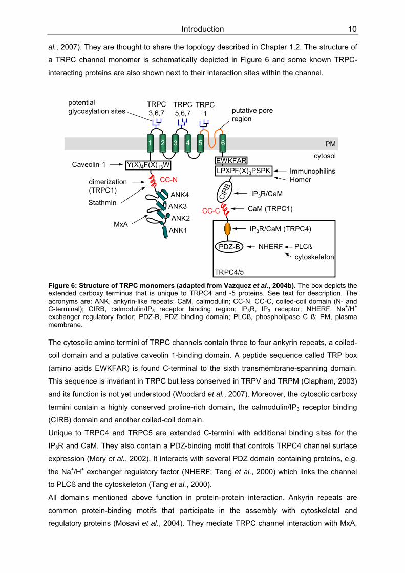
al., 2007). They are thought to share the topology described in Chapter 1.2. The structure of
a TRPC channel monomer is schematically depicted in Figure 6 and some known TRPC-
interacting proteins are also shown next to their interaction sites within the channel.
ANK3ANK4 CI
RB
cytosolPM1 2 3 4 5 6
potential glycosylation sites
TRPC3,6,7
TRPC5,6,7
TRPC1
CC-N
ANK2
ANK1
dimerization(TRPC1)
Stathmin
EWKFARY(X)4F(X)13W LPXPF(X)3PSPK
CC-C
PDZ-Bcytoskeleton
NHERF PLCß
IP3R/CaM (TRPC4)
ImmunophilinsHomer
IP3R/CaM
CaM (TRPC1)
Caveolin-1
TRPC4/5
putative poreregion
MxA
Figure 6: Structure of TRPC monomers (adapted from Vazquez et al., 2004b). The box depicts the extended carboxy terminus that is unique to TRPC4 and -5 proteins. See text for description. The acronyms are: ANK, ankyrin-like repeats; CaM, calmodulin; CC-N, CC-C, coiled-coil domain (N- and C-terminal); CIRB, calmodulin/IP BB3BB receptor binding region; IPBB3BBR, IPBB3BB receptor; NHERF, NaPP
+PP/H PP
+PP
exchanger regulatory factor; PDZ-B, PDZ binding domain; PLCß, phospholipase C ß; PM, plasma membrane. The cytosolic amino termini of TRPC channels contain three to four ankyrin repeats, a coiled-
coil domain and a putative caveolin 1-binding domain. A peptide sequence called TRP box
(amino acids EWKFAR) is found C-terminal to the sixth transmembrane-spanning domain.
This sequence is invariant in TRPC but less conserved in TRPV and TRPM (Clapham, 2003)
and its function is not yet understood (Woodard et al., 2007). Moreover, the cytosolic carboxy
termini contain a highly conserved proline-rich domain, the calmodulin/IP BB3BB receptor binding
(CIRB) domain and another coiled-coil domain.
Unique to TRPC4 and TRPC5 are extended C-termini with additional binding sites for the
IPBB3BBR and CaM. They also contain a PDZ-binding motif that controls TRPC4 channel surface
expression (Mery et al., 2002). It interacts with several PDZ domain containing proteins, e.g.
the Na PP
+PP/HPP
+PP exchanger regulatory factor (NHERF; Tang et al., 2000) which links the channel
to PLCß and the cytoskeleton (Tang et al., 2000).
All domains mentioned above function in protein-protein interaction. Ankyrin repeats are
common protein-binding motifs that participate in the assembly with cytoskeletal and
regulatory proteins (Mosavi et al., 2004). They mediate TRPC channel interaction with MxA,
Introduction 11
a member of the dynamin superfamily of GTPases (Lussier et al., 2005), and, as
demonstrated for TRPC3 and -6, are required for correct trafficking to the PM (Hofmann et
al., 2002; Wedel et al., 2003). The first ankyrin-like repeat was additionally identified as key
structure for functional homo- and heteromerization of TRPC4 and -5 channels (Schindl et
al., 2007). Coiled-coil domains have been reported to be involved in TRPC1 channel
homomerization (Engelke et al., 2002; Lepage et al., 2006) and linkage with other proteins
(Greka et al., 2003). An additional site of protein-protein interaction is the C-terminal proline-
rich region that was found to interact with FK506 binding proteins (FKBP; Sinkins et al.,
2004) and Homer (Yuan et al., 2003).
Mutations within the highly conserved pore-region result in dominant-negative monomers
that suppress the function of homo- and heteromeric channels (Hofmann et al., 2002).
Furthermore, it was demonstrated that the N-glycosylation pattern can determine the
channel’s constitutive activity. TRPC3 is a highly constitutive active channel and
monoglycosylated in the first extracellular loop. By conversion into the TRPC6-like dually
glycosylated form it becomes as tightly regulated by PLC-coupled receptors as TRPC6 and
vice versa (Dietrich et al., 2003).
1.3.2 TRPC-interacting proteins
Drosophila TRP and other components of the fruitfly phototransduction cascade are
clustered in a transducisome (reviewed by Montell, 2004), a macromolecular complex
assembled by the scaffolding protein INAD ( UUi UUnactivation UUn UUo UUa UUfterpotential UUDUU; Shieh & Zhu,
1996). Analogously, TRPCs are suggested to be organized within specific CaPP
2+PP signalling
complexes that facilitate their physical and/or functional coupling with accessory proteins
participating in Ca PP
2+PP signalling and also with proteins involved in vesicle trafficking,
cytoskeletal interaction, and scaffolding (Ambudkar & Ong, 2007). For instance, some
TRPCs have been shown to be associated with caveolae (Lockwich et al., 2000; Lockwich et
al., 2001; Torihashi et al., 2002). These are detergent-insoluble, glycosphingolipid- and
cholesterol-enriched membrane domains (so-called lipid rafts) that are assembled by the
cholesterol-binding protein caveolin (Brazer et al., 2003). Several TRPC-associated proteins
have been identified which might be involved in regulating channel function, stability, and
cellular localization (Ambudkar & Ong, 2007). According to their proposed function as
structural or regulatory proteins they are summarized in Table 1.
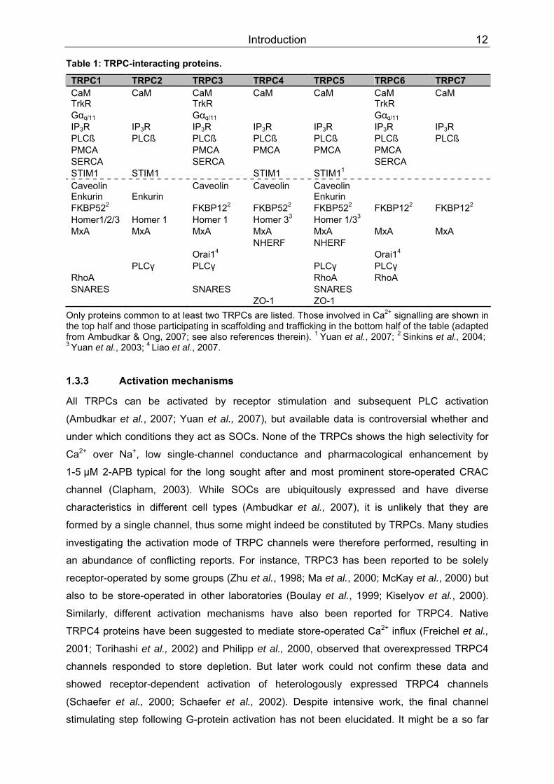
Introduction 12
Table 1: TRPC-interacting proteins.
TRPC1 TRPC2 TRPC3 TRPC4 TRPC5 TRPC6 TRPC7 CaM CaM CaM CaM CaM CaM CaM TrkR TrkR TrkR GαBBq/11BB GαBBq/11BB GαBBq/11BB IPBB3BBR IPBB3BBR IPBB3BBR IPBB3BBR IPBB3BBR IPBB3BBR IPBB3BBR PLCß PLCß PLCß PLCß PLCß PLCß PLCß PMCA PMCA PMCA PMCA PMCA SERCA SERCA SERCA STIM1 STIM1 STIM1 STIM1PP
1PP
Caveolin Caveolin Caveolin Caveolin Enkurin Enkurin Enkurin FKBP52 PP
2PP FKBP12 PP
P
2 FKBP52 PP
2PP FKBP52 PP
2PP FKBP12 PP
2PP FKBP12 PP
2PP
Homer1/2/3PP Homer 1 Homer 1 Homer 3PP
3PP Homer 1/3PP
3PP
MxA MxA MxA MxA MxA MxA MxA NHERF NHERF Orai1PP
4PP Orai1PP
4PP
PLCγ PLCγ PLCγ PLCγ RhoA RhoA RhoA SNARES SNARES SNARES ZO-1 ZO-1
Only proteins common to at least two TRPCs are listed. Those involved in CaPP
2+PP signalling are shown in
the top half and those participating in scaffolding and trafficking in the bottom half of the table (adapted from Ambudkar & Ong, 2007; see also references therein). PP
1 PPYuan et al., 2007; PP
2 PPSinkins et al., 2004; PP
3 PPYuan et al., 2003; PP
4 PPLiao et al., 2007.
1.3.3 Activation mechanisms
All TRPCs can be activated by receptor stimulation and subsequent PLC activation
(Ambudkar et al., 2007; Yuan et al., 2007), but available data is controversial whether and
under which conditions they act as SOCs. None of the TRPCs shows the high selectivity for
Ca PP
2+PP over Na PP
+PP, low single-channel conductance and pharmacological enhancement by
1-5 µM 2-APB typical for the long sought after and most prominent store-operated CRAC
channel (Clapham, 2003). While SOCs are ubiquitously expressed and have diverse
characteristics in different cell types (Ambudkar et al., 2007), it is unlikely that they are
formed by a single channel, thus some might indeed be constituted by TRPCs. Many studies
investigating the activation mode of TRPC channels were therefore performed, resulting in
an abundance of conflicting reports. For instance, TRPC3 has been reported to be solely
receptor-operated by some groups (Zhu et al., 1998; Ma et al., 2000; McKay et al., 2000) but
also to be store-operated in other laboratories (Boulay et al., 1999; Kiselyov et al., 2000).
Similarly, different activation mechanisms have also been reported for TRPC4. Native
TRPC4 proteins have been suggested to mediate store-operated Ca PP
2+PP influx (Freichel et al.,
2001; Torihashi et al., 2002) and Philipp et al., 2000, observed that overexpressed TRPC4
channels responded to store depletion. But later work could not confirm these data and
showed receptor-dependent activation of heterologously expressed TRPC4 channels
(Schaefer et al., 2000; Schaefer et al., 2002). Despite intensive work, the final channel
stimulating step following G-protein activation has not been elucidated. It might be a so far

Introduction 13
unknown PLC-dependent mechanism or a combination of messengers (Clapham et al.,
2001). Finally, basal activity of TRPC4 without stimulation has also been reported (McKay et
al., 2000).
Such discrepancies (Trebak et al., 2002) could stem from the different expression systems
that might lack certain regulatory or auxiliary proteins necessary for complex formation and
specific gating of ectopically expressed TRPCs. Observations could be further confounded
by endogenous SOCs (Ambudkar et al., 2007), channel heteromultimerization (Poteser et al.,
2006), different channel expression levels (Vazquez et al., 2003), and species-dependent
differences in the regulation of channel orthologs (Okada et al., 1999; Riccio et al., 2002).
Despite intensive effort, a general mechanism of TRPC channel activation by store depletion
has not been unravelled. Recently, several suggestions were made taking into account the
identification of STIM1 and Orai1 as the IBBCRACBB-mediating proteins (see Chapter 1.1.2). A new
molecular definition of “store operation” was suggested in which SOCs are plasma
membrane channels that are regulated by rearrangement of the ER Ca PP
2+PP-content sensor
STIM1 (Yuan et al., 2007). By these criteria, TRPC1, -4, and -5 function as SOCs as they are
directly activated by STIM1. TRPC3 and -6 can also function as SOCs due to STIM1-
dependent heteromultimerization of TRPC3 with TRPC1 and TRPC6 with TRPC4 (Huang et
al., 2006; Yuan et al., 2007). The underlying mechanism of STIM1-dependent TRPC gating
still remains to be elucidated. Another group has demonstrated that overexpressed TRPC3
and -6 become store-sensitive by coexpression of any of the three existing Orai isoforms
(Orai1-3). A novel activation model was deduced from this observation wherein SOCs are
composed of TRPC pore-forming subunits and Orai regulatory ß-subunits. Orai would relay
the store depletion signal from STIM1 to TRPC (Liao et al., 2007). A third candidate that was
reported to be involved in TRPC store-dependent activation is the scaffolding protein Homer. It mediates the physical interaction of TRPC1 with the IP BB3BBR in HEK293 cells when the stores
are replete. Depletion disrupts this association and the released channel mediates Ca PP
2+PP influx
to refill the stores (Yuan et al., 2003). This regulation mechanism could be restricted to
certain cell types since contrary observations were reported for endothelial cells and
platelets. In these cells, TRPC1-dependent store-operated Ca PP
2+PP influx required channel
association with the IP BB3BBR (Mehta et al., 2003; Rosado et al., 2005). Besides its involvement
in TRPC1 gating by the IPBB3BBR, Homer 1 also seems to participate in receptor-mediated
TRPC3 translocation to the PM and subsequent channel retrieval upon termination of the
stimulation (Kim et al., 2006a; Worley et al., 2007).
It is conceivable that all the proposed activation mechanisms exist in vivo and they might
even be integrated in the same cell type. Further studies are required to determine their
relation to each other.
Introduction 14
10 PAM units
TRPC1
TRPC5
TRPC2
TRPC3
TRPC7
TRPC4
TRPC6
1.3.4 TRPC subfamilies
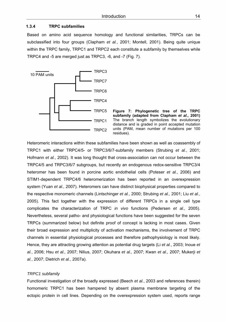
Based on amino acid sequence homology and functional similarities, TRPCs can be
subclassified into four groups (Clapham et al., 2001; Montell, 2001). Being quite unique
within the TRPC family, TRPC1 and TRPC2 each constitute a subfamily by themselves while
TRPC4 and -5 are merged just as TRPC3, -6, and -7 (Fig. 7).
Figure 7: Phylogenetic tree of the TRPC subfamily (adapted from Clapham et al., 2001) The branch length symbolizes the evolutionary distance and is graded in point accepted mutation units (PAM, mean number of mutations per 100 residues).
Heteromeric interactions within these subfamilies have been shown as well as coassembly of
TRPC1 with either TRPC4/5- or TRPC3/6/7-subfamily members (Strubing et al., 2001;
Hofmann et al., 2002). It was long thought that cross-association can not occur between the
TRPC4/5 and TRPC3/6/7 subgroups, but recently an endogenous redox-sensitive TRPC3/4
heteromer has been found in porcine aortic endothelial cells (Poteser et al., 2006) and
STIM1-dependent TRPC4/6 heteromerization has been reported in an overexpression
system (Yuan et al., 2007). Heteromers can have distinct biophysical properties compared to
the respective monomeric channels (Lintschinger et al., 2000; Strubing et al., 2001; Liu et al.,
2005). This fact together with the expression of different TRPCs in a single cell type
complicates the characterization of TRPC in vivo functions (Pedersen et al., 2005).
Nevertheless, several patho- and physiological functions have been suggested for the seven
TRPCs (summarized below) but definite proof of concept is lacking in most cases. Given
their broad expression and multiplicity of activation mechanisms, the involvement of TRPC
channels in essential physiological processes and therefore pathophysiology is most likely.
Hence, they are attracting growing attention as potential drug targets (Li et al., 2003; Inoue et
al., 2006; Hsu et al., 2007; Nilius, 2007; Okuhara et al., 2007; Kwan et al., 2007; Mukerji et
al., 2007; Dietrich et al., 2007a).
TRPC1 subfamily
Functional investigation of the broadly expressed (Beech et al., 2003 and references therein)
homomeric TRPC1 has been hampered by absent plasma membrane targeting of the
ectopic protein in cell lines. Depending on the overexpression system used, reports range

Introduction 15
from lack of robust TRPC1 signals (Strubing et al., 2001) due to retention in intracellular
membranes (Wang et al., 1999) to detailed description of channel properties in Spodoptera
frugiperda sf9 cells (Sinkins et al., 1998). Possible explanations are the absence of auxiliary
subunits or interacting proteins in some overexpression systems as plasma membrane
expression of the TRPC1 protein has been shown to depend on interaction with other
proteins, e.g. TRPCs (Hofmann et al., 2002), caveolin-1 (Brazer et al., 2003), and RhoA
(Mehta et al., 2003). Also it is not certain whether homo- or heteromeric expressed or even
native channels, which could be stimulated by TRPC1, are measured in sf9 cells (Beech et
al., 2003). TRPC1 might not be a pore-forming subunit at all, it could as well function as
regulator of other pore-forming channels (Dietrich et al., 2007b) and the existence of a native
TRPC1 homomer has not been unequivocally proven so far (Ambudkar et al., 2007).
Whereas several reports have described TRPC1 to be a store-, receptor-, IPBB3BBR-, and/or
stretch-activated channel (Ramsey et al., 2006), recent findings in vascular smooth muscle
cells of TRPC1 PP
-/-PP mice imply that the channel is not an essential component of store- and
stretch-operated channels in these cells (Dietrich et al., 2007b). However, this study does not
exclude TRPC1 contribution to such channels in other tissues.
The native protein could be involved in neuronal plasticity, since it is required for the
excitatory postsynaptic conductance in Purkinje cells (Kim et al., 2003). TRPC1 also interacts
with TRPP2 (Tsiokas et al., 1999), a distantly related TRP protein involved in development of
polycystic kidney disease. Moreover, the channel is up-regulated in neointimal hyperplasia
(Bergdahl et al., 2004; Kumar et al., 2006) and cardiac hypertrophy (Ohba et al., 2007),
interacts with a transcription factor important for myocyte development (Ma et al., 2003) and
was proposed to play a role in Duchenne muscular dystrophy (Vandebrouck et al., 2007). In
conclusion, TRPC1 may serve as developmental regulator of smooth muscle cells (SMC)
and some of its functional roles might not be easily compensated by related TRPCs. It could
be engaged in further patho- and physiological processes but its unique physiological
functions are not known yet (Dietrich et al., 2007b).
TRPC2 subfamily
TRPC2 is a pseudogene in humans, old world monkeys and apes (Wes et al., 1995; Vannier
et al., 1999; Liman & Innan, 2003), but functionally expressed in other mammalian species
and essential for pheromone sensation in rodents. Male mice lacking this channel do not
show typical male-male aggressive behaviour and court both females and males (Stowers et
al., 2002). Antibodies directed to an extracellular domain of TRPC2 inhibit the acrosomal
reaction pointing towards its importance in fertilization (Jungnickel et al., 2001). However,
TRPC2 PP
-/-PP mice show no defects in reproduction (Stowers et al., 2002). TRPC2 is activated by
DAG (Lucas et al., 2003) and does not seem to heteromultimerize with other TRPC channels
(Montell, 2005).
Introduction 16
TRPC3/6/7 subfamily
These channels share 70–80% amino acid identity and they can be directly activated by the
PLC product DAG (Hofmann et al., 1999; Okada et al., 1999; Trebak et al., 2003). TRPC3
and -6 activities are regulated by N-glycosylation (Dietrich et al., 2003) and phosphorylation
through the non-receptor tyrosine kinases Src and Fyn (Hisatsune et al., 2004; Vazquez et
al., 2004a).
TRPC3 is highly expressed in human brain, smooth and cardiac muscle cells (Dietrich et al.,
2006 and references therein). It seems to be involved in axon growth guidance (Li et al.,
2005), synaptic plasticity around the time of birth (Li et al., 1999) and cardiac CaPP
2+PP
homeostasis. In cardiomyocytes, abnormal accumulation of intracellular Na PP
+PP levels due to
TRPC3 has been shown to reverse the Na PP
+PP/Ca PP
2+PP exchanger (NCX1) transport mode (Eder et
al., 2007). This reverse mode transports Ca PP
2+PP into the cell and might be involved in
pathophysiological processes, e.g. heart failure and ischemia (Okuhara et al., 2007). As
mentioned above, TRPC3 was also found to coassemble with TRPC4 into a redox-sensitive
channel (Poteser et al., 2006). These heteromers could be activated by oxidative stress
under pathological conditions. TRPC3 antagonists might be cytoprotective by preventing the
uncontrolled Ca PP
2+PP influx and subsequent cell damage (Montell, 2001; Okuhara et al., 2007).
Furthermore, phosphorylation by protein kinase G (PKG) has been reported to inactivate
TRPC3. This might provide an endogenous negative feedback regulation mediated by the
nitric oxide/cyclic guanosine monophosphate/PKG pathway to protect vascular endothelial
cells from excessive Ca PP
2+PP influx (Kwan et al., 2004).
TRPC6 is present in brain, platelets, vascular and airway SMCs (Inoue et al., 2001; Yu et al.,
2003; Pedersen et al., 2005; Dietrich et al., 2006 and references therein). This channel was
shown to be an essential part of the α BB1BB-adrenoceptor-stimulated cation channel in rabbit
portal vein myocytes (Inoue et al., 2001). TRPC6 stimulation by agonists or increasing
intravascular pressure (Welsh et al., 2002) is postulated to depolarize the membrane,
thereby activating L-type voltage-gated Ca PP
2+PP channels that finally mediate smooth muscle
contraction (Large, 2002; Soboloff et al., 2005; Estacion et al., 2006) and reflex
vasoconstriction (Bayliss effect; Welsh et al., 2002). On the contrary, agonist-induced
bronchoconstriction mainly depends on Ca PP
2+ PPinflux mediated by voltage-independent
channels (such as TRPC6), hence, L-type Ca PP
2+ PPchannel blockers are not effective, e.g. in
asthma and chronic obstructive pulmonary disease (COPD; Gudermann et al., 2004).
Furthermore, TRPC6 is found in leukocytes probably mediating inflammatory responses in
asthma and COPD (Li et al., 2004). Idiopathic pulmonary arterial hypertension (IPAH) is a
progressive disease that can be life-shortening by resulting in right heart failure (Dietrich et
al., 2006). A major cause for the elevated pulmonary vascular resistance in these patients is

Introduction 17
excessive proliferation of pulmonary artery SMCs (PASMCs; Dietrich et al., 2005a). TRPC3
and -6 expression is significantly increased in these cells (Yu et al., 2004), and treatment
with TRPC6 small-interfering RNA (siRNA) markedly reduced hyperproliferation (Kunichika et
al., 2004). In summary, TRPC6 inhibition seems to be an interesting therapeutic strategy for
the treatment of IPAH and other chronic respiratory diseases. But TRPC6 also has
physiological functions in airway SMCs that should rather not be blocked. It is essential for
acute hypoxic pulmonary vasoconstriction (HPV), thus maintaining proper gas exchange
under acute hypoxic conditions by directing blood flow from poorly to well ventilated areas
(Weissmann et al., 2006). Disturbances in HPV as occurring in the adult respiratory distress
syndrome, pneumonia, and liver failure, can cause life-threatening arterial hypoxemia
(Dietrich et al., 2006 and references therein). Contrary to the proposed physiological functions of the channel described above, TRPC6
deficient mice have an unexpected and surprising phenotype. These animals showed airway
smooth muscle hyperreactivity in response to bronchoconstrictors, an elevated mean arterial
blood pressure, and exaggerated reflex vasoconstriction. Also the basal and agonist-induced
cation entry in SMC of TRPC6PP
-/- PPmice is higher (Freichel et al., 2005 and references therein;
Dietrich et al., 2005b). Partly, this can be explained by an increased expression of the closely
related TRPC3 channel (Dietrich et al., 2005b). It has a higher basal activity, is less tightly
regulated by vasoconstrictors and has consequently overcompensated TRPC6 knock-out,
demonstrating that both channels are not functionally redundant.
The opposite approach revealed a role for TRPC6 in the pathogenesis of cardiac
hypertrophy. Cardiac-specific TRPC6 overexpression in transgenic mice leads to an
increased Ca PP
2+PP influx that couples via calcineurin to the stimulation of NFAT (nuclear factor of
activated T cells). Pathological heart remodelling is accelerated and these mice have a
shortened life expectancy (Kuwahara et al., 2006). Whereas in vivo TRPC6 upregulation in
cardiomyocytes participates in hypertrophy, it seems to have protective antifibrotic functions
in cardiac fibroblasts in vitro (Nishida et al., 2007). Further in vivo studies are needed to
estimate the therapeutic value of TRPC6 modulation and the involvement of TRPC3
(Nakayama et al., 2006) and TRPC3/6 heteromers (Dietrich et al., 2007) in the pathogenesis
of heart failure.
Finally, convincing evidence for TRPC6 involvement in hereditary FSGS, a significant cause
of end-stage renal disease, has been presented. Kidneys ultrafiltrate the plasma with their
glomeruli and the glomerular filter is composed of a fenestrated capillary endothelium, the
basement membrane and podocytes connected by the slit diaphragm (Gudermann, 2005).
Structural damage of the glomerular filter results in proteinuria. TRPC6 PP
PP gain-of-function
mutants found in FSGS patients lead to increased Ca PP
2+ PPand Na PP
+ PPinflux into podocyte foot
processes (Winn et al., 2005; Reiser et al., 2005), but it is not known whether and how this is
disease-causing. Recently, it was also demonstrated that TRPC6 expression is up-regulated
Introduction 18
in complement-treated podocytes in vitro leading to actin cytoskeleton rearrangement,
whereas channel overexpression in vivo leads to proteinuria in mice (Moller et al., 2007).
TRPC7 is expressed in heart, lung and eyes and lower transcript levels are found in brain,
spleen and testis (Dietrich et al., 2006 and references therein). The channel is constitutively
active although it has two predicted glycosylation sites like TRPC6 (Okada et al., 1999). Its
physiological function remains obscure (Okuhara et al., 2007).
TRPC4/5 subfamily
These channels share 64% identity and are most closely related to TRPC1 (persuading
some groups to classify TRPC1 within this subfamily; Ramsey et al., 2006). A unique feature
of this subfamily is the potentiation by micromolecular concentrations of the lanthanide
cations gadolinium (Gd PP
3+PP) PP
PPand PP
PPlanthanum (La PP
3+PP) after GBBq/11 BB-coupled receptor mediated
activation (Schaefer et al., 2000; Strubing et al., 2001). In contrast to TRPC2 and the
TRPC3/6/7 subgroup, TRPC4 and -5 are not directly activated by the subsequently formed
PIPBB2BB hydrolysis product DAG (Venkatachalam et al., 2003).
Recently, lysophosphatidylcholine (LPC; Flemming et al., 2006) and sphingosine
1-phosphate (S1P; Xu et al., 2006) were identified as endogenous TRPC5 activators.
S-nitrosylation, e.g. by nitric oxide (NO), has been shown to activate both TRPC4 and
TRPC5 (Yoshida et al., 2006).
TRPC4 is widely expressed and also found in endothelial and smooth muscle cells (Freichel
et al., 2001; Beech et al., 2004). The channel was the first TRP gene to be knocked out in
mice and these animals provided insight into its biological roles. TRPC4 PP
-/-PP mice are viable
and reach maturation (Montell, 2001), but SOC-mediated Ca PP
2+PP entry into endothelial cells
(EC) is markedly reduced resulting in decreased endothelium-dependent vasorelaxation
(Freichel et al., 2001). Further studies were performed with thrombin, an important
inflammation mediator that is involved in the pathogenesis of vascular injury. In lungs,
thrombin increases vascular permeability and thus tissue water content. Lung EC of TRPC4 PP
-/-PP
mice lack thrombin-induced actin stress fiber formation, cell retraction is impaired, and lung
microvascular permeability subsequently reduced by about 50% (Tiruppathi et al., 2002).
TRPC4 is furthermore expressed in different cells within the central nervous system and
seems to be involved in neurotransmitter signalling. Release of γ-aminobutyric acid (GABA)
following application of 5-hydroxytryptamine (5-HT, serotonin) is drastically reduced in
thalamic interneurones from TRPC4 PP
-/-PP mice, whereas GABA release upon stimulation of
metabotropic glutamate receptors is not changed (Munsch et al., 2003). The thalamus
regulates sleep and wakefulness and TRPC4 could participate in processing of visual
information depending on the sleep/wake cycle (Pape et al., 2004). TRPC4 is also found in

Introduction 19
pancreatic ß-islets and was suggested to be involved in insulin secretion (Qian et al., 2002).
However, glucose-tolerance test results were similar in wild-type and TRPC4-deficient mice
(Freichel et al., 2004). Finally, the channel could be involved in regulating the motility of the
gastrointestinal tract by modulating the pacemaker activity of interstitial cells of Cajal (ICC),
(Torihashi et al., 2002).
TTTRPC5 TT is highly enriched in brain but also found peripheral, e.g. in SMC (Xu et al., 2005;
reviewed in Dietrich et al., 2006). Interestingly, the gene is located on a region of the human
X chromosome associated with non-syndromic mental retardation (Sossey-Alaoui et al.,
1999), and regulation of neurite outgrowth and growth cone morphology by TRPC5
homomers has been demonstrated in rat hippocampal neurons. Functional channel
suppression by transfection of a dominant-negative mutant led to abnormally prolonged
neurites, and overexpression resulted in neurite outgrowth inhibition (Greka et al., 2003).
Phosphatidylinositol 4-phosphate 5-kinase (PIP(5)Kα)-dependent channel insertion from
vesicles into the plasma membrane was further reported to be crucial for neurite length
regulation by TRPC5 (Bezzerides et al., 2004).
TRPC5 may have multiple functions within the cardiovascular system. For example, SMC
motility is crucial in physiological adaptive processes like wound healing but also involved in
inflammatory occlusive diseases like atherosclerosis (Inoue et al., 2006). Cell motility of
vascular SMC was evoked by the TRPC5 activator S1P and inhibited by a dominant-negative
TRPC5 mutant or an anti-TRPC5 antibody (Xu et al., 2006). Furthermore, in failing hearts
from patients with end-stage idiopathic dilated cardiomyopathy TRPC5 was found to be
selectively upregulated, whereas the expression levels of TRPC1, -4 and -6 were unchanged
and TRPC3 was not detectable (Bush et al., 2006). As Ca PP
2+PP-ATPase SERCA2 is
downregulated in cardiac hypertrophy, and siRNA-mediated SERCA2 downregulation in
neonatal rat cardiac myocytes led to a compensatory upregulation of TRPC5, TRPC4 and
NCX expression (Seth et al., 2004), an involvement of TRPC5 (and TRPC4) in cardiac
hypertrophy is conceivable (Inoue et al., 2006). Increased TRPC5 expression and channel-
mediated CaPP
2+PP influx in monocytes of hypertensive patients was reported as well (Liu et al.,
2006).
Introduction 20
1.4 Aims
The first aim of the present work was to identify new pharmacological tools that may be used
to gain a better understanding of TRPC channel function in cells and beyond. There are
many open questions regarding the native composition and activation mechanisms,
physiological functions, and roles in pathophysiology and disease of TRPC proteins. In situ
identification of native TRPC channels is complicated by their wide and partially overlapping
distribution, potential heteromultimerization, similar electrophysiological properties and a
paucity of tool compounds to unequivocally trace these channels (Moran et al., 2004).
Compensatory effects have been observed in studies with transgenic mice (Dietrich et al.,
2005b), dominant negative channel subunits or when genes were silenced with small
interfering RNA, but they are not expected to be seen when channels are instantaneously
blocked with a selective tool compound (Beech et al., 2003). The fact that known organic
inhibitors and inorganic blockers are not potent and specific enough herefore (Li et al., 2004),
motivated us to search for further TRPC blockers. In preliminary in-house experiments the
steroide norgestimate had been identified as novel TRPC6 channel inhibitor. Therefore, the
present study was designed to test its applicability as selective TRPC channel blocker by
evaluating its sensitivity and selectivity towards the TRPC4/5 and TRPC3/6/7 subfamilies in
heterologous expression systems.CC As norgestimate is a synthetic progestin and the precursor
of levonorgestrel, it should be further tested whether levonorgestrel itself and the natural
hormone progesterone are as well active on TRPC channels. CCMoreover the effects of
norgestimate should be validated in either cell lines or primary cells expressing endogenous
TRPC6-containing channel complexes. Finally, we envisaged to use norgestimate for the
study of native TRPC channel function in tissue preparations such as isolated aortic or
tracheal rings.
The second part of this study was directed towards the identification of novel regulators of
native TRPC4 channel complexes. Dysregulation of endothelial calcium signaling is involved
in many cardiovascular pathologies, such as atherosclerosis, coronary syndrome, heart and
renal failure, hypertension and thrombosis (Kwan et al., 2007). Evidence from TRPC4-
deficient mice suggests its necessity for agonist-induced endothelium-dependent vascular
relaxation and involvement in regulating endothelial barrier function (Freichel et al., 2001).
Therefore, pharmacological modulation of TRPC4 may be a promising approach to treat the
aforementioned pathophysiological conditions. Unfortunately, drug discovery for TRPC4 is
hampered by difficulties to faithfully reconstitute native currents in heterologous expression
systems. The reported gaps and discrepancies (Freichel et al., 2001; Schaefer et al., 2002)
could originate from different channel heteromultimerization in vivo and in vitro, coupling to
diverse cell type-specific signalling cascades or channel interaction with unknown accessory
proteins. To search for such novel TRPC4-binding proteins that might modify channel

Introduction 21
biophysics, activation and function, we wanted to perform a yeast two-hybrid (Y2H) screen of
a human aorta cDNA library with the mTRPC4α-C-terminus as a bait. The physical
interaction of identified preys should be biochemically validated with GST pulldown and co-
immunoprecipitation studies. Furthermore, the specificity of this interaction should be tested
with regard to related channel proteins. If a specific interaction is detected, we wanted to
investigate the functional consequences of this coupling on channel properties, activation,
and if possible on in vivo function using different approaches including protein
overexpression and knock-down experiments.
Materials and Methods 22
2 Materials and methods
2.1 Materials
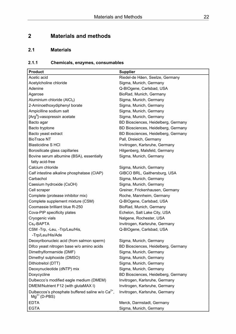
2.1.1 Chemicals, enzymes, consumables
Product Supplier Acetic acid Riedel-de Häen, Seelze, Germany Acetylcholine chloride Sigma, Munich, Germany Adenine Q-BIOgene, Carlsbad, USA Agarose BioRad, Munich, Germany Aluminium chloride (AlClBB3BB) Sigma, Munich, Germany 2-Aminoethoxydiphenyl borate Sigma, Munich, Germany Ampicilline sodium salt Sigma, Munich, Germany [ArgPP
8PP]-vasopressin acetate Sigma, Munich, Germany
Bacto agar BD Biosciences, Heidelberg, Germany Bacto tryptone BD Biosciences, Heidelberg, Germany Bacto yeast extract BD Biosciences, Heidelberg, Germany BioTrace NT Pall, Dreieich, Germany Blasticidine S HCl Invitrogen, Karlsruhe, Germany Borosilicate glass capillaries Hilgenberg, Malsfeld, Germany Bovine serum albumine (BSA), essentially Sigma, Munich, Germany
fatty acid-free Calcium chloride Sigma, Munich, Germany TTCalf intestine alkaline phosphatase (CIAP) TT GIBCO BRL, Gaithersburg, USA TTCarbacholTT Sigma, Munich, Germany TTCaesium hydroxide (CsOH)TT Sigma, Munich, Germany Cell scraper Greiner, Frickenhausen, Germany Complete (protease inhibitor mix) Roche, Mannheim, Germany Complete supplement mixture (CSM) Q-BIOgene, Carlsbad, USA Coomassie brilliant blue R-250 BioRad, Munich, Germany Cova-PIP specificity plates Echelon, Salt Lake City, USA Cryogenic vials Nalgene, Rochester, USA CsBB4BB-BAPTA Invitrogen, Karlsruhe, Germany CSM -Trp, -Leu, -Trp/Leu/His, Q-BIOgene, Carlsbad, USA
-Trp/Leu/His/Ade Deoxyribonucleic acid (from salmon sperm) Sigma, Munich, Germany Difco yeast nitrogen base w/o amino acids BD Biosciences, Heidelberg, Germany Dimethylformamide (DMF) Sigma, Munich, Germany Dimethyl sulphoxide (DMSO) Sigma, Munich, Germany Dithiotreitol (DTT) Sigma, Munich, Germany Deoxynucleotide (dNTP) mix Sigma, Munich, Germany Doxycycline BD Biosciences, Heidelberg, Germany Dulbecco’s modified eagle medium (DMEM) Invitrogen, Karlsruhe, Germany DMEM/Nutrient F12 (with glutaMAX I) Invitrogen, Karlsruhe, Germany Dulbeccos’s phosphate buffered saline w/o CaPP
2+PP,
Mg PP
2+ PP(D-PBS)
Invitrogen, Karlsruhe, Germany
EDTA Merck, Darmstadt, Germany EGTA Sigma, Munich, Germany
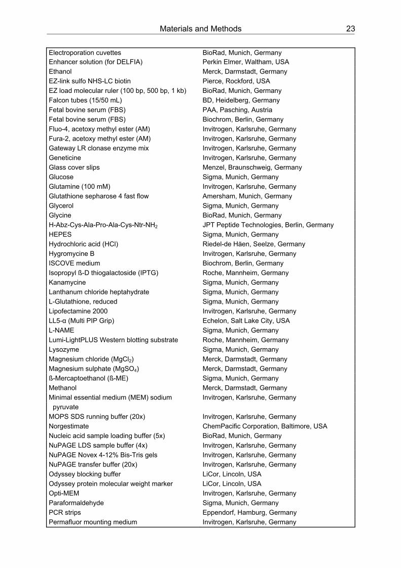
Materials and Methods 23
Electroporation cuvettes BioRad, Munich, Germany Enhancer solution (for DELFIA) Perkin Elmer, Waltham, USA Ethanol Merck, Darmstadt, Germany EZ-link sulfo NHS-LC biotin Pierce, Rockford, USA EZ load molecular ruler (100 bp, 500 bp, 1 kb) BioRad, Munich, Germany Falcon tubes (15/50 mL) BD, Heidelberg, Germany Fetal bovine serum (FBS) PAA, Pasching, Austria Fetal bovine serum (FBS) Biochrom, Berlin, Germany Fluo-4, acetoxy methyl ester (AM) Invitrogen, Karlsruhe, Germany Fura-2, acetoxy methyl ester (AM) Invitrogen, Karlsruhe, Germany Gateway LR clonase enzyme mix Invitrogen, Karlsruhe, Germany Geneticine Invitrogen, Karlsruhe, Germany Glass cover slips Menzel, Braunschweig, Germany Glucose Sigma, Munich, Germany Glutamine (100 mM) Invitrogen, Karlsruhe, Germany Glutathione sepharose 4 fast flow Amersham, Munich, Germany Glycerol Sigma, Munich, Germany Glycine BioRad, Munich, Germany H-Abz-Cys-Ala-Pro-Ala-Cys-Ntr-NH BB2BB JPT Peptide Technologies, Berlin, Germany HEPES Sigma, Munich, Germany Hydrochloric acid (HCl) Riedel-de Häen, Seelze, Germany Hygromycine B Invitrogen, Karlsruhe, Germany ISCOVE medium Biochrom, Berlin, Germany Isopropyl ß-D thiogalactoside (IPTG) Roche, Mannheim, Germany Kanamycine Sigma, Munich, Germany Lanthanum chloride heptahydrate Sigma, Munich, Germany L-Glutathione, reduced Sigma, Munich, Germany Lipofectamine 2000 Invitrogen, Karlsruhe, Germany LL5-α (Multi PIP Grip) Echelon, Salt Lake City, USA L-NAME Sigma, Munich, Germany Lumi-LightPLUS Western blotting substrate Roche, Mannheim, Germany Lysozyme Sigma, Munich, Germany Magnesium chloride (MgCl BB2BB) Merck, Darmstadt, Germany Magnesium sulphate (MgSO BB4BB) Merck, Darmstadt, Germany ß-Mercaptoethanol (ß-ME) Sigma, Munich, Germany Methanol Merck, Darmstadt, Germany Minimal essential medium (MEM) sodium Invitrogen, Karlsruhe, Germany
pyruvate MOPS SDS running buffer (20x) Invitrogen, Karlsruhe, Germany Norgestimate ChemPacific Corporation, Baltimore, USA Nucleic acid sample loading buffer (5x) BioRad, Munich, Germany NuPAGE LDS sample buffer (4x) Invitrogen, Karlsruhe, Germany NuPAGE Novex 4-12% Bis-Tris gels Invitrogen, Karlsruhe, Germany NuPAGE transfer buffer (20x) Invitrogen, Karlsruhe, Germany Odyssey blocking buffer LiCor, Lincoln, USA Odyssey protein molecular weight marker LiCor, Lincoln, USA Opti-MEM Invitrogen, Karlsruhe, Germany Paraformaldehyde Sigma, Munich, Germany PCR strips Eppendorf, Hamburg, Germany Permafluor mounting medium Invitrogen, Karlsruhe, Germany
Materials and Methods 24
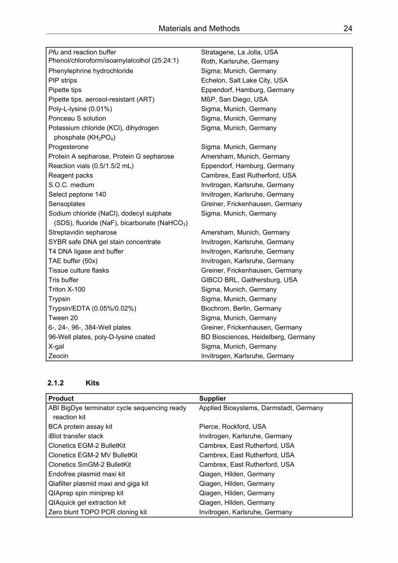
Pfu and reaction buffer Stratagene, La Jolla, USA Phenol/chloroform/isoamylalcolhol (25:24:1) Roth, Karlsruhe, Germany Phenylephrine hydrochloride Sigma; Munich, Germany PIP strips Echelon, Salt Lake City, USA Pipette tips Eppendorf, Hamburg, Germany Pipette tips, aerosol-resistant (ART) MßP, San Diego, USA Poly-L-lysine (0.01%) Sigma, Munich, Germany Ponceau S solution Sigma, Munich, Germany Potassium chloride (KCl), dihydrogen Sigma, Munich, Germany
phosphate (KHBB2BBPO BB4BB) Progesterone Sigma. Munich, Germany Protein A sepharose, Protein G sepharose Amersham, Munich, Germany Reaction vials (0.5/1.5/2 mL) Eppendorf, Hamburg, Germany Reagent packs Cambrex, East Rutherford, USA S.O.C. medium Invitrogen, Karlsruhe, Germany Select peptone 140 Invitrogen, Karlsruhe, Germany Sensoplates Greiner, Frickenhausen, Germany Sodium chloride (NaCl), dodecyl sulphate Sigma, Munich, Germany
(SDS), fluoride (NaF), bicarbonate (NaHCO BB3BB) Streptavidin sepharose Amersham, Munich, Germany SYBR safe DNA gel stain concentrate Invitrogen, Karlsruhe, Germany T4 DNA ligase and buffer Invitrogen, Karlsruhe, Germany TAE buffer (50x) Invitrogen, Karlsruhe, Germany Tissue culture flasks Greiner, Frickenhausen, Germany Tris buffer GIBCO BRL, Gaithersburg, USA Triton X-100 Sigma, Munich, Germany Trypsin Sigma, Munich, Germany Trypsin/EDTA (0.05%/0.02%) Biochrom, Berlin, Germany Tween 20 Sigma, Munich, Germany 6-, 24-, 96-, 384-Well plates Greiner, Frickenhausen, Germany 96-Well plates, poly-D-lysine coated BD Biosciences, Heidelberg, Germany X-gal Sigma, Munich, Germany Zeocin Invitrogen, Karlsruhe, Germany
2.1.2 Kits
Product Supplier ABI BigDye terminator cycle sequencing ready Applied Biosystems, Darmstadt, Germany
reaction kit BCA protein assay kit Pierce, Rockford, USA iBlot transfer stack Invitrogen, Karlsruhe, Germany Clonetics EGM-2 BulletKit Cambrex, East Rutherford, USA Clonetics EGM-2 MV BulletKit Cambrex, East Rutherford, USA Clonetics SmGM-2 BulletKit Cambrex, East Rutherford, USA Endofree plasmid maxi kit Qiagen, Hilden, Germany Qiafilter plasmid maxi and giga kit Qiagen, Hilden, Germany QIAprep spin miniprep kit Qiagen, Hilden, Germany QIAquick gel extraction kit Qiagen, Hilden, Germany Zero blunt TOPO PCR cloning kit Invitrogen, Karlsruhe, Germany
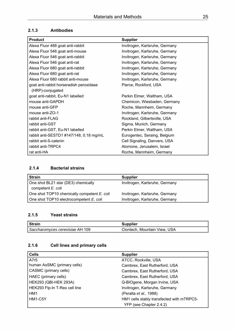
Materials and Methods 25
2.1.3 Antibodies
Product Supplier Alexa Fluor 488 goat anti-rabbit Invitrogen, Karlsruhe, Germany Alexa Fluor 546 goat anti-mouse Invitrogen, Karlsruhe, Germany Alexa Fluor 546 goat anti-rabbit Invitrogen, Karlsruhe, Germany Alexa Fluor 546 goat anti-rat Invitrogen, Karlsruhe, Germany Alexa Fluor 680 goat anti-rabbit Invitrogen, Karlsruhe, Germany Alexa Fluor 680 goat anti-rat Invitrogen, Karlsruhe, Germany Alexa Fluor 680 rabbit anti-mouse Invitrogen, Karlsruhe, Germany goat anti-rabbit horseradish peroxidase Pierce, Rockford, USA
(HRP)-conjugated goat anti-rabbit, Eu-N1 labelled Perkin Elmer, Waltham, USA mouse anti-GAPDH Chemicon, Wiesbaden, Germany mouse anti-GFP Roche, Mannheim, Germany mouse anti-ZO-1 Invitrogen, Karlsruhe, Germany rabbit anti-FLAG Rockland, Gilbertsville, USA rabbit anti-GST Sigma, Munich, Germany rabbit anti-GST, Eu-N1 labelled Perkin Elmer, Waltham, USA rabbit anti-SESTD1 #147/148, 0.18 mg/mL Eurogentec, Seraing, Belgium rabbit anti-ß-catenin Cell Signaling, Danvers, USA rabbit anti-TRPC4 Alomone, Jerusalem, Israel rat anti-HA Roche, Mannheim, Germany
2.1.4 Bacterial strains
Strain Supplier One shot BL21 star (DE3) chemically Invitrogen, Karlsruhe, Germany
competent E. coli One shot TOP10 chemically competent E. coli Invitrogen, Karlsruhe, Germany One shot TOP10 electrocompetent E. coli Invitrogen, Karlsruhe, Germany
2.1.5 Yeast strains
Strain Supplier Saccharomyces cerevisiae AH 109 Clontech, Mountain View, USA
2.1.6 Cell lines and primary cells
Cells Supplier A7r5 ATCC, Rockville, USA human AoSMC (primary cells) Cambrex, East Rutherford, USA CASMC (primary cells) Cambrex, East Rutherford, USA HAEC (primary cells) Cambrex, East Rutherford, USA HEK293 (QBI-HEK 293A) Q-BIOgene, Morgan Irvine, USA HEK293 Flp-In T-Rex cell line Invitrogen, Karlsruhe, Germany HM1 (Peralta et al., 1988) HM1-C5Y HM1 cells stably transfected with mTRPC5- YFP (see Chapter 2.4.2)
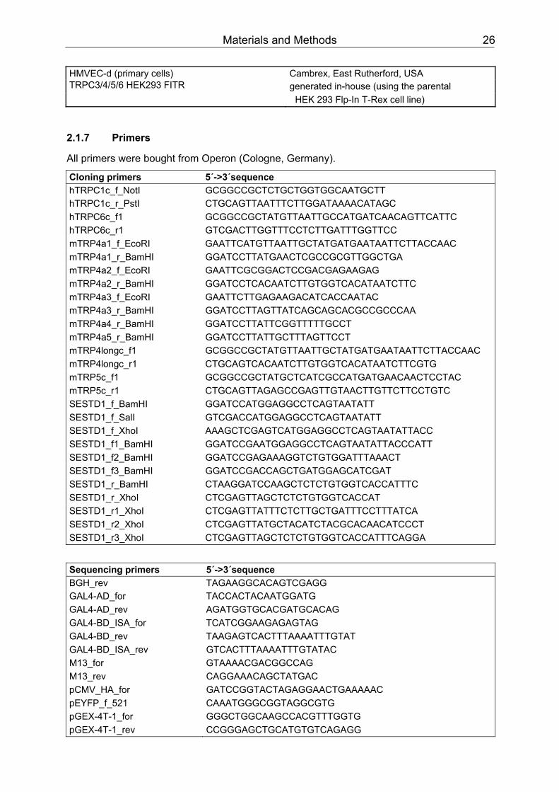
Materials and Methods 26
HMVEC-d (primary cells) Cambrex, East Rutherford, USA TRPC3/4/5/6 HEK293 FITR generated in-house (using the parental HEK 293 Flp-In T-Rex cell line)
2.1.7 Primers
All primers were bought from Operon (Cologne, Germany).
Cloning primers 5´->3´sequence hTRPC1c_f_NotI GCGGCCGCTCTGCTGGTGGCAATGCTT hTRPC1c_r_PstI CTGCAGTTAATTTCTTGGATAAAACATAGC hTRPC6c_f1 GCGGCCGCTATGTTAATTGCCATGATCAACAGTTCATTC hTRPC6c_r1 GTCGACTTGGTTTCCTCTTGATTTGGTTCC mTRP4a1_f_EcoRI GAATTCATGTTAATTGCTATGATGAATAATTCTTACCAAC mTRP4a1_r_BamHI GGATCCTTATGAACTCGCCGCGTTGGCTGA mTRP4a2_f_EcoRI GAATTCGCGGACTCCGACGAGAAGAG mTRP4a2_r_BamHI GGATCCTCACAATCTTGTGGTCACATAATCTTC mTRP4a3_f_EcoRI GAATTCTTGAGAAGACATCACCAATAC mTRP4a3_r_BamHI GGATCCTTAGTTATCAGCAGCACGCCGCCCAA mTRP4a4_r_BamHI GGATCCTTATTCGGTTTTTGCCT mTRP4a5_r_BamHI GGATCCTTATTGCTTTAGTTCCT mTRP4longc_f1 GCGGCCGCTATGTTAATTGCTATGATGAATAATTCTTACCAAC mTRP4longc_r1 CTGCAGTCACAATCTTGTGGTCACATAATCTTCGTG mTRP5c_f1 GCGGCCGCTATGCTCATCGCCATGATGAACAACTCCTAC mTRP5c_r1 CTGCAGTTAGAGCCGAGTTGTAACTTGTTCTTCCTGTC SESTD1_f_BamHI GGATCCATGGAGGCCTCAGTAATATT SESTD1_f_SalI GTCGACCATGGAGGCCTCAGTAATATT SESTD1_f_XhoI AAAGCTCGAGTCATGGAGGCCTCAGTAATATTACC SESTD1_f1_BamHI GGATCCGAATGGAGGCCTCAGTAATATTACCCATT SESTD1_f2_BamHI GGATCCGAGAAAGGTCTGTGGATTTAAACT SESTD1_f3_BamHI GGATCCGACCAGCTGATGGAGCATCGAT SESTD1_r_BamHI CTAAGGATCCAAGCTCTCTGTGGTCACCATTTC SESTD1_r_XhoI CTCGAGTTAGCTCTCTGTGGTCACCAT SESTD1_r1_XhoI CTCGAGTTATTTCTCTTGCTGATTTCCTTTATCA SESTD1_r2_XhoI CTCGAGTTATGCTACATCTACGCACAACATCCCT SESTD1_r3_XhoI CTCGAGTTAGCTCTCTGTGGTCACCATTTCAGGA
Sequencing primers 5´->3´sequence BGH_rev TAGAAGGCACAGTCGAGG GAL4-AD_for TACCACTACAATGGATG GAL4-AD_rev AGATGGTGCACGATGCACAG GAL4-BD_ISA_for TCATCGGAAGAGAGTAG GAL4-BD_rev TAAGAGTCACTTTAAAATTTGTAT GAL4-BD_ISA_rev GTCACTTTAAAATTTGTATAC M13_for GTAAAACGACGGCCAG M13_rev CAGGAAACAGCTATGAC pCMV_HA_for GATCCGGTACTAGAGGAACTGAAAAAC pEYFP_f_521 CAAATGGGCGGTAGGCGTG pGEX-4T-1_for GGGCTGGCAAGCCACGTTTGGTG pGEX-4T-1_rev CCGGGAGCTGCATGTGTCAGAGG
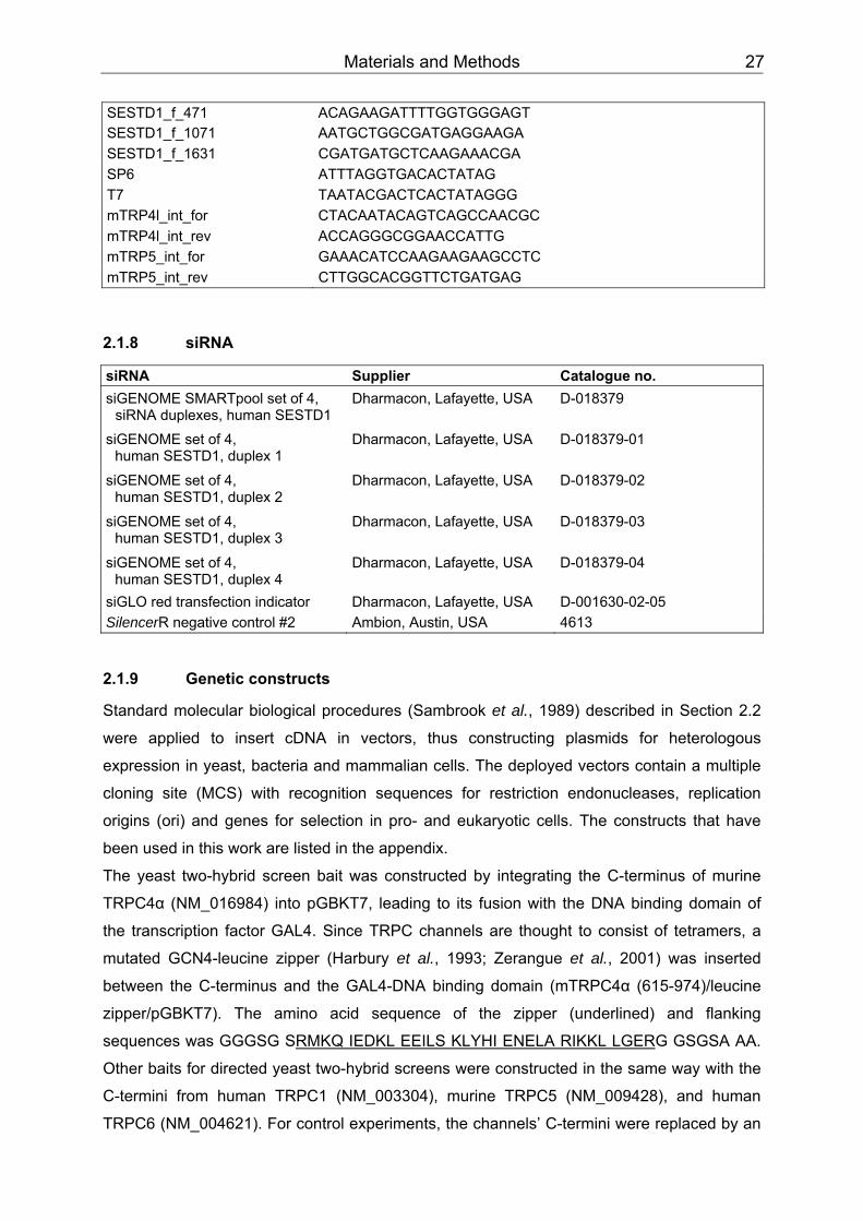
Materials and Methods 27
SESTD1_f_471 ACAGAAGATTTTGGTGGGAGT SESTD1_f_1071 AATGCTGGCGATGAGGAAGA SESTD1_f_1631 CGATGATGCTCAAGAAACGA SP6 ATTTAGGTGACACTATAG T7 TAATACGACTCACTATAGGG mTRP4l_int_for CTACAATACAGTCAGCCAACGC mTRP4l_int_rev ACCAGGGCGGAACCATTG mTRP5_int_for GAAACATCCAAGAAGAAGCCTC mTRP5_int_rev CTTGGCACGGTTCTGATGAG
2.1.8 siRNA
siRNA Supplier Catalogue no. siGENOME SMARTpool set of 4,
siRNA duplexes, human SESTD1 Dharmacon, Lafayette, USA
D-018379
siGENOME set of 4, human SESTD1, duplex 1
Dharmacon, Lafayette, USA
D-018379-01
siGENOME set of 4, human SESTD1, duplex 2
Dharmacon, Lafayette, USA
D-018379-02
siGENOME set of 4, human SESTD1, duplex 3
Dharmacon, Lafayette, USA
D-018379-03
siGENOME set of 4, human SESTD1, duplex 4
Dharmacon, Lafayette, USA
D-018379-04
siGLO red transfection indicator Dharmacon, Lafayette, USA D-001630-02-05 SilencerR negative control #2 Ambion, Austin, USA 4613
2.1.9 Genetic constructs
Standard molecular biological procedures (Sambrook et al., 1989) described in Section 2.2
were applied to insert cDNA in vectors, thus constructing plasmids for heterologous
expression in yeast, bacteria and mammalian cells. The deployed vectors contain a multiple
cloning site (MCS) with recognition sequences for restriction endonucleases, replication
origins (ori) and genes for selection in pro- and eukaryotic cells. The constructs that have
been used in this work are listed in the appendix.
The yeast two-hybrid screen bait was constructed by integrating the C-terminus of murine
TRPC4α (NM_016984) into pGBKT7, leading to its fusion with the DNA binding domain of
the transcription factor GAL4. Since TRPC channels are thought to consist of tetramers, a
mutated GCN4-leucine zipper (Harbury et al., 1993; Zerangue et al., 2001) was inserted
between the C-terminus and the GAL4-DNA binding domain (mTRPC4α (615-974)/leucine
zipper/pGBKT7). The amino acid sequence of the zipper (underlined) and flanking
sequences was GGGSG S UURMKQ IEDKL EEILS KLYHI ENELA RIKKL LGERUUG GSGSA AA.
Other baits for directed yeast two-hybrid screens were constructed in the same way with the
C-termini from human TRPC1 (NM_003304), murine TRPC5 (NM_009428), and human
TRPC6 (NM_004621). For control experiments, the channels’ C-termini were replaced by an

Materials and Methods 28
enhanced variant of the jellyfish Aequorea victoria green fluorescent protein (EGFP/leucine
zipper/pGBKT7). To map the SESTD1 interaction site on TRPC4, monomeric truncation bait
constructs were generated (lacking the zipper). This facilitated cloning as the TRPC4
C-terminal fragments could be inserted into pGBKT7 with a double instead of a triple ligation.
Constructs for recombinant expression of glutathione S-transferase (GST) fusion proteins in
bacteria were prepared by inserting full length human SESTD1 (NM_178123) or cDNA
fragments in pGEX vectors leading to their N-terminal fusion with GST: GST-
SESTD1 (1-696)/pGEX-4T-1, GST-Sec 14 (1-192)/pGEX-5X-3, GST-Spec 1 (193-
406)/pGEX-5X-3, and GST-Spec 2 (407-696)/pGEX-5X-3.
Human SESTD1 was also inserted in the vectors pCMV-HA and pEYFP-N1. When
transfected into mammalian cells it was expressed N-terminally fused to the hemagglutinin
antigenic epitope of human influenza virus (HA tag: YPYDV PDYA) or C-terminally to a
yellow-green EGFP mutant protein (EYFP), respectively (Chalfie et al., 1994). DNA
constructs were confirmed by sequencing.
2.1.10 Apparatus
Product Supplier ABI 3100 genetic analyzer Applied Biosystems, Foster City, USA ALA BPS-8 (8-channel valve perfusion ALA Scientific Instruments, Westbury, USA
system) Axiovert 200 Zeiss, Göttingen, Germany Biofuge pico, Biofuge fresco Heraeus, Hanau, Germany BioPhotometer Eppendorf, Hamburg, Germany iBlot gel transfer device Invitrogen, Karlsruhe, Germany Casy counter Schärfe System, Reutlingen, Germany Cryo 1°C freezing container Nalgene, Rochester, USA Dissecting instruments WPI, Berlin, Germany DMZ universal puller Zeitz-Instruments, Munich, Germany EPC-10 HEKA, Lambrecht, Germany FLEX station Molecular Devices, Munich, Germany Fluorometric imaging plate reader (FLIPR) Molecular Devices, Munich, Germany Gel documentation system Intas, Göttingen, Germany Gene pulser BioRad, Munich, Germany Hera safe working bench Heraeus, Hanau, Germany Imaging system T.I.L.L. Photonics, Gräfeling, Germany Leica DM IRE2 Leica, Wetzlar, Germany Lumi imager Roche, Mannheim, Germany Milli-Q water purification system Millipore, Billerica, USA Multidrop plate washer Thermo Scientific, Milford, USA Novex Xcell 2 blotmodul Invitrogen, Karlsruhe, Germany Odyssey infrared imaging system LiCor, Lincoln, USA Rotator SB2, Stuart VWR, Darmstadt, Germany T3 thermocycler Biometra, Göttingen, Germany Tecan safire 2, Tecan ultra Tecan, Crailsheim, Germany TI1 UV transilluminator Biometra, Göttingen, Germany
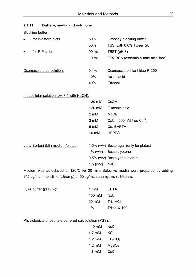
Materials and Methods 29
2.1.11 Buffers, media and solutions
UUBlocking buffer:
• for Western blots 50% Odyssey blocking buffer
50% TBS (with 0.6% Tween 20)
• for PIP strips 90 mL TBST (pH 8)
10 mL 30% BSA (essentially fatty acid-free)
UUCoomassie blue solution: UU 0.1% Coomassie brilliant blue R-250
10% Acetic acid
40% Ethanol
UUIntracellular solution (pH 7.4 with NaOH):
120 mM CsOH
120 mM Gluconic acid
2 mM MgCl BB2BB
3 mM CaCl BB2 BB(200 nM free Ca PP
2+PP)
5 mM Cs BB4BB-BAPTA
10 mM HEPES
UULuria Bertani (LB) medium/plates: UU 1.5% (w/v) Bacto agar (only for plates)
1% (w/v) Bacto tryptone
0.5% (w/v) Bacto yeast extract
1% (w/v) NaCl
Medium was autoclaved at 120°C for 20 min. Selective media were prepared by adding
100 µg/mL ampicilline (LB/amp) or 50 µg/mL kanamycine (LB/kana).
UULysis buffer (pH 7.4): UU 1 mM EDTA
150 mM NaCl
50 mM Tris-HCl
1% Triton X-100
UUPhysiological phosphate-buffered salt solution (PSS):
119 mM NaCl
4.7 mM KCl
1.2 mM KHBB2BBPOBB4BB
1.2 mM MgSOBB4BB
1.6 mM CaCl BB2
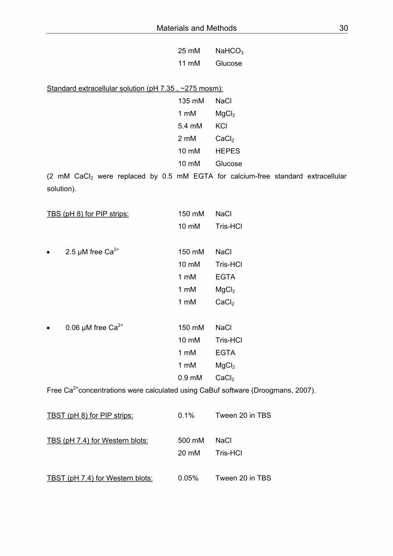
Materials and Methods 30
25 mM NaHCOBB3BB
11 mM Glucose
UUStandard extracellular solution (pH 7.35 , ~275 mosm):
135 mM NaCl
1 mM MgCl BB2BB
5.4 mM KCl
2 mM CaCl BB2
10 mM HEPES
10 mM Glucose
(2 mM CaCl BB2BB were replaced by 0.5 mM EGTA for calcium-free standard extracellular
solution).
UUTBS (pH 8) for PIP strips: UU 150 mM NaCl
10 mM Tris-HCl
• 2.5 µM free Ca PP
2+PP 150 mM NaCl
10 mM Tris-HCl
1 mM EGTA
1 mM MgCl BB2
1 mM CaCl BB2
• 0.06 µM free Ca PP
2+PP 150 mM NaCl
10 mM Tris-HCl
1 mM EGTA
1 mM MgCl BB2
0.9 mM CaCl BB2
Free Ca PP
2+PPconcentrations were calculated using CaBuf software (Droogmans, 2007).
UTBST (pH 8) for PIP strips: UU 0.1% Tween 20 in TBS
UUTBS (pH 7.4) for Western blots:UU 500 mM NaCl
20 mM Tris-HCl
UUTBST (pH 7.4) for Western blots:UU 0.05% Tween 20 in TBS
UU
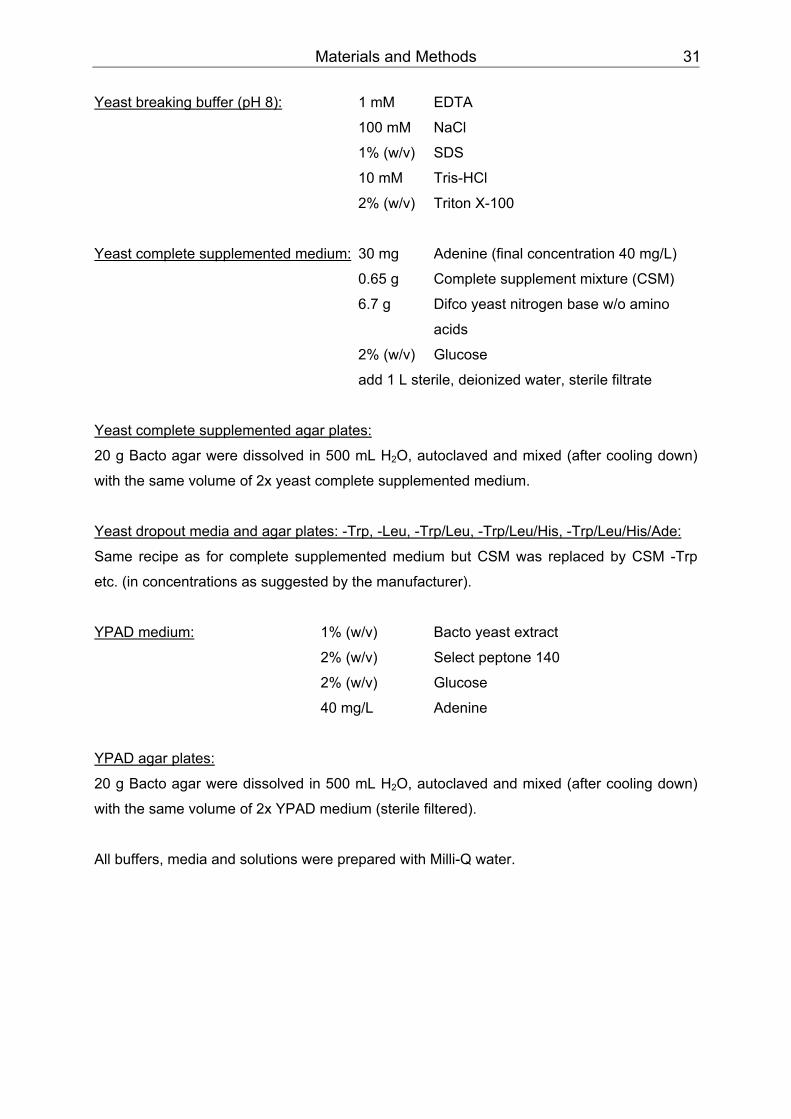
Materials and Methods 31
Yeast breaking buffer (pH 8): UU 1 mM EDTA
100 mM NaCl
1% (w/v) SDS
10 mM Tris-HCl
2% (w/v) Triton X-100
UUYeast complete supplemented medium: UU 30 mg Adenine (final concentration 40 mg/L)
0.65 g Complete supplement mixture (CSM)
6.7 g Difco yeast nitrogen base w/o amino
acids
2% (w/v) Glucose
add 1 L sterile, deionized water, sterile filtrate
UUYeast complete supplemented agar plates:
20 g Bacto agar were dissolved in 500 mL H BB2BBO, autoclaved and mixed (after cooling down)
with the same volume of 2x yeast complete supplemented medium.
UUYeast dropout media and agar plates: -Trp, -Leu, -Trp/Leu, -Trp/Leu/His, -Trp/Leu/His/Ade:
Same recipe as for complete supplemented medium but CSM was replaced by CSM -Trp
etc. (in concentrations as suggested by the manufacturer).
UUYPAD medium: UU 1% (w/v) Bacto yeast extract
2% (w/v) Select peptone 140
2% (w/v) Glucose
40 mg/L Adenine
UUYPAD agar plates:
20 g Bacto agar were dissolved in 500 mL H BB2BBO, autoclaved and mixed (after cooling down)
with the same volume of 2x YPAD medium (sterile filtered).
All buffers, media and solutions were prepared with Milli-Q water.
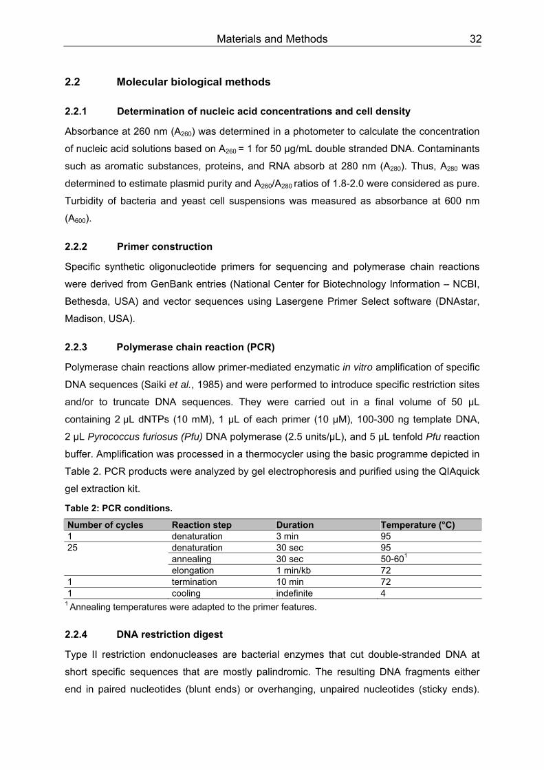
Materials and Methods 32
2.2 Molecular biological methods
2.2.1 Determination of nucleic acid concentrations and cell density
Absorbance at 260 nm (ABB260BB) was determined in a photometer to calculate the concentration
of nucleic acid solutions based on A BB260 BB= 1 for 50 µg/mL double stranded DNA. Contaminants
such as aromatic substances, proteins, and RNA absorb at 280 nm (A BB280BB). Thus, ABB280BB was
determined to estimate plasmid purity and A BB260BB/ABB280 BBratios of 1.8-2.0 were considered as pure.
Turbidity of bacteria and yeast cell suspensions was measured as absorbance at 600 nm
(ABB600BB).
2.2.2 Primer construction
Specific synthetic oligonucleotide primers for sequencing and polymerase chain reactions
were derived from GenBank entries (National Center for Biotechnology Information – NCBI,
Bethesda, USA) and vector sequences using Lasergene Primer Select software (DNAstar,
Madison, USA).
2.2.3 Polymerase chain reaction (PCR)
Polymerase chain reactions allow primer-mediated enzymatic in vitro amplification of specific
DNA sequences (Saiki et al., 1985) and were performed to introduce specific restriction sites
and/or to truncate DNA sequences. They were carried out in a final volume of 50 µL
containing 2 µL dNTPs (10 mM), 1 µL of each primer (10 µM), 100-300 ng template DNA,
2 µL Pyrococcus furiosus (Pfu) DNA polymerase (2.5 units/µL), and 5 µL tenfold Pfu reaction
buffer. Amplification was processed in a thermocycler using the basic programme depicted in
Table 2. PCR products were analyzed by gel electrophoresis and purified using the QIAquick
gel extraction kit.
Table 2: PCR conditions.
Number of cycles Reaction step Duration Temperature (°C) 1 denaturation 3 min 95 25 denaturation 30 sec 95 annealing 30 sec 50-60PP
1PP
elongation 1 min/kb 72 1 termination 10 min 72 1 cooling indefinite 4
PP
1 PPAnnealing temperatures were adapted to the primer features.
2.2.4 DNA restriction digest
Type II restriction endonucleases are bacterial enzymes that cut double-stranded DNA at
short specific sequences that are mostly palindromic. The resulting DNA fragments either
end in paired nucleotides (blunt ends) or overhanging, unpaired nucleotides (sticky ends).
Materials and Methods 33
For analytical digestions, 10 to 20 U restriction enzyme (New England Biolabs, Frankfurt,
Germany) were added to 1 µg DNA in a final volume of 30 µL 1x NEBuffer (New England
Biolabs). The DNA amount was increased up to 5 µg in 50 µL 1x NEBuffer for preparative
digestions. Buffer, addition of BSA, reaction temperature and time were applied as
suggested by the manufacturer depending on the enzyme(s) and the amount of DNA used.
The resulting DNA fragments were separated by gel electrophoresis and purified using the
QIAquick gel extraction kit.
Restriction maps were generated with Lasergene software (DNAstar, Madison, USA).
2.2.5 Dephosphorylation of linearized vectors
Removal of TT5´-phosphate groups prevents self-ligation of linearized vectors. TT1 UTT calf intestine
alkaline phosphatase (CIAP) was added TTto the restriction reaction TTand i TTncubated at 37°C for
5 min. The vector DNA was subsequently purified by gel electrophoresis and gel extraction.
2.2.6 DNA gel electrophoresis
DNA molecules have a net negative charge due to their phosphate backbone. They can thus
be forced to migrate through a gel by an electrical field resulting in their size-dependent
separation as shorter fragments migrate faster than longer ones. DNA samples were mixed
with 5x nucleic acid sample loading buffer and separated in gels of 1% agarose in TAE buffer
(prestained with SYBR safe DNA gel stain concentrate). Fragments were separated at 100-
120 V for 30-60 min and analyzed on a UV transilluminator by comparing their size with
molecular rulers. Pictures were taken with a digital gel documentation system before
fragment band excision and DNA elution with the QIAquick gel extraction kit.
2.2.7 Ligation
To join linearized DNA fragments, a final volume of 20 µL T4 DNA ligase buffer containing
50-100 ng linearized vector, the insert in a 3 molar excess and 1 U T4 ligase was incubated
for 16 hr at 16°C. The reaction was terminated by denaturating the enzyme for 10 min at
65°C. 2 µL of the reaction were transformed into electrocompetent E. coli.
2.2.8 TOPO cloning
For general subcloning, purified PCR products were inserted into the pCR-Blunt II-TOPO
vector using the zero blunt TOPO PCR cloning kit according to the manufacturer’s
instruction. This technique is based on topoisomerase I from Vaccinia virus that acts both as
restriction endonuclease and ligase. The vector is provided linearized with topoisomerase I
covalently attached to the 3’ phosphate residues. This bond can be attacked by the
5’ hydroxyl groups of an added PCR product that is consequently ligated into the vector. 1 µL
of the reaction was transformed into electrocompetent E. coli.

Materials and Methods 34
2.2.9 Gateway cloning
The Gateway technology (Invitrogen, Karlsruhe, Germany) was used to generate an
N-terminal FLAG-tagged mTRPC4ß construct for expression in mammalian cells (FLAG tag
amino acid sequence: DYKDDDDK). This system uses lambda (λ) phage-based site-specific
recombination (Landy, 1989) to insert a gene into a vector. mTRPC4ß (U50921), obtained
from the internal Genomic Sciences department, was already inserted in the entry vector
pDONR221 and thus flanked by λ phage attachment (att) sites. The destination vector
pcDNA3.1-nFLAG-DEST contains equivalent att sites. Site specific recombination was
initiated by mixing with mTRPC4ß/pDONR221 and the LR clonase enzyme mix and resulted
in the expression clone. The LR reaction was performed and terminated by incubation with
proteinase K according to the manufacturer’s instruction. 2 µL of the reaction were
transformed into electrocompetent E. coli.
2.2.10 Transformation of chemically competent bacteria
5–10 ng plasmid were added to a vial of thawed chemically competent E. coli, mixed gently
and incubated on ice for 10 min. Cell membranes were made permeable by heat shock (30
sec, 42°C) and immediately transferred to ice. After addition of 1 mL S.O.C. medium, cells
were incubated for one hr at 37°C and 250 rpm. 5 µL suspension of transformed bacteria
were diluted with 95 µL S.O.C. medium, spread on a selective LB agar plate and incubated
overnight at 37°C.
2.2.11 Electroporation of bacteria
2 µL ligation reaction were added usually into a vial of thawed electrocompetent E. coli,
mixed gently and transferred to a chilled 0.2 mm cuvette. The electroporator was charged by
2 kV with a load resistance of 400 Ω and 25 µFD capacity and bacteria were transformed
with a pulse of approximately 8 sec length. This electrical pulse disturbs the cell membrane
momentary thus allowing uptake of exogenous DNA. After addition of 1 mL S.O.C. medium,
cells were incubated for one hr at 37°C and 250 rpm. 100 µL of the bacteria suspension were
spread on a selective LB agar plate and incubated overnight at 37°C.
2.2.12 Plasmid amplification and purification
Bacteria, transformed with the corresponding plasmid, were plated on selective LB agar
plates and incubated overnight at 37°C. Next day, single colonies were used as inoculum for
liquid cultures in selective LB medium. Cells were grown overnight at 37°C and 250 rpm,
harvested by centrifugation and treated according to the manufacturer’s instruction (QIAprep
spin miniprep kit, Qiafilter plasmid maxi kit). Plasmids intended for transfection of mammalian
cells were harvested and purified with the endofree plasmid maxi kit. The human aorta cDNA

Materials and Methods 35
library used for the yeast two-hybrid screen was amplified on agar plates and prepared with
the Qiafilter plasmid giga kit (see Chapter 2.3.1).
2.2.13 DNA sequencing
Sequencing was done in-house by the Genomic Sciences department. DNA cycle
sequencing reactions were performed based on the dideoxy terminator method (Sanger et
al., 1977) with four differentially fluorescent-labelled dideoxynucleotides (Parker et al., 1996).
PCR fragments, generated by using the ABI PRISM BigDye terminator cycle sequencing
ready reaction kit, were electrophoretically separated, detected, and analyzed on an ABI
PRISM 3100 genetic analyzer.
2.2.14 Analysis of nucleotide and protein sequences
Nucleotide and protein sequences were imported from GenBank (NCBI, Bethesda, USA) and
analyzed and compared with Lasergene software (DNAstar, Madison, USA). The NCBI Basic
Local Alignment Search Tool (BLAST) was used to identify unknown sequences. Conserved
protein domains and motifs were searched with NCBI CD (Marchler-Bauer & Bryant, 2004)
and PROSITE (Swiss Institute of Bioinformatics, Basel, Switzerland).
2.2.15 Expression and purification of GST fusion proteins
Glutathione S-transferase (GST) fusion proteins were constructed by inserting a gene or
gene fragment in-frame into the MCS of a pGEX vector in which protein expression is under
control of the tac promoter and inducible by the lactose analogue isopropyl
ß-D thiogalactoside (IPTG). The resulting plasmid was transformed into protease-deficient
and chemically competent BL21 star (DE3) E. coli. Bacteria were plated on selective LB agar
plates and incubated overnight at 37°C. Next day, single colonies were used as inoculum for
a liquid overnight culture in selective LB medium (containing 0.2% (m/v) glucose to repress
protein expression). This starter culture was diluted in 100 mL selective LB medium (with
0.2% (m/v) glucose) resulting in ABB600 BB= 0.1. The culture was grown at room temperature (RT)
and agitated at 250 rpm. Protein expression was induced at A BB600 BB= 0.6–0.8 by adding 10 mM
(GST) or 20 mM (GST-SESTD1, GST-SESTD1-fragments) IPTG and incubation was
continued for 6 hr. The bacteria suspension was pelleted in 5 mL portions by centrifugation
(1 min, 16,000 x g, RT), pellets were washed once with 2 mL D-PBS (w/o Ca PP
2+PP, Mg PP
2+PP) and
stored at -80°C or lysed immediately. Each pellet was resuspended in 250 µL lysis buffer
(containing 1 mg/mL lysozyme) and incubated on ice for 20 min. After sonication (1x 5 sec)
the lysate was centrifuged 30 min at 16,000 x g and 4°C and the supernatant was transferred
to a fresh vial. 50 µL glutathione sepharose were washed three times with 500 µL lysis buffer
using a 27 gauge (G27) needle before the supernatant was added and filled up to 1 mL with
lysis buffer. After incubation on a rotator (1 hr, RT) the beads were washed three times with

Materials and Methods 36
500 µL lysis buffer. GST fusion proteins bound to glutathione sepharose were used for the
GST pulldown assay (see Chapter 2.5.5) or eluted with 100 µL elution buffer (20 mM
glutathione, 50 mM Tris-HCl, pH 8). Sepharose was vortexed and centrifuged for 1 min at
380 x g and 4°C. The supernatant was carefully transferred to a slide-a-lyzer MWCO 10,000
unit (Pierce, Rockford, USA) and dialyzed overnight against 50 mM Tris-HCl buffer (pH 8).
The protein concentration was subsequently determined using the BCA protein assay kit.
2.3 Yeast two-hybrid (Y2H) system
The MATCHMAKER two-hybrid system 3 (Clontech, Mountain View, USA) was used as a
transcriptional assay to screen for novel proteins that interact with the cytosolic C-terminus of
mTRPC4α (aa 615-974) in yeast strain AH109. The system is based on binding of the
transcription factor GAL4 upstream of four reporter genes (ADE2, HIS3, lacZ, MEL1)
activating their translation and protein expression. GAL4-DNA binding and activation are
mediated by two different protein domains which can be physically separated (Fields & Song,
1989).
The bait mTRPC4α (aa 615-974) was cloned into yeast expression vector pGBKT7 leading
to its nuclear expression as a fusion protein with the GAL4 DNA-binding domain (DNA-BD).
A mutated GCN4-leucine zipper (Harbury et al., 1993; Zerangue et al., 2001) was also
inserted between the C-terminus and the GAL4-DNA binding domain (mTRPC4α (615-
974)/leucine zipper/pGBKT7) to mimick the proposed tetrameric TRPC4 topology. A human
aorta cDNA library cloned into yeast expression vector pACT2 was screened with this bait.
The library proteins are expressed as fusion proteins with the GAL4 activation domain
(GAL4-AD) and also contain a nuclear localization sequence. When library and bait were
cotransfected into the auxotroph yeast strain AH109, the TRP1 gene of pGBKT7 and the
LEU2 gene of pACT2 served as selection markers allowing survival of cotransformants on
medium lacking the otherwise essential amino acids tryptophane (Trp) and leucine (Leu). But
only physical interaction of the bait with a library protein brought DNA-BD and GAL4-AD into
sufficient spatial proximity reconstituting the functional GAL4 and thus leading to the
expression of the reporter genes. These allowed survival on selective media additionally
lacking adenine (Ade) and histidine (His) and conversion of X-gal into a blue stain
(ß-galactosidase assay, lacZ-test). Prey plasmids were isolated from clones surviving on
-Trp/-Leu/-His/-Ade agar plates and positive reacting in the ß-galactosidase assay. First they
were analytically digested with the restriction endonucleases EcoRI/XhoI that cut the cDNA
inserts out of the vector MCS. The resulting fragments were separated by gel electrophoresis
whereby fragments of identical size pointed towards identical preys. Subsequently, the preys
were tested for intrinsic DNA-binding and/or transcriptional activity. Yeast was cotransformed
with the prey plasmid and a plasmid coding for the unrelated enhanced green fluorescent
protein (EGFP) from Aequorea victoria that was also tetramerized and fused to the DNA-BD
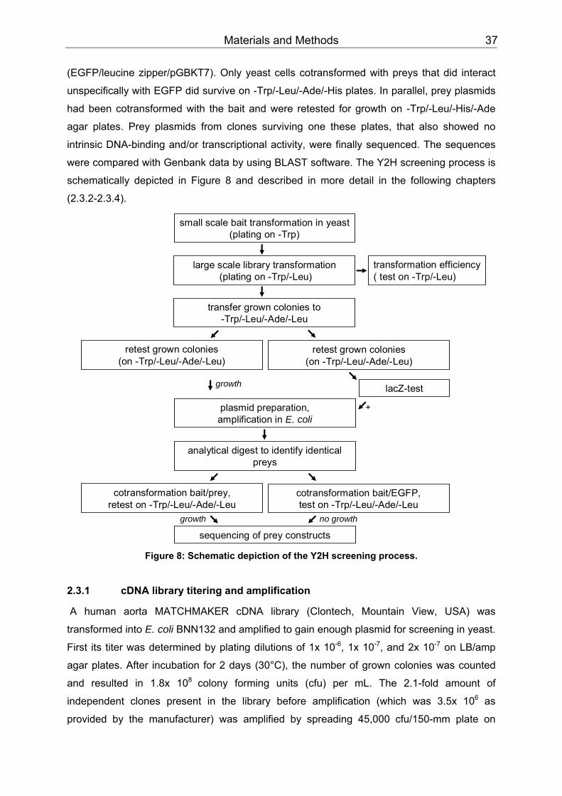
Materials and Methods 37
(EGFP/leucine zipper/pGBKT7). Only yeast cells cotransformed with preys that did interact
unspecifically with EGFP did survive on -Trp/-Leu/-Ade/-His plates. In parallel, prey plasmids
had been cotransformed with the bait and were retested for growth on -Trp/-Leu/-His/-Ade
agar plates. Prey plasmids from clones surviving one these plates, that also showed no
intrinsic DNA-binding and/or transcriptional activity, were finally sequenced. The sequences
were compared with Genbank data by using BLAST software. The Y2H screening process is
schematically depicted in Figure 8 and described in more detail in the following chapters
(2.3.2-2.3.4).
transformation efficiency( test on -Trp/-Leu)
small scale bait transformation in yeast(plating on -Trp)
large scale library transformation(plating on -Trp/-Leu)
transfer grown colonies to -Trp/-Leu/-Ade/-Leu
lacZ-test
retest grown colonies(on -Trp/-Leu/-Ade/-Leu)
retest grown colonies(on -Trp/-Leu/-Ade/-Leu)
plasmid preparation, amplification in E. coli
growth
growth no growth
analytical digest to identify identicalpreys
cotransformation bait/prey, retest on -Trp/-Leu/-Ade/-Leu
cotransformation bait/EGFP, test on -Trp/-Leu/-Ade/-Leu
sequencing of prey constructs
+
Figure 8: Schematic depiction of the Y2H screening process.
2.3.1 cDNA library titering and amplification
A human aorta MATCHMAKER cDNA library (Clontech, Mountain View, USA) was
transformed into E. coli BNN132 and amplified to gain enough plasmid for screening in yeast.
First its titer was determined by plating dilutions of 1x 10PP
-6PP, 1x 10PP
-7PP, and 2x 10 PP
-7PP on LB/amp
agar plates. After incubation for 2 days (30°C), the number of grown colonies was counted
and resulted in 1.8x 10 PP
8 PPcolony forming units (cfu) per mL. The 2.1-fold amount of
independent clones present in the library before amplification (which was 3.5x 10 PP
6PP as
provided by the manufacturer) was amplified by spreading 45,000 cfu/150-mm plate on
Materials and Methods 38
160 LB/amp agar plates. After 36 hr incubation at 30°C, cells were scraped into liquid
LB/amp (final volume ~2.1 L), pooled, partitioned into 4 aliquots, and pelleted by
centrifugation. Bacteria were lysed and plasmids purified using the QIAfilter plasmid giga kit.
The plasmid preparation protocol was adapted accordingly to the high cell density, starting
with the resuspension of each pellet in 400 mL buffer P1. Lysis and neutralization were
carried out by adding 400 mL buffer P2 and P3, respectively. An additional filtration step was
performed to preclear the lysates before loading onto two equilibrated QIAGEN-tips. Bound
DNA was washed, eluted and precipitated with isopropanol. The resulting DNA pellet was
washed with ethanol, dried in a vacuum centrifuge and resuspended in TE buffer.
2.3.2 Transformation of yeast
The DNA-BD/bait and the library were sequentially transformed into Saccharomyces
cerevisae using the polyethylene-glycol/lithium acetate (PEG/LiAc) method (Ito et al., 1983).
Through a small-scale transformation, the DNA-BD/bait was introduced into yeast and plated
on selective -Trp agar plates. Single transformants were used as inoculates for the following
large-scale transformation with the AD fusion library. It was verified that the bait is not
intrinsic DNA-binding and/or transcriptionally active by cotransformation with the negative
control vector pGADT7. pGADT7 is an empty vector leading to sole expression of the GAL4-
AD in yeast. Since this plasmid does not express a TRPC4-interacting protein,
cotransformation with the bait is not sufficient to reconstitute GAL4, thus yeast did not survive
on -Trp/-Leu/-Ade/-His plates.
Small-scale transformation
An overnight culture of Saccharomyces cerevisae AH109 grown at 30°C in 2 mL liquid YPAD
medium was transferred to 10 mL YPAD medium resulting in an ABB600 BB= 0.1. The cell
suspension was grown at 30°C until reaching the logarithmic growth phase (A BB600 BB= 0.5),
pelleted by centrifugation (5 min, 2,000 x g, RT), washed with sterile deionized water and
resuspended in 200 µL lithium acetate buffer (100 mM LiAc, 10 mM Tris-HCl, 1 mM EDTA).
18 µL carrier DNA from salmon sperm (10 mg/mL) were prepared by boiling for 40 sec, and
immediately transferred to ice. 1.5 µg DNA-BD/bait (3 µg plasmids for coexpressions) were
first mixed with the heat-denaturated carrier DNA and then added to 200 µL of the yeast
suspension. 1.2 mL PEG/LiAc-solution (40% PEG, 100 mM LiAc, 10 mM Tris-HCl, 1 mM
EDTA) were subsequently added and the cell suspension was incubated for 30 min at 30°C.
Cells were heat-shocked at 42°C for 15 min, centrifuged, resuspended in 100 µL TE-Buffer
(10 mM Tris-HCl, 1 mM EDTA, pH 8), spread on selective media, and incubated at 30°C for
at least 2 days. Yeast transformed with the DNA-BD/bait grew on -Trp medium,
cotransformants with a prey on -Trp/-Leu medium.
Materials and Methods 39
Large-scale transformation
Three overnight cultures of single yeast colonies transformed with the DNA-BD/bait were
transferred to 150 mL -Trp medium, subdivided onto twelve 50 mL-tubes and incubated
overnight at 30°C. Next day these starter cultures were pooled, pelleted by centrifugation
(3 min, 2,000 x g, RT) and resuspended in 10 mL -Trp medium. This suspension was added
to 900 mL -Trp medium (ABB600 BB= 0.15-0.25) and cells grew until the logarithmic growth phase
was reached (ABB600 BB= 0.45-0.75). The suspension was subdivided onto several 50 mL-tubes,
pelleted by centrifugation (5 min, 2,000 x g, RT), washed with 300 mL sterile deionized water
and pooled in 10 mL water. After washing with 50 mL lithium acetate buffer (100 mM LiAc,
10 mM Tris-HCl, 1 mM EDTA), cells were resuspended in 4 mL lithium acetate buffer.
In triplicates, 40 µg cDNA of the library were mixed with 145 µL heat denaturated carrier DNA
(from salmon sperm), 1.2 mL cell suspension and 8.6 mL PEG/LiAc-solution (40% PEG,
100 mM LiAc, 10 mM Tris-HCl, 1 mM EDTA) and incubated for 30 min at 250 rpm and 30°C.
After addition of 1 mL dimethyl sulphoxide (DMSO) per tube, cells were heat shocked
(15 min, 42°C), cooled down on ice and pelleted by centrifugation (5 min, 2,000 x g, RT).
Each pellet was resuspended in 25 mL -Trp/-Leu medium before they were pooled and
incubated for one hr at 250 rpm and 30°C. After centrifugation (5 min, 2,000 x g, RT), cells
were resuspended in 12 mL -Trp/-Leu medium, plated in 300 µL aliquots on -Trp/-Leu/-His
agar plates (150 mm diameter), and incubated for 2 to 5 days at 30°C. Grown colonies were
transferred to -Trp/-Leu/-His/-Ade agar plates and incubated for 2 to 3 more days. Surviving
colonies were tested with the ß-galactosidase assay and plasmids of positive reacting clones
were isolated.
To test the transformation efficiency, dilutions of 10 PP
-3PP to 10 PP
-6PP were plated on -Trp/-His agar
plates in parallel and the number of cfus was counted after incubating the plates for 2 days at
30°C. The library was considered to be screened completely, when the 4-fold number of
independent clones had been plated in total.
2.3.3 ß-galactosidase assay
Clones grown on -Trp/-Leu/-His/-Ade agar plates were top-layered with X-gal agar (0.5%
agarose, 500 mM NaPOBB4BB buffer (pH 7), 1% SDS, 2% X-gal in dimethylformamide (DMF)).
The agar was prepared by cooling down melted agarose to 50°C, addition of prewarmed
SDS and DMF, and immediate application to the yeast colonies. The yeast colonies were
incubated at 30°C up to 12 hr and generation of a blue stain was regularly checked.
2.3.4 Plasmid preparation from yeast
Clones surviving on -Trp/-Leu/-His/-Ade agar plates and positive reacting in the
ß-galactosidase assay were resuspended in 2 mL -Leu medium and grown overnight at
250 rpm and 30°C. They were pelleted by centrifugation, and resuspended in 200 µL lysis
Materials and Methods 40
buffer (2% Triton X-100, 1% SDS, 100 mM NaCl, 10 mM Tris-HCl (pH 8), 1 mM EDTA).
200 µL phenol/chloroform/isoamylalcolhol (25:24:1) and 100 µL acid-treated glass beads
were added and the cell walls were broken by vortexing this mixture. After centrifugation
(5 min, 10,000 x g, RT) 135 µL of the aqueous phase were transferred to a new tube, mixed
with 15 µL sodium acetate solution (10%) and 375 µL ethanol, and DNA was subsequently
precipitated by centrifugation (30 min, 10,000 x g, RT). The DNA pellet was washed with
ethanol (75%), dried at 37°C for 30 min, and resuspended in 10 µL Tris-HCl (10 mM, pH 8.5).
2 µL DNA were transformed by electroporation into E.coli and transformed bacteria were
grown on selective LB/kana agar plates. Next day, single colonies were grown in liquid
overnight cultures and the plasmids were purified using the QIAprep spin miniprep kit.
2.4 Culture of mammalian cells
To avoid contamination, all critical steps were carried out in a working bench under laminar
air flow. Cells were stored in liquid nitrogen until usage. A cryogenic vial containing 1.8 mL
cell suspension was thawed in a water bath (37°C). Cells were immediately and carefully
transferred to 10 mL prewarmed medium and pelleted by centrifugation (3 min, 300 x g, RT)
to remove DMSO. The cell pellet was resuspended in 15 mL fresh medium and cells were
grown in plastic tissue flasks of appropriate size at 37°C in a humified atmosphere (5 or 7%
COBB2BB). Media was replaced one day after thawing and then routinely twice a week. They were
split at 90% confluency by washing them once with D-PBS (w/o Ca PP
2+PP, Mg PP
2+PP) and adding
0.05% trypsin/0.02% EDTA for approx. 1 min. The protease solution was removed and cells
were detached by gently tapping the flask and rinsing the cells with medium. After counting
the cell number with a Casy cell counter, the desired density was adjusted with medium
before cells were seeded. For some applications (e.g. Ca PP
2+ PPimaging) glass cover slips had to
be coated before cells were seeded. The cover slips were incubated 5 min with poly-L-lysine
(0.01% stock solution diluted 1:5 in sterile deionized water), washed twice with water and air-
dried overnight. When cells were frozen for long term storage, cryogenic vials and media
were kept on ice. Cell pellets were resuspended in cold medium, the resulting cell
suspension was diluted with an equivalent volume of DMSO:FBS (1:5) freezing medium and
transferred to cryogenic vials. After cooling down in a Cryo 1°C freezing container (with
-1°C/min) that was placed in a -80°C freezer, they were transferred to liquid nitrogen on the
next day.
Primary cells (human AoSMC, CASMC, HAEC, HMVEC-d) were cultivated according to the
provider’s instruction.
Some stable cell lines (TRPC3/4/5/6 HEK293 FITR) were used that express recombinant
TRPC channels in a tetracycline-inducible manner from a specific genomic location. They
were generated in-house by using the Flp-In T-Rex system (Invitrogen, Karlsruhe, Germany)

Materials and Methods 41
and thus named FITR. This system is based on Flp recombinase that mediates site specific
recombination. The parental HEK293 Flp-In T-Rex cell line contains a Flp recombination
target (FRT) site and constitutively expresses the Tet repressor (TetR) controlled by the
human cytomegalie virus (CMV) promoter. This cell line is transfected with two plasmids. The
first contains the respective TRPC channel gene (flanked by a FRT site) whose protein
expression is controlled by a tetracycline-regulated hybrid CMV/tet operator 2 (TetOBB2 BB)
promoter. A cotransfected plasmid constitutively expresses Flp that mediates homologous
recombination between the FRT sites leading to integration of the TRPC gene into the cell’s
genome. TetR binds with high affinity to TetOBB2BB and represses TRPC channel expression.
When tetracycline (or its derivative doxycycline) is added, it binds to TetR leading to a
conformational change that prevents binding to TetO BB2BB. Therefore, the promoter that controls
TRPC expression is derepressed.
18 to 24 hr prior to the experiments, protein expression was induced in these cell lines by
adding 1 µg/mL doxycycline to the medium.
The following media were used for the indicated cell types:
• A7r5 cells (smooth muscle cells derived from embryonic rat thoracic aorta): DMEM (with
glutaMAX I, 4.5 g/L glucose and 110 mg/mL sodium pyruvate) supplemented with 10%
(v/v) FBS (from PAA).
• HAEC: Clonetics EGM-2 BulletKit
• Human AoSMC, CASMC: Clonetics SmGM-2 BulletKit
• HEK293 cells: ISCOVE medium supplemented with 10% (v/v) FBS (Biochrom) and
2 mM glutamine.
• HM1 cells (HEK293 cells stably expressing the human muscarinic receptor type 1):
DMEM/Nutrient F12 (with glutaMAX I) supplemented with 10% (v/v) FBS (PAA), 1 mM
glutamine and 400 µg/mL geneticine. 50 µg/mL zeocin were added for selection and
culture of clones stably expressing mTRPC5-YFP (HM1-C5Y cells).
• HMVEC-d: Clonetics EGM-2 MV BulletKit
• TRPC3/4/5/6 HEK293 FITR cells (HEK293 FITR cells expressing hTRPC3
(NM_003305)/mTRPC4ß (AAC05178)/mTRPC5 (NM_009428)/hTRPC6 (AF080394)
under the control of an inducible promoter as described above): DMEM (with glutaMAX I,
4.5 g/L glucose and 110 mg/mL sodium pyruvate) supplemented with 10% (v/v) FBS
(Biochrom), 1 mM glutamine, 1 mM MEM sodium pyruvate, 40 µg/mL hygromycine
(50 µg/mL for mTRPC5 FITR cells), and 15 µg/mL blasticidine S HCl.
Materials and Methods 42
2.4.1 Transfection of mammalian cells
Since FBS had no significant effect on transfection efficiency, cells were transfected in full
medium using Lipofectamine 2000. Only the liposomes were formed and loaded in
transfection medium (Opti-MEM). For co-immunoprecipitation experiments 3x 10 PP
6PP HM1 cells
(in 10 mL) were seeded in dishes of 9.4 cm diameter and grown to 80-90% confluency until
the next day. 4 µg of each plasmid were mixed in 1 mL transfection medium and incubated
for 20 min (RT) with 20 µL Lipofectamine 2000 (also diluted in 1 mL Opti-MEM). The mixture
was applied to the cells. For siRNA and plasmid cotransfections, 3x 10PP
5PP HM1 cells/2 mL/well
were seeded in a 6-well plate, growing to 30-50% confluency next day. 4 µg plasmid were
mixed with 2.5 µL 20 µM siRNA stock solution in 250 µL transfection medium and incubated
for 20 min (RT) with 5 µL Lipofectamine 2000 (also diluted in 250 µL Opti-MEM). The mixture
was applied to the cells leading to a final siRNA concentration of 20 nmol/L. These protocols
are exemplary and were adapted to the individual experimental conditions.
Success and efficiency of transfection were determined either by Western blot or by
additionally transfecting cells with the same amount of plasmid or siRNA and a small amount
(0.4 µg/well in a 6-well plate) of plasmid coding for GFP. 18 to 24 hr post transfection GFP
fluorescence was excited with an UV lamp and the number of transfected cells was
estimated. Transfection rates of 80% and more were defined as mandatory for functional
experiments with cell populations.
2.4.2 Generation of a HM1 cell line stably expressing mTRPC5-YFP
HM1 cells seeded in a 6-well plate and grown to approximately 80% confluency were
transfected with 4 µg of the mTRPC5-YFP construct using Lipofectamine 2000 as described.
The construct was generated by inserting an XbaI/NotI fragment of mTRPC5-GFP (Strubing
et al., 2003) into pcDNA3.1(-)zeo. The YFP fusion was obtained by joining a NotI/EcoRI cut
YFP-PCR fragment to the channel C-terminus. 24 hr post transfection clonal selection
started by cultivating the cells in complete medium supplemented with 50 µg/mL zeocin.
Cells were trypsinized and transferred to a new dish (94 mm diameter). Several days later
single clones were isolated by manual picking from grown colonies. During expansion,
expression of the construct was visually checked by exciting its YFP fluorescence at 515 nm.
Clones were functionally characterized in FLIPR assays and patch clamp experiments. One
clone was selected for further studies.
2.5 Protein biochemical methods
2.5.1 Preparation of cell lysates
Cells grown in tissue culture flasks were placed on ice (10 min) and washed twice with ice-
cold D-PBS (w/o Ca PP
2+PP, Mg PP
2+PP) before detaching them with a cell scraper in lysis buffer. They
Materials and Methods 43
were placed 1 hr on ice before centrifugation (15 min, 16,000 x g, 4°C). The supernatant was
frozen at -20°C (for GST-pulldown assays stabilized with 1 mM dithiotreitol and 10% glycerol)
or mixed with NuPAGE LDS sample buffer (4x) and 5% ß-mercaptoethanol (ß-ME), heat-
denaturated (95°C, 3 min) and centrifuged (16,000 x g, 2 min) before they were stored at
4°C.
2.5.2 Determination of protein content
The amount of total protein was colorimetrically detected and quantified with the
bicinchoninic acid (BCA) protein assay that is based on a biuret reaction. The absorbance of
the purple-coloured reaction product was measured at 562 nm with a Tecan safire 2 plate
reader and the protein concentration was determined based on a bovine serum albumin
(BSA) standard curve. The reaction was set up in a flat-bottom, transparent 96-well plate by
mixing
• 5 µL of each standard dilution (unknown samples were diluted 1:5 in lysis buffer)
• 150 µL of working reagent (prepared by mixing BCA reagent A with BCA reagent B
(50:1))
and incubating the plate at 37°C for 30 min before measuring the absorbance.
2.5.3 SDS-PAGE
This technique, called sodium dodecyl sulphate polyacrylamide gel electrophoresis (SDS
PAGE), allows separation of proteins. In samples treated as described in 2.5.1, proteins are
denaturated and negatively charged by SDS in proportion to their mass. When loaded on
Bis-Tris-HCl gradient gels (4-12%), they can thus be electrophoretically separated according
to their molecular weight. Electrophoresis was carried out in NuPAGE MOPS SDS running
buffer for 75 min at 150 V. Gels were blotted (see Section 2.5.4) or stained for 15 min with
Coomassie blue solution. Excess dye was removed by washing with a solution containing
10% acetic acid and 40% ethanol.
2.5.4 Western blot
After proteins have been separated by size with SDS PAGE they were transferred to a
nitrocellulose membrane (Bio TraceNT) using NuPAGE Transfer Buffer and a Novex Xcell 2
blotmodul or the iBlot gel transfer system according to the manufacturer’s instruction. To
verify transfer, the blotted proteins were stained with 0.1% Ponceau S Solution for 1 min and
excessive dye was removed by washing with water. The membrane was blocked with
blocking buffer (1 hr, RT) before incubating it with the primary antibody (1 hr, RT). After
washing it four times with TBST, it was incubated with the fluorescent-labelled or HRP-
conjugated secondary antibody (45 min, RT) and washed again four times with TBST.
Membranes were analyzed using the Odyssey infrared imaging system or the Lumi imager.
Materials and Methods 44
2.5.5 GST pulldown assay
HEK293 cells transiently transfected with the mTRPC4α-C-terminus or full length mTRPC4α,
mTRPC4ß or mTRPC5 channel were lysed and protein concentration determined using the
BCA protein assay kit. An equivalent of 50 µg total protein was added to GST or the GST
fusion proteins bound to glutathione sepharose (see 2.2.15), filled up to 1 mL with lysis buffer
and incubated on a rotator (2 hr, RT). Sepharose was washed three times with 500 µL lysis
buffer and a G27 needle before 20 µL 1x LDS sample buffer (containing 5% ß-ME) were
added. Samples were denaturated (95°C, 3 min), centrifuged and analyzed by Western blot.
2.5.6 Co-immunoprecipitation
24 hr after transfection, cells were harvested by placing on ice for 10 min before washing
twice with 10 mL ice-cold D-PBS (w/o Ca PP
2+PP, Mg PP
2+PP).
Cell membranes were isolated by scraping cells from one dish into 600 µl homogenization
buffer (320 mM sucrose, 5 mM HEPES, pH 7.4, supplemented with Complete protease
inhibitor). The cell suspensions from four dishes per transfection condition were pooled,
sonicated (1x 5 sec), centrifuged (100 x g, 10 min) and the resulting supernatant was
partitioned onto three vials and further centrifuged (100,000 x g, 30 min). Membrane proteins
were lyzed from the resulting pellet with 600 µL lysis buffer (supplemented with Complete
protease inhibitor) per pellet, incubated on ice for 1 hr and centrifuged (15 min, 16,000 x g,
4°C). The supernatants were transferred to a fresh vial and pooled. An aliquot was kept as
input control and the rest of the lysate was divided onto two vials. 4 µg of the primary
antibody were added to each sample and they were incubated overnight at 4°C on a rotator.
Cell lysates were gained by directly scraping cells from one dish into 600 µL lysis buffer
(supplemented with Complete protease inhibitor), incubation on ice for 1 hr and
centrifugation (15 min, 16,000 x g, 4°C). The supernatant was transferred to a fresh vial,
pooled with the lysates of the 3 remaining dishes that were equally transfected, and treated
as described above.
The next day, 30 µL protein A-sepharose or protein G-sepharose suspension, respectively,
were washed and added to the samples. Samples were incubated for 2 hr at 4°C. Sepharose
was washed three times with a G27 needle and 500 µL lysis buffer before 20 µL 2x LDS
sample buffer (containing 10% ß-ME) were added. Samples were denaturated (95°C, 3 min),
centrifuged and analyzed by Western blot.
2.5.7 Surface expression analysis
1.2x 10 PP
6 PPHM1 cells stably transfected with mTRPC5-YFP were plated in dishes with 94 mm
diameter. The next day, they were transfected with 40 nM siRNA or treated with liposomes
only. 48 hr post transfection, cells were washed with D-PBS. Surface proteins were
biotinylated by incubation with a membrane-impermeable form of biotin (1 mg/mL EZ-link
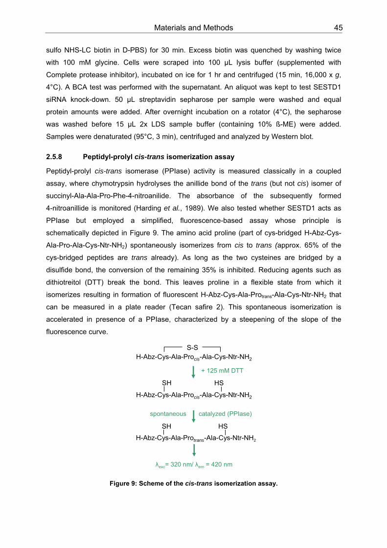
Materials and Methods 45
sulfo NHS-LC biotin in D-PBS) for 30 min. Excess biotin was quenched by washing twice
with 100 mM glycine. Cells were scraped into 100 µL lysis buffer (supplemented with
Complete protease inhibitor), incubated on ice for 1 hr and centrifuged (15 min, 16,000 x g,
4°C). A BCA test was performed with the supernatant. An aliquot was kept to test SESTD1
siRNA knock-down. 50 µL streptavidin sepharose per sample were washed and equal
protein amounts were added. After overnight incubation on a rotator (4°C), the sepharose
was washed before 15 µL 2x LDS sample buffer (containing 10% ß-ME) were added.
Samples were denaturated (95°C, 3 min), centrifuged and analyzed by Western blot.
2.5.8 Peptidyl-prolyl cis-trans isomerization assay
Peptidyl-prolyl cis-trans isomerase (PPIase) activity is measured classically in a coupled
assay, where chymotrypsin hydrolyses the anillide bond of the trans (but not cis) isomer of
succinyl-Ala-Ala-Pro-Phe-4-nitroanilide. The absorbance of the subsequently formed
4-nitroanillide is monitored (Harding et al., 1989). We also tested whether SESTD1 acts as
PPIase but employed a simplified, fluorescence-based assay whose principle is
schematically depicted in Figure 9. The amino acid proline (part of cys-bridged H-Abz-Cys-
Ala-Pro-Ala-Cys-Ntr-NH BB2BB) spontaneously isomerizes from cis to trans (approx. 65% of the
cys-bridged peptides are trans already). As long as the two cysteines are bridged by a
disulfide bond, the conversion of the remaining 35% is inhibited. Reducing agents such as
dithiotreitol (DTT) break the bond. This leaves proline in a flexible state from which it
isomerizes resulting in formation of fluorescent H-Abz-Cys-Ala-Pro BBtransBB-Ala-Cys-Ntr-NHBB2BB that
can be measured in a plate reader (Tecan safire 2). This spontaneous isomerization is
accelerated in presence of a PPIase, characterized by a steepening of the slope of the
fluorescence curve.
H-Abz-Cys-Ala-Procis-Ala-Cys-Ntr-NH2
+ 125 mM DTT
H-Abz-Cys-Ala-Procis-Ala-Cys-Ntr-NH2
spontaneous catalyzed (PPIase)
H-Abz-Cys-Ala-Protrans-Ala-Cys-Ntr-NH2
λexc= 320 nm/ λem = 420 nm
S-S
SH
HSSH
HS
Figure 9: Scheme of the cis-trans isomerization assay.

Materials and Methods 46
The assay was performed in a 384-well plate. Equal volumes (10 µL each) of 50 µM
substrate (cys-bridged H-Abz-Cys-Ala-Pro-Ala-Cys-Ntr-NH BB2BB), 125 mM DTT and 1 µM
FKBP12.6 or 4.76 µM GST-SESTD1, respectively, were mixed manually on ice before
fluorescence measurement was started. All solutions were prepared in 50 mM HEPES (pH
7.2), except GST-SESTD1 that was dialyzed against 50 mM Tris (pH 8).
2.5.9 Phospholipid overlay assay
PIP strips are commercially available nitrocellulose membranes containing 100 pmol
samples of 15 different phospholipids and a blank sample. They were blocked with blocking
buffer for PIP strips (1hr, RT) and incubated with blocking buffer containing 500 ng/mL
purified GST or GST-SESTD1 (4 hr, RT). After washing (3 times with blocking buffer), they
were incubated at 4°C overnight with the primary antibody anti-GST (1:2,000 in blocking
buffer). Three wash steps with TBST (pH 8) were followed by 45 min incubation with the
secondary HRP-conjugated anti-rabbit antibody (1:20,000 in blocking buffer). The
membranes were washed again three times with TBST (pH 8), incubated with 1 mL Lumi-
LightPLUS Western blotting substrate (5 min) and the chemiluminescent signals were
analyzed with a Lumi imager.
2.5.10 Cova-PIP specificity plate assay
The GST-tagged PH-domain of LL5-α, GST alone or GST-SESTD1 were diluted in blocking
buffer for PIP strips. Cova-PIP specificity plates loaded with 10 or 100 pmol PIP BBnBB per well
were incubated with 100 µL protein solution/well (3 hr, gentle agitation, RT). Plates were
washed manually 3 times with blocking buffer for PIP strips before incubation with 100 µL
anti-GST (1:1,000 in blocking buffer) for 1hr (gentle agitation, RT). Plates were washed
4 times with TBST for PIP strips using an automatic plate washer. For dissociation-enhanced
lanthanide fluorescence immunoassays (DELFIA), 100 µL secondary Eu-N1-labelled anti-
rabbit antibody (500 ng/mL in blocking buffer) were added per well and incubated 1 hr (gentle
agitation, RT). Plates were washed 4 times with TBST and an automatic plate washer before
100 µL enhancer solution were added per well. After 20 min incubation, fluorescence of the
lanthanide was excited (λ BBexc BB = 340 nm) and read (λ BBemBB = 620 nm) in a Tecan ultra plate reader.
2.5.11 Immunofluorescence
Five coverslips/well were placed in a 6-well plate and coated with poly-L-lysine (see Chapter
2.4.1). 5x 10 PP
5PP cells were plated per well in a volume of 2 mL. Next day they were transfected
with siRNA (see Chapter 2.4.1). 24 hr post transfection, the coverslips were transferred into a
24-well plate, washed twice with PBS (137 mM NaCl, 2.7 mM KCl, 10.1 mM Na BB2BBHPOBB4 BB,
1.4 mM KHBB2BBPOBB4BB) and fixed 15 min with 4% paraformaldehyde (in PBS). After washing with
PBS, cells were permeabilized with 0.1% Triton X-100 (in PBS) for 15 min and washed

Materials and Methods 47
again. They were blocked 1 hr with LiCor blocking buffer and thereafter incubated with the
primary antibody (diluted in LiCor blocking buffer:PBS = 1:1) for 1 hr. After washing, they
were incubated with the secondary fluorescent-labelled antibody (diluted in LiCor blocking
buffer:PBS = 1:1) for another hour. Four wash steps were followed by incubation with
Hoechst 33258 (1:10,000 in PBS) for 1 min and two additional wash steps. The coverslips
were mounted in Permafluor mounting medium and air-dried overnight. Probes were
analyzed with an inverted microscope (DM IRE2, Leica) and Leica Confocal Software (Leica,
Solms, Germany).
2.6 Fluorometric [CaPP
2+PP] BBi BB measurements
Changes in cytosolic calcium [Ca PP
2+PP] BBi BB were measured using the Ca PP
2+PP-sensitive fluorescent
dyes fluo-4 AM and fura-2 AM. The latter is a widely used indicator, whose fluorescence
excitation maximum shifts towards shorter wavelengths upon Ca PP
2+PP binding, while the
fluorescence emission maximum is relatively unchanged. Typically, the fluorescence
intensities excited at 340 nm (F BB340BB) and 380 nm (F BB380BB) are measured and the F BB340BB/F BB380BB ratio is
calculated. While FBB340 BBincreases upon binding of Ca PP
2+PP, FBB380 BBdecreases and an increase in
[Ca PP
2+PP] BBi BB consequently results in a rising FBB340BB/F BB380BB ratio. Factors that influence fluorescence
intensity, such as cell thickness, camera sensitivity, dye concentration and loss by leakage
and photobleaching, should affect measurements at both excitation wavelengths to the same
extent (Grynkiewicz et al., 1985). Thus, ratiometric measurements are less disturbed by
these effects. They were performed with single cells (CaPP
2+PP imaging) as well as cell
populations (FLEX experiments).
Ca PP
2+PP imaging
Cells grown on poly-L-lysine-coated 24-mm glass coverslips were loaded in cultivation
medium supplemented with 2 µM fura-2, acetoxy methyl ester (AM; 30 min, 37°C) and
subsequently allowed to de-esterify (through intracellular esterases cleaving off the acetate
residue) in standard extracellular solution (15 min, 37°C). Changes in [Ca PP
2+PP] BBi BB were measured
using an imaging system that consists of a xenon arc lamp, a monochromator, an inverted
microscope (Axiovert 200) and a charge-coupled device (CCD) camera. Fluorescence was
excited alternating at 340 nm and 380 nm, long-pass filtered at 440 nm and captured at 2 sec
intervals. The 340/380 nm excitation ratio of selected cell areas was calculated with T.I.L.L.
vision 4.0 software (T.I.L.L. Photonics, Gräfelfing, Germany) after correction for background
fluorescence. All experiments were performed in a recording chamber with approximately
1 mL volume.
Materials and Methods 48
FLEX experiments
20,000-40,000 cells/well were seeded in black poly-L-lysine-coated glass bottom 96-well
plates (Sensoplates) and grown overnight to an almost confluent monolayer. They were
loaded in 100 µL standard extracellular solution supplemented with 2 µM fura-2 AM (30 min,
37°C) and allowed to de-esterify (15 min, 37°C). Intracellular calcium signals were
ratiometrically measured in a benchtop scanning fluorometer (FLEX). Fluorescence was
excited alternating at 340 nm and 380 nm, long-pass filtered at 495 nm and captured at 4 sec
intervals. The FBB340BB/F BB380BB ratio was calculated using SoftMax Pro software (Molecular Devices,
Munich, Germany). Baseline fluorescence was detected for 30 sec, before an agonist was
applied with the FLEX pipettor. Since the FLEX station has an 8-channel pipettor, it takes
more time for reading a 96-well plate than a fluorometric imaging plate reader (FLIPR) with a
96-channel pipettor. Therefore, a FLIPR was used for standard inhibition assays in well
characterized FITR cell lines.
In HM1-C5Y cells, CaPP
2+ PPrelease was calculated as area under the curve in Ca PP
2+PP free standard
extracellular solution. TRPC5-mediated Ca PP
2+PP influx was calculated by subtracting Ca PP
2+
PPrelease from the area under the curve in standard extracellular solution.
FLIPR measurements
Cells grown to an almost confluent monolayer on black poly-D-lysine coated 96-well plates
were washed once with standard extracellular solution (E) and incubated (30 min, RT) with
dye solution (2 µM fluo-4 AM, 0.02% Pluronic F127, 0.1% BSA in E). After washing three
times with E, induced and non-induced HEK293 FITR cells were either incubated with buffer
only or with different concentrations of steroid (10 min). Fluo-4 fluorescence was excited at
488 nm with an argon laser, measured and imaged in the FLIPR and activators were applied
by the FLIPR pipettor. Signals were analyzed with the software provided by the
manufacturer.
The half maximal inhibitory concentrations (IC BB50BB) of norgestimate, progesterone and
levonorgestrel on TRPC3 and TRPC6 HEK293 FITR cells were calculated based on area
under the curves. Ca PP
2+PP influx following activation into induced cells that were not incubated
with steroid was definded as 0% inhibited. Ca PP
2+PP influx into induced cells that were neither
incubated with steroid nor activated with OAG was set to 100% inhibition.
The ICBB50BB values on TRPC4 and TRPC5 HEK293 FITR cells were calculated at t = 62 sec, the
time point of the channel signal maximum. The raw TRPC4 and TRPC5 channel signals
were calculated by subtracting the curve of non-induced, stimulated cells from induced,
stimulated cells. CaPP
2+PP influx following activation into induced cells not treated with steroid was
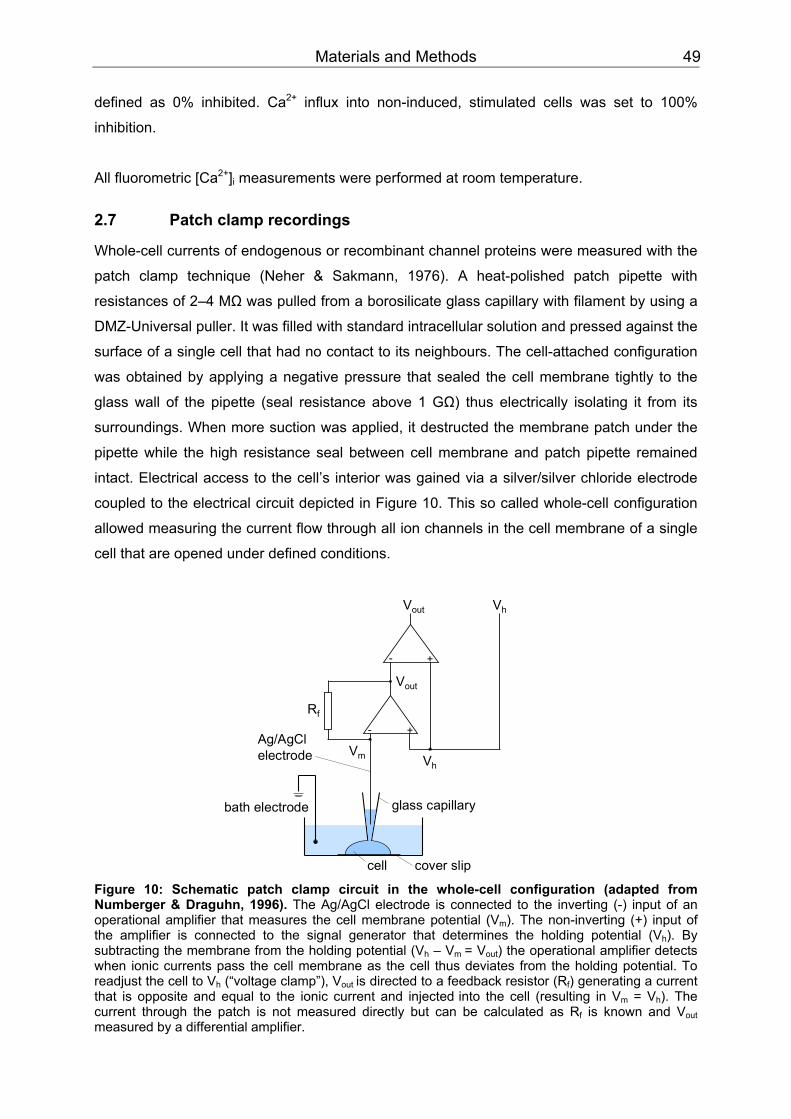
Materials and Methods 49
defined as 0% inhibited. Ca PP
2+PP influx into non-induced, stimulated cells was set to 100%
inhibition.
All fluorometric [CaPP
2+PP] BBi BB measurements were performed at room temperature.
2.7 Patch clamp recordings
Whole-cell currents of endogenous or recombinant channel proteins were measured with the
patch clamp technique (Neher & Sakmann, 1976). A heat-polished patch pipette with
resistances of 2–4 MΩ was pulled from a borosilicate glass capillary with filament by using a
DMZ-Universal puller. It was filled with standard intracellular solution and pressed against the
surface of a single cell that had no contact to its neighbours. The cell-attached configuration
was obtained by applying a negative pressure that sealed the cell membrane tightly to the
glass wall of the pipette (seal resistance above 1 GΩ) thus electrically isolating it from its
surroundings. When more suction was applied, it destructed the membrane patch under the
pipette while the high resistance seal between cell membrane and patch pipette remained
intact. Electrical access to the cell’s interior was gained via a silver/silver chloride electrode
coupled to the electrical circuit depicted in Figure 10. This so called whole-cell configuration
allowed measuring the current flow through all ion channels in the cell membrane of a single
cell that are opened under defined conditions.
bath electrode
cell
Ag/AgClelectrode
glass capillary
cover slip
- +
- +
Rf
Vout Vh
VhVm
Vout
Figure 10: Schematic patch clamp circuit in the whole-cell configuration (adapted from Numberger & Draguhn, 1996). The Ag/AgCl electrode is connected to the inverting (-) input of an operational amplifier that measures the cell membrane potential (VBBm BB). The non-inverting (+) input of the amplifier is connected to the signal generator that determines the holding potential (VBBhBB). By subtracting the membrane from the holding potential (V BBhBB – VBBm BB= VBBout BB) the operational amplifier detects when ionic currents pass the cell membrane as the cell thus deviates from the holding potential. To readjust the cell to VBBhBB (“voltage clamp”), VBBout BBis directed to a feedback resistor (RBBfBB) generating a current that is opposite and equal to the ionic current and injectedBB BBinto the cell (resulting in VBBm BB = VBBhBB). The current through the patch is not measured directly but can be calculated as RBBfBB is known and VBBout BB measured by a differential amplifier.
Materials and Methods 50
24 hr prior to experiments, coverslips were placed in a 24-well plate, coated with poly-L-
lysine (see Chapter 2.4) and 10-20,000 cells/well were plated in a volume of 0.5-1 mL.
1 µg/mL doxycycline was added to FITR cells to induce TRPC channel protein expression
(see Chapter 2.4). Next day, coverslips were transferred to a recording chamber and cells
were continuously superfused with standard extracellular solution and low pressure (approx.
10 kPa, 8-channel valve perfusion system). For A7r5 cells a modified extracellular solution
with 200 µM Ca PP
2+PP was used. Whole-cell recordings were performed with an EPC-10 amplifier
and Pulse software (HEKA, Lambrecht, Germany). Cells were held at a potential of -70 mV,
and current-voltage (I-V) relationships were routinely measured every 3 sec from voltage
ramps (-100 mV to +80 mV) lasting 200 msec.
Data was acquired at 6.67 kHz and filtered with 2.22 kHz. The series resistance was
compensated. In some experiments channels were activated by intracellular application of
AlF BB4PBPB
-PP. For its infusion, 2 µL 3 mM AlCl BB3BB were mixed with 4 µL 0.5 M NaF and diluted in 200 µL
standard intracellular solution. All experiments were performed at room temperature.
Currents were leak-corrected by subtracting completely blocked currents from currents of
activated cells. As TRPC mediated currents decay over time they were interpolated before
and after application of a modulator. Current data obtained with the modulator were
subsequently normalized to the interpolated values. Mean current densities were calculated
by normalizing current amplitudes to the cell capacitance.
2.8 In vitro vascular function
Adult male Wistar-Unilever rats (8–11 weeks old; Harlan Winkelmann, Borchen, Germany)
were sacrificed by decapitation. Thoracic aortas were excised quickly, transferred to cold
physiological salt solution (PSS) and rinsed. After connective tissue and perivascular fat had
been carefully removed, aortas were dissected in 5 mm rings and hung on special hooks
(Hugo Sachs, March-Hugstetten, Germany) by inserting two parallel wires into the lumen.
The upper hook was connected to a force transducer and the lower hook fixed the aortic
rings to the bottom of an organ bath thus allowing isometric tension recording. The aortic
rings were equilibrated in PSS (37°C, 15 min) and bath solutions were continuously gassed
with carbogen (95% OBB2BB and 5% COBB2BB) to provide oxygenation and pH of 7.4. To mimick the
physiological state, the rings were set at 1000 mg passive tension (in 200 mg steps). Vessels
strongly contracting after application of 60 mM KCl were defined as intact. They were
washed out and further used to measure cumulative dose-response curves for norgestimate.
Relaxation was expressed as a percentage of the steady-state tension produced by
preceding phenylephrine application.

Materials and Methods 51
2.9 Statistics
Averaged data is expressed as means ± SEM and number of experiments is indicated as “n”.
For statistical analysis, Wilcoxon test was performed with SAS 9 software. P values less than
0.05 were considered as statistically significant and depicted as: P < 0.05: *; P < 0.01: **;
P < 0.001: ***.
The half maximal inhibitory concentration (ICBB50BB) was calculated with SigmaPlot (Systat
software, San Jose, USA) and the sigmoidal Hill-model: f = ax PP
bPP/(c PP
bPP+xPP
bPP). The half maximal
effective concentration (ECBB50BB) was calculated analogously with f = yBB0BB+axPP
bPP/(c PP
bPP+xPP
bPP).

Results 52
3 Results
3.1 Differential inhibition of TRPC channels by norgestimate
According to the World Health Organization (WHO), 30% of all deaths worldwide were
caused by various cardiovascular (CV) diseases in 2005 (WHO, 2007). TRPC channels are
considered important pharmacological targets for the development of novel medicines for
several CV pathologies including cardiomyopathy, vascular remodelling, hypertension and
high endothelial permeability (Dietrich et al., 2007a). So far, characterization of native TRPC
channels in cardiovascular tissues is hindered by the lack of specific tool compounds that
discriminate well between and within the TRPC subfamilies. Cloned channels were at first
investigated in heterologous overexpression systems generating controversial data in terms
of channel properties and regulation. Valuable insight into native channel properties was
gained by their down-regulation in primary cells as well as studying gene-deficient mouse
models. Nevertheless, specific pharmacological TRPC inhibitors would be very useful to
further elucidate the channels’ roles under physiological as well as pathophysiological
conditions. Preliminary tests of ion channel-modulating compounds had identified
norgestimate as a putative inhibitor of TRPC-mediated Ca PP
2+PP-influx. S
The following studies were performed to evaluate in detail the potential of this compound as
a specific pharmacological TRPC6 modulator.
3.1.1 FLIPR measurements
Differential inhibition of TRPC channels by norgestimate
Our initial experiments were aimed to determine the activity of norgestimate towards different
members of the TRPC family. HEK293 cell lines heterologously expressing homomeric
channels under the control of an inducible promoter (Flp-In T-Rex system, see Chapter 2.4)
were used throughout these studies. TRPC3 and TRPC6, two representative members of the
DAG-sensitive TRPC3/6/7 subfamily, were tested as well as the DAG-insensitive TRPC4 and
TRPC5. TRPC1 was not tested since its expression does not result in measurable ion
currents (Strubing et al., 2001).
Fluorometric measurements of Ca PP
2+PP entry using FLIPR showed that application of oleoyl-2-
acetyl-sn-glycerol (OAG), a membrane permeable diacylglycerol analogue, to induced
HEK293 Flp-In T-Rex (FITR) cells expressing TRPC3 or TRPC6 resulted in a robust
increase in the intracellular Ca PP
2+PP concentration (see Fig. 11). This increase was completely
absent in non-induced cells indicating that the measured responses were solely due to
TRPC3 and TRPC6 activity (data not shown). Ca PP
2+PP influx following application of 30 µM OAG
was strongly reduced in cells preincubated with 30 µM norgestimate (Fig. 11 A, C). The ICBB50BB
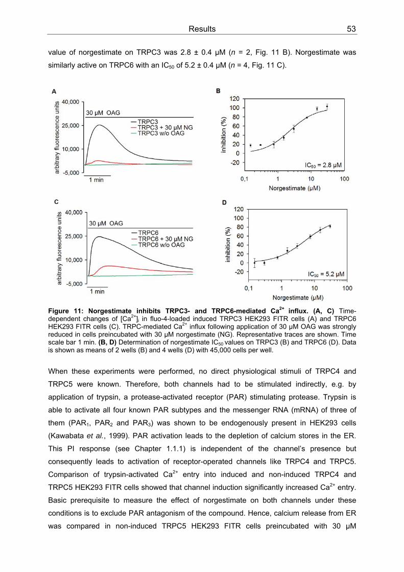
Results 53
value of norgestimate on TRPC3 was 2.8 ± 0.4 µM (n = 2, Fig. 11 B). Norgestimate was
similarly active on TRPC6 with an IC BB50BB of 5.2 ± 0.4 µM (n = 4, Fig. 11 C).
Figure 11: Norgestimate inhibits TRPC3- and TRPC6-mediated CaPP
2+ PPinflux. (A, C) Time-
dependent changes of [Ca PP
2+PP]BBi BB in fluo-4-loaded induced TRPC3 HEK293 FITR cells (A) and TRPC6
HEK293 FITR cells (C). TRPC-mediated CaPP
2+PP influx following application of 30 µM OAG was strongly
reduced in cells preincubated with 30 µM norgestimate (NG). Representative traces are shown. Time scale bar 1 min. (B, D) Determination of norgestimate IC BB50 BBvalues on TRPC3 (B) and TRPC6 (D). Data is shown as means of 2 wells (B) and 4 wells (D) with 45,000 cells per well. When these experiments were performed, no direct physiological stimuli of TRPC4 and
TRPC5 were known. Therefore, both channels had to be stimulated indirectly, e.g. by
application of trypsin, a protease-activated receptor (PAR) stimulating protease. Trypsin is
able to activate all four known PAR subtypes and the messenger RNA (mRNA) of three of
them (PAR BB1BB, PARBB2BB and PARBB3BB) was shown to be endogenously present in HEK293 cells
(Kawabata et al., 1999). PAR activation leads to the depletion of calcium stores in the ER.
This PI response (see Chapter 1.1.1) is independent of the channel’s presence but
consequently leads to activation of receptor-operated channels like TRPC4 and TRPC5.
Comparison of trypsin-activated Ca PP
2+PP entry into induced and non-induced TRPC4 and
TRPC5 HEK293 FITR cells showed that channel induction significantly increased Ca PP
2+PP entry.
Basic prerequisite to measure the effect of norgestimate on both channels under these
conditions is to exclude PAR antagonism of the compound. Hence, calcium release from ER
was compared in non-induced TRPC5 HEK293 FITR cells preincubated with 30 µM

Results 54
norgestimate or buffer only (10 min). Kinetics and quantity of calcium store release in cells
treated with 30 µM norgestimate were not changed compared to untreated cells. Therefore,
norgestimate is not a PAR antagonist (Fig. 12) and TRPC4 and TRPC5 activation via PAR
stimulation is suitable to measure the channel’s inhibition by norgestimate.
Figure 12: Norgestimate is not a PAR-antagonist. Time-dependent changes in [CaPP
2+PP]BBi BB of fluo-4-loaded TRPC5 HEK293 FITR
cells. Rise in [CaPP
2+PP]BBi BB following application of
200 nM trypsin (PI response) in non-induced TRPC5 HEK293 FITR cells was not suppressed in cells preincubated with 30 µM norgestimate (NG). Representative traces are shown. Time scale bar 1 min.
In contrast to TRPC3 and TRPC6, application of norgestimate to TRPC4 or TRPC5
expressing cells only caused a minor decrease of channel-mediated Ca PP
2+PP entry (Fig. 13 A,
C). ICBB50BB values of > 30 µM were determined for both TRPC4 (n = 2, Fig. 13 B) and TRPC5
(n = 4, Fig. 13 D). Channel inhibition by 30 µM norgestimate was amounted to 12.7% and
32.0% for TRPC4 and TRPC5 respectively.
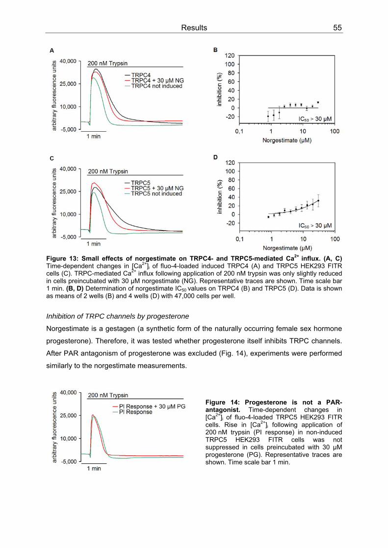
Results 55
Figure 13: Small effects of norgestimate on TRPC4- and TRPC5-mediated CaPP
2+ PPinflux. (A, C)
Time-dependent changes in [CaPP
2+PP]BBi BB of fluo-4-loaded induced TRPC4 (A) and TRPC5 HEK293 FITR
cells (C). TRPC-mediated CaPP
2+PP influx following application of 200 nM trypsin was only slightly reduced
in cells preincubated with 30 µM norgestimate (NG). Representative traces are shown. Time scale bar 1 min. (B, D) Determination of norgestimate ICBB50 BBvalues on TRPC4 (B) and TRPC5 (D). Data is shown as means of 2 wells (B) and 4 wells (D) with 47,000 cells per well.
Inhibition of TRPC channels by progesterone
Norgestimate is a gestagen (a synthetic form of the naturally occurring female sex hormone
progesterone). Therefore, it was tested whether progesterone itself inhibits TRPC channels.
After PAR antagonism of progesterone was excluded (Fig. 14), experiments were performed
similarly to the norgestimate measurements.
Figure 14: Progesterone is not a PAR-antagonist. Time-dependent changes in [CaPP
2+PP]BBi BB of fluo-4-loaded TRPC5 HEK293 FITR
cells. Rise in [CaPP
2+PP]BBi BB following application of
200 nM trypsin (PI response) in non-induced TRPC5 HEK293 FITR cells was not suppressed in cells preincubated with 30 µM progesterone (PG). Representative traces are shown. Time scale bar 1 min.
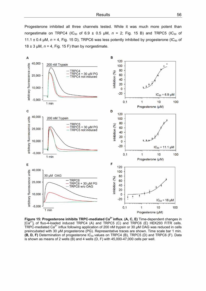
Results 56
Progesterone inhibited all three channels tested. While it was much more potent than
norgestimate on TRPC4 (ICBB50BB of 6.9 ± 0.5 µM, n = 2; Fig. 15 B) and TRPC5 (ICBB50BB of
11.1 ± 0.4 µM, n = 4, Fig. 15 D), TRPC6 was less potently inhibited by progesterone (ICBB50BB of
18 ± 3 µM, n = 4, Fig. 15 F) than by norgestimate.
Figure 15: Progesterone inhibits TRPC-mediated CaPP
2+ PPinflux. (A, C, E) Time-dependent changes in
[CaPP
2+PP]BBi BB of fluo-4-loaded induced TRPC4 (A) and TRPC5 (C) and TRPC6 (E) HEK293 FITR cells.
TRPC-mediated CaPP
2+PP influx following application of 200 nM trypsin or 30 µM OAG was reduced in cells
preincubated with 30 µM progesterone (PG). Representative traces are shown. Time scale bar 1 min. (B, D, F) Determination of progesterone IC BB50 BBvalues on TRPC4 (B), TRPC5 (D) and TRPC6 (F). Data is shown as means of 2 wells (B) and 4 wells (D, F) with 45,000-47,000 cells per well.

Results 57
Effect of progesterone, norgestimate and levonorgestrel on TRPC6
Whether or not norgestimate itself is active in vivo or merely serves as levonorgestrel
prodrug is controversially discussed (Stanczyk, 1997). Therefore, the effect of levonorgestrel
was also exemplarily tested on TRPC6-mediated Ca PP
2+PP-influx.
O
O
H
H
H
CH
H
O
HO-NH
H
H
CH3
O
CH
H
OH
OH
H
H
Figure 16: Chemical structures of progesterone (left), norgestimate (middle) and levonorgestrel (right). In contrast to norgestimate (Fig. 13 B, D) and progesterone (Fig. 15 E, F) that both inhibited
TRPC6, levonorgestrel was not active on the channel (Fig. 17 B). Even at 30 µM, the highest
levonorgestrel concentration tested, TRPC6 channels were not inhibited (Fig. 17 A).
Figure 17: TRPC6 is not inhibited by levonorgestrel. (A) Time-dependent changes in [CaPP
2+PP]BBi BB of fluo-
4-loaded induced TRPC6 HEK293 FITR cells. TRPC-mediated CaPP
2+PP influx following application of
30 µM OAG was not reduced in cells preincubated with 30 µM levonorgestrel (LG). Representative traces are shown. Time scale bar 1 min. (B) Determination of levonorgestrels IC BB50 BBvalue on TRPC6. Data is means of 4 wells with 45,000 cells per well. In summary, these FLIPR measurements showed that certain gestagens (norgestimate and
progesterone) inhibit channels of the TRPC3/6/7 subfamily as well as of the TRPC4/5
subfamily when applied at micromolar concentrations. However, this is not a general effect of
gestagens since the norgestimate metabolite levonorgestrel was completely inactive on
TRPC6.
Norgestimate was more active on the TRPC3/6/7 than on the TRPC4/5 subfamily, whereas
progesterone showed similar effects on the two subfamilies.

Results 58
3.1.2 Patch clamp recordings
Differential effect of norgestimate on recombinant homomeric TRPC5 and TRPC6 channels
The inhibition of TRPC6- and TRPC5-mediated Ca PP
2+PP-influx by norgestimate, which has been
monitored in cell populations with fluorometric experiments, was then validated by whole-cell
patch clamp recordings of single cells. Channels were indirectly excited with aluminium
tetrafluoride (AlF BB4PBPB
-PP) that was applied intracellularly via the patch pipette. The same stimulus
was used to activate both channels for better comparability of the norgestimate effect. AlFBB4PBPB
-PP
activates G proteins by mimicking guanosine triphosphate (GTP; Sternweis & Gilman, 1982;
Bigay et al., 1985). Activated GBBq/11 BB proteins stimulate PLC activity and in turn opening of
TRPC channels (Fig. 18 A, B). When 10 µM norgestimate were applied to activated TRPC6
channels, the current measured at resting membrane potential was reduced to 10.1 ± 3.1%
(n = 15, Fig. 18 C). This inhibition was reversible as the current amplitude increased again
after norgestimate wash out. The subsequent block by 10 µM lanthanum (LaPP
3+PP) was complete
and reversible (Fig. 18 A) and thus used for background (leak) calculation. By contrast,
application of 10 µM norgestimate to stimulated TRPC5 channels only had a minor effect
with a current reduction to 74.1 ± 5.7% (n = 20, Fig. 18 C). Since these channels are
potentiated by micromolar LaPP
3+ PPconcentrations (Jung et al., 2003), 2-aminoethoxydiphenyl
borate (2-APB), a known TRPC5 blocker (Xu et al., 2005) that completely and reversibly
blocked the channel at 10 µM, was used for leak correction (Fig. 18 B).
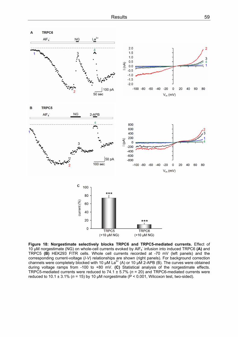
Results 59
Figure 18: Norgestimate selectively blocks TRPC6 and TRPC5-mediated currents. Effect of 10 µM norgestimate (NG) on whole-cell currents evoked by AlFBB4PBPB
-PP infusion into induced TRPC6 (A) and
TRPC5 (B) HEK293 FITR cells. Whole cell currents recorded at -70 mV (left panels) and the corresponding current-voltage (I-V) relationships are shown (right panels). For background correction channels were completely blocked with 10 µM LaPP
3+PP (A) or 10 µM 2-APB (B). The curves were obtained
during voltage ramps from -100 to +80 mV. (C) Statistical analysis of the norgestimate effects. TRPC5-mediated currents were reduced to 74.1 ± 5.7% (n = 20) and TRPC6-mediated currents were reduced to 10.1 ± 3.1% (n = 15) by 10 µM norgestimate (P < 0.001, Wilcoxon test, two-sided).
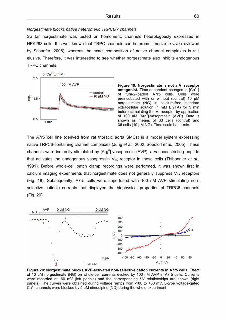
Results 60
Norgestimate blocks native heteromeric TRPC6/7 channels
So far norgestimate was tested on homomeric channels heterologously expressed in
HEK293 cells. It is well known that TRPC channels can heteromultimerize in vivo (reviewed
by Schaefer, 2005), whereas the exact composition of native channel complexes is still
elusive. Therefore, it was interesting to see whether norgestimate also inhibits endogenous
TRPC channels. Figure 19: Norgestimate is not a VBB1BB receptor antagonist. Time-dependent changes in [CaPP
2+PP]BBi BB
of fura-2-loaded A7r5 cells. Cells were preincubated with or without (control) 10 µM norgestimate (NG) in calcium-free standard extracellular solution (1 mM EGTA) for 5 min before stimulating the VBB1BB receptor by application of 100 nM [ArgPP
8PP]-vasopressin (AVP). Data is
shown as means of 33 cells (control) and 36 cells (10 µM NG). Time scale bar 1 min.
The A7r5 cell line (derived from rat thoracic aorta SMCs) is a model system expressing
native TRPC6-containing channel complexes (Jung et al., 2002; Soboloff et al., 2005). These
channels were indirectly stimulated by [Arg PP
8PP]-vasopressin (AVP), a vasoconstricting peptide
that activates the endogenous vasopressin VBB1ABB receptor in these cells (Thibonnier et al.,
1991). Before whole-cell patch clamp recordings were performed, it was shown first in
calcium imaging experiments that norgestimate does not generally suppress V BB1ABB receptors
(Fig. 19). Subsequently, A7r5 cells were superfused with 100 nM AVP stimulating non-
selective cationic currents that displayed the biophysical properties of TRPC6 channels
(Fig. 20).
Figure 20: Norgestimate blocks AVP-activated non-selective cation currents in A7r5 cells. Effect of 10 µM norgestimate (NG) on whole-cell currents evoked by 100 nM AVP in A7r5 cells. Currents were recorded at -60 mV (left panels) and the corresponding I-V relationships are shown (right panels). The curves were obtained during voltage ramps from -100 to +80 mV. L-type voltage-gated CaPP
2+PP channels were blocked by 5 µM nimodipine (ND) during the whole experiment.
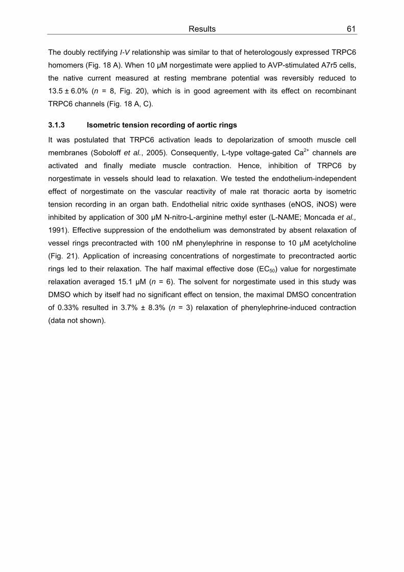
Results 61
The doubly rectifying I-V relationship was similar to that of heterologously expressed TRPC6
homomers (Fig. 18 A). When 10 µM norgestimate were applied to AVP-stimulated A7r5 cells,
the native current measured at resting membrane potential was reversibly reduced to
13.5 ± 6.0% (n = 8, Fig. 20), which is in good agreement with its effect on recombinant
TRPC6 channels (Fig. 18 A, C).
3.1.3 Isometric tension recording of aortic rings
It was postulated that TRPC6 activation leads to depolarization of smooth muscle cell
membranes (Soboloff et al., 2005). Consequently, L-type voltage-gated Ca PP
2+PP channels are
activated and finally mediate muscle contraction. Hence, inhibition of TRPC6 by
norgestimate in vessels should lead to relaxation. We tested the endothelium-independent
effect of norgestimate on the vascular reactivity of male rat thoracic aorta by isometric
tension recording in an organ bath. Endothelial nitric oxide synthases (eNOS, iNOS) were
inhibited by application of 300 µM N-nitro-L-arginine methyl ester (L-NAME; Moncada et al.,
1991). Effective suppression of the endothelium was demonstrated by absent relaxation of
vessel rings precontracted with 100 nM phenylephrine in response to 10 µM acetylcholine
(Fig. 21). Application of increasing concentrations of norgestimate to precontracted aortic
rings led to their relaxation. The half maximal effective dose (EC BB50BB) value for norgestimate
relaxation averaged 15.1 µM (n = 6). The solvent for norgestimate used in this study was
DMSO which by itself had no significant effect on tension, the maximal DMSO concentration
of 0.33% resulted in 3.7% ± 8.3% (n = 3) relaxation of phenylephrine-induced contraction
(data not shown).

Results 62
Figure 21: Endothelium-independent relaxation of precontracted rat aortic rings by norgestimate. (A) Application of norgestimate induced significant relaxation of L-NAME treated aortic rings precontracted with phenylephrine. (B) Concentration-relaxation curve of norgestimate (n = 6).

Results 63
3.2 Physical interaction of SESTD1 and TRPC channels
3.2.1 Y2H results
The second part of the present work was designed to identify novel TRPC4-interacting
proteins. Endothelial dysfunction is believed to be a major cause of various cardiovascular
diseases (Kwan et al., 2007) and evidence indicates that TRPC4 plays a critical role in
endothelial function. It is associated with regulation of endothelium-dependent vascular
relaxation (Freichel et al., 2001), may contribute to oxidative-stress induced endothelial
damage (Balzer et al., 1999; Poteser et al., 2006) and is necessary for endothelial barrier
function (Tiruppathi et al., 2002). TRPC4 expression in aortic endothelial cells has been
reported by several groups (Chang et al., 1997; Garcia & Schilling, 1997; Poteser et al.,
2006; Antoniotti et al., 2006). In search of novel proteins that interact with the cytosolic
C-terminus of mTRPC4α (aa 615-974), a human aorta cDNA library was screened with a
modified yeast two-hybrid system (Fields & Song, 1989). So far, no X-ray structures of TRPC
channels exist but due to similarity with voltage-gated K PP
+PP channels (Clapham et al., 2001),
they are expected also to form tetramers. Although our TRPC4 bait contained a putative
TRPC tetramerization domain (Lepage et al., 2006), we wanted to assure the protein
assembly in the physiological multimeric state. Therefore, the C-terminus of TRPC4 was
fused to a leucine zipper domain that has been shown previously to direct protein
tetramerization. Eleven proteins (listed in Table 3) were found to physically interact with the
mTRPC4α-C-terminus in this transcriptional assay.
Table 3: Y2H preys.
Definition Gene name Gene Bank accession no. Ankyrin repeat domain 35 ANKRD35 NM_144698 Apolipoprotein A-I binding protein APOA1BP NM_144772 Bromodomain adjacent to zinc finger domain BAZ1B AB032253 High-mobility group protein 2-like1 isoform b HMG2L1 CR456504 Makorin RING finger protein 1 MKRN1 NM_013446 Pre-B-cell leukemia homeobox interacting protein 1 PBXIP1 NM_020524 Sarcoma antigen NY-SAR-48 NM_033417 SEC14 and spectrin domains 1 SESTD1 NM_178123 Spectrin, alpha, non-erythrocytic 1 SPTAN1 U83867 Structural maintenance of chromosomes 3 SMC3 AF067163 Talin 2 TLN2 NM_015059
None of these proteins has been described before to interact with the TRPC4 channel.
SESTD1 seemed to be the most interesting potential interaction partner because a domain
search (Marchler-Bauer & Bryant, 2004) revealed the presence of an N-terminal Sec14p-like
lipid-binding domain (Fig. 22) in SESTD1. The conserved Sec14-motif is known to bind and
transport cellular phospholipids (Saito et al., 2007). Several reports have shown regulation of
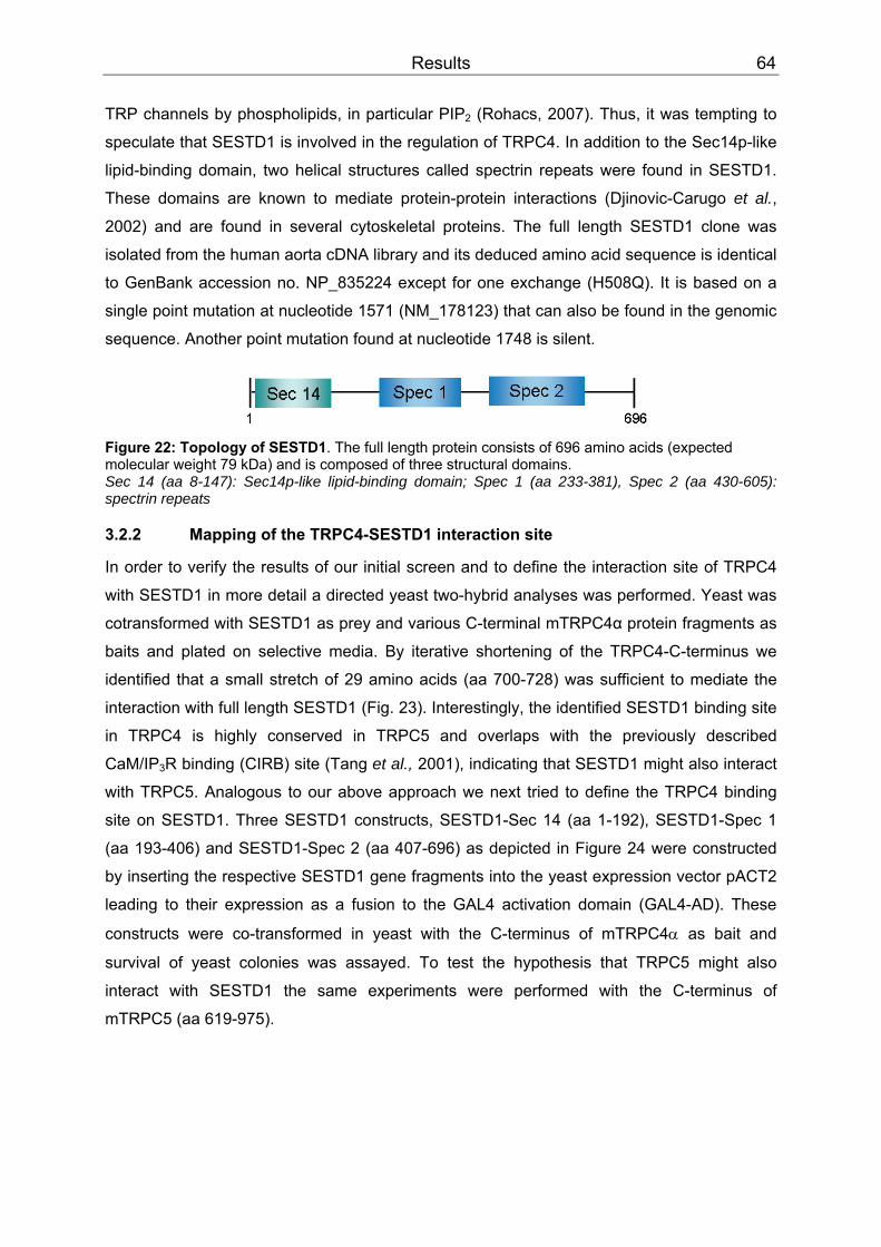
Results 64
TRP channels by phospholipids, in particular PIP BB2BB (Rohacs, 2007). Thus, it was tempting to
speculate that SESTD1 is involved in the regulation of TRPC4. In addition to the Sec14p-like
lipid-binding domain, two helical structures called spectrin repeats were found in SESTD1.
These domains are known to mediate protein-protein interactions (Djinovic-Carugo et al.,
2002) and are found in several cytoskeletal proteins. The full length SESTD1 clone was
isolated from the human aorta cDNA library and its deduced amino acid sequence is identical
to GenBank accession no. NP_835224 except for one exchange (H508Q). It is based on a
single point mutation at nucleotide 1571 (NM_178123) that can also be found in the genomic
sequence. Another point mutation found at nucleotide 1748 is silent.
Figure 22: Topology of SESTD1. The full length protein consists of 696 amino acids (expected molecular weight 79 kDa) and is composed of three structural domains. Sec 14 (aa 8-147): Sec14p-like lipid-binding domain; Spec 1 (aa 233-381), Spec 2 (aa 430-605): spectrin repeats
3.2.2 Mapping of the TRPC4-SESTD1 interaction site
In order to verify the results of our initial screen and to define the interaction site of TRPC4
with SESTD1 in more detail a directed yeast two-hybrid analyses was performed. Yeast was
cotransformed with SESTD1 as prey and various C-terminal mTRPC4α protein fragments as
baits and plated on selective media. By iterative shortening of the TRPC4-C-terminus we
identified that a small stretch of 29 amino acids (aa 700-728) was sufficient to mediate the
interaction with full length SESTD1 (Fig. 23). Interestingly, the identified SESTD1 binding site
in TRPC4 is highly conserved in TRPC5 and overlaps with the previously described
CaM/IPBB3BBR binding (CIRB) site (Tang et al., 2001), indicating that SESTD1 might also interact
with TRPC5. Analogous to our above approach we next tried to define the TRPC4 binding
site on SESTD1. Three SESTD1 constructs, SESTD1-Sec 14 (aa 1-192), SESTD1-Spec 1
(aa 193-406) and SESTD1-Spec 2 (aa 407-696) as depicted in Figure 24 were constructed
by inserting the respective SESTD1 gene fragments into the yeast expression vector pACT2
leading to their expression as a fusion to the GAL4 activation domain (GAL4-AD). These
constructs were co-transformed in yeast with the C-terminus of mTRPC4α as bait and
survival of yeast colonies was assayed. To test the hypothesis that TRPC5 might also
interact with SESTD1 the same experiments were performed with the C-terminus of
mTRPC5 (aa 619-975).
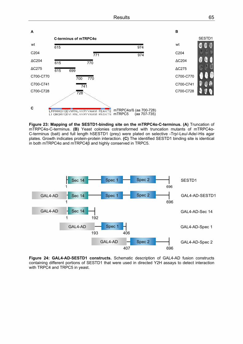
Results 65
SESTD1
B
wt
C204
∆C204
∆C275
C700-C770
C700-C741
C700-C728
wt
C204
∆C204
∆C275
C700-C770
C700-C741
C700-C728
615
700
741
728
C-terminus of mTRPC4α
615 974
974771
615 770
699
770
A
mTRPC4α/ß (aa 700-728)mTRPC5 (aa 707-735)
C
Figure 23: Mapping of the SESTD1-binding site on the mTRPC4α-C-terminus. (A) Truncation of mTRPC4α-C-terminus. (B) Yeast colonies cotransformed with truncation mutants of mTRPC4α-C-terminus (bait) and full length hSESTD1 (prey) were plated on selective -Trp/-Leu/-Ade/-His agar plates. Growth indicates protein-protein interaction. (C) The identified SESTD1 binding site is identical in both mTRPC4α and mTRPC4β and highly conserved in TRPC5.
Sec 141 192
GAL4-AD
GAL4-AD Spec 1193 406
Spec 1 Spec 2Sec 141 696
GAL4-AD Spec 2407 696
GAL4-AD-Sec 14
GAL4-AD-Spec 1
GAL4-AD-Spec 2
SESTD1
Spec 1 Spec 2Sec 141 696
GAL4-AD GAL4-AD-SESTD1
Figure 24: GAL4-AD-SESTD1 constructs. Schematic description of GAL4-AD fusion constructs containing different portions of SESTD1 that were used in directed Y2H assays to detect interaction with TRPC4 and TRPC5 in yeast.
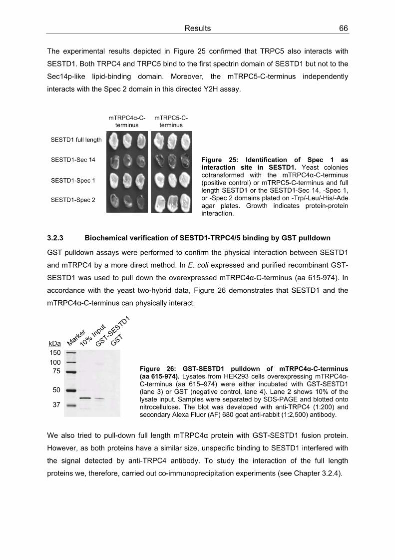
Results 66
SESTD1-Spec 2
SESTD1-Spec 1
SESTD1-Sec 14
mTRPC4α-C-terminus
mTRPC5-C-terminus
SESTD1 full length
50
37
75100150
10% In
put
GST-SESTD1
GSTkDa Mark
er
The experimental results depicted in Figure 25 confirmed that TRPC5 also interacts with
SESTD1. Both TRPC4 and TRPC5 bind to the first spectrin domain of SESTD1 but not to the
Sec14p-like lipid-binding domain. Moreover, the mTRPC5-C-terminus independently
interacts with the Spec 2 domain in this directed Y2H assay.
Figure 25: Identification of Spec 1 as interaction site in SESTD1. Yeast colonies cotransformed with the mTRPC4α-C-terminus (positive control) or mTRPC5-C-terminus and full length SESTD1 or the SESTD1-Sec 14, -Spec 1, or -Spec 2 domains plated on -Trp/-Leu/-His/-Ade agar plates. Growth indicates protein-protein interaction.
3.2.3 Biochemical verification of SESTD1-TRPC4/5 binding by GST pulldown
GST pulldown assays were performed to confirm the physical interaction between SESTD1
and mTRPC4 by a more direct method. In E. coli expressed and purified recombinant GST-
SESTD1 was used to pull down the overexpressed mTRPC4α-C-terminus (aa 615-974). In
accordance with the yeast two-hybrid data, Figure 26 demonstrates that SESTD1 and the
mTRPC4α-C-terminus can physically interact.
Figure 26: GST-SESTD1 pulldown of mTRPC4α-C-terminus (aa 615-974). Lysates from HEK293 cells overexpressing mTRPC4α-C-terminus (aa 615–974) were either incubated with GST-SESTD1 (lane 3) or GST (negative control, lane 4). Lane 2 shows 10% of the lysate input. Samples were separated by SDS-PAGE and blotted onto nitrocellulose. The blot was developed with anti-TRPC4 (1:200) and secondary Alexa Fluor (AF) 680 goat anti-rabbit (1:2,500) antibody.
We also tried to pull-down full length mTRPC4α protein with GST-SESTD1 fusion protein.
However, as both proteins have a similar size, unspecific binding to SESTD1 interfered with
the signal detected by anti-TRPC4 antibody. To study the interaction of the full length
proteins we, therefore, carried out co-immunoprecipitation experiments (see Chapter 3.2.4).

Results 67
10075
150kDa 10% In
put
GSTGST-S
pec 1
GST-Spec 2
mTRPC4α
10075
150kDa
mTRPC4ß
10075
150kDa
mTRPC5
A
B
C
Having shown that TRPC4 and SESTD1 physically interact in the pulldown assay, this
approach was further adapted to investigate the interaction sites between mTRPC4α,
mTRPC5 and SESTD1. We also included the shorter mTRPC4ß splice variant into these
experiments. Compared to mTRPC4α, it is lacking 84 C-terminal amino acids outside the
CIRB site (Schaefer et al., 2002). The same three SESTD1 domain constructs as depicted in
Figure 24 were used in these studies but they were cloned into pGEX-5X-3 instead of pACT2
and thus expressed as GST fusion proteins in the protease-deficient E. coli strain BL21 de3.
Under the chosen conditions, it was not possible to purify enough amounts of GST-Sec 14
(aa 1-192) with the Sec14p-like lipid-binding domain from bacteria. Induction of its
expression seemed to be toxic as these transformants grew much slower than bacteria
transformed with GST-Spec 1 (aa 193-406), GST-Spec 2 (aa 407-696) or full length GST-
SESTD1. Figure 27: Confirmation of Spec 1 as interaction site in SESTD1. (A, B) Anti-TRPC4 (1:200) immunoblot of samples precipitated with the indicated GST fusion proteins or GST from HEK293 cells overexpressing mTRPC4α (A) or mTRPC4ß (B). Lane 1 showing 10% of the lysate input (A, B, C) is stained turquoise (B) due to an artefact resulting from camera oversaturation. (C) Anti-TRPC5 (1:200) immunoblot of a similar experiment with HEK 293 cells overexpressing mTRPC5. Blots were developed with secondary AF 680 goat anti-rabbit (1:2,500) antibody.
All three channel proteins strongly interacted with the first spectrin domain (Fig. 27). In some
blots, a weak binding to the second domain could also be observed. Thus, the combined
results from the directed Y2H analyses (Chapter 3.2.2) and the GST pulldown studies
confirmed that the first spectrin domain of SESTD1 is the main site of interaction with
TRPC4α, TRPC4ß, and TRPC5.
3.2.4 Co-immunoprecipitation
Characterization of two polyclonal anti-SESTD1 antibodies
Co-immunoprecipitations allow to investigate protein-protein interactions of native or
recombinant proteins in vivo. As there were no commercial antibodies available against
SESTD1, two polyclonal peptide antibodies were custom-made by Eurogentec. Anti-SESTD1

Results 68
#147 antibody was directed against a sequence within the first spectrin domain (aa 265-280,
CRQRSKRTQLEEIQQK) and anti-SESTD1 #148 was directed against the SESTD1-C-
terminus (aa 682-696, KRQQLRHPEMVTTES). Antibodies were affinity-purified on the
respective peptides.
When tested on Western blot, both antibodies detected overexpressed HA-tagged SESTD1
in HM1 cell lysates (expected size 79 kDa). In addition, in non-transfected cells an
endogenous protein co-migrating with HA-SESTD1 was recognized by both antibodies.
Transfecting HM1 cells with siRNA duplexes directed against SESTD1 (see Chapter 3.3.3)
specifically suppressed the band at 79 kDa, strongly supporting the notion that this protein is
endogenous SESTD1.
100
150250
75
50
kDa
37
25
anti-HA
1:1,000
unrelated
SESTD1
A
anti-SESTD1 #148
1:5,000
100
150250
75
50
kDa
37
25
anti-HA
1:1,000
M N TM N T M N T M N T
anti-SESTD1 #147
1:200 1:1,000
unrelated
SESTD1
B
M N T
Figure 28: Polyclonal SESTD1 antibodies detect endogenous and overexpressed SESTD1. (A) A Protein marker (M) and lysates from HM1 cells either not transfected (N) or transfected with HA-tagged SESTD1 (T) were probed on Western blots with anti-HA and secondary Alexa Fluor 680 goat anti-rat (1:2,500) or anti-SESTD1 #147 and secondary AF 680 goat anti-rabbit (1:2,500). Anti-SESTD1 #147 detected HA-SESTD1 as well as endogenous SESTD1 (in non-transfected cells) as proteins with an apparent mass around 80 kDa but additionally bound to several unrelated proteins with highest affinity to a protein of ~130 kDa. (B) Similar samples as in (A) were probed on Western blots with anti-HA and secondary AF 680 goat anti-rat (1:2,500) or anti-SESTD1 #148 and secondary AF 680 goat anti-rabbit (1:2,500) antibodies. Anti-SESTD1 #148 detected HA-SESTD1 as well as endogenous SESTD1 (in non-transfected cells) but also unspecifically cross-reacted to an unrelated protein of ~50 kDa. As depicted in Figure 28, anti-SESTD1 #147 recognized several proteins on Western blot
independent of the dilution used. In addition to SESTD1, anti-SESTD1 #148 detected
another protein with an apparent mass of 50 kDa. This protein was neither suppressed with
anti-SESTD1 siRNA (see Chapter 3.3.3) nor detected with anti-SESTD1 #147 (Fig. 28 A),
thus the antibody is cross-reacting with an unrelated protein.
Unless stated otherwise the anti-SESTD1 antibody used in the following experiments was
anti-SESTD1 #148.
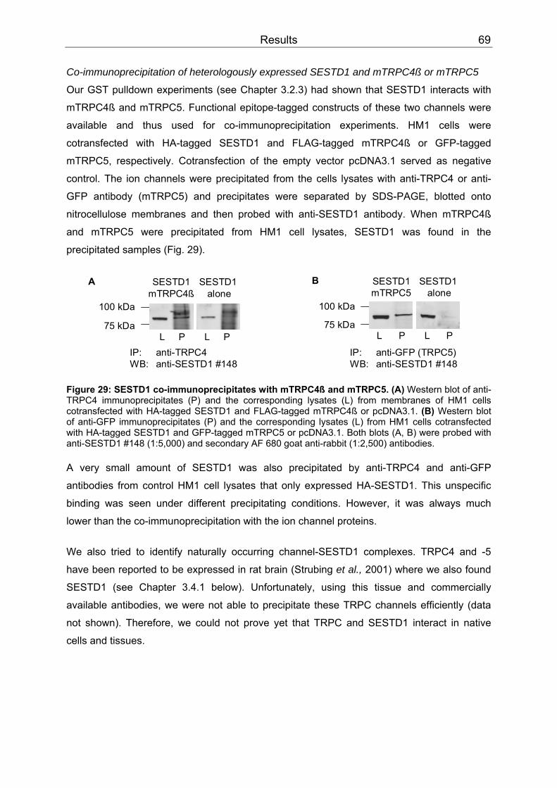
Results 69
Co-immunoprecipitation of heterologously expressed SESTD1 and mTRPC4ß or mTRPC5
Our GST pulldown experiments (see Chapter 3.2.3) had shown that SESTD1 interacts with
mTRPC4ß and mTRPC5. Functional epitope-tagged constructs of these two channels were
available and thus used for co-immunoprecipitation experiments. HM1 cells were
cotransfected with HA-tagged SESTD1 and FLAG-tagged mTRPC4ß or GFP-tagged
mTRPC5, respectively. Cotransfection of the empty vector pcDNA3.1 served as negative
control. The ion channels were precipitated from the cells lysates with anti-TRPC4 or anti-
GFP antibody (mTRPC5) and precipitates were separated by SDS-PAGE, blotted onto
nitrocellulose membranes and then probed with anti-SESTD1 antibody. When mTRPC4ß
and mTRPC5 were precipitated from HM1 cell lysates, SESTD1 was found in the
precipitated samples (Fig. 29).
100 kDa
75 kDaL P L P
SESTD1mTRPC4ß
SESTD1alone
A
100 kDa
75 kDa
SESTD1mTRPC5
SESTD1alone
L P L P
B
IP: anti-GFP (TRPC5)WB: anti-SESTD1 #148
IP: anti-TRPC4WB: anti-SESTD1 #148
Figure 29: SESTD1 co-immunoprecipitates with mTRPC4ß and mTRPC5. (A) Western blot of anti-TRPC4 immunoprecipitates (P) and the corresponding lysates (L) from membranes of HM1 cells cotransfected with HA-tagged SESTD1 and FLAG-tagged mTRPC4ß or pcDNA3.1. (B) Western blot of anti-GFP immunoprecipitates (P) and the corresponding lysates (L) from HM1 cells cotransfected with HA-tagged SESTD1 and GFP-tagged mTRPC5 or pcDNA3.1. Both blots (A, B) were probed with anti-SESTD1 #148 (1:5,000) and secondary AF 680 goat anti-rabbit (1:2,500) antibodies. A very small amount of SESTD1 was also precipitated by anti-TRPC4 and anti-GFP
antibodies from control HM1 cell lysates that only expressed HA-SESTD1. This unspecific
binding was seen under different precipitating conditions. However, it was always much
lower than the co-immunoprecipitation with the ion channel proteins.
We also tried to identify naturally occurring channel-SESTD1 complexes. TRPC4 and -5
have been reported to be expressed in rat brain (Strubing et al., 2001) where we also found
SESTD1 (see Chapter 3.4.1 below). Unfortunately, using this tissue and commercially
available antibodies, we were not able to precipitate these TRPC channels efficiently (data
not shown). Therefore, we could not prove yet that TRPC and SESTD1 interact in native
cells and tissues.
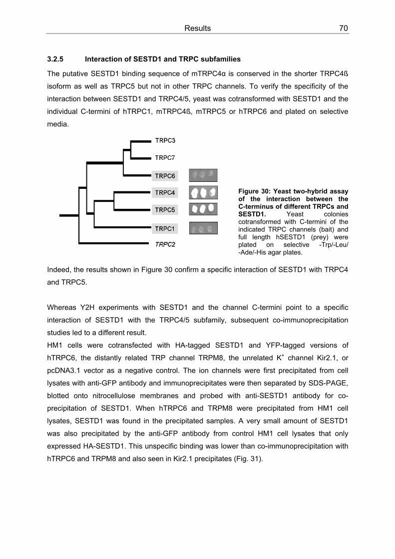
Results 70
3.2.5 Interaction of SESTD1 and TRPC subfamilies
The putative SESTD1 binding sequence of mTRPC4α is conserved in the shorter TRPC4ß
isoform as well as TRPC5 but not in other TRPC channels. To verify the specificity of the
interaction between SESTD1 and TRPC4/5, yeast was cotransformed with SESTD1 and the
individual C-termini of hTRPC1, mTRPC4ß, mTRPC5 or hTRPC6 and plated on selective
media. SS
Figure 30: Yeast two-hybrid assay of the interaction between the C-terminus of different TRPCs and SESTD1. Yeast colonies cotransformed with C-termini of the indicated TRPC channels (bait) and full length hSESTD1 (prey) were plated on selective -Trp/-Leu/ -Ade/-His agar plates.
Indeed, the results shown in Figure 30 confirm a specific interaction of SESTD1 with TRPC4
and TRPC5.
Whereas Y2H experiments with SESTD1 and the channel C-termini point to a specific
interaction of SESTD1 with the TRPC4/5 subfamily, subsequent co-immunoprecipitation
studies led to a different result.
HM1 cells were cotransfected with HA-tagged SESTD1 and YFP-tagged versions of
hTRPC6, the distantly related TRP channel TRPM8, the unrelated K PP
+PP channel Kir2.1, or
pcDNA3.1 vector as a negative control. The ion channels were first precipitated from cell
lysates with anti-GFP antibody and immunoprecipitates were then separated by SDS-PAGE,
blotted onto nitrocellulose membranes and probed with anti-SESTD1 antibody for co-
precipitation of SESTD1. When hTRPC6 and TRPM8 were precipitated from HM1 cell
lysates, SESTD1 was found in the precipitated samples. A very small amount of SESTD1
was also precipitated by the anti-GFP antibody from control HM1 cell lysates that only
expressed HA-SESTD1. This unspecific binding was lower than co-immunoprecipitation with
hTRPC6 and TRPM8 and also seen in Kir2.1 precipitates (Fig. 31).
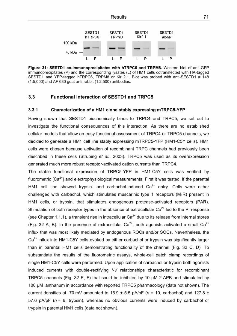
Results 71
Figure 31: SESTD1 co-immunoprecipitates with hTRPC6 and TRPM8. Western blot of anti-GFP immunoprecipitates (P) and the corresponding lysates (L) of HM1 cells cotransfected with HA-tagged SESTD1 and YFP-tagged hTRPC6, TRPM8 or Kir 2.1. Blot was probed with anti-SESTD1 # 148 (1:5,000) and AF 680 goat anti-rabbit (1:2,500) antibodies.
3.3 Functional interaction of SESTD1 and TRPC5
3.3.1 Characterization of a HM1 clone stably expressing mTRPC5-YFP
Having shown that SESTD1 biochemically binds to TRPC4 and TRPC5, we set out to
investigate the functional consequences of this interaction. As there are no established
cellular models that allow an easy functional assessment of TRPC4 or TRPC5 channels, we
decided to generate a HM1 cell line stably expressing mTRPC5-YFP (HM1-C5Y cells). HM1
cells were chosen because activation of recombinant TRPC channels had previously been
described in these cells (Strubing et al., 2003). TRPC5 was used as its overexpression
generated much more robust receptor-activated cation currents than TRPC4.
The stable functional expression of TRPC5-YFP in HM1-C5Y cells was verified by
fluorometric [Ca PP
2+PP] BBi BBand electrophysiologicalBB BBmeasurements. First it was tested, if the parental
HM1 cell line showed trypsin- and carbachol-induced Ca PP
2+PP entry. Cells were either
challenged with carbachol, which stimulates muscarinic type 1 receptors (MBB1BBR) present in
HM1 cells, or trypsin, that stimulates endogenous protease-activated receptors (PAR).
Stimulation of both receptor types in the absence of extracellular Ca PP
2+PP led to the PI response
(see Chapter 1.1.1), a transient rise in intracellular Ca PP
2+PP due to its release from internal stores
(Fig. 32 A, B). In the presence of extracellular Ca PP
2+PP, both agonists activated a small Ca PP
2+PP
influx that was most likely mediated by endogenous ROCs and/or SOCs. Nevertheless, the
Ca PP
2+PP influx into HM1-C5Y cells evoked by either carbachol or trypsin was significantly larger
than in parental HM1 cells demonstrating functionality of the channel (Fig. 32 C, D). To
substantiate the results of the fluorometric assays, whole-cell patch clamp recordings of
single HM1-C5Y cells were performed. Upon application of carbachol or trypsin both agonists
induced currents with double-rectifying I-V relationships characteristic for recombinant
TRPC5 channels (Fig. 32 E, F) that could be inhibited by 10 µM 2-APB and stimulated by
100 µM lanthanum in accordance with reported TRPC5 pharmacology (data not shown). The
current densities at -70 mV amounted to 15.9 ± 5.5 pA/pF (n = 10, carbachol) and 127.8 ±
57.6 pA/pF (n = 6, trypsin), whereas no obvious currents were induced by carbachol or
trypsin in parental HM1 cells (data not shown).
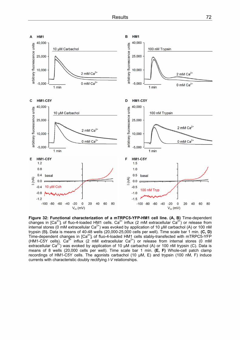
Results 72
Figure 32: Functional characterization of a mTRPC5-YFP-HM1 cell line. (A, B) Time-dependent changes in [CaPP
2+PP]BBi BB of fluo-4-loaded HM1 cells. CaPP
2+PP influx (2 mM extracellular CaPP
2+PP) or release from
internal stores (0 mM extracellular CaPP
2+PP) was evoked by application of 10 µM carbachol (A) or 100 nM
trypsin (B). Data is means of 40-48 wells (20,000-25,000 cells per well). Time scale bar 1 min. (C, D) Time-dependent changes in [CaPP
2+PP]BBi BB of fluo-4-loaded HM1 cells stably-transfected with mTRPC5-YFP
(HM1-C5Y cells). CaPP
2+PP influx (2 mM extracellular CaPP
2+PP) or release from internal stores (0 mM
extracellular CaPP
2+PP) was evoked by application of 10 µM carbachol (A) or 100 nM trypsin (C). Data is
means of 8 wells (20,000 cells per well). Time scale bar 1 min. (E, F) Whole-cell patch clamp recordings of HM1-C5Y cells. The agonists carbachol (10 µM, E) and trypsin (100 nM, F) induce currents with characteristic doubly rectifying I-V relationships.
Results 73
3.3.2 Overexpression of SESTD1 in HM1-C5Y cells
In a first attempt to modulate the interaction between TRPC5 and SESTD1, we transiently
overexpressed HA-tagged SESTD1 in HM1-C5Y cells. Since no information was available
about the cellular function of SESTD1, experimental readouts were restricted to measuring
TRPC5 function at elevated (or decreased, see Chapter 3.3.3) levels of SESTD1. TRPC5-
mediated CaPP
2+PP influx following application of carbachol or trypsin (Fig. 33 A, C) in HM1-C5Y
cells coexpressing HA-SESTD1 did not differ significantly from control cells cotransfected
with an unrelated protein (ß-galactosidase, bGAL). Ca PP
2+PP releases from internal stores were
also not significantly changed in the presence or absence of HA-tagged SESTD1 (Fig. 33 B,
D).

Results 74
Figure 33: TRPC5-mediated CaPP
2+PP-entry is unaltered in HM1-C5Y cells overexpressing
heterologous HA-SESTD1. (A-D) Time-dependent changes in [Ca PP
2+PP]BBi BB of fura-2-loaded HM1-C5Y cells
transiently transfected with HA-tagged SESTD1 or an unrelated protein (ß-galactosidase, bGAL). TRPC5-mediated CaPP
2+PP influx following application of 10 µM carbachol (A) or 100 nM trypsin (C) was
the same in presence and absence of HA-tagged SESTD1. Also Ca PP
2+PP release from internal stores was
not significantly changed in the presence or absence of HA-tagged SESTD1 after application of 10 µM carbachol (B) and 100 nM trypsin (D). Data is shown as means of 5-6 wells (40,000 cells per well). Time scale bar 1 min. (E) Statistical analysis of data presented in A-D (n = 5-6 wells per data point). CaPP
2+PP release was calculated as area under the curve (AUC; B, D) and CaPP
2+PP influx was calculated by
their subtraction from the AUCs of (A) and (C), respectively.

Results 75
3.3.3 siRNA knock-down of SESTD1
As shown above, overexpression of exogenous HA-SESTD1 had no effect on TRPC5-
mediated CaPP
2+PP influx in our cell model. However, since HM1 cells express SESTD1
endogenously (see Fig. 28), it may not be possible to further enhance SESTD1 function in
these cells. Therefore, it was tested whether knock-down of SESTD1 protein expression in
HM1 cells had an influence on TRPC5 activity.
100
75
150
kDa
37
Mock
D134D123
4
mTRPC5-GFP
SESTD1
GAPDH
C
Figure 34: CaPP
2+PP release from internal stores is suppressed by SESTD1 siRNA duplex 2. (A)
Time-dependent changes in [CaPP
2+PP]BBi BB of fluo-4-loaded HM1 cells transiently transfected with mTRPC5-
GFP and 40 nM single (duplex 1 to 4, D1 to D4) or pooled (SMARTpool, D1234) specific siRNA against SESTD1. 48 hr post transfection, CaPP
2+PP release from internal stores activated by application of
100 nM trypsin is significantly reduced in cells transfected with D2 or the complete SMARTpool (D1234). Data is shown as means of 6 wells (42,000 cells per well). Time scale bar 1 min. (B) Statistical analysis of data presented in A (n = 5-6 wells, P < 0.01, Wilcoxon two-sample test). (C) Western blot of HM1 cells transfected with GFP only (mock control) or mTRPC5-GFP plus 40 nM SMARTpool (D1234) or an siRNA pool lacking duplex 2 (D134). Blot was cut and incubated with anti-GFP (1:5,000)/AF 680 rabbit anti-mouse (1:2,500), anti-SESTD1 (1:5,000)/AF 680 goat anti-rabbit (1:2,500) and anti-GAPDH (1:10,000)/AF 680 rabbit anti-mouse (1:2,500) antibodies.
A pool of four siRNA duplexes (D1234, SMARTpool) directed against different sequences of
SESTD1 was purchased from Dharmacon and tested for its ability to decrease SESTD1
protein levels. 48 hr post transfection, SESTD1 expression was almost completely knocked
down by 40 nM siRNA whereas expression of an unrelated protein (GAPDH) was not altered
(Fig. 34 C). HM1 cells cotransfected with 40 nM of either pooled or single siRNA duplexes
and mTRPC5-GFP were then functionally analyzed by fluorometric [Ca PP
2+PP] BBi BB measurements.

Results 76
SESTD1
unrelated
GAPDH
MockUns
p. siR
NA
Sp. siR
NA
Marker
(in kD
a)
10075
50
37
While investigating TRPC5-independent Ca PP
2+PP release from internal stores that may serve as
a control for unspecific siRNA effects, we noted that the SMARTpool and duplex 2
significantly reduced Ca PP
2+PP release compared to duplex 1, 3 or 4 (Fig. 34 A, B). This
observation prompted us to check a new siRNA pool lacking duplex 2 (D134). Indeed, this
pool was as efficient as the SMARTpool in silencing SESTD1 expression (Fig. 34 C), but
without having an effect on Ca PP
2+PP release (see Fig. 36 A, B). Thus, it is likely that the
suppression of Ca PP
2+PP release by duplex 2 is an unspecific effect, not related to the SESTD1
protein knock-down. Consequently HM1-C5Y were treated with specific SESTD1 siRNA
(new pool of three duplexes, D134), unspecific non-silencing control siRNA or liposomes only
(mock). SESTD1 protein expression in suchlike treated cells was reduced by 85.5 ± 5.5%
(n = 4, compared to mock-transfected cells) or 82.3 ± 5.3% (n = 4, compared to cells treated
with unspecific, non-silencing siRNA; Fig. 35). Figure 35: SESTD1 is efficiently knocked-down by 20 nM specific SESTD1 siRNA. Western blot of HM1-C5Y cells transfected with liposomes only (mock control), 20 nM unspecific control siRNA or 20 nM pooled specific SESTD1 siRNA (duplex 1, 3 and 4). Blot was cut and incubated with anti-SESTD1 (1:5,000)/AF 680 goat anti-rabbit (1:2,500) and anti-GAPDH (1:10,000)/AF 680 rabbit anti-mouse (1:2,500) antibodies.
M1 receptor- or PAR-induced Ca PP
2+PP release from internal stores was not different between the
three groups (Fig. 36 A, B). In contrast, TRPC5-mediated Ca PP
2+PP influx following application of
carbachol or trypsin (Fig. 36 C, D) was significantly reduced in cells treated with specific
SESTD1 siRNA. TRPC5-mediated Ca PP
2+PP influx following carbachol stimulation was reduced to
45.4 ± 2.8% (compared to mock transfected cells) or 49.6 ± 3.1% (compared to cells
transfected with control siRNA, Fig. 36 E). When cells were activated with 100 nM trypsin,
TRPC5-mediated Ca PP
2+PP influx was reduced to 51.4 ± 3.7% (compared to mock transfected
cells) or 58.0 ± 4.2% (compared to cells transfected with control siRNA, Fig. 36 F).

Results 77
Figure 36: TRPC5 activity is reduced in HM1-C5Y cells transfected with specific SESTD1 siRNA. Time-dependent changes in [CaPP
2+PP]BBi BB of fura-2-loaded HM1-C5Y cells transfected with 20 nM pooled
specific SESTD1 siRNA (duplex 1, 3 and 4), unspecific control siRNA or liposomes only (mock) (A, B). 48 hr post transfection, CaPP
2+PP release from internal stores activated by application of 10 µM carbachol
(A) or 100 nM trypsin (B) is not different under the tested conditions. In contrast, TRPC5-mediated CaPP
2+PP influx following application of 10 µM carbachol (C) or 100 nM trypsin (D) was significantly
reduced in cells transfected with specific SESTD1 siRNA. Shown are means ± SEM of three independent experiments (each performed with n = 5–6 wells per experimental condition). Time scale bar 1 min. (E) Statistical analysis of data presented in A and C (P < 0.001, Wilcoxon test, two-sided). (F) Statistical analysis of data presented in B and D (P < 0.001, Wilcoxon test, two-sided).
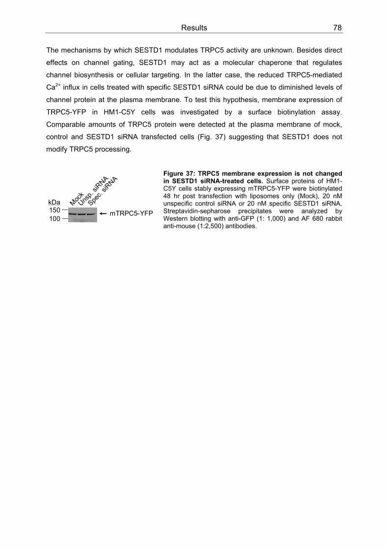
Results 78
100150kDa
mTRPC5-YFPMoc
kUns
p. siR
NA
Spec.
siRNA
The mechanisms by which SESTD1 modulates TRPC5 activity are unknown. Besides direct
effects on channel gating, SESTD1 may act as a molecular chaperone that regulates
channel biosynthesis or cellular targeting. In the latter case, the reduced TRPC5-mediated
Ca PP
2+PP influx in cells treated with specific SESTD1 siRNA could be due to diminished levels of
channel protein at the plasma membrane. To test this hypothesis, membrane expression of
TRPC5-YFP in HM1-C5Y cells was investigated by a surface biotinylation assay.
Comparable amounts of TRPC5 protein were detected at the plasma membrane of mock,
control and SESTD1 siRNA transfected cells (Fig. 37) suggesting that SESTD1 does not
modify TRPC5 processing.
Figure 37: TRPC5 membrane expression is not changed in SESTD1 siRNA-treated cells. Surface proteins of HM1-C5Y cells stably expressing mTRPC5-YFP were biotinylated 48 hr post transfection with liposomes only (Mock), 20 nM unspecific control siRNA or 20 nM specific SESTD1 siRNA. Streptavidin-sepharose precipitates were analyzed by Western blotting with anti-GFP (1: 1,000) and AF 680 rabbit anti-mouse (1:2,500) antibodies.
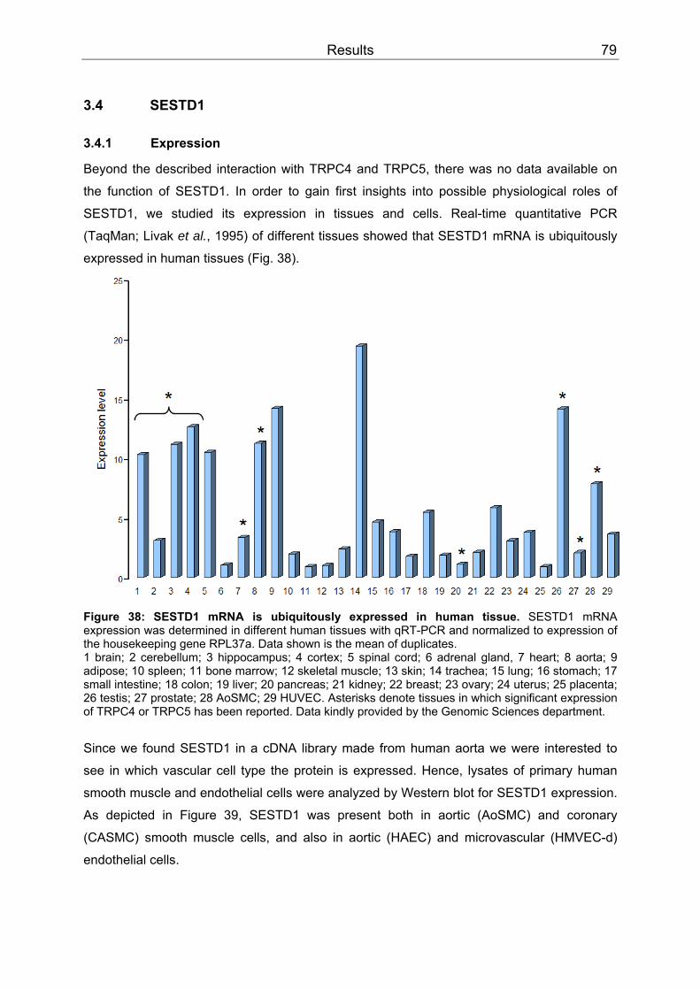
Results 79
3.4 SESTD1
3.4.1 Expression
Beyond the described interaction with TRPC4 and TRPC5, there was no data available on
the function of SESTD1. In order to gain first insights into possible physiological roles of
SESTD1, we studied its expression in tissues and cells. Real-time quantitative PCR
(TaqMan; Livak et al., 1995) of different tissues showed that SESTD1 mRNA is ubiquitously
expressed in human tissues (Fig. 38).
Figure 38: SESTD1 mRNA is ubiquitously expressed in human tissue. SESTD1 mRNA expression was determined in different human tissues with qRT-PCR and normalized to expression of the housekeeping gene RPL37a. Data shown is the mean of duplicates. 1 brain; 2 cerebellum; 3 hippocampus; 4 cortex; 5 spinal cord; 6 adrenal gland, 7 heart; 8 aorta; 9 adipose; 10 spleen; 11 bone marrow; 12 skeletal muscle; 13 skin; 14 trachea; 15 lung; 16 stomach; 17 small intestine; 18 colon; 19 liver; 20 pancreas; 21 kidney; 22 breast; 23 ovary; 24 uterus; 25 placenta; 26 testis; 27 prostate; 28 AoSMC; 29 HUVEC. Asterisks denote tissues in which significant expression of TRPC4 or TRPC5 has been reported. Data kindly provided by the Genomic Sciences department.
Since we found SESTD1 in a cDNA library made from human aorta we were interested to
see in which vascular cell type the protein is expressed. Hence, lysates of primary human
smooth muscle and endothelial cells were analyzed by Western blot for SESTD1 expression.
As depicted in Figure 39, SESTD1 was present both in aortic (AoSMC) and coronary
(CASMC) smooth muscle cells, and also in aortic (HAEC) and microvascular (HMVEC-d)
endothelial cells.
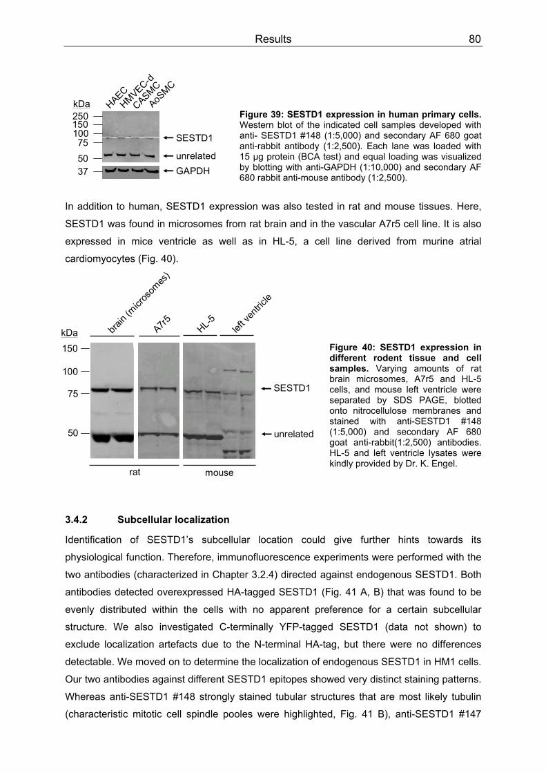
Results 80
150
100
75
50
kDa
rat
SESTD1
mouse
unrelated
A7r5 HL-5 left v
entric
le
brain
(micr
osom
es)
250150100755037
kDa HAECHMVEC-d
CASMC
AoSMC
SESTD1
GAPDHunrelated
Figure 39: SESTD1 expression in human primary cells. Western blot of the indicated cell samples developed with anti- SESTD1 #148 (1:5,000) and secondary AF 680 goat anti-rabbit antibody (1:2,500). Each lane was loaded with 15 µg protein (BCA test) and equal loading was visualized by blotting with anti-GAPDH (1:10,000) and secondary AF 680 rabbit anti-mouse antibody (1:2,500).
In addition to human, SESTD1 expression was also tested in rat and mouse tissues. Here,
SESTD1 was found in microsomes from rat brain and in the vascular A7r5 cell line. It is also
expressed in mice ventricle as well as in HL-5, a cell line derived from murine atrial
cardiomyocytes (Fig. 40). Figure 40: SESTD1 expression in different rodent tissue and cell samples. Varying amounts of rat brain microsomes, A7r5 and HL-5 cells, and mouse left ventricle were separated by SDS PAGE, blotted onto nitrocellulose membranes and stained with anti-SESTD1 #148 (1:5,000) and secondary AF 680 goat anti-rabbit(1:2,500) antibodies. HL-5 and left ventricle lysates were kindly provided by Dr. K. Engel.
3.4.2 Subcellular localization
Identification of SESTD1’s subcellular location could give further hints towards its
physiological function. Therefore, immunofluorescence experiments were performed with the
two antibodies (characterized in Chapter 3.2.4) directed against endogenous SESTD1. Both
antibodies detected overexpressed HA-tagged SESTD1 (Fig. 41 A, B) that was found to be
evenly distributed within the cells with no apparent preference for a certain subcellular
structure. We also investigated C-terminally YFP-tagged SESTD1 (data not shown) to
exclude localization artefacts due to the N-terminal HA-tag, but there were no differences
detectable. We moved on to determine the localization of endogenous SESTD1 in HM1 cells.
Our two antibodies against different SESTD1 epitopes showed very distinct staining patterns.
Whereas anti-SESTD1 #148 strongly stained tubular structures that are most likely tubulin
(characteristic mitotic cell spindle pooles were highlighted, Fig. 41 B), anti-SESTD1 #147

Results 81
stained vesicular structures (Fig. 41 A). It was already seen in Western blots (Fig. 28) that
both antibodies also have high affinities for proteins not related to SESTD1. This might
explain our immunocytochemical findings. To further elucidate the location of native
SESTD1, better antibodies will be necessary that specifically recognize the protein without
unspecific binding to unrelated structures.
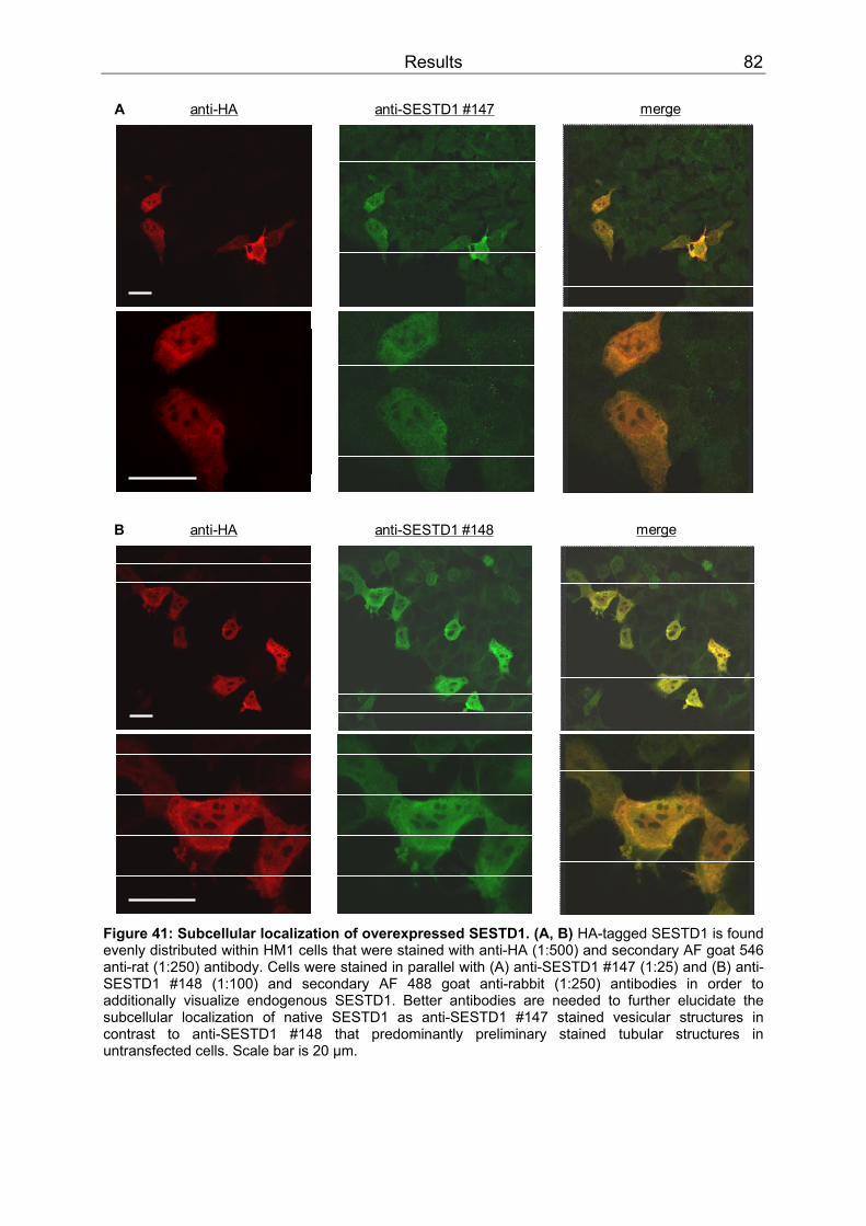
Results 82
anti-HA anti-SESTD1 #147 mergeA
anti-HA anti-SESTD1 #148 mergeB
Figure 41: Subcellular localization of overexpressed SESTD1. (A, B) HA-tagged SESTD1 is found evenly distributed within HM1 cells that were stained with anti-HA (1:500) and secondary AF goat 546 anti-rat (1:250) antibody. Cells were stained in parallel with (A) anti-SESTD1 #147 (1:25) and (B) anti-SESTD1 #148 (1:100) and secondary AF 488 goat anti-rabbit (1:250) antibodies in order to additionally visualize endogenous SESTD1. Better antibodies are needed to further elucidate the subcellular localization of native SESTD1 as anti-SESTD1 #147 stained vesicular structures in contrast to anti-SESTD1 #148 that predominantly preliminary stained tubular structures in untransfected cells. Scale bar is 20 µm.

Results 83
3.4.3 Cis-trans isomerase signature
A PROSITE motif search of SESTD1 indicated a FKBP-type peptidyl-prolyl cis-trans
isomerase signature 2 (Pattern-ID PS00454) starting from aa 427 (VDV GLQ GLR EKG QGL
LDQ ISN QAS WAY G). Peptidyl-prolyl cis-trans isomerases (PPIases) catalyze cis-trans
isomerization of proline peptide bonds thus accelerating protein folding. A possible PPIase
activity of SESTD1 was tested using a fluorescence assay and the cys-bridged peptide
H-Abz-Cys-Ala-Pro-Ala-Cys-Ntr-NH BB2BB as a substrate (see Chapter 2.5.8 for assay principle).
The known PPIase activity of FKBP12.6 (Sewell et al., 1994) served as a positive control.
Substrate isomerization by FKBP12.6 is a rapid reaction that was completed almost within a
minute. It is indicated by a steepening of the slope of the fluorescence curve compared to the
spontaneous reaction. In contrast, the slope in presence of GST-SESTD1 did not differ from
the spontaneous isomerization (Fig. 42). Hence, in this experiment GST-SESTD1 did not act
as PPIase on bridged H-Abz-Cys-Ala-Pro-Ala-Cys-Ntr-NHBB2.
Figure 42: Cis-trans isomerization assay of cys-bridged H-Abz-Cys-Ala-Pro-Ala-Cys-Ntr-NHBB2 BB. Isomerization results in a fluorescent trans-form. When the substrate is not cleaved (no isom.) baseline fluorescence is not changed. Addition of 125 mM DTT cleaves the cys-bridge resulting in spontaneous prolyl cis-trans conversion (spontan. isom.). In presence of 1 µM FKBP12.6 (and 125 mM DTT), isomerization is accelerated (FKBP12.6-catal. isom.). 4.79 µM GST-SESTD1 (in presence of 125 mM DTT) have no influence on isomerization velocity (SESTD1-catal. isom.). Data is means of 2 wells and was kindly provided by K. Sicka.
3.4.4 In vitro phospholipid binding
SESTD1 belongs to the eukaryotic Sec14 protein superfamily that was named after the
N-terminal Sec14p-like lipid-binding domain. Due to this domain its members are assumed to
specifically bind and transfer different phospholipids (Mousley et al., 2007), but some have
also been reported to bind other hydrophobic ligands than phospholipids, e.g. α-tocopherol
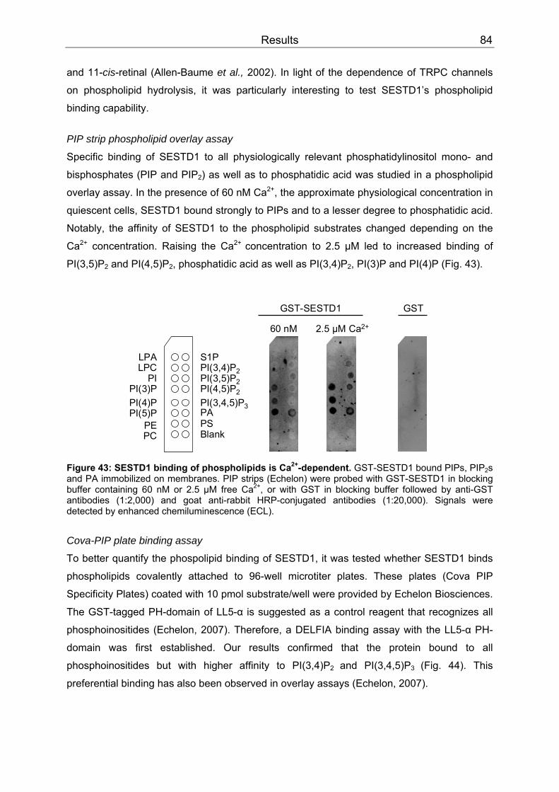
Results 84
and 11-cis-retinal (Allen-Baume et al., 2002). In light of the dependence of TRPC channels
on phospholipid hydrolysis, it was particularly interesting to test SESTD1’s phospholipid
binding capability. PIP strip phospholipid overlay assay
Specific binding of SESTD1 to all physiologically relevant phosphatidylinositol mono- and
bisphosphates (PIP and PIPBB2BB) as well as to phosphatidic acid was studied in a phospholipid
overlay assay. In the presence of 60 nM Ca PP
2+PP, the approximate physiological concentration in
quiescent cells, SESTD1 bound strongly to PIPs and to a lesser degree to phosphatidic acid.
Notably, the affinity of SESTD1 to the phospholipid substrates changed depending on the
Ca PP
2+PP concentration. Raising the Ca PP
2+ PPconcentration to 2.5 µM led to increased binding of
PI(3,5)PBB2BB and PI(4,5)PBB2BB, phosphatidic acid as well as PI(3,4)P BB2BB, PI(3)P and PI(4)P (Fig. 43).
GST-SESTD1 GST
60 nM 2.5 µM Ca2+
S1PPI(3,4)P2PI(3,5)P2PI(4,5)P2
PI(3,4,5)P3PAPSBlank
LPALPC
PIPI(3)PPI(4)PPI(5)P
PEPC
Figure 43: SESTD1 binding of phospholipids is CaPP
2+PP-dependent. GST-SESTD1 bound PIPs, PIPBB2BBs
and PA immobilized on membranes. PIP strips (Echelon) were probed with GST-SESTD1 in blocking buffer containing 60 nM or 2.5 µM free CaPP
2+PP, or with GST in blocking buffer followed by anti-GST
antibodies (1:2,000) and goat anti-rabbit HRP-conjugated antibodies (1:20,000). Signals were detected by enhanced chemiluminescence (ECL).
Cova-PIP plate binding assay
To better quantify the phospolipid binding of SESTD1, it was tested whether SESTD1 binds
phospholipids covalently attached to 96-well microtiter plates. These plates (Cova PIP
Specificity Plates) coated with 10 pmol substrate/well were provided by Echelon Biosciences.
The GST-tagged PH-domain of LL5-α is suggested as a control reagent that recognizes all
phosphoinositides (Echelon, 2007). Therefore, a DELFIA binding assay with the LL5-α PH-
domain was first established. Our results confirmed that the protein bound to all
phosphoinositides but with higher affinity to PI(3,4)PBB2 BBand PI(3,4,5)P BB3 BB(Fig. 44). This
preferential binding has also been observed in overlay assays (Echelon, 2007).
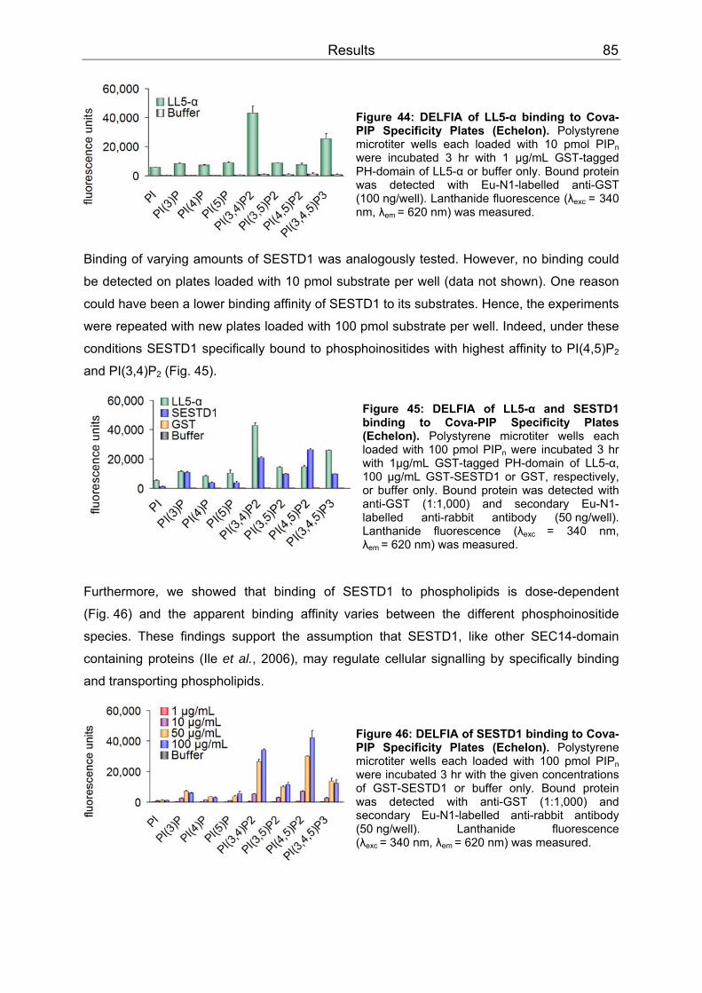
Results 85
Figure 44: DELFIA of LL5-α binding to Cova-PIP Specificity Plates (Echelon). Polystyrene microtiter wells each loaded with 10 pmol PIPBBnBB were incubated 3 hr with 1 µg/mL GST-tagged PH-domain of LL5-α or buffer only. Bound protein was detected with Eu-N1-labelled anti-GST (100 ng/well). Lanthanide fluorescence (λBBexc BB= 340 nm, λBBem BB= 620 nm) was measured.
Binding of varying amounts of SESTD1 was analogously tested. However, no binding could
be detected on plates loaded with 10 pmol substrate per well (data not shown). One reason
could have been a lower binding affinity of SESTD1 to its substrates. Hence, the experiments
were repeated with new plates loaded with 100 pmol substrate per well. Indeed, under these
conditions SESTD1 specifically bound to phosphoinositides with highest affinity to PI(4,5)P BB2 BB
and PI(3,4)P BB2BB (Fig. 45).
Figure 45: DELFIA of LL5-α and SESTD1 binding to Cova-PIP Specificity Plates (Echelon). Polystyrene microtiter wells each loaded with 100 pmol PIPBBnBB were incubated 3 hr with 1µg/mL GST-tagged PH-domain of LL5-α, 100 µg/mL GST-SESTD1 or GST, respectively, or buffer only. Bound protein was detected with anti-GST (1:1,000) and secondary Eu-N1-labelled anti-rabbit antibody (50 ng/well). Lanthanide fluorescence (λBBexc BB= 340 nm, λBBem BB= 620 nm) was measured.
Furthermore, we showed that binding of SESTD1 to phospholipids is dose-dependent
(Fig. 46) and the apparent binding affinityBB BBvaries between the different phosphoinositide
species. These findings support the assumption that SESTD1, like other SEC14-domain
containing proteins (Ile et al., 2006), may regulate cellular signalling by specifically binding
and transporting phospholipids.
Figure 46: DELFIA of SESTD1 binding to Cova-PIP Specificity Plates (Echelon). Polystyrene microtiter wells each loaded with 100 pmol PIPBBnBB were incubated 3 hr with the given concentrations of GST-SESTD1 or buffer only. Bound protein was detected with anti-GST (1:1,000) and secondary Eu-N1-labelled anti-rabbit antibody (50 ng/well). Lanthanide fluorescence (λBBexc BB= 340 nm, λ BBem BB= 620 nm) was measured.
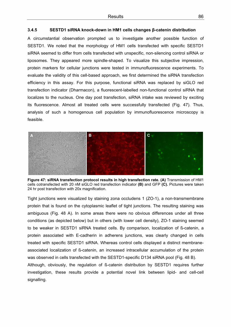
Results 86
3.4.5 SESTD1 siRNA knock-down in HM1 cells changes β-catenin distribution
A circumstantial observation prompted us to investigate another possible function of
SESTD1. We noted that the morphology of HM1 cells transfected with specific SESTD1
siRNA seemed to differ from cells transfected with unspecific, non-silencing control siRNA or
liposomes. They appeared more spindle-shaped. To visualize this subjective impression,
protein markers for cellular junctions were tested in immunofluorescence experiments. To
evaluate the validity of this cell-based approach, we first determined the siRNA transfection
efficiency in this assay. For this purpose, functional siRNA was replaced by siGLO red
transfection indicator (Dharmacon), a fluorescent-labelled non-functional control siRNA that
localizes to the nucleus. One day post transfection, siRNA intake was reviewed by exciting
its fluorescence. Almost all treated cells were successfully transfected (Fig. 47). Thus,
analysis of such a homogenous cell population by immunofluorescence microscopy is
feasible.
A B C
Figure 47: siRNA transfection protocol results in high transfection rate. (A) Transmission of HM1 cells cotransfected with 20 nM siGLO red transfection indicator (B) and GFP (C). Pictures were taken 24 hr post transfection with 20x magnification. Tight junctions were visualized by staining zona occludens 1 (ZO-1), a non-transmembrane
protein that is found on the cytoplasmic leaflet of tight junctions. The resulting staining was
ambiguous (Fig. 48 A). In some areas there were no obvious differences under all three
conditions (as depicted below) but in others (with lower cell density), ZO-1 staining seemed
to be weaker in SESTD1 siRNA treated cells. By comparison, localization of ß-catenin, a
protein associated with E-cadherin in adherens junctions, was clearly changed in cells
treated with specific SESTD1 siRNA. Whereas control cells displayed a distinct membrane-
associated localization of ß-catenin, an increased intracellular accumulation of the protein
was observed in cells transfected with the SESTD1-specific D134 siRNA pool (Fig. 48 B).
Although, obviously, the regulation of ß-catenin distribution by SESTD1 requires further
investigation, these results provide a potential novel link between lipid- and cell-cell
signalling.

Results 87
mock-transfected
A
ZO-1
unspecific siRNA SESTD1 siRNA
B
ß-catenin
Figure 48: ß-catenin distribution is changed in HM1 cells treated with SESTD1 siRNA. 48 hr post transfection, HM1 cells treated with liposomes only (mock-transfected), 20 nM unspecific, non-silencing control siRNA or 20 nM specific SESTD1 siRNA (pool D134) were fixed with paraformaldehyde, permeabilized with Triton X-100, and stained (A) with anti-ZO 1 (1:100) and AF 546 goat anti-mouse (1:250) antibodies or with anti-β-catenin (1:250) and AF 546 goat anti-rabbit (1:250) antibodies. Scale bar is 20 µM.

Discussion 88
4 Discussion
4.1 Norgestimate is a selective inhibitor of the TRPC3/6/7 subfamily
In this work, we used complementary pharmacological and molecular biological approaches
to gain a better understanding of the physiology of TRPC channels. Our search for new
pharmacological tools led to the discovery of two steroids, namely norgestimate and
progesterone, which differentially inhibit TRPC channels. While progesterone showed almost
equal activity towards all studied TRPC channels with ICBB50BB values of ~10-20 µM,
norgestimate selectively inhibited the TRPC3/6/7 subfamily with IC BB50BB values of ~5 µM. This
selectivity distinguishes norgestimate from most known TRPC channel modulating
compounds.
Based on the calculated inhibition at 10 µM, norgestimate was 4 to 5-fold more potent on
Ca PP
2+ PPinflux mediated by TRPC3 and TRPC6 compared to TRPC5. These results obtained by
fluorometric measurements were further validated by patch clamp recordings in the whole-
cell configuration. Again, 10 µM norgestimate were 3.5-fold more effective on AlF BB4PBPB
-PP-evoked
TRPC6-mediated currents compared to TRPC5, thus confirming its selective block of the
TRPC3/6/7 subfamily. Norgestimate rapidly inhibited TRPC6 channel function both after
direct stimulation by OAG in FLIPR measurements and also after indirect stimulation by AlFBB4PBPB
-PP
in patch clamp recordings suggesting that it directly blocked the channel. The highest applied
norgestimate concentration did not influence calcium store depletion following activation of
PAR in HEK293 cells or following stimulation of the V BB1ABB receptor in A7r5 cells, therefore
excluding IPBB3BBR antagonism or inhibition of the GBBq/11 BB/PI signalling cascade as mechanism of
channel inhibition. Moreover, genomic effects of steroids that occur on an hours time scale
can be excluded as channel inhibition started immediately after norgestimate application and
was rapidly and completely reversed upon washout. Taken together, these data suggest that
norgestimate inhibits TRPC6 activity by a direct interaction with the channel protein, although
single channel recordings, that would provide the most stringent proof, have not been
performed.
Compared to known TRPC channel blockers, norgestimate offers the advantage of being
reasonably selective for DAG-sensitive TRPCs by inhibiting them at low micromolar
concentrations without having an effect on the upstream PI signalling components.
Perhaps the most specific TRPC inhibitor described so far is [1-(5-chloronaphthalene-1-
sulphonyl) homopiperazine, HCl] (ML-9) which has been shown to block TRPC6 with an ICBB50BB
value of 7.8 µM but has no effect on isolated, single TRPC5 channels (Shi et al., 2007). Yet,
ML-9 is a commonly used blocker of myosin light chain kinase (MLCK; Saitoh et al., 1987)
and ML-9-mediated dephosphorylation of myosin light chains modulates the activity of many
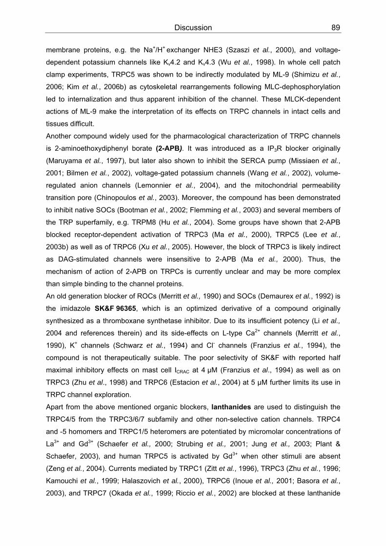
Discussion 89
membrane proteins, e.g. the Na PP
+PP/HPP
+ PPexchanger NHE3 (Szaszi et al., 2000), and voltage-
dependent potassium channels like K BBvBB4.2 and KBBvBB4.3 (Wu et al., 1998). In whole cell patch
clamp experiments, TRPC5 was shown to be indirectly modulated by ML-9 (Shimizu et al.,
2006; Kim et al., 2006b) as cytoskeletal rearrangements following MLC-dephosphorylation
led to internalization and thus apparent inhibition of the channel. These MLCK-dependent
actions of ML-9 make the interpretation of its effects on TRPC channels in intact cells and
tissues difficult.
Another compound widely used for the pharmacological characterization of TRPC channels
is 2-aminoethoxydiphenyl borate (2-APB). It was introduced as a IPBB3BBR blocker originally
(Maruyama et al., 1997), but later also shown to inhibit the SERCA pump (Missiaen et al.,
2001; Bilmen et al., 2002), voltage-gated potassium channels (Wang et al., 2002), volume-
regulated anion channels (Lemonnier et al., 2004), and the mitochondrial permeability
transition pore (Chinopoulos et al., 2003). Moreover, the compound has been demonstrated
to inhibit native SOCs (Bootman et al., 2002; Flemming et al., 2003) and several members of
the TRP superfamily, e.g. TRPM8 (Hu et al., 2004). Some groups have shown that 2-APB
blocked receptor-dependent activation of TRPC3 (Ma et al., 2000), TRPC5 (Lee et al.,
2003b) as well as of TRPC6 (Xu et al., 2005). However, the block of TRPC3 is likely indirect
as DAG-stimulated channels were insensitive to 2-APB (Ma et al., 2000). Thus, the
mechanism of action of 2-APB on TRPCs is currently unclear and may be more complex
than simple binding to the channel proteins.
An old generation blocker of ROCs (Merritt et al., 1990) and SOCs (Demaurex et al., 1992) is
the imidazole SK&F 96365, which is an optimized derivative of a compound originally
synthesized as a thromboxane synthetase inhibitor. Due to its insufficient potency (Li et al.,
2004 and references therein) and its side-effects on L-type Ca PP
2+PP channels (Merritt et al.,
1990), KPP
+PP channels (Schwarz et al., 1994) and Cl PP
-PP channels (Franzius et al., 1994), the
compound is not therapeutically suitable. The poor selectivity of SK&F with reported half
maximal inhibitory effects on mast cell IBBCRACBB at 4 µM (Franzius et al., 1994) as well as on
TRPC3 (Zhu et al., 1998) and TRPC6 (Estacion et al., 2004) at 5 µM further limits its use in
TRPC channel exploration.
Apart from the above mentioned organic blockers, lanthanides are used to distinguish the
TRPC4/5 from the TRPC3/6/7 subfamily and other non-selective cation channels. TRPC4
and -5 homomers and TRPC1/5 heteromers are potentiated by micromolar concentrations of
La PP
3+PP and Gd PP
3+PP (Schaefer et al., 2000; Strubing et al., 2001; Jung et al., 2003; Plant &
Schaefer, 2003), and human TRPC5 is activated by Gd PP
3+PP when other stimuli are absent
(Zeng et al., 2004). Currents mediated by TRPC1 (Zitt et al., 1996), TRPC3 (Zhu et al., 1996;
Kamouchi et al., 1999; Halaszovich et al., 2000), TRPC6 (Inoue et al., 2001; Basora et al.,
2003), and TRPC7 (Okada et al., 1999; Riccio et al., 2002) are blocked at these lanthanide
Discussion 90
concentrations. But there are also contradictory reports of TRPC5 inhibition by micromolar
lanthanide concentrations (Okada et al., 1998; Lee et al., 2003b), and an endothelial store-
operated Ca PP
2+PP current that is absent in TRPC4 knock-out mice is also highly susceptible to
inhibition at 1 µM La PP
3+PP (Freichel et al., 2001). Therefore, the unique feature of TRPC4/5
potentiation by lanthanides might depend on the individual expression system and thus only
has limited value for the investigation of native currents. Moreover, due to their toxicity and
rather unspecific ion channel blocking activities, the use of lanthanides in many tissue
models such as brain slices is not possible.
Norgestimate is a progestin (a synthetic gestagen). Combined with ethinyl estradiol it is a
component of oral contraceptives (CilestPP
®PP, Pramino PP
®PP). We examined in fluorometric CaPP
2+PP
influx tests whether the natural pregnancy-maintaining hormone progesterone, which is
structurally related to norgestimate, also inhibits TRPC channels. In fact, progesterone was
less active on TRPC6 compared to norgestimate, but TRPC4 and -5 were more effectively
inhibited by the hormone. Its overall effect on the TRPC4/5 and the TRPC3/6/7 subfamily
was quite comparable. Hence, progesterone does not discriminate between different
members of the TRPC family and was therefore not further investigated. Nevertheless, the
observed inhibition of TRPC channels may contribute to the reported cardiovascular effects
of progesterone. Several studies have shown that progesterone rapidly relaxed vessels, e.g.
pig coronary arteries (Crews & Khalil, 1999), rat aorta (Glusa et al., 1997; Mukerji et al.,
2000), and also guinea pig airway smooth muscles (Perusquia et al., 1997). This
vasorelaxant effect is endothelium-independent and mediated at least partly through
inhibition of L-type Ca PP
2+PP channels (Barbagallo et al., 2001; Zhang et al., 2002). Involvement
of SOCs and ROCs (Glusa et al., 1997; Mukerji et al., 2000) and opening of potassium
channels (Mukerji et al., 2000 and references therein) has been further proposed. Our study
provides first evidence that progesterone is active on TRPC channels which constitute SOCs
(Philipp et al., 1996; Philipp et al., 1998; Kiselyov et al., 1998) and ROCs (Zitt et al., 1997;
Boulay et al., 1997; Schaefer et al., 2000) in vascular SMC (Dietrich et al., 2006) and EC
(Yao & Garland, 2005). Some of them are believed to be involved in vessel constriction, like
TRPC6 (Inoue et al., 2001; Estacion et al., 2006). TRPC channel inhibition could thus
participate in the progesterone-mediated vasorelaxation observed in these reports.
It remains to be shown whether this hormone also modulates TRPC channels in vivo. Even
the elevated progesterone plasma levels in pregnant women (≈1 µM, Barbagallo et al., 2001
and references therein) are still lower than the effective concentrations for TRPC channel
inhibition in vitro (10-20 µM). However, progestins are highly lipophilic and have a large
volume of distribution, therefore resulting in a higher tissue than plasma concentration
(Lindenmaier et al., 2005). Hence, it cannot be ruled out that local progesterone

Discussion 91
concentrations are high enough to block TRPC channel function. In this regard, reports of
TRPC1, -3, -4, and -6 proteins found in term human pregnant myometrium are of interest.
They are believed to form SOCs though their exact physiological roles in this tissue are not
yet known (Dalrymple et al., 2002; Yang et al., 2002). It is conceivable that TRPC channels
would be blocked in vivo by the high gestational progesterone concentrations to limit uterine
contractibility during pregnancy (Yang et al., 2002; Dalrymple et al., 2007) but further studies
are needed to investigate this possibility.
Reports about the metabolic fate of norgestimate are sparse (Stanczyk, 1997). It appears to
be a precursor (Alton et al., 1984; Kuhnz et al., 1994) that is rapidly converted to the active
metabolite in vivo. When we tested the proposed active metabolite, levonorgestrel (Fig. 49),
to our surprise even the highest concentration applied (30 µM) had no effect on the Ca PP
2+PP-
influx mediated by TRPC6.
CH
H
O
HO-NH
H
H
CH3
O
Norgestimate
CH
H
OH
OH
H
H
Levonorgestrel
CH
H
OH
HO-N
H
H
H
Levonorgestrel-3-oxime
CH
H
O
O
H
H
H
CH3
O
Levonorgestrel-17-acetate
DeoximationDeacetylation
Deoximation Deacetylation
Figure 49: Proposed norgestimate metabolism (Juchem et al., 1993).
This finding could be a promising starting point for the optimization of TRPC6 channel
antagonists. Inactive levonorgestrel differs only slightly from active progesterone and
norgestimate in its free hydroxyl group at position 17 (steroid numbering according to IUPAC,
Discussion 92
1969). In norgestimate this group is less hydrophilic due to esterification and in progesterone
it is replaced by a carbonyl function that is attached to position 17 (Fig. 16). Unfortunately,
the structural basis for the differential effects of norgestimate, progesterone, and
levonorgestrel, on the same molecular target is unknown. However, the insensitivity of
TRPC6 to levonorgestrel strongly supports the notion that current inhibition by norgestimate
and progesterone is unlikely due to unspecific cellular effects.
Besides recombinant channels we also investigated the modulation of native TRPCs by
norgestimate. The A7r5 cell line, which was derived from rat embryonic thoracic aorta SMC,
is a validated model system for the study of native TRPC channels (Jung et al., 2002;
Soboloff et al., 2005; Maruyama et al., 2006). By means of either siRNA-mediated protein
knock-down (Soboloff et al., 2005) or overexpression of dominant negative channel subunits
(Maruyama et al., 2006), functional evidence was provided for the contribution of TRPC6
proteins to AVP-induced cationic currents in these cells. The underlying channels are most
likely heteromers, and their exact subunit composition seems to depend on the investigated
A7r5 strain and cell passage number (Moneer et al., 2005). TRPC channel expression may
in addition depend on cultivation conditions (Dietrich et al., 2007) and cell seeding density
(own observation). In our hands, more cells responded to AVP when cells were plated at a
low density. In the study of Maryuama et al., 2006, clear functional discrepancies between
expressed TRPC6 homomers and native channels pointed to TRPC6/7 heteromers
underlying the AVP-evoked currents. The endogenous current displayed a similar
extracellular Ca PP
2+PP dependency as heterologously expressed TRPC6/7 heteromers, and
native TRPC6/7 complexes were detected by co-immunoprecipitation studies. However,
TRPC7 protein was not found in A7r5 cells by another group (Soboloff et al., 2005). We did
not investigate the subunit composition of TRPC6-containing channels in our A7r5 cells but
confirmed the dependency of AVP-induced currents on extracellular Ca PP
2+PP described by
Maruyama et al., 2006 (data not shown), indicating the presence of TRPC6/7 heteromers.
Hence, the comparable norgestimate effect on AVP-stimulated currents in A7r5 cells and on
homomeric TRPC6-mediated currents observed in our study may indicate a similar sensitivity
of TRPC6 and TRPC7 subunits to norgestimate.
With the demonstration of norgestimate being more potent on the TRPC3/6/7 than on the
TRPC4/5 subfamily, we identified a novel pharmacological tool compound that can be added
to the list of already known TRP channel blockers. To further prove the potential value of
norgestimate, it would be highly interesting to see to which extent norgestimate affects native
TRPC4 or TRPC5 channels. Unfortunately, none of the described cellular models expressing
TRPC4 or TRPC5 endogenously, e.g. gastric smooth muscle cells (Lee et al., 2005) or
Discussion 93
hippocampal growth cones (Greka et al., 2003), is easily accessible and, therefore, a
comparative investigation of native TRPC channel inhibition by norgestimate could not be
accomplished within the framework of this study.
Nevertheless, we provide further support for the use of norgestimate as a tool compound in
vascular tissue. TRPC6 is a non-selective cation channel and permeable both to monovalent,
such as Na PP
+PP, and divalent ions like Ca PP
2+PP. It has been proposed that TRPC6 mainly mediates
Na PP
+PP entry in vascular smooth muscle cells, and that the subsequent membrane
depolarization results in activation of L-type Ca PP
2+PP channels that finally mediate vessel
constriction (Soboloff et al., 2005). As TRPC6 has been shown to be an essential component
of α BB1BB-AR-activated cation channels in rabbit portal vein smooth muscles (Inoue et al., 2001),
and to be present in rat aorta smooth muscle cells (Facemire et al., 2004; Lemos et al.,
2007), we tested the effect of norgestimate on isolated vessel rings from rat thoracic aorta
precontracted with the α BB1BB-AR agonist phenylephrine. The vessel rings dose-dependently
responded with relaxation to cumulative norgestimate concentrations. Norgestimate had an
ECBB50 BBvalue of 15.1 µM and the response was endothelium-independent, as both endothelial
and inducible nitric oxide synthase were pharmacologically inhibited by L-NAME. Complete
vessel relaxation was not achieved even at high micromolar concentrations most likely due to
the observed limited solubility of norgestimate in the organ bath solution. Consistent with its
suggested role in αBB1BB-adrenergic vessel constriction (Inoue et al., 2001; Soboloff et al., 2005),
this relaxation might be mediated by inhibition of the TRPC6 channel. However, possible
additional effects of norgestimate in the vessel preparation need to be evaluated before the
exact contribution of TRPC6 to the observed vessel relaxation can be finally determined. S
4.2 Identification of SESTD1 – a novel TRPC-interacting protein
More than a decade after the cloning of TRPC4 and its first functional description as
capacitative Ca PP
2+PP entry channel (Philipp et al., 1996), there are still open questions regarding
its activation mechanism and the constitution of native TRPC4 channel complexes. For
example aortic endothelial cells from TRPC4PP
-/-PP mice lack an inwardly rectifying, LaPP
3+PP-sensitive
current that is activated by store depletion and is highly Ca PP
2+PP selective (PBBCaBB/PBBNaBB = 159.7;
Freichel et al., 2001). In contrast sole TRPC4 expression, for example in HEK293 cells, is not
sufficient to reproduce these current properties. Instead, TRPC4 homomers are non-
selective (P BBCaBB/PBBNaBB = 1.05), insensitive to store depletion and La PP
3+PP, and generate currents with
a doubly rectifying current-voltage relationship (Schaefer et al., 2002).
These inconsistencies motivated us to search for novel TRPC4-interacting proteins by
screening a human aortic cDNA library with a GAL4-based yeast two-hybrid (Y2H) system.
The applicability of this transcriptional assay is largely limited to hydrophilic proteins since the
monitored interactions take place in the cell nucleus. As the transmembrane-spanning

Discussion 94
segments of ion channels are hydrophobic, we could not employ the complete TRPC4 as a
bait for our screen but instead used the soluble C-terminus of the longer mTRPC4α isoform.
It was preferred to the N-terminus as all TRPC channels including TRPC4 contain N-terminal
ankyrin repeats, which mediate protein-protein interactions and are among the most common
structural motifs found in proteins (Mosavi et al., 2004). Therefore, we expected to find a
significant number of ankyrin repeat-binding proteins that may not be specific for TRPC4
when using the N-terminal part of TRPC4 as bait. TRP channels are generally assumed to
be tetramers, although the molecular determinants of TRPC4 channel oligomerization had
not been defined when this study was performed. We therefore wanted to make sure that the
mTRPC4α-C-terminus expressed in our assay mimics its native structure as closely as
possible. Hence, the channel fragment was covalently linked with an N-terminal leucine
zipper domain, a peptide bearing a coiled-coil structure that mediates tetrameric assembly
(Zerangue et al., 2001). We reasoned that this modification favours identification of
accessory proteins that require a native, tetrameric TRPC4 channel for their physical
interaction.
Six of the eleven found mTRPC4-interacting proteins expressed transcription factors
(BAZ1B, HMG2L1) and other nuclear (SMC3, MKRN1) or cytoskeletal (TLN2, SPATN1)
proteins. Another identified protein, the pre-B-cell leukemia homeobox interacting protein 1,
is believed to regulate the homeodomain protein PBX1 during hematopoiesis and leukemic
transformation (Abramovich et al., 2000) and to modulate the estrogen receptor α-dependent
rapid estrogen signalling in a microtubule complex (Manavathi et al., 2006). Furthermore, the
sarcoma antigen NY-SAR 48 (Lee et al., 2003a) and the apolipoprotein A-I binding protein,
which is presumably involved in resorption and degradation of apoA-I (Ritter et al., 2002),
were found. None of the above mentioned proteins was further analyzed by us.
Two more proteins, the ANKRD35 and SESTD1 gene products, have not been described so
far. Of these two, SESTD1 appeared as promising candidate for further investigation for the
following reasons: (1) A domain motif search revealed the presence of an N-terminal
Sec14p-like lipid binding domain that has been described to bind phospholipids (Saito et al.,
2007). As TRPC channels are activated by phospholipid hydrolysis (Hofmann et al.; 1999;
Schaefer et al., 2000; Trebak et al., 2003), SESTD1 could potentially be involved in the
regulation of TRPC channel function. (2) Two spectrin repeats, that are multivalent binding
sites for cytoskeletal and signal transduction proteins (Djinovic-Carugo et al., 2002), were
also predicted to be present in SESTD1. Multiprotein complex assembly, a process
potentially relevant for localization and anchoring of TRPC4 in caveolae, could be mediated
by these domains. The presence of these structural features of SESTD1 finally motivated us
to examine its binding to the channel and the functional consequences in more detail.
Discussion 95
4.2.1 SESTD1 interacts with TRPC4 via the channel’s CIRB domain
The first set of experiments was aimed towards identifying the interaction site between
SESTD1 and the mTRPC4α-C-terminus. For this purpose we conducted binary Y2H tests
with SESTD1 as prey and stepwise truncated fragments of the TRPC4-C-terminus as baits.
As construction of the leucine zipper-linked baits required a more complex cloning procedure
we first tested whether this assay could be done with monomeric instead of tetrameric
TRPC4 fragments. Indeed, an interaction between SESTD1 and the complete mTRPC4α-C-
terminus could also be detected when the channel fragment was cloned into the standard
Y2H bait vector pGBKT7 (Clontech, Mountain View, USA). Although we cannot exclude the
possibility that this C-terminal TRPC4 fragment itself oligomerized in yeast, studies in
mammalian cells clearly showed that the C-terminus alone is not sufficient to cause
homophilic assembly of TRPC4 channels (Lepage et al., 2006; Schindl et al., 2007). Thus,
this result indicates that SESTD1 also is able to bind the C-terminal TRPC4 tail in its
monomeric form.
Using the directed Y2H assay, we identified a short peptide sequence of 29 amino acid
length (aa 700-728) in the TRPC4-C-terminus that was sufficient to mediate the interaction
with full length SESTD1. Most notably, this section overlaps with the CaM/IPBB3BBR binding
(CIRB) domain (aa 695-724 of TRPC4) that is conserved in all TRPC channels (Tang et al.,
2001). Although the amino acid homology of this region within the TRPC family is only
moderate, binding of CaM and the IP BB3BBR to the respective sequences of hTRPC1, mTRPC2,
hTRPC3, mTRPC4-7 was demonstrated by GST pulldown (Boulay et al., 1999; Tang et al.,
2001). On the functional level, binding of the IP BB3BBR at the CIRB site activates TRPC4 (Tang et
al., 2001) and TRPC3 (Zhang et al., 2001). In contrast, competitive, Ca PP
2+PP-dependent binding
of CaM exerted an inhibitory effect (Tang et al., 2001; Zhang et al., 2001). More recently, the
CIRB site has also been shown to be indispensable for receptor-induced activation of TRPC5
(Ordaz et al., 2005). SESTD1 thus might play a role as an additional competitor at this
domain in TRPC channels. However, when tested only TRPC4 and TRPC5, but not TRPC1
and TRPC6, were able to interact with SESTD1 in the Y2H assay, suggesting that, unlike
CaM and IP BB3BBR, SESTD1 binds specifically to the TRPC4/5 subfamily. Alignment of all TRPC
CIRB domains reveals two non-conservative amino acid substitutions (Glu PP
708PP and Asn PP
712PP in
mTRPC4) in TRPC4/5 compared to the DAG-sensitive TRPCs and TRPC1. These amino
acids may be promising starting points for further analysis of the SESTD1-TRPC interaction
by site-directed mutagenesis.
Having delineated the SESTD1-binding motif in TRPC4, we further used the Y2H approach
to define the binding region in SESTD1. Whereas the Sec14p-like lipid-binding domain of
SESTD1 did not interact with either TRPC4 or TRPC5, the Spec 1 domain of SESTD1

Discussion 96
promoted growth of yeast colonies on selective -Trp/-Leu/-His/-Ade plates when
cotransfected with the mTRPC4α- or mTRPC5-C-terminus as a bait. Interaction between the
second spectrin domain and the mTRPC4α-C-terminus was not strong enough to allow
survival of yeast, but was sufficient after cotransformation with the mTRPC5-C-terminus.
Later GST pulldown experiments confirmed the interaction of TRPC4 and TRPC5 with the
Spec 1 domain. Binding of TRPC5 to the Spec 2 domain was only observed in some blots
suggesting that this interaction is very weak. Although it can still be picked up by the highly
sensitive Y2H assay, the physiological relevance of this interaction is questionable. In
summary, the first spectrin repeat of SESTD1 was found with two independent methods to
mediate binding to mTRPC4 as well as mTRPC5. Participation of the second spectrin
domain in binding is possible but not clearly supported by our protein biochemical studies.
Interestingly, our primary Y2H screen identified the spectrin α−chain as a binding partner of
the mTRPC4-α C-terminus, while binding of non-erythrocytic ß-spectrin to TRPC5 in rat
cerebral cortex was demonstrated by using a proteomics approach (Goel et al., 2005).
Although the binding sites on ß-spectrin and TRPC5 were not further defined in this study,
this interaction could involve the same structural elements as the TRPC4/5–SESTD1
interaction. However, the currently available data does not explain how the spectrin repeats
bind to the SESTD1 binding-sequence of TRPC4 and TRPC5. As the identified binding
region carries a positive charge at physiological pH, the interaction may involve electrostatic
forces. In this regard, it would be informative to study the salt dependency of the SESTD1-
channel binding, as high salt conditions weaken electrostatic but strengthen hydrophobic
interactions (Cioffi et al., 2005).
Y2H screens are sensitive in vivo assays that allow for a relatively fast identification of
protein-protein interactions (Auerbach et al., 2002). Nonetheless, they have intrinsic caveats
as they are transcriptional assays and the investigated interactions take place in the cell
nucleus. It is therefore obligatory to validate the observed physical interactions. For this
purpose, we used two protein biochemical methods, namely GST pulldown and co-
immunoprecipitation.
Full length SESTD1 N-terminally fused to GST was able to pull down the ectopically
expressed mTRPC4α-C-terminus from HEK293 cell lysates and thus confirmed the protein-
protein interaction found in the Y2H assay. We further adapted this assay to confirm the
Y2H-based interaction site mapping on SESTD1. The SESTD1 protein was divided into three
fragments that were named after the respective included domain: GST-Sec 14 (aa 1-192),
GST-Spec 1 (aa 193-406), and GST-Spec 2 (aa 407-696). Unfortunately, GST-Sec 14 could
not be purified from E. coli. We assume that overexpression of the Sec14p-like lipid binding
domain is toxic for bacteria as induction of recombinant protein expression also reduced their
Discussion 97
growth noticeably. Similar difficulties were reported by another group (D’Angelo et al., 2006),
who was not able to purify the Sec14p-like lipid binding domain of neurofibromatosis type 1
protein. Three full-length channel proteins were tested, mTRPC4α, mTRPC4ß and mTRPC5,
and were significantly bound only by the GST-Spec 1 construct.
The most stringent test for protein-protein interaction in vivo is co-immunoprecipitation of the
respective binding partners from cells or tissues. Due to the lack of available cell lines
expressing native TRPC4 or TRPC5 channels (Greka et al., 2003; Flockerzi et al., 2005) we
investigated first whether SESTD1 co-immunoprecipitates with TRPC4 and TRPC5 when
overexpressed in HM1 cells. In fact, we detected HA-SESTD1 in precipitates from cells
transfected with FLAG-tagged mTRPC4ß as well as GFP-tagged mTRPC5 channels. A small
fraction of SESTD1 was unspecifically precipitated by anti-TRPC4 and anti-GFP antibodies
from control HM1 cell lysates that only expressed HA-SESTD1 but no channel proteins. This
background binding was seen under different precipitating conditions and was always much
lower than in the presence of ion channel proteins. We, therefore, concluded that SESTD1
can interact with TRPC4 and TRPC5 in vivo.
Our Y2H experiments indicated that SESTD1 specifically interacts with the C-terminal parts
of TRPC4 and TRPC5, but not TRPC1 or TRPC6 (see Chapter 3.2.5). When we tested the
specificity of the SESTD1-channel interaction using co-immunoprecipitation, we
unexpectedly found that SESTD1 and full length TRPC6 precipitated together. Although the
fraction of SESTD1 that immunoprecipitated with TRPC6 was smaller compared to TRPC4
or TRPC5, binding was clearly above the unspecific background. Moreover, a distantly
related TRP channel, TRPM8, which is lacking the SESTD1 binding domain, also interacted
with SESTD1 in this assay. The amount of SESTD1 binding to TRPM8 was comparable to
TRPC4/5. It is unlikely that these observations result from unspecific interactions of the used
antibodies with overexpressed membrane proteins as overexpressed Kir2.1 channels did not
immunoprecipitate with SESTD1. Apparently another SESTD1-binding site must be present
in TRPC6 and TRPM8 in addition to the one delineated in this work for TRPC4 and TRPC5,
the position of which is currently unknown. Sequence alignment of TRPC6 with TRPM8 does
not reveal conserved regions outside the TRP-box, which, at least in the TRPC channels
studied, is not interacting with SESTD1. Although the binding mode of SESTD1 to TRPC6
and TRPM8 is unclear, it is noteworthy that TRPC6 as well as TRPM8 are functionally
regulated by phosphatidylinositol phosphates (Rohacs et al., 2005; Kwon et al., 2007).
Hence, it is conceivable that SESTD1 may control TRPC6- and TRPM8-mediated currents
via PIP-binding.
Having demonstrated co-immunoprecipitation of SESTD1 with both TRPC4 and TRPC5 in
overexpressing cells, the next important step to verify the physiological relevance of this
Discussion 98
interaction would be to verify native channel-SESTD1 complexes. As source of TRPC4 and
TRPC5 protein we used rat brain, which was the only tissue for which immunoprecipitation of
both channel proteins had been confirmed (Strubing et al., 2001; Bezzerides et al., 2004;
Sinkins et al., 2004; Goel et al., 2005) at the time of our study. Unfortunately, using this
tissue and commercially available antibodies we were unable to develop a suitable protocol
for efficient precipitation of TRPC channels (data not shown). Thus, the final proof of TRPC-
SESTD1 complexes in native cells and tissues remains a challenge for future studies.
4.2.2 Functional effects of SESTD1 knock-down on TRPC5
The physical interaction of SESTD1 with TRPC4 and TRPC5 could potentially modulate
function of the channel proteins as well as of SESTD1. As the function of SESTD1 was
unknown and thus could not be measured, we decided to investigate the effect of SESTD1
on TRPC4- or TRPC5-mediated currents. Because of the above mentioned lack of cells
reliably expressing either native TRPC4 or TRPC5, we established a HEK293 cell line stably
expressing TRPC5 (HM1-C5Y cells) for this purpose. TRPC5 was chosen since expression
of mTRPC4α or mTRPC4ß generated only variable and relatively small currents (data not
shown).
Since SESTD1 co-immunoprecipitated with TRPC5-GFP, we assumed that a C-terminal
YFP-tag also should not interfere with interaction of both proteins. Selection of single
HM1-C5Y clones was guided by identification of fluorescent TRPC5-expressing cells, and
functional channel expression was tested by stimulation of endogenous GBBq/11BB-coupled
receptors (PAR) or stably overexpressed M BB1BBR in HM1 cells. Patch clamp experiments
confirmed that both PAR- and MBB1BBR-agonists activated robust TRPC5-currents in the selected
HM1-C5Y clone. As readout for TRPC5 function we used ratiometric measurements of Ca PP
2+PP
influx in HM1-C5Y cells, which allowed a fast and sensitive evaluation of many cells. With
this method we also observed endogenous carbachol- and trypsin-sensitive channels in
parental HM1 cells, but their contribution to the TRPC5-mediated Ca PP
2+PP influx signal is
negligible (see Figure 32 A–D).
The most straightforward way to test for a SESTD1 effect on TRPC5 function was to
overexpress SESTD1 in HM1-C5Y cells. These experiments, however, did not reveal any
effect of SESTD1 on agonist-induced Ca PP
2+PP influx. SESTD1 antibodies detected a protein of
expected size suggesting that HM1-C5Y cells also contain native SESTD1 protein. If the
endogenous protein is already sufficiently expressed, further expression may not have
additional effects on TRPC5.
Hence, we chose an siRNA-based knock-down strategy to investigate the effect of reduced
SESTD1 protein levels on TRPC5. The used siRNA-pool decreased the endogenous protein
levels by approx. 85% without apparent effects on general gene expression (see Figure 35).
SESTD1 knock-down significantly and comparably reduced both carbachol- and trypsin-
Discussion 99
activated Ca PP
2+PP influx by approx. 50% compared to control cells that were only treated with
liposomes or unspecific non-targeting siRNA. Since a small portion of the investigated Ca PP
2+PP
influx into HM1-C5Y cells is mediated by the endogenous trypsin- and carbachol-sensitive
channels mentioned above, their function might as well be impaired due to reduced SESTD1
protein levels. Nevertheless, we did not further investigate this possibility since they only
mediate a small fraction of the Ca PP
2+PP influx.
Importantly, no differences in CaPP
2+PP release from internal stores between the various siRNA-
and mock-treated groups were observed indicating that the complex signalling cascade
leading from PAR/MBB1BBR via PLC to IP BB3BBR opening is not affected by SESTD1. In contrast, the
function of the TRPC5 channel, which is involved in the same enzymatic signalling cascade,
is significantly impaired by SESTD1 protein knock-down pointing to a specific regulation of
TRPC5 channels by SESTD1. How this regulation is accomplished remains to be clarified.
Initial patch-clamp analyses also did not reveal substantial changes in the
electrophysiological properties of TRPC5-mediated currents in SESTD1-siRNA transfected
cells (data not shown). We also tested the idea that SESTD1 is involved in the assembly
and/or transport of TRPC5 to the plasma membrane by surface biotinylation experiments.
These studies demonstrated similar surface expression of the channel protein independent
of the treatment with liposomes, unspecific or specific siRNA. Therefore, interaction is likely
to directly modulate the activity of the channel complexes at the plasma membrane. Further
insights into the mechanism of SESTD1 regulation may be obtained from mutagenesis of the
SESTD1 binding region. In this regard it is notable that mutations in the CIRB domain that
overlaps with the SESTD1 binding sequence render TRPC5 insensitive to agonist stimulation
(Ordaz et al., 2005). Moreover, the effect of calmodulin and possibly IPBB3BBR binding on the
SESTD1-TRPC5 interaction will be of interest. As all three proteins share a common binding
domain, competition or allosteric modulation may occur. Clearly, the indicated complex
interactions with the CIRB site will make elucidation of the molecular events leading to
functional regulation of TRPC5 a formidable task.
4.3 Cell biology of SESTD1
4.3.1 Tissue expression and subcellular localization
Modulation of TRPC channel function by SESTD1 raised the question whether channel
activity vice versa may also influence SESTD1 function. Apart from the described domain
structure and related information, we could not find any published data regarding SESTD1
function. To obtain first hints about its possible physiological roles we investigated SESTD1’s
expression pattern. Analysis of a human tissue panel revealed that SESTD1 transcripts are
ubiquitously expressed and thus also found in tissues which express TRPC4 and/or TRPC5,
Discussion 100
e.g. cerebellum, hippocampus, cortex, heart, aorta and AoSMC (Okada et al., 1998; McKay
et al., 2000; Schaefer et al., 2002; Facemire et al., 2004; Soboloff et al., 2005; Fowler et al.,
2007).
We extended the expression studies to SESTD1 protein, focusing on tissues that may also
contain TRPC4 or TRPC5. Western blots confirmed the expression of SESTD1 protein in
human endothelial and smooth muscle aortic cells. Because our antibodies were raised
against conserved antigenic epitopes, they were predicted to recognize also mouse and rat
SESTD1. Indeed, SESTD1 was detected in lysates from murine cardiomyocyte HL-5 cells,
rat aortic smooth muscle A7r5 cells, mouse ventricle, and at least a fraction of the native
protein is membrane-associated as it was found in the microsomal fraction of rat brain.
Unfortunately, apart from rat brain, we could not demonstrate expression of native TRPC
channels in any of the tissues investigated. In brain microsomes we were able to identify
TRPC5 (but not TRPC4) only after immunoprecipitation of significant amounts of tissue, but
not directly on Western blots indicating a low expression level and/or low affinity to the
antibodies used (data not shown). The low detection sensitivity, compared to other studies,
may be due to different antibodies employed or differences in the origin and preparation of
brain tissue. In light of these technical difficulties we were unable to perform meaningful co-
immunoprecipitation studies of TRPC4 or -5 channels with SESTD1. Nevertheless, our
preliminary experiments provide a solid basis for further immunocytochemical analysis of
putative SESTD1-TRPC protein complexes in brain. The use of new specific antibodies or
the isolation of particular brain areas such as hippocampus containing substantial amount of
TRPC channels (Strubing et al., 2001; Greka et al.,2003; Fowler et al., 2007) are promising
options for improving the sensitivity of the co-immunoprecipitation assay.
To gain more insight into the function of SESTD1 and to investigate whether it colocalizes
with TRPC channels, we determined its subcellular localization.
As evidenced by co-staining with anti-HA, our two anti-SESTD1 antibodies recognized
overexpressed HA-SESTD1 in HM1 cells. Immunoreactivity was evenly distributed inside the
cells. This localization was further confirmed by observation of C-terminally YFP-tagged
SESTD1 that showed a similar distribution in HM1 cells (data not shown).
Although anti-SESTD1 #147 and #148 displayed a corresponding staining pattern of
overexpressed HA-SESTD1, the two antibodies yielded different results when endogenous
SESTD1 was studied by indirect immunofluorescence microscopy. Anti-SESTD1 #147
highlighted a vesicular pattern whereas anti-SESTD1 #148 predominantly visualized a
tubular subcellular structure. The latter most likely reflects cross-reactivity with the
abundantly expressed cytoskeletal protein tubulin as in some cells mitotic spindle poles were
clearly stained. In agreement with this, on Western blots the anti-SESTD1 #148 recognized a
Discussion 101
protein with the expected size of tubulin (~50 kDa). Although anti-SESTD1 #147 also
unspecifically cross-reacted with an unrelated protein on Western blots, the vesicular pattern
stained in immunofluorescence experiments with anti-SESTD1 #147 could indeed visualize
the endogenous protein. It might be associated with vesicles due to binding of a specific
phospholipid substrate (see 4.3.2 below). The abundance of this substrate in turn could be
limiting for the anchoring and localization of SESTD1, thus, leading to the observed cytosolic
distribution of heterologously overexpressed protein. In summary, although we could
demonstrate that both anti-SESTD1 #147 and anti-SESTD1 #148 recognize overexpressed
SESTD1 in immunofluorescence studies, more specific antibodies are needed to definitely
elucidate the subcellular localization of endogenous SESTD1 and subsequently perform
colocalization studies with TRPC5.
4.3.2 Enzymatic function of SESTD1
We identified two structural motifs in SESTD1 that share homology with known, catalytically
active protein domains. The first of these motifs resembles a FKBP-type peptidyl-prolyl cis-
trans isomerase (PPIase) signature (PROSITE pattern ID PS00454). PPIases accelerate
protein folding by catalyzing cis-trans isomerization of proline peptide bonds. An additional
regulatory role of prolyl isomerization in already folded, functional proteins has been
proposed recently (Andreotti, 2006). This mechanism is also based on the isomerization of
proline peptide bonds and leads to structural rearrangements, e.g. in the 5-HT BB3 BBreceptor, that
regulate opening of the neurotransmitter-gated cation channel (Lummis et al., 2005).
Immunophilins like FKBP12 and -52 are PPIases (Davies & Sanchez, 2005) and their
specific and selective interaction with TRPC3, -6, -7 (FKPB12) and TRPC1, -4, -5 (FKPB52)
has been reported (Sinkins et al., 2004). Pharmacological disruption of the FKPB12/TRPC6
interaction by the immunosuppressive drug FK506 attenuated TRPC6 current densities after
receptor stimulation. The homology of SESTD1 with the PPIase motif was confined to a short
(28 aa) peptide starting from aa 427. When tested for PPIase activity, we were not able to
demonstrate this. This finding might be explained by the fact that SESTD1 does not contain
the complete (89 aa) PPIase domain (PROSITE pattern ID PS50059) that is present in
FKBP12 and -52 (Davies & Sanchez, 2005).
SESTD1 also contains a Sec14p-like lipid binding domain (Smart entry: smart00516), a
structural motif named after the prototypic yeast Sec14p protein (Saito et al., 2007). The
SEC14 gene was originally identified in a complementation group of temperature-sensitive
secretion (sec) mutants (Novick et al., 1980) and encodes the major phosphatidylinositol
(PI)/phosphatidylcholine (PC) transfer protein in Saccharomyces cerevisiae (Bankaitis et al.,
1990). It is essential for cell viability and necessary for a certain trafficking pathway from the
trans-Golgi network to the membrane (Bankaitis et al., 1989). The crystal structure of the

Discussion 102
globular Sec14p protein reveals two lobes, the larger constituting a hydrophobic PI-binding
pocket (Sha et al., 1998). A homologous domain is found only in eukaryotes and to date
more than 500 Sec14-like proteins have been identified (Mousley et al., 2007). In most cases
the function of these proteins is unknown. However, in higher eukaryotes Sec14-like proteins
are likely to have more specialized functions than just PI transport which is mainly carried out
by a structurally unrelated class of proteins, the phosphatidylinositol transfer proteins (PITP;
Hsuan & Cockcroft, 2001; Saito et al., 2007).
Although Sec14 domains are quite homologous, two main differences exist: a) some are
devoid of the smaller lobe, and b) the amino acids forming the proposed binding pocket for
the phospholipid head group are variable. Based on this, the ligands of a certain Sec14
domain are not predictable from protein structure alone (Saito et al., 2007). We therefore
tested whether SESTD1 acts as a PI binding protein in vitro. Indeed, we demonstrated
specific binding of SESTD1 to phosphatidic acid (PA) and all naturally occurring
phosphatidylinositol mono- and bisphosphates (Fruman et al., 1998), whereas
phosphatidylinositol 3,4,5-trisphosphate was not bound. Most remarkably, binding to
phospholipids increased when the Ca PP
2+PP concentration was raised from resting physiological
values (60 nM; Meldolesi & Pozzan, 1998) to a level (2.5 µM) that can be achieved locally by
opening of Ca PP
2+PP influx channels (McCarron et al., 2006). Given the close physical association
of SESTD1 with TRPC4 and -5 it can be presumed that the fractional CaPP
2+PP-influx mediated by
the channels is sufficient to regulate SESTD1 phospholipid binding. The consequences of an
increased association of SESTD1 with phosphatidylinositol bisphosphates, however, are
unclear.
A recent report demonstrated that TRPC channels can also bind phospholipids directly with a
specificity that is strikingly similar to that observed for SESTD1 (Kwon et al., 2007). The PIP
binding site was mapped to amino acids 842-873 in hTRPC6, a region overlapping with the
CIRB domain (Tang et al., 2001). For TRPC5, PIP binding to the C-terminus was also
detected but the binding site was not further mapped and it is also not known whether PIP
binding activates or inhibits the channel’s activity. Based on this data, it is tempting to
speculate about possible mechanisms of TRPC5 regulation by SESTD1. The observation
that SESTD1 knock-down inhibits TRPC5 suggests an activating effect of SESTD1 binding to
TRPC5. Thus, it is conceivable that competition of SESTD1 with phospholipid-binding to the
CIRB site stimulates TRPC4/5. Upon initial activation of the channel, a Ca PP
2+PP-induced
increase in the affinity of SESTD1 for PIPs may displace phospholipids from the channel
leading to conformational changes and further channel activation. On the other hand,
SESTD1 may not only bind but also transport phospholipids like many of its yeast
Discussion 103
homologues (Bankaitis et al., 1990). Ca PP
2+PP entry could thus facilitate localized delivery of
specific PIPs to the channel’s C-terminus. This could then enhance channel activity either
through direct conformational coupling or by providing a substrate for PLC-mediated
activation. Obviously, there are many more possible scenarios for a mechanistic explanation
of the SESTD1-TRPC4/5 interaction. The involvement of PIP-binding or -signalling seems to
be a sensible key element of such models.
There are some crucial questions that need to be answered in order to refine our current
understanding of the putative TRPC-SESTD1-PIP connection. For instance, it would be
important to know how TRPC4/5 gating is modulated by PIPs and whether SESTD1 has a
PIP-transfer activity.
In order to allow a better quantification of SESTD1 phosholipid binding and to design an
assay that may be used to screen for modulators of SESTD1-phospholipid interaction, we
tested SESTD1 binding to phospholipid-loaded 96 well polystyrene plates (Cova-PIP
Specificity Plates). After modifying the amount of bound PIPs, we found that this assay was
suitable to measure SESTD1 binding. The plate assay confirmed the results of the overlay
assay by demonstrating selective binding of SESTD1 to PIPs. The highest affinity was
observed for PI(4,5)P BB2BB, the most abundant cellular PIPBB2BB that is also hydrolyzed by PLC. Thus,
one may assume that channel regulation by SESTD1 involves PI(4,5)P BB2BB.
In contrast to the overlay assay, we also detected binding of SESTD1 to PI(3,4,5)P BB3BB in PIP
plates. Although this result is interesting in the context of the suggested role of PI(3,4,5)PBB3BB in
TRP channel regulation (Kwon et al., 2007), it needs to be treated with caution. The
phospholipids spotted onto PIP strips and covalently attached to the polystyrene plates (Hy-
PIPs) do not differ in terms of their lipids, they possess naturally occurring diC16 acyl chains.
Yet, their physiological glycerol backbone is replaced by 1,2,3,4-butanetetraol in Hy-PIPs.
This modification introduces an additional hydroxyl group that is further linked to
phosphatidylethanolamine and covalently bound to the plate via this amine (information
provided by Michael Landward, Echelon Biosciences Inc.). Therefore, the substrates in these
two assays are not exactly the same what might explain the different binding observed to
PI(3,4,5)P BB3BB.
Independent of TRPC channel regulation, the lipid binding activity of SESTD1 may hint to
other functions of the protein. PI(3)P is involved in the membrane trafficking pathway to the
lysosome where proteins are degraded. It is present on early endosomes, and it is also a
precursor of PI(3,5)P BB2BB that is found on later endocytotic compartments. PI(4)P is located at
the Golgi but also at the PM, where it serves as a substrate for the synthesis of PI(4,5)P BB2BB.
PI(5)P was found in the nucleus, the Golgi network and the PM, and is thought to be involved
Discussion 104
in bacterial invasion and the control of cell morphology and actin assembly (Behnia & Munro,
2005; Pendaries et al., 2005). PA, that is also bound by SESTD1, serves as a precursor of
other phospholipids and triacylglycerol but also as a signalling lipid (Stace & Ktistakis, 2006),
e.g. by stimulating cardiac K BBATPBB channels (Fan et al., 2003). BB BBThe involvement of SESTD1 in
any of these processes needs to be established.
Finally, the Sec14p-like domain could regulate SESTD1’s spatial distribution as has been
demonstrated for some multi-domain proteins with Sec14p-like and spectrin domains like
Dbl, Duo and Trio (Ueda et al., 2004; Kostenko et al., 2005; Saito et al., 2007). These
proteins contain further functional domains (e.g. RhoGEF and PH domains) and constitute
guanine-nucleotide-exchange factors (GEFs) specific for the reactivation of Rho family
GTPases. They in turn modulate different downstream effectors that alter actin dynamics
and/or localization, cell adhesion, and gene transcription (O'Brien et al., 2000; Bateman &
Van Vactor, 2001).
4.3.3 Regulation of ß-catenin
HM1 cells, which were depleted of SESTD1 by the use of siRNA, seemed to differ slightly in
their morphology compared to control cells treated with unspecific, non-silencing siRNA or
liposomes only. Cells appeared more slender and less clustered, but it was difficult to
quantify these changes by light microscopy. Therefore, we tried to visualize the
morphological changes in siRNA-treated cells by immunostaining of cell adhesion markers.
Direct contacts between epithelial cells are formed by tight, gap and anchoring junctions. The
latter are subclassified in adherens junctions and desmosomes (Lodish et al., 2003). HM1
cells used for immunofluorescence experiments are of epithelial origin (Peralta et al., 1988;
Thomas & Smart, 2005) and express the scaffolding protein zona occludens 1 (ZO-1) that is
associated with tight junctions (Stevenson et al., 1986). ZO-1 was shown to colocalize with
hTRPC4 in fetal astrocytes, and this interaction was mediated by the PDZ-binding domain at
the distal C-terminus of the channel (Song et al., 2005). This motif is unique to TRPC4 and -5
within the TRPC subfamily, but binding of ZO-1 to TRPC5 has not been tested. We
compared ZO-1 staining in permeabilized SESTD1 siRNA-treated HM1 cells with control-
treated HM1 cells. The results were not unambiguous. Overall ZO-1 distribution was similar
in both cell types, but we frequently observed areas with decreased ZO1-staining only in
cells with SESTD1 knock-down. Since we did not find conditions under which this effect
could be further enhanced, modification of tight junctions by siRNA-mediated SESTD1
protein knock-down remains an open issue.
In contrast to ZO-1, we discerned a clear effect of SESTD1 knock-down on the localization of
β-catenin, a protein that connects the adherens junction component E-cadherin to α-catenin.
Discussion 105
This cadherin/catenin complex is linked to the actin cytoskeleton by direct binding of α-
catenin to actin (Rimm et al., 1995) or to α-actinin (Knudsen et al., 1995). In cells treated with
specific siRNA against SESTD1 ß-catenin distribution shifted from an almost exclusive
plasma membrane-association to a predominantly intracellular localization with some
residual staining at cell-cell contacts.
The most plausible explanation for the observed redistribution of ß-catenin is that SESTD1 is
somehow involved in the formation or maintenance of adherens junctions. If less adherens
junctions are formed, less ß-catenin in turn is recruited to the PM by binding to E-cadherin.
This would also fit to the observed slight changes in cell shape. Direct visualization of
E-cadherin could provide further evidence for such a mechanism, but we were unable to
detect E-cadherin by immunofluorescence microscopy using commercially available
antibodies. Even less clear than the mechanism of ß-catenin redistribution itself are the
consequences of this process. In addition to being a structural protein, β-catenin serves as
intracellular effector of both the integrin-linked kinase (ILK) pathway (Novak et al., 1998) and
the Wnt signalling pathway (Miller et al., 1999 and references therein). In the latter, cytosolic
ß-catenin translocates to the nucleus in a phosphorylation-dependent way, where it acts as
cofactor of the lymphoid enhancer factor/T cell factor (LEF/TCF) family of DNA-binding
proteins to regulate the transcription of diverse genes (Chesire & Isaacs, 2002 and
references therein). The dual role of ß-catenin as structural protein and gene transcription-
modulating element further complicates the interpretation of our observation.
We have not tested whether SESTD1 knock-down alters expression of other genes. Given
the known caveats of siRNA technology a thorough testing of the used siRNAs and the
development of appropriate controls are necessary before such experiments can be
considered. Undoubtly, however, such studies hold the potential to reveal many novel
aspects of SESTD1 function.
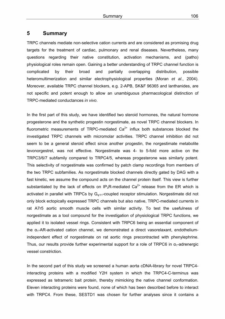
Summary 106
5 Summary
TRPC channels mediate non-selective cation currents and are considered as promising drug
targets for the treatment of cardiac, pulmonary and renal diseases. Nevertheless, many
questions regarding their native constitution, activation mechanisms, and (patho)
physiological roles remain open. CCGaining a better understanding of TRPC channel function is
complicated CCby their broad and partially overlapping distribution, possible
heteromultimerization and similar electrophysiological properties (Moran et al., 2004).
Moreover, available TRPC channel blockers, e.g. 2-APB, SK&F 96365 and lanthanides, are
not specific and potent enough to allow an unambiguous pharmacological distinction of
TRPC-mediated conductances in vivo.
In the first part of this study, we have identified two steroid hormones, the natural hormone
progesterone and the synthetic progestin norgestimate, as novel TRPC channel blockers. In
fluorometric measurements of TRPC-mediated CaPP
2+PP influx both substances blocked the
investigated TRPC channels with micromolar activities. TRPC channel inhibition did not
seem to be a general steroid effect since another progestin, the norgestimate metabolite
levonorgestrel, was not effective. Norgestimate was 4- to 5-fold more active on the
TRPC3/6/7 subfamily compared to TRPC4/5, whereas progesterone was similarly potent.
This selectivity of norgestimate was confirmed by patch clamp recordings from members of
the two TRPC subfamilies. As norgestimate blocked channels directly gated by DAG with a
fast kinetic, we assume the compound acts on the channel protein itself. This view is further
substantiated by the lack of effects on IPBB3BBR-mediated Ca PP
2+ PPrelease from the ER which is
activated in parallel with TRPCs by GBBq/11 BB-coupled receptor stimulation. Norgestimate did not
only block ectopically expressed TRPC channels but also native, TRPC-mediated currents in
rat A7r5 aortic smooth muscle cells with similar activity. To test the usefulness of
norgestimate as a tool compound for the investigation of physiological TRPC functions, we
applied it to isolated vessel rings. Consistent with TRPC6 being an essential component of
the α BB1BB-AR-activated cation channel, we demonstrated a direct vasorelaxant, endothelium-
independent effect of norgestimate on rat aortic rings precontracted with phenylephrine.
Thus, our results provide further experimental support for a role of TRPC6 in α BB1BB-adrenergic
vessel constriction.
In the second part of this study we screened a human aorta cDNA-library for novel TRPC4-
interacting proteins with a modified Y2H system in which the TRPC4-C-terminus was
expressed as tetrameric bait protein, thereby mimicking the native channel conformation.
Eleven interacting proteins were found, none of which has been described before to interact
with TRPC4. From these, SESTD1 was chosen for further analyses since it contains a
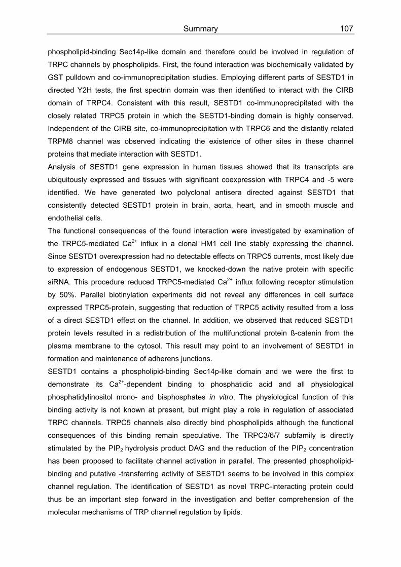
Summary 107
phospholipid-binding Sec14p-like domain and therefore could be involved in regulation of
TRPC channels by phospholipids. First, the found interaction was biochemically validated by
GST pulldown and co-immunoprecipitation studies. Employing different parts of SESTD1 in
directed Y2H tests, the first spectrin domain was then identified to interact with the CIRB
domain of TRPC4. Consistent with this result, SESTD1 co-immunoprecipitated with the
closely related TRPC5 protein in which the SESTD1-binding domain is highly conserved.
Independent of the CIRB site, co-immunoprecipitation with TRPC6 and the distantly related
TRPM8 channel was observed indicating the existence of other sites in these channel
proteins that mediate interaction with SESTD1.
Analysis of SESTD1 gene expression in human tissues showed that its transcripts are
ubiquitously expressed and tissues with significant coexpression with TRPC4 and -5 were
identified. We have generated two polyclonal antisera directed against SESTD1 that
consistently detected SESTD1 protein in brain, aorta, heart, and in smooth muscle and
endothelial cells.
The functional consequences of the found interaction were investigated by examination of
the TRPC5-mediated CaPP
2+PP influx in a clonal HM1 cell line stably expressing the channel.
Since SESTD1 overexpression had no detectable effects on TRPC5 currents, most likely due
to expression of endogenous SESTD1, we knocked-down the native protein with specific
siRNA. This procedure reduced TRPC5-mediated Ca PP
2+PP influx following receptor stimulation
by 50%. Parallel biotinylation experiments did not reveal any differences in cell surface
expressed TRPC5-protein, suggesting that reduction of TRPC5 activity resulted from a loss
of a direct SESTD1 effect on the channel. In addition, we observed that reduced SESTD1
protein levels resulted in a redistribution of the multifunctional protein ß-catenin from the
plasma membrane to the cytosol. This result may point to an involvement of SESTD1 in
formation and maintenance of adherens junctions.
SESTD1 contains a phospholipid-binding Sec14p-like domain and we were the first to
demonstrate its Ca PP
2+PP-dependent binding to phosphatidic acid and all physiological
phosphatidylinositol mono- and bisphosphates in vitro. The physiological function of this
binding activity is not known at present, but might play a role in regulation of associated
TRPC channels. TRPC5 channels also directly bind phospholipids although the functional
consequences of this binding remain speculative. The TRPC3/6/7 subfamily is directly
stimulated by the PIPBB2 BBhydrolysis product DAG and CCthe reduction of the PIP BB2BB concentration
has been proposed to facilitate channel activation in parallel. CCThe presented phospholipid-
binding and putative -transferring activity of SESTD1 seems to be involved in this complex
channel regulation. The identification of SESTD1 as novel TRPC-interacting protein could
thus be an important step forward in the investigation and CCbetter comprehension CCof the
molecular mechanisms of TRP channel regulation by lipids.
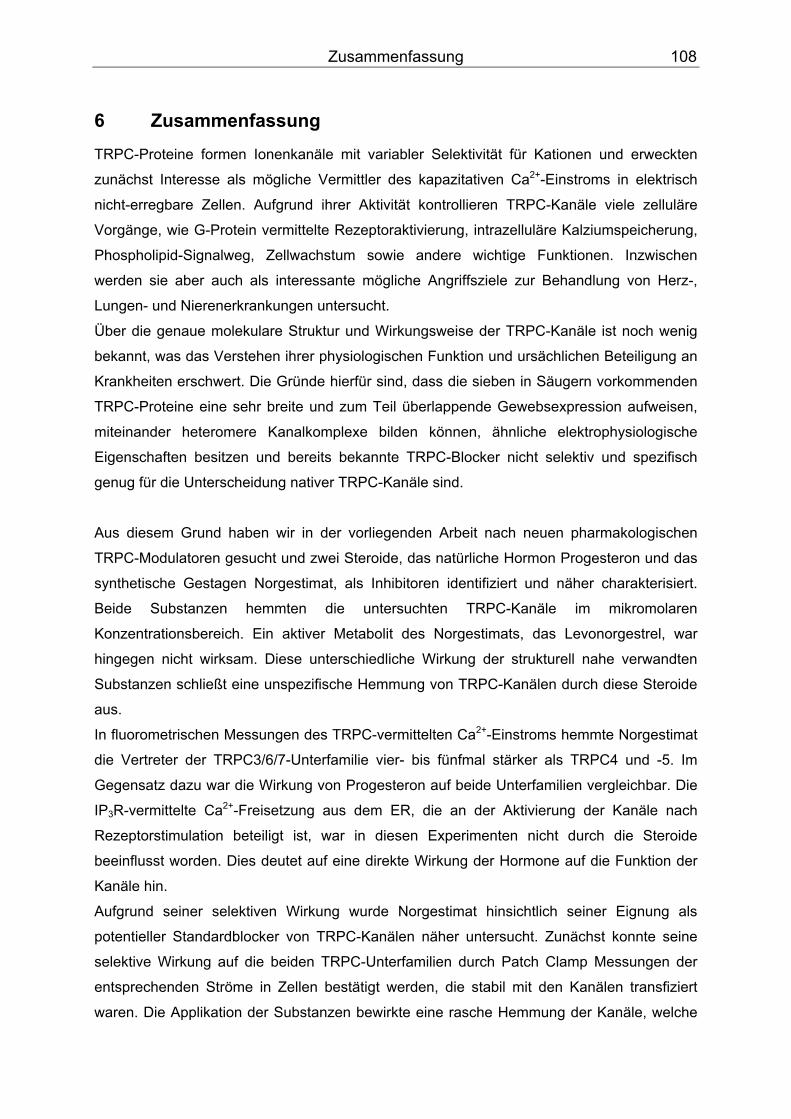
Zusammenfassung 108
6 Zusammenfassung
TRPC-Proteine formen Ionenkanäle mit variabler Selektivität für Kationen und erweckten
zunächst Interesse als mögliche Vermittler des kapazitativen Ca PP
2+PP-Einstroms in elektrisch
nicht-erregbare Zellen. Aufgrund ihrer Aktivität kontrollieren TRPC-Kanäle viele zelluläre
Vorgänge, wie G-Protein vermittelte Rezeptoraktivierung, intrazelluläre Kalziumspeicherung,
Phospholipid-Signalweg, Zellwachstum sowie andere wichtige Funktionen. Inzwischen
werden sie aber auch als interessante mögliche Angriffsziele zur Behandlung von Herz-,
Lungen- und Nierenerkrankungen untersucht.
Über die genaue molekulare Struktur und Wirkungsweise der TRPC-Kanäle ist noch wenig
bekannt, was das Verstehen ihrer physiologischen Funktion und ursächlichen Beteiligung an
Krankheiten erschwert. Die Gründe hierfür sind, dass die sieben in Säugern vorkommenden
TRPC-Proteine eine sehr breite und zum Teil überlappende Gewebsexpression aufweisen,
miteinander heteromere Kanalkomplexe bilden können, ähnliche elektrophysiologische
Eigenschaften besitzen und bereits bekannte TRPC-Blocker nicht selektiv und spezifisch
genug für die Unterscheidung nativer TRPC-Kanäle sind.
Aus diesem Grund haben wir in der vorliegenden Arbeit nach neuen pharmakologischen
TRPC-Modulatoren gesucht und zwei Steroide, das natürliche Hormon Progesteron und das
synthetische Gestagen Norgestimat, als Inhibitoren identifiziert und näher charakterisiert.
Beide Substanzen hemmten die untersuchten TRPC-Kanäle im mikromolaren
Konzentrationsbereich. Ein aktiver Metabolit des Norgestimats, das Levonorgestrel, war
hingegen nicht wirksam. Diese unterschiedliche Wirkung der strukturell nahe verwandten
Substanzen schließt eine unspezifische Hemmung von TRPC-Kanälen durch diese Steroide
aus.
In fluorometrischen Messungen des TRPC-vermittelten Ca PP
2+PP-Einstroms hemmte Norgestimat
die Vertreter der TRPC3/6/7-Unterfamilie vier- bis fünfmal stärker als TRPC4 und -5. Im
Gegensatz dazu war die Wirkung von Progesteron auf beide Unterfamilien vergleichbar. Die
IPBB3BBR-vermittelte CaPP
2+PP-Freisetzung aus dem ER, die an der Aktivierung der Kanäle nach
Rezeptorstimulation beteiligt ist, war in diesen Experimenten nicht durch die Steroide
beeinflusst worden. Dies deutet auf eine direkte Wirkung der Hormone auf die Funktion der
Kanäle hin.
Aufgrund seiner selektiven Wirkung wurde Norgestimat hinsichtlich seiner Eignung als
potentieller Standardblocker von TRPC-Kanälen näher untersucht. Zunächst konnte seine
selektive Wirkung auf die beiden TRPC-Unterfamilien durch Patch Clamp Messungen der
entsprechenden Ströme in Zellen bestätigt werden, die stabil mit den Kanälen transfiziert
waren. Die Applikation der Substanzen bewirkte eine rasche Hemmung der Kanäle, welche

Zusammenfassung 109
durch Auswaschen der Blocker ebenso schnell reversibel war. Diese schnelle Kinetik ist ein
weiterer Hinweis dafür, dass eine indirekte, für Steroidhormone charakteristische
genomische Wirkung als Ursache für die Kanalblockade ausgeschlossen werden kann. Die
Steroide hemmten zudem nicht nur die Aktivität der heterolog exprimierten Kanäle, sondern
auch native, TRPC-vermittelte Ströme in glatten Gefäßmuskelzellen aus Rattenaorten.
Aufgrund dieser Eigenschaften verwendeten wir Norgestimat, um die Beteiligung der TRPC-
Kanäle bei der Gefäßrelaxation näher zu untersuchen. Tatsächlich konnte an
vorkontrahierten Aortenringen aus der Ratte nach Gabe von Norgestimat eine endothel-
unabhängige Relaxation beobachtet werden. In Übereinstimmung mit bekannten
Literaturdaten legt auch dieses Ergebnis nahe, dass TRPC6-Kanäle an der Regulation des
Gefäßtonus beteiligt sind und damit eine wichtige Rolle bei der Kontrolle des Blutdrucks
spielen könnten. Zusammenfassend zeigen die vorgestellten Resultate, dass mit
Norgestimat ein geeignetes pharmakologisches Werkzeug gefunden wurde, das die weitere
Erforschung der physiologischen Funktionen von TRPC-Proteinen und ihrer Rolle bei
humanen Krankheiten erleichtern könnte. Zudem stellt es möglicherweise auch einen ersten
Ansatzpunkt für die weitere Entwicklung therapeutisch nützlicher Substanzen dar.
Die Suche nach TRPC-modulierenden Wirkstoffen für die therapeutische Nutzung wird auch
dadurch erschwert, dass sich die Eigenschaften von heterolog exprimierten Kanälen von
denen der nativen Kanäle unterscheiden können wie es beispielsweise für den TRPC4-Kanal
beschrieben wurde. Dies lässt darauf schließen, dass native TRPC4-Kanalkomplexe eine
andere molekulare Zusammensetzung aufweisen als heterolog exprimierte TRPC4-
Homotetramere und außerdem bislang noch unbekannte Interaktionspartner oder
regulatorische Untereinheiten existieren. Ein weiteres Ziel dieser Arbeit war es deshalb, neue
Interaktionspartner von TRPC4-Kanälen zu finden und diese anschließend funktionell zu
untersuchen. Zu diesem Zweck wurde zunächst eine cDNS-Bibliothek aus menschlichen
Aorten mit Hilfe eines modifizierten Hefe Zwei-Hybrid Systems durchmustert. Als
Köderprotein diente der C-Terminus des TRPC4-Kanalproteins. Die Besonderheit des
verwendeten Hefe Zwei-Hybrid Systems bestand darin, dass das Köderprotein als
tetrameres Fusionsprotein, d.h. in seiner nativen Konformation, vorlag.
Nach mehrmaligem Durchmustern der cDNS-Bibliothek wurden insgesamt
elf Interaktionspartner des TRPC4-Kanals isoliert, von denen keiner zuvor als
Interaktionspartner für TRPC4-Kanäle beschrieben worden ist. Aus diesen wurde das
SESTD1-Protein aufgrund seiner Struktur für weitergehende Untersuchungen ausgewählt.
Es besitzt eine phospholipidbindende Sec14p-Domäne sowie zwei Spektrindomänen. Da
TRPC-Kanäle in ihrer Aktivität durch Phospholipide reguliert werden und Spektrindomänen

Zusammenfassung 110
an der Bildung von Multiproteinkomplexen beteiligt sind, erschien SESTD1 vielversprechend
für eine detailliertere Charakterisierung.
Die in den Hefezellen beobachtete Interaktion von SESTD1 und TRPC4 wurde zunächst
durch zwei unabhängige proteinbiochemische Methoden bestätigt. Bakteriell exprimierte und
gereinigte GST-SESTD1-Fusionsproteine waren in Pulldown-Experimenten in der Lage,
TRPC4-Proteine aus Säugerzellextrakten zu binden. Ebenso wurde die
Koimmunopräzipitation beider Proteine aus Lysaten transfizierter Säugerzellen
nachgewiesen.
Unter Verwendung von SESTD1-Proteinfragmenten wurde anschließend in direkten
Interaktionsstudien in Hefezellen und in GST-Pulldown-Experimenten die erste
Spektrindomäne von SESTD1 als notwendig und ausreichend für die Bindung an das
TRPC4-Protein identifiziert. Umgekehrt konnte durch den Einsatz von C-terminal verkürzten
TRPC4-Köderproteinen die CIRB-Domäne des Kanalproteins als Bindungspartner für die
SESTD1-Spektrindomäne bestimmt werden. SESTD1 war auch in der Lage, das nahe
verwandte TRPC5-Protein zu binden, da in diesem Kanal die SESTD1-Interaktionssequenz
hoch konserviert ist. Die erfolgreiche CIRB-unabhängige Koimmunopräzipitation von
SESTD1 mit TRPC6 und dem entfernter verwandten TRPM8-Kanalprotein weisen jedoch
darauf hin, dass diese TRP-Kanäle noch weitere SESTD1-Bindungsstellen besitzen müssen.
Nachdem wir die in dem transkriptionellen Hefeassay beobachtete Interaktion zwischen
SESTD1 und TRPC4 bzw. -5 mit biochemischen Methoden verifiziert hatten, untersuchten
wir die Expression von SESTD1-Transkripten in verschiedenen Geweben. Es stellte sich
heraus, dass SESTD1 ubiquitär und damit beispielsweise in Gehirn, Herz und Aorta
überlappend mit TRPC4 bzw. -5 exprimiert wird. Für den Nachweis des SESTD1-Proteins
wurden zudem polyklonale Antikörper hergestellt. Als Antigene für die Immunisierung von
Kaninchen wurden zwei Peptide eingesetzt, deren Aminosäuresequenzen in den SESTD1-
Proteinen von Mensch, Maus und Ratte konserviert sind. Beide Antiseren erkannten in
Western Blot Analysen von stabil-transfizierten Zelllinien das SESTD1-Protein. Mithilfe dieser
Antikörper konnte das Vorkommen von SESTD1 in verschiedenen primären menschlichen
Gefäßmuskel- und Endothelzellen nachgewiesen werden. Außerdem wurde SESTD1-
Proteinexpression auch im Gehirn von Ratte und im Herz von Maus bestätigt.
Um die subzelluläre Lokalisation des endogenen SESTD1-Proteins und seine mögliche
Kolokalisation mit dem TRPC5-Kanal zu untersuchen, wurden Immunfluoreszenzstudien an
HM1-Zellen durchgeführt. Das Antiserum #148 färbte vorwiegend tubuläre Strukturen an.
Dieses Muster beruht sehr wahrscheinlich auf der bereits in der Western Blot Analyse
angedeuteten Kreuzreaktivität mit Tubulin. Mit dem Antiserum #147 ergab sich dagegen ein
hauptsächlich vesikuläres Verteilungsmuster. Da auch dieses Antiserum im Western Blot ein
Zusammenfassung 111
weiteres Protein erkannte, kann nicht mit Sicherheit angenommen werden, dass dies die
wirkliche subzelluläre Lokalisation von SESTD1 widerspiegelt. Für den eindeutigen
Nachweis einer Kolokalisation von SESTD1 mit TRPC5 waren unsere beiden Antiseren
aufgrund der zu geringen Spezifität ebenfalls nicht geeignet. Da bisher auch keine weiteren
Antiseren beschrieben oder kommerziell erhältlich sind, muss die letztliche Bestimmung der
SESTD1 Lokalisation in Zellen zukünftigen Untersuchungen vorbehalten bleiben.
Zur funktionellen Charakterisierung der gefundenen Interaktion wurden mögliche Wirkungen
von SESTD1 auf den TRPC5-vermittelten CaPP
2+PP-Einstrom exemplarisch untersucht. Dafür
wurde eine HM1-C5Y-Zelllinie hergestellt, die zusätzlich zu dem TRPC5-Kanal auch den
MBB1BB-Acetylcholinrezeptor stabil exprimiert, dessen Stimulierung zur TRPC5-Aktivierung
genutzt werden kann. Die Überexpression von SESTD1 in dieser Zelllinie hatte jedoch
keinen signifikanten Effekt auf den TRPC5-vermittelten Ca PP
2+PP-Einstrom. Western Blot Studien
ergaben allerdings, dass SESTD1 endogen von diesen Zellen exprimiert wird. Wir vermuten
deshalb, dass die Menge des endogenen SESTD1-Proteins bereits ausreichend für eine
maximale Wirkung auf den TRPC5-vermittelten Ca PP
2+PP-Einstrom ist. Deshalb wurde in einer
weiteren Studie die Menge des endogenen SESTD1-Proteins mittels spezifischer siRNA
stark reduziert. Durch die erzielte Hemmung der SESTD1-Expression war der TRPC5-
vermittelte Ca PP
2+PP-Einstrom nach Rezeptorstimulation um etwa die Hälfte verringert.
Biotinylierungsstudien zeigten aber, dass die Menge des TRPC5-Proteins an der
Plasmamembran nicht verändert war. Diese Ergebnisse legen wiederum einen direkten
Einfluss von SESTD1 auf die Kanalaktivität nahe. Immunofluoreszenzstudien zeigten
außerdem, dass die siRNA-vermittelte Reduzierung der SESTD1-Proteinexpression zu einer
Umverteilung des multifunktionellen ß-Catenin-Proteins führte. In Kontrollzellen war es vor
allem an der Plasmamembran lokalisiert, wo es an der Vermittlung von Zell-Zell-Kontakten
beteiligt ist, während es sich in SESTD1-siRNA-behandelten Zellen vor allem im Zytosol
befand. SESTD1 ist also möglicherweise an der Bildung und/oder Aufrechterhaltung von
Zell-Zell-Kontakten beteiligt.
Um erste Hinweise auf den molekularen Mechanismus der Interaktion zwischen SESTD1
und TRPC5 zu erhalten, untersuchten wir abschließend, ob SESTD1 Phospholipide binden
kann, wie es das Vorhandensein der Sec14p-Domäne andeutet. Wir fanden, dass
rekombinantes GST-SESTD1-Fusionsprotein tatsächlich Phospholipide binden konnte, die
auf Nitrocellulose-Membranen immobilisiert waren. Neben Phosphatidylsäure wurden auch
alle physiologisch vorkommenden Phosphatidylinositolmono- und -diphosphate gebunden.
Interessanterweise wurde diese Bindung Ca PP
2+PP-abhängig moduliert. Diese Calciumsensitivität
Zusammenfassung 112
eröffnet die faszinierende Möglichkeit einer dualen Regulation sowohl von TRPC4/5 durch
SESTD1 als auch von SESTD1 durch TRPC-vermittelten Ca PP
2+PP-Einstrom.
Für mögliche zukünftige nichtradioaktive SESTD1-Substratbindungsstudien haben wir einen
Bindungsassay etabliert, der diese Untersuchungen in 96-Wellplatten und damit die effiziente
Identifizierung von SESTD1-Modulatoren ermöglichen könnte. S
Phospholipide sind in komplexer Weise an der Regulation von TRPC4 und TRPC5 beteiligt.
Sie stellen das Substrat für die zur Kanalaktivierung essentiellen Hydrolysefunktionen von
PLC dar und binden darüber hinaus direkt an die Kanalproteine. Die Identifizierung von
SESTD1 als TRPC-interagierendes Protein könnte ein wichtiger Schritt zur mechanistischen
Aufklärung der Kanal-Lipid-Wechselwirkung sowie ihrer funktionellen Konsequenzen sein.
References 113
7 References
Abramovich C, Shen WF, Pineault N, Imren S, Montpetit B, Largman C, & Humphries RK (2000). Functional cloning and characterization of a novel nonhomeodomain protein that inhibits the binding of PBX1-HOX complexes to DNA. J Biol Chem 275, 26172-26177.
Alderton JM, Ahmed SA, Smith LA, & Steinhardt RA (2000). Evidence for a vesicle-mediated maintenance of store-operated calcium channels in a human embryonic kidney cell line. Cell Calcium 28, 161-169.
Allen-Baume V, Segui B, & Cockcroft S (2002). Current thoughts on the phosphatidylinositol transfer protein family. FEBS Lett 531, 74-80.
Alton KB, Hetyei NS, Shaw C, & Patrick JE (1984). Biotransformation of norgestimate in women. Contraception 29, 19-29.
Ambudkar IS, Ong HL, Liu X, Bandyopadhyay B, & Cheng KT (2007). TRPC1: The link between functionally distinct store-operated calcium channels. Cell Calcium 42, 213-223.
Ambudkar IS & Ong HL (2007). Organization and function of TRPC channelosomes. Pflugers Arch 455, 187-200.
Amiri H, Schultz G, & Schaefer M (2003). FRET-based analysis of TRPC subunit stoichiometry. Cell Calcium 33, 463-470.
Andreotti AH (2006). Opening the pore hinges on proline. Nat Chem Biol 2, 13-14.
Antoniotti S, Fiorio PA, Barral S, Scalabrino O, Munaron L, & Lovisolo D (2006). Interaction between TRPC channel subunits in endothelial cells. J Recept Signal Transduct Res 26, 225-240.
Auerbach D, Galeuchet-Schenk B, Hottiger MO, & Stagljar I (2002). Genetic approaches to the identification of interactions between membrane proteins in yeast. J Recept Signal Transduct Res 22, 471-481.
Balzer M, Lintschinger B, & Groschner K (1999). Evidence for a role of Trp proteins in the oxidative stress-induced membrane conductances of porcine aortic endothelial cells. Cardiovasc Res 42, 543-549.
Bankaitis VA, Malehorn DE, Emr SD, & Greene R (1989). The Saccharomyces cerevisiae SEC14 gene encodes a cytosolic factor that is required for transport of secretory proteins from the yeast Golgi complex. J Cell Biol 108, 1271-1281.
Bankaitis VA, Aitken JR, Cleves AE, & Dowhan W (1990). An essential role for a phospholipid transfer protein in yeast Golgi function. Nature 347, 561-562.
Bano D & Nicotera P (2007). Ca PP
2+PP signals and neuronal death in brain ischemia. Stroke 38,
674-676.
Barbagallo M, Dominguez LJ, Licata G, Shan J, Bing L, Karpinski E, Pang PK, & Resnick LM (2001). Vascular Effects of Progesterone: Role of Cellular Calcium Regulation. Hypertension 37, 142-147.
Basora N, Boulay G, Bilodeau L, Rousseau E, & Payet MD (2003). 20-hydroxyeicosatetraenoic acid (20-HETE) activates mouse TRPC6 channels expressed in HEK293 cells. J Biol Chem 278, 31709-31716.
References 114
Bassi MT, Manzoni M, Monti E, Pizzo MT, Ballabio A, & Borsani G (2000). Cloning of the gene encoding a novel integral membrane protein, mucolipidin-and identification of the two major founder mutations causing mucolipidosis type IV. Am J Hum Genet 67, 1110-1120.
Bateman J & Van VD (2001). The Trio family of guanine-nucleotide-exchange factors: regulators of axon guidance. J Cell Sci 114, 1973-1980.
Beech DJ, Xu SZ, McHugh D, & Flemming R (2003). TRPC1 store-operated cationic channel subunit. Cell Calcium 33, 433-440.
Beech DJ, Muraki K, & Flemming R (2004). Non-selective cationic channels of smooth muscle and the mammalian homologues of Drosophila TRP. J Physiol 559, 685-706.
Behnia R & Munro S (2005). Organelle identity and the signposts for membrane traffic. Nature 438, 597-604.
Bell RM & Burns DJ (1991). Lipid activation of protein kinase C. J Biol Chem 266, 4661-4664.
Bergdahl A, Gomez MF, Wihlborg AK, Erlinge D, Eyjolfson A, Xu SZ, Beech DJ, Dreja K, & Hellstrand P (2004). Plasticity of TRPC expression in arterial smooth muscle: correlation with store-operated Ca PP
2+PP entry. Am J Physiol Cell Physiol.
Berridge MJ (1993). Inositol trisphosphate and calcium signalling. Nature 361, 315-325.
Berridge MJ (1995). Capacitative calcium entry. Biochem J 312 ( Pt 1), 1-11.
Berridge MJ (2006). Remodelling Ca PP
2+PP signalling systems and cardiac hypertrophy. Biochem
Soc Trans 34, 228-231.
Bezzerides VJ, Ramsey IS, Kotecha S, Greka A, & Clapham DE (2004). Rapid vesicular translocation and insertion of TRP channels. Nat Cell Biol 6, 709-720.
Bigay J, Deterre P, Pfister C, & Chabre M (1985). Fluoroaluminates activate transducin-GDP by mimicking the gamma-phosphate of GTP in its binding site. FEBS Lett 191, 181-185.
Bilmen JG, Wootton LL, Godfrey RE, Smart OS, & Michelangeli F (2002). Inhibition of SERCA Ca PP
2+PP pumps by 2-aminoethoxydiphenyl borate (2-APB). 2-APB reduces both Ca PP
2+PP
binding and phosphoryl transfer from ATP, by interfering with the pathway leading to the Ca PP
2+PP-binding sites. Eur J Biochem 269, 3678-3687.
Bolotina VM & Csutora P (2005). CIF and other mysteries of the store-operated CaPP
2+PP-entry
pathway. Trends Biochem Sci 30, 378-387.
Bootman MD, Collins TJ, Mackenzie L, Roderick HL, Berridge MJ, & Peppiatt CM (2002). 2-aminoethoxydiphenyl borate (2-APB) is a reliable blocker of store-operated Ca PP
2+PP entry but
an inconsistent inhibitor of InsP BB3BB-induced Ca PP
2+PP release. FASEB J 16, 1145-1150.
Boulay G, Zhu X, Peyton M, Jiang M, Hurst R, Stefani E, & Birnbaumer L (1997). Cloning and expression of a novel mammalian homolog of Drosophila transient receptor potential (Trp) involved in calcium entry secondary to activation of receptors coupled by the G BBqBB class of G protein. J Biol Chem 272, 29672-29680.
Boulay G, Brown DM, Qin N, Jiang M, Dietrich A, Zhu MX, Chen Z, Birnbaumer M, Mikoshiba K, & Birnbaumer L (1999). Modulation of Ca(2+) entry by polypeptides of the inositol 1,4, 5-trisphosphate receptor (IP BB3BBR) that bind transient receptor potential (TRP): evidence for

References 115
roles of TRP and IPBB3BBR in store depletion-activated Ca(2+) entry. Proc Natl Acad Sci U S A 96, 14955-14960.
Brazer SC, Singh BB, Liu X, Swaim W, & Ambudkar IS (2003). Caveolin-1 contributes to assembly of store-operated Ca PP
2+PP influx channels by regulating plasma membrane
localization of TRPC1. J Biol Chem 278, 27208-27215.
Burdakov D, Petersen OH, & Verkhratsky A (2005). Intraluminal calcium as a primary regulator of endoplasmic reticulum function. Cell Calcium 38, 303-310.
Bush EW, Hood DB, Papst PJ, Chapo JA, Minobe W, Bristow MR, Olson EN, & McKinsey TA (2006). Canonical transient receptor potential channels promote cardiomyocyte hypertrophy through activation of calcineurin signaling. J Biol Chem 281, 33487-33496.
Cayouette S, Lussier MP, Mathieu EL, Bousquet SM, & Boulay G (2004). Exocytotic insertion of TRPC6 channel into the plasma membrane upon GBBq BBprotein-coupled receptor activation. J Biol Chem 279, 7241-7246.
Chalfie M, Tu Y, Euskirchen G, Ward WW, & Prasher DC (1994). Green fluorescent protein as a marker for gene expression. Science 263, 802-805.
Chang AS, Chang SM, Garcia RL, & Schilling WP (1997). Concomitant and hormonally regulated expression of trp genes in bovine aortic endothelial cells. FEBS Lett 415, 335-340.
Chesire DR & Isaacs WB (2002). Ligand-dependent inhibition of beta-catenin/TCF signaling by androgen receptor. Oncogene 21, 8453-8469.
Chinopoulos C, Starkov AA, & Fiskum G (2003). Cyclosporin A-insensitive permeability transition in brain mitochondria: inhibition by 2-aminoethoxydiphenyl borate. J Biol Chem 278, 27382-27389.
Cioffi DL, Wu S, Alexeyev M, Goodman SR, Zhu MX, & Stevens T (2005). Activation of the Endothelial Store-Operated ISOC CaPP
2+PP Channel Requires Interaction of Protein 4.1 With
TRPC4. Circ Res 97, 1164-1172.
Clapham DE (1995). Calcium signaling. Cell 80, 259-268.
Clapham DE, Runnels LW, & Strubing C (2001). The TRP ion channel family. Nat Rev Neurosci 2, 387-396.
Clapham DE (2003). TRP channels as cellular sensors. Nature 426, 517-524.
Cosens DJ & Manning A (1969). Abnormal electroretinogram from a Drosophila mutant. Nature 224, 285-287.
Crews JK & Khalil RA (1999). Antagonistic effects of 17 beta-estradiol, progesterone, and testosterone on Ca PP
2+PP entry mechanisms of coronary vasoconstriction. Arterioscler Thromb
Vasc Biol 19, 1034-1040.
D'Angelo I, Welti S, Bonneau F, & Scheffzek K (2006). A novel bipartite phospholipid-binding module in the neurofibromatosis type 1 protein. EMBO Rep 7, 174-179.
Dalrymple A, Slater DM, Beech D, Poston L, & Tribe RM (2002). Molecular identification and localization of Trp homologues, putative calcium channels, in pregnant human uterus. Mol Hum Reprod 8, 946-951.
References 116
Dalrymple A, Mahn K, Poston L, Songu-Mize E, & Tribe RM (2007). Mechanical stretch regulates TRPC expression and calcium entry in human myometrial smooth muscle cells. Mol Hum Reprod 13, 171-179.
Davies TH & Sanchez ER (2005). FKBP52. Int J Biochem Cell Biol 37, 42-47.
Demaurex N, Lew DP, & Krause KH (1992). Cyclopiazonic acid depletes intracellular CaPP
2+PP
stores and activates an influx pathway for divalent cations in HL-60 cells. J Biol Chem 267, 2318-2324.
Dietrich A, Schnitzler M, Emmel J, Kalwa H, Hofmann T, & Gudermann T (2003). N-linked protein glycosylation is a major determinant for basal TRPC3 and TRPC6 channel activity. J Biol Chem 278, 47842-47852.
Dietrich A, Kalwa H, Rost BR, & Gudermann T (2005a). The diacylgylcerol-sensitive TRPC3/6/7 subfamily of cation channels: functional characterization and physiological relevance. Pflugers Arch 451, 72-80.
Dietrich A, Mederos YS, Gollasch M, Gross V, Storch U, Dubrovska G, Obst M, Yildirim E, Salanova B, Kalwa H, Essin K, Pinkenburg O, Luft FC, Gudermann T, & Birnbaumer L (2005b). Increased vascular smooth muscle contractility in TRPC6-/- mice. Mol Cell Biol 25, 6980-6989.
Dietrich A, Chubanov V, Kalwa H, Rost BR, & Gudermann T (2006). Cation channels of the transient receptor potential superfamily: their role in physiological and pathophysiological processes of smooth muscle cells. Pharmacol Ther 112, 744-760.
Dietrich A, Kalwa H, Fuchs B, Grimminger F, Weissmann N, & Gudermann T (2007a). In vivo TRPC functions in the cardiopulmonary vasculature. Cell Calcium 42, 233-244.
Dietrich A, Kalwa H, Storch U, Mederos YS, Salanova B, Pinkenburg O, Dubrovska G, Essin K, Gollasch M, Birnbaumer L, & Gudermann T (2007b). Pressure-induced and store-operated cation influx in vascular smooth muscle cells is independent of TRPC1. Pflugers Arch 455, 465-477.
Djinovic-Carugo K, Gautel M, Ylanne J, & Young P (2002). The spectrin repeat: a structural platform for cytoskeletal protein assemblies. FEBS Lett 513, 119-123.
Droogmans G (2007). CaBuf Program. http://www.kuleuven.be/fysio/trp/?q=cabuf
Echelon (2007). Technical Data Sheet for MultiPIP Grip (LL5-α). TThttp://www.echelon-inc.com/objects/catalog/product/extras/1211_G-1100.pdf TT
Eder P, Probst D, Rosker C, Poteser M, Wolinski H, Kohlwein SD, Romanin C, & Groschner K (2007). Phospholipase C-dependent control of cardiac calcium homeostasis involves a TRPC3-NCX1 signaling complex. Cardiovasc Res 73, 111-119.
Engelke M, Friedrich O, Budde P, Schafer C, Niemann U, Zitt C, Jungling E, Rocks O, Luckhoff A, & Frey J (2002). Structural domains required for channel function of the mouse transient receptor potential protein homologue TRP1beta. FEBS Lett 523, 193-199.
Estacion M, Sinkins WG, & Schilling WP (2001). Regulation of Drosophila transient receptor potential-like (TrpL) channels by phospholipase C-dependent mechanisms. J Physiol 530, 1-19.
References 117
Estacion M, Li S, Sinkins WG, Gosling M, Bahra P, Poll C, Westwick J, & Schilling WP (2004). Activation of human TRPC6 channels by receptor stimulation. J Biol Chem 279, 22047-22056.
Estacion M, Sinkins WG, Jones SW, Applegate MA, & Schilling WP (2006). Human TRPC6 expressed in HEK 293 cells forms non-selective cation channels with limited Ca PP
2+PP
permeability. J Physiol 572, 359-377.
Facemire CS, Mohler PJ, & Arendshorst WJ (2004). Expression and relative abundance of short transient receptor potential channels in the rat renal microcirculation. Am J Physiol Renal Physiol 286, F546-F551.
Fagan KA, Smith KE, & Cooper DM (2000). Regulation of the Ca PP
2+PP-inhibitable adenylyl
cyclase type VI by capacitative Ca PP
2+PP entry requires localization in cholesterol-rich domains.
J Biol Chem 275, 26530-26537.
Fan Z, Gao L, & Wang W (2003). Phosphatidic acid stimulates cardiac KBBATPBB channels like phosphatidylinositols, but with novel gating kinetics. Am J Physiol Cell Physiol 284, C94-102.
Favre CJ, Jerstrom P, Foti M, Stendhal O, Huggler E, Lew DP, & Krause KH (1996). Organization of Ca PP
2+PP stores in myeloid cells: association of SERCA2b and the type-1
inositol-1,4,5-trisphosphate receptor. Biochem J 316 ( Pt 1), 137-142.
Feske S, Prakriya M, Rao A, & Lewis RS (2005). A severe defect in CRAC Ca PP
2+PP channel
activation and altered K PP
+PP channel gating in T cells from immunodeficient patients. J Exp
Med 202, 651-662.
Feske S, Gwack Y, Prakriya M, Srikanth S, Puppel SH, Tanasa B, Hogan PG, Lewis RS, Daly M, & Rao A (2006). A mutation in Orai1 causes immune deficiency by abrogating CRAC channel function. Nature 441, 179-185.
Fields S & Song O (1989). A novel genetic system to detect protein-protein interactions. Nature 340, 245-246.
Flemming R, Xu SZ, & Beech DJ (2003). Pharmacological profile of store-operated channels in cerebral arteriolar smooth muscle cells. Br J Pharmacol 139, 955-965.
Flemming PK, Dedman AM, Xu SZ, Li J, Zeng F, Naylor J, Benham CD, Bateson AN, Muraki K, & Beech DJ (2005). Sensing of lysophospholipids by TRPC5 calcium channel. J Biol Chem 281, 4977-4982.
Flockerzi V, Jung C, Aberle T, Meissner M, Freichel M, Philipp SE, Nastainczyk W, Maurer P, & Zimmermann R (2005). Specific detection and semi-quantitative analysis of TRPC4 protein expression by antibodies. Pflugers Arch 451, 81-86.
Fowler MA, Sidiropoulou K, Ozkan ED, Phillips CW, & Cooper DC (2007). Corticolimbic Expression of TRPC4 and TRPC5 Channels in the Rodent Brain. PLoS ONE 2, e573.
Franzius D, Hoth M, & Penner R (1994). Non-specific effects of calcium entry antagonists in mast cells. Pflugers Arch 428, 433-438.
Freichel M, Suh SH, Pfeifer A, Schweig U, Trost C, Weissgerber P, Biel M, Philipp S, Freise D, Droogmans G, Hofmann F, Flockerzi V, & Nilius B (2001). Lack of an endothelial store-operated Ca PP
2+PP current impairs agonist-dependent vasorelaxation in TRP4 PP
-/-PP mice. Nat Cell
Biol 3, 121-127.
References 118
Freichel M, Vennekens R, Olausson J, Stolz S, Philipp S, Weissgerber P, & Flockerzi V (2005). Functional role of TRPC proteins in native systems: implications from knockout and knock-down studies. J Physiol 567(Pt 1), 59-66.
Fruman DA, Meyers RE, & Cantley LC (1998). Phosphoinositide kinases. Annu Rev Biochem 67, 481-507.
Garcia RL & Schilling WP (1997). Differential expression of mammalian TRP homologues across tissues and cell lines. Biochem Biophys Res Commun 239, 279-283.
Glusa E, Graser T, Wagner S, & Oettel M (1997). Mechanisms of relaxation of rat aorta in response to progesterone and synthetic progestins. Maturitas 28, 181-191.
Goel M, Sinkins W, Keightley A, Kinter M, & Schilling WP (2005). Proteomic analysis of T. Pflugers Arch 451, 87-98.
Greka A, Navarro B, Oancea E, Duggan A, & Clapham DE (2003). TRPC5 is a regulator of hippocampal neurite length and growth cone morphology. Nat Neurosci 6, 837-845.
Grynkiewicz G, Poenie M, & Tsien RY (1985). A new generation of CaPP
2+PP indicators with
greatly improved fluorescence properties. J Biol Chem 260, 3440-3450.
Gudermann T, Hofmann T, Schnitzler M, & Dietrich A (2004). Activation, subunit composition and physiological relevance of DAG-sensitive TRPC proteins. Novartis Found Symp 258, 103-118.
Gudermann T (2005). A new TRP to kidney disease. Nat Genet 37, 663-664.
Halaszovich CR, Zitt C, Jungling E, & Luckhoff A (2000). Inhibition of TRP3 channels by lanthanides. Block from the cytosolic side of the plasma membrane. J Biol Chem 275, 37423-37428.
Harbury PB, Zhang T, Kim PS, & Alber T (1993). A switch between two-, three-, and four-stranded coiled coils in GCN4 leucine zipper mutants. Science 262, 1401-1407.
Hardie RC & Minke B (1992). The trp gene is essential for a light-activated Ca PP
2+PP channel in
Drosophila photoreceptors. Neuron 8, 643-651.
Hardie RC & Raghu P (2001). Visual transduction in Drosophila. Nature 413, 186-193.
Hardie RC (2003). Regulation of TRP channels via lipid second messengers. Annu Rev Physiol 65, 735-759.
Harding MW, Galat A, Uehling DE, & Schreiber SL (1989). A receptor for the immunosuppressant FK506 is a cis-trans peptidyl-prolyl isomerase. Nature 341, 758-760.
Harteneck C, Plant TD, & Schultz G (2000). From worm to man: three subfamilies of TRP channels. Trends Neurosci 23, 159-166.
Hermosura MC, Nayakanti H, Dorovkov MV, Calderon FR, Ryazanov AG, Haymer DS, & Garruto RM (2005). A TRPM7 variant shows altered sensitivity to magnesium that may contribute to the pathogenesis of two Guamanian neurodegenerative disorders. Proc Natl Acad Sci U S A 102, 11510-11515.
Hermosura MC & Garruto RM (2007). TRPM7 and TRPM2-Candidate susceptibility genes for Western Pacific ALS and PD? Biochim Biophys Acta 1772, 822-835.
References 119
Hewavitharana T, Deng X, Soboloff J, & Gill DL (2007). Role of STIM and Orai proteins in the store-operated calcium signaling pathway. Cell Calcium 42, 173-182.
Hisatsune C, Kuroda Y, Nakamura K, Inoue T, Nakamura T, Michikawa T, Mizutani A, & Mikoshiba K (2004). Regulation of TRPC6 channel activity by tyrosine phosphorylation. J Biol Chem 279, 18887-18894.
Hoenderop JG, Voets T, Hoefs S, Weidema F, Prenen J, Nilius B, & Bindels RJ (2003). Homo- and heterotetrameric architecture of the epithelial Ca PP
2+ PPchannels TRPV5 and
TRPV6. EMBO J 22, 776-785.
Hofmann T, Obukhov AG, Schaefer M, Harteneck C, Gudermann T, & Schultz G (1999). Direct activation of human TRPC6 and TRPC3 channels by diacylglycerol. Nature 397, 259-263.
Hofmann T, Schaefer M, Schultz G, & Gudermann T (2002). Subunit composition of mammalian transient receptor potential channels in living cells. Proc Natl Acad Sci U S A 99, 7461-7466.
Hoth M & Penner R (1992). Depletion of intracellular calcium stores activates a calcium current in mast cells. Nature 355, 353-356.
Hsu YJ, Hoenderop JG, & Bindels RJ (2007). TRP channels in kidney disease. Biochim Biophys Acta 1772, 928-936.
Hsuan J & Cockcroft S (2001). The PITP family of phosphatidylinositol transfer proteins. Genome Biol 2, REVIEWS3011.
Huang GN, Zeng W, Kim JY, Yuan JP, Han L, Muallem S, & Worley PF (2006). STIM1 carboxyl-terminus activates native SOC, I(crac) and TRPC1 channels. Nat Cell Biol 8, 1003-1010.
Ile KE, Schaaf G, & Bankaitis VA (2006). Phosphatidylinositol transfer proteins and cellular nanoreactors for lipid signaling. Nat Chem Biol 2, 576-583.
Inoue R, Okada T, Onoue H, Hara Y, Shimizu S, Naitoh S, Ito Y, & Mori Y (2001). The transient receptor potential protein homologue TRP6 is the essential component of vascular alpha(1)-adrenoceptor-activated Ca(2+)-permeable cation channel. Circ Res 88, 325-332.
Inoue R, Jensen LJ, Shi J, Morita H, Nishida M, Honda A, & Ito Y (2006). Transient receptor potential channels in cardiovascular function and disease. Circ Res 99, 119-131.
Irvine RF (1990). 'Quantal' Ca PP
2+PP release and the control of Ca PP
2+PP entry by inositol phosphates-
a possible mechanism. FEBS Lett 263, 5-9.
Ito H, Fukuda Y, Murata K, & Kimura A (1983). Transformation of intact yeast cells treated with alkali cations. J Bacteriol 153, 163-168.
IUPAC Commission on the Nomenclature of Organic Chemistry and IUPAC-IUB Commission on Biochemical Nomenclature (1969). Revised tentative rules for nomenclature of steroids. Biochem J TT113(1), 5-28. TT
Juchem M, Pollow K, Elger W, Hoffmann G, & Mobus V (1993). Receptor binding of norgestimate-a new orally active synthetic progestational compound. Contraception 47, 283-294.

References 120
Jung S, Strotmann R, Schultz G, & Plant TD (2002). TRPC6 is a candidate channel involved in receptor-stimulated cation currents in A7r5 smooth muscle cells. Am J Physiol Cell Physiol 282, C347-C359.
Jung S, Muhle A, Schaefer M, Strotmann R, Schultz G, & Plant TD (2003). Lanthanides potentiate TRPC5 currents by an action at extracellular sites close to the pore mouth. J Biol Chem 278, 3562-3571.
Jungnickel MK, Marrero H, Birnbaumer L, Lemos JR, & Florman HM (2001). Trp2 regulates entry of Ca PP
2+PP into mouse sperm triggered by egg ZP3. Nat Cell Biol 3, 499-502.
Kamouchi M, Philipp S, Flockerzi V, Wissenbach U, Mamin A, Raeymaekers L, Eggermont J, Droogmans G, & Nilius B (1999). Properties of heterologously expressed hTRP3 channels in bovine pulmonary artery endothelial cells. J Physiol 518 Pt 2, 345-358.
Kawabata A, Saifeddine M, Al-Ani B, Leblond L, & Hollenberg MD (1999). Evaluation of proteinase-activated receptor-1 (PAR1) agonists and antagonists using a cultured cell receptor desensitization assay: activation of PAR2 by PAR1-targeted ligands. J Pharmacol Exp Ther 288, 358-370.
Kedei N, Szabo T, Lile JD, Treanor JJ, Olah Z, Iadarola MJ, & Blumberg PM (2001). Analysis of the native quaternary structure of vanilloid receptor 1. J Biol Chem 276, 28613-28619.
Kim JY, Zeng W, Kiselyov K, Yuan JP, Dehoff MH, Mikoshiba K, Worley PF, & Muallem S (2006a). Homer 1 mediates store- and inositol 1,4,5-trisphosphate receptor-dependent translocation and retrieval of TRPC3 to the plasma membrane. J Biol Chem 281, 32540-32549.
Kim MT, Kim BJ, Lee JH, Kwon SC, Yeon DS, Yang DK, So I, & Kim KW (2006b). Involvement of calmodulin and myosin light chain kinase in activation of mTRPC5 expressed in HEK cells. Am J Physiol Cell Physiol 290, C1031-C1040.
Kim SJ, Kim YS, Yuan JP, Petralia RS, Worley PF, & Linden DJ (2003). Activation of the TRPC1 cation channel by metabotropic glutamate receptor mGluR1. Nature 426, 285-291.
Kiselyov K, Xu X, Mozhayeva G, Kuo T, Pessah I, Mignery G, Zhu X, Birnbaumer L, & Muallem S (1998). Functional interaction between InsP3 receptors and store-operated Htrp3 channels. Nature 396, 478-482.
Kiselyov KI, Shin DM, Wang Y, Pessah IN, Allen PD, & Muallem S (2000). Gating of store-operated channels by conformational coupling to ryanodine receptors. Mol Cell 6, 421-431.
Knudsen KA, Soler AP, Johnson KR, & Wheelock MJ (1995). Interaction of alpha-actinin with the cadherin/catenin cell-cell adhesion complex via alpha-catenin. J Cell Biol 130, 67-77.
Kostenko EV, Mahon GM, Cheng L, & Whitehead IP (2005). The Sec14 homology domain regulates the cellular distribution and transforming activity of the Rho-specific guanine nucleotide exchange factor Dbs. J Biol Chem 280, 2807-2817.
Kuhnz W, Blode H, & Mahler M (1994). Systemic availability of levonorgestrel after single oral administration of a norgestimate-containing combination oral contraceptive to 12 young women. Contraception 49, 255-263.
Kumar B, Dreja K, Shah S, Cheong A, Xu SZ, Sukumar P, Naylor J, Forte A, Cipollaro M, McHugh D, Kingston PA, Heagerty AM, Munsch CM, Bergdahl A, Hultgardh-Nilsson A, Gomez MF, Porter KE, Hellstrand P, & Beech DJ (2006). Upregulated TRPC1 Channel in
References 121
Vascular Injury In Vivo and Its Role in Human Neointimal Hyperplasia. Circ Res 98, 557-563.
Kunichika N, Landsberg JW, Yu Y, Kunichika H, Thistlethwaite PA, Rubin LJ, & Yuan JX (2004). Bosentan inhibits transient receptor potential channel expression in pulmonary vascular myocytes. Am J Respir Crit Care Med 170, 1101-1107.
Kuwahara K, Wang Y, McAnally J, Richardson JA, Bassel-Duby R, Hill JA, & Olson EN (2006). TRPC6 fulfills a calcineurin signaling circuit during pathologic cardiac remodeling. J Clin Invest 116, 3114-3126.
Kwan HY, Huang Y, & Yao X (2004). Regulation of canonical transient receptor potential isoform 3 (TRPC3) channel by protein kinase G. Proc Natl Acad Sci U S A 101, 2625-2630.
Kwan HY, Huang Y, & Yao X (2007). TRP channels in endothelial function and dysfunction. Biochim Biophys Acta 1772, 907-914.
Kwon Y, Hofmann T, & Montell C (2007). Integration of phosphoinositide- and calmodulin-mediated regulation of TRPC6. Mol Cell 25, 491-503.
Landy A (1989). Dynamic, structural, and regulatory aspects of lambda site-specific recombination. Annu Rev Biochem 58, 913-949.
Large WA (2002). Receptor-operated Ca2(+)-permeable nonselective cation channels in vascular smooth muscle: a physiologic perspective. J Cardiovasc Electrophysiol 13, 493-501.
Lee KP, Jun JY, Chang IY, Suh SH, So I, & Kim KW (2005). TRPC4 is an essential component of the nonselective cation channel activated by muscarinic stimulation in mouse visceral smooth muscle cells. Mol Cells 20, 435-441.
Lee SY, Obata Y, Yoshida M, Stockert E, Williamson B, Jungbluth AA, Chen YT, Old LJ, & Scanlan MJ (2003a). Immunomic analysis of human sarcoma. Proc Natl Acad Sci U S A 100, 2651-2656.
Lee YM, Kim BJ, Kim HJ, Yang DK, Zhu MH, Lee KP, So I, & Kim KW (2003b). TRPC5 as a candidate for the nonselective cation channel activated by muscarinic stimulation in murine stomach. Am J Physiol Gastrointest Liver Physiol 284, G604-G616.
Lemonnier L, Prevarskaya N, Mazurier J, Shuba Y, & Skryma R (2004). 2-APB inhibits volume-regulated anion channels independently from intracellular calcium signaling modulation. FEBS Lett 556, 121-126.
Lemos VS, Poburko D, Liao CH, Cole WC, & van BC (2007). Na PP
+PP entry via TRPC6 causes
Ca PP
2+PP entry via NCX reversal in ATP stimulated smooth muscle cells. Biochem Biophys Res
Commun 352, 130-134.
Lepage PK, Lussier MP, Barajas-Martinez H, Bousquet SM, Blanchard AP, Francoeur N, Dumaine R, & Boulay G (2006). Identification of two domains involved in the assembly of transient receptor potential canonical channels. J Biol Chem 281, 30356-30364.
Lewis RS (2001). Calcium signaling mechanisms in T lymphocytes. Annu Rev Immunol 19, 497-521.
Li HS, Xu XZ, & Montell C (1999). Activation of a TRPC3-dependent cation current through the neurotrophin BDNF. Neuron 24, 261-273.
References 122
Li SW, Westwick J, & Poll CT (2002). Receptor-operated Ca PP
2+PP influx channels in leukocytes:
a therapeutic target? Trends Pharmacol Sci 23, 63-70.
Li S, Westwick J, & Poll C (2003). Transient receptor potential (TRP) channels as potential drug targets in respiratory disease. Cell Calcium 33, 551-558.
Li S, Westwick J, Cox B, & Poll CT (2004). TRP channels as drug targets. Novartis Found Symp 258, 204-213.
Li Y, Jia YC, Cui K, Li N, Zheng ZY, Wang YZ, & Yuan XB (2005). Essential role of TRPC channels in the guidance of nerve growth cones by brain-derived neurotrophic factor. Nature 434, 894-898.
Liao Y, Erxleben C, Yildirim E, Abramowitz J, Armstrong DL, & Birnbaumer L (2007). Orai proteins interact with TRPC channels and confer responsiveness to store depletion. Proc Natl Acad Sci U S A 104, 4682-4687.
Liman ER & Innan H (2003). Relaxed selective pressure on an essential component of pheromone transduction in primate evolution. Proc Natl Acad Sci U S A 100, 3328-3332.
Lintschinger B, Balzer-Geldsetzer M, Baskaran T, Graier WF, Romanin C, Zhu MX, & Groschner K (2000). Coassembly of Trp1 and Trp3 proteins generates diacylglycerol- and Ca PP
2+PP-sensitive cation channels. J Biol Chem 275, 27799-27805.
Liou J, Kim ML, Heo WD, Jones JT, Myers JW, Ferrell JE, Jr., & Meyer T (2005). STIM is a Ca PP
2+PP sensor essential for Ca PP
2+PP-store-depletion-triggered Ca PP
2+PP influx. Curr Biol 15, 1235-
1241.
Liu D, Scholze A, Zhu Z, Krueger K, Thilo F, Burkert A, Streffer K, Holz S, Harteneck C, Zidek W, & Tepel M (2006). Transient receptor potential channels in essential hypertension. J Hypertens 24, 1105-1114.
Liu X, Bandyopadhyay BC, Singh BB, Groschner K, & Ambudkar IS (2005). Molecular Analysis of a Store-operated and 2-Acetyl-sn-glycerol-sensitive Non-selective Cation Channel: HETEROMERIC ASSEMBLY OF TRPC1-TRPC3. J Biol Chem 280, 21600-21606.
Livak KJ, Flood SJ, Marmaro J, Giusti W, & Deetz K (1995). Oligonucleotides with fluorescent dyes at opposite ends provide a quenched probe system useful for detecting PCR product and nucleic acid hybridization. PCR Methods Appl 4, 357-362.
Lockwich TP, Liu X, Singh BB, Jadlowiec J, Weiland S, & Ambudkar IS (2000). Assembly of Trp1 in a signaling complex associated with caveolin-scaffolding lipid raft domains. J Biol Chem 275, 11934-11942.
Lockwich T, Singh BB, Liu X, & Ambudkar IS (2001). Stabilization of cortical actin induces internalization of transient receptor potential 3 (Trp3)-associated caveolar Ca PP
2+PP signaling
complex and loss of Ca PP
2+PP influx without disruption of Trp3-inositol trisphosphate receptor
association. J Biol Chem 276, 42401-42408.
Lodish HF, Berk A, Matsudeira P, Kaiser CA, Krieger M, Scott MP, Zipursky SL, & Darnell J (2003). Molecular cell biology, 5th ed. W.H. Freeman and Company, New York.
Lucas P, Ukhanov K, Leinders-Zufall T, & Zufall F (2003). A diacylglycerol-gated cation channel in vomeronasal neuron dendrites is impaired in TRPC2 mutant mice: mechanism of pheromone transduction. Neuron 40, 551-561.
References 123
Lummis SC, Beene DL, Lee LW, Lester HA, Broadhurst RW, & Dougherty DA (2005). Cis-trans isomerization at a proline opens the pore of a neurotransmitter-gated ion channel. Nature 438, 248-252.
Ma HT, Patterson RL, van Rossum DB, Birnbaumer L, Mikoshiba K, & Gill DL (2000). Requirement of the inositol trisphosphate receptor for activation of store-operated Ca PP
2+PP
channels. Science 287, 1647-1651.
Ma R, Rundle D, Jacks J, Koch M, Downs T, & Tsiokas L (2003). Inhibitor of myogenic family, a novel suppressor of store-operated currents through an interaction with TRPC1. J Biol Chem 278, 52763-52772.
Manavathi B, Acconcia F, Rayala SK, & Kumar R (2006). An inherent role of microtubule network in the action of nuclear receptor. Proc Natl Acad Sci U S A 103, 15981-15986.
Marchler-Bauer A & Bryant SH (2004). CD-Search: protein domain annotations on the fly. Nucleic Acids Res 32, W327-W331.
Maruyama T, Kanaji T, Nakade S, Kanno T, & Mikoshiba K (1997). 2APB, 2-aminoethoxydiphenyl borate, a membrane-penetrable modulator of Ins(1,4,5)P BB3BB-induced Ca PP
2+PP release. J Biochem (Tokyo) 122, 498-505.
Maruyama Y, Nakanishi Y, Walsh EJ, Wilson DP, Welsh DG, & Cole WC (2006). Heteromultimeric TRPC6-TRPC7 channels contribute to arginine vasopressin-induced cation current of A7r5 vascular smooth muscle cells. Circ Res 98, 1520-1527.
McCarron JG, Chalmers S, Bradley KN, MacMillan D, & Muir TC (2006). Ca PP
2+PP microdomains
in smooth muscle. Cell Calcium 40, 461-493.
McKay RR, Szymeczek-Seay CL, Lievremont JP, Bird GS, Zitt C, Jungling E, Luckhoff A, & Putney JW, Jr. (2000). Cloning and expression of the human transient receptor potential 4 (TRP4) gene: localization and functional expression of human TRP4 and TRP3. Biochem J 351 Pt 3, 735-746.
Mehta D, Ahmmed GU, Paria BC, Holinstat M, Voyno-Yasenetskaya T, Tiruppathi C, Minshall RD, & Malik AB (2003). RhoA interaction with inositol 1,4,5-trisphosphate receptor and transient receptor potential channel-1 regulates Ca PP
2+PP entry. Role in signaling increased
endothelial permeability. J Biol Chem 278, 33492-33500.
Meldolesi J & Pozzan T (1998). The endoplasmic reticulum Ca PP
2+PP store: a view from the
lumen. Trends Biochem Sci 23, 10-14.
Merritt JE, Armstrong WP, Benham CD, Hallam TJ, Jacob R, Jaxa-Chamiec A, Leigh BK, McCarthy SA, Moores KE, & Rink TJ (1990). SK&F 96365, a novel inhibitor of receptor-mediated calcium entry. Biochem J 271, 515-522.
Mery L, Strauss B, Dufour JF, Krause KH, & Hoth M (2002). The PDZ-interacting domain of TRPC4 controls its localization and surface expression in HEK293 cells. J Cell Sci 115, 3497-3508.
Mesiano S (2007). Myometrial progesterone responsiveness. Semin Reprod Med 25, 5-13.
Miller JR, Hocking AM, Brown JD, & Moon RT (1999). Mechanism and function of signal transduction by the Wnt/beta-catenin and Wnt/Ca PP
2+PP pathways. Oncogene 18, 7860-7872.
Minke B, Wu C, & Pak WL (1975). Induction of photoreceptor voltage noise in the dark in Drosophila mutant. Nature 258, 84-87.

References 124
Missiaen L, Callewaert G, De SH, & Parys JB (2001). 2-Aminoethoxydiphenyl borate affects the inositol 1,4,5-trisphosphate receptor, the intracellular Ca PP
2+PP pump and the non-specific
Ca PP
2+PP leak from the non-mitochondrial Ca PP
2+PP stores in permeabilized A7r5 cells. Cell Calcium
29, 111-116.
Mochizuki T, Wu G, Hayashi T, Xenophontos SL, Veldhuisen B, Saris JJ, Reynolds DM, Cai Y, Gabow PA, Pierides A, Kimberling WJ, Breuning MH, Deltas CC, Peters DJ, & Somlo S (1996). PKD2, a gene for polycystic kidney disease that encodes an integral membrane protein. Science 272, 1339-1342.
Moller CC, Wei C, Altintas MM, Li J, Greka A, Ohse T, Pippin JW, Rastaldi MP, Wawersik S, Schiavi S, Henger A, Kretzler M, Shankland SJ, & Reiser J (2007). Induction of TRPC6 channel in acquired forms of proteinuric kidney disease. J Am Soc Nephrol 18, 29-36.
Moncada S, Palmer RM, & Higgs EA (1991). Nitric oxide: physiology, pathophysiology, and pharmacology. Pharmacol Rev 43, 109-142.
Montell C & Rubin GM (1989). Molecular characterization of the Drosophila trp locus: a putative integral membrane protein required for phototransduction. Neuron 2, 1313-1323.
Montell C (1997). New light on TRP and TRPL. Mol Pharmacol 52, 755-763.
Montell C (1999). Visual transduction in Drosophila. Annu Rev Cell Dev Biol 15, 231-268.
Montell C (2001). Physiology, phylogeny, and functions of the TRP superfamily of cation channels. Sci STKE 2001, RE1.
Montell C, Birnbaumer L, & Flockerzi V (2002a). The TRP channels, a remarkably functional family. Cell 108, 595-598.
Montell C, Birnbaumer L, Flockerzi V, Bindels RJ, Bruford EA, Caterina MJ, Clapham DE, Harteneck C, Heller S, Julius D, Kojima I, Mori Y, Penner R, Prawitt D, Scharenberg AM, Schultz G, Shimizu N, & Zhu MX (2002b). A unified nomenclature for the superfamily of TRP cation channels. Mol Cell 9, 229-231.
Montell C (2004). Molecular genetics of Drosophila TRP channels. Novartis Found Symp 258, 3-12.
Montell C (2005). The TRP superfamily of cation channels. Sci STKE 2005, re3.
Moran MM, Xu H, & Clapham DE (2004). TRP ion channels in the nervous system. Curr Opin Neurobiol 14, 362-369.
Mosavi LK, Cammett TJ, Desrosiers DC, & Peng ZY (2004). The ankyrin repeat as molecular architecture for protein recognition. Protein Sci 13, 1435-1448.
Mousley CJ, Tyeryar KR, Vincent-Pope P, & Bankaitis VA (2007). The Sec14-superfamily and the regulatory interface between phospholipid metabolism and membrane trafficking. Biochim Biophys Acta 1771, 727-736.
Mukerji MS, Leathard HL, & Huddart H (2000). The effect of progesterone on spontaneous and agonist-evoked contractions of the rat aorta and portal vein. J Pharm Pharmacol 52, 843-849.
Mukerji N, Damodaran TV, & Winn MP (2007). TRPC6 and FSGS: The latest TRP channelopathy. Biochim Biophys Acta 1772, 859-868.
References 125
Munsch T, Freichel M, Flockerzi V, & Pape HC (2003). Contribution of transient receptor potential channels to the control of GABA release from dendrites. Proc Natl Acad Sci U S A 100, 16065-16070.
Nakayama H, Wilkin BJ, Bodi I, & Molkentin JD (2006). Calcineurin-dependent cardiomyopathy is activated by TRPC in the adult mouse heart. FASEB J 20, 1660-1670.
Neher E & Sakmann B (1976). Single-channel currents recorded from membrane of denervated frog muscle fibres. Nature 260, 799-802.
Niemeyer BA, Suzuki E, Scott K, Jalink K, & Zuker CS (1996). The Drosophila light-activated conductance is composed of the two channels TRP and TRPL. Cell 85, 651-659.
Nilius B (2007). TRP channels in disease. Biochim Biophys Acta 1772, 805-812.
Nishida M, Onohara N, Sato Y, Suda R, Ogushi M, Tanabe S, Inoue R, Mori Y, & Kurose H (2007). Galpha 12/13-mediated upregulation of TRPC6 negatively regulates endothelin-1-induced cardiac myofibroblast formation and collagen synthesis through NFAT activation. J Biol Chem 282, 23117-23128.
Novak A, Hsu SC, Leung-Hagesteijn C, Radeva G, Papkoff J, Montesano R, Roskelley C, Grosschedl R, & Dedhar S (1998). Cell adhesion and the integrin-linked kinase regulate the LEF-1 and beta-catenin signaling pathways. Proc Natl Acad Sci U S A 95, 4374-4379.
Novick P, Field C, & Schekman R (1980). Identification of 23 complementation groups required for post-translational events in the yeast secretory pathway. Cell 21, 205-215.
Numberger M & Draguhn A (1996). Patch Clamp Technik Spektrum Akademischer Verlag, Heidelberg.
O'Brien SP, Seipel K, Medley QG, Bronson R, Segal R, & Streuli M (2000). Skeletal muscle deformity and neuronal disorder in Trio exchange factor-deficient mouse embryos. Proc Natl Acad Sci U S A 97, 12074-12078.
Odell AF, Scott JL, & Van Helden DF (2005). Epidermal growth factor induces tyrosine phosphorylation, membrane insertion, and activation of transient receptor potential channel 4. J Biol Chem 280, 37974-37987.
Ohba T, Watanabe H, Murakami M, Takahashi Y, Iino K, Kuromitsu S, Mori Y, Ono K, Iijima T, & Ito H (2007). Upregulation of TRPC1 in the development of cardiac hypertrophy. J Mol Cell Cardiol 42, 498-507.
Okada T, Shimizu S, Wakamori M, Maeda A, Kurosaki T, Takada N, Imoto K, & Mori Y (1998). Molecular cloning and functional characterization of a novel receptor-activated TRP Ca PP
2+PP channel from mouse brain. J Biol Chem 273, 10279-10287.
Okada T, Inoue R, Yamazaki K, Maeda A, Kurosaki T, Yamakuni T, Tanaka I, Shimizu S, Ikenaka K, Imoto K, & Mori Y (1999). Molecular and functional characterization of a novel mouse transient receptor potential protein homologue TRP7. Ca(2+)-permeable cation channel that is constitutively activated and enhanced by stimulation of G protein-coupled receptor. J Biol Chem 274, 27359-27370.
Okuhara DY, Hsia AY, & Xie M (2007). Transient receptor potential channels as drug targets. Expert Opin Ther Targets 11, 391-401.
Ong HL, Cheng KT, Liu X, Bandyopadhyay BC, Paria BC, Soboloff J, Pani B, Gwack Y, Srikanth S, Singh BB, Gill D, & Ambudkar IS (2007). Dynamic assembly of TRPC1-STIM1-
References 126
Orai1 ternary complex is involved in store-operated calcium influx. Evidence for similarities in store-operated and calcium release-activated calcium channel components. J Biol Chem 282, 9105-9116.
Ordaz B, Tang J, Xiao R, Salgado A, Sampieri A, Zhu MX, & Vaca L (2005). Calmodulin and calcium interplay in the modulation of TRPC5 channel activity. Identification of a novel C-terminal domain for calcium/calmodulin-mediated facilitation. J Biol Chem 280, 30788-30796.
Parekh AB & Penner R (1997). Store depletion and calcium influx. Physiol Rev 77, 901-930.
Parekh AB & Putney JW, Jr. (2005). Store-operated calcium channels. Physiol Rev 85, 757-810.
Parekh AB (2006). On the activation mechanism of store-operated calcium channels. Pflugers Arch 453, 303-311.
Parker LT, Zakeri H, Deng Q, Spurgeon S, Kwok PY, & Nickerson DA (1996). AmpliTaq DNA polymerase, FS dye-terminator sequencing: analysis of peak height patterns. Biotechniques 21, 694-699.
Patterson RL, van Rossum DB, & Gill DL (1999). Store-operated Ca PP
2+PP entry: evidence for a
secretion-like coupling model. Cell 98, 487-499.
Pedersen SF, Owsianik G, & Nilius B (2005). TRP channels: An overview. Cell Calcium 38, 233-252.
Pendaries C, Tronchere H, Racaud-Sultan C, Gaits-Iacovoni F, Coronas S, Manenti S, Gratacap MP, Plantavid M, & Payrastre B (2005). Emerging roles of phosphatidylinositol monophosphates in cellular signaling and trafficking. Adv Enzyme Regul 45, 201-214.
Peralta EG, Ashkenazi A, Winslow JW, Ramachandran J, & Capon DJ (1988). Differential regulation of PI hydrolysis and adenylyl cyclase by muscarinic receptor subtypes. Nature 334, 434-437.
Perusquia M, Hernandez R, Montano LM, Villalon CM, & Campos MG (1997). Inhibitory effect of sex steroids on guinea-pig airway smooth muscle contractions. Comp Biochem Physiol C Pharmacol Toxicol Endocrinol 118, 5-10.
Philipp S, Cavalie A, Freichel M, Wissenbach U, Zimmer S, Trost C, Marquart A, Murakami M, & Flockerzi V (1996). A mammalian capacitative calcium entry channel homologous to Drosophila TRP and TRPL. EMBO J 15, 6166-6171.
Philipp S, Hambrecht J, Braslavski L, Schroth G, Freichel M, Murakami M, Cavalie A, & Flockerzi V (1998). A novel capacitative calcium entry channel expressed in excitable cells. EMBO J 17, 4274-4282.
Philipp S, Trost C, Warnat J, Rautmann J, Himmerkus N, Schroth G, Kretz O, Nastainczyk W, Cavalie A, Hoth M, & Flockerzi V (2000). TRP4 (CCE1) protein is part of native calcium release-activated Ca PP
2+PP-like channels in adrenal cells. J Biol Chem 275, 23965-23972.
Phillips AM, Bull A, & Kelly LE (1992). Identification of a Drosophila gene encoding a
calmodulin-binding protein with homology to the trp phototransduction gene. Neuron 8, 631-642.
Plant TD & Schaefer M (2003). TRPC4 and TRPC5: receptor-operated Ca PP
2+PP-permeable
nonselective cation channels. Cell Calcium 33, 441-450.
References 127
Poteser M, Graziani A, Rosker C, Eder P, Derler I, Kahr H, Zhu MX, Romanin C, & Groschner K (2006). TRPC3 and TRPC4 associate to form a redox-sensitive cation channel. Evidence for expression of native TRPC3-TRPC4 heteromeric channels in endothelial cells. J Biol Chem 281, 13588-13595.
Prakriya M, Feske S, Gwack Y, Srikanth S, Rao A, & Hogan PG (2006). Orai1 is an essential pore subunit of the CRAC channel. Nature 443, 230-233.
Putney JW, Jr. (1986). A model for receptor-regulated calcium entry. Cell Calcium 7, 1-12.
Putney JW, Jr. & Bird GS (1993). The inositol phosphate-calcium signaling system in nonexcitable cells. Endocr Rev 14, 610-631.
Qian F, Huang P, Ma L, Kuznetsov A, Tamarina N, & Philipson LH (2002). TRP genes: candidates for nonselective cation channels and store-operated channels in insulin-secreting cells. Diabetes 51 Suppl 1, S183-S189.
Ramsey IS, Delling M, & Clapham DE (2006). An introduction to TRP channels. Annu Rev Physiol 68, 619-647.
Randriamampita C & Tsien RY (1993). Emptying of intracellular Ca PP
2+PP stores releases a novel
small messenger that stimulates Ca2+ influx. Nature 364, 809-814.
Reiser J, Polu KR, Moller CC, Kenlan P, Altintas MM, Wei C, Faul C, Herbert S, Villegas I, vila-Casado C, McGee M, Sugimoto H, Brown D, Kalluri R, Mundel P, Smith PL, Clapham DE, & Pollak MR (2005). TRPC6 is a glomerular slit diaphragm-associated channel required for normal renal function. Nat Genet 37, 739-744.
Riccio A, Mattei C, Kelsell RE, Medhurst AD, Calver AR, Randall AD, Davis JB, Benham CD, & Pangalos MN (2002). Cloning and functional expression of human short TRP7, a candidate protein for store-operated Ca PP
2+PP influx. J Biol Chem 277, 12302-12309.
Rimm DL, Koslov ER, Kebriaei P, Cianci CD, & Morrow JS (1995). Alpha 1(E)-catenin is an actin-binding and -bundling protein mediating the attachment of F-actin to the membrane adhesion complex. Proc Natl Acad Sci U S A 92, 8813-8817.
Ritter M, Buechler C, Boettcher A, Barlage S, Schmitz-Madry A, Orso E, Bared SM, Schmiedeknecht G, Baehr CH, Fricker G, & Schmitz G (2002). Cloning and characterization of a novel apolipoprotein A-I binding protein, AI-BP, secreted by cells of the kidney proximal tubules in response to HDL or ApoA-I. Genomics 79, 693-702.
Rohacs T, Lopes CM, Michailidis I, & Logothetis DE (2005). PI(4,5)P(2) regulates the activation and desensitization of TRPM8 channels through the TRP domain. Nat Neurosci 8, 626-634
Rohacs T (2007). Regulation of TRP channels by PIP(2). Pflugers Arch 453, 753-762.
Roos J, DiGregorio PJ, Yeromin AV, Ohlsen K, Lioudyno M, Zhang S, Safrina O, Kozak JA, Wagner SL, Cahalan MD, Velicelebi G, & Stauderman KA (2005). STIM1, an essential and conserved component of store-operated Ca PP
2+PP channel function. J Cell Biol 169, 435-445.
Rosado JA, Jenner S, & Sage SO (2000). A role for the actin cytoskeleton in the initiation and maintenance of store-mediated calcium entry in human platelets. Evidence for conformational coupling. J Biol Chem 275, 7527-7533.

References 128
Rosado JA, Redondo PC, Sage SO, Pariente JA, & Salido GM (2005). Store-operated Ca PP
2+PP
entry: vesicle fusion or reversible trafficking and de novo conformational coupling? J Cell Physiol 205, 262-269.
Sabbioni S, Barbanti-Brodano G, Croce CM, & Negrini M (1997). GOK: a gene at 11p15 involved in rhabdomyosarcoma and rhabdoid tumor development. Cancer Res 57, 4493-4497.
Saiki RK, Scharf S, Faloona F, Mullis KB, Horn GT, Erlich HA, & Arnheim N (1985). Enzymatic amplification of beta-globin genomic sequences and restriction site analysis for diagnosis of sickle cell anemia. Science 230, 1350-1354.
Saito K, Tautz L, & Mustelin T (2007). The lipid-binding SEC14 domain. Biochim Biophys Acta 1771, 719-726.
Saitoh M, Ishikawa T, Matsushima S, Naka M, & Hidaka H (1987). Selective inhibition of catalytic activity of smooth muscle myosin light chain kinase. J Biol Chem 262, 7796-7801.
Sambrook J, Fritsch E, & Maniatis T (1989). Molecular cloning. A laboratory manual, 2nd ed. Cold Spring Harbor, New York.
Sanger F, Nicklen S, & Coulson AR (1977). DNA sequencing with chain-terminating inhibitors. Proc Natl Acad Sci U S A 74, 5463-5467.
Schaefer M, Plant TD, Obukhov AG, Hofmann T, Gudermann T, & Schultz G (2000). Receptor-mediated regulation of the nonselective cation channels TRPC4 and TRPC5. J Biol Chem 275, 17517-17526.
Schaefer M, Plant TD, Stresow N, Albrecht N, & Schultz G (2002). Functional differences between TRPC4 splice variants. J Biol Chem 277, 3752-3759.
Schaefer M (2005). Homo- and heteromeric assembly of TRP channel subunits. Pflugers Arch 451, 35-42.
Schindl R, Frischauf I, Kahr H, Fritsch R, Krenn M, Derndl A, Vales E, Muik M, Derler I, Groschner K, & Romanin C (2007). The first ankyrin-like repeat is the minimum indispensable key structure for functional assembly of homo- and heteromeric TRPC4/TRPC5 channels. Cell Calcium. (TTdoi:10.1016/j.ceca.2007.05.015 TT).
Schlingmann KP, Weber S, Peters M, Niemann NL, Vitzthum H, Klingel K, Kratz M, Haddad E, Ristoff E, Dinour D, Syrrou M, Nielsen S, Sassen M, Waldegger S, Seyberth HW, & Konrad M (2002). Hypomagnesemia with secondary hypocalcemia is caused by mutations in TRPM6, a new member of the TRPM gene family. Nat Genet 31, 166-170.
Schwarz G, Droogmans G, & Nilius B (1994). Multiple effects of SK&F 96365 on ionic currents and intracellular calcium in human endothelial cells. Cell Calcium 15, 45-54.
Seth M, Sumbilla C, Mullen SP, Lewis D, Klein MG, Hussain A, Soboloff J, Gill DL, & Inesi G (2004). Sarco(endo)plasmic reticulum Ca PP
2+PP ATPase (SERCA) gene silencing and
remodeling of the CaPP
2+ PPsignaling mechanism in cardiac myocytes. Proc Natl Acad Sci U S
A 101, 16683-16688.
Sewell TJ, Lam E, Martin MM, Leszyk J, Weidner J, Calaycay J, Griffin P, Williams H, Hung S, Cryan J, Sigal NH, & Wiederrecht GJ (1994). Inhibition of calcineurin by a novel FK-506-binding protein 2. J Biol Chem 269, 21094-21102.

References 129
Sha B, Phillips SE, Bankaitis VA, & Luo M (1998). Crystal structure of the Saccharomyces cerevisiae phosphatidylinositol-transfer protein. Nature 391, 506-510.
Shi J, Takahashi S, Jin XH, Li YQ, Ito Y, Mori Y, & Inoue R (2007). Myosin light chain kinase-independent inhibition by ML-9 of murine TRPC6 channels expressed in HEK293 cells. Br J Pharmacol 152(1),122-31.
Shieh BH & Zhu MY (1996). Regulation of the TRP Ca PP
2+ PPchannel by INAD in Drosophila
photoreceptors. Neuron 16, 991-998.
Shimizu S, Yoshida T, Wakamori M, Ishii M, Okada T, Takahashi M, Seto M, Sakurada K, Kiuchi Y, & Mori Y (2006). Ca PP
2+-PPcalmodulin-dependent myosin light chain kinase is
essential for activation of TRPC5 channels expressed in HEK293 cells. J Physiol 570, 219-235.
Singh BB, Lockwich TP, Bandyopadhyay BC, Liu X, Bollimuntha S, Brazer SC, Combs C, Das S, Leenders AG, Sheng ZH, Knepper MA, Ambudkar SV, & Ambudkar IS (2004). VAMP2-dependent exocytosis regulates plasma membrane insertion of TRPC3 channels and contributes to agonist-stimulated Ca PP
2+PP influx. Mol Cell 15, 635-646.
Sinkins WG, Estacion M, & Schilling WP (1998). Functional expression of TrpC1: a human homologue of the Drosophila Trp channel. Biochem J 331 ( Pt 1), 331-339.
Sinkins WG, Goel M, Estacion M, & Schilling WP (2004). Association of immunophilins with mammalian TRPC channels. J Biol Chem 279, 34521-34529.
Smani T, Zakharov SI, Leno E, Csutora P, Trepakova ES, & Bolotina VM (2003). Ca PP
2+PP-
independent phospholipase A2 is a novel determinant of store-operated Ca PP
2+PP entry. J Biol
Chem 278, 11909-11915.
Soboloff J, Spassova M, Xu W, He LP, Cuesta N, & Gill DL (2005). Role of endogenous TRPC6 channels in CaPP
2+PP signal generation in A7r5 smooth muscle cells. J Biol Chem 280,
39786-39794.
Soboloff J, Spassova MA, Hewavitharana T, He LP, Xu W, Johnstone LS, Dziadek MA, & Gill DL (2006). STIM2 is an inhibitor of STIM1-mediated store-operated Ca PP
2+PP Entry. Curr Biol
16, 1465-1470.
Song X, Zhao Y, Narcisse L, Duffy H, Kress Y, Lee S, & Brosnan CF (2005). Canonical transient receptor potential channel 4 (TRPC4) co-localizes with the scaffolding protein ZO-1 in human fetal astrocytes in culture. Glia 49, 418-429.
Sossey-Alaoui K, Lyon JA, Jones L, Abidi FE, Hartung AJ, Hane B, Schwartz CE, Stevenson RE, & Srivastava AK (1999). Molecular cloning and characterization of TRPC5 (HTRP5), the human homologue of a mouse brain receptor-activated capacitative Ca PP
2+PP entry
channel. Genomics 60, 330-340.
Stace CL & Ktistakis NT (2006). Phosphatidic acid- and phosphatidylserine-binding proteins. Biochim Biophys Acta 1761, 913-926.
Stanczyk FZ (1997). Pharmacokinetics of the new progestogens and influence of gestodene and desogestrel on ethinylestradiol metabolism. Contraception 55, 273-282.
Sternweis PC & Gilman AG (1982). Aluminum: a requirement for activation of the regulatory component of adenylate cyclase by fluoride. Proc Natl Acad Sci U S A 79, 4888-4891.
References 130
Stevenson BR, Siliciano JD, Mooseker MS, & Goodenough DA (1986). Identification of ZO-1: a high molecular weight polypeptide associated with the tight junction (zonula occludens) in a variety of epithelia. J Cell Biol 103, 755-766.
Stowers L, Holy TE, Meister M, Dulac C, & Koentges G (2002). Loss of sex discrimination and male-male aggression in mice deficient for TRP2. Science 295, 1493-1500.
Strubing C, Krapivinsky G, Krapivinsky L, & Clapham DE (2001). TRPC1 and TRPC5 form a novel cation channel in mammalian brain. Neuron 29, 645-655.
Strubing C, Krapivinsky G, Krapivinsky L, & Clapham DE (2003). Formation of novel TRPC channels by complex subunit interactions in embryonic brain. J Biol Chem 278, 39014-39019.
Szaszi K, Kurashima K, Kapus A, Paulsen A, Kaibuchi K, Grinstein S, & Orlowski J (2000). RhoA and rho kinase regulate the epithelial Na PP
+PP/HPP
+PP exchanger NHE3. Role of myosin light
chain phosphorylation. J Biol Chem 275, 28599-28606.
Takemura H, Hughes AR, Thastrup O, & Putney JW, Jr. (1989). Activation of calcium entry by the tumor promoter thapsigargin in parotid acinar cells. Evidence that an intracellular calcium pool and not an inositol phosphate regulates calcium fluxes at the plasma membrane. J Biol Chem 264, 12266-12271.
Tang J, Lin Y, Zhang Z, Tikunova S, Birnbaumer L, & Zhu MX (2001). Identification of common binding sites for calmodulin and inositol 1,4,5-trisphosphate receptors on the carboxyl termini of trp channels. J Biol Chem 276, 21303-21310.
Tang Y, Tang J, Chen Z, Trost C, Flockerzi V, Li M, Ramesh V, & Zhu MX (2000). Association of mammalian trp4 and phospholipase C isozymes with a PDZ domain-containing protein, NHERF. J Biol Chem 275, 37559-37564.
Thibonnier M, Bayer AL, Simonson MS, & Kester M (1991). Multiple signaling pathways of V1-vascular vasopressin receptors of A7r5 cells. Endocrinology 129, 2845-2856.
Thomas P & Smart TG (2005). HEK293 cell line: a vehicle for the expression of recombinant proteins. J Pharmacol Toxicol Methods 51, 187-200.
Tiruppathi C, Freichel M, Vogel SM, Paria BC, Mehta D, Flockerzi V, & Malik AB (2002). Impairment of store-operated Ca PP
2+PP entry in TRPC4(-/-) mice interferes with increase in lung
microvascular permeability. Circ Res 91, 70-76.
Torihashi S, Fujimoto T, Trost C, & Nakayama S (2002). Calcium oscillation linked to pacemaking of interstitial cells of Cajal: requirement of calcium influx and localization of TRP4 in caveolae. J Biol Chem 277, 19191-19197.
Trebak M, Bird GS, McKay RR, & Putney JW, Jr. (2002). Comparison of human TRPC3 channels in receptor-activated and store-operated modes. Differential sensitivity to channel blockers suggests fundamental differences in channel composition. J Biol Chem 277, 21617-21623.
Tsiokas L, Arnould T, Zhu C, Kim E, Walz G, & Sukhatme VP (1999). Specific association of the gene product of PKD2 with the TRPC1 channel. Proc Natl Acad Sci U S A 96, 3934-3939.
Tsunoda S & Zuker CS (1999). The organization of INAD-signaling complexes by a multivalent PDZ domain protein in Drosophila photoreceptor cells ensures sensitivity and speed of signaling. Cell Calcium 26, 165-171.
References 131
Ueda S, Kataoka T, & Satoh T (2004). Role of the Sec14-like domain of Dbl family exchange factors in the regulation of Rho family GTPases in different subcellular sites. Cell Signal 16, 899-906.
Vandebrouck A, Sabourin J, Rivet J, Balghi H, Sebille S, Kitzis A, Raymond G, Cognard C, Bourmeyster N, & Constantin B (2007). Regulation of capacitative calcium entries by alpha1-syntrophin: association of TRPC1 with dystrophin complex and the PDZ domain of alpha1-syntrophin. FASEB J 21, 608-617.
Vannier B, Zhu X, Brown D, & Birnbaumer L (1998). The membrane topology of human transient receptor potential 3 as inferred from glycosylation-scanning mutagenesis and epitope immunocytochemistry. J Biol Chem 273, 8675-8679.
Vannier B, Peyton M, Boulay G, Brown D, Qin N, Jiang M, Zhu X, & Birnbaumer L (1999). Mouse trp2, the homologue of the human trpc2 pseudogene, encodes mTrp2, a store depletion-activated capacitative Ca PP
2+PP entry channel. Proc Natl Acad Sci U S A 96, 2060-
2064.
Vazquez G, Wedel BJ, Trebak M, St JB, & Putney JW, Jr. (2003). Expression level of the canonical transient receptor potential 3 (TRPC3) channel determines its mechanism of activation. J Biol Chem 278, 21649-21654.
Vazquez G, Wedel BJ, Kawasaki BT, Bird GS, & Putney JW, Jr. (2004a). Obligatory role of Src kinase in the signaling mechanism for TRPC3 cation channels. J Biol Chem 279, 40521-40528.
Vazquez G, Wedel BJ, Aziz O, Trebak M, & Putney JW, Jr. (2004b). The mammalian TRPC cation channels. Biochim Biophys Acta 1742, 21-36.
Venkatachalam K, van Rossum DB, Patterson RL, Ma HT, & Gill DL (2002). The cellular and molecular basis of store-operated calcium entry. Nat Cell Biol 4, E263-E272.
Venkatachalam K, Zheng F, & Gill DL (2003). Regulation of canonical transient receptor potential (TRPC) channel function by diacylglycerol and protein kinase C. J Biol Chem 278, 29031-29040.
Vig M, Beck A, Billingsley JM, Lis A, Parvez S, Peinelt C, Koomoa DL, Soboloff J, Gill DL, Fleig A, Kinet JP, & Penner R (2006a). CRACM1 multimers form the ion-selective pore of the CRAC channel. Curr Biol 16, 2073-2079.
Vig M, Peinelt C, Beck A, Koomoa DL, Rabah D, Koblan-Huberson M, Kraft S, Turner H, Fleig A, Penner R, & Kinet JP (2006b). CRACM1 is a plasma membrane protein essential for store-operated Ca PP
2+PP entry. Science 312, 1220-1223.
Vig M & Kinet JP (2007). The long and arduous road to CRAC. Cell Calcium 42, 157-162.
Wang W, O'Connell B, Dykeman R, Sakai T, Delporte C, Swaim W, Zhu X, Birnbaumer L, & Ambudkar IS (1999). Cloning of Trp1beta isoform from rat brain: immunodetection and localization of the endogenous Trp1 protein. Am J Physiol 276, C969-C979.
Wang Y, Deshpande M, & Payne R (2002). 2-Aminoethoxydiphenyl borate inhibits phototransduction and blocks voltage-gated potassium channels in Limulus ventral photoreceptors. Cell Calcium 32, 209-216.
Wedel BJ, Vazquez G, McKay RR, St JB, & Putney JW, Jr. (2003). A calmodulin/inositol 1,4,5-trisphosphate (IPBB3BB) receptor-binding region targets TRPC3 to the plasma membrane in a calmodulin/IP BB3BB receptor-independent process. J Biol Chem 278, 25758-25765.
References 132
Weissmann N, Dietrich A, Fuchs B, Kalwa H, Ay M, Dumitrascu R, Olschewski A, Storch U, Schnitzler M, Ghofrani HA, Schermuly RT, Pinkenburg O, Seeger W, Grimminger F, & Gudermann T (2006). Classical transient receptor potential channel 6 (TRPC6) is essential for hypoxic pulmonary vasoconstriction and alveolar gas exchange. Proc Natl Acad Sci U S A 103, 19093-19098.
Welsh DG, Morielli AD, Nelson MT, & Brayden JE (2002). Transient receptor potential channels regulate myogenic tone of resistance arteries. Circ Res 90, 248-250.
Wes PD, Chevesich J, Jeromin A, Rosenberg C, Stetten G, & Montell C (1995). TRPC1, a human homolog of a Drosophila store-operated channel. Proc Natl Acad Sci U S A 92, 9652-9656.
WHO (2007). Cardiovascular diseases. Fact Sheet NPP
oPP 317
Winn MP, Conlon PJ, Lynn KL, Farrington MK, Creazzo T, Hawkins AF, Daskalakis N, Kwan SY, Ebersviller S, Burchette JL, Pericak-Vance MA, Howell DN, Vance JM, & Rosenberg PB (2005). A Mutation in the TRPC6 Cation Channel Causes Familial Focal Segmental Glomerulosclerosis. Science 308, 1801-1804.
Woodard GE, Sage SO, & Rosado JA (2007). Transient receptor potential channels and intracellular signaling. Int Rev Cytol 256, 35-67.
Worley PF, Zeng W, Huang G, Kim JY, Shin DM, Kim MS, Yuan JP, Kiselyov K, & Muallem S (2007). Homer proteins in Ca(2+) signaling by excitable and non-excitable cells. Cell Calcium 42, 363-371.
Wu RL, Butler DM, & Barish ME (1998). Potassium current development and its linkage to membrane expansion during growth of cultured embryonic mouse hippocampal neurons: sensitivity to inhibitors of phosphatidylinositol 3-kinase and other protein kinases. J Neurosci 18, 6261-6278.
Xu XZ, Chien F, Butler A, Salkoff L, & Montell C (2000). TRPgamma, a drosophila TRP-related subunit, forms a regulated cation channel with TRPL. Neuron 26, 647-657.
Xu SZ, Zeng F, Boulay G, Grimm C, Harteneck C, & Beech DJ (2005). Block of TRPC5 channels by 2-aminoethoxydiphenyl borate: a differential, extracellular and voltage-dependent effect. Br J Pharmacol 145, 405-414.
Xu SZ, Muraki K, Zeng F, Li J, Sukumar P, Shah S, Dedman AM, Flemming PK, McHugh D, Naylor J, Cheong A, Bateson AN, Munsch CM, Porter KE, & Beech DJ (2006). A sphingosine-1-phosphate-activated calcium channel controlling vascular smooth muscle cell motility. Circ Res 98, 1381-1389.
Yang M, Gupta A, Shlykov SG, Corrigan R, Tsujimoto S, & Sanborn BM (2002). Multiple Trp isoforms implicated in capacitative calcium entry are expressed in human pregnant myometrium and myometrial cells. Biol Reprod 67, 988-994.
Yao X & Garland CJ (2005). Recent developments in vascular endothelial cell transient receptor potential channels. Circ Res 97, 853-863.
Yao Y, Ferrer-Montiel AV, Montal M, & Tsien RY (1999). Activation of store-operated Ca PP
2+PP
current in Xenopus oocytes requires SNAP-25 but not a diffusible messenger. Cell 98, 475-485.
References 133
Yeromin AV, Zhang SL, Jiang W, Yu Y, Safrina O, & Cahalan MD (2006). Molecular identification of the CRAC channel by altered ion selectivity in a mutant of Orai. Nature 443, 226-229.
Yoshida T, Inoue R, Morii T, Takahashi N, Yamamoto S, Hara Y, Tominaga M, Shimizu S, Sato Y, & Mori Y (2006). Nitric oxide activates TRP channels by cysteine S-nitrosylation. Nat Chem Biol 2, 596-607.
Yu Y, Sweeney M, Zhang S, Platoshyn O, Landsberg J, Rothman A, & Yuan JX (2003). PDGF stimulates pulmonary vascular smooth muscle cell proliferation by upregulating TRPC6 expression. Am J Physiol Cell Physiol 284, C316-C330.
Yu Y, Fantozzi I, Remillard CV, Landsberg JW, Kunichika N, Platoshyn O, Tigno DD, Thistlethwaite PA, Rubin LJ, & Yuan JX (2004). Enhanced expression of transient receptor potential channels in idiopathic pulmonary arterial hypertension. Proc Natl Acad Sci U S A 101, 13861-13866.
Yuan JP, Kiselyov K, Shin DM, Chen J, Shcheynikov N, Kang SH, Dehoff MH, Schwarz MK, Seeburg PH, Muallem S, & Worley PF (2003). Homer binds TRPC family channels and is required for gating of TRPC1 by IPBB3BB receptors. Cell 114, 777-789.
Yuan JP, Zeng W, Huang GN, Worley PF, & Muallem S (2007). STIM1 heteromultimerizes TRPC channels to determine their function as store-operated channels. Nat Cell Biol 9, 636-645.
Zeng F, Xu SZ, Jackson PK, McHugh D, Kumar B, Fountain SJ, & Beech DJ (2004). Human TRPC5 channel activated by a multiplicity of signals in a single cell. J Physiol 559, 739-750.
Zerangue N, Malan MJ, Fried SR, Dazin PF, Jan YN, Jan LY, & Schwappach B (2001). Analysis of endoplasmic reticulum trafficking signals by combinatorial screening in mammalian cells. Proc Natl Acad Sci U S A 98, 2431-2436.
Zhang M, Benishin CG, & Pang PK (2002). Rapid inhibition of the contraction of rat tail artery by progesterone is mediated by inhibition of calcium currents. J Pharm Pharmacol 54, 1667-1674.
Zhang SL, Yu Y, Roos J, Kozak JA, Deerinck TJ, Ellisman MH, Stauderman KA, & Cahalan MD (2005). STIM1 is a Ca PP
2+PP sensor that activates CRAC channels and migrates from the
Ca PP
2+PP store to the plasma membrane. Nature 437, 902-905.
Zhang Z, Tang J, Tikunova S, Johnson JD, Chen Z, Qin N, Dietrich A, Stefani E, Birnbaumer L, & Zhu MX (2001). Activation of Trp3 by inositol 1,4,5-trisphosphate receptors through displacement of inhibitory calmodulin from a common binding domain. Proc Natl Acad Sci U S A 98, 3168-3173.
Zhu X, Chu PB, Peyton M, & Birnbaumer L (1995). Molecular cloning of a widely expressed human homologue for the Drosophila trp gene. FEBS Lett 373, 193-198.
Zhu X, Jiang M, Peyton M, Boulay G, Hurst R, Stefani E, & Birnbaumer L (1996). trp, a novel mammalian gene family essential for agonist-activated capacitative Ca PP
2+PP entry. Cell 85,
661-671.
Zhu X, Jiang M, & Birnbaumer L (1998). Receptor-activated Ca PP
2+PP influx via human Trp3
stably expressed in human embryonic kidney (HEK)293 cells. Evidence for a non-capacitative Ca PP
2+PP entry. J Biol Chem 273, 133-142.

References 134
Zitt C, Zobel A, Obukhov AG, Harteneck C, Kalkbrenner F, Luckhoff A, & Schultz G (1996). Cloning and functional expression of a human Ca PP
2+PP-permeable cation channel activated by
calcium store depletion. Neuron 16, 1189-1196.
Zitt C, Obukhov AG, Strubing C, Zobel A, Kalkbrenner F, Luckhoff A, & Schultz G (1997). Expression of TRPC3 in Chinese hamster ovary cells results in calcium-activated cation currents not related to store depletion. J Cell Biol 138, 1333-1341.
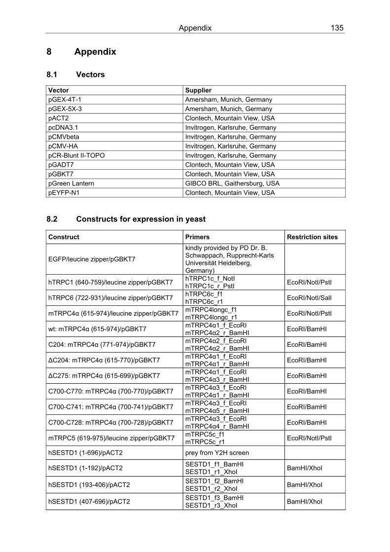
Appendix 135
8 Appendix
8.1 Vectors
Vector Supplier pGEX-4T-1 Amersham, Munich, Germany pGEX-5X-3 Amersham, Munich, Germany pACT2 Clontech, Mountain View, USA pcDNA3.1 Invitrogen, Karlsruhe, Germany pCMVbeta Invitrogen, Karlsruhe, Germany pCMV-HA Invitrogen, Karlsruhe, Germany pCR-Blunt II-TOPO Invitrogen, Karlsruhe, Germany pGADT7 Clontech, Mountain View, USA pGBKT7 Clontech, Mountain View, USA pGreen Lantern GIBCO BRL, Gaithersburg, USA pEYFP-N1 Clontech, Mountain View, USA
8.2 Constructs for expression in yeast
Construct Primers Restriction sites
EGFP/leucine zipper/pGBKT7
kindly provided by PD Dr. B. Schwappach, Rupprecht-Karls Universität Heidelberg, Germany)
hTRPC1 (640-759)/leucine zipper/pGBKT7 hTRPC1c_f_NotI hTRPC1c_r_PstI EcoRI/NotI/PstI
hTRPC6 (722-931)/leucine zipper/pGBKT7 hTRPC6c_f1 hTRPC6c_r1 EcoRI/NotI/SalI
mTRPC4α (615-974)/leucine zipper/pGBKT7 mTRPC4longc_f1 mTRPC4longc_r1 EcoRI/NotI/PstI
wt: mTRPC4α (615-974)/pGBKT7 mTRPC4α1_f_EcoRI mTRPC4α2_r_BamHI EcoRI/BamHI
C204: mTRPC4α (771-974)/pGBKT7 mTRPC4α2_f_EcoRI mTRPC4α2_r_BamHI EcoRI/BamHI
∆C204: mTRPC4α (615-770)/pGBKT7 mTRPC4α1_f_EcoRI mTRPC4α1_r_BamHI EcoRI/BamHI
∆C275: mTRPC4α (615-699)/pGBKT7 mTRPC4α1_f_EcoRI mTRPC4α3_r_BamHI EcoRI/BamHI
C700-C770: mTRPC4α (700-770)/pGBKT7 mTRPC4α3_f_EcoRI mTRPC4α1_r_BamHI EcoRI/BamHI
C700-C741: mTRPC4α (700-741)/pGBKT7 mTRPC4α3_f_EcoRI mTRPC4α5_r_BamHI EcoRI/BamHI
C700-C728: mTRPC4α (700-728)/pGBKT7 mTRPC4α3_f_EcoRI mTRPC4α4_r_BamHI EcoRI/BamHI
mTRPC5 (619-975)/leucine zipper/pGBKT7 mTRPC5c_f1 mTRPC5c_r1 EcoRI/NotI/PstI
hSESTD1 (1-696)/pACT2 prey from Y2H screen
hSESTD1 (1-192)/pACT2 SESTD1_f1_BamHI SESTD1_r1_XhoI BamHI/XhoI
hSESTD1 (193-406)/pACT2 SESTD1_f2_BamHI SESTD1_r2_XhoI BamHI/XhoI
hSESTD1 (407-696)/pACT2 SESTD1_f3_BamHI SESTD1_r3_XhoI BamHI/XhoI
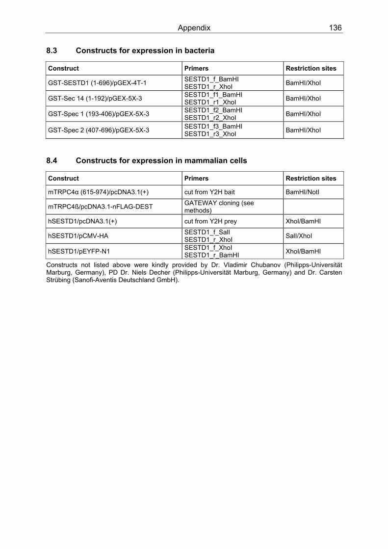
Appendix 136
8.3 Constructs for expression in bacteria
Construct Primers Restriction sites
GST-SESTD1 (1-696)/pGEX-4T-1 SESTD1_f_BamHI SESTD1_r_XhoI BamHI/XhoI
GST-Sec 14 (1-192)/pGEX-5X-3 SESTD1_f1_BamHI SESTD1_r1_XhoI BamHI/XhoI
GST-Spec 1 (193-406)/pGEX-5X-3 SESTD1_f2_BamHI SESTD1_r2_XhoI BamHI/XhoI
GST-Spec 2 (407-696)/pGEX-5X-3 SESTD1_f3_BamHI SESTD1_r3_XhoI BamHI/XhoI
8.4 Constructs for expression in mammalian cells
Construct Primers Restriction sites
mTRPC4α (615-974)/pcDNA3.1(+) cut from Y2H bait BamHI/NotI
mTRPC4ß/pcDNA3.1-nFLAG-DEST GATEWAY cloning (see methods)
hSESTD1/pcDNA3.1(+) cut from Y2H prey XhoI/BamHI
hSESTD1/pCMV-HA SESTD1_f_SalI SESTD1_r_XhoI SalI/XhoI
hSESTD1/pEYFP-N1 SESTD1_f_XhoI SESTD1_r_BamHI XhoI/BamHI
Constructs not listed above were kindly provided by Dr. Vladimir Chubanov (Philipps-Universität Marburg, Germany), PD Dr. Niels Decher (Philipps-Universität Marburg, Germany) and Dr. Carsten Strübing (Sanofi-Aventis Deutschland GmbH).
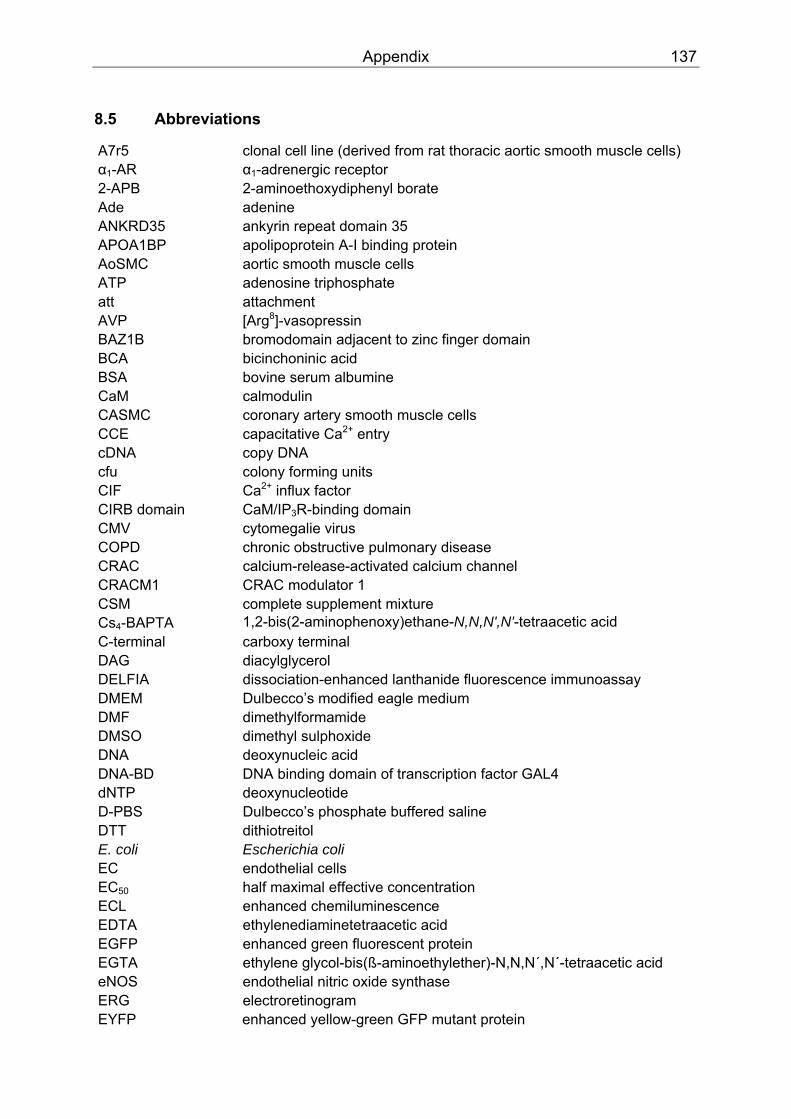
Appendix 137
8.5 Abbreviations
A7r5 clonal cell line (derived from rat thoracic aortic smooth muscle cells) α BB1BB-AR α BB1BB-adrenergic receptor 2-APB 2-aminoethoxydiphenyl borate Ade adenine ANKRD35 ankyrin repeat domain 35 APOA1BP apolipoprotein A-I binding protein AoSMC aortic smooth muscle cells ATP adenosine triphosphate att attachment AVP [Arg PP
8PP]-vasopressin
BAZ1B bromodomain adjacent to zinc finger domain BCA bicinchoninic acid BSA bovine serum albumine CaM calmodulin CASMC coronary artery smooth muscle cells CCE capacitative Ca PP
2+PP entry
cDNA copy DNA cfu colony forming units CIF Ca PP
2+PP influx factor
CIRB domain CaM/IPBB3BBR-binding domain CMV cytomegalie virus COPD chronic obstructive pulmonary disease CRAC calcium-release-activated calcium channel CRACM1 CRAC modulator 1 CSM complete supplement mixture Cs BB4BB-BAPTA 1,2-bis(2-aminophenoxy)ethane-N,N,N',N'-tetraacetic acid C-terminal carboxy terminal DAG diacylglycerol DELFIA dissociation-enhanced lanthanide fluorescence immunoassay DMEM Dulbecco’s modified eagle medium DMF dimethylformamide DMSO dimethyl sulphoxide DNA deoxynucleic acid DNA-BD DNA binding domain of transcription factor GAL4 dNTP deoxynucleotide D-PBS Dulbecco’s phosphate buffered saline DTT dithiotreitol E. coli Escherichia coli EC endothelial cells ECBB50BB half maximal effective concentration ECL enhanced chemiluminescence EDTA ethylenediaminetetraacetic acid EGFP enhanced green fluorescent protein EGTA ethylene glycol-bis(ß-aminoethylether)-N,N,N´,N´-tetraacetic acid eNOS endothelial nitric oxide synthase ERG electroretinogram EYFP enhanced yellow-green GFP mutant protein

Appendix 138
FBS fetal bovine serum FITR Flp-In T-Rex (inducible expression system) FKBP F506 binding protein FLIPR fluorometric imaging plate reader FRT site Flp recombination target site FSGS focal segmental glomerulosclerosis GAL4 transcription factor GAL4-AD activation domain of transcription factor GAL4 GAPDH glyceraldehyde-3-phosphate dehydrogenase GCN4 general control nondepressible 4 (transcriptional activator protein) GFP green fluorescent protein GST glutathione S-transferase GTP guanosine triphosphate 5-HT 5-hydroxytryptamine (serotonin) 5-HT BB3BB receptor subtype of the 5-hydroxy tryptamine (serotonin) receptor HA antigenic epitope of human influenza virus hemagglutinin protein HAEC human aortic endothelial cells HEK293 human embryonal kidney cells HL-5 a cell line derived from murine atrial cardiomyocytes HMG2L1 high-mobility group protein 2-like1 isoform b HMVEC-d human dermal microvascular endothelial cells HPV hypoxic pulmonary vasoconstriction HRP horseradish peroxidase ICBB50BB half maximal inhibitory concentration I BBCRACBB calcium-release-activated calcium current iNOS inducible nitric oxide synthase IPBB3BB inositol-1,4,5-trisphosphate IPBB3BBR IPBB3 BBreceptor IPBB6BB inositol hexaphosphate IPAH idiopathic pulmonary arterial hypertension iPLABB2BB inducible phospholipase 2 IPTG isopropyl ß-D thiogalactoside I-V current-voltage Kir inwardly rectifying K PP
+PP channel
Kv voltage-dependent K PP
+PP channel
LB medium Luria Bertani medium LiAc lithium acetate L-NAME N-nitro-L-arginine methyl ester ß-ME ß-mercaptoethanol MBB1BBR muscarinic type 1 receptor MAEC mouse vascular endothelial cells MCS multiple cloning site MEM minimal essential medium MKRN1 makorin RING finger protein 1 ML-9 [1-(5-chloronaphthalene-1-sulphonyl) homopiperazine, HCl] MLCK myosin light chain kinase mRNA messenger RNA NCBI National Center for Biotechnology Information NCX Na PP
+PP/Ca PP
2+PP exchanger
NHE Na PP
+PP/HPP
+ PPexchanger
Appendix 139
NHERF Na PP
+PP/HPP
+PP exchanger regulatory factor
NO nitric oxide N-terminal amino terminal OAG oleoyl-2-acetyl-sn-glycerol PA phosphatidic acid PAGE polyacrylamide gel electrophoresis PAR protease-activated receptor PBXIP1 pre-B-cell leukemia homeobox interacting protein 1 PCR polymerase chain reaction PDZ domain protein-protein interaction mediating domain PDZ-B PDZ-binding motif that interacts with PDZ domains PEG polyethylene-glycol Pfu Pyrococcus furiosus pH negative decadic logarithm of the concentration of hydrogen ions PH domain pleckstrin homology domain PI, PIP, PIPBB2BB, PIPBB3BB phosphatidylinositol, mono-/bis-/trisphosphate PKC protein kinase C PKG protein kinase G PLC phospholipase C PM plasma membrane PPIase peptidyl-prolyl cis-trans isomerase PSS physiological phosphate-buffered salt solution RNA ribonucleic acid ROC receptor-operated channel rpm rotations per minute RT room temperature S1P sphingosine 1-phosphate SCID severe combined immunodeficiency SDS sodium dodecyl sulphate SDS PAGE sodium dodecyl sulphate polyacrylamide gel electrophoresis SEM standard error of the mean Sec 14 Sec14p-like lipid-binding domain SERCA sarcoplasmic/endoplasmic reticulum Ca PP
2+ PPpump
SESTD1 SEC14 and spectrin domains siRNA small interference RNA SK&F 96365 1-(ß-[3-(4-methoxyphenyl)propoxyl]-4-methoxyphenethyl)-1H- HCl SMC smooth muscle cells SMC3 structural maintenance of chromosomes 3 Spec 1, Spec 2 spectrin repeats SPTAN1 spectrin, alpha, non-erythrocytic 1 SOC store-operated channel STIM1 stromal interaction molecule 1 TetOBB2BB tet operator 2 TetR Tet repressor TLN2 talin 2 tBHQ tert-butyl-benzohydrochinone Tris-HCl Tris(hydroxymethyl)aminomethane hydrochloride TRP transient receptor potential Tween 20 polyethylene glycol sorbitan monolaurate
Appendix 140
U unit UV ultraviolet v/v volume per volume VBB1A BB vasopressin receptor w/v weight per volume wt wild type X-gal 5-bromo-4-chloro-3-indolyl β-D-galactopyranoside Y2H yeast two-hybrid YFP yellow-green GFP mutant protein ZO-1 zona occludens 1 amino acids: alanine, Ala, A; arginine, Arg, R; aspartic acid, Asp, D; asparagine, Asn, N; cysteine, Cys, C; glutamic acid, Glu, E; glutamine, Gln, Q; glycine, Gly, G; histidine, His, H; isoleucine, Ile, I; leucine, Leu, L; lysine, Lys, K; methionine, Met, M; phenylalanine, Phe, F; proline, Pro, P; serine, Ser, S; threonine, Thr, T; tryptophane, Trp, W; tyrosine, Tyr, Y; valine, Val, V.
Danksagung 141
9 Danksagung
Die vorliegende Dissertation wurde als externe Doktorarbeit in der Sektion „Molekulare
Pharmakologie“ der Abteilung „Herz-Kreislauf-Erkrankungen“ der Sanofi-Aventis
Deutschland GmbH in Frankfurt am Main angefertigt.
Mein herzlicher Dank gilt:
Herrn Dr. Carsten Strübing für die Bereitstellung des Themas sowie für die hervorragende
fachliche Betreuung, seine Diskussionsbereitschaft und umfassende Unterstützung, die ganz
wesentlich zum Gelingen dieser Arbeit beigetragen haben.
Herrn Prof. Dr. Andreas E. Busch für die Möglichkeit, diese Arbeit in der Sanofi-Aventis
Deutschland GmbH durchführen und fertig stellen zu können, und für die wissenschaftliche
Begutachtung meiner Dissertation.
Herrn Prof. Dr. Dieter Steinhilber von der J.W. Goethe-Universität in Frankfurt am Main für
die wissenschaftliche Begutachtung meiner Doktorarbeit.
Herrn PD Dr. Klaus Steinmeyer für seine Hilfsbereitschaft, den freundschaftlichen Zuspruch,
das Interesse an meiner Arbeit und deren kritische Durchsicht.
Frau Andrea Bieberstein für die gute praktische Zusammenarbeit und ihre tatkräftige
Unterstützung.
Herrn Dr. Florian Bundis für die Einarbeitung in das Hefe Zwei-Hybrid System und den
tieferen Einblick, den er mir in die Molekularbiologie vermittelt hat.
Herrn Dr. Rolf Vajna für das schnelle Korrekturlesen dieser Arbeit.
Den Kolleginnen und Kollegen aus den „Herz-Kreislauf“- und „Genomic Sciences“-Laboren
für konstruktiven Gespräche und Anregungen, die gute Zusammenarbeit und nette
Arbeitsatmosphäre sowie die erfahrene praktische Unterstützung; insbesondere Beate
Preitz, Stefan Müller, Dr. Katrin Engel, Alexandra Endlich, Dr. Petra Arndt, Dr. Thorsten
Sadowski, Dr. Manuel Baader, Jennifer Reb, Dr. Aimo Kannt, Florian Werner, Kerstin Sicka,
Karolina Daton, Simone Stengelin, Elsa Kern, Elke Deckert, Claire Chenel, Dr. Paulus
Wohlfart, Dr. Thomas Wendrich, Ulrike Siebers, Dr. Ingrid Wagner, Matthias Ozog, Dr.
Matthias Löhn, Dr. Matthias Schäfer, Andreas Hertler und Dr. Yuri Ivashchenko.
Dr. Vladimir Chubanov, PD Dr. Niels Decher und PD Dr. Blanche Schwappach für die
Bereitstellung genetischer Konstrukte.
Natürlich auch meiner Familie und vor allem Thomas.

Curriculum vitae 142
10 Curriculum vitae
Aus datenschutzrechtlichen Gründen online nicht verfügbar.
Eidesstattliche Erklärung 143
11 Eidesstattliche Erklärung
Hiermit erkläre ich, Susanne Miehe, geboren am 21.02.1978 in Rochlitz, dass ich die
vorliegende Arbeit mit dem Titel „Untersuchung TRPC-modulierender Gestagene und
Proteine“ selbständig angefertigt und keine anderen Quellen oder Hilfsmittel als die hier
angegebenen benutzt habe. Ich habe bisher an keiner anderen Universität ein Gesuch um
Zulassung zur Promotion eingereicht oder eine andere Arbeit als Dissertation vorgelegt.
Teile dieser Arbeit sind Bestandteile von Patentanmeldungen (Europäische Anmeldungen
Nr. 07291300.7 und 07291301.7) der Sanofi-Aventis Deutschland GmbH.