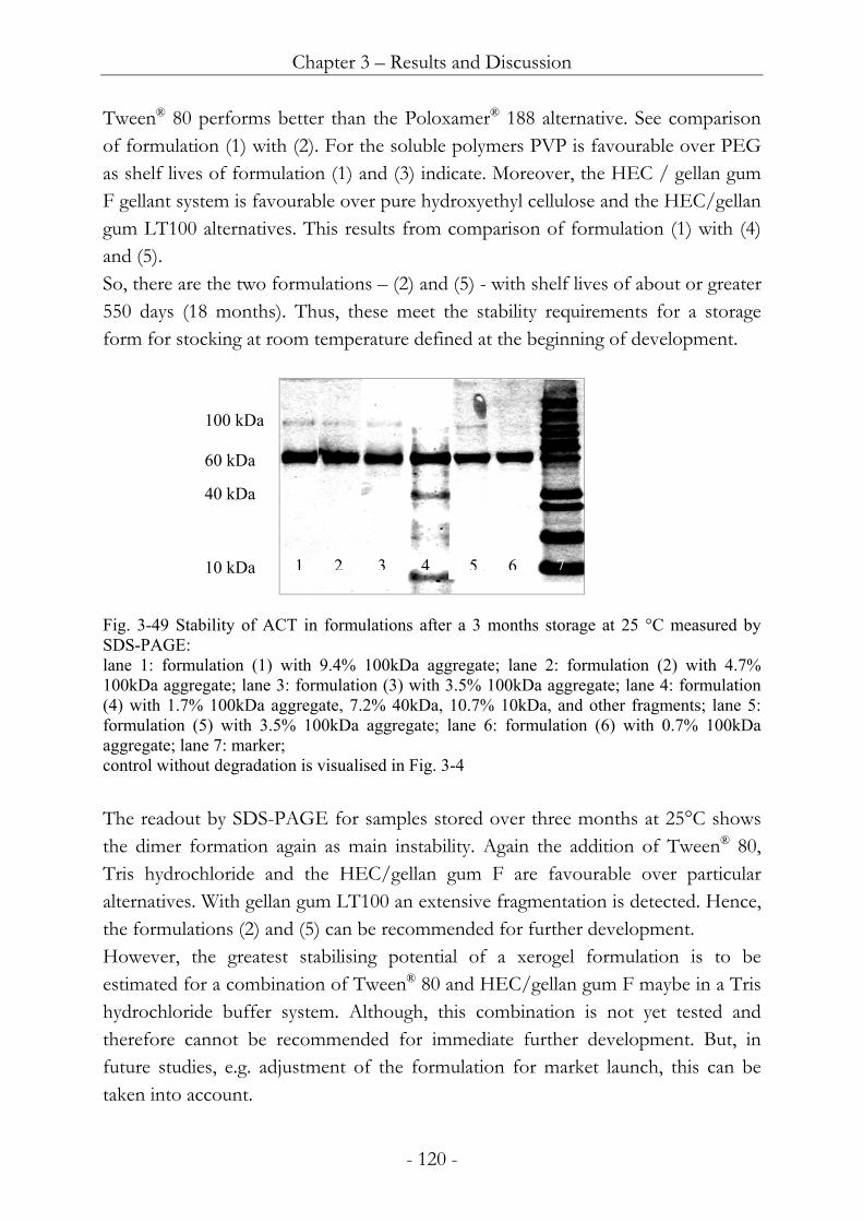
View - Elektronische Dissertationen der LMU M¼nchen - Ludwig
130
Topical delivery of α 1 -Antichymotrypsin for wound healing Dissertation zur Erlangung des Doktorgrades der Fakultät für Chemie und Pharmazie der Ludwig-Maximilians-Universität München vorgelegt von Roland Schmidt aus Treuchtlingen München 2005
Transcript of View - Elektronische Dissertationen der LMU M¼nchen - Ludwig

Topical delivery of α1-Antichymotrypsin for wound healing
Dissertation
zur Erlangung des Doktorgrades der Fakultaumlt fuumlr Chemie und Pharmazie der
Ludwig-Maximilians-Universitaumlt Muumlnchen
vorgelegt von
Roland Schmidt aus Treuchtlingen
Muumlnchen 2005
Erklaumlrung Diese Dissertation wurde im Sinne von sect 13 Abs 3 und 4 der Promotionsordnung vom 29 Januar 1998 von Herrn Prof Dr G Winter betreut
Ehrenwoumlrtliche Versicherung Diese Dissertation wurde selbstaumlndig ohne unerlaubte Hilfe erarbeitet Muumlnchen 01 Januar 2005 (Roland Schmidt) Dissertation eingereicht am 10 Januar 2005 1 Berichterstatter Prof Dr G Winter 2 Berichterstatter Prof Dr W Frieszlig Tag der muumlndlichen Pruumlfung 1 Februar 2005
ACKNOWLEDGMENTS Foremost I wish to express my deepest appreciation to my supervisor Prof Dr Gerhard Winter I am much obliged to him for his professional guidance and his scientific support On a personal note I especially want to thank him for inspiring my interest in protein pharmaceuticals for teaching me so much and for creation of an outstanding working climate I am also grateful to the Switch Biotech AG Neuried Germany for financial support I would like to acknowledge Dr Uwe Goszliglar for rendering every assistance and the always professional and personally warm contact Moreover I would like to thank Annette Bjoumlrn and especially Olivia for performing the Bioassays Thanks are also extended to Prof Dr Bracher Prof Dr Frieszlig PD Dr Paintner Prof Dr Schlitzer and Prof Dr Wagner for serving as members of my thesis advisor committee I very much enjoyed working at the Department for Pharmaceutical Technology and Biopharmaceutics of the Munich Ludwig-Maximilians-University what was mainly due to the cooperative and most convenient atmosphere Wolfgang Silke Sandra Iris Steffi Fritz Ingo and all the others it was a pleasure to work with you
To my parents
Table of contents
1 Introduction 1
11 Wound healing 3
111 Physiology of wound healing 3
1111 Wound healing process 3
1112 Growth factors in physiological wounds 8
1113 Proteases in physiological wounds 12
1114 Protease inhibitors in physiological wounds 15
112 Pathophysiology of chronic wounds 18
1121 Cellular and biochemical imbalance in chronic wounds 19
1122 Clinics of chronic wounds 20
1123 Infection of wounds 21
113 Treatment of chronic wounds 21
1131 Debridement 22
1132 Moist wound treatment 23
11321 History of moist wound treatment 23
11322 Effects of moist wound treatment 23
11323 Products for moist wound treatment 24
1133 Infection control in wounds 25
1134 Skin substitutes for wound healing 26
1135 Growth factors control in chronic wounds 26
1136 Protease control in chronic wounds 27
11361 Active dressings for chronic wounds 28
11362 Delivery of ACT in chronic wounds 28
12 Protein delivery from hydrogel formulations 30
121 Suitability of hydrogels for protein delivery 31
122 Protein delivery from hydrogels 32
1221 Application in wounds 32
1222 Transdermal delivery 33
1223 Oral delivery 34
1224 Ophthalmic delivery 35
1225 Delivery by injection and general approaches 35
123 Summary 37
13 Aim of the thesis 39
2 Materials and Methods 41
21 Materials 41
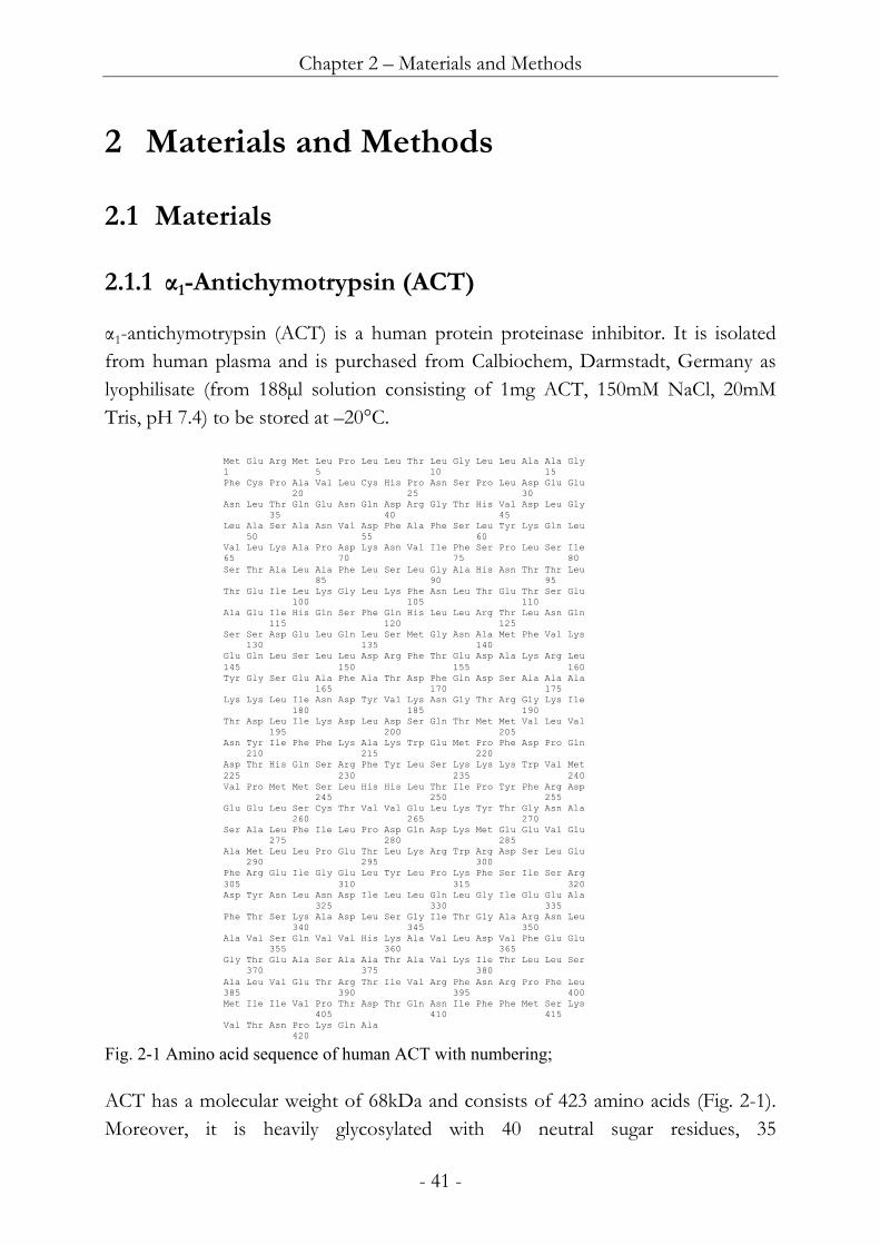
211 α1-Antichymotrypsin (ACT) 41
212 Excipients and chemicals 42
213 Polymers 43
2131 Cellulose ethers 43
2132 Gellan gum 43
2133 Other polymers 44
22 Methods 45
221 Characterisation of ACT 45
2211 ACT activity assay 45
2212 ACT ELISA 45
2213 Gel electrophoresis 46
222 Manufacture of matrices 46
2221 Wet film manufacture with the scraper 46
2222 Freeze-drying 46
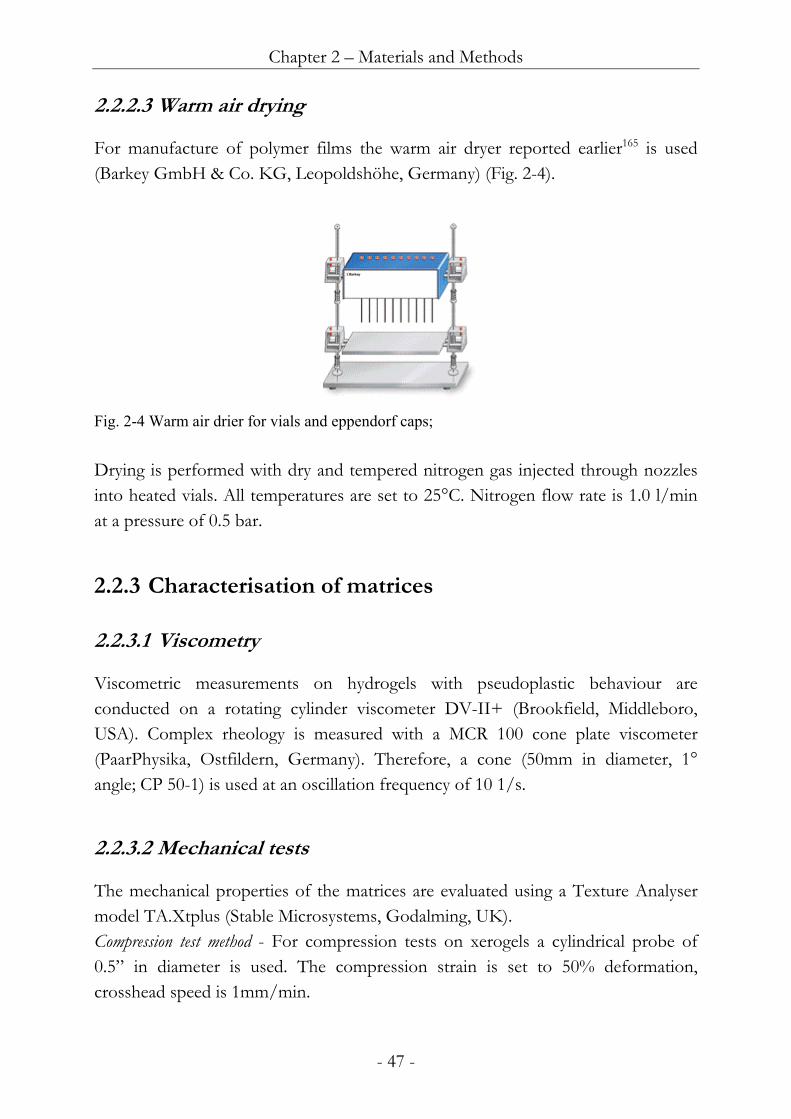
2223 Warm air drying 47
223 Characterisation of matrices 47
2231 Viscometry 47
2232 Mechanical tests 47
2233 In vitro Release tests 48
2234 Karl Fischer Titration 48
2235 Differential scanning calorimetry (DSC) 48
2236 X-ray diffraction 49
3 Results and Discussion 50
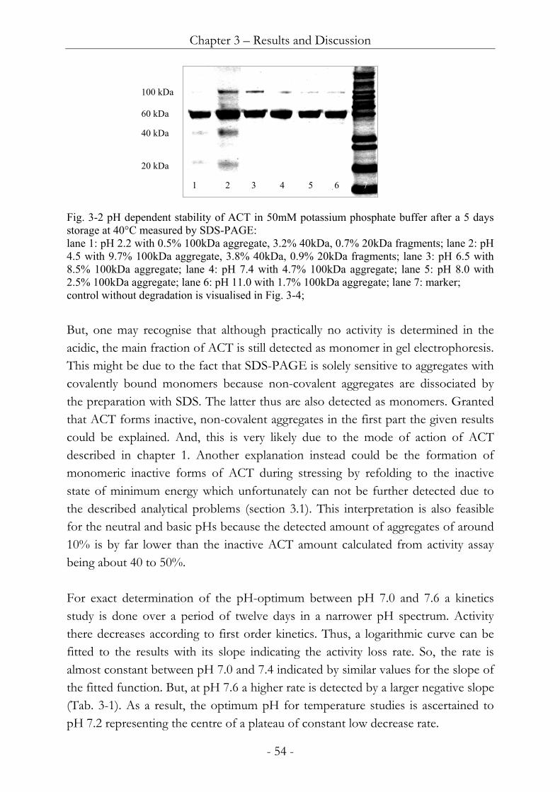
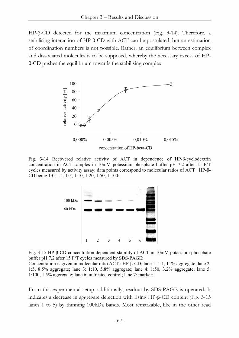
31 Analytical tools for the characterisation of ACT 51
32 Stabilisation of ACT in solution 52
321 Effects of pH buffers and electrolytes on ACT solution stability 52
3211 Effect of pH on ACT solution stability 52
3212 Effect of buffer species on ACT solution stability 56
3213 Effect of salts on ACT solution stability 57
3214 Effect of buffer content on ACT solution stability 58
3215 Summary of the effects of electrolytes on ACT solution stability 60
322 Effects of stabilisers and excipients on ACT solution stability 61
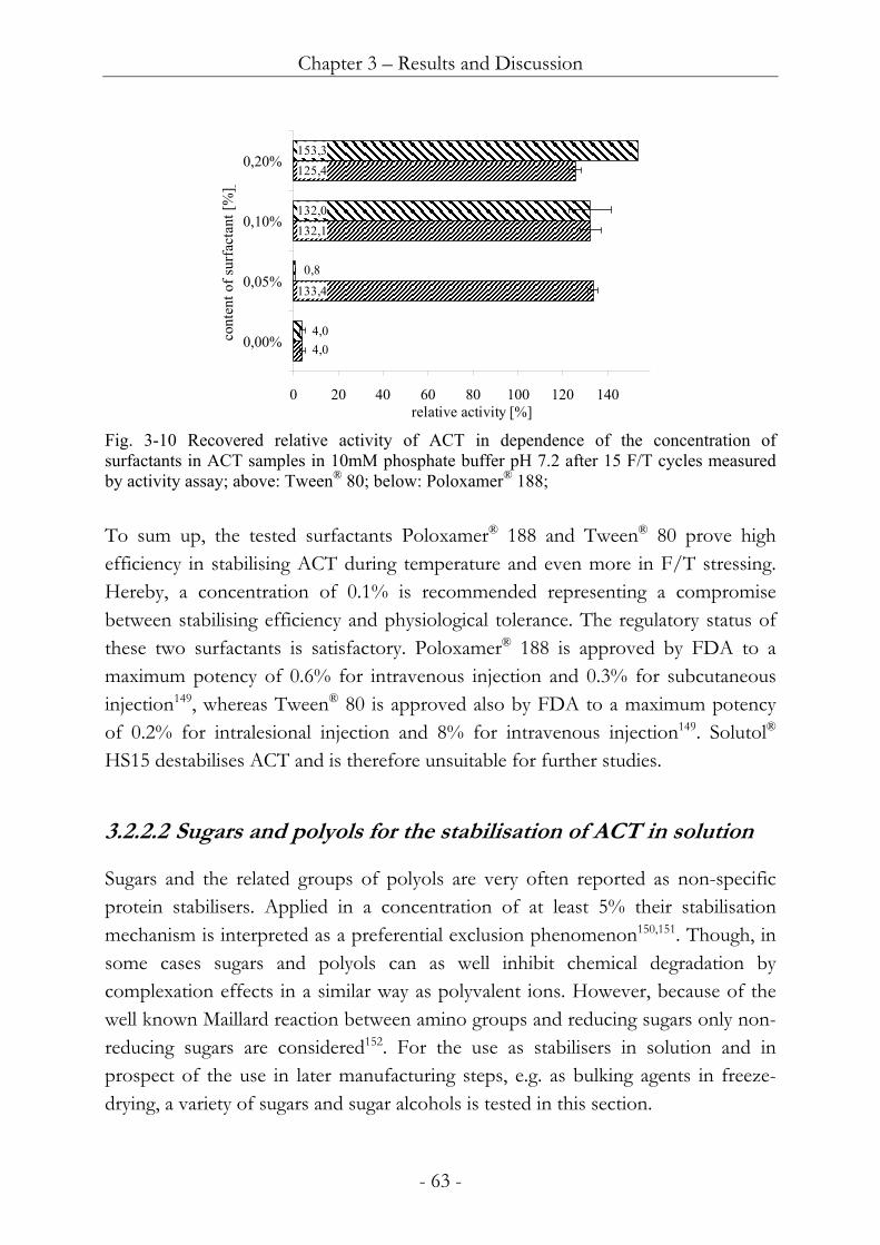
3221 Surfactants for the stabilisation of ACT in solution 61
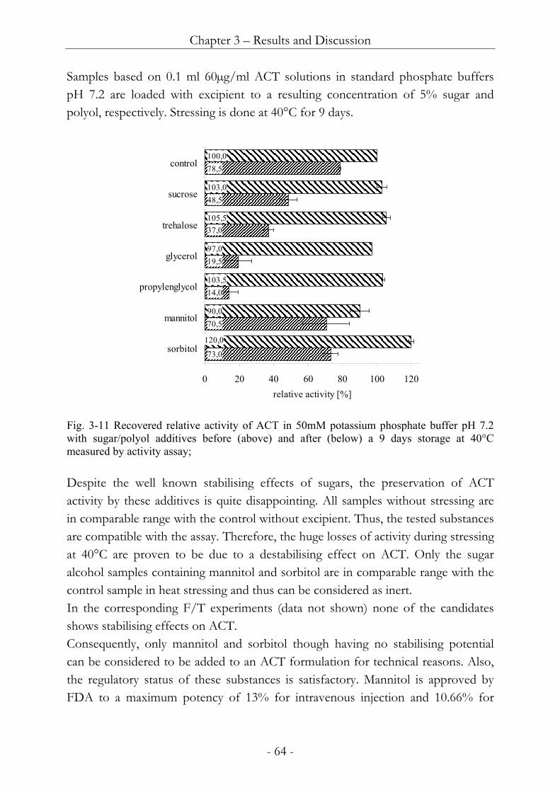
3222 Sugars and polyols for the stabilisation of ACT in solution 63
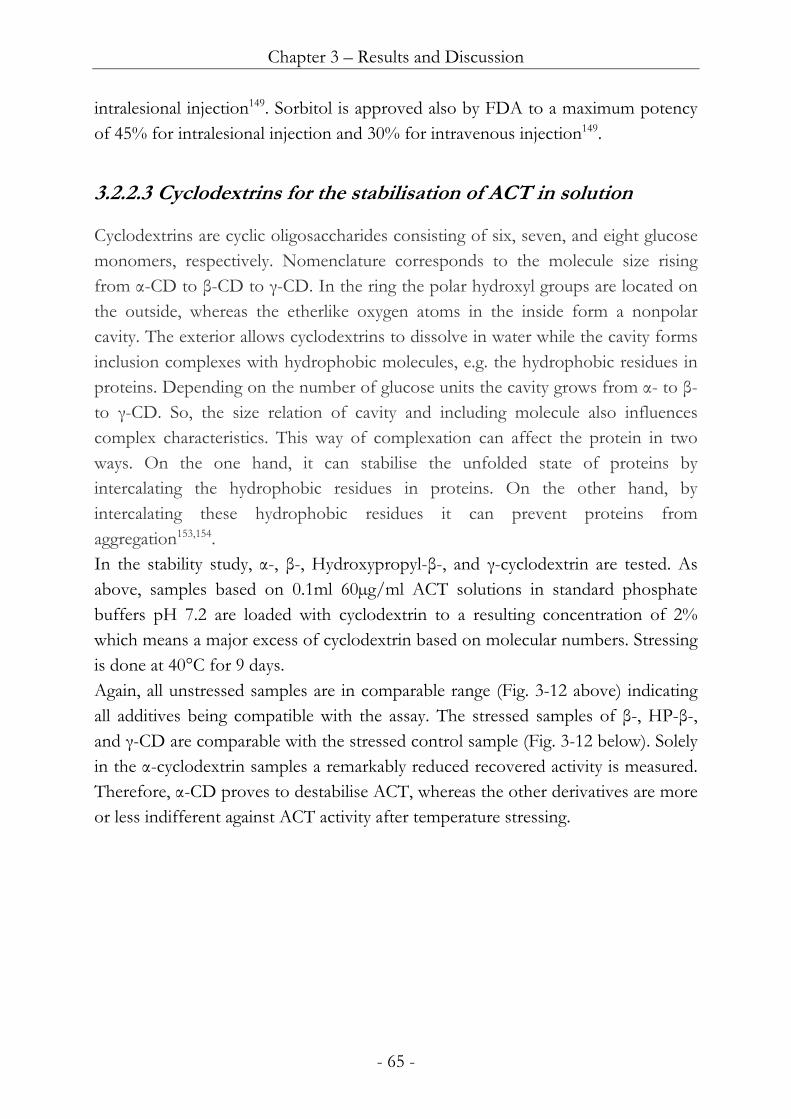
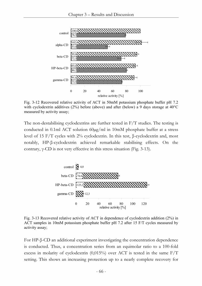
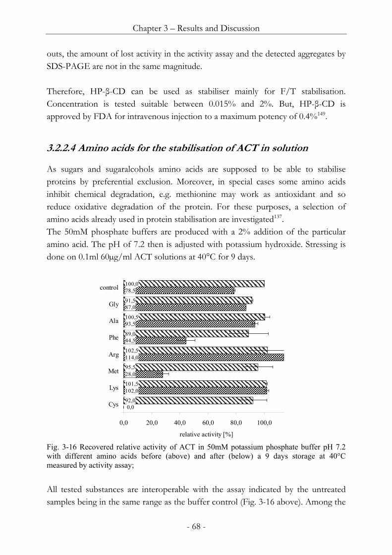
3223 Cyclodextrins for the stabilisation of ACT in solution 65
3224 Amino acids for the stabilisation of ACT in solution 68
3225 Preservatives for ACT containing solutions 70
323 Summary of ACT solution stability studies 71
33 Hydrogels as delivery system for ACT into wounds 72
331 Development as delivery system for wound healing 72
3311 Sterilisation of hydrogels 73
3312 Viscosity of hydrogels 73
3313 Viscosity of gellan gumhydroxyethyl cellulose hydrogels 76
332 Stability of ACT in hydrogel formulations 80
3321 Effects of polymers on ACT stability in hydrated formulations 80
3322 Aseptic manufacture of ACT loaded hydrogels 83
3323 Analysis of ACT loaded hydrogels 85
3324 Mid term stability of ACT in hydrogel formulations 85
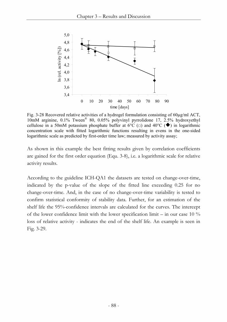
33241 Principles of data interpretation 86
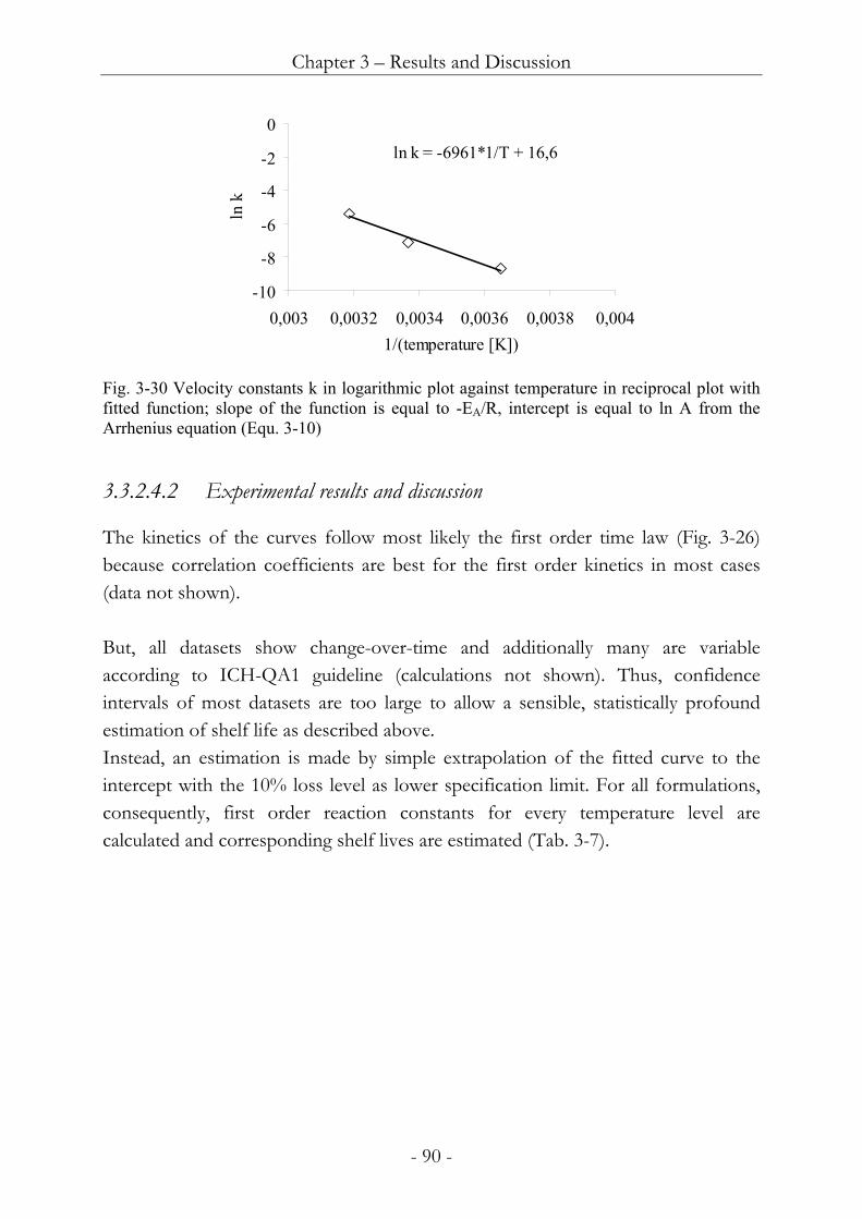
33242 Experimental results and discussion 90
33243 Summary 93
3325 Freezethaw stability of ACT in hydrogel formulations 93
333 Summary of hydrogels as ACT delivery systems 94
34 Dry delivery systems 95
341 Xerogels as drug delivery systems for wound healing 96
3411 Lyophilisation process 96
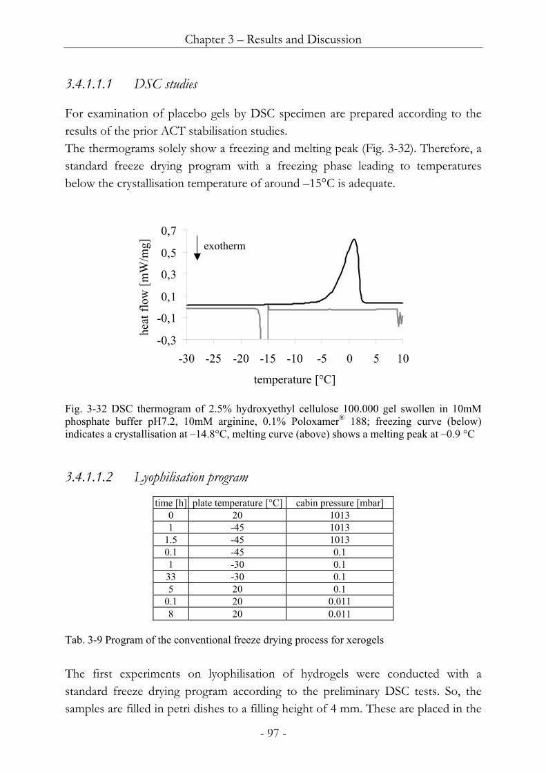
34111 DSC studies 97
34112 Lyophilisation program 97
3412 Gel composition for xerogel formation 100
34121 Hydroxyethyl cellulose qualities for xerogel formation 101
34122 Excipients in hydroxyethyl cellulose xerogels 103
34123 Hydroxyethyl cellulosegellan gum mixtures for xerogels 108
34124 Other polymers for xerogel formation 109
342 Stability of ACT in xerogel formulations 112
3421 Stability of ACT during the lyophilisation process 112
3422 Mid term stability of ACT in xerogel formulations 115
34221 Principles of data interpretation 116
34222 Experimental results and discussion 118
34223 Summary 121
343 Polymer films as drug delivery systems for wound healing 122
3431 Production process 122
3432 Gel composition for polymer film formation 124
34321 Gelling agents for film formation 124
34322 Polymers as additives to hydroxyethyl cellulose films 125
34323 Hydroxyethyl cellulose gellan gum mixtures for film formation 132
34324 Protein stabilisers in polymer films 134
344 Stability of ACT in film formulations 135
3441 Stability of ACT during the film manufacturing process 136
3442 Mid term stability 138
3443 Summary 141
345 Summary for dry matrices as ACT delivery systems 141
35 Release of ACT from dry delivery systems 143
353 Experimental setup 143
3531 Membrane 143
3532 Acceptor medium 144
3533 Chamber model 145
354 Theoretical background and data interpretation 146
355 Release of model substances from gel based matrices 147
356 Release of ACT from formulations 151
3561 Dynamic model 151
3562 Static model 153
35621 Evaluation of the model 153
35622 Release of ACT from xerogel formulations 157
35623 Release of ACT from film formulations 161
35624 Summary 166
4 General summary 167
5 References 171
Curriculum vitae 183
Chapter 1 - Introduction
- 1 -
1 Introduction The World Health Organisation of the United Nations prognoses the development of world-wide diabetes cases over the next decades in actual studies Accordingly the number of type II patients will more than double until 20301 Next to the disease itself moreover 25 of diabetes patients frequently develop chronic wounds with about half of them requiring elaborate inpatient treatment Therefore the diabetic foot causes more hospitalisation than does any other complication associated with diabetes and represents approximately 25 of all hospital admissions2 Not least driven by this need wound treatment has gone through great revolution during the last decades The paradigm shift from dry dressings based on woven fabrics towards a moist environment caused whole lots of new developments in this field Nevertheless the pathological cases of chronic wounds are still difficult to handle Even with the moist concept they require very patient and persistent treatment Therefore there still is strong desire for improved methods of therapy And due to the prognosed rise of the diabetes this desire is likely to largely increase over the next decades One resort out of this situation could be revealed by biotechnology Especially in the year 2000 the even greater revolutions in this field mostly occurring in typical scientific laboratories but praised and supported by highest authorities raised great expectations for new drug candidates Hence for example on international level United States President Bill Clinton announced the completion of the first survey of the entire human genome and nationally the government of the free state of Bavaria launched its high-tech-offensive endowed with investments of 135 billion Euros in local life science research In this environment several small new biotech companies were founded A considerable number of those established in Martinsried near Munich and began their research work One of those companies is the SWITCH BIOTECH AG focusing its research on wound treatment based on peptides One outcome of this work was the identification of α1-antichymotrypsin (ACT) as potential therapeutic for chronic wounds Hence to take the next step in development of α1-antichymotrypsin (ACT) as drug candidate a collaboration between SWITCH BIOTECH AG and the Department Pharmaceutical Technology and Biopharmaceutics of the LMU Munich was
Chapter 1 - Introduction
- 2 -
contracted in terms of a Ph D study at the Department under the supervision of Prof Dr Winter The aim of this study is to create formulations and drug carriers that stabilise and deliver ACT in bioactive state into the wound site This thesis addresses introductory remarks about wound healing and protein delivery from hydrogels followed by the results of the research on the ACT formulation and concomitant carrier development
Chapter 1 - Introduction
- 3 -
11 Wound healing In this section an overview over the physiology and pathophysiology of wound healing is described This represents the medicinal context of this work and highlights the scientific rational behind the delivery of ACT in chronic wounds Moreover the established methods for treatment of wounds including dressings and carriers are discussed Hereby the methodical context of a treatment with ACT as drug product with the indication wound treatment is given
111 Physiology of wound healing The following paragraph describes the physiology of wound healing It starts with the process of healing itself Next a selection of growth factors involved in the process is outlined in detail The role of growth factors is essential for the present understanding of wound healing Moreover growth factors are important starting points for drug based wound treatment having led to approval of platelet-derived growth factor (PDGF) the main competitor of a maybe future product based on ACT Consequently proteases and their inhibitors relevant in wound healing are highlighted As well as growth factors proteases play important roles in wounds And together with their inhibitors proteases are also starting points for wound therapy Thereby the discussion focuses on the inhibitor ACT and its target cathepsin G because ACT and its delivery represents the main topic of this thesis
1111 Wound healing process The physiological wound healing process in the present understanding is usually divided into four steps ndash coagulation inflammation followed by migration and proliferation and finally the remodelling phase These phases are not exactly distinguishable from each other because occasionally they overlap or proceed concurrently In the damaged vessel wall platelets - stimulated by mediators - immediately adhere to the exposed collagen of the vessel wall The clustered platelets partially coalesce with each other and release the platelet factors that initiate the actual clotting process During the clotting a network of fibrin forms around the platelet plug finally filling the entire wound gap The purpose of this fibrin network or first extracellular matrix is to retain cellular components of the blood eg erythrocytes
Chapter 1 - Introduction
- 4 -
and thereby form a clot for the purpose of haemostasis wound closure and provision of a matrix for the later collagen mounting3 Coagulation ndash this part is started by the platelet factors released by degranulating thrombocytes and by substances liberated from damaged tissue cells for example PDGF IGF-I EGF and TGF-β Following the coagulation cascade the well known complex chain reaction which is initiated by injury4 leading to the conversion of prothrombin into the enzyme thrombin is activated Thrombin now converts fibrinogen into fibrin monomers Flowing blood only contains fibrinogen the water-soluble precursor of fibrin The conversion is solely catalyzed by thrombin located at the wound surface Thrombin is also present in the blood as its inactive precursor prothrombin Prothrombin and fibrinogen are coagulation or clotting factors and part of the coagulation cascade Fibrinogen polymerises to fibrin chains which are finally interlinked by coagulation factor XIII to form the stable fibrin network5 A variety of inhibitors of the coagulation factors present in the blood for example antithrombin III ensure that clotting is confined to the wound site They inactivate thrombin entering the circulating blood stream Moreover the members of the cascade are at much lower concentration levels in the blood stream which decreases the presumption that the necessary partners meet to interact This contributes to the confinement of the coagulation to the wound area Inflammation - once haemostasis is achieved inflammation is initiated a few hours after injury The inflammatory phase of wound healing is characterised by recruitment and activation of granulocytes macrophages and lymphocytes that clean the wound by phagocytosis of damaged tissue and bacteria and wound debridement by enzymatically degrading foreign matter and damaged tissue The substances released from the cell debris resulting from tissue destruction are responsible for causing the characteristic inflammatory reactions Vascular changes also contribute to this reaction A fresh wound usually bleeds due to vessels rupture effecting cleansing the wound To prevent further blood loss the affected vessels narrow within the first minutes This vasoconstriction is followed by vasodilation that increases the blood circulation in the wound area Consequently a rise in the temperature of the wound and the surrounding skin is caused Moreover the permeability of the capillary walls is increased by vasodilatory agents ndash such as histamine and serotonin ndash and as a result blood plasma erythrocytes leucocytes and platelets enter the wound The outcome of these processes is wound oedema
Chapter 1 - Introduction
- 5 -
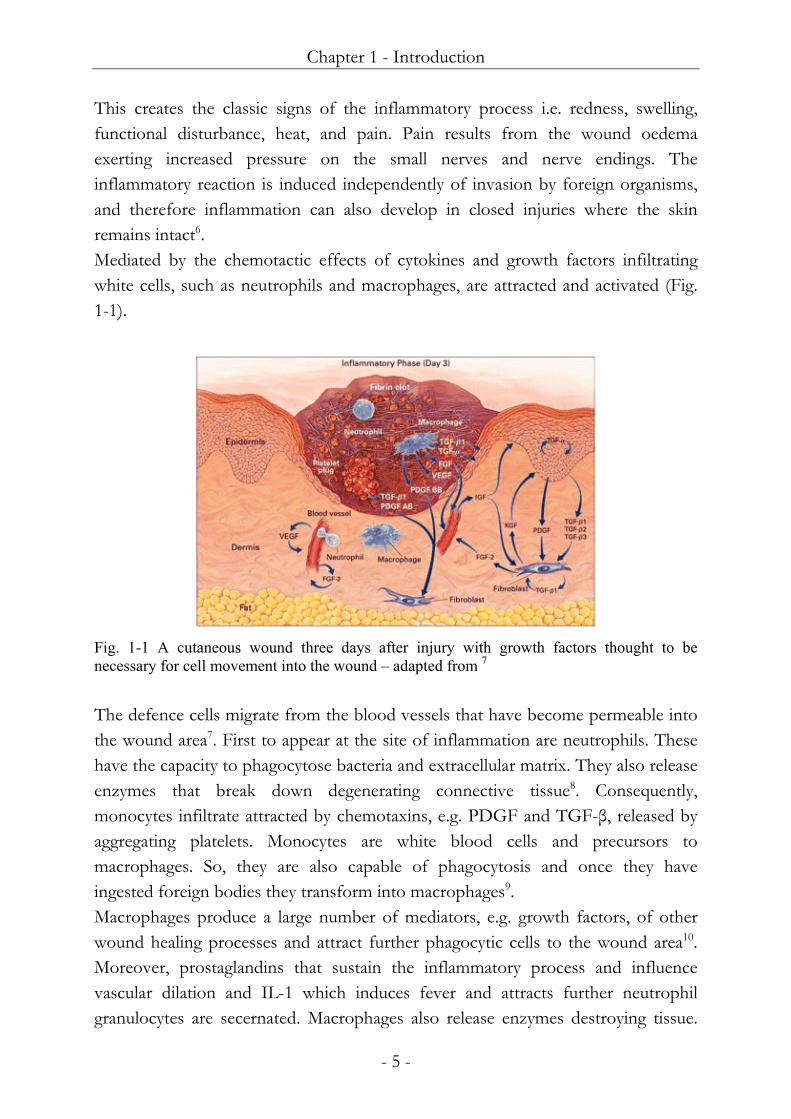
This creates the classic signs of the inflammatory process ie redness swelling functional disturbance heat and pain Pain results from the wound oedema exerting increased pressure on the small nerves and nerve endings The inflammatory reaction is induced independently of invasion by foreign organisms and therefore inflammation can also develop in closed injuries where the skin remains intact6 Mediated by the chemotactic effects of cytokines and growth factors infiltrating white cells such as neutrophils and macrophages are attracted and activated (Fig 1-1)
Fig 1-1 A cutaneous wound three days after injury with growth factors thought to be necessary for cell movement into the wound ndash adapted from 7 The defence cells migrate from the blood vessels that have become permeable into the wound area7 First to appear at the site of inflammation are neutrophils These have the capacity to phagocytose bacteria and extracellular matrix They also release enzymes that break down degenerating connective tissue8 Consequently monocytes infiltrate attracted by chemotaxins eg PDGF and TGF-β released by aggregating platelets Monocytes are white blood cells and precursors to macrophages So they are also capable of phagocytosis and once they have ingested foreign bodies they transform into macrophages9 Macrophages produce a large number of mediators eg growth factors of other wound healing processes and attract further phagocytic cells to the wound area10 Moreover prostaglandins that sustain the inflammatory process and influence vascular dilation and IL-1 which induces fever and attracts further neutrophil granulocytes are secernated Macrophages also release enzymes destroying tissue
Chapter 1 - Introduction
- 6 -
IL-1β also draws fibroblasts into the wound and up-regulates enzyme levels Importantly there is a balance between levels of enzymes and tissue inhibitors of these enzymes Inflammation physiologically last for several days Neutrophil infiltration reaches a maximum after approximately 24 hours and declines over the next few days These cells once present survive for about a further 24 hours11 Migration and proliferation - a few days after injury the migration and proliferation phase begins Whereas catabolic processes predominate in inflammation this phase of wound healing is characterised mainly by anabolic reactions ie angiogenesis epithelisation and fibroplasia (Fig 1-2) It can last for up to 24 days from the moment the wound develops
Fig 1-2 A cutaneous wound five days after injury blood vessels sprout into the fibrin clot as epidermal cells resurface the wound Proteinases thought to be necessary for cell movement are shown ndash adapted from 7 The formation of blood vessels angiogenesis starts with an endothelial cell bud formed by existing intact vessels Thereby the angiogenic stimuli in the first place emanate from macrophages by secession of growth factors and cytokines But also keratinocytes as well as fibroblasts provide chemoattractants So stimulated the endothelial cells in the venules begin to produce enzymes that break down the basal membrane in the area of the stimulus12 Soon endothelial cells migrate through the resulting gap in the direction of the wound following the oxygen gradient They divide and form tubular structures that connect with other buds As a result during the maturation process a new basal membrane develops from the extracellular matrix components The newly formed vascular loops then connect with intact
Chapter 1 - Introduction
- 7 -
vessels and differentiate accordingly into capillaries arterioles and venules respectively3 For epithelisation proceeding in parallel to angiogenesis keratinocytes migrate across the wound and as a result reconstitute epidermal covering from the wound margin and hair follicle remnants13 In addition migration essentially requires the presence of a moist substrate well perfused with blood as is the case with granulation tissue In contrast epithelial cells are not able to migrate in a dehydrated layer This is supposed to be a reason for the success of moist wound dressings Fibroplasia is determined by the chemotactically attracted migration of fibroblasts along the fibrin network into the wound site and their replicative activity there to form the new loose extracellular matrix consisting of proteoglycans as well as the water-soluble collagen fibres essential for tissue stability Thus especially at wound edges fibroblasts are the predominant cell type14 Concurrently the provisional fibrin network the first extracellular matrix is broken down by fibrinolysis This breakdown process is caused by the enzyme plasmin Mainly t-PA activates plasmin from its inactive precursor plasminogen7 Collagen is crucial to the process of wound healing as it has been identified as the most abundant connective tissue protein Collagen is a fibrous protein synthesised in several stages Its precursors are assembled from amino acids in the fibroblast These protocollagen chains are twisted together in triple helical formation and get interlinked Finally vesicles transport the collagen to the cell membrane where they are released as soluble tropocollagen into the interstitium Beyond the tropocollagen molecules accumulate to form protofibrils consequently polymerising into microfibrils Several microfibrils unite to form a collagen fibril several of which in turn arrange themselves into bundles In healthy tissue the collagen fibres are aligned in basketweave patterns This organised structure is not achieved in wound healing as the collagen fibres at the wound site will fashion themselves in an alignment parallel to the stress lines of the wound15 Collagen synthesis depends on the presence of ascorbic acid as a coenzyme and further on iron and copper as cofactors Type I and type III are the collagens most commonly found in healing wounds although at least 19 different types of collagen have been identified and characterised1617 During the process of wound healing type III collagen and fibronectin are deposited ndash type III collagen later in the remodelling phase being replaced by type I
Chapter 1 - Introduction
- 8 -
Remodelling - at last the remodelling or maturation phase finalises the wound healing process Generally it can take up to two years and means changes in the matrix composition over the healing time The wound is contracted and the tensile strength of the wound cover is enhanced The synthesis of matrix material is as mentioned provided by fibroblasts and regulated by growth factors cytokines enzymes and prostaglandin mostly derived from macrophages and fibroblasts The granulation tissue of the first extracellular matrix mostly consisting of keratinocytes gradually matures into scar tissue With the formation of new fibres the mitotic activity of the fibroblasts is concluded They then may transform into myofibroblasts18 Myofibroblasts like the muscle cells contain contractile elements which allow them to draw together The collagen fibres become taut and as far as possible aligned to the main contours of tension in the tissue As a result the scar tissue shrinks and the functional cutaneous tissue at the wound margin contracts leaving only a small defect19 As outlined above early collagen fibrils are laid down randomly resulting in a tensile strength of only 5 per cent of normal undamaged skin at two weeks post-injury Over time these type III fibrils are replaced by type I collagen fibres improving the tensile strength of the scar tissue to that of 80 per cent of normal skin Nonetheless scar tissue still appears different to original tissue the former being weaker than unwounded skin One reason for this distinction is as already mentioned that the final assembly of the collagen in granulation tissue does not resemble that of normal unwounded dermis Moreover since the pigment producing cells the melanocytes cannot be regenerated the scar tissue does not turn brown but remains white Besides this tissue contains no hairs sebaceous or sweat glands
1112 Growth factors in physiological wounds As described above the process of wound repair is characterised by a series of complex cellular and molecular events with a great degree of overlap and interdependence Growth factors play fundamental roles in this process by stimulating chemotaxis and cellular proliferation by providing signalling among cells of the same and different type by controlling extracellular matrix formation and angiogenesis by regulating the process of contraction and by re-establishing tissue integrity (Fig 1-1) They work by binding to specific cell surface receptors and can target cells in a number of recognised modes Release of these substances into the blood stream allows them to get to distant targets (endocrine mode) From
Chapter 1 - Introduction
- 9 -
the cell of origin growth factors can diffuse over short distances to affect other cells (juxtacrine mode) and to influence neighbouring cells (paracrine mode) Growth factors can also act on the cell in which they are produced (autocrine mode)1020 In the following the growth factors with greatest importance in the wound healing process are described in detail PDGF (platelet-derived growth factor) mainly is synthesised by macrophages endothelial cells fibroblasts smooth muscle cells and platelets Beyond it can be stored in platelets until cell activation for example by thrombin The synthesis of PDGF can be induced by IL-1 IL-6 TNF-α TGF-β and EGF PDGF physiologically is not released into the blood stream However by binding to several proteins eg of the extracellular matrix (ECM) local concentrations can reach increased levels PDGF is a hydrophilic protein of 30kDa molecular weight It is composed of two distinct polypeptide chains A and B that form homodimers (AA or BB) or heterodimers (AB) The subunits are linked by disulfide bonds Recently additional C and D subunits have been described acting similarly to the A and B species 21-23 Platelets synthesise a mixture of the three possible isoforms while fibroblasts stimulated with EGF synthesise AA homodimers Activated macrophages produce the BB homodimer The dimeric form of PDGF is mainly mitogenic for smooth muscle cells and vascular endothelium cells Although monomeric forms of PDGF are mainly chemotactic So PDGF is a chemoattractant for fibroblasts monocytes and neutrophils In addition PDGF is a potent vasoconstrictor However it does not act on epithelial and endothelial cells because these cells do not express PDGF receptors24 B-FGF (basic fibroblast growth factor) is the prototype of the FGF family Thereby b-FGF shows a homology to a-FGF Many cells first of all endothelial cells express b-FGF and partly store it in an inactive form It is released after tissue injuries and during inflammatory processes Binding of b-FGF to its receptors requires the interaction with proteoglycans of the ECM before full functional activity is obtained Additionally heparin is a protectant for b-FGF especially from the impact of proteases acids and heat It also improves receptor binding and hence potentiates the biological activity of b-FGF B-FGF stimulates the growth of fibroblasts endothelial cells and keratinocytes potentiated in the presence of thrombin Additionally b-FGF reduces the expression of the receptor for TGF-β thus
Chapter 1 - Introduction
- 10 -
effectively modulating the inhibitory action of TGF-β on endothelial cells Also FGFs control proliferation and migration of vascular endothelial cells important for angiogenesis As well the expression of plasminogen activator and collagenase by these cells is enhanced by b-FGF and is antagonised by TGF-β A special member of the FGF family is FGF-7 also known as KGF with the mentioned effects solely concentrated on keratinocytes1025 EGF (epidermal growth factor) is a globular protein and is produced by cells in various organs Following it is present in most body fluids It is synthesised as a larger pre-protein from which the factor itself is released by proteolytic cleavage In addition EGF is the prototype of a large family of EGF-like proteins (EGF-F) Particularly TGF-α shows a strong homology to EGF However antibodies for EGF do not bind to TGF-α Both factors are functionally analogous they bind to the same receptor and they have similar biological activities So EGF stimulates the proliferation of epidermal and epithelial cells including fibroblasts and keratinocytes This can be inhibited by the EGF inhibitor Moreover EGF strongly influences the synthesis of proteins of the ECM including fibronectin collagens laminin and glycosaminoglycans Indirectly EGF also supports angiogenesis because of its mitogenicity for endothelial cells which can be potentiated by thrombin Besides EGF is a chemoattractant for fibroblasts and epithelial cells26 TGF-α and TGF-β (transforming growth factors) are distinguished both chemically by their unique amino acid sequences and biologically by their different activities on cells The interactions of TGF-α and TGF-β can be either synergistic or antagonistic TGF-α consists of a single chain peptide and is produced by keratinocytes macrophages and platelets It has strong homology to EGF resulting in competition for receptor binding The biological activities of TGF-α as well resemble those of EGF However some biological activities of TGF-α are stronger than those of EGF Especially TGF-α is mitogenic for fibroblasts and inhibits the synthesis of collagen TGF-β is not related to TGF-α The biologically active form of TGF-β is a disulfide-linked homodimer Still TGF-β is released as the inactive complex latent-TGF with proteoglycans from the ECM This complex should represent TGF-β molecules released by platelets after tissue injuries This allows the factor to be stored in an inactive form In fact platelets contain very high amounts of TGF-β It
Chapter 1 - Introduction
- 11 -
is also produced for example by macrophages lymphocytes endothelial cells and keratinocytes27 Depending upon cell type and concentration secretion of TGF-β can be induced or inhibited by a number of different stimuli So induction can be achieved by for example EGF NGF and IL-1 On the contrary the synthesis can be inhibited by eg EGF FGF and calcium ions28 Generally TGF-β has bifunctional effects and can either stimulate or inhibit growth of the same cells depending on conditions It is a potent growth inhibitor for epithelial cells endothelial cells fibroblasts keratinocytes and smooth muscle cells It also deactivates macrophages In many cell types TGF-β antagonises the biological activities of EGF PDGF and FGFs Besides the factor stimulates the synthesis of the major matrix proteins including collagen proteoglycans glycosaminoglycans and fibronectin On the other hand it inhibits their degradation mainly by inhibiting the synthesis and secretion of proteinases and by increasing their proteinase inhibitor levels Furthermore in monocytes TGF-β stimulates the expression of IL-1 PDGF and FGF and inhibits the synthesis of TNF-α TNF-β and IFN-γ Moreover TGF-β is a chemoattractant for neutrophils10 For IGF (insulin-like growth factor) two different isoforms IGF-1 and IGF-2 of 7 kDa molecular weight have been described They display broad homology with insulin but can not be neutralised by antibodies directed against insulin Both types of IGF are synthesised in many organs throughout the body finally obtained by processing of precursors Solely IGF-1 is produced also by fibroblasts Cell types responding to IGF-1 also include epithelial cells and fibroblasts The factors regulating concentrations of IGF are somatotropin as well as PDGF and FGF Thus IGF-1 stimulates collagen and matrix synthesis In fact IGF-1 is considered to be one of the major anabolic factors regulating the metabolism of joint cartilage Besides it is also involved in angiogenesis Like insulin free IGF-1 causes hypoglycaemia Therefore binding of IGF-1 to carrier proteins prevents the establishment of a permanent hypoglycaemia in spite of high serum IGF-1 concentrations These carrier proteins also increase plasma half life of IGF-1 and prevent the release from the blood stream into interstitial spaces It also effects neurons and has been demonstrated to reduces neuronal loss after injury29 VEGF (vascular endothelial growth factor) is a homodimeric and glycosylated protein The subunits are linked by disulphide bonds The factor exists in several isoforms that are produced via precursors in many cells including endothelial types and macrophages The isoforms differ in biological properties such as recognising
Chapter 1 - Introduction
- 12 -
receptor types and interaction with proteoglycans Consequently shorter forms are soluble while the heavier forms are mostly bound to heparin containing proteoglycans of the ECM VEGF is a strong angiogenic protein especially a mitogen for vascular endothelial cells Thereby b-FGF and VEGF act synergistically in the induction of angiogenesis Moreover VEGF influences vascular permeability and is a chemoattractant for monocytes It also induces the synthesis of clotting factors and collagenase3031
1113 Proteases in physiological wounds Proteolytic enzymes are present in all wound exudates and play an essential role in the healing of acute and chronic wounds For wounds proteases are mainly produced by granulocytes keratinocytes and fibroblasts Proteases contribute to the regulation of the balance between tissue synthesis and tissue degradation Thus proteolytic activity is tightly regulated with control at the transcriptional level and control by extracellular enzyme activation and inhibition A defect in one or more of these control mechanisms would result in an increase in proteolytic activity a trait of chronic wounds and considered one of the primary causes of wound healing disorders Therefore protease control is a major goal of wound treatment Proteases comprise endopeptidases and exopeptidases which cleave peptide bonds at points within the protein and remove amino acids sequentially from either N or C-terminus respectively Endopeptidases are further classified according to the structure of their active site in cysteine proteinases aspartic proteinases metallo proteinases and serine proteinases In this chapter discussion focuses on serine proteases and serpins with regard to the main topic of this thesis being ACT Cysteine proteases - the cysteine proteinases family includes the lysosomal enzymes cathepsins B K and L Like with serine proteinases outlined below catalysis proceeds through the formation of a covalent intermediate and involves a cysteine and a histidine residue Aspartic proteinases - aspartic proteinases include lysosomal cathepsin D and other proteases of the pepsin family A second family comprises viral proteinases such as retropepsin from HIV In contrast to serine and cysteine proteases catalysis by aspartic proteinases do not involve a covalent intermediate though a tetrahedral intermediate exists
Chapter 1 - Introduction
- 13 -
Metalloproteinases - the known metalloproteinases (MMP) differ widely in their sequences and their structures However the great majority of enzymes contains conserved regions of homology and a zinc atom which is catalytically active Zinc is in most cases bound by three amino acids the fourth coordination site is occupied by a water molecule The catalytic mechanism leads to the formation of a non covalent tetrahedral intermediate after the attack of the zinc-bound water molecule on the carbonyl group of the scissile bond This intermediate is further decomposed by transfer of the glutamic acid proton to the leaving group MMPs are mostly stored in leukocytes keratinocytes and dermis cells They are activated by removing a small peptide fragment with the active enzyme exhibiting optimal activity around the physiologic pH The collagenases are very specific in their function in that they split the triple helix of fibrillar collagens The resultant denatured collagen molecule is then susceptible to attack from other proteases in particular the gelatinases In fact though their substrate specifity is very different in combination they can degrade all matrix molecules and each one deactivates inhibitors for serine proteases mostly α1-PI32 In detail the interstitial collagenase (MMP-1) degrades the collagen types 1 2 7 8 and 10 gelatine proteoglycans and entactin The neutrophil collagenase (MMP-8) works very similar it cleaves collagens 1 2 3 7 and 10 gelatine proteoglycans bradykinin and angiotensin I Unlike the others the neutrophil collagenase deactivates α1-antichymotrypsin Collagenase 3 (MMP-13) degrades elastin fibrillin fibronectin and already denatured collagen Gelatinase A (MMP-2) the 72kDa enzyme and gelatinase B (MMP-9) the 92kDa enzyme cleave collagen types 4 5 7 10 and 11 gelatine elastin fibronectin laminin and entactin They also activate pro-IL-1β The stromelysins 1 and 2 (MMP-3 and -10) degrade collagen types 4 5 9 10 and 11 fibronectin laminin proteoglycans and gelatine In addition they activate pro-MMP-1 -8 -9 and pro-IL-1β Matrilysin (MMP-7) and metalloelastase (MMP-12) degrade elastin fibronectin laminin entactin proteoglycans and collagen IV3233 Serine proteases - the serine proteinase class comprises two distinct families The chymotrypsin family which includes enzymes such as cathepsin G chymotrypsin trypsin elastase or kallikrein and the subtilisin family which include bacterial enzymes The general structure is different in the two families but they have the same active site geometry and then catalysis proceeds via the same mechanism34
Chapter 1 - Introduction
- 14 -
The serine proteinases exhibit different substrate specificities which are related to amino acid substitutions in the various enzyme subsites interacting with the substrate residues Three residues which form the catalytic triad are essential in the catalytic process ie His 57 Asp 102 and Ser 195 after chymotrypsinogen numbering (Fig 1-3A)34
Fig 1-3 chemical and kinetic mechanisms of catalysis for serine proteases The catalytic groups of trypsin (A) are shown interacting with an oligopeptide substrate C Common kinetic mechanism of catalysis for serine proteases adapted from34 The first step in the catalysis is the formation of an acyl enzyme intermediate between the substrate and the essential Serine Formation of this covalent intermediate proceeds through a negatively charged tetrahedral transition state intermediate and then the peptide bond is cleaved During the second step or deacylation the acyl-enzyme intermediate is hydrolysed by a water molecule to release the peptide and to restore the Ser-hydroxyl of the enzyme The deacylation which also involves the formation of a tetrahedral transition state intermediate proceeds through the reverse reaction pathway of acylation A water molecule is
Chapter 1 - Introduction
- 15 -
the attacking nucleophile instead of the Ser residue The His residue provides a general base and accept the OH group of the reactive Ser (Fig 1-3C) Cathepsin G is a cationic single chain glycoprotein of 29kDa molecular weight and is stored in active form within leukocyte granules and proinflammatory monocytes35 It provides a broad spectrum of biological activities whereby around pH 74 its maximum activity is developed36-38 Cathepsin G activates other enzymes ie collagenase (MMP 8) and gelatinase (MMP 9) Besides Cathepsin G degrades macromolecules of the ECM ndash elastin fibronectin laminin proteoglycans and collagen (type 4) ndash itself Also it appears to be necessary for proper elastase activity in the latter case Moreover the cytokines TNF-α and TNF-β several lymphocyte receptors and plasma proteins eg immunoglobulins and clotting factors are deactivated by cleavage Platelets on the other hand are activated Further the conversion of angiotensin I into angiotensin II is catalysed by cathepsin G39 As the latter elastase is a cationic single chain glycoprotein For storage it is embedded in leukocyte granules proinflammatory monocytes eosinophiles basophiles mast cells and lymphocytes The biological functions are similar to those of cathepsin G Along with platelets elastase also activates lymphocytes and the cytokines pro-IL-1b and IL-8 The secretion of signal molecules from cells is also induced by elastase ie GM-CSF IL-6 and IL-83940 Proteinase 3 is also found in monocytes and mast cells Function and chemical class are also similar to cathepsin G and elastase Urokinase type plasminogen activator (uPA) is stored in monocytes mononuclear phagocytes T-lymphocytes and natural killer cells Its main activity is the activation of plasmin by converting plasminogen which is present throughout body fluids Plasmin itself degrades fibrin laminin fibronectin and proteoglycans activator Besides it activates some pro-MMPs and TGF-β3940
1114 Protease inhibitors in physiological wounds The entirety of protease inhibitors in the wound healing process is called the antiproteolytic shielding It controls the destructive activity of proteases41 The plasma proteinase inhibitors after albumin and the immunoglobulins constitute with nearly 10 by weight of the total protein the third largest group of functional proteins in human plasma Serine protease inhibitors (serpins) with ACT - the serine proteinase inhibitors are a superfamily of proteins with a size of 350ndash500 amino acids They fold into a
Chapter 1 - Introduction
- 16 -
conserved structure and employ a unique suicide substrate-like inhibitory mechanism Most serpins inhibit serine proteinases of the chymotrypsin family To date around 250 serpin coding sequences are known42 They are divided into 16 clades and 10 highly diverged orphans Serpins adopt a metastable conformation that is required for their inhibitory activity Serpins in the stable latent conformation are non-inhibitory but can be converted back to the active state by denaturation and refolding The conformation of serpins consists of a conserved secondary structure comprised of three β-sheets and at least seven mostly nine α-helices In the metastable active form the reactive site loop containing the proteinase recognition site is located between the first and third β-sheet However serpins can undergo intramolecular structural changes eg to convert to the more stable latent form In that case the reactive site loop is placed into the first β-sheet while another side chain is extracted from the third sheet43 The most stable state for inhibitory serpins is a form in which the loop has fully inserted into the first β-sheet as in the latent conformation but without the extraction of the side chain from the third β-sheet The Tm for unfolding of such conformation is about 120 degC compared to about 60 degC for the native state44 Protein proteinase inhibitors act competitively by allowing their target enzymes to bind directly to a substrate-like region contained within the amino acid sequence of the inhibitor This reaction between enzyme and inhibitor is essentially second order and the resultant complex generally is equimolar45 Serpins inhibit serine proteinases by an irreversible suicide substrate mechanism The proteinase initially is bound in a non-covalent complex with serpin Secondly the active site serine of the protease forms a covalent ester with a carbonyl of the inhibitor Following the peptide bond is cleaved and the reactive site loop is inserted into the first β-sheet and transports the covalently bound proteinase with it Upon complete loop insertion the active site catalytic triad of the proteinase is distorted and therefore deactivated This conformational rearrangement is driven by the greater stability of the cleaved loop-inserted conformation compared with the native-like conformation Thus the acyl-intermediate is kinetically trapped due to slowing of the deacylation steps of the normal substrate reaction In fact serpin-proteinase complexes would be cleared long before complex decay could occur Though in the case that the described reactions are somehow impeded the enzyme may successfully complete the deacylation step and escape before entrapment This yields an active proteinase and a cleaved inactive serpin The ratio of complex and
Chapter 1 - Introduction
- 17 -
cleaved serpin products is determined by the competition between the rate of ester hydrolysis and that of loop insertion and proteinase distortion So chymotrypsin and cathepsin G produce a modified inactive no longer inhibitory form of ACT A negative effect of the need for a metastable conformation in the active state is that inappropriate loop insertion can occur mediated by several factors including formulation excipients and processes More precisely by reaction of the loop of one molecule and the beta-sheet of another aggregation to dimers and higher order oligomers can result4546 Therefore one major physical instability of ACT during formulation studies is estimated to be the dimer formation and perhaps the formation of higher order aggregates Α1-antichymotrypsin (ACT) is a plasma glycoprotein first isolated and characterised in 196247 (Fig1-4) The inhibitor is a major acute phase protein whose concentration increases rapidly and dramatically after a variety of events ACT shows the most immediate response as an acute phase protein doubling in concentration from 250microgml normal concentration in plasma within eight hours of insult
Fig 1-4 Crystal structure of ACT with partial loop insertion adapted from48 Three laboratories isolated ACT using a variety of conditions474950 Significantly ACT can be isolated from serum through its ability to bind to DNA 51 In all cases the product obtained stoichiometrically inhibited chymotrypsin to produce an equimolar complex that was denaturation resistant to dissociation The molecular weight of the native protein was between 58000 and 68000 the differences were
Chapter 1 - Introduction
- 18 -
attributable to the methodology and the high carbohydrate content (about 26) of this glycoprotein Thus ACT is a specific inhibitor of chymotrypsin-like proteinases forming stable complexes with chymotrypsin49and neutrophil cathepsin G46 No inhibition of either human trypsin or neutrophil elastase has been found52 ACT rapidly forms complexes with the mentioned chymotrypsin-like serine proteinases the rate is by far the fastest with cathepsin G (k = 51 times 107 M-1 s-1) and much slower with chymotrypsin53 Inhibitors of matrix metallo proteases - recently a number of inhibitors for MMPs have been described 54 The best known substances are TIMP-1 and -2 (tissue inhibitor of MMPs) and α2-macroglobuline These inhibitors selectively bind to MMPs and deactivate them
112 Pathophysiology of chronic wounds This paragraph outlines the pathophysiology of chronic wounds both on cellular and clinical levels A chronic wound is defined as one in which the normal process of healing is disrupted at one or more points in the phases of haemostasis inflammation proliferation and remodelling55 Chronic wounds unlike acute wounds do not undergo the ordered molecular and cellular processes of physiological tissue repair previously discussed However the healing process of chronic wounds is thought to be stuck in inflammation Chronic wounds can also be considered to be an imbalance between tissue deposition stimulated by growth factors and tissue destruction mediated by proteases56 Hereby the imbalance favours the destructive process Thus the molecular and cellular processes are disrupted leading to significant differences in the microenvironment of the wound both in terms of the constituents of the exudates and the cellular components of the wound area In addition oxidative damage by free radicals condition specific factors of underlying diseases and accumulated necrotic tissue as well contributes to the chronic state The further healing of those wounds results in skin defects of excessive fibrous appearance for instance keloids and scar contractures or alternatively in insufficient tissue replacement ie a non-healing wound
Chapter 1 - Introduction
- 19 -
1121 Cellular and biochemical imbalance in chronic wounds Moreover the persisting inflammatory phase leads to wound exudate showing - in comparison to acute wounds ndash increased protease concentration and reduced levels of growth factor activity During the inflammatory phase chemotactically attracted and activated macrophages secrete inflammatory cytokines which increase protease production and reduce the synthesis of inhibitors In a physiologically healing wound there is also a balance of pro-inflammatory cytokines and their natural inhibitors In chronic wounds on the contrary the levels of these cytokines are increased Mainly the persistent inflammatory stimulus is caused by repetitive trauma local tissue ischaemia necrotic tissue heavy bacterial burden or tissue breakdown57 As well in acute wounds proteases and their inhibitors are in equilibrium but protease concentrations are elevated in chronic wounds So levels of collagenase gelatinase A and gelatinase B (MMP-1 -2 -9) have been shown to be elevated in fluid derived from pressure ulcers and venous leg ulcers5859 Other proteases such as neutrophil elastase have also been observed to be higher in chronic wounds60 Hence elevated levels of serine proteases cause degradation of extracellular matrix resulting in impaired cell migration and connective tissue deposition Furthermore they degrade growth factors and their target cell receptors59 Growth factors applied externally to the wound are degraded the same way To sum up the wound healing balance is shifted in favour of destructive processes Emerging from cell membrane lipids break down caused by exaggerated cell necrosis associated with impaired wound healing effects higher numbers of cell death Certainly increased amounts of active oxygen species for example hydroxyl radicals peroxide anions hydroperoxyl radicals or nitric oxide (NO) are known to be key negative factors in a number of inflammatory conditions6162 Furthermore in chronic wounds the specific cell populations and processes that are essential for wound repair are disrupted So epithelial cells fail to migrate across the wound tissue Hence hyperproliferation of cells occurs at the wound edges and interferes with normal cellular migration As a result the proliferation rate of fibroblasts is reduced and their apoptosis is inhibited63-65 And generally the response of cells to growth factors is reduced because the failure to re-epithelialise the most obvious clinical feature of chronic wounds is due to a failure in migration rather than proliferation of the keratinocytes66
Chapter 1 - Introduction
- 20 -
In fact these processes effectively hold the wound in the inflammatory phase and therefore prevent a wound from entering the proliferative phase and a physiological ongoing of the healing process
1122 Clinics of chronic wounds According to their causale chronic wounds may be categorised as diabetic foot ulcers venous or arterial leg ulcers pressure ulcers tumours burns or even post-surgical wounds But the most common are the venous leg ulcer the pressure ulcer and the diabetic foot ulcer These types appear different externally but all share common characteristic features In detail recurrent trauma ischaemia and prolonged inflammation are apparent Ulcus cruris disorders are divided into venous and arterial ulcerations Venous ulcerations are the most common type of ulcer affecting the lower extremities Here a chronic venous reflux disorder occurs because of inherited or postthrombotic varicosis With vein valves becoming incompetent the resulting backflow of blood causes venous congestion The lymphatic system compensates the oedema in the first part but soon it as well suffers damage from the overload This results in oedema and a decreased oxygen supply in the surrounding skin In arterial ulcers a complete or partial arterial blockage mostly resulting from arteriosclerosis lead to similar inadequate supply of surrounding tissue Consequently in both cases tissue necrosis and ulceration will develop The pressure ulcer is a compressive-ischaemic skin lesion predominantly occurring above bony prominences such as the sacrum heel or ankle Ulceration is again caused by inadequate supply due to ischaemia resulting from abnormal pressure on the tissue on a cellular level The pressure to tissue is usually applied between a bony prominence and a hard surface for example ankles in inappropriate foot wear Beyond an ischaemic lesion of the skin develops which after a short latent period leads to ischaemic skin necrosis Diabetic foot ulcers are a common complication of diabetes mellitus because diabetes as underlying disease can cause damage to the nerve and vascular supply in the feet and legs So diabetics are prone to foot ulcerations due to both neuropathic and ischaemic complications Next to the consequences of vascular damage outlined above neuropathy contributes to ulceration In detail neural damage also leads to lacking supply and due to the loss of sensation the risk of trauma is severely increased67
Chapter 1 - Introduction
- 21 -
1123 Infection of wounds Bacterial bioburden can cause a delayed or impaired healing In detail endotoxins and proteases stimulate an inflammatory wound environment further the clotting mechanisms leukocyte function angiogenesis and formation of granulation and scar tissue are disordered Defined by extent and necessary treatment bacterial burden present in the wound is divided into several degrees68 Contamination is defined as the presence of non-replicating bacteria This is a normal condition in chronic wounds and does not contribute to impaired healing Colonisation is defined as the presence of replicating bacteria without a host reaction Replicating bacteria colonise and contaminate all chronic wounds not meaning that these wounds are infected Bacterial colonisation does not contribute to impaired healing Critical colonisation is defined as the presence of replicating microorganisms which are beginning to cause local tissue damage There may be subtle local indications that a change in the equilibrium or increasing bioburden could be contributing to delayed healing69 Infection occurs when healing is impaired because bacteria have invaded tissue are multiplying and are causing a host reaction Although bacteria are present in all chronic wounds generally only critical colonisation and infection indicate an antimicrobial treatment But additional other factors are to be maintained for every case individually ie the balance between host resistance and the quantity and virulence of bacteria second concomitant medications including immunosuppression and any underlying diseases such as diabetes Biofilms are an element of wound infection that has recently become apparent Bacteria proliferating in wounds form microcolonies attaching to the wound and secreting a biofilm that protects the organisms So biofilms are protected areas of infection and bacterial resistance within the wound protecting bacteria from the effects of antimicrobial agents such as antibiotics and antiseptics70
113 Treatment of chronic wounds In this section the methods of treatment of chronic wounds are outlined Although many older but obsolete methods are still in use in clinical practice solely the modern state-of-the-art methods are described As a first treatment the wound is debrided After that under a moist dressing depending on the wound type the healing process is allowed to proceed in moist environment Where necessary an infection controlling treatment is conducted For further support of the healing
Chapter 1 - Introduction
- 22 -
process skin substitutes are available as well as vacuum treatment devices And for the correction of the above described imbalance phenomena products for the control of growth factors and protease levels in the wound can be applied An ACT delivering device would also belong to this last group of products and thus in that section also the scientific rational behind the delivery of ACT into wounds is discussed
1131 Debridement Debridement ndash the removal of devitalised tissue - is facilitated by natural mechanisms in every wound but accelerating this process makes healing more efficient It may be necessary because devitalised tissue in the wound bed supports bacterial growth and is a physical barrier to healing Devitalised tissue may also cause excessive amounts of proteases to be released The methods of debridement in todayrsquos clinical practice are surgical enzymatic autolytic mechanical and biologic Sharp surgical debridement is a very fast and efficient way to remove necrotic tissue from the wound bed It is performed where there is an extensive amount of necrotic tissue or there is a widespread infection requiring infected material to be removed Enzymatic debridement means the use of manufactured proteolytic enzymes ie collagenases These support naturally occurring enzymes to degrade necrotic tissue Autolytic debridement is a process performed by phagocytic cells and proteolytic enzymes in the wound site liquefying and separating necrotic tissue from healthy tissue Wound dressings which maintain a moist wound bed can provide an optimal environment for debridement as they allow migration of the phagocytic cells Unsurprisingly the process of autolytic debridement can result in increased wound fluid requiring appropriate dressing Mechanical debridement is a method that physically removes debris from the wound Examples of mechanical debridement include conventional dressings causing mechanical separation of necrotic tissue from the wound bed once the dressing is removed and wound irrigation using a pressurised stream of water to remove necrotic tissue Biologic larval therapy is an alternative method using sterile maggots that break down liquefy and remove dead tissue secreting powerful proteolytic enzymes followed by eating of the digested tissue71
Chapter 1 - Introduction
- 23 -
1132 Moist wound treatment
11321 History of moist wound treatment Prior to late 20th century wounds were felt to heal better if exposed under a scab This process produced surface desiccation and eschar formation now known to deepen the wound but was felt to protect the wound from outside influence The thinking is understandable given the fact that the most severe effect of wounding at that time was infection and no antibacterial agents were available for treatment This concept remained popular until the mid-20th century Between 1948 and 1958 several articles were published describing accelerated healing of acute wounds under occlusion But the primary death from wounds especially burns was still infection and therefore standard care returned to exposure especially with the advent of a topical antibiotic silver sulfadiazine cream to be applied twice daily This and following topical antibiotics could control infection in exposure but also retarded healing especially epithelialisation which nevertheless was considered as secondary A landmark study in 1962 by George Winter from Smith ampNephew Inc and considered the father of moist wound healing demonstrated that wounds epithelialised more rapidly under occlusive dressings with the reason being that occlusive dressings maintained a moist wound surface This study was conducted on pigs showing that the moist environment accelerated the epithelialisation process about 30 compared to air dried wounds72 Numerous studies followed which demonstrated that wound occlusion and moisture increased all phases of healing Wound bacterial colonisation which was demonstrated to be higher in a case study in a moist healing environment did not appear to retard healing or cause sepsis73 However the risk of severe burden and infection decreasing the healing process was proven to be lower in occluded wounds74 In 1994 US authorities published a guideline for treatment of pressure ulcers comprising occlusion for the purpose of autolytic debridement and provision of a moist environment Following until present wet treatment has been established in clinical practice as standard care
11322 Effects of moist wound treatment In general during a moist treatment the likelihood of scarring is reduced because there is no scab formation In addition moisture is essentially required for the already described activity of growth factors and proteolytic enzymes (section
Chapter 1 - Introduction
- 24 -
1112 1113 ) It is as well necessary for surface oxygen delivery and an efficient nutrient delivery As a result moisture improves the processes of the migration and proliferation phase by providing the ability of cells to migrate across the wound surface So an increased rate of epithelisation and angiogenesis is reached and further fibroblast proliferation and thus collagen synthesis is improved Next to an improvement of the healing by biochemical means there is also an improvement for direct patientsrsquo concern Pain is a major complication for wound treatment because in open wounds the nerve endings are exposed and the wound can feel painful With a moist environment the nerve endings are cushioned and protected which gives relief from pain On the contrary any surface desiccation decreases all phases of healing In fact surface drying was shown to lead to an increase in wound depth and a higher risk of infection75
11323 Products for moist wound treatment The topical wound management product chosen will depend on wound characteristics including amount of exudate wound size the presence of infection and the characteristics of the surrounding skin Especially the amount of exudate is important because low levels of moisture may lead to the discussed disadvantages but exaggerated levels of moisture lead to unwanted counterproductive maceration of tissue including intact skin Hence moisture-retentive dressings like okklusive films hydrogels and hydrocolloids are preferably selected for wounds with light to moderate drainage Absorbent dressings like foams and alginates tend to be selected for wounds with moderate to heavy exudate Film dressings ndash occlusive films are semi-permeable polyurethane dressings that are coated with an adhesive They are used for minor exudating wounds Their purpose is to prevent bacterial infection by shielding to absorb low amounts of exudate and to maintain a moist wound environment for fresh epithelial tissue The dressings insure a gaseous exchange for vaporising superfluous liquid Hydrogels - hydrogels and hydrogel dressings are used to treat wounds with low exudate levels With these products only low amounts of exudate are necessary to provide a moist milieu since they contain high amounts of water themselves Most products contain sodium carboxymethyl cellulose or polyacrylates swollen to an amorphous gel in a propylenglycol water mixture Hydrogel dressings are used to hydrate necrotic tissue facilitating autolytic debridement while being able to absorb exudate They can also be used to provide a moist wound environment
Chapter 1 - Introduction
- 25 -
during the later stages of wound closure In clinical practice hydrogels are often additionally covered by a film dressing Hydrocolloids - for moderate exudation hydrocolloid dressings can be used They contain a layer of hydrocolloid This is defined as liquid absorbing particles in an elastic self-adhesive mass The particles mostly consist of sodium carboxymethyl cellulose calcium alginate pectine and gelatine respectively The elastic mass contains different synthetic polymers The wound exudate binds to the absorbing particles of the hydrocolloid matrix to form a cohesive gel maintaining a moist wound environment Most products as well are covered on the upper side by a semi-permeable polyurethane film Foams - foam or hydrocellular dressings are double-layer dressings consisting of a polyurethane film carrier and a polyurethane foam layer on the wound side They are used for moderate to heavily exuding wounds The foam may be combined with polyacrylate particles supporting the liquid absorption The foam core binds high amounts of debris and exudate The film again provides gaseous exchange but provides shielding against bacteria So a balance of absorbed and vaporising liquid establishes a moist milieu Speciality absorbent dressings can be used as secondary dressings Alginates - alginate dressings are used to cover heavily exuding wounds They mostly contain a combination of calcium and sodium alginate fibres Alginate dressings are highly absorbent and can incorporate high amounts of exudate by forming a hydrogel Thereby the calcium alginate polymers are soaked with exudate After that due to the high amounts of sodium in the exudate there is a diffusional exchange of calcium and sodium enabling the resulting sodium alginate to swell and form a hydrogel Moreover alginates support healing by binding bacteria and debris inside the gel structure and by providing a moist environment75
1133 Infection control in wounds The most frequently used topical antimicrobials in modern wound care practice include octenidine iodine and silver containing products Chlorhexidine hydrogen peroxide and honey as well are in discussion but seem to be used more rarely In the past acetic acid sodium hypochlorite potassium permanganate and proflavine have been used Iodine ndash iodine as element was used in treating wounds mainly in the 19th century Due to its heavy adverse effects it is obsolete today Therefore the safer formulations povidone iodine and cadexomer iodine have been developed
Chapter 1 - Introduction
- 26 -
Povidone iodine is a polyvinylpyrrolidone - iodine complex cadexomer iodine is composed of beads of dextrin and epichlorhydrin that carry iodine Whereas its efficacy as a skin disinfectant is undisputed numerous publications describe the use of iodine in cleansing wounds and as a topical agent to prevent or treat localised wound infections but controversy surrounds its safety and efficacy76 Silver - silver also has a long history as an antimicrobial agent especially since the late 19th century77 Metallic silver is not active but in aqueous environments silver ions are released and antimicrobial activity depends on the intracellular accumulation of low concentrations of silver ions These bind to negatively charged components in proteins and nucleic acids thereby effecting structural changes in bacterial cell walls membranes and nucleic acids that affect viability78 The complex issues concerning the toxicity of silver to mammalian systems and its effects on the healing process are not completely discussed Skin discolouration and irritation associated with the use of silver nitrate is well documented absorption of silver systemic distribution and excretion in urine has also been reported79 In wound care silver has been utilised in several formulations Silver nitrate application is rare but silver sulphadiazine colloidal and nanocrystalline elemental silver dressings have recently been developed and are widely used These function by the sustained release of low concentrations of silver ions over time and generally appear to stimulate healing as well as inhibiting micro-organisms80
1134 Skin substitutes for wound healing Tissue engineering has added several skin substitutes to the variety of dressings available for wound treatment These products for example consist of fibroblasts and keratinocytes grown on collagen matrices In clinical evaluation the application of Apligrafreg has been shown to accelerate wound closure compared to control81
1135 Growth factors control in chronic wounds For the described inductive effects of growth factors on cell migration the potency of these substances has been evaluated in numerous experiments Convincing results of these efforts have been published in an unmanageable amount of publications Following the results of research many clinical trials with growth factors externally applied on wounds have been conducted But various degrees of success have been reported For example Richard et al conducted a trial with b-FGF on diabetic foot ulcers with no seen advantage of verum over the placebo control82 Also EGF was exogenously applied to patients with diabetic foot
Chapter 1 - Introduction
- 27 -
ulcers83 There a significant enhancement of healing and a reduction of healing time was reported But Falanga et al84 treated patients with venous ulcers in a study with EGF Although they showed EGF was safe and significantly reduced the size of the ulcers it failed to enhance epithelialisation Robson et al applied KGF-2 or repifermin on chronic venous ulcers during clinical trial Thereby a significant acceleration of wound closure was achieved85 For PDGF-BB (platelet-derived growth factor consisting of BB-homodimer) or becaplermin several clinical trials finally leading to the approval of Regranexreg in 1999 for the treatment of diabetic foot ulcers have also been published Efficacy and safety in diabetic foot ulcers have been proofed86-88 So the application of Regranexreg achieved a 43 increase of incidents of wound closure and a 32 decrease of time until wound closure Similar trials eg concerning pressure ulcers acute and open surgical wounds have also been conducted with promising results but not yet leading to an approval89-91 In Regranexreg PDGF is formulated in an aqueous carboxymethyl cellulose hydrogel Further the formulation contains an acetate buffer lysine hydrochloride and sodium chloride Another new technology for augmenting levels of growth factors in wounds is by gene transfer Andree et al used particle-mediated and microseeding gene transfer to deliver human EGF to porcine wounds9293 A high expression of EGF as well as a significant acceleration of healing was shown in the transfected wounds For PDGF a clinical trial using a viral vector is planned94 In summary there are several growth factors being evaluated in clinical trials but given by the very diverse results the type of the individual wound is an essential criteria for the choices of growth factors Therefore the approval of Regranexreg only for diabetic foot ulcers is feasible To overcome this problem and to make allowance to the thought of growth factors acting in concert methods of autologous growth factor application have been developed Thereby a sample of the patientrsquos blood is taken and separated by centrifugation The necessary fraction eg platelets is isolated and applied to the wound area as appropriate However effects are not well proven and questionable9596
1136 Protease control in chronic wounds Next to the delivery of growth factors the protease levels in chronic wounds have been identified as efficient starting point for treatment Generally as described in section 1121 protease levels in chronic wounds are increased Therefore a
Chapter 1 - Introduction
- 28 -
decrease of these levels is a goal of treatment For that purpose so-called active dressings are available that modulate these protease levels unspecifically Moreover the delivery of a protease inhibitor represents a potent possibility to balance the exaggerated lytic activity in chronic wounds in a very distinctive way Despite many small chemical entities being protease inhibitors have been patented none of those has led to approval97 But a physiological inhibitor could be an alternative to these substances due to a bandwidth of effects outlined below Therefore the topic of this thesis is the delivery of ACT a physiological protease inhibitor into wound sites
11361 Active dressings for chronic wounds Some polymers ie collagen and oxidative regenerated cellulose proved to modulate the wound environment at the biochemical level In detail the levels of proteolytic enzymes in wound fluid are reduced by physically entrapping and mechanically inhibiting their activity This is thought to originate the described decrease of tissue destruction and prevention of growth factor degradation leading to an overall increase in granulation tissue formation and faster wound repair Therefore wound dressings consisting of collagen or oxidative regenerated cellulose were developed9899
11362 Delivery of ACT in chronic wounds It has been shown by works of SWITCH BIOTECH AG that chronic diabetic ulcera in humans have strongly reduced capability for up-regulation of the level of expression of ACT In healthy humans the expression of ACT increases drastically in the wound tissue following wounding Further it has been established that besides the reduced levels of ACT transcripts the activity of the ACT polypeptides is also selectively decreased in poorly-healing diabetic wounds compared to the oberserved increase in activity in normally healing wounds as well as in venous ulcers Thus it is the increase of both expression and function particularly the activity which leads to a strengthening of the antiprotease shield and which in turn allows an increased neosynthesis of collagen and consequently supports rapid wound healing in normally healing wounds Furthermore the results show that this disturbance in the ACT protease inhibitor equilibrium is specific for the poorly healing diabetic wounds97100 As a consequence application of ACT into chronic wounds should support the wound healing process in a variety of ways It can protect the extracellular matrix
Chapter 1 - Introduction
- 29 -
via inhibition of mast cell chymase and cathepsin G Cathepsin G itself is capable of activation of MMP 8 and MMP 9 So delivery of ACT can at least partly decrease the enzymatic activity of this group of proteases In similar way elastase activity was shown to be dependent on the presence of cathepsin G Furthermore ACT has been reported to inhibit the neutrophil chemotaxis and superoxide generation Both factors contribute to the inflammation at the wound site For this multitude of effects the protein protease inhibitor ACT is likely to be superior over small chemical entities designed to inhibit one enzyme39 Therefore the delivery of ACT into the wound area promises improvement of the disturbed healing of particularly diabetic wounds
Chapter 1 - Introduction
- 30 -
12 Protein delivery from hydrogel formulations
ldquoHydrogels are three-dimensional hydrophilic polymeric networks capable of imbibing large amounts of water or biological fluids The networks are composed of homopolymers or copolymers and are insoluble due to the presence of chemical crosslinks (tie-points junctions) or physical crosslinks such as entanglements or crystallites The latter provide the network structure and physical integrity These hydrogels exhibit a thermodynamic compatibility with water which allows them to swell in aqueous mediardquo101 Hydrogels have been in use in the pharmaceutical medicinal and cosmetic field for many years Mainly they have been applicated topically with or without a drug substance for local treatment An exception of course are the matrix based transdermal therapeutic systems for eg systemic delivery of hormones But with the availability of large molecular weight protein drugs and the grown demands for a controlled release of drugs in modern medicine hydrogels have earned further increasing attention as drug delivery systems for the systemic delivery of both peptide and small chemical compounds101 For the desired controlled delivery of drugs by gel matrices the release mechanism is a decisive parameter Generally for the release from polymeric drug delivery devices three main mechanisms are described and reviewed102 Diffusion control - in a diffusion controlled system the drug is distributed homogeneously in the gel matrix For release the protein drug permeates through the continuum of the carrier to the release site Hence diffusion of the protein through the matrix is the rate-limiting step Chemical control - in the case of chemical control the polymer is degraded resulting in an erosion of the matrix and release of the drug Figure 1-5 displays the different types of polymer degradation mechanisms In (1) a biodegradable bond is incorporated into the polymer backbone Chemical or enzymatic cleavage of the bond converts a water-insoluble polymer into water-soluble low molecular weight polymer fragments In (2) the gel exists as a covalently or ionically cross-linked matrix Therefore cleavage of unstable linkages in the crosslinks leads to a breakdown of the network structure Covalently crosslinked hydrogels and ionically cross-linked polymers degrade by this mechanism For these two mechanisms a further differentiation can be made concerning physical terms Hydrolysis can occur at an even rate throughout the whole polymer matrix indicating a bulk erosion phenomenon Contrarily in surface erosion the delivery system degrades only at its surface because the degradation is
Chapter 1 - Introduction
- 31 -
blocked inside the matrix eg by excipients changing the pH to value unfavourable for hydrolysis Besides in some systems the drug can be attached to the polymer by a covalent bond that is degraded chemically triggering the release of the drug Example (3) of Fig 1-5 describes polymer solubilisation as degradation mechanism which is not actually related to a chemical reaction Rather the system dissolves and liquefies as water diffuses into the network leading to swelling and simple dilution of the polymers (Fig 1-5)
Fig 1-5 Polymer degradation mechanisms in hydrogels and related devices (1) hydrolysis of the backbone (2) hydrolysis of the crosslinked polymer network (3) hydration and solubilisation of a polymer matrix102 Solvent activation - a third mechanism is solvent activation The drug can be released either by swelling of the polymer in which the drug was previously locked into place within the polymer matrix in a glassy state or by an osmotic effect which can be accomplished by external water entering the drug delivery system because of an osmotic driving force and subsequently driving the drug out of the system
121 Suitability of hydrogels for protein delivery
Protein drugs place stringent demands on their delivery systems due to their structure physicochemical properties stability pharmacodynamics and pharmacokinetics More specifically peptides and proteins must retain their structural integrity until they reach their delivery site and must not be degraded upon enzymatic interactions In addition the physiological barriers eg skin and cell membranes are obstacles for the successful penetration of such drugs to their site of action
Chapter 1 - Introduction
- 32 -
Thus next to protein integrity a delivery device is required to deal satisfactorily with technological factors such as protein loading efficiency and desired release characteristics Due to their high water content hydrogels are generally considered as biocompatible materials So they provide minimal mechanical irritation upon in-vivo implantation mainly due to their soft and rubbery nature which is similar to natural tissue101 Generally the mostly aqueous environment of hydrogels appears very similar to the physiological environment for proteins and peptides in the body and therefore is supposed to be favourable for stability and activity of these substances Moreover due to the low interfacial tension between water and hydrogels tendencies to protein adsorption and cell adhesion are very low Furthermore hydrogels offer a broad acceptability for individual drugs with different hydrophilicities and molecular sizes Also maximum drug load is expected to be uncritical for proteins in most cases because the high water content provides a high solubility for hydrophilic protein substances101 Therefore hydrogel carriers are favourable for the delivery of peptide and protein drugs
122 Protein delivery from hydrogels In the following an overview over protein delivering hydrogel formulations published during the last decades is outlined in dependence of the application site Thereby the application of hydrogels in wounds on skin for transdermal delivery in the gastrointestinal tract via oral administration and for injection is highlighted
1221 Application in wounds For the application of protein drug loaded hydrogels in wounds a multitude of reports has been published These mainly deal with the delivery of growth factors aiming towards an acceleration and improvement in wound closure Also a whole variety of gelling agents has been used for the various approaches A series of examples is given that report improving or accelerating impact of growth factors on wound healing Hydroxyethyl cellulose was used to incorporate a-FGF in a gel formulation103 A preparation of 1mgml a-FGF stabilised by heparin was evaluated in vitro and in vivo In release studies in a special apparatus in vitro indicated that 90 of total a-
Chapter 1 - Introduction
- 33 -
FGF content were liberated and recovered in the acceptor consisting of PBSheparin Moreover in a diabetic mouse model an improvement of wound healing was found B-FGF further was delivered from crosslinked chitosan gels104 Thereby in vitro release from a 1mgml b-FGF containing gel into PBS solely under non-degrading conditions was studied in a well plate without a membrane Thereby a maximum of 20 was released within 24 hours in PBS as acceptor It was concluded that the remaining substance was locked in the matrix while in vivo degradation was expected to liberate the whole dosis Besides in a diabetic mouse model an improvement of wound healing was found Further collagen gels have been reported to effectively deliver EGF and TGF-β105 As well collagen was used as carrier for delivery of PDGF TGF-β and FGF106 Furthermore gelatine was tested on the delivery of FGF107 Besides a gelatine gel crosslinked with dextran dialdehydes was used on EGF108 KGF was administered in a fibrin gel carrier109 Moreover TGF-β was administered in a Poloxamerreg gel as well as a polyethylene glycol gel110 Furthermore a PEG-PLGA-PEG blockcopolymer was used on a TGF-β plasmid111 And the thrombin receptor agonist peptide was embedded in a polyvinyl caprolactam - calcium alginate film112 Also methylcellulose gels delivered TGF-β113 EGF further was delivered from chitosan gels114 The research on the delivery of PDGF in a carboxymethyl cellulose hydrogel has finally led to approval of Regranexreg (see section 1135) The variety of combinations of proteins and gellant types researched for wound treatment indicates a broad compatibility of proteins and hydrogels Nevertheless data given in these publications mostly are written from the medical point of view and therefore focus on in vivo effects Stability and release concerns of the proteins however are not always investigated thoroughly
1222 Transdermal delivery Recently research on transdermal delivery of proteins is focusing on electrically-assisted mechanisms using iontophoresis and electroporation115 Polyacrylamide hydrogel formulations are being investigated as vehicles for transdermal iontophoresis to obtain the enhanced permeation of luteinising hormone releasing hormone into hairless rat skin116 Moreover a methyl cellulose hydrogel was used as a viscous ultrasonic coupling medium for transdermal sonophoresis It was concluded that an enhanced
Chapter 1 - Introduction
- 34 -
permeation of insulin and vasopressin out of a gel vehicle across human skin driven by sonophoresis is possible117
1223 Oral delivery Orally administered peptides and proteins are usually prone to hydrolysis and digestion in the harsh acidic and enzymatically active environment of the stomach To overcome this obstacle various approaches have been made to protect the drug from degradation by pH sensitive polymers in form of matrices or coatings For example a protection mechanism for oral delivery of insulin was investigated using a pH-responsive complexation hydrogel matrix118 The hydrogels used to protect insulin were crosslinked matrices consisting of a copolymer of polymethacrylic acid and polyethylene glycol Complexation of the polymeric carrier occurs via the hydrogen bonding between the carboxyl group of the methacrylic acid and the oxygen of the PEG chains which protects the drug in the low pH environment of the stomach As the environmentally sensitive polymeric carrier passes into the small intestine the pH is shifted to neutral causing deprotonation of the carboxyl group on the acrylic acid and thus creating repulsion between the polymer chains The increased mesh size due to the ionic repulsion and the uptake of water in the decomplexed state allows for release of the drug at the targeted site of absorption In vivo experiments with microparticles in oral administration studies using both healthy and diabetic rats indicated that insulin was released in the neutral and basic regions of the intestine Moreover a strong dose-dependent hypoglycaemic effect was demonstrated Besides the intestine the colon is considered as promising absorption site for oral administration of proteins and peptide drugs as well In contrast to the upper gastrointestinal tract the colon as delivery site offers very low proteolytic enzyme activities Therefore the route of oral administration of proteins to target the release of the drug to colon is under research Early approaches used polymer coatings with azo aromatic crosslinks that are degraded by specific microflora eg Bifidobacteria in the colon With this system the peptide hormones vasopressin and insulin could be delivered to the colon of rats after oral administration119 Later approaches aim towards embedding of the proteins in biodegradable matrices and hydrogels120 Thereby polymers are preferred that are degraded by colon specific microflora such as inulin Till date in vitro studies on the release of bovine serum albumin from methacrylated inulin hydrogels have been published121 To
Chapter 1 - Introduction
- 35 -
further improve the shielding against degradation in the upper gastrointestinal tract these gel systems may be combined with coatings of pH-sensitive biodegradable or bioadhesive polymers
1224 Ophthalmic delivery Maleic anhydride-alkyl vinyl ether copolymers have been used to fabricate polymeric films containing α-interferon122 The interferon was coincorporated with human serum albumin as a suspension into gels Albumin was used both as a diluent for the interferon and as an intermolecular binder for the polymer matrix The gels were processed as ophthalmic implants It was reported that the erosion of the polymer matrix next to diffusion of interferon contributes to the release profile
1225 Delivery by injection and general approaches The development of protein drug loaded hydrogels applicated by injection aims towards a parenteral depot form for these substances desirably with a controlled release behaviour Therefore most reports deal with the liberation characteristics of the peptides from hydrogels preferably with a prolonged release period with little or no burst effects In general for subcutaneous or intraperitoneal delivery of proteins in gel matrices the basic semi-solid hydrogel formulation seems inappropriate Therefore rather than semi-solid gels other hydrogel-related application forms are in the focus of delivery research These are eg stimuli-sensitive hydrogels that are injectable at room temperature but form a gel when heated to body temperature Another approach is the in situ photopolymerisation of the polymers leading to gel formation Examples are given below followed by reports about more general approaches on protein delivery from hydrogels A very well investigated thermoresponsive hydrogel is formed by Poloxamerreg 407 In general Poloxamersreg are block copolymers of polyethylene oxide and polypropylene oxide Aqueous solutions at concentrations of 20 Poloxamerreg 407 are injectable liquids that rapidly gel when exposed to body temperature after subcutaneous or intraperitoneal injection There they are not metabolised by the body but the gels dissolve over time getting cleared finally A formulation of interleukin-2 in Poloxamerreg 407 gels has shown good effects and biocompatibility in vivo Hereby interleukin-2 lost 15 of activity over a 3 days storage at 37degC but
Chapter 1 - Introduction
- 36 -
could be stabilised over that period by human serum albumin However in vitro studies indicated a release of greater 80 within 8 hours123 Moreover the release of human growth hormone from Poloxamerreg 407 gels was investigated in vitro and in vivo A release of close to zero order kinetics was found over three days After subcutaneous administration in rats the formulation was reported to liberate the protein over one week124 Another group of polymers exhibiting reverse thermal gelation properties are low molecular weight PLGA-PEG-PLGA copolymers This system known as ReGelreg was evaluated as a pharmaceutical vehicle by Zentner et al125 In vivo biodegradation studies in rats revealed that the formulation was completely resorbed from the injection site within 4ndash6 weeks The ReGelreg system has also been tested for the parenteral delivery of peptides Thereby formulations consisted of the peptide in an appropriate buffer system with the gellant but no further stabilisers Zn-insulin release from ReGelreg occurred over approximately one week and even longer depending on the basic triblock copolymer composition Moreover the in vitro studies on porcine growth hormone (pGH) and Zn-pGH indicated a continuous liberation over two weeks with no initial burst and mass balance recovery of drug In vivo results support these results Furthermore G-CSF was released from ReGelreg to 85 over 12 days Thereby no degradation of G-CSF was detected during the release period Again this was supported by in vivo data Moreover Hubbell et al introduced block copolymers of lactic acid and polyethylene glycol as protein delivery vehicles126 The polymers are terminally diacrylated and are photopolymerised in an aqueous precursor solution forming a hydrogel From that preparation insulin lysozyme lactate dehydrogenase ovalbumin and bovine serum albumin were liberated in vitro But release of all substances was finished within 2 to 5 days depending on their molecular weight A later report by Hubbell et al focuses on formation of a bilayer hydrogel depot on polyethylene glycol basis In detail it consists of a higher permeability intimally-adherent layer containing the drug and a lower permeability luminal layer Both layers are formed by photopolymerisation of polyethylene glycol diacrylate precursors of different molecular weight In this system horseradish peroxidase was used as a model protein and delivery to the arterial media was measured in rat carotid arteries ex vivo Thereby the lower permeability luminal layer served to
Chapter 1 - Introduction
- 37 -
enhance delivery of the model protein into the arterial media for delivery periods of three days As a more general approach Hennink and coworkers reported on degradable dextran hydrogels127 There the release of interleukin-2 from non-biodegradable methacrylated dextran and biodegradable lactate-hydroxyethyl methacrylated dextran hydrogels was investigated in vitro The latter are crosslinked via hydrolysable ester bonds Interestingly from non-degradable hydrogels with an initial water content above 70 the protein release followed Fickian diffusion whereas from gels with an initial water content of 70 or lower the protein was fully entrapped in the hydrogel meshes The release from the biodegradable system lasted over 5 to 15 days depending on the crosslinking density Thereby the liberation is determined by degradation and diffusion resulting in a nearly zero order profile Further interleukin-2 was recovered as monomer in all cases Later the group around Hennink reported on degradable dextran hydrogels based on stereocomplex formation between enantiomeric oligomeric lactic acid chains Release of lysozyme and immunoglobulin G in vitro were finished after six days Thereby the proteins were quantitatively released from the gels and with full preservation of lysozyme activity128
123 Summary To conclude there is a broad variety of publications dealing with the delivery of proteins out of hydrogel matrices But research on that topic seems mainly motivated by the goal to create a controlled and sustained releasing drug delivery system An exception might be the application on wounds Because these are widely based on in vivo experiments leading to results that a clinical effect is seen after administration of the preparation or not However the release from hydrogels is mainly diffusion controlled in most cases According to Fickrsquos law the release rate in this mechanism solely depends on the diffusion coefficient which is closely related to the molecular weight of the diffusing substance Hence the liberation rate of these systems is not very sustained but hard to be influenced129 Though there are certain possible manipulations reported eg concerning the crosslinking density of the network and swelling behaviour of the gel to prolong the release most of the aqueous gel preparations outlined offer a maximum releasing period from days to weeks for protein drugs (see references above)
Chapter 1 - Introduction
- 38 -
Thus to provide a further sustained release profile for example to reach a continuous release over three months other delivery devices that release the drug load by different mechanisms eg non-aqueous systems being prone to chemical degradation have to be evaluated130131 As mentioned the major part of publications concentrate on the release characteristics of the protein drug - hydrogel system However if at all the stabilisation of proteins in the particular hydrogels is addressed only concerning the release period Longer term stability of proteins embedded in the hydrogels like it is necessary for the further development of a commercial formulation seems to be investigated only to a minor degree
Chapter 1 - Introduction
- 39 -
13 Aim of the thesis
The major aim of this work was to formulate ACT in suitable carriers that provide proper stability and delivery of this protein drugs into wound sites The studies are to be performed in vitro The application form for the delivery of ACT should be based on hydrogels Because as outlined in the first introductory parts hydrogels are the state of the art application form in wound healing Besides as presented in the later part of the introduction chapter they are also known to be suitable for protein drug delivery In addition several key data have been specified by the collaboration partners based on the company medicinal research prior to this work So the concentration level of ACT is set to 60microgml in the hydrated state The shelf life of the protein delivering product should be 18 months or greater to ensure proper marketing and logistics for a future commercialised product Thereby a water containing product can be stored in a refrigerated state but a dry product should enable a storage at room temperature to limit costs of product shipping and distribution Moreover the preparations should release ACT in considerable amount within one to five days in concordance with common change of dressing intervals in clinical practice For the selection of ingredients and manufacturing methods after the concerns of protein stability and suitability for wound treatment also the regulatory status should be considered to not unnecessarily hamper a later approval process The research work is finished with the presentation and recommendation of formulations for ACT meeting the requirements given above Subsequently to this collaboration the formulations are to be introduced in animal experiments with prospect to clinical trials by the collaboration partner This thesis is divided into three main chapters The theoretical introductory remarks about wound healing and protein delivery from hydrogels are outlined before This is followed by chapter 2 describing materials and methods used in this work Consequently chapter 3 with results and discussion is presented There the structure follows the milestones of development ie stabilisation of ACT in solution development of semisolid hydrogel carriers and stabilisation of ACT therein development of dried gel forms ndash xerogels and films ndash and stabilisation of ACT therein and finally the release of ACT from the reconstituted dry systems
Chapter 1 - Introduction
- 40 -
Chapter 4 summarises the experimental outcome aims for appropriate conclusions and puts the results into perspective Chapter 5 is addressed to listen special and continuative literature referred to in the thesis
Chapter 2 ndash Materials and Methods
- 41 -
2 Materials and Methods
21 Materials
211 α1-Antichymotrypsin (ACT)
α1-antichymotrypsin (ACT) is a human protein proteinase inhibitor It is isolated from human plasma and is purchased from Calbiochem Darmstadt Germany as lyophilisate (from 188microl solution consisting of 1mg ACT 150mM NaCl 20mM Tris pH 74) to be stored at ndash20degC
Met Glu Arg Met Leu Pro Leu Leu Thr Leu Gly Leu Leu Ala Ala Gly 1 5 10 15 Phe Cys Pro Ala Val Leu Cys His Pro Asn Ser Pro Leu Asp Glu Glu 20 25 30 Asn Leu Thr Gln Glu Asn Gln Asp Arg Gly Thr His Val Asp Leu Gly 35 40 45 Leu Ala Ser Ala Asn Val Asp Phe Ala Phe Ser Leu Tyr Lys Gln Leu 50 55 60 Val Leu Lys Ala Pro Asp Lys Asn Val Ile Phe Ser Pro Leu Ser Ile 65 70 75 80 Ser Thr Ala Leu Ala Phe Leu Ser Leu Gly Ala His Asn Thr Thr Leu 85 90 95 Thr Glu Ile Leu Lys Gly Leu Lys Phe Asn Leu Thr Glu Thr Ser Glu 100 105 110 Ala Glu Ile His Gln Ser Phe Gln His Leu Leu Arg Thr Leu Asn Gln 115 120 125 Ser Ser Asp Glu Leu Gln Leu Ser Met Gly Asn Ala Met Phe Val Lys 130 135 140 Glu Gln Leu Ser Leu Leu Asp Arg Phe Thr Glu Asp Ala Lys Arg Leu 145 150 155 160 Tyr Gly Ser Glu Ala Phe Ala Thr Asp Phe Gln Asp Ser Ala Ala Ala 165 170 175 Lys Lys Leu Ile Asn Asp Tyr Val Lys Asn Gly Thr Arg Gly Lys Ile 180 185 190 Thr Asp Leu Ile Lys Asp Leu Asp Ser Gln Thr Met Met Val Leu Val 195 200 205 Asn Tyr Ile Phe Phe Lys Ala Lys Trp Glu Met Pro Phe Asp Pro Gln 210 215 220 Asp Thr His Gln Ser Arg Phe Tyr Leu Ser Lys Lys Lys Trp Val Met 225 230 235 240 Val Pro Met Met Ser Leu His His Leu Thr Ile Pro Tyr Phe Arg Asp 245 250 255 Glu Glu Leu Ser Cys Thr Val Val Glu Leu Lys Tyr Thr Gly Asn Ala 260 265 270 Ser Ala Leu Phe Ile Leu Pro Asp Gln Asp Lys Met Glu Glu Val Glu 275 280 285 Ala Met Leu Leu Pro Glu Thr Leu Lys Arg Trp Arg Asp Ser Leu Glu 290 295 300 Phe Arg Glu Ile Gly Glu Leu Tyr Leu Pro Lys Phe Ser Ile Ser Arg 305 310 315 320 Asp Tyr Asn Leu Asn Asp Ile Leu Leu Gln Leu Gly Ile Glu Glu Ala 325 330 335 Phe Thr Ser Lys Ala Asp Leu Ser Gly Ile Thr Gly Ala Arg Asn Leu 340 345 350 Ala Val Ser Gln Val Val His Lys Ala Val Leu Asp Val Phe Glu Glu 355 360 365 Gly Thr Glu Ala Ser Ala Ala Thr Ala Val Lys Ile Thr Leu Leu Ser 370 375 380 Ala Leu Val Glu Thr Arg Thr Ile Val Arg Phe Asn Arg Pro Phe Leu 385 390 395 400 Met Ile Ile Val Pro Thr Asp Thr Gln Asn Ile Phe Phe Met Ser Lys 405 410 415 Val Thr Asn Pro Lys Gln Ala 420
Fig 2-1 Amino acid sequence of human ACT with numbering ACT has a molecular weight of 68kDa and consists of 423 amino acids (Fig 2-1) Moreover it is heavily glycosylated with 40 neutral sugar residues 35
Chapter 2 ndash Materials and Methods
- 42 -
acetylglucosamine residues and 14 acetylneuraminic acid residues per molecule adding up to about 25 sugar content of the total molecular weight
212 Excipients and chemicals
Substance Quality Supplier Sodium chloride Potassium chloride Magnesium chloride Ethylenediamine tetra acetate Sodium sulphate Ammonium chloride Tetramethylammonium chloride Potassium bromide Phosphoric acid Potassium hydroxide
pa Merck KGaA Darmstadt Germany
Polysorbate (Tweenreg) 20 Ph Eur 2004 Serva Heidelberg Germany Poloxamerreg 188 Ph Eur 2004 BASF Ludwigshafen Germany Solutolreg HS15 Purest BASF Ludwigshafen Germany Sucrose Ph Eur 2004 Merck KGaA Darmstadt Germany Trehalose Purest British sugar Peterborough UK Glycerol Ph Eur 2004 Merck KGaA Darmstadt Germany Propylenglycol Ph Eur 2004 Merck KGaA Darmstadt Germany Mannitol Ph Eur 2004 Merck KGaA Darmstadt Germany Sorbitol Purest Merck KGaA Darmstadt Germany α-cyclodextrin β-cyclodextrin Hydroxypropyl-β-cyclodextrin γ-cyclodextrin
Purest Wacker Burghausen Germany
Urea Ph Eur 2004 Merck KGaA Darmstadt Germany Glyine Alanine Phenylalanine Arginine Methionine Lysine Cysteine
Ph Eur 2004 Merck KGaA Darmstadt Germany
Benzylalcohol Ph Eur 2004 Sigma Taufkirchen Germany Chlorokresol Ph Eur 2004 Sigma Taufkirchen Germany Parabene Purest Sigma Taufkirchen Germany Phenol Ph Eur 2004 Sigma Taufkirchen Germany
Chapter 2 ndash Materials and Methods
- 43 -
213 Polymers All polymers were purchased as Ph Eur 2004 grade where available Else pharma grade was ordered
2131 Cellulose ethers
Polymer Brand name Supplier Hydroxyethyl cellulose 100000 Natrosolreg 250 HHX Aqualon Duumlsseldorf Germany Hydroxyethyl cellulose 30000 Tylosereg H 30000 Clariant Wiesbaden Germany Hydroxyethyl cellulose 10000 Tylosereg H 10000 Clariant Wiesbaden Germany Hydroxyethyl cellulose 4000 Tylosereg H 4000 Clariant Wiesbaden Germany Hydroxyethyl cellulose 1000 Tylosereg H 1000 Clariant Wiesbaden Germany Hydroxyethyl cellulose 300 Tylosereg H 300 Clariant Wiesbaden Germany Carboxymethyl cellulose Na 30000 Tylopurreg C 30000 Clariant Wiesbaden Germany Carboxymethyl cellulose Na 10000 Blanosereg 7HF Aqualon Duumlsseldorf Germany Carboxymethyl cellulose Na 1000 Tylopurreg C 1000 Clariant Wiesbaden Germany Carboxymethyl cellulose Na crosslinked AcDiSolreg FMC Newark USA Methyl cellulose 1000 Tylosereg MH 1000 Clariant Wiesbaden Germany Hydroxypropyl cellulose 100000 Klucelreg HF Aqualon Duumlsseldorf Germany Hydroxypropylmethyl cellulose 100000 Methocelreg K100M Colorcon Kent UK Hydroxypropylmethyl cellulose 15000 Methocelreg K15M Colorcon Kent UK
2132 Gellan gum
Gellan gum is produced by Pseudomonas elodea It is an anionic polysaccharide consisting of a repeating linear tetrasaccharide unit The latter is a sugar sequence of β-D-glucose β-D-glucuronic acid β-D-glucose and α-L-rhamnose Native gellan gum - Kelcogel LT100 ndash is partly acetylated at the C6-atom of the first glucose unit Moreover there is a glyceryl at the same glucose unit (Fig 2-2) The acetylated polymers form soft and very elastic gels because the acetyl groups disturb and therefore reduce the intermolecular forces The deacetylated polysaccharides ndash Kelcogelreg F ndash form harder but more brittle gels (Fig 2-3) Both gellan types form thermoreversible gels Therefore gel manufacture is described as cooling of a warm gellan solution In our case this step is replaced by steam sterilisation
Chapter 2 ndash Materials and Methods
- 44 -
Fig 2-2 native gellan gum Fig 2-3 deacetylated gellan gum Gellan gum molecules are parallel double helices For gelation moreover cations like sodium potassium or calcium are used for their support of interlinking gellan gum polymers In detail the helices are linked via electrostatic interactions between monovalent cations water molecules and a carboxyl group132
double helix - K+ - water - K+ - double helix Bivalent cations form direct complexes with two carboxyl groups
2133 Other polymers
Polymer Brand name Supplier Polyethylene glycol (PEG) 400 2000 6000 20000
Clariant Wiesbaden Germany
Alginate Na Protanalreg HF120 RBS FMC Newark USA Xanthan gum Xanthuralreg 11k CP Kelco Lille Denmark Gellan gum F Kelcogelreg F CP Kelco Lille Denmark Gellan gum LT 100 Kelcogelreg LT100 CP Kelco Lille Denmark Gelatine A Sigma Taufkirchen Germany Polyacrylate Carbopolreg 940 BF Goodrich Cleveland USA Eudragitreg Eudragitreg L100 Roumlhm Darmstadt Germany Polyvinyl pyrrolidone 17 Kollidonreg 17PF BASF Ludwigshafen Germany Polyvinyl pyrrolidone 90 Kollidonreg 90 BASF Ludwigshafen Germany Polyvinyl alcohol 100000 Sigma Taufkirchen Germany Lutrolreg F127 BASF Ludwigshafen Germany
Chapter 2 ndash Materials and Methods
- 45 -
22 Methods
221 Characterisation of ACT
2211 ACT activity assay
In order to determine the ACT activity an activity assay based on Cathepsin G binding is performed During the reaction a complex is formed Cathepsin GACTAntibody 1Antibody 2 The antibody 2 is linked to horseradish peroxidase catalysing the detectable colour reaction 96-well plates are coated with BSA and subsequently with Cathepsin G (Calbiochem Darmstadt Germany) After washing ACT samples are added and incubated for 30min at 37degC After three times washing with PBS-T (phosphate buffered saline with 005 Tweenreg) buffer a rabbit anti human ACT antibody (DAKO Glostrup Denmark) is added to the wells and is incubated for 30min at 37degC After washing three times with PBS-T buffer a goat anti rabbit IgG antibody conjugated with horseradish peroxidase (DAKO Glostrup Denmark) is added and is again incubated for 30 min at 37degC The wells are again washed three times with PBS-T buffer Subsequently OPD (12-Diaminobenzene Sigma Taufkirchen Germany) substrate solution is prepared according to the manufacturerrsquos protocol with hydrogen peroxide (Sigma Taufkirchen Germany) added to the wells and incubated at room temperature in the dark After 10 minutes the reaction is stopped by adding 100microl 05M sulphuric acid per well Immediately after stopping the reaction the absorption at 490 nm is determined
2212 ACT ELISA
In order to determine the ACT content an ELISA is performed During the reaction a complex is formed Antibody1ACTAntibody2 The antibody2 is linked to horseradish peroxidase catalysing the detectable colour reaction 96-well plates are coated with rabbit anti human ACT antibody (DAKO Glostrup Denmark) over night at 6degC After washing and blocking with PBS-TBSA ACT samples are added and incubated for 120min at 37degC After three times washing with PBS-TBSA a anti human ACT antibody conjugated with horseradish peroxidase (Biotrend Cologne Germany) is added to the wells and is incubated for
Chapter 2 ndash Materials and Methods
- 46 -
120min at 25degC Subsequently OPD (12-Diaminobenzene) substrate solution is prepared according to the manufacturerrsquos protocol (Sigma Taufkirchen Germany) added to the wells and incubated at 25degC in the dark After 15 minutes the reaction is stopped by adding 100microl 05M H2SO4 per well Immediately after stopping the reaction the absorption at 490 nm is determined
2213 Gel electrophoresis
Protein integrity was analysed by sodium dodecyl sulfate polyacrylamide gel electrophoresis (SDS-PAGE) under non-reducing conditions using an XCell II Mini cell system (Novex San Diego USA) Samples were diluted in a pH 68 Tris-buffer containing 2 SDS and 2 glycerin for 30 min at 90degC and subsequently loaded into gel wells (NuPAGER Novex 10 Bis-Tris Pre-cast Gel 10 mm from Invitrogen Groningen Netherlands) Electrophoresis was performed in a constant current mode of 60 mA in a Tris-glycineSDS running buffer After staining with coomassie blue staining kit (Novex Colloidal blue stain kit) the gels were dried using a DryEaseR Gel Drying System (Invitrogen)
222 Manufacture of matrices
2221 Wet film manufacture with the scraper
For the casting of wet films a scraper is used (Erichsen Hemer Germany Model 360 03216) It is 6cm wide and provided with defined four gaps of 500microm 1000microm 1500microm and 2000microm height respectively After loading the scraper with approx 10 ml of gel the apparatus is drawn over a glass surface (retail window glass prepared by float-technique) with a slow constant speed
2222 Freeze-drying
Lyophilisation is operated in a two chamber freeze-dryer ε12G (Christ Osterode Germany) Primary packaging material for production of placebo xerogels are petri dishes whereas ACT loaded matrices are produced in 2R glass vials glass type I with Teflonreg coated chlorobutyl rubber stoppers
Chapter 2 ndash Materials and Methods
- 47 -
2223 Warm air drying
For manufacture of polymer films the warm air dryer reported earlier165 is used (Barkey GmbH amp Co KG Leopoldshoumlhe Germany) (Fig 2-4)
Fig 2-4 Warm air drier for vials and eppendorf caps Drying is performed with dry and tempered nitrogen gas injected through nozzles into heated vials All temperatures are set to 25degC Nitrogen flow rate is 10 lmin at a pressure of 05 bar
223 Characterisation of matrices
2231 Viscometry
Viscometric measurements on hydrogels with pseudoplastic behaviour are conducted on a rotating cylinder viscometer DV-II+ (Brookfield Middleboro USA) Complex rheology is measured with a MCR 100 cone plate viscometer (PaarPhysika Ostfildern Germany) Therefore a cone (50mm in diameter 1deg angle CP 50-1) is used at an oscillation frequency of 10 1s
2232 Mechanical tests
The mechanical properties of the matrices are evaluated using a Texture Analyser model TAXtplus (Stable Microsystems Godalming UK) Compression test method - For compression tests on xerogels a cylindrical probe of 05rdquo in diameter is used The compression strain is set to 50 deformation crosshead speed is 1mmmin
Chapter 2 ndash Materials and Methods
- 48 -
Tensile test method - The test apparatus for measuring the tensile strength of films consists of two mechanic grips and the test procedure is based on the ASTM D822-75 method The extension speed is 1mmmin Film specimens which break directly at the grips are discarded Puncture test method ndash Films are punctured by a driving ball probe of 05rdquo in diameter Fixation of the specimen is provided by a film holder The dry rectangular film specimens are positioned between two mounting plates and are fixed with four screws The plates contain a hole of 22mm in the centre The ball probe is driven downwards through the mounted film at a crosshead speed of 1mmmin The calculation of the nominal puncture strength and the elongation at rupture is comparable to the tensile test method However the elongation is based on the displacement of the radius instead of the length
2233 In vitro Release tests
Release tests are performed in vitro by using a modified Loth chamber134 The chamber body consists of Plexiglasreg Associated material is an IPC 12 channel hose pump (Ismatec Wertheim Germany) tubing material (Tygon R3603) cellulose acetate membrane filters 045microm (Sartorius Goumlttingen Germany) and 2ml syringes (HSW Tuttlingen Germany)
2234 Karl Fischer Titration
Residual moisture is determined via coulorimetric Karl Fischer titration with generating the iodine electrolytically to keep the reaction ongoing Therefore a coulorimetric Karl Fischer titrator with a head space oven is used (Analytik Jena AG Jena Germany) Sealed samples are heated to 80degC in the oven chamber For measurement the vaporised water is transported into the coulorimetric cell filled with Hydranalreg Coulomat AG (Riedel-de Haen) via a needle system
2235 Differential scanning calorimetry (DSC)
Measurements (DSC 204 Netzsch Selb Germany) are performed using 5mg to 10mg of sample Heating and cooling were conducted at a scan rate of 5 Kmin from 25degC to 100degC
Chapter 2 ndash Materials and Methods
- 49 -
2236 X-ray diffraction
Wide-angle X-ray scattering (WAXS) was performed by an X-ray Diffractometer XRD 3000TT (Seifert Ahrensburg Germany) equipped with a copper anode (40kV 30mA wavelength 0154178 nm) Experiments were conducted at a scan rate of 2 theta = 005deg in a 5deg to 40deg range
Chapter 3 ndash Results and Discussion
- 50 -
3 Results and Discussion
The development of ACT for topical delivery into wound sites is mainly diverted into stabilisation studies on ACT and the development of the (placebo) carrier systems Thereby the application forms of solution hydrogel xerogel and polymer film are run through During all these stages on the one hand the aspects of the delivery vehicle namely its suitability for protein stabilisation as its suitability for wound treatment are investigated And on the other hand the aspects of ACT stabilisation and later the release of ACT are researched In detail after a discussion of analytical tools the stabilisation of ACT in solutions is described in a first section Secondly the development of particular hydrogels for wound treatment and the stabilisation of ACT within these hydrogels is discussed A third section outlines the xerogel formation by lyophilisation of the named hydrogels Moreover again the suitability of xerogels for application on wounds and their ability to stabilise ACT is researched The fourth section discusses polymer films as alternative to xerogels with similar aspects Further the release of ACT from the preparations is summarised in a fifth section
Chapter 3 ndash Results and Discussion
- 51 -
31 Analytical tools for the characterisation of ACT during formulation development
This section describes evaluations for the characterisation of ACT during formulation development Due to its unique mode of action described in chapter 1 and the fact that its native and active state represents only a state with a relative minimum of free energy ACT is likely to be prone to deactivation by refolding to the non-active energy minimum state and dimerisation or formation of higher order aggregates by entanglement of the loops of two ACT molecules during loop movement Analytical tools therefore have to be chosen to mainly detect inhibitory activity and aggregate formation of ACT Furthermore it is desirable to characterise the tertiary structure especially the folding behaviour of ACT during experiments Unfortunately the low concentration of ACT in measurable samples (1microgml to 60microgml) and low sample amounts resulting from high drug prices lead to unsuitability of many standard methods for these tasks due to inappropriate detection limits and minimal sample volumes135 These methods include separation methods with UV-detection ie reversed phase liquid chromatography size exclusion chromatography capillary electrophoresis and asymmetric flow field-flow fractionation Further several analytical methods describing protein structure such as fourier transform infrared spectroscopy circular dichroism and differential scanning calorimetry are not able to generate sensible signals for ACT in the required concentration So is the case with turbidity and particulate matter detection Thus the immunochemical methods outlined in chapter 2 are used for routine detection because of their high specificity and lowest detection limits However these methods hold high error and standard deviation compared to eg established chromatographic methods The activity assay based on a cathepsin GACTantibody sandwich and a linked colour reaction is used to determine in vitro inhibitory activity of ACT Further the ELISA is used for determination of total ACT content during release experiments Moreover a SDS-PAGE method with coomassie blue detection is used for quantitative detection of aggregation and fragmentation in selected samples Hereby it has to be kept in mind that SDS-PAGE is suitable for aggregates formed by covalent binding but non-covalently bound aggregates may be undiscovered
Chapter 3 ndash Results and Discussion
- 52 -
32 Stabilisation of ACT in solution
As a first step in formulation development short-term stability studies on ACT in aqueous solution are reported Therefore the aim of that series of experiments is to identify buffer and pH conditions stabilising additives and other suitable technical ingredients Hereby both thermal stress and freezethaw (FT) stress conditions are investigated The outcome is used as an approach to further formulation development but if appropriate the formulations at this stage also can be used as early formulation for eg animal experiments or toxicology testing itself Samples are prepared in aliquots of 01ml with 60microgml ACT content in eppendorf caps and are stressed either at 40degC in an air bath or by freezethawing in liquid nitrogen and room temperature respectively Readout is done by activity assay and SDS-PAGE for some samples
321 Effects of pH buffers and electrolytes on ACT solution stability
The pH is a very powerful tool to control both the physical stability136 and the chemical stability of protein drugs by its well known general influence on chemical reactions Electrolytes including buffer salts can affect a protein solution by indirect mechanisms like changing the solvent properties or a non-specific electrostatic shielding effect Besides very specific ion binding phenomena between salts and protein are known137 These effects can also stabilise behave inert and destabilise the protein in solution respectively depending on the definite situation in the formulation Moreover the content of salts can effect protein stability directly by hydrophilic or hydrophobic interactions depending on their position in the Hoffmeister lyotropic series138139 Exceeding the above mentioned bi- or polyvalent ions can bind to charged positions within the protein molecule but may also catalyse oxidative reactions or bind catalysers eg by chelation which can result in both stabilisation and destabilisation respectively
3211 Effect of pH on ACT solution stability
For the investigation of the effect of pH on its stability ACT is formulated in a buffer solution containing 50mM potassium phosphate The pH is adjusted with
Chapter 3 ndash Results and Discussion
- 53 -
potassium hydroxide to prevent the known pH shift phenomena of sodium phosphate buffered systems during freezing in later experiments140141 For the temperature studies each sample is kept on an elevated stress level of 40degC for 5 days As described analysis is carried out by activity assay and SDS-PAGE The results of recovered activity after stressing show a strong dependency of ACT activity on the pH of the solution In the acidic region up to a pH value of 55 almost no activity can be measured But there is activity between 50 to 60 recovered around the neutral point peaking between pH 70 and 76 (Fig 3-1)
-100
1020304050607080
30 35 40 45 50 55 60 65 70 75 80pH
rela
tive
activ
ity [
]
Fig 3-1 pH stability profile of ACT in 50mM potassium phosphate buffer after a 5 days storage at 40degC measured by activity assay Gel electrophoresis also detects pH dependent degradation In acidic buffer samples the fragment bands are striking Lane 2 of Fig 3-2 show the main fraction of 85 at the monomer band at around 60kDa an aggregate band at about 100kDa to 120kDa representing 97 of total content and two fragment bands with 47 of total content each at approx 40kDa and 20kDa molecular weight Similar fragmentation is seen in lane 1 In neutral and basic pHs lane 3 to 6 of Fig 3-2 solely the aggregate band can be seen Taking results together it can be concluded that in acidic pHs fragmentation as well as aggregation leads to inactivation of ACT In the neutral and basic regions mainly aggregation is responsible for loss of activity
Chapter 3 ndash Results and Discussion
- 54 -
Fig 3-2 pH dependent stability of ACT in 50mM potassium phosphate buffer after a 5 days storage at 40degC measured by SDS-PAGE lane 1 pH 22 with 05 100kDa aggregate 32 40kDa 07 20kDa fragments lane 2 pH 45 with 97 100kDa aggregate 38 40kDa 09 20kDa fragments lane 3 pH 65 with 85 100kDa aggregate lane 4 pH 74 with 47 100kDa aggregate lane 5 pH 80 with 25 100kDa aggregate lane 6 pH 110 with 17 100kDa aggregate lane 7 marker control without degradation is visualised in Fig 3-4 But one may recognise that although practically no activity is determined in the acidic the main fraction of ACT is still detected as monomer in gel electrophoresis This might be due to the fact that SDS-PAGE is solely sensitive to aggregates with covalently bound monomers because non-covalent aggregates are dissociated by the preparation with SDS The latter thus are also detected as monomers Granted that ACT forms inactive non-covalent aggregates in the first part the given results could be explained And this is very likely due to the mode of action of ACT described in chapter 1 Another explanation instead could be the formation of monomeric inactive forms of ACT during stressing by refolding to the inactive state of minimum energy which unfortunately can not be further detected due to the described analytical problems (section 31) This interpretation is also feasible for the neutral and basic pHs because the detected amount of aggregates of around 10 is by far lower than the inactive ACT amount calculated from activity assay being about 40 to 50 For exact determination of the pH-optimum between pH 70 and 76 a kinetics study is done over a period of twelve days in a narrower pH spectrum Activity there decreases according to first order kinetics Thus a logarithmic curve can be fitted to the results with its slope indicating the activity loss rate So the rate is almost constant between pH 70 and 74 indicated by similar values for the slope of the fitted function But at pH 76 a higher rate is detected by a larger negative slope (Tab 3-1) As a result the optimum pH for temperature studies is ascertained to pH 72 representing the centre of a plateau of constant low decrease rate
100 kDa 60 kDa 40 kDa
20 kDa
1 2 3 4 5 6 7
Chapter 3 ndash Results and Discussion
- 55 -
pH slope of logarithmic curve R2 70 - 387 0979 72 - 426 0994 74 - 449 0996 76 - 555 0993
Tab 3-1 Slope and correlation coefficient of the fitted logarithmic first order kinetics equation on pH stability kinetics of ACT in 50mM potassium phosphate buffer after a 12 days storage at 40 degC measured by activity assay A similar setting as for the temperature stress study is performed for the FT tests consisting of 5 cycles of freezing in liquid nitrogen and thawing at room temperature The resulting curve of recovered activity like above shows values of around 60 between pH 72 and 76 and a total loss in the acidic region (Fig 3-3) Again gel electrophoresis indicates dimer formation during the FT stressing (Fig 3-4)
-100
10203040506070
30 35 40 45 50 55 60 65 70 75 80pH
rela
tive
activ
ity [
]
Fig 3-3 pH stability profile of ACT in 50mM potassium phosphate buffer after 5 FT cycles in liquid nitrogen and 25degC resp measured by activity assay As a matter of fact the working pH for further studies is determined at pH 72 It is located in the centre of an area of low decrease rate for both stress types That is why not only the stabilising effect of the solution is maximised but due to the plateau also the best possible robustness against small accidental changes of pH within the system is created
Chapter 3 ndash Results and Discussion
- 56 -
Fig 3-4 SDS-PAGE gel for evaluation of freezethaw stress dependent stability of ACT in 50mM potassium phosphate buffer pH 72 after 5 FT cycles lane 1 FT stressed sample with 98 100kDa aggregate lane 2 unstressed control sample without fragment and aggregate formation lane 3 marker
3212 Effect of buffer species on ACT solution stability
As a consequence of the foregoing section a choice of buffers with a maximum capacity around the neutral point is tested at the pH level of 72 The content of buffer for the FT-stress is lowered to 10mM instead of 50mM in the temperature treated samples For the temperature stress as well as for the FT-stress next to the phosphate buffer also TrisHCl and a phosphatecitrate mixture show comparable recovered relative activities and therefore comparable stabilisation characteristics (Tab 3-2) As a result these systems act as alternatives to the standard phosphate buffer for future development
buffer pH 72 stress condition rel activity [] plusmnsd 50mM phosphate 40degC9 days 782plusmn05 50mM TrisHCl 40degC9 days 685plusmn51
2525mM phosphatecitrate 40degC9 days 802plusmn68 10mM phosphate FT5 cycles 425plusmn72 10mM TrisHCl FT5 cycles 410plusmn71
1010mM phosphatecitrate FT5 cycles 461plusmn45 Tab 3-2 Recovered relative activities of ACT after temperature and freezethaw stress in dependence of the buffer system measured by activity assay The phosphatecitrate mixture is further investigated concerning the relation of concentration of the components Hence for temperature stress to a 25mM phosphate buffer increasing amounts of citrate resulting in concentrations from 10mM to 50mM are added For FT treatment the contents are lowered to 5mM phosphate and 2mM to 20mM citrate
100 kDa 60 kDa
1 2 3
Chapter 3 ndash Results and Discussion
- 57 -
Results after temperature stressing at that point do not show remarkable differences However for the freezethaw stress an optimum arises for low 5 to 10mM citrate contents (Fig 3-5) So an equimolar buffer appears the best choice for the phosphatecitrate mixture To summarise to date the phosphate buffer pH 72 is chosen as standard buffer but the TrisHCl system and the equimolar phosphatecitrate mixture can also be considered if advantage or necessity arises during further development All buffer systems are approved by FDA for injection up to a content of 1 at least which represents solutions of 70mM to 80mM
0
20
40
60
80
100
0 10 20 30 40 50citrate concentration [mM]
rela
tive
activ
ity [
]
Fig 3-5 Recovered relative activities of ACT after 9 days at 40degC storage in 25mM phosphate buffer and varying citrate contents () and after 5 FT cycles in 5mM phosphate buffer and varying citrate contents () measured by activity assay
3213 Effect of salts on ACT solution stability
For the use as osmotic agent during formulation and for general use eg during drug substance manufacturing a variety of salts is tested in temperature stress tests Samples are buffered in 50mM potassium phosphate buffer pH 72 and salts are added resulting in concentrations of 100mM except 10mM for magnesium chloride and EDTA sodium Other than before stressing at 40degC lasts for 6 days for technical reasons As a result none of the samples shows a major change of activity after stressing compared to the untreated material and therefore all of them can be used with ACT Besides generally all tested salts are compatible with the assay but the differences between some controls could be related to assay interaction or in the
Chapter 3 ndash Results and Discussion
- 58 -
case of sodium containing samples to pH changes during freezing for transportation (Fig 3-6)
1112
891
855
760
786
928
654
937
761
867
850
839
685
819
883
678
1000
742
00 200 400 600 800 1000
KBr
Me4NCl
NH4Cl
EDTA Na
Na2SO4
MgCl2
KCl
NaCl
control
relative activity []
Fig 3-6 Recovered relative activity of ACT in 50mM potassium phosphate buffer pH 72 with different salt additives before (above) and after (below) a 6 days storage at 40degC measured by activity assay
3214 Effect of buffer content and osmolarity on ACT solution stability
Next the optimal phosphate buffer content and total osmolarity for an ACT stabilising solution is to be determined In order to investigate the influence of these two closely related factors of the solution on ACT activity under temperature stress a factorial experimental design is created The first relevant factor is the concentration of the potassium phosphate buffer system It is varied from a minimum of 5mM to a maximum of 100mM The other factor is the concentration of potassium chloride as osmotic agent hereby being 0mM the low value and 100mM the high value With these two factors a simplex-lattice design of 3rd order degree ie on four levels is calculated by the Statistica 61 software For interpretation a 2nd degree multiple regression is carried out resulting in a corresponding response surface function (Fig 3-7) For the pure buffer samples (Fig 3-7 edge behind on left) an increase of activity is measured from the minimum to about 25mM of phosphate concentration To higher pure buffer contents more or less a plateau on high level can be seen
Chapter 3 ndash Results and Discussion
- 59 -
Osmolarity there is determined between 150mosmkg and 200mosmkg The addition of KCl as osmotic agent works counterproductive in most cases Only with very low buffer contents of 10mM to 20mM an improvement of stability of ACT can be obtained by an optimum concentration of KCl which is around 50mM For these samples osmolarity is measured as 154mosmkg In conclusion the favourable range of osmolarity is supposed to be between 150mosmkg to 200mosmkg Unless except for the case of low buffer contents the addition of an osmotic agent is not necessary and recommended Recovered activity values in this chart for technical reasons exceed the values from former experiments This arises from the reference to a different control sample with a lower measured activity
020
4060
80100
40
60
80
100
020
4060
80100
Rel
ativ
e ac
tivity
()
Conce
ntrati
on of
buffe
r (mM)
Concentration of KCl (mM)
Fig 3-7 Response surface calculated from recovered relative activities of ACT in samples with potassium phosphate buffer pH 72 and salt (KCl) contents according to a simplex-lattice design after temperature treatment ndash 40degC for 9 days For FT-studies on the buffer and electrolyte content solely the buffer concentration is altered without further addition of electrolytes 5 cycles of the usual FT-stressing are performed From the recovered activity curve against
Chapter 3 ndash Results and Discussion
- 60 -
buffer concentration a clear maximum at 10mM can be determined (Fig 3-8) Therefore in contrast to temperature studies a lower buffer content is to be used
202530354045505560
0 10 20 30 40 50 60 70 80 90 100
buffer concentration [mM]
rela
tive
activ
ity [
]
Fig 3-8 Recovered relative activities of ACT after 5 FT cycles in varying potassium phosphate buffer contents pH 72 measured by activity assay
3215 Summary of the effects of pH buffer and electrolytes on ACT solution stability
During the experiments of this section buffer systems and electrolyte contents of an ACT stabilising solution are investigated In detail optimal pHs buffer species buffer contents compatible salts and suitable buffersalt combinations are evaluated In fact for temperature stress a potassium phosphate buffer concentration of 50mM at a pH of 72 and for FT stress a 10mM phosphate buffer at same pH is to be recommended For temperature stress alternatively a solution containing 20mM phosphate buffer and 50mM potassium chloride is suitable Despite for further development the pure buffer alternatives are determined as standard buffer systems
Chapter 3 ndash Results and Discussion
- 61 -
322 Effects of stabilisers and excipients on ACT solution stability
In this section protein stabilisers are screened in the same manner as the bufferelectrolyte systems Besides substances able to stabilise during drying processes and substances for other technical purposes are tested in this setting The basis for the samples are 01ml solution of 60microgml ACT in a 50mM potassium phosphate buffer pH 72 for temperature stressing and a reduced buffer content of 10mM for the freezethaw experiments In literature various stabilisers different in their chemical structure and mode of action are described137 In the course of this work theses substances have to be applicable on open wounds next to their ability to stabilise So additionally for a first consideration the regulatory status of the substances is taken into account The following is dealing with the testing of surfactants sugars polyols cyclodextrins amino acids and preservatives Polymeric adjuvants are described in a later section (3321)
3221 Surfactants for the stabilisation of ACT in solution
Besides their ability to reduce surface tension in liquids surfactants are known to bind at hydrophobic surfaces142 gas-liquid interfaces143 and at the protein molecule itself mostly at hydrophobic areas144 Therefore mechanisms of protein protection by surfactants based on a competitive situation for adsorption on denaturing interfaces between protein and surfactant are discussed145 Further a mechanism relating to a direct binding of the surfactant to the protein marked by a higher necessary concentration of surfactant that depends on the protein content in the solution has been reported146 But surfactants are also able to prevent chemical degradation in some cases 147 Ionic surfactants are usually avoided because of their ability to bind to polar as well as to unpolar groups and therefore denature proteins 148 For that reason only a choice of non-ionic surfactants is investigated in this study Tweenreg 80 Poloxamerreg 188 and Solutolreg HS15 are added to the standard phosphate buffer to a maximum content of 02
Chapter 3 ndash Results and Discussion
- 62 -
20
40
60
80
100
120
000 005 010 015 020
concentration of surfactant
rela
tive
activ
ity [
]
Fig 3-9 Recovered relative activity of ACT in dependence of the concentration of surfactants in ACT samples in 50mM potassium phosphate buffer after a 9 days storage at 40degC measured by activity assay Poloxamerreg 188 Tweenreg 80 Solutolreg HS15 In the temperature stress Solutol HS15 causes a severe decay in ACT activity in all tested concentrations For that reason Solutol HS15 appears to be completely incompatible with ACT and the investigation on that surfactant is stopped here Poloxamer 188 and Tween 80 are compatible with the protein and moreover can effectively improve its stability particularly when surfactant content exceeds 01 (Fig 3-9) In the corresponding FT-stress experiments the effect is even more convincing One may notice that the control is close to zero when the stress level hereby is raised to 15 FT cycles Indeed Poloxamerreg 188 and Tweenreg 80 completely prevent the ACT activity loss even at 15 FT cycles Hereby Poloxamerreg 188 is effective from 005 and Tweenreg 80 from 01 (Fig 3-10)
Chapter 3 ndash Results and Discussion
- 63 -
1334
1321
1254
1320
1533
40
08
40
0 20 40 60 80 100 120 140
000
005
010
020
cont
ent o
f sur
fact
ant [
]_
relative activity [] Fig 3-10 Recovered relative activity of ACT in dependence of the concentration of surfactants in ACT samples in 10mM phosphate buffer pH 72 after 15 FT cycles measured by activity assay above Tweenreg 80 below Poloxamerreg 188 To sum up the tested surfactants Poloxamerreg 188 and Tweenreg 80 prove high efficiency in stabilising ACT during temperature and even more in FT stressing Hereby a concentration of 01 is recommended representing a compromise between stabilising efficiency and physiological tolerance The regulatory status of these two surfactants is satisfactory Poloxamerreg 188 is approved by FDA to a maximum potency of 06 for intravenous injection and 03 for subcutaneous injection149 whereas Tweenreg 80 is approved also by FDA to a maximum potency of 02 for intralesional injection and 8 for intravenous injection149 Solutolreg HS15 destabilises ACT and is therefore unsuitable for further studies
3222 Sugars and polyols for the stabilisation of ACT in solution
Sugars and the related groups of polyols are very often reported as non-specific protein stabilisers Applied in a concentration of at least 5 their stabilisation mechanism is interpreted as a preferential exclusion phenomenon150151 Though in some cases sugars and polyols can as well inhibit chemical degradation by complexation effects in a similar way as polyvalent ions However because of the well known Maillard reaction between amino groups and reducing sugars only non-reducing sugars are considered152 For the use as stabilisers in solution and in prospect of the use in later manufacturing steps eg as bulking agents in freeze-drying a variety of sugars and sugar alcohols is tested in this section
Chapter 3 ndash Results and Discussion
- 64 -
Samples based on 01 ml 60microgml ACT solutions in standard phosphate buffers pH 72 are loaded with excipient to a resulting concentration of 5 sugar and polyol respectively Stressing is done at 40degC for 9 days
730
705
140
195
370
485
785
1035
970
1055
1030
1000
1200
900
0 20 40 60 80 100 120
sorbitol
mannitol
propylenglycol
glycerol
trehalose
sucrose
control
relative activity [] Fig 3-11 Recovered relative activity of ACT in 50mM potassium phosphate buffer pH 72 with sugarpolyol additives before (above) and after (below) a 9 days storage at 40degC measured by activity assay Despite the well known stabilising effects of sugars the preservation of ACT activity by these additives is quite disappointing All samples without stressing are in comparable range with the control without excipient Thus the tested substances are compatible with the assay Therefore the huge losses of activity during stressing at 40degC are proven to be due to a destabilising effect on ACT Only the sugar alcohol samples containing mannitol and sorbitol are in comparable range with the control sample in heat stressing and thus can be considered as inert In the corresponding FT experiments (data not shown) none of the candidates shows stabilising effects on ACT Consequently only mannitol and sorbitol though having no stabilising potential can be considered to be added to an ACT formulation for technical reasons Also the regulatory status of these substances is satisfactory Mannitol is approved by FDA to a maximum potency of 13 for intravenous injection and 1066 for
Chapter 3 ndash Results and Discussion
- 65 -
intralesional injection149 Sorbitol is approved also by FDA to a maximum potency of 45 for intralesional injection and 30 for intravenous injection149
3223 Cyclodextrins for the stabilisation of ACT in solution
Cyclodextrins are cyclic oligosaccharides consisting of six seven and eight glucose monomers respectively Nomenclature corresponds to the molecule size rising from α-CD to β-CD to γ-CD In the ring the polar hydroxyl groups are located on the outside whereas the etherlike oxygen atoms in the inside form a nonpolar cavity The exterior allows cyclodextrins to dissolve in water while the cavity forms inclusion complexes with hydrophobic molecules eg the hydrophobic residues in proteins Depending on the number of glucose units the cavity grows from α- to β- to γ-CD So the size relation of cavity and including molecule also influences complex characteristics This way of complexation can affect the protein in two ways On the one hand it can stabilise the unfolded state of proteins by intercalating the hydrophobic residues in proteins On the other hand by intercalating these hydrophobic residues it can prevent proteins from aggregation153154 In the stability study α- β- Hydroxypropyl-β- and γ-cyclodextrin are tested As above samples based on 01ml 60microgml ACT solutions in standard phosphate buffers pH 72 are loaded with cyclodextrin to a resulting concentration of 2 which means a major excess of cyclodextrin based on molecular numbers Stressing is done at 40degC for 9 days Again all unstressed samples are in comparable range (Fig 3-12 above) indicating all additives being compatible with the assay The stressed samples of β- HP-β- and γ-CD are comparable with the stressed control sample (Fig 3-12 below) Solely in the α-cyclodextrin samples a remarkably reduced recovered activity is measured Therefore α-CD proves to destabilise ACT whereas the other derivatives are more or less indifferent against ACT activity after temperature stressing
Chapter 3 ndash Results and Discussion
- 66 -
735
802
725
530
785
910
930
950
1020
1000
0 20 40 60 80 100
gamma-CD
HP-beta-CD
beta-CD
alpha-CD
control
relative activity [] Fig 3-12 Recovered relative activity of ACT in 50mM potassium phosphate buffer pH 72 with cyclodextrin additives (2) before (above) and after (below) a 9 days storage at 40degC measured by activity assay The non-destabilising cyclodextrins are further tested in FT studies The testing is conducted in 01ml ACT solution 60microgml in 10mM phosphate buffer at a stress level of 15 FT cycles with 2 cyclodextrin In this test β-cyclodextrin and most notably HP-β-cyclodextrin achieved remarkable stabilising effects On the contrary γ-CD is not very effective in this stress situation (Fig 3-13)
40
744
1252
123
0 20 40 60 80 100 120
gamma-CD
HP-beta-CD
beta-CD
control
relative activity []
Fig 3-13 Recovered relative activity of ACT in dependence of cyclodextrin addition (2) in ACT samples in 10mM potassium phosphate buffer pH 72 after 15 FT cycles measured by activity assay For HP-β-CD an additional experiment investigating the concentration dependence is conducted Thus a concentration series from an equimolar ratio to a 100-fold excess in molarity of cyclodextrin (0015) over ACT is tested in the same FT setting This shows an increasing protection up to a nearly complete recovery for
Chapter 3 ndash Results and Discussion
- 67 -
HP-β-CD detected for the maximum concentration (Fig 3-14) Therefore a stabilising interaction of HP-β-CD with ACT can be postulated but an estimation of coordination numbers is not possible Rather an equilibrium between complex and dissociated molecules is to be supposed whereby the necessary excess of HP-β-CD pushes the equilibrium towards the stabilising complex
-20
0
20
40
60
80
100
0000 0005 0010 0015
concentration of HP-beta-CD
rela
tive
activ
ity [
]
Fig 3-14 Recovered relative activity of ACT in dependence of HP-β-cyclodextrin concentration in ACT samples in 10mM potassium phosphate buffer pH 72 after 15 FT cycles measured by activity assay data points correspond to molecular ratios of ACT HP-β-CD being 10 11 15 110 120 150 1100
Fig 3-15 HP-β-CD concentration dependent stability of ACT in 10mM potassium phosphate buffer pH 72 after 15 FT cycles measured by SDS-PAGE Concentration is given in molecular ratio ACT HP-β-CD lane 1 11 11 aggregate lane 2 15 85 aggregate lane 3 110 58 aggregate lane 4 150 32 aggregate lane 5 1100 15 aggregate lane 6 untreated control lane 7 marker From this experimental setup additionally readout by SDS-PAGE is operated It indicates a decrease in aggregate detection with rising HP-β-CD content (Fig 3-15 lanes 1 to 5) by thinning 100kDa bands Most remarkable like in the other read
1 2 3 4 5 6 7
100 kDa 60 kDa
Chapter 3 ndash Results and Discussion
- 68 -
outs the amount of lost activity in the activity assay and the detected aggregates by SDS-PAGE are not in the same magnitude Therefore HP-β-CD can be used as stabiliser mainly for FT stabilisation Concentration is tested suitable between 0015 and 2 But HP-β-CD is approved by FDA for intravenous injection to a maximum potency of 04149
3224 Amino acids for the stabilisation of ACT in solution
As sugars and sugaralcohols amino acids are supposed to be able to stabilise proteins by preferential exclusion Moreover in special cases some amino acids inhibit chemical degradation eg methionine may work as antioxidant and so reduce oxidative degradation of the protein For these purposes a selection of amino acids already used in protein stabilisation are investigated137 The 50mM phosphate buffers are produced with a 2 addition of the particular amino acid The pH of 72 then is adjusted with potassium hydroxide Stressing is done on 01ml 60microgml ACT solutions at 40degC for 9 days
1020
280
1140
445
935
870
785
920
1015
955
1025
890
1005
915
1000
00
00 200 400 600 800 1000
Cys
Lys
Met
Arg
Phe
Ala
Gly
control
relative activity [] Fig 3-16 Recovered relative activity of ACT in 50mM potassium phosphate buffer pH 72 with different amino acids before (above) and after (below) a 9 days storage at 40degC measured by activity assay All tested substances are interoperable with the assay indicated by the untreated samples being in the same range as the buffer control (Fig 3-16 above) Among the
Chapter 3 ndash Results and Discussion
- 69 -
stressed samples glycine alanine arginine and lysine allow hardly any damage to ACT activity during temperature stressing On the contrary the sulphur containing amino acids methionine and cysteine drop out as well as phenylalanine (Fig 3-16 below) From this experiment readout by SDS-PAGE is conducted In the presence of methionine phenylalanine and lysine fragments in amounts smaller 10 are observed With methionine phenylalanine cysteine glycine and alanine dimer amounts of 14 to 216 are detected (Fig 3-17) Although SDS-PAGE again only roughly correlates with activity assay results it can be concluded that arginine glycine and alanine perform best in both analytical techniques
Fig 3-17 SDS-PAGE gel on influence of amino acids on stability of ACT in 50mM potassium phosphate buffer pH 72 after thermal treatment (9 days 40degC) lane 1 methionine 96 dimer 12 fragment lane 2 arginine only monomer lane 3 phenylalanine 45 dimer 36 fragment lane 4 lysine 67 fragment lane 5 cysteine 216 dimer lane 6 glycine 14 dimer lane 7 alanine 36 dimer lane 8 marker Unfortunately during FT experiments the tested amino acids did not show any stabilising effect (data not shown) For its proven stabilisation on ACT and its known stabilisation qualities during protein drying arginine was chosen to be examined in further concentration studies155 This series was studied in the same setting as described above The results curve shows a clear optimum at 10mM (Fig 3-18) because the measured ACT activity after thermal treatment is at its maximum at that point
1 2 3 4 5 6 7 8
100 kDa 60 kDa 50 kDa
Chapter 3 ndash Results and Discussion
- 70 -
90
100
110
120
130
0 10 20 30 40 50 60 70 80 90
concentration of arginine [mM]
rela
tive
activ
ity [
]
Fig 3-18 Recovered relative activity of ACT in 50mM phosphate buffer pH 72 with different arginine contents after a 9 days storage at 40degC measured by activity assay Indeed arginine can be used for stabilisation of ACT at an optimal concentration of 10mM correspondent to 017 FDA approval is given for up to 88 for intravenous injection149
3225 Preservatives for ACT containing solutions
Finally a choice of parenterally usable preservatives is tested as excipients in this series In the standard phosphate buffer the following substances are tested 1 benzyl alcohol 01 4-chloro-m-cresol and a paraben mixture ie p-hydroxybenzoic acid methyl ester p-hydroxybenzoic acid propyl ester 01004
24
31
39
37
24
33
659100
0 20 40 60 80 100
parabens
4-chloro-m-cresol
benzyl alcohol
control
relative activity [] Fig 3-19 Recovered relative activity of ACT in 50mM potassium phosphate buffer pH 72 with preservatives before and after a 9 days storage at 40 degC measured by activity assay Unfortunately these substances are not interoperable with the assay because neither the unstressed samples nor the stressed samples show remarkable activity
Chapter 3 ndash Results and Discussion
- 71 -
(Fig 3-19) So no information about their behaviour against ACT can de obtained Due to this interference with analysis these preservatives cannot be used in a formulation for ACT
323 Summary of ACT solution stability studies
In the course of this chapter the stabilisation of ACT in solution is evaluated The main degradation pathway for ACT seems to be dimer formation and presumably later higher order aggregation Only in acidic pHs and with some amino acids a fragmentation can be seen A pH of 72 turned out to be the best choice for a buffered solution However the optimal range of pH values is very narrow First of all low pH levels can harmfully damage fragment and inactivate ACT So for application on a wound site which can provide an acidic pH a buffer system is recommended in the delivery device to guarantee suitable pH for non-liberated protein Therefore phosphate Tris and phosphatecitrate buffers can be used For phosphate the optimal content and relating osmolarity have been examined Moreover from a variety of substances surfactants cyclodextrins and amino acids have proven stabilising potential on ACT
stabiliser liquid solution frozen solution buffer 50mM potassium phosphate pH 72
alternative 20mM potassium phosphate
50mM potassium chloride pH 72 10mM potassium phosphate pH
72 alternative Tris HCl pH 72 alternative potassium phosphatecitrate pH 72
surfactant 01 Poloxamerreg 188 01 Poloxamerreg 188 alternative 01 Tweenreg 80 alternative 01 Tweenreg 80
cyclodextrin possibly HP-β-CD 0015-2 HP-β-CD amino acid 10mM arginine possibly arginine
Tab 3-3 Recipes of stabilising aqueous solutions for 60microgml ACT in liquid solution (from temperature stress study) and in frozen solution (from FT stress study) Indeed depending on the stress type two recipes for stabilising solutions can be formulated relating to solutions to be kept in liquid state and frozen solutions respectively (Tab 3-3) All ingredients are already approved for parenteral use by FDA
Chapter 3 ndash Results and Discussion
- 72 -
33 Hydrogels as delivery system for ACT into wounds
In this chapter the manufacture of hydrogels as ACT loaded delivery devices for wound healing is evaluated Therefore the gels are optimised concerning their sterilisation and rheologics to meet the requirements of wound dressings And in a second step stabilisation studies of ACT in dependence of the polymer and in the total semi-solid formulation are conducted
331 Development as delivery system for wound healing
From the great variety of pharmaceutically relevant polymers156 a selection is assembled according to these criteria
bull The polymer is already in use in medical products for wound treatment
bull The regulatory status of the polymer promises uncomplicated approval by regulatory authorities for application on open wounds
bull The polymer is readily available to affordable pricings and does not require excipients with toxic or protein destabilising potential for gelling
bull The polymer must not release monomers or oligomers during (bio-) degradation with disturbing activity on protein stability157
The resulting choice is listed in Table 3-5 Following in the course of the development of the hydrogel as wound dressing material the parameters concerning sterility and mechanical properties have to be evaluated Sterility in general can be produced by several well known techniques but steam sterilisation is the most effective and safe method Therefore gelling agents and the hydrogels made thereof that enable autoclavation without remarkable change in properties are in favour for the choice as formulation ingredient The mechanical properties in the first place are described by the rheological behaviour of the gel In detail the viscosity of the gel has to be adjusted to provide proper manufacture and application and while on the wound site staying in place
Chapter 3 ndash Results and Discussion
- 73 -
and providing intimate contact to the wound ground essential for effective drug release
3311 Sterilisation of hydrogels
A variety of gels (Tab 3-5) is evaluated concerning possible sterilisation methods As outlined above the first to choose technique for sterilisation of the hydrogels is steam sterilisation Therefore standard conditions like they are described in the pharmacopoeia are used ie a temperature of 121degC at 2 bar steam pressure is applied for 15 min From the cellulose derivatives the hydroxyethyl- and the carboxymethyl cellulose sodium species are suitable for autoclavation Resulting gels are homogeneous free of air bubbles and completely swollen Moreover changes of viscosities are negligible Further xanthan gum and gellan gum can be autoclaved successfully In addition autoclavation can replace the heat treatment of these polymers obligatory for gel formation Besides alginate gels are liquefied during autoclavation while methyl- hydroxypropyl- and hydroxymethylpropyl cellulose precipitate due to their well known paradox temperature solubility
3312 Viscosity of hydrogels
Preparations with low viscosity tend to evaporate fast and cannot provide satisfactory contact with the wound surface because their tendency to adhere to the wound site is too low A very high viscosity in contrast complicates the manufacturing processes and a convenient application So a reasonable viscosity range of the gels has to be determined which is done by comparison with commercially available semisolid products indicated for wound treatment Thus in a first series of experiments a choice of market products is characterised by viscosity measurement As all the measured gels are pseudoplastic experiments are conducted in a Brookfield rotating cylinder viscometer For interpretation of the measurements the rheograms are considered In these diagrams the shear rate χ is drawn against the shear stress σ which results in the typical curves For these gels the power-law (Ostwald-de Waele) for pseudoplastic materials is used (Equ 3-1) The equation is linearised by the logarithm and is written as Equ 3-2
Chapter 3 ndash Results and Discussion
- 74 -
nk χσ sdot= χσ lnlnln sdot+= nk Equ 3-1 Equ 3-2
Equ 3-1amp3-2 Power or Ostwald-de Waele law 3-1 in linearised logarithmic form 3-2 with shear stress σ [dyncm2 = 10Pa] shear rate χ [1s] consistency coefficient k [01Pa sn] and flow behavior index or Power-law exponent n [-] nlt1 meaning pseudoplastic and ngt1 shear thickening behaviour158 For graphic interpretation the measured curves are charted into double logarithm plots The linearised equation (Equ 3-2) now can be fitted to the results disclosing the slope n and the y-intercept ln k The correlation coefficient R indicates the quality of fitting (Fig 3-20 Tab 3-4)
-3 -2 -1 0 1 2 3 4
55
60
65
70
75
80
ln (s
hear
stre
ss [d
ync
m2 ])
ln (shear rate [1s])
Fig 3-20 Rheograms of market products listed in table 3-4 in double-logarithm plot with fitted evens legend and rheological data from fitted function parameters ln k n and R2 from power law (Equ 3-2) are listed in table 3-4
ln k n R2 Traumon Gelreg 737 014 0994 Traumaseptreg 749 019 0996
Tyrosurreg 712 017 0992 Decoderm Basiscremereg 657 032 0954
Multilind Heilpastereg 788 009 0999 Suprasorb Greg 827 039 0995 Askina Braunreg 834 032 0999
Tab 3-4 Legend and rheological data from fitted function parameters ln k (consistency coefficient flow behaviour index n and correlation coefficient R2 from power law (Equ 3-2) The resulting even of the rheograms is parallel to a major extend indicating a similar flow behavior index n and therefore a similar pseudoplastic flow behaviour
Chapter 3 ndash Results and Discussion
- 75 -
Therefore the logarithm of the consistency coefficient k (ln k) can be used as characteristic parameter for describing viscosity in this context Ln k of most products developed for application on wounds is between 70 and 75 The products with higher ln k values are gels of high viscosity that are difficult to handle for further preparations (Tab 3-4) Thus the viscosity of the hydrogels to be developed are adjusted to the lower ln k values of 70 to 75 For that purpose a set of gels from several gellants is prepared always including a concentration series for every gelling agent Where possible steam sterilisation was carried out before measurement The corresponding rheograms for every gel are determined as above It has been found that ln k is directly proportional to the concentration of most tested gelling agents (Fig 3-21) From that proportion an equation combining ln k and the gellant concentration (Equ 3-3) is gained from linear regression
( )agentgellingionconcentratBAk sdot+=ln
Equ 3-3 Relation of consistency coefficient k and concentration of gellant in tested gels
2 3 4 5 65
6
7
8
ln k
concentration of gelling agent []
Fig 3-21 Ln k values of carboxymethyl cellulose sodium 10000 () hydroxyethyl cellulose 100000 () and hydroxyethyl cellulose 10000 () gels in dependence of the gelling agent content measured after steam sterilisation Following in a simple calculation using Equ 3-3 the concentration resulting from the required ln k between 70 and 75 described above is determined for every gelling agent That is the concentration of the gelling agents to be used in the manufacture of the hydrogel as wound dressing and drug carrier in following studies (Tab 3-5)
Chapter 3 ndash Results and Discussion
- 76 -
Polymer Quality Intercept A
Slope B
Correlation coefficient R2
Concentration (polymer) []
Alginate Sodium SF250 453 058 0986 45 Carboxymethyl cellulose
sodium 1000 162 098 0993 60
Carboxymethyl cellulose sodium 10000 224 104 0986 50
Carboxymethyl cellulose sodium 30000 056 141 0985 105
Carboxymethyl cellulose sodium
cross-linked 037 060 0973 115
Hydroxyethyl cellulose 300 174 072 0982 80 Hydroxyethyl cellulose 4000 216 089 0975 60 Hydroxyethyl cellulose 10000 188 093 0978 55 Hydroxyethyl cellulose 100000 300 164 0988 25
Hydroxypropyl cellulose 100000 419 107 0999 25 Hydroxypropylmethyl
cellulose 15000 414 096 0989 35
Hydroxypropylmethyl cellulose 100000 43 128 0998 25
Methyl cellulose 1000 118 082 0999 70 Carboxymethyl starch sodium 582 020 0981 85
Gellan gum LT100 531 122 0992 15 Gellan gum F 526 118 0985 15
Xanthan gum 026 535 0978 70
Polyacrylate Sodium Carbopol 940 621 104 0975 10
Polyvinyl pyrrolidone Kollidon 90F -104 024 0998 35
Polyethylen glycol 20000 -62 019 0998 72 Tab 3-5 Gelling agents regression parameters and determined concentration to be used in wound healing gels
3313 Viscosity of gellan gumhydroxyethyl cellulose hydrogels
Since gels are mobile they offer the advantage of intimate contact with the surface of a wound but this advantage is however tempered by the conflicting needs of making the gel sufficiently mobile for application but viscous enough to prevent fast flow out of the wound under the influence of gravity The latter disadvantage of free-flowing gels could be overcome by crosslinking of polymers but this implies major challenges for manufacturing and application Hence the rheology of gellan gum could be a back door out of this dilemma because it is sensitive to monovalent cations ie gellan gum forms non free-flowing but very brittle hydrogels in the presence of eg sodium and potassium salts This is described by the flow curve of such preparations in comparison to a hydroxyethyl cellulose gel (Fig 3-22)
Chapter 3 ndash Results and Discussion
- 77 -
For this purpose the rotating cylinder method in the Brookfield viscometer used till date is not sufficient Rather an oscillating cone plate method is used for these evaluations In detail an amplitude sweep test with constant oscillation frequency ω and controlled shear deformation is performed That means that the angle of deflection φ of the cone and with that the shear rate γ is defined by the method The variable parameters are the corresponding shear stress τ and loss angle δ that are obtained from measurement The change of shear rate over time given by the apparatus in this experiment follows a sinus curve whereas the resulting change of shear stress over time follows a shifted sinus curve This phase shift between the two sinus curves is described by the loss angle δ From these values the storage modulus Gacute and the loss modulus Gacuteacute can be calculated (Equ 3-4 Equ 3-5 Equ 3-6)
δγτ cosacute sdot=A
AG δγτ sinacute sdot=A
AG ```tanGG
=δ
Equ 3-4 Equ 3-5 Equ 3-6
Equ 3-4 amp Equ 3-5 amp Equ 3-6 Storage modulus G` loss modulus G`` and loss factor tan δ with loss angle δ amplitude shear stress τA and shear rate amplitude γA Thereby the storage modulus Gacute describes the elastic properties of the sample that are responsible for a reversible deformation storing the deformation energy within the system The loss modulus Gacuteacute is a measure for the plastic or viscous behaviour of the specimen being responsible for irreversible deformation and a loss of deformation energy for the matrix Furthermore the loss factor tan δ describes the relation of viscous and elastic character in the test sample Thus these three values are used to describe the rheological behaviour of the gel system The relative positions of moduli in the double logarithm plot against shear stress and the value of the loss factor being lt 1 indicate that the elastic properties prevail over plastic behaviour This domination of the elastic character is seen in both the hydroxyethyl cellulose gel and the gellan gum swollen in sodium chloride solution (Fig 3-22) But for the gellan curves with sodium chloride addition this issue is much more pronounced due to the higher difference between the curve levels In fact the lower loss factor marks a rheology tending to be like the behaviour of solids Moreover the gellan moduli curves cross each other after very abrupt change in runs of the curves describing a break down of the gel structure at a threshold of
Chapter 3 ndash Results and Discussion
- 78 -
shear stress There a yield value τY gellan can be calculated Taken together this characterises a very hard but brittle gel with a very distinctive yield point Compared to gellan gum hydroxyethyl cellulose is characterised by both very low pronounced elasticity and yield point describing a soft almost free-flowing gel (Fig 3-22)
10
100
1000
10000
100000
1 10 100 1000 10000
shear stress [Pa]
mod
uli [
Pa]
Fig 3-22 Comparison of gellan gum gel and hydroxyethyl cellulose gel flow curves measurement in amplitude sweep with controlled shear deformation and a constant frequency ω=10 1s of 10 gellan gum in 09 sodium chloride with storage modulus Grsquo ( ) loss modulus Grsquorsquo ( ) loss factor tan δgellan yield point τY gellan 25 hydroxyethyl cellulose 100000 with storage modulus Grsquo ( ) loss modulus Grsquorsquo ( ) loss factor tan δHEC However the brittleness of gellan gum gels is inappropriate for a wound dressing and so further modifications are necessary It has been found that a combination of the two very different gellants gellan gum and hydroxyethyl cellulose forms suitable gels for wound treatment As already mentioned a higher loss factor expresses a less brittle and softer behaviour of the gel And the low loss factor of gellan gum alone can be increased by partly substitution by hydroxyethyl cellulose in isotonic sodium chloride containing gels (Tab 3-6)
hydroxyethyl cellulose gellan gum loss factor tan δ 0 15 012
20 10 018 225 075 040 25 0 079
Tab 3-6 Loss factor tan δ in dependence of hydroxyethyl cellulose and gellan gum content in hydrogels containing 09 sodium chloride
tan δgellan gum = 012 τY gellan gum = 1952 Pa
tan δHEC = 079
Chapter 3 ndash Results and Discussion
- 79 -
Although an effective increase of the loss factor values is monitored with contents lower 10 gellan gum the 20 hydroxyethyl cellulose 10 gellan gum mixture is chosen for further development due to its convenient sensory properties ie how it is felt on skin Further it is observed that the gel system containing the two gellants still provides the sensitivity against monovalent cations Hence it is castable like a liquid without salt content and forms non-free flowing but soft gels with salt content Moreover the surrogating of the sodium chloride solution against the potassium phosphate buffer used in ACT stabilising formulations is investigated Therefore the rheological behaviour of the 20 hydroxyethyl cellulose 10 gellan gum system is examined in water and in 50mM potassium phosphate buffer pH 72 respectively (Fig 3-23) Experimental setting of the rheometer method is as above
10
100
1000
10000
1 10 100 1000 10000shear stress [Pa]
mod
uli [
Pa] _
Fig 3-23 Flow curves in amplitude sweep of 20 hydroxyethyl cellulose 10 gellan gum in 50mM potassium phosphate buffer pH 72 with storage modulus Grsquo ( ) loss modulus Grsquorsquo ( ) loss factor tan δphosphate buffer yield point τY phosphate buffer in water with storage modulus Grsquo ( ) loss modulus Grsquorsquo ( ) loss factor tan δwater For interpretation again the relative position of moduli is considered For influencing the gel behaviour isotonic sodium chloride solution (tan δ09 NaCl = 018) is nearly on a par with 50mM potassium phosphate buffer pH 72 (tan δphosphate buffer = 015) like it is developed for some ACT formulations The gels also provide a yield point τY which is slightly reduced by the hydroxyethyl cellulose surrogate (τY 09 NaCl = 1690Pa τYphosphate buffer = 1687Pa) compared to gellan gum (τY gellan gum = 1952Pa) alone (Fig 3-23)
tan δphosphate buffer = 015 τY phosphate buffer = 1687 Pa
tan δwater = 075
Chapter 3 ndash Results and Discussion
- 80 -
Therefore the desired plastic behaviour of gellan gum can also be activated in the mixture by addition of the usual phosphate buffer On the contrary without salt content the hydroxyethyl cellulose fraction dominates the viscosity of the gel forming a free flowing mass To conclude the 20 hydroxyethyl cellulose 10 gellan gum mixture conveniently combines the properties of a soft free-flowing gel necessary for manufacture drug loading and provision of wound contact with the mechanic advantages of cross-linked matrices Thus this system provides a very attractive alternative to pseudoplastic hydrogels made from a single polymer Unfortunately gellan gum till date is only approved for ophthalmic solutions up to 06 by FDA149
332 Stability of ACT in hydrogel formulations
Based on the results of the stabilisation studies on ACT in aqueous solution a group of polymers is tested in similar manner for short term temperature stress and FT stress Consequently these short term studies are followed by mid term stability tests in semi-solid hydrogels according to ICH guideline Prior to these tests the manufacture of sterile ACT loaded gels is highlighted The outcome of the stability study is assessed for the purpose of evaluating the possibility of hydrogels as application form and as storage form for ACT
3321 Effects of polymers on ACT stability in hydrated formulations
Polymers are a chemically heterogeneous group of substances Therefore their effects on proteins can not be outlined in a straight way Generally polymer interaction with proteins is of great similarity to other discussed substance classes depending on their underlying chemical structure So for example surface activity preferential exclusion steric hindrance and viscosity limiting structural movement are important stabilising interactions with proteins137 Polymers are tested not only because of their own protein stabilising potency but also because of their necessity for gel forming In fact the polymers are tested for a non-destabilising effect in the formulation instead of an active stabilising effect That is why the experimental setting is changed concerning sample composition Test samples thus basically already consist of an improved phosphate buffered
Chapter 3 ndash Results and Discussion
- 81 -
(pH 72) and surfactant 01 Poloxamerreg 188 containing 60microgml ACT solution wherein the polymer is added The polymer content is reduced to 05 in these experiments to keep the samples in a liquid state This is done to provide the possibility of unchanged liquid handling during sample preparation and analytics without further stress factors influencing ACT activity As before unstressed material is measured first to detect incompatibilities with the assay (data not shown) In this set of experiments the polyacrylate polymers Carbopolreg and Eudragitreg failed due to complete deactivation of ACT even without stressing Methyl cellulose and Lutrolreg F127 were stopped here because of their middle-rate stabilising qualities and their unfavourable mechanical characteristics in xerogel and film formation (see sections there) The other tested polymers showed acceptable compatibility and are tested further on (listed in Fig 3-24)
830
1060
1305
1122
1184
858
1097
1111
1277
1019
1154
1053
0 20 40 60 80 100 120 140 160
polyvinyl alcohol
polyethylene glycol
polyvinyl pyrrolidone
gelatine A
gellan gum
xanthan gum
alginate Na
hydroxypropylmethyl cellulose
hydroxypropyl cellulose
carboxymethyl cellulose Na
hydroxyethyl cellulose
control
relative activity []
Fig 3-24 Recovered relative activity of ACT in 50mM potassium phosphate buffer pH 72 and 01 Poloxamerreg 188 with various polymers at 05 content after a 9 days storage at 40degC measured by activity assay polymers are HEC 10000 CMC Na 10000 HPC 100000 HPMC 15000 gellan gum LT100 PVP 17 PEG 2000 PVA 100000
Chapter 3 ndash Results and Discussion
- 82 -
With the latter the stressing experiments are carried out Samples for these experiments are prepared in the same improved formulation as above and are treated at 40degC for 9 days and 15 FT cycles respectively The control sample contains no polymer Most of the samples are within the range of the control where no loss of activity can be detected For xanthan gum and polyvinyl alcohol samples lower values of relative activity are obtained (Fig 3-24) For the FT studies a solution is used containing 10mM potassium phosphate buffer pH 72 and 01 Poloxamerreg 188 as surfactant Here none of the chosen polymers caused a destabilisation of ACT activity during 15 FT cycles (Fig 3-25)
1089
1052
1273
1134
1408
1015
1184
1169
1091
1039
1109
00
0 20 40 60 80 100 120 140
polyvinyl alcohol
polyethylene glycol
polyvinyl pyrrolidone
gelatine A
gellan gum
xanthan gum
alginate Na
hydroxypropylmethyl cellulose
hydroxypropyl cellulose
carboxymethyl cellulose Na
hydroxyethyl cellulose
control
relative activity []
Fig 3-25 Recovered relative activity of ACT in 10mM potassium phosphate buffer pH 72 and 01 Poloxamerreg 188 with various polymers at 05 content after 15 freezethaw cycles in liquid nitrogen measured by activity assay control is formulated in buffer polymers are HEC 10000 CMC Na 10000 HPC 100000 HPMC 15000 gellan gum LT100 PVP 17 PEG 2000 PVA 100000 In conclusion hydroxyethyl cellulose carboxymethyl cellulose Na hydroxypropyl cellulose hydroxypropylmethyl cellulose alginate Na gellan gum gelatine A polyvinyl pyrrolidone and polyethylene glycol are compatible with the analytical tool and are not depleting ACT activity in temperature- and FT stress
Chapter 3 ndash Results and Discussion
- 83 -
The regulatory status of the polymers by the FDA for drug applications is quite complex Gelatine polyethylene glycol and carboxymethyl cellulose sodium are approved for intramuscular injection up to a content of 16 3 to 65 depending on PEG species and 09 respectively Polyvinyl pyrrolidone and hydroxyethyl cellulose are approved for use in ophthalmic solutions and transdermal delivery systems Hydroxypropyl cellulose is approved for topical hydroxypropylmethyl cellulose for ophthalmic administration Alginate is solely approved for oral delivery149 Although especially alginate and hydroxyethyl cellulose are used in medical devices for wound healing So an approval as drug containing material for parenteral use with blood contact by authorities should be manageable in spite
3322 Aseptic manufacture of ACT loaded hydrogels
Obviously final sterilisation of the gel product is not possible with thermal treatment due to protein instability And hydrogels are not suitable for sterile filtration because of their viscosity Thus a separate sterilisation of protein solution and hydrogel carrier is needed followed by aseptic manufacturing Where possible steam sterilisation is conducted on the intermediate drug-free hydrogel products The other polymers are dispersed in 70 ethanol for 15 minutes After this disinfection time ethanol evaporates and leaves a dry aseptic polymer that gets hydrated with sterile solution to form the hydrogel under aseptic conditions Polymer screening till date has been performed with reduced polymer contents to maintain the liquid state There a simple mixing of drug solution with polymer solution is appropriate for sample preparation However for the manufacture of semisolid ACT loaded hydrogels another drug loading procedure has to be developed Thus as first method the homogenisation of a sterile previously made hydrogel with sterile filtered ACT solution using a static mixer is evaluated159(Fig 3-26) The sterile placebo gel is filled into a first syringe The mixer unit is connected to the syringe and the system is filled with gel to remove air A second syringe containing the concentrated ACT solution is connected without trapping air within the system The mass is pumped back and forth 10 times for complete homogenisation
Chapter 3 ndash Results and Discussion
- 84 -
Fig 3-26 Static mixer with mounting device between two syringes gel mass is pumped back and forth 10 times for homogenisation For validation of this procedure the number of homogenisation steps is evaluated with fluorescein sodium The gel was split into 05ml portions which were analysed by fluorimetric detection It turned out that 10 homogenisation steps are appropriate for effective drug loading indicated by a relative standard deviation of 27 for the concentration of fluorescein in the measured samples Moreover in the used setting a minimum of 3ml total amount is shown to be necessary whereby 1ml is lost in the apparatus Unfortunately the following experiments for loading of ACT into gels show that activity of ACT decreases to a huge extent of over 85 during that loading procedure It can be concluded that not only temperature and freezethaw stress but also the shear stress in the static mixer device can damage ACT molecules and so deactivate them That is why the method of loading ACT in a gel base with the use of the static mixer device is abandoned at that point So another method of loading ACT into a sterile hydrogel preparation with only mild stress burden has to be developed For the reason that a direct drug loading by mixing is not possible due to deactivation of ACT by shearing an indirect method via a sterile dry gel intermediate is evaluated Thereby the polymer swells into the drug solution until homogeneity avoiding shearing in favour of a longer duration of the loading process Practically a steam-sterilised gel is poured into petri dishes or cast out using a scraper to a defined thickness of 2mm Under aseptic conditions this wet film then is dried to a solid film under flowing air Consequently the film is cut in shape and hydrated in appropriate vases eg petri dishes glass vials or well plates with sterile filtered ACT solution Hereby one layer film is used for 2mm solution filling height and two layers are used for 4mm filling height each resulting in correspondent gel thickness The swelling to homogeneity is finalised after 48 hours for all polymers at 6degC Activity measurements indicate that activity of ACT is not reduced during this procedure (data not shown) Therefore for the following experiments the described indirect method is used for manufacture of ACT gels
Chapter 3 ndash Results and Discussion
- 85 -
3323 Analysis of ACT loaded hydrogels
Analysis of these gels holds similar difficulties as the manufacture For reliable and reproducible ACT activity determination by the used assay a liquid sample with the analyte in solution is necessary Moreover only mild shearing should be applied as results above indicate So to liquefy the gel formulation an enzymatic method and a method by simple dilution are evaluated The idea behind the enzymatic method is to degrade the cellulose backbone of polymers into shorter fragments being unable to maintain the semi-solid gel structure The tested enzyme cellulase is tested in a preliminary series concerning its influence on ACT activity in liquid solution Unfortunately it again has turned out that this convenient method is not suitable for ACT because no ACT activity could be recovered form test samples incubated over 8 hours at 6degC The molecular impact of cellulase on ACT was not further investigated but it can be speculated that the heavy glycosylation of ACT is a point of attack for the enzyme leading to this unwanted complete loss of activity For the reason that a chemical degradation of the polymer is unsuitable a physical degradation by dilution was investigated It was found that a dilution of 18 with 50mM potassium phosphate buffer with 01 Poloxamerreg 188 and a repeated mild shaking is sufficient to form a liquid with no detectable loss on ACT activity Unless the dilution of 18 which results in concentrations of a maximum of 75 microgml can be coped with by the activity assay protocols As before most other analytical techniques especially with direct UV detection fail because of a higher detection limits Indeed the described dilution method was used for sample preparation prior to activity analysis for all described experiments
3324 Mid term stability of ACT in hydrogel formulations
Based on the results of the stabilisation in liquids a three months stability test in semi-solid gels is launched with a set of formulations All alternatives for the stabilisers are considered But for capacity reasons solely for the polymers a selection was done this in prospect of later discussed results for dry forms and suitability for steam sterilisation In fact only hydroxyethyl cellulose 100000 and mixtures of hydroxyethyl cellulose with acylated and deacylated derivatives of gellan gum - quality LT100 and F - are tested Additionally soluble polymers are tested together with the formulations These polymers - PVP and PEG - fulfil technical purposes outlined in the sections describing the development of xerogels and films
Chapter 3 ndash Results and Discussion
- 86 -
Samples of ACT loaded gels are prepared under aseptic conditions as outlined above Packaging containers are glass vials that are sealed with a Teflonreg-coated rubber stopper The concentration of ACT was 60microgml sample volume was 04 ml So prepared samples are stored at three temperature levels namely 6degC 25degC and 40degC After 1 4 8 and 12 weeks storage time samples are drawn and stored at ndash80degC until analysis But before analysis by activity assay the samples are prediluted 18 to form a liquid gel dispensable like a liquid for handling reasons according to the evaluated method Calculations described in detail below are conducted to estimate reaction kinetics and shelf lives under storage conditions 6degC simulates storage in a refrigerator 25degC simulates storage at room temperature and 32degC simulates temperature condition during application on the wound (adapted from160) The data on 32degC level is gained from Arrhenius calculations below The assessment of samples is done in pursuance with three benchmarks First is the suitability of the hydrogel formulation as future storage form for stocking at room temperature Therefore the 25degC estimation has to exceed 18 months above the lower specification limit Second is the same question for stocking in the refrigerator And third is the suitability for application of the hydrogel formulation into the wound site For this purpose the estimated shelf life of the hydrogel at 32degC should exceed one week
33241 Principles of data interpretation
Generally results are gained as a set of curves of residual activity over time for every formulation and temperature level An example is shown in Fig 3-27 From a starting value of about 100 the activity declines over the 3 months of storage time
Chapter 3 ndash Results and Discussion
- 87 -
0
20
40
60
80
100
120
140
-10 0 10 20 30 40 50 60 70 80 90time [days]
rela
tive
activ
ity [
]
Fig 3-27 Recovered relative activities of a hydrogel formulation consisting of 60microgml ACT 10mM arginine 01 Tweenreg 80 005 PVP 17 25 hydroxyethyl cellulose in a 50mM potassium phosphate buffer pH 72 at 6 degC () and 40degC ( ) in linear scale measured by activity assay For an estimation of the reaction kinetics of the loss of ACT activity over time the relative activity results are drawn against sample time Additionally for higher reaction orders mathematical modifications are implemented on the relative activity values for linearisation according to the well known time laws
Equ 3-7 0 order tkctc sdotminus= 0)0()(
Equ 3-8 1st order tkctc sdotminus= 1)0(ln)(ln
Equ 3-9 2nd order tkctc
sdot+= 2)0(1
)(1
Equ 3-7 amp 3-8 amp 3-9 Time laws for reaction kinetics for 1st 2nd and 3rd order c(t) is relative activity at time t c(0) starting relative activity k123 are velocity constants and t is time in days For these three cases functions are fitted over the modified curves and next to the equation parameters the correlation coefficient is determined and used for assessing the feasibility of the fitted reaction order Fig 3-28 shows an example for the fitting of the first order time law
Chapter 3 ndash Results and Discussion
- 88 -
34
36
38
40
42
44
46
48
50
-10 0 10 20 30 40 50 60 70 80 90time [days]
ln (r
el a
ctiv
ity [
])
Fig 3-28 Recovered relative activities of a hydrogel formulation consisting of 60microgml ACT 10mM arginine 01 Tweenreg 80 005 polyvinyl pyrrolidone 17 25 hydroxyethyl cellulose in a 50mM potassium phosphate buffer at 6degC () and 40degC ( ) in logarithmic concentration scale with fitted logarithmic functions resulting in evens in the one-sided logarithmic scale as predicted by first-order time law measured by activity assay As shown in this example the best fitting results given by correlation coefficients are gained for the first order equation (Equ 3-8) ie a logarithmic scale for relative activity results According to the guideline ICH-QA1 the datasets are tested on change-over-time indicated by the p-value of the slope of the fitted line exceeding 025 for no change-over-time And in the case of no change-over-time variability is tested to confirm statistical conformity of stability data Further for an estimation of the shelf life the 95-confidence intervals are calculated for the curves The intercept of the lower confidence limit with the lower specification limit ndash in our case 10 loss of relative activity - indicates the end of the shelf life An example is seen in Fig 3-29
Chapter 3 ndash Results and Discussion
- 89 -
0 2 4 6 8 10 12425
430
435
440
445
450 Parameter Value ErrorA 441931 000286B -000951 486E-4R SD N P-099739 000418 4 000261
ln (r
elat
ive
activ
ity (
))
time (weeks)
10 loss
Fig 3-29 Recovered relative activities of a hydrogel formulation consisting of 60microgml ACT 10mM arginine 01 Poloxamerreg 188 15 polyvinyl pyrrolidone 17 10 PEG 400 10 gellan gum 20 hydroxyethyl cellulose in a 50mM potassium phosphate buffer at 6degC measured by activity assay relative activity is in logarithmic scale fitted logarithmic function and 95 confidence intervals are shown 10 loss line marks the lower specification limit Moreover via the Arrhenius-equation (Equ 3-10) the reaction constants are linked to temperature
ATREk A lnln 1 +sdot
minus=
Equ 3-10 Arrhenius equation k1 is the first order velocity constant EA is the activation energy [Jmol] R is the gas constant [J(Kmol)] T is the absolute temperature [K] and A is the collision factor By inserting the known values for k and T for the three temperature levels an even can be calculated from where the missing values of EA and A can be determined In detail the slope is equal to -EAR and the intercept is equal to ln A (Fig 3-28) This equation can be used to calculate the velocity constant of any temperature lying between the data limits In fact the calculations are conducted for 6degC 25degC and 32degC Based on that estimating calculations of shelf lives on these temperature levels are conducted
Chapter 3 ndash Results and Discussion
- 90 -
ln k = -69611T + 166
-10
-8
-6
-4
-2
0
0003 00032 00034 00036 00038 00041(temperature [K])
ln k
Fig 3-30 Velocity constants k in logarithmic plot against temperature in reciprocal plot with fitted function slope of the function is equal to -EAR intercept is equal to ln A from the Arrhenius equation (Equ 3-10)
33242 Experimental results and discussion
The kinetics of the curves follow most likely the first order time law (Fig 3-26) because correlation coefficients are best for the first order kinetics in most cases (data not shown) But all datasets show change-over-time and additionally many are variable according to ICH-QA1 guideline (calculations not shown) Thus confidence intervals of most datasets are too large to allow a sensible statistically profound estimation of shelf life as described above Instead an estimation is made by simple extrapolation of the fitted curve to the intercept with the 10 loss level as lower specification limit For all formulations consequently first order reaction constants for every temperature level are calculated and corresponding shelf lives are estimated (Tab 3-7)
Chapter 3 ndash Results and Discussion
- 91 -
60microgml α1-antichymotrpsin reaction constant k (1degC) 29910-4 1day
50mM phosphate buffer reaction constant k (24degC) 14110-3 1day 10mM arginine reaction constant k (41degC) 10510-2 1day
01 Tweenreg 8o shelf life (6degC) 2534 days 005 PVP 17 shelf life (25degC) 501 days
25 HEC 100000 shelf life (32degC) 221 days
60microgml α1-antichymotrpsin reaction constant k (1degC) 71910-4 1day 50mM phosphate buffer reaction constant k (24degC) 10510-3 1day 10mM arginine reaction constant k (41degC) 36310-3 1day 01 Poloxamerreg 188 shelf life 6degC 1380 days
005 PVP 17 shelf life 25degC 674 days 25 HEC 100000 shelf life 32degC 470 days
60microgml α1-antichymotrpsin reaction constant k (1degC) 15810-3 1day
5050mM phosphatecitrate buffer reaction constant k (24degC) 21510-3 1day 10mM arginine reaction constant k (41degC) 52910-3 1day
01 Poloxamerreg 188 shelf life 6degC 633 days 005 PVP 17 shelf life 25degC 370 days
25 HEC 100000 shelf life 32degC 283 days
60microgml α1-antichymotrpsin reaction constant k (1degC) 17010-4 1day 50mM phosphate buffer reaction constant k (24degC) 81610-41day 10mM arginine reaction constant k (41degC) 46110-3 1day
01 Poloxamerreg 188 shelf life 6degC 4393 days 005 PEG 2000 shelf life 25degC 968 days
25 HEC 100000 shelf life 32degC 451 days
60microgml α1-antichymotrpsin reaction constant k (1degC) 93110-4 1day 50mM phosphate buffer reaction constant k (24degC) 14410-3 1day 10mM arginine reaction constant k (41degC) 62510-3 1day
01 Poloxamerreg 188 shelf life 6degC 1057 days 005 PVP 17 shelf life 25degC 456 days
20 HEC 100000 shelf life 32degC 298 days 10 gellan gum LT100
60microgml α1-antichymotrpsin reaction constant k (1degC) 64810-3 1day
50mM phosphate buffer reaction constant k (24degC) 66210-3 1day 10mM arginine reaction constant k (41degC) 84110-3 1day
01 Tweenreg 8o shelf life 6degC 163 days 005 PVP 17 shelf life 25degC 145 days
20 HEC 100000 shelf life 32degC 137 days 10 gellan gum LT100
60microgml α1-antichymotrpsin reaction constant k (1degC) 13610-3 1day
50mM phosphate buffer reaction constant k (24degC) 35010-3 1day 10mM arginine reaction constant k (41degC) 10510-2 1day
01 Poloxamerreg 188 shelf life 6degC 631 days 15 PVP 17 shelf life 25degC 248 days 10 PEG 400 shelf life 32degC 154 days 20 HEC 100000 10 gellan gum LT100
(1) (2) (3) (4) (5) (6) (7)
Chapter 3 ndash Results and Discussion
- 92 -
60microgml α1-antichymotrpsin reaction constant k (6degC) 99510-4 1day 50mM phosphate buffer reaction constant k (24degC) 28410-3 1day 10mM arginine reaction constant k (41degC) -
01 Poloxamerreg 188 shelf life 6degC 1059 days 005 PVP 17 shelf life 25degC 371 days
20 HEC 100000 shelf life 32degC 242 days 10 gellan gum F
60microgml α1-antichymotrpsin reaction constant k (1degC) 22810-3 1day
50mM Tris buffer reaction constant k (24degC) 59310-3 1day 10mM arginine reaction constant k (41degC) 25010-2 1day
01 Poloxamerreg 188 shelf life 6degC 463 days 005 PVP 17 shelf life 25degC 178 days
25 HEC 100000 shelf life 32degC 77 days
60microgml α1-antichymotrpsin reaction constant k (1degC) 25310-3 1day 50mM TrisNaCl buffer reaction constant k (24degC) 98010-3 1day 10mM arginine reaction constant k (41degC) 20810-2 1day
01 Poloxamerreg 188 shelf life 6degC 416 days 005 PVP 17 shelf life 25degC 108 days
20 HEC 100000 shelf life 32degC 83 days 10 gellan gum F
Tab 3-7 Formulations of the mid term stability testing with ingredients (left column) reaction constants k at the experimental stress levels and estimated shelf lives at the required temperature levels (right column) Unless data show variation according to ICH-QA1 several conclusions can be made For wet state hydrogels stored in glass vials the phosphate buffer has greater stabilising potential than the Tris and citratephosphate systems This is manifested in shelf lives of formulation (2) in comparison with (3) and (9) as well as formulation (8) compared to (10) The surfactant Poloxamerreg 188 works better than the Tweenreg 80 alternative See comparison of formulation (1) with (2) and (6) with (7) For the soluble polymers polyethylene glycol is favourable over polyvinyl pyrrolidone as shelf lives of formulation (2) and (4) indicate Moreover the hydroxyethyl cellulose gelling agent is favourable over each tested alternatives This results from comparison of formulation (2) with (5) and (8) However no formulation offers enough stabilising potential to enable a hydrogel as storage form at room temperature or in the refrigerator Because neither at 6degC nor at 25degC storage temperature the estimated shelf life of any formulation reaches the required limit of 18 months (550 days) But for all tested formulations stability is above the lower specification limit for over 7 days at 32degC All formulations hence fulfil the stability requirements for topical local application in wounds Thus the gained data on advantage and disadvantage of excipients do not display knock-out criteria for choice of ingredients Indeed the spectrum of excipient candidates for further development is not narrowed
(8) (9) (10)
Chapter 3 ndash Results and Discussion
- 93 -
33243 Summary
Results indicate that reaction kinetics of activity loss of ACT is highly likely to follow a first order equation Moreover a commercial formulation of ACT in a wet hydrogel is not possible due to insufficient shelf life In contrast all formulations can be applicated into the wound site as far as stability of ACT is concerned As well several formulations are applicative as early refrigerated formulation for the purpose of animal experiment or early clinical trial where only limited shelf life is required
3325 Freezethaw stability of ACT in hydrogel formulations
For verification of the results of FT studies on ACT in solution and with polymer ingredients similar hydrogel formulations as in the prior section are tested in FT studies Sample preparation and analysis are performed as described above Stressing is done by repeated freezing in liquid nitrogen and thawing at room temperature Samples contain 60microgml ACT in 25 hydroxyethyl cellulose gels Other ingredients are listed in Tab 3-8 Results indicate a total recovery of ACT after 15 FT cycles for all excipient combinations In the buffer control without stabilisers activity is completely lost after 15 cycles (Fig 3-31) Therefore it can be concluded that all hydrogel formulations are cryoprotective on ACT This is an important pre-requisite for a successful lyophilisation and enables frozen hydrogels for the use as early formulations161 162
buffer control formulation 1 formulation 2 formulation 3 formulation 4 10 mM phosphate 10 mM phosphate 10 mM phosphate 10 mM phosphate 10 mM phosphate
10 mM arginine 10 mM arginine 10 mM arginine 01 Poloxamerreg
188 01 Poloxamerreg
188 01 Tweenreg 80 01 Poloxamerreg
188 005PVP 17 005PVP 17 005PVP 17 005 PEG 2000
Tab 3-8 Recipes for FT studies containing 60microgml ACT and 25 hydroxyethyl cellulose
Chapter 3 ndash Results and Discussion
- 94 -
-20
0
20
40
60
80
100
120
0 2 4 6 8 10 12 14 16
freezethaw cycles
rela
tive
activ
ity [
]
Fig 3-31 FT stability of ACT in hydrogel formulations kinetics of decay of relative activity in formulations listed in Tab 3-8 during FT treatment in liquid nitrogen formulation 1 ( ) formulation 2 ( ) formulation 3 ( ) formulation 4 ( ) buffer control ( )
333 Summary of hydrogels as ACT delivery systems for wound healing
During these studies hydrogels are developed for both the concerns of a wound dressing material and the needs of the drug stability of ACT In a first approach the placebo hydrogel carriers are evaluated with regard to ability for sterilisation and rheological properties Secondly short term stabilisation studies on ACT with suitable polymers are performed Consequently ACT stabilising hydrogel formulations are created containing excipients being suggested by short term stability studies Following drug loading procedures into hydrogels are evaluated After that the named formulations are introduced in mid term stability studies and FT studies Results indicate that hydrogels are suitable application forms for ACT They also can represent early formulations contingently in frozen state But hydrogels can not be used as storage form for a future drug product based on ACT due to the limited shelf lives of such products
Chapter 3 ndash Results and Discussion
- 95 -
34 Dry delivery systems
In the last section it has been shown that aqueous carriers are suitable for application of ACT on wounds and for early formulations of ACT In contrast it has been shown that aqueous carriers are not suitable for long term storage Thus a dry form is to be developed that stabilises ACT during long term storage Moreover when hydrated just before application a hydrogel is to be formed suitable as wound dressing and releasing ACT in suitable period of time For this task development starts from the gel bases discussed in the hydrogel section In Chapter 1 wound dressings that are stored as dry matrices and that form gels when put in contact with aqueous solution have been outlined For the commercial production of many of these dressings eg alginate or carboxymethyl cellulose dressings jetting or extrusion of gels or polymers followed by spinning or felting of resulting fibres is used163 This forms very fast hydrating fibre matrices due to the thin fibres and wide meshed structure Other products for example collagen sponges are produced by lyophilisation of hydrogels164 Although lyophilisation is much more expensive and time consuming the extrusion or jetting techniques are inappropriate for protein drug load due to the extended shearing of the material which is very likely to cause damage to the protein molecules during fabrication Therefore the first technique used for drying of ACT loaded hydrogels during this formulation study is lyophilisation The resulting matrices are xerogels In addition a second technique ndash the warm air drying - is evaluated Warm air drying has been successfully used for drying of protein solutions165 From this technique swellable self-supportive polymer films are formed In the first part of this chapter the development of xerogels as placebo dressings and drug loaded carriers is described In a second part the development of films also in placebo state and as drug loaded matrices is outlined
Chapter 3 ndash Results and Discussion
- 96 -
341 Xerogels as drug delivery systems for wound healing
Today there are several lyophilised and swellable polymer containing products used as drug free wound dressings in clinical practice For example Suprasorb Greg consists of collagen and Promogranreg contains a mixture of oxidised regenerated cellulose and collagen Besides many patent applications have been submitted on drugs in xerogel or sponge dressing materials Some of these matrices also contain protein drugs166-176 But despite the large number of patent applications about mainly growth factors in lyophilised polymer matrices back in the 1990s none of those has led to approval by authorities let alone to market launch yet In this section the production of placebo xerogel carriers and investigations of their mechanical properties are described Thereby influence of the lyophilisation process of the polymer composition and of further formulation ingredients are highlighted The goals of these studies are to ensure the suitability of the xerogel as wound dressing and to investigate the compatibility with protein formulation ingredients Xerogels are meant to be reconstituted before or in the moment of application So the wound will not be confronted with the dry xerogel pad It rather will get in contact with the yet hydrated gel state Therefore the swelling behaviour is the most important technical feature for xerogels In fact a fast and complete swelling is essential for such a product Moreover a homogeneous appearance a convenient and soft consistency is desirable for compliance reasons Furthermore a residual moisture of less than 2 is essential for protein drug stability
3411 Lyophilisation process
For the development of a suitable lyophilisation process for the manufacture of xerogels the cryostabilising hydrogel formulations described in the prior section are taken as starting points For preliminary studies differential scanning calorimetric experiments are conducted on placebo gels from where temperature parameters of the freeze drying process are gained Following placebo xerogels are examined optically and by x-ray analysis
Chapter 3 ndash Results and Discussion
- 97 -
34111 DSC studies
For examination of placebo gels by DSC specimen are prepared according to the results of the prior ACT stabilisation studies The thermograms solely show a freezing and melting peak (Fig 3-32) Therefore a standard freeze drying program with a freezing phase leading to temperatures below the crystallisation temperature of around ndash15degC is adequate
-03
-01
01
03
05
07
-30 -25 -20 -15 -10 -5 0 5 10
temperature [degC]
heat
flow
[mW
mg]
_
Fig 3-32 DSC thermogram of 25 hydroxyethyl cellulose 100000 gel swollen in 10mM phosphate buffer pH72 10mM arginine 01 Poloxamerreg 188 freezing curve (below) indicates a crystallisation at ndash148degC melting curve (above) shows a melting peak at ndash09 degC
34112 Lyophilisation program
time [h] plate temperature [degC] cabin pressure [mbar] 0 20 1013 1 -45 1013
15 -45 1013 01 -45 01 1 -30 01
33 -30 01 5 20 01
01 20 0011 8 20 0011
Tab 3-9 Program of the conventional freeze drying process for xerogels The first experiments on lyophilisation of hydrogels were conducted with a standard freeze drying program according to the preliminary DSC tests So the samples are filled in petri dishes to a filling height of 4 mm These are placed in the
exotherm
Chapter 3 ndash Results and Discussion
- 98 -
drying chamber at room temperature and following freezing is done at very fast rates of 11 Kmin After a retaining phase a conservative main drying step is proceeding at ndash30degC The subsequent secondary drying is also conservatively conducted at 20degC (Tab 3-9) Unfortunately the resulting xerogel pads appear very rough in visible structure which may lead to disturbed compliance The reason for this appearance was found in the random crystallisation of the gels during freezing Indeed the crystal forming in the liquid status starts anywhere in the gel and grows through the gel until completeness And the way of growth seems to be preserved in the pad (Fig 3-33)
Fig 3-33 Xerogel pads consisting of hydroxyethyl cellulose 100000 made from 25 hydrogels by conventional freeze drying process without annealing left top right bottom Several attempts aiming to the controlled induction of seed crystals like placing a cooled needle into the gel ultrasonic vibration and induction by rime formation on the surface by opening the dryer chamber door for a short time177 respectively did not succeed But the introduction of an annealing step into the freezing phase of the lyophilisation process led to a more desirable homogeneous appearance (Fig 3-34)178
Fig 3-34 Xerogel pads consisting of hydroxyethyl cellulose 100000 made from 25 hydrogels by the freeze drying process with annealing step during freezing left top right bottom
Chapter 3 ndash Results and Discussion
- 99 -
This annealing step is carried out as follows The gels are frozen under the crystallisation point until the described random crystal forming process is completed After that the gels are heated up again close below their freezing point (-3degC) to remain there for 90 minutes Consequently the gels are cooled again at a very slow rate of 7 K per hour to ndash45degC initialising the primary drying after three hours of balancing time The process is visualised in Tbl 3-10 and Fig 3-35 In fact the annealing step at that point is not introduced for the purpose of protein stabilisation but only for matrix considerations
time [h] plate temperature [degC] cabin pressure [mbar] 0 20 1013
05 -17 1013 1 -17 1013 1 -3 1013
15 -3 1013 075 -25 1013 03 -45 1013 15 -45 1013 01 -45 01 1 -30 01
33 -30 01 5 20 01
01 20 0011 8 20 0011
Tab 3-10 Program of the freeze drying process with annealing step for xerogels
-50-40-30-20-10
01020304050
0 10 20 30 40 50 60Time [h]
Tem
pera
ture
[degC
]
00001
0001
001
01
1
10
100
1000
Pres
sure
[mba
r]
Fig 3-35 Temperaturepressure ndash time diagram of freeze drying process with annealing step during freezing --- plate temperature --- product temperature --- cabin pressure Although there is clearly visible change in optical appearance in the xerogels made from lyophilisation with conventional program and program with annealing step respectively no difference is detected in x-ray analysis In detail no remarkable
Chapter 3 ndash Results and Discussion
- 100 -
peak formation at all is seen in the x-ray patterns (Fig 3-36) It can be concluded that due to the lack of regular assembly of polymer molecules no reflections of x-rays can be detected This indicates an amorphous instead of a crystalline state of the molecules in the matrix
0
500
1000
1500
0 10 20 30 40
2 theta
inte
nsity
[cps
]
Fig 3-36 X-ray patterns of xerogels consisting of hydroxyethyl cellulose 100000 made from 25 hydrogels without (above) and with (below) annealing step during lyophilisation curves are shifted on the y-axis for better demonstration
3412 Gel composition for xerogel formation For lyophilisation and xerogel formation a variety of polymers is screened for eligibility The concentration of the gellant in the hydrogel was chosen according to the results of the rheological studies during hydrogel development After freeze drying with the developed process the xerogels are examined concerning optical appearance texture analysis swelling behaviour and residual moisture As an example for the studies on different gelling agents the studies on hydroxyethyl cellulose are described briefly After an evaluation of parameters related to the polymer excipients for protein stabilisation and for adjustment of mechanical properties are incorporated into the matrices and their influence investigated Similarities and differences of the example hydroxyethyl cellulose to other gellants are outlined Moreover the formation of a xerogel matrix made from two gellants ndash hydroxyethyl cellulose and gellan gum - is described
Chapter 3 ndash Results and Discussion
- 101 -
34121 Hydroxyethyl cellulose qualities for xerogel formation
The nomenclature of the different molecular weights of hydroxyethyl cellulose qualities is taken over from the Tylosereg brand nomenclature where the characteristic number expresses the viscosity of a 2 solution measured in [mPamiddots] Differences in molecular weight in cellulose derivatives result in different polymer contents in the gel needed for attaining the same desired viscosity After freeze-drying the total content of polymer obviously is closely related to the total dry mass the density and the pore structure of the lyophilisate It is easy to imagine that those factors strongly interact with the mechanical properties of the dry pad and the reconstitution behaviour when the pad is hydrated Both is essential for the development of the xerogels as wound dressings In this series xerogels are prepared from gels with the standard viscosity highlighted in the hydrogel section but with hydroxyethyl cellulose qualities differing in their chain length In detail a series of gels from 25 of HEC 100000 to 80 of HEC 300 was manufactured As before lyophilisation was carried out with the special program with annealing step (see section 3411) and analysis was performed by swelling studies texture analysis and residual moisture detection Swelling behaviour is examined by weighing the mass difference between the dry xerogel pad and the swollen pad Therefore the xerogel pad is soaked in water or the particular reconstitution medium After three minutes swelling time the pad is removed from the medium Consequently excessive solution adhering to the gel is removed by a paper towel and the swollen gel is weighed Finally the swelling value q is calculated (Equ 3-11)
( ) ( )( )
minus=
XerogelmXerogelmgelswollenmqSwelling
Equ 3-11 Swelling value q calculated from dry weight of the pad (m(Xerogel)) and the weight after 3 minutes exposure to water (m(swollen gel) In other words the swelling value describes how many times the xerogel can take up its own dry weight of water within three minutes All samples are acceptable concerning optical appearance measured by sensory valuation The swelling of the tested xerogels instead turned out to be strongly dependent on the chain length of the used polymer So xerogels with higher chain length polymers combined with a lower total polymer content swell faster in the given period than xerogels made from polymers with shorter molecules (Fig 3-37)
Chapter 3 ndash Results and Discussion
- 102 -
277
687
1435
6626
4080
0 20 40 60 80
80 HEC 300
50 HEC 4000
40 HEC 10000
30 HEC 30000
25 HEC 100000
Swelling q
Fig 3-37 Swelling of xerogels after 3 minutes exposure to water matrices are made from different hydroxyethyl cellulose qualities in different concentrations but comparable viscosity in the hydrated state In the same way mechanics are effected Measurements are conducted by texture analysis as compression tests of the matrices with a cylinder probe This method delivers the deformation energy during compression by the probe restoring energy during release of the probe and the elastic modulus calculated as quotient of compression stress and strain The modulus can also be expressed as average slope of the force diagram (Fig 3-38) In fact matrices from short chain polymers show higher elastic moduli and slightly higher restoring energies which delivers a more stiff impression (Tab 3-12)
0
5
10
15
20
25
0 10 20 30 40 50Compressive strain []
Forc
e [N
]
Fig 3-38 Force diagram of texture analysis of xerogels made form 5 hydroxyethyl cellulose 4000 xerogel is compressed to 50 of height followed by release to beginning integrated areas are deformation and restoring energy
deformation energy
restoring energy
Chapter 3 ndash Results and Discussion
- 103 -
Polymer Quality Concentration in
the hydrogel []Elastic modulus
[mNmm2] Restoring
energy [] Hydroxyethyl
cellulose 100000 25 220 282
Hydroxyethyl cellulose 30000 30 283 298
Hydroxyethyl cellulose 10000 40 456 296
Hydroxyethyl cellulose 4000 50 492 308
Hydroxyethyl cellulose 300 80 593 382
Tab 3-12 Mechanical properties of xerogels made of different hydroxyethyl cellulose qualities and different concentrations with comparable viscosity gained from texture analysis As already pointed out higher chain length polymers effectuate lower densities of the porous freeze-dried material This results in higher pore sizes which is supposed to be the reason for the faster swelling Indeed water can penetrate faster into the core structure through the wider pores and hydrate the as well lower amount of polymer in these xerogels for gelling Mechanical properties are effected for the same reason because a more dense matrix due to a higher dry weight of polymer effectuates higher resistibility to compression forces and so higher elastic moduli ie increased stiffness Restoring energy seems to be affected to a minor extent Furthermore residual moisture detection which is a crucial factor for protein stabilisation is carried out by Karl-Fischer-Titration The samples for moisture detection were ventilated and stored under in nitrogen atmosphere until analysis Fortunately moisture content in all samples was measured between 055 and 067 which is promising for feasibility of a dry protein formulation To summarise hydroxyethyl cellulose polymers of higher chain length produce xerogels with as well improved hydration properties as mechanical properties The low residual moisture values are constant in all polymer qualities Therefore the quality 100000 appears as best choice for further development
34122 Excipients in hydroxyethyl cellulose xerogels
After the polymers as main ingredients for xerogels have been characterised the effects of excipients in xerogel matrices are evaluated Xerogels are prepared as above but the underlying hydrogels contain the additives to be tested The
Chapter 3 ndash Results and Discussion
- 104 -
excipients are substances supporting the stability of the protein drug to be loaded on the one hand ie electrolytes including buffer salts and various groups of protein stabilisers On the other hand substances useful for technical and mechanical reasons especially improvement of swelling capacities are tested Among these are soluble polymers as hydrophilisers and tablet disintegrants for improvement of swelling So the first group is tested for compatibility with the matrix and extent of disturbance of the xerogel formation and mechanical features The second group is evaluated for the purpose of improvement of matrix formation and mechanics Despite the slightly different objective the influence of the particular excipients on the produced xerogels is evaluated in the same experimental setting ie with regard to compatibility with the matrix influence on hydration influence on mechanical properties and residual moisture The test series is conducted with the polymer HEC 100000 Screening - for hydroxyethyl cellulose nearly all of the tested substances are compatible with the xerogel matrix (Tab 3-13) From the group of stabilisers only sodium sulphate and glycine could not be incorporated In detail in these cases crystals are visible on the surface of the xerogel pads Nevertheless electrolytes and sugar-like structures tend to harden the matrix and decrease swelling Surfactants and soluble polymers reduce the brittleness of the xerogels to some degree The swellable polymers crosslinked carboxymethyl cellulose carboxymethyl starch xanthan gum and gellan gum also harden the matrix The hoped for increasing effect on swelling could not be verified Unless the addition of gellan gum showed the change in flow behaviour of the hydrated gel already discussed in the hydrogel section Residual moistures are mainly between 05 and 15 and therefore suitable for protein stabilisation After the screening buffers surfactants and soluble polymers are further tested for their influence on swelling in dependence of their concentration Also a mixture of components describing a formulation suitable for stabilisation of ACT is tested
Chapter 3 ndash Results and Discussion
- 105 -
Excipient Concentration in hydrogel
Elastic modulus [mNmm2]
Restoring energy
[]
Swelling q
Residual moisture
[] Control 220 282 6626 05
Potassium phosphate buffer pH 72 50 mM 392 128 508 06
Potassium citrate buffer pH 72 50 mM 411 150 335 06
Tris HCl buffer pH 72 50 mM 203 423 344 05 Sodium chloride 09 353 156 324 05
Potassium chloride 09 349 132 338 05 Calcium chloride 05 453 159 243 08
Magnesium chloride 05 408 223 287 07 EDTA Na 05 357 122 326 07 Arginine 2 299 135 458 10
Tweenreg 80 02 249 283 697 09 Poloxamerreg 188 02 252 251 670 08
Sucrose 2 487 408 223 18 Mannitol 2 463 432 258 15
β-Cyclodextrin 1 426 357 324 14 Polyvinyl pyrrolidone 17PF 2 196 453 528 09 Polyvinyl pyrrolidone 90F 2 186 487 386 08 Polyethylenglycol 2000 2 178 528 361 08
Polyethylenglycol 20000 2 192 593 552 09 Polyvinyl alcohol 100000 005 223 312 341 05 Crosslinked carboxymethyl
cellulose sodium 5 421 253 362 12
Carboxymethyl starch 4 253 302 311 14 Xanthan gum 05 232 321 361 11 Gellan gum 05 292 483 506 09
Tab 3-13 Excipients in hydroxyethyl cellulose matrices xerogels are formed from 25 HEC 100000 hydrogels with named excipients and concentrations given is the influence on dry mechanics by elastic modulus and restoring energy the influence on swelling behaviour and on residual moisture after lyophilisation (n=2) Buffers ndash Both tested buffer types ndash potassium phosphate and Tris hydrochloride ndash decrease swelling in all tested concentrations Except for very low contents there seems to be only minor dependence on the concentration of the buffer The disturbance of swelling is far more pronounced with Tris hydrochloride than with the phosphate system (Fig 3-39)
Chapter 3 ndash Results and Discussion
- 106 -
01020304050607080
0 10 20 30 40 50
concentration of buffers [mM]
Swel
ling
q _
Fig 3-39 Swelling behaviour of xerogels made from hydrogels containing 25 hydroxyethyl cellulose 100000 and various phosphate buffer ( ) and TrisHCl buffer ( ) contents Surfactants ndash Both candidates increase swelling at all concentration levels But lower concentrations seem to be more effective Also with Tweenreg 80 this effect is more pronounced than with Poloxamerreg 188 (Fig 3-40) Further not only the swelling value but also the speed of soaking with liquid and swelling is increased (data not shown)
60
65
70
75
0 005 01 015 02
concentration of surfactants []
Swel
ling
q _
Fig 3-40 Swelling behaviour of xerogels made from hydrogels containing 25 hydroxyethyl cellulose 100000 and various Tweenreg 80 ( ) and Poloxamerreg 188 ( ) contents Soluble Polymers ndash Depending on concentration these substances can do both an increase and decrease of xerogel swelling Very low contents of less than 01 effect a high increase in swelling whereas concentrations exceeding 01 lead to a decrease (Fig 3-41)
Chapter 3 ndash Results and Discussion
- 107 -
3040
5060
7080
90100
0 01 02 03 04 05
concentration of soluble polymers []
Swel
ling
q _
Fig 3-41 Swelling behaviour of xerogels made from hydrogels containing 25 hydroxyethyl cellulose 100000 and various polyvinyl pyrrolidone 17 ( ) and polyethylene glycol 2000 ( ) contents Formulation Mixture ndash Finally formulations suitable for stabilisation of ACT are evaluated The formulations are made from hydrogels consisting of 10mM buffer 10mM amino acid 01 surfactant 005 soluble polymer and 25 hydroxyethyl cellulose 100000 According to this pattern the substances potassium phosphate Tris hydrochloride potassium phosphatecitrate arginine Tweenreg 80 Poloxamerreg 188 polyvinyl pyrrolidone 17PF and polyethylene glycol 2000 are combined in all possible variations All prepared xerogel pads appear acceptable Swelling is satisfactory in all cases with values gained exceeding 650 Residual moistures have been detected lower than 1 The observed hardening of the xerogels with some excipients should be explained by a support of the porous polymer structure by substances with stronger intermolecular bindings eg ionic bindings in the case of electrolytes and hydrogen bonds in the case of sugars On the contrary intermolecular bonds between gellant molecules should be plastisised by surfactants and other soluble polymers leading to a softening of the pad For visualisation of the reconstitution of placebo xerogel formulations one example is displayed in Fig 3-42 The xerogel pad is soft and has a favourable as well as homogeneous appearance When put in contact with aqueous solution the pad soaks within seconds until completeness without air bubbles being entrapped For the next two to three
Chapter 3 ndash Results and Discussion
- 108 -
minutes the pad keeps its shape before the gel forming process is finished and the gel starts flowing like it is typical for non-crosslinked gels (Fig 3-42) So from a practical point of view this provides enough time after external reconstitution for application into the wound site
Fig 3-42 Swelling behaviour of xerogels made from hydrogels containing 25 hydroxyethyl cellulose 100000 10 mM potassium phosphate buffer pH 72 10 mM arginine 01 Tweenreg 80 005 polyvinyl pyrrolidone 17 left dry pad 2nd from left pad is soaking when put in contact with water 3rd from left and right soaked pad stays in shape for 2-3 minutes before flowing starts To conclude the development of hydroxyethyl cellulose xerogels with formulation ingredients suitable for delivery of ACT into open wounds has grown to an acceptable state These patterns are used as starting points for the development of ACT loaded xerogels
34123 Hydroxyethyl cellulose gellan gum mixtures for xerogel formation For its convenient mechanics in the hydrogel state the mixture of hydroxyethyl cellulose 100000 with gellan gum F is also investigated for xerogel formation According to the rheological examinations the relation of gellant components in the mixture is tested The concentration range thereby is between 15 and 30 for hydroxyethyl cellulose and between 07 and 15 for gellan gum Due to the sensitivity of the rheology of gellan gum to ionic additives this examination is done both without any further excipients and with the addition of mixtures of formulation excipients respectively Moreover swelling studies are conducted with water and isotonic sodium chloride solution respectively as reconstitution media All the tested xerogels show an acceptable optical appearance Moreover they do not differ much in terms of mechanical properties swelling and residual moisture So instead of detailed datasets on all ingredient variations more generalised values are given
Chapter 3 ndash Results and Discussion
- 109 -
The pads are harder than the pure hydroxyethyl cellulose pads So the elastic moduli are measured between 275 and 321 and restoring energies are found around 45 Residual moistures are detected around 1 Swelling is also hardly affected by gellant composition in the given range ie q values of about 50 are found for the swelling without excipients With formulation additives swelling decreases slightly to a q value of 35 to 45 but is still excellent The reconstitution with water and isotonic sodium chloride solution respectively solely differs in the rheology of the resulting hydrogel The resulting gel pads provide the same differences in rheological properties as the underlying hydrogels (Fig 3-43) The reconstitution process itself seems unaffected
Fig 3-43 Hydroxyethyl cellulose gellan gum xerogel pad produced from a hydrogel containing a 2 1 mixture of hydroxyethyl cellulose 100000 and gellan gum F swollen in water left dry pad before reconstitution center flowing gel pad 5 min after reconstitution with water right non flowing gel pad 5 min after reconstitution with isotonic sodium chloride solution Therefore it can be concluded that the mixture of hydroxyethyl cellulose and gellan gum is suitable for xerogel formation Finally for further studies the relation of the two gellants in the mixture is taken over from the hydrogel section This is done because all tested relations are acceptable for xerogel formation but in the hydrated state an optimum has been found for a 20 10 mixture This mixture therefore is acceptable for both the dry and hydrated state In fact further studies are conducted with a 20 10 hydroxyethyl cellulose 100000 gellan gum mixture
34124 Other polymers for xerogel formation
Screening - next to hydroxyethyl cellulose further series of experiments were conducted on a variety of polymers Xerogels were prepared from hydrogels consisting of the polymer swollen in water Autoclavation was carried out where possible as outlined in the hydrogel section The mechanical testing concerning
Chapter 3 ndash Results and Discussion
- 110 -
swelling and texture analysis as well as moisture measurement were performed as above (Tab 3-14)
Polymer Quality Concentration
in hydrogel []l
Elastic modulus
[mNmm2]
Restoring energy []
Swelling q
Residual moisture
[] Alginate Sodium SF250 45 574 560 126 14 Carboxymethyl
cellulose sodium 1000 60 587 328 112 09
Carboxymethyl cellulose sodium 10000 50 553 302 158 08
Carboxymethyl cellulose sodium 30000 105 510 318 369 09
Carboxymethyl cellulose sodium crosslinked 115 121 95 674 04
Hydroxypropyl cellulose 100000 25 210 262 561 06
Hydroxypropylmethyl cellulose 15000 35 290 282 68 07
Hydroxypropylmethyl cellulose 100000 25 223 302 586 09
Methyl cellulose 1000 70 291 399 45 11 Carboxymethyl starch
sodium 85 350 417 273 13
Gellan gum LT100 15 352 442 458 11 Gellan gum F 15 344 416 561 09
Xanthan gum 70 293 224 160 10 Dextran 60 40 - - - - Gelatine A 10 535 458 297 20
Polyacrylate Sodium Carbopol 940reg 10 190 102 885 15
Eudragitreg L100 10 - - - - Lutrolreg F127 15 250 361 62 25
Polyvinyl pyrrolidone Kollidonreg 90F 35 - - - -
Polyvinyl pyrrolidone Kollidonreg 17PF 45 - - - -
Polyethylen glycol 20000 75 - - - - Polyethylen glycol 2000 75 - - - -
Tab 3-14 Screening of gelling agents for lyophilisation xerogels are formed from named polymers in listed concentrations given is the influence on dry mechanics by elastic modulus and restoring energy the influence on swelling behaviour and on residual moisture after lyophilisation (n=2) Concerning optical appearance and residual moisture with most of the listed materials satisfactory xerogel formation is possible However with polyvinyl pyrrolidone polyethylene glycol and Eudragitreg no acceptable xerogel formation was achieved Therefore these polymers were not further investgated
Chapter 3 ndash Results and Discussion
- 111 -
Within the optically acceptable samples though swelling performance during hydration and texture properties are very diverted The best performing polymers in this setting are the group of non-ionic cellulose ethers ie hydroxypropyl cellulose hydroxypropylmethyl cellulose and methyl cellulose with properties similar to hydroxyethyl cellulose outlined above Hereby again the higher molecular weight turned out to be advantageous More lipophilic derivatives provide reduced swelling properties This is supposed to be related to reduced interactions with hydrophilic reconstitution media Non-crosslinked carboxymethyl cellulose sodium alginate sodium and xanthan gum form harder and more slowly swelling matrices due to the strong ionic binding character of these polymers The crosslinked carboxymethyl cellulose sodium and carboxymethyl starch that are commonly used as tablet disintegrants provide an amazing swelling behaviour But the crosslinked carboxymethyl cellulose matrix is not coherent and disintegrates into powdery snatches under slight mechanical stress Carboxymethyl starch suffers from the hardness of the xerogel structure A huge swelling capacity is also seen with pure gellan gum but it as well suffers from hardness of the matrix Polyacrylate sodium also swells to huge extend but on the contrary forms an only very weak structure due to the low content Moreover gelatine forms very hard and hardly swelling matrices Excipients - excipient compatibility studies according to those with hydroxyethyl cellulose are conducted on carboxymethyl cellulose 10000 hydroxypropylmethyl cellulose 15000 hydroxypropylmethyl cellulose 100000 and hydroxypropyl cellulose 100000 Although experiments are performed as previously described results are given out in a short summary for reasons of lucidity Concerning compatibility for all tested types of xerogels the influence of additives is similar as on hydroxyethyl cellulose xerogels However in all samples hydroxypropylmethyl cellulose and hydroxypropyl cellulose matrices still show a reduced swelling compared to those of hydroxyethyl cellulose Experimental values are gained between 17 and 45 for both polymers Furthermore as carboxymethyl cellulose matrices generally are harder in texture due to higher density and the ionic structure the effects of additives are reduced Also an improvement of swelling behaviour by excipients is provoked only to a very limited extent
Chapter 3 ndash Results and Discussion
- 112 -
342 Stability of ACT in xerogel formulations
According to the results of the mechanical studies on xerogels most of the relevant protein stabilisers are compatible with the xerogels from that point of view and so can be used for further development of ACT stabilising formulations Therefore excipients are introduced in lyophilisation studies on ACT formulations that were tested positively in the prior stability studies on ACT concerning temperature and freezethaw stressing on the one hand as well as in mechanical studies on xerogels on the other hand The investigation of excipients in lyophilisation tests additionally to FT stress ndash described in the hydrogel section - is necessary because denaturation can be induced by both freezing and subsequent drying179 First the lyophilisation process itself as stress factor is investigated Second a three months stability study on xerogels is conducted
3421 Stability of ACT during the lyophilisation process
Due to the fact that most sugarlike substances demonstrated a destabilising effect on ACT in solution the polymers are used as bulking agents Thereby we have to accept that the known protective effect of disaccharides on proteins may not be mimicked by the polymers180 Sample gels are prepared in eppendorf caps to 01ml volume with 60microgml ACT and the excipients to be tested In the further descriptions all concentrations of substances refer to the hydrated state of the particular gel After swollen homogeneously the gels are freeze-dried using the procedure with annealing step described above whereby the xerogels are formed These are reconstituted with 40mM potassium phosphate buffer pH 72 immediately after lyophilisation adding up to 50mM buffer defined as standard in the solution stability testing Due to the high resulting viscosities samples are diluted for analysis as before with 50mM potassium phosphate buffer pH 72 containing 01 Poloxamerreg 188 Buffers - the first group of ingredients to be tested are the buffer components Thus 10mM potassium phosphate 55mM potassium citratephosphate mixture 10mM Tris hydrochloride 10mM arginine phosphate and 55mM arginine citratephosphate mixture are tested in 25 hydroxyethyl cellulose matrices (Fig 3-44) In all buffer systems losses between 30 to 45 are detected This indicates the necessity of further stabilisers Although arginine seems to effectuate further reduction of recovered ACT activity compared to arginine free samples differences
Chapter 3 ndash Results and Discussion
- 113 -
are too marginal to lead to a decision over a favourite buffer system This is to be evaluated during mid term stability tests
558
557
710
642
661
0 20 40 60 80 100
arginine citratephosphate
arginine phosphate
Tris hydrochloride
potassium citratephosphate
potassium phosphate
relative activity []
Fig 3-44 Relative activity of ACT in 25 hydroxyethyl cellulose 100000 xerogels with 10mM buffers after reconstitution with 40mM potassium phosphate buffer pH 72 measured by activity assay Surfactants and soluble polymers - in the next series the influence of surfactants and soluble polymers is studied Though generally surfactants are not known to inhibit protein unfolding during dehydration181 they are tested for lyoprotection because they have been proven to be beneficial during freezing in the solution stability section The soluble polymers are also known as lyoprotectants of proteins182183 Samples with a basic matrix consisting of 25 hydroxypropylmethyl cellulose 100000 and 10mM potassium phosphate buffer are spiked with 01 Poloxamerreg 188 01 Tweenreg 80 05 PEG 2000 and 05 PVP 17 respectively The control sample next to ACT solely contains gellant and buffer Freeze drying dilution and analysis is carried out as above Surfactants clearly improve the stabilisation of ACT during lyophilisation whereby Tweenreg 80 appears more effective than Poloxamerreg 188 In accordance with literature this may be explained by the protective activity of surfactants during freezing PEG 2000 as well leads to higher recovered ACT activity In contrast the PVP 17 sample is comparable to the control so PVP can be considered as inert (Fig 3-45) It has been reported that soluble polymers like PEG184 PVP185 186 and PVA187 interact with proteins during freezing and drying eg by preferential steric exclusion These interactions may lead to either stabilisation destabilisation or no change in protein stability depending on the particular protein and other factors eg concentration
Chapter 3 ndash Results and Discussion
- 114 -
695
810
981
762
643
0 20 40 60 80 100
05 Kollidon 17PF
05 PEG 2000
01 Tween 80
01 Poloxamer 188
control
relative activity []
Fig 3-45 Relative activity of ACT in 25 hydroxypropylmethyl cellulose xerogels with 10mM potassium phosphate buffer pH 72 after reconstitution with 40mM potassium phosphate buffer pH 72 measured by activity assay control without additional spiking samples with surfactant and soluble polymer respectively Gelling agents and hydroxypropyl-β-cyclodextrin - the different gelling agents are tested in already improved formulations Indeed they contain 10mM potassium phosphate buffer pH 72 01 Tweenreg 80 005 PEG 2000 and variing polymers as gelling agents These are hydroxypropyl cellulose hydroxypropylmethyl cellulose and hydroxyethyl cellulose Moreover a gellan gumhydroxyethyl cellulose mixture carboxymethyl cellulose sodium and alginate sodium are tested A variation with hydroxypropyl-β-cyclodextrin added to each gellant was conducted in parallel Without cyclodextrin the non-ionic polymers perform better than the ionic and sodium containing gelling agents The gellan gumHEC mixture surprisingly shows highest recovered ACT activities It has been reported that this class of substances can provide stabilising capacity on proteins188 The ionic sodium containing polymers may interact with the phosphate buffer system and lead to the well known pH shift during freezing This may lead to the detected loss of activity The series with cyclodextrin addition features generally lower activity values than without cyclodextrin Most notably the samples with the more lipophilic agents suffer from the cyclodextrin influence (Fig 3-46)
Chapter 3 ndash Results and Discussion
- 115 -
635
677
701
342
369
763
757
1255
952
1039
943
0 20 40 60 80 100 120
Alginate Na
Carboxymethylcellulose Na
Gellan GumHEC
Hydroxyethylcellulose
Hydroxypropylmethylcellulose
Hydroxypropylcellulose
relative activity []
Fig 3-46 Relative activity of ACT in xerogels with 10mM potassium phosphate buffer 01 Tweenreg 80 005 PEG 2000 and gelling agent (above) and additional 00075 hydroxypropyl-β-cyclodextrin spike (below) after reconstitution with 40mM potassium phosphate buffer measured by activity assay gellants are 25 hydroxypropylmethyl- hydroxypropyl- hydroxyethyl cellulose 100000 gellan gumhydroxyethyl cellulose 100000 12 mixture 30 carboxymethyl cellulose sodium 10000 50 alginate sodium sample not measured To summarise in this section the influence of the lyophilisation process for xerogel formation as stress factor on ACT stability is evaluated It is found that xerogel formation with embedded active ACT is possible Excipients can be chosen in concordance with results from studies on solution hydrogel and placebo xerogel Buffer variations can be used as tested although arginine seems to be awkward Surfactants and soluble polymers can be used without constraints For the gelling agents non-ionic species are preferable The gellan gum containing samples surprisingly turn out to provide the most effective stabilisation on ACT
3422 Mid term stability of ACT in xerogel formulations
Based on the previous results of the stabilisation in hydrogels and xerogels a three months stability test in xerogels was launched with a set of formulations Variations are made in excipient composition For the surfactant Tweenreg 80 and Poloxamerreg 188 are evaluated for buffer systems phosphate phosphatecitrate and Tris hydrochloride are tested As in the hydrogel tests for gellants only hydroxyethyl cellulose and mixtures of hydroxyethyl cellulose with acylated (LT100) and
Chapter 3 ndash Results and Discussion
- 116 -
deacylated (F) derivatives of gellan gum are investigated Despite other non-ionic cellulose ethers could also be considered according to the given results hydroxyethyl cellulose is chosen for its suitability for autoclavation Additionally the soluble polymers are varied between polyvinyl pyrrolidone and polyethylene glycol Samples of ACT loaded gels were prepared under aseptic conditions as before Packaging containers are glass vials that are sealed with a Teflonreg-coated lyophilisation rubber stopper The concentration of ACT in hydrated state was 60 microgml sample volume was 04 ml After swollen homogeneously the gels are freeze-dried using the procedure with annealing step described above whereby the xerogels are formed Xerogels are then stored at two temperature levels 25degC and 40degC After 4 8 and 12 weeks storage time samples are drawn and stored at ndash80degC until analysis Xerogel samples then are reconstituted and prediluted as described above Readout is again done by activity assay For the samples stored for three months SDS-PAGE is performed in addition Calculations described in detail in the hydrogel section are conducted to estimate reaction kinetics and shelf lives under storage conditions The 25degC level simulates real conditions for storage at room temperature 40degC simulates temperature stress conditions Other than in the hydrogel study the 6degC temperature level was omitted in this setting This was done because the changes of measured values were estimated to be too small in comparison to the spreading of measured values delivered by the activity assay In that case reasonable conclusions would be impossible Moreover a refrigerated storage of a lyophilised product at this temperature level would be hardly acceptable due to marketing concerns The assessment of samples is done in pursuance with the suitability of the xerogel formulation as future storage form for stocking at room temperature Therefore the 25degC estimation has to exceed 18 months above the lower specification limit
34221 Principles of data interpretation
Generally results are gained as a set of activity loss curves over time for every formulation and temperature level An example is shown in Fig 3-47 From a starting value of about 100 the activity declines over the 3 months of storage time
Chapter 3 ndash Results and Discussion
- 117 -
75
80
85
90
95
100
105
110
-1 0 1 2 3 4 5 6 7 8 9 10 11 12
time [weeks]
rela
tive
activ
ity [
]
Fig 3-47 Recovered relative activities of a xerogel formulation consisting of 60microgml ACT 10mM arginine 01 Poloxamerreg 188 005 PEG 2000 25 hydroxyethyl cellulose 100000 in a 10mM potassium phosphate buffer pH 72 at 25degC ( ) and 40degC () in linear scale measured by activity assay As in the mid-term stability study on hydrogels reaction kinetic functions are fitted to the modified curves and next to the equation parameters the correlation coefficient is determined and used for assessing the feasibility of the fitted reaction order Fig 3-48 shows an example for the fitting of the first order time law
435
440
445
450
455
460
465
-1 0 1 2 3 4 5 6 7 8 9 10 11 12
time [weeks]
ln (r
elat
ive
activ
ity [
])
Fig 3-48 Recovered relative activities of a xerogel formulation consisting of 60microgml ACT 10mM arginine 01 Poloxamerreg 188 005 PEG 2000 25 hydroxyethyl cellulose 100000 in a 10mM potassium phosphate buffer at 25degC ( ) and 40degC () in logarithmic concentration scale with fitted logarithmic functions resulting in evens in the one-sided logarithmic scale as predicted by first-order time law measured by activity assay
Chapter 3 ndash Results and Discussion
- 118 -
Results indicated that the best fittings given by correlation coefficients are gained for the first order equation ie a logarithmic scale for relative activity results In fact the coefficients for 1st order are between 0985 and 0999 for zero order coefficients are between 0950 and 0992 and coefficients for 2nd order are below 096 Like in the hydrogel section datasets are tested according to the guideline ICH-QA1 Further for an estimation of the shelf life the 95-confidence intervals are calculated for the curves where appropriate The intercept of the lower confidence limit with the lower specification limit ndash in our case 10 loss of relative activity - indicates the end of the shelf life Alternatively the intercept of the extrapolated decay even with the lower specification limit is equated with the end of shelf life
34222 Experimental results and discussion
The kinetics of the decay curves follow most likely the first order time law (Fig 3-48) because correlation coefficients are best for the first order kinetics in most cases (data not shown) But all datasets show change-over-time and additionally many are variable according to ICH-QA1 guideline (calculations not shown) Thus confidence intervals of most datasets are too large to allow a sensible statistically profound estimation of shelf life as described above Instead the described alternative is applied The estimation is made by simple extrapolation of the fitted curve to the intercept with the 10 loss level as lower specification limit For all formulations consequently first order reaction constants for every temperature level are calculated and corresponding shelf lives are estimated (Tab 3-15)
Chapter 3 ndash Results and Discussion
- 119 -
60microgml α1-antichymotrypsin reaction constant k (24degC) 87110-4 1day 10mM phosphate buffer reaction constant k (40degC) 30810-3 1day 10mM arginine shelf life 25degC 1210 days
01 Poloxamerreg 188 shelf life 40degC 343 days 005 PVP 17
25 HEC 100000
60microgml α1-antichymotrypsin reaction constant k (24degC) 14810-4 1day 10mM phosphate buffer reaction constant k (40degC) 12110-3 1day 10mM arginine shelf life 25degC 7113 days
01 Tweenreg 80 shelf life 40degC 873 days 005 PVP 17
25 HEC 100000
60microgml α1-antichymotrypsin reaction constant k (24degC) 11710-3 1day 10mM phosphate buffer reaction constant k (40degC) 35310-3 1day 10mM arginine shelf life 25degC 899 days
01 Poloxamerreg 188 shelf life 40degC 299 days 005 PEG 2000
25 HEC 100000
60 microgml α1-antichymotrypsin reaction constant k (24degC) 86810-4 1day 10mM phosphate buffer reaction constant k (40degC) 34710-3 1day 10mM arginine shelf life 25degC 1214 days
01 Poloxamerreg 188 shelf life 40degC 304 days 005 PVP 17
20 HEC 100000 10 gellan gum LT100
60microgml α1-antichymotrypsin reaction constant k (24degC) 19210-4 1day
10mM phosphate buffer reaction constant k (40degC) 14010-3 1day 10mM Arginine shelf life 25degC 5477 days
01 Poloxamerreg 188 shelf life 40degC 755 days 005 PVP 17
20 HEC 100000 10 gellan gum F
60microgml α1-antichymotrypsin reaction constant k (24degC) 40610-4 1day
10mM Tris HCl buffer reaction constant k (40degC) 11910-3 1day 10mM Arginine shelf life 25degC 2593 days
01 Poloxamerreg 188 shelf life 40degC 884 days 005 PVP 17
20 HEC 100000 Tab 3-15 Formulations of the mid term stability testing with ingredients (left column) first order reaction constants k at the experimental stress levels and estimated shelf lives at the temperature levels (right column) Unless data show variation according to the ICH-QA1 guideline several conclusions can be made For xerogels stored in glass vials the Tris hydrochloride system has greater stabilising potential than the phosphate buffer This is manifested in shelf lives of formulation (1) in comparison with (6) The surfactant
(1)
(2) (3) (4) (5) (6)
Chapter 3 ndash Results and Discussion
- 120 -
Tweenreg 80 performs better than the Poloxamerreg 188 alternative See comparison of formulation (1) with (2) For the soluble polymers PVP is favourable over PEG as shelf lives of formulation (1) and (3) indicate Moreover the HEC gellan gum F gellant system is favourable over pure hydroxyethyl cellulose and the HECgellan gum LT100 alternatives This results from comparison of formulation (1) with (4) and (5) So there are the two formulations ndash (2) and (5) - with shelf lives of about or greater 550 days (18 months) Thus these meet the stability requirements for a storage form for stocking at room temperature defined at the beginning of development
Fig 3-49 Stability of ACT in formulations after a 3 months storage at 25 degC measured by SDS-PAGE lane 1 formulation (1) with 94 100kDa aggregate lane 2 formulation (2) with 47 100kDa aggregate lane 3 formulation (3) with 35 100kDa aggregate lane 4 formulation (4) with 17 100kDa aggregate 72 40kDa 107 10kDa and other fragments lane 5 formulation (5) with 35 100kDa aggregate lane 6 formulation (6) with 07 100kDa aggregate lane 7 marker control without degradation is visualised in Fig 3-4 The readout by SDS-PAGE for samples stored over three months at 25degC shows the dimer formation again as main instability Again the addition of Tweenreg 80 Tris hydrochloride and the HECgellan gum F are favourable over particular alternatives With gellan gum LT100 an extensive fragmentation is detected Hence the formulations (2) and (5) can be recommended for further development However the greatest stabilising potential of a xerogel formulation is to be estimated for a combination of Tweenreg 80 and HECgellan gum F maybe in a Tris hydrochloride buffer system Although this combination is not yet tested and therefore cannot be recommended for immediate further development But in future studies eg adjustment of the formulation for market launch this can be taken into account
100 kDa 60 kDa 40 kDa
10 kDa 1 2 3 4 5 6 7
Chapter 3 ndash Results and Discussion
- 121 -
34223 Summary
Results indicate that reaction kinetics of activity decay of ACT in xerogels is highly likely to follow a first order equation Main instability again is the loss of activity and dimer formation Moreover a commercial formulation of ACT in a xerogel is possible due to sufficient shelf life Furthermore the two suitable formulations after reconstitution can be applicated into the wound site as far as stability of ACT is concerned As well these formulations are applicative for the purpose of animal experiment or clinical trial Results also indicate that a not yet tested excipient combination may be the most effective stabilising formulation This can be evaluated within later studies eg adjustment of the product for market launch
Chapter 3 ndash Results and Discussion
- 142 -
For both forms carrier development was carried out by establishing production procedures optimising ingredient composition for both mechanical properties of the products and stabilising ACT in the matrices Characterisation thereby is done by examination of texture properties swelling behaviour residual moisture ACT stability during drying and during storage Two xerogel formulations are identified that meet all the requirements They have a homogeneous and soft appearance they swell spontaneously when hydrated to a hydrogel and they deliver shelf lives for ACT exceeding 18 months For the films as well two formulations turn out to fulfil the aims These matrices offer very convenient texture properties as well form suitable hydrogels after hydration and also provide shelf lives greater 18 months for ACT Thus development of stabilising formulations for ACT suitable for application into wound sites is finished successfully In the following the release of ACT from the preparations is studied in vitro
Chapter 3 ndash Results and Discussion
- 143 -
35 Release of ACT from dry delivery systems
In this chapter the release behaviour of ACT from the matrices reported in the prior sections is investigated For that purpose an in vitro method for release studies is established For the release period a time frame of one to five days is desirable from the medical point of view because a fast release and absorption of the drug is favourable The common change of dressing intervals in clinical practice of wound care are between three to five days Therefore for highest effectiveness the maximum release period preferably should not exceed five days Moreover the in vivo release site to be simulated is an open wound Although a topical delivery is described wounds are not supposed to provide a major diffusion barrier for a drug substance Thus there is no physiological diffusion barrier like skin to be mimicked in vitro Rather a system providing a very low resistance to diffusion of the drug out of the matrix is preferable In addition the release temperature is set to 32degC representing the USP specification for dermal release195 After the experimental setup the first studies on model substances are described Following release studies of ACT from xerogels and polymer films are reported
353 Experimental setup
For the setup of this series of in vitro experiments the type of chamber model a proper acceptor medium and a suitable membrane is discussed The membrane is placed between the chambers to separate donor and acceptor and so is penetrated by the diffusing substance during release
3531 Membrane
To avoid dilution erosion and dissolution of the swellable donor gel with the liquid acceptor during the release experiment a separation of donor and acceptor chamber is obligatory Therefore a membrane is necessary that is not used for simulation of a physiological barrier eg skin rather it should mechanically separate the media Thus in preliminary tests a membrane is evaluated that allows an unimpeded diffusion of model substances but keeps the major part of the gelling agent on the donor side In preliminary studies it turns out that a cellulose
Chapter 3 ndash Results and Discussion
- 144 -
acetate filter membrane of 045microm pore size meets the named requirements at best196 (data not shown) It holds the gelling agent back in the donor chamber and provides one of the fastest release rates for FITC-Dextran 70kDa in a membrane testing series Membranes with smaller pore sizes eg 01microm and 022microm show slower release and therefore seem to impede diffusion out of the gel But membranes with larger pores eg 08microm and 12microm do not provide further increased release rates compared to 045microm Thus diffusion of the drug is not affected by these membranes However the gelling agent can pass through the pores of 08microm and 12microm diameter This is detected by an increase in viscosity of the acceptor solution Moreover gel is found on the acceptor side of the membrane having penetrated through the pores Cellulose acetate is non-lipophilic and water permeable And according to manufacturers` instructions this material has also a low protein adsorption tendency
3532 Acceptor medium
To simulate the conditions in a chronic wound site a solution similar to wound exudate would have been a reasonable choice Unfortunately the composition of wound exudate varies in very broad ranges197 Hence an exact copy of a standard wound exudate is not possible And more important priorities are set on the second factor influencing the evaluation of the acceptor solution which is the stabilising potency on ACT Release samples to be analysed are taken from the acceptor solution following a sample plan and are kept in frozen state until analysis and for later storage Therefore a degradation of the drug substance in the acceptor medium during the experiment and a degradation by freezing stress would adulterate the releasing profiles This confines the composition of the medium eg sodium is not suitable because of the pH shifting interaction with phosphate buffer during freezing Further a surfactant is desirable to minimize material loss due to adsorption to the membrane the chamber and tubing walls Thus an ACT stabilising solution consisting of 10mM potassium phosphate buffer pH 72 and 01 Poloxamerreg 188 is chosen as acceptor This solution is proven to be suitable for stabilisation in solution state during the release experiment as well as for stabilisation during storage of samples as frozen solution (section 322) Furthermore the release medium has to provide sink conditions for the diffusion of the drug substance That means a sufficient amount of acceptor medium has to
Chapter 3 ndash Results and Discussion
- 145 -
be offered in relation to the amounts of drug substance and donor medium In detail according to common definitions sink conditions for a substance are provided if its concentration at the end of the experiment does not exceed 10 of its saturation solubility in the medium Thus for the low amounts of model drugs and ACT sink is provided even with rather low acceptor volumes Therefore the minimum of acceptor medium is determined by the sample volume to be taken out of the reservoir until the experiment is finished
3533 Chamber model
For evaluation of methods it turned out to be important that the donor gel is mechanically fixed in the chamber Because by using a water permeable membrane slight differences in pressure between donor and acceptor might lead to an unwanted transport of water from one chamber into the other This is to be avoided because both a dilution or shrinking of the gel and a change of acceptor volume would produce non-evaluable results Therefore the release tests are performed in a modified Loth model134
Membrane
Seal Acceptor Chamber
Fig 3-64 Modified Loth chamber made of acrylic glass for release studies the donor chamber (left and above) keeps 125ml of donor medium at a layer thickness of 4mm the acceptor chamber (right and top) provided with a ripple plate supporting the membrane is filled with acceptor medium and is connected to the medium reservoir (not shown) chambers are separated by a cellulose acetate filter membrane with 045microm pore size the system is sealed by a polyurethane rubber o-ring There the gel containing chamber is mechanically fixed Due to the higher osmolarity of the donor medium a slight permanent pressure results in the donor chamber that is absorbed mechanically by the bending membrane and the chamber
Chapter 3 ndash Results and Discussion
- 146 -
body Moreover the model is modified in size that the donor sample cavity is circular in shape with 20cm in diameter and has a height of 04cm resulting donor volume is only 125ml The acceptor moreover provides a ripple plate supporting the membrane and providing contact between acceptor solution and the membrane Particular drill holes in the body enables the acceptor medium to be pumped through the chamber (Fig 3-64) The whole apparatus is tempered to 32degC for measurement in a cabinet heater For the provision of the acceptor medium to the chamber two alternative systems are evaluated In the first system ndash the dynamic model - the acceptor medium is pumped in closed circuit by a hose pump with a flow rate of 10mlmin The total volume is 20ml Samples are taken from the reservoir without replacement according to a sample plan In the alternative system ndash the static model ndash two syringes are connected to the acceptor chamber (Fig 3-65) A 2ml portion of acceptor medium is filled in the syringes and provided to the diffusion site Following a sample plan sample collection is done by complete exchange of the portion against fresh medium
Fig 3-65 Modified Loth chamber for release studies equipped as static model with two syringes providing the acceptor solution
354 Theoretical background and data interpretation
For the interpretation of release kinetics the theoretical background given by Higuchi is used These equations are solutions to Fickrsquos second law So they describe the diffusion process of a dissolved substance from a semi-solid base into a sink Thereby the diffusion within the donor to the releasing interface is the rate-limiting step in the overall process Equation 3-14 describes the whole process But
Chapter 3 ndash Results and Discussion
- 147 -
due to its complexity a simplified form (Equ 3-15) of this relation is used for calculations199
( )
( )
+sdotminussdotsdot= sum
infin
=
sdotsdot+sdotminus
02
412
0 1281
2
22
2m
htmD
A mechc
π
π πtDccAsdot
sdotsdot= 02
Equ 3-14 Equ 3-15 Equ 3-14amp3-15 Model functions after Higuchi describing the diffusion of a substance totally in solution out of a semi-solid matrix into a sink equ 3-14 describes the correlation more exactly equ 3-15 is a simplification strictly valid in the first third of the process hereby is cA = concentration in acceptor at time t [moll] c0 = starting concentration in donor at time t=0 [moll] t = time t [min] D = diffusion coefficient [mmin] h = thickness of diffusion layer [m] For data interpretation the released amounts of substance are drawn against releasing time The resulting curves describe a square root function Therefore this chart can be Linearised when drawn against the square root of time Consequently a linear function is fitted to the resulting even delivering the slope as a measure for the release rate Further the amounts released at a certain time point are read from this chart
355 Release of model substances from gel based matrices
In a first series of experiments the release of model drugs of different molecular weight from standard hydrogel matrices are examined using the dynamic model Thereby hydrogels consisting of 25 hydroxyethyl cellulose 100000 in water are used as standard The hydrogels moreover contain Fluorescein sodium FITC-Dextran 19kDa and FITC-Dextran 70kDa respectively as model drugs Quantification is carried out spectrofluorimetrically in microwell plates at λex = 493 nm and λin = 515 nm The released amounts of the particular model drugs - given in percentage of total content - are drawn against the diffusion time (Fig 3-66)
Chapter 3 ndash Results and Discussion
- 148 -
0
20
40
60
80
100
0 20 40 60 80
time [h]
rele
ased
am
ount
[]__
Fig 3-66 Release diagram of Fluorescein Na ( ) FITC-Dextran 19kDa ( ) FITC-Dextran 70kDa ( ) respectively from hydrogels containing 25 hydroxyethyl cellulose 100000 in water release medium is a 10mM potassium phosphate buffer pH 72 01 Poloxamerreg 188 With all tested substances the model tests follow the theoretical predictions of a square root of time function and so can be Linearised in a square root of time chart (Fig 3-67) It is seen that the release rate of the substance is clearly dependent on its molecular weight
0
20
40
60
80
100
0 2 4 6 8 10 12 14
square root (time [h])
rele
ased
am
ount
[]__
Fig 3-67 Release diagram of Fluorescein Na ( slope=344 R2=098) FITC-Dextran 19kDa ( slope=123 R2=099) FITC-Dextran 70kDa ( slope=72 R2=099) respectively from hydrogels containing 25 hydroxyethyl cellulose 100000 in water drawn against the square root of time release medium is a 10mM potassium phosphate buffer pH 72 01 Poloxamerreg 188
Chapter 3 ndash Results and Discussion
- 149 -
Bearing in mind that the diffusion coefficient of a substance is also related to its molecular weight the relation of the release rate to the diffusion coefficient becomes apparent This also is predicted by theory (Equ 3-15) Thus the model can be used for release studies of molecules of the size of proteins especially ACT with 68kDa molecular weight One may doubt that the different release rates detected in Fig 3-67 are determined by the different diffusional behaviour of the molecules in the gel matrix rather than by a restricted passage through the membrane due to the pore structure To support our view about that topic an estimation of molecule sizes is given in Fig 3-68 The molecule diameters of FITC-Dextran derivatives are given as Stokes diameters200 The diameter of ACT is calculated by an approximation according to Equ 3-16201
moleculeproteinmoleculeprotein MV sdot= 211 Equ 3-16 Approximation of the volume of a protein molecule Vprotein molecule [10-3nm3] by its molecular weight Mprotein molecule [Da] the correction factor is related to the average partial specific volume of proteins being 073 cm3g201
FITC-Dextran 19kDa
FITC-Dextran 70kDa ACT 50 nm membrane pore size
Fig 3-68 Visualisation of molecule sizes in comparison to membrane pore size FITC-Dextran molecule sizes are stokes diameters - 66nm for 19kDa derivative 120nm for 70kDa derivative200 membrane pore size is given as 450nm and ACT diameter is estimated as 54nm From the relation of diameters it can be stated that it is highly unlikely that differences in the diffusional behaviour of substances of different molecular weight displayed in Fig 3-67 are related to different degrees of restriction for the diffusion of the given substances through the membrane Nevertheless a certain restriction for the diffusion is provided by the membrane because the pore structure effects a reduction of the diffusional area in the system
Chapter 3 ndash Results and Discussion
- 150 -
compared to eg an in vivo situation where the gel has direct and intimate contact with the wound ground Therefore the release in vivo is supposed to be faster to some degree compared to in vitro studies due to the lack of the membrane In the following the different application forms under development during this work are tested with a standard load of FITC-Dextran 70kDa Xerogel and film samples are prepared as described for the ACT loaded specimen (section 3322) Samples are reconstituted with water for one minute before the release test in the dynamic model is started The release curves are again linearised and displayed in Fig 3-69
0
20
40
60
80
100
120
0 2 4 6 8 10 12 14
square root (time [h])
rele
ased
am
ount
[]__
Fig 3-69 Release diagram of FITC-Dextran 70kDa from hydrogel ( slope=72 R2=099) film ( slope=100 R2=098) xerogel ( slope=69 R2=099) respectively from compositions containing 25 hydroxyethyl cellulose 100000 against the square root of time release medium is a 10mM phosphate buffer pH 72 01 Poloxamerreg 188 The release rates from xerogels and hydrogels are very similar As a result no differences are seen between xerogels after reconstitution and the underlying hydrogels as far as release characteristics are concerned From films the release rate slightly exceeds the rate from the corresponding wet hydrogel This should be rooted in a higher concentration gradient of the drug in the film experiment Unlike gels films start the diffusion with a very low volume Though it of course increases during the hydration period at least in the first part the concentration in the film is higher than in the gel That results in a higher driving force for the diffusion and with that in a higher release rate from films Nevertheless the release from the dry matrices is still in comparable range with that from the underlying hydrogels Further it can be estimated that a protein like
Chapter 3 ndash Results and Discussion
- 151 -
ACT with around 68kDa molecular weight will be released to 90 within approximately five days The slope of the Linearised release curve of ACT is expected to be in the range of 65 to 105 However the differences in chemistry and molecular weight of the molecules might influence the kinetics to a major degree To sum up the model studies indicate that the present system is suitable for in vitro release studies of drugs with the required molecular weight from wet or reconstituted gel preparations
356 Release of ACT from formulations
In this section the release of ACT from the developed dry matrices is investigated The formulations are taken over from the mid term stability studies reported in chapter 34 After the model evaluation the experiments on xerogels and films are reported
3561 Dynamic model
Based on the convincing results of the liberation of the model compound the first release studies on ACT are also performed in the dynamic system Unfortunately the read out methods of ELISA and activity assay deliver different results on the release curves According to ELISA results ACT is liberated from the preparations and the release curves follow the expected kinetics very closely Thereby a maximium of 90 total content released is gained after 55 hours which means a slightly faster release rate than the rate gained with FITC-Dextran Indeed the active amount of ACT liberated increases in the first few hours as well But in contrast to total content after reaching a maximum at around 50 hours of releasing time the measured ACT activity declines rapidly to zero level However in the release phase before the activity decline starts slopes can be read that are higher than those gained in control experiments with FITC-Dextran 70kDa (Fig 3-70) The reason for the decline of activity in the acceptor medium is found in the system of the dynamic release model The medium is pumped through plastic tubing during the entire testing time by a hose pump Moreover the medium in the supply is homogenised by a magnetic stirrer
Chapter 3 ndash Results and Discussion
- 152 -
0
20
40
60
80
100
0 1 2 3 4 5 6 7 8 9 10square root of time [h]
rele
ased
am
ount
[]_
Fig 3-70 Linearised release diagram of ACT from a xerogel formulation in the dynamic model release profiles are given as active ACT gained by activity assay ( slope=108 R2=098) and total ACT content measured by ELISA ( slope=128 R2=098) underlying hydrogel contains 60microgml ACT 10mM potassium phosphate buffer pH 72 10mM arginine 01 Poloxamerreg 188 and 25 hydroxyethyl cellulose 100000 in water release medium is a 10mM phosphate buffer pH 72 with 01 Poloxamerreg 188 In control experiments an ACT solution similar to the acceptor medium is stressed by stirring and stirring in combination with pumping respectively Both factors put shear stress on the ACT molecule that leads to deactivation Thereby the pumping is more harmful than the stirring alone (Fig 3-71)
0
20
40
60
80
100
0 2 4 6 8 10 12square root of time [h]
rela
tive
activ
ity [
]__
Fig 3-71 Decrease of relative activity of a 60microgml ACT solution (10mM potassium phosphate buffer pH 72 01 Poloxamerreg 188) during stirring in the acceptor surplus ( ) and during pumping through the tubes and stirring in the surplus ( )
Chapter 3 ndash Results and Discussion
- 153 -
Therefore it can be concluded that the dynamic system is unfavourable for the release studies on ACT The sensitivity of ACT against shear stress also detected during experiments with static mixers (section 3322) impedes the use of hose pumps In the following the static model is used for further investigations
3562 Static model
35621 Evaluation of the model
Before the actual release tests in the static model control experiments are performed to evaluate the loss of activity and total content during the actual release tests Therefore a solution of 60microgml ACT formulated in a 50mM potassium phosphate buffer pH 72 with 01 Poloxamerreg 188 is exposed to the tubing material the chamber material and the membrane respectively A control is kept in a glass vial For analysis by activity assay and ELISA samples are drawn after 18 hours and 75 hours The loss of activity is minimal in the glass vials But in the samples exposed to tubing chamber and membrane loss of activity is observed between 10 to 25 after 75 hours (Fig 3-72)
60
70
80
90
100
110
tubing chamber membrane glass
rela
tive
activ
ity [
]_
Fig 3-72 Residual activity of a 60microgml ACT solution formulated in a 50mM potassium phosphate buffer pH 72 with 01 Poloxamerreg 188 during exposure to tubing material chamber material membrane material and glass respectively measured by activity assay left control before exposure centre exposure for 18 hours right exposure for 75 hours
Chapter 3 ndash Results and Discussion
- 154 -
The total content measured by ELISA shows a decrease over time as well but to much lower extent In detail glass again shows almost no loss but in the other samples only 90 to 95 of total content are recovered (Fig 3-73)
60
70
80
90
100
110
tubing chamber membrane glass
rela
tive
amou
nt [
]_
Fig 3-73 Residual total content of a 60microgml ACT solution formulated in a 50mM potassium phosphate buffer pH 72 with 01 Poloxamerreg 188 during exposure to tubing material chamber material membrane material and glass respectively measured by ELISA left control before exposure centre exposure for 18 hours right exposure for 75 hours Therefore it can be concluded that during the release experiments in the static model ACT activity and total content is lost as well but to much lower extent compared to the dynamic model Moreover the loss of ACT seems to follow two mechanisms The loss of total content is supposed to be rooted in absorption phenomena which result in ACT molecules remaining in the system Besides this mechanism also leads to reduced recovered activity values However the 2-fold higher values of lost activity compared to total content loss can solely be explained by an additional deactivation of non-absorbed ACT molecules Indeed based on the results it is to be expected that during release experiments with ideally releasing formulations 75 to 90 of relative activity can be recovered at best As well a maximum of 90 of total content is likely to be recovered at most A further improvement of the release system eg by using glass instead of plastic materials for the chamber or pre-treatment of the membrane would have been a promising possibility to improve ACT recovery results but was not possible for technical reasons As well a simple mathematical adjustment with a correction factor is problematic because of the measurement uncertainty of the analytical
Chapter 3 ndash Results and Discussion
- 155 -
methods being amplified in such an operation Thus a result gained after correction would imply an unsatisfactory inaccuracy Moreover the control experiments most notably on the tubing material have to be understood as worst case conditions because the surface of tubing presented to the protein containing medium is very low in the release model (Fig 3-65) compared to control experiments For the latter of course a certain amount of solution (05ml) had to be filled in the tubing for technical and analytical reasons which also requires a certain tubing volume that naturally is correlated with a higher surface area compared to the small pieces of tubing used for the release chamber This difference in the relation of solution amount and exposed tubing surface in control and actual release situation leads to an exaggerated impression about the influence of the tubing material To conclude a simple multiplication of the particular recovered activities and amounts respectively of Fig 3-72 and Fig 3-73 will not result in a correct overall value for the maximum recovered amount of ACT to be expected in release tests For those reasons the uncorrected values are used for the comparison of the release results of the different formulations
0
20
40
60
80
0 20 40 60 80 100 120time [h]
rele
ased
am
ount
of A
CT [
]_
Fig 3-74 Release diagram of ACT from a xerogel formulation in the static model release profiles are given as active ACT gained by activity assay ( ) and total ACT content measured by ELISA ( ) underlying hydrogel contains 60microgml ACT 10mM potassium phosphate buffer pH 72 10mM arginine 01 Poloxamerreg 188 005 PVP 17 and 25 hydroxyethyl cellulose 100000 in water ndash xerogel formulation (1) of Tab 3-18 release medium is a 10mM potassium phosphate buffer pH 72 with 01 Poloxamerreg 188 Fortunately for the estimation of future in vivo experiments the situation is different The constant recovered amounts in the glass samples for both analytical methods (Fig 3-72 3-73) indicate that the release system with its plastic
Chapter 3 ndash Results and Discussion
- 156 -
components is responsible for the losses In an in vivo situation however these materials and surfaces are not present in the application site why it can be stated that the here detected losses are unlikely to occur After the control experiments actual release tests on ACT xerogel formulations are performed In fact in release tests of ACT in the static model the complete loss of activity detected in the dynamic model is not observed However the curves measured by activity assay flattens after about 50 hours of testing time in comparison to the total content curve obtained from ELISA measurements (Fig 3-74) This corresponds to the control experiments predicting a higher loss of active ACT compared to total ACT content As well the magnitude of recovered contents in the release test concur with the results gained from control experiments Nevertheless when drawn in the square root of time plot both curves are linear up to 50 hours releasing time In that period a linear fitting can be conducted delivering slopes for measuring release rates (Fig 3-75)
0
20
40
60
80
2 4 6 8 10 12square root of time [h]
rele
ased
am
ount
of A
CT [
]_
Fig 3-75 Linearised release diagram of ACT from a xerogel formulation in the static model release profiles are given as active ACT gained by activity assay ( slope=66 R2=098) and total ACT content measured by ELISA ( slope 78 R2=099) underlying hydrogel contains 60microgml ACT 10mM potassium phosphate buffer pH 72 10mM arginine 01 Poloxamerreg 188 005 PVP 17 and 25 hydroxyethyl cellulose 100000 in water ndash xerogel formulation (1) of Tab 3-18 release medium is a 10mM potassium phosphate buffer pH 72 with 01 Poloxamerreg 188 For interpretation in the following next to the slopes of the linearised release curves two values are presented first the total amount of ACT released within 120 hours is gained from ELISA Second the amount of bioactive ACT is delivered by activity assay in the same release period Additionally the bioactive fraction of liberated ACT calculated by division of the two latter values is used The amount
Chapter 3 ndash Results and Discussion
- 157 -
values are read after 120 hours of releasing time because this represents the common change of dressing interval in clinical practice One may notice that slopes of release curves are lower in the static method ndash 66 vs 108 for activity assay readout and 78 vs 128 in the ELISA readout - and therefore the release is delayed in comparison with the studies on the dynamic model
35622 Release of ACT from xerogel formulations
For the release studies of ACT from xerogels a set of formulations taken over from the xerogel stability studies (section 3422) is tested using the static model Experiments are conducted over a releasing time of 120 hours (5 days) which corresponds to the usual change of dressing interval in clinical practice Approximately every 12 hours 24 hours in later phases of the release a sample is taken and analysed by both activity assay and ELISA Interpretation is carried out as given above Below the linearised release curves for each formulations are presented (Fig 3-75 to 3-80) a summary of calculations is given in Tab 3-18
0
20
40
60
80
0 2 4 6 8 10 12square root of time [h]
rele
ased
am
ount
[]_
Fig 3-76 Linearised release diagram of ACT from a xerogel formulation in the static model release profiles are given as active ACT gained by activity assay ( slope=65 R2=099) and total ACT content measured by ELISA ( slope 78 R2=098) underlying hydrogel contains 60microgml ACT 10mM potassium phosphate buffer pH 72 10mM arginine 01 Tweenreg 80 005 PVP 17 and 25 hydroxyethyl cellulose 100000 in water ndash formulation (2) release medium is a 10mM potassium phosphate buffer pH 72 with 01 Poloxamerreg 188
Chapter 3 ndash Results and Discussion
- 158 -
0
20
40
60
80
0 2 4 6 8 10 12square root of time [h]
rele
ased
am
ount
[]_
Fig 3-77 Linearised release diagram of ACT from a xerogel formulation in the static model release profiles are given as active ACT gained by activity assay ( slope=86 R2=098) and total ACT content measured by ELISA ( slope 102 R2=099) underlying hydrogel contains 60microgml ACT 10mM potassium phosphate buffer pH 72 10mM arginine 01 Poloxamerreg 188 005 PEG 2000 and 25 hydroxyethyl cellulose 100000 in water ndash formulation (3) release medium is a 10mM potassium phosphate buffer pH 72 with 01 Poloxamerreg 188
0
20
40
60
80
0 2 4 6 8 10 12square root of time [h]
rele
ased
am
ount
[]_
Fig 3-78 Linearised release diagram of ACT from a xerogel formulation in the static model release profiles are given as active ACT gained by activity assay ( slope=70 R2=099) and total ACT content measured by ELISA ( slope 85 R2=099) underlying hydrogel contains 60microgml ACT 10mM potassium phosphate buffer pH 72 10mM arginine 01 Poloxamerreg 188 005 PVP 17 20 hydroxyethyl cellulose 100000 and 10 gellan gum LT100 in water - formulation (4) release medium is a 10mM potassium phosphate buffer pH 72 with 01 Poloxamerreg 188
Chapter 3 ndash Results and Discussion
- 159 -
0
20
40
60
80
0 2 4 6 8 10 12square root of time [h]
rele
ased
am
ount
[]_
Fig 3-79 Linearised release diagram of ACT from a xerogel formulation in the static model release profiles are given as active ACT gained by activity assay ( slope=72 R2=098) and total ACT content measured by ELISA ( slope 74 R2=098) underlying hydrogel contains 60microgml ACT 10mM potassium phosphate buffer pH 72 10mM arginine 01 Poloxamerreg 188 005 PVP 17 20 hydroxyethyl cellulose 100000 and 10 gellan gum F in water - formulation (5) release medium is a 10mM potassium phosphate buffer pH 72 with 01 Poloxamerreg 188
0
20
40
60
80
0 2 4 6 8 10 12square root of time [h]
rele
ased
am
ount
[]_
Fig 3-80 Linearised release diagram of ACT from a xerogel formulation in the static model release profiles are given as active ACT gained by activity assay ( slope=61 R2=099) and total ACT content measured by ELISA ( slope 95 R2=099) underlying hydrogel contains 60microgml ACT 10mM Tris hydrochloride buffer pH 72 10mM arginine 01 Poloxamerreg 188 005 PVP 17 and 25 hydroxyethyl cellulose 100000 in water ndash formulation (6) release medium is a 10mM potassium phosphate buffer pH 72 with 01 Poloxamerreg 188 Generally the release kinetics of ACT from xerogels follow the theoretical predictions of the square root of time function (Equ 3-15 Figs 3-75 to 3-80) The total released amount of ACT values of all tested formulations vary in the range of 70 to 96 Thereby the lower values also correlated with lower release
Chapter 3 ndash Results and Discussion
- 160 -
rates are detected in Tweenreg 80 ndash formulation (2) Fig 3-76 - and gellan gum containing samples ndash formulations (3) Fig 3-77 and formulation (5) Fig 3-79 However a conclusion about specific influence of particular excipients is not significant because the released amount of bioactive ACT ranges only between 55 to 60 Solely the Tris hydrochloride containing samples ndash formulation (6) Fig 3-80 - show a lower value Slopes and released amounts of all formulations are summarised in Tab 3-18 The detected loss of activity and total amount exceed the predictions of the control experiments for the influence of the release system Therefore a certain amount of ACT should be lost by other means eg due to an incomplete release or further degradation maybe within the matrix impeding the detection by both assays Unfortunately only minimal residual amount of ACT is detected in the donor gel so no data are available to support the speculations about missing ACT As well efforts to detect the absorbed amount of ACT by desorption with sodium dodecyl sulfate lead to no result due to incompatibility with assay methods Moreover the bioactive fraction of the total released amount being a quotient of the bioactive and total amount varies in reciprocal means of the total amount The highest bioactive fraction is found in the Tweenreg 80 samples ndash formulation (2) Fig 3-76 ndash and in the gellan gum F sample ndash formulation (5) Fig 3-79 Furthermore although slopes of ACT release curves tend to be slightly higher than in the curves of the model studies on FITC-Dextran70kDa the differences are still too small to draw sound conclusions referring to interactions between diffusing substances and excipients (Tab 3-18) To conclude although they do not provide the fastest release formulation (2) and formulation (5) are to be assessed as best candidates because they offer the highest bioactive fraction released with acceptable release rates and sufficient total amounts liberated
Chapter 3 ndash Results and Discussion
- 161 -
60microgml α1-antichymotrypsin slope of ELISA curve 78 10mM phosphate buffer slope of activity curve 66 10mM arginine total amount released 85 01 Poloxamerreg 188 bioactive amount released 59 005 PVP 17 bioactive fraction 62 25 HEC 100000
60microgml α1-antichymotrypsin slope of ELISA curve 78 10mM phosphate buffer slope of activity curve 65 10mM arginine total amount released 72 01 Tweenreg 80 bioactive amount released 59 005 PVP 17 bioactive fraction 82 25 HEC 100000
60microgml α1-antichymotrypsin slope of ELISA curve 102 10mM phosphate buffer slope of activity curve 86 10mM arginine total amount released 94 01 Poloxamerreg 188 bioactive amount released 55 005 PEG 2000 bioactive fraction 58 25 HEC 100000
60microgml α1-antichymotrypsin slope of ELISA curve 85 10mM phosphate buffer slope of activity curve 70 10mM arginine total amount released 76 01 Poloxamerreg 188 bioactive amount released 57 005 PVP 17 bioactive fraction 75 20 HEC 100000 10 gellan gum LT100
60microgml α1-antichymotrypsin slope of ELISA curve 74 10mM phosphate buffer slope of activity curve 72 10mM arginine total amount released 70 01 Poloxamerreg 188 bioactive amount released 59 005 PVP 17 bioactive fraction 84 20 HEC 100000 10 gellan gum F
60microgml α1-antichymotrypsin slope of ELISA curve 95 10mM Tris HCl buffer slope of activity curve 61 10mM arginine total amount released 96 01 Poloxamerreg 188 bioactive amount released 47 005 PVP 17 bioactive fraction 49 25 HEC 100000
Tab 3-18 Formulations of the release studies on xerogels ingredients (left column) slopes of Linearised release curves for ELISA and activity assay detection respectively and content values after 120 hours release time total released amount measured by ELISA bioactive amount measured by activity assay and the bioactive fraction released calculated by the quotient of bioactive amount and total amount
35623 Release of ACT from film formulations
For the release studies of ACT from films a set of formulations taken over from the film stability studies (section 3442) is tested using the static model As given in the previous chapter dealing with the release from xerogels experiments are
(1)
(2)
(3)
(4)
(5)
(6)
Chapter 3 ndash Results and Discussion
- 162 -
conducted over a releasing time of 120 hours (5 days) which corresponds to the usual change of dressing interval in clinical practice Approximately every 12 hours 24 hours in later phases of the release a sample is taken and is analysed by both activity assay and ELISA Interpretation is carried out as given above Below the linearised release curves for each formulations are presented (Fig 3-81 to 3-85) a summary of calculations is given in Tab 3-19
0
20
40
60
80
0 2 4 6 8 10 12square root of time [h]
rele
ased
am
ount
[]_
Fig 3-81 Linearised release diagram of ACT from a film formulation in the static model release profiles are given as active ACT gained by activity assay ( slope=67 R2=099) and total ACT content measured by ELISA ( slope 78 R2=098) underlying hydrogel contains 60microgml ACT 5mM potassium phosphate buffer pH 72 10mM arginine 01 Poloxamerreg 188 20 PVP 17 20 PEG 400 and 25 hydroxyethyl cellulose 100000 in water ndash formulation (1) release medium is a 10mM potassium phosphate buffer pH 72 with 01 Poloxamerreg 188
0
20
40
60
80
0 2 4 6 8 10 12square root of time [h]
rele
ased
am
ount
[]_
Fig 3-82 Linearised release diagram of ACT from a film formulation in the static model release profiles are given as active ACT gained by activity assay ( slope=66 R2=099) and total ACT content measured by ELISA ( slope 68 R2=098) underlying hydrogel contains 60microgml ACT 5mM potassium phosphate buffer pH 72 10mM arginine 01 Tweenreg 80 20 PVP 17 20 PEG 400 and 25 hydroxyethyl cellulose 100000 in water ndash formulation (2) release medium is a 10mM potassium phosphate buffer pH 72 with 01 Poloxamerreg 188
Chapter 3 ndash Results and Discussion
- 163 -
0
20
40
60
80
0 2 4 6 8 10 12square root of time [h]
rele
ased
am
ount
[]_
Fig 3-83 Linearised release diagram of ACT from a film formulation in the static model release profiles are given as active ACT gained by activity assay ( slope=69 R2=099) and total ACT content measured by ELISA ( slope 79 R2=098) underlying hydrogel contains 60microgml ACT 5mM potassium phosphate buffer pH 72 10mM arginine 01 Poloxamerreg 188 20 PVP 17 20 PEG 400 20 hydroxyethyl cellulose 100000 and 10 gellan gum LT100 in water ndash formulation (3) release medium is a 10mM potassium phosphate buffer pH 72 with 01 Poloxamerreg 188
0
20
40
60
80
0 2 4 6 8 10 12square root of time [h]
rele
ased
am
ount
[]_
Fig 3-84 Linearised release diagram of ACT from a film formulation in the static model release profiles are given as active ACT gained by activity assay ( slope=51 R2=098) and total ACT content measured by ELISA ( slope 55 R2=099) underlying hydrogel contains 60microgml ACT 5mM potassium phosphate buffer pH 72 10mM arginine 01 Poloxamerreg 188 20 PVP 17 20 PEG 400 20 hydroxyethyl cellulose 100000 and 10 gellan gum F in water ndash formulation (4) release medium is a 10mM potassium phosphate buffer pH 72 with 01 Poloxamerreg 188
Chapter 3 ndash Results and Discussion
- 164 -
0
20
40
60
80
0 2 4 6 8 10 12square root of time [h]
rele
ased
am
ount
[]_
Fig 3-85 Linearised release diagram of ACT from a film formulation in the static model release profiles are given as active ACT gained by activity assay ( slope=57 R2=098) and total ACT content measured by ELISA ( slope 72 R2=098) underlying hydrogel contains 60microgml ACT 10mM Tris hydrochloride buffer pH 72 10mM arginine 01 Poloxamerreg 188 20 PVP 17 20 PEG 400 and 25 hydroxyethyl cellulose 100000 in water ndash formulation (5) release medium is a 10mM potassium phosphate buffer pH 72 with 01 Poloxamerreg 188 In all experiments films liberate about 55 of bioactive ACT within the given period And most of the corresponding total amounts of liberated ACT are detected between 77 and 87 But differences between the particular film formulations can be seen in the different values for the bioactive fraction of released ACT So 73 of ACT molecules released by the Tweenreg 80 containing samples ndash film formulation (2) Fig 3-82 ndash are active Similar cases are seen with the gellan gum samples ndash film formulation (3) Fig 3-83 and film formulation (4) Fig 3-84 ndash which show bioactive fractions of 71 and 91 respectively Moreover the Tris containing sample ndash film formulation (5) Fig 3-85 ndash indeed releases a high amount of ACT in the given period but only a fraction of 51 is bioactive Generally these results correspond to the behaviour of particular xerogel formulations with similar excipient compositions Besides the amounts of non-recovered ACT are in the same range as in the xerogel experiments (Tab 3-19) The observation for films providing a faster release during model studies with FITC-Dextran can not be confirmed This might be due to specific interactions of the protein structure with formulation excipients or experimental hardware superposing the small difference Moreover the uncertainty of results created by spreading of analytical read outs is also supposed to contribute to that
Chapter 3 ndash Results and Discussion
- 165 -
60microgml α1-antichymotrypsin slope of ELISA curve 75 5mM phosphate buffer slope of activity curve 67 10mM arginine total amount released 87 01 Poloxamerreg 188 bioactive amount released 55 20 PVP 17 bioactive fraction 63 20 PEG 400 25 HEC 100000
60microgml α1-antichymotrypsin slope of ELISA curve 68 5mM phosphate buffer slope of activity curve 66 10mM arginine total amount released 77 01 Tweenreg 80 bioactive amount released 56 20 PVP 17 bioactive fraction 73 20 PEG 400 25 HEC 100000
60microgml α1-antichymotrypsin slope of ELISA curve 79 5mM phosphate buffer slope of activity curve 69 10mM arginine total amount released 85 01 Poloxamerreg 188 bioactive amount released 60 20 PVP 17 bioactive fraction 71 20 PEG 400 20 HEC 100000 10 gellan gum LT100
60microgml α1-antichymotrypsin slope of ELISA curve 55 5mM phosphate buffer slope of activity curve 51 10mM arginine total amount released 55 01 Poloxamerreg 188 bioactive amount released 50 20 PVP 17 bioactive fraction 91 20 PEG 400 20 HEC 100000 10 gellan gum F
60microgml α1-antichymotrypsin slope of ELISA curve 70 10mM Tris HCl buffer slope of activity curve 57 10mM arginine total amount released 88 01 Poloxamerreg 188 bioactive amount released 45 20 PVP 17 bioactive fraction 51 20 PEG 400 25 HEC 100000
Tab 3-19 Formulations of the release studies on film formulations ingredients (left column) slopes of Linearised release curves for ELISA and activity assay detection respectively and content values after 120 hours release time total released amount measured by ELISA bioactive amount measured by activity assay and the bioactive fraction released calculated by the quotient of bioactive amount and total amount
(1)
(2)
(3)
(4)
(5)
Chapter 3 ndash Results and Discussion
- 166 -
35624 Summary
Generally the release characteristics of ACT from gel matrices follow the theoretical predictions of a square root of time function Moreover both dried delivery systems ndash the xerogels and the films - are proven to be suitable for delivery of ACT in chronic wounds In general within five days 50 to 60 of initial ACT load is liberated in bioactive form The total released content differs in broader range ndash 70 to 96 - within the different formulations Thereby the composition of ingredients has greater influence than the type of matrix For instance Tweenreg 80 and gellan gum F addition leads to lower total released amounts but provide high bioactive fractions in both xerogel and film matrices The loss of activity is partly accredited to the influence of the release apparatus and therefore can be expected to be lower in an in vivo situation However a higher bioactive fraction released form a formulation in vitro is a measure for favourable properties of this particular formulation Though the non-liberated substance is not detected properly speculations about it remaining in the gel seem appropriate since the phenomenon of incomplete release of protein drugs from gels has been described earlier202 As a result for xerogels the formulations (2) and (5) appear as best choices as is the case with formulations (2) and (4) for polymer films The ingredients advantageous for release behaviour are as well found to be favourable for stabilisation in the hydrogel state (section 33242) However like with stability studies the most promising formulation is to be estimated for a combination of Tweenreg 80 and HECgellan gum F But this combination is as well not yet tested for its release behaviour and therefore cannot be directly recommended supported by data But in future studies eg adjustment of the formulation for market launch this can be taken into account As well due to the reduced amount of liberated and bioactive ACT an adaptation of the drug load in the carriers may be necessary to ensure the delivery of a sufficient dose of ACT into its site of action However the difficulty of in vitro in vivo correlation has to be kept in mind especially against the background of the observed differences between the two release models as far as the release rate is concerned

Erklaumlrung Diese Dissertation wurde im Sinne von sect 13 Abs 3 und 4 der Promotionsordnung vom 29 Januar 1998 von Herrn Prof Dr G Winter betreut
Ehrenwoumlrtliche Versicherung Diese Dissertation wurde selbstaumlndig ohne unerlaubte Hilfe erarbeitet Muumlnchen 01 Januar 2005 (Roland Schmidt) Dissertation eingereicht am 10 Januar 2005 1 Berichterstatter Prof Dr G Winter 2 Berichterstatter Prof Dr W Frieszlig Tag der muumlndlichen Pruumlfung 1 Februar 2005
ACKNOWLEDGMENTS Foremost I wish to express my deepest appreciation to my supervisor Prof Dr Gerhard Winter I am much obliged to him for his professional guidance and his scientific support On a personal note I especially want to thank him for inspiring my interest in protein pharmaceuticals for teaching me so much and for creation of an outstanding working climate I am also grateful to the Switch Biotech AG Neuried Germany for financial support I would like to acknowledge Dr Uwe Goszliglar for rendering every assistance and the always professional and personally warm contact Moreover I would like to thank Annette Bjoumlrn and especially Olivia for performing the Bioassays Thanks are also extended to Prof Dr Bracher Prof Dr Frieszlig PD Dr Paintner Prof Dr Schlitzer and Prof Dr Wagner for serving as members of my thesis advisor committee I very much enjoyed working at the Department for Pharmaceutical Technology and Biopharmaceutics of the Munich Ludwig-Maximilians-University what was mainly due to the cooperative and most convenient atmosphere Wolfgang Silke Sandra Iris Steffi Fritz Ingo and all the others it was a pleasure to work with you
To my parents
Table of contents
1 Introduction 1
11 Wound healing 3
111 Physiology of wound healing 3
1111 Wound healing process 3
1112 Growth factors in physiological wounds 8
1113 Proteases in physiological wounds 12
1114 Protease inhibitors in physiological wounds 15
112 Pathophysiology of chronic wounds 18
1121 Cellular and biochemical imbalance in chronic wounds 19
1122 Clinics of chronic wounds 20
1123 Infection of wounds 21
113 Treatment of chronic wounds 21
1131 Debridement 22
1132 Moist wound treatment 23
11321 History of moist wound treatment 23
11322 Effects of moist wound treatment 23
11323 Products for moist wound treatment 24
1133 Infection control in wounds 25
1134 Skin substitutes for wound healing 26
1135 Growth factors control in chronic wounds 26
1136 Protease control in chronic wounds 27
11361 Active dressings for chronic wounds 28
11362 Delivery of ACT in chronic wounds 28
12 Protein delivery from hydrogel formulations 30
121 Suitability of hydrogels for protein delivery 31
122 Protein delivery from hydrogels 32
1221 Application in wounds 32
1222 Transdermal delivery 33
1223 Oral delivery 34
1224 Ophthalmic delivery 35
1225 Delivery by injection and general approaches 35
123 Summary 37
13 Aim of the thesis 39
2 Materials and Methods 41
21 Materials 41
211 α1-Antichymotrypsin (ACT) 41
212 Excipients and chemicals 42
213 Polymers 43
2131 Cellulose ethers 43
2132 Gellan gum 43
2133 Other polymers 44
22 Methods 45
221 Characterisation of ACT 45
2211 ACT activity assay 45
2212 ACT ELISA 45
2213 Gel electrophoresis 46
222 Manufacture of matrices 46
2221 Wet film manufacture with the scraper 46
2222 Freeze-drying 46
2223 Warm air drying 47
223 Characterisation of matrices 47
2231 Viscometry 47
2232 Mechanical tests 47
2233 In vitro Release tests 48
2234 Karl Fischer Titration 48
2235 Differential scanning calorimetry (DSC) 48
2236 X-ray diffraction 49
3 Results and Discussion 50
31 Analytical tools for the characterisation of ACT 51
32 Stabilisation of ACT in solution 52
321 Effects of pH buffers and electrolytes on ACT solution stability 52
3211 Effect of pH on ACT solution stability 52
3212 Effect of buffer species on ACT solution stability 56
3213 Effect of salts on ACT solution stability 57
3214 Effect of buffer content on ACT solution stability 58
3215 Summary of the effects of electrolytes on ACT solution stability 60
322 Effects of stabilisers and excipients on ACT solution stability 61
3221 Surfactants for the stabilisation of ACT in solution 61
3222 Sugars and polyols for the stabilisation of ACT in solution 63
3223 Cyclodextrins for the stabilisation of ACT in solution 65
3224 Amino acids for the stabilisation of ACT in solution 68
3225 Preservatives for ACT containing solutions 70
323 Summary of ACT solution stability studies 71
33 Hydrogels as delivery system for ACT into wounds 72
331 Development as delivery system for wound healing 72
3311 Sterilisation of hydrogels 73
3312 Viscosity of hydrogels 73
3313 Viscosity of gellan gumhydroxyethyl cellulose hydrogels 76
332 Stability of ACT in hydrogel formulations 80
3321 Effects of polymers on ACT stability in hydrated formulations 80
3322 Aseptic manufacture of ACT loaded hydrogels 83
3323 Analysis of ACT loaded hydrogels 85
3324 Mid term stability of ACT in hydrogel formulations 85
33241 Principles of data interpretation 86
33242 Experimental results and discussion 90
33243 Summary 93
3325 Freezethaw stability of ACT in hydrogel formulations 93
333 Summary of hydrogels as ACT delivery systems 94
34 Dry delivery systems 95
341 Xerogels as drug delivery systems for wound healing 96
3411 Lyophilisation process 96
34111 DSC studies 97
34112 Lyophilisation program 97
3412 Gel composition for xerogel formation 100
34121 Hydroxyethyl cellulose qualities for xerogel formation 101
34122 Excipients in hydroxyethyl cellulose xerogels 103
34123 Hydroxyethyl cellulosegellan gum mixtures for xerogels 108
34124 Other polymers for xerogel formation 109
342 Stability of ACT in xerogel formulations 112
3421 Stability of ACT during the lyophilisation process 112
3422 Mid term stability of ACT in xerogel formulations 115
34221 Principles of data interpretation 116
34222 Experimental results and discussion 118
34223 Summary 121
343 Polymer films as drug delivery systems for wound healing 122
3431 Production process 122
3432 Gel composition for polymer film formation 124
34321 Gelling agents for film formation 124
34322 Polymers as additives to hydroxyethyl cellulose films 125
34323 Hydroxyethyl cellulose gellan gum mixtures for film formation 132
34324 Protein stabilisers in polymer films 134
344 Stability of ACT in film formulations 135
3441 Stability of ACT during the film manufacturing process 136
3442 Mid term stability 138
3443 Summary 141
345 Summary for dry matrices as ACT delivery systems 141
35 Release of ACT from dry delivery systems 143
353 Experimental setup 143
3531 Membrane 143
3532 Acceptor medium 144
3533 Chamber model 145
354 Theoretical background and data interpretation 146
355 Release of model substances from gel based matrices 147
356 Release of ACT from formulations 151
3561 Dynamic model 151
3562 Static model 153
35621 Evaluation of the model 153
35622 Release of ACT from xerogel formulations 157
35623 Release of ACT from film formulations 161
35624 Summary 166
4 General summary 167
5 References 171
Curriculum vitae 183
Chapter 1 - Introduction
- 1 -
1 Introduction The World Health Organisation of the United Nations prognoses the development of world-wide diabetes cases over the next decades in actual studies Accordingly the number of type II patients will more than double until 20301 Next to the disease itself moreover 25 of diabetes patients frequently develop chronic wounds with about half of them requiring elaborate inpatient treatment Therefore the diabetic foot causes more hospitalisation than does any other complication associated with diabetes and represents approximately 25 of all hospital admissions2 Not least driven by this need wound treatment has gone through great revolution during the last decades The paradigm shift from dry dressings based on woven fabrics towards a moist environment caused whole lots of new developments in this field Nevertheless the pathological cases of chronic wounds are still difficult to handle Even with the moist concept they require very patient and persistent treatment Therefore there still is strong desire for improved methods of therapy And due to the prognosed rise of the diabetes this desire is likely to largely increase over the next decades One resort out of this situation could be revealed by biotechnology Especially in the year 2000 the even greater revolutions in this field mostly occurring in typical scientific laboratories but praised and supported by highest authorities raised great expectations for new drug candidates Hence for example on international level United States President Bill Clinton announced the completion of the first survey of the entire human genome and nationally the government of the free state of Bavaria launched its high-tech-offensive endowed with investments of 135 billion Euros in local life science research In this environment several small new biotech companies were founded A considerable number of those established in Martinsried near Munich and began their research work One of those companies is the SWITCH BIOTECH AG focusing its research on wound treatment based on peptides One outcome of this work was the identification of α1-antichymotrypsin (ACT) as potential therapeutic for chronic wounds Hence to take the next step in development of α1-antichymotrypsin (ACT) as drug candidate a collaboration between SWITCH BIOTECH AG and the Department Pharmaceutical Technology and Biopharmaceutics of the LMU Munich was
Chapter 1 - Introduction
- 2 -
contracted in terms of a Ph D study at the Department under the supervision of Prof Dr Winter The aim of this study is to create formulations and drug carriers that stabilise and deliver ACT in bioactive state into the wound site This thesis addresses introductory remarks about wound healing and protein delivery from hydrogels followed by the results of the research on the ACT formulation and concomitant carrier development
Chapter 1 - Introduction
- 3 -
11 Wound healing In this section an overview over the physiology and pathophysiology of wound healing is described This represents the medicinal context of this work and highlights the scientific rational behind the delivery of ACT in chronic wounds Moreover the established methods for treatment of wounds including dressings and carriers are discussed Hereby the methodical context of a treatment with ACT as drug product with the indication wound treatment is given
111 Physiology of wound healing The following paragraph describes the physiology of wound healing It starts with the process of healing itself Next a selection of growth factors involved in the process is outlined in detail The role of growth factors is essential for the present understanding of wound healing Moreover growth factors are important starting points for drug based wound treatment having led to approval of platelet-derived growth factor (PDGF) the main competitor of a maybe future product based on ACT Consequently proteases and their inhibitors relevant in wound healing are highlighted As well as growth factors proteases play important roles in wounds And together with their inhibitors proteases are also starting points for wound therapy Thereby the discussion focuses on the inhibitor ACT and its target cathepsin G because ACT and its delivery represents the main topic of this thesis
1111 Wound healing process The physiological wound healing process in the present understanding is usually divided into four steps ndash coagulation inflammation followed by migration and proliferation and finally the remodelling phase These phases are not exactly distinguishable from each other because occasionally they overlap or proceed concurrently In the damaged vessel wall platelets - stimulated by mediators - immediately adhere to the exposed collagen of the vessel wall The clustered platelets partially coalesce with each other and release the platelet factors that initiate the actual clotting process During the clotting a network of fibrin forms around the platelet plug finally filling the entire wound gap The purpose of this fibrin network or first extracellular matrix is to retain cellular components of the blood eg erythrocytes
Chapter 1 - Introduction
- 4 -
and thereby form a clot for the purpose of haemostasis wound closure and provision of a matrix for the later collagen mounting3 Coagulation ndash this part is started by the platelet factors released by degranulating thrombocytes and by substances liberated from damaged tissue cells for example PDGF IGF-I EGF and TGF-β Following the coagulation cascade the well known complex chain reaction which is initiated by injury4 leading to the conversion of prothrombin into the enzyme thrombin is activated Thrombin now converts fibrinogen into fibrin monomers Flowing blood only contains fibrinogen the water-soluble precursor of fibrin The conversion is solely catalyzed by thrombin located at the wound surface Thrombin is also present in the blood as its inactive precursor prothrombin Prothrombin and fibrinogen are coagulation or clotting factors and part of the coagulation cascade Fibrinogen polymerises to fibrin chains which are finally interlinked by coagulation factor XIII to form the stable fibrin network5 A variety of inhibitors of the coagulation factors present in the blood for example antithrombin III ensure that clotting is confined to the wound site They inactivate thrombin entering the circulating blood stream Moreover the members of the cascade are at much lower concentration levels in the blood stream which decreases the presumption that the necessary partners meet to interact This contributes to the confinement of the coagulation to the wound area Inflammation - once haemostasis is achieved inflammation is initiated a few hours after injury The inflammatory phase of wound healing is characterised by recruitment and activation of granulocytes macrophages and lymphocytes that clean the wound by phagocytosis of damaged tissue and bacteria and wound debridement by enzymatically degrading foreign matter and damaged tissue The substances released from the cell debris resulting from tissue destruction are responsible for causing the characteristic inflammatory reactions Vascular changes also contribute to this reaction A fresh wound usually bleeds due to vessels rupture effecting cleansing the wound To prevent further blood loss the affected vessels narrow within the first minutes This vasoconstriction is followed by vasodilation that increases the blood circulation in the wound area Consequently a rise in the temperature of the wound and the surrounding skin is caused Moreover the permeability of the capillary walls is increased by vasodilatory agents ndash such as histamine and serotonin ndash and as a result blood plasma erythrocytes leucocytes and platelets enter the wound The outcome of these processes is wound oedema
Chapter 1 - Introduction
- 5 -
This creates the classic signs of the inflammatory process ie redness swelling functional disturbance heat and pain Pain results from the wound oedema exerting increased pressure on the small nerves and nerve endings The inflammatory reaction is induced independently of invasion by foreign organisms and therefore inflammation can also develop in closed injuries where the skin remains intact6 Mediated by the chemotactic effects of cytokines and growth factors infiltrating white cells such as neutrophils and macrophages are attracted and activated (Fig 1-1)
Fig 1-1 A cutaneous wound three days after injury with growth factors thought to be necessary for cell movement into the wound ndash adapted from 7 The defence cells migrate from the blood vessels that have become permeable into the wound area7 First to appear at the site of inflammation are neutrophils These have the capacity to phagocytose bacteria and extracellular matrix They also release enzymes that break down degenerating connective tissue8 Consequently monocytes infiltrate attracted by chemotaxins eg PDGF and TGF-β released by aggregating platelets Monocytes are white blood cells and precursors to macrophages So they are also capable of phagocytosis and once they have ingested foreign bodies they transform into macrophages9 Macrophages produce a large number of mediators eg growth factors of other wound healing processes and attract further phagocytic cells to the wound area10 Moreover prostaglandins that sustain the inflammatory process and influence vascular dilation and IL-1 which induces fever and attracts further neutrophil granulocytes are secernated Macrophages also release enzymes destroying tissue
Chapter 1 - Introduction
- 6 -
IL-1β also draws fibroblasts into the wound and up-regulates enzyme levels Importantly there is a balance between levels of enzymes and tissue inhibitors of these enzymes Inflammation physiologically last for several days Neutrophil infiltration reaches a maximum after approximately 24 hours and declines over the next few days These cells once present survive for about a further 24 hours11 Migration and proliferation - a few days after injury the migration and proliferation phase begins Whereas catabolic processes predominate in inflammation this phase of wound healing is characterised mainly by anabolic reactions ie angiogenesis epithelisation and fibroplasia (Fig 1-2) It can last for up to 24 days from the moment the wound develops
Fig 1-2 A cutaneous wound five days after injury blood vessels sprout into the fibrin clot as epidermal cells resurface the wound Proteinases thought to be necessary for cell movement are shown ndash adapted from 7 The formation of blood vessels angiogenesis starts with an endothelial cell bud formed by existing intact vessels Thereby the angiogenic stimuli in the first place emanate from macrophages by secession of growth factors and cytokines But also keratinocytes as well as fibroblasts provide chemoattractants So stimulated the endothelial cells in the venules begin to produce enzymes that break down the basal membrane in the area of the stimulus12 Soon endothelial cells migrate through the resulting gap in the direction of the wound following the oxygen gradient They divide and form tubular structures that connect with other buds As a result during the maturation process a new basal membrane develops from the extracellular matrix components The newly formed vascular loops then connect with intact
Chapter 1 - Introduction
- 7 -
vessels and differentiate accordingly into capillaries arterioles and venules respectively3 For epithelisation proceeding in parallel to angiogenesis keratinocytes migrate across the wound and as a result reconstitute epidermal covering from the wound margin and hair follicle remnants13 In addition migration essentially requires the presence of a moist substrate well perfused with blood as is the case with granulation tissue In contrast epithelial cells are not able to migrate in a dehydrated layer This is supposed to be a reason for the success of moist wound dressings Fibroplasia is determined by the chemotactically attracted migration of fibroblasts along the fibrin network into the wound site and their replicative activity there to form the new loose extracellular matrix consisting of proteoglycans as well as the water-soluble collagen fibres essential for tissue stability Thus especially at wound edges fibroblasts are the predominant cell type14 Concurrently the provisional fibrin network the first extracellular matrix is broken down by fibrinolysis This breakdown process is caused by the enzyme plasmin Mainly t-PA activates plasmin from its inactive precursor plasminogen7 Collagen is crucial to the process of wound healing as it has been identified as the most abundant connective tissue protein Collagen is a fibrous protein synthesised in several stages Its precursors are assembled from amino acids in the fibroblast These protocollagen chains are twisted together in triple helical formation and get interlinked Finally vesicles transport the collagen to the cell membrane where they are released as soluble tropocollagen into the interstitium Beyond the tropocollagen molecules accumulate to form protofibrils consequently polymerising into microfibrils Several microfibrils unite to form a collagen fibril several of which in turn arrange themselves into bundles In healthy tissue the collagen fibres are aligned in basketweave patterns This organised structure is not achieved in wound healing as the collagen fibres at the wound site will fashion themselves in an alignment parallel to the stress lines of the wound15 Collagen synthesis depends on the presence of ascorbic acid as a coenzyme and further on iron and copper as cofactors Type I and type III are the collagens most commonly found in healing wounds although at least 19 different types of collagen have been identified and characterised1617 During the process of wound healing type III collagen and fibronectin are deposited ndash type III collagen later in the remodelling phase being replaced by type I
Chapter 1 - Introduction
- 8 -
Remodelling - at last the remodelling or maturation phase finalises the wound healing process Generally it can take up to two years and means changes in the matrix composition over the healing time The wound is contracted and the tensile strength of the wound cover is enhanced The synthesis of matrix material is as mentioned provided by fibroblasts and regulated by growth factors cytokines enzymes and prostaglandin mostly derived from macrophages and fibroblasts The granulation tissue of the first extracellular matrix mostly consisting of keratinocytes gradually matures into scar tissue With the formation of new fibres the mitotic activity of the fibroblasts is concluded They then may transform into myofibroblasts18 Myofibroblasts like the muscle cells contain contractile elements which allow them to draw together The collagen fibres become taut and as far as possible aligned to the main contours of tension in the tissue As a result the scar tissue shrinks and the functional cutaneous tissue at the wound margin contracts leaving only a small defect19 As outlined above early collagen fibrils are laid down randomly resulting in a tensile strength of only 5 per cent of normal undamaged skin at two weeks post-injury Over time these type III fibrils are replaced by type I collagen fibres improving the tensile strength of the scar tissue to that of 80 per cent of normal skin Nonetheless scar tissue still appears different to original tissue the former being weaker than unwounded skin One reason for this distinction is as already mentioned that the final assembly of the collagen in granulation tissue does not resemble that of normal unwounded dermis Moreover since the pigment producing cells the melanocytes cannot be regenerated the scar tissue does not turn brown but remains white Besides this tissue contains no hairs sebaceous or sweat glands
1112 Growth factors in physiological wounds As described above the process of wound repair is characterised by a series of complex cellular and molecular events with a great degree of overlap and interdependence Growth factors play fundamental roles in this process by stimulating chemotaxis and cellular proliferation by providing signalling among cells of the same and different type by controlling extracellular matrix formation and angiogenesis by regulating the process of contraction and by re-establishing tissue integrity (Fig 1-1) They work by binding to specific cell surface receptors and can target cells in a number of recognised modes Release of these substances into the blood stream allows them to get to distant targets (endocrine mode) From
Chapter 1 - Introduction
- 9 -
the cell of origin growth factors can diffuse over short distances to affect other cells (juxtacrine mode) and to influence neighbouring cells (paracrine mode) Growth factors can also act on the cell in which they are produced (autocrine mode)1020 In the following the growth factors with greatest importance in the wound healing process are described in detail PDGF (platelet-derived growth factor) mainly is synthesised by macrophages endothelial cells fibroblasts smooth muscle cells and platelets Beyond it can be stored in platelets until cell activation for example by thrombin The synthesis of PDGF can be induced by IL-1 IL-6 TNF-α TGF-β and EGF PDGF physiologically is not released into the blood stream However by binding to several proteins eg of the extracellular matrix (ECM) local concentrations can reach increased levels PDGF is a hydrophilic protein of 30kDa molecular weight It is composed of two distinct polypeptide chains A and B that form homodimers (AA or BB) or heterodimers (AB) The subunits are linked by disulfide bonds Recently additional C and D subunits have been described acting similarly to the A and B species 21-23 Platelets synthesise a mixture of the three possible isoforms while fibroblasts stimulated with EGF synthesise AA homodimers Activated macrophages produce the BB homodimer The dimeric form of PDGF is mainly mitogenic for smooth muscle cells and vascular endothelium cells Although monomeric forms of PDGF are mainly chemotactic So PDGF is a chemoattractant for fibroblasts monocytes and neutrophils In addition PDGF is a potent vasoconstrictor However it does not act on epithelial and endothelial cells because these cells do not express PDGF receptors24 B-FGF (basic fibroblast growth factor) is the prototype of the FGF family Thereby b-FGF shows a homology to a-FGF Many cells first of all endothelial cells express b-FGF and partly store it in an inactive form It is released after tissue injuries and during inflammatory processes Binding of b-FGF to its receptors requires the interaction with proteoglycans of the ECM before full functional activity is obtained Additionally heparin is a protectant for b-FGF especially from the impact of proteases acids and heat It also improves receptor binding and hence potentiates the biological activity of b-FGF B-FGF stimulates the growth of fibroblasts endothelial cells and keratinocytes potentiated in the presence of thrombin Additionally b-FGF reduces the expression of the receptor for TGF-β thus
Chapter 1 - Introduction
- 10 -
effectively modulating the inhibitory action of TGF-β on endothelial cells Also FGFs control proliferation and migration of vascular endothelial cells important for angiogenesis As well the expression of plasminogen activator and collagenase by these cells is enhanced by b-FGF and is antagonised by TGF-β A special member of the FGF family is FGF-7 also known as KGF with the mentioned effects solely concentrated on keratinocytes1025 EGF (epidermal growth factor) is a globular protein and is produced by cells in various organs Following it is present in most body fluids It is synthesised as a larger pre-protein from which the factor itself is released by proteolytic cleavage In addition EGF is the prototype of a large family of EGF-like proteins (EGF-F) Particularly TGF-α shows a strong homology to EGF However antibodies for EGF do not bind to TGF-α Both factors are functionally analogous they bind to the same receptor and they have similar biological activities So EGF stimulates the proliferation of epidermal and epithelial cells including fibroblasts and keratinocytes This can be inhibited by the EGF inhibitor Moreover EGF strongly influences the synthesis of proteins of the ECM including fibronectin collagens laminin and glycosaminoglycans Indirectly EGF also supports angiogenesis because of its mitogenicity for endothelial cells which can be potentiated by thrombin Besides EGF is a chemoattractant for fibroblasts and epithelial cells26 TGF-α and TGF-β (transforming growth factors) are distinguished both chemically by their unique amino acid sequences and biologically by their different activities on cells The interactions of TGF-α and TGF-β can be either synergistic or antagonistic TGF-α consists of a single chain peptide and is produced by keratinocytes macrophages and platelets It has strong homology to EGF resulting in competition for receptor binding The biological activities of TGF-α as well resemble those of EGF However some biological activities of TGF-α are stronger than those of EGF Especially TGF-α is mitogenic for fibroblasts and inhibits the synthesis of collagen TGF-β is not related to TGF-α The biologically active form of TGF-β is a disulfide-linked homodimer Still TGF-β is released as the inactive complex latent-TGF with proteoglycans from the ECM This complex should represent TGF-β molecules released by platelets after tissue injuries This allows the factor to be stored in an inactive form In fact platelets contain very high amounts of TGF-β It
Chapter 1 - Introduction
- 11 -
is also produced for example by macrophages lymphocytes endothelial cells and keratinocytes27 Depending upon cell type and concentration secretion of TGF-β can be induced or inhibited by a number of different stimuli So induction can be achieved by for example EGF NGF and IL-1 On the contrary the synthesis can be inhibited by eg EGF FGF and calcium ions28 Generally TGF-β has bifunctional effects and can either stimulate or inhibit growth of the same cells depending on conditions It is a potent growth inhibitor for epithelial cells endothelial cells fibroblasts keratinocytes and smooth muscle cells It also deactivates macrophages In many cell types TGF-β antagonises the biological activities of EGF PDGF and FGFs Besides the factor stimulates the synthesis of the major matrix proteins including collagen proteoglycans glycosaminoglycans and fibronectin On the other hand it inhibits their degradation mainly by inhibiting the synthesis and secretion of proteinases and by increasing their proteinase inhibitor levels Furthermore in monocytes TGF-β stimulates the expression of IL-1 PDGF and FGF and inhibits the synthesis of TNF-α TNF-β and IFN-γ Moreover TGF-β is a chemoattractant for neutrophils10 For IGF (insulin-like growth factor) two different isoforms IGF-1 and IGF-2 of 7 kDa molecular weight have been described They display broad homology with insulin but can not be neutralised by antibodies directed against insulin Both types of IGF are synthesised in many organs throughout the body finally obtained by processing of precursors Solely IGF-1 is produced also by fibroblasts Cell types responding to IGF-1 also include epithelial cells and fibroblasts The factors regulating concentrations of IGF are somatotropin as well as PDGF and FGF Thus IGF-1 stimulates collagen and matrix synthesis In fact IGF-1 is considered to be one of the major anabolic factors regulating the metabolism of joint cartilage Besides it is also involved in angiogenesis Like insulin free IGF-1 causes hypoglycaemia Therefore binding of IGF-1 to carrier proteins prevents the establishment of a permanent hypoglycaemia in spite of high serum IGF-1 concentrations These carrier proteins also increase plasma half life of IGF-1 and prevent the release from the blood stream into interstitial spaces It also effects neurons and has been demonstrated to reduces neuronal loss after injury29 VEGF (vascular endothelial growth factor) is a homodimeric and glycosylated protein The subunits are linked by disulphide bonds The factor exists in several isoforms that are produced via precursors in many cells including endothelial types and macrophages The isoforms differ in biological properties such as recognising
Chapter 1 - Introduction
- 12 -
receptor types and interaction with proteoglycans Consequently shorter forms are soluble while the heavier forms are mostly bound to heparin containing proteoglycans of the ECM VEGF is a strong angiogenic protein especially a mitogen for vascular endothelial cells Thereby b-FGF and VEGF act synergistically in the induction of angiogenesis Moreover VEGF influences vascular permeability and is a chemoattractant for monocytes It also induces the synthesis of clotting factors and collagenase3031
1113 Proteases in physiological wounds Proteolytic enzymes are present in all wound exudates and play an essential role in the healing of acute and chronic wounds For wounds proteases are mainly produced by granulocytes keratinocytes and fibroblasts Proteases contribute to the regulation of the balance between tissue synthesis and tissue degradation Thus proteolytic activity is tightly regulated with control at the transcriptional level and control by extracellular enzyme activation and inhibition A defect in one or more of these control mechanisms would result in an increase in proteolytic activity a trait of chronic wounds and considered one of the primary causes of wound healing disorders Therefore protease control is a major goal of wound treatment Proteases comprise endopeptidases and exopeptidases which cleave peptide bonds at points within the protein and remove amino acids sequentially from either N or C-terminus respectively Endopeptidases are further classified according to the structure of their active site in cysteine proteinases aspartic proteinases metallo proteinases and serine proteinases In this chapter discussion focuses on serine proteases and serpins with regard to the main topic of this thesis being ACT Cysteine proteases - the cysteine proteinases family includes the lysosomal enzymes cathepsins B K and L Like with serine proteinases outlined below catalysis proceeds through the formation of a covalent intermediate and involves a cysteine and a histidine residue Aspartic proteinases - aspartic proteinases include lysosomal cathepsin D and other proteases of the pepsin family A second family comprises viral proteinases such as retropepsin from HIV In contrast to serine and cysteine proteases catalysis by aspartic proteinases do not involve a covalent intermediate though a tetrahedral intermediate exists
Chapter 1 - Introduction
- 13 -
Metalloproteinases - the known metalloproteinases (MMP) differ widely in their sequences and their structures However the great majority of enzymes contains conserved regions of homology and a zinc atom which is catalytically active Zinc is in most cases bound by three amino acids the fourth coordination site is occupied by a water molecule The catalytic mechanism leads to the formation of a non covalent tetrahedral intermediate after the attack of the zinc-bound water molecule on the carbonyl group of the scissile bond This intermediate is further decomposed by transfer of the glutamic acid proton to the leaving group MMPs are mostly stored in leukocytes keratinocytes and dermis cells They are activated by removing a small peptide fragment with the active enzyme exhibiting optimal activity around the physiologic pH The collagenases are very specific in their function in that they split the triple helix of fibrillar collagens The resultant denatured collagen molecule is then susceptible to attack from other proteases in particular the gelatinases In fact though their substrate specifity is very different in combination they can degrade all matrix molecules and each one deactivates inhibitors for serine proteases mostly α1-PI32 In detail the interstitial collagenase (MMP-1) degrades the collagen types 1 2 7 8 and 10 gelatine proteoglycans and entactin The neutrophil collagenase (MMP-8) works very similar it cleaves collagens 1 2 3 7 and 10 gelatine proteoglycans bradykinin and angiotensin I Unlike the others the neutrophil collagenase deactivates α1-antichymotrypsin Collagenase 3 (MMP-13) degrades elastin fibrillin fibronectin and already denatured collagen Gelatinase A (MMP-2) the 72kDa enzyme and gelatinase B (MMP-9) the 92kDa enzyme cleave collagen types 4 5 7 10 and 11 gelatine elastin fibronectin laminin and entactin They also activate pro-IL-1β The stromelysins 1 and 2 (MMP-3 and -10) degrade collagen types 4 5 9 10 and 11 fibronectin laminin proteoglycans and gelatine In addition they activate pro-MMP-1 -8 -9 and pro-IL-1β Matrilysin (MMP-7) and metalloelastase (MMP-12) degrade elastin fibronectin laminin entactin proteoglycans and collagen IV3233 Serine proteases - the serine proteinase class comprises two distinct families The chymotrypsin family which includes enzymes such as cathepsin G chymotrypsin trypsin elastase or kallikrein and the subtilisin family which include bacterial enzymes The general structure is different in the two families but they have the same active site geometry and then catalysis proceeds via the same mechanism34
Chapter 1 - Introduction
- 14 -
The serine proteinases exhibit different substrate specificities which are related to amino acid substitutions in the various enzyme subsites interacting with the substrate residues Three residues which form the catalytic triad are essential in the catalytic process ie His 57 Asp 102 and Ser 195 after chymotrypsinogen numbering (Fig 1-3A)34
Fig 1-3 chemical and kinetic mechanisms of catalysis for serine proteases The catalytic groups of trypsin (A) are shown interacting with an oligopeptide substrate C Common kinetic mechanism of catalysis for serine proteases adapted from34 The first step in the catalysis is the formation of an acyl enzyme intermediate between the substrate and the essential Serine Formation of this covalent intermediate proceeds through a negatively charged tetrahedral transition state intermediate and then the peptide bond is cleaved During the second step or deacylation the acyl-enzyme intermediate is hydrolysed by a water molecule to release the peptide and to restore the Ser-hydroxyl of the enzyme The deacylation which also involves the formation of a tetrahedral transition state intermediate proceeds through the reverse reaction pathway of acylation A water molecule is
Chapter 1 - Introduction
- 15 -
the attacking nucleophile instead of the Ser residue The His residue provides a general base and accept the OH group of the reactive Ser (Fig 1-3C) Cathepsin G is a cationic single chain glycoprotein of 29kDa molecular weight and is stored in active form within leukocyte granules and proinflammatory monocytes35 It provides a broad spectrum of biological activities whereby around pH 74 its maximum activity is developed36-38 Cathepsin G activates other enzymes ie collagenase (MMP 8) and gelatinase (MMP 9) Besides Cathepsin G degrades macromolecules of the ECM ndash elastin fibronectin laminin proteoglycans and collagen (type 4) ndash itself Also it appears to be necessary for proper elastase activity in the latter case Moreover the cytokines TNF-α and TNF-β several lymphocyte receptors and plasma proteins eg immunoglobulins and clotting factors are deactivated by cleavage Platelets on the other hand are activated Further the conversion of angiotensin I into angiotensin II is catalysed by cathepsin G39 As the latter elastase is a cationic single chain glycoprotein For storage it is embedded in leukocyte granules proinflammatory monocytes eosinophiles basophiles mast cells and lymphocytes The biological functions are similar to those of cathepsin G Along with platelets elastase also activates lymphocytes and the cytokines pro-IL-1b and IL-8 The secretion of signal molecules from cells is also induced by elastase ie GM-CSF IL-6 and IL-83940 Proteinase 3 is also found in monocytes and mast cells Function and chemical class are also similar to cathepsin G and elastase Urokinase type plasminogen activator (uPA) is stored in monocytes mononuclear phagocytes T-lymphocytes and natural killer cells Its main activity is the activation of plasmin by converting plasminogen which is present throughout body fluids Plasmin itself degrades fibrin laminin fibronectin and proteoglycans activator Besides it activates some pro-MMPs and TGF-β3940
1114 Protease inhibitors in physiological wounds The entirety of protease inhibitors in the wound healing process is called the antiproteolytic shielding It controls the destructive activity of proteases41 The plasma proteinase inhibitors after albumin and the immunoglobulins constitute with nearly 10 by weight of the total protein the third largest group of functional proteins in human plasma Serine protease inhibitors (serpins) with ACT - the serine proteinase inhibitors are a superfamily of proteins with a size of 350ndash500 amino acids They fold into a
Chapter 1 - Introduction
- 16 -
conserved structure and employ a unique suicide substrate-like inhibitory mechanism Most serpins inhibit serine proteinases of the chymotrypsin family To date around 250 serpin coding sequences are known42 They are divided into 16 clades and 10 highly diverged orphans Serpins adopt a metastable conformation that is required for their inhibitory activity Serpins in the stable latent conformation are non-inhibitory but can be converted back to the active state by denaturation and refolding The conformation of serpins consists of a conserved secondary structure comprised of three β-sheets and at least seven mostly nine α-helices In the metastable active form the reactive site loop containing the proteinase recognition site is located between the first and third β-sheet However serpins can undergo intramolecular structural changes eg to convert to the more stable latent form In that case the reactive site loop is placed into the first β-sheet while another side chain is extracted from the third sheet43 The most stable state for inhibitory serpins is a form in which the loop has fully inserted into the first β-sheet as in the latent conformation but without the extraction of the side chain from the third β-sheet The Tm for unfolding of such conformation is about 120 degC compared to about 60 degC for the native state44 Protein proteinase inhibitors act competitively by allowing their target enzymes to bind directly to a substrate-like region contained within the amino acid sequence of the inhibitor This reaction between enzyme and inhibitor is essentially second order and the resultant complex generally is equimolar45 Serpins inhibit serine proteinases by an irreversible suicide substrate mechanism The proteinase initially is bound in a non-covalent complex with serpin Secondly the active site serine of the protease forms a covalent ester with a carbonyl of the inhibitor Following the peptide bond is cleaved and the reactive site loop is inserted into the first β-sheet and transports the covalently bound proteinase with it Upon complete loop insertion the active site catalytic triad of the proteinase is distorted and therefore deactivated This conformational rearrangement is driven by the greater stability of the cleaved loop-inserted conformation compared with the native-like conformation Thus the acyl-intermediate is kinetically trapped due to slowing of the deacylation steps of the normal substrate reaction In fact serpin-proteinase complexes would be cleared long before complex decay could occur Though in the case that the described reactions are somehow impeded the enzyme may successfully complete the deacylation step and escape before entrapment This yields an active proteinase and a cleaved inactive serpin The ratio of complex and
Chapter 1 - Introduction
- 17 -
cleaved serpin products is determined by the competition between the rate of ester hydrolysis and that of loop insertion and proteinase distortion So chymotrypsin and cathepsin G produce a modified inactive no longer inhibitory form of ACT A negative effect of the need for a metastable conformation in the active state is that inappropriate loop insertion can occur mediated by several factors including formulation excipients and processes More precisely by reaction of the loop of one molecule and the beta-sheet of another aggregation to dimers and higher order oligomers can result4546 Therefore one major physical instability of ACT during formulation studies is estimated to be the dimer formation and perhaps the formation of higher order aggregates Α1-antichymotrypsin (ACT) is a plasma glycoprotein first isolated and characterised in 196247 (Fig1-4) The inhibitor is a major acute phase protein whose concentration increases rapidly and dramatically after a variety of events ACT shows the most immediate response as an acute phase protein doubling in concentration from 250microgml normal concentration in plasma within eight hours of insult
Fig 1-4 Crystal structure of ACT with partial loop insertion adapted from48 Three laboratories isolated ACT using a variety of conditions474950 Significantly ACT can be isolated from serum through its ability to bind to DNA 51 In all cases the product obtained stoichiometrically inhibited chymotrypsin to produce an equimolar complex that was denaturation resistant to dissociation The molecular weight of the native protein was between 58000 and 68000 the differences were
Chapter 1 - Introduction
- 18 -
attributable to the methodology and the high carbohydrate content (about 26) of this glycoprotein Thus ACT is a specific inhibitor of chymotrypsin-like proteinases forming stable complexes with chymotrypsin49and neutrophil cathepsin G46 No inhibition of either human trypsin or neutrophil elastase has been found52 ACT rapidly forms complexes with the mentioned chymotrypsin-like serine proteinases the rate is by far the fastest with cathepsin G (k = 51 times 107 M-1 s-1) and much slower with chymotrypsin53 Inhibitors of matrix metallo proteases - recently a number of inhibitors for MMPs have been described 54 The best known substances are TIMP-1 and -2 (tissue inhibitor of MMPs) and α2-macroglobuline These inhibitors selectively bind to MMPs and deactivate them
112 Pathophysiology of chronic wounds This paragraph outlines the pathophysiology of chronic wounds both on cellular and clinical levels A chronic wound is defined as one in which the normal process of healing is disrupted at one or more points in the phases of haemostasis inflammation proliferation and remodelling55 Chronic wounds unlike acute wounds do not undergo the ordered molecular and cellular processes of physiological tissue repair previously discussed However the healing process of chronic wounds is thought to be stuck in inflammation Chronic wounds can also be considered to be an imbalance between tissue deposition stimulated by growth factors and tissue destruction mediated by proteases56 Hereby the imbalance favours the destructive process Thus the molecular and cellular processes are disrupted leading to significant differences in the microenvironment of the wound both in terms of the constituents of the exudates and the cellular components of the wound area In addition oxidative damage by free radicals condition specific factors of underlying diseases and accumulated necrotic tissue as well contributes to the chronic state The further healing of those wounds results in skin defects of excessive fibrous appearance for instance keloids and scar contractures or alternatively in insufficient tissue replacement ie a non-healing wound
Chapter 1 - Introduction
- 19 -
1121 Cellular and biochemical imbalance in chronic wounds Moreover the persisting inflammatory phase leads to wound exudate showing - in comparison to acute wounds ndash increased protease concentration and reduced levels of growth factor activity During the inflammatory phase chemotactically attracted and activated macrophages secrete inflammatory cytokines which increase protease production and reduce the synthesis of inhibitors In a physiologically healing wound there is also a balance of pro-inflammatory cytokines and their natural inhibitors In chronic wounds on the contrary the levels of these cytokines are increased Mainly the persistent inflammatory stimulus is caused by repetitive trauma local tissue ischaemia necrotic tissue heavy bacterial burden or tissue breakdown57 As well in acute wounds proteases and their inhibitors are in equilibrium but protease concentrations are elevated in chronic wounds So levels of collagenase gelatinase A and gelatinase B (MMP-1 -2 -9) have been shown to be elevated in fluid derived from pressure ulcers and venous leg ulcers5859 Other proteases such as neutrophil elastase have also been observed to be higher in chronic wounds60 Hence elevated levels of serine proteases cause degradation of extracellular matrix resulting in impaired cell migration and connective tissue deposition Furthermore they degrade growth factors and their target cell receptors59 Growth factors applied externally to the wound are degraded the same way To sum up the wound healing balance is shifted in favour of destructive processes Emerging from cell membrane lipids break down caused by exaggerated cell necrosis associated with impaired wound healing effects higher numbers of cell death Certainly increased amounts of active oxygen species for example hydroxyl radicals peroxide anions hydroperoxyl radicals or nitric oxide (NO) are known to be key negative factors in a number of inflammatory conditions6162 Furthermore in chronic wounds the specific cell populations and processes that are essential for wound repair are disrupted So epithelial cells fail to migrate across the wound tissue Hence hyperproliferation of cells occurs at the wound edges and interferes with normal cellular migration As a result the proliferation rate of fibroblasts is reduced and their apoptosis is inhibited63-65 And generally the response of cells to growth factors is reduced because the failure to re-epithelialise the most obvious clinical feature of chronic wounds is due to a failure in migration rather than proliferation of the keratinocytes66
Chapter 1 - Introduction
- 20 -
In fact these processes effectively hold the wound in the inflammatory phase and therefore prevent a wound from entering the proliferative phase and a physiological ongoing of the healing process
1122 Clinics of chronic wounds According to their causale chronic wounds may be categorised as diabetic foot ulcers venous or arterial leg ulcers pressure ulcers tumours burns or even post-surgical wounds But the most common are the venous leg ulcer the pressure ulcer and the diabetic foot ulcer These types appear different externally but all share common characteristic features In detail recurrent trauma ischaemia and prolonged inflammation are apparent Ulcus cruris disorders are divided into venous and arterial ulcerations Venous ulcerations are the most common type of ulcer affecting the lower extremities Here a chronic venous reflux disorder occurs because of inherited or postthrombotic varicosis With vein valves becoming incompetent the resulting backflow of blood causes venous congestion The lymphatic system compensates the oedema in the first part but soon it as well suffers damage from the overload This results in oedema and a decreased oxygen supply in the surrounding skin In arterial ulcers a complete or partial arterial blockage mostly resulting from arteriosclerosis lead to similar inadequate supply of surrounding tissue Consequently in both cases tissue necrosis and ulceration will develop The pressure ulcer is a compressive-ischaemic skin lesion predominantly occurring above bony prominences such as the sacrum heel or ankle Ulceration is again caused by inadequate supply due to ischaemia resulting from abnormal pressure on the tissue on a cellular level The pressure to tissue is usually applied between a bony prominence and a hard surface for example ankles in inappropriate foot wear Beyond an ischaemic lesion of the skin develops which after a short latent period leads to ischaemic skin necrosis Diabetic foot ulcers are a common complication of diabetes mellitus because diabetes as underlying disease can cause damage to the nerve and vascular supply in the feet and legs So diabetics are prone to foot ulcerations due to both neuropathic and ischaemic complications Next to the consequences of vascular damage outlined above neuropathy contributes to ulceration In detail neural damage also leads to lacking supply and due to the loss of sensation the risk of trauma is severely increased67
Chapter 1 - Introduction
- 21 -
1123 Infection of wounds Bacterial bioburden can cause a delayed or impaired healing In detail endotoxins and proteases stimulate an inflammatory wound environment further the clotting mechanisms leukocyte function angiogenesis and formation of granulation and scar tissue are disordered Defined by extent and necessary treatment bacterial burden present in the wound is divided into several degrees68 Contamination is defined as the presence of non-replicating bacteria This is a normal condition in chronic wounds and does not contribute to impaired healing Colonisation is defined as the presence of replicating bacteria without a host reaction Replicating bacteria colonise and contaminate all chronic wounds not meaning that these wounds are infected Bacterial colonisation does not contribute to impaired healing Critical colonisation is defined as the presence of replicating microorganisms which are beginning to cause local tissue damage There may be subtle local indications that a change in the equilibrium or increasing bioburden could be contributing to delayed healing69 Infection occurs when healing is impaired because bacteria have invaded tissue are multiplying and are causing a host reaction Although bacteria are present in all chronic wounds generally only critical colonisation and infection indicate an antimicrobial treatment But additional other factors are to be maintained for every case individually ie the balance between host resistance and the quantity and virulence of bacteria second concomitant medications including immunosuppression and any underlying diseases such as diabetes Biofilms are an element of wound infection that has recently become apparent Bacteria proliferating in wounds form microcolonies attaching to the wound and secreting a biofilm that protects the organisms So biofilms are protected areas of infection and bacterial resistance within the wound protecting bacteria from the effects of antimicrobial agents such as antibiotics and antiseptics70
113 Treatment of chronic wounds In this section the methods of treatment of chronic wounds are outlined Although many older but obsolete methods are still in use in clinical practice solely the modern state-of-the-art methods are described As a first treatment the wound is debrided After that under a moist dressing depending on the wound type the healing process is allowed to proceed in moist environment Where necessary an infection controlling treatment is conducted For further support of the healing
Chapter 1 - Introduction
- 22 -
process skin substitutes are available as well as vacuum treatment devices And for the correction of the above described imbalance phenomena products for the control of growth factors and protease levels in the wound can be applied An ACT delivering device would also belong to this last group of products and thus in that section also the scientific rational behind the delivery of ACT into wounds is discussed
1131 Debridement Debridement ndash the removal of devitalised tissue - is facilitated by natural mechanisms in every wound but accelerating this process makes healing more efficient It may be necessary because devitalised tissue in the wound bed supports bacterial growth and is a physical barrier to healing Devitalised tissue may also cause excessive amounts of proteases to be released The methods of debridement in todayrsquos clinical practice are surgical enzymatic autolytic mechanical and biologic Sharp surgical debridement is a very fast and efficient way to remove necrotic tissue from the wound bed It is performed where there is an extensive amount of necrotic tissue or there is a widespread infection requiring infected material to be removed Enzymatic debridement means the use of manufactured proteolytic enzymes ie collagenases These support naturally occurring enzymes to degrade necrotic tissue Autolytic debridement is a process performed by phagocytic cells and proteolytic enzymes in the wound site liquefying and separating necrotic tissue from healthy tissue Wound dressings which maintain a moist wound bed can provide an optimal environment for debridement as they allow migration of the phagocytic cells Unsurprisingly the process of autolytic debridement can result in increased wound fluid requiring appropriate dressing Mechanical debridement is a method that physically removes debris from the wound Examples of mechanical debridement include conventional dressings causing mechanical separation of necrotic tissue from the wound bed once the dressing is removed and wound irrigation using a pressurised stream of water to remove necrotic tissue Biologic larval therapy is an alternative method using sterile maggots that break down liquefy and remove dead tissue secreting powerful proteolytic enzymes followed by eating of the digested tissue71
Chapter 1 - Introduction
- 23 -
1132 Moist wound treatment
11321 History of moist wound treatment Prior to late 20th century wounds were felt to heal better if exposed under a scab This process produced surface desiccation and eschar formation now known to deepen the wound but was felt to protect the wound from outside influence The thinking is understandable given the fact that the most severe effect of wounding at that time was infection and no antibacterial agents were available for treatment This concept remained popular until the mid-20th century Between 1948 and 1958 several articles were published describing accelerated healing of acute wounds under occlusion But the primary death from wounds especially burns was still infection and therefore standard care returned to exposure especially with the advent of a topical antibiotic silver sulfadiazine cream to be applied twice daily This and following topical antibiotics could control infection in exposure but also retarded healing especially epithelialisation which nevertheless was considered as secondary A landmark study in 1962 by George Winter from Smith ampNephew Inc and considered the father of moist wound healing demonstrated that wounds epithelialised more rapidly under occlusive dressings with the reason being that occlusive dressings maintained a moist wound surface This study was conducted on pigs showing that the moist environment accelerated the epithelialisation process about 30 compared to air dried wounds72 Numerous studies followed which demonstrated that wound occlusion and moisture increased all phases of healing Wound bacterial colonisation which was demonstrated to be higher in a case study in a moist healing environment did not appear to retard healing or cause sepsis73 However the risk of severe burden and infection decreasing the healing process was proven to be lower in occluded wounds74 In 1994 US authorities published a guideline for treatment of pressure ulcers comprising occlusion for the purpose of autolytic debridement and provision of a moist environment Following until present wet treatment has been established in clinical practice as standard care
11322 Effects of moist wound treatment In general during a moist treatment the likelihood of scarring is reduced because there is no scab formation In addition moisture is essentially required for the already described activity of growth factors and proteolytic enzymes (section
Chapter 1 - Introduction
- 24 -
1112 1113 ) It is as well necessary for surface oxygen delivery and an efficient nutrient delivery As a result moisture improves the processes of the migration and proliferation phase by providing the ability of cells to migrate across the wound surface So an increased rate of epithelisation and angiogenesis is reached and further fibroblast proliferation and thus collagen synthesis is improved Next to an improvement of the healing by biochemical means there is also an improvement for direct patientsrsquo concern Pain is a major complication for wound treatment because in open wounds the nerve endings are exposed and the wound can feel painful With a moist environment the nerve endings are cushioned and protected which gives relief from pain On the contrary any surface desiccation decreases all phases of healing In fact surface drying was shown to lead to an increase in wound depth and a higher risk of infection75
11323 Products for moist wound treatment The topical wound management product chosen will depend on wound characteristics including amount of exudate wound size the presence of infection and the characteristics of the surrounding skin Especially the amount of exudate is important because low levels of moisture may lead to the discussed disadvantages but exaggerated levels of moisture lead to unwanted counterproductive maceration of tissue including intact skin Hence moisture-retentive dressings like okklusive films hydrogels and hydrocolloids are preferably selected for wounds with light to moderate drainage Absorbent dressings like foams and alginates tend to be selected for wounds with moderate to heavy exudate Film dressings ndash occlusive films are semi-permeable polyurethane dressings that are coated with an adhesive They are used for minor exudating wounds Their purpose is to prevent bacterial infection by shielding to absorb low amounts of exudate and to maintain a moist wound environment for fresh epithelial tissue The dressings insure a gaseous exchange for vaporising superfluous liquid Hydrogels - hydrogels and hydrogel dressings are used to treat wounds with low exudate levels With these products only low amounts of exudate are necessary to provide a moist milieu since they contain high amounts of water themselves Most products contain sodium carboxymethyl cellulose or polyacrylates swollen to an amorphous gel in a propylenglycol water mixture Hydrogel dressings are used to hydrate necrotic tissue facilitating autolytic debridement while being able to absorb exudate They can also be used to provide a moist wound environment
Chapter 1 - Introduction
- 25 -
during the later stages of wound closure In clinical practice hydrogels are often additionally covered by a film dressing Hydrocolloids - for moderate exudation hydrocolloid dressings can be used They contain a layer of hydrocolloid This is defined as liquid absorbing particles in an elastic self-adhesive mass The particles mostly consist of sodium carboxymethyl cellulose calcium alginate pectine and gelatine respectively The elastic mass contains different synthetic polymers The wound exudate binds to the absorbing particles of the hydrocolloid matrix to form a cohesive gel maintaining a moist wound environment Most products as well are covered on the upper side by a semi-permeable polyurethane film Foams - foam or hydrocellular dressings are double-layer dressings consisting of a polyurethane film carrier and a polyurethane foam layer on the wound side They are used for moderate to heavily exuding wounds The foam may be combined with polyacrylate particles supporting the liquid absorption The foam core binds high amounts of debris and exudate The film again provides gaseous exchange but provides shielding against bacteria So a balance of absorbed and vaporising liquid establishes a moist milieu Speciality absorbent dressings can be used as secondary dressings Alginates - alginate dressings are used to cover heavily exuding wounds They mostly contain a combination of calcium and sodium alginate fibres Alginate dressings are highly absorbent and can incorporate high amounts of exudate by forming a hydrogel Thereby the calcium alginate polymers are soaked with exudate After that due to the high amounts of sodium in the exudate there is a diffusional exchange of calcium and sodium enabling the resulting sodium alginate to swell and form a hydrogel Moreover alginates support healing by binding bacteria and debris inside the gel structure and by providing a moist environment75
1133 Infection control in wounds The most frequently used topical antimicrobials in modern wound care practice include octenidine iodine and silver containing products Chlorhexidine hydrogen peroxide and honey as well are in discussion but seem to be used more rarely In the past acetic acid sodium hypochlorite potassium permanganate and proflavine have been used Iodine ndash iodine as element was used in treating wounds mainly in the 19th century Due to its heavy adverse effects it is obsolete today Therefore the safer formulations povidone iodine and cadexomer iodine have been developed
Chapter 1 - Introduction
- 26 -
Povidone iodine is a polyvinylpyrrolidone - iodine complex cadexomer iodine is composed of beads of dextrin and epichlorhydrin that carry iodine Whereas its efficacy as a skin disinfectant is undisputed numerous publications describe the use of iodine in cleansing wounds and as a topical agent to prevent or treat localised wound infections but controversy surrounds its safety and efficacy76 Silver - silver also has a long history as an antimicrobial agent especially since the late 19th century77 Metallic silver is not active but in aqueous environments silver ions are released and antimicrobial activity depends on the intracellular accumulation of low concentrations of silver ions These bind to negatively charged components in proteins and nucleic acids thereby effecting structural changes in bacterial cell walls membranes and nucleic acids that affect viability78 The complex issues concerning the toxicity of silver to mammalian systems and its effects on the healing process are not completely discussed Skin discolouration and irritation associated with the use of silver nitrate is well documented absorption of silver systemic distribution and excretion in urine has also been reported79 In wound care silver has been utilised in several formulations Silver nitrate application is rare but silver sulphadiazine colloidal and nanocrystalline elemental silver dressings have recently been developed and are widely used These function by the sustained release of low concentrations of silver ions over time and generally appear to stimulate healing as well as inhibiting micro-organisms80
1134 Skin substitutes for wound healing Tissue engineering has added several skin substitutes to the variety of dressings available for wound treatment These products for example consist of fibroblasts and keratinocytes grown on collagen matrices In clinical evaluation the application of Apligrafreg has been shown to accelerate wound closure compared to control81
1135 Growth factors control in chronic wounds For the described inductive effects of growth factors on cell migration the potency of these substances has been evaluated in numerous experiments Convincing results of these efforts have been published in an unmanageable amount of publications Following the results of research many clinical trials with growth factors externally applied on wounds have been conducted But various degrees of success have been reported For example Richard et al conducted a trial with b-FGF on diabetic foot ulcers with no seen advantage of verum over the placebo control82 Also EGF was exogenously applied to patients with diabetic foot
Chapter 1 - Introduction
- 27 -
ulcers83 There a significant enhancement of healing and a reduction of healing time was reported But Falanga et al84 treated patients with venous ulcers in a study with EGF Although they showed EGF was safe and significantly reduced the size of the ulcers it failed to enhance epithelialisation Robson et al applied KGF-2 or repifermin on chronic venous ulcers during clinical trial Thereby a significant acceleration of wound closure was achieved85 For PDGF-BB (platelet-derived growth factor consisting of BB-homodimer) or becaplermin several clinical trials finally leading to the approval of Regranexreg in 1999 for the treatment of diabetic foot ulcers have also been published Efficacy and safety in diabetic foot ulcers have been proofed86-88 So the application of Regranexreg achieved a 43 increase of incidents of wound closure and a 32 decrease of time until wound closure Similar trials eg concerning pressure ulcers acute and open surgical wounds have also been conducted with promising results but not yet leading to an approval89-91 In Regranexreg PDGF is formulated in an aqueous carboxymethyl cellulose hydrogel Further the formulation contains an acetate buffer lysine hydrochloride and sodium chloride Another new technology for augmenting levels of growth factors in wounds is by gene transfer Andree et al used particle-mediated and microseeding gene transfer to deliver human EGF to porcine wounds9293 A high expression of EGF as well as a significant acceleration of healing was shown in the transfected wounds For PDGF a clinical trial using a viral vector is planned94 In summary there are several growth factors being evaluated in clinical trials but given by the very diverse results the type of the individual wound is an essential criteria for the choices of growth factors Therefore the approval of Regranexreg only for diabetic foot ulcers is feasible To overcome this problem and to make allowance to the thought of growth factors acting in concert methods of autologous growth factor application have been developed Thereby a sample of the patientrsquos blood is taken and separated by centrifugation The necessary fraction eg platelets is isolated and applied to the wound area as appropriate However effects are not well proven and questionable9596
1136 Protease control in chronic wounds Next to the delivery of growth factors the protease levels in chronic wounds have been identified as efficient starting point for treatment Generally as described in section 1121 protease levels in chronic wounds are increased Therefore a
Chapter 1 - Introduction
- 28 -
decrease of these levels is a goal of treatment For that purpose so-called active dressings are available that modulate these protease levels unspecifically Moreover the delivery of a protease inhibitor represents a potent possibility to balance the exaggerated lytic activity in chronic wounds in a very distinctive way Despite many small chemical entities being protease inhibitors have been patented none of those has led to approval97 But a physiological inhibitor could be an alternative to these substances due to a bandwidth of effects outlined below Therefore the topic of this thesis is the delivery of ACT a physiological protease inhibitor into wound sites
11361 Active dressings for chronic wounds Some polymers ie collagen and oxidative regenerated cellulose proved to modulate the wound environment at the biochemical level In detail the levels of proteolytic enzymes in wound fluid are reduced by physically entrapping and mechanically inhibiting their activity This is thought to originate the described decrease of tissue destruction and prevention of growth factor degradation leading to an overall increase in granulation tissue formation and faster wound repair Therefore wound dressings consisting of collagen or oxidative regenerated cellulose were developed9899
11362 Delivery of ACT in chronic wounds It has been shown by works of SWITCH BIOTECH AG that chronic diabetic ulcera in humans have strongly reduced capability for up-regulation of the level of expression of ACT In healthy humans the expression of ACT increases drastically in the wound tissue following wounding Further it has been established that besides the reduced levels of ACT transcripts the activity of the ACT polypeptides is also selectively decreased in poorly-healing diabetic wounds compared to the oberserved increase in activity in normally healing wounds as well as in venous ulcers Thus it is the increase of both expression and function particularly the activity which leads to a strengthening of the antiprotease shield and which in turn allows an increased neosynthesis of collagen and consequently supports rapid wound healing in normally healing wounds Furthermore the results show that this disturbance in the ACT protease inhibitor equilibrium is specific for the poorly healing diabetic wounds97100 As a consequence application of ACT into chronic wounds should support the wound healing process in a variety of ways It can protect the extracellular matrix
Chapter 1 - Introduction
- 29 -
via inhibition of mast cell chymase and cathepsin G Cathepsin G itself is capable of activation of MMP 8 and MMP 9 So delivery of ACT can at least partly decrease the enzymatic activity of this group of proteases In similar way elastase activity was shown to be dependent on the presence of cathepsin G Furthermore ACT has been reported to inhibit the neutrophil chemotaxis and superoxide generation Both factors contribute to the inflammation at the wound site For this multitude of effects the protein protease inhibitor ACT is likely to be superior over small chemical entities designed to inhibit one enzyme39 Therefore the delivery of ACT into the wound area promises improvement of the disturbed healing of particularly diabetic wounds
Chapter 1 - Introduction
- 30 -
12 Protein delivery from hydrogel formulations
ldquoHydrogels are three-dimensional hydrophilic polymeric networks capable of imbibing large amounts of water or biological fluids The networks are composed of homopolymers or copolymers and are insoluble due to the presence of chemical crosslinks (tie-points junctions) or physical crosslinks such as entanglements or crystallites The latter provide the network structure and physical integrity These hydrogels exhibit a thermodynamic compatibility with water which allows them to swell in aqueous mediardquo101 Hydrogels have been in use in the pharmaceutical medicinal and cosmetic field for many years Mainly they have been applicated topically with or without a drug substance for local treatment An exception of course are the matrix based transdermal therapeutic systems for eg systemic delivery of hormones But with the availability of large molecular weight protein drugs and the grown demands for a controlled release of drugs in modern medicine hydrogels have earned further increasing attention as drug delivery systems for the systemic delivery of both peptide and small chemical compounds101 For the desired controlled delivery of drugs by gel matrices the release mechanism is a decisive parameter Generally for the release from polymeric drug delivery devices three main mechanisms are described and reviewed102 Diffusion control - in a diffusion controlled system the drug is distributed homogeneously in the gel matrix For release the protein drug permeates through the continuum of the carrier to the release site Hence diffusion of the protein through the matrix is the rate-limiting step Chemical control - in the case of chemical control the polymer is degraded resulting in an erosion of the matrix and release of the drug Figure 1-5 displays the different types of polymer degradation mechanisms In (1) a biodegradable bond is incorporated into the polymer backbone Chemical or enzymatic cleavage of the bond converts a water-insoluble polymer into water-soluble low molecular weight polymer fragments In (2) the gel exists as a covalently or ionically cross-linked matrix Therefore cleavage of unstable linkages in the crosslinks leads to a breakdown of the network structure Covalently crosslinked hydrogels and ionically cross-linked polymers degrade by this mechanism For these two mechanisms a further differentiation can be made concerning physical terms Hydrolysis can occur at an even rate throughout the whole polymer matrix indicating a bulk erosion phenomenon Contrarily in surface erosion the delivery system degrades only at its surface because the degradation is
Chapter 1 - Introduction
- 31 -
blocked inside the matrix eg by excipients changing the pH to value unfavourable for hydrolysis Besides in some systems the drug can be attached to the polymer by a covalent bond that is degraded chemically triggering the release of the drug Example (3) of Fig 1-5 describes polymer solubilisation as degradation mechanism which is not actually related to a chemical reaction Rather the system dissolves and liquefies as water diffuses into the network leading to swelling and simple dilution of the polymers (Fig 1-5)
Fig 1-5 Polymer degradation mechanisms in hydrogels and related devices (1) hydrolysis of the backbone (2) hydrolysis of the crosslinked polymer network (3) hydration and solubilisation of a polymer matrix102 Solvent activation - a third mechanism is solvent activation The drug can be released either by swelling of the polymer in which the drug was previously locked into place within the polymer matrix in a glassy state or by an osmotic effect which can be accomplished by external water entering the drug delivery system because of an osmotic driving force and subsequently driving the drug out of the system
121 Suitability of hydrogels for protein delivery
Protein drugs place stringent demands on their delivery systems due to their structure physicochemical properties stability pharmacodynamics and pharmacokinetics More specifically peptides and proteins must retain their structural integrity until they reach their delivery site and must not be degraded upon enzymatic interactions In addition the physiological barriers eg skin and cell membranes are obstacles for the successful penetration of such drugs to their site of action
Chapter 1 - Introduction
- 32 -
Thus next to protein integrity a delivery device is required to deal satisfactorily with technological factors such as protein loading efficiency and desired release characteristics Due to their high water content hydrogels are generally considered as biocompatible materials So they provide minimal mechanical irritation upon in-vivo implantation mainly due to their soft and rubbery nature which is similar to natural tissue101 Generally the mostly aqueous environment of hydrogels appears very similar to the physiological environment for proteins and peptides in the body and therefore is supposed to be favourable for stability and activity of these substances Moreover due to the low interfacial tension between water and hydrogels tendencies to protein adsorption and cell adhesion are very low Furthermore hydrogels offer a broad acceptability for individual drugs with different hydrophilicities and molecular sizes Also maximum drug load is expected to be uncritical for proteins in most cases because the high water content provides a high solubility for hydrophilic protein substances101 Therefore hydrogel carriers are favourable for the delivery of peptide and protein drugs
122 Protein delivery from hydrogels In the following an overview over protein delivering hydrogel formulations published during the last decades is outlined in dependence of the application site Thereby the application of hydrogels in wounds on skin for transdermal delivery in the gastrointestinal tract via oral administration and for injection is highlighted
1221 Application in wounds For the application of protein drug loaded hydrogels in wounds a multitude of reports has been published These mainly deal with the delivery of growth factors aiming towards an acceleration and improvement in wound closure Also a whole variety of gelling agents has been used for the various approaches A series of examples is given that report improving or accelerating impact of growth factors on wound healing Hydroxyethyl cellulose was used to incorporate a-FGF in a gel formulation103 A preparation of 1mgml a-FGF stabilised by heparin was evaluated in vitro and in vivo In release studies in a special apparatus in vitro indicated that 90 of total a-
Chapter 1 - Introduction
- 33 -
FGF content were liberated and recovered in the acceptor consisting of PBSheparin Moreover in a diabetic mouse model an improvement of wound healing was found B-FGF further was delivered from crosslinked chitosan gels104 Thereby in vitro release from a 1mgml b-FGF containing gel into PBS solely under non-degrading conditions was studied in a well plate without a membrane Thereby a maximum of 20 was released within 24 hours in PBS as acceptor It was concluded that the remaining substance was locked in the matrix while in vivo degradation was expected to liberate the whole dosis Besides in a diabetic mouse model an improvement of wound healing was found Further collagen gels have been reported to effectively deliver EGF and TGF-β105 As well collagen was used as carrier for delivery of PDGF TGF-β and FGF106 Furthermore gelatine was tested on the delivery of FGF107 Besides a gelatine gel crosslinked with dextran dialdehydes was used on EGF108 KGF was administered in a fibrin gel carrier109 Moreover TGF-β was administered in a Poloxamerreg gel as well as a polyethylene glycol gel110 Furthermore a PEG-PLGA-PEG blockcopolymer was used on a TGF-β plasmid111 And the thrombin receptor agonist peptide was embedded in a polyvinyl caprolactam - calcium alginate film112 Also methylcellulose gels delivered TGF-β113 EGF further was delivered from chitosan gels114 The research on the delivery of PDGF in a carboxymethyl cellulose hydrogel has finally led to approval of Regranexreg (see section 1135) The variety of combinations of proteins and gellant types researched for wound treatment indicates a broad compatibility of proteins and hydrogels Nevertheless data given in these publications mostly are written from the medical point of view and therefore focus on in vivo effects Stability and release concerns of the proteins however are not always investigated thoroughly
1222 Transdermal delivery Recently research on transdermal delivery of proteins is focusing on electrically-assisted mechanisms using iontophoresis and electroporation115 Polyacrylamide hydrogel formulations are being investigated as vehicles for transdermal iontophoresis to obtain the enhanced permeation of luteinising hormone releasing hormone into hairless rat skin116 Moreover a methyl cellulose hydrogel was used as a viscous ultrasonic coupling medium for transdermal sonophoresis It was concluded that an enhanced
Chapter 1 - Introduction
- 34 -
permeation of insulin and vasopressin out of a gel vehicle across human skin driven by sonophoresis is possible117
1223 Oral delivery Orally administered peptides and proteins are usually prone to hydrolysis and digestion in the harsh acidic and enzymatically active environment of the stomach To overcome this obstacle various approaches have been made to protect the drug from degradation by pH sensitive polymers in form of matrices or coatings For example a protection mechanism for oral delivery of insulin was investigated using a pH-responsive complexation hydrogel matrix118 The hydrogels used to protect insulin were crosslinked matrices consisting of a copolymer of polymethacrylic acid and polyethylene glycol Complexation of the polymeric carrier occurs via the hydrogen bonding between the carboxyl group of the methacrylic acid and the oxygen of the PEG chains which protects the drug in the low pH environment of the stomach As the environmentally sensitive polymeric carrier passes into the small intestine the pH is shifted to neutral causing deprotonation of the carboxyl group on the acrylic acid and thus creating repulsion between the polymer chains The increased mesh size due to the ionic repulsion and the uptake of water in the decomplexed state allows for release of the drug at the targeted site of absorption In vivo experiments with microparticles in oral administration studies using both healthy and diabetic rats indicated that insulin was released in the neutral and basic regions of the intestine Moreover a strong dose-dependent hypoglycaemic effect was demonstrated Besides the intestine the colon is considered as promising absorption site for oral administration of proteins and peptide drugs as well In contrast to the upper gastrointestinal tract the colon as delivery site offers very low proteolytic enzyme activities Therefore the route of oral administration of proteins to target the release of the drug to colon is under research Early approaches used polymer coatings with azo aromatic crosslinks that are degraded by specific microflora eg Bifidobacteria in the colon With this system the peptide hormones vasopressin and insulin could be delivered to the colon of rats after oral administration119 Later approaches aim towards embedding of the proteins in biodegradable matrices and hydrogels120 Thereby polymers are preferred that are degraded by colon specific microflora such as inulin Till date in vitro studies on the release of bovine serum albumin from methacrylated inulin hydrogels have been published121 To
Chapter 1 - Introduction
- 35 -
further improve the shielding against degradation in the upper gastrointestinal tract these gel systems may be combined with coatings of pH-sensitive biodegradable or bioadhesive polymers
1224 Ophthalmic delivery Maleic anhydride-alkyl vinyl ether copolymers have been used to fabricate polymeric films containing α-interferon122 The interferon was coincorporated with human serum albumin as a suspension into gels Albumin was used both as a diluent for the interferon and as an intermolecular binder for the polymer matrix The gels were processed as ophthalmic implants It was reported that the erosion of the polymer matrix next to diffusion of interferon contributes to the release profile
1225 Delivery by injection and general approaches The development of protein drug loaded hydrogels applicated by injection aims towards a parenteral depot form for these substances desirably with a controlled release behaviour Therefore most reports deal with the liberation characteristics of the peptides from hydrogels preferably with a prolonged release period with little or no burst effects In general for subcutaneous or intraperitoneal delivery of proteins in gel matrices the basic semi-solid hydrogel formulation seems inappropriate Therefore rather than semi-solid gels other hydrogel-related application forms are in the focus of delivery research These are eg stimuli-sensitive hydrogels that are injectable at room temperature but form a gel when heated to body temperature Another approach is the in situ photopolymerisation of the polymers leading to gel formation Examples are given below followed by reports about more general approaches on protein delivery from hydrogels A very well investigated thermoresponsive hydrogel is formed by Poloxamerreg 407 In general Poloxamersreg are block copolymers of polyethylene oxide and polypropylene oxide Aqueous solutions at concentrations of 20 Poloxamerreg 407 are injectable liquids that rapidly gel when exposed to body temperature after subcutaneous or intraperitoneal injection There they are not metabolised by the body but the gels dissolve over time getting cleared finally A formulation of interleukin-2 in Poloxamerreg 407 gels has shown good effects and biocompatibility in vivo Hereby interleukin-2 lost 15 of activity over a 3 days storage at 37degC but
Chapter 1 - Introduction
- 36 -
could be stabilised over that period by human serum albumin However in vitro studies indicated a release of greater 80 within 8 hours123 Moreover the release of human growth hormone from Poloxamerreg 407 gels was investigated in vitro and in vivo A release of close to zero order kinetics was found over three days After subcutaneous administration in rats the formulation was reported to liberate the protein over one week124 Another group of polymers exhibiting reverse thermal gelation properties are low molecular weight PLGA-PEG-PLGA copolymers This system known as ReGelreg was evaluated as a pharmaceutical vehicle by Zentner et al125 In vivo biodegradation studies in rats revealed that the formulation was completely resorbed from the injection site within 4ndash6 weeks The ReGelreg system has also been tested for the parenteral delivery of peptides Thereby formulations consisted of the peptide in an appropriate buffer system with the gellant but no further stabilisers Zn-insulin release from ReGelreg occurred over approximately one week and even longer depending on the basic triblock copolymer composition Moreover the in vitro studies on porcine growth hormone (pGH) and Zn-pGH indicated a continuous liberation over two weeks with no initial burst and mass balance recovery of drug In vivo results support these results Furthermore G-CSF was released from ReGelreg to 85 over 12 days Thereby no degradation of G-CSF was detected during the release period Again this was supported by in vivo data Moreover Hubbell et al introduced block copolymers of lactic acid and polyethylene glycol as protein delivery vehicles126 The polymers are terminally diacrylated and are photopolymerised in an aqueous precursor solution forming a hydrogel From that preparation insulin lysozyme lactate dehydrogenase ovalbumin and bovine serum albumin were liberated in vitro But release of all substances was finished within 2 to 5 days depending on their molecular weight A later report by Hubbell et al focuses on formation of a bilayer hydrogel depot on polyethylene glycol basis In detail it consists of a higher permeability intimally-adherent layer containing the drug and a lower permeability luminal layer Both layers are formed by photopolymerisation of polyethylene glycol diacrylate precursors of different molecular weight In this system horseradish peroxidase was used as a model protein and delivery to the arterial media was measured in rat carotid arteries ex vivo Thereby the lower permeability luminal layer served to
Chapter 1 - Introduction
- 37 -
enhance delivery of the model protein into the arterial media for delivery periods of three days As a more general approach Hennink and coworkers reported on degradable dextran hydrogels127 There the release of interleukin-2 from non-biodegradable methacrylated dextran and biodegradable lactate-hydroxyethyl methacrylated dextran hydrogels was investigated in vitro The latter are crosslinked via hydrolysable ester bonds Interestingly from non-degradable hydrogels with an initial water content above 70 the protein release followed Fickian diffusion whereas from gels with an initial water content of 70 or lower the protein was fully entrapped in the hydrogel meshes The release from the biodegradable system lasted over 5 to 15 days depending on the crosslinking density Thereby the liberation is determined by degradation and diffusion resulting in a nearly zero order profile Further interleukin-2 was recovered as monomer in all cases Later the group around Hennink reported on degradable dextran hydrogels based on stereocomplex formation between enantiomeric oligomeric lactic acid chains Release of lysozyme and immunoglobulin G in vitro were finished after six days Thereby the proteins were quantitatively released from the gels and with full preservation of lysozyme activity128
123 Summary To conclude there is a broad variety of publications dealing with the delivery of proteins out of hydrogel matrices But research on that topic seems mainly motivated by the goal to create a controlled and sustained releasing drug delivery system An exception might be the application on wounds Because these are widely based on in vivo experiments leading to results that a clinical effect is seen after administration of the preparation or not However the release from hydrogels is mainly diffusion controlled in most cases According to Fickrsquos law the release rate in this mechanism solely depends on the diffusion coefficient which is closely related to the molecular weight of the diffusing substance Hence the liberation rate of these systems is not very sustained but hard to be influenced129 Though there are certain possible manipulations reported eg concerning the crosslinking density of the network and swelling behaviour of the gel to prolong the release most of the aqueous gel preparations outlined offer a maximum releasing period from days to weeks for protein drugs (see references above)
Chapter 1 - Introduction
- 38 -
Thus to provide a further sustained release profile for example to reach a continuous release over three months other delivery devices that release the drug load by different mechanisms eg non-aqueous systems being prone to chemical degradation have to be evaluated130131 As mentioned the major part of publications concentrate on the release characteristics of the protein drug - hydrogel system However if at all the stabilisation of proteins in the particular hydrogels is addressed only concerning the release period Longer term stability of proteins embedded in the hydrogels like it is necessary for the further development of a commercial formulation seems to be investigated only to a minor degree
Chapter 1 - Introduction
- 39 -
13 Aim of the thesis
The major aim of this work was to formulate ACT in suitable carriers that provide proper stability and delivery of this protein drugs into wound sites The studies are to be performed in vitro The application form for the delivery of ACT should be based on hydrogels Because as outlined in the first introductory parts hydrogels are the state of the art application form in wound healing Besides as presented in the later part of the introduction chapter they are also known to be suitable for protein drug delivery In addition several key data have been specified by the collaboration partners based on the company medicinal research prior to this work So the concentration level of ACT is set to 60microgml in the hydrated state The shelf life of the protein delivering product should be 18 months or greater to ensure proper marketing and logistics for a future commercialised product Thereby a water containing product can be stored in a refrigerated state but a dry product should enable a storage at room temperature to limit costs of product shipping and distribution Moreover the preparations should release ACT in considerable amount within one to five days in concordance with common change of dressing intervals in clinical practice For the selection of ingredients and manufacturing methods after the concerns of protein stability and suitability for wound treatment also the regulatory status should be considered to not unnecessarily hamper a later approval process The research work is finished with the presentation and recommendation of formulations for ACT meeting the requirements given above Subsequently to this collaboration the formulations are to be introduced in animal experiments with prospect to clinical trials by the collaboration partner This thesis is divided into three main chapters The theoretical introductory remarks about wound healing and protein delivery from hydrogels are outlined before This is followed by chapter 2 describing materials and methods used in this work Consequently chapter 3 with results and discussion is presented There the structure follows the milestones of development ie stabilisation of ACT in solution development of semisolid hydrogel carriers and stabilisation of ACT therein development of dried gel forms ndash xerogels and films ndash and stabilisation of ACT therein and finally the release of ACT from the reconstituted dry systems
Chapter 1 - Introduction
- 40 -
Chapter 4 summarises the experimental outcome aims for appropriate conclusions and puts the results into perspective Chapter 5 is addressed to listen special and continuative literature referred to in the thesis
Chapter 2 ndash Materials and Methods
- 41 -
2 Materials and Methods
21 Materials
211 α1-Antichymotrypsin (ACT)
α1-antichymotrypsin (ACT) is a human protein proteinase inhibitor It is isolated from human plasma and is purchased from Calbiochem Darmstadt Germany as lyophilisate (from 188microl solution consisting of 1mg ACT 150mM NaCl 20mM Tris pH 74) to be stored at ndash20degC
Met Glu Arg Met Leu Pro Leu Leu Thr Leu Gly Leu Leu Ala Ala Gly 1 5 10 15 Phe Cys Pro Ala Val Leu Cys His Pro Asn Ser Pro Leu Asp Glu Glu 20 25 30 Asn Leu Thr Gln Glu Asn Gln Asp Arg Gly Thr His Val Asp Leu Gly 35 40 45 Leu Ala Ser Ala Asn Val Asp Phe Ala Phe Ser Leu Tyr Lys Gln Leu 50 55 60 Val Leu Lys Ala Pro Asp Lys Asn Val Ile Phe Ser Pro Leu Ser Ile 65 70 75 80 Ser Thr Ala Leu Ala Phe Leu Ser Leu Gly Ala His Asn Thr Thr Leu 85 90 95 Thr Glu Ile Leu Lys Gly Leu Lys Phe Asn Leu Thr Glu Thr Ser Glu 100 105 110 Ala Glu Ile His Gln Ser Phe Gln His Leu Leu Arg Thr Leu Asn Gln 115 120 125 Ser Ser Asp Glu Leu Gln Leu Ser Met Gly Asn Ala Met Phe Val Lys 130 135 140 Glu Gln Leu Ser Leu Leu Asp Arg Phe Thr Glu Asp Ala Lys Arg Leu 145 150 155 160 Tyr Gly Ser Glu Ala Phe Ala Thr Asp Phe Gln Asp Ser Ala Ala Ala 165 170 175 Lys Lys Leu Ile Asn Asp Tyr Val Lys Asn Gly Thr Arg Gly Lys Ile 180 185 190 Thr Asp Leu Ile Lys Asp Leu Asp Ser Gln Thr Met Met Val Leu Val 195 200 205 Asn Tyr Ile Phe Phe Lys Ala Lys Trp Glu Met Pro Phe Asp Pro Gln 210 215 220 Asp Thr His Gln Ser Arg Phe Tyr Leu Ser Lys Lys Lys Trp Val Met 225 230 235 240 Val Pro Met Met Ser Leu His His Leu Thr Ile Pro Tyr Phe Arg Asp 245 250 255 Glu Glu Leu Ser Cys Thr Val Val Glu Leu Lys Tyr Thr Gly Asn Ala 260 265 270 Ser Ala Leu Phe Ile Leu Pro Asp Gln Asp Lys Met Glu Glu Val Glu 275 280 285 Ala Met Leu Leu Pro Glu Thr Leu Lys Arg Trp Arg Asp Ser Leu Glu 290 295 300 Phe Arg Glu Ile Gly Glu Leu Tyr Leu Pro Lys Phe Ser Ile Ser Arg 305 310 315 320 Asp Tyr Asn Leu Asn Asp Ile Leu Leu Gln Leu Gly Ile Glu Glu Ala 325 330 335 Phe Thr Ser Lys Ala Asp Leu Ser Gly Ile Thr Gly Ala Arg Asn Leu 340 345 350 Ala Val Ser Gln Val Val His Lys Ala Val Leu Asp Val Phe Glu Glu 355 360 365 Gly Thr Glu Ala Ser Ala Ala Thr Ala Val Lys Ile Thr Leu Leu Ser 370 375 380 Ala Leu Val Glu Thr Arg Thr Ile Val Arg Phe Asn Arg Pro Phe Leu 385 390 395 400 Met Ile Ile Val Pro Thr Asp Thr Gln Asn Ile Phe Phe Met Ser Lys 405 410 415 Val Thr Asn Pro Lys Gln Ala 420
Fig 2-1 Amino acid sequence of human ACT with numbering ACT has a molecular weight of 68kDa and consists of 423 amino acids (Fig 2-1) Moreover it is heavily glycosylated with 40 neutral sugar residues 35
Chapter 2 ndash Materials and Methods
- 42 -
acetylglucosamine residues and 14 acetylneuraminic acid residues per molecule adding up to about 25 sugar content of the total molecular weight
212 Excipients and chemicals
Substance Quality Supplier Sodium chloride Potassium chloride Magnesium chloride Ethylenediamine tetra acetate Sodium sulphate Ammonium chloride Tetramethylammonium chloride Potassium bromide Phosphoric acid Potassium hydroxide
pa Merck KGaA Darmstadt Germany
Polysorbate (Tweenreg) 20 Ph Eur 2004 Serva Heidelberg Germany Poloxamerreg 188 Ph Eur 2004 BASF Ludwigshafen Germany Solutolreg HS15 Purest BASF Ludwigshafen Germany Sucrose Ph Eur 2004 Merck KGaA Darmstadt Germany Trehalose Purest British sugar Peterborough UK Glycerol Ph Eur 2004 Merck KGaA Darmstadt Germany Propylenglycol Ph Eur 2004 Merck KGaA Darmstadt Germany Mannitol Ph Eur 2004 Merck KGaA Darmstadt Germany Sorbitol Purest Merck KGaA Darmstadt Germany α-cyclodextrin β-cyclodextrin Hydroxypropyl-β-cyclodextrin γ-cyclodextrin
Purest Wacker Burghausen Germany
Urea Ph Eur 2004 Merck KGaA Darmstadt Germany Glyine Alanine Phenylalanine Arginine Methionine Lysine Cysteine
Ph Eur 2004 Merck KGaA Darmstadt Germany
Benzylalcohol Ph Eur 2004 Sigma Taufkirchen Germany Chlorokresol Ph Eur 2004 Sigma Taufkirchen Germany Parabene Purest Sigma Taufkirchen Germany Phenol Ph Eur 2004 Sigma Taufkirchen Germany
Chapter 2 ndash Materials and Methods
- 43 -
213 Polymers All polymers were purchased as Ph Eur 2004 grade where available Else pharma grade was ordered
2131 Cellulose ethers
Polymer Brand name Supplier Hydroxyethyl cellulose 100000 Natrosolreg 250 HHX Aqualon Duumlsseldorf Germany Hydroxyethyl cellulose 30000 Tylosereg H 30000 Clariant Wiesbaden Germany Hydroxyethyl cellulose 10000 Tylosereg H 10000 Clariant Wiesbaden Germany Hydroxyethyl cellulose 4000 Tylosereg H 4000 Clariant Wiesbaden Germany Hydroxyethyl cellulose 1000 Tylosereg H 1000 Clariant Wiesbaden Germany Hydroxyethyl cellulose 300 Tylosereg H 300 Clariant Wiesbaden Germany Carboxymethyl cellulose Na 30000 Tylopurreg C 30000 Clariant Wiesbaden Germany Carboxymethyl cellulose Na 10000 Blanosereg 7HF Aqualon Duumlsseldorf Germany Carboxymethyl cellulose Na 1000 Tylopurreg C 1000 Clariant Wiesbaden Germany Carboxymethyl cellulose Na crosslinked AcDiSolreg FMC Newark USA Methyl cellulose 1000 Tylosereg MH 1000 Clariant Wiesbaden Germany Hydroxypropyl cellulose 100000 Klucelreg HF Aqualon Duumlsseldorf Germany Hydroxypropylmethyl cellulose 100000 Methocelreg K100M Colorcon Kent UK Hydroxypropylmethyl cellulose 15000 Methocelreg K15M Colorcon Kent UK
2132 Gellan gum
Gellan gum is produced by Pseudomonas elodea It is an anionic polysaccharide consisting of a repeating linear tetrasaccharide unit The latter is a sugar sequence of β-D-glucose β-D-glucuronic acid β-D-glucose and α-L-rhamnose Native gellan gum - Kelcogel LT100 ndash is partly acetylated at the C6-atom of the first glucose unit Moreover there is a glyceryl at the same glucose unit (Fig 2-2) The acetylated polymers form soft and very elastic gels because the acetyl groups disturb and therefore reduce the intermolecular forces The deacetylated polysaccharides ndash Kelcogelreg F ndash form harder but more brittle gels (Fig 2-3) Both gellan types form thermoreversible gels Therefore gel manufacture is described as cooling of a warm gellan solution In our case this step is replaced by steam sterilisation
Chapter 2 ndash Materials and Methods
- 44 -
Fig 2-2 native gellan gum Fig 2-3 deacetylated gellan gum Gellan gum molecules are parallel double helices For gelation moreover cations like sodium potassium or calcium are used for their support of interlinking gellan gum polymers In detail the helices are linked via electrostatic interactions between monovalent cations water molecules and a carboxyl group132
double helix - K+ - water - K+ - double helix Bivalent cations form direct complexes with two carboxyl groups
2133 Other polymers
Polymer Brand name Supplier Polyethylene glycol (PEG) 400 2000 6000 20000
Clariant Wiesbaden Germany
Alginate Na Protanalreg HF120 RBS FMC Newark USA Xanthan gum Xanthuralreg 11k CP Kelco Lille Denmark Gellan gum F Kelcogelreg F CP Kelco Lille Denmark Gellan gum LT 100 Kelcogelreg LT100 CP Kelco Lille Denmark Gelatine A Sigma Taufkirchen Germany Polyacrylate Carbopolreg 940 BF Goodrich Cleveland USA Eudragitreg Eudragitreg L100 Roumlhm Darmstadt Germany Polyvinyl pyrrolidone 17 Kollidonreg 17PF BASF Ludwigshafen Germany Polyvinyl pyrrolidone 90 Kollidonreg 90 BASF Ludwigshafen Germany Polyvinyl alcohol 100000 Sigma Taufkirchen Germany Lutrolreg F127 BASF Ludwigshafen Germany
Chapter 2 ndash Materials and Methods
- 45 -
22 Methods
221 Characterisation of ACT
2211 ACT activity assay
In order to determine the ACT activity an activity assay based on Cathepsin G binding is performed During the reaction a complex is formed Cathepsin GACTAntibody 1Antibody 2 The antibody 2 is linked to horseradish peroxidase catalysing the detectable colour reaction 96-well plates are coated with BSA and subsequently with Cathepsin G (Calbiochem Darmstadt Germany) After washing ACT samples are added and incubated for 30min at 37degC After three times washing with PBS-T (phosphate buffered saline with 005 Tweenreg) buffer a rabbit anti human ACT antibody (DAKO Glostrup Denmark) is added to the wells and is incubated for 30min at 37degC After washing three times with PBS-T buffer a goat anti rabbit IgG antibody conjugated with horseradish peroxidase (DAKO Glostrup Denmark) is added and is again incubated for 30 min at 37degC The wells are again washed three times with PBS-T buffer Subsequently OPD (12-Diaminobenzene Sigma Taufkirchen Germany) substrate solution is prepared according to the manufacturerrsquos protocol with hydrogen peroxide (Sigma Taufkirchen Germany) added to the wells and incubated at room temperature in the dark After 10 minutes the reaction is stopped by adding 100microl 05M sulphuric acid per well Immediately after stopping the reaction the absorption at 490 nm is determined
2212 ACT ELISA
In order to determine the ACT content an ELISA is performed During the reaction a complex is formed Antibody1ACTAntibody2 The antibody2 is linked to horseradish peroxidase catalysing the detectable colour reaction 96-well plates are coated with rabbit anti human ACT antibody (DAKO Glostrup Denmark) over night at 6degC After washing and blocking with PBS-TBSA ACT samples are added and incubated for 120min at 37degC After three times washing with PBS-TBSA a anti human ACT antibody conjugated with horseradish peroxidase (Biotrend Cologne Germany) is added to the wells and is incubated for
Chapter 2 ndash Materials and Methods
- 46 -
120min at 25degC Subsequently OPD (12-Diaminobenzene) substrate solution is prepared according to the manufacturerrsquos protocol (Sigma Taufkirchen Germany) added to the wells and incubated at 25degC in the dark After 15 minutes the reaction is stopped by adding 100microl 05M H2SO4 per well Immediately after stopping the reaction the absorption at 490 nm is determined
2213 Gel electrophoresis
Protein integrity was analysed by sodium dodecyl sulfate polyacrylamide gel electrophoresis (SDS-PAGE) under non-reducing conditions using an XCell II Mini cell system (Novex San Diego USA) Samples were diluted in a pH 68 Tris-buffer containing 2 SDS and 2 glycerin for 30 min at 90degC and subsequently loaded into gel wells (NuPAGER Novex 10 Bis-Tris Pre-cast Gel 10 mm from Invitrogen Groningen Netherlands) Electrophoresis was performed in a constant current mode of 60 mA in a Tris-glycineSDS running buffer After staining with coomassie blue staining kit (Novex Colloidal blue stain kit) the gels were dried using a DryEaseR Gel Drying System (Invitrogen)
222 Manufacture of matrices
2221 Wet film manufacture with the scraper
For the casting of wet films a scraper is used (Erichsen Hemer Germany Model 360 03216) It is 6cm wide and provided with defined four gaps of 500microm 1000microm 1500microm and 2000microm height respectively After loading the scraper with approx 10 ml of gel the apparatus is drawn over a glass surface (retail window glass prepared by float-technique) with a slow constant speed
2222 Freeze-drying
Lyophilisation is operated in a two chamber freeze-dryer ε12G (Christ Osterode Germany) Primary packaging material for production of placebo xerogels are petri dishes whereas ACT loaded matrices are produced in 2R glass vials glass type I with Teflonreg coated chlorobutyl rubber stoppers
Chapter 2 ndash Materials and Methods
- 47 -
2223 Warm air drying
For manufacture of polymer films the warm air dryer reported earlier165 is used (Barkey GmbH amp Co KG Leopoldshoumlhe Germany) (Fig 2-4)
Fig 2-4 Warm air drier for vials and eppendorf caps Drying is performed with dry and tempered nitrogen gas injected through nozzles into heated vials All temperatures are set to 25degC Nitrogen flow rate is 10 lmin at a pressure of 05 bar
223 Characterisation of matrices
2231 Viscometry
Viscometric measurements on hydrogels with pseudoplastic behaviour are conducted on a rotating cylinder viscometer DV-II+ (Brookfield Middleboro USA) Complex rheology is measured with a MCR 100 cone plate viscometer (PaarPhysika Ostfildern Germany) Therefore a cone (50mm in diameter 1deg angle CP 50-1) is used at an oscillation frequency of 10 1s
2232 Mechanical tests
The mechanical properties of the matrices are evaluated using a Texture Analyser model TAXtplus (Stable Microsystems Godalming UK) Compression test method - For compression tests on xerogels a cylindrical probe of 05rdquo in diameter is used The compression strain is set to 50 deformation crosshead speed is 1mmmin
Chapter 2 ndash Materials and Methods
- 48 -
Tensile test method - The test apparatus for measuring the tensile strength of films consists of two mechanic grips and the test procedure is based on the ASTM D822-75 method The extension speed is 1mmmin Film specimens which break directly at the grips are discarded Puncture test method ndash Films are punctured by a driving ball probe of 05rdquo in diameter Fixation of the specimen is provided by a film holder The dry rectangular film specimens are positioned between two mounting plates and are fixed with four screws The plates contain a hole of 22mm in the centre The ball probe is driven downwards through the mounted film at a crosshead speed of 1mmmin The calculation of the nominal puncture strength and the elongation at rupture is comparable to the tensile test method However the elongation is based on the displacement of the radius instead of the length
2233 In vitro Release tests
Release tests are performed in vitro by using a modified Loth chamber134 The chamber body consists of Plexiglasreg Associated material is an IPC 12 channel hose pump (Ismatec Wertheim Germany) tubing material (Tygon R3603) cellulose acetate membrane filters 045microm (Sartorius Goumlttingen Germany) and 2ml syringes (HSW Tuttlingen Germany)
2234 Karl Fischer Titration
Residual moisture is determined via coulorimetric Karl Fischer titration with generating the iodine electrolytically to keep the reaction ongoing Therefore a coulorimetric Karl Fischer titrator with a head space oven is used (Analytik Jena AG Jena Germany) Sealed samples are heated to 80degC in the oven chamber For measurement the vaporised water is transported into the coulorimetric cell filled with Hydranalreg Coulomat AG (Riedel-de Haen) via a needle system
2235 Differential scanning calorimetry (DSC)
Measurements (DSC 204 Netzsch Selb Germany) are performed using 5mg to 10mg of sample Heating and cooling were conducted at a scan rate of 5 Kmin from 25degC to 100degC
Chapter 2 ndash Materials and Methods
- 49 -
2236 X-ray diffraction
Wide-angle X-ray scattering (WAXS) was performed by an X-ray Diffractometer XRD 3000TT (Seifert Ahrensburg Germany) equipped with a copper anode (40kV 30mA wavelength 0154178 nm) Experiments were conducted at a scan rate of 2 theta = 005deg in a 5deg to 40deg range
Chapter 3 ndash Results and Discussion
- 50 -
3 Results and Discussion
The development of ACT for topical delivery into wound sites is mainly diverted into stabilisation studies on ACT and the development of the (placebo) carrier systems Thereby the application forms of solution hydrogel xerogel and polymer film are run through During all these stages on the one hand the aspects of the delivery vehicle namely its suitability for protein stabilisation as its suitability for wound treatment are investigated And on the other hand the aspects of ACT stabilisation and later the release of ACT are researched In detail after a discussion of analytical tools the stabilisation of ACT in solutions is described in a first section Secondly the development of particular hydrogels for wound treatment and the stabilisation of ACT within these hydrogels is discussed A third section outlines the xerogel formation by lyophilisation of the named hydrogels Moreover again the suitability of xerogels for application on wounds and their ability to stabilise ACT is researched The fourth section discusses polymer films as alternative to xerogels with similar aspects Further the release of ACT from the preparations is summarised in a fifth section
Chapter 3 ndash Results and Discussion
- 51 -
31 Analytical tools for the characterisation of ACT during formulation development
This section describes evaluations for the characterisation of ACT during formulation development Due to its unique mode of action described in chapter 1 and the fact that its native and active state represents only a state with a relative minimum of free energy ACT is likely to be prone to deactivation by refolding to the non-active energy minimum state and dimerisation or formation of higher order aggregates by entanglement of the loops of two ACT molecules during loop movement Analytical tools therefore have to be chosen to mainly detect inhibitory activity and aggregate formation of ACT Furthermore it is desirable to characterise the tertiary structure especially the folding behaviour of ACT during experiments Unfortunately the low concentration of ACT in measurable samples (1microgml to 60microgml) and low sample amounts resulting from high drug prices lead to unsuitability of many standard methods for these tasks due to inappropriate detection limits and minimal sample volumes135 These methods include separation methods with UV-detection ie reversed phase liquid chromatography size exclusion chromatography capillary electrophoresis and asymmetric flow field-flow fractionation Further several analytical methods describing protein structure such as fourier transform infrared spectroscopy circular dichroism and differential scanning calorimetry are not able to generate sensible signals for ACT in the required concentration So is the case with turbidity and particulate matter detection Thus the immunochemical methods outlined in chapter 2 are used for routine detection because of their high specificity and lowest detection limits However these methods hold high error and standard deviation compared to eg established chromatographic methods The activity assay based on a cathepsin GACTantibody sandwich and a linked colour reaction is used to determine in vitro inhibitory activity of ACT Further the ELISA is used for determination of total ACT content during release experiments Moreover a SDS-PAGE method with coomassie blue detection is used for quantitative detection of aggregation and fragmentation in selected samples Hereby it has to be kept in mind that SDS-PAGE is suitable for aggregates formed by covalent binding but non-covalently bound aggregates may be undiscovered
Chapter 3 ndash Results and Discussion
- 52 -
32 Stabilisation of ACT in solution
As a first step in formulation development short-term stability studies on ACT in aqueous solution are reported Therefore the aim of that series of experiments is to identify buffer and pH conditions stabilising additives and other suitable technical ingredients Hereby both thermal stress and freezethaw (FT) stress conditions are investigated The outcome is used as an approach to further formulation development but if appropriate the formulations at this stage also can be used as early formulation for eg animal experiments or toxicology testing itself Samples are prepared in aliquots of 01ml with 60microgml ACT content in eppendorf caps and are stressed either at 40degC in an air bath or by freezethawing in liquid nitrogen and room temperature respectively Readout is done by activity assay and SDS-PAGE for some samples
321 Effects of pH buffers and electrolytes on ACT solution stability
The pH is a very powerful tool to control both the physical stability136 and the chemical stability of protein drugs by its well known general influence on chemical reactions Electrolytes including buffer salts can affect a protein solution by indirect mechanisms like changing the solvent properties or a non-specific electrostatic shielding effect Besides very specific ion binding phenomena between salts and protein are known137 These effects can also stabilise behave inert and destabilise the protein in solution respectively depending on the definite situation in the formulation Moreover the content of salts can effect protein stability directly by hydrophilic or hydrophobic interactions depending on their position in the Hoffmeister lyotropic series138139 Exceeding the above mentioned bi- or polyvalent ions can bind to charged positions within the protein molecule but may also catalyse oxidative reactions or bind catalysers eg by chelation which can result in both stabilisation and destabilisation respectively
3211 Effect of pH on ACT solution stability
For the investigation of the effect of pH on its stability ACT is formulated in a buffer solution containing 50mM potassium phosphate The pH is adjusted with
Chapter 3 ndash Results and Discussion
- 53 -
potassium hydroxide to prevent the known pH shift phenomena of sodium phosphate buffered systems during freezing in later experiments140141 For the temperature studies each sample is kept on an elevated stress level of 40degC for 5 days As described analysis is carried out by activity assay and SDS-PAGE The results of recovered activity after stressing show a strong dependency of ACT activity on the pH of the solution In the acidic region up to a pH value of 55 almost no activity can be measured But there is activity between 50 to 60 recovered around the neutral point peaking between pH 70 and 76 (Fig 3-1)
-100
1020304050607080
30 35 40 45 50 55 60 65 70 75 80pH
rela
tive
activ
ity [
]
Fig 3-1 pH stability profile of ACT in 50mM potassium phosphate buffer after a 5 days storage at 40degC measured by activity assay Gel electrophoresis also detects pH dependent degradation In acidic buffer samples the fragment bands are striking Lane 2 of Fig 3-2 show the main fraction of 85 at the monomer band at around 60kDa an aggregate band at about 100kDa to 120kDa representing 97 of total content and two fragment bands with 47 of total content each at approx 40kDa and 20kDa molecular weight Similar fragmentation is seen in lane 1 In neutral and basic pHs lane 3 to 6 of Fig 3-2 solely the aggregate band can be seen Taking results together it can be concluded that in acidic pHs fragmentation as well as aggregation leads to inactivation of ACT In the neutral and basic regions mainly aggregation is responsible for loss of activity
Chapter 3 ndash Results and Discussion
- 54 -
Fig 3-2 pH dependent stability of ACT in 50mM potassium phosphate buffer after a 5 days storage at 40degC measured by SDS-PAGE lane 1 pH 22 with 05 100kDa aggregate 32 40kDa 07 20kDa fragments lane 2 pH 45 with 97 100kDa aggregate 38 40kDa 09 20kDa fragments lane 3 pH 65 with 85 100kDa aggregate lane 4 pH 74 with 47 100kDa aggregate lane 5 pH 80 with 25 100kDa aggregate lane 6 pH 110 with 17 100kDa aggregate lane 7 marker control without degradation is visualised in Fig 3-4 But one may recognise that although practically no activity is determined in the acidic the main fraction of ACT is still detected as monomer in gel electrophoresis This might be due to the fact that SDS-PAGE is solely sensitive to aggregates with covalently bound monomers because non-covalent aggregates are dissociated by the preparation with SDS The latter thus are also detected as monomers Granted that ACT forms inactive non-covalent aggregates in the first part the given results could be explained And this is very likely due to the mode of action of ACT described in chapter 1 Another explanation instead could be the formation of monomeric inactive forms of ACT during stressing by refolding to the inactive state of minimum energy which unfortunately can not be further detected due to the described analytical problems (section 31) This interpretation is also feasible for the neutral and basic pHs because the detected amount of aggregates of around 10 is by far lower than the inactive ACT amount calculated from activity assay being about 40 to 50 For exact determination of the pH-optimum between pH 70 and 76 a kinetics study is done over a period of twelve days in a narrower pH spectrum Activity there decreases according to first order kinetics Thus a logarithmic curve can be fitted to the results with its slope indicating the activity loss rate So the rate is almost constant between pH 70 and 74 indicated by similar values for the slope of the fitted function But at pH 76 a higher rate is detected by a larger negative slope (Tab 3-1) As a result the optimum pH for temperature studies is ascertained to pH 72 representing the centre of a plateau of constant low decrease rate
100 kDa 60 kDa 40 kDa
20 kDa
1 2 3 4 5 6 7
Chapter 3 ndash Results and Discussion
- 55 -
pH slope of logarithmic curve R2 70 - 387 0979 72 - 426 0994 74 - 449 0996 76 - 555 0993
Tab 3-1 Slope and correlation coefficient of the fitted logarithmic first order kinetics equation on pH stability kinetics of ACT in 50mM potassium phosphate buffer after a 12 days storage at 40 degC measured by activity assay A similar setting as for the temperature stress study is performed for the FT tests consisting of 5 cycles of freezing in liquid nitrogen and thawing at room temperature The resulting curve of recovered activity like above shows values of around 60 between pH 72 and 76 and a total loss in the acidic region (Fig 3-3) Again gel electrophoresis indicates dimer formation during the FT stressing (Fig 3-4)
-100
10203040506070
30 35 40 45 50 55 60 65 70 75 80pH
rela
tive
activ
ity [
]
Fig 3-3 pH stability profile of ACT in 50mM potassium phosphate buffer after 5 FT cycles in liquid nitrogen and 25degC resp measured by activity assay As a matter of fact the working pH for further studies is determined at pH 72 It is located in the centre of an area of low decrease rate for both stress types That is why not only the stabilising effect of the solution is maximised but due to the plateau also the best possible robustness against small accidental changes of pH within the system is created
Chapter 3 ndash Results and Discussion
- 56 -
Fig 3-4 SDS-PAGE gel for evaluation of freezethaw stress dependent stability of ACT in 50mM potassium phosphate buffer pH 72 after 5 FT cycles lane 1 FT stressed sample with 98 100kDa aggregate lane 2 unstressed control sample without fragment and aggregate formation lane 3 marker
3212 Effect of buffer species on ACT solution stability
As a consequence of the foregoing section a choice of buffers with a maximum capacity around the neutral point is tested at the pH level of 72 The content of buffer for the FT-stress is lowered to 10mM instead of 50mM in the temperature treated samples For the temperature stress as well as for the FT-stress next to the phosphate buffer also TrisHCl and a phosphatecitrate mixture show comparable recovered relative activities and therefore comparable stabilisation characteristics (Tab 3-2) As a result these systems act as alternatives to the standard phosphate buffer for future development
buffer pH 72 stress condition rel activity [] plusmnsd 50mM phosphate 40degC9 days 782plusmn05 50mM TrisHCl 40degC9 days 685plusmn51
2525mM phosphatecitrate 40degC9 days 802plusmn68 10mM phosphate FT5 cycles 425plusmn72 10mM TrisHCl FT5 cycles 410plusmn71
1010mM phosphatecitrate FT5 cycles 461plusmn45 Tab 3-2 Recovered relative activities of ACT after temperature and freezethaw stress in dependence of the buffer system measured by activity assay The phosphatecitrate mixture is further investigated concerning the relation of concentration of the components Hence for temperature stress to a 25mM phosphate buffer increasing amounts of citrate resulting in concentrations from 10mM to 50mM are added For FT treatment the contents are lowered to 5mM phosphate and 2mM to 20mM citrate
100 kDa 60 kDa
1 2 3
Chapter 3 ndash Results and Discussion
- 57 -
Results after temperature stressing at that point do not show remarkable differences However for the freezethaw stress an optimum arises for low 5 to 10mM citrate contents (Fig 3-5) So an equimolar buffer appears the best choice for the phosphatecitrate mixture To summarise to date the phosphate buffer pH 72 is chosen as standard buffer but the TrisHCl system and the equimolar phosphatecitrate mixture can also be considered if advantage or necessity arises during further development All buffer systems are approved by FDA for injection up to a content of 1 at least which represents solutions of 70mM to 80mM
0
20
40
60
80
100
0 10 20 30 40 50citrate concentration [mM]
rela
tive
activ
ity [
]
Fig 3-5 Recovered relative activities of ACT after 9 days at 40degC storage in 25mM phosphate buffer and varying citrate contents () and after 5 FT cycles in 5mM phosphate buffer and varying citrate contents () measured by activity assay
3213 Effect of salts on ACT solution stability
For the use as osmotic agent during formulation and for general use eg during drug substance manufacturing a variety of salts is tested in temperature stress tests Samples are buffered in 50mM potassium phosphate buffer pH 72 and salts are added resulting in concentrations of 100mM except 10mM for magnesium chloride and EDTA sodium Other than before stressing at 40degC lasts for 6 days for technical reasons As a result none of the samples shows a major change of activity after stressing compared to the untreated material and therefore all of them can be used with ACT Besides generally all tested salts are compatible with the assay but the differences between some controls could be related to assay interaction or in the
Chapter 3 ndash Results and Discussion
- 58 -
case of sodium containing samples to pH changes during freezing for transportation (Fig 3-6)
1112
891
855
760
786
928
654
937
761
867
850
839
685
819
883
678
1000
742
00 200 400 600 800 1000
KBr
Me4NCl
NH4Cl
EDTA Na
Na2SO4
MgCl2
KCl
NaCl
control
relative activity []
Fig 3-6 Recovered relative activity of ACT in 50mM potassium phosphate buffer pH 72 with different salt additives before (above) and after (below) a 6 days storage at 40degC measured by activity assay
3214 Effect of buffer content and osmolarity on ACT solution stability
Next the optimal phosphate buffer content and total osmolarity for an ACT stabilising solution is to be determined In order to investigate the influence of these two closely related factors of the solution on ACT activity under temperature stress a factorial experimental design is created The first relevant factor is the concentration of the potassium phosphate buffer system It is varied from a minimum of 5mM to a maximum of 100mM The other factor is the concentration of potassium chloride as osmotic agent hereby being 0mM the low value and 100mM the high value With these two factors a simplex-lattice design of 3rd order degree ie on four levels is calculated by the Statistica 61 software For interpretation a 2nd degree multiple regression is carried out resulting in a corresponding response surface function (Fig 3-7) For the pure buffer samples (Fig 3-7 edge behind on left) an increase of activity is measured from the minimum to about 25mM of phosphate concentration To higher pure buffer contents more or less a plateau on high level can be seen
Chapter 3 ndash Results and Discussion
- 59 -
Osmolarity there is determined between 150mosmkg and 200mosmkg The addition of KCl as osmotic agent works counterproductive in most cases Only with very low buffer contents of 10mM to 20mM an improvement of stability of ACT can be obtained by an optimum concentration of KCl which is around 50mM For these samples osmolarity is measured as 154mosmkg In conclusion the favourable range of osmolarity is supposed to be between 150mosmkg to 200mosmkg Unless except for the case of low buffer contents the addition of an osmotic agent is not necessary and recommended Recovered activity values in this chart for technical reasons exceed the values from former experiments This arises from the reference to a different control sample with a lower measured activity
020
4060
80100
40
60
80
100
020
4060
80100
Rel
ativ
e ac
tivity
()
Conce
ntrati
on of
buffe
r (mM)
Concentration of KCl (mM)
Fig 3-7 Response surface calculated from recovered relative activities of ACT in samples with potassium phosphate buffer pH 72 and salt (KCl) contents according to a simplex-lattice design after temperature treatment ndash 40degC for 9 days For FT-studies on the buffer and electrolyte content solely the buffer concentration is altered without further addition of electrolytes 5 cycles of the usual FT-stressing are performed From the recovered activity curve against
Chapter 3 ndash Results and Discussion
- 60 -
buffer concentration a clear maximum at 10mM can be determined (Fig 3-8) Therefore in contrast to temperature studies a lower buffer content is to be used
202530354045505560
0 10 20 30 40 50 60 70 80 90 100
buffer concentration [mM]
rela
tive
activ
ity [
]
Fig 3-8 Recovered relative activities of ACT after 5 FT cycles in varying potassium phosphate buffer contents pH 72 measured by activity assay
3215 Summary of the effects of pH buffer and electrolytes on ACT solution stability
During the experiments of this section buffer systems and electrolyte contents of an ACT stabilising solution are investigated In detail optimal pHs buffer species buffer contents compatible salts and suitable buffersalt combinations are evaluated In fact for temperature stress a potassium phosphate buffer concentration of 50mM at a pH of 72 and for FT stress a 10mM phosphate buffer at same pH is to be recommended For temperature stress alternatively a solution containing 20mM phosphate buffer and 50mM potassium chloride is suitable Despite for further development the pure buffer alternatives are determined as standard buffer systems
Chapter 3 ndash Results and Discussion
- 61 -
322 Effects of stabilisers and excipients on ACT solution stability
In this section protein stabilisers are screened in the same manner as the bufferelectrolyte systems Besides substances able to stabilise during drying processes and substances for other technical purposes are tested in this setting The basis for the samples are 01ml solution of 60microgml ACT in a 50mM potassium phosphate buffer pH 72 for temperature stressing and a reduced buffer content of 10mM for the freezethaw experiments In literature various stabilisers different in their chemical structure and mode of action are described137 In the course of this work theses substances have to be applicable on open wounds next to their ability to stabilise So additionally for a first consideration the regulatory status of the substances is taken into account The following is dealing with the testing of surfactants sugars polyols cyclodextrins amino acids and preservatives Polymeric adjuvants are described in a later section (3321)
3221 Surfactants for the stabilisation of ACT in solution
Besides their ability to reduce surface tension in liquids surfactants are known to bind at hydrophobic surfaces142 gas-liquid interfaces143 and at the protein molecule itself mostly at hydrophobic areas144 Therefore mechanisms of protein protection by surfactants based on a competitive situation for adsorption on denaturing interfaces between protein and surfactant are discussed145 Further a mechanism relating to a direct binding of the surfactant to the protein marked by a higher necessary concentration of surfactant that depends on the protein content in the solution has been reported146 But surfactants are also able to prevent chemical degradation in some cases 147 Ionic surfactants are usually avoided because of their ability to bind to polar as well as to unpolar groups and therefore denature proteins 148 For that reason only a choice of non-ionic surfactants is investigated in this study Tweenreg 80 Poloxamerreg 188 and Solutolreg HS15 are added to the standard phosphate buffer to a maximum content of 02
Chapter 3 ndash Results and Discussion
- 62 -
20
40
60
80
100
120
000 005 010 015 020
concentration of surfactant
rela
tive
activ
ity [
]
Fig 3-9 Recovered relative activity of ACT in dependence of the concentration of surfactants in ACT samples in 50mM potassium phosphate buffer after a 9 days storage at 40degC measured by activity assay Poloxamerreg 188 Tweenreg 80 Solutolreg HS15 In the temperature stress Solutol HS15 causes a severe decay in ACT activity in all tested concentrations For that reason Solutol HS15 appears to be completely incompatible with ACT and the investigation on that surfactant is stopped here Poloxamer 188 and Tween 80 are compatible with the protein and moreover can effectively improve its stability particularly when surfactant content exceeds 01 (Fig 3-9) In the corresponding FT-stress experiments the effect is even more convincing One may notice that the control is close to zero when the stress level hereby is raised to 15 FT cycles Indeed Poloxamerreg 188 and Tweenreg 80 completely prevent the ACT activity loss even at 15 FT cycles Hereby Poloxamerreg 188 is effective from 005 and Tweenreg 80 from 01 (Fig 3-10)
Chapter 3 ndash Results and Discussion
- 63 -
1334
1321
1254
1320
1533
40
08
40
0 20 40 60 80 100 120 140
000
005
010
020
cont
ent o
f sur
fact
ant [
]_
relative activity [] Fig 3-10 Recovered relative activity of ACT in dependence of the concentration of surfactants in ACT samples in 10mM phosphate buffer pH 72 after 15 FT cycles measured by activity assay above Tweenreg 80 below Poloxamerreg 188 To sum up the tested surfactants Poloxamerreg 188 and Tweenreg 80 prove high efficiency in stabilising ACT during temperature and even more in FT stressing Hereby a concentration of 01 is recommended representing a compromise between stabilising efficiency and physiological tolerance The regulatory status of these two surfactants is satisfactory Poloxamerreg 188 is approved by FDA to a maximum potency of 06 for intravenous injection and 03 for subcutaneous injection149 whereas Tweenreg 80 is approved also by FDA to a maximum potency of 02 for intralesional injection and 8 for intravenous injection149 Solutolreg HS15 destabilises ACT and is therefore unsuitable for further studies
3222 Sugars and polyols for the stabilisation of ACT in solution
Sugars and the related groups of polyols are very often reported as non-specific protein stabilisers Applied in a concentration of at least 5 their stabilisation mechanism is interpreted as a preferential exclusion phenomenon150151 Though in some cases sugars and polyols can as well inhibit chemical degradation by complexation effects in a similar way as polyvalent ions However because of the well known Maillard reaction between amino groups and reducing sugars only non-reducing sugars are considered152 For the use as stabilisers in solution and in prospect of the use in later manufacturing steps eg as bulking agents in freeze-drying a variety of sugars and sugar alcohols is tested in this section
Chapter 3 ndash Results and Discussion
- 64 -
Samples based on 01 ml 60microgml ACT solutions in standard phosphate buffers pH 72 are loaded with excipient to a resulting concentration of 5 sugar and polyol respectively Stressing is done at 40degC for 9 days
730
705
140
195
370
485
785
1035
970
1055
1030
1000
1200
900
0 20 40 60 80 100 120
sorbitol
mannitol
propylenglycol
glycerol
trehalose
sucrose
control
relative activity [] Fig 3-11 Recovered relative activity of ACT in 50mM potassium phosphate buffer pH 72 with sugarpolyol additives before (above) and after (below) a 9 days storage at 40degC measured by activity assay Despite the well known stabilising effects of sugars the preservation of ACT activity by these additives is quite disappointing All samples without stressing are in comparable range with the control without excipient Thus the tested substances are compatible with the assay Therefore the huge losses of activity during stressing at 40degC are proven to be due to a destabilising effect on ACT Only the sugar alcohol samples containing mannitol and sorbitol are in comparable range with the control sample in heat stressing and thus can be considered as inert In the corresponding FT experiments (data not shown) none of the candidates shows stabilising effects on ACT Consequently only mannitol and sorbitol though having no stabilising potential can be considered to be added to an ACT formulation for technical reasons Also the regulatory status of these substances is satisfactory Mannitol is approved by FDA to a maximum potency of 13 for intravenous injection and 1066 for
Chapter 3 ndash Results and Discussion
- 65 -
intralesional injection149 Sorbitol is approved also by FDA to a maximum potency of 45 for intralesional injection and 30 for intravenous injection149
3223 Cyclodextrins for the stabilisation of ACT in solution
Cyclodextrins are cyclic oligosaccharides consisting of six seven and eight glucose monomers respectively Nomenclature corresponds to the molecule size rising from α-CD to β-CD to γ-CD In the ring the polar hydroxyl groups are located on the outside whereas the etherlike oxygen atoms in the inside form a nonpolar cavity The exterior allows cyclodextrins to dissolve in water while the cavity forms inclusion complexes with hydrophobic molecules eg the hydrophobic residues in proteins Depending on the number of glucose units the cavity grows from α- to β- to γ-CD So the size relation of cavity and including molecule also influences complex characteristics This way of complexation can affect the protein in two ways On the one hand it can stabilise the unfolded state of proteins by intercalating the hydrophobic residues in proteins On the other hand by intercalating these hydrophobic residues it can prevent proteins from aggregation153154 In the stability study α- β- Hydroxypropyl-β- and γ-cyclodextrin are tested As above samples based on 01ml 60microgml ACT solutions in standard phosphate buffers pH 72 are loaded with cyclodextrin to a resulting concentration of 2 which means a major excess of cyclodextrin based on molecular numbers Stressing is done at 40degC for 9 days Again all unstressed samples are in comparable range (Fig 3-12 above) indicating all additives being compatible with the assay The stressed samples of β- HP-β- and γ-CD are comparable with the stressed control sample (Fig 3-12 below) Solely in the α-cyclodextrin samples a remarkably reduced recovered activity is measured Therefore α-CD proves to destabilise ACT whereas the other derivatives are more or less indifferent against ACT activity after temperature stressing
Chapter 3 ndash Results and Discussion
- 66 -
735
802
725
530
785
910
930
950
1020
1000
0 20 40 60 80 100
gamma-CD
HP-beta-CD
beta-CD
alpha-CD
control
relative activity [] Fig 3-12 Recovered relative activity of ACT in 50mM potassium phosphate buffer pH 72 with cyclodextrin additives (2) before (above) and after (below) a 9 days storage at 40degC measured by activity assay The non-destabilising cyclodextrins are further tested in FT studies The testing is conducted in 01ml ACT solution 60microgml in 10mM phosphate buffer at a stress level of 15 FT cycles with 2 cyclodextrin In this test β-cyclodextrin and most notably HP-β-cyclodextrin achieved remarkable stabilising effects On the contrary γ-CD is not very effective in this stress situation (Fig 3-13)
40
744
1252
123
0 20 40 60 80 100 120
gamma-CD
HP-beta-CD
beta-CD
control
relative activity []
Fig 3-13 Recovered relative activity of ACT in dependence of cyclodextrin addition (2) in ACT samples in 10mM potassium phosphate buffer pH 72 after 15 FT cycles measured by activity assay For HP-β-CD an additional experiment investigating the concentration dependence is conducted Thus a concentration series from an equimolar ratio to a 100-fold excess in molarity of cyclodextrin (0015) over ACT is tested in the same FT setting This shows an increasing protection up to a nearly complete recovery for
Chapter 3 ndash Results and Discussion
- 67 -
HP-β-CD detected for the maximum concentration (Fig 3-14) Therefore a stabilising interaction of HP-β-CD with ACT can be postulated but an estimation of coordination numbers is not possible Rather an equilibrium between complex and dissociated molecules is to be supposed whereby the necessary excess of HP-β-CD pushes the equilibrium towards the stabilising complex
-20
0
20
40
60
80
100
0000 0005 0010 0015
concentration of HP-beta-CD
rela
tive
activ
ity [
]
Fig 3-14 Recovered relative activity of ACT in dependence of HP-β-cyclodextrin concentration in ACT samples in 10mM potassium phosphate buffer pH 72 after 15 FT cycles measured by activity assay data points correspond to molecular ratios of ACT HP-β-CD being 10 11 15 110 120 150 1100
Fig 3-15 HP-β-CD concentration dependent stability of ACT in 10mM potassium phosphate buffer pH 72 after 15 FT cycles measured by SDS-PAGE Concentration is given in molecular ratio ACT HP-β-CD lane 1 11 11 aggregate lane 2 15 85 aggregate lane 3 110 58 aggregate lane 4 150 32 aggregate lane 5 1100 15 aggregate lane 6 untreated control lane 7 marker From this experimental setup additionally readout by SDS-PAGE is operated It indicates a decrease in aggregate detection with rising HP-β-CD content (Fig 3-15 lanes 1 to 5) by thinning 100kDa bands Most remarkable like in the other read
1 2 3 4 5 6 7
100 kDa 60 kDa
Chapter 3 ndash Results and Discussion
- 68 -
outs the amount of lost activity in the activity assay and the detected aggregates by SDS-PAGE are not in the same magnitude Therefore HP-β-CD can be used as stabiliser mainly for FT stabilisation Concentration is tested suitable between 0015 and 2 But HP-β-CD is approved by FDA for intravenous injection to a maximum potency of 04149
3224 Amino acids for the stabilisation of ACT in solution
As sugars and sugaralcohols amino acids are supposed to be able to stabilise proteins by preferential exclusion Moreover in special cases some amino acids inhibit chemical degradation eg methionine may work as antioxidant and so reduce oxidative degradation of the protein For these purposes a selection of amino acids already used in protein stabilisation are investigated137 The 50mM phosphate buffers are produced with a 2 addition of the particular amino acid The pH of 72 then is adjusted with potassium hydroxide Stressing is done on 01ml 60microgml ACT solutions at 40degC for 9 days
1020
280
1140
445
935
870
785
920
1015
955
1025
890
1005
915
1000
00
00 200 400 600 800 1000
Cys
Lys
Met
Arg
Phe
Ala
Gly
control
relative activity [] Fig 3-16 Recovered relative activity of ACT in 50mM potassium phosphate buffer pH 72 with different amino acids before (above) and after (below) a 9 days storage at 40degC measured by activity assay All tested substances are interoperable with the assay indicated by the untreated samples being in the same range as the buffer control (Fig 3-16 above) Among the
Chapter 3 ndash Results and Discussion
- 69 -
stressed samples glycine alanine arginine and lysine allow hardly any damage to ACT activity during temperature stressing On the contrary the sulphur containing amino acids methionine and cysteine drop out as well as phenylalanine (Fig 3-16 below) From this experiment readout by SDS-PAGE is conducted In the presence of methionine phenylalanine and lysine fragments in amounts smaller 10 are observed With methionine phenylalanine cysteine glycine and alanine dimer amounts of 14 to 216 are detected (Fig 3-17) Although SDS-PAGE again only roughly correlates with activity assay results it can be concluded that arginine glycine and alanine perform best in both analytical techniques
Fig 3-17 SDS-PAGE gel on influence of amino acids on stability of ACT in 50mM potassium phosphate buffer pH 72 after thermal treatment (9 days 40degC) lane 1 methionine 96 dimer 12 fragment lane 2 arginine only monomer lane 3 phenylalanine 45 dimer 36 fragment lane 4 lysine 67 fragment lane 5 cysteine 216 dimer lane 6 glycine 14 dimer lane 7 alanine 36 dimer lane 8 marker Unfortunately during FT experiments the tested amino acids did not show any stabilising effect (data not shown) For its proven stabilisation on ACT and its known stabilisation qualities during protein drying arginine was chosen to be examined in further concentration studies155 This series was studied in the same setting as described above The results curve shows a clear optimum at 10mM (Fig 3-18) because the measured ACT activity after thermal treatment is at its maximum at that point
1 2 3 4 5 6 7 8
100 kDa 60 kDa 50 kDa
Chapter 3 ndash Results and Discussion
- 70 -
90
100
110
120
130
0 10 20 30 40 50 60 70 80 90
concentration of arginine [mM]
rela
tive
activ
ity [
]
Fig 3-18 Recovered relative activity of ACT in 50mM phosphate buffer pH 72 with different arginine contents after a 9 days storage at 40degC measured by activity assay Indeed arginine can be used for stabilisation of ACT at an optimal concentration of 10mM correspondent to 017 FDA approval is given for up to 88 for intravenous injection149
3225 Preservatives for ACT containing solutions
Finally a choice of parenterally usable preservatives is tested as excipients in this series In the standard phosphate buffer the following substances are tested 1 benzyl alcohol 01 4-chloro-m-cresol and a paraben mixture ie p-hydroxybenzoic acid methyl ester p-hydroxybenzoic acid propyl ester 01004
24
31
39
37
24
33
659100
0 20 40 60 80 100
parabens
4-chloro-m-cresol
benzyl alcohol
control
relative activity [] Fig 3-19 Recovered relative activity of ACT in 50mM potassium phosphate buffer pH 72 with preservatives before and after a 9 days storage at 40 degC measured by activity assay Unfortunately these substances are not interoperable with the assay because neither the unstressed samples nor the stressed samples show remarkable activity
Chapter 3 ndash Results and Discussion
- 71 -
(Fig 3-19) So no information about their behaviour against ACT can de obtained Due to this interference with analysis these preservatives cannot be used in a formulation for ACT
323 Summary of ACT solution stability studies
In the course of this chapter the stabilisation of ACT in solution is evaluated The main degradation pathway for ACT seems to be dimer formation and presumably later higher order aggregation Only in acidic pHs and with some amino acids a fragmentation can be seen A pH of 72 turned out to be the best choice for a buffered solution However the optimal range of pH values is very narrow First of all low pH levels can harmfully damage fragment and inactivate ACT So for application on a wound site which can provide an acidic pH a buffer system is recommended in the delivery device to guarantee suitable pH for non-liberated protein Therefore phosphate Tris and phosphatecitrate buffers can be used For phosphate the optimal content and relating osmolarity have been examined Moreover from a variety of substances surfactants cyclodextrins and amino acids have proven stabilising potential on ACT
stabiliser liquid solution frozen solution buffer 50mM potassium phosphate pH 72
alternative 20mM potassium phosphate
50mM potassium chloride pH 72 10mM potassium phosphate pH
72 alternative Tris HCl pH 72 alternative potassium phosphatecitrate pH 72
surfactant 01 Poloxamerreg 188 01 Poloxamerreg 188 alternative 01 Tweenreg 80 alternative 01 Tweenreg 80
cyclodextrin possibly HP-β-CD 0015-2 HP-β-CD amino acid 10mM arginine possibly arginine
Tab 3-3 Recipes of stabilising aqueous solutions for 60microgml ACT in liquid solution (from temperature stress study) and in frozen solution (from FT stress study) Indeed depending on the stress type two recipes for stabilising solutions can be formulated relating to solutions to be kept in liquid state and frozen solutions respectively (Tab 3-3) All ingredients are already approved for parenteral use by FDA
Chapter 3 ndash Results and Discussion
- 72 -
33 Hydrogels as delivery system for ACT into wounds
In this chapter the manufacture of hydrogels as ACT loaded delivery devices for wound healing is evaluated Therefore the gels are optimised concerning their sterilisation and rheologics to meet the requirements of wound dressings And in a second step stabilisation studies of ACT in dependence of the polymer and in the total semi-solid formulation are conducted
331 Development as delivery system for wound healing
From the great variety of pharmaceutically relevant polymers156 a selection is assembled according to these criteria
bull The polymer is already in use in medical products for wound treatment
bull The regulatory status of the polymer promises uncomplicated approval by regulatory authorities for application on open wounds
bull The polymer is readily available to affordable pricings and does not require excipients with toxic or protein destabilising potential for gelling
bull The polymer must not release monomers or oligomers during (bio-) degradation with disturbing activity on protein stability157
The resulting choice is listed in Table 3-5 Following in the course of the development of the hydrogel as wound dressing material the parameters concerning sterility and mechanical properties have to be evaluated Sterility in general can be produced by several well known techniques but steam sterilisation is the most effective and safe method Therefore gelling agents and the hydrogels made thereof that enable autoclavation without remarkable change in properties are in favour for the choice as formulation ingredient The mechanical properties in the first place are described by the rheological behaviour of the gel In detail the viscosity of the gel has to be adjusted to provide proper manufacture and application and while on the wound site staying in place
Chapter 3 ndash Results and Discussion
- 73 -
and providing intimate contact to the wound ground essential for effective drug release
3311 Sterilisation of hydrogels
A variety of gels (Tab 3-5) is evaluated concerning possible sterilisation methods As outlined above the first to choose technique for sterilisation of the hydrogels is steam sterilisation Therefore standard conditions like they are described in the pharmacopoeia are used ie a temperature of 121degC at 2 bar steam pressure is applied for 15 min From the cellulose derivatives the hydroxyethyl- and the carboxymethyl cellulose sodium species are suitable for autoclavation Resulting gels are homogeneous free of air bubbles and completely swollen Moreover changes of viscosities are negligible Further xanthan gum and gellan gum can be autoclaved successfully In addition autoclavation can replace the heat treatment of these polymers obligatory for gel formation Besides alginate gels are liquefied during autoclavation while methyl- hydroxypropyl- and hydroxymethylpropyl cellulose precipitate due to their well known paradox temperature solubility
3312 Viscosity of hydrogels
Preparations with low viscosity tend to evaporate fast and cannot provide satisfactory contact with the wound surface because their tendency to adhere to the wound site is too low A very high viscosity in contrast complicates the manufacturing processes and a convenient application So a reasonable viscosity range of the gels has to be determined which is done by comparison with commercially available semisolid products indicated for wound treatment Thus in a first series of experiments a choice of market products is characterised by viscosity measurement As all the measured gels are pseudoplastic experiments are conducted in a Brookfield rotating cylinder viscometer For interpretation of the measurements the rheograms are considered In these diagrams the shear rate χ is drawn against the shear stress σ which results in the typical curves For these gels the power-law (Ostwald-de Waele) for pseudoplastic materials is used (Equ 3-1) The equation is linearised by the logarithm and is written as Equ 3-2
Chapter 3 ndash Results and Discussion
- 74 -
nk χσ sdot= χσ lnlnln sdot+= nk Equ 3-1 Equ 3-2
Equ 3-1amp3-2 Power or Ostwald-de Waele law 3-1 in linearised logarithmic form 3-2 with shear stress σ [dyncm2 = 10Pa] shear rate χ [1s] consistency coefficient k [01Pa sn] and flow behavior index or Power-law exponent n [-] nlt1 meaning pseudoplastic and ngt1 shear thickening behaviour158 For graphic interpretation the measured curves are charted into double logarithm plots The linearised equation (Equ 3-2) now can be fitted to the results disclosing the slope n and the y-intercept ln k The correlation coefficient R indicates the quality of fitting (Fig 3-20 Tab 3-4)
-3 -2 -1 0 1 2 3 4
55
60
65
70
75
80
ln (s
hear
stre
ss [d
ync
m2 ])
ln (shear rate [1s])
Fig 3-20 Rheograms of market products listed in table 3-4 in double-logarithm plot with fitted evens legend and rheological data from fitted function parameters ln k n and R2 from power law (Equ 3-2) are listed in table 3-4
ln k n R2 Traumon Gelreg 737 014 0994 Traumaseptreg 749 019 0996
Tyrosurreg 712 017 0992 Decoderm Basiscremereg 657 032 0954
Multilind Heilpastereg 788 009 0999 Suprasorb Greg 827 039 0995 Askina Braunreg 834 032 0999
Tab 3-4 Legend and rheological data from fitted function parameters ln k (consistency coefficient flow behaviour index n and correlation coefficient R2 from power law (Equ 3-2) The resulting even of the rheograms is parallel to a major extend indicating a similar flow behavior index n and therefore a similar pseudoplastic flow behaviour
Chapter 3 ndash Results and Discussion
- 75 -
Therefore the logarithm of the consistency coefficient k (ln k) can be used as characteristic parameter for describing viscosity in this context Ln k of most products developed for application on wounds is between 70 and 75 The products with higher ln k values are gels of high viscosity that are difficult to handle for further preparations (Tab 3-4) Thus the viscosity of the hydrogels to be developed are adjusted to the lower ln k values of 70 to 75 For that purpose a set of gels from several gellants is prepared always including a concentration series for every gelling agent Where possible steam sterilisation was carried out before measurement The corresponding rheograms for every gel are determined as above It has been found that ln k is directly proportional to the concentration of most tested gelling agents (Fig 3-21) From that proportion an equation combining ln k and the gellant concentration (Equ 3-3) is gained from linear regression
( )agentgellingionconcentratBAk sdot+=ln
Equ 3-3 Relation of consistency coefficient k and concentration of gellant in tested gels
2 3 4 5 65
6
7
8
ln k
concentration of gelling agent []
Fig 3-21 Ln k values of carboxymethyl cellulose sodium 10000 () hydroxyethyl cellulose 100000 () and hydroxyethyl cellulose 10000 () gels in dependence of the gelling agent content measured after steam sterilisation Following in a simple calculation using Equ 3-3 the concentration resulting from the required ln k between 70 and 75 described above is determined for every gelling agent That is the concentration of the gelling agents to be used in the manufacture of the hydrogel as wound dressing and drug carrier in following studies (Tab 3-5)
Chapter 3 ndash Results and Discussion
- 76 -
Polymer Quality Intercept A
Slope B
Correlation coefficient R2
Concentration (polymer) []
Alginate Sodium SF250 453 058 0986 45 Carboxymethyl cellulose
sodium 1000 162 098 0993 60
Carboxymethyl cellulose sodium 10000 224 104 0986 50
Carboxymethyl cellulose sodium 30000 056 141 0985 105
Carboxymethyl cellulose sodium
cross-linked 037 060 0973 115
Hydroxyethyl cellulose 300 174 072 0982 80 Hydroxyethyl cellulose 4000 216 089 0975 60 Hydroxyethyl cellulose 10000 188 093 0978 55 Hydroxyethyl cellulose 100000 300 164 0988 25
Hydroxypropyl cellulose 100000 419 107 0999 25 Hydroxypropylmethyl
cellulose 15000 414 096 0989 35
Hydroxypropylmethyl cellulose 100000 43 128 0998 25
Methyl cellulose 1000 118 082 0999 70 Carboxymethyl starch sodium 582 020 0981 85
Gellan gum LT100 531 122 0992 15 Gellan gum F 526 118 0985 15
Xanthan gum 026 535 0978 70
Polyacrylate Sodium Carbopol 940 621 104 0975 10
Polyvinyl pyrrolidone Kollidon 90F -104 024 0998 35
Polyethylen glycol 20000 -62 019 0998 72 Tab 3-5 Gelling agents regression parameters and determined concentration to be used in wound healing gels
3313 Viscosity of gellan gumhydroxyethyl cellulose hydrogels
Since gels are mobile they offer the advantage of intimate contact with the surface of a wound but this advantage is however tempered by the conflicting needs of making the gel sufficiently mobile for application but viscous enough to prevent fast flow out of the wound under the influence of gravity The latter disadvantage of free-flowing gels could be overcome by crosslinking of polymers but this implies major challenges for manufacturing and application Hence the rheology of gellan gum could be a back door out of this dilemma because it is sensitive to monovalent cations ie gellan gum forms non free-flowing but very brittle hydrogels in the presence of eg sodium and potassium salts This is described by the flow curve of such preparations in comparison to a hydroxyethyl cellulose gel (Fig 3-22)
Chapter 3 ndash Results and Discussion
- 77 -
For this purpose the rotating cylinder method in the Brookfield viscometer used till date is not sufficient Rather an oscillating cone plate method is used for these evaluations In detail an amplitude sweep test with constant oscillation frequency ω and controlled shear deformation is performed That means that the angle of deflection φ of the cone and with that the shear rate γ is defined by the method The variable parameters are the corresponding shear stress τ and loss angle δ that are obtained from measurement The change of shear rate over time given by the apparatus in this experiment follows a sinus curve whereas the resulting change of shear stress over time follows a shifted sinus curve This phase shift between the two sinus curves is described by the loss angle δ From these values the storage modulus Gacute and the loss modulus Gacuteacute can be calculated (Equ 3-4 Equ 3-5 Equ 3-6)
δγτ cosacute sdot=A
AG δγτ sinacute sdot=A
AG ```tanGG
=δ
Equ 3-4 Equ 3-5 Equ 3-6
Equ 3-4 amp Equ 3-5 amp Equ 3-6 Storage modulus G` loss modulus G`` and loss factor tan δ with loss angle δ amplitude shear stress τA and shear rate amplitude γA Thereby the storage modulus Gacute describes the elastic properties of the sample that are responsible for a reversible deformation storing the deformation energy within the system The loss modulus Gacuteacute is a measure for the plastic or viscous behaviour of the specimen being responsible for irreversible deformation and a loss of deformation energy for the matrix Furthermore the loss factor tan δ describes the relation of viscous and elastic character in the test sample Thus these three values are used to describe the rheological behaviour of the gel system The relative positions of moduli in the double logarithm plot against shear stress and the value of the loss factor being lt 1 indicate that the elastic properties prevail over plastic behaviour This domination of the elastic character is seen in both the hydroxyethyl cellulose gel and the gellan gum swollen in sodium chloride solution (Fig 3-22) But for the gellan curves with sodium chloride addition this issue is much more pronounced due to the higher difference between the curve levels In fact the lower loss factor marks a rheology tending to be like the behaviour of solids Moreover the gellan moduli curves cross each other after very abrupt change in runs of the curves describing a break down of the gel structure at a threshold of
Chapter 3 ndash Results and Discussion
- 78 -
shear stress There a yield value τY gellan can be calculated Taken together this characterises a very hard but brittle gel with a very distinctive yield point Compared to gellan gum hydroxyethyl cellulose is characterised by both very low pronounced elasticity and yield point describing a soft almost free-flowing gel (Fig 3-22)
10
100
1000
10000
100000
1 10 100 1000 10000
shear stress [Pa]
mod
uli [
Pa]
Fig 3-22 Comparison of gellan gum gel and hydroxyethyl cellulose gel flow curves measurement in amplitude sweep with controlled shear deformation and a constant frequency ω=10 1s of 10 gellan gum in 09 sodium chloride with storage modulus Grsquo ( ) loss modulus Grsquorsquo ( ) loss factor tan δgellan yield point τY gellan 25 hydroxyethyl cellulose 100000 with storage modulus Grsquo ( ) loss modulus Grsquorsquo ( ) loss factor tan δHEC However the brittleness of gellan gum gels is inappropriate for a wound dressing and so further modifications are necessary It has been found that a combination of the two very different gellants gellan gum and hydroxyethyl cellulose forms suitable gels for wound treatment As already mentioned a higher loss factor expresses a less brittle and softer behaviour of the gel And the low loss factor of gellan gum alone can be increased by partly substitution by hydroxyethyl cellulose in isotonic sodium chloride containing gels (Tab 3-6)
hydroxyethyl cellulose gellan gum loss factor tan δ 0 15 012
20 10 018 225 075 040 25 0 079
Tab 3-6 Loss factor tan δ in dependence of hydroxyethyl cellulose and gellan gum content in hydrogels containing 09 sodium chloride
tan δgellan gum = 012 τY gellan gum = 1952 Pa
tan δHEC = 079
Chapter 3 ndash Results and Discussion
- 79 -
Although an effective increase of the loss factor values is monitored with contents lower 10 gellan gum the 20 hydroxyethyl cellulose 10 gellan gum mixture is chosen for further development due to its convenient sensory properties ie how it is felt on skin Further it is observed that the gel system containing the two gellants still provides the sensitivity against monovalent cations Hence it is castable like a liquid without salt content and forms non-free flowing but soft gels with salt content Moreover the surrogating of the sodium chloride solution against the potassium phosphate buffer used in ACT stabilising formulations is investigated Therefore the rheological behaviour of the 20 hydroxyethyl cellulose 10 gellan gum system is examined in water and in 50mM potassium phosphate buffer pH 72 respectively (Fig 3-23) Experimental setting of the rheometer method is as above
10
100
1000
10000
1 10 100 1000 10000shear stress [Pa]
mod
uli [
Pa] _
Fig 3-23 Flow curves in amplitude sweep of 20 hydroxyethyl cellulose 10 gellan gum in 50mM potassium phosphate buffer pH 72 with storage modulus Grsquo ( ) loss modulus Grsquorsquo ( ) loss factor tan δphosphate buffer yield point τY phosphate buffer in water with storage modulus Grsquo ( ) loss modulus Grsquorsquo ( ) loss factor tan δwater For interpretation again the relative position of moduli is considered For influencing the gel behaviour isotonic sodium chloride solution (tan δ09 NaCl = 018) is nearly on a par with 50mM potassium phosphate buffer pH 72 (tan δphosphate buffer = 015) like it is developed for some ACT formulations The gels also provide a yield point τY which is slightly reduced by the hydroxyethyl cellulose surrogate (τY 09 NaCl = 1690Pa τYphosphate buffer = 1687Pa) compared to gellan gum (τY gellan gum = 1952Pa) alone (Fig 3-23)
tan δphosphate buffer = 015 τY phosphate buffer = 1687 Pa
tan δwater = 075
Chapter 3 ndash Results and Discussion
- 80 -
Therefore the desired plastic behaviour of gellan gum can also be activated in the mixture by addition of the usual phosphate buffer On the contrary without salt content the hydroxyethyl cellulose fraction dominates the viscosity of the gel forming a free flowing mass To conclude the 20 hydroxyethyl cellulose 10 gellan gum mixture conveniently combines the properties of a soft free-flowing gel necessary for manufacture drug loading and provision of wound contact with the mechanic advantages of cross-linked matrices Thus this system provides a very attractive alternative to pseudoplastic hydrogels made from a single polymer Unfortunately gellan gum till date is only approved for ophthalmic solutions up to 06 by FDA149
332 Stability of ACT in hydrogel formulations
Based on the results of the stabilisation studies on ACT in aqueous solution a group of polymers is tested in similar manner for short term temperature stress and FT stress Consequently these short term studies are followed by mid term stability tests in semi-solid hydrogels according to ICH guideline Prior to these tests the manufacture of sterile ACT loaded gels is highlighted The outcome of the stability study is assessed for the purpose of evaluating the possibility of hydrogels as application form and as storage form for ACT
3321 Effects of polymers on ACT stability in hydrated formulations
Polymers are a chemically heterogeneous group of substances Therefore their effects on proteins can not be outlined in a straight way Generally polymer interaction with proteins is of great similarity to other discussed substance classes depending on their underlying chemical structure So for example surface activity preferential exclusion steric hindrance and viscosity limiting structural movement are important stabilising interactions with proteins137 Polymers are tested not only because of their own protein stabilising potency but also because of their necessity for gel forming In fact the polymers are tested for a non-destabilising effect in the formulation instead of an active stabilising effect That is why the experimental setting is changed concerning sample composition Test samples thus basically already consist of an improved phosphate buffered
Chapter 3 ndash Results and Discussion
- 81 -
(pH 72) and surfactant 01 Poloxamerreg 188 containing 60microgml ACT solution wherein the polymer is added The polymer content is reduced to 05 in these experiments to keep the samples in a liquid state This is done to provide the possibility of unchanged liquid handling during sample preparation and analytics without further stress factors influencing ACT activity As before unstressed material is measured first to detect incompatibilities with the assay (data not shown) In this set of experiments the polyacrylate polymers Carbopolreg and Eudragitreg failed due to complete deactivation of ACT even without stressing Methyl cellulose and Lutrolreg F127 were stopped here because of their middle-rate stabilising qualities and their unfavourable mechanical characteristics in xerogel and film formation (see sections there) The other tested polymers showed acceptable compatibility and are tested further on (listed in Fig 3-24)
830
1060
1305
1122
1184
858
1097
1111
1277
1019
1154
1053
0 20 40 60 80 100 120 140 160
polyvinyl alcohol
polyethylene glycol
polyvinyl pyrrolidone
gelatine A
gellan gum
xanthan gum
alginate Na
hydroxypropylmethyl cellulose
hydroxypropyl cellulose
carboxymethyl cellulose Na
hydroxyethyl cellulose
control
relative activity []
Fig 3-24 Recovered relative activity of ACT in 50mM potassium phosphate buffer pH 72 and 01 Poloxamerreg 188 with various polymers at 05 content after a 9 days storage at 40degC measured by activity assay polymers are HEC 10000 CMC Na 10000 HPC 100000 HPMC 15000 gellan gum LT100 PVP 17 PEG 2000 PVA 100000
Chapter 3 ndash Results and Discussion
- 82 -
With the latter the stressing experiments are carried out Samples for these experiments are prepared in the same improved formulation as above and are treated at 40degC for 9 days and 15 FT cycles respectively The control sample contains no polymer Most of the samples are within the range of the control where no loss of activity can be detected For xanthan gum and polyvinyl alcohol samples lower values of relative activity are obtained (Fig 3-24) For the FT studies a solution is used containing 10mM potassium phosphate buffer pH 72 and 01 Poloxamerreg 188 as surfactant Here none of the chosen polymers caused a destabilisation of ACT activity during 15 FT cycles (Fig 3-25)
1089
1052
1273
1134
1408
1015
1184
1169
1091
1039
1109
00
0 20 40 60 80 100 120 140
polyvinyl alcohol
polyethylene glycol
polyvinyl pyrrolidone
gelatine A
gellan gum
xanthan gum
alginate Na
hydroxypropylmethyl cellulose
hydroxypropyl cellulose
carboxymethyl cellulose Na
hydroxyethyl cellulose
control
relative activity []
Fig 3-25 Recovered relative activity of ACT in 10mM potassium phosphate buffer pH 72 and 01 Poloxamerreg 188 with various polymers at 05 content after 15 freezethaw cycles in liquid nitrogen measured by activity assay control is formulated in buffer polymers are HEC 10000 CMC Na 10000 HPC 100000 HPMC 15000 gellan gum LT100 PVP 17 PEG 2000 PVA 100000 In conclusion hydroxyethyl cellulose carboxymethyl cellulose Na hydroxypropyl cellulose hydroxypropylmethyl cellulose alginate Na gellan gum gelatine A polyvinyl pyrrolidone and polyethylene glycol are compatible with the analytical tool and are not depleting ACT activity in temperature- and FT stress
Chapter 3 ndash Results and Discussion
- 83 -
The regulatory status of the polymers by the FDA for drug applications is quite complex Gelatine polyethylene glycol and carboxymethyl cellulose sodium are approved for intramuscular injection up to a content of 16 3 to 65 depending on PEG species and 09 respectively Polyvinyl pyrrolidone and hydroxyethyl cellulose are approved for use in ophthalmic solutions and transdermal delivery systems Hydroxypropyl cellulose is approved for topical hydroxypropylmethyl cellulose for ophthalmic administration Alginate is solely approved for oral delivery149 Although especially alginate and hydroxyethyl cellulose are used in medical devices for wound healing So an approval as drug containing material for parenteral use with blood contact by authorities should be manageable in spite
3322 Aseptic manufacture of ACT loaded hydrogels
Obviously final sterilisation of the gel product is not possible with thermal treatment due to protein instability And hydrogels are not suitable for sterile filtration because of their viscosity Thus a separate sterilisation of protein solution and hydrogel carrier is needed followed by aseptic manufacturing Where possible steam sterilisation is conducted on the intermediate drug-free hydrogel products The other polymers are dispersed in 70 ethanol for 15 minutes After this disinfection time ethanol evaporates and leaves a dry aseptic polymer that gets hydrated with sterile solution to form the hydrogel under aseptic conditions Polymer screening till date has been performed with reduced polymer contents to maintain the liquid state There a simple mixing of drug solution with polymer solution is appropriate for sample preparation However for the manufacture of semisolid ACT loaded hydrogels another drug loading procedure has to be developed Thus as first method the homogenisation of a sterile previously made hydrogel with sterile filtered ACT solution using a static mixer is evaluated159(Fig 3-26) The sterile placebo gel is filled into a first syringe The mixer unit is connected to the syringe and the system is filled with gel to remove air A second syringe containing the concentrated ACT solution is connected without trapping air within the system The mass is pumped back and forth 10 times for complete homogenisation
Chapter 3 ndash Results and Discussion
- 84 -
Fig 3-26 Static mixer with mounting device between two syringes gel mass is pumped back and forth 10 times for homogenisation For validation of this procedure the number of homogenisation steps is evaluated with fluorescein sodium The gel was split into 05ml portions which were analysed by fluorimetric detection It turned out that 10 homogenisation steps are appropriate for effective drug loading indicated by a relative standard deviation of 27 for the concentration of fluorescein in the measured samples Moreover in the used setting a minimum of 3ml total amount is shown to be necessary whereby 1ml is lost in the apparatus Unfortunately the following experiments for loading of ACT into gels show that activity of ACT decreases to a huge extent of over 85 during that loading procedure It can be concluded that not only temperature and freezethaw stress but also the shear stress in the static mixer device can damage ACT molecules and so deactivate them That is why the method of loading ACT in a gel base with the use of the static mixer device is abandoned at that point So another method of loading ACT into a sterile hydrogel preparation with only mild stress burden has to be developed For the reason that a direct drug loading by mixing is not possible due to deactivation of ACT by shearing an indirect method via a sterile dry gel intermediate is evaluated Thereby the polymer swells into the drug solution until homogeneity avoiding shearing in favour of a longer duration of the loading process Practically a steam-sterilised gel is poured into petri dishes or cast out using a scraper to a defined thickness of 2mm Under aseptic conditions this wet film then is dried to a solid film under flowing air Consequently the film is cut in shape and hydrated in appropriate vases eg petri dishes glass vials or well plates with sterile filtered ACT solution Hereby one layer film is used for 2mm solution filling height and two layers are used for 4mm filling height each resulting in correspondent gel thickness The swelling to homogeneity is finalised after 48 hours for all polymers at 6degC Activity measurements indicate that activity of ACT is not reduced during this procedure (data not shown) Therefore for the following experiments the described indirect method is used for manufacture of ACT gels
Chapter 3 ndash Results and Discussion
- 85 -
3323 Analysis of ACT loaded hydrogels
Analysis of these gels holds similar difficulties as the manufacture For reliable and reproducible ACT activity determination by the used assay a liquid sample with the analyte in solution is necessary Moreover only mild shearing should be applied as results above indicate So to liquefy the gel formulation an enzymatic method and a method by simple dilution are evaluated The idea behind the enzymatic method is to degrade the cellulose backbone of polymers into shorter fragments being unable to maintain the semi-solid gel structure The tested enzyme cellulase is tested in a preliminary series concerning its influence on ACT activity in liquid solution Unfortunately it again has turned out that this convenient method is not suitable for ACT because no ACT activity could be recovered form test samples incubated over 8 hours at 6degC The molecular impact of cellulase on ACT was not further investigated but it can be speculated that the heavy glycosylation of ACT is a point of attack for the enzyme leading to this unwanted complete loss of activity For the reason that a chemical degradation of the polymer is unsuitable a physical degradation by dilution was investigated It was found that a dilution of 18 with 50mM potassium phosphate buffer with 01 Poloxamerreg 188 and a repeated mild shaking is sufficient to form a liquid with no detectable loss on ACT activity Unless the dilution of 18 which results in concentrations of a maximum of 75 microgml can be coped with by the activity assay protocols As before most other analytical techniques especially with direct UV detection fail because of a higher detection limits Indeed the described dilution method was used for sample preparation prior to activity analysis for all described experiments
3324 Mid term stability of ACT in hydrogel formulations
Based on the results of the stabilisation in liquids a three months stability test in semi-solid gels is launched with a set of formulations All alternatives for the stabilisers are considered But for capacity reasons solely for the polymers a selection was done this in prospect of later discussed results for dry forms and suitability for steam sterilisation In fact only hydroxyethyl cellulose 100000 and mixtures of hydroxyethyl cellulose with acylated and deacylated derivatives of gellan gum - quality LT100 and F - are tested Additionally soluble polymers are tested together with the formulations These polymers - PVP and PEG - fulfil technical purposes outlined in the sections describing the development of xerogels and films
Chapter 3 ndash Results and Discussion
- 86 -
Samples of ACT loaded gels are prepared under aseptic conditions as outlined above Packaging containers are glass vials that are sealed with a Teflonreg-coated rubber stopper The concentration of ACT was 60microgml sample volume was 04 ml So prepared samples are stored at three temperature levels namely 6degC 25degC and 40degC After 1 4 8 and 12 weeks storage time samples are drawn and stored at ndash80degC until analysis But before analysis by activity assay the samples are prediluted 18 to form a liquid gel dispensable like a liquid for handling reasons according to the evaluated method Calculations described in detail below are conducted to estimate reaction kinetics and shelf lives under storage conditions 6degC simulates storage in a refrigerator 25degC simulates storage at room temperature and 32degC simulates temperature condition during application on the wound (adapted from160) The data on 32degC level is gained from Arrhenius calculations below The assessment of samples is done in pursuance with three benchmarks First is the suitability of the hydrogel formulation as future storage form for stocking at room temperature Therefore the 25degC estimation has to exceed 18 months above the lower specification limit Second is the same question for stocking in the refrigerator And third is the suitability for application of the hydrogel formulation into the wound site For this purpose the estimated shelf life of the hydrogel at 32degC should exceed one week
33241 Principles of data interpretation
Generally results are gained as a set of curves of residual activity over time for every formulation and temperature level An example is shown in Fig 3-27 From a starting value of about 100 the activity declines over the 3 months of storage time
Chapter 3 ndash Results and Discussion
- 87 -
0
20
40
60
80
100
120
140
-10 0 10 20 30 40 50 60 70 80 90time [days]
rela
tive
activ
ity [
]
Fig 3-27 Recovered relative activities of a hydrogel formulation consisting of 60microgml ACT 10mM arginine 01 Tweenreg 80 005 PVP 17 25 hydroxyethyl cellulose in a 50mM potassium phosphate buffer pH 72 at 6 degC () and 40degC ( ) in linear scale measured by activity assay For an estimation of the reaction kinetics of the loss of ACT activity over time the relative activity results are drawn against sample time Additionally for higher reaction orders mathematical modifications are implemented on the relative activity values for linearisation according to the well known time laws
Equ 3-7 0 order tkctc sdotminus= 0)0()(
Equ 3-8 1st order tkctc sdotminus= 1)0(ln)(ln
Equ 3-9 2nd order tkctc
sdot+= 2)0(1
)(1
Equ 3-7 amp 3-8 amp 3-9 Time laws for reaction kinetics for 1st 2nd and 3rd order c(t) is relative activity at time t c(0) starting relative activity k123 are velocity constants and t is time in days For these three cases functions are fitted over the modified curves and next to the equation parameters the correlation coefficient is determined and used for assessing the feasibility of the fitted reaction order Fig 3-28 shows an example for the fitting of the first order time law
Chapter 3 ndash Results and Discussion
- 88 -
34
36
38
40
42
44
46
48
50
-10 0 10 20 30 40 50 60 70 80 90time [days]
ln (r
el a
ctiv
ity [
])
Fig 3-28 Recovered relative activities of a hydrogel formulation consisting of 60microgml ACT 10mM arginine 01 Tweenreg 80 005 polyvinyl pyrrolidone 17 25 hydroxyethyl cellulose in a 50mM potassium phosphate buffer at 6degC () and 40degC ( ) in logarithmic concentration scale with fitted logarithmic functions resulting in evens in the one-sided logarithmic scale as predicted by first-order time law measured by activity assay As shown in this example the best fitting results given by correlation coefficients are gained for the first order equation (Equ 3-8) ie a logarithmic scale for relative activity results According to the guideline ICH-QA1 the datasets are tested on change-over-time indicated by the p-value of the slope of the fitted line exceeding 025 for no change-over-time And in the case of no change-over-time variability is tested to confirm statistical conformity of stability data Further for an estimation of the shelf life the 95-confidence intervals are calculated for the curves The intercept of the lower confidence limit with the lower specification limit ndash in our case 10 loss of relative activity - indicates the end of the shelf life An example is seen in Fig 3-29
Chapter 3 ndash Results and Discussion
- 89 -
0 2 4 6 8 10 12425
430
435
440
445
450 Parameter Value ErrorA 441931 000286B -000951 486E-4R SD N P-099739 000418 4 000261
ln (r
elat
ive
activ
ity (
))
time (weeks)
10 loss
Fig 3-29 Recovered relative activities of a hydrogel formulation consisting of 60microgml ACT 10mM arginine 01 Poloxamerreg 188 15 polyvinyl pyrrolidone 17 10 PEG 400 10 gellan gum 20 hydroxyethyl cellulose in a 50mM potassium phosphate buffer at 6degC measured by activity assay relative activity is in logarithmic scale fitted logarithmic function and 95 confidence intervals are shown 10 loss line marks the lower specification limit Moreover via the Arrhenius-equation (Equ 3-10) the reaction constants are linked to temperature
ATREk A lnln 1 +sdot
minus=
Equ 3-10 Arrhenius equation k1 is the first order velocity constant EA is the activation energy [Jmol] R is the gas constant [J(Kmol)] T is the absolute temperature [K] and A is the collision factor By inserting the known values for k and T for the three temperature levels an even can be calculated from where the missing values of EA and A can be determined In detail the slope is equal to -EAR and the intercept is equal to ln A (Fig 3-28) This equation can be used to calculate the velocity constant of any temperature lying between the data limits In fact the calculations are conducted for 6degC 25degC and 32degC Based on that estimating calculations of shelf lives on these temperature levels are conducted
Chapter 3 ndash Results and Discussion
- 90 -
ln k = -69611T + 166
-10
-8
-6
-4
-2
0
0003 00032 00034 00036 00038 00041(temperature [K])
ln k
Fig 3-30 Velocity constants k in logarithmic plot against temperature in reciprocal plot with fitted function slope of the function is equal to -EAR intercept is equal to ln A from the Arrhenius equation (Equ 3-10)
33242 Experimental results and discussion
The kinetics of the curves follow most likely the first order time law (Fig 3-26) because correlation coefficients are best for the first order kinetics in most cases (data not shown) But all datasets show change-over-time and additionally many are variable according to ICH-QA1 guideline (calculations not shown) Thus confidence intervals of most datasets are too large to allow a sensible statistically profound estimation of shelf life as described above Instead an estimation is made by simple extrapolation of the fitted curve to the intercept with the 10 loss level as lower specification limit For all formulations consequently first order reaction constants for every temperature level are calculated and corresponding shelf lives are estimated (Tab 3-7)
Chapter 3 ndash Results and Discussion
- 91 -
60microgml α1-antichymotrpsin reaction constant k (1degC) 29910-4 1day
50mM phosphate buffer reaction constant k (24degC) 14110-3 1day 10mM arginine reaction constant k (41degC) 10510-2 1day
01 Tweenreg 8o shelf life (6degC) 2534 days 005 PVP 17 shelf life (25degC) 501 days
25 HEC 100000 shelf life (32degC) 221 days
60microgml α1-antichymotrpsin reaction constant k (1degC) 71910-4 1day 50mM phosphate buffer reaction constant k (24degC) 10510-3 1day 10mM arginine reaction constant k (41degC) 36310-3 1day 01 Poloxamerreg 188 shelf life 6degC 1380 days
005 PVP 17 shelf life 25degC 674 days 25 HEC 100000 shelf life 32degC 470 days
60microgml α1-antichymotrpsin reaction constant k (1degC) 15810-3 1day
5050mM phosphatecitrate buffer reaction constant k (24degC) 21510-3 1day 10mM arginine reaction constant k (41degC) 52910-3 1day
01 Poloxamerreg 188 shelf life 6degC 633 days 005 PVP 17 shelf life 25degC 370 days
25 HEC 100000 shelf life 32degC 283 days
60microgml α1-antichymotrpsin reaction constant k (1degC) 17010-4 1day 50mM phosphate buffer reaction constant k (24degC) 81610-41day 10mM arginine reaction constant k (41degC) 46110-3 1day
01 Poloxamerreg 188 shelf life 6degC 4393 days 005 PEG 2000 shelf life 25degC 968 days
25 HEC 100000 shelf life 32degC 451 days
60microgml α1-antichymotrpsin reaction constant k (1degC) 93110-4 1day 50mM phosphate buffer reaction constant k (24degC) 14410-3 1day 10mM arginine reaction constant k (41degC) 62510-3 1day
01 Poloxamerreg 188 shelf life 6degC 1057 days 005 PVP 17 shelf life 25degC 456 days
20 HEC 100000 shelf life 32degC 298 days 10 gellan gum LT100
60microgml α1-antichymotrpsin reaction constant k (1degC) 64810-3 1day
50mM phosphate buffer reaction constant k (24degC) 66210-3 1day 10mM arginine reaction constant k (41degC) 84110-3 1day
01 Tweenreg 8o shelf life 6degC 163 days 005 PVP 17 shelf life 25degC 145 days
20 HEC 100000 shelf life 32degC 137 days 10 gellan gum LT100
60microgml α1-antichymotrpsin reaction constant k (1degC) 13610-3 1day
50mM phosphate buffer reaction constant k (24degC) 35010-3 1day 10mM arginine reaction constant k (41degC) 10510-2 1day
01 Poloxamerreg 188 shelf life 6degC 631 days 15 PVP 17 shelf life 25degC 248 days 10 PEG 400 shelf life 32degC 154 days 20 HEC 100000 10 gellan gum LT100
(1) (2) (3) (4) (5) (6) (7)
Chapter 3 ndash Results and Discussion
- 92 -
60microgml α1-antichymotrpsin reaction constant k (6degC) 99510-4 1day 50mM phosphate buffer reaction constant k (24degC) 28410-3 1day 10mM arginine reaction constant k (41degC) -
01 Poloxamerreg 188 shelf life 6degC 1059 days 005 PVP 17 shelf life 25degC 371 days
20 HEC 100000 shelf life 32degC 242 days 10 gellan gum F
60microgml α1-antichymotrpsin reaction constant k (1degC) 22810-3 1day
50mM Tris buffer reaction constant k (24degC) 59310-3 1day 10mM arginine reaction constant k (41degC) 25010-2 1day
01 Poloxamerreg 188 shelf life 6degC 463 days 005 PVP 17 shelf life 25degC 178 days
25 HEC 100000 shelf life 32degC 77 days
60microgml α1-antichymotrpsin reaction constant k (1degC) 25310-3 1day 50mM TrisNaCl buffer reaction constant k (24degC) 98010-3 1day 10mM arginine reaction constant k (41degC) 20810-2 1day
01 Poloxamerreg 188 shelf life 6degC 416 days 005 PVP 17 shelf life 25degC 108 days
20 HEC 100000 shelf life 32degC 83 days 10 gellan gum F
Tab 3-7 Formulations of the mid term stability testing with ingredients (left column) reaction constants k at the experimental stress levels and estimated shelf lives at the required temperature levels (right column) Unless data show variation according to ICH-QA1 several conclusions can be made For wet state hydrogels stored in glass vials the phosphate buffer has greater stabilising potential than the Tris and citratephosphate systems This is manifested in shelf lives of formulation (2) in comparison with (3) and (9) as well as formulation (8) compared to (10) The surfactant Poloxamerreg 188 works better than the Tweenreg 80 alternative See comparison of formulation (1) with (2) and (6) with (7) For the soluble polymers polyethylene glycol is favourable over polyvinyl pyrrolidone as shelf lives of formulation (2) and (4) indicate Moreover the hydroxyethyl cellulose gelling agent is favourable over each tested alternatives This results from comparison of formulation (2) with (5) and (8) However no formulation offers enough stabilising potential to enable a hydrogel as storage form at room temperature or in the refrigerator Because neither at 6degC nor at 25degC storage temperature the estimated shelf life of any formulation reaches the required limit of 18 months (550 days) But for all tested formulations stability is above the lower specification limit for over 7 days at 32degC All formulations hence fulfil the stability requirements for topical local application in wounds Thus the gained data on advantage and disadvantage of excipients do not display knock-out criteria for choice of ingredients Indeed the spectrum of excipient candidates for further development is not narrowed
(8) (9) (10)
Chapter 3 ndash Results and Discussion
- 93 -
33243 Summary
Results indicate that reaction kinetics of activity loss of ACT is highly likely to follow a first order equation Moreover a commercial formulation of ACT in a wet hydrogel is not possible due to insufficient shelf life In contrast all formulations can be applicated into the wound site as far as stability of ACT is concerned As well several formulations are applicative as early refrigerated formulation for the purpose of animal experiment or early clinical trial where only limited shelf life is required
3325 Freezethaw stability of ACT in hydrogel formulations
For verification of the results of FT studies on ACT in solution and with polymer ingredients similar hydrogel formulations as in the prior section are tested in FT studies Sample preparation and analysis are performed as described above Stressing is done by repeated freezing in liquid nitrogen and thawing at room temperature Samples contain 60microgml ACT in 25 hydroxyethyl cellulose gels Other ingredients are listed in Tab 3-8 Results indicate a total recovery of ACT after 15 FT cycles for all excipient combinations In the buffer control without stabilisers activity is completely lost after 15 cycles (Fig 3-31) Therefore it can be concluded that all hydrogel formulations are cryoprotective on ACT This is an important pre-requisite for a successful lyophilisation and enables frozen hydrogels for the use as early formulations161 162
buffer control formulation 1 formulation 2 formulation 3 formulation 4 10 mM phosphate 10 mM phosphate 10 mM phosphate 10 mM phosphate 10 mM phosphate
10 mM arginine 10 mM arginine 10 mM arginine 01 Poloxamerreg
188 01 Poloxamerreg
188 01 Tweenreg 80 01 Poloxamerreg
188 005PVP 17 005PVP 17 005PVP 17 005 PEG 2000
Tab 3-8 Recipes for FT studies containing 60microgml ACT and 25 hydroxyethyl cellulose
Chapter 3 ndash Results and Discussion
- 94 -
-20
0
20
40
60
80
100
120
0 2 4 6 8 10 12 14 16
freezethaw cycles
rela
tive
activ
ity [
]
Fig 3-31 FT stability of ACT in hydrogel formulations kinetics of decay of relative activity in formulations listed in Tab 3-8 during FT treatment in liquid nitrogen formulation 1 ( ) formulation 2 ( ) formulation 3 ( ) formulation 4 ( ) buffer control ( )
333 Summary of hydrogels as ACT delivery systems for wound healing
During these studies hydrogels are developed for both the concerns of a wound dressing material and the needs of the drug stability of ACT In a first approach the placebo hydrogel carriers are evaluated with regard to ability for sterilisation and rheological properties Secondly short term stabilisation studies on ACT with suitable polymers are performed Consequently ACT stabilising hydrogel formulations are created containing excipients being suggested by short term stability studies Following drug loading procedures into hydrogels are evaluated After that the named formulations are introduced in mid term stability studies and FT studies Results indicate that hydrogels are suitable application forms for ACT They also can represent early formulations contingently in frozen state But hydrogels can not be used as storage form for a future drug product based on ACT due to the limited shelf lives of such products
Chapter 3 ndash Results and Discussion
- 95 -
34 Dry delivery systems
In the last section it has been shown that aqueous carriers are suitable for application of ACT on wounds and for early formulations of ACT In contrast it has been shown that aqueous carriers are not suitable for long term storage Thus a dry form is to be developed that stabilises ACT during long term storage Moreover when hydrated just before application a hydrogel is to be formed suitable as wound dressing and releasing ACT in suitable period of time For this task development starts from the gel bases discussed in the hydrogel section In Chapter 1 wound dressings that are stored as dry matrices and that form gels when put in contact with aqueous solution have been outlined For the commercial production of many of these dressings eg alginate or carboxymethyl cellulose dressings jetting or extrusion of gels or polymers followed by spinning or felting of resulting fibres is used163 This forms very fast hydrating fibre matrices due to the thin fibres and wide meshed structure Other products for example collagen sponges are produced by lyophilisation of hydrogels164 Although lyophilisation is much more expensive and time consuming the extrusion or jetting techniques are inappropriate for protein drug load due to the extended shearing of the material which is very likely to cause damage to the protein molecules during fabrication Therefore the first technique used for drying of ACT loaded hydrogels during this formulation study is lyophilisation The resulting matrices are xerogels In addition a second technique ndash the warm air drying - is evaluated Warm air drying has been successfully used for drying of protein solutions165 From this technique swellable self-supportive polymer films are formed In the first part of this chapter the development of xerogels as placebo dressings and drug loaded carriers is described In a second part the development of films also in placebo state and as drug loaded matrices is outlined
Chapter 3 ndash Results and Discussion
- 96 -
341 Xerogels as drug delivery systems for wound healing
Today there are several lyophilised and swellable polymer containing products used as drug free wound dressings in clinical practice For example Suprasorb Greg consists of collagen and Promogranreg contains a mixture of oxidised regenerated cellulose and collagen Besides many patent applications have been submitted on drugs in xerogel or sponge dressing materials Some of these matrices also contain protein drugs166-176 But despite the large number of patent applications about mainly growth factors in lyophilised polymer matrices back in the 1990s none of those has led to approval by authorities let alone to market launch yet In this section the production of placebo xerogel carriers and investigations of their mechanical properties are described Thereby influence of the lyophilisation process of the polymer composition and of further formulation ingredients are highlighted The goals of these studies are to ensure the suitability of the xerogel as wound dressing and to investigate the compatibility with protein formulation ingredients Xerogels are meant to be reconstituted before or in the moment of application So the wound will not be confronted with the dry xerogel pad It rather will get in contact with the yet hydrated gel state Therefore the swelling behaviour is the most important technical feature for xerogels In fact a fast and complete swelling is essential for such a product Moreover a homogeneous appearance a convenient and soft consistency is desirable for compliance reasons Furthermore a residual moisture of less than 2 is essential for protein drug stability
3411 Lyophilisation process
For the development of a suitable lyophilisation process for the manufacture of xerogels the cryostabilising hydrogel formulations described in the prior section are taken as starting points For preliminary studies differential scanning calorimetric experiments are conducted on placebo gels from where temperature parameters of the freeze drying process are gained Following placebo xerogels are examined optically and by x-ray analysis
Chapter 3 ndash Results and Discussion
- 97 -
34111 DSC studies
For examination of placebo gels by DSC specimen are prepared according to the results of the prior ACT stabilisation studies The thermograms solely show a freezing and melting peak (Fig 3-32) Therefore a standard freeze drying program with a freezing phase leading to temperatures below the crystallisation temperature of around ndash15degC is adequate
-03
-01
01
03
05
07
-30 -25 -20 -15 -10 -5 0 5 10
temperature [degC]
heat
flow
[mW
mg]
_
Fig 3-32 DSC thermogram of 25 hydroxyethyl cellulose 100000 gel swollen in 10mM phosphate buffer pH72 10mM arginine 01 Poloxamerreg 188 freezing curve (below) indicates a crystallisation at ndash148degC melting curve (above) shows a melting peak at ndash09 degC
34112 Lyophilisation program
time [h] plate temperature [degC] cabin pressure [mbar] 0 20 1013 1 -45 1013
15 -45 1013 01 -45 01 1 -30 01
33 -30 01 5 20 01
01 20 0011 8 20 0011
Tab 3-9 Program of the conventional freeze drying process for xerogels The first experiments on lyophilisation of hydrogels were conducted with a standard freeze drying program according to the preliminary DSC tests So the samples are filled in petri dishes to a filling height of 4 mm These are placed in the
exotherm
Chapter 3 ndash Results and Discussion
- 98 -
drying chamber at room temperature and following freezing is done at very fast rates of 11 Kmin After a retaining phase a conservative main drying step is proceeding at ndash30degC The subsequent secondary drying is also conservatively conducted at 20degC (Tab 3-9) Unfortunately the resulting xerogel pads appear very rough in visible structure which may lead to disturbed compliance The reason for this appearance was found in the random crystallisation of the gels during freezing Indeed the crystal forming in the liquid status starts anywhere in the gel and grows through the gel until completeness And the way of growth seems to be preserved in the pad (Fig 3-33)
Fig 3-33 Xerogel pads consisting of hydroxyethyl cellulose 100000 made from 25 hydrogels by conventional freeze drying process without annealing left top right bottom Several attempts aiming to the controlled induction of seed crystals like placing a cooled needle into the gel ultrasonic vibration and induction by rime formation on the surface by opening the dryer chamber door for a short time177 respectively did not succeed But the introduction of an annealing step into the freezing phase of the lyophilisation process led to a more desirable homogeneous appearance (Fig 3-34)178
Fig 3-34 Xerogel pads consisting of hydroxyethyl cellulose 100000 made from 25 hydrogels by the freeze drying process with annealing step during freezing left top right bottom
Chapter 3 ndash Results and Discussion
- 99 -
This annealing step is carried out as follows The gels are frozen under the crystallisation point until the described random crystal forming process is completed After that the gels are heated up again close below their freezing point (-3degC) to remain there for 90 minutes Consequently the gels are cooled again at a very slow rate of 7 K per hour to ndash45degC initialising the primary drying after three hours of balancing time The process is visualised in Tbl 3-10 and Fig 3-35 In fact the annealing step at that point is not introduced for the purpose of protein stabilisation but only for matrix considerations
time [h] plate temperature [degC] cabin pressure [mbar] 0 20 1013
05 -17 1013 1 -17 1013 1 -3 1013
15 -3 1013 075 -25 1013 03 -45 1013 15 -45 1013 01 -45 01 1 -30 01
33 -30 01 5 20 01
01 20 0011 8 20 0011
Tab 3-10 Program of the freeze drying process with annealing step for xerogels
-50-40-30-20-10
01020304050
0 10 20 30 40 50 60Time [h]
Tem
pera
ture
[degC
]
00001
0001
001
01
1
10
100
1000
Pres
sure
[mba
r]
Fig 3-35 Temperaturepressure ndash time diagram of freeze drying process with annealing step during freezing --- plate temperature --- product temperature --- cabin pressure Although there is clearly visible change in optical appearance in the xerogels made from lyophilisation with conventional program and program with annealing step respectively no difference is detected in x-ray analysis In detail no remarkable
Chapter 3 ndash Results and Discussion
- 100 -
peak formation at all is seen in the x-ray patterns (Fig 3-36) It can be concluded that due to the lack of regular assembly of polymer molecules no reflections of x-rays can be detected This indicates an amorphous instead of a crystalline state of the molecules in the matrix
0
500
1000
1500
0 10 20 30 40
2 theta
inte
nsity
[cps
]
Fig 3-36 X-ray patterns of xerogels consisting of hydroxyethyl cellulose 100000 made from 25 hydrogels without (above) and with (below) annealing step during lyophilisation curves are shifted on the y-axis for better demonstration
3412 Gel composition for xerogel formation For lyophilisation and xerogel formation a variety of polymers is screened for eligibility The concentration of the gellant in the hydrogel was chosen according to the results of the rheological studies during hydrogel development After freeze drying with the developed process the xerogels are examined concerning optical appearance texture analysis swelling behaviour and residual moisture As an example for the studies on different gelling agents the studies on hydroxyethyl cellulose are described briefly After an evaluation of parameters related to the polymer excipients for protein stabilisation and for adjustment of mechanical properties are incorporated into the matrices and their influence investigated Similarities and differences of the example hydroxyethyl cellulose to other gellants are outlined Moreover the formation of a xerogel matrix made from two gellants ndash hydroxyethyl cellulose and gellan gum - is described
Chapter 3 ndash Results and Discussion
- 101 -
34121 Hydroxyethyl cellulose qualities for xerogel formation
The nomenclature of the different molecular weights of hydroxyethyl cellulose qualities is taken over from the Tylosereg brand nomenclature where the characteristic number expresses the viscosity of a 2 solution measured in [mPamiddots] Differences in molecular weight in cellulose derivatives result in different polymer contents in the gel needed for attaining the same desired viscosity After freeze-drying the total content of polymer obviously is closely related to the total dry mass the density and the pore structure of the lyophilisate It is easy to imagine that those factors strongly interact with the mechanical properties of the dry pad and the reconstitution behaviour when the pad is hydrated Both is essential for the development of the xerogels as wound dressings In this series xerogels are prepared from gels with the standard viscosity highlighted in the hydrogel section but with hydroxyethyl cellulose qualities differing in their chain length In detail a series of gels from 25 of HEC 100000 to 80 of HEC 300 was manufactured As before lyophilisation was carried out with the special program with annealing step (see section 3411) and analysis was performed by swelling studies texture analysis and residual moisture detection Swelling behaviour is examined by weighing the mass difference between the dry xerogel pad and the swollen pad Therefore the xerogel pad is soaked in water or the particular reconstitution medium After three minutes swelling time the pad is removed from the medium Consequently excessive solution adhering to the gel is removed by a paper towel and the swollen gel is weighed Finally the swelling value q is calculated (Equ 3-11)
( ) ( )( )
minus=
XerogelmXerogelmgelswollenmqSwelling
Equ 3-11 Swelling value q calculated from dry weight of the pad (m(Xerogel)) and the weight after 3 minutes exposure to water (m(swollen gel) In other words the swelling value describes how many times the xerogel can take up its own dry weight of water within three minutes All samples are acceptable concerning optical appearance measured by sensory valuation The swelling of the tested xerogels instead turned out to be strongly dependent on the chain length of the used polymer So xerogels with higher chain length polymers combined with a lower total polymer content swell faster in the given period than xerogels made from polymers with shorter molecules (Fig 3-37)
Chapter 3 ndash Results and Discussion
- 102 -
277
687
1435
6626
4080
0 20 40 60 80
80 HEC 300
50 HEC 4000
40 HEC 10000
30 HEC 30000
25 HEC 100000
Swelling q
Fig 3-37 Swelling of xerogels after 3 minutes exposure to water matrices are made from different hydroxyethyl cellulose qualities in different concentrations but comparable viscosity in the hydrated state In the same way mechanics are effected Measurements are conducted by texture analysis as compression tests of the matrices with a cylinder probe This method delivers the deformation energy during compression by the probe restoring energy during release of the probe and the elastic modulus calculated as quotient of compression stress and strain The modulus can also be expressed as average slope of the force diagram (Fig 3-38) In fact matrices from short chain polymers show higher elastic moduli and slightly higher restoring energies which delivers a more stiff impression (Tab 3-12)
0
5
10
15
20
25
0 10 20 30 40 50Compressive strain []
Forc
e [N
]
Fig 3-38 Force diagram of texture analysis of xerogels made form 5 hydroxyethyl cellulose 4000 xerogel is compressed to 50 of height followed by release to beginning integrated areas are deformation and restoring energy
deformation energy
restoring energy
Chapter 3 ndash Results and Discussion
- 103 -
Polymer Quality Concentration in
the hydrogel []Elastic modulus
[mNmm2] Restoring
energy [] Hydroxyethyl
cellulose 100000 25 220 282
Hydroxyethyl cellulose 30000 30 283 298
Hydroxyethyl cellulose 10000 40 456 296
Hydroxyethyl cellulose 4000 50 492 308
Hydroxyethyl cellulose 300 80 593 382
Tab 3-12 Mechanical properties of xerogels made of different hydroxyethyl cellulose qualities and different concentrations with comparable viscosity gained from texture analysis As already pointed out higher chain length polymers effectuate lower densities of the porous freeze-dried material This results in higher pore sizes which is supposed to be the reason for the faster swelling Indeed water can penetrate faster into the core structure through the wider pores and hydrate the as well lower amount of polymer in these xerogels for gelling Mechanical properties are effected for the same reason because a more dense matrix due to a higher dry weight of polymer effectuates higher resistibility to compression forces and so higher elastic moduli ie increased stiffness Restoring energy seems to be affected to a minor extent Furthermore residual moisture detection which is a crucial factor for protein stabilisation is carried out by Karl-Fischer-Titration The samples for moisture detection were ventilated and stored under in nitrogen atmosphere until analysis Fortunately moisture content in all samples was measured between 055 and 067 which is promising for feasibility of a dry protein formulation To summarise hydroxyethyl cellulose polymers of higher chain length produce xerogels with as well improved hydration properties as mechanical properties The low residual moisture values are constant in all polymer qualities Therefore the quality 100000 appears as best choice for further development
34122 Excipients in hydroxyethyl cellulose xerogels
After the polymers as main ingredients for xerogels have been characterised the effects of excipients in xerogel matrices are evaluated Xerogels are prepared as above but the underlying hydrogels contain the additives to be tested The
Chapter 3 ndash Results and Discussion
- 104 -
excipients are substances supporting the stability of the protein drug to be loaded on the one hand ie electrolytes including buffer salts and various groups of protein stabilisers On the other hand substances useful for technical and mechanical reasons especially improvement of swelling capacities are tested Among these are soluble polymers as hydrophilisers and tablet disintegrants for improvement of swelling So the first group is tested for compatibility with the matrix and extent of disturbance of the xerogel formation and mechanical features The second group is evaluated for the purpose of improvement of matrix formation and mechanics Despite the slightly different objective the influence of the particular excipients on the produced xerogels is evaluated in the same experimental setting ie with regard to compatibility with the matrix influence on hydration influence on mechanical properties and residual moisture The test series is conducted with the polymer HEC 100000 Screening - for hydroxyethyl cellulose nearly all of the tested substances are compatible with the xerogel matrix (Tab 3-13) From the group of stabilisers only sodium sulphate and glycine could not be incorporated In detail in these cases crystals are visible on the surface of the xerogel pads Nevertheless electrolytes and sugar-like structures tend to harden the matrix and decrease swelling Surfactants and soluble polymers reduce the brittleness of the xerogels to some degree The swellable polymers crosslinked carboxymethyl cellulose carboxymethyl starch xanthan gum and gellan gum also harden the matrix The hoped for increasing effect on swelling could not be verified Unless the addition of gellan gum showed the change in flow behaviour of the hydrated gel already discussed in the hydrogel section Residual moistures are mainly between 05 and 15 and therefore suitable for protein stabilisation After the screening buffers surfactants and soluble polymers are further tested for their influence on swelling in dependence of their concentration Also a mixture of components describing a formulation suitable for stabilisation of ACT is tested
Chapter 3 ndash Results and Discussion
- 105 -
Excipient Concentration in hydrogel
Elastic modulus [mNmm2]
Restoring energy
[]
Swelling q
Residual moisture
[] Control 220 282 6626 05
Potassium phosphate buffer pH 72 50 mM 392 128 508 06
Potassium citrate buffer pH 72 50 mM 411 150 335 06
Tris HCl buffer pH 72 50 mM 203 423 344 05 Sodium chloride 09 353 156 324 05
Potassium chloride 09 349 132 338 05 Calcium chloride 05 453 159 243 08
Magnesium chloride 05 408 223 287 07 EDTA Na 05 357 122 326 07 Arginine 2 299 135 458 10
Tweenreg 80 02 249 283 697 09 Poloxamerreg 188 02 252 251 670 08
Sucrose 2 487 408 223 18 Mannitol 2 463 432 258 15
β-Cyclodextrin 1 426 357 324 14 Polyvinyl pyrrolidone 17PF 2 196 453 528 09 Polyvinyl pyrrolidone 90F 2 186 487 386 08 Polyethylenglycol 2000 2 178 528 361 08
Polyethylenglycol 20000 2 192 593 552 09 Polyvinyl alcohol 100000 005 223 312 341 05 Crosslinked carboxymethyl
cellulose sodium 5 421 253 362 12
Carboxymethyl starch 4 253 302 311 14 Xanthan gum 05 232 321 361 11 Gellan gum 05 292 483 506 09
Tab 3-13 Excipients in hydroxyethyl cellulose matrices xerogels are formed from 25 HEC 100000 hydrogels with named excipients and concentrations given is the influence on dry mechanics by elastic modulus and restoring energy the influence on swelling behaviour and on residual moisture after lyophilisation (n=2) Buffers ndash Both tested buffer types ndash potassium phosphate and Tris hydrochloride ndash decrease swelling in all tested concentrations Except for very low contents there seems to be only minor dependence on the concentration of the buffer The disturbance of swelling is far more pronounced with Tris hydrochloride than with the phosphate system (Fig 3-39)
Chapter 3 ndash Results and Discussion
- 106 -
01020304050607080
0 10 20 30 40 50
concentration of buffers [mM]
Swel
ling
q _
Fig 3-39 Swelling behaviour of xerogels made from hydrogels containing 25 hydroxyethyl cellulose 100000 and various phosphate buffer ( ) and TrisHCl buffer ( ) contents Surfactants ndash Both candidates increase swelling at all concentration levels But lower concentrations seem to be more effective Also with Tweenreg 80 this effect is more pronounced than with Poloxamerreg 188 (Fig 3-40) Further not only the swelling value but also the speed of soaking with liquid and swelling is increased (data not shown)
60
65
70
75
0 005 01 015 02
concentration of surfactants []
Swel
ling
q _
Fig 3-40 Swelling behaviour of xerogels made from hydrogels containing 25 hydroxyethyl cellulose 100000 and various Tweenreg 80 ( ) and Poloxamerreg 188 ( ) contents Soluble Polymers ndash Depending on concentration these substances can do both an increase and decrease of xerogel swelling Very low contents of less than 01 effect a high increase in swelling whereas concentrations exceeding 01 lead to a decrease (Fig 3-41)
Chapter 3 ndash Results and Discussion
- 107 -
3040
5060
7080
90100
0 01 02 03 04 05
concentration of soluble polymers []
Swel
ling
q _
Fig 3-41 Swelling behaviour of xerogels made from hydrogels containing 25 hydroxyethyl cellulose 100000 and various polyvinyl pyrrolidone 17 ( ) and polyethylene glycol 2000 ( ) contents Formulation Mixture ndash Finally formulations suitable for stabilisation of ACT are evaluated The formulations are made from hydrogels consisting of 10mM buffer 10mM amino acid 01 surfactant 005 soluble polymer and 25 hydroxyethyl cellulose 100000 According to this pattern the substances potassium phosphate Tris hydrochloride potassium phosphatecitrate arginine Tweenreg 80 Poloxamerreg 188 polyvinyl pyrrolidone 17PF and polyethylene glycol 2000 are combined in all possible variations All prepared xerogel pads appear acceptable Swelling is satisfactory in all cases with values gained exceeding 650 Residual moistures have been detected lower than 1 The observed hardening of the xerogels with some excipients should be explained by a support of the porous polymer structure by substances with stronger intermolecular bindings eg ionic bindings in the case of electrolytes and hydrogen bonds in the case of sugars On the contrary intermolecular bonds between gellant molecules should be plastisised by surfactants and other soluble polymers leading to a softening of the pad For visualisation of the reconstitution of placebo xerogel formulations one example is displayed in Fig 3-42 The xerogel pad is soft and has a favourable as well as homogeneous appearance When put in contact with aqueous solution the pad soaks within seconds until completeness without air bubbles being entrapped For the next two to three
Chapter 3 ndash Results and Discussion
- 108 -
minutes the pad keeps its shape before the gel forming process is finished and the gel starts flowing like it is typical for non-crosslinked gels (Fig 3-42) So from a practical point of view this provides enough time after external reconstitution for application into the wound site
Fig 3-42 Swelling behaviour of xerogels made from hydrogels containing 25 hydroxyethyl cellulose 100000 10 mM potassium phosphate buffer pH 72 10 mM arginine 01 Tweenreg 80 005 polyvinyl pyrrolidone 17 left dry pad 2nd from left pad is soaking when put in contact with water 3rd from left and right soaked pad stays in shape for 2-3 minutes before flowing starts To conclude the development of hydroxyethyl cellulose xerogels with formulation ingredients suitable for delivery of ACT into open wounds has grown to an acceptable state These patterns are used as starting points for the development of ACT loaded xerogels
34123 Hydroxyethyl cellulose gellan gum mixtures for xerogel formation For its convenient mechanics in the hydrogel state the mixture of hydroxyethyl cellulose 100000 with gellan gum F is also investigated for xerogel formation According to the rheological examinations the relation of gellant components in the mixture is tested The concentration range thereby is between 15 and 30 for hydroxyethyl cellulose and between 07 and 15 for gellan gum Due to the sensitivity of the rheology of gellan gum to ionic additives this examination is done both without any further excipients and with the addition of mixtures of formulation excipients respectively Moreover swelling studies are conducted with water and isotonic sodium chloride solution respectively as reconstitution media All the tested xerogels show an acceptable optical appearance Moreover they do not differ much in terms of mechanical properties swelling and residual moisture So instead of detailed datasets on all ingredient variations more generalised values are given
Chapter 3 ndash Results and Discussion
- 109 -
The pads are harder than the pure hydroxyethyl cellulose pads So the elastic moduli are measured between 275 and 321 and restoring energies are found around 45 Residual moistures are detected around 1 Swelling is also hardly affected by gellant composition in the given range ie q values of about 50 are found for the swelling without excipients With formulation additives swelling decreases slightly to a q value of 35 to 45 but is still excellent The reconstitution with water and isotonic sodium chloride solution respectively solely differs in the rheology of the resulting hydrogel The resulting gel pads provide the same differences in rheological properties as the underlying hydrogels (Fig 3-43) The reconstitution process itself seems unaffected
Fig 3-43 Hydroxyethyl cellulose gellan gum xerogel pad produced from a hydrogel containing a 2 1 mixture of hydroxyethyl cellulose 100000 and gellan gum F swollen in water left dry pad before reconstitution center flowing gel pad 5 min after reconstitution with water right non flowing gel pad 5 min after reconstitution with isotonic sodium chloride solution Therefore it can be concluded that the mixture of hydroxyethyl cellulose and gellan gum is suitable for xerogel formation Finally for further studies the relation of the two gellants in the mixture is taken over from the hydrogel section This is done because all tested relations are acceptable for xerogel formation but in the hydrated state an optimum has been found for a 20 10 mixture This mixture therefore is acceptable for both the dry and hydrated state In fact further studies are conducted with a 20 10 hydroxyethyl cellulose 100000 gellan gum mixture
34124 Other polymers for xerogel formation
Screening - next to hydroxyethyl cellulose further series of experiments were conducted on a variety of polymers Xerogels were prepared from hydrogels consisting of the polymer swollen in water Autoclavation was carried out where possible as outlined in the hydrogel section The mechanical testing concerning
Chapter 3 ndash Results and Discussion
- 110 -
swelling and texture analysis as well as moisture measurement were performed as above (Tab 3-14)
Polymer Quality Concentration
in hydrogel []l
Elastic modulus
[mNmm2]
Restoring energy []
Swelling q
Residual moisture
[] Alginate Sodium SF250 45 574 560 126 14 Carboxymethyl
cellulose sodium 1000 60 587 328 112 09
Carboxymethyl cellulose sodium 10000 50 553 302 158 08
Carboxymethyl cellulose sodium 30000 105 510 318 369 09
Carboxymethyl cellulose sodium crosslinked 115 121 95 674 04
Hydroxypropyl cellulose 100000 25 210 262 561 06
Hydroxypropylmethyl cellulose 15000 35 290 282 68 07
Hydroxypropylmethyl cellulose 100000 25 223 302 586 09
Methyl cellulose 1000 70 291 399 45 11 Carboxymethyl starch
sodium 85 350 417 273 13
Gellan gum LT100 15 352 442 458 11 Gellan gum F 15 344 416 561 09
Xanthan gum 70 293 224 160 10 Dextran 60 40 - - - - Gelatine A 10 535 458 297 20
Polyacrylate Sodium Carbopol 940reg 10 190 102 885 15
Eudragitreg L100 10 - - - - Lutrolreg F127 15 250 361 62 25
Polyvinyl pyrrolidone Kollidonreg 90F 35 - - - -
Polyvinyl pyrrolidone Kollidonreg 17PF 45 - - - -
Polyethylen glycol 20000 75 - - - - Polyethylen glycol 2000 75 - - - -
Tab 3-14 Screening of gelling agents for lyophilisation xerogels are formed from named polymers in listed concentrations given is the influence on dry mechanics by elastic modulus and restoring energy the influence on swelling behaviour and on residual moisture after lyophilisation (n=2) Concerning optical appearance and residual moisture with most of the listed materials satisfactory xerogel formation is possible However with polyvinyl pyrrolidone polyethylene glycol and Eudragitreg no acceptable xerogel formation was achieved Therefore these polymers were not further investgated
Chapter 3 ndash Results and Discussion
- 111 -
Within the optically acceptable samples though swelling performance during hydration and texture properties are very diverted The best performing polymers in this setting are the group of non-ionic cellulose ethers ie hydroxypropyl cellulose hydroxypropylmethyl cellulose and methyl cellulose with properties similar to hydroxyethyl cellulose outlined above Hereby again the higher molecular weight turned out to be advantageous More lipophilic derivatives provide reduced swelling properties This is supposed to be related to reduced interactions with hydrophilic reconstitution media Non-crosslinked carboxymethyl cellulose sodium alginate sodium and xanthan gum form harder and more slowly swelling matrices due to the strong ionic binding character of these polymers The crosslinked carboxymethyl cellulose sodium and carboxymethyl starch that are commonly used as tablet disintegrants provide an amazing swelling behaviour But the crosslinked carboxymethyl cellulose matrix is not coherent and disintegrates into powdery snatches under slight mechanical stress Carboxymethyl starch suffers from the hardness of the xerogel structure A huge swelling capacity is also seen with pure gellan gum but it as well suffers from hardness of the matrix Polyacrylate sodium also swells to huge extend but on the contrary forms an only very weak structure due to the low content Moreover gelatine forms very hard and hardly swelling matrices Excipients - excipient compatibility studies according to those with hydroxyethyl cellulose are conducted on carboxymethyl cellulose 10000 hydroxypropylmethyl cellulose 15000 hydroxypropylmethyl cellulose 100000 and hydroxypropyl cellulose 100000 Although experiments are performed as previously described results are given out in a short summary for reasons of lucidity Concerning compatibility for all tested types of xerogels the influence of additives is similar as on hydroxyethyl cellulose xerogels However in all samples hydroxypropylmethyl cellulose and hydroxypropyl cellulose matrices still show a reduced swelling compared to those of hydroxyethyl cellulose Experimental values are gained between 17 and 45 for both polymers Furthermore as carboxymethyl cellulose matrices generally are harder in texture due to higher density and the ionic structure the effects of additives are reduced Also an improvement of swelling behaviour by excipients is provoked only to a very limited extent
Chapter 3 ndash Results and Discussion
- 112 -
342 Stability of ACT in xerogel formulations
According to the results of the mechanical studies on xerogels most of the relevant protein stabilisers are compatible with the xerogels from that point of view and so can be used for further development of ACT stabilising formulations Therefore excipients are introduced in lyophilisation studies on ACT formulations that were tested positively in the prior stability studies on ACT concerning temperature and freezethaw stressing on the one hand as well as in mechanical studies on xerogels on the other hand The investigation of excipients in lyophilisation tests additionally to FT stress ndash described in the hydrogel section - is necessary because denaturation can be induced by both freezing and subsequent drying179 First the lyophilisation process itself as stress factor is investigated Second a three months stability study on xerogels is conducted
3421 Stability of ACT during the lyophilisation process
Due to the fact that most sugarlike substances demonstrated a destabilising effect on ACT in solution the polymers are used as bulking agents Thereby we have to accept that the known protective effect of disaccharides on proteins may not be mimicked by the polymers180 Sample gels are prepared in eppendorf caps to 01ml volume with 60microgml ACT and the excipients to be tested In the further descriptions all concentrations of substances refer to the hydrated state of the particular gel After swollen homogeneously the gels are freeze-dried using the procedure with annealing step described above whereby the xerogels are formed These are reconstituted with 40mM potassium phosphate buffer pH 72 immediately after lyophilisation adding up to 50mM buffer defined as standard in the solution stability testing Due to the high resulting viscosities samples are diluted for analysis as before with 50mM potassium phosphate buffer pH 72 containing 01 Poloxamerreg 188 Buffers - the first group of ingredients to be tested are the buffer components Thus 10mM potassium phosphate 55mM potassium citratephosphate mixture 10mM Tris hydrochloride 10mM arginine phosphate and 55mM arginine citratephosphate mixture are tested in 25 hydroxyethyl cellulose matrices (Fig 3-44) In all buffer systems losses between 30 to 45 are detected This indicates the necessity of further stabilisers Although arginine seems to effectuate further reduction of recovered ACT activity compared to arginine free samples differences
Chapter 3 ndash Results and Discussion
- 113 -
are too marginal to lead to a decision over a favourite buffer system This is to be evaluated during mid term stability tests
558
557
710
642
661
0 20 40 60 80 100
arginine citratephosphate
arginine phosphate
Tris hydrochloride
potassium citratephosphate
potassium phosphate
relative activity []
Fig 3-44 Relative activity of ACT in 25 hydroxyethyl cellulose 100000 xerogels with 10mM buffers after reconstitution with 40mM potassium phosphate buffer pH 72 measured by activity assay Surfactants and soluble polymers - in the next series the influence of surfactants and soluble polymers is studied Though generally surfactants are not known to inhibit protein unfolding during dehydration181 they are tested for lyoprotection because they have been proven to be beneficial during freezing in the solution stability section The soluble polymers are also known as lyoprotectants of proteins182183 Samples with a basic matrix consisting of 25 hydroxypropylmethyl cellulose 100000 and 10mM potassium phosphate buffer are spiked with 01 Poloxamerreg 188 01 Tweenreg 80 05 PEG 2000 and 05 PVP 17 respectively The control sample next to ACT solely contains gellant and buffer Freeze drying dilution and analysis is carried out as above Surfactants clearly improve the stabilisation of ACT during lyophilisation whereby Tweenreg 80 appears more effective than Poloxamerreg 188 In accordance with literature this may be explained by the protective activity of surfactants during freezing PEG 2000 as well leads to higher recovered ACT activity In contrast the PVP 17 sample is comparable to the control so PVP can be considered as inert (Fig 3-45) It has been reported that soluble polymers like PEG184 PVP185 186 and PVA187 interact with proteins during freezing and drying eg by preferential steric exclusion These interactions may lead to either stabilisation destabilisation or no change in protein stability depending on the particular protein and other factors eg concentration
Chapter 3 ndash Results and Discussion
- 114 -
695
810
981
762
643
0 20 40 60 80 100
05 Kollidon 17PF
05 PEG 2000
01 Tween 80
01 Poloxamer 188
control
relative activity []
Fig 3-45 Relative activity of ACT in 25 hydroxypropylmethyl cellulose xerogels with 10mM potassium phosphate buffer pH 72 after reconstitution with 40mM potassium phosphate buffer pH 72 measured by activity assay control without additional spiking samples with surfactant and soluble polymer respectively Gelling agents and hydroxypropyl-β-cyclodextrin - the different gelling agents are tested in already improved formulations Indeed they contain 10mM potassium phosphate buffer pH 72 01 Tweenreg 80 005 PEG 2000 and variing polymers as gelling agents These are hydroxypropyl cellulose hydroxypropylmethyl cellulose and hydroxyethyl cellulose Moreover a gellan gumhydroxyethyl cellulose mixture carboxymethyl cellulose sodium and alginate sodium are tested A variation with hydroxypropyl-β-cyclodextrin added to each gellant was conducted in parallel Without cyclodextrin the non-ionic polymers perform better than the ionic and sodium containing gelling agents The gellan gumHEC mixture surprisingly shows highest recovered ACT activities It has been reported that this class of substances can provide stabilising capacity on proteins188 The ionic sodium containing polymers may interact with the phosphate buffer system and lead to the well known pH shift during freezing This may lead to the detected loss of activity The series with cyclodextrin addition features generally lower activity values than without cyclodextrin Most notably the samples with the more lipophilic agents suffer from the cyclodextrin influence (Fig 3-46)
Chapter 3 ndash Results and Discussion
- 115 -
635
677
701
342
369
763
757
1255
952
1039
943
0 20 40 60 80 100 120
Alginate Na
Carboxymethylcellulose Na
Gellan GumHEC
Hydroxyethylcellulose
Hydroxypropylmethylcellulose
Hydroxypropylcellulose
relative activity []
Fig 3-46 Relative activity of ACT in xerogels with 10mM potassium phosphate buffer 01 Tweenreg 80 005 PEG 2000 and gelling agent (above) and additional 00075 hydroxypropyl-β-cyclodextrin spike (below) after reconstitution with 40mM potassium phosphate buffer measured by activity assay gellants are 25 hydroxypropylmethyl- hydroxypropyl- hydroxyethyl cellulose 100000 gellan gumhydroxyethyl cellulose 100000 12 mixture 30 carboxymethyl cellulose sodium 10000 50 alginate sodium sample not measured To summarise in this section the influence of the lyophilisation process for xerogel formation as stress factor on ACT stability is evaluated It is found that xerogel formation with embedded active ACT is possible Excipients can be chosen in concordance with results from studies on solution hydrogel and placebo xerogel Buffer variations can be used as tested although arginine seems to be awkward Surfactants and soluble polymers can be used without constraints For the gelling agents non-ionic species are preferable The gellan gum containing samples surprisingly turn out to provide the most effective stabilisation on ACT
3422 Mid term stability of ACT in xerogel formulations
Based on the previous results of the stabilisation in hydrogels and xerogels a three months stability test in xerogels was launched with a set of formulations Variations are made in excipient composition For the surfactant Tweenreg 80 and Poloxamerreg 188 are evaluated for buffer systems phosphate phosphatecitrate and Tris hydrochloride are tested As in the hydrogel tests for gellants only hydroxyethyl cellulose and mixtures of hydroxyethyl cellulose with acylated (LT100) and
Chapter 3 ndash Results and Discussion
- 116 -
deacylated (F) derivatives of gellan gum are investigated Despite other non-ionic cellulose ethers could also be considered according to the given results hydroxyethyl cellulose is chosen for its suitability for autoclavation Additionally the soluble polymers are varied between polyvinyl pyrrolidone and polyethylene glycol Samples of ACT loaded gels were prepared under aseptic conditions as before Packaging containers are glass vials that are sealed with a Teflonreg-coated lyophilisation rubber stopper The concentration of ACT in hydrated state was 60 microgml sample volume was 04 ml After swollen homogeneously the gels are freeze-dried using the procedure with annealing step described above whereby the xerogels are formed Xerogels are then stored at two temperature levels 25degC and 40degC After 4 8 and 12 weeks storage time samples are drawn and stored at ndash80degC until analysis Xerogel samples then are reconstituted and prediluted as described above Readout is again done by activity assay For the samples stored for three months SDS-PAGE is performed in addition Calculations described in detail in the hydrogel section are conducted to estimate reaction kinetics and shelf lives under storage conditions The 25degC level simulates real conditions for storage at room temperature 40degC simulates temperature stress conditions Other than in the hydrogel study the 6degC temperature level was omitted in this setting This was done because the changes of measured values were estimated to be too small in comparison to the spreading of measured values delivered by the activity assay In that case reasonable conclusions would be impossible Moreover a refrigerated storage of a lyophilised product at this temperature level would be hardly acceptable due to marketing concerns The assessment of samples is done in pursuance with the suitability of the xerogel formulation as future storage form for stocking at room temperature Therefore the 25degC estimation has to exceed 18 months above the lower specification limit
34221 Principles of data interpretation
Generally results are gained as a set of activity loss curves over time for every formulation and temperature level An example is shown in Fig 3-47 From a starting value of about 100 the activity declines over the 3 months of storage time
Chapter 3 ndash Results and Discussion
- 117 -
75
80
85
90
95
100
105
110
-1 0 1 2 3 4 5 6 7 8 9 10 11 12
time [weeks]
rela
tive
activ
ity [
]
Fig 3-47 Recovered relative activities of a xerogel formulation consisting of 60microgml ACT 10mM arginine 01 Poloxamerreg 188 005 PEG 2000 25 hydroxyethyl cellulose 100000 in a 10mM potassium phosphate buffer pH 72 at 25degC ( ) and 40degC () in linear scale measured by activity assay As in the mid-term stability study on hydrogels reaction kinetic functions are fitted to the modified curves and next to the equation parameters the correlation coefficient is determined and used for assessing the feasibility of the fitted reaction order Fig 3-48 shows an example for the fitting of the first order time law
435
440
445
450
455
460
465
-1 0 1 2 3 4 5 6 7 8 9 10 11 12
time [weeks]
ln (r
elat
ive
activ
ity [
])
Fig 3-48 Recovered relative activities of a xerogel formulation consisting of 60microgml ACT 10mM arginine 01 Poloxamerreg 188 005 PEG 2000 25 hydroxyethyl cellulose 100000 in a 10mM potassium phosphate buffer at 25degC ( ) and 40degC () in logarithmic concentration scale with fitted logarithmic functions resulting in evens in the one-sided logarithmic scale as predicted by first-order time law measured by activity assay
Chapter 3 ndash Results and Discussion
- 118 -
Results indicated that the best fittings given by correlation coefficients are gained for the first order equation ie a logarithmic scale for relative activity results In fact the coefficients for 1st order are between 0985 and 0999 for zero order coefficients are between 0950 and 0992 and coefficients for 2nd order are below 096 Like in the hydrogel section datasets are tested according to the guideline ICH-QA1 Further for an estimation of the shelf life the 95-confidence intervals are calculated for the curves where appropriate The intercept of the lower confidence limit with the lower specification limit ndash in our case 10 loss of relative activity - indicates the end of the shelf life Alternatively the intercept of the extrapolated decay even with the lower specification limit is equated with the end of shelf life
34222 Experimental results and discussion
The kinetics of the decay curves follow most likely the first order time law (Fig 3-48) because correlation coefficients are best for the first order kinetics in most cases (data not shown) But all datasets show change-over-time and additionally many are variable according to ICH-QA1 guideline (calculations not shown) Thus confidence intervals of most datasets are too large to allow a sensible statistically profound estimation of shelf life as described above Instead the described alternative is applied The estimation is made by simple extrapolation of the fitted curve to the intercept with the 10 loss level as lower specification limit For all formulations consequently first order reaction constants for every temperature level are calculated and corresponding shelf lives are estimated (Tab 3-15)
Chapter 3 ndash Results and Discussion
- 119 -
60microgml α1-antichymotrypsin reaction constant k (24degC) 87110-4 1day 10mM phosphate buffer reaction constant k (40degC) 30810-3 1day 10mM arginine shelf life 25degC 1210 days
01 Poloxamerreg 188 shelf life 40degC 343 days 005 PVP 17
25 HEC 100000
60microgml α1-antichymotrypsin reaction constant k (24degC) 14810-4 1day 10mM phosphate buffer reaction constant k (40degC) 12110-3 1day 10mM arginine shelf life 25degC 7113 days
01 Tweenreg 80 shelf life 40degC 873 days 005 PVP 17
25 HEC 100000
60microgml α1-antichymotrypsin reaction constant k (24degC) 11710-3 1day 10mM phosphate buffer reaction constant k (40degC) 35310-3 1day 10mM arginine shelf life 25degC 899 days
01 Poloxamerreg 188 shelf life 40degC 299 days 005 PEG 2000
25 HEC 100000
60 microgml α1-antichymotrypsin reaction constant k (24degC) 86810-4 1day 10mM phosphate buffer reaction constant k (40degC) 34710-3 1day 10mM arginine shelf life 25degC 1214 days
01 Poloxamerreg 188 shelf life 40degC 304 days 005 PVP 17
20 HEC 100000 10 gellan gum LT100
60microgml α1-antichymotrypsin reaction constant k (24degC) 19210-4 1day
10mM phosphate buffer reaction constant k (40degC) 14010-3 1day 10mM Arginine shelf life 25degC 5477 days
01 Poloxamerreg 188 shelf life 40degC 755 days 005 PVP 17
20 HEC 100000 10 gellan gum F
60microgml α1-antichymotrypsin reaction constant k (24degC) 40610-4 1day
10mM Tris HCl buffer reaction constant k (40degC) 11910-3 1day 10mM Arginine shelf life 25degC 2593 days
01 Poloxamerreg 188 shelf life 40degC 884 days 005 PVP 17
20 HEC 100000 Tab 3-15 Formulations of the mid term stability testing with ingredients (left column) first order reaction constants k at the experimental stress levels and estimated shelf lives at the temperature levels (right column) Unless data show variation according to the ICH-QA1 guideline several conclusions can be made For xerogels stored in glass vials the Tris hydrochloride system has greater stabilising potential than the phosphate buffer This is manifested in shelf lives of formulation (1) in comparison with (6) The surfactant
(1)
(2) (3) (4) (5) (6)
Chapter 3 ndash Results and Discussion
- 120 -
Tweenreg 80 performs better than the Poloxamerreg 188 alternative See comparison of formulation (1) with (2) For the soluble polymers PVP is favourable over PEG as shelf lives of formulation (1) and (3) indicate Moreover the HEC gellan gum F gellant system is favourable over pure hydroxyethyl cellulose and the HECgellan gum LT100 alternatives This results from comparison of formulation (1) with (4) and (5) So there are the two formulations ndash (2) and (5) - with shelf lives of about or greater 550 days (18 months) Thus these meet the stability requirements for a storage form for stocking at room temperature defined at the beginning of development
Fig 3-49 Stability of ACT in formulations after a 3 months storage at 25 degC measured by SDS-PAGE lane 1 formulation (1) with 94 100kDa aggregate lane 2 formulation (2) with 47 100kDa aggregate lane 3 formulation (3) with 35 100kDa aggregate lane 4 formulation (4) with 17 100kDa aggregate 72 40kDa 107 10kDa and other fragments lane 5 formulation (5) with 35 100kDa aggregate lane 6 formulation (6) with 07 100kDa aggregate lane 7 marker control without degradation is visualised in Fig 3-4 The readout by SDS-PAGE for samples stored over three months at 25degC shows the dimer formation again as main instability Again the addition of Tweenreg 80 Tris hydrochloride and the HECgellan gum F are favourable over particular alternatives With gellan gum LT100 an extensive fragmentation is detected Hence the formulations (2) and (5) can be recommended for further development However the greatest stabilising potential of a xerogel formulation is to be estimated for a combination of Tweenreg 80 and HECgellan gum F maybe in a Tris hydrochloride buffer system Although this combination is not yet tested and therefore cannot be recommended for immediate further development But in future studies eg adjustment of the formulation for market launch this can be taken into account
100 kDa 60 kDa 40 kDa
10 kDa 1 2 3 4 5 6 7
Chapter 3 ndash Results and Discussion
- 121 -
34223 Summary
Results indicate that reaction kinetics of activity decay of ACT in xerogels is highly likely to follow a first order equation Main instability again is the loss of activity and dimer formation Moreover a commercial formulation of ACT in a xerogel is possible due to sufficient shelf life Furthermore the two suitable formulations after reconstitution can be applicated into the wound site as far as stability of ACT is concerned As well these formulations are applicative for the purpose of animal experiment or clinical trial Results also indicate that a not yet tested excipient combination may be the most effective stabilising formulation This can be evaluated within later studies eg adjustment of the product for market launch
Chapter 3 ndash Results and Discussion
- 142 -
For both forms carrier development was carried out by establishing production procedures optimising ingredient composition for both mechanical properties of the products and stabilising ACT in the matrices Characterisation thereby is done by examination of texture properties swelling behaviour residual moisture ACT stability during drying and during storage Two xerogel formulations are identified that meet all the requirements They have a homogeneous and soft appearance they swell spontaneously when hydrated to a hydrogel and they deliver shelf lives for ACT exceeding 18 months For the films as well two formulations turn out to fulfil the aims These matrices offer very convenient texture properties as well form suitable hydrogels after hydration and also provide shelf lives greater 18 months for ACT Thus development of stabilising formulations for ACT suitable for application into wound sites is finished successfully In the following the release of ACT from the preparations is studied in vitro
Chapter 3 ndash Results and Discussion
- 143 -
35 Release of ACT from dry delivery systems
In this chapter the release behaviour of ACT from the matrices reported in the prior sections is investigated For that purpose an in vitro method for release studies is established For the release period a time frame of one to five days is desirable from the medical point of view because a fast release and absorption of the drug is favourable The common change of dressing intervals in clinical practice of wound care are between three to five days Therefore for highest effectiveness the maximum release period preferably should not exceed five days Moreover the in vivo release site to be simulated is an open wound Although a topical delivery is described wounds are not supposed to provide a major diffusion barrier for a drug substance Thus there is no physiological diffusion barrier like skin to be mimicked in vitro Rather a system providing a very low resistance to diffusion of the drug out of the matrix is preferable In addition the release temperature is set to 32degC representing the USP specification for dermal release195 After the experimental setup the first studies on model substances are described Following release studies of ACT from xerogels and polymer films are reported
353 Experimental setup
For the setup of this series of in vitro experiments the type of chamber model a proper acceptor medium and a suitable membrane is discussed The membrane is placed between the chambers to separate donor and acceptor and so is penetrated by the diffusing substance during release
3531 Membrane
To avoid dilution erosion and dissolution of the swellable donor gel with the liquid acceptor during the release experiment a separation of donor and acceptor chamber is obligatory Therefore a membrane is necessary that is not used for simulation of a physiological barrier eg skin rather it should mechanically separate the media Thus in preliminary tests a membrane is evaluated that allows an unimpeded diffusion of model substances but keeps the major part of the gelling agent on the donor side In preliminary studies it turns out that a cellulose
Chapter 3 ndash Results and Discussion
- 144 -
acetate filter membrane of 045microm pore size meets the named requirements at best196 (data not shown) It holds the gelling agent back in the donor chamber and provides one of the fastest release rates for FITC-Dextran 70kDa in a membrane testing series Membranes with smaller pore sizes eg 01microm and 022microm show slower release and therefore seem to impede diffusion out of the gel But membranes with larger pores eg 08microm and 12microm do not provide further increased release rates compared to 045microm Thus diffusion of the drug is not affected by these membranes However the gelling agent can pass through the pores of 08microm and 12microm diameter This is detected by an increase in viscosity of the acceptor solution Moreover gel is found on the acceptor side of the membrane having penetrated through the pores Cellulose acetate is non-lipophilic and water permeable And according to manufacturers` instructions this material has also a low protein adsorption tendency
3532 Acceptor medium
To simulate the conditions in a chronic wound site a solution similar to wound exudate would have been a reasonable choice Unfortunately the composition of wound exudate varies in very broad ranges197 Hence an exact copy of a standard wound exudate is not possible And more important priorities are set on the second factor influencing the evaluation of the acceptor solution which is the stabilising potency on ACT Release samples to be analysed are taken from the acceptor solution following a sample plan and are kept in frozen state until analysis and for later storage Therefore a degradation of the drug substance in the acceptor medium during the experiment and a degradation by freezing stress would adulterate the releasing profiles This confines the composition of the medium eg sodium is not suitable because of the pH shifting interaction with phosphate buffer during freezing Further a surfactant is desirable to minimize material loss due to adsorption to the membrane the chamber and tubing walls Thus an ACT stabilising solution consisting of 10mM potassium phosphate buffer pH 72 and 01 Poloxamerreg 188 is chosen as acceptor This solution is proven to be suitable for stabilisation in solution state during the release experiment as well as for stabilisation during storage of samples as frozen solution (section 322) Furthermore the release medium has to provide sink conditions for the diffusion of the drug substance That means a sufficient amount of acceptor medium has to
Chapter 3 ndash Results and Discussion
- 145 -
be offered in relation to the amounts of drug substance and donor medium In detail according to common definitions sink conditions for a substance are provided if its concentration at the end of the experiment does not exceed 10 of its saturation solubility in the medium Thus for the low amounts of model drugs and ACT sink is provided even with rather low acceptor volumes Therefore the minimum of acceptor medium is determined by the sample volume to be taken out of the reservoir until the experiment is finished
3533 Chamber model
For evaluation of methods it turned out to be important that the donor gel is mechanically fixed in the chamber Because by using a water permeable membrane slight differences in pressure between donor and acceptor might lead to an unwanted transport of water from one chamber into the other This is to be avoided because both a dilution or shrinking of the gel and a change of acceptor volume would produce non-evaluable results Therefore the release tests are performed in a modified Loth model134
Membrane
Seal Acceptor Chamber
Fig 3-64 Modified Loth chamber made of acrylic glass for release studies the donor chamber (left and above) keeps 125ml of donor medium at a layer thickness of 4mm the acceptor chamber (right and top) provided with a ripple plate supporting the membrane is filled with acceptor medium and is connected to the medium reservoir (not shown) chambers are separated by a cellulose acetate filter membrane with 045microm pore size the system is sealed by a polyurethane rubber o-ring There the gel containing chamber is mechanically fixed Due to the higher osmolarity of the donor medium a slight permanent pressure results in the donor chamber that is absorbed mechanically by the bending membrane and the chamber
Chapter 3 ndash Results and Discussion
- 146 -
body Moreover the model is modified in size that the donor sample cavity is circular in shape with 20cm in diameter and has a height of 04cm resulting donor volume is only 125ml The acceptor moreover provides a ripple plate supporting the membrane and providing contact between acceptor solution and the membrane Particular drill holes in the body enables the acceptor medium to be pumped through the chamber (Fig 3-64) The whole apparatus is tempered to 32degC for measurement in a cabinet heater For the provision of the acceptor medium to the chamber two alternative systems are evaluated In the first system ndash the dynamic model - the acceptor medium is pumped in closed circuit by a hose pump with a flow rate of 10mlmin The total volume is 20ml Samples are taken from the reservoir without replacement according to a sample plan In the alternative system ndash the static model ndash two syringes are connected to the acceptor chamber (Fig 3-65) A 2ml portion of acceptor medium is filled in the syringes and provided to the diffusion site Following a sample plan sample collection is done by complete exchange of the portion against fresh medium
Fig 3-65 Modified Loth chamber for release studies equipped as static model with two syringes providing the acceptor solution
354 Theoretical background and data interpretation
For the interpretation of release kinetics the theoretical background given by Higuchi is used These equations are solutions to Fickrsquos second law So they describe the diffusion process of a dissolved substance from a semi-solid base into a sink Thereby the diffusion within the donor to the releasing interface is the rate-limiting step in the overall process Equation 3-14 describes the whole process But
Chapter 3 ndash Results and Discussion
- 147 -
due to its complexity a simplified form (Equ 3-15) of this relation is used for calculations199
( )
( )
+sdotminussdotsdot= sum
infin
=
sdotsdot+sdotminus
02
412
0 1281
2
22
2m
htmD
A mechc
π
π πtDccAsdot
sdotsdot= 02
Equ 3-14 Equ 3-15 Equ 3-14amp3-15 Model functions after Higuchi describing the diffusion of a substance totally in solution out of a semi-solid matrix into a sink equ 3-14 describes the correlation more exactly equ 3-15 is a simplification strictly valid in the first third of the process hereby is cA = concentration in acceptor at time t [moll] c0 = starting concentration in donor at time t=0 [moll] t = time t [min] D = diffusion coefficient [mmin] h = thickness of diffusion layer [m] For data interpretation the released amounts of substance are drawn against releasing time The resulting curves describe a square root function Therefore this chart can be Linearised when drawn against the square root of time Consequently a linear function is fitted to the resulting even delivering the slope as a measure for the release rate Further the amounts released at a certain time point are read from this chart
355 Release of model substances from gel based matrices
In a first series of experiments the release of model drugs of different molecular weight from standard hydrogel matrices are examined using the dynamic model Thereby hydrogels consisting of 25 hydroxyethyl cellulose 100000 in water are used as standard The hydrogels moreover contain Fluorescein sodium FITC-Dextran 19kDa and FITC-Dextran 70kDa respectively as model drugs Quantification is carried out spectrofluorimetrically in microwell plates at λex = 493 nm and λin = 515 nm The released amounts of the particular model drugs - given in percentage of total content - are drawn against the diffusion time (Fig 3-66)
Chapter 3 ndash Results and Discussion
- 148 -
0
20
40
60
80
100
0 20 40 60 80
time [h]
rele
ased
am
ount
[]__
Fig 3-66 Release diagram of Fluorescein Na ( ) FITC-Dextran 19kDa ( ) FITC-Dextran 70kDa ( ) respectively from hydrogels containing 25 hydroxyethyl cellulose 100000 in water release medium is a 10mM potassium phosphate buffer pH 72 01 Poloxamerreg 188 With all tested substances the model tests follow the theoretical predictions of a square root of time function and so can be Linearised in a square root of time chart (Fig 3-67) It is seen that the release rate of the substance is clearly dependent on its molecular weight
0
20
40
60
80
100
0 2 4 6 8 10 12 14
square root (time [h])
rele
ased
am
ount
[]__
Fig 3-67 Release diagram of Fluorescein Na ( slope=344 R2=098) FITC-Dextran 19kDa ( slope=123 R2=099) FITC-Dextran 70kDa ( slope=72 R2=099) respectively from hydrogels containing 25 hydroxyethyl cellulose 100000 in water drawn against the square root of time release medium is a 10mM potassium phosphate buffer pH 72 01 Poloxamerreg 188
Chapter 3 ndash Results and Discussion
- 149 -
Bearing in mind that the diffusion coefficient of a substance is also related to its molecular weight the relation of the release rate to the diffusion coefficient becomes apparent This also is predicted by theory (Equ 3-15) Thus the model can be used for release studies of molecules of the size of proteins especially ACT with 68kDa molecular weight One may doubt that the different release rates detected in Fig 3-67 are determined by the different diffusional behaviour of the molecules in the gel matrix rather than by a restricted passage through the membrane due to the pore structure To support our view about that topic an estimation of molecule sizes is given in Fig 3-68 The molecule diameters of FITC-Dextran derivatives are given as Stokes diameters200 The diameter of ACT is calculated by an approximation according to Equ 3-16201
moleculeproteinmoleculeprotein MV sdot= 211 Equ 3-16 Approximation of the volume of a protein molecule Vprotein molecule [10-3nm3] by its molecular weight Mprotein molecule [Da] the correction factor is related to the average partial specific volume of proteins being 073 cm3g201
FITC-Dextran 19kDa
FITC-Dextran 70kDa ACT 50 nm membrane pore size
Fig 3-68 Visualisation of molecule sizes in comparison to membrane pore size FITC-Dextran molecule sizes are stokes diameters - 66nm for 19kDa derivative 120nm for 70kDa derivative200 membrane pore size is given as 450nm and ACT diameter is estimated as 54nm From the relation of diameters it can be stated that it is highly unlikely that differences in the diffusional behaviour of substances of different molecular weight displayed in Fig 3-67 are related to different degrees of restriction for the diffusion of the given substances through the membrane Nevertheless a certain restriction for the diffusion is provided by the membrane because the pore structure effects a reduction of the diffusional area in the system
Chapter 3 ndash Results and Discussion
- 150 -
compared to eg an in vivo situation where the gel has direct and intimate contact with the wound ground Therefore the release in vivo is supposed to be faster to some degree compared to in vitro studies due to the lack of the membrane In the following the different application forms under development during this work are tested with a standard load of FITC-Dextran 70kDa Xerogel and film samples are prepared as described for the ACT loaded specimen (section 3322) Samples are reconstituted with water for one minute before the release test in the dynamic model is started The release curves are again linearised and displayed in Fig 3-69
0
20
40
60
80
100
120
0 2 4 6 8 10 12 14
square root (time [h])
rele
ased
am
ount
[]__
Fig 3-69 Release diagram of FITC-Dextran 70kDa from hydrogel ( slope=72 R2=099) film ( slope=100 R2=098) xerogel ( slope=69 R2=099) respectively from compositions containing 25 hydroxyethyl cellulose 100000 against the square root of time release medium is a 10mM phosphate buffer pH 72 01 Poloxamerreg 188 The release rates from xerogels and hydrogels are very similar As a result no differences are seen between xerogels after reconstitution and the underlying hydrogels as far as release characteristics are concerned From films the release rate slightly exceeds the rate from the corresponding wet hydrogel This should be rooted in a higher concentration gradient of the drug in the film experiment Unlike gels films start the diffusion with a very low volume Though it of course increases during the hydration period at least in the first part the concentration in the film is higher than in the gel That results in a higher driving force for the diffusion and with that in a higher release rate from films Nevertheless the release from the dry matrices is still in comparable range with that from the underlying hydrogels Further it can be estimated that a protein like
Chapter 3 ndash Results and Discussion
- 151 -
ACT with around 68kDa molecular weight will be released to 90 within approximately five days The slope of the Linearised release curve of ACT is expected to be in the range of 65 to 105 However the differences in chemistry and molecular weight of the molecules might influence the kinetics to a major degree To sum up the model studies indicate that the present system is suitable for in vitro release studies of drugs with the required molecular weight from wet or reconstituted gel preparations
356 Release of ACT from formulations
In this section the release of ACT from the developed dry matrices is investigated The formulations are taken over from the mid term stability studies reported in chapter 34 After the model evaluation the experiments on xerogels and films are reported
3561 Dynamic model
Based on the convincing results of the liberation of the model compound the first release studies on ACT are also performed in the dynamic system Unfortunately the read out methods of ELISA and activity assay deliver different results on the release curves According to ELISA results ACT is liberated from the preparations and the release curves follow the expected kinetics very closely Thereby a maximium of 90 total content released is gained after 55 hours which means a slightly faster release rate than the rate gained with FITC-Dextran Indeed the active amount of ACT liberated increases in the first few hours as well But in contrast to total content after reaching a maximum at around 50 hours of releasing time the measured ACT activity declines rapidly to zero level However in the release phase before the activity decline starts slopes can be read that are higher than those gained in control experiments with FITC-Dextran 70kDa (Fig 3-70) The reason for the decline of activity in the acceptor medium is found in the system of the dynamic release model The medium is pumped through plastic tubing during the entire testing time by a hose pump Moreover the medium in the supply is homogenised by a magnetic stirrer
Chapter 3 ndash Results and Discussion
- 152 -
0
20
40
60
80
100
0 1 2 3 4 5 6 7 8 9 10square root of time [h]
rele
ased
am
ount
[]_
Fig 3-70 Linearised release diagram of ACT from a xerogel formulation in the dynamic model release profiles are given as active ACT gained by activity assay ( slope=108 R2=098) and total ACT content measured by ELISA ( slope=128 R2=098) underlying hydrogel contains 60microgml ACT 10mM potassium phosphate buffer pH 72 10mM arginine 01 Poloxamerreg 188 and 25 hydroxyethyl cellulose 100000 in water release medium is a 10mM phosphate buffer pH 72 with 01 Poloxamerreg 188 In control experiments an ACT solution similar to the acceptor medium is stressed by stirring and stirring in combination with pumping respectively Both factors put shear stress on the ACT molecule that leads to deactivation Thereby the pumping is more harmful than the stirring alone (Fig 3-71)
0
20
40
60
80
100
0 2 4 6 8 10 12square root of time [h]
rela
tive
activ
ity [
]__
Fig 3-71 Decrease of relative activity of a 60microgml ACT solution (10mM potassium phosphate buffer pH 72 01 Poloxamerreg 188) during stirring in the acceptor surplus ( ) and during pumping through the tubes and stirring in the surplus ( )
Chapter 3 ndash Results and Discussion
- 153 -
Therefore it can be concluded that the dynamic system is unfavourable for the release studies on ACT The sensitivity of ACT against shear stress also detected during experiments with static mixers (section 3322) impedes the use of hose pumps In the following the static model is used for further investigations
3562 Static model
35621 Evaluation of the model
Before the actual release tests in the static model control experiments are performed to evaluate the loss of activity and total content during the actual release tests Therefore a solution of 60microgml ACT formulated in a 50mM potassium phosphate buffer pH 72 with 01 Poloxamerreg 188 is exposed to the tubing material the chamber material and the membrane respectively A control is kept in a glass vial For analysis by activity assay and ELISA samples are drawn after 18 hours and 75 hours The loss of activity is minimal in the glass vials But in the samples exposed to tubing chamber and membrane loss of activity is observed between 10 to 25 after 75 hours (Fig 3-72)
60
70
80
90
100
110
tubing chamber membrane glass
rela
tive
activ
ity [
]_
Fig 3-72 Residual activity of a 60microgml ACT solution formulated in a 50mM potassium phosphate buffer pH 72 with 01 Poloxamerreg 188 during exposure to tubing material chamber material membrane material and glass respectively measured by activity assay left control before exposure centre exposure for 18 hours right exposure for 75 hours
Chapter 3 ndash Results and Discussion
- 154 -
The total content measured by ELISA shows a decrease over time as well but to much lower extent In detail glass again shows almost no loss but in the other samples only 90 to 95 of total content are recovered (Fig 3-73)
60
70
80
90
100
110
tubing chamber membrane glass
rela
tive
amou
nt [
]_
Fig 3-73 Residual total content of a 60microgml ACT solution formulated in a 50mM potassium phosphate buffer pH 72 with 01 Poloxamerreg 188 during exposure to tubing material chamber material membrane material and glass respectively measured by ELISA left control before exposure centre exposure for 18 hours right exposure for 75 hours Therefore it can be concluded that during the release experiments in the static model ACT activity and total content is lost as well but to much lower extent compared to the dynamic model Moreover the loss of ACT seems to follow two mechanisms The loss of total content is supposed to be rooted in absorption phenomena which result in ACT molecules remaining in the system Besides this mechanism also leads to reduced recovered activity values However the 2-fold higher values of lost activity compared to total content loss can solely be explained by an additional deactivation of non-absorbed ACT molecules Indeed based on the results it is to be expected that during release experiments with ideally releasing formulations 75 to 90 of relative activity can be recovered at best As well a maximum of 90 of total content is likely to be recovered at most A further improvement of the release system eg by using glass instead of plastic materials for the chamber or pre-treatment of the membrane would have been a promising possibility to improve ACT recovery results but was not possible for technical reasons As well a simple mathematical adjustment with a correction factor is problematic because of the measurement uncertainty of the analytical
Chapter 3 ndash Results and Discussion
- 155 -
methods being amplified in such an operation Thus a result gained after correction would imply an unsatisfactory inaccuracy Moreover the control experiments most notably on the tubing material have to be understood as worst case conditions because the surface of tubing presented to the protein containing medium is very low in the release model (Fig 3-65) compared to control experiments For the latter of course a certain amount of solution (05ml) had to be filled in the tubing for technical and analytical reasons which also requires a certain tubing volume that naturally is correlated with a higher surface area compared to the small pieces of tubing used for the release chamber This difference in the relation of solution amount and exposed tubing surface in control and actual release situation leads to an exaggerated impression about the influence of the tubing material To conclude a simple multiplication of the particular recovered activities and amounts respectively of Fig 3-72 and Fig 3-73 will not result in a correct overall value for the maximum recovered amount of ACT to be expected in release tests For those reasons the uncorrected values are used for the comparison of the release results of the different formulations
0
20
40
60
80
0 20 40 60 80 100 120time [h]
rele
ased
am
ount
of A
CT [
]_
Fig 3-74 Release diagram of ACT from a xerogel formulation in the static model release profiles are given as active ACT gained by activity assay ( ) and total ACT content measured by ELISA ( ) underlying hydrogel contains 60microgml ACT 10mM potassium phosphate buffer pH 72 10mM arginine 01 Poloxamerreg 188 005 PVP 17 and 25 hydroxyethyl cellulose 100000 in water ndash xerogel formulation (1) of Tab 3-18 release medium is a 10mM potassium phosphate buffer pH 72 with 01 Poloxamerreg 188 Fortunately for the estimation of future in vivo experiments the situation is different The constant recovered amounts in the glass samples for both analytical methods (Fig 3-72 3-73) indicate that the release system with its plastic
Chapter 3 ndash Results and Discussion
- 156 -
components is responsible for the losses In an in vivo situation however these materials and surfaces are not present in the application site why it can be stated that the here detected losses are unlikely to occur After the control experiments actual release tests on ACT xerogel formulations are performed In fact in release tests of ACT in the static model the complete loss of activity detected in the dynamic model is not observed However the curves measured by activity assay flattens after about 50 hours of testing time in comparison to the total content curve obtained from ELISA measurements (Fig 3-74) This corresponds to the control experiments predicting a higher loss of active ACT compared to total ACT content As well the magnitude of recovered contents in the release test concur with the results gained from control experiments Nevertheless when drawn in the square root of time plot both curves are linear up to 50 hours releasing time In that period a linear fitting can be conducted delivering slopes for measuring release rates (Fig 3-75)
0
20
40
60
80
2 4 6 8 10 12square root of time [h]
rele
ased
am
ount
of A
CT [
]_
Fig 3-75 Linearised release diagram of ACT from a xerogel formulation in the static model release profiles are given as active ACT gained by activity assay ( slope=66 R2=098) and total ACT content measured by ELISA ( slope 78 R2=099) underlying hydrogel contains 60microgml ACT 10mM potassium phosphate buffer pH 72 10mM arginine 01 Poloxamerreg 188 005 PVP 17 and 25 hydroxyethyl cellulose 100000 in water ndash xerogel formulation (1) of Tab 3-18 release medium is a 10mM potassium phosphate buffer pH 72 with 01 Poloxamerreg 188 For interpretation in the following next to the slopes of the linearised release curves two values are presented first the total amount of ACT released within 120 hours is gained from ELISA Second the amount of bioactive ACT is delivered by activity assay in the same release period Additionally the bioactive fraction of liberated ACT calculated by division of the two latter values is used The amount
Chapter 3 ndash Results and Discussion
- 157 -
values are read after 120 hours of releasing time because this represents the common change of dressing interval in clinical practice One may notice that slopes of release curves are lower in the static method ndash 66 vs 108 for activity assay readout and 78 vs 128 in the ELISA readout - and therefore the release is delayed in comparison with the studies on the dynamic model
35622 Release of ACT from xerogel formulations
For the release studies of ACT from xerogels a set of formulations taken over from the xerogel stability studies (section 3422) is tested using the static model Experiments are conducted over a releasing time of 120 hours (5 days) which corresponds to the usual change of dressing interval in clinical practice Approximately every 12 hours 24 hours in later phases of the release a sample is taken and analysed by both activity assay and ELISA Interpretation is carried out as given above Below the linearised release curves for each formulations are presented (Fig 3-75 to 3-80) a summary of calculations is given in Tab 3-18
0
20
40
60
80
0 2 4 6 8 10 12square root of time [h]
rele
ased
am
ount
[]_
Fig 3-76 Linearised release diagram of ACT from a xerogel formulation in the static model release profiles are given as active ACT gained by activity assay ( slope=65 R2=099) and total ACT content measured by ELISA ( slope 78 R2=098) underlying hydrogel contains 60microgml ACT 10mM potassium phosphate buffer pH 72 10mM arginine 01 Tweenreg 80 005 PVP 17 and 25 hydroxyethyl cellulose 100000 in water ndash formulation (2) release medium is a 10mM potassium phosphate buffer pH 72 with 01 Poloxamerreg 188
Chapter 3 ndash Results and Discussion
- 158 -
0
20
40
60
80
0 2 4 6 8 10 12square root of time [h]
rele
ased
am
ount
[]_
Fig 3-77 Linearised release diagram of ACT from a xerogel formulation in the static model release profiles are given as active ACT gained by activity assay ( slope=86 R2=098) and total ACT content measured by ELISA ( slope 102 R2=099) underlying hydrogel contains 60microgml ACT 10mM potassium phosphate buffer pH 72 10mM arginine 01 Poloxamerreg 188 005 PEG 2000 and 25 hydroxyethyl cellulose 100000 in water ndash formulation (3) release medium is a 10mM potassium phosphate buffer pH 72 with 01 Poloxamerreg 188
0
20
40
60
80
0 2 4 6 8 10 12square root of time [h]
rele
ased
am
ount
[]_
Fig 3-78 Linearised release diagram of ACT from a xerogel formulation in the static model release profiles are given as active ACT gained by activity assay ( slope=70 R2=099) and total ACT content measured by ELISA ( slope 85 R2=099) underlying hydrogel contains 60microgml ACT 10mM potassium phosphate buffer pH 72 10mM arginine 01 Poloxamerreg 188 005 PVP 17 20 hydroxyethyl cellulose 100000 and 10 gellan gum LT100 in water - formulation (4) release medium is a 10mM potassium phosphate buffer pH 72 with 01 Poloxamerreg 188
Chapter 3 ndash Results and Discussion
- 159 -
0
20
40
60
80
0 2 4 6 8 10 12square root of time [h]
rele
ased
am
ount
[]_
Fig 3-79 Linearised release diagram of ACT from a xerogel formulation in the static model release profiles are given as active ACT gained by activity assay ( slope=72 R2=098) and total ACT content measured by ELISA ( slope 74 R2=098) underlying hydrogel contains 60microgml ACT 10mM potassium phosphate buffer pH 72 10mM arginine 01 Poloxamerreg 188 005 PVP 17 20 hydroxyethyl cellulose 100000 and 10 gellan gum F in water - formulation (5) release medium is a 10mM potassium phosphate buffer pH 72 with 01 Poloxamerreg 188
0
20
40
60
80
0 2 4 6 8 10 12square root of time [h]
rele
ased
am
ount
[]_
Fig 3-80 Linearised release diagram of ACT from a xerogel formulation in the static model release profiles are given as active ACT gained by activity assay ( slope=61 R2=099) and total ACT content measured by ELISA ( slope 95 R2=099) underlying hydrogel contains 60microgml ACT 10mM Tris hydrochloride buffer pH 72 10mM arginine 01 Poloxamerreg 188 005 PVP 17 and 25 hydroxyethyl cellulose 100000 in water ndash formulation (6) release medium is a 10mM potassium phosphate buffer pH 72 with 01 Poloxamerreg 188 Generally the release kinetics of ACT from xerogels follow the theoretical predictions of the square root of time function (Equ 3-15 Figs 3-75 to 3-80) The total released amount of ACT values of all tested formulations vary in the range of 70 to 96 Thereby the lower values also correlated with lower release
Chapter 3 ndash Results and Discussion
- 160 -
rates are detected in Tweenreg 80 ndash formulation (2) Fig 3-76 - and gellan gum containing samples ndash formulations (3) Fig 3-77 and formulation (5) Fig 3-79 However a conclusion about specific influence of particular excipients is not significant because the released amount of bioactive ACT ranges only between 55 to 60 Solely the Tris hydrochloride containing samples ndash formulation (6) Fig 3-80 - show a lower value Slopes and released amounts of all formulations are summarised in Tab 3-18 The detected loss of activity and total amount exceed the predictions of the control experiments for the influence of the release system Therefore a certain amount of ACT should be lost by other means eg due to an incomplete release or further degradation maybe within the matrix impeding the detection by both assays Unfortunately only minimal residual amount of ACT is detected in the donor gel so no data are available to support the speculations about missing ACT As well efforts to detect the absorbed amount of ACT by desorption with sodium dodecyl sulfate lead to no result due to incompatibility with assay methods Moreover the bioactive fraction of the total released amount being a quotient of the bioactive and total amount varies in reciprocal means of the total amount The highest bioactive fraction is found in the Tweenreg 80 samples ndash formulation (2) Fig 3-76 ndash and in the gellan gum F sample ndash formulation (5) Fig 3-79 Furthermore although slopes of ACT release curves tend to be slightly higher than in the curves of the model studies on FITC-Dextran70kDa the differences are still too small to draw sound conclusions referring to interactions between diffusing substances and excipients (Tab 3-18) To conclude although they do not provide the fastest release formulation (2) and formulation (5) are to be assessed as best candidates because they offer the highest bioactive fraction released with acceptable release rates and sufficient total amounts liberated
Chapter 3 ndash Results and Discussion
- 161 -
60microgml α1-antichymotrypsin slope of ELISA curve 78 10mM phosphate buffer slope of activity curve 66 10mM arginine total amount released 85 01 Poloxamerreg 188 bioactive amount released 59 005 PVP 17 bioactive fraction 62 25 HEC 100000
60microgml α1-antichymotrypsin slope of ELISA curve 78 10mM phosphate buffer slope of activity curve 65 10mM arginine total amount released 72 01 Tweenreg 80 bioactive amount released 59 005 PVP 17 bioactive fraction 82 25 HEC 100000
60microgml α1-antichymotrypsin slope of ELISA curve 102 10mM phosphate buffer slope of activity curve 86 10mM arginine total amount released 94 01 Poloxamerreg 188 bioactive amount released 55 005 PEG 2000 bioactive fraction 58 25 HEC 100000
60microgml α1-antichymotrypsin slope of ELISA curve 85 10mM phosphate buffer slope of activity curve 70 10mM arginine total amount released 76 01 Poloxamerreg 188 bioactive amount released 57 005 PVP 17 bioactive fraction 75 20 HEC 100000 10 gellan gum LT100
60microgml α1-antichymotrypsin slope of ELISA curve 74 10mM phosphate buffer slope of activity curve 72 10mM arginine total amount released 70 01 Poloxamerreg 188 bioactive amount released 59 005 PVP 17 bioactive fraction 84 20 HEC 100000 10 gellan gum F
60microgml α1-antichymotrypsin slope of ELISA curve 95 10mM Tris HCl buffer slope of activity curve 61 10mM arginine total amount released 96 01 Poloxamerreg 188 bioactive amount released 47 005 PVP 17 bioactive fraction 49 25 HEC 100000
Tab 3-18 Formulations of the release studies on xerogels ingredients (left column) slopes of Linearised release curves for ELISA and activity assay detection respectively and content values after 120 hours release time total released amount measured by ELISA bioactive amount measured by activity assay and the bioactive fraction released calculated by the quotient of bioactive amount and total amount
35623 Release of ACT from film formulations
For the release studies of ACT from films a set of formulations taken over from the film stability studies (section 3442) is tested using the static model As given in the previous chapter dealing with the release from xerogels experiments are
(1)
(2)
(3)
(4)
(5)
(6)
Chapter 3 ndash Results and Discussion
- 162 -
conducted over a releasing time of 120 hours (5 days) which corresponds to the usual change of dressing interval in clinical practice Approximately every 12 hours 24 hours in later phases of the release a sample is taken and is analysed by both activity assay and ELISA Interpretation is carried out as given above Below the linearised release curves for each formulations are presented (Fig 3-81 to 3-85) a summary of calculations is given in Tab 3-19
0
20
40
60
80
0 2 4 6 8 10 12square root of time [h]
rele
ased
am
ount
[]_
Fig 3-81 Linearised release diagram of ACT from a film formulation in the static model release profiles are given as active ACT gained by activity assay ( slope=67 R2=099) and total ACT content measured by ELISA ( slope 78 R2=098) underlying hydrogel contains 60microgml ACT 5mM potassium phosphate buffer pH 72 10mM arginine 01 Poloxamerreg 188 20 PVP 17 20 PEG 400 and 25 hydroxyethyl cellulose 100000 in water ndash formulation (1) release medium is a 10mM potassium phosphate buffer pH 72 with 01 Poloxamerreg 188
0
20
40
60
80
0 2 4 6 8 10 12square root of time [h]
rele
ased
am
ount
[]_
Fig 3-82 Linearised release diagram of ACT from a film formulation in the static model release profiles are given as active ACT gained by activity assay ( slope=66 R2=099) and total ACT content measured by ELISA ( slope 68 R2=098) underlying hydrogel contains 60microgml ACT 5mM potassium phosphate buffer pH 72 10mM arginine 01 Tweenreg 80 20 PVP 17 20 PEG 400 and 25 hydroxyethyl cellulose 100000 in water ndash formulation (2) release medium is a 10mM potassium phosphate buffer pH 72 with 01 Poloxamerreg 188
Chapter 3 ndash Results and Discussion
- 163 -
0
20
40
60
80
0 2 4 6 8 10 12square root of time [h]
rele
ased
am
ount
[]_
Fig 3-83 Linearised release diagram of ACT from a film formulation in the static model release profiles are given as active ACT gained by activity assay ( slope=69 R2=099) and total ACT content measured by ELISA ( slope 79 R2=098) underlying hydrogel contains 60microgml ACT 5mM potassium phosphate buffer pH 72 10mM arginine 01 Poloxamerreg 188 20 PVP 17 20 PEG 400 20 hydroxyethyl cellulose 100000 and 10 gellan gum LT100 in water ndash formulation (3) release medium is a 10mM potassium phosphate buffer pH 72 with 01 Poloxamerreg 188
0
20
40
60
80
0 2 4 6 8 10 12square root of time [h]
rele
ased
am
ount
[]_
Fig 3-84 Linearised release diagram of ACT from a film formulation in the static model release profiles are given as active ACT gained by activity assay ( slope=51 R2=098) and total ACT content measured by ELISA ( slope 55 R2=099) underlying hydrogel contains 60microgml ACT 5mM potassium phosphate buffer pH 72 10mM arginine 01 Poloxamerreg 188 20 PVP 17 20 PEG 400 20 hydroxyethyl cellulose 100000 and 10 gellan gum F in water ndash formulation (4) release medium is a 10mM potassium phosphate buffer pH 72 with 01 Poloxamerreg 188
Chapter 3 ndash Results and Discussion
- 164 -
0
20
40
60
80
0 2 4 6 8 10 12square root of time [h]
rele
ased
am
ount
[]_
Fig 3-85 Linearised release diagram of ACT from a film formulation in the static model release profiles are given as active ACT gained by activity assay ( slope=57 R2=098) and total ACT content measured by ELISA ( slope 72 R2=098) underlying hydrogel contains 60microgml ACT 10mM Tris hydrochloride buffer pH 72 10mM arginine 01 Poloxamerreg 188 20 PVP 17 20 PEG 400 and 25 hydroxyethyl cellulose 100000 in water ndash formulation (5) release medium is a 10mM potassium phosphate buffer pH 72 with 01 Poloxamerreg 188 In all experiments films liberate about 55 of bioactive ACT within the given period And most of the corresponding total amounts of liberated ACT are detected between 77 and 87 But differences between the particular film formulations can be seen in the different values for the bioactive fraction of released ACT So 73 of ACT molecules released by the Tweenreg 80 containing samples ndash film formulation (2) Fig 3-82 ndash are active Similar cases are seen with the gellan gum samples ndash film formulation (3) Fig 3-83 and film formulation (4) Fig 3-84 ndash which show bioactive fractions of 71 and 91 respectively Moreover the Tris containing sample ndash film formulation (5) Fig 3-85 ndash indeed releases a high amount of ACT in the given period but only a fraction of 51 is bioactive Generally these results correspond to the behaviour of particular xerogel formulations with similar excipient compositions Besides the amounts of non-recovered ACT are in the same range as in the xerogel experiments (Tab 3-19) The observation for films providing a faster release during model studies with FITC-Dextran can not be confirmed This might be due to specific interactions of the protein structure with formulation excipients or experimental hardware superposing the small difference Moreover the uncertainty of results created by spreading of analytical read outs is also supposed to contribute to that
Chapter 3 ndash Results and Discussion
- 165 -
60microgml α1-antichymotrypsin slope of ELISA curve 75 5mM phosphate buffer slope of activity curve 67 10mM arginine total amount released 87 01 Poloxamerreg 188 bioactive amount released 55 20 PVP 17 bioactive fraction 63 20 PEG 400 25 HEC 100000
60microgml α1-antichymotrypsin slope of ELISA curve 68 5mM phosphate buffer slope of activity curve 66 10mM arginine total amount released 77 01 Tweenreg 80 bioactive amount released 56 20 PVP 17 bioactive fraction 73 20 PEG 400 25 HEC 100000
60microgml α1-antichymotrypsin slope of ELISA curve 79 5mM phosphate buffer slope of activity curve 69 10mM arginine total amount released 85 01 Poloxamerreg 188 bioactive amount released 60 20 PVP 17 bioactive fraction 71 20 PEG 400 20 HEC 100000 10 gellan gum LT100
60microgml α1-antichymotrypsin slope of ELISA curve 55 5mM phosphate buffer slope of activity curve 51 10mM arginine total amount released 55 01 Poloxamerreg 188 bioactive amount released 50 20 PVP 17 bioactive fraction 91 20 PEG 400 20 HEC 100000 10 gellan gum F
60microgml α1-antichymotrypsin slope of ELISA curve 70 10mM Tris HCl buffer slope of activity curve 57 10mM arginine total amount released 88 01 Poloxamerreg 188 bioactive amount released 45 20 PVP 17 bioactive fraction 51 20 PEG 400 25 HEC 100000
Tab 3-19 Formulations of the release studies on film formulations ingredients (left column) slopes of Linearised release curves for ELISA and activity assay detection respectively and content values after 120 hours release time total released amount measured by ELISA bioactive amount measured by activity assay and the bioactive fraction released calculated by the quotient of bioactive amount and total amount
(1)
(2)
(3)
(4)
(5)
Chapter 3 ndash Results and Discussion
- 166 -
35624 Summary
Generally the release characteristics of ACT from gel matrices follow the theoretical predictions of a square root of time function Moreover both dried delivery systems ndash the xerogels and the films - are proven to be suitable for delivery of ACT in chronic wounds In general within five days 50 to 60 of initial ACT load is liberated in bioactive form The total released content differs in broader range ndash 70 to 96 - within the different formulations Thereby the composition of ingredients has greater influence than the type of matrix For instance Tweenreg 80 and gellan gum F addition leads to lower total released amounts but provide high bioactive fractions in both xerogel and film matrices The loss of activity is partly accredited to the influence of the release apparatus and therefore can be expected to be lower in an in vivo situation However a higher bioactive fraction released form a formulation in vitro is a measure for favourable properties of this particular formulation Though the non-liberated substance is not detected properly speculations about it remaining in the gel seem appropriate since the phenomenon of incomplete release of protein drugs from gels has been described earlier202 As a result for xerogels the formulations (2) and (5) appear as best choices as is the case with formulations (2) and (4) for polymer films The ingredients advantageous for release behaviour are as well found to be favourable for stabilisation in the hydrogel state (section 33242) However like with stability studies the most promising formulation is to be estimated for a combination of Tweenreg 80 and HECgellan gum F But this combination is as well not yet tested for its release behaviour and therefore cannot be directly recommended supported by data But in future studies eg adjustment of the formulation for market launch this can be taken into account As well due to the reduced amount of liberated and bioactive ACT an adaptation of the drug load in the carriers may be necessary to ensure the delivery of a sufficient dose of ACT into its site of action However the difficulty of in vitro in vivo correlation has to be kept in mind especially against the background of the observed differences between the two release models as far as the release rate is concerned