WINKELSTABILE DISTALE TIBIAPLATTE distale Tibia.pdf · Liegt eine in der frontalen Ebene führende...
8
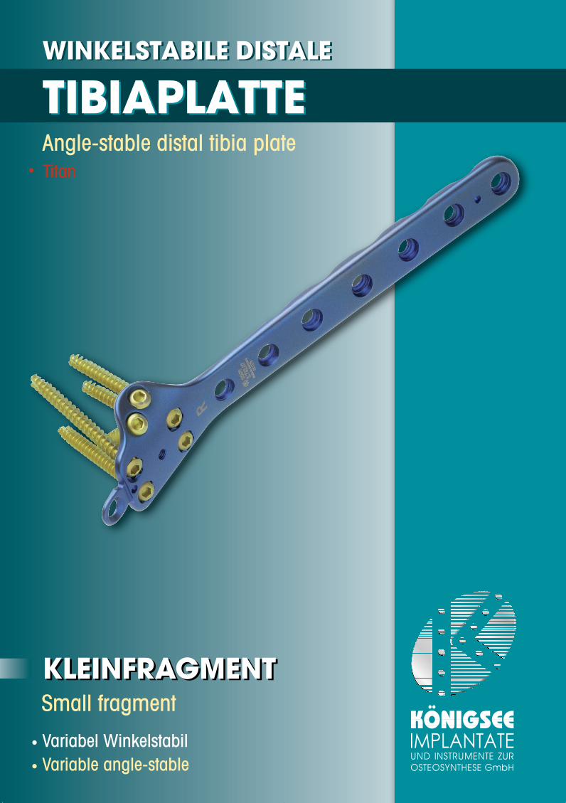
UND INSTRUMENTE ZUR OSTEOSYNTHESE GmbH Angle-stable distal tibia plate Small fragment • Variabel Winkelstabil • Variable angle-stable • Titan WINKELSTABILE DISTALE TIBIAPLATTE KLEINFRAGMENT WINKELSTABILE DISTALE TIBIAPLATTE KLEINFRAGMENT
Transcript of WINKELSTABILE DISTALE TIBIAPLATTE distale Tibia.pdf · Liegt eine in der frontalen Ebene führende...
UND INSTRUMENTE ZUROSTEOSYNTHESE GmbH
Angle-stable distal tibia plate
Small fragment
• Variabel Winkelstabil• Variable angle-stable
• Titan
WINKELSTABILE DISTALE
TIBIAPLATTE
KLEINFRAGMENT
WINKELSTABILE DISTALE
TIBIAPLATTE
KLEINFRAGMENT
OP-Anleitung Distal Tibia Kopie 30.07.2007 13:57 Uhr Seite 1
Das Implantat wurde für die operative Versorgung von Frakturen des Pilon tibiale wieauch extraartikulärer Frakturen der distalen Tibia entwickelt. Liegt eine in der frontalen Ebene führende Fraktur vor, ist eine (zusätzliche) ventralePlattenstabilisierung durch ein anderes Implantat notwendig.
The implant was developed for the operative management of fractures of the intra-articular distal tibia andalso extra-articular fractures of the distal tibia.If a fracture running in the frontal plane is present, (additional) anterior plate stabilisation by a different implantis necessary.
IndikationIndication
Einleitung Introduction
Distale Frakturen der Tibia, insbesondere bei Beteiligung der Gelenkflächen, stellen hoheAnsprüche an die Versorgung. Das hier vorgestellte Implantat kombiniert mit dem Prinzipder winkelstabilen Platte die Vorteile eines Fixateurs mit der Möglichkeit einer internenOsteosynthese. Die distale Tibiaplatte steht in verschiedenen Längen zur Verfügung und istanatomisch vorgeformt. Sie wird medial an der Tibia fixiert. Das Implantat ist in der Kleinfragment-Dimension gestaltet. Die distale Lasche ermöglichtdie Fixation des Innenknöchels mit einer konventionellen Kleinfragmentschraube. Amdistalen Ende der Platte befinden sich winkelstabile, bis 20° in der Richtung variabelbelegbare Schraubenlöcher. Der vorgegebene Schraubenstrahl gewährleistet, dass dieSchrauben berührungsfrei alle Hauptfragmente der Epi- und Metaphyse winkelstabilfixieren. Zwei weitere Schraubenlöcher befinden sich an dem ventralen Plattenausleger unddienen der Fragmentfixation in ventro-dorsaler Richtung. Proximal der Metaphyse ist derPlattenquerschnitt verstärkt. Der Unterschnitt verringert die Auflagefläche am Knochen underlaubt Korrekturen der Plattenform. Das Implantat gibt einen Mittelwert der Anatomiewieder, mit üblichen Biegeinstrumenten kann die Form geändert werden. Am proximalenPlattenschenkel hat der Operateur die Wahl durch das spezielle Design (Kombiloch) dieSchraubenlöcher mit konventionellen oder winkelstabilen Kleinfragmentschrauben zubelegen. Winkelstabil eingebracht divergieren die Schrauben alternierend. Kerbspannun-gen im Knochen und die Gefahr der sekundären Implantatmigration werden so verringert.
The management of distal fractures of the tibia, particularly those involving the joint surfaces, is demanding. Theimplant presented here combines the advantages of a fixator with the principle of an angle-stable plate thatallows internal fixation. The distal tibial plate is available in different lengths and is anatomically preshaped. It isfixed to the tibia medially.The implant is designed in small-fragment dimensions. The distal tab enables fixation of the medial malleoluswith a conventional small-fragment screw. At the distal end of the plate, there are angle-stable screw holes thatallow a variation of up to 20° in the angle of the screws. The given radiation of the screws ensures that thescrews can achieve angle-stable fixation of all the main fragments of the epiphysis and metaphysis withouttouching. There are two more screw holes on the anterior plate flange which are used for fragment fixation in theantero-posterior direction. The plate cross-section is reinforced proximal to the metaphysis. The undercutdiminishes the contact surface on the bone and allows the plate shape to be corrected. The implant reproducesthe average anatomy and the shape can be altered with the usual bending instruments. On the proximal limb ofthe plate, the surgeon can choose to occupy the screw holes with conventional or angle-stable small-fragmentscrews because of the special design (combihole). When inserted as angle-stable screws, the screws divergealternately. This diminishes notching stresses in the bone and thus the risk of secondary implant migration.
OP-Anleitung Distal Tibia Kopie 30.07.2007 13:57 Uhr Seite 2
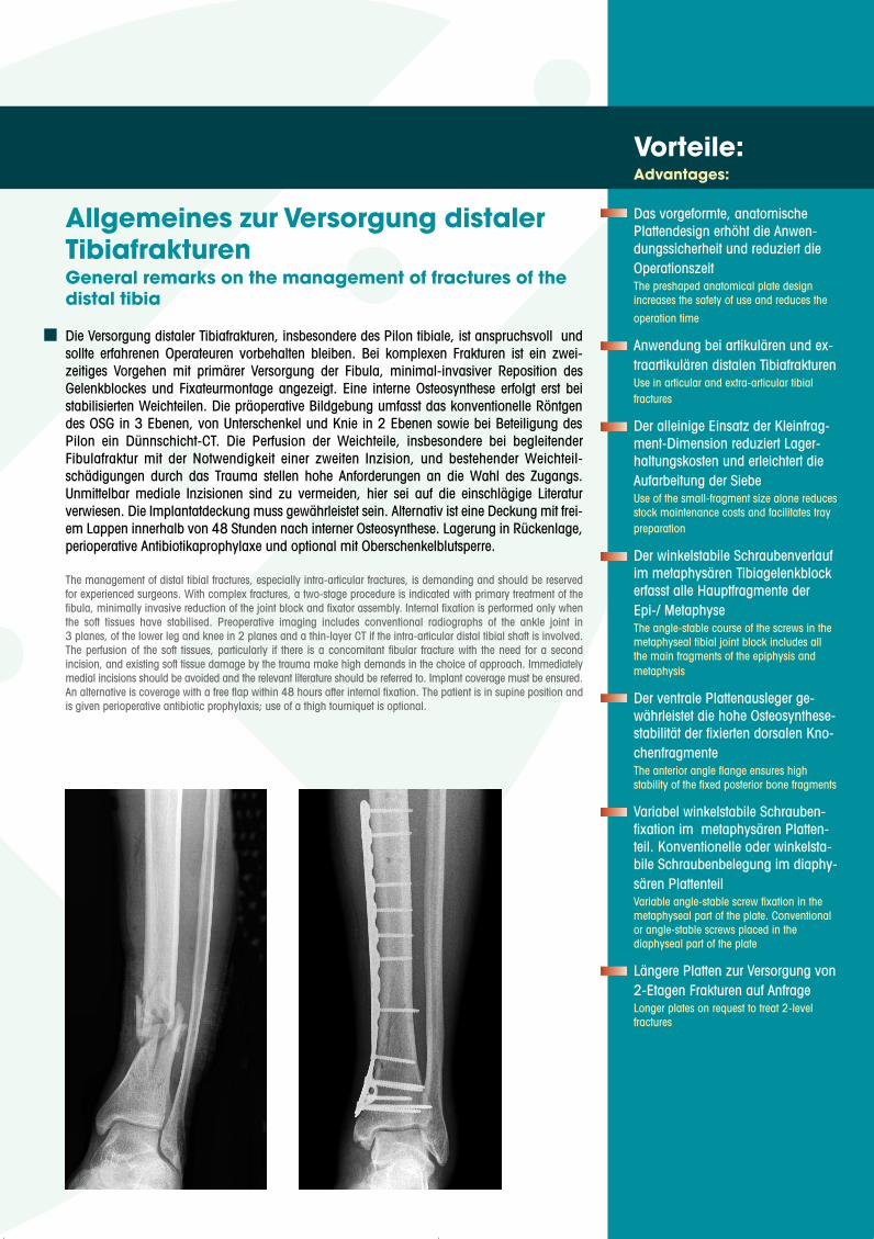
Die Versorgung distaler Tibiafrakturen, insbesondere des Pilon tibiale, ist anspruchsvoll undsollte erfahrenen Operateuren vorbehalten bleiben. Bei komplexen Frakturen ist ein zwei-zeitiges Vorgehen mit primärer Versorgung der Fibula, minimal-invasiver Reposition desGelenkblockes und Fixateurmontage angezeigt. Eine interne Osteosynthese erfolgt erst beistabilisierten Weichteilen. Die präoperative Bildgebung umfasst das konventionelle Röntgendes OSG in 3 Ebenen, von Unterschenkel und Knie in 2 Ebenen sowie bei Beteiligung desPilon ein Dünnschicht-CT. Die Perfusion der Weichteile, insbesondere bei begleitenderFibulafraktur mit der Notwendigkeit einer zweiten Inzision, und bestehender Weichteil-schädigungen durch das Trauma stellen hohe Anforderungen an die Wahl des Zugangs.Unmittelbar mediale Inzisionen sind zu vermeiden, hier sei auf die einschlägige Literaturverwiesen. Die Implantatdeckung muss gewährleistet sein. Alternativ ist eine Deckung mit frei-em Lappen innerhalb von 48 Stunden nach interner Osteosynthese. Lagerung in Rückenlage,perioperative Antibiotikaprophylaxe und optional mit Oberschenkelblutsperre.
The management of distal tibial fractures, especially intra-articular fractures, is demanding and should be reservedfor experienced surgeons. With complex fractures, a two-stage procedure is indicated with primary treatment of thefibula, minimally invasive reduction of the joint block and fixator assembly. Internal fixation is performed only whenthe soft tissues have stabilised. Preoperative imaging includes conventional radiographs of the ankle joint in3 planes, of the lower leg and knee in 2 planes and a thin-layer CT if the intra-articular distal tibial shaft is involved.The perfusion of the soft tissues, particularly if there is a concomitant fibular fracture with the need for a secondincision, and existing soft tissue damage by the trauma make high demands in the choice of approach. Immediatelymedial incisions should be avoided and the relevant literature should be referred to. Implant coverage must be ensured.An alternative is coverage with a free flap within 48 hours after internal fixation. The patient is in supine position andis given perioperative antibiotic prophylaxis; use of a thigh tourniquet is optional.
Vorteile:Advantages:
Das vorgeformte, anatomischePlattendesign erhöht die Anwen-dungssicherheit und reduziert dieOperationszeitThe preshaped anatomical plate designincreases the safety of use and reduces the
operation time
Anwendung bei artikulären und ex-traartikulären distalen TibiafrakturenUse in articular and extra-articular tibialfractures
Der alleinige Einsatz der Kleinfrag-ment-Dimension reduziert Lager-haltungskosten und erleichtert dieAufarbeitung der SiebeUse of the small-fragment size alone reducesstock maintenance costs and facilitates traypreparation
Der winkelstabile Schraubenverlaufim metaphysären Tibiagelenkblockerfasst alle Hauptfragmente derEpi-/ MetaphyseThe angle-stable course of the screws in themetaphyseal tibial joint block includes allthe main fragments of the epiphysis andmetaphysis
Der ventrale Plattenausleger ge-währleistet die hohe Osteosynthese-stabilität der fixierten dorsalen Kno-chenfragmente The anterior angle flange ensures highstability of the fixed posterior bone fragments
Variabel winkelstabile Schrauben-fixation im metaphysären Platten-teil. Konventionelle oder winkelsta-bile Schraubenbelegung im diaphy-sären PlattenteilVariable angle-stable screw fixation in themetaphyseal part of the plate. Conventionalor angle-stable screws placed in thediaphyseal part of the plate
Längere Platten zur Versorgung von2-Etagen Frakturen auf AnfrageLonger plates on request to treat 2-levelfractures
Allgemeines zur Versorgung distalerTibiafrakturenGeneral remarks on the management of fractures of thedistal tibia
OP-Anleitung Distal Tibia Kopie 30.07.2007 13:57 Uhr Seite 3
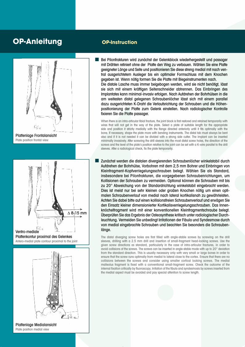
Bei Pilonfrakturen wird zunächst der Gelenkblock wiederhergestellt und passagermit Drähten retiniert ohne der Platte den Weg zu verbauen. Wählen Sie eine Plattegeeigneter Länge und Seite und positionieren Sie diese streng medial mit nach ven-tral ausgerichtetem Ausleger bis ein optimaler Formschluss mit dem Knochengegeben ist. Wenn nötig formen Sie die Platte mit Biegeinstrumenten nach.Die distale Lasche muss immer beigebogen werden, wird sie nicht benötigt, lässtsie sich mit einem kräftigen Seitenschneider abtrennen. Das Einbringen desImplantates kann minimal-invasiv erfolgen. Nach Aufdrehen der Bohrhülsen in dieam weitesten distal gelegenen Schraubenlöcher lässt sich mit einem paralleldazu ausgerichteten K-Draht die Verlaufsrichtung der Schrauben und die Höhen-positionierung der Platte zum Gelenk einstellen. Nach radiologischer Kontrollefixieren Sie die Platte passager.
When there is an intra-articular tibial fracture, the joint block is first restored and retained temporarily withwires that will not get in the way of the plate. Select a plate of suitable length for the appropriateside and position it strictly medially with the flange directed anteriorly until it fits optimally with thebone. If necessary, shape the plate more with bending instruments. The distal tab must always be bentalso and if it is not needed it can be divided with a strong side cutter. The implant can be insertedminimally invasively. After screwing the drill sleeves into the most distal screw holes, the direction of thescrews and the level of the plate‘s position relative to the joint can be set with a K-wire parallel to the drillsleeves. After a radiological check, fix the plate temporarily.
OP-Anleitung OP-Instruction
Zunächst werden die distalen divergierenden Schraubenlöcher winkelstabil durchAufdrehen der Bohrhülse, Vorbohren mit dem 2,5 mm Bohrer und Einbringen vonKleinfragment-Kopfverriegelungsschrauben belegt. Wählen Sie als Standard,insbesondere bei Pilonfrakturen, die vorgegebenen Schraubenrichtungen, umKollisionen der Schrauben zu vermeiden. Optional können die Schrauben mit biszu 20° Abweichung von der Standardrichtung winkelstabil eingebracht werden.Dies ist meist nur bei sehr kleinen oder großen Knochen nötig um einen opti-malen Schraubenverlauf von medial nach lateral kortikalisnah zu gewährleisten.Achten Sie dabei bitte auf einen kollisionsfreien Schraubenverlauf und erwägen Sieden Einsatz kleiner dimensionierter Kortikalisverriegelungsschrauben. Das Innen-knöchelfragment wird mit einer konventionellen Kleinfragmentschraube belegt.Überprüfen Sie das Ergebnis der Osteosynthese kritisch unter radiologischer Durch-leuchtung. Vermeiden Sie unbedingt Irritationen der Fibula und Syndesmose durchvon medial eingebrachte Schrauben und beachten Sie besonders die Schrauben-länge.
The distal diverging screw holes are first filled with angle-stable screws by screwing on the drillsleeves, drilling with a 2.5 mm drill and insertion of small-fragment head-locking screws. Use thegiven screw directions as standard, particularly in the case of intra-articular fractures, in order toavoid collisions of the screws. The screws can be inserted in angle-stable mode with up to 20° deviationfrom the standard direction. This is usually necessary only with very small or large bones in order toensure that the screw runs optimally from medial to lateral close to the cortex. Ensure that there are nocollisions between the screws and consider using smaller cortical locking screws. The medialmalleolus fragment is fixed with a conventional small-fragment screw. Check the outcome of theinternal fixation critically by fluoroscopy. Irritation of the fibula and syndesmosis by screws inserted fromthe medial aspect must be avoided and pay special attention to screw length.
Ventro-medialePlattenkontur proximal des GelenkesAntero-medial plate contour proximal to the joint
Plattenlage MedialansichtPlate position medial view
Plattenlage FrontalansichtPlate position frontal view
∆ 8-15 mm
OP-Anleitung Distal Tibia Kopie 30.07.2007 13:58 Uhr Seite 4
Nach abschließender radiologischer Kontrolle und Dokumentation erfolgt derWundverschluss mit Wunddrainage und Anlage einer gut gepolsterten Unter-schenkelschiene bis zur Wundheilung. Die Nachbehandlung entspricht den bekannten Richtlinien. Sie ist limitiert durchdie Stabilität der Osteosynthese und die Compliance der Patienten.
After final radiological check and documentation, the wound is closed with a drain and awellpadded lower leg splint until the wound has healed.Postoperative treatment is in accordance with the familiar guidelines. It is limited by the stability ofthe internal fixation and patient compliance.
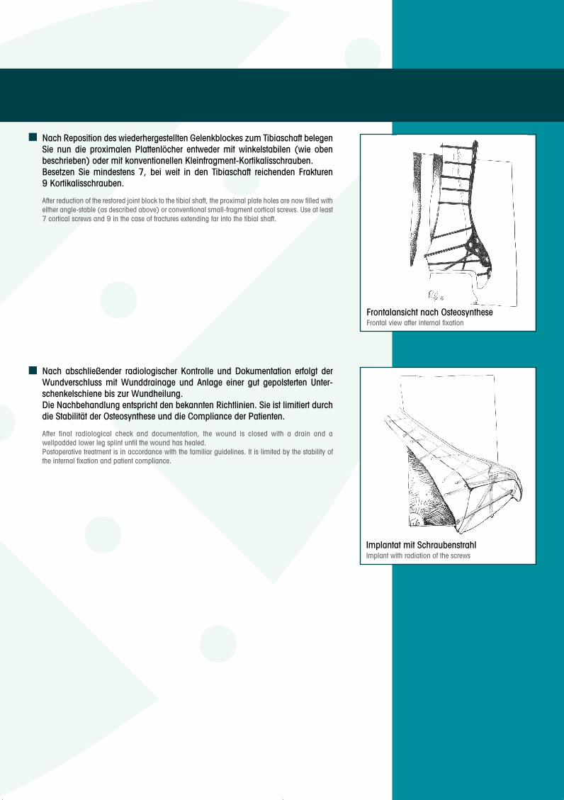
Nach Reposition des wiederhergestellten Gelenkblockes zum Tibiaschaft belegenSie nun die proximalen Plattenlöcher entweder mit winkelstabilen (wie obenbeschrieben) oder mit konventionellen Kleinfragment-Kortikalisschrauben.Besetzen Sie mindestens 7, bei weit in den Tibiaschaft reichenden Frakturen9 Kortikalisschrauben.
After reduction of the restored joint block to the tibial shaft, the proximal plate holes are now filled witheither angle-stable (as described above) or conventional small-fragment cortical screws. Use at least7 cortical screws and 9 in the case of fractures extending far into the tibial shaft.
Frontalansicht nach OsteosyntheseFrontal view after internal fixation
Implantat mit SchraubenstrahlImplant with radiation of the screws
OP-Anleitung Distal Tibia Kopie 30.07.2007 13:58 Uhr Seite 5
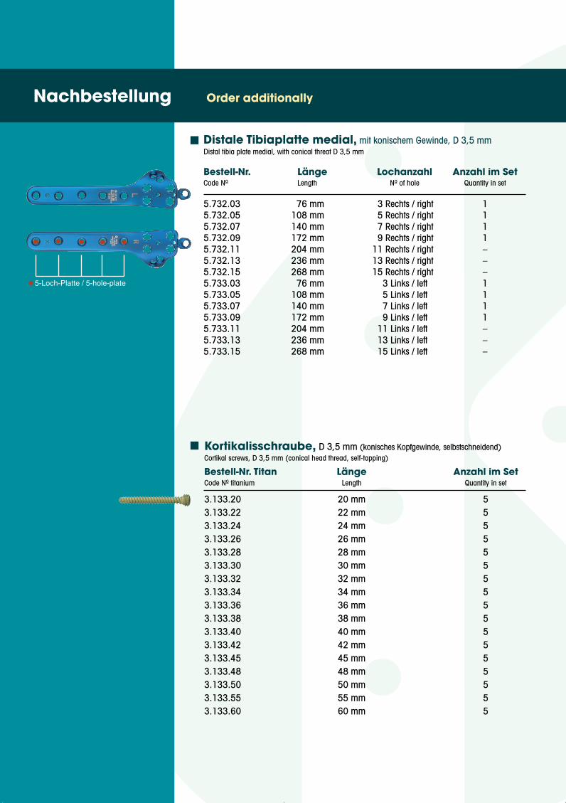
Bestell-Nr. Länge Lochanzahl Anzahl im SetCode No Length No of hole Quantity in set
Nachbestellung Order additionally
5.732.03 76 mm 3 Rechts / right 15.732.05 108 mm 5 Rechts / right 15.732.07 140 mm 7 Rechts / right 15.732.09 172 mm 9 Rechts / right 15.732.11 204 mm 11 Rechts / right –5.732.13 236 mm 13 Rechts / right –5.732.15 268 mm 15 Rechts / right –5.733.03 76 mm 3 Links / left 15.733.05 108 mm 5 Links / left 15.733.07 140 mm 7 Links / left 15.733.09 172 mm 9 Links / left 15.733.11 204 mm 11 Links / left –5.733.13 236 mm 13 Links / left –5.733.15 268 mm 15 Links / left –
Distale Tibiaplatte medial, mit konischem Gewinde, D 3,5 mmDistal tibia plate medial, with conical threat D 3,5 mm
Kortikalisschraube, D 3,5 mm (konisches Kopfgewinde, selbstschneidend)Cortikal screws, D 3,5 mm (conical head thread, self-tapping)
Bestell-Nr. Titan Länge Anzahl im SetCode No titanium Length Quantity in set
3.133.20 20 mm 53.133.22 22 mm 53.133.24 24 mm 53.133.26 26 mm 53.133.28 28 mm 53.133.30 30 mm 53.133.32 32 mm 53.133.34 34 mm 53.133.36 36 mm 53.133.38 38 mm 53.133.40 40 mm 53.133.42 42 mm 53.133.45 45 mm 53.133.48 48 mm 53.133.50 50 mm 53.133.55 55 mm 53.133.60 60 mm 5
5-Loch-Platte / 5-hole-plate
OP-Anleitung Distal Tibia Kopie 30.07.2007 13:58 Uhr Seite 6
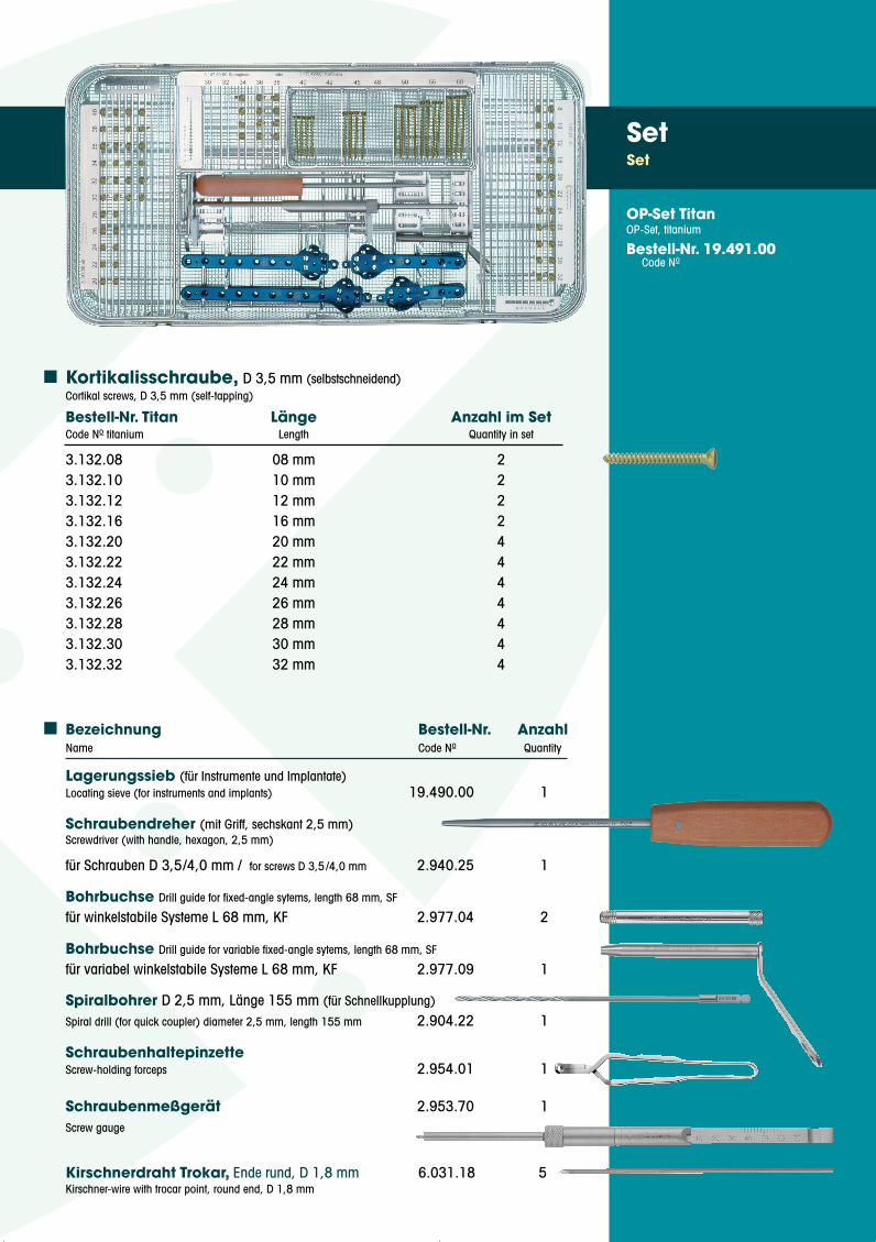
Bezeichnung Bestell-Nr. AnzahlName Code No Quantity
Lagerungssieb (für Instrumente und Implantate)Locating sieve (for instruments and implants) 19.490.00 1
Schraubendreher (mit Griff, sechskant 2,5 mm)Screwdriver (with handle, hexagon, 2,5 mm)
für Schrauben D 3,5/4,0 mm / for screws D 3,5/4,0 mm 2.940.25 1
Bohrbuchse Drill guide for fixed-angle sytems, length 68 mm, SF
für winkelstabile Systeme L 68 mm, KF 2.977.04 2
Bohrbuchse Drill guide for variable fixed-angle sytems, length 68 mm, SF
für variabel winkelstabile Systeme L 68 mm, KF 2.977.09 1
Spiralbohrer D 2,5 mm, Länge 155 mm (für Schnellkupplung)
Spiral drill (for quick coupler) diameter 2,5 mm, length 155 mm 2.904.22 1
SchraubenhaltepinzetteScrew-holding forceps 2.954.01 1
Schraubenmeßgerät 2.953.70 1Screw gauge
Kirschnerdraht Trokar, Ende rund, D 1,8 mm 6.031.18 5Kirschner-wire with trocar point, round end, D 1,8 mm
SetSet
OP-Set TitanOP-Set, titanium
Bestell-Nr. 19.491.00Code No
Kortikalisschraube, D 3,5 mm (selbstschneidend)Cortikal screws, D 3,5 mm (self-tapping)
Bestell-Nr. Titan Länge Anzahl im SetCode No titanium Length Quantity in set
3.132.08 08 mm 23.132.10 10 mm 23.132.12 12 mm 23.132.16 16 mm 23.132.20 20 mm 43.132.22 22 mm 43.132.24 24 mm 43.132.26 26 mm 43.132.28 28 mm 43.132.30 30 mm 43.132.32 32 mm 4
OP-Anleitung Distal Tibia Kopie 30.07.2007 13:59 Uhr Seite 7
Ärztlicher Autor:
Dr. med. Stefan Fabian
Oberarzt Orthopädie,
Brüderkrankenhaus St. Josef,
Paderborn
WWW.KOENIGSEE-IMPLANTATE.DEWWW.KOENIGSEE-IMPLANTATE.DE
KÖNIGSEE IMPLANTATE UND INSTRUMENTEZUR OSTEOSYNTHESE GmbHAm SandD-07426 Königsee-AschauFon +49 (0) 36738 498-0Fax +49 (0) 36738 498-19e-mail: [email protected]© Copyright Königsee Implantate GmbH
Stand: Juni 2007Bestell-Nr.: D.06.732.07
UND INSTRUMENTE ZUROSTEOSYNTHESE GmbH
zertifiziert nach EG Richtlinie93/42/EWGDIN EN ISO 13485: 2003DIN ISO 9001: 2000
Wiedergabe des Inhalts, auch aus-zugsweise, nur mit schriftlicherGenehmigung des Herausgebers.
No part of this publication may bereproduced, stored in a retrievalsystem or transmitted in any form orby any means electronic, mechani-cal, photocopying, recording orotherwise without the prior writtenpermission of the publisher.
OP-Anleitung Distal Tibia Kopie 30.07.2007 13:59 Uhr Seite 8