Zusammenfassung aller Vortrags- und Posterabstracts der 25 ... · transport of glucose by SGLT1 and...
156
Zusammenfassung aller Vortrags- und Posterabstracts der 25. Arbeitstagung der Anatomischen Gesellschaft in Würzburg vom 24.09.2008 bis 26.09.2008 1. Vortragsabstracts 2. Posterabstracts
Transcript of Zusammenfassung aller Vortrags- und Posterabstracts der 25 ... · transport of glucose by SGLT1 and...
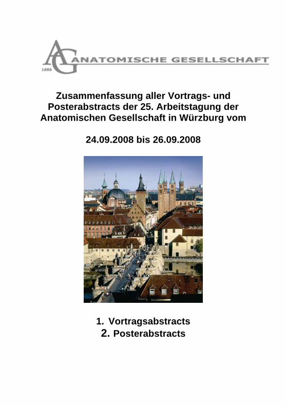
Zusammenfassung aller Vortrags- und Posterabstracts der 25. Arbeitstagung der
Anatomischen Gesellschaft in Würzburg vom
24.09.2008 bis 26.09.2008
1. Vortragsabstracts 2. Posterabstracts
Vortrag 1 Titel:Localization and regulation of the sgc in cells of the healthy and inflamed human periodontal ligament Autoren: Korkmaz Y.(1),Korkmaz Y.(1),Raab W.(1),Ulbrich H.(2),Klinz F.(2),Bloch W.(3),Addicks K.(2), Adressen:(1)Heinrich-Heine-Universität Düsseldorf|Poliklinik für Zahnerhaltung und Präventive Zahnheilkunde und der Section für Parodontologie|Düsseldorf|Deutschland; email:[email protected] ; (2)Universität zu Köln|Institut I für Anatomie|Köln|Deutschland; (3)Deutsche Sporthochschule Köln|Institut für Kreislaufforschung und Sportmedizin|Köln|Deutschland Abstract: The enzyme soluble guanylate cyclase (sGC) contains one prosthetic haem group as an alpha/beta heterodimer and two isoforms (alpha1/beta1, alpha2/beta1) have been shown to have enzymatic activity. The inflammation-dependent modulation of sGC in cells of the PDL is unknown. To test the inflammation-dependent regulation of the sGC in the healthy and inflamed PDL of the third molars and premolars, the alpha1-, alpha2- and beta1-subunit of the sGC were investigated at protein levels by immunohistochemistry and immunoblot. In comparison to the staining intensities for alpha1-, alpha2- and beta1-subunit in blood vessels, cementoblasts, PDL stroma cells and alpha2- and beta1-subunit in nerve fibers in the health PDL, the alpha1-, alpha2- and beta1-subunit of sGC were detected with weak labeling intensities in cells of the inflamed PDL. Inflammation induced down-modulation of the alpha1-, alpha2- and beta1-subunits of sGC in cells of the human PDL indicates critical roles for the sGC alpha1/beta1- as well as alpha2/beta1-heterodimers in the circulation, cementogenesis and for the sGC alpha2/beta1-heterodimer in neurotransmission of the PDL in health. Kategorie: Vortrag
Vortrag 2 Titel:The localization and function of organic cation transporters in the skin Autoren: Volk C.(1),Lips K.(2),Zapf F.(1),Uttner S.(1),Koepsell H.(1), Adressen:(1)Julius-Maximilians-Universität|Institut für Anatomie und Zellbiologie|Würzburg|Deutschland; email:[email protected]; (2)Julius-Liebig-Universität|Institut für Anatomie und Zellbiologie|Giessen|Deutschland Abstract: The organic cation transporters OCT1-3 (Slc22A1-A3) mediate cellular uptake and/or release of a variety of substrates including monoamine neurotransmitters, choline and acetylcholine. They are expressed in many tissues but their physiological function has not been sufficiently clarified. Here we investigate the localization and functional role of OCTs in the skin. Using subtype specific antibodies, we detected OCT1-3 in all layers of rat and human epidermis, with a particular high level of expression of OCT2 and OCT3 in basal cells. Additionally, OCT3 is located in smooth muscle cells of blood vessels in the dermis. Using primarily cultivated human keratinocytes we demonstrated OCT-mediated uptake of the model compound 1-methyl-4-phenylpyridinium. Since OCTs mediate acetylcholine release they are supposed to be involved in non-neuronal cholinergic regulations which e.g. control proliferation and regeneration of the epidermis. Furthermore, they perform the uptake of choline which is needed for phosphatidylcholine synthesis. Several topically applied drugs including the corticosteroid prednicarbat, the antihistaminic dimetindenmaleat and the bacteriostatic gentian violett inhibit OCT-mediated transport with comparatively high affinities. Since the observed IC50 values for these substances are considerably lower than the concentrations that can occur under topical application, this inhibition may contribute to atrophic side effects under prolonged application. Kategorie: Vortrag
Vortrag 3 Titel:Muscarinic receptor-2-mediated constriction of murine bronchi is caveolae dependent Autoren: Schlenz H.(1),Kummer W.(1),Krasteva G.(1), Adressen:(1)Justus-Liebig-University Giessen|Institute for Anatomy and Cell Biology|35385|Germany; email:[email protected] Abstract: Asthma and COPD are associated with bronchial hyperreagibility. In acetylcholine-induced constriction of bronchial smooth muscle cells (SMC), muscarinic receptor subtypes 2 (M2R) and 3 (M3R) are involved. In SMC of urinary bladder of caveolin-1-deficient (-/-) mice the contractile response to muscarine is decreased, so that an involvement of caveolae/caveolins in bronchoconstriction can be anticipated. Videomicroscopy was performed to study bronchoconstriction in living precision cut lung slices (PCLS) from M3R-/- and wild-type (M3R+/+) mice. Recordings were made before and after treatment with cyclodextrin or vehicle. The treatment efficiency was evidenced by the absence of contractile response to 5-HT and of caveolae on the cell membrane as examined by electron microscopy. M2R- and M3R-expression were quantified by real-time RT-PCR. The bronchial luminal area was decreased by muscarine (10 µM) to 40.9% in M3R+/+ and to 61.8% in M3R-/- mice (p=0.014). After cyclodextrin-treatment the response in M3R+/+ mice was reduced to 76.5% of initial area and completely abolished in M3R-/- mice, clearly demonstrating the dependence of the M2R pathway on caveolae. In these PCLS, KCl-induced constriction remained unchanged. The difference in luminal narrowing between cyclodextrin-treated and non-treated PCLS was comparable in both strains (~36). The expression level of M2R in M3R-/- and M3R+/+ mice was comparably high, pointing against a take-over of M3R expression and function by M2R in M3R-/- mice. These results demonstrate a functional role of caveolae in the regulation of cholinergic bronchoconstriction and might therefore be of therapeutical relevance. Kategorie: Vortrag
Vortrag 4 Titel:A novel approach to regulate small intestinal glucose absorption and intestinal stimulation of insulin secretion Autoren: Veyhl M.(1),Vernaleken A.(1),Gorboulev V.(1),Koepsell H.(1), Adressen:(1)Würzburg|Institut für Anatomie und Zellbiologie I|Würzburg|Deutschland; email:[email protected] Abstract: D-glucose absorption in enterocytes is carried out by combination of two transporters, the apical Na+-D-glucose cotransporter SGLT1 and the basolateral glucose transporter GLUT2. High glucose concentrations stimulate the redistribution of GLUT2 to the luminal membrane for increasing the glucose absorptive capacity rapidly. Two signals mediate the apical GLUT2 pathway, i) plasma membrane depolarization due to the electrogenic transport of glucose by SGLT1 and ii) release of glucagon-like peptide 1(GLP-1) and glucose-dependent insulinotrophic peptide (GIP ), known as incretins, by activation of sweet teste receptors in the small intestine. The incretins also upregulate the insulin secretion Since SGLT1 is also expressed in enteroendocrine cells and the intestinal stimulation of insulin secretion is abolished in SGLT1-knock-out mice we conclude that SGLT1 is involved in the release of incretins. Using the Xenopus laevis expression system we identified two tripeptides (Gln-Cys-Pro (QCP) and Gln-Ser-Pro (QSP)) that induce high affinity posttranscriptional glucose-dependent downregulation of SGLT1 at the trans-Golgi network leading to 40-50 % reduction of SGLT1 in the plasma membrane. QSP and QCP are transported by the H+-peptide cotransporter PepT1 that is colocated with SGLT1 in enterocytes and are effective when applied extracellulary. These peptides and related compounds are potential drugs for treatment of obesity and diabetes type II and if transported in enteroendocrine cells could modulate the release of GLP-1 and GIP. Kategorie: Vortrag
Vortrag 5 Titel:Localization and transcytosis of components of the renin angiotensin system (ras) in the proximal tubule Autoren: Theilig F.(1),Pohl M.(1),Willnow T.(2),Bachmann S.(1), Adressen:(1)Universitätsmedizin Berlin|Inst. für vegetative Anatomie|Berlin|Deutschland; email:[email protected]; (1)Universitätsmedizin Berlin|Inst. für Vegetative Anatomie|Berlin|Deutschland; (2)Max Delbrück Zentrum für Molekulare Medizin|Molecular vascular research|Berlin-Buch|Deutschland Abstract: The kidney is a crucial site of the mammalian organism for the biogenesis, effects, and metabolization of components of the RAS. Renin is currently thought to be synthesized by the JGA, angiotensinogen (Ao) and angiotensin converting enzyme (ACE) by the proximal tubule. However, cell-specific assignment is still unclear and local RAS functions in the proximal tubule are discussed controversially. We have analyzed the renal distribution of RAS components with focus on proximal tubular handling. We have used normal and megalin-deficient mice with a mosaic defect of proximal endocytosis. Whereas renin biosynthesis was restricted to the JGA, early proximal tubules showed megalin-dependent endosomal uptake and lysosomal processing of renin and also of Ao, whereas low levels of Ao mRNA was detected exclusively in late proximal tubule. Ao and renin were identified as ligands of megalin with their binding mapped to the extracellular LDL domains of megalin. Apart from the degradative pathway, transcytotic passage was demonstrated for intact Ao and Renin and was negatively influenced by megalin. ACE mRNA was concentrated in the late proximal tubule, with immunoreactive signal concentrated in the brush border membrane (BBM). Presence in the BBM negatively correlated with the expression of megalin. ACE was also present in intercalated cells of the connecting tubule and collecting duct. These results underline a prominent role for the proximal tubular handling of RAS components. Transcytosis may be a mode of the kidney to retrieve RAS components with potential interstitial or systemic impact. Kategorie: Vortrag
Vortrag 6 Titel:Acute effects of vasopressin in the distal convoluted tubule Autoren: Mutig K.(1),Borowski T.(1),Böhlick A.(1),Paliege A.(1),Kahl T.(1),Müller-Esterl W.(2),Bachmann S.(1), Adressen:(1)Charite Universitätsmedizin Berlin|Institut für Vegetative Anatomie|Berlin|Deutschland; email:[email protected]; (2)Universitätsklinikum Frankfurt|Institut für Biochemie II|Frankfurt|Deutschland Abstract: Vasopressin (AVP) influences salt and water transport in the renal tubule chiefly via the V2 receptors (V2R). Little is known on its function in distal convoluted tubules (DCT). We hypothesize a major role for the short term V2R-mediated activation of the Na+,Cl--cotransporter (NCC) in DCT. In analogy to the closely related Na+,K+,2Cl--cotransporter 2 (NKCC2) of the thick ascending limb (TAL), AVP-induced activation of NCC may include changes in trafficking and phosphorylation of the transporter. Interestingly, previous studies on oocytes showed that the SPAK-kinase is able to phosphorylate and activate NKCC2 and NCC. Kidneys from AVP-deficient Brattleboro (diabetes insipidus, DI) rats were studied. Short term AVP treatment with dDAVP (30min to 4h) was applied. Cellular distribution of SPAK and V2R was studied immunohistochemically. Changes of NCC and NKCC2 subcellular distribution and phosphorylation were analyzed by immunocytochemistry and Western blot. Expression of V2R in DCT was confirmed. SPAK was localized to the sub-apical cell compartments of TAL, whereas in DCT a cytosolic signal was detected. dDAVP induced increased phosphorylation of NCC and NKCC2 concomitantly (+198%), along with increased luminal expression of NCC (+67%) and NKCC2 (+86%). We for the first time demonstrated short term V2R-mediated effects of AVP in DCT, which is in agreement with our results on significant V2R expression in DCT of the rat kidney. We further localized SPAK to TAL and DCT, thus confirming the postulated role of this kinase in the phosphorylation of NKCC2 and NCC. Kategorie: Vortrag
Vortrag 7 Titel:Tnfalpha induced cytokine expression in human tenocytes in vitro. Autoren: Schulze-Tanzil G.(1),Lodka D.(1),Kohl B.(1),Jammrath J.(1),Ertel W.(1),John T.(1), Adressen:(1)Charite-Universitätsmedizin Berlin|Klinik für Unfall- und Wiederherstellungschirurgie, Campus Benjamin Franklin|12207|Deutschland; email:[email protected] Abstract: Tendon rupture induces a local post-traumatic inflammatory response, characterized by the release of multiple proinflammatory cytokines. Tendon healing is a time consuming reparative process leading to tendon scar formation which might be strongly influenced by this post-traumatic inflammation. Since the particular interplay of proinflammatory and immunoregulatory cytokines in tendon remains unknown, the present study was undertaken to characterize the effects of TNF-alpha, IL-6 and IL-10 on tenocyte inflammatory response by investigation of the expression of cytokines and suppressors of cytokine signalling. Serum-starved human tenocytes were treated with recombinant human IL-6, IL-10, TNF-alpha or combinations of TNF-alpha with IL-6 and IL-10 (10 ng/mL, 6 h, 24 h). The expression of extracellular tendon matrix components type I collagen, elastin, cytokines TNF-alpha, IL-1beta, IL-6, IL-18 and suppressors of cytokine signalling was studied by RTD PCR, immunohistochemistry or by western blot analysis. TNF-alpha severily decreased type I collagen deposition, but amplified significantly elastin expression. TNF-alpha activated the tenocytes to highly up-regulate their gene expression for proinflammatory (TNF-alpha, IL-1beta) and immunoregulatory cytokines (IL-6, IL-10) in a time-dependent manner. The treatment of tenocytes with recombinant human IL-6 and IL-10 alone had no major effect on their cytokine expression, but the co-treatment with IL-6 or IL-10 and TNF-alpha inhibited slightly the expression of IL-6 in response to TNF-alpha. TNF-alpha stimulation augmented SOCS1, whereas SOCS3 gene expression was only up-regulated by IL-6 in tenocytes. The study indicates that TNFalpha strongly amplifies pro-inflammatory cytokine signalling in tendon and might play a substantial role in post-traumatic inflammatory response in tendon. Kategorie: Vortrag
Vortrag 8 Titel:Interleukin-1beta induced inflammatory and apoptotic processes in chondrocytes are revoked by resveratrol: a new pathway in osteoarthritis therapy Autoren: Csaki C.(1),Nebrich S.(1),Shakibaei M.(1), Adressen:(1)LUDWIG-MAXIMILIANS-UNIVERSITY MUNICH|Institute for Anatomy|Munich|germany; email:[email protected] Abstract: Objective: Osteoarthitis is a degenerative joint disease affecting millions of people. Current treatments are limited to reducing pain symptoms and are only temporarily effective. The need for effective and save new chemotherapeutic agents has brought nutraceuticals such a resveratrol into focus of scientific research. Resveratrol is a polyphenol found in the skin of red grapes, berries, various other fruits and peanuts. Recent research has demonstrated its great anti-inflammatory properties, however its mechanism in chondrocytes still has to be clarified. The aim of this study was to investigate the effects of Resveratrol on the NF-kappaB signalling pathway, which amongst other things mediates Interleukin-1 beta production. Materials and methods: Human chondrocytes were treated with Interleukin-1 beta to induce inflammation, followed by a co-treatment with Resveratrol or the proteasome inhibitor N-Ac-Leu-Leu-norleucinal (ALLN). Cultures were evaluated with transmission electron microscopy, westernblot analysis, immunohistochemistry. Results: Resveratrol suppressed VEGF, MMP-3 and -9 and Cox-2 production as well as activation of caspase-3 and PARP cleavage in Interleukin-1beta stimulated cells. Further, Resveratrol suppressed nuclear translocation of the p65 subunit of NF-kappaB. Thereby Resveratrol, like ALLN, inhibited IkappaB alpha degradation through inhibiting proteasome function without affecting IkappaB alpha kinase activation, IkappaB alpha phosphorylation or IkappaB alpha ubiquitination. Conclusions: Our results suggest that in human chondrocytes, Resveratrol suppresses apoptosis and inflammatory signalling through its action on the NF-kappaB signalling pathway. We propose that Resveratrol should be further explored for prophylactic treatment of osteoarthritis in humans and companion animals. Kategorie: Vortrag
Vortrag 9 Titel:Osteoarthritis is not a one way road of cartilage loss Autoren: Eckstein F.(1),Buck R.(2),Wyman B.(2),Hudelmaier M.(1),Hellio Le Graverand M.(2), Adressen:(1)Paracelsus Medizinische Privatuniversität|Institut für Anatomie und muskuloskelettale Forschung|Salzburg|Austria; email:[email protected]; (2)Pfizer|GRD|Groton|USA Abstract: Cartilage loss is considered to be a hallmark of osteoarthritis (OA). However, until recently, no direct quantitative measure of cartilage loss was available to study changes in cartilage in clinical trials. Here we investigate the rate and variability of change in cartilage thickness over 2 years in a multicenter clinical trial using magnetic resonance imaging (MRI), comparing women with different grades of radiographic OA with healthy controls. 148 women were studied at 7 clinical centers in the US using radiography and 3 Tesla MRI: 90 healthy, 30 with radiographic OA grade2 (KLG2, presence of definite ostopyhtes, but no apparent JSN), and 28 with radiographic OA grade3 (KLG3, presence of definite ostopyhtes and JSN. Cartilage morphology in the femorotibial joint was determined quantitatively at baseline and at 2-year follow-up after segmentation by 7 experienced readers, using proprietary software. Compared with healthy controls, cartilage was significantly thicker in the KLG2 OA participants and significantly thinner in KLG3 participants at baseline. Over 2 years, significant cartilage loss occurred in KLG3, but not in KLG 2 participants. The distribution of change in KLG2 participants, however, differed from healthy participants, in that 20% had higher than expected loss, but 23% a higher than expected gain in cartilage thickness, based on the distribution of change in normal knees. These data suggest that OA is not a “one way road” of cartilage loss, but that at the early phase of radiographic OA cartilage undergoes swelling, whereas cartilage loss only occurs at later stages of radiographic OA. Kategorie: Vortrag
Vortrag 10 Titel:Medico-biological impact of the scientific study of morphology in historic remains Autoren: Rühli F.(1), Adressen:(1)Universität Zürich|Anatomisches Institut|Zürich|Schweiz; email:[email protected] Abstract: Historic human remains and data are unique sources to learn not only about the human past but also about the present. Such tissue-based historic information can contribute substantially to the understanding of ongoing interactions between mankind and environment as echoed e.g. by alterations in prevalence and morbidity of disease. “Evolutionary Medicine” merges anthropological-archeological as well as clinical issues and deals e.g. with questions of changing human anatomy and physiology as well as patterns of diseases. This can be analyzed both, in spatial and temporal terms. Exemplary, secular changes as shown by us e.g. for the intervertebral foramen size (Rühli and Henneberg, Eur Spine J, 2003) or for the stature of Swiss Armed Forces conscripts (Rühli et al., Am J Phy Anthropol, in press) will be highlighted during this talk. Also, our improvements in state-of-the-art imaging technology (e.g. Rühli et al. JAMA, 2007) for paleopathological studies will be addressed. In particular, the research aims and methodology of the transdisciplinary “Swiss Mummy Project” – our main research project to study morphology and pathology in ancient human mummified remains, will be presented too. Kategorie: Vortrag
Vortrag 11 Titel:Fluorescent in vivo imaging of zebrafish vasculature to monitor glomerular filtration: a characterization of the method Autoren: Müller T.(1),Hradetzky S.(1),Bollig F.(2),Englert C.(2),Endlich K.(1),Endlich N.(1), Adressen:(1)Greifswald|Institut für Anatomie und Zellbiologie|Greifswald|Deutschland; email:[email protected]; (2)Jena|Leibniz Institute for Age Research - Fritz Lipmann Institute|Jena|Deutschland Abstract: Podocytes are highly specialized epithelial cells covering the capillaries of the glomerular tuft. Alterations in their complex morphology can lead to a loss of renal function. For functional studies of glomerular filtration in vivo, the zebrafish larva is an ideal model organism due to its rapid development, its high optical transparency and the similarity between zebrafish and mammalian glomeruli. We have developed a method to study glomerular filtration in living zebrafish, utilizing high resolution confocal imaging. FITC-labeled dextran (MW: 500 kDa) and Alexa 647-labeled dextran (MW: 10 kDa) were injected into the caudal vein of zebrafish larvae 96 hours post-fertilization (hpf). Mean signal intensities in a section of the dorsal aorta were monitored by two-colour imaging in increasing time intervals between 30 minutes and 24 hours after injection. Dextran with 10 kDa molecular weight was fully cleared from the vasculature after 24 hours, leaving a strong fluorescence signal in the cells of the pronephric duct and tubules, due to dextran endocytosis. This localization was ascertained by injecting dextran-Alexa 647 (10 kDa) into WT1::GFP-transgenic zebrafish. The fluorescence of the 500 kDa dextran dropped to about 50% of the starting value within four hours, eventually reaching a stable plateau. FITC signal was not detected in the cells or the lumen of tubules at any time, the signal decrease being caused mainly by its slow dispersion into the intercellular space. These results provide deeper insight into the behaviour of injected fluorescent dextrans, promoting the routine use of this method for glomerular filtration analyses. Kategorie: Vortrag
Vortrag 12 Titel:Variable "spraycon" whole body conservation in a closed system Autoren: Weber G.(1), Adressen:(1)entfällt|MEDIS GmbH|Buseck|Germany Abstract: Anatomy and Histology used the "dip" method for the infiltration / fixation / conservation of tissue specimens since a long time. In histology today nearly all tissue specimens are processed in a so called "closed sytem" where the reagent is transfered to the specimen. This was mainly done in order to eliminate the dangerous formaline fumes. In anatomy it is still common to dip, for example, whole bodies for teaching purposes into a container with conservation fluid, which is covered with a removable lid. As soon as the lid is removed a huge amount of dangerous fumes hit the user, which has to wear a gass-mask in order to withstand the dangerous situation. The modular "SprayCon" system consists of a chamber which accommodates the whole body specimens. After closing all openings, the inside of the "conservation chamber" is sprayed completely so that a foggy atmosphere is present at all times. Because of the modular design of the "SprayCon" system, individual chamber-sections can be programmed so that different conservation solutions may be used. In order to avoid fungal or wurm affection inside the complete process chamber, the individual compartments are separated from each other completely. A small vacuum inside the chamber will guarantee that no fumes are entering the environment. A rinse-cycle with water will clean the bodies. In this condition they are presented to the students in the dissecting room. The European Health and Safety Authorities have already proposed that the handling of open conservation-/fixation-fluids will be prohibited in the future. Kategorie: Vortrag
Vortrag 13 Titel:Essential role for the transcription factor bcl11b during postnatal hippocampal neurogenesis. Autoren: Brylka H.(1),Schwegler H.(2),Jenkins N.(3),Copeland N.(3),Birchmeier C.(4),Britsch S.(1), Adressen:(1)Ulm|Institut für molekulare und zelluläre Anatomie|Ulm|Deutschland; email:[email protected]; (2)Otto-von-Guericke-Universität Magdeburg|Anatomisches Institut|Magdeburg|Deutschland; (3)Center for Cancer Research|National Cancer Institut|Frederick|USA; (4)Max-Delbrück-Zentrum für Molekulare Medizin|Max-Delbrück-Zentrum für Molekulare Medizin|Berlin-Buch|Deutschland Abstract: In the adult mammalian brain the dentate gyrus is one of the two locations with continuing postnatal neurogenesis and the primary gateway for inputs into the hippocampus, a cortical structure that is required for learning and memory. During development of the dentate gyrus, the transition from a proliferating progenitor cell into a postmitotic functional neuron involves a network of regulatory transcription factors. Bcl11b encodes a highly conserved C2H2 zinc finger transcription factor that is widely expressed in different regions of the developing and adult brain, such as the hippocampus, neocortex, and striatum. Mice with a null-mutation of the Bcl11b gene die after birth, which has precluded the assessment of postnatal functions of Bcl11b in the brain. To determine the role of Bcl11b in the postnatal and adult hippocampus we have employed conditional mutagenesis using the Cre/loxP system. Mice with conditional ablation of Bcl11b in the hippocampus are viable, however, they display severe deficits in spatial learning and working memory. Our phenotype analysis of mutant hippocampi demonstrates that Bcl11b is dispensible for prenatal development of the hippocampus. However, Bcl11b is essential for postnatal neurogenesis, and differentiation of dentate granule cells. As a consequence, numbers of proliferating progenitor cells, as well as postmitotic dentate granule cells are significantly depleted in mutants. Residual postmitotic neurons fail to undergo terminal differentiation and develop marked granule cell dispersion. Kategorie: Vortrag
Vortrag 14 Titel:Effects of altered thymosin beta4 expression on the development of the chicken optic tectum. Autoren: Wirsching H.(1),Dai F.(1),Kretz O.(2),Brand-Saberi B.(1), Adressen:(1)Freiburg|Anatomie und Zellbiologie, Abteilung für molekulare Embryologie|Freiburg|Deutschland; (2)Freiburg|Anatomie und Zellbiologie, Abteilung für Neuroanatomie|Freiburg|Deutschland; email:[email protected] Abstract: Thymosin beta4 is a highly conserved 43 amino acid actin-binding polypeptide that is expressed in most eukaryote tissues. During the last decade it was shown to have key regulatory functions in wound-healing and malignant progression of tumors. It does so by induction of a variety of processes, including angiogenesis, stem-cell activation and mobilization, anti-apoptotic and chemotactic effects. However, a role of Thymosin beta4 in the development of the brain is not clear yet. Here, we present data of in-ovo overexpression and knockdown experiments in the brain of the developing chicken embryo. We found that Thymosin beta4 leads to proliferation of neuroglial progenitor cells. After overexpression we obtained an increased brain surface of the targeted region, resulting in folding of the neurothelium. Knockdown of Thymosin beta4 resulted in the opposite effect. Our preliminary results suggest an important role of Thymosin beta4 for the early development of the brain. Kategorie: Vortrag
Vortrag 15 Titel:Are sympathetic neurons lipidergic? Autoren: Koch M.(1),Yasuo S.(1),Schmidt H.(2),Ziebell S.(2),Geisslinger G.(2),Dehghani F.(1),Korf H.(1), Adressen:(1)Johann Wolfgang Goethe-Universität|Dr. Senckenbergische Anatomie, Institut für Anatomie II|Frankfurt am Main|Germany; email:[email protected]; (2)Johann Wolfgang Goethe-Universität|Pharmazentrum Frankfurt/ZAFES, Institut für klinische Pharmakologie|Frankfurt am Main|Germany Abstract: Investigating the endocannabinoid system in the rat pineal gland we found that tyrosine hydroxylase immunoreactive intrapineal nerve fibers displayed specific immunoreactions for cannabinoid (CB) 1 receptor protein, N-acyl phosphatidyl ethanolamine hydrolyzing phospholipase D (NAPE-PLD), an enzyme catalyzing endocannabinoid biosynthesis and for fatty acid amide hydrolase (FAAH), an endocannabinoid catabolizing enzyme. The findings in the rat pineal gland suggest that sympathetic neurons comprise an endocannabinoid system. This hypothesis is corroborated in the present study which by means of in situ hybridization and immunohistochemistry demonstrates the expression of CB 1 receptor, NAPE-PLD and FAAH in superior cervical and stellate ganglia of rats and mice as well as in PC12 cells derived from a pheochromocytoma of the rat adrenal medulla. Liquid chromatographic-tandem mass spectrometry (LC-MS/MS) allowed to detect 2-arachidonoylglycerol (2-AG) and arachidonoylethanolamine (AEA) and to quantify their levels in PC12 cells. Taken together, the present data suggest that sympathetic neurons and adrenal medulla cells convey their signals not only via adrenergic, purinergic and peptidergic, but also via lipidergic transmitters. Kategorie: Vortrag
Vortrag 16 Titel:Expression of fatty acid amide hydrolase in the ependymal cell layer of the infundibular recess depends on the photoperiod Autoren: Yasuo S.(1),Korf H.(1), Adressen:(1)J. W. Goethe-Univ. Frankfurt|Dr. Senckenbergische Anatomie, Inst. f. Anatomie II|Frankfurt am Main|Germany; email:[email protected] Abstract: In most mammalian species reproduction is controlled by the photoperiod, i.e., the length of day and night. The length of the night is decoded by the duration of the melatonin signal generated in the pineal gland. Melatonin acts upon a hypothalamo-hypophysial circuit comprising the hypophysial pars tuberalis, GnRH producing neuroendocrine neurons, gonadotropic and lactotropic cells of the hypophysial pars distalis and specialized ependymal cells/tanycytes lining the infundibular recess of the third ventricle and the median eminence. The molecular mechanisms operating this circuit are only partially identified. Since endocannabinoids are known to play an important role in the regulation of gonadotropic and lactotropic cells, we hypothesized that endocannabinoids are involved in the photoperiodic regulation of gonads. Taking a first step to evaluate this hypothesis, we have analyzed the expression of fatty acid amide hydrolase (FAAH), the enzyme which degrades endocannabinoids like anandamide by means of in situ hybridization and immunocytochemistry in the mediobasal hypothalamus. In rat and golden hamsters, expression of Faah mRNA and protein was found in the ependymal cell layer which is a critical structure for the photoperiodic regulation of reproductive physiology and rapidly responds to changes of the photoperiod and the melatonin signal. Notably, in hamsters kept under long days, the expression of Faah in ependymal cells was upregulated as compared to hamsters kept under short days. These data suggest that changes in Faah expression in the ependymal cells of the infundibular recess may represent an important mechanism contributing to the photoperiodic control of reproductive physiology. Kategorie: Vortrag
Vortrag 17 Titel:Dynamics of active zone proteins associated with synaptic ribbons in rat pinealocytes Autoren: Spiwoks-Becker I.(1),Maus C.(1),tom Dieck S.(2),Fejtová A.(3),Engel L.(1),Wolloscheck T.(1),Wolfrum U.(4),Vollrath L.(1),Spessert R.(1), Adressen:(1)Johannes Gutenberg Univ.|Anatomie und Zellbiologie|Mainz|Deutschland; email:[email protected]; (2)Max Planck Institut für Hirnforschung|Neuroanatomie|Frankfurt|Deutschland; (3)Leibniz Institut für Neurobiologie|Neurochemie und Molecularbiologie|Magdeburg|Deutschland; (4)Johannes Gutenberg Univ.|Zoologie|Mainz|Deutschland Abstract: Synaptic ribbons (SRs) are a prominent morphological hallmark of a unique type of chemical synapse, referred to as ribbon synapse. In mammals, they occur in sensory cells of the retina and the inner ear and in pinealocytes, the parenchymal cells of the pineal gland. The putative role of photoreceptor SRs is the tonic release of neurotransmitter by providing a pool of vesicles for synaptic release and/or by acting as a conveyer belt to transport vesicles to the presynaptic membrane. In the pineal organ, their function is still obscure and their protein composition is unknown. In this study, we report that pinealocyte SRs are associated with proteins of the cytomatrix at the active zone (CAZ) such as Bassoon, Piccolo, CtBP1, Munc13-1 and the motorprotein KIF3A and, therefore, consist of a protein complex that resembles the ribbon complex of retinal and other sensory ribbon synapses. The pinealocyte ribbon complex is biochemically dynamic. Its protein composition changes in favor of Bassoon, Piccolo and Munc13-1 at night and in favor of KIF3A during the day whereas CtBP1 is equally present during the night and day. The diurnal dynamics of the ribbon complex persist under constant darkness and decrease after stimulus deprivation of the pineal gland by constant light. Here we provide evidence that CAZ proteins involved in synaptic transmission are dynamically associated with pineal SRs. Our results suggest that pineal SRs are functionally involved in transmitter release and contribute to cell signaling. Kategorie: Vortrag
Vortrag 18 Titel:The mammalian molecular clockwork controls rhythmic expression of its own input pathway components Autoren: von Gall C.(1),Ansari N.(1),Agathagelidis M.(1),Korf H.(1), Adressen:(1)Goethe Universität Frankfurt/Main|Dr. Senckenbergische Anatomie, Institut für Anatomie II|Frankfurt am Main|Deutschland; email:[email protected] Abstract: The core molecular clockwork in the suprachiasmatic nucleus (SCN) is based on autoregulatory feedback loops of transcriptional activators (CLOCK/NPAS2 and BMAL1) and inhibitors (mPER1-2 and mCRY1-2). In order to synchronize the phase of the molecular clockwork to the environmental day and night condition, light at dusk and dawn increases mPer expression. However, the signal transduction pathways differ remarkably between the day/night and the night/day transition. Light during early night leads to intracellular Ca2+ release by neuronal ryanodine receptors (RyRs) resulting in phase delays. Light during late night triggers an increase in guanylyl cyclase activity resulting in phase advances. To date, it is still unknown how the core molecular clockwork regulates the availability of the respective input pathway components. Therefore, we examined the light resetting mechanism in mice with an impaired molecular clockwork (BMAL1-/-) using in situ hybridization, real time PCR, immunohistochemistry and a luciferase reporter system. We found that in BMAL1-/- mice light–induced mPer expression was selectively impaired during early night. Ryr mRNA and RyR protein levels were dramatically reduced in BMAL1-/- mice. In addition, Ryr expression could be activated by CLOCK::BMAL1 and inhibited by mCRY1. Our findings provide the first evidence that the mammalian molecular clockwork controls Ryr expression and thus its own photic input pathway components. Kategorie: Vortrag
Vortrag 19 Titel:In vivo evaluation of polysialic acid (polysia) as part of bioengineered peripheral nerve transplants Autoren: Haastert K.(1),Schaper-Rinkel J.(2),Haile Y.(3),Rode B.(4),Gerardy-Schahn R.(5),Scheper T.(4),Grothe C.(2), Adressen:(1)Medizinische Hochschule Hannover|Neuroanatomie, Zentrum für Systemische Neurowissenschaften (ZSN) Hannover|Hannover|Deutschland; email:[email protected]; (2)Medizinische Hochschule Hannover|Neuroanatomie, Zentrum für Systemische Neurowissenschaften (ZSN) Hannover|Hannover|Deutschland; (3)Medizinische Hochschule Hannover|Neuroanatomie, Zentrum für Systemische Neurowissenschaften Hannover|Hannover|Deutschland; (4)Leibniz Universität Hannover|Technische Chemie|Hannover|Deutschland; (5)Medizinische Hochschule Hannover|Zelluläre Chemie, Zentrum für Systemische Neurowissenschaften (ZSN) Hannover|Hannover|Deutschland Abstract: PolySia is a homopolymer of alpha-2,8-linked sialic acid residues and a posttranslational modification of NCAM. It has recently been demonstrated that polySia substrate does not impair culture and maintenance of primary neurons and glia cells in vitro(1,2). We present in vivo investigation of polySia included into silicone nerve grafts bridging 10 mm gaps in adult rat sciatic nerves. Silicone tubes were differentially filled as follows: acellular transplantation conditions Matrigel or Matrigel + soluble polySia, for cellular transplantation conditions, highly enriched neonatal Schwann cells (SC) were suspended in the fore mentioned matrices. Additional animals were transplanted with SC pre-labeled with the fluorescent cell-linker PKH-GL26, to follow cell fate after transplantation. Preliminary results indicate that polySia enhances the number of gap-bridging regenerated tissue after 3 and 8 weeks under acellular conditions. Furthermore, under cellular conditions functional motor recovery was detectable 6-8 weeks after surgery and efficacy of motor recovery is suggested to be enhanced in presence of polySia (nerve conduction velocity). Further analysis will include histology of the regenerated nerves (survival of pre-labeled SC, presence of invading macrophages, regenerating nerve fibers), quantification of regenerated myelinated axons (mAx) and DiI-tracing experiments to reveal quality of regenerating neurons. So far biocompatibility of polySia in vivo could be demonstrated and underscores its promising potential to be used in biohybrid nerve transplants. (1) Haile, Y., Haastert, K. et al., Biomaterials 2007, 28, 1163-1173. (2) Haile, Y. et al., Biomaterials 2008, 29, 1880-1891. Financially supported by DFG-FOR-548-GR-857/20-3 & DFG-FOR-GR-857/24-1.

J. Schaper-Rinkel and Y. Haile contributed equally to this study. Kategorie: Vortrag
Vortrag 20 Titel:The boundaries of the subcutaneous fat compartments of the face Autoren: Pilsl U.(1),Anderhuber F.(1), Adressen:(1)Medizinische Universität Graz|Institut für Anatomie|Graz|Österreich; email:[email protected] Abstract: Abstract: The subcutaneous fat depot of the face is composed of small lobules with many fibrous septa and reaches the subcutaneous musculoaponeurotic system (SMAS). The SMAS envelopes the superficial mimetic muscles. Deep to the SMAS there are the large lobed fat pads of the face with a sparse number of septa. The subcutaneous fatty tissue is subdivided into several compartments. The subcutaneous compartments were injected with different coloured gelatines in 28 heads, 15 men and 13 women, ranging in age from 58 to 83 years. Afterwards we made dissections, and macroscopic horizontal and sagittal slices were prepared. In the following the slices were cleared in multiple baths of progressively more concentrated ethanol to improve the appearance of the connective tissue fibres. The adjacent compartments were on the one hand separated by true retaining ligaments, on the other hand by false retaining ligaments. Moreover, some compartments were separated by subtle lamellas of connective tissue, so called septa. These septa originated from the SMAS and the fascia and attached the dermis. In opposite to the retaining ligaments they were very difficult to identify. Although the septa are very subtle structures of connective tissue, they are quite impermeable to liquids, so that the injected gelatine is not able to penetrate into adjacent compartments. Kategorie: Vortrag
Vortrag 21 Titel:Development and fate of the mammalian panplacodal primordium Autoren: Washausen S.(1),Knabe W.(1), Adressen:(1)Georg-August-Universität|Zentrum Anatomie, Abt. Anatomie und Embryologie|Göttingen|Deutschland; email:[email protected] Abstract: In chicken embryos, placodal precursor cells emigrate from the U-shaped panplacodal primordium rostral to the neural plate and, at a distance, aggregate to build-up the definitive ectodermal placodes. We have investigated, whether a molecularly defined panplacodal primordium also exists in laboratory mice, and whether morphogenesis of the neurogenic epibranchial placodes depends on the apoptotic elimination of migrating placodal precursor cells. On embryonic day 8 (E8), immunohistochemistry with antibodies against the homeobox transcription factor Six1 and against the HMG box transcription factor Sox2 revealed that rostral parts of the panplacodal primordium delimited the neural plate laterally. Instead, caudal parts of the primordium were separated from the neural plate by premigratory neural crest cells which expressed the paired box transcription factor Pax3. Precursor cells in the common anlage of the epibranchial and otic placodes co-expressed Pax2 and Pax8. From E9 onward, Pax2 and Pax8 persisted in the definitive placodes, but disappeared from the intercalated ectoderm. From E10 onward, the placodal Pax code in mice differed from that previously observed in chicken and Xenopus embryos. In mice, expression of Pax2 disappeared from the epibranchial placodes, whereas the expression of Pax8 persisted. Application of the TUNEL method as well as of antibodies against cleaved caspase-3 revealed that apoptosis was centered on Pax2/Pax8 immunonegative parts of the periplacodal ectoderm. Our findings suggest that Pax transcription factors contribute to the survival of placodal precursor cells. This hypothesis will be tested by analyzing Pax2 and Pax8 knockout mice. Kategorie: Vortrag
Vortrag 22 Titel:Maturation process of mouse embryonic stem cells to cells with characteristics of insulin Autoren: Jörns A.(1),Naujok O.(2),Francini F.(2),Lenzen S.(2), Adressen:(1)Medizinische Hochschule Hannover|Zentrum für Anatomie und Institut für Klinische Biochemie|Hannover|Deutschland; email:[email protected]; (2)Medizinische Hochschule Hannover|Institut für Klinische Biochemie|Hannover|Deutschland Abstract: Implantation of mouse embryonic stem (ES) cells properly differentiated in vitro into insulin-producing cells according to an efficient differentiation protocol can have a further beneficial effect due to a final differentiation in the in vivo environment. In order to analyse this effect, a morphological study with respect to changes of beta cell characteristics after implantation of ES cells which were differentiated in vitro according to a reference protocol or a new 4 stage differentiation protocol was performed. The putative beneficial effect of an in vivo environment on ES cells developed into insulin-producing endocrine cells was evaluated before and after implantation. ES cells from two differentiation protocols were analysed for gene expression by in situ RT-PCR and by immunohistochemistry for protein expression. The signs of differentiation were additionally compared by ultrastructural analysis. In comparison with undifferentiated and nestin positive ES cells developed according the reference protocol, the number of differentiated ES cells from the new 4 stage protocol significantly increased under in vivo conditions. The cells, grown in a tissue-like structure, expressed on the gene and protein level the transcription factor Pdx1, insulin, IAPP, the GLUT2 glucose transporter and glucokinase, which are functional markers for glucose-induced insulin secretion of pancreatic beta cells. The comparative analysis revealed the maturation of in vitro differentiated ES cells after implantation in vivo. The study shows that ES cells with a higher degree of differentiation after in vitro differentiation according to the new 4 stage protocol further maturated in vivo when compared to those of the reference protocol. Kategorie: Vortrag
Vortrag 23 Titel:Regulation of clusterin protein expression in t47d, mda-mb-231 and mcf7 breast cancer cells Autoren: Pinell N.(1),Fiedor A.(1),Creutz V.(1),Hennes-Mades S.(1),Krusche C.(2), Adressen:(1)RWTH Aachen University|Department of Molecular and Cellular Anatomy|Aachen|Germany; (2)Hannover Medical School MHH|Department of Pathology|Hannover|Germany; email:[email protected] Abstract: The two isoforms of the clusterin protein exert different functions. The nuclear isoform is proapoptotic, whereas the secretory form facilitates cell survival and confers resistance to chemotherapy. The objective of this study was to examine the influence of 17beta-estradiol (E), medroxyprogesterone acetate (MPA) and the histone deacetylase inhibitor Trichostatin A (TSA), an anticarcinogenic substance, on clusterin protein expression in different breast cancer cell lines. Three different breast cancer cell lines were cultured without steroids, with E and MPA as well as with TSA. Western blot analyses were performed to identify different clusterin isoforms and immunohistochemistry to localize clusterin protein expression. In MCF7 and MDA-MB-231 cells the 65 kDa clusterin precursor protein was detected. E and MPA treatment did not influence clusterin expression. In contrast, in T47D cells the 38 kDa secretory isoform was expressed under E treatment and MPA addition suppressed the production of the secretory isoform. TSA treatment strongly increased the expression of the 65 kDa clusterin precursor and the 38 kDa secretory clusterin isoform in MCF7 cells, whereas TSA had no effect on clusterin isoform expression in MDA-MB-231 cells. Clusterin immunostaining was confined to the cytoplasm of the cell lines. In summary, T47D, MCF7 and MDA-MB-231 cells show differences in a) clusterin isoform expression and b) the regulation of isoform production by either steroids or TSA. Our data indicate that in different cell lines different therapeutic treatment schedules are needed to suppress the production of the secretory isoform, which facilitates tumor cell survival. Kategorie: Vortrag

Vortrag 24 Titel:The csn and p97/vcp form a complex that is structurally similar to the 19s proteasome regulatory particle Autoren: Klug J.(1),Cayli S.(1),Fröhlich S.(1),Krasteva G.(1),Orel L.(2), Adressen:(1)JLU Gießen|Institut für Anatomie und Zellbiologie|Gießen|Germany; email:[email protected]; (2)Medical University of Vienna|Center for Biomolecular Medicine and Pharmacology|Vienna|Austria Abstract: Ubiquitinated proteins can be alternatively delivered to the proteasome and p97/VCP. Whereas the proteasome degrades ubiquitinated proteins, the homohexameric ATPase p97/VCP seems to control the ubiquitination status of recruited substrates. The COP9 signalosome (CSN) is also involved in the ubiquitin/proteasome system (UPS) as exemplified by regulating the neddylation of ubiquitin E3 ligases. Here, we show that p97/VCP colocalizes and directly interacts with subunit 5 of the CSN (CSN5) in vivo and is associated with the whole CSN complex. We obtained further evidence that CSN5 is binding to oligoubiquitin and observed that the ubiquitination status of proteins bound to p97/VCP, and hence their fate, is controlled by the CSN deneddylase and the associated deubiquitinase USP15. Therefore, CSN and p97/VCP form a complex that is structurally similar to the 19S proteasome regulatory particle suggesting that it serves as a key mediator between ubiquitination and downstream pathways. Kategorie: Vortrag
Vortrag 25 Titel:Regulation of claudin-5 expression in endothelial cell lines Autoren: Burek M.(1),Drenckhahn D.(1),Förster C.(1), Adressen:(1)Universität Würzburg|Institut für Anatomie und Zellbiologie|Würzburg|Deutschland; email:[email protected] Abstract: Claudin-5, an integral tight-junction protein component, plays a critical role in permeability of the endothelial cell barrier. Recently, we have shown that claudin-5 protein is down-regulated by the pro-inflammatory cytokine TNF alpha and its levels restored by dexamethasone treatment. In order to investigate the regulation of claudin-5 at the transcriptional level, we have cloned the murine claudin-5 promoter. The claudin-5 promoter sequence (1131 bp) showed no consensus TATA box. We identified putative transcription factor binding sites, including six full and two half-sites degenerated glucocorticoid response elements (GREs), two NFkappaB, three Sp1, one Sp2, one Ap2, as well as three E-boxes. Serially deleted promoter constructs showed high basal activity. TNF alpha significantly reduced the promoter activity and mRNA levels of claudin-5 in brain cEND and myocardial MyEND endothelial cells. Dexamethasone and 17beta-estradiol treatment led to a significant increase of the murine claudin-5 promoter activity and mRNA levels in cEND cells. However, no claudin-5 induction could be observed in MyEND cells in response to dexamethasone. Our studies suggest tissue-specific regulation of the claudin-5 gene via glucocorticoids and a high vulnerability of claudin-5 to TNF alpha. This could be an important mechanism in diseases accompanied by the release of proinflammatory cytokines, for example in patients with chronic heart failure or multiple sclerosis. Kategorie: Vortrag
Vortrag 26 Titel:Pemphigus vulgaris IgG cause keratinocyte acantholysis independent of apoptosis and epidermal growth factor receptor signalling Autoren: Heupel W.(1),Engerer P.(1),Gutberlet J.(1),Drenckhahn D.(1),Waschke J.(1), Adressen:(1)Universität Würzburg|Institut für Anatomie und Zellbiologie|Würzburg|Deutschland; email:[email protected] Abstract: It has been reported that apoptosis and epidermal growth factor receptor (EGFR) signalling are involved in the pathogenesis of the autoimmune blistering skin disease pemphigus vulgaris (PV), which is primary caused by autoantibodies against desmosomal cadherins desmoglein (Dsg) 1 and 3. Here we studied whether these signalling pathways are essential or activated secondary to blister formation. Apoptosis as revealed by TUNEL reactivity was detected in single keratinocytes in some skin lesions, but was completely absent in other lesions from the same PV patients. In human keratinocytes PV-IgG from three different PV patients caused keratinocyte dissociation and fragmentation of Dsg 3 in the absence of nuclear fragmentation or TUNEL reactivity - hence in the absence of detectable apoptosis. Moreover, inhibition of caspases as well as overexpression of proteins inhibiting receptor-mediated apoptosis did not block PV-IgG-induced effects indicating that apoptosis is not required for loss of keratinocyte cohesion. On the other hand, by comparing the effects of EGF and PV-IgG, we found that PV-IgG neither caused canonical or c-Src-dependent EGFR activation. Nevertheless, both PV-IgG and EGF led to cell dissociation and cytokeratin retraction in keratinocyte monolayers. The effects of EGF but not PV-IgG were completely blocked by inhibition of EGFR and partially by c-Src inhibition showing that PV-IgG-mediated effects are independent of both signalling pathways. Taken together, our study indicates that loss of Dsg–mediated adhesion and keratinocyte dissociation in pemphigus is independent of apoptosis and EGFR signalling. Kategorie: Vortrag
Vortrag 27 Titel:Vasp is involved in camp-mediated rac 1 activation in microvascular endothelial cells Autoren: Schlegel N.(1),Waschke J.(1), Adressen:(1)University of Würzburg|Institute of Anatomy and Cell biology|97070|Germany; email:[email protected] Abstract: Accumulating evidence points to a significant role of vasodilator-stimulated phosphoprotein (VASP) in the maintenance of endothelial barrier functions. We have recently shown that impaired barrier functions in VASP-deficient microvascular myocardial endothelial cells (MyEnd VASP -/-) correlated with decreased Rac 1 activity. To further test the hypothesis that VASP is involved in regulation of Rac 1 activity, we studied cAMP-dependent Rac 1 activation. Both, inhibition of Rac 1 activation by NSC 23677 as well as of protein kinase A (PKA) by PKI completely blunted the efficacy of forskolin/rolipram (F/R)-mediated cAMP increase to stabilize barrier functions as revealed by measurements of transendothelial resistence (TER). Because these results indicate that PKA/Rac 1 activation is important for barrier stabilization, we tested this signalling pathway in VASP (-/-) cells. We found that F/R reduced permeability measured as FITC-dextran flux across VASP (-/-) monolayers, however, not below baseline levels of wild-type cells (WT). Moreover, cAMP-mediated Rac 1 activation was reduced to ~50% of WT levels and both PKA inhibition by PKI as well as PKA anchoring via A kinase anchoring peptides (AKAPs) by HT31 almost completely abolished Rac 1 activation in VASP (-/-) and WT endothelium. Accordingly, HT31 significantly reduced F/R-mediated TER increase in WT cells and completely blocked the protective effect of cAMP on endothelial barrier properties. Taken together, our data underline the significant role of cAMP-mediated Rac 1 activation for endothelial barrier stabilization and demonstrate that both AKAP-mediated PKA anchoring and VASP are required for this process. Kategorie: Vortrag
Vortrag 28 Titel:Role of camp and rac 1 in thrombin-induced endothelial barrier break-down Autoren: Baumer Y.(1),Drenckhahn D.(1),Waschke J.(1), Adressen:(1)Julius-Maximilians-Universität Würzburg|Institut für Anatomie und Zellbiologie, Lehrstuhl II|Würzburg|Deutschland; email:[email protected] Abstract: Several pathophysiologic responses such as inflammation and edema formation are caused by impaired endothelial barrier integrity. It is well known that cAMP as well as the Rho-GTPase Rac 1 are required for maintenance of endothelial barrier properties. Also, it is generally believed that inflammatory mediators and thrombin cause barrier dysfunction primarily via activation of Rho A. Here, we provide evidence that cAMP and Rac 1 are also critically involved in thrombin-mediated barrier disruption. Thrombin treatment of human dermal microvascular endothelial cells (HDMEC) resulted in transient intercellular gap formation and reduction of transendothelial electrical resistance (TER). This was paralleled by increase of intracellular Ca2+, decrease of cAMP, inactivation of Rac 1 as well as activation of Rho A. Barrier-destabilizing effects of thrombin were completely prevented by forskolin/rolipram (F/R)-mediated increase of cAMP as well as by incubation with the cAMP analogue 8-pCPT-2’-O-Methyl-cAMP (O-Me-cAMP) which primarily stimulates protein kinase A (PKA)-independent signaling via Epac/Rap 1. Moreover, increased cAMP blocked thrombin-induced Rac 1-inactivation and Rho A-activation suggesting that both GTPases could be involved down-stream of cAMP. Both F/R and O-Me-cAMP were not effective to block thrombin-induced gap formation and drop of TER when Rac 1 was inactivated by NSC-23766 (64 ± 5%; 54 ± 3%, respectively). Inhibition of Rho kinase by Y27632 also blocked thrombin-mediated barrier dysfunction. Taken together, these results demonstrate that thrombin caused reduction of cAMP which in turn led to endothelial barrier break-down by mechanisms involving Rac 1 and Rho A. Kategorie: Vortrag
Vortrag 29 Titel:Transforming growth factor ß promotes neurogenesis of cortical and hippocampal progenitors Autoren: Vogel T.(1),Ahrens S.(1),Krieglstein K.(2), Adressen:(1)Goerg-August-Universität Göttingen|Zentrum Anatomie, Abtl. Neuroanatomie|Göttingen|Deutschland; email:[email protected]; (2)Albert-Ludwigs-Universität Freiburg|Anatomie und Zellbiologie|Freiburg|Deutschland Abstract: Transforming Growth Factor ß (Tgfß) do have versatile functions in different organ systems including proliferation, differentiation, apoptosis or transformation. In the nervous system, Tgfßs evoke different effects depending on cell types, method and timing of treatment. E.g. overexpression of Tgfß1 in mice results in hydrocephalus and extensive gliogenesis, and treatment of dopaminergic neurons with Tgfß causes either neurotoxic degeneration or neurotrophic effects depending on culture conditions. Mesencephalic precursors undergo differentiation upon Tgfß treatment, and this effect was reported to be restricted to the midbrain as it was so far not observed in forebrain derived progenitors. However, contrasting this finding, we found that cortical and hippocampal cultures derived from E16.5 mouse brains expressed all Tgfß isoforms and the respective receptors, and responded with Smad-signalling upon exposure to Tgfß stimulus. To elucidate the cellular effects upon Tgfß-signalling, we analysed cortical and hippocampal cultures with regard to proliferation, differentiation and apoptosis. We observed that Tgfß induced neuronal differentiation through symmetric division of progenitors. The Tgfß induced differentiation program was not accompanied by changes of the cell cycle but was instead characterised by an increased exit from active proliferation. In contrast to E16.5 derived cultures, Tgfß-induced neurogenesis was not observed in cells generated from E14.5 brains, indicating that the competence of progenitors to respond to Tgfß signals might depend on their respective developmental age. Kategorie: Vortrag
Vortrag 30 Titel:The impact of the gtpase rhog, a novel target for microrna-mediated expression regulation, for neuronal differentiation processes Autoren: Schumacher S.(1),Otto W.(1),Nitsch R.(1), Adressen:(1)Charité - Universitätsmedizin Berlin|Centrum für Anatomie, Institut für Zellbiologie und Neurobiologie|Berlin|Deutschland; email:[email protected] Abstract: The impact of the GTPase RhoG, a novel target for microRNA-mediated expression regulation, for neuronal differentiation processes Schumacher, S., Otto, W., and Nitsch, R. Center for Anatomy, Institute of Cell Biology and Neurobiology, Charité - Universitätsmedizin Berlin, Philippstraße 12, D-10115 Berlin RhoG is a member of the Rho/Rac/Cdc42 family of small GTPases which are important for cytoskeletal reorganizations within cells. It has been shown to be important for neurite outgrowth in neuronal cell lines. However, the relevance of RhoG for neuronal differentiation processes is still unclear. Here we show that RhoG is a novel target for microRNA-mediated expression regulation which is dependent on miR-124, a microRNA specifically expressed in neurons. We further show that RhoG is involved in the regulation of axonal complexity in primary neurons. RhoG overexpression and misexpression of a dominant-positive RhoG mutant reduce axonal complexity through inhibition of axonal branching. Consistent with these data, a dominant-negative RhoG mutant and a knockdown of endogenously expressed RhoG enhance axonal complexity. Furthermore, interference with miR-124-mediated expression regulation leads to a phenotype of reduced axonal branching. In addition to these results we present evidence that RhoG is important for the establishment of cortical layer formation during embryonic development. Supported by DFG (SFB 665-A2 to S.S. and R.N.). Kategorie: Vortrag
Vortrag 31 Titel:Consequences of human pathological mutations in the neural cell adhesion molecule l1 Autoren: Schäfer M.(1),Rathjen F.(2),Frotscher M.(1), Adressen:(1)Albert-Ludwigs-Universität Freiburg|Institut für Anatomie und Zellbiologie|Freiburg|Deutschland; email:[email protected]; (2)Freie Universität Berlin|Max-Delbrück-Zentrum für Molekulare Medizin|Berlin|Deutschland Abstract: L1CAM is a cell adhesion molecule of the neural immunoglobulin superfamily that plays important roles during neural development. Human pathological mutations in the gene encoding for L1CAM cause heterogeneous neurological conditions, collectively termed CRASH-syndrome, which include spastic paraplegia, hydrocephalus, mental retardation and hypoplasia or apparent absence of major axonal tracts. Missense mutations affecting amino acid residues in extracellular domains of L1CAM interfere with its ligand binding, intracellular trafficking and cell surface expression. We here studied consequences of missense mutations for subcellular distribution and neurite growth promoting capability of L1CAM as well as endosomal sorting following cell surface internalisation in vitro. Several missense mutations cause L1CAM accumulation in the endoplasmatic reticulum (ER) and thereby reduce cell surface expression in neuronal and non-neuronal cell lines. Consistently, neurite growth was impaired in primary chick neurons expressing mutated human L1CAM which suggests that reduced cell surface expression represents a major neuropathological mechanism in CRASH-syndrome patients. Interestingly, some missense mutations caused altered endosomal sorting behaviour of L1CAM following cell surface internalisation. These results suggest that both ER accumulation and endosomal missorting contribute to reduced cell surface expression of human pathological L1CAM mutations. Supported by the DFG (SCHA1261/2-1) Kategorie: Vortrag
Vortrag 32 Titel:The role of the snares vti1a and vti1b for development and maintenance of the dopaminergic system Autoren: Spittau B.(1),Kunwar A.(1),Rickmann M.(1),Opazo F.(2),Fischer von Mollard G.(3),Krieglstein K.(4), Adressen:(1)Georg-August-Universität|Abteilung Neuroanatomie|Göttingen|Deutschland; email:[email protected]; (2)Georg-August-Universität|Neurodegeneration und Neurorestauration|Göttingen|Deutschland; (3)Universität Bielefeld|Fakultät für Chemie/Biochemie III|Bielefeld|Deutschland; (4)Albert-Ludwigs-Universität|Institut für Anatomie und Zellbiologie/Abteilung für Molekulare Embryologie|Freiburg|Deutschland Abstract: Vesicular transport between different cellular compartments requires complex interaction of specific members of the N-ethylmaleimide-sensitve factor attachment protein receptor (SNARE) family. Among these proteins, Vti1a and Vti1b have distinct but overlapping intracellular localizations and in vitro results revealed distinct functions for these proteins. However, generation and analysis of single-knockout mice for Vti1a and Vti1b showed only marginal phenotypes, indicating a functional redundancy of these SNAREs. Here we present results of the analysis of the nigrostriatal system of Vti1a/Vti1b-double deficient mice. Using TH immunohistochemistry at different embryonic stages, we showed that at E14 the numbers of TH-immunoreactive midbrain neurons are similar to control animals, indicating that induction of the dopaminergic phenotype is not impaired in double mutants. However, we observed that dopaminergic neurons in double-deficient mice failed to develop projections to the caudato-putamen, accompanied by degeneration of TH-immunoreactive neurons in the SNpc at E18. Moreover, we analysed adult Vti1a+/-;Vti1a-/- and Vti1a-/-;Vti1a+/- mice at different ages showing that these animals develop normal and display no morphological changes in the nigrostriatal system, as indicated by TH immunoreactivity in the SNpc and striatal TH-positive fiber densitiy. However, Vti1a+/-;Vti1a-/- mice displayed reduced striatal dopamine levels and increased dopamine metabolism at all ages analysed. Together, these results underline the importance of Vti1a and Vti1b for the development of the nigrostriatal system und suggest that these SNARE proteins might be involved in dopamine synthesis and metabolism of dopaminergic neurons. Kategorie: Vortrag
Vortrag 33 Titel:Reelin stabilizes the actin cytoskeleton by inducing n-cofilin phosphorylation at serine3 Autoren: chai x.(1),Förster E.(2),Zhao S.(1),Bock H.(3),Frotscher M.(1), Adressen:(1)Albert-Ludwigs-Universität Freiburg|Institut für Anatomie und Zellbiologie|Freiburg|Germany; (2)Universität Hamburg|Institut für Anatomie I: Zelluläre Neurobiologie|Hamburg|Germany; (3)Albert-Ludwigs-Universität Freiburg|Zentrum für Neurowissenschaften|Freiburg|Germany; email:[email protected] Abstract: The extracellular matrix protein Reelin, secreted by Cajal-Retzius cells in the marginal zone of the cortex, controls the radial migration of cortical neurons. Reelin signaling involves the lipoprotein receptors apolipoprotein E receptor 2 (ApoER2) and very low density lipoprotein receptor (VLDLR), the adapter protein Disabled1 (Dab1), and phosphatidylinositol-3-kinase (PI3K). Eventually, Reelin signaling acts on the cytoskeleton; however, these effects on cytoskeletal organization have remained elusive. In Reelin-deficient mutant mice, most cortical neurons are unable to migrate to their destinations, suggesting a role for Reelin signaling in the dynamic cytoskeletal reorganization that is required for neurons to migrate. Here, we show that Reelin signaling leads to serine3 phosphorylation of n-cofilin, an actin-depolymerizing protein that promotes the disassembly of F-actin. Phosphorylation at serine3 renders n-cofilin unable to depolymerize F-actin, thereby stabilizing the cytoskeleton. We provide evidence for ApoER2, VLDLR, Dab1, src family kinases (SFKs), and PI3K to be involved in n-cofilin serine3 phosphorylation. Phosphorylation of n-cofilin takes place in the leading processes of migrating neurons as they approach the Reelin-containing marginal zone and in the end-feet of radial glial fibers. Immunostaining for phospho-cofilin in dissociated reeler neurons is significantly increased following incubation in Reelin-containing medium compared to control medium. In a stripe choice assay, neuronal processes are stable on Reelin-coated stripes but grow on control stripes by forming lamellipodia. These novel findings suggest that Reelin-induced stabilization of neuronal and glial processes anchors them to the marginal zone which appears to be required for the directional migration process. Kategorie: Vortrag
Vortrag 34 Titel:Collybistin-deficient mice exhibit altered gabaergic inhibition and impaired induction of long-term potentiation in the dentate gyrus in vivo Autoren: Schwarzacher S.(1),Jedlicka P.(1),Papadopoulos T.(2),Betz H.(2),Deller T.(1), Adressen:(1)Goethe-Universität|Klinische Neuroanatomie|Frankfurt|Deutschland; email:[email protected]; (2)Max-Planck-Institute for Brain Research|Department of Neurochemistry|Frankfurt|Deutschland Abstract: The activity of the dentate gyrus excitatory and inhibitory neuronal network can be studied with evoked field potential recordings under in vivo conditions. Collybistin (Cb), a brain-specific guanine nucleotide exchange factor, has been shown to be essential for the gephyrin-dependent clustering of a specific set of GABA-A receptors. It is not known whether the lack of Cb affects synaptic properties and neuronal activity in an intact hippocampus. Therefore, we studied network activity in the dentate gyrus of Cb-deficient mice after perforant-path stimulation under in vivo conditions. We found a decreased threshold for evoked population spikes, indicating an increased excitability of granule cells. Paired-pulse inhibition of the population spike, a measure for GABAergic network inhibition, was significantly altered. Mutant mice exhibited steeper slopes of field excitatory postsynaptic potentials, consistent with reduced dendritic inhibition. In addition, we observed an impaired induction of long-term potentiation. In line with our functional data, the number of postsynaptic gephyrin and GABA-A receptor clusters in the Cb-deficient dentate gyrus was significantly reduced. Our findings are consistent with in vitro results from the CA1 region of Cb-deficient mice (Papadopoulos et al. 2007, EMBO J, 26:3888). Taken together, our data provide evidence in vivo that Cb-deficiency leads to significant changes of GABAergic inhibition and synaptic plasticity. (supported by DFG). Kategorie: Vortrag
Vortrag 35 Titel:Prg-1 is a novel player at the synapse modulating excitatory transmission via lipid phosphate mediated signaling Autoren: Vogt J.(1),Beed P.(2),Trimbuch T.(1),Schuchmann S.(2),Maier N.(2),Schuelke M.(3),Streu N.(1),Brunk I.(4),Strauss U.(1),Birchmeier C.(5),Sendtner M.(6),Baumgart J.(1),Geist B.(1),Savaskan N.(1),Bräuer A.(1),Ninnemann O.(1),Schmitz D.(2),Nitsch R.(1), Adressen:(1)Charité - Universitätsmedizin Berlin|Institut für Zell und Neurobiologie & NeuroCure|Berlin|Deutschland; email:[email protected]; (2)Charité - Universitätsmedizin Berlin|Neurowissenschaftliches Forschungszentrum & NeuroCure|Berlin|Deutschland; (3)Charité - Universitätsmedizin Berlin|Neuropediatrie & NeuroCure|Berlin|Deutschland; (4)Charité - Universitätsmedizin Berlin|Institut für integrative Neuroanatomie|Berlin|Deutschland; (5)Max-Dellbrück Zentrum für Molekulare Medizin|AG Genetik|Berlin|Deutschland; (6)Würzburg|Institut für Klinische Neurobiologie|Würzburg|Deutschland Abstract: Plasticity related gene-1 (PRG-1) is a brain-specific membrane protein related to the family of lipid phosphate phosphatases (LPPs). Here we show that PRG-1 in the hippocampus acts specifically at the excitatory synapse and is exclusively expressed in glutamatergic neurons. Deletion of Prg-1 in mice leads to epileptic seizures and interictal EEG patterns resembling hypsarrhythmia due to increased excitation and augmentation of EPSCs, but not IPSCs. In utero electroporation of PRG-1 into deficient animals revealed that PRG-1 modulates excitation at the synaptic junction. Mutation of a single amino acid (H252K) in the extracellular domain of PRG-1 that is crucial for the interaction of LPPs with lipid phosphates was sufficient to abolish the protein's ability to rescue hyperexcitability. Our study uncovered a hitherto unknown scenario at the synapse, in which PRG-1 is a critical player in the modulatory control of hippocampal excitability dependent on bioactive lipid signaling. Kategorie: Vortrag
Vortrag 36 Titel:Rescue of the spine apparatus in synaptopodin-deficient mice by single-cell electroporation Autoren: Nam Y.(1),Zhao S.(1),Thomsen S.(1),Deller T.(2),Yang Z.(3),Drakew A.(1),Frotscher M.(1), Adressen:(1)Albert-Ludwigs-Universität Freiburg|Institut für Anatomie und Zellbiologie|Freiburg|Deutschland; email:[email protected]; (2)Johann Wolfgang Goethe-Universität Frankfurt am Main|Institut für klinische Neuroanatomie, Dr. Senckenbergische Anatomie|Frankfurt|Deutschland; (3)Huazhong Agricultural University|College of Life Science and Technology|Wuhan|China Abstract: Synaptopodin (Synpo) is an actin-associated protein that is preferentially located in mature dendritic spines, where it accumulates in the spine neck and closely associates with the spine apparatus. A regular spine apparatus is composed of electron-dense plates and tubules of smooth endoplasmic reticulum. Previous studies have shown that Synaptopodin-deficient mice lack a spine apparatus and show deficits in synaptic plasticity and spatial learning (Deller et al., 2003). We therefore hypothesise that Synaptopodin is needed to form a spine apparatus. Synaptopodin exists in 3 isoforms, neuronal Synpo-short, renal Synpo-long, and Synpo-T. Synaptopodin-deficient mice express none of these isoforms (Asanuma et al., 2005). Here, we have studied a role for Synaptopodin in the development of the spine apparatus. We used single-cell electroporation to promote delivery of plasmids encoding either Synpo-long or Synpo-short into CA3 pyramidal cells in organotypic hippocampal slice cultures of Synaptopodin knock-out mice. The slice cultures were then processed for electron microscopy to study the transfected cells for the presence of spine apparatuses. While transfection with the Synpo-long-encoding plasmid did not promote the formation of spine apparatuses, a remarkable effect was observed following transfection with the Synpo-short-encoding plasmid; here, numerous spine apparatuses were found, not only in dendritic spines, but also in the cell soma and dendrites. Importantly, these spine apparatuses showed all features of normal spine apparatuses. We conclude that single-cell electroporation with a Synpo-short plasmid in slice cultures of Synaptopodin-deficient mice rescues the formation of spine apparatuses in the transfected neurons. Kategorie: Vortrag
Vortrag 37 Titel:Remodelling the injured brain - time lapse imaging of hippocampal neurons in vitro calls for a new model of denervation-induced spine turnover Autoren: Vlachos A.(1),Bas Orth C.(1),Schneider G.(2),Deller T.(1), Adressen:(1)Goethe-University Frankfurt|Institute of Clinical Neuroanatomy, Neuroscience Center|Frankfurt am Main|Germany; (2)Goethe-University Frankfurt|Department of Computer Science and Mathematics|Frankfurt am Main|Germany; email:[email protected] Abstract: Denervation-induced plasticity is a form of neuronal plasticity that is of particular interest in the context of neurological disease. Upon denervation neurons are believed to react with a loss of spines, and, if reinnervation occurs, with a formation of new spines. To study the dynamics of these processes, mature organotypic entorhino-hippocampal slice cultures of Thy1-GFP mice were used. In these cultures the entorhino-dentate projection develops organotypically and a subpopulation of granule cells is GFP-positive. After in vitro transection of the entorhino-hippocampal projection, denervation-induced changes of granule cell spines were monitored for up to 3 weeks post lesion. Denervation caused a rapid decrease in the number of spines (first week; degeneration), followed by a recovery in the second and third week (regeneration) while in control cultures spine density remained at a stable level. We investigated separately the two processes of the loss of existing spines and the formation of new spines. Interestingly, the rate of spine formation showed no difference between the control and the denervated segments and even remained unchanged during the phase in which spine density recovered. In contrast, the rate of spine loss was increased during degeneration and showed a significant reduction in the phase of regeneration. Accordingly, spines that were formed during regeneration were more stable than spines that were formed under control conditions. We conclude that changes in spine stability rather than spine formation account for the observed changes in spine density following denervation and propose a new "stability-model" of denervation-induced spine plasticity (Supported by DFG). Kategorie: Vortrag
Vortrag 38 Titel:Control of presynaptic terminal maturation by postsynaptic neuroligin1 Autoren: Dresbach T.(1),Wittenmayer N.(1),Brose N.(2),Kirsch J.(1), Adressen:(1)Heidelberg|Institut für Anatomie und Zellbiologie|Heidelberg|Deutschland; email:[email protected]; (2)Max-Planck-Institut für Experimentelle Medizin|Abteilung für Molekulare Neurobiologie|Göttingen|Deutschland Abstract: Presynaptic nerve terminals pass through distinct stages of maturation after their initial assembly. The biogenesis of a mature presynaptic terminal is an integral part of brain development and function, but underlying mechanisms are not yet understood. Here we show that the postsynaptic cell adhesion molecule Neuroligin1 regulates key steps of presynaptic compartment maturation. Presynaptic terminals from Neuroligin1 knockout mice remain structurally and functionally immature with respect to active zone stability and synaptic vesicle dynamics, as analyzed in cultured hippocampal neurons. Conversely, overexpression of postsynaptic Neuroligins in young neurons enhances the same parameters to the levels characteristic of mature cultures. Deletion analysis revealed that the extracellular domain of Neuroligin1 is sufficient to induce assembly of functional presynaptic terminals, while its intracellular domain is required for subsequent terminal maturation. These data show that induction of presynaptic terminal assembly and maturation involves mechanistically distinct actions of Neuroligins, and that Neuroligin1 is essential for presynaptic terminal maturation. Kategorie: Vortrag
Vortrag 39 Titel:The arylhydrocarbon receptor is constitutively active in the human granulosa cell line kgn Autoren: Horling K.(1),Navarrete Santos A.(1),Fischer B.(1), Adressen:(1)Martin Luther Universität Halle Wittenberg|Institut für Anatomie und Zellbiologie|Halle|Deutschland; email:[email protected] Abstract: A well balanced activity of the arylhydrocarbon receptor (AhR) pathway is necessary for normal ovarian function. As known from AhR knock out models the AhR is involved in folliculogenesis, gonadotropine receptor expression, proliferation of granulosa cells and intra ovarian oestrogen signalling. Stimulation of the AhR with exogenous ligands, for example with dioxin, leads to blockade of ovulation, oestrogen receptor degradation and reduction of oestrogen serum levels. Oestrogen synthesis and promotion of follicle growth and oocyte maturation are typical functions of granulosa cells. A human model to investigate the AhR function in granulosa cell physiology is offered by the immortalized cell line KGN. KGN cells express all components of the AhR receptor pathway and typical features of granulosa cells. Investigation of the functionality of AhR signalling, shown by target gene expression and induction of the xenobiotic response element by TCDD (a specific AhR agonist), indicates a constitutive activity of the AhR without stimulation with an exogenous ligand. Inhibition of the receptor with alpha naphthoflavone reduces target gene expression. Stimulation of KGN cells with FSH elevates AhR transcription. First results on the AhR-FSHR-ER interaction show down regulation of ERalpha, FSHR and Cyp19 aromatase expression following AhR activation by TCDD and reduced proliferation of KGN cells after inhibition alpha naphthoflavone. Constitutive activity of the AhR, changes in hormone receptor expression and regulation of the AhR by FSH strengthen the evidence of a physiological role of the AhR in ovarian function. Supported by the Wilhelm Roux Programme of the MLU Faculty of Medicine Kategorie: Vortrag
Vortrag 40 Titel:Hyperglycaemia affects expression of metabolically and developmentally relevant genes in the rabbit blastocyst Autoren: Navarrete Santos A.(1),Ramin N.(1),Thieme R.(1),Fischer S.(1),Fischer B.(1), Adressen:(1)Martin Luther University Faculty of Medicine|Department of Anatomy and Cell biology|Halle (Saale)|Deutschland; email:[email protected] Abstract: The effects of hyperglycaemia on early pregnancy and preimplantation embryo development are important for our understanding of metabolic programming and congenital malfunctions associated with diabetes and other metabolic diseases. We investigated the influence of hyperglycaemic developmental conditions in vivo and in vitro on rabbit blastocyst development. Hyperglycaemia was induced by alloxan treatment 10 days before mating. Blastocysts were recovered at day 6 post coitum. Normal 6 day old rabbit blastocysts were cultured for 1 to 6 hours in 1mM, 10mM or 25mM glucose-containing synthetic media and analysed for mRNA expression of hexokinase (HK), phosphoenolpyruvat carboxykinase (PEPCK), insulin receptor (IR), insulin like growth factor 1 receptor (IGF1R), Oct 4, Brachyury and wnt4. The glycolytic enzyme HK was upregulated, whereas PEPCK, a key enzyme in gluconeogenesis, was downregulated by increased glucose concentrations. Both the IR and IGF1R were significantly downregulated by 25mM glucose. Embryonic disk development and early gastrulation stages before implantation were morphologically characterized. The onset of gastrulation and Brachyury expression levels were delayed by high glucose in vitro. In the in vivo studies a significantly higher glucose concentration was measured in the uterine secretions of alloxan-treated females. Compared with the normoglycaemic controls, the number of blastocysts per female was significantly decreased. Blastocysts development was only slightly delayed. Taken together we show that hyperglycaemia is a potential risk factor for development and differentiation of embryos as early as during pre- and periimplantation phase. Supported by the Deutsche Forschungsgemeinschaft and the Wilhelm Roux Programme of the Martin Luther University Faculty of Medicine. Kategorie: Vortrag
Vortrag 41 Titel:Embryonic glycan stem cell markers are specifically expressed by spermatogonia in the adult non-human primate testis Autoren: Behr R.(1),Eildermann K.(1),Müller T.(1), Adressen:(1)Deutsches Primatenzentrum - Leibniz-Institut für Primatenforschung|Forschergruppe Stammzellen|Göttingen|Deutschland; email:[email protected] Abstract: The glycan cell surface molecules, stage-specific embryonic antigen (SSEA)-1, -3 and -4 and tumor-rejection antigen (TRA)-1-60 and -1-81, are expressed in specific combinations by undifferentiated pluripotent cells, i.e. embryonic stem cells, induced pluripotent stem cells, embryonal carcinoma cells, primordial germ cells and embryonic germ cells. Upon differentiation of the cells, these markers vanish. Recently, it has been shown that also neonatal and adult mouse testes contain pluripotent cells. Here, we aimed at identifying in situ possibly pluripotent cells in the adult primate testis. Monoclonal antibodies raised against the glyco-epitopes SSEA-1, -3 and -4 and TRA-1-60 and -1-81, respectively, were tested to detect cells expressing the antigens, by immunohistochemistry on Bouin's-fixed and paraffin-embedded adult primate testes. Man, the new-world monkey, Callithrix jacchus (common marmoset), and the old-world monkey species, Macaca mulatta (Rhesus macaque) and Macaca silenus (Lion-tailed macaque), were included. The percentage of SSEA-4-positive cells in three adult marmoset testes was determined using flow cytometry. Spermatogonia in the testes of C. jacchus were labeled by SSEA-4, TRA-1-60 and -1-81-antibodies. In the macaques, spermatogonia were detected by SSEA-4 and TRA-1-81-antibodies. TRA-1-61 did not bind to macaque spermatogonia. Also, SSEA-1 and -3 did not bind to spermatogonia in any species. The total percentage of SSEA-4-positive cells in marmoset testes was 8.6 ± 1.61%. In conclusion, SSEA-4 and TRA-1-81-antibodies may be very well suited for the identification and isolation of germline stem cells, in the non-human primate testis, thereby promoting research with a novel possibly pluripotent cell type in primates. Kategorie: Vortrag
Vortrag 42 Titel:Mp4-induced experimental autoimmune encephalomyelitis in c57bl/6 mice as a novel model for multiple sclerosis and its treatment Autoren: Kuerten S.(1),Lehmann P.(2),Addicks K.(1),Angelov D.(1), Adressen:(1)Köln|Institut für Anatomie I|Köln|Deutschland; email:[email protected]; (2)Case Western Reserve University|Department of Pathology|Cleveland|USA Abstract: Experimental autoimmune encephalomyelitis (EAE) is considered the oldest model for multiple sclerosis (MS), yet much needs to be learned about both the pathogenic processes involved, and the therapeutic options. Gene-modified mice on the C57BL/6 background are an ideal tool for such mechanism-oriented studies, but presently there are only two EAE models available relying on MOG:35-55 and PLP:178-191. To this end, we are introducing MBP-PLP fusion protein (MP4)-induced EAE as a novel model for C57BL/6 mice. Monitoring the disease in wild-type, CD4-, CD8- and B cell knock-out mice and measuring cytokine levels via ELISPOT assays, we show that MP4-induced EAE differs from the aforementioned models by a more rapid onset of the disease, the dependence on CD8+ and B cells and a multi-determinant specificity of the CD4+ T cell response. Analyzing the CNS histopathology by HE and LFB staining as well as immunohistochemistry, revealed a dynamic involvement of brain, cerebellum and spinal cord regions in respect to demyelination, lesion topology and kinetics, in addition to infiltrate composition. The structural damage was stage-dependent and closely reflected functional deficits. On the contrary, the MOG:35-55 and PLP:178-191 model showed a static immuno- and CNS histopathology with uniform CD4+ T cell and macrophage involvement throughout the disease. Taken together, the MP4 model will complement MOG:35-55- and PLP:178-191-induced EAE, thereby more closely covering the plethora of disease manifestations seen in MS and providing a unique opportunity for studying immune-pathomechanisms that have been previously neglected due to experimental shortcomings in murine EAE. Kategorie: Vortrag
Vortrag 43 Titel:Ngf exerts neuroprotective effects via nrf2 activation Autoren: Wruck C.(1),Brandenburg L.(1),Götz M.(2),Pufe T.(1), Adressen:(1)RWTH Aachen|Institut für Anatomie und Zellbiologie|Aachen|Deutschland; email:[email protected]; (2)Universitätsklinikum Kiel|Institut für Pharmakologie|24105|Deutschland Abstract: Substantial evidence suggests that among other functions, NGF protects neurons from toxic events like oxidative stress and that NGF signaling facilitates re-growth and repair. The signaling mechanisms engaged in neuro-protection have not fully been defined. We and other have shown that activation of the transcription factor Nrf2, the major regulator for a battery of genes encoding detoxifying and anti-oxidative enzymes, protects neuronal cells against a variety of insults. Here we give evidence that NGF activates Nrf2 time and dose dependently in neural PC12 cells. These findings indicate that NGF exerts its protective effects, at least in part, via activation of Nrf2. Kategorie: Vortrag
Vortrag 44 Cell Cycle Control And Adhesion Molecule Expression In Cells Of The Immune System Are Sensitive To Altered Gravity Oliver Ullrich University of Zurich, Institute of Anatomy, Winterthurer Strasse 190, CH-8057 Zurich. Switzerland, e-mail: [email protected]; Otto-von-Guericke-University Magdeburg, Institute of Mechanical Engineering, Universitätsplatz 2, D- 39106 Magdeburg, e-mail: [email protected] Since decades it is known that the activity of cells of the immune system is severely dysregulated in microgravity, but the underlying molecular aspects are not elucidated. Sensitivity to altered gravity renders immune cells an ideal model system to understand how gravity on Earth is required for normal cell function and signal transduction. Experiments have been performed using a fast-rotating 2D clinostat for simulated weightlessness and real microgravity provided by parabolic flights on board of the Airbus A300 ZERO-G. We developed an experimental equipment, which allows cell culture experiments with living mammalian cells on board of Airbus A300 aircraft. In experiments with a fast rotating 2D clinostat, we detected strong and rapid initial changes of human T lymphocyte signal transduction (e.g. MAPK activation) within minutes of simulated weightlessness, but most of the initial alterations returned to "normal" levels after 15min. However, expression of p21 protein remained constantly elevated. In parabolic flight experiments, we found that 20s microgravity resulted in distinct changes of expression of cell-cycle regulatory genes such as p21 and p27 on the transcriptional level in human T lymphocytes. In human monocytic cells, we detected a distinct downregulation of ICAM-1 (CD54) in non-stimulated and in PMA-stimulated cells. We conclude that dysregulation of immune function in microgravity might be a consequence of 1.) sustained induction of p21 as a cell cycle arrest signal in T lymphocytes and 2.) downregulation of ICAM-1 in monocytes/macrophages which are then no longer capable of interacting with T lymphocytes in the appropriate way. Kategorie: Vortrag
Vortrag 45 Titel:Functional charatarization of carcinoembryonic antigen related cell adhesion molecules (ceacams) in neutrophilic granulocytes Autoren: Singer B.(1),Scheffrahn I.(1),Öbrink B.(2),Ergün S.(1), Adressen:(1)Essen|Anatomie|Essen|Deutschland; email:[email protected]; (2)Karolinska Institutet|CMB|Stockholm|Sweden Abstract: Granulocytes form the first and fastest line of defense against pathogenic infections. Their survival is limited by apoptosis, a process that is critical for the resolution of inflammation. Pro-apoptotic and pro-inflammatory cytokines, as well as several receptors, can alter the lifespan of granulocytes. Recently we reported that ligand binding to CEACAM1 causes delay of apoptosis in rat granulocytes. Tyrosine phosphorylation of CEACAM1-L, its association with SHP-1, the activation of Erk1/2 and caspase-3 appeared to be crucial for the CEACAM1-mediated anti-apoptotic effect. As a result we started to analyze if CEACAMs in human granulocytes exert a similar anti-apoptotic effect. We found that monoclonal antibodies (mAbs) binding to CEACAM1, CEACAM3, CEACAM6 and CEACAM8 did not cause delay of apoptosis in human granulocytes. However, antibodies binding to the glycosylphosphatidylinositol (GPI) anchored CEACAM6 or CEACAM8, but not antibodies specific for the transmembrane bound CEACAM1 and CEACAM3 induced cellular clustering of human PMNs. Furthermore, incubation of human granulocytes with anti-CEACAM6 mAb led to an internalization of CEACAM6 and to a co-internalization of CEACAM1. Anti-CEACAM8 mAb treatment triggered the internalization of CEACAM8 and the co-internalization of CEACAM1. Antibodies specific for CEACAM1 did not trigger any CEACAM internalization. Therefore we postulate, that ligand binding to CEACAM6 or CEACAM8 leads to herterodimers or heterooligomers consisting of CEACAM1/CEACAM6 or CEACAM1/CEACAM8, respectively. These heterogeneous interactions of CEACAMs seemed to be crucial for the diverse signaling events mediated by CEACAM1. Kategorie: Vortrag
Vortrag 46 Titel:Localization of intrapulmonary lymph vessels in the mouse using a cd90-antibody Autoren: Kretschmer S.(1),König P.(1), Adressen:(1)Universität zu Lübeck|Institut für Anatomie|Lübeck|Deutschland Abstract: Although knowledge of the distribution of lymph vessels is important to understand immune cell trafficking, information regarding their localization in the mouse lung is lacking. We established a monoclonal antibody to CD90 as a marker for intrapulmonary lymphatic vessels in C57Bl/6 mice and determined their distribution in 200 µm thick precision cut lung slices using confocal laser scanning microscopy and electron microscopy. CD90-immunoreactivity was found in intrapulmonary lymphatic endothelial cells and in afferent lymph vessels of the draining lymph nodes. In contrast to the lymph vessel marker LYVE-1 which also labelled vascular endothelial cells in the lung, CD90-immunoreactivity was confined to lymph endothelium. The only other cell types that were CD90-immunoreactive were CD45-immunoreactive leukocytes as well as nerve fibres which were morphologically easily distinguishable from lymph vessels. Double-labelling with an alpha-smooth muscle actin antibody indicated that intrapulmonary lymph vessels lack smooth muscle cells and started as small diameter lymph capillaries in the alveolar region. Larger lymph vessels grouped around pulmonary veins but spared arteries. Lymph vessels close to bronchi located preferentially in the connective tissue between bronchi and arteries and where frequently surrounded by clusters of CD90-immunoreactive leukocytes. These data indicate that for immune cells which leave the vasculature to reach bronchi or arteries, the most likely exit site to reach the draining lymph node are lymph vessels between arteries and bronchi. This can explain the accumulation of immune cells in this compartment during pulmonary inflammation. Kategorie: Vortrag
Vortrag 47 Investigating immunmodulatory mechanisms of cannabinoids: The role of MMP-9 Svantje Tauber (1), Claudia Dumrese (1), Susanne Wolf (1), Regine Schneider-Stock (2), Oliver Ullrich (1) (1) University of Zurich, Institute of Anatomy, Winterthurer Strasse 190, CH-8057 Zurich. Switzerland. (2) Otto-von-Guericke-University Magdeburg, Institute of Pathology, Leipziger Strasse 44, D-39120 Magdeburg, Germany The endocannabinoid system, which comprises endogenous ligands and various receptors, plays an important role in immune control. Endocannabinoid signalling impacts crucial immune cell functions such as migration and chemotaxis and acts as an auto-protective and compensatory system under inflammatory conditions. The inflammatory mediator matrix-metalloproteinase 9 (MMP-9) helps immune cells to migrate to the sites of inflammation by degrading the extracellular matrix. The aim of our study was to determine the effect of cannabinoid signalling on the secretion of MMP-9 by immune cells. Treatment of macrophage-like differentiated U937 monocytes and primary human macrophages with the synthetic cannabinoid WIN 55, 212-2 induced a concentration dependent inhibition of MMP-9 secretion as shown by activity assay and Western Blot analysis of conditioned media. Kinetic analysis and Western Blot analysis of cell lysates demonstrated intracellular accumulation of MMP-9, proposing inhibition of secretion as a mechanism. Quantitative real-time-PCR was used to measure MMP-9 mRNA and revealed a possible negative feedback loop on the transcriptional level which involves MAP-kinases as shown by phosphor-specific Western Blot analysis. Concerning the signal transduction, the effect seems to be mediated by a yet unidentified cannabinoid receptor, since it shows stereospecifity but can not be blocked by pharmacological inhibitors for the known cannabinoid receptors. Our results suggest that cannabinoid-induced inhibition of MMP-9 secretion presents a new mechanism of anti-inflammatory action of the cannabinoid system and helps to provide a basis for the development of cannabinoid-based drugs for inflammatory diseases. Kategorie: Vortrag
Vortrag 48 Titel:Intravital two-photon microscopy of the small intestine: dynamics of intraepithelial lymphocytes Autoren: Gebert A.(1),von Smolinski D.(1),Blessenohl M.(1),Schüth A.(1),Orzekowsky R.(2),Koop N.(2),Hüttmann G.(2),Klinger A.(1), Adressen:(1)University of Lübeck|Institute of Anatomy|Lübeck|Germany; email:[email protected]; (2)University of Lübeck|Institute of Biomedical Optics|Lübeck|Germany Abstract: The intestinal epithelium contains large numbers of lymphocytes. Although the subset composition of these cells has been studied thoroughly in the past years, their general function and interaction with epithelial cells remain puzzling. Using a novel setting for two-photon laser scanning microscopy, we markerless identified the intraepithelial lymphocytes in small intestinal villi of living anaesthetised mice. Three-dimensional analysis of live tissue over longer time periods revealed that most if not all lymphocytes steadily migrate in the spaces formed between the gut epithelial cells. The lymphocyte migration tracks homogeneously covered all parts of the villi and displayed a random-like pattern. While moving at a speed of 8.3 ± 2.4 µm/min the lymphocytes formed amoeboid-like cytoplasmic processes which touched 6.8 ± 2.4 adjacent epithelial cells in the static situation, and newly contacted 3.2 ± 0.9 epithelial cells per minute. These quantitative data for the first time allowed a mathematical model of these dynamics to be established. It includes track speed, frequencies of cell-cell contacts, and the numerical ratio of epithelial cells versus lymphocytes (8.7 ± 1.5). The calculations revealed that, on average, an individual epithelial cell is contacted every 113 sec by one of the intraepithelial lymphocytes and that 99 percent of all epithelial cells are contacted by at least one lymphocyte within less than 13 minutes. Our data show that lymphocytes contained in the intestinal mucosa vigorously scan the epithelial cells and thus perform a highly effective immunological surveillance of the epithelial interface. Kategorie: Vortrag
Vortrag 49 Titel:The fate of blbp-expressing astroglial progenitor cells in the early postnatal murine dentate gyrus Autoren: Brunne B.(1),Zhao S.(2),Frotscher M.(3),Bock H.(1), Adressen:(1)Freiburg|Zentrum für Neurowissenschaften|Freiburg|Deutschland; (2)Freiburg|Institut für Anatomie und Zellbiologie|Freiburg|Deutschland; (3)Freiburg|Institut für Anatomie und Zellbiologie, Zentrum für Neurowissenschaften|Freiburg|Deutschland; email:[email protected] Abstract: The dentate gyrus is an integral part of the hippocampal formation. It is one of two regions in the mammalian brain where neurons are continuously born throughout life in an activity-dependent manner. Consequently, the process of neurogenesis in the postnatal dentate gyrus has attracted much attention during the past years. Surprisingly, relatively little is known about the formation and differentiation of glial cells in this important brain structure. Brain lipid binding protein (Blbp), a member of the fatty acid binding protein family, is specifically expressed by radial glial progenitors cells in the developing brain but no longer by neurons. Based on cell birthdating analyses and immunohistochemical labeling combined with timelapse microscopic studies of cultured hippocampal tissue slices, we find that Blbp serves as a marker of differentiating cells confined to an astrocytic fate in the early postnatal dentate gyrus. We propose a model where Blbp-expressing astroglial cells migrate radially outwards through the granule cell layer to adopt their final position in the molecular layer of the dentate gyrus. Kategorie: Vortrag
Vortrag 50 Titel:Selective regulation of growth factor expression in astrocytes depends on external stimuli Autoren: Braun A.(1),Norkute A.(1),Beyer C.(1),Kipp M.(1), Adressen:(1)RWTH Aachen University|Institute of Neuroanatomy|Aachen|Germany; email:[email protected] Abstract: Astroglia are integrated in the complex regulation of neurodegeneration in the CNS. It is well-known that astroglia produce a pleithora of growth factors which might be beneficial during toxic and degenerative processes in the brain. Growth factors protect neurons from cell death and promote proliferation and differentiation. We have investigated the potency of different pathological stimuli such as lipopolysaccharides (LPS), tumour necrosis factor-a (TNF-a), glutamate, and hydrogen peroxide (H2O2) to activate cortical astroglia and stimulate growth factor expression. Primary cortical astroglial cultures were prepared from albino/c mice and exposed to the above factors (LPS 100 ng/ml, TNF-a 100 ng/ml GLU 4x10⁻³M, H2O2 200μM) in concentrations known to be harmful for neurons. Cell viability was assessed by the release of lactate dehydrogenase (LDH) and measurement of metabolic activity. Expression of growth factors was investigated by real-time PCR, oligo-microarray techniques, and ELISA. Treatment with GLU, and H2O2 induced cell death in cortical neurons but not in astroglia. The pattern of astroglial expression of different growth factors (IGF-1, FGF-2, VEGF, and LIF) clearly depended on the primary stimulus. LIF expression was up-regulated under all stimuli. FGF-2 expression was increased under LPS, not affected under glutamate, and down-regulated under H2O2 exposure. Our data demonstrate that astroglia actively response to diverse pathological events by a selective regulation of growth factors. These findings make astrocytes likely candidates to participate in disease-specific characteristics of neuronal support or damage. Kategorie: Vortrag
Vortrag 51 Titel:Sex steroids prevent demyelination and affect oligodendrocyte function in the cuprizone model Autoren: Kipp M.(1),Norkute A.(1),Pott F.(1),Johann S.(2),Acs P.(3),Komoly S.(3),Beyer C.(1), Adressen:(1)RWTH Aachen University|Institute of Neuroanatomy|Aachen|Germany; email:[email protected]; (2)Institute of Neuroanatomy|RWTH Aachen University|Aachen|Germany; (3)University of Pécs|Department of Neurology|Pecs|Hungary Abstract: We have previously shown that combined estrogen and progesterone treatment prevents cuprizone-induced acute demyelination in the brain. In this study, we focused on the underlying mechanisms of protective hormonal effects. Adult male mice were fed with cuprizone for a defined time interval to induce demyelination of the corpus callosum (CC). Animals were exposed to steroids in physiological doses by s.c. injection. The status of myelination was analyzed by histological stainings and MRI. Functional markers of oligodendrocytes, microglia, and astrocytes were additionally analyzed. Direct hormonal effects on oligodendrocytes were analyzed by cell culture experiments. A combined treatment with both hormones nearly completely counteracted the process of demyelination. Premature and mature oligodendrocyte markers were significantly increased after steroid exposure. Astrogliosis was promoted in hormone-treated animals. Microglia invasion was detected in the midline of the demyelinated CC in hormone-treated animals, whereas microglia invasion was seen in the lateral part of the CC in cuprizone-fed animals. Molecular analysis of IGF-1 expression showed higher levels of IGF-1 mRNA in hormone-treated animals. Direct hormonal effects on oligodendrocyte proliferation and differentiation were marginal. Our findings suggest that beneficial steroid effects require both steroids and depend on complex interactions between astrocytes, microglia and oligodendrocytes by either preventing oligodendrocyte cell death and/or recruitment of premature oligodendrocytes for new myelin formation. Since IGF-1 expression is under steroid control and known to be implicated in the regulation of myelination, we assume that IGF-1 regulation is a key event in neuroprotection by steroids. Kategorie: Vortrag
Vortrag 52 Titel:Potential role of the formyl-peptide-receptors and scavenger receptor marco in the signal transduction of amyloid beta 1-42 (Abeta1-42) in glial cells Autoren: Brandenburg L.(1),Konrad M.(2),Wruck C.(1),Koch T.(3),Lucius R.(2),Pufe T.(1), Adressen:(1)University Hospital RWTH Aachen|Anatomie und Zellbiologie|Aachen|Germany; email:[email protected]; (2)Kiel|Anatomie|Kiel|Germany; (3)Magdeburg|Pharmakologie und Toxikologie|Magdeburg|Germany Abstract: Recent studies suggest that the chemotactic G-Protein-coupled receptor (GPCR) formyl-peptide-receptor-like-1 (FPRL1) or the scavenger receptor MARCO (Macrophage receptor with collagenous structure) plays an essential role in the inflammatory response of host defense mechanisms and neurodegenerative disorders such as Alzheimer’s disease (AD). We therefore analyzed the involvement of FPRL1 and MARCO in amyloid beta 1-42 (Abeta1-42)-induced signalling by extracellular-signal regulated kinases 1/2 (ERK1/2) phosphorylation and cAMP level measurement in glial cells (astrocytes and microglia). We inhibited the receptors by RNA interference (RNAi), determined the consequence for Abeta1-42- and MARCO agonist fucoidan-induced signal transduction and analyzed a possible interaction between the receptors by coimmunoprecipitation and fluorescence microscopy. Receptor deactivation via antagonists or RNAi verified the importance of FPRL1 for Abeta1-42-mediated signal transduction by extracellular-signal regulated kinases 1/2 (ERK1/2) phosphorylation and cAMP level measurement in glial cells. Furthermore, we demonstrated an involvement of FPRL1 in MARCO mediated signalling after fucoidan treatment by ERK1/2 phosphorylation. Interestingly, we could show an interaction between FPRL1 and MARCO by coimmunoprecipitation and fluorescence microscopy. The results suggest that FPRL1 play a pivotal role for Abeta1-42-induced signal transduction in glial cells. Kategorie: Vortrag
Vortrag 53 Titel:Dynamic regulation of ceramide platforms by intracellular calcium under conditions of cellular stress Autoren: Babiychuk E.(1),Monastyrskaya K.(1),Draeger A.(1), Adressen:(1)Bern|Inst. für Anatomie|3012|Schweiz; email:[email protected]. Abstract: The sphingolipid ceramide is a key constituent of the apoptotic cascade. Here we ascertain that annexin A1 binds selectively to ceramide-rich membranes. We use fluorescently-tagged annexin A1 to follow the fate of plasmalemmal ceramide commencing with hydrolysis of sphingomyelin, the association of individual ceramide platforms within the plasma membrane and culminating in their ATP- and cytoskeleton- independent internalization. We have identified the key enzymes responsible for the homeostasis of plasmalemmal ceramide and established that the formation of ceramide platforms is regulated by intracellular calcium under conditions of cellular stress. Our findings allow, for the first time, to monitor ceramide dynamics in living cells and establish a link between ceramide- and calcium- signalling cascades thus outlining new approaches for the control of apoptosis. Kategorie: Vortrag
Vortrag 54 Titel:Lipid phosphate phosphatases -1 and -1a control cortical layer formation Autoren: Bräuer A.(1), Adressen:(1)Charité – Universitätsmedizin Berlin|Institute of Cell Biology and Neurobiology, Center for Anatomy,|Berlin|Germany; email:[email protected] Abstract: Lipid phosphate phosphatases –1 and –1a (LPP-1,-1a) belong to a family of integral membrane proteins with central roles in bioactive lipid metabolism and signaling. The proteins are known to act as ectoenzymes, able to dephosphorylate and thereby control the levels of extracellular phospholipids such as lysophosphatidic acid (LPA) and sphingosine 1-phosphate (S1P). The fact that overexpression of LPP-1 shapes LPA-induced migration of fibroblasts in wound-healing assays in vitro is well-known. We have been able to show that LPP-1- and LPP-1a-overexpressing neuronal cells are resistant to LPA-induced neuritis collapse. However, their distribution and function in the brain remain unclear. We investigated these questions by quantitative real-time PCR, analyzing the expression of both genes during brain development. Both genes are constantly expressed during embryonic and postnatal development. Immunohistochemistry analyses also reveal an expression of LPP-1/-1a in neurons. In vivo analysis of LPP-1 and LPP-1a knock-down during development by in utero electroporation showed that cortical neurons that lack LPP-1/-1a expression are no longer able to migrate to their proper layer. Changes in LPP-1/-1a-expression levels in neuronal cell lines through overexpression or knock-down lead to morphological changes in filopodia formations. To more deeply probe the functional consequences of this cortical disorganization, we have now begun electrophysiological measurements. So far our data demonstrate that the expression level of the lipid modulators LPP-1 and LPP-1a are important for neuronal migration and therefore for correct layer formation in the neocortex, perhaps by means of their regulation of bioactive LPA levels. Kategorie: Vortrag
Vortrag 55 Titel:Molecular mechanisms of brain tumor-induced cell death Autoren: Savaskan N.(1),Heckel A.(2),Hahnen E.(3),Eyüpoglu I.(2), Adressen:(1)Universität & ETH Zürich|Brain Research Institute|Zürich|Schweiz; email:[email protected]; (2)Erlangen|Neurochirugie|Erlangen|Deutschland; (3)Köln|Humangenetik|Köln|Deutschland; (2)erlangen|Neurochirugie|Erlangen|Deutschland Abstract: Molecular mechanisms of brain tumor-induced cell death A hallmark of malignant gliomas which represent one of the most aggressive and lethal human neoplasias is the massive induction of neurodegeneration and brain edema. The mechanisms by which malignant gliomas cause neuronal degeneration and brain edema are still unclear; however, it is thought that extracellular neurotoxic factors play a pivotal role in this process. Using a combined real-time microscopy approach, we show that gliomas induce neuronal cell death mainly by glutamate secretion. Analysis of glutamate transporters revealed further that the cystine-glutamate exchanger xCT (system xc-) is elevated in primary human gliomas. siRNA mediated knock down of xCT leads to abrogated glutamate secretion in gliomas and reduces neurodegeneration, although xCT is dispensable for malignant glioma growth. Cerebral edema, as measured by MRI scans, was significantly alleviated in xCT knock down gliomas transplanted into rat brains and led to attenuated clinical deterioration and prolonged survival. These results demonstrate a critical role for xCT in glioma induced neurodegeneration and the development of brain edema supporting the concept that edema formation may in part be a consequence of peritumoral cell death. Kategorie: Vortrag
Vortrag 56 Titel:Quercetin sensitizes malignant glioma cells to trail-mediated apoptosis Autoren: Siegelin M.(1),Rami A.(2),Von Deimling A.(1), Adressen:(1)Heidelberg|Neuropathologie|Heidelberg|Deutschland; email:[email protected]; (2)Frankfurt|Anatomie|Frankfurt|Deutschland Abstract: Resistance to Tumor Necrosis Factor (TNF)-related apoptosis-inducing ligand (TRAIL/Apo2L) limits its potential as a drug for cancer therapy. Flavonoids have been reported to inhibit the proliferation of cancer cells whereas they have no effect on non-neoplastic cells. One member of this family is quercetin. We treated U87-MG, U251, A172, LN229 and U373 malignant gliomas cells with quercetin (50-200 μM). Quercetin did not induce any signs of cytotoxicity 24 h after treatment. However, combining quercetin with TNF-related apoptosis-inducing ligand (TRAIL) strongly augmented TRAIL-mediated apoptosis in U87-MG, U251, A172, LN229 glioma cells, but not in U373 cells. Notably, human Schwann-cells were not affected by the combined treatment of TRAIL and quercetin. In addition, we demonstrated that the synergistic effect of quercetin and TRAIL-induced apoptosis was mediated by the reduction of a potent inhibitor of apoptosis protein survivin. Upon treatment with quercetin the protein level of survivin was strongly suppressed in U87-MG, U251, A172, but not in U373 glioma cells. Quercetin exposure resulted in proteasomal degradation of survivin. Moreover, we found that TRAIL-quercetin induced apoptosis was markedly reduced by over-expression of survivin. In addition, we demonstrated that upon treatment with quercetin down-regulation of survivin was also regulated by the Akt pathway. Taken together, the present study suggests that quercetin acts in synergy with TRAIL and therefore might be used as an adjunct in TRAIL-based therapies. Kategorie: Vortrag

Poster 1 Titel:Body donors in the gross anatomy course at Ulm university Autoren: Brinkmann A.(1),Lamp C.(1),Lippold D.(1),Fassnacht U.(1),Boeckers T.(1),Boeckers A.(1), Adressen:(1)Ulm|Instiute of anatomy and cell biology|Ulm|Germany; email:[email protected] Abstract: In the preclinical medical curriculum anatomy plays a prominent and very important role. Dissection has always been essential for teaching purposes in gross anatomy, but has changed markedly over time according to the prevailing social norms and professional demands of each time period. Today body supply is legitimised by donation. Until now dissection passed the must important pedagogic test – time itself. We were interested in the current motivation for body donation. Moreover, we wanted to know if these motivations meet the students´ perception of body use in the gross anatomy course. To that end we send out a questionnaire to randomly selected body donors focussing on social background, decision process and motivation. In parallel, we asked the students about the importance of the donated bodies in the learning process. Evaluation of the questionnaires revealed that the body donors are mainly non-academic and are supported by their relatives concerning their donation. The decision lasted individual time periods. Interestingly, the most frequently tagged motivation was the support of students and doctors followed secondly by financial reasons. Students found the dissection of body donors essential for their learning process but the vast majority can not imagine themselves as body donors in the future. However, they were overwhelmingly thankful for having the opportunity to dissect. Data show that body donors are predominantly motivated by the expectation to help students´ and doctors´ education. This fits well with the students´ perception that gross anatomy content can not be taught as effectively without dissecting human cadavers. Kategorie: Poster
Poster 2 Titel:Development of anatomical models for surgical training Autoren: Lang A.(1),Ullrich O.(1),Groscurth P.(1), Adressen:(1)University of Zurich|Institute of Anatomy|Zurich|Switzerland; email:[email protected] Abstract: One major determinant of a patient's outcome after surgical intervention is the technical skill of the surgeon. Thus, innovative state of the art surgical simulation devices that train surgical tasks and skills without any risk to patients are highly demanded. Moreover, they should allow also detection and analysis of errors and "near misses". We developed a new technique for production of anatomical models that can be used for training of surgical techniques. The models are made by casting anatomical specimen from human cadavers (e.g. heart, lung) in a flexible silicon resin mold that is then duplicated using differentially hardened polyurethane. If necessary, the models are adjusted to mechanical or electric devices in order to simulate organ function. By this procedure, a beating heart model for coronary artery surgery was made. Another phantom consists of a retrositus of the human body with an aneurysmatic aorta that is successfully used for training of aorto-iliaco bypass surgery. Finally, a phantom for training of microvascular surgery was developed consisting of artificial blood vessels sized between 1 and 2 mm. Due to its nature-like characteristics, the models can be used not only for skill training of surgeons, but also for testing newly developed surgical instruments and devices. Surgical training with anatomical models will allow objective evaluation and follow-up of training performance and will help to define common technical standards during curricular education. We suppose that curricular integration of training with anatomical-surgical models will have long term effects on surgical performance and patient outcome. Kategorie: Poster
Poster 3 Titel:Generation of flexible semi-natural brain models Autoren: Axel Lang, Oliver Ullrich, Peter Groscurth University of Zurich, Institute of Anatomy, Winterthurer Strasse 190, CH-8057 Zurich. Switzerland, e-mail: [email protected] Gross anatomy of the central nervous system is very complex and even well-trained specialists such as neurosurgeons need support to understand the three-dimensional structure of brain hemispheres and cerebellum. Formalin fixed brains from human cadavers are sometimes helpful for learning of brain structures, but they are usually stiff and only available in limited amount. Therefore we established a new procedure which allows serial production of flexible semi-natural brain models. Briefly, a negative cast is made from a human brain using elastic silicon rubber, a material which fits perfectly on all surface structures of the brain. By further steps intermediate positive casts are formed using silicon resin. The final model consists of elastomer resin. It is flexible, tear stress resistant and allows complete inspection of sulci and gyri and even ventricles. The model corresponds perfectly to the original cadaver specimen and can be made in large series. In our experience the semi-natural models are extremely well suited for learning and understanding of the three-dimensional anatomy of the human brain. Kategorie: Poster
Poster 4 Titel:Fractals: experience of methods' tryout Autoren: Slesarenko N.(1),Nosovsky A.(2),Kapustin R.(3), Adressen:(1)Moscow State Academy of Veterinary Medicine and Biotechnology named after K.I. Skryabin|Department of Animal Anatomy and Histology|Moscow|Russia; (2)Institute for Biomedical Problems|Department of Elaboration and Realization of Scientific Programmes|Moscow|Russia; (3)Belgorod State Agricultural Academy|Department of Animal Morphology|Maiskii Belgorodskoi oblasti|Russia; email:[email protected] Abstract: Concept of «fractal» went out of the limits of geometry far, and is used at present in different spheres of knowledge - in physics, chemistry, biology, medicine, computer graphic arts and etc. The use of fractal methods gives new opportunities in studying of functional organization of living systems. Fractals are connected with the principle of invariant relations. They are their consequence. They exist due to this principle. As for the living organisms, we can say that fractal structures are the inalienable characteristic of development and formation of an organism, beginning with embryonal development, and the availability of invariant relations in the work of functional systems allows them to work in a wide range of changes of indignating factors and therefore, they are adapted to them more easily and support their homeostasis. Fractal properties express themselves very clearly in respiratory, circulatory and nervous systems. The analysis of branching of respiratory passages shows, that they are submitted to power law in a wide range of space scales, the system of blood-vessels consists of continuously branching veins and arteries, which look identical in a wide range of space scales. Fractal organization of nervous system promotes high-efficient exchange between its elements. Fractal analysis begins to find the use in experimental researches, in particular, when studying the influence over biological systems of different external factors or, when changing the functional state of a system. It is possible, using this method, to create the new original methods of diagnostics and correction of state of living systems. Kategorie: Poster
Poster 5 Titel:Novel technical approaches for laser microdissection and laser pressure catapulting of histological sections and cells Autoren: Blessenohl M.(1),von Smolinski D.(1),Klinger A.(1),Lachmann K.(2),Klages C.(2),Eckert S.(3),Vogel A.(3),Gebert A.(1), Adressen:(1)University of Luebeck|Institute of Anatomy|Luebeck|Germany; email:[email protected]; (2)Technical University of Braunschweig|Institute for Surface Engineering|Braunschweig|Germany; (3)University of Luebeck|Institute of Biomedical Optics|Luebeck|Germany Abstract: Current methods in molecular biology allow minute amounts of tissue to be analysed quantitatively concerning specific DNA, RNA or protein sequences. Using laser micro-dissection and laser pressure catapulting, individual cells of interest can be extracted from heterogeneous cell populations or histological sections with only minimal contamination. Commercial systems use UV laser pulses to cut tissue sections that are mounted on a polyethylene naphthalate (PEN)-membrane, and catapult samples into collection tubes. Problems inherent to this technique comprise an insufficient flatness of the PEN-membrane which makes it necessary to steadily re-adjust the focus, and a limited optical performance due to light scattering. In addition, the membrane is strongly auto-fluorescent and relatively expensive in routine use. We established a novel method to improve both laser micro-dissection and laser pressure catapulting. We developed a special multi-layer coating of glass slides that highly absorbs the UV laser light and carries the selected tissue into a collection tube. The method drastically enhanced the optical performance and the flatness of the section, was free of auto-fluorescent carriers, and did not interfere with RNA extraction or quantitative reverse transcription-polymerase chain reaction (qRT-PCR). The new technique thus achieves an improved optical identification of target cells and offers the use of various fluorescent labellings. Kategorie: Poster
Poster 6 Titel:Laser micro dissection (lmd) to investigate the gene expression in cells of human arteriosclerotic lesions Autoren: Bonaterra G.(1),Vorwald S.(1),Traut U.(1),Kunz P.(2),Autschbach F.(3),Lasitschka F.(3),Kinscherf R.(1), Adressen:(1)Heidelberg|Section Macroscopic Anatomy|Mannheim|Germany; (2)Heidelberg|Stiftung Orthopädische Universitätsklinik Heidelberg|Heidelberg|Germany; (3)Heidelberg|Pathology|Heidelberg|Germany; email:[email protected] Abstract: Arteriosclerosis is a systemic, inflammatory disease where macrophages are majorily involved. Migration into the subendothelial space and internalisation of modified low-density lipoproteins induces the expres-sion of several proinflammatory genes like cyclooxygenase-2 [COX-2]. We were interested to investigate the gene expression profile of proinflammatory cells in arteriosclerotic human carotid arteries by establishing the laser micro dissection (LMD) technique. Therefore, cross sections (6 µm) of arteriosclerotic plaques were cut on a cryostat and were incubated with anti-human-COX-2 antibodies. Visualization of the antibodies was performed using streptavidin-peroxidase system and histogreen as substrate. Nuclei were counterstained with hematoxylin. COX-2 positive cells were selectively excised from arteriosclerotic plaques using LMD and P.A.L.M. microbeam system, to ensure a non-touch and contamination-free isolation of immunohistochemically marked cells or cluster of cells from tissue sections. LMD was performed under RNAse free conditions. Establishment of this LMD method is described here. After RNA isolation gene expression profiles were performed using quantitative RT-PCR. LMD application and investigation of gene expression profiles in inflammatory cells of arteriosclerotic lesions may be used for i) identification of an apoptotic / inflammatory signal transduction as well as ii) the documentation of a therapeutic intervention on cellular level. Kategorie: Poster
Poster 7 Titel:Determination of neuromechanical muscle properties of the elbow extensors Autoren: Kickmeier E.(1),Thaller S.(1),Windisch G.(2), Adressen:(1)Karl-Franzens Universität Graz|Institute of Sport Science|Graz|Österreich; (2)Medizinische Universität Graz|Institut für Anatomie|Graz|Österreich; email:[email protected] Abstract: INTRODUCTION: The aim was to use a non-invasively method to evaluate neuromuscular properties of the elbow extensors. METHOD: 7 male and 5 female subjects (26.2±5.4yrs, 1.76±0.10m, 71.8±8.5kg) performed 4 dynamic and 2 isometric movements on a purpose-built inclined arm press moving a sledge. A Hill-type muscle model and a modified algorithm were used to determine isometric force, maximum contraction velocity, maximum power and activation of the elbow extensor model muscle. To find a feasible method of measuring the model input parameters the following data of 100 anatomical specimens were assessed: width of the triceps tendon, width of the olecranon and expansion of the lateral cubital retinaculum, an enhancement of the triceps tendon. RESULTS: The isometric force was 9381±3872 N, the maximum power= 267±113 W, the maximum contraction velocity= 0.36±0.07 m/s, and the activation= 10.2±3.6 seconds. The elbow specimens showed a correlation of the distance between the olecranon and the distal insertion of the lateral cubital retinaculum and the width of the triceps tendon. DISCUSSION AND CONCLUSION: In contrast to other methods for the determination of muscle properties (e.g. knee joint) the influence of adjacent joints and muscles on the performed elbow extension movements is larger. This is due to anatomical differences and to different positions of the body in the measuring device, which has to be included in the model. The additional results of the anatomical specimens lead to a feasible method to obtain values for the anthropometric input parameters for the model equations. Kategorie: Poster
Poster 8 Titel:Bronchoscopy and cricothyrotomy: thiel’s embalming method in comparison to other embalming methods and that to the living Autoren: Feigl G.(1),Lenfant F.(2),Trouilloud P.(3),Fasel J.(4),Nemetz W.(5),Bonniaud P.(6),Anderhuber F.(1),Benkhadra M.(3), Adressen:(1)Medizinische Universität Graz|Institut für Anatomie|Graz|Österreich; email:[email protected]; (2)Universität Dijon|Department für Anästhesie|Dijon|Frankreich; (3)Universität Dijon|Institut für Anatomie|Dijon|Frankreich; (4)Universität Genf|Division d´Anatomie|Genf|Schweiz; (5)Facharzt für Anästhesie|-|Graz|Österreich; (6)Universität Dijon|Respiratory Department|Dijon|Frankreich Abstract: Background: Cricothyrotomy and bronchoscopy were performed and evaluated on cadavers embalmed according to Thiel’s method to asses latter method for its suitability as a training facility and research concerning both procedures. Material and Methods: Blind cricothyrotomy was performed in 40 cadavers by using two different devices - Cook and Portex set- on each of 20 cadavers. Bronchoscopy was performed on 22 cadavers. All procedures were recorded on videotapes, evaluated by board certified anatomists and anaesthetists and compared to conditions found on the living. Tissue behaviour, resistances of ligaments, palpability of anatomical structures, the colour of mucosa, the flexibility of cadavers, most notably, jaw thrust and tongue lift for inserting the bronchoscope were evaluated. Results are presented by qualitative comparisons between conditions found on the living and those of classic formalin embalming method. Results: Portex and Cook procedures were feasible in all cases. Mucosal lesions could be verified macroscopically. The cadavers showed lifelike conditions concerning all evaluated criteria. Bronchoscopy was feasible in all cadavers without difficulty. Jaw thrust and tongue lift was never limited and lobar bronchi always reached. The limitation of bronchoscopy was caused by airway diameter at the level of the smaller bronchus or amount of liquid in half of the cases. Conclusions: Cadavers fixed according to Thiel’s method are well useable for research on cricothyrotomy and bronchoscopy. Data are transferable to the living and therefore claim validity of scientific findings. Moreover, these procedures performed on such embalmed cadavers are useful training tools which guarantee lifelike conditions. Kategorie: Poster
Poster 9 Titel:Microcirculation of the optic nerve in guinea pigs Autoren: Bolintineanu S.(1),Vaida M.(1),Grigorita L.(1),Sargan I.(1),Matu C.(2),Sisu A.(3), Adressen:(1)Uni der Medizin Victor Babes|Anatomie|Timisoara|Rumanien; email:[email protected]; (2)Uni de Medizin|Anatomie|Timisoara|Rumanien; (3)Uni der Medizin|Anatomie|Timisoara|Rumanien Abstract: The present study was carried out on 10 optic nerves from guinea pigs. The animals were administered intra-vital and intra-cardiac injections with China red. The optic nerve and the eye globe were taken out immediately after the lab animals were sacrificed. Dye staining of histological samples was performed with hematoxilin – eozine, Romhany-Barzu dye staining and Gomory trichromic staining. Vascular morphometric studies focus on the length and external diameters of the injected capillaries, the dimensions of the capillary network, as well as to the number of the vessels visualized per square millimeter. Vascular micrometric data, based on previous calculations, helped to assess the section surface of the vessels and the surface occupied by these vessels within a certain sector of the optic nerve. The density of the vessels per square millimeter, the length of the vessels, error calculus, calculus of the variation coefficient student test differentiate between the various species of animals according to the type of vascularization, which explains, at least in part, the different degree of visual sharpness in various species and the existing differences between animals belonging to the same species but of different ages. Kategorie: Poster
Poster 10 Titel:Comparative data on optic nerve vascularization in guinea pigs and rats Autoren: Bolintineanu S.(1),Grigorita L.(1),Vaida M.(1),Sargan I.(1),Haivas C.(1), Adressen:(1)Uni der Medizin Victor Babes|Anatomie|Timisoara|Rumanien; email:[email protected] Abstract: Optic nerve fragment dye-staining was carried out with haematoxylin – eosin, Romhany-Bârzu stain and Gomory stain. We studied the length and exterior diameter of the capillaries, the size of the capillary network, the number of visualized vessels, the section surface of the vessels and the surface taken up by the vessels. In guinea pigs, 47.67% of the vessels were small ones (external diameter 2 – 6 micrometers) and 30.227% were represented by the capillaries proper – belonging to the group 7 – 14 micrometers. The group 7 – 14 micrometers revealed the following characteristics: the section surface was of 6.43 x 10³ square micrometers, total surface was 310.46 x 10³ square micrometers and the volume of the vessels was 106.84 x 10³ cube micrometers. In rats, 65.15% of the vessels were small vessels (2 – 6 micrometers ) and 24.58% were represented by capillaries proper - belonging to the group 7 – 14 micrometers .The section surface was 7.72 x 10³ square micrometers, the total surface was 368.76 x 10³ square micrometers and the volume of the vessels was 118.52 x 10³ cube micrometers. The percentage of backup capillaries is greater in rats while the percentage of capillaries proper is greater in guinea pigs. The section surface, total surface and volume of lesser vessels are greater in rats. Kategorie: Poster
Poster 11 Titel:Anatomoclinical aspects of the arterial vascularisation of the cephalic extremity Autoren: Vaida M.(1),Bolintineanu S.(1),Doru Dumitrascu E.(1),Sargan I.(1),Haivas C.(1),Constantin I.(2), Adressen:(1)Victor Babes University of Medicine and Pharmacy|Department of Anatomy|Timisoara|Romania; email:[email protected]; (2)Victor Babes University of Medicine and Pharmacy|5th year student|Timisoara|Romania Abstract: The anatomy and especially the vascularization of the cephalic extremity has been and continues to be a current problem, both because of its anatomical importance and its implications in surgical practice. The study was conducted on 30 cadavers, preserved in formalin. The authors observed the trajectory of the external carotid artery and the point where it divides into collateral branches, determining the distance between the origin of the lingual artery, the origins of the facial and upper thyroid arteries, and the bifurcation point of the common carotid artery, with the aim of characterizing and comparing these distances and of noting the presence or the absence of possible asymmetries. Two of the cases presented an atypical aspect, in that the lingual artery originated in the common carotid artery. The bulk of the cases presented a direct correlation between the distances between the right lingual artery; the right facial artery, the left lingual artery ; the left facial artery, and a negative correlation between the distance between the origin of the right lingual artery and the bifurcation of the right common carotid artery. A thorough knowledge of the different origins, trajectories and distribution of the external carotid artery and its collateral branches is important whenever performing neck surgery. Kategorie: Poster
Poster 12 Titel:Ecological aspect of children's anthropometric investigation Autoren: Krikun E.(1),Boldyr V.(1),Krikun Y.(2),Kapustin R.(3), Adressen:(1)Belgorod State University|Department of Human Anatomy and Histology|Belgorod|Russia; (2)Belgorod State University|Department of Human Anatomy and Histology|Belgorod State University|Russia; (3)Belgorod State Agricultural Academy|Department of Animal Morphology|Maiskii Belgorodskoi oblasti|Russia; email:[email protected] Abstract: The aim of work is to study the main indexes of physical development of children in the age from 3 to 10, living in districts of Belgorod region (Russia) with different levels of ecological pollution. We used the complex of anthropometric measurements according to V.V. Bunak's method with the following statistic treatment of obtained material. The results of investigation have shown that component compositions of skeleton muscle tissue at boys and girls predominate over bone and lipid components. Under such conditions percent contents of bone component at boys and girls with the age decrease. Girls have higher level of contents of lipid component than boys, with its primary accumulation at femur, shin and shoulder behind. The biggest leaps of growth at children of both sexes have been observed at the age of 5- 5,5 and 8-9. The children of pre-school and younger school ages, living in regions with critical ecological situations, have really fewer values of anthropometric indexes in comparison with the children from regions with satisfactory ecological situations. Comparative analysis of component compositions of bodies at these children has shown the increase of percent contents of lipid tissue in total mass of a body at boys and girls, living in regions with critical ecological situations. Kategorie: Poster
Poster 13 Titel:Anomaly of the tympanic bone with external auditory duct aplasia in a 17th-century skull Autoren: Motoc A.(1),Munteanu M.(2),Jianu A.(1),Stana L.(1),Miclaus G.(3),Ples H.(4), Adressen:(1)„Victor Babes” University of Medicine and Pharmacy|Department of Anatomy-Embryology|Timisoara|Romania; email:[email protected]; (2)“Victor Babes” University of Medicine and Pharmacy|Department of Anatomy and Embryology|Timisoara|Romania; (3)„Victor Babes” University of Medicine and Pharmacy|Neuromed Centre for Diagnostic Imaging|Timisoara|Romania; (4)„Victor Babes” University of Medicine and Pharmacy|Department of Neurosurgery|Timisoara|Romania Abstract: The anthropological research conducted on skeletons buried in a mediaeval cemetery in St. George Square, Timisoara, revealed, in grave IX, a skull which had no external auditory duct on the left side. The skull was measured using Martin’s method, and it was scanned using a multislice Somatom Senzation 64 CT scanner (Inner Ear UH resolution). Judging by the development of the mastoids and the other bony formations, the skull belonged to a male, and the obliteration of the cranial sutures indicated an age of approximately 40. The skull was 170 mm long and 137 mm wide, with a cranial index of 86.47 mm. The man was 168.8 cm tall (short or medium-height person). The left tympanic bone was smaller than the right one, and there was a 6 mm long and 2 mm wide hiatus situated at the lateral extremity of Glasser’s scissure. The middle and internal ear had a normal aspect. The lateral wall of the bony labyrinth is made of a 1-2 mm thick bony sheet, a situation associated with transmission deafness. The external facet of this sheet presents a fossette with a diameter of 7.6 mm, situated at the bifurcation of the longitudinal root of the zygomatic arch. We consider that the lateral wall of the bony labyrinth consisting of a bony sheet with a fossette on its external facet corresponds to the development in the 7th month of intrauterine life. The importance of this study lies in the fact that the authors have not found any similar case described in literature so far. Kategorie: Poster
Poster 14 Titel:Morphological assessment of an epitympanic approach for a transmeatal selective neurectomy Autoren: Feigl G.(1),Fasel J.(2),Guyot J.(3),Anderhuber F.(1),Kos I.(3), Adressen:(1)Medizinische Universität Graz|Institut für Anatomie|Graz|Österreich; email:[email protected]; (2)Universität Genf|Division d´Anatomie|Genf|Schweiz; (3)Universität Genf|Department für HNO|Genf|Schweiz Abstract: Despite a high success rate of surgical treatment of benign paroxysmal positional vertigo, some patients still suffer because of a cupolithiasis of the canal cristae of the lateral and anterior semicircular canals. A transmeatal approach to the osseous canal of the lateral and anterior ampullary nerve was assessed morphologically. 80 halves of human heads being divided into 2 groups by 40 halves per group, all preserved with Thiel’s method were investigated. For group 1, the osseous canal of the nerves innervating, and the common ampulla of the lateral and anterior semicircular canal were probed firstly to be found the surgeon. Group 2 represented specimens where the surgeon tried to reach the nerve without touching the membranous labyrinth before anatomical assessment by dissection. Group 1 showed direct hits of the osseous canal of the two ampullary nerves in 5 cases. In 28 cases the surgeon had to open the common osseous ampulla to reach ampullary nerves. In seven cases the nerves were inaccessible because of topography of the lateral semicircular canal and the osseous canal of the ampullary nerves to facial nerve. Group 2 showed 3 direct hits of the osseous canal of the ampullary nerves, 35 times the surgeon needed to open the osseous ampulla. In two cases the ampullary nerves were not accessible. The epitympanic approach to the anterior and lateral ampullary nerves is mainly feasible via the common osseous ampulla of the semicircular canals, rarely by direct hits of the osseous canal. Kategorie: Poster
Poster 15 Titel:Anatomohistological aspects in the infiltrative breast carcinoma Autoren: Sargan I.(1),Vaida M., Motoc A., Bolintineanu S., Haivas C., Dumitrascu E., Sisu A. M.(1), Adressen:(1)Victor Babes University of Medicine and Pharmacy|Department of Anatomy-Embryology|Timisoara|Romania; email:[email protected] Abstract: Mammary gland cancer represents an extremely serious problem in medical practice due to its severity and unpredictable outcome and to the current therapy, which despite extensive mutilation, is often disappointing. In Romania, mammary gland cancer holds the first place in cancer morbidity in women, with more than 4,000 new cases every year. The patients included in the study group - 76 cases - were aged between 17 - 72 years, and from a histological point of view were diagnosed with infiltrative (invasive) carcinoma. Infiltrative ductal carcinoma is the commonest type of breast carcinoma (51 cases). According to their form, the following types were found: highly differentiated forms (10%), which appear only in the form of tubes and/or papillae; atypical forms (20%), consisting only of layers; and polymorphous forms (70%), which associate tubes and layers. The infiltrative lobular carcinoma (4 cases), often diffuse or multifocal, is made of small cells arranged in thin layers. The mucinous carcinoma (3 cases), is characterized by layers of malignant epithelial cells which form cordons or tubes. The medular carcinoma (7 cases), consists of large cells, with vesicular nuclei, and faintly delimited bulky nucleoli. The papillary carcinoma (11 cases), was mainly predominant in cystic formations. Clinically, this type of carcinoma was associated with a sanguinolent mammillary exudate. Each of these histological types rarely occurs as such; in most cases these types are associated, with the predominance of one of the forms, which is why it is extremely to establish a clear-cut diagnosis and prognosis. Kategorie: Poster
Poster 16 Titel:Comparative morph-functional characteristic of thyroid gland at wolves, dogs, cats Autoren: Slesarenko N.(1),Glod D.(1),Kapustin R.(2), Adressen:(1)Moscow State Academy of Veterinary Medicine and Biotechnology named after K.I. Skryabin|Department of Animal Anatomy and Histology|Moscow|Russia; (2)Belgorod State Agricultural Academy|Department of Animal Morphology|Maiskii Belgorodskoi oblasti|Russia; email:[email protected] Abstract: The aim of investigation is in comparative study of morph-functional status of thyroid gland at wolves, dogs, cats. The complex methodical approach, included layered anatomical macro- and micro-dissection, light microscopy and microscopic morhometry, has been used. The investigations have been done with the keeping of rules of realizing the work with experimental animals. The number of follicles, the number of interfollicle islands, the diameter of follicles and the height of epithelium has been determined. It has been shown, that micromorphological indexes of functional activity of thyroid gland correlate closely with the level of its extra-organ blood supply. Experimental modeling of hyperthyrosis at given carnivorous (cats) has been tested. The obtained results are basic in the questions of clinical-functional classification of thyroid gland's condition and in differential diagnostics of endocrinopathologies, and also in surgery practice at development of rational surgical approach to the studied organ. Kategorie: Poster
Poster 17 Titel:Anastomoses of thyroid arteries Autoren: Jianu A.(1),Motoc A.(1),Niculescu M.(1),Stana L.(1),Mihai A.(2),Sisu A.(1),Rusu M.(2), Adressen:(1)“Victor Babes” University of Medicine and Pharmacy|Department of Anatomy and Embryology|Timisoara|Romania; email:[email protected]; (2)“Carol Davila” University of Medicine and Pharmacy|Department of Anatomy and Embryology|Bucuresti|Romania Abstract: Collateral circulation in the neck has a particular importance in compensating the symptoms caused by the unilateral occlusion of the common carotid artery. At the same time, surgical procedures at the level of the thyroid gland and larynx raise the problem of thorough knowledge of the arterial morphology at that level. The present study was designed to investigate the possible morphological types of thyroid artery anastomoses. The material of the study consisted in 20 human adult specimens which were dissected, 15 in cadavers, and 5 on laryngeal specimens taken during autopsies. Dissections evidenced bilateral and unilateral anastomoses of the thyroid arteries that were classified as extralaryngeal and intralaryngeal, the former constantly being represented by the supraisthmic arcade formed by the superior thyroid arteries and the retrolobar anastomoses of the superior and inferior thyroid arteries. Intralaryngeal anastomoses occurred constantly in the superior laryngeal artery with the inferior laryngeal artery and, respectively, with the cricothyroid artery. Making an analogy with the cardiac collateral circulation, thyroid artery anastomoses may be classified as intrathyroid and interthyroid arterial anastomoses. The paper also presents a rare variant that we haven’t found in the available literature, represented by the paramedian perilaryngeal anastomosis of the suprahyoid branch emerging from the lingual artery and the cricothyroid artery originating in the superior thyroid artery. The collateral circulation in the neck is supplied by the thyroid arteries; clinicians must be aware of its possible functional value and surgeons should take into account these arterial morphological variants while performing surgery on the neck viscera. Kategorie: Poster
Poster 18 Titel:Aspects of variability morphologic of the tracheal cartilages Autoren: Niculescu M.(1), Niculescu V.(1),Boolintineanu S.(1),Stana L.(1),Boscu A.(1),Jianu A.(1), Adressen:(1)"Victor Babes" University of Medicine and Pharmacy|Department of Anatomy and Embryology|Timisoara|Romania; email:[email protected] Abstract: In 100 tracheas we identified and classified five different variations in the shape of tracheal cartilages. The present study is an attempt to explain the morphological variability of the tracheal cartilages. The differences were observed between on the different sides and levels of the trachea. Each trachea presents between 2 and 13 variations, with a mean average of 7 morphological variations. We propose a morphological typology of these variations. Kategorie: Poster
Poster 19 Titel:A study of the poorly vascularized areas of the esophagus Autoren: Dumitrascu-Doru E.(1),Vaida M.(1),Bolintineanu S.(1),Sargan I.(1),Sisu A.(1),Kuban L.(2), Adressen:(1)"Victor Babes" University of Medicine and Pharmacology, Timisoara, Romania|Chair of Anatomy|Timisoara|Romania; email:[email protected]; (2)|Dr. D. Popescu Clinical Hospital|Timisoara|Romania Abstract: The arterial vascularisation of the esophagus is very important in surgery for a correct delimitation of the esophagectomy and later, the anastomosis, in order to reconstruct the digestive tract. Taking into consideration that the esophagus has multiple sources of arterial vascularisation, we injected 45 human corpses with a coloured rubber compound and dissected them in order to study the poorly vascularized areas between two irrigation sources. These areas are localized at the supra-azygoaortic segment and the following subsegments of the infra-azygoaortic segment: supra-bifurcational, infra-bifurcational and epiphrenic. In the case of each poorly vascularized area, there are two arterial sources which contribute to the vascularisation: a proximal one and a distal one. The vascularisation provided by these two sources may be equal, or one of them may behave as cranial or caudal dominant supply. As regards the mechanism of fistula production, we have formulated a hypothesis related to the type of vascularisation displayed by each segment of the esophagus at the level of the four poorly vascularized areas. It is very important for the surgeon to know these types of vascularisation in order to make a premeditated choice regarding the area of esophagoplasty, so as to insure the appropriate irrigation of the cranial end of the anastomosis and to avoid the poorly vascularized area. Kategorie: Poster
Poster 20 Titel:Contributions regarding the topographical division of the esophagus in correlation with the modern surgical requirements Autoren: Dumitrascu-Doru E.(1),Vaida M.(1),Bolintineanu S.(1),Sargan I.(1),Sisu A.(1),Kuban L.(2), Adressen:(1)"Victor Babes" University of Medicine and Pharmacology, Timisoara, Romania|Chair of Anatomy|Timisoara|Romania; email:[email protected]; (2)|Dr. D. Popescu Clinical Hospital|Timisoara|Romania Abstract: This anatomical study was performed on 45 specimens freshly harvested from human corpses: the autopsied bodies of 19 adults and 26 previously not autopsied fetuses, deceased antepartum or postpartum. All corpses displayed a macroscopically normal esophagus. Initially, an in situ study of the mediastinum was performed; afterwards the specimens were harvested and injected with a latex compound containing coloring agents and a radiopaque substance. The surgical technique used nowadays to dissect the esophagus – namely, the stripping method – cannot be successful unless the surgeon knows exactly the anatomical relations and vascularisation of every esophageal segment. As a result of our study, we reappraised the distribution of the esophagus in parts, segments and subsegments – knowing these in detail may broaden our perspective regarding anatomical relations, as well as the surgical approach. We studied the cervical, thoracic, diaphragmatic and abdominal parts of the esophagus. Two segments of the thoracic part were described: the supra-azygoaortic and the infra-azygoaortic segment, the latter was divided into the following subsegments: supra-bifurcational, infra-bifurcational (inter-bronchial), retropericardial (retrocardial) and epiphrenic. We regarded the supra-bifurcational subsegment as a part of the inter-azygoaortic segment, whereas the other three subsegments were included in the pre-azygoaortic segment of the esophagus. Through particularizing the anatomical study of the esophagus, our intention was to bring a contribution to the grounding of surgical methods in this field. We tried to align the surgical and the morphological objective in order to optimize the surgical procedure and improve surgical results. Kategorie: Poster
Poster 21 Titel:Morphological variability of the origin of abdominal aorta parietal branches; clinical aspects Autoren: Vaida M.(1),Bolintineanu S.(1),Motoc A.(1),Sargan I.(1),Haivas C.(1),Grigorita L.(1), Adressen:(1)Victor Babes University of Medicine and Pharmacy|Department of Anatomy|Timisoara|Romania; email:[email protected] Abstract: The study aims is providing some essential information on the anatomy of the abdominal aorta, its relations to adjoining structures and the main variations of the origins of its branches. It is a study of applied anatomy including certain aspects about the surgical approach of the abdominal aorta. The study was conducted on 40 adult cadavers, preserved in formalin. Measurements were made determining the distance between the point where the aorta divides into the two common iliac arteries and the point of origin of each parietal collateral branch. Subsequently, the essential characteristics of each piece were sketched, focusing mainly on the origin, trajectory and distribution of the lower phrenic arteries, the lumbar arteries and the median sacral artery. The aorta was longer in men than in women. In 25% of the cases we found five pairs of lumbar arteries originating directly from the abdominal aorta, the average length of the aorta being greater than that of the remaining 75% of the cases, which had four pairs of lumbar arteries. In most cases, the lower phrenic arteries originated from the coeliac trunk, and only seldom from the aorta, the renal artery and the left gastric artery. In 25% of the cases we found a lateral deviation of the trajectory of the abdominal aorta, a fact which may be clinically important as it could be misdiagnosed as an aneurysm whenever a pulsatile mass can be palpated through the abdominal wall. Kategorie:Poster

Poster 22 Titel:Anatomohistological aspects in the infiltrative breast carcinoma Autoren: Sargan I.(1),Vaida M., Motoc A., Bolintineanu S., Haivas C., Dumitrascu E., Sisu A. M.(1), Adressen:(1)Victor Babes University of Medicine and Pharmacy|Department of Anatomy-Embryology|Timisoara|Romania; email:[email protected] Abstract: Mammary gland cancer represents an extremely serious problem in medical practice due to its severity and unpredictable outcome and to the current therapy, which despite extensive mutilation, is often disappointing. In Romania, mammary gland cancer holds the first place in cancer morbidity in women, with more than 4,000 new cases every year. The patients included in the study group - 76 cases - were aged between 17 - 72 years, and from a histological point of view were diagnosed with infiltrative (invasive) carcinoma. Infiltrative ductal carcinoma is the commonest type of breast carcinoma (51 cases). According to their form, the following types were found: highly differentiated forms (10%), which appear only in the form of tubes and/or papillae; atypical forms (20%), consisting only of layers; and polymorphous forms (70%), which associate tubes and layers. The infiltrative lobular carcinoma (4 cases), often diffuse or multifocal, is made of small cells arranged in thin layers. The mucinous carcinoma (3 cases), is characterized by layers of malignant epithelial cells which form cordons or tubes. The medular carcinoma (7 cases), consists of large cells, with vesicular nuclei, and faintly delimited bulky nucleoli. The papillary carcinoma (11 cases), was mainly predominant in cystic formations. Clinically, this type of carcinoma was associated with a sanguinolent mammillary exudate. Each of these histological types rarely occurs as such; in most cases these types are associated, with the predominance of one of the forms, which is why it is extremely to establish a clear-cut diagnosis and prognosis. Kategorie: Poster
Poster 23 Titel:Anatomical important of celiac trunk branches in surgical practice Autoren: Rosu L.(1),Rosu C.(2),Farca Ureche M.(3),Barbu D.(3),Haivas C.(3), Adressen:(1)University of Medicine and Farmacy|Anatomy|Timisoara|Romania; (2)University of Medicine and Farmacy " Victor Babes "|First Clinical of Surgery|Timisoara|Romania; (3)University of Medicine and Farmacy " Victor Babes "|Departament of Anatomy|Timisoara|Romania Abstract: In the modern surgery and in the hepatic transplant, know the anatomic variants has a greater importance as the anytime. This aim were estimated and classified these variants and appreciated the impact about the visceral classic surgery. Classic, the celiac trunk or the hepato-lieno-gastric trunk has as the branches the hepatic artery, the splenic artery and the gastric left artery. Were accomplished on a number of 50 corpses from the halls of dissection of Anatomie from U.M. F. Timisoara, on a duration of 5 years. We discovered the classic, completely celiac trunk in 81% from cases and the incomplete celiac trunk in 19%. They emphasized four morphological different types in completely celiac trunk and six types in incomplete celiac trunk. Key words : Haller’s celiac tripod, complete celiac trunk, incomplete celiac trunk. Kategorie: Poster
Poster 24 Titel:Subdivisions of coeliac plexus – macroscopically aspects Autoren: Sisu A.(1),Petrescu C.(1),Cebzan C.(1),Barbu D.(2),Dumitrascu-Doru E.(2),Sargan I.(2),Stana L.(2),Rusu M.(3), Adressen:(1)University of Medicine and Pharmacy “Victor Babes” Timisoara, Piata Eftimie Murgu nr.2, 300041, Timisoara, Romania|Department of Anatomy,|Timisoara|Timis; email:[email protected]; (2)University of Medicine and Pharmacy “Victor Babes” Timisoara, Piata Eftimie Murgu nr.2, 300041, Timisoara, Romania|Department of Anatomy,|Timisoara|Romania; (3)University of Medicine and Pharmacy “Carol Davila” Bucharest, Romania|Faculty of Dental Medicine|Bucharest|Romania Abstract: The subdivisions of coeliac plexus are: left gastric plexus, hepatic plexus, splenic plexus, superior mesenteric plexus, renal plexus, and suprarenal plexus. We made our study on 15 bodies. The left gastric plexus accompanies the gastric artery along the lesser curvature of the stomach, and joints with branches from the left pneumogastric nerve, distributed to the stomach. The sympatethic nerves supply at the stomach level is in 90% made from coeliac plexus. The hepatic plexus receives filaments from the left pneumogastric and right phrenic nerves, accompanying the hepatic artery. We found an anterior hepatic plexus, a posterior hepatic plexus and 1-2 retrocoledochal filets (95% of cases). The gastroepiploic plexus accompanies the right gastro-epiploic artery along the greater curvature of the stomach, anastomoting with branches from the splenic plexus (in 80%). The splenic plexus accompanies the splenic artery and its branches to the spleen, giving filaments to the pancreas ( pancreatic plexus) and left gastroepiploic plexus, which accompanies gastroepiploica sinistra artery (in 90%). The superior mesenteric plexus surrounds the superior mesenteric artery, dividing into a number of secondary plexuses distributed all the parts supplied by artery (97%). The suprarenal plexus supplies the suprarenal capsule. The branches of this plexus we had seen in our dissections remarkable for their size and number, in comparison with the size of the suprarenal gland (100%). The renal plexus accompanies the branches of renal artery in the kidney (98%). In conclusion, we had seen in our macroscopically study all the subdivision of the coeliac plexus. Kategorie: Poster
Poster 25 Titel:A study of the small intestine’s microvascularization in the case of experimental animals one hour postprandially Autoren: Cioban T.(1),Dumitrascu-Doru E.(1),Motoc A.(1),Sargan I.(1),Sisu A.(1),Dumitrascu-Doru A.(2), Adressen:(1)Victor Babes University of Medicine and Pharmacy|Department of Anatomy|Timisoara|Romania; email:[email protected]; (2)Romanian-American University|Faculty of Studies of the European Economic Integration|Bucharest|Romania Abstract: In our study we used 12 experimental animals: 6 rabbits and 6 guinea pigs. We observed the modifications of the terminal jejunal circulatory network 1 hour postprandially. After a general anesthesia, the animals were laparotomized and intravital intracardiac injection was administered with China ink. After observing the small intestine in situ, we sacrificed the animals and harvested the jejunal loops, which were fixated in formalin. The pieces were then included in paraffin and sectioned at 5-10 micrometers and consequently, coloured with multiple techniques. The aim of the study was to observe the activity of the reserve capillaries and the functional capillaries from a qualitative morphological point of view, as well as a quantitative morphometric one. For each group of the studied elements we established a synoptic morphometric table, in which we introduced the following data: the number of measured structures, the main parameters and characteristics, and most importantly, the vascular density. Almost all reserve capillaries open in this digestive state. The results were compared with those obtained a jeun and 30 minutes postprandially. The diameters of the reserve capillaries increased to 6 micrometers from 4,38±0,34 micrometers a jeun and 5,00±0,60 micrometers 30 minutes postprandially. The increase in the number of functional capillaries 1 hour postprandially did not lead to a significant increase of the transverse diameters. Kategorie: Poste r
Poster 26 Titel:Research regarding the small intestine’s microcirculation in the case of animals subjected to experimental traumatic shock Autoren: Cioban T.(1),Dumitrascu-Doru E.(1),Bolintineanu S.(1),Sargan I.(1),Sisu A.(1),Dumitrascu-Doru A.(2), Adressen:(1)Victor Babes University of Medicine and Pharmacy|Department of Anatomy|Timisoara|Romania; email:[email protected]; (2)Romanian-American University|Faculty of Studies of the European Economic Integration|Bucharest|Romania Abstract: We studied the intestinal and mesenteric microcirculation on 11 experimental animals (rabbits and guinea pigs) subjected to traumatic shock. The animals were grouped according to the elapsed time (one, respectively two hours) after the posterior limbs had been crushed. A coloured tracer was injected intracardially to the guinea pigs and in the abdominal aorta or the superior mesenteric artery in the case of rabbits. Mesoscopically, the harvested pieces, which were transparentized in tetralin, allowed for a three-dimensional visualisation of the terminal circulation. Microscopically, the standard histological procedure was applied to fragments harvested from various segments of the small intestine. For data analysis, we used statistical tables including the number of measured structures, the main parameters and characteristics. Through the evaluation of the aforementioned data regarding the microvascularization of the small intestine an hour after inducing traumatic shock, we confirmed a generalised vasoconstriction both at the level of reserve capillaries and repause capillaries. Two hours after inducing traumatic shock, we observed the collapse of the resistance sector of the intestinal microcirculation and concurrently, the constriction of the post-capilar venular sphincter, resulting in an intense sanguine stasis. Vasodilatation is morphometrically evident in all sectors of the terminal circulation of the jejunum. There are significant differences between the state of shock and the state of the unaffected control group. The reactivity of the small intestine’s microcirculation in traumatic shock is similar to that of occlusion, ischemia and intestinal infarction regarding the way it unfolds in space and time in the affected area. Kategorie: Poster
Poster 27 Titel:Ct study of anatomical alterations of the pancreas in acute pancreatitis Autoren: Matu C.(1),Bolintineanu S.(1),Vaida M.(1),Grigorita L.(1),Sargan I.(1),Haivas C.(1),Motoc A.(1), Adressen:(1)Uni der Medizin Victor Babes|Anatomie|Timisoara|Rumanien; email:[email protected] Abstract: The study assessed CT scan images focusing on various segments of the pancreas, both in normal and in pathological conditions, more precisely in acute pancreatitis. The material used consisted of 120 abdominal CT scan images, mainly focusing on the duodenopancreatic region; the images were obtained over a five-year period, in which the authors performed CT scans in the “Neuromed” Diagnostic Imaging Centre, Timisoara. Of the 120 CT images, 68 show a morphologically normal pancreas, and 32 present aspects of acute pancreatitis. The following anteroposterior diameters were obtained during our measurements: head of the pancreas – mean values: 1.8 – 2.9 cm; body of the pancreas – mean values: 1.5 – 2.4 cm; tail of the pancreas – mean values: 1.3 – 2.3 cm. The size of pancreatic segments varies according to gender, age and pathology. In patients with acute pancreatitis, the diameters of the pancreatic segments are relatively larger, showing the histomorphological alterations that develop at this level. Acute pancreatitis involves both general and segmental alterations in pancreatic size. In acute pancreatitis, enlargement of the pancreas may appear either as a whole or focally, depending on the presence of necrotic areas. The contour of the gland may appear as normal in CT scan images. Kategorie: Poster
Poster 28 Titel:Anatomopathological aspects of the gallbladder in biliary lithiasis Autoren: Matu C.(1),Bolintineanu S.(1),Pop E.(1),Vaida M.(1),Grigorita L.(2),Sargan I.(1),Motoc A.(1), Adressen:(1)Uni der Medizin Victor Babes|Anatomie|Timisoara|Rumanien; email:[email protected]; (2)Uni der Medizin Victor Babes|Anatomopatologie|Timisoara|Rumanien Abstract: Biliary lithiasis is defined by the presence of calculi in the gall bladder and/or biliary duct. The present study was conducted on 113 cases of chronic cholecystitis admitted in the Second Surgery Department of the First Timis County Hospital, in 2005. The patients underwent laparoscopic retrograde cholecystectomy. The excised gall bladders were examined both macroscopically and microscopically. The gall bladder in biliary lithiasis, correlated with the presence or absence of recurrent infectious processes or direct mechanical irritations, may be normal morphologically, with slight alterations, or it may be severely impaired by inflammatory, degenerative or benign proliferative damage. In the first case, within a normal mucosa, there are areas which display a slight thinning, or areas with sclerosis and inflammation. This situation was present in 23 of the cases – 20.35% of the total of 113 cases. In the latter situation, the chronic inflammatory damage results in a thickening of the vesicular wall and the sclerosis of the gall bladder. The biliary wall becomes whitish and the mucosa presents areas of granulation tissue or collagen. These alterations were present in 76 cases – 62.25% of the total number of cases. The presence of calculi in the gall bladder initiates and perpetrates the chronic inflammation of the biliary walls. Kategorie: Poster
Poster 29 Titel:Maffucci sindrom associated with renal malformations Autoren: Gogulescu B.(1),Gogulescu N.(2),Gogulescu D.(3),Serban E.(4), Adressen:(1)Dunarea de jos|Medizinfacultat|Romania / Galati|Romania; email:[email protected]; (2)Dunarea de Jos - Galati|Medizinfacultat|Romania / Galati|Romania; (3)Notkrankenhaus "Sf. Ap. Andrei"|Obstetrik-Gynäkologie 1|Romania - Galati|Romania; (4)Notkrankenhaus "Sf. Ap. Andrei".|Obstetrik-Gynäkologie 2|Romania - Galati|Romania Abstract: In this paper is presented the case of a woman of 37 years old result from a nonsanguin marriage, which has a lot of encondromas on the right superior and inferior member, visible vascularized little tumours on the fist, on the right hand and foot. The diagnosis was established at 5 years old. At present, it can be seen a shortening of the right arm, lower arm and hand and also of the right thigh, leg and foot associated with hypodermic soft tissue tumours easily depressible but well enough delimited, looking as multiple angioma with diameter between 5 and 12 mm. The urography shows the right kidney non functional, a double left kidney (having two ureters), and at the level of urinal bladder a bleeding formation is presented. Kategorie: Poster
Poster 30 Titel:Anatomical alterations of the uterine wall in gigantic leiomyomas Autoren: Grigorita L.(1),Bolintineanu S.(1),Vaida M.(1),Sargan I.(1),Haivas C.(1),Motoc A.(1),Dressler O.(2),Matu C.(1), Adressen:(1)Uni der Medizin Victor Babes|Anatomie|Timisoara|Rumanien; email:[email protected]; (2)Military Hospital|Anatomopatologie|Timisoara|Rumanien Abstract: The present study focuses on the anatomical alterations of the uterus in patients with uterine leiomyomas. Of a total of 34 patients, six patients, aged between 37 and 60 years, were diagnosed with uterine leiomyoma during 2007 – 2008. The anatomical pieces were studied macroscopically, following total hysterectomy. Two of the cases were diagnosed with gigantic leiomyomas. The first patient was 48 years old, and the anatomical piece obtained by hysterectomy revealed the following characteristics: enlarged uterus - 21/16 cm, marked vascular pattern of the serous membrane; the section showed the presence of leiomyomatous proliferation of the myometrium, with a central submucous nodule that took up the entire uterine cavity and two other intramural nodules. The second patient, aged 50, had a well-delimited tumour of 30/18 cm, with bosselated aspect and firm texture. Cross section showed the well-known Damask cloth. Uterine leiomyoma was diagnosed in 17.6% of the patients examined by us, and the gigantic form was present in 5.8% of the patients. Anatomical alterations are clearly discernible on the hysterectomy pieces, both on the pieces as a whole and on the cross sections, the uterine wall being affected in both cases. Kategorie: Poster

Poster 31 Titel:The morphology and the morphometry of pelvis Autoren: Haivas C.(1), Haivas C.(2),Vaida M.(3),Grigorita L.(3),Sargan I.(3),Barbu D.(3),Bolintineanu S.(3),Claudia Denise H.(4), Adressen:(1)University of Medecine and Pharmacy „Victor Babes”|Dept. of Anatomy|Timisoara|Romania; email:[email protected]; (2), University of Medecine and Pharmacy „Victor Babes”|Dept. of Anatomy|Timisoara|Romania; (3)University of Medecine and Pharmacy „Victor Babes”|dept.of anatomy|Timisoara|Romania; (4)University of Medecine and Pharmacy „Victor Babes”|student general medicine|Timisoara|Romania Abstract: This study is based on studies over 40 human bones from the anatomy laboratory of University of Medecine and Pharmacy „Victor Babes” Timisoara correlated with a study of 60 radiographic reports of pelvis bone. We measured the diameter antero-posteriorly, transverse, oblique at the level of superior and inferior circumference and middle and inferior pelvic brim. We obtained the following results: in 27 cases we found ginecoid pelvis (diameters: promonto-retropubian: 103 millimeters, maximum transversal: 130 millimeters, posterior sagital: 49 millimeters and anterior transversal: 61 millimeters). In 15 cases we found anthropoid pelvis (diameters: promonto-retropubian: 137 millimeters, maximum transversal: 122 millimeters, posterior sagital: 50 millimeters and anterior transversal: 60 millimeters). In 4 cases we found platipelic pelvis(diameters: promonto-retropubian: 105 millimeters, maximum transversal: 135 millimeters, posterior sagital: 51 millimeters and anterior transversal: 59 millimeters.). In 4 cases we found android pelvis (diameters: promonto-retropubian: 105 millimeters, maximum transversal: 135 millimeters, posterior sagital: 39 millimeters and anterior transversal: 60 millimeters). In 50 cases we found mixed types of pelvis: ginecoid/platipoid (19 cases), ginecoid/anthropoid (17cases), android/anthropoid (9 cases) and platipeloid/android (5 cases). The morphology and the size of pelvic brim has a direct influence in parturition. Key words: ginecoid pelvis, anthropoid pelvis, platipeloid pelvis, android pelvis. Kategorie: Poster
Poster 32 Titel:Morfoclinical correlation concerning the placental vascularisation in gemelary pregnancy Autoren: Barbu D.(1),Haivas C.C.(2),Lucia S.(3),Alina S.(4),Luminioara Maria R.(5),Roxana D.(6),Denise H.(7), Adressen:(1)University of Medicine and Pharmacy|Victor Babes|Timisoara|Romania; email:[email protected]; (2)University of Medicine and Pharmacy "Victor Babes"|Anatomy Department|Timisoara|Romania; (3)University of Medicine and Pharmacy "Victor Babes"|Anatomy Deparment|Timisoara|Romania; (4)University of Medicine and Pharmacy"Victor Babes"|Anatomy Department|Tmisoara|Romania; (5)University of Medicine and Pharmacy"Victor Babes"|Anatomy Department|Timisoara|Romania; (6)University of Medicine and Pharmacy"Victor Babes"|Student-University of Medicine|Tmisoara|Romania; (7)University of Medicine and Pharmacy "Victor Babes"|Student-University of Medicine|Timisoara|Romania Abstract: The study of placental vascularisation in gemelary pregnancy is one of the most important issue due to the syndromes and fetal malformations that can appear in this situations.We have study 30 placenta coming from gemelary pregnancy,and 100 bidimensional ultrasounds completed in 30 cases with power Doppler bidimensional technique.The examination methods were:macroscopic study on pieces injected with latex and milk,and also ultrasound analysis. Anatomical variations of gemelary placenta found were:-one case with marginal insertion of the umbilical cord from dichoronical,diamniotical gemelary pregnancy,two cases with circumvalata placenta coming from dichorionical,diamniotical pregnancy,one case triplets monochorionic,threeamniotic placenta without vascular anastomosis between the three placental portions.The Doppler ultrasoud analysis pointed out one case with uniqueumbilical artery.The morphologic study of the placenta vascolarisation completed in vivo involves ultrasound methods and brings important information about vascular placenta pathology with fetal prognosis on long term. Key words:placenta vascularisation,monochorionical,dichorionical Kategorie: Poster
Poster 33 Titel:The study of placental vascularisation and her systematization Autoren: Daniela B.(1),Carmen Camelia H.(2),Lucia S.(3),Luminioara Maria R.(3),Roxana D.(4),Denise H.(5), Adressen:(1)University of Medicine and Pharmacy "Victor Babes"|Anatomy Department|Timisoara|Romania; email:[email protected]; (2)University of Medicine and Pharmacy"Victor Babes"|Anatomy Deparment|Timisoara|Romania; (3)University of Medicine and Pharmacy”Victor Babes”|Anatomy Department|Timisoara|Romania; (4)University of Medicine and Pharmacy”Victor Babes”|Student University of Medicine|Timisoara|Romania; (5)University of Medicine and Pharmacy”Victor Babes"|Student University of Medicine|Timisoara|Romania Abstract: The macroscopic study of placental vascularisation was made on placenta coming from normal and phathological pregnancy in the idea of an elaborated vascular architectural systematization. We have studied 50 placenta which were injected with plastics type AGOII and then subjected to corrosion with hydrochloric acid through a standard method elaborated by the anatomy department –University of Medicine and Pharmacy “Victor Babes” Timisoara.the corrosion pieces offers a 3D pattern of the placental vascularisation relatively easy to interpret.Represents the ideal model to study the vascularisation of the placenta.Systematization was made after the division in stem trunks of each umbilical vessel.In the end we noticed that the umbilical vein has a constant distribution then the 2 arterys and following the vascular architectural systematization suggested the division of the vascular placenta in lobes and complex segments that are composed from cotyledons and pseudolobes. It was suggested the name of pseudolobes for the placental lobe because it’s separates by an incomplete sept and it’s nourisht by vessels that come from the neighbouring cotiledons.The utility of the division based on the ramification of the umbilical vein consists in the possibility of a correct surgical approach. Key words:placental cotiledons,pseudolobes, arhitectural vascular placenta. Kategorie: Poster
Poster 34 Titel:Morphological and morhometric considerations of the cervical vertebrae Autoren: Haivas C.(1),Vaida M.(1),Grigorita L.(1),Sargan I.(1),Barbu D.(1),Bolintineanu S.(1),Claudia Denise H.(1),Matu C.(2), Adressen:(1)University of Medecine and Pharmacy „Victor Babes”|dept.of anatomy|Timisoara|Romania; email: [email protected]; (2)Univercity of medicine and pharmacy"Victor Babes"|dept of anatomy|Timisoara|Romania Abstract: Morphological and morphometric considerations of the cervical vertebrae (Abstract): This study wants to measure the cervical vertebral dimensions of different parts of vertebrae like body, pedicles, anterior and posterior archs (in case of C1 ) and of odontoid process also called the dens process of axis(C2). We have to apply these measurements in practical works. The width of the anterior arch of C1 between the two lateral masses is 20 millimeters aprox. This is important to know because this is cut in order to remove the odontoid process of axis. The diameter anterio-posteriorly between the anterior and posterior tubercles is 25-29 millimeters, the transverse diameter is 24-36 millimeters. Diameter antero-posterirly of the odontoid process of C2 is between 7-10 millimeters.The height of the axis from dens process to the base is 37 millimeters. These values are important for fixing the screw when the dens process is fractured. The surfaces of vertebral bodies have dimensions between 205 and 325 square millimeters, and the somatoarticular ratio between the body and the articular process of C3 and C6 varries between 1,1 and 1,4 (average. 1,2) At C7 this ratio is 1,8. These datas provide informations for reconstructing the corp’s vertebra. Key words: cervical vertebrae, odontoid apophysis, somatoarticular ratio. Kategorie: Poster
Poster 35 Titel:Lumbar sympathetic nerve block: quo vadis liquore? preliminary results of a morphological and radiological study Autoren: Kastner M.(1),Rosmarin W.(1),Rosmarin W.(1),Anderhuber F.(1),Likar R.(2),Feigl G.(1), Adressen:(1)Medizinische Universität Graz|Institut für Anatomie|Graz|Österreich; (2)LKH Klagenfurt|Department für Anästhesie|Klagenfurt|Österreich; email:[email protected] Abstract: Introduction: The use of high volumes of 10ml of local anaesthetics per segment is quite common for the lumbar sympathetic block. To minimize risk of uncontrolled dissemination with subsequent undesirable complications the spread of two different volumes was investigated to propose ideal volumes. Materials and Methods: The dorsal paravertebral approach was performed bilaterally at level of L2 and L4 on 10 cadavers preserved with Thiel´s method at the same time. The needles were inserted and placed under CT-guidance at level of L2 and L4. Insertion point of the needle was 7cm lateral to the spinous process. 1ml of contrast agent was injected to confirm optimal needle position and extravasal injection. Subsequently 5ml of contrast were injected and spread evaluated by CT-scans followed by an additional injection of 5ml with subsequent CT verification. Dissemination of all steps of injections was 3-dimensionally reconstructed. Results: First injection of 5ml showed a local spread at level of the accompanying vertebra, with extension into the retrorenal space at level of L2 and along the psoas muscle at level of L4. Significant vertical spread was documented on both levels with unilateral vertical confluence in half of the cases. Additional injection showed a more extended vertical cephalad and caudad spread with uncontrolled dissemination into the epidural space and to the contralateral side. Conclusions: Morphologically the use of 10ml showed uncontrollable spread with subsequent possible risk of complications. Application of 5 ml of injectate should disseminate more locally and seems to be the ideal volume obviously. Kategorie: Poster
Poster 36 Titel:Frequency of successful intra-articular puncture of the sternoclavicular joint Autoren: Weinberg A.(1),Tesch N.(2),Grechenig S.(3),Pichler W.(3),Clement H.(3),Grechenig W.(3), Adressen:(1)Medical University of Graz|Department of Paediatric Surgery|Graz|Österreich; (2)Medical University of Graz|Institute of Anatomy|Graz|Österreich; (3)Medical University of Graz|Department of Traumatology|Graz|Österreich; email: [email protected] Abstract: Physicians and specialists routinely perform intra-articular punctures and injections on patients with joint injuries, chronic arthritis and arthrosis to release joint effusion or to inject drugs. The purpose of this study was to investigate the frequencies of intra- and peri-articular cannula positioning during this procedure. A total of 76 cadaveric sternoclavicular joints were injected with a methyl blue containing solution and subsequently dissected to distinguish intra- from peri-articular injections. To assess the importance of practical experience for a positive outcome, half of the injections were performed by an inexperienced resident and half by a skilled specialist. The overall frequency of occurrence of peri-articular injections was much higher than expected (overall: 26%, specialist: 19%, resident: 27 %). Even skilled specialists cannot guarantee to insert the cannula into the joint in every case. Unintended peri-articular drug injection moreover may affect the surrounding ligaments or tendons, leading to serious complications. Correct positioning of the needle in the joint may be facilitated by fluoroscopy in doubtful cases. Kategorie: Poster
Poster 37 Titel:Arthrophony of joints of the limbs: presentation and evaluation of a new method Autoren: Petrac M.(1),Florian F.(2),Anderhuber F.(1),Feigl G.(1), Adressen:(1)Medizinische Universität Graz|Institut für Anatomie|Graz|Österreich; (2)Facharzt für Radiologie|-|Graz|Österreich; email:[email protected] Abstract: Introduction: Earliest diagnosis of pathological changes of joints is desirable as well as precise location of causes of pain caused by pathological strain. Arthrophony, a method able to record mircovibration joint noises audiovisually, is investigated and evaluated as a method for such an early diagnose. Materials and Method: 2 patients were investigated. Both had severe pain in one of their femoropatellar joints by lacking morphological evidence due to radiological, ultrasound and MRI investigation. Arthrophony was performed, fixing a sensitive sensor on the ventral surface of the patella, physiotherapy initiated and adapted and results of therapy assessed by a second arthrophony. Results: Both patients showed high frequencies of microvibration noises in the painful femoropatellar joint during movement whereas the knee joint without pain presented significant fewer noises before therapy. After therapy, the second arthrophony resulted in a significant decrease of microvibration noises of the pain free joint. Conclusion: Arthrophony showed precise information and location of degenerative processes due to missing evidence of common investigations methods such as ultrasound, X-Ray or MRI. In addition the success of therapy is assessable easily and fast. Kategorie: Poster
Poster 38 Titel:Frequency of successful intra-articular puncture of the acromioclavicular joint. Autoren: Clement H.(1),Tesch N.(2),Weinberg A.(3),Grechenig S.(1),Pichler W.(1),Grechenig W.(1), Adressen:(1)Medical University of Graz|Department of Traumatology|Graz|Österreich; (2)Medical University of Graz|Institute of Anatomy|Graz|Österreich; (3)Medical University of Graz|Department of Paediatric Surgery|Graz|Österreich; email:[email protected] Abstract: Introduction: Physicians and specialists routinely perform intra-articular punctures and injections on patients with joint injuries, chronic arthritis and arthrosis to release joint effusion or to inject drugs. The purpose of this study was to investigate the frequencies of intra- and peri-articular cannula positioning during this procedure. Methods: A total of 76 cadaveric acromioclavicular joints were injected with a methyl blue containing solution and subsequently dissected to distinguish intra- from peri-articular injections. To assess the importance of practical experience for a positive outcome, half of the injections were performed by an inexperienced resident and half by a skilled specialist. Results: The overall frequency of occurrence of peri-articular injections was much higher than expected (overall: 43% (33/76), specialist: 42% (16/38), resident: 45% (17/38)). Conclusions: Even skilled specialists cannot guarantee to insert the cannula into the joint in every case. Unintended peri-articular drug injection moreover may affect the surrounding ligaments or tendons, leading to serious complications. Correct positioning of the needle in the joint may be facilitated by fluoroscopy in doubtful cases. Kategorie: Poster
Poster 39 Titel:Mathematic model of shoulder joint luxation Autoren: Gogulescu B.(1), Adressen:(1)Dunarea de Jos - Galati|Medizinfacultat|Romania / Galati|Romania; email:[email protected] Abstract: In this paper finite element analysis was used to investigate, from mechanical point of view, the orientation of hidroxiapatita cristals, simulation of making up the vertebral sindesmofit, biomechanics of cervical spine injuries, modeling of posture, stresses of coracoclavicular ligaments. It was studied the glenohumeral joint stability using finite element analysis in bi-dimensional (PLANE2D axisymmetric elements were used for humerus head and glenoid fossa). The contact between the surfaces of head humerus and glenoid fossa was simulated using very stiff elements TRUSS2D. In the three-dimensional model of glenohumeral joint were used solid elements TETRA4R for humerus, SHELL3T elements for glenoid fossa and very stiff TRUSS3D elements to simulate the contact. This joint is more stable at higher value of its first natural frequency. By analyzing the variation of the first natural frequency versus rotation of humerus in the three planes, in the range 0-1800, it comes out that the instability positions of humerus are: between 900-1800 (for INFERIOR-SUPERIOR rotation), 600 and 1800 (for LATERAL – MEDIAL rotation) and 1200 (for ANTERIOR – POSTERIOR rotation). These positions are in incidences of glenoid loosening. For numerical model, with material properties of human bone, the value of subluxation force calculated using FEM was of 500N, close to experimental values of Anglin et al. (510N-540N). This level of subluxation force is possible because of elastic ligaments and muscles. Kategorie: Poster
Poster 40 Titel:Arterio venous fistula with basilic vein Autoren: Rosu L.(1),Rosu C.(2),Farca Ureche M.(1),Barbu D.(1),Vaida M.(1), Adressen:(1)University of Medicine and Farmacy " Victor Babes "|Departament of Anatomy|Timisoara|Romania; email:[email protected]; (2)University of Medicine and Farmacy " Victor Babes "|Departament of First Clinic of Surgery|Timisoara|Romania Abstract: The subcutaneous arterio venous fistula was introduced in 1966 by the Cimino, Brescia and Hurwich. It represent the principal way to realize the vascular access for the chronic haemodialysis. The purpose of the study is to demonstrate that the arterious venous fistula with the basilic vein is a very good and eficienty variants of the brachio cephalic fistula, the principal type of fistula, in all the cases when the cephalic vein cannot be used for the anastomosys cephalic vein with sclerosis, absence or insufficient develop of the cephalic vein or the medio cephalic vein, too much distance between the cephalic vein and the artery, or other cases. The authors present the experience and results of 60 brachial basilic fistula, during a period of 5 years, 2001 to 2005. The type of the anastomosys was side to end ( brachial to basilic vein ) in 50 cases and side to side in 10 cases. The key to the long term success in this kind of fistula is a very good presurgical vascular evaluations, included clinical examination, Doppler vascular examination, a very good prepare of the vein, and a very impeccable anastomosys. The subsequent approach was possible after a period of 3 to 4 weeks; in 35 cases it was necessary to superficialization the vein in a second intervention. The brachio basilic arterio venous fistula is a good long term alternative possibility to the classic brachio cephalic fistula, in all the situations when cephalic vein cannot be utilized. Keywords: brachio basilic arteriovenous fistula; haemodialysis; vascular access. Kategorie: Poster
Poster 41 Titel:Computer aided three dimensional anthropometry of the scaphoid. Autoren: Pichler W.(1),Windisch G.(2),Schaffler G.(3),Tesch N.(2),Grechenig W.(1), Adressen:(1)Medical University of Graz|Department of Traumatology|Graz|Österreich; email:[email protected]; (2)Medical University of Graz|Institute of Anatomy|Graz|Österreich; (3)Medical University of Graz|Department of Radiology|Graz|Österreich Abstract: Introduction Scaphoid fracture fixation by a using cannulated headless compression screw and scaphoid non-union bone grafting using the Matti-Russe procedure are both performed routinely. Surgeons doing these procedures need to be familiar with the anatomy of the scaphoid. A literature review reveals relatively few articles on this subject. Methods Computer tomography scans of 30 wrists were performed by using a 64-slice Siemens SOMATOM Sensation® CT system (resolution 0.6mm). DICOM raw data were calculated to 3D by MIMICS® software. This software was used for all measurements. Results The length of scaphoid averaged 26.0mm (22.3 – 30.7mm), men were having a significant larger scaphoid than women (men: 27.8mm ±1.6mm, women: 24.5mm ±1.6mm). The width and the height were measured at three different levels resulting in an average volume of 3389.5mm³. Men were having a significantly larger volume (men: 4057.8mm³ ±740.7mm³, women: 2846.5mm³ ±617.5mm³). Conclusion The variations in size of the scaphoid are considerable. This needs to be taken into account when performing screw fixation of scaphoid fractures or non-union bone grafting. Kategorie: Poster
Poster 42 Titel:New classification of the cellular spaces of the hand and fingers Autoren: Niculescu M.(1),Niculescu V.(1),Jianu A.(1),Stana L.(1),Ciobanu I.(1),Folescur R.(1),Rusu M.(2), Adressen:(1)“Victor Babes” University of Medicine and Pharmacy|Department of Anatomy and Embryology|Timisoara|Romania; email:[email protected]; (2)“Carol Davila” University of Medicine and Pharmacy|Department of Anatomy and Embryology|Bucuresti|Romania Abstract: While at the hand level and fingers, the specialized literature abounds in describing the muscles, the tendons, the synovial sheaths and the aponeurosis and also the vessels and the nerves, the other structures were less dealing with. First studies pointing the cellular spaces of the hand and fingers belong to Kanavel, followed by Iselin and Dubau, and also to Cordier and Coulouma, who’s descriptions had constituted important marks in our work. There was described eight cellular spaces: pretendinous, retrotendinous, three commissural spaces, one thenar, other hypothenar and finally the eighth, the dorsal cellular space. We consider that the pretendinous and the retrotendinous cellular spaces should be included in just one space which we’ll call the pretendinous cellular space. Instead of three, we’ll describe four commissural cellular spaces. Finally, we believe that it should be added the digital cellular spaces and the subcutaneous cellular spaces from the palmar and dorsal region of the hand. We consider that sliding of the tendons is facilitated not only by the synovial sheaths but also by the cellular spaces around the tendons, spaces that we can take as pseudosynovials. Kategorie: Poster
Poster 43 Titel:Variations of upper limb arteries revisited: radial arteries originating from the axillar or brachial artery Autoren: Claassen H.(1),Schmitt O.(2),Wree A.(2), Adressen:(1)Martin-Luther-Universität Halle-Wittenberg|Institut für Anatomie und Zellbiologie|Halle|Sachsen-Anhalt; (2)Universität Rostock|Institut für Anatomie|Rostock|Mecklenburg-Vorpommern Abstract: Because the upper extremity is a frequent site of injury and various surgical and invasive procedures are performed in this region, it is of utmost importance to be aware of arterial variations. The variability of arm arteries was recorded during the dissection course of the Institute of Anatomy (University of Rostock) in summer term 2008. A high origin of radial artery (RA) was found in 3 of 50 upper extremities (6%). Case 1: In the left arm of a female individual RA was originating from the second part of the axillar artery and overcrossed the lateral fork of median nerve. The distal diameters of radial and ulnar artery measured 2.0 and 4.0 mm, respectively. Case 2: In the left arm of a male individual RA showed the same pattern as described above. The distal diameters of radial and ulnar artery measured 5.0 and 4.5 mm, respectively. By contrast, in the right arm of the same person RA originated from the upper third of brachial artery. The distal diameters of radial and ulnar artery measured 6.0 and 5.0 mm, respectively. A bilateral finding of the RA with high origin as observed in case 2 is rare. In case 1 the distal diameter of RA was narrower than the respective one of the ulnar artery. In case 2 distal diameters of the RA and ulnar artery were quite equally expressed with a slightly stronger caliber of the RA. The RA is increasingly being used as a coronary bypass graft. Therefore, anatomic variation including calibers of RA are important for surgeons performing radial artery harvesting. Kategorie: Poster
Poster 44 Titel:Potential danger to the radial nerve caused by fixator pins in the distal part of the upper arm – an anatomical study Autoren: Grechenig W.(1),Clement H.(1),Weinberg A.(2),Tesch N.(3),Grechenig S.(1),Pichler W.(1), Adressen:(1)Medical University of Graz|Department of Traumatology|Graz|Österreich; (2)Medical University of Graz|Department of Paediatric Surgery|Graz|Österreich; (3)Medical University of Graz|Institute of Anatomy|Graz|Österreich Abstract: The aim of this study was to assess potential damage to the radial nerve when placing fixator pins through a stab incision at the distal part of the upper arm. In the distal part of both upper arms of 36 cadavers (n=72) of the Institute of Anatomy Schanz-screws with 4 mm diameter were placed percutaneously from the lateral side through a stab incision of about 1 cm length. Subsequently the radial nerve was dissected and its course depicted especially from its passage through the lateral intermuscular septum to the ventral side onwards. Possible damage to the nerve as well as its position in correlation to the Schanz-screws were documented. In 8 cases the radial nerve was hit directly (11%). A further 15 cases (20.8%) showed that the radial nerve was not hit directly but was touching the pin. Due to the close interrelation of nerve and pins and the high percentage of possible or real damage to the radial nerve Schanz-screws should not be introduced from the lateral side into the distal part of the upper arm unless one has direct view of the area. Kategorie: Poster
Poster 45 Titel: Case presentation- aseptic necrosis of the femoral head Autoren: Sferdian M.(1),Frandes C.(2),Pop A.(3),Ciobanu G.(4),Damian G.(5),Goldis D.(6), Adressen:(1)West "Vasile Goldis" University of Arad|The Faculty of Medicine|Feleacului Nr.1|Romania; email:[email protected]; (2)West "Vasile Goldis" University of Arad|The Medicine Faculty|Feleacului Nr.1|Romania; (3)West "Vasile Goldis" University of Arad|The Medicine Faculty|Feleacului Nr 1|Romania; (4)West "Vasile Goldis" University|The Medicine Faculty|Feleacului Nr 1|Romania; (5)West "Vasile Goldis" Univeristy|The Medicine Faculty of Arad|Feleacului Nr.1|Romania; (6)West "Vasile Goldis" University|The Medicine Faculty|Feleacului Nr.1|Romania Abstract: 47 years old patient, presenting cronic pain in the left hip. Plain radiography shows femoral head lucency and subchondral sclerosis on both hips. Characteristic MRI findings for AVN of the hip include a low signal intensity band (seen on T1 and T2 images) demarcating a necrotic anterosuperior femoral head segment. Conclusion: diagnosis - aseptical necrosis of the both femoral heads, stage 1 on the right side (Ficat et al), asymptomatic, and stage 4 on the left side (Ficat el al) Aseptic necrosis of the femoral head is due to the lack of vascularization of the femoral head. It usually begins at ages between 20 to 50 years with pain in the hip, pain which can irradiate in the knee. Conditions associated with AVN are trauma(traumatic disruption of the blood supply), alcoholism, steroid use etc. Keywords : aseptic, femoral, MRI Kategorie: Poster
Poster 46 Titel:Greater saphenous vein duplicated bilateral – anatomic and clinical aspects Autoren: Sisu A.(1),Petrescu C.(1),Cebzan C.(1),Sargan I.(1),Ciobanu I.(1),Dumitrascu-Doru E.(1),Jianu A.(1),Haivas C.(1), Adressen:(1)University of Medicine and Pharmacy “Victor Babes” Timisoara, Piata Eftimie Murgu nr.2, 300041, Timisoara, Romania|Department of Anatomy,|Timisoara|Romania; email:[email protected] Abstract: The greater saphenous vein (GSV) duplicated bilateral is a rare case, seen in one of our adult specimens. In this case it is about a relative bilateral symmetric of GSV duplicated at the thigh level. As morphological individual elements: 1) on the right side the duplication had a small length, the anterior duplicated trunk received the anterior lateral femoral vein and the posterior duplicated trunk received the posterior medial femoral vein. Duplication continued to the femoral vein level. After junction, superficial epigastric vein, t superficial iliac circumflex vein and pudendal vein are drained into the femoral vein. In addition, the internal trunk of duplication (thicker) received a strong suprapubian trunk. 2) on the left side the GSV duplicated has a big lengths, continued to the saphenous femoral junction. Duplication trunks received, symmetrically with the right ones, the anterior lateral femoral vein and posterior medial femoral vein, but this time the external trunk caliber was larger. Opposite with the right side, superior veins to junction were draining in this, suprapubian vein wasn’t seen in our dissections. That’s way we talking about the “relative bilateral symmetrically “character of duplication. In literature duplication of GSV bilateral is under 1%. If GSV stripping is made on a significant trunk and the varicose veins are disposed superficially, will lead on important recurrences. When the GSV trunk and a superficial vein are incompetent it is necessary to teat both. Once more we believe in necessity of using US duplex in the varicose disease treatment. Kategorie: Poster
Poster 47 Titel:Surgically relevant anatomy of the origin and musculo-tendineous transition of the tibialis anterior muscle Autoren: Tesch N.(1),Grechenig W.(2),Weinberg A.(3),Clement H.(2),Pichler W.(2),Grechenig S.(2), Adressen:(1)Medical University of Graz|Institute of Anatomy|Graz|Österreich; email:[email protected]; (2)Medical University of Graz|Department of Traumatology|Graz|Österreich; (3)Medical University of Graz|Department of Paediatric Surgery|Graz|Österreich Abstract: The object of this study shall be the expansion of the muscle’s origin towards distal as well as the musculo-tendineous transition at its distal end with special regard to the percutaneous plate osteosynthesis on the lateral side of the tibia’s diaphysis. 21 right and 18 left extremities were examined lower extremities were examined. The extensor compartment was opened and the tibialis anterior muscle dissected as far as the ankle joint. The tibia’s length ranged from 29.5 to 45 cm. The distance between the tip of the medial malleolus and the origin of the lowest fibre of the muscle lay between 5.9 and 20.5 cm. The lowest fibre took its origin 9 times from the interosseous membrane and 30 times from the anterior border of the tibia. The distance between the tip of the medial malleolus and the fibres that reached furthest distal ranged from 1.4 to 10.8 cm. An anatomical study showed a high variability concerning the origin of the muscle as well as the transition from muscle to tendon, a correlation between the length of the tibia and that of the muscle fibres could not be found. The high variability of the musculo-tendinous transition must be taken into consideration when performing a percutaneous plate osteosythesis at the distal half of the tibia’s diaphysis. One has to proceed with caution when sliding the plate forward as well as when placing the screw, especially since the muscle and its tendon lie between the plate and the bundle of vessels and nerves. Kategorie: Poster
Poster 48 Titel:The zinc finger transcription factor bcl11a/ctip1 is essential for neuronal differentiation and sensory circuit formation in neural development Autoren: John A.(1),Brylka H.(1),Pentao L.(2),Jüttner R.(3),Crenshaw III E.(4),Jenkins N.(5),Copeland N.(5),Birchmeier C.(3),Britsch S.(1), Adressen:(1)Universiät Ulm|Universitätsklinikum Ulm|Ulm|Deutschland; (2)Wellcome Trust Genome Campus|The Wellcome Trust Sanger Institute|Cambridge|UK; (3)Max Delbrück Center for Molecular Medicine|Max Delbrück Center for Molecular Medicine|Berlin-Buch|Deutschland; (4)Mammalian Neurogenetics Group|Center for Childhood Communication|Philadelphia|USA; (5)A*STAR|Institute of Molecular and Cell Biology|Singapore|Singapore Abstract: There is emerging evidence that common regulatory mechanisms are utilized in the control of neurogenesis and the development of the lympho-hematopoetic system. The zinc finger transcription factor Bcl11a (Evi9, CTIP1) is expressed in lympho-hematopoetic tissues and in the nervous system. Bcl11a is essential for normal lymphoid development, and Bcl11a null mice lack mature B-lymphocytes. Here we demonstrate that Bcl11a is essential for neuronal development as well. Conditional ablation reveals that dorsal spinal neurons require Bcl11a for terminal differentiation and morphogenesis. Moreover, TrkA positive sensory afferents depend in their ability to grow into the dorsal horn and to provide synaptic input on the expression of Bcl11a in postsynaptic spinal target neurons. A genome-wide screen for transcriptional targets down-regulated in Bcl11a mutant spinal neurons identified genes linked to the regulation of cytoskeletal dynamics, as well as secreted factors with established functions in differentiation and guidance of neurons. Together, the genetic analysis of Bcl11a reveals, for the first time, essential functions of this factor in central nervous system development. Our data suggest that Bcl11a is required to orchestrate key developmental events, which ultimately lead to the establishment of functional neuronal circuits. Kategorie: Poster
Poster 49 Titel:Gp130-associated signaling pathways regulating self-renewal and differentiation in neural stem cell cultures Autoren: Skorupa A.(1),Hofmann H.(1),Kirsch M.(1), Adressen:(1)Albert-Ludwigs-Universität Freiburg|Institut für Anatomie und Zellbiologie, Neuroanatomie|79104|Deutschland; email:[email protected] Abstract: Neuropoietic cytokines of the IL-6 family have emerged as important regulators of neurogenesis and self-renewal of embryonic and neural stem cells. They share the common receptor subunit gp130 and signaling via gp130 activates both the janus kinase/signal transducer and activator of transcription (JAK/STAT) and the mitogen-activated protein kinase (MAPK) pathways. In order to investigate the contribution of these two signaling pathways in mediating the effects of ciliary neurotrophic factor (CNTF) on adult neural stem cells, neurosphere cultures from two different signaling module mutation mouse strains have been generated. These mutations selectively inactivate either the cytokine activated JAK/STAT- or MAPK-pathway, which leads to over activation of the remaining intact signaling pathway. We find that inhibition of progenitor cell proliferation by gp130-ligands depends on an intact JAK/STAT3-pathway, while basal proliferation in neurosphere cultures was not altered in the mutants. However, the gp130-dependent stimulation of stem cell renewal, reflected by an increase in the number of secondary sphere forming cells, requires activation of both signal transduction pathways. Further analysis using immunocytochemistry and QPCR revealed that signaling via STAT3 leads to a parallel increase of expression of neuronal (e.g. TUBB3/TUJ) and stem cells markers (GFAP, CD15, Klf4, Notch1, BLBP). In summary our results indicate, that neuropoietic cytokines stimulate both differentiation and self-renewal in neurosphere cultures via STAT3, most likely by acting differentially on different cell populations present within these cultures. Kategorie: Poster
Poster 50 Titel:Behavioral effects and pattern of brain c-fos mrna induced by 2,5-dihydro-2,4,5-trimethylthiazoline, a component of fox feces odor in mice Autoren: Janitzky K.(1),Stork O.(2),Lux A.(3),Schwegler H.(1),Linke R.(1), Adressen:(1)University of Magdeburg|Institute of Anatomy, Section Neuroanatomy|Magdeburg|Germany; email:[email protected]; (2)University of Magdeburg|Institute of Biology|Magdeburg|Germany; (3)University of Magdeburg|Institute of Biometry|Magdeburg|Germany Abstract: Predator odors, which are non-intrusive and naturalistic stressors of high ethological relevance, were used to study the neurobiology of innate fear in rodents. The present study investigates behavioral effects and the induction of c-fos mRNA in adult male predator naive mice caused by acute exposure to 2,5-dihydro-2,4,5-trimethylthiazoline (TMT), the most effective component of the fox feces odor. On the behavioral level, TMT potently increased unconditioned freezing and decreased non-defensive grooming behavior. With quantitative real time PCR we established a strong TMT-induced activation in the bed nucleus of the stria terminalis (BNST) (8-fold increase, p<0.02) and in the ventral olfactory bulb (2-fold increase, p<0.05). In contrast, no significant TMT-induced c-fos induction could be observed in the dorsal olfactory bulb or in the amygdala. These results suggest that the ventral olfactory bulb and the BNST are strongly activated during the elicitation of fear through predator odor in mice. Kategorie: Poster
Poster 51 Titel:Impact of the neurotrophic factor fgf-2 on the neurodegenerative disorder als: evidence from mutant mice Autoren: Thau N.(1),Jungnickel J.(1),Petri S.(2),Dengler R.(2),Grothe C.(1), Adressen:(1)Hannover Medical School|Institute of Neuroanatomy|Hannover|Germany; email:[email protected]; (2)Hannover Medical School|Depatment of Neurology|Hannover|Germany Abstract: Amyotrophic Lateral Sclerosis (ALS) is a fatal neurodegenerative disease characterized by selective motoneuron loss in brain and spinal cord. Mutations in the superoxide dismutase (SOD) 1 gene are detected in most of the familial ALS patients. The ALS-mouse model over-expressing a mutant human SOD1 (G93A) is similar to the human ALS disease. The cause for the selective death of motor neurons is still unclear, but many different pathomechanisms are discussed including loss of neurotrophic factors. One of these factors that could play a prominent role in the motorsystem is basic fibroblast growth factor (FGF-2). In order to evaluate the physiological role of FGF-2 in the ALS scenario, we established double mouse mutants transgenic for the human SOD1 mutation and lacking the endogenous FGF-2 gene. By using PCR and Western blot double mouse mutants were detected. Only about 50% of our offspring survived up to genotyping and only 20% of those who survived had a double mutation instead of the expected 50%. Up to now it was difficult to get double mouse mutants that are homozygous for the FGF-2 knockout. Double mutants that are heterozygous for FGF-2 were evaluated. Evaluation parameters include lifespan and survival of spinal motoneurons compared to SOD1 mutant mice. To evaluate the motoric behaviour different mutants are tested in the rotarod. The experiments are under current investigation. These results could be important to understand the underlying pathomechanisms of ALS and help to develop new therapeutic strategies for patients with motoneuron disorder. Kategorie: Poster
Poster 52 Titel:Expression of small heat shock proteins in the rat brain Autoren: Bartelt-Kirbach B.(1),Golenhofen N.(1), Adressen:(1)Ulm|Anatomie und Zellbiologie|Ulm|Deutschland; email:[email protected] Abstract: Heat shock proteins play a major role in the development of stress tolerance, which is essential for the survival of an organism. The family of small heat shock proteins (sHsps) comprises 11 members in mammals (HspB1 to HspB10 and Hsp16.2) characterized by low molecular weight (12 - 30 kDa) and displaying chaperone function. Their cytoprotective function in heart and skeletal muscle is well documented. Evidence for a neuroprotective function of sHsps comes from investigations of neurodegenerative diseases as Alzheimer and from some peripheral neuropathies caused by mutations of HspB1 and HspB8. The expression pattern of all 11 sHsps has not been investigated systematically in the mammalian brain. Thus, we measured their mRNA level in cortex, cerebellum, striatum and hippocampus of adult rats by real-time RT-PCR. HspB5 and HspB6 were found to be expressed at highest level (5 - 50 % of the geometric mean of the reference genes CycA and Rpl13A) in all brain regions except HspB6 was not expressed in cortex. Lower expression (0.4 – 2.8 %) was found for HspB1 and Hsp16.2 in all brain regions. HspB3 was only expressed in cerebellum (0.66 %), HspB4 in striatum (1.1 %) and hippocampus (6.7 %) and HspB8 in cerebellum (0.58 %) and hippocampus (1.1 %). HspB2, B7, B9 and B10 were not expressed in any brain tissue tested. In conclusion, we could show that seven sHsps are expressed in various regions of the rat brain and therefore may play a role in neuroprotection. Their precise function needs to be further investigated. Kategorie: Poster
Poster 53 Titel:Efferent projections of the posterodorsal part of the medial nucleus of the amygdala in the mouse Autoren: Schmitt O.(1),Usunoff K.(2),Dimitar I.(3),Haas S.(4),Lazarov N.(2),Rolfs A.(5),Wree A.(6), Adressen:(1)Rostock|Anatomie, Neurologie|Rostock|Deutschland; email:[email protected]; (2)Medical University Sofia|Department of Anatomy and Histology|Sofia|Bulgaria; (3)Bulgarian Academy of Sciences|Institute of Neurobiology|Sofia|Bulgaria; (4)Rostock|Anatomy|Rostock|Deutschland; (5)University of Rostock|Albrecht-Kossel-Institute for Neuroregeneration|Rostock|Deutschland; (6)Rostock|Department of Anatomy|Rostock|Deutschland Abstract: Efferent projections of the posterodorsal part of the medial nucleus (MePD) in the mouse were studied by means of anterograde axonal tracing (biotinylated dextran amine). Significant connections are destined to the bed nucleus of stria terminalis. Here, all parts of the medial division are innervated by MePD. Moderate projections reach the limbic striatum, olfactory tubercle, and the lateral septal nucleus. Substantia innominata is also innervated by MePD, and the projection to its ventral portion is substantial. The profuse innervation of the medial preoptic nucleus and medial preoptic area indicate the significant involvement of MePD in sexual behavior. Many hypothalamic nuclei are innervated but to a different extent. The strong innervation of the ventral premammillary nucleus is a sign for the involvement of MePD in sexual behavior. Substantial projections reach the anterior hypothalamus and tuber cinereum, while the connections to the lateral hypothalamus are widespread. MePD strongly innervates the ventrolateral part of the ventromedial hypothalamic nucleus and moderately its remaining parts. The neurosecretory hypothalamic nuclei and the arcuate nucleus contain a few MePD terminals. The thalamic innervation is very scant, and reaches the lateral habenular nucleus and the nuclei of the midline. The mesencephalic connections are moderate and reach the mesolimbic dopaminergic groups in the ventral tegmental area, the pars lateralis and the dorsal tier of substantia nigra pars compacta, the periaqueductal gray, and the dorsal raphe nucleus. The present results resemble data known in other rodent species, however, the efferents of MePD often differ in extent and/or topical distribution. Kategorie: Poster
Poster 54 Titel:Cuprizone treatment provokes distinct demyelination within the mouse hippocampus formation Autoren: Kipp M.(1),Norkute A.(2),Hieble A.(1),Beyer C.(1), Adressen:(1)RWTH Aachen University|Institute of Neuroanatomy|Aachen|Germany; email:[email protected]; (2)RWTH Aachen University|Institute of Neuroanatomy|Aaachen|Germany Abstract: Cognitive impairment has been documented in multiple sclerosis (MS) patients. Memory impairment is a particular phenomenon within the spectrum of cognitive deficits. One of the brain structures crucial for memory is the hippocampus formation. It is well known that hippocampal demyelination is observed in MS patients. So far, no adequate animal model is known to investigate mechanisms of hippocampal demyelination in MS. Young adult and aged mice were fed with cuprizone for a defined time interval. The status of myelination in the hippocampus was analyzed by conventional histological stainings as well as expression analysis of mature oligodendrocytes. Functional markers for microglia and astrocytes were additionally analyzed. Cuprizone induced an almost complete demyelination of the perforant path. Demyelination was pronounced in both, young adult and aged male and female mice. Demyelination of the perforant path was prominent in young adult mice after 5 weeks cuprizone treatment (0.2%). Aged mice, however, required a higher dosage and longer exposure period. Other hippocampal fibre tracts, i.e. fimbria or alveus, were not affected. Astrogliosis was evident throughout the entire hippocampus. These data strongly implicate that cuprizone-induced demyelination is an appropriate model to investigate mechanisms of hippocampal demyelination. Among the hippocampal pathways, the perforant path seems to be most reliable and reproducible. This is of particular interest, since this structure provides the major input in the hippocampal formation. We conclude that cuprizone-induced demyelination might provide a new model to investigate appropriate therapy strategies for the prevention of cognitive decline in MS patients. Kategorie: Poster
Poster 55 Titel:Neuronal outgrowth and re-innervation is enhanced by clostridial C3 proteins Autoren: Höltje M.(1),Hofmann F.(2),Hendrix S.(1),Boato F.(1),Just I.(2),Ahnert-Hilger G.(1), Adressen:(1)Charité-Universitätsmedizin Berlin|Centrum für Anatomie|Berlin|Deutschland; email:[email protected]; (2)Medizinische Hochschule Hannover|Institut für Toxikologie|Hannover|Deutschland Abstract: Rho-inactivating bacterial C3 ADP-ribosyltransferases are commonly used biochemical tools to study mechanisms of neuronal process growth and regeneration. Recently, we found that also transferase-deficient C3 from Clostridium botulinum (C3bot) exerted neurotrophic activity. We identified a region covering the amino acids 154-182 (C3bot154-182) responsible for the enzyme-independent effects. Besides trophic effects on axonal and dendritic morphology as revealed by morphometrical measurements of cultivated hippocampal neurons we were able to detect promoting effects on synaptic connectivity. Staining against the vesicular glutamate and GABA transporters further revealed that the effect was attributable both to a higher number of excitatory inputs comprising VGLUT1 and VGLUT2 expressing terminals as well as to an increase in inhibitory inputs characterized by VGAT immunoreactivity. Using organotypical slice cultures we also detected trophic effects of C3bot154-182 on length and density of outgrowing fibers from the entorhinal cortex that were comparable to the effects elicited by full length C3bot. In a further experimental approach we used an organotypical co-culture system combining wildtype hippocampus sclices with ß-actin-eGFP+ transgenic entorhinal cortex slices. Application of C3bot154-182 to this lesion model significantly enhanced the re-innervation of the hippocampus by fibers of the entorhinal perforant path. Based on these findings C3bot154-182 appears as a novel potent neuroregenerative drug. Kategorie: Poster
Poster 56 Titel:Cross-activation of the wnt-signaling pathway at excitatory synapses through nmda stimulation Autoren: Grabrucker A.(1),Schmeisser M.(1),Thomas U.(2),Gundelfinger E.(2),Boeckers T.(1), Adressen:(1)Universität Ulm|Institut für Anatomie und Zellbiologie|Ulm|Deutschland; email:[email protected]; (2)Leibniz Institut für Neurobiologie|Department of Neurochemistry|Magdeburg|Deutschland Abstract: Chemical synapses play a prominent role in information transfer as specialized cell-cell contacts between neurons in the central nervous system. The three members of the ProSAP / Shank protein family serve as major scaffolding molecules in postsynaptic Densities (PSDs) of excitatory synapses. DProSAP, the single homologue in Drosophila melanogaster was knocked down using RNAi. These flies show a Planar Cell Polarity (PCP) Phenotype. This phenotype can also be observed in mutations of the WNT - Signaling pathway. This indicates a linkage of ProSAP / Shank Proteins and the WNT-signaling pathway. This was investigated closer using NMDA stimulation of hippocampal neurons in culture. The regulation of genes of the WNT-signaling pathway, both on mRNA and protein level was shown. Among others Myc and TCFE2a display an up-regulation. TCFE2a that can also be found at PSDs accumulates in the nucleus after NMDA stimulation. This nuclear shuttling has already been known for beta-catenin. Lapser1, an interaction partner of beta-catenin and binding partner of ProSAP / Shank proteins also shows this nuclear localization after stimulation. Using Lapser1, Myc and TCFE2a over-expression and knockdown constructs their possible functions in hippocampal neurons were further elucidated. The results show a functional link between the WNT / beta-catenin pathway and synaptic activity, which leads to synaptic plasticity, regulated through ProSAP / Shank proteins. Kategorie: Poster
Poster 57 Titel:Fgf-2 deficiency alters hippocampal dendritic spines without affecting neuronal densities or catecholaminergic innervation in limbic areas Autoren: von Bohlen und Halbach O.(1),Zechel S.(2),Unsicker K.(1), Adressen:(1)Universität Heidelberg|Institut für Anatomie und Zellbiologie III|Heidelberg|Deutschland; email:[email protected]; (2)Heidelberg|Institut für Anatomie und Zellbiologie III|Heidelberg|Deutschland Abstract: FGF-2 is an important mitogen and a potent neurotrophic factor. The availability of FGF-2 deficient mice has permitted to study the role of endogenous FGF-2, as e.g. in the forebrain. Several studies have reported that pyramidal neurons in the neocortex, but not in the hippocampus, are lost in FGF-2 deficient mice. Along this line, we show that neuronal densities within the basolateral amygdalar complex are unaltered in FGF-2-/- mice, indicating that FGF-2 is not essential for establishing normal neuronal numbers in the amygdala. Moreover, we provide evidence that FGF-2 mutant mice display no obvious alterations in the catecholaminergic innervation of the hippocampal formation. With regard to the formation of dendritic spines, our study revealed that endogenous FGF-2 is not essential for hippocampal spinogenesis; however FGF-2 seems to be important for the length of individual spines. These alterations in spine morphology may be related to disturbances in mental capacities or alterations in neuronal plasticity. In this context it is of interests that in animal models of mental retardation spine densities in hippocampus were not altered, but that the length of individual spines was altered (higher number of long dendritic spines) in the hippocampus. Thus, the requirement of FGF-2 for normal spine morphology supports the notion that endogenous FGF-2 plays a role in neuronal plasticity, learning and memory. Grant sponsor: Deutsche Forschungsgemeinschaft (SFB 636/A5). Kategorie: Poster
Poster 58 Titel:Effect of non-steroidal antiinflammatory drugs on proliferation, differentiation and migration in equine mesenchymal stem cells Autoren: Müller M.(1),Nohroudi K.(1),Addicks K.(1),Arnhold S.(2), Adressen:(1)Köln|Dept. of Anatomy I|Köln|Deutschland; (2)Giessen|Dept. of Vet.- Anatomy|Giessen|Deutschland; email:[email protected] Abstract: For the treatment of orthopedic diseases in horses such as tendinopathies, in recent years, the use of bone marrow derived mesenchymal stem cells has shifted into the focus of interest. However, as routinely the affected animals are pre- and co-treated with non-steroidal antiphlogistics (NSAIDs) in order to prevent inflammation and pain, it is important to know, how a combined treatment with NSAIDs actually affects growth characteristics and the differentiation potential of MSCs in vitro. With this knowledge it is possible to also predict the influence of NSAIDs such as Phenylbutazone, Meloxicam, Celecoxib Indometacine and Flunixine on MSCs after grafting in vivo. Thus, in the presented approach the effect of NSAIDs was evaluated regarding cell viability and proliferation as well as by calculating the generation time. After cultivation of MSCs in specific differentiation media the adipogenic, osteo- and chondrogenic differentiation was evaluated using RT-PCR as well as by characteristic histological staining methods. Furthermore, the effect of NSAIDs on cell migration was assessed using a wound and healing assay. Cell locomotion was observed using a life cell imaging system. Our data reveal that, using the selected NSAIDs in therapeutically relevant concentrations, out of the NSAIDs tested only Flunixin showed a significant effect on growth characteristics, while except for phenylbutazone all of the used compounds had a severe negative effect on the osteogenic differentiation potential. Using electron micrsocopy, there are also negative effects on the chondrogenic differentiation detectable. In the presence of higher concentrations of NSAIDs cell migration is also markedly affected. Kategorie: Poster
Poster 59 Titel:EGF and bFGF elicit migration of MSC to a comparable extend Autoren: Nohroudi K.(1),Pitsch M.(1),Schäuble M.(1),Hammel L.(1),Langner A.(1),Addicks K.(1), Adressen:(1)Universität Köln|Institut I für Anatomie|Köln|Deutschland; email:[email protected] Abstract: Mesenchymal stem cells (MSC) to date represent the most applicable stem cell population for the development of cell based therapies and have already been subject of clinical trials. However, still one major concern of MSC transplantation is their migratory capacity and the mechanisms by which it is regulated. Though migration of MSC towards tumour sites is desired in the treatment of surgically unreachable cancers, it is unwanted in many aspects of cell replacement strategies, e.g. in cartilages repair underlining the need for a profound knowledge of the underlying mechanisms of MSC migration. In recent publications the role of epidermal growth factor (EGF) and basic fibroblast growth factor (bFGF) has been discussed contradictory in that way, that both showed either a minor or the predominant stimulus for MSC migration in vitro. We investigated the influence of EGF and bFGF on rat MSC migration in a comparative study of Boyden chamber-, wound-healing- and under agarose migration assay. Surprisingly, there were assay dependent favours of either EGF or bFGF, but revealing an equal effect of both in the wound healing assay. Furthermore, there was a striking variance in migration addicted to the present amount of foetal calf serum. In conclusion, choice of the assay has an essential influence on the outcome in the assessment of chemotactic factors concerning MSC migration in vitro, which must be accounted for in the future. Kategorie: Poster
Poster 60 Titel:The influence of a surface marker-dependent selection on the differentiation potential of amniotic fluid derived stem cells Autoren: Glüer S.(1),Hoopmann M.(2),Addicks K.(1),Arnhold S.(3), Adressen:(1)Köln|Dept. of Anatomy I|Köln|Deutschland; email:[email protected]; (2)Köln|Clinic for Obstetrics and Gynecology|Köln|Deutschland; (3)Giessen|Dept. of Vet.- Anatomy|Giessen|Deutschland Abstract: Recently, stem cells from the amniotic fluid have shifted into the focus of interest, as it has been shown, that these cells have the potency to differentiate into a variety of cell types. The amniotic fluid is collected during routine prenatal diagnostics. Cells are isolated from the fluid and are plated on cell culture dishes. The cells are rather heterogeneous and at least three different cell types have been described to occur within the fluid. The fraction of cells with stem cell features can be estimated as only about 2-5%. In order to obtain a homogenous cell population with stem cell properties and a high degree of plasticity, they are selected using the magnetic associated cell sorting (MACS) system using antibodies against the specific stem cell epitopes CD 90, CD 105, CD 117 and CD 271. In order to assess pluripotency selected and non-selected cells are compared regarding their differentiation potential for the chondrogenic and osteogenic lineage, which is evaluated using specific histological staining methods. Our data reveal a markedly higher degree of differentiation after magnetic cell sorting. However, there are also significant differences between the selection groups. Furthermore, after marker dependent MACS selection also the neuronal differentiation potential was studied. Induction of neuronal differentiation was performed under normoxic (21% O2) as well as under hypoxic conditions (2% O2) and was evaluated using the neural markers GFAP, Nestin, HNK-1 and ß-III tubulin. Neuronal differentiation was markedly enhanced after cultivation in an atmosphere with a reduced oxygen tension. Kategorie: Poster
Poster 61 Titel:Glucocorticoids increase ve-cadherin expression and cause cytoskeletal rearrangements in murine brain endothelial cend cells. Autoren: Blecharz K.(1),Drenckhahn D.(2),Förster C.(1), Adressen:(1)Universität Würzburg|Klinik und Polyklinik für Anästhesie|Würzburg|Deutschland; (2)Universität Würzburg|Institut für Anatomie und Zellbiologie II|Würzburg|Deutschland; email:[email protected] Abstract: Recent studies have shown the influence of glucocorticoids on the expression of the tight junction protein occludin in the brain capillary endothelial cell line cEND, contributing to improvement in endothelial barrier functions. In this study, we investigated glucocorticoid effects on the expression of the adherens junction proteins VE- (vascular-endothelial) cadherin, -catenin and -catenin as well as that of ZO-1, the plaque protein shared by both adherens and tight junctions on stimulation with dexamethasone. We were able to show a positive influence of dexamethasone administration on VE-cadherin protein levels as well as a rearrangement of VE-cadherin protein to the cytoskeleton after dexamethasone treatment. Investigation of transcriptional activation of the VE-cadherin promoter by dexamethasone, however, did not point to direct glucocorticoid-mediated VE-cadherin gene induction but rather suggested indirect steroid effects leading to increased VE-cadherin protein synthesis. Dexamethasone was further shown to induce cellular differentiation into a cobblestone cellular morphology and reinforcement of adherens junctions concomitant with the increased anchorage of VE-cadherin to the actin cytoskeleton. We thus propose that glucocorticoid effects on VE-cadherin protein synthesis and organization are important for the formation of both adherens and tight junction, and for improved barrier properties in microvascular brain endothelial cells. Kategorie: Poster
Poster 62 Titel:Investigation of ptpip51 expression in cells comprising peripheral and umbilical cord blood Autoren: Willert M.(1),Brobeil A.(1),Tag C.(1),Wimmer M.(1), Adressen:(1)Justus-Liebig-Universität Gießen|Institut für Anatomie und Zellbiologie|Gießen|Deutschland; email:[email protected] Abstract: PTPIP51 is an evolutionary conserved protein, which was shown to interact with two non-transmembrane protein-tyrosine phosphatases, PTP1b and TcPTP in vitro. The protein is phosphorylated at Tyr176 by Src kinase in vitro and in HEK293 cells. Dephosphorylation is exerted by PTP1b and TcPTP. In mammals, its expression is associated with specific tissues such as epithelia, testis, skeletal muscle and nervous tissue. PTPIP51 protein also plays a role during mammalian development and both, mRNA and protein could be traced in various carcinomas. PTPIP51 is expressed in tissues with a high cell turnover requiring a carefully balanced interplay between proliferation, differentiation and apoptosis. Haematopoiesis also dependens on the aforementioned processes. immune cell signalling rely on a non-impaired/unharmed function of PTP1b and TCPTP. Hence, PTPIP51 expression in cells comprising human peripheral blood, as well as umbilical cord blood was investigated by immunohistochemisty. Additional co-immunostaining experiments were performed with antibodies against PTP1b. The respective cell type of the immunostained cells was subsequently determined by Giemsa’s staining. All blood cells were negative for PTPIP51-immunreaction except neutrophile granulocytes, which showed an intense immunoreactivity. In these cells PTPIP51 is distributed evenly in the whole cytoplasm. Remarkably, those cells also displayed PTP1b-immunreactivity, which is highly concentrated near the nuclear membrane. Taking results of bone-marrow-staining into account, we suggest an important role of PTPIP51/PTP1b in differentiation of neutrophile granulocytes. Kategorie: Poster
Poster 63 Titel:Ptpip51 in bone marrow of chronic lymphocytic leukemia and acute myelogenous leukemia Autoren: Brobeil A.(1),Willert M.(1),Tag C.(1),Wimmer M.(1), Adressen:(1)Justus-Liebig-Universität Gießen|Institut für Anatomie und Zellbiologie|Gießen|Deutschland; email:[email protected] Abstract: Protein tyrosine phosphatase interacting protein 51 (PTPIP51) was identified as an interacting partner of two non-transmembrane protein-tyrosine phosphatases: PTP1B. and TCPTP. Interestingly, normal myelopoiesis and lymphopoiesis rely on a non-impaired function of PTP1B and TCPTP. PTPIP51 is evolutionary conserved and was shown to be expressed in different mammalian tissues and cancer, as well as different cell lines. A regulated differentiation dependent expression pattern was particularly observed for epidermis and seminiferous epithelium, both requiring a carefully controlled balance between proliferation, differentiation and apoptosis. Based on experiments revealing a vitamin- and cytokine-mediated PTPIP51-expression in cultured keratinocytes we speculated PTPIP51 to be involved in differentiation and apoptosis. Irregulated cell-differentiation is an important aspect in the ethiology of tumor genesis, as is seen in acute myelogenous leukemia (AML) and chronic lymphocytic leukemia (CLL). Therefore, we studied the PTPIP51-expression by immunochemistry in bone marrow of patients suffering from AML and CLL in comparison to samples from healthy persons. Bone marrow of controls displayed an intensive immunreactivity of granulocytes. In contrast, granulocytes of AML showed less PTPIP51 immunreactivity.. We suggest the arrested cell differentiation of promeyloblasts to be responsible for weak presence of PTPIP51 in AML bone marrow. Kategorie: Poster
Poster 64 Titel:Involvement of low molecular weight serum components in cytotoxicity of ascorbic acid in cancer cells Autoren: Kosova V.(1),Müller D.(1),Middendorff R.(1), Adressen:(1)Justus-Liebig-University Giessen|Institute of Anatomy and Cell Biology|Giessen|Germany; email:[email protected] Abstract: Recent findings that ascorbic acid (Asc) and other compounds, regarded as antioxidants, induce cell death selectively in cancer cells suggest an unanticipated therapeutic potential. How these agents can act in a cytotoxic manner remains to be established. Former studies, using MA-10-Leydig tumor cells, showed that Asc led to a loss of cell viability in a H2O2-mediated manner. Using luminometer studies, H2O2-generating, i.e. prooxidative, activity of Asc was found in the absence of cells and serum. Reducing activity of Asc, indicated by use of the tetrazolium salt MTT, was found at low concentrations and could be inhibited by a heat-resistant low-molecular-weight serum protein. In the presence of either this protein or serum, Asc-induced H2O2 generation dose-dependently increased, indicating an involvement of the protein in uncovering prooxidative Asc effects. While efforts to purify and identify the serum proteins in matter have been initiated, we already tested candidate proteins known to be heat-resistant and associated with tumorogenesis. Especially, two selected low-molecular-weight proteins, ubiquitin and thioredoxin, were investigated. While ubiquitin (8.6 kDa) completely failed to prevent Asc-induced formation of formazan, thioredoxin (12kDa), a redox protein known to reduce disulfide bonds between cysteine residues, showed the expected activity at concentrations greater than 1 µM. Taken together, Asc-induced generation of H2O2, resulting in cytotoxic effects on Leydig cells, can take place outside of cells, and a low-molecular-weight protein, presumably thioredoxin-1, is responsible for a switch of the vitamin from a predominantly antioxidative to a prooxidative, H2O2-generating, cytotoxic molecule. Kategorie: Poster
Poster 65 Titel:Saponins may induce apoptosis on cancer cells through caspase activation Autoren: Pribac G.(1),Ardelean A.(2),Frandes C.(3),Cotoraci C.(4),Mos L.(3), Adressen:(1)Western “Vasile Goldis” University, Arad,|The Faculty of Medicine|Feleacului Nr1|Romania; (2)Western “Vasile Goldis” University, Arad,|The Medicine Faculty|Feleacului nr1|Romania; (3)Western “Vasile Goldis” University, Arad|The Medicine Faculty|Feleacului Nr.1|Romania; (4)The "vasile Goldis" University of Arad|The Medicine Faculty|Feleacului nr1|Romania; (3)Western “Vasile Goldis” University, Arad|The Medicine Faculty|Feleacului nr.1|Romania Abstract: Recent studies suggest that fenugreek and its active constituents may possess anticarcinogenic potential. The methods used were those of culture and measurement of MCF7 cell line growth using MTT methods. Apoptosis was detected using agarose gel electrophoresis. The ratio of apoptotic cell was measured using APO-BRDU kit. The distribution of cell cycle and of mithocondrial membrane potential was investigated by flow-cytometry. Caspase activity was evaluated using caspase-induced apoptosis detection kit. Western blot analysis was used to evaluate the level of mithochondrial Bcl-2 expression. The results observed were those of MCF7 cells growth inhibition due to diosgenin. However, the precise mechanism of diosgenin-induced apoptosis is still unclear. Concluding, diosgenin, obtained from Trigonela foenum-graecum extract may induce apoptosis of MCF7 cancer cells by caspase pathway activation. Keywords: saponins, MCF7 cells, apoptosis, Trigonella sp. Kategorie: Poster
Poster 66 Titel:Comparative mucin expression in main and accessory lacrimal glands Autoren: Kimmig S.(1),Schicht M.(1),Paulsen F.(1), Adressen:(1)Martin-Luther-Universität Halle-Wittenberg|Institut für Anatomie und Zellbiologie|Halle (Saale)|Deutschland; email:[email protected] Abstract: At present little is known about secretion products of acessory lacrimal glands. In this study we compared mucin expression of these glands with main lacrimal glands. Ten accessory lacrimal glands (ALG; glands of Krause) and ten main lacrimal glands (MLG) from cadavers were compared by immunohistochemistry using antibodies against mucins MUC1, MUC2, MUC3, MUC4, MUC5AC, MUC5B, MUC6, MUC7, MUC8, and MUC16. Results revealed expression of MUC1 and MUC8 in 10/10 ALG and 10/10 MLG. MUC2 and MUC6 were absent in both MLG and ALG (0/10 each). MUC3 occurred in 10/10 MLG and 6/10 ALG. MUC4 was only detected in 2/10 ALG. MUC5AC was present in 9/10 MLG and 9/10 ALG; MUC5B in 10/10 MLG and 8/10 ALG; MUC7 in 10/10 MLG and 9/10 ALG, and MUC16 in 8/10 MLG and 10/10 ALG. As MUC8 was present in all tissue specimens investigated and had never been described before at the ocular surface and in the lacrimal appartus we performed additionally RT-PCR and immunohistochistry for MUC8 in human cornea, conjunctiva, nasolacrimal ducts, MLG and ALG as well as a corneal and conjunctival epithelial cell line. MUC8 transcripts were present in all structures investigated. Moreover, MUC8 could be immunolocalized in all tissues investigated. Our results show that ALG express the same mucins as MLG and that MUC8 is regularly present at the ocular surface and in the lacrimal apparatus. Kategorie: Poster
Poster 57 Titel:Anatomical and histopathological considerations on the prognosis of the uveal melanomas Autoren: Indrei A.(1),Dumitrescu G.(2),Haba D.(3),Paduraru D.(1),Costin D.(4), Adressen:(1)"Gr.T. Popa" University of Medicine and Pharmacy|Anatomy|Iasi|ROMANIA; email:[email protected]; (2)"N. Oblu" Hospital|Pathology|Iasi|ROMANIA; (3)"Gr.T. Popa" University of Medicine and Pharmacy|Radiology|Iasi|ROMANIA; (4)"Gr.T. Popa" University of Medicine and Pharmacy|Ophtalmology|Iasi|ROMANIA Abstract: Uveal melanoma is the most common primary intraocular malignancy of adults. Our study was made on 36 uveal melanomas which was diagnosed and treated in Iasi “N Oblu” Hospital Clinic of Ophthalmology between 2002 and 2007. These uveal melanomas were treated by removal of the eye. The eyes were fixed and prepared for the histopathological examination, being stained with HE, Gomori and Fontana techniques. From these cases, 32 cases were choroidal melanomas, 2 were melanomas of the ciliary body and 2 were situated exclusively in the iris. From the uveal melanomas, 3 cases presented the spindle cells type (the spindle cells are fusiform in shape and have little atypia), 2 cases presented the epitheliod cells type (the epitheliod cells are spherical and have grater cytological atypicality) but the great majority was the mix type (with spindle and epitheliod cells in various proportions. Our study revealed the fact that the incidence of this tumor increases with age and the fact that the poor prognosis of this tumor is related with the size of the tumor, the epitheliod cell type, proliferative index, extra ocular extension, the number of tumor infiltrating lymphocytes and the histological presence of looping patterns rich in laminine that surround packets of tumor cells. Kategorie: Poster
Poster 68 Titel:The constitutive phosphorylation of akt/pkb at thr308 and at ser473 in the human erm Autoren: Ulbrich H.(1),Korkmaz Y.(2),Klinz F.(1),Bloch W.(3),Raab W.(2),Addicks K.(1), Adressen:(1)Universität zu Köln|Institut I für Anatomie|Köln|Deutschland; email:[email protected]; (2)Heinrich-Heine-Universität Düsseldorf|Poliklinik für Zahnerhaltung und Präventive Zahnheilkunde und der Section für Parodontologie|Düsseldorf|Deutschland; (3)Deutsche Sporthochschule Köln|Institut für Kreislaufforschung und Sportmedizin|Köln|Deutschland Abstract: The epithelial rests of Malassez (ERM) are the only odontogenic epithelial cells that remain in the peridontium after the eruption of teeth. It has been reported that ERM are involved in the formation of radicular cysts and epithelial odontogenic tumors. The serine threonine kinase Akt/PKB is activated by extracellular stimuli and intracellular enzymes to regulate cell differentiation, cell proliferation and apoptosis. However, the existence and constitutive activation of Akt/PKB in the ERM is unknown. The human (n=12) sound third molars were extracted for orthodontic reasons. After immersion-fixation, the molars were decalcified in 4 N formic acid. The free floating consecutive frozen-sections (30 µm) were characterized by H&E staining as well as by the Tropomyosin-related kinase A (TrkA) immunolabelling and analysed by quantitative immunohistochemistry using antisera against Akt/PKB, p-Akt/PKB at Thr308 and p-Akt/PKB at Ser473. In the consecutive sections, localization of Akt/PKB and the phosphorylation sites of Akt/PKB at Thr308 and at Ser473 were detected in the ERM of the human PDL with different staining intensities. In comparison to the phosphorylation at Ser473, ERM revealed more intense phosphorylation of Akt/PKB at Thr308. The activity of Akt/PKB in the human ERM is regulated constitutively by dual phosphorylation of Akt/PKB at Thr308 and at Ser473. Because of the anti-apoptotic effects of Akt/PKB in epithelial tumor cells, it is suggested that activation of the Akt/PKB may be implicated in the control of cellular metabolism, survival and growth of the ERM under physiological conditions. Kategorie: Poster
Poster 69 Titel:The constitutive activation of erk1/2 in nerve fibers of the rat molar periodontal ligament Autoren: Korkmaz Y.(1),Raab W.(1),Ulbrich H.(2),Klinz F.(2),Bloch W.(3),Addicks K.(2), Adressen:(1)Heinrich-Heine-Universität Düsseldorf|Poliklinik für Zahnerhaltung und Präventive Zahnheilkunde und der Section für Parodontologie|Düsseldorf|Deutschland; email:[email protected] ; (2)Universität zu Köln|Institut I für Anatomie|Köln|Deutschland; (3)Deutsche Sporthochschule Köln|Institut für Kreislaufforschung und Sportmedizin|Köln|Deutschland Abstract: In addition to the regulation of gene expression, cell proliferation and cell differentiation, the extracellular signal-regulated kinases 1 and 2 (ERK1/2) have been implicated in the inflammation-dependent sensitization of nociceptors. However, the constitutive activation of ERK1/2 in peripheral sensitization remains poorly understood. Because periodontal ligament (PDL) receives rich innervation by sensory nerve fibers and contains numerous nociceptors and mechanoceptors, the existence and constitutive activation of ERK1/2 was investigated in nerve fibers of the PDL. The rat molars with PDL were perfusion-fixed, decalcified and frozen-sectioned. The free-floating sections were incubated using antisera against total (t)-ERK1/2 and phospho (p)-ERK1/2. For identification of nerve fibers from other cellular components in the PDL double staining was performed using PGP 9.5 and p-ERK1/2. The t-ERK1/2 and p-ERK1/2 labeled nerve fibers were found in close association to the blood vessels in cervical, midroot and in apical zones of the PDL. The p-ERK1/2 labeled nerve fibers were often detected at cervical and apical areas of the PDL. In nerve fibers of the PDL, p-ERK1/2 was co-localized with PGP 9.5, while in single PGP 9.5 positive nerve fibers and varicosities p-ERK1/2 was not detectable. The perivascular distribution of t-ERK1/2 and p-ERK1/2 in nerve fibers of the PDL is compatible with a role for constitutive activation of ERK1/2 in neural regulation of the blood vessels, while a constitutive activation of ERK1/2 may be involved in the modulation of the nociception and/or mechanotransduction in sensory receptors which are distributed in the PDL without an association to the blood vessels. Kategorie: Poster
Poster 70 Titel:Reduced nuclear protein transport in cells expressing lamin a-mutant proteins causing the premature aging disease hutchinson gilford progeria syndrome (hgps) and the prenatal disease restrictive dermopathy (rd) Autoren: Kiel T.(1),Busch A.(1),Hübner S.(1), Adressen:(1)Julius-Maximilians-University Würzburg|Institute for Anatomy and Cell Biology|Würzburg|Germany; email:[email protected] Abstract: Lamins are nuclear localizing intermediate filaments. They constitute the nuclear lamina and an intranuclear scaffold. The A-type lamins lamin A and lamin C have received much attention as mutations thereof cause a number of human diseases (laminopathies). Lamin A is synthesized as a precursor protein (prelamin A) that matures in multiple steps. The significance of this process is reflected by the fact, that the most severe laminopathies, the premature aging disease Hutchinson Gilford progeria syndrome (HGPS) and the prenatal disease restrictive dermopathy (RD), arise due to unsuccessful processing. HGPS patient cells have various nuclear defects (e.g. dysmorphic nuclei, thickening of the lamina and clustering of nuclear pore complexes). We found differentially affected localization for the nucleoporins p62 and NUP153 in cells expressing dsRed-tagged HGPS (LaAdelta50)- and RD (LaAdelta90)-mutant proteins. These observations, together with the finding of age-associated reduction of nuclear protein import prompted us to investigate the impact of the HGPS/RD-causing lamin A mutants on nuclear protein transport. CLSM in conjunction with quantitative image analysis were conducted on HeLa cells coexpressing the dsRed-tagged fusion proteins lamin A, LaAdelta50 or LaAdelta90 together with GFP-fusion proteins, which contained various types of nuclear localization sequences. We observed reduced nuclear accumulation for all GFP-fusion proteins when coexpressed with LaAdelta50 or LaAdelta90, but not in the presence of lamin A. The use of these mutant proteins will help to further expand our knowledge regarding the pathogenesis of laminopathies, in particular to study the impact that such lamin A-mutants have on nuclear signalling. Kategorie: Poster
Poster 71 Titel:Alterations of the peroxisomal compartment during the transdifferentiation process of alveolar type ii – type i cells. Autoren: Karnati S.(1),Baumgart-Vogt E.(1), Adressen:(1)Justus Liebig University|Institute for Anatomy and Cell Biology II|Giessen|Germay; email:[email protected] Abstract: To date only scarce knowledge is available on peroxisomes in alveolar epithelial cells II (AECII). Therefore, we characterized the peroxisomal compartment in AECII by IHC, IF and EM in lung sections and by isolation of mouse AECII, followed by subcellular fractionation for analysis of peroxisomal fractions. Further, we used primary culture of mouse AECII to characterize the peroxisomal alterations under distinct culture conditions. Our results reveal, all cell types in the murine and human lung contain peroxisomes, however, with far highest numerical abundance and selective labeling for several metabolic peroxisome markers in AECII. Primary cell cultures of AECII were used to study the alterations of the peroxisomal compartment during transdifferentiation of AECII into AECI. This transdifferentiation process was accompanied by down-regulation of specific AECII markers and concomitant increase of the AECI cell markers. In parallel, a dramatic reduction of peroxisome number and down- regulation of the peroxisomal metabolic enzymes catalase and glyceronephosphate O-acyltransferase as well as the lipid transporter ABCD3 was observed. In contrast, KGF-treatment of cell cultures inhibited this transdifferentiation process and conserved the levels of catalase-, Gnpat-, ABCD3-, and pro SP-C-proteins even after 7 days of culture. Comparative Western blots of pure AECII with relative quantitative assessment of bands revealed, that peroxisomes in AECII exhibit a distinct enzyme composition from liver peroxisomes and are selectively enriched in catalase (H2O2-degradation), thiolase (beta-oxidation) and ABCD3. Our data suggest an important role of peroxisomes in AECII cells for synthesis of surfactant components and for protection of the lung against ROS. Kategorie: Poster
Poster 72 Titel:Fret-clsm-analysis demonstrates in-situ-association of caveolin-3 with caveolin-1 and muscarinic receptor-2 in murine airway smooth muscle cells Autoren: Krasteva* G.(1),Schlenz* H.(1),Kummer W.(1), Adressen:(1)Justus-Liebig-University Giessen|Institute for Anatomy and Cell Biology, Excellence Cluster Cardio-Pulmonary System|35385|Germany; email:[email protected] Abstract: We previously reported that the muscarine-induced reduction of airway luminal is attenuated in muscarinic receptor (MR)-3-deficient mice and completely abolished in M2/3R-deficient mice, demonstrating a role of M2R in bronchoconstriction. In cardiomyocytes, caveolin (cav)-3 co-immunoprecipitates with cav-1 and with M2R after agonist stimulation. Hence, caveolins may associate with M2R in airway smooth muscle cells (ASMC). To address this issue the presence of cav-1, cav-3 and M2R in ASMC was examined by immunohistochemistry and Western blotting. We further investigated the in-situ-association of M2R/cav-1, M2R/cav-3 and cav-1/cav-3 in ASMC in tissue sections using indirect double-labeling immunofluorescence combined with FRET-CLSM-analysis, as previously established our group. Immunoreactivity for cav-1, cav-3 and M2R was colocalised at the plasma membrane of ASMC. FRET-analysis revealed an association between cav-3 and M2R. A highly significant FRET-effect (increase of the donor fluorescence=IF) of 1.77 compared to control experiments (IF=0.70) was measured. No association was found for cav-1/M2R (IF=-0.49; control=-0.15). Western blotting analysis revealed cav-1- and cav-3-oligomers. Moreover, using FRET-CLSM-analysis, an association between cav-1 and cav-3 was detected in ASMC (IF=4.69; control=-0.48) demonstrating cav-hetero-oligomerisation in these cells. An association for cav-3/M2R and cav-1/cav-3 was also detected in cardiomyocytes of heart atrium and pulmonary vein. In conclusion, we identified cav-1/cav-3 hetero-oligomers as molecular components of caveolae in ASMC and found cav-3 to be associated with M2R. These results indicate a regulatory role of cav-3/caveolae in M2R-mediated bronchoconstriction. Kategorie: Poster
Poster 73 Titel:Distribution of intracellular surfactant in erbb4 knock out mice Autoren: Nolting V.(1),Purevdorj E.(2),von Mayersbach D.(1),Zscheppang K.(3),Dammann C.(4),Schmiedl A.(1), Adressen:(1)Medizinische Hochschule Hannover|Institut für Funktionelle und Angewandte Anatomie|Hannover|Deutschland; email:[email protected]; (2)Medizinische Hochschule Hannover|Institut für Pädriatische Pulomonologie und Neonatologie|Hannover|Deutschland; (3)Medizinische Hochschule Hannover|Institut für Pädriatische Pulmonolpogie und Neonatologie|Hannover|Deutschland; (4)Floating Hospital for Children at Tufts Medical Center|Department of Pediatrics, Division of Newborn Medicine|Boston|USA Abstract: Alveolar epithelial type II cells (AEII) synthesize, store, and recycle surfactant.ErbB4-receptor and its ligand neuregulin (NRG) stimulate surfactant synthesis in the fetal lung. Deletion of ErbB4 leads to an alveolar simplification, abnormal lung function, SP-D down regulation, and signs of inflammation. Objective: We hypothesize that ErbB4 deletion additionally alters intracellular distribution of SP and the structure of lb. Subcellular volume fractions and size of lb and AEII were determined stereologically in controls and ErbB4 deleted transgenic adult lungs expressing this receptor under a cardiac-specific myosin promoter (HER4heart). Intracellular SP was detected by immunoelectron microscopy. To obtain information about preferential labeling of compartments and non-randomness of labeling, the relative labeling index (RLI) was determined. The lack of the ErbB4 receptor led to 1) no alterations of intracellular volume fractions of the organelles and of the size of lb and AEII, 2) a predominantly distribution of the precursor of SP-C (proSP-C) in lb and mvb independent of the genotype, and 3) a significantly increased preferential labeling of SP-A and SP-B within lb compared to controls. In contrast to findings in other animals, lb of mice seem to contain SP-D, which was detected both by immunoelectron microscopy and by western blots of isolated lb, using different antibodies as well. ErbB4 deletion does not affect the structure of the subcellular organelles in AE II. However, it interferes with the intracellular localization of SP-A and SP-B. The biologic relevance of these findings has to be elucidated. *Supported by the DFG, DA378 Kategorie: Poster
Poster 74 Titel:Activation of inflammation and onset of apoptosis after experimental myocardial infarction in the pig - is there a cardioprotective effect of xenon anesthesia? Autoren: Mertens C.(1),Sopka S.(2),Hein M.(2),Rossaint R.(2),Classen-Linke I.(3), Adressen:(1)RWTH Aachen University|Department of Molecular and Cellular Anatomy|Aachen|Germany; (2)RWTH Aachen University|Department of Anesthesiology|Aachen|Germany; (3)RWTH Aachen University|Department of Molecular and Cellullar Anatomy|Aachen|Germany; email:[email protected] Abstract: Xenon seems to be an ideal anesthetic for cardiocompromised patients because of its minimal haemodynamic side effects. Aim of our study was to examine whether xenon anesthesia has an effect on the extent of the inflammatory response to ischemia and reperfusion and limits the induced apoptosis. 21 pigs were studied, receiving either the anesthetic gas xenon, the volatile anesthetic isoflurane or as control barbiturate. Myocardial infarction was induced by distal occlusion of the right coronary artery for 90 min and followed by reperfusion for 120 min. Myocardial infarct size and the area of risk for myocardial infarction were measured by Evans Blue and triphenyl tetrazolium chloride staining, respectively. Tissue from the left and right ventricle was removed and samples were taken from the infarct area as well as the reperfusion and the remote area. Infiltrating neutrophils and activated Caspase-3 positive cells were detected on paraffin sections, counted in each of the specific areas and were statistically analysed. The infiltration of neutrophils was significantly increased in the right ventricle compared to the left ventricle. Apoptotic cells could be detected by the apoptotic marker Caspase 3 in all different areas but were most abundant in the infarct area. There was no significant difference between xenon or isoflurane anesthesia and the control. According to our results, the anticipations about the positive influence of xenon on the inflammatory response to ischemia and limitation of apoptosis could not be supported by the methods used. Kategorie: Poster
Poster 75 Titel:Growth-differentiation factor-15 (gdf-15) and atherogenesis Autoren: Sackmann F.(1),Bonaterra G.(2),Vorwald S.(2),Strelau J.(1),Kinscherf R.(2), Adressen:(1)Heidelberg|Anatomy and Cell Biology III|Heidelberg|Germany; (2)Heidelberg|Section Macroscopic Anatomy|Mannheim|Germany; email:[email protected] Abstract: Growth-Differentiation Factor-15 (GDF-15) is a novel member of the TGF-beta superfamily. We have shown, that GDF-15, also known as MIC-1 (Macrophage Inhibitory Cytokine-1) or NAG-1 (Nonsteroidal anti-inflammatory drug-activated gene-1), is localized and induced in macrophages of human arteriosclerotic arteries. Others have shown that plasma GDF-15 concentration is increased in patients with cardiovascular disease. The aim of our study was to investigate the effect of GDF-15 on the development of arteriosclerotic lesions in ApoE deficient (ApoE-/-) or –competent (ApoE+/+) mice after application of a cholesterol-enriched diet. Therefore, we have crossbread ApoE-/- and GDF-15 knockout (GDF-15-/-) mice to generate 4 genotypes (ApoE-/-/GDF-15-/-, ApoE-/-/GDF-15+/-, ApoE-/-/GDF-15+/+, ApoE+/+/GDF-15-/-). Mice of these groups have received a „Western Diet“ (Harlan Teklad, TD.88137) for 12 weeks. Before and after the “Western Diet“ body weight, serum cholesterol and triglyceride levels (Randox Laboratories Ltd.UK) were determined. The mice were anesthetized and the brachiocephalic trunk (A. innominata) was immediately prepared and cryo-fixed. Thereafter 6 µm cross-sections were cut on a cryostat and incubated with Movats Pentachrome staining solution. The luminal stenosis was measured using computer-assisted morphometry (Image J). Although, the body weight (about 2-fold) and the serum cholesterol levels (about 1.5-fold) were significantly increased, the luminal stenosis of the brachiocephalic trunk was significantly lower (50%) in ApoE-/-/GDF-15-/- in comparison with ApoE-/-/GDF-15+/+ mice. We suggest, that GDF-15 is involved in 1) development and progression of arteriosclerotic plaques, 2) regulation of body weight and 3) cholesterol metabolism. Kategorie: Poster
Poster 76 Titel:The survival of motoneuron (smn) protein affects a pathway regulating the actin cytoskeleton Autoren: Nölle A.(1),van Bergeijk J.(1),O´mer J.(1),Al Rayes S.(1),Grothe C.(2),Claus P.(3), Adressen:(1)Medizinische Hochschule Hannover|Institut für Neuroanatomie, OE 4140|Hannover|Deutschland; (2)Medizinische Hochschule Hannover|Institut für Neuroanatomie, OE 4140|Hannover|D; (3)Medizinische Hochschule Hannover|Institut für Neuroanatomie|Hannover|Deutschland Abstract: Spinal muscular atrophy is a neurodegenerative disease accompanied by a loss of motoneurons. Either mutations or deletions in the survival of motoneuron (SMN) gene are responsible for this defect. SMN is an assembly protein for RNA-protein complexes in the nucleus and is also found in axons of neurons. However, it is unclear which dysfunctions of SMN are important for disease progression. We analyzed the effects of SMN on neuronal differentiation associated with outgrowth of neurites in PC12 cells as a model system for neurogenesis. Suppression of endogenous SMN protein levels by siRNA decreased significantly growth of neurites, whereas cells overexpressing SMN displayed increased lengths of neurites (van Bergeijk et al., 2007, FASEB J. 21: 1492-1502). Neurite outgrowth is associated with changes of the actin cytoskeleton. Remarkably, the knock-down of SMN led to a significant change of the G-/F-actin ratio indicating a role of SMN in actin dynamics. The Rho-Kinase (ROCK) pathway is an important modulator of the actin cytoskeleton. Our data suggest that actin-regulating proteins downstream of ROCK are involved in SMN-dependent neuritogenesis defects. Importantly, analyses of this pathway could help to elucidate new molecular targets for a therapy of spinal muscular atrophy. Kategorie: Poster

Poster 77 Titel:Generation of artificial lymphoid tissue in vitro Autoren: Schnell E.(1),Addicks K.(1),Nohroudi K.(1), Adressen:(1)UK-Köln|Anatomie I|Köln|Deutschland; email:[email protected] Abstract: Current in vitro systems for immunological reactions do not represent the immune system in its multiple aspects. We use the development of tertiary lymphoid tissue during autoimmune diseases as a guideline to create artificial lymphoid tissue. In order to get a model system for immune reactions the study aims at generating artificial lymphoid tissue and its investigation for structural organization and functionality. Assuming that local mesenchymal stem cells (MSC) play a role during the development of tertiary lymphoid tissue the MSC were used to create a microenvironment which is suited for lymphocyte aggregation. Rotating culture systems were used to cultivate natural spleen, dissociated spleen cells as well as MSC on collagen sponges. Spleen was appropriate for 3D culture and was therefore chosen as control tissue. When dissociated spleen cells were cultured on a collagen sponge they resided within the sponge network but lacked typical lymphoid organization. The influence of longer cultivation periods and addition of activated lymphocytes on the organization pattern was examined. In further experiments we show that MSC are able to produce reticular fibers on collagen I sponges. In order to induce the lymphoid tissue formation we developed a 3D culture system which allows the continuous perfusion of a scaffold by medium, representing lymphocyte infiltration during inflammation. MSC were again cultured on collagen sponges and subsequently infiltrated by lymphocytes for several days. The constructs were investigated by antibody staining for the development of T- and B-cell areas. Kategorie: Poster
Poster 78 Titel:Select smooth muscle cell types and endothelial cells of subsurface capillaries in the murine gut mucosa express the muscarinic acetylcholine receptor m2 Autoren: Leimbach M.(1),Krasteva G.(2),Jositsch G.(2),Haberberger R.(3),Kummer W.(2), Adressen:(1)Justus-Liebig-University Giessen|Institute for Anatomy and Cell Biology|Giessen|Germany; email:[email protected]; (2)Justus-Liebig-University Giessen|Institute for Anatomy and Cell Biology, Excellence Cluster Cardio-Pulmonary System|Giessen|Germany; (3)Flinders University|Department of Anatomy &amp;amp; Histology|Adelaide|Australia Abstract: Gut function is under prominent cholinergic control, including regulation of motility, secretion, and intrinsic enteric neurotransmission. Based on expression levels, the dominant intestinal acetylcholine receptor is the muscarinic subtype M2 (M2R). Here, we report its spatial distribution and cellular localization in the murine intestinal tract, utilizing M2R gene-deficient mice to validate specificity of immunohistochemical labelling. A total number of 8 commercially available and newly generated antibodies directed against M2R were tested for their suitability to specifically demonstrate M2R localization in immunohistochemistry using tissue sections from wild-type and knockout mice. Among them, only the rat monoclonal antibody mab367 (Chemicon) resulted in specific immunolabelling. In all segments of the intestinal tract from stomach to colon, smooth muscle cells (SMC) of the circular and longitudinal muscle layer exhibited distinct M2R-immunolabelling at the cell membrane, whereas SMC of the muscularis mucosae were weakly labelled in exceptional cases only, and vascular smooth muscle was M2R-negative. In the esophageal muscularis consisting of striated muscle, scattered, branched alpha-smooth muscle actin immunoreactive cells exhibited M2R-immunoreactivity. In addition, endothelial cells (identified by anti-CD31 antibody) of the most apical loops of mucosal capillaries in the stomach, caecum and colon, and in the tip of villi in the small intestine (duodenum, jejunum, ileum) were M2R-positive. The data suggest that cholinergic control of intestinal smooth muscle via the M2R is mainly confined to the circular and longitudinal layer, and point to a previously unrecognized specialization and cholinergic regulation of the subsurface mucosal capillary endothelium. Kategorie: Poster
Poster 79 Titel:Effect of deoxynivalenol on proliferation and cell integrity of porcine intestinal epithelial cells in vitro Autoren: Hegewald A.(1),Nossol C.(1),Kahlert S.(1),Rothkötter H.(1),Klüß J.(1), Adressen:(1)Otto-von-Guericke-Universität Magdeburg|Institut für Anatomie|Magdeburg|Deutschland; email:[email protected] Abstract: TThe mycotoxin deoxynivalenol (DON), produced by Fusarium spp., is a common contaminant of cereals and causes vomiting and a decrease in feed intake and nutrient absorption as well asimmuno-suppression. Pigs are identified as the most susceptible species to this mycotoxin as they mainly consume cereal-based diets. The mechanisms behind these phenomena are not known yet. The toxin’s impact on intestinal cells is of special interest as the epithelium represents the organism’s first barrier for the toxin. We investigated the effect of DON on cell proliferation and integrity using two non-transformed intestinal porcine epithelial cell lines (IPEC-1, IPEC-J2). Cells were incubated for 24, 48 and 72 hours, each with increasing doses of DON (0 to 4000 ng/mL). Various test systems were applied: BrdU assay to investigate the DNA-synthesis, MTT assay for the mitochondrial activity and LDH cytotoxicity assay for cell membrane integrity. Our results indicate that dose as well as duration of DON-incubation is crucial to cell integrity and proliferation. Cells appear to be more detrimentally affected after prolonged DON-exposure, especially for more than 24 h exposure, and by doses of &gt;2000 ng/mL. At present, most existing data in the literature on DON are based on investigations of immune cells such as T-cells, macrophages and monocytes either in vivo or in vitro. Our study demonstrates that the porcine intestinal epithelial cell lines are a valuable in vitro model for further investigations to elucidate the functional properties of the important mycotoxin DON. Kategorie: Poster
Poster 80 Titel:Cytoskeleton structure of an intestinal porcine epithelial cell line (ipec-1) depends on growth support in vitro Autoren: Nossol C.(1),Hegewand A.(1),Klüß J.(1),Rothkötter H.(1),Kahlert S.(1), Adressen:(1)Otto-von-Guericke-Universität Magdeburg|Institut für Anatomie|Magdeburg|Deutschland; email:[email protected] Abstract: Enterocyte migration is a common process within the epithelium of the intestinal mucosal barrier that depends essentially on cytoskeleton rearrangement in the cell. We have analysed the growth pattern and cytoskeleton structure (F-actin, cytokeratin 18) of the intestinal porcine epithelial cell line IPEC-1 using cell culture techniques in combination with immunfluorescence. Cells were cultured either on glass or porous PET membranes (pore size 1 µm), the latter allowing fluid exchange between the apical and baso-lateral compartment. Confluent monolayers on PET membranes exhibited a polygonal growth pattern whereas confluent cultures grown on glass were of irregular shape. Considerable differences in cytoskeleton structures were observed according to their culture condition. Basal actin fibres, so-called stress fibres, were prominent in confluent glass cultures throughout 21 days of cultivation, but absent in confluent IPEC-1 cell layers grown on PET membranes. Interestingly, in 1 day old sub-confluent cultures stress fibres were always seen, independent of their growth condition. The redistribution of actin in membrane cultured IPEC-1 cells was accompanied by a transient expression of cytokeratin 18 (CK18) at day 4 of cultivation. Confluent IPEC-1 cell layers older than 4 days did not show distinct CK18 staining in immunfluorescence. In contrast, CK18 expression of IPEC-1 cells grown on glass increased continuously during cultivation (21 days). In these cells, the CK18 immunoreactivity was heterogeneously distributed. In summary, our results show the importance to select an adequate growth support for intestinal epithelial cell cultures. Kategorie: Poster
Poster 81 Titel:Characterization and validation of an in vitro model for investigations of intestinal barrier functions Autoren: Meir M.(1),Waschke J.(1),Schlegel N.(1), Adressen:(1)University of Würzburg|Institute of Anatomy and Cell biology|Würzburg|Germany; email:[email protected] Abstract: Intestinal barrier integrity is impaired in patients suffering from chronic inflammatory bowel disease and severe systemic inflammation. In order to investigate the underlying mechanisms we established an in vitro model of the intestinal barrier using a human colon carcinoma cell line (Caco2). Confluent Caco monolayers have been described in the literature to differentiate into enterocytes. In the present study we characterized epithelial barrier properties 0, 7, 14 and 21 days after Caco cells had grown to confluence. When confluent, Caco monolayers showed E-cadherin and desmoglein 2 regularly distributed at cell borders whereas tight junction proteins were hardly visible. In the following (7d- 21d) we observed maturation of tight junctions as revealed by increased protein levels of occludin and claudin 1 in Western blot analyses. Immunostaining of occludin and claudin 1 were regularly distributed along cell borders 14d after confluence. Moreover, enterocyte marker protein villin increased considerably during 21d of confluence. Permeability coefficient (PE) calculated from measurements of 4 and 70kD FITC-dextran flux across Caco monolayers decreased significantly from 0d to 21d revealing the maturation of a tight epithelial barrier. Interestingly, transepithelial resistance (TER) was significantly lower at 21d (92 ± 7 &#937;*cm²) compared to 0d after confluence (141 ± 16 &#937;*cm²). This was most likely due to a reduction of mean cell surface during differentiation paralleled by increased junctional length of 230% compared to 0d after confluence. Taken together, intestinal barrier functions closely correlate with maturation of tight junctions rather than the formation of adhesive contacts. Kategorie: Poster
Poster 82 Titel:Role of protein endocytosis on renal sodium-hydrogen exchanger (NHE3) and epithelial sodium-channel ENaC function Autoren: Kastner C.(1),Sendeski M.(2),Wagner C.(3),LeHir M.(4),Patzak A.(1),Bachmann S.(1),Theilig F.(5), Adressen:(1)Universitätsmedizin Berlin|Inst. für Vegetative Anatomie|Berlin|Deutschland; (2)Univeritätsmedizin Berlin|Inst. für Vegetative Physiologie|Berlin|Deutschland; (3)Universität Zürich|Physiologie|Zürich|Schweiz; (4)Universität Zürich|Anatomie|Zürich|Schweiz; (5)Universitätsmedizin Berlin|Inst. für Vegetative Anatomie|Berlin|Seutschland; email:[email protected] Abstract: Proteinuria characterizes most of the clinical variants of glomerular damage and is a feature of the nephrotic syndrome. The defect may be accompanied by edema and volume retention. An implication of renal tubular mechanisms has been considered, but it is presently unclear whether proximal or distal tubular functions are dominating herein. The proximal tubular NHE-3 is responsible for approximately 60% of the renal sodium reabsorption. We have studied the hypothesis that increased endocytosis of tubular proteins, mediated by megalin, interferes with NHE3 function, hereby affecting volume handling in proteinuric state. Male megalin (Cre+) and WT (Cre-) mice were therefore injected with anti-GBM serum, and a rapid progressive glomerulonephritis (GN) with proteinuria (10-15 mg/ 24h) was induced. Plasma and urine parameters and intra-arterial blood pressure were determined. Immunhistochemistry and Western blot analysis of NHE-3 and further proximal and distal tubular transporters and water channels were performed. Blood pressure was significantly increased and FENa was significantly reduced. NHE3 expression was not altered in the GN groups, whereas AQP-1 and NaPi-IIa showed a reduced expression. Immunhistochemical analysis confirmed these results. From a cell-autonomous viewpoint, changes in AQP-1 and NaPi-IIa in GN as well as NHE3 expression were independent of the expression of megalin. Analysis of distal tubular transporter revealed an increased expression of cleaved ENaC subunits in a megalin-dependent manner.We therefore assume, that increased endocytosis of ultrafiltrated proteins do not interfere with NHE-3 expression but impairs AQP-1 and NaPi-IIa retrieval. The megalin-dependent cleavage of ENaC subunits may suggest a responsibility of plasma proteases. Kategorie: Poster
Poster 83 Titel:Immunolocalization of aromatase in human articular cartilage: new insights into cartilage metabolism Autoren: M. Schicht1, S. S. Guddat2, Prof. Dr. Dr. H. Claassen1 und Prof. Dr. F. Paulsen1
1Department of Anatomy and Cell Biology, Martin-Luther-University Halle-Wittenberg, Große Steinstraße 52, D-06097 Halle (Saale), Germany 2Department of Legal Medicine, Charité, Berlin, Germany Osteoarthritis (OA) is the most common form of arthritis and a leading cause of chronic disability. Clinical studies have suggested that sex hormones are involved in the pathogenesis of OA. However, up to now it is unclear whether articular chondrocytes can synthesize sex hormones by themselves. Therefore, we proofed the possible expression of aromatase by articular cartilage chondrocytes. Articular cartilage from patients of different age as well as cultured chondrocytes from immortalized human chondrocyte cell lines C-28/I2 and T/C-28a2 were analyzed by means of RT-PCR, Western blot and immunohistochemistry. Aromatase mRNA was detected in all samples of articular cartilage from patients as well as in cell lines. Western blot analysis revealed aromatase in articular cartilage and cell lines at 55 kDa. Immunohistochemistry localized aromatase in the cytoplasm of articular chondrocytes. Aromatase is an enzyme of the cytochrome p450 family converting androgens into 17β-estradiol and androstendione into estron. Therefore, aromatase is a good marker of estrogen synthesis. Recent studies have shown that aromatase is closely related to female predilecting diseases. The reasons for this are unclear so far. Some studies have focused on the potential role of female reproductive hormones. Our results indicate the presence of aromatase in articular joint chondrocytes and suggest that chondrocytes themselves are able to synthesize sex hormones. The possible role of aromatase in articular chondrocytes of knee joint needs further elucidation. The cell lines C-28/I2 and T/C-28a2 may be a good model in doing so. Kategorie: Poster
Poster 84 Titel:Role of platelet rich plasma (prp) in fracture healing Autoren: Tohidnezhad M.(1),Wruck C.(1),Varoga D.(2),Lippross S.(2),Podschun R.(3),Seekamp A.(2),Brandenburg L.(4),Pufe T.(5), Adressen:(1)RWTH Aachen University|Anatomy and Cell Biology|Aachen|Germany; (2)UKSH Campus Kiel|Department of Trauma Surgery|Kiel|Germany; (3)UKSH Campus Kiel|Department of Microbiology|Kiel|Germany; (4)RWTH Aachen University|Department of Trauma Surgery|Aachen|Germany; (5)RWTH Aachen University|Department of Anatomy and Cell Biology|Aachen|Germany; email:[email protected] Abstract: Open fractures of bone are often challenged by several bacterias. Infections of bone lead to a poor healing response. Aim of the present study was to evaluate the potential of Platelet Rich Plasma (PRP) concerning infection prevention and fracture healing. Platelet Rich Plasma (PRP) is laboratory concentrated platelets from patient’s whole blood. The platelets produce growth factors, which may be very important for tissue regeneration. The existence of antimicrobial peptides in platelets is not known so far. The PRP is obtained after centrifuging process of patient’s own whole blood. The expression of antimicrobial peptides (AMPs) is investigated by immunhistochemistry, ELISA and Western-Blot using anti-HBD-2, -3 and LL-37 antibodies. Additionally, the BMP-4 in PRP is also detected by both ELISA and Western-Blot. The cell proliferation under treatment of PRP is measured both by simply counting and measurement of DNA content of chondrocytes. The radial disk diffusion test against gram-positive and –negative bacteria is carried out to show the antimicrobial activity of PRP.We demonstrated the expression of antimicrobial peptides HBD-2, -3, LL-37 and BMP-4 in PRP by immunohistochemistry, ELISA and Western-Blot. The radial disk diffusion test showed that PRP has inhibiting effects on gram-positive and -negative bacteria. PRP includes the antimicrobial peptides HBD-2, -3, LL-37 and the osteoinductive proteins BMP-4. Secondly, PRP can be possibly applied in autologous therapy to heal the bacterial-infected fracture, since the PRP inhibits bacteria and has a proliferative effect on fracture callus’ cells like chondrocytes. Kategorie: Poster
Poster 85 Titel:The expression and regulation of mak-v in the avian embryo Autoren: Scheerbaum M.(1),Allen S.(2),Schmidt C.(1), Adressen:(1)LMU München|Anatomische Anstalt Lehrstuhl I|München|Deutschland; (2)Royal Veterinary College|Veterinary Basic Sciences|London|United Kingdom; email:[email protected] Abstract: The expression and regulation of Mak-V in the avian embryo Vertebrate embryonic development is regulated through a cascade of inductive interactions, which specify major body axes and generate the three germ layers, ectoderm, mesoderm and endoderm. The Wnt pathway specifies cell fates during development and is implicated in cell and tissue polarization. Different branches of the Wnt signalling pathway exist. The choice of which branch is used (e.g. canonical versus non-canonical) is dependent on the receptor profile of the cell, on the activated protein domain of dishevelled but it is also modulated by the presence of molecules in the cytoplasm of the cell itself. The mechanism by which Wnt signalling switches between the canonical and the non-canonical Wnt pathway is unclear, however the selection of different molecular targets in those pathways is driven by multiple phosphorylation events. Here we cloned the metastasis-associated kinase-V (Mak-V). Mak-V phosphorylates Dishevelled, inhibits the canonical Wnt pathway and upregulates the non-canononical Wnt signalling. Using in situ hybridisation and microsurgical manipulations we show the normal expression of Mak-V in different developmental stages of the chick embryo and its regulation. In early stages of the chick Mak-V is expressed in the anlage of heart, the somites and the neural tube. Later expression is also found in the gut and the limbs. Kategorie: Poster
Poster 86 Titel:The expression and regulation of par-1a in the avian embryo Autoren: Kugelmann D.(1),Allen S.(2),Schmidt C.(1), Adressen:(1)LMU München|Anatomische Anstalt Lehrstuhl I|München|Deutschland; (2)Royal Veterinary College|Veterinary Basic Sciences|London|United Kingdom; email:[email protected] Abstract: The expression and regulation of Par-1A in the avian embryo The 21 members of Wnt genes encode secreted glycoproteins acting as intercellular mediators. Wnts are able to activate different signalling cascasdes. The choice of which of the pathways is used is dependent on the receptor profile of the cell, on the activated protein domain of dishevelled but it is also modulated by the presence of molecules in the cytoplasm of the cell itself. The precise mechanism by which Wnt signalling switches between the canonical and the non-canonical Wnt pathway is unclear, however proteins like Par-1A seem to be involved. Here we cloned chick Par-1A. Par-1A has been identified as a kinase that binds and phosphorylates Dishevelled in a region amino-terminal to the PDZ domain. Wnt signalling increases endogenous Par-1A kinase activity and this subsequently potentiates the canonical Wnt pathway at level upstream of Axin and ß-catenin. In contrast activation of Par-1A inhibits Dishevelled-mediated JNK activation and therefore the coordination of planar cell polarity. Thus Par-1A is a positive regulator of the Wnt/ß-catenin (canonical) pathway and an inhibitor of the JNK (non-canonical) pathway. Using in situ hybridisation and microsurgical manipulations we show the normal expression of Par-1A in different developmental stages of the chick embryo and its regulation. In early embryonic stages expression of Par-1A is found in the somites, the neural tube and the intermediate mesoderm, later Par-1A is also expressed in the anlagen of the gut and the limbs. Kategorie: Poster
Poster 87 Titel:Two endoderm sources in mammalian embryonic disc as suggested by sox17 expression Autoren: Hassoun R.(1),Viebahn C.(1), Adressen:(1)Goerg August Universität Göttingen|Antatomie und Embryologie|Göttingen|Deutschland; email:[email protected] Abstract: Two endoderm sources in the mammalian embryonic disc as suggested by Sox17 expression. Romia Hassoun, Christoph Viebahn Most gastrointestinal tract and associated gland epithelia originate from the endoderm germ layer as it had been discovered by Pander in 1817. The recent surge in stem cell concepts revived the interest in the 30-year-old finding that the endoderm layer itself originates from the epiblast. However, the question as to which parts of the mammalian embryonic disc generate endoderm cells is still unresolved. The present study took advantage of the detailed knowledge of key molecules found to be involved in endoderm formation during gastrulation in mouse, chick and frog and applied it to model organisms typical for mammalian gastrulation morphology. Sox17 mRNA was thus localised by whole-mount in situ hybridisation and high-resolution histological analysis of rabbit and pig embryonic discs. Sox17 expression in the mesoderm and lower layer (hypoblast) compartments within and adjacent to Hensen's node and the anterior segment of the primitive streak at early gastrulation stages confirmed the validity of this approach as this region was suspected previously to be involved endoderm formation in mouse and chick. However, Sox17 expression in central and posterior epiblast at pre-gastrulation stages together with a transient expression at the posterior extremity of the primitive streak during early neurulation stages suggests that endoderm (possibly hindgut) is formed close to the cloacal membrane in addition to endoderm cells (possibly for foregut and midgut endoderm) being inserted into the lower layer in the primitive node region. Kategorie: Poster
Poster 88 Titel:Blimp blazing the trail for germ cell development in all mammalian embryos? Autoren: Hopf C.(1),Püschel B.(1),Viebahn C.(1), Adressen:(1)Georg August Universität Göttingen|Zentrum Anatomie|Göttingen|Deutschland; email:[email protected] Abstract: Blimp1, a zinc-finger containing DNA-binding repressor (also known as Prdm1), appears to play an important role in the foundation of the germ cell lineage in the mouse embryo. It turns off the default pathway that allows cells to adopt a somatic cell fate, and shifts the transcriptional programme so that cells become exclusively allocated to the germ cell lineage. Blimp1-deficient mouse embryos fail to show the characteristic migration, proliferation and consistent repression of homeobox genes that normally accompany specification of primordial germ cells (PGCs), and according to cell lineage tracing experiments Blimp1 positive cells originating from the proximal posterior epiblast are the lineage-restricted PGC precursors. To test whether this “blimping” programme is a phenomenon generally applicable to mammalian embryos despite the interspecific variability in the topographical arrangement of interacting tissues during gastrulation, we analyzed the spatial and temporal expression patterns of Blimp1 in pregastrulation and gastrulation rabbit embryos using whole-mount in situ hybridisation: In late pregastrulation embryos Blimp1 expression is induced in the hypoblast first, and only slightly later a small cluster of Blimp1 positive cells appears in the epiblast at the posterior pole of the embryonic disc. During gastrulation there is a low number of single Blimp1 expressing cells in the developing mesoderm, which indeed could be interpreted as an indication that primordial germ cell precursors are dependent on being transiently “blimped” in mammalian embryos other than the mouse as well. Kategorie: Poster
Poster 89 Titel:Nestin expression in lung vasculature Autoren: berndt c.(1),saboor f.(1),Müller D.(1),Middendorff R.(1), Adressen:(1)Justus-Liebig-University Giessen|Institute of Anatomy and Cell Biology|Giessen|Germany; email:[email protected] Abstract: Nestin is an intermediate filament protein; because of its expression by neuronal stem cells it is used as a marker for differentiation and proliferation. Originally, nestin was thought to be expressed only in the brain, but nowadays nestin is known to be present in various organs. The present study investigated the expression sites of nestin within the lung in context of efforts to characterize vascular wall cells with proliferative potential. On the basis of transgene GFP-nestin mice, we examined the localisation of nestin in lung sections. Nestin-antibodies were used for comparison. It was clearly recognisable that nestin is present in the vasculature. In contrast to other reports which mentioned an endothelial cell expression of nestin, we assume vascular smooth muscle cells and pericytes to be the predominant cell type of nestin expression. Colocalisation of GFP-nestin and sm-actin was found in vascular smooth muscle cells. In pericytes expression of sm-actin is not strongly distinctive or missed completly. Coexpression of nestin and the endothelial cell-marker CD31 was barely detectable. Lungs of perinatal GFP-mice showed significantly more GFP-positive vascular cells than adult mice. This fits to nestin being a marker for proliferation and differentiation. In this context coexpression of nestin with the proliferation marker PCNA ensured the proliferative character of nestin-expressing cells. Together, we detected that nestin expression is associated to vessels in mice’s lungs, particularly distinct in perinatal mice. Vascular smooth muscle cells and pericytes seem to be predominantly responsible for nestin expression indicating proliferative capacity especially of these cells. Kategorie: Poster
Poster 90 Titel:Activation of the pluripotency transcription factor nanog in rabbit germ cells Autoren: Siede J.(1),Bosch A.(1),Leandri R.(2),Duranthon V.(2),Viebahn C.(1),Püschel B.(3), Adressen:(1)Georg August Universität Göttingen|Zentrum Anatomie|Göttingen|Deutschland; (2)INRA|Biologie du Développement et de la Reproduction|Jouy en Josas Cedex|France; (3)Georg August Universität Göttingen|Zentrum Anatomie|Götttingen|Deutschland; email:[email protected] Abstract: There are two ways known in sexually reproducing animals for the specification of germ cells: Either a special part of the cytoplasm, called “germ plasm”, is passed on from the oocytes during fertilization to define the germ cell fate or the germ line is induced during early embryonic development by a temporally and spatially orchestrated signaling cascade. In mouse this cascade includes the pluripotency associated homeobox transcription factor called nanog. However, whether the cascades presently known play a similar role in other mammalian species still needs to be confirmed. We therefore wanted to find out whether and when nanog is expressed during germ cell specification in the rabbit embryo. Day 6 to day 9 embryos were fixed in 4% paraformaldehyde and subjected either to whole mount in situ hybridization with a digoxigenin labeled rabbit nanog riboprobe or immunostained using the germ cell specific antibody PG2. In pregastrulation stages (stage 0-2) nanog mRNA consistently displays an expression in the epiblast whereas gastrulating embryos (stage 3-4) reveal a reduction of nanog expression which begins in the mesoderm at the posterior end of the primitive streak. Neurulating embryos (stages 5-9) show a further reduction until there is no nanog expression in any of the embryonic tissues. Comparison of germ cell specific PG2-immunostained embryos with appropriate stages of nanog in situ hybridized embryos reveal an early phase of germ cell differentiation which is recognized by PG2 only, whereas later primordial germ cells show both PG2 staining and nanog expression. Kategorie: Poster
Poster 91 Titel:Retained abortion and diseases associated with it Autoren: Samfirescu E.(1),Rugescu-Samfirescu R.(2),Motoc A.(3),Vaida M.(4),Niculescu V.(5), Adressen:(1)Universitat fur Medizin und Apotheke "Victor Babes" Temeswar|CMI Dr. Samfirescu Elena|Timisoara|Romania; email:[email protected]; (2)Polytechnische Universitat Temeswar|CMI Dr. Samfirescu Elena|Timisoara|Romania; (3)Universitat fur Medizin und Apotheke "Victor Babes" Temeswar|Klinik fur Geburtshilfe und Gynäkologie "" Temeswar|Timisoara|Romania; (4)Universitat fur Medizin und Apotheke|-|Timisoara|Romania; (5)Universitat fur Medizin und Apotheke "Victor Babes" Temeswar|-|Timisoara|Romania Abstract: In the conducted study were followed relations between pre-existing pregnancy diseases, or diseases that have appeared in pregnancy and retained abortion . Retianed abortion represents the situation in which the fetus or egg form is dead, but remains in the uterin cavity from several days to several months. There were studied 74 cases of retained abortion who were hospitalized for bleedings, abodimnal pains, decrease in volume of the uterus, lack of a corelation between uterin volume and age of the pregnancy. The data were processed statistically. Of the total cases studied, 24 cases, representing 32 percent had pregnancy associated diseases. There were found : 5 cases of hydatiform mole, 7 cases of anemia, 1 case of lues, 1 case of large fibromatous uterin node, 1 case of hypertension, 2 cases of unspecific vaginitis, 1 case of bicorn uterus with complications, 1 case of cicatricial uterus, 2 cases of white placental infarction, 1 case of uteral-placental apoplexy, 2 cases of uterine fibroma. Gestational age was studied between 6 and 24 weeks of pregnancy, most abortions were at 8 weeks of pregnancy. The age of the studied pregnant women was between 18 and 45 years, the most affected beeing 29 years followed by 27 years. Diseases associated with retained abortion are numerous and diverse, so in case of pregnant women who can not predict the time elapsed from the intrauterine death death of the egg or fetus, so hospitalization is required for a therapeutic conduct for each case individually. Kategorie: Poster
Poster 92 Titel:Non-genomic specific progesterone binding sites in human endometrial cells Autoren: Wu L.(1),Classen-Linke I.(1), Adressen:(1)RWTH Aachen University|Department of Molecular and Cellular Anatomy|Aachen|Germany; email:[email protected] Abstract: It is widely accepted that progesterone can also exert its effect in a non-genomic, nuclear transcription independent manner. We investigated the non-genomic binding sites of progesterone in a classical progesterone target tissue, the human endometrium. To localise the membrane progesterone receptor mPRalpha immunohistochemistry was carried out. Tritium-labelled progesterone binding assays were performed to measure binding capabilities of individual endometrial tissue fractions, including purified nuclear fraction, cytosolic fraction, microsomal fraction, purified plasma membrane fraction and whole tissue homogenisation. To test the binding specificity, hormone displacement assays were performed. Western blotting was carried out to clarify each fraction applied in binding assays. A strong mPRalpha positive immunoreaction was detected in the mid and late secretory phases in the endometrial epithelial cells.Binding assays (n=6) showed that the microsomal fraction had the highest binding activity and the cytosolic the lowest. The plasma membrane showed moderate level of binding. The hormone displacement assay indicated that the binding was progesterone specific. The progesterone binding in the plasma membrane and microsomal fractions could not be inhibited by its classical antagonist RU486, whereas the binding in the cytosolic fraction was prohibited. The western blotting clarified the plasma membrane fraction and demonstrated that the other fractions were all free of contamination except whole homogenisation. The present results indicate the presence of specific membrane binding sites of progesterone on human endometrial cells and mPRalpha as a possible candidate receptor. Further studies will investigate subcellular signalling pathways to prove the non-genomic actions of progesterone and its significance. Kategorie: Poster
Poster 93 Titel:Dissociation of eae protective effect and allergic side reactions upon tolerization with the mbp-plp fusion protein mp4 in sjl mice Autoren: Kuerten S.(1),Javeri S.(1),Nichlos C.(1),Lehmann P.(2),Addicks K.(1),Angelov D.(1), Adressen:(1)Köln|Institut für Anatomie I|Köln|Deutschland; email:[email protected]; (2)Case Western Reserve University|Department of Pathology|Cleveland|USA Abstract: Administration of autoantigens under conditions that induce TH2 immunity has been shown to result in protection from T cell-mediated autoimmune disease. However, with the inherent induction of IgG1 antibodies also the risk of anaphylactic side reactions emerges. We immunized SJL mice once or multiple times subcutaneously with the MBP-PLP fusion protein MP4 in incomplete Freund’s adjuvant (IFA). To assess the resulting T cell immunology we evaluated the cytokine profile on several time points after immunization using IL-2, IL-4, IL-5 and IFN-gamma ELISPOT assays. IgG1 and IgG2a production was measured in serum ELISA assays. B cell immunology was characterized by the evaluation of anaphylactic reactions after re-injection of antigen. Here, the antigen was either given in IFA or as soluble mixture, protein injections were compared to administration of peptide. We show that tolerization of SJL mice with MP4 results in TH2 immunity, EAE protection and elevation in serum IgG1 levels. Anaphylactic side reactions, however, were not observed upon repeated injections of MP4 in IFA or as soluble antigen subcutaneously. On the contrary, subcutaneous injection of PLP peptide 139-151 led to lethal anaphylaxis, but not when re-injected in IFA. Therefore, the antibody response accompanying the immune therapy constituted an anaphylactic risk factor only when the autoantigen was not retained in an adjuvant and could readily disseminate in the body because of its small size. Taken together, we elucidate the possibility of designing treatment regimens that boost a protective TH2 T cell response while avoiding the risk of antibody-mediated allergic side reactions. Kategorie: Poster
Poster 94 Titel:Antigen-specific release of granzyme b and perforin distinguishes recently activated from resting cd8+ memory t cells and dissociates in hiv infection Autoren: Kuerten S.(1),Rodi M.(1),Kirch C.(1),Tary-Lehmann M.(2),Addicks K.(1),Angelov D.(1), Adressen:(1)Köln|Institut für Anatomie I|Köln|Deutschland; email:[email protected]; (2)Case Western Reserve University|Department of Pathology|Cleveland|USA Abstract: T cell receptor-triggered release of perforin (PFN) and granzyme B (GzB) is considered a central pathway for the destruction of virus-infected target cells by CD8+ effector cells. Here we characterize the actual utilization of PFN and GzB by HIV antigen-specific CD8+ cells in HIV infection. Freshly isolated CD8+ cells of HIV-infected individuals were stimulated with 15- and 20-mer HIV peptides and the production of PFN, GzB and IFN-gamma was measured over a 24 hour period at single cell resolution using ELISPOT assays. The expression patterns seen were compared to that of CEF (CMV/EBV/Flu) recall antigen-stimulated resting CD8+ memory cells isolated from non-HIV infected control subjects. While CEF peptide-specific CD8+ cells in non-HIV infected subjects produced IFN-gamma, no PFN or GzB release was seen within 24 hours of antigen stimulation. In contrast, HIV-peptide stimulated CD8+ cells from HIV-infected donors (but not from non-HIV infected subjects) secreted PFN and GzB in addition to IFN-gamma within 24 hours after antigen challenge. The individual HIV peptides frequently induced PFN-production in CD8+ cells without detectable GzB release, and vice versa. PFN and GzB production was not regularly linked to IFN-gamma release. In conclusion, ex vivo measurements of PFN and GzB release permit the distinction between the in vivo resting- versus the activated state of CD8+ memory cells, the latter indicating active viral replication or the success of revaccination. The dissociated production of PFN, GzB and IFN-gamma reflects CD8+ effector cell diversity in HIV infection that needs to be accounted for in immune monitoring approaches. Kategorie: Poster
Poster 95 Titel:The course of the autoimmune bullous disease epidermolysis bullosa acquisita (EBA) is mediated by expression of different cytokines Autoren: Bieber K.(1),Hammers C.(1),Zillikens D.(2),Westermann J.(1), Adressen:(1)Universität zu Lübeck|Institut für Anatomie|Lübeck|Germany; email:[email protected]; (2)Universität zu Lübeck|Klinik für Dermatologie und Venerologie|Lübeck|Germany Abstract: Under normal conditions, the immune system recognizes and eliminates foreign antigens. However, B and T lymphocytes can also react against self-components which can lead to the establishment of autoimmune diseases. An example is the deposition of autoantibodies against type VII collagen at the dermal-epidermal junction which leads to blistering of the skin, a disease called epidermolysis bullosa acquisita (EBA). We have previously established an active disease model of EBA by immunizing mice with recombinant murine type VII collagen. In this model, immunization of different mouse strains leads to different courses of the disease. Here we could show that susceptible SJL-1 mice mostly express Th1 specific cytokines like IL-27, IFN-gamma and IL-17 inside the draining lymph nodes. Resistant BALB/c mice express lesser amounts of these cytokines whereas IL-4 is significant increased. In line with this, we found a significant higher expression of IgG2b in the serum of SJL-J mice, which has earlier been shown to be the most pathogenic antibody during EBA because of its complement-fixing abilities. Our results may provide a basis for novel strategies to treat EBA, i.e. by the use of appropriate cytokine application inducing anti-type VII collagen antibodies which are not pathogenic. Kategorie: Poster
Poster 96 Titel:Immunosurveillance by intestinal intraepithelial lymphocytes: an intravital real-time immunostaining study Autoren: Klinger A.(1),von Smolinski D.(1),Blessenohl M.(1),Schüth A.(1),Orzekowsky R.(2),Koop N.(2),Hüttmann G.(2),Gebert A.(1), Adressen:(1)University of Lübeck|Institute of Anatomy|Lübeck|Germany; email:[email protected]; (2)University of Lübeck|Institute of Biomedical Optics|Lübeck|Germany Abstract: The intestinal epithelium is a region constantly exposed to intraluminal matter, such as intestinal flora and food antigens. It has been suggested that the functions of intraepithelial lymphocytes (IELs) are first-stage protection and maintenance of the epithelial layer. However, the biologic role of IELs and their functional relationship to the intestinal epithelial cell remains incompletely characterized, because the study of the vital intestinal mucosa has been hampered by the lack of a suitable imaging setup. We here present a novel animal model that enables highly resolved three-dimensional imaging of the vital murine intestinal mucosa in living mice. Using two-photon laser scanning microscopy (TPLSM) and time-lapse 3D imaging, we were able to visualize cell motility and cell–cell interactions deep into intestinal mucosa. Prior to TPLSM, the cell-permeant nuclear probe Hoechst 33258 was injected intraperitoneally. We could show for the first time that individual intraepithelial lymphocytes and lymphocytes within the lamina propria (LPLs) show vigorous amoeboid movement. The mean velocity of IELs (8.3 ± 2.3 µm/min) was lower than that of LPLs (11.8 ± 4.7 µm/min) under physiological conditions. This novel experimental setup can be efficiently used to study the choreographed interactions of lymphocytes and the cellular constituents within the mucosa of the small intestine. TPLSM provides a new level of understanding of how orchestrated cell movement and interaction contribute to the physiological and pathological activities of the intestinal immune system. Kategorie: Poster
Poster 97 Titel:Changes in jejunal peyer´s patches during weaning Autoren: Post A.(1),Ketzler B.(1),Rothkötter H.(1), Adressen:(1)Otto-von-Guericke-Universität Magdeburg|Institut für Anatomie|Magdeburg|Deutschland; email: [email protected] Abstract: We analysed the microtopography of T-cells and cytokine-mRNA-expressing cells in the jejunum of pigs to elucidate how weaning affects the morphology of the PP and the mucosa. Jejunal tissue containing PP were taken from various pig groups: 28 day old, access to sow milk only; 39 days old, only sow milk; 39 d old, weaned d 28, 10 d conventional food; 5 month, weaned d 28. Immunhistochemical staining for T-cell subsets was used. Non-radioactive in-situ-hybridisation was performed for IL2 and IL10 mRNA. T-cell subsets and cytokine expression were analysed in Follicular associated epithelium (FAE), sub-epithelial area of the dome (SED) and in the villus epithelium. Cell counts were carried out, the results given as cell number/10.000µm2 area. The FAE contained 5 CD8+ cells/area, higher numbers of CD8+ cells were observed in the SED (20 cells/area). The cell numbers did not vary between weaned and non-weaned 39d old piglets. In the villus epithelium more CD8+ cells were observed in the non-weaned 39d piglets (14/area; weaned: 8/area). IL2 mRNA expressing cells were detected in the SED in all animal groups, weaned piglets of 39d had the highest numbers (about 6 cells/area). In Peyer’s patch follicles the frequency was higher (17 cells/area). The decrease of T cells in the villus epithelium may be related to a quicker growth of epithelial cells in comparison to the increase of the intraepithelial T cells. The high frequency of cytokine expressing cells in the dome area after weaning obviously reflects the adaptation of the intestinal immune system to the changing commensal flora. Kategorie: Poster
Poster 98 Titel:Aspects regarding the malformative syndrome in the human pathology correlated to some placental morphological alterations immunohistochemically identified with ki-67 anttibody Autoren: Frandes C.(1),Radu A.(2),Hermenean A.(3),Chenderes R.(4),Nanu P.(5),Pribac G.(6),Sferdean M.(6), Adressen:(1)West|The Medicine Faculty|Arad|Romania; email:[email protected]; (2)West"Vasile Goldis"University,Arad|The Medicine Faculty|Feleacului nr.1|Romania; (3)West "Vasile Goldis" University of Arad|The Medicine Faculty|Arad,Feleacului Nr1|Romania; (4)West "vasile Goldis" University,Arad|The Medicine Faculty|Feleacului Nr.1|Romania; (5)West "Vasile Goldis" University of Arad|The Medicine Faculty|Feleacului nr1Arad|Romania; (6)West "vasile Goldis" University of Arad|The Medicine Faculty|Feleacului Nr.1|Romania; (6)West "Vasile Goldis" University of Arad|The Medicine Faculty|Feleacului Nr.1|Romania Abstract: Aspects regarding the malformative syndrome in the human pathology correlated to some placental morphological alterations immunohistochemically identified with Ki-67 antibody. This study has been conducted over a period of 8 years, beginning with the year 2000, until March 2008. The OMS report of 2003 revealed a 4.2% incidence of the newborns with malformations. Nowadays, this number has risen 3 times, reaching 12.4% for the Arad County and the city of Arad. The malformative syndrome, but also prematurity, represents an important aspect for human pathology, especially for the obstetrical and the neonatal ones, having an important impact over the perinatal mortality, early mortality and infant mortality. In the hereby study we have addressed to the fetal-placental binome, establishing a correlation among the morphological and immunohistochemical methods and some developmental malformations which in our study were named as “standard”. For immunohistochemistry we used the Ki-67 antibody which identified the degree of nuclear activity, but also its non-reactivity within the degenerated villi anchored in fibrinoid necrosis masses. Kategorie: Poster