COLLES’ FRACTURE TREATMENT AND REHABILITATION AT ...
64
COLLES’ FRACTURE TREATMENT AND REHABILITATION AT UNIVERSITY HOSPITAL OF SPLIT Belcovska, Marina Master's thesis / Diplomski rad 2018 Degree Grantor / Ustanova koja je dodijelila akademski / stručni stupanj: University of Split, School of Medicine / Sveučilište u Splitu, Medicinski fakultet Permanent link / Trajna poveznica: https://urn.nsk.hr/urn:nbn:hr:171:016156 Rights / Prava: In copyright Download date / Datum preuzimanja: 2022-03-18 Repository / Repozitorij: MEFST Repository
Transcript of COLLES’ FRACTURE TREATMENT AND REHABILITATION AT ...
COLLES’ FRACTURE TREATMENT AND REHABILITATIONAT UNIVERSITY HOSPITAL OF SPLIT
Belcovska, Marina
Master's thesis / Diplomski rad
2018
Degree Grantor / Ustanova koja je dodijelila akademski / stručni stupanj: University of Split, School of Medicine / Sveučilište u Splitu, Medicinski fakultet
Permanent link / Trajna poveznica: https://urn.nsk.hr/urn:nbn:hr:171:016156
Rights / Prava: In copyright
Download date / Datum preuzimanja: 2022-03-18
Repository / Repozitorij:
MEFST Repository
UNIVERSITY OF SPLIT
SCHOOL OF MEDICINE
MARINA BELCOVSKA
COLLES’ FRACTURE TREATMENT AND REHABILITATION
AT UNIVERSITY HOSPITAL OF SPLIT: ONE-YEAR
RETROSPECTIVE STUDY
Diploma Thesis
Academic year:
2017/2018
Mentor:
Assist. Prof. Ana Poljičanin, MD, PhD
Split, July 2018
UNIVERSITY OF SPLIT
SCHOOL OF MEDICINE
MARINA BELCOVSKA
COLLES’ FRACTURE TREATMENT AND REHABILITATION
AT UNIVERSITY HOSPITAL OF SPLIT: ONE-YEAR
RETROSPECTIVE STUDY
Diploma Thesis
Academic year:
2017/2018
Mentor:
Assist. Prof. Ana Poljičanin, MD, PhD
Split, July 2018
TABLE OF CONTENTS
1. INTRODUCTION ................................................................................................................ 1
1.1. Distal radius fracture ..................................................................................................... 2
1.1.1. History ................................................................................................................. 2
1.1.2. Definition ........................................................................................................... 2
1.2. Anatomy and Function of the Wrist joint in brief ......................................................... 3
1.2.1. The radial bone ..................................................................................................... 3
1.2.2. The distal radioulnar joint .................................................................................... 4
1.2.3. The column model ................................................................................................ 4
1.3. Epidemiology ................................................................................................................ 4
1.3.1. Pediatric distal radius fractures ........................................................................... 5
1.3.2. Adolescent distal radius fractures ....................................................................... 5
1.3.3. Distal radius fractures in the elderly ................................................................... 5
1.3.4. Gender distribution ............................................................................................. 5
1.4. Mechanism of Injury ..................................................................................................... 6
1.4.1. Pediatric fracture mechanisms ............................................................................ 6
1.4.2. Adult fracture mechanisms ................................................................................. 6
1.5. Clinical presentation ..................................................................................................... 6
1.6. Diagnosis ....................................................................................................................... 7
1.6.1. Signs of distal radius fracture .............................................................................. 7
1.6.2. Radiographic imaging ......................................................................................... 7
1.6.2.1. Radiographic evaluation ........................................................................ 8
1.6.3. Ultrasound imaging ............................................................................................. 9
1.6.4. Computer tomography ........................................................................................ 9
1.6.5. Magnetic resonance imaging ............................................................................ 10
1.7. Classifications ............................................................................................................. 10
1.7.1. DRF eponyms ................................................................................................... 10
1.7.2. Frykman classification ...................................................................................... 10
1.7.3. Fernandez classification .................................................................................... 11
1.7.4. AO classification ............................................................................................... 11
1.8. Associated injuries ...................................................................................................... 11
1.8.1. Ulnar styloid fracture ........................................................................................ 11
1.8.2. Soft tissue injuries ............................................................................................. 12
1.8.3. Scaphoid bone fracture ...................................................................................... 12
1.8.4. Neurovascular injuries ...................................................................................... 12
1.9. Fracture healing basics ................................................................................................ 12
1.9.1. Primary bone healing ........................................................................................ 13
1.9.2. Secondary bone healing .................................................................................... 13
1.9.2.1. Inflammatory response ........................................................................ 13
1.9.2.2. Soft callus (cartilage) formation .......................................................... 13
1.9.2.3. Hard callus formation .......................................................................... 14
1.9.2.4. Bone remodeling ................................................................................. 14
1.10. Treatment of Colles’ fractures ................................................................................... 14
1.10.1. Acceptable alignment ...................................................................................... 14
1.10.2. Conservative treatment ................................................................................... 15
1.10.3. Surgical treatment ........................................................................................... 15
1.10.3.1. Percutaneous fixation ........................................................................ 16
1.10.3.2. External fixation ................................................................................ 16
1.10.3.3. ORIF .................................................................................................. 17
1.11. Rehabilitation ............................................................................................................ 17
1.11.1. Aims of DRF rehabilitation .............................................................................. 18
1.11.2. Rehabilitation stages for distal radius fractures ............................................... 18
1.11.2.1. Early stage ......................................................................................... 18
1.11.2.2. Intermediate stage ............................................................................. 19
1.11.2.3. Late stage ........................................................................................... 19
1.11.3. Physical therapy modalities .............................................................................. 19
1.11.3.1. Kinesiotherapy .................................................................................. 19
1.11.3.1.1. Passive exercises ................................................................ 19
1.11.3.1.2. Active assisted exercises .................................................... 21
1.11.3.1.3. Active exercises .................................................................. 21
1.11.4. Cryotherapy ...................................................................................................... 23
1.11.5. Magnetotherapy ................................................................................................ 23
1.11.6. Education .......................................................................................................... 23
1.12. Outcomes and Complications .................................................................................... 24
1.12.1. Outcome measures ........................................................................................... 24
1.12.1.1. Manual Muscle Testing ..................................................................... 26
1.12.2. Complications ................................................................................................... 26

1.12.2.1. Disrupted bone healing ...................................................................... 26
1.12.2.2. Complex regional pain syndrome ..................................................... 27
2. OBJECTIVES .................................................................................................................... 28
3. SUBJECTS AND METHODS ........................................................................................... 30
3.1. Data collection ............................................................................................................. 31
3.1.1 Inclusion criteria .................................................................................................. 31
3.1.2 Exclusion criteria ................................................................................................. 32
3.3. Statistical analysis ....................................................................................................... 32
4. RESULTS ........................................................................................................................... 33
4.1. Surgical Emergency Data ............................................................................................ 34
4.2. Rehabilitation Data ...................................................................................................... 37
5. DISCUSSION .................................................................................................................... 38
6. CONCLUSIONS ................................................................................................................ 43
7. REFERENCES ................................................................................................................... 45
8. SUMMARY ....................................................................................................................... 51
9. CROATIAN SUMMARY .................................................................................................. 53
10. CURRICULUM VITAE .................................................................................................... 56

ACKNOWLEDGMENTS
Foremost, I would like to thank my mentor, Ana Poljičanin, for the continuous guidance and
encouragement that made this diploma thesis possible.
To my dear friends and colleagues, it’s been a pleasure.
To Evin, thank you for walking this path alongside me. We started out as strangers,
now we part as sisters.
To my sister and family, thank you for giving me the opportunity to make my dream a reality.
You taught me to continue even when the going gets tough and proved that it’s worth it.
To Victor, I didn’t know what was missing until I met you. You complete me.

1. INTRODUCTION
2
Distal radius fractures (DRF) are one of the most common locomotor injuries, especially
in the elderly (1). Despite recent medical developments, the incidence is increasing, and the
functional outcomes remain diverse (1). There is no simple cause for the increasing incidence,
but there are theories based on the increased life expectancy as well as urbanization, childhood
obesity, and osteoporosis (1,2). None of the medical treatment methods available today have
been scientifically proven superior.
Due to the impact of distal radius fractures on the function of the hand, and the
increasing incidence, the area is widely researched, resulting in numerous techniques for
repositioning and fixation (both conservative and surgical). An important aspect of treatment
for long-term outcome after DRF is physical rehabilitation.
1.1. Distal radius fracture
1.1.1. History
Historically the distal radius fractures were thought to be dislocations (3). All from the
times of Hippocrates and Galen, until the French surgeon Pouteau published a paper describing
a variety of distal radius fractures in French literature in 1783. (3). This new description was
however not accepted by the English-speaking world until the Irish surgeon Abraham Colles
clinically described DRFs in 1814. (3). Dr. Colles described fractures of the distal radius
without help of radiography which was discovered 81 years later (3,4).
Today, the term distal radius fracture covers all fractures of the distal articular and
metaphyseal areas of the radial bone. DRFs are considered the most common type of fractures
in adults (5,6). They account for nearly 20% of all fractures treated in emergency departments
(7).
1.1.2. Definition
There are numerous classifications, sub-classification systems and eponyms describing
DRFs. The most used eponyms are: Colles’, Smith, Barton and Chauffer’s fractures (Table 1).
The single most commonly used eponym is Colles’ fracture (8,9). It is characterized by
extra-articular metaphyseal injury (within 2−3 cm of articular surface) of the distal radius with
characteristic dorsal angulation, dorsal shift, radial tilt, radial shift, supination and impaction
(7,9,10). Colles’ fracture is often referred to as fractura radii loco typico.
3
Smith’s fracture is a DRF with palmar tilt of the distal fragment (reversed Colles’) (7,9).
A Barton’s fracture is a displaced intra-articular fracture-dislocation of the dorsal rim of the
distal radius with displacement of the carpus (7,9). Reversed Barton’s fracture can occur with
the wrist in palmar flexion, fracturing the volar rim. Chauffer’s fracture describes an intra-
articular avulsion fracture of the radial styloid (9).
Table 1. Eponym and fracture description
Colles’ fracture
Dorsally angluated and displaced DRF
Smith's fracture (reversed Colles')
Volar angulation of distal fragment
Barton's fracture Displaced intra-articular
fracture/dislocation of the dorsal
articular rim
Chauffer's fracture Avulsion fracture of the radial styloid
1.2. Anatomy and Function of the Wrist joint in brief
The wrist is a complex joint, consisting of all the tissues between the proximal aspect
of the distal radioulnar joint and the base of the metacarpals (11). This includes the distal radius
and ulna, the eight carpal bones, the proximal metacarpals, their respective synovial
compartments and all the soft tissues surrounding the bones (10,11). A clear understanding of
the anatomy and function is necessary to treat any injuries in the wrist joint, as a fracture can
involve several soft-tissue injuries that also warrant attention (3).
1.2.1. The radial bone
The radius is a typical long bone located laterally in the forearm (12,13). It has two
facets on its distal articular surface for the scaphoid and lunate bones, on the right and left sides
respectively, forming the wrist joint (3,11,13). Together with the ulna it forms the distal
radioulnar joint (DRUJ), connected at the ulnar notch located on the medial side of the radius
(10,13). Both the radial and the ulnar bones terminate with styloid processes distally (10).
Normally the radial styloid process extends further distally than the ulnar styloid process, in
Colles’ fractures it is reversed due to the shortening of the radius (10). The ulnar variance is
dependent on the position of the wrist, in supination the ulna is longer while in pronation the
ulna is normally shorter (5).
4
1.2.2. The distal radioulnar joint
Normally the distal radial articular surface is tilted at approximately 20 to 25 degrees
toward the ulna and 10 degrees toward the palm (11). The normal wrist alignment of the radial
articular surface enables palmar tilt and ulnar inclination (11). Main functions of the DRUJ is
to facilitate supination and pronation, by allowing the radius to pivot around the ulna (10,11,13).
In order to maintain the mobility of the wrist without sacrificing stability, the bones are
connected and supported by a complex structure of ligaments (14). The main stabilizers of the
DRUJ are the triangular fibrocartilage complex (TFCC) and the palmar and dorsal radioulnar
ligaments (9). Loss of radial height (5mm or more) can cause distortion of the TFCC, which
may lead to loss of pronosupination (9).
1.2.3. The column model
A column-type model was described by Rikli and Regazzoni (9). The model divides the
anatomy of the wrist into three distinct columns: 1. Radial column; 1. Intermediate column; 3.
Ulnar column.
The radial column includes the radial styloid, scaphoid facet, and attachments of
radiolunate ligament, radioscaphocapitate ligament, and brachioradialis (9). It has little weight
bearing function, but the pull from the brachioradialis can cause loss of radial height,
inclination, and radial translation in the case of fractures (9).
The intermediate column is the primary load-bearing component formed by the lunate
facet, sigmoid notch, and ligamentous attachments. Articular congruity and mechanical axis
alignment is of greatest importance in this column.
The ulnar column is composed of the distal ulna and the TFCC, serving as the rotational
axis of the wrist. This column is critical for DRUJ stability and forearm rotation (9).
1.3. Epidemiology
Colles’ fractures are the most common fractures of the forearm in adults (5,10,15).
Annually more than 600,000 distal radius fractures occur in the United States (9,16). In very
small children the most common injuries are buckle/torus fractures of the distal radius (4,11).
The incidence of DRFs in all age groups have increased in recent time. The exact cause for this
rise is unknown, but some of the theories are increased life expectancy, urbanization, childhood
obesity, and osteoporosis (1,2).
5
1.3.1. Pediatric distal radius fractures
The incidence and fracture patterns of Colles’ fractures vary depending on age. In
children 4–10 years of age most fractures of the distal radius and ulna are incomplete, leaving
the cortex intact (buckle/torus) (11). Buckle/torus fractures are a result of compressive forces
exerted onto the relatively soft metaphysis in young children, causing cortical bone buckling
(4,17,18). These injuries are very stable, and even with some fracture angulation a simple wrist
splint gives a satisfactory result (17,18). More force produces a greenstick fracture, or a
complete fracture (18). Displaced or angulated DRFs are sufficiently reduced by closed
reduction in children (18). Open reduction is reserved for irreducible or open fractures (18).
1.3.2. Adolescent distal radius fractures
Adolescent children (11–17 years) have stronger bones, resulting in Salter-Harris II
injuries (physeal shear with marginal metaphyseal fracture) also known as juvenile Colles’
fracture (11,15,18). Salter-Harris II fractures with dorsal displacement of the distal radius are
managed non-operatively unless there is significant angulation/displacement (11,15,18).
1.3.3. Distal radius fractures in the elderly
The incidence of Colles’ fractures increases significantly after the age of 40, with a peak
in the elderly population above 65 years of age (1,8,11). Numerous factors contribute to this
increase, especially metabolic bone disorders e.g. osteoporosis, and vitamin D deficiency.
Fracture patterns in adults are more diverse in terms of displacement, comminution, and intra-
articular extent (11). The recommended treatment is dependent on the clinical and diagnostic
features.
1.3.4. Gender distribution
Gender affects the distribution of distal radius fractures across the different age groups.
The peak incidence in the pediatric population differs between boys and girls. It corresponds to
the adolescent growth spurt, age 12–14 in boys and 10–12 in girls (18). DRFs are more common
in boys than in girls in the pediatric population (1,2).
In the elderly population it is reversed, these injuries are more commonly sustained by
women than men (2). One attributing factor is the prevalence of post-menopausal osteoporosis
(19). Colles’ fractures are also more often extra-articular in women than in men (20).
6
1.4. Mechanism of injury
The distal radius is the most common site for injuries from a fall onto an outstretched
hand (FOOSH) (4,11,15). Forced dorsiflexion of the hand causes the Colles’ fracture, which is
often accompanied by avulsion of the ulnar styloid (10). About 85% of DRF are a result of wrist
hyperextension (5). Less often, the wrist is flexed during the injury (21). Injuries can also be
the result of a direct blow to the wrist (8).
1.4.1. Pediatric fracture mechanisms
Children are most likely to sustain a Colles’ fracture during the beginning of puberty,
while their bone mineralization is relatively low (2). The most common mechanisms in
adolescents are high energy injuries from sports activities, motor vehicle accidents, or falls from
greater heights (2,8). High energy injuries may cause intra-articular fractures (20). Due to the
high bone turnover and healing potential in children anatomic reduction is not required and the
fractures usually have excellent outcomes with low complication rates (1,2).
1.4.2. Adult fracture mechanisms
In the elderly population Colles’ fractures are usually sustained by a low energy injury,
the most common mechanism being FOOSH from a standing height or lower (2,8). When
weakness of the bone contributes to the cause of the fracture it is categorized as a “fragility
fracture” (8).
1.5. Clinical presentation
Patients suffering from distal radius fractures complain of wrist pain, tenderness, and
swelling (8). The pain is exacerbated by flexion of the wrist, and there is often visible bruising.
All skin breaks over possible fracture warrant surgical evaluation, as possible open fractures.
Classically a “dinner fork” deformity can be seen (8,10,11,15,21). The deformity is a
result of the dorsal angulation and dorsal displacement of the distal radius (Colles’ fracture)
(8,20). This produces a depression at the fracture site and a posterior bending of the forearm
just proximal to the wrist and the normal anterior curvature of the relaxed hand (8,10). Less
commonly, Smith fracture may present with volar displacement due to wrist flexion during
injury (21).
7
Swelling and/or deformity may injure other structures in the wrist, producing
neurological symptoms. The median nerve can be injured directly or by increased pressure on
the nerve, resulting in numbness of the index finger and a weak thumb to little finger pinch
(21).
1.6. Diagnosis
Key elements of diagnosis of DRFs are a thorough history taking of the injury and
examination of the wrist. The history is important due to the possible mechanisms of injury that
can increase the suspicion of fracture. History with a fall on an outstretched hand is suspicious
for fracture in the elderly but not in adolescents. During the examination, there are certain
diagnostic and unspecific signs of fracture that should be evaluated.
1.6.1. Signs of distal radius fracture
Diagnostic signs of DRF:
• Pathologic flexibility at injury site
• Crepitation
• Classic deformity at injury site
Unspecific signs of DRF:
• Swelling
• Tender wrist
• Pain when moving the wrist joint
• Bruising of skin above wrist
• Decreased or diminished function of
hand
1.6.2. Radiographic imaging
A definite diagnosis of a wrist fracture should be supported by conventional
radiographs. Any traumatized or painful wrist with a history or clinical suspicion of DRF should
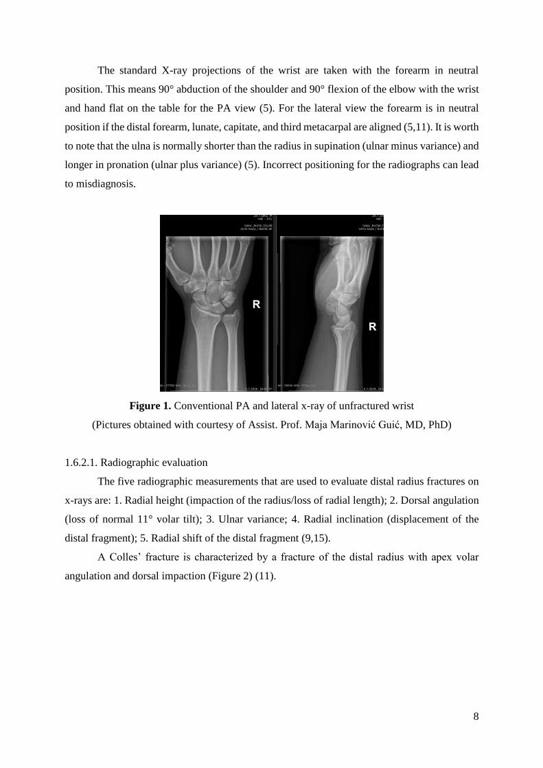
undergo radiological evaluation (4,11). The diagnosis can be made on posterior-anterior (PA)
and lateral X-rays of the wrist (Figure 1) (5). Both views need to show the distal radio-ulnar
articulation, so that the fracture line can be properly visualized and radio-ulnar misalignment
may be observed or excluded (5).
8
The standard X-ray projections of the wrist are taken with the forearm in neutral
position. This means 90° abduction of the shoulder and 90° flexion of the elbow with the wrist
and hand flat on the table for the PA view (5). For the lateral view the forearm is in neutral
position if the distal forearm, lunate, capitate, and third metacarpal are aligned (5,11). It is worth
to note that the ulna is normally shorter than the radius in supination (ulnar minus variance) and
longer in pronation (ulnar plus variance) (5). Incorrect positioning for the radiographs can lead
to misdiagnosis.
Figure 1. Conventional PA and lateral x-ray of unfractured wrist
(Pictures obtained with courtesy of Assist. Prof. Maja Marinović Guić, MD, PhD)
1.6.2.1. Radiographic evaluation
The five radiographic measurements that are used to evaluate distal radius fractures on
x-rays are: 1. Radial height (impaction of the radius/loss of radial length); 2. Dorsal angulation
(loss of normal 11° volar tilt); 3. Ulnar variance; 4. Radial inclination (displacement of the
distal fragment); 5. Radial shift of the distal fragment (9,15).
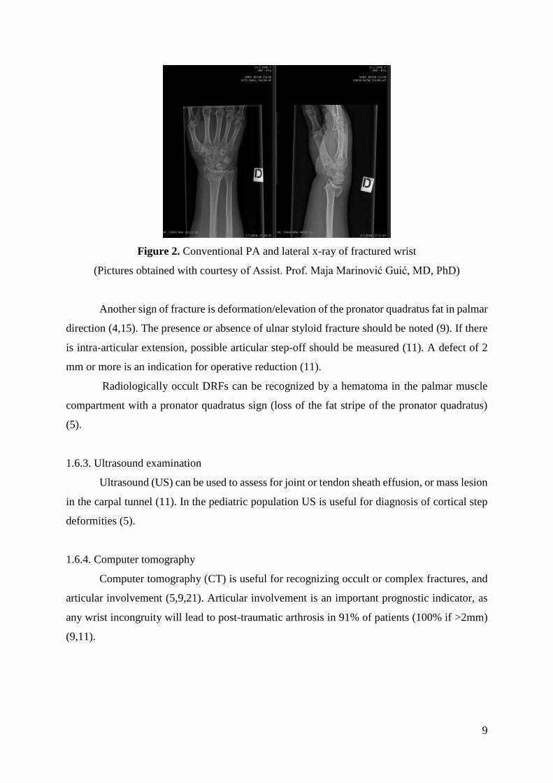
A Colles’ fracture is characterized by a fracture of the distal radius with apex volar
angulation and dorsal impaction (Figure 2) (11).
9
Figure 2. Conventional PA and lateral x-ray of fractured wrist
(Pictures obtained with courtesy of Assist. Prof. Maja Marinović Guić, MD, PhD)
Another sign of fracture is deformation/elevation of the pronator quadratus fat in palmar
direction (4,15). The presence or absence of ulnar styloid fracture should be noted (9). If there
is intra-articular extension, possible articular step-off should be measured (11). A defect of 2
mm or more is an indication for operative reduction (11).
Radiologically occult DRFs can be recognized by a hematoma in the palmar muscle
compartment with a pronator quadratus sign (loss of the fat stripe of the pronator quadratus)
(5).
1.6.3. Ultrasound examination
Ultrasound (US) can be used to assess for joint or tendon sheath effusion, or mass lesion
in the carpal tunnel (11). In the pediatric population US is useful for diagnosis of cortical step
deformities (5).
1.6.4. Computer tomography
Computer tomography (CT) is useful for recognizing occult or complex fractures, and
articular involvement (5,9,21). Articular involvement is an important prognostic indicator, as
any wrist incongruity will lead to post-traumatic arthrosis in 91% of patients (100% if >2mm)
(9,11).
10
1.6.5. Magnetic resonance imaging
The most sensitive method for detection of fractures, avascular necrosis, tenosynovitis
and mass lesions in the wrist is magnetic resonance imaging (MRI) (11). It is also useful to
detect osteochondral injuries and stress fractures (5). All mentioned injuries can be detected by
other radiological methods, however MRI is unique in its ability to assess associated carpal
ligament and TFCC injuries (5,11).
Due to cost, availability, and diagnostic value only conventional radiography is used
daily for DRF diagnosis.
1.7. Classifications
Distal radius fractures can be classified according to several classification systems that
have been developed over the years to describe fracture patterns and better guide treatment (9).
Currently, there are 15 described distal radius classification systems, describing the fractures
according to fracture patterns, comminution, and displacement (22).
The four most commonly used classification systems are Frykman (1967), Universal
(Cooney 1993), Fernández (2001), and AO (2007) (23). Neither of which is considered a golden
standard.
1.7.1. DRF eponyms
The first classifications of distal radius fractures used eponyms (Table 1). The first was
Colles, describing an extra-articular, dorsally displaced, metaphyseal fractures with radial
shortening (22). At first it was based on clinical features only (23). Today the eponym is often
used synonymously with distal radius fracture. Barton describes an intra-articular fracture with
either volar or dorsal displacement of the distal radius (22). Smith fracture (also known as
reversed Colles’) describes a volarly displaced distal radius fracture (22).
1.7.2. Frykman classification
The Frykman classification system distinguishes between four types of DRFs, focusing
on radiocarpal and/or radioulnar joint involvement, as well as the presence or absence of ulnar
styloid fracture (22).
The Universal classification system (refined by Cooney in 1993) was made in the
attempt to improve on the Frykman classification by differentiating between displaced and
nondisplaced intra-articular fractures (22). This resulted in a simple system differentiating
extra- from intra-articular fractures and displaced from non-displaced fractures (22).
11
1.7.3. Fernandez classification
Fernandez is a mechanism-based classification, aiming to provide a better assessment
of potential soft-tissue damage. It includes five types of injuries: I. bending of the metaphysis;
II. shearing fractures of the joint surface; III. compression of the joint surface; IV. avulsion or
radiocarpal fracture dislocations; V. combined fractures with high velocity injuries (22). It has
a separate group for distal radioulnar joint injuries, jointly providing information about the
fracture line, stability, and soft-tissue injury (22).
1.7.4. AO classification
The most comprehensive classification system is the AO (by the Association for the
Study of Osteosynthesis). It describes in total 27 fracture patterns of the distal radius (22). It
divides DRFs into three categories dependent on articular involvement: extra-articular, partially
articular or intra-articular (5,9). The categories are further divided into groups and subgroups
by fracture pattern, propagation, and comminution (9). Due to its extensive subdivisions it is
often omitted in favor of easier classifications like Colles’ when applicable.
All the mentioned classification systems have their strengths and weaknesses. None
have survived statistical scrutiny, proving either to be unreliable, irreproducible or simply too
complicated (22).
1.8. Associated Injuries
There are several injuries that can occur in combination with Colles’ fractures. All
associated injuries affect treatment choices. A few of these are ulnar styloid fractures, soft tissue
injuries, scaphoid fractures, and neural injury.
1.8.1. Ulnar styloid fracture
Isolated distal ulnar fractures are very rare in comparison to DRFs, hence DRFs are
often viewed as an injury with or without involvement of the ulna (5). DRFs are most often
associated with ulnar styloid fractures (4,10,15). This is clinically relevant due to its impact on
wrist stability and therefore should prompt further investigation (9).

12
1.8.2. Soft tissue injuries
The most frequently associated injuries are soft tissue injuries, especially tear of the
TFCC which is found in 39-84% of unstable distal radius fractures (9). TFCC tear should
therefore always be suspected when there is DRUJ instability (9). Soft tissue injuries may
complicate DRFs by decreasing functional outcomes, grip strength, or causing intractable pain
(9).
1.8.3. Scaphoid bone fracture
Another important injury, that can cause severe complications if missed, is fracture of
the scaphoid bone. The scaphoid bone plays a role in wrist mobility as well as carpal stability.
A missed fracture can cause post-traumatic osteoarthritis of the carpus, or avascular necrosis of
the scaphoid bone (20).
1.8.4. Neurovascular injuries
The proximity and course of the median nerve make it vulnerable to direct and indirect
injury from DRFs. Also, the radial and ulnar arteries can be affected. Hence all patients with
Colles’ fractures should undergo a thorough neurovascular examination (9).
1.9. Fracture healing basics
When the distal radius fractures, the body immediately initiates the process of healing
(24). This process is dependent on several factors, eventually resulting in the restoration of the
anatomy and function of normal bone after injury (25). The degree of fracture comminution
and displacement affect the time needed for healing and functional recovery (24,25).
Two conditions are vital for bone healing: anatomic repositioning and fracture
immobilization. Anatomic repositioning or acceptable alignment is integral for good bone
healing. Fracture displacement frequently results in malunion (25). Immobilization is necessary
to prevent secondary fracture displacement.
The mechanical stability between the fracture fragments dictates whether primary or
secondary healing will take place (31). Primary healing is the process of direct restoration of
continuity across the fracture line through intracortical remodeling (25). Secondary
(spontaneous) healing involves callus formation and endochondral ossification (25). Unstable
or insufficiently fixated fractures may lead to pseudarthrosis (25).
13
1.9.1. Primary bone healing
Primary or “direct” bone healing is characterized by the absence of callus formation
(26), and only takes place when there is minimal interfragmentary motion (24,25). It requires a
fracture gap of 0.5 millimeters or less (26). This can be accomplished by rigid internal plate
fixation. Rigid fixation diminishes motion between the fracture fragments (24). The periosteal
reaction to the bone injury is inhibited by rigid fixation, allowing osteons to directly bridge the
fracture gap and regenerate the bone (remodeling) (24,25).
Newer locking plate splints do not compress the fracture site, resulting in a more flexible
elastic fixation and callus formation (24). Plate fixation allows for earlier fracture loading and
rehabilitation than other treatment methods (24,27).
1.9.2. Secondary bone healing
Spontaneous bone healing is driven by the response of the periosteum and surrounding
soft tissues at the fracture site (25). Callus formation takes place under unstable or flexible
fixations, that allow for interfragmentary motion (24). Cast treatment, percutaneous pin
fixation, and external fixation leads to fracture repair through cartilage formation.
Secondary bone repair can be divided into 4 stages: 1. Inflammation; 2. Soft callus
(cartilage formation); 3. Hard callus (endochondral ossification); 4. Bone remodeling.
1.9.2.1. Inflammatory response
The inflammatory response begins immediately after a fracture, marked by hematoma
formation and inflammatory exudate from ruptured vessels (24). During this phase the fracture
fragments are freely moveable. The hematoma is resorbed by the end of the first week unless
excessive motion, infection, or necrosis is persisting in the surrounding soft tissues (25). This
phase persists until the formation of cartilage or bone is initiated (1-7 days) (25).
1.9.2.2. Soft callus (cartilage) formation
A few days after the injury the hematoma begins to transform into granulation tissue
(25). The formation of granulation tissue causes a slight increase in stability and mechanical
strength (24). As the maturation process advances collagen is deposited forming an internal
cartilaginous callus, the periosteum surrounding the fracture site thickens producing an external
callus (25). The soft callus formed during the first 3 weeks after injury has enough tensile
strength to prevent shortening, but protection against excessive forces is needed to prevent
shortening and angulation (24).

14
1.9.2.3. Hard callus formation
Mineralization (endochondral ossification) of the soft callus forms a hard callus that
restricts the movement of the fracture fragments (24,25). Intramembranous bone formation fills
in the fracture gap if there is sufficient vascularization and mechanical support from the callus
(25). The repair ultimately leads to firm bone union, the time required to achieve union is
dependent on fracture comminution and patient characteristics (25). The new bone has enough
strength to allow low-impact exercise (24).
1.9.2.4. Bone remodeling
Remodeling and recovery of optimal function and strength begins when the fracture has
solidly united (24,25). The average time needed for healing of Colles’ fractures is 3-5 weeks
(24).
1.10. Treatment of Colles’ fractures
The aim of DRF treatment is to restore alignment, leading to a pain free and functional
wrist (3,9,28). Proper alignment helps to prevent complications like distal radioulnar joint
instability (29). Treatment method is chosen according to several factors, including but not
limited to the mechanism of the injury, fracture pattern, instability, age and the condition of the
patient (5,28).
Restoration of anatomic alignment can be attempted by conservative or surgical means.
In a lot of cases it is not possible to restore the alignment perfectly, therefore certain
radiographic criteria for acceptable alignment are agreed upon (9,30).
1.10.1. Acceptable alignment
Radiographic criteria of acceptable alignment are: 1. Less than 2 mm radial shortening;
2. A minimum of 10° radial inclination; 3. 10° dorsal to 20° volar tilt; 4. Less than 2 mm intra-
articular step-off (9,30). It is good practice to offer surgical treatment if any of the
abovementioned parameters are not met following conservative reduction (30).
Distal radius fracture treatment should always be selected in consultation with the
patient. The treating doctor provides a professional recommendation based on an assessment of
benefits and risks of conservative vs. surgical treatment. This should be in accordance with the
patient’s wishes and needs. Limitations of each procedure should also be explained. Even when
surgery is indicated by radiological parameters, each patient is at liberty to decline surgical
intervention.
15
1.10.2. Conservative treatment
Majority of DRFs are closed fractures with or without fragment displacement (28). The
mainstay of treatment of stable fractures is closed reduction and immobilization (9). All
displaced fractures in adults need to be reduced prior to immobilization to avoid development
of long-term complications. Radiographs are taken prior to reduction, and then again after
reduction and immobilization (31). At University Hospital of Split the standard procedure is
reduction under local anesthesia, followed by plaster cast immobilization below the elbow. The
anesthesia is introduced directly into the hematoma.
The typical Colles’ fracture reduction involves placing a thumb over the fracture site as
a lever, hyperextension of the fracture fragment to distract it from the radial metaphysis,
longitudinal traction, and palmar flexion to lever the dorsally displaced fracture fragment into
position (32,33).
The American Academy of Orthopedic Surgeons recommends weekly radiographs
during the first 3 weeks following immobilization, and then again after cast removal (9). The
duration of immobilization depends on several factors, therein fracture pattern, stability and
status of the patient. The average duration of splinting or casting is 4-6 weeks (34). In the case
of secondary displacement of the fracture, reduction can be attempted again or a surgical
technique is recommended (31).
1.10.3. Surgical treatment
Surgical interventions of DRFs are often indicated when reduction is unsuccessful or
not possible, or secondary displacement takes place. Secondary displacement is more common
in elderly patients (32). Open, unstable, and comminuted fractures also warrant surgical
intervention (34). Today there are several surgical methods in use for DRF treatment, ranging
from minimally invasive to open surgery. The main surgical techniques used today include
percutaneous fixation, external fixation, ORIF (open reduction internal fixation), or in certain
cases combinations.
16
1.10.3.1. Percutaneous fixation
Percutaneous fixation is a minimally invasive technique used to fixate dorsally
displaced extra-articular DRFs (Colles’ fracture).
Kirschner wires are passed through the skin over the anatomic snuffbox or the dorsal
aspect of the distal radius and into the bone to hold the fracture fragment in the correct
anatomical position (24,35). For sufficient internal fixation cast or splint immobilization is
necessary for 4 to 6 weeks (9,24).
A successful result requires good bone quality and limited comminution (36). In patients
with more than two cortices comminuted, or older than age 55 there is a high likelihood of
fracture collapse with K-wire fixation alone (24).
Functional outcomes in patients over 60 years with low functional demands do not differ
between percutaneous fixation and cast treatment alone (37). Possible complications from k-
wire fixation includes tendon injury/rupture, pin migration, vascular injury, and pin site
infection (9).
1.10.3.2. External fixation
External fixation is not as popular as it once was, but it is still indicated as initial
treatment of patients with polytrauma, and/or open DRFs with severe soft tissue loss (9).
External fixation is a technique that maintains fracture fragment reduction by ligamentotaxis
(9).
Pins are drilled into the radius proximal to the fracture and into the index finger
metacarpal distal to the fracture and spanning the carpal joint. A mechanical frame is attached
to the pins and used to apply traction in different directions. This technique is considered a
flexible fixation, with the callus development providing the rigidity of the fixator-bone complex
(24).
Fracture fragment stability can be significantly increased, and dependence of
ligamentotaxis reduced by augmentation with percutaneous K-wires (24). An external fixator
reduces the risk of secondary displacement relative to conservative treatment, but there is a
higher risk of infection (pin sites) (9,37).
17
1.10.3.3. ORIF
Open reduction followed by internal fixation is a technique frequently used for DRF
fixation. Depending on the fracture pattern and comminution, dorsal, volar or fragment-specific
locking plates can be used. The volar locking plate (VLP) has become the mainstay in most
DRF fixations, especially Colles’ fractures (dorsally displaced) (38,39). Reduction is done
under direct visualization of the joint surface, after which fragments are splinted with internal
plates (24,32). A flexible elastic fixation is achieved by fixation of the fracture without
compression, stimulating callus formation (24).
The approach to dorsally displaced fractures (Colles’) is most commonly through an
incision over the palmar aspect of the wrist (35). The fracture line is visualized, and fragments
are released and reduced, a volar locking plate is then positioned and provisionally held in place
with K-wires until positioning is confirmed by radiography. The plate is then fixed to the bone
with angle-locking screws, normally under fluoroscopic assistance (35).
Volar locking plates (VLP) are currently popular, yet not without complications (24,32).
Iatrogenic injuries as well as intra-articular screw penetration due to fracture collapse can occur
(9,39). There is evidence that volar plating leads to better short-term functional outcomes than
dorsal plate fixation, as improved function, grip strength and decreased pain (32,37). However,
there is no conclusive evidence of volar plating leading to better long-term outcomes compared
to other fixation techniques (37).
1.11. Rehabilitation
Colles’ fractures often heal with some persisting decrease in motility despite proper
therapy (11). To decrease the incidence of chronic pain and decreased function all patients
should receive practical instructions regarding self-rehabilitation following DRF regardless of
treatment method (30).
According to the Norwegian guideline from 2015. and the Danish guideline from 2016.
on the treatment and rehabilitation of distal radius fractures, uncomplicated fractures in patients
with good function do not need to be referred to physical rehabilitation after removal of
immobilization (30,40). A satisfactory result can be achieved with a home program of exercises
(24,37).
According to Croatian common practice every diagnosed distal radius fracture should
receive physical therapy after cast removal and is referred to Physical and Rehabilitation
medicine (31).
18
The physical rehabilitation is nearly uniform among different fracture patterns, provided
it has been treated appropriately (34). The program is tailored to each patient’s needs, according
to fracture treatment and initial function (41). Therapy can be done individually with a physical
therapist, or it can be supervised in small groups. The pace of therapy can be significantly
influenced by patient factors such as age, bone density, pain tolerance, and systemic disease
(24).
1.11.1. Aims of DRF rehabilitation:
• Decrease pain, inflammation and edema in the acute phase
• Restore full joint movement and functional ability
• Maintain and increase muscle strength
• Education
1.11.2. Rehabilitation stages for distal radius fractures
Rehabilitation after distal radius fractures can be divided into three stages: early,
intermediate and late.
1.11.2.1. Early stage
The early phase is considered from the moment of injury until the 6th week post-injury
(34). In this period it is critical to limit swelling and stiffness in the hand (34). Swelling is
limited and reduced by encouraging elevation of the hand above the level of the heart and
frequent active mobilization of the upper limb (34). Stiffness can be limited by active and
passive digit ROM exercises (24,34).
This stage corresponds to the time until cast, pin or external fixators are removed (24).
Proper treatment of the fracture should provide adequate stability to allow for light use of the
hand, e.g. assist with daily activities such as dressing and feeding (34).
1.11.2.2. Intermediate stage
The intermediate phase begins once early fracture healing is established by radiography,
commonly between 6 – 8 weeks after the injury or operation (34). Casts, pins and external
fixators are removed (24). Active-assisted forearm and wrist motion is initiated in this phase to
maximize mobility (24,34).
19
1.11.2.3. Late stage
In the late phase fracture healing is well established (8 – 12 weeks after injury) (34).
After plate fixation early mobilization (starting at week 2) has not proven to provide long-term
benefits compared with delayed mobilization (starting at week 6) (37).
1.11.3. Physical therapy modalities
Physiotherapists employ active and passive interventions to achieve the aims of physical
therapy (41). These interventions can include splints, passive movements, mobilization and
strengthening exercises, cryotherapy, magnetotherapy, electrotherapy, advice and education
(42). The most commonly used modalities at University Hospital of Split are further explained.
1.11.3.1 Kinesiotherapy
Kinesiotherapy is a medical field of movement therapy with focus on exercise principles
adapted to enhance strength, endurance, and mobility of patients. It is a keystone in post-
fracture rehabilitation for the best possible functional outcome. Therapy can usually begin 4-6
weeks after injury or surgery, provided that hard callus formation is radiographically confirmed
(27).
There are three types of ROM exercises that can be used during rehabilitation: passive,
active assisted, and active (31). All exercises should initially only be conducted under
supervision by a physiotherapist, until the patient can perform them confidently and correctly
independently (31). The muscle contractions should optimally last 5-6 seconds, with a break of
10-12 seconds between every second contraction (31). Great care should be taken to not overdo
the exercises, to avoid exhaustion and pain.
The duration of treatment depends on the initial muscle strength of the patient, it can
consist of everything from one consultation to several visits over several months.
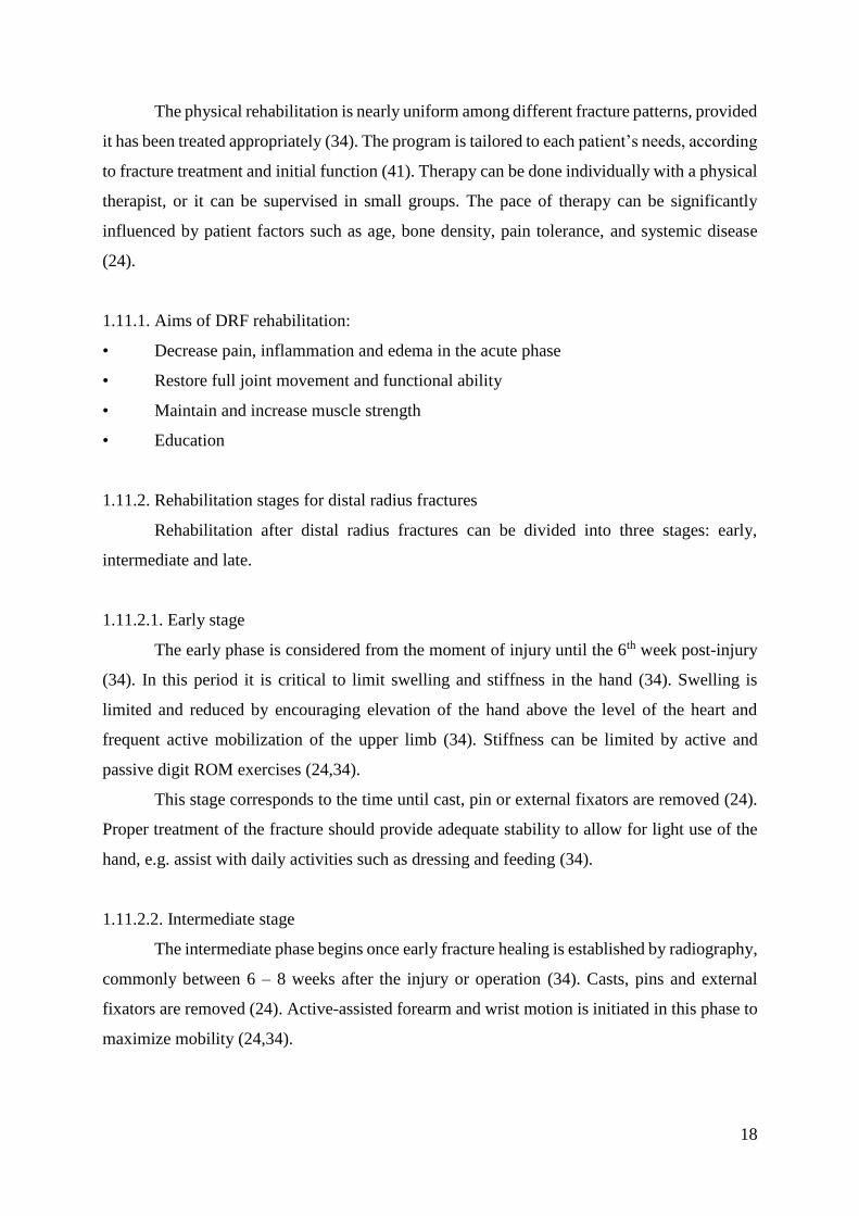
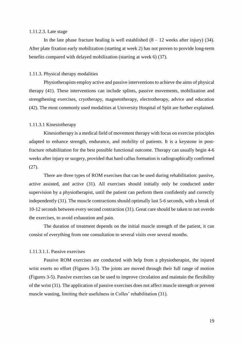
1.11.3.1.1. Passive exercises
Passive ROM exercises are conducted with help from a physiotherapist, the injured
wrist exerts no effort (Figures 3-5). The joints are moved through their full range of motion
(Figures 3-5). Passive exercises can be used to improve circulation and maintain the flexibility
of the wrist (31). The application of passive exercises does not affect muscle strength or prevent
muscle wasting, limiting their usefulness in Colles’ rehabilitation (31).
20
Figure 3. Passive exercises A. Starting position B. Passive extension of the wrist
(Pictures obtained with courtesy of Tihana Grgurević, Bacc. Physioth.)
Figure 4. Passive exercises A. Passive radial deviation B. Passive ulnar deviation
(Pictures obtained with courtesy of Tihana Grgurević, Bacc. Physioth.)
Figure 5. Passive exercises A. Passive supination B. Passive pronation
(Pictures obtained with courtesy of Tihana Grgurević, Bacc. Physioth.)
21
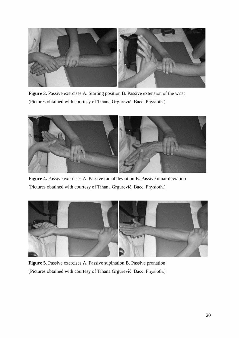
1.11.3.1.2. Active assisted exercises
Active assisted ROM exercises are performed by patients with decreased muscle
strength, with the help from a physiotherapist or the healthy arm. The patient exerts maximum
active contraction and performs as much of the motion as they can, the physiotherapist or the
patient help complete the motion (Figure 6). The only way to increase muscle strength is by
active contractions (31). These exercises are a good way to begin restoring enough strength for
active exercises.
Figure 6. Active assisted supination.
(Pictures obtained with courtesy of Tihana Grgurević, Bacc. Physioth.)
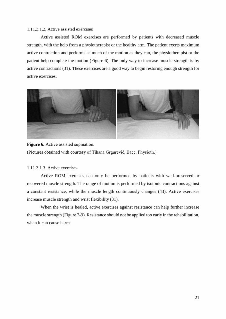
1.11.3.1.3. Active exercises
Active ROM exercises can only be performed by patients with well-preserved or
recovered muscle strength. The range of motion is performed by isotonic contractions against
a constant resistance, while the muscle length continuously changes (43). Active exercises
increase muscle strength and wrist flexibility (31).
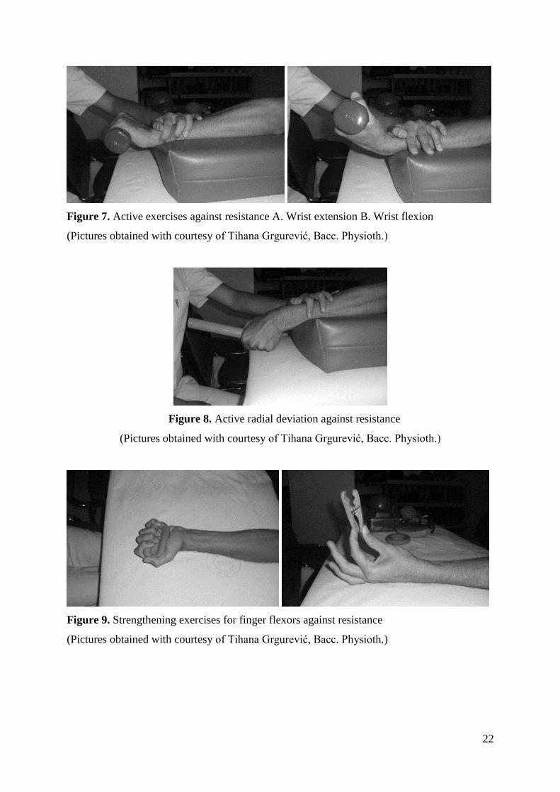
When the wrist is healed, active exercises against resistance can help further increase
the muscle strength (Figure 7-9). Resistance should not be applied too early in the rehabilitation,
when it can cause harm.
22
Figure 7. Active exercises against resistance A. Wrist extension B. Wrist flexion
(Pictures obtained with courtesy of Tihana Grgurević, Bacc. Physioth.)
Figure 8. Active radial deviation against resistance
(Pictures obtained with courtesy of Tihana Grgurević, Bacc. Physioth.)
Figure 9. Strengthening exercises for finger flexors against resistance
(Pictures obtained with courtesy of Tihana Grgurević, Bacc. Physioth.)
23
1.11.4. Cryotherapy
Cryotherapy is the use of cold in rehabilitation of injuries. Coldness affects the local
vessel and nerve endings, causing vasoconstriction, increased threshold of nociceptive
excitability, and decreased muscular spasm and spasticity (43). Cold is usually applied during
the first 24 to 48 hours following an injury to decrease inflammation and pain (43).
Vasoconstriction can control bleeding and prevent or reduce edema from trauma and
inflammation. Pain relief is accomplished by the increased threshold of nociceptive excitability
and the decreased muscular spasticity. Cold can be applied by ice packs, ice whirlpool, ice
massage, and vasocoolant sprays (43).
Ice massage is commonly used during rehabilitation. The injured area is massaged with
an ice cube using circular motions for about 5 minutes or until analgesia is accomplished. The
temperature stays above 15°C, so there is no danger of frostbite (31).
Cryotherapy can be used immediately after fracturing the distal radius, or to treat
limitations of ROM secondary to pain (43).
1.11.5. Magnetotherapy
Magnetotherapy is the use of electromagnetic fields (EMFs) mainly for the management
of pain, through effects on a cellular level (44). However, there is also clinical evidence that it
can be used to stimulate bone and wound healing, and decrease edema (44). There are several
EMF devices, the newest being the Super inductive system launched by BTL in 2017.
The Super inductive system is not yet in use in Split, but its implementation at the
University Hospital is planned. The new system is based on high intensity electromagnetic
fields, which are supposed to enhance blood circulation and subsequently callus formation and
fracture healing. The system can also be used in the treatment of known possible complications
of DRFs, such as compartment syndrome.
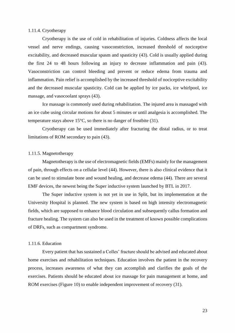
1.11.6. Education
Every patient that has sustained a Colles’ fracture should be advised and educated about
home exercises and rehabilitation techniques. Education involves the patient in the recovery
process, increases awareness of what they can accomplish and clarifies the goals of the
exercises. Patients should be educated about ice massage for pain management at home, and
ROM exercises (Figure 10) to enable independent improvement of recovery (31).
24
Figure 10. ROM exercises performed independently
(Pictures obtained with courtesy of Tihana Grgurević, Bacc. Physioth.)
1.12. Outcomes and Complications
Colles’ fracture healing is not always predictable and can result in less than perfect
recovery regardless of treatment method and rehabilitation. The overall incidence of DRF
complications varies from 6% to as much as 80% depending on the definition of complication
(45,46). Complications occur for many reasons, some are more associated with certain
treatment methods and should be prevented or recognized early and managed on time. Patient
factors affect the outcome and likelihood for complications. Patient’s gender, age,
comorbidities and previous functional status should therefore be taken into consideration when
choosing treatment method and rehabilitation (46).
1.12.1. Outcome measures
The outcomes measured after DRF can be divided into different categories, the main
ones being functional and clinical outcomes. Functional outcomes include impairment, ROM,
pain, grip strength, and patient self-assessments (47). Clinical outcomes include soft tissue
swelling and early and late complications (47). Other measures of outcomes worth mentioning
are malunion, cosmetic appearance (deformity), and patient satisfaction.
The achieved outcomes that are less than 100% normal function but do not attribute to a specific
diagnosis are not classified as complications (45).
Clinical and functional outcomes should be evaluated during every patient follow-up
after a distal radius fracture, for early detection and management. Post-immobilization there is
commonly a reduced grip strength, limited ROM, and pain (47). At the time of cast removal,
and during the course of follow-up, patients should have standard radiologic imaging preformed
to evaluate bone healing and malalignment (48).
25
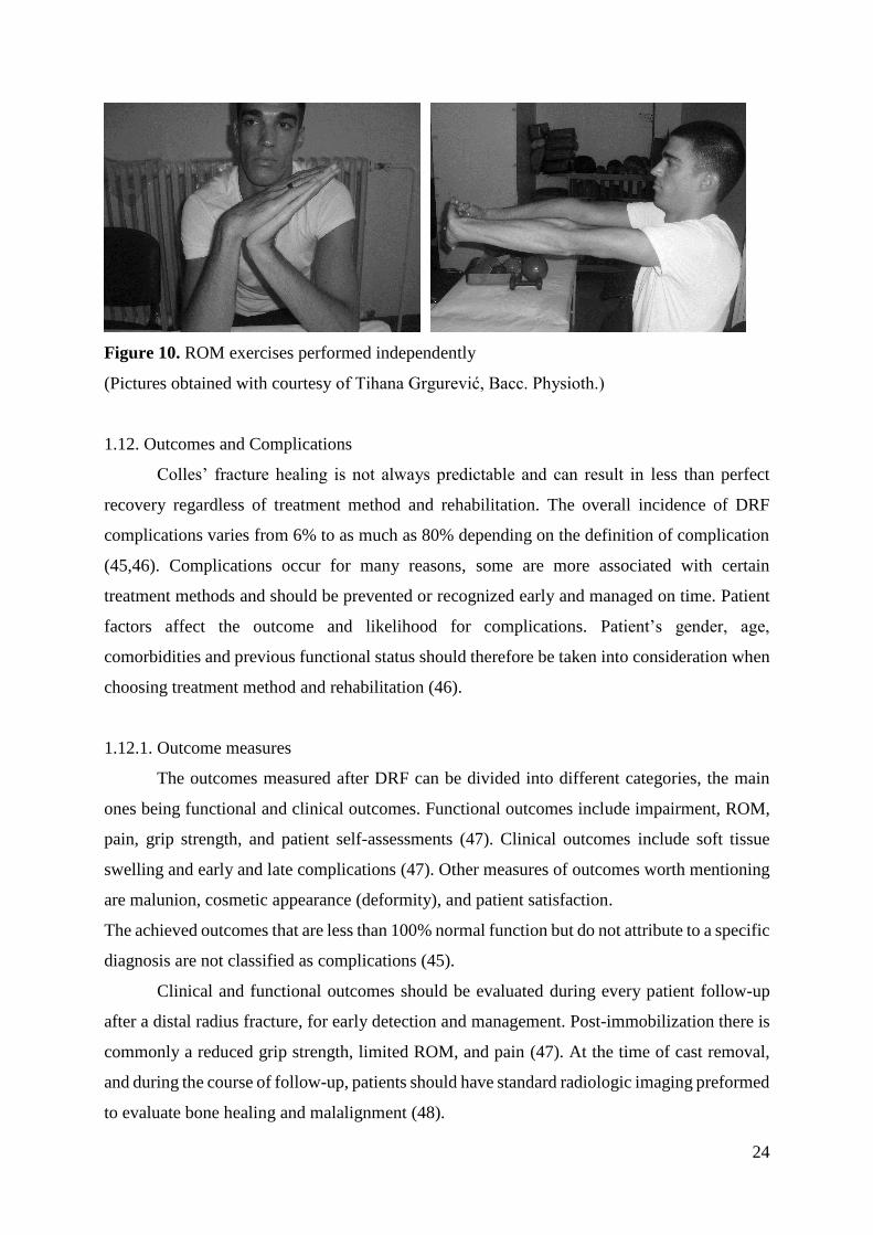
Wrist pain is assessed by a physician according to the Visual Analog Scale, while Self-
assessment by patients is commonly done by the Quick-DASH (Disability of the Arm, Shoulder
and Hand, Figure 11) and the Patient-Rated Wrist Evaluation questionnaires (48).
Figure 11. Quick-DASH questionnaire (49)
26
1.12.1.1. Manual muscle testing
Muscle testing is an important evaluation tool for objective assessment of impairments
and deficits in muscle performance (strength and power) (50). It is a practical method to
manually measure muscle strength and is used to guide rehabilitation (31). It is routinely done
at the beginning of physical therapy (initial state), during treatment (transitional state), and at
conclusion of treatment (final state) (31).
MMT can be graded 0 to 5 according to the Oxford scale: 0. No muscle contraction; 1.
Flicker of movement; 2. Full ROM with gravity counterbalanced ; 3. Full ROM against gravity;
4. Full ROM against some resistance; 5. Full ROM against strong resistance (50).
1.12.2. Complications
Colles’ fracture complications occur frequently (45). The reasons for the occurrence of
these complications vary depending on the severity, comminution and treatment method of the
fracture as well as patient characteristics like blood circulation and bone quality (46).
Complications can be divided into physician-reported and patient-reported. Patient-
reported complications can be associated with surgical fracture treatment, resulting in non-
diagnostic complaints of loose pins that require second surgery or pins cutting through skin
(45). The most common complaints after conservative treatment are deformity, pain, or stiffness
(45). The most common physician-reported complication is median nerve pathology (45).
Deformity can be a result of displacement that is inadequately repositioned or
secondarily displaced. The resulting deformity will be characterized by dorsal angulation,
limited supination, and a weak grip (8).
A common complication of long-term immobilization is muscle atrophy. Any prolonged
inactivity leads to some degree of muscle and soft tissue atrophy. Rehabilitation, especially
active exercises, is paramount for prevention and reversal of atrophy.
1.12.2.1. Disrupted bone healing
Healing is classified as delayed if the healing time exceeds twice the expected time (4
to 6 months) (26). The primary causes of delayed healing are inadequate immobilization,
impaired fracture perfusion, and infection (26). Delayed fracture healing can lead to
pseudarthrosis.
Pseudarthrosis refers to non-union at the fracture site after 6 to 8 weeks (26). Non-union
can be caused by inadequate immobilization, soft tissues interposed in the fracture gap,
extensive loss of bone, inadequate blood supply, and infection (26). There are three types of
27
pseudarthrosis: hypertrophic form, atrophic form, and defect with pseudarthrosis (26). The
treatment depends on the cause and type of non-union, in most cases treatment is a long-lasting
process.
The most common complication after a distal radius fracture is malunion (46). Malunion
occurs when a fracture heals with improper alignment, articular incongruity, loss of length, or
a combination of these factors (46). A common cause of malunion is conservative treatment
(46). Surgical correction should be considered for all patients with confirmed malunions.
1.12.2.2. Complex regional pain syndrome
Complex regional pain syndrome (CRPS) is an autonomic dysfunction that can occur
after both conservative and surgical treatments of Colles’ fractures (46). There is no definitive
cause nor treatment for the syndrome, however there are some associated factors. For
conservatively treated fractures there was found a correlation between CRPS risk and pressure
under the cast (46). For post-surgical fractures excessive distraction can increase the risk of
CRPS development (46).
There are two types of CRPS, type 1 was formerly known as reflex sympathetic
dystrophy (RSD), and type 2 (46). Type 1 CRPS is characterized by chronic pain without
identifiable nerve injury, while type 2 CRPS is characterized by nerve involvement (46).
If diagnosed early and treated promptly the recovery rate of CRPS is good. However,
diagnosis can be difficult due to the lack of standardized diagnostic criteria (46). A multimodal
approach with combined psychiatric therapy, physical therapy, and pain management has
proven most effective (46).

2. OBJECTIVES
29
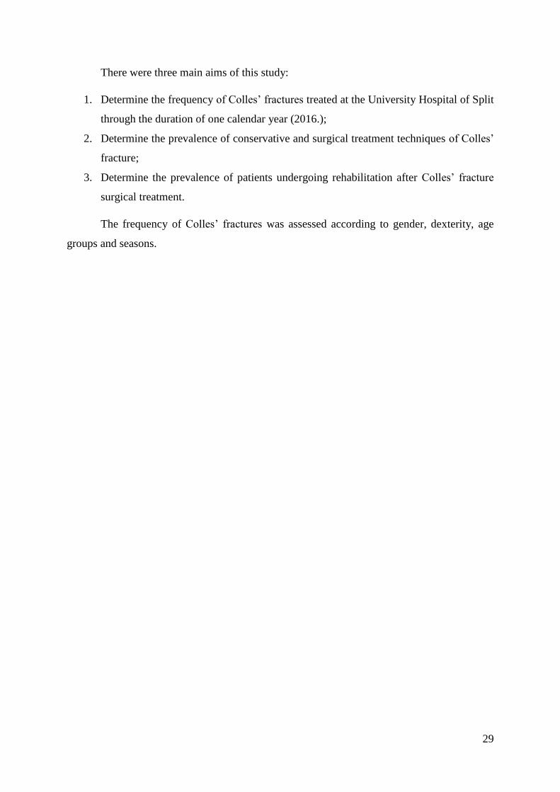
There were three main aims of this study:
1. Determine the frequency of Colles’ fractures treated at the University Hospital of Split
through the duration of one calendar year (2016.);
2. Determine the prevalence of conservative and surgical treatment techniques of Colles’
fracture;
3. Determine the prevalence of patients undergoing rehabilitation after Colles’ fracture
surgical treatment.
The frequency of Colles’ fractures was assessed according to gender, dexterity, age
groups and seasons.

3. SUBJECTS AND METHODS
31
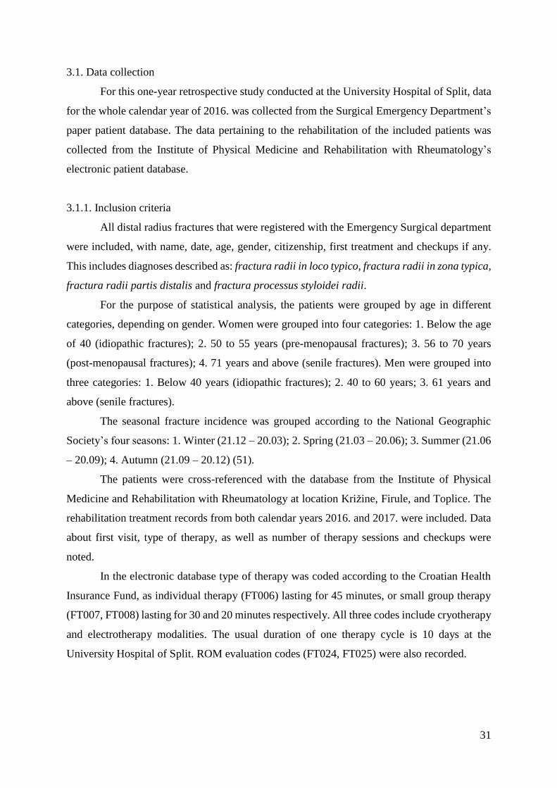
3.1. Data collection
For this one-year retrospective study conducted at the University Hospital of Split, data
for the whole calendar year of 2016. was collected from the Surgical Emergency Department’s
paper patient database. The data pertaining to the rehabilitation of the included patients was
collected from the Institute of Physical Medicine and Rehabilitation with Rheumatology’s
electronic patient database.
3.1.1. Inclusion criteria
All distal radius fractures that were registered with the Emergency Surgical department
were included, with name, date, age, gender, citizenship, first treatment and checkups if any.
This includes diagnoses described as: fractura radii in loco typico, fractura radii in zona typica,
fractura radii partis distalis and fractura processus styloidei radii.
For the purpose of statistical analysis, the patients were grouped by age in different
categories, depending on gender. Women were grouped into four categories: 1. Below the age
of 40 (idiopathic fractures); 2. 50 to 55 years (pre-menopausal fractures); 3. 56 to 70 years
(post-menopausal fractures); 4. 71 years and above (senile fractures). Men were grouped into
three categories: 1. Below 40 years (idiopathic fractures); 2. 40 to 60 years; 3. 61 years and
above (senile fractures).
The seasonal fracture incidence was grouped according to the National Geographic
Society’s four seasons: 1. Winter (21.12 – 20.03); 2. Spring (21.03 – 20.06); 3. Summer (21.06
– 20.09); 4. Autumn (21.09 – 20.12) (51).
The patients were cross-referenced with the database from the Institute of Physical
Medicine and Rehabilitation with Rheumatology at location Križine, Firule, and Toplice. The
rehabilitation treatment records from both calendar years 2016. and 2017. were included. Data
about first visit, type of therapy, as well as number of therapy sessions and checkups were
noted.
In the electronic database type of therapy was coded according to the Croatian Health
Insurance Fund, as individual therapy (FT006) lasting for 45 minutes, or small group therapy
(FT007, FT008) lasting for 30 and 20 minutes respectively. All three codes include cryotherapy
and electrotherapy modalities. The usual duration of one therapy cycle is 10 days at the
University Hospital of Split. ROM evaluation codes (FT024, FT025) were also recorded.

32
3.1.2. Exclusion criteria
Patient entries not specifying dexterity of the Colles’ fracture (right/left/bilateral) were
disregarded. Patients with suspected fracture, without later confirmed fracture, were excluded.
Foreign citizens were also disregarded, given the difficulty to ascertain their follow-up
treatment in their respective countries. Also, those who attained their fractures before 01.01.16
but returned for checkups in 2016. were excluded. Patients under age 18 were not included in
the study.
3.3. Statistical analysis
Statistical analyses were performed using the statistical software MedCalc for
Windows, version 18.5 (MedCalc Software, Ostend, Belgium). Chi-square tests were
performed with P value <0.05 as statistically significant.

4. RESULTS
34
4.1. Surgical Emergency Data
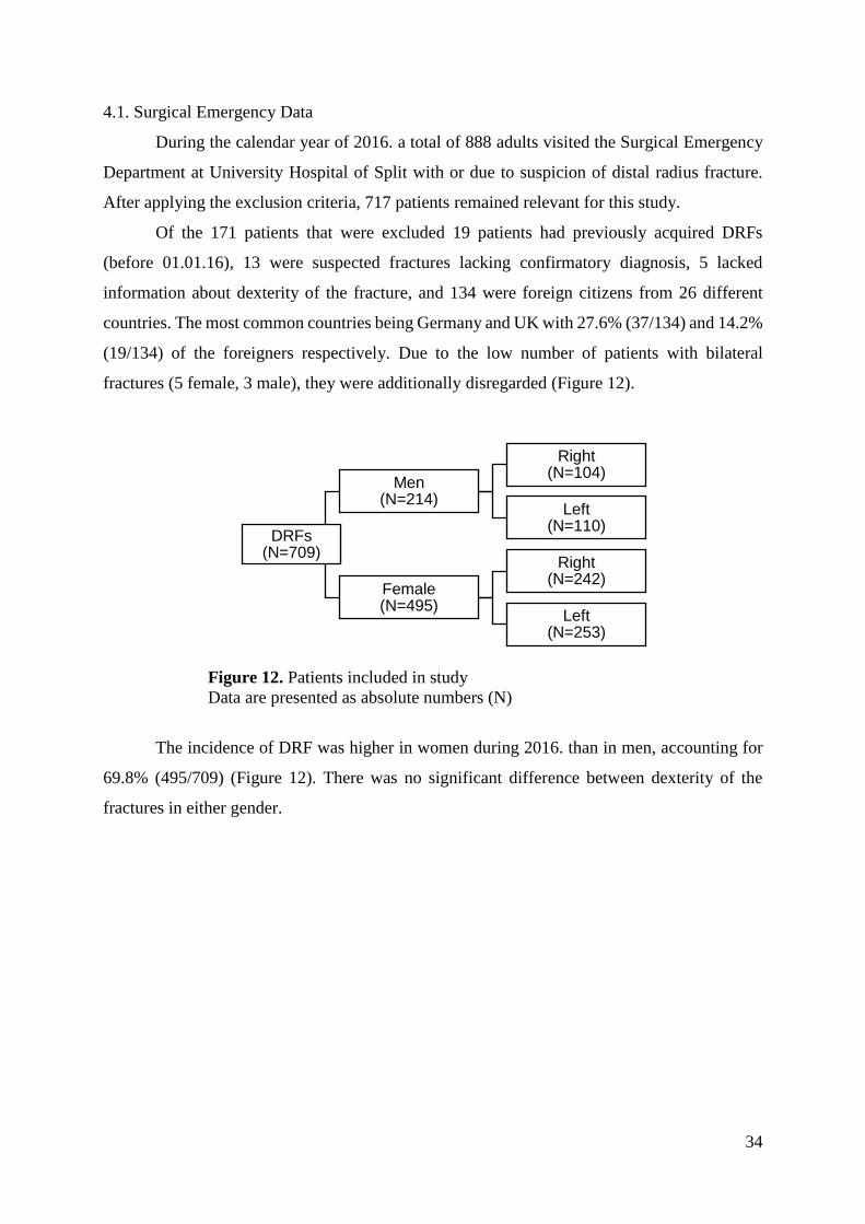
During the calendar year of 2016. a total of 888 adults visited the Surgical Emergency
Department at University Hospital of Split with or due to suspicion of distal radius fracture.
After applying the exclusion criteria, 717 patients remained relevant for this study.
Of the 171 patients that were excluded 19 patients had previously acquired DRFs
(before 01.01.16), 13 were suspected fractures lacking confirmatory diagnosis, 5 lacked
information about dexterity of the fracture, and 134 were foreign citizens from 26 different
countries. The most common countries being Germany and UK with 27.6% (37/134) and 14.2%
(19/134) of the foreigners respectively. Due to the low number of patients with bilateral
fractures (5 female, 3 male), they were additionally disregarded (Figure 12).
Figure 12. Patients included in study
Data are presented as absolute numbers (N)
The incidence of DRF was higher in women during 2016. than in men, accounting for
69.8% (495/709) (Figure 12). There was no significant difference between dexterity of the
fractures in either gender.
DRFs (N=709)
Men(N=214)
Right(N=104)
Left(N=110)
Female(N=495)
Right(N=242)
Left(N=253)
35
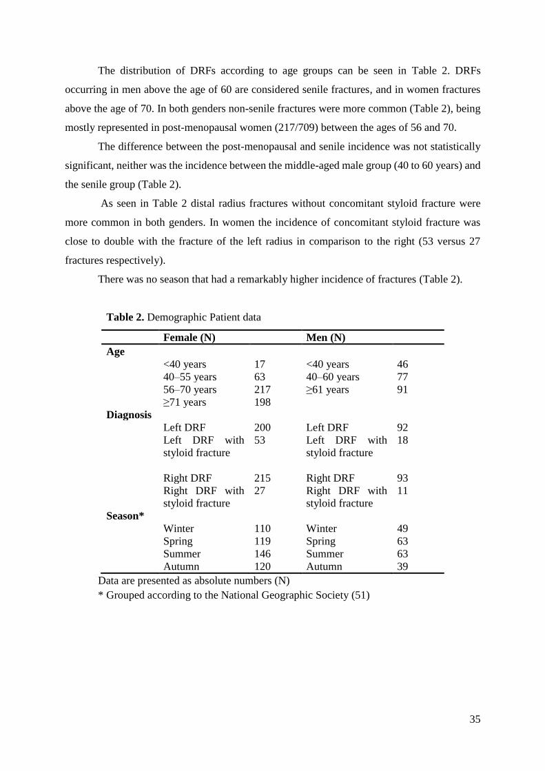
The distribution of DRFs according to age groups can be seen in Table 2. DRFs
occurring in men above the age of 60 are considered senile fractures, and in women fractures
above the age of 70. In both genders non-senile fractures were more common (Table 2), being
mostly represented in post-menopausal women (217/709) between the ages of 56 and 70.
The difference between the post-menopausal and senile incidence was not statistically
significant, neither was the incidence between the middle-aged male group (40 to 60 years) and
the senile group (Table 2).
As seen in Table 2 distal radius fractures without concomitant styloid fracture were
more common in both genders. In women the incidence of concomitant styloid fracture was
close to double with the fracture of the left radius in comparison to the right (53 versus 27
fractures respectively).
There was no season that had a remarkably higher incidence of fractures (Table 2).
Table 2. Demographic Patient data
Female (N) Men (N)
Age
<40 years 17 <40 years 46
40–55 years 63 40–60 years 77
56–70 years 217 ≥61 years 91
≥71 years 198
Diagnosis
Left DRF 200 Left DRF 92
Left DRF with
styloid fracture
53 Left DRF with
styloid fracture
18
Right DRF 215 Right DRF 93
Right DRF with
styloid fracture
27 Right DRF with
styloid fracture
11
Season*
Winter 110 Winter 49
Spring 119 Spring 63
Summer 146 Summer 63
Autumn 120 Autumn 39
Data are presented as absolute numbers (N)
* Grouped according to the National Geographic Society (51)
36
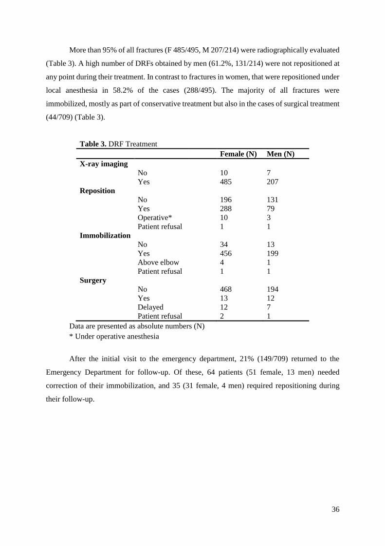
More than 95% of all fractures (F 485/495, M 207/214) were radiographically evaluated
(Table 3). A high number of DRFs obtained by men (61.2%, 131/214) were not repositioned at
any point during their treatment. In contrast to fractures in women, that were repositioned under
local anesthesia in 58.2% of the cases (288/495). The majority of all fractures were
immobilized, mostly as part of conservative treatment but also in the cases of surgical treatment
(44/709) (Table 3).
Table 3. DRF Treatment
Female (N) Men (N)
X-ray imaging
No 10 7
Yes 485 207
Reposition
No 196 131
Yes 288 79
Operative* 10 3
Patient refusal 1 1
Immobilization
No 34 13
Yes 456 199
Above elbow 4 1
Patient refusal 1 1
Surgery
No 468 194
Yes 13 12
Delayed 12 7
Patient refusal 2 1
Data are presented as absolute numbers (N)
* Under operative anesthesia
After the initial visit to the emergency department, 21% (149/709) returned to the
Emergency Department for follow-up. Of these, 64 patients (51 female, 13 men) needed
correction of their immobilization, and 35 (31 female, 4 men) required repositioning during
their follow-up.

37
4.2. Rehabilitation Data
All patients included in the study were cross-referenced with the records from the
Institute of Physical Medicine and Rehabilitation with Rheumatology at University Hospital of
Split from 2016. and 2017. In total 218 (30.7%) patients that attained a Colles’ fracture in 2016.
went to a primary visit after cast removal to one of the locations of the Institute of Physical
Medicine and Rehabilitation with Rheumatology (Figure 13).
Figure 13. Number of patients that made a visit to the Institute of Physical Medicine
and Rehabilitation with Rheumatology at University Hospital of Split
Data are presented as absolute numbers (N)
Initially 26.2% (56/214) of men that suffered DRFs went to a physical rehabilitation
consultation, compared to 32.7% (162/495) of women (Figure 13).
The records showed that 172 patients (124 female, 48 men) began with an individual
exercise protocol with a physical therapist. 6 women received only treatment in small groups.
There was missing information about the protocol applied to 4 patients. The remaining 36
patients either only went to a primary check-up without initiating any rehabilitation protocol,
or it was not recorded.
Of the patients that received physical therapy, majority received one to three therapy
cycles (170/182) during the duration of their rehabilitation. The number of patients that
underwent prior- and post-therapy ROM evaluation was 41.
158
333
49156
162
218
Men (N) Female (N) Total (N)
No PMR PMR

5. DISCUSSION
39
According to data from the Croatian Institute of Public Health from 2014., injuries are
the third most common cause of mortality (52). Injuries also rank as 6th cause of hospitalization
in Croatia (52). Distal radius fractures are one of the most common fractures, making it a
significant public health problem (1,19). According to a study performed in 2014., the incidence
of DRFs for the population of Split is 20.23 per 10000 people–year (53).
The treatment and rehabilitation of DRFs continues to present many challenges, and the
incidence throughout the world continues to increase as the population with increased life
expectancy grows (32,48,54).
Bone composition and its mechanical properties vary as a function of age (55). The
process of bone loss and gradual decrease of bone density begins in both genders around the
age of 40, slowly increasing the risk of fractures over time (19). The relative weaker bones in
women leads to a peak of DRFs in post-menopause, associated with the diminished estrogen
levels (11). This expected peak can be seen in the post-menopausal female patients included in
our study (Table 2).
The most common fracture mechanisms in non-senile adults are high energy injuries,
e.g. motor vehicle accidents, sports activities and falls from greater heights (2,8). Except for the
post-menopausal population, the incidence in non-senile women is significantly lower than in
the senile/elderly population (1,56). The number of DRFs was higher in men under the age of
60 (non-senile), than in the senile group (123 under age 60, 91 aged 60 or above) in our study
(Table 2). The male fracture distribution may be connected to the number of motor vehicle
accidents, or the surge in sports related activities (1).
Literature associates DRFs in the elderly with osteoporosis and osteopenia (1,32). A
distal radius fracture in an active elderly can be the first symptom of underlying osteoporosis
(1,54). Meta-analyses prove the connection between sustaining a DRFs (at the age of 45 or
older) and future fractures (54). A DRF doubles the risk of having a new fracture for women
and triples it for men (37).
The most common mechanism of fracture is fall on an outstretched hand in the elderly,
especially women with decreased bone density (1). Studies have shown that as much as 85% of
elderly women who suffer a Colles’ fracture may have low bone mineral density (BMD) (37).
Lower BMD is associated with more severe, intra-articular fractures (1). Despite the proven
association between DRF and osteoporosis, osteoporotic screening is not a part of clinical DRF
treatment practice (54).
40
Some known risk factors for osteoporosis are: calcium deficiency, vitamin D deficiency,
smoking, female gender, and alcohol consumption (8,19). Vitamin D deficiency plays a unique
role in fracture mechanism of the elderly. A minimum concentration of 50 nmol/L of activated
vitamin D (25(OH)D) is necessary to prevent bone demineralization (57). Despite the
Mediterranean climate, it has been established that Croatia has a high prevalence of vitamin D
deficiency in post-menopausal women (57). Close to three–fourths of post-menopausal women
in Croatia have vitamin D concentrations <50 nmol/L (57). Low vitamin D may also lead to
atrophy of type II muscle fibers (fast acting fibers), which are responsible for the “reach-out”
to break a fall reflex (57). Decreased “reach-out” reflex may also be caused by dementia (1).
Regardless of cause, decreased reflexes increase the risk of falling onto the side causing
proximal humerus or hip fracture (1,32). Although DRFs are rarely lethal, hip fractures have a
significant mortality rate (58).
A simple intervention that can prevent the development of osteoporosis, or prevent the
occurrence of DRF or repeat fractures, is vitamin D supplementation (57). The high number of
DRFs amongst post-menopausal women and middle-aged men in Split (Table 2) may perhaps
be attributed to a decreased BMD from vitamin D deficiency. DRFs can also serve as a
screening tool to identify patients with increased risk for subsequent DRFs or more debilitating
fractures (32). There are grounds to recommend bone density measurement and osteoporosis
screening of all patients above 50 that sustained a DRF (32,37).
The University Hospital of Split could benefit of evaluating every DRF as a possible
first symptom of osteopenia and monitor vitamin D levels more frequently in both women and
men.
A very low number of patient refusals of immobilization and surgical treatment were
registered in the Surgical Emergency database (Table 3). The unlikely low number could be
contributed to incomplete documentation, or perhaps good education and information of
patients by the physicians. It is unclear whether the high number of fractures not operated
(662/709, 93.4%) was because surgery was not indicated or could include patient refusals as
well. Regardless of reason, majority of Colles’ fractures during 2016. were conservatively
treated at University Hospital of Split (Table 3).
41
Despite the common impression that the frequency of DRFs is higher during the winter season,
in our study no significant seasonal differences were shown (32). Perhaps the lack of increase
during winter can be attributed to the Mediterranean climate in Split, with mild winter periods
characterized by rare snow and ice incidence. If we include foreign citizens, the number of
fractures in Summer would be increased by 82, making it the most dominant season (291/843).
However, it is not clear if this is a true increase or dependent on the increase of residents in
Split during the summer period.
There are numerous medical offices in and around Split that offer physical therapy,
therefore a greater number of patients may have received physical therapy post-fracture than
the 30.7% (218/709) that went to the Institute of Physical Medicine and Rehabilitation with
Rheumatology at University Hospital of Split.
Although it is common practice to recommend rehabilitation after a distal radius
fracture, no long-term differences have been proven between patients with uncomplicated
fractures who perform home exercises and those who receive rehabilitation by a physical
therapist (29,37). Immediate rehabilitation after cast removal can improve wrist ROM in the
short term, but it does not lead to better long-term results (37,42). However, physiotherapy may
cause a significant increase in wrist extension and grip strength making everyday tasks easier
to perform earlier (41).
At the University Hospital of Split the physical therapy session duration depends on
whether it’s an individual or small group treatment. An individual therapy session (one patient,
one physical therapist) lasts 45 min, while small group therapy session lasts 20 to 30 min. The
rehabilitation is initiated after cast removal and the average duration of one cycle of therapy
consists of 10 sessions.
The continuous increase of the DRF incidence causes an increase in the health care
spending on fracture treatment and rehabilitation (56). Cost-effective choices of treatment and
rehabilitation is therefore becoming more and more important (56). A possible area of cost
reduction in Split could be reducing the number of patients receiving individual rehabilitation
therapy, seen as there is a lack of scientifically proven benefit of individual therapy over small
group therapy or home exercises (37). Over half of the patients that received physical therapy
at University Hospital of Split received 2 or 3 cycles of individual physical therapy (95/180,
52.2% of patients). The high number of individual therapy sessions may contribute to an
unnecessary high cost of DRF rehabilitation. Only 41 patients underwent ROM evaluation
during their rehabilitation, an increase of evaluations could lower the number of unnecessary
treatment cycles.
42
The significant costs resulting from DRFs make it important to apply preventive
measures to individuals who are at high risk of suffering DRF as well as optimizing treatment
protocols (1).
Some selection bias may be present in the study due to its retrospective nature. Data
regarding follow-up is limited due to the lack of accessibility to the patient database of the
Department of Orthopedics and Traumatology.
The place of residence as well as number of private physical therapists in and around
Split may affect the number of patient rehabilitation records available from the University
Hospital of Split. The study is also sensitive to incomplete documentation or human error, given
the lack of digital record keeping.

6. CONCLUSIONS

44
1. In Split, 709 (843 including foreign citizens) patients visited the University Hospital
of Split during 2016. with Colles’ fractures, being mostly women (495/709). The
greatest peak was in post-menopausal women (217/495). In men the majority that
suffered DRFs were above 40 years old, without a great difference between the
middle-aged (40–60 years) and senile (60 years and older) group.
2. Majority of patients were treated conservatively, with repositioning and/or
immobilization. Of the total number of patients 44 underwent surgical treatment,
either immediately or delayed. There was no significant difference in the frequency
according to dexterity or season.
3. Post-fracture rehabilitation plays a big role in the recovery of full function in most
cases of Colles’ fractures. 30.7% (218/709) of the patients from 2016. received
physical therapy at the University Hospital of Split. These numbers may be strongly
affected by the number of physical therapy health facilities in and around Split. 95
of the patients that went to rehabilitation received 2 or 3 physical therapy cycles
(95/180, 52.2%).
4. The lack of treatment and rehabilitation guidelines in Croatia may be causing
unnecessary costs that could be limited by treatment optimization by creating
clinical guidelines.

7. REFERENCES
46
1. Nellans KW, Kowalski E, Chung KC. The epidemiology of distal radius fractures. Hand
Clin. 2012;28(2):113-25.
2. Porrino JA, Jr., Maloney E, Scherer K, Mulcahy H, Ha AS, Allan C. Fracture of the
distal radius: epidemiology and premanagement radiographic characterization. Am J
Roentgenol. 2014;203(3):551-9.
3. Medscape.com [Internet]. New York:; Distal Radius Fractures [updated 2016 May 6;
cited 2017 October 10]. Available from: https://emedicine.medscape.com/article/1245884.
4. Wilson AJ. Radiology of Hand and Wrist Injuries. In: Hodler J, Von Schulthess GK,
Zollikofer CL, editors. Musculoskeletal Diseases, Diagnostic Imaging and Interventional
Techniques. 37th ed: Springer; 2005. p. 13-55.
5. Bohndorf K, Imhof H, Pope TLJ. Distal Forearm Fractures. In: Musculoskeletal
Imaging: A concise Multimodality Approach.: Thieme; p. 84-8.
6. Rueger JM, Hartel MJ, Ruecker AH, Hoffmann M. [Fractures of the distal radius].
Unfallchirurg. 2014;117(11):1025-34.
7. Meena S, Sharma P, Sambharia AK, Dawar A. Fractures of distal radius: an overview.
J Family Med Prim Care. 2014;3(4):325-32.
8. OrthopaedicsOne.com [Internet]. Toronto: OrthopaedicsOne - The Orthopaedic
Knowledge Network; Distal radius (Colles) fractures [updated 2014 December 14; cited 2017
October 22] Available from: http://www.orthopaedicsone.com/x/XYGTB.
9. Mauck BM, Swigler CW. Evidence-Based Review of Distal Radius Fractures. Orthop
Clin North Am. 2018;49(2):211-22.
10. Moore KL, Agur AMR, Dalley AF. Essential Clinical Anatomy. 4th ed.: Lippincott
Williams and Wikins; 2010.
11. Manaster BJ, May DA, Disler DG. Wrist. In: Musculoskeletal Imaging, The Requisities
in Radiology. Philadelphia: Mosby Elsevier; 2007. p. 134-58.
12. Safadi FF, Khurana JS. Bone Structure and Function. In: Bonakdarpour A, Reinus WR,
Khurana JS, editors. Diagnostic Imaging of Musculoskeletal Diseases: A systematic Approach.
New York: Springer; 2010. p. 1-14.
13. BoneAndSpine.com [Internet]. New Delhi:; Radius Bone Anatomy [updated; cited 2018
February 8]. Available from: http://boneandspine.com/anatomy-of-radius-bone/.
14. Medscape.com [Internet]. New York:; Wrist Joint Anatomy [updated 2013 June 11;
cited 2018 February 2]. Available from: https://emedicine.medscape.com/article/1899456-
overview.
47
15. Murray JRD, Holmes EJ, Misra RR. Radius fracture - distal radial fractures. In: A–Z of
Musculoskeletal and Trauma Radiology. Cambridge: Cambridge University Press; 2008. p.
266-71.
16. Vanderhave KL. Distal Radius Fracture. In: Doherty GM, editor. Surgery Current
Diagnosis and Treatment.: McGraw-Hill; 2015. p. 1083-5.
17. Johnson KJ, Bache E. Imaging in Pediatric Skeletal Trauma In: Leuven BAL, Göttingen
KM, Heidelberg SK, editors.: Springer; 2008. p. 122.
18. Johnson KJ, Bache E. Imaging in Pediatric Skeletal Trauma. In: Leuven BAL,
Göttingen KM, Heidelberg SK, editors.: Springer; 2008. p. 295-9.
19. Bonakdarpour A. Systematic Approach to Metabolic Diseases of Bone. In:
Bonakdarpour A, Reinus WR, Khurana JS, editors. Diagnostic Imaging of Musculoskeletal
Diseases: A systematic Approach. New York: Springer; 2010. p. 15-60.
20. Medscape.com [Internet]. New York:; Wrist Fracture in Emergency Medicine
[updated 2018 April 4; cited 2018 June 22]. Available from:
https://emedicine.medscape.com/article/828746-overview.
21. MerckManuals.com [Internet]. New Jersey: Merck & Co., Inc.; Distal Radius Fractures
(Wrist Fractures; Colles Fractures; Smith Fractures) [updated 2017 August; cited 2017
October 19]. Available from: https://www.merckmanuals.com/professional/injuries-
poisoning/fractures/distal-radius-fractures.
22. Shehovych A, Salar O, Meyer C, Ford D. Adult distal radius fractures classification
systems: essential clinical knowledge or abstract memory testing? Ann R Coll Surg Engl.
2016;98:525-31.
23. Kleinlugtenbelt YV, Groen SR, Ham SJ, Kloen P, Haverlag R, Simons MP, et al.
Classification systems for distal radius fractures: Does the reliability improve using additional
computed tomography? Acta Orthopaedica. 2017;88(6):681-7.
24. Herman M, Hopkins H, Stursky DJ. Fractures and Injuries of the Distal Radius and
Carpus. 1st ed.: Elsevier; 2009.
25. Bigham-Sadegh A, Oryan A. Basic concepts regarding fracture healing and the current
options and future directions in managing bone fractures. Int Wound J. 2015;12(3):238-47.
26. Bohndorf K, Imhof H, Pope TLJ. Fracture Healing. In: Musculoskeletal Imaging: A
concise Multimodality Approach.: Thieme; p. 24-6.
27. Freeland AE, Luber KT. Biomechanics and biology of plate fixation of distal radius
fractures. Hand Clin. 2005;21(3):329-39.
48
28. Handoll HHG, Madhok R. Conservative interventions for treating distal radial fractures
in adults. CDSR. 2003. doi: 10.1002/14651858.CD000314.
29. Eltorai AEM, Sobel AD, Thomas NP Jr., Daniels AH, Born CT. Current Trends in the
Management of Distal Radius Fractures. Orthopedics. 2017;40(3):145-52.
30. Sundhetssyrelsen.dk [Internet]. Copenhagen: Danish Health Authority; National
clinical guideline on the treatment of distal radial fractures [updated 2016 January 18; cited
2017 December 17]. Avaliable from: https://www.sst.dk/en/publications/2014/ncg-distal-
radius-fractures.
31. Grgurevic T. Fizioterapijski postupci kod prijeloma palcane kosti na tipicnom mjestu
[dissertation]. Split: Sveučilište u Splitu; 2013.
32. Levin LS, Rozell JC, Pulos N. Distal Radius Fractures in the Elderly. J Am Acad Orthop
Surg. 2017;25(3):179-87.
33. Manaster BJ, May DA, Disler DG. Introduction to Imaging of Musculoskeletal Injury:
Bones. In: Musculoskeletal Imaging, The Requisities in Radiology. Philadelphia: Mosby
Elsevier; 2007. p. 3-25.
34. Ring D, Burchill G, Callamaro DR, Jupiter JB. Fracture of the Distal Radius. In:
Brotzman BS, Wilk KE, editors. Clinical Orthopaedic Rehabilitation. Philadelphia: Mosby,
Inc.; 2003. p. 55-67.
35. Costa ML, Achten J, Parsons NR, Rangan A, Griffin D, Tubeuf S, et al. Percutaneous
fixation with Kirschner wires versus volar locking plate fixation in adults with dorsally
displaced fracture of distal radius: randomised controlled trial. Bmj. 2014.
doi: 10.1136/bmj.g4807.
36. Chaudhry H, Kleinlugtenbelt YV, Mundi R, Ristevski B, Goslings JC, Bhandari M. Are
Volar Locking Plates Superior to Percutaneous K-wires for Distal Radius Fractures? A Meta-
analysis. Clin Orthop Relat Res. 2015;473(9):3017-27.
37. Loisel F, Bourgeois M, Rondot T, Nallet J, Boeckstins M, Rochet S, et al. Treatment
goals for distal radius fractures in 2018: recommendations and practical advice. Eur J Orthop
Surg Traumatol. 2018. doi: 10.1007/s00590-018-2196-9.
38. Zong SL, Kan SL, Su LX, Wang B. Meta-analysis for dorsally displaced distal radius
fracture fixation: volar locking plate versus percutaneous Kirschner wires. J Orthop Surg Res.
2015;10:108.
39. Rhee PC, Medoff RJ, Shin AY. Complex Distal Radius Fractures: An Anatomic
Algorithm for Surgical Management. J Am Acad Orthop Surg. 2017;25(2):77-88.
49
40. Omicsonline.org [Internet]. Norway; Norwegian Orthopaedic Association - The
Norwegian Medical Association; Treatment of distal radius fractures in adults.
[updated 2015 July 27; cited 2018 July 5]. Avaliable from:
https://files.magicapp.org/guideline/ac10868f.../published_guideline_551-2_6.pdf.
41. Bruder AM, Taylor NF, Dodd KJ, Shields N. Physiotherapy intervention practice
patterns used in rehabilitation after distal radial fracture. J Physioth. 2013;99(3):233-40.
42. Bruder A, Taylor NF, Dodd KJ, Shields N. Exercise reduces impairment and improves
activity in people after some upper limb fractures: a systematic review. J Physioth.
2011;57(2):71-82.
43. Williams A. Modalities Used in Rehabilitation. In: Brotzman BS, Wilk KE, editors.
Clinical Orthopaedic Rehabilitation. Philadelphia: Mosby, Inc.; 2003. p. 611-8.
44. Markov M. XXIst century magnetotherapy. Electromagn Biol Med. 2015;34(3):190-6.
45. McKay SD, MacDermid JC, Roth JH, Richards RS. Assessment of complications of
distal radius fractures and development of a complication checklist. J Hand Surg Am.
2001;26(5):916-22.
46. Chung KC, Mathews AL. Management of Complications of Distal Radius Fractures.
Hand clinics. 2015;31(2):205-15.
47. Handoll HHG, Elliott J. Rehabilitation for distal radial fractures in adults. CDSR.
2015;(9):1-30.
48. Quadlbauer S, Pezzei C, Jurkowitsch J, Kolmayr B, Keuchel T, Simon D, et al. Early
rehabilitation of distal radius fractures stabilized by volar locking plate: a prospective
randomized pilot study. J Wrist Surg. 2017;6(2):102-112.
49. DASH.iwh.on.ca [Internet]. Toronto: The Institute for Work & Health;
The QuickDASH [updated 2006; cited 2018 July 5]. Avaliable from:
http://www.dash.iwh.on.ca/download/quickdash/54191.
50. Physiopedia.com [Internet].:; Muscle Strength [updated 2017 June 6;
cited 2018 July 3]. Availiable from: https://www.physio-
pedia.com/index.php?title=Muscle_Strength&oldid=173074.
51. NationalGeographic.org [Internet]. Washington D.C.: National Geographic Society;
Season [updated 2011 January 21; cited 2018 July 3]. Available from:
https://www.nationalgeographic.org/encyclopedia/season/.
52. HZJZ.hr [Internet]. Zagreb: Hrvatski Zavod Za Javno Zdravstvo; Objavljeno Izvješće o
umrlim osobama u Hrvatskoj u 2014. godini [updated 2015 July 31; cited 2018 July 5].
50
Avaliable from: https://www.hzjz.hr/periodicne-publikacije/objavljeno-izvjesce-o-umrlim-
osobama-u-hrvatskoj-u-2014-godini/.
53. Gavrilović V. Prijelomi distalnog dijela palčane kosti [dissertation]. Split: Sveučilište u
Splitu; 2015.
54. MacIntyre NJ, Dewan N. Epidemiology of distal radius fractures and factors predicting
risk and prognosis. J Hand Ther. 2016;29(2):136-45.
55. Boskey AL, Coleman R. Aging and Bone. J Dent Res. 2010;89(12):1333-48.
56. Jupiter J. Future Treatment and Research Directions in Distal Radius Fracture. Hand
Clin. 2012;28(2):245-8.
57. Laktasic-Zerjavic N. Uloga vitamina D i kalcija u liječenju osteoporoze. Reuma.
2014;61(2):80-8.
58. Adams JE. Osteoporosis. In: Hodler J, Von Schulthess GK, Zollikofer CL, editors.
Musculoskeletal Diseases, Diagnostic Imaging and Interventional Techniques. 37th ed:
Springer; 2005. p. 89-105.

8. SUMMARY
52
Objectives: The purpose of this retrospective study was to establish the number, gender and
age of patients that suffered a Colles’ fracture during the calendar year 2016. in Split. As well
as the prevalence of conservative treatment compared to surgical treatment, and the prevalence
of post-fracture rehabilitation at University Hospital of Split.
Subjects and methods: Data was collected from the Surgical Emergency Department’s paper
database and cross-referenced to the Institute of Physical Medicine and Rehabilitation with
Rheumatology’s electronic database. All diagnosed adult distal radius fractures obtained during
2016. from Croatian citizens treated at the University Hospital of Split were included. Patients
with bilateral fractures were excluded.
Results: In University Hospital of Split during 2016., 709 Croatian adults were treated for a
distal radius fracture, 69.8% women (495/709). 43.8% of the female patients were post-
menopausal (217/495). More men under the age of 60 (non-senile) were treated for distal radius
fracture than men aged 60 or above (123 under 60, 91 60 and above). Radiographic imaging
was performed of 97.6% of the patients (692/709, F 485/495, M 207/214), and 93.1% of
fractures were immobilized (660/709). Female fractures were repositioned in 58.2% of cases
(288/495), while only 38.8% of male fractures were repositioned (83/214). Only 44 patients
underwent operative treatment (6.2%, 44/709). In total, 218 patients (30.7%, 218/709) visited
the Institute of Physical Medicine and Rehabilitation with Rheumatology, 56 men (26.2% of
all male patients, 56/214) and 162 women (32.7% of all female patients, 162/495). Of these,
172 patients received individual physical therapy, 6 only attended small group therapy, and 36
patients did not have a protocol registered. Majority of patients attended two or three physical
therapy cycles (95/180, 52.2%).
Conclusions: Most fractures are sustained by post-menopausal women and middle-aged men,
which may be connected to vitamin D deficiency and osteoporosis. Most fractures are treated
conservatively by repositioning and/or immobilization. There is no significant difference of
dexterity or seasonal occurrence. A low number of patients receive physiotherapy at the
University Hospital of Split, most often consisting of individual therapy. University Hospital
of Split would benefit of the development of distal radius fracture treatment and rehabilitation
guidelines.

9. CROATIAN SUMMARY
54
Naslov: PRIJELOM DISTALNOG DIJELA PALČANE KOSTI: LIJEČENJE I
REHABILITACIJU U KLINIČKOM BOLNIČKOM CENTRU SPLIT –
RETROSPEKTIVNA STUDIJA
Ciljevi: Cilj ove retrospektivne studije bio je utvrditi broj, spol i dob ispitanika kojima je
utvrđen prijelom distalnog dijela palčane kosti tijekom jedne kalendarske godine 2016. Također
smo htjeli utvrditi učestalost konzervativnog liječenja u usporedbi s operativnim liječenjem te
učestalost provođenja fizikalne terapije nakon prijeloma u Kliničkom bolničkom centru Split.
Ispitanici i metode: Za potrebe ove studije prikupljeni su podaci iz papirnate baze podataka
Hitnog kirurškog prijema te su uspoređeni s podacima iz elektronske baze podataka Zavoda za
fizikalnu medicinu i rehabilitaciju s reumatologijom Kliničkom bolničkom centru Split. U
studiju su uključeni svi punoljetni ispitanici koji su bili hrvatski građani s potvrđenom
dijagnozom frakture distalnog dijela palčane kosti, a koji su liječeni u Kliničkom bolničkom
centru Split tijekom 2016. Ispitanici koji su imali bilateralni prijelom distalnog dijela palčane
kosti isključeni su iz studije.
Rezultati: U Kliničkom bolničkom centru Split tijekom 2016. godine, prijelom distalnog dijela
palčane kosti imalo je 709 punoljetnih ispitanika koji su bili hrvatski građani, od čega je bilo
69,8% žena (495/709). Najveći postotak ispitanica bio je u postmenopauzalnoj dobi 43,8%
(217/495). Među muškim ispitanicima s prijelomom distalnog dijela palčane kosti uočen je veći
broj ispitanika u dobnoj skupini ispod 60 godina nego onih u dobnoj skupini iznad 60 godina
(senilni prijelomi) (123 ispod 60, 91 60 i iznad). Radiografska dijagnostika učinjena je u 97,6%
prijeloma (692/709, Ž 485/495, M 207/214), dok je 93,1% kirurški zbrinuto konzervativno,
sadrenom imobilizacijom (660/709). Repozicija prijeloma učinjena je kod 58,2% ženskih
ispitanica (288/495), dok je kod muških ispitanika repozicija prijeloma učinjena u samo 38,8%
slučajeva (83/214). Samo su 44 ispitanika podvrgnuta operativnom zahvatu (6,2%, 44/709).
Zavod za fizikalnu medicinu i rehabilitaciju s reumatologijom, radi pregleda fizijatra i odluke
o provedbi fizikalne terapije, posjetilo je 218 ispitanika (30,7%, 218/709), 56 muškaraca
(26,2% od svih M ispitanika, 56/214) i 162 žene (32,7% od svih Ž ispitanica, 162/495).
Individualna fizikalna terapija provedena je kod 172 ispitanika, kod 6 ispitanika provedena je
fizikalna terapija u maloj grupi, dok za 36 ispitanika provedba fizikalne terapije nije bila
evidentirana u elektronskoj bazi podataka. Većina ispitanika bila je uključena u dva ili tri
ciklusa fizikalne terapije (95/180, 52,2%).
55
Zaključci: Većina prijeloma distalnog dijela palčane kosti uočena je kod žena u postmenopauzi
te muškaraca srednje životne dobi što bi moglo biti povezano s nedostatkom vitamin D i
osteoporozom. Operativno liječenje prijeloma u većini slučajeva je konzervativno u vidu
repozicije i/ili sadrene imobilizacije. Nije bilo značajne razlike u sezonskoj pojavnosti
prijeloma kao ni zahvaćenosti desne ili lijeve ruke. U Kliničkom bolničkom centru Split mali
broj ispitanika podvrgnut je fizikalnoj terapiji te se u većini slučajeva radilo o individualnoj
fizikalnoj terapiji. Izrada kliničkih smjernica za liječenje i rehabilitaciju prijeloma distalnog
palčane dijela kosti bila bi od iznimne važnosti za poboljšanje kvalitete rada u Kliničkom
bolničkom centru Split.

10. CURRICULUM VITAE
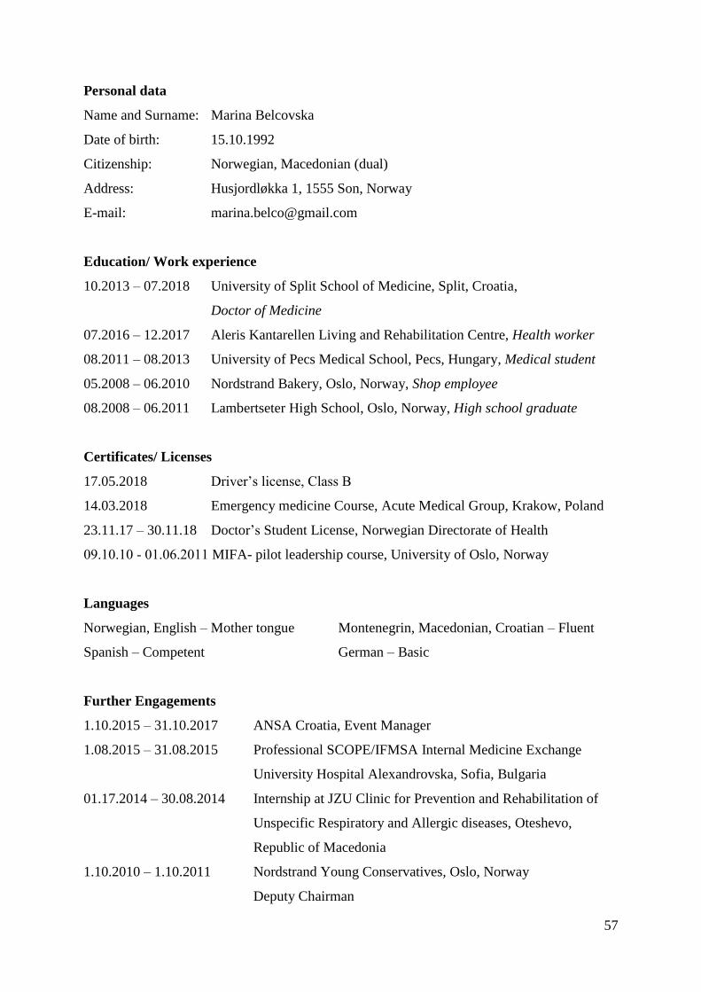
57
Personal data
Name and Surname: Marina Belcovska
Date of birth: 15.10.1992
Citizenship: Norwegian, Macedonian (dual)
Address: Husjordløkka 1, 1555 Son, Norway
E-mail: [email protected]
Education/ Work experience
10.2013 – 07.2018 University of Split School of Medicine, Split, Croatia,
Doctor of Medicine
07.2016 – 12.2017 Aleris Kantarellen Living and Rehabilitation Centre, Health worker
08.2011 – 08.2013 University of Pecs Medical School, Pecs, Hungary, Medical student
05.2008 – 06.2010 Nordstrand Bakery, Oslo, Norway, Shop employee
08.2008 – 06.2011 Lambertseter High School, Oslo, Norway, High school graduate
Certificates/ Licenses
17.05.2018 Driver’s license, Class B
14.03.2018 Emergency medicine Course, Acute Medical Group, Krakow, Poland
23.11.17 – 30.11.18 Doctor’s Student License, Norwegian Directorate of Health
09.10.10 ‑ 01.06.2011 MIFA- pilot leadership course, University of Oslo, Norway
Languages
Norwegian, English – Mother tongue Montenegrin, Macedonian, Croatian – Fluent
Spanish – Competent German – Basic
Further Engagements
1.10.2015 – 31.10.2017 ANSA Croatia, Event Manager
1.08.2015 – 31.08.2015 Professional SCOPE/IFMSA Internal Medicine Exchange
University Hospital Alexandrovska, Sofia, Bulgaria
01.17.2014 – 30.08.2014 Internship at JZU Clinic for Prevention and Rehabilitation of
Unspecific Respiratory and Allergic diseases, Oteshevo,
Republic of Macedonia
1.10.2010 – 1.10.2011 Nordstrand Young Conservatives, Oslo, Norway
Deputy Chairman