
From Eye to Insight
12
CASE STUDY GLOW800 Augmented Reality Fluorescence in AVM (Arteriovenous Malformation) treatment AUTHORS: Prof. Dr. Feres Chaddad Head of Vascular Neurosurgery Federal University of Sao Paulo, Brazil Dr. Robert Ibe Clinical Marketing Manager Leica Microsystems, Heerbrugg, Switzerland From Eye to Insight
Transcript of From Eye to Insight
CASE STUDY
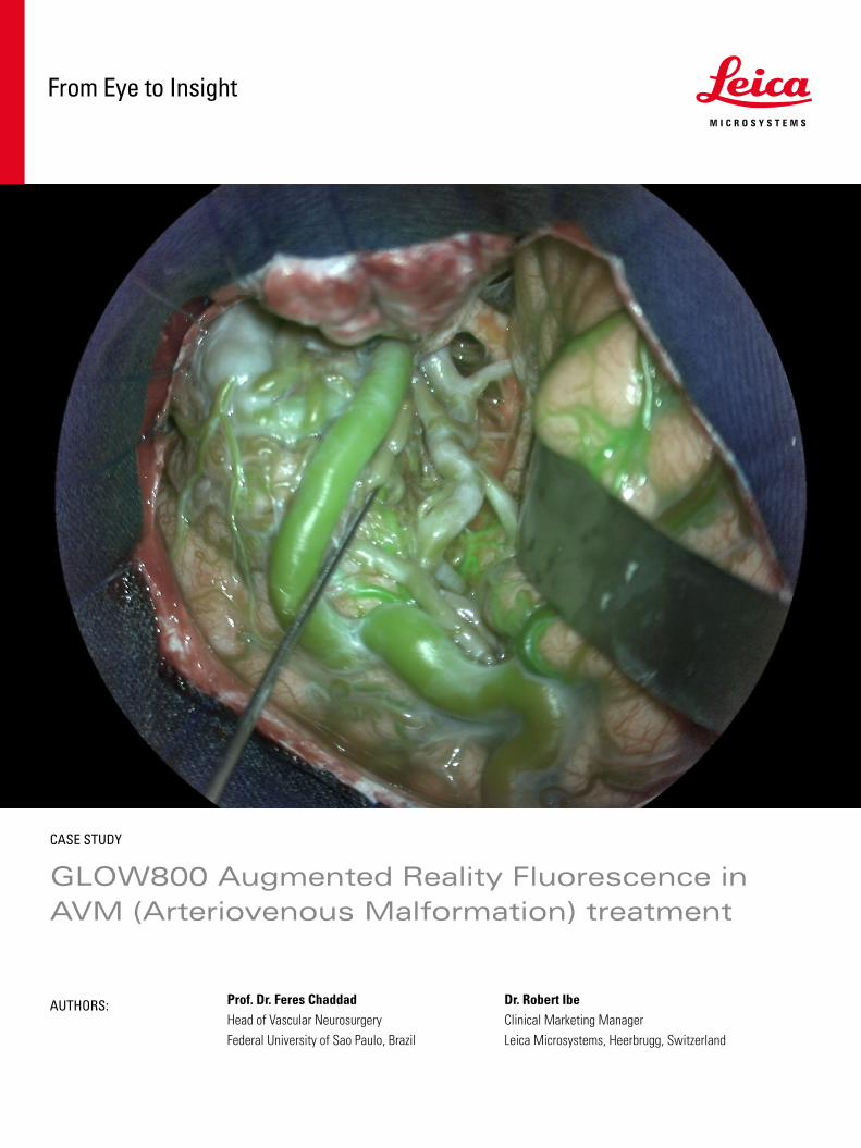
GLOW800 Augmented Reality Fluorescence in AVM (Arteriovenous Malformation) treatment
AUTHORS: Prof. Dr. Feres ChaddadHead of Vascular NeurosurgeryFederal University of Sao Paulo, Brazil
Dr. Robert IbeClinical Marketing ManagerLeica Microsystems, Heerbrugg, Switzerland
From Eye to Insight

Table of Contents
All clinical images and videos supplied for this report are courtesy of Prof. Dr. Feres Chaddad, Head of Vascular Neurosurgery at the Federal University of Sao Paulo (UNIFESP), Sao Paulo, Brazil.
www.neurocirurgiaepm.com.br
Leica Microsystems would like to thank the University of Sao Paulo for the good cooperation for this case study, and especially Prof. Dr. Feres Chaddad for his work on this report.
www.leica-microsystems.com
1. Initial patient presentation .......................................................................................................................................................3
2. Pre-operative assessment ........................................................................................................................................................4-7
3. Intra-operative course and image..................................................................................................................................................8-10
4. Post-surgery....................................................................................................................................................................................10-11
5. Impact of GLOW800 Augmented Reality Fluorescence ..........................................................................................................12

1. Initial Patient Presentation
Patient:
A 24 year old male patient with a four month history of
> Multiple seizures, two of which were described as tonic-clonic seizures. The others were focal onset seizures, characterized by loss of conciousness, rapidly followed by left hand automatisms, collapses, and return to consciousness after a few minutes
> Impaired awareness
> Severe headaches, especially after the seizures
Neurological examination showed slightly decreased attention and memory function, compared to healthy people of similar age and education. No abnormalities of motor, gait, balance, or sensory function were noted.
The patient was treated with carbamazepine (200 mg, 3 x day), which did not improve the seizures. Further assessments and imaging were performed to investigate an underlying cause (see Figures 1-8).
Diagnosis:
> Left temporal pole AVM: Spetzler-Martin Grade II
> Nidus size 2.5 cm
> Multiple supply sources: mostly from the middle cerebral and posterior cerebral arteries
> Multiple drainage points including superior anastomotic vein to the superior sagittal sinus, and tributaries to the inferior petrous sinus, basal vein, and great cerebral vein
Treatment Decision:
At multidisciplinary review, the decision was made to surgically resect the AVM due to the following reasons:
> Patient had poor response to anticonvulsant treatment
> Risk of hemorrhage from this malformation remains high if not resected
> Treatment of choice for Spetzler-Martin Grade II AVMs is microsurgical resection, because this can provide immediate cure with low risk of a recurrence
The aim of the surgery is to achieve control of the seizures and to prevent a hemorrhagic episode that could lead to a neurological sequelae.
CASE STUDY › GLOW800 AUGMENTED REALITY IN AVM TREATMENT 3
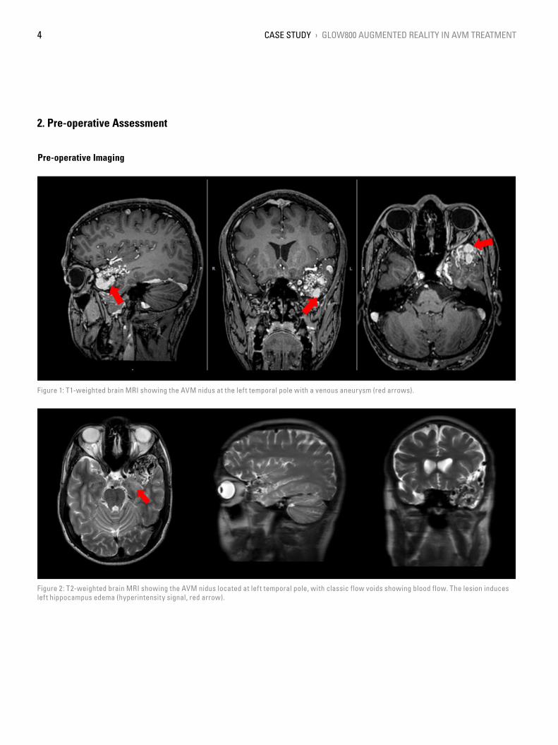
2. Pre-operative Assessment
Figure 1: T1-weighted brain MRI showing the AVM nidus at the left temporal pole with a venous aneurysm (red arrows).
Figure 2: T2-weighted brain MRI showing the AVM nidus located at left temporal pole, with classic flow voids showing blood flow. The lesion induces left hippocampus edema (hyperintensity signal, red arrow).
Pre-operative Imaging
CASE STUDY › GLOW800 AUGMENTED REALITY IN AVM TREATMENT4

Figure 3: Pre-operative DSA. AP projection showing the AVM with a nidus measuring 2.5 cm, fed partially by the middle cerebral artery through an early branch (blue arrow) and terminal branches of the interior trunk (purple arrow).
Figure 4: Lateral projection. The AVM was also partially supplied by the posterior cerebral artery through the anterior and middle inferior temporal arteries (yellow and red arrows, respectively).
Pre-operative Imaging: AVM Supply
CASE STUDY › GLOW800 AUGMENTED REALITY IN AVM TREATMENT 5

Figure 5: AP and lateral projection. The AVM has a superficial drainage through the superior anastomotic vein to the superior sagittal sinus (green arrow) and also a deep drainage through the inferior petrous sinus (orange arrow).
Figure 6: AP and lateral projection. Drainages to the basal vein (red arrow), great cerebral vein (green arrow), straight sinus and confluence of sinuses (orange arrow) were also present. The blue arrow indicates the inferior petrous sinus anastomosing with the lateral mesencephalic vein.
Pre-operative Images: AVM Drainage
CASE STUDY › GLOW800 AUGMENTED REALITY IN AVM TREATMENT6

Figure 7: AP projection. It is possible to observe a vascular steal from the contralateral carotid system (red arrow) as well as from the ipsilateral anterior cerebral artery (yellow arrow).
Pre-operative Assessment Images - Steal
CASE STUDY › GLOW800 AUGMENTED REALITY IN AVM TREATMENT 7

3. Intra-operative Course and Images
Video 1: AVM First Assessment with GLOW800. Surgeon able to manipulate and explore with stereoscopic view while simultaneously assessing blood flow.
Figure 8: After opening the dura the AVM was evident on the temporal lobe and was also assessed using GLOW800.
Video 2: AVM assessed on deeper dissection - clearly showing filling during arterial phase. During surgery GLOW800 shows the presence of fistulas that need to be disconnected and their relation to the AVM nidus, surrounding vessels and brain parenchyma.
AVM First Assessment
GLOW800 Initial Assessment
A pretemporal craniotomy was performed to take advantage of the transsylvian, lateral subfrontal, subtemporal and temporoporal corridors. With this craniotomy the superior, middle and inferior temporal gyrus, the Sylvian fissure, the inferior and part of the middle frontal gyrus were exposed.
CASE STUDY › GLOW800 AUGMENTED REALITY IN AVM TREATMENT8

Video 3: Clipping of Fistula under GLOW800. This shows GLOW800 tolerating movement that aids uninterrupted workflow.
Video 5: Delayed filling of the AVM as more feeders were excluded prior to total exclusion from the normal circulation.
Video 4: Delayed filling of the AVM as the fistulas and feeders are excluded. This helped the surgeon better assess treatment as the surgery progressed.
Video 6: AVM excluded from circulation but not yet excised. No flow into the nidus of the AVM.
GLOW800 helped to understand the superficial configuration of the AVM and to confirm the surgical strategy that was decided earlier.
Following the initial assessment of the AVM, Prof. Chaddad opened the Sylvian fissure using sharp dissection in the arachnoid space. He separated the frontal lobe from the temporal lobe in order to gain a better exposure of the AVM and expose the middle cerebral artery, which was the principal feeder of the AVM.
GLOW800 showed the flow through the vessels and the brain
parenchyma in real time, which allowed to continue working while assessing the anatomical structures. The superficial feeders and shunts were then disconnected from the AVM.
Deeper circumferential dissection was then done, coagulating and cutting off feeder vessels in order to progressively decrease the flow through the AVM.
The branches of the posterior cerebral artery were accessed through the subtemporal corridor.
AVM Resection
CASE STUDY › GLOW800 AUGMENTED REALITY IN AVM TREATMENT 9

Video 7: GLOW800 after the AVM was excised. GLOW800 confirmed complete exclusion from the normal circulation, patency of the vessels proximal to the lesion and absence of any abnormal early drainage veins.
GLOW800 Post AVM Exclusion
4. Post Surgery
Figure 9: T1-weighted MRI images showing the complete resection of AVM nidus (red arrows).
Video 8: Video showing the exposure, of the AVM, clippings and exclusions of the feeders and resection of the AVM.
CASE STUDY › GLOW800 AUGMENTED REALITY IN AVM TREATMENT10
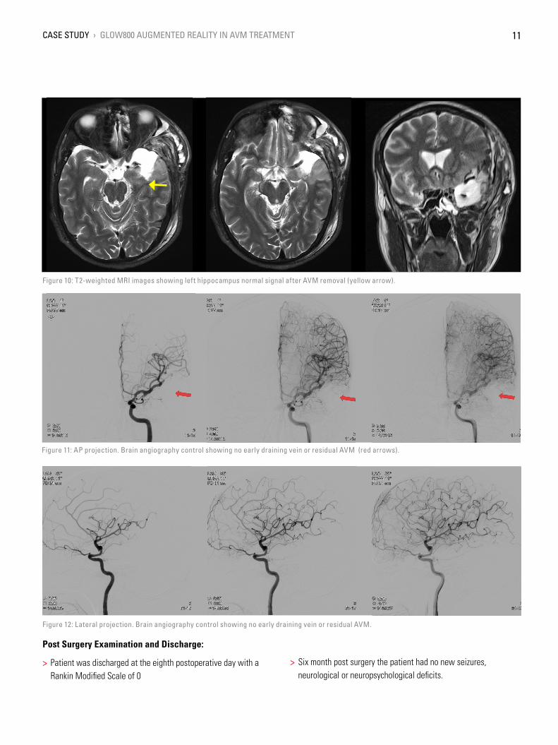
Figure 10: T2-weighted MRI images showing left hippocampus normal signal after AVM removal (yellow arrow).
Figure 11: AP projection. Brain angiography control showing no early draining vein or residual AVM (red arrows).
Figure 12: Lateral projection. Brain angiography control showing no early draining vein or residual AVM.
Post Surgery Examination and Discharge:
> Patient was discharged at the eighth postoperative day with a Rankin Modified Scale of 0
> Six month post surgery the patient had no new seizures, neurological or neuropsychological deficits.
CASE STUDY › GLOW800 AUGMENTED REALITY IN AVM TREATMENT 11

5. Impact of GLOW800 Augmented Reality Fluorescence
One augmented view during vascular neurosurgery can improve confidence in intraoperative diagnosis
> One augmented, real-time view means no more need to recalland try to reconcile the black and white blood flow video with thenatural anatomical view
> Crisp delineation helps you limit potential compromise orobstruction of surrounding perforators and small vessels
> Full depth perception and no dark peripheries supports clearspatial orientation to aid manipulation of vessels
Improved confidence in treatment
Visualization with GLOW800 AR supports each step of vascular neurosurgery procedures and also supports with post-operative imaging. For example, during AVM treatment, it helps:
> Manipulate the AVM to assess beyond the superficial anatomicalview while simultaneously observing flow
> Confidently assess flow dynamics of inflow and outflow instereoscopic view
> Assess filling and emptying of the AVM during the differentphases (arterial and venous)
> View changes in filling rate and time of the AVM as feeders andfistulas are disconnected and as surgery progresses
> Confirm complete exclusion and/or excision of the AVM from thenormal circulation and evaluate the patency of vessels.
A fundamental tool for the future practice and training in neurosurgery
In the binocular image injection (CaptiView) better defining (lowering) the dose of Indocyanine Green (ICG) was enough to generate the flow effect without saturating the view of the whole operative field.
Also, the microscope’s monitor offers a perfect view, where the observer can easily understand the anatomy and the AVM exclusion strategy adopted. Image settings can be effortlessly changed, to optimize the view as needed.
CONNECT WITH US!
Leica Microsystems (Schweiz) AG · Max Schmidheiny Strasse 201 · CH-9435 Heerbrugg
T +41 71 726 3333 · F +41 71 726 3399
www.leica-microsystems.com
MC-
0002538·
29.
04.2
021
· EN
· Co
pyrig
ht ©
202
1 Le
ica
Mic
rosy
stem
s (S
witz
erla
nd) A
G. A
ll rig
hts
rese
rved
. Sub
ject
to m
odifi
catio
ns. L
EICA
and
the
Leic
a Lo
go a
re re
gist
ered
trad
emar
ks o
f Lei
ca M
icro
syst
ems
IR G
mbH
.
Leica Microsystems (Schweiz) AGMax Schmidheiny-Strasse 2019435 Heerbrugg, Switzerland 0123
Not all products or services are approved or offered in every market and approved labeling and instructions may vary between countries. Please contact your local Leica representative for details.
Class IIa GLOW800