vImhoff-4 XP 28-11-2006 13:24 Pagina 123 Post-transplant ...Posttransplant lymphoproliferative...
24
4 Post-transplant lymphoma 123
Transcript of vImhoff-4 XP 28-11-2006 13:24 Pagina 123 Post-transplant ...Posttransplant lymphoproliferative...

4Post-transplant lymphoma
123
vImhoff-4 XP 28-11-2006 13:24 Pagina 123

124
vImhoff-4 XP 28-11-2006 13:24 Pagina 124
4.1Early onset posttransplant lymphoproliferativedisease is associated with allograft localization
Nicolaas A BakkerGustaaf W. van ImhoffErik A.M. VerschuurenWillem J. van SonJaap J. Homan van der HeideNic J.G.M. VeegerPhilip M. KluinHanneke C. Kluin-Nelemans
Clinical Transplantation 2005; 19: 327-334
125
vImhoff-4 XP 28-11-2006 13:24 Pagina 125

Posttransplant lymphoproliferative disease (PTLD) is a major compli-cation after solid organ transplantation. We analyzed incidence, patientcharacteristics, clinical presentation, and prognostic factors fortreatment outcome and survival of PTLD patients transplanted at ourcenter.
Records from adult kidney and lung transplant recipients, transplantedbetween January 1985 and December 2002 with a histological confirmeddiagnosis of PTLD, were retrieved. Histology was reviewed andprognostic factors for treatment outcome were evaluated bymultivariable analysis.
Out of 1354 kidney and 206 lung transplants, PTLD was diagnosed in 40 transplant recipients (2.6%). Lung transplant recipients had asignificantly higher incidence of PTLD (8.3%) than kidney transplantrecipients (1.7%). Sites of presentation were highly heterogeneous.Notably, PTLD localized in the allograft occurred significantly earlierafter transplantation than PTLD localized outside the allograft (p=0.001).This was true for lung (p=0.006) as well as for kidney transplantrecipients (p=0.03). In multivariable Cox regression, performance status(p=0.01) and advanced stage (p=0.04) were factors negatively predictivefor response to first line treatment. Only performance status remainedas negative predictive factor for survival (p=0.002) and freedom fromtumor progression (p=0.01).
In conclusion, the allograft is significantly more often involved asprimary site of PTLD presentation during the first post-transplant year.This may have clinical consequences and give new insights inpathogenesis of PTLD. Performance status and stage are important riskfactors for outcome of PTLD.
Introduction
Post-transplant lymphoproliferative disease (PTLD) is a life threatening com-plication after solid organ transplantation. Epstein-Barr virus (EBV) seems toplay a major role in the pathogenesis of PTLD. EBV-induced lymphoprolifera-tive disorders are considered to result from proliferating latently infected B-cellsin the absence of an appropriate EBV-specific cytotoxic T-cell response, proba-bly caused by the immunosuppressive regimen after transplantation. Althoughsome EBV negative cases have been observed, EBV encoded RNA (EBER) iscommonly detectable in PTLD lesions.1-3 Lymphomas arising early after trans-
126
vImhoff-4 XP 28-11-2006 13:24 Pagina 126
plantation are almost all EBV-positive, whereas PTLD arising late after trans-plantation may also be EBV-negative.4 An increased proliferation of EBV infectedB-cells can often be detected by measuring the amount of circulating EBV-DNAusing a quantative-competitive polymerase chain reaction (qPCR). This assaycan be very useful in identifying transplant recipients at risk for the develop-ment of PTLD.1,5-7 The most important risk factors for development of PTLDafter solid organ transplantation are primary EBV infection as well as immuno-suppression after transplantation, especially induction and rejection therapywith anti-T-cell antibodies.8,9 The incidence of PTLD is highest during the firstpost-transplant year which is probably related to the intensity of initial immuno-suppression.10 Other known risk factors are primary Cytomegalovirus (CMV)infection11 and younger recipient age at time of transplantation.9 The incidencevaries depending on the type of organ transplanted, with the highest incidencefound in lung and small bowel transplantation, whereas kidney transplant recip-ients have a much lower incidence.10,12 This is probably also related to the higherintensity of the immunosuppression in lung and small bowel transplantation ascompared with kidney transplantation.12
No standard treatment for PTLD has been established yet. The most impor-tant initial strategy is reduction of immunosuppression.13-15 However, this is notalways feasible because of the risk of rejection of organs for which no organreplacement therapy (i.e. hemodialysis) exists. Although antiviral agents againsthuman herpes viruses such as acyclovir and gancyclovir are frequently admin-istered, their value in the management of PTLD is still unclear.16 Poly-chemotherapy with cyclophosphamide, doxorubicin, vincristine, and prednisone(CHOP) has been widely used as first-line treatment in the past, but this type oftreatment is associated with substantial morbidity and mortality.15,17 Currently,anti-B-cell monoclonal antibody therapy, especially anti-CD20 (Rituximab),which has proven its value in the management of other lymphoma types, hasbecome first line treatment of PTLD.18-20 Polychemotherapy is nowadays onlyreserved for patients in whom other treatment options have failed or when PTLDis CD20 negative.
Treatment outcome of PTLD is influenced by several factors. Bad perform-ance status at diagnosis and multiple localizations of PTLD have been identifiedas negative predictors for overall survival.21 For outcome of treatment with mon-oclonal antibodies, multivisceral disease, central nervous system involvementand late onset of PTLD have been identified as key risk factors.18
We analyzed incidence, patient characteristics, clinical presentation andprognostic factors of importance for treatment outcome and survival of PTLD inpatients transplanted at our center between January 1985 and December 2002.During the study time frame, more than 1500 kidney and lung transplants havebeen performed at our center.
Early PTLD is localized in the allograft
127
vImhoff-4 XP 28-11-2006 13:24 Pagina 127

Patients and methods
PatientsWe retrieved all records from adult transplant recipients in our hospital with
a histological confirmed diagnosis of PTLD after kidney and/or pancreas trans-plantation, lung and heart-lung transplantation. The kidney transplant programwas studied from January 1985 until December 2002; the lung and heart-lungtransplant program was studied from the start of the program in January 1990until December 2002. All PTLD observed until December 2003 presenting intransplant recipients transplanted during the study time frame were included. AllPTLD were reviewed and classified according to the modified WHO classificationas proposed by Nalesnik in 2001.22 The presence of EBV in PTLD lesions wasdetermined by EBER in situ hybridization.
Clinical features, laboratory data, clinical staging, underlying disease respon-sible for transplantation, immunosuppressive regimen after transplantation,rejection episodes and management of these episodes, time to PTLD, site ofPTLD, type of treatment and response to treatment were documented. TheInternational Prognostic Index (IPI) was applied. This index, initially developedfor aggressive non-Hodgkin’s lymphoma23 and not validated for PTLD is basedupon five adverse prognostic factors (age >60 years, performance status >1, serumlactate dehydrogenase (LDH) greater than the upper normal limit, Ann Arborstage24 >II and number of extranodal sites >1) and distinguishes patients at ini-tial diagnosis in terms of the likelihood of response to treatment, progressionand overall survival. Although age >60 was used for categorization according toIPI, age above 50 years instead of 60 years was taken as cutoff point fordichotomization and analysis of individual prognostic factors because only fourpatients in our study population were older than 60 years. Performance statuswas assessed according to the Eastern Cooperative Oncology Group (ECOG) clas-sification,25 in which 0 indicated that the patient had no symptoms; 1, the patienthad symptoms but was ambulatory; 2, the patient was bedridden less than halfthe day; 3, the patient was bedridden half the day or longer; and 4, the patient waschronically bedridden and required assistance with the activities of daily living.Performance status was classified as 0 or 1 (the patient was ambulatory) or 2, 3,or 4 (the patient was not ambulatory) (equivalent Karnofsky scores, >80 and <70).
Immunosuppressive protocolsAll lung and heart-lung transplant recipients received three doses of anti-thy-
mocyte globulin (ATG) during the first week after transplantation. Kidney trans-plant recipients received induction therapy with anti-T-cell antibodies only whenthey were considered to be at high risk for rejection. This was defined as highlyimmunized patients with HLA antibodies (panel reactive antibodies >86%) or re-
4 Post-transplant lymphoma
128
vImhoff-4 XP 28-11-2006 13:24 Pagina 128
transplantation patients who had lost their allograft due to rejection. These trans-plant recipients received two doses of ATG or anti-T-cell receptor monoclonalantibody (OKT3) (before 1990) during the first week after transplantation.
Immunosuppressive maintenance therapy after transplantation changed sev-eral times during the time frame of this study. From 1985 onwards, almost allrenal transplant recipients were treated with Cyclosporine A (CsA) combinedwith Azathioprine (Aza) and Prednisolone (Pred). After 1998 immunosuppres-sion consisted of CsA, Pred and Mycophenolate Mofetil (MMF). All lung andheart/lung transplant recipients received CsA combined with Aza and Pred asimmunosuppressive maintenance therapy.
From June 2001 onwards lung transplant patients received induction therapywith anti-IL-2 receptor monoclonal antibodies (Basiliximab) and maintenancetherapy with Aza, Pred and FK506 (Tacrolimus). At the same time a prospectivestudy was started in which immunosuppression is to be tapered according to theEBV-DNA load as measured by whole blood PCR. Results of this study and howit affected incidence after June 2001 are not available yet because to few patientshave been included for meaningful analysis.
Therapy of rejectionAcute rejection periods, i.e. interstitial type for kidney transplant recipients,
were treated with high doses Methylprednisolone (3 × 1000 mg). In case of aninsufficient response to Methylprednisolone or vascular type of rejection in kid-ney transplant recipients, ATG was administered. Patients with severe rejectionover a period of time were converted to another immunosuppressive regimen. Inmost cases CsA was changed to FK506 (for kidney transplant recipients) and/orAza converted to MMF.
Reduction of immunosuppressionReduction of immunosuppression usually was the first step of treatment after
diagnosis of PTLD. In most cases CsA was lowered or even stopped and Aza wasreduced with 50%. After 1998, when kidney transplant recipients were treatedwith MMF instead of CsA, MMF was lowered or stopped. There was no differencein reduction regimen between patients treated with reduction of immunosup-pression only and patients treated with other modalities along with reduction ofimmunosuppression.
EndpointsEndpoints in this study were response to first line treatment, overall survival
(OS) and freedom from tumor progression (FFTP). Response was assessed at 3months after initiation of therapy for PTLD and classified as either complete(CR), partial (PR) or progressive disease. Overall survival and FFTP were meas-
Early PTLD is localized in the allograft
129
vImhoff-4 XP 28-11-2006 13:24 Pagina 129
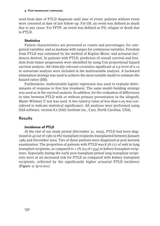
ured from date of PTLD diagnosis until date of event; patients without eventwere censored at date of last follow-up. For OS, an event was defined as deathdue to any cause. For FFTP, an event was defined as PD, relapse or death dueto PTLD.
StatisticsPatient characteristics are presented as counts and percentages, for cate-
gorical variables, and as medians with ranges for continuous variables. Freedomfrom PTLD was estimated by the method of Kaplan-Meier, and actuarial inci-dences derived. In patients with PTLD, predictors of overall survival and free-dom from tumor progression were identified by using Cox proportional hazardsurvival analysis. All clinically relevant covariates significant at a p level of 0.10in univariate analysis were included in the multivariable analysis. A backwardelimination strategy was used to achieve the most suitable model to estimate thehazard ratios (HR).
Furthermore, multivariable logistic regression was used to evaluate deter-minants of response to first line treatment. The same model-building strategywas used as in the survival analysis. In addition, for the evaluation of differencesin time between PTLD with or without primary presentation in the allograft,Mann-Whitney U test was used. A two-tailed p value of less than 0.05 was con-sidered to indicate statistical significance. All analyses were performed usingSAS software, version 8.0 (SAS-Institute inc., Cary, North Carolina, USA).
Results
Incidence of PTLDAt the end of our study period (December 31, 2003), PTLD had been diag-
nosed in 40 out of 1560 (2.6%) transplant recipients transplanted between January1985 and December 2002. Two of these patients were diagnosed at post mortemexamination. The proportion of patients with PTLD was 8.3% (17 of 206) in lungtransplant recipients, as compared to 1.7% (23 of 1354) in kidney transplant recip-ients. Especially during the early post transplant period lung transplant recipi-ents were at an increased risk for PTLD as compared with kidney transplantrecipients, reflected by the significantly higher actuarial PTLD incidence(Figure 1) (p<0.001).
4 Post-transplant lymphoma
130
vImhoff-4 XP 28-11-2006 13:24 Pagina 130

Figure 1. Actuarial incidence in lung and kidney transplant recipients.
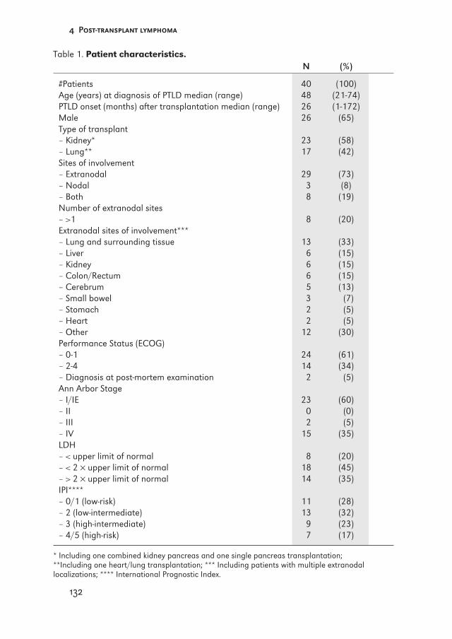
Patient characteristicsThe main characteristics of 40 patients with PTLD are described in Table 1.
Approximately two-third of the PTLD population was male, in concordance withthe fact that more than 60 percent of all patients transplanted were male. Themedian age (45 years, range 19-71 years) at time of transplantation of patientswho developed PTLD did not differ from those who did not develop PTLD. Timeof onset of PTLD after transplantation varied. Almost half of all PTLD cases(48%) were observed within the first year after transplantation.
Most patients presented with Ann Arbor Stage IE or stage IV disease.Approximately 80% of PTLD patients had serum LDH levels above the upperlimit of normal (>235 U/l). A poor performance status (performance status >1) wasobserved in 16 patients (40%). In seven out of these 16 patients this could beattributed to other diseases than PTLD, such as bronchiolitis obliterans syn-drome in lung transplant recipients.
Primary site of PTLD presentation was heterogeneous and frequently extra-nodal. Most PTLD presented in the allograft or in the digestive tract. PTLD local-ized in the allograft occurred significantly earlier after transplantation as com-pared to PTLD localized outside the allograft (median 4.5 months, range 1-99months versus median 51 months, range 2-172 months, p=0.001; Figure 2). Thisrelation was found for lung as well as kidney transplant recipients when ana-lyzed separately. In kidney transplant recipients, allograft involvement as primarysite of disease was observed in three out of ten patients (30%) who developed
Early PTLD is localized in the allograft
131
vImhoff-4 XP 28-11-2006 13:24 Pagina 131
4 Post-transplant lymphoma
132
Table 1. Patient characteristics.N (%)
#Patients 40 (100)Age (years) at diagnosis of PTLD median (range) 48 (21-74)PTLD onset (months) after transplantation median (range) 26 (1-172)Male 26 (65)Type of transplant– Kidney* 23 (58)– Lung** 17 (42)Sites of involvement– Extranodal 29 (73)– Nodal 3 (8)– Both 8 (19)Number of extranodal sites– >1 8 (20)Extranodal sites of involvement***– Lung and surrounding tissue 13 (33)– Liver 6 (15)– Kidney 6 (15)– Colon/Rectum 6 (15)– Cerebrum 5 (13)– Small bowel 3 (7)– Stomach 2 (5)– Heart 2 (5)– Other 12 (30)Performance Status (ECOG)– 0-1 24 (61)– 2-4 14 (34)– Diagnosis at post-mortem examination 2 (5)Ann Arbor Stage– I/IE 23 (60)– II 0 (0)– III 2 (5)– IV 15 (35)LDH– < upper limit of normal 8 (20)– < 2 × upper limit of normal 18 (45)– > 2 × upper limit of normal 14 (35)IPI****– 0/1 (low-risk) 11 (28)– 2 (low-intermediate) 13 (32)– 3 (high-intermediate) 9 (23)– 4/5 (high-risk) 7 (17)
* Including one combined kidney pancreas and one single pancreas transplantation; **Including one heart/lung transplantation; *** Including patients with multiple extranodal localizations; **** International Prognostic Index.
vImhoff-4 XP 28-11-2006 13:24 Pagina 132
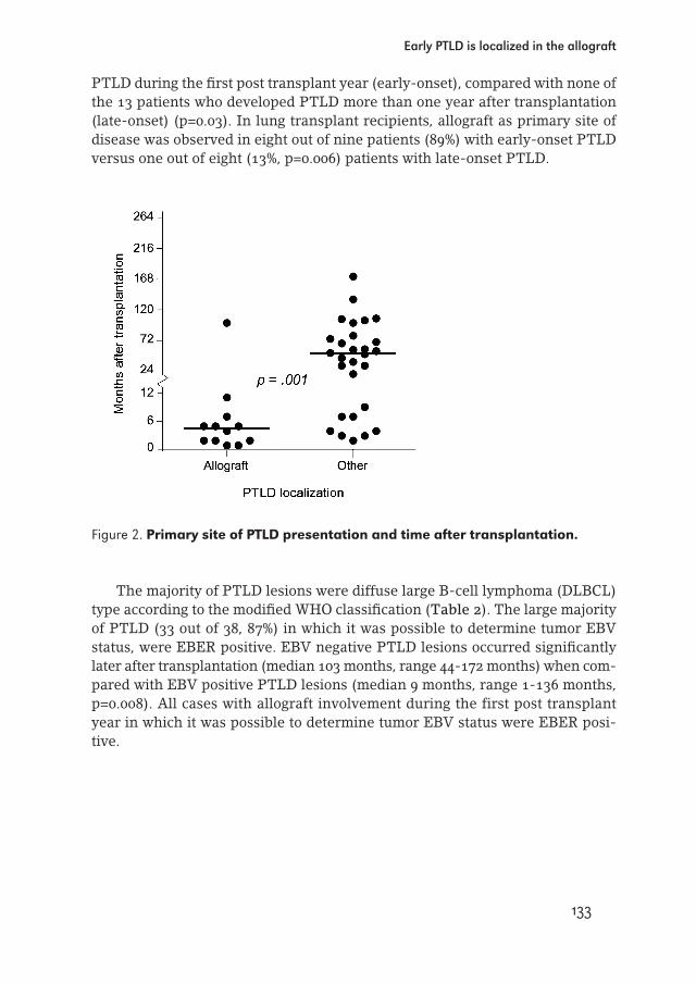
PTLD during the first post transplant year (early-onset), compared with none ofthe 13 patients who developed PTLD more than one year after transplantation(late-onset) (p=0.03). In lung transplant recipients, allograft as primary site ofdisease was observed in eight out of nine patients (89%) with early-onset PTLDversus one out of eight (13%, p=0.006) patients with late-onset PTLD.
Figure 2. Primary site of PTLD presentation and time after transplantation.
The majority of PTLD lesions were diffuse large B-cell lymphoma (DLBCL)type according to the modified WHO classification (Table 2). The large majorityof PTLD (33 out of 38, 87%) in which it was possible to determine tumor EBVstatus, were EBER positive. EBV negative PTLD lesions occurred significantlylater after transplantation (median 103 months, range 44-172 months) when com-pared with EBV positive PTLD lesions (median 9 months, range 1-136 months,p=0.008). All cases with allograft involvement during the first post transplantyear in which it was possible to determine tumor EBV status were EBER posi-tive.
Early PTLD is localized in the allograft
133
vImhoff-4 XP 28-11-2006 13:24 Pagina 133
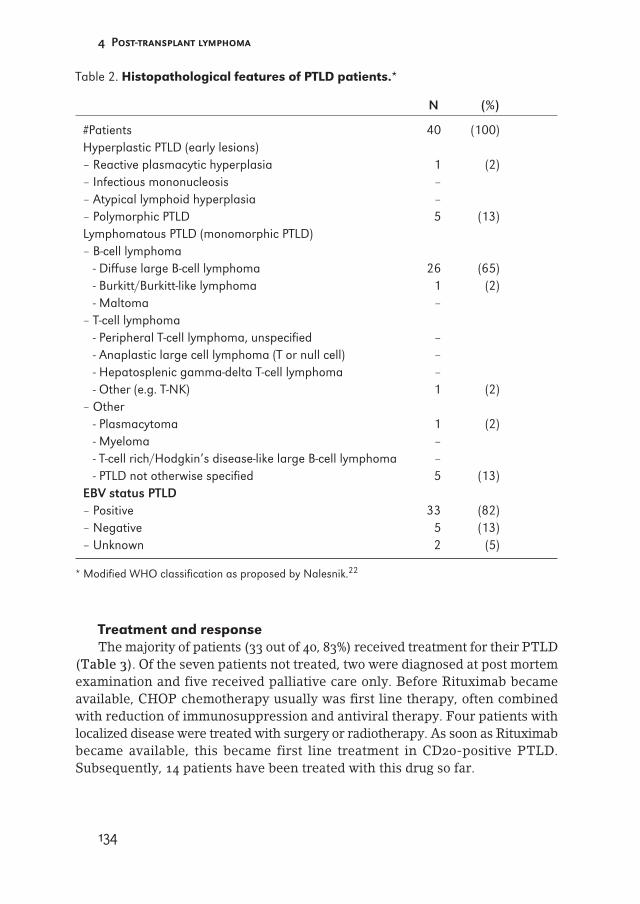
Table 2. Histopathological features of PTLD patients.*
N (%)
#Patients 40 (100)Hyperplastic PTLD (early lesions)– Reactive plasmacytic hyperplasia 1 (2)– Infectious mononucleosis –– Atypical lymphoid hyperplasia –– Polymorphic PTLD 5 (13)Lymphomatous PTLD (monomorphic PTLD)– B-cell lymphoma
- Diffuse large B-cell lymphoma 26 (65)- Burkitt/Burkitt-like lymphoma 1 (2)- Maltoma –
– T-cell lymphoma- Peripheral T-cell lymphoma, unspecified –- Anaplastic large cell lymphoma (T or null cell) –- Hepatosplenic gamma-delta T-cell lymphoma –- Other (e.g. T-NK) 1 (2)
– Other- Plasmacytoma 1 (2)- Myeloma –- T-cell rich/Hodgkin’s disease-like large B-cell lymphoma –- PTLD not otherwise specified 5 (13)
EBV status PTLD– Positive 33 (82)– Negative 5 (13)– Unknown 2 (5)
* Modified WHO classification as proposed by Nalesnik.22
Treatment and responseThe majority of patients (33 out of 40, 83%) received treatment for their PTLD
(Table 3). Of the seven patients not treated, two were diagnosed at post mortemexamination and five received palliative care only. Before Rituximab becameavailable, CHOP chemotherapy usually was first line therapy, often combinedwith reduction of immunosuppression and antiviral therapy. Four patients withlocalized disease were treated with surgery or radiotherapy. As soon as Rituximabbecame available, this became first line treatment in CD20-positive PTLD.Subsequently, 14 patients have been treated with this drug so far.
4 Post-transplant lymphoma
134
vImhoff-4 XP 28-11-2006 13:24 Pagina 134

Table 3. First line treatment and response.
#Patients Total CR PR PD Relapse (N=40) (N=24) (N=2) (N=14) (N=5)
Rituximab + RIS* 14 12 1 1 1CHOP ± RIS 7 4 0 3 1RIS only 8 4 1 3 2Surgery + RIS 3 3 0 0 1Radiotherapy + RIS 1 1 0 0 0
No treatment 7 0 0 7 0
* Reduction of immunosuppression.
A complete response after first line treatment was observed in 24 of all 33patients treated (73%). Two patients reached partial remission. Although thenumber of patients is obviously too small to draw any conclusions, patientstreated with Rituximab seemed to have a better response rate (12/14; 86%) thanpatients treated with other modalities. Antiviral therapy was not instigated sys-tematically and consequently the effect of adding antiviral agents to treatmentremains unclear.
Five out of 24 patients (21%) with a complete response had a relapse of theirPTLD at a median time of 41 months (range 1-89 months) after reaching CR. Ofthe two patients reaching PR, one patient died because of progressive PTLDwhile the other patient is still alive without signs of progression.
At a median observation time of patients still alive after PTLD of 28 months(range 7-162 months), median survival is 12 months, and median freedom fromPTLD progression (FFTP) is 17 months. Twenty seven patients (68%) have diedat a medium time of three months (range 0-96 months) after diagnosis of
PTLD, including the seven patients not receiving any treatment with curativeintention. PTLD was the main cause of death in 13 patients. Other causes ofdeath were: end-stage bronchiolitis obliterans syndrome in 5 lung transplantrecipients (1 with active PTLD); treatment related toxicity in 3 patients receiv-ing CHOP (1 with active PTLD); severe infections in 3 patients (2 with activePTLD); cardiac failure in one patient; liver cirrhosis in one patient (with activePTLD); voluntary cessation of dialysis in one patient. Although four patientstreated with Rituximab have died so far, no patient treated with Rituximab diedwith active PTLD. Patients who died due to PTLD did so at a median follow upof one month (range 0-35 months) after diagnosis. This tended to be earlier thanpatients who died due to other causes (median 6 months, range 1-96 months,Mann Whitney U: p=0.08).
Early PTLD is localized in the allograft
135
vImhoff-4 XP 28-11-2006 13:24 Pagina 135
4 Post-transplant lymphoma
136
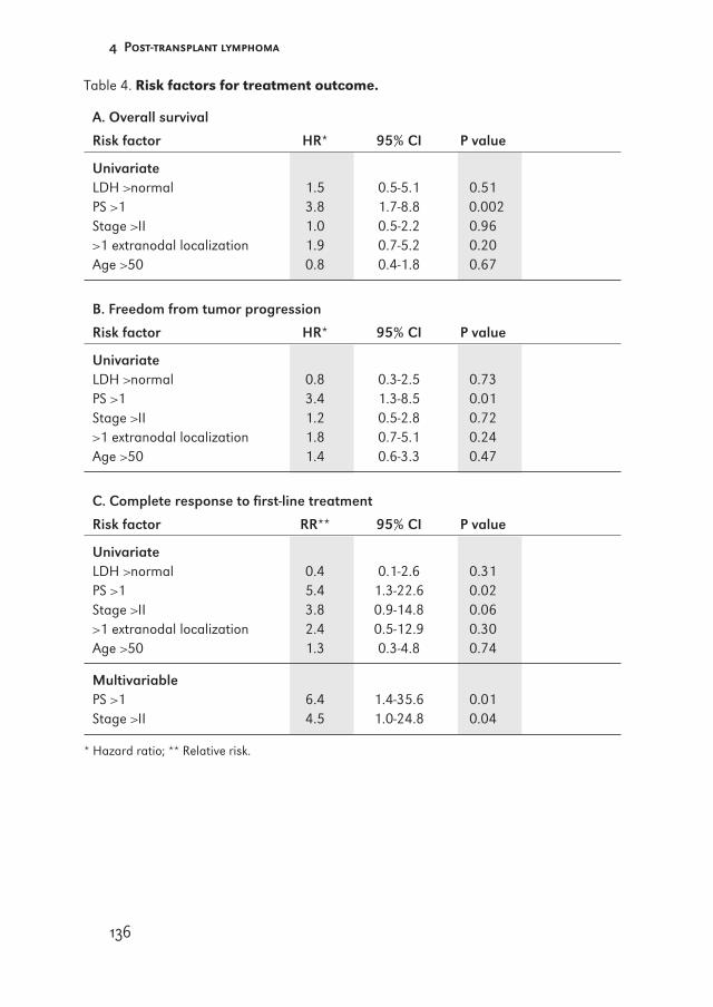
Table 4. Risk factors for treatment outcome.
A. Overall survival
Risk factor HR* 95% CI P value
UnivariateLDH >normal 1.5 0.5-5.1 0.51PS >1 3.8 1.7-8.8 0.002Stage >II 1.0 0.5-2.2 0.96>1 extranodal localization 1.9 0.7-5.2 0.20Age >50 0.8 0.4-1.8 0.67
B. Freedom from tumor progression
Risk factor HR* 95% CI P value
UnivariateLDH >normal 0.8 0.3-2.5 0.73PS >1 3.4 1.3-8.5 0.01Stage >II 1.2 0.5-2.8 0.72>1 extranodal localization 1.8 0.7-5.1 0.24Age >50 1.4 0.6-3.3 0.47
C. Complete response to first-line treatment
Risk factor RR** 95% CI P value
UnivariateLDH >normal 0.4 0.1-2.6 0.31PS >1 5.4 1.3-22.6 0.02Stage >II 3.8 0.9-14.8 0.06>1 extranodal localization 2.4 0.5-12.9 0.30Age >50 1.3 0.3-4.8 0.74
MultivariablePS >1 6.4 1.4-35.6 0.01Stage >II 4.5 1.0-24.8 0.04
* Hazard ratio; ** Relative risk.
vImhoff-4 XP 28-11-2006 13:24 Pagina 136
Prognostic factorsWe analyzed potential predictors of response to first line treatment, freedom
from tumor progression (FFTP) and survival (Table 4). Both patients with diag-nosis at post mortem examination were excluded from this analysis. Performancestatus >1 (RR 6.4, 1.4-35.6, p=0.01) as well as advanced stage (RR 4.5, 1.0-24.8,p=0.04) were factors predictive for response to first line treatment. Only per-formance status remained as predictive factor for survival (HR 3.8, 1.7-8.8,p=0.002) and freedom from tumor progression (HR 3.4, 1.3-8.5, p=0.01). Neitherelevated LDH, age at diagnosis (>50 year), number of extra nodal localizations orstage (all factors contributing to the IPI) were independent predictors of survivalor response to FFTP.
Discussion
The 2.6% overall incidence of PTLD in kidney and lung transplant recipients inour center is comparable to the incidence reported by others.9,26 The fivefoldhigher incidence observed in lung transplant recipients has also been observedelsewhere,10,11 and probably relates to the higher doses of immunosuppressiongiven to these patients. Our analysis confirms a higher incidence of PTLD in thefirst post transplant year,1 especially in lung transplant recipients, which is prob-ably also related to the more intense immunosuppression during this period.12
In concordance with the literature,10 allograft localization of PTLD was moreoften observed in lung transplant recipients as compared with kidney transplantrecipients (three of the 23 patients in kidney transplants versus 9 of the 17patients in lung transplants). Interesting in our study, however, was the findingthat a substantial number of early onset PTLD developed in the allograft.Although allograft localization was more impressive in lung transplant recipi-ents, the relation between time of onset and allograft localization was significantin both lung and kidney transplantation. Although one could still consider thepossibility that PTLD localized in the allograft might be of donor origin, especiallyin lung transplant recipients, we have found no compelling evidence for thisnotion. In six out of 17 lung transplant recipients in which it was possible todetermine PTLD origin by HLA typing, only one out of four PTLD cases in theallograft was donor derived.27
Our observation that early onset PTLD is often localized in the allograft mayhave important clinical implications, because the signs and symptoms of PTLDinvolvement of the allograft may mimic rejection.28,29 Our data indicate that incase of early organ failure, PTLD should be considered next to rejection, whereaslate organ failure will more likely be the result of true rejection. This knowledge,combined with a sensitive PCR to monitor EBV-DNA load,6 might improve thedistinction between graft rejection and development of PTLD.
Early PTLD is localized in the allograft
137
vImhoff-4 XP 28-11-2006 13:24 Pagina 137
The present study also aimed at identifying factors predictive for response totherapy and survival of patients with PTLD and to determine the prognosticvalue of the IPI in this setting. Performance status >1 and advanced stage weremain risk factors for outcome, thus confirming other studies.21 The IPI, of mainprognostic importance for aggressive non-Hodgkin lymphoma in immunocom-petent patients was not suitable for PTLD patients in our study. This confirms theresults of other studies,21,30 but conflicts with a study performed by Herzig etal.31 The fact that a large number of patients had multiple extra nodal localiza-tions, thereby placing them in stage IV according to the Ann Arbor classifica-tion, as well as the fact that age in our transplant population was significantlylower than age in other lymphoma type patients, might explain why we couldnot find a relation between stage, age, number of extra nodal localizations andoutcome in our series. Interestingly, neither elevated LDH was a risk factor fortreatment outcome in our series. Although one could argue that this might bethe result of the use of calcineurin inhibitors, i.e. CsA, which may lead toincreased serum LDH levels,32 this seems unlikely because even LDH levelsabove twice the upper limit of normal (for which calcineurin inhibitors areunlikely to be responsible) were not associated with worse outcome in our series(data not shown).
In conclusion, we have found that early onset PTLD is often localized in theallograft. This observation should trigger the awareness that in case of earlyorgan failure PTLD should be considered next to rejection and may implicate apermissive role of allograft micro-environment in the development of early onsetPTLD, an observation that supports the suggestion that (chronic) antigenic stim-ulation by the allograft may be a contributory factor in the development ofPTLD.10,33
References
1. Riddler SA, Breinig MC, McKnight JL. Increased levels of circulating Epstein-Barr virus(EBV)-infected lymphocytes and decreased EBV nuclear antigen antibody responses areassociated with the development of posttransplant lymphoproliferative disease in solid-organ transplant recipients. Blood 1994: 84: 972.
2. Birkeland SA, Hamilton-Dutoit S, Sandvej K, Andersen HM, Bendtzen K, Moller B et al.EBV-induced post-transplant lymphoproliferative disorder (PTLD). Transplant Proc 1995:27: 3467.
3. Tanner JE, Alfieri C. The Epstein-Barr virus and post-transplant lymphoproliferative dis-ease: interplay of immunosuppression, EBV, and the immune system in disease patho-genesis. Transpl Infect Dis 2001: 3: 60.
4. Dotti G, Fiocchi R, Motta T, Gamba A, Gotti E, Gridelli B et al. Epstein-Barr virus-nega-tive lymphoproliferate disorders in long-term survivors after heart, kidney, and liver trans-plant. Transplantation 2000: 69: 827.
5. Stevens SJ, Verschuuren EA, Pronk I, van Der Bij W, Harmsen MC, The TH et al. Frequentmonitoring of Epstein-Barr virus DNA load in unfractionated whole blood is essential for
4 Post-transplant lymphoma
138
vImhoff-4 XP 28-11-2006 13:24 Pagina 138
early detection of posttransplant lymphoproliferative disease in high-risk patients. Blood2001: 97: 1165.
6. Stevens SJ, Verschuuren EA, Verkuujlen SA, Van Den Brule AJ, Meijer CJ, MiddeldorpJM. Role of Epstein-Barr virus DNA load monitoring in prevention and early detection ofpost-transplant lymphoproliferative disease. Leuk Lymphoma 2002: 43: 831.
7. Smets F, Latinne D, Bazin H, Reding R, Otte JB, Buts JP et al. Ratio between Epstein-Barrviral load and anti-Epstein-Barr virus specific T-cell response as a predictive marker ofposttransplant lymphoproliferative disease. Transplantation 2002: 73: 1603.
8. Swinnen LJ, Costanzo-Nordin MR, Fisher SG, O’Sullivan EJ, Johnson MR, Heroux AL etal. Increased incidence of lymphoproliferative disorder after immunosuppression with themonoclonal antibody OKT3 in cardiac-transplant recipients. N Engl J Med 1990: 323: 1723.
9. Bustami RT, Ojo AO, Wolfe RA, Merion RM, Bennett WM, McDiarmid SV et al.Immunosuppression and the risk of post-transplant malignancy among cadaveric first kid-ney transplant recipients. Am J Transplant 2004: 4: 87.
10. Opelz G, Dohler B. Lymphomas after solid organ transplantation: a collaborative trans-plant study report. Am J Transplant 2004: 4: 222.
11. Walker RC, Marshall WF, Strickler JG, Wiesner RH, Velosa JA, Habermann TM et al.Pretransplantation assessment of the risk of lymphoproliferative disorder. Clin Infect Dis1995: 20:1346.
12. Opelz G, Henderson R. Incidence of non-Hodgkin lymphoma in kidney and heart trans-plant recipients. Lancet 1993: 342: 1514.
13. Paya CV, Fung JJ, Nalesnik MA, Kieff E, Green M, Gores G et al. Epstein-Barr virus-induced posttransplant lymphoproliferative disorders. ASTS/ASTP EBV-PTLD Task Forceand The Mayo Clinic Organized International Consensus Development Meeting.Transplantation 1999: 68: 1517.
14. Penn I. Cancers in renal transplant recipients. Adv Ren Replace Ther 2000: 7: 147.15. Tsai DE, Hardy CL, Tomaszewski JE, Kotloff RM, Oltoff KM, Somer BG et al. Reduction in
immunosuppression as initial therapy for posttransplant lymphoproliferative disorder:analysis of prognostic variables and long-term follow-up of 42 adult patients.Transplantation 2001: 71: 1076.
16. Oertel SH, Anagnostopoulos I, Hummel MW, Jonas S, Riess HB. Identification of earlyantigen BZLF1/ZEBRA protein of Epstein-Barr virus can predict the effectiveness of antivi-ral treatment in patients with post-transplant lymphoproliferative disease. Br J Haematol2002: 118: 1120.
17. Ganne V, Siddiqi N, Kamaplath B, Chang CC, Cohen EP, Bresnahan BA et al. Humanizedanti-CD20 monoclonal antibody (Rituximab) treatment for post-transplant lymphoprolif-erative disorder. Clin Transplant 2003: 17: 417.
18. Benkerrou M, Jais JP, Leblond V, Durandy A, Sutton L, Bordigoni P et al. Anti-B-cell mon-oclonal antibody treatment of severe posttransplant B-lymphoproliferative disorder: prog-nostic factors and long-term outcome. Blood 1998: 92: 3137.
19. Schaar CG, van der Pijl JW, van Hoek B, de Fijter JW, Veenendaal RA, Kluin PM et al.Successful outcome with a ‘quintuple approach’ of posttransplant lymphoproliferative dis-order. Transplantation 2001: 71: 47.
20. Verschuuren EA, Stevens SJ, van Imhoff GW, Middeldorp JM, de Boer C, Koeter G et al.Treatment of posttransplant lymphoproliferative disease with rituximab: the remission,the relapse, and the complication. Transplantation 2002: 73: 100.
21. Leblond V, Dhedin N, Mamzer Bruneel MF, Choquet S, Hermine O, Porcher R et al.Identification of prognostic factors in 61 patients with posttransplantation lymphoprolif-erative disorders. J Clin Oncol 2001: 19: 772.
Early PTLD is localized in the allograft
139
vImhoff-4 XP 28-11-2006 13:24 Pagina 139

22. Nalesnik MA. The diverse pathology of post-transplant lymphoproliferative disorders: theimportance of a standardized approach. Transpl Infect Dis 2001: 3: 88.
23. A predictive model for aggressive non-Hodgkin’s lymphoma. The International Non-Hodgkin’s Lymphoma Prognostic Factors Project. N Engl J Med 1993: 329: 987.
24. Carbone PP, Kaplan HS, Musshoff K, Smithers DW, Tubiana M. Report of the Committeeon Hodgkin’s Disease Staging Classification. Cancer Res 1971: 31: 1860.
25. Oken MM, Creech RH, Tormey DC, Horton J, Davis TE, McFadden ET et al. Toxicity andresponse criteria of the Eastern Cooperative Oncology Group. Am J Clin Oncol 1982: 5:649.
26. Soler MJ, Puig JM, Mir M, Parrilla J, Pedro C, Salar A et al. Posttransplant lymphoprolif-erative disease: treatment and outcome in renal transplant recipients. Transplant Proc2003: 35: 1709.
27. Verschuuren EA, Popa ER, van Der Bij W, Harmsen MC, The TH, Hepkema B. Donor orrecipient origin of post transplant lymphoproliferative disease after lung transplantation.J Heart Lung Transplant 2001: 20: 199.
28. Trpkov K, Marcussen N, Rayner D, Lam G, Solez K. Kidney allograft with a lymphocyticinfiltrate: acute rejection, posttransplantation lymphoproliferative disorder, neither, orboth entities? Am J Kidney Dis 1997: 30: 449.
29. Howard TK, Klintmalm GB, Stone MJ, Cofer JB, Husberg BS, Goldstein RM et al.Lymphoproliferative disorder masquerading as rejection in liver transplant recipients—anearly aggressive tumor with atypical presentation. Transplantation 1992: 53: 1145.
30. Choquet S, Mamzer BM, Hermine O, Porcher R, Nguyen QS, Davi F et al. Identification ofprognostic factors in post-transplant lymphoproliferative disorders. Recent Results CancerRes 2002: 159: 67.
31. Herzig KA, Juffs HG, Norris D, Brown AM, Gill D, Hawley CM et al. A single-centre expe-rience of post-renal transplant lymphoproliferative disorder. Transpl Int 2003: 167: 529.
32. Sipka S, Szucs K, Szanto S, Kovacs I, Lakos G, Antal-Szalmas P et al. Inhibition of cal-cineurin activity and protection against cyclosporine A induced cytotoxicity by prednisolonesodium succinate in human peripheral mononuclear cells. Immunopharmacology 2000:48: 87.
33. Birkeland SA. Chronic antigenic stimulation from the graft as a possible oncogenic factorafter renal transplant. Scand J Urol Nephrol 1983: 17: 355.
4 Post-transplant lymphoma
140
vImhoff-4 XP 28-11-2006 13:24 Pagina 140
4.2PTLD visualization by FDG-PET: Improved detection of extranodal localizations
Nicolaas A. BakkerJan PruimWieke de GraafWillem J. van SonEric J. van der JagtGustaaf W. van Imhoff
American Journal of Transplantation, 2006; 6: 1984-1985
141
vImhoff-4 XP 28-11-2006 13:24 Pagina 141

142
vImhoff-4 XP 28-11-2006 13:24 Pagina 142
Post-transplant lymphoproliferative disease (PTLD) is a rare, but seriousand potentially lethal complication of solid organ transplantation. It encompassesa heterogeneous group of diseases, ranging from Epstein-Barr virus driven poly-clonal proliferations resembling infectious mononucleosis, to highly aggressivemonomorphic proliferations indistinguishable from aggressive types of lym-phoma (1). At diagnosis, patients are usually staged according to conventionaldiagnostic methods routinely applied for malignant lymphoma, including wholebody computer tomography (CT) scanning and bone marrow biopsy (2). AlthoughPTLD characteristically involves extranodal sites, these localizations may notalways be visualized by CT scanning (3). Fluorodeoxyglucose (FDG)-positronemission tomography (PET) is a whole body imaging technique allowing func-tional characterization of hypermetabolic tissues by showing increased uptake ofFDG at these sites. It has already been shown that FDG-PET provides significantadditional information for the detection and staging of malignant lymphoma (4).We wondered whether FDG-PET would be as useful in PTLD as it is in aggres-sive lymphoma, and evaluated its value in the staging and treatment evaluationof histologically confirmed PTLD in 12 kidney transplant recipients observedbetween January 2000 and June 2004.
At PTLD diagnosis, all patients were fully staged, including whole body CTand bone marrow biopsy. FDG-PET scans were available in 10 patients at diag-nosis; 5 patients also had follow-up scans after treatment. In two additionalpatients, FDG-PET scans were available only after treatment. To compare CTwith FDG-PET, all scans were re-evaluated (blinded) by a radiologist and anexpert in nuclear medicine, respectively.
FDG-PET scans at diagnosis could readily be interpreted and showed high-FDG uptake at the primary sites of histologically confirmed PTLD. At staging,FDG-PET-positive hot spots were found in concordance with lesions found withconventional CT scanning, and also at additional extranodal sites not readilyvisualized by routine CT scanning in 5 out of 10 patients (50%). These findingsare in concordance with the observation in other types of aggressive lymphomathat FDG-PET is superior to conventional diagnostic methods for the detectionof extranodal localizations (3).
In patients with aggressive lymphoma, FDG-PET performed after treatmentmay provide a more accurate response classification and prediction of progno-sis as compared to CT-based assessment because of its ability to distinguishbetween viable tumor and necrosis or fibrosis in residual mass(es) followingtreatment (5). This report might extend these findings for PTLD, as FDG-PETalso turned out to be an excellent predictor of progression-free survival in ourpatients. Five patients reached complete remission confirmed by both CT andFDG-PET and remained progression-free with a median follow-up of 37 months(range 3-46 months). Discordant findings after treatment with Rituximab were
PTLD Visualization by FDG-PET
143
vImhoff-4 XP 28-11-2006 13:24 Pagina 143
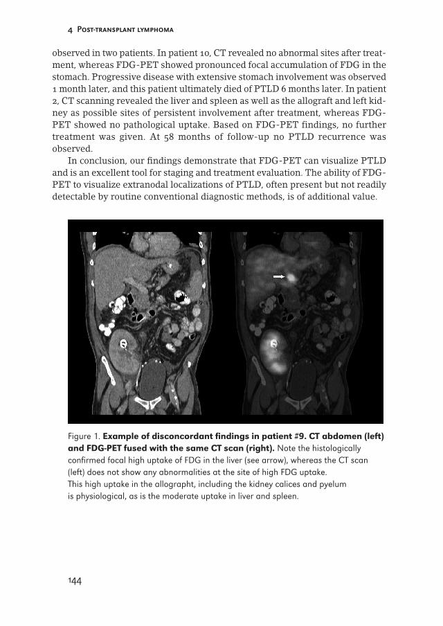
observed in two patients. In patient 10, CT revealed no abnormal sites after treat-ment, whereas FDG-PET showed pronounced focal accumulation of FDG in thestomach. Progressive disease with extensive stomach involvement was observed1 month later, and this patient ultimately died of PTLD 6 months later. In patient2, CT scanning revealed the liver and spleen as well as the allograft and left kid-ney as possible sites of persistent involvement after treatment, whereas FDG-PET showed no pathological uptake. Based on FDG-PET findings, no furthertreatment was given. At 58 months of follow-up no PTLD recurrence wasobserved.
In conclusion, our findings demonstrate that FDG-PET can visualize PTLDand is an excellent tool for staging and treatment evaluation. The ability of FDG-PET to visualize extranodal localizations of PTLD, often present but not readilydetectable by routine conventional diagnostic methods, is of additional value.
Figure 1. Example of disconcordant findings in patient #9. CT abdomen (left)and FDG-PET fused with the same CT scan (right). Note the histologicallyconfirmed focal high uptake of FDG in the liver (see arrow), whereas the CT scan(left) does not show any abnormalities at the site of high FDG uptake. This high uptake in the allographt, including the kidney calices and pyelum is physiological, as is the moderate uptake in liver and spleen.
4 Post-transplant lymphoma
144
vImhoff-4 XP 28-11-2006 13:24 Pagina 144

References
1. Nalesnik MA. The diverse pathology of post-transplant lymphoproliferative disorders: theimportance of a standardized approach. Transpl Infect Dis 2001; 3(2):88-96.
2. Cheson BD, Horning SJ, Coiffier B, Shipp MA, Fisher RI, Connors JM et al. Report of aninternational workshop to standardize response criteria for non-Hodgkin’s lymphomas.NCI Sponsored International Working Group. J Clin Oncol 1999; 17(4):1244.
3. Moog F, Bangerter M, Diederichs CG, Guhlmann A, Merkle E, Frickhofen N et al.Extranodal malignant lymphoma: detection with FDG PET versus CT. Radiology 1998;206(2):475-481.
4. Schiepers C, Filmont JE, Czernin J. PET for staging of Hodgkin’s disease and non-Hodgkin’s lymphoma. Eur J Nucl Med Mol Imaging 2003; 30(Suppl 1): S82-S88.
5. Juweid ME, Wiseman GA, Vose JM, Ritchie JM, Menda Y, Wooldridge JE et al. Responseassessment of aggressive non-Hodgkin’s lymphoma by integrated International WorkshopCriteria and fluorine-18-fluorodeoxyglucose positron emission tomography. J Clin Oncol2005; 23(21):4652-4661.
PTLD Visualization by FDG-PET
145
vImhoff-4 XP 28-11-2006 13:24 Pagina 145

146
vImhoff-4 XP 28-11-2006 13:24 Pagina 146