Osteoporosetherapie 2018 - Wo ist der Fortschritt ? · Wnt Signaling Pathway Inhibitor Sclerostin....
36
Osteoporosetherapie 2018 Wo ist der Fortschritt ? Prof. Drs. Christian Kasperk Leitender Oberarzt Innere Medizin I und Klinische Chemie (Endokrinologie und Stoffwechsel) Sektionsleiter Osteologie Leiter des Steroidlabor des Pharmakologischen Instituts Medizinische Universitätsklinik Heidelberg
Transcript of Osteoporosetherapie 2018 - Wo ist der Fortschritt ? · Wnt Signaling Pathway Inhibitor Sclerostin....
Osteoporosetherapie 2018Wo ist der Fortschritt ?
Prof. Drs. Christian KasperkLeitender Oberarzt Innere Medizin I und Klinische Chemie (Endokrinologie und Stoffwechsel)Sektionsleiter Osteologie Leiter des Steroidlabor des Pharmakologischen InstitutsMedizinische Universitätsklinik Heidelberg
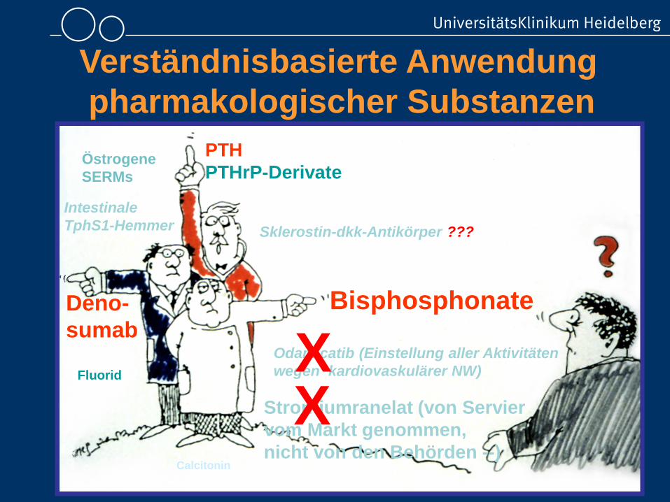
Verständnisbasierte Anwendung pharmakologischer Substanzen
PTHPTHrP-Derivate
Bisphosphonate
Calcitonin
Deno-sumab
Sklerostin-dkk-Antikörper ???
Odanacatib (Einstellung aller Aktivitätenwegen kardiovaskulärer NW)
ÖstrogeneSERMs
Strontiumranelat (von Serviervom Markt genommen, nicht von den Behörden --)
Fluorid
IntestinaleTphS1-Hemmer
XX
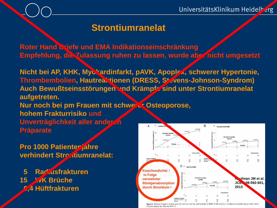
Strontiumranelat
Kaufman JM et al. JCEM 98:592-601,2013
Roter Hand Briefe und EMA Indikationseinschränkung Empfehlung, die Zulassung ruhen zu lassen, wurde aber nicht umgesetzt
Nicht bei AP, KHK, Myokardinfarkt, pAVK, Apoplex, schwerer Hypertonie,Thrombembolien, Hautreaktionen (DRESS, Stevens-Johnson-Syndrom)Auch Bewußtseinsstörungen und Krämpfe sind unter Strontiumranelat aufgetreten.Nur noch bei pm Frauen mit schwerer Osteoporose, hohem Frakturrisiko und Unverträglichkeit aller anderen Präparate
Pro 1000 Patientenjahre verhindert Strontiumranelat:
5 Radiusfrakturen15 WK Brüche0,4 Hüftfrakturen
Knochendichte ↑in Folge
vermehrter Röntgenabsorption durch Strontium !
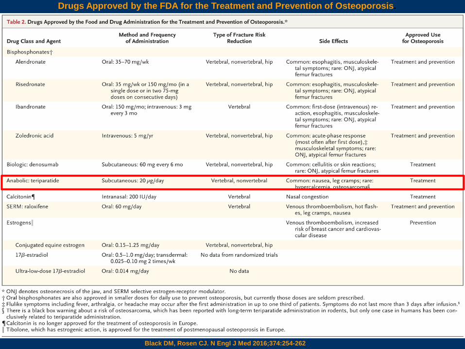
Drugs Approved by the FDA for the Treatment and Prevention of Osteoporosis.
Black DM, Rosen CJ. N Engl J Med 2016;374:254-262
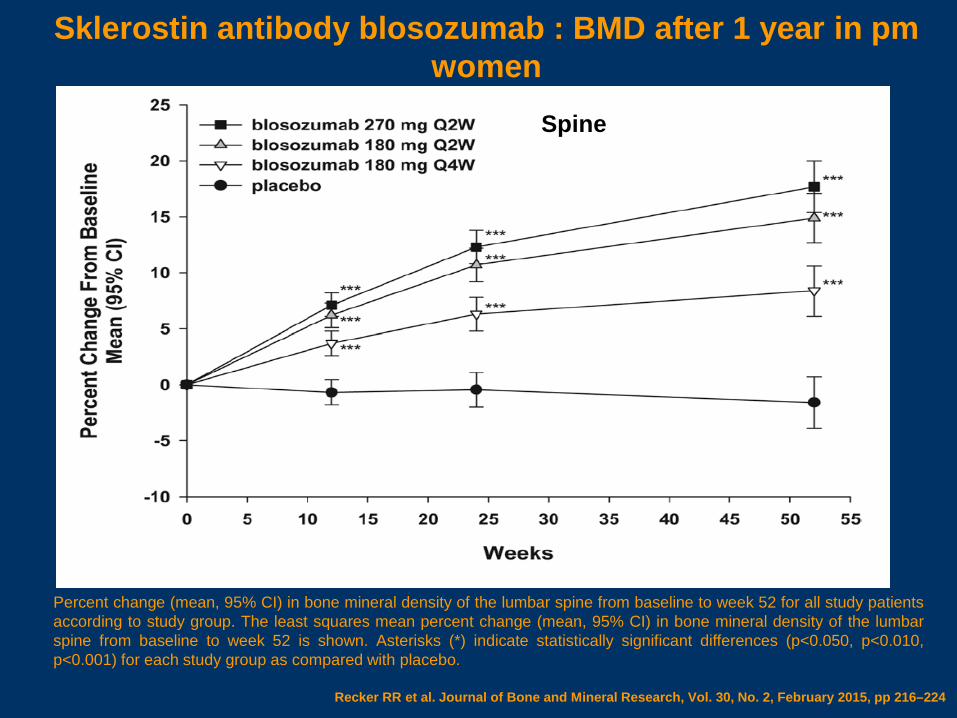
Percent change (mean, 95% CI) in bone mineral density of the lumbar spine from baseline to week 52 for all study patientsaccording to study group. The least squares mean percent change (mean, 95% CI) in bone mineral density of the lumbarspine from baseline to week 52 is shown. Asterisks (*) indicate statistically significant differences (p<0.050, p<0.010,p<0.001) for each study group as compared with placebo.
Recker RR et al. Journal of Bone and Mineral Research, Vol. 30, No. 2, February 2015, pp 216–224
Sklerostin antibody blosozumab : BMD after 1 year in pm women
Spine
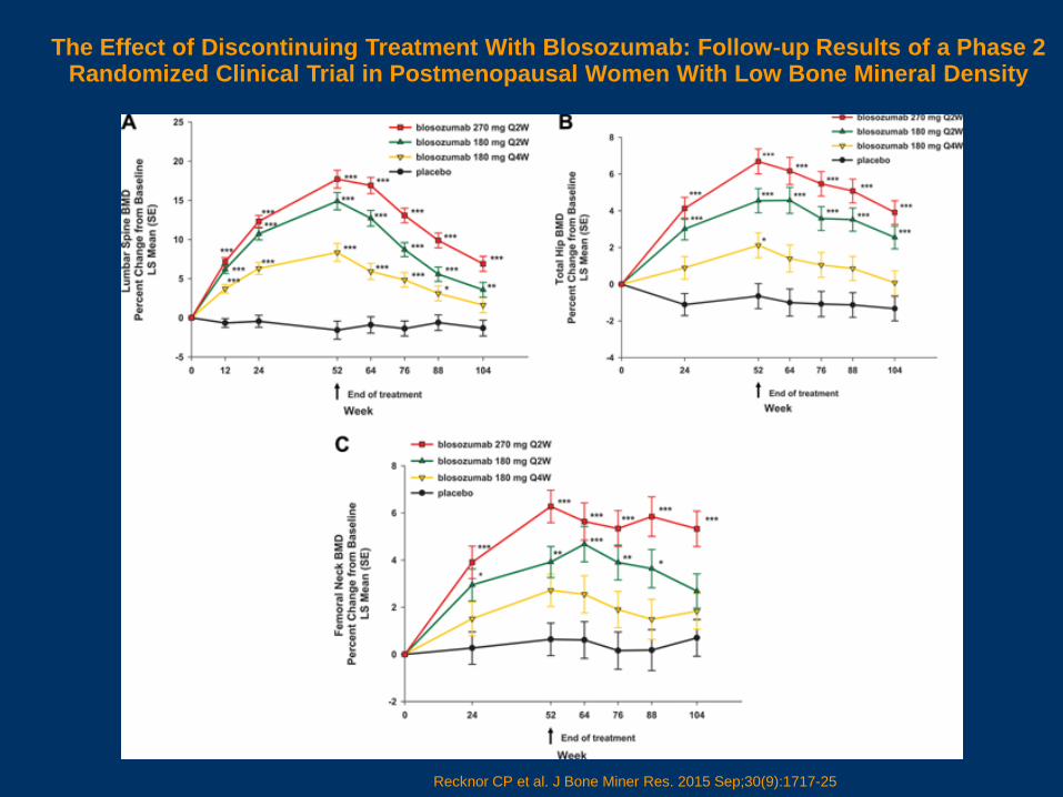
The Effect of Discontinuing Treatment With Blosozumab: Follow‐up Results of a Phase 2 Randomized Clinical Trial in Postmenopausal Women With Low Bone Mineral Density
Recknor CP et al. J Bone Miner Res. 2015 Sep;30(9):1717-25
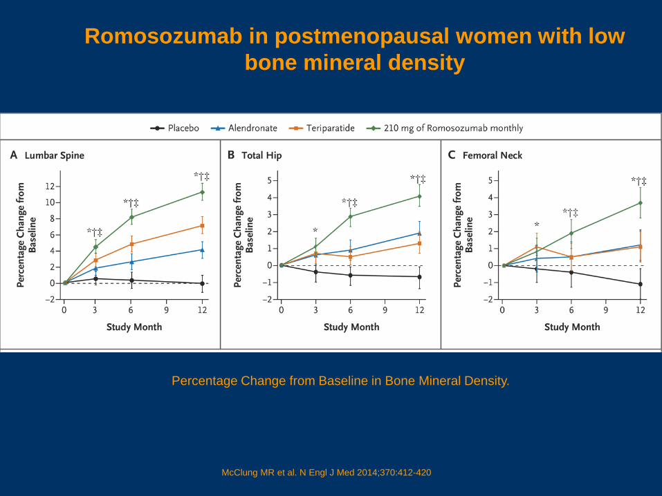
Percentage Change from Baseline in Bone Mineral Density.
McClung MR et al. N Engl J Med 2014;370:412-420
Romosozumab in postmenopausal women with low bone mineral density
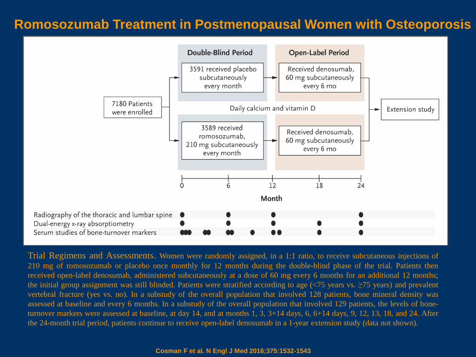
Trial Regimens and Assessments. Women were randomly assigned, in a 1:1 ratio, to receive subcutaneous injections of210 mg of romosozumab or placebo once monthly for 12 months during the double-blind phase of the trial. Patients thenreceived open-label denosumab, administered subcutaneously at a dose of 60 mg every 6 months for an additional 12 months;the initial group assignment was still blinded. Patients were stratified according to age (<75 years vs. ≥75 years) and prevalentvertebral fracture (yes vs. no). In a substudy of the overall population that involved 128 patients, bone mineral density wasassessed at baseline and every 6 months. In a substudy of the overall population that involved 129 patients, the levels of bone-turnover markers were assessed at baseline, at day 14, and at months 1, 3, 3+14 days, 6, 6+14 days, 9, 12, 13, 18, and 24. Afterthe 24-month trial period, patients continue to receive open-label denosumab in a 1-year extension study (data not shown).
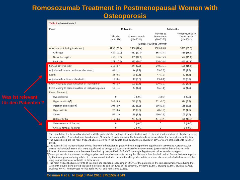
Romosozumab Treatment in Postmenopausal Women with Osteoporosis
Cosman F et al. N Engl J Med 2016;375:1532-1543
N Engl J Med. 2016 Oct 20;375(16):1532-1543Cosman F et al. Romosozumab treatment in postmenopausal women with osteoporosis. N Engl J Med 375:1532-1543, 2016
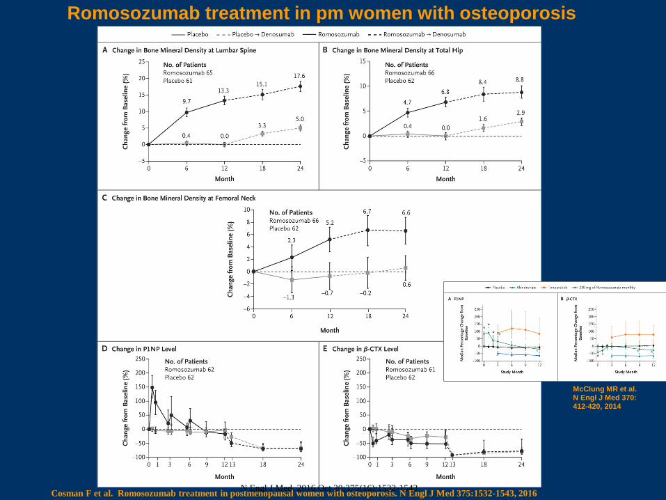
Romosozumab treatment in pm women with osteoporosis
McClung MR et al. N Engl J Med 370:412-420, 2014
Cosman F et al. Romosozumab treatment in postmenopausal women with osteoporosis.N Engl J Med 375:1532-1543, 2016
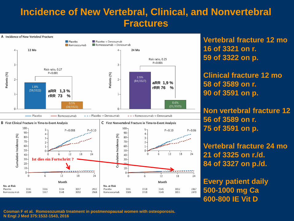
Incidence of New Vertebral, Clinical, and Nonvertebral Fractures
Vertebral fracture 12 mo16 of 3321 on r.59 of 3322 on p.
Clinical fracture 12 mo58 of 3589 on r.90 of 3591 on p.
Non vertebral fracture 12 56 of 3589 on r.75 of 3591 on p.
Vertebral fracture 24 mo21 of 3325 on r./d.84 of 3327 on p./d.
Every patient daily500-1000 mg Ca600-800 IE Vit D
aRR 1,3 %rRR 73 %
aRR 1,9 %rRR 76 %
Ist dies ein Fortschritt ?
Romosozumab Treatment in Postmenopausal Women with Osteoporosis
Adverse events
Cosman F et al. N Engl J Med 2016;375:1532-1543
Was ist relevantfür den Patienten ?
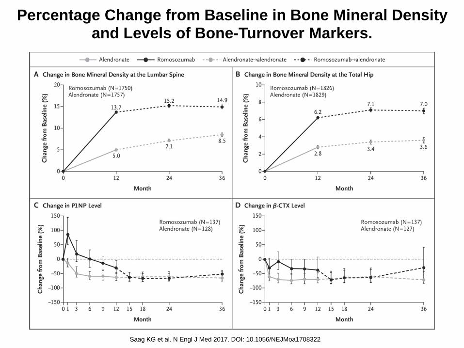
Percentage Change from Baseline in Bone Mineral Density and Levels of Bone-Turnover Markers.
Saag KG et al. N Engl J Med 2017. DOI: 10.1056/NEJMoa1708322
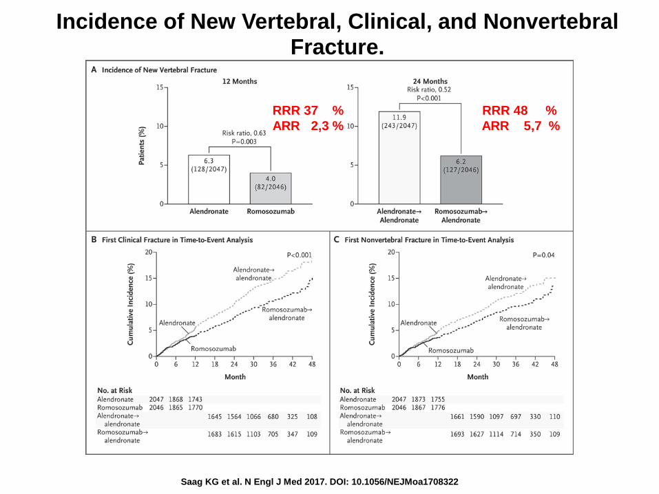
Incidence of New Vertebral, Clinical, and Nonvertebral Fracture.
Saag KG et al. N Engl J Med 2017. DOI: 10.1056/NEJMoa1708322
RRR 37 % RRR 48 %ARR 2,3 % ARR 5,7 %
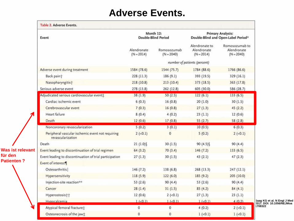
Adverse Events.
Saag KG et al. N Engl J Med 2017. DOI: 10.1056/NEJMoa1708322
Was ist relevantfür den Patienten ?
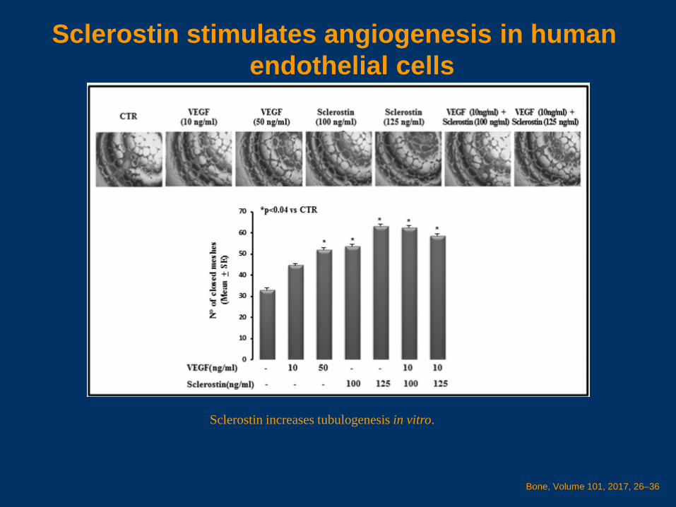
Sclerostin stimulates angiogenesis in human endothelial cells
Bone, Volume 101, 2017, 26–36
Sclerostin increases tubulogenesis in vitro.
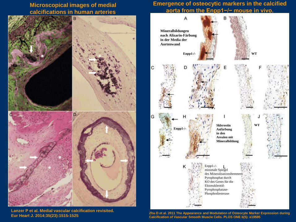
Microscopical images of medial calcifications in human arteries.
Lanzer P et al. Medial vascular calcification revisited.Eur Heart J. 2014;35(23):1515-1525
Emergence of osteocytic markers in the calcified aorta from the Enpp1−/− mouse in vivo.
Zhu D et al. 2011 The Appearance and Modulation of Osteocyte Marker Expression during Calcification of Vascular Smooth Muscle Cells. PLOS ONE 6(5): e19595
Mineralbildungennach Alizarin-Färbungin der Media der Aortenwand
SklerostinAnfärbungin den Arealen mitMineralbildung
WTEnpp1-/-
WTEnpp1-/-
Enpp1-/-minimale Spiegel des Mineralisationshemmers Pyrophosphat durch KO des Genes für dieEktonukleotid-Pyrophosphatase-Phosphodiesterase
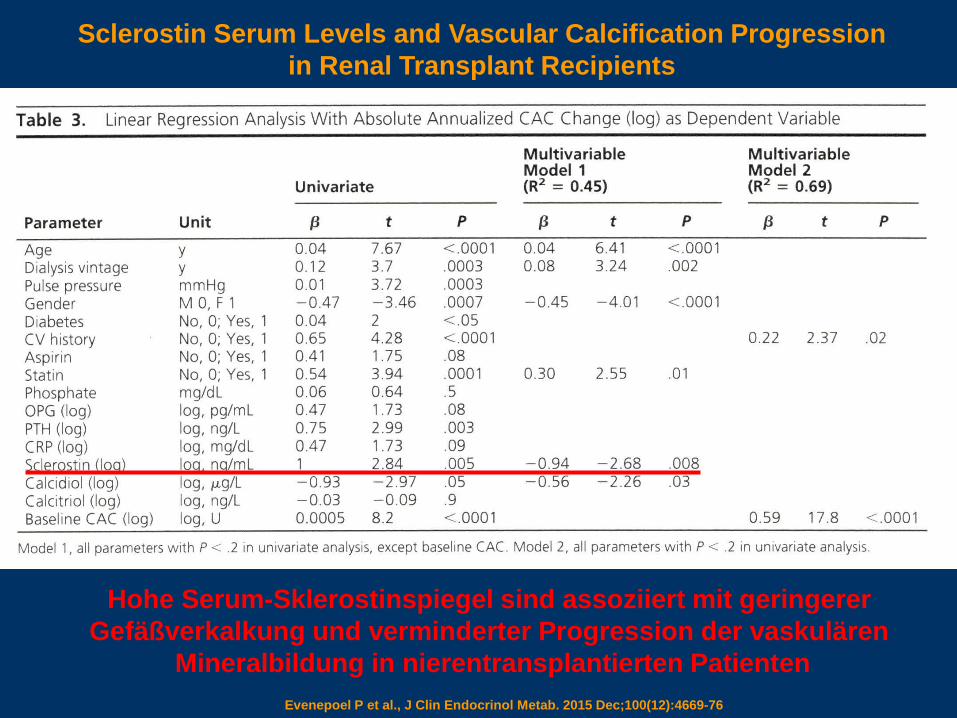
Sclerostin Serum Levels and Vascular Calcification Progression in Renal Transplant Recipients
Evenepoel P et al., J Clin Endocrinol Metab. 2015 Dec;100(12):4669-76
Hohe Serum-Sklerostinspiegel sind assoziiert mit geringerer Gefäßverkalkung und verminderter Progression der vaskulären
Mineralbildung in nierentransplantierten Patienten
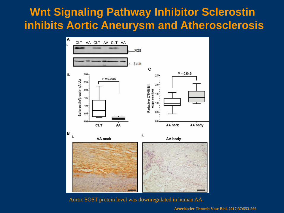
Wnt Signaling Pathway Inhibitor Sclerostininhibits Aortic Aneurysm and Atherosclerosis
Arterioscler Thromb Vasc Biol. 2017;37:553-566
Aortic SOST protein level was downregulated in human AA.
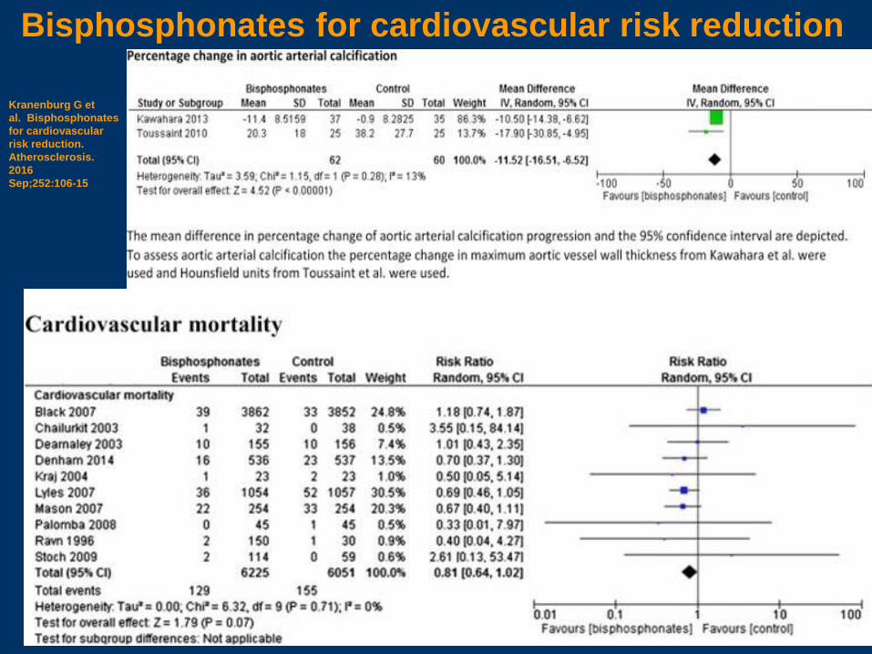
Bisphosphonates for cardiovascular risk reduction
Kranenburg G et al. Bisphosphonatesfor cardiovascularrisk reduction.Atherosclerosis.2016 Sep;252:106-15
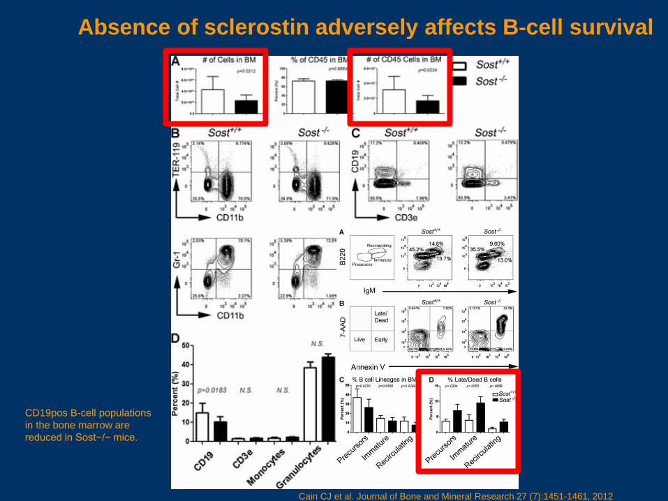
Absence of sclerostin adversely affects B‐cell survival
CD19pos B-cell populations in the bone marrow are reduced in Sost−/− mice.
Cain CJ et al. Journal of Bone and Mineral Research 27 (7):1451-1461, 2012
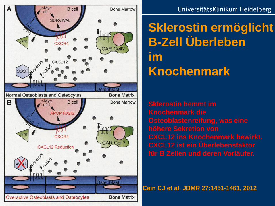
Sklerostin ermöglichtB-Zell Überlebenim Knochenmark
Cain CJ et al. JBMR 27:1451-1461, 2012
Sklerostin hemmt im Knochenmark die Osteoblastenreifung, was eine höhere Sekretion vonCXCL12 ins Knochenmark bewirkt. CXCL12 ist ein Überlebensfaktor für B Zellen und deren Vorläufer.
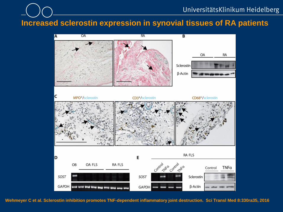
Increased sclerostin expression in synovial tissues of RA patients
Wehmeyer C et al. Sclerostin inhibition promotes TNF-dependent inflammatory joint destruction. Sci Transl Med 8:330ra35, 2016
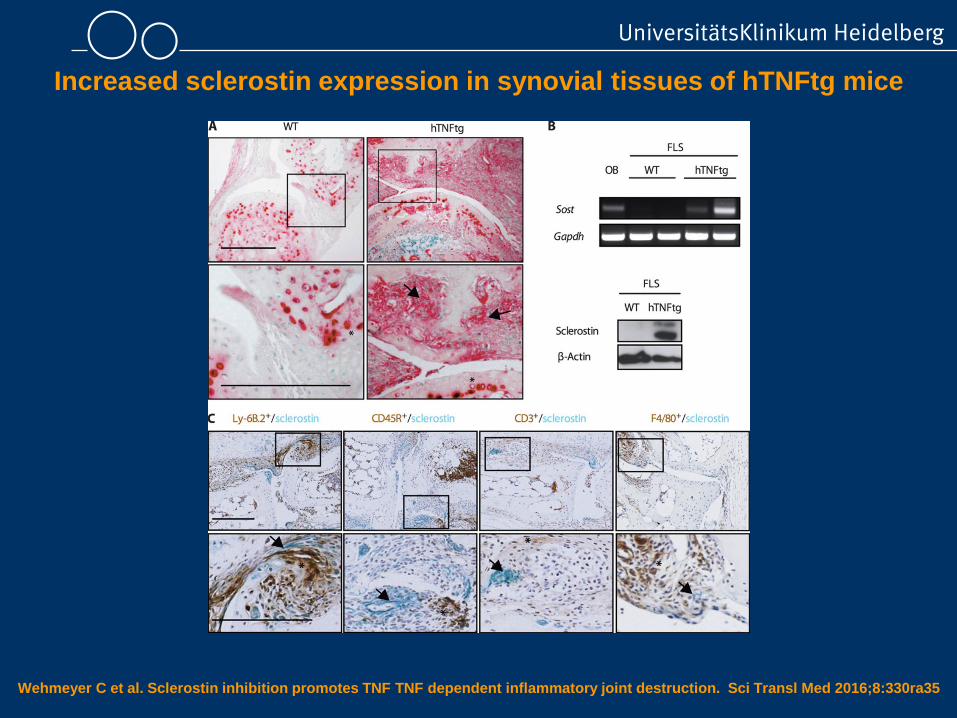
Increased sclerostin expression in synovial tissues of hTNFtg mice
Wehmeyer C et al. Sclerostin inhibition promotes TNF TNF dependent inflammatory joint destruction. Sci Transl Med 2016;8:330ra35
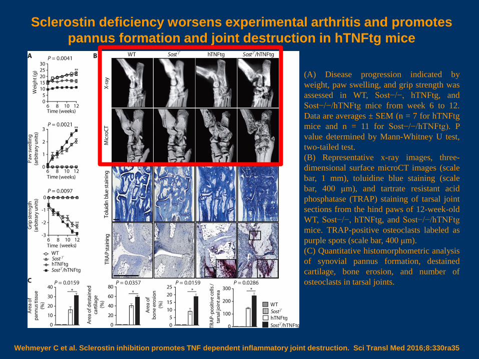
Sclerostin deficiency worsens experimental arthritis and promotes pannus formation and joint destruction in hTNFtg mice
Wehmeyer C et al. Sclerostin inhibition promotes TNF dependent inflammatory joint destruction. Sci Transl Med 2016;8:330ra35
(A) Disease progression indicated byweight, paw swelling, and grip strength wasassessed in WT, Sost−/−, hTNFtg, andSost−/−/hTNFtg mice from week 6 to 12.Data are averages ± SEM (n = 7 for hTNFtgmice and n = 11 for Sost−/−/hTNFtg). Pvalue determined by Mann-Whitney U test,two-tailed test.(B) Representative x-ray images, three-dimensional surface microCT images (scalebar, 1 mm), toluidine blue staining (scalebar, 400 μm), and tartrate resistant acidphosphatase (TRAP) staining of tarsal jointsections from the hind paws of 12-week-oldWT, Sost−/−, hTNFtg, and Sost−/−/hTNFtgmice. TRAP-positive osteoclasts labeled aspurple spots (scale bar, 400 μm).(C) Quantitative histomorphometric analysisof synovial pannus formation, destainedcartilage, bone erosion, and number ofosteoclasts in tarsal joints.
Zusammenfassung Sklerostin-Antikörper Therapie
Schnell wirksames osteoanaboles Therapieprinzip innerhalb eines Jahresbei pm Frauen mit Osteoporose, Anbaumarker aber bereits nach 6 Monatenauf Ausgangsniveau
Bisphosphonattherapie im Anschluß vermutlich sinnvoll zum vollen Erhalt der Knochenmasse nach Therapieende (Vermeidung Anschlußfrakturennach Absetzen von Denosumab: Popp AW et al. OI 2016; Lamy O et al. JCEM 102.354, 2017) ?
Ohne weitere Langzeitstudien (üblich 3 Jahre) Zurückhaltung bei Ost.Pat. mit KHK oder AVK oder Aneurysma
renaler Osteopathie
TNF vermittelten Erkrankungen wie Arthritis, Crohn, Colitis etc.
immun-kompromitierten Patienten (z.B. Transplantierte oder Patienten unter dauerhafter Glukokortikoidbehandlung)
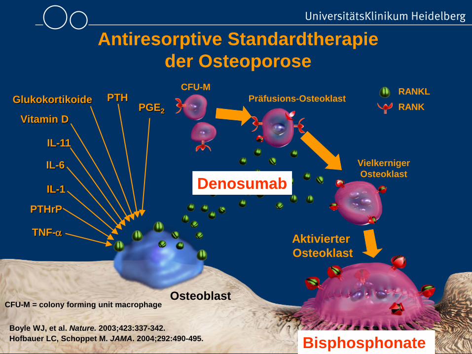
Osteoblast
Aktivierter Osteoklast
TNF-α
PTH
IL-1
PTHrP
Glukokortikoide
Vitamin DPGE2
IL-11
RANKLRANK
Boyle WJ, et al. Nature. 2003;423:337-342.Hofbauer LC, Schoppet M. JAMA. 2004;292:490-495.
Antiresorptive Standardtherapie der Osteoporose
IL-6
Präfusions-OsteoklastCFU-M
VielkernigerOsteoklast
CFU-M = colony forming unit macrophage
Denosumab
Bisphosphonate

Frakturrisiko und Dichteänderung unter Therapie
Cummings et. al, Amer. J. Med. 112; 9332-9343, 2002
Normales Risiko
Raloxifen
Alendronat
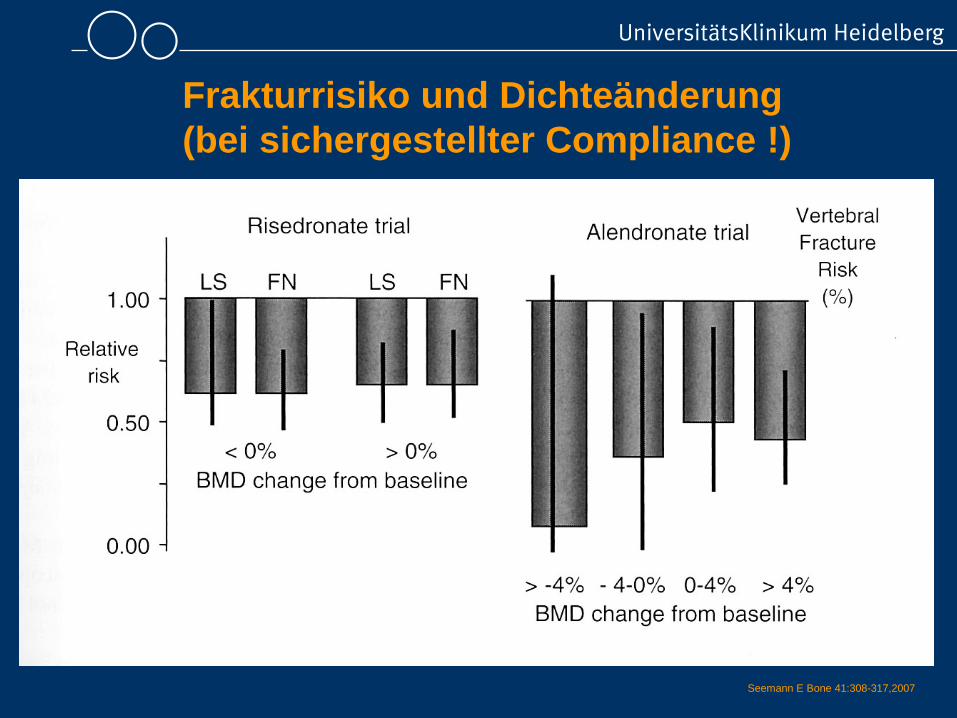
Frakturrisiko und Dichteänderung(bei sichergestellter Compliance !)
Seemann E Bone 41:308-317,2007
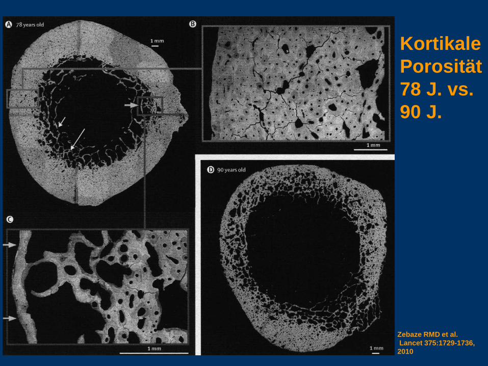
Zebaze RMD et al.Lancet 375:1729-1736, 2010
Kortikale Porosität78 J. vs. 90 J.
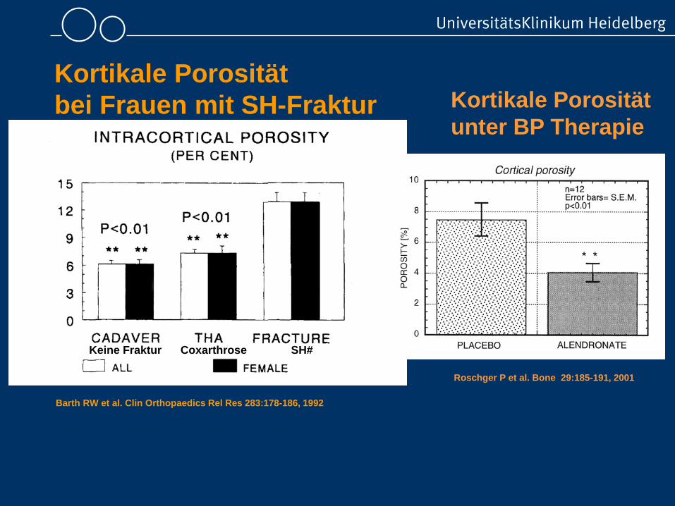
Keine Fraktur Coxarthrose SH#
Barth RW et al. Clin Orthopaedics Rel Res 283:178-186, 1992
Kortikale Porosität bei Frauen mit SH-Fraktur Kortikale Porosität
unter BP Therapie
Roschger P et al. Bone 29:185-191, 2001
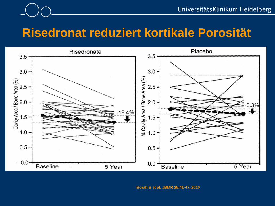
Borah B et al. JBMR 25:41-47, 2010
Risedronat reduziert kortikale Porosität
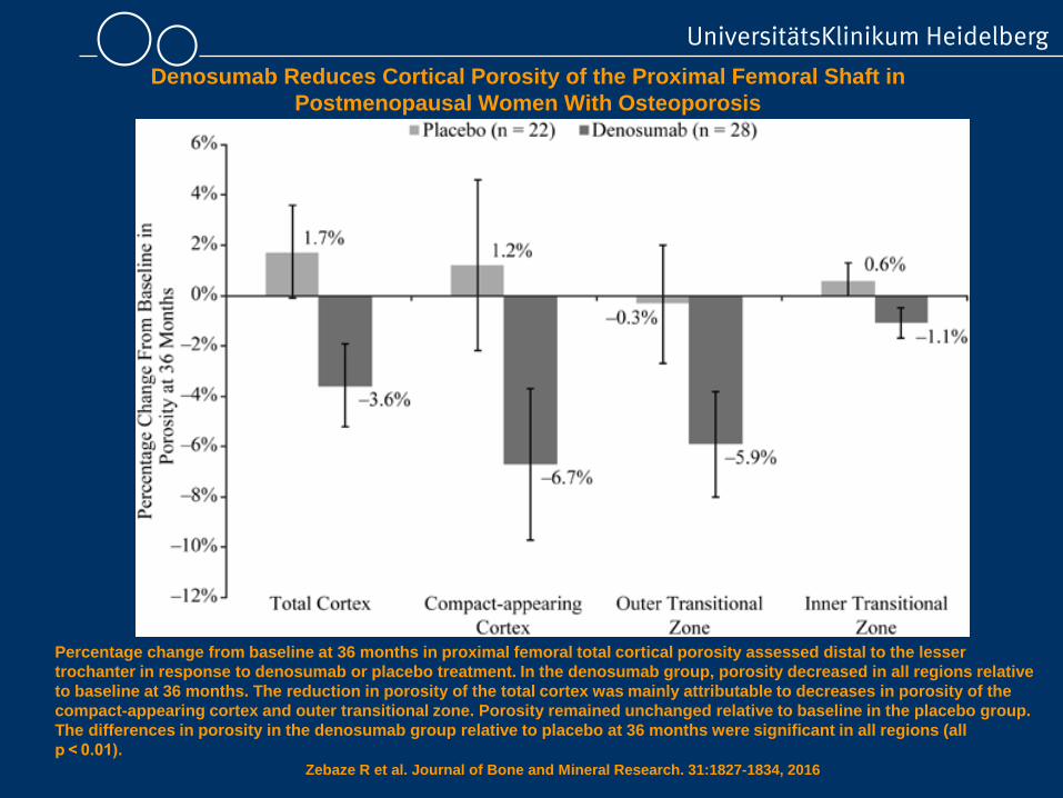
Denosumab Reduces Cortical Porosity of the Proximal Femoral Shaft in Postmenopausal Women With Osteoporosis
Percentage change from baseline at 36 months in proximal femoral total cortical porosity assessed distal to the lesser trochanter in response to denosumab or placebo treatment. In the denosumab group, porosity decreased in all regions relative to baseline at 36 months. The reduction in porosity of the total cortex was mainly attributable to decreases in porosity of the compact-appearing cortex and outer transitional zone. Porosity remained unchanged relative to baseline in the placebo group. The differences in porosity in the denosumab group relative to placebo at 36 months were significant in all regions (all p < 0.01).
Zebaze R et al. Journal of Bone and Mineral Research. 31:1827-1834, 2016
Raloxifen Raloxifene Use for The Heart trial (RUTH Studie)
Ergebnisse• Reduziert das Risiko klinisch relevanter WK-Frakturen um 35%
• Reduziert das allgemeine Brustkrebs-Risiko um 33%
• Keinen signifikanten Effekt auf das Risiko prim. CV-Ereignisse (HR 0,95)
• Keinen signifikanten Unterschied bezügl. des allgemeinen Stroke-Risikos
ABER: Raloxifen assoziiert mit einem erhöhten Risiko für letalen Stroke (um 49% höher)Risiko für venöse Thrombembolien um 44% erhöht
Nutzen Geringe Frakturpräventionabwägen Verstärkung postmenopausaler Beschwerden
Thrombembolie Risiko erhöht wie bei ÖstrogenenBarrett-Connor et al., N Engl J Med 2006; 355:125-137
Welchen Fortschritt bringt diese Substanz ?
ZusammenfassungStandardtherapie der Osteoporose sind die Bisphosphonate.
Bei sichergestellter Compliance gibt es keine „beste“ Substanz.
Neue Frakturen (Ursache !) oder Dichteverluste < 4 %/a (g/cm2 Werte) in den ersten 2 Jahren einer BP Therapie sind keine absolute Indikation zum Präparatewechsel,eher für einen längeren Therapiezeitraum.
Alternativen müssen entsprechend der individuellen Krankheitssituation des Patienten und der bekannten Nebenwirkungsprofile geprüft werden.
Experten Rat: Nach Absetzen/Pausieren von Nicht-Bisphosphonaten (z.B. Denosumab, Teriparatid) 1-jährige BP-Nachbehandlungoder zumindest Dichtekontrolle schon nach 6-12 Monaten und dann jährlich erscheint zweckmäßig.
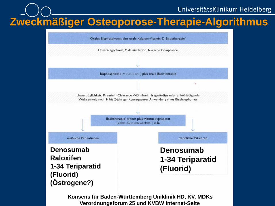
Zweckmäßiger Osteoporose-Therapie-Algorithmus
DenosumabRaloxifen1-34 Teriparatid(Fluorid) (Östrogene?)
Denosumab1-34 Teriparatid(Fluorid)
Konsens für Baden-Württemberg Uniklinik HD, KV, MDKsVerordnungsforum 25 und KVBW Internet-Seite

Vielen Dank