Targeting the endolysosomal system of cancer cells by ...
148
Dissertation zur Erlangung des Doktorgrades der Fakultät für Chemie und Pharmazie der Ludwig-Maximilians-Universität München Targeting the endolysosomal system of cancer cells by inhibition of V-ATPase and TPC function Lina Sophie Schneider aus Freising, Deutschland 2015
Transcript of Targeting the endolysosomal system of cancer cells by ...
Dissertation zur Erlangung des Doktorgrades
der Fakultät für Chemie und Pharmazie
der Ludwig-Maximilians-Universität München
Targeting the endolysosomal system of cancer cells by
inhibition of V-ATPase and TPC function
Lina Sophie Schneider
aus
Freising, Deutschland
2015
Erklärung
Diese Dissertation wurde im Sinne von § 7 der Promotionsordnung vom 28. November 2011
von Frau Prof. Dr. Angelika M. Vollmar betreut.
Eidesstattliche Versicherung
Diese Dissertation wurde eigenständig und ohne unerlaubte Hilfe erarbeitet.
München, den 28. Oktober 2015
_________________________
(Lina Sophie Schneider)
Dissertation eingereicht am 03.11.2015
1.Gutachterin Prof. Dr. Angelika M. Vollmar
2.Gutachter Prof. Dr. Christian Wahl-Schott
Mündliche Prüfung am 02.12.2015

Meinen Eltern und meiner Schwester
Table of contents I
1 Table of contents
1 Table of contents .................................................................................................................. I
2 Abbreviations ......................................................................................................................II
3 Manuscripts ....................................................................................................................... III
4 Summary ........................................................................................................................... IV
5 Introduction ......................................................................................................................... 1
5.1 Cancer and current treatment strategies ....................................................................... 1
5.2 Targeting the endolysosomal system of cancer cells ................................................... 1
5.2.1 The vacuolar-type ATPase ................................................................................... 4
5.2.2 Two-pore channels ............................................................................................... 7
5.3 Overcoming the hallmarks of cancer ........................................................................... 9
6 Aim of the study ................................................................................................................ 11
7 Short summary of manuscripts ......................................................................................... 12
7.1 Vacuolar-ATPase inhibition blocks iron metabolism to mediate therapeutic
effects in breast cancer .............................................................................................. 12
7.2 Anti-leukemic effects of the V-ATPase inhibitor Archazolid A ............................... 13
7.3 MDM2 antagonist nutlin-3a sensitizes tumors to V-ATPase inhibition ................... 14
7.4 Two-pore channel function is crucial for migration of invasive cancer cells ............ 15
8 References ......................................................................................................................... 16
9 Publications ....................................................................................................................... 22
9.1 Original articles ......................................................................................................... 22
9.2 Oral presentations ...................................................................................................... 22
9.3 Poster presentations ................................................................................................... 23
10 Acknowledgements ....................................................................................................... 24
11 Appendix ....................................................................................................................... 26
Abbreviations II
2 Abbreviations
2-OG 2-oxoglutarate
3-AP 3-aminopyridine-2-carboxaldehyde thiosemicarbazone
ADP adenosine diphosphate
AIF apoptosis-inducing factor
AML acute myeloid leukemia
Arch archazolid
ATM ataxia telangiectasia mutated
ATP adenosine triphosphate
Bax Bcl-2-associated X protein
Bcl-2 B-cell lymphoma 2
Bcl-xL B-cell lymphoma extra large
Bid BH3-interacting domain death agonist
BNIP3 Bcl-2/adenovirus E1B 19kDa interacting protein 3
CAIX carbonic anhydrase IX
Caspase cysteine-dependent aspartate-specific protease
CDK1 cyclin-dependent kinase 1
CO control
CREB cyclic AMP response element-binding
DBZ dibenzazepine
dCTP deoxycytidine triphosphate
DFO deferoxamine
DMSO dimethyl sulfoxide
Abbreviations II
DMT1 divalent metal transporter 1
dNTP deoxyribonucleotide triphosphate
Doxo doxorubicin
DSB double-strand break
DTT dithiothreitol
ECM extracellular matrix
EDTA ethylenediaminetetraacetic acid
EEA1 early endosome antigen 1
EGF epidermal growth factor
EGFR epidermal growth factor receptor
EGTA ethylene glycol tetraacetic acid
ErbB2 human epidermal growth factor receptor 2
EV empty vector
FACS fluorescence-activated cell sorting
FAK focal adhesion kinase
FCS fetal calf serum
FeCit iron citrate
Glut1 glucose transporter 1
GSI γ-secretase inhibitor
HEPES 4-(2-hydroxyethyl)-1-piperazineethanesulfonic acid
Hes1 hairy and enhancer of split 1
Hey1/2 hairy and enhancer of split related with YRPW motif 1/2
HIF1α hypoxia-inducible factor 1α
Abbreviations II
HRP horseradish peroxidase
IAP1/2 inhibitor of apoptosis 1/2
IGFBP3 insulin-like growth factor-binding protein 3
LAMP1/3 lysosomal-associated membrane protein 1/3
LDL low density lipoprotein
LDLR low density lipoprotein receptor
LMP lysosomal membrane permeabilization
MCT monocarboxylate transporter
MDM2 mouse double minute 2
MDS myelodysplastic syndrome
MEF mouse embryonic fibroblasts
MES 2-(N-morpholino)ethanesulfonic acid
MSA methanesulfonic acid
mTOR mammalian target of rapamycin
mTORC1 mammalian target of rapamycin complex 1
NAADP nicotinic acid adenine dinucleotide phosphate
NADP nicotinamide adenine dinucleotide phosphate
Ned-19 trans-Ned-19
NHE1 Na+/H
+ exchanger 1
NICD notch intracellular domain
NRARP notch-regulated ankyrin repeat protein
NT non-targeting
OXPHOS oxidative phosphorylation
Abbreviations II
PARP poly ADP ribose polymerase
PBMC peripheral blood mononuclear cell
PBS phosphate-buffered saline
PDGFRβ platelet-derived growth factor receptor β
PDX patient-derived xenograft
PFA paraformaldehyde
PHD prolyl hydroxylase
PI propidium iodide
PI(3,5)P2 phosphatidylinositol 3,5-bisphosphate
PMSF phenylmethylsulfonyl fluoride
Rb retinoblastoma
RNR ribonucleotide reductase
SD standard deviation
SEM standard error of the mean
T-ALL T-cell acute lymphoblastic leukemia
Tet tetrandrine
Tf transferrin
TfR transferrin receptor
TIGAR TP53-induced glycolysis and apoptosis regulator
TPC two-pore channel
TRP transient receptor potential
TRPML1 transient receptor potential mucolipin 1
V-ATPase vacuolar-type ATPase

Abbreviations II
VEGF vascular endothelial growth factor
VHL von Hippel-Lindau
XIAP X-linked inhibitor of apoptosis
γH2AX phosphorylation of the H2AX histone
Manuscripts III
3 Manuscripts
This thesis is based on following manuscripts, which are referred to in the text by their roman
numerals (I-IV):
I. Lina S. Schneider, Karin von Schwarzenberg, Thorsten Lehr, Melanie Ulrich,
Rebekka Kubisch-Dohmen, Johanna Liebl, Dirk Menche and Angelika M. Vollmar
Vacuolar-ATPase inhibition blocks iron metabolism to mediate therapeutic effects in
breast cancer
2015, Cancer Research, 75(14):2863–2874
II. Siwei Zhang, Lina S. Schneider, Binje Vick, Michaela Grunert, Irmela Jeremias, Dirk
Menche, Rolf Müller, Angelika M. Vollmar and Johanna Liebl
Anti-leukemic effects of the V-ATPase inhibitor Archazolid A
2015, accepted for publication in Oncotarget
III. Lina S. Schneider, Melanie Ulrich, Thorsten Lehr, Dirk Menche, Rolf Müller,
Angelika M. Vollmar and Karin von Schwarzenberg
MDM2 antagonist nutlin-3a sensitizes tumors to V-ATPase inhibition
Submitted
IV. Lina S. Schneider*, Christian Grimm*, Yu-Kai Chao, Anna Watermann, Melanie
Ulrich, Doris Mayr, Christian Wahl-Schott, Martin Biel* and Angelika M. Vollmar*
Two-pore channel function is crucial for migration of invasive cancer cells
*These authors contributed equally to this work
Submitted
Summary IV
4 Summary
Fighting cancer is one of the big challenges of this century. The success of today’s anti-cancer
agents is limited by a variety of resistance mechanisms against the majority of
chemotherapeutics. Moreover, it is dependent on the existence of metastasis. Overcoming
these restraints by addressing new targets and by developing new treatment strategies is
therefore highly appreciated.
In recent years, targeting the endolysosomal machinery in cancer cells has come into focus as
a promising anti-tumor approach. Preserving the balance between endocytic internalization
and recycling of extracellular material, receptors, ligands and plasma membrane proteins is
substantially required to control cellular processes such as signal transduction, nutrient
uptake, cell adhesion and migration. Hence, disturbing endocytosis might be a promising
strategy to interfere with pro-survival mechanisms, especially in fast proliferating and high
metabolic active cancer cells.
To this end, we here addressed two endolysosomal proteins to unveil their roles in cancer
cells: the vacuolar-type ATPases (V-ATPases) and two-pore channels (TPCs). The V-ATPase
is a multisubunit proton pump located in the membranes of endosomes and lysosomes, which
actively transports protons into the lumen. Inhibition of these pumps was shown to induce
apoptosis in different cancer cells before, however the precise mechanism stayed unclear. To
investigate this matter, we used the myxobacterial V-ATPase inhibitor archazolid. Our first
study (manuscript I, Cancer Research, 75(14):2863–2874) confirmed a strong cell death
induction in the breast cancer cell lines MCF7 and MDA-MB-231 and showed induction of
the hypoxia-inducible factor 1α (HIF1α) after archazolid treatment. Considering that HIF1α
gets not only stabilized under hypoxia but also under iron depletion has led to experiments
with excessive iron citrate in the cell culture medium which showed a complete abolishment
of HIF1α stabilization and diminished cell death induction. The lack of cytosolic iron after
archazolid treatment could be attributed to disturbed transferrin (Tf)/transferrin receptor (TfR)
recycling. As a consequence, the iron depletion led to S-phase cell cycle arrest, double-strand
breaks and the induction of the tumor suppressor p53. Moreover, this study showed in vivo
efficacy of archazolid in a 4T1 mouse model, however it was not successful in abrogating
tumor growth completely.
In a second study (manuscript II, Oncotarget), archazolid’s apoptosis-inducing capabilities
were confirmed in leukemic cell lines and in patient derived xenograft (PDX) leukemic cells.
Mechanistically, V-ATPase inhibition reduced the expression level of the anti-apoptotic
Summary IV
protein survivin, contributing to archazolid-induced cell death since overexpression
significantly diminished the amount of dead cells. In respect to the underlying mechanism,
this study ties in with the above reported findings of manuscript I that archazolid led to S-
phase arrest by interfering with the iron metabolism in leukemic cells. Since survivin
expression strongly depends on cell cycle, these results suggest that archazolid decreased
survivin at the protein level by inducing S-phase arrest.
As above mentioned, manuscript I unveiled only a moderate reduction of tumor burden in
vivo for archazolid in breast cancer cells. Thus, we intended to improve this by a rational
combination approach. Since manuscript I further showed that archazolid treatment led to p53
stabilization in p53 wild type tumor cells, combination treatment within the p53 pathway
seemed promising. To this end, combination of archazolid with the small molecule p53
activator nutlin-3a (manuscript III) in different p53 wild type tumor cells resulted in
synergistic cell death induction. Remarkably, these findings were recapitulated in a U87MG
(U87) wild type p53 xenograft model as combination was more efficient in reducing tumor
growth compared to single dose treatment. On a molecular level we found reduced glycolysis,
since TP53-induced glycolysis and apoptosis regulator (TIGAR) mRNA levels were elevated
and glucose uptake and glucose transporter 1 (Glut1) protein levels were diminished.
Moreover, pro-apoptotic pathways including insulin-like growth factor-binding protein 3
(IGFBP3) and Bcl-2-associated X protein (Bax) were activated by the combination treatment.
In a different approach we addressed the endolysosomal system of cancer cells by targeting
TPCs (manuscript IV). TPCs are recently identified nicotinic acid adenine dinucleotide
phosphate (NAADP)- and phosphatidylinositol 3,5-bisphosphate (PI(3,5)P2)-sensitive Ca2+
-
permeable outward channels located in the membrane of endosomes and lysosomes. Their
impact on cancer has not yet been studied. For that reason, silencing of TPC1 and TPC2
mRNA levels with siRNA significantly reduced adhesion, migration through pores and
directed migration of T24 cancer cells. Pharmacological inhibition of TPCs by either trans-
Ned-19 (Ned-19) or tetrandrine was efficient in diminishing migration through pores in T24,
HUH7 and 4T1 cells. Mechanistic studies revealed that β1-integrin trafficking is disturbed
after Ned-19 treatment leading to accumulation in early endosome antigen 1 (EEA1)-positive
vesicles. This is suggested to be due to alterations in Ca2+
signaling since acidification of
vesicles was not affected. As a consequence, invasive cancer cells were no longer able to form
leading edges, which is substantially required for adequate migration. Finally, tetrandrine
showed anti-metastatic efficacy in a 4T1-Luc dissemination assay in vivo, altogether
suggesting a role for TPCs in cancer cell migration.
Summary IV
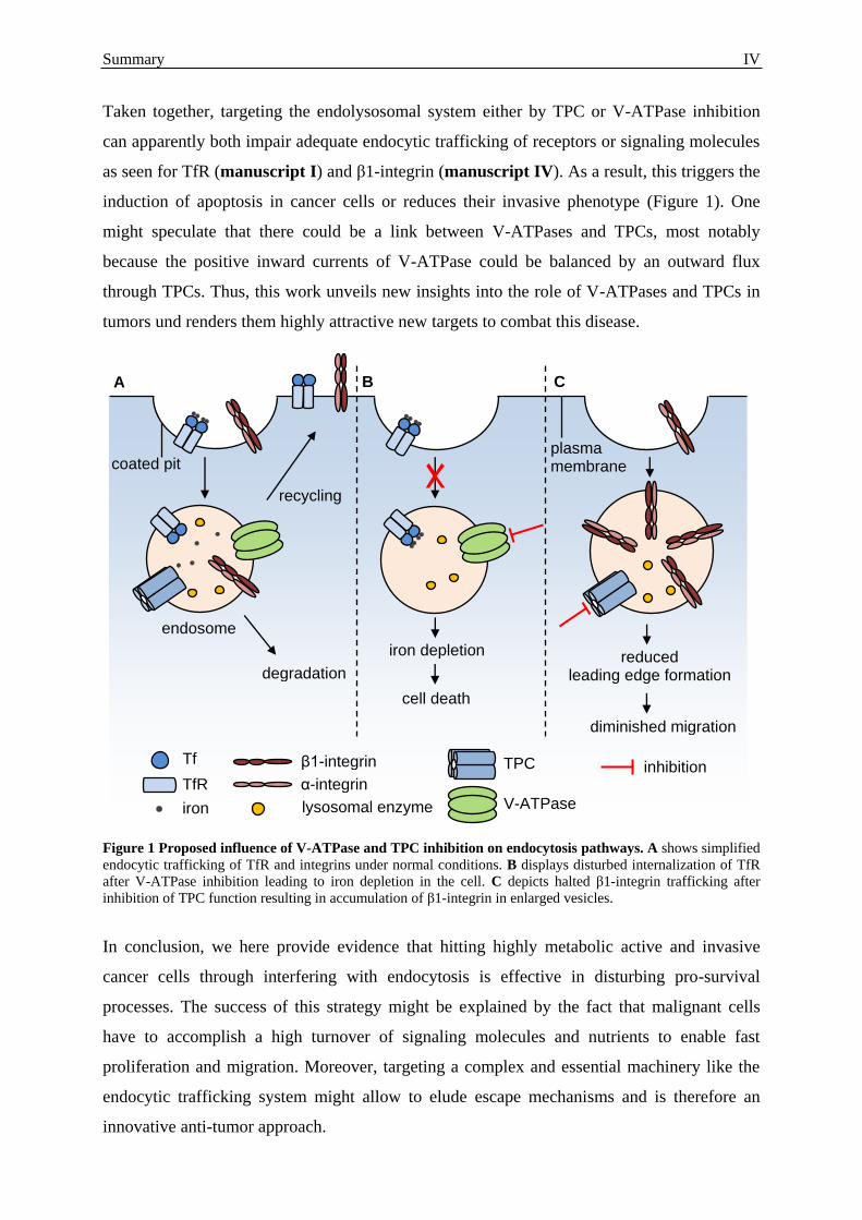
Taken together, targeting the endolysosomal system either by TPC or V-ATPase inhibition
can apparently both impair adequate endocytic trafficking of receptors or signaling molecules
as seen for TfR (manuscript I) and β1-integrin (manuscript IV). As a result, this triggers the
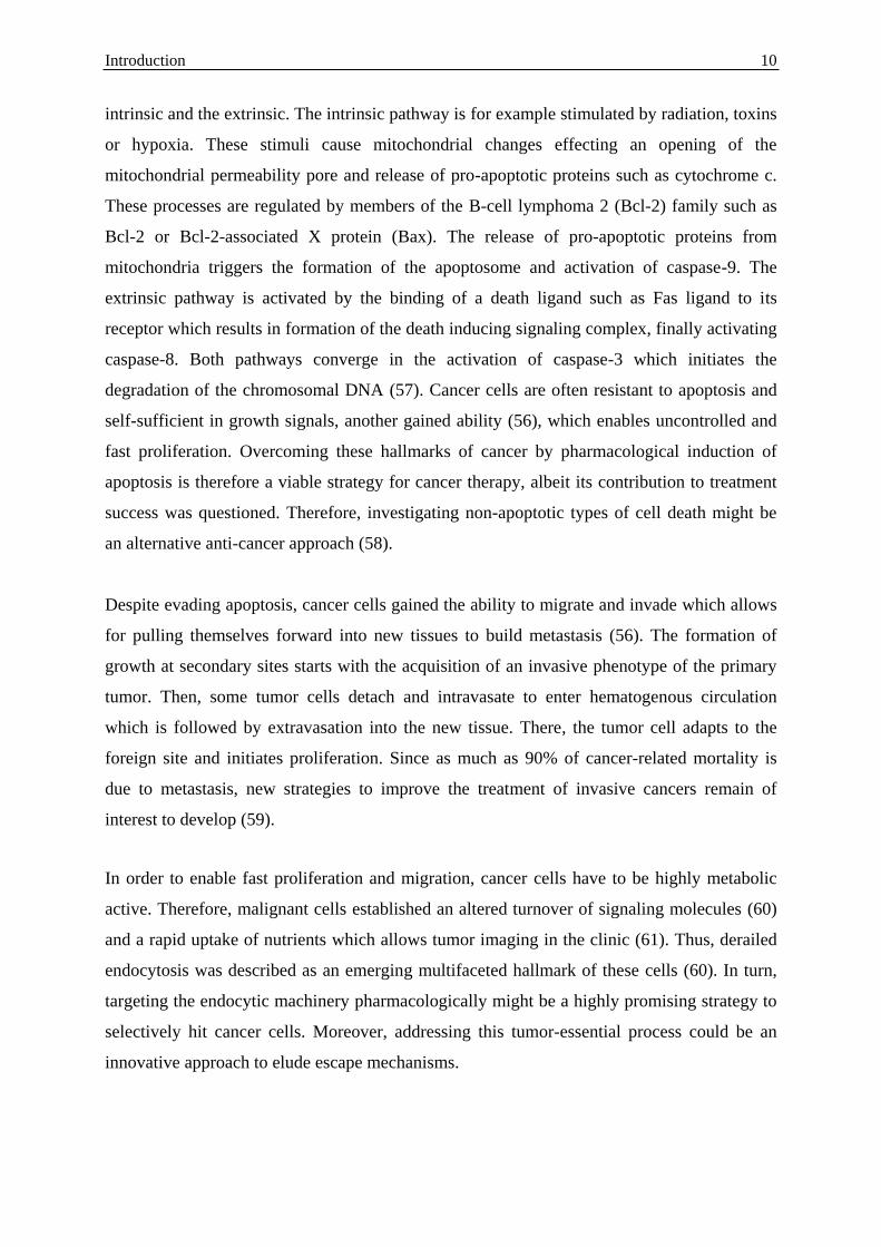
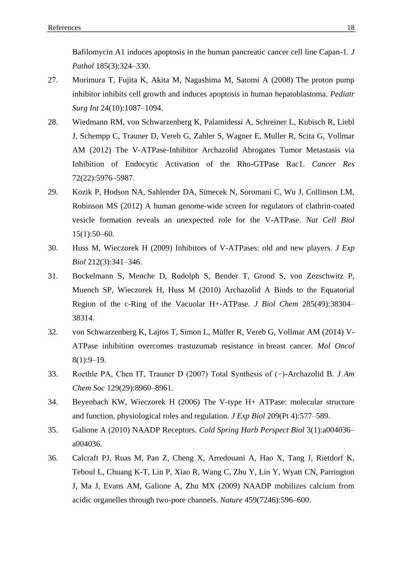
induction of apoptosis in cancer cells or reduces their invasive phenotype (Figure 1). One
might speculate that there could be a link between V-ATPases and TPCs, most notably
because the positive inward currents of V-ATPase could be balanced by an outward flux
through TPCs. Thus, this work unveils new insights into the role of V-ATPases and TPCs in
tumors und renders them highly attractive new targets to combat this disease.
Figure 1 Proposed influence of V-ATPase and TPC inhibition on endocytosis pathways. A shows simplified
endocytic trafficking of TfR and integrins under normal conditions. B displays disturbed internalization of TfR
after V-ATPase inhibition leading to iron depletion in the cell. C depicts halted β1-integrin trafficking after
inhibition of TPC function resulting in accumulation of β1-integrin in enlarged vesicles.
In conclusion, we here provide evidence that hitting highly metabolic active and invasive
cancer cells through interfering with endocytosis is effective in disturbing pro-survival
processes. The success of this strategy might be explained by the fact that malignant cells
have to accomplish a high turnover of signaling molecules and nutrients to enable fast
proliferation and migration. Moreover, targeting a complex and essential machinery like the
endocytic trafficking system might allow to elude escape mechanisms and is therefore an
innovative anti-tumor approach.
endosome
coated pit plasma membrane
recycling
degradation
iron depletion
cell death
reduced leading edge formation
diminished migration
Tf
TfR
iron
β1-integrin
α-integrin
lysosomal enzyme
TPC
V-ATPase
inhibition
A B C
Introduction 1
5 Introduction
5.1 Cancer and current treatment strategies
Cancer remains a leading cause of death worldwide. Hence, treating this disease is one of the
big challenges of this century, particularly because the burden is thought to increase in the
future (1). Nonetheless, due to improvements in treatment and earlier diagnosis the survival
rate for certain cancers has improved over the past 30 years. For example, the overall 5-year
relative survival rate for female breast cancer patients has increased from 74.8% between
1975 and 1977 to 90.3% between 2003 and 2009. However, it drops to 24.3% for patients
with distant-stage breast cancer. The clinical treatment of cancers mainly implicates,
dependent on the type and stage of the tumor, surgery, radiation and pharmacological therapy
(2). As the development of tumors involves in most cancers four to seven independent
mutations, pharmacological targeting of only one pathway is in many cases insufficient to
combat this disease. Therefore, a change to multi-target therapeutics has occurred in recent
years (3). Moreover, the success of today’s anti-cancer drugs is challenged by resistance
mechanisms against a variety of chemotherapeutics (4). Thus, there is still an urgent need to
find new targets and treatment strategies to fight cancer, especially for invasive malignancies
homing to distant sites.
5.2 Targeting the endolysosomal system of cancer cells
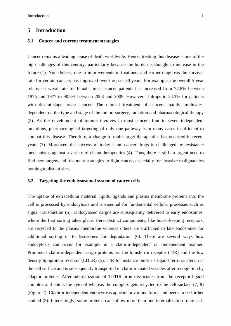
The uptake of extracellular material, lipids, ligands and plasma membrane proteins into the
cell is processed by endocytosis and is essential for fundamental cellular processes such as
signal transduction (5). Endocytosed cargos are subsequently delivered to early endosomes,
where the first sorting takes place. Here, distinct components, like house-keeping receptors,
are recycled to the plasma membrane whereas others are trafficked to late endosomes for
additional sorting or to lysosomes for degradation (6). There are several ways how
endocytosis can occur for example in a clathrin-dependent or -independent manner.
Prominent clathrin-dependent cargo proteins are the transferrin receptor (TfR) and the low
density lipoprotein receptor (LDLR) (5). TfR for instance binds its ligand ferrotransferrin at
the cell surface and is subsequently transported in clathrin-coated vesicles after recognition by
adaptor proteins. After internalization of Tf/TfR, iron dissociates from the receptor-ligand
complex and enters the cytosol whereas the complex gets recycled to the cell surface (7, 8)
(Figure 2). Clathrin-independent endocytosis appears in various forms and needs to be further
studied (5). Interestingly, some proteins can follow more than one internalization route as it
Introduction 2
has been shown for integrin heterodimers, which are a family of adhesion molecules
mediating the interaction between a cell and the extracellular matrix (ECM) (9). Preserving
the balance between endocytic internalization and recycling is substantially required to
control cellular processes such as signal transduction, nutrient uptake, cell polarity, cell
adhesion and migration (5).
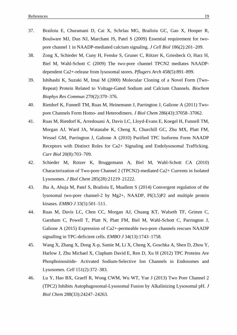
Figure 2 Endocytic trafficking pathways. The scheme depicts the transport of endocytic cargos upon
internalization exemplified by trafficking of Tf/TfR and LDL/LDLR. After uptake in clathrin-coated pits, cargos
are delivered to sorting endosomes and can be returned directly or recycled to the surface as shown for Tf/TfR or
transported to early and late endosomes and finally to lysosomes for degradation as illustrated for LDL. Scheme
designed and modified according to the model of Maxfield and MacGraw (10).
To function as the cellular sorting and degrading network, the endolysosomal system
establishes a pH gradient from 6.2 in early endosomes to 4.6 in lysosomes which allows for
high activity of degrading enzymes and for receptor-ligand processing. The luminal acidic
milieu is maintained by vacuolar-type ATPases (V-ATPases) which actively pump protons
across the endolysosomal membrane generating a positive inside membrane potential. As this
would quickly build a self-limiting electrochemical gradient, the flux of other ions is required
(6). Hence, despite V-ATPases, diverse other ion channels are thought to function in the
endolysosomal system of cells including Cl- antiporters, K
+/H
+ exchangers, Na
+/H
+
exchangers or Ca2+
/H+ exchangers and Ca
2+ channels (6, 11). Despite the mucolipins, which
sorting endosome late endosome
lysosome
coated pit plasma membrane
endocytic recycling compartment
Tf
TfR
iron
LDL
LDLR
lysosomal enzyme

Introduction 3
represent members of the transient receptor potential (TRP) superfamily of Ca2+
channels, the
two-pore channel (TPC) family has recently been identified as endolysosomal Ca2+
channels
(6) (Figure 3).
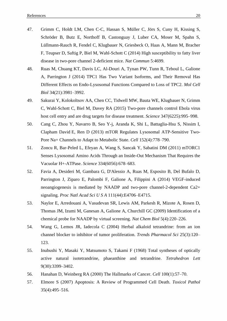
Figure 3 Overview of the main ionic influx and outflux mechanisms of an acidic organelle. Depicted is the
V-ATPase, which pumps protons across the membrane into the lumen upon ATP hydrolysis, two families of
voltage gated Ca2+ releasing channels: TPCs and the mucolipins, which are members of the TRP superfamily, the
postulated Ca2+/H+ exchanger and a Cl-/H+ antiporter (ClC, Cl- channel). TPCs and mucolipins may additionally
function as release channels for other cations (6, 11-13).
In search for new promising cancer targets lysosomes came in focus. In the acidic milieu of
these organelles hydrolases, such as cathepsins, are highly active in breaking down nucleic
acids, proteins or lipids. Interestingly, after lysosomal membrane permeabilization (LMP)
cathepsins can still function at cytosolic pH. Hence, recently it has been hypothesized that
lysosomal cell death through LMP could be implicated in cancer and might therefore be an
interesting target (14). However, secreted cathepsins in the extracellular space are of
oncogenic character, suggesting the inhibition of this process as another promising anti-cancer
strategy (15). Moreover, targeting the lysosomal pH by blocking V-ATPase in diverse cancer
cell lines showed strong anti-tumor activity (16). Taken together, first studies revealed that
addressing the endolysosomal machinery might be a very promising anti-cancer strategy.
Nevertheless, further research is needed especially because the implication of other
Ca2+
ATP
ADP + Pi
H+
H+
H+
V-ATPase
Ca2+
Cl-
ClC
Ca2+/H+ exchanger
Ca2+
TPC
mucolipin
H+
Ca2+
Ca2+
Ca2+
H+ Cl- H+
Introduction 4
endomembrane-associated proteins such as the TPCs has not yet been investigated in this
context.
5.2.1 The vacuolar-type ATPase
As mentioned above, V-ATPases are proton pumps located in the membranes of endosomes
and lysosomes of cells. In cancer cells they are additionally expressed at the plasma
membrane (17), presumably to facilitate invasion of surrounding tissue by acidifying the
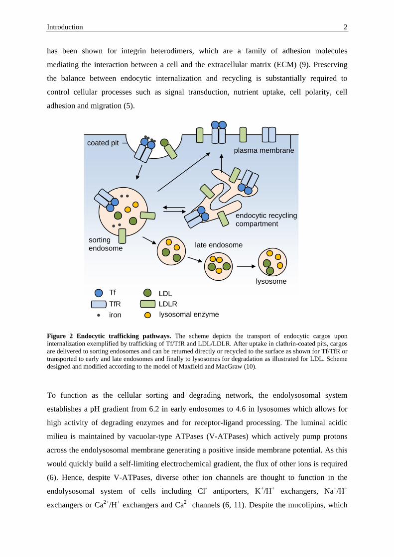
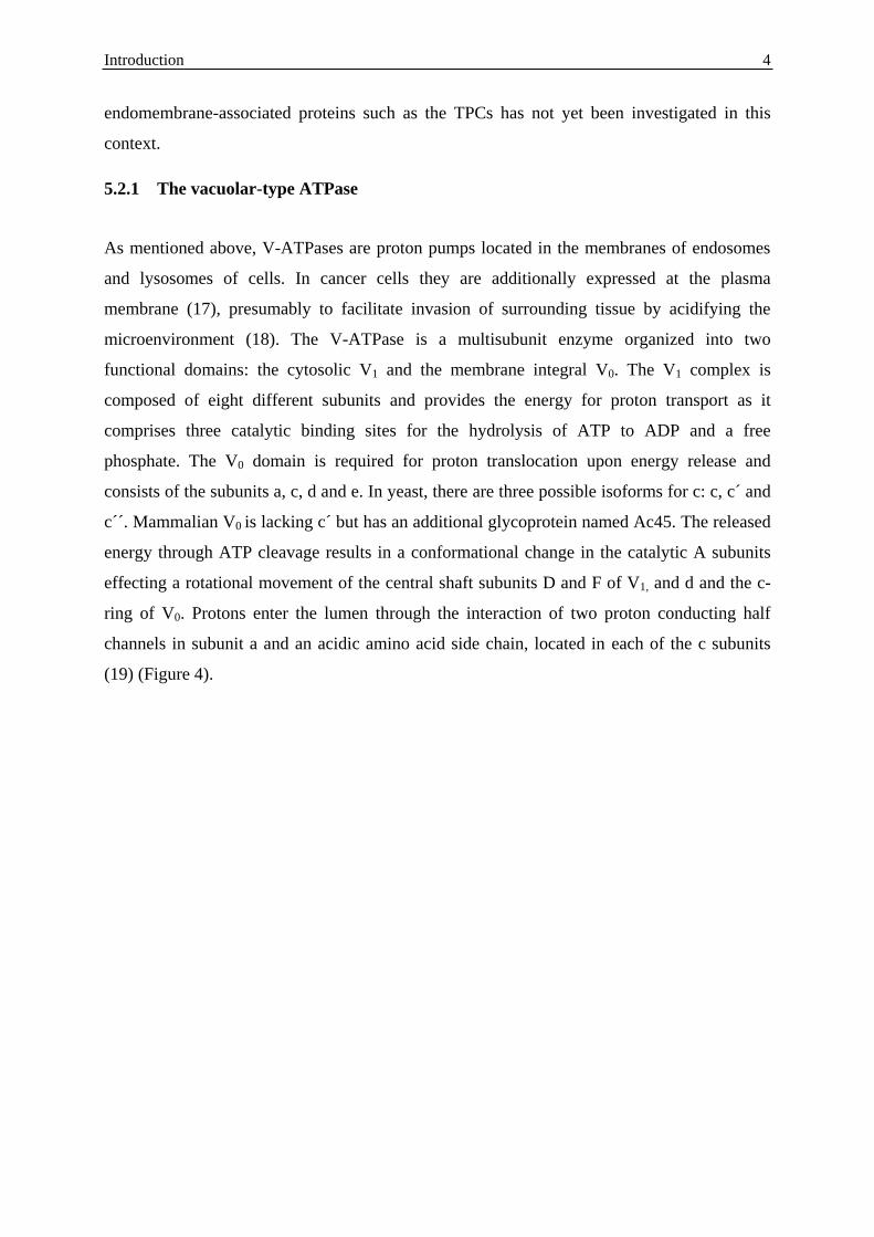
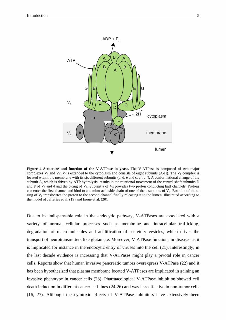
microenvironment (18). The V-ATPase is a multisubunit enzyme organized into two
functional domains: the cytosolic V1 and the membrane integral V0. The V1 complex is
composed of eight different subunits and provides the energy for proton transport as it
comprises three catalytic binding sites for the hydrolysis of ATP to ADP and a free
phosphate. The V0 domain is required for proton translocation upon energy release and
consists of the subunits a, c, d and e. In yeast, there are three possible isoforms for c: c, c´ and
c´´. Mammalian V0 is lacking c´ but has an additional glycoprotein named Ac45. The released
energy through ATP cleavage results in a conformational change in the catalytic A subunits
effecting a rotational movement of the central shaft subunits D and F of V1, and d and the c-
ring of V0. Protons enter the lumen through the interaction of two proton conducting half
channels in subunit a and an acidic amino acid side chain, located in each of the c subunits
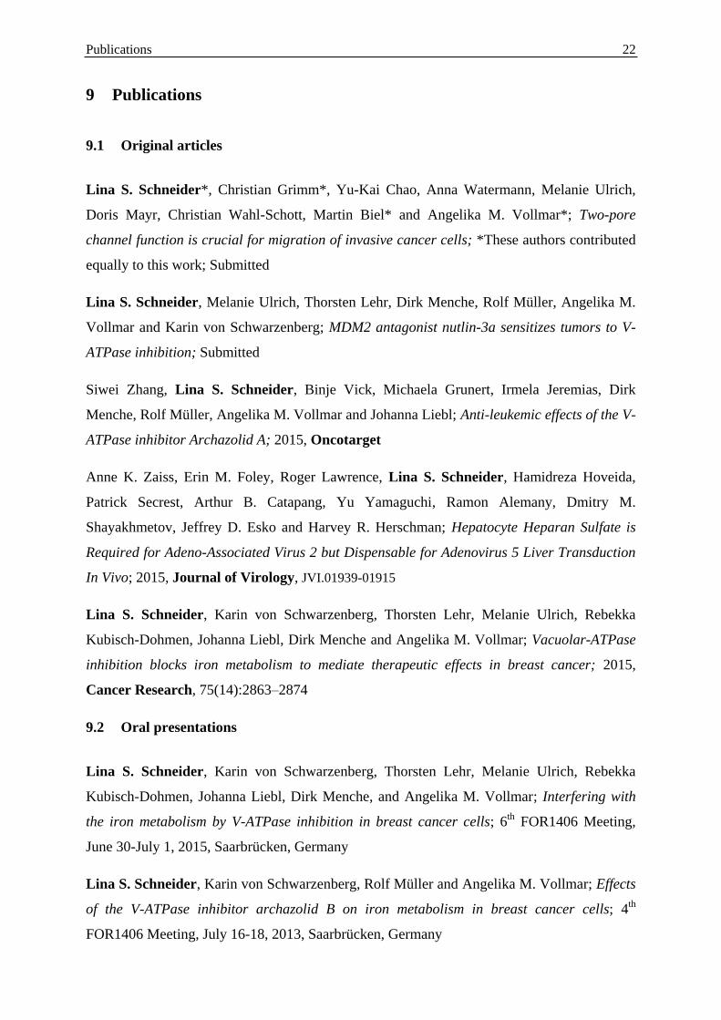
(19) (Figure 4).
Introduction 5
Figure 4 Structure and function of the V-ATPase in yeast. The V-ATPase is composed of two major
complexes V1 and V0: V1is extended to the cytoplasm and consists of eight subunits (A-H). The V0 complex is
located within the membrane with its six different subunits (a, d, e and c, c`, c``). A conformational change of the
subunit A, which is driven by ATP hydrolysis, results in the rotational movement of the central shaft subunits D
and F of V1 and d and the c-ring of V0. Subunit a of V0 provides two proton conducting half channels. Protons
can enter the first channel and bind to an amino acid side chain of one of the c subunits of V0. Rotation of the c-
ring of V0 translocates the proton to the second channel finally releasing it to the lumen. Illustrated according to
the model of Jefferies et al. (19) and Inoue et al. (20).
Due to its indispensable role in the endocytic pathway, V-ATPases are associated with a
variety of normal cellular processes such as membrane and intracellular trafficking,
degradation of macromolecules and acidification of secretory vesicles, which drives the
transport of neurotransmitters like glutamate. Moreover, V-ATPase functions in diseases as it
is implicated for instance in the endocytic entry of viruses into the cell (21). Interestingly, in
the last decade evidence is increasing that V-ATPases might play a pivotal role in cancer
cells. Reports show that human invasive pancreatic tumors overexpress V-ATPase (22) and it
has been hypothesized that plasma membrane located V-ATPases are implicated in gaining an
invasive phenotype in cancer cells (23). Pharmacological V-ATPase inhibition showed cell
death induction in different cancer cell lines (24-26) and was less effective in non-tumor cells
(16, 27). Although the cytotoxic effects of V-ATPase inhibitors have extensively been
a
C
V1
membrane
cytoplasm
G
B A
H
c
lumen
ATP
ADP + Pi
2H+
E
A
B
E
B
G
c
c' c''
d
V0
e
D
F
A
Introduction 6
studied, the precise mechanism still awaits molecular explanation. Researchers have
hypothesized that inhibition of macroautophagy (24) or the release of proteases after
lysosomal dysfunction might play a role (25). In another approach, inhibition of V-ATPase
was efficient in reducing migration of invasive cancer cells in vitro and in vivo. This work
further unveiled effects on receptor recycling after block of V-ATPase function since
epidermal growth factor receptor (EGFR) internalization was delayed (28). A contribution of
V-ATPase to receptor trafficking was also observed in another study (29).
A lot of the recent knowledge about V-ATPases has been gained by using pharmacological V-
ATPase inhibitors. The oldest members, bafilomycin and concanamycin, have already been
identified in the early 1980s (30). They are plecomacrolides and inhibit V-ATPase function
by binding to subunit c of V0 with a minor contribution of subunit a to the binding site. A
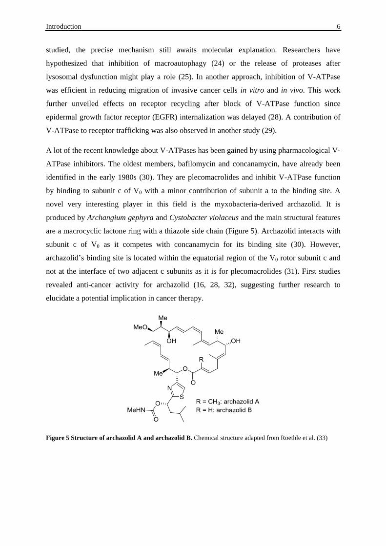
novel very interesting player in this field is the myxobacteria-derived archazolid. It is
produced by Archangium gephyra and Cystobacter violaceus and the main structural features
are a macrocyclic lactone ring with a thiazole side chain (Figure 5). Archazolid interacts with
subunit c of V0 as it competes with concanamycin for its binding site (30). However,
archazolid’s binding site is located within the equatorial region of the V0 rotor subunit c and
not at the interface of two adjacent c subunits as it is for plecomacrolides (31). First studies
revealed anti-cancer activity for archazolid (16, 28, 32), suggesting further research to
elucidate a potential implication in cancer therapy.
Figure 5 Structure of archazolid A and archazolid B. Chemical structure adapted from Roethle et al. (33)
Introduction 7
5.2.2 Two-pore channels
The very first evidence for the existence of a vacuolar located ATPase was given in 1962 and
this was confirmed by diverse studies until the early 1980s (34). The identification of
endolysosomal TPCs happened much later. There was emerging evidence at the beginning of
this century that nicotinic acid adenine dinucleotide phosphate (NAADP) is a Ca2+
mobilizing
messenger and that it addresses stores separate from the endoplasmic reticulum in acidic
organelles. However, the precise target was unclear. NAADP displays a high structural
similarity to nicotinamide adenine dinucleotide phosphate (NADP) despite the carboxyl group
at position 3 of the pyridine (35). Recently, TPCs have been identified as the long-sought
endolysosomal NAADP-dependent Ca2+
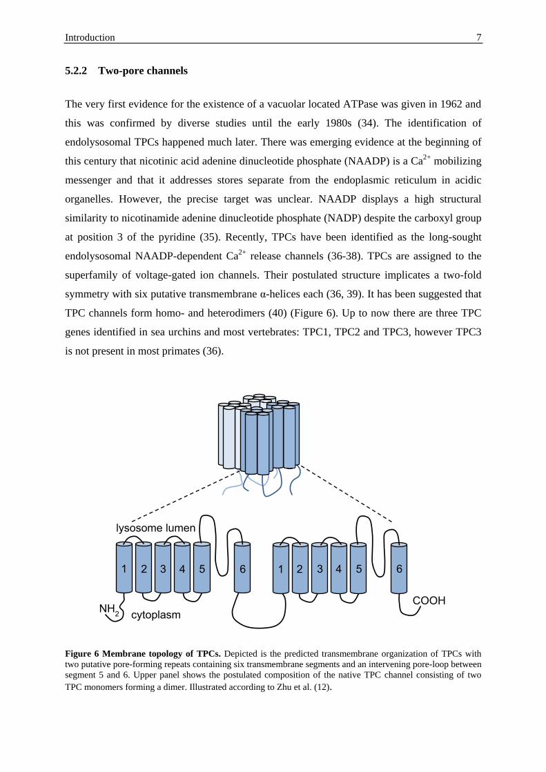
release channels (36-38). TPCs are assigned to the
superfamily of voltage-gated ion channels. Their postulated structure implicates a two-fold
symmetry with six putative transmembrane α-helices each (36, 39). It has been suggested that
TPC channels form homo- and heterodimers (40) (Figure 6). Up to now there are three TPC
genes identified in sea urchins and most vertebrates: TPC1, TPC2 and TPC3, however TPC3
is not present in most primates (36).
Figure 6 Membrane topology of TPCs. Depicted is the predicted transmembrane organization of TPCs with
two putative pore-forming repeats containing six transmembrane segments and an intervening pore-loop between
segment 5 and 6. Upper panel shows the postulated composition of the native TPC channel consisting of two
TPC monomers forming a dimer. Illustrated according to Zhu et al. (12).
1 2 3 4 5 6
1 2 3 4 5 6
lysosome lumen
cytoplasm
COOH NH
2
Introduction 8
Diverse studies revealed that TPC1 and TPC2 are located in the endolysosomal system of
cells with TPC1 expressed in endosomes and lysosomes whereas TPC2 can predominantly be
found in lysosomes (36-38, 40, 41). Although most researchers claimed that activation of
TPCs is triggered by NAADP (36-38, 42-44), it has also been suggested that
phosphatidylinositol 3,5-bisphosphate (PI(3,5)P2) is implicated (43, 45). Independent
experiments hypothesized TPCs as Ca2+
channels, suggesting them responsible for Ca2+
release from endolysosomal stores (36, 38, 42, 44), albeit conductance for Na+ was also
discussed (45).
Given the fact that TPCs are located in endosomes and lysosomes, it does not come as a
surprise that they function in the endocytic pathway. Reports show that overexpression of
TPC2 inhibits autophagosomal-lysosomal fusion (46) and that they are crucially involved in
the regulation of endolysosomal trafficking. Thus, TPC2 deficient mouse embryonic
fibroblasts showed accumulation of EGF/EGFR and LDL in intracellular vesicles (47) and
delayed platelet-derived growth factor receptor β (PDGFRβ) degradation (48). Moreover,
cholera toxin, which is under normal conditions trafficked to the Golgi, was halted in
endolysosomes after overexpression of TPCs (41). Disturbed TPC function retained Ebola
virus trafficking, which prevented host cell infection (49). Despite effects on endocytic
trafficking, TPCs were recently connected to lysosomal control of nutrient recycling. It was
hypothesized that TPCs form a lysosomal ATP-sensitive Na+ channel complex which is
associated with and regulated by the mammalian target of rapamycin (mTOR). Under
starvation mTOR delocates from the complex resulting in release of Na+ and other ions from
lysosomes (50). Interestingly, V-ATPase was also identified as a component of the mTOR
pathway. It has been postulated that V-ATPase is crucial for amino acid signaling to mTOR
complex 1 (51). Given the fact that TPCs and V-ATPases are both implicated in endocytic
trafficking and mTOR signaling, one might speculate that there could be a link between these
two proteins, most notably because the positive inward currents of V-ATPase could be
balanced by an outward flux through TPCs.
To target the TPC pathway pharmacologically the NAADP antagonist Ned-19 was used in
different studies (46, 49, 52). Ned-19 was identified as a noncompetitive antagonist of
NAADP by virtual screening, with the trans form more potent than the cis form regarding
Ca2+
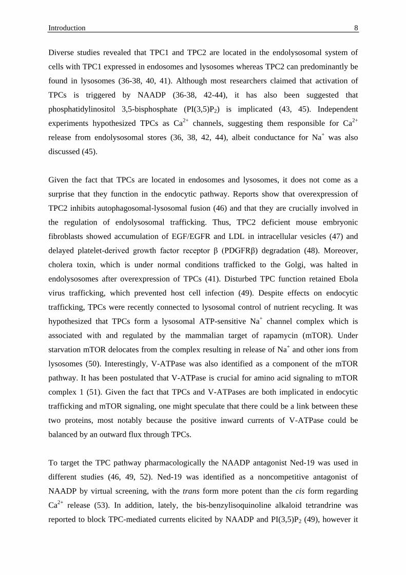
release (53). In addition, lately, the bis-benzylisoquinoline alkaloid tetrandrine was
reported to block TPC-mediated currents elicited by NAADP and PI(3,5)P2 (49), however it
Introduction 9
might act on other Ca2+
channels as well (54). Tetrandrine can be purified form the root of
Stephania tetrandra and has been used in traditional chinese medicine for instance against
hypertension (54) (Figure 7).
Figure 7 Structure of trans-Ned-19 and tetrandrine. Chemical structure of trans-Ned-19 adapted from Naylor
et al. (53). Chemical structure of tetrandrine adapted from Inubushi et al. (55).
Although in some studies cancer cell lines were used as a cellular system to investigate TPC
function, its implication in cancer progression, cancer cell migration and formation of
metastasis has not been studied up to now. Therefore, it would be highly interesting to
investigate this matter to elucidate a potential role in cancer treatment.
5.3 Overcoming the hallmarks of cancer
The development of a normal cell into a malignant cancer cell requires in most cases four to
seven mutations (3). These mutations allow cancer cells to behave different to normal cells.
For cancer therapy it is highly appreciated to find treatment strategies which mainly target
malignant cells to reduce side effects. To achieve this, it seems promising to address the
altered behavior of cancer cells.
In 2000, Hannahan and Weinberg defined the hallmarks of cancer describing the gained
abilities of malignant cells. One is to evade apoptosis (56). Apoptosis, the programmed and
controlled cell death, occurs during development or aging but is also a defense mechanism for
instance when cells are damaged by noxious agents. Morphologically, apoptosis starts with
cell shrinkage and chromatin condensation which is followed by plasma membrane blebbing
and subsequent separation of cell fragments into apoptotic bodies. Degradation of these
bodies occurs finally by phagocytosis through macrophages. Induction of apoptosis can be
triggered by a variety of signals. Mechanistically, there are two main apoptotic pathways: the
Introduction 10
intrinsic and the extrinsic. The intrinsic pathway is for example stimulated by radiation, toxins
or hypoxia. These stimuli cause mitochondrial changes effecting an opening of the
mitochondrial permeability pore and release of pro-apoptotic proteins such as cytochrome c.
These processes are regulated by members of the B-cell lymphoma 2 (Bcl-2) family such as
Bcl-2 or Bcl-2-associated X protein (Bax). The release of pro-apoptotic proteins from
mitochondria triggers the formation of the apoptosome and activation of caspase-9. The
extrinsic pathway is activated by the binding of a death ligand such as Fas ligand to its
receptor which results in formation of the death inducing signaling complex, finally activating
caspase-8. Both pathways converge in the activation of caspase-3 which initiates the
degradation of the chromosomal DNA (57). Cancer cells are often resistant to apoptosis and
self-sufficient in growth signals, another gained ability (56), which enables uncontrolled and
fast proliferation. Overcoming these hallmarks of cancer by pharmacological induction of
apoptosis is therefore a viable strategy for cancer therapy, albeit its contribution to treatment
success was questioned. Therefore, investigating non-apoptotic types of cell death might be
an alternative anti-cancer approach (58).
Despite evading apoptosis, cancer cells gained the ability to migrate and invade which allows
for pulling themselves forward into new tissues to build metastasis (56). The formation of
growth at secondary sites starts with the acquisition of an invasive phenotype of the primary
tumor. Then, some tumor cells detach and intravasate to enter hematogenous circulation
which is followed by extravasation into the new tissue. There, the tumor cell adapts to the
foreign site and initiates proliferation. Since as much as 90% of cancer-related mortality is
due to metastasis, new strategies to improve the treatment of invasive cancers remain of
interest to develop (59).
In order to enable fast proliferation and migration, cancer cells have to be highly metabolic
active. Therefore, malignant cells established an altered turnover of signaling molecules (60)
and a rapid uptake of nutrients which allows tumor imaging in the clinic (61). Thus, derailed
endocytosis was described as an emerging multifaceted hallmark of these cells (60). In turn,
targeting the endocytic machinery pharmacologically might be a highly promising strategy to
selectively hit cancer cells. Moreover, addressing this tumor-essential process could be an
innovative approach to elude escape mechanisms.
Aim of the study 11
6 Aim of the study
Cancer remains one of the leading causes of death worldwide. The success of today’s anti-
cancer drugs is challenged by a variety of resistance mechanisms against the most
chemotherapeutics and by the occurrence of metastasis. Thus, new targets and treatment
strategies are urgently needed. Previous work of our group revealed targeting the
endolysosomal vacuolar-type ATPase as a promising future anti-cancer approach as it induces
apoptosis in cancer cells. However, the precise mode of action still awaits molecular
explanation.
Hence, the aim of this study was to elucidate this matter and to extend the previous data for in
vivo efficacy including rational combination therapy. Moreover, the question was addressed
whether targeting the endolysosomal system by inhibition of two-pore channels might be a
new and viable approach for the treatment of invasive tumor cells.
Short summary of manuscripts 12
7 Short summary of manuscripts
7.1 Vacuolar-ATPase inhibition blocks iron metabolism to mediate therapeutic effects
in breast cancer
Lina S. Schneider, Karin von Schwarzenberg, Thorsten Lehr, Melanie Ulrich, Rebekka
Kubisch-Dohmen, Johanna Liebl, Dirk Menche and Angelika M. Vollmar
2015, Cancer Research, 75(14):2863–2874
Within the last decade evidence is increasing that the vacuolar-type ATPase (V-ATPase), a
heteromultimeric proton pump, plays a vital role in the survival of tumor cells. However, the
precise mode of action still awaits molecular explanation. To investigate V-ATPase mediated
cytotoxicity we used the myxobacteria-derived compound archazolid, a highly potent V-
ATPase inhibitor. While V-ATPase inhibitors showed anti-tumoral potential in diverse cancer
cell lines, in vivo data are limited up to now. Our study showed for the first time reduced
tumor growth in a 4T1 mammary mouse model. With regard to the molecular mechanisms of
V-ATPase-related cell death, we found that archazolid stabilizes the hypoxia-inducible factor
1α (HIF1α) protein in the two breast cancer cell lines MCF7 and MDA-MB-231. As HIF1α
has many target genes involved in glycolysis, we could detect elevated levels of the
hexokinase II protein as well as of other glycolytic gene products such as aldolase c. Further
experiments unveiled that the HIF1α stabilization is due to iron depletion in the cytosol since
an excess of iron citrate in the medium abolished it completely. Moreover, the lack of iron is
partially responsible for the induction of cell death after V-ATPase inhibition and could be
associated with disrupted transferrin/transferrin receptor internalization upon V-ATPase
inhibition. As a consequence, activity of the iron dependent enzyme ribonucleotide reductase
is diminished leading to S-phase block and double-strand breaks. Additionally, in p53 wild
type MCF7 cells protein levels of the tumor suppressor p53 were elevated after archazolid
treatment, which contributed to the induced cell death since pifithrin-α diminished
significantly the amount of dead cells. These findings eventually connect V-ATPase
inhibition to fundamental cellular processes such as DNA synthesis and DNA repair. Hence,
our study reveals a novel mode of action for V-ATPase-induced cell death in tumor cells and
proposes V-ATPase inhibition as a promising and viable strategy for breast cancer therapy.
Short summary of manuscripts 13
7.2 Anti-leukemic effects of the V-ATPase inhibitor Archazolid A
Siwei Zhang, Lina S. Schneider, Binje Vick, Michaela Grunert, Irmela Jeremias, Dirk
Menche, Rolf Müller, Angelika M. Vollmar and Johanna Liebl
2015, accepted for publication in Oncotarget
The V-ATPase inhibitor archazolid showed promising anti-tumor effects in different cancer
cell lines as for example demonstrated in manuscript I. Since new treatment strategies for
patients suffering from T-cell acute lymphoblastic leukemia (T-ALL) are urgently needed, we
elucidated archazolid’s potential as an anti-leukemic drug. Indeed, V-ATPase inhibition
triggered induction of apoptosis in leukemic cell lines which could be shown by flow
cytometry analysis, cleavage of caspase-3, caspase-9 and poly ADP ribose polymerase
(PARP). Along this line, archazolid treatment in patient derived xenograft (PDX) leukemic
cells, but not in non-tumor primary human peripheral blood mononuclear cells, resulted in
induction of cell death. Mechanistically, by inhibiting lysosomal acidification, archazolid
reduced activation of the notch pathway as the expression of notch1 intracellular domain
(NICD) and of the downstream target c-myc were diminished. However, NICD
overexpression was not able to rescue archazolid-mediated cell death, suggesting further
studies regarding the underlying mechanisms. In fact, V-ATPase inhibition by archazolid
decreased the expression level of the anti-apoptotic protein survivin. Remarkably, its
overexpression significantly diminished the amount of dead cells during V-ATPase inhibition
providing evidence for its implication in archazolid-induced cell death. Regarding the
underlying mechanism, this work ties in with our previous findings of manuscript I that
archazolid led to S-phase cell cycle arrest by interfering with the iron metabolism in leukemic
cells. Thus, this study renders V-ATPases as potential new targets for the treatment of T-ALL.
Declaration of contribution: I contributed to this work of Dr. Siwei Zhang and Dr. Johanna
Liebl by conducting a set of experiments including FACS and western blot analysis of PDX
leukemic cells, cell cycle analysis of Jurkat cells and PCR analysis of Jurkat cell samples. I
was partially involved in the design of the study since the investigated underlying mechanism
is associated with my earlier work (manuscript I).
Short summary of manuscripts 14
7.3 MDM2 antagonist nutlin-3a sensitizes tumors to V-ATPase inhibition
Lina S. Schneider, Melanie Ulrich, Thorsten Lehr, Dirk Menche, Rolf Müller,
Angelika M. Vollmar and Karin von Schwarzenberg
Submitted
The anti-cancer potential of the V-ATPase inhibitor archazolid has been proven in distinct in
vitro studies, however in vivo data are rare. Manuscript I showed first promising in vivo
efficacy of archazolid in a 4T1 mouse model, albeit tumor growth was not abrogated
completely. To improve this matter, we searched for a rational and innovative combination
strategy. As tumors often carry several mutations, using multiple drugs targeting different
proteins seems therefore more beneficial for cancer treatment. Since our recent study
(manuscript I) unveiled that inhibition of V-ATPase caused the induction of p53 protein levels
in cancer cells, we considered a combination strategy within the p53 pathway. In fact, in this
study we found that combining archazolid with the p53 activator, nutlin-3a, induces
synergistically cell death in different p53 wild type tumor cell lines, but not in p53 mutated
cells, as shown by flow cytometry analysis. Investigating the underlying mechanism, we
could detect reduced glycolysis as TP53-induced glycolysis and apoptosis regulator (TIGAR)
mRNA levels were elevated and glucose uptake, glucose transporter 1 (Glut1) protein levels
and ATP levels were decreased by the combination. Moreover, treatment of archazolid and
nutlin-3a activated pro-apoptotic pathways involving the induction of insulin-like growth
factor-binding protein 3 (IGFBP3) and active Bcl-2-associated X protein (Bax) and the
cleavage of caspase-9 and PARP. Finally, combination treatment was more efficient in
reducing tumor growth compared to single dose treatment in a p53 wild type U87MG
glioblastoma xenograft. Especially the success of archazolid and nutlin-3a in vivo suggests
this combination as a potential new therapeutic option for p53 wild type carcinomas.
Short summary of manuscripts 15
7.4 Two-pore channel function is crucial for migration of invasive cancer cells
Lina S. Schneider*, Christian Grimm*, Yu-Kai Chao, Anna Watermann, Melanie
Ulrich, Doris Mayr, Christian Wahl-Schott, Martin Biel* and Angelika M. Vollmar*
*These authors contributed equally to this work
Submitted
The most cancer-related deaths are caused by metastasis. Spread of single tumor cells to
anatomically distant sites requires the ability to migrate and invade. Disrupting these
processes is therefore thought to be a promising strategy for the treatment of invasive
carcinomas. As targeting the endocytic system by V-ATPase inhibition was effective in
reducing cancer cell migration before, we here investigated the impact of endolysosomal two-
pore channels (TPCs) in this context. TPCs are recently identified nicotinic acid adenine
dinucleotide phosphate (NAADP)- and phosphatidylinositol 3,5-bisphosphate (PI(3,5)P2)-
sensitive Ca2+
-permeable ion channels located in the membrane of endosomes and lysosomes.
Their implication in cancer cell progression and migration has not been studied up to now. To
this end, siRNA silencing of TPC1 and TPC2 mRNA levels in T24 cancer cells resulted in
reduced adhesion, migration through pores and directed migration. Along this line,
pharmacological inhibition of TPC function through Ned-19 and tetrandrine inhibited
migration through pores in T24, HUH7 and 4T1 cancer cells. Mechanistically, we found that
disturbing TPC function affected β1-integrin trafficking, causing accumulation in early
endosome antigen 1 (EEA1)-positive vesicles. This is suggested to result from alterations in
Ca2+
signaling since acidification of vesicles was not inhibited. Finally, trans-Ned-19-treated
cells showed insufficient formation of leading edges, which is crucial for adequate migration.
Remarkably, these effects were recapitulated in a mouse model of tumor dissemination
showing significantly reduced lung metastasis of 4T1-Luc cells after tetrandrine treatment.
Thus, this study identified TPCs as new and interesting candidates to address in the treatment
of invasive cancer cells. Once more, targeting the endolysosomal system of cancer cells seems
to be a promising anti-cancer approach.
References 16
8 References
1. Torre LA, Bray F, Siegel RL, Ferlay J, Lortet-Tieulent J, Jemal A (2015) Global
cancer statistics, 2012. CA Cancer J Clin 65(2):87–108.
2. DeSantis CE, Lin CC, Mariotto AB, Siegel RL, Stein KD, Kramer JL, Alteri R,
Robbins AS, Jemal A (2014) Cancer treatment and survivorship statistics, 2014. CA
Cancer J Clin 64(4):252–271.
3. Zimmermann GR, Lehár J, Keith CT (2007) Multi-target therapeutics: when the whole
is greater than the sum of the parts. Drug Discov Today 12(1-2):34–42.
4. Holohan C, Van Schaeybroeck S, Longley DB, Johnston PG (2013) Cancer drug
resistance: an evolving paradigm. Nat Rev Cancer 13(10):714–726.
5. Grant BD, Donaldson JG (2009) Pathways and mechanisms of endocytic recycling.
Nat Rev Mol Cell Biol 10(9):597–608.
6. Scott CC, Gruenberg J (2011) Ion flux and the function of endosomes and lysosomes:
pH is just the start. Bioessays 33(2):103–110.
7. Le Roy C, Wrana JL (2005) Clathrin- and non-clathrin-mediated endocytic regulation
of cell signalling. Nat Rev Mol Cell Biol 6(2):112–126.
8. Dautry-Varsat A, Ciechanover A, Lodish HF (1983) pH and the recycling of
transferrin during receptor-mediated endocytosis. Proc Natl Acad Sci U S A
80(8):2258–2262.
9. Caswell PT, Vadrevu S, Norman JC (2009) Integrins: masters and slaves of endocytic
transport. Nat Rev Mol Cell Biol 10(12):843–853.
10. Maxfield FR, McGraw TE (2004) Endocytic recycling. Nat Rev Mol Cell Biol
5(2):121–132.
11. Christensen KA, Myers JT, Swanson JA (2002) pH-dependent regulation of lysosomal
calcium in macrophages. J Cell Sci 115(Pt 3):599–607.
12. Zhu MX, Ma J, Parrington J, Calcraft PJ, Galione A, Evans AM (2009) Calcium
signaling via two-pore channels: local or global, that is the question. Am J Physiol Cell
Physiol 298(3):C430–C441.
13. Grimm C, Hassan S, Wahl-Schott C, Biel M (2012) Role of TRPML and Two-Pore
Channels in Endolysosomal Cation Homeostasis. J Pharmacol Exp Ther 342(2):236-
244.
14. Gyparaki M-T, Papavassiliou AG (2014) Lysosome: the cell's ‘suicidal bag’ as a
promising cancer target. Trends Mol Med 20(5):239–241.
References 17
15. Kroemer G, Jäättelä M (2005) Lysosomes and autophagy in cell death control. Nat
Rev Cancer 5(11):886–897.
16. von Schwarzenberg K, Wiedmann RM, Oak P, Schulz S, Zischka H, Wanner G,
Efferth T, Trauner D, Vollmar AM (2012) Mode of Cell Death Induction by
Pharmacological Vacuolar H+-ATPase (V-ATPase) Inhibition. J Biol Chem
288(2):1385–1396.
17. Martinez-Zaguilan R, Lynch RM, Martinez GM, Gillies RJ (1993) Vacuolar-type
H(+)-ATPases are functionally expressed in plasma membranes of human tumor cells.
Am J Physiol 265(4 Pt 1):C1015–1029.
18. Pérez-Sayáns M, Somoza-Martín JM, Barros-Angueira F, Rey JMG, García-García A
(2009) V-ATPase inhibitors and implication in cancer treatment. Cancer Treat Rev
35(8):707–713.
19. Jefferies KC, Cipriano DJ, Forgac M (2008) Function, structure and regulation of the
vacuolar (H+)-ATPases. Arch Biochem Biophys 476(1):33–42.
20. Inoue T, Wilkens S, Forgac M (2003) Subunit structure, function, and arrangement in
the yeast and coated vesicle V-ATPases. J Bioenerg Biomembr 35(4):291–299.
21. Hinton A, Bond S, Forgac M (2007) V-ATPase functions in normal and disease
processes. Pflugers Arch 457(3):589–598.
22. Ohta T, Numata M, Yagishita H, Futagami F, Tsukioka Y, Kitagawa H, Kayahara M,
Nagakawa T, Miyazaki I, Yamamoto M, Iseki S, Ohkuma S (1996) Expression of 16
kDa proteolipid of vacuolar-type H(+)-ATPase in human pancreatic cancer. Br J
Cancer 73(12):1511–1517.
23. Sennoune SR (2004) Vacuolar H+-ATPase in human breast cancer cells with distinct
metastatic potential: distribution and functional activity. Am J Physiol Cell Physiol
286(6):C1443–C1452.
24. Wu YC, Wu WK, Li Y, Yu L, Li ZJ, Wong CC, Li HT, Sung JJ, Cho CH (2009)
Inhibition of macroautophagy by bafilomycin A1 lowers proliferation and induces
apoptosis in colon cancer cells. Biochem Biophys Res Commun 382(2):451–456.
25. Nakashima S, Hiraku Y, Tada-Oikawa S, Hishita T, Gabazza EC, Tamaki S, Imoto I,
Adachi Y, Kawanishi S (2003) Vacuolar H+-ATPase inhibitor induces apoptosis via
lysosomal dysfunction in the human gastric cancer cell line MKN-1. J Biochem
134(3):359–364.
26. Ohta T, Arakawa H, Futagami F, Fushida S, Kitagawa H, Kayahara M, Nagakawa T,
Miwa K, Kurashima K, Numata M, Kitamura Y, Terada T, Ohkuma S (1998)
References 18
Bafilomycin A1 induces apoptosis in the human pancreatic cancer cell line Capan-1. J
Pathol 185(3):324–330.
27. Morimura T, Fujita K, Akita M, Nagashima M, Satomi A (2008) The proton pump
inhibitor inhibits cell growth and induces apoptosis in human hepatoblastoma. Pediatr
Surg Int 24(10):1087–1094.
28. Wiedmann RM, von Schwarzenberg K, Palamidessi A, Schreiner L, Kubisch R, Liebl
J, Schempp C, Trauner D, Vereb G, Zahler S, Wagner E, Muller R, Scita G, Vollmar
AM (2012) The V-ATPase-Inhibitor Archazolid Abrogates Tumor Metastasis via
Inhibition of Endocytic Activation of the Rho-GTPase Rac1. Cancer Res
72(22):5976–5987.
29. Kozik P, Hodson NA, Sahlender DA, Simecek N, Soromani C, Wu J, Collinson LM,
Robinson MS (2012) A human genome-wide screen for regulators of clathrin-coated
vesicle formation reveals an unexpected role for the V-ATPase. Nat Cell Biol
15(1):50–60.
30. Huss M, Wieczorek H (2009) Inhibitors of V-ATPases: old and new players. J Exp
Biol 212(3):341–346.
31. Bockelmann S, Menche D, Rudolph S, Bender T, Grond S, von Zezschwitz P,
Muench SP, Wieczorek H, Huss M (2010) Archazolid A Binds to the Equatorial
Region of the c-Ring of the Vacuolar H+-ATPase. J Biol Chem 285(49):38304–
38314.
32. von Schwarzenberg K, Lajtos T, Simon L, Müller R, Vereb G, Vollmar AM (2014) V-
ATPase inhibition overcomes trastuzumab resistance in breast cancer. Mol Oncol
8(1):9–19.
33. Roethle PA, Chen IT, Trauner D (2007) Total Synthesis of (−)-Archazolid B. J Am
Chem Soc 129(29):8960–8961.
34. Beyenbach KW, Wieczorek H (2006) The V-type H+ ATPase: molecular structure
and function, physiological roles and regulation. J Exp Biol 209(Pt 4):577–589.
35. Galione A (2010) NAADP Receptors. Cold Spring Harb Perspect Biol 3(1):a004036–
a004036.
36. Calcraft PJ, Ruas M, Pan Z, Cheng X, Arredouani A, Hao X, Tang J, Rietdorf K,
Teboul L, Chuang K-T, Lin P, Xiao R, Wang C, Zhu Y, Lin Y, Wyatt CN, Parrington
J, Ma J, Evans AM, Galione A, Zhu MX (2009) NAADP mobilizes calcium from
acidic organelles through two-pore channels. Nature 459(7246):596–600.
References 19
37. Brailoiu E, Churamani D, Cai X, Schrlau MG, Brailoiu GC, Gao X, Hooper R,
Boulware MJ, Dun NJ, Marchant JS, Patel S (2009) Essential requirement for two-
pore channel 1 in NAADP-mediated calcium signaling. J Cell Biol 186(2):201–209.
38. Zong X, Schieder M, Cuny H, Fenske S, Gruner C, Rötzer K, Griesbeck O, Harz H,
Biel M, Wahl-Schott C (2009) The two-pore channel TPCN2 mediates NAADP-
dependent Ca2+-release from lysosomal stores. Pflugers Arch 458(5):891–899.
39. Ishibashi K, Suzuki M, Imai M (2000) Molecular Cloning of a Novel Form (Two-
Repeat) Protein Related to Voltage-Gated Sodium and Calcium Channels. Biochem
Biophys Res Commun 270(2):370–376.
40. Rietdorf K, Funnell TM, Ruas M, Heinemann J, Parrington J, Galione A (2011) Two-
pore Channels Form Homo- and Heterodimers. J Biol Chem 286(43):37058–37062.
41. Ruas M, Rietdorf K, Arredouani A, Davis LC, Lloyd-Evans E, Koegel H, Funnell TM,
Morgan AJ, Ward JA, Watanabe K, Cheng X, Churchill GC, Zhu MX, Platt FM,
Wessel GM, Parrington J, Galione A (2010) Purified TPC Isoforms Form NAADP
Receptors with Distinct Roles for Ca2+ Signaling and Endolysosomal Trafficking.
Curr Biol 20(8):703–709.
42. Schieder M, Rotzer K, Bruggemann A, Biel M, Wahl-Schott CA (2010)
Characterization of Two-pore Channel 2 (TPCN2)-mediated Ca2+ Currents in Isolated
Lysosomes. J Biol Chem 285(28):21219–21222.
43. Jha A, Ahuja M, Patel S, Brailoiu E, Muallem S (2014) Convergent regulation of the
lysosomal two-pore channel-2 by Mg2+, NAADP, PI(3,5)P2 and multiple protein
kinases. EMBO J 33(5):501–511.
44. Ruas M, Davis LC, Chen CC, Morgan AJ, Chuang KT, Walseth TF, Grimm C,
Garnham C, Powell T, Platt N, Platt FM, Biel M, Wahl-Schott C, Parrington J,
Galione A (2015) Expression of Ca2+-permeable two-pore channels rescues NAADP
signalling in TPC-deficient cells. EMBO J 34(13):1743–1758.
45. Wang X, Zhang X, Dong X-p, Samie M, Li X, Cheng X, Goschka A, Shen D, Zhou Y,
Harlow J, Zhu Michael X, Clapham David E, Ren D, Xu H (2012) TPC Proteins Are
Phosphoinositide- Activated Sodium-Selective Ion Channels in Endosomes and
Lysosomes. Cell 151(2):372–383.
46. Lu Y, Hao BX, Graeff R, Wong CWM, Wu WT, Yue J (2013) Two Pore Channel 2
(TPC2) Inhibits Autophagosomal-Lysosomal Fusion by Alkalinizing Lysosomal pH. J
Biol Chem 288(33):24247–24263.
References 20
47. Grimm C, Holdt LM, Chen C-C, Hassan S, Müller C, Jörs S, Cuny H, Kissing S,
Schröder B, Butz E, Northoff B, Castonguay J, Luber CA, Moser M, Spahn S,
Lüllmann-Rauch R, Fendel C, Klugbauer N, Griesbeck O, Haas A, Mann M, Bracher
F, Teupser D, Saftig P, Biel M, Wahl-Schott C (2014) High susceptibility to fatty liver
disease in two-pore channel 2-deficient mice. Nat Commun 5:4699.
48. Ruas M, Chuang KT, Davis LC, Al-Douri A, Tynan PW, Tunn R, Teboul L, Galione
A, Parrington J (2014) TPC1 Has Two Variant Isoforms, and Their Removal Has
Different Effects on Endo-Lysosomal Functions Compared to Loss of TPC2. Mol Cell
Biol 34(21):3981–3992.
49. Sakurai Y, Kolokoltsov AA, Chen CC, Tidwell MW, Bauta WE, Klugbauer N, Grimm
C, Wahl-Schott C, Biel M, Davey RA (2015) Two-pore channels control Ebola virus
host cell entry and are drug targets for disease treatment. Science 347(6225):995–998.
50. Cang C, Zhou Y, Navarro B, Seo Y-j, Aranda K, Shi L, Battaglia-Hsu S, Nissim I,
Clapham David E, Ren D (2013) mTOR Regulates Lysosomal ATP-Sensitive Two-
Pore Na+ Channels to Adapt to Metabolic State. Cell 152(4):778–790.
51. Zoncu R, Bar-Peled L, Efeyan A, Wang S, Sancak Y, Sabatini DM (2011) mTORC1
Senses Lysosomal Amino Acids Through an Inside-Out Mechanism That Requires the
Vacuolar H+-ATPase. Science 334(6056):678–683.
52. Favia A, Desideri M, Gambara G, D'Alessio A, Ruas M, Esposito B, Del Bufalo D,
Parrington J, Ziparo E, Palombi F, Galione A, Filippini A (2014) VEGF-induced
neoangiogenesis is mediated by NAADP and two-pore channel-2-dependent Ca2+
signaling. Proc Natl Acad Sci U S A 111(44):E4706–E4715.
53. Naylor E, Arredouani A, Vasudevan SR, Lewis AM, Parkesh R, Mizote A, Rosen D,
Thomas JM, Izumi M, Ganesan A, Galione A, Churchill GC (2009) Identification of a
chemical probe for NAADP by virtual screening. Nat Chem Biol 5(4):220–226.
54. Wang G, Lemos JR, Iadecola C (2004) Herbal alkaloid tetrandrine: from an ion
channel blocker to inhibitor of tumor proliferation. Trends Pharmacol Sci 25(3):120–
123.
55. Inubushi Y, Masaki Y, Matsumoto S, Takami F (1968) Total syntheses of optically
active natural isotetrandrine, phaeanthine and tetrandrine. Tetrahedron Lett
9(30):3399–3402.
56. Hanahan D, Weinberg RA (2000) The Hallmarks of Cancer. Cell 100(1):57–70.
57. Elmore S (2007) Apoptosis: A Review of Programmed Cell Death. Toxicol Pathol
35(4):495–516.

References 21
58. de Bruin EC, Medema JP (2008) Apoptosis and non-apoptotic deaths in cancer
development and treatment response. Cancer Treat Rev 34(8):737–749.
59. Chaffer CL, Weinberg RA (2011) A Perspective on Cancer Cell Metastasis. Science
331(6024):1559–1564.
60. Mosesson Y, Mills GB, Yarden Y (2008) Derailed endocytosis: an emerging feature
of cancer. Nat Rev Cancer 8(11):835-850.
61. Plathow C, Weber WA (2008) Tumor Cell Metabolism Imaging. J Nucl Med
49(Suppl_2):43S-63S.
Publications 22
9 Publications
9.1 Original articles
Lina S. Schneider*, Christian Grimm*, Yu-Kai Chao, Anna Watermann, Melanie Ulrich,
Doris Mayr, Christian Wahl-Schott, Martin Biel* and Angelika M. Vollmar*; Two-pore
channel function is crucial for migration of invasive cancer cells; *These authors contributed
equally to this work; Submitted
Lina S. Schneider, Melanie Ulrich, Thorsten Lehr, Dirk Menche, Rolf Müller, Angelika M.
Vollmar and Karin von Schwarzenberg; MDM2 antagonist nutlin-3a sensitizes tumors to V-
ATPase inhibition; Submitted
Siwei Zhang, Lina S. Schneider, Binje Vick, Michaela Grunert, Irmela Jeremias, Dirk
Menche, Rolf Müller, Angelika M. Vollmar and Johanna Liebl; Anti-leukemic effects of the V-
ATPase inhibitor Archazolid A; 2015, Oncotarget
Anne K. Zaiss, Erin M. Foley, Roger Lawrence, Lina S. Schneider, Hamidreza Hoveida,
Patrick Secrest, Arthur B. Catapang, Yu Yamaguchi, Ramon Alemany, Dmitry M.
Shayakhmetov, Jeffrey D. Esko and Harvey R. Herschman; Hepatocyte Heparan Sulfate is
Required for Adeno-Associated Virus 2 but Dispensable for Adenovirus 5 Liver Transduction
In Vivo; 2015, Journal of Virology, JVI.01939-01915
Lina S. Schneider, Karin von Schwarzenberg, Thorsten Lehr, Melanie Ulrich, Rebekka
Kubisch-Dohmen, Johanna Liebl, Dirk Menche and Angelika M. Vollmar; Vacuolar-ATPase
inhibition blocks iron metabolism to mediate therapeutic effects in breast cancer; 2015,
Cancer Research, 75(14):2863–2874
9.2 Oral presentations
Lina S. Schneider, Karin von Schwarzenberg, Thorsten Lehr, Melanie Ulrich, Rebekka
Kubisch-Dohmen, Johanna Liebl, Dirk Menche, and Angelika M. Vollmar; Interfering with
the iron metabolism by V-ATPase inhibition in breast cancer cells; 6th
FOR1406 Meeting,
June 30-July 1, 2015, Saarbrücken, Germany
Lina S. Schneider, Karin von Schwarzenberg, Rolf Müller and Angelika M. Vollmar; Effects
of the V-ATPase inhibitor archazolid B on iron metabolism in breast cancer cells; 4th
FOR1406 Meeting, July 16-18, 2013, Saarbrücken, Germany
Publications 23
9.3 Poster presentations
Lina S. Schneider, Karin von Schwarzenberg, Thorsten Lehr, Melanie Ulrich, Rebekka
Kubisch-Dohmen, Johanna Liebl, Dirk Trauner, Dirk Menche and Angelika M. Vollmar; V-
ATPase inhibition affects iron metabolism: a novel therapeutic option for breast cancer; 5th
International HIPS-Symposium, July 2, 2015, Saarbrücken, Germany
Lina S. Schneider, Karin von Schwarzenberg, Thorsten Lehr, Melanie Ulrich, Rebekka
Kubisch-Dohmen, Johanna Liebl, Dirk Trauner, Dirk Menche and Angelika M. Vollmar; V-
ATPase inhibition affects iron metabolism: a novel therapeutic option for breast cancer; V-
ATPase Symposium, May 13-15, 2015, Milano, Italy
Lina S. Schneider, Karin von Schwarzenberg, Thorsten Lehr, Rebekka Kubisch-Dohmen,
Dirk Trauner, Dirk Menche and Angelika M. Vollmar; V-ATPase inhibition affects iron
metabolism: a novel therapeutic option for breast cancer; Deutsche Pharmazeutische
Gesellschaft (DPhG) Jahrestagung 2014, September 24-26, 2014, Frankfurt, Germany
Lina S. Schneider, D. Trauner, R. Müller, A. M. Vollmar and K. von Schwarzenberg; HIF1α
induced by V-ATPase inhibition leads to apoptosis in p53 wild type tumor cells; 1st European
Conference on Natural Products: Research and Applications, September 22-25, 2013,
Frankfurt, Germany
Lina S. Schneider, Karin von Schwarzenberg and Angelika M. Vollmar; HIF1α induced by
V-ATPase inhibition leads to apoptosis in p53 wild type tumor cells; 1st Symposium Tumor
Metabolism meets Immunology, April 25-27, 2013, Regensburg, Germany
Acknowledgements 24
10 Acknowledgements
Mein allergrößter Dank geht an Frau Prof. Dr. Vollmar. Ich danke Ihnen ganz herzlich, dass
Sie mir die Möglichkeit gegeben haben meine Doktorarbeit an Ihrem Lehrstuhl anzufertigen.
Sie waren mir mit Ihrem Engagement und Ihrer Freude an der Wissenschaft immer ein
Vorbild. Dass Ihre Türe für jegliche Fragen immer offen stand, habe ich sehr wertgeschätzt.
Ich möchte mich vor allem auch für das von Ihnen mir entgegengebrachte Vertrauen
bedanken, was mir ein sehr eigenständiges Arbeiten ermöglichte und damit zu einer
spannenden Zeit in Ihrem Arbeitskreis beitrug. Auch die Teilnahme an verschiedenen
Konferenzen, unter anderem im Rahmen der FOR1406, war eine bereichernde Erfahrung für
deren Ermöglichung ich mich bei Ihnen herzlich bedanken möchte.
Auch möchte ich mich ganz herzlich bei Herrn Prof. Dr. Zahler bedanken. Vielen Dank für
die von Ihnen aufgebrachte Zeit und Mühe mich in die unterschiedlichsten analytischen
Methoden einzuweisen. Vor allem einen großen Dank für Ihre kritischen aber konstruktiven
Anmerkungen und Fragen nach diversen Vorträgen, die stets zum Überdenken geführt und
zur Qualität der Arbeit beigetragen haben.
Ein großer Dank an meinen Zweitprüfer Herrn Prof. Dr. Wahl-Schott dafür, dass Sie meine
Arbeit bewertet haben, aber auch für die konstruktiven Diskussionen bezüglich des TPC
Projektes.
Herrn Prof. Dr. Zahler und Herrn Prof. Dr. Wagner danke ich ganz herzlich für Ihr Interesse
an meiner Arbeit als Dritt- und Viertprüfer, so wie Herrn PD Dr. Michalakis und Herrn Prof.
Dr. Bracher als Fünft- und Sechstprüfer.
Hanna und Siwei, euch danke ich für die erfolgreiche Zusammenarbeit im Leukämie Projekt
sowie Herrn PD Dr. Dr. Grimm und Herrn Prof. Dr. Biel im TPC Projekt. Rebekka und
Melanie, auch euch danke ich für die gute Zusammenarbeit in Bezug auf die Durchführung
der Tierversuche.
Liebe Karin, dir möchte ich dafür danken, dass deine Türe für alle Fragen immer offen stand,
du mir mit konstruktiven Vorschlägen in den verschiedenen Projekten weiter geholfen hast,
mich aber auch meine Ideen verfolgen hast lassen. Auch danke ich dir dafür, dass du mit
deiner ruhigen und ausgeglichenen Art immer eine sehr angenehme Arbeitsatmosphäre
geschaffen hast.
Es hat mir in den letzten 3 Jahren immer Spaß gemacht in die Arbeit zu gehen und das lag vor
allem auch am Team des AK Vollmar. Ich möchte mich hiermit bei allen für die angenehme
Zeit bedanken.
Acknowledgements 25
Vor allem aber möchte ich mich bei Henri für die wunderbare Freundschaft bedanken. Wir
haben schon viel zusammen erlebt, es war immer ein großer Spaß und schön dich dabei
gehabt zu haben. Die Doktorarbeit wird nicht das letzte gemeinsame Abenteuer gewesen sein.
Genauso möchte ich Katja danken - für all die schönen Momente, die lustigen wie auch
„zähen“ Zeiten zu zweit in unserer Box sowie und für die daraus entstandene Freundschaft.
Danke euch beiden für die unvergesslichen Jahre der Doktorarbeit mit euch.
Max, Flo und Simon, euch danke ich dafür, dass ihr den Mittagstisch und Kaffeetisch mit
euren Männergesprächen bereichert und von Kaloriengesprächen abgelenkt habt. Karin,
Christina, Fabi, Meli, Meli und Kerstin, vielen Dank für die schöne Zeit mich euch, ich hab es
sehr genossen.
Liebe Tini auch dir möchte ich danken, dass du mich als „Altdoktorandin“ so toll ins
Schwarzenberg Team aufgenommen hast und immer eine gute Ansprechpartnerin warst bzw.
bist. Sandra und Julia, vielen Dank für die unvergessliche Zeit die wir nicht nur im Labor
zusammen erlebt haben.
Appendix 26
11 Appendix
In the following, manuscripts I to IV are reprinted.
I. Lina S. Schneider, Karin von Schwarzenberg, Thorsten Lehr, Melanie Ulrich,
Rebekka Kubisch-Dohmen, Johanna Liebl, Dirk Menche and Angelika M. Vollmar
Vacuolar-ATPase inhibition blocks iron metabolism to mediate therapeutic effects in
breast cancer
2015, Cancer Research, 75(14):2863–2874
II. Siwei Zhang, Lina S. Schneider, Binje Vick, Michaela Grunert, Irmela Jeremias, Dirk
Menche, Rolf Müller, Angelika M. Vollmar and Johanna Liebl
Anti-leukemic effects of the V-ATPase inhibitor Archazolid A
2015, accepted for publication in Oncotarget
III. Lina S. Schneider, Melanie Ulrich, Thorsten Lehr, Dirk Menche, Rolf Müller,
Angelika M. Vollmar and Karin von Schwarzenberg
MDM2 antagonist nutlin-3a sensitizes tumors to V-ATPase inhibition
Submitted
IV. Lina S. Schneider*, Christian Grimm*, Yu-Kai Chao, Anna Watermann, Melanie
Ulrich, Doris Mayr, Christian Wahl-Schott, Martin Biel* and Angelika M. Vollmar*
Two-pore channel function is crucial for migration of invasive cancer cells
*These authors contributed equally to this work
Submitted
Therapeutics, Targets, and Chemical Biology
Vacuolar-ATPase Inhibition Blocks IronMetabolism to Mediate Therapeutic Effectsin Breast CancerLina S. Schneider1, Karin von Schwarzenberg1, Thorsten Lehr2, Melanie Ulrich1, RebekkaKubisch-Dohmen1, Johanna Liebl1, Dirk Trauner3, Dirk Menche4, and Angelika M.Vollmar1
Abstract
Generalized strategies to improve breast cancer treatmentremain of interest to develop. In this study, we offer preclinicalevidence of an important metabolic mechanism underlying theantitumor activity of inhibitors of the vacuolar-type ATPase(V-ATPase), a heteromultimeric proton pump. Specifically, ourinvestigations in the 4T1 model of metastatic breast cancer of theV-ATPase inhibitor archazolid suggested that its ability to triggermetabolic stress and apoptosis associated with tumor growth
inhibition related to an interference with hypoxia-inducible fac-tor-1a signaling pathways and iron metabolism. As a conse-quence of disturbed iron metabolism, archazolid caused S-phasearrest, double-stranded DNA breaks, and p53 stabilization, lead-ing to apoptosis. Our findings link V-ATPase to cell-cycle progres-sion andDNA synthesis in cancer cells, and highlight the basis forthe clinical exploration of V-ATPase as a potentially generalizabletherapy for breast cancer. Cancer Res; 75(14); 2863–74. �2015 AACR.
IntroductionBreast cancer is a major health issue, which worldwide causes
almost 500,000 female fatalities each year, being the most lethalcancer forwomen (1). Therefore. it is of utmost importance tofindnew therapeutics to combat this disease. Nature is still one of themost essential sources for new chemotherapeutics, as approxi-mately 60% of the new agents discovered in the last decades wereclassified as naturally derived or inspired (2). The myxobacterialmacrolide archazolid, which was first isolated from Archangiumgephyra (3), is a highly potent vacuolar-type-ATPase (V-ATPase)inhibitor (4). It showed first promising cytotoxic effects ondiverse cancer cells lines (5, 6) proposing pharmacologic V-ATPase inhibition as a new strategy to abrogate solid tumorgrowth. However, the precise mode of action is not defined yet.
V-ATPases are proton pumps located in the endomembranesystemof eukaryotic cells aswell as in theplasmamembrane. Theyare heteromultimeric enzymes consisting of two functionaldomains: the cytosolic hydrolytic active V1 domain and themembrane integral V0 complex, which is responsible for protontranslocation. V-ATPases actively transport protons from the
cytoplasm into intracellular compartments or across the outermembrane. As a consequence of the acidification of endosomesand lysosomes, V-ATPases play a crucial role in the receptor-mediated endocytosis and the endosomal trafficking (7). Besidesa variety of transporter and channel proteins, plasma membranelocalized V-ATPase is reported to modulate the tumor microen-vironment (7, 8). V-ATPase function can be inhibited by themyxobacterial compound archazolid, which binds to subunit cof the membrane integral V0 domain (4).
Our earlier studies revealed a strong cytostatic effect of arch-azolid ondiverse cancer cell lines in vitro and showed an inductionof cellular stress response involving the stabilization of thehypoxia-inducible factor 1 a (HIF1a) protein (6). Yet, it stayedunclear how inhibition of V-ATPase generates HIF1a stabiliza-tion. The aim of this study was now to illuminate this matter andto extend the in vitro data for in vivo efficacy. Thereby, we uncoverthat V-ATPase inhibition impedes the iron metabolism of cancercells, which opens up new therapeutic options for V-ATPaseinhibitors.
This work unveils that the natural derived V-ATPase inhibitorarchazolid disrupts endocytotic transferrin receptor (TfR) recy-cling, leading to iron depletion in the cytosol followed by stabi-lization of the HIF1a protein and reduction of ribonucleotidereductase (RNR) activity. Finally, this leads to induction of apo-ptosis in vitro and reduction of tumor growth in vivo. These resultssuggest V-ATPase as a highly promising target for breast cancertreatment at the interplay of iron metabolism and apoptoticprocesses.
Materials and MethodsCell lines and reagents
Themammary cancer cell linesMDA-MB-231 cells were recent-ly purchased from Cell Line Service Eppelheim, MCF7 fromDSMZ, and 4T1-Luc (4T1) from PerkinElmer. MCF7 cells weregrown in RPMI-1640 supplemented with 10% FCS, 1% pyruvate,
1Department of Pharmacy, Pharmaceutical Biology, Ludwig-Maximi-lians-UniversityofMunich,Munich,Germany. 2Clinical Pharmacy, Saar-land University, Saarbr€ucken, Germany. 3Department of Chemistry,Ludwig-Maximilians-University of Munich, Munich, Germany. 4Kekul�eInstitute of Organic Chemistry and Biochemistry, University of Bonn,Bonn, Germany.
Note: Supplementary data for this article are available at Cancer ResearchOnline (http://cancerres.aacrjournals.org/).
CorrespondingAuthor:AngelikaM. Vollmar, Department of Pharmacy, Ludwig-Maximilians-University of Munich, Butenandtstraße 5-13, 81377 Munich,Germany. Phone: 49-89-2180-77172; Fax: 49-89-2180-77170; E-mail:[email protected]
doi: 10.1158/0008-5472.CAN-14-2097
�2015 American Association for Cancer Research.
CancerResearch
www.aacrjournals.org 2863
1% nonessential amino acids, and 125 mg/L insulin. MDA-MB-231 cells were cultured in DMEM High Glucose containing 10%FCS and 4T1 cells in RPMI-1640 with 10% FCS. Archazolid wassynthesized by Prof. Dirk Trauner (Department of Chemistry,Ludwig-Maximilians-University of Munich, Munich, Germany)and isolated from Prof. Dirk Menche (Institute of Organic Chem-istry, University of Bonn, Bonn, Germany), QVD was purchasedfromR&DSystems, ferric citrate, deferoxamine, 3-aminopyridine-2-carboxaldehyde thiosemicarbazone (3-AP), doxorubicin, pifi-thrin-a from Sigma Aldrich, and KU55933 from Santa CruzBiotechnology.
In vivo mouse modelSixteen female BALB/cByJRj mice (Janvier) were locally
shaved and 2 � 106 4T1 cells were injected subcutaneouslyinto the flank of each mouse. Mice were divided into twogroups and treated intravenously with 0.3 mg/kg archazolidin 5% DMSO/10% solutol/PBS or equal amounts of 5%DMSO/10% solutol/PBS. Mice were treated three times a week.Measurement of tumors was done every 2 to 3 days with acaliper, using the formula a � b2/2. The average tumor volumesof the two groups were compared over time. Tumor volume wasmodeled using a sequential exponential-linear growth model.IHC analysis of tumor tissue sections was performed asdescribed previously (9) using anti-active-caspase-3-antibodyand Hoechst from Sigma. Modeling was performed using thenon-linear mixed effects modeling technique with the softwareNONMEM 7.3 (10). Animal experiments were approved by theDistrict Government of Upper Bavaria in accordance with theGerman animal welfare and institutional guidelines.
Western blot analysisProtein lysis was performed as described before (6). Antibodies
givenbelowwere used:HIF1a (BectonDickinson),Hexokinase II,PARP-1, gH2AX, p53, CREB (Cell Signaling Technology),ATP6V0C (Novus), b-actin (Millipore), HRP-goat-antirabbit(Bio-Rad),HRP-goat-antimouse (SantaCruzBiotechnology), andAlexaFluor 680-goat-antirabbit (Invitrogen).
Cell transfectionFor silencing experiments, 2 � 106 cells were transfected using
the Amaxa Nucleofector kit V (Lonza) program A-23. HIF1a andATP6V0C were silenced using ON-TARGETPlus SMARTpoolsiRNA (2 mg) from Dharmacon and nontargeting siRNA as acontrol.
Oxygen consumption assayOxygen consumption was analyzed with the MitoXpress-Xtra-
HS Kit (Luxcel Biosciences). A total of 8 � 106 MCF7 cells wereused. Phosphorescence signal was measured according to themanufacturer's instructionswith SpectraFluor Plus reader (Tecan).
Measurement of metabolic activityCellTiter-Blue Assay (Promega) was used according to the
manufacturer's instructions to measure metabolic activity.
Measurement of intracellular iron levelsFor analysis of intracellular iron levels, cells were stained with
10 nmol/L calcein-AM (Santa Cruz Biotechnology) for 15 min-utes. Fluorescence intensity was determined with FACSCalibur(Becton). In addition, treated cells seeded on m-slides 8-well
ibidiTreat (IBIDI) were stained with 10 nmol/L calcein-AM for15 minutes and analyzed by confocal microscopy.
TfR internalization assayCells seeded on m-slides 8-well ibidiTreat were starved for 2
hours. Subsequently, cells were incubated with transferrin (Tf)-rhodamine conjugate (Invitrogen) for 15 minutes at 37�C andfixed with 4% paraformaldehyde for 20 minutes. Cells weremounted with PermaFluor mounting medium (Beckman Coul-ter) and analyzedwith a Zeiss LSM510Meta confocalmicroscope(Jena).
MammospheresMCF7 spheroids were grown using the hanging drop protocol
as described before (11).
Measurement of cell death and cell cycleCells were considered as dead when cell membrane was per-
meable for propidium iodide (PI; Sigma Aldrich). Trypsinizedand washed cells were resuspended in PBS containing 5 mg/mL PIand analyzed by flow cytometry. Cells with higher fluorescenceintensities compared with control were examined as dead. Toquantify percentage of cells with sub-G1 DNA content, cell pelletwas resuspended in hypotonic-fluorochrome solution containing0.1%Triton X-100 and 50 mg/mL PI. Cells appearing left of G1
-peak were considered as apoptotic. In some experiments, cellcycle was analyzed with FlowJo 7.6.
Clonogenic assayClonogenic assay was performed as described previously (6).
Measurement of dCTP levelsdCTP levels were measured as described previously (12).
Therefore, 3 � 106 cells were seeded on 10-cm culture dishes,allowed to grow over night and treated for 24 hours.
qRT-PCR analysisTotal RNA was extracted using the RNeasy mini Kit (Qiagen
GmbH) according to the manufacturer's instructions. For cDNAsynthesis, the High Capacity cDNA Rerverse Transcription Kit(Applied Biosystems) was used. qRT-PCR was performed withthe AB 7300 RealTime PCR system, the TaqMan Gene ExpressionMasterMix (Applied Biosystems) and the SYBRGreen PCRMasterMix (Applied Biosystems) according to the manufacturer'sinstructions. All designedprimerswere purchased fromMetabion.ATP6V0C primers were purchased from Applied Biosystems.
Nuclei extractionStimulated MCF7 cells were trypsinized. Cell pellet was
resuspended in nuclear extraction buffer A (10 mmol/L HEPES,pH 7.9, 10 mmol/L KCl, 0.1 mmol/L EDTA, 0.1 mmol/L EGTA,1 mmol/L DTT, 0.5 mmol/L PMSF, 1 mmol/L CompleteEDTAfree; Roche) and incubated on ice for 15 minutes. Non-idet P-40 (0.625%) was added, probes were vortex andcentrifuged. Pellet was resuspended in nuclear extraction bufferB (20 mmol/L HEPES, pH 7.9, 0.4 mmol/L NaCl, 0.1 mmol/LEDTA, 0.1 mmol/L EGTA, 1 mmol/L DTT, 0.5 mmol/L PMSF,25% glycerol, 1 mmol/L Complete EDTAfree). After 15-minuteincubation on ice and centrifugation, supernatants were usedfor Western blot analysis.
Schneider et al.
Cancer Res; 75(14) July 15, 2015 Cancer Research2864
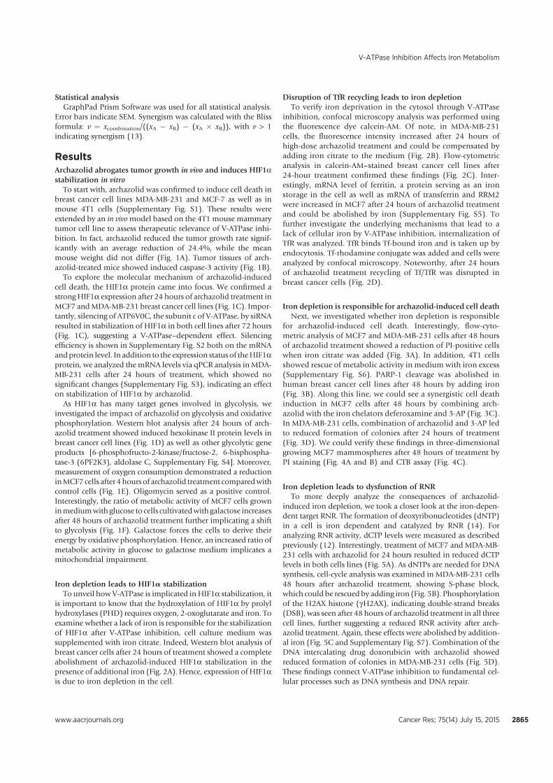
Statistical analysisGraphPad Prism Software was used for all statistical analysis.
Error bars indicate SEM. Synergism was calculated with the Blissformula: v ¼ xcombination/((xA � xB) � (xA � xB)), with v > 1indicating synergism (13).
ResultsArchazolid abrogates tumor growth in vivo and induces HIF1astabilization in vitro
To start with, archazolid was confirmed to induce cell death inbreast cancer cell lines MDA-MB-231 and MCF-7 as well as inmouse 4T1 cells (Supplementary Fig. S1). These results wereextended by an in vivomodel based on the 4T1 mouse mammarytumor cell line to assess therapeutic relevance of V-ATPase inhi-bition. In fact, archazolid reduced the tumor growth rate signif-icantly with an average reduction of 24.4%, while the meanmouse weight did not differ (Fig. 1A). Tumor tissues of arch-azolid-treated mice showed induced caspase-3 activity (Fig. 1B).
To explore the molecular mechanism of archazolid-inducedcell death, the HIF1a protein came into focus. We confirmed astrongHIF1a expression after 24 hours of archazolid treatment inMCF7 andMDA-MB-231 breast cancer cell lines (Fig. 1C). Impor-tantly, silencing of ATP6V0C, the subunit c of V-ATPase, by siRNAresulted in stabilization of HIF1a in both cell lines after 72 hours(Fig. 1C), suggesting a V-ATPase–dependent effect. Silencingefficiency is shown in Supplementary Fig. S2 both on the mRNAandprotein level. In addition to the expression status of theHIF1aprotein, we analyzed themRNA levels via qPCR analysis in MDA-MB-231 cells after 24 hours of treatment, which showed nosignificant changes (Supplementary Fig. S3), indicating an effecton stabilization of HIF1a by archazolid.
As HIF1a has many target genes involved in glycolysis, weinvestigated the impact of archazolid on glycolysis and oxidativephosphorylation. Western blot analysis after 24 hours of arch-azolid treatment showed induced hexokinase II protein levels inbreast cancer cell lines (Fig. 1D) as well as other glycolytic geneproducts [6-phosphofructo-2-kinase/fructose-2, 6-bisphospha-tase-3 (6PF2K3), aldolase C, Supplementary Fig. S4]. Moreover,measurement of oxygen consumption demonstrated a reductioninMCF7 cells after 4hours of archazolid treatment comparedwithcontrol cells (Fig. 1E). Oligomycin served as a positive control.Interestingly, the ratio of metabolic activity of MCF7 cells growninmediumwith glucose to cells cultivatedwith galactose increasesafter 48 hours of archazolid treatment further implicating a shiftto glycolysis (Fig. 1F). Galactose forces the cells to derive theirenergy by oxidative phosphorylation. Hence, an increased ratio ofmetabolic activity in glucose to galactose medium implicates amitochondrial impairment.
Iron depletion leads to HIF1a stabilizationTo unveil how V-ATPase is implicated in HIF1a stabilization, it
is important to know that the hydroxylation of HIF1a by prolylhydroxylases (PHD) requires oxygen, 2-oxoglutarate and iron. Toexamine whether a lack of iron is responsible for the stabilizationof HIF1a after V-ATPase inhibition, cell culture medium wassupplemented with iron citrate. Indeed, Western blot analysis ofbreast cancer cells after 24 hours of treatment showed a completeabolishment of archazolid-induced HIF1a stabilization in thepresence of additional iron (Fig. 2A). Hence, expression of HIF1ais due to iron depletion in the cell.
Disruption of TfR recycling leads to iron depletionTo verify iron deprivation in the cytosol through V-ATPase
inhibition, confocal microscopy analysis was performed usingthe fluorescence dye calcein-AM. Of note, in MDA-MB-231cells, the fluorescence intensity increased after 24 hours ofhigh-dose archazolid treatment and could be compensated byadding iron citrate to the medium (Fig. 2B). Flow-cytometricanalysis in calcein-AM–stained breast cancer cell lines after24-hour treatment confirmed these findings (Fig. 2C). Inter-estingly, mRNA level of ferritin, a protein serving as an ironstorage in the cell as well as mRNA of transferrin and RRM2were increased in MCF7 after 24 hours of archazolid treatmentand could be abolished by iron (Supplementary Fig. S5). Tofurther investigate the underlying mechanisms that lead to alack of cellular iron by V-ATPase inhibition, internalization ofTfR was analyzed. TfR binds Tf-bound iron and is taken up byendocytosis. Tf-rhodamine conjugate was added and cells wereanalyzed by confocal microscopy. Noteworthy, after 24 hoursof archazolid treatment recycling of Tf/TfR was disrupted inbreast cancer cells (Fig. 2D).
Iron depletion is responsible for archazolid-induced cell deathNext, we investigated whether iron depletion is responsible
for archazolid-induced cell death. Interestingly, flow-cyto-metric analysis of MCF7 and MDA-MB-231 cells after 48 hoursof archazolid treatment showed a reduction of PI-positive cellswhen iron citrate was added (Fig. 3A). In addition, 4T1 cellsshowed rescue of metabolic activity in medium with iron excess(Supplementary Fig. S6). PARP-1 cleavage was abolished inhuman breast cancer cell lines after 48 hours by adding iron(Fig. 3B). Along this line, we could see a synergistic cell deathinduction in MCF7 cells after 48 hours by combining arch-azolid with the iron chelators deferoxamine and 3-AP (Fig. 3C).In MDA-MB-231 cells, combination of archazolid and 3-AP ledto reduced formation of colonies after 24 hours of treatment(Fig. 3D). We could verify these findings in three-dimensionalgrowing MCF7 mammospheres after 48 hours of treatment byPI staining (Fig. 4A and B) and CTB assay (Fig. 4C).
Iron depletion leads to dysfunction of RNRTo more deeply analyze the consequences of archazolid-
induced iron depletion, we took a closer look at the iron-depen-dent target RNR. The formation of deoxyribonucleotides (dNTP)in a cell is iron dependent and catalyzed by RNR (14). Foranalyzing RNR activity, dCTP levels were measured as describedpreviously (12). Interestingly, treatment of MCF7 and MDA-MB-231 cells with archazolid for 24 hours resulted in reduced dCTPlevels in both cells lines (Fig. 5A). As dNTPs are needed for DNAsynthesis, cell-cycle analysis was examined in MDA-MB-231 cells48 hours after archazolid treatment, showing S-phase block,which could be rescued by adding iron (Fig. 5B). Phosphorylationof the H2AX histone (gH2AX), indicating double-strand breaks(DSB), was seen after 48 hours of archazolid treatment in all threecell lines, further suggesting a reduced RNR activity after arch-azolid treatment. Again, these effects were abolished by addition-al iron (Fig. 5C and Supplementary Fig. S7). Combination of theDNA intercalating drug doxorubicin with archazolid showedreduced formation of colonies in MDA-MB-231 cells (Fig. 5D).These findings connect V-ATPase inhibition to fundamental cel-lular processes such as DNA synthesis and DNA repair.
V-ATPase Inhibition Affects Iron Metabolism
www.aacrjournals.org Cancer Res; 75(14) July 15, 2015 2865
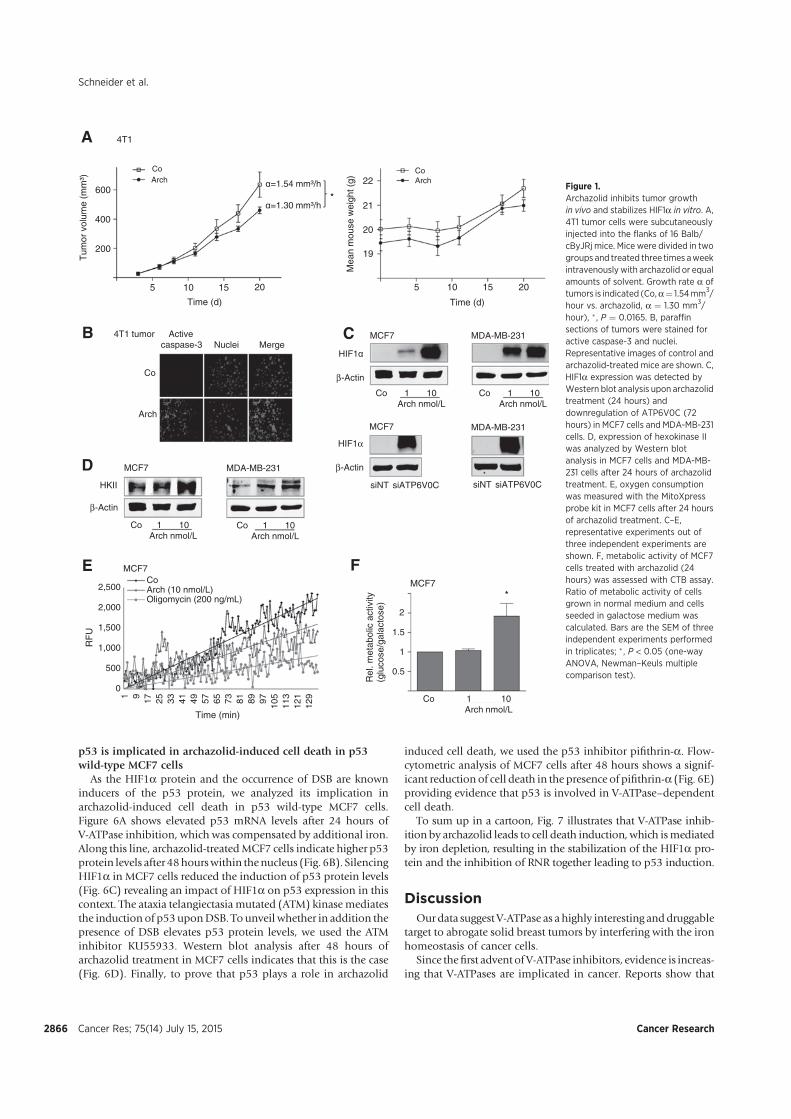
p53 is implicated in archazolid-induced cell death in p53wild-type MCF7 cells
As the HIF1a protein and the occurrence of DSB are knowninducers of the p53 protein, we analyzed its implication inarchazolid-induced cell death in p53 wild-type MCF7 cells.Figure 6A shows elevated p53 mRNA levels after 24 hours ofV-ATPase inhibition, which was compensated by additional iron.Along this line, archazolid-treatedMCF7 cells indicate higher p53protein levels after 48hourswithin thenucleus (Fig. 6B). SilencingHIF1a in MCF7 cells reduced the induction of p53 protein levels(Fig. 6C) revealing an impact of HIF1a on p53 expression in thiscontext. The ataxia telangiectasia mutated (ATM) kinasemediatesthe induction of p53uponDSB. To unveil whether in addition thepresence of DSB elevates p53 protein levels, we used the ATMinhibitor KU55933. Western blot analysis after 48 hours ofarchazolid treatment in MCF7 cells indicates that this is the case(Fig. 6D). Finally, to prove that p53 plays a role in archazolid
induced cell death, we used the p53 inhibitor pifithrin-a. Flow-cytometric analysis of MCF7 cells after 48 hours shows a signif-icant reduction of cell death in the presence of pifithrin-a (Fig. 6E)providing evidence that p53 is involved in V-ATPase–dependentcell death.
To sum up in a cartoon, Fig. 7 illustrates that V-ATPase inhib-ition by archazolid leads to cell death induction, which ismediatedby iron depletion, resulting in the stabilization of the HIF1a pro-tein and the inhibition of RNR together leading to p53 induction.
DiscussionOurdata suggest V-ATPase as a highly interesting anddruggable
target to abrogate solid breast tumors by interfering with the ironhomeostasis of cancer cells.
Since thefirst advent of V-ATPase inhibitors, evidence is increas-ing that V-ATPases are implicated in cancer. Reports show that
A
Arch
400
Tum
orvo
lum
e(m
m³)
200
600
Co
Time (d)
5 1510 20
ArchCo
Time (d)
105 15 20
21
Mea
nm
ouse
wei
ght(
g)
20
22
19
*
4T1
HIF1α
β-Actin
MDA-MB-231 MCF7
Co 1 10 Arch nmol/L
MCF7 MDA-MB-231
siNT siATP6V0C
B
D
E
Co 1 10 Arch nmol/L
siNT siATP6V0CHKII
β-Actin
MCF7 MDA-MB-231
Co 1 10 Arch nmol/L
Co 1 10 Arch nmol/L
MCF7
*
1
0.5
1.5
2
Rel
. met
abol
ic a
ctiv
ity(g
luco
se/g
alac
tose
)
MCF7
Co 1 10 Arch nmol/L
F
0
500
1,000
1,500
2,000
2,500
1 9 17 25 33 41 49 57 65 73 81 89 97 105
113
121
129
RF
U
Time (min)
CoArch (10 nmol/L)Oligomycin (200 ng/mL)
α=1.54 mm³/h
α=1.30 mm³/h
C
HIF1α
β-Actin
Arch
Co
4T1 tumor Activecaspase-3 MergeNuclei
Figure 1.Archazolid inhibits tumor growthin vivo and stabilizes HIF1a in vitro. A,4T1 tumor cells were subcutaneouslyinjected into the flanks of 16 Balb/cByJRj mice. Mice were divided in twogroups and treated three times aweekintravenously with archazolid or equalamounts of solvent. Growth rate a oftumors is indicated (Co,a¼ 1.54mm3/hour vs. archazolid, a ¼ 1.30 mm3/hour), � , P ¼ 0.0165. B, paraffinsections of tumors were stained foractive caspase-3 and nuclei.Representative images of control andarchazolid-treated mice are shown. C,HIF1a expression was detected byWestern blot analysis upon archazolidtreatment (24 hours) anddownregulation of ATP6V0C (72hours) in MCF7 cells and MDA-MB-231cells. D, expression of hexokinase IIwas analyzed by Western blotanalysis in MCF7 cells and MDA-MB-231 cells after 24 hours of archazolidtreatment. E, oxygen consumptionwas measured with the MitoXpressprobe kit in MCF7 cells after 24 hoursof archazolid treatment. C–E,representative experiments out ofthree independent experiments areshown. F, metabolic activity of MCF7cells treated with archazolid (24hours) was assessed with CTB assay.Ratio of metabolic activity of cellsgrown in normal medium and cellsseeded in galactose medium wascalculated. Bars are the SEM of threeindependent experiments performedin triplicates; � , P < 0.05 (one-wayANOVA, Newman–Keuls multiplecomparison test).
Schneider et al.
Cancer Res; 75(14) July 15, 2015 Cancer Research2866
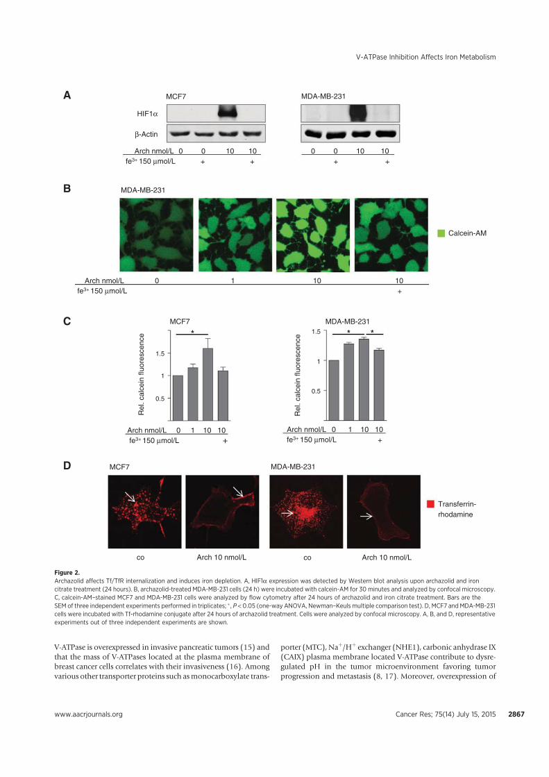
V-ATPase is overexpressed in invasive pancreatic tumors (15) andthat the mass of V-ATPases located at the plasma membrane ofbreast cancer cells correlates with their invasiveness (16). Amongvarious other transporter proteins such asmonocarboxylate trans-
porter (MTC), Naþ/Hþ exchanger (NHE1), carbonic anhydrase IX(CAIX) plasma membrane located V-ATPase contribute to dysre-gulated pH in the tumor microenvironment favoring tumorprogression and metastasis (8, 17). Moreover, overexpression of
HIF1α
β-Actin
MCF7 MDA-MB-231
Transferrin-rhodamine
MCF7 MDA-MB-231
Calcein-AM
MDA-MB-231
MCF7 MDA-MB-231
Arch 10 nmol/Lco Arch 10 nmol/Lco
Rel
. cal
cein
fluo
resc
ence
1
0.5
1.51
0.5
1.5
Rel
. cal
cein
fluo
resc
ence
*
0 1 10 10 Arch nmol/Lfe3+ +150 μmol/L
A
B
C
D
0 1 10 10 Arch nmol/Lfe3+ +150 μmol/L
* *
0 1 10 10 Arch nmol/Lfe3+ +150 μmol/L
0 0 10 10 Arch nmol/Lfe3+ + + 150 μmol/L
0 0 10 10 + +
Figure 2.Archazolid affects Tf/TfR internalization and induces iron depletion. A, HIF1a expression was detected by Western blot analysis upon archazolid and ironcitrate treatment (24 hours). B, archazolid-treated MDA-MB-231 cells (24 h) were incubated with calcein-AM for 30 minutes and analyzed by confocal microscopy.C, calcein-AM–stained MCF7 and MDA-MB-231 cells were analyzed by flow cytometry after 24 hours of archazolid and iron citrate treatment. Bars are theSEM of three independent experiments performed in triplicates; � , P < 0.05 (one-way ANOVA, Newman–Keuls multiple comparison test). D, MCF7 and MDA-MB-231cells were incubated with Tf-rhodamine conjugate after 24 hours of archazolid treatment. Cells were analyzed by confocal microscopy. A, B, and D, representativeexperiments out of three independent experiments are shown.
V-ATPase Inhibition Affects Iron Metabolism
www.aacrjournals.org Cancer Res; 75(14) July 15, 2015 2867
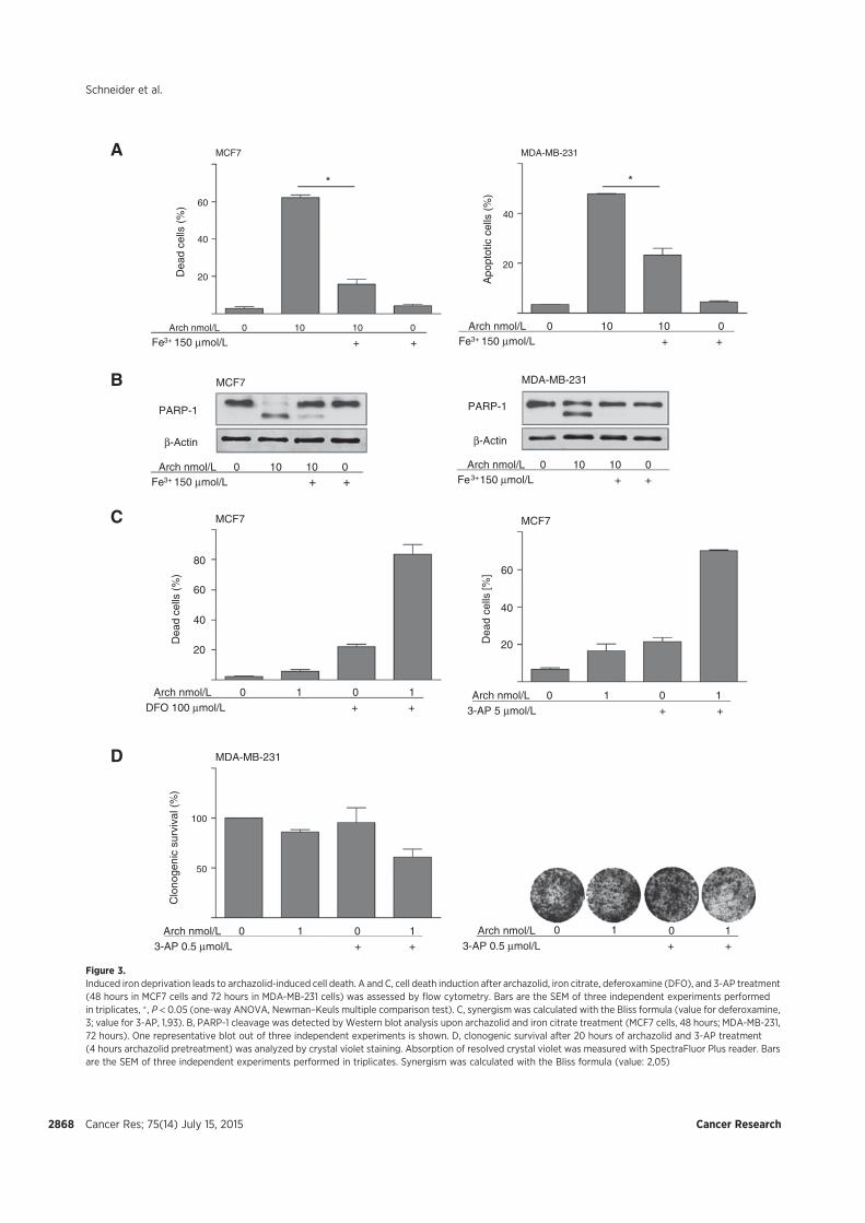
MDA-MB-231MCF7
MCF7 MDA-MB-231
PARP-1
β-Actin
MCF7 MCF7
20
40
60
Dea
d ce
lls (
%)
20
40
Apo
ptot
ic c
ells
(%
)
* *
PARP-1
β-Actin
40
60
Dea
dce
lls(%
)
80
20
40
60
Dea
d ce
lls [%
]
MDA-MB-231
50
100
Clo
noge
nic
surv
ival
(%
)
A
B
C
D
20
1 010Arch nmol/L++DFO 100 μmol/L
100 1Arch nmol/L3-AP 5 μmol/L + +
11 00Arch nmol/L+ + 3-AP 0.5 μmol/L
100 1Arch nmol/L + + 3-AP 0.5 μmol/L
0 10 10 0 Arch nmol/L
Fe3+ + + 150 μmol/L
0 10 10 0 Arch nmol/LFe3+ + + 150 μmol/L
0 10 10 0 Arch nmol/LFe3+ + + 150 μmol/L
0 10 10 0 Arch nmol/LFe3+ + + 150 μmol/L
Figure 3.Induced iron deprivation leads to archazolid-induced cell death. A and C, cell death induction after archazolid, iron citrate, deferoxamine (DFO), and 3-AP treatment(48 hours in MCF7 cells and 72 hours in MDA-MB-231 cells) was assessed by flow cytometry. Bars are the SEM of three independent experiments performedin triplicates, � , P < 0.05 (one-way ANOVA, Newman–Keuls multiple comparison test). C, synergism was calculated with the Bliss formula (value for deferoxamine,3; value for 3-AP, 1,93). B, PARP-1 cleavage was detected by Western blot analysis upon archazolid and iron citrate treatment (MCF7 cells, 48 hours; MDA-MB-231,72 hours). One representative blot out of three independent experiments is shown. D, clonogenic survival after 20 hours of archazolid and 3-AP treatment(4 hours archazolid pretreatment) was analyzed by crystal violet staining. Absorption of resolved crystal violet was measured with SpectraFluor Plus reader. Barsare the SEM of three independent experiments performed in triplicates. Synergism was calculated with the Bliss formula (value: 2,05)
Schneider et al.
Cancer Res; 75(14) July 15, 2015 Cancer Research2868
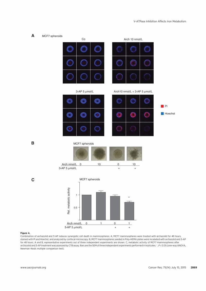
Co
Arch10 nmol/L + 3-AP 5 μmol/L3-AP 5 μmol/L
Arch 10 nmol/LMCF7 spheroids
MCF7 spheroids
1
0.5
Rel
. met
abol
ic a
ctiv
ity
MCF7 spheroids
A
B
C
PI
Hoechst
10 0 10 0Arch nmol/L3-AP 5 μmol/L + +
100 1Arch nmol/L3-AP 5 μmol/L + +
*
Figure 4.Combination of archazolid and 3-AP induces synergistic cell death in mammospheres. A, MCF7 mammospheres were treated with archazolid for 48 hours,stained with PI and Hoechst, and analyzed by confocal microscopy. B, MCF7 mammospheres seeded in Poly-HEMA plates were incubated with archazolid and 3-APfor 48 hours. A and B, representative experiments out of three independent experiments are shown. C, metabolic activity of MCF7 mammospheres afterarchazolid and3-AP treatmentwas assessedbyCTBassay. Bars are the SEMof three independent experiments performed in triplicates. � ,P<0.05 (one-wayANOVA,Newman–Keuls multiple comparison test).
V-ATPase Inhibition Affects Iron Metabolism
www.aacrjournals.org Cancer Res; 75(14) July 15, 2015 2869
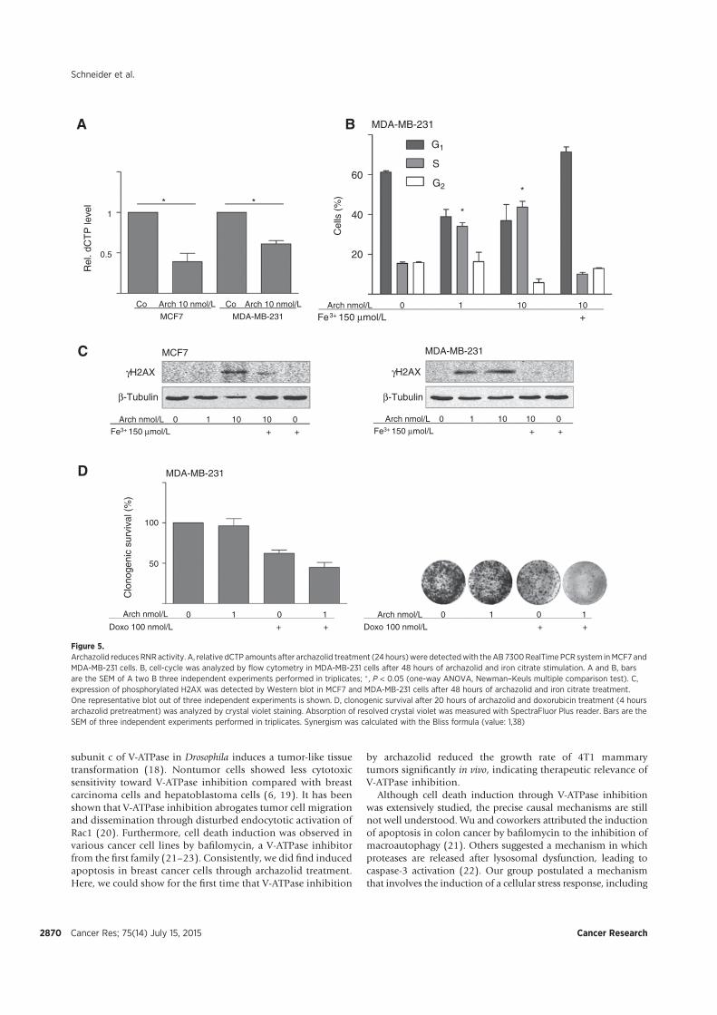
subunit c of V-ATPase in Drosophila induces a tumor-like tissuetransformation (18). Nontumor cells showed less cytotoxicsensitivity toward V-ATPase inhibition compared with breastcarcinoma cells and hepatoblastoma cells (6, 19). It has beenshown that V-ATPase inhibition abrogates tumor cell migrationand dissemination through disturbed endocytotic activation ofRac1 (20). Furthermore, cell death induction was observed invarious cancer cell lines by bafilomycin, a V-ATPase inhibitorfrom the first family (21–23). Consistently, we did find inducedapoptosis in breast cancer cells through archazolid treatment.Here, we could show for the first time that V-ATPase inhibition
by archazolid reduced the growth rate of 4T1 mammarytumors significantly in vivo, indicating therapeutic relevance ofV-ATPase inhibition.
Although cell death induction through V-ATPase inhibitionwas extensively studied, the precise causal mechanisms are stillnot well understood. Wu and coworkers attributed the inductionof apoptosis in colon cancer by bafilomycin to the inhibition ofmacroautophagy (21). Others suggested a mechanism in whichproteases are released after lysosomal dysfunction, leading tocaspase-3 activation (22). Our group postulated a mechanismthat involves the induction of a cellular stress response, including
MDA-MB-231
γH2AX
β-Tubulin
γH2AX
β-Tubulin
MCF7 MDA-MB-231
Cel
ls (
%)
20
40
60
A B
C
G1
S
G2
Arch nmol/L 0 1 10 10
Fe3+ 150 μmol/L +
Arch nmol/L 0 1 10 10 0 Fe3+ 150 μmol/L + +
Co Arch 10 nmol/L Co Arch 10 nmol/L
MCF7 MDA-MB-231
Rel
. dC
TP
leve
l
1
0.5
* **
*
Arch nmol/L 0 1 10 10 0 Fe3+ 150 μmol/L + +
100
50
Clo
noge
nic
surv
ival
(%
)
MDA-MB-231
Arch nmol/L 0 1 0 1
Doxo 100 nmol/L + +
D
Arch nmol/L 0 1 0 1
Doxo 100 nmol/L + +
Figure 5.Archazolid reduces RNRactivity. A, relative dCTP amounts after archazolid treatment (24 hours)were detectedwith theAB 7300RealTime PCR system inMCF7 andMDA-MB-231 cells. B, cell-cycle was analyzed by flow cytometry in MDA-MB-231 cells after 48 hours of archazolid and iron citrate stimulation. A and B, barsare the SEM of A two B three independent experiments performed in triplicates; �, P < 0.05 (one-way ANOVA, Newman–Keuls multiple comparison test). C,expression of phosphorylated H2AX was detected by Western blot in MCF7 and MDA-MB-231 cells after 48 hours of archazolid and iron citrate treatment.One representative blot out of three independent experiments is shown. D, clonogenic survival after 20 hours of archazolid and doxorubicin treatment (4 hoursarchazolid pretreatment) was analyzed by crystal violet staining. Absorption of resolved crystal violet was measured with SpectraFluor Plus reader. Bars are theSEM of three independent experiments performed in triplicates. Synergism was calculated with the Bliss formula (value: 1,38)
Schneider et al.
Cancer Res; 75(14) July 15, 2015 Cancer Research2870

autophagy and the stabilization of HIF1a (6). This study here tiesin with our earlier work and reveals a role for V-ATPase at theinterplay of iron metabolism and apoptotic processes. Our find-ings illuminate that the cytotoxicity of V-ATPase inhibitors ismainly due to disturbed TfR recycling, which impairs the ironsupply of cancer cells. As a consequence, the activity of iron-dependent enzymes is reduced, leading to DSB, S-phase arrest,and induction of p53.
In the last decades, it became more and more evident thatiron is implicated in cancer development. Induction of malig-nant tumors by injecting iron dextran into rat was first reportedin 1959 (24). This was confirmed later by the observation thatpatients injected with iron preparations developed sarcomas(25). Consistently, epidemiologic reports from the 1980s asso-ciated high body iron with the risk of cancer (26). The ironsupply of cancer cells is mainly mediated by the TfR. The clearlydefined function of the TfR is the interaction with the ironloaded plasma glycoprotein Tf initiating its endocytotic up take(27). Since it has been reported that TfR is overexpressed in
cancer cells (28, 29), targeted therapy became an attractivestrategy for cancer treatment. In different studies, antibodiesagainst TfR have successfully been used to abrogate tumorprogression (30, 31). Others used toxic moieties conjugatedto Tf to selectively target cancer cells (32). After internalizationof Tf/TfR, iron dissociates from the receptor-ligand complexand enters the cytosol forming the labile iron pool whereas thecomplex gets recycled to the cell surface. Essential for thedissociation is the acid pH in the endosomes (33), which ismaintained by V-ATPases. It is known that an inadequateacidification of endosomes inhibits trafficking out of themalthough precise mechanisms are not defined yet (34). Con-sistently, a role for V-ATPase in the internalization or proces-sing of receptors has been shown such as for Notch (35, 36).Our present study in breast cancer cells shows impaired TfRinternalization after archazolid treatment and connects thiswith disturbed iron metabolic pathways. The generated irondeprivation eventually induces apoptotic processes, leading tocell death. Therefore, we suggest inhibition of V-ATPase as a
A
C
D
E
p53
β-Actin
BR
el. p
53 m
RN
A le
vel
2
1
3
20
10
30
40
Dea
dce
lls(%
)
10 10 00Arch nmol/LPifithrin-α 30 μmol/L + +
MCF7
MCF7
1 10 CoArch nmol/L
p53
CREB
MCF7
p53
CREB
Arch nmol/L 0 10 siNT
0 10 siHIF1α
10 10 00Arch nmol/L+ KU55933 10 μmol/L +
MCF7
MCF7
* *
*
0 10 10 0 Arch nmol/LFe3+ + + 150 μmol/L
Figure 6.Implication of p53 in archazolid induced cell death. A, SYBR Green qRT-PCR analysis was performed to assess p53 mRNA levels in archazolid-treated (24 hours)MCF7 cells. B and C, expression of p53 in nuclei was analyzed by Western blot analysis in MCF7 cells after 48 hours of archazolid treatment C upon HIF1asilencing. CREB served as a loading control. D, expression of p53 was analyzed by Western blot analysis in MCF7 cells after 48 hours of archazolid and KU55933treatment. E, cell death was assessed by flow cytometry analysis in archazolid and pifithrin-a–treated MCF7 cells after 48 hours. A and E, bars are the SEMof three independent experiments performed in triplicates; � , P < 0.05 (one-way ANOVA, Newman–Keuls multiple comparison test). B–D, one representativeblot out of three independent experiments is shown.
V-ATPase Inhibition Affects Iron Metabolism
www.aacrjournals.org Cancer Res; 75(14) July 15, 2015 2871
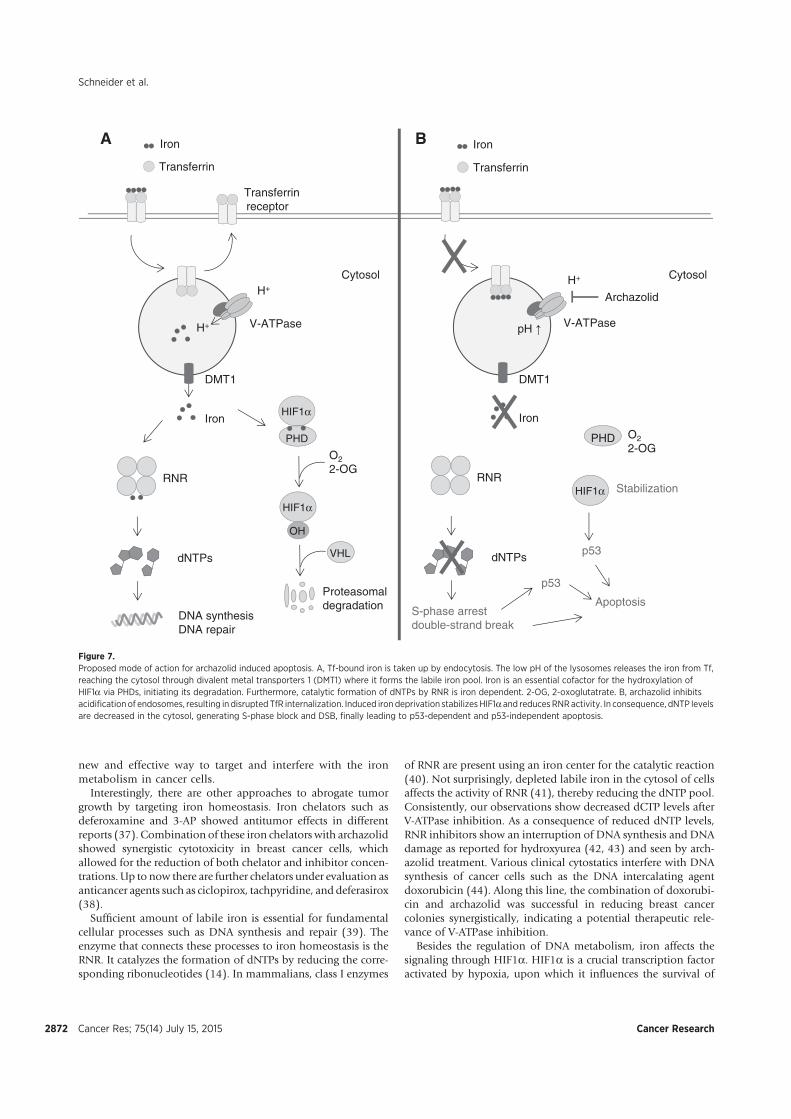
new and effective way to target and interfere with the ironmetabolism in cancer cells.
Interestingly, there are other approaches to abrogate tumorgrowth by targeting iron homeostasis. Iron chelators such asdeferoxamine and 3-AP showed antitumor effects in differentreports (37). Combination of these iron chelators with archazolidshowed synergistic cytotoxicity in breast cancer cells, whichallowed for the reduction of both chelator and inhibitor concen-trations. Up to now there are further chelators under evaluation asanticancer agents such as ciclopirox, tachpyridine, and deferasirox(38).
Sufficient amount of labile iron is essential for fundamentalcellular processes such as DNA synthesis and repair (39). Theenzyme that connects these processes to iron homeostasis is theRNR. It catalyzes the formation of dNTPs by reducing the corre-sponding ribonucleotides (14). In mammalians, class I enzymes
of RNR are present using an iron center for the catalytic reaction(40). Not surprisingly, depleted labile iron in the cytosol of cellsaffects the activity of RNR (41), thereby reducing the dNTP pool.Consistently, our observations show decreased dCTP levels afterV-ATPase inhibition. As a consequence of reduced dNTP levels,RNR inhibitors show an interruption of DNA synthesis and DNAdamage as reported for hydroxyurea (42, 43) and seen by arch-azolid treatment. Various clinical cytostatics interfere with DNAsynthesis of cancer cells such as the DNA intercalating agentdoxorubicin (44). Along this line, the combination of doxorubi-cin and archazolid was successful in reducing breast cancercolonies synergistically, indicating a potential therapeutic rele-vance of V-ATPase inhibition.
Besides the regulation of DNA metabolism, iron affects thesignaling through HIF1a. HIF1a is a crucial transcription factoractivated by hypoxia, upon which it influences the survival of
Transferrin
Transferrinreceptor
V-ATPase
H+
RNR
dNTPs
Iron
H+
DNA synthesis DNA repair
HIF1α
OH
VHL
HIF1α
PHD
O22-OG
Proteasomaldegradation
Transferrin
V-ATPase
RNR
dNTPs
Iron
S-phase arrestdouble-strand break
HIF1α
PHD O22-OG
Archazolid
Stabilization
A B
Apoptosis
DMT1 DMT1
H+
p53
p53
Iron Iron
pH ↑
Cytosol Cytosol
Figure 7.Proposed mode of action for archazolid induced apoptosis. A, Tf-bound iron is taken up by endocytosis. The low pH of the lysosomes releases the iron from Tf,reaching the cytosol through divalent metal transporters 1 (DMT1) where it forms the labile iron pool. Iron is an essential cofactor for the hydroxylation ofHIF1a via PHDs, initiating its degradation. Furthermore, catalytic formation of dNTPs by RNR is iron dependent. 2-OG, 2-oxoglutatrate. B, archazolid inhibitsacidification of endosomes, resulting in disrupted TfR internalization. Induced iron deprivation stabilizes HIF1a and reducesRNRactivity. In consequence, dNTP levelsare decreased in the cytosol, generating S-phase block and DSB, finally leading to p53-dependent and p53-independent apoptosis.
Cancer Res; 75(14) July 15, 2015 Cancer Research2872
Schneider et al.
tumor cells and shifts the cellular metabolism toward glycolysis(45). For the proteasomal degradation of HIF1a via the vonHippel–Lindau tumor suppressor, HIF1a has to be hydroxylatedbyPHDs (46). This catalytic reaction is dependent on the presenceof oxygen, 2-oxogutarat, and iron (47). Consistently, archazolid-induced iron depletion stabilizes HIF1a modifying the cellularglucose metabolism.
DNAdamage aswell asHIF1a can evoke the stabilization of thetumor suppressor p53 (48, 49). After activation of p53, it is able toinduce either cell-cycle arrest or apoptosis, thereby reducingtumor progression. Cell-type origin, strength of p53-activatingstimulus, and others can influence the outcome of p53 activation(50). p53 is widely mutated in tumors but at least 50% of allcancers are p53wild-type tumors. Therefore, a lot of effort was putinto the development of p53 activators. Up to now, the mostadvanced p53 activators are RG7112, MI-773, and DS-3032bbeing in phase I clinical trials (51). Nevertheless, it has beenreported that DNA damage can trigger apoptosis independent ofp53 in p53-deficient cells (52). Our work in p53 wild-type tumorcells (MCF7 cells) showed a clear involvement of p53 in V-ATPase–dependent cell death. Furthermore, archazolid success-fully induced cell death in p53-mutated breast cancer cells (MDA-MB-231) and p53-null breast cancer cells (4T1). Hence thisindicates that targeting the iron metabolism of cancer cells byV-ATPase inhibition can hit p53 wild-type tumors as well asmutated.Nonetheless onemight speculate that a xenograftmousemodel using a cell line expressing wild-type p53 should be moresensitive to tumor mass reduction than our p53-null 4T1 model.
Our observations that V-ATPase inhibition generates iron dep-rivation in the cell, thereby influencing activity of the iron-depen-dent enzymes PHDs and RNR evokes new options for interferingwith the iron metabolism in cancer therapy. The depicted mech-
anism of action connects V-ATPase inhibition to fundamentalcellular processes such as DNA synthase and repair. We like topoint to the fact that the growth-inhibiting effects of archazolidseen in vitro are recapitulated in a solid tumor growth modelin vivo, suggesting inhibition of V-ATPase as highly promising andviable strategy for breast cancer treatment.
Disclosure of Potential Conflicts of InterestNo potential conflicts of interest were disclosed.
Authors' ContributionsConception and design: L.S. Schneider, K. von Schwarzenberg, A.M. VollmarDevelopment of methodology: L.S. Schneider, D. Trauner, A.M. VollmarAcquisition of data (provided animals, acquired and managed patients,provided facilities, etc.): R. Kubisch-DohmenAnalysis and interpretation of data (e.g., statistical analysis, biostatistics,computational analysis): L.S. Schneider, K. von Schwarzenberg, T. Lehr,M. Ulrich, J. LieblWriting, review, and/or revision of the manuscript: L.S. Schneider, T. Lehr,D. Menche, A.M. VollmarAdministrative, technical, or material support (i.e., reporting or organizingdata, constructingdatabases):K. von Schwarzenberg,D.Menche, A.M. VollmarStudy supervision: A.M. Vollmar
Grant SupportThis work was financially supported by the German Research foundation
(DFG, FOR 1406, Vo 376-14/15; A.M. Vollmar).The costs of publication of this articlewere defrayed inpart by the payment of
page charges. This article must therefore be hereby marked advertisement inaccordance with 18 U.S.C. Section 1734 solely to indicate this fact.
Received July 16, 2014; revised February 24, 2015; accepted April 28, 2015;published OnlineFirst May 27, 2015.
References1. Jemal A, Bray F, Center MM, Ferlay J, Ward E, Forman D. Global cancer
statistics. CA Cancer J Clin 2011;61:69–90.2. Newman DJ, Cragg GM. Natural products as sources of new drugs over the
30 years from 1981 to 2010. J Nat Prod 2012;75:311–35.3. Sasse F, Steinmetz H, H€ofle G, Reichenbach H. Archazolids, new cytotoxic
macrolactones from archangium gephyra (Myxobacteria) production,isolation, physico-chemical and biological properties. J Antibiot 2003;56:520–5.
4. Bockelmann S,MencheD, Rudolph S, Bender T,GrondS, vonZezschwitz P,et al. Archazolid A binds to the equatorial region of the c-ring of thevacuolar Hþ-ATPase. J Biol Chem 2010;285:38304–14.
5. Huss M, Sasse F, Kunze B, Jansen R, Steinmetz H, Ingenhorst G, et al.Archazolid and apicularen: novel specific V-ATPase inhibitors. BMCBiochem 2005;6:13.
6. von Schwarzenberg K,Wiedmann RM,Oak P, Schulz S, ZischkaH,WannerG, et al. Mode of cell death induction by pharmacological vacuolar Hþ-ATPase (V-ATPase) inhibition. J Biol Chem 2012;288:1385–96.
7. Forgac M. Vacuolar ATPases: rotary proton pumps in physiology andpathophysiology. Nat Rev Mol Cell Biol 2007;8:917–29.
8. Barar J,Omidi Y.Dysregulated pH in tumormicroenvironment checkmatescancer therapy. Bioimpacts 2013;3:149–62.
9. Foerster F, Braig S,MoserC, KubischR, Busse J,Wagner E, et al. Targeting theactin cytoskeleton: selective antitumor action via trapping PKCe. CellDeath Dis 2014;5:e1398.
10. Simeoni M, Magni P, Cammia C, De Nicolao G, Croci V, Pesenti E, et al.Predictive pharmacokinetic-pharmacodynamicmodeling of tumor growthkinetics in xenograft models after administration of anticancer agents.Cancer Res 2004;64:1094–101.
11. Braig S, Kressirer CA, Liebl J, Bischoff F, Zahler S, Meijer L, et al. Indirubinderivative 6BIO suppresses metastasis. Cancer Res 2013;73:6004–12.
12. Wilson PM, LaBonteMJ, Russell J, Louie S, Ghobrial AA, Ladner RD. A novelfluorescence-basedassay for the rapiddetectionandquantificationof cellulardeoxyribonucleoside triphosphates. Nucleic Acids Res 2011;39:e112.
13. Bliss CI. The calculation of microbial assays. Bacteriol Rev 1956;20:243–58.
14. Elledge SJ, Zhou Z, Allen JB. Ribonucleotide reductase: regulation, regu-lation, regulation. Trends Biochem Sci 1992;17:119–23.
15. Ohta T, Numata M, Yagishita H, Futagami F, Tsukioka Y, Kitagawa H, et al.Expression of 16 kDa proteolipid of vacuolar-type H(þ)-ATPase in humanpancreatic cancer. Br J Cancer 1996;73:1511–7.
16. Hinton A, Sennoune SR, Bond S, Fang M, Reuveni M, Sahagian GG, et al.Function of a subunit isoforms of the V-ATPase in pH homeostasis and invitro invasion of MDA-MB231 human breast cancer cells. J Biol Chem2009;284:16400–8.
17. Pinheiro C, Longatto-Filho A, Azevedo-Silva J, Casal M, Schmitt FC,Baltazar F. Role of monocarboxylate transporters in human cancers: stateof the art. J Bioenerg Biomembr 2012;44:127–39.
18. Petzoldt AG, Gleixner EM, Fumagalli A, Vaccari T, Simons M. Elevatedexpression of the V-ATPase C subunit triggers JNK-dependent cellinvasion and overgrowth in a Drosophila epithelium. Dis Model Mech2013;6:689–700.
19. Morimura T, Fujita K, Akita M, NagashimaM, Satomi A. The proton pumpinhibitor inhibits cell growth and induces apoptosis in human hepato-blastoma. Pediatr Surg Int 2008;24:1087–94.
20. Wiedmann RM, von Schwarzenberg K, Palamidessi A, Schreiner L, KubischR, Liebl J, et al. The V-ATPase-inhibitor archazolid abrogates tumormetastasis via inhibition of endocytic activation of the Rho-GTPase Rac1.Cancer Res 2012;72:5976–87.
21. Wu YC, Wu WKK, Li Y, Yu L, Li ZJ, Wong CCM, et al. Inhibition ofmacroautophagy by bafilomycin A1 lowers proliferation and induces
www.aacrjournals.org Cancer Res; 75(14) July 15, 2015 2873
V-ATPase Inhibition Affects Iron Metabolism
apoptosis in colon cancer cells. Biochem Biophys Res Commun 2009;382:451–6.
22. Nakashima S. Vacuolar Hþ-ATPase inhibitor induces apoptosis via lyso-somal dysfunction in the human gastric cancer cell line MKN-1. J Biochem2003;134:359–64.
23. Ohta T, Arakawa H, Futagami F, Fushida S, Kitagawa H, Kayahara M, et al.Bafilomycin A1 induces apoptosis in the human pancreatic cancer cell lineCapan-1. J Pathol 1998;185:324–30.
24. Richmond HG. Induction of sarcoma in the rat by iron-dextran complex.Br Med J 1959;1:947–9.
25. Greenberg G. Sarcoma after intramuscular iron injection. Br Med J 1976;1:1508–9.
26. Stevens RG, Jones DY, Micozzi MS, Taylor PR. Body iron stores and the riskof cancer. New Engl J Med 1988;319:1047–52.
27. Ponka P, Lok CN. The transferrin receptor: role in health and disease. Int JBiochem Cell Biol 1999;31:1111–37.
28. Ryschich E, Huszty G, Knaebel HP, Hartel M, B€uchler MW, Schmidt J.Transferrin receptor is a marker of malignant phenotype in human pan-creatic cancer and in neuroendocrine carcinoma of the pancreas. Eur JCancer 2004;40:1418–22.
29. Law S. Overexpression of transferrin receptor CD71 and its tumorigenicproperties in esophageal squamous cell carcinoma. Oncol Rep 2014;31:1296–304.
30. Brooks D, Taylor C, Dos Santos B, Linden H, Houghton A, Hecht TT, et al.Phase Ia trial of murine immunoglobulin A antitransferrin receptor anti-body 42/6. Clin Cancer Res 1995;1:1259–65.
31. Taetle R, Castagnola J, Mendelsohn J. Mechanisms of growth inhibition byanti-transferrin receptor monoclonal antibodies. Cancer Res 1986;46:1759–63.
32. Daniels TR, Bernabeu E, Rodríguez JA, Patel S, KozmanM, Chiappetta DA,et al. The transferrin receptor and the targeted delivery of therapeutic agentsagainst cancer. Biochim Biophys Acta 2012;1820:291–317.
33. Dautry-Varsat A, Ciechanover A, Lodish HF. pH and the recycling oftransferrin during receptor-mediated endocytosis. Proc Natl Acad Sci US A 1983;80:2258–62.
34. Mellman I, Fuchs R, Helenius A. Acidification of the endocytic and exocyticpathways. Annu Rev Biochem 1986;55:663–700.
35. Kozik P, Hodson NA, Sahlender DA, Simecek N, Soromani C, Wu J, et al. Ahuman genome-wide screen for regulators of clathrin-coated vesicle for-mation reveals an unexpected role for the V-ATPase. Nat Cell Biol 2012;15:50–60.
36. Kobia F, Duchi S, Deflorian G, Vaccari T. Pharmacologic inhibition ofvacuolar Hþ ATPase reduces physiologic and oncogenic Notch signaling.Mol Oncol 2014;8:207–20.
37. Yu Y,Wong J, Lovejoy DB, Kalinowski DS, RichardsonDR. Chelators at thecancer coalface: desferrioxamine to Triapine and beyond. Clin Cancer Res2006;12:6876–83.
38. Torti SV, Torti FM. Iron and cancer: more ore to be mined. Nat Rev Cancer2013;13:342–55.
39. Robbins E, Pederson T. Iron: its intracellular localization and possible rolein cell division. Proc Natl Acad Sci U S A 1970;66:1244–51.
40. Reichard P. From RNA to DNA, why so many ribonucleotide reductases?Science 1993;260:1773–7.
41. Furukawa T, Naitoh Y, Kohno H, Tokunaga R, Taketani S. Iron deprivationdecreases ribonucleotide reductase activity and DNA synthesis. Life Sci1992;50:2059–65.
42. Koc A, Wheeler LJ, Mathews CK, Merrill GF. Hydroxyurea arrests DNAreplication by a mechanism that preserves basal dNTP pools. J Biol Chem2003;279:223–30.
43. Osterman Golkar S, Czene S, Gokarakonda A, Haghdoost S. Intracellulardeoxyribonucleotide pool imbalance and DNA damage in cells treatedwith hydroxyurea, an inhibitor of ribonucleotide reductase. Mutagenesis2013;28:653–60.
44. Box VGS. The intercalation of DNA double helices with doxorubicin andnagalomycin. J Mol Graphics Model 2007;26:14–9.
45. Piret J-P,Mottet D, RaesM,Michiels C. IsHIF-1a a pro- or an anti-apoptoticprotein? Biochem Pharmacol 2002;64:889–92.
46. Denko NC. Hypoxia, HIF1 and glucose metabolism in the solid tumour.Nat Rev Cancer 2008;8:705–13.
47. Tuderman L, Myllyla R, Kivirikko KI. Mechanism of the prolyl hydroxylasereaction. 1. Role of co-substrates. Eur J Biochem 1977;80:341–8.
48. Sermeus A, Michiels C. Reciprocal influence of the p53 and the hypoxicpathways. Cell Death Dis 2011;2:e164.
49. Lee JH, Paull TT. Activation and regulation of ATM kinase activityin response to DNA double-strand breaks. Oncogene 2007;26:7741–8.
50. Fridman JS, Lowe SW. Control of apoptosis by p53. Oncogene 2003;22:9030–40.
51. Hoe KK, Verma CS, Lane DP. Drugging the p53 pathway: under-standing the route to clinical efficacy. Nat Rev Drug Discov 2014;13:217–36.
52. Lips J, Kaina B. DNA double-strand breaks trigger apoptosis in p53-deficient fibroblasts. Carcinogenesis 2001;22:579–85.
Cancer Res; 75(14) July 15, 2015 Cancer Research2874
Schneider et al.
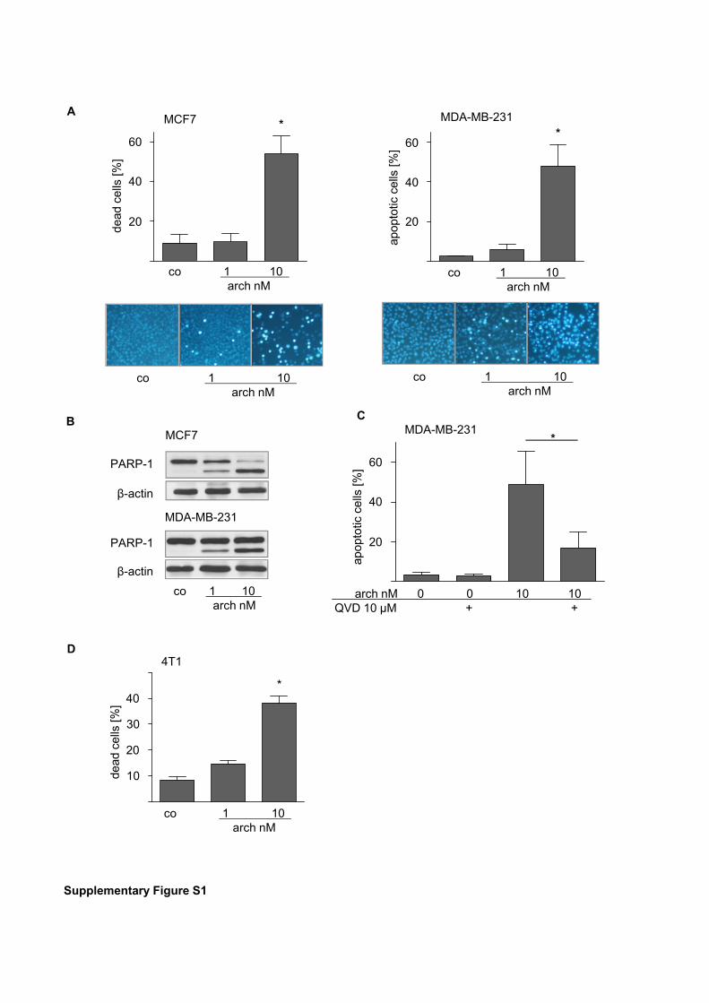
Supplementary Figure S1
MCF7
MCF7
MDA-MB-231
PARP-1
β-actin
PARP-1
β-actin
MDA-MB-231
20
40
60
A
B C
dead
cells
[%]
*
co 1 10 arch nM
co 1 10 arch nM
co 1 10 arch nM
MDA-MB-231 *
co 1 10 arch nM
co 1 10 arch nM
20
40
60
apop
totic
cells
[%]
*
20
40
60
apop
totic
cells
[%]
D
*
4T1
20
40
dead
cells
[%]
co 1 10 arch nM
10
30
arch nM 0 0 10 10 QVD 10 µM + +
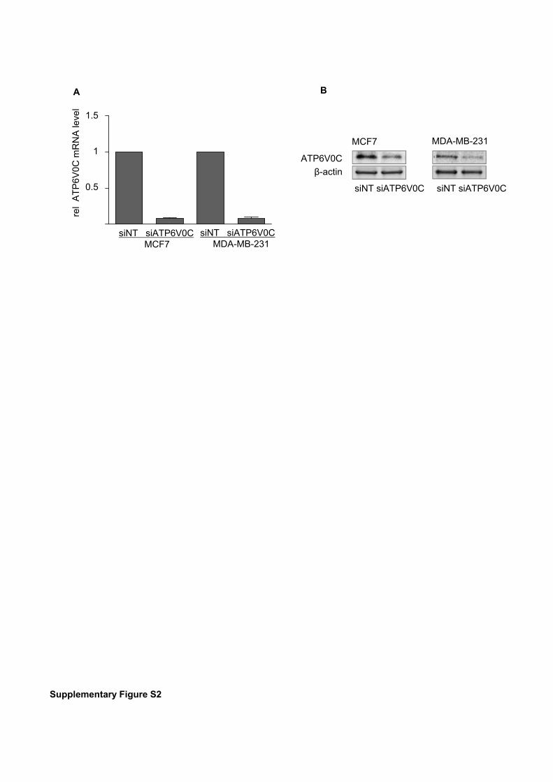
Supplementary Figure S2
rel
AT
P6V
0Cm
RN
Ale
vel
1
0.5
1.5
siNT siATP6V0CMCF7
siNT siATP6V0CMDA-MB-231
A B
MCF7 MDA-MB-231
ATP6V0C
β-actin
siNT siATP6V0C siNT siATP6V0C
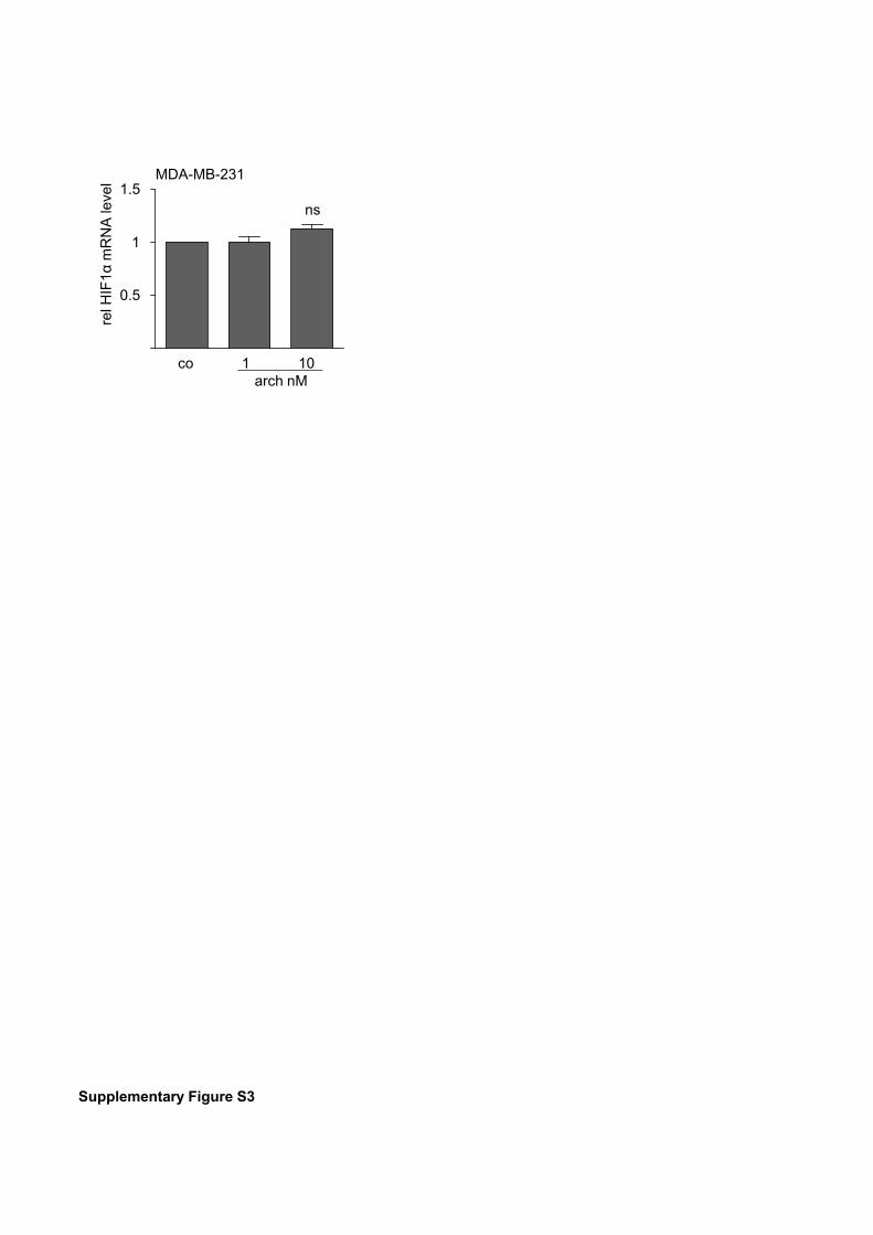
Supplementary Figure S3
ns
1
0.5
MDA-MB-231
relH
IF1α
mR
NA
leve
l 1.5
co 1 10 arch nM
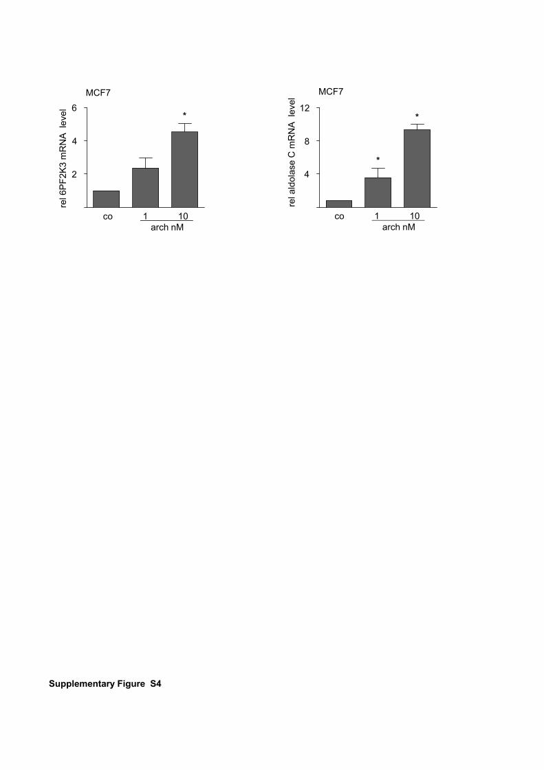
Supplementary Figure S4
MCF7
rel6
PF
2K3
mR
NA
leve
l
4
2
6*
co 1 10 arch nM
co 1 10 arch nM
4
8
12
rela
ldol
ase
C m
RN
Ale
vel
MCF7
*
*
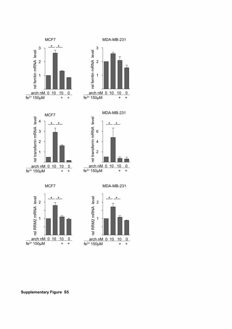
Supplementary Figure S5
MCF7 re
lfer
ritin
mR
NA
leve
l
2
1
3
arch nM 0 10 10 0 fe3+ 150µM + +
**
arch nM 0 10 10 0 fe3+ 150µM + +
relf
errit
inm
RN
Ale
vel
2
1
3
MDA-MB-231
arch nM 0 10 10 0 fe3+ 150µM + +
MCF7
relt
rans
ferr
inm
RN
Ale
vel
2
1
3
4**
arch nM 0 10 10 0 fe3+ 150µM + +
MDA-MB-231
relt
rans
ferr
inm
RN
Ale
vel
2
4
**
MCF7
arch nM 0 10 10 0 fe3+ 150µM + +
relR
RM
2 m
RN
Ale
vel
1
2
6
arch nM 0 10 10 0 fe3+ 150µM + +
relR
RM
2 m
RN
Ale
vel
1
2
MDA-MB-231
** **
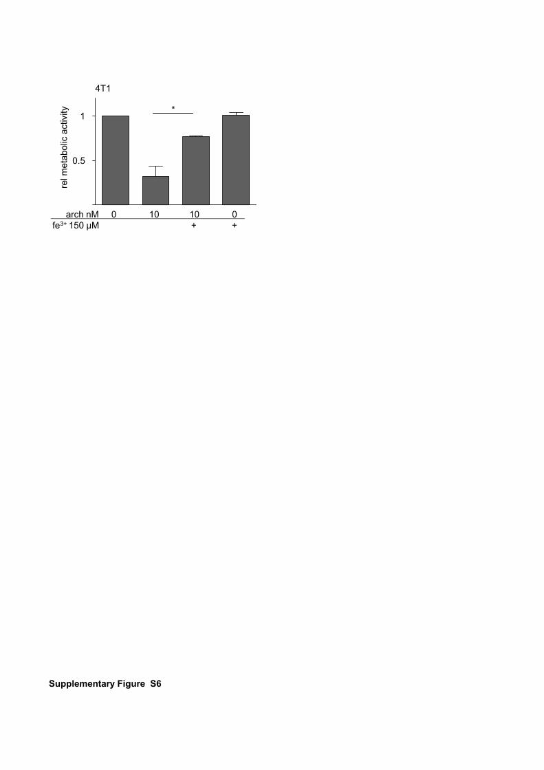
Supplementary Figure S6
4T1
0.5
1
relm
etab
olic
activ
ity *
arch nM 0 10 10 0 fe3+ 150 µM + +
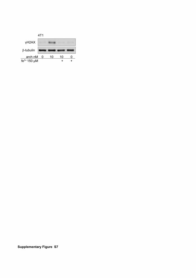
Supplementary Figure S7
γH2AX
β-tubulin
4T1
arch nM 0 10 10 0 fe3+ 150 µM + +
SUPPLEMENTARY FIGURE LEGENDS
Supplementary Figure S1 Archazolid induces apoptosis in breast cancer cells
in vitro. (A, C, D) Cell death was assessed by flow cytometry analysis in archazolid
(C and QVD) treated MCF7 cells after 48 h, MDA-MB-231 cells after 72 h and 4T1
cells after 24 h. Bars are the SEM of three independent experiments performed in
triplicates, *p<0.05 (One-way ANOVA, Newman-Keuls multiple comparison test).
Condensed chromatin was visualized by Hoechst staining after 48 h of archazolid
treatment in MCF7 cells and 72 h in MDA-MB-231 cells. One representative
experiment out of three independent experiments is shown. (B) PARP-1 cleavage
was detected by western blot upon 48 h and respectively 72 h of archazolid
treatment in MCF7 and MDA-MB-231 cells. One representative blot out of three
experiments is shown.
Supplementary Figure S2: mRNA levels of ATP6V0C after knockdown. MCF7
and MDA-MB-231 cells were transfected with siRNA-ATP6V0C and (A) mRNA was
extracted after 72 h. qPCR analysis was assessed with the TaqMan Gene
Expression Master Mix. Bars are the SEM of three independent experiments
performed in triplicates. (B) ATP6V0C protein levels were detected by western blot
after 72 h of transfection. One representative blot is shown.
Supplementary Figure S3: Archazolid has no influence on HIF1α mRNA levels
in MDA-MB-231 cells. MDA-MB-231 cells were stimulated with archazolid for 24 h
and mRNA levels were assessed by SYBR® Green qPCR analysis. Bars are the
SEM of three independent experiments performed in triplicates, p*<0.05, ns=not
significant (One-way ANOVA, Newman-Keuls multiple comparison test).
Supplementary Figure S4: Archazolid induces 6PF2K3 and aldolase C mRNA
levels. MCF7 cells were treated with archazolid for 24 h. SYBR® Green qPCR was
performed to analyze 6PF2K3 and aldolase C mRNA levels. Bars are the SEM of
three independent experiments performed in triplicates, p*<0.05 (One-way ANOVA,
Newman-Keuls multiple comparison test).
Supplementary Figure S5: Archazolid induces ferritin, transferrin and RRM2
mRNA levels. MCF7 cells and MDA-MB-231 cells were treated with archazolid for 24
h. SYBR® Green qPCR was performed to analyze ferritin, transferrin and RRM2
mRNA levels. Bars are the SEM of three independent experiments performed in
triplicates, p*<0.05 (One-way ANOVA, Newman-Keuls multiple comparison test).
Supplementary Figure S6: Iron rescues archazolid induced reduction of
metabolic activity in 4T1 cells. Metabolic activity of 4T1 cells after archazolid and
iron citrate treatment was assessed by CTB assay. Bars are the SEM of three
independent experiments performed in triplicates, p*<0.05 (One-way ANOVA,
Newman-Keuls multiple comparison test).
Supplementary Figure S7: Archazolid induces DSB in 4T1 cells, which is
compensated by iron. Expression of phosphorylated H2AX was detected by
western blot in 4T1 cells after 48 h of archazolid and iron citrate treatment. One
representative blot out of three independent experiments is shown.

SUPPLEMENTARY MATERIALS AND METHODS
Analysis of chromatin condensation
After treatment nuclei were stained with 0.5 μg/mL Hoechst33258® (Sigma Aldrich)
for 10 min to visualize condensed chromatin by fluorescence microscopy using a
Zeiss Axiovert 25 (Jena, Germany).
Oncotarget1www.impactjournals.com/oncotarget
www.impactjournals.com/oncotarget/ Oncotarget, Advance Publications 2015
Anti-leukemic effects of the V-ATPase inhibitor Archazolid A
Siwei Zhang1, Lina S. Schneider1, Binje Vick2,4, Michaela Grunert2, Irmela Jeremias2,3,4, Dirk Menche5, Rolf Müller6, Angelika M. Vollmar1 and Johanna Liebl1
1 Department of Pharmacy, Pharmaceutical Biology, Ludwig-Maximilians-University, Munich, Germany2 Department of Gene Vectors, Helmholtz Zentrum München, German Research Center for Environmental Health, Munich, Germany3 Department of Oncology/Hematology, Dr. von Haunersches Kinderspital, Munich, Germany4 German Cancer Consortium (DKTK), German Cancer Research Center (DKFZ), Heidelberg, Germany5 Kekulé-Institut für Organische Chemie und Biochemie der Universität Bonn, Bonn, Germany6 Helmholtz Institute for Pharmaceutical Research Saarland, Helmholtz Centre for Infection Research and Department of Pharmaceutical Biotechnology, Saarland University, Saarbrücken, Germany
Correspondence to: Johanna Liebl, email: [email protected]: Archazolid, leukemia, natural compoundsReceived: May 20, 2015 Accepted: October 07, 2015 Published: October 19, 2015
This is an open-access article distributed under the terms of the Creative Commons Attribution License, which permits unrestricted use, distribution, and reproduction in any medium, provided the original author and source are credited.
ABSTRACTPrognosis for patients suffering from T-ALL is still very poor and new strategies
for T-ALL treatment are urgently needed. Our study shows potent anti-leukemic effects of the myxobacterial V-ATPase inhibitor Archazolid A. Archazolid A reduced growth and potently induced death of leukemic cell lines and human leukemic samples. By inhibiting lysosomal acidification, Archazolid A blocked activation of the Notch pathway, however, this was not the mechanism of V-ATPase inhibition relevant for cell death induction. In fact, V-ATPase inhibition by Archazolid A decreased the anti-apoptotic protein survivin. As underlying mode of action, this work is in line with recent studies from our group demonstrating that Archazolid A induced S-phase cell cycle arrest by interfering with the iron metabolism in leukemic cells. Our study provides evidence for V-ATPase inhibition as a potential new therapeutic option for T-ALL.
INTRODUCTION
T-cell acute lymphoblastic leukemia (T-ALL) is an aggressive hematopoietic malignancy. Despite advances in understanding of the molecular basis of T-ALL and intensified treatment regimens that have improved outcome of therapy, some patients fail conventional chemotherapy and T-ALL remains fatal in 20% of children and more than 50% of adults [1]. Along this line, new therapeutic approaches are urgently needed to improve patient prognosis.
Activating mutations in Notch1 occur in more than 50% of T-ALLs, highlighting Notch1 as key player in T-ALL [2, 3]. In fact, constitutive activation of Notch1 signaling represents the most prominent oncogenic pathway in T-cell transformation [1]. Therefore, targeted therapies to inhibit the Notch1 pathway have been developed. γ-secretase inhibitors (GSIs) that prevent
Notch1 activating cleavage have been widely tested in preclinical models and clinical trials. However, the role of Notch in leukemia is still not entirely clear: on the one hand, it was shown that Notch signaling promotes cell survival and apoptosis resistance in leukemia [4, 5] and that Notch blockade by γ-secretase inhibition exerts pro-apoptotic effects in leukemia [6]. On the other hand, activation of Notch or of the Notch downstream target Hes1 inhibits leukemia development, growth and survival [7-9]. Furthermore, GSIs have shown only modest and temporary responses in leukemia therapy. These controversial results about Notch in leukemia demand for better understanding of the mechanisms that contribute to Notch signaling in leukemia in order to develop novel strategies to inhibit Notch1 signaling with alternative mechanism different from GSIs as potential promising approaches for T-ALL therapy.
Besides Notch activation at the plasma membrane
Oncotarget2www.impactjournals.com/oncotarget
where ligand binding initiates y-secretase dependent cleavage of the Notch receptor, and subsequently NICD generation and translocation to the nucleus, Notch signaling in part depends on endocytosis. Recent reports showed that Notch can be activated in endosomes: in drosophila, Notch signaling is abolished when endosomal entry is blocked but is enhanced when endosomal retention is increased [10]. Moreover, in drosophila, acidification of endosomes by the V-ATPase is required for Notch activation [11].
Along this line, we hypothesized that inhibition of V-ATPase might be a promising strategy for T-ALL treatment. V-ATPase is an ATP-dependent proton pump that regulates pH homeostasis by translocating protons across membranes. The main function of V-ATPase is to regulate the acidification of intracellular compartments like lysosomes [12, 13]. V-ATPases are multisubunit heteromeric protein complexes with two functional domains: the V1 domain that is composed of eight subunits (A-H) mediates ATP hydrolysis and the V0 domain that is composed of five subunits (a, c, c’’, d, e) regulates proton translocation [14]. The V-ATPase is localized at endosomes and lysosomes and is essential for endocytotic processes, receptor internalization and recycling, and finally lysosomal degradation [13]. Therefore, V-ATPase is implicated in fundamental physiological processes, like neurotransmitter uptake, renal acidification, bone resorption or sperm maturation and is associated with pathologic conditions including osteopetrosis, renal tubular acidosis and disease-related processes such as entry of toxins and viruses [13].
Recent reports indicate important functions of V-ATPase in tumors. Augmented expression of V-ATPase in cancer cells was demonstrated to contribute to metastasis, survival and growth of cancer cells [15-18]. Localization of V-ATPase at the plasma membrane was associated with invasiveness of various types of cancer including breast, pancreatic, prostate, and melanoma cancer cells [14, 19-21]. V-ATPase was shown to localize to the plasma membrane in sarcoma cells as well and was elucidated as a promising target for Ewing sarcoma, osteosarcoma, chondrosarcoma or rhabdomyosarcoma [22, 23] as well as glioblastoma [24, 25]. Along this line, V-ATPase inhibition could represent a promising approach for tumor therapy. Although only few V-ATPase inhibitors have been described so far, their V-ATPase binding properties and mode of action are well-investigated. Amongst others, a class of natural compounds - the Archazolids - inhibits V-ATPase activity by binding to the V-ATPase V0 subunit [26-28] that is responsible for proton transport across the membrane [29]. Archazolids are macrolides that have originally been isolated from the myxobacterium Archangium gephyra [30], and are available also by chemical synthesis [31, 32]. Archazolids have attracted attention as highly potent V-ATPase
inhibitors that exert promising anti-tumor effects [15-18, 33-36].
Because Notch signaling activation in part depends on endocytosis [10, 11, 37] and V-ATPase has therefore been linked with the Notch pathway [35, 38], we hypothesized that V-ATPase inhibition might represent an alternative option to target leukemic cells. Therefore, we had a closer look on the functional effects and the mechanism of action, including the Notch pathway and cellular stress response, of the V-ATPase inhibitor Archazolid A in leukemic cells.
RESULTS
V-ATPase in leukemic cells
First, we analyzed the expression of the V-ATPase subunits in different leukemic cell lines including the T-ALL cell lines Jurkat and CEM, the AML cell line HL60, and the CML cell line K562 in comparison to non-tumor primary human PBMCs. Most of the V-ATPase subunits were comparably expressed in non-tumor PBMCs, Jurkat, CEM, and HL60 cells and some subunits were increased in K562 cells (Table 1). Immunostainings show that subunit c ATP6V0C which is targeted by Archazolid A, is localized to the lysosomal system and to the plasma membrane of leukemic cells (Figure 1A). As V-ATPase is essential for endo-lysosomal function, we analyzed the size of the endo-lysosomal compartment. In fact, the size of the endosomal compartment was increased in leukemic cell lines compared to non-tumor primary human peripheral blood mononuclear cells (PBMCs) (Figure 1B). This set of data suggests a potential function of V-ATPase in leukemia.
V-ATPase inhibition by Archazolid A impairs growth and induces death of leukemic cell lines
Archazolid A inhibited V-ATPase activity in leukemic cells as shown by staining of lysosomes with a pH-sensitive fluorescence dye (LysoTracker) (Figure 2A). Archazolid A impaired proliferation of leukemic cell lines Jurkat (EC50 0.56 nM) and CEM (EC50 0.51 nM) (Figure 2B, 2C). In line, clonogenic growth of Jurkat and CEM cells was reduced by V-ATPase inhibition (Figure 2D, 2E).
Moreover, as shown by Nicoletti assay (Figure 3A, 3B) and Annexin V staining (Figure 3C), Archazolid A potently induced death of leukemic cell lines. In line with a previous report from our group [15], Archazolid A induced cleavage of procaspase-3, procaspase-9, and PARP, increased the pro-apoptotic protein BNIP3, and decreased the anti-apoptotic protein Bcl-XL in leukemic cells (Figure 3D). Moreover, the pan-caspase inhibitor QVC-OPh
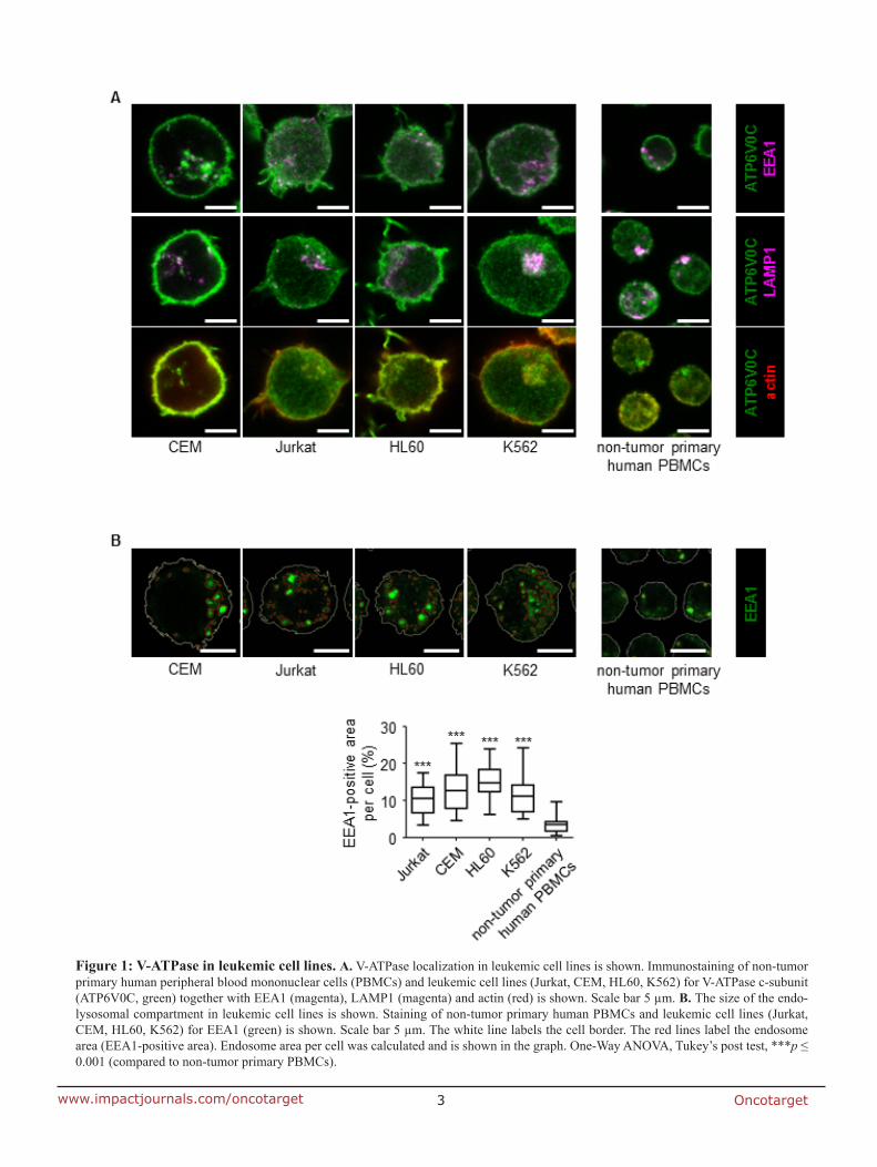
Oncotarget3www.impactjournals.com/oncotarget
Figure 1: V-ATPase in leukemic cell lines. A. V-ATPase localization in leukemic cell lines is shown. Immunostaining of non-tumor primary human peripheral blood mononuclear cells (PBMCs) and leukemic cell lines (Jurkat, CEM, HL60, K562) for V-ATPase c-subunit (ATP6V0C, green) together with EEA1 (magenta), LAMP1 (magenta) and actin (red) is shown. Scale bar 5 µm. B. The size of the endo-lysosomal compartment in leukemic cell lines is shown. Staining of non-tumor primary human PBMCs and leukemic cell lines (Jurkat, CEM, HL60, K562) for EEA1 (green) is shown. Scale bar 5 µm. The white line labels the cell border. The red lines label the endosome area (EEA1-positive area). Endosome area per cell was calculated and is shown in the graph. One-Way ANOVA, Tukey’s post test, ***p ≤ 0.001 (compared to non-tumor primary PBMCs).
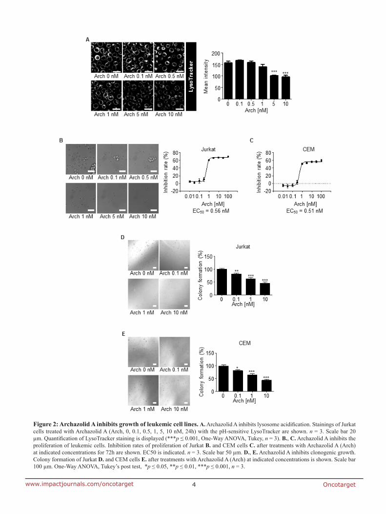
Oncotarget4www.impactjournals.com/oncotarget
Figure 2: Archazolid A inhibits growth of leukemic cell lines. A. Archazolid A inhibits lysosome acidification. Stainings of Jurkat cells treated with Archazolid A (Arch, 0, 0.1, 0.5, 1, 5, 10 nM, 24h) with the pH-sensitive LysoTracker are shown. n = 3. Scale bar 20 µm. Quantification of LysoTracker staining is displayed (***p ≤ 0.001, One-Way ANOVA, Tukey, n = 3). B., C. Archazolid A inhibits the proliferation of leukemic cells. Inhibition rates of proliferation of Jurkat B. and CEM cells C. after treatments with Archazolid A (Arch) at indicated concentrations for 72h are shown. EC50 is indicated. n = 3. Scale bar 50 µm. D., E. Archazolid A inhibits clonogenic growth. Colony formation of Jurkat D. and CEM cells E. after treatments with Archazolid A (Arch) at indicated concentrations is shown. Scale bar 100 µm. One-Way ANOVA, Tukey’s post test, *p ≤ 0.05, **p ≤ 0.01, ***p ≤ 0.001, n = 3.
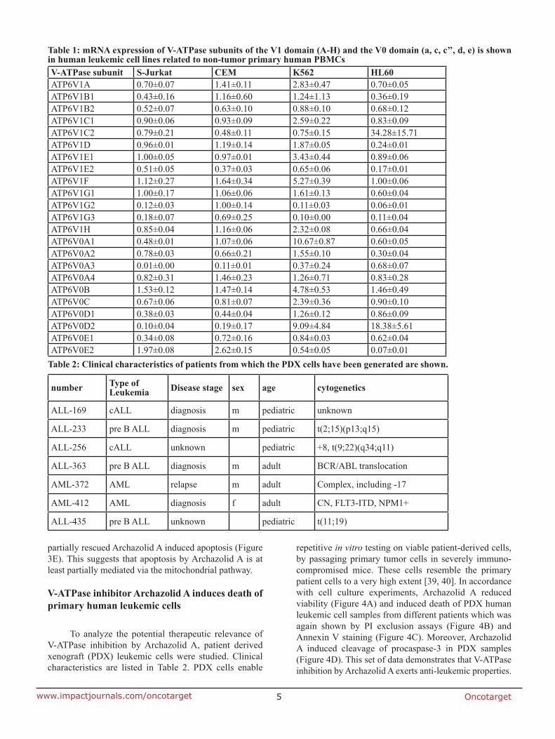
Oncotarget5www.impactjournals.com/oncotarget
partially rescued Archazolid A induced apoptosis (Figure 3E). This suggests that apoptosis by Archazolid A is at least partially mediated via the mitochondrial pathway.
V-ATPase inhibitor Archazolid A induces death of primary human leukemic cells
To analyze the potential therapeutic relevance of V-ATPase inhibition by Archazolid A, patient derived xenograft (PDX) leukemic cells were studied. Clinical characteristics are listed in Table 2. PDX cells enable
repetitive in vitro testing on viable patient-derived cells, by passaging primary tumor cells in severely immuno-compromised mice. These cells resemble the primary patient cells to a very high extent [39, 40]. In accordance with cell culture experiments, Archazolid A reduced viability (Figure 4A) and induced death of PDX human leukemic cell samples from different patients which was again shown by PI exclusion assays (Figure 4B) and Annexin V staining (Figure 4C). Moreover, Archazolid A induced cleavage of procaspase-3 in PDX samples (Figure 4D). This set of data demonstrates that V-ATPase inhibition by Archazolid A exerts anti-leukemic properties.
V-ATPase subunit S-Jurkat CEM K562 HL60ATP6V1A 0.70±0.07 1.41±0.11 2.83±0.47 0.70±0.05ATP6V1B1 0.43±0.16 1.16±0.60 1.24±1.13 0.36±0.19ATP6V1B2 0.52±0.07 0.63±0.10 0.88±0.10 0.68±0.12ATP6V1C1 0.90±0.06 0.93±0.09 2.59±0.22 0.83±0.09ATP6V1C2 0.79±0.21 0.48±0.11 0.75±0.15 34.28±15.71ATP6V1D 0.96±0.01 1.19±0.14 1.87±0.05 0.24±0.01ATP6V1E1 1.00±0.05 0.97±0.01 3.43±0.44 0.89±0.06ATP6V1E2 0.51±0.05 0.37±0.03 0.65±0.06 0.17±0.01ATP6V1F 1.12±0.27 1.64±0.34 5.27±0.39 1.00±0.06ATP6V1G1 1.00±0.17 1.06±0.06 1.61±0.13 0.60±0.04ATP6V1G2 0.12±0.03 1.00±0.14 0.11±0.03 0.06±0.01ATP6V1G3 0.18±0.07 0.69±0.25 0.10±0.00 0.11±0.04ATP6V1H 0.85±0.04 1.16±0.06 2.32±0.08 0.66±0.04ATP6V0A1 0.48±0.01 1.07±0.06 10.67±0.87 0.60±0.05ATP6V0A2 0.78±0.03 0.66±0.21 1.55±0.10 0.30±0.04ATP6V0A3 0.01±0.00 0.11±0.01 0.37±0.24 0.68±0.07ATP6V0A4 0.82±0.31 1.46±0.23 1.26±0.71 0.83±0.28ATP6V0B 1.53±0.12 1.47±0.14 4.78±0.53 1.46±0.49ATP6V0C 0.67±0.06 0.81±0.07 2.39±0.36 0.90±0.10ATP6V0D1 0.38±0.03 0.44±0.04 1.26±0.12 0.86±0.09ATP6V0D2 0.10±0.04 0.19±0.17 9.09±4.84 18.38±5.61ATP6V0E1 0.34±0.08 0.72±0.16 0.84±0.03 0.62±0.04ATP6V0E2 1.97±0.08 2.62±0.15 0.54±0.05 0.07±0.01
Table 1: mRNA expression of V-ATPase subunits of the V1 domain (A-H) and the V0 domain (a, c, c’’, d, e) is shown in human leukemic cell lines related to non-tumor primary human PBMCs
number Type ofLeukemia Disease stage sex age cytogenetics
ALL-169 cALL diagnosis m pediatric unknown
ALL-233 pre B ALL diagnosis m pediatric t(2;15)(p13;q15)
ALL-256 cALL unknown pediatric +8, t(9;22)(q34;q11)
ALL-363 pre B ALL diagnosis m adult BCR/ABL translocation
AML-372 AML relapse m adult Complex, including -17
AML-412 AML diagnosis f adult CN, FLT3-ITD, NPM1+
ALL-435 pre B ALL unknown pediatric t(11;19)
Table 2: Clinical characteristics of patients from which the PDX cells have been generated are shown.
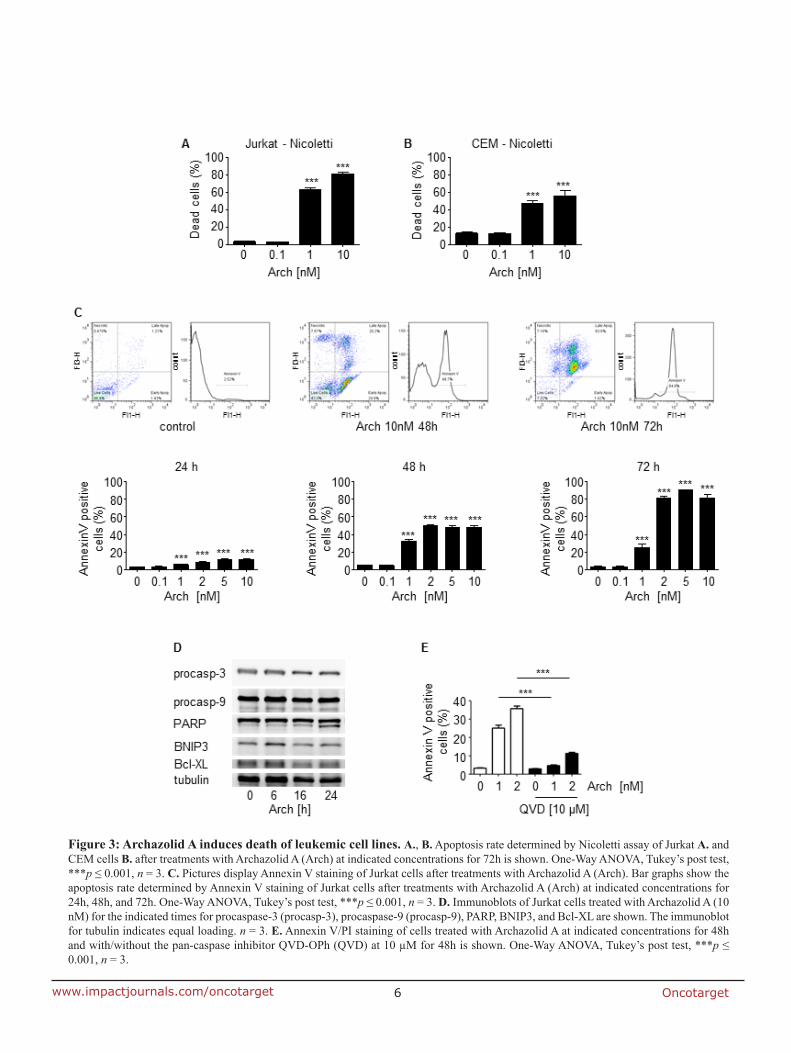
Oncotarget6www.impactjournals.com/oncotarget
Figure 3: Archazolid A induces death of leukemic cell lines. A., B. Apoptosis rate determined by Nicoletti assay of Jurkat A. and CEM cells B. after treatments with Archazolid A (Arch) at indicated concentrations for 72h is shown. One-Way ANOVA, Tukey’s post test, ***p ≤ 0.001, n = 3. C. Pictures display Annexin V staining of Jurkat cells after treatments with Archazolid A (Arch). Bar graphs show the apoptosis rate determined by Annexin V staining of Jurkat cells after treatments with Archazolid A (Arch) at indicated concentrations for 24h, 48h, and 72h. One-Way ANOVA, Tukey’s post test, ***p ≤ 0.001, n = 3. D. Immunoblots of Jurkat cells treated with Archazolid A (10 nM) for the indicated times for procaspase-3 (procasp-3), procaspase-9 (procasp-9), PARP, BNIP3, and Bcl-XL are shown. The immunoblot for tubulin indicates equal loading. n = 3. E. Annexin V/PI staining of cells treated with Archazolid A at indicated concentrations for 48h and with/without the pan-caspase inhibitor QVD-OPh (QVD) at 10 µM for 48h is shown. One-Way ANOVA, Tukey’s post test, ***p ≤ 0.001, n = 3.
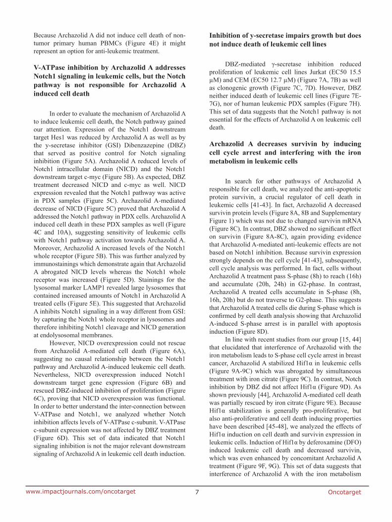
Oncotarget7www.impactjournals.com/oncotarget
Because Archazolid A did not induce cell death of non-tumor primary human PBMCs (Figure 4E) it might represent an option for anti-leukemic treatment.
V-ATPase inhibition by Archazolid A addresses Notch1 signaling in leukemic cells, but the Notch pathway is not responsible for Archazolid A induced cell death
In order to evaluate the mechanism of Archazolid A to induce leukemic cell death, the Notch pathway gained our attention. Expression of the Notch1 downstream target Hes1 was reduced by Archazolid A as well as by the y-secretase inhibitor (GSI) Dibenzazepine (DBZ) that served as positive control for Notch signaling inhibition (Figure 5A). Archazolid A reduced levels of Notch1 intracellular domain (NICD) and the Notch1 downstream target c-myc (Figure 5B). As expected, DBZ treatment decreased NICD and c-myc as well. NICD expression revealed that the Notch1 pathway was active in PDX samples (Figure 5C). Archazolid A-mediated decrease of NICD (Figure 5C) proved that Archazolid A addressed the Notch1 pathway in PDX cells. Archazolid A induced cell death in these PDX samples as well (Figure 4C and 10A), suggesting sensitivity of leukemic cells with Notch1 pathway activation towards Archazolid A. Moreover, Archazolid A increased levels of the Notch1 whole receptor (Figure 5B). This was further analyzed by immunostainings which demonstrate again that Archazolid A abrogated NICD levels whereas the Notch1 whole receptor was increased (Figure 5D). Stainings for the lysosomal marker LAMP1 revealed large lysosomes that contained increased amounts of Notch1 in Archazolid A treated cells (Figure 5E). This suggested that Archazolid A inhibits Notch1 signaling in a way different from GSI: by capturing the Notch1 whole receptor in lysosomes and therefore inhibiting Notch1 cleavage and NICD generation at endolysosomal membranes.
However, NICD overexpression could not rescue from Archazolid A-mediated cell death (Figure 6A), suggesting no causal relationship between the Notch1 pathway and Archazolid A-induced leukemic cell death. Nevertheless, NICD overexpression induced Notch1 downstream target gene expression (Figure 6B) and rescued DBZ-induced inhibition of proliferation (Figure 6C), proving that NICD overexpression was functional. In order to better understand the inter-connection between V-ATPase and Notch1, we analyzed whether Notch inhibition affects levels of V-ATPase c-subunit. V-ATPase c-subunit expression was not affected by DBZ treatment (Figure 6D). This set of data indicated that Notch1 signaling inhibition is not the major relevant downstream signaling of Archazolid A in leukemic cell death induction.
Inhibition of y-secretase impairs growth but does not induce death of leukemic cell lines
DBZ-mediated γ-secretase inhibition reduced proliferation of leukemic cell lines Jurkat (EC50 15.5 µM) and CEM (EC50 12.7 µM) (Figure 7A, 7B) as well as clonogenic growth (Figure 7C, 7D). However, DBZ neither induced death of leukemic cell lines (Figure 7E-7G), nor of human leukemic PDX samples (Figure 7H). This set of data suggests that the Notch1 pathway is not essential for the effects of Archazolid A on leukemic cell death.
Archazolid A decreases survivin by inducing cell cycle arrest and interfering with the iron metabolism in leukemic cells
In search for other pathways of Archazolid A responsible for cell death, we analyzed the anti-apoptotic protein survivin, a crucial regulator of cell death in leukemic cells [41-43]. In fact, Archazolid A decreased survivin protein levels (Figure 8A, 8B and Supplementary Figure 1) which was not due to changed survivin mRNA (Figure 8C). In contrast, DBZ showed no significant effect on survivin (Figure 8A-8C), again providing evidence that Archazolid A-mediated anti-leukemic effects are not based on Notch1 inhibition. Because survivin expression strongly depends on the cell cycle [41-43], subsequently, cell cycle analysis was performed. In fact, cells without Archazolid A treatment pass S-phase (8h) to reach (16h) and accumulate (20h, 24h) in G2-phase. In contrast, Archazolid A treated cells accumulate in S-phase (8h, 16h, 20h) but do not traverse to G2-phase. This suggests that Archazolid A treated cells die during S-phase which is confirmed by cell death analysis showing that Archazolid A-induced S-phase arrest is in parallel with apoptosis induction (Figure 8D).
In line with recent studies from our group [15, 44] that elucidated that interference of Archazolid with the iron metabolism leads to S-phase cell cycle arrest in breast cancer, Archazolid A stabilized Hif1α in leukemic cells (Figure 9A-9C) which was abrogated by simultaneous treatment with iron citrate (Figure 9C). In contrast, Notch inhibition by DBZ did not affect Hif1α (Figure 9D). As shown previously [44], Archazolid A-mediated cell death was partially rescued by iron citrate (Figure 9E). Because Hif1α stabilization is generally pro-proliferative, but also anti-proliferative and cell death inducing properties have been described [45-48], we analyzed the effects of Hif1α induction on cell death and survivin expression in leukemic cells. Induction of Hif1α by deferoxamine (DFO) induced leukemic cell death and decreased survivin, which was even enhanced by concomitant Archazolid A treatment (Figure 9F, 9G). This set of data suggests that interference of Archazolid A with the iron metabolism

Oncotarget8www.impactjournals.com/oncotarget
Figure 4: Archazolid A induces cell death in human patient derived xenograft (PDX) samples. A. Viability of leukemic PDX samples with/without treatment with Archazolid A (Arch) for 72h at indicated concentrations is shown. B. PI exclusion staining of leukemic PDX samples with/without treatment with Archazolid A (Arch) for 48h at indicated concentrations is shown. Upper panels display dot plots and histograms of PDX leukemic cells from one respective patient (PDX ALL-169). Dead cells are stained by PI and are marked in red. Live cells without PI staining are displayed in green. Lower panels show apoptosis rate of PDX leukemic cells treated with Archazolid A (Arch) at indicated concentrations. C. The specific apoptosis rate determined by Annexin V/PI staining of PDX cells after treatments with Archazolid A (Arch) at indicated concentrations for 48h is shown. D. Immunoblots of PDX samples treated with Archazolid A (10 nM, 48h) for procaspase-3 (procasp-3) are shown. Ponceau staining indicates equal loading. E. Archazolid A does not induce cell death in non-tumor primary human PBMCs. Apoptosis rate determined by Annexin V/PI staining and of non-tumor primary human PBMCs (FACS analysis with gating for lymphocytes) of two different donors treated with Archazolid A (Arch) at indicated concentrations for 48h is shown.
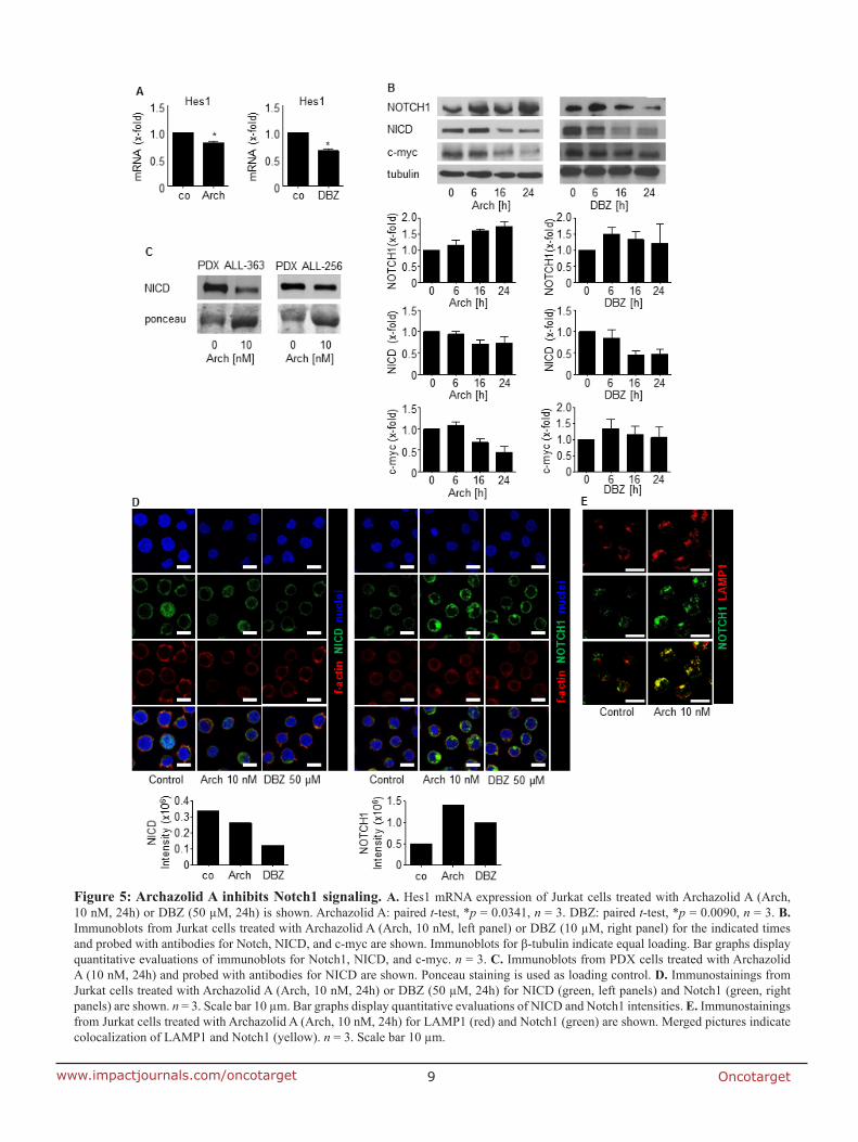
Oncotarget9www.impactjournals.com/oncotarget
Figure 5: Archazolid A inhibits Notch1 signaling. A. Hes1 mRNA expression of Jurkat cells treated with Archazolid A (Arch, 10 nM, 24h) or DBZ (50 µM, 24h) is shown. Archazolid A: paired t-test, *p = 0.0341, n = 3. DBZ: paired t-test, *p = 0.0090, n = 3. B. Immunoblots from Jurkat cells treated with Archazolid A (Arch, 10 nM, left panel) or DBZ (10 µM, right panel) for the indicated times and probed with antibodies for Notch, NICD, and c-myc are shown. Immunoblots for β-tubulin indicate equal loading. Bar graphs display quantitative evaluations of immunoblots for Notch1, NICD, and c-myc. n = 3. C. Immunoblots from PDX cells treated with Archazolid A (10 nM, 24h) and probed with antibodies for NICD are shown. Ponceau staining is used as loading control. D. Immunostainings from Jurkat cells treated with Archazolid A (Arch, 10 nM, 24h) or DBZ (50 µM, 24h) for NICD (green, left panels) and Notch1 (green, right panels) are shown. n = 3. Scale bar 10 µm. Bar graphs display quantitative evaluations of NICD and Notch1 intensities. E. Immunostainings from Jurkat cells treated with Archazolid A (Arch, 10 nM, 24h) for LAMP1 (red) and Notch1 (green) are shown. Merged pictures indicate colocalization of LAMP1 and Notch1 (yellow). n = 3. Scale bar 10 µm.
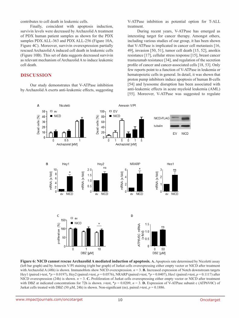
Oncotarget10www.impactjournals.com/oncotarget
contributes to cell death in leukemic cells. Finally, coincident with apoptosis induction,
survivin levels were decreased by Archazolid A treatment of PDX human patient samples as shown for the PDX samples PDX ALL-363 and PDX ALL-256 (Figure 10A, Figure 4C). Moreover, survivin overexpression partially rescued Archazolid A induced cell death in leukemic cells (Figure 10B). This set of data suggests decreased survivin as relevant mechanism of Archazolid A to induce leukemic cell death.
DISCUSSION
Our study demonstrates that V-ATPase inhibition by Archazolid A exerts anti-leukemic effects, suggesting
V-ATPase inhibition as potential option for T-ALL treatment.
During recent years, V-ATPase has emerged as interesting target for cancer therapy. Amongst others, including various studies of our group, it has been shown that V-ATPase is implicated in cancer cell metastasis [16, 49], invasion [50, 51], tumor cell death [15, 52], anoikis resistance [17], cellular stress response [15], breast cancer trastuzumab resistance [34], and regulation of the secretion profile of cancer and cancer-associated cells [18, 53]. Only few reports point to a function of V-ATPase in leukemia or hematopoietic cells in general. In detail, it was shown that proton pump inhibitors induce apoptosis of human B-cells [54] and lysosome disruption has been associated with anti-leukemic effects in acute myeloid leukemia (AML) [55]. Moreover, V-ATPase was suggested to regulate
Figure 6: NICD cannot rescue Archazolid A mediated induction of apoptosis. A. Apoptosis rate determined by Nicoletti assay (left bar graph) and by Annexin V/PI staining (right bar graph) of Jurkat cells overexpressing either empty vector or NICD after treatment with Archazolid A (48h) is shown. Immunoblots show NICD overexpression. n = 3. B. Increased expression of Notch downstream targets Hey1 (paired t-test, *p = 0.0197), Hey2 (paired t-test, p = 0.0576), NRARP (paired t-test, *p = 0.0407), Hes1 (paired t-test, p = 0.1117) after NICD overexpression (24h) is shown. n = 3. C. Proliferation of Jurkat cells overexpressing either empty vector or NICD after treatment with DBZ at indicated concentrations for 72h is shown. t-test, *p = 0.0209, n = 3. D. Expression of V-ATPase subunit c (ATP6V0C) of Jurkat cells treated with DBZ (50 µM, 24h) is shown. Non-significant (ns), paired t-test, p = 0.1886.
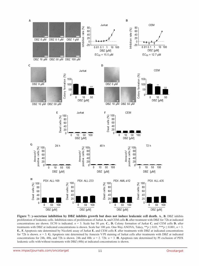
Oncotarget11www.impactjournals.com/oncotarget
Figure 7: y-secretase inhibition by DBZ inhibits growth but does not induce leukemic cell death. A., B. DBZ inhibits proliferation of leukemic cells. Inhibition rates of proliferation of Jurkat A. and CEM cells B. after treatment with DBZ for 72h at indicated concentrations are shown. EC50 is indicated. n = 3. Scale bar 50 µm. C., D. Colony formation of Jurkat C. and CEM cells D. after treatments with DBZ at indicated concentrations is shown. Scale bar 100 µm. One-Way ANOVA, Tukey, **p ≤ 0.01, ***p ≤ 0.001, n = 3. E., F. Apoptosis rate determined by Nicoletti assay of Jurkat E. and CEM cells F. after treatments with DBZ at indicated concentrations for 72h is shown. n = 3. G. Apoptosis rate determined by Annexin V/PI staining of Jurkat cells after treatments with DBZ at indicated concentrations for 24h, 48h, and 72h is shown. 24h and 48h: n = 2. 72h: n = 3. H. Apoptosis rate determined by PI exclusion of PDX leukemic cells with/without treatments with DBZ (48h) at indicated concentrations is shown.
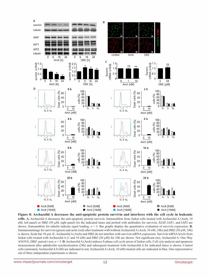
Oncotarget12www.impactjournals.com/oncotarget
Figure 8: Archazolid A decreases the anti-apoptotic protein survivin and interferes with the cell cycle in leukemic cells. A. Archazolid A decreases the anti-apoptotic protein survivin. Immunoblots from Jurkat cells treated with Archazolid A (Arch, 10 nM, left panel) or DBZ (50 µM, right panel) for the indicated times and probed with antibodies for survivin, XIAP, IAP1, and IAP2 are shown. Immunoblots for tubulin indicate equal loading. n = 3. Bar graphs display the quantitative evaluation of survivin expression. B. Immunostainings for survivin (green) and actin (red) after treatment with/without Archazolid A (Arch, 10 nM, 24h) and DBZ (50 µM, 24h) is shown. Scale bar 10 µm. C. Archazolid A (Arch) and DBZ do not interfere with survivin mRNA expression. Survivin mRNA levels from Jurkat cells treated with Archazolid A (1 and 10 nM) and DBZ (50 µM) for 24h are shown. Not significant (ns), Archazolid A: One-Way ANOVA, DBZ: paired t-test, n = 3. D. Archazolid A (Arch) induces S-phase cell cycle arrest of Jurkat cells. Cell cyle analysis and apoptosis measurement after aphidicolin synchronization (24h) and subsequent treatment with Archazolid A for indicated times is shown. Control cells (untreated, Archazolid A 0 nM) are indicated in red, Archazolid A (Arch, 10 nM) treated cells are indicated in blue. One representative out of three independent experiments is shown.
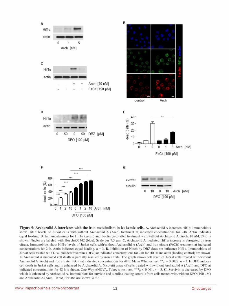
Oncotarget13www.impactjournals.com/oncotarget
Figure 9: Archazolid A interferes with the iron metabolism in leukemic cells. A. Archazolid A increases Hif1α. Immunoblots show Hif1α levels of Jurkat cells with/without Archazolid A (Arch) treatment at indicated concentrations for 24h. Actin indicates equal loading. B. Immunostainings for Hif1α (green) and f-actin (red) after treatment with/without Archazolid A (Arch, 10 nM, 24h) is shown. Nuclei are labeled with Hoechst33342 (blue). Scale bar 7.5 µm. C. Archazolid A mediated Hif1α increase is abrogated by iron citrate. Immunoblots show Hif1α levels of Jurkat cells with/without Archazolid A (Arch) and iron citrate (FeCit) treatment at indicated concentrations for 24h. Actin indicates equal loading. n = 3. D. Inhibition of Notch by DBZ does not influence Hif1α. Immunoblots of Jurkat cells treated with DBZ and deferoxamine (DFO) at indicated concentrations for 24h for Hif1α and actin (loading control) are shown. E. Archazolid A mediated cell death is partially rescued by iron citrate. The graph shows cell death of Jurkat cells treated with/without Archazolid A (Arch) and iron citrate (FeCit) at indicated concentrations for 48 h. Mann Whitney test, **p = 0.0022, n = 3. F. DFO induces cell death in Jurkat cells and is enhanced by Archazolid A. Nicoletti assay of cells treated with/without Archazolid A (Arch) and DFO at indicated concentrations for 48 h is shown. One-Way ANOVA, Tukey’s post test, ***p ≤ 0.001, n = 3. G. Survivin is decreased by DFO which is enhanced by Archazolid A. Immunoblots for survivin and tubulin (loading control) from cells treated with/without DFO (100 µM) and Archazolid A (Arch, 10 nM) for 48h are shown; n = 3.
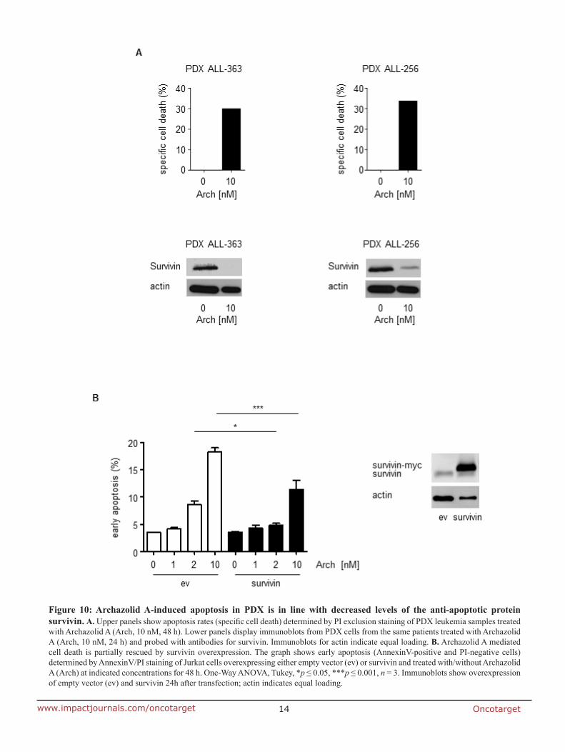
Oncotarget14www.impactjournals.com/oncotarget
Figure 10: Archazolid A-induced apoptosis in PDX is in line with decreased levels of the anti-apoptotic protein survivin. A. Upper panels show apoptosis rates (specific cell death) determined by PI exclusion staining of PDX leukemia samples treated with Archazolid A (Arch, 10 nM, 48 h). Lower panels display immunoblots from PDX cells from the same patients treated with Archazolid A (Arch, 10 nM, 24 h) and probed with antibodies for survivin. Immunoblots for actin indicate equal loading. B. Archazolid A mediated cell death is partially rescued by survivin overexpression. The graph shows early apoptosis (AnnexinV-positive and PI-negative cells) determined by AnnexinV/PI staining of Jurkat cells overexpressing either empty vector (ev) or survivin and treated with/without Archazolid A (Arch) at indicated concentrations for 48 h. One-Way ANOVA, Tukey, *p ≤ 0.05, ***p ≤ 0.001, n = 3. Immunoblots show overexpression of empty vector (ev) and survivin 24h after transfection; actin indicates equal loading.
Oncotarget15www.impactjournals.com/oncotarget
leukemic cell adhesion [56] and V-ATPase inhibition by Bafilomycin A reduced leukemic cell growth [35].
In search for the signaling pathway responsible for the anti-leukemic effects of Archazolid A-mediated V-ATPase inhibition, first the Notch pathway was analyzed as it is one of the most prominent oncogenic pathways in T-ALL [57]. Targeted therapies addressing the Notch pathway have been proposed as auspicious options for T-ALL treatment. Despite promising in vitro results and initial clinical effectiveness of GSIs that block Notch activatory cleavage, this benefit only in some studies translates into improved overall survival [58-60]. In fact, most human T-ALL cell lines are resistant to GSIs that fail to induce leukemic cell death [57]. Therefore, to inhibit Notch signaling with a mechanism different from GSIs might be a promising approach. Our study shows that V-ATPase reduces Notch1 signaling by capturing the Notch whole receptor in the endolysosomal compartment and inhibiting its activatory cleavage. This is in line with reports demonstrating that impaired endolysosomal function by V-ATPase inhibition blocks γ-secretase mediated Notch activatory cleavage at the endolysosomal membrane [11, 35, 37, 38]. However, our rescue experiments revealed that leukemic cell death by Archazolid A was not caused by inhibition of Notch signaling. Moreover, although our results revealed that GSI treatment reduced leukemic cell growth, cell death was not induced. Thus, Archazolid A-mediated induction of leukemic cell death was not based on Notch1 pathway inhibition.
Our work suggests the anti-apoptotic protein survivin as target addressed by Archazolid A to induce leukemic cell death. Survivin exerts multiple cellular functions: it participates in the regulation of cell division, apoptosis, stress response, migration, and metastasis. Whereas it is nearly absent in normal tissues, it is overexpressed in most human tumors, including hematopoietic malignancies [42, 43, 61-63]. Survivin expression has been associated with leukemia progression and poor clinical outcome [64-66]. In consequence, survivin is addressed in cancer therapy by molecular antagonists like antisense oligonucleotides, siRNA, or hammerhead ribozymes, as well as small molecules or cancer immunotherapeutics [67]. Inhibition of survivin has shown clinical benefits and chemosensitizing effects in leukemia [42, 43, 68-70]. By describing potent anti-leukemic effects of Archazolid A, our study probably expands the class of small molecule survivin antagonists.
Survivin expression strongly depends on the cell cycle, i.e. survivin is upregulated and stabilized during G2-phase [41]. Besides regulation at the transcriptional level, changes in protein stability essentially contribute to survivin expression during the cell cycle: proteasome-dependent destruction of survivin has been shown in interphase cells whereas at metaphase, mitotic phosphorylation of survivin by Cdk1 has been associated
with increased protein stability [71]. In fact, in line with recent studies from our group addressing V-ATPase in cancer [15, 44], our results presented here suggest that Archazolid decreased survivin at the protein level by inducing S-phase cell cycle arrest in leukemic cells which probably was due to its interference with the iron metabolism.
In line with previous studies from our group showing that Archazolid B led to cellular stress response and induction of Hif1α [15, 44], our present study suggests that the induction of Hif1α contributes to Archazolid A induced leukemic cell death. Although the stabilization of Hif1α during hypoxia is generally pro-proliferative as stabilization of Hif1α influences the survival of tumor cells, Hif1α has also tumor-inhibiting properties. In detail, Hif1α overexpression inhibits cell proliferation by expressing cell cycle inhibitors like p53, p21, and p27 and promotes apoptosis by inducing pro-apoptotic molecules such as p53, Nip3, and Noxa [45-48].
A recent study from our group demonstrated that inhibition of V-ATPase by Archazolid B led to iron depletion by disruption of transferrin receptor recycling which subsequently reduced ribonucleotide reductase (RNR) activity, induced S-phase cell cycle arrest and finally caused cell death [44]. Interestingly, iron overload due to inefficient erythropoiesis or blood transfusion represents a major problem in patients suffering from myelodysplastic syndromes (MDS), clonal disorders with ineffective hematopoiesis and an increased risk of transformation into acute myeloid leukemia. Iron overload is associated with osteopenia and osteoporosis in these patients [72, 73]. In consequence, V-ATPase inhibition in hematopoietic diseases might be an interesting object for further studies.
To conclude, our study provides evidence for V-ATPase inhibition as new alternative strategy and Archazolid A as interesting new compound for T-ALL therapy.
MATERIALS AND METHODS
Cells
Cell lines: Human leukemia Jurkat T cells (J16, S-Jurkat) were kindly provided by P.H. Krammer and H. Walczak, Heidelberg, Germany. S-Jurkat cells were cultured in RPMI 1640 (PAN Biotech, Aidenbach, Germany) containing 10% FCS (PAA Laboratories, Cölbe, Germany) and 1% pyruvate (Merck, Darmstadt, Germany). CCRF-CEM cells were kindly provided by Dr. Joachim Arend (Mainz, Germany) and cultured in RPMI 1640 containing 10% FCS.
Oncotarget16www.impactjournals.com/oncotarget
Reagents
The γ-secretase inhibitor DBZ was purchased from Merck Millipore. The pan-caspase inhibitor QVD-OPh (551476) was purchased from Calbiochem. Deferoxamine (DFO) was purchased from Sigma.
Patient-derived xenograft (PDX) leukemic cells
The xenograft mouse model, the transplantation of patient’s primary tumor cells, and the cell amplification have been described previously [39, 40]. In detail, primary leukemic cells were obtained from diagnostic bone marrow aspiration or peripheral blood sampling before onset of treatment. Xenografts were established by injecting 107 fresh or frozen/thawed primary leukemic cells into NSG mice (NOD SCID gamma mice, i.e. mice with dysfunctional gamma chain of the IL-2R receptor) by tail vein injection. Development of leukemia was monitored by repetitive blood sampling in mice and by determining human leukemic cells in flow cytometry upon staining of murine CD45 and human CD38. At clear leukemic engraftment or by latest after 25 weeks, mice were sacrificed and human patient-derived xenograft (PDX) leukemic cells were isolated either from bone marrow or from spleens of mice using Ficoll purification. Phenotypic markers and genetic aberrations in PDX cells compared to primary leukemic tumor cells remained mainly stable as shown before [25]. For stimulation and during experiments, cells were maintained in RPMI 1640 medium containing 20% FCS and 1% glutamine. Freshly isolated PDX leukemic cells were stimulated as indicated in vitro.
Primary non-tumor human peripheral blood mononuclear cells (PBMCs)
Isolation of primary non-tumor human PBMCs was performed using the “Ficoll-Paque PLUS” kit from GE Healthcare according to the manufacturer’s instructions. Briefly, EDTA (1.5mg/ml) blood (2 ml) was mixed with balanced salt solution (2 ml), added on top of Ficoll-Paque PLUS (3ml) in a 15ml centrifuge tube, and centrifuged (400×g, 40min, RT). Afterwards, PBMCs were carefully withdrawn from the interface between the upper plasma layer and the lower Ficoll-Paque PLUS layer, washed twice by 3 volumes of balanced salt solution, centifuged (100×g, 10 min, RT) and resuspended in culture medium (RPMI 1640+20%FCS+1%Glutamine). PBMCs were directly used for the respective assays.
For FACS analysis, gating of lymphocytes was performed and only these cells were included in the evaluation.
Tansfection of cells
Cell transfection was performed by using the Amaxxa system with the cell line nucleofector kit V (Lonza, VCA-1003) according to the manufacturer’s instructions. 1×10⁶ cells were electroporated (program X-001). NICD plasmid was from addgene (20183). Survivin plasmid was from Sino Biologicals (pCMV3-BIRC5-myc, HG10356-CM G09AU4M62). For respective assays, cells were used 24h (NICD) or 48h (survivin) after transfection.
Proliferation
CellTiter-Blue® assay was performed according manufacturer’s instructions. Briefly, cells were seeded (96 well plates, 3×10³ cells/well) for 24 h, treated with Archazolid A or DBZ at indicated concentrations and incubated for 70 h. CTB solution (20μl) was added and cells were incubated for 2h. Fluorescence was measured with a Tecan reader (Maennedorf, Austria).
PDX viability
PDX leukemic cells were seeded (96 well plates, 1x10⁵ cells/well) and the cells were treated with Archazolid A at indicated concentrations for 72h. Cell Titer Blue assay was performed according manufacturer’s instructions as described before at “proliferation”. For calculating viability, the value from the time of plating was subtracted.
Apoptosis and cell cycle
Apoptosis and cell cycle analysis were performed according to the Nicoletti method [74]. In brief, cells were seeded (24 well plates, 0.5×10⁴ cells/well for 48 h, 2.5×10⁴ cells/well for 72 h) and treated with Archazolid A or DBZ at indicated concentrations. For DNA staining, cells were permeabilized and stained by resuspension in hypotonic fluorochrome solution (HFS) containing propidium iodide (PI, 50 µg/ml) and incubated at 4°C overnight. Subsequently, flow cytometry was performed on a FACSCalibur (Becton Dickinson, Heidelberg, Germany). The sub-G1 peak accounts for apoptotic cells and was determined according to Nicoletti [74]. Cell cycle was analyzed by using FlowJo 7.6 (Tree Star Unc., Ashland, USA).
PI exclusion
Cells were seeded (96 well plates, 1×10⁵ cells/well and treated with Archazolid A or DBZ at indicated
Oncotarget17www.impactjournals.com/oncotarget
concentrations. Subsequently, cells were incubated with PI (5 μg/ml, 5 min) that was added to the cell suspension before analysis by flow cytometry. Subsequently, flow cytometry was performed on a FACSCalibur (Becton Dickinson, Heidelberg, Germany) using the Fl-2 laser.
As cells were not permeabilized, PI staining was exclusive for dead cells, whereas viable cells were not stained by PI. For quantification, PI positive dead cells can be seen in dot plots at the left and in histograms at the right and are shown in red; PI negative viable cells can be seen in dot plots at the right and in histograms at the left and are shown in green (Figure 4A).
Annexin V/PI staining
Annexin V/PI staining was performed by using the Annexin V-FITC Apoptosis Detection Kit (eBioscience Dx, BMS500FI/300CE) according to the manufacturer’s protocol. Briefly, cells were treated as indicated, collected, centrifuged (600g, 4 oC, 10min), washed with PBS, centrifuged again (600g, 4 oC, 10min) and resuspended in binding buffer. 5μl AnnexinV-FITC were added to 195μl of cell suspension, cells were mixed and incubated at RT for 10 min (cover from light), centrifuged (600g, 4 oC, 10min), washed with 200μl Binding Buffer and resuspended in Binding Buffer. 10μl PI (20μg/ml) was added to 190 µl of cell suspension and FACS analysis was performed using Fl-2 and Fl-3 lasers. AnnexinV positive cells have been considered as apoptotic. AnnexinV positive and PI negative cells have been considered as early apoptotic.
For primary PDX leukemic cells, the specific apoptosis was calculated according to the following formula: % specific apoptosis = 100 × (% total apoptosis of treated cells - % total apoptosis of untreated cells) / (100 - % total apoptosis untreated cells) [75].
For primary non-tumor human PBMCs, gating of lymphocytes was performed and only these cells were included in the evaluation.
Colony formation
Cells were seeded (6 well plates, 5×10⁵ cells/ well) and treated with Archazolid A or DBZ at indicated concentrations for 24h. Cells were freshly plated (96 well plates, 5×10³ cells/well) in RPMI 1640 Medium containing 40% FCS, 0.52% Methylcellulose, and 1% Sodium Pyruvate and incubated for 11 d. Images from each well were taken with a Zeiss 510 Meta Confocal Microscope. Colonies were counted by using Image J with the cell counter plugin.
Immunoblotting
Immunoblotting was performed as described [76]. The following primary antibodies were used: actin (MAB1501 Millipore), BCL-XL (2762 Cell Signaling), BNIP3 (ab10433 Abcam), Caspase 3 (sc-7148 Santa Cruz), Caspase 9 (9506 Cell Signaling), Hif1α (610958 BD), IAP-1 (4952 Cell Signaling), IAP-2 (3130 Cell Signaling), NICD (4147 Cell Signaling), Notch1 (4380 Cell Signaling), c-Myc (sc-788 Santa Cruz), survivin (2803 Cell Signaling), β-tubulin (2164 Cell Signaling), XIAP (610717 BD).
RT-PCR
mRNA was isolated using the Qiagen RNeasy Mini Kit. For reverse transcription the High-Capacity cDNA Reverse Transcription Kit (Applied Biosystems) was used. RT-PCR was performed with the 7300 Real Time PCR System.
For RT-PCR of Notch downstream targets, the following Taqman gene expression assays were used: Hes1 Hs00172878, HEY1 Hs00232618, HEY2 Hs00232622, NRARP Hs01104102 (Life Technologies Corporation, Carlsbad, CA, USA). GAPDH was used as housekeeper.
For RT-PCR for V-ATPase subunits, SYBR® Green PCR Master Mix (4309155, Life Technologies) was used. A list with the primer sequences for all V-ATPase subunits is included in Supplementary Table 1.
Confocal microscopy
For Immunostaining with antibodies, cells were treated as indicated, collected, resuspended and seeded on chrome alum-gelatin (0.05% CrK(SO4)2·12H2O, 0.4% gelatine in H2O) coated coverslips (30 min). Cells were fixed (4% PFA, 10mins), permablized (0.2% Triton X 100 in PBS, 5min), blocked (0.2% BSA in PBS, 1 h), incubated with primary antibodies (0.2% BSA in PBS, 1h) and subsequently secondary antibodies (0.2% BSA in PBS, 1h) and mounted. The following primary antibodies were used: EEA1 (sc-6415, Santa Cruz), LAMP1 (H4A3, Developmental Studies Hybridoma Bank), NICD (4147 Cell Signalling), Notch1 (4308, Cell Signaling). Alexa Fluor conjugated secondary antibodies (Molecular Probes) were used.
For LysoTracker experiment, cells were treated as indicated, collected, and stained with LysoTracker (Molecular Probes) for 45min. After Hoechst33342 staining (5 µg/ml, 5min), confocal microscopy was performed with a Zeiss LSM 510 META confocal microscope.
For the evaluation of the size of the endolysosomal compartment, EEA1 staining was performed. The size
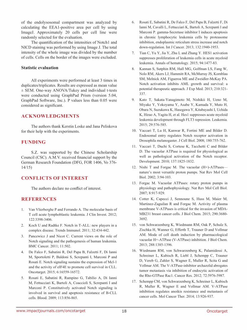
Oncotarget18www.impactjournals.com/oncotarget
of the endolysosomal compartment was analyzed by calculating the EEA1-positive area per cell by using ImageJ. Approximately 20 cells per cell line were randomly selected for the evaluation.
The quantification of the intensities of Notch1 and NICD staining was performed by using Image J. The total intensity of the whole image was divided by the number of cells. Cells on the border of the images were excluded.
Statistic evaluation
All experiments were performed at least 3 times in duplicates/triplicates. Results are expressed as mean value ± SEM. One-way ANOVA/Tukey and individual t-tests were conducted using GraphPad Prism (version 5.04, GraphPad Software, Inc.). P values less than 0.05 were considered as significant.
ACKNOWLEDGMENTS
The authors thank Kerstin Loske and Jana Peliskova for their help with the experiments.
FUNDING
S.Z. was supported by the Chinese Scholarship Council (CSC). A.M.V. received financial support by the German Research Foundation (DFG, FOR 1406, Vo 376-14/15)
CONFLICTS OF INTEREST
The authors declare no conflict of interest.
REFERENCES
1. Van Vlierberghe P and Ferrando A. The molecular basis of T cell acute lymphoblastic leukemia. J Clin Invest. 2012; 122:3398-3406.
2. Koch U and Radtke F. Notch in T-ALL: new players in a complex disease. Trends Immunol. 2011; 32:434-442.
3. Pancewicz J and Nicot C. Current views on the role of Notch signaling and the pathogenesis of human leukemia. BMC Cancer. 2011; 11:502.
4. De Falco F, Sabatini R, Del Papa B, Falzetti F, Di Ianni M, Sportoletti P, Baldoni S, Screpanti I, Marconi P and Rosati E. Notch signaling sustains the expression of Mcl-1 and the activity of eIF4E to promote cell survival in CLL. Oncotarget. 2015; 6:16559-16572.
5. Rosati E, Sabatini R, Rampino G, Tabilio A, Di Ianni M, Fettucciari K, Bartoli A, Coaccioli S, Screpanti I and Marconi P. Constitutively activated Notch signaling is involved in survival and apoptosis resistance of B-CLL cells. Blood. 2009; 113:856-865.
6. Rosati E, Sabatini R, De Falco F, Del Papa B, Falzetti F, Di Ianni M, Cavalli L, Fettucciari K, Bartoli A, Screpanti I and Marconi P. gamma-Secretase inhibitor I induces apoptosis in chronic lymphocytic leukemia cells by proteasome inhibition, endoplasmic reticulum stress increase and notch down-regulation. Int J Cancer. 2013; 132:1940-1953.
7. Tian C, Yu Y, Jia Y, Zhu L and Zhang Y. HES1 activation suppresses proliferation of leukemia cells in acute myeloid leukemia. Annals of hematology. 2015; 94:1477-83.
8. Kannan S, Sutphin RM, Hall MG, Golfman LS, Fang W, Nolo RM, Akers LJ, Hammitt RA, McMurray JS, Kornblau SM, Melnick AM, Figueroa ME and Zweidler-McKay PA. Notch activation inhibits AML growth and survival: a potential therapeutic approach. J Exp Med. 2013; 210:321-337.
9. Kato T, Sakata-Yanagimoto M, Nishikii H, Ueno M, Miyake Y, Yokoyama Y, Asabe Y, Kamada Y, Muto H, Obara N, Suzukawa K, Hasegawa Y, Kitabayashi I, Uchida K, Hirao A, Yagita H, et al. Hes1 suppresses acute myeloid leukemia development through FLT3 repression. Leukemia. 2015; 29:576-585.
10. Vaccari T, Lu H, Kanwar R, Fortini ME and Bilder D. Endosomal entry regulates Notch receptor activation in Drosophila melanogaster. J Cell Biol. 2008; 180:755-762.
11. Vaccari T, Duchi S, Cortese K, Tacchetti C and Bilder D. The vacuolar ATPase is required for physiological as well as pathological activation of the Notch receptor. Development. 2010; 137:1825-1832.
12. Nishi T and Forgac M. The vacuolar (H+)-ATPases—nature’s most versatile proton pumps. Nat Rev Mol Cell Biol. 2002; 3:94-103.
13. Forgac M. Vacuolar ATPases: rotary proton pumps in physiology and pathophysiology. Nat Rev Mol Cell Biol. 2007; 8:917-929.
14. Cotter K, Capecci J, Sennoune S, Huss M, Maier M, Martinez-Zaguilan R and Forgac M. Activity of plasma membrane V-ATPases is critical for the invasion of MDA-MB231 breast cancer cells. J Biol Chem. 2015; 290:3680-3692.
15. von Schwarzenberg K, Wiedmann RM, Oak P, Schulz S, Zischka H, Wanner G, Efferth T, Trauner D and Vollmar AM. Mode of cell death induction by pharmacological vacuolar H+-ATPase (V-ATPase) inhibition. J Biol Chem. 2013; 288:1385-1396.
16. Wiedmann RM, von Schwarzenberg K, Palamidessi A, Schreiner L, Kubisch R, Liebl J, Schempp C, Trauner D, Vereb G, Zahler S, Wagner E, Muller R, Scita G and Vollmar AM. The V-ATPase-inhibitor archazolid abrogates tumor metastasis via inhibition of endocytic activation of the Rho-GTPase Rac1. Cancer Res. 2012; 72:5976-5987.
17. Schempp CM, von Schwarzenberg K, Schreiner L, Kubisch R, Muller R, Wagner E and Vollmar AM. V-ATPase inhibition regulates anoikis resistance and metastasis of cancer cells. Mol Cancer Ther. 2014; 13:926-937.
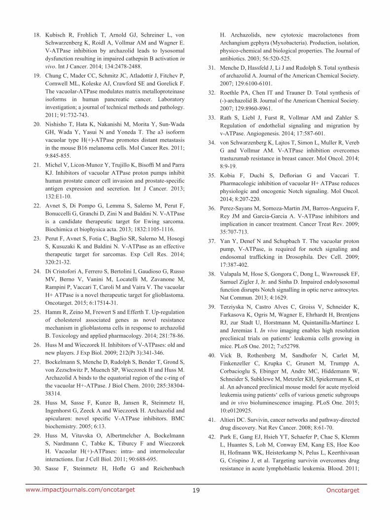
Oncotarget19www.impactjournals.com/oncotarget
18. Kubisch R, Frohlich T, Arnold GJ, Schreiner L, von Schwarzenberg K, Roidl A, Vollmar AM and Wagner E. V-ATPase inhibition by archazolid leads to lysosomal dysfunction resulting in impaired cathepsin B activation in vivo. Int J Cancer. 2014; 134:2478-2488.
19. Chung C, Mader CC, Schmitz JC, Atladottir J, Fitchev P, Cornwell ML, Koleske AJ, Crawford SE and Gorelick F. The vacuolar-ATPase modulates matrix metalloproteinase isoforms in human pancreatic cancer. Laboratory investigation; a journal of technical methods and pathology. 2011; 91:732-743.
20. Nishisho T, Hata K, Nakanishi M, Morita Y, Sun-Wada GH, Wada Y, Yasui N and Yoneda T. The a3 isoform vacuolar type H(+)-ATPase promotes distant metastasis in the mouse B16 melanoma cells. Mol Cancer Res. 2011; 9:845-855.
21. Michel V, Licon-Munoz Y, Trujillo K, Bisoffi M and Parra KJ. Inhibitors of vacuolar ATPase proton pumps inhibit human prostate cancer cell invasion and prostate-specific antigen expression and secretion. Int J Cancer. 2013; 132:E1-10.
22. Avnet S, Di Pompo G, Lemma S, Salerno M, Perut F, Bonuccelli G, Granchi D, Zini N and Baldini N. V-ATPase is a candidate therapeutic target for Ewing sarcoma. Biochimica et biophysica acta. 2013; 1832:1105-1116.
23. Perut F, Avnet S, Fotia C, Baglio SR, Salerno M, Hosogi S, Kusuzaki K and Baldini N. V-ATPase as an effective therapeutic target for sarcomas. Exp Cell Res. 2014; 320:21-32.
24. Di Cristofori A, Ferrero S, Bertolini I, Gaudioso G, Russo MV, Berno V, Vanini M, Locatelli M, Zavanone M, Rampini P, Vaccari T, Caroli M and Vaira V. The vacuolar H+ ATPase is a novel therapeutic target for glioblastoma. Oncotarget. 2015; 6:17514-31.
25. Hamm R, Zeino M, Frewert S and Efferth T. Up-regulation of cholesterol associated genes as novel resistance mechanism in glioblastoma cells in response to archazolid B. Toxicology and applied pharmacology. 2014; 281:78-86.
26. Huss M and Wieczorek H. Inhibitors of V-ATPases: old and new players. J Exp Biol. 2009; 212(Pt 3):341-346.
27. Bockelmann S, Menche D, Rudolph S, Bender T, Grond S, von Zezschwitz P, Muench SP, Wieczorek H and Huss M. Archazolid A binds to the equatorial region of the c-ring of the vacuolar H+-ATPase. J Biol Chem. 2010; 285:38304-38314.
28. Huss M, Sasse F, Kunze B, Jansen R, Steinmetz H, Ingenhorst G, Zeeck A and Wieczorek H. Archazolid and apicularen: novel specific V-ATPase inhibitors. BMC biochemistry. 2005; 6:13.
29. Huss M, Vitavska O, Albertmelcher A, Bockelmann S, Nardmann C, Tabke K, Tiburcy F and Wieczorek H. Vacuolar H(+)-ATPases: intra- and intermolecular interactions. Eur J Cell Biol. 2011; 90:688-695.
30. Sasse F, Steinmetz H, Hofle G and Reichenbach
H. Archazolids, new cytotoxic macrolactones from Archangium gephyra (Myxobacteria). Production, isolation, physico-chemical and biological properties. The Journal of antibiotics. 2003; 56:520-525.
31. Menche D, Hassfeld J, Li J and Rudolph S. Total synthesis of archazolid A. Journal of the American Chemical Society. 2007; 129:6100-6101.
32. Roethle PA, Chen IT and Trauner D. Total synthesis of (-)-archazolid B. Journal of the American Chemical Society. 2007; 129:8960-8961.
33. Rath S, Liebl J, Furst R, Vollmar AM and Zahler S. Regulation of endothelial signaling and migration by v-ATPase. Angiogenesis. 2014; 17:587-601.
34. von Schwarzenberg K, Lajtos T, Simon L, Muller R, Vereb G and Vollmar AM. V-ATPase inhibition overcomes trastuzumab resistance in breast cancer. Mol Oncol. 2014; 8:9-19.
35. Kobia F, Duchi S, Deflorian G and Vaccari T. Pharmacologic inhibition of vacuolar H+ ATPase reduces physiologic and oncogenic Notch signaling. Mol Oncol. 2014; 8:207-220.
36. Perez-Sayans M, Somoza-Martin JM, Barros-Angueira F, Rey JM and Garcia-Garcia A. V-ATPase inhibitors and implication in cancer treatment. Cancer Treat Rev. 2009; 35:707-713.
37. Yan Y, Denef N and Schupbach T. The vacuolar proton pump, V-ATPase, is required for notch signaling and endosomal trafficking in Drosophila. Dev Cell. 2009; 17:387-402.
38. Valapala M, Hose S, Gongora C, Dong L, Wawrousek EF, Samuel Zigler J, Jr. and Sinha D. Impaired endolysosomal function disrupts Notch signalling in optic nerve astrocytes. Nat Commun. 2013; 4:1629.
39. Terziyska N, Castro Alves C, Groiss V, Schneider K, Farkasova K, Ogris M, Wagner E, Ehrhardt H, Brentjens RJ, zur Stadt U, Horstmann M, Quintanilla-Martinez L and Jeremias I. In vivo imaging enables high resolution preclinical trials on patients‘ leukemia cells growing in mice. PLoS One. 2012; 7:e52798.
40. Vick B, Rothenberg M, Sandhofer N, Carlet M, Finkenzeller C, Krupka C, Grunert M, Trumpp A, Corbacioglu S, Ebinger M, Andre MC, Hiddemann W, Schneider S, Subklewe M, Metzeler KH, Spiekermann K, et al. An advanced preclinical mouse model for acute myeloid leukemia using patients‘ cells of various genetic subgroups and in vivo bioluminescence imaging. PLoS One. 2015; 10:e0120925.
41. Altieri DC. Survivin, cancer networks and pathway-directed drug discovery. Nat Rev Cancer. 2008; 8:61-70.
42. Park E, Gang EJ, Hsieh YT, Schaefer P, Chae S, Klemm L, Huantes S, Loh M, Conway EM, Kang ES, Hoe Koo H, Hofmann WK, Heisterkamp N, Pelus L, Keerthivasan G, Crispino J, et al. Targeting survivin overcomes drug resistance in acute lymphoblastic leukemia. Blood. 2011;
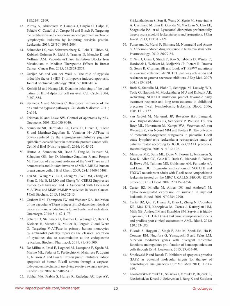
Oncotarget20www.impactjournals.com/oncotarget
118:2191-2199.43. Purroy N, Abrisqueta P, Carabia J, Carpio C, Calpe E,
Palacio C, Castellvi J, Crespo M and Bosch F. Targeting the proliferative and chemoresistant compartment in chronic lymphocytic leukemia by inhibiting survivin protein. Leukemia. 2014; 28(10):1993-2004.
44. Schneider LS, von Schwarzenberg K, Lehr T, Ulrich M, Kubisch-Dohmen R, Liebl J, Trauner D, Menche D and Vollmar AM. Vacuolar-ATPase Inhibition Blocks Iron Metabolism to Mediate Therapeutic Effects in Breast Cancer. Cancer Res. 2015; 75:2863-2874.
45. Greijer AE and van der Wall E. The role of hypoxia inducible factor 1 (HIF-1) in hypoxia induced apoptosis. Journal of clinical pathology. 2004; 57:1009-1014.
46. Koshiji M and Huang LE. Dynamic balancing of the dual nature of HIF-1alpha for cell survival. Cell Cycle. 2004; 3:853-854.
47. Sermeus A and Michiels C. Reciprocal influence of the p53 and the hypoxic pathways. Cell death & disease. 2011; 2:e164.
48. Fridman JS and Lowe SW. Control of apoptosis by p53. Oncogene. 2003; 22:9030-9040.
49. Sennoune SR, Bermudez LE, Lees JC, Hirsch J, Filleur S and Martinez-Zaguilan R. Vacuolar H+-ATPase is down-regulated by the angiogenesis-inhibitory pigment epithelium-derived factor in metastatic prostate cancer cells. Cell Mol Biol (Noisy-le-grand). 2014; 60:45-52.
50. Hinton A, Sennoune SR, Bond S, Fang M, Reuveni M, Sahagian GG, Jay D, Martinez-Zaguilan R and Forgac M. Function of a subunit isoforms of the V-ATPase in pH homeostasis and in vitro invasion of MDA-MB231 human breast cancer cells. J Biol Chem. 2009; 284:16400-16408.
51. Fan SH, Wang YY, Lu J, Zheng YL, Wu DM, Zhang ZF, Shan Q, Hu B, Li MQ and Cheng W. CERS2 Suppresses Tumor Cell Invasion and Is Associated with Decreased V-ATPase and MMP-2/MMP-9 activities in Breast Cancer. J Cell Biochem. 2015; 116:502-13.
52. Graham RM, Thompson JW and Webster KA. Inhibition of the vacuolar ATPase induces Bnip3-dependent death of cancer cells and a reduction in tumor burden and metastasis. Oncotarget. 2014; 5:1162-1173.
53. Scherer O, Steinmetz H, Kaether C, Weinigel C, Barz D, Kleinert H, Menche D, Muller R, Pergola C and Werz O. Targeting V-ATPase in primary human monocytes by archazolid potently represses the classical secretion of cytokines due to accumulation at the endoplasmic reticulum. Biochem Pharmacol. 2014; 91:490-500.
54. De Milito A, Iessi E, Logozzi M, Lozupone F, Spada M, Marino ML, Federici C, Perdicchio M, Matarrese P, Lugini L, Nilsson A and Fais S. Proton pump inhibitors induce apoptosis of human B-cell tumors through a caspase-independent mechanism involving reactive oxygen species. Cancer Res. 2007; 67:5408-5417.
55. Sukhai MA, Prabha S, Hurren R, Rutledge AC, Lee AY,
Sriskanthadevan S, Sun H, Wang X, Skrtic M, Seneviratne A, Cusimano M, Jhas B, Gronda M, MacLean N, Cho EE, Spagnuolo PA, et al. Lysosomal disruption preferentially targets acute myeloid leukemia cells and progenitors. J Clin Invest. 2013; 123:315-328.
56. Funayama K, Murai F, Shimane M, Nomura H and Asano S. Adhesion-induced drug resistance in leukemia stem cells. Pharmacology. 2010; 86:79-84.
57. O’Neil J, Grim J, Strack P, Rao S, Tibbitts D, Winter C, Hardwick J, Welcker M, Meijerink JP, Pieters R, Draetta G, Sears R, Clurman BE and Look AT. FBW7 mutations in leukemic cells mediate NOTCH pathway activation and resistance to gamma-secretase inhibitors. J Exp Med. 2007; 204:1813-1824.
58. Breit S, Stanulla M, Flohr T, Schrappe M, Ludwig WD, Tolle G, Happich M, Muckenthaler MU and Kulozik AE. Activating NOTCH1 mutations predict favorable early treatment response and long-term outcome in childhood precursor T-cell lymphoblastic leukemia. Blood. 2006; 108:1151-1157.
59. van Grotel M, Meijerink JP, Beverloo HB, Langerak AW, Buys-Gladdines JG, Schneider P, Poulsen TS, den Boer ML, Horstmann M, Kamps WA, Veerman AJ, van Wering ER, van Noesel MM and Pieters R. The outcome of molecular-cytogenetic subgroups in pediatric T-cell acute lymphoblastic leukemia: a retrospective study of patients treated according to DCOG or COALL protocols. Haematologica. 2006; 91:1212-1221.
60. Mansour MR, Sulis ML, Duke V, Foroni L, Jenkinson S, Koo K, Allen CG, Gale RE, Buck G, Richards S, Paietta E, Rowe JM, Tallman MS, Goldstone AH, Ferrando AA and Linch DC. Prognostic implications of NOTCH1 and FBXW7 mutations in adults with T-cell acute lymphoblastic leukemia treated on the MRC UKALLXII/ECOG E2993 protocol. J Clin Oncol. 2009; 27:4352-4356.
61. Carter BZ, Milella M, Altieri DC and Andreeff M. Cytokine-regulated expression of survivin in myeloid leukemia. Blood. 2001; 97:2784-2790.
62. Carter BZ, Qiu Y, Huang X, Diao L, Zhang N, Coombes KR, Mak DH, Konopleva M, Cortes J, Kantarjian HM, Mills GB, Andreeff M and Kornblau SM. Survivin is highly expressed in CD34(+)38(-) leukemic stem/progenitor cells and predicts poor clinical outcomes in AML. Blood. 2012; 120:173-180.
63. Fukuda S, Hoggatt J, Singh P, Abe M, Speth JM, Hu P, Conway EM, Nucifora G, Yamaguchi S and Pelus LM. Survivin modulates genes with divergent molecular functions and regulates proliferation of hematopoietic stem cells through Evi-1. Leukemia. 2015; 29:433-40.
64. Smolewski P and Robak T. Inhibitors of apoptosis proteins (IAPs) as potential molecular targets for therapy of hematological malignancies. Curr Mol Med. 2011; 11:633-649.
65. Glodkowska-Mrowka E, Solarska I, Mrowka P, Bajorek K, Niesiobedzka-Krezel J, Seferynska I, Borg K and Stoklosa
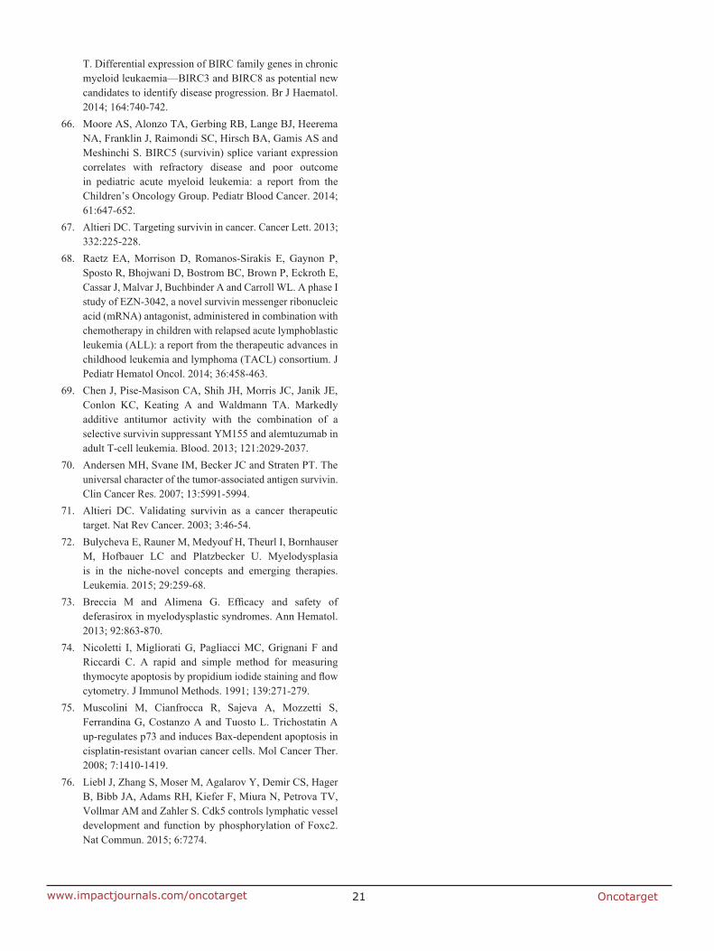
Oncotarget21www.impactjournals.com/oncotarget
T. Differential expression of BIRC family genes in chronic myeloid leukaemia—BIRC3 and BIRC8 as potential new candidates to identify disease progression. Br J Haematol. 2014; 164:740-742.
66. Moore AS, Alonzo TA, Gerbing RB, Lange BJ, Heerema NA, Franklin J, Raimondi SC, Hirsch BA, Gamis AS and Meshinchi S. BIRC5 (survivin) splice variant expression correlates with refractory disease and poor outcome in pediatric acute myeloid leukemia: a report from the Children’s Oncology Group. Pediatr Blood Cancer. 2014; 61:647-652.
67. Altieri DC. Targeting survivin in cancer. Cancer Lett. 2013; 332:225-228.
68. Raetz EA, Morrison D, Romanos-Sirakis E, Gaynon P, Sposto R, Bhojwani D, Bostrom BC, Brown P, Eckroth E, Cassar J, Malvar J, Buchbinder A and Carroll WL. A phase I study of EZN-3042, a novel survivin messenger ribonucleic acid (mRNA) antagonist, administered in combination with chemotherapy in children with relapsed acute lymphoblastic leukemia (ALL): a report from the therapeutic advances in childhood leukemia and lymphoma (TACL) consortium. J Pediatr Hematol Oncol. 2014; 36:458-463.
69. Chen J, Pise-Masison CA, Shih JH, Morris JC, Janik JE, Conlon KC, Keating A and Waldmann TA. Markedly additive antitumor activity with the combination of a selective survivin suppressant YM155 and alemtuzumab in adult T-cell leukemia. Blood. 2013; 121:2029-2037.
70. Andersen MH, Svane IM, Becker JC and Straten PT. The universal character of the tumor-associated antigen survivin. Clin Cancer Res. 2007; 13:5991-5994.
71. Altieri DC. Validating survivin as a cancer therapeutic target. Nat Rev Cancer. 2003; 3:46-54.
72. Bulycheva E, Rauner M, Medyouf H, Theurl I, Bornhauser M, Hofbauer LC and Platzbecker U. Myelodysplasia is in the niche-novel concepts and emerging therapies. Leukemia. 2015; 29:259-68.
73. Breccia M and Alimena G. Efficacy and safety of deferasirox in myelodysplastic syndromes. Ann Hematol. 2013; 92:863-870.
74. Nicoletti I, Migliorati G, Pagliacci MC, Grignani F and Riccardi C. A rapid and simple method for measuring thymocyte apoptosis by propidium iodide staining and flow cytometry. J Immunol Methods. 1991; 139:271-279.
75. Muscolini M, Cianfrocca R, Sajeva A, Mozzetti S, Ferrandina G, Costanzo A and Tuosto L. Trichostatin A up-regulates p73 and induces Bax-dependent apoptosis in cisplatin-resistant ovarian cancer cells. Mol Cancer Ther. 2008; 7:1410-1419.
76. Liebl J, Zhang S, Moser M, Agalarov Y, Demir CS, Hager B, Bibb JA, Adams RH, Kiefer F, Miura N, Petrova TV, Vollmar AM and Zahler S. Cdk5 controls lymphatic vessel development and function by phosphorylation of Foxc2. Nat Commun. 2015; 6:7274.
MDM2 antagonist nutlin-3a sensitizes tumors to V-ATPase inhibition
Lina S. Schneidera, Melanie Ulricha, Thorsten Lehrb, Dirk Menchec, Rolf Müllerd,
Angelika M. Vollmara, Karin von Schwarzenberga
aDepartment of Pharmacy, Pharmaceutical Biology, Ludwig-Maximilians-University of
Munich, Butenandtstrasse 5-13, 81377 Munich, Germany
bClinical Pharmacy, Saarland University, Campus C2 2, 66123 Saarbrücken,
Germany
cKekulé Institute of Organic Chemistry and Biochemistry, University of Bonn,
Gerhard-Domagk-Str. 1, 53121 Bonn, Germany
dSaarland University, Helmholtz Institute for Pharmaceutical Research Saarland,
Helmholtz Centre for Infection Research and Department of Pharmaceutical
Biotechnology, PO 151150, 66042 Saarbrücken, Germany
Corresponding author: Dr. Karin von Schwarzenberg, Department of Pharmacy,
Butenandtstrasse 5-13, 81377 Munich, Germany, Phone: +49-89-2180-77165, Email:
Conflict of interest: There is no conflict of interest.
Abbreviations: 1 vacuolar-type ATPase, 2 oxidative phosphorylation, 3 TP53-induced
glycolysis and apoptosis regulator, 4 U87MG, 5 insulin-like growth factor-binding
protein 3
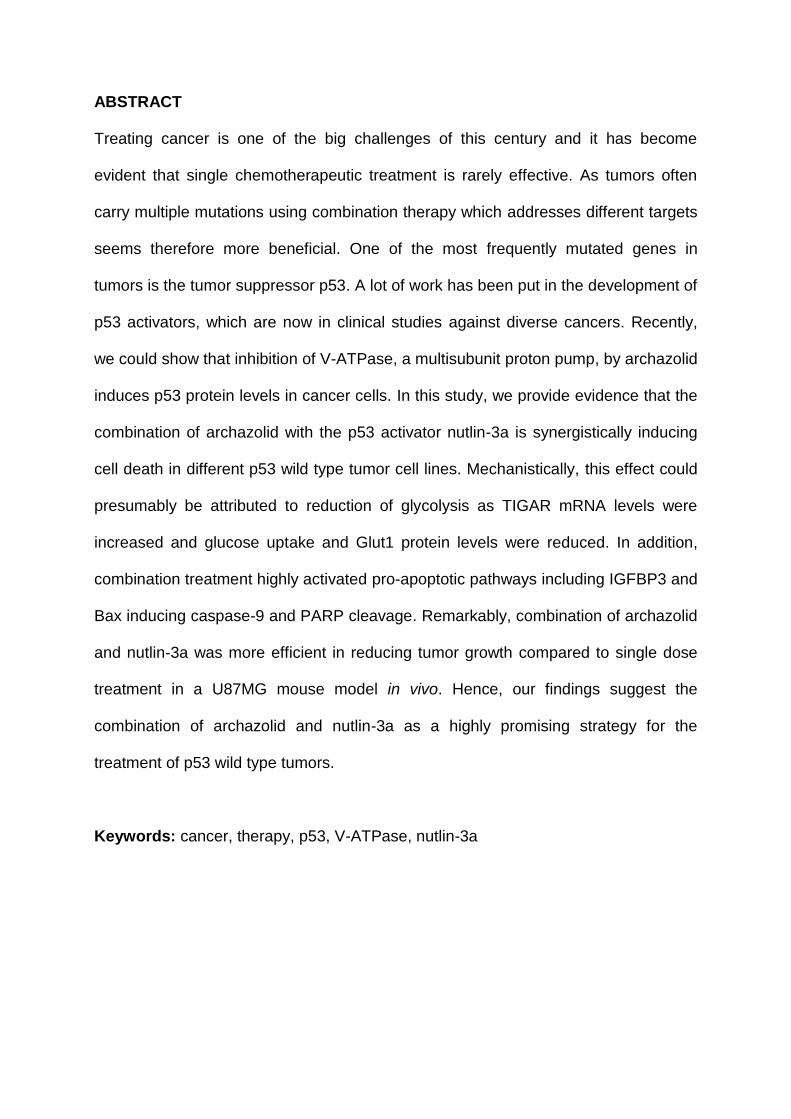
ABSTRACT
Treating cancer is one of the big challenges of this century and it has become
evident that single chemotherapeutic treatment is rarely effective. As tumors often
carry multiple mutations using combination therapy which addresses different targets
seems therefore more beneficial. One of the most frequently mutated genes in
tumors is the tumor suppressor p53. A lot of work has been put in the development of
p53 activators, which are now in clinical studies against diverse cancers. Recently,
we could show that inhibition of V-ATPase, a multisubunit proton pump, by archazolid
induces p53 protein levels in cancer cells. In this study, we provide evidence that the
combination of archazolid with the p53 activator nutlin-3a is synergistically inducing
cell death in different p53 wild type tumor cell lines. Mechanistically, this effect could
presumably be attributed to reduction of glycolysis as TIGAR mRNA levels were
increased and glucose uptake and Glut1 protein levels were reduced. In addition,
combination treatment highly activated pro-apoptotic pathways including IGFBP3 and
Bax inducing caspase-9 and PARP cleavage. Remarkably, combination of archazolid
and nutlin-3a was more efficient in reducing tumor growth compared to single dose
treatment in a U87MG mouse model in vivo. Hence, our findings suggest the
combination of archazolid and nutlin-3a as a highly promising strategy for the
treatment of p53 wild type tumors.
Keywords: cancer, therapy, p53, V-ATPase, nutlin-3a
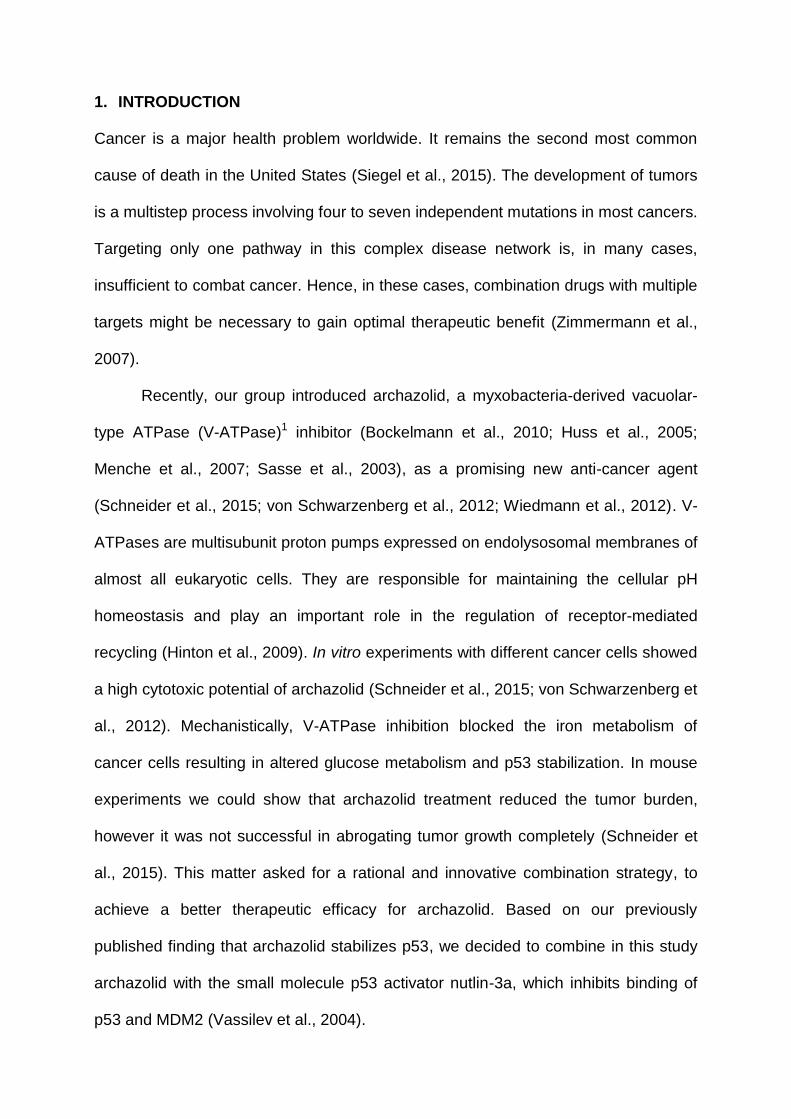
1. INTRODUCTION
Cancer is a major health problem worldwide. It remains the second most common
cause of death in the United States (Siegel et al., 2015). The development of tumors
is a multistep process involving four to seven independent mutations in most cancers.
Targeting only one pathway in this complex disease network is, in many cases,
insufficient to combat cancer. Hence, in these cases, combination drugs with multiple
targets might be necessary to gain optimal therapeutic benefit (Zimmermann et al.,
2007).
Recently, our group introduced archazolid, a myxobacteria-derived vacuolar-
type ATPase (V-ATPase)1 inhibitor (Bockelmann et al., 2010; Huss et al., 2005;
Menche et al., 2007; Sasse et al., 2003), as a promising new anti-cancer agent
(Schneider et al., 2015; von Schwarzenberg et al., 2012; Wiedmann et al., 2012). V-
ATPases are multisubunit proton pumps expressed on endolysosomal membranes of
almost all eukaryotic cells. They are responsible for maintaining the cellular pH
homeostasis and play an important role in the regulation of receptor-mediated
recycling (Hinton et al., 2009). In vitro experiments with different cancer cells showed
a high cytotoxic potential of archazolid (Schneider et al., 2015; von Schwarzenberg et
al., 2012). Mechanistically, V-ATPase inhibition blocked the iron metabolism of
cancer cells resulting in altered glucose metabolism and p53 stabilization. In mouse
experiments we could show that archazolid treatment reduced the tumor burden,
however it was not successful in abrogating tumor growth completely (Schneider et
al., 2015). This matter asked for a rational and innovative combination strategy, to
achieve a better therapeutic efficacy for archazolid. Based on our previously
published finding that archazolid stabilizes p53, we decided to combine in this study
archazolid with the small molecule p53 activator nutlin-3a, which inhibits binding of
p53 and MDM2 (Vassilev et al., 2004).
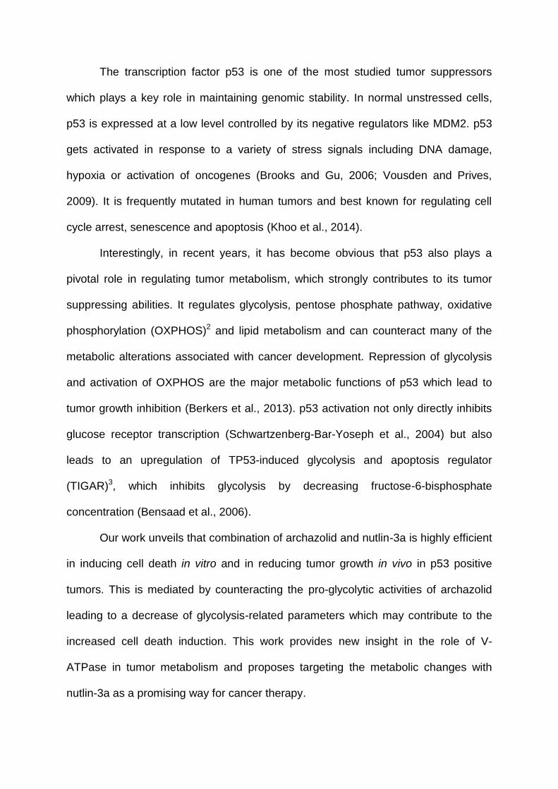
The transcription factor p53 is one of the most studied tumor suppressors
which plays a key role in maintaining genomic stability. In normal unstressed cells,
p53 is expressed at a low level controlled by its negative regulators like MDM2. p53
gets activated in response to a variety of stress signals including DNA damage,
hypoxia or activation of oncogenes (Brooks and Gu, 2006; Vousden and Prives,
2009). It is frequently mutated in human tumors and best known for regulating cell
cycle arrest, senescence and apoptosis (Khoo et al., 2014).
Interestingly, in recent years, it has become obvious that p53 also plays a
pivotal role in regulating tumor metabolism, which strongly contributes to its tumor
suppressing abilities. It regulates glycolysis, pentose phosphate pathway, oxidative
phosphorylation (OXPHOS)2 and lipid metabolism and can counteract many of the
metabolic alterations associated with cancer development. Repression of glycolysis
and activation of OXPHOS are the major metabolic functions of p53 which lead to
tumor growth inhibition (Berkers et al., 2013). p53 activation not only directly inhibits
glucose receptor transcription (Schwartzenberg-Bar-Yoseph et al., 2004) but also
leads to an upregulation of TP53-induced glycolysis and apoptosis regulator
(TIGAR)3, which inhibits glycolysis by decreasing fructose-6-bisphosphate
concentration (Bensaad et al., 2006).
Our work unveils that combination of archazolid and nutlin-3a is highly efficient
in inducing cell death in vitro and in reducing tumor growth in vivo in p53 positive
tumors. This is mediated by counteracting the pro-glycolytic activities of archazolid
leading to a decrease of glycolysis-related parameters which may contribute to the
increased cell death induction. This work provides new insight in the role of V-
ATPase in tumor metabolism and proposes targeting the metabolic changes with
nutlin-3a as a promising way for cancer therapy.
2. MATERIAL AND METHODS
2.1 Cell lines and reagents
The mammary cancer cell line MCF7, the liver carcinoma cell line HepG2 and the
cervical cancer cell line Hela were recently purchased from DSMZ (Braunschweig,
Germany). The mammary cancer cell line MDA-MB-231 was obtained from Cell Line
Service Eppelheim (Germany). The glioblastoma cell line U87MG (U87)4 was a kind
gift from Prof. Adrian L. Harris (Department of Oncology, Wheatherall Institute of
Molecular Medicine, Oxford University). MCF7 cells were cultivated in RPMI 1620
complimented with 10% FCS, 1% nonessential amino acids, 1% pyruvate and
125µg/L insulin. HepG2 and MDA-MB-231 cells were grown in DMEM High Glucose
containing 10% FCS, Hela and U87 cells in RPMI 1620 supplemented with 10%
FCS. Archazolid was isolated by Prof. Dirk Menche (Kekulé-Institute of Organic
Chemistry and Biochemistry, University of Bonn, Bonn, Germany). Nutlin-3a was
purchased from Sigma Aldrich (Taufkirchen, Germany).
2.2 Immunocytochemistry
2 x 10⁴ MCF7 cells were seeded onto µ-slides 8-well ibidiTreat (IBIDI, Martinsried,
Germany) and were treated as indicated. Then, cells were fixed with 4%
paraformaldehyde (PFA), permeabilized with 0.2% Triton X-100/PBS and blocked
with 1% BSA/PBS containing 0.1% Triton X-100. Antibody against p53 (Cell
Signaling Technology, Danvers, MA) and AlexaFluor 488-goat-antimouse (Invitrogen,
Waltham, MA) were diluted in blocking solution. Nuclei were stained with Hoechst
33342® (Hoechst) (Sigma Aldrich).
2.3 Western blot
Western blot analysis was performed as described before (Schneider et al., 2015).
Following antibodies were used: p53, pAkt Ser473, Akt, Bid, caspase-9, PARP (Cell
Signaling Technology), Glut1 (Novus, Littleton, CO), IGFBP3, HRP goat-antimouse
(Santa Cruz Biotechnology, Heidelberg, Germany), Rb (BD Bioscience, Heidelberg,
Germany), Bcl-2 (Calbiochem, Darmstadt, Germany), AIF (Chemicon, Billerica, MA),
actin (Millipore, Darmstadt, Germany) and HRP-goat-antirabbit (Bio-Rad, Munich,
Germany).
2.4 qPCR
RNA extraction, cDNA synthesis and qPCR were performed as described before
(Schneider et al., 2015). All designed primers were purchased from Metabion
(Planegg, Germany).
2.5 Cell death analysis
Cell death was assessed as described elsewhere (Schneider et al., 2015).
2.6 Measurement of metabolic activity
CellTiter-Blue Assay (Promega, Mannheim, Germany) was performed according to
the manufacturer`s protocol to quantify metabolic activity.
2.7 Measurement of glucose uptake, ATP levels, Bax activation
Quantifications were performed as reported previously (von Schwarzenberg et al.,
2012).
2.8 In vivo mouse model
For in vivo studies, six-week old female BALB/cOlaHsd-Foxn1nu/Foxn1+ mice (Harlan,
Indianapolis, IN) were used. 5 x 10⁶ U87MG cells were resuspended in PBS/matrigel
(Corning, Wiesbaden, Germany) (1:1) and injected subcutaneously into the flank of
each mouse. After formation of palpable tumors, mice were divided into groups and
treated daily intraperitoneally with 0.2 mg/kg archazolid in 5% DMSO/10%
solutol/PBS, 5 mg/kg nutlin-3a 5% DMSO/10% solutol/PBS, with both or solvent
respectively. Measurement of tumors was done every 2 to 3 days with a caliper using
the formula a x b2/2. Modeling was performed using the non-linear mixed effects
modeling technique with the software NONMEM 7.3 (Simeoni et al., 2004). Nutlin-3a
PK model was built based on literature data (Zhang et al., 2010). Tumor growth
curves were normalized to control. Animal experiments were approved by the District
Government of Upper Bavaria in accordance with the German animal welfare and
institutional guidelines.
2.9 Statistical analysis
For all statistics GraphPad Prism was used. Error bars display SEM, except in vivo
tumor growth curves which show SD. Synergism was calculated with the Bliss
formula v=xcombination/((xA-xB)-(xA*xB)) with v>1 indicating synergism.
3. RESULTS
3.1 Combination of archazolid and nutlin-3a strongly induce p53
We could recently show that the V-ATPase inhibitor archazolid induces p53 protein
expression in breast cancer cells (Schneider et al., 2015). To start with, Fig. 1A
shows that while low dose archazolid had no effect on p53 protein level, a
combination with nutlin-3a even raises p53 protein levels over the level of nutlin-3a
alone. To more deeply investigate this phenomenon, we analyzed the translocation
of p53 to the nucleus – a prerequisite for p53 to be activated. We found that p53 is
mostly located in the cytoplasm of untreated cells whereas it partially translocates to
the nucleus after high dose archazolid treatment as well as after nutlin-3a treatment.
Interestingly, a combination of the two compounds led to an almost complete
reduction of cytosolic p53 and showed only activated p53 in the nucleus (Fig. 1B).
This increased activation is due to stabilization of the protein and not enhanced
transcription as there is no elevated mRNA level after combined treatment with
archazolid and nutlin-3a (Fig 1C). Taken together, these findings indicate that
combination of archazolid and nutlin-3a might have an improved anti-tumor activity in
wild type p53 cells compared to the two compounds alone.
3.2 Combination of archazolid and nutlin-3a induces synergistic cell death in
cancer cells
To analyze weather the increased p53 activation by archazolid and nutlin-3a has an
effect on cell death induction, we used different p53 wild type tumor cell lines and
one breast cancer cell line with mutated p53 and treated them with single drugs and
combination. Subsequent flow cytometry analysis showed for MCF7, Hela and
HepG2 cells synergistic cell death induction after combination treatment and for the
glioblastoma cell line U87 reduced metabolic activity (Fig. 2A). As expected,
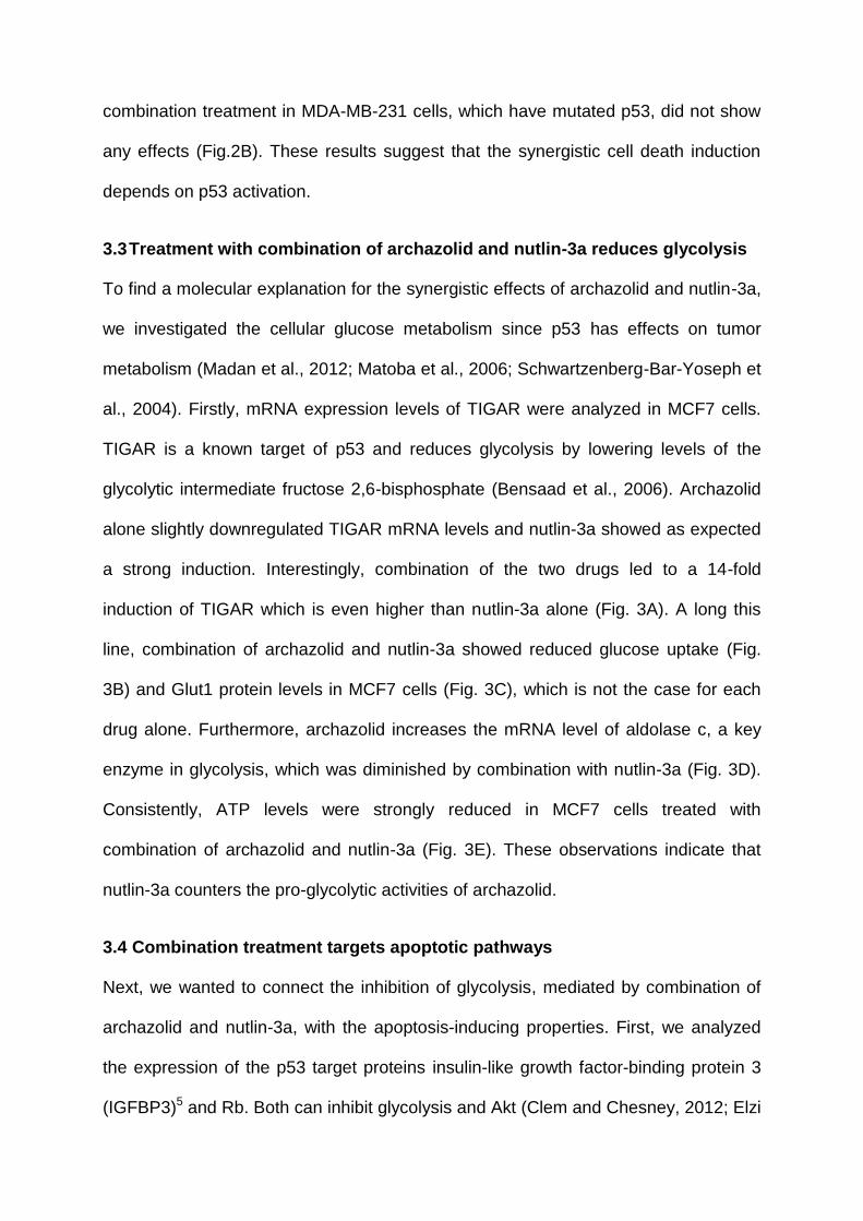
combination treatment in MDA-MB-231 cells, which have mutated p53, did not show
any effects (Fig.2B). These results suggest that the synergistic cell death induction
depends on p53 activation.
3.3 Treatment with combination of archazolid and nutlin-3a reduces glycolysis
To find a molecular explanation for the synergistic effects of archazolid and nutlin-3a,
we investigated the cellular glucose metabolism since p53 has effects on tumor
metabolism (Madan et al., 2012; Matoba et al., 2006; Schwartzenberg-Bar-Yoseph et
al., 2004). Firstly, mRNA expression levels of TIGAR were analyzed in MCF7 cells.
TIGAR is a known target of p53 and reduces glycolysis by lowering levels of the
glycolytic intermediate fructose 2,6-bisphosphate (Bensaad et al., 2006). Archazolid
alone slightly downregulated TIGAR mRNA levels and nutlin-3a showed as expected
a strong induction. Interestingly, combination of the two drugs led to a 14-fold
induction of TIGAR which is even higher than nutlin-3a alone (Fig. 3A). A long this
line, combination of archazolid and nutlin-3a showed reduced glucose uptake (Fig.
3B) and Glut1 protein levels in MCF7 cells (Fig. 3C), which is not the case for each
drug alone. Furthermore, archazolid increases the mRNA level of aldolase c, a key
enzyme in glycolysis, which was diminished by combination with nutlin-3a (Fig. 3D).
Consistently, ATP levels were strongly reduced in MCF7 cells treated with
combination of archazolid and nutlin-3a (Fig. 3E). These observations indicate that
nutlin-3a counters the pro-glycolytic activities of archazolid.
3.4 Combination treatment targets apoptotic pathways
Next, we wanted to connect the inhibition of glycolysis, mediated by combination of
archazolid and nutlin-3a, with the apoptosis-inducing properties. First, we analyzed
the expression of the p53 target proteins insulin-like growth factor-binding protein 3
(IGFBP3)5 and Rb. Both can inhibit glycolysis and Akt (Clem and Chesney, 2012; Elzi
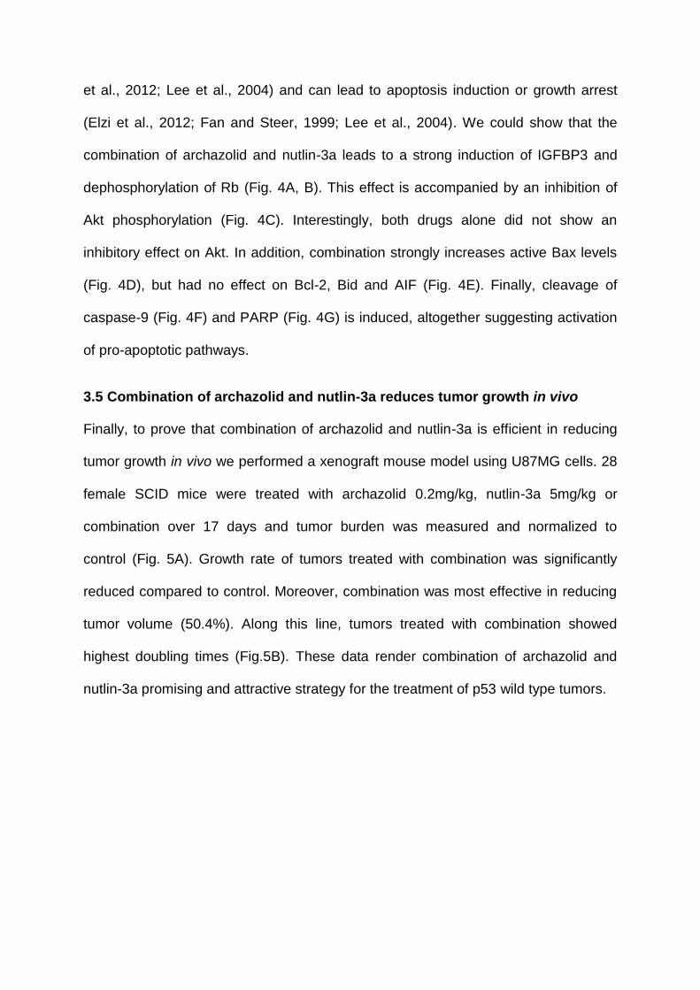
et al., 2012; Lee et al., 2004) and can lead to apoptosis induction or growth arrest
(Elzi et al., 2012; Fan and Steer, 1999; Lee et al., 2004). We could show that the
combination of archazolid and nutlin-3a leads to a strong induction of IGFBP3 and
dephosphorylation of Rb (Fig. 4A, B). This effect is accompanied by an inhibition of
Akt phosphorylation (Fig. 4C). Interestingly, both drugs alone did not show an
inhibitory effect on Akt. In addition, combination strongly increases active Bax levels
(Fig. 4D), but had no effect on Bcl-2, Bid and AIF (Fig. 4E). Finally, cleavage of
caspase-9 (Fig. 4F) and PARP (Fig. 4G) is induced, altogether suggesting activation
of pro-apoptotic pathways.
3.5 Combination of archazolid and nutlin-3a reduces tumor growth in vivo
Finally, to prove that combination of archazolid and nutlin-3a is efficient in reducing
tumor growth in vivo we performed a xenograft mouse model using U87MG cells. 28
female SCID mice were treated with archazolid 0.2mg/kg, nutlin-3a 5mg/kg or
combination over 17 days and tumor burden was measured and normalized to
control (Fig. 5A). Growth rate of tumors treated with combination was significantly
reduced compared to control. Moreover, combination was most effective in reducing
tumor volume (50.4%). Along this line, tumors treated with combination showed
highest doubling times (Fig.5B). These data render combination of archazolid and
nutlin-3a promising and attractive strategy for the treatment of p53 wild type tumors.
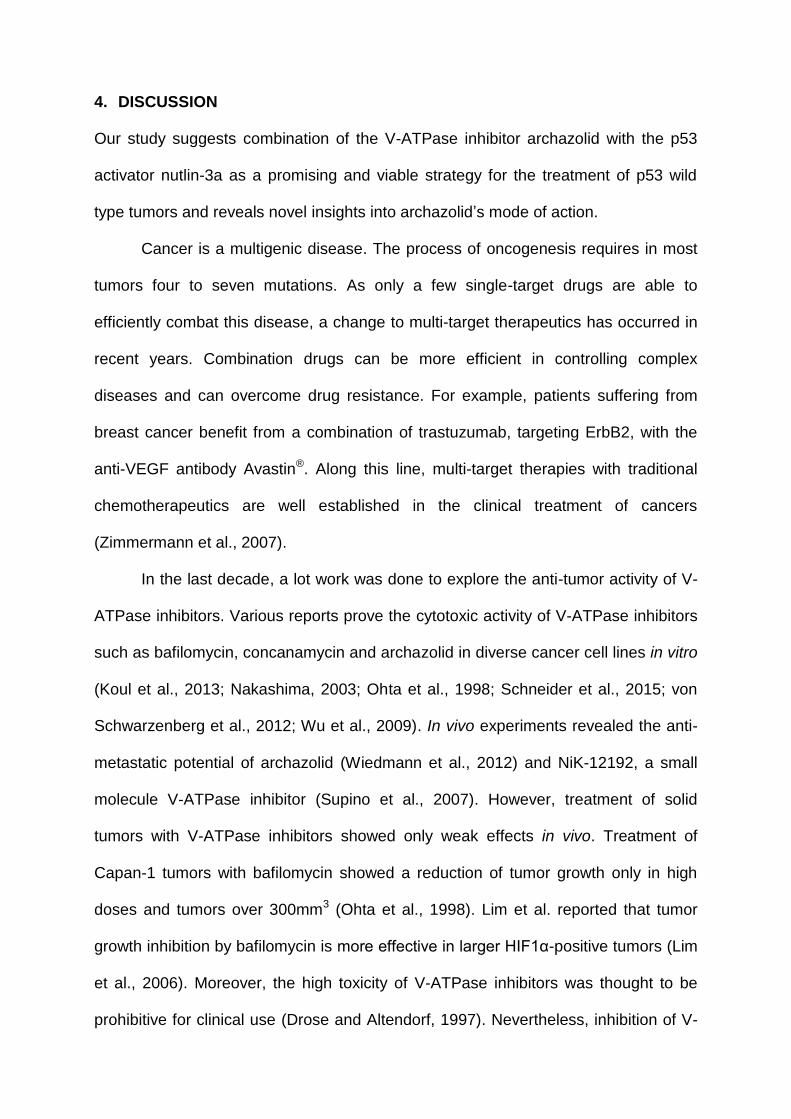
4. DISCUSSION
Our study suggests combination of the V-ATPase inhibitor archazolid with the p53
activator nutlin-3a as a promising and viable strategy for the treatment of p53 wild
type tumors and reveals novel insights into archazolid’s mode of action.
Cancer is a multigenic disease. The process of oncogenesis requires in most
tumors four to seven mutations. As only a few single-target drugs are able to
efficiently combat this disease, a change to multi-target therapeutics has occurred in
recent years. Combination drugs can be more efficient in controlling complex
diseases and can overcome drug resistance. For example, patients suffering from
breast cancer benefit from a combination of trastuzumab, targeting ErbB2, with the
anti-VEGF antibody Avastin®. Along this line, multi-target therapies with traditional
chemotherapeutics are well established in the clinical treatment of cancers
(Zimmermann et al., 2007).
In the last decade, a lot work was done to explore the anti-tumor activity of V-
ATPase inhibitors. Various reports prove the cytotoxic activity of V-ATPase inhibitors
such as bafilomycin, concanamycin and archazolid in diverse cancer cell lines in vitro
(Koul et al., 2013; Nakashima, 2003; Ohta et al., 1998; Schneider et al., 2015; von
Schwarzenberg et al., 2012; Wu et al., 2009). In vivo experiments revealed the anti-
metastatic potential of archazolid (Wiedmann et al., 2012) and NiK-12192, a small
molecule V-ATPase inhibitor (Supino et al., 2007). However, treatment of solid
tumors with V-ATPase inhibitors showed only weak effects in vivo. Treatment of
Capan-1 tumors with bafilomycin showed a reduction of tumor growth only in high
doses and tumors over 300mm3 (Ohta et al., 1998). Lim et al. reported that tumor
growth inhibition by bafilomycin is more effective in larger HIF1α-positive tumors (Lim
et al., 2006). Moreover, the high toxicity of V-ATPase inhibitors was thought to be
prohibitive for clinical use (Drose and Altendorf, 1997). Nevertheless, inhibition of V-
ATPase was shown to overcome chemoresistance in vitro (Sasazawa et al., 2009;
You et al., 2009) and trastuzumab resistance in vivo (von Schwarzenberg et al.,
2014).
As our own recently published work showed only a moderate reduction of
tumor burden in vivo after archazolid treatment, we now intended to improve this by a
rational combination approach. There are several ways for multi-target therapies to
achieve better therapeutic efficacy, one is to address different targets within the same
pathway (Zimmermann et al., 2007). Since our study further revealed that archazolid
interferes with the iron metabolism of cells finally resulting in disturbed glucose
metabolism and p53 stabilization (Schneider et al., 2015), combination treatment
within the p53 pathway seemed promising.
p53 was extensively studied in the last decades as it is the most frequently
mutated gene in tumors. Nevertheless, about half of all human cancers carry no
mutation in this gene. Therefore, a lot of work has been put in the development of
p53 activators, which are now in clinical studies against diverse cancers. One very
promising approach is to activate p53 through small molecule MDM2 antagonists. In
order to restore p53 function, in cases where it is mutated, efforts have been made to
identify drugs which stabilize p53 conformation by acting as p53 chaperones through
binding to it. Moreover, gene therapy has been used to manipulate the p53 network
(Khoo et al., 2014).
In search for compounds that are able to block the p53-MDM2 binding,
thereby activating p53, Vassilev et al. identified a class of small molecules named
nutlins. They could show that nutlin-3 was efficient in reducing tumor growth of SJSA-
1 xenografts (Vassilev, 2004) and others revealed its antitumor activity in MHM,
LnCap and 22Rv1 xenografts (Tovar et al., 2006). Moreover, efficient reduction of
tumor growth was shown in combination studies. For example targeting MDM2 with

nutlin-3 showed superior in vivo efficacy in leukemia when combined with valproic
acid, which inhibits histone deacetylases (McCormack et al., 2011). To address the
p53 pathway at two different points, we combined in this study archazolid with nutlin-
3a in different cancer cell lines. p53 wild type tumor cells showed synergistic cell
death induction in vitro while p53 mutant cells showed no effect, suggesting a p53
dependent mechanism.
Despite regulating cell cycle arrest, senescence and apoptosis, p53 is known
to act on metabolism of cells, which contributes to its tumor suppressing abilities
(Berkers et al., 2013). Interestingly, V-ATPase function was also connected to tumor
metabolism. Recently, we could show that archazolid has an impact on the glucose
metabolism of cancer cells (Schneider et al., 2015). In addition, others demonstrated
that V-ATPase acts as a metabolic sensor by playing a role in amino acid sensing via
mTOR and by physical interaction with glycolytic enzymes (Maxson and Grinstein,
2014; Zoncu et al., 2011). Along this line, we found decreased ATP levels –
indicating insufficient energy supply – by the combination therapy as well as reduced
glucose uptake and receptor expression, which can lead to a lowered glycolytic flux.
In addition, mRNA levels of the p53 target TIGAR, recently identified to decrease
fructose-6-bisphosphate concentration, and thereby glycolysis (Bensaad et al., 2006),
were 14-fold increased after combination treatment. This was accompanied by
reduced levels of the glycolytic intermediate aldolase further proposing that the
combination counteracts glycolysis.
Moreover, other proteins, which have been associated with p53, were
influenced by the combination of archazolid and nutlin-3a. The protein level of the
IGF-related tumor suppressor IGFBP3 (Buckbinder et al., 1995), which was shown to
attenuate tumor growth in different studies (Alami et al., 2008; Butt et al., 2000; Silha
et al., 2006; Williams et al., 2000), was strongly increased. This was accompanied by
reduced phosphorylation of Akt. Interestingly, a probable connection between
IGFBP3 and the Akt pathway was described before (Alami et al., 2008). In addition,
combination treatment influenced a further player in the p53 network as it strongly
dephosphorylates and activates Rb. Rb is best known for leading to cell cycle arrest
as it gets increasingly phosphorylated during G1 and is hypophosphorylated in
resting cells (Giacinti and Giordano, 2006).
One of the most studied functions of p53 is its ability to induce apoptosis in
response to a variety of stress signals. As a consequence, transcriptionally active
p53 can simultaneously target distinct pathways in the apoptotic network. For
example, it regulates the expression of pro-apoptotic members of the Bcl-2 family,
such as Bax, Puma or Bid. As a result of the predominant emergence of pro-
apoptotic proteins, caspase activation and apoptosis is induced (Fridman and Lowe,
2003). In this study, combination of archazolid and nutlin-3a results in a synergistic
activation of the pro-apoptotic protein Bax and a subsequent cleaveage of caspase-3
and PARP. Although p53 can target the apoptotic signaling cascade at several
points, apoptosis induction via Bax seems to be specific as we could find no effect on
Bcl-2, Bid or AIF, which are also p53 target proteins.
Most importantly, the combination of nutlin-3a and archazolid not only showed
an effect in vitro but also strongly reduced growth of a wild type p53 glioblastoma
xenograft mouse model compared to each drug alone.
Taken together, targeting the p53 pathway at two distinct points with
archazolid and nutlin-3a unveiled strong tumor-inhibiting capabilities in vitro and in
vivo in p53 wild type tumor cells. Mechanistically, combination treatment affects
several parts in the p53 network: it reduces glycolysis, activates Bax, induces
IGFBP3, dephosphorylates Rb and finally induces apoptosis. These results suggest

combination of archazolid and nutlin-3a as a highly promising treatment for p53 wild
type tumors and opens up new therapeutic options for V-ATPase inhibitors.
ACKNOWLEDGEMENTS
The excellent work of Anja Arner is acknowledged. This work was supported by the
DFG grant FOR 1406 SCHW 1781/1-1. The glioblastoma cell line U87 was a kind gift
from Prof. Adrian L. Harris (Department of Oncology, Wheatherall Institute of
Molecular Medicine, Oxford University). Archazolid was isolated by Prof. Dirk Menche
(Kekulé-Institute of Organic Chemistry and Biochemistry, University of Bonn, Bonn,
Germany).
REFERENCES
Alami, N., Page, V., Yu, Q., Jerome, L., Paterson, J., Shiry, L., Leyland-Jones, B.,
2008. Recombinant human insulin-like growth factor-binding protein 3 inhibits
tumor growth and targets the Akt pathway in lung and colon cancer models.
Growth Horm. IGF Res. 18, 487-496.
Bensaad, K., Tsuruta, A., Selak, M.A., Vidal, M.N.C., Nakano, K., Bartrons, R.,
Gottlieb, E., Vousden, K.H., 2006. TIGAR, a p53-Inducible Regulator of Glycolysis
and Apoptosis. Cell 126, 107-120.
Berkers, Celia R., Maddocks, Oliver D.K., Cheung, Eric C., Mor, I., Vousden,
Karen H., 2013. Metabolic Regulation by p53 Family Members. Cell Metab. 18,
617-633.
Bockelmann, S., Menche, D., Rudolph, S., Bender, T., Grond, S., von Zezschwitz, P.,
Muench, S.P., Wieczorek, H., Huss, M., 2010. Archazolid A Binds to the Equatorial
Region of the c-Ring of the Vacuolar H+-ATPase. J. Biol. Chem. 285, 38304-
38314.
Brooks, C.L., Gu, W., 2006. p53 Ubiquitination: Mdm2 and Beyond. Mol. Cell 21,
307-315.
Buckbinder, L., Talbott, R., Velasco-Miguel, S., Takenaka, I., Faha, B., Seizinger,
B.R., Kley, N., 1995. Induction of the growth inhibitor IGF-binding protein 3 by p53.
Nature 377, 646-649.
Butt, A.J., Firth, S.M., King, M.A., Baxter, R.C., 2000. Insulin-like growth factor-
binding protein-3 modulates expression of Bax and Bcl-2 and potentiates p53-
independent radiation-induced apoptosis in human breast cancer cells. J. Biol.
Chem. 275, 39174-39181.
Clem, B.F., Chesney, J., 2012. Molecular Pathways: Regulation of Metabolism by
RB. Clin. Cancer Res. 18, 6096-6100.
Drose, S., Altendorf, K., 1997. Bafilomycins and concanamycins as inhibitors of V-
ATPases and P-ATPases. J. Exp. Biol. 200, 1-8.
Elzi, D.J., Lai, Y., Song, M., Hakala, K., Weintraub, S.T., Shiio, Y., 2012.
Plasminogen activator inhibitor 1 - insulin-like growth factor binding protein 3
cascade regulates stress-induced senescence. Proc. Natl. Acad. Sci. U. S. A. 109,
12052-12057.
Fan, G., Steer, C.J., 1999. The role of retinoblastoma protein in apoptosis. Apoptosis
4, 21-29.
Fridman, J.S., Lowe, S.W., 2003. Control of apoptosis by p53. Oncogene 22, 9030-
9040.
Giacinti, C., Giordano, A., 2006. RB and cell cycle progression. Oncogene 25, 5220-
5227.
Hinton, A., Bond, S., Forgac, M., 2009. V-ATPase functions in normal and disease
processes. Pflugers Arch. 457, 589-598.
Huss, M., Sasse, F., Kunze, B., Jansen, R., Steinmetz, H., Ingenhorst, G., Zeeck, A.,
Wieczorek, H., 2005. Archazolid and apicularen: novel specific V-ATPase
inhibitors. BMC Biochem. 6, 13.
Khoo, K.H., Verma, C.S., Lane, D.P., 2014. Drugging the p53 pathway:
understanding the route to clinical efficacy. Nat. Rev. Drug Discov. 13, 217-236.
Koul, H.K., Kiyoshima, T., Yoshida, H., Wada, H., Nagata, K., Fujiwara, H., Kihara,
M., Hasegawa, K., Someya, H., Sakai, H., 2013. Chemoresistance to
Concanamycin A1 in Human Oral Squamous Cell Carcinoma Is Attenuated by an
HDAC Inhibitor Partly via Suppression of Bcl-2 Expression. PLoS One 8, e80998.
Lee, H.Y., Moon, H., Chun, K.H., Chang, Y.S., Hassan, K., Ji, L., Lotan, R., Khuri,
F.R., Hong, W.K., 2004. Effects of Insulin-like Growth Factor Binding Protein-3 and
Farnesyltransferase Inhibitor SCH66336 on Akt Expression and Apoptosis in Non-
Small-Cell Lung Cancer Cells. J. Natl. Cancer Inst. 96, 1536-1548.
Lim, J.H., Park, J.W., Kim, M.S., Park, S.K., Johnson, R.S., Chun, Y.S., 2006.
Bafilomycin Induces the p21-Mediated Growth Inhibition of Cancer Cells under
Hypoxic Conditions by Expressing Hypoxia-Inducible Factor-1 Mol. Pharmacol. 70,
1856-1865.
Madan, E., Gogna, R., Kuppusamy, P., Bhatt, M., Pati, U., Mahdi, A.A., 2012. TIGAR
induces p53-mediated cell-cycle arrest by regulation of RB-E2F1 complex. Br. J.
Cancer 107, 516-526.
Matoba, S., Kang, J.G., Patino, W.D., Wragg, A., Boehm, M., Gavrilova, O., Hurley,
P.J., Bunz, F., Hwang, P.M., 2006. p53 regulates mitochondrial respiration.
Science 312, 1650-1653.
Maxson, M.E., Grinstein, S., 2014. The vacuolar-type H+-ATPase at a glance - more
than a proton pump. J. Cell Sci. 127, 4987-4993.
McCormack, E., Haaland, I., Venås, G., Forthun, R.B., Huseby, S., Gausdal, G.,
Knappskog, S., Micklem, D.R., Lorens, J.B., Bruserud, Ø., Gjertsen, B.T., 2011.
Synergistic induction of p53 mediated apoptosis by valproic acid and nutlin-3 in
acute myeloid leukemia. Leukemia 26, 910-917.
Menche, D., Hassfeld, J., Li, J., Rudolph, S., 2007. Total Synthesis of Archazolid A.
J. Am. Chem. Soc. 129, 6100-6101.
Nakashima, S., 2003. Vacuolar H+-ATPase Inhibitor Induces Apoptosis via
Lysosomal Dysfunction in the Human Gastric Cancer Cell Line MKN-1. J.
Biochem. 134, 359-364.
Ohta, T., Arakawa, H., Futagami, F., Fushida, S., Kitagawa, H., Kayahara, M.,
Nagakawa, T., Miwa, K., Kurashima, K., Numata, M., Kitamura, Y., Terada, T.,
Ohkuma, S., 1998. Bafilomycin A1 induces apoptosis in the human pancreatic
cancer cell line Capan-1. J. Pathol. 185, 324-330.
Sasazawa, Y., Futamura, Y., Tashiro, E., Imoto, M., 2009. Vacuolar H+-ATPase
inhibitors overcome Bcl-xL-mediated chemoresistance through restoration of a
caspase-independent apoptotic pathway. Cancer Sci. 100, 1460-1467.
Sasse, F., Steinmetz, H., Höfle, G., Reichenbach, H., 2003. Archazolids, New
Cytotoxic Macrolactones from Archangium gephyra (Myxobacteria) Production,
Isolation, Physico-chemical and Biological Properties. J. Antibiot. (Tokyo) 56, 520-
525.
Schneider, L.S., von Schwarzenberg, K., Lehr, T., Ulrich, M., Kubisch-Dohmen, R.,
Liebl, J., Trauner, D., Menche, D., Vollmar, A.M., 2015. Vacuolar-ATPase
Inhibition Blocks Iron Metabolism to Mediate Therapeutic Effects in Breast Cancer.
Cancer Res. 75, 2863-2874.
Schwartzenberg-Bar-Yoseph, F., Armoni, M., Karnieli, E., 2004. The tumor
suppressor p53 down-regulates glucose transporters GLUT1 and GLUT4 gene
expression. Cancer Res. 64, 2627-2633.
Siegel, R.L., Miller, K.D., Jemal, A., 2015. Cancer statistics, 2015. CA Cancer J. Clin.
65, 5-29.
Silha, J.V., Sheppard, P.C., Mishra, S., Gui, Y., Schwartz, J., Dodd, J.G., Murphy,
L.J., 2006. Insulin-Like Growth Factor (IGF) Binding Protein-3 Attenuates Prostate
Tumor Growth by IGF-Dependent and IGF-Independent Mechanisms.
Endocrinology 147, 2112-2121.
Simeoni, M., Magni, P., Cammia, C., De Nicolao, G., Croci, V., Pesenti, E., Germani,
M., Poggesi, I., Rocchetti, M., 2004. Predictive pharmacokinetic-pharmacodynamic
modeling of tumor growth kinetics in xenograft models after administration of
anticancer agents. Cancer Res. 64, 1094-1101.
Supino, R., Petrangolini, G., Pratesi, G., Tortoreto, M., Favini, E., Bo, L.D., Casalini,
P., Radaelli, E., Croce, A.C., Bottiroli, G., Misiano, P., Farina, C., Zunino, F., 2007.
Antimetastatic Effect of a Small-Molecule Vacuolar H+-ATPase Inhibitor in in Vitro
and in Vivo Preclinical Studies. J. Pharmacol. Exp. Ther. 324, 15-22.
Tovar, C., Rosinski, J., Filipovic, Z., Higgins, B., Kolinsky, K., Hilton, H., Zhao, X., Vu,
B.T., Qing, W., Packman, K., Myklebost, O., Heimbrook, D.C., Vassilev, L.T.,
2006. Small-molecule MDM2 antagonists reveal aberrant p53 signaling in cancer:
Implications for therapy. Proc. Natl. Acad. Sci. U. S. A. 103, 1888-1893.
Vassilev, L.T., 2004. In Vivo Activation of the p53 Pathway by Small-Molecule
Antagonists of MDM2. Science 303, 844-848.
Vassilev, L.T., Vu, B.T., Graves, B., Carvajal, D., Podlaski, F., Filipovic, Z., Kong, N.,
Kammlott, U., Lukacs, C., Klein, C., Fotouhi, N., Liu, E.A., 2004. In vivo activation
of the p53 pathway by small-molecule antagonists of MDM2. Science 303, 844-
848.
von Schwarzenberg, K., Lajtos, T., Simon, L., Müller, R., Vereb, G., Vollmar, A.M.,
2014. V-ATPase inhibition overcomes trastuzumab resistance in breast cancer.
Mol. Oncol. 8, 9-19.
von Schwarzenberg, K., Wiedmann, R.M., Oak, P., Schulz, S., Zischka, H., Wanner,
G., Efferth, T., Trauner, D., Vollmar, A.M., 2012. Mode of Cell Death Induction by
Pharmacological Vacuolar H+-ATPase (V-ATPase) Inhibition. J. Biol. Chem. 288,
1385-1396.
Vousden, K.H., Prives, C., 2009. Blinded by the Light: The Growing Complexity of
p53. Cell 137, 413-431.
Wiedmann, R.M., von Schwarzenberg, K., Palamidessi, A., Schreiner, L., Kubisch,
R., Liebl, J., Schempp, C., Trauner, D., Vereb, G., Zahler, S., Wagner, E., Muller,
R., Scita, G., Vollmar, A.M., 2012. The V-ATPase-Inhibitor Archazolid Abrogates
Tumor Metastasis via Inhibition of Endocytic Activation of the Rho-GTPase Rac1.
Cancer Res. 72, 5976-5987.
Williams, A.C., Collard, T.J., Perks, C.M., Newcomb, P., Moorghen, M., Holly, J.M.,
Paraskeva, C., 2000. Increased p53-dependent apoptosis by the insulin-like
growth factor binding protein IGFBP-3 in human colonic adenoma-derived cells.
Cancer Res. 60, 22-27.
Wu, Y.C., Wu, W.K.K., Li, Y., Yu, L., Li, Z.J., Wong, C.C.M., Li, H.T., Sung, J.J.Y.,
Cho, C.H., 2009. Inhibition of macroautophagy by bafilomycin A1 lowers
proliferation and induces apoptosis in colon cancer cells. Biochem. Biophys. Res.
Commun. 382, 451-456.
You, H., Jin, J., Shu, H., Yu, B., Milito, A.D., Lozupone, F., Deng, Y., Tang, N., Yao,
G., Fais, S., Gu, J., Qin, W., 2009. Small interfering RNA targeting the subunit
ATP6L of proton pump V-ATPase overcomes chemoresistance of breast cancer
cells. Cancer Lett. 280, 110-119.
Zhang, F., Tagen, M., Throm, S., Mallari, J., Miller, L., Guy, R.K., Dyer, M.A.,
Williams, R.T., Roussel, M.F., Nemeth, K., Zhu, F., Zhang, J., Lu, M., Panetta,
J.C., Boulos, N., Stewart, C.F., 2010. Whole-Body Physiologically Based
Pharmacokinetic Model for Nutlin-3a in Mice after Intravenous and Oral
Administration. Drug Metab. Dispos. 39, 15-21.
Zimmermann, G.R., Lehár, J., Keith, C.T., 2007. Multi-target therapeutics: when the
whole is greater than the sum of the parts. Drug Discov. Today 12, 34-42.
Zoncu, R., Bar-Peled, L., Efeyan, A., Wang, S., Sancak, Y., Sabatini, D.M., 2011.
mTORC1 Senses Lysosomal Amino Acids Through an Inside-Out Mechanism
That Requires the Vacuolar H+-ATPase. Science 334, 678-683.
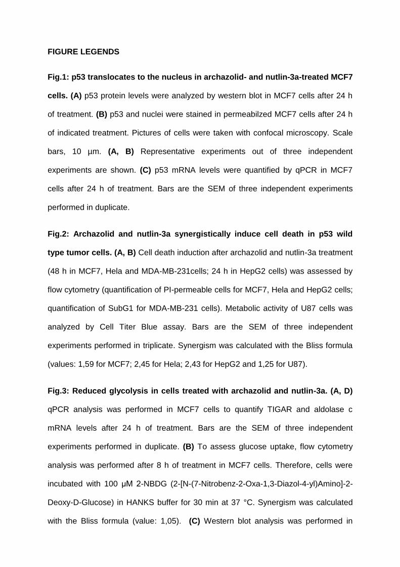
FIGURE LEGENDS
Fig.1: p53 translocates to the nucleus in archazolid- and nutlin-3a-treated MCF7
cells. (A) p53 protein levels were analyzed by western blot in MCF7 cells after 24 h
of treatment. (B) p53 and nuclei were stained in permeabilzed MCF7 cells after 24 h
of indicated treatment. Pictures of cells were taken with confocal microscopy. Scale
bars, 10 µm. (A, B) Representative experiments out of three independent
experiments are shown. (C) p53 mRNA levels were quantified by qPCR in MCF7
cells after 24 h of treatment. Bars are the SEM of three independent experiments
performed in duplicate.
Fig.2: Archazolid and nutlin-3a synergistically induce cell death in p53 wild
type tumor cells. (A, B) Cell death induction after archazolid and nutlin-3a treatment
(48 h in MCF7, Hela and MDA-MB-231cells; 24 h in HepG2 cells) was assessed by
flow cytometry (quantification of PI-permeable cells for MCF7, Hela and HepG2 cells;
quantification of SubG1 for MDA-MB-231 cells). Metabolic activity of U87 cells was
analyzed by Cell Titer Blue assay. Bars are the SEM of three independent
experiments performed in triplicate. Synergism was calculated with the Bliss formula
(values: 1,59 for MCF7; 2,45 for Hela; 2,43 for HepG2 and 1,25 for U87).
Fig.3: Reduced glycolysis in cells treated with archazolid and nutlin-3a. (A, D)
qPCR analysis was performed in MCF7 cells to quantify TIGAR and aldolase c
mRNA levels after 24 h of treatment. Bars are the SEM of three independent
experiments performed in duplicate. (B) To assess glucose uptake, flow cytometry
analysis was performed after 8 h of treatment in MCF7 cells. Therefore, cells were
incubated with 100 μM 2-NBDG (2-[N-(7-Nitrobenz-2-Oxa-1,3-Diazol-4-yl)Amino]-2-
Deoxy-D-Glucose) in HANKS buffer for 30 min at 37 °C. Synergism was calculated
with the Bliss formula (value: 1,05). (C) Western blot analysis was performed in
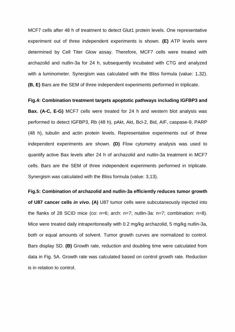
MCF7 cells after 48 h of treatment to detect Glut1 protein levels. One representative
experiment out of three independent experiments is shown. (E) ATP levels were
determined by Cell Titer Glow assay. Therefore, MCF7 cells were treated with
archazolid and nutlin-3a for 24 h, subsequently incubated with CTG and analyzed
with a luminometer. Synergism was calculated with the Bliss formula (value: 1,32).
(B, E) Bars are the SEM of three independent experiments performed in triplicate.
Fig.4: Combination treatment targets apoptotic pathways including IGFBP3 and
Bax. (A-C, E-G) MCF7 cells were treated for 24 h and western blot analysis was
performed to detect IGFBP3, Rb (48 h), pAkt, Akt, Bcl-2, Bid, AIF, caspase-9, PARP
(48 h), tubulin and actin protein levels. Representative experiments out of three
independent experiments are shown. (D) Flow cytometry analysis was used to
quantify active Bax levels after 24 h of archazolid and nutlin-3a treatment in MCF7
cells. Bars are the SEM of three independent experiments performed in triplicate.
Synergism was calculated with the Bliss formula (value: 3,13).
Fig.5: Combination of archazolid and nutlin-3a efficiently reduces tumor growth
of U87 cancer cells in vivo. (A) U87 tumor cells were subcutaneously injected into
the flanks of 28 SCID mice (co: n=6; arch: n=7; nutlin-3a: n=7; combination: n=8).
Mice were treated daily intraperitoneally with 0.2 mg/kg archazolid, 5 mg/kg nutlin-3a,
both or equal amounts of solvent. Tumor growth curves are normalized to control.
Bars display SD. (B) Growth rate, reduction and doubling time were calculated from
data in Fig. 5A. Growth rate was calculated based on control growth rate. Reduction
is in relation to control.
co arch [1 nM] arch [10 nM]
Hoechst p53
nutlin-3a [20 µM] arch [1 nM] nutlin-3a [20 µM]
arch 1 nM - + - + nutlin-3a 20 µM - - + +
p53
actin
rel p
53 m
RN
A le
vels
0.0
0.5
1.0
1.5B
A
C
Figure 1
arch 1 nM - + - + nutlin-3a 20 µM - - + +
53 kDa
43 kDa
spe
cific
ce
ll d
eat
h [%
]
0
5
10
15
20
25
arch 1 nM - + - + nutlin-3a 20 µM - - + +
spe
cific
ce
ll d
eat
h [%
]
0
5
10
15
20
25
arch 1 nM - + - + nutlin-3a 20 µM - - + +
MCF7
MDA-MB-231
spe
cific
ce
ll d
eat
h [%
]
0
5
10
15
20
25
arch 1 nM - - + + nutlin-3a 30 µM - + - +
Helasp
ecifi
c ce
ll de
ath
[%]
0
10
20
30
40
50
arch 1 nM - - + + nutlin-3a 5 µM - + - +
HepG2
rel m
eta
bo
ltic
activ
ity
0.0
0.5
1.0
U87
arch 0.5 nM - + - + nutlin-3a 10 µM - - + +
A
B
Figure 2
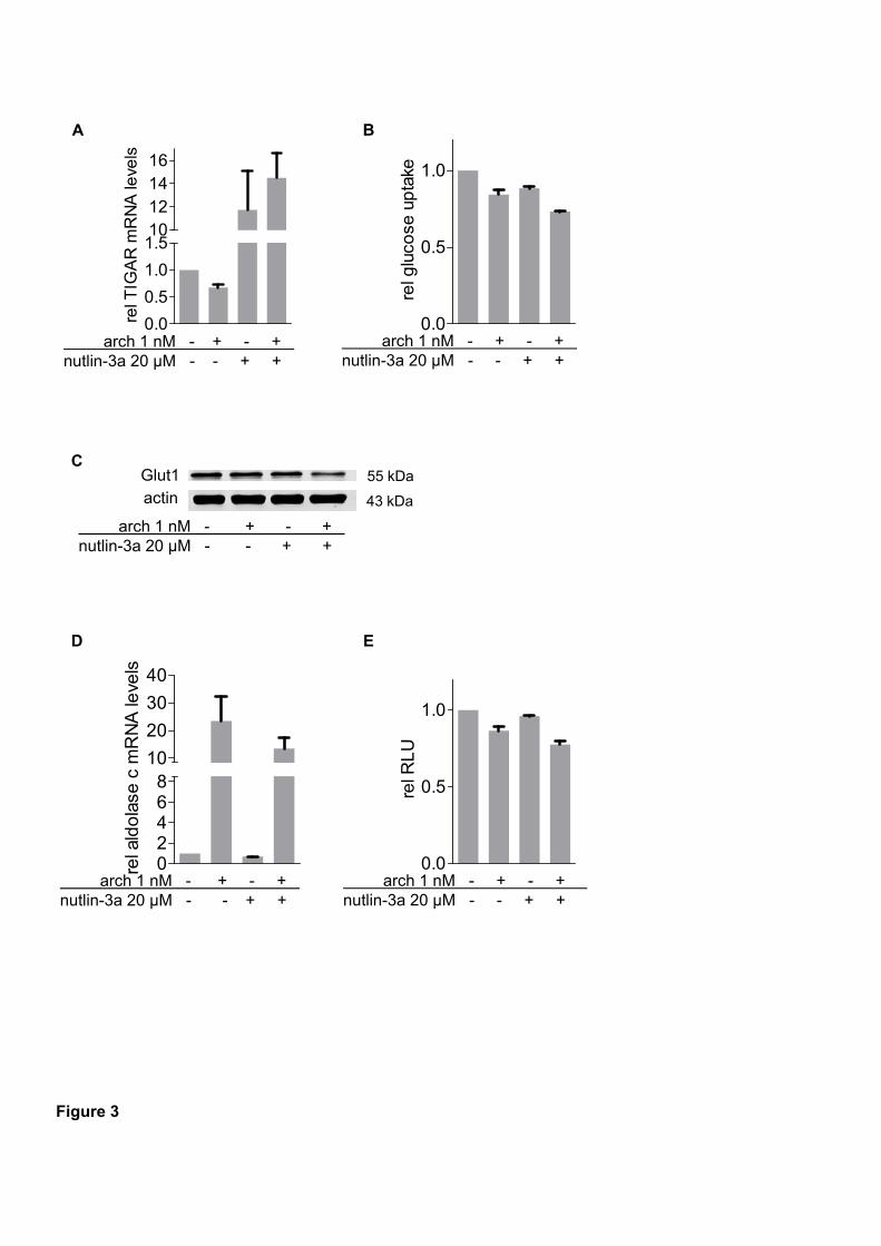
rel T
IGA
R m
RN
A le
vels
0.0
0.5
1.0
1.510121416
rel g
luco
se u
pta
ke
0.0
0.5
1.0
arch 1 nM - + - + nutlin-3a 20 µM - - + +
A Bre
l ald
ola
se c
mR
NA
leve
ls
02468
10
20
30
40
D
rel R
LU
0.0
0.5
1.0
E
arch 1 nM - + - + nutlin-3a 20 µM - - + +
arch 1 nM - + - + nutlin-3a 20 µM - - + +
arch 1 nM - + - + nutlin-3a 20 µM - - + +
Glut1
arch 1 nM - + - + nutlin-3a 20 µM - - + +
C
actin55 kDa
43 kDa
Figure 3
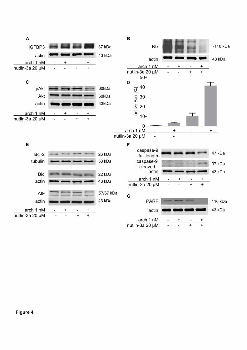
IGFBP3
actin
arch 1 nM - + - + nutlin-3a 20 µM - - + +
Akt
pAkt
activ
e B
ax [%
]
0
10
20
30
40
50
arch 1 nM - + - + nutlin-3a 20 µM - - + +
Rb
actin
A
C D
B
caspase-9 -full length-
actin
caspase-9 - cleaved-
E F
actin
PARP
actin
arch 1 nM - + - + nutlin-3a 20 µM - - + +
arch 1 nM - + - + nutlin-3a 20 µM - - + +
arch 1 nM - + - + nutlin-3a 20 µM - - + +
arch 1 nM - + - + nutlin-3a 20 µM - - + +
tubulin
Bcl-2
AIF
actin
arch 1 nM - + - + nutlin-3a 20 µM - - + +
Bid
actin
G
37 kDa
43 kDa
60kDa
43kDa
60kDa
~110 kDa
43 kDa
57/67 kDa
43 kDa
43 kDa
53 kDa
26 kDa
22 kDa
37 kDa
47 kDa
43 kDa
43 kDa
116 kDa
Figure 4
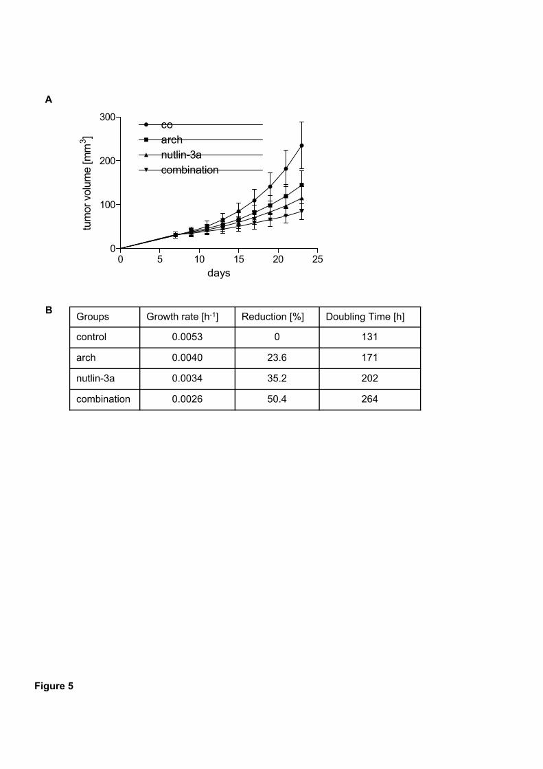
Groups Growth rate [h-1] Reduction [%] Doubling Time [h]
control 0.0053 0 131
arch 0.0040 23.6 171
nutlin-3a 0.0034 35.2 202
combination 0.0026 50.4 264
A
B
days
tum
or v
olum
e [m
m3]
0 5 10 15 20 250
100
200
300co
nutlin-3aarch
combination
Figure 5
Two-pore channel function is crucial for migration of invasive cancer cells
Lina S. Schneider1‡, Christian Grimm2‡, Yu-Kai Chao2, Anna Watermann1, Melanie
Ulrich1, Doris Mayr3, Christian Wahl-Schott2, Martin Biel2*, Angelika M. Vollmar1*
1Department of Pharmacy, Pharmaceutical Biology, Ludwig-Maximilians-University of
Munich, Butenandtstrasse 5-13, 81377 Munich, Germany
2Department of Pharmacy, Center for Drug Research and Center for Integrated
Protein Science Munich (CIPSM), Ludwig-Maximilians-University of Munich,
Butenandtstrasse 5-13, 81377 Munich, Germany
3Pathological Institute, Ludwig-Maximilians-University of Munich, Thalkirchner
Strasse 36, 80337 Munich, Germany
‡These authors contributed equally to this work
*Corresponding authors: Prof. Dr. Martin Biel, Phone: +49-89-2180-77328, Email:
[email protected]; Prof. Dr. Angelika M. Vollmar, Phone: +49-89-
2180-77172, Email: [email protected]
Running title: Role for TPCs in cancer cell migration
ABSTRACT
Metastasis is the main cause of cancer-related deaths. Hence, there is an urgent
need to find new therapies. In this study, we introduce a promising new target for the
treatment of invasive cancers. Inhibition of two-pore channels (TPCs), recently
identified NAADP- and PI(3,5)P2-sensitive Ca2+-permeable cation channels in the
endolysosomal system of cells, abrogated migration of metastatic cancer cells in
vitro. Remarkably, tetrandrine, lately discovered to inhibit TPCs, showed in vivo
efficacy reducing lung metastasis of mammary mouse cancer cells. As a molecular
explanation, we found that disrupting TPC function halted β1-integrin trafficking,
leading to accumulation in EEA1-positive vesicles. This is suggested to be due to
alterations in Ca2+ signaling since pH was not affected. As a consequence, invasive
cancer cells are no longer able to form leading edges, which is substantially required
for adequate migration. Our study links TPCs to fundamental processes in cancer cell
migration proposing that they are promising novel targets for the treatment of
metastatic cancers.
INTRODUCTION
Metastasis is the major cause of cancer-related deaths. The formation of secondary,
metastatic growth includes proliferation and extensive vascularization of the primary
tumor, detachment and invasion of tumor cells, circulation of single tumor cells, arrest
in distant sites, extravasation and finally proliferation within the organ (Fidler, 2003).
Extensive studies in the last decades contributed to a better understanding of these
processes. However, still about 90% of cancer-associated mortality is due to
metastasis. Therefore, new strategies to prevent metastasis are urgently needed
(Chaffer & Weinberg, 2011; Fidler, 2003).
To successfully colonize secondary sites, cancer cells gained the ability to
migrate (Chaffer & Weinberg, 2011; Hood & Cheresh, 2002). A crucial step in
migration is the binding to ligands of the extracellular matrix (ECM), which is
mediated by integrins. These cell surface receptors display a diverse family of
glycoproteins consisting of 18 α-subunits and 8 β-subunits. Besides mediating cell
attachment to the ECM, integrins are linked to the cytoskeleton through the formation
of clusters with actin-associated proteins such as vinculin. Furthermore, ligation of
integrins induces a network of intracellular signaling pathways including the activation
of focal adhesion kinase (FAK) and Src, altogether regulating cell migration (Hood &
Cheresh, 2002).
In order to fulfill the function as migration-promoting receptors, integrins have
to be trafficked to the front of the cell for assembly of focal adhesions. These
adhesions act as traction sites for the movement and are afterwards disassembled at
the rear of the cell, for detachment. Hence, integrins need to be dynamically turned
over, which is achieved by endocytic trafficking (Maritzen et al, 2015; Ridley, 2003).
After endocytosis, integrins are carried to early endosomes where sorting into
recycling and degradative pathways takes place. Under normal conditions, the larger
portion recycles back to the plasma membrane. Disturbing this process can alter the
composition of focal adhesions, thereby influencing cell migration (Maritzen et al,
2015).
There is increasing evidence that two-pore channels (TPCs) are key players in
the regulation of endocytic transport (Brailoiu et al, 2009; Calcraft et al, 2009; Zong et
al, 2009). It has been postulated that inhibiting these channels alters Ca2+ signaling
during endolysosomal fusion events resulting in defects in vesicle trafficking (Grimm
et al, 2014; Ruas et al, 2010; Ruas et al, 2014). TPCs are assigned to the
superfamily of voltage-gated ion channels (Calcraft et al, 2009). There are two
different TPC subtypes present in primates, TPC1 and TPC2. TPC1 is primarily
expressed on endosomal membranes while TPC2 dominates on lysosomal
membranes (Calcraft et al, 2009; Rietdorf et al, 2011; Ruas et al, 2010). Activation of
these channels is presumably triggered by the second messenger nicotinic acid
adenine dinucleotide phosphate (NAADP) and by phosphatidylinositol 3,5-
bisphosphate (PI(3,5)P2) (Calcraft et al, 2009; Jha et al, 2014; Schieder et al, 2010;
Wang et al, 2012). Distinct experiments suggested TPCs as Ca2+-permeable
channels indicating that TPCs might be involved in Ca2+ release from endo- and
lysosomal stores (Calcraft et al, 2009; Grimm et al, 2014; Ruas et al, 2015; Schieder
et al, 2010; Zong et al, 2009).
This work unveils that TPCs play a crucial role in the formation of metastasis
as silencing TPC1 and TPC2 reduced the adhesion and migration of invasive tumor
cells in vitro. Similar results were achieved with Ned-19, an antagonist of NAADP
(Naylor et al, 2009), and tetrandrine, recently shown to inhibit TPC1 and TPC2
(Sakurai et al, 2015). Remarkably, the latter showed efficacy in a metastatic in vivo
model. Inhibition of TPCs led to the accumulation of integrins in endocytic vesicles

and to impaired formation of leading edges indicating that TPCs are significantly
involved in integrin recycling.
RESULTS
TPCs in cancer cells
TPCs have been described in different cell types (Favia et al, 2014; Grimm et al,
2014; Sakurai et al, 2015), however not in cancer cells so far. TPC2 mRNA is
distinctly expressed in T24 (bladder cancer), Jurkat (leukemia) and HUH7 (HCC)
cells (Fig. 1A). mRNA levels of TPC1 were particularly high in T24 and HUH7 cells
(Fig. 1B). Tumor tissue sections of HCC patients showed a highly positive staining for
TPC2 (Fig. 1C). Together, these data suggest the presence of TPCs in cancer cells.
To verify TPC functionality in T24 cancer cells, we isolated lysosomes and performed
endolysosomal patch-clamp experiments. The applied whole-endolysosmal patch-
clamp method is illustrated in Fig. 1D. PI(3,5)P2-elicited currents were strongly
reduced after applying tetrandrine (Fig. 1E, F), recently shown to inhibit TPC1 and
TPC2 (Sakurai et al, 2015).
TPCs affect cancer cell migration and adhesion
Previous studies in TPC2-deficient MEF cells showed defects in the endolysosmal
degradation pathway (Grimm et al, 2014; Ruas et al, 2014). As alterations in this
pathway through inhibition of another endolysosome-associated protein, the vacuolar
ATPase (V-ATPase), have been described to reduce the ability of invasive cancer
cells to migrate (Wiedmann et al, 2012), we hypothesized that TPCs affect cancer
cell migration. Silencing TPC1 and TPC2 by siRNA in T24 cells resulted in
significantly reduced migration through pores in Boyden chamber assays (Fig. 2A).
Knockdown efficiency is shown in Fig. 2B. Moreover, these cells showed diminished
adhesion after 1 h of seeding (Fig. 2C). To get more information about the migratory
defect, movement of silenced cells exposed to a diffusive gradient of FCS (0 to 10%)
was analyzed. Figure 2 D shows control cells moving clearly towards the highest
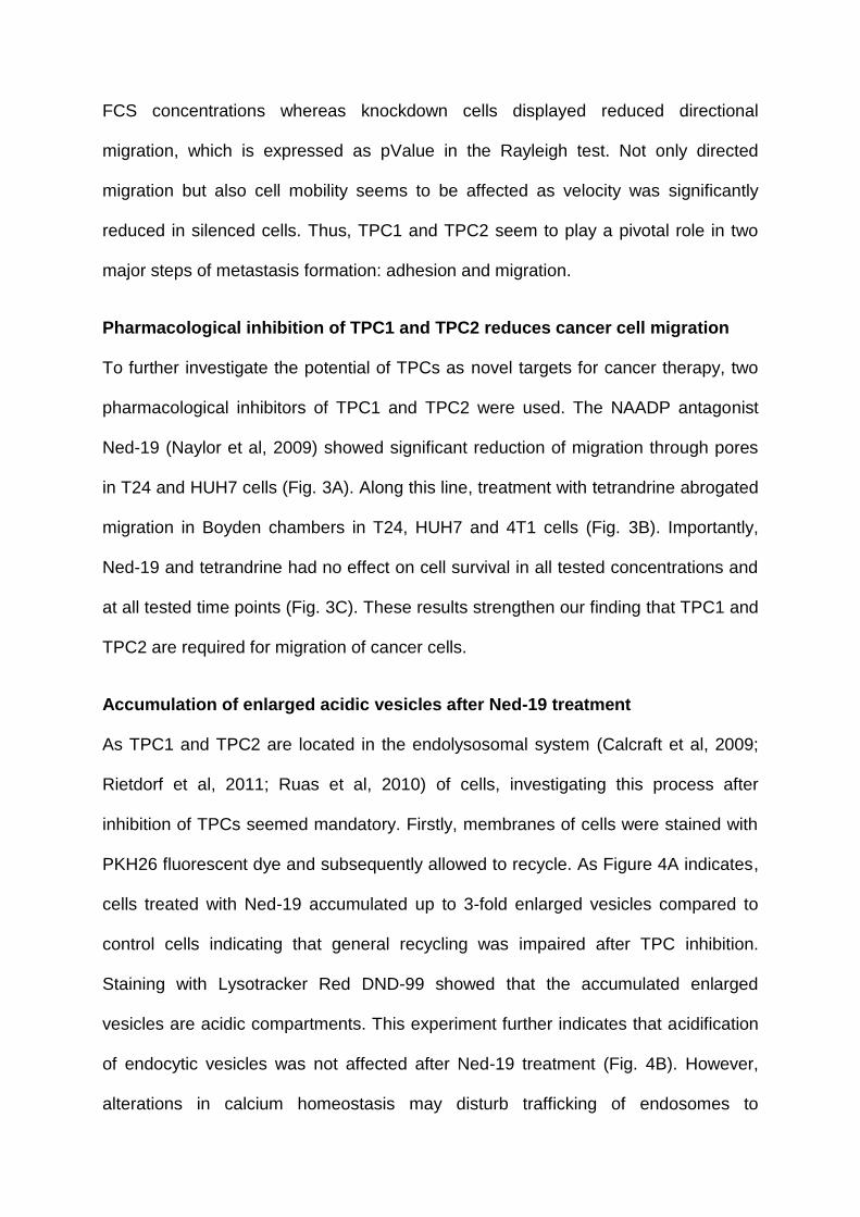
FCS concentrations whereas knockdown cells displayed reduced directional
migration, which is expressed as pValue in the Rayleigh test. Not only directed
migration but also cell mobility seems to be affected as velocity was significantly
reduced in silenced cells. Thus, TPC1 and TPC2 seem to play a pivotal role in two
major steps of metastasis formation: adhesion and migration.
Pharmacological inhibition of TPC1 and TPC2 reduces cancer cell migration
To further investigate the potential of TPCs as novel targets for cancer therapy, two
pharmacological inhibitors of TPC1 and TPC2 were used. The NAADP antagonist
Ned-19 (Naylor et al, 2009) showed significant reduction of migration through pores
in T24 and HUH7 cells (Fig. 3A). Along this line, treatment with tetrandrine abrogated
migration in Boyden chambers in T24, HUH7 and 4T1 cells (Fig. 3B). Importantly,
Ned-19 and tetrandrine had no effect on cell survival in all tested concentrations and
at all tested time points (Fig. 3C). These results strengthen our finding that TPC1 and
TPC2 are required for migration of cancer cells.
Accumulation of enlarged acidic vesicles after Ned-19 treatment
As TPC1 and TPC2 are located in the endolysosomal system (Calcraft et al, 2009;
Rietdorf et al, 2011; Ruas et al, 2010) of cells, investigating this process after
inhibition of TPCs seemed mandatory. Firstly, membranes of cells were stained with
PKH26 fluorescent dye and subsequently allowed to recycle. As Figure 4A indicates,
cells treated with Ned-19 accumulated up to 3-fold enlarged vesicles compared to
control cells indicating that general recycling was impaired after TPC inhibition.
Staining with Lysotracker Red DND-99 showed that the accumulated enlarged
vesicles are acidic compartments. This experiment further indicates that acidification
of endocytic vesicles was not affected after Ned-19 treatment (Fig. 4B). However,
alterations in calcium homeostasis may disturb trafficking of endosomes to
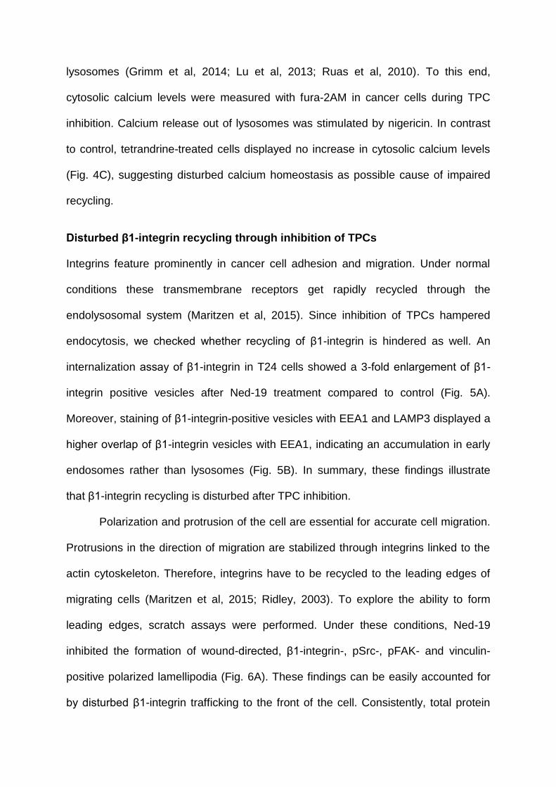
lysosomes (Grimm et al, 2014; Lu et al, 2013; Ruas et al, 2010). To this end,
cytosolic calcium levels were measured with fura-2AM in cancer cells during TPC
inhibition. Calcium release out of lysosomes was stimulated by nigericin. In contrast
to control, tetrandrine-treated cells displayed no increase in cytosolic calcium levels
(Fig. 4C), suggesting disturbed calcium homeostasis as possible cause of impaired
recycling.
Disturbed β1-integrin recycling through inhibition of TPCs
Integrins feature prominently in cancer cell adhesion and migration. Under normal
conditions these transmembrane receptors get rapidly recycled through the
endolysosomal system (Maritzen et al, 2015). Since inhibition of TPCs hampered
endocytosis, we checked whether recycling of β1-integrin is hindered as well. An
internalization assay of β1-integrin in T24 cells showed a 3-fold enlargement of β1-
integrin positive vesicles after Ned-19 treatment compared to control (Fig. 5A).
Moreover, staining of β1-integrin-positive vesicles with EEA1 and LAMP3 displayed a
higher overlap of β1-integrin vesicles with EEA1, indicating an accumulation in early
endosomes rather than lysosomes (Fig. 5B). In summary, these findings illustrate
that β1-integrin recycling is disturbed after TPC inhibition.
Polarization and protrusion of the cell are essential for accurate cell migration.
Protrusions in the direction of migration are stabilized through integrins linked to the
actin cytoskeleton. Therefore, integrins have to be recycled to the leading edges of
migrating cells (Maritzen et al, 2015; Ridley, 2003). To explore the ability to form
leading edges, scratch assays were performed. Under these conditions, Ned-19
inhibited the formation of wound-directed, β1-integrin-, pSrc-, pFAK- and vinculin-
positive polarized lamellipodia (Fig. 6A). These findings can be easily accounted for
by disturbed β1-integrin trafficking to the front of the cell. Consistently, total protein
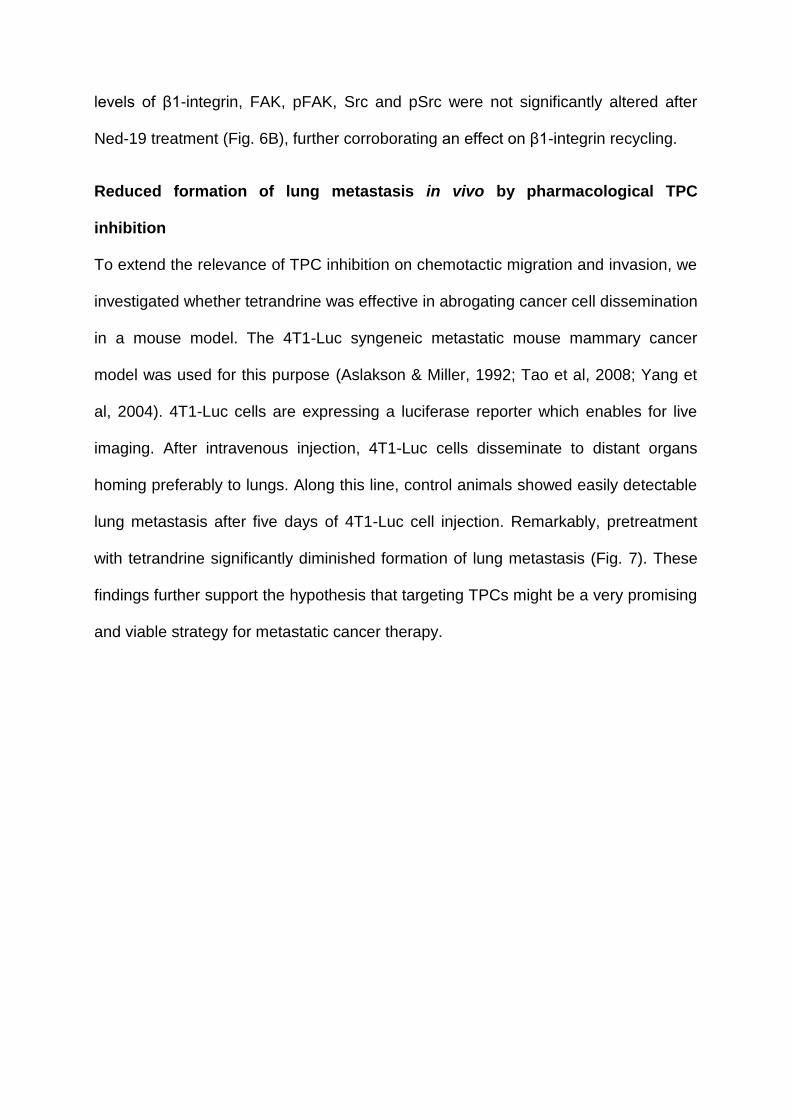
levels of β1-integrin, FAK, pFAK, Src and pSrc were not significantly altered after
Ned-19 treatment (Fig. 6B), further corroborating an effect on β1-integrin recycling.
Reduced formation of lung metastasis in vivo by pharmacological TPC
inhibition
To extend the relevance of TPC inhibition on chemotactic migration and invasion, we
investigated whether tetrandrine was effective in abrogating cancer cell dissemination
in a mouse model. The 4T1-Luc syngeneic metastatic mouse mammary cancer
model was used for this purpose (Aslakson & Miller, 1992; Tao et al, 2008; Yang et
al, 2004). 4T1-Luc cells are expressing a luciferase reporter which enables for live
imaging. After intravenous injection, 4T1-Luc cells disseminate to distant organs
homing preferably to lungs. Along this line, control animals showed easily detectable
lung metastasis after five days of 4T1-Luc cell injection. Remarkably, pretreatment
with tetrandrine significantly diminished formation of lung metastasis (Fig. 7). These
findings further support the hypothesis that targeting TPCs might be a very promising
and viable strategy for metastatic cancer therapy.
DISCUSSION
This study designates TPCs as promising targets for the treatment of invasive
cancers. Disruption of TPC function abrogates cancer cell migration in vitro and in
vivo, resulting from disturbed integrin trafficking in the endolysosomal system.
TPCs are Ca2+-permeable voltage-gated channels located in the membrane of
endosomes and lysosomes (Calcraft et al, 2009; Grimm et al, 2014; Rietdorf et al,
2011; Ruas et al, 2010; Ruas et al, 2015; Schieder et al, 2010; Zong et al, 2009). In
recent years, evidence has accumulated that TPCs are substantially implicated in the
regulation of endolysosomal trafficking. Thus, a lack of TPC2 in mouse embryonic
fibroblasts resulted in an accumulation of LDL and EGF/EGFR in intracellular
vesicles (Grimm et al, 2014) and led to delayed PDGFRβ degradation, which
requires trafficking to lysosomes (Ruas et al, 2014). Overexpression of TPCs
retained cholera toxin within endolysosomes, which is normally delivered to the Golgi,
and enlarged lysosomes dramatically (Ruas et al, 2010). Disrupting TPC function
also reduced Ebola virus trafficking through endosomal vesicles, preventing infection
(Sakurai et al, 2015). Consistently, we observed an accumulation of integrins in early
endosomes after TPC inhibition in invasive cancer cells. Alterations in Ca2+ signaling
are widely accepted to lead to impaired trafficking and fusion of endocytic vesicles
(Grimm et al, 2014; Ruas et al, 2010). This hypothesis is further supported by the
finding that Ca2+ chelators, BAPTA and EGTA-AM, are able to inhibit fusion of late
endosomes and lysosomes (Pryor et al, 2000). In this context, it is interesting to note
that another Ca2+-permeable endolysosomal channel, TRPML1, has been proposed
to be required for fusion between late endosomes and lysosomes in Drosophila
(Wong et al, 2012). In humans, loss or mutation of this channel leads to the
lysosomal storage disorder mucolipidosis type IV (Bargal et al, 2000; Chen et al,
2014; Slaugenhaupt et al, 1999), further supporting the importance of Ca2+ signaling.
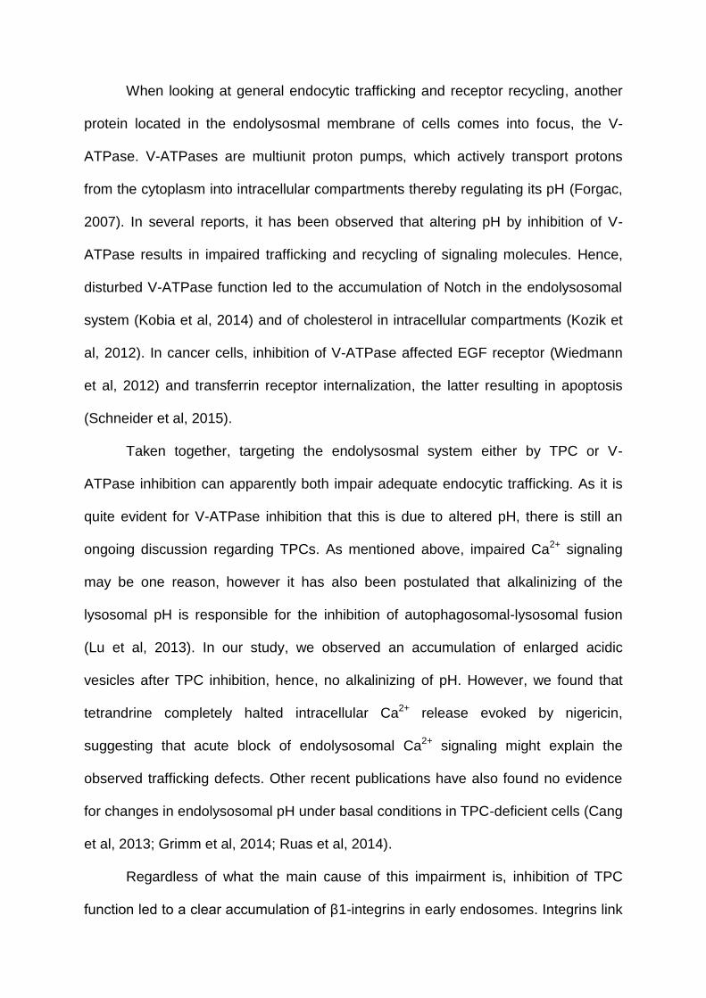
When looking at general endocytic trafficking and receptor recycling, another
protein located in the endolysosmal membrane of cells comes into focus, the V-
ATPase. V-ATPases are multiunit proton pumps, which actively transport protons
from the cytoplasm into intracellular compartments thereby regulating its pH (Forgac,
2007). In several reports, it has been observed that altering pH by inhibition of V-
ATPase results in impaired trafficking and recycling of signaling molecules. Hence,
disturbed V-ATPase function led to the accumulation of Notch in the endolysosomal
system (Kobia et al, 2014) and of cholesterol in intracellular compartments (Kozik et
al, 2012). In cancer cells, inhibition of V-ATPase affected EGF receptor (Wiedmann
et al, 2012) and transferrin receptor internalization, the latter resulting in apoptosis
(Schneider et al, 2015).
Taken together, targeting the endolysosmal system either by TPC or V-
ATPase inhibition can apparently both impair adequate endocytic trafficking. As it is
quite evident for V-ATPase inhibition that this is due to altered pH, there is still an
ongoing discussion regarding TPCs. As mentioned above, impaired Ca2+ signaling
may be one reason, however it has also been postulated that alkalinizing of the
lysosomal pH is responsible for the inhibition of autophagosomal-lysosomal fusion
(Lu et al, 2013). In our study, we observed an accumulation of enlarged acidic
vesicles after TPC inhibition, hence, no alkalinizing of pH. However, we found that
tetrandrine completely halted intracellular Ca2+ release evoked by nigericin,
suggesting that acute block of endolysosomal Ca2+ signaling might explain the
observed trafficking defects. Other recent publications have also found no evidence
for changes in endolysosomal pH under basal conditions in TPC-deficient cells (Cang
et al, 2013; Grimm et al, 2014; Ruas et al, 2014).
Regardless of what the main cause of this impairment is, inhibition of TPC
function led to a clear accumulation of β1-integrins in early endosomes. Integrins link
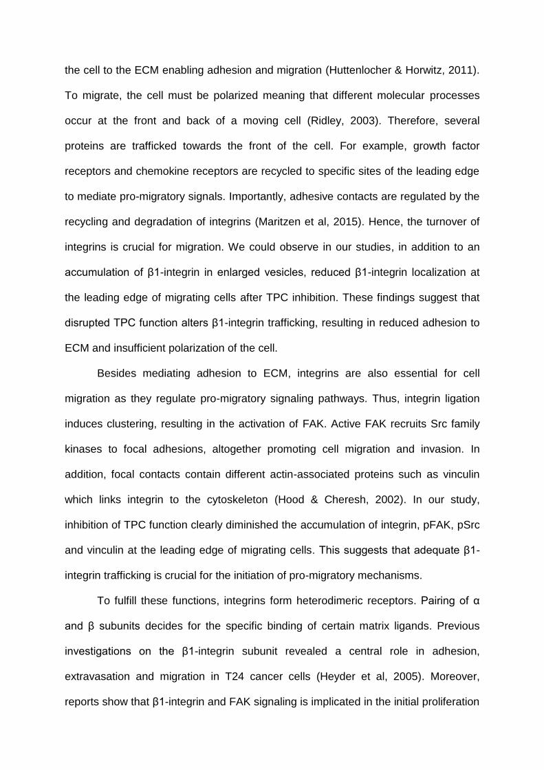
the cell to the ECM enabling adhesion and migration (Huttenlocher & Horwitz, 2011).
To migrate, the cell must be polarized meaning that different molecular processes
occur at the front and back of a moving cell (Ridley, 2003). Therefore, several
proteins are trafficked towards the front of the cell. For example, growth factor
receptors and chemokine receptors are recycled to specific sites of the leading edge
to mediate pro-migratory signals. Importantly, adhesive contacts are regulated by the
recycling and degradation of integrins (Maritzen et al, 2015). Hence, the turnover of
integrins is crucial for migration. We could observe in our studies, in addition to an
accumulation of β1-integrin in enlarged vesicles, reduced β1-integrin localization at
the leading edge of migrating cells after TPC inhibition. These findings suggest that
disrupted TPC function alters β1-integrin trafficking, resulting in reduced adhesion to
ECM and insufficient polarization of the cell.
Besides mediating adhesion to ECM, integrins are also essential for cell
migration as they regulate pro-migratory signaling pathways. Thus, integrin ligation
induces clustering, resulting in the activation of FAK. Active FAK recruits Src family
kinases to focal adhesions, altogether promoting cell migration and invasion. In
addition, focal contacts contain different actin-associated proteins such as vinculin
which links integrin to the cytoskeleton (Hood & Cheresh, 2002). In our study,
inhibition of TPC function clearly diminished the accumulation of integrin, pFAK, pSrc
and vinculin at the leading edge of migrating cells. This suggests that adequate β1-
integrin trafficking is crucial for the initiation of pro-migratory mechanisms.
To fulfill these functions, integrins form heterodimeric receptors. Pairing of α
and β subunits decides for the specific binding of certain matrix ligands. Previous
investigations on the β1-integrin subunit revealed a central role in adhesion,
extravasation and migration in T24 cancer cells (Heyder et al, 2005). Moreover,
reports show that β1-integrin and FAK signaling is implicated in the initial proliferation
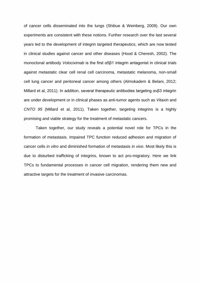
of cancer cells disseminated into the lungs (Shibue & Weinberg, 2009). Our own
experiments are consistent with these notions. Further research over the last several
years led to the development of integrin targeted therapeutics, which are now tested
in clinical studies against cancer and other diseases (Hood & Cheresh, 2002). The
monoclonal antibody Volociximab is the first α5β1 integrin antagonist in clinical trials
against metastatic clear cell renal cell carcinoma, metastatic melanoma, non-small
cell lung cancer and peritoneal cancer among others (Almokadem & Belani, 2012;
Millard et al, 2011). In addition, several therapeutic antibodies targeting αvβ3 integrin
are under development or in clinical phases as anti-tumor agents such as Vitaxin and
CNTO 95 (Millard et al, 2011). Taken together, targeting integrins is a highly
promising and viable strategy for the treatment of metastatic cancers.
Taken together, our study reveals a potential novel role for TPCs in the
formation of metastasis. Impaired TPC function reduced adhesion and migration of
cancer cells in vitro and diminished formation of metastasis in vivo. Most likely this is
due to disturbed trafficking of integrins, known to act pro-migratory. Here we link
TPCs to fundamental processes in cancer cell migration, rendering them new and
attractive targets for the treatment of invasive carcinomas.
MATERIAL AND METHODS
Cell lines and Reagents
The cancer cell line T24 was obtained from Dr. B. Mayer (Surgical Clinic, LMU,
Munich, Germany) and authenticated by the DSMZ, HUH7 were purchased from
Japanese Collection of Research Bioresources and 4T1-Luc (4T1) from PerkinElmer.
T24 cells were grown in McCoy’s, HUH7 cells in DMEM High Glucose and 4T1 cells
in RPMI-1640 medium. All media were supplemented with 10% FCS. trans-Ned-19
(Ned-19) was purchased from Tocris® Bioscience, tetrandrine from Santa Cruz
Biotechnology.
qRT-PCR
qRT-PCR of TPC1 and TPC2 was performed as described previously (Schneider et
al, 2015). Following primers were used: TPC2 fw 5’-GTA CCC CTC TTG TGT GGA
CG-3’, rv 5’-GGC CCT GAC AGT GAC AAC TT-3’; TPC1 fw 5’-GGA GCC CTT CTA
TTT CAT CGT-3’, rv 5’-CGG TAG CGC TCC TTC AAC T-3’;
Whole-endolysosomal patch clamp
Whole-endolysosomal patch-clamp recordings were performed by modified
conventional patch-clamp. T24 cells were treated with 1 μM vacuolin-1 at least 4 h.
Currents were recorded using an EPC-10 patch-clamp amplifier and PatchMaster
acquisition software (HEKA, Lambrecht/Pfalz, Germany). Data were digitized at 40
kHz and filtered at 2.8 kHz. Recording pipettes had a resistance of 8-10 MΩ. Liquid
junction potential was corrected. Pipette solution (corresponding to luminal
endolysosomal solution) contained 140 mM NaMSA, 5 mM KMSA, 2 mM CaMSA, 1
mM CaCl2, 10 mM HEPES and 10 mM MES, pH 4.6. Bath solution (corresponding to
cytosolic solution) contained 140 mM KMSA, 5 mM KOH, 4 mM NaCl, 0.39 mM
CaCl2, 1 mM EGTA, and 10 mM HEPES, pH 7.2. PI(3,5)P2 was used in a water-
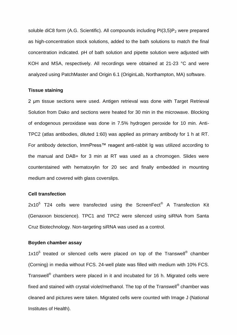
soluble diC8 form (A.G. Scientific). All compounds including PI(3,5)P2 were prepared
as high-concentration stock solutions, added to the bath solutions to match the final
concentration indicated. pH of bath solution and pipette solution were adjusted with
KOH and MSA, respectively. All recordings were obtained at 21-23 °C and were
analyzed using PatchMaster and Origin 6.1 (OriginLab, Northampton, MA) software.
Tissue staining
2 μm tissue sections were used. Antigen retrieval was done with Target Retrieval
Solution from Dako and sections were heated for 30 min in the microwave. Blocking
of endogenous peroxidase was done in 7.5% hydrogen peroxide for 10 min. Anti-
TPC2 (atlas antibodies, diluted 1:60) was applied as primary antibody for 1 h at RT.
For antibody detection, ImmPress™ reagent anti-rabbit Ig was utilized according to
the manual and DAB+ for 3 min at RT was used as a chromogen. Slides were
counterstained with hematoxylin for 20 sec and finally embedded in mounting
medium and covered with glass coverslips.
Cell transfection
2x10⁵ T24 cells were transfected using the ScreenFect® A Transfection Kit
(Genaxxon bioscience). TPC1 and TPC2 were silenced using siRNA from Santa
Cruz Biotechnology. Non-targeting siRNA was used as a control.
Boyden chamber assay
1x10⁵ treated or silenced cells were placed on top of the Transwell® chamber
(Corning) in media without FCS. 24-well plate was filled with medium with 10% FCS.
Transwell® chambers were placed in it and incubated for 16 h. Migrated cells were
fixed and stained with crystal violet/methanol. The top of the Transwell® chamber was
cleaned and pictures were taken. Migrated cells were counted with Image J (National
Institutes of Health).
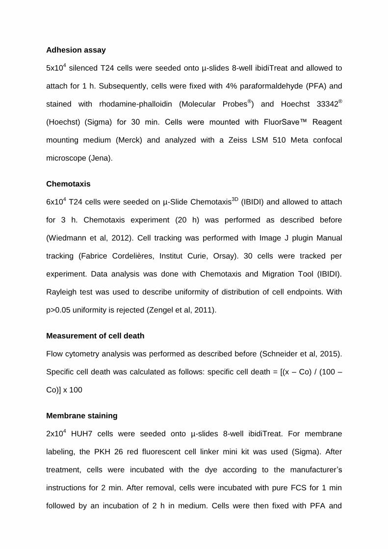
Adhesion assay
5x10⁴ silenced T24 cells were seeded onto µ-slides 8-well ibidiTreat and allowed to
attach for 1 h. Subsequently, cells were fixed with 4% paraformaldehyde (PFA) and
stained with rhodamine-phalloidin (Molecular Probes®) and Hoechst 33342®
(Hoechst) (Sigma) for 30 min. Cells were mounted with FluorSave™ Reagent
mounting medium (Merck) and analyzed with a Zeiss LSM 510 Meta confocal
microscope (Jena).
Chemotaxis
6x10⁴ T24 cells were seeded on µ-Slide Chemotaxis3D (IBIDI) and allowed to attach
for 3 h. Chemotaxis experiment (20 h) was performed as described before
(Wiedmann et al, 2012). Cell tracking was performed with Image J plugin Manual
tracking (Fabrice Cordelières, Institut Curie, Orsay). 30 cells were tracked per
experiment. Data analysis was done with Chemotaxis and Migration Tool (IBIDI).
Rayleigh test was used to describe uniformity of distribution of cell endpoints. With
p>0.05 uniformity is rejected (Zengel et al, 2011).
Measurement of cell death
Flow cytometry analysis was performed as described before (Schneider et al, 2015).
Specific cell death was calculated as follows: specific cell death = [(x – Co) / (100 –
Co)] x 100
Membrane staining
2x10⁴ HUH7 cells were seeded onto µ-slides 8-well ibidiTreat. For membrane
labeling, the PKH 26 red fluorescent cell linker mini kit was used (Sigma). After
treatment, cells were incubated with the dye according to the manufacturer’s
instructions for 2 min. After removal, cells were incubated with pure FCS for 1 min
followed by an incubation of 2 h in medium. Cells were then fixed with PFA and
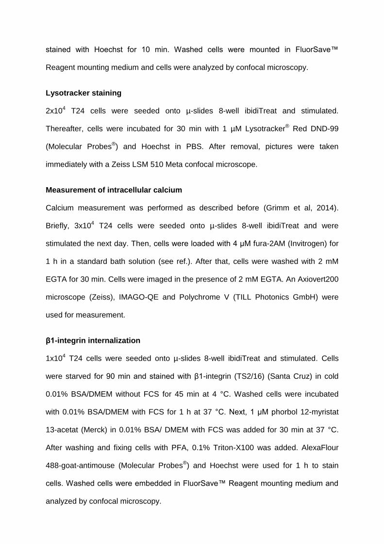
stained with Hoechst for 10 min. Washed cells were mounted in FluorSave™
Reagent mounting medium and cells were analyzed by confocal microscopy.
Lysotracker staining
2x10⁴ T24 cells were seeded onto µ-slides 8-well ibidiTreat and stimulated.
Thereafter, cells were incubated for 30 min with 1 µM Lysotracker® Red DND-99
(Molecular Probes®) and Hoechst in PBS. After removal, pictures were taken
immediately with a Zeiss LSM 510 Meta confocal microscope.
Measurement of intracellular calcium
Calcium measurement was performed as described before (Grimm et al, 2014).
Briefly, 3x10⁴ T24 cells were seeded onto µ-slides 8-well ibidiTreat and were
stimulated the next day. Then, cells were loaded with 4 μM fura-2AM (Invitrogen) for
1 h in a standard bath solution (see ref.). After that, cells were washed with 2 mM
EGTA for 30 min. Cells were imaged in the presence of 2 mM EGTA. An Axiovert200
microscope (Zeiss), IMAGO-QE and Polychrome V (TILL Photonics GmbH) were
used for measurement.
β1-integrin internalization
1x10⁴ T24 cells were seeded onto µ-slides 8-well ibidiTreat and stimulated. Cells
were starved for 90 min and stained with β1-integrin (TS2/16) (Santa Cruz) in cold
0.01% BSA/DMEM without FCS for 45 min at 4 °C. Washed cells were incubated
with 0.01% BSA/DMEM with FCS for 1 h at 37 °C. Next, 1 μM phorbol 12-myristat
13-acetat (Merck) in 0.01% BSA/ DMEM with FCS was added for 30 min at 37 °C.
After washing and fixing cells with PFA, 0.1% Triton-X100 was added. AlexaFlour
488-goat-antimouse (Molecular Probes®) and Hoechst were used for 1 h to stain
cells. Washed cells were embedded in FluorSave™ Reagent mounting medium and
analyzed by confocal microscopy.
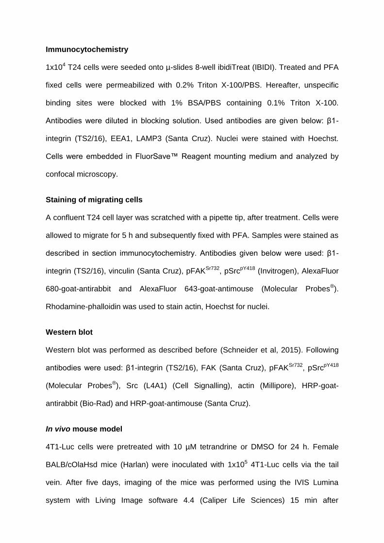
Immunocytochemistry
1x10⁴ T24 cells were seeded onto µ-slides 8-well ibidiTreat (IBIDI). Treated and PFA
fixed cells were permeabilized with 0.2% Triton X-100/PBS. Hereafter, unspecific
binding sites were blocked with 1% BSA/PBS containing 0.1% Triton X-100.
Antibodies were diluted in blocking solution. Used antibodies are given below: β1-
integrin (TS2/16), EEA1, LAMP3 (Santa Cruz). Nuclei were stained with Hoechst.
Cells were embedded in FluorSave™ Reagent mounting medium and analyzed by
confocal microscopy.
Staining of migrating cells
A confluent T24 cell layer was scratched with a pipette tip, after treatment. Cells were
allowed to migrate for 5 h and subsequently fixed with PFA. Samples were stained as
described in section immunocytochemistry. Antibodies given below were used: β1-
integrin (TS2/16), vinculin (Santa Cruz), pFAKSr732, pSrcpY418 (Invitrogen), AlexaFluor
680-goat-antirabbit and AlexaFluor 643-goat-antimouse (Molecular Probes®).
Rhodamine-phalloidin was used to stain actin, Hoechst for nuclei.
Western blot
Western blot was performed as described before (Schneider et al, 2015). Following
antibodies were used: β1-integrin (TS2/16), FAK (Santa Cruz), pFAKSr732, pSrcpY418
(Molecular Probes®), Src (L4A1) (Cell Signalling), actin (Millipore), HRP-goat-
antirabbit (Bio-Rad) and HRP-goat-antimouse (Santa Cruz).
In vivo mouse model
4T1-Luc cells were pretreated with 10 µM tetrandrine or DMSO for 24 h. Female
BALB/cOlaHsd mice (Harlan) were inoculated with 1x10⁵ 4T1-Luc cells via the tail
vein. After five days, imaging of the mice was performed using the IVIS Lumina
system with Living Image software 4.4 (Caliper Life Sciences) 15 min after
intraperitoneal injection of 6 mg Na-luciferin (PerkinElmer). The total signal per
defined region of interest was calculated as photons/second/cm2 (total flux/area).
Animal experiments were approved by the District Government of Upper Bavaria in
accordance with the German animal welfare and institutional guidelines.
Statistical analysis
GraphPad Prism Software was used for all statistical analysis. In some experiments
data were normalized to control or as indicated.

ACKNOWLEDGEMENTS
This work was supported, in part, by funding of the German Research Foundation
(SFB/TRR152 TP04 to C.G., TP06 to C.W.-S., and TP12 to M.B., FOR1406 to AMV).
CONFLICT OF INTEREST
The authors declare no conflict of interest.
THE PAPER EXPLAINED
PROBLEM: Cancer is a leading cause of death worldwide. Most cancer-related
deaths are due to metastasis. To colonize distant sites, cancer cells are highly
dependent on their ability to migrate. Disrupting this process is thought to be a
promising approach for new anti-cancer drugs.
RESULTS: Here we show that function of recently identified Ca2+-permeable
endolysosomal two pore channels (TPCs) is crucial for cancer cell motility.
Pharmacological inhibition of these channels abrogated migration of invasive cancer
cells in vitro and in vivo. Moreover, we provide evidence for the underlying molecular
mechanisms as integrin recycling was disturbed.
IMPACT: Our study suggests TPCs as promising new targets for the treatment of
metastatic cancers.
REFERENCES
Almokadem S, Belani CP (2012) Volociximab in cancer. Expert Opin Biol Ther 12:
251–257
Aslakson CJ, Miller FR (1992) Selective events in the metastatic process defined by
analysis of the sequential dissemination of subpopulations of a mouse mammary
tumor. Cancer Res 52: 1399–1405
Bargal R, Avidan N, Ben-Asher E, Olender Z, Zeigler M, Frumkin A, Raas-Rothschild
A, Glusman G, Lancet D, Bach G (2000) Identification of the gene causing
mucolipidosis type IV. Nat Genet 26: 118–123
Brailoiu E, Churamani D, Cai X, Schrlau MG, Brailoiu GC, Gao X, Hooper R,
Boulware MJ, Dun NJ, Marchant JS et al (2009) Essential requirement for two-pore
channel 1 in NAADP-mediated calcium signaling. J Cell Biol 186: 201–209
Calcraft PJ, Ruas M, Pan Z, Cheng X, Arredouani A, Hao X, Tang J, Rietdorf K,
Teboul L, Chuang K-T et al (2009) NAADP mobilizes calcium from acidic organelles
through two-pore channels. Nature 459: 596–600
Cang C, Zhou Y, Navarro B, Seo Y-j, Aranda K, Shi L, Battaglia-Hsu S, Nissim I,
Clapham David E, Ren D (2013) mTOR Regulates Lysosomal ATP-Sensitive Two-
Pore Na+ Channels to Adapt to Metabolic State. Cell 152: 778–790
Chaffer CL, Weinberg RA (2011) A Perspective on Cancer Cell Metastasis. Science
331: 1559–1564
Chen C-C, Keller M, Hess M, Schiffmann R, Urban N, Wolfgardt A, Schaefer M,
Bracher F, Biel M, Wahl-Schott C et al (2014) A small molecule restores function to
TRPML1 mutant isoforms responsible for mucolipidosis type IV. Nat Commun 5:
4681
Favia A, Desideri M, Gambara G, D'Alessio A, Ruas M, Esposito B, Del Bufalo D,
Parrington J, Ziparo E, Palombi F et al (2014) VEGF-induced neoangiogenesis is
mediated by NAADP and two-pore channel-2-dependent Ca2+ signaling. Proc Natl
Acad Sci USA 111: E4706–E4715
Fidler IJ (2003) Timeline: The pathogenesis of cancer metastasis: the 'seed and soil'
hypothesis revisited. Nat Rev Cancer 3: 453–458
Forgac M (2007) Vacuolar ATPases: rotary proton pumps in physiology and
pathophysiology. Nat Rev Mol Cell Biol 8: 917–929
Grimm C, Holdt LM, Chen C-C, Hassan S, Müller C, Jörs S, Cuny H, Kissing S,
Schröder B, Butz E et al (2014) High susceptibility to fatty liver disease in two-pore
channel 2-deficient mice. Nat Commun 5: 4699
Heyder C, Gloria-Maercker E, Hatzmann W, Niggemann B, Zänker KS, Dittmar T
(2005) Role of the β1-integrin subunit in the adhesion, extravasation and migration of
T24 human bladder carcinoma cells. Clin Exp Metastasis 22: 99–106
Hood JD, Cheresh DA (2002) Role of Integrins in Cell Invasion and Migration. Nat
Rev Cancer 2: 91–100
Huttenlocher A, Horwitz AR (2011) Integrins in Cell Migration. Cold Spring Harb
Perspect Biol 3: a005074–a005074
Jha A, Ahuja M, Patel S, Brailoiu E, Muallem S (2014) Convergent regulation of the
lysosomal two-pore channel-2 by Mg2+, NAADP, PI(3,5)P2 and multiple protein
kinases. EMBO J 33: 501–511
Kobia F, Duchi S, Deflorian G, Vaccari T (2014) Pharmacologic inhibition of vacuolar
H+ ATPase reduces physiologic and oncogenic Notch signaling. Mol Oncol 8: 207–
220
Kozik P, Hodson NA, Sahlender DA, Simecek N, Soromani C, Wu J, Collinson LM,
Robinson MS (2012) A human genome-wide screen for regulators of clathrin-coated
vesicle formation reveals an unexpected role for the V-ATPase. Nat Cell Biol 15: 50–
60
Lu Y, Hao BX, Graeff R, Wong CWM, Wu WT, Yue J (2013) Two Pore Channel 2
(TPC2) Inhibits Autophagosomal-Lysosomal Fusion by Alkalinizing Lysosomal pH. J
Biol Chem 288: 24247–24263
Maritzen T, Schachtner H, Legler DF (2015) On the move: endocytic trafficking in cell
migration. Cell Mol Life Sci 72: 2119–2134
Millard M, Odde S, Neamati N (2011) Integrin targeted therapeutics. Theranostics 1:
154–188
Naylor E, Arredouani A, Vasudevan SR, Lewis AM, Parkesh R, Mizote A, Rosen D,
Thomas JM, Izumi M, Ganesan A et al (2009) Identification of a chemical probe for
NAADP by virtual screening. Nat Chem Biol 5: 220–226
Pryor PR, Mullock BM, Bright NA, Gray SR, Luzio JP (2000) The role of
intraorganellar Ca(2+) in late endosome-lysosome heterotypic fusion and in the
reformation of lysosomes from hybrid organelles. J Cell Biol 149: 1053–1062
Ridley AJ (2003) Cell Migration: Integrating Signals from Front to Back. Science 302:
1704–1709
Rietdorf K, Funnell TM, Ruas M, Heinemann J, Parrington J, Galione A (2011) Two-
pore Channels Form Homo- and Heterodimers. J Biol Chem 286: 37058–37062
Ruas M, Rietdorf K, Arredouani A, Davis LC, Lloyd-Evans E, Koegel H, Funnell TM,
Morgan AJ, Ward JA, Watanabe K et al (2010) Purified TPC Isoforms Form NAADP
Receptors with Distinct Roles for Ca2+ Signaling and Endolysosomal Trafficking.
Curr Biol 20: 703–709
Ruas M, Chuang KT, Davis LC, Al-Douri A, Tynan PW, Tunn R, Teboul L, Galione A,
Parrington J (2014) TPC1 Has Two Variant Isoforms, and Their Removal Has
Different Effects on Endo-Lysosomal Functions Compared to Loss of TPC2. Mol Cell
Biol 34: 3981–3992
Ruas M, Davis LC, Chen CC, Morgan AJ, Chuang KT, Walseth TF, Grimm C,
Garnham C, Powell T, Platt N et al (2015) Expression of Ca2+-permeable two-pore
channels rescues NAADP signalling in TPC-deficient cells. EMBO J 34: 1743–1758
Sakurai Y, Kolokoltsov AA, Chen CC, Tidwell MW, Bauta WE, Klugbauer N, Grimm
C, Wahl-Schott C, Biel M, Davey RA (2015) Two-pore channels control Ebola virus
host cell entry and are drug targets for disease treatment. Science 347: 995–998
Schieder M, Rotzer K, Bruggemann A, Biel M, Wahl-Schott CA (2010)
Characterization of Two-pore Channel 2 (TPCN2)-mediated Ca2+ Currents in
Isolated Lysosomes. J Biol Chem 285: 21219–21222
Schneider LS, von Schwarzenberg K, Lehr T, Ulrich M, Kubisch-Dohmen R, Liebl J,
Trauner D, Menche D, Vollmar AM (2015) Vacuolar-ATPase Inhibition Blocks Iron
Metabolism to Mediate Therapeutic Effects in Breast Cancer. Cancer Res 75: 2863–
2874
Shibue T, Weinberg RA (2009) Integrin 1-focal adhesion kinase signaling directs the
proliferation of metastatic cancer cells disseminated in the lungs. Proc Natl Acad Sci
USA 106: 10290–10295
Slaugenhaupt SA, Acierno Jr JS, Helbling LA, Bove C, Goldin E, Bach G, Schiffmann
R, Gusella JF (1999) Mapping of the Mucolipidosis Type IV Gene to Chromosome
19p and Definition of Founder Haplotypes. Am J Hum Genet 65: 773–778
Tao K, Fang M, Alroy J, Sahagian GG (2008) Imagable 4T1 model for the study of
late stage breast cancer. BMC Cancer 8: 228
Wang X, Zhang X, Dong X-p, Samie M, Li X, Cheng X, Goschka A, Shen D, Zhou Y,
Harlow J et al (2012) TPC Proteins Are Phosphoinositide- Activated Sodium-
Selective Ion Channels in Endosomes and Lysosomes. Cell 151: 372–383
Wiedmann RM, von Schwarzenberg K, Palamidessi A, Schreiner L, Kubisch R, Liebl
J, Schempp C, Trauner D, Vereb G, Zahler S et al (2012) The V-ATPase-Inhibitor
Archazolid Abrogates Tumor Metastasis via Inhibition of Endocytic Activation of the
Rho-GTPase Rac1. Cancer Res 72: 5976–5987
Wong C-O, Li R, Montell C, Venkatachalam K (2012) Drosophila TRPML Is Required
for TORC1 Activation. Curr Biol 22: 1616–1621
Yang J, Mani SA, Donaher JL, Ramaswamy S, Itzykson RA, Come C, Savagner P,
Gitelman I, Richardson A, Weinberg RA (2004) Twist, a Master Regulator of
Morphogenesis, Plays an Essential Role in Tumor Metastasis. Cell 117: 927–939
Zengel P, Nguyen-Hoang A, Schildhammer C, Zantl R, Kahl V, Horn E (2011) μ-Slide
Chemotaxis: A new chamber for long-term chemotaxis studies. BMC Cell Biol 12: 21

Zong X, Schieder M, Cuny H, Fenske S, Gruner C, Rötzer K, Griesbeck O, Harz H,
Biel M, Wahl-Schott C (2009) The two-pore channel TPCN2 mediates NAADP-
dependent Ca2+-release from lysosomal stores. Pfluegers Arch/Eur J Physiol 458:
891–899
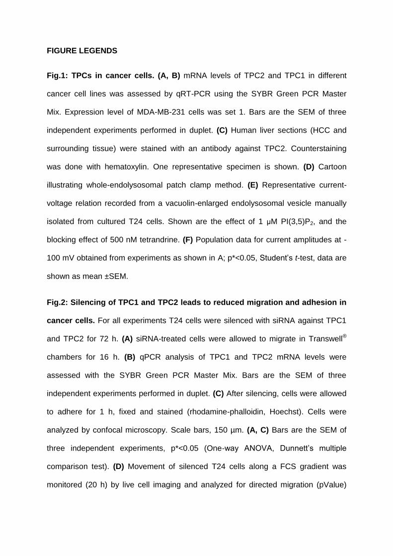
FIGURE LEGENDS
Fig.1: TPCs in cancer cells. (A, B) mRNA levels of TPC2 and TPC1 in different
cancer cell lines was assessed by qRT-PCR using the SYBR Green PCR Master
Mix. Expression level of MDA-MB-231 cells was set 1. Bars are the SEM of three
independent experiments performed in duplet. (C) Human liver sections (HCC and
surrounding tissue) were stained with an antibody against TPC2. Counterstaining
was done with hematoxylin. One representative specimen is shown. (D) Cartoon
illustrating whole-endolysosomal patch clamp method. (E) Representative current-
voltage relation recorded from a vacuolin-enlarged endolysosomal vesicle manually
isolated from cultured T24 cells. Shown are the effect of 1 μM PI(3,5)P2, and the
blocking effect of 500 nM tetrandrine. (F) Population data for current amplitudes at -
100 mV obtained from experiments as shown in A; p*<0.05, Student’s t-test, data are
shown as mean ±SEM.
Fig.2: Silencing of TPC1 and TPC2 leads to reduced migration and adhesion in
cancer cells. For all experiments T24 cells were silenced with siRNA against TPC1
and TPC2 for 72 h. (A) siRNA-treated cells were allowed to migrate in Transwell®
chambers for 16 h. (B) qPCR analysis of TPC1 and TPC2 mRNA levels were
assessed with the SYBR Green PCR Master Mix. Bars are the SEM of three
independent experiments performed in duplet. (C) After silencing, cells were allowed
to adhere for 1 h, fixed and stained (rhodamine-phalloidin, Hoechst). Cells were
analyzed by confocal microscopy. Scale bars, 150 µm. (A, C) Bars are the SEM of
three independent experiments, p*<0.05 (One-way ANOVA, Dunnett’s multiple
comparison test). (D) Movement of silenced T24 cells along a FCS gradient was
monitored (20 h) by live cell imaging and analyzed for directed migration (pValue)
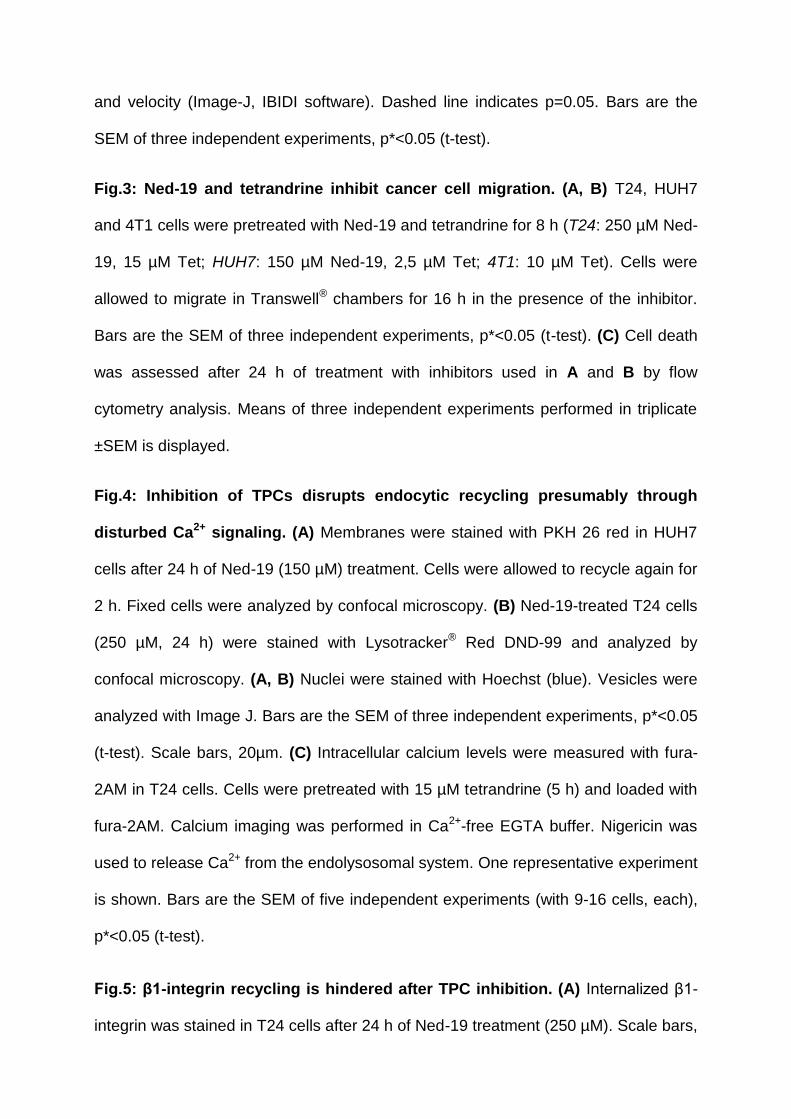
and velocity (Image-J, IBIDI software). Dashed line indicates p=0.05. Bars are the
SEM of three independent experiments, p*<0.05 (t-test).
Fig.3: Ned-19 and tetrandrine inhibit cancer cell migration. (A, B) T24, HUH7
and 4T1 cells were pretreated with Ned-19 and tetrandrine for 8 h (T24: 250 µM Ned-
19, 15 µM Tet; HUH7: 150 µM Ned-19, 2,5 µM Tet; 4T1: 10 µM Tet). Cells were
allowed to migrate in Transwell® chambers for 16 h in the presence of the inhibitor.
Bars are the SEM of three independent experiments, p*<0.05 (t-test). (C) Cell death
was assessed after 24 h of treatment with inhibitors used in A and B by flow
cytometry analysis. Means of three independent experiments performed in triplicate
±SEM is displayed.
Fig.4: Inhibition of TPCs disrupts endocytic recycling presumably through
disturbed Ca2+ signaling. (A) Membranes were stained with PKH 26 red in HUH7
cells after 24 h of Ned-19 (150 µM) treatment. Cells were allowed to recycle again for
2 h. Fixed cells were analyzed by confocal microscopy. (B) Ned-19-treated T24 cells
(250 µM, 24 h) were stained with Lysotracker® Red DND-99 and analyzed by
confocal microscopy. (A, B) Nuclei were stained with Hoechst (blue). Vesicles were
analyzed with Image J. Bars are the SEM of three independent experiments, p*<0.05
(t-test). Scale bars, 20µm. (C) Intracellular calcium levels were measured with fura-
2AM in T24 cells. Cells were pretreated with 15 µM tetrandrine (5 h) and loaded with
fura-2AM. Calcium imaging was performed in Ca2+-free EGTA buffer. Nigericin was
used to release Ca2+ from the endolysosomal system. One representative experiment
is shown. Bars are the SEM of five independent experiments (with 9-16 cells, each),
p*<0.05 (t-test).
Fig.5: β1-integrin recycling is hindered after TPC inhibition. (A) Internalized β1-
integrin was stained in T24 cells after 24 h of Ned-19 treatment (250 µM). Scale bars,
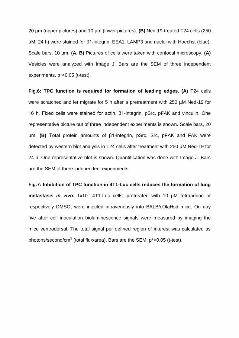
20 µm (upper pictures) and 10 µm (lower pictures). (B) Ned-19-treated T24 cells (250
µM, 24 h) were stained for β1-integrin, EEA1, LAMP3 and nuclei with Hoechst (blue).
Scale bars, 10 µm. (A, B) Pictures of cells were taken with confocal microscopy. (A)
Vesicles were analyzed with Image J. Bars are the SEM of three independent
experiments, p*<0.05 (t-test).
Fig.6: TPC function is required for formation of leading edges. (A) T24 cells
were scratched and let migrate for 5 h after a pretreatment with 250 µM Ned-19 for
16 h. Fixed cells were stained for actin, β1-integrin, pSrc, pFAK and vinculin. One
representative picture out of three independent experiments is shown. Scale bars, 20
µm. (B) Total protein amounts of β1-integrin, pSrc, Src, pFAK and FAK were
detected by western blot analysis in T24 cells after treatment with 250 µM Ned-19 for
24 h. One representative blot is shown. Quantification was done with Image J. Bars
are the SEM of three independent experiments.
Fig.7: Inhibition of TPC function in 4T1-Luc cells reduces the formation of lung
metastasis in vivo. 1x10⁵ 4T1-Luc cells, pretreated with 10 µM tetrandrine or
respectively DMSO, were injected intravenously into BALB/cOlaHsd mice. On day
five after cell inoculation bioluminescence signals were measured by imaging the
mice ventrodorsal. The total signal per defined region of interest was calculated as
photons/second/cm2 (total flux/area). Bars are the SEM, p*<0.05 (t-test).
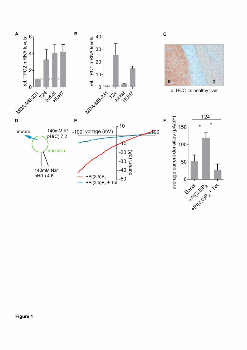
Figure 1
E
a: HCC b: healthy liver
a b
D
rel.
TP
C1
mR
NA
leve
ls
MDA-M
B-231 T24
Jurk
at
HUH70
10
20
30
40BA C
F
-10
100
+PI(3,5)P2
+PI(3,5)P2 + Tet
curr
ent
(p
A)
voltage (mV)-10010
-20
-30
-40
-50
rel.
TP
C2
mR
NA
leve
ls
MDA-M
B-231 T24
Jurk
at
HUH70
2
4
6
ave
rag
e c
urre
nt d
ens
itie
s (p
A/p
F)
Basal 2
+PI(3
,5)P
+ Tet
2
+PI(3
,5)P
0
50
100
150 * *
T24
inward 140mM K+
pH(C) 7.2
140mM Na+
pH(L) 4.6
Vacuolin
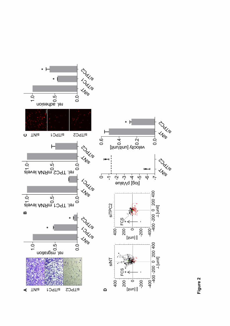
[unit]
0
200
400
-200
-400
020
040
0-2
00-4
00[u
nit]
siN
T
[unit]
0
200
400
-200
-400
020
040
0-2
00-4
00[u
nit]
siT
PC
2
rel. migration
siNT siT
PC1 siTPC2
0.0
0.5
1.0
*
*
rel. adhesion
siNT siT
PC1 siTPC2
0.0
0.5
1.0
*
*
siNT siTPC1 siTPC2
AC
[log] pValue
siNT siT
PC2-7-6-5-4-3-2-10
velocity [unit/unit]
siNT siT
PC2
0.0
0.2
0.4
0.6
*
siNT siTPC1 siTPC2
D
FC
S -+F
CS -+
rel. TPC2 mRNA levels
siNT siT
PC2
0.0
0.5
1.0
rel. TPC1 mRNA levels
siNT siT
PC1
0.0
0.5
1.0
B
Figure2
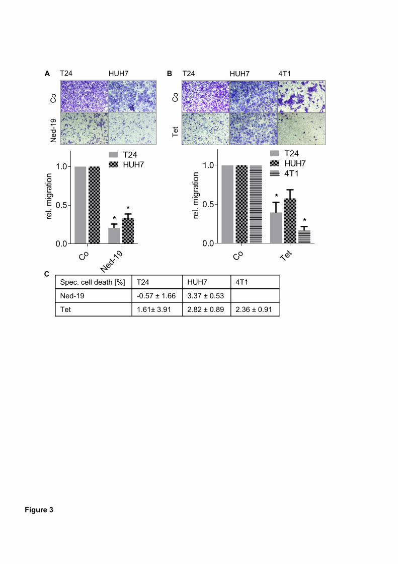
T24 HUH7
Co
Ned
-19
rel.
mig
ratio
n
Co
Ned-1
90.0
0.5
1.0
T24HUH7
** re
l. m
igra
tion
CoTet
0.0
0.5
1.0T24HUH7
*
4T1
*
T24 HUH7
Co
Tet
4T1A B
Figure 3
CSpec. cell death [%] T24 HUH7 4T1
Ned-19 -0.57 ± 1.66 3.37 ± 0.53
Tet 1.61± 3.91 2.82 ± 0.89 2.36 ± 0.91
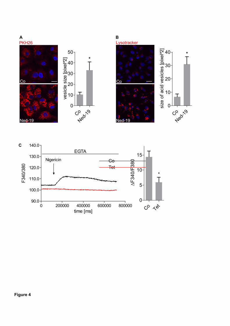
Figure 4
Co
vesi
cle
siz
e [p
ixe
l2̂
]
Co
Ned-1
90
10
20
30
40
50
*
Ned-19
PKH26
size
of
acid
ve
sicl
es
[pix
el
2̂]
Co
Ned-1
90
10
20
30
40 *
Co
Ned-19
LysotrackerA B
C
time [ms]
F34
0/38
0
0 200000 400000 600000 80000090.0
100.0
110.0
120.0
130.0
140.0
CoTet
Nigericin
EGTA
F
34
0/F
38
0
CoTet
0
5
10
15
*
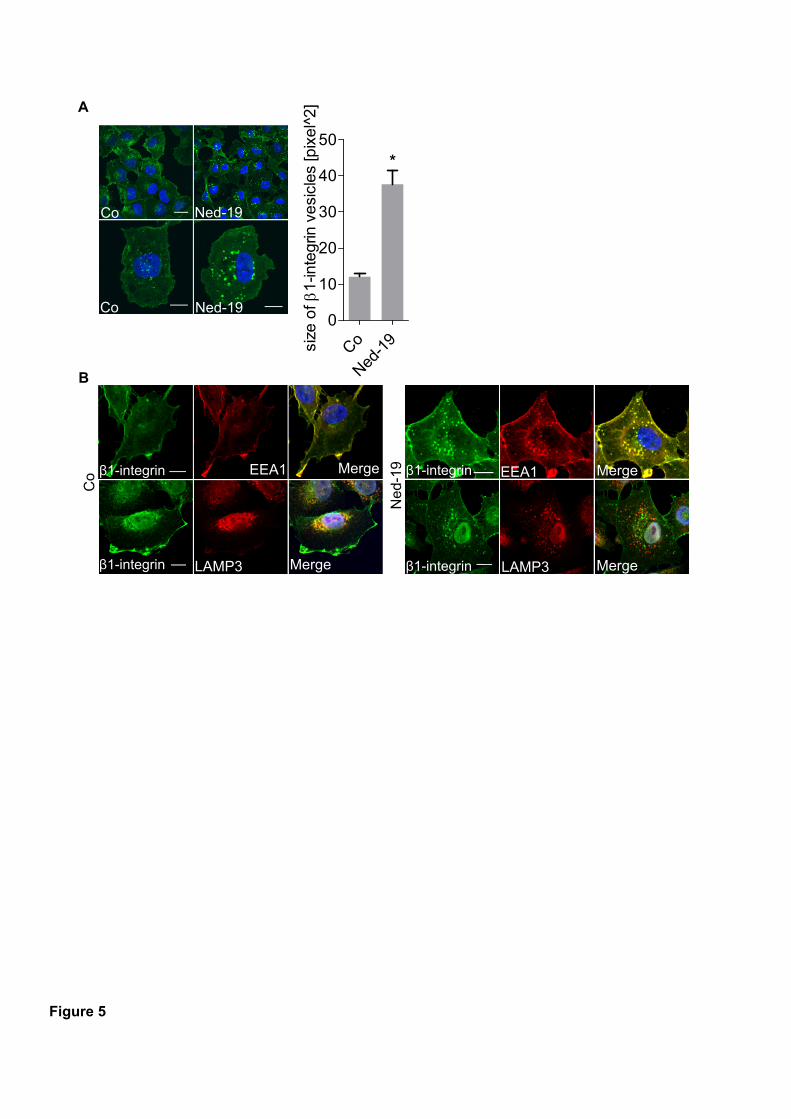
Figure 5
Co Ned-19
Co Ned-19si
ze o
f 1
-inte
grin
ve
sicl
es [p
ixel
^2]
Co
Ned-1
90
10
20
30
40
50*
A
B
β1-integrin EEA1 Merge
Co
LAMP3β1-integrin Merge
Ned
-19
LAMP3β1-integrin Merge
EEA1β1-integrin Merge
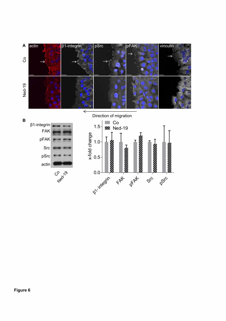
pSrc
Src
pFAK
FAK
actin
β1-integrin
x-fo
ld c
hang
e
integ
rinFAK
pFAK
SrcpS
rc0.0
0.5
1.0
1.5CoNed-19
actin β1-integrin pSrc pFAK vinculin
Co
Ned
-19
Direction of migration
Figure 6
A
B
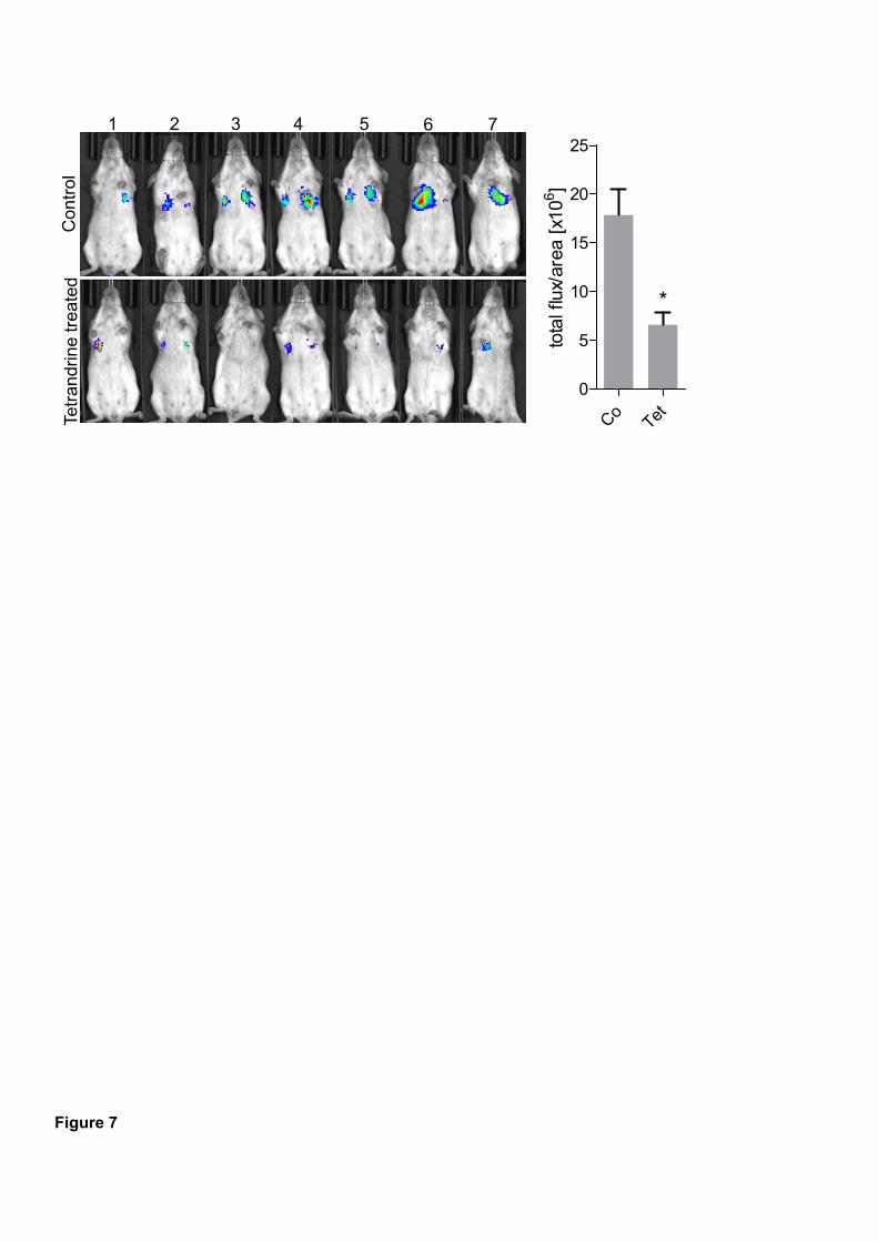
Figure 7
1 2 3 4 5 6 7
Tetr
andr
ine
trea
ted
Con
trol
tota
l flu
x/ar
ea [
x106
]
CoTet
0
5
10
15
20
25
*